EP3703750B1 - Chimeric antigen receptors specific for b-cell maturation antigen and encoding polynucleotides - Google Patents
Chimeric antigen receptors specific for b-cell maturation antigen and encoding polynucleotides Download PDFInfo
- Publication number
- EP3703750B1 EP3703750B1 EP18815386.0A EP18815386A EP3703750B1 EP 3703750 B1 EP3703750 B1 EP 3703750B1 EP 18815386 A EP18815386 A EP 18815386A EP 3703750 B1 EP3703750 B1 EP 3703750B1
- Authority
- EP
- European Patent Office
- Prior art keywords
- seq
- cdr
- nos
- region
- amino acid
- Prior art date
- Legal status (The legal status is an assumption and is not a legal conclusion. Google has not performed a legal analysis and makes no representation as to the accuracy of the status listed.)
- Active
Links
Images


Classifications
-
- C—CHEMISTRY; METALLURGY
- C07—ORGANIC CHEMISTRY
- C07K—PEPTIDES
- C07K14/00—Peptides having more than 20 amino acids; Gastrins; Somatostatins; Melanotropins; Derivatives thereof
- C07K14/435—Peptides having more than 20 amino acids; Gastrins; Somatostatins; Melanotropins; Derivatives thereof from animals; from humans
- C07K14/705—Receptors; Cell surface antigens; Cell surface determinants
-
- A—HUMAN NECESSITIES
- A61—MEDICAL OR VETERINARY SCIENCE; HYGIENE
- A61K—PREPARATIONS FOR MEDICAL, DENTAL OR TOILETRY PURPOSES
- A61K35/00—Medicinal preparations containing materials or reaction products thereof with undetermined constitution
- A61K35/12—Materials from mammals; Compositions comprising non-specified tissues or cells; Compositions comprising non-embryonic stem cells; Genetically modified cells
- A61K35/14—Blood; Artificial blood
- A61K35/17—Lymphocytes; B-cells; T-cells; Natural killer cells; Interferon-activated or cytokine-activated lymphocytes
-
- A—HUMAN NECESSITIES
- A61—MEDICAL OR VETERINARY SCIENCE; HYGIENE
- A61K—PREPARATIONS FOR MEDICAL, DENTAL OR TOILETRY PURPOSES
- A61K39/00—Medicinal preparations containing antigens or antibodies
- A61K39/395—Antibodies; Immunoglobulins; Immune serum, e.g. antilymphocytic serum
-
- A—HUMAN NECESSITIES
- A61—MEDICAL OR VETERINARY SCIENCE; HYGIENE
- A61K—PREPARATIONS FOR MEDICAL, DENTAL OR TOILETRY PURPOSES
- A61K40/00—Cellular immunotherapy
- A61K40/10—Cellular immunotherapy characterised by the cell type used
- A61K40/11—T-cells, e.g. tumour infiltrating lymphocytes [TIL] or regulatory T [Treg] cells; Lymphokine-activated killer [LAK] cells
-
- A—HUMAN NECESSITIES
- A61—MEDICAL OR VETERINARY SCIENCE; HYGIENE
- A61K—PREPARATIONS FOR MEDICAL, DENTAL OR TOILETRY PURPOSES
- A61K40/00—Cellular immunotherapy
- A61K40/30—Cellular immunotherapy characterised by the recombinant expression of specific molecules in the cells of the immune system
- A61K40/31—Chimeric antigen receptors [CAR]
-
- A—HUMAN NECESSITIES
- A61—MEDICAL OR VETERINARY SCIENCE; HYGIENE
- A61K—PREPARATIONS FOR MEDICAL, DENTAL OR TOILETRY PURPOSES
- A61K40/00—Cellular immunotherapy
- A61K40/40—Cellular immunotherapy characterised by antigens that are targeted or presented by cells of the immune system
- A61K40/41—Vertebrate antigens
- A61K40/42—Cancer antigens
- A61K40/4202—Receptors, cell surface antigens or cell surface determinants
- A61K40/4214—Receptors for cytokines
- A61K40/4215—Receptors for tumor necrosis factors [TNF], e.g. lymphotoxin receptor [LTR], CD30
-
- A—HUMAN NECESSITIES
- A61—MEDICAL OR VETERINARY SCIENCE; HYGIENE
- A61P—SPECIFIC THERAPEUTIC ACTIVITY OF CHEMICAL COMPOUNDS OR MEDICINAL PREPARATIONS
- A61P35/00—Antineoplastic agents
-
- C—CHEMISTRY; METALLURGY
- C07—ORGANIC CHEMISTRY
- C07K—PEPTIDES
- C07K14/00—Peptides having more than 20 amino acids; Gastrins; Somatostatins; Melanotropins; Derivatives thereof
- C07K14/435—Peptides having more than 20 amino acids; Gastrins; Somatostatins; Melanotropins; Derivatives thereof from animals; from humans
- C07K14/705—Receptors; Cell surface antigens; Cell surface determinants
- C07K14/70503—Immunoglobulin superfamily
- C07K14/7051—T-cell receptor (TcR)-CD3 complex
-
- C—CHEMISTRY; METALLURGY
- C07—ORGANIC CHEMISTRY
- C07K—PEPTIDES
- C07K14/00—Peptides having more than 20 amino acids; Gastrins; Somatostatins; Melanotropins; Derivatives thereof
- C07K14/435—Peptides having more than 20 amino acids; Gastrins; Somatostatins; Melanotropins; Derivatives thereof from animals; from humans
- C07K14/705—Receptors; Cell surface antigens; Cell surface determinants
- C07K14/70503—Immunoglobulin superfamily
- C07K14/70521—CD28, CD152
-
- C—CHEMISTRY; METALLURGY
- C07—ORGANIC CHEMISTRY
- C07K—PEPTIDES
- C07K14/00—Peptides having more than 20 amino acids; Gastrins; Somatostatins; Melanotropins; Derivatives thereof
- C07K14/435—Peptides having more than 20 amino acids; Gastrins; Somatostatins; Melanotropins; Derivatives thereof from animals; from humans
- C07K14/705—Receptors; Cell surface antigens; Cell surface determinants
- C07K14/70578—NGF-receptor/TNF-receptor superfamily, e.g. CD27, CD30, CD40, CD95
-
- C—CHEMISTRY; METALLURGY
- C07—ORGANIC CHEMISTRY
- C07K—PEPTIDES
- C07K16/00—Immunoglobulins [IG], e.g. monoclonal or polyclonal antibodies
- C07K16/18—Immunoglobulins [IG], e.g. monoclonal or polyclonal antibodies against material from animals or humans
- C07K16/28—Immunoglobulins [IG], e.g. monoclonal or polyclonal antibodies against material from animals or humans against receptors, cell surface antigens or cell surface determinants
- C07K16/2878—Immunoglobulins [IG], e.g. monoclonal or polyclonal antibodies against material from animals or humans against receptors, cell surface antigens or cell surface determinants against the NGF-receptor/TNF-receptor superfamily, e.g. CD27, CD30, CD40, CD95
-
- C—CHEMISTRY; METALLURGY
- C12—BIOCHEMISTRY; BEER; SPIRITS; WINE; VINEGAR; MICROBIOLOGY; ENZYMOLOGY; MUTATION OR GENETIC ENGINEERING
- C12N—MICROORGANISMS OR ENZYMES; COMPOSITIONS THEREOF; PROPAGATING, PRESERVING, OR MAINTAINING MICROORGANISMS; MUTATION OR GENETIC ENGINEERING; CULTURE MEDIA
- C12N5/00—Undifferentiated human, animal or plant cells, e.g. cell lines; Tissues; Cultivation or maintenance thereof; Culture media therefor
- C12N5/06—Animal cells or tissues; Human cells or tissues
- C12N5/0602—Vertebrate cells
- C12N5/0634—Cells from the blood or the immune system
- C12N5/0636—T lymphocytes
-
- C—CHEMISTRY; METALLURGY
- C12—BIOCHEMISTRY; BEER; SPIRITS; WINE; VINEGAR; MICROBIOLOGY; ENZYMOLOGY; MUTATION OR GENETIC ENGINEERING
- C12Q—MEASURING OR TESTING PROCESSES INVOLVING ENZYMES, NUCLEIC ACIDS OR MICROORGANISMS; COMPOSITIONS OR TEST PAPERS THEREFOR; PROCESSES OF PREPARING SUCH COMPOSITIONS; CONDITION-RESPONSIVE CONTROL IN MICROBIOLOGICAL OR ENZYMOLOGICAL PROCESSES
- C12Q1/00—Measuring or testing processes involving enzymes, nucleic acids or microorganisms; Compositions therefor; Processes of preparing such compositions
- C12Q1/68—Measuring or testing processes involving enzymes, nucleic acids or microorganisms; Compositions therefor; Processes of preparing such compositions involving nucleic acids
- C12Q1/6844—Nucleic acid amplification reactions
- C12Q1/6848—Nucleic acid amplification reactions characterised by the means for preventing contamination or increasing the specificity or sensitivity of an amplification reaction
-
- A—HUMAN NECESSITIES
- A61—MEDICAL OR VETERINARY SCIENCE; HYGIENE
- A61K—PREPARATIONS FOR MEDICAL, DENTAL OR TOILETRY PURPOSES
- A61K39/00—Medicinal preparations containing antigens or antibodies
- A61K2039/505—Medicinal preparations containing antigens or antibodies comprising antibodies
-
- A—HUMAN NECESSITIES
- A61—MEDICAL OR VETERINARY SCIENCE; HYGIENE
- A61K—PREPARATIONS FOR MEDICAL, DENTAL OR TOILETRY PURPOSES
- A61K38/00—Medicinal preparations containing peptides
-
- C—CHEMISTRY; METALLURGY
- C07—ORGANIC CHEMISTRY
- C07K—PEPTIDES
- C07K2317/00—Immunoglobulins specific features
- C07K2317/50—Immunoglobulins specific features characterized by immunoglobulin fragments
- C07K2317/52—Constant or Fc region; Isotype
- C07K2317/524—CH2 domain
-
- C—CHEMISTRY; METALLURGY
- C07—ORGANIC CHEMISTRY
- C07K—PEPTIDES
- C07K2317/00—Immunoglobulins specific features
- C07K2317/50—Immunoglobulins specific features characterized by immunoglobulin fragments
- C07K2317/52—Constant or Fc region; Isotype
- C07K2317/528—CH4 domain
-
- C—CHEMISTRY; METALLURGY
- C07—ORGANIC CHEMISTRY
- C07K—PEPTIDES
- C07K2317/00—Immunoglobulins specific features
- C07K2317/50—Immunoglobulins specific features characterized by immunoglobulin fragments
- C07K2317/52—Constant or Fc region; Isotype
- C07K2317/53—Hinge
-
- C—CHEMISTRY; METALLURGY
- C07—ORGANIC CHEMISTRY
- C07K—PEPTIDES
- C07K2317/00—Immunoglobulins specific features
- C07K2317/50—Immunoglobulins specific features characterized by immunoglobulin fragments
- C07K2317/56—Immunoglobulins specific features characterized by immunoglobulin fragments variable (Fv) region, i.e. VH and/or VL
- C07K2317/565—Complementarity determining region [CDR]
-
- C—CHEMISTRY; METALLURGY
- C07—ORGANIC CHEMISTRY
- C07K—PEPTIDES
- C07K2317/00—Immunoglobulins specific features
- C07K2317/60—Immunoglobulins specific features characterized by non-natural combinations of immunoglobulin fragments
- C07K2317/62—Immunoglobulins specific features characterized by non-natural combinations of immunoglobulin fragments comprising only variable region components
- C07K2317/622—Single chain antibody (scFv)
-
- C—CHEMISTRY; METALLURGY
- C07—ORGANIC CHEMISTRY
- C07K—PEPTIDES
- C07K2317/00—Immunoglobulins specific features
- C07K2317/70—Immunoglobulins specific features characterized by effect upon binding to a cell or to an antigen
- C07K2317/73—Inducing cell death, e.g. apoptosis, necrosis or inhibition of cell proliferation
-
- C—CHEMISTRY; METALLURGY
- C07—ORGANIC CHEMISTRY
- C07K—PEPTIDES
- C07K2319/00—Fusion polypeptide
- C07K2319/01—Fusion polypeptide containing a localisation/targetting motif
- C07K2319/02—Fusion polypeptide containing a localisation/targetting motif containing a signal sequence
-
- C—CHEMISTRY; METALLURGY
- C07—ORGANIC CHEMISTRY
- C07K—PEPTIDES
- C07K2319/00—Fusion polypeptide
- C07K2319/01—Fusion polypeptide containing a localisation/targetting motif
- C07K2319/03—Fusion polypeptide containing a localisation/targetting motif containing a transmembrane segment
-
- C—CHEMISTRY; METALLURGY
- C07—ORGANIC CHEMISTRY
- C07K—PEPTIDES
- C07K2319/00—Fusion polypeptide
- C07K2319/30—Non-immunoglobulin-derived peptide or protein having an immunoglobulin constant or Fc region, or a fragment thereof, attached thereto
-
- C—CHEMISTRY; METALLURGY
- C07—ORGANIC CHEMISTRY
- C07K—PEPTIDES
- C07K2319/00—Fusion polypeptide
- C07K2319/33—Fusion polypeptide fusions for targeting to specific cell types, e.g. tissue specific targeting, targeting of a bacterial subspecies
-
- C—CHEMISTRY; METALLURGY
- C12—BIOCHEMISTRY; BEER; SPIRITS; WINE; VINEGAR; MICROBIOLOGY; ENZYMOLOGY; MUTATION OR GENETIC ENGINEERING
- C12N—MICROORGANISMS OR ENZYMES; COMPOSITIONS THEREOF; PROPAGATING, PRESERVING, OR MAINTAINING MICROORGANISMS; MUTATION OR GENETIC ENGINEERING; CULTURE MEDIA
- C12N2510/00—Genetically modified cells
Definitions
- the present disclosure relates in some aspects to polynucleotides that encode chimeric antigen receptors (CARs) specific for B-cell maturation antigen (BCMA).
- CARs chimeric antigen receptors
- BCMA B-cell maturation antigen
- the disclosure further relates to genetically engineered cells, containing such polynucleotides encoding BCMA-binding CARs, and uses thereof in adoptive cell therapy.
- BCMA B-cell maturation antigen
- BAFF B cell activator of the TNF family
- APRIL proliferation inducing ligand
- WO2015/158671 describes BCMA specific CARs for cancer immunotherapy.
- the present invention provides a polynucleotide encoding a chimeric antigen receptor (CAR), wherein:
- the invention also provides a vector comprising the polynucleotide of the invention.
- the invention further provides an engineered cell, comprising the polynucleotide or the vector of the invention.
- the invention further provides a composition comprising the polynucleotide or the vector or the engineered cell of the invention.
- the invention further provides the engineered cell or the composition of the invention, for use in a method of treatment.
- the invention further provides the engineered cell or the composition of the invention, for use in a method of treating a disease or disorder associated with expression of B cell maturation antigen (BCMA).
- BCMA B cell maturation antigen
- the transcribed RNA, optionally messenger RNA (mRNA), from the polynucleotide exhibits at least 70%, 75%, 80%, 85%, 90%, or 95% RNA homogeneity.
- the spacer is derived from an immunoglobulin.
- the spacer includes a sequence of a hinge region, a C H 2 and a C H 3 region.
- one of more of the hinge, C H 2 and C H 3 is derived all or in part from IgG4 or IgG2. In some cases, the hinge, C H 2 and C H 3 is derived from IgG4.
- one or more of the hinge, C H 2 and C H 3 is chimeric and contains sequence derived from IgG4 and IgG2.
- the spacer contains an IgG4/2 chimeric hinge, an IgG2/4 C H 2, and an IgG4 C H 3 region.
- the encoded spacer is or contains (i) the sequence set forth in SEQ ID NO: 649; (ii) a functional variant of SEQ ID NO:649 that has at least 95%, 96%, 97%, 98% or 99% sequence identity to SEQ ID NO:649; or (iii) a contiguous portion of (i) or (ii) that is at least 125 amino acids in length.
- the encoded spacer is or includes the sequence set forth in SEQ ID NO: 649.
- the spacer has a length of 125 to 300 amino acids in length, 125 to 250 amino acids in length, 125 to 230 amino acids in length, 125 to 200 amino acids in length, 125 to 180 amino acids in length, 125 to 150 amino acids in length, 150 to 300 amino acids in length, 150 to 250 amino acids in length, 150 to 230 amino acids in length, 150 to 200 amino acids in length, 150 to 180 amino acids in length, 180 to 300 amino acids in length, 180 to 250 amino acids in length, 180 to 230 amino acids in length, 180 to 200 amino acids in length, 200 to 300 amino acids in length, 200 to 250 amino acids in length, 200 to 230 amino acids in length, 230 to 300 amino acids in length, 230 to 250 amino acids in length or 250 to 300 amino acids in length.
- the spacer is at least or at least about or is or is about 130, 140, 150, 160, 170, 180, 190, 200, 210, 220, 221, 222, 223, 224, 225, 226, 227, 228 or 229 amino acids in length, or a length between any of the foregoing.
- the reference splice acceptor and/or reference splice donor sites are canonical, non-canonical, or cryptic splice sites.
- the reference splice donor and/or reference splice acceptor site(s) has a splice site prediction score of at least or about 0.4, 0.5, 0.6, 0.70, 0.75, 0.80, 0.85, 0.90, 0.95, 0.99, or 1.0; and/or the reference splice donor and/or reference splice acceptor site(s) is/are predicted to be involved in a splice event with a probability of at least 40%, 50%, 60%, 70%, 75%, 80%, 85%, 90%, 95%, 99%, or 100%.
- At least one of the one or more nucleotide modifications are within 1, 2, 3, 4, 5, 6, 7, 8, 9 or 10 residues of the splice site junction of the reference splice acceptor and/or reference splice donor site.
- the one or more nucleotide modifications is silent and/or results in a degenerate codon compared to SEQ ID NO:621 and/or does not change the amino acid sequence of the encoded spacer.
- the modified splice donor site is set forth in tcaactggtatgtgg (SEQ ID NO:662) and/or the modified acceptor site is set forth in cagtttcttcctgtatagtagactcaccgtggataaatcaa (SEQ ID NO:672).
- the spacer is encoded by a sequence of nucleotide set forth in SEQ ID NO:622 or a portion thereof.
- the polynucleotide includes nucleic acid encoding: (a) an extracellular antigen-binding domain that specifically recognizes an antigen, in particular BCMA; (b) a spacer of at least 125 amino acids in length, wherein the encoding nucleic acid is or includes the sequence set forth in SEQ ID NO:622 or encodes a sequence of amino acids set forth in SEQ ID NO:649; (c) a transmembrane domain; and (d) an intracellular signaling region.
- the polynucleotide includes nucleic acid encoding: (a) an extracellular antigen-binding domain that specifically recognizes an antigen, in particular BCMA; (b) a spacer of at least 125 amino acids in length, wherein the encoding nucleic acid includes or mostly includes the sequence set forth in SEQ ID NO:622 or encodes a sequence of amino acids set forth in SEQ ID NO:649; (c) a transmembrane domain; and (d) an intracellular signaling region.
- the transcribed RNA following expression of the polynucleotide in a cell, the transcribed RNA, optionally messenger RNA (mRNA), from the polynucleotide, exhibits at least 70%, 75%, 80%, 85%, 90%, or 95% RNA homogeneity.
- mRNA messenger RNA
- the transcribed RNA, optionally messenger RNA (mRNA), from the polynucleotide exhibits reduced heterogeneity compared to the heterogeneity of the mRNA transcribed from a reference polynucleotide, said reference polynucleotide encoding the same amino acid sequence as the polynucleotide, wherein the reference polynucleotide differs by the presence of one or more splice donor site and/or one or more splice acceptor site in the nucleic acid encoding the spacer and/or includes one or more nucleotide modifications compared to the polynucleotide.
- mRNA messenger RNA
- the RNA heterogeneity is reduced by greater than or greater than about 10%, 15%, 20%, 25%, 30%, 40%, 50% or more.
- the transcribed RNA, optionally messenger RNA (mRNA) from the reference polynucleotide exhibits greater than or greater than about 10%, 15%, 20%, 25%, 30%, 40%, 50% or more RNA heterogeneity.
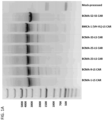
- the RNA homogeneity and/or heterogeneity is determined by agarose gel electrophoresis, chip-based capillary electrophoresis, analytical ultracentrifugation, field flow fractionation, or liquid chromatography.
- the polynucleotide is codon-optimized.
- the antigen is BCMA.
- the antigen is associated with the disease or condition or expressed in cells of the environment of a lesion associated with the disease or condition.
- the disease or condition is a cancer.
- the disease or condition is a myeloma, leukemia or lymphoma.
- the antigen-binding domain is an antibody fragment containing a variable heavy chain (V H ) and a variable light chain (V L ) region.
- V H region is or includes an amino acid sequence having at least 90%, 91%, 92%, 93%, 94%, 95%, 96%, 97%, 98% or 99% sequence identity to the V H region amino acid sequence set forth in any of SEQ ID NOs: 110-115, 247-256, 324, 325, 518-531, 533, 609 617, 772-774, or 814-832; and/or the V L region is or includes an amino acid sequence having at least 90%, 91%, 92%, 93%, 94%, 95%, 96%, 97%, 98%, or 99% sequence identity to the V L region amino acid sequence set forth in any of SEQ ID NOs: 116-127, 257-267, 326, 327, 534-550, 552-557, 610, 618, 775-777, or 8
- the V H region is or includes an amino acid sequence having at least 90%, 91%, 92%, 93%, 94%, 95%, 96%, 97%, 98% or 99% sequence identity to the V H region amino acid sequence set forth in any of SEQ ID NOs: 110, 111, 112, 113, 115, 248, 252, 253, 254, 255, 256, 324, 325, 518, 519, 520, 521, 522, 609, 617, 772-774, or 814-832; and/or the V L region is or includes an amino acid sequence having at least 90%, 91%, 92%, 93%, 94%, 95%, 96%, 97%, 98%, or 99% sequence identity to the V L region amino acid sequence set forth in any of SEQ ID NOs: 116, 117, 118, 120, 121, 124, 125, 258, 262, 263, 264, 265, 266, 267, 326, 327, 534, 535, 536, 537, 538
- the V H region is or contains a CDR-H1, CDR-H2 and CDR-H3 contained within the V H region amino acid sequence selected from any one of SEQ ID NOs:110-115, 247-256, 324, 325, 518-531, 533, 609, 617, 772-774, or 814-832; and/or the V L region is or includes a CDR-L1, CDR-L2 and CDR-L3 contained within the V L region amino acid sequence selected from any one of SEQ ID NOs: 116-127, 257-267, 326, 327, 534-550, 552-557, 610, 618, 775-777, or 833-849.
- the V H region is or contains a CDR-H1, CDR-H2 and CDR-H3 contained within the V H region amino acid sequence selected from any one of SEQ ID NOs: 110, 111, 112, 113, 115, 248, 252, 253, 254, 255, 256, 324, 325, 518, 519, 520, 521, 522, 609, 617, 772-774, or 814-832; and/or the V L region is or includes a CDR-L1, CDR-L2 and CDR-L3 contained within the V L region amino acid sequence selected from any one of SEQ ID NOs: 116, 117, 118, 120, 121, 124, 125, 258, 262, 263, 264, 265, 266, 267, 326, 327, 534, 535, 536, 537, 538, 610, 618, 775-777, or 833-849.
- the V H region is or includes (a) a heavy chain complementarity determining region 1 (CDR-H1) containing the amino acid sequence selected from any one of SEQ ID NOs:1-3, 140-144, 288, 289, 294, 295,507, 532, 593, 596, 604, 611; and/or (b) a heavy chain complementarity determining region 2 (CDR-H2) containing the amino acid sequence selected from any one of SEQ ID NOs:4-6, 145-148, 290, 291, 296, 297, 372-374, 513, 551, 594, 597, 605, or 612; and (c) a heavy chain complementarity determining region 3 (CDR-H3) containing the amino acid sequence selected from any one of SEQ ID NOs:7-11, 149-157, 279-287, 292, 293, 376-378, 517, 595, 606, 613; and/or the V L region is or includes (a) a light chain complementarity determining region 1 (CDR-
- the V H region is or contains (a) a heavy chain complementarity determining region 1 (CDR-H1) containing the amino acid sequence selected from any one of SEQ ID NOs: 1, 2, 3, 141, 143, 144, 288, 289, 507, 593, 604, 611; and/or (b) a heavy chain complementarity determining region 2 (CDR-H2) containing the amino acid sequence selected from any one of SEQ ID NOs: 4, 5, 6, 145, 147, 148, 290, 291, 372, 513, 594, 605 or 612; and (c) a heavy chain complementarity determining region 3 (CDR-H3) containing the amino acid sequence selected from any one of SEQ ID NOs: 7, 8, 9, 10, 149, 153, 154, 155, 156, 157, 292, 293, 376, 517, 595, 606 or 613; and/or the V L region is or contains (a) a heavy chain complementarity determining region 1 (CDR-H1) containing
- the V H region contains a CDR-H1, CDR-H2, and CDR-H3, selected from: a CDR-H1, CDR-H2, and CDR-H3 containing the amino acid sequence of SEQ ID NOs: 1, 4, and 7, respectively; a CDR-H1, CDR-H2, and CDR-H3 containing the amino acid sequence of SEQ ID NOs:2, 5, and 8, respectively; a CDR-H1, CDR-H2, and CDR-H3 containing the amino acid sequence of SEQ ID NOs:2, 5, and 9, respectively; a CDR-H1, CDR-H2, and CDR-H3 containing the amino acid sequence of SEQ ID NOs:2, 5, and 10, respectively; a CDR-H1, CDR-H2, and CDR-H3 containing the amino acid sequence of SEQ ID NOs:3, 6, and 11, respectively; a CDR-H1, CDR-H2, and CDR-H3 containing the amino acid sequence of SEQ ID NOs:3, 6, and 11, respectively; a CDR-H
- the V H region contains a CDR-H1, CDR-H2, and CDR-H3, selected from: a CDR-H1, CDR-H2, and CDR-H3 containing the amino acid sequence of SEQ ID NOs: 1, 4, and 7, respectively; a CDR-H1, CDR-H2, and CDR-H3 containing the amino acid sequence of SEQ ID NOs:2, 5, and 8, respectively; a CDR-H1, CDR-H2, and CDR-H3 containing the amino acid sequence of SEQ ID NOs:2, 5, and 9, respectively; a CDR-H1, CDR-H2, and CDR-H3 containing the amino acid sequence of SEQ ID NOs:2, 5, and 10, respectively; a CDR-H1, CDR-H2, and CDR-H3 containing the amino acid sequence of SEQ ID NOs: 141, 145, and 149, respectively; a CDR-H1, CDR-H2, and CDR-H3 containing the amino acid sequence of SEQ ID NOs: 141, 145,
- the V H region is or includes the amino acid sequence set forth in any of SEQ ID NOs: 110-115, 247-256, 324, 325, 518-531, 533, 609, 617, 772-774, or 814-832. In some aspects, the V H region is or includes the amino acid sequence set forth in any of SEQ ID NOs: 110, 111, 112, 113, 115, 248, 252, 253, 254, 255, 256, 324, 325, 518, 519, 520, 521, 522, 609 or 617.
- the V H region contains a CDR-H1, CDR-H2, and CDR-H3 containing the amino acid sequence of SEQ ID NOs:593, 594, and 595, respectively; or the V H region includes a CDR-H1, CDR-H2, and CDR-H3 containing the amino acid sequence of SEQ ID NOs:611, 612, and 613, respectively.
- the V H region is or includes the amino acid sequence set forth in SEQ ID NO: 617.
- the V L region includes a CDR-L1, CDR-L2, and CDR-L3 selected from: a CDR-L1, CDR-L2, and CDR-L3 containing the amino acid sequence of SEQ ID NOs:26, 37, and 47, respectively; a CDR-L1, CDR-L2, and CDR-L3 containing the amino acid sequence of SEQ ID NOs:27, 38, and 48, respectively; a CDR-L1, CDR-L2, and CDR-L3 containing the amino acid sequence of SEQ ID NOs:28, 39, and 49, respectively; a CDR-L1, CDR-L2, and CDR-L3 containing the amino acid sequence of SEQ ID NOs:29, 40, and 50, respectively; a CDR-L1, CDR-L2, and CDR-L3 containing the amino acid sequence of SEQ ID NOs:30, 39, and 51, respectively; a CDR-L1, CDR-L2, and CDR-L3 containing the amino acid sequence of SEQ ID
- the V L region includes a CDR-L1, CDR-L2, and CDR-L3 selected from: a CDR-L1, CDR-L2, and CDR-L3 containing the amino acid sequence of SEQ ID NOs:26, 37, and 47, respectively; a CDR-L1, CDR-L2, and CDR-L3 containing the amino acid sequence of SEQ ID NOs:27, 38, and 48, respectively; a CDR-L1, CDR-L2, and CDR-L3 containing the amino acid sequence of SEQ ID NOs:28, 39, and 49, respectively; a CDR-L1, CDR-L2, and CDR-L3 containing the amino acid sequence of SEQ ID NOs:30, 39, and 51, respectively; a CDR-L1, CDR-L2, and CDR-L3 containing the amino acid sequence of SEQ ID NOs:31, 41, and 52, respectively; a CDR-L1, CDR-L1, CDR-L2, and CDR-L3 containing the amino acid
- the V L region is or includes the amino acid sequence set forth in any of SEQ ID NOs: 116-127, 257-267, 326, 327, 534-550, 552-557, 610, 618, 775-777, or 833-849.
- the V L region is or contains the amino acid sequence set forth in any of SEQ ID NOs: 116, 117, 118, 120, 121, 124, 125, 258, 262, 263, 264, 265, 266, 267, 326, 327, 534, 535, 536, 537, 538, 610, 618, 775-777, or 833-849.
- the V L region contains a CDR-L1, CDR-L2, and CDR-L3 including the amino acid sequence of SEQ ID NOs:601, 602, and 603, respectively; or the V L region contains a CDR-L1, CDR-L2, and CDR-L3 including the amino acid sequence of SEQ ID NOs:614, 615, and 603, respectively.
- the V L region is or includes the amino acid sequence set forth in SEQ ID NO:618.
- the V H region is or comprises an amino acid sequence having at least 90%, 91%, 92%, 93%, 94%, 95%, 96%, 97%, 98% or 99% sequence identity to the V H region sequence of any of SEQ ID NOs:617, 110-115, 247-256, 324, 325, 518-531, 533, 609, 772-774, or 814-832; and the V L region is or comprises an amino acid sequence having at least 90%, 91%, 92%, 93%, 94%, 95%, 96%, 97%, 98%, or 99% sequence identity to the V L region sequence of any of SEQ ID NOs: 618, 116-127, 257-267, 326, 327, 534-550, 552-557, 610, 775-777, or 833-849.
- the V H region is or comprises a CDR-H1, CDR-H2 and CDR-H3 contained within the V H region amino acid sequence selected from any one of SEQ ID NOs: 617, 110-115, 247-256, 324, 325, 518-531, 533, 609, 772-774, or 814-832; and the V L region is or comprises a CDR-L1, CDR-L2 and CDR-L3 contained within the V L region amino acid sequence selected from any one of SEQ ID NOs: 618, 116-127, 257-267, 326, 327, 534-550, 552-557, 610, 775-777, or 833-849.
- the V H region is or comprises (a) a CDR-H1 comprising the sequence selected from any one of SEQ ID NOs: 593, 611, 1-3, 140-144, 288, 289, 294, 295, 507, 532, 596, or 604; (b) a CDR-H2 comprising the sequence selected from any one of SEQ ID NOs: 594, 612, 4-6, 145-148, 290, 291, 296, 297, 372-374, 513, 551, 597, or 605; and (c) a CDR-H3 comprising the sequence selected from any one of SEQ ID NOs: 595, 613, 7-11, 149-157, 279-287, 292, 293, 376-378, 517, or 606; and the V L region is or comprises (a) a CDR-L1 comprising the sequence selected from any one of SEQ ID NOs: 601, 614, 26-36, 174-178, 302, 303, 380
- the V H region and the V L regions includes the amino acid sequence set forth in SEQ ID NOs: 110 and 116, respectively, or a sequence of amino acids that has at least 90% identity to SEQ ID NO: 110 and 116, respectively;
- the V H region and the V L regions contain the amino acid sequence set forth in SEQ ID NOs: 111 and 117, respectively, or a sequence of amino acids that has at least 90% identity to SEQ ID NO: 111 and 117, respectively;
- the V H region and the V L regions contain the amino acid sequence set forth in SEQ ID NOs: 110 and 118, respectively, or a sequence of amino acids that has at least 90% identity to SEQ ID NO: 110 and 118, respectively;
- the V H region and the V L regions contain the amino acid sequence set forth in SEQ ID NOs: 110 and 119, respectively, or a sequence of amino acids that has at least 90% identity to SEQ ID NO: 110 and 119, respectively;
- the V H region and the V L regions contain the amino acid sequence set forth in SEQ ID
- the V H region is or comprises the sequence of any of SEQ ID NOs: 617, 110-115, 247-256, 324, 325, 518-531, 533, 609, 772-774, or 814-832; and the V L region is or comprises the sequence of any of SEQ ID NOs: 618, 116-127, 257-267, 326, 327, 534-550, 552-557, 610, 775-777, or 833-849.
- the fragment includes an scFv.
- the V H region and the V L region are joined by a flexible linker.
- the scFv includes a linker containing the amino acid sequence GGGGSGGGGSGGGGS (SEQ ID NO:361).
- the V H region is amino-terminal to the V L region.
- the antigen-binding domain includes the amino acid sequence selected from any one of SEQ ID NOs: 128-139, 268-278, 329, 442, 478, 558-576, 578-583, 585, or 769-771 or an amino acid sequence having at least 90%, 91%, 92%, 93%, 94%, 95%, 96%, 97%, 98%, or 99% sequence identity to the amino acid sequence selected from any one of SEQ ID NOs: 128-139, 268-278, 329, 442, 478, 558-576, 578-583, 585, or 769-771.
- the antigen-binding domain includes the amino acid sequence selected from any one of SEQ ID NOs: 128-130, 132, 133, 136, 137, 269, 273-278, 329, 442, 478, 558-563 or 585 or an amino acid sequence having at least 90%, 91%, 92%, 93%, 94%, 95%, 96%, 97%, 98%, or 99% sequence identity to the amino acid sequence selected from any one of SEQ ID NOs: 128-130, 132, 133, 136, 137, 269, 273-278, 329, 442, 478, 558-563 or 585.
- the V H region is carboxy-terminal to the V L region.
- the scFv includes the amino acid sequence set forth in SEQ ID NOs:328 or 586, or an amino acid sequence having at least 90%, 91%, 92%, 93%, 94%, 95%, 96%, 97%, 98%, or 99% sequence identity to the amino acid sequence set forth in SEQ ID NO:328 or 586.
- the nucleic acid encoding the activating cytoplasmic domain is or includes the sequence set forth in SEQ ID NO:627 or is a codon-optimized sequence and/or degenerate sequence thereof. In other embodiments, the nucleic acid encoding the activating cytoplasmic signaling domain is or includes the sequence set forth in SEQ ID NO:652.
- the intracellular signaling region further includes a costimulatory signaling region.
- the costimulatory signaling region includes an intracellular signaling domain of a T cell costimulatory molecule or a signaling portion thereof.
- the costimulatory signaling region includes an intracellular signaling domain of a CD28, a 4-1BB or an ICOS or a signaling portion thereof.
- the costimulatory signaling region includes an intracellular signaling domain of 4-1BB.
- the costimulatory signaling region is human or is derived from a human protein.
- the costimulatory signaling region is or includes the sequence set forth in SEQ ID NO:626 or a sequence of amino acids that exhibits at least 90% sequence identity to the sequence set forth in SEQ ID NO: 626.
- the nucleic acid encoding the costimulatory region is or includes the sequence set forth in SEQ ID NO:625 or is a codon-optimized sequence and/or degenerate sequence thereof.
- the nucleic acid encoding the costimulatory signaling region includes the sequence set forth in SEQ ID NO:681.
- the costimulatory signaling region is between the transmembrane domain and the intracellular signaling region.
- the transmembrane domain is or includes a transmembrane domain derived from CD4, CD28, or CD8.
- the transmembrane domain is or includes a transmembrane domain derived from a CD28. In some embodiments, the transmembrane domain is human or is derived from a human protein. In other embodiments, the transmembrane domain is or includes the sequence set forth in SEQ ID NO:624 or a sequence of amino acids that exhibits at least 90% sequence identity to SEQ ID NO:624.
- the nucleic acid encoding the transmembrane domain is or includes the sequence set forth in SEQ ID NO:623 or is a codon-optimized sequence and/or degenerate sequence thereof. In some embodiments, the nucleic acid encoding the transmembrane domain includes the sequence set forth in SEQ ID NO:688. In some embodiments of any of the polynucleotides described herein, the encoded chimeric antigen receptor includes from its N to C terminus in order: the antigen-binding domain, the spacer, the transmembrane domain and the intracellular signaling domain.
- the polynucleotide comprises the sequence set forth in any of SEQ ID NOS: 751-756 or a sequence that exhibits at least 85%, 86%, 87%, 88%, 89%, 90%, 91%, 92%, 93%, 94%, 95%, 96%, 97%, 98%, or 99% sequence identity to the sequence set forth in any of SEQ ID NOS: 751-756 and the encoded receptor retains the function to bind to BCMA and retains the reduced RNA heterogeneity.
- the polynucleotide comprises the sequence set forth in any of SEQ ID NOS: 755 and 756 or a sequence that exhibits at least 85%, 86%, 87%, 88%, 89%, 90%, 91%, 92%, 93%, 94%, 95%, 96%, 97%, 98%, or 99% sequence identity to the sequence set forth in any of SEQ ID NOS: 755 and 756 and the encoded receptor retains the function to bind to BCMA and retains the reduced RNA heterogeneity.
- the polynucleotide comprises the sequence set forth in SEQ ID NOs:755 or a sequences that exhibits at least or at least about 90%, 91%, 92%, 93%, 94%, 95%, 96%, 97%, 98% or 99% sequence identity thereto and the encoded receptor retains the function to bind to BCMA and retains the reduced RNA heterogeneity.
- the polynucleotide comprises the sequence set forth in SEQ ID NOs:755 and the encoded receptor retains the function to bind to BCMA and retains the reduced RNA heterogeneity.
- the polynucleotide further encodes a truncated receptor
- vectors comprising any of the polynucleotides of the invention.
- the vector is a viral vector.
- the viral vector is a retroviral vector.
- the viral vector is a lentiviral vector.
- engineered cells that contain a polynucleotide of any of the embodiments described herein.
- the engineered cell contains the chimeric antigen receptor of any of the embodiments described herein.
- the cell is an immune cell.
- the immune cell is a primary cell obtained from a subject.
- the immune cell is an NK cell or a T cell.
- the immune cell is a T cell and the T cell is a CD4+ and/or CD8+ T cell.
- the cell contains transcribed RNA encoding the chimeric antigen receptor, optionally messenger RNA (mRNA), that exhibits at least 70%, 75%, 80%, 85%, 90%, or 95% RNA homogeneity.
- mRNA messenger RNA
- the cell contains transcribed RNA encoding the chimeric antigen receptor, optionally messenger RNA (mRNA), that exhibits reduced heterogeneity compared to the heterogeneity of transcribed mRNA in a cell encoding a reference chimeric antigen receptor, said reference chimeric antigen receptor containing the same amino acid sequence as the chimeric antigen receptor but encoded by a different polynucleotide sequence containing one or more nucleotide differences in the polynucleotide encoding the CARs and/or in which the reference chimeric antigen receptor is encoded by a polynucleotide containing one or more splice donor site and/or one or more splice acceptor site in the nucleic acid encoding the spacer.
- mRNA messenger RNA
- the RNA heterogeneity is reduced by greater than or greater than about 10%, 15%, 20%, 25%, 30%, 40%, 50% or more.
- the cell encoding the reference CAR includes transcribed RNA encoding the reference CAR, optionally messenger RNA (mRNA), that exhibits greater than or greater than about 10%, 15%, 20%, 25%, 30%, 40%, 50% or more RNA heterogeneity.
- mRNA messenger RNA
- the RNA homogeneity and/or heterogeneity is determined by agarose gel electrophoresis, chip-based capillary electrophoresis, analytical ultracentrifugation, field flow fractionation, or liquid chromatography.
- the composition further contains a pharmaceutically acceptable excipient.
- the composition is sterile.
- any references herein to methods of treatment of the human or animal body by surgery or therapy refer to substances or compositions for use in said methods.
- methods of treatment that involve administering the engineered cells of any of the embodiments described herein or the composition of any of the embodiments described herein to a subject having a disease or disorder.
- the method comprises administering a dose of the engineered cells or a composition comprising a dose of the engineered cells.
- the disease or disorder is associated with expression of B cell maturation antigen (BCMA).
- BCMA B cell maturation antigen
- the disease or disorder associated with BCMA is a B cell-related disorder.
- the disease or disorder associated with BCMA is an autoimmune disease or disorder.
- the autoimmune disease or disorder is systemic lupus erythematosus (SLE), lupus nephritis, inflammatory bowel disease, rheumatoid arthritis, ANCA associated vasculitis, idiopathic thrombocytopenia purpura (ITP), thrombotic thrombocytopenia purpura (TTP), autoimmune thrombocytopenia, Chagas' disease, Grave's disease, Wegener's granulomatosis, poly-arteritis nodosa, Sjogren's syndrome, pemphigus vulgaris, scleroderma, multiple sclerosis, psoriasis, IgA nephropathy, IgM polyneuropathies, vasculitis, diabetes mellitus, Reynaud's syndrome, anti-phospholipid syndrome, Goodpasture's disease, Kawasaki disease, autoimmune hemolytic anemia, myasthenia gravis, or progressive glomerul
- SLE
- the disease or disorder associated with BCMA is a cancer.
- the cancer is a BCMA-expressing cancer.
- the cancer is a B cell malignancy.
- the cancer is a lymphoma, a leukemia, or a plasma cell malignancy.
- the cancer is a lymphoma and the lymphoma is Burkitt's lymphoma, non-Hodgkin's lymphoma (NHL), Hodgkin's lymphoma, Waldenstrom macroglobulinemia, follicular lymphoma, small non-cleaved cell lymphoma, mucosa-associated lymphatic tissue lymphoma (MALT), marginal zone lymphoma, splenic lymphoma, nodal monocytoid B cell lymphoma, immunoblastic lymphoma, large cell lymphoma, diffuse mixed cell lymphoma, pulmonary B cell angiocentric lymphoma, small lymphocytic lymphoma, primary mediastinal B cell lymphoma, lymphoplasmacytic lymphoma (LPL), or mantle cell lymphoma (MCL).
- NHL non-Hodgkin's lymphoma
- NHL non-Hodgkin's lymphoma
- MALT mucosa-
- the cancer is a leukemia and the leukemia is chronic lymphocytic leukemia (CLL), plasma cell leukemia or acute lymphocytic leukemia (ALL).
- CLL chronic lymphocytic leukemia
- ALL acute lymphocytic leukemia
- the cancer is a plasma cell malignancy and the plasma cell malignancy is multiple myeloma (MM) or plasmacytoma.
- the cancer is multiple myeloma (MM).
- the dose of engineered T cells comprises between at or about 1 ⁇ 10 7 CAR-expressing T cells and at or about 2 ⁇ 10 9 CAR-expressing T cells. In some of any embodiments, the dose of engineered T cells comprise between at or about 2.5 x 10 7 CAR-expressing T cells and at or about 1.2 ⁇ 10 9 CAR-expressing T cells, between at or about 5.0 ⁇ 10 7 CAR-expressing T cells and at or about 4.5 x 10 8 CAR-expressing T cells, or between at or about 1.5 x 10 8 CAR-expressing T cells and at or about 3.0 x 10 8 CAR-expressing T cells.
- the dose of engineered T cells comprise at or about 2.5 x 10 7 , at or about 5.0 x 10 7 , at or about 1.5 x 10 8 , at or about 3.0 x 10 8 , at or about 4.5 x 10 8 , at or about 8.0 x 10 8 or at or about 1.2 ⁇ 10 9 CAR-expressing T cells. In some of any embodiments, the dose of engineered T cells comprise at or about 5.0 x 10 7 , at or about 1.5 x 10 8 , at or about 3.0 x 10 8 or at or about 4.5 x 10 8 CAR-expressing T cells.
- the dose of engineered T cells comprises a combination of CD4 + T cells and CD8 + T cells, at a defined ratio of CD4 + CAR-expressing T cells to CD8 + CAR-expressing T cells and/or of CD4 + T cells to CD8 + T cells, that is or is approximately 1:1 or is between approximately 1:3 and approximately 3: 1.
- less than about 25%, 20%, 15%, 10%, 9%, 8%, 7%, 6%, 5%, 4%, 3%, 2% or 1% of the CAR-expressing T cells in the dose of engineered T cells express a marker of apoptosis, optionally Annexin V or active Caspase 3. In some of any embodiments, less than 5%, 4%, 3%, 2% or 1% of the CAR-expressing T cells in the dose of engineered T cells express Annexin V or active Caspase 3.
- the subject prior to the administration, has received a lymphodepleting therapy comprising the administration of fludarabine at or about 20-40 mg/m 2 body surface area of the subject, optionally at or about 30 mg/m 2 , daily, for 2-4 days, and/or cyclophosphamide at or about 200-400 mg/m 2 body surface area of the subject, optionally at or about 300 mg/m 2 , daily, for 2-4 days.
- a lymphodepleting therapy comprising the administration of fludarabine at or about 20-40 mg/m 2 body surface area of the subject, optionally at or about 30 mg/m 2 , daily, for 2-4 days, and/or cyclophosphamide at or about 200-400 mg/m 2 body surface area of the subject, optionally at or about 300 mg/m 2 , daily, for 2-4 days.
- the subject has received a lymphodepleting therapy comprising the administration of fludarabine at or about 30 mg/m 2 body surface area of the subject, daily, and cyclophosphamide at or about 300 mg/m 2 body surface area of the subject, daily, for 3 days.
- the subject has received three or more therapies selected from among: autologous stem cell transplant (ASCT); an immunomodulatory agent; a proteasome inhibitor; and an anti-CD38 antibody.
- ASCT autologous stem cell transplant
- an immunomodulatory agent selected from among: an immunomodulatory agent; a proteasome inhibitor; and an anti-CD38 antibody.
- the immunomodulatory agent is selected from among thalidomide, lenalidomide or pomalidomide.
- the proteasome inhibitor is selected from among bortezomib, carfilzomib or ixazomib.
- the anti-CD38 antibody is or comprises daratumumab.
- the subject at the administration of the dose of cells, the subject has not had active or history of plasma cell leukemia (PCL).
- PCL plasma cell leukemia
- the dose of engineered T cells comprise at or about 5.0 x 10 7 , at or about 1.5 x 10 8 , at or about 3.0 x 10 8 or at or about 4.5 x 10 8 CAR-expressing T cells. In some of any embodiments, the dose of the engineered T cells comprise at or about 5.0 x 10 7 CAR-expressing T cells.
- compositions, compounds, methods and uses including those targeting or directed to BCMA and BCMA-expressing cells and diseases. It is observed that BCMA is expressed, e.g., heterogeneously expressed, on certain diseases and conditions such as malignancies or tissues or cells thereof, e.g., on malignant plasma cells such as from all relapsed or newly diagnosed myeloma patients, for example, with little expression on normal tissues.
- approaches useful in the treatment of such diseases and conditions and/or for targeting such cell types including nucleic acid molecules that encode BCMA-binding receptors, including chimeric antigen receptors (CARs), and the encoded CARs, and compositions comprising the same.
- CARs chimeric antigen receptors
- the receptors contain antigen-binding antibody fragments, such as heavy chain variable (V H ) regions, single domain antibody fragments and single chain fragments, including scFvs specific for BCMA. Also disclosed are cells, such as engineered or recombinant cells expressing such anti-BCMA CARs and/or containing nucleic acids encoding such receptors, and compositions and therapeutic doses containing such cells. Also described are methods of evaluating, optimizing, making and using nucleic acid sequence(s), for example, nucleic acid sequences encoding recombinant BCMA-binding receptors.
- V H heavy chain variable
- cells e.g., engineered cells
- cells expressing or containing the recombinant BCMA-binding receptors and recombinant BCMA-binding receptor-encoding polynucleotides or compositions containing such cells.
- Adoptive cell therapies can be effective in the treatment of cancer and other diseases and disorders.
- available approaches to adoptive cell therapy may not always be entirely satisfactory.
- cytotoxic killing and secretion of various factors such as cytokines
- optimal response to therapy can depend on the ability of the engineered CARs, to be consistently and reliably expressed on the surface of the cells and/or bind the target antigen.
- heterogeneity of the transcribed RNA from an introduced transgene e.g., encoding the recombinant receptor
- the length and type of spacer in the CAR can affect the expression, activity and/or function of the receptor.
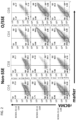
- certain recombinant receptors can exhibit antigen-independent activity or signaling (also known as "tonic signaling"), which could lead to undesirable effects, such as due to increased differentiation and/or exhaustion of T cells that express the recombinant receptor.
- antigen-independent activity or signaling also known as "tonic signaling”
- such activities may limit the T cell's activity, effect or potency.
- the cells may exhibit phenotypes indicative of exhaustion, due to tonic signaling through the recombinant receptor.
- properties of particular target antigens that the recombinant receptors specifically bind, recognize or target can that affect the activity of the receptor.
- B-cell maturation antigen BCMA
- BCMA B-cell maturation antigen
- sBCMA soluble BCMA
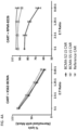
- the activity of the anti-BCMA chimeric antigen receptors can be blocked or inhibited by the presence of soluble BCMA.
- the disclosed embodiments are based on the observation that particular spacers and optimization of the nucleic acid sequences can lead to consistent and robust expression of the chimeric antigen receptor.
- the disclosed BCMA-binding chimeric antigen receptors offer advantages over available approaches for cell therapies, in particular, BCMA-targeting cell therapy.
- disclosed BCMA-binding chimeric antigen receptors are observed to exhibit reduced antigen-independent, tonic signaling and lack of inhibition by soluble BCMA.
- the disclosed BCMA-binding chimeric antigen recombinant receptors, polynucleotides encoding such receptors, engineered cells and cell compositions exhibit certain desired properties that can overcome or counteract certain limitations that can reduce optimal responses to cell therapy, for example, cell therapy with engineered cells expressing a BCMA-binding chimeric antigen receptor.
- compositions containing engineered cells expressing an exemplary BCMA-binding chimeric antigen receptor disclosed herein was observed to exhibit consistency of cell health of the engineered cells, and was associated with clinical response.
- the disclosed embodiments including the chimeric antigen receptors, polynucleotides encoding such receptors, engineered cells and cell compositions, can provide various advantages over available therapies targeting BCMA, to improve the activity of the chimeric antigen receptors and response to BCMA-targeting cell therapies.
- chimeric antigen receptors that bind or recognize BCMA molecules and polynucleotides encoding BCMA-binding chimeric antigen receptors (CARs), and cells expressing such receptors.
- CARs BCMA-binding chimeric antigen receptors
- the chimeric antigen receptors bind to an extracellular portion of BCMA.
- the polynucleotides are optimized, or contain certain features designed for optimization, such as for codon usage, to reduce RNA heterogeneity and/or to modify, e.g., increase or render more consistent among cell product lots, expression, such as surface expression, of the encoded receptor.
- polynucleotides, encoding BCMA-binding chimeric antigen receptors are modified as compared to a reference polynucleotide, such as to remove cryptic or hidden splice sites, to reduce RNA heterogeneity.
- polynucleotides, encoding BCMA-binding chimeric antigen receptors are codon optimized, such as for expression in a mammalian, e.g., human, cell such as in a human T cell.
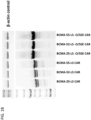
- the modified polynucleotides result in in improved, e.g., increased or more uniform or more consistent level of, expression, e.g., surface expression, when expressed in a cell.
- Such polynucleotides can be utilized in constructs for generation of engineered cells that express the encoded BCMA-binding chimeric antigen receptor.
- the disclosed polynucleotides are those that encode chimeric antigen receptors, that specifically recognize, such as specifically bind, BCMA.
- the encoded chimeric antigen receptorcontaining BCMA-binding polypeptides, and compositions and uses of the same, also are disclosed.
- the BCMA-binding polypeptides described herein are antibodies, such as single-chain antibodies (e.g., antigen binding antibody fragments), or portions thereof.
- the recombinant receptors are chimeric antigen receptors, such as those containing anti-BCMA antibodies or antigen-binding fragments thereof.
- an antigen binding fragment, in the disclosed CARs, that specifically recognizes an antigen, BCMA specifically binds to the antigen.
- the disclosed polynucleotides can be incorporated into constructs, such as deoxyribonucleic acid (DNA) or RNA constructs, such as those that can be introduced into cells for expression of the encoded recombinant BCMA-binding receptors.
- the polynucleotide encoding the BCMA-binding chimeric antigen receptor contains a signal sequence that encodes a signal peptide, in some cases encoded upstream of the nucleic acid sequences encoding the BCMA-binding chimeric antigen receptor, or joined at the 5' terminus of the nucleic acid sequences encoding the antigen-binding domain.
- the polynucleotide containing nucleic acid sequences encoding the BCMA-binding chimeric antigen receptor (CAR) contains a signal sequence that encodes a signal peptide.
- the signal sequence may encode a signal peptide derived from a native polypeptide.
- the signal sequence may encode a heterologous or non-native signal peptide.
- non-limiting exemplary signal peptide include a signal peptide of the IgG kappa chain set forth in SEQ ID NO: 620, or encoded by the nucleotide sequence set forth in SEQ ID NO: 619 or 682-685; a GMCSFR alpha chain set forth in SEQ ID NO:851 and encoded by the nucleotide sequence set forth in SEQ ID NO:850; a CD8 alpha signal peptide set forth in SEQ ID NO:852; or a CD33 signal peptide set forth in SEQ ID NO:853.
- the polynucleotide encoding the BCMA-binding receptor can contain nucleic acid sequence encoding additional molecules, such as a surrogate marker or other markers, or can contain additional components, such as promoters, regulatory elements and/or multicistronic elements.
- the nucleic acid sequence encoding the BCMA-binding receptor can be operably linked to any of the additional components.
- the disclosed BCMA-binding chimeric antigen receptors generally contain an extracellular binding molecule and an intracellular signaling domain.
- the chimeric antigen receptors disclosed herein specifically bind to or specifically recognize BCMA, such as chimeric antigen receptors containing the disclosed anti-BCMA antigen-binding fragments. Also disclosed are cells expressing the chimeric antigen receptors and uses thereof in adoptive cell therapy, such as treatment of diseases and disorders associated with BCMA expression.
- Exemplary antigen receptors including CARs, and methods for engineering and introducing such antigen receptors into cells, include those described, for example, in international patent application publication Nos. WO200014257 , WO2013126726 , WO2012/129514 , WO2014031687 , WO2013166321 , WO2013071154 , WO2013123061 U.S. patent application publication Nos. US2002131960 , US2013287748 , US20130149337 , U.S. Patent Nos.
- the antigen receptors include a CAR as described in U.S. Patent No. 7,446,190 , and those described in International Patent Application Publication No. WO2014055668 .
- Exemplary CARs include CARs as disclosed in any of the aforementioned publications, such as WO2014031687 , US 8,339,645 , US 7,446,179 , US 2013/0149337 , US 7,446,190 , and US 8,389,282 , and in which the antigen-binding portion, e.g., scFv, is replaced by an antibody or an antigen-binding fragment thereof, as disclosed herein.
- the antigen-binding portion e.g., scFv
- the disclosed CAR has an amino acid sequence selected from among SEQ ID NOs: 757-762, or exhibits at least or about at least 90%, 91%, 92%, 93%, 94%, 95%, 96%, 97%, 98% or 99% sequence identity to the amino acid sequence set forth in any of SEQ ID NOs 757-762.
- the disclosed CAR is encoded by a polynucleotide, such as an polynucleotide with the nucleic acid sequence set forth in any of SEQ ID NOs 751-756, or a sequences that exhibits at least or at least about 90%, 91%, 92%, 93%, 94%, 95%, 96%, 97%, 98% or 99% sequence identity to the nucleic acid sequence set forth in any of SEQ ID NOs: 751-756.
- a polynucleotide such as an polynucleotide with the nucleic acid sequence set forth in any of SEQ ID NOs 751-756, or a sequences that exhibits at least or at least about 90%, 91%, 92%, 93%, 94%, 95%, 96%, 97%, 98% or 99% sequence identity to the nucleic acid sequence set forth in any of SEQ ID NOs: 751-756.
- the disclosed CAR is encoded by a polynucleotide, such as an polynucleotide with the nucleic acid sequence set forth in any of SEQ ID NOs:755 and 756, or a sequences that exhibits at least or at least about 90%, 91%, 92%, 93%, 94%, 95%, 96%, 97%, 98% or 99% sequence identity to the nucleic acid sequence set forth in any of SEQ ID NOs: 755 and 756.
- a polynucleotide such as an polynucleotide with the nucleic acid sequence set forth in any of SEQ ID NOs:755 and 756, or a sequences that exhibits at least or at least about 90%, 91%, 92%, 93%, 94%, 95%, 96%, 97%, 98% or 99% sequence identity to the nucleic acid sequence set forth in any of SEQ ID NOs: 755 and 756.
- the disclosed CAR is encoded by a polynucleotide, such as an polynucleotide with the nucleic acid sequence set forth in SEQ ID NOs:755 or a sequences that exhibits at least or at least about 90%, 91%, 92%, 93%, 94%, 95%, 96%, 97%, 98% or 99% sequence identity thereto.
- the disclosed CAR is encoded by a polynucleotide, such as an polynucleotide with the nucleic acid sequence set forth in SEQ ID NOs:755.
- the nucleic acid encoding the antigen-binding domain comprises (a) the sequence of nucleotides set forth in any of SEQ ID NOS: 648, 330-352, 647, 716, or 718; (b) a sequence of nucleotides that has at least 90% sequence identity to any of SEQ ID NOS: 648, 330-352, 647, 716, or 718; or (c) a degenerate sequence of (a) or (b).
- the CARs generally include an extracellular antigen binding domain that includes, is, or is comprised within or comprises, one of the disclosed anti-BCMA antibodies.
- the CARs typically include in their extracellular portions one or more BCMA-binding molecules, such as one or more antigen-binding fragment, domain, or portion, or one or more antibody variable regions, and/or antibody molecules, such as those described herein.
- antibody herein is used in the broadest sense and includes polyclonal and monoclonal antibodies, including intact antibodies and functional (antigen-binding) antibody fragments, including fragment antigen binding (Fab) fragments, F(ab') 2 fragments, Fab' fragments, Fv fragments, recombinant IgG (rIgG) fragments, heavy chain variable (V H ) regions capable of specifically binding the antigen, single chain antibody fragments, including single chain variable fragments (scFv), and single domain antibodies (e.g., sdAb, sdFv, nanobody) fragments.
- Fab fragment antigen binding
- rIgG fragment antigen binding
- V H heavy chain variable regions capable of specifically binding the antigen
- single chain antibody fragments including single chain variable fragments (scFv), and single domain antibodies (e.g., sdAb, sdFv, nanobody) fragments.
- immunoglobulins such as intrabodies, peptibodies, chimeric antibodies, fully human antibodies, humanized antibodies, and heteroconjugate antibodies, multispecific, e.g., bispecific or trispecific, antibodies, diabodies, triabodies, and tetrabodies, tandem di-scFv, tandem tri-scFv.
- antibody should be understood to encompass functional antibody fragments thereof also referred to herein as "antigen-binding fragments.”
- the term also encompasses intact or full-length antibodies, including antibodies of any class or sub-class, including IgG and sub-classes thereof, IgM, IgE, IgA, and IgD.
- CDR complementarity determining region
- HVR hypervariable region
- FR-H1, FR-H2, FR-H3, and FR-H4 there are four FRs in each full-length heavy chain variable region (FR-H1, FR-H2, FR-H3, and FR-H4), and four FRs in each full-length light chain variable region (FR-L1, FR-L2, FR-L3, and FR-L4).
- the boundaries of a given CDR or FR may vary depending on the scheme used for identification.
- the Kabat scheme is based on structural alignments
- the Chothia scheme is based on structural information. Numbering for both the Kabat and Chothia schemes is based upon the most common antibody region sequence lengths, with insertions accommodated by insertion letters, for example, "30a,” and deletions appearing in some antibodies. The two schemes place certain insertions and deletions ("indels") at different positions, resulting in differential numbering.
- the Contact scheme is based on analysis of complex crystal structures and is similar in many respects to the Chothia numbering scheme.
- the AbM scheme is a compromise between Kabat and Chothia definitions based on that used by Oxford Molecular's AbM antibody modeling software.
- Table 1 lists exemplary position boundaries of CDR-L1, CDR-L2, CDR-L3 and CDR-H1, CDR-H2, CDR-H3 as identified by Kabat, Chothia, AbM, and Contact schemes, respectively.
- residue numbering is listed using both the Kabat and Chothia numbering schemes.
- FRs are located between CDRs, for example, with FR-L1 located before CDR-L1, FR-L2 located between CDR-L1 and CDR-L2, FR-L3 located between CDR-L2 and CDR-L3 and so forth.
- CDR complementary determining region
- individual specified CDRs e.g., CDR-H1, CDR-H2, CDR-H3
- CDR-H1, CDR-H2, CDR-H3 individual specified CDRs (e.g., CDR-H1, CDR-H2, CDR-H3), of a given antibody or region thereof, such as a variable region thereof, should be understood to encompass a (or the specific) complementary determining region as defined by any of the aforementioned schemes, or other known schemes.
- a particular CDR e.g., a CDR-H3
- a CDR-H3 contains the amino acid sequence of a corresponding CDR in a given V H or V L region amino acid sequence
- a CDR has a sequence of the corresponding CDR (e.g., CDR-H3) within the variable region, as defined by any of the aforementioned schemes, or other known schemes.
- specific CDR sequences are specified. Exemplary CDR sequences of disclosed antibodies are described using various numbering schemes, although it is understood that a disclosed antibody can include CDRs as described according to any of the other aforementioned numbering schemes or other numbering schemes known to a skilled artisan.
- a FR or individual specified FR(s) e.g., FR-H1, FR-H2, FR-H3, FR-H4
- FR-H1, FR-H2, FR-H3, FR-H4 FR-H1, FR-H2, FR-H3, FR-H4
- the scheme for identification of a particular CDR, FR, or FRs or CDRs is specified, such as the CDR as defined by the Kabat, Chothia, AbM or Contact method, or other known schemes.
- the particular amino acid sequence of a CDR or FR is given.
- variable region refers to the domain of an antibody heavy or light chain that is involved in binding the antibody to antigen.
- the variable regions of the heavy chain and light chain (V H and V L , respectively) of a native antibody generally have similar structures, with each domain comprising four conserved framework regions (FRs) and three CDRs.
- FRs conserved framework regions
- a single V H or V L domain may be sufficient to confer antigen-binding specificity.
- antibodies that bind a particular antigen may be isolated using a V H or V L domain from an antibody that binds the antigen to screen a library of complementary V L or V H domains, respectively. See, e.g., Portolano et al., J. Immunol. 150:880-887 (1993 ); Clarkson et al., Nature 352:624-628 (1991 ).
- antibody fragments refers to a molecule other than an intact antibody that comprises a portion of an intact antibody that binds the antigen to which the intact antibody binds.
- antibody fragments include but are not limited to Fv, Fab, Fab', Fab'-SH, F(ab') 2 ; diabodies; linear antibodies; heavy chain variable (V H ) regions, single-chain antibody molecules such as scFvs and single-domain antibodies comprising only the V H region; and multispecific antibodies formed from antibody fragments.
- the antigen-binding domain in the disclosed CARs is or comprises an antibody fragment comprising a variable heavy chain (V H ) and a variable light chain (V L ) region.
- the antibodies are single-chain antibody fragments comprising a heavy chain variable (V H ) region and/or a light chain variable (V L ) region, such as scFvs.
- Single-domain antibodies are antibody fragments comprising all or a portion of the heavy chain variable region or all or a portion of the light chain variable region of an antibody.
- a single-domain antibody is a human single-domain antibody.
- Antibody fragments can be made by various techniques, including but not limited to proteolytic digestion of an intact antibody as well as production by recombinant host cells.
- the antibodies are recombinantly-produced fragments, such as fragments comprising arrangements that do not occur naturally, such as those with two or more antibody regions or chains joined by synthetic linkers, e.g., peptide linkers, and/or that are may not be produced by enzyme digestion of a naturally-occurring intact antibody.
- the antibody fragments are scFvs.
- a “humanized” antibody is an antibody in which all or substantially all CDR amino acid residues are derived from non-human CDRs and all or substantially all FR amino acid residues are derived from human FRs.
- a humanized antibody optionally may include at least a portion of an antibody constant region derived from a human antibody.
- a "humanized form" of a non-human antibody refers to a variant of the non-human antibody that has undergone humanization, typically to reduce immunogenicity to humans, while retaining the specificity and affinity of the parental non-human antibody.
- some FR residues in a humanized antibody are substituted with corresponding residues from a non-human antibody (e.g., the antibody from which the CDR residues are derived), e.g., to restore or improve antibody specificity or affinity.
- a non-human antibody e.g., the antibody from which the CDR residues are derived
- human antibodies are human antibodies.
- a "human antibody” is an antibody with an amino acid sequence corresponding to that of an antibody produced by a human or a human cell, or non-human source that utilizes human antibody repertoires or other human antibody-encoding sequences, including human antibody libraries.
- the term excludes humanized forms of non-human antibodies comprising non-human antigen-binding regions, such as those in which all or substantially all CDRs are non-human.
- the term includes antigen-binding fragments of human antibodies.
- Human antibodies may be prepared by administering an immunogen to a transgenic animal that has been modified to produce intact human antibodies or intact antibodies with human variable regions in response to antigenic challenge. Such animals typically contain all or a portion of the human immunoglobulin loci, which replace the endogenous immunoglobulin loci, or which are present extrachromosomally or integrated randomly into the animal's chromosomes. In such transgenic animals, the endogenous immunoglobulin loci have generally been inactivated. Human antibodies also may be derived from human antibody libraries, including phage display and cell-free libraries, containing antibody-encoding sequences derived from a human repertoire.
- the term “monoclonal antibody” as used herein refers to an antibody obtained from or within a population of substantially homogeneous antibodies, i.e ., the individual antibodies comprising the population are identical, except for possible variants containing naturally occurring mutations or arising during production of a monoclonal antibody preparation, such variants generally being present in minor amounts.
- polyclonal antibody preparations which typically include different antibodies directed against different epitopes
- each monoclonal antibody of a monoclonal antibody preparation is directed against a single epitope on an antigen.
- a monoclonal antibody may be made by a variety of techniques, including but not limited to generation from a hybridoma, recombinant DNA methods, phage-display and other antibody display methods.
- the CAR includes a BCMA-binding portion or portions of the antibody molecule, such as a heavy chain variable (V H ) region and/or light chain variable (V L ) region of the antibody, e.g ., an scFv antibody fragment.
- the disclosed BCMA-binding CARs contain an antibody, such as an anti-BCMA antibody, or an antigen-binding fragment thereof that confers the BCMA-binding properties of the disclosed CAR.
- the antibody or antigen-binding domain can be any anti-BCMA antibody described or derived from any anti-BCMA antibody described.
- the anti-BCMA CAR contains an antigen-binding domain that is an scFv containing a variable heavy (V H ) and/or a variable light (V L ) region derived from an antibody described in WO 2016090320 or WO2016090327 .
- the anti-BCMA antibody or antigen-binding fragment contains a heavy and/or light chain variable (V H or V L ) region sequence as described, or a sufficient antigen-binding portion thereof.
- the anti-BCMA antibody, e.g ., antigen-binding fragment contains a V H region sequence or sufficient antigen-binding portion thereof that contains a CDR-H1, CDR-H2 and/or CDR-H3 as described.
- the anti-BCMA antibody, e.g., antigen-binding fragment contains a V L region sequence or sufficient antigen-binding portion that contains a CDR-L1, CDR-L2 and/or CDR-L3 as described.
- the anti-BCMA antibody e.g., antigen-binding fragment
- the antibodies are those having sequences at least at or about 90%, about 91%, about 92%, about 93%, about 94%, about 95%, about 96%, about 97%, about 98%, or about 99% identical to such a sequence.
- the antibody e.g., antigen-binding fragment thereof, in the disclosed CAR, has a heavy chain variable (V H ) region having the amino acid sequence selected from any one of SEQ ID NOs: 110-115, 247-256, 324, 325, 518-531, 533, 609, 617, and 772-774, and 814-832, or an amino acid sequence that has at least 90%, 91%, 92%, 93%, 94%, 95%, 96%, 97%, 98%, or 99% sequence identity to the V H region amino acid selected from any one of SEQ ID NOs: 110-115, 247-256, 324, 325, 518-531, 533, 609, 617, 772-774, and 814-832, or contains a CDR-H1, CDR-H2, and/or CDR-H3 present in such a V H sequence.
- the antibody or antibody fragment, in the disclosed CAR has a V H region of any of the antibodies or
- the V H region of the anti-BCMA antibody is one that includes a heavy chain complementarity determining region 3 (CDR-H3) comprising the amino acid sequence X 1 X 2 X 3 X 4 X 5 X 6 X 7 X 8 X 9 X 10 X 11 X 12 X 13 X 14 (SEQ ID NO:355), wherein X 1 is A, D, E, G, L, V or W; X 2 is A, D, G, L, P, Q or S; X 3 is A, D, G, L or Y; X 4 is D, G, P, R, S, V, Y or null; X 5 is D, I, P, S, T, Y or null; X 6 is A, G, I, S, T, V, Y or null; X 7 is A, D, E, F, L, P, S, Y or null; X 8 is P, Q, T, Y or null; X 9 is
- the antibody or antigen-binding fragment thereof comprises a CDR-H3 comprising the amino acid sequence selected from any one of SEQ ID NOs:7-11, 149-157, 279-287, 292, 293, 376-378, 517, 595, according to Kabat numbering.
- the V H region of an antibody or antigen-binding fragment thereof contains a CDR-H3 having the amino acid sequence comprising the amino acid sequence selected from any one of SEQ ID NOs:7-11, 149-157, 279-287, 292, 293, 376-378, 517, and 595 according to Chothia numbering or AbM numbering.
- the VH region of an antibody or antigen-binding fragment thereof contains a CDR-H3 having the amino acid sequence comprising the amino acid sequence selected from SEQ ID NOs: 606 and 613.
- the antibody or antigen-binding fragment thereof contains a CDR-H3 having the amino acid sequence of SEQ ID NO: 517, 595, 606, or 613.
- the antibody or antigen-binding fragment thereof can contain a V H region sequence selected from any one of SEQ ID NOs: 110-115, 247-256, 324, 325, 518-531, 533, 609, 617, 772-774, and 814-832 in which the corresponding CDR-H3 sequence contained therein (e.g.
- the V H region of an antibody or antigen-binding fragment thereof comprises a CDR-H3 contained within the V H region amino acid sequence selected from any one of SEQ ID NOs: 110-115, 247-256, 324, 325, 518-531, 533, 609, 617, 772-774, and 814-832.
- the V H region of the antibody or antigen-binding fragment thereof is one that includes a heavy chain complementarity determining region 1 (CDR-H1) comprising the amino acid sequence of X 1 X 2 X 3 MX 4 (SEQ ID NO:353)
- X 1 is D or S
- X 2 is Y or S
- X 3 is A, G, W, or Y
- X 4 is H, Q, or S.
- CDR-H1 X 1 is D; X 2 is Y; X 3 is Y; and X 4 is S.
- the V H region of an antibody or antigen-binding fragment thereof contains a CDR-H1 having the amino acid sequence comprising the amino acid sequence selected from any one of SEQ ID NOs: 1-3, 140-144, 288, 289, 507, and 593 according to Kabat numbering. In some embodiments, the V H region of an antibody or antigen-binding fragment thereof contains a CDR-H1 having the amino acid sequence comprising the amino acid sequence selected from any one of SEQ ID NOs: 12-15, 158-160, 294, 295, 532, and 596 according to Chothia numbering.
- the V H region of an antibody or antigen-binding fragment thereof contains a CDR-H1 having the amino acid sequence comprising the amino acid sequence selected from any one of SEQ ID NOs: 19-22, 165-169, 298, 299, 509, 577, and 598 according to AbM numbering. In some embodiments, the V H region of an antibody or antigen-binding fragment thereof contains a CDR-H1 having the amino acid sequence comprising the amino acid sequence selected from any one of SEQ ID NOs 604, and 611.
- the V H region of an antibody or antigen-binding fragment thereof contains a CDR-H1 having the amino acid sequence of SEQ ID NO:507, 532, 577, 593, 596, 598, 604, and 611.
- the antibody or antigen-binding fragment thereof can contain a V H region sequence selected from any one of SEQ ID NOs: 110-115, 247-256, 324, 325, 518-531, 533, 609, 617, 772-774, and 814-832 in which the corresponding CDR-H1 sequence contained therein (e.g.
- the V H region of an antibody or antigen-binding fragment thereof contains a CDR-H1 contained within the V H region amino acid sequence selected from any one of SEQ ID NOs: 110-115, 247-256, 324, 325, 518-531, 533, 609, 617, 772-774, and 814-832.
- the V H region of an antibody or antigen-binding fragment thereof is one that includes a heavy chain complementarity determining region 2 (CDR-H2) comprising the amino acid sequence of X 1 IX 2 X 3 X 4 X 5 X 6 X 7 X 8 X 9 X 10 X 11 YX 12 X 13 X 14 X 15 X 16 X 17 (SEQ ID NO:354), wherein X 1 is F, G, H, V, W or Y; X 2 is N, R, S or V; X 3 is P, Q, S, V, W or Y; X 4 is K or null; X 5 is A or null; X 6 is D, G, N, S, or Y; X 7 is G or S; X 8 is G or S; X 9 is E, G, N, T or S; X 10 is I, K, or T; X 11 is E, G, N or Y; X 12 is A or V; X
- the V H region of an antibody or antigen-binding fragment thereof contains a CDR-H2 comprising the amino acid sequence selected from any one of SEQ ID NOs: 4-6, 145-148, 290, 291, 372-374, 513, and 594 according to Kabat numbering. In some embodiments, the V H region of an antibody or antigen-binding fragment thereof contains a CDR-H2 comprising the amino acid sequence selected from any one of SEQ ID NOs: 16-18, 161-164, 296, 297, 514-516, 551, 597 according to Chothia numbering.
- the V H region of an antibody or antigen-binding fragment thereof contains a CDR-H2 comprising the amino acid sequence selected from any one of SEQ ID NOs: 23-25, 170-173, 300, 301, 510-512, 587, and 599 according to AbM numbering. In some embodiments, the V H region of an antibody or antigen-binding fragment thereof contains a CDR-H2 comprising the amino acid sequence selected from any one of SEQ ID NOs: 605 and 612. In some embodiments, the V H region of an antibody or antigen-binding fragment thereof contains a CDR-H2 having the amino acid sequence of any of SEQ ID NOs: 513, 551, 587, 594, 597, 599, 605, or 612.
- the antibody or antigen-binding fragment thereof can contain a V H region sequence selected from any one of SEQ ID NOs: 110-115, 247-256, 324, 325, 518-531, 533, 609, 617, 772-774, and 814-832 in which the corresponding CDR-H2 sequence contained therein (e.g.
- the V H region of an antibody or antigen-binding fragment thereof contains a CDR-H2 contained within the V H region amino acid sequence selected from any one of SEQ ID NOs: 110-115, 247-256, 324, 325, 518-531, 533, 609, 617, 772-774, and 814-832.
- the antibody or antigen-binding fragment thereof contains a CDR-H1 that is or comprises the amino acid sequence selected from any one of SEQ ID NOs: 1-3, 140-144, 288, 289, 507, and 593 according to Kabat numbering; a CDR-H2 that is or comprises the amino acid sequence selected from any one of SEQ ID NOs: 4-6, 145-148, 290, 291, 372-374, 513, and 594 according to Kabat numbering; and a CDR-H3 that is or comprises the amino acid sequence selected from any one of SEQ ID NOs: 7-11, 149-157, 279-287, 292, 293, 376-378, 517, and 595 according to Kabat numbering.
- the antibody or antigen-binding fragment thereof contains a CDR-H1 that is or comprises the amino acid sequence selected from any one of SEQ ID NOs: 12-15, 158-160, 294, 295, 532, and 596 according to Chothia numbering; a CDR-H2 that is or comprises the amino acid sequence selected from any one of SEQ ID NOs: 16-18, 161-164, 296, 297, 514-516, 551, 597 according to Chothia numbering; and a CDR-H3 that is or comprises the amino acid sequence selected from any one of SEQ ID NOs: 7-11, 149-157, 279-287, 292, 293, 376-378, 517, and 595 according to Chothia numbering.
- the antibody or antigen-binding fragment thereof contains a CDR-H1 that is or comprises the amino acid sequence selected from any one of SEQ ID NO: 19-22, 165-169, 509, 298, 299, 509, 577, and 598 according to AbM numbering; a CDR-H2 that is or comprises the amino acid sequence selected from any one of SEQ ID NOs:23-25, 170-173, 300, 201, 510-512, 587, and 599 according to AbM numbering; and a CDR-H3 that is or comprises the amino acid sequence selected from any one of SEQ ID NOs:7-11, 149-157, 279-287, 292, 293, 376-378, 517, 595, 606, and 613 according to AbM numbering.
- a CDR-H1 that is or comprises the amino acid sequence selected from any one of SEQ ID NO: 19-22, 165-169, 509, 298, 299, 509, 577, and 598 according to AbM numbering
- the antibody or antigen-binding fragment thereof contains a CDR-H1 that is or comprises the amino acid sequence selected from any one of SEQ ID NO:604 and 611; a CDR-H2 that is or comprises the amino acid sequence selected from any one of SEQ ID NOs:605 and 612; and a CDR-H3 that is or comprises the amino acid sequence selected from any one of SEQ ID NOs:606 and 613.
- the V H region of an antibody or antigen-binding fragment thereof comprises a CDR-H1, CDR-H2, and/or CDR-H3 according to Kabat numbering. In some embodiments, the V H region of an antibody or antigen-binding fragment thereof comprises a CDR-H1, CDR-H2, and/or CDR-H3 according to Chothia numbering. In some embodiments, the V H region of an antibody or antigen-binding fragment thereof comprises a CDR-H1, CDR-H2, and/or CDR-H3 according to AbM numbering.
- the antibody or antigen-binding fragment thereof disclosed herein comprises a V H region comprising a CDR-H1, CDR-H2, and CDR-H3 comprising the amino acid sequence selected from among: SEQ ID NOs:1, 4, and 7; SEQ ID NOs:2, 5, and 8; SEQ ID NOs:2, 5, and 9; SEQ ID NOs:2, 5, and 10; SEQ ID NOs:3, 6, and 11; SEQ ID NOs: 140, 145, and 149; SEQ ID NOs: 141, 145, and 149; SEQ ID NOs: 141, 145, and 150; SEQ ID NOs:142, 146, and 151; SEQ ID NOs:2, 5, and 152; SEQ ID NOs:143, 147, and 153; SEQ ID NOs:144, 148, and 154; SEQ ID NOs:3, 6, and 155; SEQ ID NOs:2, 5, and 156; SEQ ID NOs:2, 5, and 157; SEQ ID NOs:2, 6, and 157
- the antibody or antigen-binding fragment thereof comprises a CDR-H1, CDR-H2 and CDR-H3, respectively, comprising the amino acid sequence of a CDR-H1, a CDR-H2, and a CDR-H3 contained within the V H region amino acid sequence selected from any one of SEQ ID NOs: 110-115, 247-256, 324, 325, 518-531, 533, 609, 617, 772-774, and 814-832.
- the antibody or antigen-binding fragment thereof comprises a CDR-H1, CDR-H2 and CDR-H3, respectively, comprising the amino acid sequence of a CDR-H1, a CDR-H2, and a CDR-H3 contained within the V H region amino acid sequence of SEQ ID NO:609 or SEQ ID NO: 617.
- the V H region comprises any of the CDR-H1, CDR-H2 and CDR-H3 as described and comprises a framework region 1 (FR1), a FR2, a FR3 and/or a FR4 having at least 90%, 91%, 92%, 93%, 94%, 95%, 96%, 97%, 98%, or 99% sequence identity, respectively, to a FR1, a FR2, a FR3 and/or a FR4 contained within the V H region amino acid sequence selected from any one of SEQ ID NOs: 110-115, 247-256, 324, 325, 518-531, 533, 609, 617, 772-774, and 814-832.
- the anti-BCMA antibody or antigen-binding fragment thereof can comprise a CDR-H1, CDR-H2 and CDR-H3, respectively, contained within the V H region amino acid sequence selected from any one of SEQ ID NOs: 110-115, 247-256, 324, 325, 518-531, 533, 609, 617, 772-774, and 814-832, and a framework region (e.g., a FR1, a FR2, a FR3 and/or a FR4) that contains at least 90%, 91%, 92%, 93%, 94%, 95%, 96%, 97%, 98%, 99% sequence identity to a framework region (e.g., a FR1, a FR2, a FR3 and/or a FR4) contained within the V H region amino acid sequence selected from any one of SEQ ID NOs: 110-115, 247-256, 324, 325, 518-531, 533, 609, 617, 772-774, and 8
- the V H region comprises a FR1, a FR2, a FR3 and/or a FR4 selected from a FR1 comprising the amino acid sequence selected from any one of SEQ ID NOs:59-63, 195-203, 308, 309, and 434-439; a FR2 comprising the amino acid sequence selected from any one of SEQ ID NOs:64-66, 204-209, 310, and 311; a FR3 comprising the amino acid sequence selected from any one of SEQ ID NOs:67-69, 210-216, 312, 313, 441 and 443; and/or a FR4 comprising the amino acid sequence selected from any one of SEQ ID NOs:70-71, 217-220, 314, 315, 444 and 445.
- the V H region comprises a FR1 comprising the amino acid sequence of SEQ ID NO:61, a FR2 comprising the amino acid sequence of SEQ ID NO:65, a FR3 comprising the amino acid sequence of SEQ ID NO:69, and/or a FR4 comprising the amino acid of SEQ ID NO:70.
- the antibody or antigen-binding fragment thereof comprises a V H region comprising the amino acid sequence selected from any one of SEQ ID NOs: 110-115, 247-256, 324, 325, 518-531, 533, 609, 617, 772-774, and 814-832.
- antibodies and antigen-binding fragments thereof having sequences at least at or about at least 90%, 91%, 92%, 93%, 94%, 95%, 96%, 97%, 98%, or 99% identical to such sequences.
- an antibody or antigen-binding fragment comprising a V H region comprising an amino acid sequence having at least 90%, 91%, 92%, 93%, 94%, 95%, 96%, 97%, 98%, or 99% sequence identity to a V H region amino acid sequence selected from any one of SEQ ID NOs: 110-115, 247-256, 324, 325, 518-531, 533, 609, 617, 772-774, and 814-832.
- the antibody is a single domain antibody (sdAb) comprising only a V H region sequence or a sufficient antigen-binding portion thereof, such as any of the above described V H sequences (e.g., a CDR-H1, a CDR-H2, a CDR-H3 and/or a CDR-H4).
- sdAb single domain antibody
- an anti-BCMA antibody or antigen-binding fragment thereof comprising a V H region further comprises a light chain or a sufficient antigen binding portion thereof.
- the antibody or antigen-binding fragment thereof contains a V H region and a V L region, or a sufficient antigen-binding portion of a V H and V L region.
- a V H region sequence can be any of the above described V H sequence.
- the antibody is an antigen-binding fragment, such as a Fab or an scFv.
- the antibody is a full-length antibody that also contains a constant region.
- the antibody e.g., antigen-binding fragment thereof, has a light chain variable (V L ) region having the amino acid sequence selected from any one of SEQ ID NOs: 116-127, 257-267, 326, 327, 534-550, 552-557, 610, 618, 775-777, and 833-849, or an amino acid sequence that has at least 90%, 91%, 92%, 93%, 94%, 95%, 96%, 97%, 98%, or 99% sequence identity to a V L region amino acid sequence selected from any one of SEQ ID NOs: 116-127, 257-267, 326, 327, 534-550, 552-557, 610, 618, 775-777, and 833-849.
- the antibody or antigen-binding fragment has a VL region described in any of WO 2016090327 , WO 2016090320 , or WO 2017173256
- the antibody or antigen-binding fragment thereof contains a CDR-L3 comprising the amino acid sequence selected from any one of SEQ ID NOs:47-58, 184-194, 306, 307, 415-427, 429-433, 591 and 603 according to Kabat numbering, Chothia numbering or AbM numbering.
- the antibody or antigen-binding fragment thereof contains a CDR-L3 having the amino acid sequence of SEQ ID NO:591 or 603 according to Kabat numbering, Chothia numbering or AbM numbering.
- the antibody or antigen-binding fragment thereof can contain a V L region sequence selected from any one of SEQ ID NOs: 116-127, 257-267, 326, 327, 534-550, 552-557, 610, 618, 775-777, and 833-849 in which the corresponding CDR-L3 sequence contained therein (e.g. corresponding to amino acid residues L89 to L97 by Kabat numbering) is replaced by the CDR-L3 sequence selected from any one of SEQ ID NOs: 47-58, 184-194, 306, 307, 415-427, 429-433, 591 and 603 according to Kabat numbering, Chothia numbering or AbM numbering.
- V L region sequence selected from any one of SEQ ID NOs: 116-127, 257-267, 326, 327, 534-550, 552-557, 610, 618, 775-777, and 833-849 in which the corresponding CDR-L3 sequence contained therein (e.g. corresponding to
- the V L region of the anti-BCMA antibody or antigen-binding fragment thereof is one that includes a light chain complementarity determining region 1 (CDR-L1) that contains the amino acid sequence: X 1 X 2 X 3 X 4 X 5 X 6 X 7 X 8 X 9 X 10 X 11 X 12 X 13 X 14 X 15 X 16 X 17 (SEQ ID NO:356), wherein X 1 is G, K, R, S or T; X 2 is A, G or S; X 3 is G, N, S or T; X 4 is G, K, N, Q, R or S; X 5 is S or null; X 6 is D, N, V or null; X 7 is L, V or null; X 8 is H, S, Y or null; X 9 is S, T or null; X 10 is S or null; X 11 is D, G, I, N, S or null; X 12 is D
- the antibody or antigen-binding fragment thereof contains a CDR-L1 comprising the amino acid sequence selected from any one of SEQ ID NOs: 26-36, 174-178, 302, 303, 380-392, 394-398, 589 or 601 according to Kabat numbering, Chothia numbering or AbM numbering.
- the antibody or antigen-binding fragment thereof contains a CDR-L1 comprising the amino acid sequence selected from any one of SEQ ID NOs: 607 and 614.
- the antibody or antigen-binding fragment thereof contains a CDR-L1 having the amino acid sequence of SEQ ID NO:589 or 601 according to Kabat numbering, Chothia numbering or AbM numbering.
- the antibody or antigen-binding fragment thereof can contain a V L region sequence selected from any one of SEQ ID NOs: 116-127, 257-267, 326, 327, 534-550, 552-557, 610, 618, 775-777, and 833-849 in which the corresponding CDR-L1 sequence contained therein (e.g. corresponding to amino acid residues L24 to L34 by Kabat numbering) is replaced by the CDR-L1 sequence selected from any one of SEQ ID NOs: 26-36, 174-178, 302, 303, 380-392, 394-398, 589 or 601 according to Kabat numbering, Chothia numbering or AbM numbering.
- a V L region sequence selected from any one of SEQ ID NOs: 116-127, 257-267, 326, 327, 534-550, 552-557, 610, 618, 775-777, and 833-849 in which the corresponding CDR-L1 sequence contained therein (e.g.
- the V L region of an antibody or antigen-binding fragment thereof comprises a CDR-L1 contained within the V L region amino acid sequence selected from any one of SEQ ID NOs: 116-127, 257-267, 326, 327, 534-550, 552-557, 610, 618, 775-777, and 833-849.
- the V L region of an antibody or antigen-binding fragment thereof comprises a CDR-L1 contained within the V L region amino acid sequence of SEQ ID NO:589, 601, 607 or 614.
- the V L region of the anti-BCMA antibody or antigen-binding fragment thereof is one that includes a light chain complementarity determining region 2 (CDR-L2) that contains the amino acid sequence of X 1 X 2 X 3 X 4 X 5 X 6 X 7 (SEQ ID NO:357), wherein X 1 is A, D, E, N, S, V or W; X 2 is A, D, N, S or V; X 3 is A, D, H, I, N or S; X 4 is D, K, N, Q, R or T; X 5 is L, R or V; X 6 is A, E, P or Q; and X 7 is A, D, S or T.
- CDR-L2 light chain complementarity determining region 2
- X 1 is A, D, E, N, S, V or W
- X 2 is A, D, N, S or V
- X 3 is A, D, H, I, N or S
- X 4 is D, K
- the antibody or antigen-binding fragment thereof contains a CDR-L2 comprising the amino acid sequence selected from any one of SEQ ID NOs:37-46, 179-183, 304, 305, 399-409, 411-414, 590 and 602 according to Kabat numbering, Chothia numbering or AbM numbering. In some embodiments, the antibody or antigen-binding fragment thereof contains a CDR-L2 comprising the amino acid sequence selected from any one of SEQ ID NOs: 608 and 615.
- the antibody or antigen-binding fragment thereof contains a CDR-L2 having the amino acid sequence of SEQ ID NO:590 or SEQ ID NO: 602 according to Kabat numbering, Chothia numbering or AbM numbering.
- the antibody or antigen-binding fragment thereof can contain a V L region sequence selected from any one of SEQ ID NOs: 116-127, 257-267, 326, 327, 534-550, 552-557, 610, 618, 775-777, and 833-849 in which the corresponding CDR-L2 sequence contained therein (e.g.
- the V L region of an antibody or antigen-binding fragment thereof comprises a CDR-L2 contained within the V L region amino acid sequence selected from any one of SEQ ID NOs: 116-127, 257-267, 326, 327, 534-550, 552-557, 610, 618, 775-777, and 833-849.
- the V L region of an antibody or antigen-binding fragment thereof comprises a CDR-L2 contained within the V L region amino acid sequence of SEQ ID NO: 589, 601, 607 or 614.
- the antibody or antigen-binding fragment thereof contains a CDR-L1 that is or comprises the amino acid sequence selected from any one of SEQ ID NOs: 26-36, 174-178, 302, 303, 380-392, 394-398, 589 or 601 according to Kabat numbering, Chothia numbering or AbM numbering; a CDR-L2 that is or comprises the amino acid sequence selected from any one of SEQ ID NOs: 37-46, 179-183, 304, 305, 399-409, 411-414, 590 and 602 according to Kabat numbering, Chothia numbering or AbM numbering; and a CDR-L3 that is or comprises the amino acid sequence selected from any one of SEQ ID NOs: 47-58, 184-194, 306, 307, 415-427, 429-433, 591 and 603 according to Kabat numbering, Chothia numbering or AbM numbering.
- the V L region of an antibody or antigen-binding fragment thereof comprises a CDR-L1, CDR-L2, and/or CDR-L3 according to Kabat numbering. In some embodiments, the V L region of an antibody or antigen-binding fragment thereof comprises a CDR-L1, CDR-L2, and/or CDR-L3 according to Chothia numbering. In some embodiments, the V L region of an antibody or antigen-binding fragment thereof comprises a CDR-L1, CDR-L2, and/or CDR-L3 according to AbM numbering.
- the V L region comprises a CDR-L1, a CDR-L2, and a CDR-L3 selected from among: a CDR-L1, CDR-L2, and CDR-L3 comprising the amino acid sequence of SEQ ID NOs:26, 37, and 47, respectively; a CDR-L1, CDR-L2, and CDR-L3 comprising the amino acid sequence of SEQ ID NOs:27, 38, and 48, respectively; a CDR-L1, CDR-L2, and CDR-L3 comprising the amino acid sequence of SEQ ID NOs:28, 39, and 49, respectively; a CDR-L1, CDR-L2, and CDR-L3 comprising the amino acid sequence of SEQ ID NOs:29, 40, and 50, respectively; a CDR-L1, CDR-L2, and CDR-L3 comprising the amino acid sequence of SEQ ID NOs:30, 39, and 51, respectively; a CDR-L1, CDR-L2, and CDR-L3 comprising the amino acid
- the V L region comprises a CDR-L1, CDR-L2, and CDR-L3 comprising the amino acid sequences of SEQ ID NOs:589, 590, and 591, respectively; SEQ ID NOs:607, 608, and 591, respectively; SEQ ID NOs: 601, 602, and 603, respectively; or SEQ ID NOs:614, 615, and 603, respectively.
- the antibody or antigen-binding fragment thereof disclosed herein comprises an V L region comprising a CDR-L1, CDR-L2, and CDR-L3 comprising the amino acid sequence selected from among: SEQ ID NOs:26, 37, and 47; SEQ ID NOs:27, 38, and 48; SEQ ID NOs:28, 39, and 49; SEQ ID NOs:29, 40, and 50; SEQ ID NOs:30, 39, and 51; SEQ ID NOs:31, 41, and 52; SEQ ID NOs:32, 42, and 53; SEQ ID NOs:30, 39, and 54; SEQ ID NOs:33, 43, and 55; SEQ ID NOs:34, 44, and 56; SEQ ID NOs:35, 45, and 57; SEQ ID NOs:36, 46, and 58; SEQ ID NOs:174, 179, and 184; SEQ ID NOs:174, 179, and 185; SEQ ID NOs:174, 179, and 186; SEQ ID
- the antibody or antigen-binding fragment thereof contains a CDR-L1, CDR-L2, and CDR-L3, respectively, contained within the V L region amino acid sequence selected from any one of SEQ ID NOs: 116-127, 257-267, 326, 327, 534-550, 552-557, 610, 618, 775-777, and 833-849.
- the antibody contains a CDR-L1, CDR-L2, and CDR-L3, respectively, contained within the V L region amino acid sequence selected of SEQ ID NO: 610 or SEQ ID NO: 618.
- the antibody or antigen-binding fragment thereof comprises a V L region that comprises a CDR-L1, CDR-L2, and CDR-L3 comprising the amino acid sequence of SEQ ID NOS:601, 602, and 603, respectively; or SEQ ID NOS: 614, 615, and 603, respectively.
- the V L region comprises any of the CDR-L1, CDR-L2 and CDR-L3 as described and comprises a framework region 1 (FR1), a FR2, a FR3 and/or a FR4 having at least 90%, 91%, 92%, 93%, 94%, 95%, 96%, 97%, 98%, or 99% sequence identity, respectively, to a FR1, a FR2, a FR3 and/or a FR4 contained within the V L region amino acid sequence selected from any one of SEQ ID NOs: 116-127, 257-267, 326, 327, 534-550, 552-557, 610, 618, 775-777, and 833-849.
- the anti-BCMA antibody or antigen-binding fragment thereof can comprise a CDR-L1, CDR-L2 and CDR-L3, respectively, contained within the V L region amino acid sequence selected from any one of SEQ ID NOs: 116-127, 257-267, 326, 327, 534-550, 552-557, 610, 618, 775-777, and 833-849, and a framework region (e.g., a FR1, a FR2, a FR3 and/or a FR4) that contains at least 90%, 91%, 92%, 93%, 94%, 95%, 96%, 97%, 98%, 99% sequence identity to a framework region (e.g., a FR1, a FR2, a FR3 and/or a FR4) contained within the V L region amino acid sequence selected from any one of SEQ ID NOs: 116-127, 257-267, 326, 327, 534-550, 552-557, 610, 618, 775-7
- the V L region comprises a FR1, a FR2, a FR3 and/or a FR4 selected from a FR1 comprising the amino acid sequence selected from any one of SEQ ID NOs:72-82, 221-227, 316, 317, 446-459 and 461-466; a FR2 comprising the amino acid sequence selected from any one of SEQ ID NOs:83-92, 228-232, 318, 319, 467-477 and 479-482; a FR3 comprising the amino acid sequence selected from any one of SEQ ID NOs:93-101, 233-242, 320, 321, 483-495 and 497-501; and/or a FR4 comprising the amino acid sequence selected from any one of SEQ ID NOs:102-109, 243-246, 322, 323, 502-506 and 508.
- the V L region comprises a FR1 comprising the amino acid sequence of SEQ ID NO:79, a FR2 comprising the amino acid sequence of SEQ ID NO:89, a FR3 comprising the amino acid sequence of SEQ ID NO:98, and/or a FR4 comprising the amino acid sequence of SEQ ID NO:108.
- the antibody or antigen-binding fragment thereof comprises a V L region comprising an amino acid sequence selected from any one of SEQ ID NOs: 116-127, 257-267, 326, 327, 534-550, 552-557, 610, 618, 775-777, and 833-849.
- the antibody or antigen-binding fragment thereof contains a V L region comprises the amino acid sequence of SEQ ID NO: 610 or SEQ ID NO: 618.
- antibodies having sequences at least at or about at least 90%, 91%, 92%, 93%, 94%, 95%, 96%, 97%, 98%, or 99% identical to such sequences.
- the V H region of the antibody or fragment comprises the amino acid sequence selected from any one of SEQ ID NOs: 110-115, 247-256, 324, 325, 518-531, 533, 609, 617, 772-774, and 814-832 and the V L region of the antibody or fragment comprises the amino acid sequence selected from any one of SEQ ID NOs: 116-127, 257-267, 326, 327, 534-550, 552-557, 610, 618, 775-777, and 833-849.
- antibodies and antigen-binding fragments thereof having sequences at least at or about at least 90%, 91%, 92%, 93%, 94%, 95%, 96%, 97%, 98%, or 99% identical to such sequences.
- an antibody or antigen-binding fragment containing a V L region comprising an amino acid sequence having at least 90%, 91%, 92%, 93%, 94%, 95%, 96%, 97%, 98%, or 99% sequence identity to a V L region amino acid sequence selected from any one of SEQ ID NOs: 116-127, 257-267, 326, 327, 534-550, 552-557, 610, 618, 775-777, and 833-849 and/or comprising an amino acid sequence having at least 90%, 91%, 92%, 93%, 94%, 95%, 96%, 97%, 98%, or 99% sequence identity to a V H region amino acid sequence selected from any one of SEQ ID NOs: 110-115, 247-256, 3
- the antibody or antigen-binding fragment contains a V L region comprising the amino acid sequence selected from any one of SEQ ID NOs: 116-127, 257-267, 326, 327, 534-550, 552-557, 610, 618, 775-777, and 833-849 and a V H region the amino acid sequence selected from any one of SEQ ID NOs: 110-115, 247-256, 324, 325, 518-531, 533, 609, 617, 772-774, and 814-832.
- the V H region is or comprises an amino acid sequence having at least 90%, 91%, 92%, 93%, 94%, 95%, 96%, 97%, 98% or 99% sequence identity to the V H region sequence of any of SEQ ID NOs:617, 110-115, 247-256, 324, 325, 518-531, 533, 609, 772-774, or 814-832; and the V L region is or comprises an amino acid sequence having at least 90%, 91%, 92%, 93%, 94%, 95%, 96%, 97%, 98%, or 99% sequence identity to the V L region sequence of any of SEQ ID NOs: 618, 116-127, 257-267, 326, 327, 534-550, 552-557, 610, 775-777, or 833-849.
- the V H region and the V L regions comprise the sequence of SEQ ID NOs:617 and 618, respectively, or a sequence of amino acids having at least 90% identity thereto; the V H region and the V L regions comprise the sequence of SEQ ID NOs:110 and 116, respectively, or a sequence of amino acids having at least 90% identity thereto; the V H region and the V L regions comprise the sequence of SEQ ID NOs:111 and 117, respectively, or a sequence of amino acids having at least 90% identity thereto; the V H region and the V L regions comprise the sequence of SEQ ID NOs:110 and 118, respectively, or a sequence of amino acids having at least 90% identity thereto; the V H region and the V L regions comprise the sequence of SEQ ID NOs:110 and 119, respectively, or a sequence of amino acids having at least 90% identity thereto; the V H region and the V L regions comprise the sequence of SEQ ID NOs:110 and 120, respectively, or a sequence of amino acids having at least 90% identity thereto; the V H region and the V L
- the V H region of the antibody or antigen-binding fragment thereof comprises a CDR-H1, a CDR-H2, a CDR-H3, respectively, comprising the amino acid sequences of CDR-H1, CDR-H2, and CDR-H3 contained within the V H region amino acid sequence selected from any one of SEQ ID NOs: 617, 110-115, 247-256, 324, 325, 518-531, 533, 609, 772-774, and 814-832; and comprises a CDR-L1, a CDR-L2, a CDR-L3, respectively, comprising the amino acid sequences of CDR-L1, CDR-L2, and CDR-L3, respectively contained within the V L region amino acid sequence selected from any one of SEQ ID NOs: 618,116-127, 257-267, 326, 327, 534-550, 552-557, 610, 775-777, and 833-849.
- the V H is or comprises a CDR-H1, CDR-H2 and CDR-H3 contained within the V H sequence of SEQ ID NO: 617; and the V L is or comprises a CDR-L1, CDR-L2 and CDR-L3 contained within the V L sequence of SEQ ID NO: 618; the V H is or comprises a CDR-H1, CDR-H2 and CDR-H3 contained within the V H sequence of SEQ ID NO: 256; and the V L is or comprises a CDR-L1, CDR-L2 and CDR-L3 contained within the V L sequence of SEQ ID NO: 267; the V H is or comprises a CDR-H1, CDR-H2 and CDR-H3 contained within the V H sequence of SEQ ID NO: 519; and the V L is or comprises a CDR-L1, CDR-L2 and CDR-L3 contained within the V L sequence of SEQ ID NO: 535; the V H is or comprises a CDR-L1, CDR-L
- the V H region comprises a CDR-H1, CDR-H2 and CDR-H3 contained within the V H region amino acid sequence set forth in SEQ ID NO: 617; and the V L region comprises a CDR-L1, CDR-L2 and CDR-L3 contained within the V L region amino acid sequence set forth in SEQ ID NO: 618; the V H region comprises a CDR-H1, CDR-H2 and CDR-H3 contained within the V H region amino acid sequence set forth in SEQ ID NO: 256; and the V L region comprises a CDR-L1, CDR-L2 and CDR-L3 contained within the V L region amino acid sequence set forth in SEQ ID NO: 267; the V H region comprises a CDR-H1, CDR-H2 and CDR-H3 contained within the V H region amino acid sequence set forth in SEQ ID NO: 519; and the V L region comprises a CDR-L1, CDR-L2 and CDR-L3 contained within the V L region amino acid sequence set forth in S
- the V H region is or comprises (a) a CDR-H1 comprising the sequence selected from any one of SEQ ID NOs: 593, 611, 1-3, 140-144, 288, 289, 294, 295, 507, 532, 596, or 604; (b) a CDR-H2 comprising the sequence selected from any one of SEQ ID NOs: 594, 612, 4-6, 145-148, 290, 291, 296, 297, 372-374, 513, 551, 597, or 605; and (c) a CDR-H3 comprising the sequence selected from any one of SEQ ID NOs: 595, 613, 7-11, 149-157, 279-287, 292, 293, 376-378, 517, or 606; and the V L region is or comprises (a) a CDR-L1 comprising the sequence selected from any one of SEQ ID NOs: 601, 614, 26-36, 174-178, 302, 303, 380-392
- the V H region comprises a CDR-H1, CDR-H2, and CDR-H3 comprising the sequence of SEQ ID NOS:593, 594, and 595, respectively
- the V L region comprises a CDR-L1, CDR-L2, and CDR-L3 comprising the sequence of SEQ ID NOS:601, 602, and 603, respectively
- the V H region comprises a CDR-H1, CDR-H2, and CDR-H3 comprising the sequence of SEQ ID NOS:1, 4, and 7, respectively
- the V L region comprises a CDR-L1, CDR-L2, and CDR-L3 comprising the sequence of SEQ ID NOS:26, 37, and 47, respectively
- the V H region comprises a CDR-H1, CDR-H2, and CDR-H3 comprising the sequence of SEQ ID NOS:2, 5, and 8, respectively
- the V L region comprises a CDR-L1, CDR-L2, and CDR-L3 comprising the sequence of SEQ ID NOS:27,
- the V H region comprises a CDR-H1, CDR-H2, and CDR-H3 comprising the sequence of SEQ ID NOS:596, 597, and 595, respectively
- the V L region comprises a CDR-L1, CDR-L2, and CDR-L3 comprising the sequence of SEQ ID NOS:601, 602, and 603, respectively.
- the V H region comprises a CDR-H1, CDR-H2, and CDR-H3 comprising the sequence of SEQ ID NOS:598, 599, and 595, respectively
- the V L region comprises a CDR-L1, CDR-L2, and CDR-L3 comprising the sequence of SEQ ID NOS:601, 602, and 603, respectively.
- the V H region comprises a CDR-H1, CDR-H2, and CDR-H3 comprising the sequence of SEQ ID NOS:611, 612, and 613, respectively
- the V L region comprises a CDR-L1, CDR-L2, and CDR-L3 comprising the sequence of SEQ ID NOS:614, 615, and 603, respectively.
- the V H region is or comprises the sequence of any of SEQ ID NOs: 617, 110-115, 247-256, 324, 325, 518-531, 533, 609, 772-774, or 814-832; and the V L region is or comprises the sequence of any of SEQ ID NOs: 618, 116-127, 257-267, 326, 327, 534-550, 552-557, 610, 775-777, or 833-849.
- the V H and V L regions of the antibody or antigen-binding fragment thereof comprise the amino acid sequences of SEQ ID NOs:110 and 116, respectively; the V H and V L regions of the antibody or antigen-binding fragment thereof comprise the amino acid sequences of SEQ ID NOs:111 and 117, respectively; the V H and V L regions of the antibody or antigen-binding fragment thereof comprise the amino acid sequences of SEQ ID NOs:110 and 118, respectively; the V H and V L regions of the antibody or antigen-binding fragment thereof comprise the amino acid sequences of SEQ ID NOs:110 and 119, respectively; the V H and V L regions of the antibody or antigen-binding fragment thereof comprise the amino acid sequences of SEQ ID NOs:110 and 120, respectively; the V H and V L regions of the antibody or antigen-binding fragment thereof comprise the amino acid sequences of SEQ ID NOs:110 and 121, respectively; the V H and V L regions of the antibody or antigen-binding fragment thereof comprise the amino acid sequences of SEQ
- V H and V L regions of the antibody or antigen-binding fragment thereof disclosed therein comprise the amino acid sequences selected from: SEQ ID NOs:110 and 116; SEQIDNOs:111 and 117; SEQ ID NOs:110 and 118; SEQ ID NOs:110 and 119; SEQ ID NOs:110 and 120; SEQ ID NOs:110 and 121; SEQ ID NOs:110 and 122; SEQID NOs:110 and 123; SEQ ID NOs:112 and 124; SEQ ID NOs:113 and 125; SEQ ID NOs:114 and 126; SEQ ID NOs:115 and 127; SEQ ID NOs:247 and 257; SEQ ID NOs:248 and 258; SEQ ID NOs:249 and 259; SEQ ID NOs:250 and 260; SEQ ID NOs:251 and 261; SEQ ID NOs:252 and 262; SEQ ID NOs:253 and 263; SEQ ID NOs:25
- the V H and V L regions of the antibody or antigen-binding fragment thereof disclosed therein comprise the amino acid sequences selected from: SEQ ID NOs:617 and 618; SEQ ID NOs:256 and 267; SEQ ID NOs:519 and 535; SEQ ID NOs:115 and 536; or SEQ ID NOs:609 and 610; respectively, or any antibody or antigen-binding fragment thereof that has at least 90% sequence identity to any of the above V H and V L , such as at least 90%, 91%, 92%, 93%, 94%, 95%, 96%, 97%, 98%, or 99% sequence identity thereto, or any antibody or antigen-binding fragment thereof that comprises a CDR-H1, CDR-H2 and CDR-H3 contained within the V H region and a CDR-L1, CDR-L2 and CDR-L3 contained within the V L region of any of the above V H and V L .
- the V H and V L regions of the antibody or antigen-binding fragment thereof disclosed therein comprise the amino acid sequences selected from: SEQ ID NOs:617 and 618, or any antibody or antigen-binding fragment thereof that has at least 90% sequence identity to any of the above V H and V L , such as at least 90%, 91%, 92%, 93%, 94%, 95%, 96%, 97%, 98%, or 99% sequence identity thereto, or any antibody or antigen-binding fragment thereof that comprises a CDR-H1, CDR-H2 and CDR-H3 contained within the V H region and a CDR-L1, CDR-L2 and CDR-L3 contained within the V L region of any of the above V H and V L .
- the antibody or antigen-binding fragment thereof is a single-chain antibody fragment, such as a single chain variable fragment (scFv) or a diabody or a single domain antibody (sdAb).
- the antibody or antigen-binding fragment is a single domain antibody comprising only the V H region.
- the antibody or antigen binding fragment is an scFv comprising a heavy chain variable (V H ) region and a light chain variable (V L ) region.
- the single-chain antibody fragment e.g. scFv
- the single-chain antibody fragment includes one or more linkers joining two antibody domains or regions, such as a heavy chain variable (V H ) region and a light chain variable (V L ) region.
- the linker typically is a peptide linker, e.g., a flexible and/or soluble peptide linker.
- the linkers are those rich in glycine and serine and/or in some cases threonine.
- the linkers further include charged residues such as lysine and/or glutamate, which can improve solubility.
- the linkers further include one or more proline.
- the disclosed anti-BCMA antibodies include single-chain antibody fragments, such as scFvs and diabodies, particularly human single-chain antibody fragments, typically comprising linker(s) joining two antibody domains or regions, such V H and V L regions.
- the linker typically is a peptide linker, e.g., a flexible and/or soluble peptide linker, such as one rich in glycine and serine.
- the linkers rich in glycine and serine (and/or threonine) include at least 80%, 85%, 90%, 91%, 92%, 93%, 94%, 95%, 96%, 97%, 98%, or 99% such amino acid(s). In some embodiments, they include at least at or about 50%, 55%, 60%, 70%, or 75%, glycine, serine, and/or threonine. In some embodiments, the linker is comprised substantially entirely of glycine, serine, and/or threonine.
- the linkers generally are between about 5 and about 50 amino acids in length, typically between at or about 10 and at or about 30, e.g., 10, 11, 12, 13, 14, 15, 16, 17, 18, 19, 20, 21, 22, 23, 24, 25, 26, 27, 28, 29, or 30, and in some examples between 10 and 25 amino acids in length.
- Exemplary linkers include linkers having various numbers of repeats of the sequence GGGGS (4GS; SEQ ID NO:359) or GGGS (3GS; SEQ ID NO:360), such as between 2, 3, 4, and 5 repeats of such a sequence.
- Exemplary linkers include those having or consisting of an sequence set forth in SEQ ID NO:361 (GGGGSGGGGSGGGGS).
- Exemplary linkers further include those having or consisting of the sequence set forth in SEQ ID NO:362 (GSTSGSGKPGSGEGSTKG). Exemplary linkers further include those having or consisting of the sequence set forth in SEQ ID NO:778 (SRGGGGSGGGGSGGGGSLEMA).
- the disclosed embodiments include single-chain antibody fragments, e.g., scFvs, comprising one or more of the aforementioned linkers, such as glycine/serine rich linkers, including linkers having repeats of GGGS (SEQ ID NO: 360) or GGGGS (SEQ ID NO: 359), such as the linker set forth in SEQ ID NO:361.
- linkers such as glycine/serine rich linkers, including linkers having repeats of GGGS (SEQ ID NO: 360) or GGGGS (SEQ ID NO: 359), such as the linker set forth in SEQ ID NO:361.
- the linker has an amino acid sequence containing the sequence set forth in SEQ ID NO:361.
- the fragment e.g ., scFv
- the fragment, e.g ., the scFv may include the V L region or portion thereof, followed by the linker, followed by the V H region or portion thereof.
- the antigen-binding domain comprises the sequence selected from any one of SEQ ID NOs: 478, 128-139, 268-278, 329, 442, 558-576, 578-583, 585, or 769-771 or an amino acid sequence having at least 90%, 91%, 92%, 93%, 94%, 95%, 96%, 97%, 98%, or 99% sequence identity to the sequence selected from any one of SEQ ID NOs: 478, 128-139, 268-278, 329, 442, 558-576, 578-583, 585, or 769-771.
- an scFv disclosed herein comprises the amino acid sequence selected from any one of SEQ ID NOs:128-139, 268-278, 328, 329, 442, 478, 558-576, 578-583, 585, 586, and 769-771, or has an amino acid sequence having at least at or about 90%, 91%, 92%, 93%, 94%, 95%, 96%, 97%, 98%, or 99% sequence identity to the amino acid sequence selected from any one of SEQ ID NOs: 128-139, 268-278, 328, 329, 442, 478, 558-576, 578-583, 585, 586, and 769-771.
- the scFv disclosed herein comprises the amino acid sequence selected from any of SEQ ID NOS:128, 129, 130, 132, 133, 136, 137, 269, 273, 274, 275, 276, 277, 278, 328, 329, 442, 478, 558, 559, 560, 561, 562, 563, 564, 565, 566, 567, 568, 569, 570, 571, 572, 573, 574, 575, 576, 577, 578, 579, 580, 581, 582, 583 585, 586, 769, 770, 771, 781, 782, 783, 784, 785, 786, 787, 788, 789, 790, 791, 792, 793, 794, 795, 796, 797, 798, 799, 800, 801, 802, 803, 804, 805, 806, 807, 808, 809, 810, 811, 812, or 813
- Table 2 discloses the SEQ ID NOS: of exemplary antigen-binding domains, such as antibodies or antigen-binding fragments, that can be comprised in the disclosed anti-BCMA chimeric antigen receptors (CARs).
- the BCMA-binding chimeric antigen receptor contains a BCMA-binding antibody or fragment thereof, comprising a V H region that comprises the CDR-H1, CDR-H2, and CDR-H3 sequence and a V L region that comprises the CDR-L1, CDR-L2 and CDR-L3 sequence set forth in the SEQ ID NOS: listed in each row of Table 2 below (by Kabat numbering).
- the BCMA-binding chimeric antigen receptor contains a BCMA-binding antibody or fragment thereof, comprising a V H region sequence and a V L region sequence set forth in the SEQ ID NOS: listed in each row of Table 2 below, or an antibody comprising a V H and V L region amino acid sequence that has at least 90%, 91%, 92%, 93%, 94%, 95%, 96%, 97%, 98%, or 99% sequence identity to the V H region sequence and the V L region sequence set forth in the SEQ ID NOS: listed in each row of Table 2 below.
- the BCMA-binding chimeric antigen receptor contains a BCMA-binding antibody or fragment thereof, comprising a V H region sequence and a V L region sequence set forth in the SEQ ID NOS: listed in each row of Table 2 below.
- the BCMA-binding chimeric antigen receptor contains a BCMA-binding antibody or fragment thereof, comprising an scFv sequence set forth in the SEQ ID NOS: listed in each row of Table 2 below, or an antibody comprising an scFv amino acid sequence that has at least 90%, 91%, 92%, 93%, 94%, 95%, 96%, 97%, 98%, or 99% sequence identity to the scFv sequence set forth in the SEQ ID NOS: listed in each row of Table 2 below.
- the BCMA-binding chimeric antigen receptor contains a BCMA-binding antibody or fragment thereof, comprising an scFv sequence set forth in the SEQ ID NOS: listed in each row of Table 2 below.
- the antibodies e.g . antigen-binding fragments, in the disclosed CARs, are human antibodies.
- the human antibody contains a V H region that comprises a portion having at least 95%, 96%, 97%, 98%, 99%, or 100% sequence identity to an amino acid sequence encoded by a germline nucleotide human heavy chain V segment, a portion having at least 95%, 96%, 97%, 98%, 99%, or 100% sequence identity to an amino acid sequence encoded by a germline nucleotide human heavy chain D segment, and/or a portion having at least 95%, 96%, 97%, 98%, 99%, or 100% sequence identity to an amino acid sequence encoded by a germline nucleotide human heavy chain J segment; and/or contains a V L region that comprises a portion having at least 95%, 96%, 97%, 98%, 99%, or 100% sequence identity to
- the portion of the V H region corresponds to the CDR-H1, CDR-H2 and/or CDR-H3. In some embodiments, the portion of the V H region corresponds to the framework region 1 (FR1), FR2, FR2 and/or FR4. In some embodiments, the portion of the V L region corresponds to the CDR-L1, CDR-L2 and/or CDR-L3. In some embodiments, the portion of the V L region corresponds to the FR1, FR2, FR2 and/or FR4.
- the human antibody e.g ., antigen-binding fragment
- the human antibody in some embodiments contains a CDR-H1 having a sequence that is 100% identical or with no more than one, two or three amino acid differences as compared to the corresponding CDR-H1 region within a sequence encoded by a germline nucleotide human heavy chain V segment.
- the human antibody e.g ., antigen-binding fragment
- the human antibody in some embodiments contains a CDR-H2 having a sequence that is 100% identical or with no more than one, two or three amino acid difference as compared to the corresponding CDR-H2 region within a sequence encoded by a germline nucleotide human heavy chain V segment.
- the human antibody e.g ., antigen-binding fragment
- the human antibody in some embodiments contains a CDR-H3 having a sequence that is 100% identical or with no more than one, two or three amino acid differences as compared to the corresponding CDR-H3 region within a sequence encoded by a germline nucleotide human heavy chain V segment, D segment and J segment.
- the human antibody e.g ., antigen-binding fragment
- the human antibody in some embodiments contains a CDR-L1 having a sequence that is 100% identical or with no more than one, two or three amino acid differences as compared to the corresponding CDR-L1 region within a sequence encoded by a germline nucleotide human light chain V segment.
- the human antibody e.g ., antigen-binding fragment
- the human antibody in some embodiments contains a CDR-L2 having a sequence that is 100% identical or with no more than one, two or three amino acid difference as compared to the corresponding CDR-L2 region within a sequence encoded by a germline nucleotide human light chain V segment.
- the human antibody e.g ., antigen-binding fragment
- the human antibody in some embodiments contains a CDR-L3 having a sequence that is 100% identical or with no more than one, two or three amino acid differences as compared to the corresponding CDR-L3 region within a sequence encoded by a germline nucleotide human light chain V segment and J segment.
- the human antibody e.g ., antigen-binding fragment
- the human antibody contains a V H region in which the framework region, e.g. FR1, FR2, FR3 and FR4, has at least 95%, 96%, 97%, 98%, 99%, or 100% sequence identity to a framework region encoded by a human germline antibody segment, such as a V segment and/or J segment.
- the human antibody contains a V L region in which the framework region e.g.
- FR1, FR2, FR3 and FR4 has at least 95%, 96%, 97%, 98%, 99%, or 100% sequence identity to a framework region encoded by a human germline antibody segment, such as a V segment and/or J segment.
- a human germline antibody segment such as a V segment and/or J segment.
- the framework region sequence contained within the V H region and/or V L region differs by no more than 10 amino acids, such as no more than 9, 8, 7, 6, 5, 4, 3, 2 or 1 amino acid, compared to the framework region sequence encoded by a human germline antibody segment.
- the reference antibody can be a mouse anti-BCMA scFv described in International Patent App. Pub. No. WO 2010/104949 .
- the antibody may contain at least a portion of an immunoglobulin constant region, such as one or more constant region domain.
- the constant regions include a light chain constant region and/or a heavy chain constant region 1 (C H 1).
- the antibody includes a C H 2 and/or C H 3 domain, such as an Fc region.
- the Fc region is an Fc region of a human IgG, such as an IgG1 or IgG4.
- the CAR comprising an antibody (e.g ., antigen-binding fragment) disclosed herein further includes a spacer or spacer region, as claimed.
- the spacer typically is a polypeptide spacer and in general is located within the CAR between the antigen binding domain and the transmembrane domain of the CAR.
- the spacer as claimed may be or include at least a portion of an immunoglobulin constant region or variant or modified version thereof, such as a hinge region of an immunoglobulin, such as an IgG hinge region, e.g., an IgG4 or IgG4-derived hinge region, and/or a C H 1/CL and/or Fc region.
- the constant region or one or more of the portion(s) thereof is of a human IgG, such as of a human IgG4 or IgG1 or IgG2.
- the spacer such as the portion of the constant region, serves as a spacer region between the antigen-recognition component (e.g ., scFv) and transmembrane domain.
- the length and/or composition of the spacer as claimed is designed to optimize or promote certain features of the interaction between the CAR and its target; in some embodiments, it is designed to optimize the biophysical synapse distance between the CAR-expressing celland the cell expressing the target of the CAR during or upon or following binding of the CAR to its target on the target-expressing cell; in some embodiments, the target expressing cell is a BCMA-expressing tumor cell.
- the CAR is expressed by a T-cell
- the length of the spacer as claimed is of a length that is compatible for T-cell activation or to optimize CAR T-cell performance.
- the spacer as claimed is a spacer region, located between the ligand-binding domain and the transmembrane domain, of the CAR. In some embodiments, the spacer region as claimed is a region located between the ligand-binding domain and the transmembrane domain, of the CAR.
- the spacer as claimed can be of a length that provides for increased responsiveness of the cell following antigen binding, as compared to in the absence of the spacer and/or in the presence of a different spacer, such as one different only in length.
- the spacer is at least 100 amino acids in length, such as at least 110, 125, 130, 135, 140, 145, 150, 160, 170, 180, 190, 200, 210, 220, 230, 240, or 250 amino acids in length.
- the spacer is at or about 12 amino acids in length or is no more than 12 amino acids in length.
- Exemplary spacers described herein include those having at least about 10 to 300 amino acids, about 10 to 200 amino acids, about 50 to 175 amino acids, about 50 to 150 amino acids, about 10 to 125 amino acids, about 50 to 100 amino acids, about 100 to 300 amino acids, about 100 to 250 amino acids, about 125 to 250 amino acids, or about 200 to 250 amino acids, and including any integer between the endpoints of any of the listed ranges.
- a spacer or spacer region is at least about 12 amino acids, at least about 119 amino acids or less, at least about 125 amino acids, at least about 200 amino acids, or at least about 220 amino acids, or at least about 225 amino acids in length.
- the spacer has a length of 125 to 300 amino acids in length, 125 to 250 amino acids in length, 125 to 230 amino acids in length, 125 to 200 amino acids in length, 125 to 180 amino acids in length, 125 to 150 amino acids in length, 150 to 300 amino acids in length, 150 to 250 amino acids in length, 150 to 230 amino acids in length, 150 to 200 amino acids in length, 150 to 180 amino acids in length, 180 to 300 amino acids in length, 180 to 250 amino acids in length, 180 to 230 amino acids in length, 180 to 200 amino acids in length, 200 to 300 amino acids in length, 200 to 250 amino acids in length, 200 to 230 amino acids in length, 230 to 300 amino acids in length, 230 to 250 amino acids in length or 250 to 300 amino acids in length.
- the spacer is at least or at least about or is or is about 130, 140, 150, 160, 170, 180, 190, 200, 210, 220, 221, 222, 223, 224, 225, 226, 227, 228 or 229 amino acids in length, or a length between any of the foregoing.
- Exemplary spacers include those containing portion(s) of an immunoglobulin constant region such as those containing an Ig hinge, such as an IgG hinge domain.
- the spacer includes an IgG hinge alone, an IgG hinge linked to one or more of a C H 2 and C H 3 domain, or IgG hinge linked to the C H 3 domain.
- the IgG hinge, C H 2 and/or C H 3 can be derived all or in part from IgG4 or IgG2.
- the spacer can be a chimeric polypeptide containing one or more of a hinge, C H 2 and/or C H 3 sequence(s) derived from IgG4, IgG2, and/or IgG2 and IgG4.
- the hinge region comprises all or a portion of an IgG4 hinge region and/or of an IgG2 hinge region, wherein the IgG4 hinge region is optionally a human IgG4 hinge region and the IgG2 hinge region is optionally a human IgG2 hinge region;
- the C H 2 region comprises all or a portion of an IgG4 C H 2 region and/or of an IgG2 C H 2 region, wherein the IgG4 C H 2 region is optionally a human IgG4 C H 2 region and the IgG2 C H 2 region is optionally a human IgG2 C H 2 region;
- the C H 3 region comprises all or a portion of an IgG4 C H 3 region and/or of an IgG2 C H 3 region, wherein the IgG4 C H 3 region is optionally a human IgG4 C H 3 region and the IgG2 C H 3 region is optionally a human IgG2 C H 3 region.
- the hinge, C H 2 and C H 3 comprises all or a portion of each of a hinge region, C H 2 and C H 3 from IgG4.
- the hinge region is chimeric and comprises a hinge region from human IgG4 and human IgG2; the C H 2 region is chimeric and comprises a C H 2 region from human IgG4 and human IgG2; and/or the C H 3 region is chimeric and comprises a C H 3 region from human IgG4 and human IgG2.
- the spacer comprises an IgG4/2 chimeric hinge or a modified IgG4 hinge comprising at least one amino acid replacement compared to human IgG4 hinge region; an human IgG2/4 chimeric C H 2 region; and a human IgG4 C H 3 region.
- the spacer can be derived all or in part from IgG4 and/or IgG2 and can contain mutations, such as one or more single amino acid mutations in one or more domains.
- the amino acid modification is a substitution of a proline (P) for a serine (S) in the hinge region of an IgG4.
- the amino acid modification is a substitution of a glutamine (Q) for an asparagine (N) to reduce glycosylation heterogeneity, such as an N177Q mutation at position 177, in the C H 2 region, of the full-length IgG4 Fc sequence set forth in SEQ ID NO: 750 or an N176Q.
- the spacer is or comprises an IgG4/2 chimeric hinge or a modified IgG4 hinge; an IgG2/4 chimeric C H 2 region; and an IgG4 C H 3 region and optionally is about 228 amino acids in length; or a spacer set forth in SEQ ID NO: 649.
- the spacer comprises the amino acid sequence encoded by a polynucleotide that has been optimized for codon expression and/or to eliminate splice sites such as cryptic splice sites.
- the coding sequence for the spacer comprises the nucleic acid sequence set forth in SEQ ID NO: 622. In some embodiments, the coding sequence for the spacer comprises the nucleic acid sequence set forth in SEQ ID NO: 855 or 856.
- Additional exemplary spacers include, but are not limited to, those described in Hudecek et al. (2013) Clin. Cancer Res., 19:3153 , Hudecek et al. (2015) Cancer Immunol. Res., 3(2):125-135 , or international patent application publication number WO2014031687 .
- the nucleotide sequence of the spacer is optimized to reduce RNA heterogeneity following expression.
- the nucleotide sequence of the spacer is optimized to reduce cryptic splice sites or reduce the likelihood of a splice event at a splice site.
- the spacer has the amino acid sequence set forth in SEQ ID NO:363, and is encoded by the polynucleotide sequence set forth in SEQ ID NO:364. In some instances described herein, the spacer has the amino acid sequence set forth in SEQ ID NO:365. In some instances described herein, the spacer has the amino acid sequence set forth in SEQ ID NO:366. In some instances described herein, the spacer has the amino acid sequence set forth in SEQ ID NO: 630, and is encoded by the polynucleotide sequence set forth in SEQ ID NO: 629.
- the spacer has an amino acid sequence set forth in SEQ ID NO: 649, encoded by the polynucleotide sequence set forth in SEQ ID NO: 621,622, 855 or 856 or a polynucleotide that exhibits at least 85%, 86%, 87%, 88%, 89%, 90%, 91%, 92%, 93%, 94%, 95%, 96%, 97%, 98%, 99% or more sequence identity to SEQ ID NO: 621, 622, 855 or 856.
- the spacer has an amino acid sequence that exhibits at least 85%, 86%, 87%, 88%, 89%, 90%, 91%, 92%, 93%, 94%, 95%, 96%, 97%, 98%, 99% or more sequence identity to SEQ ID NO: 649, encoded by a polynucleotide that has been optionally optimized for codon usage and/or to reduce RNA heterogeneity.
- the spacer is or comprises an amino acid sequence encoded by the nucleotide sequence set forth in SEQ ID NO:622.
- the antigen-recognition component generally is linked to one or more intracellular signaling regions containing signaling components, such as signaling components that mimic stimulation and/or activation through an antigen receptor complex, such as a TCR complex, in the case of a CAR, and/or signal via another cell surface receptor.
- signaling components such as signaling components that mimic stimulation and/or activation through an antigen receptor complex, such as a TCR complex, in the case of a CAR, and/or signal via another cell surface receptor.
- the BCMA-binding molecule e.g ., antibody or antigen binding fragment thereof
- the transmembrane domain is linked to one or more transmembrane domains such as those described herein and intracellular signaling regions or domains comprising one or more intracellular components such as those described herein.
- the transmembrane domain is fused to the extracellular domain.
- transmembrane domain that naturally is associated with one of the domains in the CAR is used.
- the transmembrane domain is selected or modified by amino acid substitution to avoid binding of such domains to the transmembrane domains of the same or different surface membrane proteins to minimize interactions with other members of the receptor complex.
- the transmembrane domain in some embodiments is derived either from a natural or from a synthetic source. Where the source is natural, the domain in some embodiments is derived from any membrane-bound or transmembrane protein.
- Transmembrane domains include those derived from ( i.e. comprise at least the transmembrane domain(s) of) the alpha, beta or zeta chain of the T-cell receptor, CD3 epsilon, CD4, CD5, CD8, CD9, CD16, CD22, CD28, CD33, CD37, CD45, CD64, CD80, CD86, CD134, CD137, and/or CD154.
- the transmembrane domain can be a CD28 transmembrane domain that comprises the sequence of amino acids set forth in SEQ ID NO: 624, encoded by the nucleic acid sequence set forth in SEQ ID NO: 623 or SEQ ID NO:688.
- the transmembrane domain in some embodiments is synthetic.
- the synthetic transmembrane domain comprises predominantly hydrophobic residues such as leucine and valine.
- a triplet of phenylalanine, tryptophan and valine will be found at each end of a synthetic transmembrane domain.
- the linkage is by linkers, spacers, and/or transmembrane domain(s).
- intracellular signaling regions or domains are those that mimic or approximate a signal through a natural antigen receptor, a signal through such a receptor in combination with a costimulatory receptor, and/or a signal through a costimulatory receptor alone.
- a short oligo- or polypeptide linker for example, a linker of between 2 and 10 amino acids in length, such as one containing glycines and serines, e.g ., glycine-serine doublet, is present and forms a linkage between the transmembrane domain and the intracellular signaling domain of the CAR.
- the CAR generally includes an intracellular signaling region comprising at least one intracellular signaling component or components.
- the chimeric antigen receptor includes an intracellular component or signaling domain of a TCR complex, such as a TCR CD3 chain that mediates T-cell activation and cytotoxicity, e.g ., CD3 zeta chain.
- the BCMA-binding domain is linked to one or more cell signaling modules.
- cell signaling modules include CD3 transmembrane domain, CD3 intracellular signaling domains, and/or other CD transmembrane domains.
- the CAR further includes a portion of one or more additional molecules such as Fc receptor ⁇ , CD8, CD4, CD25, or CD16.
- the CAR includes a chimeric molecule between CD3-zeta (CD3- ⁇ ) or Fc receptor ⁇ and CD8, CD4, CD25 or CD 16.
- the cytoplasmic domain or intracellular signaling domain of the CAR stimulates and/or activates at least one of the normal effector functions or responses of the immune cell, e.g., T cell engineered to express the CAR.
- the CAR induces a function of a T cell such as cytolytic activity or T-helper activity, such as secretion of cytokines or other factors.
- a truncated portion of an intracellular signaling domain of an antigen receptor component or costimulatory molecule is used in place of an intact immunostimulatory chain, for example, if it transduces the effector function signal.
- the intracellular signaling domain or domains include the cytoplasmic sequences of the T cell receptor (TCR), and in some embodiments also those of co-receptors that in the natural context act in concert with such receptor to initiate signal transduction following antigen receptor engagement, and/or any derivative or variant of such molecules, and/or any synthetic sequence that has the same functional capability.
- TCR T cell receptor
- full activation In the context of a natural TCR, full activation generally requires not only signaling through the TCR, but also a costimulatory signal.
- a component for generating secondary or co-stimulatory signal is also included in the CAR.
- the CAR does not include a component for generating a costimulatory signal.
- an additional CAR is expressed in the same cell and provides the component for generating the secondary or costimulatory signal.
- T cell activation is in some embodiments described as being mediated by two classes of cytoplasmic signaling sequences: those that initiate antigen-dependent primary activation through the TCR (primary cytoplasmic signaling sequences), and those that act in an antigen-independent manner to provide a secondary or co-stimulatory signal (secondary cytoplasmic signaling sequences).
- primary cytoplasmic signaling sequences those that initiate antigen-dependent primary activation through the TCR
- secondary cytoplasmic signaling sequences secondary cytoplasmic signaling sequences
- the CAR includes one or both of such classes of cytoplasmic signaling sequences.
- the CAR includes a primary cytoplasmic signaling sequence that regulates primary stimulation and/or activation of the TCR complex.
- Primary cytoplasmic signaling sequences that act in a stimulatory manner may contain signaling motifs which are known as immunoreceptor tyrosine-based activation motifs or ITAMs.
- ITAM containing primary cytoplasmic signaling sequences include those derived from TCR or CD3 zeta, FcR gamma, CD3 gamma, CD3 delta and CD3 epsilon.
- the intracellular signaling region or domain in the CAR contain(s) a cytoplasmic signaling domain, portion thereof, or sequence derived from CD3 zeta.
- the CD3 zeta comprises the sequence of amino acids set forth in SEQ ID NO: 628, encoded by the nucleic acid sequence set forth in SEQ ID NO: 627 or SEQ ID NO: 652.
- the CAR includes a signaling domain (e . g ., an intracellular or cytoplasmic signaling domain) and/or transmembrane portion of a costimulatory molecule, such as a T cell costimulatory molecule.
- a costimulatory molecule include CD28, 4-1BB, OX40, DAP10, and ICOS.
- a costimulatory molecule can be derived from 4-1BB and can comprise the amino acid sequence set forth in SEQ ID NO: 626, encoded by the nucleotide sequence set forth in SEQ ID NO: 625 or SEQ ID NO: 681.
- the same CAR includes both the stimulatory or activating components (e.g ., cytoplasmic signaling sequence) and costimulatory components.
- the stimulatory or activating components are included within one CAR, whereas the costimulatory component is provided by another CAR recognizing another antigen.
- the CARs include activating or stimulatory CARs, and costimulatory CARs, both expressed on the same cell (see WO2014/055668 ).
- the BCMA-targeting CAR is the stimulatory or activating CAR; in other embodiments, it is the costimulatory CAR.
- the cells further include inhibitory CARs (iCARs, see Fedorov et al., Sci. Transl.
- the intracellular signaling region comprises a CD28 transmembrane and signaling domain linked to a CD3 (e.g ., CD3-zeta) intracellular domain.
- the intracellular signaling domain comprises a chimeric CD28 and CD137 (4-1BB, TNFRSF9) co-stimulatory domains, linked to a CD3 zeta intracellular domain.
- the CAR encompasses one or more, e.g ., two or more, costimulatory domains and a stimulatory or activation domain, e.g ., primary activation domain, in the cytoplasmic portion.
- exemplary CARs include intracellular components of CD3-zeta, CD28, and 4-1BB.
- the chimeric antigen receptor comprises: (a) an extracellular antigen-binding domain that specifically recognizes B cell maturation antigen (BCMA), such as any any antigen-binding domain described herein; (b) a spacer of at least 125 amino acids in length; (c) a transmembrane domain; and (d) an intracellular signaling region, as claimed.
- BCMA B cell maturation antigen
- the antigen-binding domain of such chimeric antigen receptor comprising a V H region and a V L region comprising the amino acid sequence of SEQ ID NOs:617 and 618, respectively, or a sequence of amino acids having at least 90% identity to SEQ ID NOS:617 and 618, respectively.
- the antigen-binding domain of such chimeric antigen receptor comprising a V H region that is or comprises a CDR-H1, CDR-H2 and CDR-H3 contained within the V H region amino acid sequence of SEQ ID NO: 617; and a V L region that is or comprises a CDR-L1, CDR-L2 and CDR-L3 contained within the V L region amino acid sequence of SEQ ID NO: 618.
- the antigen-binding domain of such chimeric antigen receptor comprising a V H region comprising a CDR-H1, CDR-H2, and CDR-H3 comprising SEQ ID NOS:593, 594, and 595, respectively, and a V L region comprising a CDR-L1, CDR-L2, and CDR-L3 comprising SEQ ID NOS:601, 602, and 603, respectively.
- the antigen-binding domain of such chimeric antigen receptor comprising a V H region comprising a CDR-H1, CDR-H2, and CDR-H3 comprising SEQ ID NOS:596, 597, and 595, respectively, and a V L region comprising a CDR-L1, CDR-L2, and CDR-L3 comprising SEQ ID NOS:601, 602, and 603, respectively.
- the antigen-binding domain of such chimeric antigen receptor comprising a V H region comprising a CDR-H1, CDR-H2, and CDR-H3 comprising SEQ ID NOS: 598, 599, and 595, respectively, and a V L region comprising a CDR-L1, CDR-L2, and CDR-L3 comprising SEQ ID NOS:601, 602, and 603, respectively.
- the antigen-binding domain of such chimeric antigen receptor comprising a V H region comprising a CDR-H1, CDR-H2, and CDR-H3 comprising SEQ ID NOS: 611, 612, and 613, respectively, and a V L region comprising a CDR-L1, CDR-L2, and CDR-L3 comprising SEQ ID NOS: 614, 615, and 603, respectively.
- the antigen-binding domain of such chimeric antigen receptor comprising a V H region that is or comprises the amino acid sequence of SEQ ID NO: 617; and a V L region that is or comprises the amino acid sequence of SEQ ID NO: 618.
- the antigen-binding domain of such chimeric antigen receptor comprising the amino acid sequence of SEQ ID NO: 478.
- the intracellular signaling region includes an stimulating cytoplasmic signaling domain.
- the stimulating cytoplasmic signaling domain is capable of inducing a primary activation signal in a T cell, is a T cell receptor (TCR) component and/or includes an immunoreceptor tyrosine-based activation motif (ITAM).
- TCR T cell receptor
- ITAM immunoreceptor tyrosine-based activation motif
- the stimulating cytoplasmic signaling domain is or includes a cytoplasmic signaling domain of a CD3-zeta (CD3 ⁇ ) chain or a functional variant or signaling portion thereof.
- the stimulating cytoplasmic domain is human or is derived from a human protein. In some embodiments, the stimulating cytoplasmic domain is or includes the sequence set forth in SEQ ID NO:628 or a sequence of amino acids that has at least 90% sequence identity to SEQ ID NO:628. In some embodiments, the nucleic acid encoding the stimulating cytoplasmic domain is or includes the sequence set forth in SEQ ID NO:627 or is a codon-optimized sequence and/or degenerate sequence thereof. In other embodiments, the nucleic acid encoding the stimulating cytoplasmic signaling domain is or includes the sequence set forth in SEQ ID NO:652. In some embodiments, the intracellular signaling region further includes a costimulatory signaling region.
- the costimulatory signaling region includes an intracellular signaling domain of a T cell costimulatory molecule or a signaling portion thereof. In some embodiments, the costimulatory signaling region includes an intracellular signaling domain of a CD28, a 4-1BB or an ICOS or a signaling portion thereof. In some embodiments, the costimulatory signaling region includes an intracellular signaling domain of 4-1BB. In some embodiments, the costimulatory signaling region is human or is derived from a human protein. In other embodiments, the costimulatory signaling region is or includes the sequence set forth in SEQ ID NO:626 or a sequence of amino acids that exhibits at least 90% sequence identity to the sequence set forth in SEQ ID NO: 626.
- the nucleic acid encoding the costimulatory region is or includes the sequence set forth in SEQ ID NO:625 or is a codon-optimized sequence and/or degenerate sequence thereof. In some embodiments, the nucleic acid encoding the costimulatory signaling region includes the sequence set forth in SEQ ID NO:681. In some embodiments, the costimulatory signaling region is between the transmembrane domain and the intracellular signaling region. In some embodiments, the transmembrane domain is or includes a transmembrane domain derived from CD4, CD28, or CD8. In some embodiments, the transmembrane domain is or includes a transmembrane domain derived from a CD28.
- the transmembrane domain is human or is derived from a human protein. In other embodiments, the transmembrane domain is or includes the sequence set forth in SEQ ID NO:624 or a sequence of amino acids that exhibits at least 90% sequence identity to SEQ ID NO:624.
- the chimeric antigen receptors comprise: (1) an extracellular antigen-binding domain that specifically binds human B cell maturation antigen (BCMA), wherein the extracellular antigen-binding domain comprises: (i) a variable heavy chain (V H ) comprising an amino acid sequence having at least 90%, 91%, 92%, 93%, 94%, 95%, 96%, 97%, 98%, or 99% sequence identity to the V H region sequence of SEQ ID NO: 617; and (ii) a variable light chain (V L ) region comprising an amino acid sequence having at least 90%, 91%, 92%, 93%, 94%, 95%, 96%, 97%, 98%, or 99% sequence identity to the V L region sequence of any of SEQ ID NO: 618; (2) a spacer set forth in SEQ ID NO: 649 or wherein the nucleic acid encoding the spacer is or comprises the sequence set forth in SEQ ID NO:622; (3) a transmembrane domain, optionally a trans
- the V H region comprises a CDR-H1, CDR-H2 and CDR-H3 contained within the V H region sequence of SEQ ID NO: 617; and the V L region comprises a CDR-L1, CDR-L2 and CDR-L3 contained within the V L region sequence of SEQ ID NO: 618; or the V H region comprises a CDR-H1, CDR-H2, and CDR-H3 comprising the sequence of SEQ ID NOS:593, 594, and 595, respectively, and the V L region comprises a CDR-L1, CDR-L2, and CDR-L3 comprising the sequence of SEQ ID NOS:601, 602, and 603, respectively; the V H region comprises a CDR-H1, CDR-H2, and CDR-H3 comprising the sequence of SEQ ID NOS:596, 597, and 595, respectively, and the V L region comprises a CDR-L1, CDR-L2, and CDR-L3 comprising the sequence of SEQ ID NOS:60
- the chimeric antigen receptors comprise: (1) an extracellular antigen-binding domain that specifically binds human B cell maturation antigen (BCMA), wherein the extracellular antigen-binding domain comprises: a variable heavy (V H ) region comprising a CDR-H1, CDR-H2 and CDR-H3 contained within the V H region sequence of SEQ ID NO: 617; and a variable light (V L ) region comprising a CDR-L1, CDR-L2 and CDR-L3 contained within the V L region sequence of SEQ ID NO: 618; or the V H region comprises a CDR-H1, CDR-H2 and CDR-H3 contained within the V H region sequence of SEQ ID NO: 617; and the V L region comprises a CDR-L1, CDR-L2 and CDR-L3 contained within the V L region sequence of SEQ ID NO: 618; or the V H region comprises a CDR-H1, CDR-H2, and CDR-H3 comprising the sequence
- the extracellular antigen-binding domain comprises the V H region sequence of SEQ ID NO:617 and the V L region sequence of SEQ ID NO:618.
- the antigen-binding domain of such chimeric antigen receptor comprising the amino acid sequence of SEQ ID NO: 478.
- other domains, regions, or components of the chimeric antigen receptor includes any domains, regions, or components described herein.
- the CAR further includes a surrogate marker, such as a cell surface marker (e.g., a truncated cell surface marker), which may be used to confirm transduction or engineering of the cell to express the receptor.
- a surrogate marker such as a cell surface marker (e.g., a truncated cell surface marker)
- extrinsic marker genes are utilized in connection with engineered cell therapies to permit detection or selection of cells and, in some cases, also to promote cell suicide by ADCC.
- Exemplary marker genes include truncated epidermal growth factor receptor (EGFRt), which can be co-expressed with a transgene of interest (e.g., the CAR) in transduced cells (see, e.g., U.S. Patent No. 8,802,374 ).
- EGFRt contains an epitope recognized by the antibody cetuximab (Erbitux ® ). For this reason, Erbitux ® can be used to identify or select cells that have been engineered with the EGFRt construct, including in cells also co-engineered with another recombinant receptor, such as a chimeric antigen receptor (CAR). Additionally, EGFRt is commonly used as a suicide mechanism in connection with cell therapies. In some embodiments, when EGFRt is co-expressed in cells with a transgene of interest (e.g. the CAR), it can be targeted by the cetuximab monoclonal antibody to reduce or deplete the transferred gene-modified cells via ADCC (see U.S. Patent No.
- CAR chimeric antigen receptor
- PSMA prostate-specific membrane antigen
- PSMA or modified forms thereof may comprise a sequence of amino acids bound by or recognized by a PSMA-targeting molecule, such as an antibody or an antigen-binding fragment thereof.
- PSMA-targeting molecules can be used to identify or select cells that have been engineered with a PSMA or modified construct, including in cells also co-engineered with another recombinant receptor, such as a chimeric antigen receptor (CAR) disclosed herein.
- the marker includes all or part (e.g ., truncated form) of CD34, a nerve growth factor receptor (NGFR), epidermal growth factor receptor (e.g., EGFR), or PSMA.
- NGFR nerve growth factor receptor
- EGFR epidermal growth factor receptor
- Exemplary surrogate markers can include truncated forms of cell surface polypeptides, such as truncated forms that are non-functional and to not transduce or are not capable of transducing a signal or a signal ordinarily transduced by the full-length form of the cell surface polypeptide, and/or do not or are not capable of internalizing.
- Exemplary truncated cell surface polypeptides including truncated forms of growth factors or other receptors such as a truncated human epidermal growth factor receptor 2 (tHER2), a truncated epidermal growth factor receptor (tEGFR, exemplary tEGFR sequence set forth in SEQ ID NO:11 or 76) or a prostate-specific membrane antigen (PSMA) or modified form thereof.
- tHER2 human epidermal growth factor receptor 2
- tEGFR truncated epidermal growth factor receptor
- PSMA prostate-specific membrane antigen
- tEGFR may contain an epitope recognized by the antibody cetuximab (Erbitux ® ) or other therapeutic anti-EGFR antibody or binding molecule, which can be used to identify or select cells that have been engineered with the tEGFR construct and an encoded exogenous protein, and/or to eliminate or separate cells expressing the encoded exogenous protein.
- cetuximab Erbitux ®
- the marker e.g.
- surrogate marker includes all or part (e.g ., truncated form) of CD34, a NGFR, a CD19 or a truncated CD19, e.g., a truncated non-human CD19, or epidermal growth factor receptor (e.g., tEGFR).
- the marker is or comprises a fluorescent protein, such as green fluorescent protein (GFP), enhanced green fluorescent protein (EGFP), such as super-fold GFP (sfGFP), red fluorescent protein (RFP), such as tdTomato, mCherry, mStrawberry, AsRed2, DsRed or DsRed2, cyan fluorescent protein (CFP), blue green fluorescent protein (BFP), enhanced blue fluorescent protein (EBFP), and yellow fluorescent protein (YFP), and variants thereof, including species variants, monomeric variants, and codon-optimized and/or enhanced variants of the fluorescent proteins.
- the marker is or comprises an enzyme, such as a luciferase, the lacZ gene from E.
- coli alkaline phosphatase, secreted embryonic alkaline phosphatase (SEAP), chloramphenicol acetyl transferase (CAT).
- exemplary light-emitting reporter genes include luciferase (luc), ⁇ -galactosidase, chloramphenicol acetyltransferase (CAT), ⁇ -glucuronidase (GUS) or variants thereof.
- the marker is a selection marker.
- the selection marker is or comprises a polypeptide that confers resistance to exogenous agents or drugs.
- the selection marker is an antibiotic resistance gene.
- the selection marker is an antibiotic resistance gene confers antibiotic resistance to a mammalian cell.
- the selection marker is or comprises a Puromycin resistance gene, a Hygromycin resistance gene, a Blasticidin resistance gene, a Neomycin resistance gene, a Geneticin resistance gene or a Zeocin resistance gene or a modified form thereof.
- the nucleic acid encoding the marker is operably linked to a polynucleotide encoding for a linker sequence, such as a cleavable linker sequence, e.g ., T2A.
- a linker sequence such as a cleavable linker sequence, e.g ., T2A.
- introduction of a construct encoding the CAR and surrogate marker, separated by a T2A ribosome switch can express two proteins from the same construct, such that the surrogate marker can be used as a marker to detect cells expressing such construct.
- the surrogate marker, and optionally a linker sequence can be any as disclosed in international publication no. WO2014031687 .
- the marker can be a truncated EGFR (tEGFR) or PSMA that is, optionally, linked to a linker sequence, such as a 2A cleavable linker sequence (e.g., a T2A, P2A, E2A or F2A cleavable linker, described elsewhere herein).
- a linker sequence such as a 2A cleavable linker sequence (e.g., a T2A, P2A, E2A or F2A cleavable linker, described elsewhere herein).
- An exemplary polypeptide for a truncated EGFR surrogate marker comprises the sequence of amino acids set forth in SEQ ID NO: 634 or a sequence of amino acids that exhibits at least 85%, 86%, 87%, 88%, 89%, 90%, 91%, 92%, 93%, 94%, 95%, 96%, 97%, 98%, 99% or more sequence identity to SEQ ID NO: 634.
- the spacer is or comprises a glycine-serine rich sequence or other flexible linker such as known flexible linkers.
- the marker is a molecule, e.g., cell surface protein, not naturally found on T cells or not naturally found on the surface of T cells, or a portion thereof.
- the molecule is a non-self molecule, e.g., non-self protein, i.e., one that is not recognized as "self' by the immune system of the host into which the cells will be adoptively transferred.
- the marker serves no therapeutic function and/or produces no effect other than to be used as a marker for genetic engineering, e.g ., for selecting cells successfully engineered.
- the marker may be a therapeutic molecule or molecule otherwise exerting some desired effect, such as a ligand for a cell to be encountered in vivo, such as a costimulatory or immune checkpoint molecule to enhance and/or dampen responses of the cells following adoptive transfer and encounter with ligand.
- CARs are referred to as first, second, and/or third generation CARs.
- a first generation CAR is one that solely provides a CD3-chain induced signal upon or in response to antigen binding;
- a second-generation CARs is one that provides such a signal and costimulatory signal, such as one including an intracellular signaling domain from a costimulatory receptor such as CD28 or CD137;
- a third generation CAR in some embodiments is one that includes multiple costimulatory domains of different costimulatory receptors.
- the chimeric antigen receptor includes an extracellular portion containing the antibody or fragment described herein. In some embodiments, the chimeric antigen receptor includes an extracellular portion containing the antibody or fragment described herein and an intracellular signaling domain. In some embodiments, the antibody or fragment includes an scFv or a single-domain antibody comprising only the V H region and the intracellular signaling domain contains an ITAM. In some embodiments, the intracellular signaling domain includes a signaling domain of a zeta chain of a CD3-zeta (CD3 ⁇ ) chain. In some embodiments, the chimeric antigen receptor includes a transmembrane domain linking the extracellular domain and the intracellular signaling domain.
- the transmembrane domain contains a transmembrane portion of CD28.
- the extracellular domain and transmembrane can be linked directly or indirectly.
- the extracellular domain and transmembrane are linked by a spacer as claimed.
- the chimeric antigen receptor contains an intracellular domain of a co-stimulatory molecule (e.g ., T cell costimulatory molecule), such as between the transmembrane domain and intracellular signaling domain.
- the T cell costimulatory molecule is CD28 or 4-1BB.
- the transmembrane domain of the CAR is a transmembrane domain of human CD28 or variant thereof, e.g ., a 27-amino acid transmembrane domain of a human CD28 (Accession No.: P10747.1).
- the intracellular signaling domain comprises an intracellular costimulatory signaling domain of human CD28 or functional variant thereof, such as a 41 amino acid domain thereof and/or such a domain with an LL to GG substitution at positions 186-187 of a native CD28 protein.
- the intracellular domain comprises an intracellular costimulatory signaling domain of 4-1BB or functional variant thereof, such as a 42-amino acid cytoplasmic domain of a human 4-1BB (Accession No. Q07011.1).
- the intracellular signaling domain comprises a human CD3 zeta stimulatory signaling domain or functional variant thereof, such as an 112 AA cytoplasmic domain of isoform 3 of human CD3 ⁇ (Accession No.: P20963.2) or a CD3 zeta signaling domain as described in U.S. Patent No.: 7,446,190 .
- the CAR includes a BCMA antibody or fragment, such as any of the human BCMA antibodies, including sdAbs and scFvs, described herein, a spacer such as any of the Ig-hinge containing spacers as claimed, a CD28 transmembrane domain, a CD28 intracellular signaling domain, and a CD3 zeta signaling domain.
- the CAR includes the BCMA antibody or fragment, such as any of the human BCMA antibodies, including sdAbs and scFvs described herein, a spacer such as any of the Ig-hinge containing spacers as claimed, a CD28 transmembrane domain, a 4-1BB intracellular signaling domain, and a CD3 zeta signaling domain.
- such CAR constructs further includes a T2A ribosomal skip element and/or a tEGFR sequence, e.g ., downstream of the CAR.
- multispecific CARs can contain any of the multispecific antibodies, including, e.g. bispecific antibodies, multispecific single-chain antibodies, e.g., diabodies, triabodies, and tetrabodies, tandem di-scFvs, and tandem tri-scFvs, such as any described above in Section I.A.
- the antibodies or antigen-binding fragments thereof, in the disclosed CARs have one or more specified functional features, such as binding properties, including recognizing or binding to particular epitopes, such as to epitopes that are similar to or overlap with those specifically bound by other antibodies such as reference antibodies, or epitopes that are different from those specifically bound by other antibodies such as reference antibodies, the ability to compete for binding with other antibodies such as reference antibodies, and/or particular binding affinities.
- the antibodies or antigen-binding fragments thereof, in the disclosed CARs recognize, such as specifically recognize, or bind, e.g., specifically bind, to epitopes that are different from, or do not overlap with those specifically bound by other antibodies such as reference antibodies.
- the epitopes specifically bound by the antibodies, in the disclosed CARs are different from those specifically bound by other antibodies such as reference antibodies.
- the antibodies and antigen binding fragments thereof do not directly compete for, or compete to a lower degree, with binding with other antibodies such as reference antibodies.
- the antibodies or antigen-binding fragments thereof specifically recognize or specifically bind to BCMA protein.
- an antibody or antigen binding fragment, in the disclosed CARs, that specifically recognize BCMA specifically binds BCMA.
- BCMA protein refers to human BCMA, a mouse BCMA protein, or a non-human primate ( e.g ., cynomolgus monkey) BCMA protein.
- BCMA protein refers to human BCMA protein.
- features of binding to BCMA protein such as the ability to specifically bind thereto and/or to compete for binding thereto with a reference antibody, and/or to bind with a particular affinity or compete to a particular degree, in some embodiments, refers to the ability with respect to a human BCMA protein and the antibody may not have this feature with respect to a BCMA protein of another species, such as mouse.
- the antibody or antigen-binding fragment binds to a mammalian BCMA protein, including to naturally occurring variants of BCMA, such as certain splice variants or allelic variants.
- the antibodies specifically bind to human BCMA protein, such as to an epitope or region of human BCMA protein, such as the human BCMA protein comprising the amino acid sequence of SEQ ID NO:367 (GenBank No. BAB60895.1), or SEQ ID NO:368 (NCBI No. NP_001183.2) or an allelic variant or splice variant thereof.
- the human BCMA protein is encoded by a transcript variant or is an isoform that has the sequence of amino acids forth in SEQ ID NO:369.
- the antibodies bind to cynomolgus monkey BCMA protein, such as the cynomolgus monkey BCMA protein set forth in SEQ ID NO:371 (GenBank No.
- the antibodies bind to human BCMA but do not bind to or bind in a lower level or degree or affinity to cynomolgus monkey BCMA protein, such as the cynomolgus monkey BCMA protein set forth in SEQ ID NO:371 (GenBank No. EHH60172.1). In some embodiments, the antibodies do not bind to or bind in a lower level or degree or affinity to mouse BCMA protein, such as the mouse BCMA protein set forth in SEQ ID NO:370 (NCBI No. NP_035738.1). In some embodiments, the antibodies bind to mouse BCMA protein, such as the mouse BCMA protein set forth in SEQ ID NO:370 (NCBI No. NP_035738.1).
- the antibodies bind to mouse BCMA protein, with lower affinity than its binding to a human BCMA protein and/or a cynomolgus monkey BCMA protein. In some embodiments, the antibodies bind to mouse BCMA protein and/or a cynomolgus monkey BCMA protein with lower affinity than its binding to a human BCMA protein. In some embodiments, the antibodies bind to mouse BCMA protein and/or a cynomolgus monkey BCMA protein with similar binding affinity compared to its binding to a human BCMA protein.
- the disclosed CAR exhibits preferential binding to membrane-bound BCMA as compared to soluble BCMA. In some embodiments, the disclosed CAR exhibits greater binding affinity for, membrane-bound BCMA compared to soluble BCMA.
- the extent of binding of a CAR to an unrelated, non-BCMA protein is less than at or about 10% of the binding of the CAR to human BCMA protein or human membrane-bound BCMA as measured, e.g ., by a radioimmunoassay (RIA).
- a radioimmunoassay RIA
- among the antibodies or antigen-binding domains in the disclosed CARs are antibodies or antigen-binding domains or CARs in which binding to mouse BCMA protein is less than or at or about 10% of the binding of the antibody to human BCMA protein.
- antibodies or antigen-binding domains in the disclosed CARs are antibodies in which binding to cynomolgus monkey BCMA protein is less than or at or about 10% of the binding of the antibody to human BCMA protein. In some embodiments, among the antibodies or antigen-binding domains in the disclosed CARs, are antibodies in which binding to cynomolgus monkey BCMA protein and/or a mouse BCMA protein is similar to or about the same as the binding of the antibody to human BCMA protein.
- antibodies or antigen-binding domains in the disclosed CARs are antibodies or antigen-binding domains or CARs in which binding to soluble BCMA protein is less than or at or about 10% of the binding of the antibody to membrane-bound BCMA protein.
- the antibody specifically binds to, and/or competes for binding thereto with a reference antibody, and/or binds with a particular affinity or competes to a particular degree, to a BCMA protein, e.g ., human BCMA, a mouse BCMA protein, or a non-human primate ( e.g ., cynomolgus monkey) BCMA protein.
- a BCMA protein e.g ., human BCMA, a mouse BCMA protein, or a non-human primate (e.g ., cynomolgus monkey) BCMA protein.
- the antibodies, in the disclosed CARs are capable of binding BCMA protein, such as human BCMA protein, with at least a certain affinity, as measured by any of a number of known methods.
- the affinity is represented by an equilibrium dissociation constant (K D ); in some embodiments, the affinity is represented by EC 50 .
- a variety of assays are known for assessing binding affinity and/or determining whether a binding molecule (e.g ., an antibody or fragment thereof) specifically binds to a particular ligand (e.g., an antigen, such as a BCMA protein). It is within the level of a skilled artisan to determine the binding affinity of a binding molecule, e.g ., an antibody, for an antigen, e.g. , BCMA, such as human BCMA or cynomolgus BCMA or mouse BCMA, such as by using any of a number of binding assays that are well known in the art.
- a BIAcore ® instrument can be used to determine the binding kinetics and constants of a complex between two proteins (e.g ., an antibody or fragment thereof, and an antigen, such as a BCMA protein), using surface plasmon resonance (SPR) analysis (see, e.g., Scatchard et al., Ann. N.Y. Acad. Sci. 51:660, 1949 ; Wilson, Science 295:2103, 2002 ; Wolff et al., Cancer Res. 53:2560, 1993 ; and U.S. Patent Nos. 5,283,173 , 5,468,614 , or the equivalent).
- SPR surface plasmon resonance
- SPR measures changes in the concentration of molecules at a sensor surface as molecules bind to or dissociate from the surface.
- the change in the SPR signal is directly proportional to the change in mass concentration close to the surface, thereby allowing measurement of binding kinetics between two molecules.
- the dissociation constant for the complex can be determined by monitoring changes in the refractive index with respect to time as buffer is passed over the chip.
- suitable assays for measuring the binding of one protein to another include, for example, immunoassays such as enzyme linked immunosorbent assays (ELISA) and radioimmunoassays (RIA), or determination of binding by monitoring the change in the spectroscopic or optical properties of the proteins through fluorescence, UV absorption, circular dichroism, or nuclear magnetic resonance (NMR).
- Other exemplary assays include, but are not limited to, Western blot, ELISA, analytical ultracentrifugation, spectroscopy, flow cytometry, sequencing and other methods for detection of expressed polynucleotides or binding of
- the antigen-binding domain of a CAR binds, such as specifically binds BCMA protein or an epitope therein, with an affinity or K A (i.e., an equilibrium association constant of a particular binding interaction with units of 1/M; equal to the ratio of the on-rate [k on or k a ] to the off-rate [k off or k d ] for this association reaction, assuming bimolecular interaction) equal to or greater than 10 5 M -1 .
- K A i.e., an equilibrium association constant of a particular binding interaction with units of 1/M; equal to the ratio of the on-rate [k on or k a ] to the off-rate [k off or k d ] for this association reaction, assuming bimolecular interaction
- the antibody or fragment thereof or antigen-binding domain of a CAR exhibits a binding affinity for the peptide epitope with a K D (i.e., an equilibrium dissociation constant of a particular binding interaction with units of M; equal to the ratio of the off-rate [k off or k d ] to the on-rate [k on or k a ] for this association reaction, assuming bimolecular interaction) of equal to or less than 10 -5 M.
- K D i.e., an equilibrium dissociation constant of a particular binding interaction with units of M; equal to the ratio of the off-rate [k off or k d ] to the on-rate [k on or k a ] for this association reaction, assuming bimolecular interaction
- the equilibrium dissociation constant K D ranges from 10 -5 M to 10 -13 M, such as 10 -7 M to 10 -11 M, 10 -8 M to 10 -10 M, or 10 -9 M to 10 -10 M.
- the on-rate (association rate constant; k on or k a ; units of 1/Ms) and the off-rate (dissociation rate constant; k off or k d ; units of 1/s) can be determined using any of the assay methods known in the art, for example, surface plasmon resonance (SPR).
- SPR surface plasmon resonance
- the binding affinity (EC 50 ) and/or the dissociation constant of the antigen-binding domain of a CAR to about BCMA protein, such as human BCMA protein is from or from about 0.01 nM to about 500 nM, from or from about 0.01 nM to about 400 nM, from or from about 0.01 nM to about 100 nM, from or from about 0.01 nM to about 50 nM, from or from about 0.01 nM to about 10 nM, from or from about 0.01 nM to about 1 nM, from or from about 0.01 nM to about 0.1 nM, is from or from about 0.1 nM to about 500 nM, from or from about 0.1 nM to about 400 nM, from or from about 0.1 nM to about 100 nM, from or from about 0.1 nM to about 50 nM, from or from about 0.1 nM to about 10 nM, from or from about 0.1 nM to about 1
- the binding affinity (EC 50 ) and/or the equilibrium dissociation constant, K D , of the antigen-binding domain to a BCMA protein, such as human BCMA protein is at or less than or about 400 nM, 300 nM, 200 nM, 100 nM, 50 nM, 40 nM, 30 nM, 25 nM, 20 nM, 19 nM, 18 nM, 17 nM, 16 nM, 15 nM, 14 nM, 13 nM, 12 nM, 11 nM, 10 nM, 9 nM, 8 nM, 7 nM, 6 nM, 5 nM, 4 nM, 3 nM, 2 nM, or 1 nM or less.
- the antigen-binding domains bind to a BCMA protein, such as human BCMA protein, with a sub-nanomolar binding affinity, for example, with a binding affinity less than about 1 nM, such as less than about 0.9 nM, about 0.8 nM, about 0.7 nM, about 0.6 nM, about 0.5 nM, about 0.4 nM, about 0.3 nM, about 0.2 nM or about 0.1 nM or less.
- a BCMA protein such as human BCMA protein
- a sub-nanomolar binding affinity for example, with a binding affinity less than about 1 nM, such as less than about 0.9 nM, about 0.8 nM, about 0.7 nM, about 0.6 nM, about 0.5 nM, about 0.4 nM, about 0.3 nM, about 0.2 nM or about 0.1 nM or less.
- the binding affinity may be classified as high affinity or as low affinity.
- the antigen-binding domain of a CAR that exhibits low to moderate affinity binding exhibits a K A of up to 10 7 M -1 , up to 10 6 M -1 , up to 10 5 M -1 .
- the antigen-binding domain that exhibits high affinity binding to a particular epitope interacts with such epitope with a K A of at least 10 7 M -1 , at least 10 8 M -1 , at least 10 9 M -1 , at least 10 10 M -1 , at least 10 11 M -1 , at least 10 12 M -1 , or at least 10 13 M -1 .
- the binding affinity (EC 50 ) and/or the equilibrium dissociation constant, K D , of the anti-BCMA antigen-binding domain of the CAR, to a BCMA protein is from or from about 0.01 nM to about 1 ⁇ M, 0.1 nM to 1 ⁇ M, 1 nM to 1 ⁇ M, 1 nM to 500 nM, 1 nM to 100 nM, 1 nM to 50 nM, 1 nM to 10 nM, 10 nM to 500 nM, 10 nM to 100 nM, 10 nM to 50 nM, 50 nM to 500 nM, 50 nM to 100 nM or 100 nM to 500 nM.
- the binding affinity (EC 50 ) and/or the dissociation constant of the equilibrium dissociation constant, K D , of the antigen-binding domain of a CAR, to a BCMA protein is at or about or less than at or about 1 ⁇ M, 500 nM, 100 nM, 50 nM, 40 nM, 30 nM, 25 nM, 20 nM, 19 nM, 18 nM, 17 nM, 16 nM, 15 nM, 14 nM, 13 nM, 12 nM, 11 nM, 10 nM, 9 nM, 8 nM, 7 nM, 6 nM, 5 nM, 4 nM, 3 nM, 2 nM, or 1 nM or less.
- the degree of affinity of a particular antibody can be compared with the affinity of a known antibody, such as a reference antibody.
- the binding affinity of an anti-BCMA antigen-binding domain of a CAR, for different antigens, e.g ., BCMA proteins from different species can be compared to determine the species cross-reactivity.
- species cross-reactivity can be classified as high cross reactivity or low cross reactivity.
- the equilibrium dissociation constant, K D for different antigens, e.g ., BCMA proteins from different species such as human, cynomolgus monkey or mouse, can be compared to determine species cross-reactivity.
- the species cross-reactivity of an anti-BCMA antigen-binding domain of a CAR can be high, e.g ., the anti-BCMA antigen-binding domain binds to human BCMA and a species variant BCMA to a similar degree, e.g ., the ratio of K D for human BCMA and K D for the species variant BCMA is or is about 1.
- the species cross-reactivity of an anti-BCMA antigen-binding domain of a CAR can be low, e.g ., the anti-BCMA antigen-binding domain has a high affinity for human BCMA but a low affinity for a species variant BCMA, or vice versa.
- the ratio of K D for the species variant BCMA and K D for the human BCMA is more than 10, 15, 20, 25, 30, 40, 50, 60, 70, 80, 90, 100, 200, 500, 1000, 2000 or more, and the anti-BCMA antigen-binding domain has low species cross-reactivity.
- the degree of species cross-reactivity can be compared with the species cross-reactivity of a known antibody, such as a reference antibody.
- the binding affinity of the anti-BCMA antigen-binding domain of a CAR, for different form or topological type of antigens e.g ., soluble BCMA protein compared to the binding affinity to a membrane-bound BCMA, to determine the preferential binding or relative affinity for a particular form or topological type.
- the disclosed anti-BCMA antigen-binding domains can exhibit preferential binding to membrane-bound BCMA as compared to soluble BCMA and/or exhibit greater binding affinity for, membrane-bound BCMA compared to soluble BCMA.
- the equilibrium dissociation constant, K D for different form or topological type of BCMA proteins, can be compared to determine preferential binding or relative binding affinity.
- the preferential binding or relative affinity to a membrane-bound BCMA compared to soluble BCMA can be high.
- the ratio of K D for soluble BCMA and the K D for membrane-bound BCMA is more than 10, 15, 20, 25, 30, 40, 50, 60, 70, 80, 90, 100, 200, 500, 1000, 2000 or more and the antigen-binding domain preferentially binds or has higher binding affinity for membrane-bound BCMA.
- the ratio of K A for membrane-bound BCMA and the K A for soluble BCMA is more than 10, 15, 20, 25, 30, 40, 50, 60, 70, 80, 90, 100, 200, 500, 1000, 2000 or more and the antigen-binding domain preferentially binds or has higher binding affinity for membrane-bound BCMA.
- the antigen-binding domain of CAR binds soluble BCMA and membrane-bound BCMA to a similar degree, e.g ., the ratio of K D for soluble BCMA and K D for membrane-bound BCMA is or is about 1.
- the antigen-binding domain of CAR binds soluble BCMA and membrane-bound BCMA to a similar degree, e.g ., the ratio of K A for soluble BCMA and K A for membrane-bound BCMA is or is about 1.
- the degree of preferential binding or relative affinity for membrane-bound BCMA or soluble BCMA can be compared with that of a known antibody, such as a reference antibody.
- the antibodies or antigen binding fragments thereof, in the disclosed CARs bind to a similar degree to a human BCMA protein and a non-human BCMA protein or other non-BCMA proteins.
- the antigen-binding domain of a CAR bind to a human BCMA protein, such as the human BCMA protein comprising the amino acid sequence of SEQ ID NO:367 (GenBank No. BAB60895.1), or SEQ ID NO:368 (NCBI No.
- NP_001 183.2 or an allelic variant or splice variant thereof, with an equilibrium dissociation constant (K D ), and to a non-human BCMA, such as a cynomolgus monkey BCMA, such as the cynomolgus monkey BCMA protein set forth in SEQ ID NO:371 (GenBank No. EHH60172.1), with a K D that is similar, or about the same, or less than 2-fold different, or less than 5-fold different.
- K D equilibrium dissociation constant
- the antibodies or antigen binding fragments thereof, in the disclosed CARs bind to a similar degree to a soluble BCMA protein and a membrane-bound BCMA protein, with an equilibrium dissociation constant (K D ) that is similar, or about the same, or less than 2-fold different, or less than 5-fold different.
- K D equilibrium dissociation constant
- the antibodies, in the disclosed CARs, or antigen binding fragments thereof bind to a human BCMA with a K D of about or less than at or about 1 ⁇ M, 500 nM, 100 nM, 50 nM, 40 nM, 30 nM, 25 nM, 20 nM, 19 nM, 18 nM, 17 nM, 16 nM, 15 nM, 14 nM, 13 nM, 12 nM, 11 nM, 10 nM, 9 nM, 8 nM, 7 nM, 6 nM, 5 nM, 4 nM, 3 nM, 2 nM, or 1 nM or less, and binds to a cynomolgus monkey BCMA with a K D of about or less than at or about 1 ⁇ M, 500 nM, 100 nM, 50 nM, 40 nM, 30 nM, 25 nM, 20 nM,
- the antibodies or antigen binding fragments thereof bind to a mouse BCMA protein with a K D of about or less than at or about 1 ⁇ M, 500 nM, 100 nM, 50 nM, 40 nM, 30 nM, 25 nM, 20 nM, 19 nM, 18 nM, 17 nM, 16 nM, 15 nM, 14 nM, 13 nM, 12 nM, 11 nM, 10 nM, 9 nM, 8 nM, 7 nM, 6 nM, 5 nM, 4 nM, 3 nM, 2 nM, or 1 nM or less.
- the antibodies or antigen binding fragments thereof, in the disclosed CARs bind to a human BCMA, a cynomolgus monkey BCMA and a mouse BCMA with high affinity. In some embodiments, the antibodies or antigen binding fragments thereof bind to a human BCMA and cynomolgus monkey BCMA with a high affinity, and to a mouse BCMA with low affinity. In some embodiments, the antibodies or antigen binding fragments thereof bind to a human BCMA and BCMA from other species, or other variants of the BCMA protein, with high affinity.
- the total binding capacity (R max ), as measured using particular surface plasmon resonance (SPR) conditions, is used to determine the ability or capacity of binding of the antibody or antigen binding fragment thereof to a BCMA protein, such as a human BCMA protein.
- a BCMA protein such as a human BCMA protein.
- the "ligand” is the immobilized target molecule on the surface of the sensor, for example, a BCMA protein
- the "analyte” is the tested molecule, e.g., antibody, for binding to the "ligand”.
- the "analyte” can be any of the antibodies, or antigen binding fragments thereof, that binds to a BCMA protein.
- R max (analyte molecular weight)/(ligand molecular weight) ⁇ immobilized ligand level (RU).
- the R max of binding between any of the antibody or antigen binding fragment thereof and a BCMA protein is at least or at least about 50 resonance units (RU), such as about 25 RU, 20 RU, 15 RU, 10 RU, 5 RU or 1 RU.
- RU resonance units
- the antibodies such as the human antibodies, in the disclosed CAR, specifically bind to a particular epitope or region of BCMA protein, such as generally an extracellular epitope or region.
- BCMA protein is a type III membrane 184 amino acid protein that contains an extracellular domain, a transmembrane domain, and a cytoplasmic domain.
- the extracellular domain corresponds to amino acids 1-54
- amino acids 55-77 correspond to the transmembrane domain
- amino acids 78-184 correspond to the cytoplasmic domain.
- CARs that exhibit antigen-dependent activity or signaling, i.e. signaling activity that is measurably absent or at background levels in the absence of antigen, e.g. BCMA.
- disclosed CARs do not exhibit, or exhibit no more than background or a tolerable or low level of, tonic signaling or antigen-independent activity or signaling in the absence of BCMA, being present.
- the disclosed anti-BCMA CAR-expressing cells exhibit biological activity or function, including cytotoxic activity, cytokine production, and ability to proliferate.
- biological activity or functional activity of a chimeric receptor can be measured using any of a number of known methods.
- the activity can be assessed or determined either in vitro or in vivo.
- activity can be assessed once the cells are administered to the subject (e.g., human).
- Parameters to assess include specific binding of an engineered or natural T cell or other immune cell to antigen, e.g., in vivo, e.g., by imaging, or ex vivo, e.g., by ELISA or flow cytometry.
- the ability of the engineered cells to destroy target cells can be measured using any suitable method known in the art, such as cytotoxicity assays described in, for example, Kochenderfer et al., J. Immunotherapy, 32(7): 689-702 (2009 ), and Herman et al. J. Immunological Methods, 285(1): 25-40 (2004 ).
- the biological activity of the cells also can be measured by assaying expression and/or secretion of certain cytokines, such as interlekukin-2 (IL-2), interferon-gamma (IFN ⁇ ), interleukin-4 (IL-4), TNF-alpha (TNF ⁇ ), interleukin-6 (IL-6), interleukin-10 (IL-10), interleukin-12 (IL-12), granulocyte-macrophage colony-stimulating factor (GM-CSF), CD107a, and/or TGF-beta (TGF ⁇ ).
- cytokines such as interlekukin-2 (IL-2), interferon-gamma (IFN ⁇ ), interleukin-4 (IL-4), TNF-alpha (TNF ⁇ ), interleukin-6 (IL-6), interleukin-10 (IL-10), interleukin-12 (IL-12), granulocyte-macrophage colony-stimulating factor (GM-CSF), CD107a, and/or TGF-beta (TGF ⁇ ).
- Assays to measure cytokines are well known in the art, and include but are not limited to, ELISA, intracellular cytokine staining, cytometric bead array, RT-PCR, ELISPOT, flow cytometry and bio-assays in which cells responsive to the relevant cytokine are tested for responsiveness (e.g . proliferation) in the presence of a test sample.
- the biological activity is measured by assessing clinical outcome, such as reduction in tumor burden or load.
- a reporter cell line can be employed to monitor antigen-independent activity and/or tonic signaling through anti-BCMA CAR-expressing cells.
- a T cell line such as a Jurkat cell line, contains a reporter molecule, such as a fluorescent protein or other detectable molecule, such as a red fluorescent protein, expressed under the control of the endogenous Nur77 transcriptional regulatory elements.
- the Nur77 reporter expression is cell intrinsic and dependent upon signaling through a recombinant reporter containing a primary activation signal in a T cell, a signaling domain of a T cell receptor (TCR) component, and/or a signaling domain comprising an immunoreceptor tyrosine-based activation motif (ITAM), such as a CD3 ⁇ chain.
- TCR T cell receptor
- ITAM immunoreceptor tyrosine-based activation motif
- Nur77 expression is generally not affected by other signaling pathways such as cytokine signaling or toll-like receptor (TLR) signaling, which may act in a cell extrinsic manner and may not depend on signaling through the chimeric antigen receptor.
- Nur77 expression also can show a dose-dependent response to the amount of stimulation (e.g., antigen).
- the disclosed anti-BCMA CARs exhibit improved expression on the surface of cells, such as compared to an alternative CAR that has an identical amino acid sequence but that is encoded by non-splice site eliminated and/or a codon-optimized nucleotide sequence.
- the expression of the chimeric antigen receptor on the surface of the cell can be assessed.
- Approaches for determining expression of the chimeric antigen receptor on the surface of the cell may include use of chimeric antigen receptor (CAR)-specific antibodies (e.g., Brentjens et al., Sci. Transl. Med. 2013 Mar; 5(177): 177ra38 ), Protein L ( Zheng et al., J. Transl. Med.
- CAR chimeric antigen receptor
- the expression of the chimeric antigen receptor on the surface of the cell can be assessed, for example, by flow cytometry, using binding molecules that can bind to the chimeric antigen receptor or a portion thereof that can be detected.
- the binding molecules used for detecting expression of the chimeric antigen receptor an anti-idiotypic antibody, e.g., an anti-idiotypic agonist antibody specific for a binding domain, e.g., scFv, or a portion thereof.
- the binding molecule is or comprises an isolated or purified antigen, e.g., recombinantly expressed antigen.
- the BCMA-binding domains are multispecific.
- Multispecific binding partners e.g ., antibodies
- one of the binding specificities is for BCMA and the other is for another antigen.
- additional binding molecules bind to and/or recognize a third, or more antigens.
- bispecific antibodies may bind to two different epitopes of BCMA.
- bispecific antibodies may also be used to localize cytotoxic agents to cells which express BCMA.
- bispecific antibodies can be prepared as full length antibodies or antibody fragments.
- multispecific single-chain antibodies e.g ., diabodies, triabodies, and tetrabodies, tandem di-scFvs, and tandem tri-scFvs.
- multispecific chimeric receptors such as multispecific CARs, containing the antibodies (e.g ., antigen-binding fragments).
- multispecific cells containing the antibodies or polypeptides including the same, such as cells containing a cell surface protein including the anti-BCMA antibody and an additional cell surface protein, such as an additional chimeric receptor, which binds to a different antigen or a different epitope on BCMA.
- antigens include B cell specific antigens, other tumor-specific antigens, such as antigens expressed specifically on or associated with a leukemia (e.g., B cell leukemia), lymphoma (e.g., Hodgkin's lymphoma, non-Hodgkin's lymphoma, etc.), or a myeloma, e.g., a multiple myeloma (MM), a plasma cell malignancy (e.g ., plasmacytoma).
- a leukemia e.g., B cell leukemia
- lymphoma e.g., Hodgkin's lymphoma, non-Hodgkin's lymphoma, etc.
- myeloma e.g., a multiple myeloma (MM)
- plasma cell malignancy e.g ., plasmacytoma
- antigens include those expressed specifically on or associated with B cell chronic lymphocytic leukemia (CLL), a diffuse large B-cell lymphoma (DLBCL), acute myeloid leukemia (AML), acute lymphocytic leukemia (ALL), Burkitt's lymphoma (e.g ., endemic Burkitt's lymphoma or sporadic Burkitt's lymphoma), mantle cell lymphoma (MCL), non-small cell lung cancer (NSCLC), chronic myeloid (or myelogenous) leukemia (CML), hairy cell leukemia (HCL), small lymphocytic lymphoma (SLL), Marginal zone lymphoma, Hodgkin lymphoma (HL), non-Hodgkin lymphoma (NHL), Anaplastic large cell lymphoma (ALCL), refractory follicular lymphoma, Waldenstrom macroglobulinemia, follicular lymphoma, small
- the second or additional antigens for multi-targeting strategies includes those in which at least one of the antigens is a universal tumor antigen, or a family member thereof.
- the second or additional antigen is an antigen expressed on a tumor.
- the BCMA-binding molecules disclosed herein target an antigen on the same tumor type as the second or additional antigen.
- the second or additional antigen may also be a universal tumor antigen or may be a tumor antigen specific to a tumor type.
- Exemplary second or additional antigens include CD4, CD5, CD8, CD14, CD15, CD19, CD20, CD21, CD22, CD23, CD25, CD33, CD37, CD38, CD40, CD40L, CD46, CD52, CD54, CD74, CD80, CD126, CD138, B7, MUC-1, Ia, HM1.24, HLA-DR, tenascin, an angiogenesis factor, VEGF, PIGF, ED-B fibronectin, an oncogene, an oncogene product, CD66a-d, necrosis antigens, Ii, IL-2, T101, TAC, IL-6, ROR1, TRAIL-R1 (DR4), TRAIL-R2 (DR5), , tEGFR, Her2, L1-CAM, mesothelin, CEA, hepatitis B surface antigen, anti-folate receptor, CD24, CD30, CD44, EGFR, EGP-2, EGP-4, E
- the antigen e.g., the second or additional antigen, such as the disease-specific antigen and/or related antigen
- the antigen is expressed on multiple myeloma, such as G protein-coupled receptor class C group 5 member D (GPRC5D), CD38 (cyclic ADP ribose hydrolase), CD138 (syndecan-1, syndecan, SYN-1), CS-1 (CS1, CD2 subset 1, CRACC, SLAMF7, CD319, and 19A24), BAFF-R, TACI and/or FcRH5.
- G protein-coupled receptor class C group 5 member D GPRC5D
- CD38 cyclic ADP ribose hydrolase
- CD138 seyndecan-1, syndecan, SYN-1
- CS-1 CS1, CD2 subset 1, CRACC, SLAMF7, CD319, and 19A24
- BAFF-R TACI and/or FcRH5.
- exemplary multiple myeloma antigens include CD56, TIM-3, CD33, CD123, CD44, CD20, CD40, CD74, CD200, EGFR, ⁇ 2-Microglobulin, HM1.24, IGF-1R, IL-6R, TRAIL-R1, and the activin receptor type IIA (ActRIIA).
- the antigens include those present on lymphoid tumors, myeloma, AIDS-associated lymphoma, and/or post-transplant lymphoproliferations, such as CD38.
- Antibodies or antigen-binding fragments directed against such antigens are known and include, for example, those described in U.S. Patent No. 8,153,765 ; 8,603477 , 8,008,450 ; U.S. Pub. No. US20120189622 or US20100260748 ; and/or International PCT Publication Nos. WO2006099875 , WO2009080829 or WO2012092612 or WO2014210064 .
- such antibodies or antigen-binding fragments thereof are contained in multispecific CARs, and/or multispecific cells.
- RNA messenger RNA
- the described methods or optimizations reduce heterogeneity and/or increase homogeneity of transcribed RNA, such as messenger RNA (mRNA), for example, when the polynucleotide is expressed in a cell, such as in a particular cell type, such as in a mammalian, e.g., human cell type such as a human T cell such as a primary human T cell or T cell line.
- mRNA messenger RNA
- the methods for optimizing polynucleotides include methods to identify and remove or alter the sequence of one or more cryptic splice site, such as one or both of a donor splice site or an acceptor splice site.
- the methods can additionally or further include codon optimization.
- codon optimization can be performed prior to and/or after methods of reducing heterogeneity of transcribed RNA (e.g., mRNA), such as by removal or elimination of predicted splice sites.
- codon optimization is integrated in any one or more steps of the method of reducing heterogeneity of transcribed RNAs.
- methods of reducing heterogeneity can be performed after codon optimization.
- a polynucleotide encoding a transgene including a polynucleotide encoding any of the disclosed anti-BCMA CAR polypeptides, can be optimized for expression and/or for therapeutic use.
- the polynucleotides are modified to optimize codon usage.
- the polynucleotides are codon optimized for expression in a human cell such as a human T cell such as a primary human T cell.
- the polynucleotides such as those encoding any of the chimeric antigen receptors disclosed herein, are or have been modified to reduce heterogeneity or contain one or more nucleic acid sequences observed herein (such as by the optimization methods) to result in improved features of the the CARs as compared to those containing distinct, reference, sequences or that have not been optimized.
- improvements in RNA heterogeneity such as that resulting from the presence of one or more splice sites, such as one or more cryptic splice sites, and/or improved expression and/or surface expression of the encoded protein, such as increased levels, uniformity, or consistency of expression among cells or different therapeutic cell compositions engineered to express the polypeptides.
- the polynucleotides can be codon optimized for expression in human cells.
- Genomic nucleic acid sequences generally, in nature, in a mammalian cell, undergo processing co-transcriptionally or immediately following transcription, wherein a nascent precursor messenger ribonucleic acid (pre-mRNA), transcribed from a genomic deoxyribonucleic acid (DNA) sequence, is in some cases edited by way of splicing, to remove introns, followed by ligation of the exons in eukaryotic cells.
- pre-mRNA messenger ribonucleic acid
- DNA genomic deoxyribonucleic acid
- Consensus sequences for splice sites are known, but in some embodiments, specific nucleotide information defining a splice site may be complex and may not be readily apparent based on available methods.
- Cryptic splice sites are splice sites that are not predicted based on the standard consensus sequences and are variably activated. Hence, variable splicing of pre-mRNA at cryptic splice sites leads to heterogeneity in the transcribed mRNA products following expression in eukaryotic cells.
- Polynucleotides generated for the expression of transgenes are typically constructed from nucleic acid sequences, such as complementary DNA (cDNA), or portions thereof, that do not contain introns. Thus, splicing of such sequences is not expected to occur. However, the presence of cryptic splice sites within the cDNA sequence can lead to unintended or undesired splicing reactions and heterogeneity in the transcribed mRNA. Such heterogeneity results in translation of unintended protein products, such as truncated protein products with variable amino acid sequences that exhibit modified expression and/or activity.
- cDNA complementary DNA
- the methods include determining the heterogeneity of a transcribed nucleic acid sequence that includes all or a portion of the 5' untranslated region (5' UTR), and/or all or a portion of the 3' untranslated region (3' UTR), of the transcribed nucleic acid.
- a transcribed nucleic acid e.g., transcript
- a transgene e.g., an expressed transgene transcript
- Such methods and approaches can include identifying a transgene candidate for the removal of splice sites (such as cryptic splice sites) according to the described methods and identifying one or more potential splice donor and/or splice acceptor sites within the transgene.
- the splice donor and/or splice acceptor sites can be in the translated and/or untranslated regions of the transcribed nucleic acid (e.g., transcript).
- eliminating splice sites can improve or optimize expression of a transgene product, such as a polypeptide translated from the transgene, such as an anti-BCMA CAR polypeptide.
- Splicing at cryptic splice sites of an encoded transgene, such as an encoded BMCA CAR molecule can lead to reduced protein expression, e.g., expression on cell surfaces, and/or reduced function, e.g., reduced intracellular signaling.
- polynucleotides, encoding anti-BMCA CAR proteins that have been optimized to reduce or eliminate cryptic splice sites.
- polynucleotides encoding anti-BCMA CAR proteins that have been optimized for codon expression and/or in which one or more sequence, such as one identified by the methods or observations herein regarding splice sites, is present, and/or in which an identified splice site, such as any of the identified splice sites herein, is not present.
- the disclosed polynucleotides are those exhibiting below a certain degree of RNA heterogeneity or splice forms when expressed under certain conditions and/or introduced into a specified cell type, such as a human T cell, such as a primary human T cell, and cells and compositions and articles of manufacture containing such polypeptides and/or exhibiting such properties.
- reducing RNA heterogeneity or removing potential splice site comprises modifying a polynucleotide.
- the modification includes one or more nucleotide modifications, such as a replacement or substitution, compared to a reference polynucleotideu such as an unmodified polynucleotide that encodes the same polypeptide.
- the reference polynucleotide is one in which the transcribed RNA (e.g. mRNA), when expressed in a cell, exhibits greater than or greater than about 10%, 15%, 20%, 25%, 30%, 40%, 50% or more RNA heterogeneity.
- the disclosed methods can result in polynucleotides in which RNA heterogeneity of transcribed RNA is reduced by greater than or greater than about 10%, 15%, 20%, 25%, 30%, 40%, 50% or more. In some embodiments, the disclosed methods produce polynucleotides in which RNA homogeneity of transcribed RNA is at least 70%, 75%, 80%, 85%, 90%, or 95% or greater.
- RNA heterogeneity of a nucleic acid such as of a transcribed RNA, e.g., when expressed in a particular cell type or context, as well as polynucleotides exhibiting reduction in such heterogeneity and/or risk thereof, as compared to a reference polynucleotide.
- a reference polynucleotide can be assessed for RNA heterogeneity, such as by methods as described in this Section.
- the disclosed approaches involve identifying RNA (e.g., mRNA) heterogeneity or likelihood thereof, such as in a particular cell or context, such as due to cryptic splice sites.
- RNA e.g., mRNA
- heterogeneity is identified by amplifying RNA transcripts using a first primer specific to the 5' untranslated region (5' UTR), corresponding to a portion of an element located upstream of the transgene in the transcribed RNA, such as a promoter, and a second primer specific to a 3' untranslated region (3' UTR), located downstream of the expressed transgene in the transcribed RNA sequence or specific to a sequence within the transgene.
- 5' UTR 5' untranslated region
- 3' UTR 3' untranslated region
- the methods involve amplifying a transcribed nucleic acid using at least one 5' and 3' primer pair, wherein at least one pair comprises a 5' primer that is complementary to a nucleic acid sequence within the 5' untranslated region (5' UTR) of the transcribed nucleic acid and a 3' primer that is complementary to a nucleic acid sequence within the 3' untranslated region (3' UTR) of the transcribed nucleic acid to generate one or more amplified products.
- the methods involve detecting the amplified products, wherein the presence of two or more amplified products from at least one 5' and 3' primer pair indicates heterogeneity in the amplified products.
- the detected difference in transcripts are different lengths of the amplified transcript. In some embodiments the detected difference in transcripts are differences in chromatographic profiles. Exemplary methods for identifying a polynucleotide with RNA heterogeneity are described below. In some embodiments, the methods comprise evaluating RNA heterogeneity for the need of being modified to reduce heterogeneity. In some embodiments, polynucleotides that exhibit RNA heterogeneity greater than or greater than about 10%, 15%, 20%, 25%, 30%, 40%, 50% or more are selected for nucleotide modification to remove one or more splice sites, such as one or more cryptic splice sites.
- RNA heterogeneity can be determined by any of a number of methods disclosed herein or described or known.
- RNA heterogeneity of a transcribed nucleic acid is determined by amplifying the transcribed nucleic acid, such as by reverse transcriptase polymerase chain reaction (RT-PCR) followed by detecting one or more differences, such as differences in size, in the one or more amplified products.
- RT-PCR reverse transcriptase polymerase chain reaction
- the RNA heterogeneity is determined based on the number of differently sized amplified products, or the proportion of various differently sized amplified products.
- RNA heterogeneity is quantified by determining the number, amount or proportion of differently sized amplified product compared to the number or amount of total amplified products. In some embodiments, all or substantially all of a particular transcript is determined to be equal in size, and in this case, the RNA heterogeneity is low. In some embodiments, a variety of differently sized transcripts are present, or a large proportion of a particular transcript is of a different size compared to the predicted size of the amplified product without cryptic or undesired splicing events.
- RNA heterogeneity can be calculated by dividing the total number or amount of all of amplified products that are of a different size compared to the predicted size of the amplified product by the total number or amount of all amplified products.
- the predicted size of the transcript or amplified product is from an RNA that does not contain or is not predicted to contain a cryptic splice site.
- the predicted size of the transcript or amplified product takes into account one or more splice sites that are desired or intentionally placed.
- RNA such as total RNA or cytoplasmic polyadenylated RNA
- RT-PCR reverse transcriptase polymerase chain reaction
- At least one primer complementary to a sequence in the 5' untranslated region (UTR) and at least one primer complementary to a sequence in the 3' untranslated region (UTR) are employed to amplify the transgene.
- FIG. 21B An exemplary depiction of exemplary multiple amplified products (i.e., heterogeneity) resulting from amplification of a transcript that has a 5' UTR, with a transcribed promoter sequence that contains a known splice donor site (P-SD) and a known splice acceptor site (P-SD), a transcribed transgene containing an unknown (cryptic) splice donor site (T-SD) and two unknown (cryptic) splice acceptor sites (T-SA) and a 3' UTR, using primers specific to regions of the 5' UTR and 3' UTR, is shown in FIG. 21B .
- Exemplary primers specific for the 5' untranslated region include primers directed to sequences within the promoter of the transgene.
- a primer specific to an EF1a/HTLV promoter is set forth in SEQ ID NO: 763.
- Exemplary primers specific for the 3' untranslated region include primers directed to 3' posttranscriptional regulatory elements located downstream of the transgene.
- Exemplary 3' posttranscriptional regulatory elements include the woodchuck hepatitis virus (WHP) posttranscriptional regulatory element (WPRE), set forth in SEQ ID NO: 636.
- WPRE woodchuck hepatitis virus
- An exemplary forward primer, specific to a WPRE is set forth in SEQ ID NO: 764.
- multiple primer pairs can be used to amplify the transgene, such as for long transgenes.
- sequential or nested pairs of forward and reverse primers, to crease a sliding window of amplified products can be used to gain full and overlapping coverage of the sequence.
- the primers are designed to amplify a length of transgene that is approximately 1.5-6 kb, 2-6 kb, or 3-6 kb.
- An exemplary depiction of the amplification of a transcript using nested primer pairs is provided in FIG. 21C .
- the amplified nucleic acid sequence is then analyzed for heterogeneity in terms of amplified transcript lengths.
- heterogeneity is determined by the number and intensity of the bands for the expressed sequence.
- RNA sequences having splice events upon expression generate multiple bands with different mobilities.
- a major band is detected at the predicted mobility for a sequence not having any unpredicted splice events, and 1 or more additional bands of varying intensities and mobilities indicate the occurrence of one or more cryptic splice events within the transgene sequence.
- RNA such as messenger RNA
- Non-limiting, exemplary methods include agarose gel electrophoresis, chip-based capillary electrophoresis, analytical centrifugation, field flow fractionation, and chromatography, such as size exclusion chromatography or liquid chromatography.
- the denaturing conditions can include conditions that cause denaturing of the nucleic acid transcript (e.g., mRNA) due to temperature, chaotropic agents (including salts), organic agents, among other mechanisms for denaturing.
- an elevated temperature can be applied.
- the elevated temperature can be one that is sufficient to denature intramolecular hydrogen bonds, to cause a change in or loss of secondary or tertiary structure, and so forth.
- the temperature or thermal denaturing conditions can include a temperature of 25 degrees Celsius to 95 degrees Celsius, 35 to 85 degrees Celsius, 55 to 75 degrees Celsius, or of another range within those ranges.
- the temperature or thermal denaturing conditions can also be dependent on the identity of the nucleic acid transcript, such that different temperatures are used for different nucleic acid transcripts or types of nucleic acid transcripts.
- the denaturing conditions can also include using chaotropic agents, such as lithium perchlorate and other perchlorate salts, guanidinium chloride and other guanidinium salts, urea, butanol, ethanol, lithium acetate, magnesium chloride, phenol, propanol, sodium dodecyl sulfate, thiourea, or others.
- the denaturing conditions can further include organic denaturing agents, such as dimethyl sulfoxide (DMSO), acetonitrile, and glyoxal.
- organic denaturing agents such as dimethyl sulfoxide (DMSO), acetonitrile, and glyoxal.
- the denaturing conditions can include a combination of two or more of these types of denaturing conditions. Any one or more of the steps of the RNA heterogeneity determining techniques can be performed at an elevated temperature or at ambient temperature, with or without chaotropic or organic agents.
- RNA transcript topology and apparent (hydrodynamic) size can be analyzed by gele electrophoresis, such as agarose gel electrophoresis.
- RNA transcript can be resolved on a 0.05% to 2% agarose gel, such as a 1.2% agarose gel, and visualized by staining or using probes that are specific to a particular sequence.
- RNA transcripts can be directly assessed by gel electrophoresis, or can be assessed after amplification, such as quantitative amplification methods. Nucleic acid stains for visualizing nucleic acid on agarose gel are well known.
- Exemplary stains include BlueView TM Nucleic Acid Stain (Millipore Sigma), SYBR ® Gold Nucleic Acid Stain (ThermoFisher), SYBR ® Green Nucleic Acid Stain (Millipore Sigma), SYBR ® Green II (ThermoFisher), PicoGreen ® nucleic acid stain (Invitrogen), and ethidium bromide: 0.5 ⁇ g/mL prepared in distilled water, or incorporated into the gel.
- the nucleic acid is stained using Quant-iT TM PicoGreen ® binding followed by fluorescence detection and quantitation of the amplified products.
- the agarose gel method gives a more quantitative, but less resolving, measure of size distribution.
- the nucleic acid fragments, resolved by agarose gel electrophoresis can be visualized by Northern blot for RNA or Southern blot for amplified reverse transcriptase-polymerase chain reaction (RT-PCR) products.
- RT-PCR reverse transcriptase-polymerase chain reaction
- Chip-based capillary electrophoresis can be used a rapid and routine method for monitoring RNA transcript integrity and its size distribution. The separation is based on hydrodynamic size and charge, and is affected by the nucleotide length and folded structure of the RNA transcript.
- the method includes delivering the sample into a channel of a chip with an electrolyte medium and applying an electric field to the chip that causes the RNA transcript and the impurities migrate through the channel.
- the RNA transcript has a different electrophoretic mobility than the impurities such that the RNA transcript migrates through the channel at rate that is different from a rate at which the impurities migrate through the channel.
- the electrophoretic mobility of the RNA transcript is proportional to an ionic charge the RNA transcript and inversely proportional to frictional forces in the electrolyte medium.
- the method also includes collecting from the chip the sample comprising the RNA transcript and one or more separate portions of the sample comprising the impurities.
- the method includes characterizing an aspect of at least one of the portion of the sample comprising the RNA transcript and the one or more separate portions of the sample comprising the impurities. The characterizing can include, for example, quantifying charge variants.
- AUC Analytical ultracentrifugation
- SEC matrix (resin or gel) interaction in the SEC, agarose, or other methods.
- Both equilibrium AUC and sedimentation ultracentrifugation are used, and the latter provides sedimentation coefficients that are related to both size and shape of the RNA transcript.
- a BECKMAN TM analytical ultracentrifuge equipped with a scanning UV/visible optics is used for analysis of the RNA transcript.
- FFF field flow fractionation
- Chromatography also can be used to detect heterogeneity of RNA transcript lengths. Methods of size exclusion chromatography and liquid chromatography for determining mRNA heterogeneity are described in WO2014144711 .
- the described methods include optimizing and/or modifying the polynucleotide, for example, to reduce RNA heterogeneity and/or removing or eliminating cryptic or undesired splice sites.
- described are methods of of reducing the heterogeneity of an expressed transgene transcript that involves identifying a transgene candidate for the removal of splice sites, such as by the methods described above in Section I.A.; identifying one or more potential splice donor and/or splice acceptor sites; and modifying the nucleic acid sequence at or near the one or more identified splice donor sites that were identified, thereby generating a modified polynucleotide.
- the methods also involve assessing the transgene candidacy for the removal of splice sites. In some instances described herein, the methods also include repeating one or more steps above until the heterogeneity of the transcript is reduced compared to the initial heterogeneity of the transcript as determined (such as before modification).
- methods of reducing heterogeneity can be performed after codon optimization, or on non codon-optimized RNA.
- the methods involve identifying splice sites, such as one or more potential splice donor and/or acceptor sites, and modifying or change the RNA sequence (e.g., by replacing or substituting one or more nucleotides at or near the splice site.
- codon optimization can be performed prior to and/or after methods of reducing heterogeneity of transcribed RNA (e.g., mRNA), such as by removal or elimination of predicted splice sites.
- whether a transcript is a candidate for reducing RNA heterogeneity is determined based on the method of measuring RNA heterogeneity, e.g., as described in Section II.A herein.
- a transcribed nucleic acid that is detected as having heterogeneity is identified as a transgene candidate for removal of one or more splice site.
- a transgene sequence can be a candidate for reducing heterogeneity when the transcribed nucleic acid of the transgene candidate exhibits at least or at least about 5%, 10%, 15%, 20%, 25%, 30%, 40%, 45%, 50%, 55%, 60%, 65%, 70%, 75% or more heterogeneity following expression in a cell.
- the messenger RNA (mRNA) from the polynucleotide exhibits at least 70%, 75%, 80%, 85%, 90%, or 95% RNA homogeneity.
- the methods involve identifying one or more potential splice donor and/or splice acceptor sites and modifying the nucleic acid sequence at or near the one or more of the identified splice donor sites.
- the methods also involve assessing the transgene candidacy for removal of splice sites.
- one or more steps described herein can be repeated, for example, until the potential RNA heterogeneity is reduced compared to the starting or unmodified transcript.
- the presence of potential cryptic splice sites can result in RNA heterogeneity of the transcript following expression in a cell.
- the methods involve identifying one or more potential splice sites that can be present in the transgene transcript, that are not desired and/or that may be created in a transgene transcript from various underlying sequences, following codon optimization of a transcript and/or by mutation or mistake or error in transcription.
- the splice donor sites and splice acceptor sites are identified independently.
- the splice acceptor and/or donor site(s) is/are canonical, non-canonical, and/or cryptic splice acceptor and/or donor site(s).
- the describedmethods include identifying one or more potential splice site (e.g., canonical, non-canonical, and/or cryptic splice acceptor and/or donor site(s) or branch sites) in a polynucleotide, such as a polynucleotide encoding a transgene, such as a recombinant receptor, that may exhibit RNA heterogeneity or contain undesired. Also disclosed are polypeptides having reduced numbers of such splice sites as compared to such reference polynucleotides.
- potential splice site e.g., canonical, non-canonical, and/or cryptic splice acceptor and/or donor site(s) or branch sites
- a polynucleotide such as a polynucleotide encoding a transgene, such as a recombinant receptor, that may exhibit RNA heterogeneity or contain undesired.
- identification of the one or more splice sites in a nucleic acid sequence is an iterative process.
- splice sites can be identified using a splice site and/or codon optimization prediction tool, such as by submitting the starting or reference sequence encoding the transgene, such as a BCMA-binding receptor, e.g., anti-BCMA CAR, to a database, a gene synthesis vendor or other source able to computationally or algorithmically compare the starting or reference sequence to identify or predict splice sites and/or for codon optimization and/or splice site removal.
- a splice site and/or codon optimization prediction tool such as by submitting the starting or reference sequence encoding the transgene, such as a BCMA-binding receptor, e.g., anti-BCMA CAR, to a database, a gene synthesis vendor or other source able to computationally or algorithmically compare the starting or reference sequence to identify or predict splice sites and/or for codon optimization and/
- one or more further assessment of a sequence is carried out to further evaluate for splice site removal, such as cryptic splice sites, using one or more other or additional splice site prediction tool(s).
- RNA heterogeneity can be a result of the activity of the spliceosome present in a eukaryotic cell.
- splicing is typically carried out in a series of reactions catalyzed by the spliceosome.
- Consensus sequences for splice sites are known, but in some instances described herein, specific nucleotide information defining a splice site may be complex and may not be readily apparent based on available methods.
- Cryptic splice sites are splice sites that are not predicted based on the standard consensus sequences and are variably activated.
- variable splicing of pre-mRNA at cryptic splice sites leads to heterogeneity in the transcribed mRNA products following expression in eukaryotic cells.
- a donor site usually at the 5' end of the intron
- a branch site near the 3' end of the intron
- an acceptor site 3' end of the intron
- the splice donor site can include a GU sequence at the 5' end of the intron, with a large less highly conserved region.
- the splice acceptor site at the 3' end of the intron can terminatewith an AG sequence.
- splice sites including potential cryptic splice sites can be identified by comparing sequences to known splice site sequences, such as those in a sequence database.
- splice sites can be identified by computationally by submitting nucleotide sequences for analysis by splice site prediction tools, such as Human Splice Finder ( Desmet et al., Nucl. Acids Res. 37(9):e67 (2009 )), a neural network splice site prediction tool, NNSplice ( Reese et al., J. Comput.
- Human Splice Finder Desmet et al., Nucl. Acids Res. 37(9):e67 (2009 )
- NNSplice Reese et al., J. Comput.
- different prediction tools may be adapted for different extents on different database and/or for different species.
- the one or more prediction tools are selected based upon their utility in certain database and/or for certain species. See, e.g., Saxonov et al., (2000) Nucleic Acids Res., 28, 185-190 .
- one or more splice site prediction tools are selected for use in the determination of potential splice donor and/or acceptor sites.
- splice site prediction tools that can be run locally;that can be retrained with a set of data at the user site; that can use databases for particular species (such as human), that can be compiled for multiple platforms, that allow real-time predictions for sequence selections, and/or that is an OSI certified open source software such that particular tools or plugins can be modified, can be employed.
- Exemplary tools that can be employed include NNSplice, GeneSplicer or both.
- the splice site prediction tools be used to identify a list of potential splice donor and/or splice acceptor sites in a sequence such as a polynucleotide sequence containing transgene sequences.
- the prediction tools also can generate one or more prediction scores for one or more sequences in the polynucleotide, that can indicate the likelihoods of the one or more sequences being a splice donor or acceptor site sequence.
- the method involves comparing the prediction score for a particular splice site with a threshold score or reference score to determine or identify a particular splice sites that are candidate for elimination or removal.
- the predicted splice site is identified as a potential splice site when the prediction score is greater or no less than the threshold score or reference score.
- considerations for eliminating or removing a particular splice site include the prediction score as compared to a reference score or a threshold score; and whether a particular splice site is desired or intentional (for example, when the splicing event is more advantageous or is required for regulation of transcription and/or translation).
- the likelihood that the resulting splice variant loses the desired function or has compromised function can also be considered when determining particular donor and/or acceptor sites for elimination or removal.
- the one or more potential splice donor and/or splice acceptor sites exhibit a score about or at least about 0.7, 0.75, 0.8, 0.85, 0.9, 0.95, or 1.0 (e.g., on a scale with a maximum of 1.0) of a splice event or probability of a splice event, and the site can be a candidate for splice site elimination or removal.
- the score, e.g., used by GeneSplicer, at the one or more potential splice donor and/or splice site is based on the difference between the log-odds score returned for that sequence by the true Markov model and the score is computed by the false Markov model.
- the splice donor sites and splice acceptor sites are evaluated independently, or individually.
- splice donor sites and splice acceptor sites are evaluated as a splice donor/acceptor pair.
- the describedmethods involve eliminating or eliminating one or more splice splice donor and/or splice acceptor site(s), such as the potential splice donor and/or acceptor sites that may be involved in a cryptic splicing event that is not desired or that results in undesired RNA heterogeneity.
- eliminating one or more splice sites comprises modifying one or more nucleotides (e.g., by substitution or replacement) in at, containing or near the splice donor and/or acceptor sites that are candidates for removal.
- a particular nucleotide within a codon that is at, contains or is near the splice site is modified (e.g., substituted or replaced).
- the modification (such as substitution or replacement) retains or preserves the amino acid encoded by the particular codon at the site, at the same time removing the potential splice donor and/or acceptor sites.
- the codon at or near the splice site for modification comprises one or more codons that involve one or both of the two nucleotides at the potential splice site (in some cases referred to as "splice site codon").
- splice site codon When the potential splicing is predicted to occur between two nucleotides in a codon, the codon is the only splice site codon for this splice site. If the potential splicing is predicted to occur between two adjacent codons, for example, between the last nucleotide of the first codon and the first nucleotide of the next codon, the two codons are splice site codons.
- the two adjacent codons can be candidates for nucleotide modification.
- the one or more codons comprise one splice site codon.
- the one or more codons comprise both splice site codons.
- the method involves eliminating potential potential splice donor site by modifying one or both splice site codons.
- the method involves eliminating a potential splice acceptor donor site by modifying one or both splice site codons.
- the one or both codons at the splice site is not modified, for example, when there are no synonymous codon for the splice site codon. In some instances described herein, if there are no synonymous codons available for the particular splice site codon, one or more nucleotides in a nearby codon can be modified. In some instances described herein, one or more codons that are modified include a splice site codon, wherein the modification comprises changing one or both nucleotides at the splice site to a different nucleotide or different nucleotides.
- the method involves eliminating the splice donor site by modifying one or both splice site codons., wherein the modification does not change one or two of the nucleotidesof the at the splice site to a different nucleotide, but a nearby nucleotide, e.g., a part of a codon adjacent to the splice site, is modified.
- the nearby or adjacent nucleotides that can be modified include modification of a nucleotide that is a part of a nearby or adjacent codon, such as a codon that is within one, two, three, four, five, six, seven, eight, nine or ten codons upstream or downstream of the splice site codon.
- manual modification of the polynucleotides can be employed, while preserving the encoded amino acid sequence, to reduce the probability of a predicted splice site.
- one or more of the predicted splice sites having at least 80%, 85%, 90%, or 95% probability of a splice site are manually modified to reduce the probability of the splicing event.
- the one or more modification(s) is/are by nucleotide replacement or substitution of 1, 2, 3, 4, 5, 6 or 7 nucleotides.
- the modification(s) is/are at the junction of the splice donor site or are at the junction of the splice acceptor site.
- At least one of the one or more nucleotide modifications is within 1, 2, 3, 4, 5, 6, 7, 8, 9 or 10 residues of the splice site junction of the splice acceptor and/or splice donor site.
- libraries of modified nucleic acid sequences can be generated with reduced probability of cryptic splice sites.
- splice donor sites and splice acceptor sites are evaluated as a splice donor/acceptor pair.
- the splice donor sites and splice acceptor sites are evaluated independently, or individually, and not part as a splice donor/acceptor pair.
- one or more predicted splice sites are not eliminated.
- splice sites, such as known or predicted splice sites, within the promoter region of the transcript are not eliminated.
- the method involves eliminating one or more potential donor splice site by modifying one or two splice site codons or one or more nearby or adjacent codons (for example, if a synonymous codon is not available for the splice site codon). In some instances described herein, the method involves eliminating one or more potential acceptor splice site by modifying one or two splice site codons or one or more nearby or adjacent codons (for example, if a synonymous codon is not available for the splice site codon).
- the nearby or adjacent codon that is subject to modification include a codon that is within one, two, three, four, five, six, seven, eight, nine or ten codons upstream or downstream of the splice site codon, such as a codon that is within one, two or three codons from the splice site.
- the methods can include removal or elimination of a potential branch site for splicing.
- a nucleotide within the codon at or near the branch site can be modified, e.g., substituted or replaced, to eliminiate cryptic splicing and/or reduce RNA heterogeneity.
- the modification of the one or more nucleotides can involve a substitution or replacement of one of the nucleotides that may be involved in splicing (such as at the splice donor site, splice acceptor site or splice branch site), such that the amino acid encoded by the codon is preserved, and the nucleotide substitution or replacement does not change the polypeptide sequence that is encoded by the polynucleotide.
- the third position in the codon is more degenerate than the other two positions.
- various synonymous codons can encode a particular amino acid (see, e.g., Section II.B.2 below).
- the modification includes replacing the codon with a synonymous codon used in the species of the cell into which the polynucleotide is introduced (e.g., human).
- the species is human.
- the one or more codon is replaced with a corresponding synonymous codons that the most frequently used in the species or synonymous codons that have a similar frequency of usage (e.g., most closest frequency of usage) as the corresponding codon (see, e.g., Section II.B.2 below).
- the methods also involve assessing the transgene candidacy for the removal of splice sites, after initial proposed modification.
- the proposed modification can be evaluated again, to assess the proposed modification and identify any further potential splice sites after modification and/or codon optimization.
- one or more further assessment of a sequence is carried out to further evaluate for splice site removal, such as cryptic splice sites, using the same or one or more other or additional splice site prediction tool(s).
- proposed modifications are considered for subsequent steps, and iterative optimization can be used.
- the methods also include repeating any of the identification and/or modification step, for example, until heterogeneity of the transcript is reduced compared to the heterogeneity of the transcript as initially determined.
- a further or a different modification such as with a different nucleotide replacement at the same codon or a modification at a different position or codon, can be done after an interative evaluation and assessment.
- corresponding different synonymous codon can be used, such as the second most frequently used in the particular species or a codon that has a similar frequency of usage (e.g., the next closest frequency of usage) as the corresponding codon (see, e.g., Section II.B.2 below).
- a proposed modification can be further evaluated, for example, to assess whether the modification generates an undesired or additional restriction site in the polynucleotide.
- an additional restriction site may not be desired, and a further or a different modification (e.g., with a different nucleotide replacement at the same codon or a modification at a different position or codon) can be considered.
- particular restriction site such as a designated restriction site, is avoided.
- an additional or alternative modification can be proposed.
- the splice site prediction score can be is reduced or lowered by at least about 5%, 10%, 15%, 20%, 25%, 30%, 35%, 40%, 45%, 50%, 55%, 60%, 65%, 70% or 75%, after one or more iteration of the methods.
- a computer system can be used to execute one or more steps, tools, functions, processes or scripts.
- methods described herein are computer implemented methods and/or are performed with the aid of a computer.
- the splice site prediction, evaluation and modification for elimination or removal of a splice site can be performed by computer implemented methods and/or by methods which include steps that are computer implemented steps.
- comparison of the sequences to a known database, calculating a splice site prediction score, determining potential nucleotide modifications, codon optimization and/or any one of the iterative steps can be implemented by a computer or using a computer-implemented steps, tools, functions, processes or scripts.
- a computer system comprising a processor and memory is described, wherein the memory contains instructions operable to cause the processor to carry out any one or more of steps of the methods described herein.
- the methods include steps, functions, processes or scripts that are performed computationally, e.g., performed using one or more computer programs and/or via the use of computational algorithms.
- Exemplary steps, functions, processes or scripts of the described methods for identifying and/or removing possible splice sites include one or more steps of: selecting sequence, writing FASTA format sequences, loading codon table (e.g., from www.kazusa.or.jp/codon, running GeneSplicer, loading predictions, parsing codons, determining overlaps in prediction, identifying next highest usage synonymous codon, reviewing for restriction site, creating annotations or assessing other codons. Particular steps can assess both forward and reverse strands. In some instances described herein, previously annotated splice site modifications can also be considered, to allow for iterative optimization. In some instances described herein, any one or more of the steps, functions, processes or scripts can be repeated.
- methods described herein may be practiced, at least in part, with computer system configurations, including single-processor or multi-processor computer systems, minicomputers, mainframe computers, as well as personal computers, handheld computing devices, microprocessor-based and/or programmable consumer electronics and the like, each of which may operatively communicate with one or more associated devices.
- the methods described herein may be practiced, at least in part, in distributed computing environments such that certain tasks are performed by remote processing devices that are linked through a communications network.
- program modules may be located in local and/or remote memory storage devices.
- some or all steps of the methods described herein may be practiced on stand-alone computers.
- some or all of the steps of the methods described herein can operate in the general context of computer-executable instructions, such as program modules, plugins and/or scripts executed by one or more components.
- program modules include routines, programs, objects, data structures and/or scripts, that perform particular tasks or implement particular abstract data types.
- the functionality of the program modules may be combined or distributed as desired.
- instructions operable to cause the processor to carry out any one or more steps of the methods described herein can be embodied on a computer-readable medium having computer-executable instructions and transmitted as signals manufactured to transmit such instructions as well as the results of performing the instructions, for instance, on a network.
- the polynucleotides are modified by optimization of the codons for expression in humans.
- codon optimization can be considered before and/or after the steps for splice site identification and/or splice site elimination, and/or at each of the iterative steps for reducing RNA heterogeneity.
- Codon optimization generally involves balancing the percentages of codons selected with the abundance, e.g., published abundance, of human transfer RNAs, for example, so that none is overloaded or limiting. In some cases, such balancing is necessary or useful because most amino acids are encoded by more than one codon, and codon usage generally varies from organism to organism.
- codons are chosen to select for those codons that are in balance with human usage frequency.
- the redundancy of the codons for amino acids is such that different codons code for one amino acid, such as depicted in Table 3.
- it is desired that the resulting mutation is a silent mutation such that the codon change does not affect the amino acid sequence.
- the last nucleotide of the codon (e.g., at the third position) can remain unchanged without affecting the amino acid sequence.
- Table 3 Human Codon Usage Frequency Human codon amino acid freq./ 1000 number Human codon amino acid freq./ 1000 number TTT F 17.6 714298 TCT S 15.2 618711 TTC F 20.3 824692 TCC S 17.7 718892 TTA L 7.7 311881 TCA S 12.2 496448 TTG L 12.9 525688 TCG S 4.4 179419 CTT L 13.2 536515 CCT P 17.5 713233 CTC L 19.6 796638 CCC P 19.8 804620 CTA L 7.2 290751 CCA P 16.9 688038 CTG L 39.6 1611801 CCG P 6.9 281570 ATT I 16 650473 ACT T 13.1 533609 ATC I 20.8 846466 ACC T 18.9 768147 ATA I 7.5 304565 ACA T 15.1 614523 ATG M 22 8
- the codons TCT, TCC, TCA, TCG, AGT and AGC all code for Serine (note that T in the DNA equivalent to the U in RNA).
- T the DNA equivalent to the U in RNA
- the corresponding usage frequencies for these codons are 15.2, 17.7, 12.2, 4.4, 12.1, and 19.5, respectively.
- TCG corresponds to 4.4%, if this codon were commonly used in a gene synthesis, the tRNA for this codon would be limiting.
- codon optimization the goal is to balance the usage of each codon with the normal frequency of usage in the species of animal in which the transgene is intended to be expressed.
- a starting or reference sequence encoding a transgene is assessed for codon optimization and/or splice site removal.
- an anti-BCMA CAR such as a CAR containing an scFv antigen-binding domain specific to BCMA, a spacer, such as a spacer set forth in SEQ ID NO:649, a costimulatory signaling region, such as a costimulatory signaling domain from 4-1BB and a CD3 zeta signaling region.
- a costimulatory signaling region such as a costimulatory signaling domain from 4-1BB and a CD3 zeta signaling region.
- RNA heterogeneity is/are then synthesized and used to transduce cells to test for splicing as indicated by RNA heterogeneity.
- RNA heterogeneity is assessed for amplification of the RNA.
- RT-PCR reverse transcriptase polymerase chain reaction
- agarose gel electrophoresis is resolved by agarose gel electrophoresis to determine the heterogeneity of the RNA, compared to the starting sequence.
- improved sequences can be resubmitted to the gene synthesis vendor for further codon optimization and splice site removal, followed by further cryptic splice site evaluation, modification, synthesis and testing, until the RNA on the agarose gel exhibits minimal RNA heterogeneity.
- the described methods for optimizing a coding nucleic acid sequence encoding a transgene is to both reduce or eliminate cryptic splice sites (see, e.g., SEQ ID NO: 622 for an exemplary codon optimized and splice site eliminated spacer sequence) and optimize human codon usage (see, e.g., SEQ ID NO: 855 for an exemplary codon optimized and spacer sequence).
- An exemplary optimization strategy is described in the Examples.
- polynucleotides encoding a chimeric antigen receptor comprising nucleic acid encoding: (a) an extracellular antigen-binding domain that specifically recognizes BCMA, including any of the antigen-binding domains described below; (b) a spacer of at least 125 amino acids in length; (c) a transmembrane domain; and (d) an intracellular signaling region, wherein following expression of the polynucleotide in a cell, the transcribed RNA, optionally messenger RNA (mRNA), from the polynucleotide, exhibits at least 70%, 75%, 80%, 85%, 90%, or 95% RNA homogeneity, as claimed.
- mRNA messenger RNA
- the antigen-binding domain comprises a V H region and a V L region comprising the amino acid sequence set forth in SEQ ID NOs:617 and 618, respectively, or a sequence of amino acids having at least 90% identity to SEQ ID NOS:617 and 618, respectively.
- the antigen-binding domain comprises a V H region that is or comprises a CDR-H1, CDR-H2 and CDR-H3 contained within the V H region amino acid sequence selected from SEQ ID NO: 617; and a V L region that is or comprises a CDR-L1, CDR-L2 and CDR-L3 contained within the V L region amino acid sequence selected from SEQ ID NO: 618.
- the antigen-binding domain comprises a V H region comprising a CDR-H1, CDR-H2, and CDR-H3 comprising the amino acid sequence of SEQ ID NOS:593, 594, and 595, respectively, and a V L region comprising a CDR-L1, CDR-L2, and CDR-L3 comprising the amino acid sequence of SEQ ID NOS:601, 602, and 603, respectively; or a V H region comprising a CDR-H1, CDR-H2, and CDR-H3 comprising the amino acid sequence of SEQ ID NOS:596, 597, and 595, respectively, and a V L region comprising a CDR-L1, CDR-L2, and CDR-L3 comprising the amino acid sequence of SEQ ID NOS:601, 602, and 603, respectively; or a V H region comprising a CDR-H1, CDR-H2, and CDR-H3 comprising the amino acid sequence of SEQ ID NOS: 598, 5
- exemplary antigen-binding domain in the chimeric antigen receptor encoded by the polynucleotide include those described in each row of Table 2 herein.
- the transmembrane domain of the CAR is or comprises a transmembrane domain derived from a CD28;
- the intracellular signaling region comprises a cytoplasmic signaling domain of a CD3-zeta (CD3 ⁇ ) chain or a functional variant or signaling portion thereof and a costimulatory signaling region comprises an intracellular signaling domain of 4-1BB.
- polynucleotides encoding a chimeric antigen receptor comprising nucleic acid encoding: (a) an extracellular antigen-binding domain that specifically recognizes BCMA, including any of the antigen-binding domains described below; (b) (b) a spacer, wherein the encoding nucleic acid is or comprises, or consists or consists essentially of, the sequence set forth in SEQ ID NO:622 or encodes a sequence of amino acids set forth in SEQ ID NO:649; (c) a transmembrane domain; and (d) an intracellular signaling region.
- the antigen-binding domain comprises a V H region and a V L region comprising the amino acid sequence set forth in SEQ ID NOs:617 and 618, respectively, or a sequence of amino acids having at least 90% identity to SEQ ID NOS:617 and 618, respectively.
- the antigen-binding domain comprises a V H region that is or comprises a CDR-H1, CDR-H2 and CDR-H3 contained within the V H region amino acid sequence selected from SEQ ID NO: 617; and a V L region that is or comprises a CDR-L1, CDR-L2 and CDR-L3 contained within the V L region amino acid sequence selected from SEQ ID NO: 618.
- the antigen-binding domain comprises a V H region comprising a CDR-H1, CDR-H2, and CDR-H3 comprising the amino acid sequence of SEQ ID NOS:593, 594, and 595, respectively, and a V L region comprising a CDR-L1, CDR-L2, and CDR-L3 comprising the amino acid sequence of SEQ ID NOS:601, 602, and 603, respectively; or a V H region comprising a CDR-H1, CDR-H2, and CDR-H3 comprising the amino acid sequence of SEQ ID NOS:596, 597, and 595, respectively, and a V L region comprising a CDR-L1, CDR-L2, and CDR-L3 comprising the amino acid sequence of SEQ ID NOS:601, 602, and 603, respectively; or a V H region comprising a CDR-H1, CDR-H2, and CDR-H3 comprising the amino acid sequence of SEQ ID NOS: 598, 5
- exemplary antigen-binding domain in the chimeric antigen receptor encoded by the polynucleotide include those described in each row of Table 2 herein.
- the transmembrane domain of the CAR is or comprises a transmembrane domain derived from a CD28;
- the intracellular signaling region comprises a cytoplasmic signaling domain of a CD3-zeta (CD3 ⁇ ) chain or a functional variant or signaling portion thereof and a costimulatory signaling region comprises an intracellular signaling domain of 4-1BB.
- exemplary modified polynucleotides including polynucleotides that were modified for codon optimization (O) and/or splice site elimination (SSE).
- Examples of such polynucleotides are set forth in Table 5 , wherein exemplary nucleotide (nt) sequences for the components of the exemplary CAR constructs prior to splice site elimination and codon optimization (non-opt), nucleic acid (nt) sequences for the components of the CAR constructs following splice site elimination and optimization (O/SSE), and the corresponding amino acid (aa) sequences encoded by the nucleic acid sequences are disclosed.
- the components include the IgG-kappa signaling sequence (ss), the anti-BCMA scFv, spacer region as claimed, transmembrane (tm) domain, co-signaling sequence (4-1BB co-sig or CD28 co-sig), CD3- ⁇ signaling domain (CD3- ⁇ ), T2A ribosomal skip element (T2A) and truncated EGF receptor (EGFRt) sequence.
- ss IgG-kappa signaling sequence
- tm transmembrane domain
- co-signaling sequence (4-1BB co-sig or CD28 co-sig
- CD3- ⁇ signaling domain CD3- ⁇
- T2A ribosomal skip element
- EGFRt truncated EGF receptor
- BCMA CAR components (SEQ ID NOs) Construct Sequence ss scFv spacer TM 4-1BB co-stim CD3- ⁇ BCMA-23-L CAR non-opt (nt) 619 352 621 623 625 627 BCMA-23-L CAR CO/SSE O/SSE (nt) 684 715 622 or 856 688 681 652 both aa 620 278 649 624 626 628 BCMA-25-L CAR non-opt (nt) 619 716 621 623 625 627 BCMA-25-L CAR CO/SSE O/SSE (nt) 682 717 622 or 856 688 681 652 both Aa 620 559 649 624 626 628 BCMA-26-L CAR non-opt (nt) 619 718 621 623 625 627 BCMA-26-L CAR CO/SSE O/SSE (nt) 685 719 622 or 856 688 681 6
- cells such as engineered cells that contain a chimeric antigen receptor such as one that contains an extracellular domain including an anti-BCMA antibody or fragment as disclosed herein.
- populations of such cells compositions containing such cells and/or enriched for such cells, such as in which cells expressing the BCMA-binding chimeric antigen receptor make up at least 50, 60, 70, 80, 90, 91, 92, 93, 94, 95, 96, 97, 98, 99, or more percent of the total cells in the composition or cells of a certain type such as T cells or CD8+ or CD4+ cells.
- compositions are pharmaceutical compositions and formulations for administration, such as for adoptive cell therapy.
- therapeutic methods for administering the cells and compositions to subjects e.g., patients.
- the cells generally are eukaryotic cells, such as mammalian cells, and typically are human cells.
- the cells are derived from the blood, bone marrow, lymph, or lymphoid organs, are cells of the immune system, such as cells of the innate or adaptive immunity, e.g., myeloid or lymphoid cells, including lymphocytes, typically T cells and/or NK cells.
- Other exemplary cells include stem cells, such as multipotent and pluripotent stem cells, including induced pluripotent stem cells (iPSCs).
- the cells typically are primary cells, such as those isolated directly from a subject and/or isolated from a subject and frozen.
- the cells include one or more subsets of T cells or other cell types, such as whole T cell populations, CD4+ cells, CD8+ cells, and subpopulations thereof, such as those defined by function, activation state, maturity, potential for differentiation, expansion, recirculation, localization, and/or persistence capacities, antigen-specificity, type of antigen receptor, presence in a particular organ or compartment, marker or cytokine secretion profile, and/or degree of differentiation.
- the cells may be allogeneic and/or autologous.
- the methods include off-the-shelf methods.
- the cells are pluripotent and/or multipotent, such as stem cells, such as induced pluripotent stem cells (iPSCs).
- the methods include isolating cells from the subject, preparing, processing, culturing, and/or engineering them, as described herein, and re-introducing them into the same patient, before or after cryopreservation.
- T N naive T
- T EFF effector T cells
- memory T cells and sub-types thereof such as stem cell memory T (T SCM ), central memory T (T CM ), effector memory T (T EM ), or terminally differentiated effector memory T cells, tumor-infiltrating lymphocytes (TIL), immature T cells, mature T cells, helper T cells, cytotoxic T cells, mucosa-associated invariant T (MAIT) cells, naturally occurring and adaptive regulatory T (Treg) cells, helper T cells, such as TH1 cells, TH2 cells, TH3 cells, TH17 cells, TH9 cells, TH22 cells, follicular helper T cells, alpha/beta T cells, and delta/gamma T cells.
- T N naive T
- T SCM stem cell memory T
- T CM central memory T
- T EM effector memory T
- T EM tumor-infiltrating lymphocytes
- TIL tumor-infiltra
- the cells are natural killer (NK) cells.
- the cells are monocytes or granulocytes, e.g ., myeloid cells, macrophages, neutrophils, dendritic cells, mast cells, eosinophils, and/or basophils.
- the cells include one or more polynucleotides introduced via genetic engineering, and thereby express recombinant or genetically engineered products of such polynucleotides.
- the polynucleotides are heterologous, i.e ., normally not present in a cell or sample obtained from the cell, such as one obtained from another organism or cell, which for example, is not ordinarily found in the cell being engineered and/or an organism from which such cell is derived.
- the polynucleotides are not naturally occurring, such as a polynucleotide not found in nature, including one comprising chimeric combinations of polynucleotides encoding various domains from multiple different cell types.
- the cells e.g ., engineered cells
- a vector e.g., a viral vector, expression vector, etc.
- a vector comprising a nucleic acid encoding a chimeric antigen receptor disclosed herein.
- one or more CARs can be genetically engineered into cells or plurality of cells.
- the genetic engineering generally involves introduction of a nucleic acid encoding the recombinant or engineered component into the cell, such as by retroviral transduction, transfection, or transformation.
- polynucleotides encoding the anti-BCMA chimeric antigen receptors and/or portions, e.g., chains, thereof.
- the disclosed polynucleotides are those encoding the anti-BCMA chimeric antigen receptors (e.g ., antigen-binding fragment) described herein.
- polynucleotides encoding one or more antibodies and/or portions thereof, e.g., those encoding one or more of the anti-BCMA antibodies (e.g ., antigen-binding fragment) described herein and/or other antibodies and/or portions thereof, e.g., antibodies and/or portions thereof that binds other target antigens.
- the polynucleotides may include those encompassing natural and/or non-naturally occurring nucleotides and bases, e.g ., including those with backbone modifications.
- the terms "nucleic acid molecule”, “nucleic acid” and “polynucleotide” may be used interchangeably, and refer to a polymer of nucleotides. Such polymers of nucleotides may contain natural and/or non-natural nucleotides, and include, but are not limited to, DNA, RNA, and PNA.
- Nucleic acid sequence refers to the linear sequence of nucleotides that comprise the nucleic acid molecule or polynucleotide.
- polynucleotides that have been optimized for codon usage and/or to eliminate splice sites, such as cryptic splice sites. Also described are methods of optimizing and producing the coding sequences of chimeric antigen receptors, such as any of the chimeric antigen receptors described herein. Such methods are described in Section II herein.
- vectors containing the polynucleotides such as any of the polynucleotides described herein, and host cells containing the vectors.
- the vector is a viral vector.
- the vector is a retroviral vector, or a lentiviral vector.
- the nucleic acid may encode an amino acid sequence comprising the V L region and/or an amino acid sequence comprising the V H region of the antibody (e.g ., the light and/or heavy chains of the antibody).
- the nucleic acid may encode one or more amino acid sequence comprising the V L region and/or an amino acid sequence comprising the V H region of the antibody (e.g ., the light and/or heavy chains of the antibody).
- one or more vectors e.g ., expression vectors
- a host cell comprising such polynucleotides is disclosed.
- a host cell comprises ( e.g ., has been transformed with) a vector comprising a nucleic acid that encodes an amino acid sequence comprising the V H region of the antibody.
- a host cell comprises ( e.g ., has been transformed with) (1) a vector comprising a nucleic acid that encodes an amino acid sequence comprising the V L region of the antibody and an amino acid sequence comprising the V H region of the antibody, or (2) a first vector comprising a nucleic acid that encodes an amino acid sequence comprising the V L region of the antibody and a second vector comprising a nucleic acid that encodes an amino acid sequence comprising the V H region of the antibody.
- a host cell comprises ( e.g ., has been transformed with) one or more vectors comprising one or more nucleic acid that encodes one or more an amino acid sequence comprising one or more antibodies and/or portions thereof, e.g., antigen-binding fragments thereof.
- one or more such host cells are disclosed.
- a composition containing one or more such host cells are disclosed.
- the one or more host cells can express different antibodies, or the same antibody.
- each of the host cells can express more than one antibody.
- a nucleic acid sequence encoding a chimeric receptor antibody may be isolated and inserted into one or more vectors for further cloning and/or expression in a host cell.
- Such nucleic acid sequences may be readily isolated and sequenced using conventional procedures (e.g ., by using oligonucleotide probes that are capable of binding specifically to genes encoding the heavy and light chains of the antibody).
- a method of making the anti-BCMA chimeric antigen receptor comprises culturing a host cell comprising a nucleic acid sequence encoding the antibody, as disclosed above, under conditions suitable for expression of the receptor.
- eukaryotic microbes such as filamentous fungi or yeast are suitable cloning or expression hosts for antibody-encoding vectors, including fungi and yeast strains whose glycosylation pathways have been modified to mimic or approximate those in human cells, resulting in the production of an antibody with a partially or fully human glycosylation pattern. See Gerngross, Nat. Biotech. 22: 1409-1414 (2004 ), and Li et al., Nat. Biotech. 24:210-215 (2006 ).
- Exemplary eukaryotic cells that may be used to express polypeptides, including isolated or secreted polypeptides include, but are not limited to, COS cells, including COS 7 cells; 293 cells, including 293-6E cells; CHO cells, including CHO-S, DG44. Lec13 CHO cells, and FUT8 CHO cells; PER.C6 ® cells; and NSO cells.
- the antibody heavy chains and/or light chains e.g ., V H region and/or V L region
- a particular eukaryotic host cell is selected based on its ability to make desired post-translational modifications to the heavy chains and/or light chains (e.g ., V H region and/or V L region).
- V H region and/or V L region e.g., V H region and/or V L region.
- CHO cells produce polypeptides that have a higher level of sialylation than the same polypeptide produced in 293 cells.
- immune cells such as human immune cells are used to express the disclosed polypeptides encoding chimeric antigen receptors.
- the immune cells are T cells, such as CD4+ and/or CD8+ immune cells, including primary cels, such as primary CD4+ and CD8+ cells.
- gene transfer is accomplished by first stimulating the cell, such as by combining it with a stimulus that induces a response such as proliferation, survival, and/or activation, e.g ., as measured by expression of a cytokine or activation marker, followed by transduction of the activated cells, and expansion in culture to numbers sufficient for clinical applications.
- a stimulus such as proliferation, survival, and/or activation, e.g ., as measured by expression of a cytokine or activation marker
- the engineered cells include gene segments that cause the cells to be susceptible to negative selection in vivo, such as following administration in adoptive immunotherapy.
- the cells are engineered so that they can be eliminated as a result of a change in the in vivo condition of the patient to which they are administered.
- the negative selectable phenotype may result from the insertion of a gene that confers sensitivity to an administered agent, for example, a compound.
- Negative selectable genes include the Herpes simplex virus type I thymidine kinase (HSV-I TK) gene ( Wigler et al., Cell 2:223, 1977 ) which confers ganciclovir sensitivity; the cellular hypoxanthine phosphoribosyltransferase (HPRT) gene, the cellular adenine phosphoribosyltransferase (APRT) gene, bacterial cytosine deaminase, ( Mullen et al., Proc. Natl. Acad. Sci. USA. 89:33 (1992 )).
- HSV-I TK Herpes simplex virus type I thymidine kinase
- HPRT hypoxanthine phosphoribosyltransferase
- APRT cellular adenine phosphoribosyltransferase
- the cells further are engineered to promote expression of cytokines or other factors.
- cytokines e.g ., CARs
- exemplary methods include those for transfer of polynucleotides encoding the chimeric antigen receptors, including via viral, e.g ., retroviral or lentiviral, transduction, transposons, and electroporation.
- recombinant polynucleotides are transferred into cells using recombinant infectious virus particles, such as, e.g ., vectors derived from simian virus 40 (SV40), adenoviruses, adeno-associated virus (AAV).
- recombinant polynucleotides are transferred into T cells using recombinant lentiviral vectors or retroviral vectors, such as gamma-retroviral vectors (see, e.g., Koste et al. (2014) Gene Therapy 2014 Apr 3. doi: 10.1038/gt.2014.25 ; Carlens et al.
- the retroviral vector has a long terminal repeat sequence (LTR), e.g ., a retroviral vector derived from the Moloney murine leukemia virus (MoMI,V), myeloproliferative sarcoma virus (MPSV), murine embryonic stem cell virus (MESV), murine stem cell virus (MSCV), spleen focus forming virus (SFFV), or human immunodeficiency virus type 1 (HIV-1).
- LTR long terminal repeat sequence
- retroviral vectors are derived from murine retroviruses.
- the retroviruses include those derived from any avian or mammalian cell source.
- the retroviruses typically are amphotropic, meaning that they are capable of infecting host cells of several species, including humans.
- the gene to be expressed replaces the retroviral gag, pol and/or env sequences.
- retroviral systems A number of illustrative retroviral systems have been described (e.g., U.S. Pat. Nos. 5,219,740 ; 6,207,453 ; 5,219,740 ; Miller and Rosman (1989) BioTechniques 7:980-990 ; Miller, A. D. (1990) Human Gene Therapy 1:5-14 ; Scarpa et al. (1991) Virology 180:849-852 ; Burns et al. (1993) Proc. Natl. Acad. Sci. USA 90:8033-8037 ; and Boris-Lawrie and Temin (1993) Cur. Opin. Genet. Develop. 3:102-109 .
- recombinant polynucleotides are transferred into T cells via electroporation (see, e.g., Chicaybam et al, (2013) PLoS ONE 8(3): e60298 and Van Tedeloo et al. (2000) Gene Therapy 7(16): 1431-1437 ).
- recombinant polynucleotides are transferred into T cells via transposition (see, e.g ., Manuri et al. (2010) Hum Gene Ther 21(4): 427-437 ; Sharma et al. (2013) Molec Ther Nucl Acids 2, e74 ; and Huang et al.
- genes for introduction are those to improve the efficacy of therapy, such as by promoting viability and/or function of transferred cells; genes to provide a genetic marker for selection and/or evaluation of the cells, such as to assess in vivo survival or localization; genes to improve safety, for example, by making the cell susceptible to negative selection in vivo as described by Lupton S. D. et al., Mol. and Cell Biol., 11:6 (1991 ); and Riddell et al., Human Gene Therapy 3:319-338 (1992 ); see also the publications of PCT/US91/08442 and PCT/US94/05601 by Lupton et al.
- one or more binding molecules can be genetically engineered to be expressed in cells or plurality of cells.
- a first chimeric antigen receptor and a second binding molecule e.g., recombinant receptor, are encoded by the same or separate nucleic acid molecules.
- additional binding molecules are engineered to be expressed in cells or a plurality of cells.
- the polynucleotide containing nucleic acid sequences encoding the BCMA-binding chimeric antigen receptor (CAR) contains a signal sequence that encodes a signal peptide.
- the signal sequence may encode a signal peptide derived from a native polypeptide. In other embodiments, the signal sequence may encode a heterologous or non-native signal peptide.
- non-limiting exemplary signal peptide include a signal peptide of the IgG kappa chain set forth in SEQ ID NO: 620, or encoded by the nucleotide sequence set forth in SEQ ID NO: 619 or 682-685; a GMCSFR alpha chain set forth in SEQ ID NO:851 and encoded by the nucleotide sequence set forth in SEQ ID NO:850; a CD8 alpha signal peptide set forth in SEQ ID NO:852; or a CD33 signal peptide set forth in SEQ ID NO:853.
- the vector or construct can contain promoter and/or enhancer or regulatory elements to regulate expression of the encoded chimeric antigen receptor.
- the promoter and/or enhancer or regulatory elements can be condition-dependent promoters, enhancers, and/or regulatory elements. In some examples these elements drive expression of the transgene.
- the CAR transgene can be operatively linked to a promoter, such as an EF1alpha promoter with an HTLV1 enhancer (SEQ ID NO: 635).
- the CAR transgene is operatively linked to a Woodchuck Hepatitis Virus (WHP) Posttranscriptional Regulatory Element (WPRE; SEQ ID NO: 636), located downstream of the transgene.
- WP Woodchuck Hepatitis Virus
- the vector or construct can contain a single promoter that drives the expression of one or more nucleic acid molecules.
- nucleic acid molecules e.g., transcripts
- transcription units can be engineered as a bicistronic unit containing an IRES (internal ribosome entry site), which allows coexpression of gene products (e.g . encoding a first and second chimeric receptor) by a message from a single promoter.
- IRES internal ribosome entry site
- a single promoter may direct expression of an RNA that contains, in a single open reading frame (ORF), two or three genes (e.g . encoding a first and second binding molecules) separated from one another by sequences encoding a self-cleavage peptide (e.g., 2A cleavage sequences) or a protease recognition site (e.g., furin).
- ORF open reading frame
- the ORF thus encodes a single polypeptide, which, either during (in the case of T2A) or after translation, is cleaved into the individual proteins.
- the peptide such as T2A
- Many 2A elements are known.
- 2A sequences that can be used in the methods and polynucleotides disclosed herein, without limitation, 2A sequences from the foot-and-mouth disease virus (F2A, e.g ., SEQ ID NO: 659 or 660), equine rhinitis A virus (E2A, e.g., SEQ ID NO: 657 or 658), Thosea asigna virus (T2A, e.g., SEQ ID NO: 631, 653, or 654), and porcine teschovirus-1 (P2A, e.g ., SEQ ID NO: 655 or 656) as described in U.S. Patent Publication No. 20070116690 .
- the one or more different or separate promoters drive the expression of one or more nucleic acid molecules encoding the one or more chimeric antigen receptors.
- any of the BCMA-binding chimeric antigen receptors can be encoded by polynucleotides containing one or more nucleic acid molecules encoding the receptors, in any combinations or arrangements.
- one, two, three or more polynucleotides can encode one, two, three or more different receptors or domains.
- one vector or construct contains nucleic acid molecules encoding one or more binding chimeric antigen receptors
- a separate vector or construct contains nucleic acid molecules encoding an additional binding molecule, e.g., antibody and/or recombinant receptor.
- Each of the nucleic acid molecules can also encode one or more marker(s), such as a surface marker, e.g., truncated EGFR (tEGFR).
- compositions containing one or more of the nucleic acid molecules, vectors or constructs such as any described above.
- the nucleic acid molecules, vectors, constructs or compositions can be used to engineer cells, such as T cells, to express any of the chimeric antigen receptors.
- preparation of the engineered cells includes one or more culture and/or preparation steps.
- the cells for introduction of the CAR may be isolated from a sample, such as a biological sample, e.g ., one obtained from or derived from a subject.
- the subject from which the cell is isolated is one having the disease or condition or in need of a cell therapy or to which cell therapy will be administered.
- the subject in some embodiments is a human in need of a particular therapeutic intervention, such as the adoptive cell therapy for which cells are being isolated, processed, and/or engineered.
- the cells in some embodiments are primary cells, e.g ., primary human cells.
- the samples include tissue, fluid, and other samples taken directly from the subject, as well as samples resulting from one or more processing steps, such as separation, centrifugation, genetic engineering (e.g . transduction with viral vector), washing, and/or incubation.
- the biological sample can be a sample obtained directly from a biological source or a sample that is processed.
- Biological samples include, but are not limited to, body fluids, such as blood, plasma, serum, cerebrospinal fluid, synovial fluid, urine and sweat, tissue and organ samples, including processed samples derived therefrom.
- the sample from which the cells are derived or isolated is blood or a blood-derived sample, or is or is derived from an apheresis or leukapheresis product.
- exemplary samples include whole blood, peripheral blood mononuclear cells (PBMCs), leukocytes, bone marrow, thymus, tissue biopsy, tumor, leukemia, lymphoma, lymph node, gut associated lymphoid tissue, mucosa associated lymphoid tissue, spleen, other lymphoid tissues, liver, lung, stomach, intestine, colon, kidney, pancreas, breast, bone, prostate, cervix, testes, ovaries, tonsil, or other organ, and/or cells derived therefrom.
- Samples include, in the context of cell therapy, e.g ., adoptive cell therapy, samples from autologous and allogeneic sources.
- the cells are derived from cell lines, e.g., T cell lines.
- the cells in some embodiments are obtained from a xenogeneic source, for example, from mouse, rat, non-human primate, or pig.
- isolation of the cells includes one or more preparation and/or non-affinity based cell separation steps.
- cells are washed, centrifuged, and/or incubated in the presence of one or more reagents, for example, to remove unwanted components, enrich for desired components, lyse or remove cells sensitive to particular reagents.
- cells are separated based on one or more property, such as density, adherent properties, size, sensitivity and/or resistance to particular components.
- cells from the circulating blood of a subject are obtained, e.g ., by apheresis or leukapheresis.
- the samples contain lymphocytes, including T cells, monocytes, granulocytes, B cells, other nucleated white blood cells, red blood cells, and/or platelets, and in some embodiments contain cells other than red blood cells and platelets.
- the blood cells collected from the subject are washed, e.g ., to remove the plasma fraction and to place the cells in an appropriate buffer or media for subsequent processing steps.
- the cells are washed with phosphate buffered saline (PBS).
- PBS phosphate buffered saline
- the wash solution lacks calcium and/or magnesium and/or many or all divalent cations.
- a washing step is accomplished a semi-automated "flow-through" centrifuge (for example, the Cobe 2991 cell processor, Baxter) according to the manufacturer's instructions.
- a washing step is accomplished by tangential flow filtration (TFF) according to the manufacturer's instructions.
- the cells are resuspended in a variety of biocompatible buffers after washing, such as, for example, Ca ++ /Mg ++ free PBS.
- components of a blood cell sample are removed and the cells directly resuspended in culture media.
- the methods include density-based cell separation methods, such as the preparation of white blood cells from peripheral blood by lysing the red blood cells and centrifugation through a Percoll or Ficoll gradient.
- the isolation methods include the separation of different cell types based on the expression or presence in the cell of one or more specific molecules, such as surface markers, e.g., surface proteins, intracellular markers, or nucleic acid. In some embodiments, any known method for separation based on such markers may be used. In some embodiments, the separation is affinity- or immunoaffinity-based separation.
- the isolation in some embodiments includes separation of cells and cell populations based on the cells' expression or expression level of one or more markers, typically cell surface markers, for example, by incubation with an antibody or binding partner that specifically binds to such markers, followed generally by washing steps and separation of cells having bound the antibody or binding partner, from those cells having not bound to the antibody or binding partner.
- Such separation steps can be based on positive selection, in which the cells having bound the reagents are retained for further use, and/or negative selection, in which the cells having not bound to the antibody or binding partner are retained. In some examples, both fractions are retained for further use. In some embodiments, negative selection can be particularly useful where no antibody is available that specifically identifies a cell type in a heterogeneous population, such that separation is best carried out based on markers expressed by cells other than the desired population.
- the separation need not result in 100% enrichment or removal of a particular cell population or cells expressing a particular marker.
- positive selection of or enrichment for cells of a particular type refers to increasing the number or percentage of such cells, but need not result in a complete absence of cells not expressing the marker.
- negative selection, removal, or depletion of cells of a particular type refers to decreasing the number or percentage of such cells, but need not result in a complete removal of all such cells.
- multiple rounds of separation steps are carried out, where the positively or negatively selected fraction from one step is subjected to another separation step, such as a subsequent positive or negative selection.
- a single separation step can deplete cells expressing multiple markers simultaneously, such as by incubating cells with a plurality of antibodies or binding partners, each specific for a marker targeted for negative selection.
- multiple cell types can simultaneously be positively selected by incubating cells with a plurality of antibodies or binding partners expressed on the various cell types.
- T cells such as cells positive or expressing high levels of one or more surface markers, e.g ., CD28+, CD62L+, CCR7+, CD27+, CD127+, CD4+, CD8+, CD45RA+, and/or CD45RO+ T cells, are isolated by positive or negative selection techniques.
- surface markers e.g ., CD28+, CD62L+, CCR7+, CD27+, CD127+, CD4+, CD8+, CD45RA+, and/or CD45RO+ T cells.
- CD3+, CD28+ T cells can be positively selected using CD3/CD28 conjugated magnetic beads (e.g ., DYNABEADS ® M-450 CD3/CD28 T Cell Expander, MACSiBeads TM , etc.).
- CD3/CD28 conjugated magnetic beads e.g ., DYNABEADS ® M-450 CD3/CD28 T Cell Expander, MACSiBeads TM , etc.
- isolation is carried out by enrichment for a particular cell population by positive selection, or depletion of a particular cell population, by negative selection.
- positive or negative selection is accomplished by incubating cells with one or more antibodies or other binding agent that specifically bind to one or more surface markers expressed or expressed (marker + ) at a relatively higher level (marker high ) on the positively or negatively selected cells, respectively.
- T cells are separated from a PBMC sample by negative selection of markers expressed on non-T cells, such as B cells, monocytes, or other white blood cells, such as CD14.
- CD4+ and/or CD8+ selection steps are used to separate CD4+ helper and CD8+ cytotoxic T cells from a composition, such as from a PBMC composition such as one obtained via leukapheresis.
- Such CD4+ and CD8+ populations can be further sorted into sub-populations by positive or negative selection for markers expressed or expressed to a relatively higher degree on one or more naive, memory, and/or effector T cell subpopulations.
- CD4+ and CD8+ cells are mixed at a desired ratio
- CD8+ cells are further enriched for or depleted of naive, central memory, effector memory, and/or central memory stem cells, such as by positive or negative selection based on surface antigens associated with the respective subpopulation.
- enrichment for central memory T (T CM ) cells is carried out to increase efficacy, such as to improve long-term survival, expansion, and/or engraftment following administration, which in some embodiments is particularly robust in such sub-populations. See Terakura et al. (2012) Blood.1:72-82 ; Wang et al. (2012) J Immunother. 35(9):689-701 .
- combining T CM -enriched CD8+ T cells and CD4+ T cells further enhances efficacy.
- memory T cells are present in both CD62L+ and CD62L- subsets of CD8+ peripheral blood lymphocytes.
- PBMC can be enriched for or depleted of CD62L-CD8+ and/or CD62L+CD8+ fractions, such as using anti-CD8 and anti-CD62L antibodies.
- the enrichment for central memory T (T CM ) cells is based on positive or high surface expression of CD45RO, CD62L, CCR7, CD28, CD3, and/or CD 127; in some embodiments, it is based on negative selection for cells expressing or highly expressing CD45RA and/or granzyme B. In some embodiments, isolation of a CD8+ population enriched for T CM cells is carried out by depletion of cells expressing CD4, CD14, CD45RA, and positive selection or enrichment for cells expressing CD62L.
- enrichment for central memory T (T CM ) cells is carried out starting with a negative fraction of cells selected based on CD4 expression, which is subjected to a negative selection based on expression of CD14 and CD45RA, and a positive selection based on CD62L. Such selections in some embodiments are carried out simultaneously and in other embodiments are carried out sequentially, in either order.
- the same CD4 expression-based selection step used in preparing the CD8+ cell population or subpopulation also is used to generate the CD4+ cell population or sub-population, such that both the positive and negative fractions from the CD4-based separation are retained and used in subsequent steps of the methods, optionally following one or more further positive or negative selection steps.
- a sample of PBMCs or other white blood cell sample is subjected to selection of CD4+ cells, where both the negative and positive fractions are retained.
- the negative fraction then is subjected to negative selection based on expression of CD14 and CD45RA, and positive selection based on a marker characteristic of central memory T cells, such as CD62L or CCR7, where the positive and negative selections are carried out in either order.
- CD4+ T helper cells are sorted into naive, central memory, and effector cells by identifying cell populations that have cell surface antigens.
- CD4+ lymphocytes can be obtained by standard methods.
- naive CD4+ T lymphocytes are CD45RO-, CD45RA+, CD62L+, CD4+ T cells.
- central memory CD4+ cells are CD62L+ and CD45RO+.
- effector CD4+ cells are CD62L- and CD45RO-.
- a monoclonal antibody cocktail typically includes antibodies to CD14, CD20, CD11b, CD16, HLA-DR, and CD8.
- the antibody or binding partner is bound to a solid support or matrix, such as a magnetic bead or paramagnetic bead, to allow for separation of cells for positive and/or negative selection.
- the cells and cell populations are separated or isolated using immunomagnetic (or affinitymagnetic) separation techniques (reviewed in Methods in Molecular Medicine, vol. 58: Metastasis Research Protocols , Vol. 2: Cell Behavior In vitro and In vivo, p 17-25 Edited by: S. A. Brooks and U. Schumacher ⁇ Humana Press Inc., Totowa, NJ ).
- the sample or composition of cells to be separated is incubated with small, magnetizable or magnetically responsive material, such as magnetically responsive particles or microparticles, such as paramagnetic beads (e.g ., such as Dynabeads ® or MACS ® beads).
- the magnetically responsive material, e.g ., particle generally is directly or indirectly attached to a binding partner, e.g ., an antibody, that specifically binds to a molecule, e.g ., surface marker, present on the cell, cells, or population of cells that it is desired to separate, e.g., that it is desired to negatively or positively select.
- the magnetic particle or bead comprises a magnetically responsive material bound to a specific binding member, such as an antibody or other binding partner.
- a specific binding member such as an antibody or other binding partner.
- Suitable magnetic particles include those described in Molday, U.S. Pat. No. 4,452,773 , and in European Patent Specification EP 452342 B .
- Colloidal sized particles such as those described in Owen U.S. Pat. No. 4,795,698 , and Liberti et al., U.S. Pat. No. 5,200,084 , are other examples.
- the incubation generally is carried out under conditions whereby the antibodies or binding partners, or molecules, such as secondary antibodies or other reagents, which specifically bind to such antibodies or binding partners, which are attached to the magnetic particle or bead, specifically bind to cell surface molecules if present on cells within the sample.
- the antibodies or binding partners, or molecules, such as secondary antibodies or other reagents which specifically bind to such antibodies or binding partners, which are attached to the magnetic particle or bead, specifically bind to cell surface molecules if present on cells within the sample.
- the sample is placed in a magnetic field, and those cells having magnetically responsive or magnetizable particles attached thereto will be attracted to the magnet and separated from the unlabeled cells.
- those cells having magnetically responsive or magnetizable particles attached thereto will be attracted to the magnet and separated from the unlabeled cells.
- positive selection cells that are attracted to the magnet are retained; for negative selection, cells that are not attracted (unlabeled cells) are retained.
- a combination of positive and negative selection is performed during the same selection step, where the positive and negative fractions are retained and further processed or subject to further separation steps.
- the magnetically responsive particles are coated in primary antibodies or other binding partners, secondary antibodies, lectins, enzymes, or streptavidin.
- the magnetic particles are attached to cells via a coating of primary antibodies specific for one or more markers.
- the cells, rather than the beads are labeled with a primary antibody or binding partner, and then cell-type specific secondary antibody- or other binding partner (e.g ., streptavidin)-coated magnetic particles, are added.
- streptavidin-coated magnetic particles are used in conjunction with biotinylated primary or secondary antibodies.
- the magnetically responsive particles are left attached to the cells that are to be subsequently incubated, cultured and/or engineered; in some embodiments, the particles are left attached to the cells for administration to a patient.
- the magnetizable or magnetically responsive particles are removed from the cells. Methods for removing magnetizable particles from cells are known and include, e.g ., the use of competing non-labeled antibodies, magnetizable particles or antibodies conjugated to cleavable linkers, etc. In some instances, the magnetizable particles are biodegradable.
- the affinity-based selection is via magnetic-activated cell sorting (MACS ® ) (Miltenyi Biotec, Auburn, CA). Magnetic Activated Cell Sorting (MACS ® ) systems are capable of high-purity selection of cells having magnetized particles attached thereto.
- MACS ® operates in a mode wherein the non-target and target species are sequentially eluted after the application of the external magnetic field. That is, the cells attached to magnetized particles are held in place while the unattached species are eluted. Then, after this first elution step is completed, the species that were trapped in the magnetic field and were prevented from being eluted are freed in some manner such that they can be eluted and recovered.
- the non-target cells are labelled and depleted from the heterogeneous population of cells.
- the isolation or separation is carried out using a system, device, or apparatus that carries out one or more of the isolation, cell preparation, separation, processing, incubation, culture, and/or formulation steps of the methods.
- the system is used to carry out each of these steps in a closed or sterile environment, for example, to minimize error, user handling and/or contamination.
- the system is a system as described in International Patent Application, Publication Number WO2009/072003 , or US 20110003380 A1 .
- the system or apparatus carries out one or more, e.g ., all, of the isolation, processing, engineering, and formulation steps in an integrated or self-contained system, and/or in an automated or programmable fashion.
- the system or apparatus includes a computer and/or computer program in communication with the system or apparatus, which allows a user to program, control, assess the outcome of, and/or adjust various aspects of the processing, isolation, engineering, and formulation steps.
- the separation and/or other steps is carried out using CliniMACS ® system (Miltenyi Biotec), for example, for automated separation of cells on a clinical-scale level in a closed and sterile system.
- Components can include an integrated microcomputer, magnetic separation unit, peristaltic pump, and various pinch valves.
- the integrated computer in some embodiments controls all components of the instrument and directs the system to perform repeated procedures in a standardized sequence.
- the magnetic separation unit in some embodiments includes a movable permanent magnet and a holder for the selection column.
- the peristaltic pump controls the flow rate throughout the tubing set and, together with the pinch valves, ensures the controlled flow of buffer through the system and continual suspension of cells.
- the CliniMACS ® system in some embodiments uses antibody-coupled magnetizable particles that are supplied in a sterile, non-pyrogenic solution.
- the cells after labelling of cells with magnetic particles the cells are washed to remove excess particles.
- a cell preparation bag is then connected to the tubing set, which in turn is connected to a bag containing buffer and a cell collection bag.
- the tubing set consists of pre-assembled sterile tubing, including a pre-column and a separation column, and are for single use only. After initiation of the separation program, the system automatically applies the cell sample onto the separation column. Labelled cells are retained within the column, while unlabeled cells are removed by a series of washing steps.
- the cell populations for use with the methods described herein are unlabeled and are not retained in the column. In some embodiments, the cell populations for use with the methods described herein are labeled and are retained in the column. In some embodiments, the cell populations for use with the methods described herein are eluted from the column after removal of the magnetic field, and are collected within the cell collection bag.
- separation and/or other steps are carried out using the CliniMACS Prodigy ® system (Miltenyi Biotec).
- the CliniMACS Prodigy ® system in some embodiments is equipped with a cell processing unity that permits automated washing and fractionation of cells by centrifugation.
- the CliniMACS Prodigy ® system can also include an onboard camera and image recognition software that determines the optimal cell fractionation endpoint by discerning the macroscopic layers of the source cell product. For example, peripheral blood may be automatically separated into erythrocytes, white blood cells and plasma layers.
- the CliniMACS Prodigy ® system can also include an integrated cell cultivation chamber which accomplishes cell culture protocols such as, e.g ., cell differentiation and expansion, antigen loading, and long-term cell culture.
- Input ports can allow for the sterile removal and replenishment of media and cells can be monitored using an integrated microscope. See, e.g., Klebanoff et al. (2012) J Immunother. 35(9): 651-660 , Terakura et al. (2012) Blood.1:72-82 , and Wang et al. (2012) J Immunother. 35(9):689-701 .
- a cell population described herein is collected and enriched (or depleted) via flow cytometry, in which cells stained for multiple cell surface markers are carried in a fluidic stream.
- a cell population described herein is collected and enriched (or depleted) via preparative scale (FACS)-sorting.
- a cell population described herein is collected and enriched (or depleted) by use of microelectromechanical systems (MEMS) chips in combination with a FACS-based detection system (see, e.g., WO 2010/033140 , Cho et al. (2010) Lab Chip 10, 1567-1573 ; and Godin et al. (2008) J Biophoton. 1(5):355-376 . In both cases, cells can be labeled with multiple markers, allowing for the isolation of well-defined T cell subsets at high purity.
- MEMS microelectromechanical systems
- the antibodies or binding partners are labeled with one or more detectable marker, to facilitate separation for positive and/or negative selection.
- separation may be based on binding to fluorescently labeled antibodies.
- separation of cells based on binding of antibodies or other binding partners specific for one or more cell surface markers are carried in a fluidic stream, such as by fluorescence-activated cell sorting (FACS), including preparative scale (FACS) and/or microelectromechanical systems (MEMS) chips, e.g., in combination with a flow-cytometric detection system.
- FACS fluorescence-activated cell sorting
- MEMS microelectromechanical systems
- the preparation methods include steps for freezing, e.g., cryopreserving, the cells, either before or after isolation, incubation, and/or engineering.
- the freeze and subsequent thaw step removes granulocytes and, to some extent, monocytes in the cell population.
- the cells are suspended in a freezing solution, e.g ., following a washing step to remove plasma and platelets. Any of a variety of known freezing solutions and parameters in some embodiments may be used.
- a freezing solution e.g ., following a washing step to remove plasma and platelets.
- Any of a variety of known freezing solutions and parameters in some embodiments may be used.
- PBS containing 20% DMSO and 8% human serum albumin (HSA), or other suitable cell freezing media. This is then diluted 1: 1 with media so that the final concentration of DMSO and HSA are 10% and 4%, respectively.
- the cells are then frozen to -80° C. at a rate of 1° per minute and stored in the vapor phase of
- the disclosed methods include cultivation, incubation, culture, and/or genetic engineering steps.
- the cell populations are incubated in a culture-initiating composition.
- the incubation and/or engineering may be carried out in a culture vessel, such as a unit, chamber, well, column, tube, tubing set, valve, vial, culture dish, bag, or other container for culture or cultivating cells.
- the cells are incubated and/or cultured prior to or in connection with genetic engineering.
- the incubation steps can include culture, cultivation, stimulation, activation, and/or propagation.
- the compositions or cells are incubated in the presence of stimulating conditions or a stimulatory agent. Such conditions include those designed to induce proliferation, expansion, activation, and/or survival of cells in the population, to mimic antigen exposure, and/or to prime the cells for genetic engineering, such as for the introduction of a chimeric antigen receptor.
- the conditions can include one or more of particular media, temperature, oxygen content, carbon dioxide content, time, agents, e.g ., nutrients, amino acids, antibiotics, ions, and/or stimulatory factors, such as cytokines, chemokines, antigens, binding partners, fusion proteins, recombinant soluble receptors, and any other agents designed to activate the cells.
- agents e.g ., nutrients, amino acids, antibiotics, ions, and/or stimulatory factors, such as cytokines, chemokines, antigens, binding partners, fusion proteins, recombinant soluble receptors, and any other agents designed to activate the cells.
- the stimulating conditions or agents include one or more agent, e.g., ligand, which is capable of stimulating or activating an intracellular signaling domain of a TCR complex.
- the agent turns on or initiates TCR/CD3 intracellular signaling cascade in a T cell.
- agents can include antibodies, such as those specific for a TCR, e.g. anti-CD3.
- the stimulating conditions include one or more agent, e.g. ligand, which is capable of stimulating a costimulatory receptor, e.g., anti-CD28.
- agents and/or ligands may be, bound to solid support such as a bead, and/or one or more cytokines.
- the expansion method may further comprise the step of adding anti-CD3 and/or anti CD28 antibody to the culture medium (e.g., at a concentration of at least about 0.5 ng/ml).
- the stimulating agents include IL-2, IL-15 and/or IL-7.
- the IL-2 concentration is at least about 10 units/mL.
- incubation is carried out in accordance with techniques such as those described in US Patent No. 6,040,177 to Riddell et al. , Klebanoff et al. (2012) J Immunother. 35(9): 651-660 , Terakura et al. (2012) Blood. 1:72-82 , and/or Wang et al. (2012) J Immunother. 35(9):689-701 .
- the T cells are expanded by adding to the culture-initiating composition feeder cells, such as non-dividing peripheral blood mononuclear cells (PBMC), (e.g ., such that the resulting population of cells contains at least about 5, 10, 20, or 40 or more PBMC feeder cells for each T lymphocyte in the initial population to be expanded); and incubating the culture ( e.g. for a time sufficient to expand the numbers of T cells).
- the non-dividing feeder cells can comprise gamma-irradiated PBMC feeder cells.
- the PBMC are irradiated with gamma rays in the range of about 3000 to 3600 rads to prevent cell division.
- the feeder cells are added to culture medium prior to the addition of the populations of T cells.
- the stimulating conditions include temperature suitable for the growth of human T lymphocytes, for example, at least about 25 degrees Celsius, generally at least about 30 degrees, and generally at or about 37 degrees Celsius.
- the incubation may further comprise adding non-dividing EBV-transformed lymphoblastoid cells (LCL) as feeder cells.
- LCL can be irradiated with gamma rays in the range of about 6000 to 10,000 rads.
- the LCL feeder cells in some embodiments is provided in any suitable amount, such as a ratio of LCL feeder cells to initial T lymphocytes of at least about 10:1.
- antigen-specific T cells such as antigen-specific CD4+ and/or CD8+ T cells
- antigen-specific T cell lines or clones can be generated to cytomegalovirus antigens by isolating T cells from infected subjects and stimulating the cells in vitro with the same antigen.
- cells such as engineered cells that can bind to and/or target multiple antigens.
- improved selectivity and specificity is achieved through strategies targeting multiple antigens.
- Such strategies generally involve multiple antigen-binding domains, which typically are present on distinct genetically engineered chimeric antigen receptors and specifically bind to distinct antigens.
- the cells are engineered with the ability to bind more than one antigen.
- the cells are engineered to express multispecific chimeric antigen receptors.
- the cells express multiple chimeric antigen receptors, each of which can target one antigen or multiple antigens, e.g., one receptor that targets BCMA, such as any described herein, and another receptor that targets another antigen, e.g., tumor antigen.
- a plurality of genetically engineered chimeric antigen receptors are introduced into the cell, which specifically bind to different antigens, each expressed in or on the disease or condition to be targeted with the cells or tissues or cells thereof.
- Such features can in some embodiments address or reduce the likelihood of off-target effects or increase efficacy.
- a single antigen expressed in a disease or condition is also expressed on or in non-diseased or normal cells
- multi-targeting approaches can provide selectivity for desired cell types by requiring binding via multiple chimeric antigen receptors in order to activate the cell or induce a particular effector function.
- a plurality of cells can be engineered to express one or more different chimeric antigen receptors, each of which can target one antigen or multiple antigens.
- multispecific cells containing the anti-BCMA chimeric antigen receptor and an additional cell surface protein, such as an additional chimeric receptor, which binds to a different antigen or a different epitope on BCMA.
- compositions of cells that express chimeric antigen receptors wherein one or more of the receptors bind and/or target BCMA.
- the multispecific binding chimeric antigen receptors target one or more different epitopes on BCMA.
- composition of cells wherein each type of cell expresses one or more chimeric antigen receptors.
- the cell comprises (e.g., has been transformed with) one or more vectors comprising one or more nucleic acid that encodes one or more an amino acid sequence comprising one or more antibodies and/or portions thereof, e.g., antigen-binding fragments thereof.
- one or more such cells are disclosed.
- a composition containing one or more such cells is disclosed.
- the one or more cells can express different antibodies, or the same antibody.
- each of the cells expresses one or more antibodies, such as more than one antibody.
- each of the cells expresses a multispecific CAR.
- the cells include multi-targeting strategies that target BCMA and a second or additional antigen associated with a particular disease or condition.
- the second or additional antigen is targeted by a multispecific binding molecule and/or multiple binding molecules and/or a plurality of cells, e.g., one or more cells, each engineered to express one or more recombinant receptors.
- a recombinant receptor targeting a second or additional antigen is expressed on the same cell as a BCMA binding molecule, or on a different cell.
- the second or additional antigens for multi-targeting strategies includes those in which at least one of the antigens is a universal tumor antigen, or a family member thereof.
- the second or additional antigen is an antigen expressed on a tumor.
- the BCMA-binding chimeric antigen receptors disclosed herein target an antigen on the same tumor type as the second or additional antigen.
- the second or additional antigen may also be a universal tumor antigen or may be a tumor antigen specific to a tumor type.
- the cell further comprises an additional genetically engineered antigen receptor that recognizes a second or additional antigen expressed on a disease or condition to be treated and induces a stimulatory or activating signal.
- antigens include CD4, CD5, CD8, CD14, CD15, CD19, CD20, CD21, CD22, CD23, CD25, CD33, CD37, CD38, CD40, CD40L, CD46, CD52, CD54, CD74, CD80, CD126, CD138, B7, MUC-1, Ia, HM1.24, HLA-DR, tenascin, an angiogenesis factor, VEGF, PIGF, ED-B fibronectin, an oncogene, an oncogene product, CD66a-d, necrosis antigens, Ii, IL-2, T101, TAC, IL-6, ROR1, TRAIL-R1 (DR4), TRAIL-R2 (DR5), B cell maturation antigen (BCMA), tEGFR, Her2, L1-CAM, mesothelin, CEA, hepatitis B surface antigen, anti-folate receptor, CD24, CD30, CD44, EGFR, EGP-2, E
- the plurality of antigens are expressed on the cell, tissue, or disease or condition being targeted, such as on the cancer cell.
- the cell, tissue, disease or condition is multiple myeloma or a multiple myeloma cell.
- One or more of the plurality of antigens generally also is expressed on a cell which it is not desired to target with the cell therapy, such as a normal or non-diseased cell or tissue, and/or the engineered cells themselves. In such embodiments, by requiring ligation of multiple receptors to achieve a response of the cell, specificity and/or efficacy is achieved.
- the antigen e.g., the second or additional antigen, such as the disease-specific antigen and/or related antigen
- the antigen is expressed on multiple myeloma, such as G protein-coupled receptor class C group 5 member D (GPRC5D), CD38 (cyclic ADP ribose hydrolase), CD138 (syndecan-1, syndecan, SYN-1), CS-1 (CS1, CD2 subset 1, CRACC, SLAMF7, CD319, and 19A24), BAFF-R, TACI and/or FcRH5.
- G protein-coupled receptor class C group 5 member D GPRC5D
- CD38 cyclic ADP ribose hydrolase
- CD138 seyndecan-1, syndecan, SYN-1
- CS-1 CS1, CD2 subset 1, CRACC, SLAMF7, CD319, and 19A24
- BAFF-R TACI and/or FcRH5.
- exemplary multiple myeloma antigens include CD56, TIM-3, CD33, CD123, CD44, CD20, CD40, CD74, CD200, EGFR, ⁇ 2-Microglobulin, HM1.24, IGF-1R, IL-6R, TRAIL-R1, and the activin receptor type IIA (ActRIIA).
- the antigens include those present on lymphoid tumors, myeloma, AIDS-associated lymphoma, and/or post-transplant lymphoproliferations, such as CD38.
- Antibodies or antigen-binding fragments directed against such antigens are known and include, for example, those described in U.S. Patent No. 8,153,765 ; 8,603477 , 8,008,450 ; U.S. Pub. No. US20120189622 or US20100260748 ; and/or International PCT Publication Nos. WO2006099875 , WO2009080829 or WO2012092612 or WO2014210064 .
- such antibodies or antigen-binding fragments thereof(e.g. scFv) are contained in multispecific CARs, and/or multispecific cells.
- the cells and methods include multi-targeting strategies, such as expression of two or more genetically engineered chimeric antigen receptors on the cell, each recognizing a different antigen and typically each including a different intracellular signaling component.
- multi-targeting strategies are described, for example, in International Patent Application, Publication No.: WO 2014055668 A1 (describing combinations of a stimulatory or activating and costimulatory CARs, e.g., targeting two different antigens present individually on off-target, e.g., normal cells, but present together only on cells of the disease or condition to be treated) and Fedorov et al., Sci. Transl.
- a plurality of cells each engineered to express one or more chimeric antigen receptors.
- one cell is engineered to express a chimeric antigen receptor that binds and/or targets BCMA
- another cell is engineered to express a binding molecule that binds and/or targets an additional or second antigen.
- the cells can each express a multispecific chimeric antigen receptor, where one or more of the target antigen is BCMA.
- the plurality of cells can be administered together or separately.
- the plurality of cells are administered simultaneously or concurrently with the cells, e.g., administered on the same day, and/or sequentially with or intermittently with, in any order, another engineered cell in the plurality.
- an engineered cell expressing a BCMA-binding CAR is administered simultaneously with or sequentially with, in any order, another engineered cell expressing a binding molecule that binds a different target antigen or a different epitope on BCMA.
- the plurality of cells can be in the same composition. Exemplary compositions of the cells include compositions described in Section II below.
- compositions including the BCMA-binding chimeric antigen receptors including pharmaceutical compositions and formulations.
- engineered cells including engineered cells, such as a plurality of engineered cells, expressing the disclosed anti-BCMA CARs.
- compositions and formulations comprising a BCMA-binding recombinant chimeric antigen receptors or engineered cells expressing said receptors, a plurality of engineered cells expressing said receptors and/or additional agents for combination treatment or therapy.
- the pharmaceutical compositions and formulations generally include one or more optional pharmaceutically acceptable carrier(s) or excipient(s).
- the composition includes at least one additional therapeutic agent.
- pharmaceutical formulation refers to a preparation which is in such form as to permit the biological activity of an active ingredient contained therein to be effective, and which contains no additional components which are unacceptably toxic to a subject to which the formulation would be administered.
- a “pharmaceutically acceptable carrier” refers to an ingredient in a pharmaceutical formulation, other than an active ingredient, which is nontoxic to a subject.
- a pharmaceutically acceptable carrier includes, but is not limited to, a buffer, excipient, stabilizer, or preservative.
- the choice of carrier is determined in part by the particular cell, binding molecule, and/or by the method of administration. Accordingly, there are a variety of suitable formulations.
- the pharmaceutical composition can contain preservatives. Suitable preservatives may include, for example, methylparaben, propylparaben, sodium benzoate, and benzalkonium chloride. In some embodiments, a mixture of two or more preservatives is used. The preservative or mixtures thereof are typically present in an amount of about 0.0001% to about 2% by weight of the total composition. Carriers are described, e.g., by Remington's Pharmaceutical Sciences 16th edition, Osol, A. Ed. (1980 ).
- Pharmaceutically acceptable carriers are generally nontoxic to recipients at the dosages and concentrations employed, and include, but are not limited to: buffers such as phosphate, citrate, and other organic acids; antioxidants including ascorbic acid and methionine; preservatives (such as octadecyldimethylbenzyl ammonium chloride; hexamethonium chloride; benzalkonium chloride; benzethonium chloride; phenol, butyl or benzyl alcohol; alkyl parabens such as methyl or propyl paraben; catechol; resorcinol; cyclohexanol; 3-pentanol; and m-cresol); low molecular weight (less than about 10 residues) polypeptides; proteins, such as serum albumin, gelatin, or immunoglobulins; hydrophilic polymers such as polyvinylpyrrolidone; amino acids such as glycine, glutamine, asparagine, histidine, arg
- Buffering agents in some embodiments are included in the compositions.
- Suitable buffering agents include, for example, citric acid, sodium citrate, phosphoric acid, potassium phosphate, and various other acids and salts.
- a mixture of two or more buffering agents is used.
- the buffering agent or mixtures thereof are typically present in an amount of about 0.001% to about 4% by weight of the total composition.
- Methods for preparing administrable pharmaceutical compositions are known. Exemplary methods are described in more detail in, for example, Remington: The Science and Practice of Pharmacy, Lippincott Williams & Wilkins; 21st ed. (May 1, 2005 ).
- Formulations of the antibodies described herein can include lyophilized formulations and aqueous solutions.
- the formulation or composition may also contain more than one active ingredient useful for the particular indication, disease, or condition being treated with the binding molecules or cells, preferably those with activities complementary to the binding molecule or cell, where the respective activities do not adversely affect one another.
- active ingredients are suitably present in combination in amounts that are effective for the purpose intended.
- the pharmaceutical composition further includes other pharmaceutically active agents or drugs, such as chemotherapeutic agents, e.g., asparaginase, busulfan, carboplatin, cisplatin, daunorubicin, doxorubicin, fluorouracil, gemcitabine, hydroxyurea, methotrexate, paclitaxel, rituximab, vinblastine, vincristine, etc.
- the cells are administered in the form of a salt, e.g., a pharmaceutically acceptable salt.
- Suitable pharmaceutically acceptable acid addition salts include those derived from mineral acids, such as hydrochloric, hydrobromic, phosphoric, metaphosphoric, nitric, and sulphuric acids, and organic acids, such as tartaric, acetic, citric, malic, lactic, fumaric, benzoic, glycolic, gluconic, succinic, and arylsulphonic acids, for example, p-toluenesulphonic acid.
- Active ingredients may be entrapped in microcapsules, in colloidal drug delivery systems (for example, liposomes, albumin microspheres, microemulsions, nano-particles and nanocapsules) or in macroemulsions.
- the pharmaceutical composition is formulated as an inclusion complex, such as cyclodextrin inclusion complex, or as a liposome.
- Liposomes can serve to target the host cells (e.g., T-cells or NK cells) to a particular tissue.
- Many methods are available for preparing liposomes, such as those described in, for example, Szoka etal., Ann. Rev. Biophys. Bioeng., 9: 467 (1980 ), and U.S. Patents 4,235,871 , 4,501,728 , 4,837,028 , and 5,019,369 .
- the pharmaceutical composition in some embodiments can employ time-released, delayed release, and sustained release delivery systems such that the delivery of the composition occurs prior to, and with sufficient time to cause, sensitization of the site to be treated.
- Many types of release delivery systems are available and known. Such systems can avoid repeated administrations of the composition, thereby increasing convenience to the subject and the physician.
- the pharmaceutical composition in some embodiments contains the binding molecules and/or cells in amounts effective to treat or prevent the disease or condition, such as a therapeutically effective or prophylactically effective amount.
- Therapeutic or prophylactic efficacy in some embodiments is monitored by periodic assessment of treated subjects. For repeated administrations over several days or longer, depending on the condition, the treatment is repeated until a desired suppression of disease symptoms occurs.
- other dosage regimens may be useful and can be determined.
- the desired dosage can be delivered by a single bolus administration of the composition, by multiple bolus administrations of the composition, or by continuous infusion administration of the composition.
- a subject in the context of genetically engineered cells containing the CAR a subject is administered the range of about one million to about 100 billion cells, such as, e.g., 1 million to about 50 billion cells (e.g., about 5 million cells, about 25 million cells, about 500 million cells, about 1 billion cells, about 5 billion cells, about 20 billion cells, about 30 billion cells, about 40 billion cells, or a range defined by any two of the foregoing values), such as about 10 million to about 100 billion cells (e.g., about 20 million cells, about 30 million cells, about 40 million cells, about 60 million cells, about 70 million cells, about 80 million cells, about 90 million cells, about 10 billion cells, about 25 billion cells, about 50 billion cells, about 75 billion cells, about 90 billion cells, or a range defined by any two of the foregoing values), and in some cases about 100 million cells to about 50 billion cells (e.g., about 120 million cells, about 250 million cells, about 350 million cells, about 450 million cells, about 650 million cells, about 800 million cells,
- a composition in the context of genetically engineered cells expressing the CAR, can contain at least the number of cells for administration for a dose of cell therapy, such as about or at least a number of cells described herein for administration, e.g., in Section V.A.
- The may be administered using standard administration techniques, formulations, and/or devices.
- formulations and devices such as syringes and vials, for storage and administration of the compositions.
- Administration of the cells can be autologous or heterologous.
- immunoresponsive cells or progenitors can be obtained from one subject, and administered to the same subject or a different, compatible subject.
- Peripheral blood derived immunoresponsive cells or their progeny e.g., in vivo, ex vivo or in vitro derived
- a therapeutic composition e.g., a pharmaceutical composition containing a genetically modified immunoresponsive cell
- it will generally be formulated in a unit dosage injectable form (solution, suspension, emulsion).
- Formulations include those for oral, intravenous, intraperitoneal, subcutaneous, pulmonary, transdermal, intramuscular, intranasal, buccal, sublingual, or suppository administration.
- the cell populations are administered parenterally.
- parenteral includes intravenous, intramuscular, subcutaneous, rectal, vaginal, intracranial, intrathoracic, and intraperitoneal administration.
- the cell populations are administered to a subject using peripheral systemic delivery by intravenous, intraperitoneal, or subcutaneous injection.
- compositions in some embodiments are provided as sterile liquid preparations, e.g., isotonic aqueous solutions, suspensions, emulsions, dispersions, or viscous compositions, which may in some embodiments be buffered to a selected pH.
- sterile liquid preparations e.g., isotonic aqueous solutions, suspensions, emulsions, dispersions, or viscous compositions, which may in some embodiments be buffered to a selected pH.
- Liquid preparations are normally easier to prepare than gels, other viscous compositions, and solid compositions. Additionally, liquid compositions are somewhat more convenient to administer, especially by injection. Viscous compositions, on the other hand, can be formulated within the appropriate viscosity range to provide longer contact periods with specific tissues.
- Liquid or viscous compositions can comprise carriers, which can be a solvent or dispersing medium containing, for example, water, saline, phosphate buffered saline, polyol (for example, glycerol, propylene glycol, liquid polyethylene glycol) and suitable mixtures thereof.
- carriers can be a solvent or dispersing medium containing, for example, water, saline, phosphate buffered saline, polyol (for example, glycerol, propylene glycol, liquid polyethylene glycol) and suitable mixtures thereof.
- Sterile injectable solutions can be prepared by incorporating the CAR in a solvent, such as in admixture with a suitable carrier, diluent, or excipient such as sterile water, physiological saline, glucose, dextrose, or the like.
- a suitable carrier such as a suitable carrier, diluent, or excipient
- the compositions can also be lyophilized.
- the compositions can contain auxiliary substances such as wetting, dispersing, or emulsifying agents (e.g., methylcellulose), pH buffering agents, gelling or viscosity enhancing additives, preservatives, flavoring agents, colors, and the like, depending upon the route of administration and the preparation desired. Standard texts may in some embodiments be consulted to prepare suitable preparations.
- compositions including antimicrobial preservatives, antioxidants, chelating agents, and buffers, can be added.
- antimicrobial preservatives for example, parabens, chlorobutanol, phenol, sorbic acid, and the like.
- Prolonged absorption of the injectable pharmaceutical form can be brought about by the use of agents delaying absorption, for example, aluminum monostearate and gelatin.
- Sustained-release preparations may be prepared. Suitable examples of sustained-release preparations include semipermeable matrices of solid hydrophobic polymers containing the antibody, which matrices are in the form of shaped articles, e.g. films, or microcapsules.
- the formulations to be used for in vivo administration are generally sterile. Sterility may be readily accomplished, e.g., by filtration through sterile filtration membranes.
- compositions for combination therapy can be prepared and administered as one or more pharmaceutical compositions, with the BCMA-binding chimeric antigen receptor and/or engineered cells expressing said chimeric antigen receptor described herein.
- the combination therapy can be administered in one or more pharmaceutical compositions, e.g., where the binding molecules, recombinant receptors and/or cells are in the same pharmaceutical composition as the additional agent, or in separate pharmaceutical compositions.
- the additional agent is an additional engineered cell, e.g., cell engineered to express a different recombinant receptor, and is administered in the same composition or in a separate composition.
- each of the pharmaceutical composition is formulated in a suitable formulation according to the particular binding molecule, recombinant receptor, cell, e.g., engineered cell, and/or additional agent, and the particular dosage regimen and/or method of delivery.
- BCMA-binding chimeric antigen receptors such as in the treatment of diseases, conditions, and disorders in which BCMA is expressed, and/or detection, diagnostic, and prognostic methods.
- methods of treatment such as methods of treatment, and usesare those that involve administering to a subject engineered cells, such as a plurality of engineered cells, expressing the disclosed anti-BCMA CARs.
- methods of combination therapy and/or treatment are also disclosed.
- Such methods and uses include therapeutic methods and uses, for example, involving administration of the chimeric antigen receptors), engineered cells, or compositions containing the same, to a subject having a disease, condition, or disorder associated with BCMA such as a disease, condition, or disorder associated with BCMA expression, and/or in which cells or tissues express, e.g., specifically express, BCMA.
- the molecule, cell, and/or composition is/are administered in an effective amount to effect treatment of the disease or disorder.
- the methods are carried out by administering the chimeric antigen receptors or cells, or compositions comprising the same, to the subject having, having had, or suspected of having the disease or condition. In some embodiments, the methods thereby treat the disease or condition or disorder in the subject. Also disclosed herein are of use of any of the compositions, such as pharmaceutical compositions disclosed herein, for the treatment of a disease or disorder associated with BCMA, such as use in a treatment regimen.
- treatment refers to complete or partial amelioration or reduction of a disease or condition or disorder, or a symptom, adverse effect or outcome, or phenotype associated therewith. Desirable effects of treatment include, but are not limited to, preventing occurrence or recurrence of disease, alleviation of symptoms, diminishment of any direct or indirect pathological consequences of the disease, preventing metastasis, decreasing the rate of disease progression, amelioration or palliation of the disease state, and remission or improved prognosis. The terms do not imply complete curing of a disease or complete elimination of any symptom or effect(s) on all symptoms or outcomes.
- delay development of a disease means to defer, hinder, slow, retard, stabilize, suppress and/or postpone development of the disease (such as cancer). This delay can be of varying lengths of time, depending on the history of the disease and/or subject being treated. As sufficient or significant delay can, in effect, encompass prevention, in that the subject does not develop the disease. For example, a late stage cancer, such as development of metastasis, may be delayed.
- Preventing includes providing prophylaxis with respect to the occurrence or recurrence of a disease in a subject that may be predisposed to the disease but has not yet been diagnosed with the disease.
- the disclosed molecules and compositions are used to delay development of a disease or to slow the progression of a disease.
- a function or activity is to reduce the function or activity when compared to otherwise same conditions except for a condition or parameter of interest, or alternatively, as compared to another condition.
- an antibody or composition or cell which suppresses tumor growth reduces the rate of growth of the tumor compared to the rate of growth of the tumor in the absence of the antibody or composition or cell.
- an "effective amount" of an agent e.g., a pharmaceutical formulation, binding molecule, antibody, cells, or composition, in the context of administration, refers to an amount effective, at dosages/amounts and for periods of time necessary, to achieve a desired result, such as a therapeutic or prophylactic result.
- a “therapeutically effective amount" of an agent refers to an amount effective, at dosages and for periods of time necessary, to achieve a desired therapeutic result, such as for treatment of a disease, condition, or disorder, and/or pharmacokinetic or pharmacodynamic effect of the treatment.
- the therapeutically effective amount may vary according to factors such as the disease state, age, sex, and weight of the subject, and the populations of cells administered.
- the disclosed methods involve administering the molecules, cells, and/or compositions at effective amounts, e.g., therapeutically effective amounts.
- prophylactically effective amount refers to an amount effective, at dosages and for periods of time necessary, to achieve the desired prophylactic result. Typically but not necessarily, since a prophylactic dose is used in subjects prior to or at an earlier stage of disease, the prophylactically effective amount will be less than the therapeutically effective amount.
- a "subject” or an “individual” is a mammal.
- a “mammal” includes humans, non-human primates, domestic and farm animals, and zoo, sports, or pet animals, such as dogs, horses, rabbits, cattle, pigs, hamsters, gerbils, mice, ferrets, rats, cats, monkeys, etc.
- the subject is human.
- BCMA-associated disease or disorder any disease or disorder associated with BCMA or any disease or disorder in which BCMA is specifically expressed and/or in which BCMA has been targeted for treatment (also referred to herein interchangeably as a "BCMA-associated disease or disorder").
- Cancers associated with BCMA expression include hematologic malignancies such as multiple myeloma, Waldenstrom macroglobulinemia, as well as both Hodgkin's and non-Hodgkin's lymphomas. See Coquery et al., Crit Rev Immunol., 2012, 32(4):287-305 for a review of BCMA. Since BCMA has been implicated in mediating tumor cell survival, it is a potential target for cancer therapy. Chimeric antigen receptors containing mouse anti-human BCMA antibodies and cells expressing such chimeric receptors have been previously described. See Carpenter et al., Clin Cancer Res., 2013, 19(8):2048-2060 .
- the disease or disorder associated with BCMA is a B cell-related disorder.
- the disease or disorder associated with BCMA is one or more diseases or conditions from among glioblastoma, lymphomatoid granulomatosis, post-transplant lymphoproliferative disorder, an immunoregulatory disorder, heavy-chain disease, primary or immunocyte-associated amyloidosis, or monoclonal gammopathy of undetermined significance.
- the disease or disorder associated with BCMA is an autoimmune disease or disorder.
- autoimmune diseases or disorder include, but are not limited to, systemic lupus erythematosus (SLE), lupus nephritis, inflammatory bowel disease, rheumatoid arthritis (e.g., juvenile rheumatoid arthritis), ANCA associated vasculitis, idiopathic thrombocytopenia purpura (ITP), thrombotic thrombocytopenia purpura (TTP), autoimmune thrombocytopenia, Chagas' disease, Grave's disease, Wegener's granulomatosis, polyarteritis nodosa, Sjogren's syndrome, pemphigus vulgaris, scleroderma, multiple sclerosis, psoriasis, IgA nephropathy, IgM polyneuropathies, vasculitis, diabetes mellitus, Reynaud's syndrome,
- BCMA is expressed on malignant cells and cancers.
- the cancer e.g., a BCMA-expressing cancer
- the cancer is a B cell malignancy.
- the cancer e.g., a BCMA-expressing cancer
- Lymphomas contemplated herein include, but are not limited to, Burkitt lymphoma (e.g., endemic Burkitt's lymphoma or sporadic Burkitt's lymphoma), non-Hodgkin's lymphoma (NHL), Hodgkin's lymphoma, Waldenstrom macroglobulinemia, follicular lymphoma, small non-cleaved cell lymphoma, mucosa-associated lymphatic tissue lymphoma (MALT), marginal zone lymphoma, splenic lymphoma, nodal monocytoid B cell lymphoma, immunoblastic lymphoma, large cell lymphoma, diffuse mixed cell lymphoma, pulmonary B cell angiocentric lymphoma, small lymphocytic lymphoma, primary mediastinal B cell lymphoma, lymphoplasmacytic lymphoma (LPL), or mantle cell lymphoma (MCL).
- Leukemias contemplated here include, but are not limited to, chronic lymphocytic leukemia (CLL), plasma cell leukemia or acute lymphocytic leukemia (ALL). Also contemplated herein are plasma cell malignancies including, but not limited to, multiple myeloma (e.g., non-secretory multiple myeloma, smoldering multiple myeloma) or plasmacytoma. In some embodiments the disease or condition is multiple myeloma (MM), such as relapsed and/or refractory multiple myeloma (R/R MM).
- CLL chronic lymphocytic leukemia
- ALL acute lymphocytic leukemia
- plasma cell malignancies including, but not limited to, multiple myeloma (e.g., non-secretory multiple myeloma, smoldering multiple myeloma) or plasmacytoma.
- the disease or condition is multiple myelom
- a BCMA-expressing cancer include, but are not limited to, neuroblastoma, renal cell carcinoma, colon cancer, colorectal cancer, breast cancer, epithelial squamous cell cancer, melanoma, myeloma (e.g., multiple myeloma), stomach cancer, brain cancer, lung cancer, pancreatic cancer, cervical cancer, ovarian cancer, liver cancer, bladder cancer
- the methods may identify a subject who has, is suspected to have, or is at risk for developing a BCMA-associated disease or disorder.
- a subject may be screened for the presence of a disease or disorder associated with elevated BCMA expression, such as a BCMA-expressing cancer.
- the methods include screening for or detecting the presence of a BCMA-associated disease, e.g. a tumor.
- a sample may be obtained from a patient suspected of having a disease or disorder associated with elevated BCMA expression and assayed for the expression level of BCMA.
- a subject who tests positive for a BCMA-associated disease or disorder may be selected for treatment by the present methods, and may be administered a therapeutically effective amount of a CAR comprising a BCMA-binding molecule, cells containing a recombinant receptor or a pharmaceutical composition thereof as described herein.
- the subject has persistent or relapsed disease, e.g., following treatment with another BCMA-specific antibody and/or cells expressing a BCMA-targeting chimeric receptor and/or other therapy, including chemotherapy, radiation, and/or hematopoietic stem cell transplantation (HSCT), e.g., allogeneic HSCT or autologous HSCT.
- HSCT hematopoietic stem cell transplantation
- the administration effectively treats the subject despite the subject having become resistant to another BCMA-targeted therapy.
- the subject has not relapsed but is determined to be at risk for relapse, such as at a high risk of relapse, and thus the compound or composition is administered prophylactically, e.g., to reduce the likelihood of or prevent relapse.
- the subject is one that is eligible for a transplant, such as is eligible for a hematopoietic stem cell transplantation (HSCT), e.g., allogeneic HSCT or autologous HSCT.
- HSCT hematopoietic stem cell transplantation
- the subject has not previously received a transplant, despite being eligible, prior to administration of the anti-BCMA CARs, engineered cells expressing the CARs, plurality of engineered cells expressing the chimeric antigen receptors, and/or compositions comprising the same, as disclosed herein.
- the subject is one that is not eligible for a transplant, such as is not eligible for a hematopoietic stem cell transplantation (HSCT), e.g., allogenic HSCT or autologous HSCT.
- HSCT hematopoietic stem cell transplantation
- such a subject is administered theanti-BCMA CARs, engineered cells expressing the CARs, plurality of engineered cells expressing the receptors, and/or compositions comprising the same, according to the disclosed embodiments herein.
- the subject prior to the initiation of administration of the engineered cells, the subject has received one or more prior therapies. In some embodiments, the subject has received at least 1, 2, 3, 4, 5, 6, 7, 8, 9, 10, 11, 12, 13, 14, 15, 16, 17, 18, 19 or 20 or more prior therapies. In some embodiments, the subject has received at least 3, 4, 5, 6, 7, 8, 9, 10 or more prior therapies.
- the subject has relapsed or has been refractory to the one or more prior therapies.
- the prior therapies include treatment with autologous stem cell transplant (ASCT); an immunomodulatory agent; a proteasome inhibitor; and an anti-CD38 antibody; unless the subject was not a candidate for or was contraindicated for one or more of the therapies.
- the immunomodulatory agent is selected from among thalidomide, lenalidomide or pomalidomide.
- the proteasome inhibitor is selected from among bortezomib, carfilzomib or ixazomib.
- the anti-CD38 antibody is or comprises daratumumab.
- the subject must have undergone at least 2 consecutive cycles of treatment for each regimen unless progressive disease was the best response to the regimen.
- the method can involve including or excluding particular subjects for therapy with the disclosed anti-BCMA chimeric antigen receptors and/or cells comprising such receptors, based on particular criteria, diagnosis or indication.
- the subject at the time of administration of the dose of cells or pre-treatment lymphodepleting chemotherapy, the subject has not had active or history of plasma cell leukemia (PCL).
- PCL plasma cell leukemia
- the subject if the subject had active or a history of PCL at the time of administration, the subject can be excluded from being treated according to the disclosed methods.
- the subject develops a PCL, such as secondary PCL, at the time of administration, the subject can be excluded from being treated according to the disclosed methods.
- the assessment for the criteria, diagnosis or indication can be performed at the time of screening the subjects for eligibility or suitability of treatment according to the disclosed methods, at various steps of the treatment regimen, at the time of receiving lymphodepleting therapy, and/or at or immediately prior to the initiation of administration of the engineered cells or composition thereof.
- the treatment does not induce an immune response by the subject to the therapy, and/or does not induce such a response to a degree that prevents effective treatment of the disease or condition.
- the degree of immunogenicity and/or graft versus host response is less than that observed with a different but comparable treatment.
- the degree of immunogenicity in some embodiments is reduced compared to CARs including a different antibody that binds to a similar, e.g., overlapping epitope and/or that competes for binding to BCMA with the antibody, such as a mouse or monkey or rabbit or humanized antibody.
- the methods include adoptive cell therapy, whereby genetically engineered cells expressing the disclosed BCMA-binding CARs comprising anti-BCMA antibody or antigen-binding fragment thereof are administered to subjects.
- Such administration can promote activation of the cells (e.g., T cell activation) in a BCMA-targeted manner, such that the cells of the disease or disorder are targeted for destruction.
- the disclosed methods and uses include methods and uses for adoptive cell therapy.
- the methods include administration of the cells or a composition containing the cells to a subject, tissue, or cell, such as one having, at risk for, or suspected of having the disease, condition or disorder.
- the cells, populations, and compositions are administered to a subject having the particular disease or condition to be treated, e.g., via adoptive cell therapy, such as adoptive T cell therapy.
- the cells or compositions are administered to the subject, such as a subject having or at risk for the disease or condition.
- the methods thereby treat, e.g., ameliorate one or more symptom of the disease or condition, such as by lessening tumor burden in a BCMA-expressing cancer.
- the cell therapy e.g., adoptive cell therapy, e.g., adoptive T cell therapy
- the cells are isolated and/or otherwise prepared from the subject who is to receive the cell therapy, or from a sample derived from such a subject.
- the cells are derived from a subject, e.g., patient, in need of a treatment and the cells, following isolation and processing are administered to the same subject.
- the cell therapy e.g., adoptive cell therapy, e.g., adoptive T cell therapy
- the cells are isolated and/or otherwise prepared from a subject other than a subject who is to receive or who ultimately receives the cell therapy, e.g., a first subject.
- the cells then are administered to a different subject, e.g., a second subject, of the same species.
- the first and second subjects are genetically identical.
- the first and second subjects are genetically similar.
- the second subject expresses the same HLA class or supertype as the first subject.
- the subject, to whom the cells, cell populations, or compositions are administered is a primate, such as a human.
- the subject, to whom the cells, cell populations, or compositions are administered is a non-human primate.
- the non-human primate is a monkey (e.g., cynomolgus monkey) or an ape.
- the subject can be male or female and can be any suitable age, including infant, juvenile, adolescent, adult, and geriatric subjects.
- the subject is a non-primate mammal, such as a rodent (e.g., mouse, rat, etc.).
- the patient or subject is a validated animal model for disease, adoptive cell therapy, and/or for assessing toxic outcomes such as cytokine release syndrome (CRS).
- CRS cytokine release syndrome
- the BCMA-binding CARs and cells expressing the same can be administered by any suitable means, for example, by injection, e.g., intravenous or subcutaneous injections, intraocular injection, periocular injection, subretinal injection, intravitreal injection, trans-septal injection, subscleral injection, intrachoroidal injection, intracameral injection, subconjunctival injection, subconjunctival injection, sub-Tenon's injection, retrobulbar injection, peribulbar injection, or posterior juxtascleral delivery.
- they are administered by parenteral, intrapulmonary, and intranasal, and, if desired for local treatment, intralesional administration.
- Parenteral infusions include intramuscular, intravenous, intraarterial, intraperitoneal, intracranial, intrathoracic, or subcutaneous administration. Dosing and administration may depend in part on whether the administration is brief or chronic. Various dosing schedules include but are not limited to single or multiple administrations over various time-points, bolus administration, and pulse infusion.
- the appropriate dosage of the binding molecule, recombinant receptor or cell may depend on the type of disease to be treated, the type of binding molecule or recombinant receptor, the severity and course of the disease, whether the binding molecule or recombinant receptor is administered for preventive or therapeutic purposes, previous therapy, the patient's clinical history and response to the recombinant receptor or cell, and the discretion of the attending physician.
- the compositions and molecules and cells are in some embodiments suitably administered to the patient at one time or over a series of treatments.
- the dose and/or frequency of administration is determined based on efficacy and/or response.
- efficacy is determined by evaluating disease status.
- Exemplary methods for assessing disease status include: measurement of M protein in biological fluids, such as blood and/or urine, by electrophoresis and immunofixation; quantification of sFLC ( ⁇ and ⁇ ) in blood; skeletal survey; and imaging by positron emission tomography (PET)/computed tomography (CT) in subjects with extramedullary disease.
- PET positron emission tomography
- CT computed tomography
- disease status can be evaluated by bone marrow examination.
- dose and/or frequency of administration is determined by the expansion and persistence of the chimeric antigen receptor or cell in the blood and/or bone marrow.
- dose and/or frequency of administration is determined based on the antitumor activity of the chimeric antigen receptor or engineered cell.
- antitumor activity is determined by the overall response rate (ORR) and/or International Myeloma Working Group (IMWG) Uniform Response Criteria (see Kumar et al. (2016) Lancet Oncol 17(8):e328-346 ).
- response is evaluated using minimal residual disease (MRD) assessment.
- MRD can be assessed by methods such as flow cytometry and high-throughput sequencing, e.g., deep sequencing.
- response is evaluated based on the duration of response following administration of the chimeric antigen receptor or cells.
- dose and/or frequency of administration can be based on toxicity. In some embodiments, dose and/or frequency can be determined based on health-related quality of life (HRQoL) of the subject to which the chimeric antigen receptor and/or cells is/are administered. In some embodiments dose and/or frequency of administration can be changed, i.e., increased or decreased, based on any of the above criteria.
- HRQoL health-related quality of life
- the disease or disorder to be treated is multiple myeloma.
- measurable disease criteria for multiple myeloma can include (1) serum M-protein 1 g/dL or greater; (2) Urine M-protein 200 mg or greater/24 hour; (3) involved serum free light chain (sFLC) level 10 mg/dL or greater, with abnormal ⁇ to ⁇ ratio.
- sFLC serum free light chain
- light chain disease is acceptable only for subjects without measurable disease in the serum or urine.
- the Eastern Cooperative Oncology Group (ECOG) performance status indicator can be used to assess or select subjects for treatment, e.g., subjects who have had poor performance from prior therapies (see, e.g., Oken et al. (1982) Am J Clin Oncol. 5:649-655 ).
- the ECOG Scale of Performance Status describes a patient's level of functioning in terms of their ability to care for themselves, daily activity, and physical ability (e.g., walking, working, etc.).
- an ECOG performance status of 0 indicates that a subject can perform normal activity.
- subjects with an ECOG performance status of 1 exhibit some restriction in physical activity but the subject is fully ambulatory.
- patients with an ECOG performance status of 2 is more than 50% ambulatory.
- the subject with an ECOG performance status of 2 may also be capable of selfcare; see e.g., Sorensen et al., (1993) Br J Cancer 67(4) 773-775 .
- the subject that are to be administered according to the methods or treatment regimen disclosed herein include those with an ECOG performance status of 0 or 1.
- the administration can treat the subject despite the subject having become resistant to another therapy.
- the dose or the composition when administered to subjects according to the instances described herein, is capable of achieving objective response (OR), in at least 50%, 60%, 70%, 80%, 90%, or 95% of subjects that were administered.
- OR includes subjects who achieve stringent complete response (sCR), complete response (CR), very good partial response (VGPR), partial response (PR) and minimal response (MR).
- the dose or the composition when administered to subjects according to the instances described herein, is capable of achieving stringent complete response (sCR), complete response (CR), very good partial response (VGPR) or partial response (PR), in at least 50%, 60%, 70%, 80%, or 85% of subjects that were administered. In some embodiments, when administered to subjects according to the instances described herein, the dose or the composition is capable of achieving stringent complete response (sCR) or complete response (CR) at least 20%, 30%, 40% 50%, 60% or 70% of subjects that were administered. In some embodiments, exemplary doses include about 5.0 x 10 7 , 1.5 x 10 8 , 3.0 x 10 8 or 4.5 x 10 8 CAR-expressing T cells.
- exemplary doses to achieve particular outcomes includes about 5.0 x 10 7 CAR-expressing T cells.
- toxicity and/or side-effects of treatment can be monitored and used to adjust dose and/or frequency of administration of the CAR, cells, and or compositions.
- adverse events and laboratory abnormalities can be monitored and used to adjust dose and/or frequency of administration.
- Adverse events include infusion reactions, cytokine release syndrome (CRS), neurotoxicity, macrophage activation syndrome, and tumor lysis syndrome (TLS). Any of such events can establish dose-limiting toxicities and warrant decrease in dose and/or a termination of treatment.
- non-hematologic adverse events which include but are not limited to fatigue, fever or febrile neutropenia, increase in transaminases for a set duration (e.g., less than or equal to 2 weeks or less than or equal to 7 days), headache, bone pain, hypotension, hypoxia, chills, diarrhea, nausea/vomiting, neurotoxicity (e.g., confusion, aphasia, seizures, convulsions, lethargy, and/or altered mental status), disseminated intravascular coagulation, other asymptomatic non-hematological clinical laboratory abnormalities, such as electrolyte abnormalities.
- neurotoxicity e.g., confusion, aphasia, seizures, convulsions, lethargy, and/or altered mental status
- disseminated intravascular coagulation other asymptomatic non-hematological clinical laboratory abnormalities, such as electrolyte abnormalities.
- hematologic adverse events which include but are not limited to neutropenia, leukopenia, thrombocytopenia, animal, and/or B-cell aplasia and hypogammaglobinemia.
- treatment according to the disclosed methods can result in a lower rate and/or lower degree of toxicity, toxic outcome or symptom, toxicity-promoting profile, factor, or property, such as a symptom or outcome associated with or indicative of cytokine release syndrome (CRS) or neurotoxicity, such as severe CRS or severe neurotoxicity, for example, compared to administration of other therapies.
- CRS cytokine release syndrome
- a subject in the context of genetically engineered cells containing the chimeric antigen receptors, is administered the range of about one million to about 100 billion cells and/or that amount of cells per kilogram of body weight, such as, e.g., about 1 million to about 50 billion cells (e.g., about 5 million cells, about 25 million cells, about 500 million cells, about 1 billion cells, about 5 billion cells, about 20 billion cells, about 30 billion cells, about 40 billion cells, or a range defined by any two of the foregoing values), such as about 10 million to about 100 billion cells (e.g., about 20 million cells, about 25 million cells, about 30 million cells, about 40 million cells, about 50 million cells, about 60 million cells, about 70 million cells, about 80 million cells, about 90 million cells, about 10 billion cells, about 25 billion cells, about 50 billion cells, about 75 billion cells, about 90 billion cells, or a range defined by any two of the foregoing values), and in some cases about 100 million cells to about 50 billion cells (e.g., about 120 million
- the methods comprises administering a dose of the engineered cells or a composition comprising a dose of the engineered cells.
- the engineered cells or compositions containing engineered cells can be used in a treatment regimen, wherein the treatment regimen comprises administering a dose of the engineered cells or a composition comprising a dose of the engineered cells.
- the dose can contain, for example, a particular number or range of chimeric antigen receptor-expressing T cells, total T cells, or total peripheral blood mononuclear cells (PBMCs), such as any number of such cells described herein.
- PBMCs peripheral blood mononuclear cells
- a composition containing a dose of the cells can be administered.
- the number, amount or proportion of CAR-expressing cells in a cell population or a cell composition can be assessed by detection of a surrogate marker, e.g., by flow cytometry or other means, or by detecting binding of a labelled molecule, such as a labelled antigen, that can specifically bind to the chimeric antigen receptors disclosed herein.
- a surrogate marker e.g., by flow cytometry or other means
- a labelled molecule such as a labelled antigen
- the dose includes more than about 1 x 10 6 total CAR-expressing cells, T cells, or peripheral blood mononuclear cells (PBMCs) and fewer than about 2 x 10 9 total CAR-expressing cells, T cells, or peripheral blood mononuclear cells (PBMCs), e.g., in the range of about 2.5 x 10 7 to about 1.2 x 10 9 such cells, such as 2.5 x 10 7 , 5 x 10 7 , 1.5 x 10 8 , 3 x 10 8 , 4.5 x 10 8 , 8 x 10 8 ,or 1.2 x 10 9 total such cells, or the range between any two of the foregoing values.
- PBMCs peripheral blood mononuclear cells
- the dose of genetically engineered cells comprises between at or about 2.5 x 10 7 CAR-expressing T cells, total T cells, or total peripheral blood mononuclear cells (PBMCs), and at or about 1.2 x 10 9 CAR-expressing T cells, total T cells, or total PBMCs, between at or about 5.0 x 10 7 CAR-expressing T cells and at or about 4.5 x 10 8 CAR-expressing T cells, total T cells, or total peripheral blood mononuclear cells (PBMCs), between at or about 1.5 x 10 8 CAR-expressing T cells and at or about 3.0 x 10 8 CAR-expressing T cells, total T cells, or total PBMCs, each inclusive.
- PBMCs peripheral blood mononuclear cells
- the number is with reference to the total number of CD3+ or CD8+, in some cases also CAR-expressing (e.g. CAR+) cells.
- the dose comprises a number of cell from or from about 2.5 x 10 7 to or to about 1.2 x 10 9 CD3+ or CD8+ total T cells or CD3+ or CD8+ CAR-expressing cells, from or from about 5.0 x 10 7 to or to about 4.5 x 10 8 CD3+ or CD8+ total T cells or CD3+ or CD8+ CAR-expressing cells, or from or from about 1.5 x 10 8 to or to about 3.0 x 10 8 CD3+ or CD8+ total T cells or CD3+ or CD8+CAR-expressing cells, each inclusive.
- the T cells of the dose include CD4+ T cells, CD8+ T cells or CD4+ and CD8+ T cells.
- the CD8+ T cells of the dose includes between at or about 1 x 10 6 and at or about 2 x 10 9 total CAR-expressing CD8+cells, e.g., in the range of at or about 5 x 10 7 to at or about 4.5 x 10 8 such cells, such as at or about 2.5 x 10 7 , at or about 5 x 10 7 , at or about 1.5 x 10 8 , at or about 3 x 10 8 , at or about 4.5 x 10 8 , at or about 8 x 10 8 , or at or about 1.2 x 10 9 total such cells, or the range between any two of the foregoing values.
- the dose of chimeric antigen receptor-expressing T cells is administered to the subject as a single dose or is administered only one time within a period of two weeks, one month, three months, six months, 1 year or more.
- the patient is administered multiple doses, and each of the doses or the total dose can be within any of the foregoing values.
- the engineered cells for administration or composition of engineered cells for administration exhibits properties indicative of or consistent with cell health.
- at or about or at least at or about 70, 75, 80, 85, or 90% CAR+ cells of such dose exhibit one or more properties or phenotypes indicative of cell health or biologically active CAR cell, such as absence expression of an apoptotic marker.
- the phenotype is or includes an absence of apoptosis and/or an indication the cell is undergoing the apoptotic process.
- Apoptosis is a process of programmed cell death that includes a series of stereotyped morphological and biochemical events that lead to characteristic cell changes and death, including blebbing, cell shrinkage, nuclear fragmentation, chromatin condensation, chromosomal DNA fragmentation, and global mRNA decay.
- early stages of apoptosis can be indicated by activation of certain caspases, e.g., 2, 8, 9, and 10.
- middle to late stages of apoptosis are characterized by further loss of membrane integrity, chromatin condensation and DNA fragmentation, include biochemical events such as activation of caspases 3, 6, and 7.
- the phenotype is negative expression of one or more factors associated with programmed cell death, for example pro-apoptotic factors known to initiate apoptosis, e.g., members of the death receptor pathway, activated members of the mitochondrial (intrinsic) pathway, such as Bcl-2 family members, e.g., Bax, Bad, and Bid, and caspases.
- pro-apoptotic factors known to initiate apoptosis e.g., members of the death receptor pathway, activated members of the mitochondrial (intrinsic) pathway, such as Bcl-2 family members, e.g., Bax, Bad, and Bid, and caspases.
- the phenotype is the absence of an indicator, e.g., an Annexin V molecule or by TUNEL staining, that will preferentially bind to cells undergoing apoptosis when incubated with or contacted to a cell composition.
- the phenotype is or includes the expression of one or more markers that are indicative of an apoptotic state in the cell.
- the phenotype is lack of expression and/or activation of a caspase, such as caspase 3.
- activation of caspase-3 is indicative of an increase or revival of apoptosis.
- caspase activation can be detected by known methods.
- an antibody that binds specifically to an activated caspase i.e., binds specifically to the cleaved polypeptide
- the phenotype is or includes active caspase 3-.
- the marker of apoptosis is a reagent that detects a feature in a cell that is associated with apoptosis.
- the reagent is an annexin V molecule.
- the compositions containing the engineered cells for administration contain a certain number or amount of cells that exhibit phenotypes indicative of or consistent with cell health. In some of any embodiments, less than about 25%, 20%, 15%, 10%, 9%, 8%, 7%, 6%, 5%, 4%, 3%, 2% or 1% of the CAR-expressing T cells in the dose of engineered T cells express a marker of apoptosis, optionally Annexin V or active Caspase 3. In some of any embodiments, less than 5%, 4%, 3%, 2% or 1% of the CAR-expressing T cells in the dose of engineered T cells express Annexin V or active Caspase 3.
- the cells administered are immune cells engineered to express the BCMA-binding CAR.
- the immune cells are T cells.
- the administered cells are CD4+ T cells.
- the administered cells are CD8+ T cells.
- the administered cells are a combination of CD4+ and CD8+ T cells, such as a combination of CD4+ CAR T cells and CD8+ CAR T cells, which in some embodiments are within the same vessel or cell composition or suspsension.
- the ratio of CD4+ cells to CD8+ cells (CD4:CD8) administered is 1:10, 1:9, 1:8, 1:7, 1:6, 1:5, 1:4, 1:3, 1:2, 1:1, 2:1, 3:1, 4:1, 5:1, 6:1, 7:1, 8:1, 9:1, 10:1.
- the ratio is between 1:3 and 3:1 or is between at or about 1:4 to at or about 4:1, or between at or about 1:3 to at or about 3:1, or between at or about 1:2 to at or about 2:1, or any of such ratios, within a tolerated error rate.
- the ratio of CD4+ CAR-T cells to CD8+ CAR-T cells or ratio of CD4+ to CD8+ cells is within a desired range, such as between at or about 1:4 to at or about 4:1, or between at or about 1:3 to at or about 3:1, or between at or about 1:2 to at or about 2:1, or is within such desired ratio for a given percentage of such subjects, such as for at least 65 %, at least 70 %, at least 75 % or at least 80 % or at least 85 % or at least 90 % or at least 95 %, of such subjects.
- the cells, or chimeric antigen receptors are administered as part of a combination treatment, such as simultaneously with or sequentially with, in any order, another therapeutic intervention, such as another antibody or engineered cell or receptor or agent, such as a cytotoxic or therapeutic agent.
- another therapeutic intervention such as another antibody or engineered cell or receptor or agent, such as a cytotoxic or therapeutic agent.
- the cellsand/or chimeric antigen receptors in some embodiments are co-administered with one or more additional therapeutic agents or in connection with another therapeutic intervention, either simultaneously or sequentially in any order.
- the cells are co-administered with another therapy sufficiently close in time such that the cell populations enhance the effect of one or more additional therapeutic agents, or vice versa.
- the cells, and/or chimeric antigen receptors are administered prior to the one or more additional therapeutic agents.
- the cells and/or chimeric antigen receptors are administered after to the one or more additional therapeutic agents.
- the subject may receive a bridging therapy after leukapheresis and before lymphodepleting chemotherapy.
- a treating physician can determine if bridging therapy is necessary, for example for disease control, during manufacturing of the disclosed composition or cells.
- bridging therapies do not include biological agents, such as antibodies (e.g., Daratumumab).
- bridging therapies are discontinued prior to initiation of lymphodepletion.
- bridging therapies are discontinued 1 day, 2 days 3 days, 4 days, 5 days, 7 days, 10 days, 14 days, 21 days, 28 days, 45 days, or 60 days before lymphodepletion.
- the biological activity of the engineered cell populations in some embodiments is measured by any of a number of known methods.
- Parameters to assess include specific binding of an engineered or natural T cell or other immune cell to antigen, in vivo, e.g., by imaging, or ex vivo, e.g., by ELISA or flow cytometry.
- the ability of the engineered cells to destroy target cells can be measured using any suitable method known in the art, such as cytotoxicity assays described in, for example, Kochenderfer et al., J. Immunotherapy, 32(7): 689-702 (2009 ), and Herman et al. J.
- the biological activity of the cells also can be measured by assaying expression and/or secretion of certain cytokines, such as CD 107a, IPN ⁇ , IL-2, and TNF. In some embodiments the biological activity is measured by assessing clinical outcome, such as reduction in tumor burden or load.
- engineered cells are modified in any number of ways, such that their therapeutic or prophylactic efficacy is increased.
- the engineered CAR or TCR expressed by the population in some embodiments are conjugated either directly or indirectly through a linker to a targeting moiety.
- the practice of conjugating compounds, e.g., the CAR or TCR, to targeting moieties is known in the art. See, for instance, Wadwa et al., J. Drug Targeting, 3(2):111 (1995 ), and U.S. Patent 5,087,616 .
- Also disclosed are methods of combination therapy that includes administering and uses, such as therapeutic and prophylactic uses, of the BCMA-binding CARs, engineered cells expressing the CARs, plurality of engineered cells expressing the receptors, and/or compositions comprising the same.
- the BCMA-binding chimeric antigen receptor and/or engineered cells expressing said chimeric antigen receptor) described herein are administered as part of a combination treatment or combination therapy, such as simultaneously with, sequentially with or intermittently with, in any order, one or more additional therapeutic intervention.
- the one or more additional therapeutic intervention includes, for example, an antibody, an engineered cell, a receptor and/or an agent, such as a cell expressing a recombinant receptor, and/or cytotoxic or therapeutic agent, e.g., a chemotherapeutic agent.
- the combination therapy includes administration of one or more additional agents, therapies and/or treatments, e.g., any of the additional agents, therapy and/or treatments described herein.
- the combination therapy includes administration of one or more additional agents for treatment or therapy, such as an immunomodulatory agent, immune checkpoint inhibitor, adenosine pathway or adenosine receptor antagonist or agonist and kinase inhibitors.
- the combination treatment or combination therapy includes an additional treatment, such as a surgical treatment, transplant, and/or radiation therapy. Also disclosed are methods of combination treatment or combination therapy that includes BCMA-binding CARs, cells and/or compositions described herein and one or more additional therapeutic interventions.
- the additional agent for combination treatment or combination therapy enhances, boosts and/or promotes the efficacy and/or safety of the therapeutic effect of chimeric antigen receptors, cells and/or compositions.
- the additional agent enhances or improves the efficacy, survival or persistence of the administered cells, e.g., cells expressing the chimeric antigen receptor.
- the additional agent is selected from among a protein phosphatase inhibitor, a kinase inhibitor, a cytokine, an immunomodulator, or an agent that decreases the level or activity of a regulatory T (Treg) cell.
- the additional agent enhances safety, by virtue of reducing or ameliorating adverse effects of the administered chimeric antigen receptors, cells and/or compositions.
- the additional agent can treat the same disease, condition or a comorbidity.
- the additional agent can ameliorate, reduce or eliminate one or more toxicities, adverse effects or side effects that are associated with administration of the chimeric antigen receptors, engineered cells and/or compositions, e.g., CAR-expressing cells.
- pain management medication such as acetaminophen, or antihistamine, such as diphenhydramine can be administered prior to, during or after administration of the chimeric antigen receptor, cell or composition disclosed herein, to ameliorate or reduce or eliminate minor side effects associated with treatment.
- red blood cell and platelet transfusions, and/or colony-stimulating factors can be administered reduce or eliminate one or more toxicities, adverse effects or side effects that are associated with administration of the chimeric antigen receptors, cells and/or compositions, e.g., CAR-expressing cells.
- prophylactic or empiric anti-infective agents e.g., trimethoprim/sulfamethoxazole for pneumocystis pneumonia [PCP] prophylaxis, broad spectrum antibiotics, antifungals, or antiviral agents for febrile neutropenia
- prophylaxis may be provided to treat lymphopenia and/or neutropenia occurring as a result of treatment.
- the additional therapy, treatment or agent includes chemotherapy, radiation therapy, surgery, transplantation, adoptive cell therapy, antibodies, cytotoxic agents, chemotherapeutic agents, cytokines, growth inhibitory agents, anti-hormonal agents, kinase inhibitors, anti-angiogenic agents, cardioprotectants, immunostimulatory agents, immunosuppressive agents, immune checkpoint inhibitors, antibiotics, angiogenesis inhibitors, metabolic modulators or other therapeutic agents or any combination thereof.
- the additional agent is a protein, a peptide, a nucleic acid, a small molecule agent, a cell, a toxin, a lipid, a carbohydrate or combinations thereof, or any other type of therapeutic agent, e.g. radiation.
- the additional therapy, agent or treatment includes surgery, chemotherapy, radiation therapy, transplantation, administration of cells expressing a recombinant receptor, e.g., CAR, kinase inhibitor, immune checkpoint inhibitor, mTOR pathway inhibitor, immunosuppressive agents, immunomodulators, antibodies, immunoablative agents, antibodies and/or antigen binding fragments thereof, antibody conjugates, other antibody therapies, cytotoxins, steroids, cytokines, peptide vaccines, hormone therapy, antimetabolites, metabolic modulators, drugs that inhibit either the calcium dependent phosphatase calcineurin or the p70S6 kinase FK506) or inhibit the p70S6 kinase, alkylating agents, anthracyclines, vinca alkaloids, proteasome inhibitors, GITR agonists, protein tyrosine phosphatase inhibitors, protein kinase inhibitors, an oncolytic virus, and/or other types of immunotherapy.
- the additional agent or treatment includes surgery, chemotherapy, radiation therapy
- the cells, BCMA-binding chimeric antigen receptors and/or compositions, e.g., CAR-expressing cells are administered in combination with other engineered cells, e.g., other CAR-expressing cells.
- the additional agent is a kinase inhibitor, e.g., an inhibitor of Bruton's tyrosine kinase (Btk), e.g., ibrutinib.
- the additional agent is an adenosine pathway or adenosine receptor antagonist or agonist.
- the additional agent is an immunomodulator such as thalidomide or a thalidomide derivative (e.g., lenalidomide).
- the additional agent is a gamma secretase inhibitor, such as a gamma secretase inhibitor that inhibits or reduces intramembrane cleavage of a target of a gamma secretase, e.g. BCMA, on a cell (such as a tumor/cancer cell).
- the additional therapy, agent or treatment is a cytotoxic or chemotherapy agent, a biologic therapy (e.g., antibody, e.g., monoclonal antibody, or cellular therapy), or an inhibitor (e.g., kinase inhibitor).
- a biologic therapy e.g., antibody, e.g., monoclonal antibody, or cellular therapy
- an inhibitor e.g., kinase inhibitor
- the additional agent is a chemotherapeutic agent.
- chemotherapeutic agents include an anthracycline (e.g., doxorubicin, such as liposomal doxorubicin); a vinca alkaloid (e.g., vinblastine, vincristine, vindesine, vinorelbine); an alkylating agent (e.g., cyclophosphamide, decarbazine, melphalan, ifosfamide, temozolomide); an immune cell antibody (e.g., alemtuzumab, gemtuzumab, rituximab, tositumomab); an antimetabolite (including, e.g., folic acid antagonists, pyrimidine analogs, purine analogs and adenosine deaminase inhibitors such as fludarabine); a TNFR glucocorticoid induced TNFR related protein (GI)
- the additional therapy or treatment is cell therapy, e.g., adoptive cell therapy.
- the additional therapy includes administration of engineered cells, e.g., additional CAR-expressing cell.
- the additional engineered cell is a CAR-expressing cell that expresses the same or different recombinant receptor as the engineered cells disclosed herein, e.g., anti-BCMA CAR-expressing cells.
- the recombinant receptor, e.g., CAR expressed on the additional engineered cell, recognizes a different antigen and/or epitope.
- the recombinant receptor, e.g., CAR, expressed on the additional engineered cell recognizes a different epitope of the same antigen as the recombinant receptors described herein, e.g., BCMA.
- the recombinant receptor, e.g., CAR, expressed on the additional engineered cell recognizes a different antigen, e.g., a different tumor antigen or combination of antigens.
- the recombinant receptor, e.g., CAR, expressed on the additional engineered cell targets cancer cells that express early lineage markers, e.g., cancer stem cells, while other CAR-expressing cells target cancer cells that express later lineage markers.
- the additional engineered cell is administered prior to, concurrently with, or after administration (e.g., infusion) of the CAR-expressing cells described herein.
- the additional engineered cell expresses allogeneic CAR.
- the configurations of one or more of the CAR molecules comprise a primary intracellular signaling domain and two or more, e.g., 2, 3, 4, or 5 or more, costimulatory signaling domains.
- the one or more of the CAR molecules may have the same or a different primary intracellular signaling domain, the same or different costimulatory signaling domains, or the same number or a different number of costimulatory signaling domains.
- the one or more of the CAR molecules can be configured as a split CAR, in which one of the CAR molecules comprises an antigen binding domain and a costimulatory domain (e.g., 4-1BB), while the other CAR molecule comprises an antigen binding domain and a primary intracellular signaling domain (e.g., CD3 zeta).
- a costimulatory domain e.g. 4-1BB
- a primary intracellular signaling domain e.g., CD3 zeta
- the additional agent is any of the cells engineered to express one or more of the anti-BCMA binding molecules and/or cells engineered to express additional binding molecules, e.g., recombinant receptors, e.g., CAR, that target a different antigen.
- the additional agent includes any of the cells or plurality of cells described herein, e.g., in Section I.C.
- the additional agent is a cell engineered to express a recombinant receptor, e.g., CAR, targeting a different epitope and/or antigen, e.g., a different antigen associated with a disease or condition.
- the additional agent is a cell engineered to express a recombinant receptor, e.g., CAR, targeting a second or additional antigen expressed in multiple myeloma, e.g., CD38, CD138, CS-1, BAFF-R, TACI and/or FcRH5.
- a recombinant receptor e.g., CAR
- CAR a recombinant receptor
- multiple myeloma e.g., CD38, CD138, CS-1, BAFF-R, TACI and/or FcRH5.
- the additional agent is an immunomodulatory agent.
- the combination therapy includes an immunomodulatory agent that can stimulate, amplify and/or otherwise enhance an anti-tumor immune response, e.g. anti-tumor immune response from the administered engineered cells, such as by inhibiting immunosuppressive signaling or enhancing immunostimulant signaling.
- the immunomodulatory agent is a peptide, protein or is a small molecule.
- the protein can be a fusion protein or a recombinant protein.
- the immunomodulatory agent binds to an immunologic target, such as a cell surface receptor expressed on immune cells, such a T cells, B cells or antigen-presenting cells.
- the immunomodulatory agent is an antibody or antigen-binding antibody fragment, a fusion protein, a small molecule or a polypeptide.
- the chimeric antigen receptors, cells and/or compositions are administered in combination with an additional agent that is an antibody or an antigen-binding fragment thereof, such as a monoclonal antibody.
- the immunomodulatory agent blocks, inhibits or counteracts a component of the immune checkpoint pathway.
- the immune system has multiple inhibitory pathways that are involved in maintaining self-tolerance and for modulating immune responses.
- Tumors can use certain immune-checkpoint pathways as a major mechanism of immune resistance, particularly against T cells that are specific for tumor antigens ( Pardoll (2012) Nature Reviews Cancer 12:252-264 ), e.g., engineered cells such as CAR-expressing cells. Because many such immune checkpoints are initiated by ligand-receptor interactions, they can be readily blocked by antibodies against the ligands and/or their receptors.
- RNAi nucleic acid inhibitors
- antibody molecules e.g., antigen IGF, antigen IGF, antigen IGF, antigen IGF, antigen IGF, antigen IGF, antigen IGF, antigen IGF, antigen IGF, antigen IGF, antigen IGF, antigen IGF, antigen IGF, antigen IGF, antigen IGF, antigen IGF, antigen IGF, antigen IGF, antigen IGF.
- checkpoint inhibitors do not necessarily target tumor cells directly, but rather target lymphocyte receptors or their ligands in order to enhance the endogenous antitumor activity of the immune system.
- the term "immune checkpoint inhibitor” refers to molecules that totally or partially reduce, inhibit, interfere with or modulate one or more checkpoint proteins.
- Checkpoint proteins regulate T-cell activation or function. These proteins are responsible for co-stimulatory or inhibitory interactions of T-cell responses.
- Immune checkpoint proteins regulate and maintain self-tolerance and the duration and amplitude of physiological immune responses.
- the subject can be administered an additional agent that can enhance or boost the immune response, e.g., immune response effected by the BCMA-binding chimeric antigen receptors, cells and/or compositions disclosed herein, against a disease or condition, e.g., a cancer, such as any described herein.
- Immune checkpoint inhibitors include any agent that blocks or inhibits in a statistically significant manner, the inhibitory pathways of the immune system. Such inhibitors may include small molecule inhibitors or may include antibodies, or antigen binding fragments thereof, that bind to and block or inhibit immune checkpoint receptors, ligands and/or receptorligand interaction. In some embodiments, modulation, enhancement and/or stimulation of particular receptors can overcome immune checkpoint pathway components.
- Illustrative immune checkpoint molecules that may be targeted for blocking, inhibition, modulation, enhancement and/or stimulation include, but are not limited to, PD-1 (CD279), PD-L1 (CD274, B7-H1), PDL2 (CD273, B7-DC), CTLA-4, LAG-3 (CD223), TIM-3, 4-1BB (CD137), 4-1BBL (CD 137L), GITR (TNFRSF18, AITR), CD40, OX40 (CD134, TNFRSF4), CXCR2, tumor associated antigens (TAA), B7-H3, B7-H4, BTLA, HVEM, GAL9, B7H3, B7H4, VISTA, KIR, 2B4 (belongs to the CD2 family of molecules and is expressed on all NK, ⁇ , and memory CD8+ ( ⁇ ) T cells), CD160 (also referred to as BY55), CGEN-15049, CEACAM (e.g., CEACAM-1, CEACAM-3 and/or
- Exemplary immune checkpoint inhibitors include Tremelimumab (CTLA-4 blocking antibody, also known as ticilimumab, CP-675,206), anti-OX40, PD-L1 monoclonal antibody (Anti-B7-H1; MEDI4736), MK-3475 (PD-1 blocker), nivolumab (anti-PD-1 antibody), CT-011 (anti-PD-1 antibody), BY55 monoclonal antibody, AMP224 (anti-PD-L1 antibody), BMS-936559 (anti-PD-L1 antibody), MPLDL3280A (anti-PD-L1 antibody), MSB0010718C (anti-PD-L1 antibody) and ipilimumab (anti-CTLA-4 antibody, also known as Yervoy ® , MDX-010 and MDX-101).
- CTLA-4 blocking antibody also known as ticilimumab, CP-675,206
- Anti-OX40 PD-L1 monoclonal antibody
- immunomodulatory antibodies include, but are not limited to, Daclizumab (Zenapax), Bevacizumab (Avastin ® ), Basiliximab, Ipilimumab, Nivolumab, pembrolizumab, MPDL3280A, Pidilizumab (CT-011), MK-3475, BMS-936559, MPDL3280A (Atezolizumab), tremelimumab, IMP321, BMS-986016, LAG525, urelumab, PF-05082566, TRX518, MK-4166, dacetuzumab (SGN-40), lucatumumab (HCD122), SEA-CD40, CP-870, CP-893, MEDI6469, MEDI6383, MOXR0916, AMP-224, MSB0010718C (Avelumab), MEDI4736, PDR001, rHIgM12B7, Ulocupluma
- exemplary immunomodulators include, e.g., afutuzumab (available from Roche ® ); pegfilgrastim (Neulasta ® ); lenalidomide (CC-5013, Revlimid ® ); thalidomide (Thalomid ® ), actimid (CC4047); and IRX-2 (mixture of human cytokines including interleukin 1, interleukin 2, and interferon gamma, CAS 951209-71-5 , available from IRX Therapeutics).
- PD-1 Programmed cell death 1
- B cells B cells
- NK cells B cells
- T cells T cells
- the major role of PD-1 is to limit the activity of T cells in peripheral tissues during inflammation in response to infection, as well as to limit autoimmunity.
- PD-1 expression is induced in activated T cells and binding of PD-1 to one of its endogenous ligands acts to inhibit T-cell activation by inhibiting stimulatory kinases.
- PD-1 also acts to inhibit the TCR "stop signal". PD-1 is highly expressed on Treg cells and may increase their proliferation in the presence of ligand ( Pardoll (2012) Nature Reviews Cancer 12:252-264 ).
- Anti-PD 1 antibodies have been used for treatment of melanoma, non-small-cell lung cancer, bladder cancer, prostate cancer, colorectal cancer, head and neck cancer, triple-negative breast cancer, leukemia, lymphoma and renal cell cancer ( Topalian et al., 2012, N Engl J Med 366:2443-54 ; Lipson et al., 2013, Clin Cancer Res 19:462-8 ; Berger et al., 2008, Clin Cancer Res 14:3044-51 ; Gildener-Leapman et al., 2013, Oral Oncol 49:1089-96 ; Menzies & Long, 2013, Ther Adv Med Oncol 5:278-85 ).
- Exemplary anti-PD-1 antibodies include nivolumab (Opdivo by BMS), pembrolizumab (Keytruda by Merck), pidilizumab (CT-011 by Cure Tech), lambrolizumab (MK-3475 by Merck), and AMP-224 (Merck), nivolumab (also referred to as Opdivo, BMS-936558 or MDX1106; Bristol-Myers Squibb) is a fully human IgG4 monoclonal antibody which specifically blocks PD-1.
- Nivolumab (clone 5C4) and other human monoclonal antibodies that specifically bind to PD-1 are described in US 8,008,449 and WO2006/121168 .
- Pidilizumab (CT-011; Cure Tech) is a humanized IgG1k monoclonal antibody that binds to PD-1. Pidilizumab and other humanized anti-PD-1 monoclonal antibodies are described in WO2009/101611 .
- Pembrolizumab (formerly known as lambrolizumab, and also referred to as Keytruda, MK03475; Merck) is a humanized IgG4 monoclonal antibody that binds to PD-1.
- Pembrolizumab and other humanized anti-PD-1 antibodies are described in US 8,354,509 and WO2009/114335 .
- anti-PD-1 antibodies include AMP 514 (Amplimmune), among others, e.g., anti-PD-1 antibodies described in US 8,609,089 , US 2010028330 , US 20120114649 and/or US 20150210769 .
- AMP-224 (B7-DCIg; Amplimmune; e.g., described in WO2010/027827 and WO2011/066342 ), is a PD-L2 Fc fusion soluble receptor that blocks the interaction between PD-1 and B7-H1.
- PD-L1 also known as CD274 and B7-H1
- PD-L2 also known as CD273 and B7-DC
- Anti-tumor therapies have focused on anti-PD-L1 antibodies.
- the complex of PD-1 and PD-L1 inhibits proliferation of CD8+ T cells and reduces the immune response ( Topalian et al., 2012, N Engl J Med 366:2443-54 ; Brahmer et al., 2012, N Eng J Med 366:2455-65 ).
- Anti-PD-L1 antibodies have been used for treatment of non-small cell lung cancer, melanoma, colorectal cancer, renal-cell cancer, pancreatic cancer, gastric cancer, ovarian cancer, breast cancer, and hematologic malignancies ( Brahmer et al., 2012, N Eng J Med 366:2455-65 ; Ott et al., 2013, Clin Cancer Res 19:5300-9 ; Radvanyi et al., 2013, Clin Cancer Res 19:5541 ; Menzies & Long, 2013, Ther Adv Med Oncol 5:278-85 ; Berger et al., 2008, Clin Cancer Res 14:13044-51 ).
- Exemplary anti-PD-L1 antibodies include MDX-1105 (Medarex), MEDI4736 (Medimmune) MPDL3280A (Genentech), BMS-935559 (Bristol-Myers Squibb) and MSB0010718C.
- MEDI4736 Medimmune
- MDPL3280A Genentech/Roche
- MDPL3280A and other human monoclonal antibodies to PD-L1 are described in U.S. Patent No. 7,943,743 and U.S Publication No.
- anti-PD-L1 binding agents include YW243.55.S70 (see WO2010/077634 ) and MDX-1105 (also referred to as BMS-936559, and, e.g., anti-PD-L1 binding agents described in WO2007/005874 ).
- CTLA-4 Cytotoxic T-lymphocyte-associated antigen
- CD152 Cytotoxic T-lymphocyte-associated antigen
- CTLA-4 is a co-inhibitory molecule that functions to regulate T-cell activation.
- CTLA-4 is a member of the immunoglobulin superfamily that is expressed exclusively on T-cells.
- CTLA-4 acts to inhibit T-cell activation and is reported to inhibit helper T-cell activity and enhance regulatory T-cell immunosuppressive activity.
- CTLA-4 Although the precise mechanism of action of CTLA-4 remains under investigation, it has been suggested that it inhibits T cell activation by outcompeting CD28 in binding to CD80 and CD86, as well as actively delivering inhibitor signals to the T cell ( Pardoll (2012) Nature Reviews Cancer 12:252-264 ).
- Anti-CTLA-4 antibodies have been used in clinical trials for the treatment of melanoma, prostate cancer, small cell lung cancer, non-small cell lung cancer ( Robert & Ghiringhelli, 2009, Oncologist 14:848-61 ; Ott et al., 2013, Clin Cancer Res 19:5300 ; Weber, 2007, Oncologist 12:864-72 ; Wada et al., 2013, J Transl Med 11:89 ).
- a significant feature of anti-CTLA-4 is the kinetics of anti-tumor effect, with a lag period of up to 6 months after initial treatment required for physiologic response. In some cases, tumors may actually increase in size after treatment initiation, before a reduction is seen ( Pardoll (2012) Nature Reviews Cancer 12:252-264 ).
- Exemplary anti-CTLA-4 antibodies include ipilimumab (Bristol-Myers Squibb) and tremelimumab (Pfizer). Ipilimumab has recently received FDA approval for treatment of metastatic melanoma ( Wada et al., 2013, J Transl Med 11:89 ).
- Lymphocyte activation gene-3 (LAG-3), also known as CD223, is another immune checkpoint protein.
- LAG-3 has been associated with the inhibition of lymphocyte activity and in some cases the induction of lymphocyte anergy.
- LAG-3 is expressed on various cells in the immune system including B cells, NK cells, and dendritic cells.
- LAG-3 is a natural ligand for the MHC class II receptor, which is substantially expressed on melanoma-infiltrating T cells including those endowed with potent immune-suppressive activity.
- Exemplary anti-LAG-3 antibodies include BMS-986016 (Bristol-Myers Squib), which is a monoclonal antibody that targets LAG-3.
- IMP701 is an antagonist LAG-3 antibody and IMP731 (Immutep and GlaxoSmithKline) is a depleting LAG-3 antibody.
- LAG-3 inhibitors include IMP321 (Immutep), which is a recombinant fusion protein of a soluble portion of LAG-3 and Ig that binds to MHC class II molecules and activates antigen presenting cells (APC).
- IMP321 is a recombinant fusion protein of a soluble portion of LAG-3 and Ig that binds to MHC class II molecules and activates antigen presenting cells (APC).
- APC antigen presenting cells
- T-cell immunoglobulin domain and mucin domain-3 (TIM-3), initially identified on activated Th1 cells, has been shown to be a negative regulator of the immune response.
- Blockade of TIM-3 promotes T-cell mediated anti-tumor immunity and has anti-tumor activity in a range of mouse tumor models.
- TIM-3 expression has been associated with a number of different tumor types including melanoma, NSCLC and renal cancer, and additionally, expression of intratumoral TIM-3 has been shown to correlate with poor prognosis across a range of tumor types including NSCLC, cervical, and gastric cancers. Blockade of TIM-3 is also of interest in promoting increased immunity to a number of chronic viral diseases. TIM-3 has also been shown to interact with a number of ligands including galectin-9, phosphatidylserine and HMGB1, although which of these, if any, are relevant in regulation of anti-tumor responses is not clear at present.
- antibodies, antibody fragments, small molecules, or peptide inhibitors that target TIM-3 can bind to the IgV domain of TIM-3 to inhibit interaction with its ligands.
- Exemplary antibodies and peptides that inhibit TIM-3 are described in US 2015/0218274 , WO2013/006490 and US 2010/0247521 .
- Other anti-TIM-3 antibodies include humanized versions of RMT3-23 ( Ngiow et al., 2011, Cancer Res, 71:3540-3551 ), and clone 8B.2C12 ( Monney et al., 2002, Nature, 415:536-541 ).
- Bi-specific antibodies that inhibit TIM-3 and PD-1 are described in US 2013/0156774 .
- the additional agent is a CEACAM inhibitor (e.g., CEACAM-1, CEACAM-3, and/or CEACAM-5 inhibitor).
- the inhibitor of CEACAM is an anti-CEACAM antibody molecule.
- Exemplary anti-CEACAM-1 antibodies are described in WO 2010/125571 , WO 2013/082366 WO 2014/059251 and WO 2014/022332 , e.g., a monoclonal antibody 34B1, 26H7, and 5F4; or a recombinant form thereof, as described in, e.g., US 2004/0047858 , US 7,132,255 and WO 99/052552 .
- the anti-CEACAM antibody binds to CEACAM-5 as described in, e.g., Zheng et al. PLoS One. (2011) 6(6): e21146 ), or cross reacts with CEACAM-1 and CEACAM-5 as described in, e.g., WO 2013/054331 and US 2014/0271618 .
- 4-1BB also known as CD137, is transmembrane glycoprotein belonging to the TNFR superfamily. 4-1BB receptors are present on activated T cells and B cells and monocytes.
- An exemplary anti-4-1BB antibody is urelumab (BMS-663513), which has potential immunostimulatory and antineoplastic activities.
- Tumor necrosis factor receptor superfamily member 4 (TNFRSF4), also known as OX40 and CD 134, is another member of the TNFR superfamily.
- OX40 is not constitutively expressed on resting naive T cells and acts as a secondary co-stimulatory immune checkpoint molecule.
- Exemplary anti-OX40 antibodies are MEDI6469 and MOXR0916 (RG7888, Genentech).
- the additional agent includes a molecule that decreases the regulatory T cell (Treg) population.
- Treg regulatory T cell
- Methods that decrease the number of (e.g., deplete) Treg cells are known in the art and include, e.g., CD25 depletion, cyclophosphamide administration, and modulating Glucocorticoid-induced TNFR family related gene (GITR) function.
- GITR is a member of the TNFR superfamily that is upregulated on activated T cells, which enhances the immune system.
- the additional agent includes a molecule targeting GITR and/or modulating GITR functions, such as a GITR agonist and/or a GITR antibody that depletes regulatory T cells (Tregs).
- the additional agent includes cyclophosphamide.
- the GITR binding molecule and/or molecule modulating GITR function is administered prior to the engineered cells, e.g., CAR-expressing cells.
- the GITR agonist can be administered prior to apheresis of the cells.
- cyclophosphamide is administered to the subject prior to administration (e.g., infusion or re-infusion) of the engineered cells, e.g., CAR-expressing cells or prior to apheresis of the cells.
- cyclophosphamide and an anti-GITR antibody are administered to the subject prior to administration (e.g., infusion or re-infusion) of the engineered cells, e.g., CAR-expressing cells or prior to apheresis of the cells.
- the additional agent is a GITR agonist.
- GITR agonists include, e.g., GITR fusion proteins and anti-GITR antibodies (e.g., bivalent anti-GITR antibodies) such as, e.g., a GITR fusion protein described in U.S. Patent No. 6,111,090 , European Patent No. 090505B1 , U.S Patent No. 8,586,023 , PCT Publication Nos.: WO 2010/003118 and 2011/090754 , or an anti-GITR antibody described, e.g., in U.S. Patent No. 7,025,962 , European Patent No. 1947183B1 , U.S. Patent No.
- the additional agent enhances tumor infiltration or transmigration of the administered cells, e.g., CAR-expressing cells.
- the additional agent stimulates CD40, such as CD40L, e.g., recombinant human CD40L.
- CD40 Cluster of differentiation 40
- CD40 is also a member of the TNFR superfamily.
- CD40 is a costimulatory protein found on antigen-presenting cells and mediates a broad variety of immune and inflammatory responses. CD40 is also expressed on some malignancies, where it promotes proliferation.
- anti-CD40 antibodies are dacetuzumab (SGN-40), lucatumumab (Novartis, antagonist), SEA-CD40 (Seattle Genetics), and CP-870,893.
- the additional agent that enhances tumor infiltration includes tyrosine kinase inhibitor sunitnib, heparanase, and/or chemokine receptors such as CCR2, CCR4, and CCR7.
- the additional agent includes thalidomide drugs or analogs thereof and/or derivatives thereof, such as lenalidomide, pomalidomide or apremilast. See, e.g., Bertilaccio et al., Blood (2013) 122:4171 , Otahal et al., Oncoimmunology (2016) 5(4):e1115940 ; Fecteau et al., Blood (2014) 124(10):1637-1644 and Kuramitsu et al., Cancer Gene Therapy (2015) 22:487-495 ).
- thalidomide drugs or analogs thereof and/or derivatives thereof such as lenalidomide, pomalidomide or apremilast. See, e.g., Bertilaccio et al., Blood (2013) 122:4171 , Otahal et al., Oncoimmunology (2016) 5(4):e1115940 ; Fecteau et al., Blood (2014) 124(10):1637-16
- Lenalidomide (RS)-3-(4-Amino-1-oxo-1,3-dihydro-2H-isoindol-2-yl)piperidine-2,6-dione; also known as Revlimid) is a synthetic derivative of thalidomide, and has multiple immunomodulatory effects, including enforcement of immune synapse formation between T cell and antigen presenting cells (APCs).
- APCs antigen presenting cells
- lenalidomide modulates T cell responses and results in increased interleukin (IL)-2 production in CD4+ and CD8+ T cells, induces the shift of T helper (Th) responses from Th2 to Th1, inhibits expansion of regulatory subset of T cells (Tregs), and improves functioning of immunological synapses in follicular lymphoma and chronic lymphocytic leukemia (CLL) ( Otahal et al., Oncoimmunology (2016) 5(4):e1115940 ).
- IL interleukin-2 production in CD4+ and CD8+ T cells
- Th1 T helper
- Th1 regulatory subset of T cells
- CLL chronic lymphocytic leukemia
- Lenalidomide also has direct tumoricidal activity in patients with multiple myeloma (MM) and directly and indirectly modulates survival of CLL tumor cells by affecting supportive cells, such as nurse-like cells found in the microenvironment of lymphoid tissues.
- Lenalidomide also can enhance T-cell proliferation and interferon- ⁇ production in response to activation of T cells via CD3 ligation or dendritic cell-mediated activation.
- Lenalidomide can also induce malignant B cells to express higher levels of immunostimulatory molecules such as CD80, CD86, HLA-DR, CD95, and CD40 ( Fecteau et al., Blood (2014) 124(10):1637-1644 ).
- lenalidomide is administered at a dosage of from about 1 mg to about 20 mg daily, e.g., from about 1 mg to about 10 mg, from about 2.5 mg to about 7.5 mg, from about 5 mg to about 15 mg, such as about 5 mg, 10 mg, 15 mg or 20 mg daily.
- lenalidomide is administered at a dose of from about 10 ⁇ g/kg to 5 mg/kg, e.g., about 100 ⁇ g/kg to about 2 mg/kg, about 200 ⁇ g/kg to about 1 mg/kg, about 400 ⁇ g/kg to about 600 ⁇ g/kg, such as about 500 ⁇ g/kg.
- rituximab is administered at a dosage of about 350-550 mg/m 2 (e.g., 350-375, 375-400, 400-425, 425-450, 450-475, or 475-500 mg/m 2 ), e.g., intravenously.
- lenalidomide is administered at a low dose.
- the additional agent is a B-cell inhibitor.
- the additional agent is one or more B-cell inhibitors selected from among inhibitors of CD10, CD19, CD20, CD22, CD34, CD123, CD79a, CD79b, CD179b, FLT-3, or ROR1, or a combination thereof.
- the B-cell inhibitor is an antibody (e.g., a mono- or bispecific antibody) or an antigen binding fragment thereof.
- the additional agent is an engineered cell expressing recombinant receptors that target B-cell targets, e.g., CD10, CD19, CD20, CD22, CD34, CD123, CD79a, CD79b, CD179b, FLT-3, or ROR 1.
- the additional agent is a CD20 inhibitor, e.g., an anti-CD20 antibody (e.g., an anti-CD20 mono- or bi-specific antibody) or a fragment thereof.
- anti-CD20 antibodies include but are not limited to rituximab, ofatumumab, ocrelizumab (also known as GA101 or RO5072759), veltuzumab, obinutuzumab, TRU-015 (Trubion Pharmaceuticals), ocaratuzumab (also known as AME-133v or ocaratuzumab), and Pro131921 (Genentech). See, e.g., Lim et al. Haematologica.
- the anti-CD20 antibody comprises rituximab.
- Rituximab is a chimeric mouse/human monoclonal antibody IgG1 kappa that binds to CD20 and causes cytolysis of a CD20 expressing cell.
- the additional agent includes rituximab.
- the CD20 inhibitor is a small molecule.
- the additional agent is a CD22 inhibitor, e.g., an anti-CD22 antibody (e.g., an anti-CD22 mono- or bi-specific antibody) or a fragment thereof.
- exemplary anti-CD22 antibodies include epratuzumab and RFB4.
- the CD22 inhibitor is a small molecule.
- the antibody is a monospecific antibody, optionally conjugated to a second agent such as a chemotherapeutic agent.
- the antibody is an anti-CD22 monoclonal antibody-MMAE conjugate (e.g., DCDT2980S).
- the antibody is an scFv of an anti-CD22 antibody, e.g., an scFv of antibody RFB4.
- the scFv is fused to all of or a fragment of Pseudomonas exotoxin-A (e.g., BL22).
- the scFv is fused to all of or a fragment of (e.g., a 38 kDa fragment of) Pseudomonas exotoxin-A (e.g., moxetumomab pasudotox).
- the anti-CD22 antibody is an anti-CD19/CD22 bispecific antibody, optionally conjugated to a toxin.
- the anti-CD22 antibody comprises an anti-CD19/CD22 bispecific portion, (e.g., two scFv ligands, recognizing human CD19 and CD22) optionally linked to all of or a portion of diphtheria toxin (DT), e.g., first 389 amino acids of diphtheria toxin (DT), DT 390, e.g., a ligand-directed toxin such as DT2219ARL).
- the bispecific portion e.g., anti-CD 19/anti-CD22
- the immunomodulatory agent is a cytokine.
- the immunomodulatory agent is a cytokine or is an agent that induces increased expression of a cytokine in the tumor microenvironment.
- Cytokines have important functions related to T cell expansion, differentiation, survival, and homeostasis. Cytokines that can be administered to the subject receiving the BCMA-binding chimeric antigen receptors, cells and/or compositions disclosed herein include one or more of IL-2, IL-4, IL-7, IL-9, IL-15, IL-18, and IL-21. In some embodiments, the cytokine administered is IL-7, IL-15, or IL-21, or a combination thereof.
- administration of the cytokine to the subject that has sub-optimal response to the administration of the engineered cells, e.g., CAR-expressing cells improves efficacy and/or anti-tumor activity of the administered cells, e.g., CAR-expressing cells.
- cytokine is meant a generic term for proteins released by one cell population that act on another cell as intercellular mediators.
- lymphokines include lymphokines, monokines, and traditional polypeptide hormones. Included among the cytokines are growth hormones such as human growth hormone, N-methionyl human growth hormone, and bovine growth hormone; parathyroid hormone; thyroxine; insulin; proinsulin; relaxin; prorelaxin; glycoprotein hormones such as follicle stimulating hormone (FSH), thyroid stimulating hormone (TSH), and luteinizing hormone (LH); hepatic growth factor; fibroblast growth factor; prolactin; placental lactogen; tumor necrosis factor-alpha and -beta; mullerian-inhibiting substance; mouse gonadotropin-associated peptide; inhibin; activin; vascular endothelial growth factor; integrin; thrombopoietin (TPO); nerve growth factors such as NGF-beta;
- cytokine includes proteins from natural sources or from recombinant cell culture, and biologically active equivalents of the native sequence cytokines.
- the immunomodulatory agent is a cytokine and the cytokine is IL-4, TNF- ⁇ , GM-CSF or IL-2.
- the additional agent includes an interleukin-15 (IL-15) polypeptide, an interleukin-15 receptor alpha (IL-15R ⁇ ) polypeptide, or combination thereof, e.g., hetIL-15 (Admune Therapeutics, LLC).
- hetIL-15 is a heterodimeric non-covalent complex of IL-15 and IL-15R ⁇ .
- hetIL-15 is described in, e.g., U.S. 8,124,084 , U.S. 2012/0177598 , U.S. 2009/0082299 , U.S. 2012/0141413 , and U.S. 2011/0081311 .
- the immunomodulatory agent can contain one or more cytokines.
- the interleukin can include leukocyte interleukin injection (Multikine), which is a combination of natural cytokines.
- the immunomodulatory agent is a Toll-like receptor (TLR) agonist, an adjuvant or a cytokine.
- TLR Toll-like receptor
- the additional agent is an agent that ameliorates or neutralizes one or more toxicities or side effects associated with the cell therapy.
- the additional agent is selected from among a steroid (e.g., corticosteroid), an inhibitor of TNF ⁇ , and an inhibitor of IL-6.
- a TNF ⁇ inhibitor is an anti- TNF ⁇ antibody molecule such as, infliximab, adalimumab, certolizumab pegol, and golimumab.
- Another example of a TNF ⁇ inhibitor is a fusion protein such as entanercept.
- Small molecule inhibitors of TNF ⁇ include, but are not limited to, xanthine derivatives (e.g.
- an IL-6 inhibitor is an anti-IL-6 antibody molecule such as tocilizumab, sarilumab, elsilimomab, CNTO 328, ALD518/BMS-945429, CNTO 136, CPSI-2364, CDP6038, VX30, ARGX-109, FE301, and FM101.
- the anti-IL-6 antibody molecule is tocilizumab.
- the additional agent is an IL-1R inhibitor, such as anakinra.
- the additional agent is a modulator of adenosine levels and/or an adenosine pathway component.
- Adenosine can function as an immunomodulatory agent in the body.
- adenosine and some adenosine analogs that non-selectively activate adenosine receptor subtypes decrease neutrophil production of inflammatory oxidative products ( Cronstein et al., Ann. N.Y. Acad. Sci. 451:291, 1985 ; Roberts et al., Biochem. J., 227:669, 1985 ; Schrier et al., J. Immunol. 137:3284, 1986 ; Cronstein et al., Clinical Immunol. Immunopath.
- concentration of extracellular adenosine or adenosine analogs can increase in specific environments, e.g., tumor microenvironment (TME).
- TME tumor microenvironment
- adenosine or adenosine analog signaling depends on hypoxia or factors involved in hypoxia or its regulation, e.g., hypoxia inducible factor (HIF).
- HIF hypoxia inducible factor
- increase in adenosine signaling can increase in intracellular cAMP and cAMP-dependent protein kinase that results in inhibition of proinflammatory cytokine production, and can lead to the synthesis of immunosuppressive molecules and development of Tregs ( Sitkovsky et al., Cancer Immunol Res (2014) 2(7):598-605 ).
- the additional agent can reduce or reverse immunosuppressive effects of adenosine, adenosine analogs and/or adenosine signaling.
- the additional agent can reduce or reverse hypoxia-driven A2-adenosinergic T cell immunosuppression.
- the additional agent is selected from among antagonists of adenosine receptors, extracellular adenosine-degrading agents, inhibitors of adenosine generation by CD39/CD73 ectoenzymes, and inhibitors of hypoxia-HIF-1 ⁇ signaling.
- the additional agent is an adenosine receptor antagonist or agonist.
- Inhibition or reduction of extracellular adenosine or the adenosine receptor by virtue of an inhibitor of extracellular adenosine (such as an agent that prevents the formation of, degrades, renders inactive, and/or decreases extracellular adenosine), and/or an adenosine receptor inhibitor (such as an adenosine receptor antagonist) can enhance immune response, such as a macrophage, neutrophil, granulocyte, dendritic cell, T- and/or B cell-mediated response.
- inhibitors of the Gs protein mediated cAMP dependent intracellular pathway and inhibitors of the adenosine receptor-triggered Gi protein mediated intracellular pathways can also increase acute and chronic inflammation.
- the additional agent is an adenosine receptor antagonist or agonist, e.g., an antagonist or agonist of one or more of the adenosine receptors A2a, A2b, A1, and A3.
- A1 and A3 inhibit, and A2a and A2b stimulate, respectively, adenylate cyclase activity.
- Certain adenosine receptors, such as A2a, A2b, and A3, can suppress or reduce the immune response during inflammation.
- antagonizing immunosuppressive adenosine receptors can augment, boost or enhance immune response, e.g., immune response from administered cells, e.g., CAR-expressing T cells.
- the additional agent inhibits the production of extracellular adenosine and adenosine-triggered signaling through adenosine receptors.
- enhancement of an immune response, local tissue inflammation, and targeted tissue destruction can be enhanced by inhibiting or reducing the adenosine-producing local tissue hypoxia; by degrading (or rendering inactive) accumulated extracellular adenosine; by preventing or decreasing expression of adenosine receptors on immune cells; and/or by inhibiting/antagonizing signaling by adenosine ligands through adenosine receptors.
- an antagonist is any substance that tends to nullify the action of another, as an agent that binds to a cell receptor without eliciting a biological response.
- the antagonist is a chemical compound that is an antagonist for an adenosine receptor, such as the A2a, A2b, or A3 receptor.
- the antagonist is a peptide, or a pepidomimetic, that binds the adenosine receptor but does not trigger a Gi protein dependent intracellular pathway. Exemplary antagonists are described in U.S. Pat. Nos.
- the additional agent is an A2 receptor (A2R) antagonist, such as an A2a antagonist.
- A2R antagonists include KW6002 (istradefyline), SCH58261, caffeine, paraxanthine, 3,7-dimethyl-1-propargylxanthine (DMPX), 8-(m-chlorostyryl) caffeine (CSC), MSX-2, MSX-3, MSX-4, CGS-15943, ZM-241385, SCH-442416, preladenant, vipadenant (BII014), V2006, ST-1535, SYN-115, PSB-1115, ZM241365, FSPTP, and an inhibitory nucleic acid targeting A2R expression, e.g., siRNA or shRNA, or any antibodies or antigen-binding fragment thereof that targets an A2R.
- the additional agent is an A2R antagonist described in, e.g., Ohta et al., Proc Natl Acad Sci U S A (2006) 103:13132-13137 ; Jin et al., Cancer Res. (2010) 70(6):2245-2255 ; Leone et al., Computational and Structural Biotechnology Journal (2015) 13:265-272 ; Beavis et al., Proc Natl Acad Sci U S A (2013) 110:14711-14716 ; and Pinna, A., Expert Opin Investig Drugs (2009) 18:1619-1631 ; Sitkovsky et al., Cancer Immunol Res (2014) 2(7):598-605 ; US 8,080,554 ; US 8,716,301 ; US 20140056922 ; WO2008/147482 ; US 8,883,500 ; US 20140377240 ; WO02/055083 ; US 7,141,575 ; US 7,405,219 ; US 8,
- the antagonist is an antisense molecule, inhibitory nucleic acid molecule (e.g., small inhibitory RNA (siRNA)) or catalytic nucleic acid molecule (e.g. a ribozyme) that specifically binds mRNA encoding an adenosine receptor.
- the antisense molecule, inhibitory nucleic acid molecule or catalytic nucleic acid molecule binds nucleic acids encoding A2a, A2b, or A3.
- an antisense molecule, inhibitory nucleic acid molecule or catalytic nucleic acid targets biochemical pathways downstream of the adenosine receptor.
- the antisense molecule or catalytic nucleic acid can inhibit an enzyme involved in the Gs protein- or Gi protein-dependent intracellular pathway.
- the additional agent includes dominant negative mutant form of an adenosine receptor, such as A2a, A2b, or A3.
- the additional agent that inhibits extracellular adenosine includes agents that render extracellular adenosine non-functional (or decrease such function), such as a substance that modifies the structure of adenosine to inhibit the ability of adenosine to signal through adenosine receptors.
- the additional agent is an extracellular adenosine-generating or adenosine-degrading enzyme, a modified form thereof or a modulator thereof.
- the additional agent is an enzyme (e.g.
- adenosine deaminase or another catalytic molecule that selectively binds and destroys the adenosine, thereby abolishing or significantly decreasing the ability of endogenously formed adenosine to signal through adenosine receptors and terminate inflammation.
- the additional agent is an adenosine deaminase (ADA) or a modified form thereof, e.g., recombinant ADA and/or polyethylene glycol-modified ADA (ADA-PEG), which can inhibit local tissue accumulation of extracellular adenosine.
- ADA-PEG has been used in treatment of patients with ADA SCID ( Hershfield (1995) Hum Mutat. 5:107 ).
- an agent that inhibits extracellular adenosine includes agents that prevent or decrease formation of extracellular adenosine, and/or prevent or decrease the accumulation of extracellular adenosine, thereby abolishing, or substantially decreasing, the immunosuppressive effects of adenosine.
- the additional agent specifically inhibits enzymes and proteins that are involved in regulation of synthesis and/or secretion of pro-inflammatory molecules, including modulators of nuclear transcription factors. Suppression of adenosine receptor expression or expression of the Gs protein- or Gi protein-dependent intracellular pathway, or the cAMP dependent intracellular pathway, can result in an increase/enhancement of immune response.
- the additional agent can target ectoenzymes that generate or produce extracellular adenosine.
- the additional agent targets CD39 and CD73 ectoenzymes, which function in tandem to generate extracellular adenosine.
- CD39 also called ectonucleoside triphosphate diphosphohydrolase
- ADP extracellular ATP
- CD73 also called 5'nucleotidase
- the activity of CD39 is reversible by the actions of NDP kinase and adenylate kinase, whereas the activity of CD73 is irreversible.
- CD39 and CD73 are expressed on tumor stromal cells, including endothelial cells and Tregs, and also on many cancer cells.
- the expression of CD39 and CD73 on endothelial cells is increased under the hypoxic conditions of the tumor microenvironment.
- Tumor hypoxia can result from inadequate blood supply and disorganized tumor vasculature, impairing delivery of oxygen ( Carroll and Ashcroft (2005), Expert. Rev. Mol. Med. 7(6):1-16 ).
- Hypoxia also inhibits adenylate kinase (AK), which converts adenosine to AMP, leading to very high extracellular adenosine concentration.
- AK adenylate kinase
- the additional agent is one or more of anti-CD39 antibody or antigen binding fragment thereof, anti-CD73 antibody or antigen binding fragment thereof, e.g., MEDI9447 or TY/23, ⁇ - ⁇ -methylene-adenosine diphosphate (ADP), ARL 67156, POM-3, IPH52 (see, e.g., Allard et al. Clin Cancer Res (2013) 19(20):5626-5635 ; Hausler et al., Am J Transl Res (2014) 6(2):129-139 ; Zhang, B., Cancer Res. (2010) 70(16):6407-6411 ).
- the additional agent is an inhibitor of hypoxia inducible factor 1 alpha (HIF-1 ⁇ ) signaling.
- HIF-1 ⁇ hypoxia inducible factor 1 alpha
- exemplary inhibitors of HIF-1 ⁇ include digoxin, acriflavine, sirtuin-7 and ganetespib.
- the additional agent includes a protein tyrosine phosphatase inhibitor, e.g., a protein tyrosine phosphatase inhibitor described herein.
- the protein tyrosine phosphatase inhibitor is an SHP-1 inhibitor, e.g., an SHP-1 inhibitor described herein, such as, e.g., sodium stibogluconate.
- the protein tyrosine phosphatase inhibitor is an SHP-2 inhibitor, e.g., an SHP-2 inhibitor described herein.
- the additional agent is a kinase inhibitor.
- Kinase inhibitors such as a CDK4 kinase inhibitor, a BTK kinase inhibitor, a MNK kinase inhibitor, or a DGK kinase inhibitor, can regulate the constitutively active survival pathways that exist in tumor cells and/or modulate the function of immune cells.
- the kinase inhibitor is a Bruton's tyrosine kinase (BTK) inhibitor, e.g., ibrutinib.
- BTK Bruton's tyrosine kinase
- the kinase inhibitor is a phosphatidylinositol-4,5-bisphosphate 3-kinase (PI3K) inhibitor.
- the kinase inhibitor is a CDK4 inhibitor, e.g., a CDK4/6 inhibitor.
- the kinase inhibitor is an mTOR inhibitor, such as, e.g., rapamycin, a rapamycin analog, OSI-027.
- the mTOR inhibitor can be, e.g., an mTORC1 inhibitor and/or an mTORC2 inhibitor, e.g., an mTORC1 inhibitor and/or mTORC2 inhibitor.
- the kinase inhibitor is an MNK inhibitor, or a dual PI3K/mTOR inhibitor.
- other exemplary kinase inhibitors include the AKT inhibitor perifosine, the mTOR inhibitor temsirolimus, the Src kinase inhibitors dasatinib and fostamatinib, the JAK2 inhibitors pacritinib and ruxolitinib, the PKC ⁇ inhibitors enzastaurin and bryostatin, and the AAK inhibitor alisertib.
- the kinase inhibitor is a BTK inhibitor selected from ibrutinib (PCI- 32765); GDC-0834; RN-486; CGI-560; CGI-1764; HM-71224; CC-292; ONO-4059; CNX-774; and LFM-A13.
- the BTK inhibitor does not reduce or inhibit the kinase activity of interleukin-2-inducible kinase (ITK), and is selected from GDC-0834; RN-486; CGI-560; CGI-1764; HM-71224; CC-292; ONO-4059; CNX-774; and LFM-A13.
- the kinase inhibitor is a BTK inhibitor, e.g., ibrutinib (1-[(3R)-3-[4-Amino-3-(4-phenoxyphenyl)-1H-pyrazolo[3,4-d]pyrimidin-1-yl]piperidin-1-yl]prop-2-en-1-one; also known as PCI-32765).
- BTK inhibitor e.g., ibrutinib (1-[(3R)-3-[4-Amino-3-(4-phenoxyphenyl)-1H-pyrazolo[3,4-d]pyrimidin-1-yl]piperidin-1-yl]prop-2-en-1-one; also known as PCI-32765).
- the kinase inhibitor is a BTK inhibitor, e.g., ibrutinib (PCI-32765), and the ibrutinib is administered at a dose of about 250 mg, 300 mg, 350 mg, 400 mg, 420 mg, 440 mg, 460 mg, 480 mg, 500 mg, 520 mg, 540 mg, 560 mg, 580 mg, 600 mg (e.g., 250 mg, 420 mg or 560 mg) daily for a period of time, e.g., daily for 21 day cycle, or daily for 28 day cycle. In some embodiments, 1, 2, 3, 4, 5, 6, 7, 8, 9, 10, 11, 12 or more cycles of ibrutinib are administered.
- the BTK inhibitor is a BTK inhibitor described in International Application WO 2015/079417 .
- the kinase inhibitor is a PI3K inhibitor.
- PI3K is central to the PI3K/Akt/mTOR pathway involved in cell cycle regulation and lymphoma survival.
- Exemplary PI3K inhibitor includes idelalisib (PI3K ⁇ inhibitor).
- the additional agent is idelalisib and rituximab.
- the additional agent is an inhibitor of mammalian target of rapamycin (mTOR).
- mTOR mammalian target of rapamycin
- the kinase inhibitor is an mTOR inhibitor selected from temsirolimus; ridaforolimus (also known as AP23573 and MK8669); everolimus (RAD001); rapamycin (AY22989); simapimod; AZD8055; PF04691502; SF1126; and XI,765.
- the additional agent is an inhibitor of mitogen-activated protein kinase (MAPK), such as vemurafenib, dabrafenib, and trametinib.
- MAPK mitogen-activated protein kinase
- the additional agent is an agent that regulates pro- or anti-apoptotic proteins.
- the additional agent includes a B-cell lymphoma 2 (BCL-2) inhibitor (e.g., venetoclax, also called ABT-199 or GDC-0199; or ABT-737).
- BCL-2 B-cell lymphoma 2
- Venetoclax is a small molecule (4-(4- ⁇ [2-(4-Chlorophenyl)-4,4-dimethyl-1-cyclohexen-1-yl]methyl ⁇ -1-piperazinyl)-N-( ⁇ 3-nitro-4-[(tetrahydro-2H-pyran-4-ylmethyl)amino]phenyl ⁇ sulfonyl)-2-(1H-pyrrolo[2,3-b]pyridin-5-yloxy)benzamide) that inhibits the anti-apoptotic protein, BCL-2.
- the additional agent provides a pro-apoptotic stimuli, such as recombinant tumor necrosis factor-related apoptosis-inducing ligand (TRAIL), which can activate the apoptosis pathway by binding to TRAIL death receptors DR-4 and DR-5 on tumor cell surface, or TRAII,-R2 agonistic antibodies.
- TRAIL tumor necrosis factor-related apoptosis-inducing ligand
- the additional agent includes an indoleamine 2,3-dioxygenase (IDO) inhibitor.
- IDO is an enzyme that catalyzes the degradation of the amino acid, L-tryptophan, to kynurenine.
- Many cancers overexpress IDO, e.g., prostatic, colorectal, pancreatic, cervical, gastric, ovarian, head, and lung cancer.
- Plasmacytoid dendritic cells (pDCs), macrophages, and dendritic cells (DCs) can express IDO.
- an IDO inhibitor can enhance the efficacy of the BCMA-binding chimeric antigen receptors, cells and/or compositions described herein, e.g., by decreasing the suppression or death of the administered CAR-expressing cell.
- IDO include but are not limited to 1-methyl-tryptophan, indoximod (New Link Genetics) (see, e.g., Clinical Trial Identifier Nos. NCT01191216; NCT01792050), and INCB024360 (Incyte Corp.) (see, e.g., Clinical Trial Identifier Nos. NCT01604889; NCT01685255).
- the additional agent includes a cytotoxic agent, e.g., CPX-351 (Celator Pharmaceuticals), cytarabine, daunorubicin, vosaroxin (Sunesis Pharmaceuticals), sapacitabine (Cyclacel Pharmaceuticals), idarubicin, or mitoxantrone.
- the additional agent includes a hypomethylating agent, e.g., a DNA methyltransferase inhibitor, e.g., azacitidine or decitabine.
- the additional therapy is transplantation, e.g., an allogeneic stem cell transplant.
- the additional therapy is a lymphodepleting therapy.
- Lymphodepleting chemotherapy is thought to improve engraftment and activity of recombinant receptor-expressing cells, such as CAR T cells.
- lymphodepleting chemotherapy may enhance adoptively transferred tumor-specific T cells to proliferate in vivo through homeostatic proliferation (Grossman 2004, Stachel 2004).
- chemotherapy may reduce or eliminate CD4+CD25+ regulatory T cells, which can suppress the function of tumor-targeted adoptively transferred T cells (Turk 2004).
- lymphodepleting chemotherapy prior to adoptive T-cell therapy may enhance the expression of stromal cell-derived factor 1 (SDF-1) in the bone marrow, enhancing the homing of modified T cells to the primary tumor site through binding of SDF-1 with CXCR-4 expressed on the T-cell surface (Pinthus 2004).
- lymphodepleting chemotherapy may further reduce the subject's tumor burden and potentially lower the risk and severity of CRS.
- lymphodepletion is performed on a subject, e.g., prior to administering engineered cells, e.g., CAR-expressing cells.
- the lymphodepletion comprises administering one or more of melphalan, Cytoxan, cyclophosphamide, and/or fludarabine.
- a lymphodepleting chemotherapy is administered to the subject prior to, concurrently with, or after administration (e.g., infusion) of engineered cells, e.g., CAR-expressing cells.
- the lymphodepleting chemotherapy is administered to the subject prior to administration of engineered cells, e.g., CAR-expressing cells.
- the lymphodepleting chemotherapy is administered 1 to 10 days prior to administration of engineered cells, such as 1, 2, 3, 4, 5, 6, 7, 8, 9, or 10 days prior to the initiation of administration of engineered cells, or at least 2 days prior, such as at least 3, 4, 5, 6, or 7 days prior, to the initiation of administration of engineered cell..
- the subject is administered a preconditioning agent no more than 7 days prior, such as no more than 6, 5, 4, 3, or 2 days prior, to the initiation of administration of engineered cell.
- the number of days after lymphodepleting chemotherapy that the engineered ells are administered can be determined based on clinical or logistical circumstances.
- dose adjustments or other changes to the lymphodepleting chemotherapy regimen can implemented due to a subject's health, such as the subject's underlying organ function, as determined by the treating physician.
- lymphodepleting chemotherapy comprises administration of a lymphodepleting agent, such as cyclophosphamide, fludarabine, or combinations thereof,
- a lymphodepleting agent such as cyclophosphamide, fludarabine, or combinations thereof
- the subject is administered cyclophosphamide at a dose between or between about 20 mg/kg and 100 mg/kg body weight of the subject, such as between or between about 40 mg/kg and 80 mg/kg.
- the subject is administered about 60 mg/kg of cyclophosphamide.
- the cyclophosphamide is administered once daily for one or two days.
- the subject is administered cyclophosphamide at a dose between or between about 100 mg/m 2 and 500 mg/m 2 body surface area of the subject, such as between or between about 200 mg/m 2 and 400 mg/m 2 , or 250 mg/m 2 and 350 mg/m 2 , inclusive.
- the subject is administered about 300 mg/m 2 of cyclophosphamide.
- the cyclophosphamide can be administered in a single dose or can be administered in a plurality of doses, such as given daily, every other day or every three days.
- cyclophosphamide is administered daily, such as for 1-5 days, for example, for 2 to 4 days. In some embodiments, the subject is administered about 300 mg/m 2 body surface area of the subject, of cyclophosphamide, daily for 3 days, prior to initiation of the cell therapy.
- the subject is administered fludarabine at a dose between or between about 1 mg/m 2 and 100 mg/m 2 body surface area of the subject, such as between or between about 10 mg/m 2 and 75 mg/m 2 , 15 mg/m 2 and 50 mg/m 2 , 20 mg/m 2 and 40 mg/m 2 , or 24 mg/m 2 and 35 mg/m 2 , inclusive.
- the subject is administered about 30 mg/m 2 of fludarabine.
- the fludarabine can be administered in a single dose or can be administered in a plurality of doses, such as given daily, every other day or every three days.
- fludarabine is administered daily, such as for 1-5 days, for example, for 2 to 4 days. In some embodiments, the subject is administered about 30 mg/m 2 body surface area of the subject, of fludarabine, daily for 3 days, prior to initiation of the cell therapy.
- the lymphodepleting agent comprises a combination of agents, such as a combination of cyclophosphamide and fludarabine.
- the combination of agents may include cyclophosphamide at any dose or administration schedule, such as those described above, and fludarabine at any dose or administration schedule, such as those described above.
- the subject is administered fludarabine at or about 30 mg/m 2 body surface area of the subject, daily, and cyclophosphamide at or about 300 mg/m 2 body surface area of the subject, daily, for 3 days.
- antiemetic therapy except dexamethasone or other steroids, may be given prior to lymphodepleting chemotherapy.
- Mesna may be used for subjects with a history of hemorrhagic cystitis.
- the additional agent is an oncolytic virus.
- oncolytic viruses are capable of selectively replicating in and triggering the death of or slowing the growth of a cancer cell. In some cases, oncolytic viruses have no effect or a minimal effect on non-cancer cells.
- An oncolytic virus includes but is not limited to an oncolytic adenovirus, oncolytic Herpes Simplex Viruses, oncolytic retrovirus, oncolytic parvovirus, oncolytic vaccinia virus, oncolytic Sinbis virus, oncolytic influenza virus, or oncolytic RNA virus (e.g., oncolytic reovirus, oncolytic Newcastle Disease Virus (NDV), oncolytic measles virus, or oncolytic vesicular stomatitis virus (VSV)).
- oncolytic adenovirus e.g., oncolytic Herpes Simplex Viruses, oncolytic retrovirus, oncolytic parvovirus, oncolytic vaccinia virus, oncolytic Sinbis virus, oncolytic influenza virus, or oncolytic RNA virus (e.g., oncolytic reovirus, oncolytic Newcastle Disease Virus (NDV), oncolytic measles virus, or oncolytic vesicular stomatitis virus (V
- exemplary combination therapy, treatment and/or agents include antiallergenic agents, anti-emetics, analgesics and adjunct therapies.
- the additional agent includes cytoprotective agents, such as neuroprotectants, free-radical scavengers, cardioprotectors, anthracycline extravasation neutralizers and nutrients.
- an antibody used as an additional agent is conjugated or otherwise bound to a therapeutic agent, e.g., a chemotherapeutic agent (e.g., Cytoxan, fludarabine, histone deacetylase inhibitor, demethylating agent, peptide vaccine, anti-tumor antibiotic, tyrosine kinase inhibitor, alkylating agent, anti-microtubule or anti-mitotic agent), anti-allergic agent, anti-nausea agent (or anti-emetic), pain reliever, or cytoprotective agent described herein.
- the additional agent is an antibody-drug conjugate.
- the additional agent can modulate, inhibit or stimulate particular factors at the DNA, RNA or protein levels, to enhance or boost the efficacy of the BCMA-binding chimeric antigen receptors, cells and/or compositions disclosed herein.
- the additional agent can modulate the factors at the nucleic acid level, e.g., DNA or RNA, within the administered cells, e.g., cells engineered to express CAR.
- an inhibitory nucleic acid e.g., an inhibitory nucleic acid, e.g., a dsRNA, e.g., an siRNA or shRNA, or a clustered regularly interspaced short palindromic repeats (CRISPR), a transcription-activator like effector nuclease (TALEN), or a zinc finger endonuclease (ZFN)
- an inhibitory nucleic acid e.g., an inhibitory nucleic acid, e.g., a dsRNA, e.g., an siRNA or shRNA, or a clustered regularly interspaced short palindromic repeats (CRISPR), a transcription-activator like effector nuclease (TALEN), or a zinc finger endonuclease (ZFN)
- CRISPR clustered regularly interspaced short palindromic repeats
- TALEN transcription-activator like effector nuclease
- ZFN zinc finger endonucle
- a nucleic acid molecule that encodes a dsRNA molecule that inhibits expression of the molecule that modulates or regulates, e.g., inhibits, T-cell function is operably linked to a promoter, e.g., a HI- or a U6-derived promoter such that the dsRNA molecule that inhibits expression of the inhibitory molecule is expressed within the engineered cell, e.g., CAR-expressing cell.
- a promoter e.g., a HI- or a U6-derived promoter
- the additional agent is capable of disrupting the gene encoding an inhibitory molecule, such as any immune checkpoint inhibitors described herein.
- disruption is by deletion, e.g., deletion of an entire gene, exon, or region, and/or replacement with an exogenous sequence, and/or by mutation, e.g., frameshift or missense mutation, within the gene, typically within an exon of the gene.
- the disruption results in a premature stop codon being incorporated into the gene, such that the inhibitory molecule is not expressed or is not expressed in a form that is capable of being expressed on the cells surface and/or capable of mediating cell signaling.
- the disruption is generally carried out at the DNA level.
- the disruption generally is permanent, irreversible, or not transient.
- the disruption is carried out by gene editing, such as using a DNA binding protein or DNA-binding nucleic acid, which specifically binds to or hybridizes to the gene at a region targeted for disruption.
- the protein or nucleic acid is coupled to or complexed with a nuclease, such as in a chimeric or fusion protein.
- the disruption is effected using a fusion comprising a DNA-targeting protein and a nuclease, such as a Zinc Finger Nuclease (ZFN) or TAL-effector nuclease (TALEN), or an RNA-guided nuclease such as a clustered regularly interspersed short palindromic nucleic acid (CRISPR)-Cas system, such as CRISPR-Cas9 system, specific for the gene being disrupted.
- a nuclease such as a Zinc Finger Nuclease (ZFN) or TAL-effector nuclease (TALEN)
- ZFN Zinc Finger Nuclease
- TALEN TAL-effector nuclease
- RNA-guided nuclease such as a clustered regularly interspersed short palindromic nucleic acid (CRISPR)-Cas system, such as CRISPR-Cas9 system, specific for the gene being disrupted
- methods of producing or generating genetically engineered cells include introducing into a population of cells nucleic acid molecules encoding a genetically engineered CAR and nucleic acid molecules encoding an agent targeting an inhibitory molecule that is a gene editing nuclease, such as a fusion of a DNA-targeting protein and a nuclease such as a ZFN or a TALEN, or an RNA-guided nuclease such as of the CRISPR-Cas9 system, specific for an inhibitory molecule.
- a gene editing nuclease such as a fusion of a DNA-targeting protein and a nuclease such as a ZFN or a TALEN
- an RNA-guided nuclease such as of the CRISPR-Cas9 system, specific for an inhibitory molecule.
- any of the additional agents described herein can be prepared and administered as combination therapy with the BCMA-binding chimeric antigen recepto) and/or engineered cells expressing said receptor described herein, such as in pharmaceutical compositions comprising one or more agents of the combination therapy and a pharmaceutically acceptable carrier, such as any described herein.
- the BCMA-binding chimeric antigen receptor, engineered cells expressing said receptor), plurality of engineered cells expressing said receptor can be administered simultaneously, concurrently or sequentially, in any order with the additional agents, therapy or treatment, wherein such administration provides therapeutically effective levels each of the agents in the body of the subject.
- the additional agent can be co-administered with the BCMA-binding chimeric antigen receptors, cells and/or compositions described herein, for example, as part of the same pharmaceutical composition or using the same method of delivery.
- the additional agent is administered simultaneously with the BCMA-binding chimeric antigen receptors, cells and/or compositions described herein, but in separate compositions.
- the additional agent is an additional engineered cell, e.g., cell engineered to express a different recombinant receptor, and is administered in the same composition or in a separate composition.
- the additional agent is incubated with the engineered cell, e.g., CAR-expressing cells, prior to administration of the cells.
- the one or more additional agents are administered subsequent to or prior to the administration of the BCMA-binding chimeric antigen receptors, cells and/or compositions described herein, separated by a selected time period.
- the time period is 1 day, 2 days, 3 days, 4 days, 5 days, 6 days, 1 week, 2 weeks, 3 weeks, 1 month, 2 months, or 3 months.
- the one or more additional agents are administered multiple times and/or the BCMA-binding chimeric antigen receptors, cells and/or compositions described herein, is administered multiple times.
- the additional agent is administered prior to the BCMA-binding chimeric antigen receptors, cells and/or compositions described herein, e.g., two weeks, 12 days, 10 days, 8 days, one week, 6 days, 5 days, 4 days, 3 days, 2 days or 1 day before the administration.
- the additional agent is administered after the BCMA-binding chimeric antigen receptors, cells and/or compositions described herein, e.g., two weeks, 12 days, 10 days, 8 days, one week, 6 days, 5 days, 4 days, 3 days, 2 days or 1 day after the administration.
- the dose of the additional agent can be any therapeutically effective amount, e.g., any dose amount described herein, and the appropriate dosage of the additional agent may depend on the type of disease to be treated, the type, dose and/or frequency of the recombinant receptor, cell and/or composition administered, the severity and course of the disease, whether the recombinant receptor, cell and/or composition is administered for preventive or therapeutic purposes, previous therapy, the patient's clinical history and response to the recombinant receptor, cell and/or composition, and the discretion of the attending physician.
- the recombinant receptor, cell and/or composition and/or the additional agent and/or therapy can be administered to the patient at one time, repeated or administered over a series of treatments.
- the articles of manufacture may include a container and a label or package insert on or associated with the container.
- Suitable containers include, for example, bottles, vials, syringes, test tubes, IV solution bags, etc.
- the containers may be formed from a variety of materials such as glass or plastic.
- the container has a sterile access port.
- Exemplary containers include an intravenous solution bags, vials, including those with stoppers pierceable by a needle for injection.
- the article of manufacture or kit may further include a package insert indicating that the compositions can be used to treat a particular condition such as a condition described herein (e.g., multiple myeloma).
- the article of manufacture or kit may further include another or the same container comprising a pharmaceutically-acceptable buffer. It may further include other materials such as other buffers, diluents, filters, needles, and/or syringes.
- the label or package insert may indicate that the composition is used for treating the BCMA-expressing or BCMA-associated disease, disorder or condition in an individual.
- the label or a package insert which is on or associated with the container, may indicate directions for reconstitution and/or use of the formulation.
- the label or package insert may further indicate that the formulation is useful or intended for subcutaneous, intravenous, or other modes of administration for treating or preventing a BCMA-expressing or BCMA-associated disease, disorder or condition in an individual.
- the container in some instances described herein holds a composition which is by itself or combined with another composition effective for treating, preventing and/or diagnosing the condition.
- the article of manufacture or kit may include (a) a first container with a composition contained therein (i.e., first medicament), wherein the composition includes the antibody (e.g., anti-BCMA antibody) or antigen-binding fragment thereof or recombinant receptor (e.g., CAR); and (b) a second container with a composition contained therein (i.e., second medicament), wherein the composition includes a further agent, such as a cytotoxic or otherwise therapeutic agent, and which article or kit further comprises instructions on the label or package insert for treating the subject with the second medicament, in an effective amount.
- a further agent such as a cytotoxic or otherwise therapeutic agent
- a "corresponding form" of an antibody means that when comparing a property or activity of two antibodies, the property is compared using the same form of the antibody. For example, if it is stated that an antibody has greater activity compared to the activity of the corresponding form of a first antibody, that means that a particular form, such as an scFv of that antibody, has greater activity compared to the scFv form of the first antibody.
- Fc region herein is used to define a C-terminal region of an immunoglobulin heavy chain that contains at least a portion of the constant region.
- the term includes native sequence Fc regions and variant Fc regions.
- a human IgG heavy chain Fc region extends from Cys226, or from Pro230, to the carboxyl-terminus of the heavy chain.
- the C-terminal lysine (Lys447) of the Fc region may or may not be present.
- numbering of amino acid residues in the Fc region or constant region is according to the EU numbering system, also called the EU index, as described in Kabat et al., Sequences of Proteins of Immunological Interest, 5th Ed. Public Health Service, National Institutes of Health, Bethesda, MD, 1991 .
- full length antibody “intact antibody,” and “whole antibody” are used herein interchangeably to refer to an antibody having a structure substantially similar to a native antibody structure or having heavy chains that contain an Fc region as defined herein.
- an “isolated” antibody is one which has been separated from a component of its natural environment.
- an antibody is purified to greater than 95% or 99% purity as determined by, for example, electrophoretic (e . g ., SDS-PAGE, isoelectric focusing (IEF), capillary electrophoresis) or chromatographic (e . g ., ion exchange or reverse phase HPLC).
- electrophoretic e . g ., SDS-PAGE, isoelectric focusing (IEF), capillary electrophoresis
- chromatographic e . g ., ion exchange or reverse phase HPLC
- nucleic acid refers to a nucleic acid molecule that has been separated from a component of its natural environment.
- An isolated nucleic acid includes a nucleic acid molecule contained in cells that ordinarily contain the nucleic acid molecule, but the nucleic acid molecule is present extrachromosomally or at a chromosomal location that is different from its natural chromosomal location.
- isolated nucleic acid encoding an anti-BCMA antibody refers to one or more nucleic acid molecules encoding antibody heavy and light chains (or fragments thereof), including such nucleic acid molecule(s) in a single vector or separate vectors, and such nucleic acid molecule(s) present at one or more locations in a host cell.
- host cell refers to cells into which exogenous nucleic acid has been introduced, including the progeny of such cells.
- Host cells include “transformants” and “transformed cells,” which include the primary transformed cell and progeny derived therefrom without regard to the number of passages. Progeny may not be completely identical in nucleic acid content to a parent cell, but may contain mutations. Mutant progeny that have the same function or biological activity as screened or selected for in the originally transformed cell are included herein.
- polypeptide and protein are used interchangeably to refer to a polymer of amino acid residues, and are not limited to a minimum length.
- Polypeptides including the antibodies and antibody chains and other peptides, e.g ., linkers and BCMA-binding peptides, may include amino acid residues including natural and/or non-natural amino acid residues.
- the terms also include post-expression modifications of the polypeptide, for example, glycosylation, sialylation, acetylation, phosphorylation, and the like.
- the polypeptides may contain modifications with respect to a native or natural sequence, as long as the protein maintains the desired activity. These modifications may be deliberate, as through site-directed mutagenesis, or may be accidental, such as through mutations of hosts which produce the proteins or errors due to PCR amplification.
- percent (%) amino acid sequence identity and “percent identity” and “sequence identity” when used with respect to an amino acid sequence (reference polypeptide sequence) is defined as the percentage of amino acid residues in a candidate sequence (e . g ., the subject antibody or fragment) that are identical with the amino acid residues in the reference polypeptide sequence, after aligning the sequences and introducing gaps, if necessary, to achieve the maximum percent sequence identity, and not considering any conservative substitutions as part of the sequence identity. Alignment for purposes of determining percent amino acid sequence identity can be achieved in various ways that are within the skill in the art, for instance, using publicly available computer software such as BLAST, BLAST-2, ALIGN or Megalign (DNASTAR) software. Those skilled in the art can determine appropriate parameters for aligning sequences, including any algorithms needed to achieve maximal alignment over the full length of the sequences being compared.
- An amino acid substitution may include replacement of one amino acid in a polypeptide with another amino acid.
- Amino acid substitutions may be introduced into a binding molecule, e . g ., antibody, of interest and the products screened for a desired activity, e . g ., retained/improved antigen binding, or decreased immunogenicity.
- Amino acids generally can be grouped according to the following common side-chain properties:
- Non-conservative amino acid substitutions will involve exchanging a member of one of these classes for another class.
- vector refers to a nucleic acid molecule capable of propagating another nucleic acid to which it is linked.
- the term includes the vector as a self-replicating nucleic acid structure as well as the vector incorporated into the genome of a host cell into which it has been introduced.
- Certain vectors are capable of directing the expression of nucleic acids to which they are operatively linked. Such vectors are referred to herein as "expression vectors.”
- package insert is used to refer to instructions customarily included in commercial packages of therapeutic products, that contain information about the indications, usage, dosage, administration, combination therapy, contraindications and/or warnings concerning the use of such therapeutic products.
- composition refers to any mixture of two or more products, substances, or compounds, including cells. It may be a solution, a suspension, liquid, powder, a paste, aqueous, non-aqueous or any combination thereof.
- a statement that a cell or population of cells is "positive" for a particular marker refers to the detectable presence on or in the cell of a particular marker, typically a surface marker.
- a surface marker refers to the presence of surface expression as detected by flow cytometry, for example, by staining with an antibody that specifically binds to the marker and detecting said antibody, wherein the staining is detectable by flow cytometry at a level substantially above the staining detected carrying out the same procedure with an isotype-matched control under otherwise identical conditions and/or at a level substantially similar to that for cell known to be positive for the marker, and/or at a level substantially higher than that for a cell known to be negative for the marker.
- a statement that a cell or population of cells is "negative" for a particular marker refers to the absence of substantial detectable presence on or in the cell of a particular marker, typically a surface marker.
- a surface marker refers to the absence of surface expression as detected by flow cytometry, for example, by staining with an antibody that specifically binds to the marker and detecting said antibody, wherein the staining is not detected by flow cytometry at a level substantially above the staining detected carrying out the same procedure with an isotype-matched control under otherwise identical conditions, and/or at a level substantially lower than that for cell known to be positive for the marker, and/or at a level substantially similar as compared to that for a cell known to be negative for the marker.
- V H heavy chain variable
- V L light chain variable
- V H regions were generated through a series of selection steps carried out on members of a dsDNA-encoded His-tagged human normal donor antibody V H library displayed in a cell-free system. Members of the V H library were subjected to multiple rounds of screening to select V H regions that bound specifically to soluble human BCMA fused to an immunoglobulin Fc region (hBMCA-Fc).
- hBMCA-Fc immunoglobulin Fc region
- V H regions from selected hBCMA-Fc pools were screened, by flow cytometry using a fluorochrome-conjugated anti-HIS antibody, for binding to a recombinant HEK293 cell line expressing human BCMA (hBCMA/HEK293 cell line), as compared to the parental HEK293 cell line not expressing BCMA, as well as to for binding to a human myeloma cell line expressing endogenous BCMA (H929 cells).
- the results identified V H region clones that exhibited specific binding to hBCMA/HEK293 cells and, to a lesser degree, to H929 cells.
- Exemplary V H clones exhibiting specific binding to cell lines expressing BCMA but not to BCMA-negative control cells were sequenced and purified for further characterization. Clones were purified and titrated, and their binding affinities (EC 50 ) to hBCMA/HEK293 cells were measured using a flow cytometry-based assay with the fluorochrome-conjugated anti-HIS-antibody.
- Table E1 lists heavy chain complementarity determining region 3 (CDR-H3) sequences of exemplary clones, containing human V H 3 -derived framework regions and their respective binding affinities (EC 50 ) observed in this study. Table E1.
- CDR3 amino acid sequences for representative V H clones V H Clone Name Heavy Chain CDR3 sequence (CDR-H3) a CDR-H3 Sequence Identifier Number EC 50 (nM) b V H -1 VDGPPSFDI SEQ ID NO:10 >100 V H -2 WSAPTDY SEQ ID NO:7 25 V H -3 VDGDDAFDI SEQ ID NO:279 >100 V H -4 DPLSWDSSGKGPR SEQ ID NO:280 100 V H -5 ENYDFWSWRYYYDMDV SEQ ID NO:281 >100 V H -6 VDGPPSYDI SEQ ID NO:282 >100 V H -7 GDWDDAFDI SEQ ID NO:283 >100 V H -8 VDGDYVDDY SEQ ID NO:9 ND V H -9 VDGDYEDY SEQ ID NO:284 >100 V H -10 DVPSSGDDAFDI SEQ ID NO:285 >100 V H -11 VDGDDVFDI SEQ ID
- Exemplary anti-BCMA antibodies formatted as single chain antibody fragments (scFvs), were identified and assessed for binding to BCMA.
- scFvs single chain antibody fragments
- Exemplary anti-BCMA scFv antibodies were generated through various selections, carried out on dsDNA-encoded human normal donor antibody libraries displayed in a cell-free system.
- V H region library members enriched from a first round of screening in the approach described in Example 1 were paired by shuffling with members of a human normal donor V L library, to generate an scFv library, in V H -(G 4 S) 3 -V L format.
- the resulting scFv libraries were enriched in subsequent rounds of selection for specific binding to BCMA-expressing HEK293 cells as compared to parental HEK293 cells.
- de novo selection was carried out by screening a normal donor-derived human scFv library for BCMA-specific binding to hBCMA-Fc in the presence or absence of competitive elution with a mouse anti-BCMA reference scFv antibody (either BCMA-C1, V L -V H scFv antibody, SEQ ID NO:328; or BCMA-C2, V H -V L scFv antibody, SEQ ID NO:329). After at least 2 rounds of selection, scFv binders were recovered.
- Table E2 lists sequence identifiers (SEQ ID NO) corresponding to amino acid (aa) and nucleotide (nt) sequences of the scFv and amino acid sequences of the corresponding heavy chain (V H ) or light chain (V L ) variable regions, CDRs and framework regions (FRs).
- SEQ ID NO sequence identifiers
- aa amino acid sequence
- nt nucleotide sequences of the scFv and amino acid sequences of the corresponding heavy chain (V H ) or light chain (V L ) variable regions, CDRs and framework regions (FRs).
- clone BCMA-22 the first residue of light chain CDR3 (a cysteine), which was observed to have been inherited from the germline framework region was replaced with a serine to generate an additional scFv, designated BCMA-23.
- Table E2 also sets forth the sequence of exemplary mouse anti-BCMA reference antibodies used as controls and in competition studies as described in subsequent Examples. Table E2. Sequence identifier (SEQ ID NO) for Exemplary Clones Heavy Chain Light Chain scFv Clone # V H CDR-H1, CDR-H2, CDR-H3 V H FR (FR1, 2, 3, 4 Kabat) V L CDR-L1, CDR-L2, CDR-L3 V L FR (FR1, 2, 3, 4, Kabat) aa nt BCMA-1 110 1, 4, 7 (Kabat) 59, 64, 67, 70 116 26, 37, 47 (Kabat) 72, 83, 93, 102 128 330 12, 16, 7 (Chothia) 26, 37, 47 (Chothia) 19, 23, 7 (AbM) 26, 37, 47 (AbM) BCMA-2 111 2, 5, 8 (Kabat) 60, 65, 68, 71 117 27, 38, 48 (Kabat) 73, 84, 94, 103 129
- Exemplary clones were purified and titrated, and their binding affinities (EC 50 ) to hBMCA was tested: BCMA-1, BCMA-2, BCMA-3, BCMA-4, BCMA-5, BCMA-6, BCMA-7, BCMA-8, BCMA-9, BCMA-10, BCMA-11, BCMA-12, BCMA-13, BCMA-14, BCMA-14, BCMA-15, BCMA-16, BCMA-17, BCMA-18, BCMA-19, BCMA-20, BCMA-21, BCMA-22, BCMA-23, BCMA-24, BCMA-25, BCMA-26, BCMA-27, BCMA-28 and BCMA-29.
- anti-BCMA scFv antibodies also were assessed (see Table E3), such as scFvs containing V H and V L sequences of antibodies described in WO2016090327 , and scFvs containing V H and V L sequences of BCMA antibodies described in WO2010104949 . Table E3.
- Sequence identifier for Exemplary Anti-BCMA Antibodies Heavy Chain Light Chain scFv Clone # V H CDR-H1, CDR-H2, CDR-H3 V H FR (FR1, 2, 3, 4 Kabat) V L CDR-L1, CDR-L2, CDR-L3 V L FR (FR1, 2, 3,4, Kabat) aa nt BCMA-52 609 507, 513, 517 (Kabat) 610 589, 590, 591 (Kabat) 442 647, 440 532, 551, 517(Chothia) 589, 590, 591 (Chothia) 577, 587, 517 (AbM) 589, 590, 591 (AbM) 604, 605, 606 607, 608, 591 BCMA-55 617 593, 594, 595 (Kabat) 618 601, 602, 603 (Kabat) 478 648, 460 596, 597, 595 (Chothi
- human anti-BCMA scFvs were those described in Example 2.
- CARs generated were CARs containing scFvs containing VH and VL sequences of antibodies described in WO2016090327 .
- the V H was amino-terminal to the V L and in some cases the V L was amino-terminal to the V H .
- the scFv regions in generated CARs are set forth in Table E4.
- the exemplary polynucleotide CAR constructs contained nucleic acid encoding a human IgG-kappa signaling sequence (SEQ ID NO: 619, encoding SEQ ID NO: 620), a human anti-BCMA scFv (SEQ ID NOS: 128-130, 132, 133, 136, 137, 269, 273-277, 442, 478 and 558-563), a spacer (such as a spacer containing a modified IgG4-hinge C H 2-C H 3 (SEQ ID NO:621, encoding SEQ ID NO:649) (which spacer may in some instances be referred to as "LS") or, in some cases, a shorter spacer (which may be referred to as "SS”), such as one derived from an IgG hinge region, such as an IgG4-derived hinge region or modified form thereof, or derived from a CD28 extracellular domain; a human CD28 transmembrane domain; a human 4
- Exemplary spacers included those derived from an IgG4 hinge region (such as those encoded by, e.g., SEQ ID NO:364, and/or containing the amino acid sequence of SEQ ID NO: 363) and CD28 ectodomain-derived spacers such as those encoded by, e.g., the sequence of SEQ ID NO: 629 or those having an amino acid sequence of SEQ ID NO: 630.
- a polynucleotide encoding another CAR construct also was generated containing nucleic acid encoding a human IgG-kappa signal sequence (SEQ ID NO: 619, encoding SEQ ID NO: 620), a mouse anti-BCMA scFv (SEQ ID NO: 328 (BCMA-C1; V L -V H ), 329 (BCMA-C2; V H -V L ), 585 (BCMA-C1; V H -V L ), or 586 (BCMAC-2; V L -V H )), a spacer (SEQ ID NO:621, encoding SEQ ID NO:649), a human CD28 transmembrane domain, a human 4-1BB-derived intracellular co-signaling sequence, and a CD3-zeta derived intracellular signaling domain.
- SEQ ID NO: 619, encoding SEQ ID NO: 620 a mouse anti-BCMA scFv
- cDNA clones encoding such CARs were linked to a downstream ribosomal skip element (such as T2A-encoding sequence SEQ ID NO: 686 or 687, encoding SEQ ID NO: 654) followed by a truncated receptor-encoding sequence, and cloned into a lentiviral expression vector.
- a downstream ribosomal skip element such as T2A-encoding sequence SEQ ID NO: 686 or 687, encoding SEQ ID NO: 654
- T cells were isolated by immunoaffinity-based enrichment from leukapheresis samples from human donor subjects. Isolated T cells were activated and transduced with lentiviral vectors containing the respective polynucleotides encoding the anti-BCMA CARs. After transduction and expansion, CD4+ and CD8+ T cells were stained with an antibody specific for the truncated receptor and with a fluorescently labeled-recombinant human BCMA and analyzed by flow cytometry, confirming transduction of cells and expression of the anti-BCMA CARs.
- RNA from cells transduced with exemplary anti-BCMA CARs as described in Example 3 were analyzed for heterogeneity by agarose gel electrophoresis, following reverse transcriptase polymerase chain reaction (RT-PCR) using primers specific to the promoter and the WPRE downstream in the 5' UTR and 3' UTR of the exemplary CAR transcripts.
- RT-PCR reverse transcriptase polymerase chain reaction
- Multiple bands were observed for various anti-BCMA CAR constructs containing an exemplary spacer including a modified IgG C H 2-C H 3-hinge region (BCMA-LS CAR) ( FIG. 1A ), indicating RNA heterogeneity.
- BCMA-LS CAR modified IgG C H 2-C H 3-hinge region
- RNA heterogeneity was observed for exemplary CARs containing a shorter spacer, such as that including a portion of a human CD28 extracellular region (see, e.g., BCMA-52-SS CAR).
- nucleotide sequences encoding various BCMA-LS CARs were assessed for potential splice sites and modified in a conservative manner, including removal of potential predicted splice sites.
- the sequences prior to modification (starting sequence) and those following modification (optimized sequences) were subjected to analysis to assess the presence of potential cryptic splice sites.
- Splice donor sites and splice acceptor sites were evaluated independently. Exemplary splice donor and splice acceptor sites of the starting sequences of various regions of the construct were identified (e.g. in promoter region and long spacer region).
- Exemplary splice donor sites and splice acceptor sites were identified within the long spacer region following initial codon optimization that had a splice site score of > 0.7 (> 70%), e.g. donor sites set forth in SEQ ID NO: 693 (splice site score of 0.96) and 708 (splice site score of 0.97), respectively.
- Modified constructs were generated containing additional modifications within regions assessed with a splice site score of > 0.7 (> 70%) following initial codon optimization (see, e.g., SEQ ID NO:855 for an exemplary initial codon-optimized spacer sequence) were made in order to reduce potential for unwanted splice sites.
- RNA heterogeneity was constructed and tested for RNA heterogeneity as described above. Electrophoresis confirmed reduction of RNA heterogeneity. Analysis of BCMA-CAR constructs before and after splice site elimination demonstrated reduced RNA heterogeneity ( FIG. 1B ). Exemplary O/SSE CAR constructs were generated containing the modifications of the long spacer region, e.g. BCMA-23-LS-O/SSE CAR, BCMA-25-LS-O/SSE CAR, BCMA-26-LS-O/SSE CAR, BCMA-52-LS-O/SSE CAR, and BCMA-55-LS-O/SSE CAR.
- CD4+ and CD8+ T cells transduced using the optimized sequences, BCMA-52-LS-O/SSE CAR and BCMA-55-LS-O/SSE CAR, expressed the anti-BCMA CAR on the surface, compared to cells transduced to express the same corresponding CAR via the polynucleotide having the starting (non-SSE) sequence.
- Representative data are set forth in FIG 2 and Table E5 below. Table E5.
- Kinetics of binding by the CAR to recombinant human BCMA-hFc (rhBCMA hFc) was assessed using a kinetics exclusion assay.
- Affinity of binding of an Fc fusion protein containing the scFv portion of the CAR (scFv-Fc) to recombinant human BCMA fusion protein was also assessed using a Biacore-based assay.
- Jurkat cells were transduced with a polynucleotide encoding a CAR with a BCMA-55-derived scFv binding domain and were cultured to a density of ⁇ 2 ⁇ 10 6 .
- the cells were harvested and spun at 1500g for 15 minutes at 4 °C.
- the cell pellet was washed and cells were resuspended and serially diluted in 20 nM or 1 nM biotinylated rhBCMA hFc (also referred to in this assay as the constant binding partner (CBP)). After equilibration, cells were spun down and supernatants were harvested for KinExa kinetic exclusion analysis.
- CBP constant binding partner
- the K D for the interaction of the BCMA-55-LS-O/SSE CAR-expressing cells binding to rhBCMA hFc in this assay was determined to be approximately 1.46 nM, and the expression level (EL) was determined to be approximately 146,500 CARs per CAR-expressing Jurkat cell.
- a membrane proteome array (MPA) assay was used to assess binding specificity of the BCMA-55-derived binding domain, using an scFv-Fc fusion protein.
- the interactions of BCMA-55-Fc to HEK293 cells expressing over 4400 unique human extracellular proteins, representing over 85% of the human extracellular proteome, and a fluorescent protein were evaluated using the Retrogenix TM platform. Fluorescent protein was detected to verify transfection, and CTLA4-Fc (tested at 0.2 ⁇ g/mL), containing a matched Fc, was also used to screen for CD86 as a positive control.
- An initial screening involved an scFv binding assay for BCMA-55-scFv against the full protein panel.
- CSG Cathepsin G
- Example 7 In vitro Functional Assessment of T cells Engineered to Express Various Anti-BCMA Chimeric Antigen Receptor (CARs)
- T cells expressing various exemplary anti-BCMA CARs were assessed in vitro following co-culture with BCMA-expressing target cells. T cells were transduced with BCMA-52-LS CAR, BCMA-55-LS CAR, BCMA-52-LS-O/SSE CAR, or BCMA-55-LS-O/SSE CAR). Responses were compared to reference anti-BCMA CAR-expressing cells as positive control or mock-processed cells as negative control.
- BCMA-expressing target cells were incubated with T cells expressing the BCMA-52-LS CAR, BCMA-55-LS CAR, or a reference anti-BCMA CAR at an effector to target (E:T) ratio of 5:1, 2.5:1, 1.25:1 and 0.65:1.
- E:T effector to target
- target cells were incubated with T cells not expressing a CAR (mock control).
- BCMA-transduced K562 cells K562/BCMA, BCMA high
- RPMI 8226 cells BCMA low human multiple myeloma cell line
- Target cells were labeled with NucLight Red (NLR) to permit tracking of target cells by microscopy.
- Cytolytic activity was assessed by measuring the loss of viable target cells over a period of between 24 and 72 hours, as determined by red fluorescent signal (using the IncuCyte ® Live Cell Analysis System, Essen Bioscience). Percent lysis (% Lysis) was normalized to the lysis that occurred in target cells incubated with mock-processed T cells. As shown in FIG. 4A , the anti-BCMA CAR-expressing T cells exhibited antigen-specific cytolytic activity against BCMA+ cells. The magnitude of cell lysis differed depending on the particular cell line and CAR.
- cytolytic activity was tested with RPMI 8226 target cells at a E:T ratio of 3:1.
- BCMA-52-LS- and BCMA-55-LS-CAR-expressing cells showed approximately 70% lysis, normalized to the lysis by mock-processed cells not expressing a CAR, whereas the cells expressing the CAR containing the reference anti-BCMA antibody binding domain showed approximately 50% lysis.
- the results showed that cytolytic activity of cells engineered to express BCMA-52- or BCMA-55-CARs was similar to or higher than that of the reference binding domain-containing CAR.
- T cells were engineered to express an anti-BCMA CAR using a viral vector containing either an unmodified polynucleotide construct (BCMA-52-LS CAR and BCMA-55-LS CAR) or an optimized polynucleotide construct (BCMA-52-LS-O/SSE CAR and BCMA-55-LS-O/SSE CAR). Cytolytic activity of the engineered cells was assayed substantially as described above.
- the CAR-expressing T cells were incubated with target cells, K562-BCMA, RPMI 8226, MM1.S cells (BCMA med human multiple myeloma cell line) or OPM2 cells (BCMA med human multiple myeloma cell line) target cells, at an E:T ratio of 3:1. As shown in FIG. 4C and FIG. 4D , CAR-expressing cells transduced with a CO/SSE CAR construct exhibited greater cytolytic activity compared to cells transduced with the corresponding unmodified construct.
- Cytokine release was assessed following incubation of the various anti-BCMA CAR-expressing cells with antigen-expressing target cells.
- BCMA-expressing target cells K562/BCMA or RPMI 8226 cells
- T cells expressing the BCMA-52-LS CAR, BCMA-55-LS CAR, or a reference binding domain-containing anti-BCMA CAR at an E:T ratio of 5:1, 2.5:1, 1.25:1 or 0.6:1.
- target cells were incubated with T cells not expressing a CAR (mock control).
- the co-cultured cells were incubated for about 24 hours, and then supernatants were collected for measurement of IFN- ⁇ , TNF- ⁇ and IL-2, using a multiplex cytokine immunoassay.
- the tested anti-BCMA CAR-expressing T cells produced cytokines following antigen stimulation.
- T cells were engineered to express an anti-BCMA CAR using a viral vector containing either an unmodified polynucleotide construct (BCMA-52-LS CAR and BCMA-55-LS CAR) or an optimized polynucleotide construct (BCMA-52-LS-O/SSE CAR and BCMA-55-LS-O/SSE CAR).
- BCMA-52-LS CAR and BCMA-55-LS CAR an optimized polynucleotide construct
- CAR-expressing T cells were incubated with target cells, either K562/BCMA, RPMI 8226 cells, MM1S (BCMA med human multiple myeloma cell line) or OPM2 cells (BCMA med human multiple myeloma cell line) target cells, at an E:T ratio of 3:1, 1.5:1, 0.75:1 and 0.375:1.
- target cells either K562/BCMA, RPMI 8226 cells, MM1S (BCMA med human multiple myeloma cell line) or OPM2 cells (BCMA med human multiple myeloma cell line) target cells, at an E:T ratio of 3:1, 1.5:1, 0.75:1 and 0.375:1.
- Production of cytokines IFN- ⁇ , TNF- ⁇ and IL-2 was assessed as described above.
- FIG. 5B CAR-expressing cells transduced using O/SSE optimized constructs were observed to exhibit higher cytokine production compared to cells transduced with the corresponding unmodified (starting
- Cytolytic activity, cytokine release, and proliferation were assessed following incubation of BCMA-55-LS-O/SSE CAR-expressing T cells with BCMA-expressing cells that expressed different levels of BCMA. All activity was evaluated in the presence or absence of soluble BCMA.
- a 1:1 ratio of CD4+ and CD8+ primary T cells harvested from two human donors (D#1 and D#2), were stimulated with CD3/CD28 beads and transduced with a lentiviral vector to stably express BMCA-55 CAR.
- Transduced cells were cultured in the presence of BCMA-expressing target cells at an E:T ratio of 1:3, 1:1 or 3:1.
- Mock-processed T cells from the same donors were also mixed with target cells for use as a control.
- the BCMA+ target cells, Daudi, RPMI-8226, and K562-BCMA cell exhibited different levels of BCMA antigen-density of the surface (antigen density: Daudi ( ⁇ 1000 BCMA molecules/cell) ⁇ RPMI-8226 ⁇ K562-BCMA) and were stained with carboxyfluorescein succinimidyl ester (CFSE) prior to incubation with the T cells.
- CFSE carboxyfluorescein succinimidyl ester
- An equal number of target-negative cells, not expressing BCMA and stained with cell trace violet (CTV) were also included in the cultures with the T cells and BCMA+ target cells. After a 24 hour incubation, the remaining BCMA+ vs BCMA- target cells were measured by flow cytometry, and the degree of target cell lysis, indicative of cytotoxicity, was assessed.
- BCMA-55-LS-O/SSE CAR T cells displayed similar cytolytic activity when cultured with target cells, regardless of BCMA expression levels ( FIG. 6 ). Additionally, similar results were observed for target cells (NCI-H929) expressing a greater than 100,000 molecules per cell. Mock-processed T cells did not show activity against any of the BCMA+ target cell lines. Target cells negative for BCMA expression were not lysed by the BCMA-55-LS-O/SSE CAR T cells from any of the donors tested (data not shown).
- BCMA-55-LS-O/SSE CAR-expressing T cells Activity of BCMA-55-LS-O/SSE CAR-expressing T cells in the presence vs. absence of soluble BCMA was assessed.
- BCMA-55-LS-O/SSE CAR-expressing T cells were co-cultured with RPMI-8226 tumor cells, with recombinant BCMA-Fc, or with cell culture supernatant derived from NCI-H929 multiple myeloma cells (BCMA-secreting cell line, the supernatant containing soluble BCMA).
- BCMA-secreting cell line the supernatant containing soluble BCMA.
- Neither tumor-cell lysis nor cytokine production was observed to be affected by any of the concentrations of NCI-H929-derived soluble BCMA (up to 1000 ng/mL). Both tumor-cell lysis and cytokine production were only minimally decreased at similarly high physiological levels of recombinant BCMA.
- Proliferation in response to BCMA was measured in BCMA-55-LS-O/SSE CAR-expressing T cells and mock-processed T cells.
- Transduced T cells were labeled with cell trace violet (CTV) and cultured in the presence of BCMA-positive target cells, BCMA-negative target cells, or no cells, at an effector to target (E:T) ratio of 1:1, for 72 hours.
- E:T effector to target
- Proliferation was measured by flow cytometry.
- Proliferation of T cells (CD4+ and CD8+ T cells) was observed only for BCMA-55-LS-O/SSE CAR-expressing T cells in response to incubation with BCMA-positive target cells.
- T cells engineered to express BCMA-55-LS-O/SSE CAR harvested from multiple myeloma patients were compared to those derived from healthy human donors following a 24-hour incubation with BCMA+ and BCMA- K562 target cells.
- T cells not expressing a CAR were also evaluated as a negative control.
- CAR T cells derived from multiple myeloma patients demonstrated similar expression, expansion and antigen-specific activities as compared to cells expressing the CAR derived from healthy human donors.
- Polynucleotide constructs encoding anti-BCMA CARs were generated that contained different spacer regions between the scFv and transmembrane segments of the encoded CAR polypeptide. Specifically, CARs were generated containing: (1) a spacer derived from an IgG hinge region (e.g., e.g., BCMA-5-SS, BCMA-9-SS, BCMA-18-SS, BCMA-23-SS, BCMA-25-SS, BCMA-26-SS, BCMA-52-SS, BCMA-55-SS, and Referenc1 (VH/VL)-SS); or (2) a short spacer derived from the ectodomain of CD28 (e.g.
- IgG hinge region e.g., BCMA-5-SS, BCMA-9-SS, BCMA-18-SS, BCMA-23-SS, BCMA-25-SS, BCMA-26-SS, BCMA-52-SS, BCMA-55-SS, and Referenc1 (VH/V
- T cells expressing such spacer-containing CARs were compared to T cells transduced with polynucleotide constructs encoding exemplary CARs containing spacers as described in Example 3 (e.g. BCMA-1-LS, BCMA-5-LS, BCMA-9-LS, BCMA-18-LS, BCMA-23-LS, BCMA-25-LS, BCMA-26-LS, BCMA-27-LS, BCMA-52-LS, BCMA-55-LS, and Reference1 (VH/VL)-LS).
- exemplary CARs containing spacers as described in Example 3 (e.g. BCMA-1-LS, BCMA-5-LS, BCMA-9-LS, BCMA-18-LS, BCMA-23-LS, BCMA-25-LS, BCMA-26-LS, BCMA-27-LS, BCMA-52-LS, BCMA-55-LS, and Reference1 (VH/VL)-LS).
- CAR-expressing cells were assessed for cytolytic activity by monitoring the lysis of OPM2 human multiple myeloma target cells cultured with CAR-expressing T cells at an effector to target (E:T) ratio of 1.25:1 and 0.65:1. Cells that did not express a CAR (mock) were used as a negative control. Cytolytic activity was assessed as described in Example 7. For most assessed CAR-expressing cells, target cell lysis was greater for cells engineered to express a CAR containing a C H 2-C H 3-hinge spacer as compared to cells engineered with a CAR containing a shorter spacer ( FIG. 8 ).
- anti-BCMA CAR-expressing cells The function of anti-BCMA CAR-expressing cells was assessed following incubation with BCMA-expressing target cells and soluble BCMA or other proteins. Cytolytic activity and cytokine production was assessed substantially as described in Example 7.
- cytolytic activity of T cells expressing the Reference binding domain-containing CAR or BCMA-52-LS CAR were substantially reduced in the presence of 3 ng/mL or more BCMA-Fc, however the cytolytic activity of cells expressing BCMA-55-LS CAR was not blocked by the presence of up to 300 ng/mL BCMA-Fc.
- Anti-BCMA CAR-expressing T cells (BCMA-1-LS CAR, BCMA-9-LS CAR, BCMA-23-LS CAR, BCMA-25-LS CAR, BCMA-26-LS CAR, BCMA-55-LS CAR and Reference1 (VH/VL)-LS CAR) were incubated with OPM2 target cells at an E:T ratio of 5:1 in the presence of soluble BCMA-Fc at concentrations of 0, 7.8, 15.6, 31.3, 62.5, 125, 250, 500 and 1000 ng/mL. As shown in FIG.
- Optimized, splice site eliminated (O/SSE) anti-BCMA CAR-expressing T cells BCMA-52-LS-O/SSE CAR, BCMA-55-LS-O/SSE CAR or Reference binding domain-containing CAR, were incubated with OPM2 target cells at an E:T ratio of 5:1 in the presence of 0, 111, 333 and 1000 ng/mL culture supernatant from the H929 multiple myeloma cell line.
- the concentration of soluble BCMA was quantified from the H929 supernatant by ELISA.
- FIG. 10A the cytolytic activity of cells expressing BCMA-52-LS-O/SSE CAR, BCMA-55-LS-O/SSE CAR or Reference CAR were not blocked by the presence of H929 supernatant.
- BCMA-55-LS-O/SSE CAR -expressing T cells were incubated with RPMI-8226 tumor target cells at an E:T ratio of 3:1 in the presence of 0, 111, 333 and 1000 ng/mL soluble BCMA from culture supernatant from the H929 multiple myeloma cell line (soluble BCMA quantitated by ELISA) or BCMA-Fc.
- the cytolytic activity of cells expressing BCMA-52-LS-O/SSE CAR, BCMA-55-LS-O/SSE CAR or Reference CAR was not blocked by the presence of H929 supernatant.
- BAFF B-Cell Activating Factor
- Optimized, splice site eliminated (O/SSE) anti-BCMA CAR-expressing T cells BCMA-52-LS-O/SSE CAR, BCMA-55-LS-O/SSE CAR or Reference CAR, were incubated with OPM2 target cells at an E:T ratio of 5:1 in the presence of 0, 1, 10, 100 and 1000 ng/mL recombinant B-cell activating factor (BAFF), a ligand for BCMA.
- BAFF B-cell activating factor
- FIG. 10B cytolytic activity of T cells expressing BCMA-52-LS-O/SSE CAR, BCMA-55-LS-O/SSE CAR or Reference CAR were not blocked by the presence of BAFF.
- Anti-BCMA CAR-expressing T cells BCMA-52-LS CAR, BCMA-55-LS CAR or Reference-LS CAR, were incubated with OPM2 target cells at an E:T ratio of 5:1 in the presence of soluble BCMA-Fc at 0, 111, 333 and 1000 ng/mL T cells not expressing a CAR (mock) also were assessed. Cytokine accumulation of IFN- ⁇ , TNF- ⁇ and IL-2 in supernatant was assessed. As shown in FIG.
- cytokine accumulation in cultures containing T cells expressing the Reference CAR or BCMA-52-CAR were substantially reduced in the presence of 111 ng/mL or more BCMA-Fc, however less reduction in cytokine accumulation was observed in cultures containing T cells expressing BCMA-55-CAR in the presence of soluble BCMA-Fc at all concentrations tested.
- exemplary engineered anti-BCMA CAR-expressing primary human T cells were assessed by monitoring tumors following adoptive transfer of cells in tumor-bearing animal models, including OPM2 human multiple myeloma xenograft mouse model (orthotopic bone marrow model) and RPMI 8226 human multiple myeloma xenograft mouse model (subcutaneous implant model).
- mice were injected intravenously (i.v.) with 2 ⁇ 10 6 OPM2 (multiple myeloma) cells transfected with firefly luciferase (OPM2-ffluc).
- OPM2 multiple myeloma
- OPM2-ffluc firefly luciferase
- mice received a single intravenous (i.v.) injection of anti-BCMA CAR T cells expressing optimized, splice site eliminated (O/SSE) BCMA-23-LS-O/SSE CAR, BCMA-26-LS-O/SSE CAR or BCMA-55-LS-O/SSE CAR.
- CAR-T adoptively transferred CAR-expressing
- mice receiving mock-processed T cells or no T cells continued to grow over the course of the study.
- mice that received an adoptive transfer of T cells engineered to express BCMA-23-LS-O/SSE CAR, BCMA-26-LS-O/SSE CAR, or BCMA-55-LS-O/SSE CAR were observed to generally have a lower degree of bioluminescence signal, indicating a reduction in tumor growth over time and/or a lower degree of tumor growth in the treated animals.
- the effect on tumor growth was greater with the higher dose of anti-BCMA CAR expressing cells for the exemplary tested anti-BCMA CARs.
- mice treated as described above were assessed and compared until day 79 post-infusion of CAR-expressing T cells.
- Representative survival curves Kaplan-Meier method (GraphPad Prism 7.0, GraphPad Software, La Jolla), from one donor are shown in FIG. 12B .
- the tested anti-BCMA CAR-T cells at the low and high dose resulted in greater percent survival of mice compared to mice receiving no treatment or mock-processed T cells.
- Mice also were assessed for presentation of clinical signs associated with tumor burden, including hind limb paralysis (HLP), greater than 20% body weight loss (>20% BWL), and graft-versus-host disease (GvHD). The number of mice with these clinical signs was reduced compared to mice receiving no treatment or mock T cells.
- HLP hind limb paralysis
- BWL body weight loss
- GvHD graft-versus-host disease
- mice NOD.Cg.PrkdcscidIL2rgtm1Wjl/SzJ (NSG) mice were injected subcutaneously with RPMI 8226 (peripheral blood plasmacytoma) cells. On Day 27, the mice were randomized into groups based on a minimum mean tumor volume of approximately 130 mm 3 .
- O/SSE splice site eliminated
- Tumor volume was measured by calipers twice weekly up to Day 152 post CAR T-cell transfer and euthanized when moribund, 20% weight loss, or when tumor volume exceeded 1500 mm 3 . Survival curves were plotted up to Day 108 post CAR T-cell transfer using the Kaplan-Meier method (GraphPad Prism 7.0, GraphPad)
- FIG. 13A and 13B Representative results for tumor growth and survival from CAR-expressing T cells derived from one donor are shown in FIG. 13A and 13B , respectively.
- the tumor continued to grow over the course of the study following adoptive transfer of negative control cells or in mice not receiving treatment.
- mice that received an adoptive transfer of T cells engineered to express BCMA-23-LS-O/SSE CAR, BCMA-26-LS-O/SSE CAR, or BCMA-55-LS-O/SSE CAR showed substantially reduced tumor volume after receiving the low or high dose of CAR-expressing T cells ( FIG. 13A ).
- mice administered both tested doses of anti-BCMA CAR T cells exhibited complete regression of tumor growth by 20 days post CAR T-cell transfer, which continued throughout the duration of the study assessment shown in FIG. 13A .
- mice administered anti-BCMA CAR-expressing T cells also was substantially greater than control groups ( FIG. 13B ).
- two animals had been lost post-tumor elimination in the group treated with the high dose of BCMA-26-LS-O/SSE CAR -expressing T cells, although this was likely due to graft versus host disease (GVHD) symptoms in this model.
- All other CAR-T cell treated mice remained alive up to 108 days post-CAR T cell administration.
Landscapes
- Health & Medical Sciences (AREA)
- Life Sciences & Earth Sciences (AREA)
- Chemical & Material Sciences (AREA)
- Organic Chemistry (AREA)
- Immunology (AREA)
- General Health & Medical Sciences (AREA)
- Medicinal Chemistry (AREA)
- Engineering & Computer Science (AREA)
- Genetics & Genomics (AREA)
- Zoology (AREA)
- Veterinary Medicine (AREA)
- Public Health (AREA)
- Animal Behavior & Ethology (AREA)
- Proteomics, Peptides & Aminoacids (AREA)
- Biochemistry (AREA)
- Epidemiology (AREA)
- Molecular Biology (AREA)
- Biophysics (AREA)
- Cell Biology (AREA)
- Bioinformatics & Cheminformatics (AREA)
- Biomedical Technology (AREA)
- Wood Science & Technology (AREA)
- Biotechnology (AREA)
- Pharmacology & Pharmacy (AREA)
- Chemical Kinetics & Catalysis (AREA)
- Microbiology (AREA)
- Toxicology (AREA)
- Gastroenterology & Hepatology (AREA)
- General Engineering & Computer Science (AREA)
- Hematology (AREA)
- General Chemical & Material Sciences (AREA)
- Nuclear Medicine, Radiotherapy & Molecular Imaging (AREA)
- Mycology (AREA)
- Physics & Mathematics (AREA)
- Analytical Chemistry (AREA)
- Virology (AREA)
- Developmental Biology & Embryology (AREA)
- Micro-Organisms Or Cultivation Processes Thereof (AREA)
- Peptides Or Proteins (AREA)
- Medicines Containing Material From Animals Or Micro-Organisms (AREA)
Description
- The present disclosure relates in some aspects to polynucleotides that encode chimeric antigen receptors (CARs) specific for B-cell maturation antigen (BCMA). The disclosure further relates to genetically engineered cells, containing such polynucleotides encoding BCMA-binding CARs, and uses thereof in adoptive cell therapy.
- B-cell maturation antigen (BCMA) is a transmembrane type III protein expressed on mature B lymphocytes. Following binding of BCMA to its ligands, B cell activator of the TNF family (BAFF) or a proliferation inducing ligand (APRIL), a pro-survival cell signal is delivered to the B cell which has been found to be required for plasma cell survival. The expression of BCMA has been linked to several diseases including cancer, autoimmune disorders and infectious diseases Due to the role of BCMA in various diseases and conditions, including cancer, BCMA is a therapeutic target. Various BCMA-binding chimeric antigen receptors (CARs), and cells expressing such CARs, are available. However, there remains a need for improved BCMA-binding CARs and engineered BCMA-CAR expressing targeting cells, such as for use in adoptive cell therapy. Provided herein are embodiments that meet such needs.
-
WO2015/158671 describes BCMA specific CARs for cancer immunotherapy. - Any references herein to methods of treatment of the human or animal body by surgery or therapy refer to substances or compositions for use in said methods.
- The present invention provides a polynucleotide encoding a chimeric antigen receptor (CAR), wherein:
- (i) the CAR comprises: (a) an extracellular antigen-binding domain that specifically recognizes B cell maturation antigen (BCMA); (b) a spacer of at least 125 amino acids in length; (c) a transmembrane domain; and (d) an intracellular signaling region; and
- (ii) the nucleic acid sequence encoding the spacer comprises at least one modified splice donor and/or splice acceptor site, the modified splice donor and/or acceptor site comprising one or more nucleotide modifications corresponding to a reference splice donor and/or acceptor site contained in the sequence set forth in SEQ ID NO:621; wherein:
- the modified splice donor site is set forth in agtctaaatacggac (SEQ ID NO:661), tcaactggtatgtgg (SEQ ID NO:662), accatctccaaggcc (SEQ ID NO:663) and/or gccccaggtttacac (SEQ ID NO:664); and/or
- the modified splice acceptor site is set forth in cagtttcttcctgtatagtagactcaccgtggataaatcaa (SEQ ID NO:672), gggcaacgtgttcagctgcagcgtgatgcacgaggccctgc (SEQ ID NO: 673) and/or cgccttgtcctccttgtcccgctcctcctgttgccggacct (SEQ ID NO:766).
- The invention also provides a vector comprising the polynucleotide of the invention.
- The invention further provides an engineered cell, comprising the polynucleotide or the vector of the invention.
- The invention further provides a composition comprising the polynucleotide or the vector or the engineered cell of the invention.
- The invention further provides the engineered cell or the composition of the invention, for use in a method of treatment.
- The invention further provides the engineered cell or the composition of the invention, for use in a method of treating a disease or disorder associated with expression of B cell maturation antigen (BCMA).
- The technical disclosure set out below may in some respects go beyond the scope of the claims. Elements of the disclosure which do not fall within the scope of the claims are provided for information.
- In some embodiments, following expression of the polynucleotide in a cell, the transcribed RNA, optionally messenger RNA (mRNA), from the polynucleotide, exhibits at least 70%, 75%, 80%, 85%, 90%, or 95% RNA homogeneity. In some cases, the spacer is derived from an immunoglobulin. In some embodiments, the spacer includes a sequence of a hinge region, a
C H2 and aC H3 region. In some embodiments, one of more of the hinge,C H2 andC H3 is derived all or in part from IgG4 or IgG2. In some cases, the hinge,C H2 andC H3 is derived from IgG4. In some aspects, one or more of the hinge,C H2 andC H3 is chimeric and contains sequence derived from IgG4 and IgG2. In some examples, the spacer contains an IgG4/2 chimeric hinge, an IgG2/4C H2, and anIgG4 C H3 region. In some embodiments, the encoded spacer is or contains (i) the sequence set forth in SEQ ID NO: 649; (ii) a functional variant of SEQ ID NO:649 that has at least 95%, 96%, 97%, 98% or 99% sequence identity to SEQ ID NO:649; or (iii) a contiguous portion of (i) or (ii) that is at least 125 amino acids in length. In some embodiments, the encoded spacer is or includes the sequence set forth in SEQ ID NO: 649. - In some of any embodiments, the spacer has a length of 125 to 300 amino acids in length, 125 to 250 amino acids in length, 125 to 230 amino acids in length, 125 to 200 amino acids in length, 125 to 180 amino acids in length, 125 to 150 amino acids in length, 150 to 300 amino acids in length, 150 to 250 amino acids in length, 150 to 230 amino acids in length, 150 to 200 amino acids in length, 150 to 180 amino acids in length, 180 to 300 amino acids in length, 180 to 250 amino acids in length, 180 to 230 amino acids in length, 180 to 200 amino acids in length, 200 to 300 amino acids in length, 200 to 250 amino acids in length, 200 to 230 amino acids in length, 230 to 300 amino acids in length, 230 to 250 amino acids in length or 250 to 300 amino acids in length. In some embodiments, the spacer is at least or at least about or is or is about 130, 140, 150, 160, 170, 180, 190, 200, 210, 220, 221, 222, 223, 224, 225, 226, 227, 228 or 229 amino acids in length, or a length between any of the foregoing.
- In some instances, the reference splice acceptor and/or reference splice donor sites are canonical, non-canonical, or cryptic splice sites. In some examples, the reference splice donor and/or reference splice acceptor site(s) has a splice site prediction score of at least or about 0.4, 0.5, 0.6, 0.70, 0.75, 0.80, 0.85, 0.90, 0.95, 0.99, or 1.0; and/or the reference splice donor and/or reference splice acceptor site(s) is/are predicted to be involved in a splice event with a probability of at least 40%, 50%, 60%, 70%, 75%, 80%, 85%, 90%, 95%, 99%, or 100%.
- In the polynucleotides of the invention, at least one of the one or more nucleotide modifications are within 1, 2, 3, 4, 5, 6, 7, 8, 9 or 10 residues of the splice site junction of the reference splice acceptor and/or reference splice donor site. In some aspects, the one or more nucleotide modifications is silent and/or results in a degenerate codon compared to SEQ ID NO:621 and/or does not change the amino acid sequence of the encoded spacer. In some cases, the modified splice donor site is set forth in tcaactggtatgtgg (SEQ ID NO:662) and/or the modified acceptor site is set forth in cagtttcttcctgtatagtagactcaccgtggataaatcaa (SEQ ID NO:672). In some of any such embodiments, the spacer is encoded by a sequence of nucleotide set forth in SEQ ID NO:622 or a portion thereof.
- In some embodiments the polynucleotide includes nucleic acid encoding: (a) an extracellular antigen-binding domain that specifically recognizes an antigen, in particular BCMA; (b) a spacer of at least 125 amino acids in length, wherein the encoding nucleic acid is or includes the sequence set forth in SEQ ID NO:622 or encodes a sequence of amino acids set forth in SEQ ID NO:649; (c) a transmembrane domain; and (d) an intracellular signaling region.
- In some embodiments, the polynucleotide includes nucleic acid encoding: (a) an extracellular antigen-binding domain that specifically recognizes an antigen, in particular BCMA; (b) a spacer of at least 125 amino acids in length, wherein the encoding nucleic acid includes or mostly includes the sequence set forth in SEQ ID NO:622 or encodes a sequence of amino acids set forth in SEQ ID NO:649; (c) a transmembrane domain; and (d) an intracellular signaling region.
- In some of any of the embodiments, following expression of the polynucleotide in a cell, the transcribed RNA, optionally messenger RNA (mRNA), from the polynucleotide, exhibits at least 70%, 75%, 80%, 85%, 90%, or 95% RNA homogeneity. In some embodiments, following expression in a cell, the transcribed RNA, optionally messenger RNA (mRNA), from the polynucleotide exhibits reduced heterogeneity compared to the heterogeneity of the mRNA transcribed from a reference polynucleotide, said reference polynucleotide encoding the same amino acid sequence as the polynucleotide, wherein the reference polynucleotide differs by the presence of one or more splice donor site and/or one or more splice acceptor site in the nucleic acid encoding the spacer and/or includes one or more nucleotide modifications compared to the polynucleotide. In some instances, the RNA heterogeneity is reduced by greater than or greater than about 10%, 15%, 20%, 25%, 30%, 40%, 50% or more. In some cases, the transcribed RNA, optionally messenger RNA (mRNA), from the reference polynucleotide exhibits greater than or greater than about 10%, 15%, 20%, 25%, 30%, 40%, 50% or more RNA heterogeneity. In some of any such embodiments, the RNA homogeneity and/or heterogeneity is determined by agarose gel electrophoresis, chip-based capillary electrophoresis, analytical ultracentrifugation, field flow fractionation, or liquid chromatography. In some of any such embodiments, the polynucleotide is codon-optimized.
- In the invention, the antigen is BCMA. In some embodiments of any of the polynucleotides described herein, the antigen is associated with the disease or condition or expressed in cells of the environment of a lesion associated with the disease or condition. In some cases, the disease or condition is a cancer. In some examples, the disease or condition is a myeloma, leukemia or lymphoma.
- In some of any such embodiments, the antigen-binding domain is an antibody fragment containing a variable heavy chain (VH) and a variable light chain (VL) region. In some aspects, the VH region is or includes an amino acid sequence having at least 90%, 91%, 92%, 93%, 94%, 95%, 96%, 97%, 98% or 99% sequence identity to the VH region amino acid sequence set forth in any of SEQ ID NOs: 110-115, 247-256, 324, 325, 518-531, 533, 609 617, 772-774, or 814-832; and/or the VL region is or includes an amino acid sequence having at least 90%, 91%, 92%, 93%, 94%, 95%, 96%, 97%, 98%, or 99% sequence identity to the VL region amino acid sequence set forth in any of SEQ ID NOs: 116-127, 257-267, 326, 327, 534-550, 552-557, 610, 618, 775-777, or 833-849. In some cases, the VH region is or includes an amino acid sequence having at least 90%, 91%, 92%, 93%, 94%, 95%, 96%, 97%, 98% or 99% sequence identity to the VH region amino acid sequence set forth in any of SEQ ID NOs: 110, 111, 112, 113, 115, 248, 252, 253, 254, 255, 256, 324, 325, 518, 519, 520, 521, 522, 609, 617, 772-774, or 814-832; and/or the VL region is or includes an amino acid sequence having at least 90%, 91%, 92%, 93%, 94%, 95%, 96%, 97%, 98%, or 99% sequence identity to the VL region amino acid sequence set forth in any of SEQ ID NOs: 116, 117, 118, 120, 121, 124, 125, 258, 262, 263, 264, 265, 266, 267, 326, 327, 534, 535, 536, 537, 538, 610, 618, 775-777, or 833-849.
- In some embodiments of any of the polynucleotides described herein, the VH region is or contains a CDR-H1, CDR-H2 and CDR-H3 contained within the VH region amino acid sequence selected from any one of SEQ ID NOs:110-115, 247-256, 324, 325, 518-531, 533, 609, 617, 772-774, or 814-832; and/or the VL region is or includes a CDR-L1, CDR-L2 and CDR-L3 contained within the VL region amino acid sequence selected from any one of SEQ ID NOs: 116-127, 257-267, 326, 327, 534-550, 552-557, 610, 618, 775-777, or 833-849. In some embodiments, the VH region is or contains a CDR-H1, CDR-H2 and CDR-H3 contained within the VH region amino acid sequence selected from any one of SEQ ID NOs: 110, 111, 112, 113, 115, 248, 252, 253, 254, 255, 256, 324, 325, 518, 519, 520, 521, 522, 609, 617, 772-774, or 814-832; and/or the VL region is or includes a CDR-L1, CDR-L2 and CDR-L3 contained within the VL region amino acid sequence selected from any one of SEQ ID NOs: 116, 117, 118, 120, 121, 124, 125, 258, 262, 263, 264, 265, 266, 267, 326, 327, 534, 535, 536, 537, 538, 610, 618, 775-777, or 833-849. In some embodiments, the VH region is or includes (a) a heavy chain complementarity determining region 1 (CDR-H1) containing the amino acid sequence selected from any one of SEQ ID NOs:1-3, 140-144, 288, 289, 294, 295,507, 532, 593, 596, 604, 611; and/or (b) a heavy chain complementarity determining region 2 (CDR-H2) containing the amino acid sequence selected from any one of SEQ ID NOs:4-6, 145-148, 290, 291, 296, 297, 372-374, 513, 551, 594, 597, 605, or 612; and (c) a heavy chain complementarity determining region 3 (CDR-H3) containing the amino acid sequence selected from any one of SEQ ID NOs:7-11, 149-157, 279-287, 292, 293, 376-378, 517, 595, 606, 613; and/or the VL region is or includes (a) a light chain complementarity determining region 1 (CDR-L1) containing the amino acid sequence selected from any one of SEQ ID NOs:26-36, 174-178, 302, 303, 380-392, 394-398, 589, 601, 607 or 614; (b) a light chain complementarity determining region 2 (CDR-L2) containing the amino acid sequence selected from any one of SEQ ID NOs:37-46, 179-183, 304, 305, 399-409, 411-414, 590, 602, 608 or 615; and (c) a light chain complementarity determining region 3 (CDR-L3) containing the amino acid sequence selected from any one of SEQ ID NOs:47-58, 184-194, 306, 307, 415-427, 429-433, 591, or 603.
- In some embodiments of any of the polynucleotides described herein, the VH region is or contains (a) a heavy chain complementarity determining region 1 (CDR-H1) containing the amino acid sequence selected from any one of SEQ ID NOs: 1, 2, 3, 141, 143, 144, 288, 289, 507, 593, 604, 611; and/or (b) a heavy chain complementarity determining region 2 (CDR-H2) containing the amino acid sequence selected from any one of SEQ ID NOs: 4, 5, 6, 145, 147, 148, 290, 291, 372, 513, 594, 605 or 612; and (c) a heavy chain complementarity determining region 3 (CDR-H3) containing the amino acid sequence selected from any one of SEQ ID NOs: 7, 8, 9, 10, 149, 153, 154, 155, 156, 157, 292, 293, 376, 517, 595, 606 or 613; and/or the VL region is or contains (a) a light chain complementarity determining region 1 (CDR-L1) containing the amino acid sequence selected from any one of SEQ ID NOs: 26, 27, 28, 30, 31, 33, 34, 174, 176, 177, 178, 302, 303, 380, 381, 382, 589, 601, 607 or 614; (b) a light chain complementarity determining region 2 (CDR-L2) containing the amino acid sequence selected from any one of SEQ ID NOs: 37, 38, 39, 41, 43, 44, 179, 181, 182, 183, 304, 305, 399, 400, 401, 402, 590, 602, 608 or 615; and (c) a light chain complementarity determining region 3 (CDR-L3) containing the amino acid sequence selected from any one of SEQ ID NOs: 47, 48, 49, 51, 52, 55, 56, 185, 189, 190, 191, 192, 193, 194, 306, 307, 415, 417, 418, 421, 591, or 603.
- In some embodiments of any of the polynucleotides described herein, the VH region contains a CDR-H1, CDR-H2, and CDR-H3, selected from: a CDR-H1, CDR-H2, and CDR-H3 containing the amino acid sequence of SEQ ID NOs: 1, 4, and 7, respectively; a CDR-H1, CDR-H2, and CDR-H3 containing the amino acid sequence of SEQ ID NOs:2, 5, and 8, respectively; a CDR-H1, CDR-H2, and CDR-H3 containing the amino acid sequence of SEQ ID NOs:2, 5, and 9, respectively; a CDR-H1, CDR-H2, and CDR-H3 containing the amino acid sequence of SEQ ID NOs:2, 5, and 10, respectively; a CDR-H1, CDR-H2, and CDR-H3 containing the amino acid sequence of SEQ ID NOs:3, 6, and 11, respectively; a CDR-H1, CDR-H2, and CDR-H3 containing the amino acid sequence of SEQ ID NOs: 140, 145, and 149, respectively; a CDR-H1, CDR-H2, and CDR-H3 containing the amino acid sequence of SEQ ID NOs: 141, 145, and 149, respectively; a CDR-H1, CDR-H2, and CDR-H3 containing the amino acid sequence of SEQ ID NOs: 141, 145, and 150, respectively; a CDR-H1, CDR-H2, and CDR-H3 containing the amino acid sequence of SEQ ID NOs:142, 146, and 151, respectively; a CDR-H1, CDR-H2, and CDR-H3 containing the amino acid sequence of SEQ ID NOs:2, 5, and 152, respectively; a CDR-H1, CDR-H2, and CDR-H3 containing the amino acid sequence of SEQ ID NOs: 143, 147, and 153, respectively; a CDR-H1, CDR-H2, and CDR-H3 containing the amino acid sequence of SEQ ID NOs:144, 148, and 154, respectively; a CDR-H1, CDR-H2, and CDR-H3 containing the amino acid sequence of SEQ ID NOs:3, 6, and 155, respectively; a CDR-H1, CDR-H2, and CDR-H3 containing the amino acid sequence of SEQ ID NOs:2, 5, and 156, respectively; a CDR-H1, CDR-H2, and CDR-H3 containing the amino acid sequence of SEQ ID NOs:2, 5, and 157, respectively; a CDR-H1, CDR-H2, and CDR-H3 containing the amino acid sequence of SEQ ID NOs:2, 6, and 376, respectively; a CDR-H1, CDR-H2, and CDR-H3 containing the amino acid sequence of SEQ ID NOs:3, 6, and 155, respectively; a CDR-H1, CDR-H2, and CDR-H3 containing the amino acid sequence of SEQ ID NOs:3, 372, and 376, respectively; a CDR-H1, CDR-H2, and CDR-H3 containing the amino acid sequence of SEQ ID NOs:3, 6, and 376, respectively; a CDR-H1, CDR-H2, and CDR-H3 containing the amino acid sequence of SEQ ID NOs:3, 6, and 377, respectively; a CDR-H1, CDR-H2, and CDR-H3 containing the amino acid sequence of SEQ ID NOs:2, 373, and 152, respectively; a CDR-H1, CDR-H2, and CDR-H3 containing the amino acid sequence of SEQ ID NOs:2, 5, and 378, respectively; a CDR-H1, CDR-H2, and CDR-H3 containing the amino acid sequence of SEQ ID NOs:2, 374, and 9, respectively; a CDR-H1, CDR-H2, and CDR-H3 containing the amino acid sequence of SEQ ID NOs:593, 594, and 595, respectively; a CDR-H1, CDR-H2, and CDR-H3 containing the amino acid sequence of SEQ ID NOs:611, 612, and 613, respectively; a CDR-H1, CDR-H2, and CDR-H3 containing the amino acid sequence of SEQ ID NOs:507, 513, and 517, respectively; a CDR-H1, CDR-H2, and CDR-H3 containing the amino acid sequence of SEQ ID NOs:604, 605, and 606, respectively; a CDR-H1, CDR-H2, and CDR-H3 containing the amino acid sequence of SEQ ID NOs:288, 290, and 292, respectively; or a CDR-H1, CDR-H2, and CDR-H3 containing the amino acid sequence of SEQ ID NOs:289, 291, and 293, respectively.
- In some embodiments of any of the polynucleotides described herein, the VH region contains a CDR-H1, CDR-H2, and CDR-H3, selected from: a CDR-H1, CDR-H2, and CDR-H3 containing the amino acid sequence of SEQ ID NOs: 1, 4, and 7, respectively; a CDR-H1, CDR-H2, and CDR-H3 containing the amino acid sequence of SEQ ID NOs:2, 5, and 8, respectively; a CDR-H1, CDR-H2, and CDR-H3 containing the amino acid sequence of SEQ ID NOs:2, 5, and 9, respectively; a CDR-H1, CDR-H2, and CDR-H3 containing the amino acid sequence of SEQ ID NOs:2, 5, and 10, respectively; a CDR-H1, CDR-H2, and CDR-H3 containing the amino acid sequence of SEQ ID NOs: 141, 145, and 149, respectively; a CDR-H1, CDR-H2, and CDR-H3 containing the amino acid sequence of SEQ ID NOs:143, 147, and 153, respectively; a CDR-H1, CDR-H2, and CDR-H3 containing the amino acid sequence of SEQ ID NOs:144, 148, and 154, respectively; a CDR-H1, CDR-H2, and CDR-H3 containing the amino acid sequence of SEQ ID NOs:3, 6, and 155, respectively; a CDR-H1, CDR-H2, and CDR-H3 containing the amino acid sequence of SEQ ID NOs:2, 5, and 156, respectively; a CDR-H1, CDR-H2, and CDR-H3 containing the amino acid sequence of SEQ ID NOs:2, 5, and 157, respectively; a CDR-H1, CDR-H2, and CDR-H3 containing the amino acid sequence of SEQ ID NOs:2, 6, and 376, respectively; a CDR-H1, CDR-H2, and CDR-H3 containing the amino acid sequence of SEQ ID NOs:3, 6, and 155, respectively; a CDR-H1, CDR-H2, and CDR-H3 containing the amino acid sequence of SEQ ID NOs:3, 372, and 376, respectively; a CDR-H1, CDR-H2, and CDR-H3 containing the amino acid sequence of SEQ ID NOs:3, 6, and 376, respectively; a CDR-H1, CDR-H2, and CDR-H3 containing the amino acid sequence of SEQ ID NOs:593, 594, and 595, respectively; a CDR-H1, CDR-H2, and CDR-H3 containing the amino acid sequence of SEQ ID NOs:611, 612, and 613, respectively; a CDR-H1, CDR-H2, and CDR-H3 containing the amino acid sequence of SEQ ID NOs:507, 513, and 517, respectively; a CDR-H1, CDR-H2, and CDR-H3 containing the amino acid sequence of SEQ ID NOs:604, 605, and 606, respectively; a CDR-H1, CDR-H2, and CDR-H3 containing the amino acid sequence of SEQ ID NOs:288, 290, and 292, respectively; or a CDR-H1, CDR-H2, and CDR-H3 containing the amino acid sequence of SEQ ID NOs:289, 291, and 293, respectively;
- In some embodiments of any of the polynucleotides described herein, the VH region is or includes the amino acid sequence set forth in any of SEQ ID NOs: 110-115, 247-256, 324, 325, 518-531, 533, 609, 617, 772-774, or 814-832. In some aspects, the VH region is or includes the amino acid sequence set forth in any of SEQ ID NOs: 110, 111, 112, 113, 115, 248, 252, 253, 254, 255, 256, 324, 325, 518, 519, 520, 521, 522, 609 or 617. In some embodiments, the VH region contains a CDR-H1, CDR-H2, and CDR-H3 containing the amino acid sequence of SEQ ID NOs:593, 594, and 595, respectively; or the VH region includes a CDR-H1, CDR-H2, and CDR-H3 containing the amino acid sequence of SEQ ID NOs:611, 612, and 613, respectively. In some embodiments, the VH region is or includes the amino acid sequence set forth in SEQ ID NO: 617.
- In some embodiments of any of the polynucleotides described herein, the VL region includes a CDR-L1, CDR-L2, and CDR-L3 selected from: a CDR-L1, CDR-L2, and CDR-L3 containing the amino acid sequence of SEQ ID NOs:26, 37, and 47, respectively; a CDR-L1, CDR-L2, and CDR-L3 containing the amino acid sequence of SEQ ID NOs:27, 38, and 48, respectively; a CDR-L1, CDR-L2, and CDR-L3 containing the amino acid sequence of SEQ ID NOs:28, 39, and 49, respectively; a CDR-L1, CDR-L2, and CDR-L3 containing the amino acid sequence of SEQ ID NOs:29, 40, and 50, respectively; a CDR-L1, CDR-L2, and CDR-L3 containing the amino acid sequence of SEQ ID NOs:30, 39, and 51, respectively; a CDR-L1, CDR-L2, and CDR-L3 containing the amino acid sequence of SEQ ID NOs:31, 41, and 52, respectively; a CDR-L1, CDR-L2, and CDR-L3 containing the amino acid sequence of SEQ ID NOs:32, 42, and 53, respectively; a CDR-L1, CDR-L2, and CDR-L3 containing the amino acid sequence of SEQ ID NOs:30, 39, and 54, respectively; a CDR-L1, CDR-L2, and CDR-L3 containing the amino acid sequence of SEQ ID NOs:33, 43, and 55, respectively; a CDR-L1, CDR-L2, and CDR-L3 containing the amino acid sequence of SEQ ID NOs:34, 44, and 56, respectively; a CDR-L1, CDR-L2, and CDR-L3 containing the amino acid sequence of SEQ ID NOs:35, 45, and 57, respectively; a CDR-L1, CDR-L2, and CDR-L3 containing the amino acid sequence of SEQ ID NOs:36, 46, and 58, respectively; a CDR-L1, CDR-L2, and CDR-L3 containing the amino acid sequence of SEQ ID NOs:174, 179, and 184, respectively; a CDR-L1, CDR-L2, and CDR-L3 containing the amino acid sequence of SEQ ID NOs:174, 179, and 185, respectively; a CDR-L1, CDR-L2, and CDR-L3 containing the amino acid sequence of SEQ ID NOs:174, 179, and 186, respectively; a CDR-L1, CDR-L2, and CDR-L3 containing the amino acid sequence of SEQ ID NOs:174, 179, and 187, respectively; a CDR-L1, CDR-L2, and CDR-L3 containing the amino acid sequence of SEQ ID NOs: 175, 180, and 188, respectively; a CDR-L1, CDR-L2, and CDR-L3 containing the amino acid sequence of SEQ ID NOs:174, 179, and 189, respectively; a CDR-L1, CDR-L2, and CDR-L3 containing the amino acid sequence of SEQ ID NOs:176, 181, and 190, respectively; a CDR-L1, CDR-L2, and CDR-L3 containing the amino acid sequence of SEQ ID NOs:177, 182, and 191, respectively; a CDR-L1, CDR-L2, and CDR-L3 containing the amino acid sequence of SEQ ID NOs:174, 179, and 192, respectively; a CDR-L1, CDR-L2, and CDR-L3 containing the amino acid sequence of SEQ ID NOs:178, 183, and 193, respectively; a CDR-L1, CDR-L2, and CDR-L3 containing the amino acid sequence of SEQ ID NOs:178, 183, and 194, respectively; a CDR-L1, CDR-L2, and CDR-L3 containing the amino acid sequence of SEQ ID NOs:30, 399, and 415, respectively; a CDR-L1, CDR-L2, and CDR-L3 containing the amino acid sequence of SEQ ID NOs:380, 400, and 416, respectively; a CDR-L1, CDR-L2, and CDR-L3 containing the amino acid sequence of SEQ ID NOs:33, 43, and 421, respectively; a CDR-L1, CDR-L2, and CDR-L3 containing the amino acid sequence of SEQ ID NOs:381, 401, and 417, respectively; a CDR-L1, CDR-L2, and CDR-L3 containing the amino acid sequence of SEQ ID NOs:382, 402, and 418, respectively; a CDR-L1, CDR-L2, and CDR-L3 containing the amino acid sequence of SEQ ID NOs:383, 403, and 419, respectively; a CDR-L1, CDR-L2, and CDR-L3 containing the amino acid sequence of SEQ ID NOs:384, 39, and 54, respectively; a CDR-L1, CDR-L2, and CDR-L3 containing the amino acid sequence of SEQ ID NOs:385, 180, and 58, respectively; a CDR-L1, CDR-L2, and CDR-L3 containing the amino acid sequence of SEQ ID NOs: 175, 180, and 188, respectively; a CDR-L1, CDR-L2, and CDR-L3 containing the amino acid sequence of SEQ ID NOs:386, 404, and 420, respectively; a CDR-L1, CDR-L2, and CDR-L3 containing the amino acid sequence of SEQ ID NOs:387, 405, and 422, respectively; a CDR-L1, CDR-L2, and CDR-L3 containing the amino acid sequence of SEQ ID NOs:388, 406, and 423, respectively; a CDR-L1, CDR-L2, and CDR-L3 containing the amino acid sequence of SEQ ID NOs:388, 407, and 424, respectively; a CDR-L1, CDR-L2, and CDR-L3 containing the amino acid sequence of SEQ ID NOs:389, 408, and 425, respectively; a CDR-L1, CDR-L2, and CDR-L3 containing the amino acid sequence of SEQ ID NOs:390, 183, and 193, respectively; a CDR-L1, CDR-L2, and CDR-L3 containing the amino acid sequence of SEQ ID NOs:391, 409, and 426, respectively; a CDR-L1, CDR-L2, and CDR-L3 containing the amino acid sequence of SEQ ID NOs:392, 40, and 427, respectively; a CDR-L1, CDR-L2, and CDR-L3 containing the amino acid sequence of SEQ ID NOs:394, 39, and 429, respectively; a CDR-L1, CDR-L2, and CDR-L3 containing the amino acid sequence of SEQ ID NOs:395, 411, and 430, respectively; a CDR-L1, CDR-L2, and CDR-L3 containing the amino acid sequence of SEQ ID NOs:396, 412, and 431, respectively; a CDR-L1, CDR-L2, and CDR-L3 containing the amino acid sequence of SEQ ID NOs:396, 412, and 58, respectively; a CDR-L1, CDR-L2, and CDR-L3 containing the amino acid sequence of SEQ ID NOs:397, 413, and 432, respectively; a CDR-L1, CDR-L2, and CDR-L3 containing the amino acid sequence of SEQ ID NOs:398, 414, and 433, respectively; a CDR-L1, CDR-L2, and CDR-L3 containing the amino acid sequence of SEQ ID NOs:601, 602, and 603, respectively; a CDR-L1, CDR-L2, and CDR-L3 containing the amino acid sequence of SEQ ID NOs:614, 615, and 603, respectively; a CDR-L1, CDR-L2, and CDR-L3 containing the amino acid sequence of SEQ ID NOs:589, 590, and 591, respectively; a CDR-L1, CDR-L2, and CDR-L3 containing the amino acid sequence of SEQ ID NOs:607, 608, and 591, respectively; a CDR-L1, CDR-L2, and CDR-L3 containing the amino acid sequence of SEQ ID NOs:302, 304, and 306, respectively; or a CDR-L1, CDR-L2, and CDR-L3 containing the amino acid sequence of SEQ ID NOs:303, 305, and 307, respectively.
- In some embodiments of any of the polynucleotides described herein, the VL region includes a CDR-L1, CDR-L2, and CDR-L3 selected from: a CDR-L1, CDR-L2, and CDR-L3 containing the amino acid sequence of SEQ ID NOs:26, 37, and 47, respectively; a CDR-L1, CDR-L2, and CDR-L3 containing the amino acid sequence of SEQ ID NOs:27, 38, and 48, respectively; a CDR-L1, CDR-L2, and CDR-L3 containing the amino acid sequence of SEQ ID NOs:28, 39, and 49, respectively; a CDR-L1, CDR-L2, and CDR-L3 containing the amino acid sequence of SEQ ID NOs:30, 39, and 51, respectively; a CDR-L1, CDR-L2, and CDR-L3 containing the amino acid sequence of SEQ ID NOs:31, 41, and 52, respectively; a CDR-L1, CDR-L2, and CDR-L3 containing the amino acid sequence of SEQ ID NOs:33, 43, and 55, respectively; a CDR-L1, CDR-L2, and CDR-L3 containing the amino acid sequence of SEQ ID NOs:34, 44, and 56, respectively; a CDR-L1, CDR-L2, and CDR-L3 containing the amino acid sequence of SEQ ID NOs: 174, 179, and 185, respectively; a CDR-L1, CDR-L2, and CDR-L3 containing the amino acid sequence of SEQ ID NOs:174, 179, and 189, respectively; a CDR-L1, CDR-L2, and CDR-L3 containing the amino acid sequence of SEQ ID NOs:176, 181, and 190, respectively; a CDR-L1, CDR-L2, and CDR-L3 containing the amino acid sequence of SEQ ID NOs:177, 182, and 191, respectively; a CDR-L1, CDR-L2, and CDR-L3 containing the amino acid sequence of SEQ ID NOs:174, 179, and 192, respectively; a CDR-L1, CDR-L2, and CDR-L3 containing the amino acid sequence of SEQ ID NOs:178, 183, and 193, respectively; a CDR-L1, CDR-L2, and CDR-L3 containing the amino acid sequence of SEQ ID NOs:178, 183, and 194, respectively; a CDR-L1, CDR-L2, and CDR-L3 containing the amino acid sequence of SEQ ID NOs:30, 399, and 415, respectively; a CDR-L1, CDR-L2, and CDR-L3 containing the amino acid sequence of SEQ ID NOs:380, 400, and 416, respectively; a CDR-L1, CDR-L2, and CDR-L3 containing the amino acid sequence of SEQ ID NOs:33, 43, and 421, respectively; a CDR-L1, CDR-L2, and CDR-L3 containing the amino acid sequence of SEQ ID NOs:381, 401, and 417, respectively; a CDR-L1, CDR-L2, and CDR-L3 containing the amino acid sequence of SEQ ID NOs:382, 402, and 418, respectively; a CDR-L1, CDR-L2, and CDR-L3 containing the amino acid sequence of SEQ ID NOs:601, 602, and 603, respectively; a CDR-L1, CDR-L2, and CDR-L3 containing the amino acid sequence of SEQ ID NOs:614, 615, and 603, respectively; a CDR-L1, CDR-L2, and CDR-L3 containing the amino acid sequence of SEQ ID NOs:589, 590, and 591, respectively; a CDR-L1, CDR-L2, and CDR-L3 containing the amino acid sequence of SEQ ID NOs:607, 608, and 591, respectively; a CDR-L1, CDR-L2, and CDR-L3 containing the amino acid sequence of SEQ ID NOs:302, 304, and 306, respectively; or a CDR-L1, CDR-L2, and CDR-L3 containing the amino acid sequence of SEQ ID NOs:303, 305, and 307, respectively.
- In some of any such embodiments, the VL region is or includes the amino acid sequence set forth in any of SEQ ID NOs: 116-127, 257-267, 326, 327, 534-550, 552-557, 610, 618, 775-777, or 833-849. In some aspects, the VL region is or contains the amino acid sequence set forth in any of SEQ ID NOs: 116, 117, 118, 120, 121, 124, 125, 258, 262, 263, 264, 265, 266, 267, 326, 327, 534, 535, 536, 537, 538, 610, 618, 775-777, or 833-849.
- In some embodiments of any of the polynucleotides described herein, the VL region contains a CDR-L1, CDR-L2, and CDR-L3 including the amino acid sequence of SEQ ID NOs:601, 602, and 603, respectively; or the VL region contains a CDR-L1, CDR-L2, and CDR-L3 including the amino acid sequence of SEQ ID NOs:614, 615, and 603, respectively. In some cases, the VL region is or includes the amino acid sequence set forth in SEQ ID NO:618.
- In some of any embodiments, the VH region is or comprises an amino acid sequence having at least 90%, 91%, 92%, 93%, 94%, 95%, 96%, 97%, 98% or 99% sequence identity to the VH region sequence of any of SEQ ID NOs:617, 110-115, 247-256, 324, 325, 518-531, 533, 609, 772-774, or 814-832; and the VL region is or comprises an amino acid sequence having at least 90%, 91%, 92%, 93%, 94%, 95%, 96%, 97%, 98%, or 99% sequence identity to the VL region sequence of any of SEQ ID NOs: 618, 116-127, 257-267, 326, 327, 534-550, 552-557, 610, 775-777, or 833-849.
- In some of any embodiments, the VH region is or comprises a CDR-H1, CDR-H2 and CDR-H3 contained within the VH region amino acid sequence selected from any one of SEQ ID NOs: 617, 110-115, 247-256, 324, 325, 518-531, 533, 609, 772-774, or 814-832; and the VL region is or comprises a CDR-L1, CDR-L2 and CDR-L3 contained within the VL region amino acid sequence selected from any one of SEQ ID NOs: 618, 116-127, 257-267, 326, 327, 534-550, 552-557, 610, 775-777, or 833-849.
- In some of any embodiments, the VH region is or comprises (a) a CDR-H1 comprising the sequence selected from any one of SEQ ID NOs: 593, 611, 1-3, 140-144, 288, 289, 294, 295, 507, 532, 596, or 604; (b) a CDR-H2 comprising the sequence selected from any one of SEQ ID NOs: 594, 612, 4-6, 145-148, 290, 291, 296, 297, 372-374, 513, 551, 597, or 605; and (c) a CDR-H3 comprising the sequence selected from any one of SEQ ID NOs: 595, 613, 7-11, 149-157, 279-287, 292, 293, 376-378, 517, or 606; and the VL region is or comprises (a) a CDR-L1 comprising the sequence selected from any one of SEQ ID NOs: 601, 614, 26-36, 174-178, 302, 303, 380-392, 394-398, 589, or 607; (b) a CDR-L2 comprising the sequence selected from any one of SEQ ID NOs: 602, 615, 37-46, 179-183, 304, 305, 399-409, 411-414, 590, or 608; and (c) a CDR-L3 comprising the sequence selected from any one of SEQ ID NOs: 603, 47-58, 184-194, 306, 307, 415-427, 429-433, or 591.
- In some of any such embodiments, the VH region and the VL regions includes the amino acid sequence set forth in SEQ ID NOs: 110 and 116, respectively, or a sequence of amino acids that has at least 90% identity to SEQ ID NO: 110 and 116, respectively; the VH region and the VL regions contain the amino acid sequence set forth in SEQ ID NOs: 111 and 117, respectively, or a sequence of amino acids that has at least 90% identity to SEQ ID NO: 111 and 117, respectively; the VH region and the VL regions contain the amino acid sequence set forth in SEQ ID NOs: 110 and 118, respectively, or a sequence of amino acids that has at least 90% identity to SEQ ID NO: 110 and 118, respectively; the VH region and the VL regions contain the amino acid sequence set forth in SEQ ID NOs: 110 and 119, respectively, or a sequence of amino acids that has at least 90% identity to SEQ ID NO: 110 and 119, respectively; the VH region and the VL regions contain the amino acid sequence set forth in SEQ ID NOs: 110 and 120, respectively, or a sequence of amino acids that has at least 90% identity to SEQ ID NO: 110 and 120, respectively; the VH region and the VL regions contain the amino acid sequence set forth in SEQ ID NOs: 110 and 121, respectively, or a sequence of amino acids that has at least 90% identity to SEQ ID NO: 110 and 121, respectively; the VH region and the VL regions contain the amino acid sequence set forth in SEQ ID NOs: 110 and 122, respectively, or a sequence of amino acids that has at least 90% identity to SEQ ID NO: 110 and 122, respectively; the VH region and the VL regions contain the amino acid sequence set forth in SEQ ID NOs: 110 and 123, respectively, or a sequence of amino acids that has at least 90% identity to SEQ ID NO: 110 and 123, respectively; the VH region and the VL regions contain the amino acid sequence set forth in SEQ ID NOs: 112 and 124, respectively, or a sequence of amino acids that has at least 90% identity to SEQ ID NO: 112 and 124, respectively; the VH region and the VL regions contain the amino acid sequence set forth in SEQ ID NOs: 113 and 125, respectively, or a sequence of amino acids that has at least 90% identity to SEQ ID NO: 113 and 125, respectively; the VH region and the VL regions contain the amino acid sequence set forth in SEQ ID NOs: 114 and 126, respectively, or a sequence of amino acids that has at least 90% identity to SEQ ID NO: 114 and 126, respectively; the VH region and the VL regions contain the amino acid sequence set forth in SEQ ID NOs: 115 and 127, respectively, or a sequence of amino acids that has at least 90% identity to SEQ ID NO: 115 and 127, respectively; the VH region and the VL regions contain the amino acid sequence set forth in SEQ ID NOs:247 and 257, respectively, or a sequence of amino acids that has at least 90% identity to SEQ ID NO:247 and 257, respectively; the VH region and the VL regions contain the amino acid sequence set forth in SEQ ID NOs:248 and 258, respectively, or a sequence of amino acids that has at least 90% identity to SEQ ID NO:248 and 258, respectively; the VH region and the VL regions contain the amino acid sequence set forth in SEQ ID NOs:249 and 259, respectively, or a sequence of amino acids that has at least 90% identity to SEQ ID NO:249 and 259, respectively; the VH region and the VL regions contain the amino acid sequence set forth in SEQ ID NOs:250 and 260, respectively, or a sequence of amino acids that has at least 90% identity to SEQ ID NO:250 and 260, respectively; the VH region and the VL regions contain the amino acid sequence set forth in SEQ ID NOs:251 and 261, respectively, or a sequence of amino acids that has at least 90% identity to SEQ ID NO:251 and 261, respectively; the VH region and the VL regions contain the amino acid sequence set forth in SEQ ID NOs:252 and 262, respectively, or a sequence of amino acids that has at least 90% identity to SEQ ID NO:252 and 262, respectively; the VH region and the VL regions contain the amino acid sequence set forth in SEQ ID NOs:253 and 263, respectively, or a sequence of amino acids that has at least 90% identity to SEQ ID NO:253 and 263, respectively; the VH region and the VL regions contain the amino acid sequence set forth in SEQ ID NOs:254 and 264, respectively, or a sequence of amino acids that has at least 90% identity to SEQ ID NO:254 and 264, respectively; the VH region and the VL regions contain the amino acid sequence set forth in SEQ ID NOs:255 and 265, respectively, or a sequence of amino acids that has at least 90% identity to SEQ ID NO:255 and 265, respectively; the VH region and the VL regions contain the amino acid sequence set forth in SEQ ID NOs:256 and 266, respectively, or a sequence of amino acids that has at least 90% identity to SEQ ID NO:256 and 266, respectively; the VH region and the VL regions contain the amino acid sequence set forth in SEQ ID NOs:256 and 267, respectively, or a sequence of amino acids that has at least 90% identity to SEQ ID NO:256 and 267, respectively; the VH region and the VL regions contain the amino acid sequence set forth in SEQ ID NOs:518 and 534, respectively, or a sequence of amino acids that has at least 90% identity to SEQ ID NO:518 and 534, respectively; the VH region and the VL regions contain the amino acid sequence set forth in SEQ ID NOs:519 and 535, respectively, or a sequence of amino acids that has at least 90% identity to SEQ ID NO:519 and 535, respectively; the VH region and the VL regions contain the amino acid sequence set forth in SEQ ID NOs: 115 and 536, respectively, or a sequence of amino acids that has at least 90% identity to SEQ ID NO: 115 and 536, respectively; the VH region and the VL regions contain the amino acid sequence set forth in SEQ ID NOs:520 and 264, respectively, or a sequence of amino acids that has at least 90% identity to SEQ ID NO:520 and 264, respectively; the VH region and the VL regions contain the amino acid sequence set forth in SEQ ID NOs:521 and 537, respectively, or a sequence of amino acids that has at least 90% identity to SEQ ID NO:521 and 537, respectively; the VH region and the VL regions contain the amino acid sequence set forth in SEQ ID NOs:522 and 538, respectively, or a sequence of amino acids that has at least 90% identity to SEQ ID NO:522 and 538, respectively; the VH region and the VL regions contain the amino acid sequence set forth in SEQ ID NOs:523 and 539, respectively, or a sequence of amino acids that has at least 90% identity to SEQ ID NO:523 and 539, respectively; the VH region and the VL regions contain the amino acid sequence set forth in SEQ ID NOs:519 and 540, respectively, or a sequence of amino acids that has at least 90% identity to SEQ ID NO:519 and 540, respectively; the VH region and the VL regions contain the amino acid sequence set forth in SEQ ID NOs:524 and 541, respectively, or a sequence of amino acids that has at least 90% identity to SEQ ID NO:524 and 541, respectively; the VH region and the VL regions contain the amino acid sequence set forth in SEQ ID NOs:525 and 261, respectively, or a sequence of amino acids that has at least 90% identity to SEQ ID NO:525 and 261, respectively; the VH region and the VL regions contain the amino acid sequence set forth in SEQ ID NOs:526 and 542, respectively, or a sequence of amino acids that has at least 90% identity to SEQ ID NO:526 and 542, respectively; the VH region and the VL regions contain the amino acid sequence set forth in SEQ ID NOs:527 and 543, respectively, or a sequence of amino acids that has at least 90% identity to SEQ ID NO:527 and 543, respectively; the VH region and the VL regions contain the amino acid sequence set forth in SEQ ID NOs:528 and 544, respectively, or a sequence of amino acids that has at least 90% identity to SEQ ID NO:528 and 544, respectively; the VH region and the VL regions contain the amino acid sequence set forth in SEQ ID NOs:529 and 545, respectively, or a sequence of amino acids that has at least 90% identity to SEQ ID NO:529 and 545, respectively; the VH region and the VL regions contain the amino acid sequence set forth in SEQ ID NOs:528 and 546, respectively, or a sequence of amino acids that has at least 90% identity to SEQ ID NO:528 and 546, respectively; the VH region and the VL regions contain the amino acid sequence set forth in SEQ ID NOs:522 and 547, respectively, or a sequence of amino acids that has at least 90% identity to SEQ ID NO:522 and 547, respectively; the VH region and the VL regions contain the amino acid sequence set forth in SEQ ID NOs:256 and 548, respectively, or a sequence of amino acids that has at least 90% identity to SEQ ID NO:256 and 548, respectively; the VH region and the VL regions contain the amino acid sequence set forth in SEQ ID NOs:530 and 549, respectively, or a sequence of amino acids that has at least 90% identity to SEQ ID NO:530 and 549, respectively; the VH region and the VL regions contain the amino acid sequence set forth in SEQ ID NOs:531 and 550, respectively, or a sequence of amino acids that has at least 90% identity to SEQ ID NO:531 and 550, respectively; the VH region and the VL regions contain the amino acid sequence set forth in SEQ ID NOs:519 and 552, respectively, or a sequence of amino acids that has at least 90% identity to SEQ ID NO:519 and 552, respectively; the VH region and the VL regions contain the amino acid sequence set forth in SEQ ID NOs: 110 and 553, respectively, or a sequence of amino acids that has at least 90% identity to SEQ ID NO: 110 and 553, respectively; the VH region and the VL regions contain the amino acid sequence set forth in SEQ ID NOs: 110 and 118, respectively, or a sequence of amino acids that has at least 90% identity to SEQ ID NO: 110 and 118, respectively; the VH region and the VL regions contain the amino acid sequence set forth in SEQ ID NOs:533 and 554, respectively, or a sequence of amino acids that has at least 90% identity to SEQ ID NO:533 and 554, respectively; the VH region and the VL regions contain the amino acid sequence set forth in SEQ ID NOs: 115 and 555, respectively, or a sequence of amino acids that has at least 90% identity to SEQ ID NO: 115 and 555, respectively; the VH region and the VL regions contain the amino acid sequence set forth in SEQ ID NOs:524 and 556, respectively, or a sequence of amino acids that has at least 90% identity to SEQ ID NO:524 and 556, respectively; the VH region and the VL regions contain the amino acid sequence set forth in SEQ ID NOs:519 and 557, respectively, or a sequence of amino acids that has at least 90% identity to SEQ ID NO:519 and 557, respectively; the VH region and the VL regions contain the amino acid sequence set forth in SEQ ID NOs:609 and 610, respectively, or a sequence of amino acids that has at least 90% identity to SEQ ID NO:609 and 610, respectively; the VH region and the VL regions contain the amino acid sequence set forth in SEQ ID NOs:617 and 618, respectively, or a sequence of amino acids that has at least 90% identity to SEQ ID NO:617 and 618, respectively; the VH region and the VL regions contain the amino acid sequence set forth in SEQ ID NOs:324 and 326, respectively, or a sequence of amino acids that has at least 90% identity to SEQ ID NO:324 and 326, respectively; or the VH region and the VL regions contain the amino acid sequence set forth in SEQ ID NOs:325 and 327, respectively, or a sequence of amino acids that has at least 90% identity to SEQ ID NO:325 and 327, respectively; or the VH region and the VL regions contain the amino acid sequence set forth in SEQ ID NOs:772 and 775, respectively, or a sequence of amino acids that has at least 90% identity to SEQ ID NO:772 and 775, respectively; or the VH region and the VL regions contain the amino acid sequence set forth in SEQ ID NOs:773 and 776, respectively, or a sequence of amino acids that has at least 90% identity to SEQ ID NO:773 and 776, respectively; or the VH region and the VL regions contain the amino acid sequence set forth in SEQ ID NOs:774 and 777, respectively, or a sequence of amino acids that has at least 90% identity to SEQ ID NO:774 and 777, respectively; or the VH region and the VL regions contain the amino acid sequence set forth in SEQ ID NOs:815 and 833, respectively, or a sequence of amino acids that has at least 90% identity to SEQ ID NO:815 and 833, respectively; or the VH region and the VL regions contain the amino acid sequence set forth in SEQ ID NOs:816 and 834, respectively, or a sequence of amino acids that has at least 90% identity to SEQ ID NO:816 and 834, respectively; or the VH region and the VL regions contain the amino acid sequence set forth in SEQ ID NOs:817 and 835, respectively, or a sequence of amino acids that has at least 90% identity to SEQ ID NO:817 and 835, respectively; or the VH region and the VL regions contain the amino acid sequence set forth in SEQ ID NOs:818 and 836, respectively, or a sequence of amino acids that has at least 90% identity to SEQ ID NO:818 and 836, respectively; or the VH region and the VL regions contain the amino acid sequence set forth in SEQ ID NOs:819 and 837, respectively, or a sequence of amino acids that has at least 90% identity to SEQ ID NO:819 and 837, respectively; or the VH region and the VL regions contain the amino acid sequence set forth in SEQ ID NOs:820 and 838, respectively, or a sequence of amino acids that has at least 90% identity to SEQ ID NO:820 and 838, respectively; or the VH region and the VL regions contain the amino acid sequence set forth in SEQ ID NOs:821 and 839, respectively, or a sequence of amino acids that has at least 90% identity to SEQ ID NO:821 and 839, respectively; or the VH region and the VL regions contain the amino acid sequence set forth in SEQ ID NOs:822 and 840, respectively, or a sequence of amino acids that has at least 90% identity to SEQ ID NO:822 and 840, respectively; or the VH region and the VL regions contain the amino acid sequence set forth in SEQ ID NOs:823 and 841, respectively, or a sequence of amino acids that has at least 90% identity to SEQ ID NO:823 and 841, respectively; or the VH region and the VL regions contain the amino acid sequence set forth in SEQ ID NOs:824 and 842, respectively, or a sequence of amino acids that has at least 90% identity to SEQ ID NO:824 and 842, respectively; or the VH region and the VL regions contain the amino acid sequence set forth in SEQ ID NOs:825 and 843, respectively, or a sequence of amino acids that has at least 90% identity to SEQ ID NO:825 and 843, respectively; or the VH region and the VL regions contain the amino acid sequence set forth in SEQ ID NOs:826 and 844, respectively, or a sequence of amino acids that has at least 90% identity to SEQ ID NO:826 and 844, respectively; or the VH region and the VL regions contain the amino acid sequence set forth in SEQ ID NOs:827 and 845, respectively, or a sequence of amino acids that has at least 90% identity to SEQ ID NO:827 and 845, respectively; or the VH region and the VL regions contain the amino acid sequence set forth in SEQ ID NOs:828 and 846, respectively, or a sequence of amino acids that has at least 90% identity to SEQ ID NO:828 and 846, respectively; or the VH region and the VL regions contain the amino acid sequence set forth in SEQ ID NOs:829 and 847, respectively, or a sequence of amino acids that has at least 90% identity to SEQ ID NO:829 and 847, respectively; or the VH region and the VL regions contain the amino acid sequence set forth in SEQ ID NOs:830 and 847, respectively, or a sequence of amino acids that has at least 90% identity to SEQ ID NO:830 and 847, respectively; or the VH region and the VL regions contain the amino acid sequence set forth in SEQ ID NOs:831 and 848, respectively, or a sequence of amino acids that has at least 90% identity to SEQ ID NO:831 and 848, respectively; or the VH region and the VL regions contain the amino acid sequence set forth in SEQ ID NOs:832 and 849, respectively, or a sequence of amino acids that has at least 90% identity to SEQ ID NO:832 and 849, respectively.
- In some embodiments of any of the polynucleotides described herein, the VH region and the VL regions encoded by the polynucleotides include the amino acid sequence set forth in SEQ ID NOs: 110 and 116, respectively, or a sequence of amino acids that has at least 90% identity to SEQ ID NO: 110 and 116, respectively; the VH region and the VL regions contain the amino acid sequence set forth in SEQ ID NOs: 111 and 117, respectively, or a sequence of amino acids that has at least 90% identity to SEQ ID NO: 111 and 117, respectively; the VH region and the VL regions contain the amino acid sequence set forth in SEQ ID NOs: 110 and 118, respectively, or a sequence of amino acids that has at least 90% identity to SEQ ID NO: 110 and 118, respectively; the VH region and the VL regions contain the amino acid sequence set forth in SEQ ID NOs: 110 and 120, respectively, or a sequence of amino acids that has at least 90% identity to SEQ ID NO: 110 and 120, respectively; the VH region and the VL regions contain the amino acid sequence set forth in SEQ ID NOs: 110 and 121, respectively, or a sequence of amino acids that has at least 90% identity to SEQ ID NO: 110 and 121, respectively; the VH region and the VL regions contain the amino acid sequence set forth in SEQ ID NOs: 112 and 124, respectively, or a sequence of amino acids that has at least 90% identity to SEQ ID NO: 112 and 124, respectively; the VH region and the VL regions contain the amino acid sequence set forth in SEQ ID NOs: 113 and 125, respectively, or a sequence of amino acids that has at least 90% identity to SEQ ID NO: 113 and 125, respectively; the VH region and the VL regions contain the amino acid sequence set forth in SEQ ID NOs:248 and 258, respectively, or a sequence of amino acids that has at least 90% identity to SEQ ID NO:248 and 258, respectively; the VH region and the VL regions contain the amino acid sequence set forth in SEQ ID NOs:252 and 262, respectively, or a sequence of amino acids that has at least 90% identity to SEQ ID NO:252 and 262, respectively; the VH region and the VL regions contain the amino acid sequence set forth in SEQ ID NOs:253 and 263, respectively, or a sequence of amino acids that has at least 90% identity to SEQ ID NO:253 and 263, respectively; the VH region and the VL regions contain the amino acid sequence set forth in SEQ ID NOs:254 and 264, respectively, or a sequence of amino acids that has at least 90% identity to SEQ ID NO:254 and 264, respectively; the VH region and the VL regions contain the amino acid sequence set forth in SEQ ID NOs:255 and 265, respectively, or a sequence of amino acids that has at least 90% identity to SEQ ID NO:255 and 265, respectively; the VH region and the VL regions contain the amino acid sequence set forth in SEQ ID NOs:256 and 266, respectively, or a sequence of amino acids that has at least 90% identity to SEQ ID NO:256 and 266, respectively; the VH region and the VL regions contain the amino acid sequence set forth in SEQ ID NOs:256 and 267, respectively, or a sequence of amino acids that has at least 90% identity to SEQ ID NO:256 and 267, respectively; the VH region and the VL regions contain the amino acid sequence set forth in SEQ ID NOs:518 and 534, respectively, or a sequence of amino acids that has at least 90% identity to SEQ ID NO: 518 and 534, respectively; the VH region and the VL regions contain the amino acid sequence set forth in SEQ ID NOs:519 and 535, respectively, or a sequence of amino acids that has at least 90% identity to SEQ ID NO:519 and 535, respectively; the VH region and the VL regions contain the amino acid sequence set forth in SEQ ID NOs: 115 and 536, respectively, or a sequence of amino acids that has at least 90% identity to SEQ ID NO: 115 and 536, respectively; the VH region and the VL regions contain the amino acid sequence set forth in SEQ ID NOs:520 and 264, respectively, or a sequence of amino acids that has at least 90% identity to SEQ ID NO:520 and 264, respectively; the VH region and the VL regions contain the amino acid sequence set forth in SEQ ID NOs:521 and 537, respectively, or a sequence of amino acids that has at least 90% identity to SEQ ID NO:521 and 537, respectively; the VH region and the VL regions contain the amino acid sequence set forth in SEQ ID NOs:522 and 538, respectively, or a sequence of amino acids that has at least 90% identity to SEQ ID NO:522 and 538, respectively; the VH region and the VL regions contain the amino acid sequence set forth in SEQ ID NOs:609 and 610, respectively, or a sequence of amino acids that has at least 90% identity to SEQ ID NO:609 and 610, respectively; the VH region and the VL regions contain the amino acid sequence set forth in SEQ ID NOs:617 and 618, respectively, or a sequence of amino acids that has at least 90% identity to SEQ ID NO:617 and 618, respectively; the VH region and the VL regions contain the amino acid sequence set forth in SEQ ID NOs:324 and 326, respectively, or a sequence of amino acids that has at least 90% identity to SEQ ID NO:324 and 326, respectively; or the VH region and the VL regions contain the amino acid sequence set forth in SEQ ID NOs:325 and 327, respectively, or a sequence of amino acids that has at least 90% identity to SEQ ID NO:325 and 327, respectively; the VH region and the VL regions contain the amino acid sequence set forth in SEQ ID NOs:772 and 775, respectively, or a sequence of amino acids that has at least 90% identity to SEQ ID NO:772 and 775, respectively; the VH region and the VL regions contain the amino acid sequence set forth in SEQ ID NOs:773 and 776, respectively, or a sequence of amino acids that has at least 90% identity to SEQ ID NO:773 and 776, respectively; or the VH region and the VL regions contain the amino acid sequence set forth in SEQ ID NOs:774 and 777, respectively, or a sequence of amino acids that has at least 90% identity to SEQ ID NO:774 and 777, respectively; or the VH region and the VL regions contain the amino acid sequence set forth in SEQ ID NOs:815 and 833, respectively, or a sequence of amino acids that has at least 90% identity to SEQ ID NO:815 and 833, respectively; or the VH region and the VL regions contain the amino acid sequence set forth in SEQ ID NOs:816 and 834, respectively, or a sequence of amino acids that has at least 90% identity to SEQ ID NO:816 and 834, respectively; or the VH region and the VL regions contain the amino acid sequence set forth in SEQ ID NOs:817 and 835, respectively, or a sequence of amino acids that has at least 90% identity to SEQ ID NO:817 and 835, respectively; or the VH region and the VL regions contain the amino acid sequence set forth in SEQ ID NOs: 818 and 836, respectively, or a sequence of amino acids that has at least 90% identity to SEQ ID NO:818 and 836, respectively; or the VH region and the VL regions contain the amino acid sequence set forth in SEQ ID NOs:819 and 837, respectively, or a sequence of amino acids that has at least 90% identity to SEQ ID NO:819 and 837, respectively; or the VH region and the VL regions contain the amino acid sequence set forth in SEQ ID NOs:820 and 838, respectively, or a sequence of amino acids that has at least 90% identity to SEQ ID NO:820 and 838, respectively; or the VH region and the VL regions contain the amino acid sequence set forth in SEQ ID NOs:821 and 839, respectively, or a sequence of amino acids that has at least 90% identity to SEQ ID NO:821 and 839, respectively; or the VH region and the VL regions contain the amino acid sequence set forth in SEQ ID NOs:822 and 840, respectively, or a sequence of amino acids that has at least 90% identity to SEQ ID NO:822 and 840, respectively; or the VH region and the VL regions contain the amino acid sequence set forth in SEQ ID NOs:823 and 841, respectively, or a sequence of amino acids that has at least 90% identity to SEQ ID NO:823 and 841, respectively; or the VH region and the VL regions contain the amino acid sequence set forth in SEQ ID NOs:824 and 842, respectively, or a sequence of amino acids that has at least 90% identity to SEQ ID NO:824 and 842, respectively; or the VH region and the VL regions contain the amino acid sequence set forth in SEQ ID NOs:825 and 843, respectively, or a sequence of amino acids that has at least 90% identity to SEQ ID NO:825 and 843, respectively; or the VH region and the VL regions contain the amino acid sequence set forth in SEQ ID NOs:826 and 844, respectively, or a sequence of amino acids that has at least 90% identity to SEQ ID NO:826 and 844, respectively; or the VH region and the VL regions contain the amino acid sequence set forth in SEQ ID NOs:827 and 845, respectively, or a sequence of amino acids that has at least 90% identity to SEQ ID NO:827 and 845, respectively; or the VH region and the VL regions contain the amino acid sequence set forth in SEQ ID NOs:828 and 846, respectively, or a sequence of amino acids that has at least 90% identity to SEQ ID NO:828 and 846, respectively; or the VH region and the VL regions contain the amino acid sequence set forth in SEQ ID NOs:829 and 847, respectively, or a sequence of amino acids that has at least 90% identity to SEQ ID NO:829 and 847, respectively; or the VH region and the VL regions contain the amino acid sequence set forth in SEQ ID NOs:830 and 847, respectively, or a sequence of amino acids that has at least 90% identity to SEQ ID NO:830 and 847, respectively; or the VH region and the VL regions contain the amino acid sequence set forth in SEQ ID NOs:831 and 848, respectively, or a sequence of amino acids that has at least 90% identity to SEQ ID NO:831 and 848, respectively; or the VH region and the VL regions contain the amino acid sequence set forth in SEQ ID NOs:832 and 849, respectively, or a sequence of amino acids that has at least 90% identity to SEQ ID NO:832 and 849, respectively.
- In some of any embodiments, the VH region is or comprises the sequence of any of SEQ ID NOs: 617, 110-115, 247-256, 324, 325, 518-531, 533, 609, 772-774, or 814-832; and the VL region is or comprises the sequence of any of SEQ ID NOs: 618, 116-127, 257-267, 326, 327, 534-550, 552-557, 610, 775-777, or 833-849.
- In some embodiments of any of the polynucleotides described herein, the fragment includes an scFv. In some embodiments, the VH region and the VL region are joined by a flexible linker. In some embodiments, the scFv includes a linker containing the amino acid sequence GGGGSGGGGSGGGGS (SEQ ID NO:361). In some embodiments, the VH region is amino-terminal to the VL region.
- In some embodiments of any of the polynucleotides described herein, the antigen-binding domain includes the amino acid sequence selected from any one of SEQ ID NOs: 128-139, 268-278, 329, 442, 478, 558-576, 578-583, 585, or 769-771 or an amino acid sequence having at least 90%, 91%, 92%, 93%, 94%, 95%, 96%, 97%, 98%, or 99% sequence identity to the amino acid sequence selected from any one of SEQ ID NOs: 128-139, 268-278, 329, 442, 478, 558-576, 578-583, 585, or 769-771. In some embodiments, the antigen-binding domain includes the amino acid sequence selected from any one of SEQ ID NOs: 128-130, 132, 133, 136, 137, 269, 273-278, 329, 442, 478, 558-563 or 585 or an amino acid sequence having at least 90%, 91%, 92%, 93%, 94%, 95%, 96%, 97%, 98%, or 99% sequence identity to the amino acid sequence selected from any one of SEQ ID NOs: 128-130, 132, 133, 136, 137, 269, 273-278, 329, 442, 478, 558-563 or 585.
- In some embodiments of any of the polynucleotides described herein, the nucleic acid encoding the antigen-binding domain includes (a) the sequence of nucleotides set forth in any of SEQ ID NOS: 330-352, 647, 648, 716 or 718; (b) a sequence of nucleotides that has at least 90% sequence identity to any of SEQ ID NOS: 330-352, 647, 648, 716 or 718; or (c) a degenerate sequence of (a) or (b). In some embodiments, the nucleic acid encoding the antigen-binding domain includes (a) the sequence of nucleotides set forth in any of SEQ ID NOS: 352, 647, 648, 716, or 718; (b) a sequence of nucleotides that has at least 90% sequence identity to any of SEQ ID NOS: 352, 647, 648, 716, or 718; or (c) a degenerate sequence of (a) or (b). In some embodiments, the nucleic acid encoding the antigen-binding domain is codon-optimized. In some embodiments, the nucleic acid encoding the antigen-binding domain includes the sequence of nucleotides set forth in any of SEQ ID NO: 440, 460, 715, 717 or 719. In some embodiments, the nucleic acid encoding the antigen-binding domain includes the sequence of nucleotides set forth in SEQ ID NO:460.
- In some embodiments of any of the polynucleotides described herein, the VH region is carboxy-terminal to the VL region. In some embodiments, the scFv includes the amino acid sequence set forth in SEQ ID NOs:328 or 586, or an amino acid sequence having at least 90%, 91%, 92%, 93%, 94%, 95%, 96%, 97%, 98%, or 99% sequence identity to the amino acid sequence set forth in SEQ ID NO:328 or 586.
- In some embodiments of any of the polynucleotides described herein, the intracellular signaling region includes an activating cytoplasmic signaling domain. In some embodiments, the activating cytoplasmic signaling domain is capable of inducing a primary activation signal in a T cell, is a T cell receptor (TCR) component and/or includes an immunoreceptor tyrosine-based activation motif (ITAM). In some embodiments, the activating cytoplasmic signaling domain is or includes a cytoplasmic signaling domain of a zeta chain of a CD3-zeta (CD3ζ) chain or a functional variant or signaling portion thereof. In some embodiments, the activating cytoplasmic domain is human or is derived from a human protein. In some embodiments, the activating cytoplasmic domain is or includes the sequence set forth in SEQ ID NO:628 or a sequence of amino acids that has at least 90% sequence identity to SEQ ID NO:628.
- In some embodiments of any of the polynucleotides described herein, the nucleic acid encoding the activating cytoplasmic domain is or includes the sequence set forth in SEQ ID NO:627 or is a codon-optimized sequence and/or degenerate sequence thereof. In other embodiments, the nucleic acid encoding the activating cytoplasmic signaling domain is or includes the sequence set forth in SEQ ID NO:652.
- In some embodiments of any of the polynucleotides described herein, the intracellular signaling region further includes a costimulatory signaling region. In some embodiments, the costimulatory signaling region includes an intracellular signaling domain of a T cell costimulatory molecule or a signaling portion thereof. In some embodiments, the costimulatory signaling region includes an intracellular signaling domain of a CD28, a 4-1BB or an ICOS or a signaling portion thereof. In some embodiments, the costimulatory signaling region includes an intracellular signaling domain of 4-1BB. In some embodiments, the costimulatory signaling region is human or is derived from a human protein. In other embodiments, the costimulatory signaling region is or includes the sequence set forth in SEQ ID NO:626 or a sequence of amino acids that exhibits at least 90% sequence identity to the sequence set forth in SEQ ID NO: 626.
- In some embodiments of any of the polynucleotides described herein, the nucleic acid encoding the costimulatory region is or includes the sequence set forth in SEQ ID NO:625 or is a codon-optimized sequence and/or degenerate sequence thereof. In some embodiments, the nucleic acid encoding the costimulatory signaling region includes the sequence set forth in SEQ ID NO:681. In some embodiments, the costimulatory signaling region is between the transmembrane domain and the intracellular signaling region. In some embodiments, the transmembrane domain is or includes a transmembrane domain derived from CD4, CD28, or CD8. In some embodiments, the transmembrane domain is or includes a transmembrane domain derived from a CD28. In some embodiments, the transmembrane domain is human or is derived from a human protein. In other embodiments, the transmembrane domain is or includes the sequence set forth in SEQ ID NO:624 or a sequence of amino acids that exhibits at least 90% sequence identity to SEQ ID NO:624.
- In some embodiments of any of the polynucleotides described herein, the nucleic acid encoding the transmembrane domain is or includes the sequence set forth in SEQ ID NO:623 or is a codon-optimized sequence and/or degenerate sequence thereof. In some embodiments, the nucleic acid encoding the transmembrane domain includes the sequence set forth in SEQ ID NO:688. In some embodiments of any of the polynucleotides described herein, the encoded chimeric antigen receptor includes from its N to C terminus in order: the antigen-binding domain, the spacer, the transmembrane domain and the intracellular signaling domain.
- In some of any of the embodiments, the polynucleotide comprises the sequence set forth in any of SEQ ID NOS: 751-756 or a sequence that exhibits at least 85%, 86%, 87%, 88%, 89%, 90%, 91%, 92%, 93%, 94%, 95%, 96%, 97%, 98%, or 99% sequence identity to the sequence set forth in any of SEQ ID NOS: 751-756 and the encoded receptor retains the function to bind to BCMA and retains the reduced RNA heterogeneity. In some of any of the embodiments, the polynucleotide comprises the sequence set forth in any of SEQ ID NOS: 755 and 756 or a sequence that exhibits at least 85%, 86%, 87%, 88%, 89%, 90%, 91%, 92%, 93%, 94%, 95%, 96%, 97%, 98%, or 99% sequence identity to the sequence set forth in any of SEQ ID NOS: 755 and 756 and the encoded receptor retains the function to bind to BCMA and retains the reduced RNA heterogeneity. In some of any of the embodiments, the polynucleotide comprises the sequence set forth in SEQ ID NOs:755 or a sequences that exhibits at least or at least about 90%, 91%, 92%, 93%, 94%, 95%, 96%, 97%, 98% or 99% sequence identity thereto and the encoded receptor retains the function to bind to BCMA and retains the reduced RNA heterogeneity. In some of any of the embodiments, the polynucleotide comprises the sequence set forth in SEQ ID NOs:755 and the encoded receptor retains the function to bind to BCMA and retains the reduced RNA heterogeneity.
- In some embodiments, the polynucleotide further encodes a truncated receptor
Also provided are vectors comprising any of the polynucleotides of the invention. In some of any embodiments, the vector is a viral vector. In some of any embodiments, the viral vector is a retroviral vector. In some of any embodiments, the viral vector is a lentiviral vector. - Provided in some embodiments are engineered cells that contain a polynucleotide of any of the embodiments described herein. In some embodiments of any of the engineered cells described herein, the engineered cell contains the chimeric antigen receptor of any of the embodiments described herein.
- In some embodiments of any of the engineered cells described herein, the cell is an immune cell. In some embodiments, the immune cell is a primary cell obtained from a subject. In some embodiments, the immune cell is an NK cell or a T cell. In some embodiments, the immune cell is a T cell and the T cell is a CD4+ and/or CD8+ T cell.
- In some embodiments of any of the engineered cells described herein, the cell contains transcribed RNA encoding the chimeric antigen receptor, optionally messenger RNA (mRNA), that exhibits at least 70%, 75%, 80%, 85%, 90%, or 95% RNA homogeneity. In some embodiments, the cell contains transcribed RNA encoding the chimeric antigen receptor, optionally messenger RNA (mRNA), that exhibits reduced heterogeneity compared to the heterogeneity of transcribed mRNA in a cell encoding a reference chimeric antigen receptor, said reference chimeric antigen receptor containing the same amino acid sequence as the chimeric antigen receptor but encoded by a different polynucleotide sequence containing one or more nucleotide differences in the polynucleotide encoding the CARs and/or in which the reference chimeric antigen receptor is encoded by a polynucleotide containing one or more splice donor site and/or one or more splice acceptor site in the nucleic acid encoding the spacer. In some embodiments, the RNA heterogeneity is reduced by greater than or greater than about 10%, 15%, 20%, 25%, 30%, 40%, 50% or more. In some embodiments, the cell encoding the reference CAR includes transcribed RNA encoding the reference CAR, optionally messenger RNA (mRNA), that exhibits greater than or greater than about 10%, 15%, 20%, 25%, 30%, 40%, 50% or more RNA heterogeneity. In some embodiments, the RNA homogeneity and/or heterogeneity is determined by agarose gel electrophoresis, chip-based capillary electrophoresis, analytical ultracentrifugation, field flow fractionation, or liquid chromatography.
- In some embodiments of any of the engineered cells described herein, among a plurality of the engineered cells, less than or less than about 10%, 9%, 8%, 7%, 5%, 4%, 3%, 2% or 1% of the cells in the plurality contain a chimeric antigen receptor that exhibits tonic signaling and/or antigen independent activity or signaling.
- Also provided are compositions comprising any of the engineered cells of the invention. In some of any such embodiments, the composition comprises CD4+ and CD8+ T cells and the ratio of CD4+ to CD8+ T cells is from or from about 1:3 to 3: 1.
- Also provided herein are compositions containing a polynucleotide of any of the embodiments described herein, or a engineered cell of any of the embodiments described herein. In some embodiments, the composition further contains a pharmaceutically acceptable excipient. In some of any of these embodiments, the composition is sterile.
- Any references herein to methods of treatment of the human or animal body by surgery or therapy refer to substances or compositions for use in said methods. Provided in other aspects are methods of treatment that involve administering the engineered cells of any of the embodiments described herein or the composition of any of the embodiments described herein to a subject having a disease or disorder. In some of any embodiments, the method comprises administering a dose of the engineered cells or a composition comprising a dose of the engineered cells.
- Also provided are uses any of the engineered cells or the compositions described herein for the manufacture of a medicament for the treatment of a disease or disorder. Also provided are uses any of the engineered cells or the compositions described herein for treating a disease or disorder. In some of any such embodiments, the engineered cells or the composition are for use in a treatment regimen, wherein the treatment regimen comprises administering a dose of the engineered cells or a composition comprising a dose of the engineered cells.
- In some embodiments of any of the methods described herein, the disease or disorder is associated with expression of B cell maturation antigen (BCMA). In some embodiments, the disease or disorder associated with BCMA is a B cell-related disorder. In some embodiments, the disease or disorder associated with BCMA is an autoimmune disease or disorder. In some embodiments, the autoimmune disease or disorder is systemic lupus erythematosus (SLE), lupus nephritis, inflammatory bowel disease, rheumatoid arthritis, ANCA associated vasculitis, idiopathic thrombocytopenia purpura (ITP), thrombotic thrombocytopenia purpura (TTP), autoimmune thrombocytopenia, Chagas' disease, Grave's disease, Wegener's granulomatosis, poly-arteritis nodosa, Sjogren's syndrome, pemphigus vulgaris, scleroderma, multiple sclerosis, psoriasis, IgA nephropathy, IgM polyneuropathies, vasculitis, diabetes mellitus, Reynaud's syndrome, anti-phospholipid syndrome, Goodpasture's disease, Kawasaki disease, autoimmune hemolytic anemia, myasthenia gravis, or progressive glomerulonephritis.
- In some embodiments of any of the methods described herein, the disease or disorder associated with BCMA is a cancer. In some embodiments, the cancer is a BCMA-expressing cancer. In some embodiments, the cancer is a B cell malignancy. In some embodiments, the cancer is a lymphoma, a leukemia, or a plasma cell malignancy. In some embodiments, the cancer is a lymphoma and the lymphoma is Burkitt's lymphoma, non-Hodgkin's lymphoma (NHL), Hodgkin's lymphoma, Waldenstrom macroglobulinemia, follicular lymphoma, small non-cleaved cell lymphoma, mucosa-associated lymphatic tissue lymphoma (MALT), marginal zone lymphoma, splenic lymphoma, nodal monocytoid B cell lymphoma, immunoblastic lymphoma, large cell lymphoma, diffuse mixed cell lymphoma, pulmonary B cell angiocentric lymphoma, small lymphocytic lymphoma, primary mediastinal B cell lymphoma, lymphoplasmacytic lymphoma (LPL), or mantle cell lymphoma (MCL). In some embodiments, the cancer is a leukemia and the leukemia is chronic lymphocytic leukemia (CLL), plasma cell leukemia or acute lymphocytic leukemia (ALL). In some embodiments, the cancer is a plasma cell malignancy and the plasma cell malignancy is multiple myeloma (MM) or plasmacytoma. In some embodiments, the cancer is multiple myeloma (MM).
- In some of any embodiments, the dose of engineered T cells comprises between at or about 1 × 10⁷ CAR-expressing T cells and at or about 2 × 10⁹ CAR-expressing T cells. In some of any embodiments, the dose of engineered T cells comprise between at or about 2.5 x 10⁷ CAR-expressing T cells and at or about 1.2 × 10⁹ CAR-expressing T cells, between at or about 5.0 × 10⁷ CAR-expressing T cells and at or about 4.5 x 10⁸ CAR-expressing T cells, or between at or about 1.5 x 10⁸ CAR-expressing T cells and at or about 3.0 x 10⁸ CAR-expressing T cells. In some of any embodiments, the dose of engineered T cells comprise at or about 2.5 x 10⁷, at or about 5.0 x 10⁷, at or about 1.5 x 10⁸, at or about 3.0 x 10⁸, at or about 4.5 x 10⁸, at or about 8.0 x 10⁸ or at or about 1.2 × 10⁹ CAR-expressing T cells. In some of any embodiments, the dose of engineered T cells comprise at or about 5.0 x 10⁷, at or about 1.5 x 10⁸, at or about 3.0 x 10⁸ or at or about 4.5 x 10⁸ CAR-expressing T cells. In some of any embodiments, the dose of engineered T cells comprises a combination of CD4+ T cells and CD8+ T cells, at a defined ratio of CD4+ CAR-expressing T cells to CD8+ CAR-expressing T cells and/or of CD4+ T cells to CD8+ T cells, that is or is approximately 1:1 or is between approximately 1:3 and approximately 3: 1.
- In some of any embodiments, less than about 25%, 20%, 15%, 10%, 9%, 8%, 7%, 6%, 5%, 4%, 3%, 2% or 1% of the CAR-expressing T cells in the dose of engineered T cells express a marker of apoptosis, optionally Annexin V or
active Caspase 3. In some of any embodiments, less than 5%, 4%, 3%, 2% or 1% of the CAR-expressing T cells in the dose of engineered T cells express Annexin V oractive Caspase 3. - In some of any embodiments, prior to the administration, the subject has received a lymphodepleting therapy comprising the administration of fludarabine at or about 20-40 mg/m2 body surface area of the subject, optionally at or about 30 mg/m2, daily, for 2-4 days, and/or cyclophosphamide at or about 200-400 mg/m2 body surface area of the subject, optionally at or about 300 mg/m2, daily, for 2-4 days.
- In some of any embodiments, the subject has received a lymphodepleting therapy comprising the administration of fludarabine at or about 30 mg/m2 body surface area of the subject, daily, and cyclophosphamide at or about 300 mg/m2 body surface area of the subject, daily, for 3 days.
- In some of any embodiments, at or prior to the administration of the dose of cells, the subject has received three or more therapies selected from among: autologous stem cell transplant (ASCT); an immunomodulatory agent; a proteasome inhibitor; and an anti-CD38 antibody.
- In some of any embodiments, the immunomodulatory agent is selected from among thalidomide, lenalidomide or pomalidomide. In some of any embodiments, the proteasome inhibitor is selected from among bortezomib, carfilzomib or ixazomib. In some of any embodiments, the anti-CD38 antibody is or comprises daratumumab.
- In some of any embodiments, at the administration of the dose of cells, the subject has not had active or history of plasma cell leukemia (PCL).
- In some of any embodiments, when administered to subjects, the dose or the composition is capable of achieving objective response (OR), in at least 50%, 60%, 70%, 80%, 90%, or 95% of subjects that were administered. In some of any embodiments, the OR includes subjects who achieve stringent complete response (sCR), complete response (CR), very good partial response (VGPR), partial response (PR) and minimal response (MR). In some of any embodiments, when administered to subjects, the dose or the composition is capable of achieving stringent complete response (sCR), complete response (CR), very good partial response (VGPR) or partial response (PR), in at least 50%, 60%, 70%, 80%, or 85% of subjects that were administered. In some of any embodiments, when administered to subjects, the dose or the composition is capable of achieving stringent complete response (sCR) or complete response (CR) at least 20%, 30%, 40% 50%, 60% or 70% of subjects that were administered.
- In some of any embodiments, the dose of engineered T cells comprise at or about 5.0 x 10⁷, at or about 1.5 x 10⁸, at or about 3.0 x 10⁸ or at or about 4.5 x 10⁸ CAR-expressing T cells. In some of any embodiments, the dose of the engineered T cells comprise at or about 5.0 x 10⁷ CAR-expressing T cells.
-
-
FIG. 1A and1B depict results of an assay assessing RNA heterogeneity as assessed by agarose gel electrophoresis.FIG. 1A depicts the RNA heterogeneity of several anti-BCMA-CARs, containing a long spacer (LS) region, or a shorter CD28 spacer region.FIG. 1B depicts RNA heterogeneity of three different anti-BCMA CAR encoding sequences, containing the long spacer (LS) region, before and after coding sequence optimization and splice site elimination (O/SSE). -
FIG. 2 depicts results of an assay assessing levels of BCMA-LS CAR expression on the surface of transduced T cells before (Non-SSE) and after (O/SSE) optimization and splice site elimination of the coding sequence. -
FIG. 3 depicts the comparison of transduction efficiency of lentiviral vectors encoding BMCA-LS CAR constructs and lentiviral vectors encoding BCMA-LS CAR constructs that have been codon optimized and modified to eliminate predicted splice sites (O/SSE). -
FIG. 4A depicts results of an assay assessing the cytolytic activity of BMCA-LS CAR-expressing T cells against cell lines that express high (K562BCMA) or low (RPMI 8226) levels of BCMA at several effector:target cell (E:T) ratios.FIG. 4B depicts the cytolytic activity of several BMCA-LS CAR-expressing T cells against RPMI-8226 cells at an E:T ratio of 3:1.FIG. 4C andFIG. 4D depict the cytolytic activity of non-optimized BCMA-LS CAR-expressing T cells and optimized (O/SSE) BCMA-LS CAR-expressing T cells on various BCMA-expressing cell lines. -
FIG. 5A depicts results of an assay assessing IFNγ, IL-2, and TNFα cytokine release of BMCA-LS CAR-expressing T cells in response to incubation with cell lines that express high (K562BCMA) or low (RPMI 8226) levels of BCMA at several effector:target cell (E:T) ratios (5:1, 2.5:1, 1.25:1 and 0.6:1 indicated as a, b, c and d, respectively, in the figure).FIG. 5B depicts the IFNγ, IL-2, and TNFα cytokine release of non-optimized BMCA-LS CAR-expressing T cells and optimized (O/SSE) BCMA-LS CAR-expressing T cells in response to incubation with BCMA-expressing K562/BCMA andRPMI 8226 cells at different E:T ratios (3:1, 1.5:1, 0.75:1 and 0.375:1 indicated as a, b, c and d, respectively, in the figure). -
FIG. 6 depicts results of an assay assessing cytolytic activity following incubation of BCMA-55-LS-O/SSE CAR-expressing T cells, from two donors, with BCMA-expressing cells that express varying levels of BCMA. -
FIG. 7 depicts results of an assay assessing IFNγ release following incubation of BCMA-55-LS CAR O/SSE-expressing T cells, from two donors, with BCMA-expressing cells that express varying levels of BCMA. -
FIG. 8 depicts results of an assay assessing cytolytic activity of anti-BCMA-expressing CAR T cells that express CARs containing different spacer regions, on OPM2 target cells. -
FIG. 9A and9B depict results of an assay assessing cytolytic activity of anti-BCMA CAR-expressing T cells following incubation of anti-BCMA CAR-expressing T cells with OPM2 target cells in the presence of soluble BCMA-Fc. -
FIG. 10A depicts results of an assay assessing cytolytic activity of optimized (O/SSE) anti-BCMA CAR-expressing T cells in the presence of supernatant from the H929 multiple myeloma cell line.FIG. 10B depicts results of an assay assessing cytolytic activity of optimize (O/SSE) anti-BCMA CAR-expressing T cells in the presence of recombinant B-cell activating factor (BAFF). -
FIG. 11A and11B depict results of an assay assessing IFNγ, IL-2, and TNFα cytokine release following incubation of anti-BCMA CAR-expressing T cells with OPM2 target cells in the presence of soluble BCMA-Fc (FIG. 11A ) or supernatant from a multiple myeloma cell line H929 (FIG. 11B ) at different concentrations (0 ng/mL, 111 ng/mL, 333 ng/mL and 1000 ng/mL indicated as a, b, c and d, respectively, in the figures). -
FIG. 12A depicts results of an assay assessing tumor growth in an OPM2 human multiple myeloma xenograft mouse model, following a single intravenous injection of CAR T cells expressing optimized (O/SSE) anti-BCMA CARs.FIG. 12B depicts results of an assay assessing survival in an OPM2 human multiple myeloma xenograft mouse model, following a single intravenous injection of CAR T cells expressing optimized (O/SSE) anti-BCMA CARs. -
FIG. 13A depicts results of an assay assessing tumor growth in an RPMI-8226 (subcutaneous) xenograft mouse model, following a single intravenous injection of CAR T cells expressing optimized (O/SSE) anti-BCMA CARs.FIG. 13B depicts survival in an RPMI-8226 (subcutaneous) xenograft mouse model, following a single intravenous injection of CAR T cells expressing optimized (O/SSE) anti-BCMA CARs. -
FIG. 14A and14B depict results of an assay assessing the number of CD4+ (FIG. 14A ) and CD8+ (FIG. 14B ) CAR-positive T cells in the blood from RPMI-8226 (subcutaneous) xenograft mice treated with optimized (O/SSE) anti-BCMA CAR T cells derived from a single donor (Donor 2). -
FIG. 15A and15B depict results of an assay assessing the number of CD4+ (FIG. 15A ) and CD8+ (FIG. 15B ) CAR-positive T cells in the blood from RPMI-8226 (subcutaneous) xenograft mice treated with optimized (O/SSE) anti-BCMA CAR T cells derived from a single donor (Donor 1). -
FIG. 16A depicts results of an assay assessing expression level of tdTomato and a truncated receptor (surrogate marker for CAR expression), as detected by flow cytometry, in BCMA-55-LS-O/SSE CAR-expressing cells, incubated for 6 hours in 96-well cell culture plates coated overnight with (0.008 µg/mL, 0.04 µg/mL, 0.2 µg/mL, 1 µg/mL and 5 µg/mL) of BCMA-Fc (soluble human BCMA fused at its C-terminus to an Fc region of IgG) fusion polypeptide. A recombinant Fc polypeptide was used as a control (Fc Control). -
FIG. 16B depicts results of an assay assessing percentage of tdTomato+ cells among cells expressing the truncated receptor, in reporter cells expressing BCMA-55-LS-O/SSE CAR, BCMA-26-LS-O/SSE CAR, BCMA-23-LS-O/SSE CAR, and BCMA-25-LS-O/SSE CAR, incubated with ten (10) 2-fold serial dilution of BCMA-Fc. Cells expressing a CAR specific for a different antigen (anti-CD19 CAR) was used as control. -
FIG. 17 depicts the percentage of tdTomato+ cells among reporter cells expressing BCMA-55-LS-O/SSE CAR or BCMA-55-SS CAR, following co-cultured with human BCMA-expressing K562 target cells (BCMA.K562) target cells at various E:T ratios. -
FIG. 18 depicts the expression level of tdTomato and GFP (surrogate marker for CAR expression), as detected by flow cytometry, in reporter cells expressing an anti-CD19 CAR, BCMA-55-LS-O/SSE CAR, BCMA-26-LS-O/SSE CAR, BCMA-23-LS-O/SSE CAR, or BCMA-52-LS-O/SSE CAR, incubated without antigen stimulation to assess the degree of antigen-independent (tonic) signaling for 3 days. -
FIGS. 19A and19B depict the expression level of tdTomato and truncated receptor (surrogate marker for CAR expression), as detected by flow cytometry, in reporter cells expressing an anti-CD19 CAR, BCMA-55-LS-O/SSE CAR, BCMA-26-LS-O/SSE CAR, BCMA-23-LS-O/SSE CAR, or BCMA-52-LS-O/SSE CAR that contain intracellular domains derived from 4-1BB or CD28 incubated without antigen stimulation to assess the degree of antigen-independent (tonic) signaling. -
FIG. 20A depicts the percentage of tdTomato+ cells, as assessed by flow cytometry, among the Nur77-tdTomato reporter cells engineered to express BCMA-55-LS-O/SSE CAR, specific for human BCMA, co-cultured with K562 human myelogenous leukemia cells expressing human BCMA (huBCMA), murine BCMA (muBCMA) or cynomolgus monkey BCMA (cynoBCMA), at an E:T ratio of 2:1 or 5: 1.FIG. 20B and 20C depict the percentage (FIG. 20B ) and mean fluorescence intensity (MFI;FIG. 20C ) of tdTomato+ cells, as assessed by flow cytometry, among reporter cells expressing BCMA-55-LS-O/SSE CAR, incubated with increasing concentrations (0, 0.1, 0.25, 1, 2.5, 10, 25 and 100 µg/mL) of huBCMA and cynoBCMA coated on 96-well flat-bottom plates. -
FIG. 21A depicts an exemplary amplification strategy for a transcript and predicted amplified product.FIG. 21B depicts exemplary amplified products resulting from amplification of a transcript known and unknown (cryptic) splice sites.FIG. 21C depicts exemplary sliding window amplification of a transcript using nested primer pairs. - The technical disclosure set out below may in some respects go beyond the scope of the claims. Elements of the disclosure which do not fall within the scope of the claims are provided for information.
- Among the disclosed embodiments are compositions, compounds, methods and uses including those targeting or directed to BCMA and BCMA-expressing cells and diseases. It is observed that BCMA is expressed, e.g., heterogeneously expressed, on certain diseases and conditions such as malignancies or tissues or cells thereof, e.g., on malignant plasma cells such as from all relapsed or newly diagnosed myeloma patients, for example, with little expression on normal tissues. Among the disclosed embodiments are approaches useful in the treatment of such diseases and conditions and/or for targeting such cell types, including nucleic acid molecules that encode BCMA-binding receptors, including chimeric antigen receptors (CARs), and the encoded CARs, and compositions comprising the same. The receptors contain antigen-binding antibody fragments, such as heavy chain variable (VH) regions, single domain antibody fragments and single chain fragments, including scFvs specific for BCMA. Also disclosed are cells, such as engineered or recombinant cells expressing such anti-BCMA CARs and/or containing nucleic acids encoding such receptors, and compositions and therapeutic doses containing such cells. Also described are methods of evaluating, optimizing, making and using nucleic acid sequence(s), for example, nucleic acid sequences encoding recombinant BCMA-binding receptors. Also described are methods of making and using (such as in the treatment or amelioration of BCMA-expressing diseases and conditions) cells (e.g., engineered cells) expressing or containing the recombinant BCMA-binding receptors and recombinant BCMA-binding receptor-encoding polynucleotides or compositions containing such cells.
- Adoptive cell therapies (including those involving the administration of cells expressing chimeric receptors specific for a disease or disorder of interest, such as chimeric antigen receptors (CARs) and/or other recombinant antigen receptors, as well as other adoptive immune cell and adoptive T cell therapies) can be effective in the treatment of cancer and other diseases and disorders. In certain contexts, available approaches to adoptive cell therapy may not always be entirely satisfactory. In some aspects, the ability of the administered cells to recognize and bind to target antigen BCMA, to traffic, localize to and successfully enter appropriate sites within the subject, tumors, and environments thereof, to become activated, expand, to exert various effector functions, including cytotoxic killing and secretion of various factors such as cytokines, to persist, including long-term, to differentiate, transition or engage in reprogramming into certain phenotypic states to provide effective and robust recall responses following clearance and re-exposure to target ligand or antigen, and avoid or reduce exhaustion, anergy, terminal differentiation, and/or differentiation into a suppressive state.
- In some contexts, optimal response to therapy can depend on the ability of the engineered CARs, to be consistently and reliably expressed on the surface of the cells and/or bind the target antigen. For example, in some cases, heterogeneity of the transcribed RNA from an introduced transgene (e.g., encoding the recombinant receptor) can affect the expression and/or activity of the recombinant receptor, in some cases when expressed in a cell, such as a human T cell, used in cell therapy. In some contexts, the length and type of spacer in the CAR can affect the expression, activity and/or function of the receptor.
- Also, in some contexts, certain recombinant receptors can exhibit antigen-independent activity or signaling (also known as "tonic signaling"), which could lead to undesirable effects, such as due to increased differentiation and/or exhaustion of T cells that express the recombinant receptor. In some aspects, such activities may limit the T cell's activity, effect or potency. In some cases, during engineering and ex vivo expansion of the cells for recombinant receptor expression, the cells may exhibit phenotypes indicative of exhaustion, due to tonic signaling through the recombinant receptor.
- In some contexts, properties of particular target antigens that the recombinant receptors specifically bind, recognize or target, can that affect the activity of the receptor. In some contexts, B-cell maturation antigen (BCMA), is typically expressed on malignant plasma cells and is an attractive therapeutic target for cell therapy. In some cases, BCMA is can be cleaved by gamma secretase, generating a soluble BCMA (sBCMA), or "shed" form of BCMA, reducing the BCMA expressed on the surface of target cells. In some cases, the activity of the anti-BCMA chimeric antigen receptors can be blocked or inhibited by the presence of soluble BCMA. Improved strategies are needed for optimal responses to cell therapies, in particular, for recombinant receptors that specifically bind, recognize or target BCMA.
- The disclosed embodiments, in some contexts, are based on the observation that particular spacers and optimization of the nucleic acid sequences can lead to consistent and robust expression of the chimeric antigen receptor. The disclosed BCMA-binding chimeric antigen receptors offer advantages over available approaches for cell therapies, in particular, BCMA-targeting cell therapy. In some embodiments, disclosed BCMA-binding chimeric antigen receptors are observed to exhibit reduced antigen-independent, tonic signaling and lack of inhibition by soluble BCMA. In various aspects, the disclosed BCMA-binding chimeric antigen recombinant receptors, polynucleotides encoding such receptors, engineered cells and cell compositions, exhibit certain desired properties that can overcome or counteract certain limitations that can reduce optimal responses to cell therapy, for example, cell therapy with engineered cells expressing a BCMA-binding chimeric antigen receptor. In some aspects, compositions containing engineered cells expressing an exemplary BCMA-binding chimeric antigen receptor disclosed herein was observed to exhibit consistency of cell health of the engineered cells, and was associated with clinical response. In some contexts, the disclosed embodiments, including the chimeric antigen receptors, polynucleotides encoding such receptors, engineered cells and cell compositions, can provide various advantages over available therapies targeting BCMA, to improve the activity of the chimeric antigen receptors and response to BCMA-targeting cell therapies.
- Disclosed in some aspects are chimeric antigen receptors that bind or recognize BCMA molecules and polynucleotides encoding BCMA-binding chimeric antigen receptors (CARs), and cells expressing such receptors. The BCMA-binding chimeric antigen receptorscontain antigen-binding antibody fragments, and/or other binding peptides that specifically recognize, such as specifically bind to BCMA, such as to BCMA proteins, such as human BCMA protein. In some aspects, the chimeric antigen receptors bind to an extracellular portion of BCMA.
- In some embodiments, the polynucleotides are optimized, or contain certain features designed for optimization, such as for codon usage, to reduce RNA heterogeneity and/or to modify, e.g., increase or render more consistent among cell product lots, expression, such as surface expression, of the encoded receptor. In some embodiments, polynucleotides, encoding BCMA-binding chimeric antigen receptors, are modified as compared to a reference polynucleotide, such as to remove cryptic or hidden splice sites, to reduce RNA heterogeneity. In some embodiments, polynucleotides, encoding BCMA-binding chimeric antigen receptors, are codon optimized, such as for expression in a mammalian, e.g., human, cell such as in a human T cell. In some embodiments, the modified polynucleotides result in in improved, e.g., increased or more uniform or more consistent level of, expression, e.g., surface expression, when expressed in a cell. Such polynucleotides can be utilized in constructs for generation of engineered cells that express the encoded BCMA-binding chimeric antigen receptor. Thus, also disclosed are cells expressing the chimeric antigen receptors encoded by the polynucleotides disclosed herein and uses thereof in adoptive cell therapy, such as treatment of diseases and disorders associated with BCMA expression.
- The disclosed polynucleotides are those that encode chimeric antigen receptors, that specifically recognize, such as specifically bind, BCMA. In some aspects, the encoded chimeric antigen receptorcontaining BCMA-binding polypeptides, and compositions and uses of the same, also are disclosed.
- Among the BCMA-binding polypeptides described herein are antibodies, such as single-chain antibodies (e.g., antigen binding antibody fragments), or portions thereof. In some examples, the recombinant receptors are chimeric antigen receptors, such as those containing anti-BCMA antibodies or antigen-binding fragments thereof. In any of the embodiments, an antigen binding fragment, in the disclosed CARs, that specifically recognizes an antigen, BCMA, specifically binds to the antigen. The disclosed polynucleotides can be incorporated into constructs, such as deoxyribonucleic acid (DNA) or RNA constructs, such as those that can be introduced into cells for expression of the encoded recombinant BCMA-binding receptors.
- In some cases, the polynucleotide encoding the BCMA-binding chimeric antigen receptor contains a signal sequence that encodes a signal peptide, in some cases encoded upstream of the nucleic acid sequences encoding the BCMA-binding chimeric antigen receptor, or joined at the 5' terminus of the nucleic acid sequences encoding the antigen-binding domain. In some cases, the polynucleotide containing nucleic acid sequences encoding the BCMA-binding chimeric antigen receptor (CAR), contains a signal sequence that encodes a signal peptide. In some embodiments, the signal sequence may encode a signal peptide derived from a native polypeptide. In other embodiments, the signal sequence may encode a heterologous or non-native signal peptide. In some embodiments, non-limiting exemplary signal peptide include a signal peptide of the IgG kappa chain set forth in SEQ ID NO: 620, or encoded by the nucleotide sequence set forth in SEQ ID NO: 619 or 682-685; a GMCSFR alpha chain set forth in SEQ ID NO:851 and encoded by the nucleotide sequence set forth in SEQ ID NO:850; a CD8 alpha signal peptide set forth in SEQ ID NO:852; or a CD33 signal peptide set forth in SEQ ID NO:853. In some cases, the polynucleotide encoding the BCMA-binding receptor can contain nucleic acid sequence encoding additional molecules, such as a surrogate marker or other markers, or can contain additional components, such as promoters, regulatory elements and/or multicistronic elements. In some embodiments, the nucleic acid sequence encoding the BCMA-binding receptor can be operably linked to any of the additional components.
- The disclosed BCMA-binding chimeric antigen receptors generally contain an extracellular binding molecule and an intracellular signaling domain..
- . The chimeric antigen receptors disclosed herein specifically bind to or specifically recognize BCMA, such as chimeric antigen receptors containing the disclosed anti-BCMA antigen-binding fragments. Also disclosed are cells expressing the chimeric antigen receptors and uses thereof in adoptive cell therapy, such as treatment of diseases and disorders associated with BCMA expression.
- Exemplary antigen receptors, including CARs, and methods for engineering and introducing such antigen receptors into cells, include those described, for example, in international patent application publication Nos.
WO200014257 WO2013126726 ,WO2012/129514 ,WO2014031687 ,WO2013166321 ,WO2013071154 ,WO2013123061 U.S. patent application publication Nos. US2002131960 ,US2013287748 ,US20130149337 ,U.S. Patent Nos. 6,451,995 ,7,446,190 ,8,252,592 ,8,339,645 ,8,398,282 ,7,446,179 ,6,410,319 ,7,070,995 ,7,265,209 ,7,354,762 ,7,446,191 ,8,324,353 , and8,479,118 , and European patent application No.EP2537416 , and/or those described by Sadelain et al., Cancer Discov. 2013 April; 3(4): 388-398; Davila et al. (2013) PLoS ONE 8(4): e61338; Turtle et al., Curr. Opin. Immunol., 2012 October; 24(5): 633-39; Wu et al., Cancer, 2012 March 18(2): 160-75. In some aspects, the antigen receptors include a CAR as described inU.S. Patent No. 7,446,190 , and those described in International Patent Application Publication No.WO2014055668 . Exemplary CARs include CARs as disclosed in any of the aforementioned publications, such asWO2014031687 ,US 8,339,645 ,US 7,446,179 ,US 2013/0149337 ,US 7,446,190 , andUS 8,389,282 , and in which the antigen-binding portion, e.g., scFv, is replaced by an antibody or an antigen-binding fragment thereof, as disclosed herein. - In some embodiments, the disclosed CAR has an amino acid sequence selected from among SEQ ID NOs: 757-762, or exhibits at least or about at least 90%, 91%, 92%, 93%, 94%, 95%, 96%, 97%, 98% or 99% sequence identity to the amino acid sequence set forth in any of SEQ ID NOs 757-762. In some embodiments, the disclosed CAR is encoded by a polynucleotide, such as an polynucleotide with the nucleic acid sequence set forth in any of SEQ ID NOs 751-756, or a sequences that exhibits at least or at least about 90%, 91%, 92%, 93%, 94%, 95%, 96%, 97%, 98% or 99% sequence identity to the nucleic acid sequence set forth in any of SEQ ID NOs: 751-756.
- In some embodiments, the disclosed CAR is encoded by a polynucleotide, such as an polynucleotide with the nucleic acid sequence set forth in any of SEQ ID NOs:755 and 756, or a sequences that exhibits at least or at least about 90%, 91%, 92%, 93%, 94%, 95%, 96%, 97%, 98% or 99% sequence identity to the nucleic acid sequence set forth in any of SEQ ID NOs: 755 and 756.
- In some embodiments, the disclosed CAR is encoded by a polynucleotide, such as an polynucleotide with the nucleic acid sequence set forth in SEQ ID NOs:755 or a sequences that exhibits at least or at least about 90%, 91%, 92%, 93%, 94%, 95%, 96%, 97%, 98% or 99% sequence identity thereto. In some embodiments, the disclosed CAR is encoded by a polynucleotide, such as an polynucleotide with the nucleic acid sequence set forth in SEQ ID NOs:755.
- In some embodiments, the nucleic acid encoding the antigen-binding domain comprises (a) the sequence of nucleotides set forth in any of SEQ ID NOS: 648, 330-352, 647, 716, or 718; (b) a sequence of nucleotides that has at least 90% sequence identity to any of SEQ ID NOS: 648, 330-352, 647, 716, or 718; or (c) a degenerate sequence of (a) or (b).
- Disclosed herein are chimeric antigen receptors (CARs). The CARs generally include an extracellular antigen binding domain that includes, is, or is comprised within or comprises, one of the disclosed anti-BCMA antibodies. Thus, the CARs, typically include in their extracellular portions one or more BCMA-binding molecules, such as one or more antigen-binding fragment, domain, or portion, or one or more antibody variable regions, and/or antibody molecules, such as those described herein.
- The term "antibody" herein is used in the broadest sense and includes polyclonal and monoclonal antibodies, including intact antibodies and functional (antigen-binding) antibody fragments, including fragment antigen binding (Fab) fragments, F(ab')2 fragments, Fab' fragments, Fv fragments, recombinant IgG (rIgG) fragments, heavy chain variable (VH) regions capable of specifically binding the antigen, single chain antibody fragments, including single chain variable fragments (scFv), and single domain antibodies (e.g., sdAb, sdFv, nanobody) fragments. The term encompasses genetically engineered and/or otherwise modified forms of immunoglobulins, such as intrabodies, peptibodies, chimeric antibodies, fully human antibodies, humanized antibodies, and heteroconjugate antibodies, multispecific, e.g., bispecific or trispecific, antibodies, diabodies, triabodies, and tetrabodies, tandem di-scFv, tandem tri-scFv. Unless otherwise stated, the term "antibody" should be understood to encompass functional antibody fragments thereof also referred to herein as "antigen-binding fragments." The term also encompasses intact or full-length antibodies, including antibodies of any class or sub-class, including IgG and sub-classes thereof, IgM, IgE, IgA, and IgD.
- The terms "complementarity determining region," and "CDR," synonymous with "hypervariable region" or "HVR," are known in the art to refer to non-contiguous sequences of amino acids within antibody variable regions, which confer antigen specificity and/or binding affinity. In general, there are three CDRs in each heavy chain variable region (CDR-H1, CDR-H2, CDR-H3) and three CDRs in each light chain variable region (CDR-L1, CDR-L2, CDR-L3). "Framework regions" and "FR" are known in the art to refer to the non-CDR portions of the variable regions of the heavy and light chains. In general, there are four FRs in each full-length heavy chain variable region (FR-H1, FR-H2, FR-H3, and FR-H4), and four FRs in each full-length light chain variable region (FR-L1, FR-L2, FR-L3, and FR-L4).
- The precise amino acid sequence boundaries of a given CDR or FR can be readily determined using any of a number of well-known schemes, including those described by Kabat et al. (1991), "Sequences of Proteins of Immunological Interest," 5th Ed. Public Health Service, National Institutes of Health, Bethesda, MD ("Kabat" numbering scheme); Al-Lazikani et al., (1997) JMB 273,927-948 ("Chothia" numbering scheme); MacCallum et al., J. Mol. Biol. 262:732-745 (1996), "Antibody-antigen interactions: Contact analysis and binding site topography," J. Mol. Biol. 262, 732-745." ("Contact" numbering scheme); Lefranc MP et al., "IMGT unique numbering for immunoglobulin and T cell receptor variable domains and Ig superfamily V-like domains," Dev Comp Immunol, 2003 Jan;27(1):55-77 ("IMGT" numbering scheme); Honegger A and Plückthun A, "Yet another numbering scheme for immunoglobulin variable domains: an automatic modeling and analysis tool," J Mol Biol, 2001 Jun 8;309(3):657-70, ("Aho" numbering scheme); and Martin et al., "Modeling antibody hypervariable loops: a combined algorithm," PNAS, 1989, 86(23):9268-9272, ("AbM" numbering scheme).
- The boundaries of a given CDR or FR may vary depending on the scheme used for identification. For example, the Kabat scheme is based on structural alignments, while the Chothia scheme is based on structural information. Numbering for both the Kabat and Chothia schemes is based upon the most common antibody region sequence lengths, with insertions accommodated by insertion letters, for example, "30a," and deletions appearing in some antibodies. The two schemes place certain insertions and deletions ("indels") at different positions, resulting in differential numbering. The Contact scheme is based on analysis of complex crystal structures and is similar in many respects to the Chothia numbering scheme. The AbM scheme is a compromise between Kabat and Chothia definitions based on that used by Oxford Molecular's AbM antibody modeling software.
- Table 1, below, lists exemplary position boundaries of CDR-L1, CDR-L2, CDR-L3 and CDR-H1, CDR-H2, CDR-H3 as identified by Kabat, Chothia, AbM, and Contact schemes, respectively. For CDR-H1, residue numbering is listed using both the Kabat and Chothia numbering schemes. FRs are located between CDRs, for example, with FR-L1 located before CDR-L1, FR-L2 located between CDR-L1 and CDR-L2, FR-L3 located between CDR-L2 and CDR-L3 and so forth. It is noted that because the shown Kabat numbering scheme places insertions at H35A and H35B, the end of the Chothia CDR-H1 loop when numbered using the shown Kabat numbering convention varies between H32 and H34, depending on the length of the loop.
Table 1. Boundaries of CDRs according to various numbering schemes. CDR Kabat Chothia AbM Contact CDR-L1 L24--L34 L24--L34 L24--L34 L30--L36 CDR-L2 L50--L56 L50--L56 L50--L56 L46--L55 CDR-L3 L89--L97 L89--L97 L89--L97 L89--L96 CDR-H1 (Kabat Numbering1) H31--H35B H26--H32.34 H26--H35B H30--H35B CDR-H1 (Chothia Numbering2) H31--H35 H26--H32 H26--H35 H30--H35 CDR-H2 H50--H65 H52--H56 H50--H58 H47--H58 CDR-H3 H95--H102 H95--H102 H95--H102 H93--H101 1 - Kabat et al. (1991), "Sequences of Proteins of Immunological Interest," 5th Ed. Public Health Service, National Institutes of Health, Bethesda, MD
2 - Al-Lazikani et al., (1997) 1MB 273,927-948 - Thus, unless otherwise specified, a "CDR" or "complementary determining region," or individual specified CDRs (e.g., CDR-H1, CDR-H2, CDR-H3), of a given antibody or region thereof, such as a variable region thereof, should be understood to encompass a (or the specific) complementary determining region as defined by any of the aforementioned schemes, or other known schemes. For example, where it is stated that a particular CDR (e.g., a CDR-H3) contains the amino acid sequence of a corresponding CDR in a given VH or VL region amino acid sequence, it is understood that such a CDR has a sequence of the corresponding CDR (e.g., CDR-H3) within the variable region, as defined by any of the aforementioned schemes, or other known schemes. In some embodiments, specific CDR sequences are specified. Exemplary CDR sequences of disclosed antibodies are described using various numbering schemes, although it is understood that a disclosed antibody can include CDRs as described according to any of the other aforementioned numbering schemes or other numbering schemes known to a skilled artisan.
- Likewise, unless otherwise specified, a FR or individual specified FR(s) (e.g., FR-H1, FR-H2, FR-H3, FR-H4), of a given antibody or region thereof, such as a variable region thereof, should be understood to encompass a (or the specific) framework region as defined by any of the known schemes. In some embodiments, the scheme for identification of a particular CDR, FR, or FRs or CDRs is specified, such as the CDR as defined by the Kabat, Chothia, AbM or Contact method, or other known schemes. In other cases, the particular amino acid sequence of a CDR or FR is given.
- The term "variable region" or "variable domain" refers to the domain of an antibody heavy or light chain that is involved in binding the antibody to antigen. The variable regions of the heavy chain and light chain (VH and VL, respectively) of a native antibody generally have similar structures, with each domain comprising four conserved framework regions (FRs) and three CDRs. (See, e.g., Kindt et al. Kuby Immunology, 6th ed., W.H. Freeman and Co., page 91 (2007). A single VH or VL domain may be sufficient to confer antigen-binding specificity. Furthermore, antibodies that bind a particular antigen may be isolated using a VH or VL domain from an antibody that binds the antigen to screen a library of complementary VL or VH domains, respectively. See, e.g., Portolano et al., J. Immunol. 150:880-887 (1993); Clarkson et al., Nature 352:624-628 (1991).
- Among the antibodies included in the disclosed CARs are antibody fragments. An "antibody fragment" or "antigen-binding fragment" refers to a molecule other than an intact antibody that comprises a portion of an intact antibody that binds the antigen to which the intact antibody binds. Examples of antibody fragments include but are not limited to Fv, Fab, Fab', Fab'-SH, F(ab')2; diabodies; linear antibodies; heavy chain variable (VH) regions, single-chain antibody molecules such as scFvs and single-domain antibodies comprising only the VH region; and multispecific antibodies formed from antibody fragments. In some embodiments, the antigen-binding domain in the disclosed CARs is or comprises an antibody fragment comprising a variable heavy chain (VH) and a variable light chain (VL) region. In particular instances described herein, the antibodies are single-chain antibody fragments comprising a heavy chain variable (VH) region and/or a light chain variable (VL) region, such as scFvs.
- Single-domain antibodies (sdAbs) are antibody fragments comprising all or a portion of the heavy chain variable region or all or a portion of the light chain variable region of an antibody. In certain instances, a single-domain antibody is a human single-domain antibody.
- Antibody fragments can be made by various techniques, including but not limited to proteolytic digestion of an intact antibody as well as production by recombinant host cells. In some embodiments, the antibodies are recombinantly-produced fragments, such as fragments comprising arrangements that do not occur naturally, such as those with two or more antibody regions or chains joined by synthetic linkers, e.g., peptide linkers, and/or that are may not be produced by enzyme digestion of a naturally-occurring intact antibody. In some embodiments, the antibody fragments are scFvs.
- A "humanized" antibody is an antibody in which all or substantially all CDR amino acid residues are derived from non-human CDRs and all or substantially all FR amino acid residues are derived from human FRs. A humanized antibody optionally may include at least a portion of an antibody constant region derived from a human antibody. A "humanized form" of a non-human antibody, refers to a variant of the non-human antibody that has undergone humanization, typically to reduce immunogenicity to humans, while retaining the specificity and affinity of the parental non-human antibody. In some embodiments, some FR residues in a humanized antibody are substituted with corresponding residues from a non-human antibody (e.g., the antibody from which the CDR residues are derived), e.g., to restore or improve antibody specificity or affinity.
- Among the anti-BCMA antibodies included in the disclosed CARs are human antibodies. A "human antibody" is an antibody with an amino acid sequence corresponding to that of an antibody produced by a human or a human cell, or non-human source that utilizes human antibody repertoires or other human antibody-encoding sequences, including human antibody libraries. The term excludes humanized forms of non-human antibodies comprising non-human antigen-binding regions, such as those in which all or substantially all CDRs are non-human. The term includes antigen-binding fragments of human antibodies.
- Human antibodies may be prepared by administering an immunogen to a transgenic animal that has been modified to produce intact human antibodies or intact antibodies with human variable regions in response to antigenic challenge. Such animals typically contain all or a portion of the human immunoglobulin loci, which replace the endogenous immunoglobulin loci, or which are present extrachromosomally or integrated randomly into the animal's chromosomes. In such transgenic animals, the endogenous immunoglobulin loci have generally been inactivated. Human antibodies also may be derived from human antibody libraries, including phage display and cell-free libraries, containing antibody-encoding sequences derived from a human repertoire.
- Among the antibodies included in the disclosed CARs are those that are monoclonal antibodies, including monoclonal antibody fragments. The term "monoclonal antibody" as used herein refers to an antibody obtained from or within a population of substantially homogeneous antibodies, i.e., the individual antibodies comprising the population are identical, except for possible variants containing naturally occurring mutations or arising during production of a monoclonal antibody preparation, such variants generally being present in minor amounts. In contrast to polyclonal antibody preparations, which typically include different antibodies directed against different epitopes, each monoclonal antibody of a monoclonal antibody preparation is directed against a single epitope on an antigen. The term is not to be construed as requiring production of the antibody by any particular method. A monoclonal antibody may be made by a variety of techniques, including but not limited to generation from a hybridoma, recombinant DNA methods, phage-display and other antibody display methods.
- In some embodiments, the CAR includes a BCMA-binding portion or portions of the antibody molecule, such as a heavy chain variable (VH) region and/or light chain variable (VL) region of the antibody, e.g., an scFv antibody fragment. In some embodiments, the disclosed BCMA-binding CARs contain an antibody, such as an anti-BCMA antibody, or an antigen-binding fragment thereof that confers the BCMA-binding properties of the disclosed CAR. In some embodiments, the antibody or antigen-binding domain can be any anti-BCMA antibody described or derived from any anti-BCMA antibody described. See, e.g., Carpenter et al., Clin Cancer Res., 2013, 19(8):2048-2060,
WO 2016090320 ,WO2016090327 ,WO2010104949 andWO2017173256 . Any of such anti-BCMA antibodies or antigen-binding fragments can be used in the disclosed CARs. In some embodiments, the anti-BCMA CAR contains an antigen-binding domain that is an scFv containing a variable heavy (VH) and/or a variable light (VL) region derived from an antibody described inWO 2016090320 orWO2016090327 . - In some embodiments, the anti-BCMA antibody or antigen-binding fragment, contains a heavy and/or light chain variable (VH or VL) region sequence as described, or a sufficient antigen-binding portion thereof. In some embodiments, the anti-BCMA antibody, e.g., antigen-binding fragment, contains a VH region sequence or sufficient antigen-binding portion thereof that contains a CDR-H1, CDR-H2 and/or CDR-H3 as described. In some embodiments, the anti-BCMA antibody, e.g., antigen-binding fragment, contains a VL region sequence or sufficient antigen-binding portion that contains a CDR-L1, CDR-L2 and/or CDR-L3 as described. In some embodiments, the anti-BCMA antibody, e.g., antigen-binding fragment, contains a VH region sequence that contains a CDR-H1, CDR-H2 and/or CDR-H3 as described and contains a VL region sequence that contains a CDR-L1, CDR-L2 and/or CDR-L3 as described. Also among the antibodies are those having sequences at least at or about 90%, about 91%, about 92%, about 93%, about 94%, about 95%, about 96%, about 97%, about 98%, or about 99% identical to such a sequence.
- In some embodiments, the antibody, e.g., antigen-binding fragment thereof, in the disclosed CAR, has a heavy chain variable (VH) region having the amino acid sequence selected from any one of SEQ ID NOs: 110-115, 247-256, 324, 325, 518-531, 533, 609, 617, and 772-774, and 814-832, or an amino acid sequence that has at least 90%, 91%, 92%, 93%, 94%, 95%, 96%, 97%, 98%, or 99% sequence identity to the VH region amino acid selected from any one of SEQ ID NOs: 110-115, 247-256, 324, 325, 518-531, 533, 609, 617, 772-774, and 814-832, or contains a CDR-H1, CDR-H2, and/or CDR-H3 present in such a VH sequence. In some embodiments, the antibody or antibody fragment, in the disclosed CAR, has a VH region of any of the antibodies or antibody binding fragments described in
WO 2016090327 ,WO 2016090320 , orWO 2017173256 . - In some embodiments, the VH region of the anti-BCMA antibody is one that includes a heavy chain complementarity determining region 3 (CDR-H3) comprising the amino acid sequence X1X2X3X4X5X6X7X8X9X10X11X12X13X14 (SEQ ID NO:355), wherein X1 is A, D, E, G, L, V or W; X2 is A, D, G, L, P, Q or S; X3 is A, D, G, L or Y; X4 is D, G, P, R, S, V, Y or null; X5 is D, I, P, S, T, Y or null; X6 is A, G, I, S, T, V, Y or null; X7 is A, D, E, F, L, P, S, Y or null; X8 is P, Q, T, Y or null; X9 is D, G, R, Y or null; X10 is A, F, Y or null; X11 is D, F or null; X12 is F or null; X13 is D, T or Y; and X14 is I, L, N, V or Y. In some such embodiments, in said CDR-H3, X1 is V; X2 is D; X3 is G; X4 is D; X5 is Y; X6 is V; X7 is D; X8 is null; X9 is null; X10 is null; X11 is null; X12 is null; X13 is D; and X14 is Y.
- In some embodiments, the antibody or antigen-binding fragment thereof comprises a CDR-H3 comprising the amino acid sequence selected from any one of SEQ ID NOs:7-11, 149-157, 279-287, 292, 293, 376-378, 517, 595, according to Kabat numbering. In some embodiments, the VH region of an antibody or antigen-binding fragment thereof contains a CDR-H3 having the amino acid sequence comprising the amino acid sequence selected from any one of SEQ ID NOs:7-11, 149-157, 279-287, 292, 293, 376-378, 517, and 595 according to Chothia numbering or AbM numbering. In some embodiments, the VH region of an antibody or antigen-binding fragment thereof contains a CDR-H3 having the amino acid sequence comprising the amino acid sequence selected from SEQ ID NOs: 606 and 613. In some embodiments, the antibody or antigen-binding fragment thereof contains a CDR-H3 having the amino acid sequence of SEQ ID NO: 517, 595, 606, or 613. In any of such examples, the antibody or antigen-binding fragment thereof can contain a VH region sequence selected from any one of SEQ ID NOs: 110-115, 247-256, 324, 325, 518-531, 533, 609, 617, 772-774, and 814-832 in which the corresponding CDR-H3 sequence contained therein (e.g. corresponding to amino acid residues H95 to H102 by Kabat numbering) is replaced by the CDR-H3 sequence selected from any one of SEQ ID NOs:7-11, 149-157, 279-287, 292, 293, 376-378, 517, and 595 according to Kabat numbering, any one of SEQ ID NOs:7-11, 149-157, 279-287, 292, 293, 376-378, 517, and 595 according to Chothia numbering or AbM numbering, or any one of SEQ ID NOs: 606 and 613.
- In some embodiments, the VH region of an antibody or antigen-binding fragment thereof comprises a CDR-H3 contained within the VH region amino acid sequence selected from any one of SEQ ID NOs: 110-115, 247-256, 324, 325, 518-531, 533, 609, 617, 772-774, and 814-832.
- In some embodiments, the VH region of the antibody or antigen-binding fragment thereof is one that includes a heavy chain complementarity determining region 1 (CDR-H1) comprising the amino acid sequence of X1X2X3MX4 (SEQ ID NO:353) X1 is D or S; X2 is Y or S; X3 is A, G, W, or Y; and X4 is H, Q, or S. In some embodiments, in said CDR-H1, X1 is D; X2 is Y; X3 is Y; and X4 is S.
- In some embodiments, the VH region of an antibody or antigen-binding fragment thereof contains a CDR-H1 having the amino acid sequence comprising the amino acid sequence selected from any one of SEQ ID NOs: 1-3, 140-144, 288, 289, 507, and 593 according to Kabat numbering. In some embodiments, the VH region of an antibody or antigen-binding fragment thereof contains a CDR-H1 having the amino acid sequence comprising the amino acid sequence selected from any one of SEQ ID NOs: 12-15, 158-160, 294, 295, 532, and 596 according to Chothia numbering. In some embodiments, the VH region of an antibody or antigen-binding fragment thereof contains a CDR-H1 having the amino acid sequence comprising the amino acid sequence selected from any one of SEQ ID NOs: 19-22, 165-169, 298, 299, 509, 577, and 598 according to AbM numbering. In some embodiments, the VH region of an antibody or antigen-binding fragment thereof contains a CDR-H1 having the amino acid sequence comprising the amino acid sequence selected from any one of SEQ ID NOs 604, and 611. In some embodiments, the VH region of an antibody or antigen-binding fragment thereof contains a CDR-H1 having the amino acid sequence of SEQ ID NO:507, 532, 577, 593, 596, 598, 604, and 611. In any of such examples, the antibody or antigen-binding fragment thereof can contain a VH region sequence selected from any one of SEQ ID NOs: 110-115, 247-256, 324, 325, 518-531, 533, 609, 617, 772-774, and 814-832 in which the corresponding CDR-H1 sequence contained therein (e.g. corresponding to amino acid residues H31 to H35 by Kabat numbering) is replaced by the CDR-H1 sequence selected from any one of SEQ ID NOs: 1-3, 140-144, 288, 289, 507, and 593 according to Kabat numbering, any one of SEQ ID NOs: 12-15, 158-160, 294, 295, 532, and 596 according to Chothia numbering, any one of SEQ ID NOs: 19-22, 165-169, 509, 298, 299, 509, 577, and 598 according to AbM numbering, or any one of SEQ ID NOs: 604 and 611.
- In some embodiments, the VH region of an antibody or antigen-binding fragment thereof contains a CDR-H1 contained within the VH region amino acid sequence selected from any one of SEQ ID NOs: 110-115, 247-256, 324, 325, 518-531, 533, 609, 617, 772-774, and 814-832.
- In some embodiments, the VH region of an antibody or antigen-binding fragment thereof is one that includes a heavy chain complementarity determining region 2 (CDR-H2) comprising the amino acid sequence of X1IX2X3X4X5X6X7X8X9X10X11YX12 X13 X14 X15 X16 X17 (SEQ ID NO:354), wherein X1 is F, G, H, V, W or Y; X2 is N, R, S or V; X3 is P, Q, S, V, W or Y; X4 is K or null; X5 is A or null; X6 is D, G, N, S, or Y; X7 is G or S; X8 is G or S; X9 is E, G, N, T or S; X10 is I, K, or T; X11 is E, G, N or Y; X12 is A or V; X13 is A, D or Q; X14 is K or S; X15 is F or V; X16 is K or Q; and X17 is E or G. In some embodiments in said CDR-H2, X1 is Y; X2 is S, X3 is S; X4 is null; X5 is null; X6 is S; X7 is G; X8 is S; X9 is T; X10 is I; X11 is Y; X12 is A; X13 is D; X14 is S; X15 is V; X16 is K; and X17 is G.
- In some embodiments, the VH region of an antibody or antigen-binding fragment thereof contains a CDR-H2 comprising the amino acid sequence selected from any one of SEQ ID NOs: 4-6, 145-148, 290, 291, 372-374, 513, and 594 according to Kabat numbering. In some embodiments, the VH region of an antibody or antigen-binding fragment thereof contains a CDR-H2 comprising the amino acid sequence selected from any one of SEQ ID NOs: 16-18, 161-164, 296, 297, 514-516, 551, 597 according to Chothia numbering. In some embodiments, the VH region of an antibody or antigen-binding fragment thereof contains a CDR-H2 comprising the amino acid sequence selected from any one of SEQ ID NOs: 23-25, 170-173, 300, 301, 510-512, 587, and 599 according to AbM numbering. In some embodiments, the VH region of an antibody or antigen-binding fragment thereof contains a CDR-H2 comprising the amino acid sequence selected from any one of SEQ ID NOs: 605 and 612. In some embodiments, the VH region of an antibody or antigen-binding fragment thereof contains a CDR-H2 having the amino acid sequence of any of SEQ ID NOs: 513, 551, 587, 594, 597, 599, 605, or 612. In any of such examples, the antibody or antigen-binding fragment thereof can contain a VH region sequence selected from any one of SEQ ID NOs: 110-115, 247-256, 324, 325, 518-531, 533, 609, 617, 772-774, and 814-832 in which the corresponding CDR-H2 sequence contained therein (e.g. corresponding to amino acid residues H50 to H65 by Kabat numbering) is replaced by the CDR-H2 sequence selected from any one of SEQ ID NOs: 4-6, 145-148, 290, 291, 372-374, 513, and 594 according to Kabat numbering, any one of SEQ ID NOs: 16-18, 161-164, 296, 297, 514-516, 551, 597 according to Chothia numbering, any one of SEQ ID NOs: 23-25, 170-173, 300, 301, 510-512, 587, and 599 according to AbM numbering, or any one of SEQ ID NOs 605 or 612.
- In some embodiments, the VH region of an antibody or antigen-binding fragment thereof contains a CDR-H2 contained within the VH region amino acid sequence selected from any one of SEQ ID NOs: 110-115, 247-256, 324, 325, 518-531, 533, 609, 617, 772-774, and 814-832.
- In some embodiments, the antibody or antigen-binding fragment thereof contains a CDR-H1 that is or comprises the amino acid sequence selected from any one of SEQ ID NOs: 1-3, 140-144, 288, 289, 507, and 593 according to Kabat numbering; a CDR-H2 that is or comprises the amino acid sequence selected from any one of SEQ ID NOs: 4-6, 145-148, 290, 291, 372-374, 513, and 594 according to Kabat numbering; and a CDR-H3 that is or comprises the amino acid sequence selected from any one of SEQ ID NOs: 7-11, 149-157, 279-287, 292, 293, 376-378, 517, and 595 according to Kabat numbering. In some embodiments, the antibody or antigen-binding fragment thereof contains a CDR-H1 that is or comprises the amino acid sequence selected from any one of SEQ ID NOs: 12-15, 158-160, 294, 295, 532, and 596 according to Chothia numbering; a CDR-H2 that is or comprises the amino acid sequence selected from any one of SEQ ID NOs: 16-18, 161-164, 296, 297, 514-516, 551, 597 according to Chothia numbering; and a CDR-H3 that is or comprises the amino acid sequence selected from any one of SEQ ID NOs: 7-11, 149-157, 279-287, 292, 293, 376-378, 517, and 595 according to Chothia numbering. In some embodiments, the antibody or antigen-binding fragment thereof contains a CDR-H1 that is or comprises the amino acid sequence selected from any one of SEQ ID NO: 19-22, 165-169, 509, 298, 299, 509, 577, and 598 according to AbM numbering; a CDR-H2 that is or comprises the amino acid sequence selected from any one of SEQ ID NOs:23-25, 170-173, 300, 201, 510-512, 587, and 599 according to AbM numbering; and a CDR-H3 that is or comprises the amino acid sequence selected from any one of SEQ ID NOs:7-11, 149-157, 279-287, 292, 293, 376-378, 517, 595, 606, and 613 according to AbM numbering. In some embodiments, the antibody or antigen-binding fragment thereof contains a CDR-H1 that is or comprises the amino acid sequence selected from any one of SEQ ID NO:604 and 611; a CDR-H2 that is or comprises the amino acid sequence selected from any one of SEQ ID NOs:605 and 612; and a CDR-H3 that is or comprises the amino acid sequence selected from any one of SEQ ID NOs:606 and 613.
- In some embodiments, the VH region of an antibody or antigen-binding fragment thereof comprises a CDR-H1, CDR-H2, and/or CDR-H3 according to Kabat numbering. In some embodiments, the VH region of an antibody or antigen-binding fragment thereof comprises a CDR-H1, CDR-H2, and/or CDR-H3 according to Chothia numbering. In some embodiments, the VH region of an antibody or antigen-binding fragment thereof comprises a CDR-H1, CDR-H2, and/or CDR-H3 according to AbM numbering.
- In some embodiments, the antibody or antigen-binding fragment thereof comprises an VH region comprising a CDR-H1, CDR-H2, and CDR-H3 selected from the group consisting of: a CDR-H1, CDR-H2, and CDR-H3 comprising the amino acid sequence of SEQ ID NOs: 1, 4, and 7, respectively; a CDR-H1, CDR-H2, and CDR-H3 comprising the amino acid sequence of SEQ ID NOs:2, 5, and 8, respectively; a CDR-H1, CDR-H2, and CDR-H3 comprising the amino acid sequence of SEQ ID NOs:2, 5, and 9, respectively; a CDR-H1, CDR-H2, and CDR-H3 comprising the amino acid sequence of SEQ ID NOs:2, 5, and 10, respectively; a CDR-H1, CDR-H2, and CDR-H3 comprising the amino acid sequence of SEQ ID NOs:3, 6, and 11, respectively; a CDR-H1, CDR-H2, and CDR-H3 comprising the amino acid sequence of SEQ ID NOs: 140, 145, and 149, respectively; a CDR-H1, CDR-H2, and CDR-H3 comprising the amino acid sequence of SEQ ID NOs: 141, 145, and 149, respectively; a CDR-H1, CDR-H2, and CDR-H3 comprising the amino acid sequence of SEQ ID NOs: 141, 145, and 150, respectively; a CDR-H1, CDR-H2, and CDR-H3 comprising the amino acid sequence of SEQ ID NOs:142, 146, and 151, respectively; a CDR-H1, CDR-H2, and CDR-H3 comprising the amino acid sequence of SEQ ID NOs:2, 5, and 152, respectively; a CDR-H1, CDR-H2, and CDR-H3 comprising the amino acid sequence of SEQ ID NOs:143, 147, and 153, respectively; a CDR-H1, CDR-H2, and CDR-H3 comprising the amino acid sequence of SEQ ID NOs:144, 148, and 154, respectively; a CDR-H1, CDR-H2, and CDR-H3 comprising the amino acid sequence of SEQ ID NOs:3, 6, and 155, respectively; a CDR-H1, CDR-H2, and CDR-H3 comprising the amino acid sequence of SEQ ID NOs:2, 5, and 156, respectively; a CDR-H1, CDR-H2, and CDR-H3 comprising the amino acid sequence of SEQ ID NOs:2, 5, and 157, respectively; a CDR-H1, CDR-H2, and CDR-H3 comprising the amino acid sequence of SEQ ID NOs:2, 6, and 376, respectively; a CDR-H1, CDR-H2, and CDR-H3 comprising the amino acid sequence of SEQ ID NOs:3, 372, and 376, respectively; a CDR-H1, CDR-H2, and CDR-H3 comprising the amino acid sequence of SEQ ID NOs:3, 6, and 376, respectively; a CDR-H1, CDR-H2, and CDR-H3 comprising the amino acid sequence of SEQ ID NOs:3, 6, and 377, respectively; a CDR-H1, CDR-H2, and CDR-H3 comprising the amino acid sequence of SEQ ID NOs:2, 373, and 152, respectively; a CDR-H1, CDR-H2, and CDR-H3 comprising the amino acid sequence of SEQ ID NOs:2, 5, and 378, respectively; a CDR-H1, CDR-H2, and CDR-H3 comprising the amino acid sequence of SEQ ID NOs:2, 374, and 9; a CDR-H1, CDR-H2, and CDR-H3 comprising the amino acid sequence of SEQ ID NOs:288, 290, and 292; a CDR-H1, CDR-H2, and CDR-H3 comprising the amino acid sequence of SEQ ID NOs:289, 291, 293; a CDR-H1, CDR-H2, and CDR-H3 comprising the amino acid sequence of SEQ ID NOs:507, 513, and 517; a CDR-H1, CDR-H2, and CDR-H3 comprising the amino acid sequence of SEQ ID NOs:593, 594, and 595, respectively, according to Kabat numbering.
- For example, the antibody or antigen-binding fragment thereof disclosed herein comprises a VH region comprising a CDR-H1, CDR-H2, and CDR-H3 comprising the amino acid sequence selected from among: SEQ ID NOs:1, 4, and 7; SEQ ID NOs:2, 5, and 8; SEQ ID NOs:2, 5, and 9; SEQ ID NOs:2, 5, and 10; SEQ ID NOs:3, 6, and 11; SEQ ID NOs: 140, 145, and 149; SEQ ID NOs: 141, 145, and 149; SEQ ID NOs: 141, 145, and 150; SEQ ID NOs:142, 146, and 151; SEQ ID NOs:2, 5, and 152; SEQ ID NOs:143, 147, and 153; SEQ ID NOs:144, 148, and 154; SEQ ID NOs:3, 6, and 155; SEQ ID NOs:2, 5, and 156; SEQ ID NOs:2, 5, and 157; SEQ ID NOs:2, 6, and 376; SEQ ID NOs:3, 372, and 376; SEQ ID NOs:3, 6, and 376; SEQ ID NOs:3, 6, and 377; SEQ ID NOs:2, 373, and 152; SEQ ID NOs:2, 5, and 378; SEQ ID NOs:2, 374, and 9, SEQ ID NOs:288, 290, and 292; SEQ ID NOs:289, 291, 293; SEQ ID NOs:507, 513, and 517; and SEQ ID NOs:593, 594, and 595, respectively, according to Kabat numbering.
- In some embodiments, the antibody or antigen-binding fragment thereofcomprises a CDR-H1, CDR-H2 and CDR-H3, respectively, comprising the amino acid sequence of a CDR-H1, a CDR-H2, and a CDR-H3 contained within the VH region amino acid sequence selected from any one of SEQ ID NOs: 110-115, 247-256, 324, 325, 518-531, 533, 609, 617, 772-774, and 814-832. In some embodiments, the antibody or antigen-binding fragment thereof comprises a CDR-H1, CDR-H2 and CDR-H3, respectively, comprising the amino acid sequence of a CDR-H1, a CDR-H2, and a CDR-H3 contained within the VH region amino acid sequence of SEQ ID NO:609 or SEQ ID NO: 617. In some embodiments, the antibody or antigen-binding fragment thereof comprises a VH region that comprises a CDR-H1, CDR-H2, and CDR-H3 comprising the amino acid sequence of SEQ ID NOS:593, 594, and 595, respectively; SEQ ID NOS: 596, 597, and 595, respectively; SEQ ID NOS: 598, 599, and 595, respectively; or SEQ ID NOS: 611, 612, and 613, respectively.
- In some embodiments of the antibody or antigen-binding fragment thereof disclosed herein, the VH region comprises any of the CDR-H1, CDR-H2 and CDR-H3 as described and comprises a framework region 1 (FR1), a FR2, a FR3 and/or a FR4 having at least 90%, 91%, 92%, 93%, 94%, 95%, 96%, 97%, 98%, or 99% sequence identity, respectively, to a FR1, a FR2, a FR3 and/or a FR4 contained within the VH region amino acid sequence selected from any one of SEQ ID NOs: 110-115, 247-256, 324, 325, 518-531, 533, 609, 617, 772-774, and 814-832. For example, the anti-BCMA antibody or antigen-binding fragment thereof can comprise a CDR-H1, CDR-H2 and CDR-H3, respectively, contained within the VH region amino acid sequence selected from any one of SEQ ID NOs: 110-115, 247-256, 324, 325, 518-531, 533, 609, 617, 772-774, and 814-832, and a framework region (e.g., a FR1, a FR2, a FR3 and/or a FR4) that contains at least 90%, 91%, 92%, 93%, 94%, 95%, 96%, 97%, 98%, 99% sequence identity to a framework region (e.g., a FR1, a FR2, a FR3 and/or a FR4) contained within the VH region amino acid sequence selected from any one of SEQ ID NOs: 110-115, 247-256, 324, 325, 518-531, 533, 609, 617, 772-774, and 814-832. In some embodiments, the VH region comprises a FR1, a FR2, a FR3 and/or a FR4 selected from a FR1 comprising the amino acid sequence selected from any one of SEQ ID NOs:59-63, 195-203, 308, 309, and 434-439; a FR2 comprising the amino acid sequence selected from any one of SEQ ID NOs:64-66, 204-209, 310, and 311; a FR3 comprising the amino acid sequence selected from any one of SEQ ID NOs:67-69, 210-216, 312, 313, 441 and 443; and/or a FR4 comprising the amino acid sequence selected from any one of SEQ ID NOs:70-71, 217-220, 314, 315, 444 and 445. In some embodiments, the VH region comprises a FR1 comprising the amino acid sequence of SEQ ID NO:61, a FR2 comprising the amino acid sequence of SEQ ID NO:65, a FR3 comprising the amino acid sequence of SEQ ID NO:69, and/or a FR4 comprising the amino acid of SEQ ID NO:70.
- In some embodiments, the antibody or antigen-binding fragment thereof comprises a VH region comprising the amino acid sequence selected from any one of SEQ ID NOs: 110-115, 247-256, 324, 325, 518-531, 533, 609, 617, 772-774, and 814-832.
- Also disclosed are antibodies and antigen-binding fragments thereof having sequences at least at or about at least 90%, 91%, 92%, 93%, 94%, 95%, 96%, 97%, 98%, or 99% identical to such sequences. For example, disclosed herein is an antibody or antigen-binding fragment comprising a VH region comprising an amino acid sequence having at least 90%, 91%, 92%, 93%, 94%, 95%, 96%, 97%, 98%, or 99% sequence identity to a VH region amino acid sequence selected from any one of SEQ ID NOs: 110-115, 247-256, 324, 325, 518-531, 533, 609, 617, 772-774, and 814-832.
- In some embodiments, the antibody is a single domain antibody (sdAb) comprising only a VH region sequence or a sufficient antigen-binding portion thereof, such as any of the above described VH sequences (e.g., a CDR-H1, a CDR-H2, a CDR-H3 and/or a CDR-H4).
- In some embodiments, an anti-BCMA antibody or antigen-binding fragment thereof comprising a VH region further comprises a light chain or a sufficient antigen binding portion thereof. For example, in some embodiments, the antibody or antigen-binding fragment thereof contains a VH region and a VL region, or a sufficient antigen-binding portion of a VH and VL region. In such embodiments, a VH region sequence can be any of the above described VH sequence. In some such instances, the antibody is an antigen-binding fragment, such as a Fab or an scFv. In some such instances, the antibody is a full-length antibody that also contains a constant region.
- In some embodiments, the antibody, e.g., antigen-binding fragment thereof, has a light chain variable (VL) region having the amino acid sequence selected from any one of SEQ ID NOs: 116-127, 257-267, 326, 327, 534-550, 552-557, 610, 618, 775-777, and 833-849, or an amino acid sequence that has at least 90%, 91%, 92%, 93%, 94%, 95%, 96%, 97%, 98%, or 99% sequence identity to a VL region amino acid sequence selected from any one of SEQ ID NOs: 116-127, 257-267, 326, 327, 534-550, 552-557, 610, 618, 775-777, and 833-849. In some embodiments, the antibody or antigen-binding fragment has a VL region described in any of
WO 2016090327 ,WO 2016090320 , orWO 2017173256 - In some embodiments, the VL region of the anti-BCMA antibody or antigen-binding fragment thereof is one that includes a light chain complementarity determining region 3 (CDR-L3) comprising the amino acid sequence X1X2X3X4X5X6X7X8X9X10X11X12, (SEQ ID NO:358), wherein X1 is A, C, G, H, I, Q or S; X2 is A, Q, S or V; X3 is S, W or Y; X4 is D, F, G, H or Y; X5 is D, G, M, R, S or T; X6 is A, G, H, L, R, S, T or Y; X7 is L, P, R, S or null; X8 is D, G, N, R, S, T or null; X9 is A, G, H, L, P or null; X10 is F, S or null; X11 is L, P, W or Y; and X12 is S, T or V. In some embodiments, in said CDR-L3, X1 is H; X2 is V; X3 is W; X4 is D; X5 is R; X6 is S; X7 is R; X8 is D; X9 is H; X10 is null; X11 is Y; and X12 is V.
- In some embodiments, the antibody or antigen-binding fragment thereof contains a CDR-L3 comprising the amino acid sequence selected from any one of SEQ ID NOs:47-58, 184-194, 306, 307, 415-427, 429-433, 591 and 603 according to Kabat numbering, Chothia numbering or AbM numbering. In some embodiments, the antibody or antigen-binding fragment thereof contains a CDR-L3 having the amino acid sequence of SEQ ID NO:591 or 603 according to Kabat numbering, Chothia numbering or AbM numbering. In any of such examples, the antibody or antigen-binding fragment thereof can contain a VL region sequence selected from any one of SEQ ID NOs: 116-127, 257-267, 326, 327, 534-550, 552-557, 610, 618, 775-777, and 833-849 in which the corresponding CDR-L3 sequence contained therein (e.g. corresponding to amino acid residues L89 to L97 by Kabat numbering) is replaced by the CDR-L3 sequence selected from any one of SEQ ID NOs: 47-58, 184-194, 306, 307, 415-427, 429-433, 591 and 603 according to Kabat numbering, Chothia numbering or AbM numbering.
- In some embodiments, the VL region of an antibody or antigen-binding fragment thereof comprises a CDR-L3 contained within the VL region amino acid sequence selected from any one of SEQ ID NOs: 116-127, 257-267, 326, 327, 534-550, 552-557, 610, 618, 775-777, and 833-849. In some embodiments, the VL region of an antibody or antigen-binding fragment thereof comprises a CDR-L3 contained within the VL region amino acid sequence of SEQ ID NO:610 or SEQ ID NO: 618.
- In some embodiments, the VL region of the anti-BCMA antibody or antigen-binding fragment thereof is one that includes a light chain complementarity determining region 1 (CDR-L1) that contains the amino acid sequence: X1X2X3X4X5X6X7X8X9X10X11X12X13X14X15X16X17 (SEQ ID NO:356), wherein X1 is G, K, R, S or T; X2 is A, G or S; X3 is G, N, S or T; X4 is G, K, N, Q, R or S; X5 is S or null; X6 is D, N, V or null; X7 is L, V or null; X8 is H, S, Y or null; X9 is S, T or null; X10 is S or null; X11 is D, G, I, N, S or null; X12 is D, E, G, K, I, N or null; X13 is F, G, K, N, R, S, Y or null; X14 is D, K, N, T or null; X15 is A, D, G, L, N, S, T or Y; X16 is L or V; X17 is A, H, N, Q or S. In some embodiments, X1 is G; X2 is A; X3 is N; X4 is N; X5 is null; X6 is null; X7 is null; X8 is null; X9 is null; X10 is null; X11 is I; X12 is G; X13 is S; X14 is K; X15 is S; X16 is V; X17 is H.
- In some embodiments, the antibody or antigen-binding fragment thereof contains a CDR-L1 comprising the amino acid sequence selected from any one of SEQ ID NOs: 26-36, 174-178, 302, 303, 380-392, 394-398, 589 or 601 according to Kabat numbering, Chothia numbering or AbM numbering. In some embodiments, the antibody or antigen-binding fragment thereof contains a CDR-L1 comprising the amino acid sequence selected from any one of SEQ ID NOs: 607 and 614. In some embodiments, the antibody or antigen-binding fragment thereof contains a CDR-L1 having the amino acid sequence of SEQ ID NO:589 or 601 according to Kabat numbering, Chothia numbering or AbM numbering. In any of such examples, the antibody or antigen-binding fragment thereof can contain a VL region sequence selected from any one of SEQ ID NOs: 116-127, 257-267, 326, 327, 534-550, 552-557, 610, 618, 775-777, and 833-849 in which the corresponding CDR-L1 sequence contained therein (e.g. corresponding to amino acid residues L24 to L34 by Kabat numbering) is replaced by the CDR-L1 sequence selected from any one of SEQ ID NOs: 26-36, 174-178, 302, 303, 380-392, 394-398, 589 or 601 according to Kabat numbering, Chothia numbering or AbM numbering.
- In some embodiments, the VL region of an antibody or antigen-binding fragment thereof comprises a CDR-L1 contained within the VL region amino acid sequence selected from any one of SEQ ID NOs: 116-127, 257-267, 326, 327, 534-550, 552-557, 610, 618, 775-777, and 833-849. In some embodiments, the VL region of an antibody or antigen-binding fragment thereof comprises a CDR-L1 contained within the VL region amino acid sequence of SEQ ID NO:589, 601, 607 or 614.
- In some embodiments, the VL region of the anti-BCMA antibody or antigen-binding fragment thereof is one that includes a light chain complementarity determining region 2 (CDR-L2) that contains the amino acid sequence of X1X2X3X4X5X6X7 (SEQ ID NO:357), wherein X1 is A, D, E, N, S, V or W; X2 is A, D, N, S or V; X3 is A, D, H, I, N or S; X4 is D, K, N, Q, R or T; X5 is L, R or V; X6 is A, E, P or Q; and X7 is A, D, S or T. In some embodiments, X1 is D; X2 is D; X3 is D; X4 is D; X5 is R; X6 is P; and X7 is S.
- In some embodiments, the antibody or antigen-binding fragment thereof contains a CDR-L2 comprising the amino acid sequence selected from any one of SEQ ID NOs:37-46, 179-183, 304, 305, 399-409, 411-414, 590 and 602 according to Kabat numbering, Chothia numbering or AbM numbering. In some embodiments, the antibody or antigen-binding fragment thereof contains a CDR-L2 comprising the amino acid sequence selected from any one of SEQ ID NOs: 608 and 615. In some embodiments, the antibody or antigen-binding fragment thereof contains a CDR-L2 having the amino acid sequence of SEQ ID NO:590 or SEQ ID NO: 602 according to Kabat numbering, Chothia numbering or AbM numbering. In any of such examples, the antibody or antigen-binding fragment thereof can contain a VL region sequence selected from any one of SEQ ID NOs: 116-127, 257-267, 326, 327, 534-550, 552-557, 610, 618, 775-777, and 833-849 in which the corresponding CDR-L2 sequence contained therein (e.g. corresponding to amino acid residues L50 to L56 by Kabat numbering) is replaced by the CDR-L2 sequence selected from any one of SEQ ID NOs: 37-46, 179-183, 304, 305, 399-409, 411-414, 590 and 602 according to Kabat numbering, Chothia numbering or AbM numbering, or with any of SEQ ID NOs: 608 and 615.
- In some embodiments, the VL region of an antibody or antigen-binding fragment thereof comprises a CDR-L2 contained within the VL region amino acid sequence selected from any one of SEQ ID NOs: 116-127, 257-267, 326, 327, 534-550, 552-557, 610, 618, 775-777, and 833-849. In some embodiments, the VL region of an antibody or antigen-binding fragment thereof comprises a CDR-L2 contained within the VL region amino acid sequence of SEQ ID NO: 589, 601, 607 or 614.
- In some embodiments, the antibody or antigen-binding fragment thereof contains a CDR-L1 that is or comprises the amino acid sequence selected from any one of SEQ ID NOs: 26-36, 174-178, 302, 303, 380-392, 394-398, 589 or 601 according to Kabat numbering, Chothia numbering or AbM numbering; a CDR-L2 that is or comprises the amino acid sequence selected from any one of SEQ ID NOs: 37-46, 179-183, 304, 305, 399-409, 411-414, 590 and 602 according to Kabat numbering, Chothia numbering or AbM numbering; and a CDR-L3 that is or comprises the amino acid sequence selected from any one of SEQ ID NOs: 47-58, 184-194, 306, 307, 415-427, 429-433, 591 and 603 according to Kabat numbering, Chothia numbering or AbM numbering.
- In some embodiments, the VL region of an antibody or antigen-binding fragment thereof comprises a CDR-L1, CDR-L2, and/or CDR-L3 according to Kabat numbering. In some embodiments, the VL region of an antibody or antigen-binding fragment thereof comprises a CDR-L1, CDR-L2, and/or CDR-L3 according to Chothia numbering. In some embodiments, the VL region of an antibody or antigen-binding fragment thereof comprises a CDR-L1, CDR-L2, and/or CDR-L3 according to AbM numbering.
- In some embodiments of the antibody or antigen-binding fragment thereof disclosed herein, the VL region comprises a CDR-L1, a CDR-L2, and a CDR-L3 selected from among: a CDR-L1, CDR-L2, and CDR-L3 comprising the amino acid sequence of SEQ ID NOs:26, 37, and 47, respectively; a CDR-L1, CDR-L2, and CDR-L3 comprising the amino acid sequence of SEQ ID NOs:27, 38, and 48, respectively; a CDR-L1, CDR-L2, and CDR-L3 comprising the amino acid sequence of SEQ ID NOs:28, 39, and 49, respectively; a CDR-L1, CDR-L2, and CDR-L3 comprising the amino acid sequence of SEQ ID NOs:29, 40, and 50, respectively; a CDR-L1, CDR-L2, and CDR-L3 comprising the amino acid sequence of SEQ ID NOs:30, 39, and 51, respectively; a CDR-L1, CDR-L2, and CDR-L3 comprising the amino acid sequence of SEQ ID NOs:31, 41, and 52, respectively; a CDR-L1, CDR-L2, and CDR-L3 comprising the amino acid sequence of SEQ ID NOs:32, 42, and 53, respectively; a CDR-L1, CDR-L2, and CDR-L3 comprising the amino acid sequence of SEQ ID NOs:30, 39, and 54, respectively; a CDR-L1, CDR-L2, and CDR-L3 comprising the amino acid sequence of SEQ ID NOs:33, 43, and 55, respectively; a CDR-L1, CDR-L2, and CDR-L3 comprising the amino acid sequence of SEQ ID NOs:34, 44, and 56, respectively; a CDR-L1, CDR-L2, and CDR-L3 comprising the amino acid sequence of SEQ ID NOs:35, 45, and 57, respectively; a CDR-L1, CDR-L2, and CDR-L3 comprising the amino acid sequence of SEQ ID NOs:36, 46, and 58, respectively; a CDR-L1, CDR-L2, and CDR-L3 comprising the amino acid sequence of SEQ ID NOs:174, 179, and 184, respectively; a CDR-L1, CDR-L2, and CDR-L3 comprising the amino acid sequence of SEQ ID NOs:174, 179, and 185, respectively; a CDR-L1, CDR-L2, and CDR-L3 comprising the amino acid sequence of SEQ ID NOs:174, 179, and 186, respectively; a CDR-L1, CDR-L2, and CDR-L3 comprising the amino acid sequence of SEQ ID NOs:174, 179, and 187, respectively; a CDR-L1, CDR-L2, and CDR-L3 comprising the amino acid sequence of SEQ ID NOs:175, 180, and 188, respectively; a CDR-L1, CDR-L2, and CDR-L3 comprising the amino acid sequence of SEQ ID NOs:174, 179, and 189, respectively; a CDR-L1, CDR-L2, and CDR-L3 comprising the amino acid sequence of SEQ ID NOs:176, 181, and 190, respectively; a CDR-L1, CDR-L2, and CDR-L3 comprising the amino acid sequence of SEQ ID NOs:177, 182, and 191, respectively; a CDR-L1, CDR-L2, and CDR-L3 comprising the amino acid sequence of SEQ ID NOs:174, 179, and 192, respectively; a CDR-L1, CDR-L2, and CDR-L3 comprising the amino acid sequence of SEQ ID NOs:178, 183, and 193, respectively; a CDR-L1, CDR-L2, and CDR-L3 comprising the amino acid sequence of SEQ ID NOs:178, 183, and 194, respectively; a CDR-L1, CDR-L2, and CDR-L3 comprising the amino acid sequence of SEQ ID NOs:30, 399, and 415, respectively; a CDR-L1, CDR-L2, and CDR-L3 comprising the amino acid sequence of SEQ ID NOs:380, 400, and 416, respectively; a CDR-L1, CDR-L2, and CDR-L3 comprising the amino acid sequence of SEQ ID NOs:33, 43, and 421, respectively; a CDR-L1, CDR-L2, and CDR-L3 comprising the amino acid sequence of SEQ ID NOs:381, 401, and 417, respectively; a CDR-L1, CDR-L2, and CDR-L3 comprising the amino acid sequence of SEQ ID NOs:382, 402, and 418, respectively; a CDR-L1, CDR-L2, and CDR-L3 comprising the amino acid sequence of SEQ ID NOs:383, 403, and 419, respectively; a CDR-L1, CDR-L2, and CDR-L3 comprising the amino acid sequence of SEQ ID NOs:384, 39, and 54, respectively; a CDR-L1, CDR-L2, and CDR-L3 comprising the amino acid sequence of SEQ ID NOs:385, 180, and 58, respectively; a CDR-L1, CDR-L2, and CDR-L3 comprising the amino acid sequence of SEQ ID NOs:175, 180, and 188, respectively; a CDR-L1, CDR-L2, and CDR-L3 comprising the amino acid sequence of SEQ ID NOs:386, 404, and 420, respectively; a CDR-L1, CDR-L2, and CDR-L3 comprising the amino acid sequence of SEQ ID NOs:387, 405, and 422, respectively; a CDR-L1, CDR-L2, and CDR-L3 comprising the amino acid sequence of SEQ ID NOs:388, 406, and 423, respectively; a CDR-L1, CDR-L2, and CDR-L3 comprising the amino acid sequence of SEQ ID NOs:388, 407, and 424, respectively; a CDR-L1, CDR-L2, and CDR-L3 comprising the amino acid sequence of SEQ ID NOs:389, 408, and 425, respectively; a CDR-L1, CDR-L2, and CDR-L3 comprising the amino acid sequence of SEQ ID NOs:390, 183, and 193, respectively; a CDR-L1, CDR-L2, and CDR-L3 comprising the amino acid sequence of SEQ ID NOs:391, 409, and 426, respectively; a CDR-L1, CDR-L2, and CDR-L3 comprising the amino acid sequence of SEQ ID NOs:392, 40, and 427, respectively; a CDR-L1, CDR-L2, and CDR-L3 comprising the amino acid sequence of SEQ ID NOs:394, 39, and 429, respectively; a CDR-L1, CDR-L2, and CDR-L3 comprising the amino acid sequence of SEQ ID NOs:395, 411, and 430, respectively; a CDR-L1, CDR-L2, and CDR-L3 comprising the amino acid sequence of SEQ ID NOs:396, 412, and 431, respectively; a CDR-L1, CDR-L2, and CDR-L3 comprising the amino acid sequence of SEQ ID NOs:396, 412, and 58, respectively; a CDR-L1, CDR-L2, and CDR-L3 comprising the amino acid sequence of SEQ ID NOs:397, 413, and 432, respectively; a CDR-L1, CDR-L2, and CDR-L3 comprising the amino acid sequence of SEQ ID NOs:398, 414, and 433, respectively; a CDR-L1, CDR-L2, and CDR-L3 comprising the amino acid sequence of SEQ ID NOs:302, 304, and 306, respectively; a CDR-L1, CDR-L2, and CDR-L3 comprising the amino acid sequence of SEQ ID NOs:303, 305, and 307, respectively; a CDR-L1, CDR-L2, and CDR-L3 comprising the amino acid sequence of SEQ ID NOs:589, 590, and 591, respectively; a CDR-L1, CDR-L2, and CDR-L3 comprising the amino acid sequence of SEQ ID NOs:607, 608, and 591, respectively; a CDR-L1, CDR-L2, and CDR-L3 comprising the amino acid sequence of SEQ ID NOs: 601, 602, and 603, respectively; a CDR-L1, CDR-L2, and CDR-L3 comprising the amino acid sequence of SEQ ID NOs:614, 615, and 603, respectively. In some embodiments of the antibody or antigen-binding fragment thereof disclosed herein, the VL region comprises a CDR-L1, CDR-L2, and CDR-L3 comprising the amino acid sequences of SEQ ID NOs:589, 590, and 591, respectively; SEQ ID NOs:607, 608, and 591, respectively; SEQ ID NOs: 601, 602, and 603, respectively; or SEQ ID NOs:614, 615, and 603, respectively.
- For example, the antibody or antigen-binding fragment thereof disclosed herein comprises an VL region comprising a CDR-L1, CDR-L2, and CDR-L3 comprising the amino acid sequence selected from among: SEQ ID NOs:26, 37, and 47; SEQ ID NOs:27, 38, and 48; SEQ ID NOs:28, 39, and 49; SEQ ID NOs:29, 40, and 50; SEQ ID NOs:30, 39, and 51; SEQ ID NOs:31, 41, and 52; SEQ ID NOs:32, 42, and 53; SEQ ID NOs:30, 39, and 54; SEQ ID NOs:33, 43, and 55; SEQ ID NOs:34, 44, and 56; SEQ ID NOs:35, 45, and 57; SEQ ID NOs:36, 46, and 58; SEQ ID NOs:174, 179, and 184; SEQ ID NOs:174, 179, and 185; SEQ ID NOs:174, 179, and 186; SEQ ID NOs:174, 179, and 187; SEQ ID NOs:175, 180, and 188; SEQ ID NOs:174, 179, and 189; SEQ ID NOs:176, 181, and 190; SEQ ID NOs:177, 182, and 191; SEQ ID NOs:174, 179, and 192; SEQ ID NOs:178, 183, and 193; SEQ ID NOs:178, 183, and 194; SEQ ID NOs:30, 399, and 415; SEQ ID NOs:380, 400, and 416; SEQ ID NOs:33, 43, and 421; SEQ ID NOs:381, 401, and 417; SEQ ID NOs:382, 402, and 418; SEQ ID NOs:383, 403, and 419; SEQ ID NOs:384, 39, and 54; SEQ ID NOs:385, 180, and 58; SEQ ID NOs:175, 180, and 188; SEQ ID NOs:386, 404, and 420; SEQ ID NOs:387, 405, and 422; SEQ ID NOs:388, 406, and 423; SEQ ID NOs:388, 407, and 424; SEQ ID NOs:389, 408, and 425; SEQ ID NOs:390, 183, and 193; SEQ ID NOs:391, 409, and 426; SEQ ID NOs:392, 40, and 427; SEQ ID NOs:394, 39, and 429; SEQ ID NOs:395, 411, and 430; SEQ ID NOs:396, 412, and 431; SEQ ID NOs:396, 412, and 58; SEQ ID NOs:397, 413, and 432; SEQ ID NOs:398, 414, and 433; SEQ ID NOs:589, 590, and 591; SEQ ID NOs:607, 608, and 591; SEQ ID NOs: 601, 602, and 603; or SEQ ID NOs:614, 615, and 603, respectively.
- In some embodiments, the antibody or antigen-binding fragment thereof contains a CDR-L1, CDR-L2, and CDR-L3, respectively, contained within the VL region amino acid sequence selected from any one of SEQ ID NOs: 116-127, 257-267, 326, 327, 534-550, 552-557, 610, 618, 775-777, and 833-849. In some embodiments, the antibody contains a CDR-L1, CDR-L2, and CDR-L3, respectively, contained within the VL region amino acid sequence selected of SEQ ID NO: 610 or SEQ ID NO: 618.
- In some embodiments, the antibody or antigen-binding fragment thereof comprises a VL region that comprises a CDR-L1, CDR-L2, and CDR-L3 comprising the amino acid sequence of SEQ ID NOS:601, 602, and 603, respectively; or SEQ ID NOS: 614, 615, and 603, respectively.
- In some embodiments of the antibody or antigen-binding fragment thereof disclosed herein, the VL region comprises any of the CDR-L1, CDR-L2 and CDR-L3 as described and comprises a framework region 1 (FR1), a FR2, a FR3 and/or a FR4 having at least 90%, 91%, 92%, 93%, 94%, 95%, 96%, 97%, 98%, or 99% sequence identity, respectively, to a FR1, a FR2, a FR3 and/or a FR4 contained within the VL region amino acid sequence selected from any one of SEQ ID NOs: 116-127, 257-267, 326, 327, 534-550, 552-557, 610, 618, 775-777, and 833-849. For example, the anti-BCMA antibody or antigen-binding fragment thereof can comprise a CDR-L1, CDR-L2 and CDR-L3, respectively, contained within the VL region amino acid sequence selected from any one of SEQ ID NOs: 116-127, 257-267, 326, 327, 534-550, 552-557, 610, 618, 775-777, and 833-849, and a framework region (e.g., a FR1, a FR2, a FR3 and/or a FR4) that contains at least 90%, 91%, 92%, 93%, 94%, 95%, 96%, 97%, 98%, 99% sequence identity to a framework region (e.g., a FR1, a FR2, a FR3 and/or a FR4) contained within the VL region amino acid sequence selected from any one of SEQ ID NOs: 116-127, 257-267, 326, 327, 534-550, 552-557, 610, 618, 775-777, and 833-849. In some embodiments, the VL region comprises a FR1, a FR2, a FR3 and/or a FR4 selected from a FR1 comprising the amino acid sequence selected from any one of SEQ ID NOs:72-82, 221-227, 316, 317, 446-459 and 461-466; a FR2 comprising the amino acid sequence selected from any one of SEQ ID NOs:83-92, 228-232, 318, 319, 467-477 and 479-482; a FR3 comprising the amino acid sequence selected from any one of SEQ ID NOs:93-101, 233-242, 320, 321, 483-495 and 497-501; and/or a FR4 comprising the amino acid sequence selected from any one of SEQ ID NOs:102-109, 243-246, 322, 323, 502-506 and 508. In some embodiments, the VL region comprises a FR1 comprising the amino acid sequence of SEQ ID NO:79, a FR2 comprising the amino acid sequence of SEQ ID NO:89, a FR3 comprising the amino acid sequence of SEQ ID NO:98, and/or a FR4 comprising the amino acid sequence of SEQ ID NO:108.
- In some embodiments, the antibody or antigen-binding fragment thereof comprises a VL region comprising an amino acid sequence selected from any one of SEQ ID NOs: 116-127, 257-267, 326, 327, 534-550, 552-557, 610, 618, 775-777, and 833-849. In some embodiments, the antibody or antigen-binding fragment thereof contains a VL region comprises the amino acid sequence of SEQ ID NO: 610 or SEQ ID NO: 618.
- Also disclosed are antibodies having sequences at least at or about at least 90%, 91%, 92%, 93%, 94%, 95%, 96%, 97%, 98%, or 99% identical to such sequences.
- In some embodiments, the VH region of the antibody or fragment comprises the amino acid sequence selected from any one of SEQ ID NOs: 110-115, 247-256, 324, 325, 518-531, 533, 609, 617, 772-774, and 814-832 and the VL region of the antibody or fragment comprises the amino acid sequence selected from any one of SEQ ID NOs: 116-127, 257-267, 326, 327, 534-550, 552-557, 610, 618, 775-777, and 833-849.
- Also disclosed are antibodies and antigen-binding fragments thereof having sequences at least at or about at least 90%, 91%, 92%, 93%, 94%, 95%, 96%, 97%, 98%, or 99% identical to such sequences. For example, disclosed herein is an antibody or antigen-binding fragment containing a VL region comprising an amino acid sequence having at least 90%, 91%, 92%, 93%, 94%, 95%, 96%, 97%, 98%, or 99% sequence identity to a VL region amino acid sequence selected from any one of SEQ ID NOs: 116-127, 257-267, 326, 327, 534-550, 552-557, 610, 618, 775-777, and 833-849 and/or comprising an amino acid sequence having at least 90%, 91%, 92%, 93%, 94%, 95%, 96%, 97%, 98%, or 99% sequence identity to a VH region amino acid sequence selected from any one of SEQ ID NOs: 110-115, 247-256, 324, 325, 518-531, 533, 609, 617, 772-774, and 814-832. In some embodiments, the antibody or antigen-binding fragment contains a VL region comprising the amino acid sequence selected from any one of SEQ ID NOs: 116-127, 257-267, 326, 327, 534-550, 552-557, 610, 618, 775-777, and 833-849 and a VH region the amino acid sequence selected from any one of SEQ ID NOs: 110-115, 247-256, 324, 325, 518-531, 533, 609, 617, 772-774, and 814-832.
- In some embodiments, the VH region is or comprises an amino acid sequence having at least 90%, 91%, 92%, 93%, 94%, 95%, 96%, 97%, 98% or 99% sequence identity to the VH region sequence of any of SEQ ID NOs:617, 110-115, 247-256, 324, 325, 518-531, 533, 609, 772-774, or 814-832; and the VL region is or comprises an amino acid sequence having at least 90%, 91%, 92%, 93%, 94%, 95%, 96%, 97%, 98%, or 99% sequence identity to the VL region sequence of any of SEQ ID NOs: 618, 116-127, 257-267, 326, 327, 534-550, 552-557, 610, 775-777, or 833-849.
- In some embodiments, the VH region and the VL regions comprise the sequence of SEQ ID NOs:617 and 618, respectively, or a sequence of amino acids having at least 90% identity thereto; the VH region and the VL regions comprise the sequence of SEQ ID NOs:110 and 116, respectively, or a sequence of amino acids having at least 90% identity thereto; the VH region and the VL regions comprise the sequence of SEQ ID NOs:111 and 117, respectively, or a sequence of amino acids having at least 90% identity thereto; the VH region and the VL regions comprise the sequence of SEQ ID NOs:110 and 118, respectively, or a sequence of amino acids having at least 90% identity thereto; the VH region and the VL regions comprise the sequence of SEQ ID NOs:110 and 119, respectively, or a sequence of amino acids having at least 90% identity thereto; the VH region and the VL regions comprise the sequence of SEQ ID NOs:110 and 120, respectively, or a sequence of amino acids having at least 90% identity thereto; the VH region and the VL regions comprise the sequence of SEQ ID NOs:110 and 121, respectively, or a sequence of amino acids having at least 90% identity thereto; the VH region and the VL regions comprise the sequence of SEQ ID NOs:110 and 122, respectively, or a sequence of amino acids having at least 90% identity thereto; the VH region and the VL regions comprise the sequence of SEQ ID NOs:110 and 123, respectively, or a sequence of amino acids having at least 90% identity thereto; the VH region and the VL regions comprise the sequence of SEQ ID NOs:112 and 124, respectively, or a sequence of amino acids having at least 90% identity thereto; the VH region and the VL regions comprise the sequence of SEQ ID NOs:113 and 125, respectively, or a sequence of amino acids having at least 90% identity thereto; the VH region and the VL regions comprise the sequence of SEQ ID NOs:114 and 126, respectively, or a sequence of amino acids having at least 90% identity thereto; the VH region and the VL regions comprise the sequence of SEQ ID NOs:115 and 127, respectively, or a sequence of amino acids having at least 90% identity thereto; the VH region and the VL regions comprise the sequence of SEQ ID NOs:247 and 257, respectively, or a sequence of amino acids having at least 90% identity thereto; the VH region and the VL regions comprise the sequence of SEQ ID NOs:248 and 258, respectively, or a sequence of amino acids having at least 90% identity thereto; the VH region and the VL regions comprise the sequence of SEQ ID NOs:249 and 259, respectively, or a sequence of amino acids having at least 90% identity thereto; the VH region and the VL regions comprise the sequence of SEQ ID NOs:250 and 260, respectively, or a sequence of amino acids having at least 90% identity thereto; the VH region and the VL regions comprise the sequence of SEQ ID NOs:251 and 261, respectively, or a sequence of amino acids having at least 90% identity thereto; the VH region and the VL regions comprise the sequence of SEQ ID NOs:252 and 262, respectively, or a sequence of amino acids having at least 90% identity thereto; the VH region and the VL regions comprise the sequence of SEQ ID NOs:253 and 263, respectively, or a sequence of amino acids having at least 90% identity thereto; the VH region and the VL regions comprise the sequence of SEQ ID NOs:254 and 264, respectively, or a sequence of amino acids having at least 90% identity thereto; the VH region and the VL regions comprise the sequence of SEQ ID NOs:255 and 265, respectively, or a sequence of amino acids having at least 90% identity thereto; the VH region and the VL regions comprise the sequence of SEQ ID NOs:256 and 266, respectively, or a sequence of amino acids having at least 90% identity thereto; the VH region and the VL regions comprise the sequence of SEQ ID NOs:256 and 267, respectively, or a sequence of amino acids having at least 90% identity thereto; the VH region and the VL regions comprise the sequence of SEQ ID NOs:518 and 534, respectively, or a sequence of amino acids having at least 90% identity thereto; the VH region and the VL regions comprise the sequence of SEQ ID NOs:519 and 535, respectively, or a sequence of amino acids having at least 90% identity thereto; the VH region and the VL regions comprise the sequence of SEQ ID NOs:115 and 536, respectively, or a sequence of amino acids having at least 90% identity thereto; the VH region and the VL regions comprise the sequence of SEQ ID NOs:520 and 264, respectively, or a sequence of amino acids having at least 90% identity thereto; the VH region and the VL regions comprise the sequence of SEQ ID NOs:521 and 537, respectively, or a sequence of amino acids having at least 90% identity thereto; the VH region and the VL regions comprise the sequence of SEQ ID NOs:522 and 538, respectively, or a sequence of amino acids having at least 90% identity thereto; the VH region and the VL regions comprise the sequence of SEQ ID NOs:523 and 539, respectively, or a sequence of amino acids having at least 90% identity thereto; the VH region and the VL regions comprise the sequence of SEQ ID NOs:519 and 540, respectively, or a sequence of amino acids having at least 90% identity thereto; the VH region and the VL regions comprise the sequence of SEQ ID NOs:524 and 541, respectively, or a sequence of amino acids having at least 90% identity thereto; the VH region and the VL regions comprise the sequence of SEQ ID NOs:525 and 261, respectively, or a sequence of amino acids having at least 90% identity thereto; the VH region and the VL regions comprise the sequence of SEQ ID NOs:526 and 542, respectively, or a sequence of amino acids having at least 90% identity thereto; the VH region and the VL regions comprise the sequence of SEQ ID NOs:527 and 543, respectively, or a sequence of amino acids having at least 90% identity thereto; the VH region and the VL regions comprise the sequence of SEQ ID NOs:528 and 544, respectively, or a sequence of amino acids having at least 90% identity thereto; the VH region and the VL regions comprise the sequence of SEQ ID NOs:529 and 545, respectively, or a sequence of amino acids having at least 90% identity thereto; the VH region and the VL regions comprise the sequence of SEQ ID NOs:528 and 546, respectively, or a sequence of amino acids having at least 90% identity thereto; the VH region and the VL regions comprise the sequence of SEQ ID NOs:522 and 547, respectively, or a sequence of amino acids having at least 90% identity thereto; the VH region and the VL regions comprise the sequence of SEQ ID NOs:256 and 548, respectively, or a sequence of amino acids having at least 90% identity thereto; the VH region and the VL regions comprise the sequence of SEQ ID NOs:530 and 549, respectively, or a sequence of amino acids having at least 90% identity thereto; the VH region and the VL regions comprise the sequence of SEQ ID NOs:531 and 550, respectively, or a sequence of amino acids having at least 90% identity thereto; the VH region and the VL regions comprise the sequence of SEQ ID NOs:519 and 552, respectively, or a sequence of amino acids having at least 90% identity thereto; the VH region and the VL regions comprise the sequence of SEQ ID NOs:110 and 553, respectively, or a sequence of amino acids having at least 90% identity thereto; the VH region and the VL regions comprise the sequence of SEQ ID NOs:533 and 554, respectively, or a sequence of amino acids having at least 90% identity thereto; the VH region and the VL regions comprise the sequence of SEQ ID NOs:115 and 555, respectively, or a sequence of amino acids having at least 90% identity thereto; the VH region and the VL regions comprise the sequence of SEQ ID NOs:524 and 556, respectively, or a sequence of amino acids having at least 90% identity thereto; the VH region and the VL regions comprise the sequence of SEQ ID NOs:519 and 557, respectively, or a sequence of amino acids having at least 90% identity thereto; the VH region and the VL regions comprise the sequence of SEQ ID NOs:324 and 326, respectively, or a sequence of amino acids having at least 90% identity thereto; the VH region and the VL regions comprise the sequence of SEQ ID NOs:325 and 327, respectively, or a sequence of amino acids having at least 90% identity thereto; the VH region and the VL regions comprise the sequence of SEQ ID NOs:609 and 610, respectively, or a sequence of amino acids having at least 90% identity thereto; the VH region and the VL regions comprise the sequence of SEQ ID NOs:772 and 775, respectively, or a sequence of amino acids having at least 90% identity thereto; the VH region and the VL regions comprise the sequence of SEQ ID NOs:773 and 776, respectively, or a sequence of amino acids having at least 90% identity thereto; the VH region and the VL regions comprise the sequence of SEQ ID NOs:774 and 777, respectively, or a sequence of amino acids having at least 90% identity thereto; the VH region and the VL regions comprise the sequence of SEQ ID NOs:815 and 833, respectively, or a sequence of amino acids having at least 90% identity thereto; the VH region and the VL regions comprise the sequence of SEQ ID NOs:816 and 834, respectively, or a sequence of amino acids having at least 90% identity thereto; the VH region and the VL regions comprise the sequence of SEQ ID NOs:817 and 835, respectively, or a sequence of amino acids having at least 90% identity thereto; the VH region and the VL regions comprise the sequence of SEQ ID NOs:818 and 836, respectively, or a sequence of amino acids having at least 90% identity thereto; the VH region and the VL regions comprise the sequence of SEQ ID NOs:819 and 837, respectively, or a sequence of amino acids having at least 90% identity thereto; the VH region and the VL regions comprise the sequence of SEQ ID NOs:820 and 838, respectively, or a sequence of amino acids having at least 90% identity thereto; the VH region and the VL regions comprise the sequence of SEQ ID NOs:821 and 839, respectively, or a sequence of amino acids having at least 90% identity thereto; the VH region and the VL regions comprise the sequence of SEQ ID NOs:822 and 840, respectively, or a sequence of amino acids having at least 90% identity thereto; the VH region and the VL regions comprise the sequence of SEQ ID NOs:823 and 841, respectively, or a sequence of amino acids having at least 90% identity thereto; the VH region and the VL regions comprise the sequence of SEQ ID NOs:824 and 842, respectively, or a sequence of amino acids having at least 90% identity thereto; the VH region and the VL regions comprise the sequence of SEQ ID NOs:825 and 843, respectively, or a sequence of amino acids having at least 90% identity thereto; the VH region and the VL regions comprise the sequence of SEQ ID NOs:826 and 844, respectively, or a sequence of amino acids having at least 90% identity thereto; the VH region and the VL regions comprise the sequence of SEQ ID NOs:827 and 845, respectively, or a sequence of amino acids having at least 90% identity thereto; the VH region and the VL regions comprise the sequence of SEQ ID NOs:828 and 846, respectively, or a sequence of amino acids having at least 90% identity thereto; the VH region and the VL regions comprise the sequence of SEQ ID NOs:829 and 847, respectively, or a sequence of amino acids having at least 90% identity thereto; the VH region and the VL regions comprise the sequence of SEQ ID NOs:830 and 847, respectively, or a sequence of amino acids having at least 90% identity thereto; the VH region and the VL regions comprise the sequence of SEQ ID NOs:831 and 848, respectively, or a sequence of amino acids having at least 90% identity thereto; or the VH region and the VL regions comprise the sequence of SEQ ID NOs:832 and 849, respectively, or a sequence of amino acids having at least 90% identity thereto.
- In some embodiments, the VH region of the antibody or antigen-binding fragment thereof comprises a CDR-H1, a CDR-H2, a CDR-H3, respectively, comprising the amino acid sequences of CDR-H1, CDR-H2, and CDR-H3 contained within the VH region amino acid sequence selected from any one of SEQ ID NOs: 617, 110-115, 247-256, 324, 325, 518-531, 533, 609, 772-774, and 814-832; and comprises a CDR-L1, a CDR-L2, a CDR-L3, respectively, comprising the amino acid sequences of CDR-L1, CDR-L2, and CDR-L3, respectively contained within the VL region amino acid sequence selected from any one of SEQ ID NOs: 618,116-127, 257-267, 326, 327, 534-550, 552-557, 610, 775-777, and 833-849.
- In some of any embodiments, the VH is or comprises a CDR-H1, CDR-H2 and CDR-H3 contained within the VH sequence of SEQ ID NO: 617; and the VL is or comprises a CDR-L1, CDR-L2 and CDR-L3 contained within the VL sequence of SEQ ID NO: 618; the VH is or comprises a CDR-H1, CDR-H2 and CDR-H3 contained within the VH sequence of SEQ ID NO: 256; and the VL is or comprises a CDR-L1, CDR-L2 and CDR-L3 contained within the VL sequence of SEQ ID NO: 267; the VH is or comprises a CDR-H1, CDR-H2 and CDR-H3 contained within the VH sequence of SEQ ID NO: 519; and the VL is or comprises a CDR-L1, CDR-L2 and CDR-L3 contained within the VL sequence of SEQ ID NO: 535; the VH is or comprises a CDR-H1, CDR-H2 and CDR-H3 contained within the VH sequence of SEQ ID NO:115; and the VL is or comprises a CDR-L1, CDR-L2 and CDR-L3 contained within the VL sequence of SEQ ID NO: 536; or the VH is or comprises a CDR-H1, CDR-H2 and CDR-H3 contained within the VH sequence of SEQ ID NO: 609; and the VL is or comprises a CDR-L1, CDR-L2 and CDR-L3 contained within the VL sequence of SEQ ID NO: 610. In some of any embodiments, the VH region comprises a CDR-H1, CDR-H2 and CDR-H3 contained within the VH region amino acid sequence set forth in SEQ ID NO: 617; and the VL region comprises a CDR-L1, CDR-L2 and CDR-L3 contained within the VL region amino acid sequence set forth in SEQ ID NO: 618; the VH region comprises a CDR-H1, CDR-H2 and CDR-H3 contained within the VH region amino acid sequence set forth in SEQ ID NO: 256; and the VL region comprises a CDR-L1, CDR-L2 and CDR-L3 contained within the VL region amino acid sequence set forth in SEQ ID NO: 267; the VH region comprises a CDR-H1, CDR-H2 and CDR-H3 contained within the VH region amino acid sequence set forth in SEQ ID NO: 519; and the VL region comprises a CDR-L1, CDR-L2 and CDR-L3 contained within the VL region amino acid sequence set forth in SEQ ID NO: 535; the VH region comprises a CDR-H1, CDR-H2 and CDR-H3 contained within the VH region amino acid sequence set forth in SEQ ID NO:115; and the VL region comprises a CDR-L1, CDR-L2 and CDR-L3 contained within the VL region amino acid sequence set forth in SEQ ID NO: 536; or the VH region comprises a CDR-H1, CDR-H2 and CDR-H3 contained within the VH region amino acid sequence set forth in SEQ ID NO: 609; and the VL region comprises a CDR-L1, CDR-L2 and CDR-L3 contained within the VL region amino acid sequence set forth in SEQ ID NO: 610.
- In some embodiments, the VH region is or comprises (a) a CDR-H1 comprising the sequence selected from any one of SEQ ID NOs: 593, 611, 1-3, 140-144, 288, 289, 294, 295, 507, 532, 596, or 604; (b) a CDR-H2 comprising the sequence selected from any one of SEQ ID NOs: 594, 612, 4-6, 145-148, 290, 291, 296, 297, 372-374, 513, 551, 597, or 605; and (c) a CDR-H3 comprising the sequence selected from any one of SEQ ID NOs: 595, 613, 7-11, 149-157, 279-287, 292, 293, 376-378, 517, or 606; and the VL region is or comprises (a) a CDR-L1 comprising the sequence selected from any one of SEQ ID NOs: 601, 614, 26-36, 174-178, 302, 303, 380-392, 394-398, 589, or 607; (b) a CDR-L2 comprising the sequence selected from any one of SEQ ID NOs: 602, 615, 37-46, 179-183, 304, 305, 399-409, 411-414, 590, or 608; and (c) a CDR-L3 comprising the sequence selected from any one of SEQ ID NOs: 603, 47-58, 184-194, 306, 307, 415-427, 429-433, or 591.
- In some embodiments, the VH region comprises a CDR-H1, CDR-H2, and CDR-H3 comprising the sequence of SEQ ID NOS:593, 594, and 595, respectively, and the VL region comprises a CDR-L1, CDR-L2, and CDR-L3 comprising the sequence of SEQ ID NOS:601, 602, and 603, respectively; the VH region comprises a CDR-H1, CDR-H2, and CDR-H3 comprising the sequence of SEQ ID NOS:1, 4, and 7, respectively, and the VL region comprises a CDR-L1, CDR-L2, and CDR-L3 comprising the sequence of SEQ ID NOS:26, 37, and 47, respectively; the VH region comprises a CDR-H1, CDR-H2, and CDR-H3 comprising the sequence of SEQ ID NOS:2, 5, and 8, respectively, and the VL region comprises a CDR-L1, CDR-L2, and CDR-L3 comprising the sequence of SEQ ID NOS:27, 38, and 48, respectively; the VH region comprises a CDR-H1, CDR-H2, and CDR-H3 comprising the sequence of SEQ ID NOS:1, 4, and 7, respectively, and the VL region comprises a CDR-L1, CDR-L2, and CDR-L3 comprising the sequence of SEQ ID NOS:28, 39, and 49, respectively; the VH region comprises a CDR-H1, CDR-H2, and CDR-H3 comprising the sequence of SEQ ID NOS:1, 4, and 7, respectively, and the VL region comprises a CDR-L1, CDR-L2, and CDR-L3 comprising the sequence of SEQ ID NOS:29, 40, and 50, respectively; the VH region comprises a CDR-H1, CDR-H2, and CDR-H3 comprising the sequence of SEQ ID NOS:1, 4, and 7, respectively, and the VL region comprises a CDR-L1, CDR-L2, and CDR-L3 comprising the sequence of SEQ ID NOS:30, 39, and 51, respectively; the VH region comprises a CDR-H1, CDR-H2, and CDR-H3 comprising the sequence of SEQ ID NOS:1, 4, and 7, respectively, and the VL region comprises a CDR-L1, CDR-L2, and CDR-L3 comprising the sequence of SEQ ID NOS:31, 41, and 52, respectively; the VH region comprises a CDR-H1, CDR-H2, and CDR-H3 comprising the sequence of SEQ ID NOS:1, 4, and 7, respectively, and the VL region comprises a CDR-L1, CDR-L2, and CDR-L3 comprising the sequence of SEQ ID NOS:32, 42, and 53, respectively; the VH region comprises a CDR-H1, CDR-H2, and CDR-H3 comprising the sequence of SEQ ID NOS:1, 4, and 7, respectively, and the VL region comprises a CDR-L1, CDR-L2, and CDR-L3 comprising the sequence of SEQ ID NOS:30, 39, and 54, respectively; the VH region comprises a CDR-H1, CDR-H2, and CDR-H3 comprising the sequence of SEQ ID NOS:2, 5, and 9, respectively, and the VL region comprises a CDR-L1, CDR-L2, and CDR-L3 comprising the sequence of SEQ ID NOS:33, 43, and 55, respectively; the VH region comprises a CDR-H1, CDR-H2, and CDR-H3 comprising the sequence of SEQ ID NOS:2, 5, and 10, respectively, and the VL region comprises a CDR-L1, CDR-L2, and CDR-L3 comprising the sequence of SEQ ID NOS:34, 44, and 56, respectively; the VH region comprises a CDR-H1, CDR-H2, and CDR-H3 comprising the sequence of SEQ ID NOS:3, 6, and 11, respectively, and the VL region comprises a CDR-L1, CDR-L2, and CDR-L3 comprising the sequence of SEQ ID NOS:35, 45, and 57, respectively; the VH region comprises a CDR-H1, CDR-H2, and CDR-H3 comprising the sequence of SEQ ID NOS:2, 5, and 10, respectively, and the VL region comprises a CDR-L1, CDR-L2, and CDR-L3 comprising the sequence of SEQ ID NOS:36, 46, and 58, respectively; the VH region comprises a CDR-H1, CDR-H2, and CDR-H3 comprising the sequence of SEQ ID NOS:140, 145, and 149, respectively, and the VL region comprises a CDR-L1, CDR-L2, and CDR-L3 comprising the sequence of SEQ ID NOS:174, 179, and 184, respectively; the VH region comprises a CDR-H1, CDR-H2, and CDR-H3 comprising the sequence of SEQ ID NOS:141, 145, and 149, respectively, and the VL region comprises a CDR-L1, CDR-L2, and CDR-L3 comprising the sequence of SEQ ID NOS:174, 179, and 185, respectively; the VH region comprises a CDR-H1, CDR-H2, and CDR-H3 comprising the sequence of SEQ ID NOS:141, 145, and 150, respectively, and the VL region comprises a CDR-L1, CDR-L2, and CDR-L3 comprising the sequence of SEQ ID NOS:174, 179, and 186, respectively; the VH region comprises a CDR-H1, CDR-H2, and CDR-H3 comprising the sequence of SEQ ID NOS:142, 146, and 151, respectively, and the VL region comprises a CDR-L1, CDR-L2, and CDR-L3 comprising the sequence of SEQ ID NOS:174, 179, and 187, respectively; the VH region comprises a CDR-H1, CDR-H2, and CDR-H3 comprising the sequence of SEQ ID NOS:2, 5, and 152, respectively, and the VL region comprises a CDR-L1, CDR-L2, and CDR-L3 comprising the sequence of SEQ ID NOS:175, 180, and 188, respectively; the VH region comprises a CDR-H1, CDR-H2, and CDR-H3 comprising the sequence of SEQ ID NOS:143, 147, and 153, respectively, and the VL region comprises a CDR-L1, CDR-L2, and CDR-L3 comprising the sequence of SEQ ID NOS:174, 179, and 189, respectively; the VH region comprises a CDR-H1, CDR-H2, and CDR-H3 comprising the sequence of SEQ ID NOS:144, 148, and 154, respectively, and the VL region comprises a CDR-L1, CDR-L2, and CDR-L3 comprising the sequence of SEQ ID NOS:176, 181, and 190, respectively; the VH region comprises a CDR-H1, CDR-H2, and CDR-H3 comprising the sequence of SEQ ID NOS:3, 6, and 155, respectively, and the VL region comprises a CDR-L1, CDR-L2, and CDR-L3 comprising the sequence of SEQ ID NOS:177, 182, and 191, respectively; the VH region comprises a CDR-H1, CDR-H2, and CDR-H3 comprising the sequence of SEQ ID NOS:2, 5, and 156, respectively, and the VL region comprises a CDR-L1, CDR-L2, and CDR-L3 comprising the sequence of SEQ ID NOS:174, 179, and 192, respectively; the VH region comprises a CDR-H1, CDR-H2, and CDR-H3 comprising the sequence of SEQ ID NOS:2, 5, and 157, respectively, and the VL region comprises a CDR-L1, CDR-L2, and CDR-L3 comprising the sequence of SEQ ID NOS:178, 183, and 193, respectively; the VH region comprises a CDR-H1, CDR-H2, and CDR-H3 comprising the sequence of SEQ ID NOS:2, 5, and 157, respectively, and the VL region comprises a CDR-L1, CDR-L2, and CDR-L3 comprising the sequence of SEQ ID NOS:178, 183, and 194, respectively; the VH region comprises a CDR-H1, CDR-H2, and CDR-H3 comprising the sequence of SEQ ID NOS:2, 6, and 376, respectively, and the VL region comprises a CDR-L1, CDR-L2, and CDR-L3 comprising the sequence of SEQ ID NOS:30, 399, and 415, respectively; the VH region comprises a CDR-H1, CDR-H2, and CDR-H3 comprising the sequence of SEQ ID NOS:1, 4, and 7, respectively, and the VL region comprises a CDR-L1, CDR-L2, and CDR-L3 comprising the sequence of SEQ ID NOS:380, 400, and 416, respectively; the VH region comprises a CDR-H1, CDR-H2, and CDR-H3 comprising the sequence of SEQ ID NOS:2, 5, and 10, respectively, and the VL region comprises a CDR-L1, CDR-L2, and CDR-L3 comprising the sequence of SEQ ID NOS:33, 43, and 421, respectively; the VH region comprises a CDR-H1, CDR-H2, and CDR-H3 comprising the sequence of SEQ ID NOS:3, 6, and 155, respectively, and the VL region comprises a CDR-L1, CDR-L2, and CDR-L3 comprising the sequence of SEQ ID NOS:177, 182, and 191, respectively; the VH region comprises a CDR-H1, CDR-H2, and CDR-H3 comprising the sequence of SEQ ID NOS:3, 372, and 376, respectively, and the VL region comprises a CDR-L1, CDR-L2, and CDR-L3 comprising the sequence of SEQ ID NOS:381, 401, and 417, respectively; the VH region comprises a CDR-H1, CDR-H2, and CDR-H3 comprising the sequence of SEQ ID NOS:3, 6, and 376, respectively, and the VL region comprises a CDR-L1, CDR-L2, and CDR-L3 comprising the sequence of SEQ ID NOS:382, 402, and 418, respectively; the VH region comprises a CDR-H1, CDR-H2, and CDR-H3 comprising the sequence of SEQ ID NOS:3, 6, and 377, respectively, and the VL region comprises a CDR-L1, CDR-L2, and CDR-L3 comprising the sequence of SEQ ID NOS:383, 403, and 419, respectively; the VH region comprises a CDR-H1, CDR-H2, and CDR-H3 comprising the sequence of SEQ ID NOS:1, 4, and 7, respectively, and the VL region comprises a CDR-L1, CDR-L2, and CDR-L3 comprising the sequence of SEQ ID NOS:384, 39, and 54, respectively; the VH region comprises a CDR-H1, CDR-H2, and CDR-H3 comprising the sequence of SEQ ID NOS:2, 5, and 10, respectively, and the VL region comprises a CDR-L1, CDR-L2, and CDR-L3 comprising the sequence of SEQ ID NOS:385, 180, and 58, respectively; the VH region comprises a CDR-H1, CDR-H2, and CDR-H3 comprising the sequence of SEQ ID NOS:2, 373, and 152, respectively, and the VL region comprises a CDR-L1, CDR-L2, and CDR-L3 comprising the sequence of SEQ ID NOS:175, 180, and 188, respectively; the VH region comprises a CDR-H1, CDR-H2, and CDR-H3 comprising the sequence of SEQ ID NOS:3, 6, and 11, respectively, and the VL region comprises a CDR-L1, CDR-L2, and CDR-L3 comprising the sequence of SEQ ID NOS:386, 404, and 420, respectively; the VH region comprises a CDR-H1, CDR-H2, and CDR-H3 comprising the sequence of SEQ ID NOS:2, 5, and 378, respectively, and the VL region comprises a CDR-L1, CDR-L2, and CDR-L3 comprising the sequence of SEQ ID NOS:33, 43, and 421, respectively; the VH region comprises a CDR-H1, CDR-H2, and CDR-H3 comprising the sequence of SEQ ID NOS:2, 5, and 9, respectively, and the VL region comprises a CDR-L1, CDR-L2, and CDR-L3 comprising the sequence of SEQ ID NOS:387, 405, and 422, respectively; the VH region comprises a CDR-H1, CDR-H2, and CDR-H3 comprising the sequence of SEQ ID NOS:2, 5, and 9, respectively, and the VL region comprises a CDR-L1, CDR-L2, and CDR-L3 comprising the sequence of SEQ ID NOS:388, 406, and 423, respectively; the VH region comprises a CDR-H1, CDR-H2, and CDR-H3 comprising the sequence of SEQ ID NOS:2, 5, and 9, respectively, and the VL region comprises a CDR-L1, CDR-L2, and CDR-L3 comprising the sequence of SEQ ID NOS:388, 407, and 424, respectively; the VH region comprises a CDR-H1, CDR-H2, and CDR-H3 comprising the sequence of SEQ ID NOS:3, 6, and 376, respectively, and the VL region comprises a CDR-L1, CDR-L2, and CDR-L3 comprising the sequence of SEQ ID NOS:389, 408, and 425, respectively; the VH region comprises a CDR-H1, CDR-H2, and CDR-H3 comprising the sequence of SEQ ID NOS:2, 5, and 157, respectively, and the VL region comprises a CDR-L1, CDR-L2, and CDR-L3 comprising the sequence of SEQ ID NOS:390, 183, and 193, respectively; the VH region comprises a CDR-H1, CDR-H2, and CDR-H3 comprising the sequence of SEQ ID NOS:2, 374, and 9, respectively, and the VL region comprises a CDR-L1, CDR-L2, and CDR-L3 comprising the sequence of SEQ ID NOS:391, 409, and 426, respectively; the VH region comprises a CDR-H1, CDR-H2, and CDR-H3 comprising the sequence of SEQ ID NOS:1, 4, and 7, respectively, and the VL region comprises a CDR-L1, CDR-L2, and CDR-L3 comprising the sequence of SEQ ID NOS:392, 40, and 427, respectively; the VH region comprises a CDR-H1, CDR-H2, and CDR-H3 comprising the sequence of SEQ ID NOS:1, 4, and 7, respectively, and the VL region comprises a CDR-L1, CDR-L2, and CDR-L3 comprising the sequence of SEQ ID NOS:394, 39, and 429, respectively; the VH region comprises a CDR-H1, CDR-H2, and CDR-H3 comprising the sequence of SEQ ID NOS:1, 4, and 7, respectively, and the VL region comprises a CDR-L1, CDR-L2, and CDR-L3 comprising the sequence of SEQ ID NOS:395, 411, and 430, respectively; the VH region comprises a CDR-H1, CDR-H2, and CDR-H3 comprising the sequence of SEQ ID NOS:1, 4, and 7, respectively, and the VL region comprises a CDR-L1, CDR-L2, and CDR-L3 comprising the sequence of SEQ ID NOS:28, 39, and 49, respectively; the VH region comprises a CDR-H1, CDR-H2, and CDR-H3 comprising the sequence of SEQ ID NOS:2, 5, and 10, respectively, and the VL region comprises a CDR-L1, CDR-L2, and CDR-L3 comprising the sequence of SEQ ID NOS:396, 412, and 431, respectively; the VH region comprises a CDR-H1, CDR-H2, and CDR-H3 comprising the sequence of SEQ ID NOS:2, 5, and 10, respectively, and the VL region comprises a CDR-L1, CDR-L2, and CDR-L3 comprising the sequence of SEQ ID NOS:396, 412, and 58, respectively; the VH region comprises a CDR-H1, CDR-H2, and CDR-H3 comprising the sequence of SEQ ID NOS:2, 5, and 10, respectively, and the VL region comprises a CDR-L1, CDR-L2, and CDR-L3 comprising the sequence of SEQ ID NOS:397, 413, and 432, respectively; the VH region comprises a CDR-H1, CDR-H2, and CDR-H3 comprising the sequence of SEQ ID NOS:1, 4, and 7, respectively, and the VL region comprises a CDR-L1, CDR-L2, and CDR-L3 comprising the sequence of SEQ ID NOS:398, 414, and 433, respectively; the VH region comprises a CDR-H1, CDR-H2, and CDR-H3 comprising the sequence of SEQ ID NOS:288, 290, and 292, respectively, and the VL region comprises a CDR-L1, CDR-L2, and CDR-L3 comprising the sequence of SEQ ID NOS:302, 304, and 306, respectively; the VH region comprises a CDR-H1, CDR-H2, and CDR-H3 comprising the sequence of SEQ ID NOS:288, 290, and 292, respectively, and the VL region comprises a CDR-L1, CDR-L2, and CDR-L3 comprising the sequence of SEQ ID NOS:302, 304, and 306, respectively; the VH region comprises a CDR-H1, CDR-H2, and CDR-H3 comprising the sequence of SEQ ID NOS:289, 291, and 293, respectively, and the VL region comprises a CDR-L1, CDR-L2, and CDR-L3 comprising the sequence of SEQ ID NOS:303, 305, and 307, respectively; the VH region comprises a CDR-H1, CDR-H2, and CDR-H3 comprising the sequence of SEQ ID NOS:289, 291, and 293, respectively, and the VL region comprises a CDR-L1, CDR-L2, and CDR-L3 comprising the sequence of SEQ ID NOS:303, 305, and 307, respectively; or the VH region comprises a CDR-H1, CDR-H2, and CDR-H3 comprising the sequence of SEQ ID NOS:507, 513, and 517, respectively, and the VL region comprises a CDR-L1, CDR-L2, and CDR-L3 comprising the sequence of SEQ ID NOS:589, 590, and 591, respectively.
- In some embodiments, the VH region comprises a CDR-H1, CDR-H2, and CDR-H3 comprising the sequence of SEQ ID NOS:596, 597, and 595, respectively, and the VL region comprises a CDR-L1, CDR-L2, and CDR-L3 comprising the sequence of SEQ ID NOS:601, 602, and 603, respectively. In some embodiments, the VH region comprises a CDR-H1, CDR-H2, and CDR-H3 comprising the sequence of SEQ ID NOS:598, 599, and 595, respectively, and the VL region comprises a CDR-L1, CDR-L2, and CDR-L3 comprising the sequence of SEQ ID NOS:601, 602, and 603, respectively. In some embodiments, the VH region comprises a CDR-H1, CDR-H2, and CDR-H3 comprising the sequence of SEQ ID NOS:611, 612, and 613, respectively, and the VL region comprises a CDR-L1, CDR-L2, and CDR-L3 comprising the sequence of SEQ ID NOS:614, 615, and 603, respectively.
- In some embodiments, the VH region is or comprises the sequence of any of SEQ ID NOs: 617, 110-115, 247-256, 324, 325, 518-531, 533, 609, 772-774, or 814-832; and the VL region is or comprises the sequence of any of SEQ ID NOs: 618, 116-127, 257-267, 326, 327, 534-550, 552-557, 610, 775-777, or 833-849.
- In some embodiments, the VH and VL regions of the antibody or antigen-binding fragment thereof comprise the amino acid sequences of SEQ ID NOs:110 and 116, respectively; the VH and VL regions of the antibody or antigen-binding fragment thereof comprise the amino acid sequences of SEQ ID NOs:111 and 117, respectively; the VH and VL regions of the antibody or antigen-binding fragment thereof comprise the amino acid sequences of SEQ ID NOs:110 and 118, respectively; the VH and VL regions of the antibody or antigen-binding fragment thereof comprise the amino acid sequences of SEQ ID NOs:110 and 119, respectively; the VH and VL regions of the antibody or antigen-binding fragment thereof comprise the amino acid sequences of SEQ ID NOs:110 and 120, respectively; the VH and VL regions of the antibody or antigen-binding fragment thereof comprise the amino acid sequences of SEQ ID NOs:110 and 121, respectively; the VH and VL regions of the antibody or antigen-binding fragment thereof comprise the amino acid sequences of SEQ ID NOs:110 and 122, respectively; the VH and VL regions of the antibody or antigen-binding fragment thereof comprise the amino acid sequences of SEQ ID NOs:110 and 123, respectively; the VH and VL regions of the antibody or antigen-binding fragment thereof comprise the amino acid sequences of SEQ ID NOs:112 and 124, respectively; the VH and VL regions of the antibody or antigen-binding fragment thereof comprise the amino acid sequences of SEQ ID NOs:113 and 125, respectively; the VH and VL regions of the antibody or antigen-binding fragment thereof comprise the amino acid sequences of SEQ ID NOs:114 and 126, respectively; the VH and VL regions of the antibody or antigen-binding fragment thereof comprise the amino acid sequences of SEQ ID NOs:115 and 127, respectively; the VH and VL regions of the antibody or antigen-binding fragment thereof comprise the amino acid sequences of SEQ ID NOs:247 and 257, respectively; the VH and VL regions of the antibody or antigen-binding fragment thereof comprise the amino acid sequences of SEQ ID NOs:248 and 258, respectively; the VH and VL regions of the antibody or antigen-binding fragment thereof comprise the amino acid sequences of SEQ ID NOs:249 and 259, respectively; the VH and VL regions of the antibody or antigen-binding fragment thereof comprise the amino acid sequences of SEQ ID NOs:250 and 260, respectively; the VH and VL regions of the antibody or antigen-binding fragment thereof comprise the amino acid sequences of SEQ ID NOs:251 and 261, respectively; the VH and VL regions of the antibody or antigen-binding fragment thereof comprise the amino acid sequences of SEQ ID NOs:252 and 262, respectively; the VH and VL regions of the antibody or antigen-binding fragment thereof comprise the amino acid sequences of SEQ ID NOs:253 and 263, respectively; the VH and VL regions of the antibody or antigen-binding fragment thereof comprise the amino acid sequences of SEQ ID NOs:254 and 264, respectively; the VH and VL regions of the antibody or antigen-binding fragment thereof comprise the amino acid sequences of SEQ ID NOs:255 and 265, respectively; the VH and VL regions of the antibody or antigen-binding fragment thereof comprise the amino acid sequences of SEQ ID NOs:256 and 266, respectively; the VH and VL regions of the antibody or antigen-binding fragment thereof comprise the amino acid sequences of SEQ ID NOs:256 and 267, respectively; the VH and VL regions of the antibody or antigen-binding fragment thereof comprise the amino acid sequences of SEQ ID NOs:518 and 534, respectively; the VH and VL regions of the antibody or antigen-binding fragment thereof comprise the amino acid sequences of SEQ ID NOs:519 and 535, respectively; the VH and VL regions of the antibody or antigen-binding fragment thereof comprise the amino acid sequences of SEQ ID NOs:115 and 536, respectively; the VH and VL regions of the antibody or antigen-binding fragment thereof comprise the amino acid sequences of SEQ ID NOs:520 and 264, respectively; the VH and VL regions of the antibody or antigen-binding fragment thereof comprise the amino acid sequences of SEQ ID NOs:521 and 537, respectively; the VH and VL regions of the antibody or antigen-binding fragment thereof comprise the amino acid sequences of SEQ ID NOs:522 and 538, respectively; the VH and VL regions of the antibody or antigen-binding fragment thereof comprise the amino acid sequences of SEQ ID NOs:523 and 539, respectively; the VH and VL regions of the antibody or antigen-binding fragment thereof comprise the amino acid sequences of SEQ ID NOs:519 and 540, respectively; the VH and VL regions of the antibody or antigen-binding fragment thereof comprise the amino acid sequences of SEQ ID NOs:524 and 541, respectively; the VH and VL regions of the antibody or antigen-binding fragment thereof comprise the amino acid sequences of SEQ ID NOs:525 and 261, respectively; the VH and VL regions of the antibody or antigen-binding fragment thereof comprise the amino acid sequences of SEQ ID NOs:526 and 542, respectively; the VH and VL regions of the antibody or antigen-binding fragment thereof comprise the amino acid sequences of SEQ ID NOs:527 and 543, respectively; the VH and VL regions of the antibody or antigen-binding fragment thereof comprise the amino acid sequences of SEQ ID NOs:528 and 544, respectively; the VH and VL regions of the antibody or antigen-binding fragment thereof comprise the amino acid sequences of SEQ ID NOs:529 and 545, respectively; the VH and VL regions of the antibody or antigen-binding fragment thereof comprise the amino acid sequences of SEQ ID NOs:528 and 546, respectively; the VH and VL regions of the antibody or antigen-binding fragment thereof comprise the amino acid sequences of SEQ ID NOs:522 and 547, respectively; the VH and VL regions of the antibody or antigen-binding fragment thereof comprise the amino acid sequences of SEQ ID NOs:256 and 548, respectively; the VH and VL regions of the antibody or antigen-binding fragment thereof comprise the amino acid sequences of SEQ ID NOs:530 and 549, respectively; the VH and VL regions of the antibody or antigen-binding fragment thereof comprise the amino acid sequences of SEQ ID NOs:531 and 550, respectively; the VH and VL regions of the antibody or antigen-binding fragment thereof comprise the amino acid sequences of SEQ ID NOs:519 and 552, respectively; the VH and VL regions of the antibody or antigen-binding fragment thereof comprise the amino acid sequences of SEQ ID NOs:110 and 553, respectively; the VH and VL regions of the antibody or antigen-binding fragment thereof comprise the amino acid sequences of SEQ ID NOs:110 and 118, respectively; the VH and VL regions of the antibody or antigen-binding fragment thereof comprise the amino acid sequences of SEQ ID NOs:533 and 554, respectively; the VH and VL regions of the antibody or antigen-binding fragment thereof comprise the amino acid sequences of SEQ ID NOs:115 and 555, respectively; the VH and VL regions of the antibody or antigen-binding fragment thereof comprise the amino acid sequences of SEQ ID NOs:524 and 556, respectively; the VH and VL regions of the antibody or antigen-binding fragment thereof comprise the amino acid sequences of SEQ ID NOs:519 and 557, respectively; the VH and VL regions of the antibody or antigen-binding fragment thereof comprise the amino acid sequences of SEQ ID NOs:324 and 326, respectively; the VH and VL regions of the antibody or antigen-binding fragment thereof comprise the amino acid sequences of SEQ ID NOs:325 and 327, respectively; the VH and VL regions of the antibody or antigen-binding fragment thereof comprise the amino acid sequences of SEQ ID NOs:609 and 610, respectively; the VH and VL regions of the antibody or antigen-binding fragment thereof comprise the amino acid sequences of SEQ ID NOs:617 and 618, respectively; the VH and VL regions of the antibody or antigen-binding fragment thereof comprise the amino acid sequences of SEQ ID NOs:772 and 775, respectively; the VH and VL regions of the antibody or antigen-binding fragment thereof comprise the amino acid sequences of SEQ ID NOs:773 and 776, respectively; the VH and VL regions of the antibody or antigen-binding fragment thereof comprise the amino acid sequences of SEQ ID NOs:774 and 777, respectively; the VH and VL regions of the antibody or antigen-binding fragment thereof comprise the amino acid sequences of SEQ ID NOs:815 and 833, respectively; the VH and VL regions of the antibody or antigen-binding fragment thereof comprise the amino acid sequences of SEQ ID NOs:816 and 834, respectively; the VH and VL regions of the antibody or antigen-binding fragment thereof comprise the amino acid sequences of SEQ ID NO:817 and 835, respectively; the VH and VL regions of the antibody or antigen-binding fragment thereof comprise the amino acid sequences of SEQ ID NO:818 and 836, respectively; the VH and VL regions of the antibody or antigen-binding fragment thereof comprise the amino acid sequences of SEQ ID NO:819 and 837, respectively; the VH and VL regions of the antibody or antigen-binding fragment thereof comprise the amino acid sequences of SEQ ID NO:820 and 838, respectively; the VH and VL regions of the antibody or antigen-binding fragment thereof comprise the amino acid sequences of SEQ ID NO:821 and 839, respectively; the VH and VL regions of the antibody or antigen-binding fragment thereof comprise the amino acid sequences of SEQ ID NO:822 and 840, respectively; the VH and VL regions of the antibody or antigen-binding fragment thereof comprise the amino acid sequences of SEQ ID NO:823 and 841, respectively; the VH and VL regions of the antibody or antigen-binding fragment thereof comprise the amino acid sequences of SEQ ID NO:824 and 842, respectively; the VH and VL regions of the antibody or antigen-binding fragment thereof comprise the amino acid sequences of SEQ ID NO:825 and 843, respectively; the VH and VL regions of the antibody or antigen-binding fragment thereof comprise the amino acid sequences of SEQ ID NO:826 and 844, respectively; the VH and VL regions of the antibody or antigen-binding fragment thereof comprise the amino acid sequences of SEQ ID NO:827 and 845, respectively; the VH and VL regions of the antibody or antigen-binding fragment thereof comprise the amino acid sequences of SEQ ID NO:828 and 846, respectively; the VH and VL regions of the antibody or antigen-binding fragment thereof comprise the amino acid sequences of SEQ ID NO:829 and 847, respectively; the VH and VL regions of the antibody or antigen-binding fragment thereof comprise the amino acid sequences of SEQ ID NO:830 and 847, respectively; the VH and VL regions of the antibody or antigen-binding fragment thereof comprise the amino acid sequences of SEQ ID NO:831 and 848, respectively; the VH and VL regions of the antibody or antigen-binding fragment thereof comprise the amino acid sequences of SEQ ID NO:832 and 849, respectively, or any antibody or antigen-binding fragment thereof that has at least 90% sequence identity to any of the above VH and VL, such as at least 90%, 91%, 92%, 93%, 94%, 95%, 96%, 97%, 98%, or 99% sequence identity thereto.
- For example, the VH and VL regions of the antibody or antigen-binding fragment thereof disclosed therein comprise the amino acid sequences selected from: SEQ ID NOs:110 and 116; SEQIDNOs:111 and 117; SEQ ID NOs:110 and 118; SEQ ID NOs:110 and 119; SEQ ID NOs:110 and 120; SEQ ID NOs:110 and 121; SEQ ID NOs:110 and 122; SEQID NOs:110 and 123; SEQ ID NOs:112 and 124; SEQ ID NOs:113 and 125; SEQ ID NOs:114 and 126; SEQ ID NOs:115 and 127; SEQ ID NOs:247 and 257; SEQ ID NOs:248 and 258; SEQ ID NOs:249 and 259; SEQ ID NOs:250 and 260; SEQ ID NOs:251 and 261; SEQ ID NOs:252 and 262; SEQ ID NOs:253 and 263; SEQ ID NOs:254 and 264; SEQ ID NOs:255 and 265; SEQ ID NOs:256 and 266; SEQ ID NOs:256 and 267; SEQ ID NOs:518 and 534; SEQ ID NOs:519 and 535; SEQ ID NOs:115 and 536; SEQ ID NOs:520 and 264; SEQ ID NOs:521 and 537; SEQ ID NOs:522 and 538; SEQ ID NOs:523 and 539; SEQ ID NOs:519 and 540; SEQ ID NOs:524 and 541; SEQ ID NOs:525 and 261; SEQ ID NOs:526 and 542; SEQ ID NOs:527 and 543; SEQ ID NOs:528 and 544; SEQ ID NOs:529 and 545; SEQ ID NOs:528 and 546; SEQ ID NOs:522 and 547; SEQ ID NOs:256 and 548; SEQ ID NOs:530 and 549; SEQ ID NOs:531 and 550; SEQ ID NOs:519 and 552; SEQ ID NOs:110 and 553; SEQ ID NOs:110 and 118; SEQ ID NOs:533 and 554; SEQ ID NOs:115 and 555; SEQ ID NOs:524 and 556; SEQ ID NOs:519 and 557, SEQ ID NOs:324 and 326, SEQ ID NOs:325 and 327, SEQ ID NOs:609 and 610; SEQ ID NOs:617 and 618; SEQ ID NOs:772 and 775; SEQ ID NOs:773 and 776; SEQ ID NOs:774 and 777; SEQ ID NOs:815 and 833; SEQ ID NOs:816 and 834; SEQ ID NO:817 and 835; SEQ ID NO:818 and 836; SEQ ID NO:819 and 837; SEQ ID NO:820 and 838; SEQ ID NO:821 and 839; NO:822 and 840; SEQ ID NO:823 and 841; SEQ ID NO:824 and 842; SEQ ID NO:825 and 843; SEQ ID NO:826 and 844; SEQ ID NO:827 and 845; SEQ ID NO:828 and 846; SEQ ID NO:829 and 847; SEQ ID NO:830 and 847; SEQ ID NO:831 and 848; and SEQ ID NO:832 and 849, respectively, or any antibody or antigen-binding fragment thereof that has at least 90% sequence identity to any of the above VH and VL, such as at least 90%, 91%, 92%, 93%, 94%, 95%, 96%, 97%, 98%, or 99% sequence identity thereto, or any antibody or antigen-binding fragment thereof that comprises a CDR-H1, CDR-H2 and CDR-H3 contained within the VH region and a CDR-L1, CDR-L2 and CDR-L3 contained within the VL region of any of the above VH and VL.
- In some embodiments, the VH and VL regions of the antibody or antigen-binding fragment thereof disclosed therein comprise the amino acid sequences selected from: SEQ ID NOs:617 and 618; SEQ ID NOs:256 and 267; SEQ ID NOs:519 and 535; SEQ ID NOs:115 and 536; or SEQ ID NOs:609 and 610; respectively, or any antibody or antigen-binding fragment thereof that has at least 90% sequence identity to any of the above VH and VL, such as at least 90%, 91%, 92%, 93%, 94%, 95%, 96%, 97%, 98%, or 99% sequence identity thereto, or any antibody or antigen-binding fragment thereof that comprises a CDR-H1, CDR-H2 and CDR-H3 contained within the VH region and a CDR-L1, CDR-L2 and CDR-L3 contained within the VL region of any of the above VH and VL.
- In some embodiments, the VH and VL regions of the antibody or antigen-binding fragment thereof disclosed therein comprise the amino acid sequences selected from: SEQ ID NOs:617 and 618, or any antibody or antigen-binding fragment thereof that has at least 90% sequence identity to any of the above VH and VL, such as at least 90%, 91%, 92%, 93%, 94%, 95%, 96%, 97%, 98%, or 99% sequence identity thereto, or any antibody or antigen-binding fragment thereof that comprises a CDR-H1, CDR-H2 and CDR-H3 contained within the VH region and a CDR-L1, CDR-L2 and CDR-L3 contained within the VL region of any of the above VH and VL.
- In some embodiments, the antibody or antigen-binding fragment thereof is a single-chain antibody fragment, such as a single chain variable fragment (scFv) or a diabody or a single domain antibody (sdAb). In some embodiments, the antibody or antigen-binding fragment is a single domain antibody comprising only the VH region. In some embodiments, the antibody or antigen binding fragment is an scFv comprising a heavy chain variable (VH) region and a light chain variable (VL) region. In some embodiments, the single-chain antibody fragment (e.g. scFv) includes one or more linkers joining two antibody domains or regions, such as a heavy chain variable (VH) region and a light chain variable (VL) region. The linker typically is a peptide linker, e.g., a flexible and/or soluble peptide linker. Among the linkers are those rich in glycine and serine and/or in some cases threonine. In some embodiments, the linkers further include charged residues such as lysine and/or glutamate, which can improve solubility. In some embodiments, the linkers further include one or more proline.
- Accordingly, the disclosed anti-BCMA antibodies include single-chain antibody fragments, such as scFvs and diabodies, particularly human single-chain antibody fragments, typically comprising linker(s) joining two antibody domains or regions, such VH and VL regions. The linker typically is a peptide linker, e.g., a flexible and/or soluble peptide linker, such as one rich in glycine and serine.
- In some embodiments, the linkers rich in glycine and serine (and/or threonine) include at least 80%, 85%, 90%, 91%, 92%, 93%, 94%, 95%, 96%, 97%, 98%, or 99% such amino acid(s). In some embodiments, they include at least at or about 50%, 55%, 60%, 70%, or 75%, glycine, serine, and/or threonine. In some embodiments, the linker is comprised substantially entirely of glycine, serine, and/or threonine. The linkers generally are between about 5 and about 50 amino acids in length, typically between at or about 10 and at or about 30, e.g., 10, 11, 12, 13, 14, 15, 16, 17, 18, 19, 20, 21, 22, 23, 24, 25, 26, 27, 28, 29, or 30, and in some examples between 10 and 25 amino acids in length. Exemplary linkers include linkers having various numbers of repeats of the sequence GGGGS (4GS; SEQ ID NO:359) or GGGS (3GS; SEQ ID NO:360), such as between 2, 3, 4, and 5 repeats of such a sequence. Exemplary linkers include those having or consisting of an sequence set forth in SEQ ID NO:361 (GGGGSGGGGSGGGGS). Exemplary linkers further include those having or consisting of the sequence set forth in SEQ ID NO:362 (GSTSGSGKPGSGEGSTKG). Exemplary linkers further include those having or consisting of the sequence set forth in SEQ ID NO:778 (SRGGGGSGGGGSGGGGSLEMA).
- Accordingly, in some embodiments, the disclosed embodiments include single-chain antibody fragments, e.g., scFvs, comprising one or more of the aforementioned linkers, such as glycine/serine rich linkers, including linkers having repeats of GGGS (SEQ ID NO: 360) or GGGGS (SEQ ID NO: 359), such as the linker set forth in SEQ ID NO:361.
- In some embodiments, the linker has an amino acid sequence containing the sequence set forth in SEQ ID NO:361. The fragment, e.g., scFv, may include a VH region or portion thereof, followed by the linker, followed by a VL region or portion thereof. The fragment, e.g., the scFv, may include the VL region or portion thereof, followed by the linker, followed by the VH region or portion thereof.
- In some embodiments, the antigen-binding domain comprises the sequence selected from any one of SEQ ID NOs: 478, 128-139, 268-278, 329, 442, 558-576, 578-583, 585, or 769-771 or an amino acid sequence having at least 90%, 91%, 92%, 93%, 94%, 95%, 96%, 97%, 98%, or 99% sequence identity to the sequence selected from any one of SEQ ID NOs: 478, 128-139, 268-278, 329, 442, 558-576, 578-583, 585, or 769-771.
- In some embodiments, an scFv disclosed herein comprises the amino acid sequence selected from any one of SEQ ID NOs:128-139, 268-278, 328, 329, 442, 478, 558-576, 578-583, 585, 586, and 769-771, or has an amino acid sequence having at least at or about 90%, 91%, 92%, 93%, 94%, 95%, 96%, 97%, 98%, or 99% sequence identity to the amino acid sequence selected from any one of SEQ ID NOs: 128-139, 268-278, 328, 329, 442, 478, 558-576, 578-583, 585, 586, and 769-771.
- For example, the scFv disclosed herein comprises the amino acid sequence selected from any of SEQ ID NOS:128, 129, 130, 132, 133, 136, 137, 269, 273, 274, 275, 276, 277, 278, 328, 329, 442, 478, 558, 559, 560, 561, 562, 563, 564, 565, 566, 567, 568, 569, 570, 571, 572, 573, 574, 575, 576, 577, 578, 579, 580, 581, 582, 583 585, 586, 769, 770, 771, 781, 782, 783, 784, 785, 786, 787, 788, 789, 790, 791, 792, 793, 794, 795, 796, 797, 798, 799, 800, 801, 802, 803, 804, 805, 806, 807, 808, 809, 810, 811, 812, or 813 or has an amino acid sequence having at least at or about 90%, 91%, 92%, 93%, 94%, 95%, 96%, 97%, 98%, or 99% sequence identity to the amino acid sequence selected from any one of SEQ ID NOS: 128, 129, 130, 132, 133, 136, 137, 269, 273, 274, 275, 276, 277, 278, 328, 329, 442, 478, 558, 559, 560, 561, 562, 563, 564, 565, 566, 567, 568, 569, 570, 571, 572, 573, 574, 575, 576, 577, 578, 579, 580, 581, 582, 583 585, 586, 769, 770, 771, 781, 782, 783, 784, 785, 786, 787, 788, 789, 790, 791, 792, 793, 794, 795, 796, 797, 798, 799, 800, 801, 802, 803, 804, 805, 806, 807, 808, 809, 810, 811, 812, or 813.
- Table 2 discloses the SEQ ID NOS: of exemplary antigen-binding domains, such as antibodies or antigen-binding fragments, that can be comprised in the disclosed anti-BCMA chimeric antigen receptors (CARs). In some embodiments, the BCMA-binding chimeric antigen receptor contains a BCMA-binding antibody or fragment thereof, comprising a VH region that comprises the CDR-H1, CDR-H2, and CDR-H3 sequence and a VL region that comprises the CDR-L1, CDR-L2 and CDR-L3 sequence set forth in the SEQ ID NOS: listed in each row of Table 2 below (by Kabat numbering). In some embodiments, the BCMA-binding chimeric antigen receptor contains a BCMA-binding antibody or fragment thereof, comprising a VH region sequence and a VL region sequence set forth in the SEQ ID NOS: listed in each row of Table 2 below, or an antibody comprising a VH and VL region amino acid sequence that has at least 90%, 91%, 92%, 93%, 94%, 95%, 96%, 97%, 98%, or 99% sequence identity to the VH region sequence and the VL region sequence set forth in the SEQ ID NOS: listed in each row of Table 2 below. In some embodiments, the BCMA-binding chimeric antigen receptor contains a BCMA-binding antibody or fragment thereof, comprising a VH region sequence and a VL region sequence set forth in the SEQ ID NOS: listed in each row of Table 2 below. In some embodiments, the BCMA-binding chimeric antigen receptor contains a BCMA-binding antibody or fragment thereof, comprising an scFv sequence set forth in the SEQ ID NOS: listed in each row of Table 2 below, or an antibody comprising an scFv amino acid sequence that has at least 90%, 91%, 92%, 93%, 94%, 95%, 96%, 97%, 98%, or 99% sequence identity to the scFv sequence set forth in the SEQ ID NOS: listed in each row of Table 2 below. In some embodiments, the BCMA-binding chimeric antigen receptor contains a BCMA-binding antibody or fragment thereof, comprising an scFv sequence set forth in the SEQ ID NOS: listed in each row of Table 2 below.
Table 2.Sequence identifier (SEQ ID NO) for Exemplary Antigen-binding Domains Antigen-binding domain CDR-H1 CDR-H2 CDR-H3 CDR-L1 CDR-L2 CDR-L3 VH VL scFv BCMA-1 1 4 7 26 37 47 110 116 128 BCMA-2 2 5 8 27 38 48 111 117 129 BCMA-3 1 4 7 28 39 49 110 118 130 BCMA-4 1 4 7 29 40 50 110 119 131 BCMA-5 1 4 7 30 39 51 110 120 132 BCMA-6 1 4 7 31 41 52 110 121 133 BCMA-7 1 4 7 32 42 53 110 122 134 BCMA-8 1 4 7 30 39 54 110 123 135 BCMA-9 2 5 9 33 43 55 112 124 136 BCMA-10 2 5 10 34 44 56 113 125 137 BCMA-11 3 6 11 35 45 57 114 126 138 BCMA-12 2 5 10 36 46 58 115 127 139 BCMA-13 140 145 149 174 179 184 247 257 268 BCMA-14 141 145 149 174 179 185 248 258 269 BCMA-15 141 145 150 174 179 186 249 259 270 BCMA-16 142 146 151 174 179 187 250 260 271 BCMA-17 2 5 152 175 180 188 251 261 272 BCMA-18 143 147 153 174 179 189 252 262 273 BCMA-19 144 148 154 176 181 190 253 263 274 BCMA-20 3 6 155 177 182 191 254 264 275 BCMA-21 2 5 156 174 179 192 255 265 276 BCMA-22 2 5 157 178 183 193 256 266 277 BCMA-23 2 5 157 178 183 194 256 267 278 BCMA-24 2 6 376 30 399 415 518 534 558 BCMA-25 1 4 7 380 400 416 519 535 559 BCMA-26 2 5 10 33 43 421 115 536 560 BCMA-27 3 6 155 177 182 191 520 264 561 BCMA-28 3 372 376 381 401 417 521 537 562 BCMA-29 3 6 376 382 402 418 522 538 563 BCMA-30 3 6 377 383 403 419 523 539 564 BCMA-31 1 4 7 384 39 54 519 540 565 BCMA-32 2 5 10 385 180 58 524 541 566 BCMA-33 2 373 152 175 180 188 525 261 567 BCMA-34 3 6 11 386 404 420 526 542 568 BCMA-35 2 5 378 33 43 421 527 543 569 BCMA-36 2 5 9 387 405 422 528 544 570 BCMA-37 2 5 9 388 406 423 529 545 571 BCMA-38 2 5 9 388 407 424 528 546 572 BCMA-39 3 6 376 389 408 425 522 547 573 BCMA-40 2 5 157 390 183 193 256 548 574 BCMA-41 2 374 9 391 409 426 530 549 575 BCMA-42 1 4 7 392 40 427 531 550 576 BCMA-44 1 4 7 394 39 429 519 552 578 BCMA-45 1 4 7 395 411 430 110 553 579 BCMA-46 1 4 7 28 39 49 110 118 130 BCMA-47 2 5 10 396 412 431 533 554 580 BCMA-48 2 5 10 396 412 58 115 555 581 BCMA-49 2 5 10 397 413 432 524 556 582 BCMA-51 1 4 7 398 414 433 519 557 583 BCMA-52 507 513 517 589 590 591 609 610 442 BCMA-55 593 594 595 601 602 603 617 618 478 BCMA-C1, VH-VL 288 290 292 302 304 306 324 326 585 BCMA-C1, VL-VH 288 290 292 302 304 306 324 326 328 BCMA-C2, VH-VL 289 291 293 303 305 307 325 327 329 BCMA-C2, VL-VH 289 291 293 303 305 307 325 327 586 BCMA-D1 772 775 769 BCMA-D2 773 776 770 BCMA-D3 774 777 771 BCMA-D4 814 BCMA-D5 815 833 781 BCMA-D6 816 834 782 BCMA-D7 816 834 783 BCMA-D8 817 835 784 BCMA-D9 817 835 785 BCMA-D10 818 836 786 BCMA-D11 818 836 787 BCMA-D12 819 837 788 BCMA-D13 819 837 789 BCMA-D14 820 838 790 BCMA-D15 820 838 791 BCMA-D16 821 839 792 BCMA-D17 821 839 793 BCMA-D18 822 840 794 BCMA-D19 822 840 795 BCMA-D20 823 841 796 BCMA-D21 823 841 797 BCMA-D22 824 842 798 BCMA-D23 824 842 799 BCMA-D24 824 842 800 BCMA-D25 825 843 801 BCMA-D26 826 844 802 BCMA-D27 827 845 803 BCMA-D28 828 846 804 BCMA-D29 805 BCMA-D30 829 847 806 BCMA-D31 830 847 807 BCMA-D32 831 848 808 BCMA-D33 832 849 809 BCMA-D34 810 BCMA-D35 832 849 811 BCMA-D36 831 848 812 BCMA-D37 813 - Among the antibodies, e.g. antigen-binding fragments, in the disclosed CARs, are human antibodies. In some embodiments of a disclosed human anti-BCMA antibody, e.g., antigen-binding fragments, the human antibody contains a VH region that comprises a portion having at least 95%, 96%, 97%, 98%, 99%, or 100% sequence identity to an amino acid sequence encoded by a germline nucleotide human heavy chain V segment, a portion having at least 95%, 96%, 97%, 98%, 99%, or 100% sequence identity to an amino acid sequence encoded by a germline nucleotide human heavy chain D segment, and/or a portion having at least 95%, 96%, 97%, 98%, 99%, or 100% sequence identity to an amino acid sequence encoded by a germline nucleotide human heavy chain J segment; and/or contains a VL region that comprises a portion having at least 95%, 96%, 97%, 98%, 99%, or 100% sequence identity to an amino acid sequence encoded by a germline nucleotide human kappa or lambda chain V segment, and/or a portion having at least 95%, 96%, 97%, 98%, 99%, or 100% sequence identity to an amino acid sequence encoded by a germline nucleotide human kappa or lambda chain J segment. In some embodiments, the portion of the VH region corresponds to the CDR-H1, CDR-H2 and/or CDR-H3. In some embodiments, the portion of the VH region corresponds to the framework region 1 (FR1), FR2, FR2 and/or FR4. In some embodiments, the portion of the VL region corresponds to the CDR-L1, CDR-L2 and/or CDR-L3. In some embodiments, the portion of the VL region corresponds to the FR1, FR2, FR2 and/or FR4.
- In some embodiments, the human antibody, e.g., antigen-binding fragment, contains a CDR-H1 having at least 95%, 96%, 97%, 98%, 99%, or 100% sequence identity to an amino acid sequence of the corresponding CDR-H1 region within a sequence encoded by a germline nucleotide human heavy chain V segment. For example, the human antibody in some embodiments contains a CDR-H1 having a sequence that is 100% identical or with no more than one, two or three amino acid differences as compared to the corresponding CDR-H1 region within a sequence encoded by a germline nucleotide human heavy chain V segment.
- In some embodiments, the human antibody, e.g., antigen-binding fragment, contains a CDR-H2 having at least 95%, 96%, 97%, 98%, 99%, or 100% sequence identity to an amino acid sequence of the corresponding CDR-H2 region within a sequence encoded by a germline nucleotide human heavy chain V segment. For example, the human antibody in some embodiments contains a CDR-H2 having a sequence that is 100% identical or with no more than one, two or three amino acid difference as compared to the corresponding CDR-H2 region within a sequence encoded by a germline nucleotide human heavy chain V segment.
- In some embodiments, the human antibody, e.g., antigen-binding fragment, contains a CDR-H3 having at least 95%, 96%, 97%, 98%, 99%, or 100% sequence identity to an amino acid sequence of the corresponding CDR-H3 region within a sequence encoded by a germline nucleotide human heavy chain V segment, D segment and J segment. For example, the human antibody in some embodiments contains a CDR-H3 having a sequence that is 100% identical or with no more than one, two or three amino acid differences as compared to the corresponding CDR-H3 region within a sequence encoded by a germline nucleotide human heavy chain V segment, D segment and J segment.
- In some embodiments, the human antibody, e.g., antigen-binding fragment, contains a CDR-L1 having at least 95%, 96%, 97%, 98%, 99%, or 100% sequence identity to an amino acid sequence of the corresponding CDR-L1 region within a sequence encoded by a germline nucleotide human light chain V segment. For example, the human antibody in some embodiments contains a CDR-L1 having a sequence that is 100% identical or with no more than one, two or three amino acid differences as compared to the corresponding CDR-L1 region within a sequence encoded by a germline nucleotide human light chain V segment.
- In some embodiments, the human antibody, e.g., antigen-binding fragment, contains a CDR-L2 having at least 95%, 96%, 97%, 98%, 99%, or 100% sequence identity to an amino acid sequence of the corresponding CDR-L2 region within a sequence encoded by a germline nucleotide human light chain V segment. For example, the human antibody in some embodiments contains a CDR-L2 having a sequence that is 100% identical or with no more than one, two or three amino acid difference as compared to the corresponding CDR-L2 region within a sequence encoded by a germline nucleotide human light chain V segment.
- In some embodiments, the human antibody, e.g., antigen-binding fragment, contains a CDR-L3 having at least 95%, 96%, 97%, 98%, 99%, or 100% sequence identity to an amino acid sequence of the corresponding CDR-L3 region within a sequence encoded by a germline nucleotide human light chain V segment and J segment. For example, the human antibody in some embodiments contains a CDR-L3 having a sequence that is 100% identical or with no more than one, two or three amino acid differences as compared to the corresponding CDR-L3 region within a sequence encoded by a germline nucleotide human light chain V segment and J segment.
- In some embodiments, the human antibody, e.g., antigen-binding fragment, contains a framework region that contains human germline gene segment sequences. For example, in some embodiments, the human antibody contains a VH region in which the framework region, e.g. FR1, FR2, FR3 and FR4, has at least 95%, 96%, 97%, 98%, 99%, or 100% sequence identity to a framework region encoded by a human germline antibody segment, such as a V segment and/or J segment. In some embodiments, the human antibody contains a VL region in which the framework region e.g. FR1, FR2, FR3 and FR4, has at least 95%, 96%, 97%, 98%, 99%, or 100% sequence identity to a framework region encoded by a human germline antibody segment, such as a V segment and/or J segment. For example, in some such embodiments, the framework region sequence contained within the VH region and/or VL region differs by no more than 10 amino acids, such as no more than 9, 8, 7, 6, 5, 4, 3, 2 or 1 amino acid, compared to the framework region sequence encoded by a human germline antibody segment.
- In some embodiments, the reference antibody can be a mouse anti-BCMA scFv described in International Patent App. Pub. No.
WO 2010/104949 . - The antibody, e.g., antigen-binding fragment, may contain at least a portion of an immunoglobulin constant region, such as one or more constant region domain. In some embodiments, the constant regions include a light chain constant region and/or a heavy chain constant region 1 (CH1). In some embodiments, the antibody includes a
C H2 and/orC H3 domain, such as an Fc region. In some embodiments, the Fc region is an Fc region of a human IgG, such as an IgG1 or IgG4. - The CAR comprising an antibody (e.g., antigen-binding fragment) disclosed herein, further includes a spacer or spacer region, as claimed. The spacer typically is a polypeptide spacer and in general is located within the CAR between the antigen binding domain and the transmembrane domain of the CAR. In some embodiments, the spacer as claimed may be or include at least a portion of an immunoglobulin constant region or variant or modified version thereof, such as a hinge region of an immunoglobulin, such as an IgG hinge region, e.g., an IgG4 or IgG4-derived hinge region, and/or a
C H1/CL and/or Fc region. In some embodiments, the constant region or one or more of the portion(s) thereof is of a human IgG, such as of a human IgG4 or IgG1 or IgG2. In general, the spacer, such as the portion of the constant region, serves as a spacer region between the antigen-recognition component (e.g., scFv) and transmembrane domain. In some embodiments, the length and/or composition of the spacer as claimed is designed to optimize or promote certain features of the interaction between the CAR and its target; in some embodiments, it is designed to optimize the biophysical synapse distance between the CAR-expressing celland the cell expressing the target of the CAR during or upon or following binding of the CAR to its target on the target-expressing cell; in some embodiments, the target expressing cell is a BCMA-expressing tumor cell. In some embodiments, The CAR is expressed by a T-cell, and the length of the spacer as claimed is of a length that is compatible for T-cell activation or to optimize CAR T-cell performance. In some embodiments, the spacer as claimed is a spacer region, located between the ligand-binding domain and the transmembrane domain, of the CAR. In some embodiments, the spacer region as claimed is a region located between the ligand-binding domain and the transmembrane domain, of the CAR. - In some embodiments, the spacer as claimed can be of a length that provides for increased responsiveness of the cell following antigen binding, as compared to in the absence of the spacer and/or in the presence of a different spacer, such as one different only in length. In some instances described herein, the spacer is at least 100 amino acids in length, such as at least 110, 125, 130, 135, 140, 145, 150, 160, 170, 180, 190, 200, 210, 220, 230, 240, or 250 amino acids in length. In some instances described herein, the spacer is at or about 12 amino acids in length or is no more than 12 amino acids in length. Exemplary spacers described herein include those having at least about 10 to 300 amino acids, about 10 to 200 amino acids, about 50 to 175 amino acids, about 50 to 150 amino acids, about 10 to 125 amino acids, about 50 to 100 amino acids, about 100 to 300 amino acids, about 100 to 250 amino acids, about 125 to 250 amino acids, or about 200 to 250 amino acids, and including any integer between the endpoints of any of the listed ranges. In some instances described herein, a spacer or spacer region is at least about 12 amino acids, at least about 119 amino acids or less, at least about 125 amino acids, at least about 200 amino acids, or at least about 220 amino acids, or at least about 225 amino acids in length.
- In some embodiments, the spacer has a length of 125 to 300 amino acids in length, 125 to 250 amino acids in length, 125 to 230 amino acids in length, 125 to 200 amino acids in length, 125 to 180 amino acids in length, 125 to 150 amino acids in length, 150 to 300 amino acids in length, 150 to 250 amino acids in length, 150 to 230 amino acids in length, 150 to 200 amino acids in length, 150 to 180 amino acids in length, 180 to 300 amino acids in length, 180 to 250 amino acids in length, 180 to 230 amino acids in length, 180 to 200 amino acids in length, 200 to 300 amino acids in length, 200 to 250 amino acids in length, 200 to 230 amino acids in length, 230 to 300 amino acids in length, 230 to 250 amino acids in length or 250 to 300 amino acids in length. In some embodiments, the spacer is at least or at least about or is or is about 130, 140, 150, 160, 170, 180, 190, 200, 210, 220, 221, 222, 223, 224, 225, 226, 227, 228 or 229 amino acids in length, or a length between any of the foregoing.
- Exemplary spacers include those containing portion(s) of an immunoglobulin constant region such as those containing an Ig hinge, such as an IgG hinge domain. In some embodiments, the spacer includes an IgG hinge alone, an IgG hinge linked to one or more of a
C H2 andC H3 domain, or IgG hinge linked to theC H3 domain. In some embodiments, the IgG hinge,C H2 and/orC H3 can be derived all or in part from IgG4 or IgG2. In some embodiments, the spacer can be a chimeric polypeptide containing one or more of a hinge,C H2 and/orC H3 sequence(s) derived from IgG4, IgG2, and/or IgG2 and IgG4. In some embodiments, the hinge region comprises all or a portion of an IgG4 hinge region and/or of an IgG2 hinge region, wherein the IgG4 hinge region is optionally a human IgG4 hinge region and the IgG2 hinge region is optionally a human IgG2 hinge region; theC H2 region comprises all or a portion of anIgG4 C H2 region and/or of anIgG2 C H2 region, wherein theIgG4 C H2 region is optionally ahuman IgG4 C H2 region and theIgG2 C H2 region is optionally ahuman IgG2 C H2 region; and/or theC H3 region comprises all or a portion of anIgG4 C H3 region and/or of anIgG2 C H3 region, wherein theIgG4 C H3 region is optionally ahuman IgG4 C H3 region and theIgG2 C H3 region is optionally ahuman IgG2 C H3 region. In some embodiments, the hinge,C H2 andC H3 comprises all or a portion of each of a hinge region,C H2 andC H3 from IgG4. In some embodiments, the hinge region is chimeric and comprises a hinge region from human IgG4 and human IgG2; theC H2 region is chimeric and comprises aC H2 region from human IgG4 and human IgG2; and/or theC H3 region is chimeric and comprises aC H3 region from human IgG4 and human IgG2. In some embodiments, the spacer comprises an IgG4/2 chimeric hinge or a modified IgG4 hinge comprising at least one amino acid replacement compared to human IgG4 hinge region; an human IgG2/4chimeric C H2 region; and ahuman IgG4 C H3 region. - In some embodiments, the spacer can be derived all or in part from IgG4 and/or IgG2 and can contain mutations, such as one or more single amino acid mutations in one or more domains. In some examples, the amino acid modification is a substitution of a proline (P) for a serine (S) in the hinge region of an IgG4. In some embodiments, the amino acid modification is a substitution of a glutamine (Q) for an asparagine (N) to reduce glycosylation heterogeneity, such as an N177Q mutation at position 177, in the
C H2 region, of the full-length IgG4 Fc sequence set forth in SEQ ID NO: 750 or an N176Q. at position 176, in theC H2 region, of the full-length IgG2 Fc sequence set forth in SEQ ID NO: 749. In some embodiments, the spacer is or comprises an IgG4/2 chimeric hinge or a modified IgG4 hinge; an IgG2/4chimeric C H2 region; and anIgG4 C H3 region and optionally is about 228 amino acids in length; or a spacer set forth in SEQ ID NO: 649. In some embodiments, the spacer comprises the amino acid sequenceencoded by a polynucleotide that has been optimized for codon expression and/or to eliminate splice sites such as cryptic splice sites. In some embodiments, the coding sequence for the spacer comprises the nucleic acid sequence set forth in SEQ ID NO: 622. In some embodiments, the coding sequence for the spacer comprises the nucleic acid sequence set forth in SEQ ID NO: 855 or 856.
- Additional exemplary spacers include, but are not limited to, those described in Hudecek et al. (2013) Clin. Cancer Res., 19:3153, Hudecek et al. (2015) Cancer Immunol. Res., 3(2):125-135, or international patent application publication number
WO2014031687 . In some embodiments, the nucleotide sequence of the spacer is optimized to reduce RNA heterogeneity following expression. In some embodiments, the nucleotide sequence of the spacer is optimized to reduce cryptic splice sites or reduce the likelihood of a splice event at a splice site. - In some embodiments, the spacer has the amino acid sequence set forth in SEQ ID NO:363, and is encoded by the polynucleotide sequence set forth in SEQ ID NO:364. In some instances described herein, the spacer has the amino acid sequence set forth in SEQ ID NO:365. In some instances described herein, the spacer has the amino acid sequence set forth in SEQ ID NO:366. In some instances described herein, the spacer has the amino acid sequence set forth in SEQ ID NO: 630, and is encoded by the polynucleotide sequence set forth in SEQ ID NO: 629. In some embodiments, the spacer has an amino acid sequence set forth in SEQ ID NO: 649, encoded by the polynucleotide sequence set forth in SEQ ID NO: 621,622, 855 or 856 or a polynucleotide that exhibits at least 85%, 86%, 87%, 88%, 89%, 90%, 91%, 92%, 93%, 94%, 95%, 96%, 97%, 98%, 99% or more sequence identity to SEQ ID NO: 621, 622, 855 or 856. In some embodiments, the spacer has an amino acid sequence that exhibits at least 85%, 86%, 87%, 88%, 89%, 90%, 91%, 92%, 93%, 94%, 95%, 96%, 97%, 98%, 99% or more sequence identity to SEQ ID NO: 649, encoded by a polynucleotide that has been optionally optimized for codon usage and/or to reduce RNA heterogeneity.
- In some embodiments, the spacer is or comprises an amino acid sequence encoded by the nucleotide sequence set forth in SEQ ID NO:622.
- The antigen-recognition component generally is linked to one or more intracellular signaling regions containing signaling components, such as signaling components that mimic stimulation and/or activation through an antigen receptor complex, such as a TCR complex, in the case of a CAR, and/or signal via another cell surface receptor. Thus, in some embodiments, the BCMA-binding molecule (e.g., antibody or antigen binding fragment thereof) is linked to one or more transmembrane domains such as those described herein and intracellular signaling regions or domains comprising one or more intracellular components such as those described herein. In some instances described herein, the transmembrane domain is fused to the extracellular domain. In one instance, a transmembrane domain that naturally is associated with one of the domains in the CAR is used. In some embodiments, the transmembrane domain is selected or modified by amino acid substitution to avoid binding of such domains to the transmembrane domains of the same or different surface membrane proteins to minimize interactions with other members of the receptor complex.
- The transmembrane domain in some embodiments is derived either from a natural or from a synthetic source. Where the source is natural, the domain in some embodiments is derived from any membrane-bound or transmembrane protein. Transmembrane domains include those derived from (i.e. comprise at least the transmembrane domain(s) of) the alpha, beta or zeta chain of the T-cell receptor, CD3 epsilon, CD4, CD5, CD8, CD9, CD16, CD22, CD28, CD33, CD37, CD45, CD64, CD80, CD86, CD134, CD137, and/or CD154. For example, the transmembrane domain can be a CD28 transmembrane domain that comprises the sequence of amino acids set forth in SEQ ID NO: 624, encoded by the nucleic acid sequence set forth in SEQ ID NO: 623 or SEQ ID NO:688. Alternatively the transmembrane domain in some embodiments is synthetic. In some embodiments, the synthetic transmembrane domain comprises predominantly hydrophobic residues such as leucine and valine. In some embodiments, a triplet of phenylalanine, tryptophan and valine will be found at each end of a synthetic transmembrane domain. In some embodiments, the linkage is by linkers, spacers, and/or transmembrane domain(s).
- Among the intracellular signaling regions or domains are those that mimic or approximate a signal through a natural antigen receptor, a signal through such a receptor in combination with a costimulatory receptor, and/or a signal through a costimulatory receptor alone. In some embodiments, a short oligo- or polypeptide linker, for example, a linker of between 2 and 10 amino acids in length, such as one containing glycines and serines, e.g., glycine-serine doublet, is present and forms a linkage between the transmembrane domain and the intracellular signaling domain of the CAR.
- The CAR, generally includes an intracellular signaling region comprising at least one intracellular signaling component or components. In some embodiments, the chimeric antigen receptor includes an intracellular component or signaling domain of a TCR complex, such as a TCR CD3 chain that mediates T-cell activation and cytotoxicity, e.g., CD3 zeta chain. Thus, in some embodiments, the BCMA-binding domain is linked to one or more cell signaling modules. In some embodiments, cell signaling modules include CD3 transmembrane domain, CD3 intracellular signaling domains, and/or other CD transmembrane domains. In some embodiments, the CAR further includes a portion of one or more additional molecules such as Fc receptor γ, CD8, CD4, CD25, or CD16. For example, in some embodiments, the CAR includes a chimeric molecule between CD3-zeta (CD3-ζ) or Fc receptor γ and CD8, CD4, CD25 or CD 16.
- In some embodiments, upon or following ligation of the CAR, the cytoplasmic domain or intracellular signaling domain of the CAR stimulates and/or activates at least one of the normal effector functions or responses of the immune cell, e.g., T cell engineered to express the CAR. For example, in some contexts, the CAR induces a function of a T cell such as cytolytic activity or T-helper activity, such as secretion of cytokines or other factors. In some embodiments, a truncated portion of an intracellular signaling domain of an antigen receptor component or costimulatory molecule is used in place of an intact immunostimulatory chain, for example, if it transduces the effector function signal. In some embodiments, the intracellular signaling domain or domains include the cytoplasmic sequences of the T cell receptor (TCR), and in some embodiments also those of co-receptors that in the natural context act in concert with such receptor to initiate signal transduction following antigen receptor engagement, and/or any derivative or variant of such molecules, and/or any synthetic sequence that has the same functional capability.
- In the context of a natural TCR, full activation generally requires not only signaling through the TCR, but also a costimulatory signal. Thus, in some embodiments, to promote full activation, a component for generating secondary or co-stimulatory signal is also included in the CAR. In other embodiments, the CAR does not include a component for generating a costimulatory signal. In some embodiments, an additional CAR is expressed in the same cell and provides the component for generating the secondary or costimulatory signal.
- T cell activation is in some embodiments described as being mediated by two classes of cytoplasmic signaling sequences: those that initiate antigen-dependent primary activation through the TCR (primary cytoplasmic signaling sequences), and those that act in an antigen-independent manner to provide a secondary or co-stimulatory signal (secondary cytoplasmic signaling sequences). In some embodiments, the CAR includes one or both of such classes of cytoplasmic signaling sequences.
- In some embodiments, the CAR includes a primary cytoplasmic signaling sequence that regulates primary stimulation and/or activation of the TCR complex. Primary cytoplasmic signaling sequences that act in a stimulatory manner may contain signaling motifs which are known as immunoreceptor tyrosine-based activation motifs or ITAMs. Examples of ITAM containing primary cytoplasmic signaling sequences include those derived from TCR or CD3 zeta, FcR gamma, CD3 gamma, CD3 delta and CD3 epsilon. In some embodiments, the intracellular signaling region or domain in the CAR contain(s) a cytoplasmic signaling domain, portion thereof, or sequence derived from CD3 zeta. In some embodiments the CD3 zeta comprises the sequence of amino acids set forth in SEQ ID NO: 628, encoded by the nucleic acid sequence set forth in SEQ ID NO: 627 or SEQ ID NO: 652.
- In some embodiments, the CAR includes a signaling domain (e.g., an intracellular or cytoplasmic signaling domain) and/or transmembrane portion of a costimulatory molecule, such as a T cell costimulatory molecule. Exemplary costimulatory molecules include CD28, 4-1BB, OX40, DAP10, and ICOS. For example, a costimulatory molecule can be derived from 4-1BB and can comprise the amino acid sequence set forth in SEQ ID NO: 626, encoded by the nucleotide sequence set forth in SEQ ID NO: 625 or SEQ ID NO: 681. In some embodiments, the same CAR includes both the stimulatory or activating components (e.g., cytoplasmic signaling sequence) and costimulatory components.
- In some embodiments, the stimulatory or activating components are included within one CAR, whereas the costimulatory component is provided by another CAR recognizing another antigen. In some embodiments, the CARs include activating or stimulatory CARs, and costimulatory CARs, both expressed on the same cell (see
WO2014/055668 ). In some embodiments, the BCMA-targeting CAR is the stimulatory or activating CAR; in other embodiments, it is the costimulatory CAR. In some embodiments, the cells further include inhibitory CARs (iCARs, see Fedorov et al., Sci. Transl. Medicine, 5(215) (December, 2013), such as a CAR recognizing an antigen other than BCMA, whereby a stimulatory or an activating signal delivered through the BCMA-targeting CAR is diminished or inhibited by binding of the inhibitory CAR to its ligand, e.g., to reduce off-target effects. - In certain embodiments, the intracellular signaling region comprises a CD28 transmembrane and signaling domain linked to a CD3 (e.g., CD3-zeta) intracellular domain. In some embodiments, the intracellular signaling domain comprises a chimeric CD28 and CD137 (4-1BB, TNFRSF9) co-stimulatory domains, linked to a CD3 zeta intracellular domain.
- In some embodiments, the CAR encompasses one or more, e.g., two or more, costimulatory domains and a stimulatory or activation domain, e.g., primary activation domain, in the cytoplasmic portion. Exemplary CARs include intracellular components of CD3-zeta, CD28, and 4-1BB.
- The chimeric antigen receptor comprises: (a) an extracellular antigen-binding domain that specifically recognizes B cell maturation antigen (BCMA), such as any any antigen-binding domain described herein; (b) a spacer of at least 125 amino acids in length; (c) a transmembrane domain; and (d) an intracellular signaling region, as claimed. In some embodiments, the antigen-binding domain of such chimeric antigen receptor, comprising a VH region and a VL region comprising the amino acid sequence of SEQ ID NOs:617 and 618, respectively, or a sequence of amino acids having at least 90% identity to SEQ ID NOS:617 and 618, respectively. In some embodiments, the antigen-binding domain of such chimeric antigen receptor, comprising a VH region that is or comprises a CDR-H1, CDR-H2 and CDR-H3 contained within the VH region amino acid sequence of SEQ ID NO: 617; and a VL region that is or comprises a CDR-L1, CDR-L2 and CDR-L3 contained within the VL region amino acid sequence of SEQ ID NO: 618. In some embodiments, the antigen-binding domain of such chimeric antigen receptor, comprising a VH region comprising a CDR-H1, CDR-H2, and CDR-H3 comprising SEQ ID NOS:593, 594, and 595, respectively, and a VL region comprising a CDR-L1, CDR-L2, and CDR-L3 comprising SEQ ID NOS:601, 602, and 603, respectively. In some embodiments, the antigen-binding domain of such chimeric antigen receptor, comprising a VH region comprising a CDR-H1, CDR-H2, and CDR-H3 comprising SEQ ID NOS:596, 597, and 595, respectively, and a VL region comprising a CDR-L1, CDR-L2, and CDR-L3 comprising SEQ ID NOS:601, 602, and 603, respectively. In some embodiments, the antigen-binding domain of such chimeric antigen receptor, comprising a VH region comprising a CDR-H1, CDR-H2, and CDR-H3 comprising SEQ ID NOS: 598, 599, and 595, respectively, and a VL region comprising a CDR-L1, CDR-L2, and CDR-L3 comprising SEQ ID NOS:601, 602, and 603, respectively. In some embodiments, the antigen-binding domain of such chimeric antigen receptor, comprising a VH region comprising a CDR-H1, CDR-H2, and CDR-H3 comprising SEQ ID NOS: 611, 612, and 613, respectively, and a VL region comprising a CDR-L1, CDR-L2, and CDR-L3 comprising SEQ ID NOS: 614, 615, and 603, respectively. In some embodiments, the antigen-binding domain of such chimeric antigen receptor, comprising a VH region that is or comprises the amino acid sequence of SEQ ID NO: 617; and a VL region that is or comprises the amino acid sequence of SEQ ID NO: 618. In some embodiments, the antigen-binding domain of such chimeric antigen receptor, comprising the amino acid sequence of SEQ ID NO: 478. In some embodiments, the intracellular signaling region includes an stimulating cytoplasmic signaling domain. In some embodiments, the stimulating cytoplasmic signaling domain is capable of inducing a primary activation signal in a T cell, is a T cell receptor (TCR) component and/or includes an immunoreceptor tyrosine-based activation motif (ITAM). In some embodiments, the stimulating cytoplasmic signaling domain is or includes a cytoplasmic signaling domain of a CD3-zeta (CD3ζ) chain or a functional variant or signaling portion thereof. In some embodiments, the stimulating cytoplasmic domain is human or is derived from a human protein. In some embodiments, the stimulating cytoplasmic domain is or includes the sequence set forth in SEQ ID NO:628 or a sequence of amino acids that has at least 90% sequence identity to SEQ ID NO:628. In some embodiments, the nucleic acid encoding the stimulating cytoplasmic domain is or includes the sequence set forth in SEQ ID NO:627 or is a codon-optimized sequence and/or degenerate sequence thereof. In other embodiments, the nucleic acid encoding the stimulating cytoplasmic signaling domain is or includes the sequence set forth in SEQ ID NO:652. In some embodiments, the intracellular signaling region further includes a costimulatory signaling region. In some embodiments, the costimulatory signaling region includes an intracellular signaling domain of a T cell costimulatory molecule or a signaling portion thereof. In some embodiments, the costimulatory signaling region includes an intracellular signaling domain of a CD28, a 4-1BB or an ICOS or a signaling portion thereof. In some embodiments, the costimulatory signaling region includes an intracellular signaling domain of 4-1BB. In some embodiments, the costimulatory signaling region is human or is derived from a human protein. In other embodiments, the costimulatory signaling region is or includes the sequence set forth in SEQ ID NO:626 or a sequence of amino acids that exhibits at least 90% sequence identity to the sequence set forth in SEQ ID NO: 626. In some embodiments, the nucleic acid encoding the costimulatory region is or includes the sequence set forth in SEQ ID NO:625 or is a codon-optimized sequence and/or degenerate sequence thereof. In some embodiments, the nucleic acid encoding the costimulatory signaling region includes the sequence set forth in SEQ ID NO:681. In some embodiments, the costimulatory signaling region is between the transmembrane domain and the intracellular signaling region. In some embodiments, the transmembrane domain is or includes a transmembrane domain derived from CD4, CD28, or CD8. In some embodiments, the transmembrane domain is or includes a transmembrane domain derived from a CD28. In some embodiments, the transmembrane domain is human or is derived from a human protein. In other embodiments, the transmembrane domain is or includes the sequence set forth in SEQ ID NO:624 or a sequence of amino acids that exhibits at least 90% sequence identity to SEQ ID NO:624.
- In some embodiments, the chimeric antigen receptors comprise: (1) an extracellular antigen-binding domain that specifically binds human B cell maturation antigen (BCMA), wherein the extracellular antigen-binding domain comprises: (i) a variable heavy chain (VH) comprising an amino acid sequence having at least 90%, 91%, 92%, 93%, 94%, 95%, 96%, 97%, 98%, or 99% sequence identity to the VH region sequence of SEQ ID NO: 617; and (ii) a variable light chain (VL) region comprising an amino acid sequence having at least 90%, 91%, 92%, 93%, 94%, 95%, 96%, 97%, 98%, or 99% sequence identity to the VL region sequence of any of SEQ ID NO: 618; (2) a spacer set forth in SEQ ID NO: 649 or wherein the nucleic acid encoding the spacer is or comprises the sequence set forth in SEQ ID NO:622; (3) a transmembrane domain, optionally a transmembrane domain from a human CD28; and (4) an intracellular signaling region comprising a cytoplasmic signaling domain of a CD3-zeta (CD3ζ) chain and an intracellular signaling domain of a T cell costimulatory molecule. Also disclosed are polynucleotides encoding such a chimeric antigen receptor.
- In some embodiments, the VH region comprises a CDR-H1, CDR-H2 and CDR-H3 contained within the VH region sequence of SEQ ID NO: 617; and the VL region comprises a CDR-L1, CDR-L2 and CDR-L3 contained within the VL region sequence of SEQ ID NO: 618; or the VH region comprises a CDR-H1, CDR-H2, and CDR-H3 comprising the sequence of SEQ ID NOS:593, 594, and 595, respectively, and the VL region comprises a CDR-L1, CDR-L2, and CDR-L3 comprising the sequence of SEQ ID NOS:601, 602, and 603, respectively; the VH region comprises a CDR-H1, CDR-H2, and CDR-H3 comprising the sequence of SEQ ID NOS:596, 597, and 595, respectively, and the VL region comprises a CDR-L1, CDR-L2, and CDR-L3 comprising the sequence of SEQ ID NOS:601, 602, and 603, respectively; the VH region comprises a CDR-H1, CDR-H2, and CDR-H3 comprising the sequence of SEQ ID NOS:598, 599, and 595, respectively, and the VL region comprises a CDR-L1, CDR-L2, and CDR-L3 comprising the sequence of SEQ ID NOS:601, 602, and 603, respectively; or the VH region comprises a CDR-H1, CDR-H2, and CDR-H3 comprising the sequence of SEQ ID NOS:611, 612, and 613, respectively, and the VL region comprises a CDR-L1, CDR-L2, and CDR-L3 comprising the sequence of SEQ ID NOS:614, 615, and 603, respectively.
- In some embodiments, the chimeric antigen receptors comprise: (1) an extracellular antigen-binding domain that specifically binds human B cell maturation antigen (BCMA), wherein the extracellular antigen-binding domain comprises: a variable heavy (VH) region comprising a CDR-H1, CDR-H2 and CDR-H3 contained within the VH region sequence of SEQ ID NO: 617; and a variable light (VL) region comprising a CDR-L1, CDR-L2 and CDR-L3 contained within the VL region sequence of SEQ ID NO: 618; or the VH region comprises a CDR-H1, CDR-H2 and CDR-H3 contained within the VH region sequence of SEQ ID NO: 617; and the VL region comprises a CDR-L1, CDR-L2 and CDR-L3 contained within the VL region sequence of SEQ ID NO: 618; or the VH region comprises a CDR-H1, CDR-H2, and CDR-H3 comprising the sequence of SEQ ID NOS:593, 594, and 595, respectively, and the VL region comprises a CDR-L1, CDR-L2, and CDR-L3 comprising the sequence of SEQ ID NOS:601, 602, and 603, respectively; the VH region comprises a CDR-H1, CDR-H2, and CDR-H3 comprising the sequence of SEQ ID NOS:596, 597, and 595, respectively, and the VL region comprises a CDR-L1, CDR-L2, and CDR-L3 comprising the sequence of SEQ ID NOS:601, 602, and 603, respectively; the VH region comprises a CDR-H1, CDR-H2, and CDR-H3 comprising the sequence of SEQ ID NOS:598, 599, and 595, respectively, and the VL region comprises a CDR-L1, CDR-L2, and CDR-L3 comprising the sequence of SEQ ID NOS:601, 602, and 603, respectively; or the VH region comprises a CDR-H1, CDR-H2, and CDR-H3 comprising the sequence of SEQ ID NOS:611, 612, and 613, respectively, and the VL region comprises a CDR-L1, CDR-L2, and CDR-L3 comprising the sequence of SEQ ID NOS:614, 615, and 603, respectively; (2) a spacer set forth in SEQ ID NO: 649 or wherein the nucleic acid encoding the spacer is or comprises the sequence set forth in SEQ ID NO:622; (3) a transmembrane domain, optionally a transmembrane domain from a human CD28; and (4) an intracellular signaling region comprising a cytoplasmic signaling domain of a human CD3-zeta (CD3ζ) chain and an intracellular signaling domain of a T cell costimulatory molecule, optionally from a human 4-1BB or a human CD28. Also disclosed are polynucleotides encoding such a chimeric antigen receptor. In some embodiments, the extracellular antigen-binding domain comprises the VH region sequence of SEQ ID NO:617 and the VL region sequence of SEQ ID NO:618. In some embodiments, the antigen-binding domain of such chimeric antigen receptor, comprising the amino acid sequence of SEQ ID NO: 478. In some embodimnents, other domains, regions, or components of the chimeric antigen receptor includes any domains, regions, or components described herein.
- In some embodiments, the CAR further includes a surrogate marker, such as a cell surface marker (e.g., a truncated cell surface marker), which may be used to confirm transduction or engineering of the cell to express the receptor. For example, in some embodiments, extrinsic marker genes are utilized in connection with engineered cell therapies to permit detection or selection of cells and, in some cases, also to promote cell suicide by ADCC. Exemplary marker genes include truncated epidermal growth factor receptor (EGFRt), which can be co-expressed with a transgene of interest (e.g., the CAR) in transduced cells (see, e.g.,
U.S. Patent No. 8,802,374 ). EGFRt contains an epitope recognized by the antibody cetuximab (Erbitux®). For this reason, Erbitux® can be used to identify or select cells that have been engineered with the EGFRt construct, including in cells also co-engineered with another recombinant receptor, such as a chimeric antigen receptor (CAR). Additionally, EGFRt is commonly used as a suicide mechanism in connection with cell therapies. In some embodiments, when EGFRt is co-expressed in cells with a transgene of interest (e.g. the CAR), it can be targeted by the cetuximab monoclonal antibody to reduce or deplete the transferred gene-modified cells via ADCC (seeU.S. Patent No. 8,802,374 and Liu et al., Nature Biotech. 2016 April; 34(4): 430-434). Importantly, the suicide killing approach using tEGFR requires availability of the antibody epitope. Another example of such a marker gene is prostate-specific membrane antigen (PSMA) or a modified form thereof. PSMA or modified forms thereof may comprise a sequence of amino acids bound by or recognized by a PSMA-targeting molecule, such as an antibody or an antigen-binding fragment thereof. PSMA-targeting molecules can be used to identify or select cells that have been engineered with a PSMA or modified construct, including in cells also co-engineered with another recombinant receptor, such as a chimeric antigen receptor (CAR) disclosed herein. In some embodiments, the marker includes all or part (e.g., truncated form) of CD34, a nerve growth factor receptor (NGFR), epidermal growth factor receptor (e.g., EGFR), or PSMA. - Exemplary surrogate markers can include truncated forms of cell surface polypeptides, such as truncated forms that are non-functional and to not transduce or are not capable of transducing a signal or a signal ordinarily transduced by the full-length form of the cell surface polypeptide, and/or do not or are not capable of internalizing. Exemplary truncated cell surface polypeptides including truncated forms of growth factors or other receptors such as a truncated human epidermal growth factor receptor 2 (tHER2), a truncated epidermal growth factor receptor (tEGFR, exemplary tEGFR sequence set forth in SEQ ID NO:11 or 76) or a prostate-specific membrane antigen (PSMA) or modified form thereof. tEGFR may contain an epitope recognized by the antibody cetuximab (Erbitux®) or other therapeutic anti-EGFR antibody or binding molecule, which can be used to identify or select cells that have been engineered with the tEGFR construct and an encoded exogenous protein, and/or to eliminate or separate cells expressing the encoded exogenous protein. See
U.S. Patent No. 8,802,374 and Liu et al., Nature Biotech. 2016 April; 34(4): 430-434). In some embodiments, the marker, e.g. surrogate marker, includes all or part (e.g., truncated form) of CD34, a NGFR, a CD19 or a truncated CD19, e.g., a truncated non-human CD19, or epidermal growth factor receptor (e.g., tEGFR). In some embodiments, the marker is or comprises a fluorescent protein, such as green fluorescent protein (GFP), enhanced green fluorescent protein (EGFP), such as super-fold GFP (sfGFP), red fluorescent protein (RFP), such as tdTomato, mCherry, mStrawberry, AsRed2, DsRed or DsRed2, cyan fluorescent protein (CFP), blue green fluorescent protein (BFP), enhanced blue fluorescent protein (EBFP), and yellow fluorescent protein (YFP), and variants thereof, including species variants, monomeric variants, and codon-optimized and/or enhanced variants of the fluorescent proteins. In some embodiments, the marker is or comprises an enzyme, such as a luciferase, the lacZ gene from E. coli, alkaline phosphatase, secreted embryonic alkaline phosphatase (SEAP), chloramphenicol acetyl transferase (CAT). Exemplary light-emitting reporter genes include luciferase (luc), β-galactosidase, chloramphenicol acetyltransferase (CAT), β-glucuronidase (GUS) or variants thereof. - In some embodiments, the marker is a selection marker. In some embodiments, the selection marker is or comprises a polypeptide that confers resistance to exogenous agents or drugs. In some embodiments, the selection marker is an antibiotic resistance gene. In some embodiments, the selection marker is an antibiotic resistance gene confers antibiotic resistance to a mammalian cell. In some embodiments, the selection marker is or comprises a Puromycin resistance gene, a Hygromycin resistance gene, a Blasticidin resistance gene, a Neomycin resistance gene, a Geneticin resistance gene or a Zeocin resistance gene or a modified form thereof.
- In some embodiments, the nucleic acid encoding the marker is operably linked to a polynucleotide encoding for a linker sequence, such as a cleavable linker sequence, e.g., T2A. See
WO2014031687 . In some embodiments, introduction of a construct encoding the CAR and surrogate marker, separated by a T2A ribosome switch, can express two proteins from the same construct, such that the surrogate marker can be used as a marker to detect cells expressing such construct. In some embodiments, the surrogate marker, and optionally a linker sequence, can be any as disclosed in international publication no.WO2014031687 . For example, the marker can be a truncated EGFR (tEGFR) or PSMA that is, optionally, linked to a linker sequence, such as a 2A cleavable linker sequence (e.g., a T2A, P2A, E2A or F2A cleavable linker, described elsewhere herein). An exemplary polypeptide for a truncated EGFR surrogate marker comprises the sequence of amino acids set forth in SEQ ID NO: 634 or a sequence of amino acids that exhibits at least 85%, 86%, 87%, 88%, 89%, 90%, 91%, 92%, 93%, 94%, 95%, 96%, 97%, 98%, 99% or more sequence identity to SEQ ID NO: 634. In some embodiments, the spacer is or comprises a glycine-serine rich sequence or other flexible linker such as known flexible linkers. - In some embodiments, the marker is a molecule, e.g., cell surface protein, not naturally found on T cells or not naturally found on the surface of T cells, or a portion thereof.
- In some embodiments, the molecule is a non-self molecule, e.g., non-self protein, i.e., one that is not recognized as "self' by the immune system of the host into which the cells will be adoptively transferred.
- In some embodiments, the marker serves no therapeutic function and/or produces no effect other than to be used as a marker for genetic engineering, e.g., for selecting cells successfully engineered. In other embodiments, the marker may be a therapeutic molecule or molecule otherwise exerting some desired effect, such as a ligand for a cell to be encountered in vivo, such as a costimulatory or immune checkpoint molecule to enhance and/or dampen responses of the cells following adoptive transfer and encounter with ligand.
- In some cases, CARs are referred to as first, second, and/or third generation CARs. In some embodiments, a first generation CAR is one that solely provides a CD3-chain induced signal upon or in response to antigen binding; in some embodiments, a second-generation CARs is one that provides such a signal and costimulatory signal, such as one including an intracellular signaling domain from a costimulatory receptor such as CD28 or CD137; in some embodiments, a third generation CAR in some embodiments is one that includes multiple costimulatory domains of different costimulatory receptors.
- In some embodiments, the chimeric antigen receptor includes an extracellular portion containing the antibody or fragment described herein. In some embodiments, the chimeric antigen receptor includes an extracellular portion containing the antibody or fragment described herein and an intracellular signaling domain. In some embodiments, the antibody or fragment includes an scFv or a single-domain antibody comprising only the VH region and the intracellular signaling domain contains an ITAM. In some embodiments, the intracellular signaling domain includes a signaling domain of a zeta chain of a CD3-zeta (CD3ζ) chain. In some embodiments, the chimeric antigen receptor includes a transmembrane domain linking the extracellular domain and the intracellular signaling domain. In some embodiments, the transmembrane domain contains a transmembrane portion of CD28. The extracellular domain and transmembrane can be linked directly or indirectly. In some embodiments, the extracellular domain and transmembrane are linked by a spacer as claimed. In some embodiments, the chimeric antigen receptor contains an intracellular domain of a co-stimulatory molecule (e.g., T cell costimulatory molecule), such as between the transmembrane domain and intracellular signaling domain. In some embodiments, the T cell costimulatory molecule is CD28 or 4-1BB.
- In some embodiments, the transmembrane domain of the CAR is a transmembrane domain of human CD28 or variant thereof, e.g., a 27-amino acid transmembrane domain of a human CD28 (Accession No.: P10747.1). In some embodiments, the intracellular signaling domain comprises an intracellular costimulatory signaling domain of human CD28 or functional variant thereof, such as a 41 amino acid domain thereof and/or such a domain with an LL to GG substitution at positions 186-187 of a native CD28 protein. In some embodiments, the intracellular domain comprises an intracellular costimulatory signaling domain of 4-1BB or functional variant thereof, such as a 42-amino acid cytoplasmic domain of a human 4-1BB (Accession No. Q07011.1). In some embodiments, the intracellular signaling domain comprises a human CD3 zeta stimulatory signaling domain or functional variant thereof, such as an 112 AA cytoplasmic domain of
isoform 3 of human CD3ζ (Accession No.: P20963.2) or a CD3 zeta signaling domain as described inU.S. Patent No.: 7,446,190 . - For example, in some embodiments, the CAR includes a BCMA antibody or fragment, such as any of the human BCMA antibodies, including sdAbs and scFvs, described herein, a spacer such as any of the Ig-hinge containing spacers as claimed, a CD28 transmembrane domain, a CD28 intracellular signaling domain, and a CD3 zeta signaling domain. In some embodiments, the CAR includes the BCMA antibody or fragment, such as any of the human BCMA antibodies, including sdAbs and scFvs described herein, a spacer such as any of the Ig-hinge containing spacers as claimed, a CD28 transmembrane domain, a 4-1BB intracellular signaling domain, and a CD3 zeta signaling domain. In some embodiments, such CAR constructs further includes a T2A ribosomal skip element and/or a tEGFR sequence, e.g., downstream of the CAR.
- In certain embodiments, multispecific CARs, can contain any of the multispecific antibodies, including, e.g. bispecific antibodies, multispecific single-chain antibodies, e.g., diabodies, triabodies, and tetrabodies, tandem di-scFvs, and tandem tri-scFvs, such as any described above in Section I.A.
- In some embodiments, the antibodies or antigen-binding fragments thereof, in the disclosed CARs, have one or more specified functional features, such as binding properties, including recognizing or binding to particular epitopes, such as to epitopes that are similar to or overlap with those specifically bound by other antibodies such as reference antibodies, or epitopes that are different from those specifically bound by other antibodies such as reference antibodies, the ability to compete for binding with other antibodies such as reference antibodies, and/or particular binding affinities. In other embodiments, the antibodies or antigen-binding fragments thereof, in the disclosed CARs, recognize, such as specifically recognize, or bind, e.g., specifically bind, to epitopes that are different from, or do not overlap with those specifically bound by other antibodies such as reference antibodies. For example, the epitopes specifically bound by the antibodies, in the disclosed CARs, are different from those specifically bound by other antibodies such as reference antibodies. In some embodiments, the antibodies and antigen binding fragments thereof do not directly compete for, or compete to a lower degree, with binding with other antibodies such as reference antibodies.
- In some embodiments, the antibodies or antigen-binding fragments thereof specifically recognize or specifically bind to BCMA protein. In any of the instances, an antibody or antigen binding fragment, in the disclosed CARs, that specifically recognize BCMA, specifically binds BCMA. In some embodiments disclosed herein, BCMA protein refers to human BCMA, a mouse BCMA protein, or a non-human primate (e.g., cynomolgus monkey) BCMA protein. In some embodiments of any of the embodiments herein, BCMA protein refers to human BCMA protein. The observation that an antibody or other binding molecule binds to BCMA protein or specifically binds to BCMA protein does not necessarily mean that it binds to a BCMA protein of every species. For example, in some embodiments, features of binding to BCMA protein, such as the ability to specifically bind thereto and/or to compete for binding thereto with a reference antibody, and/or to bind with a particular affinity or compete to a particular degree, in some embodiments, refers to the ability with respect to a human BCMA protein and the antibody may not have this feature with respect to a BCMA protein of another species, such as mouse.
- In some embodiments, the antibody or antigen-binding fragment binds to a mammalian BCMA protein, including to naturally occurring variants of BCMA, such as certain splice variants or allelic variants.
- In some embodiments, the antibodies specifically bind to human BCMA protein, such as to an epitope or region of human BCMA protein, such as the human BCMA protein comprising the amino acid sequence of SEQ ID NO:367 (GenBank No. BAB60895.1), or SEQ ID NO:368 (NCBI No. NP_001183.2) or an allelic variant or splice variant thereof. In one embodiment, the human BCMA protein is encoded by a transcript variant or is an isoform that has the sequence of amino acids forth in SEQ ID NO:369. In some embodiments, the antibodies bind to cynomolgus monkey BCMA protein, such as the cynomolgus monkey BCMA protein set forth in SEQ ID NO:371 (GenBank No. EHH60172.1). In some embodiments, the antibodies bind to human BCMA but do not bind to or bind in a lower level or degree or affinity to cynomolgus monkey BCMA protein, such as the cynomolgus monkey BCMA protein set forth in SEQ ID NO:371 (GenBank No. EHH60172.1). In some embodiments, the antibodies do not bind to or bind in a lower level or degree or affinity to mouse BCMA protein, such as the mouse BCMA protein set forth in SEQ ID NO:370 (NCBI No. NP_035738.1). In some embodiments, the antibodies bind to mouse BCMA protein, such as the mouse BCMA protein set forth in SEQ ID NO:370 (NCBI No. NP_035738.1). In some embodiments, the antibodies bind to mouse BCMA protein, with lower affinity than its binding to a human BCMA protein and/or a cynomolgus monkey BCMA protein. In some embodiments, the antibodies bind to mouse BCMA protein and/or a cynomolgus monkey BCMA protein with lower affinity than its binding to a human BCMA protein. In some embodiments, the antibodies bind to mouse BCMA protein and/or a cynomolgus monkey BCMA protein with similar binding affinity compared to its binding to a human BCMA protein.
- In some embodiments, the disclosed CAR exhibits preferential binding to membrane-bound BCMA as compared to soluble BCMA. In some embodiments, the disclosed CAR exhibits greater binding affinity for, membrane-bound BCMA compared to soluble BCMA.
- In one embodiment, the extent of binding of a CAR to an unrelated, non-BCMA protein, such as a non-human BCMA protein or other non-BCMA protein, is less than at or about 10% of the binding of the CAR to human BCMA protein or human membrane-bound BCMA as measured, e.g., by a radioimmunoassay (RIA). In some embodiments, among the antibodies or antigen-binding domains in the disclosed CARs, are antibodies or antigen-binding domains or CARs in which binding to mouse BCMA protein is less than or at or about 10% of the binding of the antibody to human BCMA protein. In some embodiments, among the antibodies or antigen-binding domains in the disclosed CARs, are antibodies in which binding to cynomolgus monkey BCMA protein is less than or at or about 10% of the binding of the antibody to human BCMA protein. In some embodiments, among the antibodies or antigen-binding domains in the disclosed CARs, are antibodies in which binding to cynomolgus monkey BCMA protein and/or a mouse BCMA protein is similar to or about the same as the binding of the antibody to human BCMA protein. In some embodiments, among the antibodies or antigen-binding domains in the disclosed CARs, are antibodies or antigen-binding domains or CARs in which binding to soluble BCMA protein is less than or at or about 10% of the binding of the antibody to membrane-bound BCMA protein.
- In some embodiments, the antibody specifically binds to, and/or competes for binding thereto with a reference antibody, and/or binds with a particular affinity or competes to a particular degree, to a BCMA protein, e.g., human BCMA, a mouse BCMA protein, or a non-human primate (e.g., cynomolgus monkey) BCMA protein.
- In some embodiments, the antibodies, in the disclosed CARs, are capable of binding BCMA protein, such as human BCMA protein, with at least a certain affinity, as measured by any of a number of known methods. In some embodiments, the affinity is represented by an equilibrium dissociation constant (KD); in some embodiments, the affinity is represented by EC50.
- A variety of assays are known for assessing binding affinity and/or determining whether a binding molecule (e.g., an antibody or fragment thereof) specifically binds to a particular ligand (e.g., an antigen, such as a BCMA protein). It is within the level of a skilled artisan to determine the binding affinity of a binding molecule, e.g., an antibody, for an antigen, e.g., BCMA, such as human BCMA or cynomolgus BCMA or mouse BCMA, such as by using any of a number of binding assays that are well known in the art. For example, in some embodiments, a BIAcore® instrument can be used to determine the binding kinetics and constants of a complex between two proteins (e.g., an antibody or fragment thereof, and an antigen, such as a BCMA protein), using surface plasmon resonance (SPR) analysis (see, e.g., Scatchard et al., Ann. N.Y. Acad. Sci. 51:660, 1949; Wilson, Science 295:2103, 2002; Wolff et al., Cancer Res. 53:2560, 1993; and
U.S. Patent Nos. 5,283,173 ,5,468,614 , or the equivalent). - SPR measures changes in the concentration of molecules at a sensor surface as molecules bind to or dissociate from the surface. The change in the SPR signal is directly proportional to the change in mass concentration close to the surface, thereby allowing measurement of binding kinetics between two molecules. The dissociation constant for the complex can be determined by monitoring changes in the refractive index with respect to time as buffer is passed over the chip. Other suitable assays for measuring the binding of one protein to another include, for example, immunoassays such as enzyme linked immunosorbent assays (ELISA) and radioimmunoassays (RIA), or determination of binding by monitoring the change in the spectroscopic or optical properties of the proteins through fluorescence, UV absorption, circular dichroism, or nuclear magnetic resonance (NMR). Other exemplary assays include, but are not limited to, Western blot, ELISA, analytical ultracentrifugation, spectroscopy, flow cytometry, sequencing and other methods for detection of expressed polynucleotides or binding of proteins.
- In some embodiments, the antigen-binding domain of a CAR, binds, such as specifically binds BCMA protein or an epitope therein, with an affinity or KA (i.e., an equilibrium association constant of a particular binding interaction with units of 1/M; equal to the ratio of the on-rate [kon or ka] to the off-rate [koff or kd] for this association reaction, assuming bimolecular interaction) equal to or greater than 105 M-1. In some embodiments, the antibody or fragment thereof or antigen-binding domain of a CAR exhibits a binding affinity for the peptide epitope with a KD (i.e., an equilibrium dissociation constant of a particular binding interaction with units of M; equal to the ratio of the off-rate [koff or kd] to the on-rate [kon or ka] for this association reaction, assuming bimolecular interaction) of equal to or less than 10-5 M. For example, the equilibrium dissociation constant KD ranges from 10-5 M to 10-13 M, such as 10-7 M to 10-11 M, 10-8 M to 10-10 M, or 10-9 M to 10-10 M. The on-rate (association rate constant; kon or ka; units of 1/Ms) and the off-rate (dissociation rate constant; koff or kd; units of 1/s) can be determined using any of the assay methods known in the art, for example, surface plasmon resonance (SPR).
- In some embodiments, the binding affinity (EC50) and/or the dissociation constant of the antigen-binding domain of a CAR to about BCMA protein, such as human BCMA protein, is from or from about 0.01 nM to about 500 nM, from or from about 0.01 nM to about 400 nM, from or from about 0.01 nM to about 100 nM, from or from about 0.01 nM to about 50 nM, from or from about 0.01 nM to about 10 nM, from or from about 0.01 nM to about 1 nM, from or from about 0.01 nM to about 0.1 nM, is from or from about 0.1 nM to about 500 nM, from or from about 0.1 nM to about 400 nM, from or from about 0.1 nM to about 100 nM, from or from about 0.1 nM to about 50 nM, from or from about 0.1 nM to about 10 nM, from or from about 0.1 nM to about 1 nM, from or from about 0.5 nM to about 200 nM, from or from about 1 nM to about 500 nM, from or from about 1 nM to about 100 nM, from or from about 1 nM to about 50 nM, from or from about 1 nM to about 10 nM, from or from about 2 nM to about 50 nM, from or from about 10 nM to about 500 nM, from or from about 10 nM to about 100 nM, from or from about 10 nM to about 50 nM, from or from about 50 nM to about 500 nM, from or from about 50 nM to about 100 nM or from or from about 100 nM to about 500 nM. In certain embodiments, the binding affinity (EC50) and/or the equilibrium dissociation constant, KD, of the antigen-binding domain to a BCMA protein, such as human BCMA protein, is at or less than or about 400 nM, 300 nM, 200 nM, 100 nM, 50 nM, 40 nM, 30 nM, 25 nM, 20 nM, 19 nM, 18 nM, 17 nM, 16 nM, 15 nM, 14 nM, 13 nM, 12 nM, 11 nM, 10 nM, 9 nM, 8 nM, 7 nM, 6 nM, 5 nM, 4 nM, 3 nM, 2 nM, or 1 nM or less. In some embodiments, the antigen-binding domains bind to a BCMA protein, such as human BCMA protein, with a sub-nanomolar binding affinity, for example, with a binding affinity less than about 1 nM, such as less than about 0.9 nM, about 0.8 nM, about 0.7 nM, about 0.6 nM, about 0.5 nM, about 0.4 nM, about 0.3 nM, about 0.2 nM or about 0.1 nM or less.
- In some embodiments, the binding affinity may be classified as high affinity or as low affinity. In some cases, the antigen-binding domain of a CAR that exhibits low to moderate affinity binding exhibits a KA of up to 107 M-1, up to 106 M-1, up to 105 M-1. In some cases, the antigen-binding domain that exhibits high affinity binding to a particular epitope interacts with such epitope with a KA of at least 107 M-1, at least 108 M-1, at least 109 M-1, at least 1010 M-1, at least 1011 M-1, at least 1012 M-1, or at least 1013 M-1. In some embodiments, the binding affinity (EC50) and/or the equilibrium dissociation constant, KD, of the anti-BCMA antigen-binding domain of the CAR, to a BCMA protein, is from or from about 0.01 nM to about 1 µM, 0.1 nM to 1 µM, 1 nM to 1 µM, 1 nM to 500 nM, 1 nM to 100 nM, 1 nM to 50 nM, 1 nM to 10 nM, 10 nM to 500 nM, 10 nM to 100 nM, 10 nM to 50 nM, 50 nM to 500 nM, 50 nM to 100 nM or 100 nM to 500 nM. In certain embodiments, the binding affinity (EC50) and/or the dissociation constant of the equilibrium dissociation constant, KD, of the antigen-binding domain of a CAR, to a BCMA protein, is at or about or less than at or about 1 µM, 500 nM, 100 nM, 50 nM, 40 nM, 30 nM, 25 nM, 20 nM, 19 nM, 18 nM, 17 nM, 16 nM, 15 nM, 14 nM, 13 nM, 12 nM, 11 nM, 10 nM, 9 nM, 8 nM, 7 nM, 6 nM, 5 nM, 4 nM, 3 nM, 2 nM, or 1 nM or less. The degree of affinity of a particular antibody can be compared with the affinity of a known antibody, such as a reference antibody.
- In some embodiments, the binding affinity of an anti-BCMA antigen-binding domain of a CAR, for different antigens, e.g., BCMA proteins from different species can be compared to determine the species cross-reactivity. For example, species cross-reactivity can be classified as high cross reactivity or low cross reactivity. In some embodiments, the equilibrium dissociation constant, KD, for different antigens, e.g., BCMA proteins from different species such as human, cynomolgus monkey or mouse, can be compared to determine species cross-reactivity. In some embodiments, the species cross-reactivity of an anti-BCMA antigen-binding domain of a CAR can be high, e.g., the anti-BCMA antigen-binding domain binds to human BCMA and a species variant BCMA to a similar degree, e.g., the ratio of KD for human BCMA and KD for the species variant BCMA is or is about 1. In some embodiments, the species cross-reactivity of an anti-BCMA antigen-binding domain of a CAR can be low, e.g., the anti-BCMA antigen-binding domain has a high affinity for human BCMA but a low affinity for a species variant BCMA, or vice versa. For example, the ratio of KD for the species variant BCMA and KD for the human BCMA is more than 10, 15, 20, 25, 30, 40, 50, 60, 70, 80, 90, 100, 200, 500, 1000, 2000 or more, and the anti-BCMA antigen-binding domain has low species cross-reactivity. The degree of species cross-reactivity can be compared with the species cross-reactivity of a known antibody, such as a reference antibody.
- In some embodiments, the binding affinity of the anti-BCMA antigen-binding domain of a CAR, for different form or topological type of antigens, e.g., soluble BCMA protein compared to the binding affinity to a membrane-bound BCMA, to determine the preferential binding or relative affinity for a particular form or topological type. For example, in some embodiments, the disclosed anti-BCMA antigen-binding domains can exhibit preferential binding to membrane-bound BCMA as compared to soluble BCMA and/or exhibit greater binding affinity for, membrane-bound BCMA compared to soluble BCMA. In some embodiments, the equilibrium dissociation constant, KD, for different form or topological type of BCMA proteins, can be compared to determine preferential binding or relative binding affinity. In some embodiments, the preferential binding or relative affinity to a membrane-bound BCMA compared to soluble BCMA can be high. For example, in some cases, the ratio of KD for soluble BCMA and the KD for membrane-bound BCMA is more than 10, 15, 20, 25, 30, 40, 50, 60, 70, 80, 90, 100, 200, 500, 1000, 2000 or more and the antigen-binding domain preferentially binds or has higher binding affinity for membrane-bound BCMA. In some cases, the ratio of KA for membrane-bound BCMA and the KA for soluble BCMA is more than 10, 15, 20, 25, 30, 40, 50, 60, 70, 80, 90, 100, 200, 500, 1000, 2000 or more and the antigen-binding domain preferentially binds or has higher binding affinity for membrane-bound BCMA. In some cases, the antigen-binding domain of CAR binds soluble BCMA and membrane-bound BCMA to a similar degree, e.g., the ratio of KD for soluble BCMA and KD for membrane-bound BCMA is or is about 1. In some cases, the antigen-binding domain of CAR binds soluble BCMA and membrane-bound BCMA to a similar degree, e.g., the ratio of KA for soluble BCMA and KA for membrane-bound BCMA is or is about 1. The degree of preferential binding or relative affinity for membrane-bound BCMA or soluble BCMA can be compared with that of a known antibody, such as a reference antibody.
- In some embodiments, the antibodies or antigen binding fragments thereof, in the disclosed CARs, bind to a similar degree to a human BCMA protein and a non-human BCMA protein or other non-BCMA proteins. For example, in some embodiments, the antigen-binding domain of a CAR bind to a human BCMA protein, such as the human BCMA protein comprising the amino acid sequence of SEQ ID NO:367 (GenBank No. BAB60895.1), or SEQ ID NO:368 (NCBI No. NP_001 183.2) or an allelic variant or splice variant thereof, with an equilibrium dissociation constant (KD), and to a non-human BCMA, such as a cynomolgus monkey BCMA, such as the cynomolgus monkey BCMA protein set forth in SEQ ID NO:371 (GenBank No. EHH60172.1), with a KD that is similar, or about the same, or less than 2-fold different, or less than 5-fold different.
- In some embodiments, the antibodies or antigen binding fragments thereof, in the disclosed CARs, bind to a similar degree to a soluble BCMA protein and a membrane-bound BCMA protein, with an equilibrium dissociation constant (KD) that is similar, or about the same, or less than 2-fold different, or less than 5-fold different.
- For example, in some embodiments, the antibodies, in the disclosed CARs, or antigen binding fragments thereof bind to a human BCMA with a KD of about or less than at or about 1 µM, 500 nM, 100 nM, 50 nM, 40 nM, 30 nM, 25 nM, 20 nM, 19 nM, 18 nM, 17 nM, 16 nM, 15 nM, 14 nM, 13 nM, 12 nM, 11 nM, 10 nM, 9 nM, 8 nM, 7 nM, 6 nM, 5 nM, 4 nM, 3 nM, 2 nM, or 1 nM or less, and binds to a cynomolgus monkey BCMA with a KD of about or less than at or about 1 µM, 500 nM, 100 nM, 50 nM, 40 nM, 30 nM, 25 nM, 20 nM, 19 nM, 18 nM, 17 nM, 16 nM, 15 nM, 14 nM, 13 nM, 12 nM, 11 nM, 10 nM, 9 nM, 8 nM, 7 nM, 6 nM, 5 nM, 4 nM, 3 nM, 2 nM, or 1 nM or less. In some embodiments, the antibodies or antigen binding fragments thereof bind to a mouse BCMA protein with a KD of about or less than at or about 1 µM, 500 nM, 100 nM, 50 nM, 40 nM, 30 nM, 25 nM, 20 nM, 19 nM, 18 nM, 17 nM, 16 nM, 15 nM, 14 nM, 13 nM, 12 nM, 11 nM, 10 nM, 9 nM, 8 nM, 7 nM, 6 nM, 5 nM, 4 nM, 3 nM, 2 nM, or 1 nM or less. In some embodiments, the antibodies or antigen binding fragments thereof, in the disclosed CARs, bind to a human BCMA, a cynomolgus monkey BCMA and a mouse BCMA with high affinity. In some embodiments, the antibodies or antigen binding fragments thereof bind to a human BCMA and cynomolgus monkey BCMA with a high affinity, and to a mouse BCMA with low affinity. In some embodiments, the antibodies or antigen binding fragments thereof bind to a human BCMA and BCMA from other species, or other variants of the BCMA protein, with high affinity.
- In some embodiments, the total binding capacity (Rmax), as measured using particular surface plasmon resonance (SPR) conditions, is used to determine the ability or capacity of binding of the antibody or antigen binding fragment thereof to a BCMA protein, such as a human BCMA protein. For SPR analysis, the "ligand" is the immobilized target molecule on the surface of the sensor, for example, a BCMA protein, and the "analyte" is the tested molecule, e.g., antibody, for binding to the "ligand". For example, the "analyte" can be any of the antibodies, or antigen binding fragments thereof, that binds to a BCMA protein. For a particular ligand and analyte pair in SPR, the Rmax can be determined assuming a 1:1 binding stoichiometry model, for a particular condition. Binding capacity (Rmax) was determined using the following formula: Rmax (RU) = (analyte molecular weight)/(ligand molecular weight) × immobilized ligand level (RU). For example, in a particular SPR conditions, the Rmax of binding between any of the antibody or antigen binding fragment thereof and a BCMA protein, such as a human BCMA or a cynomolgus BCMA, is at least or at least about 50 resonance units (RU), such as about 25 RU, 20 RU, 15 RU, 10 RU, 5 RU or 1 RU.
- In some embodiments, the antibodies, such as the human antibodies, in the disclosed CAR, specifically bind to a particular epitope or region of BCMA protein, such as generally an extracellular epitope or region. BCMA protein is a type III membrane 184 amino acid protein that contains an extracellular domain, a transmembrane domain, and a cytoplasmic domain. With reference to a human BCMA amino acid sequence set forth in SEQ ID NO:367, the extracellular domain corresponds to amino acids 1-54, amino acids 55-77 correspond to the transmembrane domain, and amino acids 78-184 correspond to the cytoplasmic domain.
- Among the disclosed CARs are CARs that exhibit antigen-dependent activity or signaling, i.e. signaling activity that is measurably absent or at background levels in the absence of antigen, e.g. BCMA. Thus, in some embodiments, disclosed CARs do not exhibit, or exhibit no more than background or a tolerable or low level of, tonic signaling or antigen-independent activity or signaling in the absence of BCMA, being present. In some embodiments, the disclosed anti-BCMA CAR-expressing cells exhibit biological activity or function, including cytotoxic activity, cytokine production, and ability to proliferate.
- In some embodiments, biological activity or functional activity of a chimeric receptor, such as cytotoxic activity, can be measured using any of a number of known methods. The activity can be assessed or determined either in vitro or in vivo. In some embodiments, activity can be assessed once the cells are administered to the subject (e.g., human). Parameters to assess include specific binding of an engineered or natural T cell or other immune cell to antigen, e.g., in vivo, e.g., by imaging, or ex vivo, e.g., by ELISA or flow cytometry. In certain embodiments, the ability of the engineered cells to destroy target cells can be measured using any suitable method known in the art, such as cytotoxicity assays described in, for example, Kochenderfer et al., J. Immunotherapy, 32(7): 689-702 (2009), and Herman et al. J. Immunological Methods, 285(1): 25-40 (2004). In certain embodiments, the biological activity of the cells also can be measured by assaying expression and/or secretion of certain cytokines, such as interlekukin-2 (IL-2), interferon-gamma (IFNγ), interleukin-4 (IL-4), TNF-alpha (TNFα), interleukin-6 (IL-6), interleukin-10 (IL-10), interleukin-12 (IL-12), granulocyte-macrophage colony-stimulating factor (GM-CSF), CD107a, and/or TGF-beta (TGFβ). Assays to measure cytokines are well known in the art, and include but are not limited to, ELISA, intracellular cytokine staining, cytometric bead array, RT-PCR, ELISPOT, flow cytometry and bio-assays in which cells responsive to the relevant cytokine are tested for responsiveness (e.g. proliferation) in the presence of a test sample. In some embodimentsthe biological activity is measured by assessing clinical outcome, such as reduction in tumor burden or load.
- In some embodiments, a reporter cell line can be employed to monitor antigen-independent activity and/or tonic signaling through anti-BCMA CAR-expressing cells. In some instances, a T cell line, such as a Jurkat cell line, contains a reporter molecule, such as a fluorescent protein or other detectable molecule, such as a red fluorescent protein, expressed under the control of the endogenous Nur77 transcriptional regulatory elements. In some embodiments, the Nur77 reporter expression is cell intrinsic and dependent upon signaling through a recombinant reporter containing a primary activation signal in a T cell, a signaling domain of a T cell receptor (TCR) component, and/or a signaling domain comprising an immunoreceptor tyrosine-based activation motif (ITAM), such as a CD3ζ chain. Nur77 expression is generally not affected by other signaling pathways such as cytokine signaling or toll-like receptor (TLR) signaling, which may act in a cell extrinsic manner and may not depend on signaling through the chimeric antigen receptor. Thus, only cells that express the exogenous anti-BCMA CAR containing the appropriate signaling regions is capable of expressing Nur77 upon stimulation (e.g., binding of the specific antigen). In some cases, Nur77 expression also can show a dose-dependent response to the amount of stimulation (e.g., antigen).
- In some embodiments, the disclosed anti-BCMA CARs exhibit improved expression on the surface of cells, such as compared to an alternative CAR that has an identical amino acid sequence but that is encoded by non-splice site eliminated and/or a codon-optimized nucleotide sequence. In some embodiments, the expression of the chimeric antigen receptor on the surface of the cell can be assessed. Approaches for determining expression of the chimeric antigen receptor on the surface of the cell may include use of chimeric antigen receptor (CAR)-specific antibodies (e.g., Brentjens et al., Sci. Transl. Med. 2013 Mar; 5(177): 177ra38), Protein L (Zheng et al., J. Transl. Med. 2012 Feb; 10:29), epitope tags, and monoclonal antibodies that specifically bind to a CAR polypeptide (see international patent application Pub. No.
WO2014190273 ). In some embodiments, the expression of the chimeric antigen receptor on the surface of the cell, e.g., primary T cell, can be assessed, for example, by flow cytometry, using binding molecules that can bind to the chimeric antigen receptor or a portion thereof that can be detected. In some embodiments, the binding molecules used for detecting expression of the chimeric antigen receptor an anti-idiotypic antibody, e.g., an anti-idiotypic agonist antibody specific for a binding domain, e.g., scFv, or a portion thereof. In some instances, the binding molecule is or comprises an isolated or purified antigen, e.g., recombinantly expressed antigen. - In certain embodiments, the BCMA-binding domains are multispecific. Multispecific binding partners, e.g., antibodies, have binding specificities for at least two different sites, which may be in the same or different antigens. In certain embodiments, one of the binding specificities is for BCMA and the other is for another antigen. In some embodiments, additional binding molecules bind to and/or recognize a third, or more antigens. In certain embodiments, bispecific antibodies may bind to two different epitopes of BCMA. In some embodiments, bispecific antibodies may also be used to localize cytotoxic agents to cells which express BCMA. In some embodiments, bispecific antibodies can be prepared as full length antibodies or antibody fragments. In some embodiments, among the multispecific antibodies are multispecific single-chain antibodies, e.g., diabodies, triabodies, and tetrabodies, tandem di-scFvs, and tandem tri-scFvs. Also disclosed are multispecific chimeric receptors, such as multispecific CARs, containing the antibodies (e.g., antigen-binding fragments). Also disclosed are multispecific cells containing the antibodies or polypeptides including the same, such as cells containing a cell surface protein including the anti-BCMA antibody and an additional cell surface protein, such as an additional chimeric receptor, which binds to a different antigen or a different epitope on BCMA.
- Exemplary antigens include B cell specific antigens, other tumor-specific antigens, such as antigens expressed specifically on or associated with a leukemia (e.g., B cell leukemia), lymphoma (e.g., Hodgkin's lymphoma, non-Hodgkin's lymphoma, etc.), or a myeloma, e.g., a multiple myeloma (MM), a plasma cell malignancy (e.g., plasmacytoma). For example, antigens include those expressed specifically on or associated with B cell chronic lymphocytic leukemia (CLL), a diffuse large B-cell lymphoma (DLBCL), acute myeloid leukemia (AML), acute lymphocytic leukemia (ALL), Burkitt's lymphoma (e.g., endemic Burkitt's lymphoma or sporadic Burkitt's lymphoma), mantle cell lymphoma (MCL), non-small cell lung cancer (NSCLC), chronic myeloid (or myelogenous) leukemia (CML), hairy cell leukemia (HCL), small lymphocytic lymphoma (SLL), Marginal zone lymphoma, Hodgkin lymphoma (HL), non-Hodgkin lymphoma (NHL), Anaplastic large cell lymphoma (ALCL), refractory follicular lymphoma, Waldenstrom macroglobulinemia, follicular lymphoma, small non-cleaved cell lymphoma, mucosa-associated lymphatic tissue lymphoma (MALT), marginal zone lymphoma, nodal monocytoid B cell lymphoma, immunoblastic lymphoma, large cell lymphoma, diffuse mixed cell lymphoma, pulmonary B cell angiocentric lymphoma, small lymphocytic lymphoma, primary mediastinal B cell lymphoma, lymphoplasmacytic lymphoma (LPL), neuroblastoma, renal cell carcinoma, colon cancer, colorectal cancer, breast cancer, epithelial squamous cell cancer, melanoma, myeloma such as multiple myeloma (e.g., non-secretory multiple myeloma, smoldering multiple myeloma), stomach cancer, esophageal cancer, brain cancer, lung cancer (e.g., small-cell lung cancer), pancreatic cancer, cervical cancer, ovarian cancer, liver cancer (e.g., hepatic carcinoma, hepatoma, etc.), bladder cancer, prostate cancer, testicular cancer, thyroid cancer, uterine cancer, spleen cancer (e.g., splenic lymphoma), adrenal cancer and/or head and neck cancer, and antigens expressed on T cells.
- In some embodiments, among the second or additional antigens for multi-targeting strategies includes those in which at least one of the antigens is a universal tumor antigen, or a family member thereof. In some embodiments, the second or additional antigen is an antigen expressed on a tumor. In some embodiments, the BCMA-binding molecules disclosed herein target an antigen on the same tumor type as the second or additional antigen. In some embodiments, the second or additional antigen may also be a universal tumor antigen or may be a tumor antigen specific to a tumor type.
- Exemplary second or additional antigens include CD4, CD5, CD8, CD14, CD15, CD19, CD20, CD21, CD22, CD23, CD25, CD33, CD37, CD38, CD40, CD40L, CD46, CD52, CD54, CD74, CD80, CD126, CD138, B7, MUC-1, Ia, HM1.24, HLA-DR, tenascin, an angiogenesis factor, VEGF, PIGF, ED-B fibronectin, an oncogene, an oncogene product, CD66a-d, necrosis antigens, Ii, IL-2, T101, TAC, IL-6, ROR1, TRAIL-R1 (DR4), TRAIL-R2 (DR5), , tEGFR, Her2, L1-CAM, mesothelin, CEA, hepatitis B surface antigen, anti-folate receptor, CD24, CD30, CD44, EGFR, EGP-2, EGP-4, EPHa2, ErbB2, ErbB3, ErbB4, erbB dimers, EGFR vIII, FBP, FCRL5, FCRH5, fetal acetylcholine receptor, GD2, GD3, G protein-coupled receptor class C group 5 member D (GPRC5D), HMW-MAA, IL-22R-alpha, IL-13R-alpha2, kdr, kappa light chain, Lewis Y, L1-cell adhesion molecule (L1-CAM), Melanoma-associated antigen (MAGE)-A1, MAGE-A3, MAGE-A6, Preferentially expressed antigen of melanoma (PRAME), survivin, EGP2, EGP40, TAG72, B7-H6, IL-13 receptor a2 (IL-13Ra2), CA9, CD171, G250/CAIX, HLA-AI MAGE Al, HLA-A2 NY-ESO-1, PSCA, folate receptor-a, CD44v6, CD44v7/8, avb6 integrin, 8H9, NCAM, VEGF receptors, 5T4, Foetal AchR, NKG2D ligands, dual antigen, an antigen associated with a universal tag, a cancer-testes antigen, MUC1, MUC16, NY-ESO-1, MART-1, gp100, oncofetal antigen, VEGF-R2, carcinoembryonic antigen (CEA), prostate specific antigen, PSMA, Her2/neu, estrogen receptor, progesterone receptor, ephrinB2, CD123, c-Met, GD-2, O-acetylated GD2 (OGD2), CE7, Wilms Tumor 1 (WT-1), a cyclin, cyclin A2, CCL-1, hTERT, MDM2, CYP1B, WT1, livin, AFP, p53, cyclin (D1), CS-1, BAFF-R, TACI, CD56, TIM-3, CD123, L1-cell adhesion molecule, MAGE-A1, MAGE A3, a cyclin, such as cyclin A1 (CCNA1) and/or a pathogen-specific antigen, biotinylated molecules, molecules expressed by HIV, HCV, HBV and/or other pathogens, and/or in some embodiments, neoepitopes or neoantigens thereof. In some embodiments, the antigen is associated with or is a universal tag.
- In some embodiments, the antigen, e.g., the second or additional antigen, such as the disease-specific antigen and/or related antigen, is expressed on multiple myeloma, such as G protein-coupled receptor
class C group 5 member D (GPRC5D), CD38 (cyclic ADP ribose hydrolase), CD138 (syndecan-1, syndecan, SYN-1), CS-1 (CS1,CD2 subset 1, CRACC, SLAMF7, CD319, and 19A24), BAFF-R, TACI and/or FcRH5. Other exemplary multiple myeloma antigens include CD56, TIM-3, CD33, CD123, CD44, CD20, CD40, CD74, CD200, EGFR, β2-Microglobulin, HM1.24, IGF-1R, IL-6R, TRAIL-R1, and the activin receptor type IIA (ActRIIA). See Benson and Byrd, J. Clin. Oncol. (2012) 30(16): 2013-15; Tao and Anderson, Bone Marrow Research (2011):924058; Chu et al., Leukemia (2013) 28(4):917-27; Garfall et al., Discov Med. (2014) 17(91):37-46. In some embodiments, the antigens include those present on lymphoid tumors, myeloma, AIDS-associated lymphoma, and/or post-transplant lymphoproliferations, such as CD38. Antibodies or antigen-binding fragments directed against such antigens are known and include, for example, those described inU.S. Patent No. 8,153,765 ;8,603477 ,8,008,450 ; U.S. Pub. No.US20120189622 orUS20100260748 ; and/or InternationalPCT Publication Nos. WO2006099875 ,WO2009080829 orWO2012092612 orWO2014210064 . In some embodiments, such antibodies or antigen-binding fragments thereof (e.g. scFv) are contained in multispecific CARs, and/or multispecific cells. - Described herein are methods for optimizing polynucleotides for expression and/or therapeutic use, and polynucleotides optimized, e.g., according to the methods. In some instances described herein, the described methods or optimizations reduce heterogeneity and/or increase homogeneity of transcribed RNA, such as messenger RNA (mRNA), for example, when the polynucleotide is expressed in a cell, such as in a particular cell type, such as in a mammalian, e.g., human cell type such as a human T cell such as a primary human T cell or T cell line. In some instances described herein, the methods for optimizing polynucleotides include methods to identify and remove or alter the sequence of one or more cryptic splice site, such as one or both of a donor splice site or an acceptor splice site. In some instances described herein, the methods can additionally or further include codon optimization. In some described herein, codon optimization can be performed prior to and/or after methods of reducing heterogeneity of transcribed RNA (e.g., mRNA), such as by removal or elimination of predicted splice sites. In some described herein, codon optimization is integrated in any one or more steps of the method of reducing heterogeneity of transcribed RNAs. In some instances described herein, methods of reducing heterogeneity, such as by removal or elimination of predicted splice sites, can be performed after codon optimization.In some instances described herein, described are methods in which a polynucleotide encoding a transgene, including a polynucleotide encoding any of the disclosed anti-BCMA CAR polypeptides, can be optimized for expression and/or for therapeutic use. In some embodiments, the polynucleotides are modified to optimize codon usage. In someembodiments, the polynucleotides are codon optimized for expression in a human cell such as a human T cell such as a primary human T cell. In some embodiments, the polynucleotides, such as those encoding any of the chimeric antigen receptors disclosed herein, are or have been modified to reduce heterogeneity or contain one or more nucleic acid sequences observed herein (such as by the optimization methods) to result in improved features of the the CARs as compared to those containing distinct, reference, sequences or that have not been optimized. Among such features include improvements in RNA heterogeneity, such as that resulting from the presence of one or more splice sites, such as one or more cryptic splice sites, and/or improved expression and/or surface expression of the encoded protein, such as increased levels, uniformity, or consistency of expression among cells or different therapeutic cell compositions engineered to express the polypeptides. In some embodiments, the polynucleotides can be codon optimized for expression in human cells.
- Genomic nucleic acid sequences generally, in nature, in a mammalian cell, undergo processing co-transcriptionally or immediately following transcription, wherein a nascent precursor messenger ribonucleic acid (pre-mRNA), transcribed from a genomic deoxyribonucleic acid (DNA) sequence, is in some cases edited by way of splicing, to remove introns, followed by ligation of the exons in eukaryotic cells. Consensus sequences for splice sites are known, but in some embodiments, specific nucleotide information defining a splice site may be complex and may not be readily apparent based on available methods. Cryptic splice sites are splice sites that are not predicted based on the standard consensus sequences and are variably activated. Hence, variable splicing of pre-mRNA at cryptic splice sites leads to heterogeneity in the transcribed mRNA products following expression in eukaryotic cells.
- Polynucleotides generated for the expression of transgenes are typically constructed from nucleic acid sequences, such as complementary DNA (cDNA), or portions thereof, that do not contain introns. Thus, splicing of such sequences is not expected to occur. However, the presence of cryptic splice sites within the cDNA sequence can lead to unintended or undesired splicing reactions and heterogeneity in the transcribed mRNA. Such heterogeneity results in translation of unintended protein products, such as truncated protein products with variable amino acid sequences that exhibit modified expression and/or activity.
- Also disclosed are methods and approaches for determining the heterogeneity of a transcribed nucleic acid such as one encoding or containing a transgene or encoding a recombinant protein. In some instances described herein, the methods include determining the heterogeneity of a transcribed nucleic acid sequence that includes all or a portion of the 5' untranslated region (5' UTR), and/or all or a portion of the 3' untranslated region (3' UTR), of the transcribed nucleic acid. Also described herein are methods of identifying the presence of splice sites, such as cryptic splice sites, based on the heterogeneity of the transcribed nucleic acid. Also described are methods of identifying a transgene candidate for the removal of splice sites, such as cryptic splice sites, using the disclosed methods of determining the heterogeneity of the transcribed nucleic acid of the transgene. Also described are methods of reducing the heterogeneity of an expressed transgene transcript.
- Also described herein are methods of identifying a transgene or recombinant protein or nucleic acid candidate for the removal or modification of one or more splice sites, such as cryptic splice sites, such as based on the determined heterogeneity of the transcribed nucleic acid, e.g., of the transgene.
- Also described are methods and approaches for reducing the heterogeneity of a transcribed nucleic acid (e.g., transcript) of a transgene (e.g., an expressed transgene transcript) or other nucleic acid. Such methods and approaches can include identifying a transgene candidate for the removal of splice sites (such as cryptic splice sites) according to the described methods and identifying one or more potential splice donor and/or splice acceptor sites within the transgene. In instances described herein of the described methods the splice donor and/or splice acceptor sites can be in the translated and/or untranslated regions of the transcribed nucleic acid (e.g., transcript).
- In some embodiments, eliminating splice sites, such as cryptic splice sites, can improve or optimize expression of a transgene product, such as a polypeptide translated from the transgene, such as an anti-BCMA CAR polypeptide. Splicing at cryptic splice sites of an encoded transgene, such as an encoded BMCA CAR molecule, can lead to reduced protein expression, e.g., expression on cell surfaces, and/or reduced function, e.g., reduced intracellular signaling. Disclosed herein are polynucleotides, encoding anti-BMCA CAR proteins that have been optimized to reduce or eliminate cryptic splice sites. Also disclosed herein are polynucleotides encoding anti-BCMA CAR proteins that have been optimized for codon expression and/or in which one or more sequence, such as one identified by the methods or observations herein regarding splice sites, is present, and/or in which an identified splice site, such as any of the identified splice sites herein, is not present. Among the disclosed polynucleotides are those exhibiting below a certain degree of RNA heterogeneity or splice forms when expressed under certain conditions and/or introduced into a specified cell type, such as a human T cell, such as a primary human T cell, and cells and compositions and articles of manufacture containing such polypeptides and/or exhibiting such properties.
- In some embodiments, reducing RNA heterogeneity or removing potential splice site comprises modifying a polynucleotide. In some embodiments, the modification includes one or more nucleotide modifications, such as a replacement or substitution, compared to a reference polynucleotideu such as an unmodified polynucleotide that encodes the same polypeptide. In some embodiments, the reference polynucleotide is one in which the transcribed RNA (e.g. mRNA), when expressed in a cell, exhibits greater than or greater than about 10%, 15%, 20%, 25%, 30%, 40%, 50% or more RNA heterogeneity. In some embodiments, the disclosed methods can result in polynucleotides in which RNA heterogeneity of transcribed RNA is reduced by greater than or greater than about 10%, 15%, 20%, 25%, 30%, 40%, 50% or more. In some embodiments, the disclosed methods produce polynucleotides in which RNA homogeneity of transcribed RNA is at least 70%, 75%, 80%, 85%, 90%, or 95% or greater.
- Disclosed herein are methods, approaches, and strategies for measuring, evaluating and/or reducing RNA heterogeneity of a nucleic acid, such as of a transcribed RNA, e.g., when expressed in a particular cell type or context, as well as polynucleotides exhibiting reduction in such heterogeneity and/or risk thereof, as compared to a reference polynucleotide. In some instances, a reference polynucleotide can be assessed for RNA heterogeneity, such as by methods as described in this Section. In some embodiments, the disclosed approaches involve identifying RNA (e.g., mRNA) heterogeneity or likelihood thereof, such as in a particular cell or context, such as due to cryptic splice sites. In some embodiments, such heterogeneity is identified by amplifying RNA transcripts using a first primer specific to the 5' untranslated region (5' UTR), corresponding to a portion of an element located upstream of the transgene in the transcribed RNA, such as a promoter, and a second primer specific to a 3' untranslated region (3' UTR), located downstream of the expressed transgene in the transcribed RNA sequence or specific to a sequence within the transgene. In some embodiments, the methods involve amplifying a transcribed nucleic acid using at least one 5' and 3' primer pair, wherein at least one pair comprises a 5' primer that is complementary to a nucleic acid sequence within the 5' untranslated region (5' UTR) of the transcribed nucleic acid and a 3' primer that is complementary to a nucleic acid sequence within the 3' untranslated region (3' UTR) of the transcribed nucleic acid to generate one or more amplified products. In some embodiments, the methods involve detecting the amplified products, wherein the presence of two or more amplified products from at least one 5' and 3' primer pair indicates heterogeneity in the amplified products. In some embodiments, the detected difference in transcripts are different lengths of the amplified transcript. In some embodimentsthe detected difference in transcripts are differences in chromatographic profiles. Exemplary methods for identifying a polynucleotide with RNA heterogeneity are described below. In some embodiments, the methods comprise evaluating RNA heterogeneity for the need of being modified to reduce heterogeneity. In some embodiments, polynucleotides that exhibit RNA heterogeneity greater than or greater than about 10%, 15%, 20%, 25%, 30%, 40%, 50% or more are selected for nucleotide modification to remove one or more splice sites, such as one or more cryptic splice sites.
- RNA heterogeneity can be determined by any of a number of methods disclosed herein or described or known. In some embodiments, RNA heterogeneity of a transcribed nucleic acid is determined by amplifying the transcribed nucleic acid, such as by reverse transcriptase polymerase chain reaction (RT-PCR) followed by detecting one or more differences, such as differences in size, in the one or more amplified products. In some embodiments, the RNA heterogeneity is determined based on the number of differently sized amplified products, or the proportion of various differently sized amplified products. For example, in some embodiments, RNA heterogeneity is quantified by determining the number, amount or proportion of differently sized amplified product compared to the number or amount of total amplified products. In some embodiments, all or substantially all of a particular transcript is determined to be equal in size, and in this case, the RNA heterogeneity is low. In some embodiments, a variety of differently sized transcripts are present, or a large proportion of a particular transcript is of a different size compared to the predicted size of the amplified product without cryptic or undesired splicing events. In some embodiments, RNA heterogeneity can be calculated by dividing the total number or amount of all of amplified products that are of a different size compared to the predicted size of the amplified product by the total number or amount of all amplified products. In some embodiments, the predicted size of the transcript or amplified product is from an RNA that does not contain or is not predicted to contain a cryptic splice site. In some embodiments, the predicted size of the transcript or amplified product takes into account one or more splice sites that are desired or intentionally placed.
- In some embodiments, RNA, such as total RNA or cytoplasmic polyadenylated RNA, is harvested from cells, expressing the transgene to be optimized, and amplified by reverse transcriptase polymerase chain reaction (RT-PCR) using a primer specific to the 5' untranslated region (5' UTR), in some cases corresponding to a portion of the promoter sequence in the expression vector, located upstream of the transgene in the transcribed RNA, and a primer specific to the 3' untranslated region (3' UTR), located downstream of the expressed transgene in the transcribed RNA sequence or a primer specific to a sequence within the transgene. In particular embodiments, at least one primer complementary to a sequence in the 5' untranslated region (UTR) and at least one primer complementary to a sequence in the 3' untranslated region (UTR) are employed to amplify the transgene. An exemplary depiction of the amplification of a transcript and resulting product using a forward primer specific to the 5' UTR and a primer specific to a nucleotide sequence in the 3' UTR and a predicted amplified product, where no splice events have occurred, is provided in
FIG. 21A . An exemplary depiction of exemplary multiple amplified products (i.e., heterogeneity) resulting from amplification of a transcript that has a 5' UTR, with a transcribed promoter sequence that contains a known splice donor site (P-SD) and a known splice acceptor site (P-SD), a transcribed transgene containing an unknown (cryptic) splice donor site (T-SD) and two unknown (cryptic) splice acceptor sites (T-SA) and a 3' UTR, using primers specific to regions of the 5' UTR and 3' UTR, is shown inFIG. 21B . - Exemplary primers specific for the 5' untranslated region (UTR) include primers directed to sequences within the promoter of the transgene. In some examples, a primer specific to an EF1a/HTLV promoter. An exemplary forward primer, specific to an EF1a-HTLV promoter is set forth in SEQ ID NO: 763.
- Exemplary primers specific for the 3' untranslated region (UTR) include primers directed to 3' posttranscriptional regulatory elements located downstream of the transgene. Exemplary 3' posttranscriptional regulatory elements include the woodchuck hepatitis virus (WHP) posttranscriptional regulatory element (WPRE), set forth in SEQ ID NO: 636. An exemplary forward primer, specific to a WPRE is set forth in SEQ ID NO: 764.
- In some embodiments, multiple primer pairs can be used to amplify the transgene, such as for long transgenes. In some embodiments, sequential or nested pairs of forward and reverse primers, to crease a sliding window of amplified products, can be used to gain full and overlapping coverage of the sequence. Typically, the primers are designed to amplify a length of transgene that is approximately 1.5-6 kb, 2-6 kb, or 3-6 kb. An exemplary depiction of the amplification of a transcript using nested primer pairs is provided in
FIG. 21C . - The amplified nucleic acid sequence is then analyzed for heterogeneity in terms of amplified transcript lengths. In some examples, heterogeneity is determined by the number and intensity of the bands for the expressed sequence. In some embodiments, RNA sequences having splice events upon expression generate multiple bands with different mobilities. In some embodiments, a major band is detected at the predicted mobility for a sequence not having any unpredicted splice events, and 1 or more additional bands of varying intensities and mobilities indicate the occurrence of one or more cryptic splice events within the transgene sequence.
- The skilled artisan can resolve RNA, such as messenger RNA, and analyze the heterogeneity thereof by several methods. Non-limiting, exemplary methods include agarose gel electrophoresis, chip-based capillary electrophoresis, analytical centrifugation, field flow fractionation, and chromatography, such as size exclusion chromatography or liquid chromatography.
- One or more steps of the above techniques can be performed under denaturing conditions, partially denaturing conditions, or non-denaturing conditions. The denaturing conditions can include conditions that cause denaturing of the nucleic acid transcript (e.g., mRNA) due to temperature, chaotropic agents (including salts), organic agents, among other mechanisms for denaturing. With thermal denaturing conditions, an elevated temperature can be applied. The elevated temperature can be one that is sufficient to denature intramolecular hydrogen bonds, to cause a change in or loss of secondary or tertiary structure, and so forth. For example, the temperature or thermal denaturing conditions can include a temperature of 25 degrees Celsius to 95 degrees Celsius, 35 to 85 degrees Celsius, 55 to 75 degrees Celsius, or of another range within those ranges. Similarly, higher or lower temperatures can be used as appropriate to cause the desired level of denaturing. The temperature or thermal denaturing conditions can also be dependent on the identity of the nucleic acid transcript, such that different temperatures are used for different nucleic acid transcripts or types of nucleic acid transcripts. The denaturing conditions can also include using chaotropic agents, such as lithium perchlorate and other perchlorate salts, guanidinium chloride and other guanidinium salts, urea, butanol, ethanol, lithium acetate, magnesium chloride, phenol, propanol, sodium dodecyl sulfate, thiourea, or others. The denaturing conditions can further include organic denaturing agents, such as dimethyl sulfoxide (DMSO), acetonitrile, and glyoxal. In addition, the denaturing conditions can include a combination of two or more of these types of denaturing conditions. Any one or more of the steps of the RNA heterogeneity determining techniques can be performed at an elevated temperature or at ambient temperature, with or without chaotropic or organic agents.
- In some embodiments, RNA transcript topology and apparent (hydrodynamic) size can be analyzed by gele electrophoresis, such as agarose gel electrophoresis. In some examples, RNA transcript can be resolved on a 0.05% to 2% agarose gel, such as a 1.2% agarose gel, and visualized by staining or using probes that are specific to a particular sequence. In some embodiments, RNA transcripts can be directly assessed by gel electrophoresis, or can be assessed after amplification, such as quantitative amplification methods. Nucleic acid stains for visualizing nucleic acid on agarose gel are well known. Exemplary stains include BlueView™ Nucleic Acid Stain (Millipore Sigma), SYBR® Gold Nucleic Acid Stain (ThermoFisher), SYBR® Green Nucleic Acid Stain (Millipore Sigma), SYBR® Green II (ThermoFisher), PicoGreen® nucleic acid stain (Invitrogen), and ethidium bromide: 0.5 µg/mL prepared in distilled water, or incorporated into the gel. In some examples, the nucleic acid is stained using Quant-iT™ PicoGreen ® binding followed by fluorescence detection and quantitation of the amplified products. The agarose gel method gives a more quantitative, but less resolving, measure of size distribution. In some embodiments, the nucleic acid fragments, resolved by agarose gel electrophoresis can be visualized by Northern blot for RNA or Southern blot for amplified reverse transcriptase-polymerase chain reaction (RT-PCR) products.
- Chip-based capillary electrophoresis (e.g., with the AGILENT 2100 BIOANALYZER™) can be used a rapid and routine method for monitoring RNA transcript integrity and its size distribution. The separation is based on hydrodynamic size and charge, and is affected by the nucleotide length and folded structure of the RNA transcript. In one embodiment, the method includes delivering the sample into a channel of a chip with an electrolyte medium and applying an electric field to the chip that causes the RNA transcript and the impurities migrate through the channel. The RNA transcript has a different electrophoretic mobility than the impurities such that the RNA transcript migrates through the channel at rate that is different from a rate at which the impurities migrate through the channel. The electrophoretic mobility of the RNA transcript is proportional to an ionic charge the RNA transcript and inversely proportional to frictional forces in the electrolyte medium. The method also includes collecting from the chip the sample comprising the RNA transcript and one or more separate portions of the sample comprising the impurities. In addition, the method includes characterizing an aspect of at least one of the portion of the sample comprising the RNA transcript and the one or more separate portions of the sample comprising the impurities. The characterizing can include, for example, quantifying charge variants.
- Analytical ultracentrifugation (AUC) is a solution phase method for measuring molecular weight distribution, without the potential artifacts that could be introduced by matrix (resin or gel) interaction in the SEC, agarose, or other methods. Both equilibrium AUC and sedimentation ultracentrifugation are used, and the latter provides sedimentation coefficients that are related to both size and shape of the RNA transcript. A BECKMAN™ analytical ultracentrifuge equipped with a scanning UV/visible optics is used for analysis of the RNA transcript.
- Another solution phase method for assessing hydrodynamic size distribution is field flow fractionation (FFF). FFF is a separation technique where a field is applied to a fluid suspension or solution pumped through a long and narrow channel, perpendicular to the direction of flow, to cause separation of the polynucleotides (RNA transcripts) present in the fluid, under the force exerted by the field. The field can be asymmetrical flow through a semipermeable membrane, gravitational, centrifugal, thermal-gradient, electrical, magnetic etc.
- Chromatography also can be used to detect heterogeneity of RNA transcript lengths. Methods of size exclusion chromatography and liquid chromatography for determining mRNA heterogeneity are described in
WO2014144711 . - In some instances described herein, the described methods include optimizing and/or modifying the polynucleotide, for example, to reduce RNA heterogeneity and/or removing or eliminating cryptic or undesired splice sites. In some instances, described are methods of of reducing the heterogeneity of an expressed transgene transcript that involves identifying a transgene candidate for the removal of splice sites, such as by the methods described above in Section I.A.; identifying one or more potential splice donor and/or splice acceptor sites; and modifying the nucleic acid sequence at or near the one or more identified splice donor sites that were identified, thereby generating a modified polynucleotide. In some instances described herein, the methods also involve assessing the transgene candidacy for the removal of splice sites. In some instances described herein, the methods also include repeating one or more steps above until the heterogeneity of the transcript is reduced compared to the initial heterogeneity of the transcript as determined (such as before modification).
- In some instances described herein, methods of reducing heterogeneity, such as by removal or elimination of predicted splice sites, can be performed after codon optimization, or on non codon-optimized RNA. In some instances described herein, the methods involve identifying splice sites, such as one or more potential splice donor and/or acceptor sites, and modifying or change the RNA sequence (e.g., by replacing or substituting one or more nucleotides at or near the splice site.In some instances described herein, codon optimization can be performed prior to and/or after methods of reducing heterogeneity of transcribed RNA (e.g., mRNA), such as by removal or elimination of predicted splice sites. In some instances described herein, whether a transcript is a candidate for reducing RNA heterogeneity is determined based on the method of measuring RNA heterogeneity, e.g., as described in Section II.A herein. In some instances described herein, a transcribed nucleic acid that is detected as having heterogeneity is identified as a transgene candidate for removal of one or more splice site. In some instances described herein, a transgene sequence can be a candidate for reducing heterogeneity when the transcribed nucleic acid of the transgene candidate exhibits at least or at least about 5%, 10%, 15%, 20%, 25%, 30%, 40%, 45%, 50%, 55%, 60%, 65%, 70%, 75% or more heterogeneity following expression in a cell. In some embodiments, following transcription and processing of the polynucleotide in a human cell, optionally a human T cell, the messenger RNA (mRNA) from the polynucleotide, exhibits at least 70%, 75%, 80%, 85%, 90%, or 95% RNA homogeneity.
- Described are methods of reducing heterogeneity of an expressed transgene transcript. In some instances described herein, the methods involve identifying one or more potential splice donor and/or splice acceptor sites and modifying the nucleic acid sequence at or near the one or more of the identified splice donor sites. In some instances described herein, the methods also involve assessing the transgene candidacy for removal of splice sites. In some instances described herein, one or more steps described herein can be repeated, for example, until the potential RNA heterogeneity is reduced compared to the starting or unmodified transcript.
- In some embodiments, the presence of potential cryptic splice sites (splice donor and/or acceptor sites that are present in a transcript, such as a transgene transcript, can result in RNA heterogeneity of the transcript following expression in a cell. In some instances described herein, the methods involve identifying one or more potential splice sites that can be present in the transgene transcript, that are not desired and/or that may be created in a transgene transcript from various underlying sequences, following codon optimization of a transcript and/or by mutation or mistake or error in transcription. In some of the described instances, the splice donor sites and splice acceptor sites are identified independently. In some instances described herein, the splice acceptor and/or donor site(s) is/are canonical, non-canonical, and/or cryptic splice acceptor and/or donor site(s).
- In some instances described herein, the describedmethods include identifying one or more potential splice site (e.g., canonical, non-canonical, and/or cryptic splice acceptor and/or donor site(s) or branch sites) in a polynucleotide, such as a polynucleotide encoding a transgene, such as a recombinant receptor, that may exhibit RNA heterogeneity or contain undesired. Also disclosed are polypeptides having reduced numbers of such splice sites as compared to such reference polynucleotides.
- In some instances described herein, identification of the one or more splice sites in a nucleic acid sequence is an iterative process. In some instances described herein, splice sites can be identified using a splice site and/or codon optimization prediction tool, such as by submitting the starting or reference sequence encoding the transgene, such as a BCMA-binding receptor, e.g., anti-BCMA CAR, to a database, a gene synthesis vendor or other source able to computationally or algorithmically compare the starting or reference sequence to identify or predict splice sites and/or for codon optimization and/or splice site removal. In some instances described herein, after modifying the sequence for codon optimization and/or splice site removal, one or more further assessment of a sequence, such as a revised or modified nucleic acid sequence, is carried out to further evaluate for splice site removal, such as cryptic splice sites, using one or more other or additional splice site prediction tool(s).
- In some instances described herein, RNA heterogeneity can be a result of the activity of the spliceosome present in a eukaryotic cell. In some instances described herein, splicing is typically carried out in a series of reactions catalyzed by the spliceosome. Consensus sequences for splice sites are known, but in some instances described herein, specific nucleotide information defining a splice site may be complex and may not be readily apparent based on available methods. Cryptic splice sites are splice sites that are not predicted based on the standard consensus sequences and are variably activated. Hence, variable splicing of pre-mRNA at cryptic splice sites leads to heterogeneity in the transcribed mRNA products following expression in eukaryotic cells. In some cases described herein, within spliceosomal introns, a donor site (usually at the 5' end of the intron), a branch site (near the 3' end of the intron) and an acceptor site (3' end of the intron) are required for a splicing event. The splice donor site can include a GU sequence at the 5' end of the intron, with a large less highly conserved region. The splice acceptor site at the 3' end of the intron can terminatewith an AG sequence.
- In some instances described herein, splice sites, including potential cryptic splice sites can be identified by comparing sequences to known splice site sequences, such as those in a sequence database. In some instances described herein, splice sites can be identified by computationally by submitting nucleotide sequences for analysis by splice site prediction tools, such as Human Splice Finder (Desmet et al., Nucl. Acids Res. 37(9):e67 (2009)), a neural network splice site prediction tool, NNSplice (Reese et al., J. Comput. Biol., 4(4):311 (1997)), GeneSplicer (Pertea et al., Nucleic Acids Res. 2001 29(5): 1185-1190) or NetUTR (Eden and Brunak, Nucleic Acids Res. 32(3):1131 (2004)), which identify potential splice sites and the probability of a splicing event at such sites. Additional splice prediction tools include RegRNA, ESEfinder, and MIT splice predictor. Splice site prediction tools such as GeneSplicer has been trained and/or tested successfully on databases for different species, such as human, Drosophila melanogaster, Plasmodium falciparum, Arabidopsis thaliana, and rice. In some instances described herein, different prediction tools may be adapted for different extents on different database and/or for different species. In some instances described herein, the one or more prediction tools are selected based upon their utility in certain database and/or for certain species. See, e.g.,Saxonov et al., (2000) Nucleic Acids Res., 28, 185-190.
- In some instances described herein, one or more splice site prediction tools are selected for use in the determination of potential splice donor and/or acceptor sites. In some instances described herein, splice site prediction tools that can be run locally;that can be retrained with a set of data at the user site; that can use databases for particular species (such as human), that can be compiled for multiple platforms, that allow real-time predictions for sequence selections, and/or that is an OSI certified open source software such that particular tools or plugins can be modified, can be employed. Exemplary tools that can be employed include NNSplice, GeneSplicer or both..
- In some instances described herein, the splice site prediction tools be used to identify a list of potential splice donor and/or splice acceptor sites in a sequence such as a polynucleotide sequence containing transgene sequences. In some instances described herein, the prediction tools also can generate one or more prediction scores for one or more sequences in the polynucleotide, that can indicate the likelihoods of the one or more sequences being a splice donor or acceptor site sequence.
- In some instances described herein, the method involves comparing the prediction score for a particular splice site with a threshold score or reference score to determine or identify a particular splice sites that are candidate for elimination or removal. For example, in some instances described herein, the predicted splice site is identified as a potential splice site when the prediction score is greater or no less than the threshold score or reference score. In some instances described herein, considerations for eliminating or removing a particular splice site include the prediction score as compared to a reference score or a threshold score; and whether a particular splice site is desired or intentional (for example, when the splicing event is more advantageous or is required for regulation of transcription and/or translation). In some instances described herein, the likelihood that the resulting splice variant loses the desired function or has compromised function can also be considered when determining particular donor and/or acceptor sites for elimination or removal. In some instances described herein, the one or more potential splice donor and/or splice acceptor sites exhibit a score about or at least about 0.7, 0.75, 0.8, 0.85, 0.9, 0.95, or 1.0 (e.g., on a scale with a maximum of 1.0) of a splice event or probability of a splice event, and the site can be a candidate for splice site elimination or removal. In some instances described herein, the score, e.g., used by GeneSplicer, at the one or more potential splice donor and/or splice site is based on the difference between the log-odds score returned for that sequence by the true Markov model and the score is computed by the false Markov model.In particular instances described herein, the splice donor sites and splice acceptor sites are evaluated independently, or individually. In some instances described herein, splice donor sites and splice acceptor sites are evaluated as a splice donor/acceptor pair.
- In some instances described herein, the describedmethods involve eliminating or eliminating one or more splice splice donor and/or splice acceptor site(s), such as the potential splice donor and/or acceptor sites that may be involved in a cryptic splicing event that is not desired or that results in undesired RNA heterogeneity. In some instances described herein, eliminating one or more splice sites comprises modifying one or more nucleotides (e.g., by substitution or replacement) in at, containing or near the splice donor and/or acceptor sites that are candidates for removal. In some instances described herein, a particular nucleotide within a codon that is at, contains or is near the splice site is modified (e.g., substituted or replaced). In some embodiments, the modification (such as substitution or replacement) retains or preserves the amino acid encoded by the particular codon at the site, at the same time removing the potential splice donor and/or acceptor sites.
- In some instances described herein, the codon at or near the splice site for modification comprises one or more codons that involve one or both of the two nucleotides at the potential splice site (in some cases referred to as "splice site codon"). When the potential splicing is predicted to occur between two nucleotides in a codon, the codon is the only splice site codon for this splice site. If the potential splicing is predicted to occur between two adjacent codons, for example, between the last nucleotide of the first codon and the first nucleotide of the next codon, the two codons are splice site codons. For example, for splice sites that are predicted to be at boundaries of two codons, the two adjacent codons can be candidates for nucleotide modification. In some instances described herein, the one or more codons comprise one splice site codon. In some instances described herein, the one or more codons comprise both splice site codons. In some instances described herein, the method involves eliminating potential potential splice donor site by modifying one or both splice site codons. In some instances described herein, the method involves eliminating a potential splice acceptor donor site by modifying one or both splice site codons. In some instances described herein, the one or both codons at the splice site is not modified, for example, when there are no synonymous codon for the splice site codon. In some instances described herein, if there are no synonymous codons available for the particular splice site codon, one or more nucleotides in a nearby codon can be modified. In some instances described herein, one or more codons that are modified include a splice site codon, wherein the modification comprises changing one or both nucleotides at the splice site to a different nucleotide or different nucleotides. In some instances, In some instances described herein, the method involves eliminating the splice donor site by modifying one or both splice site codons., wherein the modification does not change one or two of the nucleotidesof the at the splice site to a different nucleotide, but a nearby nucleotide, e.g., a part of a codon adjacent to the splice site, is modified. In some instances described herein, the nearby or adjacent nucleotides that can be modified include modification of a nucleotide that is a part of a nearby or adjacent codon, such as a codon that is within one, two, three, four, five, six, seven, eight, nine or ten codons upstream or downstream of the splice site codon.
- In some cases, manual modification of the polynucleotides can be employed, while preserving the encoded amino acid sequence, to reduce the probability of a predicted splice site. In some instances described herein, one or more of the predicted splice sites having at least 80%, 85%, 90%, or 95% probability of a splice site are manually modified to reduce the probability of the splicing event. In some instances described herein, the one or more modification(s) is/are by nucleotide replacement or substitution of 1, 2, 3, 4, 5, 6 or 7 nucleotides. In some instances described herein, the modification(s) is/are at the junction of the splice donor site or are at the junction of the splice acceptor site. In some instances described herein, at least one of the one or more nucleotide modifications is within 1, 2, 3, 4, 5, 6, 7, 8, 9 or 10 residues of the splice site junction of the splice acceptor and/or splice donor site. In some instances described herein, libraries of modified nucleic acid sequences can be generated with reduced probability of cryptic splice sites. In some instances described herein, splice donor sites and splice acceptor sites are evaluated as a splice donor/acceptor pair. In particular instances described herein, the splice donor sites and splice acceptor sites are evaluated independently, or individually, and not part as a splice donor/acceptor pair. In some instances described herein, one or more predicted splice sites are not eliminated. In some instances described herein, splice sites, such as known or predicted splice sites, within the promoter region of the transcript are not eliminated.
- In some instances described herein, the method involves eliminating one or more potential donor splice site by modifying one or two splice site codons or one or more nearby or adjacent codons (for example, if a synonymous codon is not available for the splice site codon). In some instances described herein, the method involves eliminating one or more potential acceptor splice site by modifying one or two splice site codons or one or more nearby or adjacent codons (for example, if a synonymous codon is not available for the splice site codon). In some instances described herein, the nearby or adjacent codon that is subject to modification include a codon that is within one, two, three, four, five, six, seven, eight, nine or ten codons upstream or downstream of the splice site codon, such as a codon that is within one, two or three codons from the splice site. In some instances described herein, the methods can include removal or elimination of a potential branch site for splicing. In some instances described herein, a nucleotide within the codon at or near the branch site can be modified, e.g., substituted or replaced, to eliminiate cryptic splicing and/or reduce RNA heterogeneity. In some instances described herein, the modification of the one or more nucleotides can involve a substitution or replacement of one of the nucleotides that may be involved in splicing (such as at the splice donor site, splice acceptor site or splice branch site), such that the amino acid encoded by the codon is preserved, and the nucleotide substitution or replacement does not change the polypeptide sequence that is encoded by the polynucleotide. In some cases, the third position in the codon is more degenerate than the other two positions. Thus, various synonymous codons can encode a particular amino acid (see, e.g., Section II.B.2 below). In some instances described herein, the modification includes replacing the codon with a synonymous codon used in the species of the cell into which the polynucleotide is introduced (e.g., human). In some instances described herein, the species is human. In some instances described herein, the one or more codon is replaced with a corresponding synonymous codons that the most frequently used in the species or synonymous codons that have a similar frequency of usage (e.g., most closest frequency of usage) as the corresponding codon (see, e.g., Section II.B.2 below).
- In some instances described herein, the methods also involve assessing the transgene candidacy for the removal of splice sites, after initial proposed modification. In some instances described herein, the proposed modification can be evaluated again, to assess the proposed modification and identify any further potential splice sites after modification and/or codon optimization. In some instances described herein, after modifying the sequence for codon optimization and/or splice site removal, one or more further assessment of a sequence, such as a revised or modified nucleic acid sequence, is carried out to further evaluate for splice site removal, such as cryptic splice sites, using the same or one or more other or additional splice site prediction tool(s). In some instances described herein, proposed modifications are considered for subsequent steps, and iterative optimization can be used. In some instances described herein, the methods also include repeating any of the identification and/or modification step, for example, until heterogeneity of the transcript is reduced compared to the heterogeneity of the transcript as initially determined. In some instances described herein, a further or a different modification, such as with a different nucleotide replacement at the same codon or a modification at a different position or codon, can be done after an interative evaluation and assessment. In some instances described herein, corresponding different synonymous codon can be used, such as the second most frequently used in the particular species or a codon that has a similar frequency of usage (e.g., the next closest frequency of usage) as the corresponding codon (see, e.g., Section II.B.2 below).
- In some instances described herein, a proposed modification can be further evaluated, for example, to assess whether the modification generates an undesired or additional restriction site in the polynucleotide. In some instances described herein, an additional restriction site may not be desired, and a further or a different modification (e.g., with a different nucleotide replacement at the same codon or a modification at a different position or codon) can be considered. In some instances described herein, particular restriction site, such as a designated restriction site, is avoided. In some instances described herein, if the modification does not substantially reduce or,the splice site prediction score, an additional or alternative modification can be proposed. In some instances described herein, the splice site prediction score can be is reduced or lowered by at least about 5%, 10%, 15%, 20%, 25%, 30%, 35%, 40%, 45%, 50%, 55%, 60%, 65%, 70% or 75%, after one or more iteration of the methods.
- In some instances of any of the methods described herein, a computer system can be used to execute one or more steps, tools, functions, processes or scripts. In certain instances described herein, methods described herein are computer implemented methods and/or are performed with the aid of a computer. In some instances described herein, the splice site prediction, evaluation and modification for elimination or removal of a splice site can be performed by computer implemented methods and/or by methods which include steps that are computer implemented steps. In some instances described herein, comparison of the sequences to a known database, calculating a splice site prediction score, determining potential nucleotide modifications, codon optimization and/or any one of the iterative steps can be implemented by a computer or using a computer-implemented steps, tools, functions, processes or scripts. In particular instances described herein, a computer system comprising a processor and memory is described, wherein the memory contains instructions operable to cause the processor to carry out any one or more of steps of the methods described herein. In some instances described herein, the methods include steps, functions, processes or scripts that are performed computationally, e.g., performed using one or more computer programs and/or via the use of computational algorithms.
- Exemplary steps, functions, processes or scripts of the described methods for identifying and/or removing possible splice sites include one or more steps of: selecting sequence, writing FASTA format sequences, loading codon table (e.g., from www.kazusa.or.jp/codon, running GeneSplicer, loading predictions, parsing codons, determining overlaps in prediction, identifying next highest usage synonymous codon, reviewing for restriction site, creating annotations or assessing other codons. Particular steps can assess both forward and reverse strands. In some instances described herein, previously annotated splice site modifications can also be considered, to allow for iterative optimization. In some instances described herein, any one or more of the steps, functions, processes or scripts can be repeated.
- In certain instances described herein, methods described herein may be practiced, at least in part, with computer system configurations, including single-processor or multi-processor computer systems, minicomputers, mainframe computers, as well as personal computers, handheld computing devices, microprocessor-based and/or programmable consumer electronics and the like, each of which may operatively communicate with one or more associated devices. In particular instances described herein, the methods described herein may be practiced, at least in part, in distributed computing environments such that certain tasks are performed by remote processing devices that are linked through a communications network. In a distributed computing environment, program modules may be located in local and/or remote memory storage devices. In particular instances described herein, some or all steps of the methods described herein may be practiced on stand-alone computers.
- In particular instances described herein, some or all of the steps of the methods described herein can operate in the general context of computer-executable instructions, such as program modules, plugins and/or scripts executed by one or more components. Generally, program modules include routines, programs, objects, data structures and/or scripts, that perform particular tasks or implement particular abstract data types. Typically, the functionality of the program modules may be combined or distributed as desired. In certain instances described herein, instructions operable to cause the processor to carry out any one or more steps of the methods described herein can be embodied on a computer-readable medium having computer-executable instructions and transmitted as signals manufactured to transmit such instructions as well as the results of performing the instructions, for instance, on a network. In some instances described herein, also described are computer systems, computer readable instructions, software, systems, networks and/or devices for carrying out or performing one or more steps of the methods described herein.
- In some embodiments the polynucleotides are modified by optimization of the codons for expression in humans. In some instances described herein, codon optimization can be considered before and/or after the steps for splice site identification and/or splice site elimination, and/or at each of the iterative steps for reducing RNA heterogeneity. Codon optimization generally involves balancing the percentages of codons selected with the abundance, e.g., published abundance, of human transfer RNAs, for example, so that none is overloaded or limiting. In some cases, such balancing is necessary or useful because most amino acids are encoded by more than one codon, and codon usage generally varies from organism to organism. Differences in codon usage between transfected or transduced genes or nucleic acids and host cells can have effects on protein expression from the nucleic acid molecule. Table 3 below sets forth an exemplary human codon usage frequency table. In some instances described herein, to generate codon-optimized nucleic acid sequences, codons are chosen to select for those codons that are in balance with human usage frequency. The redundancy of the codons for amino acids is such that different codons code for one amino acid, such as depicted in Table 3. In selecting a codon for replacement, it is desired that the resulting mutation is a silent mutation such that the codon change does not affect the amino acid sequence. Generally, the last nucleotide of the codon (e.g., at the third position) can remain unchanged without affecting the amino acid sequence.
Table 3. Human Codon Usage Frequency Human codon amino acid freq./ 1000 number Human codon amino acid freq./ 1000 number TTT F 17.6 714298 TCT S 15.2 618711 TTC F 20.3 824692 TCC S 17.7 718892 TTA L 7.7 311881 TCA S 12.2 496448 TTG L 12.9 525688 TCG S 4.4 179419 CTT L 13.2 536515 CCT P 17.5 713233 CTC L 19.6 796638 CCC P 19.8 804620 CTA L 7.2 290751 CCA P 16.9 688038 CTG L 39.6 1611801 CCG P 6.9 281570 ATT I 16 650473 ACT T 13.1 533609 ATC I 20.8 846466 ACC T 18.9 768147 ATA I 7.5 304565 ACA T 15.1 614523 ATG M 22 896005 ACG T 6.1 246105 GTT V 11 448607 GCT A 18.4 750096 GTC V 14.5 588138 GCC A 27.7 1127679 GTA V 7.1 287712 GCA A 15.8 643471 GTG V 28.1 1143534 GCG A 7.4 299495 TAT Y 12.2 495699 TGT C 10.6 430311 TAC Y 15.3 622407 TGC C 12.6 513028 TAA * 1 40285 TGA * 1.6 63237 TAG * 0.8 32109 TGG W 13.2 535595 CAT H 10.9 441711 CGT R 4.5 184609 CAC H 15.1 613713 CGC R 10.4 423516 CAA Q 12.3 501911 CGA R 6.2 250760 CAG Q 34.2 1391973 CGG R 11.4 464485 AAT N 17 689701 AGT S 12.1 493429 AAC N 19.1 776603 AGC S 19.5 791383 AAA K 24.4 993621 AGA R 12.2 494682 AAG K 31.9 1295568 AGG R 12 486463 GAT D 21.8 885429 GGT G 10.8 437126 GAC D 25.1 1020595 GGC G 22.2 903565 GAA E 29 1177632 GGA G 16.5 669873 GAG E 39.6 1609975 GGG G 16.5 669768 - For example, the codons TCT, TCC, TCA, TCG, AGT and AGC all code for Serine (note that T in the DNA equivalent to the U in RNA). From a human codon usage frequency, such as set forth in Table 3 above, the corresponding usage frequencies for these codons are 15.2, 17.7, 12.2, 4.4, 12.1, and 19.5, respectively. Since TCG corresponds to 4.4%, if this codon were commonly used in a gene synthesis, the tRNA for this codon would be limiting. In codon optimization, the goal is to balance the usage of each codon with the normal frequency of usage in the species of animal in which the transgene is intended to be expressed.
- In some instances described herein, a starting or reference sequence encoding a transgene, such as an anti-BCMA CAR, is assessed for codon optimization and/or splice site removal.
- In some instances described herein, the methods are carried out on an anti-BCMA CAR, such as a CAR containing an scFv antigen-binding domain specific to BCMA, a spacer, such as a spacer set forth in SEQ ID NO:649, a costimulatory signaling region, such as a costimulatory signaling domain from 4-1BB and a CD3 zeta signaling region. Exemplary identified splice donor sites and splice acceptor sites, and their corresponding scores, are listed in Tables 3 and 4 below for exemplary anti-BCMA CARs.
Table 4. Predicted Splice Donor Sites STARTING SEQUENCE O/SSE SEQUENCE Region of Construct splice donor site SEQ ID NO Splice score optimized splice donor site SEQ ID NO Splice score promoter cgtctag gt aagttt 689 1 no change <0.7 scFv-encoding BCMA-23 gaccaag gt gaccgt 690 N/A caccaag gt gaccgt 698 0.54 BCMA-26 tgcactg gt accagc 691 0.55 no change BCMA-52 taaactg gt accagc 692 0.76 tgaactg gt atcagc 699 <0.7 BCMA-52 atctcct gt aagggt 693 0.79 atctctt ga aatggt 700 <0.7 BCMA-52 ggtcaag gt actctg 694 0.85 ggccagg gc acactg 701 <0.7 BCMA-55 gaggaca gt aagcgg 695 0.66 gaggaca gc aagagg 702 <0.5 BCMA-55 ggtcaag gt actctg 696 0.85 ggccagg ga accctg 703 <0.5 BCMA-55 tgcctcc gt gtctgc 697 <0.50 tgccagc gt tagtgc 704 0.60 Spacer-encoding aatctaa gt acggac 705 0.65 agtctaaatacggac 661 <0.7 tcaactg gt acgtgg 706 0.96 tcaactg gt atgtgg 662 <0.7 tcaattg gt acgtgg 616 0.97 tcaactg gt atgtgg 662 <0.7 acaatta gt aaggca 707 0.43 accatctccaaggcc 663 <0.7 accacag gt gtatac 708 0.42 gccccag gt ttacac 664 <0.7 CD3zeta signaling region-encoding tttccag gt ccgccg 709 0.74 tcagcag at ccgccg 665 <0.7 Truncated receptor surrogate marker - encoding ctgctct gt gagtta 710 0.56 ctcctgt gt gaactc 666 <0.7 acgcaaa gt gtgtaa 711 0.5 tcggaaa gt gtgcaa 667 <0.7 caacatg gt cagttt 712 0.71 cagcacg gc cagttt 668 <0.7 aacagag gt gaaaac 713 0.42 aaccggg gc gagaac 669 <0.7 ctggagg gt gagcca 714 0.82 ctggaag gc gagccc 670 <0.7 tcttcat gt gagcgg 720 0.84 tgttcat gt gagcgg 671 <0.7 Table 4. Predicted Splice Acceptor Sites STARTING SEQUENCE O/SSE SEQUENCE Region of Construct splice acceptor site SEQ ID NO splice score optimized splice acceptor site SEQ ID NO Splice score Promoter 
721 0.50 no change 
722 0.71 no change 
723 0.89 no change scFv-encoding BCMA-23 
724 N/A 
735 0.46 BCMA-23 
725 N/A 
736 0.55 BCMA-25 
726 0.95 
737 <0.7 BCMA-25 
727 0.50 no change BCMA-25 
728 0.55 no change BCMA-25 
729 0.67 no change BCMA-25 
730 0.66 no change BCMA-52 
731 <0.50 
738 0.62 BCMA-52 
732 <0.50 
739 0.67 BCMA-55 
733 0.79 
740 <0.40 BCMA-55 
734 <0.50 
741 0.40 Spacer-encoding 
765 0.84 
766 <0.7 Table 4. Predicted Splice Acceptor Sites STARTING SEQUENCE O/SSE SEQUENCE Region of Construct splice acceptor site SEQ ID NO splice score optimized splice acceptor site SEQ ID NO Splice score 
742 0.97 
672 <0.7 
742 0.97 
854 
743 0.55 
673 <0.7 
767 0.74 No change CD28 TM - encoding 
744 0.4 
674 0.75 
4-1BB/ CD3zeta signaling region-encoding 
745 0.55 
675 <0.7 Truncated Receptor Surrogate Marker-encoding 
746 0.74 
676 <0.7 
747 0.73 
677 <0.7 
768 0.82 
677 <0.7 
748 0.89 
678 <0.7 - In some instances described herein, the resulting modified nucleic acid sequence(s) is/are then synthesized and used to transduce cells to test for splicing as indicated by RNA heterogeneity. Exemplary methods are as follows and described in the Examples. Briefly, RNA is harvested from the expressing cells, amplified by reverse transcriptase polymerase chain reaction (RT-PCR) and resolved by agarose gel electrophoresis to determine the heterogeneity of the RNA, compared to the starting sequence. In some cases, improved sequences can be resubmitted to the gene synthesis vendor for further codon optimization and splice site removal, followed by further cryptic splice site evaluation, modification, synthesis and testing, until the RNA on the agarose gel exhibits minimal RNA heterogeneity.
- In some instances described herein, the described methods for optimizing a coding nucleic acid sequence encoding a transgene, such as an anti-BCMA CAR disclosed herein, or a construct disclosed herein, is to both reduce or eliminate cryptic splice sites (see, e.g., SEQ ID NO: 622 for an exemplary codon optimized and splice site eliminated spacer sequence) and optimize human codon usage (see, e.g., SEQ ID NO: 855 for an exemplary codon optimized and spacer sequence). An exemplary optimization strategy is described in the Examples.
- In some embodiments, disclosed are polynucleotides encoding a chimeric antigen receptor, comprising nucleic acid encoding: (a) an extracellular antigen-binding domain that specifically recognizes BCMA, including any of the antigen-binding domains described below; (b) a spacer of at least 125 amino acids in length; (c) a transmembrane domain; and (d) an intracellular signaling region, wherein following expression of the polynucleotide in a cell, the transcribed RNA, optionally messenger RNA (mRNA), from the polynucleotide, exhibits at least 70%, 75%, 80%, 85%, 90%, or 95% RNA homogeneity, as claimed. In some embodiments the antigen-binding domain comprises a VH region and a VL region comprising the amino acid sequence set forth in SEQ ID NOs:617 and 618, respectively, or a sequence of amino acids having at least 90% identity to SEQ ID NOS:617 and 618, respectively. In some embodiments, the antigen-binding domain comprises a VH region that is or comprises a CDR-H1, CDR-H2 and CDR-H3 contained within the VH region amino acid sequence selected from SEQ ID NO: 617; and a VL region that is or comprises a CDR-L1, CDR-L2 and CDR-L3 contained within the VL region amino acid sequence selected from SEQ ID NO: 618. In some instances, In some embodiments, the antigen-binding domain comprises a VH region comprising a CDR-H1, CDR-H2, and CDR-H3 comprising the amino acid sequence of SEQ ID NOS:593, 594, and 595, respectively, and a VL region comprising a CDR-L1, CDR-L2, and CDR-L3 comprising the amino acid sequence of SEQ ID NOS:601, 602, and 603, respectively; or a VH region comprising a CDR-H1, CDR-H2, and CDR-H3 comprising the amino acid sequence of SEQ ID NOS:596, 597, and 595, respectively, and a VL region comprising a CDR-L1, CDR-L2, and CDR-L3 comprising the amino acid sequence of SEQ ID NOS:601, 602, and 603, respectively; or a VH region comprising a CDR-H1, CDR-H2, and CDR-H3 comprising the amino acid sequence of SEQ ID NOS: 598, 599, and 595, respectively, and a VL region comprising a CDR-L1, CDR-L2, and CDR-L3 comprising the amino acid sequence of SEQ ID NOS:601, 602, and 603, respectively; or a VH region comprising a CDR-H1, CDR-H2, and CDR-H3 comprising the amino acid sequence of SEQ ID NOS: 611, 612, and 613, respectively, and a VL region comprising a CDR-L1, CDR-L2, and CDR-L3 comprising the amino acid sequence of SEQ ID NOS: 614, 615, and 603, respectively; or a VH region that is or comprises the amino acid sequence set forth in SEQ ID NO: 617; and a VL region that is or comprises the amino acid sequence set forth in SEQ ID NO: 618. In some embodiments, exemplary antigen-binding domain in the chimeric antigen receptor encoded by the polynucleotide include those described in each row of Table 2 herein. In any of such embodiments, the transmembrane domain of the CAR is or comprises a transmembrane domain derived from a CD28; the intracellular signaling region comprises a cytoplasmic signaling domain of a CD3-zeta (CD3ζ) chain or a functional variant or signaling portion thereof and a costimulatory signaling region comprises an intracellular signaling domain of 4-1BB.
- In some embodiments, disclosed are polynucleotides encoding a chimeric antigen receptor, comprising nucleic acid encoding: (a) an extracellular antigen-binding domain that specifically recognizes BCMA, including any of the antigen-binding domains described below; (b) (b) a spacer, wherein the encoding nucleic acid is or comprises, or consists or consists essentially of, the sequence set forth in SEQ ID NO:622 or encodes a sequence of amino acids set forth in SEQ ID NO:649; (c) a transmembrane domain; and (d) an intracellular signaling region. In some embodiments the antigen-binding domain comprises a VH region and a VL region comprising the amino acid sequence set forth in SEQ ID NOs:617 and 618, respectively, or a sequence of amino acids having at least 90% identity to SEQ ID NOS:617 and 618, respectively. In some embodiments, the antigen-binding domain comprises a VH region that is or comprises a CDR-H1, CDR-H2 and CDR-H3 contained within the VH region amino acid sequence selected from SEQ ID NO: 617; and a VL region that is or comprises a CDR-L1, CDR-L2 and CDR-L3 contained within the VL region amino acid sequence selected from SEQ ID NO: 618. In some instances, In some embodiments, the antigen-binding domain comprises a VH region comprising a CDR-H1, CDR-H2, and CDR-H3 comprising the amino acid sequence of SEQ ID NOS:593, 594, and 595, respectively, and a VL region comprising a CDR-L1, CDR-L2, and CDR-L3 comprising the amino acid sequence of SEQ ID NOS:601, 602, and 603, respectively; or a VH region comprising a CDR-H1, CDR-H2, and CDR-H3 comprising the amino acid sequence of SEQ ID NOS:596, 597, and 595, respectively, and a VL region comprising a CDR-L1, CDR-L2, and CDR-L3 comprising the amino acid sequence of SEQ ID NOS:601, 602, and 603, respectively; or a VH region comprising a CDR-H1, CDR-H2, and CDR-H3 comprising the amino acid sequence of SEQ ID NOS: 598, 599, and 595, respectively, and a VL region comprising a CDR-L1, CDR-L2, and CDR-L3 comprising the amino acid sequence of SEQ ID NOS:601, 602, and 603, respectively; or a VH region comprising a CDR-H1, CDR-H2, and CDR-H3 comprising the amino acid sequence of SEQ ID NOS: 611, 612, and 613, respectively, and a VL region comprising a CDR-L1, CDR-L2, and CDR-L3 comprising the amino acid sequence of SEQ ID NOS: 614, 615, and 603, respectively; or a VH region that is or comprises the amino acid sequence set forth in SEQ ID NO: 617; and a VL region that is or comprises the amino acid sequence set forth in SEQ ID NO: 618. In some embodiments, exemplary antigen-binding domain in the chimeric antigen receptor encoded by the polynucleotide include those described in each row of Table 2 herein. In any of such embodiments, the transmembrane domain of the CAR is or comprises a transmembrane domain derived from a CD28; the intracellular signaling region comprises a cytoplasmic signaling domain of a CD3-zeta (CD3ζ) chain or a functional variant or signaling portion thereof and a costimulatory signaling region comprises an intracellular signaling domain of 4-1BB.
- Also disclosed herein are exemplary modified polynucleotides, including polynucleotides that were modified for codon optimization (O) and/or splice site elimination (SSE). Examples of such polynucleotides are set forth in Table 5, wherein exemplary nucleotide (nt) sequences for the components of the exemplary CAR constructs prior to splice site elimination and codon optimization (non-opt), nucleic acid (nt) sequences for the components of the CAR constructs following splice site elimination and optimization (O/SSE), and the corresponding amino acid (aa) sequences encoded by the nucleic acid sequences are disclosed. The components include the IgG-kappa signaling sequence (ss), the anti-BCMA scFv, spacer region as claimed, transmembrane (tm) domain, co-signaling sequence (4-1BB co-sig or CD28 co-sig), CD3-ζ signaling domain (CD3-ζ), T2A ribosomal skip element (T2A) and truncated EGF receptor (EGFRt) sequence. Polynucleotide sequences of exemplary CAR constructs are set forth in SEQ ID NOs: 751-756, encoding the amino acid sequences set forth in SEQ ID NOs: 757-762.
Table 5. Exemplary BCMA CAR components (SEQ ID NOs) Construct Sequence ss scFv spacer TM 4-1BB co-stim CD3-ζ BCMA-23-L CAR non-opt (nt) 619 352 621 623 625 627 BCMA-23-L CAR CO/SSE O/SSE (nt) 684 715 622 or 856 688 681 652 both aa 620 278 649 624 626 628 BCMA-25-L CAR non-opt (nt) 619 716 621 623 625 627 BCMA-25-L CAR CO/SSE O/SSE (nt) 682 717 622 or 856 688 681 652 both Aa 620 559 649 624 626 628 BCMA-26-L CAR non-opt (nt) 619 718 621 623 625 627 BCMA-26-L CAR CO/SSE O/SSE (nt) 685 719 622 or 856 688 681 652 both aa 620 560 649 624 626 628 BCMA-52-L CAR non-opt (nt) 619 647 621 623 625 627 BCMA-52-L CAR CO/SSE O/SSE (nt) 682 440 622 or 856 688 681 652 both Aa 620 442 649 624 626 628 BCMA-55-L CAR non-opt (nt) 619 648 621 623 625 627 BCMA-55-L CAR CO/SSE O/SSE (nt) 683 460 622 or 856 688 681 652 both aa 620 478 649 624 626 628 Construct Sequence ss scFv spacer TM CD28 co-stim CD3-ζ BCMA-55-L-CD28 CAR non-opt (nt) 619 648 621 623 679 627 BCMA-55-L-CD28 CAR CO/SSE O/SSE (nt) 683 460 622 688 679 652 both aa 620 478 649 624 680 628 - Also disclosed are cells such as engineered cells that contain a chimeric antigen receptor such as one that contains an extracellular domain including an anti-BCMA antibody or fragment as disclosed herein. Also disclosed are populations of such cells, compositions containing such cells and/or enriched for such cells, such as in which cells expressing the BCMA-binding chimeric antigen receptor make up at least 50, 60, 70, 80, 90, 91, 92, 93, 94, 95, 96, 97, 98, 99, or more percent of the total cells in the composition or cells of a certain type such as T cells or CD8+ or CD4+ cells. Among the compositions are pharmaceutical compositions and formulations for administration, such as for adoptive cell therapy. Also disclosed are therapeutic methods for administering the cells and compositions to subjects, e.g., patients.
- Thus also disclosed are genetically engineered cells expressing the CARs. The cells generally are eukaryotic cells, such as mammalian cells, and typically are human cells. In some embodiments, the cells are derived from the blood, bone marrow, lymph, or lymphoid organs, are cells of the immune system, such as cells of the innate or adaptive immunity, e.g., myeloid or lymphoid cells, including lymphocytes, typically T cells and/or NK cells. Other exemplary cells include stem cells, such as multipotent and pluripotent stem cells, including induced pluripotent stem cells (iPSCs). The cells typically are primary cells, such as those isolated directly from a subject and/or isolated from a subject and frozen. In some embodiments, the cells include one or more subsets of T cells or other cell types, such as whole T cell populations, CD4+ cells, CD8+ cells, and subpopulations thereof, such as those defined by function, activation state, maturity, potential for differentiation, expansion, recirculation, localization, and/or persistence capacities, antigen-specificity, type of antigen receptor, presence in a particular organ or compartment, marker or cytokine secretion profile, and/or degree of differentiation. With reference to the subject to be treated, the cells may be allogeneic and/or autologous. Among the methods include off-the-shelf methods. In some embodiments, such as for off-the-shelf technologies, the cells are pluripotent and/or multipotent, such as stem cells, such as induced pluripotent stem cells (iPSCs). In some embodiments, the methods include isolating cells from the subject, preparing, processing, culturing, and/or engineering them, as described herein, and re-introducing them into the same patient, before or after cryopreservation.
- Among the sub-types and subpopulations of T cells and/or of CD4+ and/or of CD8+ T cells are naive T (TN) cells, effector T cells (TEFF), memory T cells and sub-types thereof, such as stem cell memory T (TSCM), central memory T (TCM), effector memory T (TEM), or terminally differentiated effector memory T cells, tumor-infiltrating lymphocytes (TIL), immature T cells, mature T cells, helper T cells, cytotoxic T cells, mucosa-associated invariant T (MAIT) cells, naturally occurring and adaptive regulatory T (Treg) cells, helper T cells, such as TH1 cells, TH2 cells, TH3 cells, TH17 cells, TH9 cells, TH22 cells, follicular helper T cells, alpha/beta T cells, and delta/gamma T cells.
- In some embodiments, the cells are natural killer (NK) cells. In some embodiments, the cells are monocytes or granulocytes, e.g., myeloid cells, macrophages, neutrophils, dendritic cells, mast cells, eosinophils, and/or basophils.
- In some embodiments, the cells include one or more polynucleotides introduced via genetic engineering, and thereby express recombinant or genetically engineered products of such polynucleotides. In some embodiments, the polynucleotides are heterologous, i.e., normally not present in a cell or sample obtained from the cell, such as one obtained from another organism or cell, which for example, is not ordinarily found in the cell being engineered and/or an organism from which such cell is derived. In some embodiments, the polynucleotides are not naturally occurring, such as a polynucleotide not found in nature, including one comprising chimeric combinations of polynucleotides encoding various domains from multiple different cell types. In some embodiments, the cells (e.g., engineered cells) comprise a vector (e.g., a viral vector, expression vector, etc.) as disclosed herein such as a vector comprising a nucleic acid encoding a chimeric antigen receptor disclosed herein.
- Also disclosed are methods, polynucleotides, compositions, and kits, for expressing the anti-BCMA CARs, and for producing the genetically engineered cells expressing such CARs. In some embodiments, one or more CARs can be genetically engineered into cells or plurality of cells. The genetic engineering generally involves introduction of a nucleic acid encoding the recombinant or engineered component into the cell, such as by retroviral transduction, transfection, or transformation.
- Also disclosed are polynucleotides encoding the anti-BCMA chimeric antigen receptors and/or portions, e.g., chains, thereof. Among the disclosed polynucleotides are those encoding the anti-BCMA chimeric antigen receptors (e.g., antigen-binding fragment) described herein. Also described herein are polynucleotides encoding one or more antibodies and/or portions thereof, e.g., those encoding one or more of the anti-BCMA antibodies (e.g., antigen-binding fragment) described herein and/or other antibodies and/or portions thereof, e.g., antibodies and/or portions thereof that binds other target antigens. The polynucleotides may include those encompassing natural and/or non-naturally occurring nucleotides and bases, e.g., including those with backbone modifications. The terms "nucleic acid molecule", "nucleic acid" and "polynucleotide" may be used interchangeably, and refer to a polymer of nucleotides. Such polymers of nucleotides may contain natural and/or non-natural nucleotides, and include, but are not limited to, DNA, RNA, and PNA. "Nucleic acid sequence" refers to the linear sequence of nucleotides that comprise the nucleic acid molecule or polynucleotide.
- Also disclosed are polynucleotides that have been optimized for codon usage and/or to eliminate splice sites, such as cryptic splice sites. Also described are methods of optimizing and producing the coding sequences of chimeric antigen receptors, such as any of the chimeric antigen receptors described herein. Such methods are described in Section II herein.
- Also disclosed are vectors containing the polynucleotides, such as any of the polynucleotides described herein, and host cells containing the vectors. In some embodiments, the vector is a viral vector. In some embodiments, the vector is a retroviral vector, or a lentiviral vector. Also described are methods for producing the antibodies or antigen-binding fragments thereof. The nucleic acid may encode an amino acid sequence comprising the VL region and/or an amino acid sequence comprising the VH region of the antibody (e.g., the light and/or heavy chains of the antibody). The nucleic acid may encode one or more amino acid sequence comprising the VL region and/or an amino acid sequence comprising the VH region of the antibody (e.g., the light and/or heavy chains of the antibody). In a further instance, one or more vectors (e.g., expression vectors) comprising such polynucleotides are disclosed. In a further instance, a host cell comprising such polynucleotides is disclosed. In one such instance, a host cell comprises (e.g., has been transformed with) a vector comprising a nucleic acid that encodes an amino acid sequence comprising the VH region of the antibody. In another such instance, a host cell comprises (e.g., has been transformed with) (1) a vector comprising a nucleic acid that encodes an amino acid sequence comprising the VL region of the antibody and an amino acid sequence comprising the VH region of the antibody, or (2) a first vector comprising a nucleic acid that encodes an amino acid sequence comprising the VL region of the antibody and a second vector comprising a nucleic acid that encodes an amino acid sequence comprising the VH region of the antibody. In some instances described herein, a host cell comprises (e.g., has been transformed with) one or more vectors comprising one or more nucleic acid that encodes one or more an amino acid sequence comprising one or more antibodies and/or portions thereof, e.g., antigen-binding fragments thereof. In some instances described herein, one or more such host cells are disclosed. In some instances described herein, a composition containing one or more such host cells are disclosed. In some instances described herein, the one or more host cells can express different antibodies, or the same antibody. In some instances described herein, each of the host cells can express more than one antibody.
- Also described herein are methods of making the anti-BCMA chimeric antigen receptors. For recombinant production of the chimeric receptors, a nucleic acid sequence encoding a chimeric receptor antibody, e.g., as described herein, may be isolated and inserted into one or more vectors for further cloning and/or expression in a host cell. Such nucleic acid sequences may be readily isolated and sequenced using conventional procedures (e.g., by using oligonucleotide probes that are capable of binding specifically to genes encoding the heavy and light chains of the antibody). In some instances described herein, a method of making the anti-BCMA chimeric antigen receptor is disclosed, wherein the method comprises culturing a host cell comprising a nucleic acid sequence encoding the antibody, as disclosed above, under conditions suitable for expression of the receptor.
- In some instances described herein, for production of isolated or secreted polypeptides, in addition to prokaryotes, eukaryotic microbes such as filamentous fungi or yeast are suitable cloning or expression hosts for antibody-encoding vectors, including fungi and yeast strains whose glycosylation pathways have been modified to mimic or approximate those in human cells, resulting in the production of an antibody with a partially or fully human glycosylation pattern. See Gerngross, Nat. Biotech. 22: 1409-1414 (2004), and Li et al., Nat. Biotech. 24:210-215 (2006).
- Exemplary eukaryotic cells that may be used to express polypeptides, including isolated or secreted polypeptides, include, but are not limited to, COS cells, including
COS 7 cells; 293 cells, including 293-6E cells; CHO cells, including CHO-S, DG44. Lec13 CHO cells, and FUT8 CHO cells; PER.C6® cells; and NSO cells. In some embodiments, the antibody heavy chains and/or light chains (e.g., VH region and/or VL region) may be expressed in yeast. See, e.g., U.S. Publication No.US 2006/0270045 A1 . In some embodiments, a particular eukaryotic host cell is selected based on its ability to make desired post-translational modifications to the heavy chains and/or light chains (e.g., VH region and/or VL region). For example, in some embodiments, CHO cells produce polypeptides that have a higher level of sialylation than the same polypeptide produced in 293 cells. - In particular examples immune cells, such as human immune cells are used to express the disclosed polypeptides encoding chimeric antigen receptors. In some examples, the immune cells are T cells, such as CD4+ and/or CD8+ immune cells, including primary cels, such as primary CD4+ and CD8+ cells.
- In some instances described herein, gene transfer is accomplished by first stimulating the cell, such as by combining it with a stimulus that induces a response such as proliferation, survival, and/or activation, e.g., as measured by expression of a cytokine or activation marker, followed by transduction of the activated cells, and expansion in culture to numbers sufficient for clinical applications.
- In some contexts, overexpression of a stimulatory factor (for example, a lymphokine or a cytokine) may be toxic to a subject. Thus, in some contexts, the engineered cells include gene segments that cause the cells to be susceptible to negative selection in vivo, such as following administration in adoptive immunotherapy. For example in some embodiments, the cells are engineered so that they can be eliminated as a result of a change in the in vivo condition of the patient to which they are administered. The negative selectable phenotype may result from the insertion of a gene that confers sensitivity to an administered agent, for example, a compound. Negative selectable genes include the Herpes simplex virus type I thymidine kinase (HSV-I TK) gene (Wigler et al., Cell 2:223, 1977) which confers ganciclovir sensitivity; the cellular hypoxanthine phosphoribosyltransferase (HPRT) gene, the cellular adenine phosphoribosyltransferase (APRT) gene, bacterial cytosine deaminase, (Mullen et al., Proc. Natl. Acad. Sci. USA. 89:33 (1992)).
- In some embodiments, the cells further are engineered to promote expression of cytokines or other factors. Various methods for the introduction of genetically engineered components, e.g., CARs, are well known and may be used with the disclosed methods and compositions. Exemplary methods include those for transfer of polynucleotides encoding the chimeric antigen receptors, including via viral, e.g., retroviral or lentiviral, transduction, transposons, and electroporation.
- In some embodiments, recombinant polynucleotides are transferred into cells using recombinant infectious virus particles, such as, e.g., vectors derived from simian virus 40 (SV40), adenoviruses, adeno-associated virus (AAV). In embodiments, recombinant polynucleotides are transferred into T cells using recombinant lentiviral vectors or retroviral vectors, such as gamma-retroviral vectors (see, e.g., Koste et al. (2014) Gene Therapy 2014 ; Carlens et al. (2000) Exp Hematol 28(10): 1137-46; Alonso-Camino et al. (2013) Mol ; Park et al., Trends Biotechnol. 2011 November 29(11): 550-557).
- In some embodiments, the retroviral vector has a long terminal repeat sequence (LTR), e.g., a retroviral vector derived from the Moloney murine leukemia virus (MoMI,V), myeloproliferative sarcoma virus (MPSV), murine embryonic stem cell virus (MESV), murine stem cell virus (MSCV), spleen focus forming virus (SFFV), or human immunodeficiency virus type 1 (HIV-1). Most retroviral vectors are derived from murine retroviruses. In some embodiments, the retroviruses include those derived from any avian or mammalian cell source. The retroviruses typically are amphotropic, meaning that they are capable of infecting host cells of several species, including humans. In one embodiment, the gene to be expressed replaces the retroviral gag, pol and/or env sequences. A number of illustrative retroviral systems have been described (e.g.,
U.S. Pat. Nos. 5,219,740 ;6,207,453 ;5,219,740 ; Miller and Rosman (1989) BioTechniques 7:980-990; Miller, A. D. (1990) Human Gene Therapy 1:5-14; Scarpa et al. (1991) Virology 180:849-852; Burns et al. (1993) Proc. Natl. Acad. Sci. USA 90:8033-8037; and Boris-Lawrie and Temin (1993) Cur. Opin. Genet. Develop. 3:102-109. - Methods of lentiviral transduction are known. Exemplary methods are described in, e.g., Wang et al. (2012) J. Immunother. 35(9): 689-701; Cooper et al. (2003) Blood. 101:1637-1644; Verhoeyen et al. (2009) Methods Mol Biol. 506: 97-114; and Cavalieri et al. (2003) Blood. 102(2): 497-505.
- In some instances described herein, recombinant polynucleotides are transferred into T cells via electroporation (see, e.g., Chicaybam et al, (2013) PLoS ONE 8(3): e60298 and Van Tedeloo et al. (2000) Gene Therapy 7(16): 1431-1437). In some instances described herein, recombinant polynucleotides are transferred into T cells via transposition (see, e.g., Manuri et al. (2010) Hum Gene Ther 21(4): 427-437; Sharma et al. (2013) Molec ; and Huang et al. (2009) Methods Mol Biol 506: 115-126). Other methods of introducing and expressing genetic material in immune cells include calcium phosphate transfection (e.g., as described in Current Protocols in Molecular Biology, John Wiley & Sons, New York. N.Y.), protoplast fusion, cationic liposome-mediated transfection; tungsten particle-facilitated microparticle bombardment (Johnston, Nature, 346: 776-777 (1990)); and strontium phosphate DNA co-precipitation (Brash et al., Mol. Cell Biol., 7: 2031-2034 (1987)).
- Other approaches and vectors for transfer of the polynucleotides encoding the recombinant products are those described, e.g., in international patent application, Publication No.:
WO2014055668 , andU.S. Patent No. 7,446,190 . - Among additional polynucleotides, e.g., genes for introduction are those to improve the efficacy of therapy, such as by promoting viability and/or function of transferred cells; genes to provide a genetic marker for selection and/or evaluation of the cells, such as to assess in vivo survival or localization; genes to improve safety, for example, by making the cell susceptible to negative selection in vivo as described by Lupton S. D. et al., Mol. and Cell Biol., 11:6 (1991); and Riddell et al., Human Gene Therapy 3:319-338 (1992); see also the publications of
PCT/US91/08442 PCT/US94/05601 by Lupton et al. Riddell et al., US Patent No. 6,040,177 , at columns 14-17. - In some embodiments, one or more binding molecules, including CARs, can be genetically engineered to be expressed in cells or plurality of cells. In some embodiments, a first chimeric antigen receptor and a second binding molecule, e.g., recombinant receptor, are encoded by the same or separate nucleic acid molecules. In some embodiments, additional binding molecules are engineered to be expressed in cells or a plurality of cells.
- In some cases, the polynucleotide containing nucleic acid sequences encoding the BCMA-binding chimeric antigen receptor (CAR), contains a signal sequence that encodes a signal peptide. In some embodiments, the signal sequence may encode a signal peptide derived from a native polypeptide. In other embodiments, the signal sequence may encode a heterologous or non-native signal peptide. In some embodiments, non-limiting exemplary signal peptide include a signal peptide of the IgG kappa chain set forth in SEQ ID NO: 620, or encoded by the nucleotide sequence set forth in SEQ ID NO: 619 or 682-685; a GMCSFR alpha chain set forth in SEQ ID NO:851 and encoded by the nucleotide sequence set forth in SEQ ID NO:850; a CD8 alpha signal peptide set forth in SEQ ID NO:852; or a CD33 signal peptide set forth in SEQ ID NO:853.
- In some embodiments the vector or construct can contain promoter and/or enhancer or regulatory elements to regulate expression of the encoded chimeric antigen receptor. In some examples the promoter and/or enhancer or regulatory elements can be condition-dependent promoters, enhancers, and/or regulatory elements. In some examples these elements drive expression of the transgene. In some examples, the CAR transgene can be operatively linked to a promoter, such as an EF1alpha promoter with an HTLV1 enhancer (SEQ ID NO: 635). In some examples, the CAR transgene is operatively linked to a Woodchuck Hepatitis Virus (WHP) Posttranscriptional Regulatory Element (WPRE; SEQ ID NO: 636), located downstream of the transgene.
- In some embodiments, the vector or construct can contain a single promoter that drives the expression of one or more nucleic acid molecules. In some embodiments, such nucleic acid molecules, e.g., transcripts, can be multicistronic (bicistronic or tricistronic, see e.g.,
U.S. Patent No. 6,060,273 ). For example, in some embodiments, transcription units can be engineered as a bicistronic unit containing an IRES (internal ribosome entry site), which allows coexpression of gene products (e.g. encoding a first and second chimeric receptor) by a message from a single promoter. Alternatively, in some cases, a single promoter may direct expression of an RNA that contains, in a single open reading frame (ORF), two or three genes (e.g. encoding a first and second binding molecules) separated from one another by sequences encoding a self-cleavage peptide (e.g., 2A cleavage sequences) or a protease recognition site (e.g., furin). The ORF thus encodes a single polypeptide, which, either during (in the case of T2A) or after translation, is cleaved into the individual proteins. In some cases, the peptide, such as T2A, can cause the ribosome to skip (ribosome skipping) synthesis of a peptide bond at the C-terminus of a 2A element, leading to separation between the end of the 2A sequence and the next peptide downstream (see, for example, de Felipe. Genetic Vaccines and Ther. 2:13 (2004) and deFelipe et al. Traffic 5:616-626 (2004)). Many 2A elements are known. Examples of 2A sequences that can be used in the methods and polynucleotides disclosed herein, without limitation, 2A sequences from the foot-and-mouth disease virus (F2A, e.g., SEQ ID NO: 659 or 660), equine rhinitis A virus (E2A, e.g., SEQ ID NO: 657 or 658), Thosea asigna virus (T2A, e.g., SEQ ID NO: 631, 653, or 654), and porcine teschovirus-1 (P2A, e.g., SEQ ID NO: 655 or 656) as described inU.S. Patent Publication No. 20070116690 . In some embodiments, the one or more different or separate promoters drive the expression of one or more nucleic acid molecules encoding the one or more chimeric antigen receptors. - Any of the BCMA-binding chimeric antigen receptors, can be encoded by polynucleotides containing one or more nucleic acid molecules encoding the receptors, in any combinations or arrangements. For example, one, two, three or more polynucleotides can encode one, two, three or more different receptors or domains. In some embodiments, one vector or construct contains nucleic acid molecules encoding one or more binding chimeric antigen receptors, and a separate vector or construct contains nucleic acid molecules encoding an additional binding molecule, e.g., antibody and/or recombinant receptor. Each of the nucleic acid molecules can also encode one or more marker(s), such as a surface marker, e.g., truncated EGFR (tEGFR).
- Also disclosed are compositions containing one or more of the nucleic acid molecules, vectors or constructs, such as any described above. In some embodiments, the nucleic acid molecules, vectors, constructs or compositions can be used to engineer cells, such as T cells, to express any of the chimeric antigen receptors.
- In some instances described herein, preparation of the engineered cells includes one or more culture and/or preparation steps. The cells for introduction of the CAR may be isolated from a sample, such as a biological sample, e.g., one obtained from or derived from a subject. In some embodiments, the subject from which the cell is isolated is one having the disease or condition or in need of a cell therapy or to which cell therapy will be administered. The subject in some embodiments is a human in need of a particular therapeutic intervention, such as the adoptive cell therapy for which cells are being isolated, processed, and/or engineered.
- Accordingly, the cells in some embodiments are primary cells, e.g., primary human cells. The samples include tissue, fluid, and other samples taken directly from the subject, as well as samples resulting from one or more processing steps, such as separation, centrifugation, genetic engineering (e.g. transduction with viral vector), washing, and/or incubation. The biological sample can be a sample obtained directly from a biological source or a sample that is processed. Biological samples include, but are not limited to, body fluids, such as blood, plasma, serum, cerebrospinal fluid, synovial fluid, urine and sweat, tissue and organ samples, including processed samples derived therefrom.
- In some embodiments, the sample from which the cells are derived or isolated is blood or a blood-derived sample, or is or is derived from an apheresis or leukapheresis product. Exemplary samples include whole blood, peripheral blood mononuclear cells (PBMCs), leukocytes, bone marrow, thymus, tissue biopsy, tumor, leukemia, lymphoma, lymph node, gut associated lymphoid tissue, mucosa associated lymphoid tissue, spleen, other lymphoid tissues, liver, lung, stomach, intestine, colon, kidney, pancreas, breast, bone, prostate, cervix, testes, ovaries, tonsil, or other organ, and/or cells derived therefrom. Samples include, in the context of cell therapy, e.g., adoptive cell therapy, samples from autologous and allogeneic sources.
- In some embodiments, the cells are derived from cell lines, e.g., T cell lines. The cells in some embodiments are obtained from a xenogeneic source, for example, from mouse, rat, non-human primate, or pig.
- In some embodiments, isolation of the cells includes one or more preparation and/or non-affinity based cell separation steps. In some examples, cells are washed, centrifuged, and/or incubated in the presence of one or more reagents, for example, to remove unwanted components, enrich for desired components, lyse or remove cells sensitive to particular reagents. In some examples, cells are separated based on one or more property, such as density, adherent properties, size, sensitivity and/or resistance to particular components.
- In some examples, cells from the circulating blood of a subject are obtained, e.g., by apheresis or leukapheresis. The samples, in some embodiments, contain lymphocytes, including T cells, monocytes, granulocytes, B cells, other nucleated white blood cells, red blood cells, and/or platelets, and in some embodiments contain cells other than red blood cells and platelets.
- In some embodiments, the blood cells collected from the subject are washed, e.g., to remove the plasma fraction and to place the cells in an appropriate buffer or media for subsequent processing steps. In some instances, the cells are washed with phosphate buffered saline (PBS). In some embodiments, the wash solution lacks calcium and/or magnesium and/or many or all divalent cations. In some embodiments, a washing step is accomplished a semi-automated "flow-through" centrifuge (for example, the Cobe 2991 cell processor, Baxter) according to the manufacturer's instructions. In some embodiments, a washing step is accomplished by tangential flow filtration (TFF) according to the manufacturer's instructions. In some embodiments, the cells are resuspended in a variety of biocompatible buffers after washing, such as, for example, Ca++/Mg++free PBS. In certain embodiments, components of a blood cell sample are removed and the cells directly resuspended in culture media.
- In some embodiments, the methods include density-based cell separation methods, such as the preparation of white blood cells from peripheral blood by lysing the red blood cells and centrifugation through a Percoll or Ficoll gradient.
- In some embodiments, the isolation methods include the separation of different cell types based on the expression or presence in the cell of one or more specific molecules, such as surface markers, e.g., surface proteins, intracellular markers, or nucleic acid. In some embodiments, any known method for separation based on such markers may be used. In some embodiments, the separation is affinity- or immunoaffinity-based separation. For example, the isolation in some embodiments includes separation of cells and cell populations based on the cells' expression or expression level of one or more markers, typically cell surface markers, for example, by incubation with an antibody or binding partner that specifically binds to such markers, followed generally by washing steps and separation of cells having bound the antibody or binding partner, from those cells having not bound to the antibody or binding partner.
- Such separation steps can be based on positive selection, in which the cells having bound the reagents are retained for further use, and/or negative selection, in which the cells having not bound to the antibody or binding partner are retained. In some examples, both fractions are retained for further use. In some embodiments, negative selection can be particularly useful where no antibody is available that specifically identifies a cell type in a heterogeneous population, such that separation is best carried out based on markers expressed by cells other than the desired population.
- The separation need not result in 100% enrichment or removal of a particular cell population or cells expressing a particular marker. For example, positive selection of or enrichment for cells of a particular type, such as those expressing a marker, refers to increasing the number or percentage of such cells, but need not result in a complete absence of cells not expressing the marker. Likewise, negative selection, removal, or depletion of cells of a particular type, such as those expressing a marker, refers to decreasing the number or percentage of such cells, but need not result in a complete removal of all such cells.
- In some examples, multiple rounds of separation steps are carried out, where the positively or negatively selected fraction from one step is subjected to another separation step, such as a subsequent positive or negative selection. In some examples, a single separation step can deplete cells expressing multiple markers simultaneously, such as by incubating cells with a plurality of antibodies or binding partners, each specific for a marker targeted for negative selection. Likewise, multiple cell types can simultaneously be positively selected by incubating cells with a plurality of antibodies or binding partners expressed on the various cell types.
- For example, in some embodiments, specific subpopulations of T cells, such as cells positive or expressing high levels of one or more surface markers, e.g., CD28+, CD62L+, CCR7+, CD27+, CD127+, CD4+, CD8+, CD45RA+, and/or CD45RO+ T cells, are isolated by positive or negative selection techniques.
- For example, CD3+, CD28+ T cells can be positively selected using CD3/CD28 conjugated magnetic beads (e.g., DYNABEADS® M-450 CD3/CD28 T Cell Expander, MACSiBeads™, etc.).
- In some embodiments, isolation is carried out by enrichment for a particular cell population by positive selection, or depletion of a particular cell population, by negative selection. In some embodiments, positive or negative selection is accomplished by incubating cells with one or more antibodies or other binding agent that specifically bind to one or more surface markers expressed or expressed (marker+) at a relatively higher level (markerhigh) on the positively or negatively selected cells, respectively.
- In some embodiments, T cells are separated from a PBMC sample by negative selection of markers expressed on non-T cells, such as B cells, monocytes, or other white blood cells, such as CD14. In some embodiments, CD4+ and/or CD8+ selection steps are used to separate CD4+ helper and CD8+ cytotoxic T cells from a composition, such as from a PBMC composition such as one obtained via leukapheresis. Such CD4+ and CD8+ populations, in some embodiments, can be further sorted into sub-populations by positive or negative selection for markers expressed or expressed to a relatively higher degree on one or more naive, memory, and/or effector T cell subpopulations. In some embodiments, CD4+ and CD8+ cells are mixed at a desired ratio
- In some embodiments, CD8+ cells are further enriched for or depleted of naive, central memory, effector memory, and/or central memory stem cells, such as by positive or negative selection based on surface antigens associated with the respective subpopulation. In some embodiments, enrichment for central memory T (TCM) cells is carried out to increase efficacy, such as to improve long-term survival, expansion, and/or engraftment following administration, which in some embodiments is particularly robust in such sub-populations. See Terakura et al. (2012) Blood.1:72-82; Wang et al. (2012) J Immunother. 35(9):689-701. In some instances, combining TCM-enriched CD8+ T cells and CD4+ T cells further enhances efficacy.
- In embodiments, memory T cells are present in both CD62L+ and CD62L- subsets of CD8+ peripheral blood lymphocytes. PBMC can be enriched for or depleted of CD62L-CD8+ and/or CD62L+CD8+ fractions, such as using anti-CD8 and anti-CD62L antibodies.
- In some embodiments, the enrichment for central memory T (TCM) cells is based on positive or high surface expression of CD45RO, CD62L, CCR7, CD28, CD3, and/or CD 127; in some embodiments, it is based on negative selection for cells expressing or highly expressing CD45RA and/or granzyme B. In some embodiments, isolation of a CD8+ population enriched for TCM cells is carried out by depletion of cells expressing CD4, CD14, CD45RA, and positive selection or enrichment for cells expressing CD62L. In one embodiments, enrichment for central memory T (TCM) cells is carried out starting with a negative fraction of cells selected based on CD4 expression, which is subjected to a negative selection based on expression of CD14 and CD45RA, and a positive selection based on CD62L. Such selections in some embodiments are carried out simultaneously and in other embodiments are carried out sequentially, in either order. In some embodiments, the same CD4 expression-based selection step used in preparing the CD8+ cell population or subpopulation, also is used to generate the CD4+ cell population or sub-population, such that both the positive and negative fractions from the CD4-based separation are retained and used in subsequent steps of the methods, optionally following one or more further positive or negative selection steps.
- In a particular example, a sample of PBMCs or other white blood cell sample is subjected to selection of CD4+ cells, where both the negative and positive fractions are retained. The negative fraction then is subjected to negative selection based on expression of CD14 and CD45RA, and positive selection based on a marker characteristic of central memory T cells, such as CD62L or CCR7, where the positive and negative selections are carried out in either order.
- CD4+ T helper cells are sorted into naive, central memory, and effector cells by identifying cell populations that have cell surface antigens. CD4+ lymphocytes can be obtained by standard methods. In some embodiments, naive CD4+ T lymphocytes are CD45RO-, CD45RA+, CD62L+, CD4+ T cells. In some embodiments, central memory CD4+ cells are CD62L+ and CD45RO+. In some instances, effector CD4+ cells are CD62L- and CD45RO-.
- In one example, to enrich for CD4+ cells by negative selection, a monoclonal antibody cocktail typically includes antibodies to CD14, CD20, CD11b, CD16, HLA-DR, and CD8. In some instances, the antibody or binding partner is bound to a solid support or matrix, such as a magnetic bead or paramagnetic bead, to allow for separation of cells for positive and/or negative selection. For example, in some instances, the cells and cell populations are separated or isolated using immunomagnetic (or affinitymagnetic) separation techniques (reviewed in Methods in Molecular Medicine, vol. 58: Metastasis Research Protocols, Vol. 2: Cell Behavior In vitro and In vivo, p 17-25 Edited by: S. A. Brooks and U. Schumacher © Humana Press Inc., Totowa, NJ).
- In some embodiments, the sample or composition of cells to be separated is incubated with small, magnetizable or magnetically responsive material, such as magnetically responsive particles or microparticles, such as paramagnetic beads (e.g., such as Dynabeads® or MACS® beads). The magnetically responsive material, e.g., particle, generally is directly or indirectly attached to a binding partner, e.g., an antibody, that specifically binds to a molecule, e.g., surface marker, present on the cell, cells, or population of cells that it is desired to separate, e.g., that it is desired to negatively or positively select.
- In some embodiments, the magnetic particle or bead comprises a magnetically responsive material bound to a specific binding member, such as an antibody or other binding partner. There are many well-known magnetically responsive materials used in magnetic separation methods. Suitable magnetic particles include those described in Molday,
U.S. Pat. No. 4,452,773 , and in European Patent SpecificationEP 452342 B Owen U.S. Pat. No. 4,795,698 , andLiberti et al., U.S. Pat. No. 5,200,084 , are other examples. - The incubation generally is carried out under conditions whereby the antibodies or binding partners, or molecules, such as secondary antibodies or other reagents, which specifically bind to such antibodies or binding partners, which are attached to the magnetic particle or bead, specifically bind to cell surface molecules if present on cells within the sample.
- In some embodiments, the sample is placed in a magnetic field, and those cells having magnetically responsive or magnetizable particles attached thereto will be attracted to the magnet and separated from the unlabeled cells. For positive selection, cells that are attracted to the magnet are retained; for negative selection, cells that are not attracted (unlabeled cells) are retained. In some embodiments, a combination of positive and negative selection is performed during the same selection step, where the positive and negative fractions are retained and further processed or subject to further separation steps.
- In certain embodiments, the magnetically responsive particles are coated in primary antibodies or other binding partners, secondary antibodies, lectins, enzymes, or streptavidin. In certain embodiments, the magnetic particles are attached to cells via a coating of primary antibodies specific for one or more markers. In certain embodiments, the cells, rather than the beads, are labeled with a primary antibody or binding partner, and then cell-type specific secondary antibody- or other binding partner (e.g., streptavidin)-coated magnetic particles, are added. In certain embodiments, streptavidin-coated magnetic particles are used in conjunction with biotinylated primary or secondary antibodies.
- In some embodiments, the magnetically responsive particles are left attached to the cells that are to be subsequently incubated, cultured and/or engineered; in some embodiments, the particles are left attached to the cells for administration to a patient. In some embodiments, the magnetizable or magnetically responsive particles are removed from the cells. Methods for removing magnetizable particles from cells are known and include, e.g., the use of competing non-labeled antibodies, magnetizable particles or antibodies conjugated to cleavable linkers, etc. In some instances, the magnetizable particles are biodegradable.
- In some embodiments, the affinity-based selection is via magnetic-activated cell sorting (MACS®) (Miltenyi Biotec, Auburn, CA). Magnetic Activated Cell Sorting (MACS®) systems are capable of high-purity selection of cells having magnetized particles attached thereto. In certain embodiments, MACS® operates in a mode wherein the non-target and target species are sequentially eluted after the application of the external magnetic field. That is, the cells attached to magnetized particles are held in place while the unattached species are eluted. Then, after this first elution step is completed, the species that were trapped in the magnetic field and were prevented from being eluted are freed in some manner such that they can be eluted and recovered. In certain embodiments, the non-target cells are labelled and depleted from the heterogeneous population of cells.
- In certain embodiments, the isolation or separation is carried out using a system, device, or apparatus that carries out one or more of the isolation, cell preparation, separation, processing, incubation, culture, and/or formulation steps of the methods. In some embodiments, the system is used to carry out each of these steps in a closed or sterile environment, for example, to minimize error, user handling and/or contamination. In one example, the system is a system as described in International Patent Application, Publication Number
WO2009/072003 , orUS 20110003380 A1 . - In some embodiments, the system or apparatus carries out one or more, e.g., all, of the isolation, processing, engineering, and formulation steps in an integrated or self-contained system, and/or in an automated or programmable fashion. In some embodiments, the system or apparatus includes a computer and/or computer program in communication with the system or apparatus, which allows a user to program, control, assess the outcome of, and/or adjust various aspects of the processing, isolation, engineering, and formulation steps.
- In some embodiments, the separation and/or other steps is carried out using CliniMACS® system (Miltenyi Biotec), for example, for automated separation of cells on a clinical-scale level in a closed and sterile system. Components can include an integrated microcomputer, magnetic separation unit, peristaltic pump, and various pinch valves. The integrated computer in some embodiments controls all components of the instrument and directs the system to perform repeated procedures in a standardized sequence. The magnetic separation unit in some embodiments includes a movable permanent magnet and a holder for the selection column. The peristaltic pump controls the flow rate throughout the tubing set and, together with the pinch valves, ensures the controlled flow of buffer through the system and continual suspension of cells.
- The CliniMACS® system in some embodiments uses antibody-coupled magnetizable particles that are supplied in a sterile, non-pyrogenic solution. In some instances, after labelling of cells with magnetic particles the cells are washed to remove excess particles. A cell preparation bag is then connected to the tubing set, which in turn is connected to a bag containing buffer and a cell collection bag. The tubing set consists of pre-assembled sterile tubing, including a pre-column and a separation column, and are for single use only. After initiation of the separation program, the system automatically applies the cell sample onto the separation column. Labelled cells are retained within the column, while unlabeled cells are removed by a series of washing steps. In some embodiments, the cell populations for use with the methods described herein are unlabeled and are not retained in the column. In some embodiments, the cell populations for use with the methods described herein are labeled and are retained in the column. In some embodiments, the cell populations for use with the methods described herein are eluted from the column after removal of the magnetic field, and are collected within the cell collection bag.
- In certain embodiments, separation and/or other steps are carried out using the CliniMACS Prodigy® system (Miltenyi Biotec). The CliniMACS Prodigy® system in some embodiments is equipped with a cell processing unity that permits automated washing and fractionation of cells by centrifugation. The CliniMACS Prodigy® system can also include an onboard camera and image recognition software that determines the optimal cell fractionation endpoint by discerning the macroscopic layers of the source cell product. For example, peripheral blood may be automatically separated into erythrocytes, white blood cells and plasma layers. The CliniMACS Prodigy® system can also include an integrated cell cultivation chamber which accomplishes cell culture protocols such as, e.g., cell differentiation and expansion, antigen loading, and long-term cell culture. Input ports can allow for the sterile removal and replenishment of media and cells can be monitored using an integrated microscope. See, e.g., Klebanoff et al. (2012) J Immunother. 35(9): 651-660, Terakura et al. (2012) Blood.1:72-82, and Wang et al. (2012) J Immunother. 35(9):689-701.
- In some embodiments, a cell population described herein is collected and enriched (or depleted) via flow cytometry, in which cells stained for multiple cell surface markers are carried in a fluidic stream. In some embodiments, a cell population described herein is collected and enriched (or depleted) via preparative scale (FACS)-sorting. In certain embodiments, a cell population described herein is collected and enriched (or depleted) by use of microelectromechanical systems (MEMS) chips in combination with a FACS-based detection system (see, e.g.,
WO 2010/033140 , Cho et al. (2010) Lab ; and Godin et al. (2008) J Biophoton. 1(5):355-376. In both cases, cells can be labeled with multiple markers, allowing for the isolation of well-defined T cell subsets at high purity. - In some embodiments, the antibodies or binding partners are labeled with one or more detectable marker, to facilitate separation for positive and/or negative selection. For example, separation may be based on binding to fluorescently labeled antibodies. In some examples, separation of cells based on binding of antibodies or other binding partners specific for one or more cell surface markers are carried in a fluidic stream, such as by fluorescence-activated cell sorting (FACS), including preparative scale (FACS) and/or microelectromechanical systems (MEMS) chips, e.g., in combination with a flow-cytometric detection system. Such methods allow for positive and negative selection based on multiple markers simultaneously.
- In some embodiments, the preparation methods include steps for freezing, e.g., cryopreserving, the cells, either before or after isolation, incubation, and/or engineering. In some embodiments, the freeze and subsequent thaw step removes granulocytes and, to some extent, monocytes in the cell population. In some embodiments, the cells are suspended in a freezing solution, e.g., following a washing step to remove plasma and platelets. Any of a variety of known freezing solutions and parameters in some embodiments may be used. One example involves using PBS containing 20% DMSO and 8% human serum albumin (HSA), or other suitable cell freezing media. This is then diluted 1: 1 with media so that the final concentration of DMSO and HSA are 10% and 4%, respectively. The cells are then frozen to -80° C. at a rate of 1° per minute and stored in the vapor phase of a liquid nitrogen storage tank.
- In some embodiments, the disclosed methods include cultivation, incubation, culture, and/or genetic engineering steps. For example, in some instances, disclosed are methods for incubating and/or engineering the depleted cell populations and culture-initiating compositions.
- Thus, in some embodiments, the cell populations are incubated in a culture-initiating composition. The incubation and/or engineering may be carried out in a culture vessel, such as a unit, chamber, well, column, tube, tubing set, valve, vial, culture dish, bag, or other container for culture or cultivating cells.
- In some embodiments, the cells are incubated and/or cultured prior to or in connection with genetic engineering. The incubation steps can include culture, cultivation, stimulation, activation, and/or propagation. In some embodiments, the compositions or cells are incubated in the presence of stimulating conditions or a stimulatory agent. Such conditions include those designed to induce proliferation, expansion, activation, and/or survival of cells in the population, to mimic antigen exposure, and/or to prime the cells for genetic engineering, such as for the introduction of a chimeric antigen receptor.
- The conditions can include one or more of particular media, temperature, oxygen content, carbon dioxide content, time, agents, e.g., nutrients, amino acids, antibiotics, ions, and/or stimulatory factors, such as cytokines, chemokines, antigens, binding partners, fusion proteins, recombinant soluble receptors, and any other agents designed to activate the cells.
- In some embodiments, the stimulating conditions or agents include one or more agent, e.g., ligand, which is capable of stimulating or activating an intracellular signaling domain of a TCR complex. In some embodiments, the agent turns on or initiates TCR/CD3 intracellular signaling cascade in a T cell. Such agents can include antibodies, such as those specific for a TCR, e.g. anti-CD3. In some embodiments, the stimulating conditions include one or more agent, e.g. ligand, which is capable of stimulating a costimulatory receptor, e.g., anti-CD28. In some embodiments, such agents and/or ligands may be, bound to solid support such as a bead, and/or one or more cytokines. Optionally, the expansion method may further comprise the step of adding anti-CD3 and/or anti CD28 antibody to the culture medium (e.g., at a concentration of at least about 0.5 ng/ml). In some instances, the stimulating agents include IL-2, IL-15 and/or IL-7. In some embodiments, the IL-2 concentration is at least about 10 units/mL.
- In some embodiments, incubation is carried out in accordance with techniques such as those described in
US Patent No. 6,040,177 to Riddell et al. , Klebanoff et al. (2012) J Immunother. 35(9): 651-660, Terakura et al. (2012) Blood. 1:72-82, and/or Wang et al. (2012) J Immunother. 35(9):689-701. - In some embodiments, the T cells are expanded by adding to the culture-initiating composition feeder cells, such as non-dividing peripheral blood mononuclear cells (PBMC), (e.g., such that the resulting population of cells contains at least about 5, 10, 20, or 40 or more PBMC feeder cells for each T lymphocyte in the initial population to be expanded); and incubating the culture (e.g. for a time sufficient to expand the numbers of T cells). In some embodiments, the non-dividing feeder cells can comprise gamma-irradiated PBMC feeder cells. In some embodiments, the PBMC are irradiated with gamma rays in the range of about 3000 to 3600 rads to prevent cell division. In some embodiments, the feeder cells are added to culture medium prior to the addition of the populations of T cells.
- In some embodiments, the stimulating conditions include temperature suitable for the growth of human T lymphocytes, for example, at least about 25 degrees Celsius, generally at least about 30 degrees, and generally at or about 37 degrees Celsius. Optionally, the incubation may further comprise adding non-dividing EBV-transformed lymphoblastoid cells (LCL) as feeder cells. LCL can be irradiated with gamma rays in the range of about 6000 to 10,000 rads. The LCL feeder cells in some embodiments is provided in any suitable amount, such as a ratio of LCL feeder cells to initial T lymphocytes of at least about 10:1.
- In embodiments, antigen-specific T cells, such as antigen-specific CD4+ and/or CD8+ T cells, are obtained by stimulating naive or antigen specific T lymphocytes with antigen. For example, antigen-specific T cell lines or clones can be generated to cytomegalovirus antigens by isolating T cells from infected subjects and stimulating the cells in vitro with the same antigen.
- Also disclosed are cells such as engineered cells that can bind to and/or target multiple antigens. In some embodiments, improved selectivity and specificity is achieved through strategies targeting multiple antigens. Such strategies generally involve multiple antigen-binding domains, which typically are present on distinct genetically engineered chimeric antigen receptors and specifically bind to distinct antigens. In some embodiments, the cells are engineered with the ability to bind more than one antigen. For example, in some embodiments, the cells are engineered to express multispecific chimeric antigen receptors. In some embodiments, the cells express multiple chimeric antigen receptors, each of which can target one antigen or multiple antigens, e.g., one receptor that targets BCMA, such as any described herein, and another receptor that targets another antigen, e.g., tumor antigen. In some embodiments, a plurality of genetically engineered chimeric antigen receptors are introduced into the cell, which specifically bind to different antigens, each expressed in or on the disease or condition to be targeted with the cells or tissues or cells thereof. Such features can in some embodiments address or reduce the likelihood of off-target effects or increase efficacy. For example, where a single antigen expressed in a disease or condition is also expressed on or in non-diseased or normal cells, such multi-targeting approaches can provide selectivity for desired cell types by requiring binding via multiple chimeric antigen receptors in order to activate the cell or induce a particular effector function. In some embodiments, a plurality of cells can be engineered to express one or more different chimeric antigen receptors, each of which can target one antigen or multiple antigens.
- Also disclosed are multispecific cells containing the anti-BCMA chimeric antigen receptor and an additional cell surface protein, such as an additional chimeric receptor, which binds to a different antigen or a different epitope on BCMA. In some embodiments, disclosed are compositions of cells that express chimeric antigen receptors, wherein one or more of the receptors bind and/or target BCMA. In some embodiments, the multispecific binding chimeric antigen receptors target one or more different epitopes on BCMA.
- In some embodiments, disclosed are composition of cells, wherein each type of cell expresses one or more chimeric antigen receptors. In some instances described herein, the cell comprises (e.g., has been transformed with) one or more vectors comprising one or more nucleic acid that encodes one or more an amino acid sequence comprising one or more antibodies and/or portions thereof, e.g., antigen-binding fragments thereof. In some embodiments, one or more such cells are disclosed. In some embodiments, a composition containing one or more such cells is disclosed. In some instances described herein, the one or more cells can express different antibodies, or the same antibody. In some instances described herein, each of the cells expresses one or more antibodies, such as more than one antibody. In some embodiments, each of the cells expresses a multispecific CAR.
- In some embodiments, the cells include multi-targeting strategies that target BCMA and a second or additional antigen associated with a particular disease or condition. In some embodiments, the second or additional antigen is targeted by a multispecific binding molecule and/or multiple binding molecules and/or a plurality of cells, e.g., one or more cells, each engineered to express one or more recombinant receptors. In some embodiments, a recombinant receptor targeting a second or additional antigen is expressed on the same cell as a BCMA binding molecule, or on a different cell.
- In some embodiments, among the second or additional antigens for multi-targeting strategies includes those in which at least one of the antigens is a universal tumor antigen, or a family member thereof. In some embodiments, the second or additional antigen is an antigen expressed on a tumor. In some embodiments, the BCMA-binding chimeric antigen receptors disclosed herein target an antigen on the same tumor type as the second or additional antigen. In some embodiments, the second or additional antigen may also be a universal tumor antigen or may be a tumor antigen specific to a tumor type. In some embodiments, the cell further comprises an additional genetically engineered antigen receptor that recognizes a second or additional antigen expressed on a disease or condition to be treated and induces a stimulatory or activating signal.
- Exemplary antigens include CD4, CD5, CD8, CD14, CD15, CD19, CD20, CD21, CD22, CD23, CD25, CD33, CD37, CD38, CD40, CD40L, CD46, CD52, CD54, CD74, CD80, CD126, CD138, B7, MUC-1, Ia, HM1.24, HLA-DR, tenascin, an angiogenesis factor, VEGF, PIGF, ED-B fibronectin, an oncogene, an oncogene product, CD66a-d, necrosis antigens, Ii, IL-2, T101, TAC, IL-6, ROR1, TRAIL-R1 (DR4), TRAIL-R2 (DR5), B cell maturation antigen (BCMA), tEGFR, Her2, L1-CAM, mesothelin, CEA, hepatitis B surface antigen, anti-folate receptor, CD24, CD30, CD44, EGFR, EGP-2, EGP-4, EPHa2, ErbB2, ErbB3, ErbB4, erbB dimers, EGFR vIII, FBP, FCRL5, FCRH5, fetal acetylcholine receptor, GD2, GD3, G protein-coupled receptor class C group 5 member D (GPRC5D), HMW-MAA, IL-22R-alpha, IL-13R-alpha2, kdr, kappa light chain, Lewis Y, L1-cell adhesion molecule (L1-CAM), Melanoma-associated antigen (MAGE)-A1, MAGE-A3, MAGE-A6, Preferentially expressed antigen of melanoma (PRAME), survivin, EGP2, EGP40, TAG72, B7-H6, IL-13 receptor a2 (IL-13Ra2), CA9, CD171, G250/CAIX, HLA-AI MAGE Al, HLA-A2 NY-ESO-1, PSCA, folate receptor-a, CD44v6, CD44v7/8, avb6 integrin, 8H9, NCAM, VEGF receptors, 5T4, Foetal AchR, NKG2D ligands, dual antigen, an antigen associated with a universal tag, a cancer-testes antigen, MUC1, MUC16, NY-ESO-1, MART-1, gp100, oncofetal antigen, VEGF-R2, carcinoembryonic antigen (CEA), prostate specific antigen, PSMA, Her2/neu, estrogen receptor, progesterone receptor, ephrinB2, CD123, c-Met, GD-2, O-acetylated GD2 (OGD2), CE7, Wilms Tumor 1 (WT-1), a cyclin, cyclin A2, CCL-1, hTERT, MDM2, CYP1B, WT1, livin, AFP, p53, cyclin (D1), CS-1, BCMA, BAFF-R, TACI, CD56, TIM-3, CD123, L1-cell adhesion molecule, MAGE-A1, MAGE A3, a cyclin, such as cyclin A1 (CCNA1) and/or a pathogen-specific antigen, biotinylated molecules, molecules expressed by HIV, HCV, HBV and/or other pathogens, and/or in some embodiments, neoepitopes or neoantigens thereof. In some embodiments, the antigen is associated with or is a universal tag.
- In some embodiments, the plurality of antigens, e.g., the first antigen, e.g., BCMA, and the second or additional antigens, are expressed on the cell, tissue, or disease or condition being targeted, such as on the cancer cell. In some embodiments, the cell, tissue, disease or condition is multiple myeloma or a multiple myeloma cell. One or more of the plurality of antigens generally also is expressed on a cell which it is not desired to target with the cell therapy, such as a normal or non-diseased cell or tissue, and/or the engineered cells themselves. In such embodiments, by requiring ligation of multiple receptors to achieve a response of the cell, specificity and/or efficacy is achieved.
- In some embodiments, the antigen, e.g., the second or additional antigen, such as the disease-specific antigen and/or related antigen, is expressed on multiple myeloma, such as G protein-coupled receptor
class C group 5 member D (GPRC5D), CD38 (cyclic ADP ribose hydrolase), CD138 (syndecan-1, syndecan, SYN-1), CS-1 (CS1,CD2 subset 1, CRACC, SLAMF7, CD319, and 19A24), BAFF-R, TACI and/or FcRH5. Other exemplary multiple myeloma antigens include CD56, TIM-3, CD33, CD123, CD44, CD20, CD40, CD74, CD200, EGFR, β2-Microglobulin, HM1.24, IGF-1R, IL-6R, TRAIL-R1, and the activin receptor type IIA (ActRIIA). See Benson and Byrd, J. Clin. Oncol. (2012) 30(16): 2013-15; Tao and Anderson, Bone Marrow Research (2011):924058; Chu et al., Leukemia (2013) 28(4):917-27; Garfall et al., Discov Med. (2014) 17(91):37-46. In some embodiments, the antigens include those present on lymphoid tumors, myeloma, AIDS-associated lymphoma, and/or post-transplant lymphoproliferations, such as CD38. Antibodies or antigen-binding fragments directed against such antigens are known and include, for example, those described inU.S. Patent No. 8,153,765 ;8,603477 ,8,008,450 ; U.S. Pub. No.US20120189622 orUS20100260748 ; and/or InternationalPCT Publication Nos. WO2006099875 ,WO2009080829 orWO2012092612 orWO2014210064 . In some embodiments, such antibodies or antigen-binding fragments thereof(e.g. scFv) are contained in multispecific CARs, and/or multispecific cells. - In some embodiments, the cells and methods include multi-targeting strategies, such as expression of two or more genetically engineered chimeric antigen receptors on the cell, each recognizing a different antigen and typically each including a different intracellular signaling component. Such multi-targeting strategies are described, for example, in International Patent Application, Publication No.:
WO 2014055668 A1 (describing combinations of a stimulatory or activating and costimulatory CARs, e.g., targeting two different antigens present individually on off-target, e.g., normal cells, but present together only on cells of the disease or condition to be treated) and Fedorov et al., Sci. Transl. Medicine, 5(215) (December, 2013) (describing cells expressing a stimulatory or an activating and an inhibitory CAR, such as those in which the stimulatory or activating CAR binds to one antigen expressed on both normal or non-diseased cells and cells of the disease or condition to be treated, and the inhibitory CAR binds to another antigen expressed only on the normal cells or cells which it is not desired to treat). - In some embodiments, a plurality of cells, each engineered to express one or more chimeric antigen receptors, are disclosed. For example, in some embodiments, one cell is engineered to express a chimeric antigen receptor that binds and/or targets BCMA, and another cell is engineered to express a binding molecule that binds and/or targets an additional or second antigen. In some embodiments, the cells can each express a multispecific chimeric antigen receptor, where one or more of the target antigen is BCMA. In some of such embodiments, the plurality of cells can be administered together or separately. In some embodiments, the plurality of cells are administered simultaneously or concurrently with the cells, e.g., administered on the same day, and/or sequentially with or intermittently with, in any order, another engineered cell in the plurality. For example, in some embodiments, an engineered cell expressing a BCMA-binding CAR is administered simultaneously with or sequentially with, in any order, another engineered cell expressing a binding molecule that binds a different target antigen or a different epitope on BCMA. In some embodiments, the plurality of cells can be in the same composition. Exemplary compositions of the cells include compositions described in Section II below.
- Also disclosed are compositions including the BCMA-binding chimeric antigen receptors, and engineered cells, including pharmaceutical compositions and formulations. Among such compositions are those that include engineered cells, such as a plurality of engineered cells, expressing the disclosed anti-BCMA CARs.
- Disclosed are pharmaceutical formulations comprising a BCMA-binding recombinant chimeric antigen receptors or engineered cells expressing said receptors, a plurality of engineered cells expressing said receptors and/or additional agents for combination treatment or therapy. The pharmaceutical compositions and formulations generally include one or more optional pharmaceutically acceptable carrier(s) or excipient(s). In some embodiments, the composition includes at least one additional therapeutic agent.
- The term "pharmaceutical formulation" refers to a preparation which is in such form as to permit the biological activity of an active ingredient contained therein to be effective, and which contains no additional components which are unacceptably toxic to a subject to which the formulation would be administered.
- A "pharmaceutically acceptable carrier" refers to an ingredient in a pharmaceutical formulation, other than an active ingredient, which is nontoxic to a subject. A pharmaceutically acceptable carrier includes, but is not limited to, a buffer, excipient, stabilizer, or preservative.
- In some embodiments, the choice of carrier is determined in part by the particular cell, binding molecule, and/or by the method of administration. Accordingly, there are a variety of suitable formulations. For example, the pharmaceutical composition can contain preservatives. Suitable preservatives may include, for example, methylparaben, propylparaben, sodium benzoate, and benzalkonium chloride. In some embodiments, a mixture of two or more preservatives is used. The preservative or mixtures thereof are typically present in an amount of about 0.0001% to about 2% by weight of the total composition. Carriers are described, e.g., by Remington's Pharmaceutical Sciences 16th edition, Osol, A. Ed. (1980). Pharmaceutically acceptable carriers are generally nontoxic to recipients at the dosages and concentrations employed, and include, but are not limited to: buffers such as phosphate, citrate, and other organic acids; antioxidants including ascorbic acid and methionine; preservatives (such as octadecyldimethylbenzyl ammonium chloride; hexamethonium chloride; benzalkonium chloride; benzethonium chloride; phenol, butyl or benzyl alcohol; alkyl parabens such as methyl or propyl paraben; catechol; resorcinol; cyclohexanol; 3-pentanol; and m-cresol); low molecular weight (less than about 10 residues) polypeptides; proteins, such as serum albumin, gelatin, or immunoglobulins; hydrophilic polymers such as polyvinylpyrrolidone; amino acids such as glycine, glutamine, asparagine, histidine, arginine, or lysine; monosaccharides, disaccharides, and other carbohydrates including glucose, mannose, or dextrins; chelating agents such as EDTA; sugars such as sucrose, mannitol, trehalose or sorbitol; salt-forming counter-ions such as sodium; metal complexes (e.g. Zn-protein complexes); and/or non-ionic surfactants such as polyethylene glycol (PEG).
- Buffering agents in some embodiments are included in the compositions. Suitable buffering agents include, for example, citric acid, sodium citrate, phosphoric acid, potassium phosphate, and various other acids and salts. In some embodiments, a mixture of two or more buffering agents is used. The buffering agent or mixtures thereof are typically present in an amount of about 0.001% to about 4% by weight of the total composition. Methods for preparing administrable pharmaceutical compositions are known. Exemplary methods are described in more detail in, for example, Remington: The Science and Practice of Pharmacy, Lippincott Williams & Wilkins; 21st ed. (May 1, 2005).
- Formulations of the antibodies described herein can include lyophilized formulations and aqueous solutions.
- The formulation or composition may also contain more than one active ingredient useful for the particular indication, disease, or condition being treated with the binding molecules or cells, preferably those with activities complementary to the binding molecule or cell, where the respective activities do not adversely affect one another. Such active ingredients are suitably present in combination in amounts that are effective for the purpose intended. Thus, in some embodiments, the pharmaceutical composition further includes other pharmaceutically active agents or drugs, such as chemotherapeutic agents, e.g., asparaginase, busulfan, carboplatin, cisplatin, daunorubicin, doxorubicin, fluorouracil, gemcitabine, hydroxyurea, methotrexate, paclitaxel, rituximab, vinblastine, vincristine, etc. In some embodiments, the cells are administered in the form of a salt, e.g., a pharmaceutically acceptable salt. Suitable pharmaceutically acceptable acid addition salts include those derived from mineral acids, such as hydrochloric, hydrobromic, phosphoric, metaphosphoric, nitric, and sulphuric acids, and organic acids, such as tartaric, acetic, citric, malic, lactic, fumaric, benzoic, glycolic, gluconic, succinic, and arylsulphonic acids, for example, p-toluenesulphonic acid.
- Active ingredients may be entrapped in microcapsules, in colloidal drug delivery systems (for example, liposomes, albumin microspheres, microemulsions, nano-particles and nanocapsules) or in macroemulsions. In certain embodiments, the pharmaceutical composition is formulated as an inclusion complex, such as cyclodextrin inclusion complex, or as a liposome. Liposomes can serve to target the host cells (e.g., T-cells or NK cells) to a particular tissue. Many methods are available for preparing liposomes, such as those described in, for example, Szoka etal., Ann. Rev. Biophys. Bioeng., 9: 467 (1980), and
U.S. Patents 4,235,871 ,4,501,728 ,4,837,028 , and5,019,369 . - The pharmaceutical composition in some embodiments can employ time-released, delayed release, and sustained release delivery systems such that the delivery of the composition occurs prior to, and with sufficient time to cause, sensitization of the site to be treated. Many types of release delivery systems are available and known. Such systems can avoid repeated administrations of the composition, thereby increasing convenience to the subject and the physician.
- The pharmaceutical composition in some embodiments contains the binding molecules and/or cells in amounts effective to treat or prevent the disease or condition, such as a therapeutically effective or prophylactically effective amount. Therapeutic or prophylactic efficacy in some embodiments is monitored by periodic assessment of treated subjects. For repeated administrations over several days or longer, depending on the condition, the treatment is repeated until a desired suppression of disease symptoms occurs. However, other dosage regimens may be useful and can be determined. The desired dosage can be delivered by a single bolus administration of the composition, by multiple bolus administrations of the composition, or by continuous infusion administration of the composition.
- In certain embodiments, in the context of genetically engineered cells containing the CAR a subject is administered the range of about one million to about 100 billion cells, such as, e.g., 1 million to about 50 billion cells (e.g., about 5 million cells, about 25 million cells, about 500 million cells, about 1 billion cells, about 5 billion cells, about 20 billion cells, about 30 billion cells, about 40 billion cells, or a range defined by any two of the foregoing values), such as about 10 million to about 100 billion cells (e.g., about 20 million cells, about 30 million cells, about 40 million cells, about 60 million cells, about 70 million cells, about 80 million cells, about 90 million cells, about 10 billion cells, about 25 billion cells, about 50 billion cells, about 75 billion cells, about 90 billion cells, or a range defined by any two of the foregoing values), and in some cases about 100 million cells to about 50 billion cells (e.g., about 120 million cells, about 250 million cells, about 350 million cells, about 450 million cells, about 650 million cells, about 800 million cells, about 900 million cells, about 3 billion cells, about 30 billion cells, about 45 billion cells) or any value in between these ranges, and/or such a number of cells per kilogram of body weight of the subject. In some embodiments, in the context of genetically engineered cells expressing the CAR, a composition can contain at least the number of cells for administration for a dose of cell therapy, such as about or at least a number of cells described herein for administration, e.g., in Section V.A.
- The may be administered using standard administration techniques, formulations, and/or devices. Disclosed are formulations and devices, such as syringes and vials, for storage and administration of the compositions. Administration of the cells can be autologous or heterologous. For example, immunoresponsive cells or progenitors can be obtained from one subject, and administered to the same subject or a different, compatible subject. Peripheral blood derived immunoresponsive cells or their progeny (e.g., in vivo, ex vivo or in vitro derived) can be administered via localized injection, including catheter administration, systemic injection, localized injection, intravenous injection, or parenteral administration. When administering a therapeutic composition (e.g., a pharmaceutical composition containing a genetically modified immunoresponsive cell), it will generally be formulated in a unit dosage injectable form (solution, suspension, emulsion).
- Formulations include those for oral, intravenous, intraperitoneal, subcutaneous, pulmonary, transdermal, intramuscular, intranasal, buccal, sublingual, or suppository administration. In some embodiments, the cell populations are administered parenterally. The term "parenteral," as used herein, includes intravenous, intramuscular, subcutaneous, rectal, vaginal, intracranial, intrathoracic, and intraperitoneal administration. In some embodiments, the cell populations are administered to a subject using peripheral systemic delivery by intravenous, intraperitoneal, or subcutaneous injection.
- Compositions in some embodiments are provided as sterile liquid preparations, e.g., isotonic aqueous solutions, suspensions, emulsions, dispersions, or viscous compositions, which may in some embodiments be buffered to a selected pH. Liquid preparations are normally easier to prepare than gels, other viscous compositions, and solid compositions. Additionally, liquid compositions are somewhat more convenient to administer, especially by injection. Viscous compositions, on the other hand, can be formulated within the appropriate viscosity range to provide longer contact periods with specific tissues. Liquid or viscous compositions can comprise carriers, which can be a solvent or dispersing medium containing, for example, water, saline, phosphate buffered saline, polyol (for example, glycerol, propylene glycol, liquid polyethylene glycol) and suitable mixtures thereof.
- Sterile injectable solutions can be prepared by incorporating the CAR in a solvent, such as in admixture with a suitable carrier, diluent, or excipient such as sterile water, physiological saline, glucose, dextrose, or the like. The compositions can also be lyophilized. The compositions can contain auxiliary substances such as wetting, dispersing, or emulsifying agents (e.g., methylcellulose), pH buffering agents, gelling or viscosity enhancing additives, preservatives, flavoring agents, colors, and the like, depending upon the route of administration and the preparation desired. Standard texts may in some embodiments be consulted to prepare suitable preparations.
- Various additives which enhance the stability and sterility of the compositions, including antimicrobial preservatives, antioxidants, chelating agents, and buffers, can be added. Prevention of the action of microorganisms can be ensured by various antibacterial and antifungal agents, for example, parabens, chlorobutanol, phenol, sorbic acid, and the like. Prolonged absorption of the injectable pharmaceutical form can be brought about by the use of agents delaying absorption, for example, aluminum monostearate and gelatin.
- Sustained-release preparations may be prepared. Suitable examples of sustained-release preparations include semipermeable matrices of solid hydrophobic polymers containing the antibody, which matrices are in the form of shaped articles, e.g. films, or microcapsules.
- The formulations to be used for in vivo administration are generally sterile. Sterility may be readily accomplished, e.g., by filtration through sterile filtration membranes.
- Also disclosed are pharmaceutical compositions for combination therapy. Any of the additional agents for combination therapy described herein, such as agents described in Section III.B, can be prepared and administered as one or more pharmaceutical compositions, with the BCMA-binding chimeric antigen receptor and/or engineered cells expressing said chimeric antigen receptor described herein. The combination therapy can be administered in one or more pharmaceutical compositions, e.g., where the binding molecules, recombinant receptors and/or cells are in the same pharmaceutical composition as the additional agent, or in separate pharmaceutical compositions. For example, in some embodiments, the additional agent is an additional engineered cell, e.g., cell engineered to express a different recombinant receptor, and is administered in the same composition or in a separate composition. In some embodiments, each of the pharmaceutical composition is formulated in a suitable formulation according to the particular binding molecule, recombinant receptor, cell, e.g., engineered cell, and/or additional agent, and the particular dosage regimen and/or method of delivery.
- Also disclosed methods of using and uses of the BCMA-binding chimeric antigen receptors, engineered cells, and pharmaceutical compositions and formulations thereof, such as in the treatment of diseases, conditions, and disorders in which BCMA is expressed, and/or detection, diagnostic, and prognostic methods. Among such methods, such as methods of treatment, and usesare those that involve administering to a subject engineered cells, such as a plurality of engineered cells, expressing the disclosed anti-BCMA CARs. Also disclosed are methods of combination therapy and/or treatment.
- Also disclosed are methods of administering and uses, such as therapeutic and prophylactic uses, of the anti-BCMA CARs, engineered cells expressing the CARs, plurality of engineered cells expressing the receptors, and/or compositions comprising the same. Such methods and uses include therapeutic methods and uses, for example, involving administration of the chimeric antigen receptors), engineered cells, or compositions containing the same, to a subject having a disease, condition, or disorder associated with BCMA such as a disease, condition, or disorder associated with BCMA expression, and/or in which cells or tissues express, e.g., specifically express, BCMA. In some embodiments, the molecule, cell, and/or composition is/are administered in an effective amount to effect treatment of the disease or disorder. Disclosed herein are uses of the CARs, and engineered cells in such methods and treatments, and in the preparation of a medicament in order to carry out such therapeutic methods. In some embodiments, the methods are carried out by administering the chimeric antigen receptors or cells, or compositions comprising the same, to the subject having, having had, or suspected of having the disease or condition. In some embodiments, the methods thereby treat the disease or condition or disorder in the subject. Also disclosed herein are of use of any of the compositions, such as pharmaceutical compositions disclosed herein, for the treatment of a disease or disorder associated with BCMA, such as use in a treatment regimen.
- As used herein, "treatment" (and grammatical variations thereof such as "treat" or "treating") refers to complete or partial amelioration or reduction of a disease or condition or disorder, or a symptom, adverse effect or outcome, or phenotype associated therewith. Desirable effects of treatment include, but are not limited to, preventing occurrence or recurrence of disease, alleviation of symptoms, diminishment of any direct or indirect pathological consequences of the disease, preventing metastasis, decreasing the rate of disease progression, amelioration or palliation of the disease state, and remission or improved prognosis. The terms do not imply complete curing of a disease or complete elimination of any symptom or effect(s) on all symptoms or outcomes.
- As used herein, "delaying development of a disease" means to defer, hinder, slow, retard, stabilize, suppress and/or postpone development of the disease (such as cancer). This delay can be of varying lengths of time, depending on the history of the disease and/or subject being treated. As sufficient or significant delay can, in effect, encompass prevention, in that the subject does not develop the disease. For example, a late stage cancer, such as development of metastasis, may be delayed.
- "Preventing," as used herein, includes providing prophylaxis with respect to the occurrence or recurrence of a disease in a subject that may be predisposed to the disease but has not yet been diagnosed with the disease. In some embodiments, the disclosed molecules and compositions are used to delay development of a disease or to slow the progression of a disease.
- As used herein, to "suppress" a function or activity is to reduce the function or activity when compared to otherwise same conditions except for a condition or parameter of interest, or alternatively, as compared to another condition. For example, an antibody or composition or cell which suppresses tumor growth reduces the rate of growth of the tumor compared to the rate of growth of the tumor in the absence of the antibody or composition or cell.
- An "effective amount" of an agent, e.g., a pharmaceutical formulation, binding molecule, antibody, cells, or composition, in the context of administration, refers to an amount effective, at dosages/amounts and for periods of time necessary, to achieve a desired result, such as a therapeutic or prophylactic result.
- A "therapeutically effective amount" of an agent, e.g., a pharmaceutical formulation, binding molecule, antibody, cells, or composition refers to an amount effective, at dosages and for periods of time necessary, to achieve a desired therapeutic result, such as for treatment of a disease, condition, or disorder, and/or pharmacokinetic or pharmacodynamic effect of the treatment. The therapeutically effective amount may vary according to factors such as the disease state, age, sex, and weight of the subject, and the populations of cells administered. In some embodiments, the disclosed methods involve administering the molecules, cells, and/or compositions at effective amounts, e.g., therapeutically effective amounts.
- A "prophylactically effective amount" refers to an amount effective, at dosages and for periods of time necessary, to achieve the desired prophylactic result. Typically but not necessarily, since a prophylactic dose is used in subjects prior to or at an earlier stage of disease, the prophylactically effective amount will be less than the therapeutically effective amount.
- As used herein, a "subject" or an "individual" is a mammal. In some embodiments, a "mammal" includes humans, non-human primates, domestic and farm animals, and zoo, sports, or pet animals, such as dogs, horses, rabbits, cattle, pigs, hamsters, gerbils, mice, ferrets, rats, cats, monkeys, etc. In some embodiments, the subject is human.
- Methods for administration of cells for adoptive cell therapy are known and may be used in connection with the disclosed methods and compositions. For example, adoptive T cell therapy methods are described, e.g., in
US Pat. App. Pub. No. 2003/0170238 to Gruenberg et al ;US Patent No. 4,690,915 to Rosenberg ; Rosenberg (2011) Nat Rev Clin Oncol. 8(10):577-85). See, e.g., Themeli et al. (2013) Nat Biotechnol. 31(10): 928-933; Tsukahara et al. (2013) Biochem Biophys Res Commun 438(1): 84-9; Davila et al. (2013) PLoS ONE 8(4): e61338. - Among the diseases to be treated is any disease or disorder associated with BCMA or any disease or disorder in which BCMA is specifically expressed and/or in which BCMA has been targeted for treatment (also referred to herein interchangeably as a "BCMA-associated disease or disorder"). Cancers associated with BCMA expression include hematologic malignancies such as multiple myeloma, Waldenstrom macroglobulinemia, as well as both Hodgkin's and non-Hodgkin's lymphomas. See Coquery et al., Crit Rev Immunol., 2012, 32(4):287-305 for a review of BCMA. Since BCMA has been implicated in mediating tumor cell survival, it is a potential target for cancer therapy. Chimeric antigen receptors containing mouse anti-human BCMA antibodies and cells expressing such chimeric receptors have been previously described. See Carpenter et al., Clin Cancer Res., 2013, 19(8):2048-2060.
- In some embodiments, the disease or disorder associated with BCMA is a B cell-related disorder. In some embodiments, the disease or disorder associated with BCMA is one or more diseases or conditions from among glioblastoma, lymphomatoid granulomatosis, post-transplant lymphoproliferative disorder, an immunoregulatory disorder, heavy-chain disease, primary or immunocyte-associated amyloidosis, or monoclonal gammopathy of undetermined significance.
- In some embodiments, the disease or disorder associated with BCMA is an autoimmune disease or disorder. Such autoimmune diseases or disorder include, but are not limited to, systemic lupus erythematosus (SLE), lupus nephritis, inflammatory bowel disease, rheumatoid arthritis (e.g., juvenile rheumatoid arthritis), ANCA associated vasculitis, idiopathic thrombocytopenia purpura (ITP), thrombotic thrombocytopenia purpura (TTP), autoimmune thrombocytopenia, Chagas' disease, Grave's disease, Wegener's granulomatosis, polyarteritis nodosa, Sjogren's syndrome, pemphigus vulgaris, scleroderma, multiple sclerosis, psoriasis, IgA nephropathy, IgM polyneuropathies, vasculitis, diabetes mellitus, Reynaud's syndrome, anti-phospholipid syndrome, Goodpasture's disease, Kawasaki disease, autoimmune hemolytic anemia, myasthenia gravis, or progressive glomerulonephritis.
- In certain diseases and conditions, BCMA is expressed on malignant cells and cancers. In some embodiments, the cancer (e.g., a BCMA-expressing cancer) is a B cell malignancy. In some embodiments, the cancer (e.g., a BCMA-expressing cancer) is a lymphoma, a leukemia, or a plasma cell malignancy. Lymphomas contemplated herein include, but are not limited to, Burkitt lymphoma (e.g., endemic Burkitt's lymphoma or sporadic Burkitt's lymphoma), non-Hodgkin's lymphoma (NHL), Hodgkin's lymphoma, Waldenstrom macroglobulinemia, follicular lymphoma, small non-cleaved cell lymphoma, mucosa-associated lymphatic tissue lymphoma (MALT), marginal zone lymphoma, splenic lymphoma, nodal monocytoid B cell lymphoma, immunoblastic lymphoma, large cell lymphoma, diffuse mixed cell lymphoma, pulmonary B cell angiocentric lymphoma, small lymphocytic lymphoma, primary mediastinal B cell lymphoma, lymphoplasmacytic lymphoma (LPL), or mantle cell lymphoma (MCL). Leukemias contemplated here, include, but are not limited to, chronic lymphocytic leukemia (CLL), plasma cell leukemia or acute lymphocytic leukemia (ALL). Also contemplated herein are plasma cell malignancies including, but not limited to, multiple myeloma (e.g., non-secretory multiple myeloma, smoldering multiple myeloma) or plasmacytoma. In some embodiments the disease or condition is multiple myeloma (MM), such as relapsed and/or refractory multiple myeloma (R/R MM). Among the diseases, disorders or conditions associated with BCMA (e.g., a BCMA-expressing cancer) that can be treated include, but are not limited to, neuroblastoma, renal cell carcinoma, colon cancer, colorectal cancer, breast cancer, epithelial squamous cell cancer, melanoma, myeloma (e.g., multiple myeloma), stomach cancer, brain cancer, lung cancer, pancreatic cancer, cervical cancer, ovarian cancer, liver cancer, bladder cancer, prostate cancer, testicular cancer, thyroid cancer, uterine cancer, adrenal cancer and head and neck cancer.
- In some embodiments, the methods may identify a subject who has, is suspected to have, or is at risk for developing a BCMA-associated disease or disorder. Hence, disclosed are methods for identifying subjects with diseases or disorders associated with elevated BCMA expression and selecting them for treatment with a disclosed BCMA- CARs, and/or engineered cells expressing the chimeric antigen receptors.
- For example, a subject may be screened for the presence of a disease or disorder associated with elevated BCMA expression, such as a BCMA-expressing cancer. In some embodiments, the methods include screening for or detecting the presence of a BCMA-associated disease, e.g. a tumor. Thus, in some embodiments, a sample may be obtained from a patient suspected of having a disease or disorder associated with elevated BCMA expression and assayed for the expression level of BCMA. In some embodiments, a subject who tests positive for a BCMA-associated disease or disorder may be selected for treatment by the present methods, and may be administered a therapeutically effective amount of a CAR comprising a BCMA-binding molecule, cells containing a recombinant receptor or a pharmaceutical composition thereof as described herein.
- In some embodiments, the subject has persistent or relapsed disease, e.g., following treatment with another BCMA-specific antibody and/or cells expressing a BCMA-targeting chimeric receptor and/or other therapy, including chemotherapy, radiation, and/or hematopoietic stem cell transplantation (HSCT), e.g., allogeneic HSCT or autologous HSCT. In some embodiments, the administration effectively treats the subject despite the subject having become resistant to another BCMA-targeted therapy. In some embodiments, the subject has not relapsed but is determined to be at risk for relapse, such as at a high risk of relapse, and thus the compound or composition is administered prophylactically, e.g., to reduce the likelihood of or prevent relapse.
- In some embodiments, the subject is one that is eligible for a transplant, such as is eligible for a hematopoietic stem cell transplantation (HSCT), e.g., allogeneic HSCT or autologous HSCT. In some such embodiments, the subject has not previously received a transplant, despite being eligible, prior to administration of the anti-BCMA CARs, engineered cells expressing the CARs, plurality of engineered cells expressing the chimeric antigen receptors, and/or compositions comprising the same, as disclosed herein.
- In some embodiments, the subject is one that is not eligible for a transplant, such as is not eligible for a hematopoietic stem cell transplantation (HSCT), e.g., allogenic HSCT or autologous HSCT. In some such embodiments, such a subject is administered theanti-BCMA CARs, engineered cells expressing the CARs, plurality of engineered cells expressing the receptors, and/or compositions comprising the same, according to the disclosed embodiments herein.
- In some embodiments, prior to the initiation of administration of the engineered cells, the subject has received one or more prior therapies. In some embodiments, the subject has received at least 1, 2, 3, 4, 5, 6, 7, 8, 9, 10, 11, 12, 13, 14, 15, 16, 17, 18, 19 or 20 or more prior therapies. In some embodiments, the subject has received at least 3, 4, 5, 6, 7, 8, 9, 10 or more prior therapies.
- In some embodiments, the subject has relapsed or has been refractory to the one or more prior therapies. In some embodiments, the prior therapies include treatment with autologous stem cell transplant (ASCT); an immunomodulatory agent; a proteasome inhibitor; and an anti-CD38 antibody; unless the subject was not a candidate for or was contraindicated for one or more of the therapies. In some embodiments, the immunomodulatory agent is selected from among thalidomide, lenalidomide or pomalidomide. In some embodiments, the proteasome inhibitor is selected from among bortezomib, carfilzomib or ixazomib. In some embodiments, the anti-CD38 antibody is or comprises daratumumab. In some embodiments, the subject must have undergone at least 2 consecutive cycles of treatment for each regimen unless progressive disease was the best response to the regimen.
- In some embodiments, the method can involve including or excluding particular subjects for therapy with the disclosed anti-BCMA chimeric antigen receptors and/or cells comprising such receptors, based on particular criteria, diagnosis or indication. In some embodiments, at the time of administration of the dose of cells or pre-treatment lymphodepleting chemotherapy, the subject has not had active or history of plasma cell leukemia (PCL). In some embodiments, if the subject had active or a history of PCL at the time of administration, the subject can be excluded from being treated according to the disclosed methods. In some embodiments, if the subject develops a PCL, such as secondary PCL, at the time of administration, the subject can be excluded from being treated according to the disclosed methods. In some embodiments, the assessment for the criteria, diagnosis or indication can be performed at the time of screening the subjects for eligibility or suitability of treatment according to the disclosed methods, at various steps of the treatment regimen, at the time of receiving lymphodepleting therapy, and/or at or immediately prior to the initiation of administration of the engineered cells or composition thereof.
- In some embodiments, the treatment does not induce an immune response by the subject to the therapy, and/or does not induce such a response to a degree that prevents effective treatment of the disease or condition. In some embodiments, the degree of immunogenicity and/or graft versus host response is less than that observed with a different but comparable treatment. For example, in the case of adoptive cell therapy using cells expressing CARs including the disclosed anti-BCMA antibodies, the degree of immunogenicity in some embodiments is reduced compared to CARs including a different antibody that binds to a similar, e.g., overlapping epitope and/or that competes for binding to BCMA with the antibody, such as a mouse or monkey or rabbit or humanized antibody.
- In some embodiments, the methods include adoptive cell therapy, whereby genetically engineered cells expressing the disclosed BCMA-binding CARs comprising anti-BCMA antibody or antigen-binding fragment thereof are administered to subjects. Such administration can promote activation of the cells (e.g., T cell activation) in a BCMA-targeted manner, such that the cells of the disease or disorder are targeted for destruction.
- Thus, the disclosed methods and uses include methods and uses for adoptive cell therapy. In some embodiments, the methods include administration of the cells or a composition containing the cells to a subject, tissue, or cell, such as one having, at risk for, or suspected of having the disease, condition or disorder. In some embodiments, the cells, populations, and compositions are administered to a subject having the particular disease or condition to be treated, e.g., via adoptive cell therapy, such as adoptive T cell therapy. In some embodiments, the cells or compositions are administered to the subject, such as a subject having or at risk for the disease or condition. In some embodiments, the methods thereby treat, e.g., ameliorate one or more symptom of the disease or condition, such as by lessening tumor burden in a BCMA-expressing cancer.
- Methods for administration of cells for adoptive cell therapy are known and may be used in connection with the disclosed methods and compositions. For example, adoptive T cell therapy methods are described, e.g., in
US Patent Application Publication No. 2003/0170238 to Gruenberg et al ;US Patent No. 4,690,915 to Rosenberg ; Rosenberg (2011) Nat Rev Clin Oncol. 8(10):577-85). See, e.g., Themeli et al. (2013) Nat Biotechnol. 31(10): 928-933; Tsukahara et al. (2013) Biochem Biophys Res Commun 438(1): 84-9; Davila et al. (2013) PLoS ONE 8(4): e61338. - In some embodiments, the cell therapy, e.g., adoptive cell therapy, e.g., adoptive T cell therapy, is carried out by autologous transfer, in which the cells are isolated and/or otherwise prepared from the subject who is to receive the cell therapy, or from a sample derived from such a subject. Thus, in some embodiments, the cells are derived from a subject, e.g., patient, in need of a treatment and the cells, following isolation and processing are administered to the same subject.
- In some embodiments, the cell therapy, e.g., adoptive cell therapy, e.g., adoptive T cell therapy, is carried out by allogeneic transfer, in which the cells are isolated and/or otherwise prepared from a subject other than a subject who is to receive or who ultimately receives the cell therapy, e.g., a first subject. In such embodiments, the cells then are administered to a different subject, e.g., a second subject, of the same species. In some embodiments, the first and second subjects are genetically identical. In some embodiments, the first and second subjects are genetically similar. In some embodiments, the second subject expresses the same HLA class or supertype as the first subject.
- In some embodiments, the subject, to whom the cells, cell populations, or compositions are administered, is a primate, such as a human. In some embodiments, the subject, to whom the cells, cell populations, or compositions are administered, is a non-human primate. In some embodiments, the non-human primate is a monkey (e.g., cynomolgus monkey) or an ape. The subject can be male or female and can be any suitable age, including infant, juvenile, adolescent, adult, and geriatric subjects. In some embodiments, the subject is a non-primate mammal, such as a rodent (e.g., mouse, rat, etc.). In some examples, the patient or subject is a validated animal model for disease, adoptive cell therapy, and/or for assessing toxic outcomes such as cytokine release syndrome (CRS).
- The BCMA-binding CARs and cells expressing the same, can be administered by any suitable means, for example, by injection, e.g., intravenous or subcutaneous injections, intraocular injection, periocular injection, subretinal injection, intravitreal injection, trans-septal injection, subscleral injection, intrachoroidal injection, intracameral injection, subconjunctival injection, subconjunctival injection, sub-Tenon's injection, retrobulbar injection, peribulbar injection, or posterior juxtascleral delivery. In some embodiments, they are administered by parenteral, intrapulmonary, and intranasal, and, if desired for local treatment, intralesional administration. Parenteral infusions include intramuscular, intravenous, intraarterial, intraperitoneal, intracranial, intrathoracic, or subcutaneous administration. Dosing and administration may depend in part on whether the administration is brief or chronic. Various dosing schedules include but are not limited to single or multiple administrations over various time-points, bolus administration, and pulse infusion.
- For the prevention or treatment of disease, the appropriate dosage of the binding molecule, recombinant receptor or cell may depend on the type of disease to be treated, the type of binding molecule or recombinant receptor, the severity and course of the disease, whether the binding molecule or recombinant receptor is administered for preventive or therapeutic purposes, previous therapy, the patient's clinical history and response to the recombinant receptor or cell, and the discretion of the attending physician. The compositions and molecules and cells are in some embodiments suitably administered to the patient at one time or over a series of treatments.
- In some embodiments, the dose and/or frequency of administration is determined based on efficacy and/or response. In some embodiments, efficacy is determined by evaluating disease status. Exemplary methods for assessing disease status include: measurement of M protein in biological fluids, such as blood and/or urine, by electrophoresis and immunofixation; quantification of sFLC (κ and λ) in blood; skeletal survey; and imaging by positron emission tomography (PET)/computed tomography (CT) in subjects with extramedullary disease. In some embodiments, disease status can be evaluated by bone marrow examination. In some examples, dose and/or frequency of administration is determined by the expansion and persistence of the chimeric antigen receptor or cell in the blood and/or bone marrow. In some embodiments, dose and/or frequency of administration is determined based on the antitumor activity of the chimeric antigen receptor or engineered cell. In some embodiments antitumor activity is determined by the overall response rate (ORR) and/or International Myeloma Working Group (IMWG) Uniform Response Criteria (see Kumar et al. (2016) Lancet Oncol 17(8):e328-346). In some embodiments, response is evaluated using minimal residual disease (MRD) assessment. In embodiments, MRD can be assessed by methods such as flow cytometry and high-throughput sequencing, e.g., deep sequencing. In some embodiments, response is evaluated based on the duration of response following administration of the chimeric antigen receptor or cells. In some examples, dose and/or frequency of administration can be based on toxicity. In some embodiments, dose and/or frequency can be determined based on health-related quality of life (HRQoL) of the subject to which the chimeric antigen receptor and/or cells is/are administered. In some embodiments dose and/or frequency of administration can be changed, i.e., increased or decreased, based on any of the above criteria.
- In some embodiments, the disease or disorder to be treated is multiple myeloma. In some embodiments, measurable disease criteria for multiple myeloma can include (1) serum M-protein 1 g/dL or greater; (2) Urine M-
protein 200 mg or greater/24 hour; (3) involved serum free light chain (sFLC)level 10 mg/dL or greater, with abnormal κ to λ ratio. In some embodiments, light chain disease is acceptable only for subjects without measurable disease in the serum or urine. - In some embodiments, the Eastern Cooperative Oncology Group (ECOG) performance status indicator can be used to assess or select subjects for treatment, e.g., subjects who have had poor performance from prior therapies (see, e.g., Oken et al. (1982) Am J Clin Oncol. 5:649-655). The ECOG Scale of Performance Status describes a patient's level of functioning in terms of their ability to care for themselves, daily activity, and physical ability (e.g., walking, working, etc.). In some embodiments, an ECOG performance status of 0 indicates that a subject can perform normal activity. In some embodiments, subjects with an ECOG performance status of 1 exhibit some restriction in physical activity but the subject is fully ambulatory. In some embodiments, patients with an ECOG performance status of 2 is more than 50% ambulatory. In some embodiments, the subject with an ECOG performance status of 2 may also be capable of selfcare; see e.g., Sorensen et al., (1993) Br J Cancer 67(4) 773-775. In some embodiments, the subject that are to be administered according to the methods or treatment regimen disclosed herein include those with an ECOG performance status of 0 or 1.
- In some embodiments, the administration can treat the subject despite the subject having become resistant to another therapy. In some embodiments, when administered to subjects according to the instances described herein, the dose or the composition is capable of achieving objective response (OR), in at least 50%, 60%, 70%, 80%, 90%, or 95% of subjects that were administered. In some embodiments, OR includes subjects who achieve stringent complete response (sCR), complete response (CR), very good partial response (VGPR), partial response (PR) and minimal response (MR). In some embodiments, when administered to subjects according to the instances described herein, the dose or the composition is capable of achieving stringent complete response (sCR), complete response (CR), very good partial response (VGPR) or partial response (PR), in at least 50%, 60%, 70%, 80%, or 85% of subjects that were administered. In some embodiments, when administered to subjects according to the instances described herein, the dose or the composition is capable of achieving stringent complete response (sCR) or complete response (CR) at least 20%, 30%, 40% 50%, 60% or 70% of subjects that were administered. In some embodiments, exemplary doses include about 5.0 x 10⁷, 1.5 x 10⁸, 3.0 x 10⁸ or 4.5 x 10⁸ CAR-expressing T cells. In some embodiments, particular response to the treatment, e.g., according to the methods disclosed herein, can be assessed based on the International Myeloma Working Group (IMWG) Uniform Response Criteria (see Kumar et al. (2016) Lancet Oncol 17(8):e328-346). In some embodiments, exemplary doses to achieve particular outcomes, such as OR, includes about 5.0 x 10⁷ CAR-expressing T cells.
- In some embodiments, toxicity and/or side-effects of treatment can be monitored and used to adjust dose and/or frequency of administration of the CAR, cells, and or compositions. For example, adverse events and laboratory abnormalities can be monitored and used to adjust dose and/or frequency of administration. Adverse events include infusion reactions, cytokine release syndrome (CRS), neurotoxicity, macrophage activation syndrome, and tumor lysis syndrome (TLS). Any of such events can establish dose-limiting toxicities and warrant decrease in dose and/or a termination of treatment. Other side effects or adverse events which can be used as a guideline for establishing dose and/or frequency of administration include non-hematologic adverse events, which include but are not limited to fatigue, fever or febrile neutropenia, increase in transaminases for a set duration (e.g., less than or equal to 2 weeks or less than or equal to 7 days), headache, bone pain, hypotension, hypoxia, chills, diarrhea, nausea/vomiting, neurotoxicity (e.g., confusion, aphasia, seizures, convulsions, lethargy, and/or altered mental status), disseminated intravascular coagulation, other asymptomatic non-hematological clinical laboratory abnormalities, such as electrolyte abnormalities. Other side effects or adverse events which can be used as a guideline for establishing dose and/or frequency of administration include hematologic adverse events, which include but are not limited to neutropenia, leukopenia, thrombocytopenia, animal, and/or B-cell aplasia and hypogammaglobinemia.
- In some embodiments, treatment according to the disclosed methods can result in a lower rate and/or lower degree of toxicity, toxic outcome or symptom, toxicity-promoting profile, factor, or property, such as a symptom or outcome associated with or indicative of cytokine release syndrome (CRS) or neurotoxicity, such as severe CRS or severe neurotoxicity, for example, compared to administration of other therapies.
- In certain embodiments, in the context of genetically engineered cells containing the chimeric antigen receptors, a subject is administered the range of about one million to about 100 billion cells and/or that amount of cells per kilogram of body weight, such as, e.g., about 1 million to about 50 billion cells (e.g., about 5 million cells, about 25 million cells, about 500 million cells, about 1 billion cells, about 5 billion cells, about 20 billion cells, about 30 billion cells, about 40 billion cells, or a range defined by any two of the foregoing values), such as about 10 million to about 100 billion cells (e.g., about 20 million cells, about 25 million cells, about 30 million cells, about 40 million cells, about 50 million cells, about 60 million cells, about 70 million cells, about 80 million cells, about 90 million cells, about 10 billion cells, about 25 billion cells, about 50 billion cells, about 75 billion cells, about 90 billion cells, or a range defined by any two of the foregoing values), and in some cases about 100 million cells to about 50 billion cells (e.g., about 120 million cells, about 150 million cells, about 250 million cells, about 300 million cells, about 350 million cells, about 450 million cells, about 500 million cells, about 600 million cells, about 650 million cells, about 800 million cells, about 900 million cells, about 1 billion cells, about 1.2 billion cells, about 3 billion cells, about 30 billion cells, about 45 billion cells, or about 50 billion cells.) or any value in between these ranges and/or per kilogram of body weight. Again, dosages may vary depending on attributes particular to the disease or disorder and/or patient and/or other treatments.
- In some embodiments, the methods comprises administering a dose of the engineered cells or a composition comprising a dose of the engineered cells. In some embodiments, the engineered cells or compositions containing engineered cells can be used in a treatment regimen, wherein the treatment regimen comprises administering a dose of the engineered cells or a composition comprising a dose of the engineered cells. In some embodiments, the dose can contain, for example, a particular number or range of chimeric antigen receptor-expressing T cells, total T cells, or total peripheral blood mononuclear cells (PBMCs), such as any number of such cells described herein. In some embodiments, a composition containing a dose of the cells can be administered. In some embodiments, the number, amount or proportion of CAR-expressing cells in a cell population or a cell composition can be assessed by detection of a surrogate marker, e.g., by flow cytometry or other means, or by detecting binding of a labelled molecule, such as a labelled antigen, that can specifically bind to the chimeric antigen receptors disclosed herein.
- In some embodiments, for example, where the subject is a human, the dose includes more than about 1 x 10⁶ total CAR-expressing cells, T cells, or peripheral blood mononuclear cells (PBMCs) and fewer than about 2 x 10⁹ total CAR-expressing cells, T cells, or peripheral blood mononuclear cells (PBMCs), e.g., in the range of about 2.5 x 10⁷ to about 1.2 x 10⁹ such cells, such as 2.5 x 10⁷, 5 x 10⁷, 1.5 x 10⁸, 3 x 10⁸, 4.5 x 10⁸, 8 x 10⁸,or 1.2 x 10⁹ total such cells, or the range between any two of the foregoing values.
- In some embodiments, the dose of genetically engineered cells comprises between at or about 2.5 x 10⁷ CAR-expressing T cells, total T cells, or total peripheral blood mononuclear cells (PBMCs), and at or about 1.2 x 10⁹ CAR-expressing T cells, total T cells, or total PBMCs, between at or about 5.0 x 10⁷ CAR-expressing T cells and at or about 4.5 x 10⁸ CAR-expressing T cells, total T cells, or total peripheral blood mononuclear cells (PBMCs), between at or about 1.5 x 10⁸ CAR-expressing T cells and at or about 3.0 x 10⁸ CAR-expressing T cells, total T cells, or total PBMCs, each inclusive.. In some embodiments, the number is with reference to the total number of CD3+ or CD8+, in some cases also CAR-expressing (e.g. CAR+) cells. In some embodiments, the dose comprises a number of cell from or from about 2.5 x 10⁷ to or to about 1.2 x 10⁹ CD3+ or CD8+ total T cells or CD3+ or CD8+ CAR-expressing cells, from or from about 5.0 x 10⁷ to or to about 4.5 x 10⁸ CD3+ or CD8+ total T cells or CD3+ or CD8+ CAR-expressing cells, or from or from about 1.5 x 10⁸ to or to about 3.0 x 10⁸ CD3+ or CD8+ total T cells or CD3+ or CD8+CAR-expressing cells, each inclusive.
- In some embodiments, the T cells of the dose include CD4+ T cells, CD8+ T cells or CD4+ and CD8+ T cells.
- In some embodiments, for example, where the subject is human, the CD8+ T cells of the dose, including in a dose including CD4+ and CD8+ T cells, includes between at or about 1 x 10⁶ and at or about 2 x 10⁹ total CAR-expressing CD8+cells, e.g., in the range of at or about 5 x 10⁷ to at or about 4.5 x 10⁸ such cells, such as at or about 2.5 x 10⁷, at or about 5 x 10⁷, at or about 1.5 x 10⁸, at or about 3 x 10⁸, at or about 4.5 x 10⁸, at or about 8 x 10⁸, or at or about 1.2 x 10⁹ total such cells, or the range between any two of the foregoing values.
- In some embodiments, the dose of chimeric antigen receptor-expressing T cells, is administered to the subject as a single dose or is administered only one time within a period of two weeks, one month, three months, six months, 1 year or more. In some embodiments, the patient is administered multiple doses, and each of the doses or the total dose can be within any of the foregoing values.In some embodiments, the engineered cells for administration or composition of engineered cells for administration,exhibits properties indicative of or consistent with cell health. In some embodiments, at or about or at least at or about 70, 75, 80, 85, or 90% CAR+ cells of such dose exhibit one or more properties or phenotypes indicative of cell health or biologically active CAR cell, such as absence expression of an apoptotic marker.
- In particular embodiments, the phenotype is or includes an absence of apoptosis and/or an indication the cell is undergoing the apoptotic process. Apoptosis is a process of programmed cell death that includes a series of stereotyped morphological and biochemical events that lead to characteristic cell changes and death, including blebbing, cell shrinkage, nuclear fragmentation, chromatin condensation, chromosomal DNA fragmentation, and global mRNA decay. In some embodiments, early stages of apoptosis can be indicated by activation of certain caspases, e.g., 2, 8, 9, and 10. In some embodiments, middle to late stages of apoptosis are characterized by further loss of membrane integrity, chromatin condensation and DNA fragmentation, include biochemical events such as activation of
caspases - In particular embodiments, the phenotype is negative expression of one or more factors associated with programmed cell death, for example pro-apoptotic factors known to initiate apoptosis, e.g., members of the death receptor pathway, activated members of the mitochondrial (intrinsic) pathway, such as Bcl-2 family members, e.g., Bax, Bad, and Bid, and caspases. In certain embodiments, the phenotype is the absence of an indicator, e.g., an Annexin V molecule or by TUNEL staining, that will preferentially bind to cells undergoing apoptosis when incubated with or contacted to a cell composition. In some embodiments, the phenotype is or includes the expression of one or more markers that are indicative of an apoptotic state in the cell. In some embodiments, the phenotype is lack of expression and/or activation of a caspase, such as
caspase 3. In some embodiments, activation of caspase-3 is indicative of an increase or revival of apoptosis. In certain embodiments, caspase activation can be detected by known methods. In some embodiments, an antibody that binds specifically to an activated caspase (i.e., binds specifically to the cleaved polypeptide) can be used to detect caspase activation. In particular embodiments, the phenotype is or includes active caspase 3-. In some embodiments, the marker of apoptosis is a reagent that detects a feature in a cell that is associated with apoptosis. In certain embodiments, the reagent is an annexin V molecule. - In some embodiments, the compositions containing the engineered cells for administration contain a certain number or amount of cells that exhibit phenotypes indicative of or consistent with cell health. In some of any embodiments, less than about 25%, 20%, 15%, 10%, 9%, 8%, 7%, 6%, 5%, 4%, 3%, 2% or 1% of the CAR-expressing T cells in the dose of engineered T cells express a marker of apoptosis, optionally Annexin V or
active Caspase 3. In some of any embodiments, less than 5%, 4%, 3%, 2% or 1% of the CAR-expressing T cells in the dose of engineered T cells express Annexin V oractive Caspase 3. - In some embodiments the cells administered are immune cells engineered to express the BCMA-binding CAR. In some embodiments the immune cells are T cells. In some embodiments, the administered cells are CD4+ T cells. In some embodiments the administered cells are CD8+ T cells. In some embodiments, the administered cells are a combination of CD4+ and CD8+ T cells, such as a combination of CD4+ CAR T cells and CD8+ CAR T cells, which in some embodiments are within the same vessel or cell composition or suspsension. In some examples the ratio of CD4+ cells to CD8+ cells (CD4:CD8) administered, such as ratio within the suspension or composition or vessel, is 1:10, 1:9, 1:8, 1:7, 1:6, 1:5, 1:4, 1:3, 1:2, 1:1, 2:1, 3:1, 4:1, 5:1, 6:1, 7:1, 8:1, 9:1, 10:1. In some embodiments, the ratio is between 1:3 and 3:1 or is between at or about 1:4 to at or about 4:1, or between at or about 1:3 to at or about 3:1, or between at or about 1:2 to at or about 2:1, or any of such ratios, within a tolerated error rate. In some embodiments, among subjects receiving the therapy and/or among subjects from whom samples are taken and processed to produce the cell compositions, the ratio of CD4+ CAR-T cells to CD8+ CAR-T cells or ratio of CD4+ to CD8+ cells is within a desired range, such as between at or about 1:4 to at or about 4:1, or between at or about 1:3 to at or about 3:1, or between at or about 1:2 to at or about 2:1, or is within such desired ratio for a given percentage of such subjects, such as for at least 65 %, at least 70 %, at least 75 % or at least 80 % or at least 85 % or at least 90 % or at least 95 %, of such subjects.
- In some embodiments, the cells, or chimeric antigen receptors are administered as part of a combination treatment, such as simultaneously with or sequentially with, in any order, another therapeutic intervention, such as another antibody or engineered cell or receptor or agent, such as a cytotoxic or therapeutic agent.
- The cellsand/or chimeric antigen receptors in some embodiments are co-administered with one or more additional therapeutic agents or in connection with another therapeutic intervention, either simultaneously or sequentially in any order. In some contexts, the cells are co-administered with another therapy sufficiently close in time such that the cell populations enhance the effect of one or more additional therapeutic agents, or vice versa. In some embodiments, the cells, and/or chimeric antigen receptors are administered prior to the one or more additional therapeutic agents. In some embodiments, the cells and/or chimeric antigen receptors are administered after to the one or more additional therapeutic agents.
- In some embodiments, the subject may receive a bridging therapy after leukapheresis and before lymphodepleting chemotherapy. A treating physician can determine if bridging therapy is necessary, for example for disease control, during manufacturing of the disclosed composition or cells. In some embodiments, bridging therapies do not include biological agents, such as antibodies (e.g., Daratumumab). In some embodiments, bridging therapies are discontinued prior to initiation of lymphodepletion. In some embodiments, bridging therapies are discontinued 1 day, 2
days 3 days, 4 days, 5 days, 7 days, 10 days, 14 days, 21 days, 28 days, 45 days, or 60 days before lymphodepletion. - Once the cells are administered to a mammal (e.g., a human), the biological activity of the engineered cell populations in some embodiments is measured by any of a number of known methods. Parameters to assess include specific binding of an engineered or natural T cell or other immune cell to antigen, in vivo, e.g., by imaging, or ex vivo, e.g., by ELISA or flow cytometry. In certain embodiments, the ability of the engineered cells to destroy target cells can be measured using any suitable method known in the art, such as cytotoxicity assays described in, for example, Kochenderfer et al., J. Immunotherapy, 32(7): 689-702 (2009), and Herman et al. J. Immunological Methods, 285(1): 25-40 (2004). In certain embodiments, the biological activity of the cells also can be measured by assaying expression and/or secretion of certain cytokines, such as CD 107a, IPNγ, IL-2, and TNF. In some embodiments the biological activity is measured by assessing clinical outcome, such as reduction in tumor burden or load.
- In certain embodiments, engineered cells are modified in any number of ways, such that their therapeutic or prophylactic efficacy is increased. For example, the engineered CAR or TCR expressed by the population in some embodiments are conjugated either directly or indirectly through a linker to a targeting moiety. The practice of conjugating compounds, e.g., the CAR or TCR, to targeting moieties is known in the art. See, for instance, Wadwa et al., J. Drug Targeting, 3(2):111 (1995), and
U.S. Patent 5,087,616 . - Also disclosed are methods of combination therapy that includes administering and uses, such as therapeutic and prophylactic uses, of the BCMA-binding CARs, engineered cells expressing the CARs, plurality of engineered cells expressing the receptors, and/or compositions comprising the same.
- In some embodiments, the BCMA-binding chimeric antigen receptor and/or engineered cells expressing said chimeric antigen receptor) described herein are administered as part of a combination treatment or combination therapy, such as simultaneously with, sequentially with or intermittently with, in any order, one or more additional therapeutic intervention. In some embodiments, the one or more additional therapeutic intervention includes, for example, an antibody, an engineered cell, a receptor and/or an agent, such as a cell expressing a recombinant receptor, and/or cytotoxic or therapeutic agent, e.g., a chemotherapeutic agent. In some embodiments, the combination therapy includes administration of one or more additional agents, therapies and/or treatments, e.g., any of the additional agents, therapy and/or treatments described herein. In some embodiments, the combination therapy includes administration of one or more additional agents for treatment or therapy, such as an immunomodulatory agent, immune checkpoint inhibitor, adenosine pathway or adenosine receptor antagonist or agonist and kinase inhibitors. In some embodiments, the combination treatment or combination therapy includes an additional treatment, such as a surgical treatment, transplant, and/or radiation therapy. Also disclosed are methods of combination treatment or combination therapy that includes BCMA-binding CARs, cells and/or compositions described herein and one or more additional therapeutic interventions.
- In some embodiments, the additional agent for combination treatment or combination therapy enhances, boosts and/or promotes the efficacy and/or safety of the therapeutic effect of chimeric antigen receptors, cells and/or compositions. In some embodiments, the additional agent enhances or improves the efficacy, survival or persistence of the administered cells, e.g., cells expressing the chimeric antigen receptor. In some embodiments, the additional agent is selected from among a protein phosphatase inhibitor, a kinase inhibitor, a cytokine, an immunomodulator, or an agent that decreases the level or activity of a regulatory T (Treg) cell. In some embodiments, the additional agent enhances safety, by virtue of reducing or ameliorating adverse effects of the administered chimeric antigen receptors, cells and/or compositions. In some embodiments, the additional agent can treat the same disease, condition or a comorbidity. In some embodiments, the additional agent can ameliorate, reduce or eliminate one or more toxicities, adverse effects or side effects that are associated with administration of the chimeric antigen receptors, engineered cells and/or compositions, e.g., CAR-expressing cells.
- In some embodiments, pain management medication such as acetaminophen, or antihistamine, such as diphenhydramine can be administered prior to, during or after administration of the chimeric antigen receptor, cell or composition disclosed herein, to ameliorate or reduce or eliminate minor side effects associated with treatment. In some examples, red blood cell and platelet transfusions, and/or colony-stimulating factors can be administered reduce or eliminate one or more toxicities, adverse effects or side effects that are associated with administration of the chimeric antigen receptors, cells and/or compositions, e.g., CAR-expressing cells. In some embodiments, prophylactic or empiric anti-infective agents (e.g., trimethoprim/sulfamethoxazole for pneumocystis pneumonia [PCP] prophylaxis, broad spectrum antibiotics, antifungals, or antiviral agents for febrile neutropenia) can be administered to treat side-effects resulting from treatment. In some examples, when necessary, prophylaxis may be provided to treat lymphopenia and/or neutropenia occurring as a result of treatment.
- In some embodiments, the additional therapy, treatment or agent includes chemotherapy, radiation therapy, surgery, transplantation, adoptive cell therapy, antibodies, cytotoxic agents, chemotherapeutic agents, cytokines, growth inhibitory agents, anti-hormonal agents, kinase inhibitors, anti-angiogenic agents, cardioprotectants, immunostimulatory agents, immunosuppressive agents, immune checkpoint inhibitors, antibiotics, angiogenesis inhibitors, metabolic modulators or other therapeutic agents or any combination thereof. In some embodiments, the additional agent is a protein, a peptide, a nucleic acid, a small molecule agent, a cell, a toxin, a lipid, a carbohydrate or combinations thereof, or any other type of therapeutic agent, e.g. radiation. In some embodiments, the additional therapy, agent or treatment includes surgery, chemotherapy, radiation therapy, transplantation, administration of cells expressing a recombinant receptor, e.g., CAR, kinase inhibitor, immune checkpoint inhibitor, mTOR pathway inhibitor, immunosuppressive agents, immunomodulators, antibodies, immunoablative agents, antibodies and/or antigen binding fragments thereof, antibody conjugates, other antibody therapies, cytotoxins, steroids, cytokines, peptide vaccines, hormone therapy, antimetabolites, metabolic modulators, drugs that inhibit either the calcium dependent phosphatase calcineurin or the p70S6 kinase FK506) or inhibit the p70S6 kinase, alkylating agents, anthracyclines, vinca alkaloids, proteasome inhibitors, GITR agonists, protein tyrosine phosphatase inhibitors, protein kinase inhibitors, an oncolytic virus, and/or other types of immunotherapy. In some embodiments, the additional agent or treatment is bone marrow transplantation, T cell ablative therapy using chemotherapy agents such as, fludarabine, external-beam radiation therapy (XRT), cyclophosphamide, and/or antibody therapy.
- In some embodiments, the cells, BCMA-binding chimeric antigen receptors and/or compositions, e.g., CAR-expressing cells, are administered in combination with other engineered cells, e.g., other CAR-expressing cells. In some embodiments, the additional agent is a kinase inhibitor, e.g., an inhibitor of Bruton's tyrosine kinase (Btk), e.g., ibrutinib. In some embodiments, the additional agent is an adenosine pathway or adenosine receptor antagonist or agonist. In some embodiments, the additional agent is an immunomodulator such as thalidomide or a thalidomide derivative (e.g., lenalidomide). In some embodiments, the additional agent is a gamma secretase inhibitor, such as a gamma secretase inhibitor that inhibits or reduces intramembrane cleavage of a target of a gamma secretase, e.g. BCMA, on a cell (such as a tumor/cancer cell). In some embodiments, the additional therapy, agent or treatment is a cytotoxic or chemotherapy agent, a biologic therapy (e.g., antibody, e.g., monoclonal antibody, or cellular therapy), or an inhibitor (e.g., kinase inhibitor).
- In some embodiments, the additional agent is a chemotherapeutic agent. Exemplary chemotherapeutic agents include an anthracycline (e.g., doxorubicin, such as liposomal doxorubicin); a vinca alkaloid (e.g., vinblastine, vincristine, vindesine, vinorelbine); an alkylating agent (e.g., cyclophosphamide, decarbazine, melphalan, ifosfamide, temozolomide); an immune cell antibody (e.g., alemtuzumab, gemtuzumab, rituximab, tositumomab); an antimetabolite (including, e.g., folic acid antagonists, pyrimidine analogs, purine analogs and adenosine deaminase inhibitors such as fludarabine); a TNFR glucocorticoid induced TNFR related protein (GITR) agonist; a proteasome inhibitor (e.g., aclacinomycin A, gliotoxin or bortezomib); an immunomodulatory such as thalidomide or a thalidomide derivative (e.g., lenalidomide).
- In some embodiments, the additional therapy or treatment is cell therapy, e.g., adoptive cell therapy. In some embodiments, the additional therapy includes administration of engineered cells, e.g., additional CAR-expressing cell. In some embodiments, the additional engineered cell is a CAR-expressing cell that expresses the same or different recombinant receptor as the engineered cells disclosed herein, e.g., anti-BCMA CAR-expressing cells. In some embodiments, the recombinant receptor, e.g., CAR, expressed on the additional engineered cell, recognizes a different antigen and/or epitope. In some embodiments, the recombinant receptor, e.g., CAR, expressed on the additional engineered cell, recognizes a different epitope of the same antigen as the recombinant receptors described herein, e.g., BCMA. In some embodiments, the recombinant receptor, e.g., CAR, expressed on the additional engineered cell, recognizes a different antigen, e.g., a different tumor antigen or combination of antigens. For example, in some embodiments, the recombinant receptor, e.g., CAR, expressed on the additional engineered cell, targets cancer cells that express early lineage markers, e.g., cancer stem cells, while other CAR-expressing cells target cancer cells that express later lineage markers. In such embodiments, the additional engineered cell is administered prior to, concurrently with, or after administration (e.g., infusion) of the CAR-expressing cells described herein. In some embodiments, the additional engineered cell expresses allogeneic CAR.
- In some embodiments, the configurations of one or more of the CAR molecules comprise a primary intracellular signaling domain and two or more, e.g., 2, 3, 4, or 5 or more, costimulatory signaling domains. In some embodiments, the one or more of the CAR molecules may have the same or a different primary intracellular signaling domain, the same or different costimulatory signaling domains, or the same number or a different number of costimulatory signaling domains. In some embodiments, the one or more of the CAR molecules can be configured as a split CAR, in which one of the CAR molecules comprises an antigen binding domain and a costimulatory domain (e.g., 4-1BB), while the other CAR molecule comprises an antigen binding domain and a primary intracellular signaling domain (e.g., CD3 zeta).
- In some embodiments, the additional agent is any of the cells engineered to express one or more of the anti-BCMA binding molecules and/or cells engineered to express additional binding molecules, e.g., recombinant receptors, e.g., CAR, that target a different antigen. In some embodiments, the additional agent includes any of the cells or plurality of cells described herein, e.g., in Section I.C. In some embodiments, the additional agent is a cell engineered to express a recombinant receptor, e.g., CAR, targeting a different epitope and/or antigen, e.g., a different antigen associated with a disease or condition. In some embodiments, the additional agent is a cell engineered to express a recombinant receptor, e.g., CAR, targeting a second or additional antigen expressed in multiple myeloma, e.g., CD38, CD138, CS-1, BAFF-R, TACI and/or FcRH5.
- In some embodiments, the additional agent is an immunomodulatory agent. In some embodiments, the combination therapy includes an immunomodulatory agent that can stimulate, amplify and/or otherwise enhance an anti-tumor immune response, e.g. anti-tumor immune response from the administered engineered cells, such as by inhibiting immunosuppressive signaling or enhancing immunostimulant signaling. In some embodiments, the immunomodulatory agent is a peptide, protein or is a small molecule. In some embodiments, the protein can be a fusion protein or a recombinant protein. In some embodiments, the immunomodulatory agent binds to an immunologic target, such as a cell surface receptor expressed on immune cells, such a T cells, B cells or antigen-presenting cells. For example, in some embodiments, the immunomodulatory agent is an antibody or antigen-binding antibody fragment, a fusion protein, a small molecule or a polypeptide. In some embodiments, the chimeric antigen receptors, cells and/or compositions are administered in combination with an additional agent that is an antibody or an antigen-binding fragment thereof, such as a monoclonal antibody.
- In some embodiments, the immunomodulatory agent blocks, inhibits or counteracts a component of the immune checkpoint pathway. The immune system has multiple inhibitory pathways that are involved in maintaining self-tolerance and for modulating immune responses. Tumors can use certain immune-checkpoint pathways as a major mechanism of immune resistance, particularly against T cells that are specific for tumor antigens (Pardoll (2012) Nature Reviews Cancer 12:252-264), e.g., engineered cells such as CAR-expressing cells. Because many such immune checkpoints are initiated by ligand-receptor interactions, they can be readily blocked by antibodies against the ligands and/or their receptors.
- Therefore, therapy with antagonistic molecules blocking an immune checkpoint pathway, such as small molecules, nucleic acid inhibitors (e.g., RNAi) or antibody molecules, are becoming promising avenues of immunotherapy for cancer and other diseases. In contrast to the majority of anti-cancer agents, checkpoint inhibitors do not necessarily target tumor cells directly, but rather target lymphocyte receptors or their ligands in order to enhance the endogenous antitumor activity of the immune system.
- As used herein, the term "immune checkpoint inhibitor" refers to molecules that totally or partially reduce, inhibit, interfere with or modulate one or more checkpoint proteins. Checkpoint proteins regulate T-cell activation or function. These proteins are responsible for co-stimulatory or inhibitory interactions of T-cell responses. Immune checkpoint proteins regulate and maintain self-tolerance and the duration and amplitude of physiological immune responses. In some embodiments, the subject can be administered an additional agent that can enhance or boost the immune response, e.g., immune response effected by the BCMA-binding chimeric antigen receptors, cells and/or compositions disclosed herein, against a disease or condition, e.g., a cancer, such as any described herein.
- Immune checkpoint inhibitors include any agent that blocks or inhibits in a statistically significant manner, the inhibitory pathways of the immune system. Such inhibitors may include small molecule inhibitors or may include antibodies, or antigen binding fragments thereof, that bind to and block or inhibit immune checkpoint receptors, ligands and/or receptorligand interaction. In some embodiments, modulation, enhancement and/or stimulation of particular receptors can overcome immune checkpoint pathway components. Illustrative immune checkpoint molecules that may be targeted for blocking, inhibition, modulation, enhancement and/or stimulation include, but are not limited to, PD-1 (CD279), PD-L1 (CD274, B7-H1), PDL2 (CD273, B7-DC), CTLA-4, LAG-3 (CD223), TIM-3, 4-1BB (CD137), 4-1BBL (CD 137L), GITR (TNFRSF18, AITR), CD40, OX40 (CD134, TNFRSF4), CXCR2, tumor associated antigens (TAA), B7-H3, B7-H4, BTLA, HVEM, GAL9, B7H3, B7H4, VISTA, KIR, 2B4 (belongs to the CD2 family of molecules and is expressed on all NK, γδ, and memory CD8+ (αβ) T cells), CD160 (also referred to as BY55), CGEN-15049, CEACAM (e.g., CEACAM-1, CEACAM-3 and/or CEACAM-5), TIGIT, LAIR1, CD160, 2B4, CD80, CD86, B7-H3 (CD276), B7-H4 (VTCN1), HVEM (TNFRSF14 or CD270), KIR, A2aR, MHC class I, MHC class II, GAL9, adenosine, and a transforming growth factor receptor (TGFR; e.g., TGFR beta). Immune checkpoint inhibitors include antibodies, or antigen binding fragments thereof, or other binding proteins, that bind to and block or inhibit and/or enhance or stimulate the activity of one or more of any of the said molecules.
- Exemplary immune checkpoint inhibitors include Tremelimumab (CTLA-4 blocking antibody, also known as ticilimumab, CP-675,206), anti-OX40, PD-L1 monoclonal antibody (Anti-B7-H1; MEDI4736), MK-3475 (PD-1 blocker), nivolumab (anti-PD-1 antibody), CT-011 (anti-PD-1 antibody), BY55 monoclonal antibody, AMP224 (anti-PD-L1 antibody), BMS-936559 (anti-PD-L1 antibody), MPLDL3280A (anti-PD-L1 antibody), MSB0010718C (anti-PD-L1 antibody) and ipilimumab (anti-CTLA-4 antibody, also known as Yervoy®, MDX-010 and MDX-101). Exemplary immunomodulatory antibodies include, but are not limited to, Daclizumab (Zenapax), Bevacizumab (Avastin ®), Basiliximab, Ipilimumab, Nivolumab, pembrolizumab, MPDL3280A, Pidilizumab (CT-011), MK-3475, BMS-936559, MPDL3280A (Atezolizumab), tremelimumab, IMP321, BMS-986016, LAG525, urelumab, PF-05082566, TRX518, MK-4166, dacetuzumab (SGN-40), lucatumumab (HCD122), SEA-CD40, CP-870, CP-893, MEDI6469, MEDI6383, MOXR0916, AMP-224, MSB0010718C (Avelumab), MEDI4736, PDR001, rHIgM12B7, Ulocuplumab, BKT140, Varlilumab (CDX-1127), ARGX-110, MGA271, lirilumab (BMS-986015, IPH2101), IPH2201, ARGX-115, Emactuzumab, CC-90002 and MNRP1685A or an antibody-binding fragment thereof. Other exemplary immunomodulators include, e.g., afutuzumab (available from Roche®); pegfilgrastim (Neulasta®); lenalidomide (CC-5013, Revlimid®); thalidomide (Thalomid®), actimid (CC4047); and IRX-2 (mixture of human
cytokines including interleukin 1,interleukin 2, and interferon gamma, CAS 951209-71-5, available from IRX Therapeutics). - Programmed cell death 1 (PD-1) is an immune checkpoint protein that is expressed in B cells, NK cells, and T cells (Shinohara et al., 1995, Genomics 23:704-6; Blank et al., 2007, Cancer Immunol Immunother 56:739-45; Finger et al., 1997, Gene 197:177-87; Pardoll (2012) Nature Reviews Cancer 12:252-264). The major role of PD-1 is to limit the activity of T cells in peripheral tissues during inflammation in response to infection, as well as to limit autoimmunity. PD-1 expression is induced in activated T cells and binding of PD-1 to one of its endogenous ligands acts to inhibit T-cell activation by inhibiting stimulatory kinases. PD-1 also acts to inhibit the TCR "stop signal". PD-1 is highly expressed on Treg cells and may increase their proliferation in the presence of ligand (Pardoll (2012) Nature Reviews Cancer 12:252-264). Anti-PD 1 antibodies have been used for treatment of melanoma, non-small-cell lung cancer, bladder cancer, prostate cancer, colorectal cancer, head and neck cancer, triple-negative breast cancer, leukemia, lymphoma and renal cell cancer (Topalian et al., 2012, N Engl J Med 366:2443-54; Lipson et al., 2013, Clin Cancer Res 19:462-8; Berger et al., 2008, Clin Cancer Res 14:3044-51; Gildener-Leapman et al., 2013, Oral Oncol 49:1089-96; Menzies & Long, 2013, Ther Adv Med Oncol 5:278-85). Exemplary anti-PD-1 antibodies include nivolumab (Opdivo by BMS), pembrolizumab (Keytruda by Merck), pidilizumab (CT-011 by Cure Tech), lambrolizumab (MK-3475 by Merck), and AMP-224 (Merck), nivolumab (also referred to as Opdivo, BMS-936558 or MDX1106; Bristol-Myers Squibb) is a fully human IgG4 monoclonal antibody which specifically blocks PD-1. Nivolumab (clone 5C4) and other human monoclonal antibodies that specifically bind to PD-1 are described in
US 8,008,449 andWO2006/121168 . Pidilizumab (CT-011; Cure Tech) is a humanized IgG1k monoclonal antibody that binds to PD-1. Pidilizumab and other humanized anti-PD-1 monoclonal antibodies are described inWO2009/101611 . Pembrolizumab (formerly known as lambrolizumab, and also referred to as Keytruda, MK03475; Merck) is a humanized IgG4 monoclonal antibody that binds to PD-1. Pembrolizumab and other humanized anti-PD-1 antibodies are described inUS 8,354,509 andWO2009/114335 . Other anti-PD-1 antibodies include AMP 514 (Amplimmune), among others, e.g., anti-PD-1 antibodies described inUS 8,609,089 ,US 2010028330 ,US 20120114649 and/orUS 20150210769 . AMP-224 (B7-DCIg; Amplimmune; e.g., described inWO2010/027827 andWO2011/066342 ), is a PD-L2 Fc fusion soluble receptor that blocks the interaction between PD-1 and B7-H1. - PD-L1 (also known as CD274 and B7-H1) and PD-L2 (also known as CD273 and B7-DC) are ligands for PD-1, found on activated T cells, B cells, myeloid cells, macrophages, and some types of tumor cells. Anti-tumor therapies have focused on anti-PD-L1 antibodies. The complex of PD-1 and PD-L1 inhibits proliferation of CD8+ T cells and reduces the immune response (Topalian et al., 2012, N Engl J Med 366:2443-54; Brahmer et al., 2012, N Eng J Med 366:2455-65). Anti-PD-L1 antibodies have been used for treatment of non-small cell lung cancer, melanoma, colorectal cancer, renal-cell cancer, pancreatic cancer, gastric cancer, ovarian cancer, breast cancer, and hematologic malignancies (Brahmer et al., 2012, N Eng J Med 366:2455-65; Ott et al., 2013, Clin Cancer Res 19:5300-9; Radvanyi et al., 2013, Clin Cancer Res 19:5541; Menzies & Long, 2013, Ther Adv Med Oncol 5:278-85; Berger et al., 2008, Clin Cancer Res 14:13044-51). Exemplary anti-PD-L1 antibodies include MDX-1105 (Medarex), MEDI4736 (Medimmune) MPDL3280A (Genentech), BMS-935559 (Bristol-Myers Squibb) and MSB0010718C. MEDI4736 (Medimmune) is a human monoclonal antibody that binds to PD-L1, and inhibits interaction of the ligand with PD-1. MDPL3280A (Genentech/Roche) is a human Fc optimized IgG1 monoclonal antibody that binds to PD-L1. MDPL3280A and other human monoclonal antibodies to PD-L1 are described in
U.S. Patent No. 7,943,743 andU.S Publication No. 20120039906 . Other anti-PD-L1 binding agents include YW243.55.S70 (seeWO2010/077634 ) and MDX-1105 (also referred to as BMS-936559, and, e.g., anti-PD-L1 binding agents described inWO2007/005874 ). - Cytotoxic T-lymphocyte-associated antigen (CTLA-4), also known as CD152, is a co-inhibitory molecule that functions to regulate T-cell activation. CTLA-4 is a member of the immunoglobulin superfamily that is expressed exclusively on T-cells. CTLA-4 acts to inhibit T-cell activation and is reported to inhibit helper T-cell activity and enhance regulatory T-cell immunosuppressive activity. Although the precise mechanism of action of CTLA-4 remains under investigation, it has been suggested that it inhibits T cell activation by outcompeting CD28 in binding to CD80 and CD86, as well as actively delivering inhibitor signals to the T cell (Pardoll (2012) Nature Reviews Cancer 12:252-264). Anti-CTLA-4 antibodies have been used in clinical trials for the treatment of melanoma, prostate cancer, small cell lung cancer, non-small cell lung cancer (Robert & Ghiringhelli, 2009, Oncologist 14:848-61; Ott et al., 2013, Clin Cancer Res 19:5300; Weber, 2007, Oncologist 12:864-72; Wada et al., 2013, J Transl Med 11:89). A significant feature of anti-CTLA-4 is the kinetics of anti-tumor effect, with a lag period of up to 6 months after initial treatment required for physiologic response. In some cases, tumors may actually increase in size after treatment initiation, before a reduction is seen (Pardoll (2012) Nature Reviews Cancer 12:252-264). Exemplary anti-CTLA-4 antibodies include ipilimumab (Bristol-Myers Squibb) and tremelimumab (Pfizer). Ipilimumab has recently received FDA approval for treatment of metastatic melanoma (Wada et al., 2013, J Transl Med 11:89).
- Lymphocyte activation gene-3 (LAG-3), also known as CD223, is another immune checkpoint protein. LAG-3 has been associated with the inhibition of lymphocyte activity and in some cases the induction of lymphocyte anergy. LAG-3 is expressed on various cells in the immune system including B cells, NK cells, and dendritic cells. LAG-3 is a natural ligand for the MHC class II receptor, which is substantially expressed on melanoma-infiltrating T cells including those endowed with potent immune-suppressive activity. Exemplary anti-LAG-3 antibodies include BMS-986016 (Bristol-Myers Squib), which is a monoclonal antibody that targets LAG-3. IMP701 (Immutep) is an antagonist LAG-3 antibody and IMP731 (Immutep and GlaxoSmithKline) is a depleting LAG-3 antibody. Other LAG-3 inhibitors include IMP321 (Immutep), which is a recombinant fusion protein of a soluble portion of LAG-3 and Ig that binds to MHC class II molecules and activates antigen presenting cells (APC). Other antibodies are described, e.g., in
WO2010/019570 andUS 2015/0259420 - T-cell immunoglobulin domain and mucin domain-3 (TIM-3), initially identified on activated Th1 cells, has been shown to be a negative regulator of the immune response. Blockade of TIM-3 promotes T-cell mediated anti-tumor immunity and has anti-tumor activity in a range of mouse tumor models. Combinations of TIM-3 blockade with other immunotherapeutic agents such as TSR-042, anti-CD137 antibodies and others, can be additive or synergistic in increasing anti-tumor effects. TIM-3 expression has been associated with a number of different tumor types including melanoma, NSCLC and renal cancer, and additionally, expression of intratumoral TIM-3 has been shown to correlate with poor prognosis across a range of tumor types including NSCLC, cervical, and gastric cancers. Blockade of TIM-3 is also of interest in promoting increased immunity to a number of chronic viral diseases. TIM-3 has also been shown to interact with a number of ligands including galectin-9, phosphatidylserine and HMGB1, although which of these, if any, are relevant in regulation of anti-tumor responses is not clear at present. In some embodiments, antibodies, antibody fragments, small molecules, or peptide inhibitors that target TIM-3 can bind to the IgV domain of TIM-3 to inhibit interaction with its ligands. Exemplary antibodies and peptides that inhibit TIM-3 are described in
US 2015/0218274 ,WO2013/006490 andUS 2010/0247521 . Other anti-TIM-3 antibodies include humanized versions of RMT3-23 (Ngiow et al., 2011, Cancer Res, 71:3540-3551), and clone 8B.2C12 (Monney et al., 2002, Nature, 415:536-541). Bi-specific antibodies that inhibit TIM-3 and PD-1 are described inUS 2013/0156774 . - In some embodiments, the additional agent is a CEACAM inhibitor (e.g., CEACAM-1, CEACAM-3, and/or CEACAM-5 inhibitor). In some embodiments, the inhibitor of CEACAM is an anti-CEACAM antibody molecule. Exemplary anti-CEACAM-1 antibodies are described in
WO 2010/125571 ,WO 2013/082366 WO 2014/059251 andWO 2014/022332 , e.g., a monoclonal antibody 34B1, 26H7, and 5F4; or a recombinant form thereof, as described in, e.g.,US 2004/0047858 ,US 7,132,255 andWO 99/052552 WO 2013/054331 andUS 2014/0271618 . - 4-1BB, also known as CD137, is transmembrane glycoprotein belonging to the TNFR superfamily. 4-1BB receptors are present on activated T cells and B cells and monocytes. An exemplary anti-4-1BB antibody is urelumab (BMS-663513), which has potential immunostimulatory and antineoplastic activities.
- Tumor necrosis factor receptor superfamily, member 4 (TNFRSF4), also known as OX40 and CD 134, is another member of the TNFR superfamily. OX40 is not constitutively expressed on resting naive T cells and acts as a secondary co-stimulatory immune checkpoint molecule. Exemplary anti-OX40 antibodies are MEDI6469 and MOXR0916 (RG7888, Genentech).
- In some embodiments, the additional agent includes a molecule that decreases the regulatory T cell (Treg) population. Methods that decrease the number of (e.g., deplete) Treg cells are known in the art and include, e.g., CD25 depletion, cyclophosphamide administration, and modulating Glucocorticoid-induced TNFR family related gene (GITR) function. GITR is a member of the TNFR superfamily that is upregulated on activated T cells, which enhances the immune system. Reducing the number of Treg cells in a subject prior to apheresis or prior to administration of engineered cells, e.g., CAR-expressing cells, can reduce the number of unwanted immune cells (e.g., Tregs) in the tumor microenvironment and reduces the subject's risk of relapse. In some embodiments, the additional agent includes a molecule targeting GITR and/or modulating GITR functions, such as a GITR agonist and/or a GITR antibody that depletes regulatory T cells (Tregs). In some embodiments, the additional agent includes cyclophosphamide. In some embodiments, the GITR binding molecule and/or molecule modulating GITR function (e.g., GITR agonist and/or Treg depleting GITR antibodies) is administered prior to the engineered cells, e.g., CAR-expressing cells. For example, in some embodiments, the GITR agonist can be administered prior to apheresis of the cells. In some embodiments, cyclophosphamide is administered to the subject prior to administration (e.g., infusion or re-infusion) of the engineered cells, e.g., CAR-expressing cells or prior to apheresis of the cells. In some embodiments, cyclophosphamide and an anti-GITR antibody are administered to the subject prior to administration (e.g., infusion or re-infusion) of the engineered cells, e.g., CAR-expressing cells or prior to apheresis of the cells.
- In some embodiments, the additional agent is a GITR agonist. Exemplary GITR agonists include, e.g., GITR fusion proteins and anti-GITR antibodies (e.g., bivalent anti-GITR antibodies) such as, e.g., a GITR fusion protein described in
U.S. Patent No. 6,111,090 ,European Patent No. 090505B1 U.S Patent No. 8,586,023 ,PCT Publication Nos.: WO 2010/003118 and2011/090754 , or an anti-GITR antibody described, e.g., inU.S. Patent No. 7,025,962 ,European Patent No. 1947183B1 ,U.S. Patent No. 7,812,135 ,U.S. Patent No. 8,388,967 ,U.S. Patent No. 8,591,886 , European Patent No.EP 1866339 ,PCT Publication No. WO 2011/028683 ,PCT Publication No. WO 2013/039954 ,PCT Publication No. WO2005/007190 ,PCT Publication No. WO 2007/133822 ,PCT Publication No. WO2005/055808 ,PCT Publication No. WO 99/40196 PCT Publication No. WO 2001/03720 PCT Publication No. WO99/20758 PCT Publication No. WO2006/083289 ,PCT Publication No. WO 2005/115451 ,U.S. Patent No. 7,618,632 , andPCT Publication No. WO 2011/051726 . An exemplary anti-GITR antibody is TRX518. - In some embodiments, the additional agent enhances tumor infiltration or transmigration of the administered cells, e.g., CAR-expressing cells. For example, in some embodiments, the additional agent stimulates CD40, such as CD40L, e.g., recombinant human CD40L. Cluster of differentiation 40 (CD40) is also a member of the TNFR superfamily. CD40 is a costimulatory protein found on antigen-presenting cells and mediates a broad variety of immune and inflammatory responses. CD40 is also expressed on some malignancies, where it promotes proliferation. Exemplary anti-CD40 antibodies are dacetuzumab (SGN-40), lucatumumab (Novartis, antagonist), SEA-CD40 (Seattle Genetics), and CP-870,893. In some embodiments, the additional agent that enhances tumor infiltration includes tyrosine kinase inhibitor sunitnib, heparanase, and/or chemokine receptors such as CCR2, CCR4, and CCR7.
- In some embodiments, the additional agent includes thalidomide drugs or analogs thereof and/or derivatives thereof, such as lenalidomide, pomalidomide or apremilast. See, e.g., Bertilaccio et al., Blood (2013) 122:4171, Otahal et al., Oncoimmunology (2016) 5(4):e1115940; Fecteau et al., Blood (2014) 124(10):1637-1644 and Kuramitsu et al., Cancer Gene Therapy (2015) 22:487-495). Lenalidomide ((RS)-3-(4-Amino-1-oxo-1,3-dihydro-2H-isoindol-2-yl)piperidine-2,6-dione; also known as Revlimid) is a synthetic derivative of thalidomide, and has multiple immunomodulatory effects, including enforcement of immune synapse formation between T cell and antigen presenting cells (APCs). For example, in some cases, lenalidomide modulates T cell responses and results in increased interleukin (IL)-2 production in CD4+ and CD8+ T cells, induces the shift of T helper (Th) responses from Th2 to Th1, inhibits expansion of regulatory subset of T cells (Tregs), and improves functioning of immunological synapses in follicular lymphoma and chronic lymphocytic leukemia (CLL) (Otahal et al., Oncoimmunology (2016) 5(4):e1115940). Lenalidomide also has direct tumoricidal activity in patients with multiple myeloma (MM) and directly and indirectly modulates survival of CLL tumor cells by affecting supportive cells, such as nurse-like cells found in the microenvironment of lymphoid tissues. Lenalidomide also can enhance T-cell proliferation and interferon-γ production in response to activation of T cells via CD3 ligation or dendritic cell-mediated activation. Lenalidomide can also induce malignant B cells to express higher levels of immunostimulatory molecules such as CD80, CD86, HLA-DR, CD95, and CD40 (Fecteau et al., Blood (2014) 124(10):1637-1644). In some embodiments, lenalidomide is administered at a dosage of from about 1 mg to about 20 mg daily, e.g., from about 1 mg to about 10 mg, from about 2.5 mg to about 7.5 mg, from about 5 mg to about 15 mg, such as about 5 mg, 10 mg, 15 mg or 20 mg daily. In some embodiments, lenalidomide is administered at a dose of from about 10 µg/kg to 5 mg/kg, e.g., about 100 µg/kg to about 2 mg/kg, about 200 µg/kg to about 1 mg/kg, about 400 µg/kg to about 600 µg/kg, such as about 500 µg/kg. In some embodiments, rituximab is administered at a dosage of about 350-550 mg/m2 (e.g., 350-375, 375-400, 400-425, 425-450, 450-475, or 475-500 mg/m2), e.g., intravenously. In some embodiments, lenalidomide is administered at a low dose.
- In some embodiments, the additional agent is a B-cell inhibitor. In some embodiments, the additional agent is one or more B-cell inhibitors selected from among inhibitors of CD10, CD19, CD20, CD22, CD34, CD123, CD79a, CD79b, CD179b, FLT-3, or ROR1, or a combination thereof. In some embodiments, the B-cell inhibitor is an antibody (e.g., a mono- or bispecific antibody) or an antigen binding fragment thereof. In some embodiments, the additional agent is an engineered cell expressing recombinant receptors that target B-cell targets, e.g., CD10, CD19, CD20, CD22, CD34, CD123, CD79a, CD79b, CD179b, FLT-3, or
ROR 1. - In some embodiments, the additional agent is a CD20 inhibitor, e.g., an anti-CD20 antibody (e.g., an anti-CD20 mono- or bi-specific antibody) or a fragment thereof. Exemplary anti-CD20 antibodies include but are not limited to rituximab, ofatumumab, ocrelizumab (also known as GA101 or RO5072759), veltuzumab, obinutuzumab, TRU-015 (Trubion Pharmaceuticals), ocaratuzumab (also known as AME-133v or ocaratuzumab), and Pro131921 (Genentech). See, e.g., Lim et al. Haematologica. (2010) 95(1):135-43. In some embodiments, the anti-CD20 antibody comprises rituximab. Rituximab is a chimeric mouse/human monoclonal antibody IgG1 kappa that binds to CD20 and causes cytolysis of a CD20 expressing cell. In some embodiments, the additional agent includes rituximab. In some embodiments, the CD20 inhibitor is a small molecule.
- In some embodiments, the additional agent is a CD22 inhibitor, e.g., an anti-CD22 antibody (e.g., an anti-CD22 mono- or bi-specific antibody) or a fragment thereof. Exemplary anti-CD22 antibodies include epratuzumab and RFB4. In some embodiments, the CD22 inhibitor is a small molecule. In some embodiments, the antibody is a monospecific antibody, optionally conjugated to a second agent such as a chemotherapeutic agent. For instance, in some embodiments, the antibody is an anti-CD22 monoclonal antibody-MMAE conjugate (e.g., DCDT2980S). In some embodiments, the antibody is an scFv of an anti-CD22 antibody, e.g., an scFv of antibody RFB4. In some embodiments, the scFv is fused to all of or a fragment of Pseudomonas exotoxin-A (e.g., BL22). In some embodiments, the scFv is fused to all of or a fragment of (e.g., a 38 kDa fragment of) Pseudomonas exotoxin-A (e.g., moxetumomab pasudotox). In some embodiments, the anti-CD22 antibody is an anti-CD19/CD22 bispecific antibody, optionally conjugated to a toxin. For instance, in some embodiments, the anti-CD22 antibody comprises an anti-CD19/CD22 bispecific portion, (e.g., two scFv ligands, recognizing human CD19 and CD22) optionally linked to all of or a portion of diphtheria toxin (DT), e.g., first 389 amino acids of diphtheria toxin (DT), DT 390, e.g., a ligand-directed toxin such as DT2219ARL). In some embodiments, the bispecific portion (e.g., anti-CD 19/anti-CD22) is linked to a toxin such as deglycosylated ricin A chain (e.g., Combotox).
- In some embodiments, the immunomodulatory agent is a cytokine. In some embodiments, the immunomodulatory agent is a cytokine or is an agent that induces increased expression of a cytokine in the tumor microenvironment. Cytokines have important functions related to T cell expansion, differentiation, survival, and homeostasis. Cytokines that can be administered to the subject receiving the BCMA-binding chimeric antigen receptors, cells and/or compositions disclosed herein include one or more of IL-2, IL-4, IL-7, IL-9, IL-15, IL-18, and IL-21. In some embodiments, the cytokine administered is IL-7, IL-15, or IL-21, or a combination thereof. In some embodiments, administration of the cytokine to the subject that has sub-optimal response to the administration of the engineered cells, e.g., CAR-expressing cells improves efficacy and/or anti-tumor activity of the administered cells, e.g., CAR-expressing cells.
- By "cytokine" is meant a generic term for proteins released by one cell population that act on another cell as intercellular mediators. Examples of such cytokines are lymphokines, monokines, and traditional polypeptide hormones. Included among the cytokines are growth hormones such as human growth hormone, N-methionyl human growth hormone, and bovine growth hormone; parathyroid hormone; thyroxine; insulin; proinsulin; relaxin; prorelaxin; glycoprotein hormones such as follicle stimulating hormone (FSH), thyroid stimulating hormone (TSH), and luteinizing hormone (LH); hepatic growth factor; fibroblast growth factor; prolactin; placental lactogen; tumor necrosis factor-alpha and -beta; mullerian-inhibiting substance; mouse gonadotropin-associated peptide; inhibin; activin; vascular endothelial growth factor; integrin; thrombopoietin (TPO); nerve growth factors such as NGF-beta; platelet-growth factor; transforming growth factors (TGFs) such as TGF-alpha and TGF-beta; insulin-like growth factor-I and -II; erythropoietin (EPO); osteoinductive factors; interferons such as interferon-alpha, beta, and -gamma; colony stimulating factors (CSFs) such as macrophage-CSF (M-CSF); granulocyte-macrophage-CSF (GM-CSF); and granulocyte-CSF (G-CSF); interleukins (ILs) such as IL-1, IL-1alpha, IL-2, IL-3, IL-4, IL-5, IL-6, IL-7, IL-8, IL-9, IL-10, IL-11, IL-12; IL-15, a tumor necrosis factor such as TNF-alpha or TNF-beta; and other polypeptide factors including LIF and kit ligand (KL). As used herein, the term cytokine includes proteins from natural sources or from recombinant cell culture, and biologically active equivalents of the native sequence cytokines. For example, the immunomodulatory agent is a cytokine and the cytokine is IL-4, TNF-α, GM-CSF or IL-2.
- In some embodiments, the additional agent includes an interleukin-15 (IL-15) polypeptide, an interleukin-15 receptor alpha (IL-15Rα) polypeptide, or combination thereof, e.g., hetIL-15 (Admune Therapeutics, LLC). hetIL-15 is a heterodimeric non-covalent complex of IL-15 and IL-15Rα. hetIL-15 is described in, e.g.,
U.S. 8,124,084 ,U.S. 2012/0177598 ,U.S. 2009/0082299 ,U.S. 2012/0141413 , andU.S. 2011/0081311 . In some embodiments, the immunomodulatory agent can contain one or more cytokines. For example, the interleukin can include leukocyte interleukin injection (Multikine), which is a combination of natural cytokines. In some embodiments, the immunomodulatory agent is a Toll-like receptor (TLR) agonist, an adjuvant or a cytokine. - In some embodiments, the additional agent is an agent that ameliorates or neutralizes one or more toxicities or side effects associated with the cell therapy. In some embodiments, the additional agent is selected from among a steroid (e.g., corticosteroid), an inhibitor of TNFα, and an inhibitor of IL-6. An example of a TNFα inhibitor is an anti- TNFα antibody molecule such as, infliximab, adalimumab, certolizumab pegol, and golimumab. Another example of a TNFα inhibitor is a fusion protein such as entanercept. Small molecule inhibitors of TNFα include, but are not limited to, xanthine derivatives (e.g. pentoxifylline) and bupropion. An example of an IL-6 inhibitor is an anti-IL-6 antibody molecule such as tocilizumab, sarilumab, elsilimomab, CNTO 328, ALD518/BMS-945429, CNTO 136, CPSI-2364, CDP6038, VX30, ARGX-109, FE301, and FM101. In some embodiments, the anti-IL-6 antibody molecule is tocilizumab. In some embodiments, the additional agent is an IL-1R inhibitor, such as anakinra.
- In some embodiments, the additional agent is a modulator of adenosine levels and/or an adenosine pathway component. Adenosine can function as an immunomodulatory agent in the body. For example, adenosine and some adenosine analogs that non-selectively activate adenosine receptor subtypes decrease neutrophil production of inflammatory oxidative products (Cronstein et al., Ann. N.Y. Acad. Sci. 451:291, 1985; Roberts et al., Biochem. J., 227:669, 1985; Schrier et al., J. Immunol. 137:3284, 1986; Cronstein et al., Clinical Immunol. Immunopath. 42:76, 1987). In some cases, concentration of extracellular adenosine or adenosine analogs can increase in specific environments, e.g., tumor microenvironment (TME). In some cases, adenosine or adenosine analog signaling depends on hypoxia or factors involved in hypoxia or its regulation, e.g., hypoxia inducible factor (HIF). In some embodiments, increase in adenosine signaling can increase in intracellular cAMP and cAMP-dependent protein kinase that results in inhibition of proinflammatory cytokine production, and can lead to the synthesis of immunosuppressive molecules and development of Tregs (Sitkovsky et al., Cancer Immunol Res (2014) 2(7):598-605). In some embodiments, the additional agent can reduce or reverse immunosuppressive effects of adenosine, adenosine analogs and/or adenosine signaling. In some embodiments, the additional agent can reduce or reverse hypoxia-driven A2-adenosinergic T cell immunosuppression. In some embodiments, the additional agent is selected from among antagonists of adenosine receptors, extracellular adenosine-degrading agents, inhibitors of adenosine generation by CD39/CD73 ectoenzymes, and inhibitors of hypoxia-HIF-1α signaling. In some embodiments, the additional agent is an adenosine receptor antagonist or agonist.
- Inhibition or reduction of extracellular adenosine or the adenosine receptor by virtue of an inhibitor of extracellular adenosine (such as an agent that prevents the formation of, degrades, renders inactive, and/or decreases extracellular adenosine), and/or an adenosine receptor inhibitor (such as an adenosine receptor antagonist) can enhance immune response, such as a macrophage, neutrophil, granulocyte, dendritic cell, T- and/or B cell-mediated response. In addition, inhibitors of the Gs protein mediated cAMP dependent intracellular pathway and inhibitors of the adenosine receptor-triggered Gi protein mediated intracellular pathways, can also increase acute and chronic inflammation.
- In some embodiments, the additional agent is an adenosine receptor antagonist or agonist, e.g., an antagonist or agonist of one or more of the adenosine receptors A2a, A2b, A1, and A3. A1 and A3 inhibit, and A2a and A2b stimulate, respectively, adenylate cyclase activity. Certain adenosine receptors, such as A2a, A2b, and A3, can suppress or reduce the immune response during inflammation. Thus, antagonizing immunosuppressive adenosine receptors can augment, boost or enhance immune response, e.g., immune response from administered cells, e.g., CAR-expressing T cells. In some embodiments, the additional agent inhibits the production of extracellular adenosine and adenosine-triggered signaling through adenosine receptors. For example, enhancement of an immune response, local tissue inflammation, and targeted tissue destruction can be enhanced by inhibiting or reducing the adenosine-producing local tissue hypoxia; by degrading (or rendering inactive) accumulated extracellular adenosine; by preventing or decreasing expression of adenosine receptors on immune cells; and/or by inhibiting/antagonizing signaling by adenosine ligands through adenosine receptors.
- An antagonist is any substance that tends to nullify the action of another, as an agent that binds to a cell receptor without eliciting a biological response. In some embodiments, the antagonist is a chemical compound that is an antagonist for an adenosine receptor, such as the A2a, A2b, or A3 receptor. In some embodiments, the antagonist is a peptide, or a pepidomimetic, that binds the adenosine receptor but does not trigger a Gi protein dependent intracellular pathway. Exemplary antagonists are described in
U.S. Pat. Nos. 5,565,566 ;5,545, 627 ,5,981,524 ;5,861,405 ;6,066,642 ;6,326,390 ;5,670,501 ;6,117,998 ;6,232,297 ;5,786,360 ;5,424,297 ;6,313,131 ,5,504,090 ; and6,322,771 . - In some embodiments, the additional agent is an A2 receptor (A2R) antagonist, such as an A2a antagonist. Exemplary A2R antagonists include KW6002 (istradefyline), SCH58261, caffeine, paraxanthine, 3,7-dimethyl-1-propargylxanthine (DMPX), 8-(m-chlorostyryl) caffeine (CSC), MSX-2, MSX-3, MSX-4, CGS-15943, ZM-241385, SCH-442416, preladenant, vipadenant (BII014), V2006, ST-1535, SYN-115, PSB-1115, ZM241365, FSPTP, and an inhibitory nucleic acid targeting A2R expression, e.g., siRNA or shRNA, or any antibodies or antigen-binding fragment thereof that targets an A2R. In some embodiments, the additional agent is an A2R antagonist described in, e.g., Ohta et al., Proc Natl Acad Sci U S A (2006) 103:13132-13137; Jin et al., Cancer Res. (2010) 70(6):2245-2255; Leone et al., Computational and Structural Biotechnology Journal (2015) 13:265-272; Beavis et al., Proc Natl Acad Sci U S A (2013) 110:14711-14716; and Pinna, A., Expert Opin Investig Drugs (2009) 18:1619-1631; Sitkovsky et al., Cancer Immunol Res (2014) 2(7):598-605;
US 8,080,554 ;US 8,716,301 ;US 20140056922 ;WO2008/147482 ;US 8,883,500 ;US 20140377240 ;WO02/055083 US 7,141,575 ;US 7,405,219 ;US 8,883,500 ;US 8,450,329 andUS 8,987,279 ). - In some embodiments, the antagonist is an antisense molecule, inhibitory nucleic acid molecule (e.g., small inhibitory RNA (siRNA)) or catalytic nucleic acid molecule (e.g. a ribozyme) that specifically binds mRNA encoding an adenosine receptor. In some embodiments, the antisense molecule, inhibitory nucleic acid molecule or catalytic nucleic acid molecule binds nucleic acids encoding A2a, A2b, or A3. In some embodiments, an antisense molecule, inhibitory nucleic acid molecule or catalytic nucleic acid targets biochemical pathways downstream of the adenosine receptor. For example, the antisense molecule or catalytic nucleic acid can inhibit an enzyme involved in the Gs protein- or Gi protein-dependent intracellular pathway. In some embodiments, the additional agent includes dominant negative mutant form of an adenosine receptor, such as A2a, A2b, or A3.
- In some embodiments, the additional agent that inhibits extracellular adenosine includes agents that render extracellular adenosine non-functional (or decrease such function), such as a substance that modifies the structure of adenosine to inhibit the ability of adenosine to signal through adenosine receptors. In some embodiments, the additional agent is an extracellular adenosine-generating or adenosine-degrading enzyme, a modified form thereof or a modulator thereof. For example, in some embodiments, the additional agent is an enzyme (e.g. adenosine deaminase) or another catalytic molecule that selectively binds and destroys the adenosine, thereby abolishing or significantly decreasing the ability of endogenously formed adenosine to signal through adenosine receptors and terminate inflammation.
- In some embodiments, the additional agent is an adenosine deaminase (ADA) or a modified form thereof, e.g., recombinant ADA and/or polyethylene glycol-modified ADA (ADA-PEG), which can inhibit local tissue accumulation of extracellular adenosine. ADA-PEG has been used in treatment of patients with ADA SCID (Hershfield (1995) Hum Mutat. 5:107). In some embodiments, an agent that inhibits extracellular adenosine includes agents that prevent or decrease formation of extracellular adenosine, and/or prevent or decrease the accumulation of extracellular adenosine, thereby abolishing, or substantially decreasing, the immunosuppressive effects of adenosine. In some embodiments, the additional agent specifically inhibits enzymes and proteins that are involved in regulation of synthesis and/or secretion of pro-inflammatory molecules, including modulators of nuclear transcription factors. Suppression of adenosine receptor expression or expression of the Gs protein- or Gi protein-dependent intracellular pathway, or the cAMP dependent intracellular pathway, can result in an increase/enhancement of immune response.
- In some embodiments, the additional agent can target ectoenzymes that generate or produce extracellular adenosine. In some embodiments, the additional agent targets CD39 and CD73 ectoenzymes, which function in tandem to generate extracellular adenosine. CD39 (also called ectonucleoside triphosphate diphosphohydrolase) converts extracellular ATP (or ADP) to 5 AMP. Subsequently, CD73 (also called 5'nucleotidase) converts 5'AMP to adenosine. The activity of CD39 is reversible by the actions of NDP kinase and adenylate kinase, whereas the activity of CD73 is irreversible. CD39 and CD73 are expressed on tumor stromal cells, including endothelial cells and Tregs, and also on many cancer cells. For example, the expression of CD39 and CD73 on endothelial cells is increased under the hypoxic conditions of the tumor microenvironment. Tumor hypoxia can result from inadequate blood supply and disorganized tumor vasculature, impairing delivery of oxygen (Carroll and Ashcroft (2005), Expert. Rev. Mol. Med. 7(6):1-16). Hypoxia also inhibits adenylate kinase (AK), which converts adenosine to AMP, leading to very high extracellular adenosine concentration. Thus, adenosine is released at high concentrations in response to hypoxia, which is a condition that frequently occurs the tumor microenvironment (TME), in or around solid tumors. In some embodiments, the additional agent is one or more of anti-CD39 antibody or antigen binding fragment thereof, anti-CD73 antibody or antigen binding fragment thereof, e.g., MEDI9447 or TY/23, α-β-methylene-adenosine diphosphate (ADP), ARL 67156, POM-3, IPH52 (see, e.g., Allard et al. Clin Cancer Res (2013) 19(20):5626-5635; Hausler et al., Am J Transl Res (2014) 6(2):129-139; Zhang, B., Cancer Res. (2010) 70(16):6407-6411).
- In some embodiments, the additional agent is an inhibitor of hypoxia
inducible factor 1 alpha (HIF-1α) signaling. Exemplary inhibitors of HIF-1α include digoxin, acriflavine, sirtuin-7 and ganetespib. - In some embodiments, the additional agent includes a protein tyrosine phosphatase inhibitor, e.g., a protein tyrosine phosphatase inhibitor described herein. In some embodiments, the protein tyrosine phosphatase inhibitor is an SHP-1 inhibitor, e.g., an SHP-1 inhibitor described herein, such as, e.g., sodium stibogluconate. In some embodiments, the protein tyrosine phosphatase inhibitor is an SHP-2 inhibitor, e.g., an SHP-2 inhibitor described herein.
- In some embodiments, the additional agent is a kinase inhibitor. Kinase inhibitors, such as a CDK4 kinase inhibitor, a BTK kinase inhibitor, a MNK kinase inhibitor, or a DGK kinase inhibitor, can regulate the constitutively active survival pathways that exist in tumor cells and/or modulate the function of immune cells. In some embodiments, the kinase inhibitor is a Bruton's tyrosine kinase (BTK) inhibitor, e.g., ibrutinib. In some embodiments, the kinase inhibitor is a phosphatidylinositol-4,5-bisphosphate 3-kinase (PI3K) inhibitor. In some embodiments, the kinase inhibitor is a CDK4 inhibitor, e.g., a CDK4/6 inhibitor. In some embodiments, the kinase inhibitor is an mTOR inhibitor, such as, e.g., rapamycin, a rapamycin analog, OSI-027. The mTOR inhibitor can be, e.g., an mTORC1 inhibitor and/or an mTORC2 inhibitor, e.g., an mTORC1 inhibitor and/or mTORC2 inhibitor. In some embodiments, the kinase inhibitor is an MNK inhibitor, or a dual PI3K/mTOR inhibitor. In some embodiments, other exemplary kinase inhibitors include the AKT inhibitor perifosine, the mTOR inhibitor temsirolimus, the Src kinase inhibitors dasatinib and fostamatinib, the JAK2 inhibitors pacritinib and ruxolitinib, the PKCβ inhibitors enzastaurin and bryostatin, and the AAK inhibitor alisertib.
- In some embodiments, the kinase inhibitor is a BTK inhibitor selected from ibrutinib (PCI- 32765); GDC-0834; RN-486; CGI-560; CGI-1764; HM-71224; CC-292; ONO-4059; CNX-774; and LFM-A13. In some embodiments, the BTK inhibitor does not reduce or inhibit the kinase activity of interleukin-2-inducible kinase (ITK), and is selected from GDC-0834; RN-486; CGI-560; CGI-1764; HM-71224; CC-292; ONO-4059; CNX-774; and LFM-A13.
- In some embodiments, the kinase inhibitor is a BTK inhibitor, e.g., ibrutinib (1-[(3R)-3-[4-Amino-3-(4-phenoxyphenyl)-1H-pyrazolo[3,4-d]pyrimidin-1-yl]piperidin-1-yl]prop-2-en-1-one; also known as PCI-32765). In some embodiments, the kinase inhibitor is a BTK inhibitor, e.g., ibrutinib (PCI-32765), and the ibrutinib is administered at a dose of about 250 mg, 300 mg, 350 mg, 400 mg, 420 mg, 440 mg, 460 mg, 480 mg, 500 mg, 520 mg, 540 mg, 560 mg, 580 mg, 600 mg (e.g., 250 mg, 420 mg or 560 mg) daily for a period of time, e.g., daily for 21 day cycle, or daily for 28 day cycle. In some embodiments, 1, 2, 3, 4, 5, 6, 7, 8, 9, 10, 11, 12 or more cycles of ibrutinib are administered. In some embodiments, the BTK inhibitor is a BTK inhibitor described in International Application
WO 2015/079417 . - In some embodiments, the kinase inhibitor is a PI3K inhibitor. PI3K is central to the PI3K/Akt/mTOR pathway involved in cell cycle regulation and lymphoma survival. Exemplary PI3K inhibitor includes idelalisib (PI3Kδ inhibitor). In some embodiments, the additional agent is idelalisib and rituximab.
- In some embodiments, the additional agent is an inhibitor of mammalian target of rapamycin (mTOR). In some embodiments, the kinase inhibitor is an mTOR inhibitor selected from temsirolimus; ridaforolimus (also known as AP23573 and MK8669); everolimus (RAD001); rapamycin (AY22989); simapimod; AZD8055; PF04691502; SF1126; and XI,765. In some embodiments, the additional agent is an inhibitor of mitogen-activated protein kinase (MAPK), such as vemurafenib, dabrafenib, and trametinib.
- In some embodiments, the additional agent is an agent that regulates pro- or anti-apoptotic proteins. In some embodiments, the additional agent includes a B-cell lymphoma 2 (BCL-2) inhibitor (e.g., venetoclax, also called ABT-199 or GDC-0199; or ABT-737). Venetoclax is a small molecule (4-(4-{[2-(4-Chlorophenyl)-4,4-dimethyl-1-cyclohexen-1-yl]methyl}-1-piperazinyl)-N-({3-nitro-4-[(tetrahydro-2H-pyran-4-ylmethyl)amino]phenyl}sulfonyl)-2-(1H-pyrrolo[2,3-b]pyridin-5-yloxy)benzamide) that inhibits the anti-apoptotic protein, BCL-2. Other agents that modulate pro- or anti-apoptotic protein include BCL-2 inhibitor ABT-737, navitoclax (ABT-263); Mcl-1 siRNA or Mcl-1 inhibitor retinoid N-(4-hydroxyphenyl) retinamide (4-HPR) for maximal efficacy. In some embodiments, the additional agent provides a pro-apoptotic stimuli, such as recombinant tumor necrosis factor-related apoptosis-inducing ligand (TRAIL), which can activate the apoptosis pathway by binding to TRAIL death receptors DR-4 and DR-5 on tumor cell surface, or TRAII,-R2 agonistic antibodies.
- In some embodiments, the additional agent includes an
indoleamine 2,3-dioxygenase (IDO) inhibitor. IDO is an enzyme that catalyzes the degradation of the amino acid, L-tryptophan, to kynurenine. Many cancers overexpress IDO, e.g., prostatic, colorectal, pancreatic, cervical, gastric, ovarian, head, and lung cancer. Plasmacytoid dendritic cells (pDCs), macrophages, and dendritic cells (DCs) can express IDO. In some embodiments, a decrease in L-tryptophan (e.g., catalyzed by IDO) results in an immunosuppressive milieu by inducing T-cell anergy and apoptosis. Thus, in some embodiments, an IDO inhibitor can enhance the efficacy of the BCMA-binding chimeric antigen receptors, cells and/or compositions described herein, e.g., by decreasing the suppression or death of the administered CAR-expressing cell. Exemplary inhibitors of IDO include but are not limited to 1-methyl-tryptophan, indoximod (New Link Genetics) (see, e.g., Clinical Trial Identifier Nos. NCT01191216; NCT01792050), and INCB024360 (Incyte Corp.) (see, e.g., Clinical Trial Identifier Nos. NCT01604889; NCT01685255). - In some embodiments, the additional agent includes a cytotoxic agent, e.g., CPX-351 (Celator Pharmaceuticals), cytarabine, daunorubicin, vosaroxin (Sunesis Pharmaceuticals), sapacitabine (Cyclacel Pharmaceuticals), idarubicin, or mitoxantrone. In some embodiments, the additional agent includes a hypomethylating agent, e.g., a DNA methyltransferase inhibitor, e.g., azacitidine or decitabine.
- In another embodiment, the additional therapy is transplantation, e.g., an allogeneic stem cell transplant.
- In some embodiments, the additional therapy is a lymphodepleting therapy. Lymphodepleting chemotherapy is thought to improve engraftment and activity of recombinant receptor-expressing cells, such as CAR T cells. In some embodiments, lymphodepleting chemotherapy may enhance adoptively transferred tumor-specific T cells to proliferate in vivo through homeostatic proliferation (Grossman 2004, Stachel 2004). In some embodiments, chemotherapy may reduce or eliminate CD4+CD25+ regulatory T cells, which can suppress the function of tumor-targeted adoptively transferred T cells (Turk 2004). In some embodiments, lymphodepleting chemotherapy prior to adoptive T-cell therapy may enhance the expression of stromal cell-derived factor 1 (SDF-1) in the bone marrow, enhancing the homing of modified T cells to the primary tumor site through binding of SDF-1 with CXCR-4 expressed on the T-cell surface (Pinthus 2004). In some embodiments, lymphodepleting chemotherapy may further reduce the subject's tumor burden and potentially lower the risk and severity of CRS.
- In some embodiments, lymphodepletion is performed on a subject, e.g., prior to administering engineered cells, e.g., CAR-expressing cells. In some embodiments, the lymphodepletion comprises administering one or more of melphalan, Cytoxan, cyclophosphamide, and/or fludarabine. In some embodiments, a lymphodepleting chemotherapy is administered to the subject prior to, concurrently with, or after administration (e.g., infusion) of engineered cells, e.g., CAR-expressing cells. In an example, the lymphodepleting chemotherapy is administered to the subject prior to administration of engineered cells, e.g., CAR-expressing cells. In some embodiments the lymphodepleting chemotherapy is administered 1 to 10 days prior to administration of engineered cells, such as 1, 2, 3, 4, 5, 6, 7, 8, 9, or 10 days prior to the initiation of administration of engineered cells, or at least 2 days prior, such as at least 3, 4, 5, 6, or 7 days prior, to the initiation of administration of engineered cell.. In some embodiments, the subject is administered a preconditioning agent no more than 7 days prior, such as no more than 6, 5, 4, 3, or 2 days prior, to the initiation of administration of engineered cell.The number of days after lymphodepleting chemotherapy that the engineered ells are administered can be determined based on clinical or logistical circumstances. In some examples, dose adjustments or other changes to the lymphodepleting chemotherapy regimen can implemented due to a subject's health, such as the subject's underlying organ function, as determined by the treating physician.
- In some embodiments, lymphodepleting chemotherapy comprises administration of a lymphodepleting agent, such as cyclophosphamide, fludarabine, or combinations thereof, In some embodiments, the subject is administered cyclophosphamide at a dose between or between about 20 mg/kg and 100 mg/kg body weight of the subject, such as between or between about 40 mg/kg and 80 mg/kg. In some embodiments, the subject is administered about 60 mg/kg of cyclophosphamide. In some embodiments, the cyclophosphamide is administered once daily for one or two days. In some embodiments, where the lymphodepleting agent comprises cyclophosphamide, the subject is administered cyclophosphamide at a dose between or between about 100 mg/m2 and 500 mg/m2body surface area of the subject, such as between or between about 200 mg/m2 and 400 mg/m2, or 250 mg/m2 and 350 mg/m2, inclusive. In some embodiments, the subject is administered about 300 mg/m2 of cyclophosphamide. In some embodiments, the cyclophosphamide can be administered in a single dose or can be administered in a plurality of doses, such as given daily, every other day or every three days. In some embodiments, cyclophosphamide is administered daily, such as for 1-5 days, for example, for 2 to 4 days. In some embodiments, the subject is administered about 300 mg/m2 body surface area of the subject, of cyclophosphamide, daily for 3 days, prior to initiation of the cell therapy.
- In some embodiments, where the lymphodepleting agent comprises fludarabine, the subject is administered fludarabine at a dose between or between about 1 mg/m2 and 100 mg/m2 body surface area of the subject, such as between or between about 10 mg/m2 and 75 mg/m2, 15 mg/m2 and 50 mg/m2, 20 mg/m2 and 40 mg/m2, or 24 mg/m2 and 35 mg/m2, inclusive. In some embodiments, the subject is administered about 30 mg/m2 of fludarabine. In some embodiments, the fludarabine can be administered in a single dose or can be administered in a plurality of doses, such as given daily, every other day or every three days. In some embodiments, fludarabine is administered daily, such as for 1-5 days, for example, for 2 to 4 days. In some embodiments, the subject is administered about 30 mg/m2 body surface area of the subject, of fludarabine, daily for 3 days, prior to initiation of the cell therapy.
- In some embodiments, the lymphodepleting agent comprises a combination of agents, such as a combination of cyclophosphamide and fludarabine. Thus, the combination of agents may include cyclophosphamide at any dose or administration schedule, such as those described above, and fludarabine at any dose or administration schedule, such as those described above. For example, in some embodiments, the subject is administered fludarabine at or about 30 mg/m2 body surface area of the subject, daily, and cyclophosphamide at or about 300 mg/m2 body surface area of the subject, daily, for 3 days.
- In some embodiments, antiemetic therapy, except dexamethasone or other steroids, may be given prior to lymphodepleting chemotherapy. In some embodiments, Mesna may be used for subjects with a history of hemorrhagic cystitis.
- In some embodiments, the additional agent is an oncolytic virus. In some embodiments, oncolytic viruses are capable of selectively replicating in and triggering the death of or slowing the growth of a cancer cell. In some cases, oncolytic viruses have no effect or a minimal effect on non-cancer cells. An oncolytic virus includes but is not limited to an oncolytic adenovirus, oncolytic Herpes Simplex Viruses, oncolytic retrovirus, oncolytic parvovirus, oncolytic vaccinia virus, oncolytic Sinbis virus, oncolytic influenza virus, or oncolytic RNA virus (e.g., oncolytic reovirus, oncolytic Newcastle Disease Virus (NDV), oncolytic measles virus, or oncolytic vesicular stomatitis virus (VSV)).
- Other exemplary combination therapy, treatment and/or agents include antiallergenic agents, anti-emetics, analgesics and adjunct therapies. In some embodiments, the additional agent includes cytoprotective agents, such as neuroprotectants, free-radical scavengers, cardioprotectors, anthracycline extravasation neutralizers and nutrients.
- In some embodiments, an antibody used as an additional agent is conjugated or otherwise bound to a therapeutic agent, e.g., a chemotherapeutic agent (e.g., Cytoxan, fludarabine, histone deacetylase inhibitor, demethylating agent, peptide vaccine, anti-tumor antibiotic, tyrosine kinase inhibitor, alkylating agent, anti-microtubule or anti-mitotic agent), anti-allergic agent, anti-nausea agent (or anti-emetic), pain reliever, or cytoprotective agent described herein. In some embodiments, the additional agent is an antibody-drug conjugate.
- In some embodiments, the additional agent can modulate, inhibit or stimulate particular factors at the DNA, RNA or protein levels, to enhance or boost the efficacy of the BCMA-binding chimeric antigen receptors, cells and/or compositions disclosed herein. In some embodiments, the additional agent can modulate the factors at the nucleic acid level, e.g., DNA or RNA, within the administered cells, e.g., cells engineered to express CAR. In some embodiments, an inhibitory nucleic acid, e.g., an inhibitory nucleic acid, e.g., a dsRNA, e.g., an siRNA or shRNA, or a clustered regularly interspaced short palindromic repeats (CRISPR), a transcription-activator like effector nuclease (TALEN), or a zinc finger endonuclease (ZFN), can be used to inhibit expression of an inhibitory molecule in the engineered cell, e.g., CAR-expressing cell. In some embodiments the inhibitor is an shRNA. In some embodiments, the inhibitory molecule is inhibited within the engineered cell, e.g., CAR-expressing cell. In some embodiments, a nucleic acid molecule that encodes a dsRNA molecule that inhibits expression of the molecule that modulates or regulates, e.g., inhibits, T-cell function is operably linked to a promoter, e.g., a HI- or a U6-derived promoter such that the dsRNA molecule that inhibits expression of the inhibitory molecule is expressed within the engineered cell, e.g., CAR-expressing cell. See, e.g., Brummelkamp TR, et al. (2002) Science 296: 550- 553; Miyagishi M, et al. (2002) Nat. Biotechnol. 19: 497-500.
- In some embodiments, the additional agent is capable of disrupting the gene encoding an inhibitory molecule, such as any immune checkpoint inhibitors described herein. In some embodiments, disruption is by deletion, e.g., deletion of an entire gene, exon, or region, and/or replacement with an exogenous sequence, and/or by mutation, e.g., frameshift or missense mutation, within the gene, typically within an exon of the gene. In some embodiments, the disruption results in a premature stop codon being incorporated into the gene, such that the inhibitory molecule is not expressed or is not expressed in a form that is capable of being expressed on the cells surface and/or capable of mediating cell signaling. The disruption is generally carried out at the DNA level. The disruption generally is permanent, irreversible, or not transient.
- In some embodiments, the disruption is carried out by gene editing, such as using a DNA binding protein or DNA-binding nucleic acid, which specifically binds to or hybridizes to the gene at a region targeted for disruption. In some embodiments, the protein or nucleic acid is coupled to or complexed with a nuclease, such as in a chimeric or fusion protein. For example, in some embodiments, the disruption is effected using a fusion comprising a DNA-targeting protein and a nuclease, such as a Zinc Finger Nuclease (ZFN) or TAL-effector nuclease (TALEN), or an RNA-guided nuclease such as a clustered regularly interspersed short palindromic nucleic acid (CRISPR)-Cas system, such as CRISPR-Cas9 system, specific for the gene being disrupted. In some embodiments, methods of producing or generating genetically engineered cells, e.g., CAR-expressing cells, include introducing into a population of cells nucleic acid molecules encoding a genetically engineered CAR and nucleic acid molecules encoding an agent targeting an inhibitory molecule that is a gene editing nuclease, such as a fusion of a DNA-targeting protein and a nuclease such as a ZFN or a TALEN, or an RNA-guided nuclease such as of the CRISPR-Cas9 system, specific for an inhibitory molecule.
- Any of the additional agents described herein can be prepared and administered as combination therapy with the BCMA-binding chimeric antigen recepto) and/or engineered cells expressing said receptor described herein, such as in pharmaceutical compositions comprising one or more agents of the combination therapy and a pharmaceutically acceptable carrier, such as any described herein. In some embodiments, the BCMA-binding chimeric antigen receptor, engineered cells expressing said receptor), plurality of engineered cells expressing said receptor) can be administered simultaneously, concurrently or sequentially, in any order with the additional agents, therapy or treatment, wherein such administration provides therapeutically effective levels each of the agents in the body of the subject. In some embodiments, the additional agent can be co-administered with the BCMA-binding chimeric antigen receptors, cells and/or compositions described herein, for example, as part of the same pharmaceutical composition or using the same method of delivery. In some embodiments, the additional agent is administered simultaneously with the BCMA-binding chimeric antigen receptors, cells and/or compositions described herein, but in separate compositions. In some embodiments, the additional agent is an additional engineered cell, e.g., cell engineered to express a different recombinant receptor, and is administered in the same composition or in a separate composition. In some embodiments, the additional agent is incubated with the engineered cell, e.g., CAR-expressing cells, prior to administration of the cells.
- In some examples, the one or more additional agents are administered subsequent to or prior to the administration of the BCMA-binding chimeric antigen receptors, cells and/or compositions described herein, separated by a selected time period. In some examples, the time period is 1 day, 2 days, 3 days, 4 days, 5 days, 6 days, 1 week, 2 weeks, 3 weeks, 1 month, 2 months, or 3 months. In some examples, the one or more additional agents are administered multiple times and/or the BCMA-binding chimeric antigen receptors, cells and/or compositions described herein, is administered multiple times. For example, in some embodiments, the additional agent is administered prior to the BCMA-binding chimeric antigen receptors, cells and/or compositions described herein, e.g., two weeks, 12 days, 10 days, 8 days, one week, 6 days, 5 days, 4 days, 3 days, 2 days or 1 day before the administration. For example, in some embodiments, the additional agent is administered after the BCMA-binding chimeric antigen receptors, cells and/or compositions described herein, e.g., two weeks, 12 days, 10 days, 8 days, one week, 6 days, 5 days, 4 days, 3 days, 2 days or 1 day after the administration.
- The dose of the additional agent can be any therapeutically effective amount, e.g., any dose amount described herein, and the appropriate dosage of the additional agent may depend on the type of disease to be treated, the type, dose and/or frequency of the recombinant receptor, cell and/or composition administered, the severity and course of the disease, whether the recombinant receptor, cell and/or composition is administered for preventive or therapeutic purposes, previous therapy, the patient's clinical history and response to the recombinant receptor, cell and/or composition, and the discretion of the attending physician. The recombinant receptor, cell and/or composition and/or the additional agent and/or therapy can be administered to the patient at one time, repeated or administered over a series of treatments.
- Also described herein are articles of manufacture or kit containing the disclosed recombinant receptors (e.g., CARs), genetically engineered cells, and/or compositions comprising the same. The articles of manufacture may include a container and a label or package insert on or associated with the container. Suitable containers include, for example, bottles, vials, syringes, test tubes, IV solution bags, etc. The containers may be formed from a variety of materials such as glass or plastic. In some instances described herein, the container has a sterile access port. Exemplary containers include an intravenous solution bags, vials, including those with stoppers pierceable by a needle for injection. The article of manufacture or kit may further include a package insert indicating that the compositions can be used to treat a particular condition such as a condition described herein (e.g., multiple myeloma). Alternatively, or additionally, the article of manufacture or kit may further include another or the same container comprising a pharmaceutically-acceptable buffer. It may further include other materials such as other buffers, diluents, filters, needles, and/or syringes.
- The label or package insert may indicate that the composition is used for treating the BCMA-expressing or BCMA-associated disease, disorder or condition in an individual. The label or a package insert, which is on or associated with the container, may indicate directions for reconstitution and/or use of the formulation. The label or package insert may further indicate that the formulation is useful or intended for subcutaneous, intravenous, or other modes of administration for treating or preventing a BCMA-expressing or BCMA-associated disease, disorder or condition in an individual.
- The container in some instances described herein holds a composition which is by itself or combined with another composition effective for treating, preventing and/or diagnosing the condition. The article of manufacture or kit may include (a) a first container with a composition contained therein (i.e., first medicament), wherein the composition includes the antibody (e.g., anti-BCMA antibody) or antigen-binding fragment thereof or recombinant receptor (e.g., CAR); and (b) a second container with a composition contained therein (i.e., second medicament), wherein the composition includes a further agent, such as a cytotoxic or otherwise therapeutic agent, and which article or kit further comprises instructions on the label or package insert for treating the subject with the second medicament, in an effective amount.
- As used herein, reference to a "corresponding form" of an antibody means that when comparing a property or activity of two antibodies, the property is compared using the same form of the antibody. For example, if it is stated that an antibody has greater activity compared to the activity of the corresponding form of a first antibody, that means that a particular form, such as an scFv of that antibody, has greater activity compared to the scFv form of the first antibody.
- The term "Fc region" herein is used to define a C-terminal region of an immunoglobulin heavy chain that contains at least a portion of the constant region. The term includes native sequence Fc regions and variant Fc regions. In one instance, a human IgG heavy chain Fc region extends from Cys226, or from Pro230, to the carboxyl-terminus of the heavy chain. However, the C-terminal lysine (Lys447) of the Fc region may or may not be present. Unless otherwise specified herein, numbering of amino acid residues in the Fc region or constant region is according to the EU numbering system, also called the EU index, as described in Kabat et al., Sequences of Proteins of Immunological Interest, 5th Ed. Public Health Service, National Institutes of Health, Bethesda, MD, 1991.
- The terms "full length antibody," "intact antibody," and "whole antibody" are used herein interchangeably to refer to an antibody having a structure substantially similar to a native antibody structure or having heavy chains that contain an Fc region as defined herein.
- An "isolated" antibody is one which has been separated from a component of its natural environment. In some instances, an antibody is purified to greater than 95% or 99% purity as determined by, for example, electrophoretic (e.g., SDS-PAGE, isoelectric focusing (IEF), capillary electrophoresis) or chromatographic (e.g., ion exchange or reverse phase HPLC). For review of methods for assessment of antibody purity, see, e.g., Flatman et al., J. Chromatogr. B 848:79-87 (2007).
- An "isolated" nucleic acid refers to a nucleic acid molecule that has been separated from a component of its natural environment. An isolated nucleic acid includes a nucleic acid molecule contained in cells that ordinarily contain the nucleic acid molecule, but the nucleic acid molecule is present extrachromosomally or at a chromosomal location that is different from its natural chromosomal location.
- "Isolated nucleic acid encoding an anti-BCMA antibody" refers to one or more nucleic acid molecules encoding antibody heavy and light chains (or fragments thereof), including such nucleic acid molecule(s) in a single vector or separate vectors, and such nucleic acid molecule(s) present at one or more locations in a host cell.
- The terms "host cell," "host cell line," and "host cell culture" are used interchangeably and refer to cells into which exogenous nucleic acid has been introduced, including the progeny of such cells. Host cells include "transformants" and "transformed cells," which include the primary transformed cell and progeny derived therefrom without regard to the number of passages. Progeny may not be completely identical in nucleic acid content to a parent cell, but may contain mutations. Mutant progeny that have the same function or biological activity as screened or selected for in the originally transformed cell are included herein.
- The terms "polypeptide" and "protein" are used interchangeably to refer to a polymer of amino acid residues, and are not limited to a minimum length. Polypeptides, including the antibodies and antibody chains and other peptides, e.g., linkers and BCMA-binding peptides, may include amino acid residues including natural and/or non-natural amino acid residues. The terms also include post-expression modifications of the polypeptide, for example, glycosylation, sialylation, acetylation, phosphorylation, and the like. In some aspects, the polypeptides may contain modifications with respect to a native or natural sequence, as long as the protein maintains the desired activity. These modifications may be deliberate, as through site-directed mutagenesis, or may be accidental, such as through mutations of hosts which produce the proteins or errors due to PCR amplification.
- As used herein, "percent (%) amino acid sequence identity" and "percent identity" and "sequence identity" when used with respect to an amino acid sequence (reference polypeptide sequence) is defined as the percentage of amino acid residues in a candidate sequence (e.g., the subject antibody or fragment) that are identical with the amino acid residues in the reference polypeptide sequence, after aligning the sequences and introducing gaps, if necessary, to achieve the maximum percent sequence identity, and not considering any conservative substitutions as part of the sequence identity. Alignment for purposes of determining percent amino acid sequence identity can be achieved in various ways that are within the skill in the art, for instance, using publicly available computer software such as BLAST, BLAST-2, ALIGN or Megalign (DNASTAR) software. Those skilled in the art can determine appropriate parameters for aligning sequences, including any algorithms needed to achieve maximal alignment over the full length of the sequences being compared.
- An amino acid substitution may include replacement of one amino acid in a polypeptide with another amino acid. Amino acid substitutions may be introduced into a binding molecule, e.g., antibody, of interest and the products screened for a desired activity, e.g., retained/improved antigen binding, or decreased immunogenicity.
- Amino acids generally can be grouped according to the following common side-chain properties:
- (1) hydrophobic: Norleucine, Met, Ala, Val, Leu, Ile;
- (2) neutral hydrophilic: Cys, Ser, Thr, Asn, Gln;
- (3) acidic: Asp, Glu;
- (4) basic: His, Lys, Arg;
- (5) residues that influence chain orientation: Gly, Pro;
- (6) aromatic: Trp, Tyr, Phe.
- Non-conservative amino acid substitutions will involve exchanging a member of one of these classes for another class.
- The term "vector," as used herein, refers to a nucleic acid molecule capable of propagating another nucleic acid to which it is linked. The term includes the vector as a self-replicating nucleic acid structure as well as the vector incorporated into the genome of a host cell into which it has been introduced. Certain vectors are capable of directing the expression of nucleic acids to which they are operatively linked. Such vectors are referred to herein as "expression vectors."
- The term "package insert" is used to refer to instructions customarily included in commercial packages of therapeutic products, that contain information about the indications, usage, dosage, administration, combination therapy, contraindications and/or warnings concerning the use of such therapeutic products.
- As used herein, the singular forms "a," "an," and "the" include plural referents unless the context clearly dictates otherwise. For example, "a" or "an" means "at least one" or "one or more." It is understood that aspects, instances, and variations described herein include "comprising," "consisting," and/or "consisting essentially of" aspects, instances and variations.
- Throughout this disclosure, various aspects of the claimed subject matter are presented in a range format. It should be understood that the description in range format is merely for convenience and brevity and should not be construed as an inflexible limitation on the scope of the claimed subject matter. Accordingly, the description of a range should be considered to have specifically disclosed all the possible sub-ranges as well as individual numerical values within that range. For example, where a range of values is provided, it is understood that each intervening value, between the upper and lower limit of that range and any other stated or intervening value in that stated range is encompassed within the claimed subject matter. The upper and lower limits of these smaller ranges may independently be included in the smaller ranges, and are also encompassed within the claimed subject matter, subject to any specifically excluded limit in the stated range. Where the stated range includes one or both of the limits, ranges excluding either or both of those included limits are also included in the claimed subject matter. This applies regardless of the breadth of the range.
- The term "about" as used herein refers to the usual error range for the respective value readily known to the skilled person in this technical field. Reference to "about" a value or parameter herein includes (and describes) instances that are directed to that value or parameter per se. For example, description referring to "about X" includes description of "X".
- As used herein, a "composition" refers to any mixture of two or more products, substances, or compounds, including cells. It may be a solution, a suspension, liquid, powder, a paste, aqueous, non-aqueous or any combination thereof.
- As used herein, a statement that a cell or population of cells is "positive" for a particular marker refers to the detectable presence on or in the cell of a particular marker, typically a surface marker. When referring to a surface marker, the term refers to the presence of surface expression as detected by flow cytometry, for example, by staining with an antibody that specifically binds to the marker and detecting said antibody, wherein the staining is detectable by flow cytometry at a level substantially above the staining detected carrying out the same procedure with an isotype-matched control under otherwise identical conditions and/or at a level substantially similar to that for cell known to be positive for the marker, and/or at a level substantially higher than that for a cell known to be negative for the marker.
- As used herein, a statement that a cell or population of cells is "negative" for a particular marker refers to the absence of substantial detectable presence on or in the cell of a particular marker, typically a surface marker. When referring to a surface marker, the term refers to the absence of surface expression as detected by flow cytometry, for example, by staining with an antibody that specifically binds to the marker and detecting said antibody, wherein the staining is not detected by flow cytometry at a level substantially above the staining detected carrying out the same procedure with an isotype-matched control under otherwise identical conditions, and/or at a level substantially lower than that for cell known to be positive for the marker, and/or at a level substantially similar as compared to that for a cell known to be negative for the marker.
- Unless defined otherwise, all terms of art, notations and other technical and scientific terms or terminology used herein are intended to have the same meaning as is commonly understood by one of ordinary skill in the art to which the claimed subject matter pertains. In some cases, terms with commonly understood meanings are defined herein for clarity and/or for ready reference, and the inclusion of such definitions herein should not necessarily be construed to represent a substantial difference over what is generally understood in the art.
- The section headings used herein are for organizational purposes only and are not to be construed as limiting the subject matter described.
- The following examples are included for illustrative purposes only and are not intended to limit the scope of the claims. Where the Examples illustrate developments that are not claimed as such, they are provided for information only.
- Exemplary anti-BCMA antibodies containing a heavy chain variable (VH) region that specifically bound to BCMA, even in the absence of a light chain variable (VL) region, were generated and assessed.
- A number of BCMA-binding VH regions were generated through a series of selection steps carried out on members of a dsDNA-encoded His-tagged human normal donor antibody VH library displayed in a cell-free system. Members of the VH library were subjected to multiple rounds of screening to select VH regions that bound specifically to soluble human BCMA fused to an immunoglobulin Fc region (hBMCA-Fc). VH regions from selected hBCMA-Fc pools were screened, by flow cytometry using a fluorochrome-conjugated anti-HIS antibody, for binding to a recombinant HEK293 cell line expressing human BCMA (hBCMA/HEK293 cell line), as compared to the parental HEK293 cell line not expressing BCMA, as well as to for binding to a human myeloma cell line expressing endogenous BCMA (H929 cells). The results identified VH region clones that exhibited specific binding to hBCMA/HEK293 cells and, to a lesser degree, to H929 cells.
- Exemplary VH clones exhibiting specific binding to cell lines expressing BCMA but not to BCMA-negative control cells were sequenced and purified for further characterization. Clones were purified and titrated, and their binding affinities (EC50) to hBCMA/HEK293 cells were measured using a flow cytometry-based assay with the fluorochrome-conjugated anti-HIS-antibody. Table E1 lists heavy chain complementarity determining region 3 (CDR-H3) sequences of exemplary clones, containing human VH3 -derived framework regions and their respective binding affinities (EC50) observed in this study.
Table E1. CDR3 amino acid sequences for representative VH clones VH Clone Name Heavy Chain CDR3 sequence (CDR-H3)a CDR-H3 Sequence Identifier Number EC50 (nM)b VH-1 VDGPPSFDI SEQ ID NO:10 >100 VH-2 WSAPTDY SEQ ID NO:7 25 VH-3 VDGDDAFDI SEQ ID NO:279 >100 VH-4 DPLSWDSSGKGPR SEQ ID NO:280 100 VH-5 ENYDFWSWRYYYDMDV SEQ ID NO:281 >100 VH-6 VDGPPSYDI SEQ ID NO:282 >100 VH-7 GDWDDAFDI SEQ ID NO:283 >100 VH-8 VDGDYVDDY SEQ ID NO:9 ND VH-9 VDGDYEDY SEQ ID NO:284 >100 VH-10 DVPSSGDDAFDI SEQ ID NO:285 >100 VH-11 VDGDDVFDI SEQ ID NO:286 >100 VH-12 VDGDAFDI SEQ ID NO:287 100 aAccording to Kabat numbering.
bND indicates not detected - Exemplary anti-BCMA antibodies, formatted as single chain antibody fragments (scFvs), were identified and assessed for binding to BCMA.
- Exemplary anti-BCMA scFv antibodies were generated through various selections, carried out on dsDNA-encoded human normal donor antibody libraries displayed in a cell-free system. In one approach, VH region library members enriched from a first round of screening in the approach described in Example 1 were paired by shuffling with members of a human normal donor VL library, to generate an scFv library, in VH-(G4S)3-VL format. The resulting scFv libraries were enriched in subsequent rounds of selection for specific binding to BCMA-expressing HEK293 cells as compared to parental HEK293 cells.
- In another approach, de novo selection was carried out by screening a normal donor-derived human scFv library for BCMA-specific binding to hBCMA-Fc in the presence or absence of competitive elution with a mouse anti-BCMA reference scFv antibody (either BCMA-C1, VL-VH scFv antibody, SEQ ID NO:328; or BCMA-C2, VH-VL scFv antibody, SEQ ID NO:329). After at least 2 rounds of selection, scFv binders were recovered.
- Specific binding of resulting scFv clones to BCMA-expressing HEK293 cells, as compared to control cells not expressing BCMA, was assessed by flow cytometry either with in vitro translated crude cell lysate or with bacterially-produced supernatant. Certain scFv clones displaying binding preference for BCMA were further analyzed.
- The selected scFv clones were sequenced using forward and reverse primers and purified for further characterization. Table E2 lists sequence identifiers (SEQ ID NO) corresponding to amino acid (aa) and nucleotide (nt) sequences of the scFv and amino acid sequences of the corresponding heavy chain (VH) or light chain (VL) variable regions, CDRs and framework regions (FRs). With respect to clone BCMA-22, the first residue of light chain CDR3 (a cysteine), which was observed to have been inherited from the germline framework region was replaced with a serine to generate an additional scFv, designated BCMA-23. Table E2 also sets forth the sequence of exemplary mouse anti-BCMA reference antibodies used as controls and in competition studies as described in subsequent Examples.
Table E2. Sequence identifier (SEQ ID NO) for Exemplary Clones Heavy Chain Light Chain scFv Clone # VH CDR-H1, CDR-H2, CDR-H3 VH FR (FR1, 2, 3, 4 Kabat) VL CDR-L1, CDR-L2, CDR-L3 VL FR (FR1, 2, 3, 4, Kabat) aa nt BCMA-1 110 1, 4, 7 (Kabat) 59, 64, 67, 70 116 26, 37, 47 (Kabat) 72, 83, 93, 102 128 330 12, 16, 7 (Chothia) 26, 37, 47 (Chothia) 19, 23, 7 (AbM) 26, 37, 47 (AbM) BCMA-2 111 2, 5, 8 (Kabat) 60, 65, 68, 71 117 27, 38, 48 (Kabat) 73, 84, 94, 103 129 331 13, 17, 8 (Chothia) 27, 38, 48 (Chothia) 20, 24, 8 (AbM) 27, 38, 48 (AbM) BCMA-3 110 1, 4, 7 (Kabat) 59, 64, 67, 70 118 28, 39, 49 (Kabat) 74, 85, 93, 104 130 332 12, 16, 7 (Chothia) 28, 39, 49 (Chothia) 19, 23, 7 (AbM) 28, 39, 49 (AbM) BCMA-4 110 1, 4, 7 (Kabat) 59, 64, 67, 70 119 29, 40, 50 (Kabat) 75, 86, 95, 104 131 333 12, 16, 7 (Chothia) 29, 40, 50 (Chothia) 19, 23, 7 (AbM) 29, 40, 50 (AbM) BCMA-5 110 1, 4, 7 (Kabat) 59, 64, 67, 70 120 30, 39, 51 (Kabat) 76, 85, 93, 105 132 334 12, 16, 7 (Chothia) 30, 39, 51 (Chothia) 19, 23, 7 (AbM) 30, 39, 51 (AbM) BCMA-6 110 1, 4, 7 (Kabat) 59, 64, 67, 70 121 31, 41, 52 (Kabat) 77,87, 96, 104 133 335 12, 16, 7 (Chothia) 31, 41, 52 (Chothia) 19, 23, 7 (AbM) 31, 41, 52 (AbM) BCMA-7 110 1, 4, 7 (Kabat) 59, 64, 67, 70 122 32, 42, 53 (Kabat) 78, 88, 97, 106 134 336 12, 16, 7 (Chothia) 32, 42, 53 (Chothia) 19, 23, 7 (AbM) 32, 42, 53 (AbM) BCMA-8 110 1, 4, 7 (Kabat) 59, 64, 67, 70 123 30, 39, 54 (Kabat) 76, 85, 93, 107 135 337 12, 16, 7 (Chothia) 30, 39, 54 (Chothia) 19, 23, 7 (AbM) 30, 39, 54 (AbM) BCMA-9 112 2, 5, 9 (Kabat) 61, 65, 69, 70 124 33, 43, 55 (Kabat) 79, 89, 98, 108 136 338 13, 17, 9 (Chothia) 33, 43, 55 (Chothia) 20, 24, 9 (AbM) 33, 43, 55 (AbM) BCMA-10 113 2, 5, 10 (Kabat) 62, 65, 68, 71 125 34, 44, 56 (Kabat) 80, 90, 99, 108 137 339 14, 17, 10 (Chothia) 34, 44, 56 (Chothia) 21, 24, 10 (AbM) 34, 44, 56 (AbM) BCMA-11 114 3, 6, 11 (Kabat) 63, 66, 69, 71 126 35, 45, 57 (Kabat) 81, 91, 100, 108 138 340 15, 18, 11 (Chothia) 35, 45, 57 (Chothia) 22, 25, 11 (AbM) 35, 45, 57 (AbM) BCMA-12 115 2, 5, 10 (Kabat) 60, 65, 68, 71 127 36, 46, 58 (Kabat) 82, 92, 101, 109 139 341 13, 17, 10 (Chothia) 36, 46, 58 (Chothia) 20, 24, 10 (AbM) 36, 46, 58 (AbM) BCMA-13 247 140, 145, 149 (Kabat) 195, 204, 210, 217 257 174, 179, 184 (Kabat) 221, 228, 233, 243 268 342 158, 161, 149 (Chothia) 174, 179, 184 (Chothia) 165, 170, 149 (AbM) 174, 179, 184 (AbM) BCMA-14 248 141, 145, 149 (Kabat) 196, 204, 211, 218 258 174, 179, 185 (Kabat) 221, 228, 234, 109 269 343 158, 161, 149 (Chothia) 174, 179, 185 (Chothia) 166, 170, 149 (AbM) 174, 179, 185 (AbM) BCMA-15 249 141, 145, 150 (Kabat) 197, 204, 212, 70 259 174, 179, 186 (Kabat) 222, 228, 235, 109 270 344 158, 161, 150 (Chothia) 174, 179, 186 (Chothia) 166, 170, 150 (AbM) 174, 179, 186 (AbM) BCMA-16 250 142, 146, 151 (Kabat) 198, 205, 213, 70 260 174, 179, 187 (Kabat) 223, 228, 235, 109 271 345 159, 162, 151 (Chothia) 174, 179, 187 (Chothia) 167, 171, 151 (AbM) 174, 179, 187 (AbM) BCMA-17 251 2, 5, 152 (Kabat) 199, 206, 69, 219 261 175, 180, 188 (Kabat) 224, 229, 237, 109 272 346 13, 17, 152 (Chothia) 175, 180, 188 (Chothia) 20, 24, 152 (AbM) 175, 180, 188 (AbM) BCMA-18 252 143, 147, 153 (Kabat) 200, 207, 214, 70 262 174, 179, 189 (Kabat) 222, 228, 238, 109 273 347 158, 163, 153 (Chothia) 174, 179, 189 (Chothia) 168, 172, 153 (AbM) 174, 179, 189 (AbM) BCMA-19 253 144, 148, 154 (Kabat) 201, 208, 215, 220 263 176, 181, 190 (Kabat) 225, 230, 239, 244 274 348 160, 164, 54 (Chothia) 176, 181, 190 (Chothia) 169, 173, 154 (AbM) 176, 181, 190 (AbM) BCMA-20 254 3, 6, 155 (Kabat) 202, 209, 216, 70 264 177, 182, 191 (Kabat) 226, 231, 240, 245 275 349 15, 18, 155 (Chothia) 177, 182, 191 (Chothia) 22, 25, 155 (AbM) 177, 182, 191 (AbM) BCMA-21 255 2, 5, 156 (Kabat) 203, 65, 68, 70 265 174, 179, 192 (Kabat) 222, 228, 241, 246 276 350 13, 17, 156 (Chothia) 174, 179, 192 (Chothia) 20, 24, 156 (AbM) 174, 179, 192 (AbM) BCMA-22 256 2, 5, 157 (Kabat) 60, 65, 68, 70 266 178, 183, 193 (Kabat) 227, 232, 242, 246 277 351 13, 17, 157 (Chothia) 178, 183, 193 (Chothia) 20, 24, 157 (AbM) 178, 183, 193 (AbM) BCMA-23 256 2, 5, 157 (Kabat) 60, 65, 68, 70 267 178, 183, 194 (Kabat) 227, 232, 242, 246 278 352 13, 17, 157 (Chothia) 178, 183, 194 (Chothia) 20, 24, 157 (AbM) 178, 183, 194 (AbM) BCMA-24 518 2, 6, 376 (Kabat) 61, 65, 69, 71 534 30, 399, 415 (Kabat) 76, 85, 483, 508 558 13, 18, 376 (Chothia) 30, 399, 415 (Chothia) 20, 25, 376 (AbM) 30, 399, 415 (AbM) BCMA-25 519 1, 4, 7 (Kabat) 436, 64, 67, 70 535 380, 400, 416 (Kabat) 446, 467, 484, 502 559 716, 717 12, 16, 7 (Chothia) 380, 400, 416 (Chothia) 19, 23, 7 (AbM) 380, 400, 416 (AbM) BCMA-26 115 2, 5, 10 (Kabat) 60, 65, 68, 71 536 33, 43, 421 (Kabat) 80, 89, 98, 108 560 718, 719 13, 17, 10 (Chothia) 33, 43, 421 (Chothia) 20, 24, 10 (AbM) 33, 43, 421 (AbM) BCMA-27 520 3, 6, 155 (Kabat) 434, 209, 216, 70 264 177, 182, 191 (Kabat) 226, 231, 240, 245 561 15, 18, 155 (Chothia) 177, 182, 191 (Chothia) 22, 25, 155 (AbM) 177, 182, 191 (AbM) BCMA-28 521 3, 372, 376 (Kabat) 63, 209, 69, 444 537 381, 401, 417 (Kabat) 447, 468, 485, 508 562 15, 514, 376 (Chothia) 381, 401, 417 (Chothia) 22, 510, 376 (AbM) 381, 401, 417 (AbM) BCMA-29 522 3, 6, 376 (Kabat) 63, 209, 69, 71 538 382, 402, 418 (Kabat) 448, 469, 486, 503 563 15, 18, 376 (Chothia) 382, 402, 418 (Chothia) 22, 25, 376 (AbM) 382, 402, 418 (AbM) BCMA-30 523 3, 6, 377 (Kabat) 435, 209, 69, 71 539 383, 403, 419 (Kabat) 449, 470, 487, 104 564 12, 18, 377 (Chothia) 383, 403, 419 (Chothia) 509, 25, 377 (AbM) 383, 403, 419 (AbM) BCMA-31 519 1, 4, 7 (Kabat) 436, 64, 67, 70 540 384, 39, 54 (Kabat) 450, 471, 93, 504 565 12, 16, 7 (Chothia) 384, 39, 54 (Chothia) 19, 23, 7 (AbM) 384, 39, 54 (AbM) BCMA-32 524 2, 5, 10 (Kabat) 437, 65, 68, 71 541 385, 180, 58 (Kabat) 451, 472, 488, 109 566 13, 17, 10 (Chothia) 385, 180, 58 (Chothia) 20, 24, 10 (AbM) 385, 180, 58 (AbM) BCMA-33 525 2, 373, 152 (Kabat) 199, 65, 69, 219 261 175, 180, 188 (Kabat) 224, 229, 237, 109 567 13, 515, 152 (Chothia) 175, 180, 188 (Chothia) 20, 511, 152 (AbM) 175, 180, 188 (AbM) BCMA-34 526 3, 6, 11 (Kabat) 438, 209, 69, 71 542 386, 404, 420 (Kabat) 452, 84, 489, 504 568 15, 18, 11 (Chothia) 386, 404, 420 (Chothia) 22, 25, 11 (AbM) 386, 404, 420 (AbM) BCMA-35 527 2, 5, 378 (Kabat) 61, 65, 69, 70 543 33, 43, 421 (Kabat) 453, 89, 98, 505 569 13, 17, 378 (Chothia) 33, 43, 421 (Chothia) 20, 24, 378 (AbM) 33,43,421 (AbM) BCMA-36 528 2, 5, 9 (Kabat) 199, 65, 69, 70 544 387, 405, 422 (Kabat) 454, 473, 490, 109 570 13, 17, 9 (Chothia) 387, 405, 422 (Chothia) 20, 24, 9 (AbM) 387, 405, 422 (AbM) BCMA-37 529 2, 5, 9 (Kabat) 61,65, 441, 70 545 388, 406, 423 (Kabat) 455, 474, 491, 109 571 13, 17, 9 (Chothia) 388, 406, 423 (Chothia) 20, 24, 9 (AbM) 388, 406, 423 (AbM) BCMA-38 528 2, 5, 9 (Kabat) 199, 65, 69, 70 546 388, 407, 424 (Kabat) 456, 474, 492, 109 572 13, 17, 9 (Chothia) 388, 407, 424 (Chothia) 20, 24, 9 (AbM) 388, 407, 424 (AbM) BCMA-39 522 3, 6, 376 (Kabat) 63, 209, 69, 71 547 389, 408, 425 (Kabat) 457, 475, 493, 103 573 15, 18, 376 (Chothia) 389, 408, 425 (Chothia) 22, 25, 376 (AbM) 389, 408, 425 (AbM) BCMA-40 256 2, 5, 157 (Kabat) 60, 65, 68, 70 548 390, 183, 193 (Kabat) 227, 232, 242, 108 574 13, 17, 157 (Chothia) 390, 183, 193 (Chothia) 20, 24, 157 (AbM) 390, 183, 193 (AbM) BCMA-41 530 2, 374, 9 (Kabat) 199, 65, 68, 70 549 391, 409, 426 (Kabat) 458, 476, 494, 109 575 584 13, 516, 9 (Chothia) 391, 409, 426 (Chothia) 20, 512, 9 (AbM) 391, 409, 426 (AbM) BCMA-42 531 1, 4, 7 (Kabat) 439, 64, 67, 70 550 392, 40, 427 (Kabat) 459, 477, 495, 506 576 12, 16, 7 (Chothia) 392, 40, 427 (Chothia) 19, 23, 7 (AbM) 392, 40, 427 (AbM) BCMA-44 519 1, 4, 7 (Kabat) 436, 64, 67, 70 552 394, 39, 429 (Kabat) 461, 85, 93, 107 578 12, 16, 7 (Chothia) 394, 39, 429 (Chothia) 19, 23, 7 (AbM) 394, 39, 429 (AbM) BCMA-45 110 1, 4, 7 (Kabat) 59, 64, 67, 70 553 395, 411, 430 (Kabat) 462, 479, 497, 105 579 12, 16, 7 (Chothia) 395, 411, 430 (Chothia) 19, 23, 7 (AbM) 395, 411, 430 (AbM) BCMA-46 110 1, 4, 7 (Kabat) 59, 64, 67, 70 118 28, 39, 49 (Kabat) 74, 85, 93, 104 130 12, 16, 7 (Chothia) 28, 39, 49 (Chothia) 19, 23, 7 (AbM) 28, 39, 49 (AbM) BCMA-47 533 2, 5, 10 (Kabat) 197, 65, 443, 445 554 396, 412, 431 (Kabat) 463, 480, 498, 108 580 13, 17, 10 (Chothia) 396, 412, 431 (Chothia) 20, 24, 10 (AbM) 396, 412, 431 (AbM) BCMA-48 115 2, 5, 10 (Kabat) 60, 65, 68, 71 555 396, 412, 58 (Kabat) 464, 480, 499, 109 581 13, 17, 10 (Chothia) 396, 412, 58 (Chothia) 20, 24, 10 (AbM) 396, 412, 58 (AbM) BCMA-49 524 2, 5, 10 (Kabat) 437, 65, 68, 71 556 397, 413, 432 (Kabat) 465, 481, 500, 109 582 13, 17, 10 (Chothia) 397, 413, 432 (Chothia) 20, 24, 10 (AbM) 397, 413, 432 (AbM) BCMA-51 519 1, 4, 7 (Kabat) 436, 64, 67, 70 557 398, 414, 433 (Kabat) 466, 482, 501, 508 583 12, 16, 7 (Chothia) 398, 414, 433 (Chothia) 19, 23, 7 (AbM) 398, 414, 433 (AbM) - Exemplary clones were purified and titrated, and their binding affinities (EC50) to hBMCA was tested: BCMA-1, BCMA-2, BCMA-3, BCMA-4, BCMA-5, BCMA-6, BCMA-7, BCMA-8, BCMA-9, BCMA-10, BCMA-11, BCMA-12, BCMA-13, BCMA-14, BCMA-14, BCMA-15, BCMA-16, BCMA-17, BCMA-18, BCMA-19, BCMA-20, BCMA-21, BCMA-22, BCMA-23, BCMA-24, BCMA-25, BCMA-26, BCMA-27, BCMA-28 and BCMA-29. Other anti-BCMA scFv antibodies also were assessed (see Table E3), such as scFvs containing VH and VL sequences of antibodies described in
WO2016090327 , and scFvs containing VH and VL sequences of BCMA antibodies described inWO2010104949 .Table E3. Sequence identifier (SEQ ID NO) for Exemplary Anti-BCMA Antibodies Heavy Chain Light Chain scFv Clone # VH CDR-H1, CDR-H2, CDR-H3 VH FR (FR1, 2, 3, 4 Kabat) VL CDR-L1, CDR-L2, CDR-L3 VL FR (FR1, 2, 3,4, Kabat) aa nt BCMA-52 609 507, 513, 517 (Kabat) 610 589, 590, 591 (Kabat) 442 647, 440 532, 551, 517(Chothia) 589, 590, 591 (Chothia) 577, 587, 517 (AbM) 589, 590, 591 (AbM) 604, 605, 606 607, 608, 591 BCMA-55 617 593, 594, 595 (Kabat) 618 601, 602, 603 (Kabat) 478 648, 460 596, 597, 595 (Chothia) 601, 602, 603 (Chothia) 598, 599, 595 (AbM) 601, 602, 603 (AbM) 611, 612, 613 614, 615, 603 BCMA-C1, VH-VL 324 288, 290, 292 (Kabat) 308, 310, 312, 314 326 302, 304, 306 (Kabat) 316, 318, 320, 322 585 294, 296, 292 (Chothia) 302, 304, 306 (Chothia) 298, 300, 292 (AbM) 302, 304, 306 (AbM) BCMA-C1, VL-VH 324 288, 290, 292 (Kabat) 308, 310, 312, 314 326 302, 304, 306 (Kabat) 316, 318, 320, 322 328 294, 296, 292 (Chothia) 302, 304, 306 (Chothia) 298, 300, 292 (AbM) 302, 304, 306 (AbM) BCMA-C2, VH-VL 325 289, 291, 293 (Kabat) 309, 311, 313, 315 327 303, 305, 307 (Kabat) 317, 319, 321, 323 329 295, 297, 293 (Chothia) 303, 305, 307 (Chothia) 299, 301, 293 (AbM) 303, 305, 307 (AbM) BCMA-C2, VL-VH 325 289, 291, 293 (Kabat) 309, 311, 313, 315 327 303, 305, 307 (Kabat) 317, 319, 321, 323 586 295, 297, 293 (Chothia) 303, 305, 307 (Chothia) 299, 301, 293 (AbM) 303, 305, 307 (AbM) - Polynucleotides encoding exemplary chimeric antigen receptors (CARs), each containing a human anti-BCMA scFv antigen-binding domain, were generated. Among the human anti-BCMA scFvs were those described in Example 2. Also among the CARs generated were CARs containing scFvs containing VH and VL sequences of antibodies described in
WO2016090327 . Also generated were anti-BCMA CARs containing scFvs with VH and VL sequences of BCMA antibodies described inWO2010104949 . In some cases of the scFv, the VH was amino-terminal to the VL and in some cases the VL was amino-terminal to the VH. The scFv regions in generated CARs are set forth in Table E4. - Specifically, the exemplary polynucleotide CAR constructs contained nucleic acid encoding a human IgG-kappa signaling sequence (SEQ ID NO: 619, encoding SEQ ID NO: 620), a human anti-BCMA scFv (SEQ ID NOS: 128-130, 132, 133, 136, 137, 269, 273-277, 442, 478 and 558-563), a spacer (such as a spacer containing a modified IgG4-hinge CH2-CH3 (SEQ ID NO:621, encoding SEQ ID NO:649) (which spacer may in some instances be referred to as "LS") or, in some cases, a shorter spacer (which may be referred to as "SS"), such as one derived from an IgG hinge region, such as an IgG4-derived hinge region or modified form thereof, or derived from a CD28 extracellular domain; a human CD28 transmembrane domain; a human 4-1BB-derived intracellular co-signaling sequence; and a human CD3-zeta derived intracellular signaling domain. Exemplary spacers included those derived from an IgG4 hinge region (such as those encoded by, e.g., SEQ ID NO:364, and/or containing the amino acid sequence of SEQ ID NO: 363) and CD28 ectodomain-derived spacers such as those encoded by, e.g., the sequence of SEQ ID NO: 629 or those having an amino acid sequence of SEQ ID NO: 630.
- A polynucleotide encoding another CAR construct also was generated containing nucleic acid encoding a human IgG-kappa signal sequence (SEQ ID NO: 619, encoding SEQ ID NO: 620), a mouse anti-BCMA scFv (SEQ ID NO: 328 (BCMA-C1; VL-VH), 329 (BCMA-C2; VH-VL), 585 (BCMA-C1; VH-VL), or 586 (BCMAC-2; VL-VH)), a spacer (SEQ ID NO:621, encoding SEQ ID NO:649), a human CD28 transmembrane domain, a human 4-1BB-derived intracellular co-signaling sequence, and a CD3-zeta derived intracellular signaling domain.
Table E4. Sequences for exemplary VH-VL scFv clones (SEO ID NO) Construct Heavy Chain Variable (VH) Region Light Chain Variable (VL) Region VH-VL or VL-VH scFv Amino Acid Amino Acid Nucleotide Amino Acid BCMA-1 110 116 330 128 BCMA-2 111 117 331 129 BCMA-3 110 118 332 130 BCMA-5 110 120 334 132 BCMA-6 110 121 335 133 BCMA-9 112 124 338 136 BCMA-10 113 125 339 137 BCMA-14 248 258 343 269 BCMA-18 252 262 347 273 BCMA-19 253 263 348 274 BCMA-20 254 264 349 275 BCMA-21 255 265 350 276 BCMA-22 256 266 351 277 BCMA-23 256 267 352 278 BCMA-24 518 534 558 BCMA-25 519 535 559 BCMA-26 115 536 560 BCMA-27 520 264 561 BCMA-28 521 537 562 BCMA-29 522 538 563 BCMA-52 609 610 647 442 BCMA-55 617 618 648 478 BCMA-C1, VH-VL 324 326 585 BCMA-C1, VL-VH 324 326 328 BCMA-C2, VH-VL 325 327 329 BCMA-C2, VL-VH 325 327 586 - cDNA clones encoding such CARs, were linked to a downstream ribosomal skip element (such as T2A-encoding sequence SEQ ID NO: 686 or 687, encoding SEQ ID NO: 654) followed by a truncated receptor-encoding sequence, and cloned into a lentiviral expression vector.
- To generate anti-BCMA CAR-expressing T cells, T cells were isolated by immunoaffinity-based enrichment from leukapheresis samples from human donor subjects. Isolated T cells were activated and transduced with lentiviral vectors containing the respective polynucleotides encoding the anti-BCMA CARs. After transduction and expansion, CD4+ and CD8+ T cells were stained with an antibody specific for the truncated receptor and with a fluorescently labeled-recombinant human BCMA and analyzed by flow cytometry, confirming transduction of cells and expression of the anti-BCMA CARs.
- RNA from cells transduced with exemplary anti-BCMA CARs as described in Example 3 were analyzed for heterogeneity by agarose gel electrophoresis, following reverse transcriptase polymerase chain reaction (RT-PCR) using primers specific to the promoter and the WPRE downstream in the 5' UTR and 3' UTR of the exemplary CAR transcripts. Multiple bands were observed for various anti-BCMA CAR constructs containing an exemplary spacer including a modified IgG CH2-CH3-hinge region (BCMA-LS CAR) (
FIG. 1A ), indicating RNA heterogeneity. Less RNA heterogeneity was observed for exemplary CARs containing a shorter spacer, such as that including a portion of a human CD28 extracellular region (see, e.g., BCMA-52-SS CAR). - In the nucleotide sequences encoding various BCMA-LS CARs were assessed for potential splice sites and modified in a conservative manner, including removal of potential predicted splice sites. The sequences prior to modification (starting sequence) and those following modification (optimized sequences) were subjected to analysis to assess the presence of potential cryptic splice sites. Splice donor sites and splice acceptor sites were evaluated independently. Exemplary splice donor and splice acceptor sites of the starting sequences of various regions of the construct were identified (e.g. in promoter region and long spacer region). Exemplary splice donor sites and splice acceptor sites were identified within the long spacer region following initial codon optimization that had a splice site score of > 0.7 (> 70%), e.g. donor sites set forth in SEQ ID NO: 693 (splice site score of 0.96) and 708 (splice site score of 0.97), respectively. Modified constructs were generated containing additional modifications within regions assessed with a splice site score of > 0.7 (> 70%) following initial codon optimization (see, e.g., SEQ ID NO:855 for an exemplary initial codon-optimized spacer sequence) were made in order to reduce potential for unwanted splice sites. Among such regions further modified after codon optimization/splice site elimination were those within longer spacer region sequences, e.g. final optimized splice site eliminated (O/SSE) sequences of splice donor site and splice acceptor site is set forth in SEQ ID NOS: 662 and 672, respectively.
- The modified sequences were constructed and tested for RNA heterogeneity as described above. Electrophoresis confirmed reduction of RNA heterogeneity. Analysis of BCMA-CAR constructs before and after splice site elimination demonstrated reduced RNA heterogeneity (
FIG. 1B ). Exemplary O/SSE CAR constructs were generated containing the modifications of the long spacer region, e.g. BCMA-23-LS-O/SSE CAR, BCMA-25-LS-O/SSE CAR, BCMA-26-LS-O/SSE CAR, BCMA-52-LS-O/SSE CAR, and BCMA-55-LS-O/SSE CAR. - Lentiviral constructs containing anti-BCMA CAR-encoding polynucleotides with starting and optimized sequences, respectively, as described in Example 3, were transduced into T cells and transduced cells were analyzed for transduction (based on expression of a surrogate marker) and for CAR expression based on binding to recombinant BCMA-Fc fusion protein by flow cytometry. A greater percentage of CD4+ and CD8+ T cells transduced using the optimized sequences, BCMA-52-LS-O/SSE CAR and BCMA-55-LS-O/SSE CAR, expressed the anti-BCMA CAR on the surface, compared to cells transduced to express the same corresponding CAR via the polynucleotide having the starting (non-SSE) sequence. Representative data are set forth in
FIG 2 and Table E5 below.Table E5. Percentage of CD4+ and CD8+ T cells expressing anti-BCMA CAR BCMA-25 BCMA-25-O/SSE BCMA-52 BCMA-52-O/SSE BCMA-55 BCMA-55-O/SSE CD4+ T cells 17.9 64.9 36.4 69.6 44.1 50.2 CD8+ T cells 17.7 61.7 31.8 62.4 36.5 43.4 - Various volumes of viral preparations containing lentiviral vectors encoding CAR constructs, BCMA-23-LS CAR, BCMA 26-LS CAR, BCMA 55-LS CAR and BCMA 55-LS-O/SSE CAR, were used to transduce 500,000 donor-derived primary human T cells and transduction efficiency was compared. The percent transduction of T-cells was increased following transduction by optimized sequences (
FIG. 3 , circles) compared to starting sequences (FIG. 3 , triangles). - Cells and tissues expressing varying levels of BCMA were assessed by immunohistochemistry for binding of exemplary anti-BCMA antibodies. Binding domains (scFvs) of exemplary human-BCMA-targeted CARs, which had been fused to a mouse IgG1 Fc region peptide, were assessed for binding cells and tissues by immunohistochemistry.
- A CAR with a BCMA-55-derived scFv binding domain, a modified IgG-derived CH2-CH3-hinge spacer, a CD28 transmembrane domain, and 41BB and CD3zeta endodomain, was expressed in a Jurkat T cell line. Kinetics of binding by the CAR to recombinant human BCMA-hFc (rhBCMA hFc) was assessed using a kinetics exclusion assay. Affinity of binding of an Fc fusion protein containing the scFv portion of the CAR (scFv-Fc) to recombinant human BCMA fusion protein was also assessed using a Biacore-based assay. In these studies, the KD for binding by the CAR and scFv-Fc fusion, respectively, were observed to be approximately 1 nM and 10 nM.
- In a further experiment, Jurkat cells were transduced with a polynucleotide encoding a CAR with a BCMA-55-derived scFv binding domain and were cultured to a density of ~2 × 10⁶. The cells were harvested and spun at 1500g for 15 minutes at 4 °C. The cell pellet was washed and cells were resuspended and serially diluted in 20 nM or 1 nM biotinylated rhBCMA hFc (also referred to in this assay as the constant binding partner (CBP)). After equilibration, cells were spun down and supernatants were harvested for KinExa kinetic exclusion analysis. Briefly, supernatants from equilibrated BCMA-55-LS CAR O/SSE-expressing Jurkat cells containing rhBCMA hFc were flowed over a streptavidin bead flow cell to capture free biotinylated rhBCMA hFc. The rhBCMA was then detected using a secondary anti-hBCMA antibody that was fluorescently labelled. The absorbance of the detected rhBCMA hFc was recorded for each sample, and plotted against the number of cells in each dilution (Darling (2004) Assay Drug. Dev., 2:647-657). In this study, the KD for the interaction of the BCMA-55-LS-O/SSE CAR-expressing cells binding to rhBCMA hFc in this assay was determined to be approximately 1.46 nM, and the expression level (EL) was determined to be approximately 146,500 CARs per CAR-expressing Jurkat cell.
- A membrane proteome array (MPA) assay was used to assess binding specificity of the BCMA-55-derived binding domain, using an scFv-Fc fusion protein. The interactions of BCMA-55-Fc to HEK293 cells expressing over 4400 unique human extracellular proteins, representing over 85% of the human extracellular proteome, and a fluorescent protein were evaluated using the Retrogenix™ platform. Fluorescent protein was detected to verify transfection, and CTLA4-Fc (tested at 0.2 µg/mL), containing a matched Fc, was also used to screen for CD86 as a positive control. An initial screening involved an scFv binding assay for BCMA-55-scFv against the full protein panel. A follow-up confirmation screen was then performed retesting the interaction of BCMA-55-Fc with a subset of potential hits identified in the initial screen. BCMA was identified as the only strong, specific hit in this assay, consistent with a conclusion that this binding domain is highly selective for BCMA over other extracellular proteins. Some low level signal was observed for Cathepsin G (CTSG), but was observed not to confer functional activity (see Example 16).
- Genetically engineered human T cells expressing various exemplary anti-BCMA CARs were assessed in vitro following co-culture with BCMA-expressing target cells. T cells were transduced with BCMA-52-LS CAR, BCMA-55-LS CAR, BCMA-52-LS-O/SSE CAR, or BCMA-55-LS-O/SSE CAR). Responses were compared to reference anti-BCMA CAR-expressing cells as positive control or mock-processed cells as negative control.
- BCMA-expressing target cells were incubated with T cells expressing the BCMA-52-LS CAR, BCMA-55-LS CAR, or a reference anti-BCMA CAR at an effector to target (E:T) ratio of 5:1, 2.5:1, 1.25:1 and 0.65:1. As a control, target cells were incubated with T cells not expressing a CAR (mock control). Specifically, BCMA-transduced K562 cells (K562/BCMA, BCMAhigh) or
RPMI 8226 cells (BCMAlow human multiple myeloma cell line) were used as targets for lysis. Target cells were labeled with NucLight Red (NLR) to permit tracking of target cells by microscopy. Cytolytic activity was assessed by measuring the loss of viable target cells over a period of between 24 and 72 hours, as determined by red fluorescent signal (using the IncuCyte® Live Cell Analysis System, Essen Bioscience). Percent lysis (% Lysis) was normalized to the lysis that occurred in target cells incubated with mock-processed T cells. As shown inFIG. 4A , the anti-BCMA CAR-expressing T cells exhibited antigen-specific cytolytic activity against BCMA+ cells. The magnitude of cell lysis differed depending on the particular cell line and CAR. - In a separate experiment, cytolytic activity was tested with
RPMI 8226 target cells at a E:T ratio of 3:1. As shown inFIG. 4B , BCMA-52-LS- and BCMA-55-LS-CAR-expressing cells showed approximately 70% lysis, normalized to the lysis by mock-processed cells not expressing a CAR, whereas the cells expressing the CAR containing the reference anti-BCMA antibody binding domain showed approximately 50% lysis. Thus, the results showed that cytolytic activity of cells engineered to express BCMA-52- or BCMA-55-CARs was similar to or higher than that of the reference binding domain-containing CAR. - To compare cytolytic activity of T cells engineered with the same CAR encoded by an unmodified CAR construct or an optimized CAR construct, T cells were engineered to express an anti-BCMA CAR using a viral vector containing either an unmodified polynucleotide construct (BCMA-52-LS CAR and BCMA-55-LS CAR) or an optimized polynucleotide construct (BCMA-52-LS-O/SSE CAR and BCMA-55-LS-O/SSE CAR). Cytolytic activity of the engineered cells was assayed substantially as described above. The CAR-expressing T cells were incubated with target cells, K562-BCMA,
RPMI 8226, MM1.S cells (BCMAmed human multiple myeloma cell line) or OPM2 cells (BCMAmed human multiple myeloma cell line) target cells, at an E:T ratio of 3:1. As shown inFIG. 4C andFIG. 4D , CAR-expressing cells transduced with a CO/SSE CAR construct exhibited greater cytolytic activity compared to cells transduced with the corresponding unmodified construct. - Cytokine release was assessed following incubation of the various anti-BCMA CAR-expressing cells with antigen-expressing target cells.
- BCMA-expressing target cells, K562/BCMA or
RPMI 8226 cells, were incubated with T cells expressing the BCMA-52-LS CAR, BCMA-55-LS CAR, or a reference binding domain-containing anti-BCMA CAR at an E:T ratio of 5:1, 2.5:1, 1.25:1 or 0.6:1. As a control, target cells were incubated with T cells not expressing a CAR (mock control). The co-cultured cells were incubated for about 24 hours, and then supernatants were collected for measurement of IFN-γ, TNF-α and IL-2, using a multiplex cytokine immunoassay. As shown inFIG. 5A , the tested anti-BCMA CAR-expressing T cells produced cytokines following antigen stimulation. - To assess antigen-dependent cytokine production of T cells engineered with the same CAR encoded by an unmodified CAR construct or an optimized CAR construct, T cells were engineered to express an anti-BCMA CAR using a viral vector containing either an unmodified polynucleotide construct (BCMA-52-LS CAR and BCMA-55-LS CAR) or an optimized polynucleotide construct (BCMA-52-LS-O/SSE CAR and BCMA-55-LS-O/SSE CAR). CAR-expressing T cells were incubated with target cells, either K562/BCMA,
RPMI 8226 cells, MM1S (BCMAmed human multiple myeloma cell line) or OPM2 cells (BCMAmed human multiple myeloma cell line) target cells, at an E:T ratio of 3:1, 1.5:1, 0.75:1 and 0.375:1. Production of cytokines IFN-γ, TNF-α and IL-2 was assessed as described above. As shown inFIG. 5B , CAR-expressing cells transduced using O/SSE optimized constructs were observed to exhibit higher cytokine production compared to cells transduced with the corresponding unmodified (starting) construct. - Cytolytic activity, cytokine release, and proliferation were assessed following incubation of BCMA-55-LS-O/SSE CAR-expressing T cells with BCMA-expressing cells that expressed different levels of BCMA. All activity was evaluated in the presence or absence of soluble BCMA.
- A 1:1 ratio of CD4+ and CD8+ primary T cells, harvested from two human donors (
D# 1 and D#2), were stimulated with CD3/CD28 beads and transduced with a lentiviral vector to stably express BMCA-55 CAR. Transduced cells were cultured in the presence of BCMA-expressing target cells at an E:T ratio of 1:3, 1:1 or 3:1. Mock-processed T cells from the same donors were also mixed with target cells for use as a control. The BCMA+ target cells, Daudi, RPMI-8226, and K562-BCMA cell, exhibited different levels of BCMA antigen-density of the surface (antigen density: Daudi (<1000 BCMA molecules/cell) < RPMI-8226 < K562-BCMA) and were stained with carboxyfluorescein succinimidyl ester (CFSE) prior to incubation with the T cells. An equal number of target-negative cells, not expressing BCMA and stained with cell trace violet (CTV), were also included in the cultures with the T cells and BCMA+ target cells. After a 24 hour incubation, the remaining BCMA+ vs BCMA- target cells were measured by flow cytometry, and the degree of target cell lysis, indicative of cytotoxicity, was assessed. - BCMA-55-LS-O/SSE CAR T cells displayed similar cytolytic activity when cultured with target cells, regardless of BCMA expression levels (
FIG. 6 ). Additionally, similar results were observed for target cells (NCI-H929) expressing a greater than 100,000 molecules per cell. Mock-processed T cells did not show activity against any of the BCMA+ target cell lines. Target cells negative for BCMA expression were not lysed by the BCMA-55-LS-O/SSE CAR T cells from any of the donors tested (data not shown). - The supernatants following the incubation were analyzed for accumulated IFN-γ, TNF-α, and IL-2 cytokines. Data were consistent with a conclusion that BCMA-55-LS-O/SSE CAR T cells had released a range of cytokines following engagement with BCMA-expressing target cells; with the level of cytokines released generally corresponding with increasing level of antigen (i.e., Daudi <
RPMI 8226 < K562-BCMA). Results for IFN-γ are shown inFIG. 7 ; similar data were observed for TNF-α and IL-2 (data not shown). BCMA-55-LS CAR O/SSE T cells did not release cytokines in response to BCMA-negative targets, nor did they express cytokines without any target cells present, demonstrating specificity for BCMA+ target cells and lack of tonic signaling. - Activity of BCMA-55-LS-O/SSE CAR-expressing T cells in the presence vs. absence of soluble BCMA was assessed. BCMA-55-LS-O/SSE CAR-expressing T cells were co-cultured with RPMI-8226 tumor cells, with recombinant BCMA-Fc, or with cell culture supernatant derived from NCI-H929 multiple myeloma cells (BCMA-secreting cell line, the supernatant containing soluble BCMA). Neither tumor-cell lysis nor cytokine production was observed to be affected by any of the concentrations of NCI-H929-derived soluble BCMA (up to 1000 ng/mL). Both tumor-cell lysis and cytokine production were only minimally decreased at similarly high physiological levels of recombinant BCMA.
- Proliferation in response to BCMA was measured in BCMA-55-LS-O/SSE CAR-expressing T cells and mock-processed T cells. Transduced T cells were labeled with cell trace violet (CTV) and cultured in the presence of BCMA-positive target cells, BCMA-negative target cells, or no cells, at an effector to target (E:T) ratio of 1:1, for 72 hours. Proliferation was measured by flow cytometry. Proliferation of T cells (CD4+ and CD8+ T cells) was observed only for BCMA-55-LS-O/SSE CAR-expressing T cells in response to incubation with BCMA-positive target cells.
- T cells engineered to express BCMA-55-LS-O/SSE CAR harvested from multiple myeloma patients were compared to those derived from healthy human donors following a 24-hour incubation with BCMA+ and BCMA- K562 target cells. T cells not expressing a CAR were also evaluated as a negative control. CAR T cells derived from multiple myeloma patients demonstrated similar expression, expansion and antigen-specific activities as compared to cells expressing the CAR derived from healthy human donors.
- Polynucleotide constructs encoding anti-BCMA CARs were generated that contained different spacer regions between the scFv and transmembrane segments of the encoded CAR polypeptide. Specifically, CARs were generated containing: (1) a spacer derived from an IgG hinge region (e.g., e.g., BCMA-5-SS, BCMA-9-SS, BCMA-18-SS, BCMA-23-SS, BCMA-25-SS, BCMA-26-SS, BCMA-52-SS, BCMA-55-SS, and Referenc1 (VH/VL)-SS); or (2) a short spacer derived from the ectodomain of CD28 (e.g. BCMA-52-SCD28 and BCMA-55-SCD28). T cells expressing such spacer-containing CARs were compared to T cells transduced with polynucleotide constructs encoding exemplary CARs containing spacers as described in Example 3 (e.g. BCMA-1-LS, BCMA-5-LS, BCMA-9-LS, BCMA-18-LS, BCMA-23-LS, BCMA-25-LS, BCMA-26-LS, BCMA-27-LS, BCMA-52-LS, BCMA-55-LS, and Reference1 (VH/VL)-LS).
- CAR-expressing cells were assessed for cytolytic activity by monitoring the lysis of OPM2 human multiple myeloma target cells cultured with CAR-expressing T cells at an effector to target (E:T) ratio of 1.25:1 and 0.65:1. Cells that did not express a CAR (mock) were used as a negative control. Cytolytic activity was assessed as described in Example 7. For most assessed CAR-expressing cells, target cell lysis was greater for cells engineered to express a CAR containing a CH2-CH3-hinge spacer as compared to cells engineered with a CAR containing a shorter spacer (
FIG. 8 ). - The function of anti-BCMA CAR-expressing cells was assessed following incubation with BCMA-expressing target cells and soluble BCMA or other proteins. Cytolytic activity and cytokine production was assessed substantially as described in Example 7.
- Anti-BCMA CAR-expressing T cells, BCMA-52-LS CAR, BCMA-55-LS CAR or Reference binding domain-containing CAR, were incubated with OPM2 target cells at an E:T ratio of 5:1 in the presence of soluble BCMA-Fc at 0, 0.3, 3, 30 or 300 ng/mL. As shown in
FIG. 9A cytolytic activity of T cells expressing the Reference binding domain-containing CAR or BCMA-52-LS CAR were substantially reduced in the presence of 3 ng/mL or more BCMA-Fc, however the cytolytic activity of cells expressing BCMA-55-LS CAR was not blocked by the presence of up to 300 ng/mL BCMA-Fc. - In another experiment, Anti-BCMA CAR-expressing T cells (BCMA-1-LS CAR, BCMA-9-LS CAR, BCMA-23-LS CAR, BCMA-25-LS CAR, BCMA-26-LS CAR, BCMA-55-LS CAR and Reference1 (VH/VL)-LS CAR) were incubated with OPM2 target cells at an E:T ratio of 5:1 in the presence of soluble BCMA-Fc at concentrations of 0, 7.8, 15.6, 31.3, 62.5, 125, 250, 500 and 1000 ng/mL. As shown in
FIG. 9B the cytolytic activity of cells expressing BCMA-55-CAR was not blocked by the presence of BCMA-Fc at any of the concentrations tested; however, the presence of variable concentrations of BCMA-Fc blocked activity of cells expressing other anti-BCMA CARs to different extents. - Optimized, splice site eliminated (O/SSE) anti-BCMA CAR-expressing T cells, BCMA-52-LS-O/SSE CAR, BCMA-55-LS-O/SSE CAR or Reference binding domain-containing CAR, were incubated with OPM2 target cells at an E:T ratio of 5:1 in the presence of 0, 111, 333 and 1000 ng/mL culture supernatant from the H929 multiple myeloma cell line. The concentration of soluble BCMA was quantified from the H929 supernatant by ELISA. As shown in
FIG. 10A the cytolytic activity of cells expressing BCMA-52-LS-O/SSE CAR, BCMA-55-LS-O/SSE CAR or Reference CAR were not blocked by the presence of H929 supernatant. - In a further study, optimized, splice site eliminated (O/SSE) BCMA-55-LS-O/SSE CAR -expressing T cells, were incubated with RPMI-8226 tumor target cells at an E:T ratio of 3:1 in the presence of 0, 111, 333 and 1000 ng/mL soluble BCMA from culture supernatant from the H929 multiple myeloma cell line (soluble BCMA quantitated by ELISA) or BCMA-Fc. The cytolytic activity of cells expressing BCMA-52-LS-O/SSE CAR, BCMA-55-LS-O/SSE CAR or Reference CAR was not blocked by the presence of H929 supernatant.
- Optimized, splice site eliminated (O/SSE) anti-BCMA CAR-expressing T cells, BCMA-52-LS-O/SSE CAR, BCMA-55-LS-O/SSE CAR or Reference CAR, were incubated with OPM2 target cells at an E:T ratio of 5:1 in the presence of 0, 1, 10, 100 and 1000 ng/mL recombinant B-cell activating factor (BAFF), a ligand for BCMA. As shown in
FIG. 10B , cytolytic activity of T cells expressing BCMA-52-LS-O/SSE CAR, BCMA-55-LS-O/SSE CAR or Reference CAR were not blocked by the presence of BAFF. - Anti-BCMA CAR-expressing T cells, BCMA-52-LS CAR, BCMA-55-LS CAR or Reference-LS CAR, were incubated with OPM2 target cells at an E:T ratio of 5:1 in the presence of soluble BCMA-Fc at 0, 111, 333 and 1000 ng/mL T cells not expressing a CAR (mock) also were assessed. Cytokine accumulation of IFN-γ, TNF-α and IL-2 in supernatant was assessed. As shown in
FIG. 11A , cytokine accumulation in cultures containing T cells expressing the Reference CAR or BCMA-52-CAR were substantially reduced in the presence of 111 ng/mL or more BCMA-Fc, however less reduction in cytokine accumulation was observed in cultures containing T cells expressing BCMA-55-CAR in the presence of soluble BCMA-Fc at all concentrations tested. - Anti-BCMA CAR-expressing T cells, BCMA-52-LS CAR, BCMA-55-LS CAR or Reference-LS CAR, were incubated with OPM2 target cells at an E:T ratio of 5:1 in the presence of 0, 111, 333 and 1000 ng/mL culture supernatant from a multiple myeloma cell line H929. Cytokine accumulation in cultures containing T cells expressing BCMA-52-CAR, BCMA-55-CAR or Reference CAR were not blocked by the presence of H929 supernatant (
FIG. 11B ) - The anti-tumor effects of exemplary engineered anti-BCMA CAR-expressing primary human T cells were assessed by monitoring tumors following adoptive transfer of cells in tumor-bearing animal models, including OPM2 human multiple myeloma xenograft mouse model (orthotopic bone marrow model) and
RPMI 8226 human multiple myeloma xenograft mouse model (subcutaneous implant model). - NOD.Cg.PrkdcscidIL2rgtm1Wjl/SzJ (NSG) mice were injected intravenously (i.v.) with 2 × 10⁶ OPM2 (multiple myeloma) cells transfected with firefly luciferase (OPM2-ffluc). On day 14, following tumor engraftment, mice received a single intravenous (i.v.) injection of anti-BCMA CAR T cells expressing optimized, splice site eliminated (O/SSE) BCMA-23-LS-O/SSE CAR, BCMA-26-LS-O/SSE CAR or BCMA-55-LS-O/SSE CAR. The anti-BCMA CAR-expressing T cells were administered at a dose of either 1 × 10⁶ (low dose, n=8) or 3 × 10⁶ (high dose, n=8) CAR-expressing T cells per mouse, and each condition repeated for CAR-expressing T cells derived from two different donors. As a control, mice were administered cells not expressing a CAR (mock, n=8) or were untreated (n=3). Survival and tumor burden were assessed over 90 days.
- Anti-tumor activity of the adoptively transferred CAR-expressing (CAR-T) cells was monitored by bioluminescence imaging every 3 to 6 days post CAR-T cell administration for the length of the study. For bioluminescence imaging, mice received intraperitoneal (i.p.) injections of luciferin substrate (CaliperLife Sciences, Hopkinton, MA) resuspended in PBS (15 µg/g body weight). Mice were anesthetized and imaged essentially as described in
WO2015/095895 . The total flux (photon/s) was determined at each time point. For the negative control treated mice, animals were sacrificed between 19 and 23 days after CAR-T cell administration, due to high tumor burden. Representative results from one donor-derived CAR-expressing T cells are shown inFIG. 12A . - As shown in
FIG. 12A , for all treated mice, the tumor in mice receiving mock-processed T cells or no T cells continued to grow over the course of the study. Compared to the control mice, mice that received an adoptive transfer of T cells engineered to express BCMA-23-LS-O/SSE CAR, BCMA-26-LS-O/SSE CAR, or BCMA-55-LS-O/SSE CAR, were observed to generally have a lower degree of bioluminescence signal, indicating a reduction in tumor growth over time and/or a lower degree of tumor growth in the treated animals. The effect on tumor growth was greater with the higher dose of anti-BCMA CAR expressing cells for the exemplary tested anti-BCMA CARs. - Survival of mice treated as described above were assessed and compared until day 79 post-infusion of CAR-expressing T cells. Representative survival curves, Kaplan-Meier method (GraphPad Prism 7.0, GraphPad Software, La Jolla), from one donor are shown in
FIG. 12B . As shown, the tested anti-BCMA CAR-T cells at the low and high dose resulted in greater percent survival of mice compared to mice receiving no treatment or mock-processed T cells. Mice also were assessed for presentation of clinical signs associated with tumor burden, including hind limb paralysis (HLP), greater than 20% body weight loss (>20% BWL), and graft-versus-host disease (GvHD). The number of mice with these clinical signs was reduced compared to mice receiving no treatment or mock T cells. - NOD.Cg.PrkdcscidIL2rgtm1Wjl/SzJ (NSG) mice were injected subcutaneously with RPMI 8226 (peripheral blood plasmacytoma) cells. On
Day 27, the mice were randomized into groups based on a minimum mean tumor volume of approximately 130 mm3. On Day 29, mice received a single intravenous (i.v.) injection of primary human T cells (CD4+ and CD8+) engineered to express optimized, splice site eliminated (O/SSE) BCMA-23-LS-O/SSE CAR, BCMA-26-LS-O/SSE CAR, or BCMA-55-LS-O/SSE CAR at a dose of 1 × 10⁶ (low dose, n=8) or 3 × 10⁶ (high dose, n=8) CAR-expressing T cells. Each condition was repeated for CAR-expressing T cells derived from two different donors. Mice that were administered cells that were mock-processed and untreated mice were used as negative controls. Tumor volume was measured by calipers twice weekly up to Day 152 post CAR T-cell transfer and euthanized when moribund, 20% weight loss, or when tumor volume exceeded 1500 mm3. Survival curves were plotted up toDay 108 post CAR T-cell transfer using the Kaplan-Meier method (GraphPad Prism 7.0, GraphPad) - Representative results for tumor growth and survival from CAR-expressing T cells derived from one donor are shown in
FIG. 13A and13B , respectively. As shown inFIG. 13A , the tumor continued to grow over the course of the study following adoptive transfer of negative control cells or in mice not receiving treatment. Compared to the control mice, mice that received an adoptive transfer of T cells engineered to express BCMA-23-LS-O/SSE CAR, BCMA-26-LS-O/SSE CAR, or BCMA-55-LS-O/SSE CAR showed substantially reduced tumor volume after receiving the low or high dose of CAR-expressing T cells (FIG. 13A ). In this model, mice administered both tested doses of anti-BCMA CAR T cells exhibited complete regression of tumor growth by 20 days post CAR T-cell transfer, which continued throughout the duration of the study assessment shown inFIG. 13A . - The percent survival of mice administered anti-BCMA CAR-expressing T cells also was substantially greater than control groups (
FIG. 13B ). At 108 days post-CAR T cell infusion, two animals had been lost post-tumor elimination in the group treated with the high dose of BCMA-26-LS-O/SSE CAR -expressing T cells, although this was likely due to graft versus host disease (GVHD) symptoms in this model. All other CAR-T cell treated mice remained alive up to 108 days post-CAR T cell administration. - The presence of CAR+ T cells in the blood was monitored to assess pharmacokinetics of CAR-expressing T cells in the mice from treated. The 8 mice of each treatment group were divided into 2 groups of 4 mice. Blood was drawn weekly, by retro-orbital bleeding, alternating between the 2 groups such that each mouse was bled every other week for 4 weeks post CAR-T cell administration (i.e., on
days 7, 14, 21 and 28 post CAR-T cell administration). The collected blood was analyzed for the number of CAR-expressing T cells, as determined using an antibody against the surrogate marker or soluble BCMA-Fc, and non-CAR T cells, per µL blood by flow cytometry (FlowJo software, Treestar Inc., Ashland, OR). - The number of CD4+ and CD8+ T cells per µL of blood at
days 7, 14, 21 and 28 are shown inFIG. 14A andFIG. 14B , respectively, for one donor and inFIG. 15A andFIG. 15B , respectively, for the second donor. As shown, CAR-T expansion occurred in high and low dose groups in CD4+ and CD8+ T cells, with maximum or peak expansion observed at day 14 post CAR T-cell transfer for both donors. At all assessed times post CAR-T cell transfer, greater numbers of CD8+ CAR+ T cells were observed compared to CD4+ CAR+ T cells for both donors (compareFIG. 14A andFIG. 14B orFIG. 15A andFIG. 15B ). T cells engineered to express BCMA-55-LS-O/SSE CAR exhibited greater CAR expression compared to T cells expressing BCMA-23-LS-O/SSE CAR and BCMA-26-LS-O/SSE CAR constructs, which exhibited comparable expression to each other. These results demonstrate BCMA-55-LS CAR expressing T cells can be identified circulating in the blood during tumor clearance. - An exemplary stable Jurkat T cell reporter cell line was generated containing a Nur77 knock-in reporter, where the nucleic acid sequences encoding the reporter molecule was knocked-in at the endogenous Nur77 locus via homology dependent repair (HDR). Orphan nuclear hormone receptor Nur77 (also called Nr4a1) is an immediate-early response gene induced by activation of signal from the T cell receptor and/or via molecules containing immunoreceptor tyrosine-based activation motif (ITAM). The Nur77-reporter cell line was used to assess T cell activation in CAR-engineered cells as Nur77 is an immediate early gene product in T lymphocytes; transcription is initiated specifically downstream of CD3 zeta signaling, and is not influenced by cytokine or TLR mediated signals. In a Jurkat T cell clone E6-1 (ATCC® TIB-152™), nucleic acid sequence encoding a red fluorescent protein (RFP; such as the tdTomato fluorescent protein) was targeted for integration in-frame with the endogenous Nr4a1 (Nur77) gene at the final exon, prior to the stop codon, and after a "self-cleaving" T2A element (sequence set forth in SEQ ID NO:686 or 687, encoding polypeptide sequence set forth in SEQ ID NO: 631, 653 or 654), to allow for co-expression of RFP as a reporter of Nur77 expression, by introducing a genetic disruption using gene editing and targeting a transgene for integration at a site near the genetic disruption by homology-dependent repair (HDR). The Nur77-tdTomato reporter cell line was engineered to express various anti-BCMA chimeric antigen receptors, and reporter expression was assessed.
- Viral vectors containing polynucleotides encoding the following anti-BCMA chimeric antigen receptors (CARs), described in Example 3, were introduced into the Nur77-tdTomato reporter Jurkat T cell line: BCMA-55-LS-O/SSE CAR, BCMA-26-LS-O/SSE CAR, BCMA-23-LS-O/SSE CAR, and BCMA-25-LS-O/SSE CAR. Anti-BCMA CAR-expressing reporter cells were evaluated for activity of Nur77 signaling in response to increasing amounts of plate-bound recombinant BCMA or in response to exemplary multiple myeloma cell lines after 20 hours of co-culture.
- Reporter cells transduced with a viral vector encoding BCMA-55-LS-O/SSE CAR were incubated for 6 hours in 96-well cell culture plates that had been coated overnight with varying concentrations (0.008 µg/mL, 0.04 µg/mL, 0.2 µg/mL, 1 µg/mL and 5 µg/mL) of BCMA-Fc (soluble human BCMA fused at its C-terminus to an Fc region of IgG) fusion polypeptide. A recombinant Fc polypeptide was used as a control (Fc Control). As shown in
FIG. 16A , a dose-dependent increase in tdTomato expression was observed following stimulation of anti-BCMA CAR-expressing reporter cells with recombinant antigen. - In another study, reporter cells engineered to express BCMA-55-LS-O/SSE CAR, BCMA-26-LS-O/SSE CAR, BCMA-23-LS-O/SSE CAR, and BCMA-25-LS-O/SSE CAR were incubated with ten (10) 2-fold serial dilutions of BCMA-Fc. Reporter cells expressing an anti-CD19 CAR was used as a non-target control. The percentage of tdTomato-expressing cells within the population of cells expressing the CAR (as determined based on expression of the surrogate marker) was determined. As shown in
FIG. 16B , a dose-dependent increase in tdTomato expression was observed following stimulation of with recombinant antigen. No response to stimulation with BCMA-Fc was observed by the control reporter cells expressing a CAR against a non-target antigen. - Reporter cells transduced with a viral vector encoding BCMA-55-LS-O/SSE CAR were incubated for 20 hours with NALM6, Daudi, RPMI-8226, MM1S, OPM2, and H929 cells. Different levels of RFP expression were observed depending on the cell line which conferred stimulation of the anti-BCMA CAR-expressing reporter cells.
- To assess the amounts of BCMA expression on the surface of the multiple myeloma cell lines used to stimulate the anti-BCMA CAR-expressing reporter cells, the cells were stained with anti-human BCMA antibody (BioLegend, San Diego, CA), flow cytometry events were collected on an LSRFortessa™ flow cytometer (BD Biosciences, San Jose, CA) and data were analyzed with FlowJo software (Treestar Inc., Ashland, OR). BCMA antigen density (AD) was determined by using Quantum™ Simply Cellular® anti-Mouse IgG microsphere beads coated with the same anti-human BCMA antibody. Microspheres were labeled and BCMA antibody binding capacity was calculated. The results confirmed the detection of a parameter (detectable levels of the reporter) indicative of specific CAR activity in CAR-expressing reporter cells, when incubated with each of the various different BCMA-expressing cells, exhibiting a range of different antigen densities, and not when incubated with target-negative cells. The degree of the RFP reporter signal generally correlated with levels of surface BCMA expression. When incubat3d with cells in which lower levels of surface BCMA expression were observed, CAR-expressing reporter cells exhibited lower levels of the reporter indicative of activity. Likewise, CAR-expressing reporter cells incubated with cell lines in which higher levels of surface BCMA expression was observed exhibited higher levels of the reporter indicative of activity. Thus, the density of BCMA expression on the surface of the various multiple myeloma cell lines was observed to correlate with the level of a parameter indicative of antigen-specific activity of reporter cells expressing the BCMA-55-LS-O/SSE CAR, indicating that cells expressing the CAR can exhibit activity over a range of antigen densities, and in some aspects can exhibit increased activity with increased antigen levels.
- Expression of the reporter in cells engineered to express anti-BCMA CARs containing the same antigen-binding domain but spacers of different length was determined after co-culture with target cells. The Jurkat Nur77-tdTomato cells, generated as described in Example 11, were engineered to express BCMA-55-LS-O/SSE CAR (containing a longer spacer derived from modified IgG Hinge-CH2-
C H3, set forth in SEQ ID NO:649) or BCMA-55-SS CAR (containing a shorter spacer derived from IgG4 hinge, set forth in SEQ ID NO:363). The cells were co-cultured with human BCMA-expressing K562 target cells (BCMA-K562) target cells at various E:T ratios. Reporter cells expressing a CAR targeting a different antigen (anti-CD19 CAR), were used as control. As shown inFIG. 17 , the Nur77-tdTomato expression level was observed to be different in the anti-BCMA CARs containing different spacer lengths, and a dose-dependent response to stimulation with target cells expressing BCMA was observed. - The Nur77-tdTomato reporter cells were transduced with a viral vector encoding anti-CD19 CAR (control), BCMA-55-LS-O/SSE CAR, BCMA-26-LS-O/SSE CAR, BCMA-23-LS-O/SSE CAR, or BCMA-25-LS-O/SSE CAR as described in Example 11 above, with the exception that the surrogate marker for transduction was super-fold green fluorescent protein, sfGFP. In this model, tonic signaling was indicated by tdTomato expression in the absence of BCMA antigen stimulation.
- A viral vector encoding an anti-BCMA CAR containing a different anti-BCMA scFv, designated as BCMA-52-LS-O/SSE CAR, also was generated and transduced into the reporter cell. The various CAR-expressing cells were incubated without antigen stimulation to assess the degree of antigen-independent (tonic) signaling for 3 days and evaluated for the expression of tdTomato by flow cytometry.
- As shown in
FIG. 18 , various CAR-expressing cell lines exhibited a varying degree of tdTomato expression in the absence of antigen stimulation. The percentage of tdTomato+ cells (indicative of tonic reporter activation) among CAR-expressing cells (indicated by GFP+ cells) varied from 0.23% to 19.3%, in cells expressing different CARs. - Antigen-independent (tonic) signaling was assessed in reporter cells expressing various CARs containing different intracellular signaling regions. The Nur77-tdTomato reporter cells were transduced with a viral vector encoding anti-CD19 CAR, BCMA-55-LS-O/SSE CAR, BCMA-26-LS-O/SSE CAR, BCMA-23-LS-O/SSE CAR, or BCMA-52-LS-O/SSE CAR, generated generally as described in Example 11 and 13, with the exception that the CARs contained intracellular domains derived from 4-1BB or CD28, and the surrogate marker for transduction was a truncated receptor. The various CAR-expressing cells were incubated without antigen stimulation to assess the degree of antigen-independent (tonic) signaling and evaluated for the expression of tdTomato by flow cytometry.
- As shown in
FIG. 19A andFIG. 19B , the 4-1BB- and CD28-derived intracellular domains in various CARs resulted in different levels of tonic signaling, as indicated by the percentage of tdTomato+ cells among the CAR+ cells (as determined based on expression of the surrogate marker). - The Nur77-tdTomato cell line engineered to express BCMA-55-LS-O/SSE CAR, specific for human BCMA and generated as generally described in Example 11, was employed to assess species cross reactivity of the antigen-binding domains of CARs. The reporter cell line expressing BCMA-55-LS-O/SSE CAR was co-cultured with K562 human myelogenous leukemia cells expressing human BCMA (huBCMA), murine BCMA (muBCMA) or cynomolgus monkey BCMA (cynoBCMA), at an E:T ratio of 2:1 or 5:1. The percentage of tdTomato+ cells were determined by flow cytometry.
- As shown in
FIG. 20A , more than 90% of the BCMA-55-LS-O/SSE CAR - expressing cells were observed to be tdTomato+ when cultured with target cells expressing huBCMA, at both E:T ratios tested. In comparison, when cultured with target cells expressing muBCMA, very few cells were tdTomato+, indicating very low cross-reactivity. When cultured with target cells expressing cynoBCMA, approximately 10 to 20% of the cells were tdTomato+, indicating some cross-reactivity by cynoBCMA. - The reporter cell line expressing BCMA-55-LS-O/SSE CAR was incubated with increasing concentrations (0, 0.1, 0.25, 1, 2.5, 10, 25 and 100 µg/mL) of huBCMA and cynoBCMA coated on 96-well flat-bottom plates. The percentage of tdTomato+ cells and the mean fluorescence intensity (MFI) of the tdTomato signal in CAR+ cells were determined.
- As shown in
FIGS. 20B and 20C , cynoBCMA did not cross-react with BCMA-55-LS-O/SSE CAR at low concentrations, but did at high concentrations. - The antigen specificity for activation of BCMA-55-LS-O/SSE CAR-expressing cells was tested by comparing the activation of Jurkat Nur77 reporter cells in response to BCMA-expressing MM1S target cells, with K562 target cells engineered to express a non-BCMA protein shown to be recognized at low levels by BCMA-55-scFv Fc in Example 5C, Cathepsin G (CTSG). As a negative control, parental K542 cells also were assessed. Briefly, Nur77 reporter cells, transduced with a viral vector encoding BCMA-55-LS-O/SSE CAR, were incubated 24 hours with the target cells listed above, at 5:1, 1: 1, and 1:5 effector:target cell ratios, and activation was determined by measuring the percentage of cells expressing RFP (RFP+) by flow cytometry. The results demonstrated that BCMA-55-LS-O/SSE CAR - expressing cells were activated by BCMA-expressing MM1S cells, but not BCMA-negative target cells (parental or cells expressing the non-BCMA antigen, CTSG).
- Epitopes recognized, e.g., specifically bound to, by exemplary anti-BCMA scFv clones (BCMA-1, BCMA-5, BCMA-9, BCMA-23, BCMA-25, BCMA-26, BCMA-52 and BCMA-55 anti-BCMA scFvs), were assessed using full discontinuous epitope mapping by Chemical Linkage of Peptides onto Scaffolds (CLIPS; Pepscan Presto BV, Lelystad, The Netherlands; see, e.g., Timmerman et al., (2007) J. Mol. Recognit. 20: 283-329). Mapping was carried out using anti-BCMA scFv clones, such as those fused with mouse Fc (scFv-mFc).
- Linear and conformational peptide libraries of amino acid residues 1-54 of human BCMA (set forth as amino acid residues 1-54 of SEQ ID NO:367) were generated based on a combinatorial matrix design. Linear peptides and structural mimetics including single loop, α-helix, β-turn, combinatorial and linear disulfide bridge mimics, and discontinuous epitope mimics were used, along with positive and negative control peptides, on an amino-functionalized solid support.
- Affinities for binding to the peptides in the epitope library were determined using ELISA. The peptide arrays were incubated with a solution containing the scFv overnight at 4 °C. Affinity information was used in iterative screens to define the sequence and conformation of epitopes. Heat maps of affinity information for two or more loops were generated.
- scFvs assessed were observed to recognized conformational epitopes that included several discontinuous peptide stretches of the BCMA peptide sequence. BCMA-1, BCMA-5, BCMA-23 and BCMA-25 scFv were observed to bind to a peptide of 30SNTPPLTCQR39 (set forth in SEQ ID NO:379), which could be recognized in a linear form. In some aspects, such antibodies recognize a non-linear or linear epitope including residues of such peptide of SEQ ID NO: 379, and in some aspects to recognize a non-linear epitope further including residues of 21CIPCQLR27 (set forth in SEQ ID NO:375), 30SNTPPLTCQR39 and/or 44SVTNSVK50 (set forth in SEQ ID NO:393). The BCMA-26 scFv was observed to recognize an epitope comprising residues present in 8CSQNEYF14 (set forth in SEQ ID NO:410) and 17LLHACIPCQLR27 (set forth in SEQ ID NO:428). BCMA-52-scFv-mFc was observed to bind to an epitope containing residues of the following discontinuous peptides: 10QNEYF14 (SEQ ID NO:637), 21CIPCQL26 (SEQ ID NO:638), and 7CQRYC41 (SEQ ID NO:639). BCMA-55-scFv-mFc was observed to specifically bind to an epitope containing residues present in peptides comprising discontinuous portions of the BCMA polypeptide sequence, individually comprising the following sequences: 1MLMAG6 (SEQ ID NO:640), 13YFDSL17 (SEQ ID NO:779), and 25QLRCSSNTPPL35 (SEQ ID NO:642). In some embodiments, the provided antibody or receptor specifically binds to an epitope comprising residues present within one or more of, e.g., each of discontinuous peptides having the sequences of: MLMAG (SEQ ID NO:640), YFDSL (SEQ ID NO:779), and QLRCSSNTPPL (SEQ ID NO:642). In some aspects, the provided antibody or receptor specifically binds to an epitope comprising residues present within one or more of, e.g., each of, the following discontinuous peptides having the sequences of: MLMAG (SEQ ID NO:640), YFDSLL (SEQ ID NO:641), and QLRCSSNTPPL (SEQ ID NO:642); in some aspects, the provided antibody or receptor specifically binds to an epitope comprising residues present within one or more of, e.g., each of, the following discontinuous peptides having the sequences of: MLMAG (SEQ ID NO:640), QNEYFDSLL (SEQ ID NO:780), and QLRCSSNTPPL (SEQ ID NO:642).
- Chimeric antigen-receptor (CAR)-expressing T cell compositions containing autologous T cells expressing a CAR specific for B-cell maturation antigen (BCMA) were administered to human subjects with relapsed and/or refractory multiple myeloma (MM).
- Compositions containing autologous T cells engineered to express an exemplary CAR specific for BCMA were administered to adult human subjects with relapsed or refractory (R/R) multiple myeloma (MM), who have received 3 or more prior treatments (the 3 or more prior treatementsincluding at least a proteasome inhibitor, an immunomodulatory agent and an anti-CD38 monoclonal antibody, in each case unless the subject was not a candidate to receive such treatment such as by way of it being contraindicated).
- The administered T cell compositions had been generated by a process including immunoaffinity-based enrichment of CD4+ and CD8+ cell populations from leukapheresis samples from individual subjects with MM, combining cells of such populations, such as at or at approximately a 1:1 ratio, and subjecting the cells to processing steps including for cell transduction and expansion, and cryopreservation, and generation of cells with a range of CD4+ to CD8+ CAR T cell ratios. The CAR contained a BCMA-55-derived scFv binding domain, a modified IgG-derived CH2-CH3-hinge spacer, a CD28 transmembrane domain, and an intracecllular signaling region including, in series, a 4-1BB endodomain and a CD3zeta endodomains. The polynucleotide sequence encoding the anti-BCMA CAR did not include identified potential cryptic splice donor and acceptor sites.
- Two to seven days prior to CAR+ T cell infusion (and completed at least 48 hours prior to CAR-T infusion) subjects received a lymphodepleting chemotherapy (LDC) with flurdarabine (flu, 30 mg/m2/day) and cyclophosphamide (Cy, 300mg/m2/day) for 3 days, the LDC completed at least 48 hours prior to CAR-T infusion. The cryopreserved cell compositions were thawed at bedside prior to intravenous administration, with the day of infusion being designated
day 1. Onday 1, subjects were administered a dose of CAR-expressing T cells as follows: a single dose of dose level 1 (DL1) containing 5 × 10⁷ total CAR-expressing T cells, or a single dose of dose level 2 (DL2) containing 1.5 × 10⁸ total CAR-expressing T cells. - At a particular timepoint of analysis, 19 adult subjects had been enrolled in an ongoing clinical study involving such therapy. Of these 19 subjects at this particular timepoint, 13 subjects had been administered the anti-BCMA CAR+ cells, each either at DL1 or DL2. Of these 13 subjects, at this particular timepoint in the ongoing study, 8 subjects were evaluable for attributes indicative of safety (evaluability based on ≥ 1 mo. follow-up) (n = 5 DL1; n = 3 DL2). One subject had been unable to receive CAR+ T cells, due to sepsis after LDC, leading to death before CAR+ T cell administration. Three subjects (all DL1) were evaluable at this timepoint for confirmed response (evaluability based on ≥ 2 mo. follow-up) according to International Myeloma Working Group (IMWG) uniform response criteria (Kumar et al. (2016) Lancet Oncol 17(8):e328-346).
- For these 8 subjects assessed at this timepoint, median follow-up was 5 weeks (range 4-13 weeks). Median age was 53 years (range 36 - 66) with a median time from diagnosis of 4 years (range 2 - 12). Subjects had received a median of 10 prior regimens (range 4 - 15) for MM. Of the 8 subjects, 4 (50%) had been refractory (no response or progression within 60 days of last therapy) to bortezomib, carfilzomib, lenalidomide, pomalidomide and an anti-CD38 monoclonal antibody. Seven of the 8 subjects (88%) had had prior autologous stem cell transplant and 4 of 8 (50%) had IMWG high risk cytogenetics.
- At the time of the assessement at the timepoint in the ongoing study, no DLTs had been observed in the subjects assessed receiving DL1 or DL2. Cytokine release syndrome (CRS), all
grade grade 2 CRS at the timepoint had required vasopressor support and only 1 subject had received tocilizumab. None of the subjects had exhibited CRS ofgrade 3 or higher. Three of 8 (38%) subjects had experienced neurologic adverse events (AE). Two of the eight subjects at the timepoint had exhibitedgrade 1 events, and 1 had exhibited agrade 3 event (lethargy), which had resolved within 24 hours after receiving steroids. Onset of neurologic AEs was 9, 11 and 12 days, with a duration of 2, 3 and 1 days, respectively, for the 3 subjects experiencing neurological AE. The subject who had experiencedgrade 3 neurotoxicity (NT) as-of the analysis at this timepoint had developed secondary plasma cell leukemia (PCL) just prior to receiving LDC. - All 8 subjects at the timepoint were observed to have had evidence of objective response, including the subject with secondary PCL. Three subjects, all administered DL1, were observed to have achieved confirmed responses (1 partial response, PR; 2 stringent complete response, sCR), whereas the remaining subjects remained unconfirmed (1 complete response, CR; 2 very good partial response, VGPR; 1 PR, 1 MR). As of the timepoint for assessment, no subject had been observed to have progressed.
- The results showed that at the assessed dose levels, administration of the anti-BCMA CAR cell therapy exhibited favorable safety profiles, with no DLTs reported at this timepoint in an ongoing clinical study. The results were consistent with a conclusion that at this timepoint the incidence of
grade 3 or higher NT was low, and nograde 3 or higher CRS had been observed with clinical response. -
SE Q ID NO SEQUENCE Description 1 DYAMS BCMA-1, -3, -4, -5, -6, - 7, -8, -25, -31, -42, -44, - 45, -46, -51 CDR-H1 (aa) Kabat numbering 2 DYYMS BCMA-2, -9, -10, -12, - 17, -21, -22, -23, -24, - 26, -32, -33, -35, -36, - 37, -38, -40, -41, -47, - 48, -49 CDR-H1 (aa) Kabat numbering 3 DYAMH BCMA-11, -20, -27, -28, -29, -30, -34, -39 CDR-H1 (aa) Kabat numbering 4 FIRSKAYGGTTEYAASVKG BCMA-1, -3, -4, -5, -6, - 7, -8, -25, -31, -42, -44, - 45, -46, -51 CDR-H2 (aa) Kabat numbering 5 YISSSGSTIYYADSVKG BCMA-2, -9, -10, -12, - 17, -21, -22, -23, -26, - 32, -35, -36, -37, -38, - 40, -47, -48, -49 CDR-H2 (aa) Kabat numbering 6 GISWNSGSIGYADSVKG BCMA-11, -20, -24, -27, -29, -30, -34, -39 CDR-H2 (aa) Kabat numbering 7 WSAPTDY BCMA-1, -3, -4, -5, -6, - 7, -8, -25, -31, -42, -44, - 45, -46, -51 and VH-2 CDR-H3 (aa) 8 VDGPPSSDI BCMA-2 CDR-H3 (aa) 9 VDGDYVDDY BCMA-9, -36, -37, -38, - 41 and VH-8 CDR-H3 (aa) 10 VDGPPSFDI BCMA-10, -12, -26, -32, -47, -48, -49 and VH-1 CDR-H3 (aa) 11 DLGPDYDPDAFDI BCMA-11, -34 CDR-H3 (aa) 12 GFTFGDY BCMA-1, -3, -4, -5, -6, - 7, -8, -25,-30, -31, -42, - 44, -45, -46, -51 CDR-H1 (aa) Chothia numbering 13 GFTFSDY BCMA-2, -9, -12, -17, - 21, -22, -23, -24, -26, - 32, -33, -35, -36, -37, - 38, -40, -41, -47, -48, -49 CDR-H1 (aa) Chothia numbering 14 GFPFSDY BCMA-10 CDR-H1 (aa) Chothia numbering 15 GFTFDDY BCMA-11, -20, -27, -28, -29, -34, -39 CDR-H1 (aa) Chothia numbering 16 RSKAYGGT BCMA-1, -3, -4, -5, -6, - 7, -8, -25, -31, -42, -44, - 45, -46, -51 CDR-H2 (aa) Chothia numbering 17 SSSGST BCMA-2, -9, -10, -12, - 17, -21, -22, -23, -26, - 32, -35, -36, -37, -38, - 40, -47, -48 CDR-H2 (aa) Chothia numbering 18 SWNSGS BCMA-11, -20, -24, -27, -29, -30, -34, -39 CDR-H2 (aa) Chothia numbering 19 GFTFGDYAMS BCMA-1, -3, -4, -5, -6, - 7, -8, -25, -31, -42, -44, - 45, -46, -51 CDR-H1 (aa) AbM numbering 20 GFTFSDYYMS BCMA-2, -9, -12, -17, - 21, -22, -23, -24, -26, - 32, -33, -35, -36, -37, - 38, -40, -41, -47, -48, -49 CDR-H1 (aa) AbM numbering 21 GFPFSDYYMS BCMA-10 CDR-H1 (aa) AbM numbering 22 GFTFDDYAMH BCMA-11, -20, -27, -28, -29, -34, -39 CDR-H1 (aa) AbM numbering 23 FIRSKAYGGTTE BCMA-1, -3, -4, -5, -6, - 7, -8, -25, -31, -42, -44, - 45, -46, -51 CDR-H2 (aa) AbM numbering 24 YISSSGSTIY BCMA-2, -9, -10, -12, - 17, -21, -22, -23, -26, - 32, -35, -36, -37, -38, - 40, -47, -48, -49 CDR-H2 (aa) AbM numbering 25 GISWNSGSIG BCMA-11, -20, -24, -27, -29, -30, -34, -39 CDR-H2 (aa) AbM numbering 26 KSSQSVLSTSNNKNYLA BCMA-1 CDR-L1 (aa) 27 RASQSIKTNLA BCMA-2 CDR-L1 (aa) 28 KSSQSVLHSSNNKNYLA BCMA-3, -46 CDR-L1 (aa) 29 RASQDIRNSLA BCMA-4 CDR-L1 (aa) 30 KSSQSVLYSSNNKNYLA BCMA-5, -8, -24 CDR-L1 (aa) 31 RASQSISNSLA BCMA-6 CDR-L1 (aa) 32 RASQDIGDYLA BCMA-7 CDR-L1 (aa) 33 GANNIGSKSVH BCMA-9, -26, -35 CDR-L1 (aa) 34 GGNNIERKNVH BCMA-10 CDR-L1 (aa) 35 SGSSSNIGSNAVN BCMA-11 CDR-L1 (aa) 36 SGSRSNIGNNYVS BCMA-12 CDR-L1 (aa) 37 WASTREA BCMA-1 CDR-L2 (aa) 38 AASTRAT BCMA-2 CDR-L2 (aa) 39 WASTRES BCMA-3, -5, -8, -31, - 44, -46 CDR-L2 (aa) 40 AASRLES BCMA-4, -42 CDR-L2 (aa) 41 AASNVED BCMA-6 CDR-L2 (aa) 42 VASTLQS BCMA-7 CDR-L2 (aa) 43 DDDDRPS BCMA-9, -26, -35 CDR-L2 (aa) 44 DDSDRAS BCMA-10 CDR-L2 (aa) 45 NSHQRPS BCMA-11 CDR-L2 (aa) 46 DNAKRPS BCMA-12 CDR-L2 (aa) 47 QQYFSSPYT BCMA-1 CDR-L3 (aa) 48 QQYGSSPT BCMA-2 CDR-L3 (aa) 49 QQYYTTPLT BCMA-3, -46 CDR-L3 (aa) 50 QQYYSLPLS BCMA-4 CDR-L3 (aa) 51 QQYYSTPWT BCMA-5 CDR-L3 (aa) 52 QQSHMYPPT BCMA-6 CDR-L3 (aa) 53 QQYHSHPWT BCMA-7 CDR-L3 (aa) 54 QQYYSTPYT BCMA-8, -31 CDR-L3 (aa) 55 HVWDRSRDHYV BCMA-9 CDR-L3 (aa) 56 QAWDSSSTLYV BCMA-10 CDR-L3 (aa) 57 AAWDDSLRGYV BCMA-11 CDR-L3 (aa) 58 QVWDSSSDHWV BCMA-12, -32, -48 CDR-L3 (aa) 59 QVQLVQSGGGLVQPGRSLRLSCTASGFTFG BCMA-1, -3, -4, -5, -6, - 7, -8, -45, -46 VH FR1 (aa) 60 EVQLVESGGGLVKPGGSLRLSCAASGFTFS BCMA-2, -12, -22, -23, - 26, -40, -48 VH FR1 (aa) 61 EVQLVQSGGGLVKPGGSLRLSCAASGFTFS BCMA-9, -24, -35, -37 VH FR1 (aa) 62 EVQLVESGGGLVKPGGSLRLSCAASGFPFS BCMA-10 VH FR1 (aa) 63 QVQLVQSGGGLVQPGRSLRLSCAASGFTFD BCMA-11, -28, -29, -39 VH FR1 (aa) 64 WFRQAPGKGLEWVG BCMA-1, -3, -4, -5, -6, - 7, -8, -25, -31, -42, -44, - 45, -46, -51 VH FR2 (aa) 65 WIRQAPGKGLEWVS BCMA-2, -9, -10, -12, - 21, -22, -23, -24, -26, - 32, -33, -35, -36, -37, - 38, -40, -41, -47, -48, -49 VH FR2 (aa) 66 WVRRAPGKGLEWVS BCMA-11 VH FR2 (aa) 67 RFTISRDDSKSIAYLQMNSLKTEDTAVYYCAA BCMA-1, -3, -4, -5, -6, - 7, -8, -25, -31, -42, -44, - 45, -46, -51 VH FR3 (aa) 68 RFTISRDNAKNSLYLQMNSLRAEDTAVYYCAK BCMA-2, -10, -12, -21, - 22, -23, -26, -32, -40, - 41, -48, -49 VH FR3 (aa) 69 RFTISRDNAKNSLYLQMNSLRAEDTAVYYCAR BCMA-9, -11, -17, -24, - 28, -29, -30, -33, -34, - 35, -36, -38, -39 VH FR3 (aa) 70 WGQGTLVTVSS BCMA-1, -3, -4, -5, -6, - 7, -8, -9, -15, -16, -18, - 20, -21, -22, -23, -25, - 27, -31, -35, -36, -37, - 38, -40, -41, -42, -44, - 45, -46, -51 VH FR4 (aa) 71 WGQGTMVTVSS BCMA-2, -10, -11, -12, - 24, -26, -29, -30, -32, - 34, -39, -48, -49, -50 VH FR4 (aa) 72 DIVMTQSPDSLSVSPGERATISC BCMA-1 VL FR1 (aa) 73 EIVMTQSPATLSVSPGETATLSC BCMA-2 VL FR1 (aa) 74 DIVMTQSPDSLWSLGERATINC BCMA-3, -46 VL FR1 (aa) 75 AIRMTQSPSSLSASLGDRVTITC BCMA-4 VL FR1 (aa) 76 DIVMTQSPDSLAVSLGERATINC BCMA-5, -8, -24 VL FR1 (aa) 77 DIVMTQSPSSLSVSVGERVTITC BCMA-6 VL FR1 (aa) 78 VIQLTQSPSSLSASVGDRVTITC BCMA-7 VL FR1 (aa) 79 SYELTQPPSVSVAPGQTARVTC BCMA-9 VL FR1 (aa) 80 SYVLTQPPSVSVAPGQTARITC BCMA-10, -26 VL FR1 (aa) 81 QLVLTQPPSASGTPGQRVTISC BCMA-11 VL FR1 (aa) 82 QSALTQPPSVSAAPGQKVTISC BCMA-12 VL FR1 (aa) 83 WYQQKPGQPPRLLLY BCMA-1 VL FR2 (aa) 84 WYQQKPGQAPRLLIY BCMA-2, -34 VL FR2 (aa) 85 WYQQKPGQPPKLLIY BCMA-3, -5, -8, -24, - 44, -46 VL FR2 (aa) 86 WYQQRPGKAPKLLLS BCMA-4 VL FR2 (aa) 87 WYKQRPGEAPKLLIH BCMA-6 VL FR2 (aa) 88 WFQQRPGKAPKSLIY BCMA-7 VL FR2 (aa) 89 WYQQKPGQAPMLWY BCMA-9, -26, -35 VL FR2 (aa) 90 WYQQKPGQAPVPWY BCMA-10 VL FR2 (aa) 91 WYQQLPGTAPEVLIY BCMA-11 VL FR2 (aa) 92 WYQQLPGTAPKLLIY BCMA-12 VL FR2 (aa) 93 GVPDRFSGSGSGTDFTLTISSLQAEDVAVYYC BCMA-1, -3, -5, -8, -31, -44, -46 VL FR3 (aa) 94 GIPDRFSGSGSGTDFTLTITRLEPEDFAVYYC BCMA-2 VL FR3 (aa) 95 GVPSRFSGTTSGAEYALSISSLQPEDVASYFC BCMA-4 VL FR3 (aa) 96 GVPSRFSGRGSGTVFTLAISNVQPEDFATYYC BCMA-6 VL FR3 (aa) 97 GVPSRFSGSGSGTHFTLTINSLQPEDFATYYC BCMA-7 VL FR3 (aa) 98 GIPERFSGSNSGNTATLTISGVEAGDEADYFC BCMA-9, -26, -35 VL FR3 (aa) 99 GIPERFSASNSGNTATLTISGAQATDEAEYYC BCMA-10 VL FR3 (aa) 100 GVPDRFSGSKSGTSASLAINGLQSEDEADYYC BCMA-11 VL FR3 (aa) 101 GIPDRFSGSKSGTSATLDIAGLQTGDEADYYC BCMA-12 VL FR3 (aa) 102 FGHGTKLEIK BCMA-1 VL FR4 (aa) 103 FGRGTKLEIK BCMA-2, -39 VL FR4 (aa) 104 FGGGTKVEIK BCMA-3, -4, -6, -30, -46 VL FR4 (aa) 105 FGQGTKVDIK BCMA-5, -45 VL FR4 (aa) 106 FGPGTKVDIK BCMA-7 VL FR4 (aa) 107 FGQGTKLEIK BCMA-8, -44 VL FR4 (aa) 108 FGTGTKLTVL BCMA-9, -10, -11, -26, - 40, -47 VL FR4 (aa) 109 FGGGTKLTVL BCMA-12, -14, -15, -16, -17, -18, -32, -33, -36, - 37, -38, -41, -48, -49, -50 VL FR4 (aa) 110 
BCMA-1, -3, -4, -5, -6, - 7, -8, -45, -46 VH Chain (aa) 111 
BCMA-2 VH Chain (aa) 112 
BCMA-9 VH Chain (aa) 113 
BCMA-10 VH Chain (aa) 114 
BCMA-11 VH Chain (aa) 115 
BCMA-12, -26, -48 VH Chain (aa) 116 
BCMA-1 VL Chain (aa) 117 
BCMA-2 VL Chain (aa) 118 
BCMA-3, -46 VL Chain (aa) 119 
BCMA-4 VL Chain (aa) 120 
BCMA-5 VL Chain (aa) 121 
BCMA-6 VL Chain (aa) 122 
BCMA-7 VL Chain (aa) 123 
BCMA-8 VL Chain (aa) 124 
BCMA-9 VL Chain (aa) 125 
BCMA-10 VL Chain (aa) 126 
BCMA-11 VL Chain (aa) 127 
BCMA-12 VL Chain (aa) 128 
BCMA-1 scFv (aa) 129 
BCMA-2 scFv (aa) 130 
BCMA-3, 46 scFv (aa) 131 
BCMA-4 scFv (aa) 
132 
BCMA-5 scFv (aa) 133 
BCMA-6 scFv (aa) 134 
BCMA-7 scFv (aa) 135 
BCMA-8 scFv (aa) 136 
BCMA-9 scFv (aa) 137 
BCMA-10 scFv (aa) 138 
BCMA-11 scFv (aa) 139 
BCMA-12 scFv (aa) 140 SYAMH BCMA-13 CDR-H1 (aa) Kabat numbering 141 SYGMH BCMA-14, -15 CDR-H1 (aa) Kabat numbering 142 SSAMQ BCMA-16 CDR-H1 (aa) Kabat numbering 143 SYWMS BCMA-18 CDR-H1 (aa) Kabat numbering 144 SYYMH BCMA-19 CDR-H1 (aa) Kabat numbering 145 VISYDGSNKYYADSVKG BCMA-13, -14, -15 CDR-H2 (aa) Kabat numbering 146 WIWGSGNTNYAQKFQE BCMA-16 CDR-H2 (aa) Kabat numbering 147 HINQDGSEKYYVDSVKG BCMA-18 CDR-H2 (aa) Kabat numbering 148 WINPNSGGTNYAQKFQG BCMA-19 CDR-H2 (aa) Kabat numbering 149 LPGRDGYPGAFDY BCMA-13, -14 CDR-H3 (aa) 150 DQYSSSAQRADFDY BCMA-15 CDR-H3 (aa) 151 APYYDILTGYYL BCMA-16 CDR-H3 (aa) 152 EADSSADY BCMA-17, -33 CDR-H3 (aa) 153 WLAVTN BCMA-18 CDR-H3 (aa) 154 DGGDV BCMA-19 CDR-H3 (aa) 155 GGLGITPYYFDY BCMA-20, -27 CDR-H3 (aa) 156 VDGGYTEDY BCMA-21 CDR-H3 (aa) 157 VDGDYTEDY BCMA-22, -23, -40 CDR-H3 (aa) 158 GFTFSSY BCMA-13, -14, -15, -18 CDR-H1 (aa) Chothia numbering 159 GFTFTSS BCMA-16 CDR-H1 (aa) Chothia numbering 160 GYTFTSY BCMA-19 CDR-H1 (aa) Chothia numbering 161 SYDGSN BCMA-13, -14, -15 CDR-H2 (aa) Chothia numbering 162 WGSGN BCMA-16 CDR-H2 (aa) Chothia numbering 163 NQDGSE BCMA-18 CDR-H2 (aa) Chothia numbering 164 NPNSGG BCMA-19 CDR-H2 (aa) Chothia numbering 165 GFTFSSYAMH BCMA-13 CDR-H1 (aa) AbM numbering 166 GFTFSSYGMH BCMA-14, -15 CDR-H1 (aa) AbM numbering 167 GFTFTSSAMQ BCMA-16 CDR-H1 (aa) AbM numbering 168 GFTFSSYWMS BCMA-18 CDR-H1 (aa) AbM numbering 169 GYTFTSYYMH BCMA-19 CDR-H1 (aa) AbM numbering 170 VISYDGSNKY BCMA-13, -14, -15 CDR-H2 (aa) AbM numbering 171 WIWGSGNTN BCMA-16 CDR-H2 (aa) AbM numbering 172 HINQDGSEKY BCMA-18 CDR-H2 (aa) AbM numbering 173 WINPNSGGTN BCMA-19 CDR-H2 (aa) AbM numbering 174 GSGSNIGSNDVS BCMA-13, -14, -15, -16, -18, -21 CDR-L1 (aa) 175 GGNNIGFKGVQ BCMA-17, -33 CDR-L1 (aa) 176 TGTSSDVGDYNYVA BCMA-19 CDR-L1 (aa) 177 SGGKTVN BCMA-20, -27 CDR-L1 (aa) 178 TGSSSDVGKYNLVS BCMA-22, -23 CDR-L1 (aa) 179 WNDQRPS BCMA-13, -14, -15, -16, -18, -21 CDR-L2 (aa) 180 DDSDRPS BCMA-17, -32, -33 CDR-L2 (aa) 181 EVINRPS BCMA-19 CDR-L2 (aa) 182 SNDQRPS BCMA-20, -27 CDR-L2 (aa) 183 DVNKRPS BCMA-22, -23, -40 CDR-L2 (aa) 184 AAWDDSLGGSWV BCMA-13 CDR-L3 (aa) 185 AAWDDRLNGFWV BCMA-14 CDR-L3 (aa) 186 AAWDDSLSGWV BCMA-15 CDR-L3 (aa) 187 ASWDDSLSGWV BCMA-16 CDR-L3 (aa) 188 QVWDSASDHWV BCMA-17, -33 CDR-L3 (aa) 189 AAWDDSLNGWV BCMA-18 CDR-L3 (aa) 190 ISYSRGSTPYV BCMA-19 CDR-L3 (aa) 191 GSWDDSLNAWV BCMA-20, -27 CDR-L3 (aa) 192 AAWDDSLNGYV BCMA-21 CDR-L3 (aa) 193 CSYGGSRSYV BCMA-22, -40 CDR-L3 (aa) 194 SSYGGSRSYV BCMA-23 CDR-L3 (aa) 195 QMQLVQYGGGWQPGRSLRLSCAASGFTFS BCMA-13 VH FR1 (aa) 196 EVQLLESGGGWQPGRSLRLSCAASGFTFS BCMA-14 VH FR1 (aa) 197 QVQLLESGGGLVKPGGSLRLSCAASGFTFS BCMA-15, -47 VH FR1 (aa) 198 EVQLVQSGPEVKKPGTSVKVSCKASGFTFT BCMA-16 VH FR1 (aa) 199 QVQLVQSGGGLVKPGGSLRLSCAASGFTFS BCMA-17, -33, -36, -38, -41 VH FR1 (aa) 200 EVQLLESGGGLVQPGGSLRLSCAASGFTFS BCMA-18 VH FR1 (aa) 201 QVQLVQSGAEVKKPGASVKVSCKASGYTFT BCMA-19 VH FR1 (aa) 202 EVQLLESGGGLVQPGRSLRLSCAASGFTFD BCMA-20 VH FR1 (aa) 203 EVQLVESGGGLVKPGGSLKLSCAASGFTFS BCMA-21 VH FR1 (aa) 204 WVRQAPGKGLEWVA BCMA-13, -14, -15 VH FR2 (aa) 205 WVRQARGQRLEWIG BCMA-16 VH FR2 (aa) 206 WIRLAPGKGLEWVS BCMA-17 VH FR2 (aa) 207 WHRQAPGKGPEWVA BCMA-18 VH FR2 (aa) 208 WVRQAPGQGLEWMG BCMA-19 VH FR2 (aa) 209 WVRQAPGKGLEWVS BCMA-20, -27, -28, -29, -30, -34, -39 VH FR2 (aa) 210 RFTISRDNSKNTLYLQMNSLKAEDTAVYYCAT BCMA-13 VH FR3 (aa) 211 RFTISRDNSKNTLYLQMNSLRAEDTAVYYCAT BCMA-14 VH FR3 (aa) 212 RFTISRDNSKNTLYLQMNSLRAEDTAVYYCAK BCMA-15 VH FR3 (aa) 213 RVTITRDMSTSTAYMELSSLRSEDTAVYYCAA BCMA-16 VH FR3 (aa) 214 RFTISRDNAESSLYLQMNSLRAEDTAVYYCAR BCMA-18 VH FR3 (aa) 215 RVTMTRDTSISTAYMELSRLRSDDTAVYYCAR BCMA-19 VH FR3 (aa) 216 RFTISRDNAKNSLYLQMNSLRAEDTALYYCAK BCMA-20, -27 VH FR3 (aa) 217 RGQGTLVTVSS BCMA-13 VH FR4 (aa) 218 RGPGTLVTVSS BCMA-14 VH FR4 (aa) 219 WGQGTLVNVSS BCMA-17, -33 VH FR4 (aa) 220 WGQGTTVTVSS BCMA-19 VH FR4 (aa) 221 QAVLTQPPSASGTPGQRVTISC BCMA-13, -14 VL FR1 (aa) 222 QSVLTQPPSASGTPGQRVTISC BCMA-15, -18, -21 VL FR1 (aa) 223 QSALTQPPSASGTPGQRVTISC BCMA-16 VL FR1 (aa) 224 QPVLTQPPSVSVAPGKTAMITC BCMA-17, -33 VL FR1 (aa) 225 QAVLTQPASVSGSPGQSITISC BCMA-19 VL FR1 (aa) 226 QPVLTQPPSASGTPGQRVTISC BCMA-20, -27 VL FR1 (aa) 227 QSALTQPASVSGSPGQSITISC BCMA-22, -23, -40 VL FR1 (aa) 228 WYQQIPGTAPKLLIY BCMA-13, -14, -15, -16, -18, -21 VL FR2 (aa) 229 WYQQKTGQAPVLWY BCMA-17, -33 VL FR2 (aa) 230 WYQQHPGKDPKLMIF BCMA-19 VL FR2 (aa) 231 WFRQVPGTAPQLLIY BCMA-20, -27 VL FR2 (aa) 232 WYQQPPGKAPKLIIY BCMA-22, -23, -40 VL FR2 (aa) 233 GVPDRFSASKSGTSASLAISGLRSEDEADYYC BCMA-13 VL FR3 (aa) 234 GVPDRFSGSKSGASASLAISGLQSEDEADYYC BCMA-14 VL FR3 (aa) 235 GVPDRFSGSKSGTSASLVISGLRSEDEADYYC BCMA-15 VL FR3 (aa) 236 GVPDRFSGSKSGTSASLAISGLQSEDEADYYC BCMA-16 VL FR3 (aa) 237 GIPERFSGSNSGNTATLTISRVEAGDEADYYC BCMA-17, -33 VL FR3 (aa) 238 GVPDRFSGSKSGTSASLAISGLRSEDEADYYC BCMA-18 VL FR3 (aa) 239 GVSDRFSGSKSGNTASLDISGLQPEDEADYYC BCMA-19 VL FR3 (aa) 240 GVPDRFSGSKSGSSASLDISGLQSEDEAYYYC BCMA-20, -27 VL FR3 (aa) 241 GVPDRFSGSKSGISASLAISGLRSEDEADYYC BCMA-21 VL FR3 (aa) 242 GVSNRFSGSKSGNTATLTISGLQGDDEADYYC BCMA-22, -23, -40 VL FR3 (aa) 243 FGGGTKVTVL BCMA-13 VL FR4 (aa) 244 IGTGTKVTVL BCMA-19 VL FR4 (aa) 245 FGGETKLTVL BCMA-20, -27 VL FR4 (aa) 246 FGTGTKVTVL BCMA-21, -22, -23 VL FR4 (aa) 247 
BCMA-13 VH Chain (aa) 248 
BCMA-14 VH Chain (aa) 249 
BCMA-15 VH Chain (aa) 250 
BCMA-16 VH Chain (aa) 251 
BCMA-17 VH Chain (aa) 252 
BCMA-18 VH Chain (aa) 253 
BCMA-19 VH Chain (aa) 254 
BCMA-20 VH Chain (aa) 255 
BCMA-21 VH Chain (aa) 256 
BCMA-22, -23, -40 VH Chain (aa) 257 
BCMA-13 VL Chain (aa) 258 
BCMA-14 VL Chain (aa) 259 
BCMA-15 VL Chain (aa) 260 
BCMA-16 VL Chain (aa) 261 
BCMA-17, -33 VL Chain (aa) 262 
BCMA-18 VL Chain (aa) 263 
BCMA-19 VL Chain (aa) 264 
BCMA-20, -27 VL Chain (aa) 265 
BCMA-21 VL Chain (aa) 266 
BCMA-22 VL Chain (aa) 267 
BCMA-23 VL Chain (aa) 268 
BCMA-13 scFv (aa) 269 
BCMA-14 scFv (aa) 270 
BCMA-15 scFv (aa) 271 
BCMA-16 scFv (aa) 272 
BCMA-17 scFv (aa) 273 
BCMA-18 scFv (aa) 274 
BCMA-19 scFv (aa) 275 
BCMA-20 scFv (aa) 276 
BCMA-21 scFv (aa) 
277 
BCMA-22 scFv (aa) 278 
BCMA-23 scFv (aa) 279 VDGDDAFDI VH-3 CDR-H3 (aa) 280 DPLSWDSSGKGPR VH-4 CDR-H3 (aa) 281 ENYDFWSWRYYYDMDV VH-5 CDR-H3 (aa) 282 VDGPPSYDI VH-6 CDR-H3 (aa) 283 GDWDDAFDI VH-7 CDR-H3 (aa) 284 VDGDYEDY VH-9 CDR-H3 (aa) 285 DVPSSGDDAFDI VH-10 CDR-H3 (aa) 286 VDGDDVFDI VH-11 CDR-H3 (aa) 287 VDGDAFDI VH-12 CDR-H3 (aa) 288 DYSIN BCMA-C1 VH CDR-H1 (aa) 289 NFGMN BCMA-C2 VH CDR-H1 (aa) 290 WINTETREPAYAYDFRG BCMA-C1 VH CDR-H2 (aa) 291 WINTYTGESYFADDFKG BCMA-C2 VH CDR-H2 (aa) 292 DYSYAMDY BCMA-C1 VH CDR-H3 (aa) 293 GEIYYGYDGGFAY BCMA-C2 VH CDR-H3 (aa) 294 GYTFTDY BCMA-C1 VH CDR-H1 (aa) Chothia numbering 295 GYTFTNF BCMA-C2 VH CDR-H1 (aa) Chothia numbering 296 NTETRE BCMA-C1 VH CDR-H2 (aa) Chothia numbering 297 NTYTGE BCMA-C2 VH CDR-H2 (aa) Chothia numbering 298 GYTFTDYSIN BCMA-C1 VH CDR-H1 (aa) AbM numbering 299 GYTFTNFGMN BCMA-C2 VH CDR-H1 (aa) AbM numbering 300 WINTETREPA BCMA-C1 VH CDR-H2 (aa) AbM numbering 301 WINTYTGESY BCMA-C2 VH CDR-H2 (aa) AbM numbering 302 RASESVTILGSHLIH BCMA-C1 VL CDR-L1 (aa) 303 RASQDVNTAVS BCMA-C2 VL CDR-L1 (aa) 304 LASNVQT BCMA-C1 VL CDR-L2 (aa) 305 SASYRYT BCMA-C2 VL CDR-L2 (aa) 306 LQSRTIPRT BCMA-C1 VL CDR-L3 (aa) 307 QQHYSTPWT BCMA-C2 VL CDR-L3 (aa) 308 QIQLVQSGPELKKPGETVKISCKASGYTFT BCMA-C1 VH FR1 (aa) 309 QIQLVQSGPDLKKPGETVKLSCKASGYTFT BCMA-C2 VH FR1 (aa) 310 WVKRAPGKGLKWMG BCMA-C1 VH FR2 (aa) 311 WVKQAPGKGFKWMA BCMA-C2 VH FR2 (aa) 312 RFAFSLETSASTAYLQINNLKYEDTATYFCAL BCMA-C1 VH FR3 (aa) 313 RFAFSVETSATTAYLQINNLKTEDTATYFCAR BCMA-C2 VH FR3 (aa) 314 WGQGTSVTVSS BCMA-C1 VH FR4 (aa) 315 WGQGTLVTVSA BCMA-C2 VH FR4 (aa) 316 DIVLTQSPPSLAMSLGKRATISC BCMA-C1 VL FR1 (aa) 317 DWMTQSHRFMSTSVGDRVSITC BCMA-C2 VL FR1 (aa) 318 WYQQKPGQPPTLLIQ BCMA-C1 VL FR2 (aa) 319 WYQQKPGQSPKLLIF BCMA-C2 VL FR2 (aa) 320 GVPARFSGSGSRTDFTLTIDPVEEDDVAVYYC BCMA-C1 VL FR3 (aa) 321 GVPDRFTGSGSGADFTLTISSVQAEDLAVYYC BCMA-C2 VL FR3 (aa) 322 FGGGTKLEIK BCMA-C1 VL FR4 (aa) 323 FGGGTKLDIK BCMA-C2 VL FR4 (aa) 324 
BCMA-C1 VH Chain (aa) 325 
BCMA-C2 VH Chain (aa) 326 
BCMA-C1 VL Chain (aa) 327 
BCMA-C2 VL Chain (aa) 328 
BCMA-C1 VL-VH scFv (aa) 329 
BCMA-C2 VH-VL scFv (aa) 330 
BCMA-1 scFv (nt) 331 
BCMA-2 scFv (nt) 
332 
BCMA-3 scFv (nt) 333 
BCMA-4 scFv (nt) 334 
BCMA-5 scFv (nt) 335 
BCMA-6 scFv (nt) 336 
BCMA-7 scFv (nt) 
337 
BCMA-8 scFv (nt) 338 
BCMA-9 scFv (nt) 339 
BCMA-10 scFv (nt) 340 
BCMA-11 scFv (nt) 341 
BCMA-12 scFv (nt) 
342 
BCMA-13 scFv (nt) 343 
BCMA-14 scFv (nt) 344 
BCMA-15 scFv (nt) 345 
BCMA-16 scFv (nt) 346 
BCMA-17 scFv (nt) 347 
BCMA-18 scFv (nt) 348 
BCMA-19 scFv (nt) 349 
BCMA-20 scFv (nt) 350 
BCMA-21 scFv (nt) 351 
BCMA-22 scFv (nt) 352 GAAGTGCAGCTGGTGGAGTCTGGGGGAGGCTTGGTCAAGCCTGGAGGGTCCCTGAGACT BCMA-23 scFv (nt) 
353 X1X2X3MX4 Consensus CDR-H1 X1 = D or S; X2 = Y or S; X3 = A, G, W, or Y; X4 = H, Q, or S 354 X1IX2X3X4X5X6X7X8X9X10X11YX12 X13 X14 X15 X16 X17 Consensus CDR-H2 X1 = F, G, H, V, W or Y; X2 = N, R, S or V; X3 = P, Q, S, V, W or Y; X4 = K or null; X5 = A or null; X6 = D, G, N, S, or Y; X7 = G or S; X8 = G or S; X9 = E, G, N, T or S; X10 = I, K, or T; X11 = E, G, N or Y; X12 = A or V; X13 = A, D or Q; X14 = K or S; X15 = F or V; X16 = K or Q; X17 = E or G 355 X1X2X3X4X5X6X7X8X9X10X11X12X13X14 Consensus CDR-H3 X1 = A, D, E, G, L, V or W; X2 = A, D, G, L, P, Q or S; X3 = A, D, G, L or Y; X4 = D, G, P, R, S, V, Y or null; X5 = D, I, P, S, T, Y or null; X6 = A, G, I, S, T, V, Y or null; X7 = A, D, E, F, L, P, S, Y or null; X8 = P, Q, T, Y or null; X9 = D, G, R, Y or null; X10 = A, F, Y or null; X11 = D, F or null; X12 = F or null; X13 = D, T or Y; X14 = I, L, N, V or Y 356 X1X2X3X4XSX6X7X8X9X10X11X12X13X14 X15 X16 X17 Consensus CDR-L1 X1 = G, K, R, S or T; X2 = A, G or S; X3 = G, N, S or T; X4 = G, K, N, Q, R or S; X5 = S or null; X6 = D, N, V or null; X7 = L, V or null; X8 = H, S, Y or null; X9 = S, T or null; X10 = S or null; X11 = D, G, I, N, S or null; X12 = D, E, G, K, I, N or null; X13 = F, G, K, N, R, S, Y or null; X14 = D, K, N, T or null; X15 = A, D, G, L, N, S, T or Y; X16 = L or V; X17 = A, H, N, Q or S 357 X1X2X3X4X5X6X7 Consensus CDR-L2 X1 = A, D, E, N, S, V or W; X2 = A, D, N, S or V; X3 = A, D, H, I, N or S; X4 = D, K, N, Q, R or T; X5 = L, R or V; X6 = A, E, P or Q; X7 = A, D, S or T 358 X1X2X3X4X5X6X7X8X9X10X11X12 X1 = A, C, G, H, I, Q or S; X2 = A, Q, S or V; X3 = S, W or Y;X4 = D, F, G, H or Y; X5 = D, G, M, R, S or T; X6 = A, G, H, L, R, S, T or Y; X7 = L, P, R, S or null; X8 = D, G, N, R, S, T or null; X9 = A, G, H, L, P or null; X10 = F, S or null;X11 = L, P, W or Y; X12 = S, T or V Consensus CDR-L3 359 GGGGS 4GS linker (aa) 360 GGGS 3GS linker (aa) 361 GGGGSGGGGSGGGGS (4GS)3 linker (aa) 362 GSTSGSGKPGSGEGSTKG Linker (aa) 363 ESKYGPPCPPCP Spacer (IgG4hinge) (aa) 364 gaatctaagtacggaccgccctgccccccttgccct Spacer (IgG4hinge) (nt) 365 
Hinge-CH3 spacer (aa) 366 
Hinge-CH2-CH3 spacer (aa) 367 
Human BCMA; GenBank No. BAB60895.1 368 
Human BCMA; NCBI No. NP _001183.2 369 
Human BCMA Variant; GenBank No. ABN42510.1 370 
Mouse BCMA; NCBI No. NP_035738.1 371 
Cynomolgus BCMA; GenBank No. EHH60172.1 372 GISWNSGSIXYADSVKG BCMA-28 CDR-H2 (aa) Kabat numbering 373 YISGSGSTIYYADSVKG BCMA-33 CDR-H2 (aa) Kabat numbering 374 YISSSGNTIYYADSVKG BCMA-41 CDR-H2 Kabat numbering 375 CIPCQLR human BCMA epitope (residues 21-27) 376 DLGPPYGDDAFDI BCMA-24, -28, -29, -39 CDR-H3 (aa) 377 DLDPDDAFDI BCMA-30 CDR-H3 (aa) 378 VDGDYDDY BCMA-35 CDR-H3 (aa) 379 SNTPPLTCQR human BCMA epitope (residues 30-39) 380 RASQGISNYLA BCMA-25 CDR-L1 (aa) 381 RSSQSLLHSNGYNYLD BCMA-28 CDR-L1 (aa) 382 TGTSSDVGSYNLVS BCMA-29 CDR-L1 (aa) 383 RASQPIRSNLA BCMA-30 CDR-L1 (aa) 384 KSSQSVLNSSNNKNYVA BCMA-31 CDR-L1 (aa) 385 GGNNIGSKGVH BCMA-32 CDR-L1 (aa) 386 RASQSISNYLA BCMA-34 CDR-L1 (aa) 387 GSSTGPVTSAHSPS BCMA-36 CDR-L1 (aa) 388 GSSTGAVTNGHSPY BCMA-37, -38 CDR-L1 (aa) 389 RASQGIRYELX BCMA-39 CDR-L1 (aa) 390 TGSSSDVSKYNLVS BCMA-40 CDR-L1 (aa) 391 SGSSSNIGGNSVD BCMA-41 CDR-L1 (aa) 392 RASQGIGNGLA BCMA-42 CDR-L1 (aa) 393 SVTNSVK human BCMA epitope (residues 44-50) 394 KSSQNLLYSSNNKNYLA BCMA-44 CDR-L1 (aa) 395 RASQGIGRSLA BCMA-45 CDR-L1 (aa) 396 GGNNIGSKSVH BCMA-47, -48 CDR-L1 (aa) 397 GGDQIGRKSVH BCMA-49 CDR-L1 (aa) 398 RASQNIGDWLA BCMA-51 CDR-L1 (aa) 399 WGSTRES BCMA-24 CDR-L2 (aa) 400 SASTLQS BCMA-25 CDR-L2 (aa) 401 LGSNRAS BCMA-28 CDR-L2 (aa) 402 EVSKRPS BCMA-29 CDR-L2 (aa) 403 SASTRAT BCMA-30 CDR-L2 (aa) 404 DASNRAT BCMA-34 CDR-L2 (aa) 405 ETTNRHS BCMA-36 CDR-L2 (aa) 406 DTTNRHS BCMA-37 CDR-L2 (aa) 407 DTNNRHS BCMA-38 CDR-L2 (aa) 408 AASTLQS BCMA-39 CDR-L2 (aa) 409 ANDRRPS BCMA-41 CDR-L2 (aa) 410 CSQNEYF human BCMA epitope (residues 8-15) 411 DASSLRS BCMA-45 CDR-L2 (aa) 412 YDTDRPS BCMA-47, -48 CDR-L2 (aa) 413 YDSDRPS BCMA-49 CDR-L2 (aa) 414 GASILES BCMA-51 CDR-L2 (aa) 415 QQYISLPWT BCMA-24 CDR-L3 (aa) 416 QQSYTSRQT BCMA-25 CDR-L3 (aa) 417 MQALQTPPWT BCMA-28 CDR-L3 (aa) 418 CSYAGSSTSRDV BCMA-29 CDR-L3 (aa) 419 RHYAPLT BCMA-30 CDR-L3 (aa) 420 QQRSNWPPYT BCMA-34 CDR-L3 (aa) 421 HLWDRSRDHYV BCMA-26, -35 CDR-L3 (aa) 422 LLSSGDARMV BCMA-36 CDR-L3 (aa) 423 SLSHAGDRVF BCMA-37 CDR-L3 (aa) 424 LLSYSDARLA BCMA-38 CDR-L3 (aa) 425 LQHNSYPLT BCMA-39 CDR-L3 (aa) 426 ESWDDALNGHV BCMA-41 CDR-L3 (aa) 427 QQYVEDALT BCMA-42 CDR-L3 (aa) 428 LLHACIPCQLR human BCMA epitope (residues 17-27) 429 QQYYSSPYT BCMA-44 CDR-L3 (aa) 430 QQLNGYPWT BCMA-45 CDR-L3 (aa) 431 QLWDSDSDDFA BCMA-47 CDR-L3 (aa) 432 QVWDSSTGQYW BCMA-49 CDR-L3 (aa) 433 QKYDGAPPWT BCMA-51 CDR-L3 (aa) 434 EVQLLESGGGLVQPGRSLRLSCVASGFTFD BCMA-27 VH FR1 (aa) 435 QVQLVQSGGGLVQPGRSLRLSCAASGFTFG BCMA-30 VH FR1 (aa) 436 EVQLVQSGGGLVQPGRSLRLSCTASGFTFG BCMA-25, -31, -44, -51 VH FR1 (aa) 437 QVQLVESGGGLVKPGGSLRLSCAASGFTFS BCMA-32, -49 VH FR1 (aa) 438 TGQLVQSGGGLVQPGRSLRLSCAASGFTFD BCMA-34 VH FR1 (aa) 439 EVQLLESGGGLVQPGRSLRLSCTASGFTFG BCMA-42 VH FR1 (aa) 440 
BCMA-52 scFv (nt) (O/SSE) 441 RFTISRDNAKSSLYLQMNSLRAEDTAVYYCAR BCMA-37 VH FR3 (aa) 442 
BCMA-52 scFv (aa) 443 DSPSPGTTPKNSLYLQMNSLRAEDTAVYYCAK BCMA-47 VH FR3 (aa) 444 GGQGTMVTVSS BCMA-28 VH FR4 (aa) 445 WRQGTMVTVSS BCMA-47 VH FR4 (aa) 446 DIQMTQSPAFLSASVGDRVTVTC BCMA-25 VL FR1 (aa) 447 DIVMTQSPLSLSVTPGEPASISC BCMA-28 VL FR1 (aa) 448 QPVLTQPASVSGSPGQSITISC BCMA-29 VL FR1 (aa) 449 EIVLTQSPATLSVSPGERATLSC BCMA-30 VL FR1 (aa) 450 DVVMTQSPDSLAVSLGERATISC BCMA-31 VL FR1 (aa) 451 QTVVTQPPSVSVAPGQTARITC BCMA-32 VL FR1 (aa) 452 EIVMTQSPATLSLSPGDRATLSC BCMA-34 VL FR1 (aa) 453 NFMLTQPPSVSVAPGQTARITC BCMA-35 VL FR1 (aa) 454 QSVLTQEPSLTVSPGETVTLTC BCMA-36 VL FR1 (aa) 455 QLVLTQEPSLTVSPGGTVTLTC BCMA-37 VL FR1 (aa) 456 QAVLTQEPSLTVSPGGTVTLTC BCMA-38 VL FR1 (aa) 457 DIQXTQSPSSLSASVGDRVTITC BCMA-39 VL FR1 (aa) 458 QPVLTQPPSVSGTPGQRVTIPC BCMA-41 VL FR1 (aa) 459 DIQMTQSPSLVSASVGDRVTITC BCMA-42 VL FR1 (aa) 460 
BCMA-55 scFv (nt) (O/SSE) 
461 DVVMTQSPDSLAVSLGERATINC BCMA-44 VL FR1 (aa) 462 AIRMTQSPSSLSASVGDRVTITC BCMA-45 VL FR1 (aa) 463 QAVLTQPPSVSVAPGKTATITC BCMA-47 VL FR1 (aa) 464 QPVLTQPPSVSVAPGKTATITC BCMA-48 VL FR1 (aa) 465 LPVLTQPPSVSVAPGKTARITC BCMA-49 VL FR1 (aa) 466 AIQLTQSPSTLSASVGDRVAITC BCMA-51 VL FR1 (aa) 467 WYQQKPGNAPRLLIY BCMA-25 VL FR2 (aa) 468 WYLQKPGQSPQLLIY BCMA-28 VL FR2 (aa) 469 WYQQHPGKAPKLMIY BCMA-29 VL FR2 (aa) 470 WYQQKPGQAPKLLIY BCMA-30 VL FR2 (aa) 471 WYKQKPGQPPKLVIS BCMA-31 VL FR2 (aa) 472 WYRQRPGQAPEVVIY BCMA-32 VL FR2 (aa) 473 WFQKKPGQAPTTLIY BCMA-36 VL FR2 (aa) 474 WFQQKPGQAPRTLIY BCMA-37, -38 VL FR2 (aa) 475 WYQQKPGKAPKLLIY BCMA-39 VL FR2 (aa) 476 WFQEVPGTAPKLLIY BCMA-41 VL FR2 (aa) 477 WYQQKPGKAPKLLLF BCMA-42 VL FR2 (aa) 478 
BCMA-55 scFv (aa) 479 WYKQKPGGVPQLLIH BCMA-45 VL FR2 (aa) 480 WYQRKPGQGPVVVIQ BCMA-47, -48 VL FR2 (aa) 481 WYQQKPGQAPVLVMS BCMA-49 VL FR2 (aa) 482 WYQQKPGKAPKLLIF BCMA-51 VL FR2 (aa) 483 GVPDRFSGSGSGTDFTLTISSLQAEDVAIYHC BCMA-24 VL FR3 (aa) 484 GVPSRFRGTGYGTEFSLTIDSLQPEDFATYYC BCMA-25 VL FR3 (aa) 485 GVPDRFSGSGSGTDFTLKISRVEAEDVGVYYC BCMA-28 VL FR3 (aa) 486 GVSNRFSGSKSGNTASPTISGLQAEDEADYYC BCMA-29 VL FR3 (aa) 487 GIPDRFSGSGSGTDFTLTISRLEHEDFAVYYR BCMA-30 VL FR3 (aa) 488 GVPDRFSGSNSGNTATLTVRGVEAGDEADYYC BCMA-32 VL FR3 (aa) 489 GIPARFSGSGSGTDFTLTISSLEPEDFAVYYC BCMA-34 VL FR3 (aa) 490 WTPARFSGSLLGGKAALTLSGAQPEDEADYYC BCMA-36 VL FR3 (aa) 491 WTPARFSGSLLGGKAALTLSGAQPEDEAEYYC BCMA-37 VL FR3 (aa) 492 WTPARFSGSLLGGKAALTLSGAQPEDEADYFC BCMA-38 VL FR3 (aa) 493 GVPSRFSGSGSGTDFALTIRSLQPEDFATYYC BCMA-39 VL FR3 (aa) 494 GVPDRFSGTKSGTSASLAIRGLQSDDDAHYYC BCMA-41 VL FR3 (aa) 495 GVPSRFSGSRSGTDYTLTISSLQPEDVATYYC BCMA-42 VL FR3 (aa) 496 
BCMA-52 VH chain (nt) 497 GVPSRFSGSGSGTEFTLTISGVQSEDSATYHC BCMA-45 VL FR3 (aa) 498 GIPERFSGSKSGDTASLTISGVEAGDEADYYC BCMA-47 VL FR3 (aa) 499 GIPERFSGSNSGNTATLTISRVEAGDEGDYYC BCMA-48 VL FR3 (aa) 500 GIPERFSGSNSGNTATLTISRVEAGDEAAYYC BCMA-49 VL FR3 (aa) 501 GVPSRFSGSGSGTDFTLTISSLQPEDVAVYYC BCMA-51 VLFR3 (aa) 502 FGPGTRLDIK BCMA-25 VL FR4 (aa) 503 FGXGTKLTVL BCMA-29 VL FR4 (aa) 504 FGQGTKLDIK BCMA-31, -34 VL FR4 (aa) 505 FGTGTKLDIK BCMA-35 VL FR4 (aa) 506 FGGGTKVDIK BCMA-42 VL FR4 (aa) 507 SYWIG BCMA-52 CDR-H1 (aa) - Kabat numbering 508 FGQGTKVEIK BCMA-24, -28, -51 VL FR4 (aa) 509 GFTFGDYAMH BCMA-30 CDR-H1 (aa) AbM numbering 510 GISWNSGSIX BCMA-28 CDR-H2 (aa) AbM numbering 511 YISGSGSTIY BCMA-33 CDR-H2 (aa) AbM numbering 512 YISSSGNTIY BCMA-41 CDR-H2 AbM numbering 513 IIYPGDSDTRYSPSFQG BCMA-52 CDR-H2 (aa) - Kabat numbering 514 SWNSG BCMA-28 CDR-H2 (aa) Chothia numbering 515 SGSGST BCMA-33 CDR-H2 (aa) Chothia numbering 516 SSSGNT BCMA-41 CDR-H2 Chothia numbering 517 YSGSFDN BCMA-52 CDR-H3 (aa) - Kabat, Chothia, and AbM numbering 518 
BCMA-24 VH chain (aa) 519 
BCMA-25, -31, -44, -51 VH chain (aa) 520 
BCMA-27 VH chain (aa) 521 
BCMA-28 VH chain (aa) 522 
BCMA-29, -39 VH chain (aa) 523 
BCMA-30 VH chain (aa) 524 
BCMA-32, -49 VH chain (aa) 525 
BCMA-33 VH chain (aa) 526 
BCMA-34 VH chain (aa) 527 
BCMA-35 VH chain (aa) 528 
BCMA-36, 38 VH chain (aa) 529 
BCMA-37 VH chain (aa) 530 
BCMA-41 VH chain (aa) 531 
BCMA-42 VH chain (aa) 532 GYSFTSY BCMA-52 CDR-H1 (aa) - Chothia numbering 533 
BCMA-47 VH chain (aa) 534 
BCMA-24 VL chain (aa) 535 
BCMA-25 VL chain (aa) 536 
BCMA-26 VL chain (aa) 537 
BCMA-28 VL chain (aa) 538 
BCMA-29 VL chain (aa) 539 
BCMA-30 VL chain (aa) 540 
BCMA-31 VL chain (aa) 541 
BCMA-32 VL chain (aa) 542 
BCMA-34 VL chain (aa) 543 
BCMA-35 VL chain (aa) 544 
BCMA-36 VL chain (aa) 545 
BCMA-37 VL chain (aa) 546 
BCMA-38 VL chain (aa) 547 
BCMA-39 VL chain (aa) 548 
BCMA-40 VL chain (aa) 549 
BCMA-41 VL chain (aa) 550 
BCMA-42 VL chain (aa) 551 YPGDSD BCMA-52 CDR-H2 (aa) - Chothia numbering 552 
BCMA-44 VL chain (aa) 553 
BCMA-45 VL chain (aa) 554 
BCMA-47 VL chain (aa) 555 
BCMA-48 VL chain (aa) 556 
BCMA-49 VL chain (aa) 557 
BCMA-51 VL chain (aa) 558 
BCMA-24 scFv sequence (aa) 559 
BCMA-25 scFv sequence (aa) 560 
BCMA-26 scFv sequence (aa) 
561 
BCMA-27 scFv sequence (aa) 562 
BCMA-28 scFv sequence (aa) 563 
BCMA-29 scFv sequence (aa) 564 
BCMA-30 scFv sequence (aa) 565 
BCMA-31 scFv sequence (aa) 566 
BCMA-32 scFv sequence (aa) 567 
BCMA-33 scFv sequence (aa) 568 
BCMA-34 scFv sequence (aa) 569 
BCMA-35 scFv sequence (aa) 570 
BCMA-36 scFv sequence (aa) 571 
BCMA-37 scFv sequence (aa) 572 
BCMA-38 scFv sequence (aa) 573 
BCMA-39 scFv sequence (aa) 
574 
BCMA-40 scFv sequence (aa) 575 
BCMA-41 scFv sequence (aa) 576 
BCMA-42 scFv sequence (aa) 577 GYSFTSYWIG BCMA-52 CDR-H1 (aa) - AbM numbering 578 
BCMA-44 scFv sequence (aa) 579 
BCMA-45 scFv sequence (aa) 580 
BCMA-47 scFv sequence (aa) 581 
BCMA-48 scFv sequence (aa) 582 
BCMA-49 scFv sequence (aa) 583 
BCMA-51 scFv sequence (aa) 584 
BCMA-41 scFv sequence (nt) 585 
BCMA-C1 VH-VL scFv (aa) 586 
BCMA-C2 VL-VH scFv (aa) 587 IIYPGDSDTR BCMA-52 CDR-H2 (aa) - AbM numbering 588 
BCMA-52 VH chain (nt) (O/SSE) 589 SGTSSNIGSHSVN BCMA-52 CDR-L1 (aa) - Kabat, Chothia, and AbM numbering 590 TNNQRPS BCMA-52 CDR-L2 (aa) - Kabat, Chothia, and AbM numbering 591 AAWDGSLNGLV BCMA-52 CDR-L3 (aa) - Kabat, Chothia, and AbM numbering 592 
BCMA-52 VL chain (nt) 593 DYYVY BCMA-55 CDR-H1 (aa) - Kabat numbering 594 WINPNSGGTNYAQKFQG BCMA-55 CDR-H2 (aa) - Kabat numbering 595 SQRDGYMDY BCMA-55 CDR-H3 (aa) - Kabat, Chothia, and AbM numbering 596 GYTFIDY BCMA-55 CDR-H1 (aa) - Chothia numbering 597 NPNSGG BCMA-55 CDR-H2 (aa) - Chothia numbering 598 GYTFIDYYVY BCMA-55 CDR-H1 (aa) - AbM numbering 599 WINPNSGGTN BCMA-55 CDR-H2 (aa) - AbM numbering 600 
BCMA-52 VL chain (nt) (O/SSE) 601 TGTSSDVG BCMA-55 CDR-L1 (aa) - Kabat, Chothia, and AbM numbering 602 EDSKRPS BCMA-55 CDR-L2 (aa) - Kabat, Chothia, and AbM numbering 603 SSNTRSSTLV BCMA-55 CDR-L3 (aa) - Kabat, Chothia, and AbM numbering 604 GYSFTSYW BCMA-52 CDR-H1 (aa) 605 IYPGDSDT BCMA-52 CDR-H2 (aa) 606 ARYSGSFDN BCMA-52 CDR-H3 (aa) 607 SSNIGSHS BCMA-52 CDR-L1 (aa) 608 TNN BCMA-52 CDR-L2 (aa) 609 
BCMA-52 VH chain (aa) 610 
BCMA-52 VL chain (aa) 611 GYTFIDYY BCMA-55 CDR-H1 (aa) 612 INPNSGGT BCMA-55 CDR-H2 (aa) 613 ARSQRDGYMDY BCMA-55 CDR-H3 (aa) 614 ISCTGTSSD BCMA-55 CDR-L1 (aa) 615 EDS BCMA-55 CDR-L2 (aa) 616 tcaattggtacgtgg predicted splice donor site 617 
BCMA-55 VH chain (aa) 618 
BCMA-55 VL chain (aa) 619 
human IgG -kappa signal sequence (nt) 620 MVLQTQVFISLLLWISGAYG human IgG -kappa signal peptide(aa) 621 
IgG4/IgG2 hinge-IgG2/IgG4 CH2- IgG4 CH3 spacer (nt) 622 
optimized SSE IgG4/IgG2 hinge-IgG2/IgG4 CH2- IgG4 CH3 spacer (nt) 623 
CD28 transmembrane domain (nt) 624 MFWVLVVVGGVLACYSLLVTVAFIIFWV CD28 transmembrane domain (aa) 625 
4-1BB-derived intracellular co-signaling sequence (nt) 626 KRGRKKLLYIFKQPFMRPVQTTQEEDGCSCRFPEEEEGGCEL 4-1BB-derived intracellular co-signaling sequence (aa) 627 
CD3-zeta derived intracellular signaling domain (nt) 628 
CD3-zeta derived 
intracellular signaling domain (aa) 629 
CD28 ectodomain spacer (nt) 630 IEVMYPPPYLDNEKSNGTIIHVKGKHLCPSPLFPGPSKP CD28 ectodomain spacer (aa) 631 EGRGSLLTCGDVEENPGP T2A peptide (aa) 632 
truncated EGFR (tEGFR) sequence (nt) 633 
truncated EGFR (tEGFR) sequence (nt) (O/SSE) 634 
truncated EGFR (tEGFR) sequence (aa) 635 
EF1alpha promoter with HTLV1 enhancer 636 
Woodchuck Hepatitis Virus (WHP) Posttranscriptional 
Regulatory Element (WPRE) 637 QNEYF BCMA-52-scFV-mFc BCMA binding epitope 1 638 CIPCQL BCMA-52-scFV-mFc BCMA binding epitope 2 639 CQRYC BCMA-52-scFV-mFc BCMA binding epitope 3 640 MLMAG BCMA-55-scFV-mFc BCMA binding epitope 1 641 YFDSLL BCMA-55-scFV-mFc BCMA binding epitope 2 642 QLRCSSNTPPL BCMA-55-scFV-mFc BCMA binding epitope 3 643 
BCMA-55 VH chain (nt) 644 
BCMA-55 VH chain (nt) (O/SSE) 645 
BCMA-55 VL chain (nt) 646 
BCMA-55 VL chain (nt) (O/SSE) 647 
BCMA-52 scFv 648 
BCMA-55 scFv 
649 
IgG4/IgG2 hinge-IgG2/IgG4 CH2- IgG4 CH3 spacer (aa) 650 
modified EF1 alpha promoter 651 
MND promoter 652 
CD3-zeta derived intracellular signaling domain (nt) 653 GSGEGRGSLLTCGDVEENPGP T2A peptide (aa) 654 LEGGGEGRGSLLTCGDVEENPGPR T2A peptide (aa) 655 ATNFSLLKQAGDVEENPGP P2A peptide (aa) 656 GSGATNFSLLKQAGDVEENPGP P2A peptide (aa) 657 QCTNYALLKLAGDVESNPGP E2A peptide (aa) 658 GSGQCTNYALLKLAGDVESNPGP E2A peptide (aa) 659 VKQTLNFDLLKLAGDVESNPGP F2A peptide (aa) 660 GSGVKQTLNFDLLKLAGDVESNPGP F2A peptide (aa) 661 agtctaaatacggac Optimized splice donor site 662 tcaactggtatgtgg Optimized splice donor site 663 accatctccaaggcc Optimized splice donor site 664 gccccaggtttacac Optimized splice donor site 665 tcagcagatccgccg Optimized splice donor site 666 ctcctgtgtgaactc Optimized splice donor site 667 tcggaaagtgtgcaa Optimized splice donor site 668 cagcacggccagttt Optimized splice donor site 669 aaccggggcgagaac Optimized splice donor site 670 ctggaaggcgagccc Optimized splice donor site 671 tgttcatgtgagcgg Optimized splice donor site (last 4 nt outside of coding region) 672 cagtttcttcctgtatagtagactcaccgtggataaatcaa Optimized splice acceptor site 673 gggcaacgtgttcagctgcagcgtgatgcacgaggccctgc Optimized splice acceptor site 674 cggagtgctggcctgttacagcctgctggttaccgtggcct Optimized splice acceptor site 675 gctgagagtgaagttcagcagatccgccgacgctccagcct Optimized splice acceptor site 676 acacctccactggatccccaagagctggatatcctgaaaac Optimized splice acceptor site 677 accggattcctcctgatccaagcctggccagagaacagaac Optimized splice acceptor site 678 acggccagtttagcctggctgtggtgtctctgaacatcacc Optimized splice acceptor site 679 
CD28 endo (nt) 680 RSKRSRLLHSDYMNMTPRRPGPTRKHYQPYAPPRDFAAYRS CD28 endo (aa) 681 
4-1BB-derived intracellular co-signaling sequence (nt) 682 
human IgG -kappa signal sequence (nt) 683 
human IgG -kappa signal sequence (nt) 684 
human IgG -kappa signal sequence (nt) 685 
human IgG -kappa signal sequence (nt) 686 
T2A peptide (nt) 687 
T2A peptide (nt) 688 
CD28 transmembrane domain (nt) 689 cgtctaggtaagttt Predicted splice donor site 690 gaccaaggtgaccgt Predicted splice donor site 691 tgcactggtaccagc Predicted splice donor site 692 taaactggtaccagc Predicted splice donor site 693 atctcctgtaagggt Predicted splice donor site 694 ggtcaaggtactctg Predicted splice donor site 695 gaggacagtaagcgg Predicted splice donor site 696 ggtcaaggtactctg Predicted splice donor site 697 tgcctccgtgtctgc Predicted splice donor site 698 caccaaggtgaccgt Predicted splice donor site 699 tgaactggtatcagc Predicted splice donor site 700 atctcttgaaatggt Predicted splice donor site 701 ggccagggcacactg Predicted splice donor site 702 gaggacagcaagagg Predicted splice donor site 703 ggccagggaaccctg Predicted splice donor site 704 tgccagcgttagtgc Predicted splice donor site 705 aatctaagtacggac Predicted splice donor site 706 tcaactggtacgtgg Predicted splice donor site 707 acaattagtaaggca Predicted splice donor site 708 accacaggtgtatac Predicted splice donor site 709 tttccaggtccgccg Predicted splice donor site 710 ctgctctgtgagtta Predicted splice donor site 711 acgcaaagtgtgtaa Predicted splice donor site 712 caacatggtcagttt Predicted splice donor site 713 aacagaggtgaaaac Predicted splice donor site 714 ctggagggtgagcca Predicted splice donor site 715 
BCMA-23 scFv (nt) 716 
BCMA-25 scFV(nt) 717 
BCMA-25 scFV(nt) (O/SSE) 
718 
BCMA-26 scFV(nt) 719 
BCMA-26 scFV(nt) (O/SSE) 720 tcttcatgtgagcgg truncated marker predicted splice donor site 721 tggctccgcctttttcccgagggtgggggagaaccgtatat promoter predicted splice acceptor site 722 tgaactgcgtccgccgtctaggtaagtttaaagctcaggtc promoter predicted splice acceptor site 723 ttctgttctgcgccgttacagatccaagctgtgaccggcgc promoter predicted splice acceptor site 724 ctactacatgagctggatccgccaggctccagggaaggggc BCMA-23 predicted splice acceptor site 725 ggctgattattattgtagctcatatggaggtagtaggtctt BCMA-23 predicted splice acceptor site 726 ctatgccatgtcctggttcaggcaggcaccaggcaagggcc BCMA-25 predicted splice acceptor site 727 gtccgcctctgtgggcgatagggtgaccgtgacatgtcgcg BCMA-25 predicted splice acceptor site 728 gtgggctttatccgctctaaggcctacggcggcaccacaga BCMA-25 predicted splice acceptor site 729 gtgacatgtcgcgcctcccagggcatctctaactacctggc BCMA-25 predicted splice acceptor site 730 tacagcgcctccaccctgcagagcggagtgccctcccggtt BCMA-25 predicted splice acceptor site 731 ctggccatcagtggcctccagtctgaggatgaggctgatta BCMA-52 predicted splice acceptor site 732 agatacagcccgtccttccaaggccacgtcaccatctcagc BCMA-52 predicted splice acceptor site 733 cgaggctgattattactgcagctcaaatacaagaagcagca BCMA-55 predicted splice acceptor site 734 gccctcaggggtttctaatcgcttctctggctccaagtctg BCMA-55 predicted splice acceptor site 735 ctactatatgtcctggatcagacaggcacctggcaagggcc BCMA-23 predicted splice acceptor site (O/SSE) 736 ggcagattactattgttctagctacggcggcagcagatcct BCMA-23 predicted splice acceptor site (O/SSE) 737 ctatgccatgtcctggttcaagcaggcaccaggcaagggcc BCMA-25 predicted splice acceptor site (O/SSE) 738 ctggctatttctggactgcagagcgaggacgaggccgacta BCMA-52 predicted splice acceptor site (O/SSE) 739 agatacagccctagctttcagggccacgtgaccatcagcgc BCMA-52 predicted splice acceptor site (O/SSE) 740 cgaggccgattactactgcagcagcaacacccggtccagca BCMA-55 predicted splice acceptor site (O/SSE) 741 gcccagcggcgtgtccaatagattcagcggcagcaagagcg BCMA-55 predicted splice acceptor site (O/SSE) 742 aagtttctttctgtattccaggctgaccgtggataaatctc spacer predicted splice acceptor site 743 gggcaacgtgttctcttgcagtgtcatgcacgaagccctgc spacer predicted splice acceptor site 744 aggggtgctggcctgttacagcctgctggtgacagtcgctt CD28TM predicted splice acceptor site 745 gctgagagtcaagttttccaggtccgccgacgctccagcct 4-1BB/CD3 zeta predicted splice acceptor site 746 actcctcctctggatccacaggaactggatattctgaaaac truncated marker predicted splice acceptor site 747 acagggtttttgctgattcaggcttggcctgaaaacaggac truncated marker predicted splice acceptor site 748 atggtcagttttctcttgcagtcgtcagcctgaacataaca truncated marker predicted splice acceptor site 749 
Human IgG2 Fc (Uniprot P01859) 750 
Human IgG4 Fc (Uniprot P01861) 751 
anti-BMCA CAR 
752 
anti-BMCA CAR 753 
anti-BMCA CAR 
754 
anti-BMCA CAR 755 
anti-BMCA CAR 
756 
anti-BMCA CAR 757 
anti-BMCA CAR 758 
anti-BMCA CAR 759 
anti-BMCA CAR 760 
anti-BMCA CAR 761 
anti-BMCA CAR 762 
anti-BMCA CAR 
763 ctttttcgcaacgggtttgc EF1a/HTLV promoter forward primer 764 gatatcgaattcctgcagcc Reverse primer just 5' of WPRE 765 cgccttgtcctccttgtccagctcctcctgttgccggacct predicted splice acceptor site 766 cgccttgtcctccttgtcccgctcctcctgttgccggacct optimized splice acceptor site 767 cagtttcttcctgtatagtagactcaccgtggataaatcaa predicted splice acceptor site 768 accggattcctcctgattcaggcctggccagagaacagaac predicted splice acceptor site 769 
anti-BMCA scFv 770 
anti-BCMA scFV 771 
anti-BCMA scFV 772 
anti-BMCA VH 773 
anti-BMCA VH 774 
anti-BMCA VH 775 
anti-BCMA VL 776 
anti-BMCA VL 777 
anti-BMCA VL 778 SRGGGGSGGGGSGGGGSLEMA linker 779 YFDSL BCMA epitope 780 QNEYFDSLL BMCA epitope 781 
anti-BCMA scFv 782 
anti-BCMA scFv 783 
anti-BCMA scFv 
784 
anti-BCMA scFv 785 
anti-BCMA scFv 786 
anti-BCMA scFv 787 
anti-BCMA scFv 788 
anti-BCMA scFv 789 
anti-BCMA scFv 790 
anti-BCMA scFv 791 
anti-BCMA scFv 792 
anti-BCMA scFv 793 
anti-BCMA scFv 794 
anti-BCMA scFv 795 
anti-BCMA scFv 796 
anti-BCMA scFv 
797 
anti-BCMA scFv 798 
anti-BCMA scFv 799 
anti-BCMA scFv 800 
anti-BCMA scFv 801 
anti-BCMA scFv 802 
anti-BCMA scFv 803 
anti-BCMA scFv 804 
anti-BCMA scFv 805 
anti-BCMA scFv 806 
anti-BCMA scFv 807 
anti-BCMA scFv 808 
anti-BCMA scFv 809 
anti-BCMA scFv 810 
anti-BCMA scFv 811 
anti-BCMA scFv 812 
anti-BCMA scFv 813 
anti-BCMA scFv 814 
anti-BCMA VH 815 
anti-BMCA VH 816 
anti-BMCA VH 817 
anti-BMCA VH 818 
anti-BMCA VH 819 
anti-BMCA VH 820 
anti-BMCA VH 821 
anti-BMCA VH 822 
anti-BMCA VH 823 
anti-BMCA VH 824 
anti-BMCA VH 825 
anti-BMCA VH 826 
anti-BMCA VH 827 
anti-BMCA VH 828 
anti-BMCA VH 829 
anti-BMCA VH 830 
anti-BMCA VH 831 
anti-BMCA VH 
832 
anti-BMCA VH 833 
anti-BCMA VL 834 
anti-BCMA VL 835 
anti-BCMA VL 836 
anti-BCMA VL 837 
anti-BCMA VL 838 
anti-BCMA VL 839 
anti-BCMA VL 840 
anti-BCMA VL 841 
anti-BCMA VL 842 
anti-BCMA VL 843 
anti-BCMA VL 844 
anti-BCMA VL 845 
anti-BCMA VL 846 
anti-BCMA VL 847 
anti-BCMA VL 848 
anti-BCMA VL 849 
anti-BCMA VL 850 
GMCSFR alpha chain signal sequence 851 MLLLVTSLLLCELPHPAFLLIP GMCSFR alpha chain signal peptide 852 MALPVTALLLPLALLLHA CD8 alpha signal peptide 853 MPLLLLLPLLWAGALA CD33 signal peptide 854 aagtttctttctgtattccagactgaccgtggataaatctc Optimized splice acceptor site 855 
Spacer - codon optimized (nt) 856 
Alternative CO/SSE spacer (nt) 
Claims (36)
- A polynucleotide encoding a chimeric antigen receptor (CAR), wherein:(i) the CAR comprises: (a) an extracellular antigen-binding domain that specifically recognizes B cell maturation antigen (BCMA); (b) a spacer of at least 125 amino acids in length;
(c) a transmembrane domain; and (d) an intracellular signaling region; and(ii) the nucleic acid sequence encoding the spacer comprises at least one modified splice donor and/or splice acceptor site, the modified splice donor and/or acceptor site comprising one or more nucleotide modifications corresponding to a reference splice donor and/or acceptor site contained in the sequence set forth in SEQ ID NO:621; wherein:the modified splice donor site is set forth in agtctaaatacggac (SEQ ID NO:661), tcaactggtatgtgg (SEQ ID NO:662), accatctccaaggcc (SEQ ID NO:663) and/or gccccaggtttacac (SEQ ID NO:664); and/orthe modified splice acceptor site is set forth in cagtttcttcctgtatagtagactcaccgtggataaatcaa (SEQ ID NO:672), gggcaacgtgttcagctgcagcgtgatgcacgaggccctgc (SEQ ID NO: 673) and/or cgccttgtcctccttgtcccgctcctcctgttgccggacct (SEQ ID NO:766). - The polynucleotide of claim 1, wherein the modified splice donor site is set forth in SEQ ID NO:662 and/or the modified splice acceptor site is set forth in SEQ ID NO: 672.
- The polynucleotide of claim 1 or 2, wherein following expression of the polynucleotide in a human cell the RNA from the polynucleotide, exhibits:(a) at least 70%, 75%, 80%, 85%, 90%, or 95% RNA homogeneity; and/or(b) reduced heterogeneity compared to the heterogeneity of the mRNA transcribed from a reference polynucleotide, said reference polynucleotide encoding the same amino acid sequence as the polynucleotide, wherein the reference polynucleotide differs by the presence of one or more splice donor site and/or one or more splice acceptor site in the nucleic acid encoding the spacer and/or comprises one or more nucleotide modifications compared to the polynucleotide and/or comprises the sequence set forth in SEQ ID NO:621.
- The polynucleotide of any of claims 1-3, wherein the encoded spacer comprises a sequence of a hinge region, a CH2 region and a CH3 region.
- The polynucleotide of any of claims 1-4, wherein the encoded spacer is or comprises (i) the sequence set forth in SEQ ID NO: 649; (ii) a functional variant of SEQ ID NO:649 that has at least 95%, 96%, 97%, 98% or 99% sequence identity to SEQ ID NO:649; or
(iii) a contiguous portion of (i) or (ii) that is at least 125 amino acids in length. - The polynucleotide of any of claims 1-5, wherein the spacer is encoded by the nucleotide sequence set forth in SEQ ID NO:622.
- The polynucleotide of any of claims 1-6, wherein the polynucleotide is codon- optimized for expression in a human cell.
- The polynucleotide of any of claims 1-7, wherein the encoded antigen-binding domain is an antibody fragment comprising a variable heavy chain (VH) region and a variable light chain (VL) region.
- The polynucleotide of claim 8, wherein:the VH region and the VL regions comprise the amino acid sequence set forth in SEQ ID NOs:617 and 618, respectively, or a sequence of amino acids having at least 90% identity to SEQ ID NOS:617 and 618, respectively; orthe VH region and the VL regions comprise the amino acid sequence set forth in SEQ ID NOs:256 and 267, respectively, or a sequence of amino acids having at least 90% identity to SEQ ID NOS:256 and 267, respectively; orthe VH region and the VL regions comprise the amino acid sequence set forth in SEQ ID NOs:519 and 535, respectively, or a sequence of amino acids having at least 90% identity to SEQ ID NOS:519 and 535, respectively; orthe VH region and the VL regions comprise the amino acid sequence set forth in SEQ ID NOs:115 and 536, respectively, or a sequence of amino acids having at least 90% identity to SEQ ID NOS:115 and 536, respectively; orthe VH region and the VL regions comprise the amino acid sequence set forth in SEQ ID NOs:609 and 610, respectively, or a sequence of amino acids having at least 90% identity to SEQ ID NOS:609 and 610, respectively.
- The polynucleotide of claim 8 or 9, wherein:the VH region comprises a CDR-H1, CDR-H2, and CDR-H3 comprising the amino acid sequence of SEQ ID NOS:593, 594, and 595, respectively, and the VL region comprises a CDR- L1, CDR-L2, and CDR-L3 comprising the amino acid sequence of SEQ ID NOS:601, 602, and 603, respectively; orthe VH region comprises a CDR-H1, CDR-H2, and CDR-H3 comprising the amino acid sequence of SEQ ID NOS:2, 5, and 157, respectively, and the VL region comprises a CDR-L1, CDR-L2, and CDR-L3 comprising the amino acid sequence of SEQ ID NOS: 178, 183, and 194, respectively; orthe VH region comprises a CDR-H1, CDR-H2, and CDR-H3 comprising the amino acid sequence of SEQ ID NOS: 1, 4, and 7, respectively, and the VL region comprises a CDR-L1, CDR-L2, and CDR-L3 comprising the amino acid sequence of SEQ ID NOS:380, 400, and 416, respectively; orthe VH region comprises a CDR-H1, CDR-H2, and CDR-H3 comprising the amino acid sequence of SEQ ID NOS:2, 5, and 10, respectively, and the VL region comprises a CDR-L1, CDR-L2, and CDR-L3 comprising the amino acid sequence of SEQ ID NOS:33, 43, and 421, respectively; orthe VH region comprises a CDR-H1, CDR-H2, and CDR-H3 comprising the amino acid sequence of SEQ ID NOS:507, 513, and 517, respectively, and the VL region comprises a CDR- L1, CDR-L2, and CDR-L3 comprising the amino acid sequence of SEQ ID NOS:589, 590, and 591, respectively.
- The polynucleotide of any of claims 8-10, wherein:the VH region is or comprises the amino acid sequence set forth in SEQ ID NO: 617; and the VL region is or comprises the amino acid sequence set forth in SEQ ID NO:618;the VH region is or comprises the amino acid sequence set forth in SEQ ID NO: 256; and the VL region is or comprises the amino acid sequence set forth in SEQ ID NO:267;the VH region is or comprises the amino acid sequence set forth in SEQ ID NO: 519; and the VL region is or comprises the amino acid sequence set forth in SEQ ID NO:535;the VH region is or comprises the amino acid sequence set forth in SEQ ID NO: 115; and the VL region is or comprises the amino acid sequence set forth in SEQ ID NO:536; orthe VH region is or comprises the amino acid sequence set forth in SEQ ID NO: 609; and the VL region is or comprises the amino acid sequence set forth in SEQ ID NO:610
- The polynucleotide of any of claims 8-11, wherein the fragment comprises an scFv.
- The polynucleotide of any of claims 8-12, wherein the VH region is amino- terminal to the VL region or the VH region is carboxy-terminal to the VL region.
- The polynucleotide of any of claims 8-13, wherein:(a) the antigen-binding domain comprises the amino acid sequence selected from any one of SEQ ID NOs: 478, 278, 559, 560, or 442 or an amino acid sequence having at least 90%, 91%, 92%, 93%, 94%, 95%, 96%, 97%, 98%, or 99% sequence identity to the amino acid sequence selected from any one of SEQ ID NOs: 478, 278, 559, 560, or 442; and/or(b) the nucleic acid encoding the antigen-binding domain comprises the sequence of nucleotides set forth in any of SEQ ID NO: 460, 440, 715, 717 or 719.
- The polynucleotide of any of claims 1-14, wherein the intracellular signaling region comprises an activating cytoplasmic signaling domain, wherein the activating cytoplasmic signaling domain is capable of inducing a primary activation signal in a T cell, is a T cell receptor (TCR) component and/or comprises an immunoreceptor tyrosine-based activation motif (ITAM).
- The polynucleotide of claim 15, wherein the activating cytoplasmic signaling domain is or comprises a cytoplasmic signaling domain of a CD3-zeta (CD3ζ) chain or a functional variant or signaling portion thereof.
- The polynucleotide of claim 16, wherein:(a) the cytoplasmic signaling domain is or comprises the sequence set forth in SEQ ID NO:628 or a sequence of amino acids that has at least 90% sequence identity to SEQ ID NO:628, and/or(b) the nucleic acid encoding the cytoplasmic signaling domain is or comprises the sequence set forth in SEQ ID NO:627 or is a codon-optimized sequence and/or degenerate sequence thereof, and/or(c) the nucleic acid encoding the cytoplasmic signaling domain is or comprises the sequence set forth in SEQ ID NO:652.
- The polynucleotide of any of claims 15-17, wherein the intracellular signaling region further comprises a costimulatory signaling region.
- The polynucleotide of claim 18, wherein the costimulatory signaling region comprises an intracellular signaling domain of a CD28, a 4-1BB or an ICOS or a signaling portion thereof.
- The polynucleotide of claim 19, wherein:(a) the costimulatory signaling region is or comprises the sequence set forth in SEQ ID NO:626 or a sequence of amino acids that exhibits at least 90% sequence identity to the sequence set forth in SEQ ID NO: 626, and/or(b) the nucleic acid encoding the costimulatory region is or comprises the sequence set forth in SEQ ID NO:625 or is a codon-optimized sequence and/or degenerate sequence thereof, and/or(c) the nucleic acid encoding the costimulatory signaling region comprises the sequence set forth in SEQ ID NO:681, and/or(d) the intracellular signaling region comprises the sequence set forth in SEQ ID NO:628 or a sequence of amino acids that has at least 90%, 91%, 92%, 93%, 94%, 95%, 96%, 97%, 98%, or 99% sequence identity to SEQ ID NO:628 and the sequence set forth in SEQ ID NO:626 or a sequence of amino acids that exhibits at least 90%, 91%, 92%, 93%, 94%, 95%, 96%, 97%, 98%, or 99% sequence identity to the sequence set forth in SEQ ID NO: 626, and/or(e) the intracellular signaling region is or comprises the sequences set forth in SEQ ID NO:628 and SEQ ID NO:626.
- The polynucleotide of any of claims 1-20, wherein the transmembrane domain is or comprises a transmembrane domain from CD4, CD28, or CD8.
- The polynucleotide of claim 21, wherein:(a) the transmembrane domain is human or is from a human protein, and/or(b) the transmembrane domain is or comprises the sequence set forth in SEQ ID NO:624 or a sequence of amino acids that exhibits at least 90% sequence identity to SEQ ID NO:624, and/or(c) the nucleic acid encoding the transmembrane domain is or comprises the sequence set forth in SEQ ID NO:623 or is a codon-optimized sequence and/or degenerate sequence thereof.
- The polynucleotide of any of claims 1-22, wherein the polynucleotide further encodes a truncated receptor.
- The polynucleotide of any of claims 1-23, wherein the polynucleotide comprises the sequence set forth in any of SEQ ID NOS: 751-756 or a sequence that exhibits at least 85%, 86%, 87%, 88%, 89%, 90%, 91%, 92%, 93%, 94%, 95%, 96%, 97%, 98%, or 99% sequence identity to the sequence set forth in any of SEQ ID NOS: 751-756 and the encoded receptor retains the function to bind to BCMA and retains the reduced RNA heterogeneity.
- A vector comprising the polynucleotide of any of claims 1-24.
- The vector of claim 25, wherein the vector is a viral vector, optionally a retroviral vector or a lentiviral vector.
- An engineered cell, comprising the polynucleotide of any of claims 1-24 or the vector of claim 25 or 26.
- The engineered cell of claim 27, wherein the cell is an immune cell, optionally wherein:(a) the immune cell is a primary cell obtained from a subject, and/or(b) the immune cell is an NK cell or a T cell.
- A composition comprising the polynucleotide of any of claims 1-24 or the vector of claim 25 or 26, or the engineered cell of claim 27 or 28.
- The engineered cell of claim 27 or 28 or the composition of claim 29, for use in a method of treatment.
- The engineered cell of claim 27 or 28 or the composition of claim 29, for use in a method of treating a disease or disorder associated with expression ofB cell maturation antigen (BCMA).
- The engineered cell or the composition for the use of claim 31, wherein the disease or disorder associated with expression ofB cell maturation antigen (BCMA) is multiple myeloma (MM).
- The engineered cell or the composition for the use of claim 31 or 32, wherein the dose of engineered T cells comprises between at or about 1 × 10⁷ CAR-expressing T cells and at or about 2 × 10⁹ CAR-expressing T cells.
- The engineered cell or the composition for the use of any one of claims 31-33, wherein:(a) prior to the administration, the subject has received a lymphodepleting therapy comprising the administration of fludarabine at or about 20-40 mg/m2 body surface area of the subject, daily, for 2-4 days, and/or cyclophosphamide at or about 200-400 mg/m2 body surface area of the subject, for 2-4 days, and/or(b) the subject has received a lymphodepleting therapy comprising the administration of fludarabine at or about 30 mg/m2 body surface area of the subject, daily, and cyclophosphamide at or about 300 mg/m2 body surface area of the subject, daily, for 3 days.
- The engineered cells or the composition for the use of any of claims 31-34, wherein at or prior to the administration of the dose of cells, the subject has received three or more prior therapies for the disease or disorder.
- The engineered cells or the composition for the use of any of claims 31-35, wherein:(a) at the time of the administration of the dose of cells, and/or at the time of lymphodepleting chemotherapy or leukapheresis, the subject has not had active or history of plasma cell leukemia (PCL), and/or(b) at the time of the administration of the dose of cells the subject has developed secondary plasma cell leukemia (PCL), and/or(c) at the time of administration, the subject:(i) has relapsed or been refractory following at least 3 or at least 4 prior therapies for multiple myeloma; and/or(ii) is an adult subject or is 25 or 35 years of age or older; and/or(iii) has a time from diagnosis of multiple myeloma of approximately 4 years or between 2 and 15 or 2 and 12 years; and/or(iv) has received about 10 or between 3 and 15 or between 4 and 15 prior regimens for multiple myeloma; and/or(v) has been refractory to or not responded to bortezomib, carfilzomib, lenalidomide, pomalidomide and/or an anti-CD38 monoclonal antibody; and/or(vi)has had prior autologous stem cell transplant or has not had prior autologous stem cell transplant; and/or(vii) has IMWG high risk cytogenetics.
Applications Claiming Priority (11)
| Application Number | Priority Date | Filing Date | Title |
|---|---|---|---|
| US201762580445P | 2017-11-01 | 2017-11-01 | |
| US201762580439P | 2017-11-01 | 2017-11-01 | |
| US201762582932P | 2017-11-07 | 2017-11-07 | |
| US201762582938P | 2017-11-07 | 2017-11-07 | |
| US201762596765P | 2017-12-08 | 2017-12-08 | |
| US201762596763P | 2017-12-08 | 2017-12-08 | |
| US201862614960P | 2018-01-08 | 2018-01-08 | |
| US201862614963P | 2018-01-08 | 2018-01-08 | |
| US201862665447P | 2018-05-01 | 2018-05-01 | |
| US201862665442P | 2018-05-01 | 2018-05-01 | |
| PCT/US2018/058811 WO2019090003A1 (en) | 2017-11-01 | 2018-11-01 | Chimeric antigen receptors specific for b-cell maturation antigen (bcma) |
Publications (2)
| Publication Number | Publication Date |
|---|---|
| EP3703750A1 EP3703750A1 (en) | 2020-09-09 |
| EP3703750B1 true EP3703750B1 (en) | 2024-12-04 |
Family
ID=64650478
Family Applications (1)
| Application Number | Title | Priority Date | Filing Date |
|---|---|---|---|
| EP18815386.0A Active EP3703750B1 (en) | 2017-11-01 | 2018-11-01 | Chimeric antigen receptors specific for b-cell maturation antigen and encoding polynucleotides |
Country Status (22)
| Country | Link |
|---|---|
| US (2) | US11066475B2 (en) |
| EP (1) | EP3703750B1 (en) |
| JP (2) | JP7447006B2 (en) |
| KR (1) | KR102845789B1 (en) |
| CN (2) | CN121537526A (en) |
| AU (1) | AU2018360800B2 (en) |
| BR (1) | BR112020008638A2 (en) |
| CA (1) | CA3082010A1 (en) |
| CL (1) | CL2020001151A1 (en) |
| CO (1) | CO2020006441A2 (en) |
| DK (1) | DK3703750T3 (en) |
| ES (1) | ES3003809T3 (en) |
| FI (1) | FI3703750T3 (en) |
| IL (1) | IL274292B2 (en) |
| MA (1) | MA50860A (en) |
| MX (1) | MX2020004572A (en) |
| PE (1) | PE20200850A1 (en) |
| PL (1) | PL3703750T3 (en) |
| PT (1) | PT3703750T (en) |
| SG (1) | SG11202003866QA (en) |
| TW (1) | TW201932482A (en) |
| WO (1) | WO2019090003A1 (en) |
Families Citing this family (69)
| Publication number | Priority date | Publication date | Assignee | Title |
|---|---|---|---|---|
| WO2016075176A1 (en) * | 2014-11-11 | 2016-05-19 | Medimmune Limited | Therapeutic combinations comprising anti-cd73 antibodies and a2a receptor inhibitor and uses thereof |
| EP4310097A3 (en) | 2014-12-05 | 2024-04-03 | Memorial Sloan Kettering Cancer Center | Chimeric antigen receptors targeting b-cell maturation antigen and uses thereof |
| CA2969877A1 (en) | 2014-12-05 | 2016-06-09 | Memorial Sloan-Kettering Cancer Center | Antibodies targeting b-cell maturation antigen and methods of use |
| EP3548046A2 (en) | 2016-12-03 | 2019-10-09 | Juno Therapeutics, Inc. | Methods and compositions for use of therapeutic t cells in combination with kinase inhibitors |
| CN110461335A (en) | 2017-02-17 | 2019-11-15 | 弗雷德哈钦森癌症研究中心 | Combination therapy for the treatment of BCMA-related cancers and autoimmune disorders |
| KR20200054160A (en) | 2017-06-02 | 2020-05-19 | 주노 쎄러퓨티크스 인코퍼레이티드 | Preparation and method of articles for treatment with adoptive cell therapy |
| KR20200074997A (en) | 2017-11-01 | 2020-06-25 | 주노 쎄러퓨티크스 인코퍼레이티드 | Antibodies and chimeric antigen receptors specific for B-cell maturation antigens |
| KR102845789B1 (en) | 2017-11-01 | 2025-08-14 | 주노 쎄러퓨티크스 인코퍼레이티드 | Chimeric antigen receptor and encoding polynucleotide specific for B cell maturation antigen |
| US11851679B2 (en) | 2017-11-01 | 2023-12-26 | Juno Therapeutics, Inc. | Method of assessing activity of recombinant antigen receptors |
| US12193994B2 (en) | 2017-11-06 | 2025-01-14 | Juno Therapeutics, Inc. | Combination of a cell therapy and a gamma secretase inhibitor |
| WO2019113557A1 (en) | 2017-12-08 | 2019-06-13 | Juno Therapeutics, Inc. | Process for producing a composition of engineered t cells |
| US20200080056A1 (en) * | 2018-09-06 | 2020-03-12 | The Trustees Of The University Of Pennsylvania | CRISPR-Cas9 Knock-out of SHP-1/2 to Reduce T cell Exhaustion in Adoptive Cell Therapy |
| PL3856775T3 (en) | 2018-09-27 | 2025-06-09 | Autolus Limited | Chimeric antigen receptor |
| MX2021005024A (en) | 2018-11-01 | 2021-07-21 | Juno Therapeutics Inc | TREATMENT METHODS USING SPECIFIC CHIMERIC ANTIGEN RECEPTORS FOR CELL MATURATION ANTIGEN B. |
| CA3116413A1 (en) | 2018-11-01 | 2020-05-07 | Juno Therapeutics, Inc. | Chimeric antigen receptors specific for g protein-coupled receptor class c group 5 member d (gprc5d) |
| KR20210125978A (en) | 2018-11-01 | 2021-10-19 | 그라셀 바이오테크놀로지스 (상하이) 컴퍼니, 리미티드 | Compositions and methods for T cell manipulation |
| CA3138687A1 (en) * | 2019-04-30 | 2020-11-05 | Memorial Sloan-Kettering Cancer Center | Combination therapies |
| JP2022544592A (en) | 2019-08-16 | 2022-10-19 | ヤンセン バイオテツク,インコーポレーテツド | THERAPEUTIC IMMUNE CELLS WITH IMPROVED FUNCTIONS AND METHOD FOR PRODUCING SAME |
| IL293430A (en) | 2019-12-06 | 2022-07-01 | Juno Therapeutics Inc | Anti-idiotic antibodies to bcma-targeted binding domains and related compositions and methods |
| CN115335399A (en) | 2019-12-06 | 2022-11-11 | 朱诺治疗学股份有限公司 | Anti-idiotype antibodies directed against GPRC5D target binding domains and related compositions and methods |
| MX2022009830A (en) | 2020-02-12 | 2022-10-28 | Juno Therapeutics Inc | Bcma-directed chimeric antigen receptor t cell compositions and methods and uses thereof. |
| KR102626782B1 (en) * | 2020-02-14 | 2024-01-19 | 주식회사 셀젠텍 | Chimeric Antigen Receptor Targeting B Cell Maturation Antigen and Uses Thereof |
| US20230085724A1 (en) * | 2020-03-05 | 2023-03-23 | Neotx Therapeutics Ltd. | Methods and compositions for treating cancer with immune cells |
| BR112022020333A2 (en) * | 2020-04-10 | 2022-11-22 | Juno Therapeutics Inc | METHODS AND USES RELATED TO CELL THERAPY DESIGNED WITH A CHIMERIC ANTIGEN RECEPTOR THAT TARGETS B CELL MATURATION ANTIGEN |
| WO2021216994A1 (en) * | 2020-04-24 | 2021-10-28 | St. Jude Children's Research Hospital, Inc. | Grp78 targeted adoptive cell therapy |
| IL297688A (en) * | 2020-04-28 | 2022-12-01 | Juno Therapeutics Inc | Combination of bcma-directed t-cell therapy and an immunomodulatory compound |
| EP4150640A1 (en) | 2020-05-13 | 2023-03-22 | Juno Therapeutics, Inc. | Methods of identifying features associated with clinical response and uses thereof |
| KR20230022964A (en) * | 2020-06-08 | 2023-02-16 | 얀센 바이오테크 인코포레이티드 | Cell-Based Assay for Determining In Vitro Tumor Killing Activity of Chimeric Antigen Expressing Immune Cells |
| KR102594083B1 (en) | 2020-07-07 | 2023-10-25 | 칸큐어 엘엘씨 | MIC Antibodies and Binding Agents and Methods of Using the Same |
| US20230324408A1 (en) | 2020-08-05 | 2023-10-12 | Juno Therapeutics, Inc. | Anti-idiotypic antibodies to ror1-targeted binding domains and related compositions and methods |
| US20260028412A1 (en) | 2020-08-10 | 2026-01-29 | Precision Biosciences, Inc. | Antibodies and fragments specific for b-cell maturation antigen and uses thereof |
| JP2023549780A (en) | 2020-11-04 | 2023-11-29 | ジュノー セラピューティクス インコーポレイテッド | Cells expressing chimeric receptors from engineered invariant CD3 immunoglobulin superfamily chain loci and related polynucleotides and methods |
| CA3208273A1 (en) | 2021-01-20 | 2022-07-28 | Coding Bio Limited | Methods for high throughput screening of chimeric antigen receptors |
| WO2022156739A1 (en) * | 2021-01-20 | 2022-07-28 | 江苏恒瑞医药股份有限公司 | Antigen binding molecule specifically binding to bcma and cd3, and medical use thereof |
| GB202100754D0 (en) | 2021-01-20 | 2021-03-03 | Coding Bio Ltd | Methods for high throughput screening of chimeric antigen receptors |
| AU2022232943A1 (en) * | 2021-03-12 | 2023-09-28 | Icahn School Of Medicine At Mount Sinai | Neutralizing monoclonal antibodies to bk virus |
| CA3214280A1 (en) | 2021-04-16 | 2022-10-20 | Julie Ann RYTLEWSKI | T cell therapy in patients who have had prior stem cell transplant |
| CN115010799A (en) * | 2021-06-01 | 2022-09-06 | 百奥赛图(北京)医药科技股份有限公司 | Construction method and application of BCMA gene humanized non-human animal |
| US20230103457A1 (en) * | 2021-07-02 | 2023-04-06 | Kite Pharma, Inc. | Method for identifying variants in gene products from gene constructs used in cell therapy applications |
| WO2023288070A1 (en) * | 2021-07-15 | 2023-01-19 | The Regents Of The University Of California | Human ipsc-derived macrophage |
| WO2023034917A1 (en) | 2021-09-01 | 2023-03-09 | Springworks Therapeutics, Inc. | Synthesis of nirogacestat |
| WO2023064872A1 (en) | 2021-10-14 | 2023-04-20 | Precision Biosciences, Inc. | Combinations of anti-bcma car t cells and gamma secretase inhibitors |
| KR20240112994A (en) | 2021-11-03 | 2024-07-19 | 셀진 코포레이션 | Chimeric antigen receptor specific for B-cell maturation antigen for use in treating myeloma |
| GB202116514D0 (en) | 2021-11-16 | 2021-12-29 | Coding Bio Ltd | Computational methods for the design and optimisation of chimeric antigen receptors (cars) |
| CN116284385A (en) * | 2021-12-07 | 2023-06-23 | 信达细胞制药(苏州)有限公司 | P329G antibody targeting BCMA and its combination and application with chimeric antigen receptor cells |
| CN119013393A (en) | 2022-01-28 | 2024-11-22 | 朱诺治疗学股份有限公司 | Method for producing cell composition |
| US20250302954A1 (en) | 2022-05-11 | 2025-10-02 | Celgene Corporation | Methods to overcome drug resistance by re-sensitizing cancer cells to treatment with a prior therapy via treatment with a t cell therapy |
| KR20250008774A (en) | 2022-05-11 | 2025-01-15 | 셀진 코포레이션 | T cell therapy and methods and uses relating to its production |
| EP4525899A1 (en) | 2022-05-17 | 2025-03-26 | Umoja Biopharma, Inc. | Manufacturing viral particles |
| WO2023230581A1 (en) | 2022-05-25 | 2023-11-30 | Celgene Corporation | Methods of manufacturing t cell therapies |
| US20250345432A1 (en) | 2022-05-25 | 2025-11-13 | Celgene Corporation | Method for predicting response to a t cell therapy |
| CA3259061A1 (en) | 2022-06-10 | 2023-12-14 | Umoja Biopharma, Inc. | Engineered stem cells and uses thereof |
| EP4611798A1 (en) | 2022-11-02 | 2025-09-10 | Celgene Corporation | Methods of treatment with t cell therapy and immunomodulatory agent maintenance therapy |
| TW202434735A (en) | 2022-11-04 | 2024-09-01 | 美商烏莫賈生物製藥股份有限公司 | Particles displaying adhesion-molecule fusions |
| WO2024168192A1 (en) | 2023-02-10 | 2024-08-15 | Celgene Corporation | Assessment of bcma in biological samples |
| CN116444669B (en) * | 2023-04-04 | 2024-02-13 | 科弈(浙江)药业科技有限公司 | Humanized antibodies targeting BCMA CAR-T cells |
| WO2025049250A1 (en) | 2023-08-25 | 2025-03-06 | Juno Therapeutics, Inc. | 6,6-core silacycle based inhibitors of dna-dependent protein kinase and compositions and application in gene editing |
| WO2025049254A1 (en) | 2023-08-25 | 2025-03-06 | Juno Therapeutics, Inc. | Pyrazolopyridine based inhibitors of dna-dependent protein kinase and compositions and application in gene editing |
| WO2025049247A1 (en) | 2023-08-25 | 2025-03-06 | Juno Therapeutics, Inc. | 5,6-core silacycle based inhibitors of dna-dependent protein kinase and compositions and application in gene editing |
| WO2025049253A1 (en) | 2023-08-25 | 2025-03-06 | Juno Therapeutics, Inc. | Bridged cycle‑based inhibitors of dna‑dependent protein kinase and compositions and application in gene editing |
| US20250101122A1 (en) | 2023-09-25 | 2025-03-27 | Kelonia Therapeutics, Inc. | Antigen binding polypeptides |
| WO2025072253A1 (en) * | 2023-09-25 | 2025-04-03 | Kelonia Therapeutics, Inc. | Antigen binding polypeptides |
| US12440564B2 (en) | 2023-09-25 | 2025-10-14 | Kelonia Therapeutics, Inc. | Compositions for treating cancer |
| WO2025129084A1 (en) | 2023-12-13 | 2025-06-19 | Umoja Biopharma, Inc. | Engineered induced stem cell derived myeloid cells and methods of differentiating and using same |
| WO2025160395A1 (en) | 2024-01-25 | 2025-07-31 | Juno Therapeutics, Inc. | Use of bridged cycle-based inhibitors of dna-dependent protein kinase in combination of dna polymerase theta inhibitor and compositions and application in gene editing |
| TW202547886A (en) * | 2024-02-02 | 2025-12-16 | 美商黛安瑟斯醫療運營公司 | Compositions, doses, and methods for treatment of c1s mediated diseases and disorders |
| WO2025179188A1 (en) | 2024-02-22 | 2025-08-28 | Juno Therapeutics, Inc. | Pyrazole-based inhibitors of dna-dependent protein kinase and compositions and applications in gene editing |
| WO2025231174A1 (en) | 2024-04-30 | 2025-11-06 | Umoja Biopharma, Inc. | Manufacturing viral particles |
| WO2025235604A1 (en) | 2024-05-08 | 2025-11-13 | Umoja Biopharma, Inc. | Fusion protein for use as immune cell engager |
Family Cites Families (250)
| Publication number | Priority date | Publication date | Assignee | Title |
|---|---|---|---|---|
| US4235871A (en) | 1978-02-24 | 1980-11-25 | Papahadjopoulos Demetrios P | Method of encapsulating biologically active materials in lipid vesicles |
| GB2116183B (en) | 1982-03-03 | 1985-06-05 | Genentech Inc | Human antithrombin iii dna sequences therefore expression vehicles and cloning vectors containing such sequences and cell cultures transformed thereby a process for expressing human antithrombin iii and pharmaceutical compositions comprising it |
| US4452773A (en) | 1982-04-05 | 1984-06-05 | Canadian Patents And Development Limited | Magnetic iron-dextran microspheres |
| US4501728A (en) | 1983-01-06 | 1985-02-26 | Technology Unlimited, Inc. | Masking of liposomes from RES recognition |
| US5019369A (en) | 1984-10-22 | 1991-05-28 | Vestar, Inc. | Method of targeting tumors in humans |
| US4690915A (en) | 1985-08-08 | 1987-09-01 | The United States Of America As Represented By The Department Of Health And Human Services | Adoptive immunotherapy as a treatment modality in humans |
| US4795698A (en) | 1985-10-04 | 1989-01-03 | Immunicon Corporation | Magnetic-polymer particles |
| IN165717B (en) | 1986-08-07 | 1989-12-23 | Battelle Memorial Institute | |
| US4837028A (en) | 1986-12-24 | 1989-06-06 | Liposome Technology, Inc. | Liposomes with enhanced circulation time |
| US5219740A (en) | 1987-02-13 | 1993-06-15 | Fred Hutchinson Cancer Research Center | Retroviral gene transfer into diploid fibroblasts for gene therapy |
| US5565566A (en) | 1987-04-24 | 1996-10-15 | Discovery Therapeutics, Inc. | N6 -substituted 9-methyladenines: a new class of adenosine receptor antagonists |
| US5298508A (en) | 1988-07-19 | 1994-03-29 | The United States Of America As Represented By The Department Of Health And Human Services | Irreversible inhibitors of adenosine receptors |
| ATE114507T1 (en) | 1988-12-28 | 1994-12-15 | Stefan Miltenyi | METHODS AND MATERIALS FOR HIGH GRADUATION MAGNETIC SEPARATION OF BIOLOGICAL MATERIALS. |
| US5283173A (en) | 1990-01-24 | 1994-02-01 | The Research Foundation Of State University Of New York | System to detect protein-protein interactions |
| US5200084A (en) | 1990-09-26 | 1993-04-06 | Immunicon Corporation | Apparatus and methods for magnetic separation |
| DK0557459T3 (en) | 1990-11-13 | 1997-12-15 | Immunex Corp | Bifunctional, selectable fusion genes |
| US5424297A (en) | 1992-04-27 | 1995-06-13 | University Of Virginia Alumni Patents Foundation | Adenosine dextran conjugates |
| DE4228458A1 (en) | 1992-08-27 | 1994-06-01 | Beiersdorf Ag | Multicistronic expression units and their use |
| WO1994025462A1 (en) | 1993-05-03 | 1994-11-10 | The United States Of America, Represented By The | 8-substituted 1,3,7-trialkyl-xanthine derivatives as a2-selective adenosine receptor antagonists |
| AU6953394A (en) | 1993-05-21 | 1994-12-20 | Targeted Genetics Corporation | Bifunctional selectable fusion genes based on the cytosine deaminase (cd) gene |
| US5504090A (en) | 1994-03-30 | 1996-04-02 | Trustees Of The University Of Pennsylvania | Compositions and methods for the prevention and treatment of ischemia-reperfusion organ injury |
| US5827642A (en) | 1994-08-31 | 1998-10-27 | Fred Hutchinson Cancer Research Center | Rapid expansion method ("REM") for in vitro propagation of T lymphocytes |
| US5670501A (en) | 1994-09-01 | 1997-09-23 | Discovery Therapeutics, Inc. | N-substituted 9-alkyladenines |
| JP4431638B2 (en) | 1996-01-29 | 2010-03-17 | アメリカ合衆国 | Dihydropyridine-, pyridine-, benzopyran-one- and triazoloquinazoline derivatives, their preparation and their use as adenosine receptor antagonists |
| DE19608753C1 (en) | 1996-03-06 | 1997-06-26 | Medigene Gmbh | Transduction system based on rep-negative adeno-associated virus vector |
| WO1997034634A1 (en) | 1996-03-20 | 1997-09-25 | Sloan-Kettering Institute For Cancer Research | Single chain fv constructs of anti-ganglioside gd2 antibodies |
| US6111090A (en) | 1996-08-16 | 2000-08-29 | Schering Corporation | Mammalian cell surface antigens; related reagents |
| WO1998006842A1 (en) | 1996-08-16 | 1998-02-19 | Schering Corporation | Mammalian cell surface antigens; related reagents |
| US5786360A (en) | 1996-11-19 | 1998-07-28 | Link Technology Incorporated | A1 adenosine receptor antagonists |
| WO1999020758A1 (en) | 1997-10-21 | 1999-04-29 | Human Genome Sciences, Inc. | Human tumor necrosis factor receptor-like proteins tr11, tr11sv1, and tr11sv2 |
| CA2319236A1 (en) | 1998-02-09 | 1999-08-12 | Genentech, Inc. | Novel tumor necrosis factor receptor homolog and nucleic acids encoding the same |
| JP4843138B2 (en) | 1998-04-15 | 2011-12-21 | ザ・ブリガーム・アンド・ウーメンズ・ホスピタル・インコーポレーテッド | T cell inhibitory receptor composition and use thereof |
| US6326390B1 (en) | 1998-08-25 | 2001-12-04 | King Pharmaceuticals Reseach And Development, Inc. | Use of adenosine A3 receptor antagonists to inhibit tumor growth |
| JP2002524081A (en) | 1998-09-04 | 2002-08-06 | スローン − ケッタリング インスティチュート フォー キャンサー リサーチ | Fusion receptor specific for prostate-specific membrane antigen and uses thereof |
| WO2000023573A2 (en) | 1998-10-20 | 2000-04-27 | City Of Hope | Cd20-specific redirected t cells and their use in cellular immunotherapy of cd20+ malignancies |
| US6232297B1 (en) | 1999-02-01 | 2001-05-15 | University Of Virginia Patent Foundation | Methods and compositions for treating inflammatory response |
| US6313131B1 (en) | 1999-02-16 | 2001-11-06 | Upsher-Smith Laboratories, Inc. | Method of kidney treatment |
| US6322771B1 (en) | 1999-06-18 | 2001-11-27 | University Of Virginia Patent Foundation | Induction of pharmacological stress with adenosine receptor agonists |
| CA2378179A1 (en) | 1999-07-12 | 2001-01-18 | Genentech, Inc. | Promotion or inhibition of angiogenesis and cardiovascularization by tumor necrosis factor ligand/receptor homologs |
| KR20090016772A (en) | 1999-08-17 | 2009-02-17 | 바이오겐 아이덱 엠에이 인코포레이티드 | BAF receptor (UCMA), immunomodulator |
| US20040002068A1 (en) | 2000-03-01 | 2004-01-01 | Corixa Corporation | Compositions and methods for the detection, diagnosis and therapy of hematological malignancies |
| AU2001265346A1 (en) | 2000-06-02 | 2001-12-17 | Memorial Sloan-Kettering Cancer Center | Artificial antigen presenting cells and methods of use thereof |
| DE60143798D1 (en) | 2000-06-16 | 2011-02-17 | Cambridge Antibody Tech | IMMUNOPEIFICALLY BINDING ANTIBODIES AGAINST BLYS |
| US7879328B2 (en) | 2000-06-16 | 2011-02-01 | Human Genome Sciences, Inc. | Antibodies that immunospecifically bind to B lymphocyte stimulator |
| CA2425862C (en) | 2000-11-07 | 2013-01-22 | City Of Hope | Cd19-specific redirected immune cells |
| GB0100624D0 (en) | 2001-01-10 | 2001-02-21 | Vernalis Res Ltd | Chemical compounds VII |
| US7070995B2 (en) | 2001-04-11 | 2006-07-04 | City Of Hope | CE7-specific redirected immune cells |
| US20090257994A1 (en) | 2001-04-30 | 2009-10-15 | City Of Hope | Chimeric immunoreceptor useful in treating human cancers |
| US7939059B2 (en) | 2001-12-10 | 2011-05-10 | California Institute Of Technology | Method for the generation of antigen-specific lymphocytes |
| EP1465634B1 (en) | 2001-12-12 | 2014-10-22 | The Government of the United States of America, as represented by the Secretary Department of Health and Human Services | Methods for using adenosine receptor inhibitors to enhance immune response and inflammation |
| US20030170238A1 (en) | 2002-03-07 | 2003-09-11 | Gruenberg Micheal L. | Re-activated T-cells for adoptive immunotherapy |
| US7446190B2 (en) | 2002-05-28 | 2008-11-04 | Sloan-Kettering Institute For Cancer Research | Nucleic acids encoding chimeric T cell receptors |
| US20040047858A1 (en) | 2002-09-11 | 2004-03-11 | Blumberg Richard S. | Therapeutic anti-BGP(C-CAM1) antibodies and uses thereof |
| CN1753912B (en) | 2002-12-23 | 2011-11-02 | 惠氏公司 | Anti-PD-1 antibodies and uses thereof |
| US20050129671A1 (en) | 2003-03-11 | 2005-06-16 | City Of Hope | Mammalian antigen-presenting T cells and bi-specific T cells |
| US20050025763A1 (en) | 2003-05-08 | 2005-02-03 | Protein Design Laboratories, Inc. | Therapeutic use of anti-CS1 antibodies |
| RU2369636C2 (en) | 2003-05-23 | 2009-10-10 | Уайт | Ligand gitr and molecules and antibodies bound with ligand gitr, and versions of their application |
| US20050048054A1 (en) | 2003-07-11 | 2005-03-03 | Shino Hanabuchi | Lymphocytes; methods |
| CN101979650B (en) | 2003-10-22 | 2015-09-16 | 凯克研究生院 | Use monoploid mating strategy in yeast, synthesize the method for different poly-many subunit polypeptides |
| US7435596B2 (en) | 2004-11-04 | 2008-10-14 | St. Jude Children's Research Hospital, Inc. | Modified cell line and method for expansion of NK cell |
| JP2007518399A (en) | 2003-12-02 | 2007-07-12 | ジェンザイム コーポレイション | Compositions and methods for diagnosing and treating lung cancer |
| WO2005053742A1 (en) | 2003-12-04 | 2005-06-16 | Kyowa Hakko Kogyo Co., Ltd. | Medicine containing antibody composition |
| AU2005235811B2 (en) | 2004-02-06 | 2011-11-03 | Morphosys Ag | Anti-CD38 human antibodies and uses therefor |
| GB0409799D0 (en) | 2004-04-30 | 2004-06-09 | Isis Innovation | Method of generating improved immune response |
| US20080220416A1 (en) | 2004-05-20 | 2008-09-11 | The Board Of Trustees Of The University Of Illinoi | Compositions for Inhibiting Cell Growth and Inducing Apoptosis in Cancer Cells and Methods of Use Thereof |
| EP1765402A2 (en) | 2004-06-04 | 2007-03-28 | Duke University | Methods and compositions for enhancement of immunity by in vivo depletion of immunosuppressive cell activity |
| EP3312196B1 (en) | 2005-03-23 | 2019-07-17 | Genmab A/S | Antibodies against cd38 for treatment of multiple myeloma |
| WO2006105021A2 (en) | 2005-03-25 | 2006-10-05 | Tolerrx, Inc. | Gitr binding molecules and uses therefor |
| ES2720160T3 (en) | 2005-05-09 | 2019-07-18 | Ono Pharmaceutical Co | Human monoclonal antibodies against programmed death 1 (PD-1) and methods for treating cancer using antibodies directed against PD-1 alone or together with other immunotherapeutic substances |
| EP3263581B2 (en) | 2005-05-17 | 2025-07-09 | University of Connecticut | Compositions and methods for immunomodulation in an organism |
| EA019344B1 (en) | 2005-07-01 | 2014-03-31 | МЕДАРЕКС, Эл.Эл.Си. | Human monoclonal antibodies to programmed death ligand 1 (pd-l1) and uses thereof |
| MX357691B (en) | 2006-01-13 | 2018-07-19 | The Government Of The United States As Represented By The Secretary Of The Department Of Health And | Codon optimi zed il- 15 and il- 15r-alpha genes for expression in mammalian cells. |
| US20110212086A1 (en) | 2006-01-19 | 2011-09-01 | Genzyme Corporation | GITR Antibodies For The Treatment of Cancer |
| US20080058316A1 (en) | 2006-02-27 | 2008-03-06 | The Johns Hopkins University | Cancer treatment with gama-secretase inhibitors |
| WO2007141278A2 (en) | 2006-06-06 | 2007-12-13 | Crucell Holland B.V. | Human binding molecules having killing activity against enterococci and staphylococcus aureus and uses thereof |
| EP1914242A1 (en) | 2006-10-19 | 2008-04-23 | Sanofi-Aventis | Novel anti-CD38 antibodies for the treatment of cancer |
| CA2678180A1 (en) | 2007-02-13 | 2008-12-04 | Northeastern University | Methods and compositions for improving immune responses |
| CA2679266A1 (en) | 2007-02-27 | 2008-09-04 | Forerunner Pharma Research Co., Ltd. | Pharmaceutical composition comprising anti-grp78 antibody as active ingredient |
| RU2500686C2 (en) | 2007-03-22 | 2013-12-10 | Дженентек, Инк. | APOPTOTIC ANTIBODIES AGAINST IgE |
| DK2141997T3 (en) | 2007-03-30 | 2013-02-11 | Sloan Kettering Inst Cancer | CONSTITUTIVE EXPRESSION OF CO-STIMULATORY LIGANDS ON ADOPTIVELY TRANSMITTED T lymphocytes |
| JP5191537B2 (en) | 2007-06-18 | 2013-05-08 | エム・エス・ディー・オス・ベー・フェー | Antibodies against human programmed death receptor PD-1 |
| AU2008269032B2 (en) | 2007-06-27 | 2013-12-05 | Novartis Ag | Complexes of IL-15 and IL-15Ralpha and uses thereof |
| CA2693677C (en) | 2007-07-12 | 2018-02-13 | Tolerx, Inc. | Combination therapies employing gitr binding molecules |
| CA2703947C (en) | 2007-10-26 | 2018-12-04 | Governing Council Of The University Of Toronto | Therapeutic and diagnostic methods using tim-3 |
| DK2433713T3 (en) | 2007-12-07 | 2017-09-25 | Miltenyi Biotec Gmbh | CELL PROCESSING SYSTEMS AND PROCEDURES |
| US8479118B2 (en) | 2007-12-10 | 2013-07-02 | Microsoft Corporation | Switching search providers within a browser search box |
| BRPI0821447A2 (en) | 2007-12-26 | 2015-06-16 | Biotest Ag | Engineered targeting antibody, pharmaceutical composition, hybridoma, antibody based assay, isolated polypeptide, and method for homogeneous binding |
| EP2242773B1 (en) | 2008-02-11 | 2017-06-14 | Cure Tech Ltd. | Monoclonal antibodies for tumor treatment |
| EP2262837A4 (en) | 2008-03-12 | 2011-04-06 | Merck Sharp & Dohme | BINDING PROTEINS WITH PD-1 |
| US20120164718A1 (en) | 2008-05-06 | 2012-06-28 | Innovative Micro Technology | Removable/disposable apparatus for MEMS particle sorting device |
| JP5173594B2 (en) | 2008-05-27 | 2013-04-03 | キヤノン株式会社 | Management apparatus, image forming apparatus, and processing method thereof |
| GB0906579D0 (en) | 2009-04-16 | 2009-05-20 | Vernalis R&D Ltd | Pharmaceuticals, compositions and methods of making and using the same |
| WO2010003118A1 (en) | 2008-07-02 | 2010-01-07 | Trubion Pharmaceuticals, Inc. | Tgf-b antagonist multi-target binding proteins |
| AR072999A1 (en) | 2008-08-11 | 2010-10-06 | Medarex Inc | HUMAN ANTIBODIES THAT JOIN GEN 3 OF LYMPHOCYTARY ACTIVATION (LAG-3) AND THE USES OF THESE |
| RS54233B1 (en) | 2008-08-25 | 2015-12-31 | Amplimmune Inc. | PD-1 ANTAGONIST COMPOSITIONS AND PROCEDURES FOR THEIR APPLICATION |
| EP2328920A2 (en) | 2008-08-25 | 2011-06-08 | Amplimmune, Inc. | Targeted costimulatory polypeptides and methods of use to treat cancer |
| WO2010030002A1 (en) | 2008-09-12 | 2010-03-18 | 国立大学法人三重大学 | Cell capable of expressing exogenous gitr ligand |
| KR20110096536A (en) | 2008-10-31 | 2011-08-30 | 애보트 바이오테라퓨틱스 코포레이션 | Use of anti-CS1 antibodies for the treatment of rare lymphomas |
| DK2356270T3 (en) | 2008-11-07 | 2016-12-12 | Fabrus Llc | Combinatorial antibody libraries and uses thereof |
| WO2010065959A1 (en) | 2008-12-05 | 2010-06-10 | Northeastern University | Method of preparing adenosine-resistant anti-tumor t lymphocytes for adoptive immunotherapy |
| HUE065752T2 (en) | 2008-12-09 | 2024-06-28 | Hoffmann La Roche | Anti-pd-l1 antibodies and their use to enhance t-cell function |
| ES2629337T3 (en) | 2009-02-09 | 2017-08-08 | Inserm - Institut National De La Santé Et De La Recherche Médicale | Antibodies against PD-1 and antibodies against PD-L1 and uses thereof |
| AU2010224160A1 (en) | 2009-03-10 | 2011-09-22 | Biogen Idec Ma Inc. | Anti-BCMA antibodies |
| KR20120057565A (en) | 2009-04-01 | 2012-06-05 | 제넨테크, 인크. | Anti-fcrh5 antibodies and immunoconjugates and methods of use |
| PL2990421T3 (en) | 2009-04-30 | 2018-08-31 | Tel Hashomer Medical Research Infrastructure And Services Ltd. | ANTI-CEACAM1 ANTIBODIES AND METHODS OF THEIR USE |
| NZ714757A (en) | 2009-08-14 | 2018-07-27 | Us Gov Health & Human Services | Use of il-15 to increase thymic output and to treat lymphopenia |
| RU2595409C2 (en) | 2009-09-03 | 2016-08-27 | Мерк Шарп И Доум Корп., | Anti-gitr antibodies |
| GB0919054D0 (en) | 2009-10-30 | 2009-12-16 | Isis Innovation | Treatment of obesity |
| ES2717629T3 (en) | 2009-11-03 | 2019-06-24 | Hope City | Truncated epidermal growth factor receptor (EGFRt) for selection of transduced T cells |
| EP2504028A4 (en) | 2009-11-24 | 2014-04-09 | Amplimmune Inc | Simultaneous inhibition of pd-l1/pd-l2 |
| SG181952A1 (en) | 2009-12-29 | 2012-07-30 | Emergent Product Dev Seattle | Heterodimer binding proteins and uses thereof |
| RS62853B1 (en) | 2010-01-06 | 2022-02-28 | Takeda Pharmaceuticals Co | Plasma kallikrein binding proteins |
| JP2011178691A (en) | 2010-02-26 | 2011-09-15 | Takuya Ueda | Catenin-binding probe and use thereof |
| WO2011159877A2 (en) | 2010-06-18 | 2011-12-22 | The Brigham And Women's Hospital, Inc. | Bi-specific antibodies against tim-3 and pd-1 for immunotherapy in chronic immune conditions |
| EP2640750A1 (en) | 2010-11-16 | 2013-09-25 | Boehringer Ingelheim International GmbH | Agents and methods for treating diseases that correlate with bcma expression |
| PH12013501201A1 (en) | 2010-12-09 | 2013-07-29 | Univ Pennsylvania | Use of chimeric antigen receptor-modified t cells to treat cancer |
| JOP20210044A1 (en) | 2010-12-30 | 2017-06-16 | Takeda Pharmaceuticals Co | Anti-CD38 . antibody |
| TWI530489B (en) | 2011-03-22 | 2016-04-21 | 必治妥美雅史谷比公司 | Bis(fluoroalkyl)-1,4-benzodiazepine compound |
| RU2688185C2 (en) | 2011-03-23 | 2019-05-21 | Фред Хатчинсон Кэнсер Рисерч Сентер | Method and compositions for cellular immunotherapy |
| US20130101599A1 (en) | 2011-04-21 | 2013-04-25 | Boehringer Ingelheim International Gmbh | Bcma-based stratification and therapy for multiple myeloma patients |
| US8398282B2 (en) | 2011-05-12 | 2013-03-19 | Delphi Technologies, Inc. | Vehicle front lighting assembly and systems having a variable tint electrowetting element |
| SG194176A1 (en) | 2011-05-27 | 2013-12-30 | Glaxo Group Ltd | Bcma (cd269/tnfrsf17) -binding proteins |
| WO2013006490A2 (en) | 2011-07-01 | 2013-01-10 | Cellerant Therapeutics, Inc. | Antibodies that specifically bind to tim3 |
| JO3148B1 (en) | 2011-07-27 | 2017-09-20 | Lilly Co Eli | Notch pathway signaling inhibitor compound |
| WO2013039954A1 (en) | 2011-09-14 | 2013-03-21 | Sanofi | Anti-gitr antibodies |
| WO2013054320A1 (en) | 2011-10-11 | 2013-04-18 | Tel Hashomer Medical Research Infrastructure And Services Ltd. | Antibodies to carcinoembryonic antigen-related cell adhesion molecule (ceacam) |
| JP6368243B2 (en) | 2011-11-11 | 2018-08-08 | フレッド ハッチンソン キャンサー リサーチ センター | T-cell immunotherapy targeted to cyclin A1 for cancer |
| TWI679212B (en) | 2011-11-15 | 2019-12-11 | 美商安進股份有限公司 | Binding molecules for e3 of bcma and cd3 |
| EP2785743B1 (en) | 2011-12-01 | 2019-08-14 | The Brigham and Women's Hospital, Inc. | Anti-ceacam1 recombinant antibodies for cancer therapy |
| US20140377240A1 (en) | 2012-01-17 | 2014-12-25 | Northeastern University | Methods and compositions for expanding immunosuppressive t regulatory cells in vitro and uses thereof |
| EP2814846B1 (en) | 2012-02-13 | 2020-01-08 | Seattle Children's Hospital d/b/a Seattle Children's Research Institute | Bispecific chimeric antigen receptors and therapeutic uses thereof |
| WO2013126726A1 (en) | 2012-02-22 | 2013-08-29 | The Trustees Of The University Of Pennsylvania | Double transgenic t cells comprising a car and a tcr and their methods of use |
| EA201990959A1 (en) | 2012-04-11 | 2020-02-10 | Дзе Юнайтед Стейтс Оф Америка, Эз Репрезентед Бай Дзе Секретари, Департмент Оф Хелс Энд Хьюман Сёрвисез | CHIMERE ANTIGENIC RECEPTORS TARGETED ON B-CELL MATTERING ANTIGEN |
| US20130280220A1 (en) | 2012-04-20 | 2013-10-24 | Nabil Ahmed | Chimeric antigen receptor for bispecific activation and targeting of t lymphocytes |
| US9751928B2 (en) | 2012-05-03 | 2017-09-05 | Fred Hutchinson Cancer Research Center | Enhanced affinity T cell receptors and methods for making the same |
| EP2674439B1 (en) | 2012-06-13 | 2017-02-01 | Rottapharm Biotech S.r.l. | Anti-TrkA antibodies, derivatives and uses thereof |
| WO2014022332A1 (en) | 2012-07-31 | 2014-02-06 | The Brigham And Women's Hospital, Inc. | Modulation of the immune response |
| KR102264290B1 (en) * | 2012-08-20 | 2021-06-10 | 프레드 헛친슨 켄서 리서치 센터 | Method and compositions for cellular immunotherapy |
| CN112458057A (en) | 2012-10-02 | 2021-03-09 | 纪念斯隆-凯特琳癌症中心 | Compositions and methods for immunotherapy |
| US20150273056A1 (en) | 2012-10-12 | 2015-10-01 | The Brigham And Women's Hospital, Inc. | Enhancement of the immune response |
| US9243058B2 (en) | 2012-12-07 | 2016-01-26 | Amgen, Inc. | BCMA antigen binding proteins |
| WO2014087010A1 (en) | 2012-12-07 | 2014-06-12 | Ablynx N.V. | IMPROVED POLYPEPTIDES DIRECTED AGAINST IgE |
| TW201425336A (en) | 2012-12-07 | 2014-07-01 | Amgen Inc | BCMA antigen binding proteins |
| US9405601B2 (en) | 2012-12-20 | 2016-08-02 | Mitsubishi Electric Corporation | In-vehicle apparatus and program |
| AU2013204922B2 (en) | 2012-12-20 | 2015-05-14 | Celgene Corporation | Chimeric antigen receptors |
| WO2014102100A1 (en) * | 2012-12-31 | 2014-07-03 | Boehringer Ingelheim International Gmbh | Heterologous intron within an immunoglobulin domain |
| TWI635098B (en) | 2013-02-01 | 2018-09-11 | 再生元醫藥公司 | Antibody containing chimeric constant region |
| EP2961831B1 (en) | 2013-02-26 | 2020-06-10 | Memorial Sloan Kettering Cancer Center | Compositions and methods for immunotherapy |
| US20160017286A1 (en) | 2013-03-06 | 2016-01-21 | The Trustees Of The University Of Pennsylvania | Ikaros inhibition to augment adoptive t cell transfer |
| US20160032273A1 (en) | 2013-03-15 | 2016-02-04 | Moderna Therapeutics, Inc. | Characterization of mrna molecules |
| US20160017313A1 (en) | 2013-03-15 | 2016-01-21 | Moderna Therapeutics, Inc. | Analysis of mrna heterogeneity and stability |
| TWI654206B (en) | 2013-03-16 | 2019-03-21 | 諾華公司 | Treatment of cancer with a humanized anti-CD19 chimeric antigen receptor |
| WO2014190273A1 (en) | 2013-05-24 | 2014-11-27 | Board Of Regents, The University Of Texas System | Chimeric antigen receptor-targeting monoclonal antibodies |
| US9890393B2 (en) | 2013-05-29 | 2018-02-13 | Cellectis | Methods for engineering T cells for immunotherapy by using RNA-guided CAS nuclease system |
| TWI725931B (en) | 2013-06-24 | 2021-05-01 | 美商建南德克公司 | Anti-fcrh5 antibodies |
| US9698876B2 (en) | 2013-07-23 | 2017-07-04 | Telefonaktiebolaget Lm Ericsson (Publ) | Transmission mode allocation in LTE networks |
| US9108442B2 (en) | 2013-08-20 | 2015-08-18 | Ricoh Company, Ltd. | Image forming apparatus |
| GB201317929D0 (en) | 2013-10-10 | 2013-11-27 | Ucl Business Plc | Chimeric antigen receptor |
| SG11201603484PA (en) | 2013-11-21 | 2016-05-30 | Ucl Business Plc | Cell |
| US9512084B2 (en) | 2013-11-29 | 2016-12-06 | Novartis Ag | Amino pyrimidine derivatives |
| SG11201605046YA (en) | 2013-12-20 | 2016-07-28 | Hutchinson Fred Cancer Res | Tagged chimeric effector molecules and receptors thereof |
| CN107074957B (en) | 2014-01-13 | 2021-05-07 | 希望之城公司 | Chimeric Antigen Receptors (CARs) having mutations in the Fc spacer region and methods of use thereof |
| ES2716685T3 (en) | 2014-01-24 | 2019-06-14 | Dana Farber Cancer Inst Inc | Antibody molecules for PD-1 and uses thereof |
| DK3099717T3 (en) | 2014-01-31 | 2019-07-01 | Novartis Ag | ANTISTOF MOLECULES WITH TIME 3 AND THEIR USES |
| US10836998B2 (en) | 2014-02-14 | 2020-11-17 | Cellectis | Cells for immunotherapy engineered for targeting antigen present both on immune cells and pathological cells |
| TWI777174B (en) | 2014-03-14 | 2022-09-11 | 瑞士商諾華公司 | Antibody molecules to lag-3 and uses thereof |
| JP2017513818A (en) | 2014-03-15 | 2017-06-01 | ノバルティス アーゲー | Treatment of cancer using chimeric antigen receptors |
| SI3888674T1 (en) | 2014-04-07 | 2024-08-30 | Novartis Ag | Treatment of cancer using anti-cd19 chimeric antigen receptor |
| CN112877291A (en) | 2014-04-10 | 2021-06-01 | 西雅图儿童医院 (Dba西雅图儿童研究所) | Genetically modified T cell products, methods of making and uses thereof |
| AU2015248956B2 (en) | 2014-04-14 | 2020-06-25 | Cellectis | BCMA (CD269) specific chimeric antigen receptors for cancer immunotherapy |
| US10174095B2 (en) | 2014-07-21 | 2019-01-08 | Novartis Ag | Nucleic acid encoding a humanized anti-BCMA chimeric antigen receptor |
| EP3193915A1 (en) | 2014-07-21 | 2017-07-26 | Novartis AG | Combinations of low, immune enhancing. doses of mtor inhibitors and cars |
| MX2017001079A (en) | 2014-07-24 | 2017-09-12 | Bluebird Bio Inc | Bcma chimeric antigen receptors. |
| SMT202300081T1 (en) | 2014-08-22 | 2023-05-12 | Celgene Corp | Methods of treating multiple myeloma with immunomodulatory compounds in combination with antibodies |
| PT3198345T (en) | 2014-09-22 | 2021-02-25 | Sacmi | Line for the production of individual products in succession in a continuous cycle |
| EP3227323B1 (en) | 2014-12-03 | 2020-08-05 | Juno Therapeutics, Inc. | Methods and compositions for adoptive cell therapy |
| PT3227339T (en) | 2014-12-05 | 2022-01-14 | Eureka Therapeutics Inc | CHIMERIC ANTIGEN RECEPTORS TARGETING RECEPTOR 4 COUPLE TO PROTEIN G AND USES THEREOF |
| EP4310097A3 (en) | 2014-12-05 | 2024-04-03 | Memorial Sloan Kettering Cancer Center | Chimeric antigen receptors targeting b-cell maturation antigen and uses thereof |
| CA2969877A1 (en) | 2014-12-05 | 2016-06-09 | Memorial Sloan-Kettering Cancer Center | Antibodies targeting b-cell maturation antigen and methods of use |
| KR20230159637A (en) | 2014-12-05 | 2023-11-21 | 메모리얼 슬로안 케터링 캔서 센터 | Antibodies targeting g-protein coupled receptor and methods of use |
| SMT201900596T1 (en) * | 2014-12-12 | 2019-11-13 | Bluebird Bio Inc | Bcma chimeric antigen receptors |
| AU2016206457B2 (en) | 2015-01-16 | 2021-11-11 | Juno Therapeutics, Inc. | Antibodies and chimeric antigen receptors specific for ROR1 |
| US11161907B2 (en) | 2015-02-02 | 2021-11-02 | Novartis Ag | Car-expressing cells against multiple tumor antigens and uses thereof |
| US10647778B2 (en) | 2015-02-09 | 2020-05-12 | University Of Florida Research Foundation, Incorporated | Bi-specific chimeric antigen receptor and uses thereof |
| WO2016154628A1 (en) | 2015-03-26 | 2016-09-29 | Xiuli Wang | Bi-specific targeted chimeric antigen receptor t cells |
| EP3280795B1 (en) | 2015-04-07 | 2021-03-24 | Novartis AG | Combination of chimeric antigen receptor therapy and amino pyrimidine derivatives |
| JP6921001B2 (en) | 2015-04-13 | 2021-08-18 | ファイザー・インク | Chimeric antigen receptor that targets B cell maturation antigens |
| TWI609687B (en) | 2015-04-14 | 2018-01-01 | 美國禮來大藥廠 | Targeted treatment of leiomyosarcoma |
| CA2986030A1 (en) | 2015-05-15 | 2016-11-24 | City Of Hope | Chimeric antigen receptor compositions |
| MX2017014822A (en) | 2015-05-18 | 2018-04-30 | Tcr2 Therapeutics Inc | Compositions and methods for tcr reprogramming using fusion proteins. |
| EP3313883B1 (en) | 2015-06-23 | 2023-12-06 | Memorial Sloan Kettering Cancer Center | Novel pd-1 immune modulating agents |
| JP6961497B2 (en) | 2015-06-25 | 2021-11-05 | アイセル・ジーン・セラピューティクス・エルエルシー | Composition of chimeric antibody receptors (CARs) and how to use them |
| WO2016210262A1 (en) | 2015-06-26 | 2016-12-29 | Celgene Corporation | Methods for the treatment of kaposi's sarcoma or kshv-induced lymphoma using immunomodulatory compounds, and uses of biomarkers |
| SG10201913841SA (en) | 2015-07-24 | 2020-03-30 | Oncotracker Inc | Gamma secretase modulators for the treatment of immune system dysfunction |
| HK1256087A1 (en) | 2015-08-07 | 2019-09-13 | Seattle Children's Hospital, Dba Seattle Children's Research Institute | Bispecific car t-cells for solid tumor targeting |
| CN105384825B (en) | 2015-08-11 | 2018-06-01 | 南京传奇生物科技有限公司 | A kind of bispecific chimeric antigen receptor and its application based on single domain antibody |
| TWI811023B (en) | 2015-08-17 | 2023-08-01 | 美商健生生物科技公司 | Anti-bcma antibodies, bispecific antigen binding molecules that bind bcma and cd3, and uses thereof |
| US11747346B2 (en) | 2015-09-03 | 2023-09-05 | Novartis Ag | Biomarkers predictive of cytokine release syndrome |
| WO2017041143A1 (en) | 2015-09-11 | 2017-03-16 | Ctm@Crc Ltd. | Chimeric antigen receptors and uses thereof |
| MX2018003353A (en) | 2015-09-17 | 2018-09-17 | Novartis Ag | Car t cell therapies with enhanced efficacy. |
| WO2017058754A1 (en) | 2015-09-28 | 2017-04-06 | Celgene Corporation | Combination therapy for treatment of hematological cancers and solid tumors |
| RU2759879C2 (en) | 2015-10-06 | 2021-11-18 | Сити Оф Хоуп | Chimeric antigen receptors aimed at psca |
| GB201518136D0 (en) | 2015-10-14 | 2015-11-25 | Glaxosmithkline Ip Dev Ltd | Novel chimeric antigen receptors |
| WO2017070608A1 (en) | 2015-10-23 | 2017-04-27 | Eureka Therapeutics, Inc. | Antibody/t-cell receptor chimeric constructs and uses thereof |
| WO2017087547A1 (en) * | 2015-11-17 | 2017-05-26 | Oncomed Pharmaceuticals, Inc. | Pd-l1-binding agents and uses thereof |
| US9892580B2 (en) | 2015-12-02 | 2018-02-13 | International Business Machines Corporation | Operating electronic lock automatically based on user profile |
| AU2016363025B2 (en) | 2015-12-03 | 2021-04-08 | Juno Therapeutics, Inc. | Modified chimeric receptors and related compositions and methods |
| WO2017130223A2 (en) | 2016-01-29 | 2017-08-03 | Virocan Therapeutics Pvt. Ltd. | A chimeric antigen receptor specific to b-cell maturation antigen, a recombinant expression vector and a method thereof |
| CA3012827A1 (en) | 2016-02-02 | 2017-08-10 | Fred Hutchinson Cancer Research Center | Anti-ror1 antibodies and uses thereof |
| WO2017149515A1 (en) | 2016-03-04 | 2017-09-08 | Novartis Ag | Cells expressing multiple chimeric antigen receptor (car) molecules and uses therefore |
| CN105647873A (en) | 2016-03-14 | 2016-06-08 | 紫程瑞生会(北京)生物技术发展有限公司 | Preparation method and kit of bispecific chimeric antigen receptor gene modified natural killer cells |
| JP7208010B2 (en) | 2016-03-29 | 2023-01-18 | ユニバーシティ オブ サザン カリフォルニア | Chimeric antigen receptor targeting cancer |
| WO2017173349A1 (en) | 2016-04-01 | 2017-10-05 | Kite Pharma, Inc. | Bcma binding molecules and methods of use thereof |
| PT3436079T (en) | 2016-04-01 | 2021-10-06 | Kite Pharma Inc | CHIMERIC AND T-CELL ANTIGEN RECEIVERS AND METHODS OF USE |
| CN105777911B (en) | 2016-04-12 | 2019-07-02 | 上海优卡迪生物医药科技有限公司 | Anti- BCMA Chimeric antigen receptor, encoding gene, recombinant expression carrier and its construction method and application |
| MX380780B (en) | 2016-04-12 | 2025-03-12 | Lilly Co Eli | COMBINATION THERAPY WITH NOTCH AND PI3K/mTOR INHIBITORS. |
| CN109311963A (en) * | 2016-04-14 | 2019-02-05 | 蓝鸟生物公司 | Rescue Chimeric Antigen Receptor System |
| CN109715808A (en) | 2016-04-15 | 2019-05-03 | 诺华股份有限公司 | Compositions and methods for selective protein expression |
| CN105837693A (en) | 2016-05-30 | 2016-08-10 | 李斯文 | BCMA-based (B cell maturation antigen-based) chimeric antigen receptor and preparation method and application thereof |
| KR20240036704A (en) | 2016-06-06 | 2024-03-20 | 주노 쎄러퓨티크스 인코퍼레이티드 | Methods for the treatment of b cell malignancies using adoptive cell therapy |
| JP7114490B2 (en) | 2016-06-24 | 2022-08-08 | アイセル・ジーン・セラピューティクス・エルエルシー | Construction of chimeric antibody receptors (CARs) and methods of their use |
| TWI781108B (en) | 2016-07-20 | 2022-10-21 | 比利時商健生藥品公司 | Anti- gprc5d antibodies, bispecific antigen binding molecules that bind gprc5d and cd3, and uses thereof |
| WO2018071873A2 (en) | 2016-10-13 | 2018-04-19 | Juno Therapeutics, Inc. | Immunotherapy methods and compositions involving tryptophan metabolic pathway modulators |
| ES2916335T3 (en) | 2016-10-20 | 2022-06-30 | Celgene Corp | Cereblon-based heterodimerizable chimeric antigen receptors |
| US20190292246A1 (en) | 2016-11-03 | 2019-09-26 | Juno Therapeutics, Inc. | Combination therapy of a cell based therapy and a microglia imhibitor |
| US20190298772A1 (en) | 2016-11-03 | 2019-10-03 | Juno Therapeutics, Inc. | Combination therapy of a t cell-based therapy and a btk inhibitor |
| BR112019009099A2 (en) | 2016-11-04 | 2019-07-16 | Bluebird Bio Inc | anti-bcma t cell compositions |
| WO2018102786A1 (en) | 2016-12-03 | 2018-06-07 | Juno Therapeutics, Inc. | Methods for modulation of car-t cells |
| EP3548046A2 (en) | 2016-12-03 | 2019-10-09 | Juno Therapeutics, Inc. | Methods and compositions for use of therapeutic t cells in combination with kinase inhibitors |
| JP7227131B2 (en) | 2016-12-03 | 2023-02-21 | ジュノー セラピューティクス インコーポレイテッド | Methods for Determining Dosing of CAR-T Cells |
| WO2018147245A1 (en) | 2017-02-07 | 2018-08-16 | 第一三共株式会社 | Anti-gprc5d antibody and molecule containing same |
| WO2018175988A1 (en) | 2017-03-24 | 2018-09-27 | Lentigen Technology, Inc. | Compositions and methods for treating cancer with anti-cd33 immunotherapy |
| WO2018177774A1 (en) | 2017-03-28 | 2018-10-04 | The Swatch Group Research And Development Ltd | Mechanical timepiece comprising a movement of which the operation is improved by a correction device |
| US11149073B2 (en) * | 2017-04-28 | 2021-10-19 | Julius-Maximilians-Universität Würzburg | ROR1-specific chimeric antigen receptors (CAR) with humanized targeting domains |
| WO2018201056A1 (en) | 2017-04-28 | 2018-11-01 | Novartis Ag | Cells expressing a bcma-targeting chimeric antigen receptor, and combination therapy with a gamma secretase inhibitor |
| CA3061945A1 (en) | 2017-05-01 | 2018-11-08 | Juno Therapeutics, Inc. | Combination of a cell therapy and an immunomodulatory compound |
| US10415017B2 (en) * | 2017-05-17 | 2019-09-17 | Thunder Biotech, Inc. | Transgenic macrophages, chimeric antigen receptors, and associated methods |
| CN107827989A (en) | 2017-10-18 | 2018-03-23 | 银丰生物工程集团有限公司 | Transgenic T cells targeting myeloma BCMA antigen, preparation method and application thereof |
| KR20200074997A (en) | 2017-11-01 | 2020-06-25 | 주노 쎄러퓨티크스 인코퍼레이티드 | Antibodies and chimeric antigen receptors specific for B-cell maturation antigens |
| KR102845789B1 (en) * | 2017-11-01 | 2025-08-14 | 주노 쎄러퓨티크스 인코퍼레이티드 | Chimeric antigen receptor and encoding polynucleotide specific for B cell maturation antigen |
| US12193994B2 (en) | 2017-11-06 | 2025-01-14 | Juno Therapeutics, Inc. | Combination of a cell therapy and a gamma secretase inhibitor |
| CN108239144B (en) | 2018-01-26 | 2021-05-25 | 重庆精准生物技术有限公司 | The modified hinge and its application in the construction of CAR skeleton |
| TWI829667B (en) | 2018-02-09 | 2024-01-21 | 瑞士商赫孚孟拉羅股份公司 | Antibodies binding to gprc5d |
| MX2021005024A (en) | 2018-11-01 | 2021-07-21 | Juno Therapeutics Inc | TREATMENT METHODS USING SPECIFIC CHIMERIC ANTIGEN RECEPTORS FOR CELL MATURATION ANTIGEN B. |
| CA3116413A1 (en) * | 2018-11-01 | 2020-05-07 | Juno Therapeutics, Inc. | Chimeric antigen receptors specific for g protein-coupled receptor class c group 5 member d (gprc5d) |
| CN113365660A (en) * | 2019-01-29 | 2021-09-07 | 朱诺治疗学股份有限公司 | Antibodies and chimeric antigen receptors specific for receptor tyrosine kinase-like orphan receptor 1(ROR1) |
| CN115348868A (en) | 2020-01-17 | 2022-11-15 | 克里斯珀医疗股份公司 | Genetically engineered T cells expressing BCMA-specific chimeric antigen receptors and their use in cancer therapy |
| MX2022009830A (en) * | 2020-02-12 | 2022-10-28 | Juno Therapeutics Inc | Bcma-directed chimeric antigen receptor t cell compositions and methods and uses thereof. |
| BR112022020333A2 (en) * | 2020-04-10 | 2022-11-22 | Juno Therapeutics Inc | METHODS AND USES RELATED TO CELL THERAPY DESIGNED WITH A CHIMERIC ANTIGEN RECEPTOR THAT TARGETS B CELL MATURATION ANTIGEN |
| US20230346734A1 (en) * | 2020-04-14 | 2023-11-02 | Julius-Maximilians-Universität Würzburg | Combination therapy of atra or other retinoids with immunotherapeutic agents binding to bcma |
-
2018
- 2018-11-01 KR KR1020207015509A patent/KR102845789B1/en active Active
- 2018-11-01 FI FIEP18815386.0T patent/FI3703750T3/en active
- 2018-11-01 AU AU2018360800A patent/AU2018360800B2/en active Active
- 2018-11-01 DK DK18815386.0T patent/DK3703750T3/en active
- 2018-11-01 US US16/178,571 patent/US11066475B2/en active Active
- 2018-11-01 CN CN202511257936.7A patent/CN121537526A/en active Pending
- 2018-11-01 TW TW107138735A patent/TW201932482A/en unknown
- 2018-11-01 MX MX2020004572A patent/MX2020004572A/en unknown
- 2018-11-01 PT PT188153860T patent/PT3703750T/en unknown
- 2018-11-01 EP EP18815386.0A patent/EP3703750B1/en active Active
- 2018-11-01 PL PL18815386.0T patent/PL3703750T3/en unknown
- 2018-11-01 WO PCT/US2018/058811 patent/WO2019090003A1/en not_active Ceased
- 2018-11-01 SG SG11202003866QA patent/SG11202003866QA/en unknown
- 2018-11-01 CA CA3082010A patent/CA3082010A1/en active Pending
- 2018-11-01 ES ES18815386T patent/ES3003809T3/en active Active
- 2018-11-01 MA MA050860A patent/MA50860A/en unknown
- 2018-11-01 BR BR112020008638-0A patent/BR112020008638A2/en unknown
- 2018-11-01 JP JP2020543738A patent/JP7447006B2/en active Active
- 2018-11-01 CN CN201880084824.8A patent/CN111902159B/en active Active
- 2018-11-01 PE PE2020000469A patent/PE20200850A1/en unknown
-
2020
- 2020-04-27 IL IL274292A patent/IL274292B2/en unknown
- 2020-04-30 CL CL2020001151A patent/CL2020001151A1/en unknown
- 2020-05-27 CO CONC2020/0006441A patent/CO2020006441A2/en unknown
-
2021
- 2021-06-21 US US17/353,648 patent/US12428486B2/en active Active
-
2024
- 2024-02-28 JP JP2024027963A patent/JP2024063086A/en active Pending
Non-Patent Citations (1)
| Title |
|---|
| HARRINGTON K ET AL: "Development of JCARH125: Optimization of a Fully Human Anti-Bcma CAR for Use in the Treatment of Multiple Myeloma", BLOOD,, vol. 130, no. S1, 7 December 2017 (2017-12-07), XP002795011 * |
Also Published As
Similar Documents
| Publication | Publication Date | Title |
|---|---|---|
| US12428486B2 (en) | Chimeric antigen receptors specific for B-cell maturation antigen and encoding polynucleotides | |
| US20250242020A1 (en) | Antibodies and chimeric antigen receptors specific for receptor tyrosine kinase like orphan receptor 1 (ror1) | |
| US12473345B2 (en) | Methods for treatment using chimeric antigen receptors specific for B-cell maturation antigen | |
| US20210393689A1 (en) | Chimeric antigen receptors specific for g protein-coupled receptor class c group 5 member d (gprc5d) | |
| US20240041929A1 (en) | Chimeric antigen receptors specific for gprc5d and bcma | |
| US20230149462A1 (en) | Methods and uses related to cell therapy engineered with a chimeric antigen receptor targeting b-cell maturation antigen | |
| US20240226298A1 (en) | Chimeric antigen receptors specific for baff-r and cd19 and methods and uses thereof |
Legal Events
| Date | Code | Title | Description |
|---|---|---|---|
| STAA | Information on the status of an ep patent application or granted ep patent |
Free format text: STATUS: UNKNOWN |
|
| STAA | Information on the status of an ep patent application or granted ep patent |
Free format text: STATUS: THE INTERNATIONAL PUBLICATION HAS BEEN MADE |
|
| PUAI | Public reference made under article 153(3) epc to a published international application that has entered the european phase |
Free format text: ORIGINAL CODE: 0009012 |
|
| STAA | Information on the status of an ep patent application or granted ep patent |
Free format text: STATUS: REQUEST FOR EXAMINATION WAS MADE |
|
| 17P | Request for examination filed |
Effective date: 20200519 |
|
| AK | Designated contracting states |
Kind code of ref document: A1 Designated state(s): AL AT BE BG CH CY CZ DE DK EE ES FI FR GB GR HR HU IE IS IT LI LT LU LV MC MK MT NL NO PL PT RO RS SE SI SK SM TR |
|
| AX | Request for extension of the european patent |
Extension state: BA ME |
|
| REG | Reference to a national code |
Ref country code: HK Ref legal event code: DE Ref document number: 40036469 Country of ref document: HK |
|
| STAA | Information on the status of an ep patent application or granted ep patent |
Free format text: STATUS: EXAMINATION IS IN PROGRESS |
|
| 17Q | First examination report despatched |
Effective date: 20211215 |
|
| GRAP | Despatch of communication of intention to grant a patent |
Free format text: ORIGINAL CODE: EPIDOSNIGR1 |
|
| STAA | Information on the status of an ep patent application or granted ep patent |
Free format text: STATUS: GRANT OF PATENT IS INTENDED |
|
| INTG | Intention to grant announced |
Effective date: 20240626 |
|
| GRAS | Grant fee paid |
Free format text: ORIGINAL CODE: EPIDOSNIGR3 |
|
| GRAA | (expected) grant |
Free format text: ORIGINAL CODE: 0009210 |
|
| STAA | Information on the status of an ep patent application or granted ep patent |
Free format text: STATUS: THE PATENT HAS BEEN GRANTED |
|
| P01 | Opt-out of the competence of the unified patent court (upc) registered |
Free format text: CASE NUMBER: APP_55819/2024 Effective date: 20241016 |
|
| AK | Designated contracting states |
Kind code of ref document: B1 Designated state(s): AL AT BE BG CH CY CZ DE DK EE ES FI FR GB GR HR HU IE IS IT LI LT LU LV MC MK MT NL NO PL PT RO RS SE SI SK SM TR |
|
| REG | Reference to a national code |
Ref country code: GB Ref legal event code: FG4D |
|
| REG | Reference to a national code |
Ref country code: CH Ref legal event code: EP |
|
| REG | Reference to a national code |
Ref country code: DE Ref legal event code: R096 Ref document number: 602018077299 Country of ref document: DE |
|
| REG | Reference to a national code |
Ref country code: IE Ref legal event code: FG4D |
|
| REG | Reference to a national code |
Ref country code: PT Ref legal event code: SC4A Ref document number: 3703750 Country of ref document: PT Date of ref document: 20250117 Kind code of ref document: T Free format text: AVAILABILITY OF NATIONAL TRANSLATION Effective date: 20250110 |
|
| REG | Reference to a national code |
Ref country code: FI Ref legal event code: FGE |
|
| REG | Reference to a national code |
Ref country code: DK Ref legal event code: T3 Effective date: 20250204 |
|
| REG | Reference to a national code |
Ref country code: SE Ref legal event code: TRGR |
|
| REG | Reference to a national code |
Ref country code: NL Ref legal event code: FP |
|
| REG | Reference to a national code |
Ref country code: ES Ref legal event code: FG2A Ref document number: 3003809 Country of ref document: ES Kind code of ref document: T3 Effective date: 20250311 |
|
| REG | Reference to a national code |
Ref country code: LT Ref legal event code: MG9D |
|
| PG25 | Lapsed in a contracting state [announced via postgrant information from national office to epo] |
Ref country code: HR Free format text: LAPSE BECAUSE OF FAILURE TO SUBMIT A TRANSLATION OF THE DESCRIPTION OR TO PAY THE FEE WITHIN THE PRESCRIBED TIME-LIMIT Effective date: 20241204 |
|
| PG25 | Lapsed in a contracting state [announced via postgrant information from national office to epo] |
Ref country code: BG Free format text: LAPSE BECAUSE OF FAILURE TO SUBMIT A TRANSLATION OF THE DESCRIPTION OR TO PAY THE FEE WITHIN THE PRESCRIBED TIME-LIMIT Effective date: 20241204 |
|
| PG25 | Lapsed in a contracting state [announced via postgrant information from national office to epo] |
Ref country code: LV Free format text: LAPSE BECAUSE OF FAILURE TO SUBMIT A TRANSLATION OF THE DESCRIPTION OR TO PAY THE FEE WITHIN THE PRESCRIBED TIME-LIMIT Effective date: 20241204 |
|
| PG25 | Lapsed in a contracting state [announced via postgrant information from national office to epo] |
Ref country code: RS Free format text: LAPSE BECAUSE OF FAILURE TO SUBMIT A TRANSLATION OF THE DESCRIPTION OR TO PAY THE FEE WITHIN THE PRESCRIBED TIME-LIMIT Effective date: 20250304 |
|
| REG | Reference to a national code |
Ref country code: GR Ref legal event code: EP Ref document number: 20250400385 Country of ref document: GR Effective date: 20250409 |
|
| PG25 | Lapsed in a contracting state [announced via postgrant information from national office to epo] |
Ref country code: SM Free format text: LAPSE BECAUSE OF FAILURE TO SUBMIT A TRANSLATION OF THE DESCRIPTION OR TO PAY THE FEE WITHIN THE PRESCRIBED TIME-LIMIT Effective date: 20241204 |
|
| PG25 | Lapsed in a contracting state [announced via postgrant information from national office to epo] |
Ref country code: IS Free format text: LAPSE BECAUSE OF FAILURE TO SUBMIT A TRANSLATION OF THE DESCRIPTION OR TO PAY THE FEE WITHIN THE PRESCRIBED TIME-LIMIT Effective date: 20250404 |
|
| VS25 | Lapsed in a validation state [announced via postgrant information from nat. office to epo] |
Ref country code: MD Free format text: LAPSE BECAUSE OF FAILURE TO SUBMIT A TRANSLATION OF THE DESCRIPTION OR TO PAY THE FEE WITHIN THE PRESCRIBED TIME-LIMIT Effective date: 20241204 |
|
| PG25 | Lapsed in a contracting state [announced via postgrant information from national office to epo] |
Ref country code: EE Free format text: LAPSE BECAUSE OF FAILURE TO SUBMIT A TRANSLATION OF THE DESCRIPTION OR TO PAY THE FEE WITHIN THE PRESCRIBED TIME-LIMIT Effective date: 20241204 |
|
| PG25 | Lapsed in a contracting state [announced via postgrant information from national office to epo] |
Ref country code: RO Free format text: LAPSE BECAUSE OF FAILURE TO SUBMIT A TRANSLATION OF THE DESCRIPTION OR TO PAY THE FEE WITHIN THE PRESCRIBED TIME-LIMIT Effective date: 20241204 |
|
| PG25 | Lapsed in a contracting state [announced via postgrant information from national office to epo] |
Ref country code: SK Free format text: LAPSE BECAUSE OF FAILURE TO SUBMIT A TRANSLATION OF THE DESCRIPTION OR TO PAY THE FEE WITHIN THE PRESCRIBED TIME-LIMIT Effective date: 20241204 |
|
| REG | Reference to a national code |
Ref country code: DE Ref legal event code: R097 Ref document number: 602018077299 Country of ref document: DE |
|
| PLBE | No opposition filed within time limit |
Free format text: ORIGINAL CODE: 0009261 |
|
| STAA | Information on the status of an ep patent application or granted ep patent |
Free format text: STATUS: NO OPPOSITION FILED WITHIN TIME LIMIT |
|
| REG | Reference to a national code |
Ref country code: CH Ref legal event code: L10 Free format text: ST27 STATUS EVENT CODE: U-0-0-L10-L00 (AS PROVIDED BY THE NATIONAL OFFICE) Effective date: 20251015 |
|
| PGFP | Annual fee paid to national office [announced via postgrant information from national office to epo] |
Ref country code: FR Payment date: 20250930 Year of fee payment: 8 |
|
| 26N | No opposition filed |
Effective date: 20250905 |
|
| PGFP | Annual fee paid to national office [announced via postgrant information from national office to epo] |
Ref country code: NL Payment date: 20251003 Year of fee payment: 8 |
|
| REG | Reference to a national code |
Ref country code: CH Ref legal event code: U11 Free format text: ST27 STATUS EVENT CODE: U-0-0-U10-U11 (AS PROVIDED BY THE NATIONAL OFFICE) Effective date: 20251201 |
|
| PGFP | Annual fee paid to national office [announced via postgrant information from national office to epo] |
Ref country code: PT Payment date: 20251103 Year of fee payment: 8 |
|
| PGFP | Annual fee paid to national office [announced via postgrant information from national office to epo] |
Ref country code: DE Payment date: 20250930 Year of fee payment: 8 |
|
| PGFP | Annual fee paid to national office [announced via postgrant information from national office to epo] |
Ref country code: GB Payment date: 20251001 Year of fee payment: 8 |
|
| PGFP | Annual fee paid to national office [announced via postgrant information from national office to epo] |
Ref country code: NO Payment date: 20251113 Year of fee payment: 8 |
|
| PGFP | Annual fee paid to national office [announced via postgrant information from national office to epo] |
Ref country code: AT Payment date: 20251027 Year of fee payment: 8 |
|
| PGFP | Annual fee paid to national office [announced via postgrant information from national office to epo] |
Ref country code: FI Payment date: 20251113 Year of fee payment: 8 Ref country code: DK Payment date: 20251112 Year of fee payment: 8 Ref country code: IT Payment date: 20251022 Year of fee payment: 8 |
|
| PGFP | Annual fee paid to national office [announced via postgrant information from national office to epo] |
Ref country code: GR Payment date: 20251014 Year of fee payment: 8 Ref country code: BE Payment date: 20251006 Year of fee payment: 8 Ref country code: TR Payment date: 20251024 Year of fee payment: 8 |
|
| PGFP | Annual fee paid to national office [announced via postgrant information from national office to epo] |
Ref country code: CH Payment date: 20251201 Year of fee payment: 8 Ref country code: SE Payment date: 20251001 Year of fee payment: 8 |
|
| PGFP | Annual fee paid to national office [announced via postgrant information from national office to epo] |
Ref country code: IE Payment date: 20251001 Year of fee payment: 8 Ref country code: CZ Payment date: 20251021 Year of fee payment: 8 |
|
| PGFP | Annual fee paid to national office [announced via postgrant information from national office to epo] |
Ref country code: PL Payment date: 20251007 Year of fee payment: 8 |
|
| PGFP | Annual fee paid to national office [announced via postgrant information from national office to epo] |
Ref country code: ES Payment date: 20251211 Year of fee payment: 8 |