EP3677592B1 - Modifiziertes hsv-gd-protein und impfstoff damit - Google Patents
Modifiziertes hsv-gd-protein und impfstoff damit Download PDFInfo
- Publication number
- EP3677592B1 EP3677592B1 EP18849655.8A EP18849655A EP3677592B1 EP 3677592 B1 EP3677592 B1 EP 3677592B1 EP 18849655 A EP18849655 A EP 18849655A EP 3677592 B1 EP3677592 B1 EP 3677592B1
- Authority
- EP
- European Patent Office
- Prior art keywords
- hsv
- amino acid
- antibody
- seq
- modified
- Prior art date
- Legal status (The legal status is an assumption and is not a legal conclusion. Google has not performed a legal analysis and makes no representation as to the accuracy of the status listed.)
- Active
Links
Images


Classifications
-
- A—HUMAN NECESSITIES
- A61—MEDICAL OR VETERINARY SCIENCE; HYGIENE
- A61K—PREPARATIONS FOR MEDICAL, DENTAL OR TOILETRY PURPOSES
- A61K39/00—Medicinal preparations containing antigens or antibodies
- A61K39/12—Viral antigens
- A61K39/245—Herpetoviridae, e.g. herpes simplex virus
-
- A—HUMAN NECESSITIES
- A61—MEDICAL OR VETERINARY SCIENCE; HYGIENE
- A61K—PREPARATIONS FOR MEDICAL, DENTAL OR TOILETRY PURPOSES
- A61K39/00—Medicinal preparations containing antigens or antibodies
- A61K39/12—Viral antigens
-
- A—HUMAN NECESSITIES
- A61—MEDICAL OR VETERINARY SCIENCE; HYGIENE
- A61P—SPECIFIC THERAPEUTIC ACTIVITY OF CHEMICAL COMPOUNDS OR MEDICINAL PREPARATIONS
- A61P31/00—Antiinfectives, i.e. antibiotics, antiseptics, chemotherapeutics
- A61P31/12—Antivirals
- A61P31/20—Antivirals for DNA viruses
- A61P31/22—Antivirals for DNA viruses for herpes viruses
-
- A—HUMAN NECESSITIES
- A61—MEDICAL OR VETERINARY SCIENCE; HYGIENE
- A61P—SPECIFIC THERAPEUTIC ACTIVITY OF CHEMICAL COMPOUNDS OR MEDICINAL PREPARATIONS
- A61P37/00—Drugs for immunological or allergic disorders
- A61P37/02—Immunomodulators
- A61P37/04—Immunostimulants
-
- C—CHEMISTRY; METALLURGY
- C07—ORGANIC CHEMISTRY
- C07K—PEPTIDES
- C07K14/00—Peptides having more than 20 amino acids; Gastrins; Somatostatins; Melanotropins; Derivatives thereof
- C07K14/005—Peptides having more than 20 amino acids; Gastrins; Somatostatins; Melanotropins; Derivatives thereof from viruses
-
- A—HUMAN NECESSITIES
- A61—MEDICAL OR VETERINARY SCIENCE; HYGIENE
- A61K—PREPARATIONS FOR MEDICAL, DENTAL OR TOILETRY PURPOSES
- A61K39/00—Medicinal preparations containing antigens or antibodies
- A61K2039/57—Medicinal preparations containing antigens or antibodies characterised by the type of response, e.g. Th1, Th2
- A61K2039/572—Medicinal preparations containing antigens or antibodies characterised by the type of response, e.g. Th1, Th2 cytotoxic response
-
- C—CHEMISTRY; METALLURGY
- C12—BIOCHEMISTRY; BEER; SPIRITS; WINE; VINEGAR; MICROBIOLOGY; ENZYMOLOGY; MUTATION OR GENETIC ENGINEERING
- C12N—MICROORGANISMS OR ENZYMES; COMPOSITIONS THEREOF; PROPAGATING, PRESERVING, OR MAINTAINING MICROORGANISMS; MUTATION OR GENETIC ENGINEERING; CULTURE MEDIA
- C12N15/00—Mutation or genetic engineering; DNA or RNA concerning genetic engineering, vectors, e.g. plasmids, or their isolation, preparation or purification; Use of hosts therefor
- C12N15/09—Recombinant DNA-technology
-
- C—CHEMISTRY; METALLURGY
- C12—BIOCHEMISTRY; BEER; SPIRITS; WINE; VINEGAR; MICROBIOLOGY; ENZYMOLOGY; MUTATION OR GENETIC ENGINEERING
- C12N—MICROORGANISMS OR ENZYMES; COMPOSITIONS THEREOF; PROPAGATING, PRESERVING, OR MAINTAINING MICROORGANISMS; MUTATION OR GENETIC ENGINEERING; CULTURE MEDIA
- C12N2710/00—MICROORGANISMS OR ENZYMES; COMPOSITIONS THEREOF; PROPAGATING, PRESERVING, OR MAINTAINING MICROORGANISMS; MUTATION OR GENETIC ENGINEERING; CULTURE MEDIA dsDNA viruses
- C12N2710/00011—Details
- C12N2710/16011—Herpesviridae
- C12N2710/16611—Simplexvirus, e.g. human herpesvirus 1, 2
- C12N2710/16622—New viral proteins or individual genes, new structural or functional aspects of known viral proteins or genes
-
- C—CHEMISTRY; METALLURGY
- C12—BIOCHEMISTRY; BEER; SPIRITS; WINE; VINEGAR; MICROBIOLOGY; ENZYMOLOGY; MUTATION OR GENETIC ENGINEERING
- C12N—MICROORGANISMS OR ENZYMES; COMPOSITIONS THEREOF; PROPAGATING, PRESERVING, OR MAINTAINING MICROORGANISMS; MUTATION OR GENETIC ENGINEERING; CULTURE MEDIA
- C12N2710/00—MICROORGANISMS OR ENZYMES; COMPOSITIONS THEREOF; PROPAGATING, PRESERVING, OR MAINTAINING MICROORGANISMS; MUTATION OR GENETIC ENGINEERING; CULTURE MEDIA dsDNA viruses
- C12N2710/00011—Details
- C12N2710/16011—Herpesviridae
- C12N2710/16611—Simplexvirus, e.g. human herpesvirus 1, 2
- C12N2710/16634—Use of virus or viral component as vaccine, e.g. live-attenuated or inactivated virus, VLP, viral protein
Definitions
- the present invention relates to modified HSV gD proteins and vaccines containing the same.
- HSV Human herpes simplex virus
- HSV a dsDNA virus
- HSV-1 and HSV-2 two serotypes
- HSV causes a variety of diseases in humans, such as encephalitis, meningitis, lip herpes, genital herpes, skin diseases, corneal herpes, and systemic neonatal herpes.
- encephalitis a variety of diseases in humans, such as encephalitis, meningitis, lip herpes, genital herpes, skin diseases, corneal herpes, and systemic neonatal herpes.
- acyclovir and valacyclovir have been developed.
- anti-HSV agents inhibit replication of viral DNA that is proliferating in infected cells, so they do not have an effect on HSVs that latently infect inside the ganglion in a DNA state. That is, when one is infected with HSV, it is not possible to remove the HSV from the latent infection site with an existing anti-HSV agent. To break such a situation, a new vaccine that is both effective in first infection prevention and recurrence prevention needs to be developed.
- Pathogens causing an infection are roughly classified into Class I pathogen, which can be achieved sufficient efficacy with conventional vaccines, and Class II pathogen, which cannot be achieved sufficient protective immunity with conventional vaccines or pathogen infection history.
- Class II pathogen which cannot be achieved sufficient protective immunity with conventional vaccines or pathogen infection history.
- the HSV is classified as a Class II pathogen, which is believed that because the HSV has an immunoediting system and ingeniously passes through the host's immune reaction.
- HSV vaccine researches with a weakly toxic live vaccine or an adjuvant inactivated vaccine have been tried. However, either response is inadequate in both of T cell immune and B cell immune and did not differ significantly from the level of inadequate immune responses obtained after natural infection.
- the HSV achieves invasion into the host cell through steps such as adsorption to the cell surface, association with a viral receptor, and membrane fusion of the cell membrane and the viral envelope.
- This system is caused by the association of multiple viral envelope proteins with host cell membrane proteins.
- envelope glycoprotein B gB
- envelope glycoprotein C gC
- envelope glycoprotein D gD
- envelope glycoprotein H gH
- envelope glycoprotein L gL
- HVEM herpesvirus entry mediator
- Nectin-1 Nectin-1
- Nectin-2 Nectin-2
- Non Patent Literature 3 The structurally altered gD interacts with gH/gL, and the activated gH/gL further activates gB (Non Patent Literature 3). gB is believed to play a major function of membrane fusion through the receptor 3-O-sulfonated heparan sulfate (3-OS HS) or PILR ⁇ (Non Patent Literatures 4 and 5). Thus, it has been suggested that binding of HSV gD to receptors is a trigger for viral entry.
- US2010/172906 discloses anti-HSV antibodies and methods of use thereof.
- JP2015/057054 discloses modified HSV, uses and methods for the preparation thereof.
- antiviral drugs such as acyclovir have been used to treat HSV
- these antiviral drugs cannot completely remove the virus, and the virus reactivates when taking of the drugs is stopped.
- a preventive vaccine that prevents infection of HSV itself or a therapeutic vaccine that relieves symptoms of recurrence
- An object of the present invention is to provide modified HSV gD proteins and vaccines containing the same that can induce, upon immune induction, immune serum containing a higher percentage of neutralizing antibodies that exhibit higher neutralizing activity against HSV gD compared to wild-type HSV gD and can be utilized for the prevention and/or treatment of HSV infections.
- gD proteins known as one of the major preventive antigens of HSV the present inventors attempted to perform comprehensive B cell epitope analysis and T cell epitope analysis to classify beneficial epitopes and unbeneficial or deleterious epitopes in preventive activity expression. Then, by de-epitoping unbeneficial or deleterious epitope, leading to immunologically emphasis of the beneficial epitope, or by adding a beneficial epitope such as a promiscuous T cell epitope, the present inventors have attempted to induce immune refocusing and enhance neutralizing antibody induction and cellular immunity, and, as the result, have completed a modified HSV gD vaccine that has enhanced infection prevention ability.
- modified HSV gD proteins of the present invention When immunity is induced by modified HSV gD proteins of the present invention and vaccines comprising the same, relatively more neutralizing antibodies with higher neutralizing activity can be contained in serum as compared to when immunity is induced by wild-type HSV gD. That is, the modified HSV gD proteins of the present invention and vaccines containing the same can induce immune refocusing and provide a strong protective effect against HSV Thus, high preventive and therapeutic effects can be expected against HSV infections.
- the modified HSV gD protein of the present invention as defined in the appended claims is a modified protein of a herpes simplex virus (HSV) envelope glycoprotein D (gD), the modified HSV gD protein is derived from a wild-type HSV gD by modification of at least one of B cell epitopes having low or no neutralizing antibody-inducing activity compared to a B cell epitope present in a receptor-binding domain (RBD) (decotopes) in the ectodomain of the wild-type HSV gD, so that the modified epitope does not function as an epitope (de-epitope).
- HSV herpes simplex virus
- gD herpes simplex virus
- the present invention is based on the hypothesis proposed by the present inventors that there is a "decoy region" in the HSV gD antigen.
- the "decoy region” is derived from an English word “decoy” and is considered one of the immunoediting systems by which the pathogen escapes the host's immune response.
- the "decoy region” is an antigen region that induces antibodies having no or low neutralizing antibody activity, and is believed to be a mechanism by which pathogens escape the host's immune response such that the neutralizing antibodies are not produced or are produced in small amounts by the deceptive inprinting (also referred to as "immune deviation").
- the present inventors performed detailed epitope mapping analyses on anti-gD monoclonal antibodies obtained by comprehensive exploration of anti-HSV gD antibodies performed using a human antibody library. As the result, the presence of decoy regions in which unbeneficial or deleterious epitopes are concentrated was revealed by classifying B cell epitopes of HSV gD into unbeneficial or deleterious epitopes and beneficial epitopes capable of inducing neutralizing antibodies. Then, by de-epitoping unbeneficial or deleterious epitope in the decoy region and immunologically emphasizing the beneficial epitope, modified HSV gD proteins that are capable of inducing antibodies with high neutralizing activity have been finally obtained.
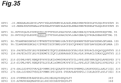
- wild-type HSV gD refers to the full length of HSV-1 derived envelope glycoprotein D (gD) having the amino acid sequence set forth in SEQ ID NO: 2, or HSV-2 derived gD having the amino acid sequence set forth in SEQ ID NO: 3 (GenBank Accession No.: ABU 45433.1). As the result of comparing the two sequences in multiple alignments, the sequence identity is 84% ( Figure 35 ).
- gD The conformation of gD has also been analyzed and, for example, in gD derived from HSV-1, it is known that it consists of a transmembrane domain having amino acid residues at positions 317 to 339, an intracellular domain having amino acid residues at positions 340 to 369, and the ectodomain having amino acid residue at positions 1 to 316.
- Single crystal structures of gD derived from HSV-1 are reported in Non Patent Literature 7, and co-crystal structures thereof are reported in Non Patent Literatures 2 and 6.
- the crystal structure of gD derived from HSV-2 has been reported in Non Patent Literatures 11 and 12.
- the "ectodomain of wild-type HSV gD” means a soluble, antigenic extracellular region of the wild-type HSV gD.
- An example of the ectodomain of the wild-type HSV gD is a wild-type gD ectodomain 1-315 derived from a 333 strain of HSV-2 consisting of the amino acid sequence set forth in SEQ ID NO: 1.
- modified HSV gD protein (“modified protein of HSV gD” or “variant”) is a protein in which at least one amino acid residue or a region of contiguous amino acid residues is substituted, deleted or added to a wild-type HSV gD, and includes a protein in which a protein modification that is not present in the wild-type is performed, such as a protein in which a glycochain is introduced by substitution or deficiency of the amino acid residue.
- neutralizing antibody-inducing activity refers to the ability to induce neutralizing antibodies of an antigen protein.
- the "neutralizing antibody-inducing activity” can be evaluated by the neutralizing antibody titer in immune serum obtained by inoculating the antigen protein into a subject animal.
- the "neutralizing antibody” refers to an antibody that is capable of losing the infectivity of a viral particle.
- the “neutralizing antibody” is, for example, determined by the degree of neutralizing activity of the antibody at a concentration (NT50) necessary to reduce the plaque number of the subject virus by 50%.
- a B cell epitope with high neutralizing antibody-inducing activity in the ectodomain of wild-type HSV gD is referred to as a "beneficial epitope".
- beneficial epitopes in the ectodomain of wild-type HSV gD include B cell epitopes typically present in the receptor-binding domain (RBD).
- examples thereof include an epitope containing an amino acid residue corresponding to at least one amino acid residue selected from the group consisting of an arginine residue at position 134, an aspartic acid residue at position 139, and 222nd arginine residue in the amino acid sequence set forth in SEQ ID NO: 1.
- the “decotope” refers to a B cell epitope having low or no neutralizing antibody-inducing activity compared to a B cell epitope present in RBD in the ectodomain of wild-type HSV gD.
- the “decotope” is classified herein as an "unbeneficial or deleterious epitope”.
- the "decotope” is a B cell epitope having low or no neutralizing antibody-inducing activity compared to a B cell epitope containing an amino acid residue corresponding to at least one amino acid residue selected from the group consisting of an arginine residue at position 134, an aspartic acid residue at position 139, and 222nd arginine residue in the amino acid sequence set forth in SEQ ID NO: 1 in the ectodomain of wild-type HSV gD.
- Examples of the "decotope” in the ectodomain of wild-type HSV gD in the present embodiment include an epitope present in the N-terminal proline-rich region (PRR) of the gD ectodomain.
- PRR N-terminal proline-rich region
- the area where decotopes concentrate is referred to as the "decoy region".
- the PRR of HSV gD is one example of the decoy region.
- PRR is located on an opposite side to RBD in the crystal structure of the ectodomain of wild-type HSV gD ( Figure 3 ).
- the P50 peripheral region in PRR is a very typical decoy region.
- the "P50 peripheral region” refers to a region that has a distance of no more than 1.5 nm from an amino acid residue corresponding to a proline residue at position 50 in the wild-type gD ectodomain 1-315 derived from HSV-2 consisting of the amino acid sequence set forth in SEQ ID NO: 1 in the surface of the crystal structure of the wild-type HSV gD ectodomain.
- the “distance from an amino acid residue” refers to the linear distance from an amino acid residue corresponding to the proline residue at position 50, regardless of the shape of the surface of the crystal structure of the wild-type HSV gD ectodomain.
- decotope is an epitope comprising an amino acid residue corresponding to a proline residue at position 50 in the wild-type gD ectodomain 1-315 derived from HSV-2 consisting of the amino acid sequence set forth in SEQ ID NO: 1.
- the "corresponding" amino acid residue herein means an amino acid residue of other related gD at a position corresponding to a predetermined amino acid residue set forth in SEQ ID NO: 1 in an aligned sequence when the amino acid sequence of the wild-type gD ectodomain derived from HSV-2 consisting of the amino acid sequence set forth in SEQ ID NO: 1 is multiplex aligned (multiple-sequence alignment) with the amino acid sequence of the other related gD.
- decotope is an epitope comprising at least one amino acid residue present on the surface of a crystal structure of the ectodomain of HSV gD in a region at a distance of no more than 1.5 nm from an amino acid corresponding to a proline residue at position 50 (i.e., P50 peripheral region) in the wild-type gD ectodomain 1-315 derived from HSV-2 consisting of the amino acid sequence set forth in SEQ ID NO: 1.
- the decotope can be identified from the crystal structure of wild-type HSV gD. It is preferred that the distance is 1 nm or less.
- the percentage of production of unbeneficial or deleterious antibodies can be reduced, and the percentage of production of neutralizing antibodies with high neutralizing activity can be increased by emphasizing beneficial epitopes.
- the "de-epitoping” refers to a modification of a site contributed to antibody production as an epitope in the wild-type HSV gD so as not to function as an epitope.
- the “de-epitoping” is also referred to as epitope masking.
- Examples of the method of de-epitoping include a method of substituting an amino acid residue at the site of an epitope with another amino acid residue; a method of defecting (deleting) an amino acid residue at the site of an epitope; and a method of introducing a glycochain by substitution or deficiency of an amino acid residue at the site of an epitope.
- the method of introducing a glycochain in particular an N-type glycochain (N-glycoside-linked glycochain) is preferable.
- the method has advantages in that not only the portion where the glycochain is introduced, but also the decotopes in the periphery can be masked at the same time due to its bulkiness.
- the size ratio to proteins such as antibodies or receptors that interact with gD it is expected that the dot-to-dot interaction such that binding is formed in a very narrow range of about a few amino acids is less likely to occur.
- Non Patent Literature 10 Non Patent Literature 10
- Introduction of a glycochain to an amino acid residue refers to an introduction of a glycochain to three contiguous amino acid residues including the position of the amino acid residue by deletion, substitution or addition of an amino acid at the position of the amino acid residue.
- Methods of introducing a glycochain are not particularly limited as long as a conventional method.
- the amino acid sequence of the wild-type gD protein ectodomain (SEQ ID NO: 1) is used as a template, and the primer is designed such that the three contiguous amino acid sequences of the site of interest at which the N-type glycochain is introduced become N-X-S/T (X is any amino acid other than proline), then a mutation is introduced by PCR.
- the nucleic acid sequence of mutated gD protein of interest, or the nucleic acid sequence further linked to a tag such as 6xHis as required, can be cloned into an appropriate vector, then expressed to acquire a gD variant. Then, an N-type glycochain is added to asparagine at the site of interest of the gD modified by a conventional method.
- the N-type glycochain include a GlcNAc-based high mannose type, a hybrid type, and a complex type.
- the de-epitoping i.e., the modification of the decotope includes a modification made by introducing a glycochain into at least one of amino acid residues corresponding to a proline residue at position 50, proline at position 74, and arginine at position 186 in the amino acid sequence set forth in SEQ ID NO: 1 of the ectodomain of the wild-type HSV gD.
- the ectodomain of the wild-type HSV gD consists of the amino acid sequence set forth in SEQ ID NO: 1; and the modification of the decotope includes at least one modification selected from the group consisting of: a modification by introducing a glycochain by substitution of a proline residue at position 50 with an asparagine residue and substitution of a proline residue at position 51 with an amino acid residue other than a proline residue in the amino acid sequence set forth in SEQ ID NO: 1; a modification by introducing a glycochain by substitution of a proline residue at position 74 with an asparagine residue and substitution of a glutamic acid residue at position 76 with a serine residue in the amino acid sequence set forth in SEQ ID NO: 1; and a modification by introducing a glycochain by substitution of an arginine residue at position 186 with an asparagine residue in the amino acid sequence set forth in SEQ ID NO: 1.
- the modified HSV gD protein further contains deficiency of at least a portion of amino acid residues corresponding to amino acid residues at positions 251 to 315 in the amino acid sequence set forth in SEQ ID NO: 1 in the wild-type HSV gD.
- the amino acid residues corresponding to amino acid residues at positions 251 to 315 in the amino acid sequence set forth in SEQ ID NO: 1 form a C-terminal functional region 3 (FR3) in the ectodomain of the wild-type HSV gD.
- FR3 C-terminal functional region 3
- Non Patent Literature 7 Reported crystal structure analysis of HSV gD1 (Non Patent Literature 7) suggests that FR3 and the N-terminal side sequence FR1 can bind to wrap around exactly the same surface of the core beta-sheet structure FR2 in the gD molecule. Since FR1 and FR3 interfere with each other, only one of them can bind to FR2. It has been presumed that on the viral envelope FR3 normally binds, but upon binding to the receptor, FR3 falls off, and the structure changes such that FR1 binds, and the receptor binding region is exposed. From the epitope analysis of neutralizing antibody No. 82 which the present inventors have independently obtained, it has been found that antibody No.
- 82 binds to a Nectin-1 binding region, has reduced reactivity with the FR1-deficient mutant (gD34-315), and inhibits binding of gD to HVEM or Nectin-1. That is, it is believed that defecting FR3 or inhibiting binding of FR3 to FR2 is effective in order to emphasize the epitope of antibody No. 82, i.e. RBD, and to induce immune refocusing on the region.
- the deficiency of at least a portion of FR3 also includes a deletion of the full length of FR3 or a deletion of a contiguous or non-contiguous sequence of a portion of FR3, and substitution of some amino acid residues with other amino acid residues.
- a partial deficiency of FR3 is preferably a deletion of a portion corresponding to amino acid residues at positions 276 to 315 in the wild-type gD ectodomain 1-315 derived from HSV-2 consisting of the amino acid sequence set forth in SEQ ID NO: 1, and may be a deletion of all or a portion of amino acid residues at positions 276 to 315.
- the modified HSV gD protein further comprises at least one promiscuous T cell epitope linked at a C-terminal of the ectodomain of the HSV gD.
- T cell epitopes are also present in the transmembrane region and the intracellular region, but given their use as a vaccine, the design of the secretory phenotype composed of the extracellular region is preferred.
- the present inventors have performed comprehensive exploration of the HLA Class II constrained promiscuous T cell epitope cluster for the full length including the intracellular domain of gD2. As a result, five cluster peptides DP1-DP5 described below were found. It is preferred that the promiscuous T cell epitope is a promiscuous T cell epitope consisting of the amino acid sequence set forth in SEQ ID NO: 4, SEQ ID NO: 5, SEQ ID NO: 6, SEQ ID NO: 7, or SEQ ID NO: 8. Among them, those that actually have promiscuous stimulation activity against both mouse and human T cells are three peptides DP2, DP3, and DP5. In particular, DP5, which consists of the amino acid sequence set forth in SEQ ID NO: 8, is preferred.
- the linking to a promiscuous T cell epitope may include a linking to two or more promiscuous T cell epitopes consisting of the amino acid sequence set forth in SEQ ID NO: 8 at the C-terminal of the ectodomain of the wild-type HSV gD. [Table 1] Table 1.
- the HSV gD protein further contains substitution of an amino acid residue corresponding to a valine residue at position 231 with another amino acid residue in the wild-type gD ectodomain 1-315 derived from HSV-2 consisting of the amino acid sequence set forth in SEQ ID NO: 1, in particular a tryptophan residue.
- the variant containing this further mutation is preferred from the viewpoint of inhibiting binding of FR3 to FR2. This mutation more emphasizes the B cell epitope present in the receptor binding domain and can increase the percentage of production of neutralizing antibodies upon immune induction.
- the modified HSV gD protein of the present invention can be produced by genetic engineering methods. Production methods thereof are not particularly limited, but examples thereof include a production method including: using the cDNA of the wild-type gD protein (SEQ ID NO: 33) as a template; designing a primer to introduce the mutation of interest; obtaining a nucleic acid into which the mutation has been introduced by PCR; functionally linking the nucleic acid to an expression promoter, and optionally a tag; and introducing the resultant into an appropriate expression vector to express a modified HSV gD protein.
- the modified HSV gD protein by introducing a glycochain can be obtained as described above.
- Vectors and promoters are not particularly limited, but examples thereof include pCAG vectors and CAG promoters.
- the produced modified HSV gD protein may be purified as needed.
- Purification methods are not particularly limited, but examples thereof include purifications with affinity chromatography column, gel filtration chromatography column, and ion exchange chromatography column.
- HSV infections include infections by HSV-1 and HSV-2, and examples thereof include lip herpes, corneal herpes, genital herpes, systemic neonatal herpes, and stomatitis, skin diseases, encephalitis, meningitis and myelitis due to HSV
- the HSV vaccine of the present invention contains the modified HSV gD protein of the present invention.
- Examples of the dosage form of the HSV vaccine of the present embodiment include, liquid, powder (lyophilized powder, dried powder), capsule, tablet, and frozen form.
- the HSV vaccine of the present embodiment may contain a pharmaceutically acceptable carrier.
- carriers commonly used in vaccine production can be used without limitation, and specific examples thereof include saline, buffered saline, dextrose, water, glycerol, isotonic aqueous buffers, and combinations of these.
- the vaccine may further contain emulsifiers, preservatives (e.g., thimerosal), isotonic agents, pH adjusting agents, and the like.
- the HSV vaccine of the present embodiment further contains an adjuvant to further enhance the immunogenicity.
- the adjuvant include aluminum adjuvants; oil-in-water emulsion adjuvants containing squalene (AS03, MF59, or the like), ligands of Toll-like receptors such as CpG and 3-O-deacylated-4'-monophosphoryl lipid A (MPL); saponin-based adjuvants; polymer-based adjuvants such as polyy-glutamic acid; and polysaccharides such as chitosan and inulin.
- aluminum adjuvants oil-in-water emulsion adjuvants containing squalene (AS03, MF59, or the like), ligands of Toll-like receptors such as CpG and 3-O-deacylated-4'-monophosphoryl lipid A (MPL); saponin-based adjuvants; polymer-based adjuvants such as polyy-glutamic
- the HSV vaccine of the present embodiment can be obtained by mixing a modified HSV gD protein of the present invention with a carrier, adjuvant, or the like, as needed.
- the adjuvant may be mixed when used.
- Examples of the administration route of the HSV vaccine include transdermal administration, sublingual administration, ophthalmic administration, intradermal administration, intramuscular administration, oral administration, enteral administration, nasal administration, intravenous administration, subcutaneous administration, intraperitoneal administration, and oral to pulmonary inhalation administration.
- Examples of the administration method of the HSV vaccine include a syringe, a transdermal patch, a microneedle, an implantable sustained-release device, a syringe with microneedle, a needleless device, and a spray.
- HSV-2 gD2 To perform a comprehensive epitope analysis of HSV-2 gD (gD2), various antibodies that bind to various epitopes on gD2 were acquired by biopanning against gD2.
- the gD 1- 315 protein was expressed in host cells and purified.
- An scFv-phage display library prepared using human VH and VL cDNA made from mRNA derived from human B cells was used as the library.
- the scFv-phage display library prepared using human VH and VL cDNA from mRNA derived from human B cells was screened to acquire scFv-phage having reactivity with HSV-2 gD.
- scFv-phages Reactivity of the expressed scFv-phages with gD1-315 was confirmed by phage ELISA.
- 100 ⁇ L of gD2 (2 ⁇ g/mL PBS) was immobilized in a 96-well microtiter plate (Maxisorp Plate, NUNC) overnight at 4°C, each well was washed three times with PBS and blocked with 300 ⁇ L of 1% BSA/PBS for 1 hour at room temperature. Each well was washed 3 times with PBS-T (0.05% Tween/PBS). Then, 100 ⁇ L of scFv-phage diluted 10-fold with 1% BSA/PBS was added, and reacted at 37°C for 1 hour.
- ScFv-hFc was manufactured based on the acquired seven scFv-phages.
- the variable region of the isolated scFv gene was linked to human Fc gene, then cloned into a pCAG vector to construct a scFv-hFc expression plasmid.
- Each expression plasmid was expressed using an Expi293 expression system (Life Technologies). After 4-6 days of culture, the supernatant was purified with Protein A affinity chromatography column (HiTrap Protein A HP Columns, GE Healthcare) and dialyzed with PBS. The purity was confirmed by size exclusion chromatography (Superdex 200 5/150 GL, GE Healthcare) and SDS-PAGE.
- the competition inhibition test was performed by the following competitive ELISA. 100 ⁇ L of gD2 (2 ⁇ g/mL PBS) was immobilized in a 96-well microtiter plate (Maxisorp Plate, NUNC) over 2 hours at room temperature. Each well was then washed three times with PBS and blocked with 300 ⁇ L of 1% BSA PBS for 1 hour at room temperature. Each well was washed 5 times with PBS-T (0.05% Tween PBS). 20 ⁇ g/mL scFv-hFc was diluted at any dilution fold with 1% BSA PBS, then 100 ⁇ L of the diluted scFv-hFc was added to the well, and reacted at 37°C for 1 hour.
- scFv-phage or Nectin-1 (Recombinant Human Nectin-1 Protein, R&D Systems, Inc.) was diluted at any dilution fold with 1% BSA PBS, then 100 ⁇ L of the diluent was added to the well, and reacted at 37°C for 1 hour.
- HRP-labeled antibody (anti-M13/HRP/1% BSA PBS or HRP-added anti-His-tag antibody diluted with 1% BSA PBS: anti-His-tag/HRP/1% BSA PBS) was added and reacted at 37°C for 1 hour.
- antibody No. 82 has heavy chains CDR1 to CDR3 consisting of the amino acid sequences set forth in SEQ ID NOs: 9-11 and light chains CDR1 to CDR3 consisting of the amino acid sequences set forth in SEQ ID NOs: 12-14.
- Heavy chain CDR1 GYAIN (SEQ ID NO: 9) Heavy chain CDR2: GINWIFGTSNYAQKFQ (SEQ ID NO: 10) Heavy chain CDR3: DWGAPLEKGAGSPFDV (SEQ ID NO: 11) Light chain CDR1: RASQSVSSSYLA (SEQ ID NO: 12) Light chain CDR2: GASSRAT (SEQ ID NO: 13) Light chain CDR3: QQYGSSPRS (SEQ ID NO: 14)
- the membrane After washed with PBS-T, the membrane was reacted with each scFv-hFc at a concentration of 1 ⁇ g/mL 2% fat-free milk-PBS-T at room temperature for 30 minutes. After washed again, the membrane was reacted with anti-hFc/HRP/2% fat-free milk-PBS-T, and colored with Immobilon Western Detection Regent (Millipore).
- FIG. 1 shows a schematic diagram of the primary structure of gD, showing FR1 (K1-H39), FR2 (I55-R184), and FR3 (T251-G315), respectively.
- the glycochains originally attached to gD bind at N94, N121 and N262.
- the cDNA of the wild-type gD protein (SEQ ID NO: 33) derived from HSV-2 333 strain was used as a template. Since the N-linked glycochains bind to asparagine of N-X-S/T (X is any amino acid other than proline), upon introducing of a glycochain, mutations were performed by PCR using the following primers such that the amino acid sequence at the site of interest would be NXT or NXS (X is any amino acid other than proline). Fw represents Forward, Re represents Reverse. The underline indicates the mutated part. DNA designed to be a signal sequence followed by the nucleic acid sequence of the mutated gD protein of interest, and further the nucleic acid sequence of 6 ⁇ His were genetically synthesized and cloned into a pUC19 vector.
- the completed sequence of the variant was cloned into a pCAGGS1-dhfr-neo vector to acquire a plasmid for expression.
- Each plasmid for expression was expressed using an Expi293 expression system.
- the supernatant was purified with a Ni-NTA affinity chromatography column (TALON Superflow Metal Affinity Resin, Takara Bio Inc.) and dialyzed with PBS. The purity was confirmed by size exclusion chromatography and SDS-PAGE.
- Reactivity analysis of each antibody against these glycochain introduced mutants was performed with competitive ELISA.
- 100 ⁇ L of gD variant (2 ⁇ g/mL PBS) was immobilized in a 96-well microtiter plate (Maxisorp Plate, NUNC) overnight at 4°C.
- Each well was washed three times with PBS and blocked with 300 ⁇ L of 1% BSA PBS for 1 hour at room temperature.
- Each well was washed 3 times with PBS-T (0.05% Tween PBS).
- Each scFv-hFc was diluted at any dilution fold with 1% BSA PBS, then 100 ⁇ L of the diluted scFv-hFc was added to the well, and reacted at 37°C for 1 hour.
- antibody No. 72 and No. 75 which showed no change in reactivity with gD1-275
- antibody No. 72 reduced the reactivity
- antibody No. 75 eliminated the reaction when gD25-253 was used.
- at least a portion of the epitopes of antibody No. 72 and antibody No. 75 is present in gD254-275.
- gD254-275 is present in the vicinity of P50 (SC-F), thus it is also contemplated that these antibodies may recognize both of them.
- antibody No. 75 which reduced reactivity also in H242 (SC-B) is presumed to be possibly heavily influenced by H242 (SC-B) present in the vicinity of FR3 due to its high binding dependency on the epitope in gD254-275.
- gD254-275 is a flexible region at the "root" of FR3, it is inferred that the strength of binding of antibodies No. 72 and No. 75 changes depending on the whole structure of FR3. That is, it is believed that FR3 binds to FR2 in the FR1-deficient mutant gD34-315, then the epitope near P50 (SC-F) is away from the epitope in gD254-275, leading to reduction of the reactivity.
- Antibody No. 75 should have been affected similarly by the epitope near P50 (SC-F) being away from the epitope in gD254-275 in the same way. However, it is inferred that antibody No. 75 was less susceptible to this influence because antibody No. 75 strongly binds to gD254-275.
- the receptor binding domain (RBD) that is a Nectin-1 binding region, and the peripheral region of P50 (SC-F), where the presence of an epitope of antibody No. 82 was predicted, were alanine scanned.
- the amino acids which were believed to be structurally exposed to the surface were mainly selected, then fourteen mutants in which amino acids are substituted with alanine, and blocking mutants of the neutralizing antibody LP2, T213M and S216N, were manufactured.
- the each of genes having alanine substitution were constructed by PCR and cloned into pCAGGS1-dhfr-neo. For expression, a FreeStyle 293 or Expi293 expression system was used.
- D30A showed reduced reactivity.
- D30 is believed to be a portion of an epitope present in 25-33 amino acids.
- R222 and D30 when FR1 binds to FR2, it is not possible to be present in the same interface because FR1 becomes a hindrance.
- antibody No. 82 binds, it is believed that the reaction proceeds with unbinding of FR1 and FR2.
- the alanine substitutes that have reduced reactivity with antibody No.5 were three substitutes I55, E76, and I80, and the binding regions were scattered in different directions about 120° from the binding regions of antibodies No. 72, No. 75 and No. 78 with I55 as the center.
- the epitopes of antibody No. 13 were not determined. This is presumably because the epitopes of antibody No. 13 are suggested to be linear, thus have not been able to mutate directly in the mutants obtained so far.
- FIG. 4 shows a MOE diagram of the gD receptor binding domain (RBD) peripheral region and the P50 peripheral region in an HSV gD structure.
- the epitopes of antibody No. 82 (R134, D139, and R222) can be confirmed to be present in the RBD region.
- Table 4 Table 4. Presumed epitope regions of various anti-gD2 antibodies Group Antibody No. Epitope region A 82 RBD (+FR1) B 1 FR3 C1 5 P50 periphery + ⁇ 13 Unknown C2 72 P50 periphery (+ FR3) 75 P50 periphery + FR3 78 P50 periphery
- HSV neutralizing activity analysis of seven antibody clones in vitro was performed in a plaque number reduction (plaque reduction) test and a cell-to-cell infection spread-suppression test.
- HSV-2 Human herpes virus 2
- VR-540 Human herpes virus 1
- HSV-1 KOS strain VR-1493
- Vero cells purchased from ATCC (CCL.81) were used for viral culture, measurement of infectivity titer, and measurement of neutralizing antibody titer. Vero cells are cultured at 37°C under 5%CO 2 conditions. 10% FBS-containing MEM medium was used for proliferation, maintenance, and analysis plate preparation, and 2% FBS-containing MEM medium was used for measurement of infectivity titer and measurement of neutralizing antibody titer.
- Viral banks for neutralization test and infection prevention ability analysis described below were prepared by the following methods.
- a predetermined concentration of the subject antibody was prepared and mixed with approximately 100 PFU of an HSV-2 MS strain or an HSV-1 KOS strain, then reacted at 37°C for 1 hour.
- the reaction solution was plated to Vero cells full-sheeted in a 48-well plate, and after adsorption at 30°C for 1 hour, cultured in 1% methylcellulose-containing MEM (2% FBS) medium for 24 hours, then inactivated and immobilized at -20°C for 30 minutes with 50% methanol/50% ethanol (-20°C) obtained by mixing methanol and ethanol at 1 : 1.
- the anti-HSV gD monoclonal antibody was then reacted at 37°C for 1 hour, the resultant was immunostained with anti-mouse IgG-HRP (DakoP0447) and TMBH, and images of each well were captured with an ELISpot analyzer (Immunospot S6 Analyzer, CTL) to count plaque numbers with analytical software (BioSpot, CTL).
- HSV-2 MS strain or HSV-1 KOS strain were inoculated into Vero cells full-sheeted in a 48-well plate and adsorption was performed at 30°C for 1 hour. Thereafter, a predetermined concentration of the subject antibody in 1% methylcellulose-containing MEM (2% FBS) medium (antibody concentrations 5 ⁇ g/mL, 25 ⁇ g/mL, and 125 ⁇ g/mL) was added, and the HSV-2 MS strain was cultured for about 40 hours and the HSV-1 KOS strain was cultured for about 48 hours before inactivated and immobilized at -20°C for 30 minutes with 50% methanol/50% ethanol (-20°C).
- Self-prepared anti-HSV gD monoclonal antibodies were then reacted at 37°C for 1 hour, the resulting cells were immunostained with anti-mouse IgG-HRP and TMBH, and images of each well were captured with an ELISpot analyzer to analyze average plaque size values with an analytical software.
- Antibody No. 78 inhibited dose-dependently, but the pattern of the tilt of inhibitory effect thereof against the tilt of 5-fold serial dilution was mild compared to that of others.
- each antibody was set to 20 ⁇ g/mL for the MS strain (HSV-2) and examined in duplicate.
- HSV-2 MS strain
- the only antibody No. 82 showed definite inhibitory activity, while the other 6 antibodies did not show definite activity.
- This activity is considered to be an important activity that may lead to a suppression effect on the spread of infection, or even a suppression effect on recurrent symptoms, in therapeutic administration under situations where a viral infection is already established in vivo.
- antibody No. 82 is a unique antibody with superior characteristics that are fundamentally different from other anti-gD-binding antibodies in that it has not only potent plaque number-reducing activity against both HSV-1 and HSV-2 strains, but also cell-to-cell infection spread-suppression activity that may lead to therapeutic effects, and it is believed that the superiority of which is associated with the presence of an epitope region on the gD receptor binding domain (RBD).
- RBD gD receptor binding domain
- a human-mouse chimeric IgG with a mouse Fc region and a human-guinea pig chimeric IgG with a guinea pig Fc region were prepared, and each neutralizing activity (plaque number-reducing activity) was investigated for HSV-2 (MS strain) and HSV-1 (KOS strain).
- the VH region of the isolated scFv gene was linked to the H chain constant region gene (CH1-CH2-CH3) derived from mouse IgG2a, then cloned into a pCAG vector to construct a H-chain expression plasmid. Furthermore, the VL region of the scFv gene was linked to mouse CL gene, then cloned into a pCAG vector to construct an L-chain expression plasmid.
- An Expi293 expression system was employed for expression. Expression plasmids were transfected into cells and culture supernatants were collected at 4-6 days. Culture supernatants were purified with Hi Trap Protein A HP Column (GE Healthcare) and dialyzed with PBS. The purity was confirmed by size exclusion chromatography and SDS-PAGE. Human-mouse chimeric IgG2a was acquired by the above methods.
- the VH region of the isolated scFv gene was linked to a H-chain constant region gene derived from guinea pig IgG2 (CH1-CH2-CH3), and cloned into the pCAG vector.
- the VL region of the scFv gene was linked to guinea pig CK gene, and cloned into the pCAG vector.
- the cloned antibody genes were then amplified by PCR, cloned into a pXC vector (Lonza) to construct expression plasmids for H and L chains. Both plasmids were then linked to prepare one expression plasmid. CHO cells were used for expression.
- Expression plasmids were stably introduced into CHO cells and antibody high-expression CHO cells were obtained using GS Xceed expression system (Lonza). High-expression cells were fed-batch cultured for 12 days and the culture supernatant was collected. The culture supernatants were purified using rProtein A sepharose Fast Flow (Cat# 17127903/GE Healthcare) to afford a human-guinea pig chimeric IgG2 ⁇ antibody.
- the scFv-hFc, human-mouse chimeric IgG, and human-guinea pig chimeric IgG of anti-gD2 antibody No. 82 were analyzed for viral neutralizing activity (plaque number-reducing activity) as described in Example 4. The results are shown in Table 6. Any of the scFv-hFc, human-mouse chimeric IgG, and human-guinea pig chimeric IgG of antibody No. 82 exhibited 50% plaque number-reducing activity at concentrations of 0.05 ⁇ g/mL or more against both MS (HSV-2) and KOS (HSV-1) strains.
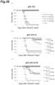
- the mouse genital herpes infection model was used to evaluate the infection prevention ability in the preventive and therapeutic administration of anti-gD2 antibody No. 82.
- An infection-prevention test was performed in the preventive and therapeutic administration of anti-HSV gD2 monoclonal antibodies using a mouse genital herpes infection model.
- BALB/c mice (5 weeks old, female) were used.
- a predetermined amount of the antibody was dissolved in saline for injection and administered intraperitoneally at a dose of 200 ⁇ L/mouse 24 hours prior to viral inoculation for preventive administration and 48 hours after viral inoculation for therapeutic administration.
- the number of N 10 cases per group was set.
- Depo-Provera was inoculated subcutaneously at 2 mg/mouse 6 days prior to viral inoculation.
- HSV-2 MS strain was inoculated transvaginally under anesthesia and observed for 21 days.
- the infection prevention ability was evaluated using survival time (survival rate) and symptom score as indexes.
- Symptom scores were determined by the presence or absence and the extent of vaginal lesion symptoms, and shown as an average value in each group. Scoring was set as 0: no change, 1: partial erythema/swelling, 2: extensive swelling/edema, 3: ulceration/bleeding, 4: death.
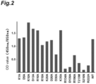
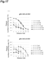
- the survival times by dosage, the survival rates and the symptom scores in preventive administration are shown in Table 7, Figure 4 , and Figure 5 , respectively.
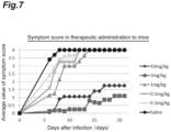
- the survival times by dosage, the survival rates, and the symptom scores in therapeutic administration are shown in Table 8, Figure 6 , and Figure 7 , respectively.
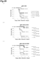
- Infection prevention ability of anti-gD2 antibody No. 82 in the preventive and therapeutic administration was evaluated using the guinea pig genital herpes infection model (acute phase).
- An infection-prevention test of anti-gD2 antibody No. 82 (human-guinea pig chimeric IgG2 ⁇ ) in the preventive and therapeutic administration was performed using a guinea pig genital herpes infection model. Hartley guinea pig (3-5 weeks old, female) purchased from Japan SLC, Inc. was used. A predetermined amount of the antibody was dissolved in saline for injection and administered intraperitoneally 1 mg to 30 mg/kg/guinea pig 24 hours before viral inoculation for preventive administration and 4 days after viral inoculation for therapeutic administration. For therapeutic administration, symptom observation was performed prior to administration, and individuals presenting vaginal symptoms were sorted and randomized to avoid deviation in average scores for each group.
- N 10-15 cases per group was set.
- Viral inoculation was performed by transvaginally inoculating 5 ⁇ 10 5 PFU/50 ⁇ L HSV-2 MS strain under anesthesia and acute phase symptoms were observed for 2-3 weeks after inoculation.
- Symptom scores were set as 0: no clear lesion, 0.5-1: erythema, 1.5-2: localized blisters, 2.5-3: localized ulcers or scabs, 3-5: extensive blisters/ulcers or scabs, 3-7: extensive ulcers or scabs with incontinence, 7.5: euthanasia due to severe symptoms, 8: death.
- Vaginal swabs were also collected at day 7 after viral inoculation and the virus release amount was measured by plaque method.
- Vaginal swabs were collected by inserting a cotton swab moistened with MEM medium into the vagina, then wiping off the mucosa on the vaginal inner wall. Vaginal swabs collected were suspended in MEM medium dispensed by 1 mL into siliconized tubes and stored frozen until use. Vaginal swabs as stock solution, or 10-fold, 100-fold, 1000-fold dilution were inoculated at 100 uL/well into 96-well or 48-well full-sheet Vero cells.
- Viral adsorption was performed at 37°C for 1 hour after vaginal swab inoculation, and cultured in 1% methylcellulose in 2% FBS MEM medium for 24 to 72 hours, then the plaque number was measured in a predetermined manner.
- Symptom scores in preventive administration are shown in Figure 8 .
- the symptom scores in the results of therapeutic administration and the HSV release amounts in the vaginal swab are shown in Figure 9 and Figure 10 , respectively.
- therapeutic administration of antibody No. 82 at 30 mg/kg to guinea pigs that were already presenting vaginal symptoms at 4 days after infection showed a significant relief in symptom scores relative to the saline administration group set as a negative control group.
- vaginal swabs were harvested at 7 days after virus inoculation and the virus release amount was measured by a plaque method. As the result, a significant reduction in viral release amount relative to the negative control group was observed.
- antibody No. 82 exhibits significant infection prevention effect not only in preventive administration but also in therapeutic administration.
- HLA Class II constrained promiscuous T cell epitope cluster sequences were explored using an algorithm (EpiMatrix) by EpiVax, Inc.
- the cluster sequence is a peptide sequence consisting of 15-25 amino acids that are predicted to have high probability (Z-Score ⁇ 1.64) to bind to a majority of the eight major HLA DR super type (DRB1*0101, DRB1*0301, DRB1*0401, DRB1*0701, DRB1*0801, DRB1*1101, DRB1*1301, DRB1*1501) which are analysis targets of EpiMatrix.
- PBMCs derived from anti-HSV antibody-negative donors were also purchased to analyze non-specific responses.
- frozen cells were thawed and washed with Thawing Medium (CTL Wash(TM) Medium), then prepared to a predetermined concentration in medium (CTL Test(TM) Medium) to subject human IFN- ⁇ ELISpot assay.
- CTL Wash(TM) Medium Thawing Medium
- CTL Test(TM) Medium a predetermined concentration in medium
- Cells were seeded in a 96-well plate dedicated to ELISpot at a concentration of 0.5 and 1 ⁇ 10 7 cells/mL by 100 ⁇ L, and to which 100 ⁇ L of each peptide solution prepared to 20 ⁇ M (final concentration: 10 ⁇ M/medium with 0.1% DMSO) was added, and cultured at 37°C in CO 2 incubator for 5 days.
- the cells were then colored according to a predetermined protocol, and the number of positive cells (IFN- ⁇ producing cells) for each well was measured with an ELISpot reader.
- the evaluation was performed in culture medium with 0.1% DMSO using inactivated HSV-1 (10 PFU/cell), ConA (final concentration: 2 ⁇ g/mL) as a negative control (None).
- Detection of IFN- ⁇ -producing cell numbers was performed using Human IFN gamma ELISpot Ready-SET-Go! (R)(eBioscience, Inc., 88-7386-88) by capturing images with ELISpot analyzer (CTL, Immunospot S5 versa analyzer) and counting the number of spots with Immunospot software.
- CTL ELISpot analyzer
- Table 10(A) shows the average spot numbers of IFN- ⁇ -producing cell numbers, where the grey netting indicates that the spot numbers were too small to be determinable.
- Table 10(B) indicates the determination of the presence or absence of T cell stimulation activity by stimulation index (SI) against the negative control (None) as an indicator. The determination was performed as Positive when SI is 3, Marginal when SI is 2 or more and less than 3, and Negative when Si is less than 2. As the result, it was revealed that all five peptides had human T cell stimulation activity, and of which four peptides were capable of stimulating T cells of multiple human PBMCs of different HLA types. [Table 10] Table 10.
- T cell stimulation activity analysis was performed by mouse immunogenicity test of synthetic peptides.
- a stock solution in which the synthetic peptide was dissolved or suspended in 10 mM with DMSO was prepared.
- 10% HCO-60/saline (saline for injection) was also prepared as a solvent for administration using NIKKOL HCO-60 (manufactured by Nikko Chemicals Co., Ltd.).
- 100 ⁇ g of the synthetic peptide was mixed with 10 ⁇ g of CpG and 10 ⁇ g of MPLA in a solvent and prepared into a dose of 210 ⁇ l/mouse and administered subcutaneously to the back of the mouse (4-5 weeks old, female).
- mice BALB/c Three strains of mouse BALB/c (I-Ad/I-Ed), C57BL/6 (I-Ab), and C3H/HeN (I-Ak/IEk) were used. Twenty-one days after the initial immunization, additional immunization was performed, and two weeks later, spleen was collected and splenocytes were prepared to be subjected to the following cytokine production response analysis (ELISpot assay). The prepared splenocytes were seeded in 96 wells of PVDF membrane (MSIPS4W10 Millipore) to 1 ⁇ 10 6 cells/well and cultured with 10 ⁇ M of various peptides for 20 hours.
- ELISpot assay cytokine production response analysis
- mice 2 for each group
- BALB/c I-Ad/I-Ed
- C57BL/6 I-Ab
- C3H/HeN I-Ak/IEk
- spleen was collected and splenocytes were prepared to analyze T cell responsiveness (IFN- ⁇ and IL-2 production stimulation activity) to each peptide by ELISpot. All measurements of each sample were performed at two wells. The experimental results are shown in Figure 11 .
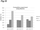
- Example 8 Population analysis of anti-gD2 antibodies contained in human serum fractions (immunoglobulins)
- a competition test with the obtained antibodies was conducted using Venilon, a human gamma globulin cocktail (blood donation Venilon(R)-I for IV injection, General Foundation: The Chemo-Sero-Therapeutic Research Institute).
- Venilon a human gamma globulin cocktail
- gD1-315 were reacted with Venilon, then with monoclonal antibodies.
- SPR Biacore, Inc.
- the chip regeneration was performed by treating the chip surface with 350 mM EDTA for 10 seconds, twice, and washing with Buffer for 10 seconds. Immobilization and regeneration were performed in a similar manner each time new samples were measured. Competition rates were calculated as follows: in the case that increased RU when an antibody under consideration at the concentration of 20 ⁇ g/mL is applied at a flow rate of 20 ⁇ L/min for 120 seconds is set to (1); and increased RU when Venilon at the concentration of 150 ⁇ g/mL is continued to be applied at a flow rate of 20 ⁇ L/min for 120 seconds, then the antibody at the concentration of 20 ⁇ g/mL is applied for 120 seconds is set to (2), the competition rate is calculated by the expression (1 - (2)/(1)) ⁇ 100. All samples were measured only once.
- the analytical results of the Venilon Competition Test are shown in Table 11. The highest competition rate was shown for antibody No. 5, a weak neutralizing antibody, followed by antibody No. 13, antibody No. 75, antibody No. 78, and antibody No. 72, where the group of antibodies having an epitope in the P50 peripheral region were ranked high. Meanwhile, antibody No. 82 was the sixth of 7 clones, exhibiting a relatively low competition rate, and the lowest one was antibody No. 1.
- the present inventors designed gD variants as described below considering three perspectives of emphasizing a beneficial epitope; performing de-epitoping of an epitope in a decoy region by glycochain introduction, deficiency mutation, or the like; and being capable of efficiently and effectively eliciting both a liquid and a cellular immune response by further linking the promiscuous T cell epitope cluster peptide, and examined their neutralizing antibody-inducing activities.
- the beneficial epitopes present on the wild-type HSV gD antigen the gD receptor binding domain (RBD), an epitope of anti-gD2 antibody No. 82, and the promiscuous T cell epitope cluster DP5 (SEQ ID NO: 8) predicted from T cell epitope analysis were assumed, and as the decoy region in which epitopes of relatively less beneficial groups of antibodies were concentrated, the P50 peripheral region was assumed.
- FR1-deficient mutant binds to a Nectin-1 binding region, has reduced reactivity with the FR1-deficient mutant (gD34-315), and inhibits binding of gD to HVEM or Nectin-1. That is, it is believed that defecting FR3 or inhibiting binding of FR3 to FR2 is effective in order to emphasize the epitope of antibody No. 82, and to induce immune refocusing on the region.
- FR3-deficient mutants were based on gD1-275 as reported in a literature (Non Patent Literature 12). It was also expected that the presence of an epitope of antibody No. 1 on FR3 would inhibit the induction of some non-neutralizing antibodies due to FR3 deficiency.
- Non Patent Literature 9 gD1-315 V231W mutant (gD1-315V) similarly reported in a literature (Non Patent Literature 9). It has been suggested that the V231W mutation inhibits binding of FR3 to FR2. Although this variant could not be expected to inhibit the induction of non-neutralizing antibodies such as antibody No. 1, it was considered possible to emphasize the epitope of antibody No. 82.
- T cell immune response can be induced by linking the sequence of the predicted T cell epitope to the C-terminal or the like of the variant.
- T cell epitopes are also present in the transmembrane region and the intracellular region, but given their use as a vaccine, the design in the secretory phenotype composed of the extracellular regions is required. Then, by linking T cell epitopes present in the transmembrane region or intracellular region, the T cell epitope that is not included in the extracellular region can be effectively utilized.
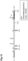
- Figure 12 A schematic diagram of the above modified gD design strategy is shown in Figure 12 .
- Figure 3 also shows the conformation model of HSV gD2 monomers added with the glycochain introduction sites and expected antibody No. 82 epitopes or the like.
- gD1-275, gD1-315 and primers used in introducing glycochains were as previously described.
- DNA was designed to link C-terminal residues (D275 or G315) of the gD sequence of interest, followed by a linker (GPGPG), each T cell epitope peptide, and 6 ⁇ His-Tag in this order, and the full length was artificially gene synthesized and cloned into a pUC19 vector.
- the completed modified sequence was cloned into the pCAGGS1-dhfr-neo vector to acquire a plasmid for expression.
- Each anti-gD2 monoclonal antibody and each gD variant were expressed using an Expi293 expression system. After 4-6 days of culture, the supernatant was purified with Protein A affinity chromatography column or Ni-NTA affinity chromatography column and dialyzed with PBS. The purity was confirmed by size exclusion chromatography and SDS-PAGE.
- HRP-labeled antibody (anti-hFc/HRP/1% BSA PBS) was added and reacted at 37°C for 1 hour. Each well was washed with PBS-T, then colored with TMB at room temperature for 30 minutes. After the reaction was stopped with 1N sulfuric acid, absorbance of 450 nm/650 nm was measured.
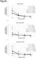
- Figure 13 shows the relative values of the reactivity of each gD variant with antibody No. 82 when the reactivity of gD1-315 (wild-type) is set to "1".
- Antibody No. 82 bound to all gD variants produced, but the intensity of the reactivities was varied. Based on the reactivity of gD1-315 with antibody No. 82, the reactivity with antibody No. 82 was enhanced in most variants. From the viewpoint of glycochain introduction, the simultaneous introduction of P50 (SC-F) and R186 (SC-A) was more effective than the introduction of P50 (SC-F) alone (gD1-315v5-55, gD1-315v5-55V).
- antibody No. 82 has epitopes in the gD receptor binding region and a portion of FR1. Similar to the reasons mentioned in aggregate formation of gD1-275v3-55, it is believed that R186 (SC-A) deprived flexibility of FR3 and created a situation in which FR1 was easier to bind to the gD receptor binding region. Furthermore, reactivity with antibody No. 82 was significantly enhanced in the variant linked to DP5, and particularly gD1-315v5-55 showed approximately 12-fold more reactivity than the original gD1-315. Tandem linkage of DP5 to the C-terminal is believed that the sequence of DP5 itself plays a role in inhibiting binding of FR3 to the gD receptor binding region.
- V231W mutants (gD1-315v3-55V, gD1-315v5-55V) resulted in a slightly reduced reactivity with antibody No. 82 compared to the unmutated variant (gD1-315v3-55, gD1-315v5-55).
- a literature Non Patent Literature 9 introducing V231W reported that V231W has an effect of inhibiting FR3 binding, it is believed that V231W has in fact an effect of slightly inhibiting FR1 binding or altering the original binding structure depending on its bulkiness.
- each variant takes the structure in which FR1 is bound to the receptor binding region.
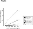
- the reactivity of HVEM with each variant was then measured by competitive ELISA.
- HVEM can bind gD for the first time when FR1 binds to the receptor binding region and takes a characteristic hairpin structure. All HVEM interaction sites were shown to be present in FR1, and increased reactivity with HVEM was as previously inferred. Compared to the original gD1-315, the highest reactivity with HVEM was shown for gD1-315v5-55, then gD1-315v5-55V or gD1-275v5-55, and gD1-315v3-55 in this order, and little reaction was observed in gD1-315 and gD1-315v5.
- FR3-deficient gD1-275v5-55 promoted FR1 binding and further HVEM binding over other gD1-315-based variants in which FR3 was present. Since 1-275v5 and 1-315v5 showed substantially similar reactivity in the reactivity analysis with antibody No. 82, it can be inferred that the effect due to FR3 deficiency is approximately the same as that due to R186 (SC-A) introduction.
- Immunogenicity test of the modified gD antigen was performed using the wild-type gD antigen gD1-315 (WT) as a positive control and saline as a negative control.
- a predetermined amount of antigen was dissolved in saline for injection (saline) with MPLA (10 ⁇ g/mouse) and CpG (1 ⁇ g/mouse) and immunized to mice at a volume of 200 ⁇ L/mouse.
- Two weeks after the final immunization (third dose) blood was collected and serum was prepared for each individual. The prepared serum was serially diluted and assessed for neutralizing antibody-inducing activity against HSV-2 (50% plaque number-reducing activity).
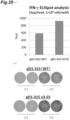
- the IFN- ⁇ production response activity (recall activity) of splenocytes (a pool sample of four cases) prepared from mice immunized with gD1-315v3-55 as an example of a modified gD antigen and wild-type gD (gD1-315) (3 ⁇ g/mouse of the administration group) was compared and analyzed. Specifically, 5 weeks old BALB/c mice were immunized with 3 ⁇ g of antigen subcutaneously in back at 2-week intervals for a total of 3 times, and splenocytes were collected 2 weeks after the final immunization.
- Anti-IFN- ⁇ was immobilized in a 96-well plate with a PVDF membrane on the bottom surface and cells were seeded at 1 ⁇ 10 6 cells/100 ⁇ L/well. Immune antigens were added as stimulants at 10 mM and the cells were cultured at 37°C and 5%CO 2 for 20 hours. The supernatant was removed and the anti-mouse IFNy-HRP antibody was reacted, then HRP was stained with AEC reagents to count IFNy-producing cell numbers with an Image Analyzer (CTL). The results are shown in Figure 25 . It was found that the gD1-315v3-55 immune group exhibits higher response activity than the wild-type gD immune group. It was found that cellular immune (T cell immune) inducing activity could be enhanced by linking two HLA Class II constrained promiscuous T cell epitope cluster DP5s to the C-terminal of the modified gD antigen.
- T cell immune cellular immune
- Depo-Provera was inoculated subcutaneously at 2 mg/mouse 6 days prior to viral inoculation to improve the multiplicity of infection upon viral inoculation 2 weeks after final immunization (third dose).
- 5 ⁇ 10 5 PFU/20 ⁇ L HSV-2 MS strain was inoculated transvaginally under anesthesia and observed for 21 days. Infection prevention ability was evaluated using survival time (survival rate) as index.
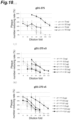
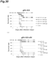
- the infection model of the mouse genital herpes infection model was used to evaluate infection prevention ability of various modified gD in preventive administration.
- the experiments were performed a total of four times (experiments 1-4) and four doses of 0.1 ⁇ g/mouse/time, 0.03 ⁇ g/mouse/time, 0.01 ⁇ g/mouse/time and 0.003 ⁇ g/mouse/time were set for all of the positive control, negative control, and subject modified gD antigen.
- Depo-Provera was inoculated subcutaneously at 2 mg/mouse 6 days prior to viral inoculation to improve the multiplicity of infection upon viral inoculation 2 weeks after final immunization (third dose). 5 ⁇ 10 5 PFU/20 ⁇ L HSV-2 MS strain was inoculated transvaginally under anesthesia and observed for 21 days. Infection prevention ability was evaluated using survival time (survival rate) as index. Mean values of those were plotted in the graph.
- a Significant difference test for the negative control group was performed by the Kaplan-Meier method, and those with a minimum effective dose based on the Significant difference test in three-fold common ratio relative to wild type gD of two or more doses stronger were determined to be "++", those of one dose stronger were determined to be "+”, and those of an equivalent value were determined to be " ⁇ ".
- IgG was purified from serum and analyzed by competitive methods using SPR (Biacore, Inc.) to make antibody amounts uniform.
- the anti-gD2 antibody competition test was performed using Biacore 3000 (GE Healthcare), where, in all experiments, HBS-EP buffer (GE Healthcare) was used, the temperature was set to 25°C, and the flow rate was set to 20 ⁇ L/min.
- CM5 senor chip (GE Healthcare) was used as the sensor chip, and the experiment was performed based on the recommended protocol.
- gD1-315-His was immobilized at about 300 RU on the chip surface, and for chip regeneration, glycine-hydrochloride buffer of 0.1 M and pH 2.0 was used and the chip was washed twice at a flow rate of 30 ⁇ L/min for 30 seconds. Procedures for competition and calculation of the competition rate were as follows.
- each anti-gD2 antibody or buffer 10 ⁇ g/mL of each anti-gD2 antibody or buffer was applied to the chip under a condition of binding for 1 minute and dissociation for 2.5 minutes.
- 20 ⁇ g/mL of each immune serum purified by IgG was then applied under a condition of binding for 1 minute and dissociation for 5 minutes.
- the RU detected when immune serum was applied was set to 100%, and the decrease in immune serum response by anti-gD2 antibody application was calculated as the competitive rate.
- Inhibition of gD-HVEM interactions by modified gD immune serum was also analyzed. Specifically, 100 ⁇ L of gD1-305-cys-strep dimer adjusted to a concentration of 5 ⁇ g/mL with phosphate buffered saline was immobilized in a 96-well microtiter plate at 4°C overnight. Subsequently, blocking and washing were performed as in the competitive ELISA method described above, 100 ⁇ L of IgG purified from each immune serum was added at any concentration and reacted at room temperature for one hour.
- HVEM Recombinant Human HVEMITNFRSF14 Fc Chimera Protein, R&D SYSTEMS, Inc.
- HVEM Recombinant Human HVEMITNFRSF14 Fc Chimera Protein, R&D SYSTEMS, Inc.
- HRP-labeled antibody anti-hFc/HRP/1% BSA PBS
- absorbance of 450 nm/650 nm was measured.
- Modified HSV gD proteins of the present invention can be used in the production of vaccines effective for prevention and treatment of HSV infections.
Landscapes
- Health & Medical Sciences (AREA)
- Life Sciences & Earth Sciences (AREA)
- Virology (AREA)
- Chemical & Material Sciences (AREA)
- General Health & Medical Sciences (AREA)
- Medicinal Chemistry (AREA)
- Organic Chemistry (AREA)
- Public Health (AREA)
- Veterinary Medicine (AREA)
- Pharmacology & Pharmacy (AREA)
- Animal Behavior & Ethology (AREA)
- Immunology (AREA)
- Molecular Biology (AREA)
- Chemical Kinetics & Catalysis (AREA)
- General Chemical & Material Sciences (AREA)
- Nuclear Medicine, Radiotherapy & Molecular Imaging (AREA)
- Engineering & Computer Science (AREA)
- Epidemiology (AREA)
- Genetics & Genomics (AREA)
- Biotechnology (AREA)
- Gastroenterology & Hepatology (AREA)
- Oncology (AREA)
- Biochemistry (AREA)
- Biophysics (AREA)
- Communicable Diseases (AREA)
- Proteomics, Peptides & Aminoacids (AREA)
- Microbiology (AREA)
- Mycology (AREA)
- Bioinformatics & Cheminformatics (AREA)
- Peptides Or Proteins (AREA)
- Medicines Containing Antibodies Or Antigens For Use As Internal Diagnostic Agents (AREA)
- Micro-Organisms Or Cultivation Processes Thereof (AREA)
Claims (9)
- Modifiziertes Protein eines Herpes-simplex-Virus- (HSV-) Hüllglykoproteins D(gD) (modifiziertes HSV-gD-Protein), wobei:
das modifizierte HSV-gD-Protein von einem Wildtyp-HSV-gD durch Modifikation von mindestens einem B-Zellen-Epitop abgeleitet ist, das eine geringe oder keine neutralisierende Antikörper-induzierende Aktivität verglichen mit einem B-Zellen-Epitop aufweist, das in einer rezeptorbindenden Domäne (RBD) (einem Dekotop) in der Ektodomäne des Wildtyp-HSV-gD vorhanden ist, so dass das modifizierte Epitop nicht als Epitop wirkt; wobei:(i) die Modifikation des Dekotops eine Modifikation durch Einführen einer Glykokette in einen Aminosäurerest umfasst, der einem Prolinrest bei Position 50 in der Aminosäuresequenz nach SEQ ID NO: 1 in der Ektodomäne des Wildtyp-HSV-gD entspricht; und/oder(ii) die Modifikation des Dekotops eine Modifikation durch Einführen einer Glykokette in einen Aminosäurerest umfasst, der einem Prolinrest bei Position 74 in der Aminosäuresequenz nach SEQ ID NO: 1 in der Ektodomäne des Wildtyp-HSV-gD entspricht; und/oder(iii) die Modifikation des Dekotops eine Modifikation durch Einführen einer Glykokette in einen Aminosäurerest umfasst, der einem Argininrest bei Position 186 in der Aminosäuresequenz nach SEQ ID NO: 1 in der Ektodomäne des Wildtyp-HSV-gD entspricht. - Modifiziertes Protein eines Herpes-simplex-Virus- (HSV-) Hüllglykoproteins D(gD) (modifiziertes HSV-gD-Protein) nach Anspruch 1, wobei:
das modifizierte HSV-gD-Protein von einem Wildtyp-HSV-gD durch Modifikation von mindestens einem B-Zellen-Epitop abgeleitet ist, das eine geringe oder keine neutralisierende Antikörper-induzierende Aktivität verglichen mit einem B-Zellen-Epitop aufweist, das in einer rezeptorbindenden Domäne (RBD) (einem Dekotop) in der Ektodomäne des Wildtyp-HSV-gD vorhanden ist, so dass das modifizierte Epitop nicht als Epitop wirkt; und wobei:die Ektodomäne des Wildtyp-HSV-gD aus der Aminosäuresequenz nach SEQ ID NO: 1 besteht; unddie Modifikation des Dekotops mindestens eine Modifikation ausgewählt aus der Gruppe umfasst, die besteht aus:einer Modifikation durch Einführen einer Glykokette durch Substitution eines Prolinrests bei Position 50 mit einem Asparaginrest und Substitution eines Prolinrests bei Position 51 mit einem Aminosäurerest mit Ausnahme von einem Prolinrest in der Aminosäuresequenz nach SEQ ID NO: 1;einer Modifikation durch Einführen einer Glykokette durch Substitution eines Prolinrests bei Position 74 mit einem Asparaginrest und Substitution eines Glutaminsäurerests bei Position 76 mit einem Serinrest in der Aminosäuresequenz nach SEQ ID NO: 1; undeiner Modifikation durch Einführen einer Glykokette durch Substitution eines Argininrests bei Position 186 mit einem Asparaginrest in der Aminosäuresequenz nach SEQ ID NO: 1. - Modifiziertes HSV-gD-Protein nach Anspruch 1 oder 2, wobei die Glykokette eine Glykokette vom Typ N ist.
- Modifiziertes HSV-gD-Protein nach einem der Ansprüche 1 bis 3, wobei das modifizierte HSV-gD-Protein ferner mindestens ein promiskuitives T-Zellen-Epitop umfasst, das an eine C-terminale Seite der Ektodomäne des HSV-gD gebunden ist, wobei das promiskuitive T-Zellen-Epitop ein promiskuitives T-Zellen-Epitop bestehend aus der Aminosäuresequenz nach SEQ ID NO: 4, SEQ ID NO: 5, SEQ ID NO: 6, SEQ ID NO: 7 oder SEQ ID NO: 8 ist.
- Modifiziertes HSV-gD-Protein nach Anspruch 4, wobei das promiskuitive T-Zellen-Epitop ein promiskuitives T-Zellen-Epitop bestehend aus der Aminosäuresequenz nach SEQ ID NO: 8 ist.
- Modifiziertes HSV-gD-Protein nach einem der Ansprüche 1 bis 5, wobei das modifizierte HSV-gD-Protein ferner eine Defizienz von mindestens einem Teil von Aminosäureresten enthält, die Aminosäureresten bei Position 251 bis 315 in der Aminosäuresequenz nach SEQ ID NO: 1 im Wildtyp-HSV-gD entsprechen.
- Modifiziertes HSV-gD-Protein nach einem der Ansprüche 1 bis 6, wobei das modifizierte HSV-gD-Protein ferner eine Modifikation durch Substitution eines Aminosäurerests, der einem Valinrest bei Position 231 entspricht, mit einem weiteren Aminosäurerest in der Aminosäuresequenz nach SEQ ID NO: 1 in der Ektodomäne des Wildtyp-HSV-gD umfasst.
- Modifiziertes HSV-gD-Protein nach einem der Ansprüche 1 bis 7, wobei das HSV HSV-1 oder HSV-2 ist.
- HSV-Vakzin, das das modifizierte HSV-gD-Protein nach einem der Ansprüche 1 bis 8 umfasst.
Applications Claiming Priority (2)
| Application Number | Priority Date | Filing Date | Title |
|---|---|---|---|
| JP2017165681 | 2017-08-30 | ||
| PCT/JP2018/032018 WO2019044925A1 (ja) | 2017-08-30 | 2018-08-29 | 改変型HSV gDタンパク質及びこれを含むワクチン |
Publications (4)
| Publication Number | Publication Date |
|---|---|
| EP3677592A1 EP3677592A1 (de) | 2020-07-08 |
| EP3677592A4 EP3677592A4 (de) | 2021-06-09 |
| EP3677592C0 EP3677592C0 (de) | 2024-07-17 |
| EP3677592B1 true EP3677592B1 (de) | 2024-07-17 |
Family
ID=65525657
Family Applications (1)
| Application Number | Title | Priority Date | Filing Date |
|---|---|---|---|
| EP18849655.8A Active EP3677592B1 (de) | 2017-08-30 | 2018-08-29 | Modifiziertes hsv-gd-protein und impfstoff damit |
Country Status (6)
| Country | Link |
|---|---|
| US (1) | US11357847B2 (de) |
| EP (1) | EP3677592B1 (de) |
| JP (1) | JP7204653B2 (de) |
| CN (1) | CN111051333B (de) |
| AU (1) | AU2018323502B2 (de) |
| WO (1) | WO2019044925A1 (de) |
Families Citing this family (1)
| Publication number | Priority date | Publication date | Assignee | Title |
|---|---|---|---|---|
| CN112730851B (zh) * | 2021-03-31 | 2021-06-22 | 南京立顶医疗科技有限公司 | 一种高灵敏SARS-CoV-2中和抗体的检测方法、检测试剂盒 |
Family Cites Families (10)
| Publication number | Priority date | Publication date | Assignee | Title |
|---|---|---|---|---|
| EP1237930B1 (de) * | 1999-12-08 | 2006-11-08 | Intellect Neurosciences, Inc. | Schimärische amyloid-beta peptide |
| ATE319833T1 (de) * | 1999-12-17 | 2006-03-15 | Wyeth Corp | Impfstoffe zur erhöhung der immunantworten gegen herpes simplex virus |
| US20060051368A1 (en) | 2004-07-23 | 2006-03-09 | Northwestern University | Herpes simplex virus mutations |
| EP2502634A1 (de) * | 2006-12-28 | 2012-09-26 | The Trustees of The University of Pennsylvania | Mit Herpes-simplex-Virus kombinierte Untereinheitsimpfstoffe und Verfahren zu deren Verwendung |
| ES2667622T3 (es) * | 2008-05-29 | 2018-05-11 | Alma Mater Studiorum - Università di Bologna | Virus del herpes simple (VHS) con tropismo modificado, utilizaciones y procedimiento de preparación del mismo |
| US8252906B2 (en) | 2009-01-05 | 2012-08-28 | Dcb-Usa Llc | Anti-herpes simplex virus antibodies and methods of use thereof |
| WO2010087813A1 (en) | 2009-01-05 | 2010-08-05 | Dcb-Usa Llc | Anti-herpes simplex virus antibodies |
| JP6099573B2 (ja) * | 2011-01-31 | 2017-03-22 | ザ トラスティーズ オブ ザ ユニバーシティ オブ ペンシルバニア | 新規なヘルペス抗原をコードする核酸分子、それを含むワクチン及びその使用方法 |
| ES2673556T3 (es) * | 2012-05-16 | 2018-06-22 | Immune Design Corp. | Vacunas para el VHS-2 |
| JP6025793B2 (ja) | 2014-09-11 | 2016-11-16 | アルマ マータ ステューディオラム — ユニバーシタ ディ ボローニャ | 親和性を改変した単純ヘルペスウイルス(hsv)、その使用および調製法 |
-
2018
- 2018-08-29 AU AU2018323502A patent/AU2018323502B2/en active Active
- 2018-08-29 WO PCT/JP2018/032018 patent/WO2019044925A1/ja not_active Ceased
- 2018-08-29 CN CN201880055611.2A patent/CN111051333B/zh active Active
- 2018-08-29 JP JP2019539594A patent/JP7204653B2/ja active Active
- 2018-08-29 EP EP18849655.8A patent/EP3677592B1/de active Active
- 2018-08-29 US US16/641,420 patent/US11357847B2/en active Active
Also Published As
| Publication number | Publication date |
|---|---|
| EP3677592C0 (de) | 2024-07-17 |
| EP3677592A4 (de) | 2021-06-09 |
| CN111051333B (zh) | 2024-04-05 |
| AU2018323502B2 (en) | 2023-01-12 |
| EP3677592A1 (de) | 2020-07-08 |
| US20210205441A1 (en) | 2021-07-08 |
| CN111051333A (zh) | 2020-04-21 |
| WO2019044925A1 (ja) | 2019-03-07 |
| US11357847B2 (en) | 2022-06-14 |
| AU2018323502A2 (en) | 2020-08-27 |
| AU2018323502A1 (en) | 2020-04-16 |
| JP7204653B2 (ja) | 2023-01-16 |
| JPWO2019044925A1 (ja) | 2020-09-17 |
| WO2019044925A9 (ja) | 2019-10-03 |
Similar Documents
| Publication | Publication Date | Title |
|---|---|---|
| JP7145862B2 (ja) | 抗HSV gBモノクローナル抗体又はその抗原結合断片 | |
| US12064476B2 (en) | Zika virus immunogenic compositions | |
| AU2018323504B2 (en) | Modified HSV gB protein and HSV vaccine including same | |
| TWI454279B (zh) | 抗單純皰疹病毒抗體及其使用方法 | |
| US12491386B2 (en) | Anti-varicella-zoster virus antibody | |
| KR20150135231A (ko) | 인간 메타뉴모바이러스에 특이적인 인간 항체 또는 그의 항원 결합성 단편 | |
| EP3677592B1 (de) | Modifiziertes hsv-gd-protein und impfstoff damit | |
| US20240166725A1 (en) | Anti-varicella-zoster virus antibody and use thereof | |
| EP4293038A1 (de) | Epitoppeptid und antikörper zur vorbeugung und behandlung von eb-virus-infektion und verwandten erkrankungen | |
| JP2023554587A (ja) | Sars-cov-2 スパイクタンパク質の受容体結合ドメインにコンジュゲートまたは融合している抗体およびワクチン目的でのそれらの使用 | |
| AU2021287508B2 (en) | Fusion protein of pentamer and gB of cytomegalovirus, and vaccine containing said fusion protein | |
| US20250262294A1 (en) | Vaccine | |
| KR20120054465A (ko) | 소 코로나바이러스 항원 결정기를 포함하는 스파이크 재조합 단백질 및 이에 대한 항체 | |
| WO2023108117A2 (en) | Engineered fc domains and engineered hcmv viral fc receptors |
Legal Events
| Date | Code | Title | Description |
|---|---|---|---|
| STAA | Information on the status of an ep patent application or granted ep patent |
Free format text: STATUS: THE INTERNATIONAL PUBLICATION HAS BEEN MADE |
|
| PUAI | Public reference made under article 153(3) epc to a published international application that has entered the european phase |
Free format text: ORIGINAL CODE: 0009012 |
|
| STAA | Information on the status of an ep patent application or granted ep patent |
Free format text: STATUS: REQUEST FOR EXAMINATION WAS MADE |
|
| 17P | Request for examination filed |
Effective date: 20200323 |
|
| AK | Designated contracting states |
Kind code of ref document: A1 Designated state(s): AL AT BE BG CH CY CZ DE DK EE ES FI FR GB GR HR HU IE IS IT LI LT LU LV MC MK MT NL NO PL PT RO RS SE SI SK SM TR |
|
| AX | Request for extension of the european patent |
Extension state: BA ME |
|
| DAV | Request for validation of the european patent (deleted) | ||
| DAX | Request for extension of the european patent (deleted) | ||
| A4 | Supplementary search report drawn up and despatched |
Effective date: 20210507 |
|
| RIC1 | Information provided on ipc code assigned before grant |
Ipc: C07K 14/035 20060101AFI20210430BHEP Ipc: A61K 39/245 20060101ALI20210430BHEP Ipc: A61P 31/22 20060101ALI20210430BHEP Ipc: A61P 37/04 20060101ALI20210430BHEP Ipc: C12N 15/09 20060101ALI20210430BHEP |
|
| STAA | Information on the status of an ep patent application or granted ep patent |
Free format text: STATUS: EXAMINATION IS IN PROGRESS |
|
| 17Q | First examination report despatched |
Effective date: 20221201 |
|
| GRAP | Despatch of communication of intention to grant a patent |
Free format text: ORIGINAL CODE: EPIDOSNIGR1 |
|
| STAA | Information on the status of an ep patent application or granted ep patent |
Free format text: STATUS: GRANT OF PATENT IS INTENDED |
|
| INTG | Intention to grant announced |
Effective date: 20240214 |
|
| GRAS | Grant fee paid |
Free format text: ORIGINAL CODE: EPIDOSNIGR3 |
|
| GRAA | (expected) grant |
Free format text: ORIGINAL CODE: 0009210 |
|
| STAA | Information on the status of an ep patent application or granted ep patent |
Free format text: STATUS: THE PATENT HAS BEEN GRANTED |
|
| AK | Designated contracting states |
Kind code of ref document: B1 Designated state(s): AL AT BE BG CH CY CZ DE DK EE ES FI FR GB GR HR HU IE IS IT LI LT LU LV MC MK MT NL NO PL PT RO RS SE SI SK SM TR |
|
| REG | Reference to a national code |
Ref country code: CH Ref legal event code: EP |
|
| REG | Reference to a national code |
Ref country code: DE Ref legal event code: R096 Ref document number: 602018071977 Country of ref document: DE |
|
| REG | Reference to a national code |
Ref country code: IE Ref legal event code: FG4D |
|
| U01 | Request for unitary effect filed |
Effective date: 20240731 |
|
| U07 | Unitary effect registered |
Designated state(s): AT BE BG DE DK EE FI FR IT LT LU LV MT NL PT SE SI Effective date: 20240819 |
|
| U20 | Renewal fee for the european patent with unitary effect paid |
Year of fee payment: 7 Effective date: 20241106 |
|
| PG25 | Lapsed in a contracting state [announced via postgrant information from national office to epo] |
Ref country code: NO Free format text: LAPSE BECAUSE OF FAILURE TO SUBMIT A TRANSLATION OF THE DESCRIPTION OR TO PAY THE FEE WITHIN THE PRESCRIBED TIME-LIMIT Effective date: 20241017 |
|
| PG25 | Lapsed in a contracting state [announced via postgrant information from national office to epo] |
Ref country code: GR Free format text: LAPSE BECAUSE OF FAILURE TO SUBMIT A TRANSLATION OF THE DESCRIPTION OR TO PAY THE FEE WITHIN THE PRESCRIBED TIME-LIMIT Effective date: 20241018 Ref country code: PL Free format text: LAPSE BECAUSE OF FAILURE TO SUBMIT A TRANSLATION OF THE DESCRIPTION OR TO PAY THE FEE WITHIN THE PRESCRIBED TIME-LIMIT Effective date: 20240717 |
|
| PG25 | Lapsed in a contracting state [announced via postgrant information from national office to epo] |
Ref country code: IS Free format text: LAPSE BECAUSE OF FAILURE TO SUBMIT A TRANSLATION OF THE DESCRIPTION OR TO PAY THE FEE WITHIN THE PRESCRIBED TIME-LIMIT Effective date: 20241117 |
|
| PG25 | Lapsed in a contracting state [announced via postgrant information from national office to epo] |
Ref country code: HR Free format text: LAPSE BECAUSE OF FAILURE TO SUBMIT A TRANSLATION OF THE DESCRIPTION OR TO PAY THE FEE WITHIN THE PRESCRIBED TIME-LIMIT Effective date: 20240717 |
|
| PG25 | Lapsed in a contracting state [announced via postgrant information from national office to epo] |
Ref country code: ES Free format text: LAPSE BECAUSE OF FAILURE TO SUBMIT A TRANSLATION OF THE DESCRIPTION OR TO PAY THE FEE WITHIN THE PRESCRIBED TIME-LIMIT Effective date: 20240717 Ref country code: RS Free format text: LAPSE BECAUSE OF FAILURE TO SUBMIT A TRANSLATION OF THE DESCRIPTION OR TO PAY THE FEE WITHIN THE PRESCRIBED TIME-LIMIT Effective date: 20241017 |
|
| PG25 | Lapsed in a contracting state [announced via postgrant information from national office to epo] |
Ref country code: RS Free format text: LAPSE BECAUSE OF FAILURE TO SUBMIT A TRANSLATION OF THE DESCRIPTION OR TO PAY THE FEE WITHIN THE PRESCRIBED TIME-LIMIT Effective date: 20241017 Ref country code: PL Free format text: LAPSE BECAUSE OF FAILURE TO SUBMIT A TRANSLATION OF THE DESCRIPTION OR TO PAY THE FEE WITHIN THE PRESCRIBED TIME-LIMIT Effective date: 20240717 Ref country code: NO Free format text: LAPSE BECAUSE OF FAILURE TO SUBMIT A TRANSLATION OF THE DESCRIPTION OR TO PAY THE FEE WITHIN THE PRESCRIBED TIME-LIMIT Effective date: 20241017 Ref country code: IS Free format text: LAPSE BECAUSE OF FAILURE TO SUBMIT A TRANSLATION OF THE DESCRIPTION OR TO PAY THE FEE WITHIN THE PRESCRIBED TIME-LIMIT Effective date: 20241117 Ref country code: HR Free format text: LAPSE BECAUSE OF FAILURE TO SUBMIT A TRANSLATION OF THE DESCRIPTION OR TO PAY THE FEE WITHIN THE PRESCRIBED TIME-LIMIT Effective date: 20240717 Ref country code: GR Free format text: LAPSE BECAUSE OF FAILURE TO SUBMIT A TRANSLATION OF THE DESCRIPTION OR TO PAY THE FEE WITHIN THE PRESCRIBED TIME-LIMIT Effective date: 20241018 Ref country code: ES Free format text: LAPSE BECAUSE OF FAILURE TO SUBMIT A TRANSLATION OF THE DESCRIPTION OR TO PAY THE FEE WITHIN THE PRESCRIBED TIME-LIMIT Effective date: 20240717 |
|
| REG | Reference to a national code |
Ref country code: CH Ref legal event code: PL |
|
| PG25 | Lapsed in a contracting state [announced via postgrant information from national office to epo] |
Ref country code: SM Free format text: LAPSE BECAUSE OF FAILURE TO SUBMIT A TRANSLATION OF THE DESCRIPTION OR TO PAY THE FEE WITHIN THE PRESCRIBED TIME-LIMIT Effective date: 20240717 |
|
| PG25 | Lapsed in a contracting state [announced via postgrant information from national office to epo] |
Ref country code: MC Free format text: LAPSE BECAUSE OF FAILURE TO SUBMIT A TRANSLATION OF THE DESCRIPTION OR TO PAY THE FEE WITHIN THE PRESCRIBED TIME-LIMIT Effective date: 20240717 Ref country code: CH Free format text: LAPSE BECAUSE OF NON-PAYMENT OF DUE FEES Effective date: 20240831 |
|
| PG25 | Lapsed in a contracting state [announced via postgrant information from national office to epo] |
Ref country code: CZ Free format text: LAPSE BECAUSE OF FAILURE TO SUBMIT A TRANSLATION OF THE DESCRIPTION OR TO PAY THE FEE WITHIN THE PRESCRIBED TIME-LIMIT Effective date: 20240717 |
|
| PG25 | Lapsed in a contracting state [announced via postgrant information from national office to epo] |
Ref country code: SK Free format text: LAPSE BECAUSE OF FAILURE TO SUBMIT A TRANSLATION OF THE DESCRIPTION OR TO PAY THE FEE WITHIN THE PRESCRIBED TIME-LIMIT Effective date: 20240717 |
|
| PLBE | No opposition filed within time limit |
Free format text: ORIGINAL CODE: 0009261 |
|
| STAA | Information on the status of an ep patent application or granted ep patent |
Free format text: STATUS: NO OPPOSITION FILED WITHIN TIME LIMIT |
|
| 26N | No opposition filed |
Effective date: 20250422 |
|
| PG25 | Lapsed in a contracting state [announced via postgrant information from national office to epo] |
Ref country code: IE Free format text: LAPSE BECAUSE OF NON-PAYMENT OF DUE FEES Effective date: 20240829 |
|
| U20 | Renewal fee for the european patent with unitary effect paid |
Year of fee payment: 8 Effective date: 20250818 |
|
| PGFP | Annual fee paid to national office [announced via postgrant information from national office to epo] |
Ref country code: GB Payment date: 20250818 Year of fee payment: 8 |
|
| PG25 | Lapsed in a contracting state [announced via postgrant information from national office to epo] |
Ref country code: RO Free format text: LAPSE BECAUSE OF FAILURE TO SUBMIT A TRANSLATION OF THE DESCRIPTION OR TO PAY THE FEE WITHIN THE PRESCRIBED TIME-LIMIT Effective date: 20240717 |
|
| PG25 | Lapsed in a contracting state [announced via postgrant information from national office to epo] |
Ref country code: CY Free format text: LAPSE BECAUSE OF FAILURE TO SUBMIT A TRANSLATION OF THE DESCRIPTION OR TO PAY THE FEE WITHIN THE PRESCRIBED TIME-LIMIT; INVALID AB INITIO Effective date: 20180829 |