EP3639733A2 - Verfahren und vorrichtung zur überwachung der kardiopulmonaren gesundheit - Google Patents
Verfahren und vorrichtung zur überwachung der kardiopulmonaren gesundheit Download PDFInfo
- Publication number
- EP3639733A2 EP3639733A2 EP19191677.4A EP19191677A EP3639733A2 EP 3639733 A2 EP3639733 A2 EP 3639733A2 EP 19191677 A EP19191677 A EP 19191677A EP 3639733 A2 EP3639733 A2 EP 3639733A2
- Authority
- EP
- European Patent Office
- Prior art keywords
- patient
- cardio
- movement
- pulmonary health
- respiratory
- Prior art date
- Legal status (The legal status is an assumption and is not a legal conclusion. Google has not performed a legal analysis and makes no representation as to the accuracy of the status listed.)
- Granted
Links
Images


Classifications
-
- A—HUMAN NECESSITIES
- A61—MEDICAL OR VETERINARY SCIENCE; HYGIENE
- A61B—DIAGNOSIS; SURGERY; IDENTIFICATION
- A61B5/00—Measuring for diagnostic purposes; Identification of persons
- A61B5/08—Measuring devices for evaluating the respiratory organs
-
- A—HUMAN NECESSITIES
- A61—MEDICAL OR VETERINARY SCIENCE; HYGIENE
- A61B—DIAGNOSIS; SURGERY; IDENTIFICATION
- A61B5/00—Measuring for diagnostic purposes; Identification of persons
- A61B5/103—Measuring devices for testing the shape, pattern, colour, size or movement of the body or parts thereof, for diagnostic purposes
- A61B5/11—Measuring movement of the entire body or parts thereof, e.g. head or hand tremor or mobility of a limb
- A61B5/113—Measuring movement of the entire body or parts thereof, e.g. head or hand tremor or mobility of a limb occurring during breathing
-
- A—HUMAN NECESSITIES
- A61—MEDICAL OR VETERINARY SCIENCE; HYGIENE
- A61B—DIAGNOSIS; SURGERY; IDENTIFICATION
- A61B5/00—Measuring for diagnostic purposes; Identification of persons
- A61B5/48—Other medical applications
- A61B5/4806—Sleep evaluation
- A61B5/4818—Sleep apnoea
-
- A—HUMAN NECESSITIES
- A61—MEDICAL OR VETERINARY SCIENCE; HYGIENE
- A61B—DIAGNOSIS; SURGERY; IDENTIFICATION
- A61B5/00—Measuring for diagnostic purposes; Identification of persons
- A61B5/72—Signal processing specially adapted for physiological signals or for diagnostic purposes
- A61B5/7221—Determining signal validity, reliability or quality
-
- A—HUMAN NECESSITIES
- A61—MEDICAL OR VETERINARY SCIENCE; HYGIENE
- A61B—DIAGNOSIS; SURGERY; IDENTIFICATION
- A61B5/00—Measuring for diagnostic purposes; Identification of persons
- A61B5/72—Signal processing specially adapted for physiological signals or for diagnostic purposes
- A61B5/7235—Details of waveform analysis
- A61B5/7264—Classification of physiological signals or data, e.g. using neural networks, statistical classifiers, expert systems or fuzzy systems
-
- A—HUMAN NECESSITIES
- A61—MEDICAL OR VETERINARY SCIENCE; HYGIENE
- A61B—DIAGNOSIS; SURGERY; IDENTIFICATION
- A61B5/00—Measuring for diagnostic purposes; Identification of persons
- A61B5/72—Signal processing specially adapted for physiological signals or for diagnostic purposes
- A61B5/7271—Specific aspects of physiological measurement analysis
- A61B5/7275—Determining trends in physiological measurement data; Predicting development of a medical condition based on physiological measurements, e.g. determining a risk factor
-
- A—HUMAN NECESSITIES
- A61—MEDICAL OR VETERINARY SCIENCE; HYGIENE
- A61B—DIAGNOSIS; SURGERY; IDENTIFICATION
- A61B5/00—Measuring for diagnostic purposes; Identification of persons
- A61B5/74—Details of notification to user or communication with user or patient; User input means
- A61B5/742—Details of notification to user or communication with user or patient; User input means using visual displays
-
- A—HUMAN NECESSITIES
- A61—MEDICAL OR VETERINARY SCIENCE; HYGIENE
- A61B—DIAGNOSIS; SURGERY; IDENTIFICATION
- A61B5/00—Measuring for diagnostic purposes; Identification of persons
- A61B5/74—Details of notification to user or communication with user or patient; User input means
- A61B5/746—Alarms related to a physiological condition, e.g. details of setting alarm thresholds or avoiding false alarms
-
- G—PHYSICS
- G08—SIGNALLING
- G08B—SIGNALLING SYSTEMS, e.g. PERSONAL CALLING SYSTEMS; ORDER TELEGRAPHS; ALARM SYSTEMS
- G08B21/00—Alarms responsive to a single specified undesired or abnormal condition and not otherwise provided for
- G08B21/02—Alarms for ensuring the safety of persons
-
- G—PHYSICS
- G16—INFORMATION AND COMMUNICATION TECHNOLOGY [ICT] SPECIALLY ADAPTED FOR SPECIFIC APPLICATION FIELDS
- G16H—HEALTHCARE INFORMATICS, i.e. INFORMATION AND COMMUNICATION TECHNOLOGY [ICT] SPECIALLY ADAPTED FOR THE HANDLING OR PROCESSING OF MEDICAL OR HEALTHCARE DATA
- G16H40/00—ICT specially adapted for the management or administration of healthcare resources or facilities; ICT specially adapted for the management or operation of medical equipment or devices
- G16H40/60—ICT specially adapted for the management or administration of healthcare resources or facilities; ICT specially adapted for the management or operation of medical equipment or devices for the operation of medical equipment or devices
- G16H40/63—ICT specially adapted for the management or administration of healthcare resources or facilities; ICT specially adapted for the management or operation of medical equipment or devices for the operation of medical equipment or devices for local operation
-
- G—PHYSICS
- G16—INFORMATION AND COMMUNICATION TECHNOLOGY [ICT] SPECIALLY ADAPTED FOR SPECIFIC APPLICATION FIELDS
- G16H—HEALTHCARE INFORMATICS, i.e. INFORMATION AND COMMUNICATION TECHNOLOGY [ICT] SPECIALLY ADAPTED FOR THE HANDLING OR PROCESSING OF MEDICAL OR HEALTHCARE DATA
- G16H50/00—ICT specially adapted for medical diagnosis, medical simulation or medical data mining; ICT specially adapted for detecting, monitoring or modelling epidemics or pandemics
- G16H50/20—ICT specially adapted for medical diagnosis, medical simulation or medical data mining; ICT specially adapted for detecting, monitoring or modelling epidemics or pandemics for computer-aided diagnosis, e.g. based on medical expert systems
-
- A—HUMAN NECESSITIES
- A61—MEDICAL OR VETERINARY SCIENCE; HYGIENE
- A61B—DIAGNOSIS; SURGERY; IDENTIFICATION
- A61B5/00—Measuring for diagnostic purposes; Identification of persons
- A61B5/05—Detecting, measuring or recording for diagnosis by means of electric currents or magnetic fields; Measuring using microwaves or radio waves
- A61B5/0507—Detecting, measuring or recording for diagnosis by means of electric currents or magnetic fields; Measuring using microwaves or radio waves using microwaves or terahertz waves
-
- A—HUMAN NECESSITIES
- A61—MEDICAL OR VETERINARY SCIENCE; HYGIENE
- A61B—DIAGNOSIS; SURGERY; IDENTIFICATION
- A61B5/00—Measuring for diagnostic purposes; Identification of persons
- A61B5/08—Measuring devices for evaluating the respiratory organs
- A61B5/0816—Measuring devices for examining respiratory frequency
-
- A—HUMAN NECESSITIES
- A61—MEDICAL OR VETERINARY SCIENCE; HYGIENE
- A61B—DIAGNOSIS; SURGERY; IDENTIFICATION
- A61B5/00—Measuring for diagnostic purposes; Identification of persons
- A61B5/08—Measuring devices for evaluating the respiratory organs
- A61B5/087—Measuring breath flow
-
- A—HUMAN NECESSITIES
- A61—MEDICAL OR VETERINARY SCIENCE; HYGIENE
- A61B—DIAGNOSIS; SURGERY; IDENTIFICATION
- A61B5/00—Measuring for diagnostic purposes; Identification of persons
- A61B5/08—Measuring devices for evaluating the respiratory organs
- A61B5/097—Devices for facilitating collection of breath or for directing breath into or through measuring devices
-
- A—HUMAN NECESSITIES
- A61—MEDICAL OR VETERINARY SCIENCE; HYGIENE
- A61B—DIAGNOSIS; SURGERY; IDENTIFICATION
- A61B5/00—Measuring for diagnostic purposes; Identification of persons
- A61B5/103—Measuring devices for testing the shape, pattern, colour, size or movement of the body or parts thereof, for diagnostic purposes
- A61B5/11—Measuring movement of the entire body or parts thereof, e.g. head or hand tremor or mobility of a limb
- A61B5/1113—Local tracking of patients, e.g. in a hospital or private home
- A61B5/1115—Monitoring leaving of a patient support, e.g. a bed or a wheelchair
-
- A—HUMAN NECESSITIES
- A61—MEDICAL OR VETERINARY SCIENCE; HYGIENE
- A61B—DIAGNOSIS; SURGERY; IDENTIFICATION
- A61B5/00—Measuring for diagnostic purposes; Identification of persons
- A61B5/103—Measuring devices for testing the shape, pattern, colour, size or movement of the body or parts thereof, for diagnostic purposes
- A61B5/11—Measuring movement of the entire body or parts thereof, e.g. head or hand tremor or mobility of a limb
- A61B5/1118—Determining activity level
-
- A—HUMAN NECESSITIES
- A61—MEDICAL OR VETERINARY SCIENCE; HYGIENE
- A61B—DIAGNOSIS; SURGERY; IDENTIFICATION
- A61B5/00—Measuring for diagnostic purposes; Identification of persons
- A61B5/103—Measuring devices for testing the shape, pattern, colour, size or movement of the body or parts thereof, for diagnostic purposes
- A61B5/11—Measuring movement of the entire body or parts thereof, e.g. head or hand tremor or mobility of a limb
- A61B5/1126—Measuring movement of the entire body or parts thereof, e.g. head or hand tremor or mobility of a limb using a particular sensing technique
-
- A—HUMAN NECESSITIES
- A61—MEDICAL OR VETERINARY SCIENCE; HYGIENE
- A61B—DIAGNOSIS; SURGERY; IDENTIFICATION
- A61B5/00—Measuring for diagnostic purposes; Identification of persons
- A61B5/48—Other medical applications
- A61B5/4806—Sleep evaluation
- A61B5/4809—Sleep detection, i.e. determining whether a subject is asleep or not
-
- A—HUMAN NECESSITIES
- A61—MEDICAL OR VETERINARY SCIENCE; HYGIENE
- A61B—DIAGNOSIS; SURGERY; IDENTIFICATION
- A61B5/00—Measuring for diagnostic purposes; Identification of persons
- A61B5/72—Signal processing specially adapted for physiological signals or for diagnostic purposes
- A61B5/7235—Details of waveform analysis
- A61B5/7246—Details of waveform analysis using correlation, e.g. template matching or determination of similarity
-
- G—PHYSICS
- G16—INFORMATION AND COMMUNICATION TECHNOLOGY [ICT] SPECIALLY ADAPTED FOR SPECIFIC APPLICATION FIELDS
- G16H—HEALTHCARE INFORMATICS, i.e. INFORMATION AND COMMUNICATION TECHNOLOGY [ICT] SPECIALLY ADAPTED FOR THE HANDLING OR PROCESSING OF MEDICAL OR HEALTHCARE DATA
- G16H50/00—ICT specially adapted for medical diagnosis, medical simulation or medical data mining; ICT specially adapted for detecting, monitoring or modelling epidemics or pandemics
- G16H50/30—ICT specially adapted for medical diagnosis, medical simulation or medical data mining; ICT specially adapted for detecting, monitoring or modelling epidemics or pandemics for calculating health indices; for individual health risk assessment
Definitions
- the present technology relates to one or more of the diagnosis, treatment and ameliorationof respiratory disorders, and to procedures to prevent respiratory disorders.
- the present technology relates to medical devices, and their use for treating respiratory disorders and for preventing respiratory disorders.
- the respiratory system of the body facilitates gas exchange.
- the nose and mouth form the entrance to the airways of a patient.
- the airways include a series of branching tubes, which become narrower, shorter and more numerous as they penetrate deeper into the lung.
- the prime function of the lung is gas exchange, allowing oxygen to move from the air into the venous blood and carbon dioxide to move out.
- the trachea divides into right and left main bronchi, which further divide eventually into terminal bronchioles.
- the bronchi make up the conducting airways, and do not take part in gas exchange. Further divisions of the airways lead to the respiratory bronchioles, and eventually to the alveoli.
- the alveolated region of the lung is where the gas exchange takes place, and is referred to as the respiratory zone. See West, Respiratory Physiology- the essentials.
- Obstructive Sleep Apnea a form of Sleep Disordered Breathing (SDB), is characterized by occlusion of the upper air passage during sleep. It results from a combination of an abnormally small upper airway and the normal loss of muscle tone in the region of the tongue, soft palate and posterior oropharyngeal wall during sleep.
- the condition causes the affected patient to stop breathing for periods typically of 30 to 120 seconds duration, sometimes 200 to 300 times per night Itoften causes excessive daytime somnolence, and it may cause cardiovascular disease and brain damage.
- the syndrome is a common disorder, particularly in middle aged overweight males, although a person affected may have no awareness of the problem. See US Patent 4,944,310 (Sullivan ).
- CSR Cheyne-Stokes Respiration
- a patient's respiratory controller in which there are rhythmic alternating periods of waxing and waning ventilation (hyperpneas and apneas / hypopneas), causing repetitive de-oxygenation and re-oxygenation of the arterial blood. It is possible that CSR is harmful because of the repetitive hypoxia. In some patients CSR is associated with repetitive arousal from sleep, which causes severe sleep disruption, increased sympathetic activity, and increased afterload. See US Patent 6,532,959 (Berthon-Jones ).
- Obesity Hyperventilation Syndrome is defined as the combination of severe obesity and awake chronic hypercapnia, in the absence of other known causes for hypoventilation. Symptoms include dyspnea, morning headache and excessive daytime sleepiness.
- COPD Chronic Obstructive Pulmonary Disease
- COPD encompasses any of a group of lower airway diseases that have certain characteristics in common. These include increased resistance to air movement, extended expiratory phase of respiration, and loss of the normal elasticity of the lung. Examples of COPD are emphysema and chronic bronchitis. COPD is caused by chronic tobacco smoking (primary risk factor), occupational exposures, air pollution and genetic factors. Symptoms include: dyspnea on exertion, chronic cough and sputum production.
- Neuromuscular Disease is a broad term that encompasses many diseases and ailments that impair the functioning of the muscles either directly via intrinsic muscle pathology, or indirectly via nerve pathology.
- Some NMD patients are characterised by progressive muscular impairment leading to loss of ambulation, being wheelchair-bound, swallowing difficulties, respiratory muscle weakness and, eventually, death from respiratory failure.
- Neuromuscular disorders can be divided into rapidly progressive and slowly progressive: (i) Rapidly progressive disorders: Characterised by muscle impairment that worsens over months and results in death within a few years (e.g.
- ALS Amyotrophic lateral sclerosis
- DMD Duchenne muscular dystrophy
- Variable or slowly progressive disorders Characterised by muscle impairment that worsens over years and only mildly reduces life expectancy (e.g. Limb girdle, Facioscapulohumeral and Myotonic muscular dystrophy).
- Symptoms of respiratory failure in NMD include: increasing generalised weakness, dysphagia, dyspnea on exertion and at rest, fatigue, sleepiness, morning headache, and difficulties with concentration and mood changes.
- Chest wall disorders are a group of thoracic deformities that result in inefficient coupling between the respiratory muscles and the thoracic cage.
- the disorders are usually characterised by a restrictive defect and share the potential of long term hypercapnic respiratory failure.
- Scoliosis and/or kyphoscoliosis may cause severe respiratory failure.
- Symptoms of respiratory failure include: dyspnea on exertion, peripheral oedema, orthopnea, repeated chest infections, morning headaches, fatigue, poor sleep quality and loss of appetite.
- Heart failure is a relatively common and severe clinical condition, characterised by the inability of the heart to keep up with the oxygen demands of the body. Management of heart failure is a significant challenge to modem healthcare systems due to its high prevalence and severity. Heart failure is a chronic condition, which is progressive in nature. The progression of heart failure is often characterized as relatively stable over long periods of time (albeit with reduced cardiovascular function) punctuated by episodes of an acute nature. In these acute episodes, the patient experiences worsening of symptoms such as dyspnea (difficulty breathing), gallop rhythms, increased jugular venous pressure, and orthopnea. This is typically accompanied by overt congestion (which is the buildup of fluid in the pulmonary cavity). This excess fluid often leads to measurable weight gain of several kilograms. In many cases, however, by the time overt congestion has occurred, there are limited options for the doctor to help restabilize the patients, and in many cases the patient requires hospitalization. In extreme cases, without timely treatment, the patient may undergo acute decompensated heart failure (ADHF).
- CPAP Nasal Continuous Positive Airway Pressure
- OSA Obstructive Sleep Apnea
- NMV Non-invasive ventilation
- the air at positive pressure under CPAP therapy is typically supplied to the airway of a patient by a PAP device such as a motor-driven blower.
- the outlet of the blower is connected via a flexible delivery conduit to a patient interface such as a mask.
- the present technology is directed towards providing medical devices for use in the diagnosis and prognosis of cardio-pulmonary disorders having one or more of improved comfort, cost, efficacy, ease of use and manufacturability.
- a first aspect of the present technology relates to apparatus used in the diagnosis and prognosis of a cardio-pulmonary disorder.
- Another aspect of the present technology relates to methods used in the diagnosis and prognosis of a cardio-pulmonary disorder.
- One form of the present technology comprises cardio-pulmonary health monitoring apparatus and methods for monitoring cardio-pulmonary health of a patient that extract features indicative of the severity of sleep disordered breathing by a patient from movement signal(s) obtained from a contactless motion sensor representing bodily movement of the patient, and uses the extracted features to predict whether a clinical event is likely to occur during a predetermined prediction horizon.
- cardio-pulmonary health monitoring apparatus and methods that analyse sensor data related to cardio-pulmonary health, generate a query for display to a patient based on the analysis, and generate a clinical alert based on a response to the query.
- cardio-pulmonary health monitoring apparatus and methods that analyse respiratory parameters extracted from sensor data and generate a potential relapse alert based on the analysis.
- cardio-pulmonary health monitoring apparatus and methods that extract features indicative of the severity of sleep disordered breathing from movement signal(s) obtained from a contactless motion sensor representing bodily movement of the patient.
- portions of the aspects may form sub-aspects of the present technology.
- various ones of the sub-aspects and/or aspects may be combined in various manners and also constitute additional aspects or sub-aspects of the present technology.
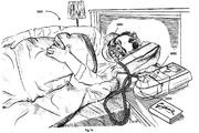
- Fig. 1a shows an example system suitable for implementation with in accordance with the present technology.
- a patient 1000 wearing a patient interface 3000 receives a supply of air at positive pressure from a PAP device 4000. Air from the PAP device is humidified in a humidifier 5000, and passes along an air circuit 4170 to the patient 1000.
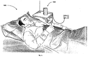
- Fig. 1b shows a contactless sensorunit monitoring a sleeping patient.
- Fig. 2a shows an overview of a human respiratory system including the nasal and oral cavities, the larynx, vocal folds, oesophagus, trachea, bronchus, lung, alveolar sacs, heart and diaphragm.
- Fig. 2b shows a view of a human upper airway including the nasal cavity, nasal bone, lateral nasal cartilage, greater alar cartilage, nostril, lip superior, lip inferior, larynx, hard palate, soft palate, oropharynx, tongue, epiglottis, vocal folds, oesophagus and trachea.
- Fig. 3a shows a patient interface in accordance with one form of the present technology.
- Fig. 4a shows a PAP device in accordance with one form of the present technology.
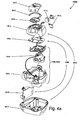
- Fig. 5a shows a humidifier in accordance with one aspect of the present technology.
- Fig. 6a shows a model typical breath waveform of a person while sleeping.
- the horizontal axis is time, and the vertical axis is respiratory flow.
- a typical breath may have the following approximate values: tidal volume, Vt, 0.5L, inhalation time, Ti, 1.6s, peak inspiratory flow, Qpeak, 0.4 L/s, exhalation time, Te, 2.4s, peak expiratory flow, Qpeak, -0.5 L/s.
- the total duration of the breath, Ttot is about 4s.
- the person typically breathes at a rate of about 15 breaths per minute (BPM), with Ventilation, Vent, about 7.5 L/minute.
- BPM breaths per minute
- a typical duty cycle the ratio of Ti to Ttot is about 40%.
- Fig. 6b shows a patient during Non-REM sleep breathing normally over a period of about ninety seconds, with about 34 breaths. This being treated with Automatic PAP, and the mask pressure was about 11 cmH 2 O.
- the top channel shows oximetry (SpO2), the scale has a range of saturation from 90 to 99% in the vertical direction. The patient maintained a saturation of about 95% throughout the period shown.
- the second channel shows quantitative respiratory airflow, and the scale ranges from -1 to +1 LPS in a vertical direction, and with inspiration positive. Thoracic and abdominal movement are shown in the third and fourth channels.
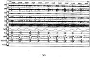
- Fig. 6c shows polysomnography of a patient
- the top two channels both are EEG (electoencephalogram) from different scalp locations.
- Periodic spikes in second represent cortical arousal and related activity.
- the third channel down is submental EMG (electromyogram). Increasing activity around time of arousals represent genioglossus recruitment.
- the fourth & fifth channels are EOG (electro-oculogram).
- the sixth channel is an electocardiogram.
- the seventh channel shows pulse oximetry (SpO2) with repetitive desaturations to below 70% from about 90%.
- the eighth channel is respiratory airflow using nasal cannula connected to differential pressure transducer.
- apneas Repetitive apneas of 25 to 35 seconds alternating with 10 to 15 second bursts of recovery breathing coinciding with EEG arousal and increased EMG activity.
- the ninth shows movement of chest and tenth shows movement of abdomen.
- the abdomen shows a crescendo of movement over the length of the apnea leading to the arousal. Both become untidy during the arousal due to gross bodily movement during recovery hyperpnea.
- the apneas are therefore obstructive, and the condition is severe.
- the lowest channel is posture, and in this example it does not show change.
- Fig. 6d shows patient flow data where the patient is experiencing a series of total obstructive apneas.
- the duration of the recording is approximately 160 seconds.
- Flow ranges from about +1 L/s to about -1.5L/s.
- Each apnea lasts approximately 10-15s.
- Fig. 6e shows a patient with Cheyne-Stokes respiration.
- the data span six minutes.
- the signal representative of flow was measured using a pressure sensor connected to nasal cannulae.
- the patient exhibits apneas of about 22 seconds alternating with hyperpneas of about 38 seconds.
- the higher-frequency, low-amplitude oscillation occurring during each apnea is cardiogenic.
- Fig. 7a is a block diagram illustrating anapparatus,including the contactless sensor unit of Fig. 1b, for monitoring the cardio-pulmonary health of a patient.
- Fig. 7b is a flow chart illustrating a method of monitoring the cardio-pulmonary health of a patient, as carried out by the monitoring apparatus of Fig. 7a .
- Fig. 7c is a flow chart illustrating a method of monitoring the cardio-pulmonary health of a patient, as implemented within the monitoring apparatus of Fig. 7a .
- Fig. 7d is a flow chart illustrating the example steps in a method of predicting clinical events from signals representative of patient movement, as may be used in the method of Fig. 7b or Fig. 7c .
- Fig. 7e is a block diagram illustrating the example modules that may serve to implement making up the feature extraction step in the method of Fig. 7d and their relationship under the combined or single-channelapproach in one form of the present technology.
- Fig. 7f is a block diagram illustrating the example modules making up that may serve to implement the feature extraction step in the method of Fig. 7d and their relationship under the hybridapproach in one form of the present technology.
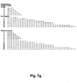
- Fig. 7g illustrates Lotjonen pattern expansion as applied during one implementation of the sleep / wake analysis module of Fig. 7e and 7f .
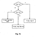
- Fig. 7h is a flow chart illustrating a method of setting a waveform length threshold in an alternative implementation of the sleep / wake analysis module of Figs. 7e and 7f .
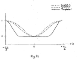
- Fig. 7i contains illustrations of examples of three generic SDB respiratory effort reduction templates.
- Fig. 7j is a flow chart illustrating a method that may be used to implement of the prediction step of the method of Fig. 7d .
- the present technology comprises apparatus for treating a cardio-pulmonary disorder.
- the treatment apparatus may comprise a PAP device 4000 for supplying pressurised respiratory gas, such as air, to the patient 1000 via an air delivery tube leading to a patient interface 3000.
- the present technology comprises a method for treating a respiratory disorder comprising the step of applying positive pressure to the entrance of the airways of a patient 1000.
- a PAP device 4000 in accordance with one aspect of the present technology comprises mechanical and pneumatic components 4100, electrical components 4200 and is programmed to execute one or more processes4300.
- the PAP device preferably has an external housing 4010, preferably formed in two parts, an upper portion 4012 of the external housing 4010, and a lower portion 4014 of the external housing 4010.
- the external housing 4010 may include one or more panel(s) 4015.
- the PAP device 4000 comprises a chassis 4016 that supports one or more internal components of the PAP device 4000.
- a pneumatic block 4020 is supported by, or formed as part of the chassis 4016.
- the PAP device 4000 may include a handle 4018.
- a humidifier 5000 comprising a water reservoir 5110, and a heating plate 5120.
- the present technology comprises apparatus 7000 for monitoring the cardio-pulmonary health of a patient.
- the apparatus 7000 comprises a contactless sensor unit 1200 positioned adjacent and relatively close to a sleeping patient 1000 (e.g. on a bedside table), as illustrated in Fig. 1b.
- Heart failure has been shown to be highly correlated with sleep disordered breathing (SDB).
- SDB sleep disordered breathing
- CSR Cheyne-Stokes respiration
- Fig. 6e is caused in general by an instability in the body's respiratory control system, one cause of which is heart failure.
- features indicative of the severity of OSA such as the Apnea / Hypopnea Index (AHI) have been shown to be independent predictors of death by, and hospitalization for, ADHF events. Therefore, one approach to predicting ADHF events is to use features that indicate the severity of sleep disordered breathing, i.e. SDB features.
- SDB features may be a set of features that indicate the extent to which respiration during sleep resembles classic CSR, i.e. "Cheyne-Stokes-like" features.
- the values of and changes in such SDB features may contain useful information about the likelihood of ADHF events.
- the disclosed monitoring apparatus 7000 is therefore configured to extract and analyse features indicative of the severity of sleep disordered breathing of the patient 1000, i.e. SDBfeatures.
- SDBfeatures may be determined in accordance with any of the methodologies described in International Publication Number WO 2006/066337 filed on 21 December 2005 .
- Cheyne-Stokes respiration features may also be a short term marker of changes during the acute phase of a decompensation event of a heart failure patient (a decompensation event occurs when the compensatory mechanisms that the heart uses to maintain adequate cardiac output, are no longer sufficient - these decompensations can lead to a rapid worsening of symptoms and often require hospitalization).
- the medical response to a decompensation is often to temporarily increase diuretic use, and hence remove excess fluid. This can cause consequent changes in the breathing parameters, and hence the "Cheyne-Stokes-like" features described above could be used for assessing short-term effectiveness of treatments.
- the modulation cycle length of the Cheyne-Stokes respiration can be correlated with the circulation delay of the body (e.g., see Dai Yumino and T. Douglas Bradley “Central Sleep Apnea and Cheyne-Stokes Respiration", Proceedings of the American Thoracic Society, Vol. 5, No. 2 (2008), pp. 226-236 .) and hence be used as a prognostic marker.
- Fig. 7a is a block diagram illustrating anapparatus 7000, including the contactless sensor unit 1200 of Fig. 1b, for monitoring the cardio-pulmonary health of a patient 1000 in more detail according to one form of the present technology.
- the contactless sensor unit 1200 includes a contactless motion sensor 7010 generally directed toward the patient 1000.
- the motion sensor 7010 is configured to generate one or more signals representing bodily movement of the patient 1000, from which may be obtained one or more signals representing respiratory movement of the patient.
- the sensor unit 1200 may also include a microcontroller unit (MCU) 7001, memory 7002 (e.g. a memory card) for logging data.
- MCU microcontroller unit
- the sensor unit 1200 includes communications circuitry 7004 configured to transfer data to an external computing device 7005, e.g. a local general purpose computer, or a remote server, via a connection 7008.
- the connection 7008 may be wired or wireless, in which case the communications circuitry 7004 has wireless capability, and may be direct or indirect via a local network or a wide-area network such as the Internet (not shown).
- the sensor unit 1200 includes a processor 7006 configured to process the signals generated by the motion sensor 7010 as described in detail below.
- the sensor unit 1200 includes a display device 7015 configured to provide visual feedback to a user.
- the display device 7015 comprises one or more warning lights (e.g., one or more light emitting diodes).
- the display device 7015 may also be implemented as a display screen such as an LCD or a touch-sensitive display. Operation of the display device 7015 is controlled by the processor 7006 based on an assessment of the patient's cardio-pulmonary health.
- the display device 7015 may be implemented to visually show information to a user of the monitoring apparatus 7000, such as a patient or a physician or other clinician.
- the display device 7015 may also display a graphical user interface for operation of the monitoring apparatus 7000.
- the sensor unit 1200 includes an audio output 7017configured to provide acoustic feedback to a user under the control of the processor 7006, e.g., a tone whose frequency varies with breathing, or an alarm which sounds when certain conditions are met.
- User control of the operation of the monitoring apparatus 7000 may be based on operation of controls(not shown) that are sensed by the processor 7006 of the monitoring apparatus 7000.
- a sensor unit 1200 is the SleepMinder device manufactured by ResMed Sensor Technologies Ltd, which contains a contactless Doppler radio-frequency (RF) motion sensor 7010.
- RF radio-frequency
- the motion sensor 7010 includes anRFtransmitter 7020 configured to transmit anRFsignal 7060.
- the carrier frequency is f c (typically in the range 100 MHz to 100 GHz, e.g. 3GHz to 12GHz, e.g. 5.8 GHz or 10.5 GHz)
- t is time
- ⁇ is an arbitrary phase angle
- u ( t ) is a pulse shape.
- the magnitude of u ( t ) may be unitary, and can be omitted from Eq. 1.
- T P ⁇ T
- the spectrum of the emitted signal becomes very wide, and the system is referred to as an ultra-wideband (UWB) radar or impulse radar.
- the carrier frequency of the RF transmitted signal 7060 can be varied (chirped) to produce a so-called frequency modulated continuous wave (FMCW) system.
- FMCW frequency modulated continuous wave
- the radio frequency signal 7060 may be generated by the transmitter 7020 using a local oscillator 7040 coupled with circuitry for applying the pulse gating.
- a voltage-controlled oscillator is used together with a voltage-frequency converter to produce the RF signal 7060for transmission.
- the coupling of the transmitted RF signal 7060 to the air may be accomplished using an antenna 7050.
- the antenna 7050 can be omnidirectional (transmitting power more-or-less equally in all directions) or directional (transmitting power preferentially in certain directions). It may be advantageous to use a directional antenna 7050 in the apparatus 7000 so that transmitted and reflected energy are primarily coming from one direction.
- a single antenna 7050 is used for both the transmitter 7020 and the receiver 7030, with a single carrier frequency.
- multiple receive and transmit antennas 7050 can be used, with multiple carrier frequencies.
- the apparatus 7000 is compatible in various embodiments with various types of antenna 7050 such as simple dipole antennas, patch antennas, and helical antennas, and the choice of antenna can be influenced by factors such as the required directionality, size, shape, or cost. It should be noted that the apparatus 7000 can be operated in a manner which has been shown to be safe for human use. The apparatus 7000 has been demonstrated with a total system emitted average power of 1 mW (0 dBm) and lower. The recommended safety level for RF exposure is 1 mW/cm 2 . At a distance of 1 meter from a system transmitting at0 dBm, the equivalent power density will be at least 100 times less than this recommended limit.
- the transmitted RF signal 7060 is reflected off objects that reflect radio waves (such as the air-body interface of the patient 1000), and some of the reflected signal 7070 will be received at a receiver 7030, which can be collocated with the transmitter 7020, or which can be separate from the transmitter 7020, in a so-called "bistatic" configuration.
- the received signal 7070 and the transmitted signal 7060 can be multiplied together in a mixer 7080 (either in an analog or digital fashion).
- This mixer 7080 can be of the form of a multiplier (as denoted below in (Eq.
- ⁇ ( t ) is a phase term resulting from the path difference of the transmitted and received signals 7060 and 7070 (in the case where the reflection is dominated by a single reflective object), and ⁇ is the attenuation experienced by the reflected signal 7070. If the reflecting object is fixed, then ⁇ ( t ) is fixed.
- the reflecting object e.g., the chest of the patient 1000
- ⁇ ( t ) will be time-varying.
- the mixed signal m ( t ) contains a component at f m (as well as a component centred at 2 f c which can be simply removed by low pass filtering).
- the signal at the output of the low pass filter after mixing is referred to as the raw or demodulated sensor signal 7003, and contains information about gross bodily (non-respiratory) movement, and respiratory movement.
- the amplitude of the demodulated sensor signal 7003 is affected by the mean path distance of the reflected signal, leading to detection nulls and peaks in the sensor 7010 (areas where the sensor is less or more sensitive). This effect can be minimised by using quadrature techniques in which the transmitter 7020 simultaneously transmits a signal 90 degrees out of phase(in quadrature) with the signal 7060 of Eq. 1. This results in two reflected signals, both of which can be mixed and lowpass filtered by the mixer 7080, leading to two demodulated sensor signals 7003a (the "I signal”) and 7003b (the "Q signal”) in respective I- and Q-"channels".
- an alternative method of acquiring a demodulated sensor signal 7003 may be used.
- a demodulated sensor signal 7003 may be computed as the average of the time delays over that period.
- the motion sensor 7010 e.g., a radio-frequency sensor, can estimate the respiratory movement of the chest wall, or more generally the movement of the part of the body of the patient 1000 whom the apparatus 7000 is monitoring.
- the received signal 7070 can include large motion artefacts, e.g. as the result of gross bodily movement. This is due to the fact that the reflected signals from the body can contain more than one reflection path, and lead to complex signals (for example if one hand is moving towards the sensor, and the chest is moving away). The reception of such signals is useful as it can indicate that the upper body is in motion, which is useful in determining sleep state.
- the sensor can also be used to detect motion of a lower part of the body (such as involuntary leg jerks) of a patient 1000, which are useful in the diagnosis of sleep disorders such as Restless Legs Syndrome or Periodic Limb Movements.
- the physical volume from which reflected energy is collected by the sensor unit 1200 can be restricted using various methods.
- the sensor unit 1200 can be made "directionally selective" (that is, it transmits more energy in certain directions), as can the antenna of the receiver 7030.
- Directional selectivity can be achieved using directional antennas 7050, or multiple RF transmitters 7020.
- a continuous wave, an FMCW, or a UWB radar is used to obtain similar signals.
- a technique called "time-domain gating" can be used to only measure reflected signals 7070 which arise from signals at a certain physical distance from the sensor unit 1200. Frequency domain gating (filtering) can be used to ignore motions of the reflected object above a certain frequency.
- the lower frequency can be used to determine large motions accurately without phase ambiguity, which can then be subtracted from the higher-frequency sensor signals (which are more suited to measuring small motions).
- the apparatus 7000 collects information from the patient 1000, and uses that information to determine respiratory movement, and more general bodily movement information.
- contactless motion sensors e.g. infrared sensors, ultrasound sensors, optical sensors, or contact motion sensors, such as piezoelectric sensors or respiratory inductance plethysmographs
- additional motion sensors may be used as additional motion sensors to, the contactless motion sensor 7010, either as part of the sensor unit 1200, or as part of a separate sensor unit
- Such additional motion sensors need to be positioned relative to the patient 1000 in accordance with the characteristics of the respective motion sensors.
- the motion sensor is a respiratory inductance plethysmograph
- the sensor may be positioned around the chest or abdomen of the patient 1000.
- the motion sensor is a piezoelectric sensor, or a direct air-mat pressure sensor, such asensor may be positioned under the mattress of the patient 1000.
- one or more contactless non-motion sensors configured to provide data relevant to the cardio-pulmonary health of the patient 1000 may be incorporated into the apparatus 7000 to enhance overall accuracy or provide additional robustness.
- a contactless non-motion sensors is a sound sensors. The characteristic sound patterns associated with sleep apnea may be analysed and used to enhance the accuracy of the generated summary measures of SDB. The system described by Karunajeewa A.S., Abeyratne U.R., Hukins C. in "Silence-breathing-snore classification from snore-related sounds", Physiol. Meas.
- acoustic-based screening for SDB is one example of acoustic-based screening for SDB, and such techniques could be readily incorporated into with the apparatus 7000 to provide a useful and robust tool for monitoringcardio-pulmonary health.
- the advantage of such incorporation is that the respiratory movement signal may be occasionally unusable due to excessive motion artefacts. At such times, the acoustic signal may provide reasonable estimates of respiratory parameters. Conversely, the acoustic signal may sometimes be unusable due to background noise contamination, at which times respiratory movement takes over as the primary sensor modality.
- the apparatus 7000 may incorporate one or more contact non-motion sensorsconfigured to provide data relevant to the cardio-pulmonary health of the patient 1000.
- contact sensors include an oximeter (which measures blood oxygen levels), an oronasal cannula (which directly measures respiratory airflow), and an electrocardiogram (ECG) monitor.
- ECG monitor may be configured to detect cardiac-related characteristics such as a heart rate and may also determine respiratory parameters (such as central or obstructive apneas, hypopneas, etc.)
- these parameters may be computed by the processor 7006 based on ECG data transmitted to the processor 7006, or they may be computed by the ECG monitor and be transmitted to the processor 7006.
- the oximetry sensor highlighted above has the advantage that it can also provide a respiratory effort signal; such a signal can be derived from the underlying photoplethysmogram (PPG) component of the oximeter by tracking the peak and trough values of the PPG.
- PPG photoplethysmogram
- one or more three-dimensional accelerometers can be worn by the patient which can provide information about their angle of recline, and their bodily position (supine, on left, on right, or prone). This information can be combined with the measured respiratory parameters to provide further insight into the patient status.
- the processor 7006of the sensor unit 1200 processes the signals 7003 acquired from the sensors, e.g. the motion sensor(s) 7010, as described in detail below.
- the instructions for the described processing may be stored on a computer-readable storage medium, e.g. the memory 7002 of the sensor unit 1200, and interpreted and executed by a processor, e.g. the processor 7006 of the sensor unit 1200.
- the processing may be canied out in "batch mode", i.e. the acquired signals 7003 are stored, either in memory 7002 of the sensor unit 1200 or in that of the external computing device 7005, for one or more complete monitoring sessions, and subsequently processed in a single batch.
- each monitoring session is one night in duration.
- the processing may be carried out in "real time" during the monitoring session.
- the external computing device 7005 may implement the described functionality, based on data transmitted to it by the sensor unit 1200 and any other sensors in the apparatus 7000 as described above.
- the above descriptions of the visual display 7015 and the audio output 7017 apply equally to comparable elements of the external computing device 7005.
- the external computing device 7005 is clinician-accessible apparatus such as a multi-patient monitoring system that allows a clinicianto review data from remote patient data recording devices such as the monitoring apparatus 7000.
- a database may be provided to record patient monitoring data.
- clinicians may receive a report or alert that the patient may require closer observation or should be brought to hospital.
- the processor 7006 executes processes 4300 tomonitor the patient's cardio-pulmonary health from data received from the sensor unit 1200 and any other sensors described above.
- the processes 4300 may be carried out to monitor the patient 1000 before, during, and after hospitalization.
- heart failure and COPD patients may suffer from exacerbation or decompensation (ADHF) events.
- ADHF decompensation
- Untreated exacerbation could lead to further exacerbation and potentially require hospitalization for the patient.
- exacerbation is discovered early enough, such as at the earliest stages of its onset, it may be treated in a manner that may avoid (re-)hospitalization.
- the processor 7006 analyses the processed or received sensor data to trigger generation of a clinical alert based on features related to cardio-pulmonary health extracted from the sensor data. Additional examples of such analysis, which may include evaluation of one or more change condition indicators, are described in U.S. Patent Application No. 12/751,174, filed on March 31, 2010 and U.S. Patent Application No. 12/483,357, filed on June 12, 2009 , the entire disclosures of which are incorporated herein by cross reference.
- the clinical alert may include a warning or alert message taking a number of forms.
- the processor 7006, to generate a clinical alert may activate a status light (e.g., an LED or an icon on the display device 7015) of the monitoring apparatus 7000. A more detailed message concerning the assessment of the indicator may also be displayed on the display device 7015.
- the processor 7006 may also, or alternatively, send an alert message to an external computing device 7005 associated with a clinician via the connection 7008.
- Such a message may take the form of a wired or wireless communication.
- the processor 7006 may generate an alert message via a paging system such as by automatically dialing a paging system.
- the processor 7006 may also be configured to generate an automated voice phone call message.
- the processor 7006 may also send the alert message by a fax transmission.
- the processor 7006 may also send an alert message via any internet messaging protocol, such as an email message, or by any other internet data file transport protocol.
- the alert messages may even be encrypted to keep patient information confidential.
- a typical alert message may identify the patient.
- Such a message may also include data recorded by the monitoring apparatus 7000 or any other recorded patient information.
- the alert message may even express that the patient should be considered for additional treatment, hospitalization, or an evaluation due to the detection of a potential ADHF event or exacerbation of COPD.
- alert messages may be directed by the processor 7006 to the patient via the display device 7015 of the monitoring apparatus 7000 and / or the clinicianvia the connection 7008, in some embodiments, the alert messages could be directed more selectively. For example, a first alert message may be only transmitted to a clinician by only transmitting the alert message to an external computing device 7005 through the connection 7008 without showing any alert on the display device 7015. However, a second alert message, which may be a more urgent message, could then be actively displayed on the display device 7015 in addition to being transmitted to the external computing device 7005. An audible alarm from an optional speaker controlled by the processor 7006 may also be implemented. Use of an audible alarm may depend on the urgency of the alert message.
- the processor 7006 may condition an alert on input responses to a patient query that may serve to avoid unnecessary alerts. For example, based on an analysisof sensor data by the monitoring apparatus 7000 (e.g., a comparison of one or more respiratory parameters with one or more thresholds) the processor 7006may trigger a presentation of a patient query to the patient to prompt the patient for input based on the assessment made by the processor. In such a case, the display device 7015 under control of the processor 7006 may present a query to the patient, prompting the patient to input additional information via a user interface.
- an analysisof sensor data by the monitoring apparatus 7000 e.g., a comparison of one or more respiratory parameters with one or more thresholds
- the processor 7006 may trigger a presentation of a patient query to the patient to prompt the patient for input based on the assessment made by the processor.
- the display device 7015 under control of the processor 7006 may present a query to the patient, prompting the patient to input additional information via a user interface.
- the presented question or questions of the query may be selected from a database, or other data structure of questions, such as a data structure in a memory of the apparatus, such that the selected questions are particularly associated with a pattern detected by the processor in the analysis.
- the processor 7006 may then further evaluate data of the receivedresponses to the query. Based on this further evaluation, the processor 7006may trigger an alert message, refrain from triggering an alert message, and/or delay a triggering of an alert message pending responses to one or more additional triggered queries. Such additional queries may be triggered after a certain time, after a further detected pattern or after a further use of the monitoring apparatus 7000.
- ADHF acute decompensated heart failure
- COPD COPD patient for exacerbations
- queries may serve to reduce false positives (e.g., when the processingflagsa need for clinical contact and the clinical contact is later found to have been unnecessary). This type of false positive may be due to changes in patient behavior, which may be corrected without medical intervention. Such behaviors may include missed or incorrect dosage of medication, non-compliance with dietary instructions and/or rest requirements, and the like.
- the processor 7006 may detect a respiratory pattern or potential clinical events that might require a clinical alert (e.g., a certain number of SDB events over a certain period of time that may be indicative of COPD exacerbation and/or ADHF). Based on the detected pattern or events, the processor 7006may present one or more questions in a queryto the patient on a user interface of the apparatus 7000. Such questions may address pharmaceutical and/or lifestyle compliance by the patient (e.g., has the patient been taking prescribed medication and/or following physician's treatment advice, etc.). Optionally, in some cases, one or more questions may address the operational integrity of the monitoring apparatus 7000 to ensure that the collected data is valid.
- the processor 7006 may pursue a series of queries over a predetermined span of time (such as one or more monitoring sessions or nights of sleep) and generate a clinical alert only after the predetermined span of time has elapsed.
- the processor 7006 may prompt a series of queries to the patient regarding diet that might have a causal relationship with the recurring pattern. If the patient is not in compliance with the dietary requirements as determined by the patient's responses input to the monitoring apparatus 7000, the processor 7006may then continue to monitor and query the patient again after a further monitoring time period has elapsed (e.g., query the patient after a number of minutes, hours, or days).
- the processor 7006 may then trigger a clinical alert message to a clinician via a notification infrastructure (e.g., tele-monitoring) to notify a clinician directly as described below.
- a notification infrastructure e.g., tele-monitoring
- a certain received response(s) to one or more questions of the query may alternatively rule out the triggering of such an alert message. For example, a query and response may determine that the patient was not wearing his or her mask and as a result, the processor 7006may refrain from triggering an alert.
- a heart failure patient does not take the prescribed diuretics for a period of time. Due to noncompliance, the patient experiences dyspnea and breathing irregularities at night.
- the processor 7006of the monitoring apparatus 7000 may detect such events, or a pattern of such events, which may be indicative of an imminent decompensation or exacerbation event. Instead of directly issuing a clinical alert to the clinician, the monitoring apparatus 7000 may trigger a query to the patient (e.g., via display device 7015) to determine whether the patient has taken the prescribed diuretics. The evaluation of the response may trigger a message to the patient, rather than an alert to a clinician, to remind the patient to take the medication if the input answer is negative.
- the processor 7006of the monitoring apparatus 7000 may then further evaluate the breathing patterns of the patient to see if the pattern recurs or has been resolved (by taking of the diuretics). An alert message would not be generated if the processor 7006does not detect the previously detected respiratory pattern that triggered the initial query.
- the processor 7006 may also confirm by a supplemental query that the patient has taken the medication. In such cases, monitoring may resume as usual thereafter. If, however, the recurring pattern is still or again detected by the processor after the initial query, a subsequent query, such as one with different questions, may be presented to the patient on the display device 7015.
- the monitoring apparatus 7000 may then trigger an alert message to notify the clinician of an imminent clinical event.
- the processor may be configured to dispose of simple cases of non-compliance (such as dietary or exercise) or explain apparatus malfunctions due to, for example, unintentional disconnections or power loss, without the need for a clinical alert message requesting that the patient be contacted.
- the processor 7006 may access the memory 7002 that includes a set of queries.
- Each question of a query may be associated with one or more detectable respiratory patterns or events.
- a question may be broad (e.g., "has patient complied with the prescribed diet?") or specific (e.g., "has patient diet been fortified with potassium?").
- the processor 7006 may then select a subset of questions for a query based on the detected pattern or event.
- Questions may be presented in series in response to a particular monitoring session.
- the processor 7006 may prompt two, three, four, five, six or more questions in a row so as to identify or rule out causes of the detected respiratory abnormality of the monitoring session that would or would not need an alert message.
- the processor 7006 may access an associated rank or priority for the question that represents an order of likelihood.
- the processor 7006 may conditionally present a series of questions according to the rank associated with each question. For example, the controller may present a first query in response to a predicted event.
- the response and determination may be logged and the processor 7006 may proceed to a second monitoring session during a predetermined period of time. If the respiratory pattern is again detected, a second query of a different rank from the first query may then be triggered. This detection and querying cycle may be repeated until no further queries remain or a response to a query indicates a need for an alert, after which the processor 7006may then trigger an alert message.
- Fig. 7b is a flow chart illustrating a method 7100 of monitoring cardio-pulmonary health as implemented within the monitoring apparatus 7000 in one form of the present technology.
- the method 7100 is carried out by the processor 7006 of the monitoring apparatus 7000, configured by instructions stored on computer-readable storage medium such as the memory 7002.
- the processor 7006 monitors and analyses data received from the sensor unit 1200 and any other sensors described aboveso as to predict whether a clinical event is likely to occur within a predetermined prediction horizon, as described in detail below. If at step 7120 no event is predicted ("N"), the processor 7006may continue monitoring / analysis at 7110. If a potential clinical event is predicted at 7120 "Y"), the processor 7006at 7130 triggers generation of one or more queries to evaluate a potential cause of the clinical event, such as by displaying them on display device 7015, as discussed in more detail herein, and/or on another processing device (e.g., tablet computer, mobile phone or other computing device etc.) in direct or indirect communication with the processor 7006.
- another processing device e.g., tablet computer, mobile phone or other computing device etc.
- the processor 7006 and/or such other processing device may proceed to step 7180 to trigger generation of a clinical alert immediately (shown as a dashed arrow 7145), or postpone the clinical alert generation by proceeding to step 7150.
- the processor 7006 may control adjustment of a treatment in response to the received responseto the patient queries. For example, the processor 7006may modify atarget ventilation, or other treatment control parameters of a PAP device 4000. Alternatively, the processor 7006may maintain the same treatment as that provided during the monitoring / analysis step 7110, and instead issue an instructional message to instruct the patient to make an adjustment, such as a change in diet or medication or a resetting or repair of the apparatus 7000 or PAP device 4000.
- a predetermined delay e.g.
- the apparatus monitors and analyses the sensor datain similar fashion to step 7110 to determine whether the instructed or controlled adjustment of step 7150 resolved the issue previously detected atstep 7110 (e.g., by determining if the predicted clinical event is still predicted). If at step 7170 the issue has been resolved (e.g., the clinical event is no longer predicted) ("Y"), the method 7100 returns to step 7110 to continue monitoring and analysingsensor data. If the clinical event is still predicted ("N"), the method 7100 may return to step 7130 or 7150 (via dashed arrows 7175 and 7178 respectively) if further queries or adjustments are available, or if not, generation of a clinical alert is triggered at step 7180 as described in more detail herein.
- the monitoring apparatus 7000 may be implemented with additional operations. For example, as previously mentioned, the monitoring apparatus 7000 may be useful to determine whether or not a hospitalized patient (e.g., a heart failure or COPD patient) is ready to be released or should not be released from the hospital. Releasing a patient too soon may not be beneficial for the patient, particularly if a relapse occurs shortly thereafter and the patient must be re-admitted. Heart failure and COPD patients suffer from decompensations and/or exacerbations and frequently require re-admission to a hospital due to relapse that might be avoided with a longer initial stay. Similarly, releasing a patient too soon may have consequences for other entities.
- a hospitalized patient e.g., a heart failure or COPD patient
- hospitals may not be reimbursed or may only be partially reimbursed for the costs associated for a re-admission as a result of such a relapse.
- Some forms of the present technology evaluate a patient's condition during a first time period to provide a prediction relating to the potential for relapse in a subsequent time period.
- the potential for post-hospitalization decompensation and/or exacerbation events may be predicted from an analysis of respiratory parameters during hospitalization, such as an analysis of the respiratory movement signalsas previously described.
- a monitoring apparatus 7000 may be configured to monitor and analyse respiratory parameters during a time period from admittance to discharge that may be indicative of a likelihood of post-discharge relapse. By monitoring and analysing the respiratory parameters within the period from admittance to a point near or prior to discharge, the monitoring apparatus 7000 may determine whether the patient might be at risk for re-admission soon after release. In this regard, the methodology of the monitoring apparatus 7000 can provide a potential relapsealert to a clinicianto advise whether release might be premature due to a risk of relapse.
- the monitoring apparatus 7000 might warn the clinicianthat once released, the patient might be at a high risk for relapse and/or readmission and may further advise that careful monitoring for relapse should be considered for the particular patient or that the release should be reconsidered or postponed.
- a potential relapse alert may be presented as a numerical value, such as a Booleanindicator or a probability, which may comprise an estimate within a range so as to yield a scaled indication of greater or lesser risk of future relapse.
- Fig. 7c is a flow chart illustrating a method of monitoring the cardio-pulmonary health of a patient as implemented within the monitoring apparatus 7000 in one form of the present technology.
- the method 7200 which may be used with a patient while the patient is hospitalized, is carried out by the processor 7006 of the monitoring apparatus 7000 and/or a processor in direct or indirect communication with such processor, configured by instructions stored on computer-readable storage medium such as the memory 7002.
- the method 7200 at the first step 7210 extracts, monitors, and records respiratory parameters of a patient over the admission period, including one or more of the breathingrate, minute ventilation, tidal volume, sleep-disordered breathing severity indicators(e.g., apnea / hypopnea index), inspiratory waveform shape, expiratory waveform shape and the like,from data obtained with any one or more of the previously mentioned sensors.
- the method 7200 may perform an analysis of one or more of the extractedrespiratory parameters to calculate an indicator of potential relapse or a probability of relapse at step 7220. Step 7220 may involve assessing how the parameters have changed during the course of the hospitalization period.
- One or more of the parameters or the changes therein may be compared to one or more thresholds in order to compute an indicator of potential relapse.
- Such thresholds may be empirically determined from historic data of one or more hospitalized patients who have relapsed and/or not relapsed after hospitalization.
- data may be based on thresholds taken from historic data of the particular patient.
- a probability of relapse may be calculated at step 7220 based on a pattern of a plurality of the parameters or changes in a plurality of parameters.
- Such comparisons and/or pattern evaluations may be made, for example, by a decision-tree, a classifier or any other method of evaluation.
- a potential relapse alert may be generated at step 7230.
- data indicative of the probability of relapse such as the potential relapse indicator or the calculated probability of potential relapse, may be displayed on display device 7015 and/or sent as a message to a clinician via the connection 7008 at step 7230.
- the potential relapse indicator or probability of relapse may be continuously updated and displayed or transmitted based on a continuous monitoring and analysis of the extracted respiratory parameters during the monitoring period of hospitalization.
- the indicator, probability and/or message concerning the potential for relapse may be generated in response to a request made by a user, such as a clinician, either through a user interface of the apparatus 7000 (e.g., one or more buttons or operation of a user interface of the apparatus) or by transmitting an electronic request to initiate the method 7200.
- the monitoring apparatus 7000 may be configured to generate the potential relapse alert based on its previous analysis of the respiratory parameters.
- the respiratory parameters used in the method 7200 may be extracted by and transmitted from the monitoring apparatus 7000 to the external computing device 7005, which may implement the steps 7220and 7230 that calculate the probability of potential relapse or relapse indicator from the received respiratory parameters, and generate the potential relapse alert.
- Fig. 7d is a flow chart illustrating the principal steps in a method 7300 of predicting clinical events from signals representing movement (movement signals).
- the method 7300 may be used to implement step 7110 of the method 7100 in one form of the present technology in which the sensor data are one or more movement signals obtained from the motion sensor 7010, and the features related to cardio-pulmonary health are one or more SDB features.
- the method 7300 may also be used to implement steps 7210 and 7220 of the method 7200 in one form of the present technology in which the sensor data are one or more movement signals obtained from the motion sensor 7010, and the respiratory parameters are one or more SDB features.
- the method 7300 is carried out by the processor 7006 of the monitoring apparatus 7000, configured by instructions stored on computer-readable storage medium such as the memory 7002, and the movement signals are obtained from the contactless motion sensor 7010.
- the method 7300 starts at step 7310, at which the movement signals are pre-processed to condition them for further processing.
- the pre-processing step 7310 (shown dashed in Fig. 7d ) is optional and may be omitted.
- the pre-processed movement signals are analysed to extract SDBfeatures indicative of the severity of SDB by the patient 1000 during the period represented by the movement signals.
- the method 7300 uses the extracted SDBfeatures to predict whether a clinical event is likely to occur within the predetermined prediction horizon.
- the contactless motion sensor 7010 is a Doppler RF motion sensor.
- the motion sensor 7010 provides two raw movement signals, labelled I and Q signals 7003a and 7003b, each generally indicative of bodily movement, but generally 90 degrees out of phase with each other.
- the steps 7310 and 7320 are performed on each of the I and Q signals 7003a and 7003bin parallel, and the separately obtained features are combined during the feature extraction step 7320.
- the pre-processing step 7310 may be omitted.
- the I and Q signals 7003a and 7003bare combined as part of the pre-processing step 7310, and the processing steps 7320 to 7330 are carried out on the combined movement signal.
- the combined approach has the advantage of less computational complexity than the parallel approach, at the potential cost of lower prediction accuracy.
- the contactless motion sensor 7010 provides a single movement signal 7003. This is referred to below as the "single-channel" approach.
- step 7330 is omitted, i.e. no prediction of clinical events is made.
- the SDB features extracted at step 7320 may be used to trigger clinical alerts as described above.
- the extracted SDB features may be stored in the memory 7002 or transmitted to the external computing device 7005 for later diagnostic review.
- the pre-processing step 7310 begins by combining the land Q signals 7003a and 7003b in an adaptive geometrical manner into a combined movement signal c.
- the combination substep comprises three stages, applied to a window that slides along the I and Q signals 7003a and 7003b.
- the window is of 10 seconds duration with 50% overlap.
- the combined movement signal c is then (optionally) de-trended to remove baseline wandering.
- de-trending is implemented using double-pass median filtering.
- the de-trended signal c 1 is (optionally) bandpass filtered with a Butterworth bandpass filter with range set to the frequency range of respiratory functioning, this being in one implementation [0.1 Hz, 0.8 Hz] (corresponding to 6 to 48 breaths per minute).
- a further (optional) sub-stepin the pre-processing step 7310 is noise reduction.
- Artefactness quantifies the degree to which an artefact affects the signal at that scale.
- Artefactness is a measure of the skewness of the signal which can contain unlikely high amplitude values.
- the combinationsub-step is omitted from the pre-processing step 7310, and any or all of the subsequent sub-steps (de-trending, filtering, and noise reduction) are performed in parallel on each of the I and Q signals 7003a and 7003b.
- any or all of the de-trending, filtering, and noise reduction sub-steps are performed on the movement signal 7003.
- the input(s) to the feature extraction step 7320 is / are referred to as (pre-processed) movement signal(s) to reflect the optional nature of the pre-processing step 7310.
- Fig. 7e is a block diagram 7400 illustrating the modules making up one implementation of the feature extraction step 7320 and their relationship under the combined or single-channel approach
- an activity estimation and movement detection module 7410 generates an activity count signal and a movement flag series from the (pre-processed) movement signal.
- a presence / absence detection module 7420 generates a presence / absence flag series from the (pre-processed) movement signal and the movement flag series.
- a sleep / wake analysis module 7430 calculates a hypnogram from the presence / absence flag series, the movement flag series, and the activity count signal.
- a breathing rate estimation module 7440 generates a series of estimates of the breathing rate of the patient from the (pre-processed) movement signal and the hypnogram.
- a signal selection module 7450 selects sections of the (pre-processed) movement signal, using the movement flag series and the hypnogram.
- a modulation cycle metrics calculation module 7455 generates an estimate of the modulation cycle length of the patient's respiration from the selected sections of the (pre-processed) movement signal.
- An envelope generation module 7460 generates envelopes of the selected sections of the (pre-processed) movement signalusing the estimated breathing rate.
- An SDB event detection module 7465 generates candidate SDB events from the selected sections of the (pre-processed) movement signalusing the estimated modulation cycle length.
- An SDB event confirmation module 7470 confirms the candidate SDB events generated by the SDB event detection module 7465.
- a feature calculation module 7580 calculates SDB feature values from the confirmed SDB events.
- modules 7410 to 7470 of the block diagram 7400 are simply duplicated to process each of the I and Q signals 7003a and 7003b independently.
- a modified version of the feature calculation module 7480 combines the SDB events from the two parallel processing streams to calculating a single SDB feature set for the two (pre-processed) movement signals.
- Fig. 7f is a block diagram 7500 illustrating the modules making up one implementation of the feature extraction step 7320 and their relationship under the hybrid approach.
- an activity estimation and movement detection module 7510 generates an activity count signal and a movement flag series from the I and Q signals 7003a and 7003b.
- a presence / absence detection module 7520 generates a presence / absence flag series from the I and Q signals 7003a and 7003b and the movement flag series.
- a sleep / wake analysis module 7530 calculates a hypnogram from the presence / absence flag series, the movement flag series, and the activity count signal.
- a breathing rate estimation module 7540 generates a series of estimates of the breathing rate of the patient from the I and Q signals 7003a and 7003b and the hypnogram.
- Signal selection modules 7550a and 7550b select sections of the I and Q signals 7003a and 7003b respectively, using the movement flag series and the hypnogram.
- a modulation cycle metrics calculation module 7555 generates an estimate of the modulation cycle length of the patient's respiration from the selected sections of the I and Q signals 7003a and 7003b.
- Envelope generation modules 7560a and 7560b generate envelopes of the selected sections of the I and Q signals 7003a and 7003b respectively using the estimated breathing rate.
- SDB event detection modules 7565a and 7565b generate candidate SDB events from the selected sections of the I and Q signals 7003a and 7003b respectively using the estimated the modulation cycle length.
- SDB event confirmation modules 7570a and 7570b confirm the candidate SDB events generated by the SDB event detection modules 7565a and 7565b respectively.
- a feature calculation-module 7580 calculates SDBfeature values from the confirmed SDB events.
- the activity estimation and movement detection module returns a time series of "activity count” valuesindicating the level of bodily activity and a time series of binary movement flags indicating the presence or absence of gross bodily (non-respiratory)movement. Each time series is sampled at the sampling rate of the (pre-processed) movement signals (the movement signal frequency), equal to 10 Hz in one implementation.
- the movement flag series is used by the presence / absence detection, sleep / wake analysis, signal selection, and breathing rate estimation modules described below.
- the signal selection module selects for analysis those sections of signal that are not dominated by gross bodily movement.
- the activity count series, as well as the duration of movements and the time elapsed from the previous movement, is used by the sleep / wake analysis module to determine the sleep/wake state of the patient.
- the activity estimation and movement detection module is based on movement signal power levels, movement signal morphology, and movement patterns.
- five main sub-modules form part of the activity estimation and movement detectionmodule.
- An activity mappingsub-module generates a series of activity count values which are generally proportional to the power of the movement signal at corresponding times.
- the activity count is generated in the following manner:
- amovement detector sub-module proceeds as follows.
- the signal is first highpass filtered to remove VLF components and baseline. Three features are then derived from the baseline-removedsignal. The first feature is referred to as a noise feature.
- the second and third features are correlation and power features and are computed on a different frequency band to the noise feature.
- the mean breathing rate of the overall monitoring session is first estimated by running an unbiased autocorrelation of the baseline-removed signal and finding the first minimum, which defines half the mean breathing rate.
- the mean breathing rate defines a patient-specific acceptable breathing rate frequency window as a range of frequencies around the mean breathing rate.
- a lowpass filter with cutoff around 2 Hz in one implementation, is applied to the baseline-removed signal.
- the lowpass filtered signal is then partitioned into overlapping windows, de-trended and de-meaned.
- the windows are 15 seconds long with 12 seconds of overlap (three seconds of shift).
- the correlation feature is calculated by:
- the power feature may be calculated by:
- a maximum filter may be run over the correlation and power features so calculated.
- All three features may bethen interpolated to the movement signal frequency so that a feature value is present for each sample in the (pre-processed) movement signal.
- the noise feature may be normalized by the mean power feature calculated for all samples with high correlation feature values (those with values greater than 0.9) and those with high power feature values (those with values greater than 0.8).
- the baseline of the noise feature may be calculated by assigning to noise feature values over 0.003 the 95th percentile of all values below 0.003. Then a median filter of width 180 seconds is run and the baseline is defined as the 50th percentile of the filtered series. This baseline is then subtracted from the noise feature.
- the corresponding movement flag is set to true. If the baseline removed noise feature is above its mean value, the movement flag is also set to true.
- the movement flag series is finally expanded before and after each detected movement section by running the movement flag series through a maximum filter of multi-second duration, equal to 4 seconds in one implementation.
- the movement correction sub-module produces a binary "validation" flag for the movements detected in the movement detector sub-module, by analysing trends of movement over several minutes. Movements associated with apneas, in fact, need not to be labelled as such as it would impair the performance of both the sleep/wake analysis and SDB event detection modules.
- the difference between the value of each peak and its preceding trough may be calculated and, if found to be greater than a set threshold, the whole interval is marked as non-movement, as a repetitive pattern compatible with a train of SDB events is deemed to be likely.
- the combination sub-module is not needed.
- the final sub-module calculates movement state duration. This sub-module identifies all movement / no-movement transitions and generates a vector with the duration of all individual movement/no movement states.
- the mean non-zero activity count may also be calculated and stored.
- the total duration of intervals with non-zero activity counts may be calculated and stored as the Total Movement Time.
- the percentage of intervals with non-zero activity counts during the monitoring session mayalso be calculated and stored. This percentage may also becalculated on those periods that are marked as the first and second halvesof the main sleep period (defined below).
- the presence / absence detection module detects whether the patient is present within the field of the sensor 7010 (i.e. in bed), or is absent (i.e. not in bed).
- the presence/absence detection module generates a time series of presence / absence flagsbased on signal power levels, signal morphology, and the movement flag series generated by the activity estimation and movement detection module.
- the presence / absence detection module may include sub modules.
- five main sub-modules may be implemented as follows:
- the presence / absence flag is set to False (absence) if the RMS value of the (pre-processed) movement signal within the epoch falls below a "noise-only" threshold, set to 0.015 in one version of the alternative implementation in which the epochs are of 5-second duration. Otherwise, the presence / absence flag for the epoch is set to True (presence).
- the alternative implementation does not use the movement flag series.
- the sleep/ wake analysis module aims to classify each epoch as asleep, awake, absent, or unknown. (The epochscoincide with the epochs of the presence / absence detection module.) The resulting series of epoch classifications is referred to as a hypnogram.
- the sleep / wake analysis module uses the activity estimation and movement detection module outputs to generate features which are fed into a classifier for each epoch. Some initial post-processing, integration with the presence/absence flags from the presence/absence detection module, and final post-processing allows the sleep / wake analysis moduleto refine the hypnogram.
- the sleep/ wake analysismodule may be implemented with a number of sub-modules (e.g., four) as follows.
- a feature extraction sub-module extracts some or all of the following features from the activity count signal and movement flag series:
- the classifier is a pre-trained linear discriminant analysis (LDA) classifier.
- a post-processing sub-module may process the provisional sleep / wake labelseries in the following fashion:
- the first temporary hypnogram becomes the final hypnogram after the following final post-processing steps:
- the sleep / wake analysis module uses the presence / absence flag series from the presence / absence detection module and the combined (pre-processed) movement signal to generate the hypnogram labelling each epoch as Sleep, Wake, or Absent.
- the sleep / wake analysismodule does not use the movement flag series generated by the activity estimation and movement detection module, but rather generates its own movement flag series (one flag for each epoch, indicating gross bodily movement within the epoch) by comparing the variance of the movement signal within the epoch with an adaptive threshold for the monitoring session.
- the adaptive threshold may, for example, beset in one version of the alternative implementation to the 95 th percentile of the variance over the monitoring session minus twice the 75 th percentile of the variance over the same monitoring session (when the 75 th percentile is bigger than 0.5), or to the 95 th percentile of the variance over the monitoring session minus 1.5 times the 75 th percentile of the variance over the monitoring session (when the 75 th percentile is less or equal to 0.5). If the variance exceeds the adaptive threshold, the movement flag for the epoch is set to True.
- the epochs labelled as Absent in the presence / absence flag series are first labelled as Absent in the hypnogram.
- a "waveform length" feature is extracted from the (pre-processed) movement signal c 3 within each epoch labelled as Present in the presence / absence flag series. Waveform length is calculated by measuring the cumulative changes in amplitude from sample to sample over the epoch. Metaphorically, it is the equivalent of treating the movement signal waveform like a jumbled string, and measuring the straightened-out length of the string.
- the waveform length feature effectively encapsulates the amplitude, frequency, and duration of the movement signal over the epoch.
- the waveform length feature value w i for each Present epoch i is compared with an adaptive threshold T . If the value w i is less than the threshold T , the Present epoch is labelled as Sleep in the hypnogram; otherwise, the Present epoch is labelled as Wake.
- the threshold value T for the monitoring session the inter-quartile range features are calculated for the waveform length feature w i over the monitoring session (with each point in this feature vector representing the waveform length of an epoch, in similar fashion to the well-known R&K sleep scoring criteria).
- the threshold T is computed from the 50 th , 75 th , and 95 th percentiles w 50 , w 75 , and w 95 of the waveform length feature w i using the method illustrated in the flow chart of Fig. 7h .
- the parameters ⁇ 1 , ⁇ 2 , ⁇ 1 , ⁇ 2 , and ⁇ 3 are set to 4, 2.78, 10, 0.95, and 4 in one version of the alternative implementation.
- the hypnogram might indicate multiple separate contiguous sections where the patient is present (i.e. marked as Sleep or Wake). This is especially true for patients who are spending part of the daytime in bed because of their condition.
- the summary sub-moduletherefore generates "temporary sleep periods" by locating contiguous sections marked as Sleep or Wake, ignoring Absent-marked sections that are shorter than a threshold, set in one implementation to 30 minutes. For example, from five sections with the patient present during the monitoring session, two "temporary sleep periods" might be generated, because three of the four Absent-marked gaps between the presence sections are shorter than the threshold.
- the summary sub-modulethen defines a "main sleep period” as the longest of the "temporary sleep periods” so generated, and marks the start and end of the first and second halves of the main sleep period.
- the breathing rate estimation module makes an offline non-causal estimation of the breathing rate for each epoch of the monitoring session, using a breath detector based on peak-trough detection of the (pre-processed) movement signals. (The epochs coincide with the epochs of the presence / absence detection module and the sleep / wake analysis module.)
- the output of the breathing rate estimation module is referred to as a respirogram.
- four main sub-modules may serve as a breathing rate estimation module:
- the breathing rate estimation module Given the length of the window used for breathing rate estimation, the breathing rate estimation module includes a fair amount of averaging and is therefore fairly robust against artefacts. However, for the same reasons, it is not very sensitive to local breathing rate variations.
- the breathing rate is assigned a NaN value.
- the breathing rate estimate is simply the value Freq computed using (Eq. 19) by the activity estimation and movement detection sub-module.
- the mean breathing rate over the monitoring session together with its standard deviation and the 5th, 25th, 50th, 75th and 95th percentile of the distribution, are may also be calculated and stored by the breathing rate estimation module.
- the signal selection module aims at selecting suitable sections of the (pre-processed) movement signals for subsequent SDB event detection.
- One idea underlying signal selection is that SDB event detection may be performed only on sections of the movement signals where the patient is asleep.
- the hypnogram output of the Sleep/Wake analysis module can therefore serve to gate the SDB event detection through the signal selection module.
- movement sections are also removed by the signal selection module, leaving only sleep-and-breathing sections for SDB event detection.
- the methods employed in the SDB event detection module are time-domain amplitude-dependent methods, it is helpful to ensure that a sufficient signal to noise ratio is present, in order to guarantee a sufficient margin to detect the drop in respiratory effort that characterises an SDB event.
- the signal selectionmodule works as an interface between the Sleep/Wake analysis module and the SDB event detection module. Its main function is to feed the subsequent signal processing modules only with sections of sleep and breathing and sufficient signal quality.
- the hypnogram (upsampled to the sampling rate of the movement flag series) and the movement flag series are initially combined to obtain a mask of sleep and breathing.
- the mask has the value 1 at samples where the hypnogram indicates sleep and the movement flag is false.
- the mask of sleep and breathing is then applied to each (pre-processed) movement signal, resulting in one or more sections of variable duration.For each of these sections, a figure of merit is calculated, and a decision on whether that sectionshould be selected is taken based on the comparison of a figure of merit with a threshold.
- a sleep-and-breathing signal section should have an RMS value that is at least given by rms ⁇ 2 ⁇ 4 2 2 ⁇ rms max noise wherer ms maxnoise is a maximum RMS value for noise.
- the signal selection module may therefore apply a running standard deviation (RMS) filter, of duration equal to 150 seconds in one implementation, to each section of sleep-and-breathing (pre-processed) movement signal.
- the section is deemed to be of good quality, and is therefore selected by the signal selection module, when a high percentile, set in one implementation to the 75 th percentile, of the distribution of the RMS values for the section exceeds a noise threshold.
- the noise threshold is calculated as the smaller of a maximum RMS noise value for the motion sensor 7010 (equal to 0.005 for the SleepMinder sensor unit) and the 5 th percentile of the distribution of the RMS values of the pre-processed movement signal calculated for the section.
- each (pre-processed) movement signal is processed independently so certain sections might be selected for one channel only.
- the total analysis time (TAT) is calculated as the total duration of signal that has been selected for SDB event detectionin at least one channel.
- the TAT is used as the denominator of the AHI calculated by the feature calculation module described below.
- the total duration of sleep-and-breathing signalsections for each channel is computed and stored.
- the total duration of good-quality sleep-and-breathing signal for each channel is computed and stored.
- the total duration of poor-quality sleep-and-breathing signal for each channel is also computed and stored.
- At least one of the following conditions may beimposed before continuing with the SDB event detection in each channel:
- the envelope generation module aims at generating a measure of the respiratory effort, in the form of an envelope that retains not only the frequency but also the amplitude content of breathing modulation.
- the envelope generation module relies on a few signal processing steps. For each section of good quality sleep-and-breathing signal selected by the signal selection module any of the following may be implemented:
- the envelope of each selected section c 3 of the (pre-processed) movement signal may be generated by:
- the envelope may be generated by lowpass filtering the modulus of c 3 + jH ⁇ c 3 ⁇ , where j is the square root of -1.
- the lowpass filter has a cutoff frequency of 0.15 Hz.
- Themodulation cycle metrics calculation module estimates the mean modulation cycle length of the patient's modulated breathing cycles to better tailor the SDB event detection module to that patient.
- the main challenges are the selection of correct samples to base the estimation upon and the extraction of the modulation frequency in the VLF band of the spectrum using the selected samples.
- the modulation cycle metrics calculation module begins by pre-processing each selected section of the (pre-processed) movement signal in an identical manner to that of the envelope generation module described above (outlier removal and bandpass filtering).
- each pre-processed section may be normalised to zero mean and unity standard deviation, and an envelope may be generated for each normalised section using any of the methods described above in the envelope generation module.
- overlapping macro-epochs may be generated.
- the choice of the duration of the macro-epochs is influenced by the fact that modulated breathingcycles might last up to two minutes each for some patients and that three wavelengths are desirable for estimating the modulation cycle length.
- the macro-epochs have a duration of six minutes with an overlapping of four minutes
- Each macro-epoch may be processed with any of the following steps:
- All macro-epochs for the monitoring session may then be processed with any of the following:.
- the mean of all remaining modulation cycle lengths (weighted by their modulation power) over the monitoring session may then becomputed, together with its standard deviation and 5 th , 25 th , 50 th , 75 th and 95 th percentiles.
- congestive heart failure and CSR are connected is the buildup of fluid in the pulmonary cavity, which affects the "loop gain" of the body's respiratory control system.
- the extent of CSR depends not only upon the raw amount of fluid in the pulmonary cavity, but also upon its distribution. This distribution changes over the monitoring session, since the person's body position has changed at the start of the session from generally vertical to generally horizontal. Therefore, the values of the extracted features may be expected to vary over of the monitoring session. The later values of those features during a monitoring session may provide a truer indication of the severity of congestive heart failure and therefore the likelihood of ADHF events that the earlier values, since by that time the fluid should have assumed a new equilibrium distribution.
- the mean of all remaining modulation cycle lengths (weighted by their modulation power), together with its standard deviation and 5 th , 25 th , 50 th , 75 th and 95 th percentiles, mayalso therefore becomputed for those periods marked as first and second halves of the main sleep period by the sleep / wake analysis module.
- values of modulation power and OBW for each macro-epoch with at least one valid peak may becalculated and stored, together with their means, standard deviations, and their 5 th , 25 th , 50 th , 75 th and 95 th percentiles for the overall monitoring session and each half of the main sleep period.
- the statistics from both channels may be combined by averaging the statistics from both channels, weighted by the number of valid macro-epochs for each channel. Under the other approaches, such combination is not needed.
- the SDB event detection module aims todetectportions of the movement signal where SDB events, including both OSA and CSR episodes, are likely to have occurred. Such portions are referred to as candidate SDB events.
- the SDB event detection module uses the modulation cycle length estimated by the modulation cycle length estimation module to make a set of one or more generic SDB respiratory effort templates specific to the patient 1000.
- the generic templates may be designed to detect a reduction in respiratory effort associated with an SDB event. By cross-correlatingthe patient-specific templates against the envelope (measure of respiratory effort) generated by the envelope generation module, and comparing with a threshold, candidate SDB events may bedetected.
- Fig. 7i contains illustrations of three example generic SDB respiratory effort reduction templates in one versionof this implementation.
- Appendix A contains specifications for each of the three generic templates, parameterised by the modulation cycle length CL.
- the generic templates are instantiated by filling in the modulation cycle length value estimated by the modulation cycle length estimation module as the parameter CL.
- a correlation feature is generated by cross-correlating the envelope with each of the patient-specific templates and accepting the maximum value of the correlation values for each sample. In one implementation, this operation is performed at a small fraction, e.g. one tenth, of the sampling rate of the movement signals, to reduce the computational burden.
- a threshold set to 0.5 in one implementation, for a time that is greater than another threshold (set in one implementation to 1/6 of the modulation cycle length or 10 seconds, whichever is smaller)
- a candidate SDB event is detected.
- CandidateSDB events too close to each other, i.e. such that their mid-points are within half of the mean modulation cycle length are discarded.
- the result is a vector of locations of candidate SDB events within the monitoring session.
- SDB event detection module detects candidate SDB events by comparing the drop in the respiratory effort envelope with a baselineaccording to the AASM scoring rules:
- the main window is the one in which the mean envelope value is computed as a proxy for the airflow.
- the main window is a sliding window of 10 seconds with 75% overlap.
- the other two windows, which surround the main window as it slides, are utilized to determine the baseline.
- these two baseline windows comprise the previous 35 seconds and the next 35 seconds before and after the main window. If the mean envelope value in the main window satisfies either of the above criteria (without the oxygen desaturation) with respect to the baseline defined by the mean envelope value in the two baseline windows, a candidate SDB event is detected.
- the alternative implementation of the SDB event detection module then discards candidate SDB events of duration less than 10 seconds, in accordance with the AASM scoring rules described above.
- a further alternative implementation of the SDB event detection module is also suitable for the combined or single-channel approach.
- Oneversion of the further alternative implementation operates on the un-pre-processed and pre-processed movement signal sc and c 3 and the envelope Ec 3 , and therefore does not use the modules 7410 to 7450.
- Var is computed on a 30-second sliding window with 75% overlap.
- Spectral analysis provides a straight forward and robust way to extract the main harmonic of the signal c 3 , which is the respiratory movement when such a movement is present.
- an Auto-Regressive Yule Walker method (of order 9) may be used to estimate the Power Spectral Density (PSD) of c 3 and its envelope Ec 3 , thus producing two PSDs: P c 3 ( ⁇ ) and P Ec 3 ( ⁇ ).
- the bandwidth of a signal may bea measure of its "irregularity".
- ⁇ c 3 is the location of the highest peak of the magnitude of the PSD P c 3 ( ⁇ )
- ⁇ is the frequency range of interest
- P ⁇ c 3 ( ⁇ ) is the normalised PSD of c 3 computed as:
- P ⁇ c 3 ⁇ P c 3 ⁇ ⁇ ⁇ P c 3 ⁇ d ⁇
- the spectral entropy takes full advantage of the previously estimated PSDs.
- the spectral entropy Hc 3 of the signal c 3 and the spectral entropy HEc 3 of the envelope Ec 3 are both computed using (Eq. 22).
- the entropies Hc 3 and HEc 3 have characteristic behaviours depending on the breathing category.
- a clustering algorithm based on Gaussian Mixture Models is applied to the entropies and Hc 3 and HEc 3 to find an approximation of the set of three probability densities that best correspond to the three categories.
- a Landmark-based Spectral Clustering is applied first followed by a Gaussian fit of the probability densities of the clusters.
- the centres (means) of the three Gaussians identified by the clustering, by either version, are expressed as 3-vectors G c 3 and G Ec 3 .
- Hhc 3 min Med G c 3 , 0.8
- H i Sat NL exp ⁇ ⁇ H c 3 ⁇ Hhc 3 , T c 3 , S c 3
- S c 3 are parameters.
- Boolean-valued indicator or "truth" variables CS T , M T , NM T , and AH T may becomputed as time series, indicating that the movement signal belongs to (true) or does not belong to (false) the respective classes at a given time instant.
- the first three truth variables CS T , M T , NM T which are mutually exclusive in the sense that only one of the three can be true at any time, are computed by applying a set of logical rules.
- the logical rules are:
- the fourth truth variable AH T is computed using adaptive thresholding on a sliding window of length W AH .
- W AH is set to 30 seconds.
- the value of W AH may be increased to account for longer duration apneas or hypopneas.
- the candidate SDB events are the intervals in which either AH T or CS T are true.
- the event confirmation module confirms or discards each candidate SDB event based on analysis of the (pre-processed) movement signal c 3 corresponding with the candidate CSR event, or its envelope representing respiratory effort.
- One implementation of the event confirmation module compares the duration and the amount of reduction in respiratory effort associated with each candidate SDB event against patient specific threshold time-domain and amplitude-domain criteria defining SDB events. This implementation of the event confirmation module also extracts features of the hyperpnea sectionsadjacent to the candidate SDB events to exclude misdetections due to "twitches" or other non-SDB physiological events.
- the event confirmation module comprises two main stages. First, the extreme points of each candidate SDB event are located by:
- Each candidate SDB event may then be confirmed by:
- An alternative implementation of the SDB event confirmation module computes certain features from the (pre-processed) movement signal c 3 corresponding to each candidate SDB event, or its envelope, and applies a rule-based inference engine to the computed features to determine whether to confirm or discard each candidate SDB event.
- the (pre-processed) movement signal c 3 corresponding with each candidate SDB event is first partitioned into two periods: the apnea / hypopnea period, and the ventilatory or hyperpnea period.
- the computed features may be some or all of the following:
- the rule-based inference engine applies a set of rules to the computed feature values to confirm each candidate SDB event.
- the candidate SDB event is confirmed or discarded depending on whether the feature values satisfy the specified rules.
- the inference engine applies the rulesset out in Appendix B.
- the alternative implementation of the SDB event confirmation module also analyses the candidate SDB events to see if they correspond to CSR events or not. As with SDB event confirmation under the alternative implementation, certain features are computed from the (pre-processed) movement signal c 3 corresponding with each candidate SDB event, and a rule-based inference engine is applied to the computed features to determine whether to mark each candidateSDB event as a CSR event or not.
- the rule-based inference engine applies a set of rules to the computed feature values to mark each candidateSDB event as CSR event, or not a CSR event.
- the inference engine applies the rules set out in Appendix C.
- the alternative implementation of the SDB event confirmation module then partitions the (pre-processed) movement signal into consecutive non-overlapping segments, set in one version of the alternative implementation to 10 minutes in length.
- the alternative implementation of the event confirmation module then calculates how many CSR events occur in each of the segments. Each segment is marked as a CSRsegment if it contains at least 3 consecutive CSR events.
- the alternative implementation of the SDB event confirmation module calculates the ratio of the total duration of CSR segments to the total sleep time (TST), and the ratio of the total duration of CSR segments to the length of the monitoring session (total recording time or TRT).
- All implementations of the SDB event confirmation module conclude by storing the event duration and minimum (residual)respiratory effort for each confirmed SDB event.
- the feature calculation module first combines the confirmed SDB events in each channel by a logical OR operation. Under the combined or single-channel approach, no combination is needed.
- the feature calculation module calculates the apnea / hypopnea index (AHI) as the total number of confirmed SDB events divided by the total analysis time (TAT), in hours, computed by the signal selection module.
- AHI apnea / hypopnea index
- the feature calculation module also computes and stores the mean and standard deviation values for duration and residual effort of the confirmed SDB events, both for the overall monitoring session and for the first and second halves of the main sleep period.
- a basic calculated feature set may include some or all of the features summarised in Table 1.
- Table 1 Basic calculated feature set Feature group Description Number of features Breathing Rate Mean, Std, 5 th , 25 th , 50 th 75 th , 95 th percentiles of breathing rate estimates over all epochs 7 Sleep/Wake Total Sleep Time, Total Time in Bed, Sleep Efficiency, un-labelled section duration 4 Activity Total Movement Time and percentage of intervals with non-zero activity counts 2 Mean, Std, skewness, kurtosis, and 5 th , 25 th , 50 th , 75 th , 95 th percentile of activity count over all intervals with non-zero activity counts 9 Signal quality Total sleep-and-breathing time with good quality and poor quality 2 Confirmed SDB events AHI 1 For full monitoring session: number of SDB events 1 mean, std, skewness, and kurtosis of SDB event duration 4 mean, std, skewness, and kur
- the feature calculation module optionally extends the basic feature set by combining the basic features in certain ways and deriving new features from the basic featuresover a single monitoring session.
- Table 2 summarises the single-session combined and derived features in one implementation of the feature calculation module: Table 2: Single-session combined and derived features Feature group Description Number of features Combination of previous features Total time with SDB 5 Ratio of total time with SDB to TST (mins per hour) Average SDB per minute of sleep Modulation cycle length / SDB event duration AHI * Modulation cycle length Combined breathing rate Difference between the 95 th percentile and the 5 th , 25 th , 50 th , 75 th percentile of all valid breathing rate estimates 4 Combined Modulation Cycle Length Difference between the 95 th percentile and the * 5 th , 25 th , 50 th , 75 th percentile of all valid modulation cycle length estimates 4 Difference 2 nd to 1 st half Difference between mean modulation cycle length, SDB event duration and residual effort values between the first and the
- the feature calculation module optionally further derives features from the basic featureset over multiple monitoring sessions.
- Table 3 summarises the multi-session derived features in one implementation of the feature calculation module. (The current monitoring session is numbered i .)
- differential filtering is applied to the basic feature set.
- differential filtering comprises subtraction of each basic feature value at monitoring session i -N from its value at the current monitoring session i .
- the calculated features may be normalised before passing to the prediction step 7330. Normalisation prevents a feature from dominating the prediction merely by virtue of having large absolute values. Conventional normalisation involves dividing the difference between each feature value and its mean value by the standard deviation of that feature value, resulting in a normalised feature with zero mean and unity standard deviation.
- normalisation calculates the mean and standard deviation of each feature over a sliding window comprising the n most recent feature values.
- the mean and standard deviation are calculated over all the available data.
- n is set to 35.
- the complete set of basic, combined, single-session-derived, and multi-session-derived features calculated by the feature calculation module are passed to the prediction step 7330.
- only a subset of the complete set is passed to the prediction step 7330. Such implementations reduce the amount of computation required by the prediction step 7330, at the possible cost of reduced accuracy of event prediction.
- the list of some or all of the features passed to the predictionstep 7330 is given in Table 4, which also indicates whether each feature is normalised or un-normalised:
- Feature name Feature type Normalised Total sleep time Basic No Activity 75 percentile Basic Modulation cycle length 50 th percentile Basic Residual effort (2 nd half - 1 st half) Combined Apnea duration (mean / std) Single-session derived Modulation Cycle Length Range Multi-session derived Activity 5 th percentile Basic Yes Number of SDB events Basic Breathing rate 95 th - 50 th Combined Residual effort range Multi-session derived
- the feature calculation module derives eight statistics from the four truth variables CS T , M T , NM T , and AH T : computed by the alternative implementation of the SDB event detection module:
- the percentage values are the total time values normalised by the duration D of the monitoring session.
- the feature calculation module partitions the monitoring session into N sub-sessions Q ( n ) of equal length.
- the bandwidth, modulation depth and Cheyne-Stokes index values BW, MD and CS computed by the alternative implementation of the SDB event detection module are summed over the CS-class periods within each sub-session, and normalised by D:
- BW CS Q n 1 D ⁇ j ⁇ Q n
- CS T j true BW j
- MD CS Q n 1 D ⁇ j ⁇ Q n
- CS T j true MD j
- CS CS Q n 1 D ⁇ j ⁇ Q n
- CS T j true CS j
- H i Q n 1 D ⁇ j ⁇ Q n H i j
- N 4, so the sub-sessions are quartiles.
- a subset of the features calculated by the alternative implementation of the feature calculation module are passed to the predictionstep 7330.
- One such subset comprises some or all of the six featureslisted in Table 5.
- Table 5 List of features passed to predictionstep 7330 in one implementation of the alternative implementation of the feature calculation module Feature name Feature type CS Cum Whole-session AH Cum Whole-session MD CS Q (3) Sub-session MD CS Q (4) Sub-session CS CS Q (3) Sub-session CS CS Q (4) Sub-session
- One such subset is may include some or all features listed in Table 6:
- the SDBfeatures returned by the feature extraction step 7320 are the AHI, the total sleep time (TST), the ratio of the total duration of CSR events to the total sleep time (TST), and the ratio of the total duration of CSR events to the length of the monitoring session (total recording time or TRT).
- Subsets of features such as thoselisted above in Table 4,Table 5, and Table 6 may be obtained by feature selection.
- Feature selection aims to identify a subset of the completefeatureset that allows the prediction step 7330 to efficiently predict clinical events from unseen data with reasonable accuracy.
- DEFS differential evaluation feature selection
- DEFS uses a pre-specifiednumber of features to be selected, population size, and number of iterations.
- DEFS first generates from the completefeature set a population of subsets equal to the pre-specified population size. Each subset comprises a number of randomly selected features equal to the pre-specified number of features.
- DEFS applies a perturbation to each of the generated subsets and determines whether each perturbed subset should replace the corresponding generated subset in the population.
- the perturbation and replacement steps are repeated for the pre-specified number of iterations. For all iterations, the determination of replacement is made using a predetermined figure of merit, by determining whether the perturbed subset yields a better figure of merit than its counterpart. After all iterations are complete, the subset in the population with the best figure of merit is returned as the selected subset.
- the predetermined figure of merit is based on the performance of the prediction step 7330 on training data.
- the training data set comprises multiple monitoring sessions of data obtained from multiple patients. At least some of the sessions in the training data set are associated with a clinical event that has been identified by a clinical expert in the corresponding patient in the period immediately following the associated session. Features are shifted by one monitoring session to ensure the monitoring apparatus 7000 is genuinely predictive (i.e. features at session n cannot be used to predict a clinical event at session n).
- the prediction step 7330 (if training is needed) is trained using the, sessions from all but that patient, and the trained prediction is carried out on the sessions from that patient to obtain the true positive (TP), false positive (FP), false negative (FN) and true negative (TN) performance ratios for the subset.
- the figure of merit is the maximum specificityallowing at least 75% of sensitivity, to ensure a steep rise in the receiver operating curve (ROC). A higher figure of merit indicates better performance.
- the figure of merit is the ratio of the false positive coverage (FPCoverage), which is the area covered by false positives and is inversely related to specificity, to the sensitivity (TP/(TP+FN)).
- FPCoverage and sensitivity are set to have upper and lower bounds of 40% and 75% respectively.
- a lower figure of merit indicates better performance.
- the numberof features selected is reduced from the pre-specified number by identifying all sub-subsets of the subset selected by DEFS, evaluating the performance of each sub-subset against that of the full subset, and choosing asub-subset whose performance does not decrease significantly against that of the full subset, as judged by the figure of merit.
- This extension typically reduces the number of selected features by two.
- Another optional extension is a voting system, in which multiple rounds of DEFS are performed, each one specifying a larger number of features than the originally pre-specified number. The number of features is then reduced using a voting scheme on all the selected subsets, whereby a feature is discardedunless least a certain percentage (e.g. 80%) of the selectedsubsets included the feature.
- the prediction step 7330 generates a Boolean indication of whether a clinical event is likely to occur within the predetermined prediction horizon, from the vector xof SDBfeatures passed by the feature extraction step 7320.
- Fig. 7j is a flow chart illustrating a method 7900 that may be used to implement the prediction step 7330 in one form of the present technology.
- the method 7900 starts at step 7910, which applies State-Space Principal Component Analysis (SSPCA) to generate the principal components S 1 , S 2 , ... of the feature vectorx.
- SSPCA is equivalent to a self-tuning linear filter bank with orthogonalising properties that is applied to reduce the dimensionality of the feature space.
- SSPCA starts by embedding the SDB feature vector x, and its predecessors from the preceding monitoring sessions, in a higher-dimensional space to generate a trajectory matrix Z.
- x i ( j ) is the value of the i -th component of the feature vector x at the monitoring session indexed by j.
- the matrix SSPC has n 0 rows and L columns.
- the rows of the matrix SSPC are the n 0 principal components S 1 ., S 2 , ..., S n 0 of the feature vector x.In one implementation, n 0 is set to 4.
- the n 0 principal components S 1 ., S 2 ,..., S n 0 are each bandpass filtered to the frequency band [0.001 Hz, 0.05 Hz] using a second order Butterworth filter.
- an optional step 7920 estimates the coefficients H of an adaptive predictive linear (APL) model for the principal component S 1 over the predetermined prediction horizon.
- the APL model is an Auto-Regressive Moving-Average model with exogenous input (ARMAX) model of order 12, and the prediction horizon is seven sessions.
- a least mean square (LMS) algorithm may be used to estimate the coefficients H of the APL model for the principal component S 1 .
- a further optional step 7930 (also shown dashed in Fig. 7j ) then computes a differential measure W of the APL model coefficients H computed by the step 7920.
- the reason for using the differential operator to obtain the differential measure W is that the rate of change of the APL model coefficients H, rather than their absolute value, is of interest for prediction purposes.
- step 7940 takes one or more of the principal components S 1 , S 2 , ...,, and the differential measure W if the optional steps 7920 and 7930 if were performed, and generates a Boolean indication of whether a clinical event is likely to occur within the prediction horizon, as described in more detail below.
- the method 7900 then concludes.
- a heuristic rule is used to generate the Boolean indication.
- T a , T w , and T n are chosen in the following ranges dependent on the extreme values of S 1 and W : T a ⁇ 0 0.1 max S 1 T w ⁇ 0 0.1 max W T n ⁇ 0 0.3 min S 1
- a differentheuristic rule is applied to generate the Boolean indication P from the first two principal components S 1 and S 2 .
- a classifier is applied to estimate the posterior probability of its input vector y belonging to each of c output classes, one of which is a "no-event-occurring" class and the remainder being associated with various types of clinical event occurring during the predetermined prediction horizon.Each estimated posterior probability is compared with a posterior probability threshold. If the posterior probability of anyclass associated with a clinical event occurring is greater than the posterior probability threshold, the Boolean indication is set to True; otherwise, the Boolean indication is set to False.
- the input vector y to the classifier is one or more of the principal components S 1 . ,S 2 ,..., S n 0 and the differential measure W (if computed).
- the classifier is a Quantum Neural Network (QNN) applied to the principal component S 1 and the differential measure W.
- QNN Quantum Neural Network
- the QNN is first trained on training data comprising multiple input vectors with respective associated class labels.
- a QNN works as a fuzzy nonlinear inference system and can approximate complex functions with very few hidden neurons.
- the classifier is a naive Bayesian classifier applied to the principal component S 1 and the differential measure W .
- the Bayesian classifier is naive in that it assumes the independence of the classes.
- a QNN or a Bayesian classifier as described above is applied to the first k principal components S 1 ,..., S k , omitting the differential measure W.
- step 7330 the steps 7910, 7920, and 7930 are omitted, and the input vector y to the classifier used at step 7940 is simply theSDB feature vector x.
- Such "direct" implementations of step 7330 are less computationally intensive than those using the complete method 7900, provided the feature vector x is not too long.
- the classifier used at step 7940 is a linear discriminant analysis (LDA) classifier.
- LDA linear discriminant analysis
- the basic idea of LDA classification is to find a linear combination of features which characterize or separate different classes. LDA does not require multiple passes over the data for parameter optimization and naturally handles problems with more than two classes.
- the parameters W 0 (a c -vector) and W 1 (a c -by N f matrix, where N f is the length of the input vector y) of the linear transformation are obtained by training the LDA classifier on training data comprising multiple vectors with respective associated class labels.
- the class means ⁇ k and covariances ⁇ k of the training vectors in each class k are first obtained.
- the common covariance ⁇ is then computed as the weighted sum of the class oovariances ⁇ k .
- the weights of all classes are set to 1/ c in one implementation. In an alternative implementation, the weight of each class is the fraction of training vectors labelled with that class.
- the linear transformation parameters defined by (Eq. 55) and (Eq. 56) cannot be applied to the incomplete feature vector y as in (Eq. 54).
- "reduced" class means ⁇ k ' and covariances ⁇ k ' are computed for each class k based only on the available features in the incomplete input feature vector y.
- a reduced common covariance matrix ⁇ ' is then computed as the weighted sum of the reduced class covariances ⁇ k ' as above.
- the number c of classes is usually two, since the training data vectors have only two labels: event predicted, or event not predicted.
- the event-predicted class may be subdivided into sub-classes associated with different predicted events by first clustering the associated vectors into sub-classes, e.g. by k-means clustering.
- the value of the posterior probability threshold needs to be set.
- the training data set is used to set the posterior probability threshold to the value that minimises the false positive coverage in the ROC for a sensitivity value greater or equal than a threshold, set in one implementation to 75%.
- the setting of the posterior probability threshold is combined with feature selection, by making the posterior probability threshold an extra parameter to be optimised by the DEFS method described above, constrained to lie within the range [0, 1].
- each positive prediction is expanded by seven sessions into the future. This modification was included in the calculation of sensitivity and false positive coverage used in classifier training and feature selection.
- air supplied to a patient may be atmospheric air, and in other forms of the present technology atmospheric air may be supplemented with oxygen.
- Continuous Positive Airway Pressure The application of a supply of air or breathable gas to the entrance to the airways at a pressure that is continuously positive with respect to atmosphere, and preferably approximately constant through a respiratory cycle of a patient
- the pressure at the entrance to the airways will vary by a few centimeters of water within a single respiratory cycle, for example being higher during inhalation and lower during exhalation.
- the pressure at the entrance to the airways will be slightly higher during exhalation, and slightly lower during inhalation.
- the pressure will vary between different respiratory cycles of the patient, for example being increased in response to detection of indications of partial upper airway obstruction, and decreased in the absence of indications of partial upper airway obstruction.
- Air circuit A conduit or tube constructed and arranged in use to deliver a supply of air or breathable gas between a PAP device and a patient interface.
- the air circuit may be in fluid connection with the outlet of the pneumatic block and the patient interface.
- the air circuit may be referred to as air delivery tube.
- Blower or flow generator A device that delivers a flow of air at a pressure above ambient pressure.
- Controller A device, or portion of a device that adjusts an output based on an input
- one form of controller has a variable that is under control-the control variable- that constitutes the input to the device.
- the output of the device is a function of the current value of the control variable, and a set point for the variable.
- a servo-ventilator may include a controller that has ventilation as an input, a target ventilation as the set point, and level of pressure support as an output.
- Other forms of input may be one or more of oxygen saturation (SaO2), partial pressure of carbon dioxide (PCO2), movement, a signal from a photoplethysmogram, and peak flow.
- the set point of the controller may be one or more of fixed, variable or learned.
- the set point in a ventilator may be a long term average of the measured ventilation of a patient.
- Another ventilator may have a ventilation set point that changes with time.
- a pressure controller may be configured to control a blower or pump to deliver air at a particular pressure.
- Therapy in the present context may be one or more of positive pressure therapy, oxygen therapy, carbon dioxide therapy, control of dead space, and the administration of a drug.
- PAP device A device for providing a supply of air at positive pressure to the airways.
- Transducers A device for converting one form of energy or signal into another.
- a transducer may be a sensor or detector for converting mechanical energy (such as movement) into an electrical signal.
- Examples of transducers include pressure sensors, flow sensors, carbon dioxide (CO 2 ) sensors, oxygen (O 2 ) sensors, effort sensors, movement sensors, noise sensors, a plethysmograph, and cameras.
- Apnea is said to have occurred when flow falls below a predetermined threshold for a duration, e.g. 10 seconds.
- An obstructive apnea will be said to have occurred when, despite patient effort, some obstruction of the airway does not allow air to flow.
- a central apnea will be said to have occurred when an apnea is detected that is due to a reduction in respiratory effort, or the absence of respiratory effort.
- Breathing rate The rate of spontaneous respiration of a patient, usually measured in breaths per minute.
- Duty cycle The ratio of inhalation time, Ti to total breath time, Ttot.
- Expiratory portion of a breathing cycle The period from the start of expiratory flow to the start of inspiratory flow.
- flow limitation will be taken to be the state of affairs in a patient's respiration where an increase in effort by the patient does not give rise to a corresponding increase in flow.
- flow limitation occurs during an inspiratory portion of the breathing cycle it may be described as inspiratory flow limitation.
- flow limitation occurs during an expiratory portion of the breathing cycle it may be described as expiratory flow limitation.
- hypopnea Preferably, a hypopnea will be taken to be a reduction in flow, but not a cessation of flow. In one form, a hypopnea may be said to have occurred when there is a reduction in flow below a threshold for a duration. In one form in adults, the following either of the following may be regarded as being hypopneas:
- Hyperpnea An increase in flow to a level higher than normal flow.
- Inspiratory portion of a breathing cycle Preferably the period from the start of inspiratory flow to the start of expiratory flow will be taken to be the inspiratory portion of a breathing cycle.
- Patency The degree of the airway being open, or the extent to which the airway is open. A patent airway is open. Airway patency may be quantified, for example with a value of one (1) being patent, and a value of zero (0), being closed.
- Respiratory flow, airflow, patient airflow, respiratory airflow (Qr): These synonymous terms may be understood to refer to the PAP device's estimate of respiratory airflow, as opposed to "true respiratory flow” or “true respiratory airflow”, which is the actual respiratory flow experienced by the patient, usually expressed in litres per minute.
- Upper airway obstruction includes both partial and total upper airway obstruction. This may be associated with a state of flow limitation, in which the level of flow increases only slightly or may even decrease as the pressure difference across the upper airway increases (Starling resistor behaviour).
- Flow rate The instantaneous volume (or mass) of air delivered per unit time. While flow rate and ventilation have the same dimensions of volume or mass per unit time, flow rate is measured over a much shorter period of time. Flow may be nominally positive for the inspiratory portion of a breathing cycle of a patient, and hence negative for the expiratory portion of the breathing cycle of a patient. In some cases, a reference to flow rate will be a reference to a scalar quantity, namely a quantity having magnitude only. In other cases, a reference to flow rate will be a reference to a vector quantity, namely a quantity having both magnitude and direction. Flow will be given the symbol Q. Total flow, Qt, is the flow of air leaving the PAP device.
- Vent flow, Qv is the flow of air leaving a vent to allow washout of exhaled gases.
- Leak flow, Ql is the flow rate of unintentional leak from a patient interface system.
- Respiratory flow, Qr is the flow of air that is received into the patient's respiratory system.
- Leak Preferably, the word leak will be taken to be a flow of air to the ambient. Leak may be intentional, for example to allow for the washout of exhaled CO 2 . Leak may be unintentional, for example, as the result of an incomplete seal between a mask and a patient's face.
- Pressure Force per unit area. Pressure may be measured in a range of units, including cmH 2 O, g-f/cm 2 , hectopascal. 1cmH 2 O is equal to 1 g-f/cm 2 and is approximately 0.98 hectopascal. In this specification, unless otherwise stated, pressure is given in units of cmH 2 O.
- a reference to treatment pressure is a reference to a pressure in the range of about 4-20 cmH 2 O, or about 4-30 cmH 2 O. The pressure in the patient interface is given the symbol Pm.
- Diaphragm A sheet of muscle that extends across the bottom of the rib cage. The diaphragm separates the thoracic cavity, containing the heart, lungs and ribs, from the abdominal cavity. As the diaphragm contracts the volume of the thoracic cavity increases and air is drawn into the lungs.
- Larynx The larynx, or voice box houses the vocal folds and connects the inferior part of the pharynx (hypopharynx) with the trachea.
- Lungs The organs of respiration in humans.
- the conducting zone of the lungs contains the trachea, the bronchi, the bronchioles, and the terminal bronchioles.
- the respiratory zone contains the respiratory bronchioles, the alveolar ducts, and the alveoli.
- Nasatcavity The nasal cavity (or nasal fossa) is a large air filled space above and behind the nose in the middle of the face.
- the nasal cavity is divided in two by a vertical fin called the nasal septum.
- On the sides of the nasal cavity are three horizontal outgrowths called nasal conchae (singular "concha") or turbinates.
- nasal conchae singular "concha”
- turbinates To the front of the nasal cavity is the nose, while the back blends, via the choanae, into the nasopharynx.
- Pharynx The part of the throat situated immediately inferior to (below) the nasal cavity, and superior to the oesophagus and larynx.
- the pharynx is conventionally divided into three sections: the nasopharynx (epipharynx) (the nasal part of the pharynx), the oropharynx (mesopharynx) (the oral part of the pharynx), and the laryngopharynx (hypopharynx).
- any and all components herein described are understood to be capable of being manufactured and, as such, may be manufactured together or separately.
Landscapes
- Health & Medical Sciences (AREA)
- Life Sciences & Earth Sciences (AREA)
- Engineering & Computer Science (AREA)
- Biomedical Technology (AREA)
- Public Health (AREA)
- Medical Informatics (AREA)
- Physics & Mathematics (AREA)
- General Health & Medical Sciences (AREA)
- Pathology (AREA)
- Biophysics (AREA)
- Heart & Thoracic Surgery (AREA)
- Molecular Biology (AREA)
- Surgery (AREA)
- Animal Behavior & Ethology (AREA)
- Veterinary Medicine (AREA)
- Physiology (AREA)
- Artificial Intelligence (AREA)
- Signal Processing (AREA)
- Psychiatry (AREA)
- Computer Vision & Pattern Recognition (AREA)
- Oral & Maxillofacial Surgery (AREA)
- Dentistry (AREA)
- Business, Economics & Management (AREA)
- Epidemiology (AREA)
- Primary Health Care (AREA)
- Pulmonology (AREA)
- Emergency Management (AREA)
- General Physics & Mathematics (AREA)
- Databases & Information Systems (AREA)
- Data Mining & Analysis (AREA)
- Evolutionary Computation (AREA)
- Fuzzy Systems (AREA)
- Mathematical Physics (AREA)
- General Business, Economics & Management (AREA)
- Measurement Of The Respiration, Hearing Ability, Form, And Blood Characteristics Of Living Organisms (AREA)
- Measuring And Recording Apparatus For Diagnosis (AREA)
Priority Applications (1)
| Application Number | Priority Date | Filing Date | Title |
|---|---|---|---|
| EP22202980.3A EP4154804A1 (de) | 2012-05-30 | 2013-05-30 | Vorrichtung zur überwachung der herz-lungen-gesundheit |
Applications Claiming Priority (6)
| Application Number | Priority Date | Filing Date | Title |
|---|---|---|---|
| IE20120254 | 2012-05-30 | ||
| AU2012902693A AU2012902693A0 (en) | 2012-06-26 | Methods and Apparatus for Monitoring and Treating Respiratory Insufficiency | |
| AU2012905221A AU2012905221A0 (en) | 2012-11-30 | Method and Apparatus for the Detection and Treatment of Respiratory Disorders | |
| AU2013901482A AU2013901482A0 (en) | 2013-04-29 | Method and apparatus for predicting cardio-pulmonary events | |
| EP13796432.6A EP2854636B1 (de) | 2012-05-30 | 2013-05-30 | Verfahren und vorrichtung zur überwachung der kardiopulmonaren gesundheit |
| PCT/AU2013/000564 WO2013177621A1 (en) | 2012-05-30 | 2013-05-30 | Method and apparatus for monitoring cardio-pulmonary health |
Related Parent Applications (1)
| Application Number | Title | Priority Date | Filing Date |
|---|---|---|---|
| EP13796432.6A Division EP2854636B1 (de) | 2012-05-30 | 2013-05-30 | Verfahren und vorrichtung zur überwachung der kardiopulmonaren gesundheit |
Related Child Applications (1)
| Application Number | Title | Priority Date | Filing Date |
|---|---|---|---|
| EP22202980.3A Division EP4154804A1 (de) | 2012-05-30 | 2013-05-30 | Vorrichtung zur überwachung der herz-lungen-gesundheit |
Publications (3)
| Publication Number | Publication Date |
|---|---|
| EP3639733A2 true EP3639733A2 (de) | 2020-04-22 |
| EP3639733A3 EP3639733A3 (de) | 2020-07-29 |
| EP3639733B1 EP3639733B1 (de) | 2022-10-26 |
Family
ID=49672180
Family Applications (3)
| Application Number | Title | Priority Date | Filing Date |
|---|---|---|---|
| EP19191677.4A Active EP3639733B1 (de) | 2012-05-30 | 2013-05-30 | Vorrichtung zur überwachung der kardiopulmonaren gesundheit |
| EP22202980.3A Withdrawn EP4154804A1 (de) | 2012-05-30 | 2013-05-30 | Vorrichtung zur überwachung der herz-lungen-gesundheit |
| EP13796432.6A Active EP2854636B1 (de) | 2012-05-30 | 2013-05-30 | Verfahren und vorrichtung zur überwachung der kardiopulmonaren gesundheit |
Family Applications After (2)
| Application Number | Title | Priority Date | Filing Date |
|---|---|---|---|
| EP22202980.3A Withdrawn EP4154804A1 (de) | 2012-05-30 | 2013-05-30 | Vorrichtung zur überwachung der herz-lungen-gesundheit |
| EP13796432.6A Active EP2854636B1 (de) | 2012-05-30 | 2013-05-30 | Verfahren und vorrichtung zur überwachung der kardiopulmonaren gesundheit |
Country Status (6)
| Country | Link |
|---|---|
| US (3) | US10426380B2 (de) |
| EP (3) | EP3639733B1 (de) |
| JP (4) | JP6655991B2 (de) |
| CN (3) | CN104736055A (de) |
| AU (2) | AU2013270443B2 (de) |
| WO (1) | WO2013177621A1 (de) |
Cited By (1)
| Publication number | Priority date | Publication date | Assignee | Title |
|---|---|---|---|---|
| WO2024252374A1 (en) * | 2023-06-08 | 2024-12-12 | Nox Medical Ehf | Methods, apparatuses, and systems for determining a physiological effectiveness of an airway pressure device |
Families Citing this family (157)
| Publication number | Priority date | Publication date | Assignee | Title |
|---|---|---|---|---|
| US8437843B1 (en) | 2006-06-16 | 2013-05-07 | Cleveland Medical Devices Inc. | EEG data acquisition system with novel features |
| US9202008B1 (en) * | 2007-06-08 | 2015-12-01 | Cleveland Medical Devices Inc. | Method and device for sleep analysis |
| US10426399B1 (en) * | 2007-06-08 | 2019-10-01 | Cleveland Medial Devices Inc. | Method and device for in-home sleep and signal analysis |
| US8826473B2 (en) | 2011-07-19 | 2014-09-09 | Hill-Rom Services, Inc. | Moisture detection system |
| US8870764B2 (en) | 2011-09-06 | 2014-10-28 | Resmed Sensor Technologies Limited | Multi-modal sleep system |
| EP3639733B1 (de) * | 2012-05-30 | 2022-10-26 | ResMed Sensor Technologies Limited | Vorrichtung zur überwachung der kardiopulmonaren gesundheit |
| US10525219B2 (en) | 2012-06-26 | 2020-01-07 | Resmed Sensor Technologies Limited | Methods and apparatus for monitoring and treating respiratory insufficiency |
| US9757560B2 (en) | 2013-11-19 | 2017-09-12 | The Cleveland Clinic Foundation | System and method for treating obstructive sleep apnea |
| WO2015128842A1 (en) * | 2014-02-27 | 2015-09-03 | Technion Research & Development Foundation Ltd. | Method device and system for monitoring sub-clincal progression and regression of heart failure |
| DE102014003542B4 (de) * | 2014-03-12 | 2021-09-30 | Drägerwerk AG & Co. KGaA | Verfahren und Vorrichtung zur Erzeugung eines Alarms während einer maschinellen Patientenbeatmung |
| JP6765971B2 (ja) * | 2014-05-26 | 2020-10-07 | レスメッド センサー テクノロジーズ リミテッド | 慢性疾患をモニタする方法及び機器 |
| US9931483B2 (en) * | 2014-05-28 | 2018-04-03 | Devilbiss Healtcare Llc | Detection of periodic breathing during CPAP therapy |
| US11386998B2 (en) | 2014-08-07 | 2022-07-12 | Board Of Regents Of The University Of Nebraska | Systems and techniques for estimating the severity of chronic obstructive pulmonary disease in a patient |
| TWI549090B (zh) * | 2014-08-29 | 2016-09-11 | 華梵大學 | Portable sensing operation device |
| JP6676877B2 (ja) * | 2015-03-09 | 2020-04-08 | 富士通株式会社 | 食事時間推定方法、食事時間推定装置及び食事時間推定プログラム |
| US12048810B2 (en) * | 2015-03-13 | 2024-07-30 | ResMed Pty Ltd | Respiratory therapy apparatus and method |
| CN104757967A (zh) * | 2015-04-27 | 2015-07-08 | 张政波 | 心肺耦合反馈方法及其装置 |
| EP4424238A3 (de) * | 2015-05-13 | 2024-10-30 | ResMed Pty Ltd | Systeme und verfahren zum screening, zur diagnose und zur überwachung von atemstörungen im schlaf |
| US10542961B2 (en) | 2015-06-15 | 2020-01-28 | The Research Foundation For The State University Of New York | System and method for infrasonic cardiac monitoring |
| CN107924606A (zh) * | 2015-08-10 | 2018-04-17 | 柯尼卡美能达株式会社 | 被监视者监视系统、监视信息画面显示装置及方法 |
| WO2017032873A2 (en) | 2015-08-26 | 2017-03-02 | Resmed Sensor Technologies Limited | Systems and methods for monitoring and management of chronic desease |
| ES3063638T3 (en) * | 2015-10-07 | 2026-04-17 | Precordior Oy | Method and apparatus for producing information indicative of cardiac condition |
| US12207949B2 (en) * | 2015-12-08 | 2025-01-28 | ResMed Pty Ltd | Non-contact diagnosis and monitoring of sleep disorders |
| WO2017119638A1 (ko) * | 2016-01-08 | 2017-07-13 | 전남대학교산학협력단 | 실시간 수면장애 감시 장치 |
| US10537253B2 (en) * | 2016-02-25 | 2020-01-21 | Samsung Electronics Company, Ltd. | Detecting live tissues using signal analysis |
| CN108882892A (zh) * | 2016-03-31 | 2018-11-23 | Zoll医疗公司 | 跟踪患者运动的系统和方法 |
| US10660577B2 (en) | 2016-04-01 | 2020-05-26 | Cardiac Pacamakers, Inc. | Systems and methods for detecting worsening heart failure |
| WO2017192782A1 (en) | 2016-05-03 | 2017-11-09 | Pneuma Respiratory, Inc. | Systems and methods comprising a droplet delivery device and a breathing assist device for therapeutic treatment |
| EP3452150B1 (de) | 2016-05-03 | 2024-01-17 | Pneuma Respiratory, Inc. | Tröpfchenausgabevorrichtung zur ausgabe von flüssigkeiten an das lungensystem |
| CA3022918A1 (en) * | 2016-05-03 | 2017-11-09 | Pneuma Respiratory, Inc. | Systems and methods for pulmonary health management |
| EP3452152B1 (de) | 2016-05-03 | 2025-07-02 | Pneuma Respiratory, Inc. | Tröpfchenabgabevorrichtung zur erzeugung und abgabe von tröpfchen an das lungensystem |
| WO2017192773A1 (en) | 2016-05-03 | 2017-11-09 | Pneuma Respiratory, Inc. | Methods for treatment of pulmonary lung diseases with improved therapeutic efficacy and improved dose efficiency |
| WO2017192774A1 (en) | 2016-05-03 | 2017-11-09 | Pneuma Respiratory, Inc. | Methods for the systemic delivery of therapeutic agents to the pulmonary system using a droplet delivery device |
| EP3471050A4 (de) | 2016-06-09 | 2019-06-05 | Konica Minolta, Inc. | Vorrichtung, verfahren und systeme zur überwachung einer überwachten person |
| US10376221B2 (en) * | 2016-07-06 | 2019-08-13 | Biosense Webster (Israel) Ltd. | Automatic creation of multiple electroanatomic maps |
| KR101897065B1 (ko) * | 2016-07-20 | 2018-09-12 | 한국과학기술연구원 | 스마트 섬유를 이용한 카펫 시스템 |
| US11350874B2 (en) | 2016-10-11 | 2022-06-07 | ResMed Pty Ltd | Apparatus and methods for screening, diagnosis and monitoring of respiratory disorders |
| GB2570829B (en) * | 2016-10-14 | 2021-12-01 | Facense Ltd | Detecting physiological responses based on thermal asymmetry of the face |
| KR102087583B1 (ko) * | 2016-10-18 | 2020-03-11 | 한국전자통신연구원 | 이상호흡 감지 장치 및 방법 |
| EP3537961B1 (de) | 2016-11-10 | 2025-06-25 | The Research Foundation for The State University of New York | System, verfahren und biomarker für verstopfungen der atemwege |
| EP3320840B1 (de) | 2016-11-11 | 2022-08-17 | Tata Consultancy Services Limited | System und verfahren zur überwachung der gesundheit der lunge |
| EP3838139B1 (de) * | 2016-12-05 | 2024-01-03 | Medipines Corporation | Vorrichtung für atemmessungen mit atemgasproben |
| KR101917313B1 (ko) * | 2017-01-26 | 2018-11-12 | (주)더블유알티랩 | 레이더를 이용하여 객체 탐지를 위한 임계 값을 적응적으로 설정하는 방법 및 장치 |
| US10709349B2 (en) * | 2017-04-18 | 2020-07-14 | Boston Scientific Scimed Inc. | Annotation waveform |
| US10321837B2 (en) * | 2017-04-28 | 2019-06-18 | Biosense Webster (Israel) Ltd. | ECG machine including filter for feature detection |
| EP4223335A3 (de) | 2017-05-19 | 2023-11-08 | Pneuma Respiratory, Inc. | Vorrichtung zur ausgabe von trockenpulver und verfahren zur verwendung |
| EP3424418B1 (de) * | 2017-07-05 | 2023-11-08 | Stichting IMEC Nederland | Verfahren und system zur erkennung einer lebensfunktion einer person |
| WO2019018938A1 (en) * | 2017-07-26 | 2019-01-31 | Thorasys Thoracic Medical Systems Inc. | METHOD AND SYSTEM FOR ACQUIRING OSCILLOMETRY MEASUREMENTS |
| IT201700093172A1 (it) * | 2017-08-11 | 2019-02-11 | Restech S R L | Metodo per l'identificazione precoce di riacutizzazioni di broncopneumopatia cronica ostruttiva |
| US20190053754A1 (en) * | 2017-08-18 | 2019-02-21 | Fitbit, Inc. | Automated detection of breathing disturbances |
| WO2019060298A1 (en) | 2017-09-19 | 2019-03-28 | Neuroenhancement Lab, LLC | METHOD AND APPARATUS FOR NEURO-ACTIVATION |
| CN107744392A (zh) * | 2017-09-28 | 2018-03-02 | 惠州Tcl家电集团有限公司 | 呼吸异常监控方法、装置及计算机可读存储介质 |
| JP2020536614A (ja) | 2017-10-04 | 2020-12-17 | ニューマ・リスパイラトリー・インコーポレイテッド | 呼吸により電気的に作動するインライン液滴送達装置および使用方法 |
| CA3079189A1 (en) | 2017-10-17 | 2019-04-25 | Pneuma Respiratory, Inc. | Nasal drug delivery apparatus and methods of use |
| JP2021502178A (ja) | 2017-11-08 | 2021-01-28 | ニューマ・リスパイラトリー・インコーポレイテッド | 小容積アンプルを有して呼吸により電気的に作動するインライン液滴送達装置および使用方法 |
| US11717686B2 (en) | 2017-12-04 | 2023-08-08 | Neuroenhancement Lab, LLC | Method and apparatus for neuroenhancement to facilitate learning and performance |
| US10573155B2 (en) * | 2017-12-07 | 2020-02-25 | Covidien Lp | Closed loop alarm management |
| CN116898406A (zh) | 2017-12-22 | 2023-10-20 | 瑞思迈传感器技术有限公司 | 用于运动感测的设备、系统和方法 |
| WO2019122412A1 (en) * | 2017-12-22 | 2019-06-27 | Resmed Sensor Technologies Limited | Apparatus, system, and method for health and medical sensing |
| JP7510346B2 (ja) | 2017-12-22 | 2024-07-03 | レスメッド センサー テクノロジーズ リミテッド | 車両内生理学的感知のための装置、システムおよび方法 |
| CN108304912B (zh) * | 2017-12-29 | 2020-12-29 | 北京理工大学 | 一种运用抑制信号实现脉冲神经网络监督学习的系统和方法 |
| US12280219B2 (en) | 2017-12-31 | 2025-04-22 | NeuroLight, Inc. | Method and apparatus for neuroenhancement to enhance emotional response |
| US11478603B2 (en) | 2017-12-31 | 2022-10-25 | Neuroenhancement Lab, LLC | Method and apparatus for neuroenhancement to enhance emotional response |
| EP3738126A1 (de) * | 2018-01-12 | 2020-11-18 | Cardiac Pacemakers, Inc. | Beurteilung der ausgabebereitschaft |
| JP7006296B2 (ja) * | 2018-01-19 | 2022-01-24 | 富士通株式会社 | 学習プログラム、学習方法および学習装置 |
| WO2019161065A1 (en) | 2018-02-16 | 2019-08-22 | University Of Louisville Research Foundation, Inc. | Respiratory training and airway pressure monitoring device |
| JP6944402B2 (ja) * | 2018-03-08 | 2021-10-06 | パナソニック インテレクチュアル プロパティ コーポレーション オブ アメリカPanasonic Intellectual Property Corporation of America | 在不在判定方法、プログラム、センサ処理システム、及びセンサシステム |
| SG11202008962XA (en) | 2018-03-14 | 2020-10-29 | Chronolife | System and method for processing multiple signals |
| WO2019196076A1 (en) | 2018-04-13 | 2019-10-17 | Vita-Course Technologies Co., Ltd. | Systems and methods for determining blood pressure of subject |
| US11364361B2 (en) | 2018-04-20 | 2022-06-21 | Neuroenhancement Lab, LLC | System and method for inducing sleep by transplanting mental states |
| JP2021523799A (ja) * | 2018-05-23 | 2021-09-09 | ユニバーシティ オブ ワシントンUniversity of Washington | 呼吸不全検知システムおよび関連する方法 |
| CN109009222A (zh) * | 2018-06-19 | 2018-12-18 | 杨成伟 | 面向心脏病类型和严重程度的智能评估诊断方法及系统 |
| US11638795B2 (en) * | 2018-06-29 | 2023-05-02 | Koninklijke Philips N.V. | System and method for providing enhanced PAP metrics |
| CN112399819B (zh) * | 2018-07-02 | 2024-08-23 | 3M创新有限公司 | 用于监测时间相关过程的传感系统和方法 |
| EP3591663A1 (de) * | 2018-07-06 | 2020-01-08 | Koninklijke Philips N.V. | Computergestützte diagnose und überwachung von herzinsuffizienz-patienten |
| US12419537B2 (en) | 2018-07-31 | 2025-09-23 | The Trustees Of Dartmouth College | Device for automatically detecting lung function variability |
| JP6626543B2 (ja) * | 2018-08-17 | 2019-12-25 | パラマウントベッド株式会社 | 呼吸障害判定装置及び呼吸障害判定方法 |
| CN112638249B (zh) * | 2018-08-23 | 2024-07-26 | 深圳迈瑞生物医疗电子股份有限公司 | 医疗设备、呼吸暂停事件监测方法和装置 |
| US11452839B2 (en) | 2018-09-14 | 2022-09-27 | Neuroenhancement Lab, LLC | System and method of improving sleep |
| US11484256B2 (en) * | 2018-10-01 | 2022-11-01 | Koninklijke Philips N.V. | Systems and methods for sleep staging |
| WO2020082115A1 (en) * | 2018-10-22 | 2020-04-30 | Alertness CRC Ltd | Decision support software system for sleep disorder identification |
| CN109602414B (zh) * | 2018-11-12 | 2022-01-28 | 安徽心之声医疗科技有限公司 | 一种多视角转换的心电信号数据增强方法 |
| CN113710151B (zh) * | 2018-11-19 | 2025-04-11 | 瑞思迈传感器技术有限公司 | 用于检测呼吸障碍的方法和设备 |
| WO2020156909A1 (en) | 2019-01-29 | 2020-08-06 | Koninklijke Philips N.V. | A method and system for generating a respiration alert |
| EP3695776A1 (de) * | 2019-02-13 | 2020-08-19 | Koninklijke Philips N.V. | Verfahren und system zur erzeugung eines atmungsinstabilitätssignals |
| CN110151156B (zh) * | 2019-04-07 | 2021-10-29 | 西安电子科技大学 | 一种基于窗内自动加权平均算法的胎心率提取方法及系统 |
| US11464446B2 (en) * | 2019-04-17 | 2022-10-11 | Mediatek Inc. | Physiological status monitoring apparatus and method |
| EP3963596A4 (de) * | 2019-05-02 | 2023-01-25 | Moon Factory Inc. | System zur atemmessung und zur anpassung von atemübungen |
| EP4241690A3 (de) | 2019-05-02 | 2023-10-25 | XII Medical, Inc. | Systeme zur verbesserung von atemstörungen im schlaf |
| US11351377B2 (en) | 2019-05-03 | 2022-06-07 | The Cleveland Clinic Foundation | Intra-oral appliances and systems |
| US11786694B2 (en) | 2019-05-24 | 2023-10-17 | NeuroLight, Inc. | Device, method, and app for facilitating sleep |
| US11488702B2 (en) * | 2019-07-18 | 2022-11-01 | Physiq, Inc. | System and method for improving cardiovascular health of humans |
| US11134900B2 (en) * | 2019-07-23 | 2021-10-05 | KMZ Holdings LLC | System and method for diagnostic analysis of human body systems, organs, and cells |
| JP7205433B2 (ja) * | 2019-09-24 | 2023-01-17 | カシオ計算機株式会社 | 状態推定装置、状態推定方法及びプログラム |
| WO2021064467A1 (en) * | 2019-09-30 | 2021-04-08 | Mari Co., Ltd | Apparatus and method for snoring sound detection based on sound analysis |
| WO2021076188A1 (en) | 2019-10-15 | 2021-04-22 | Enhale Medical, Inc. | Biased neuromodulation lead and method of using same |
| US20220401673A1 (en) * | 2019-10-31 | 2022-12-22 | Resmed Sensor Technologies Limited | Systems and methods for injecting substances into a respiratory system |
| GB2628494B (en) * | 2019-11-04 | 2024-12-11 | Fisher & Paykel Healthcare Ltd | Breathing assistance apparatuses and/or components thereof and/or uses thereof |
| AU2020394610B2 (en) * | 2019-11-30 | 2022-10-06 | Resmed Sensor Technologies Limited | Systems and methods for adjusting user position using multi-compartment bladders |
| WO2021122210A1 (en) * | 2019-12-18 | 2021-06-24 | Koninklijke Philips N.V. | System and method for detecting respiratory information using contact sensor |
| CN111142102B (zh) * | 2019-12-26 | 2022-07-19 | 华为技术有限公司 | 一种呼吸数据计算方法以及相关设备 |
| CN115244624A (zh) * | 2020-01-31 | 2022-10-25 | 瑞思迈传感器技术有限公司 | 用于请求数据同意的系统和方法 |
| AU2021212395B2 (en) * | 2020-01-31 | 2026-04-23 | Resmed Sensor Technologies Limited | Sleep status detection for apnea-hypopnea index calculation |
| JP7471437B2 (ja) * | 2020-02-26 | 2024-04-19 | ノバレスプ テクノロジーズ インコーポレイテッド | 気道圧の管理のための睡眠および呼吸行動の決定および/または予測のための方法および装置 |
| EP3875026A1 (de) * | 2020-03-03 | 2021-09-08 | Koninklijke Philips N.V. | Schlafapnoeerkennungssystem und -verfahren |
| RU2020113220A (ru) * | 2020-04-09 | 2021-10-11 | Общество с ограниченной ответственностью «Парма-Телеком» | Способ оценки рисков для здоровья человека |
| US12193795B2 (en) * | 2020-04-10 | 2025-01-14 | Norbert Health, Inc. | Contactless sensor-driven device, system, and method enabling ambient health monitoring and predictive assessment |
| CN111462863B (zh) * | 2020-04-14 | 2023-06-13 | 赣州市全标生物科技有限公司 | 营养自查与膳食推荐方法及系统 |
| EP3944250A1 (de) * | 2020-07-22 | 2022-01-26 | Koninklijke Philips N.V. | Verfahren und systeme zum durchsuchen einer ekg-datenbank |
| CN115836353A (zh) * | 2020-06-10 | 2023-03-21 | 皇家飞利浦有限公司 | 用于搜索ecg数据库的方法和系统 |
| WO2022024010A1 (en) * | 2020-07-30 | 2022-02-03 | ResMed Pty Ltd | Systems and methods for determining a health condition on a device local to a respiratory system user |
| TWI785378B (zh) * | 2020-09-03 | 2022-12-01 | 雲云科技股份有限公司 | 具雷達偵測輔助之光影像生理監視系統 |
| US12502099B2 (en) | 2020-09-08 | 2025-12-23 | Medtronic, Inc. | Detection of changes in patient health based on peak and non-peak patient activity data |
| CN112043251B (zh) * | 2020-09-30 | 2021-05-25 | 深圳市艾利特医疗科技有限公司 | 动静态切换下的心肺功能评估方法、装置、设备、存储介质及系统 |
| CA3200939A1 (en) * | 2020-11-04 | 2022-05-12 | The Alfred E. Mann Foundation For Scientific Research | Sensors and methods for determining respiration |
| US12009087B2 (en) | 2020-11-18 | 2024-06-11 | Evernorth Strategic Development, Inc. | Predictive modeling for mental health management |
| US12249414B2 (en) | 2020-11-18 | 2025-03-11 | Evernorth Strategic Development, Inc. | Mental health predictive model management system |
| US20240000338A1 (en) * | 2020-11-19 | 2024-01-04 | Analog Devices International Unlimited Company | Techniques for extracting respiratory parameters from noisy short duration thoracic impedance measurements |
| CN112381233A (zh) * | 2020-11-20 | 2021-02-19 | 北京百度网讯科技有限公司 | 数据压缩方法、装置、电子设备和存储介质 |
| CA3193910A1 (en) * | 2020-12-04 | 2022-06-09 | Gary N. Garcia Molina | Bed having features for automatic sensing of illness states |
| CN112598033B (zh) * | 2020-12-09 | 2022-08-30 | 兰州大学 | 生理信号处理方法、装置、设备及存储介质 |
| US11691010B2 (en) | 2021-01-13 | 2023-07-04 | Xii Medical, Inc. | Systems and methods for improving sleep disordered breathing |
| CN112932457B (zh) * | 2021-01-26 | 2022-11-25 | 四川大学 | 呼吸系统健康监测装置 |
| WO2022173663A1 (en) * | 2021-02-09 | 2022-08-18 | Halare, Inc. | Multi-therapy systems, methods and apparatuses for the alleviation of sleep disordered breathing |
| US11633112B2 (en) | 2021-03-08 | 2023-04-25 | Medtronic, Inc. | Automatic alert control for acute health event |
| JP2024516492A (ja) | 2021-03-08 | 2024-04-16 | メドトロニック,インコーポレイテッド | 急性の健康状態事象の監視及び検証 |
| WO2022231679A1 (en) * | 2021-04-30 | 2022-11-03 | Medtronic, Inc. | Sensing respiration parameters as indicator of sudden cardiac arrest event |
| USD1014517S1 (en) | 2021-05-05 | 2024-02-13 | Fisher & Paykel Healthcare Limited | Display screen or portion thereof with graphical user interface |
| CN112990789B (zh) * | 2021-05-10 | 2021-11-02 | 明品云(北京)数据科技有限公司 | 一种用户健康风险分析系统 |
| US11847127B2 (en) | 2021-05-12 | 2023-12-19 | Toyota Research Institute, Inc. | Device and method for discovering causal patterns |
| EP4088651A1 (de) * | 2021-05-12 | 2022-11-16 | Ectosense NV | Verfahren zur erkennung von schlafstörenden ereignissen |
| AU2022286727A1 (en) * | 2021-06-02 | 2023-12-07 | Fisher & Paykel Healthcare Limited | Humidification device communications |
| FR3123795B1 (fr) * | 2021-06-10 | 2025-03-21 | Valeo Systemes Thermiques | Système d’aide pour fournir une information de diagnostic |
| PL4359046T3 (pl) | 2021-06-22 | 2025-10-13 | Pneuma Respiratory, Inc. | Urządzenie do podawania kropelkowego z wyrzutem wypychającym |
| CN113368403B (zh) * | 2021-06-24 | 2022-01-04 | 深圳市恒康泰医疗科技有限公司 | 一种可以提高心肺功能的智能理疗系统 |
| AU2021107064B4 (en) * | 2021-08-24 | 2022-08-25 | Rudder Technology Pty Ltd | Non-contact human respiration detection with radar signals |
| EP4147635B1 (de) * | 2021-09-09 | 2025-01-01 | Bitsensing Inc. | Vorrichtung, verfahren und computerprogramm zur analyse von schlafatmung mit radar |
| CN115804581B (zh) * | 2021-09-15 | 2023-12-15 | 深圳先进技术研究院 | 心率特征的测量方法、症状检测方法及相关设备 |
| KR102451624B1 (ko) * | 2021-10-05 | 2022-10-11 | 연세대학교 산학협력단 | 수면 무호흡증 인자를 고려한 심혈관 질환 위험도 분석 시스템 및 그 방법 |
| CN113892931B (zh) * | 2021-10-14 | 2023-08-22 | 重庆大学 | 一种基于深度学习的fmcw雷达提取分析腹内压力方法 |
| JP2024542184A (ja) * | 2021-11-15 | 2024-11-13 | フィッシャー アンド ペイケル ヘルスケア リミテッド | 呼吸補助装置のためのデータ取込み、処理、格納及びレンダリングシステム |
| CN113951869B (zh) * | 2021-11-17 | 2024-05-28 | 上海跃扬医疗科技有限公司 | 一种呼吸障碍检测方法、装置、设备及介质 |
| CN118679526A (zh) * | 2022-02-02 | 2024-09-20 | 数眠公司 | 具有用于确定充血性心力衰竭风险的特征的床 |
| KR102840526B1 (ko) * | 2022-03-08 | 2025-07-31 | 영남대학교 산학협력단 | 비접촉 방식으로 측정된 생체 신호에 기반하여 건강 정보를 제공하는 방법 및 시스템 |
| IL315680A (en) * | 2022-03-18 | 2024-11-01 | Ohio State Innovation Foundation | Portable ultra-wide-band radar for monitoring fluid levels, homeostasis, and cardiorespiratory function |
| CN114778699A (zh) * | 2022-03-29 | 2022-07-22 | 哈尔滨工业大学 | 一种基于声发射技术的微细磨削在线无损监测方法 |
| US12161795B2 (en) | 2022-07-18 | 2024-12-10 | Pneuma Respiratory, Inc. | Small step size and high resolution aerosol generation system and method |
| CN115462780A (zh) * | 2022-09-14 | 2022-12-13 | 上海联影医疗科技股份有限公司 | 呼吸信号标记补充方法、装置、计算机设备和存储介质 |
| CN120129490A (zh) * | 2022-09-28 | 2025-06-10 | 脉冲健康有限责任公司 | 用于个性化治疗反馈的可穿戴生物信号设备和系统 |
| US11874271B1 (en) * | 2022-10-04 | 2024-01-16 | Gmeci, Llc | Apparatus and method for human performance exhalation sensing |
| CN116211324A (zh) * | 2023-03-21 | 2023-06-06 | 上海讯安益科医疗科技有限公司 | 一种肢体痉挛程度评估方法 |
| CN117503153B (zh) * | 2024-01-05 | 2024-03-15 | 北华大学 | 基于人工智能的患者术后康复评价方法 |
| CN117860241B (zh) * | 2024-03-11 | 2024-06-11 | 简阳市人民医院 | 一种急性心肌梗死患者的自我管理行为监控方法和系统 |
| PL449800A1 (pl) * | 2024-09-11 | 2026-03-16 | Anna Węsierska | System oraz sposób monitorowania i rejestrowania |
| CN120036753B (zh) * | 2025-02-21 | 2025-11-14 | 松研科技(杭州)有限公司 | 一种基于身心健康分析的心率计算方法与监测传感装置 |
| CN120431677B (zh) * | 2025-07-08 | 2025-09-16 | 运城市恩光科技有限公司 | 基于智能穿戴设备的协同应急处理方法、系统及设备 |
| CN121512463B (zh) * | 2026-01-15 | 2026-04-17 | 西南医科大学附属医院 | 一种康复护理的风险预警系统、疲劳风险预警方法、设备及介质 |
Citations (3)
| Publication number | Priority date | Publication date | Assignee | Title |
|---|---|---|---|---|
| US4944310A (en) | 1981-04-24 | 1990-07-31 | Somed Pty. Ltd. | Device for treating snoring sickness |
| US6532959B1 (en) | 1998-05-22 | 2003-03-18 | Resmed, Ltd. | Ventilatory assistance for treatment of cardiac failure and cheyne-stokes breathing |
| WO2006066337A1 (en) | 2004-12-23 | 2006-06-29 | Resmed Limited | Method for detecting and disciminatng breathing patterns from respiratory signals |
Family Cites Families (79)
| Publication number | Priority date | Publication date | Assignee | Title |
|---|---|---|---|---|
| DE3401841A1 (de) | 1984-01-20 | 1985-07-25 | Drägerwerk AG, 2400 Lübeck | Beatmungssystem und betriebsverfahren hierzu |
| US5522382A (en) | 1987-06-26 | 1996-06-04 | Rescare Limited | Device and method for treating obstructed breathing having a delay/ramp feature |
| EP1488743A3 (de) | 1993-11-05 | 2005-01-12 | Resmed Limited | Steuerung für die CPAP Behandlung |
| US5738102A (en) * | 1994-03-31 | 1998-04-14 | Lemelson; Jerome H. | Patient monitoring system |
| AUPN236595A0 (en) | 1995-04-11 | 1995-05-11 | Rescare Limited | Monitoring of apneic arousals |
| AUPO247496A0 (en) | 1996-09-23 | 1996-10-17 | Resmed Limited | Assisted ventilation to match patient respiratory need |
| JP2001525706A (ja) | 1997-05-16 | 2001-12-11 | レスメッド・リミテッド | 呼吸分析システム |
| US7308894B2 (en) | 1998-06-03 | 2007-12-18 | Scott Laboratories, Inc. | Apparatuses and methods for providing a conscious patient relief from pain and anxiety associated with medical or surgical procedures according to appropriate clinical heuristics |
| CN1311877C (zh) | 1998-06-03 | 2007-04-25 | 斯科特实验室公司 | 为患者缓解与内科或外科处理相关的痛苦的设备和方法 |
| US6390091B1 (en) | 1999-02-03 | 2002-05-21 | University Of Florida | Method and apparatus for controlling a medical ventilator |
| US6367475B1 (en) | 1999-04-02 | 2002-04-09 | Korr Medical Technologies, Inc. | Respiratory flow meter and methods of use |
| US7593952B2 (en) * | 1999-04-09 | 2009-09-22 | Soll Andrew H | Enhanced medical treatment system |
| US20060030890A1 (en) * | 1999-04-16 | 2006-02-09 | Cosentino Daniel L | System, method, and apparatus for automated interactive verification of an alert generated by a patient monitoring device |
| US6895963B1 (en) | 1999-06-16 | 2005-05-24 | Resmed Limited | Apparatus with automatic respiration monitoring and display |
| US6600949B1 (en) | 1999-11-10 | 2003-07-29 | Pacesetter, Inc. | Method for monitoring heart failure via respiratory patterns |
| US6398728B1 (en) | 1999-11-16 | 2002-06-04 | Cardiac Intelligence Corporation | Automated collection and analysis patient care system and method for diagnosing and monitoring respiratory insufficiency and outcomes thereof |
| FR2804405B1 (fr) | 2000-01-28 | 2002-05-10 | Schmalbach Lubeca | Procede et appareil de stockage de preformes plastiques dans un container, procede et installation de fabrication et de stockage de preformes plastiques |
| US6644312B2 (en) | 2000-03-07 | 2003-11-11 | Resmed Limited | Determining suitable ventilator settings for patients with alveolar hypoventilation during sleep |
| US6752151B2 (en) * | 2000-09-25 | 2004-06-22 | Respironics, Inc. | Method and apparatus for providing variable positive airway pressure |
| EP1349491B1 (de) * | 2000-12-07 | 2013-04-17 | Children's Medical Center Corporation | Automatisches interpretierendes medizinisches versorgungssystem |
| US20030014222A1 (en) | 2001-02-28 | 2003-01-16 | Klass David B. | Method and system for monitoring patient care |
| AU2002350108B2 (en) | 2001-11-01 | 2008-09-11 | Scott Laboratories, Inc. | User interface for sedation and analgesia delivery systems and methods |
| US20040122487A1 (en) | 2002-12-18 | 2004-06-24 | John Hatlestad | Advanced patient management with composite parameter indices |
| JP3872371B2 (ja) | 2002-03-29 | 2007-01-24 | セイコーインスツル株式会社 | 携帯型生体情報収集装置、生体情報収集システム及び生体情報収集方法 |
| AU2003262625A1 (en) | 2002-08-01 | 2004-02-23 | California Institute Of Technology | Remote-sensing method and device |
| UA90651C2 (uk) * | 2002-10-09 | 2010-05-25 | Компьюмедикс Лимитед | Спосіб та пристрій для підтримування та контролювання якості сну при терапевтичному лікуванні |
| DE10248590B4 (de) | 2002-10-17 | 2016-10-27 | Resmed R&D Germany Gmbh | Verfahren und Vorrichtung zur Durchführung einer signalverarbeitenden Betrachtung eines mit der Atmungstätigkeit einer Person im Zusammenhang stehenden Messsignales |
| US7438686B2 (en) * | 2003-01-10 | 2008-10-21 | Medtronic, Inc. | Apparatus and method for monitoring for disordered breathing |
| JP4750032B2 (ja) * | 2003-08-18 | 2011-08-17 | カーディアック ペースメイカーズ, インコーポレイテッド | 医療用装置 |
| NZ567968A (en) | 2003-12-29 | 2009-12-24 | Resmed Ltd | Mechanical ventilation in the presence of sleep disordered breathing |
| WO2005067790A1 (en) * | 2004-01-16 | 2005-07-28 | Compumedics Ltd | Method and apparatus for ecg-derived sleep disordered breathing monitoring, detection and classification |
| US20070118054A1 (en) | 2005-11-01 | 2007-05-24 | Earlysense Ltd. | Methods and systems for monitoring patients for clinical episodes |
| US8403865B2 (en) * | 2004-02-05 | 2013-03-26 | Earlysense Ltd. | Prediction and monitoring of clinical episodes |
| NZ547857A (en) | 2004-02-11 | 2010-08-27 | Resmed Ltd | Session-by-session adjustment of a device for treating sleep disordered breathing |
| SE0400378D0 (sv) * | 2004-02-17 | 2004-02-17 | Jan Hedner | Sätt att behandla och diagnostisera andningsstörningar i sömnen och medel för att utföra sättet |
| WO2005096729A2 (en) | 2004-03-31 | 2005-10-20 | Resmed Limited | Methods and apparatus for monitoring the cardiovascular condition of patients with sleep disordered breathing |
| US7878198B2 (en) * | 2004-03-31 | 2011-02-01 | Michael Farrell | Methods and apparatus for monitoring the cardiovascular condition of patients with sleep disordered breathing |
| WO2006037184A1 (en) | 2004-10-06 | 2006-04-13 | Resmed Limited | Method and apparatus for non-invasive monitoring of respiratory parameters in sleep disordered breathing |
| US20060089542A1 (en) | 2004-10-25 | 2006-04-27 | Safe And Sound Solutions, Inc. | Mobile patient monitoring system with automatic data alerts |
| US8784324B2 (en) * | 2004-11-02 | 2014-07-22 | University College Dublin, National University Of Ireland | Sleep monitoring system |
| US7578793B2 (en) * | 2004-11-22 | 2009-08-25 | Widemed Ltd. | Sleep staging based on cardio-respiratory signals |
| US20090149778A1 (en) | 2004-11-23 | 2009-06-11 | Koninklijke Philips Electronics, N.V. | Depression detection system |
| CN101185000A (zh) * | 2005-03-29 | 2008-05-21 | 因弗因斯医药瑞士股份有限公司 | 监测患者的方法和装置 |
| US20090048500A1 (en) | 2005-04-20 | 2009-02-19 | Respimetrix, Inc. | Method for using a non-invasive cardiac and respiratory monitoring system |
| US20070055115A1 (en) * | 2005-09-08 | 2007-03-08 | Jonathan Kwok | Characterization of sleep disorders using composite patient data |
| CN101024106A (zh) * | 2006-02-17 | 2007-08-29 | 雷斯梅德有限公司 | 呼吸设备的非接触控制系统 |
| US20110263997A1 (en) * | 2006-04-20 | 2011-10-27 | Engineered Vigilance, Llc | System and method for remotely diagnosing and managing treatment of restrictive and obstructive lung disease and cardiopulmonary disorders |
| US7551078B2 (en) | 2006-05-08 | 2009-06-23 | Ihc Intellectual Asset Management, Llc | Device alert system and method |
| WO2007143535A2 (en) * | 2006-06-01 | 2007-12-13 | Biancamed Ltd. | Apparatus, system, and method for monitoring physiological signs |
| DE102007039004A1 (de) | 2006-08-30 | 2008-03-20 | Weinmann Geräte für Medizin GmbH + Co. KG | Verfahren und Vorrichtung zur Bilevel-Beatmung |
| US20080275349A1 (en) * | 2007-05-02 | 2008-11-06 | Earlysense Ltd. | Monitoring, predicting and treating clinical episodes |
| WO2009026582A1 (en) | 2007-08-23 | 2009-02-26 | Invacare Corporation | Method and apparatus for adjusting desired pressure in positive airway pressure devices |
| JP5115704B2 (ja) | 2007-10-31 | 2013-01-09 | 株式会社エクォス・リサーチ | ステアリング |
| US7808395B2 (en) * | 2007-11-09 | 2010-10-05 | Emfit Oy | Occupancy detecting method and system |
| US20100152600A1 (en) * | 2008-04-03 | 2010-06-17 | Kai Sensors, Inc. | Non-contact physiologic motion sensors and methods for use |
| CN102046076A (zh) * | 2008-04-03 | 2011-05-04 | Kai医药公司 | 非接触式生理运动传感器及其使用方法 |
| WO2009136337A1 (en) * | 2008-05-09 | 2009-11-12 | Philips Intellectual Property & Standards Gmbh | Contactless respiration monitoring of a patient |
| EP2701131A2 (de) * | 2008-05-12 | 2014-02-26 | Earlysense Ltd. | Überwachung, Prognose und Behandlung von klinischen Episoden |
| US8298153B2 (en) * | 2008-07-09 | 2012-10-30 | Medtronic, Inc. | System and method for the detection of acute myocardial infarction |
| US8417463B2 (en) | 2008-07-22 | 2013-04-09 | Cardiac Pacemakers, Inc. | Systems and methods for monitoring pulmonary edema dynamics |
| US8844525B2 (en) * | 2008-07-25 | 2014-09-30 | Resmed Limited | Method and apparatus for detecting and treating heart failure |
| CN107506569B (zh) * | 2008-09-24 | 2021-07-13 | 瑞思迈传感器技术有限公司 | 用于评估和干预的生活质量参数的无接触和最小接触监控 |
| CA2739829C (en) | 2008-10-12 | 2017-05-02 | University Of Maryland, Baltimore | Predetermined presentation of patient data at bedside |
| US9526429B2 (en) * | 2009-02-06 | 2016-12-27 | Resmed Sensor Technologies Limited | Apparatus, system and method for chronic disease monitoring |
| AU2010201032B2 (en) | 2009-04-29 | 2014-11-20 | Resmed Limited | Methods and Apparatus for Detecting and Treating Respiratory Insufficiency |
| US8478538B2 (en) * | 2009-05-07 | 2013-07-02 | Nellcor Puritan Bennett Ireland | Selection of signal regions for parameter extraction |
| WO2011006184A1 (en) | 2009-07-14 | 2011-01-20 | Resmed Ltd | Setup automation for respiratory treatment apparatus |
| GB2471902A (en) | 2009-07-17 | 2011-01-19 | Sharp Kk | Sleep management system which correlates sleep and performance data |
| US8884813B2 (en) | 2010-01-05 | 2014-11-11 | The Invention Science Fund I, Llc | Surveillance of stress conditions of persons using micro-impulse radar |
| US8862195B2 (en) * | 2010-03-10 | 2014-10-14 | University Of Valladolid | Method, system, and apparatus for automatic detection of obstructive sleep apnea from oxygen saturation recordings |
| WO2011141916A1 (en) * | 2010-05-13 | 2011-11-17 | Sensewiser Ltd. | Contactless non-invasive analyzer of breathing sounds |
| WO2011143631A2 (en) * | 2010-05-14 | 2011-11-17 | Kai Medical, Inc. | Systems and methods for non-contact multiparameter vital signs monitoring, apnea therapy, sway cancellation, patient identification, and subject monitoring sensors |
| US8695591B2 (en) * | 2010-05-26 | 2014-04-15 | Lloyd Verner Olson | Apparatus and method of monitoring and responding to respiratory depression |
| US20120041771A1 (en) * | 2010-08-11 | 2012-02-16 | Cosentino Daniel L | Systems, methods, and computer program products for patient monitoring |
| IT1401645B1 (it) | 2010-09-10 | 2013-08-02 | Milano Politecnico | Sistema per la valutazione automatica di patologie respiratorie e per la predizione di acute future instabilita' delle vie aeree |
| US20120138533A1 (en) | 2010-12-01 | 2012-06-07 | Curtis James R | Dialysis system control system with user interface |
| CN102415879A (zh) * | 2011-09-09 | 2012-04-18 | 北京大学深圳研究生院 | 一种基于压电薄膜传感器的睡眠监测装置 |
| GB201116860D0 (en) | 2011-09-30 | 2011-11-09 | Guy S And St Thomas Nhs Foundation Trust | Patent monitoring method and monitoring device |
| EP3639733B1 (de) * | 2012-05-30 | 2022-10-26 | ResMed Sensor Technologies Limited | Vorrichtung zur überwachung der kardiopulmonaren gesundheit |
-
2013
- 2013-05-30 EP EP19191677.4A patent/EP3639733B1/de active Active
- 2013-05-30 US US14/403,398 patent/US10426380B2/en active Active
- 2013-05-30 EP EP22202980.3A patent/EP4154804A1/de not_active Withdrawn
- 2013-05-30 CN CN201380040871.XA patent/CN104736055A/zh active Pending
- 2013-05-30 EP EP13796432.6A patent/EP2854636B1/de active Active
- 2013-05-30 WO PCT/AU2013/000564 patent/WO2013177621A1/en not_active Ceased
- 2013-05-30 CN CN201910792858.9A patent/CN110720918B/zh active Active
- 2013-05-30 AU AU2013270443A patent/AU2013270443B2/en active Active
- 2013-05-30 CN CN202211678094.9A patent/CN115813368A/zh active Pending
- 2013-05-30 JP JP2015514289A patent/JP6655991B2/ja active Active
-
2017
- 2017-01-06 AU AU2017200083A patent/AU2017200083B2/en active Active
-
2018
- 2018-05-11 JP JP2018092234A patent/JP6787947B2/ja active Active
-
2019
- 2019-08-21 US US16/546,916 patent/US11850077B2/en active Active
-
2020
- 2020-10-28 JP JP2020180519A patent/JP7273774B2/ja active Active
-
2023
- 2023-04-28 JP JP2023074413A patent/JP2023099124A/ja active Pending
- 2023-11-10 US US18/388,624 patent/US20240164726A1/en active Pending
Patent Citations (3)
| Publication number | Priority date | Publication date | Assignee | Title |
|---|---|---|---|---|
| US4944310A (en) | 1981-04-24 | 1990-07-31 | Somed Pty. Ltd. | Device for treating snoring sickness |
| US6532959B1 (en) | 1998-05-22 | 2003-03-18 | Resmed, Ltd. | Ventilatory assistance for treatment of cardiac failure and cheyne-stokes breathing |
| WO2006066337A1 (en) | 2004-12-23 | 2006-06-29 | Resmed Limited | Method for detecting and disciminatng breathing patterns from respiratory signals |
Non-Patent Citations (3)
| Title |
|---|
| DAI YUMINOT. DOUGLAS BRADLEY: "Central Sleep Apnea and Cheyne-Stokes Respiration", PROCEEDINGS OF THE AMERICAN THORACIC SOCIETY, vol. 5, no. 2, 2008, pages 226 - 236 |
| KARUNAJEEWA A.S.ABEYRATNE U.R.HUKINS C.: "Silence-breathing-snore classification from snore-related sounds", PHYSIOL. MEAS., vol. 29, no. 2, February 2008 (2008-02-01), pages 227 - 43, XP020130228 |
| WHITE LHBRADLEY TD: "Role of nocturnal rostral fluid shift in the pathogenesis of obstructive and central sleep apnoea", J PHYSIOL., 1 March 2013 (2013-03-01), pages 591 |
Cited By (1)
| Publication number | Priority date | Publication date | Assignee | Title |
|---|---|---|---|---|
| WO2024252374A1 (en) * | 2023-06-08 | 2024-12-12 | Nox Medical Ehf | Methods, apparatuses, and systems for determining a physiological effectiveness of an airway pressure device |
Also Published As
| Publication number | Publication date |
|---|---|
| EP2854636B1 (de) | 2019-08-21 |
| EP2854636A4 (de) | 2016-02-24 |
| JP2023099124A (ja) | 2023-07-11 |
| US20200113484A1 (en) | 2020-04-16 |
| JP2015522314A (ja) | 2015-08-06 |
| JP6655991B2 (ja) | 2020-03-04 |
| EP2854636A1 (de) | 2015-04-08 |
| CN110720918A (zh) | 2020-01-24 |
| US11850077B2 (en) | 2023-12-26 |
| AU2013270443B2 (en) | 2016-10-13 |
| US10426380B2 (en) | 2019-10-01 |
| EP3639733A3 (de) | 2020-07-29 |
| JP2021035521A (ja) | 2021-03-04 |
| US20150164375A1 (en) | 2015-06-18 |
| JP7273774B2 (ja) | 2023-05-15 |
| WO2013177621A1 (en) | 2013-12-05 |
| AU2017200083B2 (en) | 2019-07-11 |
| EP3639733B1 (de) | 2022-10-26 |
| CN104736055A (zh) | 2015-06-24 |
| CN110720918B (zh) | 2023-01-10 |
| JP6787947B2 (ja) | 2020-11-18 |
| CN115813368A (zh) | 2023-03-21 |
| EP4154804A1 (de) | 2023-03-29 |
| JP2018153661A (ja) | 2018-10-04 |
| WO2013177621A9 (en) | 2014-02-13 |
| US20240164726A1 (en) | 2024-05-23 |
| AU2017200083A1 (en) | 2017-02-02 |
| AU2013270443A1 (en) | 2014-12-18 |
Similar Documents
| Publication | Publication Date | Title |
|---|---|---|
| US20240164726A1 (en) | Method and apparatus for monitoring cardio-pulmonary health | |
| US12245870B2 (en) | Methods and apparatus for monitoring chronic disease | |
| US12350034B2 (en) | Methods and apparatus for detection of disordered breathing | |
| US20230190140A1 (en) | Methods and apparatus for detection and monitoring of health parameters | |
| US11980484B2 (en) | Systems and methods for monitoring and management of chronic disease | |
| EP4472498B1 (de) | Systeme für screening, diagnose, nachweis, überwachung und/oder therapie |
Legal Events
| Date | Code | Title | Description |
|---|---|---|---|
| PUAI | Public reference made under article 153(3) epc to a published international application that has entered the european phase |
Free format text: ORIGINAL CODE: 0009012 |
|
| STAA | Information on the status of an ep patent application or granted ep patent |
Free format text: STATUS: THE APPLICATION HAS BEEN PUBLISHED |
|
| AC | Divisional application: reference to earlier application |
Ref document number: 2854636 Country of ref document: EP Kind code of ref document: P |
|
| AK | Designated contracting states |
Kind code of ref document: A2 Designated state(s): AL AT BE BG CH CY CZ DE DK EE ES FI FR GB GR HR HU IE IS IT LI LT LU LV MC MK MT NL NO PL PT RO RS SE SI SK SM TR |
|
| PUAL | Search report despatched |
Free format text: ORIGINAL CODE: 0009013 |
|
| AK | Designated contracting states |
Kind code of ref document: A3 Designated state(s): AL AT BE BG CH CY CZ DE DK EE ES FI FR GB GR HR HU IE IS IT LI LT LU LV MC MK MT NL NO PL PT RO RS SE SI SK SM TR |
|
| RIC1 | Information provided on ipc code assigned before grant |
Ipc: A61B 5/00 20060101AFI20200622BHEP Ipc: A61B 5/08 20060101ALI20200622BHEP |
|
| STAA | Information on the status of an ep patent application or granted ep patent |
Free format text: STATUS: REQUEST FOR EXAMINATION WAS MADE |
|
| 17P | Request for examination filed |
Effective date: 20210129 |
|
| RBV | Designated contracting states (corrected) |
Designated state(s): AL AT BE BG CH CY CZ DE DK EE ES FI FR GB GR HR HU IE IS IT LI LT LU LV MC MK MT NL NO PL PT RO RS SE SI SK SM TR |
|
| STAA | Information on the status of an ep patent application or granted ep patent |
Free format text: STATUS: EXAMINATION IS IN PROGRESS |
|
| 17Q | First examination report despatched |
Effective date: 20211210 |
|
| GRAP | Despatch of communication of intention to grant a patent |
Free format text: ORIGINAL CODE: EPIDOSNIGR1 |
|
| STAA | Information on the status of an ep patent application or granted ep patent |
Free format text: STATUS: GRANT OF PATENT IS INTENDED |
|
| INTG | Intention to grant announced |
Effective date: 20220512 |
|
| GRAS | Grant fee paid |
Free format text: ORIGINAL CODE: EPIDOSNIGR3 |
|
| GRAA | (expected) grant |
Free format text: ORIGINAL CODE: 0009210 |
|
| STAA | Information on the status of an ep patent application or granted ep patent |
Free format text: STATUS: THE PATENT HAS BEEN GRANTED |
|
| AC | Divisional application: reference to earlier application |
Ref document number: 2854636 Country of ref document: EP Kind code of ref document: P |
|
| AK | Designated contracting states |
Kind code of ref document: B1 Designated state(s): AL AT BE BG CH CY CZ DE DK EE ES FI FR GB GR HR HU IE IS IT LI LT LU LV MC MK MT NL NO PL PT RO RS SE SI SK SM TR |
|
| RAP3 | Party data changed (applicant data changed or rights of an application transferred) |
Owner name: RESMED SENSOR TECHNOLOGIES LIMITED |
|
| REG | Reference to a national code |
Ref country code: GB Ref legal event code: FG4D |
|
| RIN1 | Information on inventor provided before grant (corrected) |
Inventor name: REDMOND, STEPHEN JAMES Inventor name: O'HARE, EMER Inventor name: CELKA, PATRICK Inventor name: FOX, NIALL Inventor name: ZAFFARONI, ALBERTO Inventor name: DE CHAZAL, PHILIP Inventor name: HENEGHAN, CONOR Inventor name: KHUSHABA, RAMI Inventor name: JAVED, FAIZAN Inventor name: COLEFAX, MICHAEL WACLAW Inventor name: FARRUGIA, STEVEN PAUL Inventor name: SCHINDHELM, KLAUS HENRY |
|
| REG | Reference to a national code |
Ref country code: CH Ref legal event code: EP |
|
| REG | Reference to a national code |
Ref country code: AT Ref legal event code: REF Ref document number: 1526455 Country of ref document: AT Kind code of ref document: T Effective date: 20221115 |
|
| REG | Reference to a national code |
Ref country code: DE Ref legal event code: R096 Ref document number: 602013082773 Country of ref document: DE |
|
| REG | Reference to a national code |
Ref country code: IE Ref legal event code: FG4D |
|
| REG | Reference to a national code |
Ref country code: LT Ref legal event code: MG9D |
|
| REG | Reference to a national code |
Ref country code: NL Ref legal event code: MP Effective date: 20221026 |
|
| REG | Reference to a national code |
Ref country code: AT Ref legal event code: MK05 Ref document number: 1526455 Country of ref document: AT Kind code of ref document: T Effective date: 20221026 |
|
| PG25 | Lapsed in a contracting state [announced via postgrant information from national office to epo] |
Ref country code: NL Free format text: LAPSE BECAUSE OF FAILURE TO SUBMIT A TRANSLATION OF THE DESCRIPTION OR TO PAY THE FEE WITHIN THE PRESCRIBED TIME-LIMIT Effective date: 20221026 |
|
| PG25 | Lapsed in a contracting state [announced via postgrant information from national office to epo] |
Ref country code: SE Free format text: LAPSE BECAUSE OF FAILURE TO SUBMIT A TRANSLATION OF THE DESCRIPTION OR TO PAY THE FEE WITHIN THE PRESCRIBED TIME-LIMIT Effective date: 20221026 Ref country code: PT Free format text: LAPSE BECAUSE OF FAILURE TO SUBMIT A TRANSLATION OF THE DESCRIPTION OR TO PAY THE FEE WITHIN THE PRESCRIBED TIME-LIMIT Effective date: 20230227 Ref country code: NO Free format text: LAPSE BECAUSE OF FAILURE TO SUBMIT A TRANSLATION OF THE DESCRIPTION OR TO PAY THE FEE WITHIN THE PRESCRIBED TIME-LIMIT Effective date: 20230126 Ref country code: LT Free format text: LAPSE BECAUSE OF FAILURE TO SUBMIT A TRANSLATION OF THE DESCRIPTION OR TO PAY THE FEE WITHIN THE PRESCRIBED TIME-LIMIT Effective date: 20221026 Ref country code: FI Free format text: LAPSE BECAUSE OF FAILURE TO SUBMIT A TRANSLATION OF THE DESCRIPTION OR TO PAY THE FEE WITHIN THE PRESCRIBED TIME-LIMIT Effective date: 20221026 Ref country code: ES Free format text: LAPSE BECAUSE OF FAILURE TO SUBMIT A TRANSLATION OF THE DESCRIPTION OR TO PAY THE FEE WITHIN THE PRESCRIBED TIME-LIMIT Effective date: 20221026 Ref country code: AT Free format text: LAPSE BECAUSE OF FAILURE TO SUBMIT A TRANSLATION OF THE DESCRIPTION OR TO PAY THE FEE WITHIN THE PRESCRIBED TIME-LIMIT Effective date: 20221026 |
|
| PG25 | Lapsed in a contracting state [announced via postgrant information from national office to epo] |
Ref country code: RS Free format text: LAPSE BECAUSE OF FAILURE TO SUBMIT A TRANSLATION OF THE DESCRIPTION OR TO PAY THE FEE WITHIN THE PRESCRIBED TIME-LIMIT Effective date: 20221026 Ref country code: PL Free format text: LAPSE BECAUSE OF FAILURE TO SUBMIT A TRANSLATION OF THE DESCRIPTION OR TO PAY THE FEE WITHIN THE PRESCRIBED TIME-LIMIT Effective date: 20221026 Ref country code: LV Free format text: LAPSE BECAUSE OF FAILURE TO SUBMIT A TRANSLATION OF THE DESCRIPTION OR TO PAY THE FEE WITHIN THE PRESCRIBED TIME-LIMIT Effective date: 20221026 Ref country code: IS Free format text: LAPSE BECAUSE OF FAILURE TO SUBMIT A TRANSLATION OF THE DESCRIPTION OR TO PAY THE FEE WITHIN THE PRESCRIBED TIME-LIMIT Effective date: 20230226 Ref country code: HR Free format text: LAPSE BECAUSE OF FAILURE TO SUBMIT A TRANSLATION OF THE DESCRIPTION OR TO PAY THE FEE WITHIN THE PRESCRIBED TIME-LIMIT Effective date: 20221026 Ref country code: GR Free format text: LAPSE BECAUSE OF FAILURE TO SUBMIT A TRANSLATION OF THE DESCRIPTION OR TO PAY THE FEE WITHIN THE PRESCRIBED TIME-LIMIT Effective date: 20230127 |
|
| P01 | Opt-out of the competence of the unified patent court (upc) registered |
Effective date: 20230523 |
|
| REG | Reference to a national code |
Ref country code: DE Ref legal event code: R097 Ref document number: 602013082773 Country of ref document: DE |
|
| PG25 | Lapsed in a contracting state [announced via postgrant information from national office to epo] |
Ref country code: SM Free format text: LAPSE BECAUSE OF FAILURE TO SUBMIT A TRANSLATION OF THE DESCRIPTION OR TO PAY THE FEE WITHIN THE PRESCRIBED TIME-LIMIT Effective date: 20221026 Ref country code: RO Free format text: LAPSE BECAUSE OF FAILURE TO SUBMIT A TRANSLATION OF THE DESCRIPTION OR TO PAY THE FEE WITHIN THE PRESCRIBED TIME-LIMIT Effective date: 20221026 Ref country code: EE Free format text: LAPSE BECAUSE OF FAILURE TO SUBMIT A TRANSLATION OF THE DESCRIPTION OR TO PAY THE FEE WITHIN THE PRESCRIBED TIME-LIMIT Effective date: 20221026 Ref country code: DK Free format text: LAPSE BECAUSE OF FAILURE TO SUBMIT A TRANSLATION OF THE DESCRIPTION OR TO PAY THE FEE WITHIN THE PRESCRIBED TIME-LIMIT Effective date: 20221026 Ref country code: CZ Free format text: LAPSE BECAUSE OF FAILURE TO SUBMIT A TRANSLATION OF THE DESCRIPTION OR TO PAY THE FEE WITHIN THE PRESCRIBED TIME-LIMIT Effective date: 20221026 |
|
| PG25 | Lapsed in a contracting state [announced via postgrant information from national office to epo] |
Ref country code: SK Free format text: LAPSE BECAUSE OF FAILURE TO SUBMIT A TRANSLATION OF THE DESCRIPTION OR TO PAY THE FEE WITHIN THE PRESCRIBED TIME-LIMIT Effective date: 20221026 Ref country code: AL Free format text: LAPSE BECAUSE OF FAILURE TO SUBMIT A TRANSLATION OF THE DESCRIPTION OR TO PAY THE FEE WITHIN THE PRESCRIBED TIME-LIMIT Effective date: 20221026 |
|
| PLBE | No opposition filed within time limit |
Free format text: ORIGINAL CODE: 0009261 |
|
| STAA | Information on the status of an ep patent application or granted ep patent |
Free format text: STATUS: NO OPPOSITION FILED WITHIN TIME LIMIT |
|
| 26N | No opposition filed |
Effective date: 20230727 |
|
| PG25 | Lapsed in a contracting state [announced via postgrant information from national office to epo] |
Ref country code: SI Free format text: LAPSE BECAUSE OF FAILURE TO SUBMIT A TRANSLATION OF THE DESCRIPTION OR TO PAY THE FEE WITHIN THE PRESCRIBED TIME-LIMIT Effective date: 20221026 |
|
| REG | Reference to a national code |
Ref country code: CH Ref legal event code: PL |
|
| PG25 | Lapsed in a contracting state [announced via postgrant information from national office to epo] |
Ref country code: MC Free format text: LAPSE BECAUSE OF FAILURE TO SUBMIT A TRANSLATION OF THE DESCRIPTION OR TO PAY THE FEE WITHIN THE PRESCRIBED TIME-LIMIT Effective date: 20221026 |
|
| REG | Reference to a national code |
Ref country code: BE Ref legal event code: MM Effective date: 20230531 |
|
| PG25 | Lapsed in a contracting state [announced via postgrant information from national office to epo] |
Ref country code: MC Free format text: LAPSE BECAUSE OF FAILURE TO SUBMIT A TRANSLATION OF THE DESCRIPTION OR TO PAY THE FEE WITHIN THE PRESCRIBED TIME-LIMIT Effective date: 20221026 Ref country code: LU Free format text: LAPSE BECAUSE OF NON-PAYMENT OF DUE FEES Effective date: 20230530 Ref country code: LI Free format text: LAPSE BECAUSE OF NON-PAYMENT OF DUE FEES Effective date: 20230531 Ref country code: CH Free format text: LAPSE BECAUSE OF NON-PAYMENT OF DUE FEES Effective date: 20230531 |
|
| REG | Reference to a national code |
Ref country code: IE Ref legal event code: MM4A |
|
| PG25 | Lapsed in a contracting state [announced via postgrant information from national office to epo] |
Ref country code: IE Free format text: LAPSE BECAUSE OF NON-PAYMENT OF DUE FEES Effective date: 20230530 |
|
| PG25 | Lapsed in a contracting state [announced via postgrant information from national office to epo] |
Ref country code: IE Free format text: LAPSE BECAUSE OF NON-PAYMENT OF DUE FEES Effective date: 20230530 |
|
| PG25 | Lapsed in a contracting state [announced via postgrant information from national office to epo] |
Ref country code: IT Free format text: LAPSE BECAUSE OF FAILURE TO SUBMIT A TRANSLATION OF THE DESCRIPTION OR TO PAY THE FEE WITHIN THE PRESCRIBED TIME-LIMIT Effective date: 20221026 Ref country code: BE Free format text: LAPSE BECAUSE OF NON-PAYMENT OF DUE FEES Effective date: 20230531 |
|
| PG25 | Lapsed in a contracting state [announced via postgrant information from national office to epo] |
Ref country code: BG Free format text: LAPSE BECAUSE OF FAILURE TO SUBMIT A TRANSLATION OF THE DESCRIPTION OR TO PAY THE FEE WITHIN THE PRESCRIBED TIME-LIMIT Effective date: 20221026 |
|
| PG25 | Lapsed in a contracting state [announced via postgrant information from national office to epo] |
Ref country code: BG Free format text: LAPSE BECAUSE OF FAILURE TO SUBMIT A TRANSLATION OF THE DESCRIPTION OR TO PAY THE FEE WITHIN THE PRESCRIBED TIME-LIMIT Effective date: 20221026 |
|
| PGFP | Annual fee paid to national office [announced via postgrant information from national office to epo] |
Ref country code: DE Payment date: 20250423 Year of fee payment: 13 |
|
| PGFP | Annual fee paid to national office [announced via postgrant information from national office to epo] |
Ref country code: GB Payment date: 20250423 Year of fee payment: 13 |
|
| PGFP | Annual fee paid to national office [announced via postgrant information from national office to epo] |
Ref country code: FR Payment date: 20250423 Year of fee payment: 13 |
|
| PG25 | Lapsed in a contracting state [announced via postgrant information from national office to epo] |
Ref country code: CY Free format text: LAPSE BECAUSE OF FAILURE TO SUBMIT A TRANSLATION OF THE DESCRIPTION OR TO PAY THE FEE WITHIN THE PRESCRIBED TIME-LIMIT; INVALID AB INITIO Effective date: 20130530 |
|
| PG25 | Lapsed in a contracting state [announced via postgrant information from national office to epo] |
Ref country code: HU Free format text: LAPSE BECAUSE OF FAILURE TO SUBMIT A TRANSLATION OF THE DESCRIPTION OR TO PAY THE FEE WITHIN THE PRESCRIBED TIME-LIMIT; INVALID AB INITIO Effective date: 20130530 |
|
| PG25 | Lapsed in a contracting state [announced via postgrant information from national office to epo] |
Ref country code: TR Free format text: LAPSE BECAUSE OF FAILURE TO SUBMIT A TRANSLATION OF THE DESCRIPTION OR TO PAY THE FEE WITHIN THE PRESCRIBED TIME-LIMIT Effective date: 20221026 |