EP3632924A1 - Binders for inhibiting formation of multimeric proteins - Google Patents
Binders for inhibiting formation of multimeric proteins Download PDFInfo
- Publication number
- EP3632924A1 EP3632924A1 EP18306324.7A EP18306324A EP3632924A1 EP 3632924 A1 EP3632924 A1 EP 3632924A1 EP 18306324 A EP18306324 A EP 18306324A EP 3632924 A1 EP3632924 A1 EP 3632924A1
- Authority
- EP
- European Patent Office
- Prior art keywords
- protein
- polypeptide
- variant
- sac7d
- binding
- Prior art date
- Legal status (The legal status is an assumption and is not a legal conclusion. Google has not performed a legal analysis and makes no representation as to the accuracy of the status listed.)
- Ceased
Links
Images


Classifications
-
- C—CHEMISTRY; METALLURGY
- C07—ORGANIC CHEMISTRY
- C07K—PEPTIDES
- C07K14/00—Peptides having more than 20 amino acids; Gastrins; Somatostatins; Melanotropins; Derivatives thereof
- C07K14/435—Peptides having more than 20 amino acids; Gastrins; Somatostatins; Melanotropins; Derivatives thereof from animals; from humans
- C07K14/44—Peptides having more than 20 amino acids; Gastrins; Somatostatins; Melanotropins; Derivatives thereof from animals; from humans from protozoa
-
- C—CHEMISTRY; METALLURGY
- C07—ORGANIC CHEMISTRY
- C07K—PEPTIDES
- C07K14/00—Peptides having more than 20 amino acids; Gastrins; Somatostatins; Melanotropins; Derivatives thereof
- C07K14/195—Peptides having more than 20 amino acids; Gastrins; Somatostatins; Melanotropins; Derivatives thereof from bacteria
-
- A—HUMAN NECESSITIES
- A61—MEDICAL OR VETERINARY SCIENCE; HYGIENE
- A61K—PREPARATIONS FOR MEDICAL, DENTAL OR TOILETRY PURPOSES
- A61K45/00—Medicinal preparations containing active ingredients not provided for in groups A61K31/00 - A61K41/00
- A61K45/06—Mixtures of active ingredients without chemical characterisation, e.g. antiphlogistics and cardiaca
-
- C—CHEMISTRY; METALLURGY
- C12—BIOCHEMISTRY; BEER; SPIRITS; WINE; VINEGAR; MICROBIOLOGY; ENZYMOLOGY; MUTATION OR GENETIC ENGINEERING
- C12N—MICROORGANISMS OR ENZYMES; COMPOSITIONS THEREOF; PROPAGATING, PRESERVING, OR MAINTAINING MICROORGANISMS; MUTATION OR GENETIC ENGINEERING; CULTURE MEDIA
- C12N15/00—Mutation or genetic engineering; DNA or RNA concerning genetic engineering, vectors, e.g. plasmids, or their isolation, preparation or purification; Use of hosts therefor
- C12N15/09—Recombinant DNA-technology
- C12N15/10—Processes for the isolation, preparation or purification of DNA or RNA
- C12N15/1034—Isolating an individual clone by screening libraries
- C12N15/1086—Preparation or screening of expression libraries, e.g. reporter assays
-
- G—PHYSICS
- G01—MEASURING; TESTING
- G01N—INVESTIGATING OR ANALYSING MATERIALS BY DETERMINING THEIR CHEMICAL OR PHYSICAL PROPERTIES
- G01N33/00—Investigating or analysing materials by specific methods not covered by groups G01N1/00 - G01N31/00
- G01N33/48—Biological material, e.g. blood, urine; Haemocytometers
- G01N33/50—Chemical analysis of biological material, e.g. blood, urine; Testing involving biospecific ligand binding methods; Immunological testing
- G01N33/68—Chemical analysis of biological material, e.g. blood, urine; Testing involving biospecific ligand binding methods; Immunological testing involving proteins, peptides or amino acids
-
- A—HUMAN NECESSITIES
- A61—MEDICAL OR VETERINARY SCIENCE; HYGIENE
- A61K—PREPARATIONS FOR MEDICAL, DENTAL OR TOILETRY PURPOSES
- A61K38/00—Medicinal preparations containing peptides
-
- C—CHEMISTRY; METALLURGY
- C07—ORGANIC CHEMISTRY
- C07K—PEPTIDES
- C07K2318/00—Antibody mimetics or scaffolds
- C07K2318/20—Antigen-binding scaffold molecules wherein the scaffold is not an immunoglobulin variable region or antibody mimetics
-
- C—CHEMISTRY; METALLURGY
- C07—ORGANIC CHEMISTRY
- C07K—PEPTIDES
- C07K2319/00—Fusion polypeptide
- C07K2319/30—Non-immunoglobulin-derived peptide or protein having an immunoglobulin constant or Fc region, or a fragment thereof, attached thereto
Definitions
- the invention is in the field of molecular biology and relates in particular to the development of novel molecules and conjugates having the ability to inhibit the formation of multimeric protein complexes.
- Some proteins having biological properties may be active as multimeric proteins, i.e. polypeptides that are formed by multiple polypeptide chains, called subunits.
- a multimeric protein or polypeptide may be formed of the same subunits (homomultimer) or of different subunits (heteromultimer). Consequently, a heteromultimeric polypeptide describes a protein containing two or more different polypeptide chains, whereas a homomultimeric polypeptide describing a protein containing two or more polypeptide chains that are all identical.
- one strategy is to avoid the formation of the biologically active multimeric protein or shift the equilibrium towards the monomeric (or multimeric inactive) subunits. This may be performed by blocking the assembly of the subunit, for instance by sterically blocking the site of assembly of the multimer.
- the invention relates to variants of a OB-fold protein, in particular a protein of the Sac7d family that inhibit the formation of a biologically active multimeric protein, by binding a subunit protein at an epitope involved in such formation of the multimer, or to a sub-multimer, thereby preventing the formation of the biologically active multimer.
- the invention also encompasses a polypeptide comprising such variant of a protein of the Sac7d family.
- the variant comprises between 4 and 22 mutated amino acids in the binding site of the protein of the Sac7d family, and binds to a subunit of a multimeric protein but doesn't bind to the subunit of the protein when such subunit protein is involved in the fully formed multimeric protein in its native state.
- the specification discloses below the Sac7d proteins and family, but the teachings are applicable to other OB-fold domains as disclosed in WO2007139397 .
- the invention is also applicable to SH3 domains, a small protein domain of about 60 amino acid residues, initially, described as a conserved sequence in the viral adaptor protein v-Crk and described under PF00018 in the PFAM database.
- the SH3 domain has a characteristic beta-barrel fold that consists of five or six ⁇ -strands arranged as two tightly packed anti-parallel ⁇ sheets.
- the linker regions may contain short helices.
- OB-fold and SH3 domains share homology and that, in view of the knowledge of the sequence and structure of these domains, it is possible to determine, in any of OB-fold or SH3 domain, to which amino acids correspond the amino acids as disclosed below for Sac7d.
- the multimeric protein is part of the TNF-alpha superfamily, and more specifically TNF-alpha, RANKL or TRAIL (TNFSF10).
- the variant is present in a polypeptide and is thus covalently linked by amine bounds to other proteins presenting a biological interest.
- the variant is conjugated to an organic molecule that presents some functionality.
- the invention also relates to these variants in therapeutic, diagnostic or purification uses.
- the invention also relates to composition, in particular oral or topical (dermal), containing the variants.
- the invention also pertains to a method that makes it possible to identify such variants that bind to the face of subunits that are involved in the formation of multimeric proteins (faces that are involved in the protein-protein interactions in the multimer.
- a polypeptide comprising a variant of a protein of the Sac7d family, the variant comprising between 4 and 22 mutated amino acids in the binding site of the protein of the Sac7d family, that binds to a subunit of a multimeric protein and that doesn't bind to the subunit of the protein when such subunit protein is involved in the fully formed multimeric protein in its native state. It is reminded that the sequence of Sac7d is:
- the mutated amino acids of the Sac7d variant are selected from the group consisting of V2, K3, K5, K7, Y8, K9, G10, E11, K13, E14, T17, K21, K22, W24, V26, G27, K28, M29, S31, T33, Y34, D35, D36, N37, G38, K39, T40, G41, R42, A44, S46, E47, K48, D49, A50 and P51 of Sac7d.
- Sac7d variant contains 4 to 17 mutated amino acids selected from the group corresponding to K7, Y8, K9, E11, K21, K22, W24, V26, M29, S31, T33, D35, T40, G41, R42, A44, and S46 of Sac7d.
- the multimeric protein is part of the Tumor necrosis factor superfamily, and is preferably TNF-alpha, RANKL or TRAIL.
- the polypeptide comprises the sequence or
- the polypeptide may also comprise SEQ ID NO: 16, where between 1 to 13, preferably from 1 to 11, more preferably from 1 to 10, more preferably from 1 to 8, more preferably from 1 to 6, more preferably from 1 to 5, more preferably from 1 to 4, more preferably from 1 to 3, more preferably 2, more preferably 1 amino acid selected from the group consisting of V7, M8, F9, K11, V21, H22, M24, Q26, L29, E35, D41, F44 and P46, more preferably from the group consisting of V7, M8, F9, K11, V21, Q26, L29, E35, D41, F44 and P46, have been replaced by another amino acid.
- the polypeptide is such that the variant of a protein of the Sac7d family binding to the subunit of a multimeric protein is linked or fused to another protein or polypeptide, in particular another variant of a protein of the Sac7d family or an antibody, in particular binding to IL17, TNF-alpha or Her2/neu.
- polypeptide is conjugated to an organic molecule.
- a method for obtaining and identifying a protein that binds to a monomer of a multimeric protein is also available.
- Sac7d family is defined as relating to the Sac7d protein and corresponds to a family of 7 kDa DNA-binding proteins isolated from extremophilic bacteria. It is herein disclosed as a representative species of OB-fold domains, that is preferably used in the context of the invention. Since SH3 domains share homology with OB-fold domains, the teachings pertaining to Sac7d are also applicable to SH3 scaffolds.
- a protein belongs to the Sac7d family when it has one of the sequences SEQ ID NO: 1 to SEQ ID NO: 14, or when it has a sequence corresponding to the sequence SEQ ID NO: 15, which is a consensus sequence (obtained from SEQ ID NO: 1 to SEQ ID NO: 9 and SEQ ID NO: 12 to SEQ ID NO: 14. In this consensus sequence, a dash - indicates no amino acid, the proteins not having all the same size).
- This Sac7d family comprises in particular the Sac7d or Sac7e proteins derived from Sulfolobus acidocaldarius, the Sso7d protein derived from Sulfolobus solfataricus, the DBP 7 also called Sto7 protein derived from Sulfolobus tokodaii, the Ssh7b protein derived from Sulfolobus shibatae, the Ssh7a protein derived from Sulfolobus shibatae, Mse7 derived from Metallosphaera sedula, Mcu7 derived from Metallosphaera cuprina, Aho7a or Aho7b or Aho7c derived from Acidianus hospitalis, Sis7a or Sis7b derived from Sulfolobus islandicus and the p7ss protein derived from Sulfolobus solfataricus.
- the number of mutated residues in said variant is preferably between 4 and 25, or more specifically between 4 and 22.
- the invention can be implemented with variants preferably having at least 4, more preferably at least 5, more preferably at least 6, more preferably at least 7 or 8, even more preferably at least 10, but generally less than 25, more preferably less than 22, even more preferably less than 20 or less than 15 or 14 substituted amino acids compared with the wild-type OB-fold protein (or domain). It is to be noted that all and any ranges are considered herein, (such as 5-20 or 7-25 and so forth). Particularly preferred ranges are 4-17 and 6-17, 4-14 and 6-14.
- the number of mutated amino acids is between 7 and 14 (limits included).
- these variants can also comprise amino acid insertions as indicated above.
- proteins of the Sac7d family are Sac7d or Sac7e derived from Sulfolobus acidocaldarius, Sso7d derived from Sulfolobus solfataricus, DBP 7 also called Sto7 derived from Sulfolobus tokodaii, Ssh7b derived from Sulfolobus shibatae, Ssh7a derived from Sulfolobus shibatae, Mse7 derived from Metallosphaera sedula, Mcu7 derived from Metallosphaera cuprina, Aho7a or Aho7b or Aho7c derived from Acidianus hospitalis, Sis7a or Sis7b derived from Sulfolobus islandicus and p7ss derived from Sulfolobus solfataricus.
- Sac7d The various sequences of the Sac7d, Sso7d, Sac7e, Ssh7b, Ssh7a, DBP7, Sis7a (3 alleles), Mse7, Mcu7, Aho7a, Aho7b and Aho7c proteins are represented by SEQ ID NO: 1 to SEQ ID NO: 14 respectively.
- a variant of a protein of this Sac7d family may be called a nanofitin.
- the invention is thus preferentially implemented on variants of the proteins represented by SEQ ID NO: 1 to SEQ ID NO: 14, or a protein having the sequence SEQ ID NO: 15, in particular on variants of Sac7d.
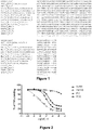
- the mutated amino acids are selected in the group consisting of V2, K3, K5, K7, Y8, K9, G10, E11, K13, E14, T17, K21, K22, W24, V26, G27, K28, M29, S31, T33, Y34, D35, D36, N37, G38, K39, T40, G41, R42, A44, S46, E47, K48, D49, A50 and P51 (reference is made to SEQ ID NO: 1, and to the alignment of Figure 1 ).
- the variant shall comprise mutated amino-acids (preferably from 7 to 12 as indicated above) chosen among these amino acids, and may also contain other mutated amino acids (preferably from 0 to 5) in other regions (i.e. chosen among other residues of Sa7d).
- mutated amino-acids preferably from 7 to 12 as indicated above
- other mutated amino acids preferably from 0 to 5
- A59, R60, A61, E62, R63, E64 and/or K66 can be deleted.
- the mutated amino acids are selected from the group consisting of K7, Y8, K9, E11, K21, K22, W24, V26, M29, S31, T33, D35, T40, G41, R42, A44, and S46.
- variants herein disclosed can be obtained by the method described in WO 2008/068637 (various rounds of enrichment of the combinatorial libraries, in particular using ribosome display).
- the variant of OB-fold domain is thus preferably a variant of a protein of the Sac7d family (Sac7d variant), comprising from 5 to 20 (preferably from 7 to 14) mutated amino acids in the binding site of the protein.
- the mutated amino acids of the Sac7d variant are selected from the group consisting of V2, K3, K5, K7, Y8, K9, G10, E11, K13, E14, T17, K21, K22, W24, V26, G27, K28, M29, S31, T33, Y34, D35, D36, N37, G38, K39, T40, G41, R42, A44, S46, E47, K48, D49, A50 and P51 (in reference to SEQ ID NO:1 and the alignment of Figure 1 ).
- Sac7d variant contains 4 to 17 mutated amino acids chosen in the group consisting of K7, Y8, K9, E11, K21, K22, W24, V26, M29, S31, T33, D35, T40, G41, R42, A44, and S46.
- the advantage of the method described in WO 2008/068637 is that it makes it possible to obtain variants of OB-fold proteins by screening combinatorial libraries containing or expressing a plurality of variants in which a certain number of amino acids have been "randomized", i.e. replaced with a random amino acid.
- the screening of these libraries makes it possible to identify variants of these proteins which bind specifically, generally with a strong affinity (application WO 2008/068637 in fact describes affinities of the order of one nanomolar), with a target of interest, other than the natural ligand of the wild-type protein from which the combinatorial library was generated.
- WO 2008/068637 in fact describes affinities of the order of one nanomolar
- OB-fold proteins are known in the art. They are in particular described in the documents cited above, and also in Arcus (Curr Opin Struct Biol. 2002 Dec; 12(6):794-801 ).
- OB-fold is in the form of a cylinder having five beta ( ⁇ ) sheets.
- Most OB-fold proteins use the same binding interface of their natural ligand, which may be an oligosaccharide, an oligonucleotide, a protein, a metal ion or a catalytic substrate. This binding interface comprises mainly the residues located in the beta sheets. Certain residues located in the loops may also be involved in the binding of an OB-fold protein with its natural ligand.
- applications WO 2007/139397 and WO 2008/068637 and the Arcus document describe the OB-fold-protein domains for binding with their natural ligand.
- Sac7d SEQ ID NO: 1
- these are the residues V2, K3, K5, K7, Y8, K9, G10, E11, K13, E14, T17, K21, K22, W24, V26, G27, K28, M29, S31, T33, Y34, D35, D36, N37, G38, K39, T40, G41, R42, A44, S46, E47, K48, D49, A50 and P51.
- the binding domains of other OB-fold proteins can be identified as described in WO 2008/068637 .
- This application indicates that it is possible to perform a superimposition of 3D structures of OB-fold proteins or domains (10 domains were used in this application, including Sac7d), using the DALI website (http://www.ebi.ac.uk/dali/interactive.html)( Holm and Sander, 1998, Nucleic Acids Res 26, 316-319 ).
- DALI website http://www.ebi.ac.uk/dali/interactive.html
- it is easy to identify, for any OB-fold protein (or any OB-fold domain) the amino acids involved in the binding site and corresponding to the Sac7d amino acids mentioned above. Consequently, providing the amino acids that can be mutated in one of these proteins makes it possible to identify the corresponding amino acids for any other OB-fold domain.
- residues which can be deleted are: A59, R60, A61, E62, R63, E64 and/or K66.
- WO 2008/068637 also indicates that amino acids can optionally be inserted into the loops of the OB-fold proteins, in particular the proteins of the Sac7d family; in particular, insertions of 1 to 15 amino acid residues can be made in loop 3 (as defined in figures 1b and 2 of WO 2008/068637 ), for example in the region of residues 25 to 30 of Sac7d, preferably between residues 27 and 28, insertions of 1 to 15 amino acid residues can be made in loop 4 (as defined in figures 1b and 2 of WO 2008/068637 ), for example in the region of residues 35-40 of Sac7d, preferably between residues 37 and 38, and insertions of 1 to 20 residues can be made in loop 1 (as defined in figures 1b and 2 of WO 2008/068637 ), for example in the region of residues 7 to 12 of Sac7d, preferably between residues 9 and 10.
- modified OB-fold domain comprises a) at least one modified amino acid residue in a ⁇ -strand of the OB-fold domain binding face as compared to the naturally occurring OB-fold domain, or b) at least one modified amino acid residue in a ⁇ -strand of the OB-fold domain binding face and at least one modified amino acid residue in a strand of the OB-fold domain loop region, or c) at least one modified amino acid residue in a strand of the OB-fold domain loop region.
- the modified OB-fold domain has altered binding characteristics as compared to the naturally occurring OB-fold domain.
- WO 2008/068637 describes the use of a library based on the Sac7d protein for obtaining ligands which have an affinity for targets of interest.
- the method described in WO 2008/068637 comprises the generation of combinatorial libraries containing a plurality of DNA molecules all having the same sequence, except for the presence of certain random mutations leading to the production of variants of a wild-type protein, exhibiting mutations at certain amino acids of the binding site of this wild-type OB-fold protein.
- the wild-type OB-fold protein is a Sac7d protein, into which mutations are introduced in order to generate a variability, in particular at amino acids chosen from K7, Y8, K9, E11, K21, K22, W24, V26, M29, S31, T33, D35, T40, G41, R42, A44, and S46, or on other amino acids, such as V26, G27, K28, M29, S31, R42, A44, S46, E47 and K48. These amino acids are based on the Sac7d sequence, as represented by SEQ ID NO: 1.
- WO 2012/150314 shows that the mutations from one protein of the Sac7d family can be carried to another protein of the same family. This portability amounts to creating a mutant of another protein of the Sac7d family, starting from a mutant of one protein of said family.
- the first mutant may have been obtained in particular by carrying out the process of WO 2008/068637 . Consequently, it is possible, using in particular the teachings of WO 2012/150314 and of figure 1 , to obtain a mutant of any protein of the Sac7d family, starting from a mutant of any other protein of such family.
- a mutant of Sac7d one only has to introduce the mutated amino acids of the Sac7d mutant within the scaffold of another protein, using the sequence alignment of figure 1 .
- Non-limitative examples of OB-fold proteins which can be used according to the invention are Sac7d, Sso7d, the N-terminal domain of SEB (Papageorgiou et al., 1998), the chain A of the Shiga-like toxin IIe (PDB 2bosa), the human Neutrophil Activatin Peptide-2 (NAP-2, PDB 1tvxA), the Molybdenum Binding Protein (modg) of Azotobacter vinelandii (PDB 1h9j), the N-terminal domain of SPE-C (Roussel et al., 1997), the B5 subunit of E.
- EPPase coli inorganic pyrophosphatase EPPase (Samygina et al., 2001), or any of the proteins listed in Table 3 of the article by (Arcus, 2002), such as 1krs (Lysyl-tRNA synthetase LysS, E.coli), 1c0aA (Asp- tRNA synthetase, E.coli), 1b8aA (Asp- tRNA synthetase, P.
- aureus 1jmc (Major cold-shock protein, E.coli), 1bkb (Initiation translation factor 5a, P. aerophylum), 1sro (S1 RNA-binding domain of PNPase, E.coli), 1d7qA (Initiation translation factor 1, elF1a, Human), 1ah9 (Initiation translation factor 1, IF1, E.coli), 1b9mA (Mo-dependent transcriptional regulator ModE, E.coli), 1ckmA (RNA guanylyltransferase, Chlorella virus, PBCV-1), 1a0i (ATP-dependent DNA ligase, Bacteriophage T7), 1snc (Staphylococcal nuclease, S.
- Non-exhaustive examples of proteins with SH3 domaines are Signal transducing adaptor proteins, CDC24, Cdc25, PI3 kinase, Phospholipase, Ras GTPase-activating protein, Vav proto-oncogene, GRB2, p54 S6 kinase 2 (S6K2), SH3D21, C10orf76 (potentially), STAC3, Some myosins, SHANK1,2,3, ARHGAP12, C8orf46, TANGO1, Integrase, Focal Adhesion Kinase (FAK, PTK2), Proline-rich tyrosine kinase (Pyk2, CADTK, PTK2beta), or TRIP10 (cip4).
- Signal transducing adaptor proteins CDC24, Cdc25, PI3 kinase, Phospholipase, Ras GTPase-activating protein, Vav proto-oncogene, GRB2, p
- the tumor necrosis factor (TNF) superfamily is a protein superfamily of type II transmembrane proteins containing TNF homology domain and forming trimers.
- cytokine such as TNF-alpha, which is involved in inflammation.
- These proteins are expressed predominantly by immune cells and regulate diverse cell functions, including regulation of immune response and inflammation, but also proliferation, differentation, apoptosis and embryogenesis.
- the superfamily contains 19 members (Lymphotoxin alpha, Tumor necrosis factor, Lymphotoxin beta, OX40 ligand, CD40 ligand, Fas ligand, CD27 ligand, CD30 ligand, CD137 ligand, TNF-related apoptosis-inducing ligand, Receptor activator of nuclear factor kappa-B ligand (RANKL), TNF-related weak inducer of apoptosis, A proliferation-inducing ligand, B-cell activating factor, LIGHT, Vascular endothelial growth inhibitor, TNF superfamily member 18, and Ectodysplasin A).
- TNF receptor superfamily which comprises 29 members.
- Tumor necrosis factor (TNF, tumor necrosis factor alpha, TNF ⁇ , cachexin, or cachectin) is a cell signaling protein (cytokine) involved in systemic inflammation and is one of the cytokines that make up the acute phase reaction. It is produced chiefly by activated macrophages, although it can be produced by many other cell types such as CD4+ lymphocytes, NK cells, neutrophils, mast cells, eosinophils, and neurons. Dysregulation of TNF production has been implicated in a variety of human diseases including Alzheimer's disease,[6] cancer,[7] major depression,[8] psoriasis[9] and inflammatory bowel disease (IBD).
- IBD inflammatory bowel disease
- TNF acts in particular by promoting release of other cytokines including IL-1, IL-6, GM-CSF, and IL-10, inducing chemokines, increasing adhesion molecules, permitting growth of blood vessels, and allowing release of tissue destructive enzymes. It also activates T cells.
- TNF may also play a role in cancer by promoting an inflammatory microenvironment around the tumor, therefore limiting the efficacy of treatments (chemotherapy, CAR-T cells).
- a variant of an OB-fold protein (in particular from the Sac7d family), having the ability to inhibit formation of a functional TNF trimer, can be used in the same applications as the above antibodies, and for treating the same diseases. Furthermore, it is advantageous to use such variants, as they can be orally or topically (applied to the skin) dispensed to the patient, an mode of administration that is not available for antibodies.
- Another advantage is an enhanced long-term safety profile thanks to the capacity to selectively neutralize the activity of the soluble TNFalpha over its membrane-bound format. Blocking the membrane-bound TNF is indeed suspected of inducing (i) immune response against the blocking agent ( Deora et al, MABS. 2017; 9(4):680-695 ) and (ii) undesired side effects such as Multiple Sclerosis.
- Receptor activator of nuclear factor kappa-B ligand is an apoptosis regulator gene, a binding partner of osteoprotegerin (OPG), a ligand for the receptor RANK and controls cell proliferation by modifying protein levels of Id4, Id2 and cyclin D1. It is supposed to be involved in formation of bone metastases of breast or prostate cancers. Overproduction of RANKL is also implicated in a variety of degenerative bone diseases, such as rheumatoid arthritis and psoriatic arthritis.
- Denosumab is an antibody to RANKL, used to treat postmenopausal patients suffering with osteoporosis (PMO), and which can also be used to reduce cancer treatment-induced bone loss in breast and prostate cancers. It can also be used in multiple myeloma to prevent the development of bone complications.
- PMO osteoporosis
- a variant of an OB-fold protein (in particular from the Sac7d family), having the ability to inhibit formation of a functional RANKL trimer, can be used in the same applications, and for treating or preventing the same diseases or complications.
- TNF-related apoptosis-inducing ligand is a protein functioning as a ligand that induces the process of cell death called apoptosis.
- TRAIL and its receptors have been used as the targets of several anti-cancer therapeutics.
- TRAIL has also been implicated as a pathogenic or protective factor in various pulmonary diseases, particularly pulmonary arterial hypertension. It has been shown recently that neutralization of TNFSF10 ameliorates functional outcome in a murine model of Alzheimer's disease ( Cantarella et al, Brain. 2015 Jan; 138(1): 203-216 ). Consequently, a variant of an OB-fold protein (in particular from the Sac7d family), having the ability to inhibit formation of a functional TRAIL trimer, can be used for the treatment of Alzheimer disease, for treating the disease or preventing further progression of it.
- the invention also provides a method for obtaining a protein that binds to a monomer of a multimeric protein comprising the steps of:
- This method thus makes it possible to identify a variant that binds to the subunit and inhibits or slows the formation of the biologically functional multimer, from the library of variants.
- the variant inhibits multimerization of a multimeric (whether homo or hetero multimeric) protein.
- protein-protein binding is a dynamic process and that two polypeptides shall be bound at one time and separate at another time.
- the main question for obtaining stable multimers is the affinity of two proteins (one for the other) which will control in particular the duration of binding of the proteins.
- the variant inhibits the formation of the multimer when the equilibrium between the free monomer and the monomer bound in the multimeric protein is shifted. This is performed by competition, for the unbound monomeric form, between the variant and the other monomers. If the free unbound monomer binds the variant, there is less multimer that is formed and the equilibrium is thus shifted.
- variants herein disclosed is thus to be understood as being a competitor partner for an unbound free monomeric part of a multimeric protein, that will bind to such monomer at an epitope such that binding to the other natural partners will be prevented, thereby limiting the formation of the functional multimeric protein.
- Step a) consists in providing a library of variants of an OB-fold or an SH3 protein, in particular of variants of protein of the Sac7d family. These variants in the library are randomly mutated at a given number of amino acids that are positioned in the binding site.
- the prior art in particular WO2007139397 and WO 2008/068637 ) explains in details how the binding site of these proteins can be determined.
- Step b) consists in the expression of the variants.
- Such expression is performed by any method in the art, such as bacterial, yeast or ribosome display. Ribosome display is particularly adapted.
- the two patent application documents provide all necessary guidance to perform this step.
- step c) the expressed variants are presented to the subunit of the multimeric protein, that are conjugated to a solid support.
- Such conjugation has been performed in condition such that the multimerization of the protein has been abolished (i.e. the protein is not anymore in the multimeric form), so that the face of the subunit that is involved in the multimerization of the multimeric protein (protein-protein binding wituin the multimer) is presented and thus is available for binding by the variants.
- the variants that bind to the proteins presented in step c) are selected, thereby providing a a sub-library containing said selected variants.
- selection steps are known in the art and classical in the process of identifying binders in a library.
- Steps b) to e) can be repeated, generally one to three times, so as to improve convergence of the sub-library (i.e. limitation of the number of variants in sequential sub-libraries), in particular by increasing the stringency of binding (in order, notably to obtain binders with improved affinities). This would eventually improve the quality of the binders, and make it possible to eventually select very specific and affine binders (step g).
- step c namely being sure that such face is presented to the variants in the library, is important.
- Such ligand grafted in a. may be biotin which can easily be used for conjugation of proteins.
- the fact that the ligand is conjugated to the multimeric form of the protein makes it possible to make sure that it will not bind the face of the subunits that are involved in the multimerization and stabilization of the multimer.
- Immobilization of the grafted/conjugated protein on a solid surface is performed via the ligand.
- the liaison biotin/avidin is particularly adapted for such step b.
- the solid surface is washed so as to eliminate both unbound multimeric protein and to "demultimerize” the protein, i.e. to eliminate the subunits that are involved in the multimer but have not be bound to the surface. This, will only remain on the surface the "lonely” subunits that are bound via the ligand. Such subunits will thus "present” the face involved in multimerization as the target for the variants of the library.
- the face involved in multimerization of a multimeric protein can be determined by crystallization of the multimer.
- directed mutagenesis followed by cross-linkage experiments (such as SDS-PAGE or size exclusion) makes it possible to determine the amino acids involved in protein-protein interaction and formation of the multimer.
- the multimer are first bound to the solid surface. Making different washes eventually leads to the release of the subunit that are involved in the multimer and not bound to the solid surface, thereby presenting the face involved in the protein-protein interaction in the multimer.
- sequence of the identified variant can be cloned in any appropriate vector by any molecular genetic methods known in the art.
- DNA constructs comprising a nucleotide sequence, coding for a polypeptide comprising a variant as described above, are used in connection with a vector, such as a plasmid, phagemid, phage or viral vector.
- Recombinant constructs of the invention comprise the expression vectors that are capable of expressing the RNA and thus lead to production of proteins from the above genetic sequences.
- the vector may thus further comprise regulatory sequences, including a suitable promoter operably linked to the open reading frame (ORF) of the genetic sequences herein disclosed.
- the vector may further comprise a selectable marker sequence such as an antibiotic resistance gene. Specific initiation and bacterial secretory signals also may be required for efficient translation of the coding sequences when bacteria as used as the expression host.
- Cells are transfected or transformed with vectors containing the sequences coding for the polypeptides comprising the variant as disclosed above.
- the cells are the cultured in such conditions as to have the protein expressed and favorably secreted.
- the conditions of culture of the cells are the conditions generally used for recombinant antibody production and are known in the art. Such conditions that are known in the art can also be optimized by the person skilled in the art if needed. Kunert and Reinhart (Appl Microbiol Biotechnol. 2016; 100: 3451-3461 ) review such methods and provide ample references thereto.
- CHO Chonese Hamster Ovary
- PER.C6 human cell line, Pau et al, Vaccine. 2001 21;19(17-19):2716-21
- HEK 293b cells Human embryonic kidney cells 293
- NS0 cells cell line derived from the non-secreting murine myeloma
- EB66 cells a duck cell line Valneva, Lyons, France
- host cells containing at least one of the DNAs constructs coding for a polypeptide comprising the variant as disclosed herein.
- the host cell can be any cell for which expression vectors are available. As indicated above, it may be a higher eukaryotic host cell, such as a mammalian cell, a lower eukaryotic host cell, such as a yeast cell, or a prokaryotic cell, such as a bacterial cell.
- the vectors can be inserted within the genome of the host cell, or be maintained as an extragenomic vector (such as a Bacterial Artificial Chromosome or a Yeast Artificial Chromosome). When introduced within the cell genome, such introduction may be random or targeted using methods known in the art (homologous recombination or the like).
- Useful expression vectors for bacterial use are constructed by inserting the recombinant DNA sequence together with suitable translation initiation and termination signals in operable reading phase with a functional promoter.
- the vector will comprise one or more phenotypic selectable markers and an origin of replication to ensure maintenance of the vector and, if desirable, to provide amplification within the host.
- Suitable prokaryotic hosts for transformation include E. coli, Bacillus subtilis, Salmonella typhimurium and various species within the genera Pseudomonas, Streptomyces, and Staphylococcus.
- Examples of the eukaryotic host cells include vertebrate cells, insect cells, and yeast cells. In particular, one can use the cells mentioned above.
- the transformed or transfected cells are cultured according to methods known in the art and the polypeptide are recovered from intracellular or extracellular fractions (depending on whether it is secreted or not).
- the recombinant multi-specific protein produced can be separated and purified by any of various known separation methods utilizing the physical or chemical property of the protein, from the intracellular or extracellular fraction.
- various types of liquid chromatography such as molecular sieve chromatography (gel filtration), adsorption chromatography, ion exchange chromatography, and affinity chromatography, dialysis, and a combination thereof.
- any method known and used to purify recombinant polypeptides is adapted for the purification of the molecules herein disclosed.
- a tag has been introduced within the recombinant sequence (such as a poly-Histidine tag)
- affinity it is preferred to use affinity to purify the molecules.
- affinity column affinity column, FACS, beads
- One particular advantage of the molecules herein disclosed is that they don't need to be glycosylated to be active and can thus be produced in any type of cells, and not necessarily eukaryotic cells. They are particularly well produced in bacterial cells.
- variants isolated, in particular by the method disclosed above, and having the ability to shift the formation of the multimeric form of the target can be modified by any method known in the art.
- the resulting expressed protein would thus be a polypeptide that would contain both proteins.
- the vector may be built in such a way that it would contain a sequence coding for a linker that would be located between the two proteins in the expressed polypeptide.
- the invention also encompasses a polypeptide comprising the variant of a protein of the OB-fold protein (preferably of the Sac7d family) binding to the subunit of a multimeric protein which is linked (preferably through amine bound as disclosed above) to another protein or polypeptide.
- the other protein or polypeptide comprises another variant of a protein of the Sac7d family.
- the other protein or polypeptide is an antibody.
- the variant of the OB-fold domain is fused to at least one of the heavy or light chain of an immunoglobulin monomer, preferably at the N- or C-terminus of the light or heavy chain. In another embodiment, the variant may be fused to both heavy or light chains.
- a genetic construct comprising a DNA sequence selected from the group consisting of
- This fusion can be made at the N-terminus and/or the C-terminus of the antibody chain (heavy and/or light chain). It is to be noted that, especially when using a small OB-fold domain (about 70 amino acids) such as a protein from the Sac7d family, it is possible to obtain a molecule that will have the structure of the antibody (two light chains paired to two heavy chains, and such dimers paired together), with the antibody regions, and further binding regions consisting of the modified OB-fold domain.
- a small OB-fold domain about 70 amino acids
- a protein from the Sac7d family it is possible to obtain a molecule that will have the structure of the antibody (two light chains paired to two heavy chains, and such dimers paired together), with the antibody regions, and further binding regions consisting of the modified OB-fold domain.
- the antibody part of the protein herein disclosed is a IgG molecule.
- the antibody part of the protein herein disclosed is a IgA molecule.
- the antibody part of the protein herein disclosed is a IgM molecule.
- the antibody part of the protein herein disclosed is a IgD molecule.
- the antibody part of the protein herein disclosed is a IgE molecule.
- the antibody may be a human antibody, a rodent antibody (such as a mouse antibody or a rat antibody), a cat antibody, a dog antibody, a chicken antibody, a goat antibody, a camelid antibody (such as a camel antibody, a llama antibody, an alpaca antibody or a nanobody), a shark antibody, or an antibody from any other species. It may be a chimeric or humanized antibody. As reminded in Wikipedia, humanized antibodies are antibodies from non-human species whose protein sequences have been modified to increase their similarity to antibody variants produced naturally in humans. A chimeric antibody contains sequences from different species.
- the antibody that is part of the molecule herein disclosed is an antibody that comprises two identical heavy chains (of about 400 to 500 amino acids, generally around 450 amino acids) and two identical light chains. Consequently, the antibody comprises the same Fab variable regions. This antibody is therefore a monospecific antibody where both parts (combination of light and heavy chains) of the antibody bind to the same epitope of an antigen.
- the antibody may present different heavy and/or light chains.
- the antibody is a bi-specific antibody.
- the term “antibody” thus encompasses both “classical antibodies” as disclosed above having identical heavy and light chains, but also engineered antibodies that have more than one specificity.
- the antibody presents one heavy and light chain from one antibody and another heavy and light chain from another antibody.
- the antibody may bind to a target selected in the group consisting of:
- Cell surface receptors Insulin receptor, Low density lipoprotein receptor-related protein 1, Transferrin receptor, Epidermal growth factor receptor, Epidermal growth factor receptor variant III, Vascular endothelial growth factor receptor 1, Vascular endothelial growth factor receptor 2, Her2, Her3, Her4, PMSA, IGF-1R, GITR, RAGE, CD28.
- Cell surface proteins Mesothelin, EpCam, CD19, CD20, CD38, CD3, TIM-3, CEA, cMet, ICAM1, ICAM3, MadCam, a4b7, CD7, CD4, CD138.
- Angiogenesis factors and Growth factors VEGF, Angiopoietin 2, HGF, PDGF, EGF, GM-CSF, HB-EGF, TGF
- Immune checkpoint inhibitors or activators PD-1, PD-L1, CTLA4, CD28, B7-1, B7-2, ICOS, ICOSL, B7-H3, B7-H4, LAG3, KIR, 4-1BB, OX40, CD27, CD40L, TIM3, A2aR
- Circulating proteins TNFa, IL23, IL12, IL33, IL4, IL13, IL5, IL6, IL4, IFNg, IL17, RANKL, Bace1, alpha Synuclein, Tau, amyloid.
- the antibody targets IL17 or a cell surface receptor in particular involved in cancer.
- the polypeptide may contain a variant as disclosed above, and a biologically active molecule, such as an erythropoietin, an interferon, or etanercept.
- a biologically active molecule such as an erythropoietin, an interferon, or etanercept.
- the variant of the OB-fold domain is conjugated to an organic molecule.
- an organic molecule This may be done by any method known in the art.
- antiproliferative (cytotoxic and cytostatic agents) include cytotoxic compounds (e.g., broad spectrum), angiogenesis inhibitors, cell cycle progression inhibitors, PBK/m-TOR/AKT pathway inhibitors, MAPK signaling pathway inhibitors, kinase inhibitors, protein chaperones inhibitors, HDAC inhibitors, PARP inhibitors, Wnt/Hedgehog signaling pathway inhibitors, RNA polymerase inhibitors and proteasome inhibitors.
- cytotoxic compounds e.g., broad spectrum
- angiogenesis inhibitors e.g., cell cycle progression inhibitors
- PBK/m-TOR/AKT pathway inhibitors e.g., MAPK signaling pathway inhibitors
- kinase inhibitors kinase inhibitors
- protein chaperones inhibitors kina
- DNA-binding or alkylating drugs such as anthracyclines (doxorubicin, epirubicin, idarubicin, daunorubicin) and its analogs, alkylating agents, such as calicheamicins, dactinomycines, mitromycines, pyrrolobenzodiazepines, and the like.
- alkylating agents such as calicheamicins, dactinomycines, mitromycines, pyrrolobenzodiazepines, and the like.
- cell cycle progression inhibitors such as CDK inhibitors, Rho-kinase inhibitors, checkpoint kinase inhibitors, aurora kinase inhibitors, PLK inhibitors, and KSP inhibitors.
- thalidomide and its derivatives lenalidomide and pomalidomide In order for treating inflammation disorders, one can also use cyclooxygenase-2 inhibitors, 5-lipoxygenase inhibitors, quercetin and/
- the variants can be used in particular in therapeutic methods.
- a variant targeting the TNFalpha would target the monomer or dimer form of the protein and can be used, in particular, for the treatment of inflammatory diseases (such as Irritable Bowel Disease (IBD), rheumatoid arthritis (RA), psoriasis and the like) or for the treatment of the diseases mentioned above.
- IBD Irritable Bowel Disease
- RA rheumatoid arthritis
- psoriasis and the like
- a variant targeting RANKL would target the monomer or dimeric form and can be used in particular for the treatment of inflammatory diseases (such as the ones listed above), or of bone diseases (such as osteoporosis), or for the treatment of the diseases mentioned above.
- a variant targeting the TRAIL protein would target the monomeric or dimeric form and be used for the treatment of Alzheimer's disease, or for the treatment of the diseases mentioned above.
- the invention thus relates to methods of treatment of the diseases as mentioned above, comprising the administration of a therapeutic amount of a variant of an OB-fold (in particular a variant of a protein of the Sac7d family) to a patient in need thereof.
- a variant of an OB-fold in particular a variant of a protein of the Sac7d family
- therapeutic amount or "effective amount”, as used herein, is the amount sufficient to effect beneficial or desired results, such as clinical results, and an “effective amount” depends upon the context in which it is being applied.
- An effective amount is an amount that provides therapeutic improvement while minimizing side or adverse effect.
- variants or polypeptides containing the variants can also be used in diagnostic methods.
- the invention thus also relates to a method for detecting the presence of, or for quantifying, a subunit of a multimeric protein in a sample, comprising the step of
- the recovery of b) can be perfomed by various washes or methods common in the art.
- the detection or quantification can be performed by any method such as ELISA or other methods in the art.
- the invention also encompasses a method for purifying a subunit of a multimeric protein in a sample, comprising the step of
- Step c can be performed in particular by changing buffer composition (increasing/decreasing salts concentration, detergents, chaotropic agents) or pH (increasing/decreasing).
- polypeptide comprising the variant fused to another protein that can thus be used to recover the variant bound to the subunit (for instance on columns affine for this other protein).
- the screening was performed using a library comprising random mutations in 17 positions of the sequence of Sac7d (SEQ ID NO: 1).
- the mutated positions were K7, Y8, K9, E11, K21, K22, W24, V26, M29, S31, T33, D35, T40, G41, R42, A44, and S46.
- the targets were TNF-alpha and RANKL.
- the targets in the trimeric soluble form, were biotinylated and attached via biotin/avidin liaison on solid beads. They were then thoroughly washed in order to dissociate the trimers.
- biotinylated hTNFa R&D systems
- mTNFa R&D systems
- the biotinylation was performed by incubation of a 50 ⁇ M solution of the target protein with a 5-fold molar excess of sulfosuccinimidyl-6-(biotinamido) hexanoate (Sulfo-NHS-LC-LC-Biotin, Pierce) in PBS (Sigma-Aldrich) on ice for 1 h.
- biotinylated proteins were buffer-exchanged using protein desalting spin columns from Pierce equilibrated in 20 mM Tris-HCI, 150 mM NaCl pH 7.4 (TBS).
- the degree of biotinylation was determined, using the HABA assay (Sigma), as being about 0.5 to 5 molecules of biotin per protein molecule.
- the generation of the library corresponding to the random mutagenesis of positions as indicated above in Sac7d protein has been previously described (in particular in Mouratou et al, Methods Mol Biol. 2012;805:315-31 ; Mouratou et al, Proc Natl Acad Sci U S A. 2007 Nov 13; 104(46): 17983-8 ; WO 2008/068637 ).
- the PCR-amplified library was transcribed and the selection was done at 4°C essentially as previously described (Mouratou et al, 2007 and 2012, op.cit., Binz et al, Nat Biotechnol 2004, 22:575-582 ).
- streptavidin 66 nM, 100 ⁇ l/well; Sigma-Aldrich
- neutravidin 66 nM, 100 ⁇ l/well; Thermo Scientific
- TBS150 50 mM Tris HCl, pH 7.4, 150 mM NaCl
- a ribosome-display round consisted of a 1 h pre-panning steps on streptavidin (or neutravidin) coated material and a 1 h binding step on the target protein. After washing, RNA purification and reverse transcription, the DNA pool was amplified by RT-PCR
- One assay to verify the binding to the target is performed using a variant produced with a His-Tag.
- Biotinylated antigens are immobilized on streptavidin-coated plates as described above, except that the blocking step is performed for 1h at room temperature.
- 100 ⁇ l of E. coli crude extracts are applied to wells with or without immobilized antigen for 1 h at RT.
- the binding is visualized using anti-RGSHis antibody HRP conjugate (Qiagen) which detects RGSHis6 tag from the variant and a solution at 1 mg/ml of o-phenylenediamine substrate (OPD) in revelation buffer (0.05 M citric acid, 0.05% hydrogen peroxide).
- OPD o-phenylenediamine substrate
- Absorbance at 450 nm is measured using an ELISA plate reader. Quantitative ELISAs is done in the same manner, except purified protein is used.
- N11 Characterization of a specific variant binding to TNF-alpha, called N11, is disclosed in more details in the following examples.
- TNFRI TNF Receptor I
- TNFRII TNF Receptor II
- ADA Adalimumab
- IFX Infliximab
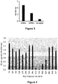
- binding of N11 is neutralized by TNFRI, TNFRII, and Adalimumab whereas binding of N11 is not neutralized by Infliximab
- EC100 for N11-human TNFa (hTNFa) interaction was determined by ELISA using plate functionalized with hTNFa and a concentration range of N11.
- each residue of N11 differing from the Sac7d (SEQ ID NO: 1) sequence was replaced by an Alanine (Alascan).
- Exposition of the obtained proteins to TNFa was then performed and binding was measured by ELISA (measure of EC100 using plate functionalized with hTNFa and a concentration range of the variants of N11).
- Residues 22 and 24 are also important, although mutations thereof would essentially reduce the affinity.
- sequences SEQ ID NO: 17 (including the very important amino acids 31, 33, 40, 42 of N11) and SEQ NO: 18 (including the very important amino acids 31, 33, 40, 42 and residues 22 and 24 of N11) represent minimal sequences that maintain the binding to TNFa.
- mutant Y151A is most likely well folded and that the residue is involved in the epitope recognized by N11.
- N11 binding is significantly impacted by Y151A mutation.
- Residue Y151 is involved in the epitope of N11 It appears that residue Y151 is burried in the core of the TNF alpha trimer This is an additional argument pointing on a mode of interaction relying on the blockade of the TNFalpha trimerization
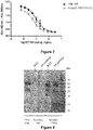
- Figure 8 shows that bands could be attributed to a complex of N11 with a monomer and a dimer of TNFa were observed on SDS-PAGE gel after cross-linking, whereas there was no evidence of an interaction between N11 and the trimer of TNFa. It was further observed complexes of lower molecular weight when TNFa is incubated with N11 for 24h at RT, that could be attributed to a monomer of TNFa in complex with one Nanofitin and a dimer of TNFa in complex with one Nanofitin.
- N11 binds both monomer and dimer of TNFalpha but not the trimer.
- Epitope of N11 involves a residue that is burried in the core of TNFalpha trimer.
- N11 cannot bind the stabilized trimer of TNFalpha. Binding of N11 is facilitated when TNFalpha is present in its lower oligomeric states
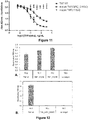
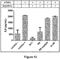
- N9 another (less efficient) variant isolated in Example 1
- N9mut42A the N9 variant with a mutation to abolish binding to TNF
- Irr NF a irrelevant variant of Sac7d
Landscapes
- Health & Medical Sciences (AREA)
- Life Sciences & Earth Sciences (AREA)
- Chemical & Material Sciences (AREA)
- Engineering & Computer Science (AREA)
- Organic Chemistry (AREA)
- Genetics & Genomics (AREA)
- General Health & Medical Sciences (AREA)
- Molecular Biology (AREA)
- Biomedical Technology (AREA)
- Zoology (AREA)
- Biochemistry (AREA)
- Biotechnology (AREA)
- General Engineering & Computer Science (AREA)
- Medicinal Chemistry (AREA)
- Bioinformatics & Cheminformatics (AREA)
- Wood Science & Technology (AREA)
- Biophysics (AREA)
- Microbiology (AREA)
- Physics & Mathematics (AREA)
- Proteomics, Peptides & Aminoacids (AREA)
- Crystallography & Structural Chemistry (AREA)
- Gastroenterology & Hepatology (AREA)
- Bioinformatics & Computational Biology (AREA)
- Plant Pathology (AREA)
- Urology & Nephrology (AREA)
- Immunology (AREA)
- Hematology (AREA)
- Animal Behavior & Ethology (AREA)
- Toxicology (AREA)
- Veterinary Medicine (AREA)
- Public Health (AREA)
- Epidemiology (AREA)
- Pharmacology & Pharmacy (AREA)
- Food Science & Technology (AREA)
- Cell Biology (AREA)
- Analytical Chemistry (AREA)
- General Physics & Mathematics (AREA)
- Pathology (AREA)
- Peptides Or Proteins (AREA)
- Preparation Of Compounds By Using Micro-Organisms (AREA)
Priority Applications (7)
| Application Number | Priority Date | Filing Date | Title |
|---|---|---|---|
| EP18306324.7A EP3632924A1 (en) | 2018-10-07 | 2018-10-07 | Binders for inhibiting formation of multimeric proteins |
| EP19779920.8A EP3861011B1 (en) | 2018-10-07 | 2019-10-06 | Binders for inhibiting formation of multimeric proteins |
| PCT/EP2019/076986 WO2020074402A1 (en) | 2018-10-07 | 2019-10-06 | Binders for inhibiting formation of multimeric proteins |
| ES19779920T ES3068042T3 (es) | 2018-10-07 | 2019-10-06 | Ligandos para inhibir la formación de proteínas multiméricas |
| JP2021518942A JP7699539B2 (ja) | 2018-10-07 | 2019-10-06 | 多量体タンパク質の形成を阻害するバインダー |
| US17/283,369 US20210380647A1 (en) | 2018-10-07 | 2019-10-06 | Binders for inhibiting formation of multimeric proteins |
| JP2025064215A JP2025108528A (ja) | 2018-10-07 | 2025-04-09 | 多量体タンパク質の形成を阻害するバインダー |
Applications Claiming Priority (1)
| Application Number | Priority Date | Filing Date | Title |
|---|---|---|---|
| EP18306324.7A EP3632924A1 (en) | 2018-10-07 | 2018-10-07 | Binders for inhibiting formation of multimeric proteins |
Publications (1)
| Publication Number | Publication Date |
|---|---|
| EP3632924A1 true EP3632924A1 (en) | 2020-04-08 |
Family
ID=64017330
Family Applications (2)
| Application Number | Title | Priority Date | Filing Date |
|---|---|---|---|
| EP18306324.7A Ceased EP3632924A1 (en) | 2018-10-07 | 2018-10-07 | Binders for inhibiting formation of multimeric proteins |
| EP19779920.8A Active EP3861011B1 (en) | 2018-10-07 | 2019-10-06 | Binders for inhibiting formation of multimeric proteins |
Family Applications After (1)
| Application Number | Title | Priority Date | Filing Date |
|---|---|---|---|
| EP19779920.8A Active EP3861011B1 (en) | 2018-10-07 | 2019-10-06 | Binders for inhibiting formation of multimeric proteins |
Country Status (5)
| Country | Link |
|---|---|
| US (1) | US20210380647A1 (cg-RX-API-DMAC7.html) |
| EP (2) | EP3632924A1 (cg-RX-API-DMAC7.html) |
| JP (2) | JP7699539B2 (cg-RX-API-DMAC7.html) |
| ES (1) | ES3068042T3 (cg-RX-API-DMAC7.html) |
| WO (1) | WO2020074402A1 (cg-RX-API-DMAC7.html) |
Cited By (1)
| Publication number | Priority date | Publication date | Assignee | Title |
|---|---|---|---|---|
| EP4190358A1 (en) * | 2021-12-05 | 2023-06-07 | Affilogic | Polypeptide as carrier for intestinal barrier crossing |
Families Citing this family (2)
| Publication number | Priority date | Publication date | Assignee | Title |
|---|---|---|---|---|
| FR3004650B1 (fr) * | 2013-04-22 | 2015-05-29 | Affilogic | Composition topique comprenant un variant d'une protéine sauvage à pli-OB, ainsi que son procédé de préparation |
| EP4043481A1 (en) | 2021-02-15 | 2022-08-17 | Affilogic | Compounds and methods for extending half life of biomolecules |
Citations (6)
| Publication number | Priority date | Publication date | Assignee | Title |
|---|---|---|---|---|
| WO2007139397A1 (en) | 2006-05-26 | 2007-12-06 | Waikatolink Limited | Ob fold domains |
| EP1930342A1 (en) * | 2006-12-04 | 2008-06-11 | Institut Pasteur | OB-fold used as scaffold for engineering new specific binders |
| WO2012150314A1 (fr) | 2011-05-04 | 2012-11-08 | Centre National De La Recherche Scientifique | Méthode de génération de protéines |
| WO2014173899A1 (fr) | 2013-04-22 | 2014-10-30 | Affilogic | Compositions topiques comprenant des variantes de pli-ob |
| WO2016062874A1 (fr) | 2014-10-24 | 2016-04-28 | Affilogic | Compositions pour administration par voie orale comprenant un variant d'une protéine à pli-ob |
| WO2017161096A1 (en) * | 2016-03-16 | 2017-09-21 | Tarveda Therapeutics, Inc. | Antibody mimic conjugates and particles |
Family Cites Families (2)
| Publication number | Priority date | Publication date | Assignee | Title |
|---|---|---|---|---|
| WO2007011891A2 (en) * | 2005-07-15 | 2007-01-25 | Strategene California | Dna binding protein-polymerase chimeras |
| EP3483180A1 (en) * | 2017-11-14 | 2019-05-15 | Affilogic | Multi specific molecules |
-
2018
- 2018-10-07 EP EP18306324.7A patent/EP3632924A1/en not_active Ceased
-
2019
- 2019-10-06 EP EP19779920.8A patent/EP3861011B1/en active Active
- 2019-10-06 JP JP2021518942A patent/JP7699539B2/ja active Active
- 2019-10-06 WO PCT/EP2019/076986 patent/WO2020074402A1/en not_active Ceased
- 2019-10-06 US US17/283,369 patent/US20210380647A1/en active Pending
- 2019-10-06 ES ES19779920T patent/ES3068042T3/es active Active
-
2025
- 2025-04-09 JP JP2025064215A patent/JP2025108528A/ja active Pending
Patent Citations (7)
| Publication number | Priority date | Publication date | Assignee | Title |
|---|---|---|---|---|
| WO2007139397A1 (en) | 2006-05-26 | 2007-12-06 | Waikatolink Limited | Ob fold domains |
| EP1930342A1 (en) * | 2006-12-04 | 2008-06-11 | Institut Pasteur | OB-fold used as scaffold for engineering new specific binders |
| WO2008068637A2 (en) | 2006-12-04 | 2008-06-12 | Institut Pasteur | Ob-fold used as scaffold for engineering new specific binders |
| WO2012150314A1 (fr) | 2011-05-04 | 2012-11-08 | Centre National De La Recherche Scientifique | Méthode de génération de protéines |
| WO2014173899A1 (fr) | 2013-04-22 | 2014-10-30 | Affilogic | Compositions topiques comprenant des variantes de pli-ob |
| WO2016062874A1 (fr) | 2014-10-24 | 2016-04-28 | Affilogic | Compositions pour administration par voie orale comprenant un variant d'une protéine à pli-ob |
| WO2017161096A1 (en) * | 2016-03-16 | 2017-09-21 | Tarveda Therapeutics, Inc. | Antibody mimic conjugates and particles |
Non-Patent Citations (20)
| Title |
|---|
| ANA M.G.C. DIAS ET AL: "The future of protein scaffolds as affinity reagents for purification : The Future of Protein Scaffolds", BIOTECHNOLOGY AND BIOENGINEERING, vol. 114, no. 3, 1 March 2017 (2017-03-01), US, pages 481 - 491, XP055530301, ISSN: 0006-3592, DOI: 10.1002/bit.26090 * |
| ARCUS, CURR OPIN STRUCT BIOL., vol. 12, no. 6, December 2002 (2002-12-01), pages 794 - 801 |
| BEHAR ET AL., PROTEIN ENGINEERING, DESIGN & SELECTION, vol. 26, no. 4, 2013, pages 267 - 275 |
| BÉHAR GHISLAINE ET AL: "Affitins as robust tailored reagents for affinity chromatography purification of antibodies and non-immunoglobulin proteins", JOURNAL OF CHROMATOGRAPHY A, ELSEVIER, AMSTERDAM, NL, vol. 1441, 27 February 2016 (2016-02-27), pages 44 - 51, XP029464321, ISSN: 0021-9673, DOI: 10.1016/J.CHROMA.2016.02.068 * |
| BINZ ET AL., NAT BIOTECHNOL, vol. 22, 2004, pages 575 - 582 |
| CANTARELLA ET AL., BRAIN, vol. 138, no. 1, January 2015 (2015-01-01), pages 203 - 216 |
| CHRIS W. DIEHNELT ET AL: "Discovery of High-Affinity Protein Binding Ligands - Backwards", PLOS ONE, vol. 5, no. 5, 1 January 2010 (2010-01-01), pages e10728 - e10728, XP055052323, ISSN: 1932-6203, DOI: 10.1371/journal.pone.0010728 * |
| DEORA ET AL., MABS, vol. 9, no. 4, 2017, pages 680 - 695 |
| GERA ET AL., J MOL BIOL., vol. 409, no. 4, 17 June 2011 (2011-06-17), pages 601 - 16 |
| GOCHA ET AL., SCIENTIFIC REPORTS, vol. 7, no. 12021, 2017 |
| HOLM; PARK, BIOINFORMATICS, vol. 16, 2000, pages 566 - 567 |
| HOLM; SANDER, NUCLEIC ACIDS RES, vol. 26, 1998, pages 316 - 319 |
| KUNERT; REINHART, APPL MICROBIOL BIOTECHNOL, vol. 100, 2016, pages 3451 - 3461 |
| LOPEZ ET AL., NUCLEIC ACIDS RES, vol. 31, 2003, pages 3795 - 3798 |
| MOURATOU ET AL., METHODS MOL BIOL, vol. 805, 2012, pages 315 - 31 |
| MOURATOU ET AL., PROC NATL ACAD SCI USA., vol. 104, no. 46, 13 November 2007 (2007-11-13), pages 17983 - 8 |
| NOTREDAME ET AL., J MOL BIOL, vol. 302, 2000, pages 205 - 217 |
| PAU ET AL., VACCINE, vol. 19, no. 17-19, 2001, pages 2716 - 21 |
| SAMBROOK J; FRITSCHI EF; MANIATIS T: "Molecular cloning: a laboratorymanual", 1989, COLD SPRING HARBOR LABORATORY PRESS |
| SHUKRA ET AL., EUR J MICROBIOL IMMUNOL (BP, vol. 4, no. 2, 2014, pages 91 - 98 |
Cited By (2)
| Publication number | Priority date | Publication date | Assignee | Title |
|---|---|---|---|---|
| EP4190358A1 (en) * | 2021-12-05 | 2023-06-07 | Affilogic | Polypeptide as carrier for intestinal barrier crossing |
| WO2023099783A1 (en) | 2021-12-05 | 2023-06-08 | Affilogic | Polypeptide as carrier for intestinal barrier crossing |
Also Published As
| Publication number | Publication date |
|---|---|
| JP2022501063A (ja) | 2022-01-06 |
| JP7699539B2 (ja) | 2025-06-27 |
| JP2025108528A (ja) | 2025-07-23 |
| EP3861011B1 (en) | 2026-02-11 |
| WO2020074402A1 (en) | 2020-04-16 |
| EP3861011A1 (en) | 2021-08-11 |
| ES3068042T3 (es) | 2026-05-27 |
| EP3861011C0 (en) | 2026-02-11 |
| US20210380647A1 (en) | 2021-12-09 |
Similar Documents
| Publication | Publication Date | Title |
|---|---|---|
| US11434585B2 (en) | OB-fold used as scaffold for engineering new specific binders | |
| JP2025108528A (ja) | 多量体タンパク質の形成を阻害するバインダー | |
| JP2019501885A (ja) | 多特異性抗体 | |
| JP2019501887A (ja) | 多特異性抗体 | |
| Ubah et al. | Novel, anti-hTNF-α variable new antigen receptor formats with enhanced neutralizing potency and multifunctionality, generated for therapeutic development | |
| CN116284380A (zh) | 白细胞介素2结合分子、其衍生物、试剂盒及其生产方法和用途 | |
| US20230120167A1 (en) | Variants of sac7d and their use in cancer therapy | |
| CN115043940A (zh) | 抗人血清白蛋白抗体及其应用 | |
| US20240124531A1 (en) | Compounds and methods for extending half life of biomolecules | |
| JP6137169B2 (ja) | 新規抗ヒトil−23受容体抗体 | |
| CN114555633A (zh) | 用于靶向扩增免疫效应细胞的方法和材料 | |
| EP4059949A1 (en) | Anti-factor c3 sac7d variants and their medical use for treating complement-mediated disorders | |
| EP4190358A1 (en) | Polypeptide as carrier for intestinal barrier crossing | |
| Geng et al. | Screening and Characterization of EGFR-Targeted Specific Nanobodies | |
| CN121930341A (zh) | Tl1a结合分子及其应用 | |
| WO2026085874A1 (zh) | Tl1a结合分子及其应用 | |
| CN117597353A (zh) | 抗因子c3的sac7d变体及其治疗补体介导疾患的医疗用途 | |
| HK1117548B (en) | Ob-fold used as scaffold for engineering new specific binders |
Legal Events
| Date | Code | Title | Description |
|---|---|---|---|
| PUAI | Public reference made under article 153(3) epc to a published international application that has entered the european phase |
Free format text: ORIGINAL CODE: 0009012 |
|
| AK | Designated contracting states |
Kind code of ref document: A1 Designated state(s): AL AT BE BG CH CY CZ DE DK EE ES FI FR GB GR HR HU IE IS IT LI LT LU LV MC MK MT NL NO PL PT RO RS SE SI SK SM TR |
|
| AX | Request for extension of the european patent |
Extension state: BA ME |
|
| STAA | Information on the status of an ep patent application or granted ep patent |
Free format text: STATUS: THE APPLICATION HAS BEEN REFUSED |
|
| 18R | Application refused |
Effective date: 20200620 |