EP3626735A1 - Polypeptides stabilisés de facteur de croissance de type insuline - Google Patents
Polypeptides stabilisés de facteur de croissance de type insuline Download PDFInfo
- Publication number
- EP3626735A1 EP3626735A1 EP19191570.1A EP19191570A EP3626735A1 EP 3626735 A1 EP3626735 A1 EP 3626735A1 EP 19191570 A EP19191570 A EP 19191570A EP 3626735 A1 EP3626735 A1 EP 3626735A1
- Authority
- EP
- European Patent Office
- Prior art keywords
- deleted
- igf
- amino acid
- amino acids
- substituted
- Prior art date
- Legal status (The legal status is an assumption and is not a legal conclusion. Google has not performed a legal analysis and makes no representation as to the accuracy of the status listed.)
- Withdrawn
Links
- 108090000723 Insulin-Like Growth Factor I Proteins 0.000 title claims abstract description 220
- 102000013275 Somatomedins Human genes 0.000 title description 31
- 108090000765 processed proteins & peptides Proteins 0.000 claims abstract description 174
- 102000004196 processed proteins & peptides Human genes 0.000 claims abstract description 153
- 229920001184 polypeptide Polymers 0.000 claims abstract description 136
- 208000021642 Muscular disease Diseases 0.000 claims abstract description 19
- 208000029578 Muscle disease Diseases 0.000 claims abstract description 16
- 230000004048 modification Effects 0.000 claims abstract description 16
- 238000012986 modification Methods 0.000 claims abstract description 16
- 102000004218 Insulin-Like Growth Factor I Human genes 0.000 claims abstract description 7
- 235000001014 amino acid Nutrition 0.000 claims description 658
- 150000001413 amino acids Chemical class 0.000 claims description 627
- ZDXPYRJPNDTMRX-UHFFFAOYSA-N glutamine Natural products OC(=O)C(N)CCC(N)=O ZDXPYRJPNDTMRX-UHFFFAOYSA-N 0.000 claims description 207
- 101000599951 Homo sapiens Insulin-like growth factor I Proteins 0.000 claims description 117
- 102000044162 human IGF1 Human genes 0.000 claims description 112
- 125000000404 glutamine group Chemical group N[C@@H](CCC(N)=O)C(=O)* 0.000 claims description 76
- 235000004279 alanine Nutrition 0.000 claims description 67
- 125000003295 alanine group Chemical group N[C@@H](C)C(=O)* 0.000 claims description 52
- 201000000585 muscular atrophy Diseases 0.000 claims description 35
- 239000008194 pharmaceutical composition Substances 0.000 claims description 35
- 238000011282 treatment Methods 0.000 claims description 35
- WHUUTDBJXJRKMK-UHFFFAOYSA-N Glutamic acid Natural products OC(=O)C(N)CCC(O)=O WHUUTDBJXJRKMK-UHFFFAOYSA-N 0.000 claims description 32
- 206010028289 Muscle atrophy Diseases 0.000 claims description 32
- 235000013922 glutamic acid Nutrition 0.000 claims description 32
- 239000004220 glutamic acid Substances 0.000 claims description 32
- 150000007523 nucleic acids Chemical class 0.000 claims description 30
- 125000000291 glutamic acid group Chemical group N[C@@H](CCC(O)=O)C(=O)* 0.000 claims description 27
- ONIBWKKTOPOVIA-UHFFFAOYSA-N Proline Natural products OC(=O)C1CCCN1 ONIBWKKTOPOVIA-UHFFFAOYSA-N 0.000 claims description 26
- 102000039446 nucleic acids Human genes 0.000 claims description 26
- 108020004707 nucleic acids Proteins 0.000 claims description 26
- 125000001500 prolyl group Chemical group [H]N1C([H])(C(=O)[*])C([H])([H])C([H])([H])C1([H])[H] 0.000 claims description 26
- 102000040430 polynucleotide Human genes 0.000 claims description 25
- 108091033319 polynucleotide Proteins 0.000 claims description 25
- 239000002157 polynucleotide Substances 0.000 claims description 25
- 230000020763 muscle atrophy Effects 0.000 claims description 24
- DHMQDGOQFOQNFH-UHFFFAOYSA-N Glycine Natural products NCC(O)=O DHMQDGOQFOQNFH-UHFFFAOYSA-N 0.000 claims description 21
- 239000004471 Glycine Substances 0.000 claims description 20
- QNAYBMKLOCPYGJ-REOHCLBHSA-N L-alanine Chemical compound C[C@H](N)C(O)=O QNAYBMKLOCPYGJ-REOHCLBHSA-N 0.000 claims description 16
- 238000002560 therapeutic procedure Methods 0.000 claims description 4
- 239000002243 precursor Substances 0.000 abstract description 85
- 238000003776 cleavage reaction Methods 0.000 abstract description 6
- 230000007017 scission Effects 0.000 abstract description 6
- 229940024606 amino acid Drugs 0.000 description 635
- 210000004027 cell Anatomy 0.000 description 187
- 108090000623 proteins and genes Proteins 0.000 description 142
- 235000018102 proteins Nutrition 0.000 description 123
- 102000004169 proteins and genes Human genes 0.000 description 123
- MTCFGRXMJLQNBG-UHFFFAOYSA-N Serine Natural products OCC(N)C(O)=O MTCFGRXMJLQNBG-UHFFFAOYSA-N 0.000 description 121
- 239000013598 vector Substances 0.000 description 81
- 108020004414 DNA Proteins 0.000 description 67
- 238000000034 method Methods 0.000 description 57
- 230000035772 mutation Effects 0.000 description 49
- 241001522306 Serinus serinus Species 0.000 description 46
- 239000000203 mixture Substances 0.000 description 42
- 241000282414 Homo sapiens Species 0.000 description 36
- 230000014509 gene expression Effects 0.000 description 35
- 238000004519 manufacturing process Methods 0.000 description 33
- 238000010367 cloning Methods 0.000 description 32
- 239000012634 fragment Substances 0.000 description 30
- 239000013612 plasmid Substances 0.000 description 30
- 238000003752 polymerase chain reaction Methods 0.000 description 29
- 230000001225 therapeutic effect Effects 0.000 description 29
- 230000010261 cell growth Effects 0.000 description 28
- 230000026731 phosphorylation Effects 0.000 description 28
- 238000006366 phosphorylation reaction Methods 0.000 description 28
- 108060003951 Immunoglobulin Proteins 0.000 description 25
- 102100036721 Insulin receptor Human genes 0.000 description 25
- 102000018358 immunoglobulin Human genes 0.000 description 25
- 108010001127 Insulin Receptor Proteins 0.000 description 24
- 210000001789 adipocyte Anatomy 0.000 description 24
- 102220492229 Replication stress response regulator SDE2_R37A_mutation Human genes 0.000 description 23
- 239000013604 expression vector Substances 0.000 description 23
- 125000005647 linker group Chemical group 0.000 description 23
- 210000004962 mammalian cell Anatomy 0.000 description 23
- 230000027455 binding Effects 0.000 description 22
- 108010022394 Threonine synthase Proteins 0.000 description 19
- 102000004419 dihydrofolate reductase Human genes 0.000 description 19
- 102000005962 receptors Human genes 0.000 description 19
- 108020003175 receptors Proteins 0.000 description 19
- FAPWRFPIFSIZLT-UHFFFAOYSA-M Sodium chloride Chemical compound [Na+].[Cl-] FAPWRFPIFSIZLT-UHFFFAOYSA-M 0.000 description 18
- 230000037396 body weight Effects 0.000 description 17
- 210000004978 chinese hamster ovary cell Anatomy 0.000 description 17
- 108020001507 fusion proteins Proteins 0.000 description 17
- 102000037865 fusion proteins Human genes 0.000 description 17
- 230000002829 reductive effect Effects 0.000 description 17
- 238000002965 ELISA Methods 0.000 description 16
- 125000003275 alpha amino acid group Chemical group 0.000 description 16
- 230000000694 effects Effects 0.000 description 16
- 208000001076 sarcopenia Diseases 0.000 description 16
- 230000003321 amplification Effects 0.000 description 15
- 150000001875 compounds Chemical class 0.000 description 15
- 239000003550 marker Substances 0.000 description 15
- 210000003205 muscle Anatomy 0.000 description 15
- 238000003199 nucleic acid amplification method Methods 0.000 description 15
- 229920002873 Polyethylenimine Polymers 0.000 description 14
- 239000003795 chemical substances by application Substances 0.000 description 14
- 238000012217 deletion Methods 0.000 description 14
- 230000037430 deletion Effects 0.000 description 14
- 239000003814 drug Substances 0.000 description 14
- 238000002347 injection Methods 0.000 description 14
- 239000007924 injection Substances 0.000 description 14
- 230000004190 glucose uptake Effects 0.000 description 13
- 238000007920 subcutaneous administration Methods 0.000 description 13
- 239000006144 Dulbecco’s modified Eagle's medium Substances 0.000 description 12
- 238000005516 engineering process Methods 0.000 description 12
- 238000001802 infusion Methods 0.000 description 12
- 239000000463 material Substances 0.000 description 12
- 239000002609 medium Substances 0.000 description 12
- 230000002441 reversible effect Effects 0.000 description 12
- 102200061561 rs121918552 Human genes 0.000 description 12
- 239000000243 solution Substances 0.000 description 12
- 102000009109 Fc receptors Human genes 0.000 description 11
- 108010087819 Fc receptors Proteins 0.000 description 11
- 239000013543 active substance Substances 0.000 description 11
- UREBDLICKHMUKA-CXSFZGCWSA-N dexamethasone Chemical compound C1CC2=CC(=O)C=C[C@]2(C)[C@]2(F)[C@@H]1[C@@H]1C[C@@H](C)[C@@](C(=O)CO)(O)[C@@]1(C)C[C@@H]2O UREBDLICKHMUKA-CXSFZGCWSA-N 0.000 description 11
- 208000037265 diseases, disorders, signs and symptoms Diseases 0.000 description 11
- 229940079593 drug Drugs 0.000 description 11
- 239000000047 product Substances 0.000 description 11
- 230000001105 regulatory effect Effects 0.000 description 11
- 241000699666 Mus <mouse, genus> Species 0.000 description 10
- 108091005804 Peptidases Proteins 0.000 description 10
- 239000004365 Protease Substances 0.000 description 10
- 241000700159 Rattus Species 0.000 description 10
- 229960003957 dexamethasone Drugs 0.000 description 10
- 230000005764 inhibitory process Effects 0.000 description 10
- 102000028416 insulin-like growth factor binding Human genes 0.000 description 10
- 108091022911 insulin-like growth factor binding Proteins 0.000 description 10
- 102220123744 rs186714117 Human genes 0.000 description 10
- 102220041372 rs34084741 Human genes 0.000 description 10
- 102200147815 rs72559734 Human genes 0.000 description 10
- 210000002966 serum Anatomy 0.000 description 10
- 239000011780 sodium chloride Substances 0.000 description 10
- LFQSCWFLJHTTHZ-UHFFFAOYSA-N Ethanol Chemical compound CCO LFQSCWFLJHTTHZ-UHFFFAOYSA-N 0.000 description 9
- PEDCQBHIVMGVHV-UHFFFAOYSA-N Glycerine Chemical compound OCC(O)CO PEDCQBHIVMGVHV-UHFFFAOYSA-N 0.000 description 9
- 208000027747 Kennedy disease Diseases 0.000 description 9
- 208000006269 X-Linked Bulbo-Spinal Atrophy Diseases 0.000 description 9
- 238000003556 assay Methods 0.000 description 9
- 230000000295 complement effect Effects 0.000 description 9
- 230000001965 increasing effect Effects 0.000 description 9
- 208000014674 injury Diseases 0.000 description 9
- 238000001990 intravenous administration Methods 0.000 description 9
- 210000003098 myoblast Anatomy 0.000 description 9
- 102200015319 rs113994140 Human genes 0.000 description 9
- 238000012163 sequencing technique Methods 0.000 description 9
- 238000001890 transfection Methods 0.000 description 9
- XLYOFNOQVPJJNP-UHFFFAOYSA-N water Substances O XLYOFNOQVPJJNP-UHFFFAOYSA-N 0.000 description 9
- VRYALKFFQXWPIH-PBXRRBTRSA-N (3r,4s,5r)-3,4,5,6-tetrahydroxyhexanal Chemical compound OC[C@@H](O)[C@@H](O)[C@H](O)CC=O VRYALKFFQXWPIH-PBXRRBTRSA-N 0.000 description 8
- SHZGCJCMOBCMKK-UHFFFAOYSA-N D-mannomethylose Natural products CC1OC(O)C(O)C(O)C1O SHZGCJCMOBCMKK-UHFFFAOYSA-N 0.000 description 8
- PMMURAAUARKVCB-UHFFFAOYSA-N alpha-D-ara-dHexp Natural products OCC1OC(O)CC(O)C1O PMMURAAUARKVCB-UHFFFAOYSA-N 0.000 description 8
- 230000010056 antibody-dependent cellular cytotoxicity Effects 0.000 description 8
- 230000003833 cell viability Effects 0.000 description 8
- 208000020832 chronic kidney disease Diseases 0.000 description 8
- 206010012601 diabetes mellitus Diseases 0.000 description 8
- 230000006870 function Effects 0.000 description 8
- 239000001963 growth medium Substances 0.000 description 8
- NOESYZHRGYRDHS-UHFFFAOYSA-N insulin Chemical compound N1C(=O)C(NC(=O)C(CCC(N)=O)NC(=O)C(CCC(O)=O)NC(=O)C(C(C)C)NC(=O)C(NC(=O)CN)C(C)CC)CSSCC(C(NC(CO)C(=O)NC(CC(C)C)C(=O)NC(CC=2C=CC(O)=CC=2)C(=O)NC(CCC(N)=O)C(=O)NC(CC(C)C)C(=O)NC(CCC(O)=O)C(=O)NC(CC(N)=O)C(=O)NC(CC=2C=CC(O)=CC=2)C(=O)NC(CSSCC(NC(=O)C(C(C)C)NC(=O)C(CC(C)C)NC(=O)C(CC=2C=CC(O)=CC=2)NC(=O)C(CC(C)C)NC(=O)C(C)NC(=O)C(CCC(O)=O)NC(=O)C(C(C)C)NC(=O)C(CC(C)C)NC(=O)C(CC=2NC=NC=2)NC(=O)C(CO)NC(=O)CNC2=O)C(=O)NCC(=O)NC(CCC(O)=O)C(=O)NC(CCCNC(N)=N)C(=O)NCC(=O)NC(CC=3C=CC=CC=3)C(=O)NC(CC=3C=CC=CC=3)C(=O)NC(CC=3C=CC(O)=CC=3)C(=O)NC(C(C)O)C(=O)N3C(CCC3)C(=O)NC(CCCCN)C(=O)NC(C)C(O)=O)C(=O)NC(CC(N)=O)C(O)=O)=O)NC(=O)C(C(C)CC)NC(=O)C(CO)NC(=O)C(C(C)O)NC(=O)C1CSSCC2NC(=O)C(CC(C)C)NC(=O)C(NC(=O)C(CCC(N)=O)NC(=O)C(CC(N)=O)NC(=O)C(NC(=O)C(N)CC=1C=CC=CC=1)C(C)C)CC1=CN=CN1 NOESYZHRGYRDHS-UHFFFAOYSA-N 0.000 description 8
- SXTAYKAGBXMACB-UHFFFAOYSA-N methionine sulfoximine Chemical compound CS(=N)(=O)CCC(N)C(O)=O SXTAYKAGBXMACB-UHFFFAOYSA-N 0.000 description 8
- 230000002103 transcriptional effect Effects 0.000 description 8
- 206010068597 Bulbospinal muscular atrophy congenital Diseases 0.000 description 7
- 208000006545 Chronic Obstructive Pulmonary Disease Diseases 0.000 description 7
- 241000699802 Cricetulus griseus Species 0.000 description 7
- 208000013016 Hypoglycemia Diseases 0.000 description 7
- 102000048143 Insulin-Like Growth Factor II Human genes 0.000 description 7
- 108090001117 Insulin-Like Growth Factor II Proteins 0.000 description 7
- 102100039688 Insulin-like growth factor 1 receptor Human genes 0.000 description 7
- 208000023178 Musculoskeletal disease Diseases 0.000 description 7
- 208000008589 Obesity Diseases 0.000 description 7
- 108091034117 Oligonucleotide Proteins 0.000 description 7
- -1 R37 amino acid Chemical class 0.000 description 7
- 230000009471 action Effects 0.000 description 7
- 238000004113 cell culture Methods 0.000 description 7
- 239000003937 drug carrier Substances 0.000 description 7
- 230000002218 hypoglycaemic effect Effects 0.000 description 7
- 235000020824 obesity Nutrition 0.000 description 7
- 230000006320 pegylation Effects 0.000 description 7
- 230000008569 process Effects 0.000 description 7
- 108091006020 Fc-tagged proteins Proteins 0.000 description 6
- DNIAPMSPPWPWGF-UHFFFAOYSA-N Propylene glycol Chemical compound CC(O)CO DNIAPMSPPWPWGF-UHFFFAOYSA-N 0.000 description 6
- 208000027418 Wounds and injury Diseases 0.000 description 6
- 239000000872 buffer Substances 0.000 description 6
- 238000005520 cutting process Methods 0.000 description 6
- 230000007423 decrease Effects 0.000 description 6
- 201000010099 disease Diseases 0.000 description 6
- 238000002474 experimental method Methods 0.000 description 6
- 230000004927 fusion Effects 0.000 description 6
- 239000002502 liposome Substances 0.000 description 6
- 231100000219 mutagenic Toxicity 0.000 description 6
- 230000003505 mutagenic effect Effects 0.000 description 6
- 239000002773 nucleotide Substances 0.000 description 6
- 125000003729 nucleotide group Chemical group 0.000 description 6
- 230000001575 pathological effect Effects 0.000 description 6
- 238000002741 site-directed mutagenesis Methods 0.000 description 6
- UCSJYZPVAKXKNQ-HZYVHMACSA-N streptomycin Chemical compound CN[C@H]1[C@H](O)[C@@H](O)[C@H](CO)O[C@H]1O[C@@H]1[C@](C=O)(O)[C@H](C)O[C@H]1O[C@@H]1[C@@H](NC(N)=N)[C@H](O)[C@@H](NC(N)=N)[C@H](O)[C@H]1O UCSJYZPVAKXKNQ-HZYVHMACSA-N 0.000 description 6
- 238000006467 substitution reaction Methods 0.000 description 6
- 108020004705 Codon Proteins 0.000 description 5
- 102100031780 Endonuclease Human genes 0.000 description 5
- 108010042407 Endonucleases Proteins 0.000 description 5
- 101001034652 Homo sapiens Insulin-like growth factor 1 receptor Proteins 0.000 description 5
- 102100037852 Insulin-like growth factor I Human genes 0.000 description 5
- 201000009623 Myopathy Diseases 0.000 description 5
- 102000035195 Peptidases Human genes 0.000 description 5
- 101100437153 Rattus norvegicus Acvr2b gene Proteins 0.000 description 5
- 108010008281 Recombinant Fusion Proteins Proteins 0.000 description 5
- 102000007056 Recombinant Fusion Proteins Human genes 0.000 description 5
- 102100037486 Reverse transcriptase/ribonuclease H Human genes 0.000 description 5
- 238000010521 absorption reaction Methods 0.000 description 5
- 230000002776 aggregation Effects 0.000 description 5
- 238000004220 aggregation Methods 0.000 description 5
- 125000000539 amino acid group Chemical group 0.000 description 5
- 210000004369 blood Anatomy 0.000 description 5
- 239000008280 blood Substances 0.000 description 5
- 238000010276 construction Methods 0.000 description 5
- 230000006378 damage Effects 0.000 description 5
- 208000035475 disorder Diseases 0.000 description 5
- 239000006185 dispersion Substances 0.000 description 5
- 238000012377 drug delivery Methods 0.000 description 5
- 239000012091 fetal bovine serum Substances 0.000 description 5
- 238000009472 formulation Methods 0.000 description 5
- 239000007943 implant Substances 0.000 description 5
- 230000001976 improved effect Effects 0.000 description 5
- 230000003993 interaction Effects 0.000 description 5
- 210000003734 kidney Anatomy 0.000 description 5
- 210000004897 n-terminal region Anatomy 0.000 description 5
- 238000002360 preparation method Methods 0.000 description 5
- 230000002035 prolonged effect Effects 0.000 description 5
- 239000003981 vehicle Substances 0.000 description 5
- 102100032187 Androgen receptor Human genes 0.000 description 4
- 206010006895 Cachexia Diseases 0.000 description 4
- KRKNYBCHXYNGOX-UHFFFAOYSA-K Citrate Chemical compound [O-]C(=O)CC(O)(CC([O-])=O)C([O-])=O KRKNYBCHXYNGOX-UHFFFAOYSA-K 0.000 description 4
- BCCRXDTUTZHDEU-VKHMYHEASA-N Gly-Ser Chemical compound NCC(=O)N[C@@H](CO)C(O)=O BCCRXDTUTZHDEU-VKHMYHEASA-N 0.000 description 4
- 108010000521 Human Growth Hormone Proteins 0.000 description 4
- 102000002265 Human Growth Hormone Human genes 0.000 description 4
- 239000000854 Human Growth Hormone Substances 0.000 description 4
- 102000004877 Insulin Human genes 0.000 description 4
- FBOZXECLQNJBKD-ZDUSSCGKSA-N L-methotrexate Chemical compound C=1N=C2N=C(N)N=C(N)C2=NC=1CN(C)C1=CC=C(C(=O)N[C@@H](CCC(O)=O)C(O)=O)C=C1 FBOZXECLQNJBKD-ZDUSSCGKSA-N 0.000 description 4
- CSNNHWWHGAXBCP-UHFFFAOYSA-L Magnesium sulfate Chemical compound [Mg+2].[O-][S+2]([O-])([O-])[O-] CSNNHWWHGAXBCP-UHFFFAOYSA-L 0.000 description 4
- 241001465754 Metazoa Species 0.000 description 4
- 102000004472 Myostatin Human genes 0.000 description 4
- 108010056852 Myostatin Proteins 0.000 description 4
- 206010028980 Neoplasm Diseases 0.000 description 4
- 108010076504 Protein Sorting Signals Proteins 0.000 description 4
- 108010078152 TCCGGA-specific type II deoxyribonucleases Proteins 0.000 description 4
- 108010017070 Zinc Finger Nucleases Proteins 0.000 description 4
- 239000000556 agonist Substances 0.000 description 4
- 206010002026 amyotrophic lateral sclerosis Diseases 0.000 description 4
- 238000004458 analytical method Methods 0.000 description 4
- 239000003242 anti bacterial agent Substances 0.000 description 4
- 239000003963 antioxidant agent Substances 0.000 description 4
- 235000006708 antioxidants Nutrition 0.000 description 4
- 230000001580 bacterial effect Effects 0.000 description 4
- 108010006025 bovine growth hormone Proteins 0.000 description 4
- 201000011510 cancer Diseases 0.000 description 4
- 239000000969 carrier Substances 0.000 description 4
- 238000004587 chromatography analysis Methods 0.000 description 4
- 239000012228 culture supernatant Substances 0.000 description 4
- 230000002950 deficient Effects 0.000 description 4
- 230000012010 growth Effects 0.000 description 4
- 229940125396 insulin Drugs 0.000 description 4
- 238000007918 intramuscular administration Methods 0.000 description 4
- 239000003446 ligand Substances 0.000 description 4
- 210000004185 liver Anatomy 0.000 description 4
- 238000005259 measurement Methods 0.000 description 4
- 108020004999 messenger RNA Proteins 0.000 description 4
- 229960000485 methotrexate Drugs 0.000 description 4
- 208000017445 musculoskeletal system disease Diseases 0.000 description 4
- 229920001223 polyethylene glycol Polymers 0.000 description 4
- 239000000843 powder Substances 0.000 description 4
- 230000004044 response Effects 0.000 description 4
- 238000003345 scintillation counting Methods 0.000 description 4
- 239000012679 serum free medium Substances 0.000 description 4
- 210000002027 skeletal muscle Anatomy 0.000 description 4
- 210000003491 skin Anatomy 0.000 description 4
- 239000002904 solvent Substances 0.000 description 4
- 208000024891 symptom Diseases 0.000 description 4
- IIZPXYDJLKNOIY-JXPKJXOSSA-N 1-palmitoyl-2-arachidonoyl-sn-glycero-3-phosphocholine Chemical compound CCCCCCCCCCCCCCCC(=O)OC[C@H](COP([O-])(=O)OCC[N+](C)(C)C)OC(=O)CCC\C=C/C\C=C/C\C=C/C\C=C/CCCCC IIZPXYDJLKNOIY-JXPKJXOSSA-N 0.000 description 3
- 102000014914 Carrier Proteins Human genes 0.000 description 3
- 241000282552 Chlorocebus aethiops Species 0.000 description 3
- 241000701022 Cytomegalovirus Species 0.000 description 3
- 102000004190 Enzymes Human genes 0.000 description 3
- 108090000790 Enzymes Proteins 0.000 description 3
- 108010010803 Gelatin Proteins 0.000 description 3
- 101000852815 Homo sapiens Insulin receptor Proteins 0.000 description 3
- 108010031794 IGF Type 1 Receptor Proteins 0.000 description 3
- 108090001061 Insulin Proteins 0.000 description 3
- 108091092195 Intron Proteins 0.000 description 3
- 108700026244 Open Reading Frames Proteins 0.000 description 3
- 229930182555 Penicillin Natural products 0.000 description 3
- JGSARLDLIJGVTE-MBNYWOFBSA-N Penicillin G Chemical compound N([C@H]1[C@H]2SC([C@@H](N2C1=O)C(O)=O)(C)C)C(=O)CC1=CC=CC=C1 JGSARLDLIJGVTE-MBNYWOFBSA-N 0.000 description 3
- 206010035226 Plasma cell myeloma Diseases 0.000 description 3
- 108020004511 Recombinant DNA Proteins 0.000 description 3
- HEMHJVSKTPXQMS-UHFFFAOYSA-M Sodium hydroxide Chemical compound [OH-].[Na+] HEMHJVSKTPXQMS-UHFFFAOYSA-M 0.000 description 3
- 239000002671 adjuvant Substances 0.000 description 3
- 230000009286 beneficial effect Effects 0.000 description 3
- 108091008324 binding proteins Proteins 0.000 description 3
- 230000003115 biocidal effect Effects 0.000 description 3
- 230000015572 biosynthetic process Effects 0.000 description 3
- 230000008499 blood brain barrier function Effects 0.000 description 3
- 210000001218 blood-brain barrier Anatomy 0.000 description 3
- 230000015556 catabolic process Effects 0.000 description 3
- 238000006243 chemical reaction Methods 0.000 description 3
- 208000019425 cirrhosis of liver Diseases 0.000 description 3
- KRKNYBCHXYNGOX-UHFFFAOYSA-N citric acid Chemical compound OC(=O)CC(O)(C(O)=O)CC(O)=O KRKNYBCHXYNGOX-UHFFFAOYSA-N 0.000 description 3
- 238000000576 coating method Methods 0.000 description 3
- 238000002648 combination therapy Methods 0.000 description 3
- 239000003636 conditioned culture medium Substances 0.000 description 3
- 238000013270 controlled release Methods 0.000 description 3
- 230000003247 decreasing effect Effects 0.000 description 3
- 238000006731 degradation reaction Methods 0.000 description 3
- 230000001419 dependent effect Effects 0.000 description 3
- 238000011161 development Methods 0.000 description 3
- 230000018109 developmental process Effects 0.000 description 3
- 230000004069 differentiation Effects 0.000 description 3
- 239000002612 dispersion medium Substances 0.000 description 3
- 239000012636 effector Substances 0.000 description 3
- 238000001943 fluorescence-activated cell sorting Methods 0.000 description 3
- 239000008273 gelatin Substances 0.000 description 3
- 229920000159 gelatin Polymers 0.000 description 3
- 235000019322 gelatine Nutrition 0.000 description 3
- 235000011852 gelatine desserts Nutrition 0.000 description 3
- 230000002068 genetic effect Effects 0.000 description 3
- 102000005396 glutamine synthetase Human genes 0.000 description 3
- 108020002326 glutamine synthetase Proteins 0.000 description 3
- 238000001727 in vivo Methods 0.000 description 3
- 230000001939 inductive effect Effects 0.000 description 3
- 239000003112 inhibitor Substances 0.000 description 3
- 238000003780 insertion Methods 0.000 description 3
- 230000037431 insertion Effects 0.000 description 3
- 238000007912 intraperitoneal administration Methods 0.000 description 3
- 238000007914 intraventricular administration Methods 0.000 description 3
- 239000007951 isotonicity adjuster Substances 0.000 description 3
- 239000000787 lecithin Substances 0.000 description 3
- 229940067606 lecithin Drugs 0.000 description 3
- 235000010445 lecithin Nutrition 0.000 description 3
- 210000004072 lung Anatomy 0.000 description 3
- 201000000050 myeloid neoplasm Diseases 0.000 description 3
- 210000005036 nerve Anatomy 0.000 description 3
- 229940049954 penicillin Drugs 0.000 description 3
- 208000033808 peripheral neuropathy Diseases 0.000 description 3
- 230000003389 potentiating effect Effects 0.000 description 3
- 230000000069 prophylactic effect Effects 0.000 description 3
- 238000000746 purification Methods 0.000 description 3
- 230000009467 reduction Effects 0.000 description 3
- 125000003607 serino group Chemical group [H]N([H])[C@]([H])(C(=O)[*])C(O[H])([H])[H] 0.000 description 3
- 229960005322 streptomycin Drugs 0.000 description 3
- 238000010254 subcutaneous injection Methods 0.000 description 3
- 238000001356 surgical procedure Methods 0.000 description 3
- 238000013268 sustained release Methods 0.000 description 3
- 230000008685 targeting Effects 0.000 description 3
- 238000012360 testing method Methods 0.000 description 3
- 230000014616 translation Effects 0.000 description 3
- 230000008733 trauma Effects 0.000 description 3
- 229940121358 tyrosine kinase inhibitor Drugs 0.000 description 3
- 239000005483 tyrosine kinase inhibitor Substances 0.000 description 3
- 150000004917 tyrosine kinase inhibitor derivatives Chemical class 0.000 description 3
- 238000005406 washing Methods 0.000 description 3
- YBJHBAHKTGYVGT-ZKWXMUAHSA-N (+)-Biotin Chemical compound N1C(=O)N[C@@H]2[C@H](CCCCC(=O)O)SC[C@@H]21 YBJHBAHKTGYVGT-ZKWXMUAHSA-N 0.000 description 2
- VBICKXHEKHSIBG-UHFFFAOYSA-N 1-monostearoylglycerol Chemical compound CCCCCCCCCCCCCCCCCC(=O)OCC(O)CO VBICKXHEKHSIBG-UHFFFAOYSA-N 0.000 description 2
- AECDBHGVIIRMOI-UHFFFAOYSA-N 7-[3-(azetidin-1-ylmethyl)cyclobutyl]-5-(3-phenylmethoxyphenyl)pyrrolo[2,3-d]pyrimidin-4-amine Chemical compound C1=2C(N)=NC=NC=2N(C2CC(CN3CCC3)C2)C=C1C(C=1)=CC=CC=1OCC1=CC=CC=C1 AECDBHGVIIRMOI-UHFFFAOYSA-N 0.000 description 2
- 208000030507 AIDS Diseases 0.000 description 2
- GUBGYTABKSRVRQ-XLOQQCSPSA-N Alpha-Lactose Chemical compound O[C@@H]1[C@@H](O)[C@@H](O)[C@@H](CO)O[C@H]1O[C@@H]1[C@@H](CO)O[C@H](O)[C@H](O)[C@H]1O GUBGYTABKSRVRQ-XLOQQCSPSA-N 0.000 description 2
- CIWBSHSKHKDKBQ-JLAZNSOCSA-N Ascorbic acid Chemical compound OC[C@H](O)[C@H]1OC(=O)C(O)=C1O CIWBSHSKHKDKBQ-JLAZNSOCSA-N 0.000 description 2
- 108091003079 Bovine Serum Albumin Proteins 0.000 description 2
- 208000024806 Brain atrophy Diseases 0.000 description 2
- 208000029402 Bulbospinal muscular atrophy Diseases 0.000 description 2
- 239000004322 Butylated hydroxytoluene Substances 0.000 description 2
- NLZUEZXRPGMBCV-UHFFFAOYSA-N Butylhydroxytoluene Chemical compound CC1=CC(C(C)(C)C)=C(O)C(C(C)(C)C)=C1 NLZUEZXRPGMBCV-UHFFFAOYSA-N 0.000 description 2
- 102000000844 Cell Surface Receptors Human genes 0.000 description 2
- 108010001857 Cell Surface Receptors Proteins 0.000 description 2
- 108091033380 Coding strand Proteins 0.000 description 2
- 241000699800 Cricetinae Species 0.000 description 2
- FBPFZTCFMRRESA-FSIIMWSLSA-N D-Glucitol Natural products OC[C@H](O)[C@H](O)[C@@H](O)[C@H](O)CO FBPFZTCFMRRESA-FSIIMWSLSA-N 0.000 description 2
- FBPFZTCFMRRESA-KVTDHHQDSA-N D-Mannitol Chemical compound OC[C@@H](O)[C@@H](O)[C@H](O)[C@H](O)CO FBPFZTCFMRRESA-KVTDHHQDSA-N 0.000 description 2
- FBPFZTCFMRRESA-JGWLITMVSA-N D-glucitol Chemical compound OC[C@H](O)[C@@H](O)[C@H](O)[C@H](O)CO FBPFZTCFMRRESA-JGWLITMVSA-N 0.000 description 2
- 102000053602 DNA Human genes 0.000 description 2
- 206010012289 Dementia Diseases 0.000 description 2
- KCXVZYZYPLLWCC-UHFFFAOYSA-N EDTA Chemical compound OC(=O)CN(CC(O)=O)CCN(CC(O)=O)CC(O)=O KCXVZYZYPLLWCC-UHFFFAOYSA-N 0.000 description 2
- LYCAIKOWRPUZTN-UHFFFAOYSA-N Ethylene glycol Chemical compound OCCO LYCAIKOWRPUZTN-UHFFFAOYSA-N 0.000 description 2
- 206010016654 Fibrosis Diseases 0.000 description 2
- 102000016970 Follistatin Human genes 0.000 description 2
- 108010014612 Follistatin Proteins 0.000 description 2
- 229930182566 Gentamicin Natural products 0.000 description 2
- CEAZRRDELHUEMR-URQXQFDESA-N Gentamicin Chemical compound O1[C@H](C(C)NC)CC[C@@H](N)[C@H]1O[C@H]1[C@H](O)[C@@H](O[C@@H]2[C@@H]([C@@H](NC)[C@@](C)(O)CO2)O)[C@H](N)C[C@@H]1N CEAZRRDELHUEMR-URQXQFDESA-N 0.000 description 2
- 101800001586 Ghrelin Proteins 0.000 description 2
- 102400000442 Ghrelin-28 Human genes 0.000 description 2
- WQZGKKKJIJFFOK-GASJEMHNSA-N Glucose Natural products OC[C@H]1OC(O)[C@H](O)[C@@H](O)[C@@H]1O WQZGKKKJIJFFOK-GASJEMHNSA-N 0.000 description 2
- NSVOVKWEKGEOQB-LURJTMIESA-N Gly-Pro-Gly Chemical compound NCC(=O)N1CCC[C@H]1C(=O)NCC(O)=O NSVOVKWEKGEOQB-LURJTMIESA-N 0.000 description 2
- 101000775732 Homo sapiens Androgen receptor Proteins 0.000 description 2
- 101000976075 Homo sapiens Insulin Proteins 0.000 description 2
- 101000685982 Homo sapiens NAD(+) hydrolase SARM1 Proteins 0.000 description 2
- 102000038455 IGF Type 1 Receptor Human genes 0.000 description 2
- 101710184277 Insulin-like growth factor 1 receptor Proteins 0.000 description 2
- GUBGYTABKSRVRQ-QKKXKWKRSA-N Lactose Natural products OC[C@H]1O[C@@H](O[C@H]2[C@H](O)[C@@H](O)C(O)O[C@@H]2CO)[C@H](O)[C@@H](O)[C@H]1O GUBGYTABKSRVRQ-QKKXKWKRSA-N 0.000 description 2
- 239000012515 MabSelect SuRe Substances 0.000 description 2
- 241000282567 Macaca fascicularis Species 0.000 description 2
- 229930195725 Mannitol Natural products 0.000 description 2
- 108700021757 Minicore Myopathy with External Ophthalmoplegia Proteins 0.000 description 2
- 241001529936 Murinae Species 0.000 description 2
- 102100023356 NAD(+) hydrolase SARM1 Human genes 0.000 description 2
- 108020004485 Nonsense Codon Proteins 0.000 description 2
- 108091028043 Nucleic acid sequence Proteins 0.000 description 2
- 208000001132 Osteoporosis Diseases 0.000 description 2
- ISWSIDIOOBJBQZ-UHFFFAOYSA-N Phenol Chemical compound OC1=CC=CC=C1 ISWSIDIOOBJBQZ-UHFFFAOYSA-N 0.000 description 2
- NBIIXXVUZAFLBC-UHFFFAOYSA-N Phosphoric acid Chemical compound OP(O)(O)=O NBIIXXVUZAFLBC-UHFFFAOYSA-N 0.000 description 2
- 239000002202 Polyethylene glycol Substances 0.000 description 2
- ZTHYODDOHIVTJV-UHFFFAOYSA-N Propyl gallate Chemical compound CCCOC(=O)C1=CC(O)=C(O)C(O)=C1 ZTHYODDOHIVTJV-UHFFFAOYSA-N 0.000 description 2
- 206010060862 Prostate cancer Diseases 0.000 description 2
- 208000000236 Prostatic Neoplasms Diseases 0.000 description 2
- 108091027967 Small hairpin RNA Proteins 0.000 description 2
- UIIMBOGNXHQVGW-UHFFFAOYSA-M Sodium bicarbonate Chemical compound [Na+].OC([O-])=O UIIMBOGNXHQVGW-UHFFFAOYSA-M 0.000 description 2
- 208000003954 Spinal Muscular Atrophies of Childhood Diseases 0.000 description 2
- 229920002472 Starch Polymers 0.000 description 2
- 102220578366 Sulfite oxidase, mitochondrial_G42K_mutation Human genes 0.000 description 2
- 239000012505 Superdex™ Substances 0.000 description 2
- 230000001154 acute effect Effects 0.000 description 2
- 229960000723 ampicillin Drugs 0.000 description 2
- AVKUERGKIZMTKX-NJBDSQKTSA-N ampicillin Chemical compound C1([C@@H](N)C(=O)N[C@H]2[C@H]3SC([C@@H](N3C2=O)C(O)=O)(C)C)=CC=CC=C1 AVKUERGKIZMTKX-NJBDSQKTSA-N 0.000 description 2
- 239000012491 analyte Substances 0.000 description 2
- 230000000844 anti-bacterial effect Effects 0.000 description 2
- 230000003432 anti-folate effect Effects 0.000 description 2
- 229940127074 antifolate Drugs 0.000 description 2
- 239000003429 antifungal agent Substances 0.000 description 2
- 229940121375 antifungal agent Drugs 0.000 description 2
- 230000006907 apoptotic process Effects 0.000 description 2
- 238000013459 approach Methods 0.000 description 2
- 230000008901 benefit Effects 0.000 description 2
- 229940124748 beta 2 agonist Drugs 0.000 description 2
- WQZGKKKJIJFFOK-VFUOTHLCSA-N beta-D-glucose Chemical compound OC[C@H]1O[C@@H](O)[C@H](O)[C@@H](O)[C@@H]1O WQZGKKKJIJFFOK-VFUOTHLCSA-N 0.000 description 2
- 230000004071 biological effect Effects 0.000 description 2
- 230000033228 biological regulation Effects 0.000 description 2
- 210000000988 bone and bone Anatomy 0.000 description 2
- RMRJXGBAOAMLHD-IHFGGWKQSA-N buprenorphine Chemical compound C([C@]12[C@H]3OC=4C(O)=CC=C(C2=4)C[C@@H]2[C@]11CC[C@]3([C@H](C1)[C@](C)(O)C(C)(C)C)OC)CN2CC1CC1 RMRJXGBAOAMLHD-IHFGGWKQSA-N 0.000 description 2
- 235000010354 butylated hydroxytoluene Nutrition 0.000 description 2
- 229940095259 butylated hydroxytoluene Drugs 0.000 description 2
- 239000012876 carrier material Substances 0.000 description 2
- 210000000845 cartilage Anatomy 0.000 description 2
- 230000030833 cell death Effects 0.000 description 2
- 230000004663 cell proliferation Effects 0.000 description 2
- OSASVXMJTNOKOY-UHFFFAOYSA-N chlorobutanol Chemical compound CC(C)(O)C(Cl)(Cl)Cl OSASVXMJTNOKOY-UHFFFAOYSA-N 0.000 description 2
- 230000007882 cirrhosis Effects 0.000 description 2
- 239000011248 coating agent Substances 0.000 description 2
- 239000002299 complementary DNA Substances 0.000 description 2
- 230000000593 degrading effect Effects 0.000 description 2
- 238000011118 depth filtration Methods 0.000 description 2
- 238000013461 design Methods 0.000 description 2
- 238000001514 detection method Methods 0.000 description 2
- 239000002552 dosage form Substances 0.000 description 2
- 230000005782 double-strand break Effects 0.000 description 2
- 229940000406 drug candidate Drugs 0.000 description 2
- 239000003995 emulsifying agent Substances 0.000 description 2
- 210000003527 eukaryotic cell Anatomy 0.000 description 2
- 239000004052 folic acid antagonist Substances 0.000 description 2
- 230000037433 frameshift Effects 0.000 description 2
- 230000002496 gastric effect Effects 0.000 description 2
- BGHSOEHUOOAYMY-JTZMCQEISA-N ghrelin Chemical compound C([C@@H](C(=O)N[C@@H](CC(C)C)C(=O)N[C@@H](CO)C(=O)N1[C@@H](CCC1)C(=O)N[C@@H](CCC(O)=O)C(=O)N[C@@H](CC=1N=CNC=1)C(=O)N[C@@H](CCC(N)=O)C(=O)N[C@@H](CCCNC(N)=N)C(=O)N[C@@H](C(C)C)C(=O)N[C@@H](CCC(N)=O)C(=O)N[C@@H](CCC(N)=O)C(=O)N[C@@H](CCCNC(N)=N)C(=O)N[C@@H](CCCCN)C(=O)N[C@@H](CCC(O)=O)C(=O)N[C@@H](CO)C(=O)N[C@@H](CCCCN)C(=O)N[C@@H](CCCCN)C(=O)N1[C@@H](CCC1)C(=O)N1[C@@H](CCC1)C(=O)N[C@@H](C)C(=O)N[C@@H](CCCCN)C(=O)N[C@@H](CC(C)C)C(=O)N[C@@H](CCC(N)=O)C(=O)N1[C@@H](CCC1)C(=O)N[C@@H](CCCNC(N)=N)C(O)=O)NC(=O)[C@H](CO)NC(=O)[C@H](CO)NC(=O)CN)C1=CC=CC=C1 BGHSOEHUOOAYMY-JTZMCQEISA-N 0.000 description 2
- 239000011521 glass Substances 0.000 description 2
- 239000008103 glucose Substances 0.000 description 2
- 230000013595 glycosylation Effects 0.000 description 2
- 238000006206 glycosylation reaction Methods 0.000 description 2
- 239000003102 growth factor Substances 0.000 description 2
- 238000003306 harvesting Methods 0.000 description 2
- 230000036541 health Effects 0.000 description 2
- 238000004128 high performance liquid chromatography Methods 0.000 description 2
- 102000047882 human INSR Human genes 0.000 description 2
- JYGXADMDTFJGBT-VWUMJDOOSA-N hydrocortisone Chemical compound O=C1CC[C@]2(C)[C@H]3[C@@H](O)C[C@](C)([C@@](CC4)(O)C(=O)CO)[C@@H]4[C@@H]3CCC2=C1 JYGXADMDTFJGBT-VWUMJDOOSA-N 0.000 description 2
- 230000006698 induction Effects 0.000 description 2
- 230000002401 inhibitory effect Effects 0.000 description 2
- 230000002608 insulinlike Effects 0.000 description 2
- 238000007913 intrathecal administration Methods 0.000 description 2
- 239000008101 lactose Substances 0.000 description 2
- 239000007788 liquid Substances 0.000 description 2
- 238000004895 liquid chromatography mass spectrometry Methods 0.000 description 2
- HQKMJHAJHXVSDF-UHFFFAOYSA-L magnesium stearate Chemical compound [Mg+2].CCCCCCCCCCCCCCCCCC([O-])=O.CCCCCCCCCCCCCCCCCC([O-])=O HQKMJHAJHXVSDF-UHFFFAOYSA-L 0.000 description 2
- 229910052943 magnesium sulfate Inorganic materials 0.000 description 2
- 235000019341 magnesium sulphate Nutrition 0.000 description 2
- 238000012423 maintenance Methods 0.000 description 2
- 239000000594 mannitol Substances 0.000 description 2
- 235000010355 mannitol Nutrition 0.000 description 2
- 230000001404 mediated effect Effects 0.000 description 2
- 239000012528 membrane Substances 0.000 description 2
- 210000004379 membrane Anatomy 0.000 description 2
- 238000010369 molecular cloning Methods 0.000 description 2
- 238000000569 multi-angle light scattering Methods 0.000 description 2
- 201000006938 muscular dystrophy Diseases 0.000 description 2
- 230000003274 myotonic effect Effects 0.000 description 2
- 201000001119 neuropathy Diseases 0.000 description 2
- 230000007823 neuropathy Effects 0.000 description 2
- 238000010899 nucleation Methods 0.000 description 2
- 230000003204 osmotic effect Effects 0.000 description 2
- 210000001672 ovary Anatomy 0.000 description 2
- 239000002245 particle Substances 0.000 description 2
- 239000000546 pharmaceutical excipient Substances 0.000 description 2
- 230000035479 physiological effects, processes and functions Effects 0.000 description 2
- 230000008488 polyadenylation Effects 0.000 description 2
- 229920005862 polyol Polymers 0.000 description 2
- 150000003077 polyols Chemical class 0.000 description 2
- 230000002265 prevention Effects 0.000 description 2
- 230000001737 promoting effect Effects 0.000 description 2
- 230000017854 proteolysis Effects 0.000 description 2
- 150000003839 salts Chemical class 0.000 description 2
- 230000028327 secretion Effects 0.000 description 2
- 239000000849 selective androgen receptor modulator Substances 0.000 description 2
- 238000000926 separation method Methods 0.000 description 2
- 239000004055 small Interfering RNA Substances 0.000 description 2
- 238000002415 sodium dodecyl sulfate polyacrylamide gel electrophoresis Methods 0.000 description 2
- GEHJYWRUCIMESM-UHFFFAOYSA-L sodium sulfite Chemical compound [Na+].[Na+].[O-]S([O-])=O GEHJYWRUCIMESM-UHFFFAOYSA-L 0.000 description 2
- 239000000600 sorbitol Substances 0.000 description 2
- 125000006850 spacer group Chemical group 0.000 description 2
- 239000008107 starch Substances 0.000 description 2
- 235000019698 starch Nutrition 0.000 description 2
- 238000011146 sterile filtration Methods 0.000 description 2
- 230000000638 stimulation Effects 0.000 description 2
- 239000007929 subcutaneous injection Substances 0.000 description 2
- 235000000346 sugar Nutrition 0.000 description 2
- 150000008163 sugars Chemical class 0.000 description 2
- 238000002198 surface plasmon resonance spectroscopy Methods 0.000 description 2
- 239000004094 surface-active agent Substances 0.000 description 2
- 239000012730 sustained-release form Substances 0.000 description 2
- 208000011580 syndromic disease Diseases 0.000 description 2
- 238000003786 synthesis reaction Methods 0.000 description 2
- 230000004797 therapeutic response Effects 0.000 description 2
- 230000000699 topical effect Effects 0.000 description 2
- 238000012546 transfer Methods 0.000 description 2
- 230000009466 transformation Effects 0.000 description 2
- 208000001072 type 2 diabetes mellitus Diseases 0.000 description 2
- 230000035899 viability Effects 0.000 description 2
- 239000000080 wetting agent Substances 0.000 description 2
- QIJRTFXNRTXDIP-UHFFFAOYSA-N (1-carboxy-2-sulfanylethyl)azanium;chloride;hydrate Chemical compound O.Cl.SCC(N)C(O)=O QIJRTFXNRTXDIP-UHFFFAOYSA-N 0.000 description 1
- MTCFGRXMJLQNBG-REOHCLBHSA-N (2S)-2-Amino-3-hydroxypropansäure Chemical group OC[C@H](N)C(O)=O MTCFGRXMJLQNBG-REOHCLBHSA-N 0.000 description 1
- VZQHRKZCAZCACO-PYJNHQTQSA-N (2s)-2-[[(2s)-2-[2-[[(2s)-2-[[(2s)-2-amino-5-(diaminomethylideneamino)pentanoyl]amino]propanoyl]amino]prop-2-enoylamino]-3-methylbutanoyl]amino]propanoic acid Chemical compound OC(=O)[C@H](C)NC(=O)[C@H](C(C)C)NC(=O)C(=C)NC(=O)[C@H](C)NC(=O)[C@@H](N)CCCNC(N)=N VZQHRKZCAZCACO-PYJNHQTQSA-N 0.000 description 1
- GVJHHUAWPYXKBD-IEOSBIPESA-N (R)-alpha-Tocopherol Natural products OC1=C(C)C(C)=C2O[C@@](CCC[C@H](C)CCC[C@H](C)CCCC(C)C)(C)CCC2=C1C GVJHHUAWPYXKBD-IEOSBIPESA-N 0.000 description 1
- 108091032973 (ribonucleotides)n+m Proteins 0.000 description 1
- JKMHFZQWWAIEOD-UHFFFAOYSA-N 2-[4-(2-hydroxyethyl)piperazin-1-yl]ethanesulfonic acid Chemical compound OCC[NH+]1CCN(CCS([O-])(=O)=O)CC1 JKMHFZQWWAIEOD-UHFFFAOYSA-N 0.000 description 1
- FJKROLUGYXJWQN-UHFFFAOYSA-N 4-hydroxybenzoic acid Chemical compound OC(=O)C1=CC=C(O)C=C1 FJKROLUGYXJWQN-UHFFFAOYSA-N 0.000 description 1
- 208000030090 Acute Disease Diseases 0.000 description 1
- 229920000936 Agarose Polymers 0.000 description 1
- 108700028369 Alleles Proteins 0.000 description 1
- 208000024827 Alzheimer disease Diseases 0.000 description 1
- 208000000044 Amnesia Diseases 0.000 description 1
- 206010002091 Anaesthesia Diseases 0.000 description 1
- 206010003210 Arteriosclerosis Diseases 0.000 description 1
- 201000001320 Atherosclerosis Diseases 0.000 description 1
- 206010003694 Atrophy Diseases 0.000 description 1
- 241000894006 Bacteria Species 0.000 description 1
- 206010051900 Benign congenital hypotonia Diseases 0.000 description 1
- 208000010392 Bone Fractures Diseases 0.000 description 1
- 206010006187 Breast cancer Diseases 0.000 description 1
- 208000026310 Breast neoplasm Diseases 0.000 description 1
- 206010007559 Cardiac failure congestive Diseases 0.000 description 1
- 208000002177 Cataract Diseases 0.000 description 1
- 208000034628 Celiac artery compression syndrome Diseases 0.000 description 1
- 208000015374 Central core disease Diseases 0.000 description 1
- 241000282693 Cercopithecidae Species 0.000 description 1
- 206010008342 Cervix carcinoma Diseases 0.000 description 1
- 241000862448 Chlorocebus Species 0.000 description 1
- 208000017667 Chronic Disease Diseases 0.000 description 1
- 102000008186 Collagen Human genes 0.000 description 1
- 108010035532 Collagen Proteins 0.000 description 1
- 208000028399 Critical Illness Diseases 0.000 description 1
- 101150074155 DHFR gene Proteins 0.000 description 1
- 238000001712 DNA sequencing Methods 0.000 description 1
- 206010011878 Deafness Diseases 0.000 description 1
- FEWJPZIEWOKRBE-JCYAYHJZSA-N Dextrotartaric acid Chemical compound OC(=O)[C@H](O)[C@@H](O)C(O)=O FEWJPZIEWOKRBE-JCYAYHJZSA-N 0.000 description 1
- 206010013883 Dwarfism Diseases 0.000 description 1
- 238000008157 ELISA kit Methods 0.000 description 1
- LVGKNOAMLMIIKO-UHFFFAOYSA-N Elaidinsaeure-aethylester Natural products CCCCCCCCC=CCCCCCCCC(=O)OCC LVGKNOAMLMIIKO-UHFFFAOYSA-N 0.000 description 1
- 206010014561 Emphysema Diseases 0.000 description 1
- YQYJSBFKSSDGFO-UHFFFAOYSA-N Epihygromycin Natural products OC1C(O)C(C(=O)C)OC1OC(C(=C1)O)=CC=C1C=C(C)C(=O)NC1C(O)C(O)C2OCOC2C1O YQYJSBFKSSDGFO-UHFFFAOYSA-N 0.000 description 1
- 208000001441 Familial Periodic Paralyses Diseases 0.000 description 1
- 208000036119 Frailty Diseases 0.000 description 1
- 206010017533 Fungal infection Diseases 0.000 description 1
- 108091092584 GDNA Proteins 0.000 description 1
- 229920002527 Glycogen Polymers 0.000 description 1
- 108010017080 Granulocyte Colony-Stimulating Factor Proteins 0.000 description 1
- 102000004269 Granulocyte Colony-Stimulating Factor Human genes 0.000 description 1
- 102000004457 Granulocyte-Macrophage Colony-Stimulating Factor Human genes 0.000 description 1
- 108010017213 Granulocyte-Macrophage Colony-Stimulating Factor Proteins 0.000 description 1
- 206010019280 Heart failures Diseases 0.000 description 1
- 241000282412 Homo Species 0.000 description 1
- 208000023105 Huntington disease Diseases 0.000 description 1
- 206010020772 Hypertension Diseases 0.000 description 1
- PIWKPBJCKXDKJR-UHFFFAOYSA-N Isoflurane Chemical compound FC(F)OC(Cl)C(F)(F)F PIWKPBJCKXDKJR-UHFFFAOYSA-N 0.000 description 1
- 239000011786 L-ascorbyl-6-palmitate Substances 0.000 description 1
- QAQJMLQRFWZOBN-LAUBAEHRSA-N L-ascorbyl-6-palmitate Chemical compound CCCCCCCCCCCCCCCC(=O)OC[C@H](O)[C@H]1OC(=O)C(O)=C1O QAQJMLQRFWZOBN-LAUBAEHRSA-N 0.000 description 1
- 102100022745 Laminin subunit alpha-2 Human genes 0.000 description 1
- NNJVILVZKWQKPM-UHFFFAOYSA-N Lidocaine Chemical compound CCN(CC)CC(=O)NC1=C(C)C=CC=C1C NNJVILVZKWQKPM-UHFFFAOYSA-N 0.000 description 1
- 208000010557 Lipid storage disease Diseases 0.000 description 1
- 206010058467 Lung neoplasm malignant Diseases 0.000 description 1
- 206010025323 Lymphomas Diseases 0.000 description 1
- 208000026139 Memory disease Diseases 0.000 description 1
- 241000699673 Mesocricetus auratus Species 0.000 description 1
- 208000001145 Metabolic Syndrome Diseases 0.000 description 1
- 206010068836 Metabolic myopathy Diseases 0.000 description 1
- FQISKWAFAHGMGT-SGJOWKDISA-M Methylprednisolone sodium succinate Chemical compound [Na+].C([C@@]12C)=CC(=O)C=C1[C@@H](C)C[C@@H]1[C@@H]2[C@@H](O)C[C@]2(C)[C@@](O)(C(=O)COC(=O)CCC([O-])=O)CC[C@H]21 FQISKWAFAHGMGT-SGJOWKDISA-M 0.000 description 1
- 201000002169 Mitochondrial myopathy Diseases 0.000 description 1
- 208000026072 Motor neurone disease Diseases 0.000 description 1
- 241000699660 Mus musculus Species 0.000 description 1
- 241000187479 Mycobacterium tuberculosis Species 0.000 description 1
- 208000031888 Mycoses Diseases 0.000 description 1
- 206010028629 Myoglobinuria Diseases 0.000 description 1
- 201000002481 Myositis Diseases 0.000 description 1
- 208000010358 Myositis Ossificans Diseases 0.000 description 1
- 206010061533 Myotonia Diseases 0.000 description 1
- GXCLVBGFBYZDAG-UHFFFAOYSA-N N-[2-(1H-indol-3-yl)ethyl]-N-methylprop-2-en-1-amine Chemical compound CN(CCC1=CNC2=C1C=CC=C2)CC=C GXCLVBGFBYZDAG-UHFFFAOYSA-N 0.000 description 1
- 208000001388 Opportunistic Infections Diseases 0.000 description 1
- 240000007594 Oryza sativa Species 0.000 description 1
- 235000007164 Oryza sativa Nutrition 0.000 description 1
- 102220581210 Oxidized purine nucleoside triphosphate hydrolase_G42I_mutation Human genes 0.000 description 1
- 238000009004 PCR Kit Methods 0.000 description 1
- 206010033799 Paralysis Diseases 0.000 description 1
- 208000018737 Parkinson disease Diseases 0.000 description 1
- 235000019483 Peanut oil Nutrition 0.000 description 1
- 101800001442 Peptide pr Proteins 0.000 description 1
- 208000037581 Persistent Infection Diseases 0.000 description 1
- 108091000080 Phosphotransferase Proteins 0.000 description 1
- 229920002732 Polyanhydride Polymers 0.000 description 1
- 229920000954 Polyglycolide Polymers 0.000 description 1
- 229920001710 Polyorthoester Polymers 0.000 description 1
- 208000020584 Polyploidy Diseases 0.000 description 1
- 108010029485 Protein Isoforms Proteins 0.000 description 1
- 102000001708 Protein Isoforms Human genes 0.000 description 1
- 208000032225 Proximal spinal muscular atrophy type 1 Diseases 0.000 description 1
- 208000033526 Proximal spinal muscular atrophy type 3 Diseases 0.000 description 1
- 241000700157 Rattus norvegicus Species 0.000 description 1
- 208000001647 Renal Insufficiency Diseases 0.000 description 1
- 206010039020 Rhabdomyolysis Diseases 0.000 description 1
- 206010039203 Road traffic accident Diseases 0.000 description 1
- 241000283984 Rodentia Species 0.000 description 1
- 229940124639 Selective inhibitor Drugs 0.000 description 1
- 108091081021 Sense strand Proteins 0.000 description 1
- 206010040047 Sepsis Diseases 0.000 description 1
- 102000012479 Serine Proteases Human genes 0.000 description 1
- 108010022999 Serine Proteases Proteins 0.000 description 1
- 208000020221 Short stature Diseases 0.000 description 1
- VYPSYNLAJGMNEJ-UHFFFAOYSA-N Silicium dioxide Chemical compound O=[Si]=O VYPSYNLAJGMNEJ-UHFFFAOYSA-N 0.000 description 1
- 108020004682 Single-Stranded DNA Proteins 0.000 description 1
- 206010040799 Skin atrophy Diseases 0.000 description 1
- 206010040954 Skin wrinkling Diseases 0.000 description 1
- 208000006011 Stroke Diseases 0.000 description 1
- CZMRCDWAGMRECN-UGDNZRGBSA-N Sucrose Chemical compound O[C@H]1[C@H](O)[C@@H](CO)O[C@@]1(CO)O[C@@H]1[C@H](O)[C@@H](O)[C@H](O)[C@@H](CO)O1 CZMRCDWAGMRECN-UGDNZRGBSA-N 0.000 description 1
- 229930006000 Sucrose Natural products 0.000 description 1
- 108700005078 Synthetic Genes Proteins 0.000 description 1
- FEWJPZIEWOKRBE-UHFFFAOYSA-N Tartaric acid Natural products [H+].[H+].[O-]C(=O)C(O)C(O)C([O-])=O FEWJPZIEWOKRBE-UHFFFAOYSA-N 0.000 description 1
- 239000007984 Tris EDTA buffer Substances 0.000 description 1
- 206010067584 Type 1 diabetes mellitus Diseases 0.000 description 1
- GBOGMAARMMDZGR-UHFFFAOYSA-N UNPD149280 Natural products N1C(=O)C23OC(=O)C=CC(O)CCCC(C)CC=CC3C(O)C(=C)C(C)C2C1CC1=CC=CC=C1 GBOGMAARMMDZGR-UHFFFAOYSA-N 0.000 description 1
- 208000006105 Uterine Cervical Neoplasms Diseases 0.000 description 1
- 108010084455 Zeocin Proteins 0.000 description 1
- 201000000690 abdominal obesity-metabolic syndrome Diseases 0.000 description 1
- 239000003070 absorption delaying agent Substances 0.000 description 1
- 239000002253 acid Substances 0.000 description 1
- 230000002378 acidificating effect Effects 0.000 description 1
- 150000007513 acids Chemical class 0.000 description 1
- 239000004480 active ingredient Substances 0.000 description 1
- 210000000577 adipose tissue Anatomy 0.000 description 1
- 210000004100 adrenal gland Anatomy 0.000 description 1
- 208000025368 adrenal gland disease Diseases 0.000 description 1
- 206010064930 age-related macular degeneration Diseases 0.000 description 1
- 230000032683 aging Effects 0.000 description 1
- 229940087168 alpha tocopherol Drugs 0.000 description 1
- HSFWRNGVRCDJHI-UHFFFAOYSA-N alpha-acetylene Natural products C#C HSFWRNGVRCDJHI-UHFFFAOYSA-N 0.000 description 1
- 229910000147 aluminium phosphate Inorganic materials 0.000 description 1
- 150000001408 amides Chemical class 0.000 description 1
- 150000001412 amines Chemical class 0.000 description 1
- 210000001776 amniocyte Anatomy 0.000 description 1
- 239000003708 ampul Substances 0.000 description 1
- 230000037005 anaesthesia Effects 0.000 description 1
- 238000000540 analysis of variance Methods 0.000 description 1
- 208000007502 anemia Diseases 0.000 description 1
- 230000003466 anti-cipated effect Effects 0.000 description 1
- 230000003092 anti-cytokine Effects 0.000 description 1
- 230000000692 anti-sense effect Effects 0.000 description 1
- 229940088710 antibiotic agent Drugs 0.000 description 1
- 239000002246 antineoplastic agent Substances 0.000 description 1
- 229940041181 antineoplastic drug Drugs 0.000 description 1
- 230000003078 antioxidant effect Effects 0.000 description 1
- 239000007864 aqueous solution Substances 0.000 description 1
- 238000000149 argon plasma sintering Methods 0.000 description 1
- 208000011775 arteriosclerosis disease Diseases 0.000 description 1
- 235000010323 ascorbic acid Nutrition 0.000 description 1
- 229960005070 ascorbic acid Drugs 0.000 description 1
- 239000011668 ascorbic acid Substances 0.000 description 1
- 235000010385 ascorbyl palmitate Nutrition 0.000 description 1
- 238000000429 assembly Methods 0.000 description 1
- 230000000712 assembly Effects 0.000 description 1
- 238000007845 assembly PCR Methods 0.000 description 1
- 206010003549 asthenia Diseases 0.000 description 1
- 230000037444 atrophy Effects 0.000 description 1
- 230000001363 autoimmune Effects 0.000 description 1
- 125000000852 azido group Chemical group *N=[N+]=[N-] 0.000 description 1
- 238000010923 batch production Methods 0.000 description 1
- 229960002537 betamethasone Drugs 0.000 description 1
- UREBDLICKHMUKA-DVTGEIKXSA-N betamethasone Chemical compound C1CC2=CC(=O)C=C[C@]2(C)[C@]2(F)[C@@H]1[C@@H]1C[C@H](C)[C@@](C(=O)CO)(O)[C@@]1(C)C[C@@H]2O UREBDLICKHMUKA-DVTGEIKXSA-N 0.000 description 1
- 239000011230 binding agent Substances 0.000 description 1
- 229920000249 biocompatible polymer Polymers 0.000 description 1
- 229960002685 biotin Drugs 0.000 description 1
- 235000020958 biotin Nutrition 0.000 description 1
- 239000011616 biotin Substances 0.000 description 1
- BBWBEZAMXFGUGK-UHFFFAOYSA-N bis(dodecylsulfanyl)-methylarsane Chemical compound CCCCCCCCCCCCS[As](C)SCCCCCCCCCCCC BBWBEZAMXFGUGK-UHFFFAOYSA-N 0.000 description 1
- OWMVSZAMULFTJU-UHFFFAOYSA-N bis-tris Chemical compound OCCN(CCO)C(CO)(CO)CO OWMVSZAMULFTJU-UHFFFAOYSA-N 0.000 description 1
- DQXBYHZEEUGOBF-UHFFFAOYSA-N but-3-enoic acid;ethene Chemical compound C=C.OC(=O)CC=C DQXBYHZEEUGOBF-UHFFFAOYSA-N 0.000 description 1
- 235000019282 butylated hydroxyanisole Nutrition 0.000 description 1
- 239000001506 calcium phosphate Substances 0.000 description 1
- 229910000389 calcium phosphate Inorganic materials 0.000 description 1
- 235000011010 calcium phosphates Nutrition 0.000 description 1
- 210000000692 cap cell Anatomy 0.000 description 1
- 239000002775 capsule Substances 0.000 description 1
- 230000033077 cellular process Effects 0.000 description 1
- 230000005754 cellular signaling Effects 0.000 description 1
- 239000001913 cellulose Substances 0.000 description 1
- 229920002678 cellulose Polymers 0.000 description 1
- 201000007303 central core myopathy Diseases 0.000 description 1
- 210000003169 central nervous system Anatomy 0.000 description 1
- 238000005119 centrifugation Methods 0.000 description 1
- 201000010881 cervical cancer Diseases 0.000 description 1
- 230000008859 change Effects 0.000 description 1
- 238000012512 characterization method Methods 0.000 description 1
- 239000002738 chelating agent Substances 0.000 description 1
- 239000003153 chemical reaction reagent Substances 0.000 description 1
- 229960004926 chlorobutanol Drugs 0.000 description 1
- 101150040312 cho gene Proteins 0.000 description 1
- 210000000349 chromosome Anatomy 0.000 description 1
- 230000001684 chronic effect Effects 0.000 description 1
- 238000005352 clarification Methods 0.000 description 1
- 239000013599 cloning vector Substances 0.000 description 1
- 229920001436 collagen Polymers 0.000 description 1
- 210000001072 colon Anatomy 0.000 description 1
- 238000013329 compounding Methods 0.000 description 1
- 230000001143 conditioned effect Effects 0.000 description 1
- 239000004020 conductor Substances 0.000 description 1
- 201000006815 congenital muscular dystrophy Diseases 0.000 description 1
- 201000011474 congenital myopathy Diseases 0.000 description 1
- 230000001268 conjugating effect Effects 0.000 description 1
- 230000021615 conjugation Effects 0.000 description 1
- 238000011217 control strategy Methods 0.000 description 1
- 238000012937 correction Methods 0.000 description 1
- 239000003246 corticosteroid Substances 0.000 description 1
- 229960001334 corticosteroids Drugs 0.000 description 1
- 230000008878 coupling Effects 0.000 description 1
- 238000010168 coupling process Methods 0.000 description 1
- 238000005859 coupling reaction Methods 0.000 description 1
- 238000012258 culturing Methods 0.000 description 1
- 229960001305 cysteine hydrochloride Drugs 0.000 description 1
- GBOGMAARMMDZGR-TYHYBEHESA-N cytochalasin B Chemical compound C([C@H]1[C@@H]2[C@@H](C([C@@H](O)[C@@H]3/C=C/C[C@H](C)CCC[C@@H](O)/C=C/C(=O)O[C@@]23C(=O)N1)=C)C)C1=CC=CC=C1 GBOGMAARMMDZGR-TYHYBEHESA-N 0.000 description 1
- GBOGMAARMMDZGR-JREHFAHYSA-N cytochalasin B Natural products C[C@H]1CCC[C@@H](O)C=CC(=O)O[C@@]23[C@H](C=CC1)[C@H](O)C(=C)[C@@H](C)[C@@H]2[C@H](Cc4ccccc4)NC3=O GBOGMAARMMDZGR-JREHFAHYSA-N 0.000 description 1
- 230000006735 deficit Effects 0.000 description 1
- 230000003412 degenerative effect Effects 0.000 description 1
- 239000007857 degradation product Substances 0.000 description 1
- 230000001934 delay Effects 0.000 description 1
- 230000002638 denervation Effects 0.000 description 1
- UGMCXQCYOVCMTB-UHFFFAOYSA-K dihydroxy(stearato)aluminium Chemical compound CCCCCCCCCCCCCCCCCC(=O)O[Al](O)O UGMCXQCYOVCMTB-UHFFFAOYSA-K 0.000 description 1
- 239000003085 diluting agent Substances 0.000 description 1
- 238000010790 dilution Methods 0.000 description 1
- 239000012895 dilution Substances 0.000 description 1
- 239000000539 dimer Substances 0.000 description 1
- 230000003292 diminished effect Effects 0.000 description 1
- 229940042399 direct acting antivirals protease inhibitors Drugs 0.000 description 1
- 208000037765 diseases and disorders Diseases 0.000 description 1
- BNIILDVGGAEEIG-UHFFFAOYSA-L disodium hydrogen phosphate Chemical compound [Na+].[Na+].OP([O-])([O-])=O BNIILDVGGAEEIG-UHFFFAOYSA-L 0.000 description 1
- 229910000397 disodium phosphate Inorganic materials 0.000 description 1
- 235000019800 disodium phosphate Nutrition 0.000 description 1
- 239000002270 dispersing agent Substances 0.000 description 1
- 238000010494 dissociation reaction Methods 0.000 description 1
- 230000005593 dissociations Effects 0.000 description 1
- 201000009338 distal myopathy Diseases 0.000 description 1
- 238000009826 distribution Methods 0.000 description 1
- 229940072185 drug for treatment of tuberculosis Drugs 0.000 description 1
- 238000004520 electroporation Methods 0.000 description 1
- 239000012149 elution buffer Substances 0.000 description 1
- 239000000839 emulsion Substances 0.000 description 1
- 238000005538 encapsulation Methods 0.000 description 1
- 231100000317 environmental toxin Toxicity 0.000 description 1
- 230000010502 episomal replication Effects 0.000 description 1
- 150000002148 esters Chemical class 0.000 description 1
- BEFDCLMNVWHSGT-UHFFFAOYSA-N ethenylcyclopentane Chemical compound C=CC1CCCC1 BEFDCLMNVWHSGT-UHFFFAOYSA-N 0.000 description 1
- LVGKNOAMLMIIKO-QXMHVHEDSA-N ethyl oleate Chemical compound CCCCCCCC\C=C/CCCCCCCC(=O)OCC LVGKNOAMLMIIKO-QXMHVHEDSA-N 0.000 description 1
- 229940093471 ethyl oleate Drugs 0.000 description 1
- 239000005038 ethylene vinyl acetate Substances 0.000 description 1
- 125000002534 ethynyl group Chemical group [H]C#C* 0.000 description 1
- 230000029142 excretion Effects 0.000 description 1
- 206010016165 failure to thrive Diseases 0.000 description 1
- 206010016208 familial periodic paralysis Diseases 0.000 description 1
- 239000000835 fiber Substances 0.000 description 1
- 210000002950 fibroblast Anatomy 0.000 description 1
- 230000004761 fibrosis Effects 0.000 description 1
- 238000001914 filtration Methods 0.000 description 1
- 235000013312 flour Nutrition 0.000 description 1
- 229940014144 folate Drugs 0.000 description 1
- OVBPIULPVIDEAO-LBPRGKRZSA-N folic acid Chemical compound C=1N=C2NC(N)=NC(=O)C2=NC=1CNC1=CC=C(C(=O)N[C@@H](CCC(O)=O)C(O)=O)C=C1 OVBPIULPVIDEAO-LBPRGKRZSA-N 0.000 description 1
- 235000019152 folic acid Nutrition 0.000 description 1
- 239000011724 folic acid Substances 0.000 description 1
- 238000004108 freeze drying Methods 0.000 description 1
- 239000000499 gel Substances 0.000 description 1
- 230000030279 gene silencing Effects 0.000 description 1
- 239000003862 glucocorticoid Substances 0.000 description 1
- YQEMORVAKMFKLG-UHFFFAOYSA-N glycerine monostearate Natural products CCCCCCCCCCCCCCCCCC(=O)OC(CO)CO YQEMORVAKMFKLG-UHFFFAOYSA-N 0.000 description 1
- SVUQHVRAGMNPLW-UHFFFAOYSA-N glycerol monostearate Natural products CCCCCCCCCCCCCCCCC(=O)OCC(O)CO SVUQHVRAGMNPLW-UHFFFAOYSA-N 0.000 description 1
- 229940096919 glycogen Drugs 0.000 description 1
- 208000007345 glycogen storage disease Diseases 0.000 description 1
- 125000003630 glycyl group Chemical group [H]N([H])C([H])([H])C(*)=O 0.000 description 1
- 230000005484 gravity Effects 0.000 description 1
- 238000012787 harvest procedure Methods 0.000 description 1
- 230000010370 hearing loss Effects 0.000 description 1
- 231100000888 hearing loss Toxicity 0.000 description 1
- 208000016354 hearing loss disease Diseases 0.000 description 1
- 239000000833 heterodimer Substances 0.000 description 1
- 210000005260 human cell Anatomy 0.000 description 1
- 229960000890 hydrocortisone Drugs 0.000 description 1
- 150000002433 hydrophilic molecules Chemical class 0.000 description 1
- WGCNASOHLSPBMP-UHFFFAOYSA-N hydroxyacetaldehyde Natural products OCC=O WGCNASOHLSPBMP-UHFFFAOYSA-N 0.000 description 1
- 201000001421 hyperglycemia Diseases 0.000 description 1
- 230000000642 iatrogenic effect Effects 0.000 description 1
- 230000001900 immune effect Effects 0.000 description 1
- 230000008629 immune suppression Effects 0.000 description 1
- 230000006028 immune-suppresssive effect Effects 0.000 description 1
- 230000016784 immunoglobulin production Effects 0.000 description 1
- 230000003116 impacting effect Effects 0.000 description 1
- 230000001771 impaired effect Effects 0.000 description 1
- 238000002513 implantation Methods 0.000 description 1
- 238000000338 in vitro Methods 0.000 description 1
- 230000005917 in vivo anti-tumor Effects 0.000 description 1
- 208000023692 inborn mitochondrial myopathy Diseases 0.000 description 1
- 201000008319 inclusion body myositis Diseases 0.000 description 1
- 239000000411 inducer Substances 0.000 description 1
- 230000002757 inflammatory effect Effects 0.000 description 1
- 239000004615 ingredient Substances 0.000 description 1
- 239000007972 injectable composition Substances 0.000 description 1
- PBGKTOXHQIOBKM-FHFVDXKLSA-N insulin (human) Chemical compound C([C@@H](C(=O)N[C@@H](CC(C)C)C(=O)N[C@H]1CSSC[C@H]2C(=O)N[C@H](C(=O)N[C@@H](CO)C(=O)N[C@H](C(=O)N[C@H](C(N[C@@H](CO)C(=O)N[C@@H](CC(C)C)C(=O)N[C@@H](CC=3C=CC(O)=CC=3)C(=O)N[C@@H](CCC(N)=O)C(=O)N[C@@H](CC(C)C)C(=O)N[C@@H](CCC(O)=O)C(=O)N[C@@H](CC(N)=O)C(=O)N[C@@H](CC=3C=CC(O)=CC=3)C(=O)N[C@@H](CSSC[C@H](NC(=O)[C@H](C(C)C)NC(=O)[C@H](CC(C)C)NC(=O)[C@H](CC=3C=CC(O)=CC=3)NC(=O)[C@H](CC(C)C)NC(=O)[C@H](C)NC(=O)[C@H](CCC(O)=O)NC(=O)[C@H](C(C)C)NC(=O)[C@H](CC(C)C)NC(=O)[C@H](CC=3NC=NC=3)NC(=O)[C@H](CO)NC(=O)CNC1=O)C(=O)NCC(=O)N[C@@H](CCC(O)=O)C(=O)N[C@@H](CCCNC(N)=N)C(=O)NCC(=O)N[C@@H](CC=1C=CC=CC=1)C(=O)N[C@@H](CC=1C=CC=CC=1)C(=O)N[C@@H](CC=1C=CC(O)=CC=1)C(=O)N[C@@H]([C@@H](C)O)C(=O)N1[C@@H](CCC1)C(=O)N[C@@H](CCCCN)C(=O)N[C@@H]([C@@H](C)O)C(O)=O)C(=O)N[C@@H](CC(N)=O)C(O)=O)=O)CSSC[C@@H](C(N2)=O)NC(=O)[C@H](CCC(N)=O)NC(=O)[C@H](CCC(O)=O)NC(=O)[C@H](C(C)C)NC(=O)[C@@H](NC(=O)CN)[C@@H](C)CC)[C@@H](C)CC)[C@@H](C)O)NC(=O)[C@H](CCC(N)=O)NC(=O)[C@H](CC(N)=O)NC(=O)[C@@H](NC(=O)[C@@H](N)CC=1C=CC=CC=1)C(C)C)C1=CN=CN1 PBGKTOXHQIOBKM-FHFVDXKLSA-N 0.000 description 1
- 210000004347 intestinal mucosa Anatomy 0.000 description 1
- 238000001361 intraarterial administration Methods 0.000 description 1
- 229960002725 isoflurane Drugs 0.000 description 1
- 201000004815 juvenile spinal muscular atrophy Diseases 0.000 description 1
- 229930027917 kanamycin Natural products 0.000 description 1
- 229960000318 kanamycin Drugs 0.000 description 1
- SBUJHOSQTJFQJX-NOAMYHISSA-N kanamycin Chemical compound O[C@@H]1[C@@H](O)[C@H](O)[C@@H](CN)O[C@@H]1O[C@H]1[C@H](O)[C@@H](O[C@@H]2[C@@H]([C@@H](N)[C@H](O)[C@@H](CO)O2)O)[C@H](N)C[C@@H]1N SBUJHOSQTJFQJX-NOAMYHISSA-N 0.000 description 1
- 229930182823 kanamycin A Natural products 0.000 description 1
- 210000003292 kidney cell Anatomy 0.000 description 1
- 210000001985 kidney epithelial cell Anatomy 0.000 description 1
- 201000006370 kidney failure Diseases 0.000 description 1
- 230000003907 kidney function Effects 0.000 description 1
- 229960004194 lidocaine Drugs 0.000 description 1
- 238000001638 lipofection Methods 0.000 description 1
- 208000019423 liver disease Diseases 0.000 description 1
- 239000003589 local anesthetic agent Substances 0.000 description 1
- 201000005202 lung cancer Diseases 0.000 description 1
- 208000020816 lung neoplasm Diseases 0.000 description 1
- 208000014416 lysosomal lipid storage disease Diseases 0.000 description 1
- 208000002780 macular degeneration Diseases 0.000 description 1
- ZLNQQNXFFQJAID-UHFFFAOYSA-L magnesium carbonate Chemical compound [Mg+2].[O-]C([O-])=O ZLNQQNXFFQJAID-UHFFFAOYSA-L 0.000 description 1
- 239000001095 magnesium carbonate Substances 0.000 description 1
- 229910000021 magnesium carbonate Inorganic materials 0.000 description 1
- 235000019359 magnesium stearate Nutrition 0.000 description 1
- 210000001161 mammalian embryo Anatomy 0.000 description 1
- 150000008146 mannosides Chemical class 0.000 description 1
- 108010000594 mecasermin Proteins 0.000 description 1
- 229960001311 mecasermin Drugs 0.000 description 1
- 230000006984 memory degeneration Effects 0.000 description 1
- 208000023060 memory loss Diseases 0.000 description 1
- 208000030159 metabolic disease Diseases 0.000 description 1
- 230000002503 metabolic effect Effects 0.000 description 1
- 230000006609 metabolic stress Effects 0.000 description 1
- 229910052751 metal Inorganic materials 0.000 description 1
- 239000002184 metal Substances 0.000 description 1
- 229960004584 methylprednisolone Drugs 0.000 description 1
- 239000004530 micro-emulsion Substances 0.000 description 1
- 238000001471 micro-filtration Methods 0.000 description 1
- 244000005700 microbiome Species 0.000 description 1
- 239000003094 microcapsule Substances 0.000 description 1
- 239000011859 microparticle Substances 0.000 description 1
- 235000010446 mineral oil Nutrition 0.000 description 1
- 239000002480 mineral oil Substances 0.000 description 1
- 208000037226 minicore myopathy Diseases 0.000 description 1
- 208000005264 motor neuron disease Diseases 0.000 description 1
- 210000002200 mouth mucosa Anatomy 0.000 description 1
- 208000010924 multiminicore myopathy Diseases 0.000 description 1
- 239000013642 negative control Substances 0.000 description 1
- 230000016273 neuron death Effects 0.000 description 1
- 230000006780 non-homologous end joining Effects 0.000 description 1
- 231100001221 nontumorigenic Toxicity 0.000 description 1
- 238000010606 normalization Methods 0.000 description 1
- 235000016709 nutrition Nutrition 0.000 description 1
- 239000003921 oil Substances 0.000 description 1
- 235000019198 oils Nutrition 0.000 description 1
- 239000004006 olive oil Substances 0.000 description 1
- 235000008390 olive oil Nutrition 0.000 description 1
- 239000003538 oral antidiabetic agent Substances 0.000 description 1
- 229940127209 oral hypoglycaemic agent Drugs 0.000 description 1
- 210000000056 organ Anatomy 0.000 description 1
- 150000002895 organic esters Chemical class 0.000 description 1
- 201000008482 osteoarthritis Diseases 0.000 description 1
- 239000006179 pH buffering agent Substances 0.000 description 1
- 230000036407 pain Effects 0.000 description 1
- 238000007911 parenteral administration Methods 0.000 description 1
- 230000036961 partial effect Effects 0.000 description 1
- 230000007170 pathology Effects 0.000 description 1
- 239000000312 peanut oil Substances 0.000 description 1
- 239000000137 peptide hydrolase inhibitor Substances 0.000 description 1
- 239000003208 petroleum Substances 0.000 description 1
- 229940124531 pharmaceutical excipient Drugs 0.000 description 1
- CWCMIVBLVUHDHK-ZSNHEYEWSA-N phleomycin D1 Chemical compound N([C@H](C(=O)N[C@H](C)[C@@H](O)[C@H](C)C(=O)N[C@@H]([C@H](O)C)C(=O)NCCC=1SC[C@@H](N=1)C=1SC=C(N=1)C(=O)NCCCCNC(N)=N)[C@@H](O[C@H]1[C@H]([C@@H](O)[C@H](O)[C@H](CO)O1)O[C@@H]1[C@H]([C@@H](OC(N)=O)[C@H](O)[C@@H](CO)O1)O)C=1N=CNC=1)C(=O)C1=NC([C@H](CC(N)=O)NC[C@H](N)C(N)=O)=NC(N)=C1C CWCMIVBLVUHDHK-ZSNHEYEWSA-N 0.000 description 1
- 238000003566 phosphorylation assay Methods 0.000 description 1
- 102000020233 phosphotransferase Human genes 0.000 description 1
- 239000006187 pill Substances 0.000 description 1
- 208000017402 pituitary gland disease Diseases 0.000 description 1
- 229920001200 poly(ethylene-vinyl acetate) Polymers 0.000 description 1
- 229920000747 poly(lactic acid) Polymers 0.000 description 1
- 239000004633 polyglycolic acid Substances 0.000 description 1
- 239000004626 polylactic acid Substances 0.000 description 1
- 229920000642 polymer Polymers 0.000 description 1
- 208000005987 polymyositis Diseases 0.000 description 1
- 229960005205 prednisolone Drugs 0.000 description 1
- OIGNJSKKLXVSLS-VWUMJDOOSA-N prednisolone Chemical compound O=C1C=C[C@]2(C)[C@H]3[C@@H](O)C[C@](C)([C@@](CC4)(O)C(=O)CO)[C@@H]4[C@@H]3CCC2=C1 OIGNJSKKLXVSLS-VWUMJDOOSA-N 0.000 description 1
- 229960004618 prednisone Drugs 0.000 description 1
- XOFYZVNMUHMLCC-ZPOLXVRWSA-N prednisone Chemical compound O=C1C=C[C@]2(C)[C@H]3C(=O)C[C@](C)([C@@](CC4)(O)C(=O)CO)[C@@H]4[C@@H]3CCC2=C1 XOFYZVNMUHMLCC-ZPOLXVRWSA-N 0.000 description 1
- 239000003755 preservative agent Substances 0.000 description 1
- 210000001236 prokaryotic cell Anatomy 0.000 description 1
- 230000009465 prokaryotic expression Effects 0.000 description 1
- 230000035755 proliferation Effects 0.000 description 1
- 239000000473 propyl gallate Substances 0.000 description 1
- 235000010388 propyl gallate Nutrition 0.000 description 1
- 229940075579 propyl gallate Drugs 0.000 description 1
- QQONPFPTGQHPMA-UHFFFAOYSA-N propylene Natural products CC=C QQONPFPTGQHPMA-UHFFFAOYSA-N 0.000 description 1
- 125000004805 propylene group Chemical group [H]C([H])([H])C([H])([*:1])C([H])([H])[*:2] 0.000 description 1
- 210000002307 prostate Anatomy 0.000 description 1
- 238000000159 protein binding assay Methods 0.000 description 1
- 230000009145 protein modification Effects 0.000 description 1
- 238000001243 protein synthesis Methods 0.000 description 1
- 230000002685 pulmonary effect Effects 0.000 description 1
- 230000010837 receptor-mediated endocytosis Effects 0.000 description 1
- 238000011084 recovery Methods 0.000 description 1
- 230000010076 replication Effects 0.000 description 1
- 238000011160 research Methods 0.000 description 1
- 230000001177 retroviral effect Effects 0.000 description 1
- 206010039073 rheumatoid arthritis Diseases 0.000 description 1
- 235000009566 rice Nutrition 0.000 description 1
- 102220061219 rs786201529 Human genes 0.000 description 1
- 239000012146 running buffer Substances 0.000 description 1
- CVHZOJJKTDOEJC-UHFFFAOYSA-N saccharin Chemical compound C1=CC=C2C(=O)NS(=O)(=O)C2=C1 CVHZOJJKTDOEJC-UHFFFAOYSA-N 0.000 description 1
- 238000012216 screening Methods 0.000 description 1
- 239000006152 selective media Substances 0.000 description 1
- 230000035945 sensitivity Effects 0.000 description 1
- 239000008159 sesame oil Substances 0.000 description 1
- 235000011803 sesame oil Nutrition 0.000 description 1
- 230000011664 signaling Effects 0.000 description 1
- 239000000741 silica gel Substances 0.000 description 1
- 229910002027 silica gel Inorganic materials 0.000 description 1
- 238000001542 size-exclusion chromatography Methods 0.000 description 1
- 235000020183 skimmed milk Nutrition 0.000 description 1
- 150000003384 small molecules Chemical class 0.000 description 1
- 235000017557 sodium bicarbonate Nutrition 0.000 description 1
- 229910000030 sodium bicarbonate Inorganic materials 0.000 description 1
- WBHQBSYUUJJSRZ-UHFFFAOYSA-M sodium bisulfate Chemical compound [Na+].OS([O-])(=O)=O WBHQBSYUUJJSRZ-UHFFFAOYSA-M 0.000 description 1
- 229910000342 sodium bisulfate Inorganic materials 0.000 description 1
- 229940100996 sodium bisulfate Drugs 0.000 description 1
- HRZFUMHJMZEROT-UHFFFAOYSA-L sodium disulfite Chemical compound [Na+].[Na+].[O-]S(=O)S([O-])(=O)=O HRZFUMHJMZEROT-UHFFFAOYSA-L 0.000 description 1
- 229940001584 sodium metabisulfite Drugs 0.000 description 1
- 235000010262 sodium metabisulphite Nutrition 0.000 description 1
- RYYKJJJTJZKILX-UHFFFAOYSA-M sodium octadecanoate Chemical compound [Na+].CCCCCCCCCCCCCCCCCC([O-])=O RYYKJJJTJZKILX-UHFFFAOYSA-M 0.000 description 1
- 229940001482 sodium sulfite Drugs 0.000 description 1
- 235000010265 sodium sulphite Nutrition 0.000 description 1
- 235000010199 sorbic acid Nutrition 0.000 description 1
- 239000004334 sorbic acid Substances 0.000 description 1
- 229940075582 sorbic acid Drugs 0.000 description 1
- 239000003549 soybean oil Substances 0.000 description 1
- 235000012424 soybean oil Nutrition 0.000 description 1
- 230000009870 specific binding Effects 0.000 description 1
- 208000020431 spinal cord injury Diseases 0.000 description 1
- 238000003153 stable transfection Methods 0.000 description 1
- 238000010561 standard procedure Methods 0.000 description 1
- 239000008227 sterile water for injection Substances 0.000 description 1
- 230000001954 sterilising effect Effects 0.000 description 1
- 238000004659 sterilization and disinfection Methods 0.000 description 1
- 238000012414 sterilization procedure Methods 0.000 description 1
- 238000003860 storage Methods 0.000 description 1
- 238000011421 subcutaneous treatment Methods 0.000 description 1
- 239000005720 sucrose Substances 0.000 description 1
- 150000005846 sugar alcohols Polymers 0.000 description 1
- 239000006228 supernatant Substances 0.000 description 1
- 239000000829 suppository Substances 0.000 description 1
- 108010033090 surfactant protein A receptor Proteins 0.000 description 1
- 230000004083 survival effect Effects 0.000 description 1
- 239000000725 suspension Substances 0.000 description 1
- 230000002459 sustained effect Effects 0.000 description 1
- 230000002195 synergetic effect Effects 0.000 description 1
- 230000009885 systemic effect Effects 0.000 description 1
- 239000003826 tablet Substances 0.000 description 1
- 239000000454 talc Substances 0.000 description 1
- 229910052623 talc Inorganic materials 0.000 description 1
- 239000011975 tartaric acid Substances 0.000 description 1
- 235000002906 tartaric acid Nutrition 0.000 description 1
- 229940124597 therapeutic agent Drugs 0.000 description 1
- 210000001685 thyroid gland Anatomy 0.000 description 1
- AOBORMOPSGHCAX-DGHZZKTQSA-N tocofersolan Chemical compound OCCOC(=O)CCC(=O)OC1=C(C)C(C)=C2O[C@](CCC[C@H](C)CCC[C@H](C)CCCC(C)C)(C)CCC2=C1C AOBORMOPSGHCAX-DGHZZKTQSA-N 0.000 description 1
- 229960000984 tocofersolan Drugs 0.000 description 1
- 238000011200 topical administration Methods 0.000 description 1
- 231100000331 toxic Toxicity 0.000 description 1
- 230000002588 toxic effect Effects 0.000 description 1
- 238000013518 transcription Methods 0.000 description 1
- 230000035897 transcription Effects 0.000 description 1
- 238000003151 transfection method Methods 0.000 description 1
- 239000012096 transfection reagent Substances 0.000 description 1
- 230000001131 transforming effect Effects 0.000 description 1
- 230000008736 traumatic injury Effects 0.000 description 1
- 150000003626 triacylglycerols Chemical class 0.000 description 1
- QORWJWZARLRLPR-UHFFFAOYSA-H tricalcium bis(phosphate) Chemical compound [Ca+2].[Ca+2].[Ca+2].[O-]P([O-])([O-])=O.[O-]P([O-])([O-])=O QORWJWZARLRLPR-UHFFFAOYSA-H 0.000 description 1
- 230000001960 triggered effect Effects 0.000 description 1
- 201000008827 tuberculosis Diseases 0.000 description 1
- 238000000108 ultra-filtration Methods 0.000 description 1
- 238000001291 vacuum drying Methods 0.000 description 1
- 238000009777 vacuum freeze-drying Methods 0.000 description 1
- 238000010200 validation analysis Methods 0.000 description 1
- 235000015112 vegetable and seed oil Nutrition 0.000 description 1
- 239000008158 vegetable oil Substances 0.000 description 1
- 235000013311 vegetables Nutrition 0.000 description 1
- 210000003501 vero cell Anatomy 0.000 description 1
- 230000003612 virological effect Effects 0.000 description 1
- 230000002747 voluntary effect Effects 0.000 description 1
- 238000009736 wetting Methods 0.000 description 1
- 230000037303 wrinkles Effects 0.000 description 1
- 239000002076 α-tocopherol Substances 0.000 description 1
- 235000004835 α-tocopherol Nutrition 0.000 description 1
Images
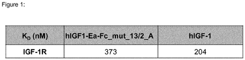

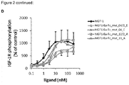
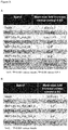


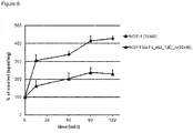

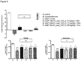


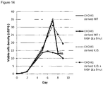

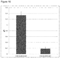
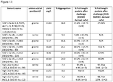


Classifications
-
- C—CHEMISTRY; METALLURGY
- C07—ORGANIC CHEMISTRY
- C07K—PEPTIDES
- C07K14/00—Peptides having more than 20 amino acids; Gastrins; Somatostatins; Melanotropins; Derivatives thereof
- C07K14/435—Peptides having more than 20 amino acids; Gastrins; Somatostatins; Melanotropins; Derivatives thereof from animals; from humans
- C07K14/575—Hormones
- C07K14/65—Insulin-like growth factors, i.e. somatomedins, e.g. IGF-1, IGF-2
-
- A—HUMAN NECESSITIES
- A61—MEDICAL OR VETERINARY SCIENCE; HYGIENE
- A61P—SPECIFIC THERAPEUTIC ACTIVITY OF CHEMICAL COMPOUNDS OR MEDICINAL PREPARATIONS
- A61P11/00—Drugs for disorders of the respiratory system
-
- A—HUMAN NECESSITIES
- A61—MEDICAL OR VETERINARY SCIENCE; HYGIENE
- A61P—SPECIFIC THERAPEUTIC ACTIVITY OF CHEMICAL COMPOUNDS OR MEDICINAL PREPARATIONS
- A61P13/00—Drugs for disorders of the urinary system
- A61P13/12—Drugs for disorders of the urinary system of the kidneys
-
- A—HUMAN NECESSITIES
- A61—MEDICAL OR VETERINARY SCIENCE; HYGIENE
- A61P—SPECIFIC THERAPEUTIC ACTIVITY OF CHEMICAL COMPOUNDS OR MEDICINAL PREPARATIONS
- A61P17/00—Drugs for dermatological disorders
- A61P17/02—Drugs for dermatological disorders for treating wounds, ulcers, burns, scars, keloids, or the like
-
- A—HUMAN NECESSITIES
- A61—MEDICAL OR VETERINARY SCIENCE; HYGIENE
- A61P—SPECIFIC THERAPEUTIC ACTIVITY OF CHEMICAL COMPOUNDS OR MEDICINAL PREPARATIONS
- A61P19/00—Drugs for skeletal disorders
-
- A—HUMAN NECESSITIES
- A61—MEDICAL OR VETERINARY SCIENCE; HYGIENE
- A61P—SPECIFIC THERAPEUTIC ACTIVITY OF CHEMICAL COMPOUNDS OR MEDICINAL PREPARATIONS
- A61P21/00—Drugs for disorders of the muscular or neuromuscular system
-
- C—CHEMISTRY; METALLURGY
- C07—ORGANIC CHEMISTRY
- C07K—PEPTIDES
- C07K2319/00—Fusion polypeptide
- C07K2319/30—Non-immunoglobulin-derived peptide or protein having an immunoglobulin constant or Fc region, or a fragment thereof, attached thereto
Definitions
- This invention is in the field of Insulin-like growth factors 1 (IGF-1) modifications.
- IGF-1 Insulin-like growth factors 1
- Insulin-like growth factors are part of a complex system that cells use to communicate with their physiologic environment.
- This complex system (often referred to as the insulin-like growth factor axis) consists of two cell-surface receptors (IGF-1R and IGF-2R), two ligands (IGF-1 and IGF-2), a family of six high-affinity IGF-binding proteins (IGFBP 1-6), and associated IGFBP degrading enzymes (proteases).
- IGF-1R and IGF-2R two cell-surface receptors
- IGF-1 and IGF-2 two ligands
- IGFBP 1-6 high-affinity IGF-binding proteins
- protes associated IGFBP degrading enzymes
- IGF-1 The IGF axis has been shown to play roles in the promotion of cell proliferation and the inhibition of cell death (apoptosis).
- IGF-1 is mainly secreted by the liver as a result of stimulation by human growth hormone (hGH). Almost every cell in the human body is affected by IGF-1, especially cells in muscles, cartilage, bones, liver, kidney, nerves, skin and lungs. In addition to the insulin-like effects, IGF-1 can also regulate cell growth. IGF-1 and IGF-2 are regulated by a family of gene products known as the IGF-binding proteins.
- IGFBP1-6 characterized binding proteins
- human IGF-1 also called somatomedin
- somatomedin is a small protein of 70 amino acids that has been shown to stimulate growth of a wide range of cells in culture.
- the IGF-1 protein is initially encoded by three known splice variant mRNAs.
- the open reading frame of each mRNA encodes a precursor protein containing the 70 amino acid IGF-1 (SEQ ID NO.:1) and a particular E-peptide at the C-terminus, depending on the particular IGF-1 mRNA.
- E-peptides have been termed the Ea (rsvraqrhtdmpktqkevhlknasrgsagnknyrm; SEQ ID NO.:2), Eb (rsvraqrhtdmpktqkyqppstnkntksqrrkgwpkthpggeqkegteaslqirgkkkeqrreigsrnaecrgkk gk; SEQ ID NO.:3) and Ec (rsvraqrhtdmpktqkyqppstnkntksqrrkgstfeerk; SEQ ID NO.: 4) peptides and range from 35 to 87 amino acids in length and encompass a common sequence region at the N-terminus and a variable sequence region at the C-terminus.
- the wild-type open reading frame for the IGF-1-Ea encodes a polypeptide of 135 amino acids including the leader sequence and a polypeptide of 105 amino acids without the leader sequence (gpetlcgaelvdalqfvcgdrgfyfnkptgygsssrrapqtgivdeccfrscdlrrlemycaplkpaksarsvraqrh tdmpktqkevhlknasrgsagnknyrm; SEQ ID NO.:5).
- the E-peptides are cleaved off of the precursor by endogenous proteases to yield the mature 70 amino acid IGF-1.
- IGFBP's proteases and IGF-1 binding proteins (IGFBP's). IGFBP's can either inhibit or potentiate IGF-1 activities ( Oh Y, et al., Characterization of the affinities of insulin-like growth factor (IGF)-binding proteins 1-4 for IGF-I, IGF-II, IGF-I/insulin hybrid, and IGF-I analogs. Endocrinology. 1993 Mar;132(3):1337-44 ). Strategies to increase the half-life of IGF-1 have been described in the prior art. Strategies that have been contemplated are
- the IGF-1 precursor variants fused to a human immunoglobulin Fc region remain poor drug candidates for mainly two reasons: (i) low production yield in mammalian production system, and (ii) increased binding affinity to the insulin receptor (InsR) compared to the unmodified human wildtype IGF-1, which can result in hypoglycemia, an adverse event of therapeutic concern.
- InsR insulin receptor
- a first subject matter of the disclosure relates to a polypeptide comprising a human IGF-1 (hIGF-1) protein, wherein the amino acid glycine at position 42 of said hIGF-1 protein is deleted or substituted, and wherein the numbering of the amino acids corresponds to SEQ ID NO.: 1.
- hIGF-1 human IGF-1
- An additional embodiment of the disclosure relates to a polypeptide comprising a human IGF-1 protein fused to an immunoglobulin Fc region of a human IgG, wherein the amino acid glycine at position 42 of the human IGF-1 protein is deleted or substituted, and wherein the numbering of the amino acids corresponds to SEQ ID NO.: 1.
- Another subject matter of the disclosure relates to a polypeptide comprising a human IGF-1 precursor protein, wherein the amino acid glycine at position 42 of said human IGF-1 precursor protein is deleted or substituted, and wherein the numbering of the amino acids corresponds to SEQ ID NO.: 5.
- An additional embodiment of the disclosure relates to a polypeptide comprising a human IGF-1 precursor protein fused to an immunoglobulin Fc region of a human IgG, wherein the amino acid glycine at position 42 of the human IGF-1 precursor protein is deleted or substituted, and wherein the numbering of the amino acids corresponds to SEQ ID NO.: 5.
- the disclosure relates to the above described polypeptides, wherein the amino acid glycine at position 42 is deleted or substituted by serine.
- the above mentioned human IGF-1 protein comprises additional deletions or mutations at amino acids G1, P2, E3, R36, R37, K68, S69 and/or A70.
- the above mentioned human IGF-1 precursor protein comprises an Ea-peptide and additional deletions or mutations of amino acids G1, P2, E3, R36, R37, K68, S69, A70, R71, S72, R74, R77 G96, S97, A98, G99, N100, K101, N102, Y103, Q104 and/or M105.
- the human IGF-1 precursor protein and the Fc region are separated by a peptide hinge region.
- the disclosure also relates to the above described human IGF-1 precursor protein, wherein the peptide hinge region is selected from the group consisting of peptides hinge 1 (SEQ ID NO.: 22), hinge 2 (SEQ ID NO.: 23) and hinge 3 (SEQ ID NO.: 24).
- the above described human IGF-1 precursor proteins when being mutated in the Ea-peptide at amino acids R74, R77 and/or R104, said amino acids R74, R77 and R104 are mutated to glutamine (Q).
- a polypeptide comprising or consisting of a human IGF-1 protein or a human IGF-1 precursor protein as described above, wherein
- Another particular embodiment of the disclosure relates to the above described human IGF-1 precursor protein, wherein the E-peptide is the Ea-petide and wherein
- Another particular embodiment of the disclosure relates to the above described human IGF-1 precursor protein, wherein the E-peptide is the Ea-petide and wherein
- the disclosure relates to the above described human IGF-1 precursor protein, wherein the E-peptide is the Ea-petide and wherein
- the disclosure relates to the above described human IGF-1 precursor protein, wherein the E-peptide is the Ea-petide and wherein
- the disclosure relates to the above described human IGF-1 precursor Fc fusion protein, wherein the E-peptide is the Ea-petide and wherein
- the disclosure relates to the above described human IGF-1 precursor Fc fusion protein, wherein the E-peptide is the Ea-petide and wherein
- the disclosure relates to the above described polypeptides, wherein the Fc region is modified to modulate its binding to Fc receptor.
- the disclosure relates to the above described human IGF-1 precursor Fc fusion protein, wherein the Fc region is modified to
- Another particular embodiment of the disclosure relates to the above described human IGF-1 precursor Fc fusion proteins comprising SEQ ID NO.: 8, or SEQ ID NO.: 9, or SEQ ID NO.: 10, or SEQ ID NO.: 11, or SEQ ID NO.: 12, or SEQ ID NO.: 13, or SEQ ID NO.: 14, or SEQ ID NO.: 27.
- the human IGF-1 precursor Fc fusion protein is glycosylated.
- the disclosure relates to a polynucleotide comprising a nucleic acid molecule encoding the above described human IGF-1 precursor Fc fusion proteins. Accordingly, the disclosure relates to a polynucleotide comprising the nucleic acid molecule as depicted in SEQ ID NO.:15 or SEQ ID NO.: 16 or SEQ ID NO.: 17 or SEQ ID NO.: 18 or SEQ ID NO.: 19 or SEQ ID NO.: 20 or SEQ ID NO.: 21.
- the disclosure furthermore provides for a pharmaceutical composition comprising a polypeptide comprising any one of the above disclosed human IGF-1 precursor proteins for use in therapy.
- the above mentioned therapeutic use is the treatment of a muscle disorder in a patient in need thereof.
- the therapeutic use is the treatment of burn patients suffering from loss of lean body mass and/or muscle wasting or the treatment of COPD patients, or the treatment of Kennedy disease patients, or the treatment of chronic kidney disease patients.
- the muscle disorder described above is muscle atrophy. Accordingly, in some aspects of the disclosure, the therapeutic use is the treatment of obesity-associated sarcopenia, sarcopenia, and diabetes-associated muscle atrophy.
- the disclosure furthermore provides for a method of treating a muscle disorder in a patient in need thereof, the method comprising administering a therapeutically effective amount of the above described human IGF-1 precursor protein of the invention.
- the disclosure relates to a method of treating burn patients suffering from loss of lean body mass and/or muscle wasting, or a method of treating COPD patients, or a method of treating Kennedy disease patients, or a method of treating chronic kidney disease patients
- the disclosure provides a method of treating a muscle disorder in a patient in need thereof, wherein the muscle disorder is a muscle atrophy selected from the group consisting of obesity-associated sarcopenia, sarcopenia, and diabetes-associated muscle atrophy.
- the disclosure relates to an IGF-1 polypeptide or an IGF-1 precursor polypeptide as described above, wherein the amino acid glycine at position 42 is deleted. In another embodiment, the disclosure relates to an IGF-1 polypeptide or an IGF-1 precursor polypeptide as described above, wherein said protein is pegylated.
- composition comprising
- X may consist exclusively of X or may include something additional e.g. X + Y.
- ⁇ or the letters “d” or “D”: in the context of a protein description e.g. "hIGF-1-Ea- ⁇ 1-3, R37A, ⁇ 71-72, R77Q-fc domain” or “hIGF-1-Ea-D1-3, R37A, D71-72, R77Q-fc domain”
- hIGF-1-Ea-D1-3, R37A, D71-72, R77Q-fc domain refers an amino acid deletion.
- D71-72, 77 in the context of the protein hIGF1-Ea-D1-3, R37A, D71-72, 77-fc describes the fact that the amino acids 71, 72 and 77 have been deleted.
- Insulin like growth factor 1 protein or a variant thereof refers to proteins being encoded by Insulin like growth factor 1 genes, particularly preferred is the human Insulin like growth factor 1 (hIGF-1) protein and variants thereof.
- An IGF-1 protein variant is a protein that differs by at least one amino acid from the IGF-1 wild-type sequence, wherein the term "wild-type sequence” refers to a polypeptide or gene sequence available in at least one naturally occurring organism or a polypeptide or gene sequence that has not been changed, mutated, or otherwise manipulated by man.
- wild-type sequence refers to a polypeptide or gene sequence available in at least one naturally occurring organism or a polypeptide or gene sequence that has not been changed, mutated, or otherwise manipulated by man.
- IGF-1 variant and IGF-1 mimetic are used interchangeably throughout the document.
- An IGF-1 variant is also the IGF-1 precursor protein or the pro-IGF-1 protein comprising a peptide leader sequence.
- An IGF-1 variant is also a fusion protein comprising an IGF-1 protein, e.g. a protein comprising an IGF-1 protein fused to an immunoglobulin Fc region. Examples for IGF-1 variants are disclosed inter alia in the patent applications WO05033134 (stabilized IGF-1 protein fused to an immunoglobulin Fc region) and WO2007146689 (stabilized IGF-1 precursor proteins).
- An IGF-1 variant as described above retains its biological activity in the sense that such a protein can be considered as a functional equivalent of the wildtype IGF-1.
- Functional equivalents with regard to the IGF-1 protein have to be understood as IGF-1 proteins comprising natural or artificial mutation. Mutations can be insertions, deletions or substitutions of one or more nucleic acids that do not diminish the biological activity of the IGF-1 protein. Functional equivalents having an identity of at least 80%, preferably 85%, more preferably 90%, most preferably more than 95%, very especially preferably at least 98% identity - but less than 100% identity to the IGF-1 wildtype protein, e.g. the human IGF-1 protein SEQ ID NO.: 1. In case of fusion proteins as described above, the 100% identity shall be defined only on the basis of the IGF-1 part of such a fusion protein.
- Insulin like growth factors are part of a complex system that cells use to communicate with their physiologic environment.
- This complex system (often referred to as the insulin-like growth factor axis) consists of two cell-surface receptors (IGF-1R and IGF-2R), two ligands (IGF-1 and IGF-2), a family of six high-affinity IGF-binding proteins (IGFBP 1-6), and associated IGFBP degrading enzymes (proteases).
- IGF-1R and IGF-2R two cell-surface receptors
- IGF-1 and IGF-2 two ligands
- IGFBP 1-6 high-affinity IGF-binding proteins
- protes associated IGFBP degrading enzymes
- IGF-1 is mainly secreted by the liver as a result of stimulation by human growth hormone (hGH). Almost every cell in the human body is affected by IGF-1, especially cells in muscles, cartilage, bones, liver, kidney, nerves, skin and lungs. In addition to the insulin-like effects, IGF-1 can also regulate cell growth. IGF-1 and IGF-2 are regulated by a family of gene products known as the IGF-binding proteins. These proteins help to modulate IGF action in complex ways that involve both inhibiting IGF action by preventing binding to the IGF receptors as well as promoting IGF action through aiding delivery to the receptors and increasing IGF half-life in the blood stream. There are at least six characterized binding proteins (IGFBP1-6).
- IGF-1 is used in a wide range of therapeutic applications.
- Mecasermin brand name IncrelexTM
- IGF-1 is a synthetic analog of IGF-1 which is approved for the treatment of growth failure.
- Several companies have evaluated IGF-1 in clinical trials for a variety of additional indications, including type 1 diabetes, type 2-diabetes, amyotrophic lateral sclerosis, severe burn injury and myotonic muscular dystrophy.
- Mammalian cell the term "mammalian cell” in the context of the disclosed method refers to mammalian cells which are suitable for protein production at industrial manufacturing scale. Those cells are well known to the skilled person and have originated for example from Cricetulus griseus, Cercopithecus aethiops, Homo sapiens, Mesocricetus auratus, Mus musculus and Chlorocebus species.
- the respective cell lines are known as CHO-cells (Chinese Hamster Ovary), COS-cells (a cell line derived from monkey kidney (African green monkey), Vero-cells (kidney epithelial cells extracted from African green monkey), Hela-cells (The line was derived from cervical cancer cells taken from Henrietta Lacks), BHK-cells (baby hamster kidney cells, HEK-cells (Human Embryonic Kidney), NS0-cells (Murine myeloma cell line), C127-cells (nontumorigenic mouse cell line), PerC6®-cells (human cell line, Crucell), CAP-cells (CEVEC's Amniocyte Production) and Sp-2/0-cells (Mouse myeloma cells).
- COS-cells a cell line derived from monkey kidney (African green monkey), Vero-cells (kidney epithelial cells extracted from African green monkey), Hela-cells (The line was derived
- receptor specificity of stabilised IGF-1 prior art proteins refers also to the reduced ability to induce insulin receptor phosphorylation (decreased potency and/or efficacy) of the IGF-1 molecules of the inventions compared to the prior art IGF-1 variants, which reduces the risk of inducing hypoglycemia, an adverse event of therapeutic concern.
- Precursor when used in the context of the present invention shall refer to the precursor of the mature human IGF-1 protein without signal peptide, but including the Ea, Eb and Ec peptide, respectively.
- Molecules comprising an Ea-peptide fused to an immunoglobulin Fc region, wherein the protein residues G1, P2, E3, R71 and S72 have been deleted, the amino acid R77 has been deleted or substituted by glutamine and the R37 amino acid has been substituted by alanine (hIGF1-Ea-del1-3, R37A, del71-72, 77-fc domain (SEQ ID NO.:6) and hIGF1-Ea-del1-3, R37A, del71-72, R77Q-fc domain (SEQ ID NO.:7)) have been produced and tested.
- hIGF1-Ea-del1-3, R37A, del71-72del71-72, 77-fc domain SEQ ID NO.:6
- hIGF1-Ea-del1-3, R37A, del71-72del71-72, 77-fc domain (SEQ ID NO.:6) proteins showed a decrease in receptor specificity compared to the unmodified mature IGF-1 in an NIH3T3 InsR phosphorylation assay (see figure 3 ).
- the IGF-1 precursor variants fused to a human immunoglobulin Fc region are poor drug candidates, because (i) low production yield in mammalian production system, and (ii) increased binding affinity to the insulin receptor (InsR) compared to the unmodified human wildtype IGF-1, which can result in hypoglycemia, an adverse event of therapeutic concern.
- InsR insulin receptor
- the invention is based on the surprising observation that (1) production yield in mammalian cell production systems and (2) the receptor specificity of native IGF-1 proteins or stabilised IGF-1 prior art proteins can be improved by introducing additional specific amino acid mutations, wherein different mutations solve different problems and said mutations can be combined to address multiple issues related to production yield, efficacy and/or receptor specificity.
- a furthermore particular surprising result was the observation that human IGF-1 precursor prior art proteins being mutated at position G42 (deletion or specific substitutions) show a similar ability to stimulate insulin receptor (InsR) phosphorylation as the unmodified human wildtype IGF-1, which diminishes the hypoglycemia risk of the prior art IGF-1 variants ( figure 3 ).
- the G42S mutation has an additional positive impact on the production yield of human IGF-1 precursor prior art proteins being fused to an immunoglobulin Fc region in mammalian cell ( figure 10/17 ), i.e. the formation of aggregates negatively impacting production yield could be dramatically reduced by introducing a G42S mutation ( figure 9/17 ).
- the invention is to some extend based on the surprising finding, that by manipulating the amino acid glycine 42 in human IGF-1 protein or human IGF-1 precursor variants thereof, two major technical hurdles in the development of therapeutic IGF-1 variants could have been overcome.
- the invention provides a solution for the problem that prior art IGF-1 precursor proteins being fused to a immunoglobulin Fc region of a human IgG cannot be produced at industrial scale in mammalian cells, because said fusion proteins are readily being degraded by mammalian cell proteases. Consequently, in another aspect the invention is based on the surprising finding that by manipulating specific amino acids in IGF-1 Ea-peptide precursor variants an additional major technical hurdle in the development of therapeutic IGF-1 precursor variants could have been overcome.
- polypeptides examples include, but are not limited to, the following polypeptides:
- a human IGF-1 protein (SEQ ID No.: 1) wherein the amino acid G42 is substituted by the amino acid serine and wherein the amino acid E3 is deleted and amino acid R37 is substituted by alanine.
- a human IGF-1 protein (SEQ ID No.: 1) wherein the amino acid G42 is substituted by the amino acid serine and wherein the amino acid E3 is deleted and amino acid R37 is substituted by alanine fused to an immunoglobulin Fc region, particularly a modified Fc region, particularly an Fc region, which is modified to modulate its binding to the Fc receptor, as described below.
- Another polypeptide of the invention is the human IGF-1 protein of SEQ ID NO.: 117.
- Another polypeptide of the invention is the human IGF-1 precursor protein of SEQ ID NO.: 118. It has been discovered that the mutation or deletion of amino acids R74, R77, G96, S97, A98, G99, N100, K101, N102, Y103, Q104 and/or M105 of the prior art IGF-1 Ea peptide precursor proteins mentioned above leads to a higher yield of non-degraded protein when expressed by mammalian cells. Is has furthermore been discovered that combination of the mentioned mutations/deletions at positions R74, R77, G96, S97, A98, G99, N100, K101, N102, Y103, Q104 and/or M105 results in a synergistic effect.
- R74, R77, G96, S97, A98, G99, N100, K101, N102, Y103, Q104 and/or M105 is deleted or mutated in the herein disclosed modified IGF-1 precursor proteins.
- R74, R77 and/or R104 are mutated to Q in the herein disclosed IGF-1 precursor proteins.
- K68, S69, A70, R71 and/or R72 may additionally be deleted or mutated in the herein disclosed modified IGF-1 precursor proteins.
- the invention provides a polypeptide containing a human IGF-1 precursor protein, i.e. comprising the Ea-peptide from human IGF-1, wherein the amino acid glycine at position 42 is deleted or substituted by another amino acid and wherein the numbering of the amino acids corresponds to SEQ ID NO.: 5.
- the E-peptide may be the Ea, Eb, or Ec peptide (SEQ ID NOs: 2-4).
- the amino acid glycine at position 42 is deleted or substituted by the amino acid or serine.
- the above described human IGF-1 Ea-peptide precursor protein being mutated at position G42 as described above comprises additional deletions and/or mutations at amino acids G1, P2, E3, R36, R37, K68, S69, A70, R71, S72, R74, R77, G96, S97, A98, G99, N100, K101, N102, Y103, Q104 and/or M105, wherein the numbering of the amino acids corresponds to SEQ ID NO.: 5.
- polypeptides include, but are not limited to, the following polypeptides: A polypeptide comprising a human IGF-1 Ea-peptide precursor protein wherein the amino acid G42 is substituted by the amino acid serine and wherein the amino acid(s)
- the disclosure relates to the above described proteins comprising the polypeptides (1)-(29a), wherein said molecules instead of being mutated at the positions 1-3, only the amino acid E3 is deleted (e.g. the molecule (28a) could also refer to a polypeptide comprising a human IGF-1 Ea-peptide precursor protein wherein the amino acid G42 is substituted by the amino acid serine and wherein the amino acid E3 is deleted, amino acid R36 and R37 are both substituted by glutamine (Q), the amino acids K68, S69, A70, R71 and S72 are deleted and amino acids R74, R77 and R104 are mutated to glutamine (Q).)
- the disclosure relates to a polypeptide containing a human IGF-1 precursor protein, i.e. comprising the Ea-peptide from human IGF-1, which is fused to an immunoglobulin Fc region and wherein the amino acid glycine at position 42 is substituted by another amino acid and wherein the numbering of the amino acids of the IGF-1 part of said protein corresponds to SEQ ID NO.: 5.
- the E-peptide may be the Ea, Eb, or Ec peptide and the amino acid by which the glycine at position 42 is substituted is serine.
- the disclosure relates to a polypeptide comprising a human IGF-1 precursor protein; (a) wherein the amino acid G42 is deleted or substituted by the amino acid serine; and (b) which is linked to an immunoglobulin Fc region, particularly a modified Fc region, particularly an Fc region, which is modified to modulate its binding to the Fc receptor.
- an immunoglobulin Fc region particularly a modified Fc region, particularly an Fc region, which is modified to modulate its binding to the Fc receptor.
- one or more amino acids can be replaced with a different amino acid residue such that the Fc region has an altered affinity for the Fc receptor or the C1 component of complement.
- So called silenced immunoglobulin Fc regions have been described in the art: LALA and N297A ( Strohl, W., 2009, Curr. Opin. Biotechnol. vol.
- silent Fc IgG1 antibodies comprise the so-called LALA mutant comprising L234A and L235A mutation in the IgG1 Fc amino acid sequence.
- Another example of a silent IgG1 antibody comprises the D265A mutation.
- Another silent IgG1 antibody comprises the N297A mutation, which results in aglycosylated/non-glycosylated antibodies.
- the disclosed hIGF-1 precursor protein is fused to an Fc region comprising the L234A and L235A mutation or the D265A mutation or the N297A mutation.
- Fc LALA, D265A or N297A constructs have reduced ADCC activity
- the polypeptide containing a human IGF-1 Ea-peptide precursor protein fused to an immunoglobulin Fc region, wherein the amino acid glycine at position 42 is substituted by the amino acid serine comprises additional deletions and/or mutations at amino acids G1, P2, E3, R36, R37, K68, S69, A70, R71, S72, R74, R77 and/or R104.
- polypeptides include, but are not limited to, the following polypeptides: A polypeptide containing a human IGF-1 Ea-peptide precursor protein fused to an immunoglobulin Fc region wherein the amino acid glycine at position 42 is substituted by the amino acid serine, wherein the numbering of the amino acids of the IGF-1 part of said protein corresponds to SEQ ID NO.: 5 and wherein the amino acid(s)
- the disclosure relates to the above describes proteins comprising the above described polypeptides (1b)-(29c), wherein said molecules instead of being mutated at the positions 1-3, only the amino acid E3 is deleted (e.g. the molecule (28c) could also refer to a polypeptide comprising a human IGF-1 precursor protein fused to an immunoglobulin Fc region wherein the amino acid G42 is substituted by the amino acid amino acid serine and wherein the amino acid(s) E3 are deleted, amino acid R36 and R37 are both substituted by glutamine (Q) and the amino acids K68, S69, A70, R71 and S72 are deleted and amino acids R74, R77 and R104 are mutated to glutamine (Q).)
- the disclosure relates to the above described polypeptides (e.g. the polypeptides 1-29c), comprising an mutated E-peptide consisting of the amino acids
- the disclosure provides the above described IGF-1 precursor protein being fused to an Fc region, wherein the Fc region may be directly fused to the modified IGF-1 precursor polypeptide or may be connected by a hinge region using recombinant DNA technologies well known in the art. If a DNA hinge region is used, the Fc region may be connected to any part of the modified IGF-1 precursor polypeptide. In one embodiment the Fc region is fused directly to the C-terminus of the modified IGF-1 precursor polypeptide. In another embodiment, the Fc region is linked to the C-terminus of the modified IGF-1 precursor polypeptide by a Gly Ser (-GS-) linker.
- a DNA linker may be used to provide a restriction site between components for ease of manipulation.
- a linker may also be provided to enhance expression of the polypeptide from a host cell, to decrease steric hindrance such that the component may assume its optimal tertiary or quaternary structure and/or interact appropriately with its target molecule.
- linkers and methods of identifying desirable spacers see, for example, George et al. (2003) Protein Engineering 15:871-879 .
- a linker sequence may include one or more amino acids naturally connected to a receptor component, or may be an added sequence used to enhance expression of the fusion protein, provide specifically desired sites of interest, allowing component domains to form optimal tertiary structures and/or to enhance the interaction of a component with its target molecule.
- the linker comprises one or more peptide sequences which are between 1-100 amino acids, preferably 1-25 amino acids in length. In one embodiment, the linker is 1-5 amino acids in length.
- the linker is a three amino acid sequence; more specifically, the three amino acid sequence of Gly Pro Gly. In another embodiment, the linker is Gly Ser.
- hinge molecules include, but are not limited to, the following polypeptides:
- the disclosure relates to a polypeptide containing a human IGF-1 Ea-peptide precursor protein which is fused to an immunoglobulin Fc region, particularly a modified Fc region, particularly an Fc region, which is modified to modulate its binding to Fc receptor, preferably by substituting one or both of amino acids 234 and 235 to alanine as described above, wherein the amino acid glycine at position 42 is substituted by serine, wherein the numbering of the amino acids corresponds to SEQ ID NO.: 5 and wherein the immunoglobulin Fc region is fused to the IGF-1 precursor protein via a hinge region.
- polypeptides examples include, but are not limited to, the following polypeptides:
- amino acids R71 and S72 of the above described IGF-1 precursor proteins can be mutated as follows:
- the Fc region is modified to modulate its binding to Fc receptor. In one embodiment, the Fc region is modified to reduce its affinity for Fc receptor. In one embodiment, the Fc region is modified to reduce ADCC activity. In one embodiment, the Fc region is modified to prevent ADCC activity.
- the invention provides a polypeptide comprising SEQ ID NO.: 8 (hlgF1-Ea-Fc_mut 13/2_E). In one embodiment, the invention provides a polypeptide comprising SEQ ID NO.: 9 (hlgF1-Ea-Fc_mut 13/2_A). In one embodiment, the invention provides a polypeptide comprising SEQ ID NO.:10 (hlgF1-Ea-Fc_mut 13/2_C). In one embodiment, the invention provides a polypeptide comprising SEQ ID NO.: 11 (hlgF1-Ea-Fc_mut 13/2_F).
- the invention provides a polypeptide comprising SEQ ID NO.:12 (hlgF1-Ea-Fc_mut 04/2_E).
- the invention provides a polypeptide comprising SEQ ID NO.:13 (hlgF1-Ea-Fc_mut 04/2_A).
- the invention provides a polypeptide comprising SEQ ID NO.:14 (hlgF1-Ea-Fc_mut 04/2_F).
- the invention provides a polypeptide consisting of SEQ ID NOs.:8, 9, 10, 11, 12, 13 or 14.
- the present disclosure provides a polypeptide that is at least 95%, at least 96%, at least 96%, at least 97%, at least 98%, at least 99% identical to SEQ ID NO.: 8.
- the present disclosure provides a polypeptide that is at least 95%, at least 96%, at least 96%, at least 97%, at least 98%, at least 99% identical to SEQ ID NO.: 9.
- the present disclosure provides a polypeptide that is at least 95%, at least 96%, at least 96%, at least 97%, at least 98%, at least 99% identical to SEQ ID NO.: 10.
- the present disclosure provides a polypeptide that is at least 95%, at least 96%, at least 96%, at least 97%, at least 98%, at least 99% identical to SEQ ID NO.: 11.
- the present disclosure provides a polypeptide that is at least 95%, at least 96%, at least 96%, at least 97%, at least 98%, at least 99% identical to SEQ ID NO.: 12.
- the present disclosure provides a polypeptide that is at least 95%, at least 96%, at least 96%, at least 97%, at least 98%, at least 99% identical to SEQ ID NO.: 13.
- the present disclosure provides a polypeptide that is at least 95%, at least 96%, at least 96%, at least 97%, at least 98%, at least 99% identical to SEQ ID NO.: 14.
- polypeptides of the present invention are glycosylated.
- amino acids may be mutated to another amino acid. Typically the amino acid is replaced with an alanine residue.
- other amino acids may be used, such as non-natural amino acids or natural amino acids from another group (i.e. polar, acidic, basic or non-polar).
- Methods of introducing a mutation into amino acids of a protein are well known to those skilled in the art. See, e.g., Ausubel (ed.), Current Protocols in Molecular Biology, John Wiley and Sons, Inc. (1994 ); T. Maniatis, E. F. Fritsch and J. Sambrook, Molecular Cloning: A Laboratory Manual, Cold Spring Harbor laboratory, Cold Spring Harbor, N.Y. (1989 )).
- Methods comprise, but are not limited to, amplification of the DNA encoding the polypeptide functionally active variant or fragments thereof by polymerase chain reaction (PCR) conducted with mutagenic primers and assembling the fragments by assembly PCR if needed or introduction of mutations using commercially available kits such as "QuikChange.TM. Site-Directed Mutagenesis Kit” (Stratagene). See, e.g., Ausubel (ed.), Current Protocols in Molecular Biology, John Wiley and Sons, Inc. (1994 ); T. Maniatis, E. F. Fritsch and J. Sambrook, Molecular Cloning: A Laboratory Manual, Cold Spring Harbor laboratory, Cold Spring Harbor, N.Y. (1989 )).
- PCR polymerase chain reaction
- mutated sequences can be obtained by synthetic gene synthesis a service provided by commercial companies (e.g. Geneart, Life Technology).
- the generation of a polypeptide functionally active variant or derivative to a polypeptide by replacing an amino acid that does not influence the function of a polypeptide can be accomplished by one skilled in the art.
- the Fc region is from IgG, IgM or IgA. In one embodiment the Fc region derives from IgG.
- the Fc domain of IgG may be selected from the isotypes IgG1, IgG2, IgG3, and IgG4, as well as any allotype within each isotype group. In one embodiment the Fc region is a human Fc region.
- the Fc region may be altered by replacing at least one amino acid residue with a different amino acid residue to alter the effector functions of the Fc region.
- one or more amino acids can be replaced with a different amino acid residue such that the Fc region has an altered affinity for the Fc receptor or the C1 component of complement.
- residues 234 and 235 may be mutated.
- these mutations may be to alanine.
- the antibody of the invention has a mutation in the Fc region at one or both of amino acids 234 and 235.
- one or both of amino acids 234 and 235 may be substituted to alanine.
- Such an Fc variant where residues 234 and 235 have been substituted for alanine is commonly referred to as "LALA".
- LALA constructs have reduced ADCC activity.
- the IGF-1 precursor protein is linked to an IgG1 LALA Fc.
- Fusion to or “linked to” as used herein in the context of the disclosed proteins fused to/linked to an immunoglobulin Fc region shall mean combining two polypeptides that are not naturally present in the same polypeptide.
- the immunoglobulin Fc region may be directly fused to the modified IGF-1 polypeptide or may be connected by a linker using recombinant DNA technologies well known in the art. If a DNA linker is used, the Fc region may be connected to any part of the modified IGF-1 polypeptide. In one embodiment the Fc region is fused directly to the C-terminus of the modified IGF-1 polypeptide.
- the Fc region is linked to the C-terminus of the modified IGF-1 polypeptide by a Gly Ser (-GS-) linker and/or a hinge region (SEQ IDs NO.: 22 to 24) such as for hlgF1-Ea-Fc_mut 13/2_E (SEQ IDs NO.: 8), hlgF1-Ea-Fc_mut 13/2_A (SEQ IDs NO.: 9), hlgF1-Ea-Fc_mut 13/2_C (SEQ IDs NO.: 10), hlgF1-Ea-Fc_mut 13/2_F (SEQ IDs NO.: 11), hlgF1-Ea-Fc_mut 04/2_E (SEQ IDs NO.: 12), hlgF1-Ea-Fc_mut 04/2_A (SEQ IDs NO.: 13) or hlgF1-Ea-Fc_mut 04/2_F (SEQ IDs NO.:14).
- a DNA linker may be used to provide a restriction site between components for ease of manipulation.
- a linker may also be provided to enhance expression of the polypeptide from a host cell, to decrease steric hindrance such that the component may assume its optimal tertiary or quaternary structure and/or interact appropriately with its target molecule.
- linkers and methods of identifying desirable spacers see, for example, George et al. (2003) Protein Engineering 15:871-879 .
- a linker sequence may include one or more amino acids naturally connected to a receptor component, or may be an added sequence used to enhance expression of the fusion protein, provide specifically desired sites of interest, allowing component domains to form optimal tertiary structures and/or to enhance the interaction of a component with its target molecule.
- the linker comprises one or more peptide sequences which are between 1-100 amino acids, preferably 1-25 amino acids in length. In one embodiment, the linker is 1-5 amino acids in length.
- the linker is a three amino acid sequence; more specifically, the three amino acid sequence of Gly Pro Gly. In another embodiment, the linker is Gly Ser.
- the invention also provides nucleic acid molecules that encode the polypeptides of the invention.
- Preferred nucleic acid sequences are those encoding hlgF1-Ea-Fc_mut 13/2_E, hlgF1-Ea-Fc_mut 13/2_A, hlgF1-Ea-Fc_mut 13/2_C, hlgF1-Ea-Fc_mut 13/2_F, hlgF1-Ea-Fc_mut 04/2_E, hlgF1-Ea-Fc_mut 04/2_A or hlgF1-Ea-Fc_mut 04/2_F (SEQ ID NOs.: 15-21).
- Nucleic acid molecules of the present invention may be in the form of RNA, such as mRNA, or in the form of DNA, including, for instance cDNA or synthetic DNA.
- the nucleic acid molecules may be double-stranded or single-stranded.
- Single-stranded DNA may be the coding strand, also known as the sense strand, or it may be the non-coding strand, also referred to as the antisense strand.
- nucleic acid sequences may code for the same polypeptide sequence.
- variant nucleic acid sequences are included within the scope of the application. Indeed, it may be preferable to make conservative modifications to a nucleic acid sequence to improve expression of a polypeptide of the invention.
- the invention further provides a vector comprising a nucleic acid of the invention.
- the vector may be a cloning or expression vector.
- the nucleic acid may be comprised within an expression vector carrying elements needed for efficient expression well known by the skilled person in the art.
- Elements include for example a promoter, such as for example the Cytomegalovirus (CMV) enhancer-promoter, a signal sequence for secretion, such as the natural or any other sequence known to facilitate secretion, a polyadenylation signal and transcription terminator, for example derived from Bovine Growth Hormone (BGH) gene, an element allowing episomal replication and replication in prokaryotes (e.g. SV40 origin and ColE1 or others known in the art) and elements to allow selection, such as ampicillin resistance gene and zeocin or hygromycin marker.
- CMV Cytomegalovirus
- BGH Bovine Growth Hormone
- the disclosure relates to the above described IGF-1 variant polypeptides, wherein said human IGF-1 precursor proteins are pegylated.
- the pegylation is particularly preferred for IGF-1 precursor proteins that are not fused to an immunoglobulin Fc region of a human IgG.
- Conjugation to poly(ethylene glycol) (PEG; pegylation) have proven to be beneficial in prolonging the half-life of therapeutic proteins drugs. It is expected that pegylation of the IGF precursor polypeptides of the invention may result in similar pharmaceutical advantages. Methods of pegylation of IGF-1 are well known in the art.
- lysine-monopegylation can be adapted for the precursor IGF polypeptides of the invention.
- pegylation can be achieved in any part of a polypeptide of the invention by the introduction of a nonnatural amino acid.
- Certain nonnatural amino acids can be introduced by the technology described in Deiters et al., J Am Chem Soc 125:11782-11783, 2003 ; Wang and Schultz, Science 301:964-967, 2003 ; Wang et al., Science 292:498-500, 2001 ; Zhang et al., Science 303:371-373, 2004 or in US Patent No. 7,083,970 . Briefly, some of these expression systems involve site-directed mutagenesis to introduce a nonsense codon, such as an amber TAG, into the open reading frame encoding a polypeptide of the invention.
- a nonsense codon such as an amber TAG
- Such expression vectors are then introduced into a host that can utilize a tRNA specific for the introduced nonsense codon and charged with the nonnatural amino acid of choice.
- Particular nonnatural amino acids that are beneficial for purpose of conjugating moieties to the polypeptides of the invention include those with acetylene and azido side chains.
- the IGF precursor polypeptides containing these novel amino acids can then be pegylated at these chosen sites in the protein. In addition, such pegylated IGF molecules without the E-peptide are also useful as therapeutics.
- Mammalian host cells for expressing the recombinant antibodies of the invention include Chinese Hamster Ovary (CHO cells) (including dhfr- CHO cells, described Urlaub and Chasin, 1980 Proc. Natl. Acad. Sci. USA 77:4216-4220 used with a DH FR selectable marker, e.g. as described in R.J. Kaufman and P.A. Sharp, 1982 Mol. Biol. 159:601-621 ), HEK293 cells (Human Embryonic Kidney 293 cells), NSO murine myeloma cells, COS cells and SP2 cells.
- Preferred host cells are CHO K1 derivative cells.
- a suitable method for the production of recombinant IGF-1 or variants thereof in mammalian cells for example a CHO cell, wherein said mammalian cell being deficient in the expression of a functional Insulin like growth factor 1 receptor (IGF-1 R) comprises the following step:
- CHO cells like CHO-K1 derivative cells, CHO-DUXB11 derivative cells or CHO-DG44 cells.
- Selection protocols are routinely used to facilitate selection of cells that are likely to have integrated the recombinant DNA encoding the desired therapeutic protein, like growth factors, e.g. IGF-1.
- Antibiotic resistance or the ability to grow in a nutritionally selective medium conferred by a gene co-integrated on the transformation vector is routinely used. (see Weber, W. and Fussenegger, M.
- DHFR dihydrofolate reductase
- MTX methotrexate
- MSX glutamine synthetase
- Vectors particularly suitable for producing polypeptides in mammalian cells particularly rodent cells such as CHO and DHFR gene defective CHO cells have been disclosed in the patent application WO09080720A .
- rodent cells such as CHO and DHFR gene defective CHO cells
- Suitable host cells are described above. After introduction of the expression vector nucleic acid into the host cell(s), the obtained transformants are cultured under selective conditions suitable for assaying the expression of the mammalian selectable marker gene enclosed in the expression cassette (MSM).
- MSM mammalian selectable marker gene enclosed in the expression cassette
- a second selection step may be performed by culturing the transformants in a selection medium adapted for selecting the amplifiable, selectable marker gene comprised in the expression cassette (MASM).
- a selection medium adapted for selecting the amplifiable, selectable marker gene comprised in the expression cassette (MASM).
- the transformants can be cultured in a nucleotide or purine-free medium in the presence of a DHFR inhibitor.
- a corresponding induction signal should be provided in order to commence expression of the polypeptide.
- said host cells may be cultured in the presence of a DHFR inhibitor.
- Suitable DHFR inhibitors are antifolates such as e.g. MTX.
- the concentration of antifolate/MTX used depends on the host cell and the DHFR variant incorporated in the vector.
- the concentration range can be chosen for multistep amplification procedures in DHFR" host cells for example at values around 5nM - 20nM ranging to values of 500nM to 1000nM or even higher for secondary or further amplification steps.
- starting concentrations are usually higher in the range of 100nM to 750nM, preferably 500nM in the first steps and 500nM to 1000nM and above for further amplification steps.
- Suitable DHFR variants are described above.
- said host cells may be cultured in the presence of e.g. MSX.
- concentration of MSX used depends on the host cell.
- concentration range can be chosen between from about 15 to 150 micromolar, 20 to 100 micromolar and 25 to 50 micromolar. These ranges are particularly suitable for NSO and CHO cells.
- the present invention provides a composition, e.g. a pharmaceutical composition, containing one or a combination of the above described IGF-1 variants, formulated together with a pharmaceutically acceptable carrier.
- a pharmaceutical composition e.g. a pharmaceutical composition, containing one or a combination of the above described IGF-1 variants, formulated together with a pharmaceutically acceptable carrier.
- Pharmaceutical compositions of the invention also can be administered in combination therapy, i.e. combined with other agents.
- the IGF-1 variants of the present invention can be combined with at least one muscle mass/strength increasing agent, for example, anti-ActRIIB antibody, IGF-2 or variants IGF-2, an anti-myostatin antibody, a myostatin propeptide, a myostatin decoy protein that binds ActRIIB but does not activate it, a beta 2 agonist, a Ghrelin agonist, a SARM, GH agonists/mimetics or follistatin.
- muscle mass/strength increasing agent for example, anti-ActRIIB antibody, IGF-2 or variants IGF-2, an anti-myostatin antibody, a myostatin propeptide, a myostatin decoy protein that binds ActRIIB but does not activate it, a beta 2 agonist, a Ghrelin agonist, a SARM, GH agonists/mimetics or follistatin.
- therapeutic agents that can be used in combination therapy are described in
- pharmaceutically acceptable means approved by a regulatory agency of the Federal or a state government or listed in the U.S. Pharmacopeia or other generally recognized pharmacopeia for use in animals, and more particularly in humans.
- carrier refers to a diluent, adjuvant, excipient, or vehicle with which the therapeutic is administered.
- Such pharmaceutical carriers can be sterile liquids, such as water and oils, including those of petroleum, animal, vegetable or synthetic origin, such as peanut oil, soybean oil, mineral oil, sesame oil and the like.
- Suitable pharmaceutical excipients include starch, glucose, lactose, sucrose, gelatin, malt, rice, flour, chalk, silica gel, sodium stearate, glycerol monostearate, talc, sodium chloride, dried skim milk, glycerol, propylene, glycol, water, ethanol and the like.
- the composition if desired, can also contain minor amounts of wetting or emulsifying agents, or pH buffering agents. These compositions can take the form of solutions, suspensions, emulsion, tablets, pills, capsules, powders, sustained-release formulations and the like.
- the composition can be formulated as a suppository, with traditional binders and carriers such as triglycerides.
- Oral formulation can include standard carriers such as pharmaceutical grades of mannitol, lactose, starch, magnesium stearate, sodium saccharine, cellulose, magnesium carbonate, etc. Examples of suitable pharmaceutical carriers are described in "Remington's Pharmaceutical Sciences” by E. W. Martin.
- the composition is formulated in accordance with routine procedures as a pharmaceutical composition adapted for intravenous administration to human beings.
- the composition may also include a solubilizing agent and a local anesthetic such as lidocaine to ease pain at the site of the injection.
- a solubilizing agent such as lidocaine to ease pain at the site of the injection.
- the composition is to be administered by infusion, it can be dispensed with an infusion bottle containing sterile pharmaceutical grade water or saline.
- an ampoule of sterile water for injection or saline can be provided so that the ingredients may be mixed prior to administration.
- Pharmaceutically acceptable carrier include any and all solvents, dispersion media, coatings, antibacterial and antifungal agents, isotonic and absorption delaying agents, and the like that are physiologically compatible.
- the carrier should be suitable for intravenous, intramuscular, subcutaneous, parenteral, spinal or epidermal administration (e.g. by injection or infusion).
- the active compound i.e. antibody, immunoconjuage, or bispecific molecule, may be coated in a material to protect the compound from the action of acids and other natural conditions that may inactivate the compound.
- a pharmaceutical composition of the invention also may include a pharmaceutically acceptable anti-oxidant.
- pharmaceutically acceptable antioxidants include: water soluble antioxidants, such as ascorbic acid, cysteine hydrochloride, sodium bisulfate, sodium metabisulfite, sodium sulfite and the like; oil-soluble antioxidants, such as ascorbyl palmitate, butylated hydroxyanisole (BHA), butylated hydroxytoluene (BHT), lecithin, propyl gallate, alpha-tocopherol, and the like; and metal chelating agents, such as citric acid, ethylenediamine tetraacetic acid (EDTA), sorbitol, tartaric acid, phosphoric acid, and the like.
- water soluble antioxidants such as ascorbic acid, cysteine hydrochloride, sodium bisulfate, sodium metabisulfite, sodium sulfite and the like
- oil-soluble antioxidants such as ascorbyl palmitate, butylated hydroxyanisole
- aqueous and nonaqueous carriers examples include water, ethanol, polyols (such as glycerol, propylene glycol, polyethylene glycol, and the like), and suitable mixtures thereof, vegetable oils, such as olive oil, and injectable organic esters, such as ethyl oleate.
- polyols such as glycerol, propylene glycol, polyethylene glycol, and the like
- vegetable oils such as olive oil
- injectable organic esters such as ethyl oleate.
- Proper fluidity can be maintained, for example, by the use of coating materials, such as lecithin, by the maintenance of the required particle size in the case of dispersions, and by the use of surfactants.
- compositions may also contain adjuvants such as preservatives, wetting agents, emulsifying agents and dispersing agents. Prevention of presence of microorganisms may be ensured both by sterilization procedures, supra, and by the inclusion of various antibacterial and antifungal agents, for example, paraben, chlorobutanol, phenol sorbic acid, and the like. It may also be desirable to include isotonic agents, such as sugars, sodium chloride, and the like into the compositions. In addition, prolonged absorption of the injectable pharmaceutical form may be brought about by the inclusion of agents which delay absorption such as, aluminum monostearate and gelatin.
- Pharmaceutically acceptable carriers include sterile aqueous solutions or dispersions and sterile powders for the extemporaneous preparation of sterile injectable solutions or dispersion.
- the use of such media and agents for pharmaceutically active substances is known in the art. Except insofar as any conventional media or agent is incompatible with the active compound, use thereof in the pharmaceutical compositions of the invention is contemplated. Supplementary active compounds can also be incorporated into the compositions.
- Therapeutic compositions typically must be sterile and stable under the conditions of manufacture and storage.
- the composition can be formulated as a solution, microemulsion, liposome, or other ordered structure suitable to high drug concentration.
- the carrier can be a solvent or dispersion medium containing, for example, water, ethanol, polyol (for example, glycerol, propylene glycol, and liquid polyethylene glycol, and the like), and suitable mixtures thereof.
- the proper fluidity can be maintained, for example, by the use of a coating such as lecithin, by the maintenance of the required particle size in the case of dispersion and by the use of surfactants.
- Prolonged absorption of the injectable compositions can be brought about by including in the composition an agent that delays absorption for example, monostearate salts and gelatin.
- Sterile injectable solutions can be prepared by incorporating the active compound in the required amount in an appropriate solvent with one or a combination of agents enumerated above, as required, followed by sterilization microfiltration.
- dispersions are prepared by incorporating the active compound into a sterile vehicle that contains a basic dispersion medium and the required other agents from those enumerated above.
- the methods of preparation are vacuum drying and freeze-drying (lyophilization) that yield a powder of the active agent plus any additional desired agent from a previously sterile-filtered solution thereof.
- the amount of active agent which can be combined with a carrier material to produce a single dosage form will vary depending upon the subject being treated, and the particular mode of administration.
- the amount of active agent which can be combined with a carrier material to produce a single dosage form will generally be that amount of the composition which produces a therapeutic effect. Generally, out of one hundred percent, this amount will range from about 0.01 per cent to about ninety-nine percent of active agent, from about 0.1 per cent to about 70 per cent, or from about 1 percent to about 30 percent of active agent in combination with a pharmaceutically acceptable carrier.
- Dosage regimens are adjusted to provide the optimum desired response (e.g. a therapeutic response). For example, a single bolus may be administered, several divided doses may be administered over time or the dose may be proportionally reduced or increased as indicated by the exigencies of the therapeutic situation. It is especially advantageous to formulate parenteral compositions in dosage unit form for ease of administration and uniformity of dosage.
- Dosage unit form as used herein refers to physically discrete units suited as unitary dosages for the subjects to be treated; each unit contains a predetermined quantity of active compound calculated to produce the desired therapeutic effect in association with the required pharmaceutical carrier.
- the specification for the dosage unit forms of the invention are dictated by and directly dependent on the unique characteristics of the active compound and the particular therapeutic effect to be achieved, and the limitations inherent in the art of compounding such an active compound for the treatment of sensitivity in individuals.
- a therapeutically effective amount of a polypeptide in the context of administrating the IGF-1 variants of the disclosure or composition comprising said IGF-1 variants ranges from about 0.001 to 100 mg/kg, or 0.01 to 30 mg/kg, and more usually 0.1 to 10 mg/kg of the host body weight.
- dosages can be about 0.1 mg/kg body weight, can be about 0.2 mg/kg body weight, can be about 0.3 mg/kg body weight, can be about 1 mg/kg body weight, can be about 3 mg/kg body weight, can be about 5 mg/kg body weight or about 10 mg/kg body weight.
- the skilled person knows to identify a suitable effective dose, which will vary depending on the rout of administration (e.g. intraveneously or subcutaneously).
- An exemplary treatment regime entails administration once per day, once every week, once every two weeks, once every three weeks, once every four weeks or once a month. Such administration may be carried out intraveneously or subcutaneously.
- Dosage regimens for IGF-1 variants of the invention include 0.1 mg/kg body weight or 0.2 mg/kg body weight or 0.3 mg/kg body weight or 0.5 mg/kg body weight or 1 mg/kg body weight or 3 mg/kg body weight or 10 mg/kg body weight by intravenous administration.
- the composition can be a sustained release formulation, in which case less frequent administration is required.
- Dosage and frequency vary depending on the half-life of the antibody in the patient. The dosage and frequency of administration can vary depending on whether the treatment is prophylactic or therapeutic.
- a relatively low dosage is administered at relatively infrequent intervals over a long period of time. Some patients continue to receive treatment for the rest of their lives. In therapeutic applications, a relatively high dosage at relatively short intervals is sometimes required until progression of the disease is reduced or terminated or until the patient shows partial or complete amelioration of symptoms of disease. Thereafter, the patient can be administered a prophylactic regime.
- Actual dosage levels of the active agents in the pharmaceutical compositions of the present invention may be varied so as to obtain an amount of the active agent which is effective to achieve the desired therapeutic response for a particular patient, composition, and mode of administration, without being toxic to the patient.
- the selected dosage level will depend upon a variety of pharmacokinetic factors including the activity of the particular compositions of the present invention employed, or the ester, salt or amide thereof, the route of administration, the time of administration, the rate of excretion of the particular compound being employed, the duration of the treatment, other drugs, compounds and/or materials used in combination with the particular compositions employed, the age, sex, weight, condition, general health and prior medical history of the patient being treated, and like factors well known in the medical arts.
- Administration of a therapeutically effective dose of an IGF-1 variant comprised in the compositions of the invention can result in a decrease in severity of disease symptoms, an increase in frequency and duration of disease symptom-free periods, or a prevention of impairment or disability due to the disease affliction i.e. an increase in muscle mass and/or function or decrease/reduction of wound area in burn patients.
- Patients will receive an effective amount of the polypeptide active ingredient i.e. an amount that is sufficient to detect, treat, ameliorate, or prevent the disease or disorder in question. Therapeutic effects may also include reduction in physical symptoms.
- the optimum effective amount and concentration of a therapeutic protein for any particular subject will depend upon various factors, including the patient's age size health and/or gender, the nature and extent of the condition, the activity of the particular therapeutic protein, the rate of its clearance by the body, and also on any possible further therapeutic(s) administered in combination with the therapeutic protein.
- the effective amount delivered for a given situation can be determined by routine experimentation and is within the judgment of a clinician. Dosage can be by a single dose schedule or a multiple dose schedule.
- a composition of the present invention can be administered by one or more routes of administration using one or more of a variety of methods known in the art. As will be appreciated by the skilled artisan, the route and/or mode of administration will vary depending upon the desired results. Routes of administration for the therapeutic proteins of the invention include intravenous, intramuscular, intradermal, intraperitoneal, subcutaneous, spinal or other parenteral routes of administration, for example by injection or infusion.
- parenteral administration means modes of administration other than enteral and topical administration, usually by injection, and includes, without limitation, intravenous, intramuscular, intraarterial, intrathecal, intracapsular, intraorbital, intracardiac, intradermal, intraperitoneal, transtracheal, subcutaneous, subcuticular, intraarticular, subcapsular, subarachnoid, intraspinal, epidural and intrastemal injection and infusion.
- the antibody comprising composition is administered intravenously. In another embodiment the antibody is administered subcutaneously.
- an IGF-1 variant comprising composition of the invention can be administered by a nonparenteral route, such as a topical, epidermal or mucosal route of administration, for example, intranasally, orally, vaginally, rectally, sublingually or topically.
- a nonparenteral route such as a topical, epidermal or mucosal route of administration, for example, intranasally, orally, vaginally, rectally, sublingually or topically.
- the active compounds can be prepared with carriers that will protect the compound against rapid release, such as a controlled release formulation, including implants, transdermal patches, and microencapsulated delivery systems.
- a controlled release formulation including implants, transdermal patches, and microencapsulated delivery systems.
- Biodegradable, biocompatible polymers can be used, such as ethylene vinyl acetate, polyanhydrides, polyglycolic acid, collagen, polyorthoesters, and polylactic acid. Many methods for the preparation of such formulations are patented or generally known to those skilled in the art. See, e.g. Sustained and Controlled Release Drug Delivery Systems, J.R. Robinson, ed., Marcel Dekker, Inc., New York, 1978 .
- Therapeutic compositions can be administered with medical devices known in the art.
- a therapeutic composition of the invention can be administered with a needleless hypodermic injection device, such as the devices shown in U.S. Patent Nos. 5,399,163 ; 5,383,851 ; 5,312,335 ; 5,064,413 ; 4,941,880 ; 4,790,824 or 4,596,556 .
- a needleless hypodermic injection device such as the devices shown in U.S. Patent Nos. 5,399,163 ; 5,383,851 ; 5,312,335 ; 5,064,413 ; 4,941,880 ; 4,790,824 or 4,596,556 .
- Examples of well known implants and modules useful in the present invention include: U.S. Patent No. 4,487,603 , which shows an implantable micro-infusion pump for dispensing medication at a controlled rate; U.S. Patent No. 4,486,194 , which shows a therapeutic device for administering medicants through the skin; U.S. Patent No.
- the human IGF-1 variant comprising composition of the invention can be formulated to ensure proper distribution in vivo.
- the blood-brain barrier excludes many highly hydrophilic compounds.
- the therapeutic compounds of the invention cross the BBB (if desired); they can be formulated, for example, in liposomes.
- liposomes For methods of manufacturing liposomes, see, e.g. U.S. Patents 4,522,811 ; 5,374,548 ; and 5,399,331 .
- the liposomes may comprise one or more moieties which are selectively transported into specific cells or organs, thus enhance targeted drug delivery (see, e.g. V.V. Ranade, 1989 J. Clin Pharmacol. 29:685 ).
- Exemplary targeting moieties include folate or biotin (see, e.g. U.S. Patent 5,416,016 ); mannosides ( Umezawa et al., 1988 Biochem. Biophys. Res. Commun. 153:1038 ); antibodies ( P.G. Bloeman et al., 1995 FEBS Lett. 357:140 ; M. Owais et al., 1995 Antimicrob. Agents Chernother. 39:180 ); surfactant protein A receptor ( Briscoe et al., 1995 Am. J. Physiol.1233:134); p120 ( Schreier et al., 1994 J. Biol. Chem. 269:9090 ); see also K. Keinanen; M.L. Laukkanen, 1994 FEBSLett. 346:123 ; J.J. Killion; I.J. Fidler, 1994 Imrnunomethods 4:273 .
- the invention provides a polypeptide, nucleic acid or pharmaceutical composition of the invention for use in therapy.
- the invention further provides a polypeptide, nucleic acid or pharmaceutical composition of the invention for use in the treatment of a pathological disorder.
- the invention further provides use of a polypeptide, nucleic acid or pharmaceutical composition of the invention in the manufacture of a medicament for the treatment of a pathological disorder.
- the invention further provides a method of treating a patient suffering from a pathological disorder comprising administering a therapeutically effective amount of a polypeptide, nucleic acid or pharmaceutical composition of the invention to said patient.
- the pathological disorder may be a musculoskeletal disease or disorder, such as muscle atrophy.
- muscle atrophy There are many causes of muscle atrophy, including as a result of treatment with a glucocorticoid such as cortisol, dexamethasone, betamethasone, prednisone, methylprednisolone, or prednisolone.
- the muscle atrophy can also be a result of denervation due to nerve trauma or a result of degenerative, metabolic, or inflammatory neuropathy (e.g., Guillian-Barré syndrome, peripheral neuropathy, or exposure to environmental toxins or drugs).
- the muscle atrophy can be a result of myopathy, such as myotonia; a congential myopathy, including nemalene myopathy, multi/minicore myopathy and myotubular (centronuclear) myopathy; mitochondrial myopathy; familial periodic paralysis; inflammatory myopathy; metabolic myopathy, such as caused by a glycogen or lipid storage disease; dermatomyositisis; polymyositis; inclusion body myositis; myositis ossificans; rhabdomyolysis and myoglobinurias.
- myopathy such as myotonia; a congential myopathy, including nemalene myopathy, multi/minicore myopathy and myotubular (centronuclear) myopathy; mitochondrial myopathy; familial periodic paralysis; inflammatory myopathy; metabolic myopathy, such as caused by a glycogen or lipid storage disease; dermatomyositisis; polymyositis; inclusion body myositis; my
- the pharmaceutical composition of the invention can be used for the treatment of Kennedy Disease or chronic kidney disease
- the myopathy may be caused by a muscular dystrophy syndrome, such as Duchenne, Becker, myotonic, fascioscapulohumeral, Emery-Dreifuss, oculopharyngeal, scapulohumeral, limb girdle, Fukuyama, a congenital muscular dystrophy, or hereditary distal myopathy.
- the musculoskeletal disease can also be osteoporosis, a bone fracture, short stature, or dwarfism.
- the muscle atrophy can be a result of an adult motor neuron disease such as amyotrophic lateral sclerosis; infantile spinal muscular atrophy, juvenile spinal muscular atrophy, autoimmune motor neuropathy with multifocal conductor block, paralysis due to stroke or spinal cord injury, skeletal immobilization due to trauma, prolonged bed rest, voluntary inactivity, involuntary inactivity, metabolic stress or nutritional insufficiency, cancer, AIDS, fasting, a thyroid gland or adrenal gland or pituitary gland disorder, diabetes, benign congenital hypotonia, central core disease, , liver diseases (examples such as fibrosis, cirrhosis), sepsis, renal failure, congestive heart failure, ageing, space travel or time spent in a zero gravity environment.
- an adult motor neuron disease such as amyotrophic lateral sclerosis; infantile spinal muscular atrophy, juvenile spinal muscular atrophy, autoimmune motor neuropathy with multifocal conductor block, paralysis due to stroke or spinal cord injury, skeletal immobilization due to trauma
- the pharmaceutical composition of the invention can be used for the treatment of burn patients including adult and pediatric burn injury, suffering from loss of lean body mass and/or muscle wasting.
- age-related conditions examples include sarcopenia, skin atrophy, muscle wasting, brain atrophy, atherosclerosis, arteriosclerosis, pulmonary emphysema, osteoporosis, osteoarthritis, immunologic incompetence, high blood pressure, dementia, Huntington's disease, Alzheimer's disease, cataracts, age-related macular degeneration, prostate cancer, stroke, diminished life expectancy, frailty, memory loss, wrinkles, impaired kidney function, and age-related hearing loss; metabolic disorders, including Type II Diabetes, Metabolic Syndrome, hyperglycemia, and obesity.
- the pharmaceutical composition of the invention can be used for the treatment of chronic obstructive pulmonary disease (COPD) patients
- COPD chronic obstructive pulmonary disease
- the pharmaceutical composition of the invention can be used for the treatment of muscle atrophy.
- the disclosure relates to the use of the pharmaceutical composition of the invention for the treatment of muscle atrophy, wherein the atrophy group is selected from the group consisting of obesity-associated sarcopenia, sarcopenia, and diabetes-associated muscle atrophy.
- Other conditions that may be treated include acute and/or chronic renal disease or failure, liver fibrosis or cirrhosis, cancer such as pancreatic, gastrointestinal (including esophageal, gastric, and colon), lung, prostate, lymphoma, or breast cancer; Parkinson's Disease; conditions associated with neuronal death, such as ALS (amyotrophic lateral sclerosis), brain atrophy, or dementia and anemia; chronic infections such as tuberculosis, whether caused by Mycobacterium tuberculosis or by atypical mycobacteria; chronic fungal infections; and opportunistic infections in the setting of immune suppression, whether iatrogenic or due to AIDS.
- ALS amotrophic lateral sclerosis
- chronic infections such as tuberculosis, whether caused by Mycobacterium tuberculosis or by atypical mycobacteria
- chronic fungal infections and opportunistic infections in the setting of immune suppression, whether iatrogenic or due to AIDS.
- Further conditions include cachexia, cachexia associated with a rheumatoid arthritis and cachexia associated with cancer.
- the disclosure relates to method of treating a muscle disorder, the method comprising administering a therapeutically effective amount, as described above, of the polypeptides of the invention.
- the need of treatment with the disclosed polypeptides or compositions comprising them to increase muscle mass can result from one of the above mentioned conditions, particularly as a consequence of a musculoskeletal disease or disorder, such as muscle atrophy, wherein the muscle disorder is a muscle atrophy selected from the group consisting of obesity-associated sarcopenia, sarcopenia, and diabetes-associated muscle atrophy.
- the disclosure relates to method of treating a burn injury, a chronic obstructive pulmonary disease (COPD), an age related condition like sarcopenia, the Kennedy disease, or a chronic kidney disease, comprising administering a therapeutically effective amount to a patient, as described above, of the polypeptides of the invention.
- COPD chronic obstructive pulmonary disease
- the disclosure relates to a method for increasing muscle mass.
- the disclosure relates to a method for increasing muscle mass in a patient in need thereof.
- the need to increase muscle mass can result from one of the above mentioned conditions, particularly as a consequence of a musculoskeletal disease or disorder, such as muscle atrophy.
- the need to increase muscle mass can also result from a burn injury, a chronic obstructive pulmonary disease (COPD), an age related condition like sarcopenia, the Kennedy disease, or a chronic kidney disease.
- COPD chronic obstructive pulmonary disease
- a pharmaceutical composition of the invention can be administered to a patient. Administration will typically be via a syringe.
- a delivery device e.g. a syringe
- a pharmaceutical composition of the invention can be administered to a patient. Administration will typically be via a syringe.
- a delivery device e.g. a syringe
- a pharmaceutical composition of the invention can be administered to a patient. Administration will typically be via a syringe.
- a delivery device e.g. a syringe
- Various delivery systems are known and can be used to administer the polypeptide of the invention, e.g., encapsulation in liposomes, microparticles, microcapsules, recombinant cells capable of expressing the protein, receptor-mediated endocytosis (see, e.g., Wu and Wu, J Biol Chem 262:4429-4432, 1987 ), construction of a nucleic acid as part of a retroviral, adeno-associated viral, adenoviral, poxviral (e.g., avipoxviral, particularly fowlpoxviral) or other vector, etc.
- a retroviral e.g., avipoxviral, particularly fowlpoxviral
- poxviral e.g., avipoxviral, particularly fowlpoxviral
- Methods of introduction can be enteral or parenteral and include but are not limited to intradermal, intramuscular, intraperitoneal, intravenous, subcutaneous, pulmonary, intranasal, intraocular, epidural, and oral routes.
- the polypeptides can be administered by any convenient route, for example by infusion or bolus injection, by absorption through epithelial or mucocutaneous linings (e.g., oral mucosa, rectal and intestinal mucosa, etc.) and may be administered together with other biologically active agents. Administration can be systemic or local.
- intraventricular injection may be facilitated by an intraventricular catheter, for example, attached to a reservoir, such as an Ommaya reservoir.
- intraventricular catheter for example, attached to a reservoir, such as an Ommaya reservoir.
- the pharmaceutical composition can be delivered in a vesicle, in particular a liposome (see Langer, Science 249:1527-1533, 1990 ).
- the active agent can be delivered in a controlled release system.
- a pump may be used.
- Patients who can benefit from the proposed treatment include patients recovering from acute or critical illness requiring intensive care or prolonged hospitalization (more than 1 week); frail elderly patients with sarcopenia; young adults recovering from severe trauma, such as motor vehicle accidents, severe burns, combat injuries, and other traumatic injuries; patients with chronic diseases known to cause cachexia, as listed above; and patients with muscle diseases, as listed above. Since loss of muscle is a common complication of most illnesses that are either severe or prolonged, it is anticipated that reversal of muscle wasting will speed the recovery and return to function of patients who experience muscle loss regardless of the root cause of this loss.
- This treatment may be combined with any treatment aimed at the primary cause of the muscle wasting process.
- Such combinations may include corticosteroids, immune suppressive agents, anti-cytokine agents, anti-cancer drugs; growth factors such as erythropoeitin, G-CSF, GM-CSF, or others; drugs used for the treatment of diabetes (including insulin and oral hypoglycemic agents), anti-tuberculosis drugs, and antibiotics.
- Combinations may include both small molecule and biomolecule agents.
- compositions of the invention may be administered as the sole active agent or in conjunction with, e.g. as an adjuvant to or in combination to, other drugs e.g. an ActRIIB antibody, an ActRIIA antibody, a soluble ActRIIB decoy mimetic, an anti-myostatin antibody, a myostatin propeptide, a myostatin decoy protein that binds ActRIIB but does not activate it, a beta 2 agonist, a Ghrelin agonist, a SARM, GH agonists/mimetics or follistatin.
- the drug of the invention may be used in combination with an ActRIIB antibody as disclosed in WO2010125003 . Sequences SEQ ID NO.
- hIgF1-Ea-Fc_mut 13/2_E 9 PROT.
- hIgF1-Ea-Fc_mut 13/2_A 10 PROT.
- hIgF1-Ea-Fc_mut 13/2_C 11 PROT.
- hIgF1-Ea-Fc_mut 13/2_F 12 PROT.
- hIgF1-Ea-Fc_mut mut 04/2_F 15
- DNA hIgF1-Ea-Fc_mut 13/2_E 16 DNA hIgF1-Ea-Fc_mut 13/2_A 17
- DNA hIgF1-Ea-Fc_mut 13/2_C 18
- DNA hIgF1-Ea-Fc_mut 04/2_E 20 DNA hIgF1-Ea-Fc_mut 04/2_A 21 DNA hIgF1-Ea-Fc_mut 04/2_F 22 PROT.
- hinge 1 CPPCPA 23 PROT.
- hinge 2 DKTHTCPPCPA 24 PROT.
- EPKSCDKTHTCPPCPA 25 PROT. mutated Ea-peptide 1 VQAQQHTDMPKTQKEVHLKNASG 26 PROT. mutated E-peptide VQAQQHTDMPKTQKYQPPATNKNTKSQRRKGS 27 PROT. hIGF1-Ea-mut 03 28 DNA hIGF1-Ea-mut 03 29 PROT. hIgF1-Ea-Fc_mut 02 30 PROT. hIgF1-Ea-Fc_mut 04 31 PROT. hIgF1-Ea-Fc_mut 13 32 PROT. Example 8 33 PROT. Example 9 34 PROT. Example 10 35 PROT. Example 11 36 PROT. Example 12 37 PROT.
- Example 43 68 PROT.
- Example 44 69 PROT.
- Example 45 70 PROT.
- Example 46 71 PROT.
- Example 47 72 PROT.
- Example 48 73 PROT.
- Example 49 74 PROT.
- Example 50 75 PROT.
- Example 51 76 PROT.
- Example 52 77 PROT.
- Example 53 78 PROT. hIGF1-Ea-D1-3, R36Q, D68-72, R74Q, R77Q, R104Q -fc domain 79 PROT. hIGF1-Ea-D1-3, R36Q, G42A, D68-72, R74Q, R77Q, R104Q -fc domain 80 PROT.
- hIGF1-Ea-D1-3, R36Q, G42K, D68-72, R74Q, R77Q, R104Q -fc domain 83 PROT.
- hIGF1-Ea-D1-3, R36Q, G42I, D68-72, R74Q, R77Q, R104Q -fc domain 85 PROT.
- hIGF1-Ea-D1-3, R36Q, G42Y, D68-72, R74Q, R77Q, R104Q -fc domain 86 PROT.
- hIGF1-Ea-D1-3, R36Q, D42, D68-72, R74Q, R77Q, R104Q -fc domain 87 PROT.
- hIGF1-Ea (delGPE, R37A) 88 DNA hIGF1-Ea-hFc_mut2 forward primer TGACACTATAGAATAACATCCACTTTGCC 89 DNA hIGF1-Ea-hFc_mut2 reverse primer TCACAGCTCCGGAAGCAGCACTCATCCACGATGCTTGTCTGAGGCGCCGCCC 90 DNA hIGF1-Ea-hFc_mut3 for ward primer TGACACTATAGAATAACATCCACTTTGCC 91 DNA hIGF1-Ea-hFc_mut3 rev erse primer TCACAGCTCCGGAAGCAGCACTCATCCACGATGGGTGTCTGAGGCGCCGCCC 92 DNA hIGF1-Ea-hFc_mut4 forward primer TGACACTATAGAATAACATCCACTTTGCC 93 DNA hIGF1-Ea-hFc_mut4 reverse primer 94 DNA hIGF1-Ea-hFc_mut12
- the herein disclosed recombinant IGF-1 variants have a wildtype protein like receptor affinity ( figures 1 ), but show a better pharmacokinetic in vivo profile than wildtype IGF-1 ( figure 7 ) and can be used to prevent muscle atrophy with less frequent dosing ( figure 8 ).
- the disclose human IGF-1 precursor proteins being mutated at position G42 (deletion or specific substitutions) show a wildtype like ability to stimulate insulin receptor (InsR) phosphorylation, which diminishes said hypoglycemia risk ( figure 5 / 6 / 19 ).
- the herein disclosed IGF-1 variants can be produced at high titers and without degradation in mammalian cells systems, allowing an industrial scale production.
- Recombinant human IGF-1 was from Novartis AG and recombinant human insulin was purchased from Promocell (#C-60212).
- site directed mutagenesis (Quick Change II site directed Mutagenesis Kit, Stratagene) was done to remove the two residues R71 and S72 using the previous construct hIGF1-Ea-delGPE-R37A/pCDNA3.1.
- the resulting construct hIGF1-Ea-delGPE-R37A-delRS was used to reclone hIGF1-Ea (delGPE, R37A, deIRS) into mammalian expression vector pRS5a (Novartis propriety vector, NPL000961).
- PRS5a is a mammalian expression vector under CMV promoter, polyadenylation site from the BGH gene (bovine growth hormone) and ampicillin resistance.
- hIGF1-Ea (delGPE, R37A, delRS)/pRS5a was archived as NPL009759 (Novartis propriety vector).
- hIGF1-Ea (del GPE, R37A, del RS) was amplified from NPL009759. PCR product was digested by 5'HindIII/3'BamHI and cloned into pRS5a-hIgG1 LALA vector (NPL012935, Novartis propriety vector). Several rounds of site directed mutagenesis as described above were performed to deliver the plasmid for hIGF1-Ea (delGPE, R37A, delRS)-[R74Q-R78Q -R104Q] (NPL017580, Novartis propriety vector). This plasmid was used as basis for further clonings described below.
- Plasmid of hIGF1-Ea-hFc_mut2 A PCR fragment that contains the G42S mutations was amplified from NPL017580 using mutagenic oligos (5' TGACACTATAGAATAACATC CACTTTGCC 3') (SEQ ID NO.: 88) and (5' TCACAGCTCCGGAAGCAGCACTCATCCACG ATGCTTGTCTGAGGCGCCGCCC 3') (SEQ ID NO.: 89). Blpl and BspEI endonuclease restriction sited were used for cloning the generated PCR fragment back into NPL017580.
- Plasmid of hIGF1-Ea-hFc_mut3 A PCR fragment that contains the G42P mutations was amplified from NPL017580 using mutagenic oligos (5' TGACACTATAGAATAACATCCA CTTTGCC 3') (SEQ ID NO.: 90) and (5' TCACAGCTCCGGAAGCAGCACTCATCCACGAT GGGTGTCTGAGGCGCCGCCC 3') (SEQ ID NO.: 91). Blpl and BspEI endonuclease restriction sited were used for cloning the generated PCR fragment back into NPL017580.
- Plasmid of hIGF1-Ea-hFc_mut4 A PCR fragment that contains the A37E mutation was amplified from NPL017580 using mutagenic oligos (5' TGACACTATAGAATAACATCCA CTTTGCC 3') (SEQ ID NO.: 92) and (5' TCACAGCTCCGGAAGCAGCACTCATCCACGAT GCCTGTCTGAGGCGCMNNCCGACTGCTGGAGCCATACCCTGTGG) (SEQ ID NO.: 93).
- the wobble codon is given in IUPAC nomenclature, where M stands for the bases A or C and N stands for the bases A, C, G or T. The latter oligo would carry a wobble codon for position 37.
- Blpl and BspEI endonuclease restriction sited were used for cloning the generated PCR fragment back into NPL017580. A37E substitution was selected by sequencing.
- Plasmid of hIGF1-Ea-hFc_mut12 A PCR fragment that contains the R36A-A37R mutation was amplified from NPL017580 using mutagenic oligos (5' TGACACTATAGAATAAC AT CCACTTTGCC 3') (SEQ ID NO.: 94) and (5' TGTCTGAGGCGCCCGCGCACTGCTGGA GCCATACCCTGTGGGC 3') (SEQ ID NO.: 95). Blpl and Sfol endonuclease restriction sited were used for cloning the generated PCR fragment back into NPL017580.
- Plasmid of hIGF1-Ea-hFc_mut13 A PCR fragment that contains the R36Q mutation was amplified from NPL017580 (Novartis propriety vector) using mutagenic oligos (5' TGACACTATAGAATAACATCCACTTTGCC 3') (SEQ ID NO.: 96) and (5' TGTCTGAGGCGCCGCCTGACTGCTG GAGCCATACCCTGTGG 3') (SEQ ID NO.: 97); Blpl and Sfol endonuclease restriction sited were used for cloning the generated PCR fragment back into NPL017580.
- the variants hIGF1-Ea-hFc mut4 and mut13 were further modified to introduce a different linker (i.e. SEQ ID 22 and 23): linker sequences spanning from BspEI endonuclease restriction site (corresponding to residue IGF1 F49-R50) to Alel (in the Fc portion) were ordered at Geneart. Linkers were cloned by standard cut and paste to obtain plasmids for hIGF1-Ea-hFc_mut4_E, hIGF1-Ea-hFc_mut13_E and hIGF1-Ea-hFc_mut13_A. In a next step, the mutation G42S was introduced.
- SEQ ID 22 and 23 linker sequences spanning from BspEI endonuclease restriction site (corresponding to residue IGF1 F49-R50) to Alel (in the Fc portion) were ordered at Geneart. Linkers were cloned by standard cut and
- Plasmid of hIGF1-Ea-hFc_mut4/2_E and hIGF1-Ea-hFc_mut13/2_E A PCR fragment that contains the G42S mutations was amplified from plasmid of hIGF1-Ea-hFc_mut2 using oligos (5' TGACACTATAGAATAACATCCACTTTGCC 3') (SEQ ID No.: 88) and (5' CGGTACGTGC TGGCGTACTGCTCCTCCCGCGGCTTTG 3') (SEQ ID No.: 98).
- BspEI and Sfol endonuclease restriction sites were used for cloning the generated PCR fragment into plasmids of hIGF1-Ea-hFc_mut4_E and hIGF1-Ea-hFc_mut13_E.
- Plasmid of hIGF1-Ea-hFc_mut13/2_A A PCR fragments that contain G42S-R36Q mutations was amplified from the plasmid of hIGF1-Ea-hFc_mut13/2_E using oligos (5' TGACACTATAGAATAACATCCACTTTGCC 3') (SEQ ID No.: 88) and (5' CGGTACGTGC TGGCGTACTGCTCCTCCCGCGGCTTTG 3') (SEQ ID No.: 98). BspEI and Sfol endonuclease restriction sites were used for cloning the generated PCR fragment into the plasmid of hIGF1-Ea-hFc_mut13_A.
- HEK 293F culture was transfected with (PEI) as follows: 100 ⁇ g of plasmid DNA in water were diluted in 7 ml of FreeStyle expression medium (GIBCO, Cat. 12338026) for 10 min at room temperature. 300 ⁇ g of PEI (from 1 mg/mL PEI stock) were diluted in 7 ml FreeStyle expression medium at room temperature. The DNA and PEI solutions were then mixed together and incubated for 15 min before adding to 36 ml of HEK 293F cell culture at a density of about 1.4x10 6 cells/mL in a 500 ml shake flask.
- PEI FreeStyle expression medium
- the culture was incubated in a Kuehner-Shaker ISF1-X (Kuehner) set at 100 rpm, 6% CO2 and 37 °C for 6 days.
- IGF1-Fc variants were then purified from cleared cell culture supernatants by Protein A chromatography using HiTrap MabSelect Sure 1ml column (GE Healthcare) on an AKTA Avant (GE Healthcare). After base-line washing with PBS, bound material was eluted with 50 mM citrate, pH 3.0, 150 mM NaCl and immediately neutralized.
- HEK 293F culture 100 ml of HEK 293F culture was transfected with FuGENE HD Transfection Reagent (Roche) as follows: 100 ⁇ g of plasmid DNA in water were diluted in 1 ml of FreeStyle expression medium (GIBCO, Cat. 12338026) kept at room temperature. 400 ⁇ l of FuGENE were added to the diluted DNA and the mixture was incubated for 15 min at room temperature. The 1.4 ml of DNA-FuGENE mixture was then added to 98.6 mL of HEK 293F (Invitrogen) cell culture in a 500 ml flask having a cell density of about 0.5x10 6 cells/mL.
- FuGENE HD Transfection Reagent FuGENE HD Transfection Reagent
- the culture was incubated in a Kuehner-Shaker ISF1-X (Kuehner) set at 100 rpm, 6% CO2 and 37 °C for 7 days.
- IGF1-Fc variants were then purified from cleared cell culture supernatants by Protein A chromatography using HiTrap MabSelect Sure 1ml column (GE Healthcare) on an AKTA Avant (GE Healthcare). After base-line washing with PBS, bound material was eluted with 50 mM citrate, pH 3.0, 150 mM NaCl and immediately neutralized.
- PEI polyethylenimine
- 10 mg of plasmid DNA in TE buffer was incubated in 250 mL room temperature OptiMEM1 medium [Gibco Cat. No # 11058-021] for 5 minutes.
- 20 mg of Polyethylenimine PEI was added to 250 mL room temperature OptiMEM1 medium (Gibco Cat.no# 11058-021).
- the DNA and PEI solutions were then mixed together and incubated for 15 minutes.
- the 500 mL of DNA-PEI mixture was then added to 9.5 L HEK culture at a density between 0.75x10⁶ cells/mL and 1.25x10⁶ cells/mL.
- Transfected cells were inoculated at 37oC in a 20 L Wave Cellbag (GE Healthcare, # CB0020L10-03).
- the Cellbag was placed on a Wave System 20/50 EHT set at 25 rpm, 7o rocking angle, 7% CO2, 0.50 Ipm air, and 37oC.
- Fc fusion constructs were captured from concentrated culture supernatants by Protein A chromatography. After base-line washing with PBS, bound material was eluted with 50 mM citrate, pH 2.7, 140 mM NaCl and immediately neutralized. The neutralized fraction was then concentrated by ultrafiltration and sized over Superdex 200 in PBS in order to remove some contaminating aggregates. Purity of the material was >95% as judged by both SDS-PAGE and LC-MS analysis.
- Protein A purified IGF-1-Fc variants were tested for aggregation level by size-exclusion chromatography coupled with multi-angle light scattering detector (SEC-MALS) measurements were performed on an Agilent 1200 HPLC system (Agilent Technologies) connected to a triangle light scattering detector (miniDAWN Treos, Wyatt Technology, Santa Barbara, CA, USA). The concentration of the sample was followed online with a differential refractometer (Optilab rEX, Wyatt Technology) using a specific refractive index increment (dn/dc) value of 0.186 ml/g. Sample volumes of 50 ul were injected on a Superdex 200 10/300 GL column (GE Healthcare).
- SEC-MALS multi-angle light scattering detector
- Human skeletal muscle (skMC) cells were obtained from Cambrex (#CC-2561). Human primary myoblasts were cultured in growth medium [SkGM containing 20% fetal bovine serum (FBS, # 2-01F40-1, Amimed) and 0.1% gentamycin]. After 4-5 days (at 37°C, 5% CO2 and 95% humidity) cells were seeded in growth medium at a density of 150.000 cells/well and grown (at 37°C, 5% CO2 and 95% humidity). At day 3 after seeding skMC myoblasts were used for signaling experiments.
- NIH3T3-IGF-1R and NIH3T3-InsR were maintained in D-MEM containing 10% FBS, 100U/ml penicillin, and 100 ⁇ g/ml streptomycin.
- Primary cynomolgus myoblasts were isolated from the gastrocnemius of a Macaca fascicularis, cynomolgus monkey. Cells were cultured in SkBM containing 20% fetal bovine serum (FBS, # 2-01F40-1, Amimed) and 0.1% gentamycin (Life Technologies, #15750-037).
- 3T3-L1 adipocytes and C2C12 cells were obtained from American Type Culture collection (ATCC-CL-173 and ATCC; #91031101, respectively).
- 3T3-L1 adipocytes were maintained in DMEM, High Glucose, 1.5 g/Liter NaHCO3 (ATTC # 30-2002) and differentiation was intiated by addition of DMEM, containing 10% FCS, 1% penicillin/streptomycin, 0.5 mM IBMX, 1 ⁇ M dexamethasone. C2C12 myoblasts were differentiated at day 3 after seeding.
- DM differentiation medium
- HS heat inactivated horse serum
- glutamine 1% penicillin/streptomycin
- glutamine 1% glutamine
- the binding properties were characterized by SPR using a Biacore T200 instrument at 25 °C.
- Three CM5 chips (GE, BR-1005-30) were prepared by applying the standard amine coupling procedure.
- Flow cell 1 was blank immobilized to serve as reference, whereas IGF-1 and hlGF-1-Ea-Fc_mut_13/2_A were immobilized on the measuring flow cells 2 and 3.
- Immobilization levels were adapted to the individual interaction and ranged from 20 -143 RU (IGF-1) and from 32 - 113 RU (hlGF-1-Ea-Fc_mut_13/2_A). Dilution series of rec.
- human IGF-1R (R&D Systems, 391-GR) were prepared in running buffer, 1x HBS-EP+ (Teknova, H8022) containing 30 mM citrate and 0.05 % BSA, and injected on the chip at a flow rate of 50 ⁇ l/min.
- the analyte concentration ranges were adapted to the individual interaction and ranged from 1000 - 3.9 nM (IGF-1R).
- Ligands were regenerated by injecting gentle Ag/Ab elution buffer (Thermo Scientific, 21027) for 90 sec at 50 ⁇ l/min. Kinetic rate constants and KD were calculated by fitting double-referenced sensorgrams to a 1:1 binding model.
- IGF-1R phosphorylation cells plated onto 6-well plates were cultured in growth medium for 24 (NIH3T3-IGF-1R) or 72 hours (human and cynomolgus primary myoblasts). Cells were starved for 24 (NIH3T3-IGF-1R) or 4 (human and cynomolgus primary myoblasts) hours and then stimulated with the indicated peptides for 15 min at 37°C.
- Cells were lysed with PhosphoSafe buffer (Cell Signaling) containing various protease inhibitors and cleared by centrifugation at 14,000 x g for 15 minutes at 4°C and IGF-1R phosphorylation levels were analyzed by ELISA using DuoSet IC human phosphor-IGF-1R kit (R&D Systems).
- PhosphoSafe buffer Cell Signaling
- IGF-1R phosphorylation levels were analyzed by ELISA using DuoSet IC human phosphor-IGF-1R kit (R&D Systems).
- NIH3T3-InsR cells were plated at a density of 0.2 x 10 6 cells per well of a 6-well plate and were cultured in growth medium for 24 hours. Cells were starved for 18 hours in serum-free medium and then stimulated with different ligands at 37°C for 10 minutes. Cells were lysed as described above and InsR phosphorylation levels were analyzed by ELISA using DuoSet IC human phosphor-InsR ELISA kit (R&D Systems).
- 3T3-L1 adipocytes and C2C12 mouse myotubes cell were seeded onto 24-well plates and cultured in serum-free DMEM for 4 hours. Serum-free DMEM was then replaced with KRP buffer (130 mM NaCl, 1.3 mM MgSO4, 1.3 mM CaCI, 5 mM KCI and 10 mM Na2HPO4) or HBS (140 mM NaCl, 2.5 mM MgSO4, 1.0 mM CaCI, 5 mM KCI and 10 mM Hepes) for 3T3-L1 adipocytes and C2C12, respectively. Cells were treated for 1 hour with the specified peptides at 37°C.
- Glucose uptake was measured by adding 0.4 (adipocytes) or 0.8 (C2C12) ⁇ Ci of [3H] 2-deoxy-D-glucose and 0.1 (adipocytes) or 0.01 (C2C12) mM 2-deoxy-D-glucose for 10 (adipocytes) or 5 (C2C12) minutes at room temperature.
- Medium was aspirated and the assay was terminated by adding KRP or HBS buffer containing 1 ⁇ M cytochalasin B. Cells were then washed with ice-cold PBS and lysed using 0.2 M NaOH and radioactivity analyzed by scintillation counting (see figures 5/6 ).
- hIGF-1-Ea-Fc_mut 13/2_A or hlGF-1-Ea-Fc_mut 04/2_E at 10 mg/kg or hIGF-1 (1 mg/kg).
- Serial blood specimens were collected at 2, 4, 8, 24, 48, 72, 96, 168 and 336 hours after administration of IGF-1 variants or 0.083, 0.25, 0.5, 2, 4, 8 and 24 hours after hIGF-1 administration. Serum concentrations of recombinant proteins were determined by ELISA.
- Dexamethasone (Dex) was dissolved in PBS to achieve a dose of 0.075 mg/kg/day with the Alzet model 2ML2 for 28 days.
- Dex was combined in the minipumps with hIGF-1 at a dose of 3.8 mg/kg/day in group C.
- Dex treatment was combined with every other day subcutaneous treatment of hIGF1-Ea-Fc_13/2_A in groups D, E and F at doses of 3, 10 and 30 mg/kg, respectively. Pumps were filled with the solution and kept for several hours at 37°C in PBS until surgical implantation.
- Rats were treated subcutaneously with Temgesic at a dose of 0.02 mg/kg with a volume of 1 mL/kg at least 30 minutes before surgery, and then the pumps filled with the solution indicated above were implanted subcutaneously into the back of the rats under anesthesia with isoflurane at a concentration of 3%. Temgesic was administered subcutaneously to the rats 24 h and 48 h after the surgery. Rats in groups A, B and C were treated with daily subcutaneous injection of PBS. Body weights were measured twice per week. Four weeks after the treatment rats were euthanized with CO2, and muscles were dissected and weighed.
- a prior art molecule comprising an Ea-peptide, wherein the protein residues E3, R71 and S72 have been deleted and the R37 amino acid has been substituted by alanine (hIGF1-Ea-Fc-mut 3) (SEQ ID NO.: 27) has been produced and tested.
- Proteins produced in mammalian cell expression systems may suffer from proteolytic degradation during expression.
- mammalian cell expression systems such as for example CHO
- a protease challenge assay using purified protein and conditioned media from CHO cell cultures. Comparison of that assay to cleavage pattern observed for an IGF-1-Fc variant produced in CHO cell provided an assay validation.
- CHO cells were cultivated (37°C, 6% CO2, 85% rel humidity, 90 -150 rpm) in CHODM122 media from 2E5 cells/ml to a density of up to 1E7 cells/ml. The cleared supernatant provides the conditioned media used for the assay.
- IGF-1-Fc variants were purified as described above (A3) and spiked in at a concentration of 100 ug/ml in conditioned media or PBS as control. After sterile filtration, samples were incubated at 37°C up to 20 days and samples to be analysed at various time points were taken. The different degradation products could be well isolated by standard SDS-PAGE techniques (NuPAGE Bis-Tris, Invitrogen) and the intensity of the degradation bands was quantified densitometrically. The results are shown in Figure 17 .
- Example 3 High affinity binding of hIGF-1 and analogues to recombinant human IGF-1R
- hIGF-1 and hIGF-1 variants were first evaluated in NIH3T3 cells overexpressing human IGF-1 R (NIH3T3-IGF-1R) by ELISA.
- IGF-1 R phosphorylation was induced in a concentration-dependent manner by all tested peptides.
- Logistic curve fitting of the averaged ELISA data showed that the potency (EC 50 ) of hIGF1-Ea-D1-3, R37A, D71-72, R77Q-Fc and and hIGF1-Ea -Fc_mut_04/2_E was decreased compared hIGF-1 ( Figure 2A-B ).
- IGF-1 variants affect receptor specificity
- NIH3T3-InsR cell transfectants were treated with different concentrations of IGF-1 variants, hIGF-1 and insulin. Equimolar concentrations of the indicated peptides were used. The results of these experiments are shown in Figure 3 / 18 .
- hIGF1-Ea Fc_mut_13/2_A and hIGF1-Ea Fc_mut_04/2_E were shown to be significantly less potent than IGF-1 on effector functions driven by rapid insulin receptor binding (e.g. glucose uptake in differentiated adipocytes) as shown in Figure 5B and D and in Figure 6 .
- hIGF1-Ea Fc_mut_13/2_A and hIGF1-Ea Fc_mut_04/2_E and hIGF-1 were equipotent for IGF-1 R-mediated functions such as glucose uptake in C2C12 myotubes ( Figure 5A and C ).
- the terminal half-life is around 75.3 and 50.1 hours, respectively.
- the maximal concentration was reached after 8 and 24 hours (Tmax) of s.c. dose administration, respectively.
- IGF-1 has been shown to stimulate protein synthesis and inhibit protein degradation in skeletal muscle
- Male Wistar rats were continuously infused via Alzet pump with 75 ⁇ g of dexamethasone/kg/day either alone or together with vehicle (PBS), or hIGF-1.
- An additional group of animals infused with dexamethasone as described above received every other day s.c. injection of hIGF-1-Ea_Fc_mut_13/2_A. All animals were treated for twenty-eight days and then sacrificed. Body weight was monitored at the start of the experiment and then at day 28 post injection.
- hIGF-1-Ea precursor polypeptides variants in addition to the above described hIGF-1-Ea precursor polypeptides variants, the following additional protein variants can be produced and used in accordance with the invention:
- G1, P2, E3 are deleted, amino acid R36 is substituted by glutamine (Q), G42 is substituted by serin, the amino acids R71 and S72 are deleted and amino acids R74, R77 and R104 are mutated to glutamine (Q).
- G1, P2, E3 are deleted, amino acid R37 is substituted by glutamic acid (E), G42 is substituted by serin, the amino acids R71 and S72 are deleted and amino acid R77 is mutated to glutamine (Q).
- G1, P2, E3 are deleted, amino acid R37 is substituted by glutamic acid (E), G42 is substituted by serin, the amino acids R71 and S72 are deleted and amino acids R74 and R77 are mutated to glutamine (Q).
- G1, P2, E3 are deleted, amino acid R37 is substituted by glutamic acid (E), G42 is substituted by serin, the amino acids R71 and S72 are deleted and amino acids R74, R77and R104 are mutated to glutamine (Q).
- G1, P2, E3 are deleted, amino acid R37 is substituted by alanine (A), G42 is substituted by serin, the amino acids R71 and S72 are deleted and amino acid R74 is mutated to glutamine (Q).
- G1, P2, E3 are deleted, amino acid R37 is substituted by alanine (A), G42 is substituted by serin, the amino acids R71 and S72 are deleted and amino acids R74 and R77 are mutated to glutamine (Q).
- G1, P2, E3 are deleted, amino acid R37 is substituted by proline (P), G42 is substituted by serin, the amino acids R71 and S72 are deleted and amino acids R74 and R77 are mutated to glutamine (Q).
- G1, P2, E3 are deleted, amino acid R37 is substituted by proline (P), G42 is substituted by serin, the amino acids R71 and S72 are deleted and amino acids R74, R77and R104 are mutated to glutamine (Q).
- G1, P2, E3 are deleted, amino acid R36 and R37 are both substituted by glutamine (Q), G42 is substituted by serin, the amino acids R71 and S72 are deleted and amino acid R74 is mutated to glutamine (Q).
- G1, P2, E3 are deleted, amino acid R36 is substituted by glutamine (Q), R37 is substituted by alanine, G42 is substituted by serin, the amino acids R71 and S72 are deleted and amino acids R74 and R77 are mutated to glutamine (Q).
- G1, P2, E3 are deleted, amino acid R36 is substituted by glutamine (Q), R37 is substituted by alanine, G42 is substituted by serin, the amino acids R71 and S72 are deleted and amino acids R74, R77 and R104 are mutated to glutamine (Q).
- G1, P2, E3 are deleted, amino acid R37 is substituted by glutamic acid (E), G42 is substituted by serin, the amino acids K68, S69, A70, R71 and S72 are deleted.
- G1, P2, E3 are deleted, amino acid R37 is substituted by alanine, G42 is substituted by serin, the amino acids K68, S69, A70, R71 and S72 are deleted.
- G1, P2, E3 are deleted, amino acid R36 and R37 are both substituted by glutamine (Q), G42 is substituted by serin and the amino acids K68, S69, A70, R71 and S72 are deleted.
- G1, P2, E3 are deleted, amino acid R36 is substituted by glutamine (Q), R37 is substituted by alanine, G42 is substituted by serin, the amino acids K68, S69, A70, R71 and S72 are deleted.
- G1, P2, E3 are deleted, amino acid R36 is substituted by glutamine (Q), G42 is substituted by serin, the amino acids K68, S69, A70, R71 and S72 are deleted and amino acids R74, R77 and R104 are mutated to glutamine (Q).
- G1, P2, E3 are deleted, amino acid R37 is substituted by glutamic acid (E), G42 is substituted by serin, the amino acids K68, S69, A70, R71 and S72 are deleted and amino acid R74 is mutated to glutamine (Q).
- G1, P2, E3 are deleted, amino acid R37 is substituted by glutamic acid (E), G42 is substituted by serin, the amino acids K68, S69, A70, R71 and S72 are deleted and amino acids R74 and R77 are mutated to glutamine (Q).
- G1, P2, E3 are deleted, amino acid R37 is substituted by glutamic acid (E), G42 is substituted by serin, the amino acids K68, S69, A70, R71 and S72 are deleted and amino acids R74, R77and R104 are mutated to glutamine (Q).
- G1, P2, E3 are deleted, amino acid R37 is substituted by alanine (A), G42 is substituted by serin, the amino acids K68, S69, A70, R71 and S72 are deleted and amino acid R74 is mutated to glutamine (Q).
- G1, P2, E3 are deleted, amino acid R37 is substituted by alanine (A), G42 is substituted by serin, the amino acids K68, S69, A70, R71 and S72 are deleted and amino acids R74 and R77 are mutated to glutamine (Q).
- G1, P2, E3 are deleted, amino acid R37 is substituted by proline (P), G42 is substituted by serin, the amino acids K68, S69, A70, R71 and S72 72 are deleted and amino acids R74 and R77 are mutated to glutamine (Q).
- G1, P2, E3 are deleted, amino acid R37 is substituted by proline (P), G42 is substituted by serin, the amino acids K68, S69, A70, R71 and S72 are deleted and amino acids R74, R77and R104 are mutated to glutamine (Q).
- G1, P2, E3 are deleted, amino acid R36 and R37 are both substituted by glutamine (Q), G42 is substituted by serin, the amino acids K68, S69, A70, R71 and S72 are deleted and amino acid R74 is mutated to glutamine (Q).
- G1, P2, E3 are deleted, amino acid R36 and R37 are both substituted by glutamine (Q), G42 is substituted by serin, the amino acids K68, S69, A70, R71 and S72 are deleted and amino acids R74 and R77 are mutated to glutamine (Q).
- G1, P2, E3 are deleted, amino acid R36 is substituted by glutamine (Q), R37 is substituted by alanine, G42 is substituted by serin, the amino acids K68, S69, A70, R71 and S72 are deleted and amino acids R74 and R77 are mutated to glutamine (Q).
- G1, P2, E3 are deleted, amino acid R36 is substituted by glutamine (Q), R37 is substituted by alanine, G42 is substituted by serin, the amino acids K68, S69, A70, R71 and S72 are deleted and amino acids R74, R77 and R104 are mutated to glutamine (Q).
- Example 54 Expression of recombinant IGF-1 in CHO IGF-1 Receptor deficient cell lines.
- hIGF-1Ea 3mut SEQ ID NO.: 27
- the maximum titer measurement of hIGF-1Ea 3mut was 8 ug/ml which corresponds to 100 mg/L of an antibody titer (based on molar mass).
- the average titer measurements of a recombinant antibody in bioreactor process are around 3 g/L.
- One cause of the low titer of IGF-1 is reduced cell growth and low cell viability of IGF-1 expressing cells.
- CHO-K1 derivative cell cells grow up to 2-2.5 x 10 7 cells/ml and the cell viability is over 97% during the first 230-260 h cultivation time.
- CHO-K1 derivative cells expressing IGF-1 grow only up to 0.5-0.9 x 10 7 cells/ml and the cell viability drops already after 80 h under 97% (see figure 11 ).
- the reduced cell growth could also be detected during co-cultivation of non-transfected CHO-K1 derivative cells with IGF-1.
- Figure 12 shows that parental CHO-K1 derivative cells grow up to 2.5 x 10 7 cells/ml.
- CHO-K1 derivative cells with wildtype IGF-1or hIGF-1Ea 3mut (50 mg/L) cell growth is also significant inhibited (0.9 x 10 7 cells/ml).
- NVPAEW541 IGF-IR tyrosine kinase inhibitor
- a knockout of IGF-1 R using zinc finger nuclease technique was performed in CHO-K1 derivative cell and CHO-DUXB11 derived cell lines.
- ZFN zinc finger nuclease technique
- Each ZFN subunit binds specific 18 base pair long sequences; therefore overall a 36 bp sequence is specific recognized (avoiding random cutting on other locations of the genome).
- the endonuclease domain of Fokl is reengineered to function only as heterodimer in order to cleave DNA.
- the ZFN dimer creates targeted double strand breaks at exon 3 of IGF-1 R. Through the error prone cellular process of non-homologous end joining, this double strand break can result in modification of the DNA sequence and therefor creates a functional knockout of the targeted gene.
- CHO-K1 derivative cell three knock out clones were generated (knock out on both alleles and frame shift mutations): Clone 1: ⁇ 2 (SEQ ID NO.: 99), clone 2: ⁇ 5 (SEQ ID NO.: 100) and clone 3: ⁇ 2 (SEQ ID NO.: 101), Clone 1: +18 (and 14 bps substitution) (SEQ ID NO.: 103), clone 2: ⁇ 22 (SEQ ID NO.: 102) and clone 3: ⁇ 114 (SEQ ID NO.: 104).
- CHO-DUXB11 derived cell line is in contrast to CHO-K1 derivative cell polyclonal and polyploidy which made the knockout of the IGF-1R challenging (more than 2 IGF-1R copies/genome had to be knocked out).
- Clone 12 ⁇ 7 (50%) (SEQ ID NO.: 106)/ ⁇ 22 (50%) (SEQ ID NO.: 105), clone 19: ⁇ 7 (14.5%) / ⁇ 16 (44%) / ⁇ 22 (18%) / ⁇ 22mut (15%).
- the two generated IGF-1R KO CHO-DUXB11 derived clones were also cultivated in the presence/absence of IGF-1. Similar as CHO-K1 IGF-1R KO clones an improved cell growth could be detected for the KO clones in comparison to the wild-type CHO-DUXB11 derived cells (see figure 15 ). One of the KO clones showed no cell growth inhibition in the presence of IGF-1 and the other KO clone had in the presence of IGF-1 a similar max. viable cell count as the CHO-DUXB11 derived cell without IGF-1 co-cultivation.
- Vector pBW806, encoding the hIGF1-Ea-fc_mut 13/2_A was prepared following two consecutive cloning steps.
- plasmid 11AARNSC_hIGF1-Ea-fc_mut 13/2_E _pMA-T Novartis propriety vector
- pBW679 Novartis propriety vector
- AscI and Xbal generating the corresponding backbone fragment carrying transcriptional and translational regulatory elements as well as a G418/DHFR selection/amplification marker.
- the intermediate vector pBW805 was subsequently digested with Sse232I and Xbal delivering the desired backbone fraction which finally was ligated with the 11AARNUC_hIGF1-Ea-fc_mut 13/2_A_pMA-T fragment resulting in the final expression vector pBW806.
- Vector pBW807 encoding the hIGF1-Ea-fc_mut 13/2_C, was prepared following two consecutive cloning steps.
- plasmid 11AARNSC_hIGF1-Ea-fc_mut 13/2_E _pMA-T Novartis propriety vector
- pBW679 Novartis propriety vector
- AscI and Xbal generating the corresponding backbone fragment carrying transcriptional and translational regulatory elements as well as a G418/DHFR selection/amplification marker.
- the intermediate vector pBW805 was subsequently digested with Sse232I and Xbal delivering the desired backbone fraction which finally was ligated with the 11AARNUC_hIGF1-Ea-fc_mut 13/2_C_pMA-T (Novartis propriety vector) fragment resulting in the final expression vector pBW807.
- Vector pBW808, encoding the hlgF1-Ea-Fc_mut 13/2_F was prepared following two consecutive cloning steps.
- plasmid 11AARNSC_hIGF1-Ea-fc_mut 13/2_E_pMA-T (Novartis propriety vector) was digested with Xbal and AscI in order to extract the de novo synthesized Fc region.
- pBW679 Novartis propriety vector
- was digested with AscI and Xbal generating the corresponding backbone fragment carrying transcriptional and translational regulatory elements as well as a G418/DHFR selection/amplification marker.
- the intermediate vector pBW805 was subsequently digested with Sse232I and Xbal delivering the desired backbone fraction which finally was ligated with the 11AARNUC_hlgF1-Ea-Fc_mut 13/2_F_pMA-T (Novartis propriety vector) fragment resulting in the final expression vector pBW808.
- Vector pBW809 encoding the HIGF1-EA-FC_MUT 04/2_E Fc fusion sequence, was prepared following two consecutive cloning steps.
- 11AARNSC_hIGF1-Ea-fc_mut 13/2_E_pMA-T Novartis propriety vector
- pBW679 Novartis propriety vector
- AscI and Xbal generating the corresponding backbone fragment carrying transcriptional and translational regulatory elements as well as a G418/DHFR selection/amplification marker.
- the intermediate vector pBW805 was subsequently digested with Sse232I and Xbal delivering the desired backbone fraction which finally was ligated with the 11AARNUC_hIGF1-Ea-fc_mut 04/2_E_pMA-T (Novartis propriety vector) fragment resulting in the final expression vector pBW809.
- Vector pBW810 encoding the hIGF1-Ea-fc_mut 04/2_A, was prepared following two consecutive cloning steps.
- plasmid 11AARNSC_hIGF1-Ea-fc_mut 13/2_E_pMA-T Novartis propriety vector
- pBW679 Novartis propriety vector
- AscI and Xbal generating the corresponding backbone fragment carrying transcriptional and translational regulatory elements as well as a G418/DHFR selection/amplification marker.
- the intermediate vector pBW805 was subsequently digested with Sse232I and Xbal delivering the desired backbone fraction which finally was ligated with the 11AARNUC_hIGF1-Ea-fc_mut 04/2_A_pMA-T (Novartis propriety vector) fragment resulting in the final expression vector pBW810.
- Vector 0610900pGA4 (Novartis propriety vector), encoding the hIGF-1Ea 3mut sequence was digested with Xbal and Mlul in order to extract the de novo synthesized coding IGF sequence.
- pBW165 Novartis propriety vector
- pBW410 was digested with Mlul and Xbal generating the corresponding backbone fragment carrying transcriptional and translational regulatory elements as well as a G418/DHFR selection/amplification marker. Both digested elements were ligated by their compatible ends resulting in the final expression vector pBW410.
- Vector 0905915 (Novartis propriety vector), encoding the hIGF1-Ea-D1-3, R37A, D71-72, 77-fc domain sequence was digested with Xbal and AscI in order to extract the de novo synthesized coding IGF sequence.
- pBW596 (Novartis propriety vector) was digested with AscI and Xbal generating the corresponding backbone fragment carrying transcriptional and translational regulatory elements as well as a G418/DHFR selection/amplification marker. Both digested elements were ligated by their compatible ends resulting in the final expression vector pBW664.
- Vector 0950919 (Novartis propriety vector), encoding the hIGF1-Ea- ⁇ 1-3, R37A, ⁇ 71-72, R77Q-fc domain sequence was digested with Xbal and AscI in order to extract the de novo synthesized coding IGF sequence.
- pBW596 (Novartis propriety vector) was digested with AscI and Xbal generating the corresponding backbone fragment carrying transcriptional and translational regulatory elements as well as a G418/DHFR selection/amplification marker. Both digested elements were ligated by their compatible ends resulting in the final expression vector pBW666.
- the ⁇ 5/ ⁇ 22 CHO-K1 derivative cell IGF-1R KO clone as well as the ⁇ 7/ ⁇ 22 CHO-DUXB11 cell derived IGF-1R KO clone were transfected with 5 different IGF-1-FC fusion candidates (see figure 15 ).
- a 5-17 fold titer increase of recombinant IGF-1-FC protein could be detected on pool level compared to the wild type CHO-K1 derivative cell/CHO-DUXB11 cell derived cell line transfected with a different IGF-1-FC fusion candidate.
- Two of the IGF-1-FC fusion candidates (hIGF1-Ea-fc_mut 13/2_A and hIGF1-Ea-fc_mut 04/2_E) expressed in CHO-K1 derivative cell IGF1R-KO or CHO-DUXB11 derived IGF1R-KO cell lines was cultivated in 100 L wave bioreactor (fedbatch process and temperature shift).
- CHO-K1 derivative cell IGF1RKO pools expressing hIGF1-Ea-fc_mut 13/2_A/4 are growing up to a max. viable cell density of 3 x 10 7 cells/ml, which is higher than an average AB process (average cell density is 2.2 x 10 7 cells/ml.
- CHO-DUXB11 derived IGF1RKO cells expressing hIGF1-Ea-fc_mut 13/2_A/4 were growing up to a max. cell density of 1.5-2 x 10⁷ cells/ml which is higher max. cell density compared to the wildtype CHO-DUXB11 derived cell line.
- the CHO-DUXB11 derived IGF1R KO cells expressing hIGF1-Ea-fc_mut 13/2_A were single cell sorted and batch titer in 24 well as well as in 50 ml batch cultures determined.
- the 24 well titer of the 30 best hIGF1-Ea-fc_mut 13/2_A clones in CHO-DUXB11 derived IGF-1R KO clones as well as of the 30 best hIGF-1-Ea-D1-3, R37A, D71-72, 77-fc domain expressing wildtype CHO-DUXB11 derived clones are shown; in figure 16 the shake flask titer of the 15 best clones from each group are shown.
- Overall the CHO-DUXB11 cell derived IGF-1R KO clones have an 8 fold higher 24 well titer and a 7 fold higher shake flask titer.
- Example 55 Cultivation of transformed CHO cells expressing IGF-1 in bioreactor.
- Example 56 Harvesting of IGF-1 from CHO cells.
- Example 57 Design/production and use of ZFNs which are specific for exon 3 of IGF-1R:
- Exon 3 of IGF-1R and the flanking introns were sequenced in our CHO cell lines (SEQ ID NO.: 110/111).
- exon 3 was sequenced using a hamster DNA contig covering a part of IGF-1R cDNA which comprises exon 3, as well as the mouse sequence of the IGF-1R gene.
- PCR primers were designed on conserved parts and resulting PCR products were sequenced.
- the exon 3 sequence obtained was given to Sigma to sequence the exon 3 flanking introns as well as to design two engineered ZFNs targeting IGF-1R exon 3. Each ZFN is targeting and binding to 18 nucleotides on the reverse (on the 5') respectively the forward (on the 3') DNA strand.
- the two binding sites are separated by the five nucleotides of the cutting site (SEQ ID NO.: 107).
- a product description and methods is available at Sigma in a document called '74188 CompoZr Custom ZFN Tech Bulletin'. Sigma also designed the forward and reverse PCR primers in the surrounding intron sequences (SEQ ID NO.: 108 and 109) to amplify IGF-1R exon 3 in the gDNA, giving rise to a 501bp PCR product.
- E.Coli were transformed - according to well-known transformation protocols- with either vector and spread on 25 ⁇ g/ml Kanamycin containing agarose plates. For each vector, 4 bacterial colonies were picked and expanded.
- the ZFN sequences of the four purified DNA plasmid samples of each pZFN were validated using T7 forward and BHG reverse primers and pooled. 6 ⁇ g or 10 ⁇ g of a homogeneous mix of circular pZFN1 and pZFN2 vectors were transfected in CHO-K1 derivative cell and CHO-DUXB11 derivative parental cells, three times for each quantity, such that six pools were generated (plus negative control of transfection) with each cell line.
- the Surveyor Mutation Detection assay (Transgenomics, catalog 706025) was used at days 3 and 10 after transfection (counted as day zero), as described in a confidential protocol provided by Sigma.
- the genomic DNA of the pools was isolated using the GenElute Mammalian Genomic DNA Miniprep kit (Sigma, catalog G1N70-1KT), the exon 3 amplified in a PCR reaction using the forward and reverse sequencing primers of IGF-1R lying in the flanking intron sequences. The PCR product was then denatured under high temperatures.
- the clone with the genotype ⁇ 5/ ⁇ 22 was selected for transfection with the Insulin like growth factor 1 protein, e.g. the human IGF-1 (SEQ ID NO.:1 or 5) or a variant thereof (e.g. SEQ ID NOs.: 8-14).
- CHO-DUXB11 derivative cell line In the pZFN-transfected CHO-DUXB11 derivative cell line, only 117 clones were grown in 96-well plates and were assessed for mutations with Surveyor assay. 28 clones had at least one mutated copy of exon 3 (two smaller bands detected), but sequencing indicated that all those clones still had wild type copies; the wild type sequence had a higher intensity in sequencing chromatograms than the mutated sequence.
- the CHO-DUXB11 derivative cell line is mutagenized and polyclonal; karyotypes show up to eight copies of a chromosome in some cells, making the estimation of the expected copy number of a gene difficult.
- sequenced clones were heterozygotes with the wild type and the expected mutated sequence ( ⁇ 22 or ⁇ 16); -20% of the clones had a wild type and 2 mutated sequences (mostly with deletions around the cutting site); in two clones 4 different sequences were detected (confirmed with TOPO cloning, but one of the sequence was either wild type or only one nucleotide insertion); 2 clones were K.O. clones, both with 2 sequences detected ( ⁇ 16/ ⁇ 22 and ⁇ 16/ ⁇ 5), but their growth in presence of IGF-1 was too little better than in parental CHO-DUXB11 derivative cell line.
- a PCR primer binding the wild type cutting site sequence in IGF-1R exon 3 was designed, allowing an efficient screening of mutated clones.
- the PCR was performed together with the forward sequencing primer for IGF-1R; assuming the PCR always works, if a clone was PCR negative, either there was no wild type sequence, or the clone did not grow well (too little DNA extracted so few cells grown). From the 389 grown clones such screened, 58 were negative and were sequenced. In 30 of those clones the wild type sequence was not detected, and for 22 clones mutations are were frameshifts (13 clones with the two sequences ⁇ 22/ ⁇ 7, from the clone ⁇ 22/ ⁇ 7/wild-type).
Landscapes
- Health & Medical Sciences (AREA)
- Chemical & Material Sciences (AREA)
- Life Sciences & Earth Sciences (AREA)
- Organic Chemistry (AREA)
- Medicinal Chemistry (AREA)
- General Health & Medical Sciences (AREA)
- Veterinary Medicine (AREA)
- Bioinformatics & Cheminformatics (AREA)
- Engineering & Computer Science (AREA)
- Public Health (AREA)
- Pharmacology & Pharmacy (AREA)
- Animal Behavior & Ethology (AREA)
- General Chemical & Material Sciences (AREA)
- Chemical Kinetics & Catalysis (AREA)
- Nuclear Medicine, Radiotherapy & Molecular Imaging (AREA)
- Proteomics, Peptides & Aminoacids (AREA)
- Diabetes (AREA)
- Molecular Biology (AREA)
- Gastroenterology & Hepatology (AREA)
- Zoology (AREA)
- Endocrinology (AREA)
- Biophysics (AREA)
- Biochemistry (AREA)
- Toxicology (AREA)
- Genetics & Genomics (AREA)
- Physical Education & Sports Medicine (AREA)
- Urology & Nephrology (AREA)
- Dermatology (AREA)
- Neurology (AREA)
- Orthopedic Medicine & Surgery (AREA)
- Pulmonology (AREA)
- Medicines That Contain Protein Lipid Enzymes And Other Medicines (AREA)
- Peptides Or Proteins (AREA)
- Immunology (AREA)
- Epidemiology (AREA)
- Preparation Of Compounds By Using Micro-Organisms (AREA)
- Medicinal Preparation (AREA)
- Medicines Containing Antibodies Or Antigens For Use As Internal Diagnostic Agents (AREA)
Applications Claiming Priority (3)
| Application Number | Priority Date | Filing Date | Title |
|---|---|---|---|
| US201261738475P | 2012-12-18 | 2012-12-18 | |
| EP13828987.1A EP2935320B1 (fr) | 2012-12-18 | 2013-12-16 | Polypeptides stabilisés de facteur de croissance de type insuline |
| PCT/IB2013/060985 WO2014097116A1 (fr) | 2012-12-18 | 2013-12-16 | Polypeptides stabilisés de facteur de croissance de type insuline |
Related Parent Applications (1)
| Application Number | Title | Priority Date | Filing Date |
|---|---|---|---|
| EP13828987.1A Division EP2935320B1 (fr) | 2012-12-18 | 2013-12-16 | Polypeptides stabilisés de facteur de croissance de type insuline |
Publications (1)
| Publication Number | Publication Date |
|---|---|
| EP3626735A1 true EP3626735A1 (fr) | 2020-03-25 |
Family
ID=50073229
Family Applications (2)
| Application Number | Title | Priority Date | Filing Date |
|---|---|---|---|
| EP13828987.1A Active EP2935320B1 (fr) | 2012-12-18 | 2013-12-16 | Polypeptides stabilisés de facteur de croissance de type insuline |
| EP19191570.1A Withdrawn EP3626735A1 (fr) | 2012-12-18 | 2013-12-16 | Polypeptides stabilisés de facteur de croissance de type insuline |
Family Applications Before (1)
| Application Number | Title | Priority Date | Filing Date |
|---|---|---|---|
| EP13828987.1A Active EP2935320B1 (fr) | 2012-12-18 | 2013-12-16 | Polypeptides stabilisés de facteur de croissance de type insuline |
Country Status (37)
| Country | Link |
|---|---|
| US (1) | US10167329B2 (fr) |
| EP (2) | EP2935320B1 (fr) |
| JP (1) | JP6499080B2 (fr) |
| KR (1) | KR102182523B1 (fr) |
| CN (1) | CN105121462B (fr) |
| AP (1) | AP2015008440A0 (fr) |
| AU (1) | AU2013365796B2 (fr) |
| BR (1) | BR112015013708B1 (fr) |
| CA (1) | CA2894976C (fr) |
| CL (1) | CL2015001708A1 (fr) |
| CR (2) | CR20200079A (fr) |
| CU (1) | CU24308B1 (fr) |
| CY (1) | CY1122309T1 (fr) |
| DK (1) | DK2935320T3 (fr) |
| EA (1) | EA034356B1 (fr) |
| EC (1) | ECSP15031046A (fr) |
| ES (1) | ES2756330T3 (fr) |
| GT (1) | GT201500168A (fr) |
| HK (1) | HK1212712A1 (fr) |
| HR (1) | HRP20191965T1 (fr) |
| HU (1) | HUE047386T2 (fr) |
| IL (1) | IL239387A0 (fr) |
| LT (1) | LT2935320T (fr) |
| MA (1) | MA38153B1 (fr) |
| MX (1) | MX363021B (fr) |
| MY (1) | MY174820A (fr) |
| NZ (1) | NZ708091A (fr) |
| PE (1) | PE20151612A1 (fr) |
| PH (1) | PH12015501271A1 (fr) |
| PL (1) | PL2935320T3 (fr) |
| PT (1) | PT2935320T (fr) |
| RS (1) | RS59545B1 (fr) |
| SA (1) | SA515360603B1 (fr) |
| SG (1) | SG11201503782PA (fr) |
| SI (1) | SI2935320T1 (fr) |
| TN (1) | TN2015000182A1 (fr) |
| WO (1) | WO2014097116A1 (fr) |
Families Citing this family (8)
| Publication number | Priority date | Publication date | Assignee | Title |
|---|---|---|---|---|
| AU2014330853A1 (en) * | 2013-10-02 | 2016-02-25 | National Institutes Of Health | Insulin-like growth factor mimetics for use in therapy |
| MA43588A (fr) * | 2016-03-04 | 2021-06-02 | Takeda Pharmaceuticals Co | Protéines de fusion follistatine-fc recombinantes et leur utilisation dans le traitement de la dystrophie musculaire de duchenne |
| US10953074B2 (en) * | 2017-01-08 | 2021-03-23 | Richard D. Lippman | Composition and method for improving sensorineural hearing |
| EP3569614A1 (fr) | 2018-05-18 | 2019-11-20 | Julius-Maximilians-Universität Würzburg | Composés et procédés pour l'immobilisation des inhibiteurs de myostatine sur la matrice extracellulaire par la transglutaminase |
| WO2020006273A1 (fr) * | 2018-06-27 | 2020-01-02 | Juvena Therapeutics, Inc. | Polypeptides associés à l'héparine et leurs utilisations |
| CN109053875B (zh) * | 2018-08-31 | 2021-06-29 | 重庆大学 | 突变型igf-1、重组质粒、重组蛋白及应用 |
| RU2711111C1 (ru) * | 2018-09-05 | 2020-01-15 | Илья Владимирович Духовлинов | Гибридный белок igf-1-long для лечения инсульта, нуклеиновая кислота, вектор, клетка, фармацевтическая композиция, способ лечения |
| CN110642937B (zh) * | 2019-10-11 | 2021-04-06 | 南开大学 | 多肽衍生物、纳米纤维及其应用 |
Citations (27)
| Publication number | Priority date | Publication date | Assignee | Title |
|---|---|---|---|---|
| US4439196A (en) | 1982-03-18 | 1984-03-27 | Merck & Co., Inc. | Osmotic drug delivery system |
| US4447233A (en) | 1981-04-10 | 1984-05-08 | Parker-Hannifin Corporation | Medication infusion pump |
| US4447224A (en) | 1982-09-20 | 1984-05-08 | Infusaid Corporation | Variable flow implantable infusion apparatus |
| US4475196A (en) | 1981-03-06 | 1984-10-02 | Zor Clair G | Instrument for locating faults in aircraft passenger reading light and attendant call control system |
| US4486194A (en) | 1983-06-08 | 1984-12-04 | James Ferrara | Therapeutic device for administering medicaments through the skin |
| US4487603A (en) | 1982-11-26 | 1984-12-11 | Cordis Corporation | Implantable microinfusion pump system |
| US4522811A (en) | 1982-07-08 | 1985-06-11 | Syntex (U.S.A.) Inc. | Serial injection of muramyldipeptides and liposomes enhances the anti-infective activity of muramyldipeptides |
| US4596556A (en) | 1985-03-25 | 1986-06-24 | Bioject, Inc. | Hypodermic injection apparatus |
| US4790824A (en) | 1987-06-19 | 1988-12-13 | Bioject, Inc. | Non-invasive hypodermic injection device |
| US4941880A (en) | 1987-06-19 | 1990-07-17 | Bioject, Inc. | Pre-filled ampule and non-invasive hypodermic injection device assembly |
| US5064413A (en) | 1989-11-09 | 1991-11-12 | Bioject, Inc. | Needleless hypodermic injection device |
| US5312335A (en) | 1989-11-09 | 1994-05-17 | Bioject Inc. | Needleless hypodermic injection device |
| US5374548A (en) | 1986-05-02 | 1994-12-20 | Genentech, Inc. | Methods and compositions for the attachment of proteins to liposomes using a glycophospholipid anchor |
| US5383851A (en) | 1992-07-24 | 1995-01-24 | Bioject Inc. | Needleless hypodermic injection device |
| US5399331A (en) | 1985-06-26 | 1995-03-21 | The Liposome Company, Inc. | Method for protein-liposome coupling |
| US5416016A (en) | 1989-04-03 | 1995-05-16 | Purdue Research Foundation | Method for enhancing transmembrane transport of exogenous molecules |
| US5624821A (en) | 1987-03-18 | 1997-04-29 | Scotgen Biopharmaceuticals Incorporated | Antibodies with altered effector functions |
| WO2000040613A1 (fr) | 1999-01-06 | 2000-07-13 | Genentech, Inc. | Variants mutants du facteur de croissance insulinomimetique (igf) de type i |
| WO2000040612A1 (fr) * | 1999-01-06 | 2000-07-13 | Genentech, Inc. | Variants mutants du facteur de croissance i semblable a l'insuline (igf) |
| WO2005033134A2 (fr) | 2003-09-30 | 2005-04-14 | Regeneron Pharmaceuticals, Inc. | Therapeutique et utilisations d'une proteine secretee |
| WO2006074390A2 (fr) | 2005-01-07 | 2006-07-13 | Regeneron Pharmaceuticals, Inc. | Polypeptides de fusion contenant igf-1 et utilisations therapeutiques de ces polypeptides |
| US20060154865A1 (en) | 2004-12-22 | 2006-07-13 | Beat Amrein | Conjugates of insulin-like growth factor-1 and poly(ethylene glycol) |
| US7083970B2 (en) | 2001-04-19 | 2006-08-01 | The Scripps Research Institute | Methods and compositions for the production of orthogonal tRNA-aminoacyl tRNA synthetase pairs |
| WO2007146689A2 (fr) | 2006-06-09 | 2007-12-21 | Novartis Ag | Polypeptides du facteur de croissance de type insuline stabilisés |
| WO2009080720A1 (fr) | 2007-12-21 | 2009-07-02 | Novartis Ag | Composés organiques |
| WO2010125003A1 (fr) | 2009-04-27 | 2010-11-04 | Novartis Ag | Compositions et procédés pour l'augmentation de la croissance des muscles |
| WO2011011073A1 (fr) * | 2009-07-22 | 2011-01-27 | Ipsen Pharma S.A.S. | Igf-1 pégylé spécifique de site ou lipidé spécifique de site et analogues de ligf-1 |
Family Cites Families (6)
| Publication number | Priority date | Publication date | Assignee | Title |
|---|---|---|---|---|
| US7521211B2 (en) * | 2005-04-05 | 2009-04-21 | Regeneron Pharmaceuticals, Inc | IGF-1 and IGF-2 chimeric polypeptides and therapeutic uses thereof |
| CN101466398A (zh) * | 2006-06-09 | 2009-06-24 | 诺瓦提斯公司 | 稳定的胰岛素样生长因子多肽 |
| WO2011011071A2 (fr) | 2009-07-22 | 2011-01-27 | Ipsen Pharma S.A.S. | Analogues au facteur-1 de croissance analogue à l'insuline (igf-1) |
| CN104884467A (zh) | 2012-12-18 | 2015-09-02 | 诺华股份有限公司 | 在遗传修饰的哺乳动物细胞中生产治疗性蛋白质 |
| AU2014330853A1 (en) | 2013-10-02 | 2016-02-25 | National Institutes Of Health | Insulin-like growth factor mimetics for use in therapy |
| UY35874A (es) | 2013-12-12 | 2015-07-31 | Novartis Ag | Un proceso para la preparación de una composición de proteínas pegiladas |
-
2013
- 2013-12-16 CN CN201380066090.8A patent/CN105121462B/zh active Active
- 2013-12-16 SI SI201331611T patent/SI2935320T1/sl unknown
- 2013-12-16 PL PL13828987T patent/PL2935320T3/pl unknown
- 2013-12-16 DK DK13828987T patent/DK2935320T3/da active
- 2013-12-16 EP EP13828987.1A patent/EP2935320B1/fr active Active
- 2013-12-16 JP JP2015547255A patent/JP6499080B2/ja active Active
- 2013-12-16 MY MYPI2015001207A patent/MY174820A/en unknown
- 2013-12-16 PT PT138289871T patent/PT2935320T/pt unknown
- 2013-12-16 MA MA38153A patent/MA38153B1/fr unknown
- 2013-12-16 US US14/651,686 patent/US10167329B2/en active Active
- 2013-12-16 MX MX2015007932A patent/MX363021B/es unknown
- 2013-12-16 HU HUE13828987A patent/HUE047386T2/hu unknown
- 2013-12-16 PE PE2015001039A patent/PE20151612A1/es not_active Application Discontinuation
- 2013-12-16 NZ NZ708091A patent/NZ708091A/en unknown
- 2013-12-16 CU CUP2015000064A patent/CU24308B1/xx unknown
- 2013-12-16 ES ES13828987T patent/ES2756330T3/es active Active
- 2013-12-16 WO PCT/IB2013/060985 patent/WO2014097116A1/fr active Application Filing
- 2013-12-16 RS RS20191416A patent/RS59545B1/sr unknown
- 2013-12-16 KR KR1020157018991A patent/KR102182523B1/ko active IP Right Grant
- 2013-12-16 SG SG11201503782PA patent/SG11201503782PA/en unknown
- 2013-12-16 LT LT13828987T patent/LT2935320T/lt unknown
- 2013-12-16 EA EA201591165A patent/EA034356B1/ru not_active IP Right Cessation
- 2013-12-16 BR BR112015013708-3A patent/BR112015013708B1/pt active IP Right Grant
- 2013-12-16 CA CA2894976A patent/CA2894976C/fr active Active
- 2013-12-16 EP EP19191570.1A patent/EP3626735A1/fr not_active Withdrawn
- 2013-12-16 CR CR20200079A patent/CR20200079A/es unknown
- 2013-12-16 AU AU2013365796A patent/AU2013365796B2/en active Active
- 2013-12-16 AP AP2015008440A patent/AP2015008440A0/xx unknown
-
2015
- 2015-05-08 TN TNP2015000182A patent/TN2015000182A1/fr unknown
- 2015-06-04 PH PH12015501271A patent/PH12015501271A1/en unknown
- 2015-06-11 IL IL239387A patent/IL239387A0/en active IP Right Grant
- 2015-06-17 CL CL2015001708A patent/CL2015001708A1/es unknown
- 2015-06-17 SA SA515360603A patent/SA515360603B1/ar unknown
- 2015-06-18 CR CR20150324A patent/CR20150324A/es unknown
- 2015-06-18 GT GT201500168A patent/GT201500168A/es unknown
- 2015-07-17 EC ECIEPI201531046A patent/ECSP15031046A/es unknown
-
2016
- 2016-01-20 HK HK16100605.9A patent/HK1212712A1/zh unknown
-
2019
- 2019-10-30 HR HRP20191965TT patent/HRP20191965T1/hr unknown
- 2019-11-19 CY CY20191101212T patent/CY1122309T1/el unknown
Patent Citations (29)
| Publication number | Priority date | Publication date | Assignee | Title |
|---|---|---|---|---|
| US4475196A (en) | 1981-03-06 | 1984-10-02 | Zor Clair G | Instrument for locating faults in aircraft passenger reading light and attendant call control system |
| US4447233A (en) | 1981-04-10 | 1984-05-08 | Parker-Hannifin Corporation | Medication infusion pump |
| US4439196A (en) | 1982-03-18 | 1984-03-27 | Merck & Co., Inc. | Osmotic drug delivery system |
| US4522811A (en) | 1982-07-08 | 1985-06-11 | Syntex (U.S.A.) Inc. | Serial injection of muramyldipeptides and liposomes enhances the anti-infective activity of muramyldipeptides |
| US4447224A (en) | 1982-09-20 | 1984-05-08 | Infusaid Corporation | Variable flow implantable infusion apparatus |
| US4487603A (en) | 1982-11-26 | 1984-12-11 | Cordis Corporation | Implantable microinfusion pump system |
| US4486194A (en) | 1983-06-08 | 1984-12-04 | James Ferrara | Therapeutic device for administering medicaments through the skin |
| US4596556A (en) | 1985-03-25 | 1986-06-24 | Bioject, Inc. | Hypodermic injection apparatus |
| US5399331A (en) | 1985-06-26 | 1995-03-21 | The Liposome Company, Inc. | Method for protein-liposome coupling |
| US5374548A (en) | 1986-05-02 | 1994-12-20 | Genentech, Inc. | Methods and compositions for the attachment of proteins to liposomes using a glycophospholipid anchor |
| US5624821A (en) | 1987-03-18 | 1997-04-29 | Scotgen Biopharmaceuticals Incorporated | Antibodies with altered effector functions |
| US5648260A (en) | 1987-03-18 | 1997-07-15 | Scotgen Biopharmaceuticals Incorporated | DNA encoding antibodies with altered effector functions |
| US4941880A (en) | 1987-06-19 | 1990-07-17 | Bioject, Inc. | Pre-filled ampule and non-invasive hypodermic injection device assembly |
| US4790824A (en) | 1987-06-19 | 1988-12-13 | Bioject, Inc. | Non-invasive hypodermic injection device |
| US5416016A (en) | 1989-04-03 | 1995-05-16 | Purdue Research Foundation | Method for enhancing transmembrane transport of exogenous molecules |
| US5064413A (en) | 1989-11-09 | 1991-11-12 | Bioject, Inc. | Needleless hypodermic injection device |
| US5312335A (en) | 1989-11-09 | 1994-05-17 | Bioject Inc. | Needleless hypodermic injection device |
| US5383851A (en) | 1992-07-24 | 1995-01-24 | Bioject Inc. | Needleless hypodermic injection device |
| US5399163A (en) | 1992-07-24 | 1995-03-21 | Bioject Inc. | Needleless hypodermic injection methods and device |
| WO2000040613A1 (fr) | 1999-01-06 | 2000-07-13 | Genentech, Inc. | Variants mutants du facteur de croissance insulinomimetique (igf) de type i |
| WO2000040612A1 (fr) * | 1999-01-06 | 2000-07-13 | Genentech, Inc. | Variants mutants du facteur de croissance i semblable a l'insuline (igf) |
| US7083970B2 (en) | 2001-04-19 | 2006-08-01 | The Scripps Research Institute | Methods and compositions for the production of orthogonal tRNA-aminoacyl tRNA synthetase pairs |
| WO2005033134A2 (fr) | 2003-09-30 | 2005-04-14 | Regeneron Pharmaceuticals, Inc. | Therapeutique et utilisations d'une proteine secretee |
| US20060154865A1 (en) | 2004-12-22 | 2006-07-13 | Beat Amrein | Conjugates of insulin-like growth factor-1 and poly(ethylene glycol) |
| WO2006074390A2 (fr) | 2005-01-07 | 2006-07-13 | Regeneron Pharmaceuticals, Inc. | Polypeptides de fusion contenant igf-1 et utilisations therapeutiques de ces polypeptides |
| WO2007146689A2 (fr) | 2006-06-09 | 2007-12-21 | Novartis Ag | Polypeptides du facteur de croissance de type insuline stabilisés |
| WO2009080720A1 (fr) | 2007-12-21 | 2009-07-02 | Novartis Ag | Composés organiques |
| WO2010125003A1 (fr) | 2009-04-27 | 2010-11-04 | Novartis Ag | Compositions et procédés pour l'augmentation de la croissance des muscles |
| WO2011011073A1 (fr) * | 2009-07-22 | 2011-01-27 | Ipsen Pharma S.A.S. | Igf-1 pégylé spécifique de site ou lipidé spécifique de site et analogues de ligf-1 |
Non-Patent Citations (31)
| Title |
|---|
| "Sustained and Controlled Release Drug Delivery Systems", 1978, MARCEL DEKKER, INC. |
| BAUDINO ET AL., J. IMMUNOL., vol. 181, 2008, pages 6664 - 69 |
| BRISCOE ET AL., AM. J. PHYSIOL., vol. 1233, 1995, pages 134 |
| CARLOS GARCIA-ECHEVERRIA ET AL., CANCER CELL, 26 February 2004 (2004-02-26) |
| CURR. PROTOC. PROTEIN SCI., May 2001 (2001-05-01) |
| DATABASE UniProt [online] 21 July 1986 (1986-07-21), "RecName: Full=Insulin-like growth factor II; Short=IGF-II; AltName: Full=Somatomedin-A; Contains: RecName: Full=Insulin-like growth factor II; Contains:", XP002724513, retrieved from EBI accession no. UNIPROT:P01344 Database accession no. P01344 * |
| DEITERS ET AL., J AM CHEM SOC, vol. 125, 2003, pages 11782 - 11783 |
| GEORGE ET AL., PROTEIN ENGINEERING, vol. 15, 2003, pages 871 - 879 |
| GLASS, NAT CELL BIOL, vol. 5, 2003, pages 87 - 90 |
| GREEN HKEHINDE O: "An established preadipose cell line and its differentiation in culture. II. Factors affecting the adipose conversion", CELL, vol. 5, no. 1, 1975, pages 19 - 27, XP023909984, doi:10.1016/0092-8674(75)90087-2 |
| J.J. KILLIONI.J. FIDLER, IMRNUNOMETHODS, vol. 4, 1994, pages 273 |
| K. KEINANENM.L. LAUKKANEN, FEBSLETT., vol. 346, 1994, pages 123 |
| LANGER, SCIENCE, vol. 249, 1990, pages 1527 - 1533 |
| M. OWAIS ET AL., ANTIMICROB. AGENTS CHERNOTHER., vol. 39, 1995, pages 180 |
| MAHESH PRASHADAKLAUS TARRACH: "Depth filtration: Cell clarification of bioreactor offloads", FILTRATION & SEPARATION, vol. 43, no. 7, September 2006 (2006-09-01), pages 28 - 30 |
| NIRAJ KUMARPATRICK GAMMELLMARTIN CLYNES: "Proliferation control strategies to improve productivity and survival during CHO based production culture", CYTOTECHNOLOGY, vol. 53, 2007, pages 33 - 46, XP019499816, doi:10.1007/s10616-007-9047-6 |
| OH Y ET AL.: "Characterization of the affinities of insulin-like growth factor (IGF)-binding proteins 1-4 for IGF-I, IGF-II, IGF-I/insulin hybrid", AND IGF-I ANALOGS. ENDOCRINOLOGY, vol. 132, no. 3, March 1993 (1993-03-01), pages 1337 - 44 |
| P.G. BLOEMAN ET AL., FEBS LETT., vol. 357, 1995, pages 140 |
| R.J. KAUFMANP.A. SHARP, MOL. BIOL., vol. 159, 1982, pages 601 - 621 |
| RITA COSTA AELISA RODRIGUES MHENRIQUES MAZEREDO JOLIVEIRA R, EUR J PHARM BIOPHARM, vol. 74, no. 2, February 2010 (2010-02-01), pages 127 - 38 |
| SCHREIER ET AL., J. BIOL. CHEM., vol. 269, 1994, pages 9090 |
| STROHL, W., CURR. OPIN. BIOTECHNOL., vol. 20, no. 6, 2009, pages 685 - 691 |
| TODARO GJGREEN H: "Quantitative studies of the growth of mouse embryo cells in culture and their development into established lines", J. CELL BIOL., vol. 17, 1963, pages 299 - 313 |
| UMEZAWA ET AL., BIOCHEM. BIOPHYS. RES. COMMUN., vol. 153, 1988, pages 1038 |
| URLAUBCHASIN, PROC. NATL. ACAD. SCI. USA, vol. 77, 1980, pages 4216 - 4220 |
| V.V. RANADE, J. CLIN PHARMACOL., vol. 29, 1989, pages 685 |
| WANG ET AL., SCIENCE, vol. 292, 2001, pages 498 - 500 |
| WANGSCHULTZ, SCIENCE, vol. 301, 2003, pages 964 - 967 |
| WEBER, W.FUSSENEGGER, M.: "Gene transfer and expression in mammalian cells", 2003, ELSEVIER, article "Inducible gene expression in mammalian cells", pages: 589 - 604 |
| WUWU, J BIOL CHEM, vol. 262, 1987, pages 4429 - 4432 |
| ZHANG ET AL., SCIENCE, vol. 303, 2004, pages 371 - 373 |
Also Published As
Similar Documents
| Publication | Publication Date | Title |
|---|---|---|
| EP2935320B1 (fr) | Polypeptides stabilisés de facteur de croissance de type insuline | |
| TWI784288B (zh) | Fc區之多肽及包含其之Fc融合蛋白 | |
| JP2021097697A (ja) | フォリスタチン関連融合タンパク質およびその使用 | |
| JP2018500328A (ja) | 一本鎖Fc融合タンパク質 | |
| JP2020202841A (ja) | C1エステラーゼ阻害因子融合タンパク質及びその使用 | |
| EP3672621A1 (fr) | Polypeptides de fusion glp-2 et leurs utilisations pour traiter et prévenir les troubles gastro-intestinaux | |
| EP3808771A1 (fr) | Anticorps gipr et proteine de fusion glp-1 de ce dernier, et composition pharmaceutique et son application | |
| US11427627B2 (en) | Fc-containing molecules exhibiting predictable, consistent, and reproducible glycoform profiles | |
| KR102645625B1 (ko) | Fc-함유 단백질의 발현 | |
| WO2022169913A2 (fr) | Système de dégradation synthétique pour la dégradation ciblée de protéines | |
| EP3808847A1 (fr) | Anticorps apj, protéine de fusion de celui-ci avec elabela, et compositions pharmaceutiques et utilisation associées | |
| TW201526908A (zh) | 製備聚乙二醇化蛋白質組合物之方法 | |
| CN111164101A (zh) | 甲状腺素视黄质运载蛋白免疫球蛋白融合体 | |
| US20230340057A1 (en) | Parathyroid hormone variants | |
| WO2024041477A1 (fr) | Utilisation d'une protéine de fusion à domaines multiples |
Legal Events
| Date | Code | Title | Description |
|---|---|---|---|
| PUAI | Public reference made under article 153(3) epc to a published international application that has entered the european phase |
Free format text: ORIGINAL CODE: 0009012 |
|
| STAA | Information on the status of an ep patent application or granted ep patent |
Free format text: STATUS: THE APPLICATION HAS BEEN PUBLISHED |
|
| AC | Divisional application: reference to earlier application |
Ref document number: 2935320 Country of ref document: EP Kind code of ref document: P |
|
| AK | Designated contracting states |
Kind code of ref document: A1 Designated state(s): AL AT BE BG CH CY CZ DE DK EE ES FI FR GB GR HR HU IE IS IT LI LT LU LV MC MK MT NL NO PL PT RO RS SE SI SK SM TR |
|
| AX | Request for extension of the european patent |
Extension state: BA |
|
| 18D | Application deemed to be withdrawn |
Effective date: 20200926 |