EP3611191A1 - Anti-siglec-8 antibodies and methods of use thereof - Google Patents
Anti-siglec-8 antibodies and methods of use thereof Download PDFInfo
- Publication number
- EP3611191A1 EP3611191A1 EP19186014.7A EP19186014A EP3611191A1 EP 3611191 A1 EP3611191 A1 EP 3611191A1 EP 19186014 A EP19186014 A EP 19186014A EP 3611191 A1 EP3611191 A1 EP 3611191A1
- Authority
- EP
- European Patent Office
- Prior art keywords
- seq
- antibody
- amino acid
- acid sequence
- siglec
- Prior art date
- Legal status (The legal status is an assumption and is not a legal conclusion. Google has not performed a legal analysis and makes no representation as to the accuracy of the status listed.)
- Pending
Links
Images


Classifications
-
- A—HUMAN NECESSITIES
- A61—MEDICAL OR VETERINARY SCIENCE; HYGIENE
- A61P—SPECIFIC THERAPEUTIC ACTIVITY OF CHEMICAL COMPOUNDS OR MEDICINAL PREPARATIONS
- A61P37/00—Drugs for immunological or allergic disorders
-
- A—HUMAN NECESSITIES
- A61—MEDICAL OR VETERINARY SCIENCE; HYGIENE
- A61K—PREPARATIONS FOR MEDICAL, DENTAL OR TOILETRY PURPOSES
- A61K39/00—Medicinal preparations containing antigens or antibodies
- A61K39/0005—Vertebrate antigens
-
- A—HUMAN NECESSITIES
- A61—MEDICAL OR VETERINARY SCIENCE; HYGIENE
- A61K—PREPARATIONS FOR MEDICAL, DENTAL OR TOILETRY PURPOSES
- A61K39/00—Medicinal preparations containing antigens or antibodies
- A61K39/395—Antibodies; Immunoglobulins; Immune serum, e.g. antilymphocytic serum
- A61K39/39533—Antibodies; Immunoglobulins; Immune serum, e.g. antilymphocytic serum against materials from animals
- A61K39/3955—Antibodies; Immunoglobulins; Immune serum, e.g. antilymphocytic serum against materials from animals against proteinaceous materials, e.g. enzymes, hormones, lymphokines
-
- A—HUMAN NECESSITIES
- A61—MEDICAL OR VETERINARY SCIENCE; HYGIENE
- A61K—PREPARATIONS FOR MEDICAL, DENTAL OR TOILETRY PURPOSES
- A61K39/00—Medicinal preparations containing antigens or antibodies
- A61K39/395—Antibodies; Immunoglobulins; Immune serum, e.g. antilymphocytic serum
- A61K39/39533—Antibodies; Immunoglobulins; Immune serum, e.g. antilymphocytic serum against materials from animals
- A61K39/39566—Antibodies; Immunoglobulins; Immune serum, e.g. antilymphocytic serum against materials from animals against immunoglobulins, e.g. anti-idiotypic antibodies
-
- C—CHEMISTRY; METALLURGY
- C07—ORGANIC CHEMISTRY
- C07K—PEPTIDES
- C07K16/00—Immunoglobulins [IGs], e.g. monoclonal or polyclonal antibodies
- C07K16/18—Immunoglobulins [IGs], e.g. monoclonal or polyclonal antibodies against material from animals or humans
- C07K16/28—Immunoglobulins [IGs], e.g. monoclonal or polyclonal antibodies against material from animals or humans against receptors, cell surface antigens or cell surface determinants
- C07K16/2803—Immunoglobulins [IGs], e.g. monoclonal or polyclonal antibodies against material from animals or humans against receptors, cell surface antigens or cell surface determinants against the immunoglobulin superfamily
-
- A—HUMAN NECESSITIES
- A61—MEDICAL OR VETERINARY SCIENCE; HYGIENE
- A61K—PREPARATIONS FOR MEDICAL, DENTAL OR TOILETRY PURPOSES
- A61K39/00—Medicinal preparations containing antigens or antibodies
- A61K2039/505—Medicinal preparations containing antigens or antibodies comprising antibodies
-
- A—HUMAN NECESSITIES
- A61—MEDICAL OR VETERINARY SCIENCE; HYGIENE
- A61K—PREPARATIONS FOR MEDICAL, DENTAL OR TOILETRY PURPOSES
- A61K39/00—Medicinal preparations containing antigens or antibodies
- A61K39/395—Antibodies; Immunoglobulins; Immune serum, e.g. antilymphocytic serum
-
- C—CHEMISTRY; METALLURGY
- C07—ORGANIC CHEMISTRY
- C07K—PEPTIDES
- C07K2317/00—Immunoglobulins specific features
- C07K2317/20—Immunoglobulins specific features characterized by taxonomic origin
- C07K2317/24—Immunoglobulins specific features characterized by taxonomic origin containing regions, domains or residues from different species, e.g. chimeric, humanized or veneered
-
- C—CHEMISTRY; METALLURGY
- C07—ORGANIC CHEMISTRY
- C07K—PEPTIDES
- C07K2317/00—Immunoglobulins specific features
- C07K2317/30—Immunoglobulins specific features characterized by aspects of specificity or valency
- C07K2317/33—Crossreactivity, e.g. for species or epitope, or lack of said crossreactivity
-
- C—CHEMISTRY; METALLURGY
- C07—ORGANIC CHEMISTRY
- C07K—PEPTIDES
- C07K2317/00—Immunoglobulins specific features
- C07K2317/30—Immunoglobulins specific features characterized by aspects of specificity or valency
- C07K2317/34—Identification of a linear epitope shorter than 20 amino acid residues or of a conformational epitope defined by amino acid residues
-
- C—CHEMISTRY; METALLURGY
- C07—ORGANIC CHEMISTRY
- C07K—PEPTIDES
- C07K2317/00—Immunoglobulins specific features
- C07K2317/40—Immunoglobulins specific features characterized by post-translational modification
- C07K2317/41—Glycosylation, sialylation, or fucosylation
-
- C—CHEMISTRY; METALLURGY
- C07—ORGANIC CHEMISTRY
- C07K—PEPTIDES
- C07K2317/00—Immunoglobulins specific features
- C07K2317/50—Immunoglobulins specific features characterized by immunoglobulin fragments
- C07K2317/56—Immunoglobulins specific features characterized by immunoglobulin fragments variable (Fv) region, i.e. VH and/or VL
- C07K2317/565—Complementarity determining region [CDR]
-
- C—CHEMISTRY; METALLURGY
- C07—ORGANIC CHEMISTRY
- C07K—PEPTIDES
- C07K2317/00—Immunoglobulins specific features
- C07K2317/50—Immunoglobulins specific features characterized by immunoglobulin fragments
- C07K2317/56—Immunoglobulins specific features characterized by immunoglobulin fragments variable (Fv) region, i.e. VH and/or VL
- C07K2317/567—Framework region [FR]
-
- C—CHEMISTRY; METALLURGY
- C07—ORGANIC CHEMISTRY
- C07K—PEPTIDES
- C07K2317/00—Immunoglobulins specific features
- C07K2317/70—Immunoglobulins specific features characterized by effect upon binding to a cell or to an antigen
- C07K2317/73—Inducing cell death, e.g. apoptosis, necrosis or inhibition of cell proliferation
- C07K2317/732—Antibody-dependent cellular cytotoxicity [ADCC]
-
- C—CHEMISTRY; METALLURGY
- C07—ORGANIC CHEMISTRY
- C07K—PEPTIDES
- C07K2317/00—Immunoglobulins specific features
- C07K2317/90—Immunoglobulins specific features characterized by (pharmaco)kinetic aspects or by stability of the immunoglobulin
-
- C—CHEMISTRY; METALLURGY
- C07—ORGANIC CHEMISTRY
- C07K—PEPTIDES
- C07K2317/00—Immunoglobulins specific features
- C07K2317/90—Immunoglobulins specific features characterized by (pharmaco)kinetic aspects or by stability of the immunoglobulin
- C07K2317/92—Affinity (KD), association rate (Ka), dissociation rate (Kd) or EC50 value
-
- C—CHEMISTRY; METALLURGY
- C07—ORGANIC CHEMISTRY
- C07K—PEPTIDES
- C07K2317/00—Immunoglobulins specific features
- C07K2317/90—Immunoglobulins specific features characterized by (pharmaco)kinetic aspects or by stability of the immunoglobulin
- C07K2317/94—Stability, e.g. half-life, pH, temperature or enzyme-resistance
Definitions
- This invention relates to anti-human Siglec-8 antibodies and methods of treating or preventing a disease mediated by cells expressing Siglec-8.
- Siglecs are single-pass transmembrane cell surface proteins found predominantly on leukocytes and that are characterized by their specificity for sialic acids attached to cell-surface glycoconjugates.
- the Siglec family contains at least 15 members that are found in mammals ( Pillai et al., Annu Rev Immunol., 2012, 30:357-392 ).
- Siglec-8 a member that is expressed in humans but not in mouse, was first discovered as part of efforts to identify novel human eosinophil proteins. In addition to expression by eosinophils, it is also expressed by mast cells and basophils. Siglec-8 recognizes a sulfated glycan, i.e.
- ITIM immunoreceptor tyrosine-based inhibitory motif
- eosinophils can promote an inflammatory response that plays a beneficial functional role such as controlling an infection at a specific tissue site.
- apoptosis of eosinophils can be inhibited through the activity of survival-promoting cytokines such as IL-3 and GM-CSF.
- survival-promoting cytokines such as IL-3 and GM-CSF.
- an increase of activated eosinophils that are not rapidly removed by apoptosis can result in the release of eosinophil granule proteins at already inflamed sites which can damage tissue and cause inflammation to be further exacerbated.
- anti-Siglec-8 antibodies including humanized anti-Siglec-8 antibodies
- compositions comprising thereof, and methods of using the same.
- a humanized antibody that specifically binds to a human Siglec-8, wherein the binding affinity and/or binding avidity of the humanized antibody to a human Siglec-8 are higher than the binding affinity and/or binding avidity of antibody 2E2 and/or antibody 2C4 to the human Siglec-8.
- the human Siglec-8 is a dimer.
- the human Siglec-8 comprises an extracellular domain human Siglec-8 fused to a Fc region of an immunoglobulin.
- the Fc region is a human IgG1 Fc region.
- the Fc region is a human IgG4 Fc region.
- the human Siglec-8 comprises the amino acid sequence of SEQ ID NO:74.
- the humanized antibody comprises a heavy chain variable region and a light chain variable region, wherein the heavy chain variable region comprises (i) HVR-H1 comprising the amino acid sequence of SEQ ID NO:61, (ii) HVR-H2 comprising the amino acid sequence of SEQ ID NO:62, and (iii) HVR-H3 comprising the amino acid sequence of SEQ ID NO:63; and/or wherein the light chain variable region comprises (i) HVR-L1 comprising the amino acid sequence of SEQ ID NO:64, (ii) HVR-L2 comprising the amino acid sequence of SEQ ID NO:65, and (iii) HVR-L3 comprising the amino acid sequence of SEQ ID NO:66.
- the antibody comprises a heavy chain variable region comprising the amino acid sequence of SEQ ID NO:6; and/or a light chain variable region comprising the amino acid sequence selected from SEQ ID NOs:16 or 21.
- the antibody may comprise a heavy chain Fc region comprising a human IgG Fc region.
- the human IgG Fc region comprises a human IgG1 or IgG4.
- the human IgG1 comprises the amino acid sequence of SEQ ID NO:78.
- the human IgG4 comprises the amino acid sequence of SEQ ID NO:79.
- the antibody may comprise a heavy chain comprising the amino acid sequence of SEQ ID NO:75; and/or a light chain comprising the amino acid sequence selected from SEQ ID NOs:76 or 77.
- the human IgG4 comprises the amino acid substitution S228P, wherein the amino acid residues are numbered according to the EU index as in Kabat.
- the antibody may comprise a heavy chain comprising the amino acid sequence of SEQ ID NO:87; and/or a light chain comprising the amino acid sequence of SEQ ID NO:76.
- the antibody has been engineered to improve antibody-dependent cell-mediated cytotoxicity (ADCC) activity.
- the antibody comprises two heavy chains and wherein at least one of the two or both heavy chains of the antibody is non-fucosylated.
- a humanized antibody that specifically binds to a human Siglec-8, wherein the antibody has a Tm of at least about 70°C to at least about 72°C in a thermal shift assay.
- the antibody has a Tm at about 70°C, at about 71°C, or at about 72°C in a thermal shift assay.
- the antibody has the same or higher Tm as compared to a chimeric 2C4 antibody.
- the antibody has the same or higher Tm as compared to an antibody having a heavy chain comprising the amino acid sequence of SEQ ID NO:84 and a light chain comprising the amino acid sequence of SEQ ID NO:85.
- the antibody comprises a heavy chain variable region and a light chain variable region, wherein the heavy chain variable region comprises (i) HVR-H1 comprising the amino acid sequence of SEQ ID NO:61, (ii) HVR-H2 comprising the amino acid sequence of SEQ ID NO:62, and (iii) HVR-H3 comprising the amino acid sequence of SEQ ID NO:63; and/or wherein the light chain variable region comprises (i) HVR-L1 comprising the amino acid sequence of SEQ ID NO:64, (ii) HVR-L2 comprising the amino acid sequence of SEQ ID NO:65, and (iii) HVR-L3 comprising the amino acid sequence of SEQ ID NO:66.
- the antibody comprises a heavy chain variable region comprising the amino acid sequence of SEQ ID NO:6; and/or a light chain variable region comprising the amino acid sequence selected from SEQ ID NOs:16 or 21.
- the antibody may comprise a heavy chain Fc region comprising a human IgG Fc region.
- the human IgG Fc region comprises a human IgG1 or IgG4.
- the human IgG1 comprises the amino acid sequence of SEQ ID NO:78.
- the human IgG4 comprises the amino acid sequence of SEQ ID NO:79.
- the antibody may comprise a heavy chain comprising the amino acid sequence of SEQ ID NO:75; and/or a light chain comprising the amino acid sequence selected from SEQ ID NOs:76 or 77.
- the human IgG4 comprises the amino acid substitution S228P, wherein the amino acid residues are numbered according to the EU index as in Kabat.
- the antibody may comprise a heavy chain comprising the amino acid sequence of SEQ ID NO:87; and/or a light chain comprising the amino acid sequence of SEQ ID NO:76.
- the antibody has been engineered to improve antibody-dependent cell-mediated cytotoxicity (ADCC) activity.
- ADCC antibody-dependent cell-mediated cytotoxicity
- at least one or two of the heavy chains of the antibody is non-fucosylated.
- a humanized antibody that specifically binds to a human Siglec-8, wherein the antibody comprises a heavy chain variable region and a light chain variable region, wherein the heavy chain variable region comprises (i) HVR-H1 comprising the amino acid sequence of SEQ ID NO:61, (ii) HVR-H2 comprising the amino acid sequence of SEQ ID NO:62, and (iii) HVR-H3 comprising the amino acid sequence selected from SEQ ID NOs:63 and 67-70; and/or wherein the light chain variable region comprises (i) HVR-L1 comprising the amino acid sequence of SEQ ID NO:64, (ii) HVR-L2 comprising the amino acid sequence of SEQ ID NO:65, and (iii) HVR-L3 comprising the amino acid sequence of SEQ ID NO:66 or 71.
- the antibody comprises a heavy chain variable region comprising the amino acid sequence selected from SEQ ID NOs:11-14; and/or a light chain variable region comprising the amino acid sequence selected from SEQ ID NOs:23-24.
- the heavy chain variable region comprises (i) HVR-H1 comprising the amino acid sequence of SEQ ID NO:61, (ii) HVR-H2 comprising the amino acid sequence of SEQ ID NO:62, and (iii) HVR-H3 comprising the amino acid sequence selected from SEQ ID NO:63; and/or the light chain variable region comprises (i) HVR-L1 comprising the amino acid sequence of SEQ ID NO:64, (ii) HVR-L2 comprising the amino acid sequence of SEQ ID NO:65, and (iii) HVR-L3 comprising the amino acid sequence of SEQ ID NO:66.
- the antibody comprises a heavy chain variable region comprising the amino acid sequence selected from SEQ ID NO:6; and/or a light chain variable region comprising the amino acid sequence selected from SEQ ID NO:16 or 21.
- the antibody may comprise a heavy chain Fc region comprising a human IgG Fc region.
- the human IgG Fc region comprises a human IgG1 or IgG4.
- the human IgG1 comprises the amino acid sequence of SEQ ID NO:78.
- the human IgG4 comprises the amino acid sequence of SEQ ID NO:79.
- the human IgG4 comprises the amino acid substitution S228P, wherein the amino acid residues are numbered according to the EU index as in Kabat.
- the antibody has been engineered to improve antibody-dependent cell-mediated cytotoxicity (ADCC) activity.
- ADCC antibody-dependent cell-mediated cytotoxicity
- at least one or two of the heavy chains of the antibody is non-fucosylated.
- a humanized antibody that specifically binds to human Siglec-8, wherein the antibody comprises a heavy chain variable region comprising the amino acid sequence selected from SEQ ID NOs:2-14; and/or a light chain variable region comprising the amino acid sequence selected from SEQ ID NOs:16-24.
- the antibody has been engineered to improve antibody-dependent cell-mediated cytotoxicity (ADCC) activity.
- ADCC antibody-dependent cell-mediated cytotoxicity
- at least one or two of the heavy chains of the antibody is non-fucosylated.
- a humanized antibody that specifically binds to human Siglec-8, wherein the antibody comprises a heavy chain variable region comprising the amino acid sequence selected from SEQ ID NOs:2-10; and/or a light chain variable region comprising the amino acid sequence selected from SEQ ID NOs:16-22.
- the antibody has been engineered to improve antibody-dependent cell-mediated cytotoxicity (ADCC) activity.
- ADCC antibody-dependent cell-mediated cytotoxicity
- at least one or two of the heavy chains of the antibody is non-fucosylated.
- a humanized antibody that specifically binds to a human Siglec-8, wherein the antibody comprises a heavy chain variable region and a light chain variable region, wherein (a) the heavy chain variable region comprises: (1) an HC-FR1 comprising the amino acid sequence selected from SEQ ID NOs:26-29; (2) an HVR-H1 comprising the amino acid sequence of SEQ ID NO:61; (3) an HC-FR2 comprising the amino acid sequence selected from SEQ ID NOs:31-36; (4) an HVR-H2 comprising the amino acid sequence of SEQ ID NO:62; (5) an HC-FR3 comprising the amino acid sequence selected from SEQ ID NOs:38-43; (6) an HVR-H3 comprising the amino acid sequence of SEQ ID NO:63; and (7) an HC-FR4 comprising the amino acid sequence selected from SEQ ID NOs:45-46, and/or (b) the light chain variable region comprises: (1) an HC-FR1 comprising the amino acid sequence selected from SEQ ID NOs:26-
- an isolated antibody that binds a human Siglec-8 and kills mast cells expressing Siglec-8 by ADCC activity.
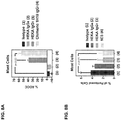
- the antibody kills mast cells expressing Siglec-8 in vitro (e.g., measured in a cell culture assay as described in Example 2).
- the antibody depletes mast cells expressing Siglec-8 in a subject when a therapeutically effective amount is administered.
- the antibody depletes at least about 20% (e.g., at least of the mast cells expressing Siglec-8 in a sample obtained from the subject as compared to a baseline level before treatment.
- the sample can be a tissue sample or a biological fluid sample.
- the tissue sample is one or more selected from the group consisting of: skin, lung, bone marrow, and nasal polyps.
- the biological fluid sample is one or more selected from the group consisting of: blood, bronchoalveolar lavage, and nasal lavage.
- the antibody can be engineered to improve antibody-dependent cell-mediated cytotoxicity (ADCC) activity.
- ADCC antibody-dependent cell-mediated cytotoxicity
- at least one or two of the heavy chains of the antibody is non-fucosylated.
- the antibody may be produced in a cell line having a alpha1,6-fucosyltransferase (Fut8) knockout.
- the antibody may be produced in a cell line overexpressing ⁇ 1,4-N-acetylglycosminyltransferase III (GnT-III).
- the cell line additionally overexpresses Golgi ⁇ -mannosidase II (ManII).
- the antibody may comprise at least one amino acid substitution in the Fc region that improves ADCC activity.
- the antibody may be a humanized antibody, a chimeric antibody or a human antibody.
- the antibody is a human IgG1 antibody.
- the antibody is a murine antibody.
- the antibody may comprise a heavy chain variable region comprising (i) HVR-H1 comprising the amino acid sequence of SEQ ID NO:88, (ii) HVR-H2 comprising the amino acid sequence of SEQ ID NO:91, and (iii) HVR-H3 comprising the amino acid sequence of SEQ ID NO:94; and/or a light chain variable region comprising (i) HVR-L1 comprising the amino acid sequence of SEQ ID NO:97, (ii) HVR-L2 comprising the amino acid sequence of SEQ ID NO:100, and (iii) HVR-L3 comprising the amino acid sequence of SEQ ID NO:103.
- the antibody comprises a heavy chain variable region comprising the amino acid sequence of SEQ ID NO:106; and/or a light chain variable region comprising the amino acid sequence of SEQ ID NO:109.
- the antibody may comprise a heavy chain variable region comprising a heavy chain variable region comprising (i) HVR-H1 comprising the amino acid sequence of SEQ ID NO:89, (ii) HVR-H2 comprising the amino acid sequence of SEQ ID NO:92, and (iii) HVR-H3 comprising the amino acid sequence of SEQ ID NO:95; and/or a light chain variable region comprising (i) HVR-L1 comprising the amino acid sequence of SEQ ID NO:98, (ii) HVR-L2 comprising the amino acid sequence of SEQ ID NO:101, and (iii) HVR-L3 comprising the amino acid sequence of SEQ ID NO:104.
- the antibody comprises a heavy chain variable region comprising the amino acid sequence of SEQ ID NO:107; and/or a light chain variable region comprising the amino acid sequence of SEQ ID NO:110.
- the antibody may comprise a heavy chain variable region comprising a heavy chain variable region comprising a heavy chain variable region comprising (i) HVR-H1 comprising the amino acid sequence of SEQ ID NO:90, (ii) HVR-H2 comprising the amino acid sequence of SEQ ID NO:93, and (iii) HVR-H3 comprising the amino acid sequence of SEQ ID NO:96; and/or a light chain variable region comprising (i) HVR-L1 comprising the amino acid sequence of SEQ ID NO:99, (ii) HVR-L2 comprising the amino acid sequence of SEQ ID NO:102, and (iii) HVR-L3 comprising the amino acid sequence of SEQ ID NO:105.
- the antibody comprises a heavy chain variable region comprising the amino acid sequence of SEQ ID NO:108; and/or a light chain variable region comprising the amino acid sequence of SEQ ID NO:111.
- the antibody may comprise a heavy chain variable region and a light chain variable region, wherein the heavy chain variable region comprises (i) HVR-H1 comprising the amino acid sequence of SEQ ID NO:61, (ii) HVR-H2 comprising the amino acid sequence of SEQ ID NO:62, and (iii) HVR-H3 comprising the amino acid sequence of SEQ ID NO:63; and/or wherein the light chain variable region comprises (i) HVR-L1 comprising the amino acid sequence of SEQ ID NO:64, (ii) HVR-L2 comprising the amino acid sequence of SEQ ID NO:65, and (iii) HVR-L3 comprising the amino acid sequence of SEQ ID NO:66.
- the antibody may comprise a heavy chain variable region and a light chain variable region, wherein the heavy chain variable region comprises (i) HVR-H1 comprising the amino acid sequence of SEQ ID NO:61, (ii) HVR-H2 comprising the amino acid sequence of SEQ ID NO:62, and (iii) HVR-H3 comprising the amino acid sequence selected from SEQ ID NOs:67-70; and/or wherein the light chain variable region comprises (i) HVR-L1 comprising the amino acid sequence of SEQ ID NO:64, (ii) HVR-L2 comprising the amino acid sequence of SEQ ID NO:65, and (iii) HVR-L3 comprising the amino acid sequence of SEQ ID NO:71.
- the subject can be a human.
- the antibody may comprise a heavy chain variable region comprising a heavy chain variable region comprising (i) HVR-H1 comprising the amino acid sequence of SEQ ID NO:89, (ii) HVR-H2 comprising the amino acid sequence of SEQ ID NO:92, and (iii) HVR-H3 comprising the amino acid sequence of SEQ ID NO:95; and/or a light chain variable region comprising (i) HVR-L1 comprising the amino acid sequence of SEQ ID NO:98, (ii) HVR-L2 comprising the amino acid sequence of SEQ ID NO:101, and (iii) HVR-L3 comprising the amino acid sequence of SEQ ID NO:104.
- the antibody comprises a heavy chain variable region comprising the amino acid sequence of SEQ ID NO:107; and/or a light chain variable region comprising the amino acid sequence of SEQ ID NO:110.
- the antibody may comprise a heavy chain variable region comprising a heavy chain variable region comprising a heavy chain variable region comprising (i) HVR-H1 comprising the amino acid sequence of SEQ ID NO:90, (ii) HVR-H2 comprising the amino acid sequence of SEQ ID NO:93, and (iii) HVR-H3 comprising the amino acid sequence of SEQ ID NO:96; and/or a light chain variable region comprising (i) HVR-L1 comprising the amino acid sequence of SEQ ID NO:99, (ii) HVR-L2 comprising the amino acid sequence of SEQ ID NO:102, and (iii) HVR-L3 comprising the amino acid sequence of SEQ ID NO:105.
- the antibody comprises a heavy chain variable region comprising the amino acid sequence of SEQ ID NO:108; and/or a light chain variable region comprising the amino acid sequence of SEQ ID NO:111.
- the antibody may bind to an epitope in Domain 1 of human Siglec-8 (e.g., Domain 1 that comprises the amino acid sequence of SEQ ID NO:112).
- the antibody may bind to an epitope in Domain 3 of human Siglec-8 (e.g., Domain 3 that comprises the amino acid sequence of SEQ ID NO:114).
- the antibody may bind to an epitope in Domain 2 of human Siglec-8 (e.g., Domain 2 that comprises the amino acid sequence of SEQ ID NO:113).
- the antibody may be a humanized antibody, a chimeric antibody or a human antibody.
- the antibody is a murine antibody.
- the antibody is an IgG1 or IgG4 antibody ( e.g ., human IgG1 or IgG4).
- an anti-human Siglec 8 antibody that binds to a fusion protein comprising the amino acid of SEQ ID NO:116 but not to a fusion protein comprising the amino acid of SEQ ID NO:115.
- the antibody described herein binds to a fusion protein comprising the amino acid of SEQ ID NO:117 but not to a fusion protein comprising the amino acid of SEQ ID NO:115.
- the antibody described herein binds to a fusion protein comprising the amino acid of SEQ ID NO:117 but not to a fusion protein comprising the amino acid of SEQ ID NO:116.
- the antibody may comprise a heavy chain variable region comprising (i) HVR-H1 comprising the amino acid sequence of SEQ ID NO:88, (ii) HVR-H2 comprising the amino acid sequence of SEQ ID NO:91, and (iii) HVR-H3 comprising the amino acid sequence of SEQ ID NO:94; and/or a light chain variable region comprising (i) HVR-L1 comprising the amino acid sequence of SEQ ID NO:97, (ii) HVR-L2 comprising the amino acid sequence of SEQ ID NO:100, and (iii) HVR-L3 comprising the amino acid sequence of SEQ ID NO:103.
- the antibody comprises a heavy chain variable region comprising the amino acid sequence of SEQ ID NO:106; and/or a light chain variable region comprising the amino acid sequence of SEQ ID NO:109.
- the antibody may comprise a heavy chain variable region comprising a heavy chain variable region comprising (i) HVR-H1 comprising the amino acid sequence of SEQ ID NO:89, (ii) HVR-H2 comprising the amino acid sequence of SEQ ID NO:92, and (iii) HVR-H3 comprising the amino acid sequence of SEQ ID NO:95; and/or a light chain variable region comprising (i) HVR-L1 comprising the amino acid sequence of SEQ ID NO:98, (ii) HVR-L2 comprising the amino acid sequence of SEQ ID NO:101, and (iii) HVR-L3 comprising the amino acid sequence of SEQ ID NO:104.
- the antibody comprises a heavy chain variable region comprising the amino acid sequence of SEQ ID NO:107; and/or a light chain variable region comprising the amino acid sequence of SEQ ID NO:110.
- the antibody may comprise a heavy chain variable region comprising a heavy chain variable region comprising a heavy chain variable region comprising (i) HVR-H1 comprising the amino acid sequence of SEQ ID NO:90, (ii) HVR-H2 comprising the amino acid sequence of SEQ ID NO:93, and (iii) HVR-H3 comprising the amino acid sequence of SEQ ID NO:96; and/or a light chain variable region comprising (i) HVR-L1 comprising the amino acid sequence of SEQ ID NO:99, (ii) HVR-L2 comprising the amino acid sequence of SEQ ID NO:102, and (iii) HVR-L3 comprising the amino acid sequence of SEQ ID NO:105.
- the antibody comprises a heavy chain variable region comprising
- a humanized antibody that binds to a human Siglec-8, wherein the EC 50 in depleting activated human eosinophils is less than the EC 50 of antibody 2E2 or 2C4 to the human Siglec-8.
- the EC 50 of the humanized antibody is about 85% or less than the EC 50 of antibody 2E2 or 2C4 to the human Siglec-8.
- the EC 50 of the humanized antibody is about 85%, about 80%, about 70%, about 65%, about 60%, about 55%, about 50%, about 45%, about 40%, about 35%, about 30%, about 25%, about 20%, about 15%, about 10% or about 5% or less than the EC 50 of antibody 2E2 or 2C4 to the human Siglec-8.
- the humanized antibody may comprise a heavy chain variable region and a light chain variable region, wherein the heavy chain variable region comprises (i) HVR-H1 comprising the amino acid sequence of SEQ ID NO:61, (ii) HVR-H2 comprising the amino acid sequence of SEQ ID NO:62, and (iii) HVR-H3 comprising the amino acid sequence of SEQ ID NO:63; and/or wherein the light chain variable region comprises (i) HVR-L1 comprising the amino acid sequence of SEQ ID NO:64, (ii) HVR-L2 comprising the amino acid sequence of SEQ ID NO:65, and (iii) HVR-L3 comprising the amino acid sequence of SEQ ID NO:66.
- the heavy chain variable region comprises (i) HVR-H1 comprising the amino acid sequence of SEQ ID NO:61, (ii) HVR-H2 comprising the amino acid sequence of SEQ ID NO:62, and (iii) HVR-H3 comprising the amino acid sequence of SEQ ID NO
- the humanized antibody may comprise a heavy chain variable region comprising the amino acid sequence of SEQ ID NO:6; and/or a light chain variable region comprising the amino acid sequence selected from SEQ ID NOs:16 or 21.
- the humanized antibody comprises a heavy chain variable region and a light chain variable region, wherein the heavy chain variable region comprises (i) HVR-H1 comprising the amino acid sequence of SEQ ID NO:61, (ii) HVR-H2 comprising the amino acid sequence of SEQ ID NO:62, and (iii) HVR-H3 comprising the amino acid sequence selected from SEQ ID NOs:67-70; and/or wherein the light chain variable region comprises (i) HVR-L1 comprising the amino acid sequence of SEQ ID NO:64, (ii) HVR-L2 comprising the amino acid sequence of SEQ ID NO:65, and (iii) HVR-L3 comprising the amino acid sequence of SEQ ID NO:71.
- the humanized antibody may comprise a heavy chain variable region comprising the amino acid sequence selected from SEQ ID NOs:2-14; and/or a light chain variable region comprising the amino acid sequence selected from SEQ ID NOs: 16-24.
- the antibody may comprise a heavy chain variable region comprising (i) HVR-H1 comprising the amino acid sequence of SEQ ID NO:88, (ii) HVR-H2 comprising the amino acid sequence of SEQ ID NO:91, and (iii) HVR-H3 comprising the amino acid sequence of SEQ ID NO:94; and/or a light chain variable region comprising (i) HVR-L1 comprising the amino acid sequence of SEQ ID NO:97, (ii) HVR-L2 comprising the amino acid sequence of SEQ ID NO:100, and (iii) HVR-L3 comprising the amino acid sequence of SEQ ID NO:103.
- the antibody may comprise a heavy chain variable region comprising a heavy chain variable region comprising (i) HVR-H1 comprising the amino acid sequence of SEQ ID NO:89, (ii) HVR-H2 comprising the amino acid sequence of SEQ ID NO:92, and (iii) HVR-H3 comprising the amino acid sequence of SEQ ID NO:95; and/or a light chain variable region comprising (i) HVR-L1 comprising the amino acid sequence of SEQ ID NO:98, (ii) HVR-L2 comprising the amino acid sequence of SEQ ID NO:101, and (iii) HVR-L3 comprising the amino acid sequence of SEQ ID NO:104.
- the antibody may comprise a heavy chain variable region comprising a heavy chain variable region comprising a heavy chain variable region comprising (i) HVR-H1 comprising the amino acid sequence of SEQ ID NO:90, (ii) HVR-H2 comprising the amino acid sequence of SEQ ID NO:93, and (iii) HVR-H3 comprising the amino acid sequence of SEQ ID NO:96; and/or a light chain variable region comprising (i) HVR-L1 comprising the amino acid sequence of SEQ ID NO:99, (ii) HVR-L2 comprising the amino acid sequence of SEQ ID NO:102, and (iii) HVR-L3 comprising the amino acid sequence of SEQ ID NO:105.
- the antibody may comprise a heavy chain Fc region comprising a human IgG Fc region.
- the human IgG Fc region comprises a human IgG1 or IgG4.
- the human IgG1 comprises the amino acid sequence of SEQ ID NO:78.
- the human IgG4 comprises the amino acid sequence of SEQ ID NO:79.
- the antibody may comprise a heavy chain comprising the amino acid sequence of SEQ ID NO:75; and/or a light chain comprising the amino acid sequence selected from SEQ ID NOs:76 or 77.
- the human IgG4 comprises the amino acid substitution S228P, wherein the amino acid residues are numbered according to the EU index as in Kabat.
- the antibody may comprise a heavy chain comprising the amino acid sequence of SEQ ID NO:87; and/or a light chain comprising the amino acid sequence of SEQ ID NO:76.
- nucleic acid encoding any antibody described above and herein.
- a vector comprising a nucleic acid described herein.
- the vector is an expression vector.
- a host cell comprising a nucleic acid described herein. In some embodiments, the host cell expresses and produces the antibody.
- a method of producing an antibody comprising culturing a host cell comprising one or more nucleic acids encoding an antibody described herein under a condition that produces the antibody. In some embodiments, the method further comprises recovering the antibody produced by the host cell. Also provided herein is an anti-Siglec-8 antibody produced by the method. Also provided herein is an antigen-binding fragment of an anti-Siglec-8 antibody described herein.
- composition comprising an antibody described above and herein or an antigen-binding fragment thereof and a pharmaceutically acceptable carrier.
- composition comprising an antibody or fragment thereof that specifically binds to human Siglec-8, wherein the antibody comprises a Fc region and N-glycoside-linked carbohydrate chains linked to the Fc region, wherein less than 50% of the N-glycoside-linked carbohydrate chains in the composition contain a fucose residue. In some embodiments, substantially none of the N-glycoside-linked carbohydrate chains contain a fucose residue.
- the antibody is a humanized antibody, a chimeric antibody or a human antibody.
- the antibody comprises a heavy chain variable region and a light chain variable region, wherein the heavy chain variable region comprises (i) HVR-H1 comprising the amino acid sequence of SEQ ID NO:61, (ii) HVR-H2 comprising the amino acid sequence of SEQ ID NO:62, and (iii) HVR-H3 comprising the amino acid sequence of SEQ ID NO:63; and/or wherein the light chain variable region comprises (i) HVR-L1 comprising the amino acid sequence of SEQ ID NO:64, (ii) HVR-L2 comprising the amino acid sequence of SEQ ID NO:65, and (iii) HVR-L3 comprising the amino acid sequence of SEQ ID NO:66.
- the antibody comprises a heavy chain variable region comprising the amino acid sequence of SEQ ID NOs:2-10; and/or a light chain variable region comprising the amino acid sequence of SEQ ID NOs:16-22.
- the antibody comprises a heavy chain variable region and a light chain variable region, wherein the heavy chain variable region comprises (i) HVR-H1 comprising the amino acid sequence of SEQ ID NO:61, (ii) HVR-H2 comprising the amino acid sequence of SEQ ID NO:62, and (iii) HVR-H3 comprising the amino acid sequence selected from SEQ ID NOs:67-70; and/or wherein the light chain variable region comprises (i) HVR-L1 comprising the amino acid sequence of SEQ ID NO:64, (ii) HVR-L2 comprising the amino acid sequence of SEQ ID NO:65, and (iii) HVR-L3 comprising the amino acid sequence of SEQ ID NO:71.
- the antibody comprises a heavy chain variable region comprising the amino acid sequence selected from SEQ ID NOs: 11-14; and/or a light chain variable region comprising the amino acid sequence selected from SEQ ID NOs:23-24. In some embodiments, the antibody comprises a heavy chain variable region comprising the amino acid sequence selected from SEQ ID NOs:2-14; and/or a light chain variable region comprising the amino acid sequence selected from SEQ ID NOs: 16-24. In any of the embodiments herein, the composition may further comprise a pharmaceutically acceptable carrier.
- the binding affinity and/or binding avidity of the antibody to a human Siglec-8 can be higher than the binding affinity and/or binding avidity of antibody 2E2 or 2C4 to the human Siglec-8.
- the antibody may have a Tm of at least about 70°C to at least about 72°C in a thermal shift assay. In some embodiments, the antibody has a Tm of about 70°C, about 71°C, or about 72°C. In some embodiments, the antibody has the same or higher Tm as compared to a chimeric 2C4 antibody. In some embodiments, the antibody has the same or higher Tm as compared to an antibody having a heavy chain comprising the amino acid sequence of SEQ ID NO:84 and a light chain comprising the amino acid sequence of SEQ ID NO:85.
- a method of treating or preventing a disease mediated by cells expressing Siglec-8 in a subject comprising administering to the subject an effective amount of an antibody described herein or an antigen-binding fragment thereof or a composition described herein.
- the disease is an eosinophil mediated-disease.
- the disease is a mast cell mediated-disease.
- the disease is selected from the group consisting of asthma, allergic rhinitis, nasal polyposis, atopic dermatitis, chronic urticaria, mastocytosis, eosinophilic leukemia, and hypereosinophilic syndrome.
- the disease is selected from the group consisting of pauci granulocytic asthma, acute or chronic airway hypersensitivity, eosinophilic esophagitis, Churg-Strauss syndrome, inflammation associated with a cytokine, inflammation associated with cells expressing Siglec-8, malignancy associated with cells expressing Siglec-8, physical urticaria, cold urticaria, pressure-urticaria, bullous pemphigoid, food allergy, and allergic bronchopulmonary aspergillosis (ABPA).
- the antibody inhibits one or more symptoms of an allergic reaction.
- the allergic reaction is a Type I hypersensitivity reaction.
- the subject may be suffering from asthma that is not adequately controlled by an inhaled corticosteroid, a short acting ⁇ 2 agonist, a long acting ⁇ 2 agonist, or a combination thereof.
- a method of depleting mast cells expressing Siglec-8 in a subject comprising administering to the subject an effective amount of an antibody that binds to human Siglec-8, wherein the antibody kills mast cells expressing Siglec-8 by ADCC activity.
- the antibody kills mast cells expressing Siglec-8 in vitro (e.g., measured in a cell culture assay as described in Example 2).
- the antibody depletes at least about 20% of the mast cells expressing Siglec-8 in a sample obtained from the subject as compared to a baseline level before treatment.
- the sample can be a tissue sample or a biological fluid sample.
- the tissue sample is one or more selected from the group consisting of: skin, lung, bone marrow, and nasal polyps.
- the biological fluid sample is one or more selected from the group consisting of: blood, bronchoalveolar lavage, and nasal lavage.
- the antibody can be engineered to improve antibody-dependent cell-mediated cytotoxicity (ADCC) activity.
- the antibody may comprise two heavy chains and wherein at least one of the two or both heavy chains of the antibody is non-fucosylated.
- the antibody may be produced in a cell line having a alpha1,6-fucosyltransferase (Fut8) knockout.
- the antibody may be produced in a cell line overexpressing ⁇ 1,4-N-acetylglycosminyltransferase III (GnT-III).
- the cell line additionally overexpresses Golgi ⁇ -mannosidase II (ManII).
- the antibody may comprise at least one amino acid substitution in the Fc region that improves ADCC activity.
- the antibody may be a humanized antibody, a chimeric antibody or a human antibody.
- the antibody is a human IgG1 antibody.
- the antibody may comprise a heavy chain variable region comprising (i) HVR-H1 comprising the amino acid sequence of SEQ ID NO:88, (ii) HVR-H2 comprising the amino acid sequence of SEQ ID NO:91, and (iii) HVR-H3 comprising the amino acid sequence of SEQ ID NO:94; and/or a light chain variable region comprising (i) HVR-L1 comprising the amino acid sequence of SEQ ID NO:97, (ii) HVR-L2 comprising the amino acid sequence of SEQ ID NO:100, and (iii) HVR-L3 comprising the amino acid sequence of SEQ ID NO:103.
- the antibody comprises a heavy chain variable region comprising the amino acid sequence of SEQ ID NO:106; and/or a light chain variable region comprising the amino acid sequence of SEQ ID NO:109.
- the antibody may comprise a heavy chain variable region comprising a heavy chain variable region comprising (i) HVR-H1 comprising the amino acid sequence of SEQ ID NO:89, (ii) HVR-H2 comprising the amino acid sequence of SEQ ID NO:92, and (iii) HVR-H3 comprising the amino acid sequence of SEQ ID NO:95; and/or a light chain variable region comprising (i) HVR-L1 comprising the amino acid sequence of SEQ ID NO:98, (ii) HVR-L2 comprising the amino acid sequence of SEQ ID NO:101, and (iii) HVR-L3 comprising the amino acid sequence of SEQ ID NO:104.
- the antibody comprises a heavy chain variable region comprising the amino acid sequence of SEQ ID NO:107; and/or a light chain variable region comprising the amino acid sequence of SEQ ID NO:110.
- the antibody may comprise a heavy chain variable region comprising a heavy chain variable region comprising a heavy chain variable region comprising (i) HVR-H1 comprising the amino acid sequence of SEQ ID NO:90, (ii) HVR-H2 comprising the amino acid sequence of SEQ ID NO:93, and (iii) HVR-H3 comprising the amino acid sequence of SEQ ID NO:96; and/or a light chain variable region comprising (i) HVR-L1 comprising the amino acid sequence of SEQ ID NO:99, (ii) HVR-L2 comprising the amino acid sequence of SEQ ID NO:102, and (iii) HVR-L3 comprising the amino acid sequence of SEQ ID NO:105.
- the antibody comprises a heavy chain variable region comprising the amino acid sequence of SEQ ID NO:108; and/or a light chain variable region comprising the amino acid sequence of SEQ ID NO:111.
- the antibody may comprise a heavy chain variable region and a light chain variable region, wherein the heavy chain variable region comprises (i) HVR-H1 comprising the amino acid sequence of SEQ ID NO:61, (ii) HVR-H2 comprising the amino acid sequence of SEQ ID NO:62, and (iii) HVR-H3 comprising the amino acid sequence of SEQ ID NO:63; and/or wherein the light chain variable region comprises (i) HVR-L1 comprising the amino acid sequence of SEQ ID NO:64, (ii) HVR-L2 comprising the amino acid sequence of SEQ ID NO:65, and (iii) HVR-L3 comprising the amino acid sequence of SEQ ID NO:66.
- the antibody may comprise a heavy chain variable region and a light chain variable region, wherein the heavy chain variable region comprises (i) HVR-H1 comprising the amino acid sequence of SEQ ID NO:61, (ii) HVR-H2 comprising the amino acid sequence of SEQ ID NO:62, and (iii) HVR-H3 comprising the amino acid sequence selected from SEQ ID NOs:67-70; and/or wherein the light chain variable region comprises (i) HVR-L1 comprising the amino acid sequence of SEQ ID NO:64, (ii) HVR-L2 comprising the amino acid sequence of SEQ ID NO:65, and (iii) HVR-L3 comprising the amino acid sequence of SEQ ID NO:71.
- the heavy chain variable region comprises (i) HVR-H1 comprising the amino acid sequence of SEQ ID NO:61, (ii) HVR-H2 comprising the amino acid sequence of SEQ ID NO:62, and (iii) HVR-H3 comprising the amino acid sequence selected from S
- the subject has a disease mediated by cells expressing Siglec-8.
- the disease is selected from the group consisting of asthma, allergic rhinitis, nasal polyposis, atopic dermatitis, chronic urticaria, mastocytosis, eosinophilic leukemia, and hypereosinophilic syndrome.
- the disease is selected from the group consisting of pauci granulocytic asthma, acute or chronic airway hypersensitivity, eosinophilic esophagitis, Churg-Strauss syndrome, inflammation associated with a cytokine, inflammation associated with cells expressing Siglec-8, malignancy associated with cells expressing Siglec-8, physical urticaria, cold urticaria, pressure-urticaria, bullous pemphigoid, food allergy, and allergic bronchopulmonary aspergillosis (ABPA).
- the antibody inhibits one or more symptoms of an allergic reaction.
- the allergic reaction is a Type I hypersensitivity reaction.
- the subject may be suffering from asthma that is not adequately controlled by an inhaled corticosteroid, a short acting ⁇ 2 agonist, a long acting ⁇ 2 agonist, or a combination thereof.
- the subject can be a human.
- antibody includes polyclonal antibodies, monoclonal antibodies (including full length antibodies which have an immunoglobulin Fc region), antibody compositions with polyepitopic specificity, multispecific antibodies (e.g ., bispecific antibodies, diabodies, and single-chain molecules, as well as antibody fragments ( e.g ., Fab, F(ab') 2 , and Fv).
- immunoglobulin Ig is used interchangeably with “antibody” herein.
- the basic 4-chain antibody unit is a heterotetrameric glycoprotein composed of two identical light (L) chains and two identical heavy (H) chains.
- An IgM antibody consists of 5 of the basic heterotetramer units along with an additional polypeptide called a J chain, and contains 10 antigen binding sites, while IgA antibodies comprise from 2-5 of the basic 4-chain units which can polymerize to form polyvalent assemblages in combination with the J chain.
- the 4-chain unit is generally about 150,000 daltons.
- Each L chain is linked to an H chain by one covalent disulfide bond, while the two H chains are linked to each other by one or more disulfide bonds depending on the H chain isotype.
- Each H and L chain also has regularly spaced intrachain disulfide bridges.
- Each H chain has at the N-terminus, a variable domain (V H ) followed by three constant domains (C H ) for each of the ⁇ and ⁇ chains and four C H domains for ⁇ and ⁇ isotypes.
- Each L chain has at the N-terminus, a variable domain (V L ) followed by a constant domain at its other end.
- the V L is aligned with the V H and the C L is aligned with the first constant domain of the heavy chain (C H 1). Particular amino acid residues are believed to form an interface between the light chain and heavy chain variable domains.
- the pairing of a V H and V L together forms a single antigen-binding site.
- immunoglobulins can be assigned to different classes or isotypes. There are five classes of immunoglobulins: IgA, IgD, IgE, IgG and IgM, having heavy chains designated ⁇ , ⁇ , ⁇ , ⁇ and ⁇ , respectively.
- the ⁇ and ⁇ classes are further divided into subclasses on the basis of relatively minor differences in the CH sequence and function, e.g ., humans express the following subclasses: IgG1, IgG2, IgG3, IgG4, IgA1 and IgA2.
- IgG1 antibodies can exist in multiple polymorphic variants termed allotypes (reviewed in Jefferis and Lefranc 2009. mAbs Vol 1 Issue 4 1-7 ) any of which are suitable for use in the invention. Common allotypic variants in human populations are those designated by the letters a,f,n,z.
- an “isolated” antibody is one that has been identified, separated and/or recovered from a component of its production environment (e.g ., naturally or recombinantly).
- the isolated polypeptide is free of association with all other components from its production environment.
- Contaminant components of its production environment such as that resulting from recombinant transfected cells, are materials that would typically interfere with research, diagnostic or therapeutic uses for the antibody, and may include enzymes, hormones, and other proteinaceous or non-proteinaceous solutes.
- the polypeptide is purified: (1) to greater than 95% by weight of antibody as determined by, for example, the Lowry method, and in some embodiments, to greater than 99% by weight; (1) to a degree sufficient to obtain at least 15 residues of N-terminal or internal amino acid sequence by use of a spinning cup sequenator, or (3) to homogeneity by SDS-PAGE under non-reducing or reducing conditions using Coomassie blue or silver stain.
- Isolated antibody includes the antibody in situ within recombinant cells since at least one component of the antibody's natural environment will not be present. Ordinarily, however, an isolated polypeptide or antibody is prepared by at least one purification step.
- monoclonal antibody refers to an antibody obtained from a population of substantially homogeneous antibodies, i.e. , the individual antibodies comprising the population are identical except for possible naturally occurring mutations and/or post-translation modifications (e.g ., isomerizations, amidations) that may be present in minor amounts.
- monoclonal antibodies have a C-terminal cleavage at the heavy chain and/or light chain. For example, 1, 2, 3, 4, or 5 amino acid residues are cleaved at the C-terminus of heavy chain and/or light chain. In some embodiments, the C-terminal cleavage removes a C-terminal lysine from the heavy chain.
- monoclonal antibodies have an N-terminal cleavage at the heavy chain and/or light chain. For example, 1, 2, 3, 4, or 5 amino acid residues are cleaved at the N-terminus of heavy chain and/or light chain.
- truncated forms of monoclonal antibodies can be made by recombinant techniques.
- monoclonal antibodies are highly specific, being directed against a single antigenic site. In some embodiments, monoclonal antibodies are highly specific, being directed against multiple antigenic sites (such as a bispecific antibody or a multispecific antibody).
- the modifier "monoclonal” indicates the character of the antibody as being obtained from a substantially homogeneous population of antibodies, and is not to be construed as requiring production of the antibody by any particular method.
- the monoclonal antibodies to be used in accordance with the present invention may be made by a variety of techniques, including, for example, the hybridoma method, recombinant DNA methods, phage-display technologies, and technologies for producing human or human-like antibodies in animals that have parts or all of the human immunoglobulin loci or genes encoding human immunoglobulin sequences.
- naked antibody refers to an antibody that is not conjugated to a cytotoxic moiety or radiolabel.
- full-length antibody “intact antibody” or “whole antibody” are used interchangeably to refer to an antibody in its substantially intact form, as opposed to an antibody fragment. Specifically whole antibodies include those with heavy and light chains including an Fc region.
- the constant domains may be native sequence constant domains (e.g ., human native sequence constant domains) or amino acid sequence variants thereof. In some cases, the intact antibody may have one or more effector functions.
- antibody fragment comprises a portion of an intact antibody, the antigen binding and/or the variable region of the intact antibody.
- antibody fragments include Fab, Fab', F(ab') 2 and Fv fragments; diabodies; linear antibodies (see U.S. Pat. No. 5,641,870 , Example 2; Zapata et al., Protein Eng. 8(10): 1057-1062 [1995 ]); single-chain antibody molecules and multispecific antibodies formed from antibody fragments.
- Papain digestion of antibodies produced two identical antigen-binding fragments, called “Fab” fragments, and a residual "Fc” fragment, a designation reflecting the ability to crystallize readily.
- the Fab fragment consists of an entire L chain along with the variable region domain of the H chain (V H ), and the first constant domain of one heavy chain (C H 1).
- Each Fab fragment is monovalent with respect to antigen binding, i.e. , it has a single antigen-binding site.
- Pepsin treatment of an antibody yields a single large F(ab') 2 fragment which roughly corresponds to two disulfide linked Fab fragments having different antigen-binding activity and is still capable of cross-linking antigen.
- Fab' fragments differ from Fab fragments by having a few additional residues at the carboxy terminus of the C H 1 domain including one or more cysteines from the antibody hinge region.
- Fab'-SH is the designation herein for Fab' in which the cysteine residue(s) of the constant domains bear a free thiol group.
- F(ab') 2 antibody fragments originally were produced as pairs of Fab' fragments which have hinge cysteines between them. Other chemical couplings of antibody fragments are also known.
- the Fc fragment comprises the carboxy-terminal portions of both H chains held together by disulfides.
- the effector functions of antibodies are determined by sequences in the Fc region, the region which is also recognized by Fc receptors (FcR) found on certain types of cells.
- Fv is the minimum antibody fragment which contains a complete antigen-recognition and -binding site. This fragment consists of a dimer of one heavy- and one light-chain variable region domain in tight, non-covalent association. From the folding of these two domains emanate six hypervariable loops (3 loops each from the H and L chain) that contribute the amino acid residues for antigen binding and confer antigen binding specificity to the antibody. However, even a single variable domain (or half of an Fv comprising only three HVRs specific for an antigen) has the ability to recognize and bind antigen, although at a lower affinity than the entire binding site.
- Single-chain Fv also abbreviated as “sFv” or “scFv” are antibody fragments that comprise the VH and VL antibody domains connected into a single polypeptide chain.
- the sFv polypeptide further comprises a polypeptide linker between the V H and V L domains which enables the sFv to form the desired structure for antigen binding.
- Fully fragments of the antibodies of the invention comprise a portion of an intact antibody, generally including the antigen binding or variable region of the intact antibody or the Fv region of an antibody which retains or has modified FcR binding capability.
- antibody fragments include linear antibody, single-chain antibody molecules and multispecific antibodies formed from antibody fragments.
- the monoclonal antibodies herein specifically include "chimeric" antibodies (immunoglobulins) in which a portion of the heavy and/or light chain is identical with or homologous to corresponding sequences in antibodies derived from a particular species or belonging to a particular antibody class or subclass, while the remainder of the chain(s) is (are) identical with or homologous to corresponding sequences in antibodies derived from another species or belonging to another antibody class or subclass, as well as fragments of such antibodies, so long as they exhibit the desired biological activity ( U.S. Pat. No. 4,816,567 ; Morrison et al., Proc. Natl. Acad. Sci. USA, 81:6851-6855 (1984 )).
- chimeric antibodies immunoglobulins in which a portion of the heavy and/or light chain is identical with or homologous to corresponding sequences in antibodies derived from a particular species or belonging to a particular antibody class or subclass, while the remainder of the chain(s) is (are) identical with or
- Chimeric antibodies of interest herein include PRIMATIZED® antibodies wherein the antigen-binding region of the antibody is derived from an antibody produced by, e.g ., immunizing macaque monkeys with an antigen of interest.
- PRIMATIZED® antibodies wherein the antigen-binding region of the antibody is derived from an antibody produced by, e.g ., immunizing macaque monkeys with an antigen of interest.
- humanized antibody is used as a subset of “chimeric antibodies.”
- Humanized forms of non-human (e.g ., murine) antibodies are chimeric antibodies that contain minimal sequence derived from non-human immunoglobulin.
- a humanized antibody is a human immunoglobulin (recipient antibody) in which residues from an HVR of the recipient are replaced by residues from an HVR of a non-human species (donor antibody) such as mouse, rat, rabbit or non-human primate having the desired specificity, affinity, and/or capacity.
- donor antibody such as mouse, rat, rabbit or non-human primate having the desired specificity, affinity, and/or capacity.
- FR residues of the human immunoglobulin are replaced by corresponding non-human residues.
- humanized antibodies may comprise residues that are not found in the recipient antibody or in the donor antibody. These modifications may be made to further refine antibody performance, such as binding affinity.
- a humanized antibody will comprise substantially all of at least one, and typically two, variable domains, in which all or substantially all of the hypervariable loops correspond to those of a non-human immunoglobulin sequence, and all or substantially all of the FR regions are those of a human immunoglobulin sequence, although the FR regions may include one or more individual FR residue substitutions that improve antibody performance, such as binding affinity, isomerization, immunogenicity, etc.
- the number of these amino acid substitutions in the FR are no more than 6 in the H chain, and in the L chain, no more than 3.
- the humanized antibody optionally will also comprise at least a portion of an immunoglobulin constant region (Fc), typically that of a human immunoglobulin.
- Fc immunoglobulin constant region
- humanized antibodies are directed against a single antigenic site. In some embodiments, humanized antibodies are directed against multiple antigenic sites.
- An alternative humanization method is described in U.S. Pat. No. 7,981,843 and U.S. Patent Application Publication No. 2006/0134098 .
- variable region refers to the amino-terminal domains of the heavy or light chain of the antibody.
- variable domains of the heavy chain and light chain may be referred to as "VH” and “VL”, respectively. These domains are generally the most variable parts of the antibody (relative to other antibodies of the same class) and contain the antigen binding sites.
- hypervariable region when used herein refers to the regions of an antibody-variable domain that are hypervariable in sequence and/or form structurally defined loops.
- antibodies comprise six HVRs; three in the VH (H1, H2, H3), and three in the VL (L1, L2, L3).
- H3 and L3 display the most diversity of the six HVRs, and H3 in particular is believed to play a unique role in conferring fine specificity to antibodies. See, e.g. , Xu et al.
- HVR delineations are in use and are encompassed herein.
- the HVRs that are Kabat complementarity-determining regions (CDRs) are based on sequence variability and are the most commonly used ( Kabat et al., Sequences of Proteins of Immunological Interest, 5th Ed. Public Health Service, National Institute of Health, Bethesda, MD (1991 )).
- Chothia HVRs refer instead to the location of the structural loops ( Chothia and Lesk J. Mol. Biol. 196:901-917 (1987)).
- the "contact" HVRs are based on an analysis of the available complex crystal structures. The residues from each of these HVRs are noted below.
- variable-domain residues HVR residues and framework region residues
- HVR residues and framework region residues are numbered according to Kabat et al. , supra.
- Framework or "FR” residues are those variable-domain residues other than the HVR residues as herein defined.
- variable-domain residue-numbering as in Kabat or "amino-acid-position numbering as in Kabat,” and variations thereof, refers to the numbering system used for heavy-chain variable domains or light-chain variable domains of the compilation of antibodies in Kabat et al. , supra. Using this numbering system, the actual linear amino acid sequence may contain fewer or additional amino acids corresponding to a shortening of, or insertion into, a FR or HVR of the variable domain.
- a heavy-chain variable domain may include a single amino acid insert (residue 52a according to Kabat) after residue 52 of H2 and inserted residues ( e.g. residues 82a, 82b, and 82c, etc. according to Kabat) after heavy-chain FR residue 82.
- the Kabat numbering of residues may be determined for a given antibody by alignment at regions of homology of the sequence of the antibody with a "standard" Kabat numbered sequence.
- acceptor human framework for the purposes herein is a framework comprising the amino acid sequence of a VL or VH framework derived from a human immunoglobulin framework or a human consensus framework.
- An acceptor human framework "derived from” a human immunoglobulin framework or a human consensus framework may comprise the same amino acid sequence thereof, or it may contain pre-existing amino acid sequence changes. In some embodiments, the number of pre-existing amino acid changes are 10 or less, 9 or less, 8 or less, 7 or less, 6 or less, 5 or less, 4 or less, 3 or less, or 2 or less.
- Percent (%) amino acid sequence identity with respect to a reference polypeptide sequence is defined as the percentage of amino acid residues in a candidate sequence that are identical with the amino acid residues in the reference polypeptide sequence, after aligning the sequences and introducing gaps, if necessary, to achieve the maximum percent sequence identity, and not considering any conservative substitutions as part of the sequence identity. Alignment for purposes of determining percent amino acid sequence identity can be achieved in various ways that are within the skill in the art, for instance, using publicly available computer software such as BLAST, BLAST-2, ALIGN or Megalign (DNASTAR) software. Those skilled in the art can determine appropriate parameters for aligning sequences, including any algorithms needed to achieve maximal alignment over the full length of the sequences being compared.
- the % amino acid sequence identity of a given amino acid sequence A to, with, or against a given amino acid sequence B is calculated as follows: 100 times the fraction X / Y where X is the number of amino acid residues scored as identical matches by the sequence in that program's alignment of A and B, and where Y is the total number of amino acid residues in B. It will be appreciated that where the length of amino acid sequence A is not equal to the length of amino acid sequence B, the % amino acid sequence identity of A to B will not equal the % amino acid sequence identity of B to A.
- an antibody that "binds to”, “specifically binds to” or is “specific for” a particular a polypeptide or an epitope on a particular polypeptide is one that binds to that particular polypeptide or epitope on a particular polypeptide without substantially binding to any other polypeptide or polypeptide epitope.
- binding of an anti-Siglec-8 antibody described herein to an unrelated, non-Siglec-8 polypeptide is less than about 10% of the antibody binding to Siglec-8 as measured by methods known in the art (e.g ., enzyme-linked immunosorbent assay (ELISA)).
- the antibody that binds to a Siglec-8 e.g.
- Siglec-8 Fc fusion protein in dimer form (SEQ ID NO:74)) has a dissociation constant (Kd) of ⁇ 1 ⁇ M, ⁇ 100 nM, ⁇ 10 nM, ⁇ 2 nM, ⁇ 1 nM, ⁇ 0.7 nM, ⁇ 0 .6 nM, ⁇ 0.5 nM, ⁇ 0.1 nM, ⁇ 0.01 nM, or ⁇ 0.001 nM (e.g. 10 -8 M or less, e.g. from 10 -8 M to 10 -13 M, e.g., from 10 -9 M to 10 -13 M).
- Kd dissociation constant
- Siglec-8 refers to a human Siglec-8 protein.
- the term also includes naturally occurring variants of Siglec-8, including splice variants or allelic variants.
- the amino acid sequence of an exemplary human Siglec-8 is shown in SEQ ID NO:72.
- the amino acid sequence of another exemplary human Siglec-8 is shown in SEQ ID NO:73.
- a human Siglec-8 protein comprises the human Siglec-8 extracellular domain fused to an immunoglobulin Fc region.
- the amino acid sequence of an exemplary human Siglec-8 extracellular domain fused to an immunoglobulin Fc region is shown in SEQ ID NO:74.
- the amino acid sequence underlined in SEQ ID NO:74 indicates the Fc region of the Siglec-8 Fc fusion protein amino acid sequence.
- Antibodies that "induce apoptosis” or are “apoptotic” are those that induce programmed cell death as determined by standard apoptosis assays, such as binding of annexin V, fragmentation of DNA, cell shrinkage, dilation of endoplasmic reticulum, cell fragmentation, and/or formation of membrane vesicles (called apoptotic bodies).
- apoptotic bodies such as binding of annexin V, fragmentation of DNA, cell shrinkage, dilation of endoplasmic reticulum, cell fragmentation, and/or formation of membrane vesicles.
- the apoptotic activity of the anti-Siglec-8 antibodies of the present invention can be showed by staining cells with annexin V.
- Antibody effector functions refer to those biological activities attributable to the Fc region (a native sequence Fc region or amino acid sequence variant Fc region) of an antibody, and vary with the antibody isotype. Examples of antibody effector functions include: C1q binding and complement dependent cytotoxicity; Fc receptor binding; antibody-dependent cell-mediated cytotoxicity (ADCC); phagocytosis; down regulation of cell surface receptors ( e.g. , B cell receptors); and B cell activation.
- ADCC antibody-dependent cell-mediated cytotoxicity
- FcRs Fc receptors
- cytotoxic cells e.g ., natural killer (NK) cells, neutrophils and macrophages
- NK cells natural killer cells
- neutrophils and macrophages e.g ., neutrophils and macrophages
- the antibodies “arm” the cytotoxic cells and are required for killing of the target cell by this mechanism.
- the primary cells for mediating ADCC, NK cells express Fc ⁇ RIII only, whereas monocytes express Fc ⁇ RI, Fc ⁇ RII and Fc ⁇ RIII.
- an anti-Siglec-8 antibody described herein enhances ADCC.
- an in vitro ADCC assay such as that described in U.S. Pat. No. 5,500,362 or 5,821,337 may be performed.
- Useful effector cells for such assays include peripheral blood mononuclear cells (PBMC) and natural killer (NK) cells.
- PBMC peripheral blood mononuclear cells
- NK natural killer cells.
- ADCC activity of the molecule of interest may be assessed in vivo , e.g.
- Fc variants that alter ADCC activity and other antibody properties include those disclosed by Ghetie et al., Nat Biotech. 15:637-40, 1997 ; Duncan et al, Nature 332:563-564, 1988 ; Lund et al., J. Immunol 147:2657-2662, 1991 ; Lund et al, Mol Immunol 29:53-59, 1992 ; Alegre et al, Transplantation 57:1537-1543, 1994 ; Hutchins et al., Proc Natl.
- Fc region herein is used to define a C-terminal region of an immunoglobulin heavy chain, including native-sequence Fc regions and variant Fc regions.
- the human IgG heavy-chain Fc region is usually defined to stretch from an amino acid residue at position Cys226, or from Pro230, to the carboxyl-terminus thereof.
- Suitable native-sequence Fc regions for use in the antibodies of the invention include human IgG1, IgG2, IgG3 and IgG4.
- a single amino acid substitution (S228P according to Kabat numbering; designated IgG4Pro) may be introduced to abolish the heterogeneity observed in recombinant IgG4 antibody. See Angal, S. et al. (1993) Mol Immunol 30, 105-108 .
- Non-fucosylated or fucose-deficient antibody refers to a glycosylation antibody variant comprising an Fc region wherein a carbohydrate structure attached to the Fc region has reduced fucose or lacks fucose. In some embodiments, an antibody with reduced fucose or lacking fucose has improved ADCC function. Non-fucosylated or fucose-deficient antibodies have reduced fucose relative to the amount of fucose on the same antibody produced in a cell line.
- a non-fucosylated or fucose-deficient antibody composition contemplated herein is a composition wherein less than about 50% of the N-linked glycans attached to the Fc region of the antibodies in the composition comprise fucose.
- fucosylation refers to the presence of fucose residues within the oligosaccharides attached to the peptide backbone of an antibody.
- a fucosylated antibody comprises ⁇ (1,6)-linked fucose at the innermost N-acetylglucosamine (GlcNAc) residue in one or both of the N-linked oligosaccharides attached to the antibody Fc region, e.g. at position Asn 297 of the human IgG1 Fc domain (EU numbering of Fc region residues). Asn297 may also be located about + 3 amino acids upstream or downstream of position 297, i.e. between positions 294 and 300, due to minor sequence variations in immunoglobulins.
- GlcNAc N-acetylglucosamine
- the "degree of fucosylation” is the percentage of fucosylated oligosaccharides relative to all oligosaccharides identified by methods known in the art e.g. , in an N-glycosidase F treated antibody composition assessed by matrix-assisted laser desorption-ionization time-of-flight mass spectrometry (MALDI TOF MS).
- a composition of a "fully fucosylated antibody” essentially all oligosaccharides comprise fucose residues, i.e. are fucosylated.
- a composition of a fully fucosylated antibody has a degree of fucosylation of at least about 90%.
- an individual antibody in such a composition typically comprises fucose residues in each of the two N-linked oligosaccharides in the Fc region.
- a composition of a "fully non-fucosylated” antibody essentially none of the oligosaccharides are fucosylated, and an individual antibody in such a composition does not contain fucose residues in either of the two N-linked oligosaccharides in the Fc region.
- a composition of a fully non-fucosylated antibody has a degree of fucosylation of less than about 10%.
- a composition of a "partially fucosylated antibody" only part of the oligosaccharides comprise fucose.
- An individual antibody in such a composition can comprise fucose residues in none, one or both of the N-linked oligosaccharides in the Fc region, provided that the composition does not comprise essentially all individual antibodies that lack fucose residues in the N-linked oligosaccharides in the Fc region, nor essentially all individual antibodies that contain fucose residues in both of the N- linked oligosaccharides in the Fc region.
- a composition of a partially fucosylated antibody has a degree of fucosylation of about 10% to about 80% (e.g., about 50% to about 80%, about 60% to about 80%, or about 70% to about 80%).
- Binding affinity refers to the strength of the non-covalent interactions between a single binding site of a molecule (e.g. , an antibody) and its binding partner (e.g. , an antigen).
- a single binding site of a molecule e.g. , an antibody
- its binding partner e.g. , an antigen
- the affinity of an antibody for a Siglec-8 can generally be represented by a dissociation constant ( Kd ). Affinity can be measured by common methods known in the art, including those described herein.
- Binding avidity refers to the binding strength of multiple binding sites of a molecule (e.g. , an antibody) and its binding partner (e.g ., an antigen).
- an "isolated" nucleic acid molecule encoding the antibodies herein is a nucleic acid molecule that is identified and separated from at least one contaminant nucleic acid molecule with which it is ordinarily associated in the environment in which it was produced. In some embodiments, the isolated nucleic acid is free of association with all components associated with the production environment.
- the isolated nucleic acid molecules encoding the polypeptides and antibodies herein is in a form other than in the form or setting in which it is found in nature. Isolated nucleic acid molecules therefore are distinguished from nucleic acid encoding the polypeptides and antibodies herein existing naturally in cells.
- pharmaceutical formulation refers to a preparation that is in such form as to permit the biological activity of the active ingredient to be effective, and that contains no additional components that are unacceptably toxic to a subject to which the formulation would be administered. Such formulations are sterile.
- Carriers as used herein include pharmaceutically acceptable carriers, excipients, or stabilizers that are nontoxic to the cell or mammal being exposed thereto at the dosages and concentrations employed. Often the physiologically acceptable carrier is an aqueous pH buffered solution.
- physiologically acceptable carriers include buffers such as phosphate, citrate, and other organic acids; antioxidants including ascorbic acid; low molecular weight (less than about 10 residues) polypeptide; proteins, such as serum albumin, gelatin, or immunoglobulins; hydrophilic polymers such as polyvinylpyrrolidone; amino acids such as glycine, glutamine, asparagine, arginine or lysine; monosaccharides, disaccharides, and other carbohydrates including glucose, mannose, or dextrins; chelating agents such as EDTA; sugar alcohols such as mannitol or sorbitol; salt-forming counterions such as sodium; and/or nonionic surfactants such as TWEENTM, polyethylene glycol (PEG), and PLURONICSTM.
- buffers such as phosphate, citrate, and other organic acids
- antioxidants including ascorbic acid
- proteins such as serum albumin,
- treatment refers to clinical intervention designed to alter the natural course of the individual or cell being treated during the course of clinical pathology. Desirable effects of treatment include decreasing the rate of disease progression, ameliorating or palliating the disease state, and remission or improved prognosis.
- An individual is successfully "treated", for example, if one or more symptoms associated with a disorder (e.g. , an eosinophil-mediated disease) are mitigated or eliminated.
- an individual is successfully "treated” if treatment results in increasing the quality of life of those suffering from a disease, decreasing the dose of other medications required for treating the disease, reducing the frequency of recurrence of the disease, lessening severity of the disease, delaying the development or progression of the disease, and/or prolonging survival of individuals.
- conjunction with refers to administration of one treatment modality in addition to another treatment modality.
- in conjunction with refers to administration of one treatment modality before, during or after administration of the other treatment modality to the individual.
- prevention includes providing prophylaxis with respect to occurrence or recurrence of a disease in an individual.
- An individual may be predisposed to, susceptible to a disorder, or at risk of developing a disorder, but has not yet been diagnosed with the disorder.
- anti-Siglec-8 antibodies described herein are used to delay development of a disorder.
- an individual “at risk” of developing a disorder may or may not have detectable disease or symptoms of disease, and may or may not have displayed detectable disease or symptoms of disease prior to the treatment methods described herein.
- “At risk” denotes that an individual has one or more risk factors, which are measurable parameters that correlate with development of the eosinophil-mediated disorder and/or mast cell mediated, as known in the art. An individual having one or more of these risk factors has a higher probability of developing the disorder than an individual without one or more of these risk factors.
- an “effective amount” refers to at least an amount effective, at dosages and for periods of time necessary, to achieve the desired or indicated effect, including a therapeutic or prophylactic result.
- An effective amount can be provided in one or more administrations.
- a “therapeutically effective amount” is at least the minimum concentration required to effect a measurable improvement of a particular disorder.
- a therapeutically effective amount herein may vary according to factors such as the disease state, age, sex, and weight of the patient, and the ability of the antibody to elicit a desired response in the individual.
- a therapeutically effective amount may also be one in which any toxic or detrimental effects of the antibody are outweighed by the therapeutically beneficial effects.
- prophylactically effective amount refers to an amount effective, at the dosages and for periods of time necessary, to achieve the desired prophylactic result. Typically but not necessarily, since a prophylactic dose is used in subjects prior to or at the earlier stage of disease, the prophylactically effective amount can be less than the therapeutically effective amount.
- Chronic administration refers to administration of the medicament(s) in a continuous as opposed to acute mode, so as to main the initial therapeutic effect (activity) for an extended period of time.
- Intermittent administration is treatment that is not consecutively done without interruption, but rather is cyclic in nature.
- an "individual” or a “subject” is a mammal.
- a “mammal” for purposes of treatment includes humans, domestic and farm animals, and zoo, sports, or pet animals, such as dogs, horses, rabbits, cattle, pigs, hamsters, gerbils, mice, ferrets, rats, cats, etc.
- the individual or subject is human.
- an anti-Siglec-8 antibody described herein has one or more of the following characteristics: (1) binds a human Siglec-8; (2) binds to an extracellular domain of a human Siglec-8; (3) binds a human Siglec-8 with a higher affinity than mouse antibody 2E2 and/or mouse antibody 2C4; (4) binds a human Siglec-8 with a higher avidity than mouse antibody 2E2 and/or mouse antibody 2C4; (5) has a T m of about 70°C-72°C or higher in a thermal shift assay; (6) with a reduced degree of fucosylation or is non-fucosylated; (7) binds a human Siglec-8 expressed on eosinophils and induces apoptosis of eosinophils; (8) binds a human Siglec-8 expressed on mast cells and depletes mast cells; (9) binds
- the invention provides antibodies that bind to a human Siglec-8.
- the human Siglec-8 comprises an amino acid sequence of SEQ ID NO:72.
- the human Siglec-8 comprises an amino acid sequence of SEQ ID NO:73.
- the antibody described herein binds to an epitope in Domain 1 of human Siglec-8, wherein Domain 1 comprises the amino acid sequence of SEQ ID NO: 112.
- the antibody described herein binds to an epitope in Domain 2 of human Siglec-8, wherein Domain 2 comprises the amino acid sequence of SEQ ID NO: 113.
- the antibody described herein binds to an epitope in Domain 3 of human Siglec-8, wherein Domain 3 comprises the amino acid sequence of SEQ ID NO: 114. In some embodiments, the antibody described herein binds to a fusion protein comprising the amino acid of SEQ ID NO:116 but not to a fusion protein comprising the amino acid of SEQ ID NO:115. In some embodiments, the antibody described herein binds to a fusion protein comprising the amino acid of SEQ ID NO:117 but not to a fusion protein comprising the amino acid of SEQ ID NO:115.
- the antibody described herein binds to a fusion protein comprising the amino acid of SEQ ID NO:117 but not to a fusion protein comprising the amino acid of SEQ ID NO:116. In some embodiments, the antibody described herein binds to a linear epitope in the extracellular domain of human Siglec-8. In some embodiments, the antibody described herein binds to a conformational epitope in the extracellular domain of human Siglec-8. In some embodiments, an antibody described herein binds to a human Siglec-8 expressed on eosinophils and induces apoptosis of eosinophils.
- an antibody described herein binds to a human Siglec-8 expressed on mast cells and depletes mast cells. In some embodiments, an antibody described herein binds to a human Siglec-8 expressed on mast cells and inhibits mast cell-mediated activity. In some embodiments, an antibody described herein binds to a human Siglec-8 expressed on mast cells and kills mast cells by ADCC activity. In some embodiments, an antibody described herein depletes mast cells and inhibits mast cell activation. In some embodiments, an antibody herein depletes activated eosinophils and inhibits mast cell activation.
- the invention provides antibodies that bind to a non-human primate Siglec-8. In one aspect, the invention provides antibodies that bind to a human Siglec-8 and a non-human primate Siglec-8. In some embodiments, the non-human primate Siglec-8 comprises an amino acid sequence of SEQ ID NO:118 or a portion thereof. In some embodiments, the non-human primate Siglec-8 comprises an amino acid sequence of SEQ ID NO:119 or a portion thereof.
- the non-human primate is a baboon ( e.g. , Papio Anubis ).
- the antibody that binds to a human Siglec-8 and a non-human primate Siglec-8 binds to an epitope in Domain 1 of human Siglec-8.
- Domain 1 of human Siglec-8 comprises the amino acid sequence of SEQ ID NO:112.
- the antibody that binds to a human Siglec-8 and a non-human primate Siglec-8 binds to an epitope in Domain 3 of human Siglec-8.
- Domain 3 of human Siglec-8 comprises the amino acid sequence of SEQ ID NO:114.
- the antibody that binds to a human Siglec-8 and a non-human primate Siglec-8 is a humanized antibody, a chimeric antibody, or a human antibody. In some embodiments, the antibody that binds to a human Siglec-8 and a non-human primate Siglec-8 is a murine antibody. In some embodiments, the antibody that binds to a human Siglec-8 and a non-human primate Siglec-8 is a human IgG1 antibody.
- an anti-Siglec-8 antibody described herein is a monoclonal antibody.
- an anti-Siglec-8 antibody described herein is an antibody fragment (including antigen-binding fragment), e.g. , a Fab, Fab'-SH, Fv, scFv, or (Fab') 2 fragment.
- an anti-Siglec-8 antibody described herein is a chimeric, humanized, or human antibody. In one aspect, any of the anti-Siglec-8 antibodies described herein are purified.
- anti-Siglec-8 antibodies that compete with murine 2E2 antibody and murine 2C4 antibody binding to Siglec-8 are provided.
- Anti-Siglec-8 antibodies that bind to the same epitope as murine 2E2 antibody and murine 2C4 antibody are also provided.
- anti-Siglec-8 antibodies that compete with any anti-Siglec-8 antibody described herein e.g., HEKA, HEKF, 1C3, 1H10, 4F11
- Anti-Siglec-8 antibodies that bind to the same epitope as any anti-Siglec-8 antibody described herein e.g., HEKA, HEKF, 1C3, 1H10, 4F11 are also provided.
- polynucleotides encoding anti-Siglec-8 antibodies are provided.
- vectors comprising polynucleotides encoding anti-Siglec-8 antibodies are provided.
- host cells comprising such vectors are provided.
- compositions comprising anti-Siglec-8 antibodies or polynucleotides encoding anti-Siglec-8 antibodies are provided.
- a composition of the invention is a pharmaceutical formulation for the treatment of an eosinophil-mediated disorder and/or mast cell-mediated disorder, such as those enumerated herein.
- an anti-Siglec-8 antibody comprising 1, 2, 3, 4, 5, or 6 of the HVR sequences of the murine antibody 2C4. In one aspect, provided herein is an anti-Siglec-8 antibody comprising 1, 2, 3, 4, 5, or 6 of the HVR sequences of the murine antibody 2E2. In some embodiments, the HVR is a Kabat CDR or a Chothia CDR.
- an anti-Siglec-8 antibody comprising 1, 2, 3, 4, 5, or 6 of the HVR sequences of the murine antibody 1C3. In one aspect, provided herein is an anti-Siglec-8 antibody comprising 1, 2, 3, 4, 5, or 6 of the HVR sequences of the murine antibody 4F11. In one aspect, provided herein is an anti-Siglec-8 antibody comprising 1, 2, 3, 4, 5, or 6 of the HVR sequences of the murine antibody 1H10. In some embodiments, the HVR is a Kabat CDR or a Chothia CDR.
- the antibody described herein binds to an epitope in Domain 1 of human Siglec-8, wherein Domain 1 comprises the amino acid sequence of SEQ ID NO:112. In some embodiments, the antibody described herein binds to an epitope in Domain 2 of human Siglec-8, wherein Domain 2 comprises the amino acid sequence of SEQ ID NO:113. In some embodiments, the antibody described herein binds to an epitope in Domain 3 of human Siglec-8, wherein Domain 3 comprises the amino acid sequence of SEQ ID NO:114.
- the antibody described herein binds to a fusion protein comprising the amino acid of SEQ ID NO:116 but not to a fusion protein comprising the amino acid of SEQ ID NO:115. In some embodiments, the antibody described herein binds to a fusion protein comprising the amino acid of SEQ ID NO:117 but not to a fusion protein comprising the amino acid of SEQ ID NO:115. In some embodiments, the antibody described herein binds to a fusion protein comprising the amino acid of SEQ ID NO:117 but not to a fusion protein comprising the amino acid of SEQ ID NO:116.
- an anti-Siglec-8 antibody comprising a heavy chain variable region and a light chain variable region
- the heavy chain variable region comprises (i) HVR-H1 comprising the amino acid sequence of SEQ ID NO:61, (ii) HVR-H2 comprising the amino acid sequence of SEQ ID NO:62, and (iii) HVR-H3 comprising the amino acid sequence of SEQ ID NO:63
- the light chain variable region comprises (i) HVR-L1 comprising the amino acid sequence of SEQ ID NO:64, (ii) HVR-L2 comprising the amino acid sequence of SEQ ID NO:65, and (iii) HVR-L3 comprising the amino acid sequence of SEQ ID NO:66.
- an anti-Siglec-8 antibody comprising a heavy chain variable region and a light chain variable region
- the heavy chain variable region comprises (i) HVR-H1 comprising the amino acid sequence of SEQ ID NO:61, (ii) HVR-H2 comprising the amino acid sequence of SEQ ID NO:62, and (iii) HVR-H3 comprising the amino acid sequence selected from SEQ ID NOs:67-70; and/or wherein the light chain variable region comprises (i) HVR-L1 comprising the amino acid sequence of SEQ ID NO:64, (ii) HVR-L2 comprising the amino acid sequence of SEQ ID NO:65, and (iii) HVR-L3 comprising the amino acid sequence of SEQ ID NO:66.
- an anti-Siglec-8 antibody comprising a heavy chain variable region and a light chain variable region
- the heavy chain variable region comprises (i) HVR-H1 comprising the amino acid sequence of SEQ ID NO:61, (ii) HVR-H2 comprising the amino acid sequence of SEQ ID NO:62, and (iii) HVR-H3 comprising the amino acid sequence of SEQ ID NO:63
- the light chain variable region comprises (i) HVR-L1 comprising the amino acid sequence of SEQ ID NO:64, (ii) HVR-L2 comprising the amino acid sequence of SEQ ID NO:65, and (iii) HVR-L3 comprising the amino acid sequence of SEQ ID NO:71.
- an anti-Siglec-8 antibody comprising a heavy chain variable region and a light chain variable region
- the heavy chain variable region comprises (i) HVR-H1 comprising the amino acid sequence of SEQ ID NO:61, (ii) HVR-H2 comprising the amino acid sequence of SEQ ID NO:62, and (iii) HVR-H3 comprising the amino acid sequence selected from SEQ ID NOs:67-70; and/or wherein the light chain variable region comprises (i) HVR-L1 comprising the amino acid sequence of SEQ ID NO:64, (ii) HVR-L2 comprising the amino acid sequence of SEQ ID NO:65, and (iii) HVR-L3 comprising the amino acid sequence of SEQ ID NO:71.
- an anti-Siglec-8 antibody comprising a heavy chain variable region and a light chain variable region, wherein the heavy chain variable region comprises (i) HVR-H1 comprising the amino acid sequence of SEQ ID NO:88, (ii) HVR-H2 comprising the amino acid sequence of SEQ ID NO:91, and (iii) HVR-H3 comprising the amino acid sequence of SEQ ID NO:94; and/or a light chain variable region comprising (i) HVR-L1 comprising the amino acid sequence of SEQ ID NO:97, (ii) HVR-L2 comprising the amino acid sequence of SEQ ID NO:100, and (iii) HVR-L3 comprising the amino acid sequence of SEQ ID NO:103.
- the antibody described herein binds to an epitope in Domain 2 of human Siglec-8, wherein Domain 2 comprises the amino acid sequence of SEQ ID NO: 113.
- an anti-Siglec-8 antibody comprising a heavy chain variable region and a light chain variable region, wherein the heavy chain variable region comprises (i) HVR-H1 comprising the amino acid sequence of SEQ ID NO:89, (ii) HVR-H2 comprising the amino acid sequence of SEQ ID NO:92, and (iii) HVR-H3 comprising the amino acid sequence of SEQ ID NO:95; and/or a light chain variable region comprising (i) HVR-L1 comprising the amino acid sequence of SEQ ID NO:98, (ii) HVR-L2 comprising the amino acid sequence of SEQ ID NO:101, and (iii) HVR-L3 comprising the amino acid sequence of SEQ ID NO:104.
- the antibody described herein binds to an epitope in Domain 3 of human Siglec-8, wherein Domain 3 comprises the amino acid sequence of SEQ ID NO: 114. In some embodiments, the antibody described herein binds to human Siglec-8 and non-human primate Siglec-8.
- an anti-Siglec-8 antibody comprising a heavy chain variable region and a light chain variable region, wherein the heavy chain variable region comprises (i) HVR-H1 comprising the amino acid sequence of SEQ ID NO:90, (ii) HVR-H2 comprising the amino acid sequence of SEQ ID NO:93, and (iii) HVR-H3 comprising the amino acid sequence of SEQ ID NO:96; and/or a light chain variable region comprising (i) HVR-L1 comprising the amino acid sequence of SEQ ID NO:99, (ii) HVR-L2 comprising the amino acid sequence of SEQ ID NO:102, and (iii) HVR-L3 comprising the amino acid sequence of SEQ ID NO:105.
- the antibody described herein binds to an epitope in Domain 1 of human Siglec-8, wherein Domain 1 comprises the amino acid sequence of SEQ ID NO: 112. In some embodiments, the antibody described herein binds to human Siglec-8 and non-human primate Siglec-8.
- an anti-Siglec-8 antibody described herein may comprise any suitable framework variable domain sequence, provided that the antibody retains the ability to bind human Siglec-8.
- heavy chain framework regions are designated "HC-FR1-FR4”
- light chain framework regions are designated "LC-FR1-FR4.”
- the anti-Siglec-8 antibody comprises a heavy chain variable domain framework sequence of SEQ ID NO:26, 34, 38, and 45 (HC-FR1, HC-FR2, HC-FR3, and HC-FR4, respectively).
- the anti-Siglec-8 antibody comprises a light chain variable domain framework sequence of SEQ ID NO:48, 51, 55, and 60 (LC-FR1, LC-FR2, LC-FR3, and LC-FR4, respectively).
- the anti-Siglec-8 antibody comprises a light chain variable domain framework sequence of SEQ ID NO:48, 51, 58, and 60 (LC-FR1, LC-FR2, LC-FR3, and LC-FR4, respectively).
- an anti-Siglec-8 antibody comprises a heavy chain variable domain comprising a framework sequence and hypervariable regions, wherein the framework sequence comprises the HC-FR1-HC-FR4 sequences SEQ ID NOs:26-29 (HC-FR1), SEQ ID NOs:31-36 (HC-FR2), SEQ ID NOs:38-43 (HC-FR3), and SEQ ID NOs:45 or 46 (HC-FR4), respectively;
- the HVR-H1 comprises the amino acid sequence of SEQ ID NO:61;
- the HVR-H2 comprises the amino acid sequence of SEQ ID NO:62;
- the HVR-H3 comprises an amino acid sequence of SEQ ID NO:63.
- an anti-Siglec-8 antibody comprises a heavy chain variable domain comprising a framework sequence and hypervariable regions, wherein the framework sequence comprises the HC-FR1-HC-FR4 sequences SEQ ID NOs:26-29 (HC-FR1), SEQ ID NOs:31-36 (HC-FR2), SEQ ID NOs:38-43 (HC-FR3), and SEQ ID NOs:45 or 46 (HC-FR4), respectively;
- the HVR-H1 comprises the amino acid sequence of SEQ ID NO:61;
- the HVR-H2 comprises the amino acid sequence of SEQ ID NO:62;
- the HVR-H3 comprises an amino acid sequence selected from SEQ ID NOs:67-70.
- an anti-Siglec-8 antibody comprises a light chain variable domain comprising a framework sequence and hypervariable regions, wherein the framework sequence comprises the LC-FR1-LC-FR4 sequences SEQ ID NOs:48 or 49 (LC-FR1), SEQ ID NOs:51-53 (LC-FR2), SEQ ID NOs:55-58 (LC-FR3), and SEQ ID NO:60 (LC-FR4), respectively;
- the HVR-L1 comprises the amino acid sequence of SEQ ID NO:64;
- the HVR-L2 comprises the amino acid sequence of SEQ ID NO:65;
- the HVR-L3 comprises an amino acid sequence of SEQ ID NO:66.
- an anti-Siglec-8 antibody comprises a light chain variable domain comprising a framework sequence and hypervariable regions, wherein the framework sequence comprises the LC-FR1-LC-FR4 sequences SEQ ID NOs:48 or 49 (LC-FR1), SEQ ID NOs:51-53 (LC-FR2), SEQ ID NOs:55-58 (LC-FR3), and SEQ ID NO:60 (LC-FR4), respectively;
- the HVR-L1 comprises the amino acid sequence of SEQ ID NO:64;
- the HVR-L2 comprises the amino acid sequence of SEQ ID NO:65;
- the HVR-L3 comprises an amino acid sequence of SEQ ID NO:71.
- the heavy chain variable domain comprises an amino acid sequence selected from SEQ ID NOs:2-10 and the light chain variable domain comprises and amino acid sequence selected from SEQ ID NOs: 16-22. In one embodiment of these antibodies, the heavy chain variable domain comprises an amino acid sequence selected from SEQ ID NOs:2-10 and the light chain variable domain comprises and amino acid sequence selected from SEQ ID NOs:23 or 24. In one embodiment of these antibodies, the heavy chain variable domain comprises an amino acid sequence selected from SEQ ID NOs:11-14 and the light chain variable domain comprises and amino acid sequence selected from SEQ ID NOs:16-22.
- the heavy chain variable domain comprises an amino acid sequence selected from SEQ ID NOs: 11-14 and the light chain variable domain comprises and amino acid sequence selected from SEQ ID NOs:23 or 24. In one embodiment of these antibodies, the heavy chain variable domain comprises an amino acid sequence of SEQ ID NO:6 and the light chain variable domain comprises and amino acid sequence of SEQ ID NO:16. In one embodiment of these antibodies, the heavy chain variable domain comprises an amino acid sequence of SEQ ID NO:6 and the light chain variable domain comprises and amino acid sequence of SEQ ID NO:21.
- the heavy chain HVR sequences comprise the following:
- the heavy chain HVR sequences comprise the following:
- the heavy chain FR sequences comprise the following:
- the light chain HVR sequences comprise the following:
- the light chain HVR sequences comprise the following:
- the light chain FR sequences comprise the following:
- an anti-Siglec-8 antibody e.g., a humanized anti-Siglec-8 antibody that specifically binds to human Siglec-8, wherein the antibody comprises a heavy chain variable region and a light chain variable region, wherein the antibody comprises:
- an anti-Siglec-8 antibody comprising a heavy chain variable domain selected from SEQ ID NOs:2-10 and/or comprising a light chain variable domain selected from SEQ ID NOs:16-22. In one aspect, provided herein is an anti-Siglec-8 antibody comprising a heavy chain variable domain selected from SEQ ID NOs:2-10 and/or comprising a light chain variable domain selected from SEQ ID NO:23 or 24. In one aspect, provided herein is an anti-Siglec-8 antibody comprising a heavy chain variable domain selected from SEQ ID NOs:11-14 and/or comprising a light chain variable domain selected from SEQ ID NOs:16-22.
- an anti-Siglec-8 antibody comprising a heavy chain variable domain selected from SEQ ID NOs:11-14 and/or comprising a light chain variable domain selected from SEQ ID NO:23 or 24. In one aspect, provided herein is an anti-Siglec-8 antibody comprising a heavy chain variable domain of SEQ ID NO:6 and/or comprising a light chain variable domain selected from SEQ ID NO:16 or 21.
- an anti-Siglec-8 antibody comprising a heavy chain variable domain selected from SEQ ID NOs:106-108 and/or comprising a light chain variable domain selected from SEQ ID NOs:109-111.
- an anti-Siglec-8 antibody comprising a heavy chain variable domain of SEQ ID NO:106 and/or comprising a light chain variable domain of SEQ ID NO:109.
- an anti-Siglec-8 antibody comprising a heavy chain variable domain of SEQ ID NO:107 and/or comprising a light chain variable domain of SEQ ID NO:110.
- an anti-Siglec-8 antibody comprising a heavy chain variable domain of SEQ ID NO:108 and/or comprising a light chain variable domain of SEQ ID NO:111.
- an anti-Siglec-8 antibody comprising a heavy chain variable domain comprising an amino acid sequence having at least 90%, 91%, 92%, 93%, 94%, 95%, 96%, 97%, 98%, or 99% sequence identity to an amino acid sequence selected from SEQ ID NOs:2-14.
- an anti-Siglec-8 antibody comprising a heavy chain variable domain comprising an amino acid sequence having at least 90%, 91%, 92%, 93%, 94%, 95%, 96%, 97%, 98%, or 99% sequence identity to an amino acid sequence selected from SEQ ID NOs: 106-108.
- an amino acid sequence having at least 90%, 91%, 92%, 93%, 94%, 95%, 96%, 97%, 98%, or 99% sequence identity contains substitutions, insertions, or deletions relative to the reference sequence, but an antibody comprising that amino acid sequence retains the ability to bind to human Siglec-8.
- the substitutions, insertions, or deletions e.g ., 1, 2, 3, 4, or 5 amino acids
- an anti-Siglec-8 antibody comprises a heavy chain variable domain comprising an amino acid sequence of SEQ ID NO:6.
- an anti-Siglec-8 antibody comprises a heavy chain variable domain comprising an amino acid sequence selected from SEQ ID NOs:106-108.
- an anti-Siglec-8 antibody comprising a light chain variable domain comprising an amino acid sequence having at least 90%, 91%, 92%, 93%, 94%, 95%, 96%, 97%, 98%, or 99% sequence identity to an amino acid sequence selected from SEQ ID NOs:16-24.
- an anti-Siglec-8 antibody comprising a light chain variable domain comprising an amino acid sequence having at least 90%, 91%, 92%, 93%, 94%, 95%, 96%, 97%, 98%, or 99% sequence identity to an amino acid sequence selected from SEQ ID NOs:109-111.
- an amino acid sequence having at least 90%, 91%, 92%, 93%, 94%, 95%, 96%, 97%, 98%, or 99% sequence identity contains substitutions, insertions, or deletions relative to the reference sequence, but an antibody comprising that amino acid sequence retains the ability to bind to human Siglec-8.
- the substitutions, insertions, or deletions e.g ., 1, 2, 3, 4, or 5 amino acids
- an anti-Siglec-8 antibody comprises a light chain variable domain comprising an amino acid sequence of SEQ ID NO:16 or 21.
- an anti-Siglec-8 antibody comprises a heavy chain variable domain comprising an amino acid sequence selected from SEQ ID NOs:109-111.
- an anti-Siglec-8 antibody comprises a heavy chain variable domain as depicted in FIG. 1 or FIG. 3 .
- an anti-Siglec-8 antibody comprises a light chain variable domain as depicted in FIG. 2 or FIG. 3 .
- the invention provides an anti-Siglec-8 antibody comprising (a) one, two, or three VH HVRs selected from those shown in FIG. 1 or FIG. 3 and/or (b) one, two, or three VL HVRs selected from those shown in FIG. 2 or FIG. 3 .
- the invention provides an anti-Siglec-8 antibody comprising a heavy chain variable domain selected from those shown in FIG. 1 or FIG. 3 and a light chain variable domain selected from those shown in FIG. 2 or FIG. 3 .
- an anti-Siglec-8 antibody comprising a heavy chain variable domain and/or a light chain variable domain of an antibody shown in Table 3, for example, HAKA antibody, HAKB antibody, HAKC antibody, etc.
- immunoglobulins There are five classes of immunoglobulins: IgA, IgD, IgE, IgG and IgM, having heavy chains designated ⁇ , ⁇ , ⁇ , ⁇ and ⁇ , respectively.
- the ⁇ and ⁇ classes are further divided into subclasses e.g ., humans express the following subclasses: IgG1, IgG2, IgG3, IgG4, IgA1 and IgA2.
- IgG1 antibodies can exist in multiple polymorphic variants termed allotypes (reviewed in Jefferis and Lefranc 2009. mAbs Vol 1 Issue 4 1-7 ) any of which are suitable for use in some of the embodiments herein.
- the antibody may comprise a heavy chain Fc region comprising a human IgG Fc region.
- the human IgG Fc region comprises a human IgG1 or IgG4.
- the human IgG4 comprises the amino acid substitution S228P, wherein the amino acid residues are numbered according to the EU index as in Kabat.
- the human IgG1 comprises the amino acid sequence of SEQ ID NO:78.
- the human IgG4 comprises the amino acid sequence of SEQ ID NO:79.
- an anti-Siglec-8 antibody comprising a heavy chain comprising the amino acid sequence of SEQ ID NO:75; and/or a light chain comprising the amino acid sequence selected from SEQ ID NOs:76 or 77.
- the antibody may comprise a heavy chain comprising the amino acid sequence of SEQ ID NO:87; and/or a light chain comprising the amino acid sequence of SEQ ID NO:76.
- the anti-Siglec-8 antibody depletes mast cells and inhibits mast cell activation.
- the anti-Siglec-8 antibody depletes activated eosinophils and inhibits mast cell activation.
- an anti-Siglec-8 antibody described herein binds to human Siglec-8 with about the same or higher affinity and/or higher avidity as compared mouse antibody 2E2 and/or mouse antibody 2C4.
- an anti-Siglec-8 antibody provided herein has a dissociation constant (Kd) of ⁇ 1 ⁇ M, ⁇ 150 nM, ⁇ 100 nM, ⁇ 50 nM, ⁇ 10 nM, ⁇ 1 nM, ⁇ 0.1 nM, ⁇ 0.01 nM, or ⁇ 0.001 nM (e.g. 10 -8 M or less, e.g. from 10 -8 M to 10 -13 M, e.g.
- an anti-Siglec-8 antibody described herein binds to human Siglec-8 at about 1.5-fold, about 2- fold, about 3-fold, about 4-fold, about 5-fold, about 6-fold, about 7-fold, about 8-fold, about 9-fold or about 10-fold higher affinity than mouse antibody 2E2 and/or mouse antibody 2C4.
- the anti-Siglec-8 antibody comprises a heavy chain variable region comprising the amino acid sequence of SEQ ID NO:6; and/or a light chain variable region comprising the amino acid sequence selected from SEQ ID NOs:16 or 21.
- the binding affinity of the anti-Siglec-8 antibody can be determined by a surface plasmon resonance assay.
- the Kd or Kd value can be measured by using a BIAcoreTM-2000 or a BIAcoreTM-3000 (BIAcore, Inc., Piscataway, N.J.) at 25° C with immobilized antigen CM5 chips at -10 response units (RU). Briefly, carboxymethylated dextran biosensor chips (CM5, BIAcore® Inc.) are activated with N-ethyl-N'-(3-dimethylaminopropyl)-carbodiimide hydrochloride (EDC) and N-hydroxysuccinimide (NHS) according to the supplier's instructions.
- EDC N-ethyl-N'-(3-dimethylaminopropyl)-carbodiimide hydrochloride
- NHS N-hydroxysuccinimide
- Capture antibodies e.g ., anti-human-Fc are diluted with 10 mM sodium acetate, pH 4.8, before injection at a flow rate of 30 ⁇ l/minute and further immobilized with an anti-Siglec-8 antibody.
- two-fold serial dilutions of dimeric Siglec-8 are injected in PBS with 0.05% Tween 20 (PBST) at 25° C at a flow rate of approximately 25 ⁇ l/min.
- Association rates (kon) and dissociation rates (koff) are calculated using a simple one-to-one Langmuir binding model (BIAcore® Evaluation Software version 3.2) by simultaneously fitting the association and dissociation sensorgrams.
- the equilibrium dissociation constant (Kd) is calculated as the ratio koff/kon. See, e.g. , Chen, Y., et al., (1999) J. Mol. Biol. 293:865-881 .
- biolayer interferometry may be used to determine the affinity of anti-Siglec-8 antibodies against Siglec-8.
- Siglec-8-Fc tagged protein is immobilized onto anti-human capture sensors, and incubated with increasing concentrations of mouse, chimeric, or humanized anti-Siglec-8 Fab fragments to obtain affinity measurements using an instrument such as, for example, the Octet Red 384 System (ForteBio).
- the binding affinity of the anti-Siglec-8 antibody can, for example, also be determined by the Scatchard analysis described in Munson et al., Anal. Biochem., 107:220 (1980 ) using standard techniques well known in the relevant art. See also Scatchard, G., Ann. N.Y. Acad. Sci. 51:660 (1947 ).
- the binding avidity of the anti-Siglec-8 antibody can be determined by a surface plasmon resonance assay.
- the Kd or Kd value can be measured by using a BIAcore T100.
- Capture antibodies e.g ., goat-anti-human-Fc and goat-anti-mouse-Fc
- Flow-cells can be immobilized with anti-human or with anti-mouse antibodies.
- the assay is conducted at a certain temperature and flow rate, for example, at 25°C at a flow rate of 30 ⁇ l/min.
- Dimeric Siglec-8 is diluted in assay buffer at various concentrations, for example, at a concentration ranging from 15nM to 1.88pM.
- Antibodies are captured and high performance injections are conducted, followed by dissociations.
- Flow cells are regenerated with a buffer, for example, 50mM glycine pH 1.5.
- Results are blanked with an empty reference cell and multiple assay buffer injections, and analyzed with 1:1 global fit parameters.
- Competition assays can be used to determine whether two antibodies bind the same epitope by recognizing identical or sterically overlapping epitopes or one antibody competitively inhibits binding of another antibody to the antigen. These assays are known in the art. Typically, antigen or antigen expressing cells is immobilized on a multi-well plate and the ability of unlabeled antibodies to block the binding of labeled antibodies is measured. Common labels for such competition assays are radioactive labels or enzyme labels. In some embodiments, an anti-Siglec-8 antibody described herein competes with a 2E2 antibody described herein, for binding to the epitope present on the cell surface of a cell ( e.g. , an eosinophil).
- an anti-Siglec-8 antibody described herein competes with an antibody comprising a heavy chain variable domain comprising the amino acid sequence of SEQ ID NO:1, and a light chain variable region comprising the amino acid sequence of SEQ ID NO:15, for binding to the epitope present on the cell surface of a cell ( e.g. , an eosinophil).
- an anti-Siglec-8 antibody described herein competes with a 2C4 antibody described herein, for binding to the epitope present on the cell surface of a cell ( e.g. , an eosinophil).
- an anti-Siglec-8 antibody described herein competes with an antibody comprising a heavy chain variable domain comprising the amino acid sequence of SEQ ID NO:2 (as found in U.S. Pat. No. 8,207,305 ), and a light chain variable region comprising the amino acid sequence of SEQ ID NO:4 (as found in U.S. Pat. No. 8,207,305 ), for binding to the epitope present on the cell surface of a cell (e.g. , an eosinophil).
- an anti-Siglec-8 described herein has a melting temperature (Tm) of at least about 70°C, at least about 71°C, or at least about 72°C in a thermal shift assay.
- Tm melting temperature
- samples comprising a humanized anti-Siglec-8 antibody are incubated with a fluorescent dye (Sypro Orange) for 71 cycles with 1°C increase per cycle in a qPCR thermal cycler to determine the Tm.
- the anti-Siglec-8 antibody has a similar or higher Tm as compared to mouse 2E2 antibody and/or mouse 2C4 antibody.
- the anti-Siglec-8 antibody comprises a heavy chain variable region comprising the amino acid sequence of SEQ ID NO:6; and/or a light chain variable region comprising the amino acid sequence selected from SEQ ID NOs:16 or 21.
- the anti-Siglec-8 antibody has the same or higher Tm as compared to a chimeric 2C4 antibody.
- the anti-Siglec-8 antibody has the same or higher Tm as compared to an antibody having a heavy chain comprising the amino acid sequence of SEQ ID NO:84 and a light chain comprising the amino acid sequence of SEQ ID NO:85.
- an anti-Siglec-8 described herein induces apoptosis of eosinophils.
- an anti-Siglec-8 described herein depletes mast cells.
- Assays for assessing apoptosis of cells are well known in the art, for example staining with Annexin V and the TUNNEL assay.
- an exemplary cell apoptosis assay fresh buffy coat from a blood sample is resuspended in media and plated in a 96-well U-bottom plate. A series of serial 5-fold dilutions of anti-Siglec-8 antibody is added to each well and the plate is incubated at 37°C at 5% CO 2 for greater than four hours.
- the cells are fixed with paraformaldehyde diluted in PBS and stained with conjugated antibodies specific for eosinophils for detection using a microscope.
- the eosinophil population in the total peripheral blood leukocytes is evaluated when the buffy coat is incubated in the presence of the anti-Siglec-8 antibody as compared to when the buffy coat is not incubated in the presence of the anti-Siglec-8 antibody.
- eosinophils purified from a blood sample e.g ., Miltenyi Eosinophil Isolation Kit
- a blood sample e.g ., Miltenyi Eosinophil Isolation Kit
- the cultured eosinophils are subsequently harvested by centrifugation, resuspended in media, and plated in a 96-well U-bottom plate.
- a series of serial 5-fold dilutions of anti-Siglec-8 antibody is added to each well and the plate is incubated at 37°C at 5% CO 2 for greater than four hours.
- the cells are fixed and stained with Annexin-V using standard techniques well known in the art the number of eosinophils is detected using a microscope.
- the eosinophil population in the sample is evaluated when the purified cells are incubated in the presence of the anti-Siglec-8 antibody as compared to when the purified cells are not incubated in the presence of the anti-Siglec-8 antibody.
- an anti-Siglec-8 antibody described herein induces ADCC activity. In some other aspects, an anti-Siglec-8 antibody described herein kills mast cells expressing Siglec-8 by ADCC activity.
- a composition comprises non-fucosylated (i.e. , afucosylated) anti-Siglec-8 antibodies. In some embodiments, a composition comprising non-fucosylated anti-Siglec-8 antibodies described herein enhances ADCC activity as compared to a composition comprising partially fucosylated anti-Siglec-8 antibodies. Assays for assessing ADCC activity are well known in the art and described herein. In an exemplary assay, to measure ADCC activity, effector cells and target cells are used.
- effector cells include natural killer (NK) cells, large granular lymphocytes (LGL), lymphokine-activated killer (LAK) cells and PBMC comprising NK and LGL, or leukocytes having Fc receptors on the cell surfaces, such as neutrophils, eosinophils and macrophages.
- the target cell is any cell which expresses on the cell surface antigens that antibodies to be evaluated can recognize.
- An example of such a target cell is an eosinophil which expresses Siglec-8 on the cell surface.
- Another example of such a target cell is a mast cell which expresses Siglec-8 on the cell surface.
- Target cells are labeled with a reagent that enables detection of cytolysis.
- reagents for labeling include a radio-active substance such as sodium chromate (Na 2 51 CrO 4 ). See, e.g. , Immunology, 14, 181 (1968 ); J. Immunol. Methods, 172, 227 (1994 ); and J. Immunol. Methods, 184, 29 (1995 ).
- a radio-active substance such as sodium chromate (Na 2 51 CrO 4 ). See, e.g. , Immunology, 14, 181 (1968 ); J. Immunol. Methods, 172, 227 (1994 ); and J. Immunol. Methods, 184, 29 (1995 ).
- an anti-Siglec-8 antibody described herein inhibits mast cell-mediated activities.
- Mast cell tryptase has been used as a biomarker for total mast cell number and activation.
- total and active tryptase as well as histamine, N-methyl histamine, and 11-beta-prostaglandin F2 can be measured in blood or urine to assess the reduction in mast cells. See, e.g. , U.S. Patent Application Publication No. US 20110293631 for an exemplary mast cell activity assay.
- Assays described in Example 2 herein can also be used to assess ADCC and apoptotic activity of anti-Siglec-8 antibodies on mast cells.
- Binding assays with fusion proteins can be used to determine the epitope recognized by an antibody. Assays using fusion proteins for epitope mapping are known in the art. For example, a fusion protein comprising a portion of a Siglec-8 protein fused to a human Ig-Fc is immobilized on a multi-well plate and the ability of antibodies to bind to the fusion protein is measured. In some embodiments, an anti-Siglec-8 antibody described herein binds to a fusion protein comprising the amino acid sequence of SEQ ID NO:115. In some embodiments, an anti-Siglec-8 antibody described herein binds to a fusion protein comprising the amino acid sequence of SEQ ID NO:116.
- an anti-Siglec-8 antibody described herein binds to a fusion protein comprising the amino acid sequence of SEQ ID NO:117. In some embodiments, an anti-Siglec-8 antibody described herein binds to a fusion protein comprising the amino acid sequence of SEQ ID NO:118.
- the antibody described herein is prepared using techniques available in the art for generating antibodies, exemplary methods of which are described in more detail in the following sections.
- the present invention encompasses antibody fragments.
- Antibody fragments may be generated by traditional means, such as enzymatic digestion, or by recombinant techniques. In certain circumstances there are advantages of using antibody fragments, rather than whole antibodies. For a review of certain antibody fragments, see Hudson et al. (2003) Nat. Med. 9:129-134 .
- F(ab') 2 fragments can be isolated directly from recombinant host cell culture.
- Fab and F(ab') 2 fragment with increased in vivo half-life comprising salvage receptor binding epitope residues are described in U.S. Pat. No. 5,869,046 .
- Other techniques for the production of antibody fragments will be apparent to the skilled practitioner.
- an antibody is a single chain Fv fragment (scFv). See WO 93/16185 ; U.S. Pat. Nos. 5,571,894 ; and 5,587,458 .
- Fv and scFv are the only species with intact combining sites that are devoid of constant regions; thus, they may be suitable for reduced nonspecific binding during in vivo use.
- scFv fusion proteins may be constructed to yield fusion of an effector protein at either the amino or the carboxy terminus of an scFv. See Antibody Engineering, ed. Borrebaeck , supra.
- the antibody fragment may also be a "linear antibody", e.g ., as described in U.S. Pat. No. 5,641,870 , for example. Such linear antibodies may be monospecific or bispecific.
- the invention encompasses humanized antibodies.
- a humanized antibody can have one or more amino acid residues introduced into it from a source which is non-human. These non-human amino acid residues are often referred to as "import" residues, which are typically taken from an "import” variable domain.
- Humanization can be essentially performed following the method of Winter ( Jones et al. (1986) Nature 321:522-525 ; Riechmann et al. (1988) Nature 332:323-327 ; Verhoeyen et al. (1988) Science 239:1534-1536 ), by substituting hypervariable region sequences for the corresponding sequences of a human antibody.
- humanized antibodies are chimeric antibodies ( U.S. Pat. No. 4,816,567 ) wherein substantially less than an intact human variable domain has been substituted by the corresponding sequence from a non-human species.
- humanized antibodies are typically human antibodies in which some hypervariable region residues and possibly some FR residues are substituted by residues from analogous sites in rodent antibodies.
- variable domains both light and heavy
- the choice of human variable domains, both light and heavy, to be used in making the humanized antibodies can be important to reduce antigenicity.
- the sequence of the variable domain of a rodent (e.g ., mouse) antibody is screened against the entire library of known human variable-domain sequences.
- the human sequence which is closest to that of the rodent is then accepted as the human framework for the humanized antibody ( Sims et al. (1993) J. Immunol. 151:2296 ; Chothia et al. (1987) J. Mol. Biol. 196:901 .
- Another method uses a particular framework derived from the consensus sequence of all human antibodies of a particular subgroup of light or heavy chains.
- the same framework may be used for several different humanized antibodies ( Carter et al. (1992) Proc. Natl. Acad. Sci. USA, 89:4285 ; Presta et al. (1993) J. Immunol., 151:2623 .
- humanized antibodies are prepared by a process of analysis of the parental sequences and various conceptual humanized products using three-dimensional models of the parental and humanized sequences.
- Three-dimensional immunoglobulin models are commonly available and are familiar to those, skilled in the art.
- Computer programs are available which illustrate and display probable three-dimensional conformational structures of selected candidate immunoglobulin sequences. Inspection of these displays permits analysis of the likely role of the residues in the functioning of the candidate immunoglobulin sequence, i.e. , the analysis of residues that influence the ability of the candidate immunoglobulin to bind its antigen.
- FR residues can be selected and combined from the recipient and import sequences so that the desired antibody characteristic, such as increased affinity for the target antigen(s), is achieved.
- the hypervariable region residues are directly and most substantially involved in influencing antigen binding.
- Human anti-Siglec-8 antibodies of the invention can be constructed by combining Fv clone variable domain sequence(s) selected from human-derived phage display libraries with known human constant domain sequences(s).
- human monoclonal anti-Siglec-8 antibodies of the invention can be made by the hybridoma method.
- Human myeloma and mouse-human heteromyeloma cell lines for the production of human monoclonal antibodies have been described, for example, by Kozbor J. Immunol., 133: 3001 (1984 ); Brodeur et al., Monoclonal Antibody Production Techniques and Applications, pp. 51-63 (Marcel Dekker, Inc., New York, 1987 ); and Boerner et al., J. Immunol., 147: 86 (1991 ).
- transgenic animals e.g ., mice
- transgenic animals e.g ., mice
- JH antibody heavy-chain joining region
- transfer of the human germ-line immunoglobulin gene array in such germ-line mutant mice will result in the production of human antibodies upon antigen challenge.
- Jakobovits et al. Proc. Natl. Acad. Sci. USA, 90: 2551 (1993 ); Jakobovits et al., Nature, 362: 255 (1993 ); Bruggermann et al., Year in Immunol., 7: 33 (1993 ).
- Gene shuffling can also be used to derive human antibodies from non-human (e.g ., rodent) antibodies, where the human antibody has similar affinities and specificities to the starting non-human antibody.
- this method which is also called "epitope imprinting" either the heavy or light chain variable region of a non-human antibody fragment obtained by phage display techniques as described herein is replaced with a repertoire of human V domain genes, creating a population of non-human chain/human chain scFv or Fab chimeras.
- Bispecific antibodies are monoclonal antibodies that have binding specificities for at least two different antigens.
- bispecific antibodies are human or humanized antibodies.
- one of the binding specificities is for Siglec-8 and the other is for any other antigen.
- bispecific antibodies may bind to two different epitopes of Siglec-8.
- Bispecific antibodies may also be used to localize cytotoxic agents to cells which express Siglec-8.
- Bispecific antibodies can be prepared as full length antibodies or antibody fragments (e.g. F(ab') 2 bispecific antibodies).
- Bispecific antibodies include cross-linked or "heteroconjugate" antibodies.
- one of the antibodies in the heteroconjugate can be coupled to avidin, the other to biotin.
- Heteroconjugate antibodies may be made using any convenient cross-linking method. Suitable cross-linking agents are well known in the art, and are disclosed in U.S. Pat. No. 4,676,980 , along with a number of cross-linking techniques.
- an antibody of the invention is a single-domain antibody.
- a single-domain antibody is a single polyeptide chain comprising all or a portion of the heavy chain variable domain or all or a portion of the light chain variable domain of an antibody.
- a single-domain antibody is a human single-domain antibody (Domantis, Inc., Waltham, Mass.; see, e.g., U.S. Pat. No. 6,248,516 B1 ).
- a single-domain antibody consists of all or a portion of the heavy chain variable domain of an antibody.
- amino acid sequence modification(s) of the antibodies described herein are contemplated.
- Amino acid sequence variants of the antibody may be prepared by introducing appropriate changes into the nucleotide sequence encoding the antibody, or by peptide synthesis. Such modifications include, for example, deletions from, and/or insertions into and/or substitutions of, residues within the amino acid sequences of the antibody. Any combination of deletion, insertion, and substitution can be made to arrive at the final construct, provided that the final construct possesses the desired characteristics.
- the amino acid alterations may be introduced in the subject antibody amino acid sequence at the time that sequence is made.
- a useful method for identification of certain residues or regions of the antibody that are preferred locations for mutagenesis is called "alanine scanning mutagenesis" as described by Cunningham and Wells (1989) Science, 244:1081-1085 .
- a residue or group of target residues are identified (e.g., charged residues such as arg, asp, his, lys, and glu) and replaced by a neutral or negatively charged amino acid (e.g., alanine or polyalanine) to affect the interaction of the amino acids with antigen.
- Those amino acid locations demonstrating functional sensitivity to the substitutions then are refined by introducing further or other variants at, or for, the sites of substitution.
- the site for introducing an amino acid sequence variation is predetermined, the nature of the mutation per se need not be predetermined. For example, to analyze the performance of a mutation at a given site, ala scanning or random mutagenesis is conducted at the target codon or region and the expressed immunoglobulins are screened for the desired activity.
- Amino acid sequence insertions include amino- and/or carboxyl-terminal fusions ranging in length from one residue to polypeptides containing a hundred or more residues, as well as intrasequence insertions of single or multiple amino acid residues.
- terminal insertions include an antibody with an N-terminal methionyl residue.
- Other insertional variants of the antibody molecule include the fusion to the N- or C-terminus of the antibody to an enzyme or a polypeptide which increases the serum half-life of the antibody.
- an antibody of the invention is altered to increase or decrease the extent to which the antibody is glycosylated.
- Glycosylation of polypeptides is typically either N-linked or O-linked.
- N-linked refers to the attachment of a carbohydrate moiety to the side chain of an asparagine residue.
- the tripeptide sequences asparagine-X-serine and asparagine-X-threonine, where X is any amino acid except proline, are the recognition sequences for enzymatic attachment of the carbohydrate moiety to the asparagine side chain.
- the presence of either of these tripeptide sequences in a polypeptide creates a potential glycosylation site.
- O-linked glycosylation refers to the attachment of one of the sugars N-aceylgalactosamine, galactose, or xylose to a hydroxyamino acid, most commonly serine or threonine, although 5-hydroxyproline or 5-hydroxylysine may also be used.
- Addition or deletion of glycosylation sites to the antibody is conveniently accomplished by altering the amino acid sequence such that one or more of the above-described tripeptide sequences (for N-linked glycosylation sites) is created or removed.
- the alteration may also be made by the addition, deletion, or substitution of one or more serine or threonine residues to the sequence of the original antibody (for O-linked glycosylation sites).
- the carbohydrate attached thereto may be altered.
- antibodies with a mature carbohydrate structure that lacks fucose attached to an Fc region of the antibody are described in US Pat Appl No US 2003/0157108 (Presta, L. ). See also US 2004/0093621 (Kyowa Hakko Kogyo Co., Ltd).
- Antibodies with a bisecting N-acetylglucosamine (GlcNAc) in the carbohydrate attached to an Fc region of the antibody are referenced in WO 2003/011878, Jean-Mairet et al. and U.S. Pat. No. 6,602,684, Umana et al.
- Antibodies with at least one galactose residue in the oligosaccharide attached to an Fc region of the antibody are reported in WO 1997/30087, Patel et al. See, also, WO 1998/58964 (Raju, S. ) and WO 1999/22764 (Raju, S. ) concerning antibodies with altered carbohydrate attached to the Fc region thereof. See also US 2005/0123546 (Umana et al. ) on antigen-binding molecules with modified glycosylation.
- a glycosylation variant comprises an Fc region, wherein a carbohydrate structure attached to the Fc region lacks fucose or has reduced fucose.
- Such variants have improved ADCC function.
- the Fc region further comprises one or more amino acid substitutions therein which further improve ADCC, for example, substitutions at positions 298, 333, and/or 334 of the Fc region (Eu numbering of residues).
- Examples of publications related to "defucosylated” or “fucose-deficient” antibodies include: US 2003/0157108 ; WO 2000/61739 ; WO 2001/29246 ; US 2003/0115614 ; US 2002/0164328 ; US 2004/0093621 ; US 2004/0132140 ; US 2004/0110704 ; US 2004/0110282 ; US 2004/0109865 ; WO 2003/085119 ; WO 2003/084570 ; WO 2005/035586 ; WO 2005/035778 ; WO2005/053742 ; Okazaki et al. J. Mol. Biol.
- Antibodies are contemplated herein that have reduced fucose relative to the amount of fucose on the same antibody produced in a wild-type CHO cell.
- the antibody has a lower amount of fucose than it would otherwise have if produced by native CHO cells (e.g. , a CHO cell that produce a native glycosylation pattern, such as, a CHO cell containing a native FUT8 gene).
- an anti-Siglec-8 antibody provided herein is one wherein less than about 50%, 40%, 30%, 20%, 10%, 5% or 1% of the N-linked glycans thereon comprise fucose.
- an anti-Siglec-8 antibody provided herein is one wherein none of the N-linked glycans thereon comprise fucose, i.e., wherein the antibody is completely without fucose, or has no fucose or is non-fucosylated or is afucosylated.
- the amount of fucose can be determined by calculating the average amount of fucose within the sugar chain at Asn297, relative to the sum of all glycostructures attached to Asn297 ( e.g ., complex, hybrid and high mannose structures) as measured by MALDI-TOF mass spectrometry, as described in WO 2008/077546 , for example.
- Asn297 refers to the asparagine residue located at about position 297 in the Fc region (Eu numbering of Fc region residues); however, Asn297 may also be located about ⁇ 3 amino acids upstream or downstream of position 297, i.e., between positions 294 and 300, due to minor sequence variations in antibodies. In some embodiments, at least one or two of the heavy chains of the antibody is non-fucosylated.
- the antibody is altered to improve its serum half-life.
- a salvage receptor binding epitope into the antibody (especially an antibody fragment) as described in U.S. Pat. No. 5,739,277 , for example.
- the term "salvage receptor binding epitope” refers to an epitope of the Fc region of an IgG molecule ( e.g. , IgG1, IgG2, IgG3, or IgG4) that is responsible for increasing the in vivo serum half-life of the IgG molecule ( US 2003/0190311 , U.S. Pat. No. 6,821,505 ; U.S. Pat. No.
- variants are an amino acid substitution variant. These variants have at least one amino acid residue in the antibody molecule replaced by a different residue. Sites of interest for substitutional mutagenesis include the hypervariable regions, but FR alterations are also contemplated. Conservative substitutions are shown in Table 1 under the heading of "preferred substitutions.” If such substitutions result in a desirable change in biological activity, then more substantial changes, denominated "exemplary substitutions" in Table 1, or as further described below in reference to amino acid classes, may be introduced and the products screened. Table 1 .
- Substantial modifications in the biological properties of the antibody are accomplished by selecting substitutions that differ significantly in their effect on maintaining (a) the structure of the polypeptide backbone in the area of the substitution, for example, as a sheet or helical conformation, (b) the charge or hydrophobicity of the molecule at the target site, or c) the bulk of the side chain.
- Amino acids may be grouped according to similarities in the properties of their side chains (in A. L. Lehninger, in Biochemistry, second ed., pp. 73-75, Worth Publishers, New York (1975 )):
- Naturally occurring residues may be divided into groups based on common side-chain properties:
- Non-conservative substitutions will entail exchanging a member of one of these classes for another class. Such substituted residues also may be introduced into the conservative substitution sites or, into the remaining (non-conserved) sites.
- substitutional variant involves substituting one or more hypervariable region residues of a parent antibody (e.g ., a humanized or human antibody).
- a parent antibody e.g ., a humanized or human antibody
- the resulting variant(s) selected for further development will have modified (e.g ., improved) biological properties relative to the parent antibody from which they are generated.
- a convenient way for generating such substitutional variants involves affinity maturation using phage display. Briefly, several hypervariable region sites (e.g ., 6-7 sites) are mutated to generate all possible amino acid substitutions at each site.
- the antibodies thus generated are displayed from filamentous phage particles as fusions to at least part of a phage coat protein (e.g. , the gene III product of M13) packaged within each particle.
- the phage-displayed variants are then screened for their biological activity (e.g ., binding affinity).
- scanning mutagenesis e.g ., alanine scanning
- contact residues and neighboring residues are candidates for substitution according to techniques known in the art, including those elaborated herein.
- Nucleic acid molecules encoding amino acid sequence variants of the antibody are prepared by a variety of methods known in the art. These methods include, but are not limited to, isolation from a natural source (in the case of naturally occurring amino acid sequence variants) or preparation by oligonucleotide-mediated (or site-directed) mutagenesis, PCR mutagenesis, and cassette mutagenesis of an earlier prepared variant or a non-variant version of the antibody.
- the Fc region variant may comprise a human Fc region sequence (e.g., a human IgG1, IgG2, IgG3 or IgG4 Fc region) comprising an amino acid modification (e.g. a substitution) at one or more amino acid positions including that of a hinge cysteine.
- the Fc region variant comprises a human IgG4 Fc region.
- the human IgG4 Fc region comprises the amino acid substitution S228P, wherein the amino acid residues are numbered according to the EU index as in Kabat.
- an antibody of the invention may comprise one or more alterations as compared to the wild type counterpart antibody, e.g. in the Fc region. These antibodies would nonetheless retain substantially the same characteristics required for therapeutic utility as compared to their wild type counterpart. For example, it is thought that certain alterations can be made in the Fc region that would result in altered (i.e., either improved or diminished) C1q binding and/or Complement Dependent Cytotoxicity (CDC), e.g., as described in WO99/51642 . See also Duncan & Winter Nature 322:738-40 (1988 ); U.S. Pat. No. 5,648,260 ; U.S. Pat. No.
- the nucleic acid encoding it is isolated and inserted into a replicable vector for further cloning (amplification of the DNA) or for expression.
- DNA encoding the antibody is readily isolated and sequenced using conventional procedures (e.g., by using oligonucleotide probes that are capable of binding specifically to genes encoding the heavy and light chains of the antibody).
- Many vectors are available. The choice of vector depends in part on the host cell to be used. Generally, host cells are of either prokaryotic or eukaryotic (generally mammalian) origin. It will be appreciated that constant regions of any isotype can be used for this purpose, including IgG, IgM, IgA, IgD, and IgE constant regions, and that such constant regions can be obtained from any human or animal species.
- Polynucleotide sequences encoding polypeptide components of the antibody of the invention can be obtained using standard recombinant techniques. Desired polynucleotide sequences may be isolated and sequenced from antibody producing cells such as hybridoma cells. Alternatively, polynucleotides can be synthesized using nucleotide synthesizer or PCR techniques. Once obtained, sequences encoding the polypeptides are inserted into a recombinant vector capable of replicating and expressing heterologous polynucleotides in prokaryotic hosts. Many vectors that are available and known in the art can be used for the purpose of the present invention.
- Selection of an appropriate vector will depend mainly on the size of the nucleic acids to be inserted into the vector and the particular host cell to be transformed with the vector.
- Each vector contains various components, depending on its function (amplification or expression of heterologous polynucleotide, or both) and its compatibility with the particular host cell in which it resides.
- the vector components generally include, but are not limited to: an origin of replication, a selection marker gene, a promoter, a ribosome binding site (RBS), a signal sequence, the heterologous nucleic acid insert and a transcription termination sequence.
- plasmid vectors containing replicon and control sequences which are derived from species compatible with the host cell are used in connection with these hosts.
- the vector ordinarily carries a replication site, as well as marking sequences which are capable of providing phenotypic selection in transformed cells.
- E. coli is typically transformed using pBR322, a plasmid derived from an E. coli species.
- pBR322 contains genes-encoding ampicillin (Amp) and tetracycline (Tet) resistance and thus provides easy means for identifying transformed cells.
- pBR322 its derivatives, or other microbial plasmids or bacteriophage may also contain, or be modified to contain, promoters which can be used by the microbial organism for expression of endogenous proteins.
- promoters which can be used by the microbial organism for expression of endogenous proteins. Examples of pBR322 derivatives used for expression of particular antibodies are described in detail in Carter et al., U.S. Pat. No. 5,648,237 .
- phage vectors containing replicon and control sequences that are compatible with the host microorganism can be used as transforming vectors in connection with these hosts.
- bacteriophage such as ⁇ GEM.TM.-11 may be utilized in making a recombinant vector which can be used to transform susceptible host cells such as E. coli LE392.
- the expression vector of the invention may comprise two or more promoter-cistron pairs, encoding each of the polypeptide components.
- a promoter is an untranslated regulatory sequence located upstream (5') to a cistron that modulates its expression.
- Prokaryotic promoters typically fall into two classes, inducible and constitutive. Inducible promoter is a promoter that initiates increased levels of transcription of the cistron under its control in response to changes in the culture condition, e.g. the presence or absence of a nutrient or a change in temperature.
- the selected promoter can be operably linked to cistron DNA encoding the light or heavy chain by removing the promoter from the source DNA via restriction enzyme digestion and inserting the isolated promoter sequence into the vector of the invention.
- Both the native promoter sequence and many heterologous promoters may be used to direct amplification and/or expression of the target genes.
- heterologous promoters are utilized, as they generally permit greater transcription and higher yields of expressed target gene as compared to the native target polypeptide promoter.
- Promoters suitable for use with prokaryotic hosts include the PhoA promoter, the ⁇ -galactamase and lactose promoter systems, a tryptophan (trp) promoter system and hybrid promoters such as the tac or the trc promoter.
- trp tryptophan
- other promoters that are functional in bacteria such as other known bacterial or phage promoters
- Their nucleotide sequences have been published, thereby enabling a skilled worker operably to ligate them to cistrons encoding the target light and heavy chains ( Siebenlist et al. (1980) Cell 20: 269 ) using linkers or adaptors to supply any required restriction sites.
- each cistron within the recombinant vector comprises a secretion signal sequence component that directs translocation of the expressed polypeptides across a membrane.
- the signal sequence may be a component of the vector, or it may be a part of the target polypeptide DNA that is inserted into the vector.
- the signal sequence selected for the purpose of this invention should be one that is recognized and processed (i.e. cleaved by a signal peptidase) by the host cell.
- the signal sequence is substituted by a prokaryotic signal sequence selected, for example, from the group consisting of the alkaline phosphatase, penicillinase, Ipp, or heat-stable enterotoxin II (STII) leaders, LamB, PhoE, PelB, OmpA and MBP.
- STII heat-stable enterotoxin II
- LamB, PhoE, PelB, OmpA and MBP are STII signal sequences or variants thereof.
- the production of the immunoglobulins according to the invention can occur in the cytoplasm of the host cell, and therefore does not require the presence of secretion signal sequences within each cistron.
- immunoglobulin light and heavy chains are expressed, folded and assembled to form functional immunoglobulins within the cytoplasm.
- Certain host strains e.g., the E. coli trxB-strains
- Antibodies of the invention can also be produced by using an expression system in which the quantitative ratio of expressed polypeptide components can be modulated in order to maximize the yield of secreted and properly assembled antibodies of the invention. Such modulation is accomplished at least in part by simultaneously modulating translational strengths for the polypeptide components.
- TIR translational initiation region
- a series of amino acid or nucleic acid sequence variants can be created with a range of translational strengths, thereby providing a convenient means by which to adjust this factor for the desired expression level of the specific chain.
- TIR variants can be generated by conventional mutagenesis techniques that result in codon changes which can alter the amino acid sequence. In certain embodiments, changes in the nucleotide sequence are silent.
- Alterations in the TIR can include, for example, alterations in the number or spacing of Shine-Dalgarno sequences, along with alterations in the signal sequence.
- One method for generating mutant signal sequences is the generation of a "codon bank" at the beginning of a coding sequence that does not change the amino acid sequence of the signal sequence (i.e., the changes are silent). This can be accomplished by changing the third nucleotide position of each codon; additionally, some amino acids, such as leucine, serine, and arginine, have multiple first and second positions that can add complexity in making the bank. This method of mutagenesis is described in detail in Yansura et al. (1992) METHODS: A Companion to Methods in Enzymol. 4:151-158 .
- a set of vectors is generated with a range of TIR strengths for each cistron therein. This limited set provides a comparison of expression levels of each chain as well as the yield of the desired antibody products under various TIR strength combinations.
- TIR strengths can be determined by quantifying the expression level of a reporter gene as described in detail in Simmons et al. U.S. Pat. No. 5,840,523 . Based on the translational strength comparison, the desired individual TIRs are selected to be combined in the expression vector constructs of the invention.
- Prokaryotic host cells suitable for expressing antibodies of the invention include Archaebacteria and Eubacteria, such as Gram-negative or Gram-positive organisms.
- useful bacteria include Escherichia (e.g., E. coli ), Bacilli (e.g., B. subtilis ), Enterobacteria, Pseudomonas species (e.g., P. aeruginosa ), Salmonella typhimurium, Serratia marcescans , Klebsiella , Proteus , Shigella , Rhizobia , Vitreoscilla , or Paracoccus.
- gram-negative cells are used.
- E. coli cells are used as hosts for the invention.
- E. coli strains include strain W3110 ( Bachmann, Cellular and Molecular Biology, vol. 2 (Washington, D.C.: American Society for Microbiology, 1987), pp. 1190-1219 ; ATCC Deposit No. 27,325) and derivatives thereof, including strain 33D3 having genotype W3110 ⁇ fhuA ( ⁇ tonA) ptr3 lac Iq lacL8 ⁇ ompT ⁇ (nmpc-fepE) degP41 kanR ( U.S. Pat. No. 5,639,635 ).
- Other strains and derivatives thereof such as E. coli 294 (ATCC 31,446), E. coli B, E. coli ⁇ 1776 (ATCC 31,537) and E.
- coli RV308 (ATCC 31,608) are also suitable. These examples are illustrative rather than limiting. Methods for constructing derivatives of any of the above-mentioned bacteria having defined genotypes are known in the art and described in, for example, Bass et al., Proteins, 8:309-314 (1990 ). It is generally necessary to select the appropriate bacteria taking into consideration replicability of the replicon in the cells of a bacterium. For example, E . coli , Serratia , or Salmonella species can be suitably used as the host when well known plasmids such as pBR322, pBR325, pACYC177, or pKN410 are used to supply the replicon. Typically the host cell should secrete minimal amounts of proteolytic enzymes, and additional protease inhibitors may desirably be incorporated in the cell culture.
- Host cells are transformed with the above-described expression vectors and cultured in conventional nutrient media modified as appropriate for inducing promoters, selecting transformants, or amplifying the genes encoding the desired sequences.
- Transformation means introducing DNA into the prokaryotic host so that the DNA is replicable, either as an extrachromosomal element or by chromosomal integrant.
- transformation is done using standard techniques appropriate to such cells.
- the calcium treatment employing calcium chloride is generally used for bacterial cells that contain substantial cell-wall barriers.
- Another method for transformation employs polyethylene glycol/DMSO.
- Yet another technique used is electroporation.
- Prokaryotic cells used to produce the polypeptides of the invention are grown in media known in the art and suitable for culture of the selected host cells.
- suitable media include luria broth (LB) plus necessary nutrient supplements.
- the media also contains a selection agent, chosen based on the construction of the expression vector, to selectively permit growth of prokaryotic cells containing the expression vector. For example, ampicillin is added to media for growth of cells expressing ampicillin resistant gene.
- any necessary supplements besides carbon, nitrogen, and inorganic phosphate sources may also be included at appropriate concentrations introduced alone or as a mixture with another supplement or medium such as a complex nitrogen source.
- the culture medium may contain one or more reducing agents selected from the group consisting of glutathione, cysteine, cystamine, thioglycollate, dithioerythritol and dithiothreitol.
- the prokaryotic host cells are cultured at suitable temperatures.
- growth temperatures range from about 20° C. to about 39° C.; from about 25° C. to about 37° C.; or about 30° C.
- the pH of the medium may be any pH ranging from about 5 to about 9, depending mainly on the host organism. In certain embodiments, for E. coli , the pH is from about 6.8 to about 7.4, or about 7.0.
- an inducible promoter is used in the expression vector of the invention, protein expression is induced under conditions suitable for the activation of the promoter.
- PhoA promoters are used for controlling transcription of the polypeptides.
- the transformed host cells are cultured in a phosphate-limiting medium for induction.
- the phosphate-limiting medium is the C.R.A.P. medium (see, e.g., Simmons et al., J. Immunol. Methods (2002), 263:133-147 ).
- a variety of other inducers may be used, according to the vector construct employed, as is known in the art.
- the expressed polypeptides of the present invention are secreted into and recovered from the periplasm of the host cells.
- Protein recovery typically involves disrupting the microorganism, generally by such means as osmotic shock, sonication or lysis. Once cells are disrupted, cell debris or whole cells may be removed by centrifugation or filtration. The proteins may be further purified, for example, by affinity resin chromatography. Alternatively, proteins can be transported into the culture media and isolated therein. Cells may be removed from the culture and the culture supernatant being filtered and concentrated for further purification of the proteins produced. The expressed polypeptides can be further isolated and identified using commonly known methods such as polyacrylamide gel electrophoresis (PAGE) and Western blot assay.
- PAGE polyacrylamide gel electrophoresis
- antibody production is conducted in large quantity by a fermentation process.
- Various large-scale fed-batch fermentation procedures are available for production of recombinant proteins.
- Large-scale fermentations have at least 1000 liters of capacity, and in certain embodiments, about 1,000 to 100,000 liters of capacity.
- These fermentors use agitator impellers to distribute oxygen and nutrients, especially glucose.
- Small scale fermentation refers generally to fermentation in a fermentor that is no more than approximately 100 liters in volumetric capacity, and can range from about 1 liter to about 100 liters.
- induction of protein expression is typically initiated after the cells have been grown under suitable conditions to a desired density, e.g., an OD550 of about 180-220, at which stage the cells are in the early stationary phase.
- a desired density e.g., an OD550 of about 180-220
- inducers may be used, according to the vector construct employed, as is known in the art and described above. Cells may be grown for shorter periods prior to induction. Cells are usually induced for about 12-50 hours, although longer or shorter induction time may be used.
- various fermentation conditions can be modified.
- additional vectors overexpressing chaperone proteins such as Dsb proteins (DsbA, DsbB, DsbC, DsbD and or DsbG) or FkpA (a peptidylprolyl cis,trans-isomerase with chaperone activity) can be used to co-transform the host prokaryotic cells.
- the chaperone proteins have been demonstrated to facilitate the proper folding and solubility of heterologous proteins produced in bacterial host cells. Chen et al. (1999) J. Biol. Chem.
- certain host strains deficient for proteolytic enzymes can be used for the present invention.
- host cell strains may be modified to effect genetic mutation(s) in the genes encoding known bacterial proteases such as Protease III, OmpT, DegP, Tsp, Protease I, Protease Mi, Protease V, Protease VI and combinations thereof.
- E. coli protease-deficient strains are available and described in, for example, Joly et al. (1998), supra; Georgiou et al., U.S. Pat. No. 5,264,365 ; Georgiou et al., U.S. Pat. No. 5,508,192 ; Hara et al., Microbial Drug Resistance, 2:63-72 (1996 ).
- E. coli strains deficient for proteolytic enzymes and transformed with plasmids overexpressing one or more chaperone proteins are used as host cells in the expression system of the invention.
- the antibody protein produced herein is further purified to obtain preparations that are substantially homogeneous for further assays and uses.
- Standard protein purification methods known in the art can be employed. The following procedures are exemplary of suitable purification procedures: fractionation on immunoaffinity or ion-exchange columns, ethanol precipitation, reverse phase HPLC, chromatography on silica or on a cation-exchange resin such as DEAE, chromatofocusing, SDS-PAGE, ammonium sulfate precipitation, and gel filtration using, for example, Sephadex G-75.
- Protein A immobilized on a solid phase is used for immunoaffinity purification of the antibody products of the invention.
- Protein A is a 41 kD cell wall protein from Staphylococcus aureas which binds with a high affinity to the Fc region of antibodies. Lindmark et al (1983) J. Immunol. Meth. 62:1-13 .
- the solid phase to which Protein A is immobilized can be a column comprising a glass or silica surface, or a controlled pore glass column or a silicic acid column. In some applications, the column is coated with a reagent, such as glycerol, to possibly prevent nonspecific adherence of contaminants.
- a preparation derived from the cell culture as described above can be applied onto a Protein A immobilized solid phase to allow specific binding of the antibody of interest to Protein A.
- the solid phase would then be washed to remove contaminants non-specifically bound to the solid phase.
- the antibody of interest is recovered from the solid phase by elution.
- a vector for use in a eukaryotic host cell generally includes one or more of the following non-limiting components: a signal sequence, an origin of replication, one or more marker genes, an enhancer element, a promoter, and a transcription termination sequence.
- a vector for use in a eukaryotic host cell may also contain a signal sequence or other polypeptide having a specific cleavage site at the N-terminus of the mature protein or polypeptide of interest.
- the heterologous signal sequence selected may be one that is recognized and processed (i.e., cleaved by a signal peptidase) by the host cell.
- mammalian signal sequences as well as viral secretory leaders, for example, the herpes simplex gD signal are available.
- the DNA for such a precursor region is ligated in reading frame to DNA encoding the antibody.
- an origin of replication component is not needed for mammalian expression vectors.
- the SV40 origin may typically be used only because it contains the early promoter.
- Selection genes may contain a selection gene, also termed a selectable marker.
- Typical selection genes encode proteins that (a) confer resistance to antibiotics or other toxins, e.g., ampicillin, neomycin, methotrexate, or tetracycline, (b) complement auxotrophic deficiencies, where relevant, or (c) supply critical nutrients not available from complex media.
- One example of a selection scheme utilizes a drug to arrest growth of a host cell. Those cells that are successfully transformed with a heterologous gene produce a protein conferring drug resistance and thus survive the selection regimen. Examples of such dominant selection use the drugs neomycin, mycophenolic acid and hygromycin.
- Suitable selectable markers for mammalian cells are those that enable the identification of cells competent to take up the antibody nucleic acid, such as DHFR, thymidine kinase, metallothionein-I and -II, primate metallothionein genes, adenosine deaminase, ornithine decarboxylase, etc.
- cells transformed with the DHFR selection gene are first identified by culturing all of the transformants in a culture medium that contains methotrexate (Mtx), a competitive antagonist of DHFR.
- Mtx methotrexate
- an appropriate host cell when wild-type DHFR is employed is the Chinese hamster ovary (CHO) cell line deficient in DHFR activity (e.g., ATCC CRL-9096).
- host cells can be selected by cell growth in medium containing a selection agent for the selectable marker such as an aminoglycosidic antibiotic, e.g., kanamycin, neomycin, or G418.
- APH aminoglycoside 3'-phosphotransferase
- Host cells may include NS0, CHOK1, CHOK1SV or derivatives, including cell lines deficient in glutamine synthetase (GS). Methods for the use of GS as a selectable marker for mammalian cells are described in U.S. Pat. No. 5,122,464 and U.S. Pat. No. 5,891,693 .
- Expression and cloning vectors usually contain a promoter that is recognized by the host organism and is operably linked to nucleic acid encoding a polypeptide of interest (e.g., an antibody).
- Promoter sequences are known for eukaryotes. For example, virtually all eukaryotic genes have an AT-rich region located approximately 25 to 30 bases upstream from the site where transcription is initiated. Another sequence found 70 to 80 bases upstream from the start of transcription of many genes is a CNCAAT region where N may be any nucleotide. At the 3' end of most eukaryotic genes is an AATAAA sequence that may be the signal for addition of the poly A tail to the 3' end of the coding sequence. In certain embodiments, any or all of these sequences may be suitably inserted into eukaryotic expression vectors.
- Transcription from vectors in mammalian host cells is controlled, for example, by promoters obtained from the genomes of viruses such as polyoma virus, fowlpox virus, adenovirus (such as Adenovirus 2), bovine papilloma virus, avian sarcoma virus, cytomegalovirus, a retrovirus, hepatitis-B virus and Simian Virus 40 (SV40), from heterologous mammalian promoters, e.g., the actin promoter or an immunoglobulin promoter, from heat-shock promoters, provided such promoters are compatible with the host cell systems.
- viruses such as polyoma virus, fowlpox virus, adenovirus (such as Adenovirus 2), bovine papilloma virus, avian sarcoma virus, cytomegalovirus, a retrovirus, hepatitis-B virus and Simian Virus 40 (SV40), from heterologous ma
- the early and late promoters of the SV40 virus are conveniently obtained as an SV40 restriction fragment that also contains the SV40 viral origin of replication.
- the immediate early promoter of the human cytomegalovirus is conveniently obtained as a HindIII E restriction fragment.
- a system for expressing DNA in mammalian hosts using the bovine papilloma virus as a vector is disclosed in U.S. Pat. No. 4,419,446 . A modification of this system is described in U.S. Pat. No. 4,601,978 .
- Enhancer sequences are now known from mammalian genes (globin, elastase, albumin, ⁇ -fetoprotein, and insulin). Typically, however, one will use an enhancer from a eukaryotic cell virus. Examples include the SV40 enhancer on the late side of the replication origin (bp 100-270), the human cytomegalovirus early promoter enhancer, the mouse cytomegalovirus early promoter enhancer, the polyoma enhancer on the late side of the replication origin, and adenovirus enhancers.
- enhancer elements for activation of eukaryotic promoters.
- the enhancer may be spliced into the vector at a position 5' or 3' to the antibody polypeptide-encoding sequence, but is generally located at a site 5' from the promoter.
- Expression vectors used in eukaryotic host cells may also contain sequences necessary for the termination of transcription and for stabilizing the mRNA. Such sequences are commonly available from the 5' and, occasionally 3', untranslated regions of eukaryotic or viral DNAs or cDNAs. These regions contain nucleotide segments transcribed as polyadenylated fragments in the untranslated portion of the mRNA encoding an antibody.
- One useful transcription termination component is the bovine growth hormone polyadenylation region. See WO94/11026 and the expression vector disclosed therein.
- Suitable host cells for cloning or expressing the DNA in the vectors herein include higher eukaryote cells described herein, including vertebrate host cells. Propagation of vertebrate cells in culture (tissue culture) has become a routine procedure. Examples of useful mammalian host cell lines are monkey kidney CV1 line transformed by SV40 (COS-7, ATCC CRL 1651); human embryonic kidney line (293 or 293 cells subcloned for growth in suspension culture, Graham et al., J. Gen Virol. 36:59 (1977 )); baby hamster kidney cells (BHK, ATCC CCL 10); Chinese hamster ovary cells/-DHFR (CHO, Urlaub et al., Proc. Natl. Acad. Sci.
- COS-7 monkey kidney CV1 line transformed by SV40
- human embryonic kidney line (293 or 293 cells subcloned for growth in suspension culture, Graham et al., J. Gen Virol. 36:59 (1977 )
- mice sertoli cells TM4, Mather, Biol. Reprod. 23:243-251 (1980 )); monkey kidney cells (CV1 ATCC CCL 70); African green monkey kidney cells (VERO-76, ATCC CRL-1587); human cervical carcinoma cells (HELA, ATCC CCL 2); canine kidney cells (MDCK, ATCC CCL 34); buffalo rat liver cells (BRL 3A, ATCC CRL 1442); human lung cells (W138, ATCC CCL 75); human liver cells (Hep G2, HB 8065); mouse mammary tumor (MMT 060562, ATCC CCL51); TRI cells ( Mather et al., Annals N.Y. Acad. Sci. 383:44-68 (1982 )); MRC 5 cells; FS4 cells; CHOK1 cells, CHOK1SV cells or derivatives and a human hepatoma line (Hep G2).
- Host cells are transformed with the above-described-expression or cloning vectors for antibody production and cultured in conventional nutrient media modified as appropriate for inducing promoters, selecting transformants, or amplifying the genes encoding the desired sequences.
- the host cells used to produce an antibody of this invention may be cultured in a variety of media.
- Commercially available media such as Ham's F10 (Sigma), Minimal Essential Medium ((MEM), Sigma), RPMI-1640 (Sigma), and Dulbecco's Modified Eagle's Medium ((DMEM), Sigma) are suitable for culturing the host cells.
- any of these media may be supplemented as necessary with hormones and/or other growth factors (such as insulin, transferrin, or epidermal growth factor), salts (such as sodium chloride, calcium, magnesium, and phosphate), buffers (such as HEPES), nucleotides (such as adenosine and thymidine), antibiotics (such as GENTAMYCINTM drug), trace elements (defined as inorganic compounds usually present at final concentrations in the micromolar range), and glucose or an equivalent energy source. Any other supplements may also be included at appropriate concentrations that would be known to those skilled in the art.
- the culture conditions such as temperature, pH, and the like, are those previously used with the host cell selected for expression, and will be apparent to the ordinarily skilled artisan.
- the antibody can be produced intracellularly, or directly secreted into the medium. If the antibody is produced intracellularly, as a first step, the particulate debris, either host cells or lysed fragments, may be removed, for example, by centrifugation or ultrafiltration. Where the antibody is secreted into the medium, supernatants from such expression systems may be first concentrated using a commercially available protein concentration filter, for example, an Amicon or Millipore Pellicon ultrafiltration unit. A protease inhibitor such as PMSF may be included in any of the foregoing steps to inhibit proteolysis, and antibiotics may be included to prevent the growth of adventitious contaminants.
- a protease inhibitor such as PMSF may be included in any of the foregoing steps to inhibit proteolysis, and antibiotics may be included to prevent the growth of adventitious contaminants.
- the antibody composition prepared from the cells can be purified using, for example, hydroxylapatite chromatography, gel electrophoresis, dialysis, and affinity chromatography, with affinity chromatography being a convenient technique.
- affinity chromatography is a convenient technique.
- the suitability of protein A as an affinity ligand depends on the species and isotype of any immunoglobulin Fc domain that is present in the antibody.
- Protein A can be used to purify antibodies that are based on human ⁇ 1, ⁇ 2, or ⁇ 4 heavy chains ( Lindmark et al., J. Immunol. Methods 62:1-13 (1983 )).
- Protein G is recommended for all mouse isotypes and for human ⁇ 3 ( Guss et al., EMBO J. 5:15671575 (1986)).
- the matrix to which the affinity ligand is attached may be agarose, but other matrices are available. Mechanically stable matrices such as controlled pore glass or poly(styrenedivinyl)benzene allow for faster flow rates and shorter processing times than can be achieved with agarose. Where the antibody comprises a CH3 domain, the Bakerbond ABXTM resin (J. T. Baker, Phillipsburg, N.J.) is useful for purification.
- the mixture comprising the antibody of interest and contaminants may be subjected to further purification, for example, by low pH hydrophobic interaction chromatography using an elution buffer at a pH between about 2.5-4.5, performed at low salt concentrations (e.g ., from about 0-0.25M salt).
- methods for preparing antibodies with a reduced degree of fucosylation include, but are not limited to, use of cell lines deficient in protein fucosylation (e . g ., Lec 13 CHO cells, alpha-1,6-fucosyltransferase gene knockout CHO cells, cells overexpressing ⁇ 1,4-N-acetylglycosminyltransferase III and further overexpressing Golgi ⁇ -mannosidase II, etc.), and addition of a fucose analog(s) in a cell culture medium used for the production of the antibodies. See Ripka et al. Arch. Biochem. Biophys.
- Additional techniques for reducing the fucose content of antibodies include Glymaxx technology described in U.S. Patent Application Publication No. 2012/0214975 . Additional techniques for reducing the fucose content of antibodies also include the addition of one or more glycosidase inhibitors in a cell culture medium used for the production of the antibodies.
- Glycosidase inhibitors include ⁇ -glucosidase I, ⁇ -glucosidase II, and ⁇ -mannosidase I.
- the glycosidase inhibitor is an inhibitor of ⁇ -mannosidase I ( e . g ., kifunensine).
- core fucosylation refers to addition of fucose (“fucosylation”) to N-acetylglucosamine (“GlcNAc”) at the reducing terminal of an N-linked glycan. Also provided are antibodies produced by such methods and compositions thereof.
- fucosylation of complex N-glycoside-linked sugar chains bound to the Fc region (or domain) is reduced.
- a "complex N-glycoside-linked sugar chain” is typically bound to asparagine 297 (according to the number of Kabat), although a complex N-glycoside linked sugar chain can also be linked to other asparagine residues.
- a “complex N-glycoside-linked sugar chain” excludes a high mannose type of sugar chain, in which only mannose is incorporated at the non-reducing terminal of the core structure, but includes 1) a complex type, in which the non-reducing terminal side of the core structure has one or more branches of galactose-N-acetylglucosamine (also referred to as "gal-GlcNAc”) and the non-reducing terminal side of Gal-GlcNAc optionally has a sialic acid, bisecting N-acetylglucosamine or the like; or 2) a hybrid type, in which the non-reducing terminal side of the core structure has both branches of the high mannose N-glycoside-linked sugar chain and complex N-glycoside-linked sugar chain.
- a complex type in which the non-reducing terminal side of the core structure has one or more branches of galactose-N-acetylglucosamine (also referred to as "gal-GlcNAc”) and the non
- the "complex N-glycoside-linked sugar chain” includes a complex type in which the non-reducing terminal side of the core structure has zero, one or more branches of galactose-N-acetylglucosamine (also referred to as "gal-GlcNAc”) and the non-reducing terminal side of Gal-GlcNAc optionally further has a structure such as a sialic acid, bisecting N-acetylglucosamine or the like.
- typically only a minor amount of fucose is incorporated into the complex N-glycoside-linked sugar chain(s).
- less than about 60%, less than about 50%, less than about 40%, less than about 30%, less than about 20%, less than about 15%, less than about 10%, less than about 5%, or less than about 1% of the antibody has core fucosylation by fucose in a composition.
- substantially none (i.e. , less than about 0.5%) of the antibody has core fucosylation by fucose in a composition.
- more than about 40%, more than about 50%, more than about 60%, more than about 70%, more than about 80%, more than about 90%, more than about 91%, more than about 92%, more than about 93%, more than about 94%, more than about 95%, more than about 96%, more than about 97%, more than about 98%, or more than about 99% of the antibody is nonfucosylated in a composition.
- an antibody wherein substantially none ( i.e. , less than about 0.5%) of the N-glycoside-linked carbohydrate chains contain a fucose residue. In some embodiments, provided herein is an antibody wherein at least one or two of the heavy chains of the antibody is non-fucosylated.
- an effective amount of a fucose analog is added to the culture media.
- an "effective amount” refers to an amount of the analog that is sufficient to decrease fucose incorporation into a complex N-glycoside-linked sugar chain of an antibody by at least about 10%, at least about 20%, at least about 30%, at least about 40% or at least about 50%.
- antibodies produced by the instant methods comprise at least about 10%, at least about 20%, at least about 30%, at least about 40% or at least about 50% non-core fucosylated protein (e.g., lacking core fucosylation), as compared with antibodies produced from the host cells cultured in the absence of a fucose analog.
- the content (e.g., the ratio) of sugar chains in which fucose is not bound to N-acetylglucosamine in the reducing end of the sugar chain versus sugar chains in which fucose is bound to N-acetylglucosamine in the reducing end of the sugar chain can be determined, for example, as described in the Examples.
- Other methods include hydrazinolysis or enzyme digestion (see, e.g., Biochemical Experimentation Methods 23: Method for Studying Glycoprotein Sugar Chain (Japan Scientific Societies Press), edited by Reiko Takahashi (1989 )), fluorescence labeling or radioisotope labeling of the released sugar chain and then separating the labeled sugar chain by chromatography.
- compositions of the released sugar chains can be determined by analyzing the chains by the HPAEC-PAD method (see, e.g., J. Liq Chromatogr. 6:1557 (1983 )). (See generally U.S. Patent Application Publication No. 2004/0110282 .).
- compositions comprising any of the anti-Siglec-8 antibodies described herein.
- a composition comprising an anti-Siglec-8 antibody described herein, wherein the antibody comprises a Fc region and N-glycoside-linked carbohydrate chains linked to the Fc region, wherein less than about 50% of the N-glycoside-linked carbohydrate chains contain a fucose residue.
- composition comprising an anti-Siglec-8 antibody described herein, wherein the antibody comprises a Fc region and N-glycoside-linked carbohydrate chains linked to the Fc region, wherein substantially none of the N-glycoside-linked carbohydrate chains contain a fucose residue.
- Therapeutic formulations are prepared for storage by mixing the active ingredient having the desired degree of purity with optional pharmaceutically acceptable carriers, excipients or stabilizers ( Remington: The Science and Practice of Pharmacy, 20th Ed., Lippincott Williams & Wiklins, Pub., Gennaro Ed., Philadelphia, Pa. 2000 ).
- Acceptable carriers, excipients, or stabilizers are nontoxic to recipients at the dosages and concentrations employed, and include buffers, antioxidants including ascorbic acid, methionine, Vitamin E, sodium metabisulfite; preservatives, isotonicifiers, stabilizers, metal complexes (e.g. Zn-protein complexes); chelating agents such as EDTA and/or non-ionic surfactants.
- Buffers can be used to control the pH in a range which optimizes the therapeutic effectiveness, especially if stability is pH dependent. Buffers can be present at concentrations ranging from about 50 mM to about 250 mM.
- Suitable buffering agents for use with the present invention include both organic and inorganic acids and salts thereof. For example, citrate, phosphate, succinate, tartrate, fumarate, gluconate, oxalate, lactate, acetate. Additionally, buffers may be comprised of histidine and trimethylamine salts such as Tris.
- Preservatives can be added to prevent microbial growth, and are typically present in a range from about 0.2%-1.0% (w/v).
- Suitable preservatives for use with the present invention include octadecyldimethylbenzyl ammonium chloride; hexamethonium chloride; benzalkonium halides (e.g., chloride, bromide, iodide), benzethonium chloride; thimerosal, phenol, butyl or benzyl alcohol; alkyl parabens such as methyl or propyl paraben; catechol; resorcinol; cyclohexanol, 3-pentanol, and m-cresol.
- octadecyldimethylbenzyl ammonium chloride hexamethonium chloride
- benzalkonium halides e.g., chloride, bromide, iodide
- Tonicity agents sometimes known as “stabilizers” can be present to adjust or maintain the tonicity of liquid in a composition. When used with large, charged biomolecules such as proteins and antibodies, they are often termed “stabilizers” because they can interact with the charged groups of the amino acid side chains, thereby lessening the potential for inter and intramolecular interactions. Tonicity agents can be present in any amount between about 0.1% to about 25% by weight or between about 1 to about 5% by weight, taking into account the relative amounts of the other ingredients.
- tonicity agents include polyhydric sugar alcohols, trihydric or higher sugar alcohols, such as glycerin, erythritol, arabitol, xylitol, sorbitol and mannitol.
- excipients include agents which can serve as one or more of the following: (1) bulking agents, (2) solubility enhancers, (3) stabilizers and (4) and agents preventing denaturation or adherence to the container wall.
- excipients include: polyhydric sugar alcohols (enumerated above); amino acids such as alanine, glycine, glutamine, asparagine, histidine, arginine, lysine, ornithine, leucine, 2-phenylalanine, glutamic acid, threonine, etc.; organic sugars or sugar alcohols such as sucrose, lactose, lactitol, trehalose, stachyose, mannose, sorbose, xylose, ribose, ribitol, myoinisitose, myoinisitol, galactose, galactitol, glycerol, cyclitols (e.g., inositol),
- Non-ionic surfactants or detergents can be present to help solubilize the therapeutic agent as well as to protect the therapeutic protein against agitation-induced aggregation, which also permits the formulation to be exposed to shear surface stress without causing denaturation of the active therapeutic protein or antibody.
- Non-ionic surfactants are present in a range of about 0.05 mg/ml to about 1.0 mg/ml or about 0.07 mg/ml to about 0.2 mg/ml. In some embodiments, non-ionic surfactants are present in a range of about 0.001% to about 0.1% w/v or about 0.01% to about 0.1% w/v or about 0.01% to about 0.025% w/v.
- Suitable non-ionic surfactants include polysorbates (20, 40, 60, 65, 80, etc.), polyoxamers (184, 188, etc.), PLURONIC® polyols, TRITON®, polyoxyethylene sorbitan monoethers (TWEEN®-20, TWEEN®-80, etc.), lauromacrogol 400, polyoxyl 40 stearate, polyoxyethylene hydrogenated castor oil 10, 50 and 60, glycerol monostearate, sucrose fatty acid ester, methyl celluose and carboxymethyl cellulose.
- Anionic detergents that can be used include sodium lauryl sulfate, dioctyle sodium sulfosuccinate and dioctyl sodium sulfonate.
- Cationic detergents include benzalkonium chloride or benzethonium chloride.
- the formulations In order for the formulations to be used for in vivo administration, they must be sterile.
- the formulation may be rendered sterile by filtration through sterile filtration membranes.
- the therapeutic compositions herein generally are placed into a container having a sterile access port, for example, an intravenous solution bag or vial having a stopper pierceable by a hypodermic injection needle.
- the route of administration is in accordance with known and accepted methods, such as by single or multiple bolus or infusion over a long period of time in a suitable manner, e.g., injection or infusion by subcutaneous, intravenous, intraperitoneal, intramuscular, intraarterial, intralesional or intraarticular routes, topical administration, inhalation or by sustained release or extended-release means.
- the formulation herein may also contain more than one active compound as necessary for the particular indication being treated, preferably those with complementary activities that do not adversely affect each other.
- the composition may comprise a cytotoxic agent, cytokine or growth inhibitory agent.
- cytotoxic agent cytokine or growth inhibitory agent.
- Such molecules are suitably present in combination in amounts that are effective for the purpose intended.
- an anti-Siglec-8 antibody described herein e.g. , a humanized anti-Siglec-8 antibody
- the subject e.g., a human patient
- the subject has been diagnosed with an eosinophil-mediated disorder or is at risk of developing the eosinophil-mediated disorder.
- the subject e.g., a human patient
- the subject has an eosinophil-mediated disorder or a mast cell-mediated disorder.
- the depletion or reduction of eosinophils comprising administering to a subject an effective amount of an anti-Siglec-8 antibody described herein (e.g. , a humanized anti-Siglec-8 antibody).
- an anti-Siglec-8 antibody described herein e.g. , a humanized anti-Siglec-8 antibody.
- the depletion or reduction of eosinophils is measured by comparing the eosinophil population number in a sample (e.g. , a tissue sample) from a subject after treatment with the antibody to the eosinophil population number in a sample from a subject before treatment with the antibody.
- the depletion or reduction of eosinophils is measured by comparing the eosinophil population number in a sample (e.g.
- the sample is a tissue sample (e.g. , a lung sample, a nasal polyposis sample, etc.).
- depletion of reduction of eosinophils is due to apoptosis of activated eosinophils.
- Eosinophils can be activated or sensitized by cytokines or hormones such as, but not limited to, IL-5, GM-CSF, IL-33, IFN- ⁇ , TNF- ⁇ , and leptin.
- depletion of reduction of eosinophils is due to apoptosis of resting eosinophils. In some embodiments, depletion of reduction of eosinophils is due to antibody-dependent cell-mediated cytotoxicity (ADCC). In some embodiments, the eosinophil production of inflammatory mediators is prevented or reduced.
- exemplary inflammatory mediators include, but are not limited to, reactive oxygen species, granule proteins (e.g ., eosinophil cationic protein, major basic protein, eosinophil-derived neurotoxin, eosinophil peroxidase, etc.), lipid mediators ( e .
- elastase growth factors
- growth factors e.g. , VEGF, PDGF, TGF- ⁇ , TGF- ⁇ , etc.
- chemokines e.g ., RANTES, MCP-1, MCP-3, MCP4, eotaxin, etc.
- cytokines e.g ., IL-3, IL-5, IL-10, IL-13, IL-15, IL-33, TNF- ⁇ , etc.
- the depletion or reduction of mast cells comprising administering to a subject an effective amount of an anti-Siglec-8 antibody described herein (e.g. , a humanized anti-Siglec-8 antibody).
- the depletion or reduction of mast cells is measured by comparing the mast cell population number in a sample (e.g. , a tissue sample or a biological fluid sample) from a subject after treatment with the antibody to the mast cell population number in a sample from a subject before treatment with the antibody.
- the depletion or reduction of mast cells is measured by comparing the mast cell population number in a sample (e.g.
- the sample is a tissue sample (e.g. , a skin sample, a lung sample, a bone marrow sample, a nasal polyposis sample, etc.).
- the sample is a biological fluid sample (e.g. , a blood sample, a bronchoalveolar lavage sample, and a nasal lavage sample).
- depletion of reduction of mast cells is due to antibody-dependent cell-mediated cytotoxicity (ADCC).
- depletion or reduction of mast cells is the reduction or prevention of preformed or newly formed inflammatory mediators produced from mast cells.
- inflammatory mediators include, but are not limited to, histamine, N-methyl histamine, enzymes (e.g. , tryptase, chymase, cathespin G, carboxypeptidase, etc.), lipid mediators (e.g. , prostaglandin D2, prostaglandin E2, leukotriene B4, leukotriene C4, platelet-activating factor, 11-beta-prostaglandin F2, etc.), chemokines (e.g. , CCL2, CCL3, CCL4, CCL11 ( i.e.
- eotaxin eotaxin
- CXCL1, CXCL2, CXCL3, CXCL10, etc. eotaxin
- cytokines e.g ., IL-3, IL-4, IL-5, IL-15, IL-33, GM-CSF, TNF, etc.
- an anti-Siglec-8 antibody described herein e.g. , a humanized anti-Siglec-8 antibody
- the anti-Siglec-8 antibody kills mast cells expressing Siglec-8 by ADCC activity.
- the anti-Siglec-8 antibody depletes at least about 20%, about 30%, about 40%, about 50%, about 60%, about 70%, about 80%, about 90% or about 100% of the mast cells expressing Siglec-8 in a sample obtained from the subject as compared to a baseline level before treatment.
- the anti-Siglec-8 antibody depletes at least about 20% of the mast cells expressing Siglec-8 in a sample obtained from the subject as compared to a baseline level before treatment.
- the depletion or killing of mast cells is measured by comparing the mast cell population number in a sample (e.g. , a tissue sample or a biological fluid sample) from a subject after treatment with the antibody to the mast cell population number in a sample from a subject before treatment with the antibody.
- the depletion or killing of mast cells is measured by comparing the mast cell population number in a sample (e.g.
- the sample is a tissue sample (e.g. , a skin sample, a lung sample, a bone marrow sample, a nasal polyposis sample, etc.).
- the sample is a biological fluid sample (e.g. , a blood sample, a bronchoalveolar lavage sample, and a nasal lavage sample).
- the anti-Siglec-8 antibody has been engineered to improve ADCC activity.
- the anti-Siglec-8 antibody comprises at least one amino acid substitution in the Fc region that improves ADCC activity. In some embodiments, at least one or two of the heavy chains of the antibody is non-fucosylated. In some embodiments, depletion or killing of mast cells is the reduction or prevention of preformed or newly formed inflammatory mediators produced from mast cells.
- Exemplary inflammatory mediators include, but are not limited to, histamine, N-methyl histamine, enzymes (e.g ., tryptase, chymase, cathespin G, carboxypeptidase, etc.), lipid mediators (e.g ., prostaglandin D2, prostaglandin E2, leukotriene B4, leukotriene C4, platelet-activating factor, 11-beta-prostaglandin F2, etc.), chemokines (e.g. , CCL2, CCL3, CCL4, CCL11 ( i.e.
- eotaxin eotaxin
- CXCL1, CXCL2, CXCL3, CXCL10, etc. eotaxin
- cytokines e.g ., IL-3, IL-4, IL-5, IL-13, IL-15, IL-33, GM-CSF, TNF, etc.
- Also provided herein are methods of inhibiting mast cell-mediated activity comprising administering to a subject an effective amount of an anti-Siglec-8 antibody described herein (e.g. , a humanized anti-Siglec-8 antibody).
- the inhibition of mast cell-mediated activity is measured by comparing the mast cell-mediated activity in a sample (e.g. , a tissue sample or a blood sample) from a subject after treatment with the antibody to the mast cell-mediated activity in a sample from a subject before treatment with the antibody.
- the inhibition of mast cell-mediated activity is measured by comparing the mast cell-mediated activity in a sample (e.g.
- the sample is a tissue sample (e.g. , a skin sample, a lung sample, a bone marrow sample, a nasal polyposis sample, etc.).
- the sample is a biological fluid sample (e.g. , a blood sample, a bronchoalveolar lavage sample, and a nasal lavage sample).
- inhibition of mast cell-mediated activity is the inhibition of mast cell degranulation.
- inhibition of mast cell-mediated activity is the inhibition of airway smooth muscle contraction. In some embodiments, inhibition of mast cell-mediated activity is the inhibition of calcium flux in mast cells. In some embodiments, inhibition of mast cell-mediated activity is the inhibition of release of preformed or newly formed inflammatory mediators from mast cells.
- exemplary inflammatory mediators include, but are not limited to, histamine, N-methyl histamine, enzymes ( e.g. , tryptase, chymase, cathespin G, carboxypeptidase, etc.), lipid mediators (e.g.
- chemokines e.g. , CCL2, CCL3, CCL4, CCL11 ( i.e. , eotaxin), CXCL1, CXCL2, CXCL3, CXCL10, etc.
- cytokines e.g ., IL-3, IL-4, IL-5, IL-13, IL-15, IL-33, GM-CSF, TNF, etc.
- an active agent for the prevention or treatment of disease, the appropriate dosage of an active agent, will depend on the type of disease to be treated, as defined above, the severity and course of the disease, whether the agent is administered for preventive or therapeutic purposes, previous therapy, the subject's clinical history and response to the agent, and the discretion of the attending physician.
- the agent is suitably administered to the subject at one time or over a series of treatments.
- an interval between administrations of an anti-Siglec-8 antibody described is about one month or longer.
- the interval between administrations is about two months, about three months, about four months, about five months, about six months or longer.
- an interval between administrations refers to the time period between one administration of the antibody and the next administration of the antibody.
- an interval of about one month includes four weeks. Accordingly, in some embodiments, the interval between administrations is about four weeks, about eight weeks, about twelve weeks, about sixteen weeks, about twenty weeks, about twenty four weeks, or longer.
- the treatment includes multiple administrations of the antibody, wherein the interval between administrations may vary. For example, the interval between the first administration and the second administration is about one month, and the intervals between the subsequent administrations are about three months. In some embodiments, the interval between the first administration and the second administration is about one month, the interval between the second administration and the third administration is about two months, and the intervals between the subsequent administrations are about three months.
- an anti-Siglec-8 antibody described herein is administered at a flat dose.
- an anti-Siglec-8 antibody described herein is administered to a subject at a dosage from about 150 to about 450 mg per dose. In some embodiments, the anti-Siglec-8 antibody is administered to a subject at a dosage of about any of 150 mg, 200 mg, 250 mg, 300 mg, 350 mg, 400 mg, and 450 mg per dose. In some embodiments, an anti-Siglec-8 antibody described herein is administered to a subject at a dosage from about 0.1 mg/kg to about 10 mg/kg or about 1.0 mg/kg to about 10 mg/kg.
- an anti-Siglec-8 antibody described herein is administered to a subject at a dosage of about any of 0, 1 mg/kg, 0.5 mg/kg, 1.0 mg/kg, 1.5 mg/kg, 2.0 mg/kg, 2.5 mg/kg, 3.0 mg/kg, 3.5 mg/kg, 4.0 mg/kg, 4.5 mg/kg, 5.0 mg/kg, 5.5 mg/kg, 6.0 mg/kg, 6.5 mg/kg, 7.0 mg/kg, 7.5 mg/kg, 8.0 mg/kg, 8.5 mg/kg, 9.0 mg/kg, 9.5 mg/kg, or 10.0 mg/kg. Any of the dosing frequency described above may be used.
- a method of treatment contemplated herein is the treatment of eosinophil-mediated disorders and/or mast cell-mediated disorders with an anti-Siglec-8 antibody described herein.
- Eosinophil-mediated disorders include a disorder or disease associated with eosinophil migration, chemotaxis, generation, or granulation.
- mast cell-mediated disorders include a disorder or disease associated with mast cell migration, chemotaxis, generation, or granulation.
- Eosinophil-mediated disorders and/or mast cell-mediated disorders that are treatable with the formulations of this present invention include asthma, allergic rhinitis, nasal polyposis, atopic dermatitis, chronic urticaria (e.g., chronic idiopathic urticaria and chronic spontaneous urticaria), mastocytosis, eosinophilic leukemia, and hypereosinophilic syndrome.
- chronic urticaria e.g., chronic idiopathic urticaria and chronic spontaneous urticaria
- mastocytosis eosinophilic leukemia
- hypereosinophilic syndrome eosinophilic syndrome.
- Eosinophil-mediated disorders and/or mast cell-mediated disorders that are treatable with the formulations of this present invention also include pauci granulocytic asthma, acute or chronic airway hypersensitivity, eosinophilic esophagitis, Churg-Strauss syndrome, inflammation associated with a cytokine, inflammation associated with cells expressing Siglec-8, malignancy associated with cells expressing Siglec-8, physical urticaria, cold urticaria, pressure-urticaria, bullous pemphigoid, food allergy, and allergic bronchopulmonary aspergillosis (ABPA).
- pauci granulocytic asthma acute or chronic airway hypersensitivity
- eosinophilic esophagitis Churg-Strauss syndrome
- inflammation associated with a cytokine inflammation associated with cells expressing Siglec-8
- malignancy associated with cells expressing Siglec-8 malignancy associated with cells expressing Siglec-8
- physical urticaria cold urticaria
- an anti-Siglec-8 antibody provided herein inhibits one or more symptoms of an allergic reaction.
- the allergic reaction is a Type I hypersensitivity reaction.
- Allergic rhinitis also known as allergic rhinoconjunctivitis or hay fever
- Allergic rhinitis is the most common manifestation of an atopic reaction to inhaled allergens, the severity and duration of which is often correlative with the intensity and length of exposure to the allergen. It is a chronic disease, which may first appear at any age, but the onset is usually during childhood or adolescence.
- a typical attack consists of profuse watery rhinorrhea, paroxysmal sneezing, nasal obstruction and itching of the nose and palate. Postnasal mucus drainage also causes sore throat, throat clearing and cough.
- Asthma also known as reversible obstructive airway disease, is characterized by hyperresponsiveness of the tracheobronchial tree to respiratory irritants and bronchoconstrictor chemicals, producing attacks of wheezing, dyspnea, chest tightness, and cough that are reversible spontaneously or with treatment. It is a chronic disease involving the entire airway, but varies in severity from occasional mild transient episodes to severe, chronic, life-threatening bronchial obstruction. Physical signs of an asthma attack include tachypnea, audible wheezing, and use of the accessory muscles of respiration. Rapid pulse and elevated blood pressure are also typically present, as are elevated levels of eosinophils in the peripheral blood and nasal secretions.
- Asthma and atopy may coexist, but only about half of asthmatics are also atopic, and an even smaller percentage of atopic patients also have asthma.
- atopy and asthma are not entirely independent in that asthma occurs more frequently among atopic than amongst nonatopic individuals, especially during childhood.
- Asthma has further been historically broken down into two subgroups, extrinsic asthma and intrinsic asthma.
- asthma involves chronic inflammation of the airways, acute exacerbations varying in frequency between different patients and in response to environmental triggers. In severe cases, chronic remodeling of the airways may occur.
- Extrinsic asthma also known as allergic, atopic or immunologic asthma, is descriptive of patients that generally develop asthma early in life, usually during infancy or childhood. Other manifestations of atopy, including eczema or allergic rhinitis often coexist. Asthmatic attacks can occur during pollen seasons, in the presence of animals, or on exposure to house dust, feather pillows, or other allergens. Skin tests show positive wheal-and-flare reactions to the causative allergens. Interestingly, serum total IgE concentrations are frequently elevated, but are sometimes normal. Intrinsic asthma, also known as nonallergic or idopathic asthma, typically first occurs during adult life, after an apparent respiratory infection.
- Symptoms include chronic or recurrent bronchial obstruction unrelated to pollen seasons or exposure to other allergens. Skin tests are negative to the usual atopic allergens, serum IgE concentration is normal. Additional symptoms include sputum blood and eosinophilia.
- "eosinophil-mediated disorders” includes both allergic and non-allergic asthma.
- a subject with an eosinophil-mediated disorder(s) and/or mast cell-mediated disorder(s) is suffering from asthma that is not adequately controlled by an inhaled corticosteroid, a short acting ⁇ 2 agonist, a long acting ⁇ 2 agonist, or a combination thereof.
- Atopic dermatitis also known as eczema, neurodermatitis, atopic eczema or Besnier's prurigo, is common chronic skin disorder specific to a subset of patients with the familial and immunologic features of atopy.
- the essential feature is a pruritic dermal inflammatory response, which induces a characteristic symmetrically distributed skin eruption with predilection for certain sites.
- atopic dermatitis is classified as a cutaneous form of atopy because it is associated with allergic rhinitis and asthma and high IgE levels, the severity of the dermatitis, however, does not always correlate with exposure to allergens on skin testing, and desensitization (unlike other allergic diseases) is not effective treatment.
- Chronic lesions feature epidermal hyperplasia, hyperkeratosis and parakeratosis, and the dermis is infiltrated with mononuclear cells, Langerhans' cells and mast cells. There may also be focal areas of fibrosis, including involvement of the perineurium of small nerves.
- Urticaria and angioedema refers to the physical swelling, erythema and itching resulting from histamine stimulated receptor in superficial cutaneous blood vessels, and is the hallmark cutaneous feature of systemic anaphylaxis.
- Systemic anaphylaxis is the occurrence of an IgE-mediated reaction simultaneously in multiple organs resulting from drug, insect venom or food. It is caused suddenly by allergen induced, mast cell loaded IgE, resulting in profound and life-threatening alteration in the functioning of various vital organs. Vascular collapse, acute airway obstruction, cutaneous vasodilation and edema, and gastrointestinal and genitourinary muscle spasm occur almost simultaneously, although not always to the same degree.
- the pathology of anaphylaxis includes angioedema and hyperinflated lungs, with mucous plugging of airways and focal atelectasis.
- the lungs appear similarly as during an acute asthma attack, with hypersecretion of bronchial submucosal glands, mucosal and submucosal edema, peribronchial vascular congestion and eosinophilia in the bronchial walls.
- Pulmonary edema and hemorrhage may be present. Bronchial muscle spasm, hyperinflation, and even rupture of alveoli may also be present.
- Important features of human anaphylaxis include edema, vascular congestion, and eosinophilia in the lamina intestinal of the larynx, trachea, epiglottis and hypopharynx.
- Exposure to the allergen may be through ingestion, injection, inhalation or contact with skin or mucous membrane. The reaction begins within seconds or minutes after exposure to the allergen. There may be an initial fright or sense of impending doom, followed rapidly by symptoms in one or more target organ systems: cardiovascular, respiratory, cutaneous or gastrointestinal.
- the allergens responsible for anaphylaxis differ from those commonly associated with atopy. Foods, drugs, insect venoms or latex are the common sources.
- Food allergens includes those found in crustaceans, mollusks (e.g., lobster, shrimp, crab), fish, legumes (e.g., peanuts, peas, beans, licorice), seeds (e.g. sesame, cottonseed, caraway, mustard, flaxseed, sunflower), nuts, berries, egg whites, buckwheat and milk.
- Drug allergens include those found in heterologous proteins and polypeptides, polysaccharides and haptenic drugs.
- Insect allergens include Hymenoptera insects, including the honeybee, yellow jacket, hornet, wasp and fire ant. While epinephrine is the typical treatment for anaphylaxis, antihistamine or other histamine blockers are typically prescribed for less severe urticaria or angioedemic reaction.
- Nasal polyposis is a chronic inflammatory disease of the upper respiratory tract characterized by an outgrowth of inflamed tissue into the nasal cavity, and although the exact etiology is unknown, it is known to have prevalence between 1 to 5% of adults ( Settipane G A: Epidemiology of nasal polyps. Allergy Asthma Proc, 1996, 17:231-236 ).
- Nasal polyposis typically presents in males 20 years of age or older and causes nasal obstruction, hyposmia, and recurrent infections with a significantly higher impact to quality of life than perennial allergic rhinitis ( Li et al., Characterizing T-Cell Phenotypes in Nasal Polyposis in Chinese Patients, J Investig Allergol Clin Immunol, 2009; Vol. 19(4):276-282 ). Up to one third of all patients with nasal polyposis are reported to have asthma however only 7% of asthma patients have nasal polyposis. Predominate cell types implicated in nasal polyposis include eosinophils and mast cells.
- an article of manufacture or kit which comprises an anti-Siglec-8 antibody described herein.
- the article of manufacture or kit may further comprise instructions for use of the antibody in the methods of the invention.
- the article of manufacture or kit comprises instructions for the use of an anti-Siglec-8 antibody in methods for treating or preventing an eosinophil-mediated disorder and/or mast cell-mediated disorder in an individual comprising administering to the individual an effective amount of an anti-Siglec-8 antibody.
- the individual is a human.
- the individual has a disease selected from the group consisting of asthma, allergic rhinitis, nasal polyposis, atopic dermatitis, chronic urticaria, mastocytosis, eosinophilic leukemia, and hypereosinophilic syndrome.
- a disease selected from the group consisting of asthma, allergic rhinitis, nasal polyposis, atopic dermatitis, chronic urticaria, mastocytosis, eosinophilic leukemia, and hypereosinophilic syndrome.
- the individual has a disease selected from the group consisting of pauci granulocytic asthma, acute or chronic airway hypersensitivity, eosinophilic esophagitis, Churg-Strauss syndrome, inflammation associated with a cytokine, inflammation associated with cells expressing Siglec-8, malignancy associated with cells expressing Siglec-8, physical urticaria, cold urticaria, pressure-urticaria, bullous pemphigoid, food allergy, and allergic bronchopulmonary aspergillosis (ABPA).
- a disease selected from the group consisting of pauci granulocytic asthma, acute or chronic airway hypersensitivity, eosinophilic esophagitis, Churg-Strauss syndrome, inflammation associated with a cytokine, inflammation associated with cells expressing Siglec-8, malignancy associated with cells expressing Siglec-8, physical urticaria, cold urticaria, pressure-urticaria, bullous pemphigoid, food allergy, and allergic
- the article of manufacture or kit may further comprise a container.
- Suitable containers include, for example, bottles, vials ( e.g ., dual chamber vials), syringes (such as single or dual chamber syringes) and test tubes.
- the container may be formed from a variety of materials such as glass or plastic. The container holds the formulation.
- the article of manufacture or kit may further comprise a label or a package insert, which is on or associated with the container, may indicate directions for reconstitution and/or use of the formulation.
- the label or package insert may further indicate that the formulation is useful or intended for subcutaneous, intravenous, or other modes of administration for treating or preventing an eosinophil-mediated disorder and/or mast cell-mediated disorder in an individual.
- the container holding the formulation may be a single-use vial or a multi-use vial, which allows for repeat administrations of the reconstituted formulation.
- the article of manufacture or kit may further comprise a second container comprising a suitable diluent.
- the article of manufacture or kit may further include other materials desirable from a commercial, therapeutic, and user standpoint, including other buffers, diluents, filters, needles, syringes, and package inserts with instructions for use.
- kits for a single dose-administration unit comprise a container of an aqueous formulation of therapeutic antibody, including both single or multi-chambered pre-filled syringes.
- exemplary pre-filled syringes are available from Vetter GmbH, Ravensburg, Germany.
- the article of manufacture or kit herein optionally further comprises a container comprising a second medicament, wherein the anti-Siglec-8 antibody is a first medicament, and which article or kit further comprises instructions on the label or package insert for treating the subject with the second medicament, in an effective amount.
- An exemplary second medicament may be an anti-IgE antibody, an antihistamine, a bronchodilator, a glucocorticoid, an NSAID, a decongestant, a cough suppressant, an analgesic, a TNF-antagonist, an integrin antagonist, an immunosuppressive agent, an IL-4 antagonist, an IL-13 antagonist, a dual IL-4/IL-13 antagonist, a DMARD, an antibody that binds to a B-cell surface marker, and/or a BAFF antagonist.
- an article of manufacture or kit comprising the formulations described herein for administration in an auto-injector device.
- An auto-injector can be described as an injection device that upon activation, will deliver its contents without additional necessary action from the patient or administrator. They are particularly suited for self-medication of therapeutic formulations when the delivery rate must be constant and the time of delivery is greater than a few moments.
- Siglecs are single-pass transmembrane cell surface proteins found predominantly on leukocytes.
- Siglec-8 a member of the Siglec family, was first discovered as part of efforts to identify novel human eosinophil proteins. In addition to expression with human eosinophils, it is also expressed by mast cells.
- Siglec-8 recognizes a sulfated glycan, 6'-sulfo-sialyl Lewis X, and contains an intracellular immunoreceptor tyrosine-based inhibitory motif (ITIM) domain shown to inhibit mast cell function.
- ITIM immunoreceptor tyrosine-based inhibitory motif
- Chimeric antibodies were generated from the mouse 2E2 antibody and mouse 2C4 antibody, and were subsequently analyzed for binding activity to human Siglec-8.
- ch2E2 For generation of a chimeric 2E2 antibody (ch2E2), snap-frozen mouse hybridoma 2E2 cell lysates were processed using the RNeasy Mini kit (Qiagen) to isolate total RNA according to the manufacturer's protocol. A 3 ⁇ g sample of isolated RNA was reverse-transcribed to produce 2E2 cDNA using the 1st strand cDNA synthesis kit (GE Life Sciences) according to the manufacturer's protocol. 2E2 cDNA was subsequently amplified by PCR using Phusion Flash High-Fidelity PCR Master Mix (Thermo Scientific) and the sequences in the PCR reactions were confirmed.
- RNeasy Mini kit Qiagen
- the immunoglobulin heavy chain variable region (VH) cDNA and kappa light chain variable region (VL) were PCR-amplified using the Phusion High-Fidelity PCR Master Mix.
- the result of each PCR reaction was a single amplification product that was purified using the QIAquick PCR Purification kit (Qiagen) according to the manufacturer's protocol and the sequence for each immunoglobulin chain was obtained.
- the consensus sequence of the kappa light chain variable region was designated 2E2 VK and the consensus sequence of the heavy light chain variable region was designated 2E2 VH.
- the 2E2 VK protein sequence was identical to that of the mouse 2C4 IgG1 antibody ( see Kikly et al., J. Allergy Clin Immunol., 2000; 105:1093-1100 ) except for the first residue, where Gln was replaced by a Glu while the 2E2 VH protein sequence was completely identical to that of 2C4.
- chimeric expression vectors entailed cloning the amplified variable regions into IgG/kappa constant region vectors using ligase-independent cloning. Briefly, the pCMV vectors were digested with BfuA1 (BsPM1), the digested vector was purified by gel electrophoresis, and compatible overhangs in the vector were generated by incubation with T4 DNA polymerase and 100 mM dATP. For the insert, antibody sequences were amplified by PCR from 2E2 cDNA with primers containing the 3' end of the leader sequence, i.e. , forward primer, or the beginning of the constant region (IgG1 or kappa), i .
- the amplified inserts was purified using a PCR purification kit (Qiagen) and complementary overhangs were generated in the insert by incubation with T4 DNA polymerase and 100mM dTTP. Vector and inserts were incubated at room temperature and used to transform chemically-competent TOP10 bacteria (Invitrogen) that were subsequently plated on culture plates containing kanamycin. Several clones were isolated and colonies were screened by PCR. Clones containing the correct sized PCR products were selected, DNA was isolated using a miniprep kit (Qiagen) and the DNA was sequenced.
- Qiagen PCR purification kit
- VH 2C4 heavy chain variable region
- VK kappa light chain variable region
- Expi293 suspension cells (Human Embryonic Kidney cells) growing in Expi293 transfection medium (Invitrogen) and antibiotics were co-transfected with constructs encoding ch2E2 heavy chain and ch2E2 kappa light chain or constructs encoding ch2C4 heavy chain and ch2C4 kappa light chain (1 ⁇ g DNA each) using ExpiFectamine 293 Reagent provided with the Expi293 Expression System kit (Invitrogen) according to the manufacturer's protocol. The cells were grown in 2 ml growth medium in six well plates for 7 days before media was harvested and assayed for recombinant protein expression by ELISA.
- Siglec-8 extracellular domain ECD
- chimeric 2E2 and chimeric 2C4 IgG1K antibodies were measured by ELISA.
- Siglec-8 binding assay a 384-well SpectraPlate (Perkin Elmer) was prepared by coating with 30 ⁇ L per well of 0.4 ⁇ g/mL Siglec-8 ECD overnight at 4°C followed by removal of the coating solution by washing wells in wash buffer (0.1% Tween 20), and blocking with 90 ⁇ L 5% BSA/TBS solution for 2 hours at room temperature.
- the sequence of the chimeric antibody 2E2 and chimeric antibody 2C4 was used to design humanized versions of the mouse 2E2 antibodies.
- a homology model of mouse antibody 2C4 variable regions had been calculated using the Modeller program ( Eswar et al., Curr. Protoc. Bioinformatics, 2006, Ch. 5:Unit 5.6 ) included in the Discovery Studio package (Accelrys, Inc.).
- the atomic coordinates of 1a7O.pdb, 1dqd.pdb and 1ORS.pdb were the highest framework identity sequence templates for the VH, VL and backbone/interface, respectively, as determined by Basic Local Alignment Search Tool (BLAST) analysis of the antibody pdb structures database (Accelrys, Inc.). These templates were used to generate 30 initial models of the framework and the lowest energy model was used to generate 20 loop models (with all CDR loops included), with its 5 best loop templates, using the Kabat definition to eventually generate a final mouse 2C4 model.
- BLAST Basic Local Alignment Search Tool
- AF471521 was the next best candidate with a CDR3 size of 14 residues with the highest identity score of key framework residues (19/24 4 ⁇ envelope and 18/22 VCI), while the other sequences had additional differences, which rendered them at lower priority.
- AF471521 had 11 somatic mutations from its germline VH gene X92218.
- any framework residue that differed from germline and/or was not conserved in the mouse was back mutated to human germline sequence. Therefore six framework residues were back mutated to germline and the remaining five residues that differed from germline were key framework residues and were identical to the mouse sequence.
- a human kappa chain was identified in a similar process to that of the heavy chain.
- AY867246 was the sequence with the highest identity to 2E2 VK in the 4 ⁇ CDR envelope/VCI but had six somatic mutations.
- AY867246 was discarded in favor of X93721, which contained 21/25 4 ⁇ envelope and 15/17 VCI and only one somatic mutation from the nearest germline VK gene, X01668.
- the framework from X93721 was used to design the DNA and protein for the humanized constructs.
- CDR 1, 2 and 3 from 2E2 VK (gray shade) were grafted into the acceptor FW of X93721 to generate 2E2RKA ( FIG. 2 ).
- the genes for 2E2RHA and 2E2RKA were synthesized (GenScript) and codon optimized for human sequences.
- the natural human framework sequences AF471521 and X93721, heavy and light chains, respectively, and the natural mouse CDR sequences were assembled in silico and designated 2E2RHA through 2E2RHG and 2E2RKA through 2E2RKG.
- Using software algorithms (GenScript) the sequences were optimized by silent mutagenesis to use codons preferentially utilized by human cells and synthesized.
- RHA/B and RKA/B constructs were PCR amplified with specific primers to the expression vector and insert, as described above in Example 1 for PCR amplification of the chimeric antibodies, and inserted into IgG/kappa constant region vectors by ligase-independent cloning reactions and used to transform TOP10 bacteria as described in Example 1 for generation of chimeric antibodies.
- RKA and RHA were subsequently modified by PCR mutagenesis using the QuickChange Lightning Site-Directed Mutagenesis kit (Stratagene) according to the manufacture's protocol to obtain all the human antibody variants ( FIG. 1 , FIG. 2 , Table 3, Table 4, and Table 5). Table 3.
- Clones were sequenced and expression plasmid DNA was prepared using the Plasmid Miniprep Kit (Qiagen) or PureYield Plasmid Maxiprep System (Promega) according to the manufacturer's protocol.
- Expression plasmid preparations encoding humanized or chimeric VH and VK were used to transfect Expi293 cells (Invitrogen) as described above in Example 1. The cells were cultured for 7 days in serum free media, whereupon the conditioned medium from the cells was harvested and assayed by ELISA to confirm production of the antibodies.
- the humanized heavy chains RHC (A24V), RHD (V48L), RHE (S49G), RHF (F67L) and RHG (T68S) were expressed in combination with all light chain variants and compared to RHA and RHB versions and the chimeric antibody control.
- the results of these antibodies binding to Siglec-8 ECD suggested that all these humanized antibody combinations bound Siglec-8 at the same potency except for families RHA, RHF and RHG (with all light chain variants) (Table 6).
- a subsequent humanized heavy chain was generated based on the closest germline gene.
- the IGHV4-59 germline sequence the most similar germline to 2E2VH ( FIG. 1 ), was used to design the graft of the mouse CDRs, synthesized (GenScript) and prepared in the same manner as the other constructs described above, to generate humanized antibodies for comparison with the first version of RHA and RHB chains.
- GenScript synthesized
- Three mutations were introduced in the CDR3 of the RHE variant heavy chain (single and triple mutations) and one mutation in the CDR3 of RKA and RKF light chain ( FIG. 3 ) by site-directed mutagenesis.
- Antibodies containing RHE mutated or RKA/RKF mutated were expressed and combined with the complete panel of the complementary chain using the same method described above.
- the heavy chain CDRs grafted into human germline was compared with the other heavy chain versions.
- the straight graft (RHA2) completely disrupted binding to the antigen and the germline framework with the 4 ⁇ /VCI residues back mutated to mouse (RHB2) behaved very similarly to the first RHB variant but contained 10 mouse residues as compared to the 5 of RHB.
- thermo stability of the humanized antibodies was compared.
- the antibodies were subjected to higher temperatures, varying from -20 to 85°C for 10 minutes, cooled to room temperature and assessed in an ELISA assay at an EC80 concentration of each candidate.
- the lead antibody candidates appeared stable ( FIG. 4 ).
- the HEKA/KF with CDRL3 (i.e., CDR3 of the light chain) mutated antibodies were completely inactive at 68°C, while the chimeric was inactive at 70°C, at which temperature the lead candidates HEKA and HEKF were still 25-50% active, only showing complete inactivity at 75°C.
- the chimeric, HEKA, HEKF and the same humanized candidates with the CDRL3 (i.e., CDR3 of the light chain) mutation were purified in a 2-step affinity chromatography and gel filtration system and tested in a thermal shift assay.
- Antibodies were incubated at two different concentrations with a fluorescent dye (Sypro Orange) for 71 cycles with 1°C increase per cycle in a qPCR thermal cycler. Tm was defined as temperature for 50% maximal fluorescence.
- Tm for the chimeric and the five humanized antibodies confirmed the results obtained in the thermo stability assay: the most stable antibodies were HEKA and HEKF having a higher Tm than the other humanized antibodies tested.
- HEKA has a higher Tm than the chimeric antibody ( FIG. 5 and Table 7).
- Table 7 Tm of chimeric and humanized antibodies
- Antibody avidity determination was carried out by SPR analysis using a Biacore T200. Binding of human Siglec-8 ECD protein to mouse, chimeric and humanized anti-Siglec-8 antibodies was measured on a Biacore T100. The capture antibodies (goat-anti-human-Fc and goat-anti-mouse-Fc from Jackson Immunoresearch) were immobilized on a CM5 chip according to manufacturer's protocol (Biacore, GE). Flow-cell 1, 2 and 3 were immobilized with anti-human, and flow cell 4 with anti-mouse antibodies. The assay was conducted at 25°C, at a flow rate of 30 ⁇ l/min.
- Assay buffer was 20mM Tris-HCl pH 8.3, 150mM Sodium Chloride, 0.05% Polysorbate 20, 10% glycerol, 0.1% BSA, made in ultrapure water.
- Dimeric Siglec-8 impurities of monomeric and oligomeric Siglec-8 were removed by size-exclusion chromatography
- Assay buffer was 20mM Tris-HCl pH 8.3, 150mM Sodium Chloride, 0.05% Polysorbate 20, 10% glycerol, 0.1% BSA, made in ultrapure water.
- Dimeric Siglec-8 impurities of monomeric and oligomeric Siglec-8 were removed by size-exclusion chromatography
- Antibodies were captured to a change of approximately 120 RU.
- Six-minute high performance injections were conducted, followed by 120-minute dissociations.
- Flow cells were regenerated with 50mM glycine pH 1.5. Results were double blanked with an empty reference cell and multiple assay buffer injections, and analyzed with 1:1 global fit parameters.
- the avidity of murine 2E2 and chimeric 2E2 antibodies was determined to be 28 pM and 16 pM, respectively (Table 8).
- the avidities for the humanized antibodies were 17 pM for HEKA and 21 pM for HEKF which indicated that the humanization had successfully retained and enhanced the binding activity.
- Table 8 Avidity determination of mouse, chimeric and humanized antibodies
- Antibody affinity determination was also carried out by bio-layer interferometry (ForteBio). Binding of mouse, chimeric and humanized anti-Siglec-8 antibody Fab fragments to human Siglec-8 protein was measured on a ForteBio Octet Red 384. The assay was conducted at 25°C, at an RPM of 1000 in assay buffer. HBS buffer with 1x ForteBio Kinetics buffer was made from stock solutions (Biacore BR-10670, ForteBio 18-132 respectively) in ultrapure water. Fab fragments (antibodies were digested with Thermo-Pierce immobilized papain per manufacturer's specifications) were diluted in assay buffer from 50nM to 1.56nM with 2x dilutions.
- the Siglec-8-Fc tagged protein was immobilized on Anti-Human-Capture sensors at 100nM in assay buffer for 3min to a change in nm of approximately 1.2. Two-minute associations were conducted, followed by 10-minute dissociations. Results were blanked with an empty reference AHC sensor, and analyzed in ForteBio analysis software with 1:1 global fit parameters.
- the affinity of murine 2E2 and chimeric 2E2 Fab fragments was determined to be 536 pM and 585 pM, respectively (Table 9).
- the affinities for the humanized antibodies were 464 pM for HEKA and 592 pM for HEKF which indicated that the humanization had successfully retained the binding activity for these two humanized antibodies.
- HEKA had a higher monovalent affinity for Siglec-8 than mouse and chimeric 2E2 in this assay.
- the purified chimeric and candidate antibodies were sequentially concentrated using centrifugal filter devices (Amicon 30K 4mL, 4000g 5 min - first concentration; Amicon Ultra 0.5 mL 3K, 14000g - following concentrations) and the concentration measured at each step. All of the samples were concentrated in total by a factor of up to 21-24 without precipitating and tested by ELISA which showed that none had lost binding potency to Siglec-8.
- the antibodies were not prone to precipitation at concentrations up to at least 25 mg/mL.
- solubility for ch2E2 was at least 18mg/mL, for HEKA was at least 25 mg/mL, for HEKF was at least 8 mg/mL, for HEKAmut at least 29 mg/mL, and HEKFmut at least 17 mg/mL.
- Samples were filtered prior to analysis to remove any salt or protein precipitation and concentrations were re-measured. They were then injected at 0.4mL/min into a size exclusion column in an HPLC system and analyzed by multi-angle light scattering to determine the absolute molar masses and checked for aggregation. All variants showed no signs of aggregation with an average molecular weight ranging from 134.9-138.2 kDa, which was the expected range for an IgG monomer in this analysis (Table 10).
- HEKA showed the highest stability in this assay ( FIG.6 ).
- RBC Lysis Buffer (10X RBC Lysis Buffer): Dilute to IX as directed by manufacturer (eBioscience, 00-4300-54).
- PBS DPBS without Ca 2+ /Mg 2+ (Hyclone, SH30028.02).
- Complete RPMI Sterile Filtered RPMI-1640 (Invitrogen) with 10% FBS. 96-well U-Bottom Plate (Falcon, 353077).
- LDH Assay CytoTox 96 Non-Radioactive Cytotoxicity Assay (Promega, G1780)
- FIX Buffer 1 - 4% paraformaldehyde in PBS. Prepare from 16% paraformaldehyde (EM grade, methanol-free) by diluting in PBS (Electron Microscopy Diatom, 50-980-488).
- PBLs are obtained by standard methods from donor blood collected less than 24 hours since harvest and are resuspended in Complete RPMI medium. Cells are counted and adjusted to 10 x 10 6 /mL in Complete RPMI medium and plated at 100 ⁇ L/well in a sterile 96-well U-bottom plate. Anti-Siglec-8 antibody is added at a concentration between 0.0001 ng/mL to 10 ⁇ g/mL. Plates are centrifuged at 200 g for 1 minute and incubated in a humidified 37°C incubator at 5% CO 2 for > 4 hours.
- the cell populations are evaluated by flow cytometry to assess depletion of eosinophils and basophils for example using reagents shown in Table 11 and evaluated by flow cytometry. Removal of CCR3-positive, CD16-negative granular cells (high side-scatter) cells can be used to detect depletion of eosinophils. Basophil counts can be determined for example by analysis of CCR3-positive low side-scatter cells. Table 11.
- eosinophils For the evaluation of the ability of anti-Siglec-8 antibodies to induce apoptosis in purified eosinophils, fresh buffy coat collected less than 24 hours since harvest from a blood sample or an equivalent blood product is used. Purification of eosinophils is conducted following the manufacturer's instructions (Miltenyi Eosinophil Isolation Kit, 130-092-010). Purified eosinophils are resupended at 1 x 10 6 /mL in Complete RPMI medium and cultured in the presence or absence of IL-5 (at a concentration of about 1 ng/mL to about 50 ng/mL) overnight. The following day, cultured eosinophils are harvested by repeated washing of the plate or flask.
- the cells are centrifuged at 200 - 400 g for less than 10 minutes and resuspended at 1 x 10 6 /mL in Complete RPMI medium. Eosinophils are plated at 100 ⁇ L/well in a sterile 96-well U-bottom plate. 100 uL of 2X reagents prepared in Complete RPMI medium are added to each well and dilutions are prepared as described above. The plates are centrifuged at 200 g for 1 minute and incubated in a humidified 37°C incubator at 5% CO 2 for ⁇ 4 hours. Annexin-V staining is performed according to manufacturer instructions, and apoptotic and necrotic cells are analyzed by flow cytometry.
- human mast cells are isolated from human tissues according to published protocols ( Guhl et al., Biosci. Biotechnol. Biochem. , 2011, 75:382-384 ; Kulka et al., In Current Protocols in Immunology, 2001, (John Wiley & Sons, Inc. )) or differentiated from human hematopoietic stem cells, for example as described by Yokoi et al., J Allergy Clin Immunol., 2008, 121:499-505 .
- Purified mast cells are resuspended at 1 x 10 6 /mL in Complete RPMI medium in a sterile 96-well U-bottom plate and incubated in the presence or absence of anti-Siglec-8 antibodies for 30 minutes at concentrations ranging between 0.0001 ng/ml and 10 ⁇ g/ml. Samples are incubated for a further 4 to 16 hours with and without purified natural killer (NK) cells or fresh PBL to induce ADCC. Cell-killing by apoptosis or ADCC is analyzed by flow cytometry using fluorescent conjugated antibodies to detect mast cells (CD117 and Fc ⁇ R1) and Annexin-V and 7AAD to discriminate live and dead or dying cells. Annexin-V and 7AAD staining are performed according to manufacturer's instructions.
- Humanized antibodies were evaluated for their ability to induce Siglec-8 mediated depletion of eosinophils from normal human blood in vitro in comparison with the murine 2E2 antibody.
- PBL Peripheral blood leukocytes from human donor blood collected less than 24 hours after harvest were resuspended in complete RPMI medium [(RPMI-1640 medium (Invitrogen, Catalog Number A10491-01) supplemented with 10% Fetal Bovine Serum)]. Cells were adjusted to 10 7 per mL in Complete RPMI medium supplemented with 50 ng/ml recombinant human IL-5 (R&D Systems, Catalog Number 205-IL-025) and plated at 100 ⁇ L/well in a sterile 96-well U-bottom plate.
- RPMI-1640 medium Invitrogen, Catalog Number A10491-01
- Fetal Bovine Serum Fetal Bovine Serum
- Anti-Siglec-8 antibodies were added at concentrations ranging from 0.1 pg/mL and 10 ⁇ g/mL (i.e., 1 pg/mL, 10 pg/mL, 0.1 ng/mL, 1 ng/mL, 10 ng/mL, 0.1 ⁇ g/mL, 1 ⁇ g/mL, and 10 ⁇ g/mL) to determine the dose response and concentration providing 50% maximal eosinophil depletion (EC 50 ). Plates were centrifuged at 200 g for 1 minute and incubated in a humidified 37°C incubator at 5% CO 2 for 16 hours.
- Each of the tested humanized antibodies showed equivalent or increased potency (i.e. , lower EC 50 ) compared with the mouse 2E2 antibody for depletion of human eosinophils (Table 12).
- the antibody potency for depletion of human eosinophils was not dependent on isotype as the HEKA IgG1 antibody and HEKA IgG4 antibody showed similar potency. Table 12.
- Anti-Siglec-8 antibodies with an active isotype are capable of inducing ADCC-mediated killing of human mast cells in vitro and in vivo.
- the HEKA IgG1antibody was expressed from a CHO cell line deficient in fucosyl transferase-8 (Lonza, Potelligent CHOK1SV cells) to generate an antibody with carbohydrates lacking ⁇ 1,6 fucose ( i.e., non-fucosylated antibody).
- a murine monoclonal anti-Siglec-8 antibody with a murine IgG2a isotype (i.e. , 1C3 antibody) that recognizes a different extracellular region of Siglec-8 than the HEKA IgG1 antibody was generated as described in Example 3.
- the chimeric 1H10 antibody contains the V-regions of mouse monoclonal antibody 1H10 and human IgG1 kappa constant regions. Chimeric 1H10 antibody was expressed from the human 293TS cell line cultured in the presence of 10 ⁇ M Kifunensine to generate low fucose antibody and purified by Protein A affinity chromatography.
- NK-cell-mediated ADCC activity against human mast cells was evaluated in vitro.
- Primary human mast cells were isolated by lavage of the peritoneal cavity of immunodeficient NSGS mice engrafted with human hematopoietic stem cells.
- Humanized anti-Siglec-8 inhibits an IgE-induced passive cutaneous anaphylaxis reaction mediated by human mast cells in vivo
- mice capable of generating abundant human mast cells after engraftment with human hematopoietic stem cells (HSC) have been described ( Tanaka et al., J Immunol., 2012, 188(12):6145-55 ).
- the mouse strain designated NSGS (The Jackson Laboratory) is a derivative of the nonobese diabetic/severe combined immunodeficiency (NOD SCID) mouse with a deletion of the IL-2 receptor gamma-chain gene (NSG mouse).
- NSGS mice are additionally transgenic for 3 human cytokines (stem cell factor [SCF], IL-3, and GM-CSF) to facilitate engraftment with human hematopoietic stem cells.
- SCF stem cell factor
- IL-3 IL-3
- GM-CSF GM-CSF
- human CD34 + cells Upon engraftment of NSGS mice, human CD34 + cells generate human eosinophils and enhanced numbers of human mast cells. Both cell types in engrafted NSGS mice express Siglec-8 at levels comparable to the levels on the corresponding cell types isolated from human peripheral blood and tissues. Thus, these mice provide an attractive model for evaluation of activity of anti-Siglec-8 antibodies on human cells in vivo.
- an IgE-mediated ear swelling model was established in humanized NSGS mice.
- PCA passive cutaneous anaphylaxis
- anti-NP IgE specific monoclonal anti-hapten IgE
- NP-BSA hapten-conjugated bovine serum albumin
- NSGS mice were engrafted with human CD34 + HSC eight to twelve weeks prior to the assay.
- Chimeric monoclonal anti-NP IgE with a human constant region was intradermally injected in a mouse at a dose of 100 ng into the right ear to sensitize human but not mouse skin mast cells and PBS was intradermally injected into the left ear. Twenty four hours later, PCA was induced by intravenous injection of 0.5 mg NP-BSA.
- mice were dosed by intravenous injection with 0.1 mg anti-Siglec-8 antibody (i.e., HEKA IgG4 antibody) or human IgG4 isotype control antibody 24 hours pre-sensitization or 2 hours post-sensitization with anti-NP IgE. Ear thickness was measured at time points up to four hours post-induction and at 24 hours post-induction to determine the early-phase and late-phase ear swelling response, respectively.
- anti-Siglec-8 antibody i.e., HEKA IgG4 antibody
- human IgG4 isotype control antibody 24 hours pre-sensitization or 2 hours post-sensitization with anti-NP IgE.
- Ear thickness was measured at time points up to four hours post-induction and at 24 hours post-induction to determine the early-phase and late-phase ear swelling response, respectively.
- HEKA IgG4 antibody prevented or inhibited both early and late-phase cutaneous allergic reactions in this PCA in vivo model ( FIG. 9 ).
- the early-phase reaction is dependent on mast-cell degranulation and histamine release while the late-phase reaction is dependent on mast cell secretion of de novo synthesized mediators, including cytokines, as well as eosinophil and basophil infiltration.
- HEKA IgG4 antibody also prevented or inhibited the PCA response in humanized NSGS mice when dosed either 24 hours pre-sensitization or 2 hours post-sensitization with anti-NP IgE ( FIG. 9 ). No adverse effects of antibody treatment were observed during the course of these experiments.
- the extracellular region of Siglec-8 is composed of three immunoglobulin-like domains: a unique N-terminal V-set domain (Domain 1) that binds ligands, followed by two C-set domains (Domains 2 and 3).
- Antibody 1C3 is a murine monoclonal antibody with an IgG2a heavy chain and kappa light chain raised against a recombinant extracellular domain of human Siglec-8 (SEQ ID NO:74).
- Monoclonal antibodies 1H10 and 4F11 are murine IgG1 heavy chain and kappa light chain antibodies raised against a recombinant extracellular domain of human Siglec-8 (SEQ ID NO:74). See Table 13.
- fusion proteins containing each of the Siglec-8 extracellular domains fused to human Ig-Fc were expressed and purified from CHO cells. Fusion proteins containing human Domain 1 (SEQ ID NO:115), Domains 1 and 2 (SEQ ID NO:116); or Domains 1, 2, and 3 (SEQ ID NO:117) were used in ELISA assays for determination of antibody binding.
- ELISA plates (MaxiSorp; Nunc) were coated overnight at 4°C with fusion protein at 0.2 ⁇ g/ml and blocked for 1 hour at room temperature with 2% BSA in PBS. Antibodies at 1 ⁇ g/ml were added and the plates were incubated for 2 hours at room temperature. After washing the plates, horseradish peroxidase conjugated secondary antibody was added and the plates were incubated for 1 hour. Secondary antibodies were anti-human H+L HRP (Jackson ImmunoResearch, Catalog Number 709-035-149) for humanized antibodies or anti-mouse H+L HRP (Jackson ImmunoResearch, Catalog Number 715-035-151) for mouse antibodies. The plates were developed with TMB Substrate (Sigma, Catalog Number T0440-1L).
- 4F11 recognized a linear epitope in human Siglec-8 Domain 1 and 1H10 recognized a linear epitope that included sequences in human Siglec-8 Domain 3.
- 1C3 did not recognize denatured sequences in human Siglec-8 Domain 2 indicating it recognized a conformational epitope.
- Antibodies 4F11 and 1H10 show potent depletion of eosinophils from human peripheral blood leukocytes in the presence of 50 ng/ml IL-5, with an EC 50 of 5.9 and 41 ng/ml, respectively (Table 15).
- Murine 1C3 antibody and murine 2E2 antibody specific for human Siglec-8 did not cross-react with baboon Siglec-8.
- Table 15 shows that Murine 1C3 antibody and murine 2E2 antibody specific for human Siglec-8 did not cross-react with baboon Siglec-8.
- Binding of anti-Siglec-8 antibodies to epitopes in human or baboon Siglec-8 and depleting activity of the antibodies for human eosinophils Antibody Epitope in human Siglec-8 domain Linear epitope (reduced SDS-PAGE) Baboon cross-reactivity (ELISA) Mean EC 50 (ng/ml) for eosinophil killing (2 donors) 4F11 1 + + 5.9 1H10 3 + + 41 1C3 2 - - 7.7 2E2 1 + - 6.9 Mean EC50 indicates mean half-maximal antibody concentrations required for eosinophil depletion from 2 independent assays.
- Binding of antibodies to human and baboon eosinophils was determined by flow cytometry. Human or baboon peripheral blood leukocyte preparations were labeled with saturating amounts of anti-Siglec-8 monoclonal antibodies 2E2, 1C3, and 1H10 or mouse IgG1 isotype control antibody. Anti-Siglec-8 antibodies were visualized by a secondary anti-mouse IgG H+L AlexaFluor 647. Eosinophils were identified using primate cross-reactive monoclonal antibodies to CD49d and CD16 along with high granularity scatter.
- Murine 1H10 antibody bound to baboon and human eosinophils while mouse 2E2 and 1C3 antibodies bound to human eosinophils but did not cross-react with baboon eosinophils ( FIG. 10 ).
Landscapes
- Health & Medical Sciences (AREA)
- Chemical & Material Sciences (AREA)
- Immunology (AREA)
- Life Sciences & Earth Sciences (AREA)
- Medicinal Chemistry (AREA)
- General Health & Medical Sciences (AREA)
- Organic Chemistry (AREA)
- Public Health (AREA)
- Veterinary Medicine (AREA)
- Pharmacology & Pharmacy (AREA)
- Animal Behavior & Ethology (AREA)
- Epidemiology (AREA)
- Mycology (AREA)
- Microbiology (AREA)
- Bioinformatics & Cheminformatics (AREA)
- Engineering & Computer Science (AREA)
- Proteomics, Peptides & Aminoacids (AREA)
- Biochemistry (AREA)
- Biophysics (AREA)
- Genetics & Genomics (AREA)
- Molecular Biology (AREA)
- Chemical Kinetics & Catalysis (AREA)
- General Chemical & Material Sciences (AREA)
- Nuclear Medicine, Radiotherapy & Molecular Imaging (AREA)
- Endocrinology (AREA)
- Peptides Or Proteins (AREA)
- Medicines Containing Antibodies Or Antigens For Use As Internal Diagnostic Agents (AREA)
- Preparation Of Compounds By Using Micro-Organisms (AREA)
- Micro-Organisms Or Cultivation Processes Thereof (AREA)
Applications Claiming Priority (3)
| Application Number | Priority Date | Filing Date | Title |
|---|---|---|---|
| US201361913891P | 2013-12-09 | 2013-12-09 | |
| EP14869424.3A EP3079719B8 (en) | 2013-12-09 | 2014-12-09 | Anti-siglec-8 antibodies and methods of use thereof |
| PCT/US2014/069409 WO2015089117A1 (en) | 2013-12-09 | 2014-12-09 | Anti-siglec-8 antibodies and methods of use thereof |
Related Parent Applications (2)
| Application Number | Title | Priority Date | Filing Date |
|---|---|---|---|
| EP14869424.3A Division-Into EP3079719B8 (en) | 2013-12-09 | 2014-12-09 | Anti-siglec-8 antibodies and methods of use thereof |
| EP14869424.3A Division EP3079719B8 (en) | 2013-12-09 | 2014-12-09 | Anti-siglec-8 antibodies and methods of use thereof |
Publications (1)
| Publication Number | Publication Date |
|---|---|
| EP3611191A1 true EP3611191A1 (en) | 2020-02-19 |
Family
ID=53371783
Family Applications (2)
| Application Number | Title | Priority Date | Filing Date |
|---|---|---|---|
| EP19186014.7A Pending EP3611191A1 (en) | 2013-12-09 | 2014-12-09 | Anti-siglec-8 antibodies and methods of use thereof |
| EP14869424.3A Active EP3079719B8 (en) | 2013-12-09 | 2014-12-09 | Anti-siglec-8 antibodies and methods of use thereof |
Family Applications After (1)
| Application Number | Title | Priority Date | Filing Date |
|---|---|---|---|
| EP14869424.3A Active EP3079719B8 (en) | 2013-12-09 | 2014-12-09 | Anti-siglec-8 antibodies and methods of use thereof |
Country Status (23)
| Country | Link |
|---|---|
| US (4) | US9546215B2 (ja) |
| EP (2) | EP3611191A1 (ja) |
| JP (4) | JP6549143B2 (ja) |
| KR (3) | KR102311761B1 (ja) |
| CN (3) | CN113861293A (ja) |
| AU (2) | AU2014363944B2 (ja) |
| BR (1) | BR112016013109B1 (ja) |
| CA (1) | CA2930886C (ja) |
| CY (1) | CY1122329T1 (ja) |
| DK (1) | DK3079719T3 (ja) |
| ES (1) | ES2758508T3 (ja) |
| HR (1) | HRP20192052T1 (ja) |
| HU (1) | HUE047283T2 (ja) |
| IL (3) | IL298470A (ja) |
| LT (1) | LT3079719T (ja) |
| NZ (1) | NZ720769A (ja) |
| PL (1) | PL3079719T3 (ja) |
| PT (1) | PT3079719T (ja) |
| RS (1) | RS59567B1 (ja) |
| SG (2) | SG10201808835QA (ja) |
| SI (1) | SI3079719T1 (ja) |
| WO (1) | WO2015089117A1 (ja) |
| ZA (1) | ZA201603435B (ja) |
Families Citing this family (18)
| Publication number | Priority date | Publication date | Assignee | Title |
|---|---|---|---|---|
| WO2015131155A1 (en) | 2014-02-28 | 2015-09-03 | Allakos Inc. | Methods and compositions for treating siglec-8 associated diseases |
| US9616114B1 (en) | 2014-09-18 | 2017-04-11 | David Gordon Bermudes | Modified bacteria having improved pharmacokinetics and tumor colonization enhancing antitumor activity |
| EP3310385A4 (en) * | 2015-06-17 | 2018-12-19 | Allakos Inc. | Methods and compositions for treating fibrotic diseases |
| US10604577B2 (en) * | 2015-10-22 | 2020-03-31 | Allakos Inc. | Methods and compositions for treating systemic mastocytosis |
| US11129906B1 (en) | 2016-12-07 | 2021-09-28 | David Gordon Bermudes | Chimeric protein toxins for expression by therapeutic bacteria |
| US11180535B1 (en) | 2016-12-07 | 2021-11-23 | David Gordon Bermudes | Saccharide binding, tumor penetration, and cytotoxic antitumor chimeric peptides from therapeutic bacteria |
| EP3565597A4 (en) * | 2017-01-06 | 2021-01-13 | Allakos Inc. | METHOD AND COMPOSITION FOR TREATMENT OF CHRONIC OBSTRUCTIVE LUNG DISEASE |
| CA3062430A1 (en) * | 2017-05-05 | 2018-11-08 | Christopher Robert Bebbington | Methods and compositions for treating inflammatory gastrointestinal disorders |
| US11203638B2 (en) * | 2017-05-05 | 2021-12-21 | Allakos Inc. | Methods and compositions for treating perennial allergic conjunctivitis and keratoconjunctivitis |
| BR112020022236A2 (pt) * | 2018-05-04 | 2021-02-02 | Allakos Inc. | métodos e composições para tratamento de urticária crônica |
| MX2021009626A (es) * | 2019-02-15 | 2021-09-08 | Allakos Inc | Metodos y composiciones para el tratamiento de gastritis de celulas cebadas, esofagitis de celulas cebadas, enteritis de celulas cebadas, duodenitis de celulas cebadas y/o gastroenteritis de celulas cebadas. |
| AU2020325097A1 (en) * | 2019-08-02 | 2022-03-03 | Allakos Inc. | Methods of administering anti-Siglec-8 antibodies and corticosteroids |
| EP4048698A4 (en) * | 2019-10-24 | 2023-12-06 | Allakos Inc. | METHODS AND COMPOSITIONS FOR TREATING IRGITABLE BOWEL SYNDROME AND FUNCTIONAL DYSPEPSIA |
| WO2021247821A1 (en) * | 2020-06-03 | 2021-12-09 | The Wistar Institute Of Anatomy And Biology | Monoclonal antibodies against human siglec-9 and use thereof for immunotherapy |
| WO2022061032A1 (en) * | 2020-09-17 | 2022-03-24 | Allakos Inc. | Methods and compositions for treating viral infection |
| WO2022215566A1 (ja) * | 2021-04-05 | 2022-10-13 | 国立大学法人筑波大学 | アレルギー性疾患の予防又は治療剤 |
| WO2023215907A1 (en) * | 2022-05-06 | 2023-11-09 | Seattle Children's Hospital D/B/A Seattle Children's Research Institute | Inhibiting mast cell activation by binding sialic acid-binding immunoglobulin-like lectin-9 (siglec-9) |
| WO2024043940A1 (en) * | 2022-08-25 | 2024-02-29 | Allakos Inc. | Methods and compositions for treating atopic dermatitis |
Citations (80)
| Publication number | Priority date | Publication date | Assignee | Title |
|---|---|---|---|---|
| USRE30985E (en) | 1978-01-01 | 1982-06-29 | Serum-free cell culture media | |
| US4419446A (en) | 1980-12-31 | 1983-12-06 | The United States Of America As Represented By The Department Of Health And Human Services | Recombinant DNA process utilizing a papilloma virus DNA as a vector |
| US4560655A (en) | 1982-12-16 | 1985-12-24 | Immunex Corporation | Serum-free cell culture medium and process for making same |
| US4601978A (en) | 1982-11-24 | 1986-07-22 | The Regents Of The University Of California | Mammalian metallothionein promoter system |
| WO1987000195A1 (en) | 1985-06-28 | 1987-01-15 | Celltech Limited | Animal cell culture |
| US4657866A (en) | 1982-12-21 | 1987-04-14 | Sudhir Kumar | Serum-free, synthetic, completely chemically defined tissue culture media |
| US4676980A (en) | 1985-09-23 | 1987-06-30 | The United States Of America As Represented By The Secretary Of The Department Of Health And Human Services | Target specific cross-linked heteroantibodies |
| US4767704A (en) | 1983-10-07 | 1988-08-30 | Columbia University In The City Of New York | Protein-free culture medium |
| US4816567A (en) | 1983-04-08 | 1989-03-28 | Genentech, Inc. | Recombinant immunoglobin preparations |
| WO1990003430A1 (en) | 1988-09-23 | 1990-04-05 | Cetus Corporation | Cell culture medium for enhanced cell growth, culture longevity and product expression |
| US4927762A (en) | 1986-04-01 | 1990-05-22 | Cell Enterprises, Inc. | Cell culture medium with antioxidant |
| US4965199A (en) | 1984-04-20 | 1990-10-23 | Genentech, Inc. | Preparation of functional human factor VIII in mammalian cells using methotrexate based selection |
| US5122469A (en) | 1990-10-03 | 1992-06-16 | Genentech, Inc. | Method for culturing Chinese hamster ovary cells to improve production of recombinant proteins |
| US5122464A (en) | 1986-01-23 | 1992-06-16 | Celltech Limited, A British Company | Method for dominant selection in eucaryotic cells |
| WO1993006213A1 (en) | 1991-09-23 | 1993-04-01 | Medical Research Council | Production of chimeric antibodies - a combinatorial approach |
| WO1993008829A1 (en) | 1991-11-04 | 1993-05-13 | The Regents Of The University Of California | Compositions that mediate killing of hiv-infected cells |
| WO1993016185A2 (en) | 1992-02-06 | 1993-08-19 | Creative Biomolecules, Inc. | Biosynthetic binding protein for cancer marker |
| US5264365A (en) | 1990-11-09 | 1993-11-23 | Board Of Regents, The University Of Texas System | Protease-deficient bacterial strains for production of proteolytically sensitive polypeptides |
| WO1994011026A2 (en) | 1992-11-13 | 1994-05-26 | Idec Pharmaceuticals Corporation | Therapeutic application of chimeric and radiolabeled antibodies to human b lymphocyte restricted differentiation antigen for treatment of b cell lymphoma |
| WO1994029351A2 (en) | 1993-06-16 | 1994-12-22 | Celltech Limited | Antibodies |
| US5500362A (en) | 1987-01-08 | 1996-03-19 | Xoma Corporation | Chimeric antibody with specificity to human B cell surface antigen |
| US5508192A (en) | 1990-11-09 | 1996-04-16 | Board Of Regents, The University Of Texas System | Bacterial host strains for producing proteolytically sensitive polypeptides |
| US5571894A (en) | 1991-02-05 | 1996-11-05 | Ciba-Geigy Corporation | Recombinant antibodies specific for a growth factor receptor |
| US5587458A (en) | 1991-10-07 | 1996-12-24 | Aronex Pharmaceuticals, Inc. | Anti-erbB-2 antibodies, combinations thereof, and therapeutic and diagnostic uses thereof |
| US5624821A (en) | 1987-03-18 | 1997-04-29 | Scotgen Biopharmaceuticals Incorporated | Antibodies with altered effector functions |
| US5639635A (en) | 1994-11-03 | 1997-06-17 | Genentech, Inc. | Process for bacterial production of polypeptides |
| US5641870A (en) | 1995-04-20 | 1997-06-24 | Genentech, Inc. | Low pH hydrophobic interaction chromatography for antibody purification |
| US5648237A (en) | 1991-09-19 | 1997-07-15 | Genentech, Inc. | Expression of functional antibody fragments |
| WO1997030087A1 (en) | 1996-02-16 | 1997-08-21 | Glaxo Group Limited | Preparation of glycosylated antibodies |
| US5677425A (en) | 1987-09-04 | 1997-10-14 | Celltech Therapeutics Limited | Recombinant antibody |
| US5739277A (en) | 1995-04-14 | 1998-04-14 | Genentech Inc. | Altered polypeptides with increased half-life |
| US5821337A (en) | 1991-06-14 | 1998-10-13 | Genentech, Inc. | Immunoglobulin variants |
| US5834597A (en) | 1996-05-20 | 1998-11-10 | Protein Design Labs, Inc. | Mutated nonactivating IgG2 domains and anti CD3 antibodies incorporating the same |
| US5840523A (en) | 1995-03-01 | 1998-11-24 | Genetech, Inc. | Methods and compositions for secretion of heterologous polypeptides |
| WO1998058964A1 (en) | 1997-06-24 | 1998-12-30 | Genentech, Inc. | Methods and compositions for galactosylated glycoproteins |
| US5869046A (en) | 1995-04-14 | 1999-02-09 | Genentech, Inc. | Altered polypeptides with increased half-life |
| US5885573A (en) | 1993-06-01 | 1999-03-23 | Arch Development Corporation | Methods and materials for modulation of the immunosuppressive activity and toxicity of monoclonal antibodies |
| US5891693A (en) | 1990-01-25 | 1999-04-06 | Alusuisse Holdings A.G. | Recombinant DNA methods vectors and host cells |
| WO1999022764A1 (en) | 1997-10-31 | 1999-05-14 | Genentech, Inc. | Methods and compositions comprising glycoprotein glycoforms |
| WO1999051642A1 (en) | 1998-04-02 | 1999-10-14 | Genentech, Inc. | Antibody variants and fragments thereof |
| US6027888A (en) | 1996-04-05 | 2000-02-22 | Board Of Regents, The University Of Texas System | Methods for producing soluble, biologically-active disulfide-bond containing eukaryotic proteins in bacterial cells |
| US6083715A (en) | 1997-06-09 | 2000-07-04 | Board Of Regents, The University Of Texas System | Methods for producing heterologous disulfide bond-containing polypeptides in bacterial cells |
| WO2000042072A2 (en) | 1999-01-15 | 2000-07-20 | Genentech, Inc. | Polypeptide variants with altered effector function |
| US6121022A (en) | 1995-04-14 | 2000-09-19 | Genentech, Inc. | Altered polypeptides with increased half-life |
| WO2000061739A1 (en) | 1999-04-09 | 2000-10-19 | Kyowa Hakko Kogyo Co., Ltd. | Method for controlling the activity of immunologically functional molecule |
| US6165745A (en) | 1992-04-24 | 2000-12-26 | Board Of Regents, The University Of Texas System | Recombinant production of immunoglobulin-like domains in prokaryotic cells |
| US6194551B1 (en) | 1998-04-02 | 2001-02-27 | Genentech, Inc. | Polypeptide variants |
| WO2001029246A1 (fr) | 1999-10-19 | 2001-04-26 | Kyowa Hakko Kogyo Co., Ltd. | Procede de production d'un polypeptide |
| US6248516B1 (en) | 1988-11-11 | 2001-06-19 | Medical Research Council | Single domain ligands, receptors comprising said ligands methods for their production, and use of said ligands and receptors |
| US6277375B1 (en) | 1997-03-03 | 2001-08-21 | Board Of Regents, The University Of Texas System | Immunoglobulin-like domains with increased half-lives |
| US20020164328A1 (en) | 2000-10-06 | 2002-11-07 | Toyohide Shinkawa | Process for purifying antibody |
| WO2003011878A2 (en) | 2001-08-03 | 2003-02-13 | Glycart Biotechnology Ag | Antibody glycosylation variants having increased antibody-dependent cellular cytotoxicity |
| US20030092091A1 (en) * | 2000-03-07 | 2003-05-15 | Abrahamson Julie A. | Sialoadhesin factor-2 antibodies |
| US20030115614A1 (en) | 2000-10-06 | 2003-06-19 | Yutaka Kanda | Antibody composition-producing cell |
| US6602684B1 (en) | 1998-04-20 | 2003-08-05 | Glycart Biotechnology Ag | Glycosylation engineering of antibodies for improving antibody-dependent cellular cytotoxicity |
| US20030157108A1 (en) | 2001-10-25 | 2003-08-21 | Genentech, Inc. | Glycoprotein compositions |
| US20030190311A1 (en) | 2000-12-12 | 2003-10-09 | Dall'acqua William | Molecules with extended half-lives, compositions and uses thereof |
| WO2003085119A1 (fr) | 2002-04-09 | 2003-10-16 | Kyowa Hakko Kogyo Co., Ltd. | Procede d'amelioration de l'activite d'une composition d'anticorps de liaison avec le recepteur fc$g(g) iiia |
| WO2003084570A1 (fr) | 2002-04-09 | 2003-10-16 | Kyowa Hakko Kogyo Co., Ltd. | Medicament contenant une composition d'anticorps appropriee au patient souffrant de polymorphisme fc$g(g)riiia |
| US20040093621A1 (en) | 2001-12-25 | 2004-05-13 | Kyowa Hakko Kogyo Co., Ltd | Antibody composition which specifically binds to CD20 |
| US6737056B1 (en) | 1999-01-15 | 2004-05-18 | Genentech, Inc. | Polypeptide variants with altered effector function |
| US20040109865A1 (en) | 2002-04-09 | 2004-06-10 | Kyowa Hakko Kogyo Co., Ltd. | Antibody composition-containing medicament |
| US20040110282A1 (en) | 2002-04-09 | 2004-06-10 | Kyowa Hakko Kogyo Co., Ltd. | Cells in which activity of the protein involved in transportation of GDP-fucose is reduced or lost |
| US20040110704A1 (en) | 2002-04-09 | 2004-06-10 | Kyowa Hakko Kogyo Co., Ltd. | Cells of which genome is modified |
| US20040132140A1 (en) | 2002-04-09 | 2004-07-08 | Kyowa Hakko Kogyo Co., Ltd. | Production process for antibody composition |
| WO2004056312A2 (en) | 2002-12-16 | 2004-07-08 | Genentech, Inc. | Immunoglobulin variants and uses thereof |
| US20050014934A1 (en) | 2002-10-15 | 2005-01-20 | Hinton Paul R. | Alteration of FcRn binding affinities or serum half-lives of antibodies by mutagenesis |
| WO2005035778A1 (ja) | 2003-10-09 | 2005-04-21 | Kyowa Hakko Kogyo Co., Ltd. | α1,6-フコシルトランスフェラーゼの機能を抑制するRNAを用いた抗体組成物の製造法 |
| WO2005035586A1 (ja) | 2003-10-08 | 2005-04-21 | Kyowa Hakko Kogyo Co., Ltd. | 融合蛋白質組成物 |
| US20050123546A1 (en) | 2003-11-05 | 2005-06-09 | Glycart Biotechnology Ag | Antigen binding molecules with increased Fc receptor binding affinity and effector function |
| WO2005053742A1 (ja) | 2003-12-04 | 2005-06-16 | Kyowa Hakko Kogyo Co., Ltd. | 抗体組成物を含有する医薬 |
| US6982321B2 (en) | 1986-03-27 | 2006-01-03 | Medical Research Council | Altered antibodies |
| US20060134098A1 (en) | 2004-11-16 | 2006-06-22 | Kalobios, Inc. | Immunoglobulin variable region cassette exchange |
| US7087409B2 (en) | 1997-12-05 | 2006-08-08 | The Scripps Research Institute | Humanization of murine antibody |
| US7317091B2 (en) | 2002-03-01 | 2008-01-08 | Xencor, Inc. | Optimized Fc variants |
| WO2008077546A1 (en) | 2006-12-22 | 2008-07-03 | F. Hoffmann-La Roche Ag | Antibodies against insulin-like growth factor i receptor and uses thereof |
| US7981843B2 (en) | 2004-01-20 | 2011-07-19 | Kalobios Pharmaceuticals, Inc. | Antibody specificity transfer using minimal essential binding determinants |
| US20110293631A1 (en) | 2010-05-28 | 2011-12-01 | Northwestern University | Mast cell markers and prevention, diagnosis, and therapy for chronic pelvic pain syndrome |
| US20120214975A1 (en) | 2009-09-22 | 2012-08-23 | Volker Sandig | Aad-1 event das-40278-9, related transgenic corn lines, and event-specific identification thereof |
| US8574907B2 (en) | 2008-05-02 | 2013-11-05 | Seattle Genetics, Inc. | Methods and compositions for making antibodies and antibody derivatives with reduced core fucosylation |
Family Cites Families (11)
| Publication number | Priority date | Publication date | Assignee | Title |
|---|---|---|---|---|
| JP4860877B2 (ja) * | 2000-03-07 | 2012-01-25 | ジョンズ・ホプキンス・ユニバーシティ | シアロアドへジンファクター−2抗体 |
| WO2005116088A2 (en) | 2004-05-25 | 2005-12-08 | The Johns Hopkins University | Methods and compositions for treating diseases and disorders associated with siglec-8 expressing cells |
| EP1951740B1 (en) | 2005-11-10 | 2014-07-16 | James Paulson | High affinity siglec ligands |
| WO2007056870A1 (en) | 2005-11-21 | 2007-05-24 | The Governors Of The University Of Alberta | Methods and compositions for pharmacologically controlled targeted immunotherapy |
| BR112012004387A2 (pt) * | 2009-08-28 | 2015-09-08 | Vlst Corp | anticorpos anticinas que se l igam a múltiplas quimiocinas cc. |
| EP2465873A1 (en) | 2010-12-16 | 2012-06-20 | Deutsches Rheuma-Forschungszentrum Berlin | Eosinophils as a therapeutic target |
| CA2829110C (en) * | 2011-03-06 | 2019-01-15 | Merck Serono S.A. | Low fucose cell lines and uses thereof |
| EP2812442B9 (en) * | 2012-02-10 | 2023-02-15 | University of Maryland, Baltimore | Chemoenzymatic glycoengineering of antibodies and fc fragments thereof |
| WO2015131155A1 (en) * | 2014-02-28 | 2015-09-03 | Allakos Inc. | Methods and compositions for treating siglec-8 associated diseases |
| US10604577B2 (en) * | 2015-10-22 | 2020-03-31 | Allakos Inc. | Methods and compositions for treating systemic mastocytosis |
| US11203638B2 (en) * | 2017-05-05 | 2021-12-21 | Allakos Inc. | Methods and compositions for treating perennial allergic conjunctivitis and keratoconjunctivitis |
-
2014
- 2014-12-09 KR KR1020167018130A patent/KR102311761B1/ko active IP Right Grant
- 2014-12-09 CN CN202111165428.8A patent/CN113861293A/zh active Pending
- 2014-12-09 BR BR112016013109-6A patent/BR112016013109B1/pt active IP Right Grant
- 2014-12-09 KR KR1020237021876A patent/KR20230107382A/ko not_active Application Discontinuation
- 2014-12-09 WO PCT/US2014/069409 patent/WO2015089117A1/en active Application Filing
- 2014-12-09 PL PL14869424T patent/PL3079719T3/pl unknown
- 2014-12-09 NZ NZ720769A patent/NZ720769A/en unknown
- 2014-12-09 RS RS20191504A patent/RS59567B1/sr unknown
- 2014-12-09 DK DK14869424T patent/DK3079719T3/da active
- 2014-12-09 EP EP19186014.7A patent/EP3611191A1/en active Pending
- 2014-12-09 ES ES14869424T patent/ES2758508T3/es active Active
- 2014-12-09 CN CN201480067583.8A patent/CN105916519B/zh active Active
- 2014-12-09 AU AU2014363944A patent/AU2014363944B2/en active Active
- 2014-12-09 SG SG10201808835QA patent/SG10201808835QA/en unknown
- 2014-12-09 HU HUE14869424A patent/HUE047283T2/hu unknown
- 2014-12-09 IL IL298470A patent/IL298470A/en unknown
- 2014-12-09 EP EP14869424.3A patent/EP3079719B8/en active Active
- 2014-12-09 SG SG11201604632PA patent/SG11201604632PA/en unknown
- 2014-12-09 CN CN202111165415.0A patent/CN114044825A/zh active Pending
- 2014-12-09 JP JP2016557547A patent/JP6549143B2/ja active Active
- 2014-12-09 US US14/565,370 patent/US9546215B2/en active Active
- 2014-12-09 LT LT14869424T patent/LT3079719T/lt unknown
- 2014-12-09 CA CA2930886A patent/CA2930886C/en active Active
- 2014-12-09 PT PT148694243T patent/PT3079719T/pt unknown
- 2014-12-09 KR KR1020217032002A patent/KR20210125114A/ko not_active IP Right Cessation
- 2014-12-09 SI SI201431406T patent/SI3079719T1/sl unknown
-
2016
- 2016-05-16 IL IL245663A patent/IL245663B/en active IP Right Grant
- 2016-05-19 ZA ZA2016/03435A patent/ZA201603435B/en unknown
- 2016-12-07 US US15/372,268 patent/US20170209556A1/en not_active Abandoned
-
2019
- 2019-02-15 JP JP2019025337A patent/JP6963577B2/ja active Active
- 2019-05-17 US US16/416,085 patent/US20200054723A1/en not_active Abandoned
- 2019-11-13 HR HRP20192052TT patent/HRP20192052T1/hr unknown
- 2019-11-27 CY CY20191101242T patent/CY1122329T1/el unknown
-
2020
- 2020-02-25 JP JP2020029472A patent/JP2020078336A/ja not_active Withdrawn
- 2020-06-16 AU AU2020204016A patent/AU2020204016A1/en not_active Abandoned
-
2021
- 2021-01-13 IL IL280155A patent/IL280155A/en unknown
-
2022
- 2022-01-26 US US17/585,323 patent/US20220331412A1/en active Pending
- 2022-04-06 JP JP2022063368A patent/JP2022082715A/ja active Pending
Patent Citations (87)
| Publication number | Priority date | Publication date | Assignee | Title |
|---|---|---|---|---|
| USRE30985E (en) | 1978-01-01 | 1982-06-29 | Serum-free cell culture media | |
| US4419446A (en) | 1980-12-31 | 1983-12-06 | The United States Of America As Represented By The Department Of Health And Human Services | Recombinant DNA process utilizing a papilloma virus DNA as a vector |
| US4601978A (en) | 1982-11-24 | 1986-07-22 | The Regents Of The University Of California | Mammalian metallothionein promoter system |
| US4560655A (en) | 1982-12-16 | 1985-12-24 | Immunex Corporation | Serum-free cell culture medium and process for making same |
| US4657866A (en) | 1982-12-21 | 1987-04-14 | Sudhir Kumar | Serum-free, synthetic, completely chemically defined tissue culture media |
| US4816567A (en) | 1983-04-08 | 1989-03-28 | Genentech, Inc. | Recombinant immunoglobin preparations |
| US4767704A (en) | 1983-10-07 | 1988-08-30 | Columbia University In The City Of New York | Protein-free culture medium |
| US4965199A (en) | 1984-04-20 | 1990-10-23 | Genentech, Inc. | Preparation of functional human factor VIII in mammalian cells using methotrexate based selection |
| WO1987000195A1 (en) | 1985-06-28 | 1987-01-15 | Celltech Limited | Animal cell culture |
| US4676980A (en) | 1985-09-23 | 1987-06-30 | The United States Of America As Represented By The Secretary Of The Department Of Health And Human Services | Target specific cross-linked heteroantibodies |
| US5122464A (en) | 1986-01-23 | 1992-06-16 | Celltech Limited, A British Company | Method for dominant selection in eucaryotic cells |
| US6982321B2 (en) | 1986-03-27 | 2006-01-03 | Medical Research Council | Altered antibodies |
| US4927762A (en) | 1986-04-01 | 1990-05-22 | Cell Enterprises, Inc. | Cell culture medium with antioxidant |
| US5500362A (en) | 1987-01-08 | 1996-03-19 | Xoma Corporation | Chimeric antibody with specificity to human B cell surface antigen |
| US5624821A (en) | 1987-03-18 | 1997-04-29 | Scotgen Biopharmaceuticals Incorporated | Antibodies with altered effector functions |
| US5648260A (en) | 1987-03-18 | 1997-07-15 | Scotgen Biopharmaceuticals Incorporated | DNA encoding antibodies with altered effector functions |
| US5677425A (en) | 1987-09-04 | 1997-10-14 | Celltech Therapeutics Limited | Recombinant antibody |
| WO1990003430A1 (en) | 1988-09-23 | 1990-04-05 | Cetus Corporation | Cell culture medium for enhanced cell growth, culture longevity and product expression |
| US6248516B1 (en) | 1988-11-11 | 2001-06-19 | Medical Research Council | Single domain ligands, receptors comprising said ligands methods for their production, and use of said ligands and receptors |
| US5891693A (en) | 1990-01-25 | 1999-04-06 | Alusuisse Holdings A.G. | Recombinant DNA methods vectors and host cells |
| US5122469A (en) | 1990-10-03 | 1992-06-16 | Genentech, Inc. | Method for culturing Chinese hamster ovary cells to improve production of recombinant proteins |
| US5264365A (en) | 1990-11-09 | 1993-11-23 | Board Of Regents, The University Of Texas System | Protease-deficient bacterial strains for production of proteolytically sensitive polypeptides |
| US5508192A (en) | 1990-11-09 | 1996-04-16 | Board Of Regents, The University Of Texas System | Bacterial host strains for producing proteolytically sensitive polypeptides |
| US5571894A (en) | 1991-02-05 | 1996-11-05 | Ciba-Geigy Corporation | Recombinant antibodies specific for a growth factor receptor |
| US5821337A (en) | 1991-06-14 | 1998-10-13 | Genentech, Inc. | Immunoglobulin variants |
| US5648237A (en) | 1991-09-19 | 1997-07-15 | Genentech, Inc. | Expression of functional antibody fragments |
| WO1993006213A1 (en) | 1991-09-23 | 1993-04-01 | Medical Research Council | Production of chimeric antibodies - a combinatorial approach |
| US5587458A (en) | 1991-10-07 | 1996-12-24 | Aronex Pharmaceuticals, Inc. | Anti-erbB-2 antibodies, combinations thereof, and therapeutic and diagnostic uses thereof |
| WO1993008829A1 (en) | 1991-11-04 | 1993-05-13 | The Regents Of The University Of California | Compositions that mediate killing of hiv-infected cells |
| WO1993016185A2 (en) | 1992-02-06 | 1993-08-19 | Creative Biomolecules, Inc. | Biosynthetic binding protein for cancer marker |
| US6165745A (en) | 1992-04-24 | 2000-12-26 | Board Of Regents, The University Of Texas System | Recombinant production of immunoglobulin-like domains in prokaryotic cells |
| WO1994011026A2 (en) | 1992-11-13 | 1994-05-26 | Idec Pharmaceuticals Corporation | Therapeutic application of chimeric and radiolabeled antibodies to human b lymphocyte restricted differentiation antigen for treatment of b cell lymphoma |
| US5885573A (en) | 1993-06-01 | 1999-03-23 | Arch Development Corporation | Methods and materials for modulation of the immunosuppressive activity and toxicity of monoclonal antibodies |
| WO1994029351A2 (en) | 1993-06-16 | 1994-12-22 | Celltech Limited | Antibodies |
| US5639635A (en) | 1994-11-03 | 1997-06-17 | Genentech, Inc. | Process for bacterial production of polypeptides |
| US5840523A (en) | 1995-03-01 | 1998-11-24 | Genetech, Inc. | Methods and compositions for secretion of heterologous polypeptides |
| US6121022A (en) | 1995-04-14 | 2000-09-19 | Genentech, Inc. | Altered polypeptides with increased half-life |
| US5739277A (en) | 1995-04-14 | 1998-04-14 | Genentech Inc. | Altered polypeptides with increased half-life |
| US5869046A (en) | 1995-04-14 | 1999-02-09 | Genentech, Inc. | Altered polypeptides with increased half-life |
| US5641870A (en) | 1995-04-20 | 1997-06-24 | Genentech, Inc. | Low pH hydrophobic interaction chromatography for antibody purification |
| WO1997030087A1 (en) | 1996-02-16 | 1997-08-21 | Glaxo Group Limited | Preparation of glycosylated antibodies |
| US6027888A (en) | 1996-04-05 | 2000-02-22 | Board Of Regents, The University Of Texas System | Methods for producing soluble, biologically-active disulfide-bond containing eukaryotic proteins in bacterial cells |
| US5834597A (en) | 1996-05-20 | 1998-11-10 | Protein Design Labs, Inc. | Mutated nonactivating IgG2 domains and anti CD3 antibodies incorporating the same |
| US6277375B1 (en) | 1997-03-03 | 2001-08-21 | Board Of Regents, The University Of Texas System | Immunoglobulin-like domains with increased half-lives |
| US6821505B2 (en) | 1997-03-03 | 2004-11-23 | Board Of Regents, The University Of Texas System | Immunoglobin-like domains with increased half lives |
| US6083715A (en) | 1997-06-09 | 2000-07-04 | Board Of Regents, The University Of Texas System | Methods for producing heterologous disulfide bond-containing polypeptides in bacterial cells |
| WO1998058964A1 (en) | 1997-06-24 | 1998-12-30 | Genentech, Inc. | Methods and compositions for galactosylated glycoproteins |
| WO1999022764A1 (en) | 1997-10-31 | 1999-05-14 | Genentech, Inc. | Methods and compositions comprising glycoprotein glycoforms |
| US7087409B2 (en) | 1997-12-05 | 2006-08-08 | The Scripps Research Institute | Humanization of murine antibody |
| WO1999051642A1 (en) | 1998-04-02 | 1999-10-14 | Genentech, Inc. | Antibody variants and fragments thereof |
| US6194551B1 (en) | 1998-04-02 | 2001-02-27 | Genentech, Inc. | Polypeptide variants |
| US6602684B1 (en) | 1998-04-20 | 2003-08-05 | Glycart Biotechnology Ag | Glycosylation engineering of antibodies for improving antibody-dependent cellular cytotoxicity |
| WO2000042072A2 (en) | 1999-01-15 | 2000-07-20 | Genentech, Inc. | Polypeptide variants with altered effector function |
| US7335742B2 (en) | 1999-01-15 | 2008-02-26 | Genentech, Inc. | Polypeptide variants with altered effector function |
| US6737056B1 (en) | 1999-01-15 | 2004-05-18 | Genentech, Inc. | Polypeptide variants with altered effector function |
| WO2000061739A1 (en) | 1999-04-09 | 2000-10-19 | Kyowa Hakko Kogyo Co., Ltd. | Method for controlling the activity of immunologically functional molecule |
| WO2001029246A1 (fr) | 1999-10-19 | 2001-04-26 | Kyowa Hakko Kogyo Co., Ltd. | Procede de production d'un polypeptide |
| US7871612B2 (en) | 2000-03-07 | 2011-01-18 | The Johns Hopkins University | Methods of use of sialoadhesin factor-2 antibodies |
| US20030092091A1 (en) * | 2000-03-07 | 2003-05-15 | Abrahamson Julie A. | Sialoadhesin factor-2 antibodies |
| US8207305B2 (en) | 2000-03-07 | 2012-06-26 | The John Hopkins University | Sialoadhesin factor-2 antibodies |
| US8197811B2 (en) | 2000-03-07 | 2012-06-12 | The Johns Hopkins University | Methods of use of sialoadhesin factor-2 antibodies |
| US7557191B2 (en) | 2000-03-07 | 2009-07-07 | Smithkline Beecham Corporation | Sialoadhesin factor-2 antibodies |
| US20020164328A1 (en) | 2000-10-06 | 2002-11-07 | Toyohide Shinkawa | Process for purifying antibody |
| US20030115614A1 (en) | 2000-10-06 | 2003-06-19 | Yutaka Kanda | Antibody composition-producing cell |
| US20030190311A1 (en) | 2000-12-12 | 2003-10-09 | Dall'acqua William | Molecules with extended half-lives, compositions and uses thereof |
| WO2003011878A2 (en) | 2001-08-03 | 2003-02-13 | Glycart Biotechnology Ag | Antibody glycosylation variants having increased antibody-dependent cellular cytotoxicity |
| US20030157108A1 (en) | 2001-10-25 | 2003-08-21 | Genentech, Inc. | Glycoprotein compositions |
| US20040093621A1 (en) | 2001-12-25 | 2004-05-13 | Kyowa Hakko Kogyo Co., Ltd | Antibody composition which specifically binds to CD20 |
| US7317091B2 (en) | 2002-03-01 | 2008-01-08 | Xencor, Inc. | Optimized Fc variants |
| US20040132140A1 (en) | 2002-04-09 | 2004-07-08 | Kyowa Hakko Kogyo Co., Ltd. | Production process for antibody composition |
| WO2003085119A1 (fr) | 2002-04-09 | 2003-10-16 | Kyowa Hakko Kogyo Co., Ltd. | Procede d'amelioration de l'activite d'une composition d'anticorps de liaison avec le recepteur fc$g(g) iiia |
| WO2003084570A1 (fr) | 2002-04-09 | 2003-10-16 | Kyowa Hakko Kogyo Co., Ltd. | Medicament contenant une composition d'anticorps appropriee au patient souffrant de polymorphisme fc$g(g)riiia |
| US20040109865A1 (en) | 2002-04-09 | 2004-06-10 | Kyowa Hakko Kogyo Co., Ltd. | Antibody composition-containing medicament |
| US20040110282A1 (en) | 2002-04-09 | 2004-06-10 | Kyowa Hakko Kogyo Co., Ltd. | Cells in which activity of the protein involved in transportation of GDP-fucose is reduced or lost |
| US20040110704A1 (en) | 2002-04-09 | 2004-06-10 | Kyowa Hakko Kogyo Co., Ltd. | Cells of which genome is modified |
| US20050014934A1 (en) | 2002-10-15 | 2005-01-20 | Hinton Paul R. | Alteration of FcRn binding affinities or serum half-lives of antibodies by mutagenesis |
| WO2004056312A2 (en) | 2002-12-16 | 2004-07-08 | Genentech, Inc. | Immunoglobulin variants and uses thereof |
| WO2005035586A1 (ja) | 2003-10-08 | 2005-04-21 | Kyowa Hakko Kogyo Co., Ltd. | 融合蛋白質組成物 |
| WO2005035778A1 (ja) | 2003-10-09 | 2005-04-21 | Kyowa Hakko Kogyo Co., Ltd. | α1,6-フコシルトランスフェラーゼの機能を抑制するRNAを用いた抗体組成物の製造法 |
| US20050123546A1 (en) | 2003-11-05 | 2005-06-09 | Glycart Biotechnology Ag | Antigen binding molecules with increased Fc receptor binding affinity and effector function |
| WO2005053742A1 (ja) | 2003-12-04 | 2005-06-16 | Kyowa Hakko Kogyo Co., Ltd. | 抗体組成物を含有する医薬 |
| US7981843B2 (en) | 2004-01-20 | 2011-07-19 | Kalobios Pharmaceuticals, Inc. | Antibody specificity transfer using minimal essential binding determinants |
| US20060134098A1 (en) | 2004-11-16 | 2006-06-22 | Kalobios, Inc. | Immunoglobulin variable region cassette exchange |
| WO2008077546A1 (en) | 2006-12-22 | 2008-07-03 | F. Hoffmann-La Roche Ag | Antibodies against insulin-like growth factor i receptor and uses thereof |
| US8574907B2 (en) | 2008-05-02 | 2013-11-05 | Seattle Genetics, Inc. | Methods and compositions for making antibodies and antibody derivatives with reduced core fucosylation |
| US20120214975A1 (en) | 2009-09-22 | 2012-08-23 | Volker Sandig | Aad-1 event das-40278-9, related transgenic corn lines, and event-specific identification thereof |
| US20110293631A1 (en) | 2010-05-28 | 2011-12-01 | Northwestern University | Mast cell markers and prevention, diagnosis, and therapy for chronic pelvic pain syndrome |
Non-Patent Citations (107)
| Title |
|---|
| "Method for Studying Glycoprotein Sugar Chain", vol. 23, 1989, JAPAN SCIENTIFIC SOCIETIES PRESS, article "Biochemical Experimentation Methods" |
| A. L. LEHNINGER: "Biochemistry", 1975, WORTH PUBLISHERS, pages: 73 - 75 |
| ALEGRE ET AL., TRANSPLANTATION, vol. 57, 1994, pages 1537 - 1543 |
| ALLERGY ASTHMA PROC, vol. 17, 1996, pages 231 - 236 |
| ANGAL, S. ET AL., MOL IMMUNOL, vol. 30, 1993, pages 105 - 108 |
| ARIE ET AL., MOL. MICROBIOL., vol. 39, 2001, pages 199 - 210 |
| ARMOUR ET AL., EUR J IMMUNOL, vol. 29, 1999, pages 2613 - 2624 |
| BARNES ET AL., ANAL. BIOCHEM., vol. 102, 1980, pages 255 |
| BASS ET AL., PROTEINS, vol. 8, 1990, pages 309 - 314 |
| BOERNER ET AL., J. IMMUNOL., vol. 147, 1991, pages 86 |
| BOTHMANNPLUCKTHUN, J. BIOL. CHEM., vol. 275, 2000, pages 17106 - 17113 |
| BRENNAN ET AL., SCIENCE, vol. 229, 1985, pages 81 |
| BRODEUR ET AL.: "Monoclonal Antibody Production Techniques and Applications", vol. 2, 1987, AMERICAN SOCIETY FOR MICROBIOLOGY, pages: 1190 - 1219 |
| BRUGGERMANN ET AL., YEAR IN IMMUNOL., vol. 7, 1993, pages 33 |
| CARTER ET AL., BIO/TECHNOLOGY, vol. 10, 1992, pages 163 - 167 |
| CARTER ET AL., PROC. NATL. ACAD. SCI. USA, vol. 89, 1992, pages 4285 |
| CHEN ET AL., J. BIOL. CHEM., vol. 274, 1999, pages 19601 - 19605 |
| CHEN, Y. ET AL., J. MOL. BIOL., vol. 293, 1999, pages 865 - 881 |
| CHO, URLAUB ET AL., PROC. NATL. ACAD. SCI. USA, vol. 77, 1980, pages 4216 |
| CHOTHIA ET AL., J. MOL. BIOL., vol. 196, 1987, pages 901 - 917 |
| CLYNES ET AL., PNAS USA, vol. 95, 1998, pages 652 - 656 |
| CUELLO, NATURE, vol. 305, 1983, pages 537 |
| CUNNINGHAMWELLS, SCIENCE, vol. 244, 1989, pages 1081 - 1085 |
| ESWAR ET AL.: "Curr. Protoc. Bioinformatics", 2006 |
| FLOYD H ET AL: "A NOVEL EOSINOPHIL-SPECIFIC MEMBER OF THE IMMUNOGLOBULIN SUPERFAMILY", JOURNAL OF BIOLOGICAL CHEMISTRY, AMERICAN SOCIETY FOR BIOCHEMISTRY AND MOLECULAR BIOLOGY, US, vol. 275, no. 2, 1 January 2000 (2000-01-01), pages 861 - 866, XP000907574, ISSN: 0021-9258, DOI: 10.1074/JBC.275.2.861 * |
| GHETIE ET AL., NAT BIOTECH., vol. 15, 1997, pages 637 - 40 |
| GRAHAM ET AL., J. GEN VIROL., vol. 36, 1977, pages 59 |
| GUHL ET AL., BIOSCI. BIOTECHNOL. BIOCHEM., vol. 75, 2011, pages 382 - 384 |
| GUSS ET AL., EMBO J., vol. 5, 1986, pages 15671575 |
| GUYER ET AL., J. IMMUNOL., vol. 117, 1976, pages 587 |
| HAM ET AL., METH. ENZ., vol. 58, 1979, pages 44 |
| HAMERS-CASTERMAN ET AL., NATURE, vol. 362, 1993, pages 255 - 448 |
| HARA ET AL., MICROBIAL DRUG RESISTANCE, vol. 2, 1996, pages 63 - 72 |
| HARRIS, BIOCHEM. SOC. TRANSACTIONS, vol. 23, 1995, pages 1035 - 1038 |
| HUDSON ET AL., J. CLIN. IMMUNOL., vol. 31, no. 6, 2011, pages 1045 - 53 |
| HUDSON ET AL., NAT. MED., vol. 248, 2003, pages 129 - 134 |
| HURLEGROSS, CURR. OP. BIOTECH., vol. 113, 1994, pages 428 - 433 |
| HUTCHINS ET AL., PROC NATL. ACAD SCI USA, vol. 92, 1995, pages 11980 - 11984 |
| IDUSOGIE ET AL., J IMMUNOL, vol. 164, no. 4178-4184, 2000, pages 1925 - 1933 |
| IDUSOGIE ET AL., J IMMUNOL, vol. 166, 2001, pages 2571 - 2575 |
| IDUSOGIE ET AL., J. IMMUNOL., vol. 164, 2000, pages 4178 - 4184 |
| IMMUNOLOGY, vol. 14, 1968, pages 181 |
| INTERNATIONAL IMMUNOGENETICS DATABASE, 2009 |
| J. IMMUNOL. METHODS, vol. 172, 1994, pages 227 |
| J. IMMUNOL. METHODS, vol. 184, 1995, pages 29 |
| J. LIQ, CHROMATOGR, vol. 6, 1983, pages 1557 |
| JAKOBOVITS ET AL., PROC. NATL. ACAD. SCI. USA, vol. 90, 1993, pages 2551 |
| JEFFERIS ET AL., IMMUNOL LETT, vol. 54, 1996, pages 101 - 104 |
| JEFFERIS ET AL., IMMUNOL LETT, vol. 82, 2002, pages 57 - 65 |
| JEFFERIS ET AL., IMMUNOL LETT., vol. 44, 1995, pages 111 - 117 |
| JEFFERISLEFRANC, MABS, vol. 1, no. 4, 2009, pages 1 - 7 |
| JONES ET AL., NATURE, vol. 321, 1986, pages 522 - 525 |
| KABAT ET AL., NIH NATIONAL TECHNICAL INFORMATION SERVICE, 1991, pages 1 - 3242 |
| KIKLY ET AL., J. ALLERGY CLIN IMMUNOL., vol. 105, 2000, pages 1093 - 1100 |
| KIKLY K K ET AL: "Identification of SAF-2, a novel siglec expressed on eosinophils, mast cells, and basophils", JOURNAL OF ALLERGY AND CLINICAL IMMUNOLOGY, ELSEVIER, AMSTERDAM, NL, vol. 105, no. 6, 1 June 2000 (2000-06-01), pages 1093 - 1100, XP027420934, ISSN: 0091-6749, [retrieved on 20000601], DOI: 10.1067/MAI.2000.107127 * |
| KIM ET AL., J. IMMUNOL., vol. 24, 1994, pages 249 |
| KIWAMOTO TAKUMI ET AL: "Siglec-8 as a drugable target to treat eosinophil and mast cell-associated conditions", PHARMACOLOGY AND THERAPEUTICS, vol. 135, no. 3, 27 June 2012 (2012-06-27), pages 327 - 336, XP028932703, ISSN: 0163-7258, DOI: 10.1016/J.PHARMTHERA.2012.06.005 * |
| KOZBOR, J. IMMUNOL., vol. 133, 1984, pages 3001 |
| KULKA ET AL.: "In Current Protocols in Immunology", 2001, JOHN WILEY & SONS, INC. |
| LAZAR ET AL., PROC. NATL. ACAD. SCI. USA, vol. 103, 2006, pages 4005 - 4010 |
| LEFRANC, MP., NUCLEIC ACID RES., vol. 31, no. 1, 2003, pages 307 - 10 |
| LI ET AL.: "Characterizing T-Cell Phenotypes in Nasal Polyposis in Chinese Patients", J INVESTIG ALLERGOL CLIN IMMUNOL, vol. 19, no. 4, 2009, pages 276 - 282 |
| LINDMARK ET AL., J. IMMUNOL. METH., vol. 62, 1983, pages 1 - 13 |
| LINDMARK ET AL., J. IMMUNOL. METHODS, vol. 62, 1983, pages 1 - 13 |
| LUND ET AL., FASEB J9, 1995, pages 115 - 119 |
| LUND ET AL., J IMMUNOL, vol. 157, 1996, pages 4963 - 4969 |
| LUND ET AL., J. IMMUNOL, vol. 147, 1991, pages 2657 - 2662 |
| LUND ET AL., MOL IMMUNOL, vol. 29, 1992, pages 53 - 59 |
| MATHERANNALS N.Y. ET AL., ACAD. SCI., vol. 383, 1982, pages 44 - 68 |
| MORIMOTO ET AL., JOURNAL OF BIOCHEMICAL AND BIOPHYSICAL METHODS, vol. 24, 1992, pages 107 - 117 |
| MORRISON ET AL., PROC. NATL. ACAD. SCI. USA, vol. 81, 1984, pages 6851 - 6855 |
| NUTKU E ET AL: "Mechanism of Siglec-8-induced human eosinophil apoptosis: Role of caspases and mitochondrial injury", BIOCHEMICAL AND BIOPHYSICAL RESEARCH COMMUNICATIONS, ELSEVIER, AMSTERDAM, NL, vol. 336, no. 3, 28 October 2005 (2005-10-28), pages 918 - 924, XP027229944, ISSN: 0006-291X, [retrieved on 20050920], DOI: 10.1016/J.BBRC.2005.08.202 * |
| NUTKU ET AL., BLOOD, vol. 336, 2003, pages 918 - 24 |
| OKAZAKI ET AL., J. MOL. BIOL., vol. 336, 2004, pages 1239 - 1249 |
| O'REILLY ET AL., TRENDS PHARMACOL SCI., vol. 30, no. 5, 2009, pages 240 - 248 |
| PILLAI ET AL., ANNU REV IMMUNOL., vol. 30, 2012, pages 357 - 392 |
| PRESTA ET AL., BIOCHEM SOC TRANS, vol. 30, 2002, pages 487 - 490 |
| PRESTA ET AL., J. IMMUNOL., vol. 151, 1993, pages 2623 |
| PRESTA, CURR. OP. STRUCT. BIOL., vol. 2, 1992, pages 593 - 596 |
| PROBAPLUCKTHUN, GENE, vol. 159, 1995, pages 203 |
| RAVETCHKINET, ANNU. REV. IMMUNOL., vol. 9, 1991, pages 457 - 92 |
| REYES ET AL., NATURE, vol. 297, 1982, pages 598 - 601 |
| RIECHMANN ET AL., NATURE, vol. 322, 1988, pages 738 - 327 |
| RIPKA ET AL., ARCH, BIOCHEM. BIOPHYS., vol. 249, 1986, pages 533 - 545 |
| RIPKA ET AL., ARCH. BIOCHEM. BIOPHYS., vol. 249, 1986, pages 533 - 545 |
| SCATCHARD, G.ANN. N.Y., ACAD. SCI., vol. 51, 1947, pages 660 |
| SHERIFF ET AL., NATURE STRUCT. BIOL., vol. 3, 1996, pages 733 - 736 |
| SHERRY A HUDSON ET AL: "Developmental, Malignancy-Related, and Cross-Species Analysis of Eosinophil, Mast Cell, and Basophil Siglec-8 Expression", JOURNAL OF CLINICAL IMMUNOLOGY, KLUWER ACADEMIC PUBLISHERS-PLENUM PUBLISHERS, NE, vol. 31, no. 6, 22 September 2011 (2011-09-22), pages 1045 - 1053, XP019983560, ISSN: 1573-2592, DOI: 10.1007/S10875-011-9589-4 * |
| SHIELDS ET AL., J BIOL CHEM, vol. 276, 2001, pages 6591 - 6604 |
| SHIELDS ET AL., J. BIOL. CHEM., vol. 9, no. 2, 2001, pages 6591 - 6604 |
| SIEBENLIST ET AL., CELL, vol. 20, 1980, pages 269 |
| SIMMONS ET AL., J. IMMUNOL. METHODS, vol. 263, 2002, pages 133 - 147 |
| SURESH ET AL., METHODS IN ENZYMOLOGY, vol. 121, 1986, pages 210 |
| TANAKA ET AL., J IMMUNOL., vol. 188, no. 12, 2012, pages 6145 - 55 |
| TM4, MATHER, BIOL. REPROD., vol. 23, 1980, pages 243 - 251 |
| TRAUNECKER ET AL., EMBO J., vol. 10, 1991, pages 3655 |
| VASWANIHAMILTON, ANN. ALLERGY, ASTHMA & IMMUNOL., vol. 1, 1998, pages 105 - 115 |
| VERHOEYEN ET AL., SCIENCE, vol. 239, 1988, pages 1534 - 1536 |
| WECHSLER ET AL., J ALLERGY CLIN IMMUNOL., vol. 130, no. 3, 2012, pages 563 - 71 |
| XU ET AL., CELL IMMUNOL, vol. 200, 2000, pages 16 - 26 |
| XU ET AL., IMMUNITY, vol. 13, 2000, pages 37 - 45 |
| YAMANE-OHNUKI ET AL., BIOTECH, BIOENG., vol. 87, 2004, pages 614 |
| YAMANE-OHNUKI ET AL., BIOTECH. BIOENG., vol. 87, 2004, pages 614 |
| YANSURA ET AL., METHODS: A COMPANION TO METHODS IN ENZYMOL., vol. 4, 1992, pages 151 - 158 |
| YOKOI ET AL., J ALLERGY CLIN IMMUNOL., vol. 121, 2008, pages 499 - 505 |
| YOKOI ET AL: "Inhibition of FcepsilonRI-dependent mediator release and calcium flux from human mast cells by sialic acid-binding immunoglobulin-like lectin 8 engagement", JOURNAL OF ALLERGY AND CLINICAL IMMUNOLOGY, ELSEVIER, AMSTERDAM, NL, vol. 121, no. 2, 26 November 2007 (2007-11-26), pages 499 - 505.e1, XP022471339, ISSN: 0091-6749, DOI: 10.1016/J.JACI.2007.10.004 * |
| ZAPATA ET AL., PROTEIN ENG., vol. 8, no. 10, 1995, pages 1057 - 1062 |
Also Published As
Similar Documents
| Publication | Publication Date | Title |
|---|---|---|
| US20220331412A1 (en) | Anti-siglec-8 antibodies and methods of use thereof | |
| US10183996B2 (en) | Methods and compositions for treating Siglec-8 associated diseases | |
| JP2017501744A5 (ja) | ||
| US11203638B2 (en) | Methods and compositions for treating perennial allergic conjunctivitis and keratoconjunctivitis | |
| US20220257758A1 (en) | Methods of administering anti-siglec-8 antibodies and corticosteroids | |
| US20190338027A1 (en) | Methods and compositions for treating chronic obstructive pulmonary disorder | |
| US20210246205A1 (en) | Methods and compositions for treating chronic urticaria | |
| US20230406920A1 (en) | Anti-siglec-8 antibody formulations | |
| BEBBINGTON et al. | Patent 2930886 Summary |
Legal Events
| Date | Code | Title | Description |
|---|---|---|---|
| PUAI | Public reference made under article 153(3) epc to a published international application that has entered the european phase |
Free format text: ORIGINAL CODE: 0009012 |
|
| STAA | Information on the status of an ep patent application or granted ep patent |
Free format text: STATUS: THE APPLICATION HAS BEEN PUBLISHED |
|
| AC | Divisional application: reference to earlier application |
Ref document number: 3079719 Country of ref document: EP Kind code of ref document: P |
|
| AK | Designated contracting states |
Kind code of ref document: A1 Designated state(s): AL AT BE BG CH CY CZ DE DK EE ES FI FR GB GR HR HU IE IS IT LI LT LU LV MC MK MT NL NO PL PT RO RS SE SI SK SM TR |
|
| STAA | Information on the status of an ep patent application or granted ep patent |
Free format text: STATUS: REQUEST FOR EXAMINATION WAS MADE |
|
| 17P | Request for examination filed |
Effective date: 20200817 |
|
| RBV | Designated contracting states (corrected) |
Designated state(s): AL AT BE BG CH CY CZ DE DK EE ES FI FR GB GR HR HU IE IS IT LI LT LU LV MC MK MT NL NO PL PT RO RS SE SI SK SM TR |
|
| REG | Reference to a national code |
Ref country code: HK Ref legal event code: DE Ref document number: 40020878 Country of ref document: HK |
|
| RAP3 | Party data changed (applicant data changed or rights of an application transferred) |
Owner name: ALLAKOS INC. |
|
| STAA | Information on the status of an ep patent application or granted ep patent |
Free format text: STATUS: EXAMINATION IS IN PROGRESS |
|
| 17Q | First examination report despatched |
Effective date: 20230405 |