EP3606421B1 - Coronary artery disease metric based on estimation of myocardial microvascular resistance from ecg signal - Google Patents
Coronary artery disease metric based on estimation of myocardial microvascular resistance from ecg signal Download PDFInfo
- Publication number
- EP3606421B1 EP3606421B1 EP18711252.9A EP18711252A EP3606421B1 EP 3606421 B1 EP3606421 B1 EP 3606421B1 EP 18711252 A EP18711252 A EP 18711252A EP 3606421 B1 EP3606421 B1 EP 3606421B1
- Authority
- EP
- European Patent Office
- Prior art keywords
- myocardial infarction
- image data
- boundary conditions
- determined
- processor
- Prior art date
- Legal status (The legal status is an assumption and is not a legal conclusion. Google has not performed a legal analysis and makes no representation as to the accuracy of the status listed.)
- Active
Links
Images


Classifications
-
- G—PHYSICS
- G16—INFORMATION AND COMMUNICATION TECHNOLOGY [ICT] SPECIALLY ADAPTED FOR SPECIFIC APPLICATION FIELDS
- G16H—HEALTHCARE INFORMATICS, i.e. INFORMATION AND COMMUNICATION TECHNOLOGY [ICT] SPECIALLY ADAPTED FOR THE HANDLING OR PROCESSING OF MEDICAL OR HEALTHCARE DATA
- G16H50/00—ICT specially adapted for medical diagnosis, medical simulation or medical data mining; ICT specially adapted for detecting, monitoring or modelling epidemics or pandemics
- G16H50/30—ICT specially adapted for medical diagnosis, medical simulation or medical data mining; ICT specially adapted for detecting, monitoring or modelling epidemics or pandemics for calculating health indices; for individual health risk assessment
-
- A—HUMAN NECESSITIES
- A61—MEDICAL OR VETERINARY SCIENCE; HYGIENE
- A61B—DIAGNOSIS; SURGERY; IDENTIFICATION
- A61B5/00—Measuring for diagnostic purposes; Identification of persons
- A61B5/02—Detecting, measuring or recording for evaluating the cardiovascular system, e.g. pulse, heart rate, blood pressure or blood flow
- A61B5/02007—Evaluating blood vessel condition, e.g. elasticity, compliance
-
- A—HUMAN NECESSITIES
- A61—MEDICAL OR VETERINARY SCIENCE; HYGIENE
- A61B—DIAGNOSIS; SURGERY; IDENTIFICATION
- A61B5/00—Measuring for diagnostic purposes; Identification of persons
- A61B5/02—Detecting, measuring or recording for evaluating the cardiovascular system, e.g. pulse, heart rate, blood pressure or blood flow
- A61B5/026—Measuring blood flow
- A61B5/0263—Measuring blood flow using NMR
-
- A—HUMAN NECESSITIES
- A61—MEDICAL OR VETERINARY SCIENCE; HYGIENE
- A61B—DIAGNOSIS; SURGERY; IDENTIFICATION
- A61B5/00—Measuring for diagnostic purposes; Identification of persons
- A61B5/24—Detecting, measuring or recording bioelectric or biomagnetic signals of the body or parts thereof
- A61B5/316—Modalities, i.e. specific diagnostic methods
- A61B5/318—Heart-related electrical modalities, e.g. electrocardiography [ECG]
- A61B5/319—Circuits for simulating ECG signals
-
- A—HUMAN NECESSITIES
- A61—MEDICAL OR VETERINARY SCIENCE; HYGIENE
- A61B—DIAGNOSIS; SURGERY; IDENTIFICATION
- A61B5/00—Measuring for diagnostic purposes; Identification of persons
- A61B5/24—Detecting, measuring or recording bioelectric or biomagnetic signals of the body or parts thereof
- A61B5/316—Modalities, i.e. specific diagnostic methods
- A61B5/318—Heart-related electrical modalities, e.g. electrocardiography [ECG]
- A61B5/346—Analysis of electrocardiograms
- A61B5/349—Detecting specific parameters of the electrocardiograph cycle
- A61B5/364—Detecting abnormal ECG interval, e.g. extrasystoles, ectopic heartbeats
-
- A—HUMAN NECESSITIES
- A61—MEDICAL OR VETERINARY SCIENCE; HYGIENE
- A61B—DIAGNOSIS; SURGERY; IDENTIFICATION
- A61B6/00—Apparatus or devices for radiation diagnosis; Apparatus or devices for radiation diagnosis combined with radiation therapy equipment
- A61B6/02—Arrangements for diagnosis sequentially in different planes; Stereoscopic radiation diagnosis
- A61B6/03—Computed tomography [CT]
- A61B6/032—Transmission computed tomography [CT]
-
- A—HUMAN NECESSITIES
- A61—MEDICAL OR VETERINARY SCIENCE; HYGIENE
- A61B—DIAGNOSIS; SURGERY; IDENTIFICATION
- A61B6/00—Apparatus or devices for radiation diagnosis; Apparatus or devices for radiation diagnosis combined with radiation therapy equipment
- A61B6/50—Apparatus or devices for radiation diagnosis; Apparatus or devices for radiation diagnosis combined with radiation therapy equipment specially adapted for specific body parts; specially adapted for specific clinical applications
- A61B6/503—Apparatus or devices for radiation diagnosis; Apparatus or devices for radiation diagnosis combined with radiation therapy equipment specially adapted for specific body parts; specially adapted for specific clinical applications for diagnosis of the heart
-
- G—PHYSICS
- G06—COMPUTING OR CALCULATING; COUNTING
- G06N—COMPUTING ARRANGEMENTS BASED ON SPECIFIC COMPUTATIONAL MODELS
- G06N20/00—Machine learning
-
- G—PHYSICS
- G06—COMPUTING OR CALCULATING; COUNTING
- G06T—IMAGE DATA PROCESSING OR GENERATION, IN GENERAL
- G06T7/00—Image analysis
- G06T7/0002—Inspection of images, e.g. flaw detection
- G06T7/0012—Biomedical image inspection
-
- G—PHYSICS
- G06—COMPUTING OR CALCULATING; COUNTING
- G06T—IMAGE DATA PROCESSING OR GENERATION, IN GENERAL
- G06T7/00—Image analysis
- G06T7/10—Segmentation; Edge detection
- G06T7/12—Edge-based segmentation
-
- G—PHYSICS
- G16—INFORMATION AND COMMUNICATION TECHNOLOGY [ICT] SPECIALLY ADAPTED FOR SPECIFIC APPLICATION FIELDS
- G16H—HEALTHCARE INFORMATICS, i.e. INFORMATION AND COMMUNICATION TECHNOLOGY [ICT] SPECIALLY ADAPTED FOR THE HANDLING OR PROCESSING OF MEDICAL OR HEALTHCARE DATA
- G16H30/00—ICT specially adapted for the handling or processing of medical images
- G16H30/20—ICT specially adapted for the handling or processing of medical images for handling medical images, e.g. DICOM, HL7 or PACS
-
- G—PHYSICS
- G16—INFORMATION AND COMMUNICATION TECHNOLOGY [ICT] SPECIALLY ADAPTED FOR SPECIFIC APPLICATION FIELDS
- G16H—HEALTHCARE INFORMATICS, i.e. INFORMATION AND COMMUNICATION TECHNOLOGY [ICT] SPECIALLY ADAPTED FOR THE HANDLING OR PROCESSING OF MEDICAL OR HEALTHCARE DATA
- G16H30/00—ICT specially adapted for the handling or processing of medical images
- G16H30/40—ICT specially adapted for the handling or processing of medical images for processing medical images, e.g. editing
-
- G—PHYSICS
- G16—INFORMATION AND COMMUNICATION TECHNOLOGY [ICT] SPECIALLY ADAPTED FOR SPECIFIC APPLICATION FIELDS
- G16H—HEALTHCARE INFORMATICS, i.e. INFORMATION AND COMMUNICATION TECHNOLOGY [ICT] SPECIALLY ADAPTED FOR THE HANDLING OR PROCESSING OF MEDICAL OR HEALTHCARE DATA
- G16H50/00—ICT specially adapted for medical diagnosis, medical simulation or medical data mining; ICT specially adapted for detecting, monitoring or modelling epidemics or pandemics
- G16H50/20—ICT specially adapted for medical diagnosis, medical simulation or medical data mining; ICT specially adapted for detecting, monitoring or modelling epidemics or pandemics for computer-aided diagnosis, e.g. based on medical expert systems
-
- G—PHYSICS
- G16—INFORMATION AND COMMUNICATION TECHNOLOGY [ICT] SPECIALLY ADAPTED FOR SPECIFIC APPLICATION FIELDS
- G16H—HEALTHCARE INFORMATICS, i.e. INFORMATION AND COMMUNICATION TECHNOLOGY [ICT] SPECIALLY ADAPTED FOR THE HANDLING OR PROCESSING OF MEDICAL OR HEALTHCARE DATA
- G16H50/00—ICT specially adapted for medical diagnosis, medical simulation or medical data mining; ICT specially adapted for detecting, monitoring or modelling epidemics or pandemics
- G16H50/50—ICT specially adapted for medical diagnosis, medical simulation or medical data mining; ICT specially adapted for detecting, monitoring or modelling epidemics or pandemics for simulation or modelling of medical disorders
-
- A—HUMAN NECESSITIES
- A61—MEDICAL OR VETERINARY SCIENCE; HYGIENE
- A61B—DIAGNOSIS; SURGERY; IDENTIFICATION
- A61B2505/00—Evaluating, monitoring or diagnosing in the context of a particular type of medical care
- A61B2505/01—Emergency care
-
- A—HUMAN NECESSITIES
- A61—MEDICAL OR VETERINARY SCIENCE; HYGIENE
- A61B—DIAGNOSIS; SURGERY; IDENTIFICATION
- A61B2576/00—Medical imaging apparatus involving image processing or analysis
- A61B2576/02—Medical imaging apparatus involving image processing or analysis specially adapted for a particular organ or body part
- A61B2576/023—Medical imaging apparatus involving image processing or analysis specially adapted for a particular organ or body part for the heart
-
- A—HUMAN NECESSITIES
- A61—MEDICAL OR VETERINARY SCIENCE; HYGIENE
- A61B—DIAGNOSIS; SURGERY; IDENTIFICATION
- A61B5/00—Measuring for diagnostic purposes; Identification of persons
- A61B5/72—Signal processing specially adapted for physiological signals or for diagnostic purposes
- A61B5/7271—Specific aspects of physiological measurement analysis
- A61B5/7278—Artificial waveform generation or derivation, e.g. synthesizing signals from measured signals
-
- A—HUMAN NECESSITIES
- A61—MEDICAL OR VETERINARY SCIENCE; HYGIENE
- A61B—DIAGNOSIS; SURGERY; IDENTIFICATION
- A61B5/00—Measuring for diagnostic purposes; Identification of persons
- A61B5/72—Signal processing specially adapted for physiological signals or for diagnostic purposes
- A61B5/7271—Specific aspects of physiological measurement analysis
- A61B5/7282—Event detection, e.g. detecting unique waveforms indicative of a medical condition
-
- A—HUMAN NECESSITIES
- A61—MEDICAL OR VETERINARY SCIENCE; HYGIENE
- A61B—DIAGNOSIS; SURGERY; IDENTIFICATION
- A61B6/00—Apparatus or devices for radiation diagnosis; Apparatus or devices for radiation diagnosis combined with radiation therapy equipment
- A61B6/46—Arrangements for interfacing with the operator or the patient
- A61B6/461—Displaying means of special interest
- A61B6/466—Displaying means of special interest adapted to display 3D data
-
- A—HUMAN NECESSITIES
- A61—MEDICAL OR VETERINARY SCIENCE; HYGIENE
- A61B—DIAGNOSIS; SURGERY; IDENTIFICATION
- A61B6/00—Apparatus or devices for radiation diagnosis; Apparatus or devices for radiation diagnosis combined with radiation therapy equipment
- A61B6/50—Apparatus or devices for radiation diagnosis; Apparatus or devices for radiation diagnosis combined with radiation therapy equipment specially adapted for specific body parts; specially adapted for specific clinical applications
- A61B6/507—Apparatus or devices for radiation diagnosis; Apparatus or devices for radiation diagnosis combined with radiation therapy equipment specially adapted for specific body parts; specially adapted for specific clinical applications for determination of haemodynamic parameters, e.g. perfusion CT
-
- A—HUMAN NECESSITIES
- A61—MEDICAL OR VETERINARY SCIENCE; HYGIENE
- A61B—DIAGNOSIS; SURGERY; IDENTIFICATION
- A61B6/00—Apparatus or devices for radiation diagnosis; Apparatus or devices for radiation diagnosis combined with radiation therapy equipment
- A61B6/52—Devices using data or image processing specially adapted for radiation diagnosis
- A61B6/5211—Devices using data or image processing specially adapted for radiation diagnosis involving processing of medical diagnostic data
- A61B6/5217—Devices using data or image processing specially adapted for radiation diagnosis involving processing of medical diagnostic data extracting a diagnostic or physiological parameter from medical diagnostic data
-
- G—PHYSICS
- G06—COMPUTING OR CALCULATING; COUNTING
- G06T—IMAGE DATA PROCESSING OR GENERATION, IN GENERAL
- G06T2207/00—Indexing scheme for image analysis or image enhancement
- G06T2207/30—Subject of image; Context of image processing
- G06T2207/30004—Biomedical image processing
- G06T2207/30048—Heart; Cardiac
-
- G—PHYSICS
- G06—COMPUTING OR CALCULATING; COUNTING
- G06T—IMAGE DATA PROCESSING OR GENERATION, IN GENERAL
- G06T2207/00—Indexing scheme for image analysis or image enhancement
- G06T2207/30—Subject of image; Context of image processing
- G06T2207/30004—Biomedical image processing
- G06T2207/30101—Blood vessel; Artery; Vein; Vascular
-
- G—PHYSICS
- G16—INFORMATION AND COMMUNICATION TECHNOLOGY [ICT] SPECIALLY ADAPTED FOR SPECIFIC APPLICATION FIELDS
- G16H—HEALTHCARE INFORMATICS, i.e. INFORMATION AND COMMUNICATION TECHNOLOGY [ICT] SPECIALLY ADAPTED FOR THE HANDLING OR PROCESSING OF MEDICAL OR HEALTHCARE DATA
- G16H50/00—ICT specially adapted for medical diagnosis, medical simulation or medical data mining; ICT specially adapted for detecting, monitoring or modelling epidemics or pandemics
- G16H50/70—ICT specially adapted for medical diagnosis, medical simulation or medical data mining; ICT specially adapted for detecting, monitoring or modelling epidemics or pandemics for mining of medical data, e.g. analysing previous cases of other patients
Definitions
- the following generally relates to a coronary artery disease metric and more particularly to a coronary artery disease metric based on an estimation of myocardial microvascular resistance from an electrocardiogram (ECG) signal, and is described with particular application to computed tomography (CT), but is also amenable to magnetic resonance (MR) angiography and/or other imaging modalities.
- ECG electrocardiogram
- the coronary arteries which include a tree of vessels, normally deliver arterial blood and thus Oxygen to the heart muscle or myocardium via the microvascular structure connecting the coronary arteries with the myocardium.
- CAD coronary artery disease
- lipid- and calcium-composited coronary plaque deposits block one or more of the vessels (stenosis).
- Stenosis can cause heart and chest pain (angina) and also acute myocardial infarction and brain stroke when plaque ruptures and blocks a downstream artery.
- Coronary microvascular dysfunction may also play a role in cardiovascular disease, e.g., myocardial ischemia in patients with angina.
- Coronary Computed Tomography Angiography is a non-invasive test to detect CAD in patients with chest pain and a gatekeeper technique to invasive Coronary Angiography (CA) in the Catheterization Lab.
- CA Coronary Computed Tomography Angiography
- assessment of coronary function with an invasive pressure- or flow-sensor tipped catheter may be performed as well to gauge the functional impact of a stenosis in a fractional flow reserve (FFR) or instant wave-free ratio (iFR) measurement.
- Non-invasive techniques include simulating FFR and IFR based on CT data using computational fluid dynamics (CFD) and related computational methods (FFR-CT, iFR-CT). These techniques rely not only on the anatomical image data but also on boundary conditions of blood flow and pressure at the ostium, the proximal inlet of the coronary tree, and the tips of the coronary arteries.
- the boundary conditions generally, are assumed, including at the point at the tips where they become too thin to be faithfully extracted from the image data.
- a fundamental limitation of all the approaches is the fact that patient-specific boundary conditions need to be assigned using a generic model. Models typically involve externally measured blood pressure and the diameters of the arterial tips. Unfortunately, since these are determined mainly by the quality of the CT scan rather than actual patient anatomy, errors are introduced to the FFR-CT results that may lead to a wrong recommendation or diagnosis.
- a problem is that the resistance to blood flow transitioning from the coronaries into the myocardial microvascular structure is not taken into account, and this is exacerbated by the prevalence of CMD, and microvascular resistance is not available to direct measurement.
- US 2012/0243761A1 describes a system and method for estimating vascular flow using CT imaging include a computer readable storage medium having stored thereon a computer program comprising instructions, which, when executed by a computer, cause the computer to acquire a first set of data comprising anatomical information of an imaging subject, the anatomical information comprises information of at least one vessel.
- the instructions further cause the computer to process the anatomical information to generate an image volume comprising the at least one vessel, generate hemodynamic information based on the image volume, and acquire a second set of data of the imaging subject.
- the computer is also caused to generate an image comprising the hemodynamic information in combination with a visualization based on the second set of data.
- a computing system includes a computer readable storage medium with computer executable instructions, including a biophysical simulator and an electrocardiogram signal analyzer.
- the computing system further includes a processor configured to execute the electrocardiogram signal analyzer determine myocardial infarction characteristics from an input electrocardiogram and to execute the biophysical simulator to simulate, from input cardiac image data and the determined myocardial infarction characteristics, a fractional flow reserve or an instant wave-free ratio index, wherein the biophysical simulator adjusts boundary conditions based on the determined myocardial infarction characteristics.
- a computer readable storage medium is encoded with computer readable instructions, which, when executed by a processor of a computing system, causes the processor to receive cardiac image data, receive an electrocardiogram signal, determine myocardial infarction characteristics from the input electrocardiogram signal, and simulate, from the cardiac image data and myocardial infarction characteristics of the electrocardiogram signal, a fractional flow reserve or an instant wave-free ratio index, wherein the biophysical simulator adjusts boundary conditions based on the determined myocardial infarction characteristics.
- a method in another aspect, includes receiving cardiac image data, receiving an electrocardiogram signal, determining myocardial infarction characteristics from the input electrocardiogram signal, and simulating, from the cardiac image data and myocardial infarction characteristics of the electrocardiogram signal, a fractional flow reserve or an instant wave-free ratio index, wherein the biophysical simulator adjusts boundary conditions based on the determined myocardial infarction characteristics.
- FIGURE 1 schematically illustrates a system 100 including an imaging system 102 such as a CT scanner.
- the imaging system 100 includes an MR scanner.
- the illustrated imaging system 102 includes a generally stationary gantry 104 and a rotating gantry 106, which is rotatably supported by the stationary gantry 104 and rotates around an examination region 108 about a z-axis.
- a subject support 110 such as a couch, supports an object or subject in the examination region 108.
- a radiation source 112 such as an x-ray tube, is rotatably supported by the rotating gantry 106, rotates with the rotating gantry 106, and emits radiation that traverses the examination region 108.
- a radiation sensitive detector array 114 subtends an angular arc opposite the radiation source 112 across the examination region 1088. The array 114 detects radiation traversing the examination region 108 and generates an electrical signal(s) (projection data) indicative thereof.
- a reconstructor 116 reconstructs the projection data, generating volumetric image data indicative of the examination region 108.
- the system 100 further includes a computing system 118, which, in this example, serves as an operator console.
- the console 118 includes a processor 120 (e.g., a microprocessor, a central processing unit, etc.) and a computer readable storage medium 122, which excludes transitory medium, and includes non-transitory medium such as a physical memory device, etc.
- the console 118 further includes a human readable output device(s) such as a display monitor, and an input device(s) such as a keyboard, mouse, etc.
- the computer readable storage medium 122 includes instructions 124 for a biophysical simulator 126 and an electrocardiogram (ECG, or EKG) analyzer 128.
- the processor 120 is configured to execute the instructions 124 and/or software that allows the operator to interact with and/or operate the scanner 102 via a graphical user interface (GUI) or otherwise.
- the processor 120 may additionally, or alternatively, execute a computer readable instruction(s) carried by a carrier wave, a signal and/or other transitory medium.
- the biophysical simulator 126 and the ECG analyzer 128 are part of another computing system, which is separate from the console 118 and the system 100.
- the other computing system is similar to the console 118 in that it includes a processor, computer readable storage medium, an input device, and an output device, but it does not include the software that allows the operator to interact with and/or operate the scanner 102.
- the ECG analyzer 128 receives, an input, an ECG signal of a patient under evaluation.
- the ECG signal can be acquired concurrently with scanning a patient, before scanning the patient and/or after scanning the patient.
- the ECG signal includes a 12-lead ECG signal.
- the ECG signal includes a 3-lead, 5-lead, a more than 12-lead, etc.
- the ECG signal is determined from a cardiac mapping using a vest of electrodes, such as the ECVUE vest, a product of CardioInsight, Ohio, USA.
- the ECG analyzer 128 analyzes the ECG signal and estimates an existence, a position and/or a size of a myocardial infarction (MI) therefrom, as described in detail below.
- MI myocardial infarction
- the biophysical simulator 126 is configured to process the volumetric image data and the ECG estimates and perform a biophysical simulation. With respect to FFR, the biophysical simulator determines the index based on CCTA image data. In one instance, this includes using CCTA image data to derive a geometrical model of the coronary tree and determine boundary conditions therefrom for the simulation. As described in detail below, the biophysical simulator 126 adjusts the boundary conditions (e.g., microvascular resistance) based on the ECG estimates and/or first integrates the ECG estimates into the CCTA image data. By taking into account the ECG signal, the biophysical simulator 126 can provide a more accurate index (e.g., less error introduced by assumptions, models, image quality, etc.), relative to a configuration which does not consider this information.
- a more accurate index e.g., less error introduced by assumptions, models, image quality, etc.
- FIGURE 2 schematically illustrates an example of the biophysical simulator 126.
- the biophysical simulator 126 includes a segmentor 202, a boundary condition determiner 204, a boundary condition adapter 206, and a flow simulator 208.
- the biophysical simulator 126 receives, as input, CCTA image data from the imaging system 100, a data repository (e.g., a radiology information system (RIS), a picture and archiving system (PACS), etc.), and/or other apparatus.
- the biophysical simulator 126 also receives, as input, the MI estimates (of the existence, the position, the size, etc. of an infarct) from the ECG analyzer 128.
- RIS radiology information system
- PPS picture and archiving system
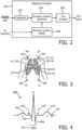
- the electrodes include a right arm (RA) electrode 302, a left arm (LA) electrode 304, a right leg (RL) electrode 306, a left leg (LG) electrode 308, a V1 electrode 310 in the fourth intercostal space (between ribs 4 and 5) just to the right of the sternum (breastbone), a V2 electrode 312 in the fourth intercostal space (between ribs 4 and 5) just to the left of the sternum, a V3 electrode 314 over rib 5, a V4 electrode 316 in the fifth intercostal space (between ribs 5 and 6) in the mid-clavicular line, a V5 electrode 318 horizontally even with V4, in the left anterior axillary line, and a V6 electrode 320 horizontally even with V4 and V5 in the midaxillary line.
- RA right arm
- LA left arm
- RL right leg
- LG left leg
- V1 electrode 310 in the fourth intercostal space (between ribs 4 and 5) just
- the 12 leads are: I, II, III, aVR, aVL, aVF, V1, V2, V3, V4, V5 and V6.
- FIGURE 4 schematically illustrates an example of an ECG signal 400 in "normal" sinus rhythm.
- the signal 400 includes a P wave 402, a Q wave 404, an R wave 406, an S wave 408, a T wave 410, and a U wave 412.
- the P wave 402 represents atrial depolarization
- the QRS complex 414 represents ventricular depolarization
- the T wave 410 represents ventricular repolarization
- the U wave 412 represents papillary muscle repolarization.
- a PR interval 416 is the interval from the beginning of the P wave 402 to the beginning of the QRS complex 414.
- An ST segment 418 connects the QRS complex 414 and the T wave 410, and represents the period when the ventricles are depolarized.
- the ECG analyzer 128 analyzes the ECG signal and estimates an existence of a myocardial infarction (MI) therefrom. In one instance, the ECG analyzer 128 achieves this by analyzing one or more of the waves 402-412. For example, the ECG analyzer 128 can analyze the ST segment 418, where a depressed or elevated ST segment 418 may indicate an MI. In another example, the ECG analyzer 128 can analyze the T wave 410, where an inverted T wave 410 may indicate an MI.
- MI myocardial infarction
- This data can be analyzed for a patient through a comparison with a previously acquired and known normal ECG signal of the patient, through a comparison with a model normal and/or abnormal ECG signal, through a comparison of known normal and/or abnormal ECG signals from a population of patients, etc.
- the ECG analyzer 128 estimates a position of the MI.
- the ECG analyzer 128 can use the leads V1 to V4, which measure electrical activity from the front of the heart, which is supplied by the left anterior descending coronary artery (LAD), to estimate an MI in an anterior region of the heart.
- the ECG analyzer 128 can use the leads I, aVL, V5 and V6, which measure electrical activity from the left of the heart, which is supplied by the left circumflex coronary artery (LC), to estimate an MI in a lateral region of the heart.
- the ECG analyzer 128 can use the II, III and aVF, which measure electrical activity from under the heart, which is supplied by the right coronary artery (RCA), to estimate an MI in an inferior region of the heart.
- the ECG analyzer 128 estimates a size of the MI.
- MI size can be estimated by ECG signal characteristics such as a deepened Q wave, reduced R-wave amplitude, elevated ST segments and/or inverted T wave on various leads.
- the ECG analyzer 128 can estimates any or all of these characteristics.
- the ECG analyzer 128 can analyze characteristics as described in US 8,688,206 B2, entitled “Visualization of myocardial infarct size in diagnostic ECG," and filed on April 25, 2011 . Additionally, or alternatively, a clinician may visually analyze the ECG signal and provide additional information to the ECG analyzer 128, which can use this information to estimate an MI.
- FIGURES 10, 11, 12 and 13 show example ECG signals with deviations from the "normal" ECG signal shown in FIGURE 4 .
- FIGURE 10 shows an ECG signal indicating a Stage I extended front apical infarction.
- FIGURE 11 shows an ECG signal indicating a Stage III anteroseptal infarction.
- FIGURE 12 shows an ECG signal indicating an intermediate stage posterolateral infarction.
- FIGURE 13 shows an ECG signal indicating a Stage I rear wall infarction.
- the segmentor 202 employs a segmentation algorithm to segment the coronary tree from the CCTA imaging data.
- the segmentation can be performed automatically (e.g., machine learning, etc.) or semiautomatically (e.g., with user assistance).
- the segmentation includes identifying and/or extracting coronary artery centerlines and/or lumen geometry (e.g., diameter, perimeter, cross-sectional area, etc.) therefrom.
- the segmentation can be based on voxel intensity, object shape, and/or other characteristics.
- FIGURE 5 shows segmentation of a portion 500 of an individual vessel showing opposing walls 502 of the vessel lumen
- FIGURE 6 shows a segmented coronary tree 600.
- the boundary condition determiner 204 determines boundary conditions for a computational fluid dynamic simulation of blood flow in vessels from the user adjusted coronary tree segmentation and/or the segmentor 202 adapted user adjusted coronary tree segmentation.
- a parametric lumped model is employed.
- the model includes a centerline representation using nonlinear resistances, with elements indicating inflow and outflow boundary conditions, and elements representing tree segment transfer functions, which include a series of linear and nonlinear resistance elements reflecting vessel geometry (e.g., diameter, perimeter, cross-sectional area, etc.) and/or hydraulic effects.
- MVR myocardial vascular resistance
- the amount of change ( ⁇ R) of the boundary conditions can be estimated from data known from cardiac physiology.
- late enhancement describes the delayed myocardial influx of contrast media typically seen in post-ischemic myocardial infarction scar tissue and caused by the altered microcirculatory resistance in subendocardial tissue layer.
- the model can be trained against data, and the boundary conditions can be trained such that the calculated and measured FFR (and/or iFR) data match.
- the flow simulator 210 performs a flow simulation with the boundary conditions and generates and outputs FFR values.
- Flow simulations can be done, e.g., using a computational fluid dynamics (CFD) approach and/or other approach.
- CFD computational fluid dynamics
- Examples of computing FFR values are described in US 2015/0092999 A1, filed May 10, 2013 , and entitled “Determination of a fractional flow reserve (FFR) value for a stenosis of a vessel," US 2015/0282765 A1, filed October 24, 2013 , and entitled “Fractional flow reserve (FFR) index".
- the FFR index can be displayed via a display monitor, stored, conveyed to another device, etc.
- FFR index can be displayed via a display monitor, stored, conveyed to another device, etc.
- FIGURE 7 schematically illustrates a variation in which the biophysical simulator 126 includes an image data adapter 702 configured to integrate the MI estimates into spatial coordinates of the cardiac image data.
- the biophysical simulator 126 does not include boundary condition adapter 206.
- the biophysical simulator 126 includes both the boundary condition adapter 206 and the image data adapter 702.
- the image data adapter 702 integrates the ECG estimates into the CCTA image data. This can be achieved via a personalized cardiac shape model, for example by inferring the coronary arteries associated with the involved cardiac feeding territory or territories and/or otherwise.
- the biophysical simulator 126 then processes the image data as described herein with the segmentor 202, the boundary condition determiner 204, and the flow simulator 208. In this instance, the boundary conditions reflect the MI estimates, which were integrated with the image data prior to boundary condition determination.
- the approaches described in connection with FIGURES 2 and 7 can be combined with invasive catheter measurements, which may deliver improved quantitative data of coronary flow.
- FIGURES 1 , 2 and -7 described example that use FFR as a measure of a functional significance of coronary artery disease.
- the approach described herein can also be applied to instantaneous wave-free ratio (iFR) and/or other measures.
- iFR is performed using pressure wires that are passed distal to the coronary stenosis and isolates a specific period in diastole, called the wave-free period, and computes a ratio of distal coronary pressure to a pressure observed in the aorta over this period.
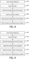
- FIGURE 8 illustrates an example method in accordance with an embodiment described herein.
- cardiac imaging data is acquired, as described herein and/or otherwise.
- an ECG signal is acquired, as described herein and/or otherwise.
- characteristics e.g., an existence, a position, a size, etc.
- characteristics of an MI is determined from the ECG signal, as described herein and/or otherwise.
- boundary conditions are determined from the image data, as described herein and/or otherwise.
- the boundary conditions are adapted based on the MI characteristics, as described herein and/or otherwise.
- the coronary function is assessed using the adapted boundary conditions, as described herein and/or otherwise.
- the above may be implemented by way of computer readable instructions, encoded or embedded on computer readable storage medium, which, when executed by a computer processor(s), cause the processor(s) to carry out the described acts. Additionally, or alternatively, at least one of the computer readable instructions is carried by a signal, carrier wave or other transitory medium, which is not computer readable storage medium.
- FIGURE 9 illustrates an example method in accordance with an embodiment described herein.
- cardiac imaging data is acquired, as described herein and/or otherwise.
- an ECG signal is acquired, as described herein and/or otherwise.
- characteristics e.g., an existence, a position, a size, etc.
- characteristics of an MI is determined from the ECG signal, as described herein and/or otherwise.
- the MI characteristics are integrated with the image data, as described herein and/or otherwise.
- boundary conditions are determined from the image data integrated with the MI characteristics, as described herein and/or otherwise.
- the coronary function is assessed using the adapted boundary conditions, as described herein and/or otherwise.
- the above may be implemented by way of computer readable instructions, encoded or embedded on computer readable storage medium, which, when executed by a computer processor(s), cause the processor(s) to carry out the described acts. Additionally, or alternatively, at least one of the computer readable instructions is carried by a signal, carrier wave or other transitory medium, which is not computer readable storage medium.
Landscapes
- Health & Medical Sciences (AREA)
- Engineering & Computer Science (AREA)
- Life Sciences & Earth Sciences (AREA)
- Medical Informatics (AREA)
- Public Health (AREA)
- General Health & Medical Sciences (AREA)
- Biomedical Technology (AREA)
- Cardiology (AREA)
- Pathology (AREA)
- Physics & Mathematics (AREA)
- Veterinary Medicine (AREA)
- Molecular Biology (AREA)
- Animal Behavior & Ethology (AREA)
- Surgery (AREA)
- Biophysics (AREA)
- Heart & Thoracic Surgery (AREA)
- Primary Health Care (AREA)
- Epidemiology (AREA)
- Radiology & Medical Imaging (AREA)
- Nuclear Medicine, Radiotherapy & Molecular Imaging (AREA)
- Data Mining & Analysis (AREA)
- Theoretical Computer Science (AREA)
- Databases & Information Systems (AREA)
- General Physics & Mathematics (AREA)
- Computer Vision & Pattern Recognition (AREA)
- Physiology (AREA)
- High Energy & Nuclear Physics (AREA)
- Optics & Photonics (AREA)
- Software Systems (AREA)
- Hematology (AREA)
- Oral & Maxillofacial Surgery (AREA)
- Dentistry (AREA)
- Computing Systems (AREA)
- Artificial Intelligence (AREA)
- Evolutionary Computation (AREA)
- Quality & Reliability (AREA)
- General Engineering & Computer Science (AREA)
- Mathematical Physics (AREA)
- Pulmonology (AREA)
- Vascular Medicine (AREA)
Applications Claiming Priority (3)
| Application Number | Priority Date | Filing Date | Title |
|---|---|---|---|
| US201762482223P | 2017-04-06 | 2017-04-06 | |
| US201762557213P | 2017-09-12 | 2017-09-12 | |
| PCT/EP2018/055367 WO2018184779A1 (en) | 2017-04-06 | 2018-03-05 | Coronary artery disease metric based on estimation of myocardial microvascular resistance from ecg signal |
Publications (2)
| Publication Number | Publication Date |
|---|---|
| EP3606421A1 EP3606421A1 (en) | 2020-02-12 |
| EP3606421B1 true EP3606421B1 (en) | 2025-05-07 |
Family
ID=61655731
Family Applications (1)
| Application Number | Title | Priority Date | Filing Date |
|---|---|---|---|
| EP18711252.9A Active EP3606421B1 (en) | 2017-04-06 | 2018-03-05 | Coronary artery disease metric based on estimation of myocardial microvascular resistance from ecg signal |
Country Status (5)
| Country | Link |
|---|---|
| US (1) | US11710569B2 (https=) |
| EP (1) | EP3606421B1 (https=) |
| JP (1) | JP7229170B2 (https=) |
| CN (1) | CN110494081A (https=) |
| WO (1) | WO2018184779A1 (https=) |
Families Citing this family (19)
| Publication number | Priority date | Publication date | Assignee | Title |
|---|---|---|---|---|
| US10210956B2 (en) | 2012-10-24 | 2019-02-19 | Cathworks Ltd. | Diagnostically useful results in real time |
| JP7036742B2 (ja) | 2016-05-16 | 2022-03-15 | キャスワークス リミテッド | 血管評価システム |
| EP4241694A3 (en) | 2016-05-16 | 2023-12-20 | Cathworks Ltd. | Selection of vascular paths from images |
| JP7181216B2 (ja) * | 2017-03-31 | 2022-11-30 | コーニンクレッカ フィリップス エヌ ヴェ | 経カテーテル大動脈弁移植術(tavi)が冠血流量及び冠動脈圧に及ぼす影響のシミュレーション |
| CN112105970B (zh) * | 2018-04-27 | 2022-05-31 | 日本瑞翁株式会社 | 宽带波长膜及其制造方法、以及圆偏振膜的制造方法 |
| JP7532402B2 (ja) | 2019-04-01 | 2024-08-13 | キャスワークス リミテッド | 血管造影画像選択のための方法および装置 |
| EP4033964B1 (en) | 2019-09-23 | 2025-04-09 | Cathworks Ltd. | Methods, apparatus, and system for synchronization between a three-dimensional vascular model and an imaging device |
| EP4062362A1 (en) | 2019-11-22 | 2022-09-28 | The Regents Of The University Of Michigan | Anatomical and functional assessment of coronary artery disease using machine learning |
| CN111067494B (zh) * | 2019-12-27 | 2022-04-26 | 西北工业大学 | 基于血流储备分数和血流阻力模型的微循环阻力快速计算方法 |
| CN111067495A (zh) * | 2019-12-27 | 2020-04-28 | 西北工业大学 | 基于血流储备分数和造影图像的微循环阻力计算方法 |
| CN111652881B (zh) * | 2020-07-01 | 2025-01-10 | 杭州脉流科技有限公司 | 基于深度学习的冠脉重构和血流储备分数计算方法、装置、设备以及可读存储介质 |
| US12315076B1 (en) | 2021-09-22 | 2025-05-27 | Cathworks Ltd. | Four-dimensional motion analysis of a patient's coronary arteries and myocardial wall |
| KR20240148399A (ko) | 2022-02-10 | 2024-10-11 | 캐스웍스 엘티디. | 기계 학습 기반 센서 분석 및 혈관 트리 분할을 위한 시스템 및 방법 |
| CN115299956B (zh) * | 2022-08-19 | 2024-06-25 | 山东大学 | 一种基于确定学习和心电图的心肌缺血检测方法及系统 |
| US12475559B2 (en) | 2023-05-18 | 2025-11-18 | Regents Of The University Of Michigan | Machine learning approach for coronary 3D reconstruction from X-ray angiography images |
| CN121942048A (zh) | 2023-08-09 | 2026-04-28 | 凯思沃克斯有限公司 | 针对血管指数测量的增强用户界面和串扰分析 |
| IL326432A (en) | 2023-08-09 | 2026-04-01 | Cathworks Ltd | Coronary artery assessment after PCI |
| CN117617986A (zh) * | 2023-12-15 | 2024-03-01 | 中国医学科学院阜外医院 | 微循环阻力指数的确定方法、装置及电子设备 |
| US12512196B2 (en) | 2024-06-12 | 2025-12-30 | Cathworks Ltd. | Systems and methods for secure sharing of cardiac assessments using QR codes |
Citations (1)
| Publication number | Priority date | Publication date | Assignee | Title |
|---|---|---|---|---|
| US9129053B2 (en) * | 2012-02-01 | 2015-09-08 | Siemens Aktiengesellschaft | Method and system for advanced measurements computation and therapy planning from medical data and images using a multi-physics fluid-solid heart model |
Family Cites Families (14)
| Publication number | Priority date | Publication date | Assignee | Title |
|---|---|---|---|---|
| US20040138574A1 (en) | 2002-07-17 | 2004-07-15 | Resolution Medical, Inc. | Methods and apparatus for enhancing diagnosis of myocardial infarctions |
| NL1024765C2 (nl) * | 2003-11-12 | 2005-05-17 | Consult In Medicine B V | Werkwijze en inrichting voor het vaststellen van de aanwezigheid van een ischemisch gebied in het hart van een mens of dier. |
| JP5906234B2 (ja) * | 2010-04-28 | 2016-04-20 | コーニンクレッカ フィリップス エヌ ヴェKoninklijke Philips N.V. | 診断ecgにおける心筋梗塞サイズの可視化 |
| US10186056B2 (en) | 2011-03-21 | 2019-01-22 | General Electric Company | System and method for estimating vascular flow using CT imaging |
| US10162932B2 (en) | 2011-11-10 | 2018-12-25 | Siemens Healthcare Gmbh | Method and system for multi-scale anatomical and functional modeling of coronary circulation |
| JP6173438B2 (ja) | 2012-05-14 | 2017-08-02 | コーニンクレッカ フィリップス エヌ ヴェKoninklijke Philips N.V. | 血管の狭窄のための冠血流予備量比(ffr)値の決定 |
| CN104582572B (zh) * | 2012-08-16 | 2018-04-13 | 东芝医疗系统株式会社 | 图像处理装置、医用图像诊断装置以及血压监视器 |
| WO2014072861A2 (en) | 2012-11-06 | 2014-05-15 | Koninklijke Philips N.V. | Fractional flow reserve (ffr) index |
| US10595806B2 (en) | 2013-10-22 | 2020-03-24 | Koninklijke Philips N.V. | Fractional flow reserve (FFR) index with adaptive boundary condition parameters |
| US9595089B2 (en) * | 2014-05-09 | 2017-03-14 | Siemens Healthcare Gmbh | Method and system for non-invasive computation of hemodynamic indices for coronary artery stenosis |
| CN106659400B (zh) | 2014-06-30 | 2021-01-05 | 皇家飞利浦有限公司 | 用于确定血流储备分数值的装置 |
| US9668700B2 (en) * | 2014-09-09 | 2017-06-06 | Heartflow, Inc. | Method and system for quantifying limitations in coronary artery blood flow during physical activity in patients with coronary artery disease |
| US10262101B2 (en) * | 2015-04-02 | 2019-04-16 | Heartflow, Inc. | Systems and methods for predicting perfusion deficits from physiological, anatomical, and patient characteristics |
| WO2017093337A1 (en) * | 2015-12-02 | 2017-06-08 | Siemens Healthcare Gmbh | Personalized assessment of patients with acute coronary syndrome |
-
2018
- 2018-03-05 EP EP18711252.9A patent/EP3606421B1/en active Active
- 2018-03-05 JP JP2019554674A patent/JP7229170B2/ja active Active
- 2018-03-05 CN CN201880023414.2A patent/CN110494081A/zh active Pending
- 2018-03-05 US US16/500,207 patent/US11710569B2/en active Active
- 2018-03-05 WO PCT/EP2018/055367 patent/WO2018184779A1/en not_active Ceased
Patent Citations (1)
| Publication number | Priority date | Publication date | Assignee | Title |
|---|---|---|---|---|
| US9129053B2 (en) * | 2012-02-01 | 2015-09-08 | Siemens Aktiengesellschaft | Method and system for advanced measurements computation and therapy planning from medical data and images using a multi-physics fluid-solid heart model |
Also Published As
| Publication number | Publication date |
|---|---|
| EP3606421A1 (en) | 2020-02-12 |
| CN110494081A (zh) | 2019-11-22 |
| JP2020516348A (ja) | 2020-06-11 |
| US20210118569A1 (en) | 2021-04-22 |
| US11710569B2 (en) | 2023-07-25 |
| WO2018184779A1 (en) | 2018-10-11 |
| JP7229170B2 (ja) | 2023-02-27 |
Similar Documents
| Publication | Publication Date | Title |
|---|---|---|
| EP3606421B1 (en) | Coronary artery disease metric based on estimation of myocardial microvascular resistance from ecg signal | |
| EP2932469B1 (en) | Method of determining the blood flow through coronary arteries | |
| Berger et al. | Single-beat noninvasive imaging of cardiac electrophysiology of ventricular pre-excitation | |
| EP3160335B1 (en) | Apparatus for determining a fractional flow reserve value | |
| CA2357729C (en) | Method and apparatus for characterizing cardiac tissue from local electrograms | |
| US9633454B2 (en) | Fluid-dynamic analysis of a vascular tree using angiography | |
| US10646183B2 (en) | Detection of scar and fibrous cardiac zones | |
| EP2442276B1 (en) | Medical image processing apparatus and medical image processing method | |
| EP3606433B1 (en) | Standardized coronary artery disease metric | |
| US12380562B2 (en) | Vessel registration using functional information | |
| JP2022510879A (ja) | 血行力学的シミュレーションのための最も関連のあるx線画像の選択 | |
| CN112673433A (zh) | 基于心肌呈色特性计算用于虚拟FFR和iFR计算的边界条件 | |
| EP3773169B1 (en) | Multi-dimensional method of fundamental solutions for reconstruction of electrophysiological activity | |
| EP3512416A1 (en) | Apparatus and method for determining a fractional flow reserve | |
| JP7483347B2 (ja) | 異なる心調律に応じた磁気共鳴画像法(mri)の画像フィルタリング | |
| US20230190104A1 (en) | Automated mapping and/or signal processing responsive to cardiac signal features | |
| CN110520038B (zh) | 心律失常驱动器的连通性分析 | |
| CN114040707A (zh) | 诊断支持程序 | |
| Zhang et al. | Quantification of effects of mean blood pressure and left ventricular mass on noninvasive fast fractional flow reserve | |
| Grothues et al. | Serial assessment of ventricular morphology and function | |
| Nash et al. | An experimental–computational framework for validating in vivo ECG inverse algorithm | |
| JP2023038932A (ja) | シグモイド曲線を用いて投影された電気生理学的波速度の重み付け | |
| WO2025181580A1 (en) | Analysis of ectopic electrophysiological signals | |
| EP4452068A1 (en) | Automated mapping and/or signal processing responsive to cardiac signal features | |
| HK40060267A (en) | Diagnosis assisting program |
Legal Events
| Date | Code | Title | Description |
|---|---|---|---|
| STAA | Information on the status of an ep patent application or granted ep patent |
Free format text: STATUS: UNKNOWN |
|
| STAA | Information on the status of an ep patent application or granted ep patent |
Free format text: STATUS: THE INTERNATIONAL PUBLICATION HAS BEEN MADE |
|
| PUAI | Public reference made under article 153(3) epc to a published international application that has entered the european phase |
Free format text: ORIGINAL CODE: 0009012 |
|
| STAA | Information on the status of an ep patent application or granted ep patent |
Free format text: STATUS: REQUEST FOR EXAMINATION WAS MADE |
|
| 17P | Request for examination filed |
Effective date: 20191106 |
|
| AK | Designated contracting states |
Kind code of ref document: A1 Designated state(s): AL AT BE BG CH CY CZ DE DK EE ES FI FR GB GR HR HU IE IS IT LI LT LU LV MC MK MT NL NO PL PT RO RS SE SI SK SM TR |
|
| AX | Request for extension of the european patent |
Extension state: BA ME |
|
| RAP1 | Party data changed (applicant data changed or rights of an application transferred) |
Owner name: KONINKLIJKE PHILIPS N.V. |
|
| DAV | Request for validation of the european patent (deleted) | ||
| DAX | Request for extension of the european patent (deleted) | ||
| STAA | Information on the status of an ep patent application or granted ep patent |
Free format text: STATUS: EXAMINATION IS IN PROGRESS |
|
| 17Q | First examination report despatched |
Effective date: 20230619 |
|
| REG | Reference to a national code |
Ref country code: DE Ref legal event code: R079 Ipc: A61B0005020000 Ref country code: DE Ref legal event code: R079 Ref document number: 602018081699 Country of ref document: DE Free format text: PREVIOUS MAIN CLASS: A61B0005045200 Ipc: A61B0005020000 |
|
| GRAP | Despatch of communication of intention to grant a patent |
Free format text: ORIGINAL CODE: EPIDOSNIGR1 |
|
| STAA | Information on the status of an ep patent application or granted ep patent |
Free format text: STATUS: GRANT OF PATENT IS INTENDED |
|
| RIC1 | Information provided on ipc code assigned before grant |
Ipc: G16H 50/50 20180101ALI20241007BHEP Ipc: A61B 6/03 20060101ALI20241007BHEP Ipc: A61B 5/055 20060101ALI20241007BHEP Ipc: A61B 5/026 20060101ALI20241007BHEP Ipc: A61B 5/02 20060101AFI20241007BHEP |
|
| INTG | Intention to grant announced |
Effective date: 20241023 |
|
| GRAS | Grant fee paid |
Free format text: ORIGINAL CODE: EPIDOSNIGR3 |
|
| GRAA | (expected) grant |
Free format text: ORIGINAL CODE: 0009210 |
|
| STAA | Information on the status of an ep patent application or granted ep patent |
Free format text: STATUS: THE PATENT HAS BEEN GRANTED |
|
| AK | Designated contracting states |
Kind code of ref document: B1 Designated state(s): AL AT BE BG CH CY CZ DE DK EE ES FI FR GB GR HR HU IE IS IT LI LT LU LV MC MK MT NL NO PL PT RO RS SE SI SK SM TR |
|
| REG | Reference to a national code |
Ref country code: GB Ref legal event code: FG4D |
|
| REG | Reference to a national code |
Ref country code: CH Ref legal event code: EP |
|
| REG | Reference to a national code |
Ref country code: DE Ref legal event code: R096 Ref document number: 602018081699 Country of ref document: DE |
|
| REG | Reference to a national code |
Ref country code: IE Ref legal event code: FG4D |
|
| REG | Reference to a national code |
Ref country code: DE Ref legal event code: R084 Ref document number: 602018081699 Country of ref document: DE |
|
| REG | Reference to a national code |
Ref country code: NL Ref legal event code: MP Effective date: 20250507 |
|
| PG25 | Lapsed in a contracting state [announced via postgrant information from national office to epo] |
Ref country code: PT Free format text: LAPSE BECAUSE OF FAILURE TO SUBMIT A TRANSLATION OF THE DESCRIPTION OR TO PAY THE FEE WITHIN THE PRESCRIBED TIME-LIMIT Effective date: 20250908 Ref country code: ES Free format text: LAPSE BECAUSE OF FAILURE TO SUBMIT A TRANSLATION OF THE DESCRIPTION OR TO PAY THE FEE WITHIN THE PRESCRIBED TIME-LIMIT Effective date: 20250507 Ref country code: FI Free format text: LAPSE BECAUSE OF FAILURE TO SUBMIT A TRANSLATION OF THE DESCRIPTION OR TO PAY THE FEE WITHIN THE PRESCRIBED TIME-LIMIT Effective date: 20250507 |
|
| REG | Reference to a national code |
Ref country code: LT Ref legal event code: MG9D |
|
| PG25 | Lapsed in a contracting state [announced via postgrant information from national office to epo] |
Ref country code: GR Free format text: LAPSE BECAUSE OF FAILURE TO SUBMIT A TRANSLATION OF THE DESCRIPTION OR TO PAY THE FEE WITHIN THE PRESCRIBED TIME-LIMIT Effective date: 20250808 Ref country code: NO Free format text: LAPSE BECAUSE OF FAILURE TO SUBMIT A TRANSLATION OF THE DESCRIPTION OR TO PAY THE FEE WITHIN THE PRESCRIBED TIME-LIMIT Effective date: 20250807 |
|
| PG25 | Lapsed in a contracting state [announced via postgrant information from national office to epo] |
Ref country code: NL Free format text: LAPSE BECAUSE OF FAILURE TO SUBMIT A TRANSLATION OF THE DESCRIPTION OR TO PAY THE FEE WITHIN THE PRESCRIBED TIME-LIMIT Effective date: 20250507 Ref country code: PL Free format text: LAPSE BECAUSE OF FAILURE TO SUBMIT A TRANSLATION OF THE DESCRIPTION OR TO PAY THE FEE WITHIN THE PRESCRIBED TIME-LIMIT Effective date: 20250507 |
|
| REG | Reference to a national code |
Ref country code: AT Ref legal event code: MK05 Ref document number: 1791535 Country of ref document: AT Kind code of ref document: T Effective date: 20250507 |
|
| PG25 | Lapsed in a contracting state [announced via postgrant information from national office to epo] |
Ref country code: BG Free format text: LAPSE BECAUSE OF FAILURE TO SUBMIT A TRANSLATION OF THE DESCRIPTION OR TO PAY THE FEE WITHIN THE PRESCRIBED TIME-LIMIT Effective date: 20250507 |
|
| PG25 | Lapsed in a contracting state [announced via postgrant information from national office to epo] |
Ref country code: HR Free format text: LAPSE BECAUSE OF FAILURE TO SUBMIT A TRANSLATION OF THE DESCRIPTION OR TO PAY THE FEE WITHIN THE PRESCRIBED TIME-LIMIT Effective date: 20250507 |
|
| PG25 | Lapsed in a contracting state [announced via postgrant information from national office to epo] |
Ref country code: AT Free format text: LAPSE BECAUSE OF FAILURE TO SUBMIT A TRANSLATION OF THE DESCRIPTION OR TO PAY THE FEE WITHIN THE PRESCRIBED TIME-LIMIT Effective date: 20250507 |
|
| PG25 | Lapsed in a contracting state [announced via postgrant information from national office to epo] |
Ref country code: RS Free format text: LAPSE BECAUSE OF FAILURE TO SUBMIT A TRANSLATION OF THE DESCRIPTION OR TO PAY THE FEE WITHIN THE PRESCRIBED TIME-LIMIT Effective date: 20250807 |
|
| PG25 | Lapsed in a contracting state [announced via postgrant information from national office to epo] |
Ref country code: IS Free format text: LAPSE BECAUSE OF FAILURE TO SUBMIT A TRANSLATION OF THE DESCRIPTION OR TO PAY THE FEE WITHIN THE PRESCRIBED TIME-LIMIT Effective date: 20250907 |
|
| PG25 | Lapsed in a contracting state [announced via postgrant information from national office to epo] |
Ref country code: LV Free format text: LAPSE BECAUSE OF FAILURE TO SUBMIT A TRANSLATION OF THE DESCRIPTION OR TO PAY THE FEE WITHIN THE PRESCRIBED TIME-LIMIT Effective date: 20250507 |
|
| REG | Reference to a national code |
Ref country code: GB Ref legal event code: 746 Effective date: 20251114 |
|
| PG25 | Lapsed in a contracting state [announced via postgrant information from national office to epo] |
Ref country code: SM Free format text: LAPSE BECAUSE OF FAILURE TO SUBMIT A TRANSLATION OF THE DESCRIPTION OR TO PAY THE FEE WITHIN THE PRESCRIBED TIME-LIMIT Effective date: 20250507 Ref country code: DK Free format text: LAPSE BECAUSE OF FAILURE TO SUBMIT A TRANSLATION OF THE DESCRIPTION OR TO PAY THE FEE WITHIN THE PRESCRIBED TIME-LIMIT Effective date: 20250507 |
|
| PG25 | Lapsed in a contracting state [announced via postgrant information from national office to epo] |
Ref country code: CZ Free format text: LAPSE BECAUSE OF FAILURE TO SUBMIT A TRANSLATION OF THE DESCRIPTION OR TO PAY THE FEE WITHIN THE PRESCRIBED TIME-LIMIT Effective date: 20250507 |
|
| PG25 | Lapsed in a contracting state [announced via postgrant information from national office to epo] |
Ref country code: EE Free format text: LAPSE BECAUSE OF FAILURE TO SUBMIT A TRANSLATION OF THE DESCRIPTION OR TO PAY THE FEE WITHIN THE PRESCRIBED TIME-LIMIT Effective date: 20250507 |
|
| PG25 | Lapsed in a contracting state [announced via postgrant information from national office to epo] |
Ref country code: RO Free format text: LAPSE BECAUSE OF FAILURE TO SUBMIT A TRANSLATION OF THE DESCRIPTION OR TO PAY THE FEE WITHIN THE PRESCRIBED TIME-LIMIT Effective date: 20250507 Ref country code: SK Free format text: LAPSE BECAUSE OF FAILURE TO SUBMIT A TRANSLATION OF THE DESCRIPTION OR TO PAY THE FEE WITHIN THE PRESCRIBED TIME-LIMIT Effective date: 20250507 |
|
| PG25 | Lapsed in a contracting state [announced via postgrant information from national office to epo] |
Ref country code: IT Free format text: LAPSE BECAUSE OF FAILURE TO SUBMIT A TRANSLATION OF THE DESCRIPTION OR TO PAY THE FEE WITHIN THE PRESCRIBED TIME-LIMIT Effective date: 20250507 |
|
| REG | Reference to a national code |
Ref country code: DE Ref legal event code: R097 Ref document number: 602018081699 Country of ref document: DE |
|
| PLBE | No opposition filed within time limit |
Free format text: ORIGINAL CODE: 0009261 |
|
| STAA | Information on the status of an ep patent application or granted ep patent |
Free format text: STATUS: NO OPPOSITION FILED WITHIN TIME LIMIT |
|
| REG | Reference to a national code |
Ref country code: CH Ref legal event code: L10 Free format text: ST27 STATUS EVENT CODE: U-0-0-L10-L00 (AS PROVIDED BY THE NATIONAL OFFICE) Effective date: 20260318 |
|
| PGFP | Annual fee paid to national office [announced via postgrant information from national office to epo] |
Ref country code: GB Payment date: 20260319 Year of fee payment: 9 |
|
| PGFP | Annual fee paid to national office [announced via postgrant information from national office to epo] |
Ref country code: DE Payment date: 20260320 Year of fee payment: 9 |
|
| 26N | No opposition filed |
Effective date: 20260210 |
|
| PGFP | Annual fee paid to national office [announced via postgrant information from national office to epo] |
Ref country code: FR Payment date: 20260323 Year of fee payment: 9 |