EP3551042B1 - Dispositif servant à l'examen d'un patient humain - Google Patents
Dispositif servant à l'examen d'un patient humain Download PDFInfo
- Publication number
- EP3551042B1 EP3551042B1 EP17818029.5A EP17818029A EP3551042B1 EP 3551042 B1 EP3551042 B1 EP 3551042B1 EP 17818029 A EP17818029 A EP 17818029A EP 3551042 B1 EP3551042 B1 EP 3551042B1
- Authority
- EP
- European Patent Office
- Prior art keywords
- patient
- measuring
- measurement data
- display device
- mental
- Prior art date
- Legal status (The legal status is an assumption and is not a legal conclusion. Google has not performed a legal analysis and makes no representation as to the accuracy of the status listed.)
- Active
Links
Images


Classifications
-
- A—HUMAN NECESSITIES
- A61—MEDICAL OR VETERINARY SCIENCE; HYGIENE
- A61B—DIAGNOSIS; SURGERY; IDENTIFICATION
- A61B5/00—Measuring for diagnostic purposes; Identification of persons
- A61B5/01—Measuring temperature of body parts ; Diagnostic temperature sensing, e.g. for malignant or inflamed tissue
-
- A—HUMAN NECESSITIES
- A61—MEDICAL OR VETERINARY SCIENCE; HYGIENE
- A61B—DIAGNOSIS; SURGERY; IDENTIFICATION
- A61B5/00—Measuring for diagnostic purposes; Identification of persons
- A61B5/68—Arrangements of detecting, measuring or recording means, e.g. sensors, in relation to patient
- A61B5/6887—Arrangements of detecting, measuring or recording means, e.g. sensors, in relation to patient mounted on external non-worn devices, e.g. non-medical devices
- A61B5/6891—Furniture
-
- A—HUMAN NECESSITIES
- A61—MEDICAL OR VETERINARY SCIENCE; HYGIENE
- A61B—DIAGNOSIS; SURGERY; IDENTIFICATION
- A61B5/00—Measuring for diagnostic purposes; Identification of persons
- A61B5/01—Measuring temperature of body parts ; Diagnostic temperature sensing, e.g. for malignant or inflamed tissue
- A61B5/015—By temperature mapping of body part
-
- A—HUMAN NECESSITIES
- A61—MEDICAL OR VETERINARY SCIENCE; HYGIENE
- A61B—DIAGNOSIS; SURGERY; IDENTIFICATION
- A61B5/00—Measuring for diagnostic purposes; Identification of persons
- A61B5/02—Detecting, measuring or recording for evaluating the cardiovascular system, e.g. pulse, heart rate, blood pressure or blood flow
- A61B5/0205—Simultaneously evaluating both cardiovascular conditions and different types of body conditions, e.g. heart and respiratory condition
- A61B5/02055—Simultaneously evaluating both cardiovascular condition and temperature
-
- A—HUMAN NECESSITIES
- A61—MEDICAL OR VETERINARY SCIENCE; HYGIENE
- A61B—DIAGNOSIS; SURGERY; IDENTIFICATION
- A61B5/00—Measuring for diagnostic purposes; Identification of persons
- A61B5/05—Detecting, measuring or recording for diagnosis by means of electric currents or magnetic fields; Measuring using microwaves or radio waves
-
- A—HUMAN NECESSITIES
- A61—MEDICAL OR VETERINARY SCIENCE; HYGIENE
- A61B—DIAGNOSIS; SURGERY; IDENTIFICATION
- A61B5/00—Measuring for diagnostic purposes; Identification of persons
- A61B5/16—Devices for psychotechnics; Testing reaction times ; Devices for evaluating the psychological state
- A61B5/165—Evaluating the state of mind, e.g. depression, anxiety
-
- A—HUMAN NECESSITIES
- A61—MEDICAL OR VETERINARY SCIENCE; HYGIENE
- A61B—DIAGNOSIS; SURGERY; IDENTIFICATION
- A61B5/00—Measuring for diagnostic purposes; Identification of persons
- A61B5/24—Detecting, measuring or recording bioelectric or biomagnetic signals of the body or parts thereof
-
- A—HUMAN NECESSITIES
- A61—MEDICAL OR VETERINARY SCIENCE; HYGIENE
- A61B—DIAGNOSIS; SURGERY; IDENTIFICATION
- A61B5/00—Measuring for diagnostic purposes; Identification of persons
- A61B5/48—Other medical applications
- A61B5/4836—Diagnosis combined with treatment in closed-loop systems or methods
-
- A—HUMAN NECESSITIES
- A61—MEDICAL OR VETERINARY SCIENCE; HYGIENE
- A61B—DIAGNOSIS; SURGERY; IDENTIFICATION
- A61B5/00—Measuring for diagnostic purposes; Identification of persons
- A61B5/68—Arrangements of detecting, measuring or recording means, e.g. sensors, in relation to patient
- A61B5/6887—Arrangements of detecting, measuring or recording means, e.g. sensors, in relation to patient mounted on external non-worn devices, e.g. non-medical devices
-
- A—HUMAN NECESSITIES
- A61—MEDICAL OR VETERINARY SCIENCE; HYGIENE
- A61B—DIAGNOSIS; SURGERY; IDENTIFICATION
- A61B5/00—Measuring for diagnostic purposes; Identification of persons
- A61B5/74—Details of notification to user or communication with user or patient; User input means
- A61B5/742—Details of notification to user or communication with user or patient; User input means using visual displays
- A61B5/7445—Display arrangements, e.g. multiple display units
-
- A—HUMAN NECESSITIES
- A61—MEDICAL OR VETERINARY SCIENCE; HYGIENE
- A61B—DIAGNOSIS; SURGERY; IDENTIFICATION
- A61B2560/00—Constructional details of operational features of apparatus; Accessories for medical measuring apparatus
- A61B2560/04—Constructional details of apparatus
-
- A—HUMAN NECESSITIES
- A61—MEDICAL OR VETERINARY SCIENCE; HYGIENE
- A61B—DIAGNOSIS; SURGERY; IDENTIFICATION
- A61B5/00—Measuring for diagnostic purposes; Identification of persons
- A61B5/16—Devices for psychotechnics; Testing reaction times ; Devices for evaluating the psychological state
- A61B5/163—Devices for psychotechnics; Testing reaction times ; Devices for evaluating the psychological state by tracking eye movement, gaze, or pupil change
Definitions
- the invention relates to a device for examining a human patient, comprising a support for the patient, at least one display device - in particular designed as a screen - which is visible to the patient lying on the support, at least two measuring devices for recording measurement data from the patient, which output measurement data relating to the condition of the patient, and an evaluation device which is connected to the display device and the measuring device and which is designed to determine output signals as a function of measurement data originating from at least two different measuring devices and their interrelationship, wherein at least one display device and at least one sensor of a measuring device are movably mounted relative to the support for the patient.
- the US 2002/0123704 A1 A massage chair with a remote control that features a display and several sensors.
- the sensors When a person holds the remote control in their hand, the sensors come into contact with the person's skin and can then take appropriate measurements to determine the effect of a massage on the person.
- a patient health monitoring system is known, by means of which the health status of a patient can be monitored remotely.
- the object of the invention is to provide a device for examining a human patient with which it is possible to obtain a good overall picture of the patient's state of health in a short time.
- the at least one display device and the at least one sensor of a measuring device are mounted on a support device in a motor-movable manner, and that the at least one measuring device comprises a medical thermal scanner.
- the display device and the sensor of the measuring device are mounted on a support device that can be moved by a motor, fully automated operation with consistent results is possible.
- a thermal scanner can be used to perform a wide variety of physical diagnoses, in particular to identify areas of inflammation and other changes.
- the movably mounted display device can be used in a variety of ways. During a purely physical examination of a patient, the display device can be positioned in a position that is convenient for the patient to view, allowing them to clearly see the examination and subsequent results. The examination itself can be performed using at least one sensor of a measuring device that is movably mounted relative to the patient lying on the support.
- a particularly advantageous embodiment is one in which the display device and the sensor of the measuring device are movably mounted on a common carriage.
- the sensor can record or scan physical measurement data in one operating position.
- the patient can view the screen, which is adjusted to a suitable operating position.
- the data on the screen can not only be status data regarding the examination performed or the examination result, as explained above. Rather, it is also possible to provide the patient with visual information via the display device, possibly together with audio information, and then measure the patient's response simultaneously or in a short period of time.
- a device for examining a human patient comprising at least two, preferably several different measuring devices which output measurement data relating to the condition of the patient, wherein an electronic evaluation device is provided which determines output signals as a function of measurement data originating from at least two different measuring devices and their interrelationship.
- the key feature here is the ability to evaluate the interrelationship between the measurement data alongside the actual measurement data itself. Only through this correlation of the measurement data is it often possible to assess the overall health situation. This is especially true when part of the correlated measurement data is physical, while the other part is mental. This allows the patient's physical and mental states to be correlated, and based on this correlation, a diagnosis and/or automated control of therapy devices can be performed.
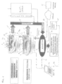
- the Figure 1 shows an embodiment of a device according to the invention in a schematic representation.
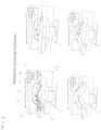
- the Figure 2 shows various "biological screening positions" for recording physical measurement data of the patient lying on a support.
- the Figure 3 shows "psychological, neurological and mental screening positions" for recording psychological (mental) measurement data of the patient sitting or lying on the carrier.
- the Figure 4 shows an embodiment of a device according to the invention in a schematic block diagram.
- the Figure 5 shows in a schematic overview the "biological scans" for determining physical measurement data and “mental scans” for determining mental measurement data that reflect the psychological state of the patient.
- FIGS. 6 and 7 show in separate illustrations the biological screening for determining physical measurement data and the psychological screening for determining mental measurement data that reflect the psychological state of the patient.
- the Figure 1 shows an embodiment of a device according to the invention for examining a human patient 1 who can sit on a support 2.
- the two superimposed representations of the different positions of carrier 2 refer to one and the same facility at two different times. In the upper position, psychological, neurological, and mental diagnoses are made; in the lower position, biological, physical diagnoses.
- a display device 3 designed as a screen is provided, which is visible to the patient lying on the carrier 2 (upper position in Figure 1 ). Furthermore, there is a measuring device 4 for recording measurement data of the patient 1 and an evaluation device 5, which is connected to the display device 3 and the measuring device 4 via schematically illustrated lines 5.
- both the display device 3 and at least one sensor 4a of the measuring device 4 are movably mounted relative to the support 2 for the patient 1.
- the measuring device is a thermal scanner, wherein the sensor 4a is an infrared sensor.
- the measuring device in the form of the thermal scanner is mounted on a holder 6 in the form of a carriage and is guided linearly and longitudinally movable in the direction of the arrows 7.
- the holder 6 is a common holder for the thermal scanner 4 and the display device 3. To move the holder from the scanning position in the middle of the Figure 1 to the upper position in Figure 1 To bring the patient into the correct position, it can be swivelled in the direction of arrows 8, allowing the patient to see the display device or screen. This can be easily integrated into Figure 1
- the solution shown can be realized in that the holder 6 has a movable base part and a pivoting part pivotably mounted on this, which carries the display device 3 and the measuring device 4 or its sensor 4a.
- the display device can also be assigned at least one loudspeaker (not shown). This can be mounted either on the fixed part of the The speaker can be located on the device or on the movable carriage. It is also possible for the speaker to be housed in a headset.
- an audiovisual image or sequence of audiovisual images is presented to the patient, and subsequent or simultaneous measurement data is collected.
- This is reactive measurement data that provides information about the patient's psychological health. For example, stressful and conflict situations can be simulated for the patient and their reaction can be monitored. Colors can also be simulated and their reaction monitored.
- measurement data can also be buffered during consecutive examinations in order to be able to perform the correlation with other data described below.
- the display device 3 and the sensor 4a of the measuring device 4 are mounted in a motor-movable manner on a support device 9, which can be accommodated in a visually appealing housing.
- the carrying device 9 can also carry the carrier 2 for the patient.
- this carrier 2 is Figure 1 shown, rests directly on the floor. Then the support device or the housing 9 and the support 2 for the patient arranged across the floor of the room in a fixed spatial relationship to each other.
- a further display device 10 is provided which can be pivoted out of the housing in the direction of arrow 11 or, when not in use, can be pivoted back in flush with it.
- a receptacle 12 in the support device 9 or in its housing, which is easily accessible from the patient side.
- This receptacle 12 can accommodate additional sensors of other medical measuring devices, as will be shown in the following. Figures 2 and 3 will be described in more detail.
- the support 2 for the patient 1 is designed as a multi-part seat-bed, the positions of the parts of which are motor-adjustable.
- the Figure 1 also shows that the lounger can be moved to a sitting position (upper position in Figure 1 ), with the display device pivoted downwards so that the patient's head is at substantially the same height as the display device. This essentially corresponds to the normal television or screen viewing position.
- An alternative would be one in which the seating couch and the display device can be moved into a position in which the patient looks essentially straight ahead at the display device. This would be possible, for example, if the patient is lying flat and still needs to see something on the screen.
- the device for examining the human patient 1 has at least two, preferably several different measuring devices, each of which provides measurement data regarding the patient's condition. Furthermore, an electronic evaluation device 5 is provided, which, depending on at least two different measuring devices and their interrelationship (correlation) output signals are determined, which then, like the Figure 1 shows, can be fed, for example, via a line 13 to a computer intended for a doctor (doctor's PC 14). The output signals can also be fed as control data for one or more therapy devices 15 via a line 16.
- the output signals can be passed on to the doctor, therapist or patient in a report in digital or printed form, if necessary together with a therapy suggestion.
- the evaluation device can also operate partially decentrally, whereby an external server located at a different location can also be accessed via a schematically illustrated line 17 and communication means not shown in detail (for example, via the Internet).
- This external server can, on the one hand, provide computing power, but, above all, also store reference data that allows the patient's recorded measurement data to be evaluated for diagnosis.
- an automatic therapy therapy device 15, line 16
- a therapy suggestion the physician always remains the one who can intervene in the therapy suggestion or the automatic therapy, for example via line 19.
- the Figure 2 On the one hand, it shows different positions of the support designed as a seat for the patient, which can be adjusted motorically depending on the type of examination.
- FIG 2 Top left: The patient lies supine.
- the blood pressure is measured using a blood pressure monitor 4' with arm cuff and data processing.
- An electrocardiogram can be recorded using an ECG device 4" with electrodes 20.
- an image can be generated using the thermal scanner 4, which can be moved linearly in the direction of the double arrow 7.
- the support device 9 has a receptacle 12 (in optical attractive elliptical shape) in which the various measuring devices and their sensors are housed.
- FIG. 3 On the left, the wearer 2 is seen in a sitting position.
- the display device 3 in the form of a touchscreen is pivoted down in the direction of arrow 8 and can be viewed by the patient.
- audiovisual data is played to the patient, brain waves and blood pressure are recorded via a schematically shown EEG device 21 and the blood pressure monitor 4'. This allows reactive data to be obtained that provide information about the patient's psychological state.
- the electronic evaluation device 5 is supplied with mental measurement data, which reflect the psychological state of the patient, and with physical measurement data, which reflect the physical state of the patient, on the one hand, wherein the electronic evaluation device 5 determines output signals from the mental measurement data and the physical measurement data and their interrelation (correlation), which output signals can then fulfil further functions via lines 13, 16, 17, 22, as already described with reference to the Figure 1 has been briefly described and based on the Figures 4 and 5 will be described in more detail.
- FIG. 4 The diagram shows the schematic structure of the mechanical components of the device according to the invention, with the support 2 designed as a reclining seat. The patient is not shown for reasons of clarity.
- the reclining seat 2 can be placed in a reclining position, and an ECG can be recorded, blood pressure measured, and certain parameters, such as blood sugar, can be determined in a blood laboratory using measuring devices or sensors (not shown in detail here but generally known). This essentially involves physical measurement data, which is then fed to the electronic evaluation device 5.
- audiovisual images can be presented to the patient via the screen 2 and a loudspeaker (not shown).
- the patient's reaction can then be recorded, for example, via a camera 22 (eye tracking).
- Speech analysis is also possible, in which the patient is asked, for example, to answer a given text or a question presented in a picture.
- the microphone 24 records the speech and can perform a speech analysis.
- An electroencephalogram (EEG) as used, for example, in the Figure 3
- EEG electroencephalogram
- Figure 4 is shown using a simplified example how a correlation of one type of physical measurement data (here blood pressure B) with a set of mental measurement data (here eye tracking A) leads to defined output signals on the output lines 25 of the electronic evaluation device 5.
- one type of physical measurement data here blood pressure B
- a set of mental measurement data here eye tracking A
- each field of the correlation matrix assigning a unique value of the output signal to a value pair W, A.
- this can also be multiple values, for example three output values on the output lines 25.
- the values can also be processed in the output unit 27 for output to a printer 28 or a screen 29. Processing for the doctor WC 14 and transmission of the data from the transfer line 13 is also possible.
- the physician can then decide whether to share all or part of the data with the patient and, if necessary, display it on the additional screen 10.
- the physician can control the entire measurement data acquisition process, in particular the scanning process, via line 30.
- the physician's PC 14 can also communicate with other components, in particular the electronic evaluation device 5, via wired or wireless connections. It can also be connected to decentralized external servers via the Internet. The same applies to the electronic evaluation device, which can itself be supplied with reference data REF from an external source directly via the Internet. This is also possible, in particular, via the Internet.
- the values in the correlation matrix may be determined via an external reference input REF.
- a function to be stored in the correlation matrix that determines the corresponding matrix element or set of matrix elements from the numerically expressed measurement data and a known functional relationship and outputs it via output lines 25.
- the matrix is only two-dimensional because only two selected measurement data were shown.
- the matrix will naturally be three-dimensional (which, however, can no longer be represented graphically).
- physical measurement data or their associated numerical values and mental measurement data or their associated numerical values can be correlated with each other and processed into output signals.
- a measurement (here ECG) produces, among other things, several partial results, which are then available separately as numerical measurement data values.
- the position of certain sections of the ECG can be determined more easily automatically.
- this is Figure 4 Shown in the middle left, where the numerical evaluation resulted in the value 1.
- ECG ECG
- Other characteristics of the ECG sometimes do not allow for automatic evaluation.
- the physician is connected via the "Evaluation Lines 31."
- the physician then views the ECG on PC 14 and can evaluate it numerically, for example, with the number 6, as shown in the present example. A fully automated correlation can then be performed.
- the mental measurement data, on the one hand, and the physical measurement data, on the other hand originate at least partially from different measuring devices that are specially adapted for the respective task.
- the same measuring device it is entirely possible for one and the same measuring device to be used for both purposes, i.e., for determining physical measurement data and for determining mental measurement data.
- a device is provided, preferably for non-invasively and contactlessly influencing the patient, and if at least one measuring device records the patient's reaction and outputs reactive measurement data dependent on it, which can then be used as mental measurement data.
- the patient can be influenced through sequences of images or videos, and their response can be viewed on an ECG to obtain mental measurements.
- the same ECG can also capture physical measurements during a resting state.
- a key advantage of the device according to the invention is its ability to link mental and physical measurement data and provide output signals based on this correlation. This allows a comprehensive overall picture of the patient's health status to be obtained and communicated to the physician, therapist, and patient. Furthermore, it is possible to use this data as control signals for connected therapy devices.
- FIGS. 6 and 7 show in separate illustrations the biological screening for determining physical measurement data and the psychological screening for determining mental measurement data that reflect the psychological state of the patient. Overall, it is possible to process all biological and psychological health parameters simultaneously and quickly, including correlation testing. Outputs are sent to electronic and digital devices, and instructions are sent to the various connected therapeutic equipment.
- the "Full Body Screening" system is capable of converting the patient's biological, mental, or psychological data, which are collected and analyzed in a very short time, into a technical machine language after the correlation calculation, which can also be performed via a central server, and automatically transmitting them to a wide variety of therapeutic equipment.
- Both audiovisual and electronic signals are transmitted to the various pieces of equipment. "If Then" commands are used to control the connected equipment, allowing the physician or therapist to intervene in the course of therapy at any time if deemed necessary.
- the various pieces of equipment send the results back to the central server or evaluation unit after the therapy has been completed.
- This result check is determined by sensors built into the equipment, such as HRV, EEG, ECG, magnetic impedance measurements, microcurrent measurements, motion sensors, blood pressure and blood oxygen measurements, frequency measurements, etc. This provides the doctor with additional information on how the therapies have worked.
- the data can also be sent "just in time” to an intermediary telemedicine center, where it can be reviewed by specialists and medical staff.
- a further correlation run is preferably performed in the patient database on the central server after treatment to compare the treatment results with the initial data and, if necessary, adapt the treatments.
- the screening system determines people's health status in an unprecedented, holistic way. This reveals existing or emerging diseases that would be difficult to detect with a regular examination, let alone with such a short time investment.
- the screening system is capable of detecting not only the symptoms but also many causes and malfunctions that lead to a disease, and of providing targeted therapies. Most current analyses focus on diagnosing recognizable symptoms.
- the system according to the invention in addition to high diagnostic accuracy, focuses primarily on determining the causes, with some therapies being able to be performed directly by the system.
- a further advantage of the system according to the invention is the fact that it can compare all determined parameters with numerous comparative data and, using a special algorithm, particularly matrix calculation, determines the future course of the determined vitality, health, or disease status. This allows simulation of how a person's condition will change if they continue living as normal or what happens if they change their lifestyle.
- the screening system can also detect food intolerances with unprecedented precision.
- a whole-body scan using photons from tissue and a magnetic field, as well as energy density measurements, is used to test for general intolerance.
- blood tests and a breath test are used to assess intolerance.
- HRV heart rate variability
- the system is also capable of identifying drug intolerances and suggesting compatible alternatives based on the numerous comparison parameters integrated into the system. These alternatives can then be verified for compatibility through further screening.
- the patient receives exactly the medication that is ideal for them.
- Incompatible medications are identified and can be replaced with compatible medications and treatment methods.
- the system is capable not only of producing excellent analyses and diagnoses, but also, and this is also unique worldwide, of treating these analyses and diagnoses using various methods directly on the device or of suggesting therapies automatically.
- the user of the system saves time and avoids having to travel to various specialists and can immediately receive comprehensive treatment based on the latest findings.
- the data and facts collected by the system can be transmitted to a telemedicine center in real time.
- the screening system's analysis and diagnostic program not only presents all health and disease facts, but also simultaneously suggests the best possible treatment options.
- a team of doctors either at the telemedicine center or on-site next to the system, reviews these data and facts and implements a final recommendation in the system's status report.
- the reviewed report is then sent to the attending physician or on-site medical staff, who are in direct contact with the patient or guest.
- This reviewed report which can be adapted by the telemedicine center if necessary, provides the medical staff with the perfect information to provide on-site care to the user.
- the analyses, diagnoses and treatment recommendations that are provided to patients via the system are completely new, especially the fact that all these results of the analyses, diagnoses, risk parameters and also the Recommended treatments can be communicated in language understandable to any layperson. This information is supported and supplemented with clear and explanatory graphics and videos.
- the system can automatically translate all information, including all medical terminology, into the patient's/guest's native language.
- the physician or medical staff receives all data in a familiar medical format, with the scope of the information being unique due to its density and data quality.
- the user of the system receives all information in an understandable, partly graphic and pictorially animated way, explained in his national language and can understand and use this information perfectly.
- the physician receives significantly more information about the patient/guest in a very short time, allowing them to tailor their treatment much more precisely.
- the time savings also provide the physician with a significant economic advantage.
- the following screenings which are not the subject of the present invention, can be carried out essentially synchronously during the examination in a very short time and can determine comprehensive data, which can then be immediately transmitted to the medical staff in the form of a treatment recommendation.
- HRV measurements photon measurements, energy density measurements and thermal sensors with imaging are used.
Landscapes
- Health & Medical Sciences (AREA)
- Life Sciences & Earth Sciences (AREA)
- Engineering & Computer Science (AREA)
- Animal Behavior & Ethology (AREA)
- Public Health (AREA)
- Veterinary Medicine (AREA)
- Biophysics (AREA)
- Pathology (AREA)
- Physics & Mathematics (AREA)
- Biomedical Technology (AREA)
- Heart & Thoracic Surgery (AREA)
- Medical Informatics (AREA)
- Molecular Biology (AREA)
- Surgery (AREA)
- General Health & Medical Sciences (AREA)
- Cardiology (AREA)
- Psychiatry (AREA)
- Physiology (AREA)
- Child & Adolescent Psychology (AREA)
- Radiology & Medical Imaging (AREA)
- Nuclear Medicine, Radiotherapy & Molecular Imaging (AREA)
- Pulmonology (AREA)
- Developmental Disabilities (AREA)
- Educational Technology (AREA)
- Hospice & Palliative Care (AREA)
- Psychology (AREA)
- Social Psychology (AREA)
- Measuring And Recording Apparatus For Diagnosis (AREA)
- Measurement Of The Respiration, Hearing Ability, Form, And Blood Characteristics Of Living Organisms (AREA)
Claims (16)
- Système servant à l'examen d'un patient (1) humain avec un support (2) pour le patient (1), au moins un dispositif d'affichage (3) - réalisé en particulier en tant qu'écran -, qui est visible par le patient (1) se trouvant sur le support (2), au moins deux dispositifs de mesure (4) destinés à détecter des données de mesure du patient (1), qui fournissent des données de mesure concernant l'état du patient, ainsi qu'un système d'évaluation (5) électronique, qui est en liaison avec le dispositif d'affichage (3) et le dispositif de mesure (4) et qui est conçu pour déterminer des signaux de sortie en fonction de données de mesure provenant d'au moins deux dispositifs de mesure différents et de leur corrélation, dans lequel au moins un dispositif d'affichage et au moins un capteur (4a) d'un dispositif de mesure (4) sont montés de manière mobile par rapport au support (2) pour le patient (1), caractérisé en ce que l'au moins un dispositif d'affichage (3) et l'au moins un capteur (4a) d'un dispositif de mesure (4) sont montés de manière à pouvoir être déplacés de manière motorisée sur un dispositif de support (9), et que l'au moins un dispositif de mesure (4) comprend un scanner thermique médical.
- Système selon la revendication 1, caractérisé en ce qu'au moins un dispositif d'affichage et au moins un capteur (4a) d'un dispositif de mesure (4) sont montés de manière à pouvoir être déplacés sur un système de maintien commun (6), en particulier un chariot monté de manière à pouvoir être déplacé.
- Système selon la revendication 2, caractérisé en ce que le système de maintien (6) présente une partie de base pouvant être déplacée et une partie pivotante montée de manière à pouvoir pivoter sur celle-ci, qui supporte l'au moins un dispositif d'affichage (3) et l'au moins un capteur (4a) d'un dispositif de mesure (4).
- Système selon l'une quelconque des revendications 1 à 3, caractérisé en ce qu'un dispositif d'entrée pouvant être utilisé par le patient, en particulier sous la forme d'un écran tactile, est associé au dispositif d'affichage.
- Système selon l'une quelconque des revendications 1 à 4, caractérisé en ce que l'au moins un dispositif de mesure (4) comprend une caméra - adaptée en particulier pour le suivi oculaire - et/ou un microphone destiné à enregistrer des bruits du patient.
- Système selon l'une quelconque des revendications 1 à 5, caractérisé en ce qu'un autre dispositif d'affichage (10) est monté - de préférence de manière rigide ou de manière à pouvoir pivoter - sur un dispositif de support (9) - pourvu de préférence d'un boîtier - sur lequel le dispositif d'affichage est monté de manière mobile.
- Système selon l'une quelconque des revendications 1 à 6, caractérisé en ce que le support (2) pour le patient (1) est réalisé en tant que chaise longue en plusieurs parties, dont les parties peuvent être ajustées dans leur position - de préférence de manière motorisée -, dans lequel la chaise longue est réalisée de préférence en trois parties avec une partie poitrine, une partie centrale et une partie pieds.
- Système selon l'une quelconque des revendications 1 à 7, caractérisé en ce que les dispositifs de mesure comprennent au moins deux des dispositifs de mesure suivants :- un système d'électrocardiographie (ECG),- un système de mesure de flux réduit,- un système de mesure de saturation d'oxygène dans le sang,- un système de balayage thermique infrarouge,- un système de mesure de pression artérielle,- un système de mesure de variabilité du rythme cardiaque (VRC),- un système d'analyse du sang,- un système d'analyse de fréquence d'organes,- un système de mesure de photons provenant d'un tissu biologique,- un système de mesure par magnétisme et impédance d'un tissu biologique,- un système d'examen de la peau et/ou des dents au moyen d'une détection par imagerie,- un système d'examen et d'analyse de l'air inspiré,- un dispositif de mesure par ultrasons,- un système d'électroencéphalographie (EEG),- un système d'électromyographie (EMG),- un système d'électroneurographie (ENG),- un dispositif d'entrée pouvant être utilisé par le patient,- un système de détection vidéo de la démarche du patient.
- Système selon l'une quelconque des revendications 1 à 8, caractérisé en ce que d'une part des données de mesure mentales, qui reproduisent l'état psychique du patient, et d'autre part des données de mesure physiques, qui reproduisent l'état physique du patient, sont amenées au système d'évaluation électronique, dans lequel le système d'évaluation (5) électronique est conçu pour déterminer des signaux de sortie à partir des données de mesure mentales et des données de mesure physiques et de leur corrélation.
- Système selon la revendication 9, caractérisé en ce que les données de mesure mentales d'une part et les données de mesure physiques d'autre part proviennent au moins en partie de différents dispositifs de mesure.
- Système selon la revendication 10, caractérisé en ce que les données de mesure physiques proviennent au moins d'un des dispositifs de mesure suivants :- un système d'électrocardiographie (ECG),- un système de mesure de flux réduit,- un système de mesure de saturation d'oxygène dans le sang,- un système de balayage thermique infrarouge,- un système de mesure de pression artérielle,- un système de mesure de variabilité du rythme cardiaque (VRC),- un système d'analyse du sang,- un système d'analyse de fréquence d'organes,- un système de mesure de photons provenant d'un tissu biologique,- un système de mesure par magnétisme et impédance d'un tissu biologique,- un système d'examen de la peau et/ou des dents au moyen d'une détection par imagerie,- un système d'examen et d'analyse de l'air inspiré,- un dispositif de mesure par ultrasons,- un système d'électromyographie (EMG),dans lequel les données de mesure mentales proviennent d'au moins un des dispositifs de mesure suivants :- un système d'électrocardiographie (ECG),- un système de mesure de flux réduit,- un système de mesure de variabilité du rythme cardiaque (VRC),- un système d'analyse de fréquence d'organes,- un système de mesure de photons provenant d'un tissu biologique,- un système de mesure par magnétisme et impédance d'un tissu biologique,- un système d'électroencéphalographie (EEG),- un système d'électroneurographie (ENG),- un dispositif d'entrée pouvant être utilisé par le patient,- un système de détection vidéo de la démarche du patient.
- Système selon l'une quelconque des revendications 1 à 11, caractérisé en ce qu'un système est prévu pour influencer le patient - de préférence de manière non invasive et sans contact -, qui comprend de préférence un écran (3) et/ou un haut-parleur, dans lequel au moins un dispositif de mesure est conçu pour détecter la réaction du patient et pour fournir des données de mesure en réponse en fonction de celle-ci.
- Système selon la revendication 12, caractérisé en ce qu'il présente un dispositif d'entrée pouvant être utilisé par le patient, en particulier sous la forme d'un écran tactile (3), dans lequel les données de mesure en réponse correspondent aux données d'entrée entrées dans le dispositif d'entrée.
- Système selon la revendication 12 ou 13, caractérisé en ce que le système est conçu de telle sorte que les données de mesure en réponse, et de préférence uniquement celles-ci, sont utilisées en tant que données de mesure mentales.
- Système selon l'une quelconque des revendications 1 à 14, caractérisé en ce qu'au moins un dispositif de traitement (15) est prévu, auquel au moins une partie des signaux de sortie du système d'évaluation (5) électronique sont amenés en tant que signaux de commande, en fonction desquels le dispositif de traitement fonctionne différemment.
- Système selon l'une quelconque des revendications 1 à 15, caractérisé en ce qu'au moins un dispositif d'affichage (14) est prévu pour le médecin, au moins un dispositif d'affichage (10) est prévu pour le patient et/ou au moins un dispositif d'impression (28) est prévu, auxquels des signaux de sortie provenant du système d'évaluation (5) électronique sont amenés pour l'affichage graphique et/ou pour l'impression.
Applications Claiming Priority (2)
| Application Number | Priority Date | Filing Date | Title |
|---|---|---|---|
| ATA51108/2016A AT519413A1 (de) | 2016-12-06 | 2016-12-06 | Einrichtung zur Untersuchung eines menschlichen Patienten |
| PCT/AT2017/060321 WO2018102841A1 (fr) | 2016-12-06 | 2017-12-04 | Dispositif servant à l'examen d'un patient humain |
Publications (3)
| Publication Number | Publication Date |
|---|---|
| EP3551042A1 EP3551042A1 (fr) | 2019-10-16 |
| EP3551042C0 EP3551042C0 (fr) | 2025-03-12 |
| EP3551042B1 true EP3551042B1 (fr) | 2025-03-12 |
Family
ID=60781401
Family Applications (1)
| Application Number | Title | Priority Date | Filing Date |
|---|---|---|---|
| EP17818029.5A Active EP3551042B1 (fr) | 2016-12-06 | 2017-12-04 | Dispositif servant à l'examen d'un patient humain |
Country Status (3)
| Country | Link |
|---|---|
| EP (1) | EP3551042B1 (fr) |
| AT (2) | AT519413A1 (fr) |
| WO (1) | WO2018102841A1 (fr) |
Families Citing this family (1)
| Publication number | Priority date | Publication date | Assignee | Title |
|---|---|---|---|---|
| AT526119A1 (de) * | 2022-05-03 | 2023-11-15 | Skrabal Dr Falko | Diagnosesystem |
Family Cites Families (12)
| Publication number | Priority date | Publication date | Assignee | Title |
|---|---|---|---|---|
| US5441047A (en) * | 1992-03-25 | 1995-08-15 | David; Daniel | Ambulatory patient health monitoring techniques utilizing interactive visual communication |
| US5640953A (en) * | 1995-03-09 | 1997-06-24 | Siemens Medical Systems, Inc. | Portable patient monitor reconfiguration system |
| US20040254501A1 (en) * | 2000-08-11 | 2004-12-16 | Mault James R. | Achieving a relaxed state |
| TW510789B (en) * | 2001-03-01 | 2002-11-21 | Sanyo Electric Co | Massage machine and physiological quantity measuring device used in the same |
| JP4105472B2 (ja) * | 2002-04-12 | 2008-06-25 | 株式会社フィジオン | 身体組成測定装置 |
| US7410138B2 (en) * | 2003-03-14 | 2008-08-12 | Tgr Intellectual Properties, Llc | Display adjustably positionable about swivel and pivot axes |
| JP3959644B2 (ja) * | 2003-09-30 | 2007-08-15 | 英宏 藤江 | 歯科診療装置 |
| US8083676B2 (en) * | 2005-01-05 | 2011-12-27 | Halliday Thomas S | Apparatus and system for collection and storage of vital signs medical information and methods related thereto |
| US20110270050A1 (en) * | 2010-05-03 | 2011-11-03 | Morteza Naghavi | Self Administered Health Assessment Method and Apparatus |
| DE102012023528A1 (de) * | 2012-11-30 | 2014-06-05 | Horst-W. Spechtmeyer | Arbeitsplatz für psychoneurologische Behandlung |
| WO2014143896A2 (fr) * | 2013-03-15 | 2014-09-18 | Simon Adam J | Système et signatures pour la stimulation et l'évaluation physiologiques multimodales d'une santé du cerveau |
| CA2967065C (fr) * | 2014-11-11 | 2023-06-13 | Global Stress Index Pty Ltd | Systeme et procede de generation d'informations de niveau de stress et de niveau de resistance au stress d'un individu |
-
2016
- 2016-12-06 AT ATA51108/2016A patent/AT519413A1/de active IP Right Grant
- 2016-12-06 AT ATGM8042/2018U patent/AT16349U1/de unknown
-
2017
- 2017-12-04 WO PCT/AT2017/060321 patent/WO2018102841A1/fr not_active Ceased
- 2017-12-04 EP EP17818029.5A patent/EP3551042B1/fr active Active
Also Published As
| Publication number | Publication date |
|---|---|
| AT16349U1 (de) | 2019-07-15 |
| EP3551042A1 (fr) | 2019-10-16 |
| AT519413A1 (de) | 2018-06-15 |
| WO2018102841A1 (fr) | 2018-06-14 |
| EP3551042C0 (fr) | 2025-03-12 |
Similar Documents
| Publication | Publication Date | Title |
|---|---|---|
| EP4081116B1 (fr) | Procédé et agencement pour générer un signal ecg | |
| DE102008002933A1 (de) | Datenaufzeichnung zur Patientenstatusanalyse | |
| Sherlin et al. | Respiratory sinus arrhythmia feedback in a stressed population exposed to a brief stressor demonstrated by quantitative EEG and sLORETA | |
| DE102008037558A1 (de) | System und Verfahren zum Diagnostizieren eines medizinischen Zustands | |
| Harmon-Jones et al. | Methods in Social and Personality Psychology | |
| Mucci et al. | Hemispheric lateralization patterns and psychotic experiences in healthy subjects | |
| Knott | Quantitative EEG methods and measures in human psychopharmacological research | |
| DE102008021940A1 (de) | Stimulationsanordnung zur Messung der physiologischen Signalreaktivität | |
| HUT64459A (en) | Process and apparatus for the diagnostics of cardiovascular | |
| Wu et al. | The effects of swLORETA Z-score neurofeedback for patients comorbid with major depressive disorder and anxiety symptoms | |
| EP3551042B1 (fr) | Dispositif servant à l'examen d'un patient humain | |
| EP4518750A1 (fr) | Système de diagnostic | |
| EP3551044B1 (fr) | Dispositif servant à l'examen d'un patient humain | |
| Lima et al. | Analysis of brain activation and wave frequencies during a sentence completion task: a paradigm used with EEG in aphasic participants | |
| DE202016105331U1 (de) | System zur Durchführung einer körperlichen Fernuntersuchung | |
| Harmon-Jones et al. | Electroencephalographic methods in psychology. | |
| CH719154A2 (de) | WC-Sitzgarnitur und computerimplementiertes, auf künstlicher Intelligenz basiertes Klassifikationsverfahren. | |
| DE102007011467A1 (de) | Auswertung eines physischen Belastungstests | |
| Wisco et al. | Ambulatory physiological assessment of posttraumatic stress disorder: Integrating passive sensing with ecological momentary assessment to measure trauma reactivity. | |
| DE102010025038A1 (de) | Diagnostischer Garten | |
| Sevelsted Stærmose et al. | Movement-related beta modulation in amyotrophic lateral sclerosis depends on muscle strength: A magnetoencephalography study | |
| Montgomery et al. | Neural and Circulatory Monitoring of Cognition | |
| Headset | Towards Multimodal Neuroimaging | |
| DE102018107080A1 (de) | Vorrichtung und System zum Erzeugen mindestens eines Injektionsparameters für einen Kontrastmittelinjektor für eine Computertomographieaufnahme sowie Verfahren damit | |
| DE102023213190A1 (de) | Verbesserte Behandlungsplanung für Kinder |
Legal Events
| Date | Code | Title | Description |
|---|---|---|---|
| STAA | Information on the status of an ep patent application or granted ep patent |
Free format text: STATUS: UNKNOWN |
|
| STAA | Information on the status of an ep patent application or granted ep patent |
Free format text: STATUS: THE INTERNATIONAL PUBLICATION HAS BEEN MADE |
|
| PUAI | Public reference made under article 153(3) epc to a published international application that has entered the european phase |
Free format text: ORIGINAL CODE: 0009012 |
|
| STAA | Information on the status of an ep patent application or granted ep patent |
Free format text: STATUS: REQUEST FOR EXAMINATION WAS MADE |
|
| 17P | Request for examination filed |
Effective date: 20190624 |
|
| AK | Designated contracting states |
Kind code of ref document: A1 Designated state(s): AL AT BE BG CH CY CZ DE DK EE ES FI FR GB GR HR HU IE IS IT LI LT LU LV MC MK MT NL NO PL PT RO RS SE SI SK SM TR |
|
| AX | Request for extension of the european patent |
Extension state: BA ME |
|
| DAV | Request for validation of the european patent (deleted) | ||
| DAX | Request for extension of the european patent (deleted) | ||
| STAA | Information on the status of an ep patent application or granted ep patent |
Free format text: STATUS: EXAMINATION IS IN PROGRESS |
|
| 17Q | First examination report despatched |
Effective date: 20220902 |
|
| GRAP | Despatch of communication of intention to grant a patent |
Free format text: ORIGINAL CODE: EPIDOSNIGR1 |
|
| STAA | Information on the status of an ep patent application or granted ep patent |
Free format text: STATUS: GRANT OF PATENT IS INTENDED |
|
| INTG | Intention to grant announced |
Effective date: 20241029 |
|
| GRAS | Grant fee paid |
Free format text: ORIGINAL CODE: EPIDOSNIGR3 |
|
| GRAA | (expected) grant |
Free format text: ORIGINAL CODE: 0009210 |
|
| STAA | Information on the status of an ep patent application or granted ep patent |
Free format text: STATUS: THE PATENT HAS BEEN GRANTED |
|
| AK | Designated contracting states |
Kind code of ref document: B1 Designated state(s): AL AT BE BG CH CY CZ DE DK EE ES FI FR GB GR HR HU IE IS IT LI LT LU LV MC MK MT NL NO PL PT RO RS SE SI SK SM TR |
|
| RAP1 | Party data changed (applicant data changed or rights of an application transferred) |
Owner name: SCHLETTERER, HEINZ |
|
| REG | Reference to a national code |
Ref country code: GB Ref legal event code: FG4D Free format text: NOT ENGLISH |
|
| RIN1 | Information on inventor provided before grant (corrected) |
Inventor name: SCHLETTERER, HEINZ |
|
| REG | Reference to a national code |
Ref country code: CH Ref legal event code: EP |
|
| REG | Reference to a national code |
Ref country code: DE Ref legal event code: R096 Ref document number: 502017016745 Country of ref document: DE |
|
| REG | Reference to a national code |
Ref country code: IE Ref legal event code: FG4D Free format text: LANGUAGE OF EP DOCUMENT: GERMAN |
|
| U01 | Request for unitary effect filed |
Effective date: 20250407 |
|
| U07 | Unitary effect registered |
Designated state(s): AT BE BG DE DK EE FI FR IT LT LU LV MT NL PT RO SE SI Effective date: 20250411 |
|
| PG25 | Lapsed in a contracting state [announced via postgrant information from national office to epo] |
Ref country code: RS Free format text: LAPSE BECAUSE OF FAILURE TO SUBMIT A TRANSLATION OF THE DESCRIPTION OR TO PAY THE FEE WITHIN THE PRESCRIBED TIME-LIMIT Effective date: 20250612 |
|
| PG25 | Lapsed in a contracting state [announced via postgrant information from national office to epo] |
Ref country code: ES Free format text: LAPSE BECAUSE OF FAILURE TO SUBMIT A TRANSLATION OF THE DESCRIPTION OR TO PAY THE FEE WITHIN THE PRESCRIBED TIME-LIMIT Effective date: 20250312 |
|
| PG25 | Lapsed in a contracting state [announced via postgrant information from national office to epo] |
Ref country code: NO Free format text: LAPSE BECAUSE OF FAILURE TO SUBMIT A TRANSLATION OF THE DESCRIPTION OR TO PAY THE FEE WITHIN THE PRESCRIBED TIME-LIMIT Effective date: 20250612 |
|
| PG25 | Lapsed in a contracting state [announced via postgrant information from national office to epo] |
Ref country code: HR Free format text: LAPSE BECAUSE OF FAILURE TO SUBMIT A TRANSLATION OF THE DESCRIPTION OR TO PAY THE FEE WITHIN THE PRESCRIBED TIME-LIMIT Effective date: 20250312 |
|
| PG25 | Lapsed in a contracting state [announced via postgrant information from national office to epo] |
Ref country code: GR Free format text: LAPSE BECAUSE OF FAILURE TO SUBMIT A TRANSLATION OF THE DESCRIPTION OR TO PAY THE FEE WITHIN THE PRESCRIBED TIME-LIMIT Effective date: 20250613 |
|
| PG25 | Lapsed in a contracting state [announced via postgrant information from national office to epo] |
Ref country code: SM Free format text: LAPSE BECAUSE OF FAILURE TO SUBMIT A TRANSLATION OF THE DESCRIPTION OR TO PAY THE FEE WITHIN THE PRESCRIBED TIME-LIMIT Effective date: 20250312 |
|
| PG25 | Lapsed in a contracting state [announced via postgrant information from national office to epo] |
Ref country code: PL Free format text: LAPSE BECAUSE OF FAILURE TO SUBMIT A TRANSLATION OF THE DESCRIPTION OR TO PAY THE FEE WITHIN THE PRESCRIBED TIME-LIMIT Effective date: 20250312 |
|
| PG25 | Lapsed in a contracting state [announced via postgrant information from national office to epo] |
Ref country code: CZ Free format text: LAPSE BECAUSE OF FAILURE TO SUBMIT A TRANSLATION OF THE DESCRIPTION OR TO PAY THE FEE WITHIN THE PRESCRIBED TIME-LIMIT Effective date: 20250312 |
|
| PG25 | Lapsed in a contracting state [announced via postgrant information from national office to epo] |
Ref country code: SK Free format text: LAPSE BECAUSE OF FAILURE TO SUBMIT A TRANSLATION OF THE DESCRIPTION OR TO PAY THE FEE WITHIN THE PRESCRIBED TIME-LIMIT Effective date: 20250312 |
|
| PG25 | Lapsed in a contracting state [announced via postgrant information from national office to epo] |
Ref country code: IS Free format text: LAPSE BECAUSE OF FAILURE TO SUBMIT A TRANSLATION OF THE DESCRIPTION OR TO PAY THE FEE WITHIN THE PRESCRIBED TIME-LIMIT Effective date: 20250712 |