EP3540632B1 - Procédé pour la classification des échantillons tissulaires - Google Patents
Procédé pour la classification des échantillons tissulaires Download PDFInfo
- Publication number
- EP3540632B1 EP3540632B1 EP18162285.3A EP18162285A EP3540632B1 EP 3540632 B1 EP3540632 B1 EP 3540632B1 EP 18162285 A EP18162285 A EP 18162285A EP 3540632 B1 EP3540632 B1 EP 3540632B1
- Authority
- EP
- European Patent Office
- Prior art keywords
- tissue samples
- tissue
- training data
- classifier
- patient
- Prior art date
- Legal status (The legal status is an assumption and is not a legal conclusion. Google has not performed a legal analysis and makes no representation as to the accuracy of the status listed.)
- Active
Links
- 238000000034 method Methods 0.000 title claims description 31
- 238000012549 training Methods 0.000 claims description 61
- 206010028980 Neoplasm Diseases 0.000 claims description 39
- 238000004458 analytical method Methods 0.000 claims description 32
- 238000013528 artificial neural network Methods 0.000 claims description 27
- 238000012545 processing Methods 0.000 claims description 20
- 230000008569 process Effects 0.000 claims description 3
- 238000005070 sampling Methods 0.000 claims 8
- 210000001519 tissue Anatomy 0.000 description 160
- 238000011161 development Methods 0.000 description 8
- 230000018109 developmental process Effects 0.000 description 8
- 230000006978 adaptation Effects 0.000 description 7
- 210000004027 cell Anatomy 0.000 description 6
- 238000001514 detection method Methods 0.000 description 4
- 230000006870 function Effects 0.000 description 4
- 238000002271 resection Methods 0.000 description 4
- 230000008901 benefit Effects 0.000 description 3
- HCHKCACWOHOZIP-UHFFFAOYSA-N Zinc Chemical compound [Zn] HCHKCACWOHOZIP-UHFFFAOYSA-N 0.000 description 2
- 239000012620 biological material Substances 0.000 description 2
- 238000004422 calculation algorithm Methods 0.000 description 2
- 238000007635 classification algorithm Methods 0.000 description 2
- 238000013527 convolutional neural network Methods 0.000 description 2
- 238000003745 diagnosis Methods 0.000 description 2
- 238000000605 extraction Methods 0.000 description 2
- 230000004807 localization Effects 0.000 description 2
- 230000003287 optical effect Effects 0.000 description 2
- 210000000056 organ Anatomy 0.000 description 2
- 230000035945 sensitivity Effects 0.000 description 2
- 239000000126 substance Substances 0.000 description 2
- 238000012800 visualization Methods 0.000 description 2
- 239000011701 zinc Substances 0.000 description 2
- 229910052725 zinc Inorganic materials 0.000 description 2
- 206010006187 Breast cancer Diseases 0.000 description 1
- 208000026310 Breast neoplasm Diseases 0.000 description 1
- 206010060862 Prostate cancer Diseases 0.000 description 1
- 208000000236 Prostatic Neoplasms Diseases 0.000 description 1
- 230000002159 abnormal effect Effects 0.000 description 1
- 210000004204 blood vessel Anatomy 0.000 description 1
- 238000004040 coloring Methods 0.000 description 1
- 238000013135 deep learning Methods 0.000 description 1
- 230000001419 dependent effect Effects 0.000 description 1
- 238000002845 discoloration Methods 0.000 description 1
- 238000009826 distribution Methods 0.000 description 1
- 238000003708 edge detection Methods 0.000 description 1
- 230000005670 electromagnetic radiation Effects 0.000 description 1
- 238000011156 evaluation Methods 0.000 description 1
- 238000002372 labelling Methods 0.000 description 1
- 238000012886 linear function Methods 0.000 description 1
- 210000004185 liver Anatomy 0.000 description 1
- 238000010801 machine learning Methods 0.000 description 1
- 210000002569 neuron Anatomy 0.000 description 1
- 238000005457 optimization Methods 0.000 description 1
- 210000004923 pancreatic tissue Anatomy 0.000 description 1
- 230000036285 pathological change Effects 0.000 description 1
- 231100000915 pathological change Toxicity 0.000 description 1
- 238000002360 preparation method Methods 0.000 description 1
- 210000002307 prostate Anatomy 0.000 description 1
- 230000005855 radiation Effects 0.000 description 1
- 230000009467 reduction Effects 0.000 description 1
- 230000011218 segmentation Effects 0.000 description 1
- 238000013334 tissue model Methods 0.000 description 1
- 238000002604 ultrasonography Methods 0.000 description 1
Images
Classifications
-
- G—PHYSICS
- G06—COMPUTING; CALCULATING OR COUNTING
- G06T—IMAGE DATA PROCESSING OR GENERATION, IN GENERAL
- G06T7/00—Image analysis
- G06T7/0002—Inspection of images, e.g. flaw detection
- G06T7/0012—Biomedical image inspection
-
- G—PHYSICS
- G06—COMPUTING; CALCULATING OR COUNTING
- G06F—ELECTRIC DIGITAL DATA PROCESSING
- G06F18/00—Pattern recognition
- G06F18/20—Analysing
- G06F18/21—Design or setup of recognition systems or techniques; Extraction of features in feature space; Blind source separation
- G06F18/217—Validation; Performance evaluation; Active pattern learning techniques
-
- G—PHYSICS
- G06—COMPUTING; CALCULATING OR COUNTING
- G06F—ELECTRIC DIGITAL DATA PROCESSING
- G06F18/00—Pattern recognition
- G06F18/20—Analysing
- G06F18/24—Classification techniques
-
- G—PHYSICS
- G06—COMPUTING; CALCULATING OR COUNTING
- G06F—ELECTRIC DIGITAL DATA PROCESSING
- G06F18/00—Pattern recognition
- G06F18/20—Analysing
- G06F18/24—Classification techniques
- G06F18/241—Classification techniques relating to the classification model, e.g. parametric or non-parametric approaches
- G06F18/2413—Classification techniques relating to the classification model, e.g. parametric or non-parametric approaches based on distances to training or reference patterns
-
- G—PHYSICS
- G06—COMPUTING; CALCULATING OR COUNTING
- G06N—COMPUTING ARRANGEMENTS BASED ON SPECIFIC COMPUTATIONAL MODELS
- G06N3/00—Computing arrangements based on biological models
- G06N3/02—Neural networks
- G06N3/04—Architecture, e.g. interconnection topology
- G06N3/045—Combinations of networks
-
- G—PHYSICS
- G06—COMPUTING; CALCULATING OR COUNTING
- G06T—IMAGE DATA PROCESSING OR GENERATION, IN GENERAL
- G06T7/00—Image analysis
- G06T7/0002—Inspection of images, e.g. flaw detection
- G06T7/0012—Biomedical image inspection
- G06T7/0014—Biomedical image inspection using an image reference approach
-
- G—PHYSICS
- G06—COMPUTING; CALCULATING OR COUNTING
- G06T—IMAGE DATA PROCESSING OR GENERATION, IN GENERAL
- G06T7/00—Image analysis
- G06T7/10—Segmentation; Edge detection
- G06T7/143—Segmentation; Edge detection involving probabilistic approaches, e.g. Markov random field [MRF] modelling
-
- G—PHYSICS
- G06—COMPUTING; CALCULATING OR COUNTING
- G06V—IMAGE OR VIDEO RECOGNITION OR UNDERSTANDING
- G06V10/00—Arrangements for image or video recognition or understanding
- G06V10/10—Image acquisition
- G06V10/17—Image acquisition using hand-held instruments
-
- G—PHYSICS
- G06—COMPUTING; CALCULATING OR COUNTING
- G06V—IMAGE OR VIDEO RECOGNITION OR UNDERSTANDING
- G06V10/00—Arrangements for image or video recognition or understanding
- G06V10/40—Extraction of image or video features
- G06V10/44—Local feature extraction by analysis of parts of the pattern, e.g. by detecting edges, contours, loops, corners, strokes or intersections; Connectivity analysis, e.g. of connected components
- G06V10/443—Local feature extraction by analysis of parts of the pattern, e.g. by detecting edges, contours, loops, corners, strokes or intersections; Connectivity analysis, e.g. of connected components by matching or filtering
- G06V10/449—Biologically inspired filters, e.g. difference of Gaussians [DoG] or Gabor filters
- G06V10/451—Biologically inspired filters, e.g. difference of Gaussians [DoG] or Gabor filters with interaction between the filter responses, e.g. cortical complex cells
- G06V10/454—Integrating the filters into a hierarchical structure, e.g. convolutional neural networks [CNN]
-
- G—PHYSICS
- G06—COMPUTING; CALCULATING OR COUNTING
- G06V—IMAGE OR VIDEO RECOGNITION OR UNDERSTANDING
- G06V10/00—Arrangements for image or video recognition or understanding
- G06V10/70—Arrangements for image or video recognition or understanding using pattern recognition or machine learning
- G06V10/764—Arrangements for image or video recognition or understanding using pattern recognition or machine learning using classification, e.g. of video objects
-
- G—PHYSICS
- G06—COMPUTING; CALCULATING OR COUNTING
- G06V—IMAGE OR VIDEO RECOGNITION OR UNDERSTANDING
- G06V10/00—Arrangements for image or video recognition or understanding
- G06V10/70—Arrangements for image or video recognition or understanding using pattern recognition or machine learning
- G06V10/77—Processing image or video features in feature spaces; using data integration or data reduction, e.g. principal component analysis [PCA] or independent component analysis [ICA] or self-organising maps [SOM]; Blind source separation
- G06V10/776—Validation; Performance evaluation
-
- G—PHYSICS
- G06—COMPUTING; CALCULATING OR COUNTING
- G06V—IMAGE OR VIDEO RECOGNITION OR UNDERSTANDING
- G06V10/00—Arrangements for image or video recognition or understanding
- G06V10/70—Arrangements for image or video recognition or understanding using pattern recognition or machine learning
- G06V10/82—Arrangements for image or video recognition or understanding using pattern recognition or machine learning using neural networks
-
- G—PHYSICS
- G06—COMPUTING; CALCULATING OR COUNTING
- G06V—IMAGE OR VIDEO RECOGNITION OR UNDERSTANDING
- G06V20/00—Scenes; Scene-specific elements
- G06V20/60—Type of objects
- G06V20/69—Microscopic objects, e.g. biological cells or cellular parts
- G06V20/695—Preprocessing, e.g. image segmentation
-
- G—PHYSICS
- G06—COMPUTING; CALCULATING OR COUNTING
- G06V—IMAGE OR VIDEO RECOGNITION OR UNDERSTANDING
- G06V20/00—Scenes; Scene-specific elements
- G06V20/60—Type of objects
- G06V20/69—Microscopic objects, e.g. biological cells or cellular parts
- G06V20/698—Matching; Classification
-
- G—PHYSICS
- G06—COMPUTING; CALCULATING OR COUNTING
- G06T—IMAGE DATA PROCESSING OR GENERATION, IN GENERAL
- G06T2207/00—Indexing scheme for image analysis or image enhancement
- G06T2207/20—Special algorithmic details
- G06T2207/20081—Training; Learning
-
- G—PHYSICS
- G06—COMPUTING; CALCULATING OR COUNTING
- G06T—IMAGE DATA PROCESSING OR GENERATION, IN GENERAL
- G06T2207/00—Indexing scheme for image analysis or image enhancement
- G06T2207/20—Special algorithmic details
- G06T2207/20084—Artificial neural networks [ANN]
-
- G—PHYSICS
- G06—COMPUTING; CALCULATING OR COUNTING
- G06T—IMAGE DATA PROCESSING OR GENERATION, IN GENERAL
- G06T2207/00—Indexing scheme for image analysis or image enhancement
- G06T2207/30—Subject of image; Context of image processing
- G06T2207/30004—Biomedical image processing
- G06T2207/30096—Tumor; Lesion
-
- G—PHYSICS
- G06—COMPUTING; CALCULATING OR COUNTING
- G06V—IMAGE OR VIDEO RECOGNITION OR UNDERSTANDING
- G06V2201/00—Indexing scheme relating to image or video recognition or understanding
- G06V2201/03—Recognition of patterns in medical or anatomical images
Definitions
- the invention relates to a method for classifying tissue samples with regard to at least one property.
- the tissue samples are thus classified accordingly, depending on whether they have the property or not.
- One area of application of the present invention is, for example, detection of tumor boundaries.
- WO 2018/060243 A1 describes a method for determining a tissue type of a tissue, in which electromagnetic radiation emitted by a tissue sample of the tissue is detected by means of a radiation sensor.
- training data sets are provided using tissue samples, for example healthy as well as tumor tissue.
- the training data sets are analyzed and validated, preferably also recorded by the same individual.
- a self-learning evaluation unit that has been trained accordingly determines a tissue type of a tissue sample.
- document U.S. 2015/0317509 A1 describes a method for automated tissue analysis.
- a learning mode a user selects a plurality of areas of interest, containing, for example, abnormal cells, and at least one area of no interest, containing, for example, healthy cells.
- a database is created and a classification algorithm is trained.
- a productive mode an image of pancreatic tissue is classified using a classification algorithm trained in learning mode based on a pancreatic database.
- WO 2017/103196 A1 describes a method for characterizing tissue using ultrasound.
- the procedure should be individually adapted to each patient in order to improve sensitivity and accuracy.
- a patient-specific tissue model is created.
- the object of the present invention is to enable tissue classification that is more reliable than in the prior art.
- a method according to the invention serves to classify tissue samples.
- a tissue sample in this sense is biological material, in particular comprising one or more cells, which has been removed from a body, for example a patient.
- annotated training data are generated from at least one tissue sample that is known to be positive and exhibits a specified property, and at least one tissue sample that is known to be negative and does not exhibit the specified property.
- the positive and negative tissue samples come from the same patient.
- the annotated training data is then provided to an automatic classifier.
- Annotated training data is such training data that is provided with a marking, a description or a label, which explicitly indicates whether the tissue sample on which the respective training data is based has the specified property or not.
- the training data can be or include the tissue samples themselves. However, the training data can also be generated or obtained from the tissue samples by one or more processing steps. This can include or mean, for example, preparation, coloring,—for example, chemical or physical—processing,—for example, optical—detection, photographing or scanning, and/or the like.
- the annotated training data can therefore be purely electronic in nature, but can also include physical objects.
- a tissue sample can be chemically prepared and arranged on a slide for microscopic detection and a corresponding positive or negative annotation can be entered in an associated input or data field or an associated database.
- the classifier uses the annotated training data patient-specifically for the same patient from whom the known positive and negative Tissue samples originate, is trained to recognize whether a tissue sample of this patient has the specified property.
- input data for the classifier are generated from a number of unknown tissue samples, which also come from the same patient from whom the known tissue samples originate.
- the unknown tissue samples are tissue samples of which it is not known before they are classified by the classifier whether they have the specified property.
- the unknown tissue samples are then automatically classified using the classifier with regard to the specified property based on the input data generated from them.
- the generation of the input data from the unknown tissue samples is analogous to the generation of the training data from the known positive and negative, i.e. the known tissue samples, although in advance, i.e. before their classification by the classifier, no positive or negative annotation or labeling is added to the input data or is assigned.
- the automatic classifier used for the present invention uses, i.e. is based on, a machine learning method.
- a system that has already been completely and conclusively trained prior to productive use is not used here.
- the classifier is trained or retrained patient-specifically and individually, that is to say adjusted or adapted to or on a specific patient, before the unknown tissue sample taken from precisely this individual patient is classified.
- This individualized adjustment or adaptation ie the patient-specific training of the classifier using the annotated training data, can be carried out particularly advantageously during or parallel to an operation or an intervention on the same patient, ie intra-operatively.
- tissue changes, tumors and the like can be very varied and individually pronounced, these patients-specific, individualized adaptation of the classifier advantageously results in improved reliability when classifying unknown tissue samples from the same patient. Due to this improved, more reliable or more precise classification and the possibility of classifying the unknown tissue samples online, for example during the operation or during the intervention, for example within a few seconds, the doctor in charge is advantageously able to narrow down the affected tissue area more precisely.
- a portion of healthy tissue surrounding the affected tissue area can be reduced upon its resection or ectomy.
- the present invention thus enables a gentler treatment in which less healthy, that is to say unaffected tissue, is removed without leading to an increased risk.
- the presence or occurrence of a pathological change on or in the respective tissue sample can be specified or defined as the specified property.
- Specific properties such as discoloration, presence and/or spatial distribution and/or density of certain specified structures, a specific shape, for example a cell or a cell part or cell assembly, or the like can be or will be defined and specified.
- the classifier can also independently learn relevant properties and differences between affected, i.e. positive, tissue samples on the one hand and healthy, i.e. unaffected, negative tissue samples on the other.
- an image of the affected tissue area and its unaffected surroundings with respective image coordinates of removal locations of the classified tissue samples is provided electronically to an analysis device and based on this at least automatically by the analysis device a suggestion for a removal location for a further unknown tissue sample to delimit the affected tissue area is determined and output to a user.
- the analysis device can, for example, access a provided database in which, for example, typical shapes, sizes, directions of growth and the like of tumors are stored.
- other data or variables can also be taken into account.
- the analysis device can iteratively take into account all removal locations of the tissue samples classified up to the respective point in time for the respective next suggestion.
- a step-by-step delimitation of the affected tissue area based on objective criteria or data can be realized, for example controlled by algorithms. This can advantageously result in the affected tissue area being able to be determined in a particularly reliable, precise, rapid manner and with a minimum number of tissue samples.
- the classifier has adaptable parameters, in particular a neural network, a nonlinear polynomial function and/or a fuzzy logic system, which are adapted, in particular automatically, depending on the training data for processing the input data.
- the training data and the input data are thus classified, for example, by the classifier using a neural network.
- the classifier is or includes, for example, at least one neural network, which is or is trained using the training data to classify the input data with regard to the specified property.
- Adapting the parameters means, in particular, adapting the respective values or magnitudes of the parameters.
- the neural network can advantageously be a convolutional neural network (CNN), for example.
- CNN convolutional neural network
- the neural network can be deep, ie it can have one or more hidden intermediate layers of neurons. As a result, a particularly precise classification of even complex training and input data can advantageously be achieved.
- the adaptable parameters in particular the neural network, the non-linear polynomial function and/or the fuzzy logic system, are pre-trained using a large number of annotated training data from a large number of different patients and are patient-specific for that patient using the positive and negative tissue samples , from which the unknown tissue samples to be subsequently classified originate, post-trained.
- the adaptable parameters - for example the neural network - are adapted using the known positive and negative tissue samples of the specific patient, i.e. adjusted, without new random parameters being used for each new patient from whom the respective unknown tissue samples are to be classified , i.e. without the neural network being re-initialized with random parameters or weights, for example.
- This procedure can be particularly advantageous since the variety of training data originating from the large number of different patients can particularly reliably prevent a bias in the values or magnitudes of the parameters or, for example, the neural network, or unintentional learning of irrelevant properties of the tissue samples.
- the pre-training of the neural network for example, with the training data originating from the large number of different patients can therefore be carried out before the neural network is used productively.
- a particularly extensive training of the neural network can advantageously be combined with the advantageous individualized, patient-specific adaptation of the neural network, in particular with a time expenditure that requires a patient-specifically adapted classification, i.e. one that is tailored or adapted to the individual patient to be treated, during or parallel to a treatment, in particular during a single treatment session, of the respective patient.
- a patient-specifically adapted classification i.e. one that is tailored or adapted to the individual patient to be treated, during or parallel to a treatment, in particular during a single treatment session, of the respective patient.
- the adaptable parameters in particular the neural network, the non-linear polynomial function and/or the fuzzy logic system, are trained exclusively on the basis of training data that are obtained from a number of known positive and known negative tissue samples from the same patient from whom the subsequently derived from unknown tissue samples to be classified.
- the parameters or, for example, the neural network are trained exclusively using or on the basis of tissue samples from a single patient and corresponding training data.
- the neural network for example, can advantageously be trained particularly precisely for individual properties or peculiarities of the respective patient, which could otherwise be lost or averaged out, ie during training using tissue samples from a large number of different patients.
- tumors and other histological characteristics can vary from patient to patient, a particularly reliable distinction can be made between those who are affected and those who are healthy Tissue samples, i.e. achieve a particularly accurate and reliable classification.
- Today, specialized computer chips and processor devices are available that allow such patient-specific, individualized training even during a typical period of an operation or a medical intervention, for example within a few hours.
- initially random parameters or parameter values can be used, for example the neural network can be initialized with random parameters or weights, and then finally adapted or trained exclusively based on tissue samples or training data obtained from a single patient. This can also be particularly advantageous with regard to confidentiality and data security of medical data, since no medical data of other patients have to be used for training the neural network for the respective individual patient.
- the respective tissue samples ie the tissue samples on which the training data and the input data are based
- the training data and the input data are then analyzed using image processing for classification by the classifier.
- the classifier does not process the actual tissue samples themselves, but image data of the tissue samples that are obtained or generated by means of their optical detection.
- a camera can be used for this, for example.
- the camera can be combined or coupled with a microscope, for example.
- a classification on the basis of data processing of image data of the tissue samples can advantageously be carried out particularly easily and quickly.
- the complexity of a corresponding analysis device comprising the classifier can be reduced because, for example, no requirements regarding the handling of biological material have to be observed.
- the image processing can include processing, for example by means of a neural network, as well as conventional image processing steps, for example algorithmic edge detection, segmentation and/or the like.
- a tissue sample from a central region of a tumor is processed as the known positive tissue sample and a tissue sample from a region surrounding the tumor is processed as the known negative tissue sample to generate the training data.
- the unknown tissue samples those tissue samples are processed whose removal locations have a smaller spatial distance to a removal location of the known positive tissue sample than a removal location of the known negative tissue sample.
- the input data are based on tissue samples whose removal locations are located between the removal locations of the known positive and the known negative tissue samples. In this way, a boundary of the tumor and thus its extent, ie the extent or size of the affected tissue area, can be determined particularly reliably and precisely.
- an image of the affected tissue area and its unaffected surroundings with respective image coordinates of removal locations of the classified tissue samples, in particular the removal locations of the known positive and negative tissue samples, is electronically provided to an analysis device. Based on this, an estimated extent of the affected tissue area is automatically determined using the analysis device. The estimated extent is then automatically visualized in the image by the analysis device, ie it is graphically represented or marked, for example. The picture then represents the affected and an unaffected surrounding tissue area, for example an area of an organ that is larger than the respective tumor.
- the extent or size of the affected tissue area can be estimated on the basis of a predefined algorithm.
- the analysis device can, for example, access a provided medical database with typical tumor sizes, data on the respective patient and the like.
- the extent can be estimated or determined depending on the respective results of an analysis of the tissue samples and/or an image processing of images or image data of the tissue samples.
- the respective classification i.e. a result or output date of the classifier, can also be taken into account.
- at least one further biological or histological property or a variable of a biological or histological parameter can be determined for each of the tissue samples. Based on this, it can then be automatically estimated, for example, whether the respective tissue sample comes from a central area or an edge area of the respective tumor.
- a size, number or density of blood vessels could be determined and compared with typical values or sizes from different areas of a tumor and/or surrounding tissue.
- the visualization of the determined, estimated extent can be implemented, for example, by displaying or overlaying a corresponding marking on a screen or display device at the same time as the image of the affected tissue area and its surroundings.
- This visualization can particularly advantageously be iteratively adapted with each classified tissue sample in order to gradually achieve an improved estimate of the actual extent of the affected tissue area.
- the respective treating doctor can be advantageously supported in the treatment of the patient.
- a reduction in the amount of healthy tissue which would otherwise be removed unnecessarily can ultimately be achieved in a particularly reliable manner.
- An analysis device for classifying tissue samples which has an input interface for receiving training data generated from tissue samples and input data, an automatic classifier for classifying the tissue samples with regard to a predetermined property by processing the training data and the input data, and an output device for outputting corresponding respective classifications of the tissue samples , can be set up to carry out the method according to the invention.
- This analysis device is set up to automatically determine, based on an image of a tissue area that is partially affected by the specified property and associated image coordinates of already classified tissue samples from this tissue area, a suggestion for a removal location for a further tissue sample for delimiting, i.e. localizing, an affected part of the tissue area and spend.
- the analysis device can be designed particularly advantageously as a hand-held device, ie in a miniaturized form.
- the analysis device can advantageously have a sensor device.
- This sensor device can in particular include the camera and/or at least one optical, acoustic, electrical and/or chemical sensor for detecting the respective tissue samples, ie in particular for generating the training and/or input data.
- the sensor device can advantageously in a tip or a Tail of the hand-held analyzer can be arranged.
- the tip or the end piece can connect to a handle or a body of the analysis device. Electronic devices and circuits for data processing can then be arranged in the handle or body.
- Analysis devices can each have at least one data processing device with at least one electronic data memory and at least one processor device.
- a program code can be stored in the electronic data memory, which can be executed by means of the processor device and which represents or encodes the respective control instructions for the analysis device for carrying out the described measures or method steps during its execution.
- the described components of the embodiments each represent individual features of the invention to be considered independently of one another, which also develop the invention independently of one another and are therefore also to be regarded as part of the invention individually or in a combination other than the one shown. Furthermore, the described embodiments can also be supplemented by further features of the invention that have already been described.
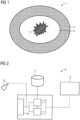
- FIG 1 shows a schematic sectional view of a tissue region 1 with a central tumor 2 and surrounding healthy tissue 3.
- tumor borders One problem with tumor resections or ectomies is finding the tumor borders.
- the tumor 2 is to be completely removed, but on the other hand not too much healthy tissue 3 is to be excised, in order not to damage a respective affected organ, such as the liver, any more than necessary.
- a safety zone 4 According to the current state of the art, not only the tumor 2 but also the tissue surrounding it is usually removed in a safety zone 4 .
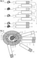
- the analysis device 5 comprises an input interface 6 for receiving training data and input data generated from tissue samples. This data is forwarded to an automatic classifier 7 of the analysis device 5, which uses a neural network to classify the training and input data and thus the respective underlying tissue samples with regard to at least one predetermined property.
- the property used is whether the respective tissue sample is tumor tissue from tumor 2 or healthy tissue 3.
- the classifier 7 or its neural network is first trained.
- This training is schematic in 3 illustrated.
- annotated training data are generated from tissue samples which originate from tissue region 1 .
- the annotated training data are based on several known negative tissue samples 12 originating from the healthy tissue 3 and several known positive tissue samples 13 originating from the tumor 2 .
- the annotated training data are made available to the classifier 7, which then supplies a series of incorrect classifications 14, for example.
- a respective parameter adjustment 15 takes place, in which parameters or weights of the classifier 7 used for the classification are modified, in particular automatically.
- the classifier 7 is finally trained to the point where it supplies, ie outputs, correct classifications 16 both for the known negative tissue samples 12 and for the known positive tissue samples 13 .
- This training or adaptation process of the classifier 7 is carried out individually for each patient to be treated, in this case for the patient from whom the Tissue area 1 originates. The classifier 7 is thus trained in a patient-specific manner.
- the classifications 14, 16 of the classifier 7 are output or displayed on a display device 8, for example.
- the classifier 7 is therefore designed as a learning system.
- the learning classifier 7 can be implemented by means of a non-linear function with a large number of parameters—first randomly initialized before the training.
- the training works because the correct classifications 16 for the annotated training data based on the known negative and positive tissue samples 12, 13 are known in advance and thus a respective output or a respective result, i.e. the classifications 14, 16, can be compared with the desired, known target result.
- the parameter adjustments 15 are implemented using a method of non-linear optimization.
- the classifier 7 can also correctly classify unknown tissue samples by processing the respective input data generated from them.
- the unknown tissue samples to be classified come from the same patient from whom the known negative and known positive tissue samples 12, 13 used for training or adapting the classifier 7 come.
- an improved classification of histological tissue samples for example obtained intra-operatively, can be achieved, which enables a more precise, ie narrower and gentler resection of the tumor 2 for the respective patient.
- the sensitivity of the classifier 7 can be increased by the patient-specific adaptation of the classifier 7 to precisely this patient and his specific tumor 2, so that unknown tissue samples from the safety zone 4 are more reliable and more accurate can be classified correctly. This is schematic in FIG 4 illustrated.
- FIG 4 the tissue area 1 with the tumor 2 and the healthy tissue 3 is again shown schematically.
- a number of unknown tissue samples from which it is initially not known whether they originate from the tumor 2 or from the healthy tissue 3 , are classified using the classifier 7 .
- These unknown tissue samples come from different removal sites 17, which are located between a center of the tumor 2 and the in FIG 1 shown area of the healthy tissue 3 are arranged.
- a new safety zone 18 can thus be defined, which is smaller, i.e. surrounds the tumor 2 more closely and thus includes less healthy tissue 3 than the larger safety zone 4 that is conventionally provided.
- the analysis device 5 in the present case also has a data processing device 9 (see FIG 2 ) on.
- the data processing device 9 is provided with an image of the tissue region 1 captured by a camera 10 and the respective removal locations 17 of the classified unknown tissue samples and the known negative and known positive tissue samples 12, 13.
- the data processing device 9 has access to a medical database 11.
- the database 11 can be part of the analysis device 5 in whole or in part.
- the database 11 can also be part or all of an external server device, for example a cloud server.
- an estimated extent 19 of the tumor 2 is determined as a function of this, ie on the basis of the data provided, and also visualized, ie represented, on the display device.
- the data processing device 9 or the analysis device 5 outputs suggested removal locations 20 for further unknown tissue samples for further, more precise localization of the tumor 2 to a user, ie for example the doctor treating the patient.
Claims (9)
- Procédé de classification d'échantillons tissulaires, dans lequel- à partir d'au moins un échantillon (13) tissulaire positif connu, qui a une propriété donnée à l'avance, et d'au moins un échantillon (12) tissulaire négatif connu, qui n'a pas la propriété donnée à l'avance, on produit des données d'apprentissage annotées, dans lequel- on met les données d'apprentissage annotées à disposition d'un classificateur (7) automatique,- on entraîne le classificateur (7) à l'aide des données d'apprentissage annotées, pour qu'il reconnaisse si un échantillon tissulaire de ce patient a la propriété donnée à l'avance,- pour la délimitation d'une partie (2) tissulaire concernée par la propriété donnée à l'avance et constituée de plusieurs échantillons tissulaires inconnus, dont il n'est pas connu s'ils ont la propriété donnée à l'avance, on produit des données d'entrée pour le classificateur (7),- on classe automatiquement, en ce qui concerne la propriété donnée à l'avance, les échantillons tissulaires inconnus, à l'aide des données d'entrée au moyen du classificateur (7) ;- on se procure, électroniquement d'un appareil (5) d'analyse, une image de la partie (2) tissulaire concernée et de ce qui l'entoure (3, 4, 18) par des coordonnées d'image respectives d'emplacements (17) de prélèvement des échantillons tissulaires classifiés, et- en se fondant sur l'appareil (5) d'analyse, on détermine automatiquement une proposition d'un emplacement (17, 20) de prélèvement d'un autre échantillon tissulaire inconnu, pour la délimitation de la partie (2) tissulaire concernée et on l'envoie à un utilisateur,
caractériséen ce que les échantillons (12, 13) tissulaires positifs et négatifs, à partir desquels on produit les données d'apprentissage annotées, proviennent du même patient,en ce que l'on fait subir au classificateur (7) un apprentissage spécifique à un patient pour le même patient, dont proviennent les échantillons (12, 13) tissulaires positifs et négatifs connus, et en ce que les échantillons tissulaires inconnus proviennent également du même patient, dont proviennent les échantillons (12, 13) tissulaires connus. - Procédé suivant la revendication 1, caractérisé en ce que le classificateur (7) a des paramètres adaptables, notamment un réseau neuronal, une fonction polynôme non linéaire et/ou un système de logique floue, qui sont, en fonction des données d'apprentissage, adaptés, notamment automatiquement, au traitement des données d'entrée.
- Procédé suivant la revendication 2, caractérisé en ce que l'on fait subir au préalable un apprentissage aux paramètres adaptables, notamment au réseau neuronal, à la fonction polynôme non linéaire et/ou le système de logique floue, à l'aide d'une pluralité de données d'entraînement annotées d'une pluralité de patients différents et on les entraîne ensuite, à l'aide des échantillons (12, 13) tissulaires positifs et négatifs spécifiques au patient pour le patient, dont proviennent les échantillons tissulaires inconnus à classifier ensuite.
- Procédé suivant la revendication 2, caractérisé en ce que l'on fait subir un apprentissage aux paramètres adaptables, notamment au réseau neuronal, à la fonction polynôme non linéaire et/ou au système de logique floue, exclusivement à l'aide de données d'apprentissage, qui ont été produites à partir de respectivement plusieurs échantillons (12, 13) tissulaires positifs connus et négatifs connus du même patient, dont proviennent également les échantillons tissulaires inconnus à classifier ensuite.
- Procédé suivant l'une des revendications précédentes, caractérisé en ce que, pour la production des données d'apprentissage et des données d'entrée, on détecte optiquement les échantillons (12, 13) tissulaires respectifs et on analyse, au moyen d'un traitement d'image, les données d'apprentissage et les données d'entrée, pour la classification par le classificateur (7) .
- Procédé suivant l'une des revendications précédentes, caractérisé en ce que, pour la production des données d'apprentissage, on traite un échantillon tissulaire d'une partie centrale d'une tumeur (2) comme l'échantillon (13) tissulaire positif connu, un échantillon tissulaire d'une partie (3) environnante de la tumeur (2) comme l'échantillon (12) tissulaire négatif connu et comme échantillon tissulaire inconnu, les échantillons tissulaires dont l'emplacement (17) de prélèvement a une distance dans l'espace à un emplacement de prélèvement de l'échantillon (13) tissulaire positif connu plus petite que celle à un emplacement de prélèvement de l'échantillon (12) tissulaire négatif connu.
- Procédé suivant l'une des revendications précédentes, caractérisé en ce que- on se procure, électroniquement d'un appareil d'analyse, une image de la partie (2) tissulaire concernée et de ce qui l'entoure (3, 4, 18), par des coordonnées d'image respectifs d'emplacements (17) de prélèvement des échantillons tissulaires classifiés,- en se fondant sur cela, on détermine au moyen de l'appareil (5) d'analyse automatiquement une étendue (19) estimée de la partie (2) tissulaire concernée, et- on visualise, dans l'image automatiquement, l'étendue (19) estimée par l'appareil (5) d'analyse.
- Procédé suivant l'une des revendications précédentes, caractérisé en ce que l'on accède, pour déterminer la proposition de l'emplacement de prélèvement au moyen de l'appareil d'analyse, à une base de données, dans laquelle sont mises en mémoire, par exemple des formes, des dimensions et des directions de croissance typiques de tumeurs.
- Procédé suivant la revendication 8, caractérisé en ce qu'il est pris en compte pour la proposition suivante, par l'appareil d'analyse, itérativement, respectivement tous les emplacements de prélèvements des échantillons tissulaires classifiés jusqu'à l'instant respectif.
Priority Applications (3)
| Application Number | Priority Date | Filing Date | Title |
|---|---|---|---|
| EP18162285.3A EP3540632B1 (fr) | 2018-03-16 | 2018-03-16 | Procédé pour la classification des échantillons tissulaires |
| US16/355,693 US11461897B2 (en) | 2018-03-16 | 2019-03-15 | Method and analysis devices for classifying tissue samples |
| CN201910202102.4A CN110276368B (zh) | 2018-03-16 | 2019-03-18 | 用于对组织样本进行分类的方法和分析设备 |
Applications Claiming Priority (1)
| Application Number | Priority Date | Filing Date | Title |
|---|---|---|---|
| EP18162285.3A EP3540632B1 (fr) | 2018-03-16 | 2018-03-16 | Procédé pour la classification des échantillons tissulaires |
Publications (2)
| Publication Number | Publication Date |
|---|---|
| EP3540632A1 EP3540632A1 (fr) | 2019-09-18 |
| EP3540632B1 true EP3540632B1 (fr) | 2023-04-26 |
Family
ID=61768054
Family Applications (1)
| Application Number | Title | Priority Date | Filing Date |
|---|---|---|---|
| EP18162285.3A Active EP3540632B1 (fr) | 2018-03-16 | 2018-03-16 | Procédé pour la classification des échantillons tissulaires |
Country Status (3)
| Country | Link |
|---|---|
| US (1) | US11461897B2 (fr) |
| EP (1) | EP3540632B1 (fr) |
| CN (1) | CN110276368B (fr) |
Families Citing this family (5)
| Publication number | Priority date | Publication date | Assignee | Title |
|---|---|---|---|---|
| CN109069007A (zh) | 2016-03-08 | 2018-12-21 | 泽博拉医疗科技公司 | 皮肤疾病的非侵入式检测 |
| CN112166474A (zh) * | 2018-05-16 | 2021-01-01 | 皇家飞利浦有限公司 | 使用机器学习的在外科手术期间的自动化肿瘤识别 |
| DE102019208355A1 (de) | 2019-06-07 | 2020-12-10 | Siemens Healthcare Gmbh | Verfahren und System zur Navigationsunterstützung einer Person zur Navigation bezüglich eines Resektats, Computerprogramm und elektronisch lesbarer Datenträger |
| WO2021097142A1 (fr) * | 2019-11-13 | 2021-05-20 | Enspectra Health, Inc. | Méthodes et systèmes permettant d'identifier des caractéristiques de tissu |
| CN114469342B (zh) * | 2022-01-17 | 2023-07-25 | 四川大学华西医院 | 肿瘤切缘边距场的定义方法、建立系统及应用 |
Family Cites Families (6)
| Publication number | Priority date | Publication date | Assignee | Title |
|---|---|---|---|---|
| WO2004025569A2 (fr) * | 2002-09-13 | 2004-03-25 | Arcturus Bioscience, Inc. | Analyse interactive et automatique d'images tissulaires au moyen d'une base de donnees de formation generale et traitement a niveaux d'abstraction variables dans des applications de classification d'echantillons cytologiques et de microdissection laser |
| ITMI20030541A1 (it) * | 2003-03-20 | 2004-09-21 | Ist Naz Stud Cura Dei Tumori | Apparecchio per la caratterizzazione di lesioni pigmentale della cute |
| WO2010067364A1 (fr) * | 2008-12-10 | 2010-06-17 | Yeda Research And Development Co. Ltd. | Procédé et système pour détecter et classer un cancer de la prostate |
| JP6192747B2 (ja) * | 2013-03-15 | 2017-09-06 | ベンタナ メディカル システムズ, インコーポレイテッド | デジタル・ホール・スライドの自動採点のための組織物体に基づく機械学習システム |
| JP6538280B2 (ja) * | 2015-12-18 | 2019-07-03 | コーニンクレッカ フィリップス エヌ ヴェKoninklijke Philips N.V. | 被験者の組織を特徴付ける装置及び方法 |
| WO2018059744A1 (fr) * | 2016-09-27 | 2018-04-05 | Siemens Aktiengesellschaft | Endoscope et procédé pour faire fonctionner un endoscope |
-
2018
- 2018-03-16 EP EP18162285.3A patent/EP3540632B1/fr active Active
-
2019
- 2019-03-15 US US16/355,693 patent/US11461897B2/en active Active
- 2019-03-18 CN CN201910202102.4A patent/CN110276368B/zh active Active
Also Published As
| Publication number | Publication date |
|---|---|
| CN110276368B (zh) | 2023-11-21 |
| US11461897B2 (en) | 2022-10-04 |
| CN110276368A (zh) | 2019-09-24 |
| EP3540632A1 (fr) | 2019-09-18 |
| US20190287246A1 (en) | 2019-09-19 |
Similar Documents
| Publication | Publication Date | Title |
|---|---|---|
| EP3540632B1 (fr) | Procédé pour la classification des échantillons tissulaires | |
| EP1625876B1 (fr) | Dispositif pour radiotherapie | |
| WO1997041416A1 (fr) | Procede d'analyse automatisee assistee par ordinateur d'echantillons tissulaires ou d'echantillons de liquides organiques | |
| DE1498824A1 (de) | Vorrichtung zur maschinellen Krebsdiagnose | |
| WO2013098074A2 (fr) | Procédé et système de vérification | |
| EP3287914A1 (fr) | Determination de donnees de resultat en fonction de donnees de mesure medicales provenant de differentes mesures | |
| WO2019091509A1 (fr) | Procédé d'évaluation de la mesure de l'image de la peau d'une personne dans le contexte de l'examen de dépistage de cancer de la peau et dispositif de mise en œuvre de ce procédé | |
| DE102015212841A1 (de) | Betrieb eines Röntgensystems zur Untersuchung eines Objektes | |
| DE102010000483A1 (de) | Automatische Ergebnisanalyse unter Verwendung radiologischer Bilder | |
| DE102005022156A1 (de) | Verfahren und Vorrichtung zum Einstufen von Pixeln in medizinischer Bildgebung | |
| DE102004003381A1 (de) | Verfahren zur Bestimmung der Lage wenigstens einer Schicht in einem Untersuchungsgebiet, in welcher Schicht eine Schichtbildaufnahme erfolgen soll | |
| EP3155588B1 (fr) | Système d'acquisition et de traitement d'une image d'un corps complet et procédé pour faire fonctionner celui-ci | |
| EP3605404B1 (fr) | Procédé et dispositif d'entraînement d'une routine d'apprentissage mécanique permettant de commander un système technique | |
| EP3797692B1 (fr) | Procédé et dispositif de commande d'un appareil d'imagerie médicale | |
| EP3488281B1 (fr) | Détermination d'un type de tissu animal ou humain | |
| DE102016217781B4 (de) | Erzeugen einer abgestimmten Darstellung unterschiedlicher Mammogramme im direkten Vergleich | |
| DE102014222804A1 (de) | Vorrichtung und Verfahren zum Bestimmen einer Wandschubspannung und System zur Erkennung von Arteriosklerose | |
| DE102018216295B4 (de) | Verfahren und Vorrichtung zum Ermitteln eines Ansteuersignals zum Ansteuern eines Aktors | |
| EP3472759A1 (fr) | Procédé et dispositif d'analyse assistée par ordinateur d'au moins un second vecteur d'entrée d'un système cible | |
| EP2693400B1 (fr) | Détection automatique d'un muscle pectoral | |
| EP4300418A1 (fr) | Système d'assistance d'un utilisateur dans la détection basée sur l'image d'une dégustation tissulaire | |
| DE102005028975B4 (de) | Verfahren zur Ermittlung eines Biomarkers zur Kennzeichnung eines spezifischen biologischen Zustands eines Organismus aus mindestens einem Datensatz | |
| DE102021134276A1 (de) | Medizinische Vorrichtung und Verfahren zur Untersuchung eines organischen Gewebes | |
| DE102007025401A1 (de) | Verfahren zur Auswertung eines Tomographie-Datensatzes und Tomographie-Arbeitsstation | |
| EP2654548B1 (fr) | Procédé et dispositif servant à examiner un organe creux au moyen d'une capsule endoscopique guidée par magnétisme |
Legal Events
| Date | Code | Title | Description |
|---|---|---|---|
| PUAI | Public reference made under article 153(3) epc to a published international application that has entered the european phase |
Free format text: ORIGINAL CODE: 0009012 |
|
| STAA | Information on the status of an ep patent application or granted ep patent |
Free format text: STATUS: THE APPLICATION HAS BEEN PUBLISHED |
|
| AK | Designated contracting states |
Kind code of ref document: A1 Designated state(s): AL AT BE BG CH CY CZ DE DK EE ES FI FR GB GR HR HU IE IS IT LI LT LU LV MC MK MT NL NO PL PT RO RS SE SI SK SM TR |
|
| AX | Request for extension of the european patent |
Extension state: BA ME |
|
| STAA | Information on the status of an ep patent application or granted ep patent |
Free format text: STATUS: REQUEST FOR EXAMINATION WAS MADE |
|
| 17P | Request for examination filed |
Effective date: 20191219 |
|
| RBV | Designated contracting states (corrected) |
Designated state(s): AL AT BE BG CH CY CZ DE DK EE ES FI FR GB GR HR HU IE IS IT LI LT LU LV MC MK MT NL NO PL PT RO RS SE SI SK SM TR |
|
| STAA | Information on the status of an ep patent application or granted ep patent |
Free format text: STATUS: EXAMINATION IS IN PROGRESS |
|
| 17Q | First examination report despatched |
Effective date: 20210512 |
|
| STAA | Information on the status of an ep patent application or granted ep patent |
Free format text: STATUS: EXAMINATION IS IN PROGRESS |
|
| REG | Reference to a national code |
Ref country code: DE Ref legal event code: R079 Ref document number: 502018012015 Country of ref document: DE Free format text: PREVIOUS MAIN CLASS: G06K0009000000 Ipc: G06V0010100000 |
|
| GRAP | Despatch of communication of intention to grant a patent |
Free format text: ORIGINAL CODE: EPIDOSNIGR1 |
|
| STAA | Information on the status of an ep patent application or granted ep patent |
Free format text: STATUS: GRANT OF PATENT IS INTENDED |
|
| RIC1 | Information provided on ipc code assigned before grant |
Ipc: G06T 7/00 20170101ALI20221110BHEP Ipc: G06V 20/69 20220101ALI20221110BHEP Ipc: G06V 10/82 20220101ALI20221110BHEP Ipc: G06V 10/776 20220101ALI20221110BHEP Ipc: G06V 10/764 20220101ALI20221110BHEP Ipc: G06V 10/44 20220101ALI20221110BHEP Ipc: G06V 10/10 20220101AFI20221110BHEP |
|
| INTG | Intention to grant announced |
Effective date: 20221123 |
|
| GRAS | Grant fee paid |
Free format text: ORIGINAL CODE: EPIDOSNIGR3 |
|
| GRAA | (expected) grant |
Free format text: ORIGINAL CODE: 0009210 |
|
| STAA | Information on the status of an ep patent application or granted ep patent |
Free format text: STATUS: THE PATENT HAS BEEN GRANTED |
|
| AK | Designated contracting states |
Kind code of ref document: B1 Designated state(s): AL AT BE BG CH CY CZ DE DK EE ES FI FR GB GR HR HU IE IS IT LI LT LU LV MC MK MT NL NO PL PT RO RS SE SI SK SM TR |
|
| REG | Reference to a national code |
Ref country code: GB Ref legal event code: FG4D Free format text: NOT ENGLISH |
|
| REG | Reference to a national code |
Ref country code: CH Ref legal event code: EP |
|
| REG | Reference to a national code |
Ref country code: DE Ref legal event code: R096 Ref document number: 502018012015 Country of ref document: DE |
|
| REG | Reference to a national code |
Ref country code: AT Ref legal event code: REF Ref document number: 1563402 Country of ref document: AT Kind code of ref document: T Effective date: 20230515 |
|
| REG | Reference to a national code |
Ref country code: IE Ref legal event code: FG4D Free format text: LANGUAGE OF EP DOCUMENT: GERMAN |
|
| REG | Reference to a national code |
Ref country code: LT Ref legal event code: MG9D |
|
| REG | Reference to a national code |
Ref country code: NL Ref legal event code: MP Effective date: 20230426 |
|
| PG25 | Lapsed in a contracting state [announced via postgrant information from national office to epo] |
Ref country code: NL Free format text: LAPSE BECAUSE OF FAILURE TO SUBMIT A TRANSLATION OF THE DESCRIPTION OR TO PAY THE FEE WITHIN THE PRESCRIBED TIME-LIMIT Effective date: 20230426 |
|
| PG25 | Lapsed in a contracting state [announced via postgrant information from national office to epo] |
Ref country code: SE Free format text: LAPSE BECAUSE OF FAILURE TO SUBMIT A TRANSLATION OF THE DESCRIPTION OR TO PAY THE FEE WITHIN THE PRESCRIBED TIME-LIMIT Effective date: 20230426 Ref country code: PT Free format text: LAPSE BECAUSE OF FAILURE TO SUBMIT A TRANSLATION OF THE DESCRIPTION OR TO PAY THE FEE WITHIN THE PRESCRIBED TIME-LIMIT Effective date: 20230828 Ref country code: NO Free format text: LAPSE BECAUSE OF FAILURE TO SUBMIT A TRANSLATION OF THE DESCRIPTION OR TO PAY THE FEE WITHIN THE PRESCRIBED TIME-LIMIT Effective date: 20230726 Ref country code: ES Free format text: LAPSE BECAUSE OF FAILURE TO SUBMIT A TRANSLATION OF THE DESCRIPTION OR TO PAY THE FEE WITHIN THE PRESCRIBED TIME-LIMIT Effective date: 20230426 |
|
| PG25 | Lapsed in a contracting state [announced via postgrant information from national office to epo] |
Ref country code: RS Free format text: LAPSE BECAUSE OF FAILURE TO SUBMIT A TRANSLATION OF THE DESCRIPTION OR TO PAY THE FEE WITHIN THE PRESCRIBED TIME-LIMIT Effective date: 20230426 Ref country code: PL Free format text: LAPSE BECAUSE OF FAILURE TO SUBMIT A TRANSLATION OF THE DESCRIPTION OR TO PAY THE FEE WITHIN THE PRESCRIBED TIME-LIMIT Effective date: 20230426 Ref country code: LV Free format text: LAPSE BECAUSE OF FAILURE TO SUBMIT A TRANSLATION OF THE DESCRIPTION OR TO PAY THE FEE WITHIN THE PRESCRIBED TIME-LIMIT Effective date: 20230426 Ref country code: LT Free format text: LAPSE BECAUSE OF FAILURE TO SUBMIT A TRANSLATION OF THE DESCRIPTION OR TO PAY THE FEE WITHIN THE PRESCRIBED TIME-LIMIT Effective date: 20230426 Ref country code: IS Free format text: LAPSE BECAUSE OF FAILURE TO SUBMIT A TRANSLATION OF THE DESCRIPTION OR TO PAY THE FEE WITHIN THE PRESCRIBED TIME-LIMIT Effective date: 20230826 Ref country code: HR Free format text: LAPSE BECAUSE OF FAILURE TO SUBMIT A TRANSLATION OF THE DESCRIPTION OR TO PAY THE FEE WITHIN THE PRESCRIBED TIME-LIMIT Effective date: 20230426 Ref country code: GR Free format text: LAPSE BECAUSE OF FAILURE TO SUBMIT A TRANSLATION OF THE DESCRIPTION OR TO PAY THE FEE WITHIN THE PRESCRIBED TIME-LIMIT Effective date: 20230727 |
|
| PG25 | Lapsed in a contracting state [announced via postgrant information from national office to epo] |
Ref country code: FI Free format text: LAPSE BECAUSE OF FAILURE TO SUBMIT A TRANSLATION OF THE DESCRIPTION OR TO PAY THE FEE WITHIN THE PRESCRIBED TIME-LIMIT Effective date: 20230426 |
|
| PG25 | Lapsed in a contracting state [announced via postgrant information from national office to epo] |
Ref country code: SK Free format text: LAPSE BECAUSE OF FAILURE TO SUBMIT A TRANSLATION OF THE DESCRIPTION OR TO PAY THE FEE WITHIN THE PRESCRIBED TIME-LIMIT Effective date: 20230426 |
|
| REG | Reference to a national code |
Ref country code: DE Ref legal event code: R097 Ref document number: 502018012015 Country of ref document: DE |
|
| PG25 | Lapsed in a contracting state [announced via postgrant information from national office to epo] |
Ref country code: SM Free format text: LAPSE BECAUSE OF FAILURE TO SUBMIT A TRANSLATION OF THE DESCRIPTION OR TO PAY THE FEE WITHIN THE PRESCRIBED TIME-LIMIT Effective date: 20230426 Ref country code: SK Free format text: LAPSE BECAUSE OF FAILURE TO SUBMIT A TRANSLATION OF THE DESCRIPTION OR TO PAY THE FEE WITHIN THE PRESCRIBED TIME-LIMIT Effective date: 20230426 Ref country code: RO Free format text: LAPSE BECAUSE OF FAILURE TO SUBMIT A TRANSLATION OF THE DESCRIPTION OR TO PAY THE FEE WITHIN THE PRESCRIBED TIME-LIMIT Effective date: 20230426 Ref country code: EE Free format text: LAPSE BECAUSE OF FAILURE TO SUBMIT A TRANSLATION OF THE DESCRIPTION OR TO PAY THE FEE WITHIN THE PRESCRIBED TIME-LIMIT Effective date: 20230426 Ref country code: DK Free format text: LAPSE BECAUSE OF FAILURE TO SUBMIT A TRANSLATION OF THE DESCRIPTION OR TO PAY THE FEE WITHIN THE PRESCRIBED TIME-LIMIT Effective date: 20230426 Ref country code: CZ Free format text: LAPSE BECAUSE OF FAILURE TO SUBMIT A TRANSLATION OF THE DESCRIPTION OR TO PAY THE FEE WITHIN THE PRESCRIBED TIME-LIMIT Effective date: 20230426 |
|
| RAP2 | Party data changed (patent owner data changed or rights of a patent transferred) |
Owner name: SIEMENS HEALTHINEERS AG |
|
| REG | Reference to a national code |
Ref country code: DE Ref legal event code: R081 Ref document number: 502018012015 Country of ref document: DE Owner name: SIEMENS HEALTHINEERS AG, DE Free format text: FORMER OWNER: SIEMENS HEALTHCARE GMBH, MUENCHEN, DE |
|
| PLBE | No opposition filed within time limit |
Free format text: ORIGINAL CODE: 0009261 |
|
| STAA | Information on the status of an ep patent application or granted ep patent |
Free format text: STATUS: NO OPPOSITION FILED WITHIN TIME LIMIT |
|
| 26N | No opposition filed |
Effective date: 20240129 |
|
| PG25 | Lapsed in a contracting state [announced via postgrant information from national office to epo] |
Ref country code: SI Free format text: LAPSE BECAUSE OF FAILURE TO SUBMIT A TRANSLATION OF THE DESCRIPTION OR TO PAY THE FEE WITHIN THE PRESCRIBED TIME-LIMIT Effective date: 20230426 |