EP3521315B1 - Anticorps anti-transglutaminase 2 - Google Patents
Anticorps anti-transglutaminase 2 Download PDFInfo
- Publication number
- EP3521315B1 EP3521315B1 EP19163818.8A EP19163818A EP3521315B1 EP 3521315 B1 EP3521315 B1 EP 3521315B1 EP 19163818 A EP19163818 A EP 19163818A EP 3521315 B1 EP3521315 B1 EP 3521315B1
- Authority
- EP
- European Patent Office
- Prior art keywords
- antibody
- antigen
- binding fragment
- human
- antibodies
- Prior art date
- Legal status (The legal status is an assumption and is not a legal conclusion. Google has not performed a legal analysis and makes no representation as to the accuracy of the status listed.)
- Active
Links
- 230000027455 binding Effects 0.000 claims description 297
- 239000012634 fragment Substances 0.000 claims description 246
- 239000000427 antigen Substances 0.000 claims description 213
- 108091007433 antigens Proteins 0.000 claims description 213
- 102000036639 antigens Human genes 0.000 claims description 213
- 108090000765 processed proteins & peptides Proteins 0.000 claims description 118
- 230000000694 effects Effects 0.000 claims description 117
- 150000001413 amino acids Chemical class 0.000 claims description 108
- 241000282414 Homo sapiens Species 0.000 claims description 100
- 102000004196 processed proteins & peptides Human genes 0.000 claims description 97
- 101000666171 Homo sapiens Protein-glutamine gamma-glutamyltransferase 2 Proteins 0.000 claims description 96
- 238000000034 method Methods 0.000 claims description 93
- 150000001875 compounds Chemical class 0.000 claims description 80
- 229920001184 polypeptide Polymers 0.000 claims description 75
- 108060008539 Transglutaminase Proteins 0.000 claims description 63
- 102000003601 transglutaminase Human genes 0.000 claims description 63
- 239000008194 pharmaceutical composition Substances 0.000 claims description 57
- 230000005764 inhibitory process Effects 0.000 claims description 55
- 108091033319 polynucleotide Proteins 0.000 claims description 45
- 239000002157 polynucleotide Substances 0.000 claims description 45
- 102000040430 polynucleotide Human genes 0.000 claims description 45
- 239000003814 drug Substances 0.000 claims description 34
- 208000037265 diseases, disorders, signs and symptoms Diseases 0.000 claims description 33
- 231100000433 cytotoxic Toxicity 0.000 claims description 32
- 230000001472 cytotoxic effect Effects 0.000 claims description 32
- 206010028980 Neoplasm Diseases 0.000 claims description 31
- 201000010099 disease Diseases 0.000 claims description 30
- 238000009472 formulation Methods 0.000 claims description 30
- 239000003795 chemical substances by application Substances 0.000 claims description 27
- 201000011510 cancer Diseases 0.000 claims description 23
- 230000037390 scarring Effects 0.000 claims description 22
- 208000015943 Coeliac disease Diseases 0.000 claims description 20
- 239000000203 mixture Substances 0.000 claims description 20
- 208000005017 glioblastoma Diseases 0.000 claims description 16
- 102000001706 Immunoglobulin Fab Fragments Human genes 0.000 claims description 14
- 108010054477 Immunoglobulin Fab Fragments Proteins 0.000 claims description 14
- 206010016654 Fibrosis Diseases 0.000 claims description 12
- KDXKERNSBIXSRK-UHFFFAOYSA-N Lysine Natural products NCCCCC(N)C(O)=O KDXKERNSBIXSRK-UHFFFAOYSA-N 0.000 claims description 12
- 239000002671 adjuvant Substances 0.000 claims description 12
- 230000004761 fibrosis Effects 0.000 claims description 12
- 238000011282 treatment Methods 0.000 claims description 12
- 208000023105 Huntington disease Diseases 0.000 claims description 11
- 108010067060 Immunoglobulin Variable Region Proteins 0.000 claims description 11
- 102000017727 Immunoglobulin Variable Region Human genes 0.000 claims description 11
- 239000004472 Lysine Substances 0.000 claims description 11
- 238000004132 cross linking Methods 0.000 claims description 11
- 208000023275 Autoimmune disease Diseases 0.000 claims description 10
- 206010023421 Kidney fibrosis Diseases 0.000 claims description 9
- 208000015122 neurodegenerative disease Diseases 0.000 claims description 9
- 230000002829 reductive effect Effects 0.000 claims description 9
- 208000018737 Parkinson disease Diseases 0.000 claims description 8
- 208000005069 pulmonary fibrosis Diseases 0.000 claims description 8
- 230000002159 abnormal effect Effects 0.000 claims description 7
- 239000004480 active ingredient Substances 0.000 claims description 7
- 230000003176 fibrotic effect Effects 0.000 claims description 7
- 201000001441 melanoma Diseases 0.000 claims description 7
- 108010040003 polyglutamine Proteins 0.000 claims description 7
- 229920000155 polyglutamine Polymers 0.000 claims description 7
- 230000029663 wound healing Effects 0.000 claims description 7
- 208000024827 Alzheimer disease Diseases 0.000 claims description 6
- 201000001320 Atherosclerosis Diseases 0.000 claims description 6
- 206010003694 Atrophy Diseases 0.000 claims description 6
- 230000037444 atrophy Effects 0.000 claims description 6
- 208000019425 cirrhosis of liver Diseases 0.000 claims description 6
- 238000003745 diagnosis Methods 0.000 claims description 6
- 206010061989 glomerulosclerosis Diseases 0.000 claims description 6
- ZDXPYRJPNDTMRX-UHFFFAOYSA-N glutamine Natural products OC(=O)C(N)CCC(N)=O ZDXPYRJPNDTMRX-UHFFFAOYSA-N 0.000 claims description 6
- 208000027866 inflammatory disease Diseases 0.000 claims description 6
- 201000008129 pancreatic ductal adenocarcinoma Diseases 0.000 claims description 6
- 231100000241 scar Toxicity 0.000 claims description 6
- 208000037999 tubulointerstitial fibrosis Diseases 0.000 claims description 6
- 208000031261 Acute myeloid leukaemia Diseases 0.000 claims description 5
- 208000032544 Cicatrix Diseases 0.000 claims description 5
- 201000008163 Dentatorubral pallidoluysian atrophy Diseases 0.000 claims description 5
- 208000022559 Inflammatory bowel disease Diseases 0.000 claims description 5
- 208000029523 Interstitial Lung disease Diseases 0.000 claims description 5
- 208000007766 Kaposi sarcoma Diseases 0.000 claims description 5
- 208000027747 Kennedy disease Diseases 0.000 claims description 5
- 206010024229 Leprosy Diseases 0.000 claims description 5
- 201000011062 Li-Fraumeni syndrome Diseases 0.000 claims description 5
- 208000019693 Lung disease Diseases 0.000 claims description 5
- 201000003793 Myelodysplastic syndrome Diseases 0.000 claims description 5
- 208000012902 Nervous system disease Diseases 0.000 claims description 5
- 208000025966 Neurological disease Diseases 0.000 claims description 5
- 206010033799 Paralysis Diseases 0.000 claims description 5
- 208000033063 Progressive myoclonic epilepsy Diseases 0.000 claims description 5
- 201000004681 Psoriasis Diseases 0.000 claims description 5
- 208000009415 Spinocerebellar Ataxias Diseases 0.000 claims description 5
- 208000006269 X-Linked Bulbo-Spinal Atrophy Diseases 0.000 claims description 5
- 230000009787 cardiac fibrosis Effects 0.000 claims description 5
- 208000008609 collagenous colitis Diseases 0.000 claims description 5
- 239000003085 diluting agent Substances 0.000 claims description 5
- 230000001969 hypertrophic effect Effects 0.000 claims description 5
- 208000002780 macular degeneration Diseases 0.000 claims description 5
- 206010028537 myelofibrosis Diseases 0.000 claims description 5
- 208000025113 myeloid leukemia Diseases 0.000 claims description 5
- 230000002071 myeloproliferative effect Effects 0.000 claims description 5
- 230000000626 neurodegenerative effect Effects 0.000 claims description 5
- 208000021090 palsy Diseases 0.000 claims description 5
- 208000037803 restenosis Diseases 0.000 claims description 5
- 230000037387 scars Effects 0.000 claims description 5
- 208000011580 syndromic disease Diseases 0.000 claims description 5
- 239000000546 pharmaceutical excipient Substances 0.000 claims description 4
- 238000007920 subcutaneous administration Methods 0.000 claims description 4
- LNLLNTMHVMIMOG-YUMQZZPRSA-N Gamma-glutamyl-Lysine Chemical compound NCCCC[C@@H](C(O)=O)NC(=O)CC[C@H](N)C(O)=O LNLLNTMHVMIMOG-YUMQZZPRSA-N 0.000 claims description 3
- 238000007918 intramuscular administration Methods 0.000 claims description 3
- 238000001990 intravenous administration Methods 0.000 claims description 3
- 108020004414 DNA Proteins 0.000 description 188
- 210000004027 cell Anatomy 0.000 description 121
- 229940024606 amino acid Drugs 0.000 description 107
- 235000001014 amino acid Nutrition 0.000 description 106
- 108090000623 proteins and genes Proteins 0.000 description 89
- 125000003275 alpha amino acid group Chemical group 0.000 description 67
- 102000004169 proteins and genes Human genes 0.000 description 64
- 230000002401 inhibitory effect Effects 0.000 description 62
- 235000018102 proteins Nutrition 0.000 description 62
- 238000002965 ELISA Methods 0.000 description 39
- 239000003112 inhibitor Substances 0.000 description 39
- 241000700159 Rattus Species 0.000 description 37
- 241000699666 Mus <mouse, genus> Species 0.000 description 36
- 210000004408 hybridoma Anatomy 0.000 description 35
- KIDHWZJUCRJVML-UHFFFAOYSA-N putrescine Chemical compound NCCCCN KIDHWZJUCRJVML-UHFFFAOYSA-N 0.000 description 34
- 238000003556 assay Methods 0.000 description 32
- 230000003197 catalytic effect Effects 0.000 description 32
- 102100024952 Protein CBFA2T1 Human genes 0.000 description 30
- 102100038095 Protein-glutamine gamma-glutamyltransferase 2 Human genes 0.000 description 30
- 206010052428 Wound Diseases 0.000 description 30
- 208000027418 Wounds and injury Diseases 0.000 description 30
- 102000004190 Enzymes Human genes 0.000 description 29
- 108090000790 Enzymes Proteins 0.000 description 29
- 229940088598 enzyme Drugs 0.000 description 29
- 108700039882 Protein Glutamine gamma Glutamyltransferase 2 Proteins 0.000 description 28
- 230000014509 gene expression Effects 0.000 description 27
- 238000010348 incorporation Methods 0.000 description 27
- 239000013598 vector Substances 0.000 description 27
- 239000002953 phosphate buffered saline Substances 0.000 description 23
- 238000006243 chemical reaction Methods 0.000 description 22
- 239000013604 expression vector Substances 0.000 description 22
- 230000004927 fusion Effects 0.000 description 21
- 102000010834 Extracellular Matrix Proteins Human genes 0.000 description 20
- 108010037362 Extracellular Matrix Proteins Proteins 0.000 description 20
- 239000005700 Putrescine Substances 0.000 description 20
- FAPWRFPIFSIZLT-UHFFFAOYSA-M Sodium chloride Chemical compound [Na+].[Cl-] FAPWRFPIFSIZLT-UHFFFAOYSA-M 0.000 description 20
- 210000001519 tissue Anatomy 0.000 description 20
- 241000699670 Mus sp. Species 0.000 description 19
- 229940002612 prodrug Drugs 0.000 description 19
- 239000000651 prodrug Substances 0.000 description 19
- 108091032973 (ribonucleotides)n+m Proteins 0.000 description 18
- 241001529936 Murinae Species 0.000 description 18
- 239000002585 base Substances 0.000 description 18
- SDZRWUKZFQQKKV-JHADDHBZSA-N cytochalasin D Chemical compound C([C@H]1[C@@H]2[C@@H](C([C@@H](O)[C@H]\3[C@]2([C@@H](/C=C/[C@@](C)(O)C(=O)[C@@H](C)C/C=C/3)OC(C)=O)C(=O)N1)=C)C)C1=CC=CC=C1 SDZRWUKZFQQKKV-JHADDHBZSA-N 0.000 description 18
- 210000002744 extracellular matrix Anatomy 0.000 description 17
- 230000002441 reversible effect Effects 0.000 description 17
- 238000012360 testing method Methods 0.000 description 17
- 108010047041 Complementarity Determining Regions Proteins 0.000 description 16
- 108010076504 Protein Sorting Signals Proteins 0.000 description 16
- 230000015572 biosynthetic process Effects 0.000 description 16
- 239000000872 buffer Substances 0.000 description 16
- 238000002474 experimental method Methods 0.000 description 16
- 239000000758 substrate Substances 0.000 description 16
- 230000002255 enzymatic effect Effects 0.000 description 15
- -1 form amine hydrochlorides Chemical class 0.000 description 15
- 230000009257 reactivity Effects 0.000 description 15
- 230000001225 therapeutic effect Effects 0.000 description 15
- NFGXHKASABOEEW-UHFFFAOYSA-N 1-methylethyl 11-methoxy-3,7,11-trimethyl-2,4-dodecadienoate Chemical compound COC(C)(C)CCCC(C)CC=CC(C)=CC(=O)OC(C)C NFGXHKASABOEEW-UHFFFAOYSA-N 0.000 description 14
- 150000003839 salts Chemical class 0.000 description 14
- 241000282693 Cercopithecidae Species 0.000 description 13
- KCXVZYZYPLLWCC-UHFFFAOYSA-N EDTA Chemical compound OC(=O)CN(CC(O)=O)CCN(CC(O)=O)CC(O)=O KCXVZYZYPLLWCC-UHFFFAOYSA-N 0.000 description 13
- 241000588724 Escherichia coli Species 0.000 description 13
- 108010068370 Glutens Proteins 0.000 description 13
- 229940099500 cystamine Drugs 0.000 description 13
- OOTFVKOQINZBBF-UHFFFAOYSA-N cystamine Chemical compound CCSSCCN OOTFVKOQINZBBF-UHFFFAOYSA-N 0.000 description 13
- 235000021312 gluten Nutrition 0.000 description 13
- 238000004519 manufacturing process Methods 0.000 description 13
- 239000000047 product Substances 0.000 description 13
- 239000006228 supernatant Substances 0.000 description 13
- 101710132601 Capsid protein Proteins 0.000 description 12
- 238000007792 addition Methods 0.000 description 12
- 239000002299 complementary DNA Substances 0.000 description 12
- 231100000673 dose–response relationship Toxicity 0.000 description 12
- RAXXELZNTBOGNW-UHFFFAOYSA-N imidazole Natural products C1=CNC=N1 RAXXELZNTBOGNW-UHFFFAOYSA-N 0.000 description 12
- AOJJSUZBOXZQNB-TZSSRYMLSA-N Doxorubicin Chemical compound O([C@H]1C[C@@](O)(CC=2C(O)=C3C(=O)C=4C=CC=C(C=4C(=O)C3=C(O)C=21)OC)C(=O)CO)[C@H]1C[C@H](N)[C@H](O)[C@H](C)O1 AOJJSUZBOXZQNB-TZSSRYMLSA-N 0.000 description 11
- 102000016359 Fibronectins Human genes 0.000 description 11
- 108010067306 Fibronectins Proteins 0.000 description 11
- 238000010367 cloning Methods 0.000 description 11
- 230000006870 function Effects 0.000 description 11
- 238000001727 in vivo Methods 0.000 description 11
- 238000002347 injection Methods 0.000 description 11
- 239000007924 injection Substances 0.000 description 11
- 239000000523 sample Substances 0.000 description 11
- 210000002966 serum Anatomy 0.000 description 11
- 102000008394 Immunoglobulin Fragments Human genes 0.000 description 10
- 108010021625 Immunoglobulin Fragments Proteins 0.000 description 10
- 239000007983 Tris buffer Substances 0.000 description 10
- 230000004913 activation Effects 0.000 description 10
- 238000004113 cell culture Methods 0.000 description 10
- 229940127089 cytotoxic agent Drugs 0.000 description 10
- 238000003780 insertion Methods 0.000 description 10
- 230000037431 insertion Effects 0.000 description 10
- 230000002285 radioactive effect Effects 0.000 description 10
- 239000011780 sodium chloride Substances 0.000 description 10
- LENZDBCJOHFCAS-UHFFFAOYSA-N tris Chemical compound OCC(N)(CO)CO LENZDBCJOHFCAS-UHFFFAOYSA-N 0.000 description 10
- CCSGGWGTGOLEHK-OBJOEFQTSA-N 5-[(3as,4s,6ar)-2-oxo-1,3,3a,4,6,6a-hexahydrothieno[3,4-d]imidazol-4-yl]-n-(5-aminopentyl)pentanamide Chemical compound N1C(=O)N[C@@H]2[C@H](CCCCC(=O)NCCCCCN)SC[C@@H]21 CCSGGWGTGOLEHK-OBJOEFQTSA-N 0.000 description 9
- 206010069754 Acquired gene mutation Diseases 0.000 description 9
- 241000894006 Bacteria Species 0.000 description 9
- WQZGKKKJIJFFOK-GASJEMHNSA-N Glucose Natural products OC[C@H]1OC(O)[C@H](O)[C@@H](O)[C@@H]1O WQZGKKKJIJFFOK-GASJEMHNSA-N 0.000 description 9
- DHMQDGOQFOQNFH-UHFFFAOYSA-N Glycine Chemical compound NCC(O)=O DHMQDGOQFOQNFH-UHFFFAOYSA-N 0.000 description 9
- ZDXPYRJPNDTMRX-VKHMYHEASA-N L-glutamine Chemical compound OC(=O)[C@@H](N)CCC(N)=O ZDXPYRJPNDTMRX-VKHMYHEASA-N 0.000 description 9
- 101000666170 Mus musculus Protein-glutamine gamma-glutamyltransferase 2 Proteins 0.000 description 9
- 240000004808 Saccharomyces cerevisiae Species 0.000 description 9
- 235000014680 Saccharomyces cerevisiae Nutrition 0.000 description 9
- 239000002253 acid Substances 0.000 description 9
- 238000013459 approach Methods 0.000 description 9
- 125000004429 atom Chemical group 0.000 description 9
- 125000004122 cyclic group Chemical group 0.000 description 9
- 238000012217 deletion Methods 0.000 description 9
- 230000037430 deletion Effects 0.000 description 9
- 239000000499 gel Substances 0.000 description 9
- 210000004602 germ cell Anatomy 0.000 description 9
- 239000008103 glucose Substances 0.000 description 9
- 230000001965 increasing effect Effects 0.000 description 9
- 239000002773 nucleotide Substances 0.000 description 9
- 125000003729 nucleotide group Chemical group 0.000 description 9
- 239000013612 plasmid Substances 0.000 description 9
- 230000037439 somatic mutation Effects 0.000 description 9
- 238000002560 therapeutic procedure Methods 0.000 description 9
- MTCFGRXMJLQNBG-REOHCLBHSA-N (2S)-2-Amino-3-hydroxypropansäure Chemical compound OC[C@H](N)C(O)=O MTCFGRXMJLQNBG-REOHCLBHSA-N 0.000 description 8
- 108091003079 Bovine Serum Albumin Proteins 0.000 description 8
- 102100035360 Cerebellar degeneration-related antigen 1 Human genes 0.000 description 8
- TWRXJAOTZQYOKJ-UHFFFAOYSA-L Magnesium chloride Chemical compound [Mg+2].[Cl-].[Cl-] TWRXJAOTZQYOKJ-UHFFFAOYSA-L 0.000 description 8
- 108091028043 Nucleic acid sequence Proteins 0.000 description 8
- 210000001744 T-lymphocyte Anatomy 0.000 description 8
- 230000008901 benefit Effects 0.000 description 8
- 238000001574 biopsy Methods 0.000 description 8
- 229940098773 bovine serum albumin Drugs 0.000 description 8
- 239000002254 cytotoxic agent Substances 0.000 description 8
- 238000010494 dissociation reaction Methods 0.000 description 8
- 230000005593 dissociations Effects 0.000 description 8
- 229940079593 drug Drugs 0.000 description 8
- 238000005516 engineering process Methods 0.000 description 8
- 238000002649 immunization Methods 0.000 description 8
- 150000007523 nucleic acids Chemical group 0.000 description 8
- 239000000126 substance Substances 0.000 description 8
- 238000003786 synthesis reaction Methods 0.000 description 8
- 230000008685 targeting Effects 0.000 description 8
- 235000003351 Brassica cretica Nutrition 0.000 description 7
- 235000003343 Brassica rupestris Nutrition 0.000 description 7
- 102000007056 Recombinant Fusion Proteins Human genes 0.000 description 7
- 108010008281 Recombinant Fusion Proteins Proteins 0.000 description 7
- 125000000539 amino acid group Chemical group 0.000 description 7
- 238000004458 analytical method Methods 0.000 description 7
- 239000003636 conditioned culture medium Substances 0.000 description 7
- 230000029087 digestion Effects 0.000 description 7
- 238000000338 in vitro Methods 0.000 description 7
- 235000010460 mustard Nutrition 0.000 description 7
- 231100000252 nontoxic Toxicity 0.000 description 7
- 230000003000 nontoxic effect Effects 0.000 description 7
- 102000039446 nucleic acids Human genes 0.000 description 7
- 108020004707 nucleic acids Proteins 0.000 description 7
- 238000002823 phage display Methods 0.000 description 7
- 238000012163 sequencing technique Methods 0.000 description 7
- 239000000243 solution Substances 0.000 description 7
- 238000001262 western blot Methods 0.000 description 7
- PEDCQBHIVMGVHV-UHFFFAOYSA-N Glycerine Chemical compound OCC(O)CO PEDCQBHIVMGVHV-UHFFFAOYSA-N 0.000 description 6
- 108060003951 Immunoglobulin Proteins 0.000 description 6
- 241000282567 Macaca fascicularis Species 0.000 description 6
- 241000699660 Mus musculus Species 0.000 description 6
- 102000035195 Peptidases Human genes 0.000 description 6
- 108091005804 Peptidases Proteins 0.000 description 6
- 230000001580 bacterial effect Effects 0.000 description 6
- 210000004556 brain Anatomy 0.000 description 6
- 230000021164 cell adhesion Effects 0.000 description 6
- 235000018417 cysteine Nutrition 0.000 description 6
- 230000001419 dependent effect Effects 0.000 description 6
- 238000011161 development Methods 0.000 description 6
- 230000018109 developmental process Effects 0.000 description 6
- 238000010790 dilution Methods 0.000 description 6
- 239000012895 dilution Substances 0.000 description 6
- 235000004554 glutamine Nutrition 0.000 description 6
- 102000018358 immunoglobulin Human genes 0.000 description 6
- APFVFJFRJDLVQX-AHCXROLUSA-N indium-111 Chemical compound [111In] APFVFJFRJDLVQX-AHCXROLUSA-N 0.000 description 6
- 230000000968 intestinal effect Effects 0.000 description 6
- 239000000816 peptidomimetic Substances 0.000 description 6
- 230000003389 potentiating effect Effects 0.000 description 6
- 239000007787 solid Substances 0.000 description 6
- 241000894007 species Species 0.000 description 6
- 230000006641 stabilisation Effects 0.000 description 6
- 238000006467 substitution reaction Methods 0.000 description 6
- XLYOFNOQVPJJNP-UHFFFAOYSA-N water Substances O XLYOFNOQVPJJNP-UHFFFAOYSA-N 0.000 description 6
- 241000219193 Brassicaceae Species 0.000 description 5
- OYPRJOBELJOOCE-UHFFFAOYSA-N Calcium Chemical compound [Ca] OYPRJOBELJOOCE-UHFFFAOYSA-N 0.000 description 5
- UXVMQQNJUSDDNG-UHFFFAOYSA-L Calcium chloride Chemical compound [Cl-].[Cl-].[Ca+2] UXVMQQNJUSDDNG-UHFFFAOYSA-L 0.000 description 5
- ZCYVEMRRCGMTRW-AHCXROLUSA-N Iodine-123 Chemical compound [123I] ZCYVEMRRCGMTRW-AHCXROLUSA-N 0.000 description 5
- 241001465754 Metazoa Species 0.000 description 5
- 229920001213 Polysorbate 20 Polymers 0.000 description 5
- ONIBWKKTOPOVIA-UHFFFAOYSA-N Proline Natural products OC(=O)C1CCCN1 ONIBWKKTOPOVIA-UHFFFAOYSA-N 0.000 description 5
- 150000001412 amines Chemical class 0.000 description 5
- AVKUERGKIZMTKX-NJBDSQKTSA-N ampicillin Chemical compound C1([C@@H](N)C(=O)N[C@H]2[C@H]3SC([C@@H](N3C2=O)C(O)=O)(C)C)=CC=CC=C1 AVKUERGKIZMTKX-NJBDSQKTSA-N 0.000 description 5
- 229960000723 ampicillin Drugs 0.000 description 5
- 230000003321 amplification Effects 0.000 description 5
- 239000002246 antineoplastic agent Substances 0.000 description 5
- 239000011575 calcium Substances 0.000 description 5
- 229910052791 calcium Inorganic materials 0.000 description 5
- 239000013592 cell lysate Substances 0.000 description 5
- XUJNEKJLAYXESH-UHFFFAOYSA-N cysteine Natural products SCC(N)C(O)=O XUJNEKJLAYXESH-UHFFFAOYSA-N 0.000 description 5
- 230000007423 decrease Effects 0.000 description 5
- 229960004679 doxorubicin Drugs 0.000 description 5
- 230000012010 growth Effects 0.000 description 5
- 230000001900 immune effect Effects 0.000 description 5
- 230000002163 immunogen Effects 0.000 description 5
- 210000003000 inclusion body Anatomy 0.000 description 5
- 230000003834 intracellular effect Effects 0.000 description 5
- 238000002955 isolation Methods 0.000 description 5
- 210000003734 kidney Anatomy 0.000 description 5
- 238000011813 knockout mouse model Methods 0.000 description 5
- 239000006166 lysate Substances 0.000 description 5
- 238000013507 mapping Methods 0.000 description 5
- 239000003550 marker Substances 0.000 description 5
- 230000001404 mediated effect Effects 0.000 description 5
- 230000004770 neurodegeneration Effects 0.000 description 5
- 238000003199 nucleic acid amplification method Methods 0.000 description 5
- 239000000256 polyoxyethylene sorbitan monolaurate Substances 0.000 description 5
- 235000010486 polyoxyethylene sorbitan monolaurate Nutrition 0.000 description 5
- 238000000746 purification Methods 0.000 description 5
- 238000012216 screening Methods 0.000 description 5
- 239000004017 serum-free culture medium Substances 0.000 description 5
- 150000003384 small molecules Chemical class 0.000 description 5
- 238000007056 transamidation reaction Methods 0.000 description 5
- 230000014616 translation Effects 0.000 description 5
- NFZZAUJDSJWLTQ-UHFFFAOYSA-M 1,3-dimethyl-2-[(2-oxopropyl)thio]imidazolium chloride Chemical group [Cl-].CC(=O)CSC=1N(C)C=C[N+]=1C NFZZAUJDSJWLTQ-UHFFFAOYSA-M 0.000 description 4
- PNDPGZBMCMUPRI-HVTJNCQCSA-N 10043-66-0 Chemical compound [131I][131I] PNDPGZBMCMUPRI-HVTJNCQCSA-N 0.000 description 4
- UAIUNKRWKOVEES-UHFFFAOYSA-N 3,3',5,5'-tetramethylbenzidine Chemical compound CC1=C(N)C(C)=CC(C=2C=C(C)C(N)=C(C)C=2)=C1 UAIUNKRWKOVEES-UHFFFAOYSA-N 0.000 description 4
- IJGRMHOSHXDMSA-UHFFFAOYSA-N Atomic nitrogen Chemical compound N#N IJGRMHOSHXDMSA-UHFFFAOYSA-N 0.000 description 4
- 102000005701 Calcium-Binding Proteins Human genes 0.000 description 4
- 108010045403 Calcium-Binding Proteins Proteins 0.000 description 4
- 102000008186 Collagen Human genes 0.000 description 4
- 108010035532 Collagen Proteins 0.000 description 4
- 102000013446 GTP Phosphohydrolases Human genes 0.000 description 4
- 108091006109 GTPases Proteins 0.000 description 4
- 108010061711 Gliadin Proteins 0.000 description 4
- XEEYBQQBJWHFJM-UHFFFAOYSA-N Iron Chemical compound [Fe] XEEYBQQBJWHFJM-UHFFFAOYSA-N 0.000 description 4
- FBOZXECLQNJBKD-ZDUSSCGKSA-N L-methotrexate Chemical compound C=1N=C2N=C(N)N=C(N)C2=NC=1CN(C)C1=CC=C(C(=O)N[C@@H](CCC(O)=O)C(O)=O)C=C1 FBOZXECLQNJBKD-ZDUSSCGKSA-N 0.000 description 4
- 108010049190 N,N-dimethylcasein Proteins 0.000 description 4
- PXHVJJICTQNCMI-UHFFFAOYSA-N Nickel Chemical compound [Ni] PXHVJJICTQNCMI-UHFFFAOYSA-N 0.000 description 4
- ISWSIDIOOBJBQZ-UHFFFAOYSA-N Phenol Chemical compound OC1=CC=CC=C1 ISWSIDIOOBJBQZ-UHFFFAOYSA-N 0.000 description 4
- 108090000608 Phosphoric Monoester Hydrolases Proteins 0.000 description 4
- 102000004160 Phosphoric Monoester Hydrolases Human genes 0.000 description 4
- 239000004365 Protease Substances 0.000 description 4
- 241000283984 Rodentia Species 0.000 description 4
- DTQVDTLACAAQTR-UHFFFAOYSA-N Trifluoroacetic acid Chemical compound OC(=O)C(F)(F)F DTQVDTLACAAQTR-UHFFFAOYSA-N 0.000 description 4
- VWQVUPCCIRVNHF-OUBTZVSYSA-N Yttrium-90 Chemical compound [90Y] VWQVUPCCIRVNHF-OUBTZVSYSA-N 0.000 description 4
- 238000002835 absorbance Methods 0.000 description 4
- RJURFGZVJUQBHK-UHFFFAOYSA-N actinomycin D Natural products CC1OC(=O)C(C(C)C)N(C)C(=O)CN(C)C(=O)C2CCCN2C(=O)C(C(C)C)NC(=O)C1NC(=O)C1=C(N)C(=O)C(C)=C2OC(C(C)=CC=C3C(=O)NC4C(=O)NC(C(N5CCCC5C(=O)N(C)CC(=O)N(C)C(C(C)C)C(=O)OC4C)=O)C(C)C)=C3N=C21 RJURFGZVJUQBHK-UHFFFAOYSA-N 0.000 description 4
- 229960003767 alanine Drugs 0.000 description 4
- 230000006907 apoptotic process Effects 0.000 description 4
- 230000009286 beneficial effect Effects 0.000 description 4
- 238000013357 binding ELISA Methods 0.000 description 4
- 230000004071 biological effect Effects 0.000 description 4
- 239000001110 calcium chloride Substances 0.000 description 4
- 229910001628 calcium chloride Inorganic materials 0.000 description 4
- 125000003178 carboxy group Chemical group [H]OC(*)=O 0.000 description 4
- 230000001413 cellular effect Effects 0.000 description 4
- 230000008859 change Effects 0.000 description 4
- 238000012512 characterization method Methods 0.000 description 4
- 238000010276 construction Methods 0.000 description 4
- 231100000599 cytotoxic agent Toxicity 0.000 description 4
- STQGQHZAVUOBTE-VGBVRHCVSA-N daunorubicin Chemical compound O([C@H]1C[C@@](O)(CC=2C(O)=C3C(=O)C=4C=CC=C(C=4C(=O)C3=C(O)C=21)OC)C(C)=O)[C@H]1C[C@H](N)[C@H](O)[C@H](C)O1 STQGQHZAVUOBTE-VGBVRHCVSA-N 0.000 description 4
- 210000001723 extracellular space Anatomy 0.000 description 4
- 125000000404 glutamine group Chemical group N[C@@H](CCC(N)=O)C(=O)* 0.000 description 4
- 230000001976 improved effect Effects 0.000 description 4
- 229940055742 indium-111 Drugs 0.000 description 4
- 229910001629 magnesium chloride Inorganic materials 0.000 description 4
- 210000004962 mammalian cell Anatomy 0.000 description 4
- 239000002609 medium Substances 0.000 description 4
- 210000004379 membrane Anatomy 0.000 description 4
- 239000012528 membrane Substances 0.000 description 4
- 230000004048 modification Effects 0.000 description 4
- 238000012986 modification Methods 0.000 description 4
- 230000007170 pathology Effects 0.000 description 4
- 238000007747 plating Methods 0.000 description 4
- 229920000136 polysorbate Polymers 0.000 description 4
- 125000002924 primary amino group Chemical group [H]N([H])* 0.000 description 4
- 108060006613 prolamin Proteins 0.000 description 4
- 239000011541 reaction mixture Substances 0.000 description 4
- 230000001105 regulatory effect Effects 0.000 description 4
- 230000004044 response Effects 0.000 description 4
- 238000006798 ring closing metathesis reaction Methods 0.000 description 4
- 239000012679 serum free medium Substances 0.000 description 4
- 230000011664 signaling Effects 0.000 description 4
- 238000002415 sodium dodecyl sulfate polyacrylamide gel electrophoresis Methods 0.000 description 4
- WYWHKKSPHMUBEB-UHFFFAOYSA-N tioguanine Chemical compound N1C(N)=NC(=S)C2=C1N=CN2 WYWHKKSPHMUBEB-UHFFFAOYSA-N 0.000 description 4
- 238000013519 translation Methods 0.000 description 4
- FDKXTQMXEQVLRF-ZHACJKMWSA-N (E)-dacarbazine Chemical compound CN(C)\N=N\c1[nH]cnc1C(N)=O FDKXTQMXEQVLRF-ZHACJKMWSA-N 0.000 description 3
- PIMKUYUCUVIIQL-UHFFFAOYSA-N 1-(1h-imidazol-2-ylsulfanyl)propan-2-one Chemical compound CC(=O)CSC1=NC=CN1 PIMKUYUCUVIIQL-UHFFFAOYSA-N 0.000 description 3
- JKMHFZQWWAIEOD-UHFFFAOYSA-N 2-[4-(2-hydroxyethyl)piperazin-1-yl]ethanesulfonic acid Chemical compound OCC[NH+]1CCN(CCS([O-])(=O)=O)CC1 JKMHFZQWWAIEOD-UHFFFAOYSA-N 0.000 description 3
- STQGQHZAVUOBTE-UHFFFAOYSA-N 7-Cyan-hept-2t-en-4,6-diinsaeure Natural products C1=2C(O)=C3C(=O)C=4C(OC)=CC=CC=4C(=O)C3=C(O)C=2CC(O)(C(C)=O)CC1OC1CC(N)C(O)C(C)O1 STQGQHZAVUOBTE-UHFFFAOYSA-N 0.000 description 3
- 102100031934 Adhesion G-protein coupled receptor G1 Human genes 0.000 description 3
- 102000002260 Alkaline Phosphatase Human genes 0.000 description 3
- 108020004774 Alkaline Phosphatase Proteins 0.000 description 3
- 102100026882 Alpha-synuclein Human genes 0.000 description 3
- 241000283707 Capra Species 0.000 description 3
- 108010080937 Carboxypeptidases A Proteins 0.000 description 3
- 102000000496 Carboxypeptidases A Human genes 0.000 description 3
- 102000004594 DNA Polymerase I Human genes 0.000 description 3
- 108010017826 DNA Polymerase I Proteins 0.000 description 3
- 238000012286 ELISA Assay Methods 0.000 description 3
- LFQSCWFLJHTTHZ-UHFFFAOYSA-N Ethanol Chemical compound CCO LFQSCWFLJHTTHZ-UHFFFAOYSA-N 0.000 description 3
- PXGOKWXKJXAPGV-UHFFFAOYSA-N Fluorine Chemical compound FF PXGOKWXKJXAPGV-UHFFFAOYSA-N 0.000 description 3
- GHASVSINZRGABV-UHFFFAOYSA-N Fluorouracil Chemical compound FC1=CNC(=O)NC1=O GHASVSINZRGABV-UHFFFAOYSA-N 0.000 description 3
- 102000053187 Glucuronidase Human genes 0.000 description 3
- 108010060309 Glucuronidase Proteins 0.000 description 3
- 239000007995 HEPES buffer Substances 0.000 description 3
- 101000775042 Homo sapiens Adhesion G-protein coupled receptor G1 Proteins 0.000 description 3
- NWIBSHFKIJFRCO-WUDYKRTCSA-N Mytomycin Chemical compound C1N2C(C(C(C)=C(N)C3=O)=O)=C3[C@@H](COC(N)=O)[C@@]2(OC)[C@@H]2[C@H]1N2 NWIBSHFKIJFRCO-WUDYKRTCSA-N 0.000 description 3
- 108091034117 Oligonucleotide Proteins 0.000 description 3
- 206010050207 Skin fibrosis Diseases 0.000 description 3
- QAOWNCQODCNURD-UHFFFAOYSA-N Sulfuric acid Chemical compound OS(O)(=O)=O QAOWNCQODCNURD-UHFFFAOYSA-N 0.000 description 3
- 230000006044 T cell activation Effects 0.000 description 3
- XSQUKJJJFZCRTK-UHFFFAOYSA-N Urea Natural products NC(N)=O XSQUKJJJFZCRTK-UHFFFAOYSA-N 0.000 description 3
- JXLYSJRDGCGARV-WWYNWVTFSA-N Vinblastine Natural products O=C(O[C@H]1[C@](O)(C(=O)OC)[C@@H]2N(C)c3c(cc(c(OC)c3)[C@]3(C(=O)OC)c4[nH]c5c(c4CCN4C[C@](O)(CC)C[C@H](C3)C4)cccc5)[C@@]32[C@H]2[C@@]1(CC)C=CCN2CC3)C JXLYSJRDGCGARV-WWYNWVTFSA-N 0.000 description 3
- 238000009825 accumulation Methods 0.000 description 3
- 238000001042 affinity chromatography Methods 0.000 description 3
- 102000005840 alpha-Galactosidase Human genes 0.000 description 3
- 108010030291 alpha-Galactosidase Proteins 0.000 description 3
- 108090000185 alpha-Synuclein Proteins 0.000 description 3
- 229940045799 anthracyclines and related substance Drugs 0.000 description 3
- 230000000692 anti-sense effect Effects 0.000 description 3
- WPYMKLBDIGXBTP-UHFFFAOYSA-N benzoic acid Chemical compound OC(=O)C1=CC=CC=C1 WPYMKLBDIGXBTP-UHFFFAOYSA-N 0.000 description 3
- 230000000903 blocking effect Effects 0.000 description 3
- 210000004369 blood Anatomy 0.000 description 3
- 239000008280 blood Substances 0.000 description 3
- 230000008499 blood brain barrier function Effects 0.000 description 3
- 210000001218 blood-brain barrier Anatomy 0.000 description 3
- 230000030833 cell death Effects 0.000 description 3
- 238000004587 chromatography analysis Methods 0.000 description 3
- 229920001436 collagen Polymers 0.000 description 3
- 230000000052 comparative effect Effects 0.000 description 3
- 238000012258 culturing Methods 0.000 description 3
- 125000000151 cysteine group Chemical group N[C@@H](CS)C(=O)* 0.000 description 3
- 229960000975 daunorubicin Drugs 0.000 description 3
- 230000006240 deamidation Effects 0.000 description 3
- KXGVEGMKQFWNSR-LLQZFEROSA-N deoxycholic acid Chemical compound C([C@H]1CC2)[C@H](O)CC[C@]1(C)[C@@H]1[C@@H]2[C@@H]2CC[C@H]([C@@H](CCC(O)=O)C)[C@@]2(C)[C@@H](O)C1 KXGVEGMKQFWNSR-LLQZFEROSA-N 0.000 description 3
- 238000013461 design Methods 0.000 description 3
- 238000001514 detection method Methods 0.000 description 3
- 208000035475 disorder Diseases 0.000 description 3
- 210000002919 epithelial cell Anatomy 0.000 description 3
- 210000002950 fibroblast Anatomy 0.000 description 3
- ODKNJVUHOIMIIZ-RRKCRQDMSA-N floxuridine Chemical compound C1[C@H](O)[C@@H](CO)O[C@H]1N1C(=O)NC(=O)C(F)=C1 ODKNJVUHOIMIIZ-RRKCRQDMSA-N 0.000 description 3
- 229960002949 fluorouracil Drugs 0.000 description 3
- 238000004108 freeze drying Methods 0.000 description 3
- IRSCQMHQWWYFCW-UHFFFAOYSA-N ganciclovir Chemical compound O=C1NC(N)=NC2=C1N=CN2COC(CO)CO IRSCQMHQWWYFCW-UHFFFAOYSA-N 0.000 description 3
- 238000001502 gel electrophoresis Methods 0.000 description 3
- 230000028993 immune response Effects 0.000 description 3
- 229940072221 immunoglobulins Drugs 0.000 description 3
- 238000011065 in-situ storage Methods 0.000 description 3
- 230000006698 induction Effects 0.000 description 3
- 210000002510 keratinocyte Anatomy 0.000 description 3
- 238000002372 labelling Methods 0.000 description 3
- 230000000670 limiting effect Effects 0.000 description 3
- 125000005647 linker group Chemical group 0.000 description 3
- 210000004072 lung Anatomy 0.000 description 3
- 238000005259 measurement Methods 0.000 description 3
- SGDBTWWWUNNDEQ-LBPRGKRZSA-N melphalan Chemical compound OC(=O)[C@@H](N)CC1=CC=C(N(CCCl)CCCl)C=C1 SGDBTWWWUNNDEQ-LBPRGKRZSA-N 0.000 description 3
- GLVAUDGFNGKCSF-UHFFFAOYSA-N mercaptopurine Chemical compound S=C1NC=NC2=C1NC=N2 GLVAUDGFNGKCSF-UHFFFAOYSA-N 0.000 description 3
- 229960000485 methotrexate Drugs 0.000 description 3
- 230000007659 motor function Effects 0.000 description 3
- 229910052757 nitrogen Inorganic materials 0.000 description 3
- 238000007911 parenteral administration Methods 0.000 description 3
- 239000002245 particle Substances 0.000 description 3
- 230000001575 pathological effect Effects 0.000 description 3
- 239000000137 peptide hydrolase inhibitor Substances 0.000 description 3
- 238000003359 percent control normalization Methods 0.000 description 3
- 102000013415 peroxidase activity proteins Human genes 0.000 description 3
- 108040007629 peroxidase activity proteins Proteins 0.000 description 3
- YBYRMVIVWMBXKQ-UHFFFAOYSA-N phenylmethanesulfonyl fluoride Chemical compound FS(=O)(=O)CC1=CC=CC=C1 YBYRMVIVWMBXKQ-UHFFFAOYSA-N 0.000 description 3
- BASFCYQUMIYNBI-UHFFFAOYSA-N platinum Chemical compound [Pt] BASFCYQUMIYNBI-UHFFFAOYSA-N 0.000 description 3
- 229920002401 polyacrylamide Polymers 0.000 description 3
- 230000036515 potency Effects 0.000 description 3
- 239000002243 precursor Substances 0.000 description 3
- 230000008569 process Effects 0.000 description 3
- XXRYFVCIMARHRS-UHFFFAOYSA-N propan-2-yl n-dimethoxyphosphorylcarbamate Chemical compound COP(=O)(OC)NC(=O)OC(C)C XXRYFVCIMARHRS-UHFFFAOYSA-N 0.000 description 3
- 235000019833 protease Nutrition 0.000 description 3
- 238000000159 protein binding assay Methods 0.000 description 3
- 230000017854 proteolysis Effects 0.000 description 3
- 238000011084 recovery Methods 0.000 description 3
- 238000011160 research Methods 0.000 description 3
- 230000000717 retained effect Effects 0.000 description 3
- 238000002741 site-directed mutagenesis Methods 0.000 description 3
- 210000000813 small intestine Anatomy 0.000 description 3
- 210000004988 splenocyte Anatomy 0.000 description 3
- 229930101283 tetracycline Natural products 0.000 description 3
- 238000013518 transcription Methods 0.000 description 3
- 230000035897 transcription Effects 0.000 description 3
- YNJBWRMUSHSURL-UHFFFAOYSA-N trichloroacetic acid Chemical compound OC(=O)C(Cl)(Cl)Cl YNJBWRMUSHSURL-UHFFFAOYSA-N 0.000 description 3
- 210000004926 tubular epithelial cell Anatomy 0.000 description 3
- JXLYSJRDGCGARV-XQKSVPLYSA-N vincaleukoblastine Chemical compound C([C@@H](C[C@]1(C(=O)OC)C=2C(=CC3=C([C@]45[C@H]([C@@]([C@H](OC(C)=O)[C@]6(CC)C=CCN([C@H]56)CC4)(O)C(=O)OC)N3C)C=2)OC)C[C@@](C2)(O)CC)N2CCC2=C1NC1=CC=CC=C21 JXLYSJRDGCGARV-XQKSVPLYSA-N 0.000 description 3
- YBJHBAHKTGYVGT-ZKWXMUAHSA-N (+)-Biotin Chemical group N1C(=O)N[C@@H]2[C@H](CCCCC(=O)O)SC[C@@H]21 YBJHBAHKTGYVGT-ZKWXMUAHSA-N 0.000 description 2
- JPKNLFVGUZRHOB-SFYZADRCSA-N (2r)-2-amino-6-[[(4s)-4-amino-4-carboxybutanoyl]amino]hexanoic acid Chemical compound OC(=O)[C@H](N)CCCCNC(=O)CC[C@H](N)C(O)=O JPKNLFVGUZRHOB-SFYZADRCSA-N 0.000 description 2
- FPVKHBSQESCIEP-UHFFFAOYSA-N (8S)-3-(2-deoxy-beta-D-erythro-pentofuranosyl)-3,6,7,8-tetrahydroimidazo[4,5-d][1,3]diazepin-8-ol Natural products C1C(O)C(CO)OC1N1C(NC=NCC2O)=C2N=C1 FPVKHBSQESCIEP-UHFFFAOYSA-N 0.000 description 2
- WUAPFZMCVAUBPE-NJFSPNSNSA-N 188Re Chemical compound [188Re] WUAPFZMCVAUBPE-NJFSPNSNSA-N 0.000 description 2
- VGONTNSXDCQUGY-RRKCRQDMSA-N 2'-deoxyinosine Chemical compound C1[C@H](O)[C@@H](CO)O[C@H]1N1C(N=CNC2=O)=C2N=C1 VGONTNSXDCQUGY-RRKCRQDMSA-N 0.000 description 2
- QKNYBSVHEMOAJP-UHFFFAOYSA-N 2-amino-2-(hydroxymethyl)propane-1,3-diol;hydron;chloride Chemical compound Cl.OCC(N)(CO)CO QKNYBSVHEMOAJP-UHFFFAOYSA-N 0.000 description 2
- 101710164994 50S ribosomal protein L13, chloroplastic Proteins 0.000 description 2
- 101710177347 50S ribosomal protein L15, chloroplastic Proteins 0.000 description 2
- LRFVTYWOQMYALW-UHFFFAOYSA-N 9H-xanthine Chemical compound O=C1NC(=O)NC2=C1NC=N2 LRFVTYWOQMYALW-UHFFFAOYSA-N 0.000 description 2
- 108010066676 Abrin Proteins 0.000 description 2
- 229920000936 Agarose Polymers 0.000 description 2
- QGZKDVFQNNGYKY-UHFFFAOYSA-N Ammonia Chemical compound N QGZKDVFQNNGYKY-UHFFFAOYSA-N 0.000 description 2
- QGZKDVFQNNGYKY-OUBTZVSYSA-N Ammonia-15N Chemical compound [15NH3] QGZKDVFQNNGYKY-OUBTZVSYSA-N 0.000 description 2
- PAYRUJLWNCNPSJ-UHFFFAOYSA-N Aniline Chemical compound NC1=CC=CC=C1 PAYRUJLWNCNPSJ-UHFFFAOYSA-N 0.000 description 2
- 108010006654 Bleomycin Proteins 0.000 description 2
- 241000219198 Brassica Species 0.000 description 2
- OKTJSMMVPCPJKN-OUBTZVSYSA-N Carbon-13 Chemical compound [13C] OKTJSMMVPCPJKN-OUBTZVSYSA-N 0.000 description 2
- BVKZGUZCCUSVTD-UHFFFAOYSA-L Carbonate Chemical compound [O-]C([O-])=O BVKZGUZCCUSVTD-UHFFFAOYSA-L 0.000 description 2
- DLGOEMSEDOSKAD-UHFFFAOYSA-N Carmustine Chemical compound ClCCNC(=O)N(N=O)CCCl DLGOEMSEDOSKAD-UHFFFAOYSA-N 0.000 description 2
- JWBOIMRXGHLCPP-UHFFFAOYSA-N Chloditan Chemical compound C=1C=CC=C(Cl)C=1C(C(Cl)Cl)C1=CC=C(Cl)C=C1 JWBOIMRXGHLCPP-UHFFFAOYSA-N 0.000 description 2
- KRKNYBCHXYNGOX-UHFFFAOYSA-K Citrate Chemical compound [O-]C(=O)CC(O)(CC([O-])=O)C([O-])=O KRKNYBCHXYNGOX-UHFFFAOYSA-K 0.000 description 2
- 108091026890 Coding region Proteins 0.000 description 2
- 108700010070 Codon Usage Proteins 0.000 description 2
- 108091035707 Consensus sequence Proteins 0.000 description 2
- UHDGCWIWMRVCDJ-CCXZUQQUSA-N Cytarabine Chemical compound O=C1N=C(N)C=CN1[C@H]1[C@@H](O)[C@H](O)[C@@H](CO)O1 UHDGCWIWMRVCDJ-CCXZUQQUSA-N 0.000 description 2
- 102000000311 Cytosine Deaminase Human genes 0.000 description 2
- 108010080611 Cytosine Deaminase Proteins 0.000 description 2
- QNAYBMKLOCPYGJ-UHFFFAOYSA-N D-alpha-Ala Natural products CC([NH3+])C([O-])=O QNAYBMKLOCPYGJ-UHFFFAOYSA-N 0.000 description 2
- 108010092160 Dactinomycin Proteins 0.000 description 2
- 208000007342 Diabetic Nephropathies Diseases 0.000 description 2
- 239000006144 Dulbecco’s modified Eagle's medium Substances 0.000 description 2
- 206010015124 Ergot poisoning Diseases 0.000 description 2
- 229910052688 Gadolinium Inorganic materials 0.000 description 2
- WHUUTDBJXJRKMK-UHFFFAOYSA-N Glutamic acid Natural products OC(=O)C(N)CCC(O)=O WHUUTDBJXJRKMK-UHFFFAOYSA-N 0.000 description 2
- 108010070675 Glutathione transferase Proteins 0.000 description 2
- 239000004471 Glycine Substances 0.000 description 2
- 108010043121 Green Fluorescent Proteins Proteins 0.000 description 2
- 102000004144 Green Fluorescent Proteins Human genes 0.000 description 2
- 102100029100 Hematopoietic prostaglandin D synthase Human genes 0.000 description 2
- 102000002265 Human Growth Hormone Human genes 0.000 description 2
- 108010000521 Human Growth Hormone Proteins 0.000 description 2
- 239000000854 Human Growth Hormone Substances 0.000 description 2
- 241000701044 Human gammaherpesvirus 4 Species 0.000 description 2
- 102100034343 Integrase Human genes 0.000 description 2
- 102000004889 Interleukin-6 Human genes 0.000 description 2
- 108090001005 Interleukin-6 Proteins 0.000 description 2
- 208000002260 Keloid Diseases 0.000 description 2
- QNAYBMKLOCPYGJ-UWTATZPHSA-N L-Alanine Natural products C[C@@H](N)C(O)=O QNAYBMKLOCPYGJ-UWTATZPHSA-N 0.000 description 2
- QNAYBMKLOCPYGJ-REOHCLBHSA-N L-alanine Chemical compound C[C@H](N)C(O)=O QNAYBMKLOCPYGJ-REOHCLBHSA-N 0.000 description 2
- LEVWYRKDKASIDU-IMJSIDKUSA-N L-cystine Chemical compound [O-]C(=O)[C@@H]([NH3+])CSSC[C@H]([NH3+])C([O-])=O LEVWYRKDKASIDU-IMJSIDKUSA-N 0.000 description 2
- GQYIWUVLTXOXAJ-UHFFFAOYSA-N Lomustine Chemical compound ClCCN(N=O)C(=O)NC1CCCCC1 GQYIWUVLTXOXAJ-UHFFFAOYSA-N 0.000 description 2
- 239000006142 Luria-Bertani Agar Substances 0.000 description 2
- 241000124008 Mammalia Species 0.000 description 2
- PWHULOQIROXLJO-UHFFFAOYSA-N Manganese Chemical compound [Mn] PWHULOQIROXLJO-UHFFFAOYSA-N 0.000 description 2
- 108010000684 Matrix Metalloproteinases Proteins 0.000 description 2
- 102000002274 Matrix Metalloproteinases Human genes 0.000 description 2
- BAVYZALUXZFZLV-UHFFFAOYSA-N Methylamine Chemical compound NC BAVYZALUXZFZLV-UHFFFAOYSA-N 0.000 description 2
- 102100022365 NAD(P)H dehydrogenase [quinone] 1 Human genes 0.000 description 2
- 102000004459 Nitroreductase Human genes 0.000 description 2
- 241000283973 Oryctolagus cuniculus Species 0.000 description 2
- 102000004316 Oxidoreductases Human genes 0.000 description 2
- 108090000854 Oxidoreductases Proteins 0.000 description 2
- 229910019142 PO4 Inorganic materials 0.000 description 2
- 239000002033 PVDF binder Substances 0.000 description 2
- 102000057297 Pepsin A Human genes 0.000 description 2
- 108090000284 Pepsin A Proteins 0.000 description 2
- OAICVXFJPJFONN-OUBTZVSYSA-N Phosphorus-32 Chemical compound [32P] OAICVXFJPJFONN-OUBTZVSYSA-N 0.000 description 2
- 206010035226 Plasma cell myeloma Diseases 0.000 description 2
- 102000001708 Protein Isoforms Human genes 0.000 description 2
- 108010029485 Protein Isoforms Proteins 0.000 description 2
- 101000762949 Pseudomonas aeruginosa (strain ATCC 15692 / DSM 22644 / CIP 104116 / JCM 14847 / LMG 12228 / 1C / PRS 101 / PAO1) Exotoxin A Proteins 0.000 description 2
- 108010092799 RNA-directed DNA polymerase Proteins 0.000 description 2
- 108020004511 Recombinant DNA Proteins 0.000 description 2
- 108010039491 Ricin Proteins 0.000 description 2
- 206010039710 Scleroderma Diseases 0.000 description 2
- 241000533293 Sesbania emerus Species 0.000 description 2
- NKANXQFJJICGDU-QPLCGJKRSA-N Tamoxifen Chemical compound C=1C=CC=CC=1C(/CC)=C(C=1C=CC(OCCN(C)C)=CC=1)/C1=CC=CC=C1 NKANXQFJJICGDU-QPLCGJKRSA-N 0.000 description 2
- 239000004098 Tetracycline Substances 0.000 description 2
- 108010022394 Threonine synthase Proteins 0.000 description 2
- 108090000190 Thrombin Proteins 0.000 description 2
- 108010000499 Thromboplastin Proteins 0.000 description 2
- IQFYYKKMVGJFEH-XLPZGREQSA-N Thymidine Natural products O=C1NC(=O)C(C)=CN1[C@@H]1O[C@H](CO)[C@@H](O)C1 IQFYYKKMVGJFEH-XLPZGREQSA-N 0.000 description 2
- 102000006601 Thymidine Kinase Human genes 0.000 description 2
- 108020004440 Thymidine kinase Proteins 0.000 description 2
- 102100030859 Tissue factor Human genes 0.000 description 2
- 101710183280 Topoisomerase Proteins 0.000 description 2
- 101710120037 Toxin CcdB Proteins 0.000 description 2
- 102000004887 Transforming Growth Factor beta Human genes 0.000 description 2
- 108090001012 Transforming Growth Factor beta Proteins 0.000 description 2
- 229940098113 Transglutaminase inhibitor Drugs 0.000 description 2
- 108090000631 Trypsin Proteins 0.000 description 2
- 102000004142 Trypsin Human genes 0.000 description 2
- 108060008682 Tumor Necrosis Factor Proteins 0.000 description 2
- 102000000852 Tumor Necrosis Factor-alpha Human genes 0.000 description 2
- RJURFGZVJUQBHK-IIXSONLDSA-N actinomycin D Chemical compound C[C@H]1OC(=O)[C@H](C(C)C)N(C)C(=O)CN(C)C(=O)[C@@H]2CCCN2C(=O)[C@@H](C(C)C)NC(=O)[C@H]1NC(=O)C1=C(N)C(=O)C(C)=C2OC(C(C)=CC=C3C(=O)N[C@@H]4C(=O)N[C@@H](C(N5CCC[C@H]5C(=O)N(C)CC(=O)N(C)[C@@H](C(C)C)C(=O)O[C@@H]4C)=O)C(C)C)=C3N=C21 RJURFGZVJUQBHK-IIXSONLDSA-N 0.000 description 2
- 230000009471 action Effects 0.000 description 2
- 239000013543 active substance Substances 0.000 description 2
- 229940100198 alkylating agent Drugs 0.000 description 2
- 239000002168 alkylating agent Substances 0.000 description 2
- 230000009435 amidation Effects 0.000 description 2
- 238000007112 amidation reaction Methods 0.000 description 2
- 150000001408 amides Chemical class 0.000 description 2
- ROBVIMPUHSLWNV-UHFFFAOYSA-N aminoglutethimide Chemical compound C=1C=C(N)C=CC=1C1(CC)CCC(=O)NC1=O ROBVIMPUHSLWNV-UHFFFAOYSA-N 0.000 description 2
- 238000010171 animal model Methods 0.000 description 2
- 150000001450 anions Chemical class 0.000 description 2
- 239000003242 anti bacterial agent Substances 0.000 description 2
- 229940088710 antibiotic agent Drugs 0.000 description 2
- 230000000890 antigenic effect Effects 0.000 description 2
- 229940041181 antineoplastic drug Drugs 0.000 description 2
- 238000003782 apoptosis assay Methods 0.000 description 2
- 239000007864 aqueous solution Substances 0.000 description 2
- 239000012298 atmosphere Substances 0.000 description 2
- 102000005936 beta-Galactosidase Human genes 0.000 description 2
- 108010005774 beta-Galactosidase Proteins 0.000 description 2
- 102000006635 beta-lactamase Human genes 0.000 description 2
- AFYNADDZULBEJA-UHFFFAOYSA-N bicinchoninic acid Chemical compound C1=CC=CC2=NC(C=3C=C(C4=CC=CC=C4N=3)C(=O)O)=CC(C(O)=O)=C21 AFYNADDZULBEJA-UHFFFAOYSA-N 0.000 description 2
- XFLVBMBRLSCJAI-UHFFFAOYSA-N biotin amide Natural products N1C(=O)NC2C(CCCCC(=O)N)SCC21 XFLVBMBRLSCJAI-UHFFFAOYSA-N 0.000 description 2
- QKSKPIVNLNLAAV-UHFFFAOYSA-N bis(2-chloroethyl) sulfide Chemical compound ClCCSCCCl QKSKPIVNLNLAAV-UHFFFAOYSA-N 0.000 description 2
- 229960001561 bleomycin Drugs 0.000 description 2
- OYVAGSVQBOHSSS-UAPAGMARSA-O bleomycin A2 Chemical compound N([C@H](C(=O)N[C@H](C)[C@@H](O)[C@H](C)C(=O)N[C@@H]([C@H](O)C)C(=O)NCCC=1SC=C(N=1)C=1SC=C(N=1)C(=O)NCCC[S+](C)C)[C@@H](O[C@H]1[C@H]([C@@H](O)[C@H](O)[C@H](CO)O1)O[C@@H]1[C@H]([C@@H](OC(N)=O)[C@H](O)[C@@H](CO)O1)O)C=1N=CNC=1)C(=O)C1=NC([C@H](CC(N)=O)NC[C@H](N)C(N)=O)=NC(N)=C1C OYVAGSVQBOHSSS-UAPAGMARSA-O 0.000 description 2
- 230000023555 blood coagulation Effects 0.000 description 2
- 239000004202 carbamide Substances 0.000 description 2
- 239000000969 carrier Substances 0.000 description 2
- BECPQYXYKAMYBN-UHFFFAOYSA-N casein, tech. Chemical class NCCCCC(C(O)=O)N=C(O)C(CC(O)=O)N=C(O)C(CCC(O)=N)N=C(O)C(CC(C)C)N=C(O)C(CCC(O)=O)N=C(O)C(CC(O)=O)N=C(O)C(CCC(O)=O)N=C(O)C(C(C)O)N=C(O)C(CCC(O)=N)N=C(O)C(CCC(O)=N)N=C(O)C(CCC(O)=N)N=C(O)C(CCC(O)=O)N=C(O)C(CCC(O)=O)N=C(O)C(COP(O)(O)=O)N=C(O)C(CCC(O)=N)N=C(O)C(N)CC1=CC=CC=C1 BECPQYXYKAMYBN-UHFFFAOYSA-N 0.000 description 2
- 239000003054 catalyst Substances 0.000 description 2
- 230000003915 cell function Effects 0.000 description 2
- 230000004700 cellular uptake Effects 0.000 description 2
- 238000005119 centrifugation Methods 0.000 description 2
- 229940124587 cephalosporin Drugs 0.000 description 2
- 238000002512 chemotherapy Methods 0.000 description 2
- 229960005091 chloramphenicol Drugs 0.000 description 2
- WIIZWVCIJKGZOK-RKDXNWHRSA-N chloramphenicol Chemical compound ClC(Cl)C(=O)N[C@H](CO)[C@H](O)C1=CC=C([N+]([O-])=O)C=C1 WIIZWVCIJKGZOK-RKDXNWHRSA-N 0.000 description 2
- 208000020832 chronic kidney disease Diseases 0.000 description 2
- 230000002860 competitive effect Effects 0.000 description 2
- 230000000295 complement effect Effects 0.000 description 2
- 238000012790 confirmation Methods 0.000 description 2
- 239000000470 constituent Substances 0.000 description 2
- 238000007796 conventional method Methods 0.000 description 2
- 230000009260 cross reactivity Effects 0.000 description 2
- 239000012228 culture supernatant Substances 0.000 description 2
- 229960003067 cystine Drugs 0.000 description 2
- 230000003436 cytoskeletal effect Effects 0.000 description 2
- 229960000640 dactinomycin Drugs 0.000 description 2
- 229940009976 deoxycholate Drugs 0.000 description 2
- 230000008021 deposition Effects 0.000 description 2
- VGONTNSXDCQUGY-UHFFFAOYSA-N desoxyinosine Natural products C1C(O)C(CO)OC1N1C(NC=NC2=O)=C2N=C1 VGONTNSXDCQUGY-UHFFFAOYSA-N 0.000 description 2
- 208000033679 diabetic kidney disease Diseases 0.000 description 2
- 102000004419 dihydrofolate reductase Human genes 0.000 description 2
- 229940042399 direct acting antivirals protease inhibitors Drugs 0.000 description 2
- 230000003828 downregulation Effects 0.000 description 2
- 239000003937 drug carrier Substances 0.000 description 2
- 230000012202 endocytosis Effects 0.000 description 2
- 108010032995 epsilon-(gamma-glutamyl)-lysine Proteins 0.000 description 2
- 208000006852 ergotism Diseases 0.000 description 2
- 229960005420 etoposide Drugs 0.000 description 2
- VJJPUSNTGOMMGY-MRVIYFEKSA-N etoposide Chemical compound COC1=C(O)C(OC)=CC([C@@H]2C3=CC=4OCOC=4C=C3[C@@H](O[C@H]3[C@@H]([C@@H](O)[C@@H]4O[C@H](C)OC[C@H]4O3)O)[C@@H]3[C@@H]2C(OC3)=O)=C1 VJJPUSNTGOMMGY-MRVIYFEKSA-N 0.000 description 2
- LIQODXNTTZAGID-OCBXBXKTSA-N etoposide phosphate Chemical compound COC1=C(OP(O)(O)=O)C(OC)=CC([C@@H]2C3=CC=4OCOC=4C=C3[C@@H](O[C@H]3[C@@H]([C@@H](O)[C@@H]4O[C@H](C)OC[C@H]4O3)O)[C@@H]3[C@@H]2C(OC3)=O)=C1 LIQODXNTTZAGID-OCBXBXKTSA-N 0.000 description 2
- 229960000752 etoposide phosphate Drugs 0.000 description 2
- 238000000605 extraction Methods 0.000 description 2
- 238000000684 flow cytometry Methods 0.000 description 2
- 238000013467 fragmentation Methods 0.000 description 2
- 238000006062 fragmentation reaction Methods 0.000 description 2
- UIWYJDYFSGRHKR-UHFFFAOYSA-N gadolinium atom Chemical compound [Gd] UIWYJDYFSGRHKR-UHFFFAOYSA-N 0.000 description 2
- 229960002963 ganciclovir Drugs 0.000 description 2
- 150000002309 glutamines Chemical class 0.000 description 2
- 239000005090 green fluorescent protein Substances 0.000 description 2
- 239000001963 growth medium Substances 0.000 description 2
- 210000002216 heart Anatomy 0.000 description 2
- 238000004128 high performance liquid chromatography Methods 0.000 description 2
- 238000003384 imaging method Methods 0.000 description 2
- 230000002998 immunogenetic effect Effects 0.000 description 2
- 238000003364 immunohistochemistry Methods 0.000 description 2
- 238000001114 immunoprecipitation Methods 0.000 description 2
- 230000006872 improvement Effects 0.000 description 2
- 229940100601 interleukin-6 Drugs 0.000 description 2
- XMBWDFGMSWQBCA-YPZZEJLDSA-N iodane Chemical compound [125IH] XMBWDFGMSWQBCA-YPZZEJLDSA-N 0.000 description 2
- 229940044173 iodine-125 Drugs 0.000 description 2
- 229910052742 iron Inorganic materials 0.000 description 2
- BPHPUYQFMNQIOC-NXRLNHOXSA-N isopropyl beta-D-thiogalactopyranoside Chemical compound CC(C)S[C@@H]1O[C@H](CO)[C@H](O)[C@H](O)[C@H]1O BPHPUYQFMNQIOC-NXRLNHOXSA-N 0.000 description 2
- 210000001117 keloid Anatomy 0.000 description 2
- 210000004558 lewy body Anatomy 0.000 description 2
- 210000004185 liver Anatomy 0.000 description 2
- 229910052748 manganese Inorganic materials 0.000 description 2
- 239000011572 manganese Substances 0.000 description 2
- 239000011159 matrix material Substances 0.000 description 2
- 230000007246 mechanism Effects 0.000 description 2
- HAWPXGHAZFHHAD-UHFFFAOYSA-N mechlorethamine Chemical compound ClCCN(C)CCCl HAWPXGHAZFHHAD-UHFFFAOYSA-N 0.000 description 2
- 229960001924 melphalan Drugs 0.000 description 2
- 229960001428 mercaptopurine Drugs 0.000 description 2
- CFCUWKMKBJTWLW-BKHRDMLASA-N mithramycin Chemical compound O([C@@H]1C[C@@H](O[C@H](C)[C@H]1O)OC=1C=C2C=C3C[C@H]([C@@H](C(=O)C3=C(O)C2=C(O)C=1C)O[C@@H]1O[C@H](C)[C@@H](O)[C@H](O[C@@H]2O[C@H](C)[C@H](O)[C@H](O[C@@H]3O[C@H](C)[C@@H](O)[C@@](C)(O)C3)C2)C1)[C@H](OC)C(=O)[C@@H](O)[C@@H](C)O)[C@H]1C[C@@H](O)[C@H](O)[C@@H](C)O1 CFCUWKMKBJTWLW-BKHRDMLASA-N 0.000 description 2
- 229960004857 mitomycin Drugs 0.000 description 2
- HDZGCSFEDULWCS-UHFFFAOYSA-N monomethylhydrazine Chemical class CNN HDZGCSFEDULWCS-UHFFFAOYSA-N 0.000 description 2
- 238000010172 mouse model Methods 0.000 description 2
- 201000000050 myeloid neoplasm Diseases 0.000 description 2
- 210000002682 neurofibrillary tangle Anatomy 0.000 description 2
- 230000000926 neurological effect Effects 0.000 description 2
- 229910052759 nickel Inorganic materials 0.000 description 2
- 108020001162 nitroreductase Proteins 0.000 description 2
- 210000003463 organelle Anatomy 0.000 description 2
- 238000012856 packing Methods 0.000 description 2
- 230000036961 partial effect Effects 0.000 description 2
- 230000037361 pathway Effects 0.000 description 2
- 230000035515 penetration Effects 0.000 description 2
- FPVKHBSQESCIEP-JQCXWYLXSA-N pentostatin Chemical compound C1[C@H](O)[C@@H](CO)O[C@H]1N1C(N=CNC[C@H]2O)=C2N=C1 FPVKHBSQESCIEP-JQCXWYLXSA-N 0.000 description 2
- 229940111202 pepsin Drugs 0.000 description 2
- 230000000144 pharmacologic effect Effects 0.000 description 2
- NBIIXXVUZAFLBC-UHFFFAOYSA-K phosphate Chemical compound [O-]P([O-])([O-])=O NBIIXXVUZAFLBC-UHFFFAOYSA-K 0.000 description 2
- 239000010452 phosphate Substances 0.000 description 2
- 229940097886 phosphorus 32 Drugs 0.000 description 2
- 239000013600 plasmid vector Substances 0.000 description 2
- 229960003171 plicamycin Drugs 0.000 description 2
- 229920002981 polyvinylidene fluoride Polymers 0.000 description 2
- 239000013641 positive control Substances 0.000 description 2
- 238000002360 preparation method Methods 0.000 description 2
- CPTBDICYNRMXFX-UHFFFAOYSA-N procarbazine Chemical compound CNNCC1=CC=C(C(=O)NC(C)C)C=C1 CPTBDICYNRMXFX-UHFFFAOYSA-N 0.000 description 2
- 230000005522 programmed cell death Effects 0.000 description 2
- 230000009696 proliferative response Effects 0.000 description 2
- 230000001737 promoting effect Effects 0.000 description 2
- 230000001681 protective effect Effects 0.000 description 2
- 230000004845 protein aggregation Effects 0.000 description 2
- 230000002797 proteolythic effect Effects 0.000 description 2
- 239000002510 pyrogen Substances 0.000 description 2
- 239000002534 radiation-sensitizing agent Substances 0.000 description 2
- 102000005962 receptors Human genes 0.000 description 2
- 108020003175 receptors Proteins 0.000 description 2
- 238000012552 review Methods 0.000 description 2
- WUAPFZMCVAUBPE-IGMARMGPSA-N rhenium-186 Chemical compound [186Re] WUAPFZMCVAUBPE-IGMARMGPSA-N 0.000 description 2
- 239000012146 running buffer Substances 0.000 description 2
- 238000003118 sandwich ELISA Methods 0.000 description 2
- 238000002864 sequence alignment Methods 0.000 description 2
- 238000013207 serial dilution Methods 0.000 description 2
- 235000020183 skimmed milk Nutrition 0.000 description 2
- PFNFFQXMRSDOHW-UHFFFAOYSA-N spermine Chemical compound NCCCNCCCCNCCCN PFNFFQXMRSDOHW-UHFFFAOYSA-N 0.000 description 2
- 238000010186 staining Methods 0.000 description 2
- UCSJYZPVAKXKNQ-HZYVHMACSA-N streptomycin Chemical compound CN[C@H]1[C@H](O)[C@@H](O)[C@H](CO)O[C@H]1O[C@@H]1[C@](C=O)(O)[C@H](C)O[C@H]1O[C@@H]1[C@@H](NC(N)=N)[C@H](O)[C@@H](NC(N)=N)[C@H](O)[C@H]1O UCSJYZPVAKXKNQ-HZYVHMACSA-N 0.000 description 2
- 229960001052 streptozocin Drugs 0.000 description 2
- ZSJLQEPLLKMAKR-GKHCUFPYSA-N streptozocin Chemical compound O=NN(C)C(=O)N[C@H]1[C@@H](O)O[C@H](CO)[C@@H](O)[C@@H]1O ZSJLQEPLLKMAKR-GKHCUFPYSA-N 0.000 description 2
- 235000011149 sulphuric acid Nutrition 0.000 description 2
- 238000002198 surface plasmon resonance spectroscopy Methods 0.000 description 2
- 239000004094 surface-active agent Substances 0.000 description 2
- 230000004083 survival effect Effects 0.000 description 2
- 239000000725 suspension Substances 0.000 description 2
- 239000003826 tablet Substances 0.000 description 2
- 150000004579 taxol derivatives Chemical class 0.000 description 2
- 238000011191 terminal modification Methods 0.000 description 2
- 229960002180 tetracycline Drugs 0.000 description 2
- 235000019364 tetracycline Nutrition 0.000 description 2
- 150000003522 tetracyclines Chemical class 0.000 description 2
- ZRKFYGHZFMAOKI-QMGMOQQFSA-N tgfbeta Chemical compound C([C@H](NC(=O)[C@H](C(C)C)NC(=O)CNC(=O)[C@H](CCC(O)=O)NC(=O)[C@H](CCCNC(N)=N)NC(=O)[C@H](CC(N)=O)NC(=O)[C@H](CC(C)C)NC(=O)[C@H]([C@@H](C)O)NC(=O)[C@H](CCC(O)=O)NC(=O)[C@H]([C@@H](C)O)NC(=O)[C@H](CC(C)C)NC(=O)CNC(=O)[C@H](C)NC(=O)[C@H](CO)NC(=O)[C@H](CCC(N)=O)NC(=O)[C@@H](NC(=O)[C@H](C)NC(=O)[C@H](C)NC(=O)[C@@H](NC(=O)[C@H](CC(C)C)NC(=O)[C@@H](N)CCSC)C(C)C)[C@@H](C)CC)C(=O)N[C@@H]([C@@H](C)O)C(=O)N[C@@H](C(C)C)C(=O)N[C@@H](CC=1C=CC=CC=1)C(=O)N[C@@H](C)C(=O)N1[C@@H](CCC1)C(=O)N[C@@H]([C@@H](C)O)C(=O)N[C@@H](CC(N)=O)C(=O)N[C@@H](CCC(O)=O)C(=O)N[C@@H](C)C(=O)N[C@@H](CC=1C=CC=CC=1)C(=O)N[C@@H](CCCNC(N)=N)C(=O)N[C@@H](C)C(=O)N[C@@H](CC(C)C)C(=O)N1[C@@H](CCC1)C(=O)N1[C@@H](CCC1)C(=O)N[C@@H](CCCNC(N)=N)C(=O)N[C@@H](CCC(O)=O)C(=O)N[C@@H](CCCNC(N)=N)C(=O)N[C@@H](CO)C(=O)N[C@@H](CCCNC(N)=N)C(=O)N[C@@H](CC(C)C)C(=O)N[C@@H](CC(C)C)C(O)=O)C1=CC=C(O)C=C1 ZRKFYGHZFMAOKI-QMGMOQQFSA-N 0.000 description 2
- 229940124597 therapeutic agent Drugs 0.000 description 2
- CWERGRDVMFNCDR-UHFFFAOYSA-N thioglycolic acid Chemical compound OC(=O)CS CWERGRDVMFNCDR-UHFFFAOYSA-N 0.000 description 2
- 229960004072 thrombin Drugs 0.000 description 2
- 229960003087 tioguanine Drugs 0.000 description 2
- 230000001988 toxicity Effects 0.000 description 2
- 231100000419 toxicity Toxicity 0.000 description 2
- VZCYOOQTPOCHFL-UHFFFAOYSA-N trans-butenedioic acid Natural products OC(=O)C=CC(O)=O VZCYOOQTPOCHFL-UHFFFAOYSA-N 0.000 description 2
- 230000002103 transcriptional effect Effects 0.000 description 2
- 230000007704 transition Effects 0.000 description 2
- 230000032258 transport Effects 0.000 description 2
- 239000012588 trypsin Substances 0.000 description 2
- 229960003048 vinblastine Drugs 0.000 description 2
- 230000003612 virological effect Effects 0.000 description 2
- 239000011534 wash buffer Substances 0.000 description 2
- XLYOFNOQVPJJNP-OUBTZVSYSA-N water-17o Chemical compound [17OH2] XLYOFNOQVPJJNP-OUBTZVSYSA-N 0.000 description 2
- PXOMSWXCVZBBIV-PQKSKRJKSA-N (2S,3S,4S,5R,6R)-6-[(2S,3R,4S,6R)-4-amino-2-methyl-6-[[(1S,3S)-3,5,12-trihydroxy-3-(2-hydroxyacetyl)-10-methoxy-6,11-dioxo-2,4-dihydro-1H-tetracen-1-yl]oxy]oxan-3-yl]oxy-3,4,5-trihydroxyoxane-2-carboxylic acid Chemical compound C[C@H]1[C@@H]([C@H](C[C@@H](O1)O[C@H]2C[C@@](CC3=C2C(=C4C(=C3O)C(=O)C5=C(C4=O)C(=CC=C5)OC)O)(C(=O)CO)O)N)O[C@H]6[C@@H]([C@H]([C@@H]([C@H](O6)C(=O)O)O)O)O PXOMSWXCVZBBIV-PQKSKRJKSA-N 0.000 description 1
- NDQQRRVKUBPTHQ-QBIQUQHTSA-N (2r,3r,4r,5s)-6-(methylamino)hexane-1,2,3,4,5-pentol Chemical compound CNC[C@H](O)[C@@H](O)[C@H](O)[C@H](O)CO.CNC[C@H](O)[C@@H](O)[C@H](O)[C@H](O)CO NDQQRRVKUBPTHQ-QBIQUQHTSA-N 0.000 description 1
- XLSLFPQAPYONPW-WHUHBCJBSA-N (2s,3s,4s,5r,6r)-6-[(r)-cyano(phenyl)methoxy]-3,4,5-trihydroxyoxane-2-carboxylic acid Chemical compound O1[C@H](C(O)=O)[C@@H](O)[C@H](O)[C@@H](O)[C@@H]1O[C@@H](C#N)C1=CC=CC=C1 XLSLFPQAPYONPW-WHUHBCJBSA-N 0.000 description 1
- FKAKYXAWOQIXGQ-JHZZJYKESA-N (2s,3s,4s,5r,6s)-6-[4-[bis(2-chloroethyl)amino]phenoxy]-3,4,5-trihydroxyoxane-2-carboxylic acid Chemical compound O1[C@H](C(O)=O)[C@@H](O)[C@H](O)[C@@H](O)[C@@H]1OC1=CC=C(N(CCCl)CCCl)C=C1 FKAKYXAWOQIXGQ-JHZZJYKESA-N 0.000 description 1
- MZOFCQQQCNRIBI-VMXHOPILSA-N (3s)-4-[[(2s)-1-[[(2s)-1-[[(1s)-1-carboxy-2-hydroxyethyl]amino]-4-methyl-1-oxopentan-2-yl]amino]-5-(diaminomethylideneamino)-1-oxopentan-2-yl]amino]-3-[[2-[[(2s)-2,6-diaminohexanoyl]amino]acetyl]amino]-4-oxobutanoic acid Chemical compound OC[C@@H](C(O)=O)NC(=O)[C@H](CC(C)C)NC(=O)[C@H](CCCN=C(N)N)NC(=O)[C@H](CC(O)=O)NC(=O)CNC(=O)[C@@H](N)CCCCN MZOFCQQQCNRIBI-VMXHOPILSA-N 0.000 description 1
- NWXMGUDVXFXRIG-WESIUVDSSA-N (4s,4as,5as,6s,12ar)-4-(dimethylamino)-1,6,10,11,12a-pentahydroxy-6-methyl-3,12-dioxo-4,4a,5,5a-tetrahydrotetracene-2-carboxamide Chemical compound C1=CC=C2[C@](O)(C)[C@H]3C[C@H]4[C@H](N(C)C)C(=O)C(C(N)=O)=C(O)[C@@]4(O)C(=O)C3=C(O)C2=C1O NWXMGUDVXFXRIG-WESIUVDSSA-N 0.000 description 1
- YYGNTYWPHWGJRM-UHFFFAOYSA-N (6E,10E,14E,18E)-2,6,10,15,19,23-hexamethyltetracosa-2,6,10,14,18,22-hexaene Chemical compound CC(C)=CCCC(C)=CCCC(C)=CCCC=C(C)CCC=C(C)CCC=C(C)C YYGNTYWPHWGJRM-UHFFFAOYSA-N 0.000 description 1
- YPVMPWUFEGUJOE-RCZVLFRGSA-N (6r,7r)-3-[[4-[bis(2-chloroethyl)amino]phenyl]carbamoyloxymethyl]-8-oxo-7-[(2-phenylacetyl)amino]-5-thia-1-azabicyclo[4.2.0]oct-2-ene-2-carboxylic acid Chemical compound O=C([C@@H](NC(=O)CC=1C=CC=CC=1)[C@H]1SC2)N1C(C(=O)O)=C2COC(=O)NC1=CC=C(N(CCCl)CCCl)C=C1 YPVMPWUFEGUJOE-RCZVLFRGSA-N 0.000 description 1
- XUCIJNAGGSZNQT-JHSLDZJXSA-N (R)-amygdalin Chemical compound O[C@@H]1[C@@H](O)[C@H](O)[C@@H](CO)O[C@H]1OC[C@@H]1[C@@H](O)[C@H](O)[C@@H](O)[C@H](O[C@@H](C#N)C=2C=CC=CC=2)O1 XUCIJNAGGSZNQT-JHSLDZJXSA-N 0.000 description 1
- UKAUYVFTDYCKQA-UHFFFAOYSA-N -2-Amino-4-hydroxybutanoic acid Natural products OC(=O)C(N)CCO UKAUYVFTDYCKQA-UHFFFAOYSA-N 0.000 description 1
- GEYOCULIXLDCMW-UHFFFAOYSA-N 1,2-phenylenediamine Chemical compound NC1=CC=CC=C1N GEYOCULIXLDCMW-UHFFFAOYSA-N 0.000 description 1
- FJQZXCPWAGYPSD-UHFFFAOYSA-N 1,3,4,6-tetrachloro-3a,6a-diphenylimidazo[4,5-d]imidazole-2,5-dione Chemical compound ClN1C(=O)N(Cl)C2(C=3C=CC=CC=3)N(Cl)C(=O)N(Cl)C12C1=CC=CC=C1 FJQZXCPWAGYPSD-UHFFFAOYSA-N 0.000 description 1
- CBCKQZAAMUWICA-UHFFFAOYSA-N 1,4-phenylenediamine Chemical class NC1=CC=C(N)C=C1 CBCKQZAAMUWICA-UHFFFAOYSA-N 0.000 description 1
- WVWOOAYQYLJEFD-UHFFFAOYSA-N 1-(2-nitroimidazol-1-yl)-3-piperidin-1-ylpropan-2-ol Chemical compound C1=CN=C([N+]([O-])=O)N1CC(O)CN1CCCCC1 WVWOOAYQYLJEFD-UHFFFAOYSA-N 0.000 description 1
- 102100025573 1-alkyl-2-acetylglycerophosphocholine esterase Human genes 0.000 description 1
- VSNHCAURESNICA-NJFSPNSNSA-N 1-oxidanylurea Chemical compound N[14C](=O)NO VSNHCAURESNICA-NJFSPNSNSA-N 0.000 description 1
- 102100027831 14-3-3 protein theta Human genes 0.000 description 1
- NHBKXEKEPDILRR-UHFFFAOYSA-N 2,3-bis(butanoylsulfanyl)propyl butanoate Chemical compound CCCC(=O)OCC(SC(=O)CCC)CSC(=O)CCC NHBKXEKEPDILRR-UHFFFAOYSA-N 0.000 description 1
- FJRPOHLDJUJARI-UHFFFAOYSA-N 2,3-dihydro-1,2-oxazole Chemical compound C1NOC=C1 FJRPOHLDJUJARI-UHFFFAOYSA-N 0.000 description 1
- RNAMYOYQYRYFQY-UHFFFAOYSA-N 2-(4,4-difluoropiperidin-1-yl)-6-methoxy-n-(1-propan-2-ylpiperidin-4-yl)-7-(3-pyrrolidin-1-ylpropoxy)quinazolin-4-amine Chemical compound N1=C(N2CCC(F)(F)CC2)N=C2C=C(OCCCN3CCCC3)C(OC)=CC2=C1NC1CCN(C(C)C)CC1 RNAMYOYQYRYFQY-UHFFFAOYSA-N 0.000 description 1
- CTRPRMNBTVRDFH-UHFFFAOYSA-N 2-n-methyl-1,3,5-triazine-2,4,6-triamine Chemical class CNC1=NC(N)=NC(N)=N1 CTRPRMNBTVRDFH-UHFFFAOYSA-N 0.000 description 1
- BRMWTNUJHUMWMS-UHFFFAOYSA-N 3-Methylhistidine Natural products CN1C=NC(CC(N)C(O)=O)=C1 BRMWTNUJHUMWMS-UHFFFAOYSA-N 0.000 description 1
- GSCPDZHWVNUUFI-UHFFFAOYSA-N 3-aminobenzamide Chemical compound NC(=O)C1=CC=CC(N)=C1 GSCPDZHWVNUUFI-UHFFFAOYSA-N 0.000 description 1
- FEWJPZIEWOKRBE-UHFFFAOYSA-M 3-carboxy-2,3-dihydroxypropanoate Chemical compound OC(=O)C(O)C(O)C([O-])=O FEWJPZIEWOKRBE-UHFFFAOYSA-M 0.000 description 1
- JEDVKUHCDPPWNR-UHFFFAOYSA-N 3h-thieno[2,3-d]pyrimidin-4-one Chemical compound O=C1NC=NC2=C1C=CS2 JEDVKUHCDPPWNR-UHFFFAOYSA-N 0.000 description 1
- WEQPBCSPRXFQQS-UHFFFAOYSA-N 4,5-dihydro-1,2-oxazole Chemical class C1CC=NO1 WEQPBCSPRXFQQS-UHFFFAOYSA-N 0.000 description 1
- FHIDNBAQOFJWCA-UAKXSSHOSA-N 5-fluorouridine Chemical compound O[C@@H]1[C@H](O)[C@@H](CO)O[C@H]1N1C(=O)NC(=O)C(F)=C1 FHIDNBAQOFJWCA-UAKXSSHOSA-N 0.000 description 1
- 229940117976 5-hydroxylysine Drugs 0.000 description 1
- QTBSBXVTEAMEQO-UHFFFAOYSA-M Acetate Chemical compound CC([O-])=O QTBSBXVTEAMEQO-UHFFFAOYSA-M 0.000 description 1
- 229930024421 Adenine Natural products 0.000 description 1
- 102100033312 Alpha-2-macroglobulin Human genes 0.000 description 1
- 108090000531 Amidohydrolases Proteins 0.000 description 1
- 102000004092 Amidohydrolases Human genes 0.000 description 1
- 102000004400 Aminopeptidases Human genes 0.000 description 1
- 108090000915 Aminopeptidases Proteins 0.000 description 1
- QGZKDVFQNNGYKY-UHFFFAOYSA-O Ammonium Chemical compound [NH4+] QGZKDVFQNNGYKY-UHFFFAOYSA-O 0.000 description 1
- VHUUQVKOLVNVRT-UHFFFAOYSA-N Ammonium hydroxide Chemical compound [NH4+].[OH-] VHUUQVKOLVNVRT-UHFFFAOYSA-N 0.000 description 1
- 208000037259 Amyloid Plaque Diseases 0.000 description 1
- 102000013455 Amyloid beta-Peptides Human genes 0.000 description 1
- 108010090849 Amyloid beta-Peptides Proteins 0.000 description 1
- 102100021569 Apoptosis regulator Bcl-2 Human genes 0.000 description 1
- 108010000519 Aryl-acylamidase Proteins 0.000 description 1
- 102000009133 Arylsulfatases Human genes 0.000 description 1
- 108010024976 Asparaginase Proteins 0.000 description 1
- 208000037260 Atherosclerotic Plaque Diseases 0.000 description 1
- NOWKCMXCCJGMRR-UHFFFAOYSA-N Aziridine Chemical class C1CN1 NOWKCMXCCJGMRR-UHFFFAOYSA-N 0.000 description 1
- 244000063299 Bacillus subtilis Species 0.000 description 1
- 235000014469 Bacillus subtilis Nutrition 0.000 description 1
- 239000005711 Benzoic acid Substances 0.000 description 1
- DWRXFEITVBNRMK-UHFFFAOYSA-N Beta-D-1-Arabinofuranosylthymine Natural products O=C1NC(=O)C(C)=CN1C1C(O)C(O)C(CO)O1 DWRXFEITVBNRMK-UHFFFAOYSA-N 0.000 description 1
- 108020004256 Beta-lactamase Proteins 0.000 description 1
- 238000012492 Biacore method Methods 0.000 description 1
- BVKZGUZCCUSVTD-UHFFFAOYSA-M Bicarbonate Chemical compound OC([O-])=O BVKZGUZCCUSVTD-UHFFFAOYSA-M 0.000 description 1
- ZOXJGFHDIHLPTG-BJUDXGSMSA-N Boron-10 Chemical compound [10B] ZOXJGFHDIHLPTG-BJUDXGSMSA-N 0.000 description 1
- 241000283690 Bos taurus Species 0.000 description 1
- 238000009010 Bradford assay Methods 0.000 description 1
- 208000014644 Brain disease Diseases 0.000 description 1
- COVZYZSDYWQREU-UHFFFAOYSA-N Busulfan Chemical compound CS(=O)(=O)OCCCCOS(C)(=O)=O COVZYZSDYWQREU-UHFFFAOYSA-N 0.000 description 1
- FVLVBPDQNARYJU-XAHDHGMMSA-N C[C@H]1CCC(CC1)NC(=O)N(CCCl)N=O Chemical compound C[C@H]1CCC(CC1)NC(=O)N(CCCl)N=O FVLVBPDQNARYJU-XAHDHGMMSA-N 0.000 description 1
- 108010006303 Carboxypeptidases Proteins 0.000 description 1
- 102000005367 Carboxypeptidases Human genes 0.000 description 1
- 101100007328 Cocos nucifera COS-1 gene Proteins 0.000 description 1
- 108020004705 Codon Proteins 0.000 description 1
- 241000699800 Cricetinae Species 0.000 description 1
- 241000699802 Cricetulus griseus Species 0.000 description 1
- 108010069514 Cyclic Peptides Proteins 0.000 description 1
- 102000001189 Cyclic Peptides Human genes 0.000 description 1
- 102000003910 Cyclin D Human genes 0.000 description 1
- 108090000259 Cyclin D Proteins 0.000 description 1
- 229920000858 Cyclodextrin Polymers 0.000 description 1
- CMSMOCZEIVJLDB-UHFFFAOYSA-N Cyclophosphamide Chemical compound ClCCN(CCCl)P1(=O)NCCCO1 CMSMOCZEIVJLDB-UHFFFAOYSA-N 0.000 description 1
- 102000004127 Cytokines Human genes 0.000 description 1
- 108090000695 Cytokines Proteins 0.000 description 1
- 206010048843 Cytomegalovirus chorioretinitis Diseases 0.000 description 1
- 150000008574 D-amino acids Chemical class 0.000 description 1
- DSLZVSRJTYRBFB-LLEIAEIESA-N D-glucaric acid Chemical compound OC(=O)[C@@H](O)[C@@H](O)[C@H](O)[C@@H](O)C(O)=O DSLZVSRJTYRBFB-LLEIAEIESA-N 0.000 description 1
- RGHNJXZEOKUKBD-SQOUGZDYSA-M D-gluconate Chemical compound OC[C@@H](O)[C@@H](O)[C@H](O)[C@@H](O)C([O-])=O RGHNJXZEOKUKBD-SQOUGZDYSA-M 0.000 description 1
- 230000006820 DNA synthesis Effects 0.000 description 1
- 108010014303 DNA-directed DNA polymerase Proteins 0.000 description 1
- 102000016928 DNA-directed DNA polymerase Human genes 0.000 description 1
- 102000004163 DNA-directed RNA polymerases Human genes 0.000 description 1
- 108090000626 DNA-directed RNA polymerases Proteins 0.000 description 1
- WEAHRLBPCANXCN-UHFFFAOYSA-N Daunomycin Natural products CCC1(O)CC(OC2CC(N)C(O)C(C)O2)c3cc4C(=O)c5c(OC)cccc5C(=O)c4c(O)c3C1 WEAHRLBPCANXCN-UHFFFAOYSA-N 0.000 description 1
- 108090000204 Dipeptidase 1 Proteins 0.000 description 1
- 241000196324 Embryophyta Species 0.000 description 1
- 102000005593 Endopeptidases Human genes 0.000 description 1
- 108010059378 Endopeptidases Proteins 0.000 description 1
- 241000701959 Escherichia virus Lambda Species 0.000 description 1
- 108700039887 Essential Genes Proteins 0.000 description 1
- 102000018389 Exopeptidases Human genes 0.000 description 1
- 108010091443 Exopeptidases Proteins 0.000 description 1
- 108010000196 Factor XIIIa Proteins 0.000 description 1
- 241000282326 Felis catus Species 0.000 description 1
- 101000941893 Felis catus Leucine-rich repeat and calponin homology domain-containing protein 1 Proteins 0.000 description 1
- 108010088842 Fibrinolysin Proteins 0.000 description 1
- 102000003688 G-Protein-Coupled Receptors Human genes 0.000 description 1
- 108090000045 G-Protein-Coupled Receptors Proteins 0.000 description 1
- 102000030782 GTP binding Human genes 0.000 description 1
- 108091000058 GTP-Binding Proteins 0.000 description 1
- 206010018364 Glomerulonephritis Diseases 0.000 description 1
- 239000004366 Glucose oxidase Substances 0.000 description 1
- 108010015776 Glucose oxidase Proteins 0.000 description 1
- 102100036263 Glutamyl-tRNA(Gln) amidotransferase subunit C, mitochondrial Human genes 0.000 description 1
- 102000005744 Glycoside Hydrolases Human genes 0.000 description 1
- 108010031186 Glycoside Hydrolases Proteins 0.000 description 1
- 101150009006 HIS3 gene Proteins 0.000 description 1
- 241000238631 Hexapoda Species 0.000 description 1
- 241000282412 Homo Species 0.000 description 1
- 101000971171 Homo sapiens Apoptosis regulator Bcl-2 Proteins 0.000 description 1
- 101000933465 Homo sapiens Beta-glucuronidase Proteins 0.000 description 1
- 101001001786 Homo sapiens Glutamyl-tRNA(Gln) amidotransferase subunit C, mitochondrial Proteins 0.000 description 1
- 101000666174 Homo sapiens Protein-glutamine gamma-glutamyltransferase 6 Proteins 0.000 description 1
- 240000005979 Hordeum vulgare Species 0.000 description 1
- 235000007340 Hordeum vulgare Nutrition 0.000 description 1
- VEXZGXHMUGYJMC-UHFFFAOYSA-N Hydrochloric acid Chemical compound Cl VEXZGXHMUGYJMC-UHFFFAOYSA-N 0.000 description 1
- UFHFLCQGNIYNRP-UHFFFAOYSA-N Hydrogen Chemical compound [H][H] UFHFLCQGNIYNRP-UHFFFAOYSA-N 0.000 description 1
- CPELXLSAUQHCOX-UHFFFAOYSA-N Hydrogen bromide Chemical compound Br CPELXLSAUQHCOX-UHFFFAOYSA-N 0.000 description 1
- 102000004157 Hydrolases Human genes 0.000 description 1
- 108090000604 Hydrolases Proteins 0.000 description 1
- PMMYEEVYMWASQN-DMTCNVIQSA-N Hydroxyproline Chemical compound O[C@H]1CN[C@H](C(O)=O)C1 PMMYEEVYMWASQN-DMTCNVIQSA-N 0.000 description 1
- VSNHCAURESNICA-UHFFFAOYSA-N Hydroxyurea Chemical compound NC(=O)NO VSNHCAURESNICA-UHFFFAOYSA-N 0.000 description 1
- 206010020880 Hypertrophy Diseases 0.000 description 1
- DGAQECJNVWCQMB-PUAWFVPOSA-M Ilexoside XXIX Chemical compound C[C@@H]1CC[C@@]2(CC[C@@]3(C(=CC[C@H]4[C@]3(CC[C@@H]5[C@@]4(CC[C@@H](C5(C)C)OS(=O)(=O)[O-])C)C)[C@@H]2[C@]1(C)O)C)C(=O)O[C@H]6[C@@H]([C@H]([C@@H]([C@H](O6)CO)O)O)O.[Na+] DGAQECJNVWCQMB-PUAWFVPOSA-M 0.000 description 1
- 206010061216 Infarction Diseases 0.000 description 1
- 206010061218 Inflammation Diseases 0.000 description 1
- 102000012355 Integrin beta1 Human genes 0.000 description 1
- 108010022222 Integrin beta1 Proteins 0.000 description 1
- 102000014150 Interferons Human genes 0.000 description 1
- 108010050904 Interferons Proteins 0.000 description 1
- 108010002350 Interleukin-2 Proteins 0.000 description 1
- AHLPHDHHMVZTML-BYPYZUCNSA-N L-Ornithine Chemical compound NCCC[C@H](N)C(O)=O AHLPHDHHMVZTML-BYPYZUCNSA-N 0.000 description 1
- ONIBWKKTOPOVIA-BYPYZUCNSA-N L-Proline Chemical compound OC(=O)[C@@H]1CCCN1 ONIBWKKTOPOVIA-BYPYZUCNSA-N 0.000 description 1
- 150000008575 L-amino acids Chemical class 0.000 description 1
- 150000008539 L-glutamic acids Chemical class 0.000 description 1
- 229930182816 L-glutamine Natural products 0.000 description 1
- UKAUYVFTDYCKQA-VKHMYHEASA-N L-homoserine Chemical compound OC(=O)[C@@H](N)CCO UKAUYVFTDYCKQA-VKHMYHEASA-N 0.000 description 1
- FEWJPZIEWOKRBE-JCYAYHJZSA-L L-tartrate(2-) Chemical compound [O-]C(=O)[C@H](O)[C@@H](O)C([O-])=O FEWJPZIEWOKRBE-JCYAYHJZSA-L 0.000 description 1
- JVTAAEKCZFNVCJ-UHFFFAOYSA-M Lactate Chemical compound CC(O)C([O-])=O JVTAAEKCZFNVCJ-UHFFFAOYSA-M 0.000 description 1
- 102100038609 Lactoperoxidase Human genes 0.000 description 1
- 108010023244 Lactoperoxidase Proteins 0.000 description 1
- 108090001090 Lectins Proteins 0.000 description 1
- 102000004856 Lectins Human genes 0.000 description 1
- GDBQQVLCIARPGH-UHFFFAOYSA-N Leupeptin Natural products CC(C)CC(NC(C)=O)C(=O)NC(CC(C)C)C(=O)NC(C=O)CCCN=C(N)N GDBQQVLCIARPGH-UHFFFAOYSA-N 0.000 description 1
- 102000003960 Ligases Human genes 0.000 description 1
- 108090000364 Ligases Proteins 0.000 description 1
- 239000006137 Luria-Bertani broth Substances 0.000 description 1
- 101001133631 Lysinibacillus sphaericus Penicillin acylase Proteins 0.000 description 1
- FYYHWMGAXLPEAU-UHFFFAOYSA-N Magnesium Chemical compound [Mg] FYYHWMGAXLPEAU-UHFFFAOYSA-N 0.000 description 1
- 206010027476 Metastases Diseases 0.000 description 1
- AFVFQIVMOAPDHO-UHFFFAOYSA-M Methanesulfonate Chemical compound CS([O-])(=O)=O AFVFQIVMOAPDHO-UHFFFAOYSA-M 0.000 description 1
- 229930192392 Mitomycin Natural products 0.000 description 1
- 206010061296 Motor dysfunction Diseases 0.000 description 1
- 102000016943 Muramidase Human genes 0.000 description 1
- 108010014251 Muramidase Proteins 0.000 description 1
- JDHILDINMRGULE-LURJTMIESA-N N(pros)-methyl-L-histidine Chemical compound CN1C=NC=C1C[C@H](N)C(O)=O JDHILDINMRGULE-LURJTMIESA-N 0.000 description 1
- 108010062010 N-Acetylmuramoyl-L-alanine Amidase Proteins 0.000 description 1
- 108020000284 NAD(P)H dehydrogenase (quinone) Proteins 0.000 description 1
- 229910002651 NO3 Inorganic materials 0.000 description 1
- 229930193140 Neomycin Natural products 0.000 description 1
- NHNBFGGVMKEFGY-UHFFFAOYSA-N Nitrate Chemical compound [O-][N+]([O-])=O NHNBFGGVMKEFGY-UHFFFAOYSA-N 0.000 description 1
- 108010011356 Nucleoside phosphotransferase Proteins 0.000 description 1
- 108010038807 Oligopeptides Proteins 0.000 description 1
- 102000015636 Oligopeptides Human genes 0.000 description 1
- AHLPHDHHMVZTML-UHFFFAOYSA-N Orn-delta-NH2 Natural products NCCCC(N)C(O)=O AHLPHDHHMVZTML-UHFFFAOYSA-N 0.000 description 1
- UTJLXEIPEHZYQJ-UHFFFAOYSA-N Ornithine Natural products OC(=O)C(C)CCCN UTJLXEIPEHZYQJ-UHFFFAOYSA-N 0.000 description 1
- 239000008118 PEG 6000 Substances 0.000 description 1
- 229930012538 Paclitaxel Natural products 0.000 description 1
- 108090000526 Papain Proteins 0.000 description 1
- 241001494479 Pecora Species 0.000 description 1
- 229930182555 Penicillin Natural products 0.000 description 1
- 108010073038 Penicillin Amidase Proteins 0.000 description 1
- JGSARLDLIJGVTE-MBNYWOFBSA-N Penicillin G Chemical compound N([C@H]1[C@H]2SC([C@@H](N2C1=O)C(O)=O)(C)C)C(=O)CC1=CC=CC=C1 JGSARLDLIJGVTE-MBNYWOFBSA-N 0.000 description 1
- 101710123388 Penicillin G acylase Proteins 0.000 description 1
- 102000007079 Peptide Fragments Human genes 0.000 description 1
- 108010033276 Peptide Fragments Proteins 0.000 description 1
- 108010038988 Peptide Hormones Proteins 0.000 description 1
- 102000015731 Peptide Hormones Human genes 0.000 description 1
- 108091093037 Peptide nucleic acid Proteins 0.000 description 1
- 101800001442 Peptide pr Proteins 0.000 description 1
- 229920002584 Polyethylene Glycol 6000 Polymers 0.000 description 1
- 239000002202 Polyethylene glycol Substances 0.000 description 1
- 239000004793 Polystyrene Substances 0.000 description 1
- ZLMJMSJWJFRBEC-UHFFFAOYSA-N Potassium Chemical compound [K] ZLMJMSJWJFRBEC-UHFFFAOYSA-N 0.000 description 1
- 108010015078 Pregnancy-Associated alpha 2-Macroglobulins Proteins 0.000 description 1
- 229940124158 Protease/peptidase inhibitor Drugs 0.000 description 1
- 102000001253 Protein Kinase Human genes 0.000 description 1
- 102100038112 Protein-glutamine gamma-glutamyltransferase 6 Human genes 0.000 description 1
- 101100084022 Pseudomonas aeruginosa (strain ATCC 15692 / DSM 22644 / CIP 104116 / JCM 14847 / LMG 12228 / 1C / PRS 101 / PAO1) lapA gene Proteins 0.000 description 1
- 108020005067 RNA Splice Sites Proteins 0.000 description 1
- 230000004570 RNA-binding Effects 0.000 description 1
- 238000011530 RNeasy Mini Kit Methods 0.000 description 1
- 108091007187 Reductases Proteins 0.000 description 1
- 101100394989 Rhodopseudomonas palustris (strain ATCC BAA-98 / CGA009) hisI gene Proteins 0.000 description 1
- 108010041388 Ribonucleotide Reductases Proteins 0.000 description 1
- 102000000505 Ribonucleotide Reductases Human genes 0.000 description 1
- 108090000829 Ribosome Inactivating Proteins Proteins 0.000 description 1
- 239000006146 Roswell Park Memorial Institute medium Substances 0.000 description 1
- 241000209056 Secale Species 0.000 description 1
- 235000007238 Secale cereale Nutrition 0.000 description 1
- 229940124639 Selective inhibitor Drugs 0.000 description 1
- MTCFGRXMJLQNBG-UHFFFAOYSA-N Serine Natural products OCC(N)C(O)=O MTCFGRXMJLQNBG-UHFFFAOYSA-N 0.000 description 1
- 108010003723 Single-Domain Antibodies Proteins 0.000 description 1
- 108020004459 Small interfering RNA Proteins 0.000 description 1
- ZSJLQEPLLKMAKR-UHFFFAOYSA-N Streptozotocin Natural products O=NN(C)C(=O)NC1C(O)OC(CO)C(O)C1O ZSJLQEPLLKMAKR-UHFFFAOYSA-N 0.000 description 1
- 229930006000 Sucrose Natural products 0.000 description 1
- CZMRCDWAGMRECN-UGDNZRGBSA-N Sucrose Chemical compound O[C@H]1[C@H](O)[C@@H](CO)O[C@@]1(CO)O[C@@H]1[C@H](O)[C@@H](O)[C@H](O)[C@@H](CO)O1 CZMRCDWAGMRECN-UGDNZRGBSA-N 0.000 description 1
- QAOWNCQODCNURD-UHFFFAOYSA-L Sulfate Chemical compound [O-]S([O-])(=O)=O QAOWNCQODCNURD-UHFFFAOYSA-L 0.000 description 1
- UCKMPCXJQFINFW-UHFFFAOYSA-N Sulphide Chemical compound [S-2] UCKMPCXJQFINFW-UHFFFAOYSA-N 0.000 description 1
- 102000000011 Syndecan-4 Human genes 0.000 description 1
- 108010055215 Syndecan-4 Proteins 0.000 description 1
- 102000019355 Synuclein Human genes 0.000 description 1
- 108050006783 Synuclein Proteins 0.000 description 1
- GKLVYJBZJHMRIY-OUBTZVSYSA-N Technetium-99 Chemical compound [99Tc] GKLVYJBZJHMRIY-OUBTZVSYSA-N 0.000 description 1
- BHEOSNUKNHRBNM-UHFFFAOYSA-N Tetramethylsqualene Natural products CC(=C)C(C)CCC(=C)C(C)CCC(C)=CCCC=C(C)CCC(C)C(=C)CCC(C)C(C)=C BHEOSNUKNHRBNM-UHFFFAOYSA-N 0.000 description 1
- FOCVUCIESVLUNU-UHFFFAOYSA-N Thiotepa Chemical compound C1CN1P(N1CC1)(=S)N1CC1 FOCVUCIESVLUNU-UHFFFAOYSA-N 0.000 description 1
- 102100037454 Torsin-1A Human genes 0.000 description 1
- 102000046299 Transforming Growth Factor beta1 Human genes 0.000 description 1
- 101800002279 Transforming growth factor beta-1 Proteins 0.000 description 1
- 241000209140 Triticum Species 0.000 description 1
- 235000021307 Triticum Nutrition 0.000 description 1
- 102000009270 Tumour necrosis factor alpha Human genes 0.000 description 1
- 108050000101 Tumour necrosis factor alpha Proteins 0.000 description 1
- 102000014384 Type C Phospholipases Human genes 0.000 description 1
- 108010079194 Type C Phospholipases Proteins 0.000 description 1
- 206010046406 Ureteric obstruction Diseases 0.000 description 1
- 102000003990 Urokinase-type plasminogen activator Human genes 0.000 description 1
- 108090000435 Urokinase-type plasminogen activator Proteins 0.000 description 1
- 241000251539 Vertebrata <Metazoa> Species 0.000 description 1
- 229940122803 Vinca alkaloid Drugs 0.000 description 1
- 102100033220 Xanthine oxidase Human genes 0.000 description 1
- 108010093894 Xanthine oxidase Proteins 0.000 description 1
- JLCPHMBAVCMARE-UHFFFAOYSA-N [3-[[3-[[3-[[3-[[3-[[3-[[3-[[3-[[3-[[3-[[3-[[5-(2-amino-6-oxo-1H-purin-9-yl)-3-[[3-[[3-[[3-[[3-[[3-[[5-(2-amino-6-oxo-1H-purin-9-yl)-3-[[5-(2-amino-6-oxo-1H-purin-9-yl)-3-hydroxyoxolan-2-yl]methoxy-hydroxyphosphoryl]oxyoxolan-2-yl]methoxy-hydroxyphosphoryl]oxy-5-(5-methyl-2,4-dioxopyrimidin-1-yl)oxolan-2-yl]methoxy-hydroxyphosphoryl]oxy-5-(6-aminopurin-9-yl)oxolan-2-yl]methoxy-hydroxyphosphoryl]oxy-5-(6-aminopurin-9-yl)oxolan-2-yl]methoxy-hydroxyphosphoryl]oxy-5-(6-aminopurin-9-yl)oxolan-2-yl]methoxy-hydroxyphosphoryl]oxy-5-(6-aminopurin-9-yl)oxolan-2-yl]methoxy-hydroxyphosphoryl]oxyoxolan-2-yl]methoxy-hydroxyphosphoryl]oxy-5-(5-methyl-2,4-dioxopyrimidin-1-yl)oxolan-2-yl]methoxy-hydroxyphosphoryl]oxy-5-(4-amino-2-oxopyrimidin-1-yl)oxolan-2-yl]methoxy-hydroxyphosphoryl]oxy-5-(5-methyl-2,4-dioxopyrimidin-1-yl)oxolan-2-yl]methoxy-hydroxyphosphoryl]oxy-5-(5-methyl-2,4-dioxopyrimidin-1-yl)oxolan-2-yl]methoxy-hydroxyphosphoryl]oxy-5-(6-aminopurin-9-yl)oxolan-2-yl]methoxy-hydroxyphosphoryl]oxy-5-(6-aminopurin-9-yl)oxolan-2-yl]methoxy-hydroxyphosphoryl]oxy-5-(4-amino-2-oxopyrimidin-1-yl)oxolan-2-yl]methoxy-hydroxyphosphoryl]oxy-5-(4-amino-2-oxopyrimidin-1-yl)oxolan-2-yl]methoxy-hydroxyphosphoryl]oxy-5-(4-amino-2-oxopyrimidin-1-yl)oxolan-2-yl]methoxy-hydroxyphosphoryl]oxy-5-(6-aminopurin-9-yl)oxolan-2-yl]methoxy-hydroxyphosphoryl]oxy-5-(4-amino-2-oxopyrimidin-1-yl)oxolan-2-yl]methyl [5-(6-aminopurin-9-yl)-2-(hydroxymethyl)oxolan-3-yl] hydrogen phosphate Polymers Cc1cn(C2CC(OP(O)(=O)OCC3OC(CC3OP(O)(=O)OCC3OC(CC3O)n3cnc4c3nc(N)[nH]c4=O)n3cnc4c3nc(N)[nH]c4=O)C(COP(O)(=O)OC3CC(OC3COP(O)(=O)OC3CC(OC3COP(O)(=O)OC3CC(OC3COP(O)(=O)OC3CC(OC3COP(O)(=O)OC3CC(OC3COP(O)(=O)OC3CC(OC3COP(O)(=O)OC3CC(OC3COP(O)(=O)OC3CC(OC3COP(O)(=O)OC3CC(OC3COP(O)(=O)OC3CC(OC3COP(O)(=O)OC3CC(OC3COP(O)(=O)OC3CC(OC3COP(O)(=O)OC3CC(OC3COP(O)(=O)OC3CC(OC3COP(O)(=O)OC3CC(OC3COP(O)(=O)OC3CC(OC3COP(O)(=O)OC3CC(OC3CO)n3cnc4c(N)ncnc34)n3ccc(N)nc3=O)n3cnc4c(N)ncnc34)n3ccc(N)nc3=O)n3ccc(N)nc3=O)n3ccc(N)nc3=O)n3cnc4c(N)ncnc34)n3cnc4c(N)ncnc34)n3cc(C)c(=O)[nH]c3=O)n3cc(C)c(=O)[nH]c3=O)n3ccc(N)nc3=O)n3cc(C)c(=O)[nH]c3=O)n3cnc4c3nc(N)[nH]c4=O)n3cnc4c(N)ncnc34)n3cnc4c(N)ncnc34)n3cnc4c(N)ncnc34)n3cnc4c(N)ncnc34)O2)c(=O)[nH]c1=O JLCPHMBAVCMARE-UHFFFAOYSA-N 0.000 description 1
- 230000001594 aberrant effect Effects 0.000 description 1
- 238000010521 absorption reaction Methods 0.000 description 1
- MNOGBRROKHFONU-CJUKMMNNSA-N ac1l2wzw Chemical compound C1N2C(C(C(C)=C(NCCOP(O)(O)=O)C3=O)=O)=C3[C@@H](COC(N)=O)[C@@]2(OC)[C@@H]2[C@H]1N2 MNOGBRROKHFONU-CJUKMMNNSA-N 0.000 description 1
- 239000000370 acceptor Substances 0.000 description 1
- 108010076089 accutase Proteins 0.000 description 1
- 230000021736 acetylation Effects 0.000 description 1
- 238000006640 acetylation reaction Methods 0.000 description 1
- 230000002378 acidificating effect Effects 0.000 description 1
- 150000007513 acids Chemical class 0.000 description 1
- 230000003213 activating effect Effects 0.000 description 1
- 239000011149 active material Substances 0.000 description 1
- 230000010933 acylation Effects 0.000 description 1
- 238000005917 acylation reaction Methods 0.000 description 1
- 239000000654 additive Substances 0.000 description 1
- 230000000996 additive effect Effects 0.000 description 1
- 229960000643 adenine Drugs 0.000 description 1
- GFFGJBXGBJISGV-UHFFFAOYSA-N adenyl group Chemical group N1=CN=C2N=CNC2=C1N GFFGJBXGBJISGV-UHFFFAOYSA-N 0.000 description 1
- 230000001464 adherent effect Effects 0.000 description 1
- 230000001780 adrenocortical effect Effects 0.000 description 1
- 239000011543 agarose gel Substances 0.000 description 1
- 239000000556 agonist Substances 0.000 description 1
- 235000004279 alanine Nutrition 0.000 description 1
- 229910052783 alkali metal Inorganic materials 0.000 description 1
- 229910052784 alkaline earth metal Inorganic materials 0.000 description 1
- 125000002947 alkylene group Chemical group 0.000 description 1
- 230000003281 allosteric effect Effects 0.000 description 1
- 229960000473 altretamine Drugs 0.000 description 1
- WNROFYMDJYEPJX-UHFFFAOYSA-K aluminium hydroxide Chemical compound [OH-].[OH-].[OH-].[Al+3] WNROFYMDJYEPJX-UHFFFAOYSA-K 0.000 description 1
- ILRRQNADMUWWFW-UHFFFAOYSA-K aluminium phosphate Chemical compound O1[Al]2OP1(=O)O2 ILRRQNADMUWWFW-UHFFFAOYSA-K 0.000 description 1
- 229940001007 aluminium phosphate Drugs 0.000 description 1
- 229910000147 aluminium phosphate Inorganic materials 0.000 description 1
- 159000000013 aluminium salts Chemical class 0.000 description 1
- 150000003862 amino acid derivatives Chemical class 0.000 description 1
- 125000003277 amino group Chemical group 0.000 description 1
- 229960003437 aminoglutethimide Drugs 0.000 description 1
- 229910021529 ammonia Inorganic materials 0.000 description 1
- 239000000908 ammonium hydroxide Substances 0.000 description 1
- BFNBIHQBYMNNAN-UHFFFAOYSA-N ammonium sulfate Chemical compound N.N.OS(O)(=O)=O BFNBIHQBYMNNAN-UHFFFAOYSA-N 0.000 description 1
- 229910052921 ammonium sulfate Inorganic materials 0.000 description 1
- 239000001166 ammonium sulphate Substances 0.000 description 1
- 235000011130 ammonium sulphate Nutrition 0.000 description 1
- 229940089837 amygdalin Drugs 0.000 description 1
- YZLOSXFCSIDECK-UHFFFAOYSA-N amygdalin Natural products OCC1OC(OCC2OC(O)C(O)C(O)C2O)C(O)C(O)C1OC(C#N)c3ccccc3 YZLOSXFCSIDECK-UHFFFAOYSA-N 0.000 description 1
- 210000004102 animal cell Anatomy 0.000 description 1
- 238000005571 anion exchange chromatography Methods 0.000 description 1
- 230000025164 anoikis Effects 0.000 description 1
- 239000005557 antagonist Substances 0.000 description 1
- RGHILYZRVFRRNK-UHFFFAOYSA-N anthracene-1,2-dione Chemical compound C1=CC=C2C=C(C(C(=O)C=C3)=O)C3=CC2=C1 RGHILYZRVFRRNK-UHFFFAOYSA-N 0.000 description 1
- 230000002424 anti-apoptotic effect Effects 0.000 description 1
- 230000001093 anti-cancer Effects 0.000 description 1
- 230000000340 anti-metabolite Effects 0.000 description 1
- 230000000259 anti-tumor effect Effects 0.000 description 1
- 230000010056 antibody-dependent cellular cytotoxicity Effects 0.000 description 1
- 229940100197 antimetabolite Drugs 0.000 description 1
- 239000002256 antimetabolite Substances 0.000 description 1
- 229940045686 antimetabolites antineoplastic purine analogs Drugs 0.000 description 1
- 229940045687 antimetabolites folic acid analogs Drugs 0.000 description 1
- 229940045719 antineoplastic alkylating agent nitrosoureas Drugs 0.000 description 1
- 229940045688 antineoplastic antimetabolites pyrimidine analogues Drugs 0.000 description 1
- 239000003963 antioxidant agent Substances 0.000 description 1
- 239000000074 antisense oligonucleotide Substances 0.000 description 1
- 238000012230 antisense oligonucleotides Methods 0.000 description 1
- 230000001640 apoptogenic effect Effects 0.000 description 1
- 239000013584 assay control Substances 0.000 description 1
- VSRXQHXAPYXROS-UHFFFAOYSA-N azanide;cyclobutane-1,1-dicarboxylic acid;platinum(2+) Chemical compound [NH2-].[NH2-].[Pt+2].OC(=O)C1(C(O)=O)CCC1 VSRXQHXAPYXROS-UHFFFAOYSA-N 0.000 description 1
- DMLAVOWQYNRWNQ-UHFFFAOYSA-N azobenzene Chemical compound C1=CC=CC=C1N=NC1=CC=CC=C1 DMLAVOWQYNRWNQ-UHFFFAOYSA-N 0.000 description 1
- 108010066657 azoreductase Proteins 0.000 description 1
- 210000003719 b-lymphocyte Anatomy 0.000 description 1
- PXXJHWLDUBFPOL-UHFFFAOYSA-N benzamidine Chemical compound NC(=N)C1=CC=CC=C1 PXXJHWLDUBFPOL-UHFFFAOYSA-N 0.000 description 1
- SRSXLGNVWSONIS-UHFFFAOYSA-M benzenesulfonate Chemical compound [O-]S(=O)(=O)C1=CC=CC=C1 SRSXLGNVWSONIS-UHFFFAOYSA-M 0.000 description 1
- 235000010233 benzoic acid Nutrition 0.000 description 1
- WQZGKKKJIJFFOK-FPRJBGLDSA-N beta-D-galactose Chemical compound OC[C@H]1O[C@@H](O)[C@H](O)[C@@H](O)[C@H]1O WQZGKKKJIJFFOK-FPRJBGLDSA-N 0.000 description 1
- WQZGKKKJIJFFOK-VFUOTHLCSA-N beta-D-glucose Chemical compound OC[C@H]1O[C@@H](O)[C@H](O)[C@@H](O)[C@@H]1O WQZGKKKJIJFFOK-VFUOTHLCSA-N 0.000 description 1
- 102000006995 beta-Glucosidase Human genes 0.000 description 1
- 108010047754 beta-Glucosidase Proteins 0.000 description 1
- IQFYYKKMVGJFEH-UHFFFAOYSA-N beta-L-thymidine Natural products O=C1NC(=O)C(C)=CN1C1OC(CO)C(O)C1 IQFYYKKMVGJFEH-UHFFFAOYSA-N 0.000 description 1
- 239000012148 binding buffer Substances 0.000 description 1
- 230000000975 bioactive effect Effects 0.000 description 1
- 230000008238 biochemical pathway Effects 0.000 description 1
- 239000013060 biological fluid Substances 0.000 description 1
- 229960002685 biotin Drugs 0.000 description 1
- 235000020958 biotin Nutrition 0.000 description 1
- 239000011616 biotin Substances 0.000 description 1
- QAOWNCQODCNURD-UHFFFAOYSA-M bisulphate group Chemical group S([O-])(O)(=O)=O QAOWNCQODCNURD-UHFFFAOYSA-M 0.000 description 1
- 210000004204 blood vessel Anatomy 0.000 description 1
- 238000006664 bond formation reaction Methods 0.000 description 1
- 210000004899 c-terminal region Anatomy 0.000 description 1
- 238000010805 cDNA synthesis kit Methods 0.000 description 1
- 244000309466 calf Species 0.000 description 1
- 150000001718 carbodiimides Chemical class 0.000 description 1
- 229910052799 carbon Inorganic materials 0.000 description 1
- 125000004432 carbon atom Chemical group C* 0.000 description 1
- 239000011545 carbonate/bicarbonate buffer Substances 0.000 description 1
- 229960004562 carboplatin Drugs 0.000 description 1
- 230000000747 cardiac effect Effects 0.000 description 1
- 229960005243 carmustine Drugs 0.000 description 1
- 238000006555 catalytic reaction Methods 0.000 description 1
- 238000005277 cation exchange chromatography Methods 0.000 description 1
- 150000001768 cations Chemical class 0.000 description 1
- 238000000423 cell based assay Methods 0.000 description 1
- 230000011712 cell development Effects 0.000 description 1
- 210000002230 centromere Anatomy 0.000 description 1
- 239000002738 chelating agent Substances 0.000 description 1
- 239000007795 chemical reaction product Substances 0.000 description 1
- 239000003153 chemical reaction reagent Substances 0.000 description 1
- 230000000973 chemotherapeutic effect Effects 0.000 description 1
- 210000004978 chinese hamster ovary cell Anatomy 0.000 description 1
- 229960004630 chlorambucil Drugs 0.000 description 1
- JCKYGMPEJWAADB-UHFFFAOYSA-N chlorambucil Chemical compound OC(=O)CCCC1=CC=C(N(CCCl)CCCl)C=C1 JCKYGMPEJWAADB-UHFFFAOYSA-N 0.000 description 1
- 125000002668 chloroacetyl group Chemical group ClCC(=O)* 0.000 description 1
- DQLATGHUWYMOKM-UHFFFAOYSA-L cisplatin Chemical compound N[Pt](N)(Cl)Cl DQLATGHUWYMOKM-UHFFFAOYSA-L 0.000 description 1
- 229960004316 cisplatin Drugs 0.000 description 1
- 239000013599 cloning vector Substances 0.000 description 1
- 239000011248 coating agent Substances 0.000 description 1
- 238000000576 coating method Methods 0.000 description 1
- 210000001072 colon Anatomy 0.000 description 1
- 238000012875 competitive assay Methods 0.000 description 1
- 238000004590 computer program Methods 0.000 description 1
- 230000021615 conjugation Effects 0.000 description 1
- 230000001276 controlling effect Effects 0.000 description 1
- NKLPQNGYXWVELD-UHFFFAOYSA-M coomassie brilliant blue Chemical compound [Na+].C1=CC(OCC)=CC=C1NC1=CC=C(C(=C2C=CC(C=C2)=[N+](CC)CC=2C=C(C=CC=2)S([O-])(=O)=O)C=2C=CC(=CC=2)N(CC)CC=2C=C(C=CC=2)S([O-])(=O)=O)C=C1 NKLPQNGYXWVELD-UHFFFAOYSA-M 0.000 description 1
- 238000010168 coupling process Methods 0.000 description 1
- 238000005859 coupling reaction Methods 0.000 description 1
- 125000004093 cyano group Chemical group *C#N 0.000 description 1
- 229940097362 cyclodextrins Drugs 0.000 description 1
- 229960004397 cyclophosphamide Drugs 0.000 description 1
- 229960000684 cytarabine Drugs 0.000 description 1
- JVHIPYJQMFNCEK-UHFFFAOYSA-N cytochalasin Natural products N1C(=O)C2(C(C=CC(C)CC(C)CC=C3)OC(C)=O)C3C(O)C(=C)C(C)C2C1CC1=CC=CC=C1 JVHIPYJQMFNCEK-UHFFFAOYSA-N 0.000 description 1
- ZMAODHOXRBLOQO-UHFFFAOYSA-N cytochalasin-A Natural products N1C(=O)C23OC(=O)C=CC(=O)CCCC(C)CC=CC3C(O)C(=C)C(C)C2C1CC1=CC=CC=C1 ZMAODHOXRBLOQO-UHFFFAOYSA-N 0.000 description 1
- 208000001763 cytomegalovirus retinitis Diseases 0.000 description 1
- 210000000805 cytoplasm Anatomy 0.000 description 1
- 230000003013 cytotoxicity Effects 0.000 description 1
- 231100000135 cytotoxicity Toxicity 0.000 description 1
- 231100000263 cytotoxicity test Toxicity 0.000 description 1
- 230000006378 damage Effects 0.000 description 1
- 230000034994 death Effects 0.000 description 1
- 230000003247 decreasing effect Effects 0.000 description 1
- 230000006735 deficit Effects 0.000 description 1
- 230000003412 degenerative effect Effects 0.000 description 1
- 239000008367 deionised water Substances 0.000 description 1
- 230000002939 deleterious effect Effects 0.000 description 1
- 229940124447 delivery agent Drugs 0.000 description 1
- YSMODUONRAFBET-UHFFFAOYSA-N delta-DL-hydroxylysine Natural products NCC(O)CCC(N)C(O)=O YSMODUONRAFBET-UHFFFAOYSA-N 0.000 description 1
- CFCUWKMKBJTWLW-UHFFFAOYSA-N deoliosyl-3C-alpha-L-digitoxosyl-MTM Natural products CC=1C(O)=C2C(O)=C3C(=O)C(OC4OC(C)C(O)C(OC5OC(C)C(O)C(OC6OC(C)C(O)C(C)(O)C6)C5)C4)C(C(OC)C(=O)C(O)C(C)O)CC3=CC2=CC=1OC(OC(C)C1O)CC1OC1CC(O)C(O)C(C)O1 CFCUWKMKBJTWLW-UHFFFAOYSA-N 0.000 description 1
- 229960003964 deoxycholic acid Drugs 0.000 description 1
- 239000010432 diamond Substances 0.000 description 1
- 229910003460 diamond Inorganic materials 0.000 description 1
- 238000003113 dilution method Methods 0.000 description 1
- 230000003292 diminished effect Effects 0.000 description 1
- LOKCTEFSRHRXRJ-UHFFFAOYSA-I dipotassium trisodium dihydrogen phosphate hydrogen phosphate dichloride Chemical compound P(=O)(O)(O)[O-].[K+].P(=O)(O)([O-])[O-].[Na+].[Na+].[Cl-].[K+].[Cl-].[Na+] LOKCTEFSRHRXRJ-UHFFFAOYSA-I 0.000 description 1
- 238000009826 distribution Methods 0.000 description 1
- VHJLVAABSRFDPM-QWWZWVQMSA-N dithiothreitol Chemical compound SC[C@@H](O)[C@H](O)CS VHJLVAABSRFDPM-QWWZWVQMSA-N 0.000 description 1
- PMMYEEVYMWASQN-UHFFFAOYSA-N dl-hydroxyproline Natural products OC1C[NH2+]C(C([O-])=O)C1 PMMYEEVYMWASQN-UHFFFAOYSA-N 0.000 description 1
- 239000003534 dna topoisomerase inhibitor Substances 0.000 description 1
- PRAKJMSDJKAYCZ-UHFFFAOYSA-N dodecahydrosqualene Natural products CC(C)CCCC(C)CCCC(C)CCCCC(C)CCCC(C)CCCC(C)C PRAKJMSDJKAYCZ-UHFFFAOYSA-N 0.000 description 1
- 239000002552 dosage form Substances 0.000 description 1
- 238000012377 drug delivery Methods 0.000 description 1
- 238000001035 drying Methods 0.000 description 1
- 239000012149 elution buffer Substances 0.000 description 1
- 239000000839 emulsion Substances 0.000 description 1
- 229940066758 endopeptidases Drugs 0.000 description 1
- 210000002889 endothelial cell Anatomy 0.000 description 1
- 230000009088 enzymatic function Effects 0.000 description 1
- 239000002532 enzyme inhibitor Substances 0.000 description 1
- 229940125532 enzyme inhibitor Drugs 0.000 description 1
- 210000000981 epithelium Anatomy 0.000 description 1
- YSMODUONRAFBET-UHNVWZDZSA-N erythro-5-hydroxy-L-lysine Chemical compound NC[C@H](O)CC[C@H](N)C(O)=O YSMODUONRAFBET-UHNVWZDZSA-N 0.000 description 1
- 239000003797 essential amino acid Substances 0.000 description 1
- 235000020776 essential amino acid Nutrition 0.000 description 1
- 150000002148 esters Chemical class 0.000 description 1
- CCIVGXIOQKPBKL-UHFFFAOYSA-M ethanesulfonate Chemical compound CCS([O-])(=O)=O CCIVGXIOQKPBKL-UHFFFAOYSA-M 0.000 description 1
- 238000012869 ethanol precipitation Methods 0.000 description 1
- 125000004494 ethyl ester group Chemical group 0.000 description 1
- YGHHWSRCTPQFFC-UHFFFAOYSA-N eucalyptosin A Natural products OC1C(O)C(O)C(CO)OC1OC1C(OC(C#N)C=2C=CC=CC=2)OC(CO)C(O)C1O YGHHWSRCTPQFFC-UHFFFAOYSA-N 0.000 description 1
- 210000003527 eukaryotic cell Anatomy 0.000 description 1
- 230000005284 excitation Effects 0.000 description 1
- 230000000367 exoproteolytic effect Effects 0.000 description 1
- 239000013613 expression plasmid Substances 0.000 description 1
- 210000001508 eye Anatomy 0.000 description 1
- 230000002349 favourable effect Effects 0.000 description 1
- 108060002894 fibrillar collagen Proteins 0.000 description 1
- 102000013373 fibrillar collagen Human genes 0.000 description 1
- 230000003328 fibroblastic effect Effects 0.000 description 1
- 229960000961 floxuridine Drugs 0.000 description 1
- XRECTZIEBJDKEO-UHFFFAOYSA-N flucytosine Chemical compound NC1=NC(=O)NC=C1F XRECTZIEBJDKEO-UHFFFAOYSA-N 0.000 description 1
- 229960004413 flucytosine Drugs 0.000 description 1
- 229960000390 fludarabine Drugs 0.000 description 1
- GIUYCYHIANZCFB-FJFJXFQQSA-N fludarabine phosphate Chemical compound C1=NC=2C(N)=NC(F)=NC=2N1[C@@H]1O[C@H](COP(O)(O)=O)[C@@H](O)[C@@H]1O GIUYCYHIANZCFB-FJFJXFQQSA-N 0.000 description 1
- 239000012530 fluid Substances 0.000 description 1
- 150000005699 fluoropyrimidines Chemical class 0.000 description 1
- 229960002074 flutamide Drugs 0.000 description 1
- MKXKFYHWDHIYRV-UHFFFAOYSA-N flutamide Chemical compound CC(C)C(=O)NC1=CC=C([N+]([O-])=O)C(C(F)(F)F)=C1 MKXKFYHWDHIYRV-UHFFFAOYSA-N 0.000 description 1
- 150000002224 folic acids Chemical class 0.000 description 1
- 125000002485 formyl group Chemical group [H]C(*)=O 0.000 description 1
- VZCYOOQTPOCHFL-OWOJBTEDSA-L fumarate(2-) Chemical compound [O-]C(=O)\C=C\C([O-])=O VZCYOOQTPOCHFL-OWOJBTEDSA-L 0.000 description 1
- 108010062699 gamma-Glutamyl Hydrolase Proteins 0.000 description 1
- 230000002496 gastric effect Effects 0.000 description 1
- 210000001035 gastrointestinal tract Anatomy 0.000 description 1
- 229960005277 gemcitabine Drugs 0.000 description 1
- SDUQYLNIPVEERB-QPPQHZFASA-N gemcitabine Chemical compound O=C1N=C(N)C=CN1[C@H]1C(F)(F)[C@H](O)[C@@H](CO)O1 SDUQYLNIPVEERB-QPPQHZFASA-N 0.000 description 1
- 230000002068 genetic effect Effects 0.000 description 1
- 229940050410 gluconate Drugs 0.000 description 1
- 229940116332 glucose oxidase Drugs 0.000 description 1
- 235000019420 glucose oxidase Nutrition 0.000 description 1
- WHUUTDBJXJRKMK-VKHMYHEASA-L glutamate group Chemical group N[C@@H](CCC(=O)[O-])C(=O)[O-] WHUUTDBJXJRKMK-VKHMYHEASA-L 0.000 description 1
- 230000013595 glycosylation Effects 0.000 description 1
- 238000006206 glycosylation reaction Methods 0.000 description 1
- PCHJSUWPFVWCPO-UHFFFAOYSA-N gold Chemical compound [Au] PCHJSUWPFVWCPO-UHFFFAOYSA-N 0.000 description 1
- 239000010931 gold Substances 0.000 description 1
- 229910052737 gold Inorganic materials 0.000 description 1
- 239000008187 granular material Substances 0.000 description 1
- 208000006359 hepatoblastoma Diseases 0.000 description 1
- 210000003494 hepatocyte Anatomy 0.000 description 1
- UUVWYPNAQBNQJQ-UHFFFAOYSA-N hexamethylmelamine Chemical compound CN(C)C1=NC(N(C)C)=NC(N(C)C)=N1 UUVWYPNAQBNQJQ-UHFFFAOYSA-N 0.000 description 1
- HNDVDQJCIGZPNO-UHFFFAOYSA-N histidine Natural products OC(=O)C(N)CC1=CN=CN1 HNDVDQJCIGZPNO-UHFFFAOYSA-N 0.000 description 1
- 229940088597 hormone Drugs 0.000 description 1
- 239000005556 hormone Substances 0.000 description 1
- 229940042795 hydrazides for tuberculosis treatment Drugs 0.000 description 1
- 239000001257 hydrogen Substances 0.000 description 1
- 229910052739 hydrogen Inorganic materials 0.000 description 1
- XMBWDFGMSWQBCA-UHFFFAOYSA-N hydrogen iodide Chemical compound I XMBWDFGMSWQBCA-UHFFFAOYSA-N 0.000 description 1
- 230000007062 hydrolysis Effects 0.000 description 1
- 238000006460 hydrolysis reaction Methods 0.000 description 1
- 238000004191 hydrophobic interaction chromatography Methods 0.000 description 1
- 125000002887 hydroxy group Chemical group [H]O* 0.000 description 1
- 229960001330 hydroxycarbamide Drugs 0.000 description 1
- 238000012872 hydroxylapatite chromatography Methods 0.000 description 1
- 229960002591 hydroxyproline Drugs 0.000 description 1
- 229960001101 ifosfamide Drugs 0.000 description 1
- HOMGKSMUEGBAAB-UHFFFAOYSA-N ifosfamide Chemical compound ClCCNP1(=O)OCCCN1CCCl HOMGKSMUEGBAAB-UHFFFAOYSA-N 0.000 description 1
- 230000008105 immune reaction Effects 0.000 description 1
- 230000000899 immune system response Effects 0.000 description 1
- 230000006058 immune tolerance Effects 0.000 description 1
- 230000036039 immunity Effects 0.000 description 1
- 238000003018 immunoassay Methods 0.000 description 1
- 239000000677 immunologic agent Substances 0.000 description 1
- 239000000367 immunologic factor Substances 0.000 description 1
- 239000000568 immunological adjuvant Substances 0.000 description 1
- 229940124541 immunological agent Drugs 0.000 description 1
- 238000011534 incubation Methods 0.000 description 1
- 230000001939 inductive effect Effects 0.000 description 1
- 230000007574 infarction Effects 0.000 description 1
- 230000008595 infiltration Effects 0.000 description 1
- 238000001764 infiltration Methods 0.000 description 1
- 230000004054 inflammatory process Effects 0.000 description 1
- 238000001802 infusion Methods 0.000 description 1
- 150000002484 inorganic compounds Chemical class 0.000 description 1
- 229910010272 inorganic material Inorganic materials 0.000 description 1
- 230000010354 integration Effects 0.000 description 1
- 102000006495 integrins Human genes 0.000 description 1
- 108010044426 integrins Proteins 0.000 description 1
- 230000003993 interaction Effects 0.000 description 1
- 229940079322 interferon Drugs 0.000 description 1
- 210000005206 intestinal lamina propria Anatomy 0.000 description 1
- 230000002427 irreversible effect Effects 0.000 description 1
- 229960000318 kanamycin Drugs 0.000 description 1
- 229930027917 kanamycin Natural products 0.000 description 1
- SBUJHOSQTJFQJX-NOAMYHISSA-N kanamycin Chemical compound O[C@@H]1[C@@H](O)[C@H](O)[C@@H](CN)O[C@@H]1O[C@H]1[C@H](O)[C@@H](O[C@@H]2[C@@H]([C@@H](N)[C@H](O)[C@@H](CO)O2)O)[C@H](N)C[C@@H]1N SBUJHOSQTJFQJX-NOAMYHISSA-N 0.000 description 1
- 229930182823 kanamycin A Natural products 0.000 description 1
- BWHLPLXXIDYSNW-UHFFFAOYSA-N ketorolac tromethamine Chemical compound OCC(N)(CO)CO.OC(=O)C1CCN2C1=CC=C2C(=O)C1=CC=CC=C1 BWHLPLXXIDYSNW-UHFFFAOYSA-N 0.000 description 1
- 208000017169 kidney disease Diseases 0.000 description 1
- 230000003907 kidney function Effects 0.000 description 1
- 101150066555 lacZ gene Proteins 0.000 description 1
- 150000003951 lactams Chemical class 0.000 description 1
- 229940057428 lactoperoxidase Drugs 0.000 description 1
- 239000002523 lectin Substances 0.000 description 1
- 210000000265 leukocyte Anatomy 0.000 description 1
- 230000021633 leukocyte mediated immunity Effects 0.000 description 1
- GDBQQVLCIARPGH-ULQDDVLXSA-N leupeptin Chemical compound CC(C)C[C@H](NC(C)=O)C(=O)N[C@@H](CC(C)C)C(=O)N[C@H](C=O)CCCN=C(N)N GDBQQVLCIARPGH-ULQDDVLXSA-N 0.000 description 1
- 108010052968 leupeptin Proteins 0.000 description 1
- 150000002634 lipophilic molecules Chemical class 0.000 description 1
- 239000007788 liquid Substances 0.000 description 1
- 210000005229 liver cell Anatomy 0.000 description 1
- 238000002865 local sequence alignment Methods 0.000 description 1
- 230000004807 localization Effects 0.000 description 1
- 229960002247 lomustine Drugs 0.000 description 1
- 210000001165 lymph node Anatomy 0.000 description 1
- 210000004698 lymphocyte Anatomy 0.000 description 1
- 125000003588 lysine group Chemical group [H]N([H])C([H])([H])C([H])([H])C([H])([H])C([H])([H])C([H])(N([H])[H])C(*)=O 0.000 description 1
- 229960000274 lysozyme Drugs 0.000 description 1
- 239000004325 lysozyme Substances 0.000 description 1
- 235000010335 lysozyme Nutrition 0.000 description 1
- 210000002540 macrophage Anatomy 0.000 description 1
- 229910052749 magnesium Inorganic materials 0.000 description 1
- 239000011777 magnesium Substances 0.000 description 1
- 238000002595 magnetic resonance imaging Methods 0.000 description 1
- 238000012423 maintenance Methods 0.000 description 1
- VZCYOOQTPOCHFL-UPHRSURJSA-N maleic acid Chemical compound OC(=O)\C=C/C(O)=O VZCYOOQTPOCHFL-UPHRSURJSA-N 0.000 description 1
- 210000001161 mammalian embryo Anatomy 0.000 description 1
- 239000000463 material Substances 0.000 description 1
- 229960004961 mechlorethamine Drugs 0.000 description 1
- 230000009401 metastasis Effects 0.000 description 1
- 230000001394 metastastic effect Effects 0.000 description 1
- 208000037819 metastatic cancer Diseases 0.000 description 1
- 208000011575 metastatic malignant neoplasm Diseases 0.000 description 1
- 206010061289 metastatic neoplasm Diseases 0.000 description 1
- 125000002496 methyl group Chemical group [H]C([H])([H])* 0.000 description 1
- 125000000250 methylamino group Chemical group [H]N(*)C([H])([H])[H] 0.000 description 1
- 239000004005 microsphere Substances 0.000 description 1
- 230000005012 migration Effects 0.000 description 1
- 238000013508 migration Methods 0.000 description 1
- 230000003278 mimic effect Effects 0.000 description 1
- OBBCSXFCDPPXOL-UHFFFAOYSA-N misonidazole Chemical compound COCC(O)CN1C=CN=C1[N+]([O-])=O OBBCSXFCDPPXOL-UHFFFAOYSA-N 0.000 description 1
- 229950010514 misonidazole Drugs 0.000 description 1
- 229960000350 mitotane Drugs 0.000 description 1
- 229960001156 mitoxantrone Drugs 0.000 description 1
- KKZJGLLVHKMTCM-UHFFFAOYSA-N mitoxantrone Chemical compound O=C1C2=C(O)C=CC(O)=C2C(=O)C2=C1C(NCCNCCO)=CC=C2NCCNCCO KKZJGLLVHKMTCM-UHFFFAOYSA-N 0.000 description 1
- 108091005601 modified peptides Proteins 0.000 description 1
- 238000010369 molecular cloning Methods 0.000 description 1
- 239000003068 molecular probe Substances 0.000 description 1
- MLEBFEHOJICQQS-UHFFFAOYSA-N monodansylcadaverine Chemical compound C1=CC=C2C(N(C)C)=CC=CC2=C1S(=O)(=O)NCCCCCN MLEBFEHOJICQQS-UHFFFAOYSA-N 0.000 description 1
- 230000035772 mutation Effects 0.000 description 1
- 229930014626 natural product Natural products 0.000 description 1
- 239000013642 negative control Substances 0.000 description 1
- 229960004927 neomycin Drugs 0.000 description 1
- 210000001577 neostriatum Anatomy 0.000 description 1
- 210000002569 neuron Anatomy 0.000 description 1
- 239000002547 new drug Substances 0.000 description 1
- 210000004940 nucleus Anatomy 0.000 description 1
- 235000015097 nutrients Nutrition 0.000 description 1
- 229940063149 nutropin Drugs 0.000 description 1
- 230000009437 off-target effect Effects 0.000 description 1
- 238000011275 oncology therapy Methods 0.000 description 1
- 230000003287 optical effect Effects 0.000 description 1
- 150000002894 organic compounds Chemical class 0.000 description 1
- 229960003104 ornithine Drugs 0.000 description 1
- 210000001672 ovary Anatomy 0.000 description 1
- 230000002018 overexpression Effects 0.000 description 1
- 108700025694 p53 Genes Proteins 0.000 description 1
- 229960001592 paclitaxel Drugs 0.000 description 1
- CWODDUGJZSCNGB-HQNRRURTSA-N palytoxin Chemical compound O([C@@H]1[C@@H](O)[C@H](O)[C@@H](O)[C@@H](O)[C@H](O)CCCCC[C@H](C)C[C@@H]2[C@@]3(C)C[C@H](C)C[C@@](O3)(CCCCCCC[C@H](O)C[C@@H]3[C@@H]([C@@H](O)[C@@H](O)[C@](O)(C[C@H](O)[C@@H](C)\C=C\[C@@H](O)CC[C@@H](O)[C@@H](O)[C@H]4O[C@H](C[C@@H](O)[C@H](O)C[C@@H]5[C@H]([C@H](O)[C@@H](O)[C@H](C[C@H](O)\C=C/C=C/C[C@@H](O)[C@H](O)[C@H](O)C\C=C/C(=C)CC[C@H](O)[C@@H](O)[C@H](O)[C@H](C)C[C@@H]6[C@@H]([C@@H](O)[C@H](O)[C@@H](\C=C/[C@@H](O)[C@H](O)C[C@H]7O[C@H]8C[C@H](O[C@@H]8CC[C@@H]8[C@@H](C[C@@H](CN)O8)O)C7)O6)O)O5)O)[C@@H](O)[C@H](O)C4)O3)O)O2)[C@H](C[C@H](O)[C@H](O)C(\C)=C\[C@H](O)C[C@@H](C)[C@H](O)C(=O)N\C=C\C(=O)NCCCO)[C@H](O)[C@@H](O)[C@@H]1O CWODDUGJZSCNGB-HQNRRURTSA-N 0.000 description 1
- 229960005548 palytoxin Drugs 0.000 description 1
- 210000000496 pancreas Anatomy 0.000 description 1
- 238000004091 panning Methods 0.000 description 1
- 229940055729 papain Drugs 0.000 description 1
- 235000019834 papain Nutrition 0.000 description 1
- FIKAKWIAUPDISJ-UHFFFAOYSA-L paraquat dichloride Chemical compound [Cl-].[Cl-].C1=C[N+](C)=CC=C1C1=CC=[N+](C)C=C1 FIKAKWIAUPDISJ-UHFFFAOYSA-L 0.000 description 1
- 230000008506 pathogenesis Effects 0.000 description 1
- 239000008188 pellet Substances 0.000 description 1
- 229940049954 penicillin Drugs 0.000 description 1
- 229960002340 pentostatin Drugs 0.000 description 1
- 239000000813 peptide hormone Substances 0.000 description 1
- 238000010647 peptide synthesis reaction Methods 0.000 description 1
- 230000002085 persistent effect Effects 0.000 description 1
- 210000001539 phagocyte Anatomy 0.000 description 1
- 239000008024 pharmaceutical diluent Substances 0.000 description 1
- 229940124531 pharmaceutical excipient Drugs 0.000 description 1
- 239000002831 pharmacologic agent Substances 0.000 description 1
- 101150009573 phoA gene Proteins 0.000 description 1
- 239000008363 phosphate buffer Substances 0.000 description 1
- 229940080469 phosphocellulose Drugs 0.000 description 1
- 238000002428 photodynamic therapy Methods 0.000 description 1
- 229940109328 photofrin Drugs 0.000 description 1
- 230000004962 physiological condition Effects 0.000 description 1
- 229950010456 pimonidazole Drugs 0.000 description 1
- 230000001817 pituitary effect Effects 0.000 description 1
- 229910052697 platinum Inorganic materials 0.000 description 1
- 230000004983 pleiotropic effect Effects 0.000 description 1
- 229920001992 poloxamer 407 Polymers 0.000 description 1
- 229920001223 polyethylene glycol Polymers 0.000 description 1
- 229920000642 polymer Polymers 0.000 description 1
- 238000003752 polymerase chain reaction Methods 0.000 description 1
- 229920002223 polystyrene Polymers 0.000 description 1
- 229910052700 potassium Inorganic materials 0.000 description 1
- 239000011591 potassium Substances 0.000 description 1
- 239000000843 powder Substances 0.000 description 1
- 230000002265 prevention Effects 0.000 description 1
- 238000012913 prioritisation Methods 0.000 description 1
- 229960000624 procarbazine Drugs 0.000 description 1
- 238000012545 processing Methods 0.000 description 1
- 210000001236 prokaryotic cell Anatomy 0.000 description 1
- 230000000069 prophylactic effect Effects 0.000 description 1
- 238000011321 prophylaxis Methods 0.000 description 1
- 235000019419 proteases Nutrition 0.000 description 1
- 108060006633 protein kinase Proteins 0.000 description 1
- 239000012460 protein solution Substances 0.000 description 1
- 238000001243 protein synthesis Methods 0.000 description 1
- 230000002685 pulmonary effect Effects 0.000 description 1
- 150000003212 purines Chemical class 0.000 description 1
- 150000003230 pyrimidines Chemical class 0.000 description 1
- 238000000163 radioactive labelling Methods 0.000 description 1
- 102000005912 ran GTP Binding Protein Human genes 0.000 description 1
- 238000011552 rat model Methods 0.000 description 1
- 239000011535 reaction buffer Substances 0.000 description 1
- 230000010837 receptor-mediated endocytosis Effects 0.000 description 1
- 210000000664 rectum Anatomy 0.000 description 1
- 230000008929 regeneration Effects 0.000 description 1
- 238000011069 regeneration method Methods 0.000 description 1
- 201000002793 renal fibrosis Diseases 0.000 description 1
- 230000000241 respiratory effect Effects 0.000 description 1
- 230000001177 retroviral effect Effects 0.000 description 1
- 238000005070 sampling Methods 0.000 description 1
- 238000013077 scoring method Methods 0.000 description 1
- 238000007423 screening assay Methods 0.000 description 1
- 150000003333 secondary alcohols Chemical class 0.000 description 1
- 229960003440 semustine Drugs 0.000 description 1
- 230000035945 sensitivity Effects 0.000 description 1
- 230000009131 signaling function Effects 0.000 description 1
- 210000002027 skeletal muscle Anatomy 0.000 description 1
- 210000003491 skin Anatomy 0.000 description 1
- 210000002460 smooth muscle Anatomy 0.000 description 1
- 229910052708 sodium Inorganic materials 0.000 description 1
- 239000011734 sodium Substances 0.000 description 1
- 239000001488 sodium phosphate Substances 0.000 description 1
- 229910000162 sodium phosphate Inorganic materials 0.000 description 1
- 238000000527 sonication Methods 0.000 description 1
- 230000009870 specific binding Effects 0.000 description 1
- 229940063675 spermine Drugs 0.000 description 1
- 210000000952 spleen Anatomy 0.000 description 1
- 238000001694 spray drying Methods 0.000 description 1
- 229940031439 squalene Drugs 0.000 description 1
- TUHBEKDERLKLEC-UHFFFAOYSA-N squalene Natural products CC(=CCCC(=CCCC(=CCCC=C(/C)CCC=C(/C)CC=C(C)C)C)C)C TUHBEKDERLKLEC-UHFFFAOYSA-N 0.000 description 1
- 238000011105 stabilization Methods 0.000 description 1
- 239000012192 staining solution Substances 0.000 description 1
- 238000010561 standard procedure Methods 0.000 description 1
- 230000004936 stimulating effect Effects 0.000 description 1
- 238000003860 storage Methods 0.000 description 1
- 229960005322 streptomycin Drugs 0.000 description 1
- KDYFGRWQOYBRFD-UHFFFAOYSA-L succinate(2-) Chemical compound [O-]C(=O)CCC([O-])=O KDYFGRWQOYBRFD-UHFFFAOYSA-L 0.000 description 1
- 239000005720 sucrose Substances 0.000 description 1
- 108060007951 sulfatase Proteins 0.000 description 1
- GLBQVJGBPFPMMV-UHFFFAOYSA-N sulfilimine Chemical compound S=N GLBQVJGBPFPMMV-UHFFFAOYSA-N 0.000 description 1
- 229910021653 sulphate ion Inorganic materials 0.000 description 1
- 239000000375 suspending agent Substances 0.000 description 1
- 230000002459 sustained effect Effects 0.000 description 1
- 238000013268 sustained release Methods 0.000 description 1
- 239000012730 sustained-release form Substances 0.000 description 1
- 208000024891 symptom Diseases 0.000 description 1
- 230000009885 systemic effect Effects 0.000 description 1
- 229960001603 tamoxifen Drugs 0.000 description 1
- 229940095064 tartrate Drugs 0.000 description 1
- RCINICONZNJXQF-MZXODVADSA-N taxol Chemical compound O([C@@H]1[C@@]2(C[C@@H](C(C)=C(C2(C)C)[C@H](C([C@]2(C)[C@@H](O)C[C@H]3OC[C@]3([C@H]21)OC(C)=O)=O)OC(=O)C)OC(=O)[C@H](O)[C@@H](NC(=O)C=1C=CC=CC=1)C=1C=CC=CC=1)O)C(=O)C1=CC=CC=C1 RCINICONZNJXQF-MZXODVADSA-N 0.000 description 1
- 229940056501 technetium 99m Drugs 0.000 description 1
- NRUKOCRGYNPUPR-QBPJDGROSA-N teniposide Chemical compound COC1=C(O)C(OC)=CC([C@@H]2C3=CC=4OCOC=4C=C3[C@@H](O[C@H]3[C@@H]([C@@H](O)[C@@H]4O[C@@H](OC[C@H]4O3)C=3SC=CC=3)O)[C@@H]3[C@@H]2C(OC3)=O)=C1 NRUKOCRGYNPUPR-QBPJDGROSA-N 0.000 description 1
- 229960001278 teniposide Drugs 0.000 description 1
- 239000002562 thickening agent Substances 0.000 description 1
- 230000034005 thiol-disulfide exchange Effects 0.000 description 1
- 229960001196 thiotepa Drugs 0.000 description 1
- 229940104230 thymidine Drugs 0.000 description 1
- JOXIMZWYDAKGHI-UHFFFAOYSA-M toluene-4-sulfonate Chemical compound CC1=CC=C(S([O-])(=O)=O)C=C1 JOXIMZWYDAKGHI-UHFFFAOYSA-M 0.000 description 1
- 229940044693 topoisomerase inhibitor Drugs 0.000 description 1
- 231100000331 toxic Toxicity 0.000 description 1
- 231100000167 toxic agent Toxicity 0.000 description 1
- 230000002588 toxic effect Effects 0.000 description 1
- 239000003440 toxic substance Substances 0.000 description 1
- 230000005026 transcription initiation Effects 0.000 description 1
- 230000005030 transcription termination Effects 0.000 description 1
- 238000001890 transfection Methods 0.000 description 1
- 230000005945 translocation Effects 0.000 description 1
- 150000004654 triazenes Chemical class 0.000 description 1
- RYFMWSXOAZQYPI-UHFFFAOYSA-K trisodium phosphate Chemical compound [Na+].[Na+].[Na+].[O-]P([O-])([O-])=O RYFMWSXOAZQYPI-UHFFFAOYSA-K 0.000 description 1
- 230000004614 tumor growth Effects 0.000 description 1
- 239000010981 turquoise Substances 0.000 description 1
- 125000001493 tyrosinyl group Chemical group [H]OC1=C([H])C([H])=C(C([H])=C1[H])C([H])([H])C([H])(N([H])[H])C(*)=O 0.000 description 1
- 241000701161 unidentified adenovirus Species 0.000 description 1
- 241000701447 unidentified baculovirus Species 0.000 description 1
- 241001515965 unidentified phage Species 0.000 description 1
- 238000011144 upstream manufacturing Methods 0.000 description 1
- 150000003672 ureas Chemical class 0.000 description 1
- 229960005356 urokinase Drugs 0.000 description 1
- 229960005486 vaccine Drugs 0.000 description 1
- 230000002792 vascular Effects 0.000 description 1
- 210000003556 vascular endothelial cell Anatomy 0.000 description 1
- 210000004509 vascular smooth muscle cell Anatomy 0.000 description 1
- 239000003981 vehicle Substances 0.000 description 1
- OGWKCGZFUXNPDA-XQKSVPLYSA-N vincristine Chemical compound C([N@]1C[C@@H](C[C@]2(C(=O)OC)C=3C(=CC4=C([C@]56[C@H]([C@@]([C@H](OC(C)=O)[C@]7(CC)C=CCN([C@H]67)CC5)(O)C(=O)OC)N4C=O)C=3)OC)C[C@@](C1)(O)CC)CC1=C2NC2=CC=CC=C12 OGWKCGZFUXNPDA-XQKSVPLYSA-N 0.000 description 1
- 229960004528 vincristine Drugs 0.000 description 1
- OGWKCGZFUXNPDA-UHFFFAOYSA-N vincristine Natural products C1C(CC)(O)CC(CC2(C(=O)OC)C=3C(=CC4=C(C56C(C(C(OC(C)=O)C7(CC)C=CCN(C67)CC5)(O)C(=O)OC)N4C=O)C=3)OC)CN1CCC1=C2NC2=CC=CC=C12 OGWKCGZFUXNPDA-UHFFFAOYSA-N 0.000 description 1
- 239000000277 virosome Substances 0.000 description 1
- 229940053728 vitrasert Drugs 0.000 description 1
- 238000005406 washing Methods 0.000 description 1
- 239000008215 water for injection Substances 0.000 description 1
- 230000004580 weight loss Effects 0.000 description 1
- 229940075420 xanthine Drugs 0.000 description 1
- 210000005253 yeast cell Anatomy 0.000 description 1
Images


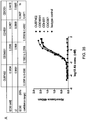


Classifications
-
- C—CHEMISTRY; METALLURGY
- C07—ORGANIC CHEMISTRY
- C07K—PEPTIDES
- C07K16/00—Immunoglobulins [IGs], e.g. monoclonal or polyclonal antibodies
- C07K16/40—Immunoglobulins [IGs], e.g. monoclonal or polyclonal antibodies against enzymes
-
- A—HUMAN NECESSITIES
- A61—MEDICAL OR VETERINARY SCIENCE; HYGIENE
- A61K—PREPARATIONS FOR MEDICAL, DENTAL OR TOILETRY PURPOSES
- A61K51/00—Preparations containing radioactive substances for use in therapy or testing in vivo
- A61K51/02—Preparations containing radioactive substances for use in therapy or testing in vivo characterised by the carrier, i.e. characterised by the agent or material covalently linked or complexing the radioactive nucleus
- A61K51/04—Organic compounds
- A61K51/08—Peptides, e.g. proteins, carriers being peptides, polyamino acids, proteins
- A61K51/10—Antibodies or immunoglobulins; Fragments thereof, the carrier being an antibody, an immunoglobulin or a fragment thereof, e.g. a camelised human single domain antibody or the Fc fragment of an antibody
- A61K51/1075—Antibodies or immunoglobulins; Fragments thereof, the carrier being an antibody, an immunoglobulin or a fragment thereof, e.g. a camelised human single domain antibody or the Fc fragment of an antibody the antibody being against an enzyme
-
- A—HUMAN NECESSITIES
- A61—MEDICAL OR VETERINARY SCIENCE; HYGIENE
- A61P—SPECIFIC THERAPEUTIC ACTIVITY OF CHEMICAL COMPOUNDS OR MEDICINAL PREPARATIONS
- A61P1/00—Drugs for disorders of the alimentary tract or the digestive system
-
- A—HUMAN NECESSITIES
- A61—MEDICAL OR VETERINARY SCIENCE; HYGIENE
- A61P—SPECIFIC THERAPEUTIC ACTIVITY OF CHEMICAL COMPOUNDS OR MEDICINAL PREPARATIONS
- A61P1/00—Drugs for disorders of the alimentary tract or the digestive system
- A61P1/04—Drugs for disorders of the alimentary tract or the digestive system for ulcers, gastritis or reflux esophagitis, e.g. antacids, inhibitors of acid secretion, mucosal protectants
-
- A—HUMAN NECESSITIES
- A61—MEDICAL OR VETERINARY SCIENCE; HYGIENE
- A61P—SPECIFIC THERAPEUTIC ACTIVITY OF CHEMICAL COMPOUNDS OR MEDICINAL PREPARATIONS
- A61P1/00—Drugs for disorders of the alimentary tract or the digestive system
- A61P1/16—Drugs for disorders of the alimentary tract or the digestive system for liver or gallbladder disorders, e.g. hepatoprotective agents, cholagogues, litholytics
-
- A—HUMAN NECESSITIES
- A61—MEDICAL OR VETERINARY SCIENCE; HYGIENE
- A61P—SPECIFIC THERAPEUTIC ACTIVITY OF CHEMICAL COMPOUNDS OR MEDICINAL PREPARATIONS
- A61P11/00—Drugs for disorders of the respiratory system
-
- A—HUMAN NECESSITIES
- A61—MEDICAL OR VETERINARY SCIENCE; HYGIENE
- A61P—SPECIFIC THERAPEUTIC ACTIVITY OF CHEMICAL COMPOUNDS OR MEDICINAL PREPARATIONS
- A61P13/00—Drugs for disorders of the urinary system
- A61P13/12—Drugs for disorders of the urinary system of the kidneys
-
- A—HUMAN NECESSITIES
- A61—MEDICAL OR VETERINARY SCIENCE; HYGIENE
- A61P—SPECIFIC THERAPEUTIC ACTIVITY OF CHEMICAL COMPOUNDS OR MEDICINAL PREPARATIONS
- A61P17/00—Drugs for dermatological disorders
-
- A—HUMAN NECESSITIES
- A61—MEDICAL OR VETERINARY SCIENCE; HYGIENE
- A61P—SPECIFIC THERAPEUTIC ACTIVITY OF CHEMICAL COMPOUNDS OR MEDICINAL PREPARATIONS
- A61P17/00—Drugs for dermatological disorders
- A61P17/02—Drugs for dermatological disorders for treating wounds, ulcers, burns, scars, keloids, or the like
-
- A—HUMAN NECESSITIES
- A61—MEDICAL OR VETERINARY SCIENCE; HYGIENE
- A61P—SPECIFIC THERAPEUTIC ACTIVITY OF CHEMICAL COMPOUNDS OR MEDICINAL PREPARATIONS
- A61P17/00—Drugs for dermatological disorders
- A61P17/06—Antipsoriatics
-
- A—HUMAN NECESSITIES
- A61—MEDICAL OR VETERINARY SCIENCE; HYGIENE
- A61P—SPECIFIC THERAPEUTIC ACTIVITY OF CHEMICAL COMPOUNDS OR MEDICINAL PREPARATIONS
- A61P21/00—Drugs for disorders of the muscular or neuromuscular system
- A61P21/02—Muscle relaxants, e.g. for tetanus or cramps
-
- A—HUMAN NECESSITIES
- A61—MEDICAL OR VETERINARY SCIENCE; HYGIENE
- A61P—SPECIFIC THERAPEUTIC ACTIVITY OF CHEMICAL COMPOUNDS OR MEDICINAL PREPARATIONS
- A61P25/00—Drugs for disorders of the nervous system
-
- A—HUMAN NECESSITIES
- A61—MEDICAL OR VETERINARY SCIENCE; HYGIENE
- A61P—SPECIFIC THERAPEUTIC ACTIVITY OF CHEMICAL COMPOUNDS OR MEDICINAL PREPARATIONS
- A61P25/00—Drugs for disorders of the nervous system
- A61P25/14—Drugs for disorders of the nervous system for treating abnormal movements, e.g. chorea, dyskinesia
- A61P25/16—Anti-Parkinson drugs
-
- A—HUMAN NECESSITIES
- A61—MEDICAL OR VETERINARY SCIENCE; HYGIENE
- A61P—SPECIFIC THERAPEUTIC ACTIVITY OF CHEMICAL COMPOUNDS OR MEDICINAL PREPARATIONS
- A61P25/00—Drugs for disorders of the nervous system
- A61P25/28—Drugs for disorders of the nervous system for treating neurodegenerative disorders of the central nervous system, e.g. nootropic agents, cognition enhancers, drugs for treating Alzheimer's disease or other forms of dementia
-
- A—HUMAN NECESSITIES
- A61—MEDICAL OR VETERINARY SCIENCE; HYGIENE
- A61P—SPECIFIC THERAPEUTIC ACTIVITY OF CHEMICAL COMPOUNDS OR MEDICINAL PREPARATIONS
- A61P25/00—Drugs for disorders of the nervous system
- A61P25/30—Drugs for disorders of the nervous system for treating abuse or dependence
-
- A—HUMAN NECESSITIES
- A61—MEDICAL OR VETERINARY SCIENCE; HYGIENE
- A61P—SPECIFIC THERAPEUTIC ACTIVITY OF CHEMICAL COMPOUNDS OR MEDICINAL PREPARATIONS
- A61P27/00—Drugs for disorders of the senses
- A61P27/02—Ophthalmic agents
-
- A—HUMAN NECESSITIES
- A61—MEDICAL OR VETERINARY SCIENCE; HYGIENE
- A61P—SPECIFIC THERAPEUTIC ACTIVITY OF CHEMICAL COMPOUNDS OR MEDICINAL PREPARATIONS
- A61P29/00—Non-central analgesic, antipyretic or antiinflammatory agents, e.g. antirheumatic agents; Non-steroidal antiinflammatory drugs [NSAID]
-
- A—HUMAN NECESSITIES
- A61—MEDICAL OR VETERINARY SCIENCE; HYGIENE
- A61P—SPECIFIC THERAPEUTIC ACTIVITY OF CHEMICAL COMPOUNDS OR MEDICINAL PREPARATIONS
- A61P31/00—Antiinfectives, i.e. antibiotics, antiseptics, chemotherapeutics
- A61P31/04—Antibacterial agents
-
- A—HUMAN NECESSITIES
- A61—MEDICAL OR VETERINARY SCIENCE; HYGIENE
- A61P—SPECIFIC THERAPEUTIC ACTIVITY OF CHEMICAL COMPOUNDS OR MEDICINAL PREPARATIONS
- A61P35/00—Antineoplastic agents
-
- A—HUMAN NECESSITIES
- A61—MEDICAL OR VETERINARY SCIENCE; HYGIENE
- A61P—SPECIFIC THERAPEUTIC ACTIVITY OF CHEMICAL COMPOUNDS OR MEDICINAL PREPARATIONS
- A61P35/00—Antineoplastic agents
- A61P35/02—Antineoplastic agents specific for leukemia
-
- A—HUMAN NECESSITIES
- A61—MEDICAL OR VETERINARY SCIENCE; HYGIENE
- A61P—SPECIFIC THERAPEUTIC ACTIVITY OF CHEMICAL COMPOUNDS OR MEDICINAL PREPARATIONS
- A61P37/00—Drugs for immunological or allergic disorders
- A61P37/02—Immunomodulators
- A61P37/06—Immunosuppressants, e.g. drugs for graft rejection
-
- A—HUMAN NECESSITIES
- A61—MEDICAL OR VETERINARY SCIENCE; HYGIENE
- A61P—SPECIFIC THERAPEUTIC ACTIVITY OF CHEMICAL COMPOUNDS OR MEDICINAL PREPARATIONS
- A61P39/00—General protective or antinoxious agents
- A61P39/02—Antidotes
-
- A—HUMAN NECESSITIES
- A61—MEDICAL OR VETERINARY SCIENCE; HYGIENE
- A61P—SPECIFIC THERAPEUTIC ACTIVITY OF CHEMICAL COMPOUNDS OR MEDICINAL PREPARATIONS
- A61P43/00—Drugs for specific purposes, not provided for in groups A61P1/00-A61P41/00
-
- A—HUMAN NECESSITIES
- A61—MEDICAL OR VETERINARY SCIENCE; HYGIENE
- A61P—SPECIFIC THERAPEUTIC ACTIVITY OF CHEMICAL COMPOUNDS OR MEDICINAL PREPARATIONS
- A61P9/00—Drugs for disorders of the cardiovascular system
-
- A—HUMAN NECESSITIES
- A61—MEDICAL OR VETERINARY SCIENCE; HYGIENE
- A61P—SPECIFIC THERAPEUTIC ACTIVITY OF CHEMICAL COMPOUNDS OR MEDICINAL PREPARATIONS
- A61P9/00—Drugs for disorders of the cardiovascular system
- A61P9/10—Drugs for disorders of the cardiovascular system for treating ischaemic or atherosclerotic diseases, e.g. antianginal drugs, coronary vasodilators, drugs for myocardial infarction, retinopathy, cerebrovascula insufficiency, renal arteriosclerosis
-
- A—HUMAN NECESSITIES
- A61—MEDICAL OR VETERINARY SCIENCE; HYGIENE
- A61K—PREPARATIONS FOR MEDICAL, DENTAL OR TOILETRY PURPOSES
- A61K39/00—Medicinal preparations containing antigens or antibodies
- A61K2039/505—Medicinal preparations containing antigens or antibodies comprising antibodies
-
- C—CHEMISTRY; METALLURGY
- C07—ORGANIC CHEMISTRY
- C07K—PEPTIDES
- C07K2317/00—Immunoglobulins specific features
- C07K2317/30—Immunoglobulins specific features characterized by aspects of specificity or valency
- C07K2317/34—Identification of a linear epitope shorter than 20 amino acid residues or of a conformational epitope defined by amino acid residues
-
- C—CHEMISTRY; METALLURGY
- C07—ORGANIC CHEMISTRY
- C07K—PEPTIDES
- C07K2317/00—Immunoglobulins specific features
- C07K2317/40—Immunoglobulins specific features characterized by post-translational modification
-
- C—CHEMISTRY; METALLURGY
- C07—ORGANIC CHEMISTRY
- C07K—PEPTIDES
- C07K2317/00—Immunoglobulins specific features
- C07K2317/70—Immunoglobulins specific features characterized by effect upon binding to a cell or to an antigen
- C07K2317/76—Antagonist effect on antigen, e.g. neutralization or inhibition of binding
Definitions
- the present invention relates to inhibitors of TG2 and methods for providing and using such inhibitors.
- Transglutaminase type 2 (TG2; also known as tissue transglutaminase, tTg) is part of the wider 9 member transglutaminase family that includes Factor Xllla, which is critical for blood clotting, as well as Keratinocyte Transglutaminase (TG1) and Epidermal Transglutaminase (TG3) that are involved in terminal differentiation of the keratinocyte.
- TG2 tissue transglutaminase
- tTg tissue transglutaminase
- TG3 Keratinocyte Transglutaminase
- TG3 Epidermal Transglutaminase
- TG2 functions primarily as a protein cross-linking enzyme by catalysing the formation of ⁇ ( ⁇ -glutamyl) lysine iso-dipeptide bonds. Elevated expression of TG2 leads to aberrant protein cross-linking which has been associated with several pathologies including various types of tissue scarring, the formation of neurofibrillary tangles in several brain disorders and resistance to chemotherapy in some cancers.
- TG2 is also able to deamidate proteins. TG2 deamidates gliadin and the TG2/Gliadin complex is the primary autoantigen in celiac disease.
- TG2 has a GTP binding function and can act as a GTPase, although this has not been linked to a pathological role.
- Elevated TG2 activity is primarily associated with abnormal wound healing [1] leading to liver [2], pulmonary [3], heart [4] and kidney fibrosis, [5] as well as atherosclerosis [6].
- the process of scarring and fibrosis is linked to the increased synthesis and, most importantly, raised export of TG2 to the interstitial space.
- TG2 is able to crosslink extracellular matrix (ECM) proteins such as fibronectin and collagen [7] by the incorporation of an ⁇ ( ⁇ -glutamyl) lysine di-peptide bond [8].
- ECM extracellular matrix
- TG2 has an emerging role in the activation of latent TGF- ⁇ 1 in the scarring process [11] and has also been associated with interleukin-6 [12] and tumour necrosis factor- ⁇ activation pathways [13].
- TG2 neurodegenerative diseases characterised by the presence of protein aggregates in the degenerative region of the brain that TG2 has been implicated in forming.
- the best characterised is in Huntington's chorea.
- Huntington protein (htt) contains expanded polyglutamine repeats in its N-terminal domain. Wild-type htt contains less than 35 consecutive glutamines while disease-related htt typically has over 40 consecutive glutamines which makes it an excellent TG2 substrate.
- insoluble aggregates are formed in the striatum and cortex of Huntington's disease patients. The frequency of aggregates correlates well with the severity of the disease.
- Alzheimer's disease is typified by the presence of extracellular senile plaques composed of aggregated amyloid ⁇ -protein and intracellular neurofibrillary tangles consisting of a highly phosphorylated form of the protein tau. These plaques contain large amounts of ⁇ ( ⁇ -glutamyl) lysine iso-dipeptide bonds.
- Parkinson's disease a hallmark of Parkinson's disease is the presence of alpha-synuclein aggregates called Lewy bodies in the cytoplasm of affected neurons which again contain ⁇ ( ⁇ -glutamyl) lysine iso-dipeptide bonds. All of the aforementioned proteins are good substrates of TG2 in vitro. Furthermore, the affected region of the brain contains higher levels of TG2 protein than non-affected regions of the brain in the same patients. The correlation between the TG2 substrate specificity for disease-relevant aggregated proteins and increased TG2 expression levels suggest a role for enzymatically active TG2 in each disease.
- TG inhibitors have been shown to exert therapeutic effects in multiple biological models of neurodegenerative diseases.
- a cell culture model of Parkinson's disease transfecting COS-7 cells with alpha-synuclein and TG2 simultaneously covalent - synuclein aggregates, reminiscent of Lewy bodies in Parkinson's disease, form and are dependent upon enzymatically active TG2 since the C277S inactive TG2 mutant failed to induce aggregate formation.
- Treatment of these cotransfected cells with cystamine significantly reduced the quantity of alpha-synuclein aggregates as well as the percentage of cells containing the aggregates.
- Cystamine has a beneficial therapeutic effect in vivo when dosed in mouse models of Huntington's disease.
- Huntington R6/2 mice dosed with cystamine showed improved motor function, less severe weight loss, and increased survival compared to non-treated controls.
- ex vivo TG2 activity in brain homogenates was lower after dosing with cystamine at least 60 min after injection.
- cystamine was able to decrease the level of striatal atrophy but unable to improve animal weight or motor function, indicating a beneficial effect of cystamine at the cellular and tissue level but not in disease symptoms.
- R6/2 TG2 -/- mice showed a delay in the onset of motor dysfunction and improved survival compared to R6/2 TG2 +/+ mice implying a role for TG2 in the pathogenesis of neurodegeneration in the R6/2 model.
- Celiac sprue is a T cell-mediated inflammatory disorder of the small intestine caused by a class of proteins called prolamins found in wheat, barley, and rye.
- prolamins found in wheat, barley, and rye.
- the high proline and glutamine content of these proteins makes them resistant to natural gastric, pancreatic, and intestinal proteases, and peptidases during digestion.
- the resulting peptide fragments remain undigested well into the small intestine and gain access to the intestinal lamina intestinal where, after modification by TG2, they can stimulate a T cell-mediated immune response leading to inflammation and destruction of intestinal architecture.
- Intestinal TG2 deamidates specific glutamine residues in the prolamin peptides to glutamate residues. In HLA-DQ2/8 individuals these modified peptides are presented to corresponding autoreactive T cells by class II MHC molecules. Although prolamins have a high glutamine content (around 30-35%), only a few of these glutamine residues are targeted by human TG2. An excellent correlation between TG2 substrate specificity, DQ2 binding affinity, and T cell stimulatory potential of TG2-treated prolamins strongly suggests that peptide deamidation is mediated by TG2 and plays a significant role in determining the severity of disease. Further, celiac patients generate an autoantibody response to TG2-gliadin complexes. These anti-TG2 antibodies are found in both the small intestine, where they have been shown to co-localize with extracellular TG2, and in the blood, where they are exploited as a diagnostic disease marker.
- pan-TG inhibitor 2-[(2-oxopropyl)thio]imidazolium inhibitor was able to prevent the in situ crosslinking of gluten peptides to endogenous proteins in thin tissue sections taken from both celiac sprue patients and controls. More importantly, the authors showed that incubation of intact celiac small intestinal biopsies with 2-[(2-oxopropyl)thio]imidazolium prevented T-cell activation induced by the non-deamidated form of an immunodominant gluten peptide. In contrast, TG inhibition was ineffective at controlling T-cell activation when the biopsies were incubated with the deamidated version of the same peptide. These results suggest that inhibition of endogenous TG2 in celiac patient biopsies can prevent gluten peptide deamidation and, therefore, reduce T-cell activation.
- TG2 plays a role in the development of certain types of cancer.
- Multiple studies have shown that TG2 protein is up-regulated in cancerous tissue relative to healthy tissue in cancers such as glioblastomas, malignant melanomas, and pancreatic ductal adenocarcinomas to name a few.
- a positive correlation between the chemotherapeutic resistance and metastatic potential of certain cancers with TG2 expression levels has been demonstrated, while in certain cell types TG2 has been shown to exert anti-apoptotic effects on cells while siRNA down-regulation of TG2 protein expression levels or treatment with TG2 inhibitors sensitizes these cells to apoptosis.
- WO 2006/100679 describes a specific anti-TG2 antibody produced by recombinant technology from samples collected from three adult celiac patients with high anti-TG2 antibody titres.
- CUB7402 (ab2386) is an antibody commercially available from Abcam.
- WO 2010/113025 describes selective diagnosis of gluten-induced autoimmune diseases by binding assays utilizing the main celiac epitope present on proteins of the transglutaminase family.
- WO 02/068616 describes transglutaminase 6 as a diagnostic indicator of autoimmune diseases.
- WO 2010/116196 describes the selective diagnosis of gluten-induced autoimmune diseases by binding assays utilizing the main celiac epitope present on proteins of the transglutaminase family. WO 2010/116196 also describes the treatment of gluten-induced autoimmune diseases by inhibiting binding of celiac autoantibodies by therapeutic compounds.
- Simon-Vescsei et al., 2012; PNAS 109(2): 433-434 describes that a single conformational transglutaminase 2 epitope contributed by three domains is critical for celiac antibody binding and effects.
- the present invention provides an antibody or antigen-binding fragment thereof that selectively binds to an epitope within the core region of human transglutaminase type 2 (TG2) and inhibits human TG2 activity, wherein said core region consists of amino acids 143 to 473 of human TG2, wherein the antibody or antigen-binding fragment thereof selectively binds to the region comprising amino acids 304 to 326 of human TG2, and wherein the human TG2 activity that is inhibited is the TG2 cross-linking of lysine and glutamine with N ⁇ ( ⁇ -glutamyl)lysine isopeptide bonds.
- TG2 human transglutaminase type 2
- the antibody or antigen-binding fragment thereof does not inhibit TG1, TG3, TG13 and/or TG7 activity.
- the antibody or antigen-binding fragment thereof : a. comprises or consists of an intact antibody, or b. comprises or consists of an antigen-binding fragment selected from the group consisting of: an Fv fragment (for example a single chain Fv fragment or a disulphide-bonded Fv fragment); a Fab fragment; and a Fab-like fragment (for example an Fab' fragment or an F(ab) 2 fragment).
- the antibody or antigen-binding fragment thereof binds to the same epitope as an antibody comprising the following CDR sequences:
- the antibody or antigen-binding fragment thereof competes with and disrupts binding of an antibody or antigen-binding fragment thereof having a light chain variable region selected from the following sequences: a heavy chain variable region selected from the following sequences:
- the binding of the antibody or antigen-binding fragment thereof to human TG2 is inhibited or reduced when an antibody having a light chain variable region selected from the following sequences: a heavy chain variable region selected from the following sequences: EVQLLESGGGLVQPGGSLRLSCAASGFTLSTHAMSWVRQAPGKGLEWVATISS GGRSTYYPDSVKGRFTISRDNSKNTLYLQMNSLRAEDTAVYFCARLISTYWGQGTLVTV SS is bound to human TG2.
- the present invention also provides a polynucleotide encoding an antibody or an antigen-binding fragment of the present invention.
- the present invention provides a compound comprising an antibody or antigen-binding fragment thereof of the invention and a further moiety, for example a readily detectable moiety, and/or a directly or indirectly cytotoxic moiety.
- the present invention also provides a pharmaceutical composition/formulation comprising an antibody or antigen-binding fragment thereof of the present invention, or a polynucleotide of the present invention, or a compound of the present invention, in admixture with a pharmaceutically acceptable excipient, adjuvant, diluent or carrier, optionally further comprising one or more further active ingredients, preferably wherein the composition/formulation is formulated for intravenous, intramuscular, or subcutaneous delivery to a patient.
- the present invention also provides a kit of parts comprising an antibody or antigen-binding fragment thereof of the present invention, or a polynucleotide of the present invention, or a compound of the present invention; and a further agent.
- the present invention also provides an antibody or antigen-binding fragment thereof of the present invention, or a polynucleotide of the present invention, or a compound of the present invention, for use in medicine.
- the present invention provides an antibody or antigen-binding fragment thereof of the present invention, or a polynucleotide of the present invention, or a compound of the present invention, for use in the treatment and/or diagnosis of Celiac disease, abnormal wound healing, scarring, hypertrophic scars, ocular scarring, inflammatory bowel disease, macular degeneration, psoriasis, fibrosis-related diseases (e.g.
- liver fibrosis pulmonary fibrosis such as interstitial lung disease and fibrotic lung disease
- cardiac fibrosis myelofibrosis
- kidney fibrosis such as glomerulosclerosis and tubulointerstitial fibrosis
- atherosclerosis restenosis
- inflammatory diseases e.g., autoimmune diseases, neurodegenerative/neurological diseases (e.g.
- glioblastomas such as glioblastoma in Li-Fraumeni syndrome and sporadic glioblastoma, malignant melanomas, pancreatic ductal adenocarcinomas, myeloid leukemia, acute myelogenous leukemia, myelodysplastic syndrome, myeloproliferative syndrome, gynaecological cancer, Kaposi's sarcoma, Hansen's disease and/or collagenous colitis.
- glioblastomas such as glioblastoma in Li-Fraumeni syndrome and sporadic glioblastoma, malignant melanomas, pancreatic ductal adenocarcinomas, myeloid leukemia, acute myelogenous leukemia, myelodysplastic syndrome, myeloproliferative syndrome, gynaecological cancer, Kaposi's sarcoma, Hansen's disease and/or collagenous colitis.
- the present invention further provides a method of producing an antibody or antigen-binding fragment thereof of the invention, the method comprising isolating the antibody or antigen-binding fragment thereof from an ex-vivo sample obtained from a non-human animal which was previously immunized using:
- the present invention provides an antibody or an antigen-binding fragment thereof that selectively binds to an epitope within the core region of human transglutaminase type 2 (TG2).
- TG2 human transglutaminase type 2
- an antibody or antigen-binding fragment thereof described herein may selectively bind to an epitope within the core region of human TG2, rat TG2, and/or mouse TG2.
- the present inventors have raised antibodies to TG2 by immunising mice with a recombinant protein encompassing amino acids 143 to 473 of the human TG2 core. Hybridoma were screened for TG2 specificity and inhibition with any suitable candidates cloned. IgG was purified from these to calculate efficacy and the target epitope mapped by screening of a human TG2 library by phage display.
- the present approach to producing antibodies against TG2 by using a recombinant TG2 core protein has not been tried before and it surprisingly led to the isolation and characterisation of antibodies to TG2 that were highly selective for TG2 and showed strong inhibitory characteristics.
- Prior attempts to raise antibodies to TG2 have led to the isolation of relatively unselective antibodies that cross-react with other members of the transglutaminase family and thus would not represent promising antibodies for clinical use.
- the antibodies of the present invention on the other hand are promising candidates for clinical trials for diseases exacerbated by or mediated by TG2 activity.
- TG2 core removed much of the tertiary structure of the enzyme (in particular the two carboxy terminal beta barrel domains). It is possible that some of the epitopes that perhaps may be less available or immunogenic within a native full length TG2 molecule may be more attractive epitopes with the core in the format described herein.
- the antibodies described herein recognised linear epitopes (i.e. bound to TG2 on a reducing SDS PAGE gel), whereas 80 % of the antibodies we previously isolated using full length TG2 as an immunogen were conformation dependent.
- inhibitory antibodies to human TG2 were raised by immunising with the core given that the recombinant core protein may not have demonstrated the same folding characteristics as the full length protein.
- the TG catalytic core is highly conserved between members of the TG family and across species. This suggests that development of not only specific small molecule inhibitors but also of antibody-based inhibitors may be technically challenging. Nevertheless, the present disclosure provides antibodies that are highly selective. That this is possible may reflect the fact that there are some regions within the TG2 catalytic domain where there is some heterogeneity. The present antibodies may therefore exploit these small differences. The surprising selectivity of the present antibodies may enable the development of therapeutics that can interfere efficiently with TG2 activity and thus provide potentially effective therapies for conditions exacerbated by or caused by TG2 activity where there is currently no satisfactory therapeutic option.
- the antibodies described in WO 2006/100679 which were produced by recombinant technology from samples from three adult celiac patients with high anti-TG2 antibody titres, proved to be of low efficacy when tested by the present inventors (Example 2).
- the antibodies of the present invention were far superior to those of WO 2006/100679 in terms of selectivity for TG2 and in terms of inhibitory activity.
- the present inventors generated a Fab fragment of the antibody of WO 2006/100679 , which was applied at the same concentration in TG2 inhibition assays as the antibodies of the present invention.
- the Fab fragment amount in the tests was twice molar wise that of the antibodies of the present invention, but they still failed to show any inhibition of TG2 activity.
- the full length WO 2006/100679 antibody was tested for inhibition in our standard putrescine incorporation assay no inhibition of TG2 activity was found.
- the methods of the present invention and the antibodies produced by those methods are superior to those previously described.
- antibody we include substantially intact antibody molecules, as well as chimeric antibodies, humanised antibodies, human antibodies (wherein at least one amino acid is mutated relative to the naturally occurring human antibodies), single chain antibodies, bi-specific antibodies, antibody heavy chains, antibody light chains, homodimers and heterodimers of antibody heavy and/or light chains, and antigen binding fragments and derivatives of the same.
- variants, fusions and derivatives of the antibodies and antigen-binding fragments thereof within the meaning of the terms "antibody” and "antigen-binding fragments thereof".
- antibody also includes all classes of antibodies, including IgG, IgA, IgM, IgD and IgE.
- the antibody may be an IgG molecule, such as an IgG1, IgG2, IgG3, or IgG4 molecule.
- the antibody of the invention is an IgG molecule, or an antigen-binding fragment, or variant, fusion or derivative thereof. More preferably the antibody is an IgG2 molecule.
- the antibodies, compositions, uses and methods described herein encompass variants, fusions and derivatives of the defined antibodies and antigen-binding fragments thereof, as well as fusions of a said variants or derivatives, provided such variants, fusions and derivatives have binding specificity for TG2.
- antibodies and antigen-binding fragments thereof comprise one or more polypeptide component
- variants, fusions and derivatives of the antibody and antigen-binding fragment thereof as defined herein may be made using the methods of protein engineering and site-directed mutagenesis well known in the art using the recombinant polynucleotides (see example, see Molecular Cloning: a Laboratory Manual, 3rd edition, Sambrook & Russell, 2001, Cold Spring Harbor Laboratory Press ).
- variants, fusions and derivatives of the antibody or antigen-binding fragment thereof as defined herein may be made based on the polypeptide component of the antibody or antigen-binding fragment thereof.
- fusion we include said polypeptide fused to any other polypeptide.
- the said polypeptide may be fused to a polypeptide such as glutathione-S-transferase (GST) or protein A in order to facilitate purification of said polypeptide. Examples of such fusions are well known to those skilled in the art.
- the said polypeptide may be fused to an oligo-histidine tag such as His6 or to an epitope recognised by an antibody such as the well-known Myc-tag epitope. Fusions to any variant or derivative of said polypeptide are also included. It will be appreciated that fusions (or variants or derivatives thereof) which retain desirable properties, such as have binding specificity for TG2, are preferred.
- the fusion may comprise or consist of a further portion which confers a desirable feature on the said polypeptide; for example, the portion may be useful in detecting or isolating the polypeptide, or promoting cellular uptake of the polypeptide.
- the portion may be, for example, a biotin moiety, a radioactive moiety, a fluorescent moiety, for example a small fluorophore or a green fluorescent protein (GFP) fluorophore, as well known to those skilled in the art.
- the moiety may be an immunogenic tag, for example a Myc-tag, as known to those skilled in the art or may be a lipophilic molecule or polypeptide domain that is capable of promoting cellular uptake of the polypeptide, as known to those skilled in the art.
- variants of said polypeptide we refer to a polypeptide wherein at one or more positions there have been amino acid insertions, deletions, and/or substitutions, either conservative or non-conservative, provided that such changes result in a protein whose basic properties, for example binding properties, thermostability, activity in a certain pH-range (pH-stability) have not significantly been changed. "Significantly” in this context means that one skilled in the art would say that the properties of the variant may still be different but would not be unobvious over the ones of the original protein. Thus, we include variants of the polypeptide where such changes do not substantially alter the activity of the said polypeptide. In particular, we include variants of the polypeptide where such changes do not substantially alter the binding specificity for TG2.
- substitutions are intended combinations such as Gly, Ala; Val, Ile, Leu; Asp, Glu; Asn, Gln; Ser, Thr; Lys, Arg; and Phe, Tyr.
- Such variants may be made using the methods of protein engineering and site-directed mutagenesis.
- the polypeptide variant may have an amino acid sequence which has at least 75% identity with one or more of the amino acid sequences given herein, for example at least 80%, at least 90%, at least 95%, at least 96%, at least 97%, at least 98%, or at least 99% identity with one or more of the amino acid sequences specified herein.
- the percent sequence identity between two polypeptides may be determined using suitable computer programs, for example the GAP program of the University of Wisconsin Genetic Computing Group and it will be appreciated that percent identity is calculated in relation to polypeptides whose sequences have been aligned optimally.
- the alignment may alternatively be carried out using the Clustal W program (as described in Thompson et al., 1994, Nucl. Acid Res. 22:4673-4680 ).
- the parameters used may be as follows:
- the BESTFIT program may be used to determine local sequence alignments.
- the antibody, antigen-binding fragment, variant, fusion or derivative used in the methods or uses of the invention may comprise or consist of one or more amino acids which have been modified or derivatised.
- Chemical derivatives of one or more amino acids may be achieved by reaction with a functional side group.
- derivatised molecules include, for example, those molecules in which free amino groups have been derivatised to form amine hydrochlorides, p-toluene sulphonyl groups, carboxybenzoxy groups, t-butyloxycarbonyl groups, chloroacetyl groups or formyl groups.
- Free carboxyl groups may be derivatised to form salts, methyl and ethyl esters or other types of esters and hydrazides.
- Free hydroxyl groups may be derivatised to form O-acyl or O-alkyl derivatives.
- Also included as chemical derivatives are those peptides which contain naturally occurring amino acid derivatives of the twenty standard amino acids.
- 4-hydroxyproline may be substituted for proline
- 5-hydroxylysine may be substituted for lysine
- 3-methylhistidine may be substituted for histidine
- homoserine may be substituted for serine and ornithine for lysine.
- Derivatives also include peptides containing one or more additions or deletions as long as the requisite activity is maintained.
- Other included modifications are amidation, amino terminal acylation (e.g. acetylation or thioglycolic acid amidation), terminal carboxylamidation (e.g. with ammonia or methylamine), and the like terminal modifications.
- peptidomimetic compounds may also be useful.
- 'polypeptide' we include peptidomimetic compounds which are capable of binding to an epitope within the TG2 core region.
- 'peptidomimetic' refers to a compound that mimics the conformation and desirable features of a particular peptide as a therapeutic agent.
- the said polypeptide includes not only molecules in which amino acid residues are joined by peptide (-CO-NH-) linkages but also molecules in which the peptide bond is reversed.
- retro-inverso peptidomimetics may be made using methods known in the art, for example such as those described in Meziere et al. (1997) J. Immunol. 159, 3230-3237 . This approach involves making pseudo-peptides containing changes involving the backbone, and not the orientation of side chains. Retro-inverse peptides, which contain NH-CO bonds instead of CO-NH peptide bonds, are much more resistant to proteolysis.
- the said polypeptide may be a peptidomimetic compound wherein one or more of the amino acid residues are linked by a -y(CH 2 NH)- bond in place of the conventional amide linkage.
- the peptide bond may be dispensed with altogether provided that an appropriate linker moiety which retains the spacing between the carbon atoms of the amino acid residues is used; it may be advantageous for the linker moiety to have substantially the same charge distribution and substantially the same planarity as a peptide bond.
- the said polypeptide may conveniently be blocked at its N- or C-terminus so as to help reduce susceptibility to exo-proteolytic digestion.
- a presumed bioactive conformation may be stabilised by a covalent modification, such as cyclisation or by incorporation of lactam or other types of bridges, for example see Veber et al., 1978, Proc. Natl. Acad. Sci. USA 75:2636 and Thursell et al., 1983, Biochem. Biophys. Res. Comm. 111:166 .
- a common theme among many of the synthetic strategies has been the introduction of some cyclic moiety into a peptide-based framework.
- the cyclic moiety restricts the conformational space of the peptide structure and this frequently results in an increased specificity of the peptide for a particular biological receptor.
- An added advantage of this strategy is that the introduction of a cyclic moiety into a peptide may also result in the peptide having a diminished sensitivity to cellular peptidases.
- exemplary polypeptides useful in the methods and uses of the invention comprise or consist of terminal cysteine amino acids.
- Such a polypeptide may exist in a heterodetic cyclic form by disulphide bond formation of the mercaptide groups in the terminal cysteine amino acids or in a homodetic form by amide peptide bond formation between the terminal amino acids.
- cyclising small peptides through disulphide or amide bonds between the N- and C-terminus cysteines may circumvent problems of specificity and half-life sometime observed with linear peptides, by decreasing proteolysis and also increasing the rigidity of the structure, which may yield higher specificity compounds.
- Polypeptides cyclised by disulphide bonds have free amino and carboxy-termini which still may be susceptible to proteolytic degradation, while peptides cyclised by formation of an amide bond between the N-terminal amine and C-terminal carboxyl and hence no longer contain free amino or carboxy termini.
- the peptides can be linked either by a C-N linkage or a disulphide linkage.
- heterodetic linkages may include, but are not limited to formation via disulphide, alkylene or sulphide bridges.
- Methods of synthesis of cyclic homodetic peptides and cyclic heterodetic peptides, including disulphide, sulphide and alkylene bridges, are disclosed in US 5,643,872 .
- Other examples of cyclisation methods are discussed and disclosed in US 6,008,058 .
- RCM ring-closing metathesis
- terminal modifications are useful, as is well known, to reduce susceptibility by proteinase digestion and therefore to prolong the half-life of the peptides in solutions, particularly in biological fluids where proteases may be present.
- Polypeptide cyclisation is also a useful modification because of the stable structures formed by cyclisation and in view of the biological activities observed for cyclic peptides.
- the said polypeptide may be cyclic.
- the said polypeptide may be linear.
- TG2 By “selectively binds to an epitope within the core region of TG2” we mean an antibody or antigen-binding fragment thereof that is capable of binding to an epitope in the TG2 core region selectively.
- capable of binding selectively we include such antibody-derived binding moieties which bind at least 10-fold more strongly to the TG2 core than to other proteins; for example at least 50-fold more strongly, or at least 100-fold more strongly.
- the binding moiety may be capable of binding selectively to an epitope in the TG2 core under physiological conditions, e.g. in vivo.
- binding specificity may be determined by methods well known in the art, such as enzyme-linked immunosorbent assay (ELISA), immunohistochemistry, immunoprecipitation, western blot and flow cytometry using transfected cells expressing TG2 or the TG2 core, or a fragment thereof.
- ELISA enzyme-linked immunosorbent assay
- Suitable methods for measuring relative binding strengths include immunoassays, for example where the binding moiety is an antibody (see Harlow & Lane, "Antibodies: A Laboratory Manual", Cold Spring Habor Laboratory Press, New York ).
- binding may be assessed using competitive assays or using Biacore® analysis (Biacore International AB, Sweden).
- the antibody or antigen-binding fragment of the invention binds exclusively to TG2.
- binding specificity of an antibody or antigen binding fragment thereof is conferred by the presence of complementarity determining regions (CDRs) within the variable regions of the constituent heavy and light chains.
- CDRs complementarity determining regions
- binding specificity for TG2 is conferred by the presence of one or more of the CDRs identified.
- sequences that may comprise or consist of the CDR sequences of AB-1 VL and VH include KASQDINSYLT, RTNRLFD, LQYDDFPYT, SSAMS, TISVGGGKTYYPDSVKG, and LISLY.
- sequences that may comprise or consist of the CDR sequences of BB-7 VL and VH include KASQDINSYLT, LTNRLMD, LQYVDFPYT, SSAMS, TISSGGRSTYYPDSVKG, and LISPY.
- Sequences that may comprise or consist of the CDR sequences of DC-1 VL and VH include KASQDINSYLT, LVNRLVD, LQYDDFPYT , THAMS, TISSGGRSTYYPDSVKG, and LISTY. It is preferred that the antibodies and antigen-binding fragments thereof defined herein comprise or consist of CDR sequences, or CDR and flanking sequences, as defined in Table 24A.
- the antibodies and antigen-binding fragments thereof defined herein comprise or consist of the CDR sequences, or CDR and flanking sequences, of the exemplary antibody AB-1, or BB-7, or DC-1 (as defined, for example, in Table 24A).
- the antibody or antigen-binding fragment thereof retains the binding specificity for TG2 of the original antibody.
- the antibody or antigen-binding fragment thereof is capable of competing for binding to TG2 with the exemplary antibodies of the disclosure, for example AB-1, AG-1, AG-9, AH-1, AH-3, BB-7, DC-1, EH-6, JE-12, IA-12, DF-4, DH-2, DD-6 and/or DD-9 (see accompanying Examples).
- the antibody or antigen-binding fragment thereof may bind to the same epitope on TG2 as an antibody comprising the following sequences: KASQDINSYLT, RTNRLFD, LQYDDFPYT, SSAMS, TISVGGGKTYYPDSVKG, and LISLY.
- epitope it is herein intended to mean a site of a molecule to which an antibody binds, i.e. a molecular region of an antigen.
- An epitope may be a linear epitope, which is determined by e.g. the amino acid sequence, i.e. the primary structure, or a three-dimensional epitope, defined by the secondary structure, e.g. folding of a peptide chain into beta sheet or alpha helical, or by the tertiary structure, e.g. way which helices or sheets are folded or arranged to give a three-dimensional structure, of an antigen.
- test antibody is capable of competing for binding with second antibody
- methods for determining whether a test antibody is capable of competing for binding with second antibody are well known in the art (such as, for example sandwich-ELISA or reverse-sandwich-ELISA techniques) and described, for example, in Antibodies: A Laboratory Manual, Harlow & Lane (1988, CSHL, NY, ISBN 0-87969-314-2 ).
- the antibody or antigen-binding fragment thereof, with binding specificity for an epitope in the TG2 core region may also retain one or more of the same biological properties as the original antibody (such as the exemplary antibodies provided in the Examples).
- TG2 is a calcium-dependent multi-functional protein that catalyzes the formation of N ⁇ -( ⁇ -glutamyl)lysine isopeptide bonds between lysine and glutamine residues.
- TG2 comprises an N terminal beta sandwich domain that contains binding sites (e.g. fibronectin) and sequences required for enzyme export. This links to the catalytic core domain. Central to the domain is a catalytic triad consisting of Cys 277, Asp 358, and His 355, plus several putative calcium binding sites. This links to the third domain, beta barrel 1 where a GTP binding site resides conveying the enyme with GTPase activity. Beta barrel 1 also contains an integrin binding site used in cell adhesion.
- binding sites e.g. fibronectin
- Beta Barrel 1 along with the fourth TG2 domain, Beta barrel 2 are involved in the conformational change in TG2 required for its activation.
- Barrel 1 and 2 swing down from the closed and folded inactive form to convey TG2 with a linear structure opening up the catalytic core allowing activation ( Pinkas et al (2007) PLoS Biol. Transglutaminase 2 undergoes a large conformational change upon activation. 5(12): e327 ).
- the core region of transglutaminase type 2 we include a region of TG2 comprising the catalytic triad described above, excluding the beta sandwich domain and beta barrels 1 and 2.
- the core region comprises or consists of amino acids 143 to 473 of human TG2, or a fragment thereof.
- Amino acids 143 to 473 of human TG2 consist of the following sequence:
- the core region consists of amino acids 143 to 473 of human TG2.
- the epitope of an antibody described herein could thus be any epitope within the region defined by amino acids 143 to 473 of human TG2.
- the epitope may be a fragment of this sequence or it could be made up of various amino acid residues within this fragment that may not be adjacent one another in the primary amino acid structure but localise with one another in the secondary, tertiary or even quaternary structure of the protein, as would be understood by a person of skill in the art.
- the antibody or antigen-binding fragment thereof of the invention selectively binds to a region comprising amino acids 304 to 326 of human TG2.
- This region (amino acids 304 to 326 of human TG2) is referred to as Group 1 in Figure 5 and comprises the amino acid sequence AHDQNSNLLIEYFRNEFGEIQGD.
- An antibody or antigen-binding fragment thereof of the disclosure may selectively bind in whole or in part to a region comprising amino acids 351 to 365 of human TG2.
- This region (amino acids 351 to 365 of human TG2) is referred to as Group 2 in Figure 5 and comprises the amino acid sequence YEGWQALDPTPQEKS.
- An antibody or antigen-binding fragment thereof of the disclosure may selectively bind in whole or in part to a region comprising amino acids 450 to 467 of human TG2.
- This region (amino acids 450 to 467 of human TG2) is referred to as Group 3 in Figure 5 and comprises the amino acid sequence SEEREAFTRANHLNKLAE.
- the antibody or antigen-binding fragment thereof of the invention inhibit human TG2 activity.
- An antibody or antigen-binding fragment thereof of the invention may comprise one or more of the following amino acid sequences:
- the sequences of the immediately preceding embodiments are considered to comprise the complementarity determining regions of the light and heavy variable regions of the exemplary antibody AB-1 (see Example 1) and certain specified humanised variants of the AB-1 antibody.
- the antibody may comprise the amino acid sequence or KASQDINSYLTXXXXXXXXXXXXXXXXXXXXXXXXXXXXXXXXXXXXXXXXXXXXXXXXXXXXXXXXXXXXXXXXXXXXXXXXXF; or FXXXXXXXXXXXXXXXXLQYDDFPYT, wherein X is any amino acid.
- the antibody or antigen-binding fragment thereof may comprise the amino acid sequence TCKASQDINSYLTWF or TCKASQDINSYLTWY; and/or LLIYRTNRLFDGVP or SLIYRTNRLFDGVP or LLIYRTNRLFDGVPXXFSGSGSGQDFF or SLIYRTNRLFDGVPXXFSGSGSGQDFF; and/or YCLQYDDFPYTFG; and/or FTFSSSAMSWVR or CXAXXFTFSSSAMSWVR; and/or WVSTISVGGGKTYYPDSVKGRFTISR or WVSTISVGGGKTYYPDSVKGRFTISRXNXXXXL; and/or YCAKLISLYWG, wherein X is any amino acid.
- the antibody or antigen-binding fragment thereof may have at least one light chain variable region comprising the following sequences: KASQDINSYLT; and RTNRLFD; and LQYDDFPYT.
- the antibody or antigen-binding fragment thereof may have at least one heavy chain variable region comprising the following sequences: SSAMS; and TISVGGGKTYYPDSVKG; and LISLY.
- the antibody or antigen-binding fragment thereof may have at least one light chain variable region comprising the amino acid sequence DIKMTQSPSSMYASLGERVTITCKASQDINSYLTWFQQKPGKSPKTLIYRTNRLFDGVPS RFSGSGQDFFLTISSLEYEDMGIYYCLQYDDFPYTFGGGTKLEIK.
- These light chain variable regions correspond with that found in exemplary antibody AB-1 ( Figures 7 and 18 ).
- the antibody or antigen-binding fragment thereof may have at least one light chain variable region comprising the amino acid sequence:
- sequences are humanised variants of the AB-1 light chain sequences provided above.
- the antibody or antigen-binding fragment thereof may have at least one heavy chain variable region comprising the amino acid sequence EVQLVESGGGLVKPGGSLKLSCAASGFTLSSSAMSWVRQTPDRRLEWVATISVGGGKTYY PDSVKGRFTISRDNAKNTLYLQMNSLRSEDTAMYYCAKLISLYWGQGTTLTVSS.
- heavy chain variable regions correspond with that found in exemplary antibody AB-1 ( Figures 7 and 18 ).
- the antibody or antigen-binding fragment thereof may have at least one heavy chain variable region comprising the amino acid sequence:
- the antibody or antigen-binding fragment thereof may have at least one heavy chain variable region comprising the amino acid sequence: EVQLLESGGGLVQPGGSLRLSCAASGFTFSSSAMSWVRQAPGKGLEWVSTISVGGGKTYY PDSVKGRFTISRDNSKNTLYLQMNSLRAEDTAVYYCAKLISLYWGQGTLVTVSS (hAB-1_RHA).
- This sequence is a humanised variant of the AB-1 heavy chain sequence provided above.
- an antibody or antigen-binding fragment thereof of the disclosure may have:
- the sequences of the immediately preceding embodiment are considered to comprise the complementarity determining regions of the light and heavy variable regions of the exemplary antibody BB-7 (see Figure 19 ).
- the antibody may comprise the amino acid sequence KASQDINSYLTXXXXXXXXXXXXXXXXXXLTNRLMDXXXXXXXXXXFXXXXXXXXXXXXXXXXXXXXLQYVDFPYT; or KASQDINSYLTXXXXXXXXXXXXXXXXXXXXXXXXXXXXXXXXXXXXXXXXXXXXXXXXXXXXXXXXXXXXXXXXXXXXXXXXXXXXXXF; or LTNRLMDXXXXXXXXXXXXF; or FXXXXXXXXXXXXXXXXXXLQYVDFPY
- the antibody or antigen-binding fragment thereof may have at least one light chain variable region comprising the following sequences: KASQDINSYLT; and LTNRLMD; and LQYVDFPYT.
- the antibody or antigen-binding fragment thereof may have at least one heavy chain variable region comprising the following sequences: SSAMS; and TISSGGRSTYYPDSVKG; and LISPY.
- the antibody or antigen-binding fragment thereof may have at least one light chain variable region comprising the amino acid sequence AIKMTQSPSSMYASLGERVIITCKASQDINSYLTWFQQKPGKSPKTLIYLTNRLMDGVPS RFSGSGSGQEFLLTISGLEHEDMGIYYCLQYVDFPYTFGGGTKLEIK.
- This light chain variable region corresponds with that found in exemplary antibody BB-7 ( Figure 19 ).
- the antibody or antigen-binding fragment thereof may have at least one heavy chain variable region comprising the amino acid sequence AVQLVESGGGLVKPGGSLKLSCAASGIIFSSSAMSWVRQTPEKRLEWVATISSGGRSTYY PDSVKGRFTVSRDSAKNTLYLQMDSLRSEDTAIYYCAKLISPYWGQGTTLTVSS.
- This heavy chain variable region corresponds with that found in exemplary antibody BB-7 ( Figure 19 ).
- an antibody or antigen-binding fragment thereof of the disclosure may have:
- an antibody or antigen-binding fragment thereof of the disclosure may have:
- the sequences of the immediately preceding embodiment are considered to comprise the complementarity determining regions of the light and heavy variable regions of the exemplary antibody DC-1 (see Figure 20 ).
- the antibody may comprise the amino acid sequence KASQDINSYLTXXXXXXXXXXXXXXXXXXLVNRLVDXXXXXXXXXXXXXXXXXXAXXXXXXXXXXXXXXXXXXXXXXXXXXXXXXXXXXXXXXXXXXXXXXXXXXXXXXXXXXXXXXXXXXXXXXXXXXXXXXXXXXXXXXXXXXXXXXXXA; or LVNRLVDXXXXXXXXXXXXXXXXXXXXA; or AXXXXXXXXXXXXXXXXXLQYDDFPYT, wherein
- the antibody or antigen-binding fragment thereof may have at least one light chain variable region comprising the following sequences: KASQDINSYLT; and LVNRLVD; and LQYDDFPYT
- the antibody or antigen-binding fragment thereof may have at least one heavy chain variable region comprising the following sequences: THAMS; and TISSGGRSTYYPDSVKG; and LISTY.
- the antibody or antigen-binding fragment thereof may have at least one light chain variable region comprising the amino acid sequence DITMTQSPSSIYASLGERVTITCKASQDINSYLTWFQQKPGKSPKILIYLVNRLVDGVPS RFSGSGQDYALTISSLEYEDMGIYYCLQYDDFPYTFGGGTKLEIK.
- This light chain variable region corresponds with that found in exemplary antibody DC-1 ( Figure 20 ).
- the antibody or antigen-binding fragment thereof may have at least one heavy chain variable region comprising the amino acid sequence EVQLVESGGGLVKPGGSLKLSCAASGFTLSTHAMSWVRQTPEKRLEWVATISSGGRSTYY PDSVKGRFTISRDNVKNTLYLQLSSLRSEDTAVYFCARLISTYWGQGTTLTVSS.
- This heavy chain variable region corresponds with that found in exemplary antibody DC-1 ( Figure 20 ).
- an antibody or antigen-binding fragment thereof of the disclosure may have:
- an antibody or antigen-binding fragment thereof of the disclosure may have:
- An antibody or antigen-binding fragment thereof described herein may comprise one or more of the following amino acid sequences:
- An antibody or antigen-binding fragment thereof described herein may comprise one or more of the following amino acid sequences:
- An antibody or antigen-binding fragment thereof described herein may have at least one light chain variable region comprising the amino acid sequence QIVLTQSPAIMSASPGEKVTMTCSASSSVDYMYWYQQKPGSSPRLLIYDTSNLASGVPVR FSGSGSGTSYSLTISRMGAEDAATYYCQQWNSSPLTFGAGTKLELK.
- This light chain variable region corresponds with that found in exemplary antibody DD-9 ( Figure 25 ).
- An antibody or antigen-binding fragment thereof described herein may have at least one heavy chain variable region comprising the amino acid sequence This heavy chain variable region corresponds with that found in exemplary antibody DD-9 ( Figure 25 ).
- an antibody or antigen-binding fragment thereof of the disclosure may have:
- An antibody or antigen-binding fragment thereof may described herein have at least one light chain variable region comprising the amino acid sequence QIVLTQSPAIMSASPGEKVTMTCSASSSVSYMYWYQQKPGSSPRLLIYDTSNLASGVPVR FSGSGSGTSYSLTISRMEAEDAATFYCQQWSSSPLTFGAGTKLELK.
- This light chain variable region corresponds with that found in exemplary antibody DH-2 ( Figure 26 ).
- An antibody or antigen-binding fragment thereof described herein may have at least one heavy chain variable region comprising the amino acid sequence This heavy chain variable region corresponds with that found in exemplary antibody DH-2 ( Figure 26 ).
- an antibody or antigen-binding fragment thereof of the disclosure may have:
- an antibody or antigen-binding fragment thereof may have:
- the antibody or antigen-binding fragment of the preceding description may comprise an amino acid sequence having at least 75% identity with one or more of the amino acid sequences given above, for example at least 80%, at least 90%, at least 95%, at least 96%, at least 97%, at least 98%, or at least 99% identity with one or more of the amino acid sequences specified above. It is also envisaged that the antibody or antigen-binding fragment may comprise up to 1, 2, 3, 4, 5, 6, 7, 8, 9, 10 or more insertions, deletions, conservative substitutions and/or non-conservative substitutions.
- the antibody or antigen-binding fragment thereof of the invention may comprise one or more of the following amino acid sequences:
- the antibody or antigen-binding fragment thereof of the invention comprises one or more of the following amino acid sequences:
- the antibody or antigen-binding fragment thereof of the invention comprises one or more of the following amino acid sequences:
- the antibody or antigen-binding fragment thereof of the invention may comprise one or more of the following amino acid sequences:
- sequences of the preceding embodiment are considered to comprise the complementarity determining regions of the light and heavy variable regions of the exemplified antibody AB-1.
- the antibody or antigen-binding fragment thereof may comprise the amino acid sequence KASQDINSYLTXXXXXXXXXXXXXXXXXXXXXXXXXRTNRLFDXXXXXXXXXXXXXXFXXXXXXXXXXXXXXXXXLQYDDFPYT; or KASQDINSYLTXXXXXXXXXXXXXXXXXXXXXXXXXXXXXXXXXXXXXXXXXXXXXXXXXXXXXF; or RTNRLFDXXXXXXXXXXXXXXXXF; or FXXXXXXXXXXXXXXXXXLQYDDFPYT, wherein X is any amino acid.
- the antibody or antigen-binding fragment thereof may comprise the amino acid sequence TCKASQDINSYLTWF or TCKASQDINSYLTWY; and/or LLIYRTNRLFDGVP or SLIYRTNRLFDGVP or LLIYRTNRLFDGVPXXFSGSGSGQDFF or SLIYRTNRLFDGVPXXFSGSGSGQDFF; and/or YCLQYDDFPYTFG; and/or FTFSSSAMSWVR or CXAXXFTFSSSAMSWVR; and/or WVSTISVGGGKTYYPDSVKGRFTISR or WVSTISVGGGKTYYPDSVKGRFTISRXNXXXXL; and/or YCAKLISLYWG, wherein X is any amino acid.
- the antibody or antigen-binding fragment thereof may have at least one light chain variable region comprising the following CDRs: KASQDINSYLT; and RTNRLFD; and LQYDDFPYT.
- the antibody or antigen-binding fragment thereof may have at least one heavy chain variable region comprising the following CDRs: SSAMS; and TISVGGGKTYYPDSVKG; and LISLY.
- the antibody or antigen-binding fragment thereof of the second aspect may have at least one light chain variable region comprising the amino acid sequence
- the antibody or antigen-binding fragment thereof may have at least one heavy chain variable region comprising the amino acid sequence
- an antibody or antigen-binding fragment thereof having at least one light chain variable region as embodied in the second aspect and at least one heavy chain variable region as embodied in the second aspect.
- an antibody or antigen-binding fragment thereof of the disclosure may comprise any amino acid sequence provided in relation to the first aspect.
- the antibody or antigen-binding fragment thereof may include any VK and/or VK region as exemplified in any of Figures 18 to 28 or Tables 14 to 24 and 24A, or any variant, fragment or derivative thereof.
- antibody or antigen-binding fragment thereof of any aspect of the invention may be or comprise any humanised or chimeric antibody described herein, in particular, antibody or antigen-binding fragment thereof may comprise or consist of any of the sequences provided in Tables 14 to 24 and 24A.
- an antibody or antigen-binding fragment described herein may comprise an amino acid sequence having at least 75% identity with one or more of the amino acid sequences given above, for example at least 80%, at least 90%, at least 95%, at least 96%, at least 97%, at least 98%, or at least 99% identity with one or more of the amino acid sequences specified above. It is also envisaged that the antibody or antigen-binding fragment may comprise up to 1, 2, 3, 4, 5, 6, 7, 8, 9, 10 or more insertions, deletions, conservative substitutions and/or non-conservative substitutions.
- the antibody or antigen-binding fragment thereof may comprise or consist of an intact antibody.
- the antibody or antigen-binding fragment thereof may consist essentially of an intact antibody.
- consist essentially of we mean that the antibody or antigen-binding fragment thereof consists of a portion of an intact antibody sufficient to display binding specificity for TG2.
- the antibody or antigen-binding fragment of the invention may be a non-naturally occurring antibody.
- the antibody is a naturally occurring antibody, it is provided in an isolated form ( i.e. distinct from that in which it is found in nature).
- the antibody or antigen-binding fragment thereof may comprise or consist of an antigen-binding fragment selected from the group consisting of: an Fv fragment; an Fab fragment; and an Fab-like fragment.
- the Fv fragment may be a single chain Fv fragment or a disulphide-bonded Fv fragment.
- the Fab-like fragment may be an Fab' fragment or an F(ab) 2 fragment.
- variable heavy (V H ) and variable light (V L ) domains of the antibody are involved in antigen recognition, a fact first recognised by early protease digestion experiments. Further confirmation was found by "humanisation" of rodent antibodies. Variable domains of rodent origin may be fused to constant domains of human origin such that the resultant antibody retains the antigenic specificity of the rodent-parented antibody ( Morrison et al (1984) Proc. Natl. Acad. Sci. USA 81, 6851-6855 ).
- variable domains Antigenic specificity is conferred by variable domains and is independent of the constant domains, as known from experiments involving the bacterial expression of antibody fragments, all containing one or more variable domains.
- variable domains include Fab-like molecules ( Better et al (1988) Science 240, 1041 ); Fv molecules ( Skerra et al (1988) Science 240, 1038 ); single-chain Fv (ScFv) molecules where the V H and V L partner domains are linked via a flexible oligopeptide ( Bird et al (1988) Science 242, 423 ; Huston et al (1988) Proc. Natl. Acad. Sci.
- antigen-binding fragment we include a functional fragment of an antibody that is capable of binding to TG2.
- Exemplary antigen-binding fragments may be selected from the group consisting of Fv fragments (e.g. single chain Fv and disulphide-bonded Fv), and Fab-like fragments (e.g. Fab fragments, Fab' fragments and F(ab) 2 fragments).
- Fv fragments e.g. single chain Fv and disulphide-bonded Fv
- Fab-like fragments e.g. Fab fragments, Fab' fragments and F(ab) 2 fragments.
- the antigen-binding fragment is an scFv.
- antibody fragments rather than whole antibodies
- the smaller size of the fragments may lead to improved pharmacological properties, such as better penetration of solid tissue.
- antigen-binding fragments such as Fab, Fv, ScFv and dAb antibody fragments can be expressed in and secreted from E. coli, thus allowing the facile production of large amounts of the said fragments.
- modified versions of antibodies and an antigen-binding fragments thereof e.g. modified by the covalent attachment of polyethylene glycol or other suitable polymer.
- the antibody or antigen-binding fragment thereof is a recombinant molecule.
- antibodies may be generated via any one of several methods which employ induction of in vivo production of antibody molecules, screening of immunoglobulin libraries ( Orlandi. et al, 1989. Proc. Natl. Acad. Sci. U.S.A. 86:3833-3837 ; Winter et al., 1991, Nature 349:293-299 ) or generation of monoclonal antibody molecules by cell lines in culture.
- these include, but are not limited to, the hybridoma technique, the human B-cell hybridoma technique, and the Epstein-Barr virus (EBV)-hybridoma technique ( Kohler et al., 1975.
- the invention provides an antibody or antigen-binding fragment thereof wherein the antibody is a recombinant antibody (i.e. wherein it is produced by recombinant means).
- the antibody may be a monoclonal antibody.
- Suitable monoclonal antibodies to selected antigens may be prepared by known techniques, for example those disclosed in “ Monoclonal Antibodies: A manual of techniques", H Zola (CRC Press, 1988 ) and in “ Monoclonal Hybridoma Antibodies: Techniques and Applications", J G R Hurrell (CRC Press, 1982 ). Exemplary monoclonal antibodies of the invention and suitable methods for their manufacture are provided in the Examples below.
- Antibody fragments can also be obtained using methods well known in the art (see, for example, Harlow & Lane, 1988, "Antibodies: A Laboratory Manual", Cold Spring Harbor Laboratory, New York ).
- antibody fragments of the present invention can be prepared by proteolytic hydrolysis of the antibody or by expression in E. coli or mammalian cells (e.g. Chinese hamster ovary cell culture or other protein expression systems) of DNA encoding the fragment.
- antibody fragments can be obtained by pepsin or papain digestion of whole antibodies by conventional methods.
- the antibody or antigen-binding fragment thereof may be a human antibody or a humanised antibody.
- humanised antibodies may be used.
- Humanised forms of non-human (e.g. murine) antibodies are genetically engineered chimeric antibodies or antibody fragments having minimal-portions derived from non-human antibodies.
- Humanised antibodies include antibodies in which complementary determining regions of a human antibody (recipient antibody) are replaced by residues from a complementary determining region of a non human species (donor antibody) such as mouse, rat or rabbit having the desired functionality.
- donor antibody such as mouse, rat or rabbit having the desired functionality.
- Fv framework residues of the human antibody are replaced by corresponding non-human residues.
- Humanised antibodies may also comprise residues which are found neither in the recipient antibody nor in the imported complementarity determining region or framework sequences.
- the humanised antibody will comprise substantially all of at least one, and typically two, variable domains, in which all or substantially all of the complementarity determining regions correspond to those of a non-human antibody and all, or substantially all, of the framework regions correspond to those of a relevant human consensus sequence.
- Humanised antibodies optimally also include at least a portion of an antibody constant region, such as an Fc region, typically derived from a human antibody (see, for example, Jones et al., 1986. Nature 321:522-525 ; Riechmann et al., 1988, Nature 332:323-329 ; Presta, 1992, Curr. Op. Struct. Biol. 2:593-596 ).
- the humanised antibody has one or more amino acid residues introduced into it from a source which is non-human. These non-human amino acid residues, often referred to as imported residues, are typically taken from an imported variable domain.
- Humanisation can be essentially performed as described (see, for example, Jones et al., 1986, Nature 321:522-525 ; Reichmann et al., 1988. Nature 332:323-327 ; Verhoeyen et al., 1988, Science 239:1534-1536I ; US 4,816,567 ) by substituting human complementarity determining regions with corresponding rodent complementarity determining regions.
- humanised antibodies are chimeric antibodies, wherein substantially less than an intact human variable domain has been substituted by the corresponding sequence from a non-human species.
- humanised antibodies may be typically human antibodies in which some complementarity determining region residues and possibly some framework residues are substituted by residues from analogous sites in rodent antibodies.
- Human antibodies can also be identified using various techniques known in the art, including phage display libraries (see, for example, Hoogenboom & Winter, 1991, J. Mol. Biol. 227:381 ; Marks et al., 1991, J. Mol. Biol. 222:581 ; Cole et al., 1985, In: Monoclonal antibodies and Cancer Therapy, Alan R. Liss, pp. 77 ; Boerner et al., 1991. J. Immunol. 147:86-95 , Soderlind et al., 2000, Nat Biotechnol 18:852-6 and WO 98/32845 ).
- phage display libraries see, for example, Hoogenboom & Winter, 1991, J. Mol. Biol. 227:381 ; Marks et al., 1991, J. Mol. Biol. 222:581 ; Cole et al., 1985, In: Monoclonal antibodies and Cancer Therapy, Alan R. Liss, pp
- amino acid includes the standard twenty genetically-encoded amino acids and their corresponding stereoisomers in the 'D' form (as compared to the natural 'L' form), omega-amino acids other naturally-occurring amino acids, unconventional amino acids (e.g. ⁇ , ⁇ -disubstituted amino acids, N-alkyl amino acids, etc .) and chemically derivatised amino acids.
- amino acid When an amino acid is being specifically enumerated, such as “alanine” or “Ala” or “A”, the term refers to both L-alanine and D-alanine unless explicitly stated otherwise.
- Other unconventional amino acids may also be suitable components for polypeptides of the present invention, as long as the desired functional property is retained by the polypeptide.
- each encoded amino acid residue where appropriate, is represented by a single letter designation, corresponding to the trivial name of the conventional amino acid.
- polypeptides as defined herein comprise or consist of L-amino acids.
- suitable antibodies may be tested for activity, such as binding specificity or a biological activity of the antibody, for example by ELISA, immunohistochemistry, flow cytometry, immunoprecipitation, Western blots, etc.
- the biological activity may be tested in different assays with readouts for that particular feature.
- the present invention provides a polynucleotide encoding an antibody or an antigen-binding fragment thereof of the invention.
- Described herein is an isolated polynucleotide comprising or consisting of the nucleotide sequences:
- Also described herein is an isolated polynucleotide comprising or consisting of any of the nucleic acid sequences listed in any of Figures 18 to 28 .
- the antibody or antigen-binding fragment thereof of the invention inhibits TG2 activity.
- the antibody of the invention inhibits TG2 enzymatic activity and thus prevents it from functioning to cross-link lysine and glutamine residues with N ⁇ -( ⁇ -glutamyl)lysine isopeptide bonds. It is preferred that the enzymatic activity of TG2 is completely abrogated, but it is envisaged that the inhibition may be partial inhibition in instances where this partial inhibition is sufficient to provide a useful therapeutic or non-therapeutic outcome. The skilled person would be able to determine whether the extent of inhibition is effective to achieve the desired outcome.
- the antibody or antigen-binding fragment thereof is specific for TG2 inhibition.
- the antibody or antigen-binding fragment thereof does not inhibit TG1, TG3, TG13 and/or TG7 activity.
- an antibody that effectively inhibits TG2 activity but is sufficiently selective so that it does not significantly inhibit TG1, TG3, TG13 and/or TG7 activity may be particularly useful in medicine.
- the antibody exclusively inhibits TG2 activity.
- an antibody or antigen-binding fragment thereof whose binding to TG2 (for example, human TG2) is inhibited or reduced when an antibody according to any preceding aspect is bound to TG2 (for example, human TG2).
- the present invention provides a compound comprising an antibody or antigen-binding fragment thereof of the present invention and a further moiety.
- the further moiety may be a directly or indirectly cytotoxic moiety.
- directly cytotoxic we include the meaning that the moiety is one which on its own is cytotoxic.
- directly cytotoxic we include the meaning that the moiety is one which, although is not itself cytotoxic, can induce cytotoxicity, for example by its action on a further molecule or by further action on it.
- the cytotoxic moiety may be selected from, but is not limited to, the group comprising a directly cytotoxic chemotherapeutic agent, a directly cytotoxic polypeptide, a moiety which is able to convert a relatively non-toxic prodrug into a cytotoxic drug, a radiosensitizer, a directly cytotoxic nucleic acid, a nucleic acid molecule that encodes a directly or indirectly cytotoxic polypeptide, a nucleic acid molecule that encodes a therapeutic polypeptide, or a radioactive atom.
- the radioactive atom may be any one of phosphorus-32, iodine-125, iodine-131, indium-111, rhenium-186, rhenium-188 or yttrium-90.
- Cytotoxic chemotherapeutic agents include: alkylating agents including nitrogen mustards such as mechlorethamine (HN 2 ), cyclophosphamide, ifosfamide, melphalan (L-sarcolysin) and chlorambucil; ethylenimines and methylmelamines such as hexamethylmelamine, thiotepa; alkyl sulphonates such as busulfan; nitrosoureas such as carmustine (BCNU), lomustine (CCNU), semustine (methyl-CCNU) and streptozocin (streptozotocin); and triazenes such as decarbazine (DTIC; dimethyltriazenoimidazole-carboxamide); Antimetabolites including folic acid analogues such as methotrexate (amethopterin); pyrimidine analogues such as fluorouracil (5-fluorouracil; 5-FU),
- Natural Products including vinca alkaloids such as vinblastine (VLB) and vincristine; epipodophyllotoxins such as etoposide and teniposide; antibiotics such as dactinomycin (actinomycin D), daunorubicin (daunomycin; rubidomycin), doxorubicin, bleomycin, plicamycin (mithramycin) and mitomycin (mitomycin C); enzymes such as L-asparaginase; and biological response modifiers such as interferon alphenomes.
- VLB vinblastine
- epipodophyllotoxins such as etoposide and teniposide
- antibiotics such as dactinomycin (actinomycin D), daunorubicin (daunomycin; rubidomycin), doxorubicin, bleomycin, plicamycin (mithramycin) and mitomycin (mitomycin C)
- enzymes such as L-asparaginas
- Miscellaneous agents including platinum coordination complexes such as cisplatin ( cis -DDP) and carboplatin; anthracenedione such as mitoxantrone and anthracycline; substituted urea such as hydroxyurea; methyl hydrazine derivative such as procarbazine (N-methylhydrazine, MIH); and adrenocortical suppressant such as mitotane ( o,p '-DDD) and aminoglutethimide; taxol and analogues/derivatives; and hormone agonists/antagonists such as flutamide and tamoxifen.
- platinum coordination complexes such as cisplatin ( cis -DDP) and carboplatin; anthracenedione such as mitoxantrone and anthracycline; substituted urea such as hydroxyurea; methyl hydrazine derivative such as procarbazine (N-methylhydrazine,
- Cytotoxic peptides or polypeptide moieties include any moiety which leads to cell death.
- Cytotoxic peptide and polypeptide moieties are well known in the art and include, for example, ricin, abrin, Pseudomonas exotoxin, tissue factor and the like. Methods for linking them to targeting moieties such as antibodies are also known in the art.
- ricin as a cytotoxic agent is described in Burrows & Thorpe (1993) Proc. Natl. Acad. Sci. USA 90, 8996-9000 and the use of tissue factor, which leads to localised blood clotting and infarction of a tumour, has been described by Ran et al (1998) Cancer Res.
- Tsai et al (1995) Dis. Colon Rectum 38, 1067-1074 describes the abrin A chain conjugated to a monoclonal antibody.
- Other ribosome inactivating proteins are described as cytotoxic agents in WO 96/06641 .
- Pseudomonas exotoxin may also be used as the cytotoxic polypeptide moiety (see, for example, Aiello et al (1995) Proc. Natl. Acad. Sci. USA 92, 10457-10461 ).
- cytokines such as TNF ⁇ and IL-2, may also be useful as cytotoxic agents.
- radioactive atoms may also be cytotoxic if delivered in sufficient doses.
- the cytotoxic moiety may comprise a radioactive atom which, in use, delivers a sufficient quantity of radioactivity to the target site so as to be cytotoxic.
- Suitable radioactive atoms include phosphorus-32, iodine-125, iodine-131, indium-111, rhenium-186, rhenium-188 or yttrium-90, or any other isotope which emits enough energy to destroy neighbouring cells, organelles or nucleic acid.
- the isotopes and density of radioactive atoms in the compound of the invention are such that a dose of more than 4000 cGy (preferably at least 6000, 8000 or 10000 cGy) is delivered to the target site and, preferably, to the cells at the target site and their organelles, particularly the nucleus.
- the radioactive atom may be attached to the antibody in known ways. For example EDTA or another chelating agent may be attached to the antibody and used to attach 111 In or 90 Y. Tyrosine residues may be labelled with 125 I or 131 I.
- the cytotoxic moiety may be a suitable indirectly cytotoxic polypeptide.
- the indirectly cytotoxic polypeptide is a polypeptide which has enzymatic activity and can convert a relatively non-toxic prodrug into a cytotoxic drug.
- ADEPT Antibody-Directed Enzyme Prodrug Therapy
- the system requires that the targeting moiety locates the enzymatic portion to the desired site in the body of the patient (ie the site expressing MR, such as new vascular tissue associated with a tumour) and after allowing time for the enzyme to localise at the site, administering a prodrug which is a substrate for the enzyme, the end product of the catalysis being a cytotoxic compound.
- the object of the approach is to maximise the concentration of drug at the desired site and to minimise the concentration of drug in normal tissues (see Senter, P.D. et al (1988) "Anti-tumor effects of antibody-alkaline phosphatase conjugates in combination with etoposide phosphate" Proc. Natl. Acad. Sci.
- the cytotoxic substance may be any existing anti-cancer drug such as an alkylating agent; an agent which intercalates in DNA; an agent which inhibits any key enzymes such as dihydrofolate reductase, thymidine synthetase, ribonucleotide reductase, nucleoside kinases or topoisomerase; or an agent which effects cell death by interacting with any other cellular constituent.
- Etoposide is an example of a topoisomerase inhibitor.
- Reported prodrug systems include: a phenol mustard prodrug activated by an E. coli ⁇ -glucuronidase (Wang et al, 1992 and Roffler et al, 1991); a doxorubicin prodrug activated by a human ⁇ -glucuronidase (Bosslet et al, 1994); further doxorubicin prodrugs activated by coffee bean ⁇ -galactosidase (Azoulay et al, 1995); daunorubicin prodrugs, activated by coffee bean ⁇ -D-galactosidase (Gesson et al, 1994); a 5-fluorouridine prodrug activated by an E.
- methotrexate prodrugs eg methotrexate-alanine activated by carboxypeptidase A (Kuefner et al, 1990, Vitols et al, 1992 and Vitols et al, 1995). These and others are included in Table 1.
- Suitable enzymes for forming part of the enzymatic portion of the invention include: exopeptidases, such as carboxypeptidases G, G1 and G2 (for glutamylated mustard prodrugs), carboxypeptidases A and B (for MTX-based prodrugs) and aminopeptidases (for 2- ⁇ -aminocyl MTC prodrugs); endopeptidases, such as eg thrombolysin (for thrombin prodrugs); hydrolases, such as phosphatases (eg alkaline phosphatase) or sulphatases (eg aryl sulphatases) (for phosphylated or sulphated prodrugs); amidases, such as penicillin amidases and arylacyl amidase; lactamases, such as ⁇ -lactamases; glycosidases, such as ⁇ -glucuronidase (for ⁇ -glucuronomide anthracyclines), ⁇ -galactosi
- the prodrug is relatively non-toxic compared to the cytotoxic drug. Typically, it has less than 10% of the toxicity, preferably less than 1% of the toxicity as measured in a suitable in vitro cytotoxicity test. It is likely that the moiety which is able to convert a prodrug to a cytotoxic drug will be active in isolation from the rest of the compound but it is necessary only for it to be active when (a) it is in combination with the rest of the compound and (b) the compound is attached to, adjacent to or internalised in target cells.
- the two portions may be linked together by any of the conventional ways of cross-linking polypeptides, such as those generally described in O'Sullivan et al (1979) Anal. Biochem. 100, 100-108 .
- the compound may be produced as a fusion compound by recombinant DNA techniques whereby a length of DNA comprises respective regions encoding the two moieties of the compound of the invention either adjacent one another or separated by a region encoding a linker peptide which does not destroy the desired properties of the compound.
- the two portions of the compound may overlap wholly or partly.
- the cytotoxic moiety may be a radiosensitizer.
- Radiosensitizers include fluoropyrimidines, thymidine analogues, hydroxyurea, gemcitabine, fludarabine, nicotinamide, halogenated pyrimidines, 3-aminobenzamide, 3-aminobenzodiamide, etanixadole, pimonidazole and misonidazole (see, for example, McGinn et al (1996) J. Natl. Cancer Inst. 88, 1193-11203 ; Shewach & Lawrence (1996) Invest. New Drugs 14, 257-263 ; Horsman (1995) Acta Oncol.
- genes into cells can radiosensitise them, for example delivery of the p53 gene or cyclin D ( Lang et al (1998) J. Neurosurg. 89, 125-132 ; Coco Martin et al (1999) Cancer Res. 59, 1134-1140 ).
- the further moiety may be one which becomes cytotoxic, or releases a cytotoxic moiety, upon irradiation.
- the boron-10 isotope when appropriately irradiated, releases ⁇ particles which are cytotoxic (see for example, US 4,348,376 to Goldenberg ; Primus et al (1996) Bioconjug. Chem. 7, 532-535 ).
- the cytotoxic moiety may be one which is useful in photodynamic therapy such as photofrin (see, for example, Dougherty et al (1998) J. Natl. Cancer Inst. 90, 889-905 ).
- the cytotoxic moiety may be a nucleic acid molecule which is directly or indirectly cytotoxic.
- the nucleic acid molecule may be an antisense oligonucleotide which, upon localisation at the target site is able to enter cells and lead to their death.
- the oligonucleotide therefore, may be one which prevents expression of an essential gene, or one which leads to a change in gene expression which causes apoptosis.
- Suitable oligonucleotides include those directed at bcl-2 ( Ziegler et al (1997) J. Natl. Cancer Inst. 89, 1027-1036 ), and DNA polymerase ⁇ and topoisomerase Il ⁇ ( Lee et al (1996) Anticancer Res. 16, 1805-1811 .
- Peptide nucleic acids may be useful in place of conventional nucleic acids (see Knudsen & Nielsen (1997) Anticancer Drugs 8, 113-118 ).
- the antibody or antigen-binding fragment thereof and the cytotoxic moiety may be polypeptides which are fused.
- a polynucleotide encoding such a compound is described herein.
- the further moiety may be a readily detectable moiety. It is envisaged that readily detectable moiety may comprise a suitable amount of any one of iodine-123, iodine-131, indium-111, fluorine-19, carbon-13, nitrogen-15, oxygen-17, technitium-99m, gadolinium, manganese or iron.
- a "readily detectable moiety” we include the meaning that the moiety is one which, when located at the target site following administration of the compound of the invention into a patient, may be detected, typically non-invasively from outside the body and the site of the target located.
- the compounds of this embodiment of the invention are useful in imaging and diagnosis.
- the readily detectable moiety is or comprises a radioactive atom which is useful in imaging.
- Suitable radioactive atoms include technetium-99m or iodine-123 for scintigraphic studies.
- Other readily detectable moieties include, for example, spin labels for magnetic resonance imaging (MRI) such as iodine-123 again, iodine-131, indium-111, fluorine-19, carbon-13, nitrogen-15, oxygen-17, gadolinium, manganese or iron.
- MRI magnetic resonance imaging
- the compound of the invention must have sufficient of the appropriate atomic isotopes in order for the molecule to be readily detectable.
- the radio- or other labels may be incorporated in the compound of the invention in known ways.
- the antibody is a polypeptide it may be biosynthesised or may be synthesised by chemical amino acid synthesis using suitable amino acid precursors involving, for example, fluorine-19 in place of hydrogen.
- Labels such as 99m Tc, 123 I, 186 Rh, 188 Rh and 111 In can, for example, be attached via cysteine residues in the antibody.
- Yttrium-90 can be attached via a lysine residue.
- the IODOGEN method Fraker er al (1978) Biochem. Biophys. Res. Comm. 80, 49-57 ) can be used to incorporate iodine-123.
- Reference Monoclonal Antibodies in Immunoscintigraphy", J-F Chatal, CRC Press, 1989 ) describes other methods in detail.
- Described herein is a vector comprising any polynucleotide of the invention.
- Typical prokaryotic vector plasmids are: pUC18, pUC19, pBR322 and pBR329 available from Biorad Laboratories (Richmond, CA, USA); pTrc99A, pKK223-3, pKK233-3, pDR540 and pRIT5 available from Pharmacia (Piscataway, NJ, USA); pBS vectors, Phagescript vectors, Bluescript vectors, pNH8A, pNH16A, pNH18A, pNH46A available from Stratagene Cloning Systems (La Jolla, CA 92037, USA).
- a typical mammalian cell vector plasmid is pSVL available from Pharmacia (Piscataway, NJ, USA).
- An example of an inducible mammalian expression vector is pMSG, also available from Pharmacia (Piscataway, NJ, USA).
- Useful yeast plasmid vectors are pRS403-406 and pRS413-416 and are generally available from Stratagene Cloning Systems (La Jolla, CA 92037, USA).
- Plasmids pRS403, pRS404, pRS405 and pRS406 are Yeast Integrating plasmids (YIps) and incorporate the yeast selectable markers HIS3, TRP1, LEU2 and URA3.
- Plasmids pRS413-416 are Yeast Centromere plasmids (YCps).
- a host cell comprising any polynucleotide or vector described herein.
- bacteria eg. E. coli and Bacillus subtilis
- yeasts eg. Saccharomyces cerevisiae transformed with, for example, yeast expression vectors
- insect cell systems transformed with, for example, viral expression vectors (eg. baculovirus)
- plant cell systems transfected with, for example viral or bacterial expression vectors
- animal cell systems transfected with, for example, adenovirus expression vectors.
- the vectors can include a prokaryotic replicon, such as the Col E1 ori, for propagation in a prokaryote, even if the vector is to be used for expression in other, non-prokaryotic cell types.
- the vectors can also include an appropriate promoter such as a prokaryotic promoter capable of directing the expression (transcription and translation) of the genes in a bacterial host cell, such as E. coli, transformed therewith.
- a promoter is an expression control element formed by a DNA sequence that permits binding of RNA polymerase and transcription to occur.
- Promoter sequences compatible with exemplary bacterial hosts are typically provided in plasmid vectors containing convenient restriction sites for insertion of a DNA segment described herein.
- the polynucleotide in a suitable host cell may be expressed to produce the antibody or compound of the invention.
- the polynucleotide may be used in accordance with known techniques, appropriately modified in view of the teachings contained herein, to construct an expression vector, which is then used to transform an appropriate host cell for the expression and production of the antibody or compound of the invention.
- Such techniques include those disclosed in US Patent Nos. 4,440,859 ; 4,530,901 ; 4,582,800 ; 4,677,063 ; 4,678,751 ; 4,704,362 ; 4,710,463 ; 4,757,006 ; 4,766,075 ; and 4,810,648 .
- the polynucleotide may be joined to a wide variety of other DNA sequences for introduction into an appropriate host.
- the companion DNA will depend upon the nature of the host, the manner of the introduction of the DNA into the host, and whether episomal maintenance or integration is desired.
- the polynucleotide is inserted into an expression vector, such as a plasmid, in proper orientation and correct reading frame for expression.
- the DNA may be linked to the appropriate transcriptional and translational regulatory control nucleotide sequences recognised by the desired host, although such controls are generally available in the expression vector.
- the DNA insert may be operatively linked to an appropriate promoter.
- Bacterial promoters include the E. coli lacl and lacZ promoters, the T3 and T7 promoters, the gpt promoter, the phage ⁇ PR and PL promoters, the phoA promoter and the trp promoter.
- Eukaryotic promoters include the CMV immediate early promoter, the HSV thymidine kinase promoter, the early and late SV40 promoters and the promoters of retroviral LTRs. Other suitable promoters will be known to the skilled artisan.
- the expression constructs will desirably also contain sites for transcription initiation and termination, and in the transcribed region, a ribosome binding site for translation. ( Hastings et al, International Patent No. WO 98/16643, published 23 April 1998 )
- the vector is then introduced into the host through standard techniques. Generally, not all of the hosts will be transformed by the vector and it will therefore be necessary to select for transformed host cells.
- One selection technique involves incorporating into the expression vector a DNA sequence marker, with any necessary control elements, that codes for a selectable trait in the transformed cell. These markers include dihydrofolate reductase, G418 or neomycin resistance for eukaryotic cell culture, and tetracyclin, kanamycin or ampicillin resistance genes for culturing in E. coli and other bacteria.
- the gene for such selectable trait can be on another vector, which is used to co-transform the desired host cell.
- Host cells that have been transformed by the recombinant DNA are then cultured for a sufficient time and under appropriate conditions known to those skilled in the art in view of the teachings disclosed herein to permit the expression of the polypeptide, which can then be recovered.
- the antibody or compound can be recovered and purified from recombinant cell cultures by well-known methods including ammonium sulphate or ethanol precipitation, acid extraction, anion or cation exchange chromatography, phosphocellulose chromatography, hydrophobic interaction chromatography, affinity chromatography, hydroxylapatite chromatography and lectin chromatography. Most preferably, high performance liquid chromatography (“HPLC”) is employed for purification.
- HPLC high performance liquid chromatography
- a stable host cell line producing an antibody or antigen-binding fragment thereof according to any preceding aspect or a compound of the invention resulting from incorporation in the cell line an exogenous polynucleotide or vector described herein.
- the host cell can be either prokaryotic or eukaryotic.
- Bacterial cells are preferred prokaryotic host cells and typically are a strain of Escherichia coli such as, for example, the E. coli strains DH5 available from Bethesda Research Laboratories Inc., Bethesda, MD, USA, and RR1 available from the American Type Culture Collection (ATCC) of Rockville, MD, USA (No ATCC 31343).
- Preferred eukaryotic host cells include yeast and mammalian cells, preferably vertebrate cells such as those from a mouse, rat, monkey or human fibroblastic cell line.
- Yeast host cells include YPH499, YPH500 and YPH501 which are generally available from Stratagene Cloning Systems, La Jolla, CA 92037, USA.
- Preferred mammalian host cells include Chinese hamster ovary (CHO) cells available from the ATCC as CCL61, NIH Swiss mouse embryo cells NIH/3T3 available from the ATCC as CRL 1658, and monkey kidney-derived COS-1 cells available from the ATCC as CRL 1650.
- the present disclosure also contemplates a culture of those cells, preferably a monoclonal (clonally homogeneous) culture, or a culture derived from a monoclonal culture, in a nutrient medium.
- the present invention provides a pharmaceutical composition/formulation comprising an antibody or antigen-binding fragment thereof according to any aspect of the invention, or a polynucleotide according to the invention, or a compound according to the invention, in admixture with a pharmaceutically acceptable excipient, adjuvant, diluent or carrier.
- Suitable pharmaceutical carriers are well known in the art of pharmacy.
- the carrier(s) must be “acceptable” in the sense of being compatible with the compound of the invention and not deleterious to the recipients thereof.
- the carriers will be water or saline which will be sterile and pyrogen free; however, other acceptable carriers may be used.
- the pharmaceutical composition/formulation of the invention may further comprise a further active ingredient, i.e. a therapeutically active agent other than the antibody or antigen-binding fragment thereof of the invention. It is envisaged that one or more additional active agents may increase the efficacy of the pharmaceutical composition/formulation against the targeted disease as appropriate.
- the further active ingredient may be a therapeutic agent selected from an agent involved in reducing tissue scarring, reducing neurofibrilliary tangles, and/or reducing resistance to chemotherapy.
- composition/formulation may be formulated for intravenous, intramuscular, or subcutaneous delivery to a patient.
- composition/formulation comprises an amount of the antibody or antigen-binding fragment of the invention effective to treat the various conditions described above and further below.
- a further aspect of the invention provides a kit of parts comprising an antibody or antigen-binding fragment thereof according to any aspect of the invention, or a polynucleotide according of the invention, or a compound of the invention; and one or more further agents. It is envisaged that the further agents may be any one of the further active ingredients described above, or any other suitable agent.
- the invention provides an antibody or antigen-binding fragment thereof according to any aspect of the invention, or a polynucleotide of the invention, or a compound, pharmaceutical composition/formulation, for use in medicine.
- TG2 clearly is a multifunction enzyme and has been linked to a range of cellular functions including nuclear stabilisation and transport [28, 29], endocytosis [30, 31], GTPase signalling [32-34], Apoptosis [35, 36], cell adhesion [37-39], cytoskeletal integrity [28, 29] and ECM stabilisation [9].
- a small molecule inhibitor may impede on all of these functions as in general they have free access to the extracellular space and cell interior. An antibody cannot enter the cell and as such the intracellular roles of TG2 would not be affected by a TG2 specific antibody administered in vivo.
- the antibodies and antigen-binding fragments thereof of the invention would offer clinical advantages over previously available drugs, for example in blocking TG2 in fibrotic and scarring diseases where TG2 crosslinks ECM proteins, in celiac disease where gliadin is deamidated in the extracellular space and in chemo-resistance in cancer where cell adhesion appears to be the protective factor.
- the small antibody fragments of the invention for example the Fab fragments could cross the blood brain barrier and inhibit TG2 in the brain and potentially offer effective therapies for neurological pathologies with TG2 involvement.
- an antibody or antigen-binding fragment thereof according to any aspect of the invention, or a polynucleotide of the invention, or a compound, pharmaceutical composition/formulation, or kit of parts of the invention, for use in reducing or inhibiting TG2 enzyme activity in an individual in need thereof.
- an antibody or antigen-binding fragment thereof according to any aspect of the invention, or a polynucleotide of the invention, or a compound, pharmaceutical composition/formulation, or kit of parts of the invention, in the manufacture of a medicament for reducing or inhibiting TG2 enzyme activity in an individual in need thereof.
- Also described herein is a method of reducing or inhibiting TG2 enzyme activity in an individual in need thereof, the method comprising the step of administering an antibody or antigen-binding fragment thereof, or a variant, fusion or derivative thereof according to any aspect of the invention, or a polynucleotide of the invention, or a compound, pharmaceutical composition/formulation, or kit of parts of the invention, to the individual.
- a further aspect of the invention provides an antibody or antigen-binding fragment thereof according to any aspect of the invention, or a polynucleotide of the invention, or a compound, pharmaceutical composition/formulation, for use in the treatment and/or diagnosis of Celiac disease, abnormal wound healing, scarring, hypertrophic scars, ocular scarring, inflammatory bowel disease, macular degeneration, psoriasis, fibrosis-related diseases (e.g.
- liver fibrosis pulmonary fibrosis such as interstitial lung disease and fibrotic lung disease, cardiac fibrosis, skin fibrosis, myelofibrosis, kidney fibrosis such as glomerulosclerosis and tubulointerstitial fibrosis), atherosclerosis, restenosis, inflammatory diseases, autoimmune diseases, neurodegenerative/neurological diseases (e.g.
- glioblastomas such as glioblastoma in Li-Fraumeni syndrome and sporadic glioblastoma, malignant melanomas, pancreatic ductal adenocarcinomas, myeloid leukemia, acute myelogenous leukemia, myelodysplastic syndrome, myeloproliferative syndrome, gynaecological cancer, Kaposi's sarcoma, Hansen's disease, and/or collagenous colitis.
- glioblastomas such as glioblastoma in Li-Fraumeni syndrome and sporadic glioblastoma, malignant melanomas, pancreatic ductal adenocarcinomas, myeloid leukemia, acute myelogenous leukemia, myelodysplastic syndrome, myeloproliferative syndrome, gynaecological cancer, Kaposi's sarcoma, Hansen's disease, and/or collagenous colitis.
- Also described herein is use of a therapeutically effective amount of an antibody or antigen-binding fragment thereof according to any aspect of the invention, or a polynucleotide of the invention, or a compound, pharmaceutical composition/formulation, or kit of parts of the invention, in the manufacture of a medicament for treating and/or diagnosing Celiac disease, abnormal wound healing, scarring, scleroderma, keloids and hypertrophic scars, ocular scarring, inflammatory bowel disease, macular degeneration, Grave's opthalmopathy, drug-induced ergotism, psoriasis, fibrosis-related diseases (e.g.
- liver fibrosis pulmonary fibrosis such as interstitial lung disease and fibrotic lung disease, cardiac fibrosis, skin fibrosis, myelofibrosis, kidney fibrosis such as glomerulosclerosis and tubulointerstitial fibrosis), atherosclerosis, restenosis, inflammatory diseases, autoimmune diseases, neurodegenerative/neurological diseases (e.g. Huntington's Disease, Alzheimer's disease, Parkinson's disease, polyglutamine disease, spinobulbar muscular atrophy, dentatorubral-pallidoluysian atrophy, spinocerebellar ataxias 1, 2, 3, 6, 7 and 12, rubropallidal atrophy, spinocerebellar palsy), and/or cancer (e.g.
- neurodegenerative/neurological diseases e.g. Huntington's Disease, Alzheimer's disease, Parkinson's disease, polyglutamine disease, spinobulbar muscular atrophy, dentatorubral-pallidoluysian atrophy, spin
- glioblastomas such as glioblastoma in Li-Fraumeni syndrome and sporadic glioblastoma, malignant melanomas, pancreatic ductal adenocarcinomas, myeloid leukemia, acute myelogenous leukemia, myelodysplastic syndrome, myeloproliferative syndrome, gynaecological cancer, Kaposi's sarcoma, Hansen's disease, collagenous colitis).
- Celiac disease abnormal wound healing, scarring, scleroderma, keloids and hypertrophic scars, ocular scarring, inflammatory bowel disease, macular degeneration, Grave's opthalmopathy, drug-induced ergotism, psoriasis, fibrosis-related diseases (e.g.
- liver fibrosis pulmonary fibrosis such as interstitial lung disease and fibrotic lung disease, cardiac fibrosis, skin fibrosis, myelofibrosis, kidney fibrosis such as glomerulosclerosis and tubulointerstitial fibrosis), atherosclerosis, restenosis, inflammatory diseases, autoimmune diseases, neurodegenerative/neurological diseases (e.g. Huntington's Disease, Alzheimer's disease, Parkinson's disease, polyglutamine disease, spinobulbar muscular atrophy, dentatorubral-pallidoluysian atrophy, spinocerebellar ataxias 1, 2, 3, 6, 7 and 12, rubropallidal atrophy, spinocerebellar palsy), and/or cancer (e.g.
- neurodegenerative/neurological diseases e.g. Huntington's Disease, Alzheimer's disease, Parkinson's disease, polyglutamine disease, spinobulbar muscular atrophy, dentatorubral-pallidoluysian atrophy, spin
- glioblastomas such as glioblastoma in Li-Fraumeni syndrome and sporadic glioblastoma, malignant melanomas, pancreatic ductal adenocarcinomas, myeloid leukemia, acute myelogenous leukemia, myelodysplastic syndrome, myeloproliferative syndrome, gynaecological cancer, Kaposi's sarcoma, Hansen's disease, collagenous colitis) in a patient, the method comprising administering a therapeutically effective amount of an antibody or antigen-binding fragment thereof according to any aspect of the invention, or a polynucleotide of the invention, or a compound, pharmaceutical composition/formulation, or kit of parts of the invention, to the patient.
- treatment we include both therapeutic and prophylactic treatment of a subject/patient.
- prophylactic is used to encompass the use of an antibody, medicament, compound, composition, or kit described herein which either prevents or reduces the likelihood of the occurrence or development of a condition or disorder (such as a fibrosis-related disorder) in an individual.
- the patient is a human but the patient may be any other mammal that may benefit from the treatment.
- the patient may be a mouse, a rat, a hamster, a rabbit, a cat, a dog, a goat, a sheep, a monkey or an ape.
- a “therapeutically effective amount”, or “effective amount”, or “therapeutically effective”, as used herein, refers to that amount which provides a therapeutic effect for a given condition and administration regimen. This is a predetermined quantity of active material calculated to produce a desired therapeutic effect in association with the required additive and diluent, i.e. a carrier or administration vehicle. Further, it is intended to mean an amount sufficient to reduce or prevent a clinically significant deficit in the activity, function and response of the host. Alternatively, a therapeutically effective amount is sufficient to cause an improvement in a clinically significant condition in a host, for example a mammal.
- the agents i.e. antibody, antigen-binding fragment, variant, fusion or derivative thereof
- medicaments, compounds, pharmaceutical compositions/formulations and kits of the invention may be delivered using an injectable sustained-release drug delivery system. These are designed specifically to reduce the frequency of injections.
- An example of such a system is Nutropin Depot which encapsulates recombinant human growth hormone (rhGH) in biodegradable microspheres that, once injected, release rhGH slowly over a sustained period.
- delivery is performed intra-muscularly (i.m.) and/or sub-cutaneously (s.c.) and/or intravenously (i.v.).
- the agents, medicaments, compounds, pharmaceutical compositions/formulations and kits of the invention can be administered by a surgically implanted device that releases the drug directly to the required site.
- a surgically implanted device that releases the drug directly to the required site.
- Vitrasert releases ganciclovir directly into the eye to treat CMV retinitis.
- the direct application of this toxic agent to the site of disease achieves effective therapy without the drug's significant systemic side-effects.
- the medicaments and/or pharmaceutical compositions/formulations of the present invention is a unit dosage containing a daily dose or unit, daily sub-dose or an appropriate fraction thereof, of the active ingredient(s).
- agents, medicaments, compounds, pharmaceutical compositions and kits of the invention will normally be administered by any parenteral route, in the form of a pharmaceutical composition comprising the active ingredient(s), optionally in the form of a non-toxic organic, or inorganic, acid, or base, addition salt, in a pharmaceutically acceptable dosage form.
- a pharmaceutical composition comprising the active ingredient(s), optionally in the form of a non-toxic organic, or inorganic, acid, or base, addition salt, in a pharmaceutically acceptable dosage form.
- the compositions may be administered at varying doses.
- the agents, medicaments, compounds, pharmaceutical compositions/formulations, and kits of the invention can be administered alone but will generally be administered in admixture with a suitable pharmaceutical excipient, diluent or carrier selected with regard to the intended route of administration and standard pharmaceutical practice.
- the agents, medicaments, compounds, pharmaceutical compositions/formulations and kits of the invention can be administered parenterally, for example, intravenously, intra-arterially, intraperitoneally, intra-thecally, intra-muscularly or subcutaneously, or they may be administered by infusion techniques. They are best used in the form of a sterile aqueous solution which may contain other substances, for example, enough salts or glucose to make the solution isotonic with blood.
- the aqueous solutions should be suitably buffered (preferably to a pH of from 3 to 9), if necessary.
- the preparation of suitable parenteral formulations under sterile conditions is readily accomplished by standard pharmaceutical techniques well-known to those skilled in the art.
- Medicaments and pharmaceutical compositions suitable for parenteral administration include aqueous and non-aqueous sterile injection solutions which may contain antioxidants, buffers, bacteriostats and solutes which render the formulation isotonic with the blood of the intended recipient; and aqueous and non-aqueous sterile suspensions which may include suspending agents and thickening agents.
- the medicaments and pharmaceutical compositions may be presented in unit-dose or multi-dose containers, for example sealed ampoules and vials, and may be stored in a freeze-dried (lyophilised) condition requiring only the addition of the sterile liquid carrier, for example water for injections, immediately prior to use.
- Extemporaneous injection solutions and suspensions may be prepared from sterile powders, granules and tablets of the kind previously described.
- the daily dosage level of the agents, medicaments and pharmaceutical compositions of the invention will usually be from 1 ⁇ g to 10 mg per adult per day administered in single or divided doses.
- the physician in any event will determine the actual dosage which will be most suitable for any individual patient and it will vary with the age, weight and response of the particular patient.
- the above dosages are exemplary of the average case. There can, of course, be individual instances where higher or lower dosage ranges are merited and such are within the scope of this invention.
- the medicaments, pharmaceutical compositions/formulations and kits of the invention will contain the agent of the invention at a concentration of between approximately 2 mg/ml and 150 mg/ml or between approximately 2 mg/ml and 200 mg/ml. In a preferred embodiment, the medicaments, pharmaceutical compositions/formulations and kits of the invention will contain the agent of the invention at a concentration of 10 mg/ml.
- parenteral administration of the agents, medicaments, compounds, pharmaceutical compositions/formulations and kits of the invention is the preferred route, being the most convenient.
- the agents, medicaments, compounds, pharmaceutical compositions/formulations, and kits of the invention are administered as a suitably acceptable formulation in accordance with normal veterinary practice and the veterinary surgeon will determine the dosing regimen and route of administration which will be most appropriate for a particular animal.
- the present invention also includes pharmaceutical compositions/formulations comprising pharmaceutically acceptable acid or base addition salts of the polypeptide binding moieties of the present invention.
- the acids which are used to prepare the pharmaceutically acceptable acid addition salts of the aforementioned base compounds useful in this invention are those which form non-toxic acid addition salts, i.e.
- salts containing pharmacologically acceptable anions such as the hydrochloride, hydrobromide, hydroiodide, nitrate, sulphate, bisulphate, phosphate, acid phosphate, acetate, lactate, citrate, acid citrate, tartrate, bitartrate, succinate, maleate, fumarate, gluconate, saccharate, benzoate, methanesulphonate, ethanesulphonate, benzenesulphonate, p-toluenesulphonate and pamoate [ i.e. 1,1'-methylene-bis-(2-hydroxy-3 naphthoate)] salts, among others.
- pharmacologically acceptable anions such as the hydrochloride, hydrobromide, hydroiodide, nitrate, sulphate, bisulphate, phosphate, acid phosphate, acetate, lactate, citrate, acid citrate, tartrate, bitartrate, succinate, maleate, fuma
- Pharmaceutically acceptable base addition salts may also be used to produce pharmaceutically acceptable salt forms of the agents (i.e. antibody or antigen-binding fragment thereof) according to the present invention.
- the chemical bases that may be used as reagents to prepare pharmaceutically acceptable base salts of the present agents that are acidic in nature are those that form non-toxic base salts with such compounds.
- Such non-toxic base salts include, but are not limited to those derived from such pharmacologically acceptable cations such as alkali metal cations (e.g. potassium and sodium) and alkaline earth metal cations ( e.g. calcium and magnesium), ammonium or water-soluble amine addition salts such as N-methylglucamine-(meglumine), and the lower alkanolammonium and other base salts of pharmaceutically acceptable organic amines, among others.
- the agents and/or polypeptide binding moieties of the invention may be lyophilised for storage and reconstituted in a suitable carrier prior to use. Any suitable lyophilisation method (e.g. spray drying, cake drying) and/or reconstitution techniques can be employed. It will be appreciated by those skilled in the art that lyophilisation and reconstitution can lead to varying degrees of antibody activity loss (e.g. with conventional immunoglobulins, IgM antibodies tend to have greater activity loss than IgG antibodies) and that use levels may have to be adjusted upward to compensate.
- lyophilisation method e.g. spray drying, cake drying
- reconstitution techniques can be employed. It will be appreciated by those skilled in the art that lyophilisation and reconstitution can lead to varying degrees of antibody activity loss (e.g. with conventional immunoglobulins, IgM antibodies tend to have greater activity loss than IgG antibodies) and that use levels may have to be adjusted upward to compensate.
- the lyophilised (freeze dried) polypeptide binding moiety loses no more than about 20%, or no more than about 25%, or no more than about 30%, or no more than about 35%, or no more than about 40%, or no more than about 45%, or no more than about 50% of its activity (prior to lyophilisation) when re-hydrated.
- the invention provides an antibody, compound, pharmaceutical composition/formulation, kit, use or method wherein the effective amount of the antibody or antigen-binding fragment thereof is between about 0.0001 mg/kg to 50 mg/kg of the antibody or antigen-binding fragment thereof.
- a therapeutically effective amount of a compound may vary depending on its specific activity. Suitable dosage amounts may contain a predetermined quantity of active composition calculated to produce the desired therapeutic effect in association with the required diluent. In the methods and use for manufacture of compositions of the invention, a therapeutically effective amount of the active component is provided. A therapeutically effective amount can be determined by the ordinary skilled medical or veterinary worker based on patient characteristics, such as age, weight, sex, condition, complications, other diseases, etc ., as is well known in the art.
- TG2 enzyme activity comprising administering an antibody or antigen-binding fragment thereof according to any aspect of the invention, or a polynucleotide according to the invention, or a compound or kit of the invention, to a sample comprising TG2.
- sample may be any sample obtained from an appropriate source, for example a mammalian source.
- the sample may be a tissue or cell sample comprising TG2.
- Exemplary tissues include tissue obtained from a patient's brain, gastro-intestinal tract, lung, pancreas, liver, skin, kidney, eye, heart, blood vessels, lymph nodes, spine, and skeletal or smooth muscle.
- Also described herein is a method of reducing or inhibiting TG2 enzyme activity in an individual in need thereof, the method comprising administering an effective amount of a polynucleotide encoding an antibody or antigen-binding fragment thereof according to any aspect of the invention, to the individual.
- Described herein is an in vitro method of reducing or inhibiting TG2 enzyme activity, the method comprising administering a polynucleotide encoding an antibody or antigen-binding fragment thereof according to any aspect of the invention, to a sample comprising TG2, for example a tissue or cell sample comprising TG2.
- Also described herein is a method of producing an antibody or antigen-binding fragment according to the invention, or a compound of the invention comprising an antibody or antigen-binding fragment according to the invention, the method comprising expressing a polynucleotide of the invention, or culturing a stable host cell line described herein.
- Described herein is a method of selecting an antibody or antigen-binding fragment thereof that selectively binds a transglutaminase protein, the method comprising the step of selecting an antibody or antigen-binding fragment thereof that selectively binds a polypeptide comprising a transglutaminase core region/catalytic domain but not comprising a transglutaminase barrel or sandwich domain.
- Described herein is a method of selecting an antibody or antigen-binding fragment thereof, or a compound comprising an antibody or antigen-binding fragment, the method comprising the step of selecting an antibody or antigen-binding fragment thereof that selectively binds a polypeptide sequence consisting of the polypeptide sequence of amino acids 143 to 473 of human TG2 or a fragment thereof.
- the method may be carried out using antibody phage display. It is preferred that the antibody or antigen-binding fragment thereof is an inhibitory antibody that inhibits catalytic activity of the transglutaminase protein.
- an antibody or antigen-binding fragment thereof that selectively binds a transglutaminase protein comprising administering to a non-human animal a compound comprising:
- polypeptide comprising a transglutaminase core region/catalytic domain but not comprising a transglutaminase barrel or sandwich domain will comprise the catalytic triad described above, and optionally, also the GTP binding site of the transglutaminase protein.
- the method may further comprise the step of selecting an antibody or antigen-binding fragment thereof on the basis of its selective binding to a transglutaminase protein.
- Described herein is a method of selecting an antibody or antigen-binding fragment thereof that selectively binds a transglutaminase protein, the method comprising the step of selecting an antibody or antigen-binding fragment thereof that selectively binds a polypeptide sequence consisting of the polypeptide sequence of amino acids 143 to 473 of human TG2 or a fragment thereof.
- Described herein is a method of producing an antibody or antigen-binding fragment comprising administering to a non-human animal a compound comprising:
- the method may further comprise the step of selecting an antibody or antigen-binding fragment thereof on the basis of its selective binding to TG2, for example human TG2.
- an antibody or antigen-binding fragment thereof obtainable by any of the preceding methods of producing or selecting an antibody or antigen-binding fragment thereof.
- adjuvant we include any a pharmacological or immunological agent that enhances the recipient's immune response to the polypeptide of the invention.
- Immunologic adjuvants are added to vaccines to stimulate the immune system's response to the target antigen, but do not in themselves confer immunity.
- adjuvants include oil emulsions, inorganic compounds such as aluminium salts, for example aluminum hydroxide or aluminium phosphate, organic compounds such as Squalene, virosomes, or any other suitable compound or compounds as would be understood by a person of skill in the art.
- Described herein is an antibody or antigen-binding fragment thereof for use in treating a condition associated with TG2 activity substantially as described herein with reference to the description and figures.
- Example 1 Developing a TG2 inhibitory antibody suitable for therapeutic use in man with the identification of 3 specific inhibitory epitopes.
- Transglutaminase type 2 catalyses the formation of an ⁇ -( ⁇ -glutamyl)-lysine isopeptide bond between adjacent peptides or proteins including those of the extracellular matrix (ECM). Elevated extracellular TG2 leads to accelerated ECM deposition and reduced clearance that underlies tissue scarring and fibrosis. It also is linked to celiac disease, neurodegenerative disorders and some cancers. While numerous compounds have been developed that inhibit transglutaminases, none of these are specific to TG2, inhibiting all transglutaminases to some extent. While these have allowed proof of concept studies for TG2's role in these pathologies, the lack of isoform specificity has prevented their application in man. To address this, we set out to develop a high affinity TG2 specific antibody that would inhibit only TG2 activity.
- a recombinant protein encompassing amino acids 143 to 473 of the human TG2 core was produced in Escherichia coli, re-folded and 100 ⁇ g injected into 4 mice with boosts at 2, 5, 7, and 10 weeks. Spleens were recovered 4 days after the final boost and splenocytes fused to Sp2/0-Ag-14 myeloma cells. Seventy-five hybridoma supernatants showed specificity to TG2. These hybridoma supernatants were screened for their ability to inhibit TG2 activity in a putrescine incorporation assay containing 100 ⁇ g of TG2. Ten TG2 specific supernatants were inhibitory. These were subsequently double cloned.
- each antibody was mapped to a precise epitope in the TG2 core domain and 3 distinct inhibitory epitopes determined.
- the amount of antibody to reduce the activity from 100 ng of TG2 by 50 % was determined.
- AB1 and DC1 bound to amino acids 304 to 327 and had an IC 50 of 1.1 x 10 -5 mg/ml IgG per ng of recombinant TG2.
- Application of AB1 & DC1 was able to inhibit TG2 successfully in human Hep2G cells and extracellular TG2 in human HK-2 cells when applied to the culture media.
- mice with the TG2 core domain surprisingly enabled the generation of monoclonal antibodies that target previously unreported epitopes within the catalytic core. These antibodies are specific, inhibit TG2 activity effectively and are suitable for in vivo application.
- the catalytic core domain of human TG2 (residues Cys143 - Met 473 of TG2) was expressed, refolded and purified to permit immunisation in mice.
- the catalytic core domain (PCR sense primer GCG CGC GCT AGC TGC CCA GCG GAT GCT GTG TAC CTG GAC, anti-sense GCG CGC AAG CTT CAT CCC TGT CTC CTT CTC GGC CAG) was cloned into the expression vector pET21a(+) and expressed as insoluble inclusion bodies in E. coli strain BL21-CodonPlus(DE3)-RIPL (Agilent Technologies).
- Inclusion bodies were harvested by centrifugation at 40,000 x g and washed three times in wash buffer B (50 mM Tris; 1 mM EDTA; 10 mM DTT; 2% sodium deoxycholate, pH 8.0) before a final wash in deionised water.
- wash buffer B 50 mM Tris; 1 mM EDTA; 10 mM DTT; 2% sodium deoxycholate, pH 8.0
- Inclusion bodies were solubilised in 3.5mls of resolubilisation buffer (40 mM Tris-HCI, 8 M urea, and 10 mM DTT pH12) and refolded over a period of 16 hours in refolding buffer (40 mM Tris HCl; 150 mM NaCl; 20% glycerol; 5 mM cysteine;0.5 mM cystine pH 8) at 4°C in the dark.
- resolubilisation buffer 40 mM Tris-HCI, 8 M urea, and 10 mM DTT pH12
- refolding buffer 40 mM Tris HCl; 150 mM NaCl; 20% glycerol; 5 mM cysteine;0.5 mM cystine pH 8
- the resolubilised inclusion bodies were loaded onto a 1ml Nickel column. Briefly, the column was pre- equilibrated with binding buffer (40 mM Tris; 300 mM NaCl; 10 mM Imidazole) and the inclusion bodies applied. The column was extensively washed (40 mM Tris; 300 mM NaCl; 30 mM imidazole). The recombinant protein was eluted by high concentration imidazole buffer (40 mM Tris; 300 mM NaCl; 300 mM imidazole). Eluted protein containing fractions were pooled and dialysed overnight against an appropriate buffer (40 mM Tris; 300 mM NaCl pH 8). Protein was assessed using the Bradford protein assay
- HepG2 cells were kindly supplied by Richard Ross (University of Sheffield). Cells were routinely grown at 37°C in a 95 % humidified atmosphere of 5 % CO 2 in DMEM/4.5g per litre glucose supplemented with 10 % foetal calf serum (FCS), 100 IU penicillin and 100 ⁇ g/ml streptomycin, 2 mM I-glutamine (all GIBCO). Two million cells were seeded on a 10cm dishes and grown for 48 hours.
- FCS foetal calf serum
- streptomycin 100 IU penicillin
- streptomycin all mM I-glutamine
- Cells were lysed in 250 ⁇ l of STE buffer (0.32M sucrose, 5mM Tris, 1 mM EDTA containing protease inhibitors Phenylmethylsulphonyl fluoride (1 mM), benzamidine (5 mM), and leupeptin (10 ⁇ g/ml) and sonicated on ice to produce a cell lysate usable in TG2 activity assay.
- STE buffer 0.32M sucrose, 5mM Tris, 1 mM EDTA containing protease inhibitors Phenylmethylsulphonyl fluoride (1 mM), benzamidine (5 mM), and leupeptin (10 ⁇ g/ml) and sonicated on ice to produce a cell lysate usable in TG2 activity assay.
- HK-2 cells kidney proximal tubular epithelium
- KSFM keratinocyte serum free medium
- EGF 0.1-0.2ng/ml
- bovine pituitary extract 20-30ug/ml
- the purity of recombinant proteins was checked by running 5 ⁇ g of the recovered protein on a 10% (w/v) polyacrylamide denaturing gel and staining with Coomassie Brilliant Blue R staining solution (Sigma).
- mice Each mouse was immunised with a mixture of 50 ⁇ g of antigen (made up to a volume of 50 ⁇ l with sterile PBS) and 50 ⁇ l of complete Freund's adjuvant.
- 50 ⁇ g of antigen made up to a volume of 50 ⁇ l with sterile PBS
- 50 ⁇ l of complete Freund's adjuvant was used for these injections.
- Four boost immunisations were carried out (day 14 and day 35) using the same procedure with the exception that incomplete Freund's adjuvant was used for these injections.
- test bleeds were taken from all animals and assessed for reactivity to TG2 by ELISA.
- the two best responders were further boosted by injection of 100 ⁇ g of core protein (in PBS) again mixed with incomplete Freuds Adjuvant at 10 weeks, and 4 days later the animals were sacrificed for splenocyte recovery and fusion with Sp2/0-Ag-14 myeloma cells. From this fusion, approximately 1000 wells were screened for reactivity to TG2 protein by ELISA.
- TG activity is measured by the Ca 2+ dependent incorporation of 3 H-putrescine into N',N'-dimethylcasein.
- Recombinant human TG2 100 ng was pre-incubated for twenty minutes at room temperature with the test sample (conditioned medium or purified IgG) before starting the reaction.
- reaction mix (5 ⁇ l of 25 mM CaCl 2 , 5 ⁇ l of 40 mM dithiothreitol, 5 ⁇ l 3 H-putrescine mix, and 10 ⁇ l 25 mg/ml of N,N' dimethylcasein (replace 25 mM CaCl 2 with 100 mM EDTA for a non-enzymatic control) was added to start the reaction and the samples incubated at 37°C for up to 1 hour.
- Monoclonal antibody isolation was undertaken from the cloned inhibitory hybridomas. Initially identified hybridoma wells were doubly cloned by a limiting dilution process (to ensure stability and clonality) according to conventional methods (Loirat MJ et al, 1992) with sub-clones tested as described by ELISA and activity screens. Selected antibody producing clones were expanded in 25 and 75 cm 2 flasks and fed with serum free medium (Hyclone, Fisher Scientific, Loughborough, UK). As cells were expanded, conditioned medium was collected for IgG purification using affinity chromatography on protein G column (Amersham Life Sciences).
- the conditioned medium was diluted in an equal volume of 10 mM sodium phosphate, pH 7.25, and applied to the protein G column at a flow rate of 1.0 to 2.0 ml/min.
- the column was extensively washed with 10 column volumes of the same buffer.
- Bound antibody was eluted in glycine solution (0.1M; pH 2.7) and neutralised by 0.15% volumes of 1M Tris / HCI pH 9. Samples were dialysed against 1000 volumes of phosphate buffer saline solution for 24 hours with 2 buffer changes.
- the full length coding sequence of human TG2 was amplified by polymerase chain reaction using the following primers; TG2-FL-1 5' ATGGCCGAGGAGCTGGTCTTAGAGA 3' and TG2-FL-2 5' GGCGGGGCCAATGATGACATTCCGGA 3'.
- the approximately 2 kb amplification product was purified using Quiagen PCR cleanup kit (Qiagen) and digested into random fragments using RQ DNAse I (Promega).
- the RQ DNAse reaction was treated with Klenow fragment of DNA polymerase I and T4 DNA polymerase to generate blunt-ended fragments. These were purified by gel electrophoresis, and fragments in the range of 50-150 bp extracted using Qiagen gel recovery kit (Qiagen, Crawley UK).
- a phage display vector was digested with EcoRV, treated with alkaline phosphatase and purified by gel electrophoresis and the Qiagen gel recovery kit. 100 ng of purified vector was ligated to 15 ng of prepared blunt fragments of human TG2 cDNA. The resultant ligation was electroporated into XL1-Blue electrocompetent cells (Agilent Technologies) and the fragment library rescued with VCSM13 helper phage (Agilent). Phage particles were precipitated with 2 % glucose and 4 % PEG 6000 and resuspended in PBS 0.1 % Tween 20 (v:v) 1 % BSA (w:v).
- Epitope mapping was carried out using the following procedure. ELISA wells were coated overnight at 4 °C with 30 ⁇ g of monoclonal antibody in 100 ⁇ l of coating buffer. The coated well was washed with PBS/Tween and blocked with 400 ⁇ l of 3 % BSA in PBS (w:v) for 1 h at room temperature. Approximately 10 10 phage particles (100 ⁇ l) were added to the blocked well and incubated at room temperature for 1 h. The well was washed 8 times with 400 ⁇ l PBS/0.5 % Tween (v:v) and adherent phage eluted with 0.2 M glycine pH 2.2.
- Eluted phage were used to infect 1 ml of XL1-Blue host and samples plated onto LB agar (60 ⁇ g/ml ampicillin, 15 ⁇ g/ml tetracycline), the remaining host was added to 100 ml LB media (60 ⁇ g/ml ampicillin, 15 ⁇ g/ml tetracycline) and grown overnight at 37°C in a shaking incubator at 200 rpm to generate the enriched library of selected fragments. This enrichment process was repeated 5 times and random colonies from the final round were selected for sequencing.
- Heavy chain sense primers A pair of highly degenerate FR1 primers, MH1 and MH2 (Wang et al 2000), were combined with 3 constant region primers to amplify VH genes.
- MH1 5' CGCGCGCTCGAGSARGTNMAGCTGSAGTC 3'
- MH2 5'CGCGCGCTCGAGSARGTNMAGCTGSAGSAGTC 3'
- Mouse-G1 5' AGGCGCAGTACTACAATCCCTGGGCACAATTTTCTTGTCCACC 3'
- Mouse-G2a 5' AGGCGCAGTACTACAGGGCTTGATTGTGGGCCCTCTGGG 3'
- Mouse-G2b 5' AGGCGCAGTACTACAGGGGTTGATTGTTGAAATGGGCCCG 3'
- the reaction mix was as follows; 1 ⁇ g total RNA, 0.1 ⁇ g oligo (dN) 6 , 12 ⁇ l ImProm II buffer, 1 ⁇ l 10 mM dNTPs (Promega), 8 ⁇ l 25 mM MgCl 2 , 4 ⁇ l ImProm II reverse transcriptase (Promega), DEPC-treated H 2 O up to total reaction volume of 60 ⁇ l.
- the RNA and random primer mix was heated to 70 °C for 10 min and then placed on ice. The remaining reaction components were added and then incubated at 20 °C for 10 min, then at 40 °C for a further 40 min.
- VH and VK genes were carried out with GoTaq polymerase (Promega). Each 50 ⁇ l reaction contained the following; cDNA 2 ⁇ l, 20 pmol sense and antisense primers, 10 ⁇ l GoTaq reaction buffer, 1 ⁇ l 10 mM dNTPs, 5 ⁇ l 25 mM MgCl 2 , 2.5 u GoTaq polymerase, H 2 O to a final volume of 50 ⁇ l. Reactions were cycled 35 times using the following conditions: initial denature 95 °C 2 min; denature 94 °C 1 min, anneal 56 °C 1 min, extension 72 °C 1 min. PCR products were analysed by gel electrophoresis and cloned using the TOPO TA cloning kit (Invitrogen). Random minipreps of heavy and light chain PCR products were selected for sequencing.
- Extracellular TG activity was measured by modified cell ELISA.
- HK-2 epithelial cells were harvested using 0.1M EDTA or 0.25% trypsin/EDTA and plated at a density of 8 ⁇ 10 4 cells/well in serum free medium onto a 96 well plate that had been coated overnight with 100 ⁇ l/well of fibronectin (5 ⁇ g/ml in 50 mM Tris-HCI pH 7.4) (Sigma, Poole UK). Cells were allowed to attach for 2.5 h at 37 °C in the presence of the 0.1 mM biotin cadaverine [N-(5 amino pentyl biotinamide) trifluoroacetic acid] (Molecular Probes, Eugene OR, USA).
- Plates were washed twice with 3 mM EDTA/PBS and cells removed with 0.1% (w/v) deoxycholate in 5 mM EDTA/PBS. The supernatant was collected and used for protein determination. Plates were washed with 50mMTris- HCI and incorporated biotin cadaverine revealed using 1:5000 extravidin HRP (Sigma, Poole, UK) for 1 h at room temperature followed by a TMB (3,3',5,5'-tetramethylbenzidine) substrate. The reaction was stopped with 50 ⁇ l 2.5 M H2SO4 and the absorbance read at 450 nm.
- ECM collagen was assessed by labelling with 20 ⁇ Ci of 3,4 H proline (1.0 mCi/ml, ICN). Labelling was performed for 72 h under standard cell culture conditions. Following labelling, the media was removed, cells washed with PBS and removed with 2 ml of 0.25 M ammonium hydroxide in 50 mM Tris pH 7.4 at 37 °C for 10 min. The soluble fraction was collected and protein concentration determined using the bicinchoninic acid (BCA) assay.
- BCA bicinchoninic acid
- the dishes were washed extensively with increasing volumes of PBS before the ECM was solubilised with 2 ml of 2.5% (w/v) SDS in 50 mM Tris pH 6.8.
- the dish was scraped to ensure complete removal of the ECM and 200 ⁇ l was measured for radioactivity in a beta scintillation counter. Counts were corrected per mg of solubilised cell protein and expressed as a percentage of the mean control value.
- a human-rat chimeric antibody from the sequence of a 'human' single-chain Fv of an antibody against human type-II transglutaminase was generated.
- the antibody is called QPCDTGII (shortened to QCT), and the sequences of the variable regions are available in WO 2006/100679A2 .
- a rat ⁇ 2a subclass was selected for the heavy chain constant regions, removing the glycosylation site to reduce the chance of an ADCC reaction in the rat test animals.
- the selected rat constant region for the heavy chain was 013593 ( Bruggemann, M. Gene 74: 473-482 (1988 ); Bruggemann, M., Free, J., Diamond, A., Howard, J., Cobbold, S. and Waldmann, H. Proc. Natl. Acad. Sci USA 83: 6075-6079 (1986 )) from the Kabat database ( Kabat, E. A., Wu, T. T., Perry, H. M., Gottesman, K. S., and Foeller, C. Sequences of Proteins of Immunological Interest.
- heavy chain and kappa chain coding sequences were generated by DNA synthesis (codon usage was adapted to a mammalian codon bias).
- the heavy chain gene synthesis product was amplified by PCR using the primers QCT_HindIII and QCT_H_rev.
- the PCR-product was cut with HindIII and NgoMIV and ligated into MRCT expression vector.
- Clones of competent DH5 ⁇ bacteria chemically transformed by the ligation product were PCR-screened using the primers HCMVi and rat_gamma1. Three clones generating a PCR product of the predicted size were sequenced.
- the kappa chain gene synthesis product was amplified by PCR using the primers QCT_HindIII and QCT_L_rev.
- the PCR-product was cut with HindIII and PpuMI and ligated into the expression vector pKN100.
- Clones of competent DH5 ⁇ bacteria chemically transformed by the ligation product were PCR-screened using the primers HCMVi and rat_kappa. Three clones generating a PCR product of the predicted size were sequenced.
- a double insert expression vector coding for both Heavy and kappa chains was generated and transfected into HEK293T cells.
- Cell culture supernatant from two large scale HEK293T transfections was pooled and affinity purified on a 1ml Protein L-agarose column using an ⁇ KTA Explorer chromatography system, in accordance with the manufacturer's protocols.
- a single OD 280nm peak eluted with IgG Elution Buffer, and was dialysed against two changes of PBS. This was assayed both by UV absorption at 280nm, and by rat IgG 2a ELISA. The total yield was approximately 700 ⁇ g (by OD 280nm ); 303.5 ⁇ g (by ELISA).
- a homology model of the mouse antibody AB1 variable regions has been calculated using the Modeller program 103 run in automatic mode.
- the atomic coordinates of 1 MQK.pdb, 3LIZ.pdb and 1MQK.pdb were the highest identity sequence templates for the Interface, VL and VH respectively as determined by Blast analysis of the Accelrys antibody pdb structures database. These templates were used to generate 20 initial models, the best of which was refined by modeling each CDR loop with its 3 best loop templates.
- the sequence analysis program gibsSR, was used to interrogate the human VH and VK databases with the AB1 VHc, VKc and VKc 1 protein sequences using various selection criteria. FW residues within 5 ⁇ of a CDR residue (Kabat definition) in the homology model of mouse antibody AB1, were identified, and designated as the "5 ⁇ Proximity" residues.
- AF06220 was chosen as the FW on which to base the initial humanised AB1 VHc construct.
- Table 1 shows the alignment and residue identity of AF06220 to murine Ab1.
- Table 2 shows the 5 ⁇ proximity envelope of the sequences.
- AF062260 has only 1 somatic mutation away from its germline VH gene Z12347 (Table 3).
- AY247656 was chosen as the FW on which to base the initial humanised AB1 VKc construct.
- the alignment and residue identity to murine AB1 are shown in Table 4;
- Table 5 shows the 5 ⁇ proximity envelope of the sequences.
- the sequence shows 5 somatic mutations from its germline VK gene X93620 (Table 6).
- AF193851 was chosen as the FW on which to base the initial AB1 VKc 1 construct.
- the alignment and residue identity to murine AB1 are shown in Table 7.
- Table 8 shows the 5 ⁇ proximity envelope of the sequences.
- the sequence shows no somatic mutations from its germline VK gene J00248 (Table 9).
- HEK 293F cells were co-transfected with combinations of different humanised light chain vectors in association with different humanised heavy chain vectors.
- Recombinant human TG2 was used to measure antibody binding by ELISA.
- Heavy Chain version RHA is an un-modified graft of the mouse CDR regions of the AB1 antibody onto the Human donor sequence.
- both Light Chain versions RKE and RKJ have the same single 5 ⁇ proximity reside backmutation, F72 (Kabat numbering - shown in green). This backmutation lies outside the Vernier 104 , Canonical 105 or Interface 106 residues (see Table 11).
- This protein was solubilised and refolded in 40 mM Tris HCl; 150 mM NaCl; 20 % glycerol; 5 mM cysteine; 0.5 mM cystine pH 8. 10 ng of this was run on a non-reducing polyacrylamide gel, western blotted and immunoprobed with CUB7402. A clear band was visible at 37 kDa which is consistent with the predicted size of the TG2 core ( Figure 1B ). Larger bands were also immunoreactive to CUB7402 which most likely represent aggregates of the core protein as these were not present when a reducing gel was run (data not shown).
- mice were immunised with 50 ⁇ g of rhTG2 core. At approximately five and nine weeks post immunisation, a serum sample was taken from each mouse and tested for reactivity against rh TG2 by ELISA using a serial dilution of the serum. All mice showed a strong immune reaction to rhTG2 core, even at the highest dilution used (1: 51 000) ( Figure 2A ). To confirm the antibodies would also recognise the full-length TG2, rhTG2 and rhTG2 core protein were run on a non-denaturing gel, western blotted and immunoprobed with a 1 in 1000 dilution of mouse serum ( Figure 2B ). The mouse with the strongest reactivity (mouse C) against both proteins was boosted and splenocytes recovered for fusion using the University of Sheffield's Hybridoma service, Bioserv.
- Each cloned hybridoma had its IgG tested for TG2 inhibitory activity against human, rat and mouse TG2 and the IC 50 calculated based on the amount of IgG required to inhibit 1 ng of TG2. There was approximately a 12 fold range in IC 50 values against human TG2 ranging from the most effective AB-1 at 1.1x10 -5 mg/ml of IgG to the least effective JE12 at 12.3 1.1x10 -5 mg/ml of IgG ( Figure 9 ; Table 1).
- DH2 having a IC 50 of 2.23 x 10 -4 mg/ml of IgG being some 6 fold less active than against human TG2 and comparatively 38 fold less active that the best AB-1 inhibitor against TG2 ( Figure 9 ; Table 1). None of the inhibitory antibodies were able to inhibit mouse TG2, probably due to immune tolerance.
- each antibody was mapped using phage display.
- a TG2 phage library was constructed and panned against each mAb. The epitope was then determined by consensus sequencing of the binding phages.
- DF4 uniquely targeted a sequence running from amino acid 351 to 365 ( Figure 5 ) which runs from front to rear of core encompassing Asp 358 in the catalytic triad ( Figure 6 ). This we termed Group 2.
- DH2 and DD9 bound to a sequence spanning amino acids 450 to 467 ( Figure 5 ).
- These Group 3 antibodies bind to a region at the rear of core near the junction with ⁇ barrel-1, that we termed the DH2 site.
- the epitope encompasses a putative calcium binding site ( Figure 6 ).
- RNA from each inhibitory hybridoma was extracted, reverse transcribed and amplified by PCR using a pair of highly degenerate FR1 primers, MH1 and MH2 primers being combined with 3 constant region primers to amplify VH genes.
- TG2 The most potent inhibitory antibody against recombinant TG2 is AB1. To be of value therapeutically, it must be able to not only inhibit TG2 activity in a pure solution, but also in a complex protein solution and not associate in anyway with other proteins. To test this, a homogenate of the human hepatocyte cell line HepG2 was prepared. Application of 0.5 ⁇ g of AB1 was able to inhibit 70 % of the TG2 activity ( Figure 8A ). However BB7 produced a significantly better inhibition knocking down 90 % of the TG2 activity. Immunoprobing 25 ⁇ g of this lysate with AB1 showed no off target association with a single immunoreactive band at a size corresponding to TG2 ( Figure 8B ).
- AB1 Figure 10a
- DC1 Figure 10b
- AB1 was applied to human kidney 2 (HK-2) tubular epithelial cells in culture and extracellular TG activity assayed using the biotin cadaverine assay.
- AB1 was able to achieve a 60% inhibition and DC1 a 55% inhibition of activity when applied at 4ng/ul in the culture media which was comparable to the chemical pan TG inhibitor 1,3-Dimethyl-2-[(2-oxo-propyl)thio]imidazolium chloride applied at 400uM.
- TG2 As a therapeutic target in man across a range of diseases where experimental studies have suggested its involvement. These include tissue scarring, celiac disease, neurodegenerative diseases and chemo-resistance in some cancers. Limiting this has been the lack of truly TG2 specific compounds that can selectively inhibit TG2 activity in man.
- the AB1 epitope is by far the most potent to target, which is perhaps surprising given the position of the epitope. Examination of its position within the predicted TG2 active structure [20] suggests it binds in the entry port to the catalytic triad in what may be a substrate pocket. Given the substrates we used in our screening assay are relatively small (putrescine and dimethyl casein), it is perhaps surprising that this site is so effective. However the position of the epitope must be such that the large IgG (150 kDa) is positioned tightly into the catalytic site.
- the DD9 site may be more effective as it is associated with a putative calcium binding site [21].
- examination of the literature suggests 5 or more putative Ca 2+ binding sites [21] and while it clearly has a dramatic effect, is not critical for all TG2 activity.
- the DF4 site would be hypothetically the most effective epitope as the antibody binds to 1 of the essential amino acids in the catalytic triad.
- TG inhibitors there are a wide range of TG inhibitors available. Notably the thiomidazole based compounds originally developed by Merke Sharpe Dome [22] the CBZ-glutamyl analogues developed by Griffin and colleagues [23] which we have used very successfully to treat experimental kidney scarring [16] and the dihydroisoxazole type inhibitors developed by Khosla and collegues [24-27] used successfully in various cancer models. There has been hope that continual refinement of these compounds may yield a viable human TG2 inhibitor, but cross TG family reactivity or the potential toxic nature of the compounds seems to have prevented this.
- TG2 clearly is a multifunction enzyme and has been linked to a range of cellular functions including nuclear stabilisation and transport [28, 29], endocytosis [30, 31], GTPase signalling [32-34], Apoptosis [35, 36], cell adhesion [37-39], cytoskeletal integrity [28, 29] and ECM stabilisation [9].
- a small molecule inhibitor may impede on all of these functions as in general they have free access to the extracellular space and cell interior. An antibody cannot enter the cell and as such the intracellular roles of TG2 would not be affected.
- Importantly most of the pathological roles of TG2 appear to be extracellular such as its role in tissue scarring and fibrosis, celiac disease and cancer
- an antibody would offer advantages in blocking TG2 in fibrotic and scarring diseases where TG2 crosslinks ECM proteins, in celiac disease where gliadin is deamidated in the extracellular space and in chemo-resistance in cancer where cell adhesion appears to be the protective factor.
- a small Fab fragment could be designed that could cross the blood brain barrier a TG2 inhibiting antibody would be little use in treating neurological pathologies.
- Example 2 Sequencing of novel TG2 inhibitory antibodies of the invention.
- a pellet of the hybridoma cells was processed using the Qiagen RNeasy Mini Kit to extract the RNA following the manufacturer's protocols.
- the extracted RNA was reverse transcribed to produce a cDNA using a 1 st Strand cDNA Synthesis Kit (GE Healthcare), using a NotI-dT 18 primer, in accordance with the manufacturer's protocols.
- the cDNA preparation was cleaned up using the Qiagen PCR Purification Kit, in accordance with the manufacturer's protocols.
- the mouse cDNA was amplified by PCR using a set of degenerate primers (MHV1-12) with a constant region primer (MHCG1, MHCG2A, MHCG2B, MHCG3, or a mixture of the four) as shown in Table 12.
- MHV1-12 degenerate primers
- MVK1-11 constant region primer
- a 5' RACE PCR (Invitrogen) was carried out, using the NotI-dT 18 primer to generate cDNA; and the constant region primers (MHCG1, MHCG2A, MHCG2B, MHCG3, or a mixture of the four) and the 5' RACE Anchor Primer, GGCCACGCGTCGACTAGTACGGGIIGGGIIGGGIIG (where I is the base for deoxyinosine) for the PCR.
- MHV indicates primers that hybridize to the leader sequences of mouse heavy chain variable region genes
- MHCG indicates primers that hybridize to the mouse constant region genes.
- Table 13 PCR Primers for Cloning Mouse Kappa Light Chain Variable Regions Name Size Sequence MKV1 30-mer ATGAAGTTGVVTGTTAGGCTGTTGGTGCTG MKV2 29-mer ATGGAGWCAGACACACTCCTGYTATGGGTG MKV3 30-mer ATGAGTGTGCTCACTCAGGTCCTGGSGTTG MKV4 33-mer ATGAGGRCCCCTGCTCAGWTTYTTGGMWTCTTG MKV5 30-mer ATGGATTTWAGGTGCAGATTWTCAGCTTC MKV6 27-mer ATGAGGTKCKKTGKTSAGSTSCTGRGG MKV7 31-mer ATGGGCWTCAAGATGGAGTCACAKWYYCWGG MKV8 31-mer ATGTGGGGAYCTKTTTYCMMTTTCAATTG MKV9 25-mer ATGGTRTCCW
- Antibody AB1 was sequenced in addition to Antibodies BB7, DC1, JE12, EH6, AG9, AH3, DD9, DH2, DD6 and IA12. The sequences are provided in Figures 18 to 28 .
- a panel of chimeric TG2 antibodies were constructed (murine variable regions and human IgG1 and human kappa). The methodology used to produce the chimeric antibodies is set out below.
- a homology model of the mouse antibody AB1 variable regions has been calculated using the Modeller program 3 run in automatic mode.
- the atomic coordinates of 1 MQK.pdb, 3LIZ.pdb and 1MQK.pdb were the highest identity sequence templates for the Interface, VL and VH respectively as determined by Blast analysis of the Accelrys antibody pdb structures database. These templates were used to generate 20 initial models, the best of which was refined by modeling each CDR loop with its 3 best loop templates.
- the sequence analysis program gibsSR, was used to interrogate the human VH and VK databases with the AB1 VHc, VKc and VKc 1 , the BB7 VHc and VKc and the DC1 VHc and VKc protein sequences using various selection criteria. FW residues within 5 ⁇ of a CDR residue (Kabat definition) in the homology model of mouse antibody AB1, were identified, and designated as the "5 ⁇ Proximity" residues.
- AF06220 was chosen as the FW on which to base the initial humanised heavy chain versions.
- Table 14 shows the alignment and residue identity of AF06220 to murine antibodies.
- Table 15 shows the 5 ⁇ proximity envelope of the sequences.
- AF062260 has only 1 somatic mutation away from its germline VH gene Z12347 (Table 16).
- AY247656 was chosen as the FW on which to base the initial AB1 humanised kappa light chain.
- the alignment and residue identity to murine AB1 antibody kappa light chain are shown in Table 17;
- Table 18 shows the 5 ⁇ proximity envelope of the sequences.
- the sequence shows 5 somatic mutations from its germline VK gene X93620 (Table 19).
- AF193851 was chosen as the FW on which to base the other humanised kappa light chain constructs.
- the alignment and residue identity to the murine antibodies are shown in Table 20.
- Table 21 shows the 5 ⁇ proximity envelope of the sequences.
- the sequence shows no somatic mutations from its germline VK gene J00248 (Table 22).
- chimeric expression vectors entails adding a suitable leader sequence to VH and VL, preceded by a Hind III restriction site and a Kozak sequence.
- the Kozak sequence ensures efficient translation of the variable region sequence. It defines the correct AUG codon from which a ribosome can commence translation, and the most critical base is the adenine at position -3, upstream of the AUG start.
- the construction of the chimeric expression vectors entails introducing a 5' fragment of the human ⁇ 1 constant region, up to a natural Apal restriction site, contiguous with the 3' end of the J region of the variable region.
- the CH is encoded in the expression vector downstream of the inserted VH sequence but lacks the V-C intron.
- the natural splice donor site and a BamHI site is added downstream of the V region.
- the splice donor sequence facilitates splicing out the kappa V:C intron which is necessary for in-frame attachment of the VL to the constant region.
- the DNA sequences of the variable regions were optimized and synthesized by GeneArt®.
- the leader sequence has been selected as one that gives good expression of antibody in cultured mammalian cells.
- Heavy Chain variable region constructs were excised from the cloning vector using HindIII + Apal digestion, purified and ligated into the similarly-cut and phosphatase-treated MRCT heavy chain expression vector, and were used to transform TOP10 bacteria.
- Kappa chain variable region constructs were excised using HindIII + BamHI digestion, purified, ligated into the similarly-cut and phosphatase treated MRCT kappa light chain expression vector, and were used to transform TOP10 bacteria.
- a double insert expression vector coding for both Heavy and kappa chains was generated and transfected into HEK293T cells.
- Cell culture supernatant was purified by affinity chromatography on Protein G-agarose in accordance with the manufacturer's protocols.
- HEK 293F cells were co-transfected with combinations of different humanised light chain vectors in association with different humanised heavy chain vectors.
- Recombinant human TG2 was used to measure antibody binding by ELISA.
- a similar approach was used to identify optimal pairs of humanized BB7 heavy and light chains, and humanised DC1 heavy and light chains.
- Heavy Chain version RHA is an un-modified graft of the mouse CDR regions of the AB1 antibody onto the Human donor sequence.
- both Light Chain versions RKE and RKJ have the same single 5 ⁇ proximity reside backmutation, F72 (Table 24). This back mutation lies outside the Vernier 4 , Canonical 5 or Interface 6 residues.
- Table 14 showing the alignment and residue identity of AF062260 to the murine antibodies. Residue identities are shown by a dot (.) character. Residue differences are shown where applicable. Gaps (-) are used to maintain Kabat numbering, and to show residue insertion or deletion where applicable.
- Table 15 showing the antibody heavy chain framework residues that lie within a 5 ⁇ envelope of the CDR's. Residue identities are shown by a dot (.) character. Residue differences are shown where applicable.
- Table 17 showing the alignment and residue identity of AY247656 to the murine AB1 antibody. Residue identities are shown by a dot (.) character. Residue differences are shown where applicable. Gaps (-) are used to maintain Kabat numbering, and to show residue insertion or deletion where applicable.
- Table 18 5 ⁇ Proximity Residues AB_ VKc (mAB001VK) EIVLTQTCWFTLIYGVPFSGSGSGQDFFYCFG AY247656 .........YL........ Vietnamese T..T....
- Table 18 showing the AB1 antibody kappa light chain framework residues that lie within a 5 ⁇ envelope of the CDR's. Residue identities are shown by a dot (.) character. Residue differences are shown where applicable.
- Table 19 showing AY247656 has 5 somatic mutation away from the germline VK gene X93620. Residue identities are shown by a dot (.) character. Residue differences are shown where applicable.
- Table 20 showing the alignment and residue identity of AF193851 to the murine antibodies. Residue identities are shown by a dot (.) character. Residue differences are shown where applicable. Gaps (-) are used to maintain Kabat numbering, and to show residue insertion or deletion where applicable.
- Table 21 showing the antibody kappa light chain framework residues that lie within a 5 ⁇ envelope of the CDR's. Residue identities are shown by a dot (.) character. Residue differences are shown where applicable.
- Table 23 showing the sequence alignments of the final humanised versions of AB1, BB7 and DC1 heavy chains. Gaps (-) are used to maintain Kabat numbering, and to show residue insertion or deletion where applicable.
- Table 24 showing the sequence alignments of the final humanised versions of AB1, BB7 and DC1 kappa light chains. Gaps (-) are used to maintain Kabat numbering, and to show residue insertion or deletion where applicable.
- Table 24a summarises the sequence information presented in Tables 23 and 24, in particular showing the sequence of the CDRs, and the CDRs with flanking regions, in the heavy- and light-chains of the AB, BB-7 and DC-1 antibodies.
- Heavy chain - CDR1 AB CAASGFTFSSSAMSWVR or FTFSSSAMSWVR or SSAMS.
- Heavy chain - CDR2 AB WVSTISVGGGKTYYPDSVKGRFTISRDNSKNTL or WVSTISVGGGKTYYPDSVKGRFTISRDN or WVSTISVGGGKTYYPDSVKGRFTISR or TISVGGGKTYYPDSVKG.
- BB7 RHB WVSTISSGGRSTYYPDSVKGRFTISRDNSKNTL or WVSTISSGGRSTYYPDSVKGRFTISRDN or WVSTISSGGRSTYYPDSVKGRFTISR or TISSGGRSTYYPDSVKG.
- BB7 RHA WVATISSGGRSTYYPDSVKGRFTVSRDSSKNTL or WVATISSGGRSTYYPDSVKGRFTVSRDS or WVATISSGGRSTYYPDSVKGRFTVSR or TISSGGRSTYYPDSVKG.
- DC1 RHA WVSTISSGGRSTYYPDSVKGRFTISRDNSKNTL or WVSTISSGGRSTYYPDSVKGRFTISRDN or WVSTISSGGRSTYYPDSVKGRFTISR or TISSGGRSTYYPDSVKG.
- BB7 RKB TCKASQDINSYLTWF or KASQDINSYLT.
- BB7 RKA TCKASQDINSYLTWF or KASQDINSYLT.
- DC1 RKA TCKASQDINSYLTWF or KASQDINSYLT.
- DC1 RKB TCKASQDINSYLTWF or KASQDINSYLT.
- Light chain - CDR2 AB RKE LLIYRTNRLFDGVPSRFSGSGSGTDFF or LLIYRTNRLFDGVP or RTNRLFD AB RKJ SLIYRTNRLFDGVPSRFSGSGSGTDFF or SLIYRTNRLFDGVP or RTNRLFD BB7 RKB SLIYLTNRLMDGVPSRFSGSGSGTDFF or SLIYLTNRLMDGVP or LTNRLMD BB7 RKA TLIYLTNRLMDGVPSRFSGSGSGQEFL or TLIYLTNRLMDGVP or LTNRLMD DC1 RKA SLIYLVNRLVDGVPSRFSGSGSGTDFF or SLIYLVNRLVDGVP or LVNRLVD DC1 RKB ILIYLVNRLVDGVPSRFSGSGSGQDYA or ILIYLVNRLVDGVP or LVNRLVD Light chain - CDR3 AB RKE YCLQYDDFPYTFG or LQYDDFPYT.
- BB7 RKB YCLQYVDFPYTFG or LQYVDFPYT.
- BB7 RKA YCLQYVDFPYTFG or LQYVDFPYT.
- DC1 RKA YCLQYDDFPYTFG or LQYDDFPYT.
- DC1 RKB YCLQYDDFPYTFG or LQYDDFPYT.
- Chimeric and humanised Abs were assayed for binding to human and cynomolgus monkey TG2 and for enzymatic inhibition of these enzymes according to the methodology described below.
- Antibody binding to human and cynomolgus monkey TG2 was determined in an ELISA assay. Clear polystyrene "Maxisorp" 96-well plates (Nunc) were coated with 50ng purified human or cynomogulus monkey TG2 in 50 ⁇ l 0.05 M carbonate-bicarbonate buffer pH 9.6 at 4°C overnight. Control wells were coated with 50 ⁇ l 100 ⁇ g/ml bovine serum albumin (BSA). Plates were washed 3x with 300 ⁇ l phosphate-buffered saline pH7.4 (PBS) containing 0.1% Tween 20 (PBST) and blocked with 300 ⁇ l 3% w/v Marvel skimmed milk in PBS for 1 hour at room temperature.
- PBS phosphate-buffered saline pH7.4
- the plates were washed 3x in PBST and incubated with 50 ⁇ l peroxidase-conjugated goat anti-human IgG (Fc) (Serotec) diluted 1/5,000 in 3% w/v Marvel skimmed milk in PBS or for wells containing CUB7402 peroxidase-conjugated 1/5,000 goat anti-mouse IgG (Fc) (Sigma) for 1 hour at room temperature.
- Fc peroxidase-conjugated goat anti-human IgG
- Fc goat anti-human IgG
- Transglutaminase activities of purified human (Zedira) or cynomogolus monkey TG2 enzymes (Trenzyme) were measured by incorporation of dansylated lysine KxD (Zedira) into N , N -dimethylated casein (DMC, Sigma).
- Human or cynomogolus monkey TG2 were diluted in transamidation buffer (25mM HEPES pH 7.4 containing 250mM NaCl, 2mM MgCl 2 , 5mM CaCl 2 , 0.2mM DTT and 0.05% v/v Pluronic F-127) to 1nM and 10nM respectively and mixed with various concentrations of protein-A purified murine, chimeric or humanised TG2 antibodies for 180 min at room temperature in 384-well black microtiter plates (Corning).
- transamidation buffer 25mM HEPES pH 7.4 containing 250mM NaCl, 2mM MgCl 2 , 5mM CaCl 2 , 0.2mM DTT and 0.05% v/v Pluronic F-127
- Antibody dose response curves were plotted using GraphPad prism software and fitted using a 4-parameter logistical model to return IC50 and other statistical parameters. The results are illustrated in Figures 29 to 33 .
- chimeric and humanized TG2 antibodies to inhibit transamidation by human TG2 was determined by dose-dependent inhibition of TG2-dependent incorporation of dansylated lysine into N , N -dimethylated casein (exemplified in figures 29 and 31 ).
- Both chimeric and humanized antibodies from group 1 e.g. cAB003, cBB001, cDC001, hBB001AA, hAB001BB, hAB005 and hAB004
- the human TG2 ELISA binding data for the exemplified humanized antibodies hBB001AA, hBB001BB and hAB004 show comparable EC50 values with those obtained for the chimeric versions cBB001 and cAB003, indicating that binding affinity has also been preserved in the humanized versions.
- Chimeric and humanized antibodies also demonstrate potent inhibition of cynomogulus monkey TG2 ( figures 30 and 32 ) and comparable ELISA EC50's ( figures 36 and 38 ) across the species, consistent with the conservation of the cognate epitope in cynomogulus monkey TG2, which has overall 99% sequence identity.
- CUB7402 shows comparable binding to TG2 of both species as the group 1 antibodies, but inhibits neither enzyme activity ( figures 35-38 and 29-30 ).
- Binding of antibodies of the invention to extracellular TG2 from HK-2 epithelial cells was assayed using the following protocol.
- Extracellular TG activity was measured by modified cell ELISA.
- HK-2 epithelial cells were harvested using Accutase and plated at a density of 2 ⁇ 10 4 cells/well in serum free medium onto a 96 well plate that had been coated overnight with 100 ⁇ l/well of fibronectin (5 ⁇ g/ml in 50 mM Tris-HCI pH 7.4) (Sigma, Poole UK). Cells were allowed to attach for O/N at 37 °C. Media was replaced with DMEM (Life Technologies) and compounds, antibodies or controls were added and allowed to bind at 37°C.
- biotin cadaverine [N-(5 amino pentyl biotinamide) trifluoroacetic acid] (Zedira) was added to wells and the plate returned to 37 °C for 2 hours. Plates were washed twice with 3 mM EDTA/PBS and cells removed with 0.1% (w/v) deoxycholate in 5 mM EDTA/PBS. The supernatant was collected and used for protein determination. Plates were washed with PBS/Tween and incorporated biotin cadaverine revealed using 1:5000 extravidin HRP (Sigma, Poole, UK) for 1 h at room temperature followed by K Blue substrate (SkyBio). The reaction was stopped with Red Stop (SkyBio) and the absorbance read at 4 650 nm. Each antibody was tested on at least three separate occasions.
- Results are provided in Figures 39 and 40 and show an exemplar curve and table of IC50 values obtained for the antibodies tested.
- Figure 39 displays the results with Humanised AB1
- Figure 40 displays the results with Humanised BB7.
- hAB005 inhibited the extracellular TG2 of the HK2 cells with an IC50 of 71.85nM and a maximal inhibition of about 30% control activity.
- hBB001AA inhibited the activity with an IC50 of 19.8nM and a maximal inhibition of 40% control activity.
- hBB001BB had a better IC50 of 4.9nM but a maximal inhibition of about 55% control.
- Scratch wound assays were also performed to assess binding activity of humanised and or chimeric anti-TG2 antibodies of the invention.
- TG2 has been shown to have an important role in lung fibrosis and TG2 knockout mice show reduced scarring and fibrosis in the bleomycin model.
- WI-38 cells normal human lung fibroblasts ATCC cat#CCL-75
- ⁇ MEM media Life Technologies cat#32511
- Cytochalasin D was used as an assay control at 0.1 ⁇ M.
- R281 a small molecule nonspecific transglutaminase inhibitor, was tested at 100 ⁇ M.
- Z DON a peptide non reversible transglutaminase inhibitor was tested at 10 ⁇ M and 100 ⁇ M.
- the commercially available TG2 antibody Cub7402 (ABcam cat#ab2386) was tested at 5 ⁇ g/ml.
- Antibodies of the invention were tested on at least three occasions at various concentrations as indicated. In all experiments controls were Cytochalasin D at 0.1uM and ZDON at two concentrations to show a dose dependant effect.
- FIG. 41 Exemplar results of the scratch assays are shown in Figures 41 to 44 .
- Cytochalasin D, R281 and ZDON all inhibited wound closure (ZDON was shown to inhibit in a dose dependant manner) but the antibody Cub7402 did not inhibit wound closure.
- Humanised BB7, Humanised AB1 and Chimeric DC1 all inhibited wound closure.
- Recombinant human TG2 was obtained from Zedira GmbH (cat. no.: T002). Recombinant cynomolgus monkey TG2 was obtained from Trenzyme. Surface plasmon resonance (SPR) was measured on a Biacore T200 instrument (GE Healthcare). CM5 chips (GE Healthcare cat. no.: BR-1006-68) were coated with monoclonal mouse anti-human IgG1 (Fc) (MAH) antibody (GE Healthcare cat. no.: BR-1008-39) by amine-coupling as described in the manufacturers instructions.
- SPR Surface plasmon resonance
- HBS-EP+ buffer (0.01 M HEPES, 0.15 M NaCl, 3 mM EDTA, 0.05% v/v Surfactant P20) and HBS-P+ buffer (0.01 M HEPES, 0.15 M NaCl, 0.05 % Surfactant P20) were purchased from GE Healthcare as 10x stocks (cat. nos.: BR-1006-69 and BR-1006-71). Calcium Chloride solution was obtained from Sigma Aldrich (cat. no.:21115).
- the method employed to determine the affinity of the anti-TG2 antibodies involved the capture of the chimeric or humanised antibodies on a MAH coated CM5 chip, followed by the injection of a series of TG2 samples in running buffer.
- the running buffer was 1x HBS-P+ containing 1 mM CaCl 2 , or 1x HBS-EP+ for calcium-free experiments.
- Antibody capture was carried out for a contact time of 120 seconds at a flow rate of 10 ⁇ l/min resulting in the capture of approximately 40-80 RU.
- TG2 was injected over the immobilised antibody at concentrations ranging from 25 nM to 400 nM with a contact time up to 600 seconds at a flow rate of 30 ⁇ l/min.
- Dissociation of TG2 was typically measured for up to 5400 seconds (1.5 hours). Regeneration of the chip was then performed using 3 M MgCl 2 , for a contact time of 60 seconds at a flow rate of 30 ⁇ l/ml, followed by a 300s stabilisation period before the next sample.
- 3 M MgCl 2 for a contact time of 60 seconds at a flow rate of 30 ⁇ l/ml, followed by a 300s stabilisation period before the next sample.
- Table 25 shows the kinetic data obtained against human TG2. Where k off rates were calculated to be less than 10 -5 s -1 , values were set to 10 -5 s -1 for analysis, as rates slower than this could not be accurately determined.
- Table 26 Antibody Cynomolgus TG2 k off (s -1 ) st.dev k on (M -1 s -1 ) st.dev K D (M) cAB003 + Ca 2+ 1.2x10 -5 8.1x10 -6 2.4x10 5 1.1x10 5 5.0x10 -11 - Ca 2+ 1.3x10 -5 1.5x10 -6 1.4x10 5 1.9x10 4 9.3x10 -11 cBB001 + Ca 2+ ⁇ 10 -5 - 2.9x10 5 9.0x10 4 ⁇ 3x10 -11 - Ca 2+ ⁇ 10 -5 - 1.9x10 5 2.1x10 4 ⁇ 5x10 -11 hAB004 (hAB001AE) 1.8x
- Table 26 shows the kinetic data obtained against cynomolgus TG2. Where k off rates were calculated to be less than 10 -5 s -1 , values were set to 10 -5 s -1 for analysis, as rates slower than this could not be accurately determined.
- Figures 45 to 47 provide example Biacore data sets.
- the humanised and chimeric antibodies cAB003, cBB001, hAB004, hAB005, hBB001AA, hBB001BB, cDC001 for TG2 have excellent affinity for human and cynomolgus TG2, with K D values of 120pM or better.
- the chimeric antibodies cDH001 and cDD9001 exhibit slower association rates to human and cynomolgus TG2 and weaker overall affinity. Examination of a selection of antibodies in the absence of calcium shows that there is little or no effect, except in the case of cDH001 and cDD9001, where binding is weaker due to faster dissociation rates (k off ).
Landscapes
- Health & Medical Sciences (AREA)
- Chemical & Material Sciences (AREA)
- Life Sciences & Earth Sciences (AREA)
- Organic Chemistry (AREA)
- Medicinal Chemistry (AREA)
- General Health & Medical Sciences (AREA)
- Public Health (AREA)
- Pharmacology & Pharmacy (AREA)
- Veterinary Medicine (AREA)
- Animal Behavior & Ethology (AREA)
- General Chemical & Material Sciences (AREA)
- Nuclear Medicine, Radiotherapy & Molecular Imaging (AREA)
- Chemical Kinetics & Catalysis (AREA)
- Engineering & Computer Science (AREA)
- Bioinformatics & Cheminformatics (AREA)
- Neurology (AREA)
- Biomedical Technology (AREA)
- Neurosurgery (AREA)
- Immunology (AREA)
- Proteomics, Peptides & Aminoacids (AREA)
- Dermatology (AREA)
- Biochemistry (AREA)
- Biophysics (AREA)
- Genetics & Genomics (AREA)
- Molecular Biology (AREA)
- Heart & Thoracic Surgery (AREA)
- Oncology (AREA)
- Cardiology (AREA)
- Pain & Pain Management (AREA)
- Urology & Nephrology (AREA)
- Psychology (AREA)
- Psychiatry (AREA)
- Optics & Photonics (AREA)
- Physics & Mathematics (AREA)
- Epidemiology (AREA)
- Pulmonology (AREA)
- Transplantation (AREA)
- Ophthalmology & Optometry (AREA)
- Hospice & Palliative Care (AREA)
- Gastroenterology & Hepatology (AREA)
Claims (14)
- Anticorps ou fragment de liaison à l'antigène de celui-ci qui se lie sélectivement à un épitope dans la région centrale de la transglutaminase humaine de type 2 (TG2) et inhibe l'activité de la TG2 humaine, ladite région centrale consistant en les acides amines 143 à 473 de la TG2 humaine, l'anticorps ou le fragment de liaison à l'antigène de celui-ci se liant sélectivement à la région comprenant les acides aminés 304 à 326 de la TG2 humaine, et l'activité de la TG2 humaine qui est inhibée étant la réticulation par la TG2 de lysine et glutamine avec des liaisons isopeptitiques Nε(γ-glutamyl)lysine.
- Anticorps ou fragment de liaison à l'antigène de celui-ci selon la revendication 1, qui n'inhibe pas l'activité de TG1, TG3, TG13 et/ou TG7.
- Anticorps ou fragment de liaison à l'antigène de celui-ci selon l'une quelconque des revendications précédentes, dans lequel l'anticorps ou le fragment de liaison à l'antigène de celui-ci :a. comprend ou consiste en un anticorps intact, oub. comprend ou consiste en un fragment de liaison à l'antigène sélectionné dans le groupe constitué par : un fragment Fv (par exemple un fragment Fv à chaîne unique ou un fragment Fv à liaison disulfure) ; un fragment Fab ; et un fragment de type Fab (par exemple un fragment Fab' ou un fragment F(ab)2).
- Anticorps ou fragment de liaison à l'antigène de celui-ci selon l'une quelconque des revendications précédentes, qui se lie au même épitope qu'un anticorps comprenant les séquences CDR suivantes :KASQDINSYLT (LCDR1) ;RTNRLFD (LCDR2) ;LQYDDFPYT (LCDR3) ;SSAMS (HCDR1) ;TISVGGGKTYYPDSVKG (HCDR2) ; etLISLY (HCDR3).
- Anticorps ou fragment de liaison à l'antigène de celui-ci selon l'une quelconque des revendications précédentes, qui concurrence et perturbe la liaison d'un anticorps ou d'un fragment de liaison à l'antigène de celui-ci ayant une région variable à chaîne légère sélectionnée parmi les séquences suivantes :


 une région variable à chaîne lourde sélectionnée parmi les séquences suivantes :


- Anticorps ou fragment de liaison à l'antigène de celui-ci selon l'une quelconque des revendications précédentes, dont la liaison à la TG2 humaine est inhibée ou réduite lorsqu'un anticorps ayant une région variable à chaîne légère sélectionnée parmi les séquences suivantes :


 et une région variable à chaîne lourde sélectionnée parmi les séquences suivantes :


 est relié à la TG2 humaine.
- Polynucléotide codant un anticorps ou un fragment de liaison à l'antigène de celui-ci selon l'une quelconque des revendications précédentes.
- Composé comprenant un anticorps ou un fragment de liaison à l'antigène de celui-ci selon l'une quelconque des revendications 1 à 6 et une fraction supplémentaire, par exemple une fraction facilement détectable, et/ou une fraction directement ou indirectement cytotoxique.
- Composition pharmaceutique comprenant l'anticorps ou le fragment de liaison à l'antigène de celui-ci selon l'une quelconque des revendications 1 à 6, ou le polynucléotide selon la revendication 7 ou le composé selon la revendication 8, en mélange avec un excipient, adjuvant, diluant ou véhicule pharmaceutiquement acceptable, éventuellement comprenant un ou plusieurs ingrédients actifs supplémentaires.
- Composition pharmaceutique selon la revendication 9, la composition/formulation étant formulée pour une administration intraveineuse, intramusculaire ou sous-cutanée à un patient.
- Kits d'éléments comprenant l'anticorps ou le fragment de liaison à l'antigène de celui-ci selon l'une quelconque des revendications 1 à 6, ou le polynucléotide selon la revendication 7, ou le composé selon la revendication 8 ; et un agent supplémentaire.
- Anticorps ou fragment de liaison à l'antigène de celui-ci selon l'une quelconque des revendications 1 à 6, ou polynucléotide selon la revendication 7, ou composé selon la revendication 8, ou composition pharmaceutique selon la revendication 9 ou 10 pour une utilisation en médecine.
- Anticorps ou fragment de liaison à l'antigène de celui-ci selon l'une quelconque des revendications 1 à 6, ou polynucléotide selon la revendication 7, ou composé selon la revendication 8, ou composition pharmaceutique selon la revendication 9 ou 10, pour une utilisation dans le traitement et/ou le diagnostic de la maladie cœliaque, de la cicatrisation anormale, de la scarification, des cicatrices hypertrophiques, de la scarification oculaire, de la maladie inflammatoire de l'intestin, de la dégénération maculaire, du psoriasis, des maladies liées à la fibrose (p. ex. la fibrose hépatique, la fibrose pulmonaire telle que la pneumopathie interstitielle et la fibrose pulmonaire, la fibrose cardiaque, la myélofibrose, la fibrose rénale telle que la glomérulosclérose et la fibrose tubulo-interstitielle), l'athérosclérose, la resténose, les maladies inflammatoires, les maladies auto-immunitaires, les maladies neurodégénératives/neurologiques (p. ex. la maladie d'Huntington, la maladie d'Alzheimer, la maladie de Parkinson, la maladie de la polyglutamine, l'atrophie musculaire spino-bulbaire, l'atrophie dentato-rubro-pallido-luysienne, les ataxies spinocérébelleuses 1, 2, 3, 6, 7 et 12, l'atrophie rubro-pallidale, la paralysie spinocérébelleuse), les glioblastomes tels que le glioblastome dans le syndrome de Li-Fraumeni et le glioblastome sporadique, les mélanomes malins, les adénocarcinomes du canal pancréatique, la leucémie myéloïde, la leucémie myélogène aiguë, le syndrome myélodysplasique, le syndrome myéloprolifératif, le cancer gynécologique, le sarcome de Kaposi, la maladie de Hansen et/ou la colite collagène.
- Procédé de production d'un anticorps ou d'un fragment de liaison à l'antigène de celui-ci selon l'une quelconque des revendications 1 à 6, le procédé comprenant l'isolement de l'anticorps ou d'un fragment de liaison à l'antigène de celui-ci à partir d'un échantillon ex-vivo obtenu à partir d'un animal non humain qui a été immunisé auparavant en utilisant :a) un composé comprenant :i) une séquence polypeptidique consistant en la séquence polypeptidique des acides aminés 143 à 473 de la TG2 humaine, ouii) une séquence polypeptidique consistant en les acides amines 304 à 326 de la TG2 humaine ; etb) éventuellement un adjuvant ;et le procédé comprenant en outre l'étape de sélection d'un anticorps ou d'un fragment de liaison à l'antigène de celui-ci sur la base de sa liaison sélective à la région comprenant les acides aminés 304 à 326 de la TG2 humaine à la TG2 et de son inhibition de l'activité de la TG2 humaine, l'activité de la TG2 qui est inhibée étant la réticulation par la TG2 de lysine et glutamine avec des liaisons isopeptitiques Nε(γ-glutamyl)lysine.
Priority Applications (4)
| Application Number | Priority Date | Filing Date | Title |
|---|---|---|---|
| PL19163818T PL3521315T3 (pl) | 2012-05-24 | 2013-05-24 | Przeciwciała przeciwko transglutaminazie 2 |
| RS20210033A RS61286B1 (sr) | 2012-05-24 | 2013-05-24 | Anti-transglutaminazna 2 antitela |
| SI201331811T SI3521315T1 (sl) | 2012-05-24 | 2013-05-24 | Protitelesa proti anti-transglutaminazi 2 |
| HRP20202059TT HRP20202059T1 (hr) | 2012-05-24 | 2020-12-23 | Anti-transglutaminaza 2 antitijela |
Applications Claiming Priority (3)
| Application Number | Priority Date | Filing Date | Title |
|---|---|---|---|
| GBGB1209096.5A GB201209096D0 (en) | 2012-05-24 | 2012-05-24 | Compounds |
| PCT/GB2013/051373 WO2013175229A1 (fr) | 2012-05-24 | 2013-05-24 | Anticorps anti-transglutaminase 2 |
| EP13730049.7A EP2855530B1 (fr) | 2012-05-24 | 2013-05-24 | Anticorps anti-transglutaminase 2 |
Related Parent Applications (2)
| Application Number | Title | Priority Date | Filing Date |
|---|---|---|---|
| EP13730049.7A Division EP2855530B1 (fr) | 2012-05-24 | 2013-05-24 | Anticorps anti-transglutaminase 2 |
| EP13730049.7A Division-Into EP2855530B1 (fr) | 2012-05-24 | 2013-05-24 | Anticorps anti-transglutaminase 2 |
Publications (2)
| Publication Number | Publication Date |
|---|---|
| EP3521315A1 EP3521315A1 (fr) | 2019-08-07 |
| EP3521315B1 true EP3521315B1 (fr) | 2020-10-14 |
Family
ID=46546551
Family Applications (2)
| Application Number | Title | Priority Date | Filing Date |
|---|---|---|---|
| EP19163818.8A Active EP3521315B1 (fr) | 2012-05-24 | 2013-05-24 | Anticorps anti-transglutaminase 2 |
| EP13730049.7A Active EP2855530B1 (fr) | 2012-05-24 | 2013-05-24 | Anticorps anti-transglutaminase 2 |
Family Applications After (1)
| Application Number | Title | Priority Date | Filing Date |
|---|---|---|---|
| EP13730049.7A Active EP2855530B1 (fr) | 2012-05-24 | 2013-05-24 | Anticorps anti-transglutaminase 2 |
Country Status (39)
| Country | Link |
|---|---|
| US (3) | US10005846B2 (fr) |
| EP (2) | EP3521315B1 (fr) |
| JP (2) | JP6411998B2 (fr) |
| KR (1) | KR102091223B1 (fr) |
| CN (1) | CN104321346B (fr) |
| AU (1) | AU2013265025B2 (fr) |
| BR (1) | BR112014029089B1 (fr) |
| CA (1) | CA2874488C (fr) |
| CL (1) | CL2014003125A1 (fr) |
| CO (1) | CO7151528A2 (fr) |
| CY (2) | CY1121932T1 (fr) |
| DK (2) | DK3521315T3 (fr) |
| EA (1) | EA036810B1 (fr) |
| EC (1) | ECSP14030942A (fr) |
| ES (2) | ES2835383T3 (fr) |
| GB (1) | GB201209096D0 (fr) |
| HK (1) | HK1203974A1 (fr) |
| HR (2) | HRP20191293T1 (fr) |
| HU (2) | HUE052490T2 (fr) |
| IL (1) | IL235369B (fr) |
| IN (1) | IN2014MN02231A (fr) |
| LT (2) | LT2855530T (fr) |
| MA (1) | MA37670B1 (fr) |
| ME (1) | ME03393B (fr) |
| MX (1) | MX367517B (fr) |
| MY (1) | MY185966A (fr) |
| NZ (1) | NZ701424A (fr) |
| PE (1) | PE20150346A1 (fr) |
| PH (1) | PH12014502582B1 (fr) |
| PL (2) | PL3521315T3 (fr) |
| PT (2) | PT3521315T (fr) |
| RS (2) | RS61286B1 (fr) |
| SG (1) | SG11201407053YA (fr) |
| SI (2) | SI2855530T1 (fr) |
| TN (1) | TN2014000458A1 (fr) |
| TR (1) | TR201906781T4 (fr) |
| UA (1) | UA117657C2 (fr) |
| WO (1) | WO2013175229A1 (fr) |
| ZA (1) | ZA201408213B (fr) |
Families Citing this family (7)
| Publication number | Priority date | Publication date | Assignee | Title |
|---|---|---|---|---|
| CN110055235A (zh) * | 2019-05-14 | 2019-07-26 | 深圳市亚辉龙生物科技股份有限公司 | 用于转谷氨酰胺酶抗体检测的抗原及其制备方法、试剂盒及检测方法 |
| GB202115127D0 (en) | 2021-10-21 | 2021-12-08 | Ucb Biopharma Sprl | Formulations |
| GB202115121D0 (en) | 2021-10-21 | 2021-12-08 | Ucb Biopharma Sprl | Formulations |
| WO2023089042A1 (fr) | 2021-11-18 | 2023-05-25 | UCB Biopharma SRL | Procédé de traitement d'une pneumopathie interstitielle chronique progressive |
| GB202116665D0 (en) | 2021-11-18 | 2022-01-05 | UCB Biopharma SRL | Method for the treatment of a scleroderma disease |
| GB202118010D0 (en) | 2021-12-13 | 2022-01-26 | UCB Biopharma SRL | Method for detecting and/or quantifying crosslinks formed by transglutaminases |
| CN116515786B (zh) * | 2023-04-26 | 2024-05-28 | 广东医科大学 | 一种人tgm3乙酰化多肽、抗原、抗体及其制备方法和应用 |
Family Cites Families (14)
| Publication number | Priority date | Publication date | Assignee | Title |
|---|---|---|---|---|
| US4912120A (en) | 1986-03-14 | 1990-03-27 | Syntex (U.S.A.) Inc. | 3,5-substituted 4,5-dihydroisoxazoles as transglutaminase inhibitors |
| WO1992012238A1 (fr) | 1991-01-04 | 1992-07-23 | The Board Of Regents, University Of Texas System | Clonage et expression de transglutaminases tissulaires |
| WO1998004245A1 (fr) | 1996-07-25 | 1998-02-05 | The Victoria University Of Manchester | Utilisation de modulateurs de la transglutaminase pour favoriser la cicatrisation |
| GB0103024D0 (en) * | 2001-02-07 | 2001-03-21 | Rsr Ltd | Assay for Autoantibodies to tissue transglutaminase |
| US20060211624A1 (en) | 2003-02-02 | 2006-09-21 | Goverment of the US, as Represented by | Methods and compositions for the treatment of parkinson's disease and other alpha-synucleinopathies |
| GB0314262D0 (en) | 2003-06-19 | 2003-07-23 | Univ Nottingham Trent | Novel compounds and methods of using the same |
| WO2006100679A2 (fr) * | 2005-03-22 | 2006-09-28 | Quark Pharmaceuticals, Inc. | Anticorps de recombinaison diriges contre la transglutaminase humaine de type ii et utilisations de ces anticorps |
| US8299040B2 (en) | 2006-10-18 | 2012-10-30 | Board Of Regents, The University Of Texas System | Methods for treating cancer targeting transglutaminase |
| CN104530233B (zh) * | 2008-04-24 | 2018-01-30 | 株式会社遗传科技 | 细胞外基质蛋白的氨基酸序列rgd的特异性人源化抗体及其应用 |
| CA2755786A1 (fr) | 2009-03-19 | 2010-09-23 | Tristan Croll | Cibles pour la signalisation des facteurs de croissance et procedes therapeutiques |
| HU0900199D0 (en) * | 2009-04-01 | 2009-06-29 | Debreceni Egyetem | Diagnosis of gluten-induced autoimmune diseases |
| HU0900195D0 (en) | 2009-04-01 | 2009-06-29 | Debreceni Egyetem | Diagnosis and treatment of gluten-induced autoimmune diseases |
| EP2579861A2 (fr) | 2010-06-02 | 2013-04-17 | INSERM (Institut National de la Santé et de la Recherche Médicale) | Inhibiteurs de la transglutaminase 2 destinés être utilisés dans la prévention ou le traitement de la glomérulonéphrite maligne |
| GB2490655A (en) | 2011-04-28 | 2012-11-14 | Univ Aston | Modulators of tissue transglutaminase |
-
2012
- 2012-05-24 GB GBGB1209096.5A patent/GB201209096D0/en not_active Ceased
-
2013
- 2013-05-24 LT LTEP13730049.7T patent/LT2855530T/lt unknown
- 2013-05-24 WO PCT/GB2013/051373 patent/WO2013175229A1/fr active Application Filing
- 2013-05-24 UA UAA201413832A patent/UA117657C2/uk unknown
- 2013-05-24 MX MX2014014264A patent/MX367517B/es active IP Right Grant
- 2013-05-24 CA CA2874488A patent/CA2874488C/fr active Active
- 2013-05-24 IN IN2231MUN2014 patent/IN2014MN02231A/en unknown
- 2013-05-24 TR TR2019/06781T patent/TR201906781T4/tr unknown
- 2013-05-24 ES ES19163818T patent/ES2835383T3/es active Active
- 2013-05-24 DK DK19163818.8T patent/DK3521315T3/da active
- 2013-05-24 EP EP19163818.8A patent/EP3521315B1/fr active Active
- 2013-05-24 ME MEP-2019-181A patent/ME03393B/fr unknown
- 2013-05-24 RS RS20210033A patent/RS61286B1/sr unknown
- 2013-05-24 ES ES13730049T patent/ES2728856T3/es active Active
- 2013-05-24 SI SI201331487T patent/SI2855530T1/sl unknown
- 2013-05-24 KR KR1020147036419A patent/KR102091223B1/ko active IP Right Grant
- 2013-05-24 PE PE2014002040A patent/PE20150346A1/es active IP Right Grant
- 2013-05-24 PT PT191638188T patent/PT3521315T/pt unknown
- 2013-05-24 DK DK13730049.7T patent/DK2855530T3/da active
- 2013-05-24 EA EA201492162A patent/EA036810B1/ru not_active IP Right Cessation
- 2013-05-24 JP JP2015513275A patent/JP6411998B2/ja active Active
- 2013-05-24 NZ NZ701424A patent/NZ701424A/en unknown
- 2013-05-24 HU HUE19163818A patent/HUE052490T2/hu unknown
- 2013-05-24 EP EP13730049.7A patent/EP2855530B1/fr active Active
- 2013-05-24 SG SG11201407053YA patent/SG11201407053YA/en unknown
- 2013-05-24 RS RS20190909A patent/RS59135B1/sr unknown
- 2013-05-24 MA MA37670A patent/MA37670B1/fr unknown
- 2013-05-24 US US14/402,675 patent/US10005846B2/en active Active
- 2013-05-24 PT PT13730049T patent/PT2855530T/pt unknown
- 2013-05-24 SI SI201331811T patent/SI3521315T1/sl unknown
- 2013-05-24 PL PL19163818T patent/PL3521315T3/pl unknown
- 2013-05-24 CN CN201380026672.3A patent/CN104321346B/zh active Active
- 2013-05-24 LT LTEP19163818.8T patent/LT3521315T/lt unknown
- 2013-05-24 AU AU2013265025A patent/AU2013265025B2/en active Active
- 2013-05-24 HU HUE13730049A patent/HUE045539T2/hu unknown
- 2013-05-24 BR BR112014029089-0A patent/BR112014029089B1/pt active IP Right Grant
- 2013-05-24 PL PL13730049T patent/PL2855530T3/pl unknown
- 2013-05-24 MY MYPI2014003271A patent/MY185966A/en unknown
-
2014
- 2014-10-28 IL IL235369A patent/IL235369B/en active IP Right Grant
- 2014-10-28 TN TN2014000458A patent/TN2014000458A1/fr unknown
- 2014-11-10 ZA ZA2014/08213A patent/ZA201408213B/en unknown
- 2014-11-18 CL CL2014003125A patent/CL2014003125A1/es unknown
- 2014-11-19 PH PH12014502582A patent/PH12014502582B1/en unknown
- 2014-12-12 EC ECIEPI201430942A patent/ECSP14030942A/es unknown
- 2014-12-23 CO CO14281628A patent/CO7151528A2/es unknown
-
2015
- 2015-05-11 HK HK15104401.8A patent/HK1203974A1/xx unknown
-
2018
- 2018-05-23 US US15/987,611 patent/US10961319B2/en active Active
- 2018-05-30 JP JP2018104139A patent/JP2018166510A/ja active Pending
-
2019
- 2019-07-18 HR HRP20191293TT patent/HRP20191293T1/hr unknown
- 2019-07-29 CY CY20191100807T patent/CY1121932T1/el unknown
-
2020
- 2020-12-14 US US17/121,541 patent/US11718686B2/en active Active
- 2020-12-23 HR HRP20202059TT patent/HRP20202059T1/hr unknown
-
2021
- 2021-01-11 CY CY20211100013T patent/CY1123926T1/el unknown
Non-Patent Citations (1)
| Title |
|---|
| None * |
Also Published As
Similar Documents
| Publication | Publication Date | Title |
|---|---|---|
| US11718686B2 (en) | Anti-transglutaminase 2 antibodies | |
| JP7474817B2 (ja) | オンコスタチンm受容体抗原結合タンパク質 | |
| TWI526223B (zh) | 人化axl抗體類 | |
| ES2427964T3 (es) | Nuevos anticuerpos anti-IGF-IR y sus aplicaciones | |
| US11359021B2 (en) | PD-L1 antibody, antigen-binding fragment thereof, and pharmaceutical use thereof | |
| KR20190003561A (ko) | Alk7 결합 단백질 및 이들의 용도 | |
| CA3209675A1 (fr) | Nanocorps anti-tslp et son utilisation | |
| CN110964107B (zh) | Met结合分子、其组合和用途 | |
| KR20230117183A (ko) | 새로운 종양 인게이저 치료 약물의 개발 및 이의 용도 | |
| JP2024095814A (ja) | オンコスタチンm受容体抗原結合タンパク質 | |
| CN113683686A (zh) | 抗新冠病毒中和性单克隆抗体的制备和应用 |
Legal Events
| Date | Code | Title | Description |
|---|---|---|---|
| PUAI | Public reference made under article 153(3) epc to a published international application that has entered the european phase |
Free format text: ORIGINAL CODE: 0009012 |
|
| STAA | Information on the status of an ep patent application or granted ep patent |
Free format text: STATUS: THE APPLICATION HAS BEEN PUBLISHED |
|
| AC | Divisional application: reference to earlier application |
Ref document number: 2855530 Country of ref document: EP Kind code of ref document: P |
|
| AK | Designated contracting states |
Kind code of ref document: A1 Designated state(s): AL AT BE BG CH CY CZ DE DK EE ES FI FR GB GR HR HU IE IS IT LI LT LU LV MC MK MT NL NO PL PT RO RS SE SI SK SM TR |
|
| AX | Request for extension of the european patent |
Extension state: BA ME |
|
| STAA | Information on the status of an ep patent application or granted ep patent |
Free format text: STATUS: REQUEST FOR EXAMINATION WAS MADE |
|
| 17P | Request for examination filed |
Effective date: 20200131 |
|
| RAX | Requested extension states of the european patent have changed |
Extension state: ME Payment date: 20200131 Extension state: BA Payment date: 20200131 |
|
| RBV | Designated contracting states (corrected) |
Designated state(s): AL AT BE BG CH CY CZ DE DK EE ES FI FR GB GR HR HU IE IS IT LI LT LU LV MC MK MT NL NO PL PT RO RS SE SI SK SM TR |
|
| STAA | Information on the status of an ep patent application or granted ep patent |
Free format text: STATUS: EXAMINATION IS IN PROGRESS |
|
| 17Q | First examination report despatched |
Effective date: 20200406 |
|
| GRAP | Despatch of communication of intention to grant a patent |
Free format text: ORIGINAL CODE: EPIDOSNIGR1 |
|
| STAA | Information on the status of an ep patent application or granted ep patent |
Free format text: STATUS: GRANT OF PATENT IS INTENDED |
|
| RIC1 | Information provided on ipc code assigned before grant |
Ipc: A61P 21/02 20060101ALI20200616BHEP Ipc: A61P 37/06 20060101ALI20200616BHEP Ipc: A61P 43/00 20060101ALI20200616BHEP Ipc: A61P 1/00 20060101ALI20200616BHEP Ipc: A61P 17/06 20060101ALI20200616BHEP Ipc: A61P 1/04 20060101ALI20200616BHEP Ipc: A61P 25/30 20060101ALI20200616BHEP Ipc: A61P 1/16 20060101ALI20200616BHEP Ipc: A61P 35/02 20060101ALI20200616BHEP Ipc: A61P 17/02 20060101ALI20200616BHEP Ipc: A61P 33/00 20060101ALI20200616BHEP Ipc: C07K 16/40 20060101AFI20200616BHEP Ipc: A61P 11/00 20060101ALI20200616BHEP Ipc: A61P 25/16 20060101ALI20200616BHEP Ipc: A61P 25/00 20060101ALI20200616BHEP Ipc: A61P 25/14 20060101ALI20200616BHEP Ipc: A61P 31/04 20060101ALI20200616BHEP Ipc: A61K 39/00 20060101ALN20200616BHEP Ipc: A61P 25/28 20060101ALI20200616BHEP Ipc: A61P 9/10 20060101ALI20200616BHEP Ipc: A61P 29/00 20060101ALI20200616BHEP Ipc: A61P 13/12 20060101ALI20200616BHEP |
|
| REG | Reference to a national code |
Ref country code: HK Ref legal event code: DE Ref document number: 40012662 Country of ref document: HK |
|
| INTG | Intention to grant announced |
Effective date: 20200706 |
|
| GRAS | Grant fee paid |
Free format text: ORIGINAL CODE: EPIDOSNIGR3 |
|
| GRAA | (expected) grant |
Free format text: ORIGINAL CODE: 0009210 |
|
| STAA | Information on the status of an ep patent application or granted ep patent |
Free format text: STATUS: THE PATENT HAS BEEN GRANTED |
|
| AC | Divisional application: reference to earlier application |
Ref document number: 2855530 Country of ref document: EP Kind code of ref document: P |
|
| AK | Designated contracting states |
Kind code of ref document: B1 Designated state(s): AL AT BE BG CH CY CZ DE DK EE ES FI FR GB GR HR HU IE IS IT LI LT LU LV MC MK MT NL NO PL PT RO RS SE SI SK SM TR |
|
| AX | Request for extension of the european patent |
Extension state: BA ME |
|
| REG | Reference to a national code |
Ref country code: GB Ref legal event code: FG4D |
|
| REG | Reference to a national code |
Ref country code: AT Ref legal event code: REF Ref document number: 1323469 Country of ref document: AT Kind code of ref document: T Effective date: 20201015 Ref country code: CH Ref legal event code: EP |
|
| REG | Reference to a national code |
Ref country code: PT Ref legal event code: SC4A Ref document number: 3521315 Country of ref document: PT Date of ref document: 20201029 Kind code of ref document: T Free format text: AVAILABILITY OF NATIONAL TRANSLATION Effective date: 20201021 |
|
| REG | Reference to a national code |
Ref country code: DE Ref legal event code: R096 Ref document number: 602013073387 Country of ref document: DE |
|
| REG | Reference to a national code |
Ref country code: DK Ref legal event code: T3 Effective date: 20201104 Ref country code: FI Ref legal event code: FGE |
|
| REG | Reference to a national code |
Ref country code: IE Ref legal event code: FG4D |
|
| REG | Reference to a national code |
Ref country code: RO Ref legal event code: EPE |
|
| REG | Reference to a national code |
Ref country code: NL Ref legal event code: FP |
|
| REG | Reference to a national code |
Ref country code: SE Ref legal event code: TRGR |
|
| REG | Reference to a national code |
Ref country code: HR Ref legal event code: TUEP Ref document number: P20202059 Country of ref document: HR |
|
| REG | Reference to a national code |
Ref country code: EE Ref legal event code: FG4A Ref document number: E020080 Country of ref document: EE Effective date: 20201130 |
|
| REG | Reference to a national code |
Ref country code: NO Ref legal event code: T2 Effective date: 20201014 |
|
| REG | Reference to a national code |
Ref country code: CH Ref legal event code: NV Representative=s name: VENI GMBH, CH |
|
| REG | Reference to a national code |
Ref country code: SK Ref legal event code: T3 Ref document number: E 36056 Country of ref document: SK |
|
| REG | Reference to a national code |
Ref country code: HR Ref legal event code: T1PR Ref document number: P20202059 Country of ref document: HR |
|
| REG | Reference to a national code |
Ref country code: GR Ref legal event code: EP Ref document number: 20210400029 Country of ref document: GR Effective date: 20210215 |
|
| REG | Reference to a national code |
Ref country code: HU Ref legal event code: AG4A Ref document number: E052490 Country of ref document: HU |
|
| REG | Reference to a national code |
Ref country code: HR Ref legal event code: ODRP Ref document number: P20202059 Country of ref document: HR Payment date: 20210521 Year of fee payment: 9 |
|
| REG | Reference to a national code |
Ref country code: ES Ref legal event code: FG2A Ref document number: 2835383 Country of ref document: ES Kind code of ref document: T3 Effective date: 20210622 |
|
| REG | Reference to a national code |
Ref country code: DE Ref legal event code: R097 Ref document number: 602013073387 Country of ref document: DE |
|
| PLBE | No opposition filed within time limit |
Free format text: ORIGINAL CODE: 0009261 |
|
| STAA | Information on the status of an ep patent application or granted ep patent |
Free format text: STATUS: NO OPPOSITION FILED WITHIN TIME LIMIT |
|
| 26N | No opposition filed |
Effective date: 20210715 |
|
| REG | Reference to a national code |
Ref country code: HR Ref legal event code: ODRP Ref document number: P20202059 Country of ref document: HR Payment date: 20220524 Year of fee payment: 10 |
|
| REG | Reference to a national code |
Ref country code: HR Ref legal event code: ODRP Ref document number: P20202059 Country of ref document: HR Payment date: 20230511 Year of fee payment: 11 |
|
| PGFP | Annual fee paid to national office [announced via postgrant information from national office to epo] |
Ref country code: GB Payment date: 20230330 Year of fee payment: 11 |
|
| P01 | Opt-out of the competence of the unified patent court (upc) registered |
Effective date: 20230421 |
|
| PGFP | Annual fee paid to national office [announced via postgrant information from national office to epo] |
Ref country code: SM Payment date: 20230428 Year of fee payment: 11 Ref country code: RS Payment date: 20230426 Year of fee payment: 11 Ref country code: RO Payment date: 20230427 Year of fee payment: 11 Ref country code: MC Payment date: 20230426 Year of fee payment: 11 Ref country code: LT Payment date: 20230522 Year of fee payment: 11 Ref country code: IT Payment date: 20230412 Year of fee payment: 11 Ref country code: IE Payment date: 20230412 Year of fee payment: 11 Ref country code: FR Payment date: 20230411 Year of fee payment: 11 Ref country code: ES Payment date: 20230602 Year of fee payment: 11 Ref country code: EE Payment date: 20230404 Year of fee payment: 11 Ref country code: DK Payment date: 20230511 Year of fee payment: 11 Ref country code: DE Payment date: 20230331 Year of fee payment: 11 Ref country code: CZ Payment date: 20230428 Year of fee payment: 11 Ref country code: CY Payment date: 20230407 Year of fee payment: 11 Ref country code: CH Payment date: 20230602 Year of fee payment: 11 Ref country code: BG Payment date: 20230420 Year of fee payment: 11 Ref country code: PT Payment date: 20230523 Year of fee payment: 11 Ref country code: NO Payment date: 20230510 Year of fee payment: 11 |
|
| PGFP | Annual fee paid to national office [announced via postgrant information from national office to epo] |
Ref country code: TR Payment date: 20230523 Year of fee payment: 11 Ref country code: SK Payment date: 20230412 Year of fee payment: 11 Ref country code: SI Payment date: 20230413 Year of fee payment: 11 Ref country code: SE Payment date: 20230412 Year of fee payment: 11 Ref country code: PL Payment date: 20230413 Year of fee payment: 11 Ref country code: LV Payment date: 20230404 Year of fee payment: 11 Ref country code: IS Payment date: 20230412 Year of fee payment: 11 Ref country code: HU Payment date: 20230419 Year of fee payment: 11 Ref country code: HR Payment date: 20230511 Year of fee payment: 11 Ref country code: GR Payment date: 20230419 Year of fee payment: 11 Ref country code: FI Payment date: 20230513 Year of fee payment: 11 Ref country code: AT Payment date: 20230425 Year of fee payment: 11 |
|
| PGFP | Annual fee paid to national office [announced via postgrant information from national office to epo] |
Ref country code: BE Payment date: 20230418 Year of fee payment: 11 |
|
| PGFP | Annual fee paid to national office [announced via postgrant information from national office to epo] |
Ref country code: MT Payment date: 20230516 Year of fee payment: 11 |
|
| PGFP | Annual fee paid to national office [announced via postgrant information from national office to epo] |
Ref country code: AL Payment date: 20230509 Year of fee payment: 11 |
|
| PGFP | Annual fee paid to national office [announced via postgrant information from national office to epo] |
Ref country code: MK Payment date: 20230413 Year of fee payment: 11 |
|
| PGFP | Annual fee paid to national office [announced via postgrant information from national office to epo] |
Ref country code: NL Payment date: 20240415 Year of fee payment: 12 |
|
| REG | Reference to a national code |
Ref country code: HR Ref legal event code: ODRP Ref document number: P20202059 Country of ref document: HR Payment date: 20240415 Year of fee payment: 12 |
|
| PGFP | Annual fee paid to national office [announced via postgrant information from national office to epo] |
Ref country code: LU Payment date: 20240513 Year of fee payment: 12 |