EP3517136A1 - Nanoparticules ultrafines a matrice polyorganosiloxane fonctionnalisee et incluant des complexes metalliques pour leurs applications en imagerie diagnostique et/ou therapie - Google Patents
Nanoparticules ultrafines a matrice polyorganosiloxane fonctionnalisee et incluant des complexes metalliques pour leurs applications en imagerie diagnostique et/ou therapie Download PDFInfo
- Publication number
- EP3517136A1 EP3517136A1 EP19159482.9A EP19159482A EP3517136A1 EP 3517136 A1 EP3517136 A1 EP 3517136A1 EP 19159482 A EP19159482 A EP 19159482A EP 3517136 A1 EP3517136 A1 EP 3517136A1
- Authority
- EP
- European Patent Office
- Prior art keywords
- nanoparticles
- composition
- use according
- particles
- gadolinium
- Prior art date
- Legal status (The legal status is an assumption and is not a legal conclusion. Google has not performed a legal analysis and makes no representation as to the accuracy of the status listed.)
- Pending
Links
Images





Classifications
-
- A—HUMAN NECESSITIES
- A61—MEDICAL OR VETERINARY SCIENCE; HYGIENE
- A61K—PREPARATIONS FOR MEDICAL, DENTAL OR TOILETRY PURPOSES
- A61K49/00—Preparations for testing in vivo
- A61K49/06—Nuclear magnetic resonance [NMR] contrast preparations; Magnetic resonance imaging [MRI] contrast preparations
- A61K49/18—Nuclear magnetic resonance [NMR] contrast preparations; Magnetic resonance imaging [MRI] contrast preparations characterised by a special physical form, e.g. emulsions, microcapsules, liposomes
- A61K49/1818—Nuclear magnetic resonance [NMR] contrast preparations; Magnetic resonance imaging [MRI] contrast preparations characterised by a special physical form, e.g. emulsions, microcapsules, liposomes particles, e.g. uncoated or non-functionalised microparticles or nanoparticles
- A61K49/1821—Nuclear magnetic resonance [NMR] contrast preparations; Magnetic resonance imaging [MRI] contrast preparations characterised by a special physical form, e.g. emulsions, microcapsules, liposomes particles, e.g. uncoated or non-functionalised microparticles or nanoparticles coated or functionalised microparticles or nanoparticles
- A61K49/1824—Nuclear magnetic resonance [NMR] contrast preparations; Magnetic resonance imaging [MRI] contrast preparations characterised by a special physical form, e.g. emulsions, microcapsules, liposomes particles, e.g. uncoated or non-functionalised microparticles or nanoparticles coated or functionalised microparticles or nanoparticles coated or functionalised nanoparticles
-
- A—HUMAN NECESSITIES
- A61—MEDICAL OR VETERINARY SCIENCE; HYGIENE
- A61K—PREPARATIONS FOR MEDICAL, DENTAL OR TOILETRY PURPOSES
- A61K41/00—Medicinal preparations obtained by treating materials with wave energy or particle radiation ; Therapies using these preparations
- A61K41/0038—Radiosensitizing, i.e. administration of pharmaceutical agents that enhance the effect of radiotherapy
-
- A—HUMAN NECESSITIES
- A61—MEDICAL OR VETERINARY SCIENCE; HYGIENE
- A61K—PREPARATIONS FOR MEDICAL, DENTAL OR TOILETRY PURPOSES
- A61K49/00—Preparations for testing in vivo
- A61K49/0002—General or multifunctional contrast agents, e.g. chelated agents
-
- A—HUMAN NECESSITIES
- A61—MEDICAL OR VETERINARY SCIENCE; HYGIENE
- A61K—PREPARATIONS FOR MEDICAL, DENTAL OR TOILETRY PURPOSES
- A61K49/00—Preparations for testing in vivo
- A61K49/001—Preparation for luminescence or biological staining
- A61K49/0063—Preparation for luminescence or biological staining characterised by a special physical or galenical form, e.g. emulsions, microspheres
- A61K49/0069—Preparation for luminescence or biological staining characterised by a special physical or galenical form, e.g. emulsions, microspheres the agent being in a particular physical galenical form
- A61K49/0089—Particulate, powder, adsorbate, bead, sphere
- A61K49/0091—Microparticle, microcapsule, microbubble, microsphere, microbead, i.e. having a size or diameter higher or equal to 1 micrometer
- A61K49/0093—Nanoparticle, nanocapsule, nanobubble, nanosphere, nanobead, i.e. having a size or diameter smaller than 1 micrometer, e.g. polymeric nanoparticle
-
- A—HUMAN NECESSITIES
- A61—MEDICAL OR VETERINARY SCIENCE; HYGIENE
- A61K—PREPARATIONS FOR MEDICAL, DENTAL OR TOILETRY PURPOSES
- A61K49/00—Preparations for testing in vivo
- A61K49/06—Nuclear magnetic resonance [NMR] contrast preparations; Magnetic resonance imaging [MRI] contrast preparations
- A61K49/18—Nuclear magnetic resonance [NMR] contrast preparations; Magnetic resonance imaging [MRI] contrast preparations characterised by a special physical form, e.g. emulsions, microcapsules, liposomes
- A61K49/1818—Nuclear magnetic resonance [NMR] contrast preparations; Magnetic resonance imaging [MRI] contrast preparations characterised by a special physical form, e.g. emulsions, microcapsules, liposomes particles, e.g. uncoated or non-functionalised microparticles or nanoparticles
- A61K49/1821—Nuclear magnetic resonance [NMR] contrast preparations; Magnetic resonance imaging [MRI] contrast preparations characterised by a special physical form, e.g. emulsions, microcapsules, liposomes particles, e.g. uncoated or non-functionalised microparticles or nanoparticles coated or functionalised microparticles or nanoparticles
- A61K49/1824—Nuclear magnetic resonance [NMR] contrast preparations; Magnetic resonance imaging [MRI] contrast preparations characterised by a special physical form, e.g. emulsions, microcapsules, liposomes particles, e.g. uncoated or non-functionalised microparticles or nanoparticles coated or functionalised microparticles or nanoparticles coated or functionalised nanoparticles
- A61K49/1827—Nuclear magnetic resonance [NMR] contrast preparations; Magnetic resonance imaging [MRI] contrast preparations characterised by a special physical form, e.g. emulsions, microcapsules, liposomes particles, e.g. uncoated or non-functionalised microparticles or nanoparticles coated or functionalised microparticles or nanoparticles coated or functionalised nanoparticles having a (super)(para)magnetic core, being a solid MRI-active material, e.g. magnetite, or composed of a plurality of MRI-active, organic agents, e.g. Gd-chelates, or nuclei, e.g. Eu3+, encapsulated or entrapped in the core of the coated or functionalised nanoparticle
- A61K49/1851—Nuclear magnetic resonance [NMR] contrast preparations; Magnetic resonance imaging [MRI] contrast preparations characterised by a special physical form, e.g. emulsions, microcapsules, liposomes particles, e.g. uncoated or non-functionalised microparticles or nanoparticles coated or functionalised microparticles or nanoparticles coated or functionalised nanoparticles having a (super)(para)magnetic core, being a solid MRI-active material, e.g. magnetite, or composed of a plurality of MRI-active, organic agents, e.g. Gd-chelates, or nuclei, e.g. Eu3+, encapsulated or entrapped in the core of the coated or functionalised nanoparticle having a (super)(para)magnetic core coated or functionalised with an organic macromolecular compound, i.e. oligomeric, polymeric, dendrimeric organic molecule
- A61K49/1857—Nuclear magnetic resonance [NMR] contrast preparations; Magnetic resonance imaging [MRI] contrast preparations characterised by a special physical form, e.g. emulsions, microcapsules, liposomes particles, e.g. uncoated or non-functionalised microparticles or nanoparticles coated or functionalised microparticles or nanoparticles coated or functionalised nanoparticles having a (super)(para)magnetic core, being a solid MRI-active material, e.g. magnetite, or composed of a plurality of MRI-active, organic agents, e.g. Gd-chelates, or nuclei, e.g. Eu3+, encapsulated or entrapped in the core of the coated or functionalised nanoparticle having a (super)(para)magnetic core coated or functionalised with an organic macromolecular compound, i.e. oligomeric, polymeric, dendrimeric organic molecule the organic macromolecular compound being obtained otherwise than by reactions only involving carbon-to-carbon unsaturated bonds, e.g. PLGA
-
- A—HUMAN NECESSITIES
- A61—MEDICAL OR VETERINARY SCIENCE; HYGIENE
- A61K—PREPARATIONS FOR MEDICAL, DENTAL OR TOILETRY PURPOSES
- A61K49/00—Preparations for testing in vivo
- A61K49/06—Nuclear magnetic resonance [NMR] contrast preparations; Magnetic resonance imaging [MRI] contrast preparations
- A61K49/18—Nuclear magnetic resonance [NMR] contrast preparations; Magnetic resonance imaging [MRI] contrast preparations characterised by a special physical form, e.g. emulsions, microcapsules, liposomes
- A61K49/1818—Nuclear magnetic resonance [NMR] contrast preparations; Magnetic resonance imaging [MRI] contrast preparations characterised by a special physical form, e.g. emulsions, microcapsules, liposomes particles, e.g. uncoated or non-functionalised microparticles or nanoparticles
- A61K49/1821—Nuclear magnetic resonance [NMR] contrast preparations; Magnetic resonance imaging [MRI] contrast preparations characterised by a special physical form, e.g. emulsions, microcapsules, liposomes particles, e.g. uncoated or non-functionalised microparticles or nanoparticles coated or functionalised microparticles or nanoparticles
- A61K49/1824—Nuclear magnetic resonance [NMR] contrast preparations; Magnetic resonance imaging [MRI] contrast preparations characterised by a special physical form, e.g. emulsions, microcapsules, liposomes particles, e.g. uncoated or non-functionalised microparticles or nanoparticles coated or functionalised microparticles or nanoparticles coated or functionalised nanoparticles
- A61K49/1878—Nuclear magnetic resonance [NMR] contrast preparations; Magnetic resonance imaging [MRI] contrast preparations characterised by a special physical form, e.g. emulsions, microcapsules, liposomes particles, e.g. uncoated or non-functionalised microparticles or nanoparticles coated or functionalised microparticles or nanoparticles coated or functionalised nanoparticles the nanoparticle having a magnetically inert core and a (super)(para)magnetic coating
- A61K49/1881—Nuclear magnetic resonance [NMR] contrast preparations; Magnetic resonance imaging [MRI] contrast preparations characterised by a special physical form, e.g. emulsions, microcapsules, liposomes particles, e.g. uncoated or non-functionalised microparticles or nanoparticles coated or functionalised microparticles or nanoparticles coated or functionalised nanoparticles the nanoparticle having a magnetically inert core and a (super)(para)magnetic coating wherein the coating consists of chelates, i.e. chelating group complexing a (super)(para)magnetic ion, bound to the surface
-
- A—HUMAN NECESSITIES
- A61—MEDICAL OR VETERINARY SCIENCE; HYGIENE
- A61K—PREPARATIONS FOR MEDICAL, DENTAL OR TOILETRY PURPOSES
- A61K9/00—Medicinal preparations characterised by special physical form
- A61K9/14—Particulate form, e.g. powders, Processes for size reducing of pure drugs or the resulting products, Pure drug nanoparticles
-
- A—HUMAN NECESSITIES
- A61—MEDICAL OR VETERINARY SCIENCE; HYGIENE
- A61K—PREPARATIONS FOR MEDICAL, DENTAL OR TOILETRY PURPOSES
- A61K9/00—Medicinal preparations characterised by special physical form
- A61K9/0012—Galenical forms characterised by the site of application
- A61K9/0019—Injectable compositions; Intramuscular, intravenous, arterial, subcutaneous administration; Compositions to be administered through the skin in an invasive manner
-
- B—PERFORMING OPERATIONS; TRANSPORTING
- B82—NANOTECHNOLOGY
- B82Y—SPECIFIC USES OR APPLICATIONS OF NANOSTRUCTURES; MEASUREMENT OR ANALYSIS OF NANOSTRUCTURES; MANUFACTURE OR TREATMENT OF NANOSTRUCTURES
- B82Y15/00—Nanotechnology for interacting, sensing or actuating, e.g. quantum dots as markers in protein assays or molecular motors
-
- B—PERFORMING OPERATIONS; TRANSPORTING
- B82—NANOTECHNOLOGY
- B82Y—SPECIFIC USES OR APPLICATIONS OF NANOSTRUCTURES; MEASUREMENT OR ANALYSIS OF NANOSTRUCTURES; MANUFACTURE OR TREATMENT OF NANOSTRUCTURES
- B82Y5/00—Nanobiotechnology or nanomedicine, e.g. protein engineering or drug delivery
-
- Y—GENERAL TAGGING OF NEW TECHNOLOGICAL DEVELOPMENTS; GENERAL TAGGING OF CROSS-SECTIONAL TECHNOLOGIES SPANNING OVER SEVERAL SECTIONS OF THE IPC; TECHNICAL SUBJECTS COVERED BY FORMER USPC CROSS-REFERENCE ART COLLECTIONS [XRACs] AND DIGESTS
- Y10—TECHNICAL SUBJECTS COVERED BY FORMER USPC
- Y10T—TECHNICAL SUBJECTS COVERED BY FORMER US CLASSIFICATION
- Y10T428/00—Stock material or miscellaneous articles
- Y10T428/29—Coated or structually defined flake, particle, cell, strand, strand portion, rod, filament, macroscopic fiber or mass thereof
- Y10T428/2982—Particulate matter [e.g., sphere, flake, etc.]
Definitions
- the invention relates to novel biocompatible hybrid nanoparticles of very small size, useful especially for diagnosis and / or therapy.
- the invention also relates to their process of obtaining and their applications.
- these may include probes for biological labeling with magnetic, fluorescent or radioactive properties, contrast agents for magnetic resonance imaging MRI, for SPECT (Single Photon) scintigraphy imaging.
- Emission Computed Tomography for PET (Positron Emission Tomography) imaging for fluorescence imaging, for optical imaging, for X-ray imaging or for multimodal imaging.
- the uses envisaged are those of radio-sensitizing or radioactive agents for radiotherapy (eg brachytherapy), for neuron-therapy, agents for PDT (photodynamic therapy), agents for vectorization of molecules with effect therapeutic or cell labeling agents.
- the nanoparticles concerned may be targeting agents for therapeutic or diagnostic sites in vivo.
- MRI is one of the most used techniques for medical diagnosis, it combines the advantages of being non-invasive, fast and safe for the patient. It is based on the observation of proton relaxation of water which is directly dependent on magnetic fields (the important magnetic field B 0 and radio-frequency fields), the pulse sequence, the environment of the water in the body ... The interpretation of images gives access to the identification of most tissues. Contrast can be increased by two types of agents: positive contrast agents T 1 and negative T 2 .
- T 1 contrast agents used clinically include: Gd (III) DTPA or Gd (III) DOTA.
- the T 2 contrast agents are those which make it possible to reduce the transverse relaxation time.
- the encapsulated superparamagnetic iron oxide nanoparticles are examples of T 2 agents used clinically.
- T 1 contrast agents are preferable to T 2 contrast agents because of the positive contrast they provide.
- the non-specificity, the low contrast they cause, their rapid renal excretion and their field-dependent properties further limit their application.
- a local concentration of gadolinium ions that is often low makes it difficult to detect medical abnormalities smaller than a few centimeters in size. It would therefore be desirable to be able to implement specific site targeting in vivo using gadolinium-loaded elements.
- nano-objects have several advantages: higher local concentration Gd and greater relaxivity (r 1 ) by gadolinium because of the mass and the greater rigidity of the nanoparticles.
- contrast enhancement it is also sought to limit the concentration of contrast agents in the human body.
- a multimodal approach is undertaken for these particles.
- This approach consists in implementing a set of nanoparticles comprising several contrast agents molecular. It combines not only different imaging techniques, but also different therapy techniques by bringing together several active agents in therapy in the same set of nanoparticles. Active imaging agents and active agents in therapy may be the same or different from each other.
- FR-2867180-B1 describes hybrid nanoparticles probes for biological labeling (fluorescence-luminescence-radioactivity), for the detection (recognition) or monitoring of specific substances, called targets.
- Such probes are particularly used for flow cytometry, histology, immunological tests or fluorescence microscopy, both for the study of biological materials and non-biological materials.
- Nanoparticles according to the FR-2867180-B1 have a core / shell structure (core / shell), the core is composed to at least 90% by weight of Gd 2 O 3, that is to say of a rare earth oxide optionally doped with a rare earth or an actinide, and whose shell is a coating consisting mainly of polysiloxane obtained by hydrolysis / condensation sol / gel of aminopropyltriethoxysilane (APTES) and tetraethoxysilane (TEOS) in the presence of triethylamine.
- the diameter of this core / shell structure is, for example, 8.5; 11.5 or 9.2 nm.
- the polyorganosiloxane shell (POS) may be functionalized, that is to say grafted with a marker dye (fluorescein) or a biological ligand such as a nucleic acid.
- FR-2922106-A1 discloses the use of radio-sensitizing hybrid nanoparticles, with a high concentration of lanthanide oxides, as radio-sensitizers which act in combination with radiation to induce a more efficient response and increase the therapeutic efficacy.
- These nanoparticles have a core / shell structure whose core (diameter eg 1.5 nm) is composed of Gd 2 O 3 (optionally combined in a 50/50 mixture with holmium oxide), and whose shell is a shell.
- This type of functionalized core / shell nanoparticle has for example a total diameter of 11.7 nm.
- FR-2930890-A1 discloses novel targeted radiotherapy or brachytherapy agents (radionuclides) consisting of nanoparticles with a high concentration of radioactive rare earth oxide or oxohydroxide.
- These nanoparticles are, for example, nanoparticles consisting of a holmium oxide core coated with functionalized polysiloxane.
- the core is obtained by dissolving salts of holmium chloride in diethylene glycol.
- the size of this core is about 1.5 nm.
- the functionalized polysiloxane layer with a thickness of 0.5 nm is synthesized sol-gel, mainly from APTES and TEOS.
- the particles thus coated are then functionalized either with PEG250 or with diethylenetriaminepentaacetic acid dianhydride (DTPABA).
- DTPABA diethylenetriaminepentaacetic acid dianhydride
- the suspension thus prepared is lyophilized.
- the hydrodynamic sizes are for the heart: 1.5 nm; after coating: 3.1 nm and after functionalization with DTPABA: 3.6 nm; after lyophilization: 4.0 nm.
- Nanoparticles according to these three documents FR-2867180-B1 , FR-2922106-A1 and FR-2930890-A1 have particular interest, their polysiloxane shell that improves their biocompatibility. Their small size is also an asset in terms of renal elimination (clearance) and in vivo biodistribution.
- the active element T1 contrast agent in MRI in these nanoparticles namely for example the gadolinium present in the center of the lanthanide oxide core, is clearly less accessible by water than that on the surface or that which would be complexed by DTPA or DOTA within or on the surface of the polysiloxane layer.
- hybrid nanoparticles useful as multimodal contrast agents in imaging including MRI comprise an inorganic polymer matrix based on silica (or organic: polyacrylate-polylactide) and a plurality of coordination complexes each comprising a functionalized chelating group (DTTA-DTPA-DOTA), a paramagnetic metal ion (lanthanide / actinide: Gd-Mn), a phosphor (fluorescein).
- the functionalization radical is for example an aminopropyl- (trimethoxysilyl).
- the silica nanoparticles thus obtained have a diameter of the order of 40 nm. These nanoparticles do not have a core / shell structure and therefore, a priori, do not have the disadvantages thereof. However, their size of several tens of nanometers is a serious handicap in terms of elimination in vivo.
- the prior art does not disclose nanoparticles combining a very small size, for example less than 20 nm, and a high level of contrast medium filler, e.g. T1 for MRI (gadolinium).
- the very small size of the nanoparticles is essential to obtain in vivo good renal elimination and adapted biodistribution.
- the high charge rate in magnetic complexes combined with the size and rigidity of the object makes it possible to obtain a r 1 per object and per gadolinium atom (greater than that of the free ion and even more so for molecular compounds ) which is required to have in imaging (eg MRI) a strong enhancement and correct response to high frequencies.
- imaging eg MRI
- the invention provides a suspension of nanoparticles and the dry extract of such a suspension.
- the invention provides an injectable liquid comprising these nanoparticles.
- nanoparticles therefore have the peculiarity of being "heartless”, or more precisely not presenting a heart. They do not include a crystallized heart. More specifically, they do not comprise a heart encapsulated by at least one coating.
- FIG 1 is a TEM (magnification ⁇ 600,000 microscope) image of precursor particles of the nanoparticles according to the invention. These precursor particles are formed by M metal oxide cores (gadolinium oxide), coated with polysiloxane, and functionalized with a C 1 complexing agent (eg anhydrous DOTA). The sizes observed are between 3 and 5 nm.
- M metal oxide cores gadolinium oxide
- polysiloxane coated with polysiloxane
- C 1 complexing agent eg anhydrous DOTA
- the molecules of DOTA are not visible at the TEM, we only see the coated heart.
- the figure 2 annexed is an image in TEM after dissolution of the hearts, taken in the same conditions as the image of the figure 1 . No particles with a core are observed, while local chemical analysis indicates the presence of gadolinium in the observed area.
- these nanoparticles are also characterized by their molar mass (in kDa) greater than or equal to, in increasing order of preference, 2, 3, 5, and less than or equal to, in an increasing order of preference, 200, 100, 50, 20, 10.
- POS nanoparticles comprise a high density of C 1 -linked (or non-C 2) chelating grafts covalently to the POS skeleton (matrix).
- the chelating agents C1 and / or C2 are present in the nanoparticles in an amount such that all or some of the ions, in particular the M n + or even D m + cations, are not free but are complexed. More preferably, the mass percentage of elements M and D involved in MOM or MOD or DOD or MO-Si or DO-Si bonds, relative to the total number of elements M and D, less than or equal to, in ascending order. preferably 10; 5; 2, 1; 0.5; 10 -1 ; 10 -2 ; 10 -3 .
- the number of C 1 and / or C 2 chelating agents in the nanoparticles is greater than the number of remaining cations M n + and / or D m + , or even other cations (eg Ca ++ , Mg + ) optionally added to pus of the M n + and / or D m + .
- This percentage of M-O-M or M-O-D or D-O-D or M-O-Si or D-O-Si bonds can be measured by known EXAFS, XPS, vibrational spectroscopy, transmission electron microscopy coupled with structural analyzes, etc.
- This measurement makes it possible to evaluate the number of specific bonds of the heart, this measurement allows a quantitative measurement of the presence or not of the heart, it also makes it possible to evaluate the species M or D which could easily dissolve and be in ionic form in solution.
- C1 chelants can be grafted onto the surface of the polysiloxane particles or directly inserted into the POS matrix. Some or all of these chelants are intended to complex M n + (eg gadolinium) or even D m + cations. An other part These chelants can be used for the complexation of endogenous cations to ensure good compatibility with the biological media encountered.
- At least 1% and preferably at least 10% of the C 1 chelating agents are not complexed with M n + cations or even D m +
- M and / or D can be an active agent in imaging (e.g. a contrast agent) and / or in therapy.
- M / M n + eg gadolinium
- D / D m + exhibit remarkable properties for biological applications in imaging and / or therapy, such as magnetic properties, fluorescent, radioactive (high atomic number element) or radiosensitizers (X, gamma, beta, neutron).
- nanoparticles can therefore be used as a contrast agent in imaging systems such as: MRI, SPECT scintigraphy, PET scans, fluorescence imaging, X-ray scanners.
- these nanoparticles can be modified (functionalization) at the surface by hydrophilic compounds (PEG) and / or charged differently to adapt their bio-distribution within the body and / or to allow good cell labeling, especially for monitoring cell therapies.
- PEG hydrophilic compounds
- these nanoparticles can be functionalized on the surface by biological targeting compounds, to preferentially access certain areas of interest of the body, in particular the tumor areas.
- the agent carried by these nanoparticles is concentrated in the zone of interest without having to increase very significantly the injected quantities as is currently the case.
- the functionalization can also be done by compounds comprising an active ingredient PA1 and / or luminescent compounds (fluorescein)
- the result is the potential for therapeutic uses as a radiosensitizing agent in combination with radiotherapies, neurotherapies, as a radioactive agent for brachytherapy treatments, as an agent for PDT (photodynamic therapy) or as a vector for the delivery of molecules with a therapeutic effect.
- these nanoparticles are also characterized by their molar mass (in kDa) greater than or equal to, in increasing order of preference, 2, 3, 5, and less than or equal to, in an increasing order of preference, 200, 100, 50, 20, 10.
- nanoparticles Another characteristic of these nanoparticles is the maintenance of the rigid character of the objects and the global geometry of the particles after injection.
- This strong three-dimensional rigidity is provided by the polysiloxane matrix, where the majority of the silicas are linked to 3 or 4 other silicon atoms via an oxygen bridge.
- the combination of this rigidity with their small size makes it possible to increase the relaxivity of these nanoparticles for intermediate frequencies (20 to 60 MHz) compared to commercial compounds (Gd-DOTA-based complexes for example), but also for frequencies above 100 MHz present in the new generation high-field MRIs.
- This rigidity is also an asset for the vectorization and accessibility of the targeting molecules.
- Monovalent hydrocarbon radicals are e.g. alkyls, cycloalkyls, aryls, arylalkyls, alkenyls or alkynyls.
- the atomic ratio [(M / Si) * 100] is another representative parameter of the nanoparticles according to the invention.
- these nanoparticles are characterized by an atomic% [(M / Si) x 100] ratio of between 10 and 60, preferably between 25 and 40.
- silicas of the POS matrix of the nanoparticles are bonded, by silane bond Si-C, to at least one C1 chelator.
- each of the nanoparticles comprises at least one outer layer comprising a chelating functionalising graft C 1 grafted onto the surface of the POS matrix; it is expected that between 1 and 80%, ideally 20 and 60% of the silicon atoms, are linked by Si-C bonds to the chelating functionalising graft C 1 .
- These new nanoparticles can therefore be used as a platform to combine many functionalities (in addition to those in MRI): imaging functions (fluorescence, scintigraphy ...) and / or therapeutic functions (radiosensitization, brachytherapy, dynamic phototherapy. ..).
- the nanoparticles according to the invention have a relaxivity r 1 by ion M n + is greater than 5 mM -1 (of ion M n + ) .s -1 preferably 10 mM -1 (of ion M n + ) .s -1 for a frequency of 20 MHz.
- these nanoparticles have a relaxivity r 1 by M n + ion at 60 MHz which is greater than or equal to the relaxivity r 1 by M n + ion at 20 MHz.
- Gd, Dy are well suited for nanoparticles useful as an MRI contrast agent.
- Eu, Tb, Nd, Yb, Er are well suited for nanoparticles useful as fluorescers.
- Lu are well suited e.g. for nanoparticles useful as brachytherapy agent.
- Lu, Yb, Gd, Ho are well suited for nanoparticles useful as radio-sensitizing agents.
- the Mn + and / or Dm + cations are located on the surface of the nanoparticles.
- chelating agents examples include those comprising a DTPA, DOTA, DTDTPA (dithiolated DTPA) unit or a succinic acid unit.
- C1, C2 is advantageously selected from those which have complexing properties of lanthanides, in particular those whose complexation constant log (K C1 ) is greater than 15, preferably 20. (eg DTPA or DOTA ).
- the hydrophilic compounds from which the functionalization grafts are derived are chosen from the group of polyols, preferably from the subgroup comprising glycols, sugars and their mixtures; dextrans, PEG and PPG being particularly preferred.
- the hydrophilic compounds advantageously comprise functions containing alcohols or carboxylic acids or amines or amides or esters or ether-oxides or sulfonates or phosphonates or phosphinates and will be linked, preferably covalently, to at least 10% of the silicon atoms of the POS of the matrix.
- the chelator C1, C2 may be a hydrophilic compound and vice versa.
- the active ingredient PA1 or PA2 different from [M n + ] / [C1] and [D m + ] / [C1].
- the nanoparticles carry an active ingredient PA2, via labile chemical bonds, preferably chosen from the group of labile chemical bonds comprising: amide, -SS-, or a bond capable of to be lysed by a specific enzyme.
- the active ingredient PA1 or PA2 (eg included in a functionalization graft or complexed with C1) is chosen from the group of therapeutic molecules, magnetic compounds, radioactive compounds or compounds likely to become so. after activation.
- the choice of the biological target is made among the following families of biovectors, used in imaging and / or therapy, preferably in the group of species able to recognize and specifically pair with areas of biological interest and thus allowing a targeting said areas. It can be egde: proteins, glycoproteins, lipoproteins, polypeptides, peptides (eg RGD), peptidomimetics, dyes, sugars, oligosaccharides, neuromediators, quaternary amines, aptamers, antibodies and combinations thereof.
- the luminescent compounds are selected from the group of organic or inorganic fluorophores
- the invention also relates to a process for obtaining nanoparticles, in particular of the type of those defined above.
- This method is characterized in that it comprises the steps (a), (b), (c), (d), (e) ((f): optional) defined above; steps (c), (d), (e), (f) can be performed in a different order or at the same time.
- Step (a) consists more precisely in practice of forming a nanoparticle of core / shell type with a lanthanide oxide core (modified polyol) and a polysiloxane shell (by sol / gel), this object has by for example a size around 10 nm (preferably 5 nanometers).
- a lanthanide oxide core of very small size can thus be prepared in an alcohol by one of the methods described in the following publications ( P. Perriat et al., J. Coll. Int. Sci 2004, 273, 191 ; O. Tillement et al., J. Am. Chem. Soc., 2007, 129, 5076 and P. Perriat et al., J. Phys. Chem. C, 2009, 113, 4038 ).
- these cores can be coated with a polysiloxane layer by following for example a protocol described in the following publications ( C. Louis et al., Chem. Mat., 2005, 17, 1673 and O. Tillement et al., J. Am. Chem. Soc., 2007, 129, 5076 ).
- step (c) specific chelants of the target metal cations are grafted onto the surface of the polysiloxane; we can also insert a part of it inside the layer but the control of the formation of the polysiloxane is complex and the simple external grafting gives, at these very small sizes, a sufficient proportion of grafting.
- the purification step (d) consists more precisely in separating the nanoparticles from the synthesis residues by a tangential dialysis or filtration method, on a membrane comprising pores of suitable size.
- the core is destroyed by dissolution (for example by modifying the pH or by bringing complexing molecules into the solution).
- This destruction of the core then makes it possible to scatter the polysiloxane layer (according to a mechanism of collapse or slow corrosion), which makes it possible finally to obtain a polysiloxane object of complex morphology, the characteristic dimensions of which are of the order of magnitude of the thickness of the polysiloxane layer, that is to say much smaller than the objects so far developed.
- C1 chelants A high content of C1 chelants is also obtained since these are initially grafted onto the surface of the polysiloxane and, at these very small sizes, the surface concerns a very high proportion of the material of the particle, the surface-to-volume ratio varies in size function in 1 / r (radius). During the collapse mechanism of this structure other complexes may also settle to saturation on newly formed "fresh" surfaces. Complexing contents are thus much higher than those which would have been obtained by conventional surface functionalization of finer silica particles, subject to the availability of such particles.
- Removing the core thus makes it possible to go from a particle size of about 5 nanometers in diameter to a size of about 3 nanometers.
- this operation makes it possible to increase the number of M (eg gadolinium) per nm 3 in comparison with a theoretical polysiloxane nanoparticle of the same size but comprising M (eg gadolinium) only at the surface.
- the number of M for a nanoparticle size is evaluable thanks to the atomic ratio M / Si measured by EDX.
- a solution is prepared by dissolving an amount of 11 g / L of gadolinium chloride salt (GdCl 3 , 6H 2 O) in a volume of 375 mL of diethylene glycol (DEG). To the solution obtained, an addition of 375 ml of a 0.1 mol / l sodium hydroxide solution in DEG is carried out at room temperature in 15 hours.
- GdCl 3 , 6H 2 O gadolinium chloride salt
- DEG diethylene glycol
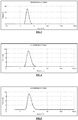
- the figure 3 annexed shows the size distribution of gadolinium oxide cores, measured in DEG by PCS; average: 3.6 nm.
- a layer of functionalized polysiloxane is synthesized by sol-gel.
- the 750 ml core solution is heated to 40 ° C. in an oil bath, with stirring.
- 787 ⁇ L of APTES, 502 ⁇ L of TEOS and 1913 ⁇ L of an aqueous solution of triethylamine at 0.1 mol / L are added to the core solution.
- These additions are repeated a second time after 24 hours of waiting.
- the solution is then left for 48 hours at 40 ° C. with stirring.
- Heart-shell particles having a size of approximately 5 nm are obtained with surface amine functions.
- the figure 4 annexed shows the size distribution of coated polysiloxane cores, measured in the DEG by PCS; average: 4.9 nm.
- DTPABA dimethyl sulfoxide
- DMSO dimethyl sulfoxide
- gadolinium oxide core solution is added to the DTPABA solution. The mixture is stirred for 24 hours.
- the nanoparticles are then precipitated by centrifugation in four drums of 500 ml of acetone. Then the acetone is removed, and the particles are redispersed in 100 mL of water in total. They are then purified by tangential filtration. The large impurities are removed by filtration using a syringe, through a 0.2 micron membrane.
- the aqueous solution of nanoparticles can be stored for several months in the refrigerator after lyophilization, in hermetically sealed pill containers.
- Example 2 To the particles of Example 2, dispersed in water, concentrated hydrochloric acid is added until a pH of 2.5 is reached. We let shake one night.
- the solution is purified by tangential filtration, to remove Gd 3+ ions that have been dissolved from the core of the particles. Particles having a size of about 3.5 nm are obtained.
- the figure 8 annexed shows the particle size distribution decreased with hydrochloric acid, measured in water by PCS; average: 3.4 nm.
- Example 4 Dissolution of the core of the functionalized gadolinium oxide particles DTPABA using free DTPA
- Example 2 To the particles of Example 2, dispersed in water, free DTPA is added in large excess. The pH of the solution is adjusted to 7. It is allowed to stir overnight.
- the solution is purified by tangential filtration, to remove Gd 3+ ions that have been dissolved from the core of the particles, and complexed with free DTPA. Particles having a size of about 4 nm are obtained.
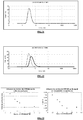
- the figure 9 annexed shows the size distribution of the decreased particles with addition of DTPA, measured in water by PCS; average: 4.3 nm.
- the signal of the particles in relaxometry r 1 decreased by 71% compared to the particles of Example 2.
- a loss of complexed Gd 3+ on surface-grafted DTPA is also probable, which explains the greater decrease in the signal in relaxometry compared to Example 3.
- the solution is purified by tangential filtration to remove Gd 3+ ions that have not been complexed by DTPA. Particles having a size of about 3.5 nm are obtained.
- the figure 10 annexed shows the size distribution of the decreased particles with addition of DTPA, then with addition of Gd3 +, measured in water by PCS; average: 3.7 nm.
- the figure 11 summarizes the experiments of decrease of previous sizes: in thick solid line ( ) the size distribution of the initial particles; in solid line ( ⁇ ) the particle size distribution decreased with HCl; dotted line (----) the particle size distribution decreased with free DTPA.
- Example 5 Dissolution of polysiloxane-coated gadolinium oxide cores directly using a DTPABA addition in large excess
- Particles are synthesized according to the protocol described in Example 2, however varying the amount of DTPABA added for functionalization. This quantity varies from a DTPABA / APTES ratio of 0.5 to 3. The number of APTES is chosen as a parameter because it represents the number of potentially reactive functions on the surface of the particles.
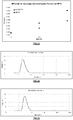
- the figure 23 appended shows the variation in size (left) and relaxometry signal (right), with the number of DTPABA added for functionalization.
- the relaxometry signal is measured in aqueous solution diluted by 10.
- the nanoparticles retain a large size.
- the size of the particles seen in PCS decreases because the heart of the particles dissolves.
- the core of the particles is completely destroyed, and the particle size stagnates at about 2 nm.
- the relaxometric signal of the particles decreases proportionally with size, which reflects the proportional decrease in the amount of gadolinium present in the hearts.
- a layer of functionalized polysiloxane is synthesized by sol-gel.
- the 750 ml core solution is heated to 40 ° C. in an oil bath, with stirring.
- 787 ⁇ L of APTES coupled to 0.75 mg of fluorescein isothiocyanate (FITC), 502 ⁇ L of TEOS and 1913 ⁇ L of an aqueous solution of triethylamine at 0.1 mol / L are added to the core solution.
- FITC fluorescein isothiocyanate
- 502 ⁇ L of TEOS and 1913 ⁇ L of an aqueous solution of triethylamine at 0.1 mol / L are added to the core solution.
- DOTA-NHS 1.12 g of DOTA-NHS are dispersed in 20 ml of anhydrous ethanol. Then 100 mL of the gadolinium oxide core solution is added to the DOTA-NHS solution. The mixture is stirred for 24 hours.
- the nanoparticles are then precipitated in 500 mL of acetone. Then the acetone is removed, and the particles are redispersed in 20 mL of water. They are then purified by tangential filtration. The large impurities are removed by filtration using a syringe, through a 0.2 micron membrane. Particles having a size of about 7 nm are obtained.
- the figure 13 annexed shows the size distribution of polysiloxane coated functionalized cores DOTA-NHS, measured in water by PCS; average: 6.8 nm.
- the figure 14 annexed shows the follow-up of the previous synthesis in granulometry: in solid line ( ⁇ ) the size distribution of the hearts alone; dotted line (----) the size distribution of the coated polysiloxane cores; in thick solid line ( ) the size distribution of polysiloxane coated and functionalized DOTA-NHS cores.
- Example 6 To the particles of Example 6, dispersed in water, free DTPA is added in large excess. The pH of the solution is adjusted to 7. It is allowed to stir overnight.
- the solution is purified by tangential filtration, to remove Gd 3+ ions that have been dissolved from the core of the particles. Particles having a size of about 3.5 nm are obtained.
- the figure 15 annexed shows the size distribution of the decreased particles with addition of DTPA, measured in water by PCS; average: 3.7 nm.
- the signal of the particles in relaxometry r1 has decreased by 84%, which reflects a sharp decrease in the amount of gadolinium present in the particles.
- the figure 16 annexed shows the experience of decreasing sizes: in thick solid lines ( ) the size distribution of the initial particles; in full line ( ⁇ ) the particle size distribution decreased with free DTPA.
- a layer of functionalized polysiloxane is synthesized by sol-gel.
- the 750 ml core solution is heated to 40 ° C. in an oil bath, with stirring.
- 787 ⁇ L of APTES coupled with 0.75 mg of FITC, 502 ⁇ L of TEOS and 1913 ⁇ L of an aqueous solution of triethylamine at 0.1 mol / L are added to the core solution.
- These additions are repeated a second time after 24 hours of waiting, and a third time after 48 hours of waiting.
- the solution is then left for 48 hours at 40 ° C. with stirring.
- Heart-shell particles with a size of about 4.5 nm are obtained with surface amine functions.
- the figure 17 annexed shows the size distribution of coated polysiloxane cores, measured in the DEG by PCS; average: 4.7 nm.
- Particle size is also measured by TEM.
- the figure 18 annexed shows the TEM image of coated polysiloxane cores, dispersed using grafting of polyethylene glycol; the observed sizes are between 4 and 5 nm.
- anhydrous DOTA is dispersed in 20 mL of DMSO. Then 100 mL of the gadolinium oxide core solution is added to the anhydrous DOTA solution. The mixture is stirred for 24 hours.
- Example 8 The nanoparticles of Example 8 are precipitated in 500 ml of acetone. Then the acetone is removed, and the particles are redispersed in 20 mL of water. They are then purified by tangential filtration. The large impurities are removed by filtration using a syringe, through a 0.2 micron membrane. Particles having a size of about 6.5 nm are obtained.
- the figure 19 annexed shows the size distribution of polysiloxane coated hearts, functionalized DOTA anhydrous, measured in water by PCS; average: 6.3 nm.
- the figure 1 annexed shows the TEM image of coated polysiloxane cores, functionalized DOTA anhydrous; the sizes observed are between 3 and 5 nm.
- the molecules of DOTA are not visible at the TEM, we only see the coated hearts.
- the figure 20 annexed shows the follow-up of the synthesis in granulometry: in thick solid line ( ) the size distribution of hearts alone; in solid line ( ⁇ ) the size distribution of coated polysiloxane cores; dotted line (----) the size distribution of the polysiloxane coated and functionalized DOTA anhydrous cores.
- the nanoparticle solution can be stored for several months in the refrigerator after lyophilization.
- a solution of DTPA in water is prepared, then the pH of the solution is reduced to 6.
- DTPA solution is added in large excess. . We let shake one night.
- the solution is purified by tangential filtration, to remove Gd 3+ ions that have been dissolved from the core of the particles. At the same time the particles are passed into the water. Particles having a size of about 3.5 nm are obtained.
- the figure 21 annexed shows the size distribution of the decreased particles with addition of DTPA, measured in water by PCS; average: 3.6 nm.
- the figure 2 annexed shows the TEM image of particles whose size has been reduced. The image was taken under the same conditions as the previous ones. In addition, the local chemical analysis indicates the presence of Gadolinium in a significant amount at the observed location, but no particle is visible. The particles therefore no longer have a crystallized core at this stage.
- the figure 22 annexed shows the experience of decreasing sizes: in thick solid lines ( ) the size distribution of the initial particles; in full line ( ⁇ ) the particle size distribution decreased with free DTPA.
- the signal of the particles in relaxometry r1 decreased by 71%. This reflects a sharp decrease in the amount of gadolinium present in the particles.
- Nanoparticles are synthesized according to the protocol described in Example 8, but the surface functionalization is done with a large excess of anhydrous DOTA (2 DOTA / Gd). Thus all the Gd atoms are directly complexed by DOTA molecules, and there remains a significant part of uncomplexed DOTA.
- the solution is placed in a dialysis membrane of 4-6kDa, and is purified by dialysis in a volume of permuted water ten times larger.
- the dialysis bath is renewed twice, after 24h and 48h. Dialysis is stopped after 72h.
- the contents of the dialysis membranes are recovered.
- the solution appears slightly cloudy, so it is filtered through a 0.2 ⁇ m filter to remove impurities and large particles.
- the final size of the nanoparticles is about 3 nm.
- the figure 24 annexed shows the size distribution of functionalized anhydrous DOTA particles, measured in water by PCS; average: 2.7 nm.
- the nanoparticle solution can be stored for several months in the refrigerator after lyophilization.
- Gadolinium chloride is dissolved in water, and the pH of the solution is reduced to 6.
- the Gd 3+ ions thus obtained are added to the particles of Example 11, in excess.
- the solution is allowed to stir for 24 hours.
- the solution is purified by tangential filtration to remove Gd 3+ ions which would not have been bound to the DOTA molecules.
- a Fisher rat was anesthetized with isoflurane® (gaseous anesthetic), and the nanoparticle solution was injected into the tail vein.
- the rat then underwent MRI (t1 sequence) for an angiographic study. A significant increase in signal was observed at the level of the middle cerebral artery in this animal compared to control animals.
- Europium chloride is dissolved in water, and the pH of the solution is reduced to 6.
- the Eu 3+ ions thus obtained are added to the particles of Example 11, in excess.
- the solution is allowed to stir for 24 hours.
- the solution is purified by tangential filtration to remove the Eu 3+ ions which would not be bound to the DOTA molecules.
- a time-resolved fluorescence measurement shows that the Eu 3+ ions are mainly in the complexed state in the solution.
- the figure 25 annexed shows this spectrum of fluorescence.
- the device used for time resolved fluorescence is a device manufactured by AXINT, for the specific needs of the laboratory.
- Nanoparticles are synthesized according to the protocol described in Example 8, but the surface functionalization is done with a large excess of anhydrous DOTA (2 DOTA / Gd). Thus all the Gd atoms are directly consumed by the DOTA complexes.
- the nanoparticles are precipitated in cans of 500 mL of acetone. Then the acetone is removed, and the particles are redispersed in water. They are then purified by tangential filtration. The large impurities are removed by filtration using a syringe, through a 0.2 micron membrane.
- the figure 31 annexed shows the size distribution of functionalized anhydrous DOTA particles, measured in water by PCS; average: 2.8 nm.
- the nanoparticle solution can be stored for several months in the refrigerator after lyophilization.
- Human T2 cells (ATCC No. CRL-1992 TM) were incubated in Hank's Balanced Salt Solution (HBSS) for 1 hour with the nanoparticles of Example 15 (NP15), with gadolinium concentrations of 0.2; 0.5 and 1mM. After incubation, the cells were washed twice in HBSS.
- HBSS Hank's Balanced Salt Solution
- the viability was assessed by counting grid Malassez after adding Trypan blue (dilution 1/20). For the three gadolinium concentrations present in the incubation medium, cell viability was good because greater than 90%. In this concentration range, the nanoparticles of Example 15 were not toxic.
- the figure 29 attached shows the level of viability of T2 cells for each incubation condition.
- the cells were fixed in a 4% paraformaldehyde solution and then the labeling efficiency was evaluated by measuring the longitudinal relaxation time T 1 .
- the higher r 1 1 / T 1 , the better the marking.
- the labeling with the nanoparticles of Example 15 was therefore better than with the Gd-DOTA complexes.
- the signal increase was 15% with 50,000 cells / mm3.
- the figure 30 Appended shows the labeling efficiency of the particles of Example 15 in comparison with the Gd-DOTA complexes, and as a function of the gadolinium concentration.
- the relaxivity of the particles of Example 15 was measured in relaxometry, the actual amounts of gadolinium being measured by ICP, on a Varian 710-ES apparatus.
- a relaxivity r 1 (of the gadolinium species) 11.4 s -1 mM -1 (gadolinium) -1 .s -1 at 50 MHz, which is about three times higher than that of the Gd-DOTA complex alone. (reduced to the gadolinium species), which is 3.8 s -1 mM -1 (gadolinium) -1 .s -1 at 50 MHz.
- Nanoparticles are synthesized according to the protocol described in Example 8, but the surface functionalization is done with a quantity of DOTA equal to 0.6 DOTA / Gd.
- the solution was then purified by tangential filtration.
- the large impurities were removed by syringe filtration through a 0.2 ⁇ m membrane.
- the figure 32 annexed shows the size distribution of functionalized anhydrous DOTA particles, measured in water by PCS; average: 3.5 nm.
- the nanoparticle solution can be stored for several months in the refrigerator after lyophilization.
- the relaxivity of the particles was measured in relaxometry, the actual quantities of gadolinium being measured by ICP.
- a relaxivity r1 (of the gadolinium species) 11.8 s -1 mM -1 (gadolinium) -1 .s -1 at 50 MHz, which is about three times higher than that of the Gd-DOTA complex alone ( reduced to the gadolinium species), which is 3.8 s -1 mM -1 (gadolinium) -1 .s -1 at 50 MHz.
- a solution is prepared by dissolving an amount of 2.25 g of lutetium chloride salt (LuCl 3 , 6H 2 O) in a volume of 245 mL of DEG. To the solution obtained, an addition of 245 ml of a 0.08 mol / L sodium hydroxide solution in DEG is carried out at room temperature over 10 hours.
- lutetium chloride salt LiCl 3 , 6H 2 O
- the final size of the nanoparticles is about 5 nm.
- the figure 26 annexed shows the size distribution of the lutetium oxide cores, measured in the DEG by PCS; average: 5.0 nm.
- the 490 mL of lutetium core solution of Example 18 are diluted by two with 490 mL of DEG.
- a layer of functionalized polysiloxane is synthesized by sol-gel.
- the core solution is heated to 40 ° C in an oil bath, with stirring.
- 811 ⁇ L of APTES, 516 ⁇ L of TEOS and 1965 ⁇ L of a 0.1 mol / L aqueous solution of triethylamine are added to the core solution. The solution is then left for 48 hours at 40 ° C. with stirring.
- Heart-shell particles having a size of about 8 nm are obtained with surface amine functions.
- the figure 27 annexed shows the size distribution of lutetium oxide cores coated polysiloxane; average: 8.3 nm.
- DTPABA is dispersed in 2 mL of DMSO. Then 12 mL of the heart solution is added to the DTPABA solution. The mixture is stirred for 24 hours.
- the nanoparticles are then precipitated in 100 mL of acetone. Then the acetone is removed, and the particles are redispersed in 20 mL of water. They are then purified by tangential filtration. Particles having a size of about 2.5 nm are obtained.
- DTPABA in large excess compared to lutetium (4 DTPABA / Lu), dissolved the heart of the particles in part, hence their small size.
- the figure 28 annexed shows the size distribution of functionalized DTPA particles; average: 2.4 nm.
- the nanoparticle solution can be stored for several months in the refrigerator after lyophilization.
- Example 20 Complexation of lutetium oxide particles with Gd 3+ ions
- Gadolinium chloride is dissolved in water, and the pH of the solution is reduced to 6.
- the Gd 3+ ions thus obtained are added to the lutetium particles of Example 19, in excess.
- the solution is allowed to stir 24h.
- the solution is purified by tangential filtration to remove Gd 3+ ions which would not have been bound to the
Landscapes
- Health & Medical Sciences (AREA)
- Chemical & Material Sciences (AREA)
- Engineering & Computer Science (AREA)
- Nanotechnology (AREA)
- Life Sciences & Earth Sciences (AREA)
- Epidemiology (AREA)
- Animal Behavior & Ethology (AREA)
- General Health & Medical Sciences (AREA)
- Public Health (AREA)
- Veterinary Medicine (AREA)
- Nuclear Medicine, Radiotherapy & Molecular Imaging (AREA)
- Radiology & Medical Imaging (AREA)
- Medicinal Chemistry (AREA)
- Pharmacology & Pharmacy (AREA)
- Biomedical Technology (AREA)
- Immunology (AREA)
- Molecular Biology (AREA)
- Bioinformatics & Cheminformatics (AREA)
- Medicines Containing Antibodies Or Antigens For Use As Internal Diagnostic Agents (AREA)
Applications Claiming Priority (3)
| Application Number | Priority Date | Filing Date | Title |
|---|---|---|---|
| FR1053389A FR2959502B1 (fr) | 2010-04-30 | 2010-04-30 | Nanoparticules ultrafines a matrice polyorganosiloxane fonctionnalisee et incluant des complexes metalliques ; leur procede d'obtention et leurs applications en imagerie medicale et/ou therapie |
| PCT/EP2011/056972 WO2011135101A2 (fr) | 2010-04-30 | 2011-05-02 | Nanoparticules ultrafines a matrice polyorganosiloxane fonctionnalisee et incluant des complexes metalliques; leur procede d'obtention et leurs applications en imagerie medicale et/ou therapie |
| EP11716589.4A EP2563408B1 (fr) | 2010-04-30 | 2011-05-02 | Nanoparticules ultrafines a matrice polyorganosiloxane fonctionnalisee et incluant des complexes metalliques; leur procede d'obtention et leurs applications en imagerie medicale |
Related Parent Applications (2)
| Application Number | Title | Priority Date | Filing Date |
|---|---|---|---|
| EP11716589.4A Division EP2563408B1 (fr) | 2010-04-30 | 2011-05-02 | Nanoparticules ultrafines a matrice polyorganosiloxane fonctionnalisee et incluant des complexes metalliques; leur procede d'obtention et leurs applications en imagerie medicale |
| EP11716589.4A Division-Into EP2563408B1 (fr) | 2010-04-30 | 2011-05-02 | Nanoparticules ultrafines a matrice polyorganosiloxane fonctionnalisee et incluant des complexes metalliques; leur procede d'obtention et leurs applications en imagerie medicale |
Publications (1)
| Publication Number | Publication Date |
|---|---|
| EP3517136A1 true EP3517136A1 (fr) | 2019-07-31 |
Family
ID=43384584
Family Applications (2)
| Application Number | Title | Priority Date | Filing Date |
|---|---|---|---|
| EP19159482.9A Pending EP3517136A1 (fr) | 2010-04-30 | 2011-05-02 | Nanoparticules ultrafines a matrice polyorganosiloxane fonctionnalisee et incluant des complexes metalliques pour leurs applications en imagerie diagnostique et/ou therapie |
| EP11716589.4A Active EP2563408B1 (fr) | 2010-04-30 | 2011-05-02 | Nanoparticules ultrafines a matrice polyorganosiloxane fonctionnalisee et incluant des complexes metalliques; leur procede d'obtention et leurs applications en imagerie medicale |
Family Applications After (1)
| Application Number | Title | Priority Date | Filing Date |
|---|---|---|---|
| EP11716589.4A Active EP2563408B1 (fr) | 2010-04-30 | 2011-05-02 | Nanoparticules ultrafines a matrice polyorganosiloxane fonctionnalisee et incluant des complexes metalliques; leur procede d'obtention et leurs applications en imagerie medicale |
Country Status (11)
| Country | Link |
|---|---|
| US (4) | US20130195766A1 (pl) |
| EP (2) | EP3517136A1 (pl) |
| DK (1) | DK2563408T3 (pl) |
| ES (1) | ES2731649T3 (pl) |
| FR (1) | FR2959502B1 (pl) |
| HU (1) | HUE044384T4 (pl) |
| PL (1) | PL2563408T3 (pl) |
| PT (1) | PT2563408T (pl) |
| SI (1) | SI2563408T1 (pl) |
| TR (1) | TR201909432T4 (pl) |
| WO (1) | WO2011135101A2 (pl) |
Families Citing this family (22)
| Publication number | Priority date | Publication date | Assignee | Title |
|---|---|---|---|---|
| FR2959502B1 (fr) | 2010-04-30 | 2012-09-07 | Nanoh | Nanoparticules ultrafines a matrice polyorganosiloxane fonctionnalisee et incluant des complexes metalliques ; leur procede d'obtention et leurs applications en imagerie medicale et/ou therapie |
| FR2989280B1 (fr) | 2012-04-13 | 2017-02-24 | Univ Claude Bernard Lyon | Nanoparticules ultrafines comme agent de contraste multimodal |
| US10472556B2 (en) | 2014-12-15 | 2019-11-12 | Total Sa | Nano-inhibitors |
| EP3192530A1 (fr) | 2016-01-15 | 2017-07-19 | Institut National De La Sante Et De La Recherche Medicale (Inserm) | Nanoparticules ultrafines comme agent d'imagerie pour el diagnostic d'une dysfonction rénale |
| EP3551231A4 (en) | 2016-12-08 | 2020-08-12 | The Brigham and Women's Hospital, Inc. | BISMUTH-GADOLINIUM NANOPARTICLES |
| AU2018280870B2 (en) * | 2017-06-09 | 2024-07-11 | Centre National De La Recherche Scientifique - Cnrs | Method for synthesizing silica nanoparticles |
| AU2018283039A1 (en) | 2017-06-14 | 2019-11-14 | Dana-Farber Cancer Institute, Inc. | B-cell maturation antigen (BCMA)-directed nanoparticles |
| EP3424533A1 (en) * | 2017-07-05 | 2019-01-09 | Nh Theraguix | Methods for treating tumors |
| FR3072281B1 (fr) * | 2017-10-13 | 2020-12-04 | Nh Theraguix | Nanovecteurs et utilisations, en particulier pour le traitement de tumeurs |
| FR3075649A1 (fr) * | 2017-12-22 | 2019-06-28 | Mexbrain | Dispositif pour le maintien de l'homeostasie metallique, et ses utilisations |
| CA3148734A1 (en) | 2019-07-29 | 2021-02-04 | Nh Theraguix | High-z-element containing nanoparticles for use in the treatement of tumors |
| FR3099883A1 (fr) | 2019-08-14 | 2021-02-19 | Nh Theraguix | Nanoparticules pour le traitement du cancer par rayonnement de radiofréquence |
| EP3795178A1 (en) | 2019-09-19 | 2021-03-24 | Nh Theraguix | Methods for triggering m1 macrophage polarization |
| WO2021116748A2 (en) * | 2019-12-11 | 2021-06-17 | Novocure Gmbh | Compositions and methods for imaging a cell |
| AU2021270780A1 (en) | 2020-05-15 | 2022-11-24 | Centre Georges-François Leclerc | Methods for image-guided radiotherapy |
| FR3116216B1 (fr) | 2020-11-19 | 2023-10-27 | Nh Theraguix | Procédé de préparation de nanoparticules |
| FR3116197A1 (fr) | 2020-11-19 | 2022-05-20 | Nh Theraguix | Procédé de traitement de tumeurs par captation du cuivre et/ou du fer |
| CN120359049A (zh) | 2022-07-13 | 2025-07-22 | 蒙彼利埃大学 | 纳米颗粒和放射性药物的联合疗法 |
| EP4605019A2 (en) | 2022-10-18 | 2025-08-27 | Nh Theraguix | Methods for imaging using spcct |
| WO2024236048A1 (en) | 2023-05-16 | 2024-11-21 | Nh Theraguix | Combination therapy for treating tumors with radiotherapy |
| FR3151755A1 (fr) | 2023-07-31 | 2025-02-07 | Nh Theraguix | Procédé de quantification de la concentration d’un agent de contraste et dispositif associé |
| WO2025072955A1 (en) * | 2023-09-30 | 2025-04-03 | The Brigham And Women's Hospital, Inc. | Novel compositions, methods of manufacture and uses thereof |
Citations (5)
| Publication number | Priority date | Publication date | Assignee | Title |
|---|---|---|---|---|
| WO2005061009A2 (en) * | 2003-06-20 | 2005-07-07 | University Of Maryland, Baltimore | Microparticles for microarterial imaging and radiotherapy |
| FR2867180A1 (fr) * | 2004-03-02 | 2005-09-09 | Univ Claude Bernard Lyon | Nanoparticules hybrides comprenant un coeur de ln203 porteuses de ligands biologiques et leur procede de preparation |
| WO2007124131A2 (en) | 2006-04-20 | 2007-11-01 | The University Of North Carolina At Chapel Hill | Hybrid nanomaterials as multimodal imaging contrast agents |
| FR2922106A1 (fr) | 2007-10-16 | 2009-04-17 | Univ Claude Bernard Lyon I Eta | Utilisation de nanoparticules a base de lanthanides comme agents radiosensibilisants. |
| FR2930890A1 (fr) | 2008-05-06 | 2009-11-13 | Univ Claude Bernard Lyon I Eta | Nouveaux agents de radiotherapie ciblee ou de curietherapie a base d'oxydes ou d'oxo-hydroxydes de terre rare |
Family Cites Families (9)
| Publication number | Priority date | Publication date | Assignee | Title |
|---|---|---|---|---|
| EP1289571B1 (en) * | 2000-05-12 | 2004-07-21 | University Hospital | Prochelators of radiometal labeled molecules |
| WO2009142754A1 (en) * | 2008-05-22 | 2009-11-26 | Goverment Of The United States Of America, As Represented By The Secretary, Department Of Health And Human Services | Dendritic conjugates and methods of use |
| FR2959502B1 (fr) | 2010-04-30 | 2012-09-07 | Nanoh | Nanoparticules ultrafines a matrice polyorganosiloxane fonctionnalisee et incluant des complexes metalliques ; leur procede d'obtention et leurs applications en imagerie medicale et/ou therapie |
| AU2018280870B2 (en) | 2017-06-09 | 2024-07-11 | Centre National De La Recherche Scientifique - Cnrs | Method for synthesizing silica nanoparticles |
| EP3424533A1 (en) | 2017-07-05 | 2019-01-09 | Nh Theraguix | Methods for treating tumors |
| FR3072281B1 (fr) | 2017-10-13 | 2020-12-04 | Nh Theraguix | Nanovecteurs et utilisations, en particulier pour le traitement de tumeurs |
| CA3148734A1 (en) | 2019-07-29 | 2021-02-04 | Nh Theraguix | High-z-element containing nanoparticles for use in the treatement of tumors |
| FR3099883A1 (fr) | 2019-08-14 | 2021-02-19 | Nh Theraguix | Nanoparticules pour le traitement du cancer par rayonnement de radiofréquence |
| EP3795178A1 (en) | 2019-09-19 | 2021-03-24 | Nh Theraguix | Methods for triggering m1 macrophage polarization |
-
2010
- 2010-04-30 FR FR1053389A patent/FR2959502B1/fr active Active
-
2011
- 2011-05-02 DK DK11716589.4T patent/DK2563408T3/da active
- 2011-05-02 PL PL11716589T patent/PL2563408T3/pl unknown
- 2011-05-02 US US13/695,090 patent/US20130195766A1/en not_active Abandoned
- 2011-05-02 HU HUE11716589A patent/HUE044384T4/hu unknown
- 2011-05-02 EP EP19159482.9A patent/EP3517136A1/fr active Pending
- 2011-05-02 WO PCT/EP2011/056972 patent/WO2011135101A2/fr not_active Ceased
- 2011-05-02 ES ES11716589T patent/ES2731649T3/es active Active
- 2011-05-02 SI SI201131749T patent/SI2563408T1/sl unknown
- 2011-05-02 PT PT11716589T patent/PT2563408T/pt unknown
- 2011-05-02 TR TR2019/09432T patent/TR201909432T4/tr unknown
- 2011-05-02 EP EP11716589.4A patent/EP2563408B1/fr active Active
-
2017
- 2017-08-15 US US15/677,167 patent/US10987435B2/en active Active
-
2018
- 2018-05-23 US US15/987,500 patent/US11497818B2/en active Active
-
2022
- 2022-10-04 US US17/960,002 patent/US12533426B2/en active Active
Patent Citations (6)
| Publication number | Priority date | Publication date | Assignee | Title |
|---|---|---|---|---|
| WO2005061009A2 (en) * | 2003-06-20 | 2005-07-07 | University Of Maryland, Baltimore | Microparticles for microarterial imaging and radiotherapy |
| FR2867180A1 (fr) * | 2004-03-02 | 2005-09-09 | Univ Claude Bernard Lyon | Nanoparticules hybrides comprenant un coeur de ln203 porteuses de ligands biologiques et leur procede de preparation |
| FR2867180B1 (fr) | 2004-03-02 | 2006-06-16 | Univ Claude Bernard Lyon | Nanoparticules hybrides comprenant un coeur de ln203 porteuses de ligands biologiques et leur procede de preparation |
| WO2007124131A2 (en) | 2006-04-20 | 2007-11-01 | The University Of North Carolina At Chapel Hill | Hybrid nanomaterials as multimodal imaging contrast agents |
| FR2922106A1 (fr) | 2007-10-16 | 2009-04-17 | Univ Claude Bernard Lyon I Eta | Utilisation de nanoparticules a base de lanthanides comme agents radiosensibilisants. |
| FR2930890A1 (fr) | 2008-05-06 | 2009-11-13 | Univ Claude Bernard Lyon I Eta | Nouveaux agents de radiotherapie ciblee ou de curietherapie a base d'oxydes ou d'oxo-hydroxydes de terre rare |
Non-Patent Citations (15)
| Title |
|---|
| ANNA MIGNOT ET AL: "A Top-Down Synthesis Route to Ultrasmall Multifunctional Gd-Based Silica Nanoparticles for Theranostic Applications", CHEMISTRY - A EUROPEAN JOURNAL, vol. 19, no. 19, 3 May 2013 (2013-05-03), DE, pages 6122 - 6136, XP055283579, ISSN: 0947-6539, DOI: 10.1002/chem.201203003 * |
| ANNE-CHARLOTTE FAURE ET AL: "Core/shell nanoparticles for multiple biological detection with enhanced sensitivity and kinetics", NANOTECHNOLOGY, vol. 19, no. 48, 3 December 2008 (2008-12-03), pages 485103, XP020144895, ISSN: 0957-4484, DOI: 10.1088/0957-4484/19/48/485103 * |
| BARBILLON G ET AL: "How nanoparticles encapsulating fluorophores allow a double detection of biomolecules by localized surface plasmon resonance and luminescence", NANOTECHNOLOGY, vol. 19, no. 3, 23 January 2008 (2008-01-23), pages 35705, XP020129707, ISSN: 0957-4484 * |
| BRIDOT J-L ET AL: "Hybrid gadolinium oxide nanoparticles: Multimodal contrast agents for in vivo imaging", JOURNAL OF THE AMERICAN CHEMICAL SOCIETY, vol. 129, no. 16, 31 March 2007 (2007-03-31), pages 5076 - 5084, XP002481277, ISSN: 0002-7863, [retrieved on 20070331], DOI: 10.1021/JA068356J * |
| BRIDOT, JEAN-LUC ET AL: "Hybrid gadolinium oxide nanoparticles combining imaging and therapy", JOURNAL OF MATERIALS CHEMISTRY, vol. 19, no. 16, 2009, pages 2328, XP002615956, ISSN: 0959-9428, DOI: 10.1039/b815836c * |
| C. LOUIS ET AL., CHEM. MAT., vol. 17, 2005, pages 1673 |
| CLAIRE BERNHARD ET AL: "DOTAGA-Anhydride: A Valuable Building Block for the Preparation of DOTA-Like Chelating Agents", CHEMISTRY - A EUROPEAN JOURNAL, vol. 18, no. 25, 21 May 2012 (2012-05-21), DE, pages 7834 - 7841, XP055465245, ISSN: 0947-6539, DOI: 10.1002/chem.201200132 * |
| FREDRIK SÖDERLIND ET AL: "Colloidal synthesis and characterization of ultrasmall perovskite GdFeO3 nanocrystals", NANOTECHNOLOGY, vol. 19, no. 8, 27 February 2008 (2008-02-27), pages 85608, XP020129856, ISSN: 0957-4484 * |
| KOOLE ROLF ET AL: "Magnetic quantum dots for multimodal imaging", WILEY INTERDISCIPLINARY REVIEWS-NANOMEDICINE AND NANOBIOTECHNOLOGY, vol. 1, no. 5, September 2009 (2009-09-01), pages 475 - 491, XP002615957, ISSN: 1939-5116, DOI: 10.1002/wnan.014 * |
| LOUIS C ET AL: "Nanosized hybrid particles with double luminescence for biological labeling", CHEMISTRY OF MATERIALS, vol. 17, no. 7, 5 April 2005 (2005-04-05), pages 1673 - 1682, XP002333998, ISSN: 1520-5002, DOI: 10.1021/CM0480162 * |
| O. TILLEMENT ET AL., J. AM. CHEM. SOC., vol. 129, 2007, pages 5076 |
| P. PERRIAT ET AL., J. COLL. INT. SCI, vol. 273, 2004, pages 191 |
| P. PERRIAT ET AL., J. PHYS. CHEM. C, vol. 113, 2009, pages 4038 |
| ROUX S ET AL: "Multifunctional gadolinium oxide nanoparticles: Towards image-guided therapy", IMAGING IN MEDICINE, vol. 2, no. 2, April 2010 (2010-04-01), pages 211 - 223, XP008131128, ISSN: 1755-5191, DOI: 10.2217/IIM.10.5 * |
| VOISIN PIERRE ET AL: "Use of lanthanide-grafted inorganic nanoparticles as effective contrast agents for cellular uptake imaging.", BIOCONJUGATE CHEMISTRY, vol. 18, no. 4, July 2007 (2007-07-01), pages 1053 - 1063, XP002615958, ISSN: 1043-1802, DOI: 10.1021/bc060269t * |
Also Published As
| Publication number | Publication date |
|---|---|
| PT2563408T (pt) | 2019-07-15 |
| WO2011135101A3 (fr) | 2012-05-31 |
| TR201909432T4 (tr) | 2019-07-22 |
| EP2563408A2 (fr) | 2013-03-06 |
| US12533426B2 (en) | 2026-01-27 |
| ES2731649T3 (es) | 2019-11-18 |
| FR2959502B1 (fr) | 2012-09-07 |
| US10987435B2 (en) | 2021-04-27 |
| US20180264145A1 (en) | 2018-09-20 |
| US20130195766A1 (en) | 2013-08-01 |
| SI2563408T1 (sl) | 2019-08-30 |
| FR2959502A1 (fr) | 2011-11-04 |
| HUE044384T2 (hu) | 2019-10-28 |
| HUE044384T4 (hu) | 2020-10-28 |
| EP2563408B1 (fr) | 2019-04-10 |
| US11497818B2 (en) | 2022-11-15 |
| DK2563408T3 (da) | 2019-07-15 |
| US20230109283A1 (en) | 2023-04-06 |
| US20180008729A1 (en) | 2018-01-11 |
| WO2011135101A2 (fr) | 2011-11-03 |
| PL2563408T3 (pl) | 2019-09-30 |
Similar Documents
| Publication | Publication Date | Title |
|---|---|---|
| EP2563408B1 (fr) | Nanoparticules ultrafines a matrice polyorganosiloxane fonctionnalisee et incluant des complexes metalliques; leur procede d'obtention et leurs applications en imagerie medicale | |
| EP2200659B1 (fr) | Utilisation de nanoparticules a base de lanthanides comme agents radiosensibilisants | |
| Gerion et al. | Paramagnetic silica-coated nanocrystals as an advanced MRI contrast agent | |
| Yang et al. | Beyond the roles in biomimetic chemistry: an insight into the intrinsic catalytic activity of an enzyme for tumor-selective phototheranostics | |
| EP2836237B1 (fr) | Nanoparticules ultrafines comme agent de contraste multimodal | |
| KR101043407B1 (ko) | 암 표적성이 우수한 단백질 복합체 및 이의 제조방법 | |
| US20100119458A1 (en) | Compositions Containing Metal Oxide Particles and Their Use | |
| US20110158901A1 (en) | Chitosan-based nanoparticles and methods for making and using the same | |
| CA2558033A1 (fr) | Nanoparticules hybrides comprenant un coeur de ln2o3 porteuses de ligands biologiques et leur procede de preparation | |
| JP2020527538A (ja) | シリカナノ粒子の合成方法 | |
| Ji et al. | Gadolinium-incorporated carbon nanodots for t 1-weighted magnetic resonance imaging | |
| Yin et al. | Fluorescent oligo (p-phenyleneethynylene) contained amphiphiles-encapsulated magnetic nanoparticles for targeted magnetic resonance and two-photon optical imaging in vitro and in vivo | |
| EP3694560A1 (fr) | Nanovecteurs et utilisations | |
| Lee et al. | Amphiphilic hyaluronic acid-based nanoparticles for tumor-specific optical/MR dual imaging | |
| Hu et al. | Rational design of multimodal and multifunctional InP quantum dot nanoprobes for cancer: in vitro and in vivo applications | |
| US20110318275A1 (en) | Multifunctional contrast agent using biocompatible polymer and preparation method | |
| EP2823016A1 (fr) | Particules d'oxyde à base de terres rares et utilisation notamment en imagerie | |
| FR3116216A1 (fr) | Procédé de préparation de nanoparticules | |
| WO2021028642A1 (fr) | Nanoparticules pour le traitement du cancer par rayonnement de radiofréquence | |
| FR2981849A1 (fr) | Nanoparticules fonctionnalisees pour le ciblage des proteoglycanes et leurs applications | |
| FR2930890A1 (fr) | Nouveaux agents de radiotherapie ciblee ou de curietherapie a base d'oxydes ou d'oxo-hydroxydes de terre rare | |
| Abtahi | Surface Modification of NaLnF4 and NbO2 Nanoparticles With Bisphosphonate Derivatives for Mass Cytometry Applications |
Legal Events
| Date | Code | Title | Description |
|---|---|---|---|
| PUAI | Public reference made under article 153(3) epc to a published international application that has entered the european phase |
Free format text: ORIGINAL CODE: 0009012 |
|
| STAA | Information on the status of an ep patent application or granted ep patent |
Free format text: STATUS: THE APPLICATION HAS BEEN PUBLISHED |
|
| AC | Divisional application: reference to earlier application |
Ref document number: 2563408 Country of ref document: EP Kind code of ref document: P |
|
| AK | Designated contracting states |
Kind code of ref document: A1 Designated state(s): AL AT BE BG CH CY CZ DE DK EE ES FI FR GB GR HR HU IE IS IT LI LT LU LV MC MK MT NL NO PL PT RO RS SE SI SK SM TR |
|
| STAA | Information on the status of an ep patent application or granted ep patent |
Free format text: STATUS: REQUEST FOR EXAMINATION WAS MADE |
|
| 17P | Request for examination filed |
Effective date: 20200115 |
|
| RBV | Designated contracting states (corrected) |
Designated state(s): AL AT BE BG CH CY CZ DE DK EE ES FI FR GB GR HR HU IE IS IT LI LT LU LV MC MK MT NL NO PL PT RO RS SE SI SK SM TR |
|
| STAA | Information on the status of an ep patent application or granted ep patent |
Free format text: STATUS: EXAMINATION IS IN PROGRESS |
|
| RIC1 | Information provided on ipc code assigned before grant |
Ipc: B82Y 5/00 20110101ALN20200701BHEP Ipc: A61K 41/00 20200101ALI20200701BHEP Ipc: A61K 49/18 20060101AFI20200701BHEP Ipc: B82Y 15/00 20110101ALN20200701BHEP |
|
| 17Q | First examination report despatched |
Effective date: 20200722 |
|
| 111L | Licence recorded |
Designated state(s): AL AT BE BG CH CY CZ DE DK EE ES FI FR GB GR HR HU IE IS IT LT LU LV MC MK MT NL NO PL PT RO RS SE SI SK SM TR Free format text: EXCLUSIVE LICENSE Name of requester: NH THERAGUIX, FR Effective date: 20200826 Designated state(s): AL AT BE BG CH CY CZ DE DK EE ES FI FR GB GR HR HU IE IS IT LT LU LV MC MK MT NL NO PL PT RO RS SE SI SK SM TR Free format text: EXCLUSIVE LICENSE Name of requester: PULSALYS, FR Effective date: 20200826 |
|
| RIC1 | Information provided on ipc code assigned before grant |
Ipc: B82Y 15/00 20110101ALN20240502BHEP Ipc: B82Y 5/00 20110101ALN20240502BHEP Ipc: A61K 49/00 20060101ALI20240502BHEP Ipc: A61K 41/00 20060101ALI20240502BHEP Ipc: A61K 49/18 20060101AFI20240502BHEP |