EP3514539B1 - Testverfahren und testreagens für herztroponin - Google Patents
Testverfahren und testreagens für herztroponin Download PDFInfo
- Publication number
- EP3514539B1 EP3514539B1 EP17850867.7A EP17850867A EP3514539B1 EP 3514539 B1 EP3514539 B1 EP 3514539B1 EP 17850867 A EP17850867 A EP 17850867A EP 3514539 B1 EP3514539 B1 EP 3514539B1
- Authority
- EP
- European Patent Office
- Prior art keywords
- antibody
- cardiac troponin
- pretreatment
- sample
- ctni
- Prior art date
- Legal status (The legal status is an assumption and is not a legal conclusion. Google has not performed a legal analysis and makes no representation as to the accuracy of the status listed.)
- Active
Links
Images

Classifications
-
- G—PHYSICS
- G01—MEASURING; TESTING
- G01N—INVESTIGATING OR ANALYSING MATERIALS BY DETERMINING THEIR CHEMICAL OR PHYSICAL PROPERTIES
- G01N33/00—Investigating or analysing materials by specific methods not covered by groups G01N1/00 - G01N31/00
- G01N33/48—Biological material, e.g. blood, urine; Haemocytometers
- G01N33/50—Chemical analysis of biological material, e.g. blood, urine; Testing involving biospecific ligand binding methods; Immunological testing
- G01N33/53—Immunoassay; Biospecific binding assay; Materials therefor
- G01N33/5306—Improving reaction conditions, e.g. reduction of non-specific binding, promotion of specific binding
-
- G—PHYSICS
- G01—MEASURING; TESTING
- G01N—INVESTIGATING OR ANALYSING MATERIALS BY DETERMINING THEIR CHEMICAL OR PHYSICAL PROPERTIES
- G01N33/00—Investigating or analysing materials by specific methods not covered by groups G01N1/00 - G01N31/00
- G01N33/48—Biological material, e.g. blood, urine; Haemocytometers
- G01N33/50—Chemical analysis of biological material, e.g. blood, urine; Testing involving biospecific ligand binding methods; Immunological testing
- G01N33/53—Immunoassay; Biospecific binding assay; Materials therefor
-
- G—PHYSICS
- G01—MEASURING; TESTING
- G01N—INVESTIGATING OR ANALYSING MATERIALS BY DETERMINING THEIR CHEMICAL OR PHYSICAL PROPERTIES
- G01N33/00—Investigating or analysing materials by specific methods not covered by groups G01N1/00 - G01N31/00
- G01N33/48—Biological material, e.g. blood, urine; Haemocytometers
- G01N33/50—Chemical analysis of biological material, e.g. blood, urine; Testing involving biospecific ligand binding methods; Immunological testing
- G01N33/68—Chemical analysis of biological material, e.g. blood, urine; Testing involving biospecific ligand binding methods; Immunological testing involving proteins, peptides or amino acids
- G01N33/6887—Chemical analysis of biological material, e.g. blood, urine; Testing involving biospecific ligand binding methods; Immunological testing involving proteins, peptides or amino acids from muscle, cartilage or connective tissue
Definitions
- the present invention relates to a measurement method and a use of a measurement reagent for immunoassay of cardiac troponin I.
- Cardiac troponin is involved in regulation of myocardial contraction, and present as a complex constituted by three kinds of subunits: cardiac troponin I, troponin C, and cardiac troponin T. Both cardiac troponin I and cardiac troponin T are expressed specifically in the heart, and released into blood when cardiomyocytes are injured. They are therefore used as blood markers in diagnosis of myocardial infarction and monitoring of heart diseases.
- Patent Document 1 describes that stabilization of cardiac troponin in a standard solution is possible by use of a matrix containing a prescribed anionic surfactant (alkyl group having one sulfonate group).
- Patent Document 2 describes that a divalent cation can be used in immunoassay of cardiac troponin.
- Patent Document 3 discloses a method of improving the stability of troponin in solution wherein troponin is combined to a carrier molecule that is immunologically unreactive in a buffer comprising a surfactant.
- Patent Document 4 discloses a method of measurement of a specific protein, such as troponin, in a processed food product, extracted under heat using a buffer solution comprising a surfactant.
- Patent Document 5 relates to immunogenic products comprising amyloid beta and a method of diagnosing an amyloidosis.
- Non-patent document 6 discloses extraction of troponin from rabbit muscle and analysis of the extracted troponin.
- Blood cardiac troponin is widely used as a diagnostic marker for myocardial infarction.
- it is present as a complex composed of cardiac troponin I, troponin C, and cardiac troponin T, and also forms a complex with blood components.
- cardiac troponin I easily changes its properties depending on conditions of other components and the like. For example, it shows interaction with heparin in the presence of heparin, and is easily degraded by blood protease.
- cardiac troponin I does not show stable values in immunoassays using anti-troponin antibodies, it has been regarded as an instable protein.
- a measured value of cardiac troponin I obtained using serum as a blood sample is not necessarily the same as a measured value of cardiac troponin I obtained using plasma as a blood sample.
- blood collection tubes containing various anticoagulants for example, heparin, EDTA, and citric acid
- the measured value of cardiac troponin I in plasma may vary depending on the type of the anticoagulant used for the preparation of the plasma.
- measurement reagents using different antibodies for the detection show deviations of the measured value of troponin.
- An object of the present invention is to provide a measurement method and a use of a measurement reagent for immunoassay of cardiac troponin I as set out in the appended claims, which enable more accurate measurement of the amount of cardiac troponin I contained in a serum or plasma sample irrespective of the presence or absence of other components.
- a highly reproducible measured value of cardiac troponin can be obtained by carrying out, before subjecting the biological sample to an immune reaction, a pretreatment step of mixing with a pretreatment liquid containing one or both of a surfactant and an acidifier, irrespective of the type of the biological sample and the presence or absence of other components.
- a measurement method and a use of a measurement reagent for immunoassay of cardiac troponin I which enable accurate measurement of the amount of cardiac troponin I contained in a serum or plasma sample and the presence or absence of other components is provided.
- a measurement method and a use of a measurement reagent for immunoassay of cardiac troponin I which enable stable and sensitive measurement of cardiac troponin I is provided.
- the cardiac troponin to be measured by the method of the present invention is cardiac troponin I.
- Cardiac troponin I (cTnI) is one of the three kinds of subunits (troponins I, C, and T) constituting the cardiac troponin complex, which is involved in regulation of myocardial contraction.
- the cardiac troponin to be measured by the present invention is cardiac troponin derived from an arbitrary animal.
- the cardiac troponin is preferably cardiac troponin derived from a mammal (for example, a primate such as human, monkey, or chimpanzee; a rodent such as mouse, rat, or rabbit; a pet animal such as dog or cat; a domestic animal such as pig or cow; or a working animal such as horse or sheep), more preferably cardiac troponin derived from a primate, especially preferably cardiac troponin derived from human.
- a mammal for example, a primate such as human, monkey, or chimpanzee; a rodent such as mouse, rat, or rabbit; a pet animal such as dog or cat; a domestic animal such as pig or cow; or a working animal such as horse or sheep
- cardiac troponin I for example, GenBank:CAA62301.1.
- the cardiac troponin I derived from human is not limited to cardiac troponin I having the amino acid sequence referred by the above number, and may also be a mutant (for example, a naturally occurring mutant) thereof.
- the cardiac troponin I to be measured by the present invention may be present as a free form, in the form of a complex with troponin C and/or troponin T, or in the form of a complex with another molecule such as an autoantibody, in the biological sample.
- an amino acid sequence of troponin C derived from human see, for example, GenBank:AAA36772.1.
- an amino acid sequence of cardiac troponin T derived from human see, for example, GenBank:CAA52818.1.
- the method of the present invention is a method in which cardiac troponin I present in a biological sample is measured using immune reaction by reacting the biological sample with an antibody.
- the method is characterized in that it includes a pretreatment step of mixing the biological sample with a pretreatment liquid before the immune reaction (reaction step).
- reaction step By the pretreatment step, cardiac troponin I can be brought into a free state, so that the influence of interactions with other components such as proteins can be reduced.
- the pretreatment liquid contains an anionic surfactant or an acidifier.
- the volume ratio between the biological sample and the pretreatment liquid to be mixed in the pretreatment step is preferably 1:10 to 10:1, more preferably 1:5 to 5:1, still more preferably 1:3 to 3:1.
- the biological sample to be used in the present invention is serum or plasma.
- the surfactant to be contained in the pretreatment liquid is an anionic surfactant.
- the anionic surfactant include sodium dodecyl sulfate (SDS), sodium N -lauroyl sarcosinate (NLS), lithium dodecyl sulfate, sodium dodecylbenzene sulfonate, and deoxycholic acid. SDS or NLS may be especially preferably used.
- the surfactant needs to have a concentration sufficient for releasing of cardiac troponin I from other proteins and the like.
- the concentration during the pretreatment of the mixed liquid prepared by mixing with the biological sample is preferably 0.1 to 12.5%, more preferably 0.25 to 10%, still more preferably 0.5 to 7.5%.
- the surfactant concentration is 0.1 to 12.5%, sufficient release of cardiac troponin I and suppression of precipitation and the like can be effectively achieved.
- Preferred examples of the acidifier contained in the pretreatment liquid include hydrochloric acid, sulfuric acid, and acetic acid.
- the normality of the acid in the pretreatment liquid in terms of the concentration during the pretreatment, is preferably more than 0 N and not more than 0.5 N, especially preferably 0.03 N to 0.125 N.
- a cationic surfactant is preferably added in order to prevent occurrence of precipitation upon mixing with the biological sample.
- the cationic surfactant is especially preferably a cationic surfactant having, in a single molecule, a single-chain alkyl group having 10 or more carbon atoms, and a tertiary amine or a quaternary ammonium salt.
- Examples of such a surfactant include decyltrimethylammonium chloride, dodecyltrimethylammonium chloride, tetradecyltrimethylammonium chloride, hexadecyltrimethylammonium chloride (C16TAC), decyltrimethylammonium bromide, dodecyltrimethylammonium bromide, tetradecyltrimethylammonium bromide, hexadecyltrimethylammonium bromide (CTAB), laurylpyridinium chloride, tetradecylpyridinium chloride, and cetylpyridinium chloride.
- the amount of the cationic surfactant to be added is preferably 0.01% to 15%, more preferably 0.05% to 10%.
- a reducing agent is also preferably used for the pretreatment liquid.
- the reducing agent any of known reducing agents such as 2-(diethylamino)ethanethiol hydrochloride (DEAET), tris(2-carboxyethyl)phosphine hydrochloride (TCEP), dithiothreitol (DTT), 2-mercaptoethanol, thioglycerol, sodium sulfite, and borohydride may be used. From the viewpoint of stability in the solution, DEAET or TCEP may be especially preferably used.
- the concentration of the reducing agent in terms of the concentration during the pretreatment, is preferably 0.1 to 200 mM, more preferably 0.5 to 100 mM, still more preferably 1.0 to 40.0 mM.
- the pretreatment liquid may contain another protein denaturant such as urea or thiourea.
- concentration of the denaturant in terms of the concentration during the treatment, is preferably not less than 0.1 M, more preferably not less than 0.5 M and less than 4 M.
- the pretreatment liquid may contain either a monosaccharide or a disaccharide, or a combination of both of these.
- the pretreatment liquid may also contain a chelating agent. Cardiac troponin I is known to easily cause interaction with troponin C and the like in the presence of a divalent cation such as calcium ion.
- the chelating agent By the use of the chelating agent, the influence of calcium ions and the like can be avoided, and easier release of cardiac troponin I can be achieved.
- the chelating agent any of EDTA, citric acid, EGTA, phytic acid, and the like may be used. EDTA is especially preferably used.
- the mixing of the biological sample with the pretreatment liquid is preferably further followed by heating.
- heating is preferably carried out in order to increase its effect.
- the heating temperature is preferably 35 to 95°C, more preferably 50 to 90°C, still more preferably 70 to 85°C.
- the heating time is preferably not less than 1 minute, more preferably not less than 3 minutes, still more preferably not less than 5 minutes. There is no upper limit of the heating time.
- the heating time may be usually not more than 60 minutes, especially preferably not more than 30 minutes.
- the pretreatment step may also have, after the mixing of the biological sample with the pretreatment liquid, a neutralization process of adding and mixing a neutralization liquid.
- a neutralization process of adding and mixing a neutralization liquid.
- an acidifier used for the pretreatment liquid, it is useful to carry out the neutralization process before the reaction step (antigen-antibody reaction) in order to adjust the pH of the mixed liquid to a condition suitable for the reaction.
- a solution containing an alkalizer such as sodium hydroxide or potassium hydroxide, or a pH buffer such as bicine or tricine may be preferably used.
- the neutralization liquid may also contain a surfactant such as SDS or NLS.
- the buffer examples include those based on MES buffer, phosphate buffer, citrate buffer, Tris buffer, or carbonate buffer. Buffers based on phosphate buffer or citrate buffer may be especially preferably used.
- the buffer may also contain a chelating agent such as EDTA for maintaining the effect of the pretreatment.
- a pretreatment liquid containing a surfactant for example, a buffer containing a water-soluble polymer such as BSA, polyvinyl pyrrolidone (PVP), polyvinyl alcohol (PVA), or dextran sulfate sodium at about 0.01 to 10%, especially 0.05 to 5.0% in terms of the final concentration after mixing with the pretreated mixed liquid is preferably used.
- a pretreatment liquid containing an acidifier it is preferred to use a buffer containing an alkaline agent or having a buffer capacity capable of decreasing the influence of the acid in the pretreatment liquid.
- the mixed liquid of the pretreatment step and the buffer are mixed at a volume ratio of preferably 1:10 to 10:1, more preferably 1:5 to 5:1, still more preferably 1:3 to 3:1.
- the antibody against cardiac troponin I to be used in the method of the present invention is an antibody that recognizes at least part of the amino acid sequence of cardiac troponin I as an epitope.
- epitopes recognized by antibodies against cardiac troponin I various epitopes including specific epitopes are known (for example, Filatov vl et al., Biochem. Mol. Biol. Int. 1998, 45(6): 1179-1187 ; WO 2012/115221 ).
- the antibody against cardiac troponin I is not limited, and its examples include antibodies that recognize such a variety of epitopes.
- an antibody having a property which allows recognition of free cardiac troponin I may be used.
- an antibody whose reactivity to free (simple) cardiac troponin I is higher than reactivity to cardiac troponin forming a complex is preferred.
- the epitopes of such antibodies include a region overlapping with the binding site for troponin C (43rd to 65th amino acid residues), and parts of this region.
- the examples also include a region overlapping with the binding site for cardiac troponin T (66th to 89th), and parts of this region. Since such epitope regions are present in the inside of the complex in a normal sample, they are less likely to be degraded, and can be stably present.
- the amino acid positions in cardiac troponin I protein in the present description are based on the amino acid sequence described in GenBank:CAA62301.1 (SEQ ID NO: 1).
- the antibody against cardiac troponin I may be a commercially available antibody that can be easily obtained.
- Examples of the epitope in the amino acid sequence of human-derived cardiac troponin I include epitopes found in the peptide portion composed of the 20th to 60th amino acid residues (for example, the peptide composed of the 24th to 40th or 41st to 49th amino acid residues), epitopes found in the peptide portion composed of the 61st to 120th amino acid residues (for example, the peptide composed of the 86th to 90th amino acid residues), epitopes found in the peptide portion composed of the 130th to 150th amino acid residues, and epitopes found in the peptide portion composed of the 160th to 209th amino acid residues.
- the antibody against cardiac troponin I is an antibody that recognizes an epitope specific to cardiac troponin I (especially an epitope specific to human cardiac troponin I).
- the antibody against cardiac troponin may be either a polyclonal antibody or a monoclonal antibody.
- the antibody against cardiac troponin may be any isotype of immunoglobulins (for example, IgG, IgM, IgA, IgD, IgE, or IgY).
- the antibody against cardiac troponin may be a full-length antibody.
- the full-length antibody means an antibody containing a heavy chain and a light chain each having a variable region and a constant region (for example, an antibody containing two Fab portions and an Fc portion).
- the antibody against cardiac troponin may also be an antibody fragment derived from such a full-length antibody.
- the antibody fragment is part of a full-length antibody, and examples of the antibody fragment include antibodies lacking the constant region (for example, F(ab')2, Fab', Fab, or Fv).
- the antibody against cardiac troponin may also be a modified antibody such as a single-chain antibody.
- the antibody against cardiac troponin can be prepared using a conventionally known method.
- the antibody against cardiac troponin can be prepared using the above-described epitope as an antigen.
- the antibody against cardiac troponin since a number of antibodies against cardiac troponin that recognize the above-described epitopes are commercially available, such commercially available products may also be used.
- the antibody against cardiac troponin may be immobilized on a solid phase.
- an antibody immobilized on a solid phase may be simply referred to as an immobilized antibody.
- the solid phase include solid phases in which a liquid phase can be stored or loaded (for example, supports such as plates, membranes, and test tubes; and containers such as well plates, microchannels, glass capillaries, nanopillars, and monolith columns) and solid phases that can be suspended or dispersed in a liquid phase (for example, solid-phase carriers such as particles).
- the material of the solid phase include glasses, plastics, metals, and carbons. As the material of the solid phase, a non-magnetic material or a magnetic material may be used.
- the material is preferably a magnetic material.
- the solid phase is preferably a solid-phase carrier, more preferably a magnetic solid-phase carrier, still more preferably a magnetic particle.
- a conventionally known method may be used. Examples of such a method include physical adsorption, covalent bonding, use of an affinity substance (biotin, streptavidin, or the like), and ionic bonding.
- the antibody against cardiac troponin is an antibody immobilized on a solid phase, preferably an antibody immobilized on a magnetic solid phase, more preferably an antibody immobilized on a magnetic particle.
- the reaction step after the mixing of the mixed liquid of the pretreatment step with the buffer, the resulting mixture may be brought into contact with the immobilized antibody, or, for example, an antibody immobilized on particles may be preliminarily included in a buffer to provide a particle liquid followed by mixing the above mixed liquid with the particle liquid.
- a secondary reaction step may also be provided. In cases where the secondary reaction step is provided, a washing step for removal of unreacted components may be provided between the primary reaction step and the secondary reaction step.
- the antibody against cardiac troponin I may be labeled with a labeling substance.
- an antibody labeled with a labeling substance may be simply referred to as a labeled antibody.
- the labeling substance include enzymes (peroxidase, alkaline phosphatase, luciferase, ⁇ -galactosidase, and the like), affinity substances (streptavidin, biotin, and the like), fluorescent substances and proteins (fluorescein, fluorescein isothiocyanate, rhodamine, green fluorescent protein, red fluorescent protein, and the like), luminescent or light-absorbing substances (luciferin, aequorin, acridinium, ruthenium, and the like), and radioactive substances ( 3 H, 14 C, 32 P, 35 S, 125 I, and the like).
- the antibody to be used for the secondary reaction may be labeled with such a labeling substance.
- the antibody to be used for the secondary reaction in the method of the present invention includes another antibody against cardiac troponin I that recognizes an epitope different from that of the above antibody against cardiac troponin I.
- the other antibody Details of such an epitope recognized by the other antibody are the same as the details of the epitope of the above-described antibody against cardiac troponin (however, in the case of combined use, the types of the epitopes are different).
- the combination of the epitope recognized by the antibody against cardiac troponin I and the epitope recognized by the other antibody against cardiac troponin I is not limited.
- an antibody that recognizes a particular epitope found in the peptide portion composed of the 20th to 60th amino acid residues (for example, the peptide composed of the 24th to 40th or 41st to 49th amino acid residues) is used as the antibody against cardiac troponin I
- an antibody that recognizes an epitope other than the particular epitope for example, another epitope found in the peptide portion composed of the 20th to 60th amino acid residues (for example, the peptide composed of the 24th to 40th or 41st to 49th amino acid residues), an epitope found in the peptide portion composed of the 61st to 120th amino acid residues (for example, the peptide composed of the 86th to 90th amino acid residues), an epitope found in the peptide portion composed of the 130th to 150th amino acid residues, or an epitope found in the peptide portion composed of the 160th to 209th amino acid residues, may be used as the other antibody against cardiac tropon
- the detection is carried out by a method suitable for the label used.
- the detection is carried out by adding a substrate of the enzyme.
- ALP alkaline phosphatase
- AMPPD 3-(2'-spiroadamantane)-4-methoxy-4-(3'-phosphoryloxy)phenyl-1,2-dioxetane disodium salt
- CLIA chemiluminescent enzyme immunoassay
- the method of the present invention is an immunoassay using an antibody against cardiac troponin I, as set out in the appended claims.
- an immunoassay include the direct competitive method, indirect competitive method, and sandwich method.
- Further examples of such an immunoassay include chemiluminescent enzyme immunoassay (CLEIA), chemiluminescence immunoassay (CLIA), turbidimetric immunoassay (TIA), enzyme immunoassay (EIA) (for example, direct competitive ELISA, indirect competitive ELISA, and sandwich ELISA), radioimmunoassay (RIA), latex agglutination, fluoroimmunoassay (FIA), and immunochromatography.
- CLIA chemiluminescent enzyme immunoassay
- CLIA chemiluminescence immunoassay
- TIA turbidimetric immunoassay
- EIA enzyme immunoassay
- RIA radioimmunoassay
- FIA fluoro
- the direct competitive method is a method in which an antibody against a target antigen to be measured (in the present invention, cardiac troponin I) is immobilized on a solid phase (the solid phase and the immobilization are as described above), and blocking treatment (treatment of the solid phase with a solution of protein such as serum albumin) for prevention of non-specific adsorption is carried out, followed by reacting this antibody with a test sample containing the target antigen (in the present invention, a biological sample subjected to the pretreatment step as described above) and a certain amount of labeled antigen (the label is as described above), performing washing, and then quantifying the label bound to the solid phase.
- a test sample containing the target antigen in the present invention, a biological sample subjected to the pretreatment step as described above
- a certain amount of labeled antigen the label is as described above
- the amount of the label bound to the solid phase decreases.
- Antigen standard solutions with various known concentrations are prepared, and the amount of the label (the absorbance, luminescence intensity, fluorescence intensity, or the like depending on the properties of the label; the same applies hereinafter) immobilized on the solid phase is measured for each solution, followed by preparation of a calibration curve in which the antigen concentration is taken along the abscissa, and the amount of the label is taken along the ordinate.
- the amount of the antigen in the unknown test sample can be measured.
- the direct competitive method per se is well known in the art, and described in, for example, US 20150166678 A1 .
- a target antigen in the present invention, cardiac troponin I
- a test sample containing the target antigen in the present invention, a biological sample subjected to the pretreatment step as described above
- a test sample containing the target antigen in the present invention, a biological sample subjected to the pretreatment step as described above
- an anti-target-antigen antibody is mixed with a certain amount of an anti-target-antigen antibody, followed by reaction with the immobilized antigen.
- the anti-target-antigen antibody bound to the solid phase is quantified. This can be carried out by allowing reaction with a labeled secondary antibody (the label is as described above) against the anti-target-antigen antibody, performing washing, and then measuring the amount of the label.
- Antigen standard solutions with various known concentrations are prepared, and the amount of the label immobilized on the solid phase is measured for each solution, followed by preparation of a calibration curve.
- the amount of the label in the unknown test sample can be measured. It is also possible to use a labeled primary antibody without using the labeled secondary antibody.
- the indirect competitive method per se is well known in the art, and described in, for example, the above-mentioned US 20150166678 A1 .
- the sandwich method is a method in which an anti-target-antigen antibody is immobilized on a solid phase (the solid phase and the immobilization are as described above), and blocking treatment is carried out, followed by reaction with a test sample containing a target antigen (in the present invention, a biological sample subjected to the pretreatment step as described above), washing, reaction with a labeled secondary antibody against the target antigen (the label is as described above), washing, and then quantification of the label bound to the solid phase.
- Antigen standard solutions with various known concentrations are prepared, and the amount of the label immobilized on the solid phase is measured for each solution, followed by preparation of a calibration curve.
- the amount of the antigen in the unknown test sample can be measured.
- the sandwich method per se is well known in the art, and described in, for example, US 20150309016 A1 .
- chemiluminescent enzyme immunoassay (CLEIA), chemiluminescence immunoassay (CLIA), enzyme immunoassay (EIA), radioimmunoassay (RIA), and fluoroimmunoassay (FIA) are immunoassays classified based on the type of the label to be used when the direct competitive method, indirect competitive method, sandwich method, or the like described above is carried out.
- ⁇ -galactosidase MG: 4-methylumbelliferyl galactoside
- NG nitrophenyl galactoside
- RIA Radioimmunoassay
- radioactive substance examples include radioactive elements such as 3 H, 14 C, 32 P, 35 S, and 125 I as described above.
- Fluoroimmunoassay is a method which uses a fluorescent substance or a fluorescent protein as a label.
- fluorescent substance or the fluorescent protein examples include, as described above, fluorescein, fluorescein isothiocyanate, rhodamine, green fluorescent protein, and red fluorescent protein. Immunoassays per se using these labels are well known in the art, and described in, for example, US 8039223 B and US 20150309016 A1 .
- Turbidimetric immunoassay is an immunoassay which utilizes the phenomenon that an antigen-antibody complex produced by antigen-antibody reaction between a target antigen to be measured (in the present invention, cardiac troponin I) and an antibody against this antigen causes an increase in the turbidity.
- the antigen is added, at various known concentrations, to an anti-target-antigen antibody solution, and the turbidity of each resulting mixture is measured to prepare a calibration curve.
- the amount of the antigen in the unknown test sample can be measured.
- Turbidimetric immunoassay per se is well known in the art, and described in, for example, US 20140186238 A1 .
- Latex agglutination is a method similar to turbidimetric immunoassay, but uses a suspension of latex particles whose surfaces have an anti-target-antigen antibody immobilized thereon, instead of the antibody solution in turbidimetric immunoassay.
- Turbidimetric immunoassay and latex agglutination per se are well known in the art, and described in, for example, US 820,398 B .
- Immunochromatography is a method in which the above-described sandwich method or competitive method is carried out on a substrate (also called a matrix or a strip) formed with a porous material such as filter paper, cellulose membrane, glass fiber, or non-woven fabric.
- a substrate also called a matrix or a strip
- a porous material such as filter paper, cellulose membrane, glass fiber, or non-woven fabric.
- a test sample containing a target antigen in the present invention, a biological sample subjected to the pretreatment step as described above
- a developer to flow from the upstream side, thereby allowing the target antigen to migrate to the detection zone and immobilizing the target antigen on the detection zone.
- the immobilized target antigen is sandwiched with a labeled secondary antibody, and the label immobilized on the detection zone is detected to detect the target antigen in the test sample.
- the conjugate of the target antigen and the labeled secondary antibody can be immobilized on the detection zone.
- the label is an enzyme
- a substrate zone containing a substrate of the enzyme is also provided in the upstream side of the detection zone.
- the target antigen may be immobilized on the detection zone, and the target antigen in the test sample may be allowed to compete with the target antigen immobilized on the detection zone.
- the target antigen in the test sample can be detected or quantified.
- Immunochromatography per se is well known in the art, and described in, for example, US 6210898 B .
- the use of a measurement reagent for immunoassay of cardiac troponin I of the present invention is a use of a measurement reagent that can realize the above-described measurement method for immunoassay of cardiac troponin I.
- the measurement reagent used in the present invention is as set out in the appended claims, and is characterized in that it contains, as a constituting component, a pretreatment liquid containing an anionic surfactant or an acidifier, in addition to the constitution used for ordinary immunoassays.
- the reagent used in the present invention contains the constituting components in a form in which they are isolated from each other, or in the form of a composition. More specifically, the constituting components may be provided in a form in which they are stored in different containers (for example, tubes or plates), or some of the constituting components may be provided in the form of a composition (for example, in a single solution). Alternatively, the reagent of the present invention may be provided in the form of a device. More specifically, the reagent may be provided in a form in which all constituting components are stored in a device.
- the reagent may be provided in a form in which some of the constituting components are stored in a device while the remaining constituting components are not stored in the device (for example, in a form in which they are stored in a different container(s)).
- the constituting components not stored in the device may be used by injection into the device upon the measurement of the target substance.
- the reagent of the present invention may have a constitution suitable for the type of the immunoassay to be employed.
- the reagent of the present invention may contain, as indispensable constituting components, i) a pretreatment liquid, ii) an antibody against cardiac troponin I, and iii) a buffer; and, as arbitrary constituting components, iv) another antibody against cardiac troponin I, v) a labeling substance, vi) a diluent, and, when necessary, vii) a substrate that reacts with the labeling substance.
- the constituting components ii) and iii) may be contained in a single solution.
- the constituting component iv) may be labeled with the labeling substance v).
- the antibody against cardiac troponin may be preferably immobilized on a magnetic particle.
- an antibody dilution solution (0.1 M sodium hydrogen carbonate, pH 9.6) containing 2 ⁇ g/mL anti-cTnI antibody 19C7 (manufactured by Hytest Ltd.) was dispensed at 100 ⁇ L/well, and the plate was then incubated at 4°C overnight.
- the microwell plate was washed with PBS three times, and then a blocking liquid (PBS containing 0.5% casein sodium, 2% sucrose, and 0.05% ProClin (registered trademark) 300) was dispensed thereto at 350 ⁇ L/well, followed by incubation at room temperature for not less than 2 hours. After removing the blocking liquid, the plate was dried to provide an anti-cTnI antibody plate.
- a buffer 24 mM potassium dihydrogen phosphate, 76 mM dipotassium hydrogen phosphate, 1.0% BSA, 1.0% PVP, 0.05% casein sodium, 0.05% Tween 20 (trade name), 0.05% sodium chloride, 0.10% Proclin (registered trademark) 300
- a buffer containing 1 ⁇ g/mL biotinylated anti-cTnI antibody 16A11 (Hytest Inc.) was dispensed at 100 ⁇ L/well, and the reaction was allowed to proceed with shaking at room temperature for 1 hour (secondary reaction).
- a labeled antibody liquid prepared by 10,000-fold dilution of HRP-labeled streptavidin (manufactured by Roche) with the buffer was dispensed at 100 ⁇ L/well, and the reaction was allowed to proceed with shaking at room temperature for 30 minutes.
- OPD substrate liquid manufactured by Wako Pure Chemical Industries, Ltd.
- OPD substrate liquid manufactured by Wako Pure Chemical Industries, Ltd.
- 2 N sulfuric acid was dispensed at 100 ⁇ L/well, and the plate was left to stand at room temperature for 15 minutes in the dark.
- the reaction was stopped, and the absorbance at 490 nm/630 nm was measured for each well.
- Example 2 Four serum samples with known concentrations were subjected to the same test as in Example 2 except that 20 ⁇ g/mL anti-cTnI antibody 19C7 (Hytest) was added to the primary reaction system, and that 10 ⁇ g/mL unlabeled anti-cTnI antibody 16A11 was added to the secondary reaction system (inhibition test). Comparison of the measurement result for each condition with the result of Example 2 is shown in Table 3. In the inhibition test, it could be confirmed that all samples show inhibitions of more than 95% also in the cases where the pretreatment was carried out. Thus, the measurement system could be confirmed to have specificity.
- Serum sample A (cTnI 2.3 ng/mL) 0.052 0.001 97% 0.077 0.003 96%
- Serum sample B (cTnI 7.74 ng/mL) 0.098 0 100% 0.237 0.004 98%
- Serum sample D (cTnI 5.34 ng/mL) 0.128 0 100% 0.236 0.006 97%
- Serum sample F (cTnI 8.77 ng/mL) 0.108 0 100% 0.238 0 100%
- an antibody dilution solution 0.1 M sodium hydrogen carbonate, pH 9.6
- 2 ⁇ g/mL anti-cTnI antibody 24F9 manufactured by Fujirebio Inc.; which recognizes the 37th to 60th amino acid residues of cTnI
- the microwell plate was washed with PBS three times, and then a blocking liquid (PBS containing 0.5% casein sodium, 2% sucrose, and 0.05% ProClin (registered trademark) 300) was dispensed at 350 ⁇ L/well, followed by incubation at room temperature for not less than 2 hours. After removing the blocking liquid, the plate was dried to provide an anti-cTnI antibody plate.
- a blocking liquid PBS containing 0.5% casein sodium, 2% sucrose, and 0.05% ProClin (registered trademark) 300
- a buffer 0.6 M bicine, 2% sucrose, 10 mM EDTA 2Na, 2% BSA, Proclin (registered trademark) 300, NaOH (about pH 9.2)
- 75 ⁇ L of each of the samples was added thereto, followed by mixing the resulting mixture.
- a buffer at pH 7.0 was used for the untreated sample.
- washing was carried out five times with a washing liquid (0.05% Tween 20 (trade name)/PBS).
- a buffer containing 1 ⁇ g/mL biotinylated anti-cTnI antibody 16A11 (Hytest Inc.) was dispensed at 100 ⁇ L/well, and the reaction was allowed to proceed with shaking at 37°C for 1 hour (secondary reaction).
- a labeled antibody liquid prepared by 10,000-fold dilution of HRP-labeled streptavidin (manufactured by Roche) with the buffer was dispensed at 100 ⁇ L/well, and the reaction was allowed to proceed with shaking at room temperature for 30 minutes.
- OPD substrate liquid manufactured by Sigma
- 2 N sulfuric acid was dispensed at 100 ⁇ L/well, and the plate was left to stand at room temperature for 15 minutes in the dark.
- the reaction was stopped, and the absorbance at 490 nm/630 nm was measured for each well.
- Antibody-bound particle solution (immobilized antibody solution): antibody-bound magnetic particles in which an anti-cTnI antibody 24F9 is bound to carboxylated magnetic particles (manufactured by Fujirebio Inc.) were suspended in a buffer (36 mM potassium dihydrogen phosphate, 114 mM dipotassium hydrogen phosphate, 2.5% BSA, 0.05% casein sodium, 1.5% Triton X-100 (trade name), 0.1 M sodium chloride, 20 mM EDTA 2Na, 0.1% Proclin (registered trademark) 300; pH 7.0) at a concentration of 0.025% (w/v), to prepare an antibody-bound particle solution.
- a buffer 36 mM potassium dihydrogen phosphate, 114 mM dipotassium hydrogen phosphate, 2.5% BSA, 0.05% casein sodium, 1.5% Triton X-100 (trade name), 0.1 M sodium chloride, 20 mM EDTA 2Na, 0.1% Proclin (registered trademark) 300;
- Labeled antibody solution a labeled antibody obtained by labeling an anti-cTnI antibody 16A11 (Hytest Inc.) with alkaline phosphatase (highly active, sugar-reduced recombinant; manufactured by Roche) was diluted to 0.5 ⁇ g/mL with a labeled-substance diluent (50 mM MES, 2.5% (w/v) BSA, 100 mM NaCl, 0.3 mM ZnCL 2 , and 1.0 mM MgCh; pH6.8), to prepare a labeled antibody solution.
- a labeled-substance diluent 50 mM MES, 2.5% (w/v) BSA, 100 mM NaCl, 0.3 mM ZnCL 2 , and 1.0 mM MgCh; pH6.8
- a neutralization liquid (0.6 M bicine, 2% sucrose, 10 mM EDTA 2Na, 2% BSA, 200 mM NLS, Proclin (registered trademark) 300, 1 N NaOH (about pH 9.2)) was added to the mixture, and the resulting mixture was mixed to prepare an acidification-pretreated sample.
- a neutralization liquid 0.6 M bicine, 2% sucrose, 10 mM EDTA 2Na, 2% BSA, 200 mM NLS, Proclin (registered trademark) 300, 1 N NaOH (about pH 9.2)
- the amount of cTnI in the acidification-pretreated sample was measured according to the following procedure.
- reaction cuvette 50 ⁇ L of the antibody-bound particle solution and 100 ⁇ L of the acidification-pretreated sample were dispensed to prepare a first reaction liquid.
- the first reaction liquid was stirred and then incubated at 37°C for 8 minutes to allow formation of an immune complex of the anti-cTnI antibody bound to the magnetic particles and the cTnI antigen contained in the sample.
- the magnetic particles were collected onto the tube wall using a magnet, and substances unbound to the magnetic particles were removed. Thereafter, injection of a washing liquid (Lumipulse (registered trademark) washing liquid, manufactured by Fujirebio Inc.) and removal of the washing liquid were repeated to wash the magnetic particles.
- a washing liquid Lipulse (registered trademark) washing liquid, manufactured by Fujirebio Inc.
- the labeled antibody solution was mixed with the magnetic particles to prepare a second reaction liquid.
- the second reaction liquid was incubated at 37°C for 8 minutes to allow formation of an immune complex constituted by the anti-cTnI antibody-cTnI antigen-labeled antibody, immobilized on the magnetic particles.
- the magnetic particles were collected again onto the tube wall using a magnet, and substances unbound to the magnetic particles were removed. Thereafter, injection of the washing liquid and removal of the washing liquid were repeated to wash the magnetic particles.
- a substrate liquid containing AMPPD (3-(2'-spiroadamantane)-4-methoxy-4-(3'-phosphoryloxy)phenyl-1,2-dioxetane disodium salt) (Lumipulse (registered trademark) substrate liquid, manufactured by Fujirebio Inc.) was added, and the resulting mixture was stirred, followed by incubation at 37°C for 4 minutes.
- the AMPPD contained in the substrate liquid was degraded by catalytic action of the alkaline phosphatase indirectly bound to the magnetic particles, to release light having an emission maximum at a wavelength of 477 nm. Since the luminescence intensity reflects the amount of cTnI bound to the magnetic particles, the amount of cTnI can be measured by measuring the luminescence intensity (counts) at a wavelength of 477 nm.
- reagent Measured value pg/mL
- Luminescence intensity (counts) 1 180 2234 2 500 3208 3 4000 25871 4 220 997 5 460 2517 6 850 4688 7 8970 21031 8 9990 37315 9 11870 48468 10 13790 66238 11 16970 61841 12 29790 118636 13 33890 145771 14 2020 18581 15 3600 97078 16 260 27091
- a gel filtration sample was prepared. The whole sample was applied to a gel filtration column, and separation was carried out under the following conditions.
- the cTnI measurement method using the commercially available reagent, which was carried out for comparison in Example 5, is a system for measurement of native TnI which does not include a step of pretreatment of the sample or the like. There is thus a possibility that, in the measurement of sample No. 16, the value was lower than the actual amount of cTnI due to influence of the above inhibitor.
- the method of the present invention simplifies cTnI by the pretreatment of the sample, and therefore the influence of the inhibitor that specifically inhibits the reaction with native TnI (which is assumed to be autoantibody) can be avoided. This is assumed to be the reason why the measured value of cTnI was high, leading to the deviation toward the high-value side from the correlation curve with the commercially available reagent.
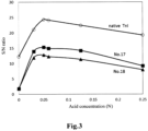
- the optimum concentration of the acidifier to be used for the acidification pretreatment was studied. Seven kinds of pretreatment agents were prepared by the same method as in Example 5 except that the hydrochloric acid concentration was one of 0.03, 0.05, 0.06, 0.125, 0.25, 0.28, and 0.5 N.
- the neutralization liquid for each pretreatment agent was prepared by the same method as in Example 5 except that the pH was adjusted such that the pH after mixing with the pretreatment liquid became 7.5 ⁇ 0.2.
- Table 6 and Fig. 3 show the S/N ratios obtained under the various acid treatment conditions.
- Acid concentration (N) Native TnI No. 17 No. 18 0 12.3 1.8 1.9 0.03 21.1 13.9 11.9 0.05 24.4 15.3 12.8 0.06 24.1 14.9 12.3 0.125 22.4 14.2 11.4 0.25 19.2 9.2 7.9 0.28 19.8 11.6 9.2 0.5 19.4 10.2 8.2
- the acidification pretreatment had an effect of increasing the S/N ratio under the conditions in which the acid concentration was more than 0 N and not more than 0.5N. With the acid concentrations of 0.03 N to 0.125 N, particular increases in the S/N ratio were found.
- the present inventors confirmed that the anti-cTnI antibody 24F9 used in the present Example reacts more strongly with simple cTnI than with native TnI (data not shown).
- the commercially available anti-cTnI antibody 19C7 also reacts with simple cTnI, it reacts more strongly with native TnI (data not shown).
- an antibody such as 19C7 although the effect of avoiding the influence of the inhibitor can be obtained by the acidification pretreatment, this effect could be canceled by the simplification of cTnI.
Landscapes
- Health & Medical Sciences (AREA)
- Life Sciences & Earth Sciences (AREA)
- Engineering & Computer Science (AREA)
- Immunology (AREA)
- Biomedical Technology (AREA)
- Chemical & Material Sciences (AREA)
- Molecular Biology (AREA)
- Urology & Nephrology (AREA)
- Hematology (AREA)
- Cell Biology (AREA)
- Analytical Chemistry (AREA)
- Biotechnology (AREA)
- Pathology (AREA)
- Food Science & Technology (AREA)
- Medicinal Chemistry (AREA)
- Physics & Mathematics (AREA)
- Microbiology (AREA)
- Biochemistry (AREA)
- General Health & Medical Sciences (AREA)
- General Physics & Mathematics (AREA)
- Proteomics, Peptides & Aminoacids (AREA)
- Chemical Kinetics & Catalysis (AREA)
- Investigating Or Analysing Biological Materials (AREA)
- Peptides Or Proteins (AREA)
Claims (5)
- Verfahren zum Messen von kardialem Troponin I in einer von einem Körper getrennten Serum- oder Plasmaprobe mittels Immunassay, wobei das Verfahren einen Vorbehandlungsschritt des Mischens der von einem Körper getrennten Serum- oder Plasmaprobe mit einer Vorbehandlungsflüssigkeit umfasst, die ein anionisches Tensid oder ein Säuerungsmittel enthält, wobei der Vorbehandlungsschritt unter Wärme durchgeführt wird.
- Verfahren nach Anspruch 1, wobei die Vorbehandlungsflüssigkeit ferner ein Reduktionsmittel enthält.
- Verfahren nach Anspruch 1 oder 2, wobei die Vorbehandlungsflüssigkeit ein anionisches Tensid enthält.
- Verfahren nach Anspruch 1, wobei die Vorbehandlungsflüssigkeit ein Säuerungsmittel enthält und das Säuerungsmittel im Vorbehandlungsschritt eine endgültige Konzentration von mehr als 0,05 N und nicht mehr als 0,5 N aufweist.
- Verwendung eines Reagens für ein Immunassay für kardiales Troponin I in einer Serum- oder Plasmaprobe, wobei das Reagens eine Vorbehandlungsflüssigkeit umfasst, die ein anionisches Tensid oder ein Säuerungsmittel enthält.
Applications Claiming Priority (2)
| Application Number | Priority Date | Filing Date | Title |
|---|---|---|---|
| JP2016178919 | 2016-09-13 | ||
| PCT/JP2017/032793 WO2018051965A1 (ja) | 2016-09-13 | 2017-09-12 | 心筋トロポニンの測定方法及び測定試薬 |
Publications (3)
| Publication Number | Publication Date |
|---|---|
| EP3514539A1 EP3514539A1 (de) | 2019-07-24 |
| EP3514539A4 EP3514539A4 (de) | 2020-04-01 |
| EP3514539B1 true EP3514539B1 (de) | 2024-11-20 |
Family
ID=61619149
Family Applications (1)
| Application Number | Title | Priority Date | Filing Date |
|---|---|---|---|
| EP17850867.7A Active EP3514539B1 (de) | 2016-09-13 | 2017-09-12 | Testverfahren und testreagens für herztroponin |
Country Status (6)
| Country | Link |
|---|---|
| US (1) | US11802867B2 (de) |
| EP (1) | EP3514539B1 (de) |
| JP (1) | JP6999561B2 (de) |
| CN (1) | CN109564214A (de) |
| TW (1) | TW201812300A (de) |
| WO (1) | WO2018051965A1 (de) |
Families Citing this family (7)
| Publication number | Priority date | Publication date | Assignee | Title |
|---|---|---|---|---|
| CN117054645A (zh) * | 2016-09-06 | 2023-11-14 | 富士瑞必欧株式会社 | 甲状腺球蛋白的测定方法及测定试剂 |
| CN110082541A (zh) * | 2019-05-14 | 2019-08-02 | 深圳天深医疗器械有限公司 | 高敏肌钙蛋白i试剂盒和样本处理液 |
| CN111948384B (zh) * | 2019-05-23 | 2024-02-27 | 裴广华 | 用于elisa试剂盒的含蔗糖的生物制品稳定剂 |
| CN110672844A (zh) * | 2019-10-29 | 2020-01-10 | 华中科技大学 | 一种新城疫病毒抗体磁免疫化学发光检测试剂盒及其应用 |
| CN112505332A (zh) * | 2020-11-23 | 2021-03-16 | 厦门宝太生物科技有限公司 | 一种高灵敏度的cTnI检测方法及其试剂盒 |
| EP4279504A4 (de) * | 2021-01-18 | 2024-11-27 | Fujirebio Inc. | Verfahren zur verarbeitung von löslichen gpc3-haltigen proben in löslichen gpc3-immuntests |
| CN113671170B (zh) * | 2021-09-09 | 2024-08-13 | 郑州安图生物工程股份有限公司 | 样本稀释液及其制备方法和消除新鲜样本检测异常的免疫分析方法 |
Family Cites Families (22)
| Publication number | Priority date | Publication date | Assignee | Title |
|---|---|---|---|---|
| US820398A (en) | 1904-10-25 | 1906-05-15 | Samuel Cleland Davidson | Centrifugal fan or pump. |
| US6100099A (en) | 1994-09-06 | 2000-08-08 | Abbott Laboratories | Test strip having a diagonal array of capture spots |
| DE19524572A1 (de) * | 1995-07-06 | 1997-01-09 | Bayer Ag | Verfahren zur Herstellung eines synthetischen Kalibrators für den Einsatz in Immunoassays, bestehend aus den Analyten oder Teilsequenzen davon, die an inerten Trägermoleküle konjugiert sind |
| JP3740898B2 (ja) * | 1998-07-31 | 2006-02-01 | 三菱化学株式会社 | 生理活性成分の測定法 |
| US6607891B1 (en) | 1998-07-31 | 2003-08-19 | Mitsubishi Chemical Corporation | Method of assaying insulin-like growth factor |
| US20070015218A1 (en) * | 2003-10-14 | 2007-01-18 | University Of South Florida | A Method for the Separation Anti-Amyloid Beta Antibody with Amyloid Beta Peptide |
| MXPA06004235A (es) | 2003-10-15 | 2007-02-14 | Daiichi Pure Chemicals Co Ltd | Metodo de separar y ensayar multimero de adiponectina. |
| CN1864067B (zh) * | 2003-10-15 | 2011-07-06 | 积水医疗株式会社 | 试样的前处理方法及使用该方法的免疫学测定方法 |
| CA2544185C (en) * | 2003-10-28 | 2010-09-28 | Advanced Life Science Institute, Inc. | Method of detecting hepatitis c virus |
| KR20060123751A (ko) * | 2003-11-19 | 2006-12-04 | 다이이치 가가쿠 야쿠힝 가부시키가이샤 | 헤모글로빈 함유 시료 중의 기질의 측정방법 |
| CA2606333A1 (en) | 2005-04-28 | 2006-11-02 | Abbott Laboratories | Stabilization of cardiac troponin |
| JP4197180B2 (ja) * | 2005-06-29 | 2008-12-17 | 株式会社シバヤギ | 検体中の内分泌物質測定方法 |
| CN101553579B (zh) | 2006-10-06 | 2016-01-20 | 赛里根有限公司 | 用于定向生物标志物信号放大的荧光方法和材料 |
| JP4841494B2 (ja) * | 2006-12-11 | 2011-12-21 | 株式会社マルハニチロ水産 | 食品中タンパク質の高感度検出法 |
| CN101017172A (zh) * | 2007-03-08 | 2007-08-15 | 湖南景达生物工程有限公司 | 一种测定丙型肝炎病毒总核心抗原的方法 |
| MX2010002269A (es) | 2007-08-28 | 2010-03-25 | Abbott Biotech Ltd | Composiciones y metodos que comprenden proteinas de union para adalimumab. |
| CN103380377B (zh) * | 2011-02-25 | 2016-01-27 | 美迪恩斯生命科技株式会社 | 心肌肌钙蛋白的测定方法 |
| US8475739B2 (en) | 2011-09-25 | 2013-07-02 | Theranos, Inc. | Systems and methods for fluid handling |
| US10036745B2 (en) * | 2012-10-03 | 2018-07-31 | Gyros Patent Ab | Method and kit for analyte determination at acidic conditions |
| JP6330666B2 (ja) * | 2013-02-06 | 2018-05-30 | 富士レビオ株式会社 | 標的物質の測定方法 |
| RU2750268C2 (ru) * | 2014-07-07 | 2021-06-25 | Эббви Дойчланд Гмбх Унд Ко. Кг | ИММУНОГЕННЫЕ ПРОДУКТЫ, ПОЛУЧЕННЫЕ НА ОСНОВЕ АМИНОКИСЛОТНЫХ ПОСЛЕДОВАТЕЛЬНОСТЕЙ МУТЕИНОВОГО АМИЛОИДА β (Aβ), И СПОСОБЫ ИХ ПРИМЕНЕНИЯ |
| CN117054645A (zh) * | 2016-09-06 | 2023-11-14 | 富士瑞必欧株式会社 | 甲状腺球蛋白的测定方法及测定试剂 |
-
2017
- 2017-09-12 EP EP17850867.7A patent/EP3514539B1/de active Active
- 2017-09-12 JP JP2018539718A patent/JP6999561B2/ja active Active
- 2017-09-12 TW TW106131254A patent/TW201812300A/zh unknown
- 2017-09-12 US US16/332,647 patent/US11802867B2/en active Active
- 2017-09-12 WO PCT/JP2017/032793 patent/WO2018051965A1/ja not_active Ceased
- 2017-09-12 CN CN201780046298.1A patent/CN109564214A/zh active Pending
Also Published As
| Publication number | Publication date |
|---|---|
| TW201812300A (zh) | 2018-04-01 |
| EP3514539A1 (de) | 2019-07-24 |
| CN109564214A (zh) | 2019-04-02 |
| EP3514539A4 (de) | 2020-04-01 |
| WO2018051965A1 (ja) | 2018-03-22 |
| US20210215680A1 (en) | 2021-07-15 |
| JPWO2018051965A1 (ja) | 2019-06-24 |
| US11802867B2 (en) | 2023-10-31 |
| JP6999561B2 (ja) | 2022-02-10 |
Similar Documents
| Publication | Publication Date | Title |
|---|---|---|
| EP3514539B1 (de) | Testverfahren und testreagens für herztroponin | |
| EP4130741B1 (de) | Verfahren zum immuntest von amyloid blut und kit dafür | |
| AU2025230780A1 (en) | Method for detecting biomarkers | |
| EP3511713B1 (de) | Tumormarkermessverfahren und -messreagenz | |
| CN107102135A (zh) | 用于减少非特异性结合的免疫检验方法和试剂 | |
| JP7594913B2 (ja) | 抗体コンジュゲート | |
| JP7138627B2 (ja) | インスリンの測定方法及び測定試薬 | |
| EP3511712B1 (de) | Verfahren zur messung von thyreoglobulin | |
| EP3978921B1 (de) | Verfahren und system zur messung von thyreoglobulin | |
| US9494606B2 (en) | Quantification of lipoproteins | |
| JP2023017986A (ja) | 自己抗体の直接イムノアッセイ測定法 | |
| CN115280146B (zh) | 包含人iv型胶原7s结构域的片段的测定方法以及用于该测定方法的试剂盒 | |
| WO2023013725A1 (ja) | サイログロブリンのイムノアッセイ及びそのためのキット | |
| HK1195617A (en) | Immunoassay methods and reagents for decreasing nonspecific binding | |
| HK1195617B (en) | Immunoassay methods and reagents for decreasing nonspecific binding |
Legal Events
| Date | Code | Title | Description |
|---|---|---|---|
| STAA | Information on the status of an ep patent application or granted ep patent |
Free format text: STATUS: THE INTERNATIONAL PUBLICATION HAS BEEN MADE |
|
| PUAI | Public reference made under article 153(3) epc to a published international application that has entered the european phase |
Free format text: ORIGINAL CODE: 0009012 |
|
| STAA | Information on the status of an ep patent application or granted ep patent |
Free format text: STATUS: REQUEST FOR EXAMINATION WAS MADE |
|
| 17P | Request for examination filed |
Effective date: 20190312 |
|
| AK | Designated contracting states |
Kind code of ref document: A1 Designated state(s): AL AT BE BG CH CY CZ DE DK EE ES FI FR GB GR HR HU IE IS IT LI LT LU LV MC MK MT NL NO PL PT RO RS SE SI SK SM TR |
|
| AX | Request for extension of the european patent |
Extension state: BA ME |
|
| DAV | Request for validation of the european patent (deleted) | ||
| DAX | Request for extension of the european patent (deleted) | ||
| A4 | Supplementary search report drawn up and despatched |
Effective date: 20200304 |
|
| RIC1 | Information provided on ipc code assigned before grant |
Ipc: G01N 33/68 20060101ALI20200227BHEP Ipc: G01N 33/53 20060101AFI20200227BHEP |
|
| STAA | Information on the status of an ep patent application or granted ep patent |
Free format text: STATUS: EXAMINATION IS IN PROGRESS |
|
| 17Q | First examination report despatched |
Effective date: 20230126 |
|
| RAP3 | Party data changed (applicant data changed or rights of an application transferred) |
Owner name: FUJIREBIO INC. |
|
| GRAP | Despatch of communication of intention to grant a patent |
Free format text: ORIGINAL CODE: EPIDOSNIGR1 |
|
| STAA | Information on the status of an ep patent application or granted ep patent |
Free format text: STATUS: GRANT OF PATENT IS INTENDED |
|
| INTG | Intention to grant announced |
Effective date: 20240724 |
|
| GRAS | Grant fee paid |
Free format text: ORIGINAL CODE: EPIDOSNIGR3 |
|
| GRAA | (expected) grant |
Free format text: ORIGINAL CODE: 0009210 |
|
| STAA | Information on the status of an ep patent application or granted ep patent |
Free format text: STATUS: THE PATENT HAS BEEN GRANTED |
|
| AK | Designated contracting states |
Kind code of ref document: B1 Designated state(s): AL AT BE BG CH CY CZ DE DK EE ES FI FR GB GR HR HU IE IS IT LI LT LU LV MC MK MT NL NO PL PT RO RS SE SI SK SM TR |
|
| P01 | Opt-out of the competence of the unified patent court (upc) registered |
Free format text: CASE NUMBER: APP_55778/2024 Effective date: 20241011 |
|
| REG | Reference to a national code |
Ref country code: GB Ref legal event code: FG4D |
|
| REG | Reference to a national code |
Ref country code: CH Ref legal event code: EP |
|
| REG | Reference to a national code |
Ref country code: DE Ref legal event code: R096 Ref document number: 602017086291 Country of ref document: DE |
|
| REG | Reference to a national code |
Ref country code: IE Ref legal event code: FG4D |
|
| REG | Reference to a national code |
Ref country code: LT Ref legal event code: MG9D |
|
| REG | Reference to a national code |
Ref country code: NL Ref legal event code: MP Effective date: 20241120 |
|
| PG25 | Lapsed in a contracting state [announced via postgrant information from national office to epo] |
Ref country code: HR Free format text: LAPSE BECAUSE OF FAILURE TO SUBMIT A TRANSLATION OF THE DESCRIPTION OR TO PAY THE FEE WITHIN THE PRESCRIBED TIME-LIMIT Effective date: 20241120 Ref country code: PT Free format text: LAPSE BECAUSE OF FAILURE TO SUBMIT A TRANSLATION OF THE DESCRIPTION OR TO PAY THE FEE WITHIN THE PRESCRIBED TIME-LIMIT Effective date: 20250320 Ref country code: IS Free format text: LAPSE BECAUSE OF FAILURE TO SUBMIT A TRANSLATION OF THE DESCRIPTION OR TO PAY THE FEE WITHIN THE PRESCRIBED TIME-LIMIT Effective date: 20250320 |
|
| PG25 | Lapsed in a contracting state [announced via postgrant information from national office to epo] |
Ref country code: FI Free format text: LAPSE BECAUSE OF FAILURE TO SUBMIT A TRANSLATION OF THE DESCRIPTION OR TO PAY THE FEE WITHIN THE PRESCRIBED TIME-LIMIT Effective date: 20241120 Ref country code: NL Free format text: LAPSE BECAUSE OF FAILURE TO SUBMIT A TRANSLATION OF THE DESCRIPTION OR TO PAY THE FEE WITHIN THE PRESCRIBED TIME-LIMIT Effective date: 20241120 |
|
| REG | Reference to a national code |
Ref country code: AT Ref legal event code: MK05 Ref document number: 1744011 Country of ref document: AT Kind code of ref document: T Effective date: 20241120 |
|
| PG25 | Lapsed in a contracting state [announced via postgrant information from national office to epo] |
Ref country code: BG Free format text: LAPSE BECAUSE OF FAILURE TO SUBMIT A TRANSLATION OF THE DESCRIPTION OR TO PAY THE FEE WITHIN THE PRESCRIBED TIME-LIMIT Effective date: 20241120 |
|
| PG25 | Lapsed in a contracting state [announced via postgrant information from national office to epo] |
Ref country code: ES Free format text: LAPSE BECAUSE OF FAILURE TO SUBMIT A TRANSLATION OF THE DESCRIPTION OR TO PAY THE FEE WITHIN THE PRESCRIBED TIME-LIMIT Effective date: 20241120 |
|
| PG25 | Lapsed in a contracting state [announced via postgrant information from national office to epo] |
Ref country code: NO Free format text: LAPSE BECAUSE OF FAILURE TO SUBMIT A TRANSLATION OF THE DESCRIPTION OR TO PAY THE FEE WITHIN THE PRESCRIBED TIME-LIMIT Effective date: 20250220 |
|
| PG25 | Lapsed in a contracting state [announced via postgrant information from national office to epo] |
Ref country code: LV Free format text: LAPSE BECAUSE OF FAILURE TO SUBMIT A TRANSLATION OF THE DESCRIPTION OR TO PAY THE FEE WITHIN THE PRESCRIBED TIME-LIMIT Effective date: 20241120 Ref country code: AT Free format text: LAPSE BECAUSE OF FAILURE TO SUBMIT A TRANSLATION OF THE DESCRIPTION OR TO PAY THE FEE WITHIN THE PRESCRIBED TIME-LIMIT Effective date: 20241120 Ref country code: GR Free format text: LAPSE BECAUSE OF FAILURE TO SUBMIT A TRANSLATION OF THE DESCRIPTION OR TO PAY THE FEE WITHIN THE PRESCRIBED TIME-LIMIT Effective date: 20250221 |
|
| PG25 | Lapsed in a contracting state [announced via postgrant information from national office to epo] |
Ref country code: PL Free format text: LAPSE BECAUSE OF FAILURE TO SUBMIT A TRANSLATION OF THE DESCRIPTION OR TO PAY THE FEE WITHIN THE PRESCRIBED TIME-LIMIT Effective date: 20241120 |
|
| PG25 | Lapsed in a contracting state [announced via postgrant information from national office to epo] |
Ref country code: RS Free format text: LAPSE BECAUSE OF FAILURE TO SUBMIT A TRANSLATION OF THE DESCRIPTION OR TO PAY THE FEE WITHIN THE PRESCRIBED TIME-LIMIT Effective date: 20250220 |
|
| PG25 | Lapsed in a contracting state [announced via postgrant information from national office to epo] |
Ref country code: SM Free format text: LAPSE BECAUSE OF FAILURE TO SUBMIT A TRANSLATION OF THE DESCRIPTION OR TO PAY THE FEE WITHIN THE PRESCRIBED TIME-LIMIT Effective date: 20241120 |
|
| PG25 | Lapsed in a contracting state [announced via postgrant information from national office to epo] |
Ref country code: DK Free format text: LAPSE BECAUSE OF FAILURE TO SUBMIT A TRANSLATION OF THE DESCRIPTION OR TO PAY THE FEE WITHIN THE PRESCRIBED TIME-LIMIT Effective date: 20241120 |
|
| PG25 | Lapsed in a contracting state [announced via postgrant information from national office to epo] |
Ref country code: EE Free format text: LAPSE BECAUSE OF FAILURE TO SUBMIT A TRANSLATION OF THE DESCRIPTION OR TO PAY THE FEE WITHIN THE PRESCRIBED TIME-LIMIT Effective date: 20241120 |
|
| PG25 | Lapsed in a contracting state [announced via postgrant information from national office to epo] |
Ref country code: RO Free format text: LAPSE BECAUSE OF FAILURE TO SUBMIT A TRANSLATION OF THE DESCRIPTION OR TO PAY THE FEE WITHIN THE PRESCRIBED TIME-LIMIT Effective date: 20241120 |
|
| PG25 | Lapsed in a contracting state [announced via postgrant information from national office to epo] |
Ref country code: SK Free format text: LAPSE BECAUSE OF FAILURE TO SUBMIT A TRANSLATION OF THE DESCRIPTION OR TO PAY THE FEE WITHIN THE PRESCRIBED TIME-LIMIT Effective date: 20241120 |
|
| PG25 | Lapsed in a contracting state [announced via postgrant information from national office to epo] |
Ref country code: CZ Free format text: LAPSE BECAUSE OF FAILURE TO SUBMIT A TRANSLATION OF THE DESCRIPTION OR TO PAY THE FEE WITHIN THE PRESCRIBED TIME-LIMIT Effective date: 20241120 |
|
| REG | Reference to a national code |
Ref country code: DE Ref legal event code: R097 Ref document number: 602017086291 Country of ref document: DE |
|
| PG25 | Lapsed in a contracting state [announced via postgrant information from national office to epo] |
Ref country code: SE Free format text: LAPSE BECAUSE OF FAILURE TO SUBMIT A TRANSLATION OF THE DESCRIPTION OR TO PAY THE FEE WITHIN THE PRESCRIBED TIME-LIMIT Effective date: 20241120 |
|
| PLBE | No opposition filed within time limit |
Free format text: ORIGINAL CODE: 0009261 |
|
| STAA | Information on the status of an ep patent application or granted ep patent |
Free format text: STATUS: NO OPPOSITION FILED WITHIN TIME LIMIT |
|
| PGFP | Annual fee paid to national office [announced via postgrant information from national office to epo] |
Ref country code: DE Payment date: 20250919 Year of fee payment: 9 |
|
| PGFP | Annual fee paid to national office [announced via postgrant information from national office to epo] |
Ref country code: IT Payment date: 20250923 Year of fee payment: 9 |
|
| PGFP | Annual fee paid to national office [announced via postgrant information from national office to epo] |
Ref country code: FR Payment date: 20250922 Year of fee payment: 9 |
|
| 26N | No opposition filed |
Effective date: 20250821 |