EP3496604B1 - Vorrichtung und system zur überwachung der peripheren arteriellen perfusion einer person - Google Patents
Vorrichtung und system zur überwachung der peripheren arteriellen perfusion einer person Download PDFInfo
- Publication number
- EP3496604B1 EP3496604B1 EP17749684.1A EP17749684A EP3496604B1 EP 3496604 B1 EP3496604 B1 EP 3496604B1 EP 17749684 A EP17749684 A EP 17749684A EP 3496604 B1 EP3496604 B1 EP 3496604B1
- Authority
- EP
- European Patent Office
- Prior art keywords
- ppg
- detection data
- speckle
- signal
- ratio
- Prior art date
- Legal status (The legal status is an assumption and is not a legal conclusion. Google has not performed a legal analysis and makes no representation as to the accuracy of the status listed.)
- Active
Links
Images



Classifications
-
- A—HUMAN NECESSITIES
- A61—MEDICAL OR VETERINARY SCIENCE; HYGIENE
- A61B—DIAGNOSIS; SURGERY; IDENTIFICATION
- A61B5/00—Measuring for diagnostic purposes; Identification of persons
- A61B5/02—Detecting, measuring or recording for evaluating the cardiovascular system, e.g. pulse, heart rate, blood pressure or blood flow
- A61B5/02007—Evaluating blood vessel condition, e.g. elasticity, compliance
-
- A—HUMAN NECESSITIES
- A61—MEDICAL OR VETERINARY SCIENCE; HYGIENE
- A61B—DIAGNOSIS; SURGERY; IDENTIFICATION
- A61B5/00—Measuring for diagnostic purposes; Identification of persons
- A61B5/02—Detecting, measuring or recording for evaluating the cardiovascular system, e.g. pulse, heart rate, blood pressure or blood flow
- A61B5/02028—Determining haemodynamic parameters not otherwise provided for, e.g. cardiac contractility or left ventricular ejection fraction
-
- A—HUMAN NECESSITIES
- A61—MEDICAL OR VETERINARY SCIENCE; HYGIENE
- A61B—DIAGNOSIS; SURGERY; IDENTIFICATION
- A61B5/00—Measuring for diagnostic purposes; Identification of persons
- A61B5/02—Detecting, measuring or recording for evaluating the cardiovascular system, e.g. pulse, heart rate, blood pressure or blood flow
- A61B5/026—Measuring blood flow
- A61B5/0261—Measuring blood flow using optical means, e.g. infrared light
-
- A—HUMAN NECESSITIES
- A61—MEDICAL OR VETERINARY SCIENCE; HYGIENE
- A61B—DIAGNOSIS; SURGERY; IDENTIFICATION
- A61B5/00—Measuring for diagnostic purposes; Identification of persons
- A61B5/02—Detecting, measuring or recording for evaluating the cardiovascular system, e.g. pulse, heart rate, blood pressure or blood flow
- A61B5/026—Measuring blood flow
- A61B5/0295—Measuring blood flow using plethysmography, i.e. measuring the variations in the volume of a body part as modified by the circulation of blood therethrough, e.g. impedance plethysmography
-
- A—HUMAN NECESSITIES
- A61—MEDICAL OR VETERINARY SCIENCE; HYGIENE
- A61B—DIAGNOSIS; SURGERY; IDENTIFICATION
- A61B5/00—Measuring for diagnostic purposes; Identification of persons
- A61B5/72—Signal processing specially adapted for physiological signals or for diagnostic purposes
- A61B5/7221—Determining signal validity, reliability or quality
-
- A—HUMAN NECESSITIES
- A61—MEDICAL OR VETERINARY SCIENCE; HYGIENE
- A61B—DIAGNOSIS; SURGERY; IDENTIFICATION
- A61B2562/00—Details of sensors; Constructional details of sensor housings or probes; Accessories for sensors
- A61B2562/02—Details of sensors specially adapted for in-vivo measurements
- A61B2562/0233—Special features of optical sensors or probes classified in A61B5/00
Definitions
- the present invention relates to a processing device and system for monitoring of peripheral arterial perfusion of a subject.
- PPG Photoplethysmography
- PPG is a known technology that can be used to monitor perfusion.
- PPG is an optical measurement technique that evaluates a time-variant change of light reflectance or transmission of an area or volume of interest.
- PPG is based on the principle that blood absorbs light (more than surrounding tissue), so variations in blood volume with every heart beat affect transmission or reflectance correspondingly.
- a PPG waveform can comprise information attributable to further physiological phenomena such as the respiration.
- pulsation of arterial blood causes changes in light absorption.
- Those changes observed with a photodetector (or an array of photodetectors) form a PPG (photoplethysmography) signal (also called, among other, a pleth wave).
- PPG photoplethysmography
- Pulsation of the blood is caused by the beating heart, i.e. peaks in the PPG signal correspond to the individual beats of the heart. Therefore, a PPG signal is a heartbeat signal in itself.
- the normalized amplitude of this signal is different for different wavelengths, and for some wavelengths it is also a function of blood oxygenation.
- US 2015/0105638 A1 and US 2011/0082355 A1 disclose a system and method for measuring one or more light-absorption related blood analyte concentration parameters of a mammalian subject.
- the system comprises: a) a photoplethysmography (PPG) device configured to effect a PPG measurement by illuminating skin of the subject with at least two distinct wavelengths of light and determining relative absorbance of each of the wavelengths; b) a dynamic light scattering measurement (DLS) device configured to effect a DLS measurement of the subject to rheologically measure a pulse parameter of the subject; and c) electronic circuitry configured to: i) temporally correlating the results of the PPG and DLS measurements; and ii) in accordance with the temporal correlation between the PPG and DLS measurements, assessing value(s) of the one or more light-absorption related blood analyte concentration parameter(s).
- PPG photoplethysmography
- DLS dynamic light scattering
- WO 2013/030744 A1 discloses a wearable pulse oximetry device that is mounted on a wrist strap and fixates an area above a distal end of the ulna with a dome-shaped structure. This area is used as the measuring area. The measurement is carried out by a detector positioned above the fixated area that detects light emitted by light sources having different wavelengths that are located at a periphery of the fixated area. Hence, the reflections are measured at neither a reflection mode nor a transmission mode, but are at an angle between 20° and 160° from the emitted light.
- This mode termed trans-illumination, shall allow achieving an excellent signal-to-noise ratio and shall enable continuous and reliable measurement of oximetry data on the wrist. It is further describes that coherent light scattering (CLS) can be used for coping with motion artifacts.
- CLS coherent light scattering
- a processing device provide a processing device, system and method for use in monitoring of peripheral arterial perfusion of a subject which support distinguishing between well-compensated and ill-compensated hypovolemia.
- a processing device for use in monitoring of peripheral arterial perfusion of a subject comprising
- the evaluation unit can be configured for evaluating said PPG signal and said flow signal to obtain information on the peripheral arterial perfusion, wherein the evaluation unit is adapted to determine a centralized vascular condition or a state of low cardiac output based on a combined evaluation of said PPG signal and said flow signal.
- a system for monitoring of peripheral arterial perfusion of a subject comprising:
- a computer program which comprises program code means for causing a computer to perform the steps of the method disclosed herein when said computer program is carried out on a computer as well as a non-transitory computer-readable recording medium that stores therein a computer program product, which, when executed by a processor, causes the method disclosed herein to be performed.
- the present invention is based on the idea to alleviate the above described ambiguity with respect to low pulsatility. It has been found that from a flow signal (e.g. a speckle signal or a laser Doppler signal) indicative of a flow of light scattering particles within the skin region (which is preferably identical to the tissue region), in particular based on interferometric techniques such as laser speckle imaging or laser Doppler, obtained in addition to a PPG signal, information on the peripheral arterial perfusion can be obtained.
- a flow signal e.g. a speckle signal or a laser Doppler signal
- the vascular compliance is low. In other words, the blood vessels are not sufficiently responding to the increase of blood flow.
- a normal or high vascular compliance should prevent the blood flow velocity (measurable by the speckle signal or laser Doppler signal) to become high by increasing the blood volume (measured by the PPG signal).
- first detection data and second detection data are obtained (i.e. received or retrieved from a detector or database) separately.
- a detector e.g. a PPG sensor
- an imaging unit e.g. a camera
- the second detection data are used as first detection data, i.e. the first detection data are identical to the second detection data, and the detector is an imaging unit for acquiring image data representing the second detection data.
- perfusion refers to how much blood flows through a given tissue region, i.e., units of blood volume per time per tissue volume. Perfusion can thus be high even in absence of pulsatile blood volume or pulsatile blood flow, i.e., even be high for a completely non-pulsatile flow. Vice versa, a strong PPG signal can be found in cases of low perfusion, for example, if there is a blockage of flow but high pulsatile input. Hence, as used herein, a (pulsatile) PPG signal originates from the pulsatile blood volume, i.e., is an absorption dominated signal indicative of absorption of light within the tissue region.
- the (pulsatile) flow signal originates from the (pulsatile) movement of particles within the tissue region.
- a speckle pattern can be disturbed or a frequency shift can occur due to moving particles.
- the flow signal can be determined using the principles of Laser Doppler or laser speckle imaging (LSI).
- PPG signals and flow signals may thus be described as color-based probing of blood absorption and color-blind probing of the blood motion, respectively.
- a PPG signal as used herein can be seen as conventional PPG signal indicative of an absorption of light within the tissue region at a given wavelength.
- a flow signal as used herein can be obtained by techniques such as laser speckle imaging (LSI) or laser Doppler.
- Coherent (laser) light scattered from moving objects or particles produces intensity fluctuations that can be used to measure the velocity of the scatterers.
- LSI laser speckle imaging
- laser Doppler Coherent
- speckle An overview of laser Doppler and speckle is given by Briers, "Laser Doppler and time-varying speckle: a reconciliation” in the Journal of the Optical Society of America, vol. 13, no. 2, 1996 as well as in the topical review paper by Briers, "Laser Doppler, speckle and related techniques for blood perfusion mapping and imaging", physiol. Meas. 22, R35-R66, 2001.
- the flow signal can indicate the degree at which light received from the skin region of the subject is perturbed by moving particles such as blood cells.
- laser Doppler velocimetry uses the frequency shift produced by the Doppler effect to measure velocity. It can be used to monitor blood flow in the body.
- Laser speckle refers to a random interference effect that gives a grainy appearance to objects illuminated by laser light. If the object comprises individual moving scatterers (such as blood cells), the speckle pattern fluctuates.
- a low vascular compliance can be sub-divided or attributed to two different conditions: (i) vascular stiffness and (ii) a centralized condition".
- Vascular stiffness can refer to the stiffness/elasticity of the vascular wall, which may change for example with age and by cardiovascular disease.
- a centralized condition can refer to a compensatory cardiovascular state of the patient where the body increases the vascular tone (decreases the diameter) of the arteries in the peripheral tissues (arms and legs), to reduce the blood flow to these tissues, to direct more blood flow to the central organs.
- Vascular stiffness changes very slowly over time, typically in the order of years, while centralization may occur within seconds/minutes. Both increased stiffness and vascular tone lead to decreased vascular compliance (ability to dilate upon arrival of a blood pressure pulse).
- vascular compliance can thus cover both (age-related) vascular stiffness and centralization.
- the evaluation unit is adapted to determine a state of well-compensated and ill-compensated hypovolemia based on a combined evaluation of the PPG signal and the flow signal.
- a condition of low cardiac output may lead to centralization which leads to vasoconstriction in peripheral tissues.
- Vasoconstriction decreases the diameter and compliance of the vessels. Although they become stiffer by this, this is caused by the smooth muscles around the arteries, rather than the arterial wall (i.e. not age-related stiffness due to arteriosclerosis), such that vascular compliance is decreased.
- the PPG signal is first expected to decrease while the flow will remain unchanged, as the body will centralize to redirect flow to the heart to maintain cardiac output even though blood volume is lower.
- cardiac output and the flow signal may decline.
- the combined evaluation enables to distinguish between well-compensated and ill-compensated hypovolemia.
- the evaluation unit can be configured to evaluating said PPG signal and said flow signal over time to distinguish between well-compensated and ill-compensated hypovolemia based on the transient behavior of the PPG signal and the flow signal, in particular, by evaluating changes in vascular compliance (PPG vs. LSI) and flow (LSI).
- said evaluation unit is configured to evaluate a speckle pattern represented by said flow signal (in this case representing a speckle signal).
- a speckle pattern represented by said flow signal in this case representing a speckle signal.
- the evaluation unit is preferably configured to evaluate motion blur by detecting speckle contrast variations.
- said evaluation unit is configured to evaluate a PPG ratio of the AC component to the DC component of the PPG signal and a speckle ratio of the AC component to the DC component of the flow signal.
- the PPG signal is composed of a pulsatile waveform (AC component), caused by the changes in blood volume with each heartbeat, overlapped on a slowly varying DC component at lower frequencies caused by respiration and motion.
- AC component pulsatile waveform
- the flow signal can be decomposed into an AC component, reflecting the modulations in the blood flow velocity, and a DC component, reflecting the blood flow. Combining the two signals enables distinguish poor peripheral perfusion from low cardiac output.
- the evaluation unit is advantageously configured to determine a state of well-compensated hypovolemia of the subject if the speckle ratio exceeds a first speckle threshold and if the PPG ratio is below a first PPG threshold and to determine a state of ill-compensated hypovolemia of the subject if the speckle ratio is below a second speckle threshold and if the PPG ratio is below the first PPG threshold.
- the evaluation unit is configured to determine a state of non-compliant blood vessels (and a high peripheral perfusion) if the speckle ratio exceeds a second speckle threshold and if the PPG ratio exceeds a second PPG threshold and to determine a state of compliant blood vessels if the speckle ratio is below a second speckle threshold and if the PPG ratio exceeds the second PPG threshold.
- the vascular compliance indicates the ability to dilate upon arrival of a blood pressure pulse. A (too) high flow in combination with a PPG ratio exceeding the second PPG threshold can indicated that the blood vessels do not sufficiently accommodate the arrival of the blood pressure pulse and can thus be indicative of non-compliant (or not sufficiently compliant) blood vessels.
- the respective thresholds may e.g. be predetermined and obtained from earlier measurements, e.g. as average of measurements for a plurality of subjects or from earlier measurements with the same subject.
- the thresholds may also be adaptive and may e.g. adjusted from time to time based on measurements with the same subject, for instance as a kind of learning system.
- P AC PPG / DC PPG AC flow / DC flow
- the speckle ratio is the ratio of the AC component to the DC component of the flow signal
- the PPG ratio is the ratio of the AC component to the DC component of the PPG signal.
- An increase in P can indicate an increased vascular compliance.
- a decrease in P accompanied by a decrease in DC flow can indicate a decrease in cardiac output.
- the input is configured to obtain first detection data acquired in response to artificial illumination of the tissue region by predetermined radiation, in particular visible or infrared light.
- the system may comprise an illumination unit for artificial illumination of the tissue region by predetermined radiation, in particular visible or infrared light.
- one or more LEDs may be used as illumination unit, as e.g. conventionally used in PPG imaging.
- a laser device for emitting a laser beam for illuminating the skin region may be used instead or in addition.
- a single illumination unit is used for illuminating a skin region, which thus corresponds to the tissue region for which the PPG data are derived (which is generally preferred, but not mandatory), and only a single set of second detection data is acquired, from which the flow signal and the PPG signal are derived.
- the flow unit can be configured to derive the flow signal based on laser Doppler and/or laser speckle technique.
- Doppler light hitting moving particles such as blood cells undergo a change in wavelength/frequency (also referred to as Doppler shift), while light particles which encounter static structures return unchanged. Some of the light can be registered by a detector such as a photodiode.
- the flow signal can be calculated based on the detection data since the magnitude and frequency distribution of the Doppler shifted light are directly related to the number and velocity of the blood cells.
- the output signal can thus provide information about the microcirculatory blood flow at the first wavelength and at the second wavelength.
- Fig. 1 shows a schematic diagram of a first embodiment of a system 10 and a processing device 12 for monitoring of peripheral arterial perfusion of a subject 14 according to the present invention.
- the processing device 12 may be briefly referred to as device 12.
- the system 10 comprises a detector 18, 19 for acquiring first detection data of a tissue region 13 (e.g. the forehead, the cheek, a hand, etc.) of the subject 14, said first detection data being acquired over time by detecting radiation reflected from and/or transmitted through tissue of the subject 14, and for acquiring second detection data of a skin region 15 (e.g. the forehead, the cheek, a hand, etc., i.e. the same region as the tissue region or a different region) of the subject 14, said second detection data comprising a sequence of images of said skin region acquired over time. Based on the acquired first detection data and second detection data, the device 12 determines peripheral arterial perfusion of the subject 14.
- a tissue region 13 e.g. the forehead, the cheek, a hand, etc.
- second detection data e.g. the same region as the tissue region or a different region
- a detector also called signal acquisition unit
- a detector for detecting electromagnetic radiation transmitted through or reflected from a subject and for acquiring second detection data.
- a detector also called signal acquisition unit
- different embodiments are used together.
- the detector For acquiring second detection data of the subject 14 from the skin region 15, the detector comprises a camera 18 (also referred to as imaging unit) including a suitable photosensor for (remotely and unobtrusively) capturing image frames of the subject 14, in particular for acquiring a sequence of image frames of the subject 14 over time.
- the image frames captured by the camera 18 may particularly correspond to a video sequence captured by means of an analog or digital photosensor, e.g. in a (digital) camera.
- Such a camera 18 usually includes a photosensor, such as a CMOS or CCD sensor, which may also operate in a specific spectral range (visible, IR) or provide information for different spectral ranges.
- the camera 18 may provide an analog or digital signal.
- the image frames include a plurality of image pixels having associated pixel values.
- the image frames include pixels representing light intensity values captured with different photosensitive elements of a photosensor. These photosensitive elements may be sensitive in a specific spectral range (i.e. representing a specific color).
- the image frames include at least two groups of some image pixels each being representative of a different skin region of the subject, e.g. the forehead, the cheek, the throat, the hand, etc. Thereby, an image pixel may correspond to one photosensitive element of a photo-detector and its (analog or digital) output or may be determined based on a combination (e.g. through binning) of a plurality of the photosensitive elements.
- the detector For acquiring first detection data of the tissue region 13 of the subject 14, the detector comprises an optical photoplethysmography sensor 19 (also referred to as contact PPG sensor) configured for being mounted to the tissue region 13 of the subject 14 for acquiring photoplethysmography signals from the tissue region 13.
- the PPG sensor 19 may e.g. be designed in the form of a finger-clip (as conventionally used for measuring blood oxygen saturation) or a sticker (as e.g. used for measuring heart rate), just to name a few of all the possible embodiments.
- the PPG sensor 19 may also be designed in other forms and arranged at other skin regions of the subject's body.
- the system 10 may further optionally comprises a light source 22 (also called illumination source), such as a lamp or laser, for illuminating the tissue region 13 with light, for instance in a predetermined wavelength range or ranges (e.g. in the red, green and/or infrared wavelength range(s)).
- the light source 22 particularly comprises a coherent light source for emitting coherent light, in particular at a predetermined wavelength or wavelength range.
- the light reflected from said tissue region 13 in response to said illumination is detected by the camera 18.
- no dedicated light source is provided, but ambient light is used for illumination of the subject 14. From the reflected light only light in a desired wavelength ranges (e.g. green, red and/or infrared light, or light in a sufficiently large wavelength range covering at least two wavelength channels) may be detected and/or evaluated.
- the device 12 is further preferably connected to an interface 20 for displaying the determined information and/or for providing medical personnel with an interface to change settings of the device 12, the camera 18, the PPG sensor 19, the light source 22 and/or any other parameters of the system 10.
- an interface 20 may comprise different displays, buttons, touchscreens, keyboards or other human machine interface means.
- a system 10 as illustrated in Fig. 1 may, e.g., be located in a hospital, healthcare facility, elderly care facility or the like. Apart from the monitoring of patients, the present invention may also be applied in other fields such as neonate monitoring, general surveillance applications, security monitoring or so-called life style environments, such as fitness equipment, a wearable, a handheld device like a smartphone, or the like.
- the uni- or bidirectional communication between the device 12, the camera 18, the PPG sensor 19 and the interface 20 may work via a wireless or wired communication interface.
- Other embodiments of the present invention may include a device 12, which is not provided stand-alone, but integrated into the camera 18 or the interface 20.
- Fig. 2 shows a schematic diagram of a first embodiment of a device 12a according to the present invention, which may be used as device 12 in the system 10 shown in Fig. 1 .
- the device 12a comprises an input (or input interface) 30 for obtaining (i.e. retrieving or receiving) first detection data of a tissue region of a subject, said first detection data being acquired over time by detecting radiation reflected from and/or transmitted through tissue of the subject, and for obtaining second detection data of a skin region of a subject, said second detection data comprising a sequence of images of said skin region acquired over time.
- the device 12a further comprises a PPG unit 32 for deriving a PPG signal from said first detection data and a flow unit 34 for deriving a flow signal from said second detection data, said flow signal being indicative of a flow of light scattering particles within the skin region.
- the flow signal may e.g. be a speckle signal representing speckle or a Doppler signal representing Doppler shifts.
- An evaluation unit 36 evaluates said PPG signal and said flow signal to obtain information on the peripheral arterial perfusion of the subject 14.
- the evaluation unit 36 is adapted to determine (or to distinguish between) a state of low vascular compliance and/or a state of low cardiac output based on a combined evaluation of said PPG signal and said flow signal.
- the evaluation unit can be adapted to classify a state of the subject as a state of low vascular compliance and/or a state of low cardiac output based on the a combined evaluation of said PPG signal and said flow signal.
- the PPG unit 14, the flow unit 34 and the evaluation unit 36 may e.g. be implemented hard- and/or software, e.g. by one or more programmed processors or computers.
- Fig. 3 shows a schematic diagram of a second embodiment of a system 10a according to the present invention.
- the system 10a only comprises a single detector 18, i.e. the camera (or imaging unit).
- the second detection data acquired by the camera 18 are thus also used as first detection data for deriving the PPG signal using the above described remote PPG technique.
- An additional PPG sensor (19 in Fig. 1 ) is thus not required, and the tissue region thus corresponds to the skin region.
- PPG monitoring for peripheral perfusion cannot distinguish between low pulsatility caused by low vascular compliance (centralization and/or vascular stiffness) or low pulsatility caused by low cardiac output.
- the heartbeat induces a pulsatile pressure/flow variation.
- the blood vessels walls will dilate or contract to allow more or less blood volume to circulate through.
- PPG Blood pulsatility can be measured from the PPG signal.
- PPG is a technique measuring changes of blood volume in the microvascular bed of tissue, which are caused by the blood pressure variations. It is an optical technique, measuring the light absorption over time.
- the PPG signal is composed of a pulsatile (AC) waveform, caused by the changes in blood volume with each heartbeat, overlapped on a slowly varying (DC) component, at lower frequencies, caused by respiration and motion.
- AC pulsatile
- DC slowly varying
- this technique can be applied both in contact, using a contact source/detector geometry (cPPG), and remotely, using a camera (rPPG).
- cPPG contact source/detector geometry
- rPPG camera
- a PPG signal is correlated to the blood volume in the tissue. Therefore, if this is low, according to the compliance relation (equation 3) it can be concluded that, when the pulsatility component of the PPG signal is low, corresponding to a small depth PPG signal modulation AC/DC, this can be interpreted in two ways:
- measuring a low 1 A dA dt could also mean a low ⁇ p .
- measuring 1 v dv dt could relieve the ambiguity, as follows: if 1 v dv dt is low, then 1 q dq dt is low, meaning low cardiac output; else, if 1 v dv dt is high for a low 1 A dA dt , and therefore 1 q dq dt is high, meaning that ⁇ p is not low, but C, the vascular compliance is low.
- measuring and combining 1 v dv dt and 1 A dA dt allows distinguishing between low cardiac output (and low peripheral perfusion) or low vascular compliance. Changes in vascular compliance can be detected and it can be assessed whether or not this is due to lowering cardiac output.
- speckle imaging e.g. laser speckle imaging (LSI)
- LSI laser speckle imaging
- the speckle signal can also be decomposed as an AC component, reflecting the modulations in the blood flow velocity, and a DC component, reflecting the blood flow. Therefore, combining the two signals, poor peripheral perfusion or low vascular compliance can be distinguished from low cardiac output.
- a low blood volume variation, which can be observed in the PPG signal by a small AC/DC component or a small 1 A dA dt will correspond to:
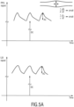
- FIG. 5 An illustration of the PPG signal and the speckle signal (as one embodiment for the flow signal) for different situations is shown in Fig. 5 .
- Fig. 5A shows a situation in which the modulation in the PPG signal and the modulation in the speckle signal (LSI signal) are low (small) indicating low cardiac output. In addition to the low cardiac output, the vascular compliance can also be low. This situation may be considered as indicative of ill-compensated hypovolemia.
- Fig. 5B shows a situation in which the modulation in the PPG signal is low and the modulation in the speckle signal (LSI signal) is high (large) indicating low vascular compliance. In other words, the situation shown in Fig. 5B can be indicative of low vascular compliance (only) but sufficient cardiac output. This situation may be considered as indicative of well-compensated hypovolemia.
- the vascular compliance is low. In other words, the blood vessels are not sufficiently responding to the increase of blood flow. Consequently, a similar conclusion can be drawn.
- a normal or high vascular compliance should prevent the blood flow velocity (measurable by the speckle signal) to become high, by increasing the blood volume (PPG signal).
- the proposed idea combines two measurements methods, PPG imaging and flow signal evaluation (e.g. speckle imaging (e.g. LSI) or Laser Doppler imaging). This may be based on single-camera measurement (which is contactless) or a combination of a camera measurement and a contact measurement. It does generally not require visible light.
- a PPG signal and a flow signal are derived, from which the desired information on the peripheral arterial perfusion is obtained, for instance based on a functional relationship (e.g. based on an algorithm or function) or a tabular relation (e.g. based on an look-up table, LUT) between the compliance measure P and the PPG signal amplitude and flow signal amplitude.
- activity, posture, biometrics and/or vital signs can be determined and monitored, particularly from the obtained second detection data. This information may be used in addition in the decision about arterial perfusion.
- Another advantage of the proposed solution is that, since it is based on local area measurement, it can conveniently measure compliance on easily accessible areas, such as face, arm, etc. As explained above, although the method is preferably designed for remote measurement, it can also be applied in contact.
- the measurement can take place either in the non-visible part of the spectrum, enabling measurements in dark, and/or in the visible range.
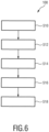
- FIG. 6 A flow chart of an embodiment of a method 100 according to the present invention is illustrated in Fig. 6 .
- first detection data of a tissue region of a subject are obtained.
- second detection data of a skin region of a subject are obtained.
- a PPG signal is derived from said first detection data.
- a flow signal is derived from said second detection data.
- said PPG signal and said flow signal are evaluated to obtain information on the peripheral arterial perfusion.
- a state of low vascular compliance and/or a state of low cardiac output can be determined based on a combined evaluation of said PPG signal and said flow signal.
- a computer program may be stored/distributed on a suitable non-transitory medium, such as an optical storage medium or a solid-state medium supplied together with or as part of other hardware, but may also be distributed in other forms, such as via the Internet or other wired or wireless telecommunication systems.
- a suitable non-transitory medium such as an optical storage medium or a solid-state medium supplied together with or as part of other hardware, but may also be distributed in other forms, such as via the Internet or other wired or wireless telecommunication systems.
Landscapes
- Health & Medical Sciences (AREA)
- Life Sciences & Earth Sciences (AREA)
- Engineering & Computer Science (AREA)
- Surgery (AREA)
- General Health & Medical Sciences (AREA)
- Biophysics (AREA)
- Pathology (AREA)
- Physiology (AREA)
- Biomedical Technology (AREA)
- Heart & Thoracic Surgery (AREA)
- Medical Informatics (AREA)
- Molecular Biology (AREA)
- Cardiology (AREA)
- Animal Behavior & Ethology (AREA)
- Physics & Mathematics (AREA)
- Public Health (AREA)
- Veterinary Medicine (AREA)
- Hematology (AREA)
- Vascular Medicine (AREA)
- Artificial Intelligence (AREA)
- Computer Vision & Pattern Recognition (AREA)
- Psychiatry (AREA)
- Signal Processing (AREA)
- Measuring Pulse, Heart Rate, Blood Pressure Or Blood Flow (AREA)
Claims (12)
- Verarbeitungsvorrichtung zur Verwendung bei der Überwachung von peripherer arterieller Perfusion eines
Subjekts, wobei die Verarbeitungsvorrichtung umfasst:- einen Eingang (30) zum Empfangen von ersten Erkennungsdaten eines Gewebebereichs eines Subjekts, wobei die ersten Erkennungsdaten über die Zeit erfasst werden durch Erkennen von Strahlung, die vom Gewebe des Subjekts reflektiert und/oder durch dieses übertragen wird, und zum Empfangen von zweiten Erkennungsdaten eines Hautbereichs eines Subjekts, wobei die zweiten Erkennungsdaten über die Zeit erfasst werden durch Erkennen von Strahlung, die in Reaktion darauf, dass kohärentes Licht in Richtung des Hautbereichs emittiert wird, aus dem Hautbereich empfangen wird,- eine PPG-Einheit (32) zum Ableiten eines Photoplethysmographie-, PPG-, Signals aus den ersten Erkennungsdaten,- eine Strömungseinheit (34) zum Ableiten eines Strömungssignals, das eine Strömung von lichtstreuenden Partikeln innerhalb des Hautbereichs angibt, aus den zweiten Erkennungsdaten, und- eine Auswertungseinheit (36) zum Auswerten des PPG-Signals und des Strömungssignals, um Informationen über die periphere arterielle Perfusion zu erhalten, dadurch gekennzeichnet, dass die Auswertungseinheit dazu ausgelegt ist, auf Basis einer kombinierten Auswertung des PPG-Signals und des Strömungssignals einen Zustand niedriger vaskulärer Compliance und/oder einen Zustand niedrigen Herzzeitvolumens zu bestimmen;wobei die Auswertungseinheit (36) so konfiguriert ist, dass sie ein PPG-Verhältnis der Wechselstromkomponente zur Gleichstromkomponente des PPG-Signals und ein Speckle-Verhältnis der Wechselstromkomponente zur Gleichstromkomponente des Strömungssignals auswertet;wobei die Auswertungseinheit (36) so konfiguriert ist, dass sie einen Zustand nicht konformer Blutgefäße und einer hohen peripheren Perfusion bestimmt, wenn das Speckle-Verhältnis eine zweite Speckle-Schwelle überschreitet und wenn das PPG-Verhältnis eine zweite PPG-Schwelle überschreitet, und einen Zustand konformer Blutgefäße bestimmt, wenn das Speckle-Verhältnis unter einer zweiten Speckle-Schwelle liegt und wenn das PPG-Verhältnis die zweite PPG-Schwelle überschreitet. - Verarbeitungsvorrichtung nach Anspruch 1, wobei die Strömungseinheit (34) so konfiguriert ist, dass sie das Strömungssignal auf Basis einer Laser-Doppler- und/oder Laser-Speckle-Technik ableitet.
- Verarbeitungsvorrichtung nach Anspruch 1,
wobei die Auswertungseinheit (36) so konfiguriert ist, dass sie ein Speckle-Muster, das durch das Strömungssignal dargestellt wird, auswertet. - Verarbeitungsvorrichtung nach Anspruch 3,
wobei die Auswertungseinheit (36) so konfiguriert ist, dass sie Bewegungsunschärfe durch Erkennen von Speckle-Kontrast-Variationen auswertet. - Verarbeitungsvorrichtung nach Anspruch 1,
wobei die Auswertungseinheit (36) so konfiguriert ist, dass sie einen Zustand gut kompensierter Hypovolämie des Subjekts bestimmt, wenn das Speckle-Verhältnis eine erste Speckle-Schwelle überschreitet und wenn das PPG-Verhältnis unter einer ersten PPG-Schwelle liegt, und einen Zustand schlecht kompensierter Hypovolämie des Subjekts bestimmt, wenn das Speckle-Verhältnis unter einer zweiten Speckle-Schwelle liegt und wenn das PPG-Verhältnis unter der ersten PPG-Schwelle liegt. - Verarbeitungsvorrichtung nach Anspruch 1,
wobei die Auswertungseinheit (36) so konfiguriert ist, dass sie ein Compliance-Maß P auswertet, das ein Verhältnis des Speckle-Verhältnisses zum PPG-Verhältnis angibt, das gegeben ist durch
- Verarbeitungsvorrichtung nach Anspruch 1,
wobei der Eingang (30) so konfiguriert ist, dass er erste Erkennungsdaten erhält, die in Reaktion auf künstliche Beleuchtung des Gewebes mit vorbestimmter Strahlung, insbesondere sichtbarem oder Infrarot-Licht, erfasst werden. - Verarbeitungsvorrichtung nach Anspruch 1,
die weiter so konfiguriert ist, dass sie die zweiten Erkennungsdaten als erste Erkennungsdaten verwendet. - System zur Überwachung von peripherer arterieller Perfusion, wobei das System umfasst:- einen Detektor (18, 19) zum Erfassen von ersten Erkennungsdaten eines Gewebebereichs eines Subjekts, wobei die ersten Erkennungsdaten über die Zeit erfasst werden durch Erkennen von Strahlung, die vom Gewebe des Subjekts reflektiert und/oder durch dieses übertragen wird, und zum Erfassen von zweiten Erkennungsdaten eines Hautbereichs eines Subjekts, wobei die zweiten Erkennungsdaten eine Bildsequenz des Hautbereichs umfassen, die über die Zeit erfasst wird, und- eine Verarbeitungsvorrichtung (12, 12a) nach Anspruch 1 zum Überwachen von peripherer arterieller Perfusion auf Basis der erfassten ersten Erkennungsdaten und zweiten Erkennungsdaten.
- System nach Anspruch 9,
das weiter eine Beleuchtungseinheit (22) zur künstlichen Beleuchtung des Gewebes mit vorbestimmter Strahlung, insbesondere sichtbarem oder Infrarot-Licht, umfasst. - System nach Anspruch 10,
wobei die Beleuchtungseinheit (22) eine kohärente Lichtquelle zum Emittieren von kohärentem Licht, insbesondere eine Laservorrichtung zum Emittieren eines Laserstrahls, zum Beleuchten des Hautbereichs umfasst. - Computerprogramm, das Programmcodemittel umfasst zum Bewirken, dass ein Computer, wenn das Computerprogramm auf dem Computer ausgeführt wird, die folgenden Schritte ausführt:- Empfangen von ersten Erkennungsdaten eines Gewebebereichs eines Subjekts, wobei die ersten Erkennungsdaten über die Zeit erfasst werden durch Erkennen von Strahlung, die vom Gewebe des Subjekts reflektiert und/oder durch dieses übertragen wird,- Empfangen von zweiten Erkennungsdaten eines Hautbereichs eines Subjekts, wobei die zweiten Erkennungsdaten über die Zeit erfasst werden durch Erkennen von Strahlung, die in Reaktion darauf, dass kohärentes Licht in Richtung des Hautbereichs emittiert wird, aus dem Hautbereich empfangen wird,- Ableiten eines Photoplethysmographie-, PPG-, Signals aus den ersten Erkennungsdaten,- Ableiten eines Strömungssignals, das eine Strömung von lichtstreuenden Partikeln innerhalb des Hautbereichs angibt, aus den zweiten Erkennungsdaten, und- Auswerten des PPG-Signals und des Strömungssignals, um Informationen über die periphere arterielle Perfusion zu erhalten, undgekennzeichnet durch Bestimmen eines Zustands geringer vaskulärer Compliance und/oder eines Zustands niedrigen Herzzeitvolumens auf Basis einer kombinierten Auswertung des PPG-Signals und des Strömungssignals;wobei die Auswertung das Auswerten eines PPG-Verhältnisses der Wechselstromkomponente zur Gleichstromkomponente des PPG-Signals und eines Speckle-Verhältnisses der Wechselstromkomponente zur Gleichstromkomponente des Strömungssignals umfasst;wobei die Auswertung das Bestimmen eines Zustands nicht konformer Blutgefäße und einer hohen peripheren Perfusion umfasst, wenn das Speckle-Verhältnis eine zweite Speckle-Schwelle überschreitet und wenn das PPG-Verhältnis eine zweite PPG-Schwelle überschreitet, und das Bestimmen eines Zustands konformer Blutgefäße, wenn das Speckle-Verhältnis unter einer zweiten Speckle-Schwelle liegt und wenn das PPG-Verhältnis die zweite PPG-Schwelle überschreitet.
Applications Claiming Priority (2)
| Application Number | Priority Date | Filing Date | Title |
|---|---|---|---|
| EP16183398 | 2016-08-09 | ||
| PCT/EP2017/069375 WO2018029033A1 (en) | 2016-08-09 | 2017-08-01 | Device, system and method for monitoring of peripheral arterial perfusion of a subject |
Publications (2)
| Publication Number | Publication Date |
|---|---|
| EP3496604A1 EP3496604A1 (de) | 2019-06-19 |
| EP3496604B1 true EP3496604B1 (de) | 2025-02-12 |
Family
ID=56615916
Family Applications (1)
| Application Number | Title | Priority Date | Filing Date |
|---|---|---|---|
| EP17749684.1A Active EP3496604B1 (de) | 2016-08-09 | 2017-08-01 | Vorrichtung und system zur überwachung der peripheren arteriellen perfusion einer person |
Country Status (5)
| Country | Link |
|---|---|
| US (1) | US12543959B2 (de) |
| EP (1) | EP3496604B1 (de) |
| JP (1) | JP7116042B2 (de) |
| CN (1) | CN109561843B (de) |
| WO (1) | WO2018029033A1 (de) |
Families Citing this family (29)
| Publication number | Priority date | Publication date | Assignee | Title |
|---|---|---|---|---|
| JP7012655B2 (ja) | 2016-02-24 | 2022-01-28 | インセプト、リミテッド、ライアビリティ、カンパニー | 可撓性の強化された神経血管カテーテル |
| EP3565511B1 (de) | 2017-01-06 | 2023-10-18 | Incept, LLC | Thromboresistente beschichtungen für aneurysmenbehandlungsvorrichtung |
| US11395665B2 (en) | 2018-05-01 | 2022-07-26 | Incept, Llc | Devices and methods for removing obstructive material, from an intravascular site |
| EP3787523A4 (de) | 2018-05-01 | 2022-02-23 | Incept, LLC | Vorrichtungen und verfahren zum entfernen von obstruktivem material aus einer intravaskulären stelle |
| WO2020010310A1 (en) | 2018-07-06 | 2020-01-09 | Imperative Care, Inc. | Sealed neurovascular extendable catheter |
| US11471582B2 (en) | 2018-07-06 | 2022-10-18 | Incept, Llc | Vacuum transfer tool for extendable catheter |
| US11766539B2 (en) | 2019-03-29 | 2023-09-26 | Incept, Llc | Enhanced flexibility neurovascular catheter |
| WO2021076642A1 (en) | 2019-10-15 | 2021-04-22 | Imperative Care, Inc. | Systems and methods for multivariate stroke detection |
| US11638637B2 (en) | 2019-12-18 | 2023-05-02 | Imperative Care, Inc. | Method of removing embolic material with thrombus engagement tool |
| US11439799B2 (en) | 2019-12-18 | 2022-09-13 | Imperative Care, Inc. | Split dilator aspiration system |
| EP4076611A4 (de) | 2019-12-18 | 2023-11-15 | Imperative Care, Inc. | Verfahren und systeme zur behandlung venöser thromboembolischer erkrankungen |
| CN118925025A (zh) | 2020-03-10 | 2024-11-12 | 因普瑞缇夫护理公司 | 增强的柔韧性的神经血管导管 |
| CN111543972B (zh) * | 2020-05-08 | 2022-11-04 | 上海理工大学 | 用于激光散斑血流灌注成像系统的数据库建立方法 |
| US11207497B1 (en) | 2020-08-11 | 2021-12-28 | Imperative Care, Inc. | Catheter with enhanced tensile strength |
| CN112587105B (zh) * | 2020-12-11 | 2023-10-27 | 复旦大学附属中山医院 | 一种人体循环灌注状态评估系统 |
| EP4014853A1 (de) | 2020-12-17 | 2022-06-22 | Koninklijke Philips N.V. | Vorrichtung, verfahren und systeme zur abbildung eines oder mehrerer aspekte der blutperfusion |
| US12076141B2 (en) | 2021-02-26 | 2024-09-03 | Covidien Lp | System and method for digitally calibrating a medical sensor |
| US12558175B2 (en) | 2021-08-12 | 2026-02-24 | Imperative Care, Inc. | Multi catheter method of performing a robotic neurovascular procedure |
| EP4395634A4 (de) * | 2021-08-30 | 2025-05-21 | TopSight Monitoring Ltd | Am körper tragbares physiologisches überwachungssystem |
| US12201396B2 (en) | 2021-09-10 | 2025-01-21 | Rockley Photonics Limited | Optical speckle receiver |
| US12458295B2 (en) | 2021-10-01 | 2025-11-04 | Covidien Lp | System and method for correcting for errors dependent upon spectral characteristics of tissue |
| USD1077996S1 (en) | 2021-10-18 | 2025-06-03 | Imperative Care, Inc. | Inline fluid filter |
| US12390117B2 (en) | 2021-11-16 | 2025-08-19 | Rockley Photonics Limited | Optical sensor module for speckleplethysmography (SPG) and photoplethysmography (PPG) |
| EP4230117A1 (de) * | 2022-02-18 | 2023-08-23 | Koninklijke Philips N.V. | System und verfahren zur gewebeanalyse unter verwendung von ferngesteuerter ppg |
| EP4230115A1 (de) * | 2022-02-18 | 2023-08-23 | Koninklijke Philips N.V. | System und verfahren zur gewebeanalyse unter verwendung von ferngesteuerter ppg |
| EP4309573A1 (de) | 2022-07-21 | 2024-01-24 | Sonion Nederland B.V. | Bestimmung eines parameters in bezug auf den blutfluss in einem blutdurchströmten teil unter verwendung eines vcsel |
| WO2024213563A1 (en) * | 2023-04-14 | 2024-10-17 | Sonion Nederland B.V. | A system and a method of determining a physiological parameter of a body comprising blood perfused tissue |
| US12396648B1 (en) | 2024-11-27 | 2025-08-26 | Rockley Photonics Limited | Wearable device with light source and optical sensor |
| US12484796B1 (en) | 2024-11-27 | 2025-12-02 | Rockley Photonics Limited | System and method for measuring pulse wave velocity |
Family Cites Families (18)
| Publication number | Priority date | Publication date | Assignee | Title |
|---|---|---|---|---|
| JP2001321347A (ja) | 2000-05-16 | 2001-11-20 | Nippon Koden Corp | 血圧監視装置 |
| US7887502B2 (en) * | 2006-09-15 | 2011-02-15 | University Of Florida Research Foundation, Inc. | Method for using photoplethysmography to optimize fluid removal during renal replacement therapy by hemodialysis or hemofiltration |
| JP2010508056A (ja) * | 2006-10-30 | 2010-03-18 | エルフィ−テック リミテッド | 生物学的パラメータの体内での測定のためのシステム及び方法 |
| WO2008144404A1 (en) | 2007-05-16 | 2008-11-27 | Massachusetts Instutute Of Technology | Systems and methods for model-based estimation of cardiac output and total peripheral resistance |
| US8551006B2 (en) * | 2008-09-17 | 2013-10-08 | Covidien Lp | Method for determining hemodynamic effects |
| JP2012504454A (ja) | 2008-10-01 | 2012-02-23 | 株式会社イルメディ | 心血管分析装置 |
| US9788733B2 (en) | 2009-06-02 | 2017-10-17 | Cordex Systems, Inc. | Method and device for detecting and assessing reactive hyperemia using segmental plethysmography |
| US9775529B2 (en) * | 2009-06-17 | 2017-10-03 | Sotera Wireless, Inc. | Body-worn pulse oximeter |
| US20110082355A1 (en) | 2009-07-30 | 2011-04-07 | Oxitone Medical Ltd. | Photoplethysmography device and method |
| US8868149B2 (en) | 2009-07-30 | 2014-10-21 | Oxitone Medical Ltd. | Photoplethysmography device and method |
| GB201119982D0 (en) | 2011-11-18 | 2012-01-04 | Maarek Albert | Medical device and measurement of cardiac output |
| GB2494622A (en) | 2011-08-30 | 2013-03-20 | Oxitone Medical Ltd | Wearable pulse oximetry device |
| JP3180987U (ja) | 2012-11-02 | 2013-01-17 | 中原大學 | 画像式脈波伝播速度測定装置 |
| EP2762066A1 (de) | 2013-02-05 | 2014-08-06 | Koninklijke Philips N.V. | Verfahren und Gerät zur Bestimmung Vitalparamterinformation |
| US10368794B2 (en) * | 2014-08-29 | 2019-08-06 | Ecole Polytechnique Federale De Lausanne (Epfl) | System for the remote monitoring of the hydration status of a living being |
| KR102299361B1 (ko) * | 2014-09-03 | 2021-09-07 | 삼성전자주식회사 | 혈압을 모니터링하는 장치 및 방법, 혈압 모니터링 기능을 갖는 웨어러블 디바이스 |
| KR20160044267A (ko) * | 2014-10-15 | 2016-04-25 | 삼성전자주식회사 | 생체 정보 획득 장치 및 방법 |
| CN109561842B (zh) * | 2016-08-09 | 2022-07-12 | 皇家飞利浦有限公司 | 用于血氧饱和度测量的设备 |
-
2017
- 2017-08-01 EP EP17749684.1A patent/EP3496604B1/de active Active
- 2017-08-01 CN CN201780049162.6A patent/CN109561843B/zh active Active
- 2017-08-01 JP JP2019506486A patent/JP7116042B2/ja active Active
- 2017-08-01 US US16/323,567 patent/US12543959B2/en active Active
- 2017-08-01 WO PCT/EP2017/069375 patent/WO2018029033A1/en not_active Ceased
Also Published As
| Publication number | Publication date |
|---|---|
| CN109561843A (zh) | 2019-04-02 |
| US20190175030A1 (en) | 2019-06-13 |
| JP2019524295A (ja) | 2019-09-05 |
| CN109561843B (zh) | 2022-07-15 |
| US12543959B2 (en) | 2026-02-10 |
| WO2018029033A1 (en) | 2018-02-15 |
| EP3496604A1 (de) | 2019-06-19 |
| JP7116042B2 (ja) | 2022-08-09 |
Similar Documents
| Publication | Publication Date | Title |
|---|---|---|
| US12543959B2 (en) | Device, system and method for monitoring of peripheral arterial perfusion of a subject | |
| EP3496603B1 (de) | Vorrichtung zur verwendung bei der blutsauerstoffsättigungsmessung | |
| US10478078B2 (en) | Device and method for determining vital signs of a subject | |
| CN105377126B (zh) | 用于筛查对象的氧合状态的系统 | |
| JP6336141B2 (ja) | 対象の血液内の物質の濃度を決定する装置、システム及び方法 | |
| EP3073905B1 (de) | Vorrichtung und verfahren zum erhalt von pulslaufzeiten und/oder pulswellengeschwindigkeitsinformationen einer person | |
| JP6615176B2 (ja) | 非干渉的な皮膚組織ハイドレーション測定デバイス及び関連方法 | |
| US20200253560A1 (en) | Device, system and method for determining at least one vital sign of a subject | |
| JP2018534968A (ja) | 生理情報を抽出するためのデバイス、システム、及び方法 | |
| JP2016511659A (ja) | 対象者のバイタルサイン情報を決定するためのシステム及び方法 | |
| WO2018029123A1 (en) | Device for use in blood oxygen saturation measurement | |
| US20220265150A1 (en) | Device, system and method for determining physiological information | |
| JP2018528807A (ja) | 対象の心拍関連情報の監視のための装置及びシステム | |
| WO2014141001A1 (en) | Device and method for determining the blood oxygen saturation of a subject | |
| JP2017512578A (ja) | 対象のバイタルサインを決定する装置、システム及び方法 | |
| Hu | Non-contact reflection photoplethysmography towards effective human physiological monitoring | |
| Ahmed et al. | Photonic sensor design evaluation for measuring the photoplethysmogram |
Legal Events
| Date | Code | Title | Description |
|---|---|---|---|
| STAA | Information on the status of an ep patent application or granted ep patent |
Free format text: STATUS: UNKNOWN |
|
| STAA | Information on the status of an ep patent application or granted ep patent |
Free format text: STATUS: THE INTERNATIONAL PUBLICATION HAS BEEN MADE |
|
| PUAI | Public reference made under article 153(3) epc to a published international application that has entered the european phase |
Free format text: ORIGINAL CODE: 0009012 |
|
| STAA | Information on the status of an ep patent application or granted ep patent |
Free format text: STATUS: REQUEST FOR EXAMINATION WAS MADE |
|
| 17P | Request for examination filed |
Effective date: 20190311 |
|
| AK | Designated contracting states |
Kind code of ref document: A1 Designated state(s): AL AT BE BG CH CY CZ DE DK EE ES FI FR GB GR HR HU IE IS IT LI LT LU LV MC MK MT NL NO PL PT RO RS SE SI SK SM TR |
|
| AX | Request for extension of the european patent |
Extension state: BA ME |
|
| DAV | Request for validation of the european patent (deleted) | ||
| DAX | Request for extension of the european patent (deleted) | ||
| RAP1 | Party data changed (applicant data changed or rights of an application transferred) |
Owner name: KONINKLIJKE PHILIPS N.V. |
|
| STAA | Information on the status of an ep patent application or granted ep patent |
Free format text: STATUS: EXAMINATION IS IN PROGRESS |
|
| 17Q | First examination report despatched |
Effective date: 20220621 |
|
| GRAP | Despatch of communication of intention to grant a patent |
Free format text: ORIGINAL CODE: EPIDOSNIGR1 |
|
| STAA | Information on the status of an ep patent application or granted ep patent |
Free format text: STATUS: GRANT OF PATENT IS INTENDED |
|
| INTG | Intention to grant announced |
Effective date: 20240903 |
|
| GRAS | Grant fee paid |
Free format text: ORIGINAL CODE: EPIDOSNIGR3 |
|
| GRAA | (expected) grant |
Free format text: ORIGINAL CODE: 0009210 |
|
| STAA | Information on the status of an ep patent application or granted ep patent |
Free format text: STATUS: THE PATENT HAS BEEN GRANTED |
|
| AK | Designated contracting states |
Kind code of ref document: B1 Designated state(s): AL AT BE BG CH CY CZ DE DK EE ES FI FR GB GR HR HU IE IS IT LI LT LU LV MC MK MT NL NO PL PT RO RS SE SI SK SM TR |
|
| REG | Reference to a national code |
Ref country code: GB Ref legal event code: FG4D |
|
| REG | Reference to a national code |
Ref country code: CH Ref legal event code: EP |
|
| REG | Reference to a national code |
Ref country code: DE Ref legal event code: R096 Ref document number: 602017087701 Country of ref document: DE |
|
| REG | Reference to a national code |
Ref country code: IE Ref legal event code: FG4D |
|
| REG | Reference to a national code |
Ref country code: NL Ref legal event code: MP Effective date: 20250212 |
|
| PG25 | Lapsed in a contracting state [announced via postgrant information from national office to epo] |
Ref country code: RS Free format text: LAPSE BECAUSE OF FAILURE TO SUBMIT A TRANSLATION OF THE DESCRIPTION OR TO PAY THE FEE WITHIN THE PRESCRIBED TIME-LIMIT Effective date: 20250512 |
|
| PG25 | Lapsed in a contracting state [announced via postgrant information from national office to epo] |
Ref country code: FI Free format text: LAPSE BECAUSE OF FAILURE TO SUBMIT A TRANSLATION OF THE DESCRIPTION OR TO PAY THE FEE WITHIN THE PRESCRIBED TIME-LIMIT Effective date: 20250212 |
|
| PG25 | Lapsed in a contracting state [announced via postgrant information from national office to epo] |
Ref country code: PL Free format text: LAPSE BECAUSE OF FAILURE TO SUBMIT A TRANSLATION OF THE DESCRIPTION OR TO PAY THE FEE WITHIN THE PRESCRIBED TIME-LIMIT Effective date: 20250212 |
|
| PG25 | Lapsed in a contracting state [announced via postgrant information from national office to epo] |
Ref country code: ES Free format text: LAPSE BECAUSE OF FAILURE TO SUBMIT A TRANSLATION OF THE DESCRIPTION OR TO PAY THE FEE WITHIN THE PRESCRIBED TIME-LIMIT Effective date: 20250212 |
|
| REG | Reference to a national code |
Ref country code: LT Ref legal event code: MG9D |
|
| PG25 | Lapsed in a contracting state [announced via postgrant information from national office to epo] |
Ref country code: NO Free format text: LAPSE BECAUSE OF FAILURE TO SUBMIT A TRANSLATION OF THE DESCRIPTION OR TO PAY THE FEE WITHIN THE PRESCRIBED TIME-LIMIT Effective date: 20250512 Ref country code: IS Free format text: LAPSE BECAUSE OF FAILURE TO SUBMIT A TRANSLATION OF THE DESCRIPTION OR TO PAY THE FEE WITHIN THE PRESCRIBED TIME-LIMIT Effective date: 20250612 |
|
| PG25 | Lapsed in a contracting state [announced via postgrant information from national office to epo] |
Ref country code: NL Free format text: LAPSE BECAUSE OF FAILURE TO SUBMIT A TRANSLATION OF THE DESCRIPTION OR TO PAY THE FEE WITHIN THE PRESCRIBED TIME-LIMIT Effective date: 20250212 |
|
| PG25 | Lapsed in a contracting state [announced via postgrant information from national office to epo] |
Ref country code: HR Free format text: LAPSE BECAUSE OF FAILURE TO SUBMIT A TRANSLATION OF THE DESCRIPTION OR TO PAY THE FEE WITHIN THE PRESCRIBED TIME-LIMIT Effective date: 20250212 |
|
| PG25 | Lapsed in a contracting state [announced via postgrant information from national office to epo] |
Ref country code: PT Free format text: LAPSE BECAUSE OF FAILURE TO SUBMIT A TRANSLATION OF THE DESCRIPTION OR TO PAY THE FEE WITHIN THE PRESCRIBED TIME-LIMIT Effective date: 20250612 Ref country code: LV Free format text: LAPSE BECAUSE OF FAILURE TO SUBMIT A TRANSLATION OF THE DESCRIPTION OR TO PAY THE FEE WITHIN THE PRESCRIBED TIME-LIMIT Effective date: 20250212 |
|
| PG25 | Lapsed in a contracting state [announced via postgrant information from national office to epo] |
Ref country code: GR Free format text: LAPSE BECAUSE OF FAILURE TO SUBMIT A TRANSLATION OF THE DESCRIPTION OR TO PAY THE FEE WITHIN THE PRESCRIBED TIME-LIMIT Effective date: 20250513 Ref country code: BG Free format text: LAPSE BECAUSE OF FAILURE TO SUBMIT A TRANSLATION OF THE DESCRIPTION OR TO PAY THE FEE WITHIN THE PRESCRIBED TIME-LIMIT Effective date: 20250212 |
|
| REG | Reference to a national code |
Ref country code: AT Ref legal event code: MK05 Ref document number: 1765258 Country of ref document: AT Kind code of ref document: T Effective date: 20250212 |
|
| PG25 | Lapsed in a contracting state [announced via postgrant information from national office to epo] |
Ref country code: SE Free format text: LAPSE BECAUSE OF FAILURE TO SUBMIT A TRANSLATION OF THE DESCRIPTION OR TO PAY THE FEE WITHIN THE PRESCRIBED TIME-LIMIT Effective date: 20250212 |
|
| PG25 | Lapsed in a contracting state [announced via postgrant information from national office to epo] |
Ref country code: SM Free format text: LAPSE BECAUSE OF FAILURE TO SUBMIT A TRANSLATION OF THE DESCRIPTION OR TO PAY THE FEE WITHIN THE PRESCRIBED TIME-LIMIT Effective date: 20250212 |
|
| PG25 | Lapsed in a contracting state [announced via postgrant information from national office to epo] |
Ref country code: DK Free format text: LAPSE BECAUSE OF FAILURE TO SUBMIT A TRANSLATION OF THE DESCRIPTION OR TO PAY THE FEE WITHIN THE PRESCRIBED TIME-LIMIT Effective date: 20250212 |
|
| PGFP | Annual fee paid to national office [announced via postgrant information from national office to epo] |
Ref country code: DE Payment date: 20250827 Year of fee payment: 9 |
|
| PG25 | Lapsed in a contracting state [announced via postgrant information from national office to epo] |
Ref country code: IT Free format text: LAPSE BECAUSE OF FAILURE TO SUBMIT A TRANSLATION OF THE DESCRIPTION OR TO PAY THE FEE WITHIN THE PRESCRIBED TIME-LIMIT Effective date: 20250212 |
|
| PG25 | Lapsed in a contracting state [announced via postgrant information from national office to epo] |
Ref country code: AT Free format text: LAPSE BECAUSE OF FAILURE TO SUBMIT A TRANSLATION OF THE DESCRIPTION OR TO PAY THE FEE WITHIN THE PRESCRIBED TIME-LIMIT Effective date: 20250212 |
|
| PG25 | Lapsed in a contracting state [announced via postgrant information from national office to epo] |
Ref country code: EE Free format text: LAPSE BECAUSE OF FAILURE TO SUBMIT A TRANSLATION OF THE DESCRIPTION OR TO PAY THE FEE WITHIN THE PRESCRIBED TIME-LIMIT Effective date: 20250212 Ref country code: CZ Free format text: LAPSE BECAUSE OF FAILURE TO SUBMIT A TRANSLATION OF THE DESCRIPTION OR TO PAY THE FEE WITHIN THE PRESCRIBED TIME-LIMIT Effective date: 20250212 |
|
| PG25 | Lapsed in a contracting state [announced via postgrant information from national office to epo] |
Ref country code: RO Free format text: LAPSE BECAUSE OF FAILURE TO SUBMIT A TRANSLATION OF THE DESCRIPTION OR TO PAY THE FEE WITHIN THE PRESCRIBED TIME-LIMIT Effective date: 20250212 |
|
| PG25 | Lapsed in a contracting state [announced via postgrant information from national office to epo] |
Ref country code: SK Free format text: LAPSE BECAUSE OF FAILURE TO SUBMIT A TRANSLATION OF THE DESCRIPTION OR TO PAY THE FEE WITHIN THE PRESCRIBED TIME-LIMIT Effective date: 20250212 |
|
| REG | Reference to a national code |
Ref country code: DE Ref legal event code: R097 Ref document number: 602017087701 Country of ref document: DE |
|
| PLBE | No opposition filed within time limit |
Free format text: ORIGINAL CODE: 0009261 |
|
| STAA | Information on the status of an ep patent application or granted ep patent |
Free format text: STATUS: NO OPPOSITION FILED WITHIN TIME LIMIT |
|
| REG | Reference to a national code |
Ref country code: CH Ref legal event code: L10 Free format text: ST27 STATUS EVENT CODE: U-0-0-L10-L00 (AS PROVIDED BY THE NATIONAL OFFICE) Effective date: 20251224 |
|
| 26N | No opposition filed |
Effective date: 20251113 |
|
| REG | Reference to a national code |
Ref country code: CH Ref legal event code: H13 Free format text: ST27 STATUS EVENT CODE: U-0-0-H10-H13 (AS PROVIDED BY THE NATIONAL OFFICE) Effective date: 20260324 |
|
| PG25 | Lapsed in a contracting state [announced via postgrant information from national office to epo] |
Ref country code: MC Free format text: LAPSE BECAUSE OF FAILURE TO SUBMIT A TRANSLATION OF THE DESCRIPTION OR TO PAY THE FEE WITHIN THE PRESCRIBED TIME-LIMIT Effective date: 20250212 |
|
| PG25 | Lapsed in a contracting state [announced via postgrant information from national office to epo] |
Ref country code: LU Free format text: LAPSE BECAUSE OF NON-PAYMENT OF DUE FEES Effective date: 20250801 |
|
| PG25 | Lapsed in a contracting state [announced via postgrant information from national office to epo] |
Ref country code: CH Free format text: LAPSE BECAUSE OF NON-PAYMENT OF DUE FEES Effective date: 20250831 |
|
| GBPC | Gb: european patent ceased through non-payment of renewal fee |
Effective date: 20250801 |