EP3474913B1 - Faserverstärkte medizinische biokompositimplantate mit hohem mineralgehalt - Google Patents
Faserverstärkte medizinische biokompositimplantate mit hohem mineralgehalt Download PDFInfo
- Publication number
- EP3474913B1 EP3474913B1 EP17819487.4A EP17819487A EP3474913B1 EP 3474913 B1 EP3474913 B1 EP 3474913B1 EP 17819487 A EP17819487 A EP 17819487A EP 3474913 B1 EP3474913 B1 EP 3474913B1
- Authority
- EP
- European Patent Office
- Prior art keywords
- implant
- fibers
- fiber
- mol
- polymer
- Prior art date
- Legal status (The legal status is an assumption and is not a legal conclusion. Google has not performed a legal analysis and makes no representation as to the accuracy of the status listed.)
- Active
Links
- 239000007943 implant Substances 0.000 title claims description 371
- 239000000835 fiber Substances 0.000 title claims description 267
- 239000011173 biocomposite Substances 0.000 title claims description 79
- 229910052500 inorganic mineral Inorganic materials 0.000 title claims description 78
- 239000011707 mineral Substances 0.000 title claims description 78
- 229920000642 polymer Polymers 0.000 claims description 169
- 210000000988 bone and bone Anatomy 0.000 claims description 94
- 239000000203 mixture Substances 0.000 claims description 83
- 239000011159 matrix material Substances 0.000 claims description 69
- 238000006731 degradation reaction Methods 0.000 claims description 63
- 230000015556 catabolic process Effects 0.000 claims description 59
- 239000002557 mineral fiber Substances 0.000 claims description 52
- 239000012783 reinforcing fiber Substances 0.000 claims description 44
- 230000002787 reinforcement Effects 0.000 claims description 43
- 230000003014 reinforcing effect Effects 0.000 claims description 39
- VYPSYNLAJGMNEJ-UHFFFAOYSA-N Silicium dioxide Chemical compound O=[Si]=O VYPSYNLAJGMNEJ-UHFFFAOYSA-N 0.000 claims description 32
- 238000004519 manufacturing process Methods 0.000 claims description 29
- 229920001432 poly(L-lactide) Polymers 0.000 claims description 20
- 229920001610 polycaprolactone Polymers 0.000 claims description 16
- 239000004632 polycaprolactone Substances 0.000 claims description 16
- 239000000377 silicon dioxide Substances 0.000 claims description 16
- 229920000747 poly(lactic acid) Polymers 0.000 claims description 15
- KKCBUQHMOMHUOY-UHFFFAOYSA-N Na2O Inorganic materials [O-2].[Na+].[Na+] KKCBUQHMOMHUOY-UHFFFAOYSA-N 0.000 claims description 13
- 229910052681 coesite Inorganic materials 0.000 claims description 12
- 229910052906 cristobalite Inorganic materials 0.000 claims description 12
- 229910052682 stishovite Inorganic materials 0.000 claims description 12
- 229910052905 tridymite Inorganic materials 0.000 claims description 12
- 229920001244 Poly(D,L-lactide) Polymers 0.000 claims description 10
- 239000000178 monomer Substances 0.000 claims description 10
- RBMHUYBJIYNRLY-UHFFFAOYSA-N 2-[(1-carboxy-1-hydroxyethyl)-hydroxyphosphoryl]-2-hydroxypropanoic acid Chemical group OC(=O)C(O)(C)P(O)(=O)C(C)(O)C(O)=O RBMHUYBJIYNRLY-UHFFFAOYSA-N 0.000 claims description 8
- 229920001434 poly(D-lactide) Polymers 0.000 claims description 8
- 229920002643 polyglutamic acid Polymers 0.000 claims description 5
- 150000001875 compounds Chemical class 0.000 claims description 4
- 210000002435 tendon Anatomy 0.000 claims description 4
- 210000003127 knee Anatomy 0.000 claims description 2
- 239000002131 composite material Substances 0.000 description 103
- 239000000463 material Substances 0.000 description 76
- 238000012360 testing method Methods 0.000 description 29
- 210000001519 tissue Anatomy 0.000 description 25
- 238000002513 implantation Methods 0.000 description 21
- 238000000034 method Methods 0.000 description 20
- 230000000399 orthopedic effect Effects 0.000 description 20
- 239000000945 filler Substances 0.000 description 19
- 239000012890 simulated body fluid Substances 0.000 description 18
- 206010017076 Fracture Diseases 0.000 description 17
- 239000003733 fiber-reinforced composite Substances 0.000 description 17
- 230000007423 decrease Effects 0.000 description 16
- 239000003365 glass fiber Substances 0.000 description 14
- 208000010392 Bone Fractures Diseases 0.000 description 13
- 238000000338 in vitro Methods 0.000 description 12
- QORWJWZARLRLPR-UHFFFAOYSA-H tricalcium bis(phosphate) Chemical compound [Ca+2].[Ca+2].[Ca+2].[O-]P([O-])([O-])=O.[O-]P([O-])([O-])=O QORWJWZARLRLPR-UHFFFAOYSA-H 0.000 description 12
- 239000011521 glass Substances 0.000 description 11
- 230000004580 weight loss Effects 0.000 description 11
- 238000000748 compression moulding Methods 0.000 description 10
- 238000011534 incubation Methods 0.000 description 10
- 230000001054 cortical effect Effects 0.000 description 9
- 238000013461 design Methods 0.000 description 9
- 239000004626 polylactic acid Substances 0.000 description 9
- 238000007655 standard test method Methods 0.000 description 9
- 229920002988 biodegradable polymer Polymers 0.000 description 8
- 239000004621 biodegradable polymer Substances 0.000 description 8
- 238000003780 insertion Methods 0.000 description 8
- 230000037431 insertion Effects 0.000 description 8
- 239000002952 polymeric resin Substances 0.000 description 8
- 238000005452 bending Methods 0.000 description 7
- 229920006237 degradable polymer Polymers 0.000 description 7
- 230000035876 healing Effects 0.000 description 7
- 230000001965 increasing effect Effects 0.000 description 7
- 238000005297 material degradation process Methods 0.000 description 7
- 229910052751 metal Inorganic materials 0.000 description 7
- 239000002184 metal Substances 0.000 description 7
- 238000013001 point bending Methods 0.000 description 7
- 239000002861 polymer material Substances 0.000 description 7
- 239000012763 reinforcing filler Substances 0.000 description 7
- 230000008961 swelling Effects 0.000 description 7
- 230000009286 beneficial effect Effects 0.000 description 6
- OSGAYBCDTDRGGQ-UHFFFAOYSA-L calcium sulfate Chemical compound [Ca+2].[O-]S([O-])(=O)=O OSGAYBCDTDRGGQ-UHFFFAOYSA-L 0.000 description 6
- 238000009826 distribution Methods 0.000 description 6
- 230000006870 function Effects 0.000 description 6
- 230000014759 maintenance of location Effects 0.000 description 6
- 230000000278 osteoconductive effect Effects 0.000 description 6
- 230000003252 repetitive effect Effects 0.000 description 6
- 230000001976 improved effect Effects 0.000 description 5
- 238000001727 in vivo Methods 0.000 description 5
- 230000007246 mechanism Effects 0.000 description 5
- 230000002829 reductive effect Effects 0.000 description 5
- 239000000243 solution Substances 0.000 description 5
- 229920003002 synthetic resin Polymers 0.000 description 5
- BVKZGUZCCUSVTD-UHFFFAOYSA-M Bicarbonate Chemical compound OC([O-])=O BVKZGUZCCUSVTD-UHFFFAOYSA-M 0.000 description 4
- 239000000654 additive Substances 0.000 description 4
- 230000000996 additive effect Effects 0.000 description 4
- 238000013459 approach Methods 0.000 description 4
- 239000001506 calcium phosphate Substances 0.000 description 4
- 239000007857 degradation product Substances 0.000 description 4
- 239000011152 fibreglass Substances 0.000 description 4
- 229910052588 hydroxylapatite Inorganic materials 0.000 description 4
- 238000001746 injection moulding Methods 0.000 description 4
- XYJRXVWERLGGKC-UHFFFAOYSA-D pentacalcium;hydroxide;triphosphate Chemical compound [OH-].[Ca+2].[Ca+2].[Ca+2].[Ca+2].[Ca+2].[O-]P([O-])([O-])=O.[O-]P([O-])([O-])=O.[O-]P([O-])([O-])=O XYJRXVWERLGGKC-UHFFFAOYSA-D 0.000 description 4
- 239000011208 reinforced composite material Substances 0.000 description 4
- 230000004044 response Effects 0.000 description 4
- 238000001356 surgical procedure Methods 0.000 description 4
- 230000002459 sustained effect Effects 0.000 description 4
- 238000009864 tensile test Methods 0.000 description 4
- 230000008901 benefit Effects 0.000 description 3
- 239000005312 bioglass Substances 0.000 description 3
- 229910000389 calcium phosphate Inorganic materials 0.000 description 3
- 235000011010 calcium phosphates Nutrition 0.000 description 3
- 230000001413 cellular effect Effects 0.000 description 3
- 239000000919 ceramic Substances 0.000 description 3
- 239000007822 coupling agent Substances 0.000 description 3
- 230000001419 dependent effect Effects 0.000 description 3
- 230000009969 flowable effect Effects 0.000 description 3
- 239000012634 fragment Substances 0.000 description 3
- 230000036541 health Effects 0.000 description 3
- 230000002757 inflammatory effect Effects 0.000 description 3
- 239000011810 insulating material Substances 0.000 description 3
- 230000010354 integration Effects 0.000 description 3
- 238000011068 loading method Methods 0.000 description 3
- 238000003754 machining Methods 0.000 description 3
- 238000005259 measurement Methods 0.000 description 3
- 150000002739 metals Chemical class 0.000 description 3
- 230000036961 partial effect Effects 0.000 description 3
- 239000000047 product Substances 0.000 description 3
- 238000011002 quantification Methods 0.000 description 3
- 230000009467 reduction Effects 0.000 description 3
- 239000002990 reinforced plastic Substances 0.000 description 3
- 238000007634 remodeling Methods 0.000 description 3
- 229920005989 resin Polymers 0.000 description 3
- 239000011347 resin Substances 0.000 description 3
- 210000004872 soft tissue Anatomy 0.000 description 3
- 238000004544 sputter deposition Methods 0.000 description 3
- 229920002430 Fibre-reinforced plastic Polymers 0.000 description 2
- 206010061218 Inflammation Diseases 0.000 description 2
- 239000004696 Poly ether ether ketone Substances 0.000 description 2
- 229920000954 Polyglycolide Polymers 0.000 description 2
- 230000002378 acidificating effect Effects 0.000 description 2
- PNEYBMLMFCGWSK-UHFFFAOYSA-N aluminium oxide Inorganic materials [O-2].[O-2].[O-2].[Al+3].[Al+3] PNEYBMLMFCGWSK-UHFFFAOYSA-N 0.000 description 2
- 239000004599 antimicrobial Substances 0.000 description 2
- 239000012620 biological material Substances 0.000 description 2
- 238000012668 chain scission Methods 0.000 description 2
- 239000003795 chemical substances by application Substances 0.000 description 2
- 238000003776 cleavage reaction Methods 0.000 description 2
- 229910052593 corundum Inorganic materials 0.000 description 2
- 208000031513 cyst Diseases 0.000 description 2
- 230000006378 damage Effects 0.000 description 2
- 230000003247 decreasing effect Effects 0.000 description 2
- 230000023753 dehiscence Effects 0.000 description 2
- 238000005553 drilling Methods 0.000 description 2
- 230000001747 exhibiting effect Effects 0.000 description 2
- 238000001125 extrusion Methods 0.000 description 2
- 239000011151 fibre-reinforced plastic Substances 0.000 description 2
- 238000006460 hydrolysis reaction Methods 0.000 description 2
- 230000002209 hydrophobic effect Effects 0.000 description 2
- 230000004054 inflammatory process Effects 0.000 description 2
- 238000002347 injection Methods 0.000 description 2
- 239000007924 injection Substances 0.000 description 2
- 150000002484 inorganic compounds Chemical class 0.000 description 2
- 229910010272 inorganic material Inorganic materials 0.000 description 2
- 230000007774 longterm Effects 0.000 description 2
- 238000002844 melting Methods 0.000 description 2
- 230000008018 melting Effects 0.000 description 2
- 238000000465 moulding Methods 0.000 description 2
- 231100000252 nontoxic Toxicity 0.000 description 2
- 230000003000 nontoxic effect Effects 0.000 description 2
- 230000002138 osteoinductive effect Effects 0.000 description 2
- 239000002245 particle Substances 0.000 description 2
- 230000035515 penetration Effects 0.000 description 2
- 229920002530 polyetherether ketone Polymers 0.000 description 2
- 238000012667 polymer degradation Methods 0.000 description 2
- 229920006254 polymer film Polymers 0.000 description 2
- -1 polytetrafluoroethylene Polymers 0.000 description 2
- 229920001343 polytetrafluoroethylene Polymers 0.000 description 2
- 239000004810 polytetrafluoroethylene Substances 0.000 description 2
- 238000007639 printing Methods 0.000 description 2
- 230000008569 process Effects 0.000 description 2
- 230000002035 prolonged effect Effects 0.000 description 2
- 239000002994 raw material Substances 0.000 description 2
- 230000000717 retained effect Effects 0.000 description 2
- 238000010079 rubber tapping Methods 0.000 description 2
- 230000007017 scission Effects 0.000 description 2
- 235000012239 silicon dioxide Nutrition 0.000 description 2
- 239000002904 solvent Substances 0.000 description 2
- 239000010935 stainless steel Substances 0.000 description 2
- 238000007920 subcutaneous administration Methods 0.000 description 2
- KZNICNPSHKQLFF-UHFFFAOYSA-N succinimide Chemical compound O=C1CCC(=O)N1 KZNICNPSHKQLFF-UHFFFAOYSA-N 0.000 description 2
- XLYOFNOQVPJJNP-UHFFFAOYSA-N water Substances O XLYOFNOQVPJJNP-UHFFFAOYSA-N 0.000 description 2
- 229910001845 yogo sapphire Inorganic materials 0.000 description 2
- 238000010146 3D printing Methods 0.000 description 1
- 208000027502 Ankle fracture Diseases 0.000 description 1
- 206010065687 Bone loss Diseases 0.000 description 1
- 208000024779 Comminuted Fractures Diseases 0.000 description 1
- IAYPIBMASNFSPL-UHFFFAOYSA-N Ethylene oxide Chemical compound C1CO1 IAYPIBMASNFSPL-UHFFFAOYSA-N 0.000 description 1
- 206010023215 Joint effusion Diseases 0.000 description 1
- NIPNSKYNPDTRPC-UHFFFAOYSA-N N-[2-oxo-2-(2,4,6,7-tetrahydrotriazolo[4,5-c]pyridin-5-yl)ethyl]-2-[[3-(trifluoromethoxy)phenyl]methylamino]pyrimidine-5-carboxamide Chemical compound O=C(CNC(=O)C=1C=NC(=NC=1)NCC1=CC(=CC=C1)OC(F)(F)F)N1CC2=C(CC1)NN=N2 NIPNSKYNPDTRPC-UHFFFAOYSA-N 0.000 description 1
- 206010030113 Oedema Diseases 0.000 description 1
- 241000283973 Oryctolagus cuniculus Species 0.000 description 1
- 206010073853 Osteochondral fracture Diseases 0.000 description 1
- 206010069135 Periprosthetic fracture Diseases 0.000 description 1
- 229910004298 SiO 2 Inorganic materials 0.000 description 1
- 229910001069 Ti alloy Inorganic materials 0.000 description 1
- RTAQQCXQSZGOHL-UHFFFAOYSA-N Titanium Chemical compound [Ti] RTAQQCXQSZGOHL-UHFFFAOYSA-N 0.000 description 1
- 229920010741 Ultra High Molecular Weight Polyethylene (UHMWPE) Polymers 0.000 description 1
- 238000010521 absorption reaction Methods 0.000 description 1
- 229910045601 alloy Inorganic materials 0.000 description 1
- 239000000956 alloy Substances 0.000 description 1
- 230000000845 anti-microbial effect Effects 0.000 description 1
- 230000004888 barrier function Effects 0.000 description 1
- 238000003339 best practice Methods 0.000 description 1
- 239000005313 bioactive glass Substances 0.000 description 1
- 239000000560 biocompatible material Substances 0.000 description 1
- 230000004071 biological effect Effects 0.000 description 1
- 230000017531 blood circulation Effects 0.000 description 1
- 230000036770 blood supply Effects 0.000 description 1
- 239000010839 body fluid Substances 0.000 description 1
- 210000001124 body fluid Anatomy 0.000 description 1
- 230000037118 bone strength Effects 0.000 description 1
- 239000006227 byproduct Substances 0.000 description 1
- 238000005266 casting Methods 0.000 description 1
- 239000011248 coating agent Substances 0.000 description 1
- 238000000576 coating method Methods 0.000 description 1
- 230000006835 compression Effects 0.000 description 1
- 238000007906 compression Methods 0.000 description 1
- 238000012790 confirmation Methods 0.000 description 1
- 238000010276 construction Methods 0.000 description 1
- 230000018044 dehydration Effects 0.000 description 1
- 238000006297 dehydration reaction Methods 0.000 description 1
- 238000000113 differential scanning calorimetry Methods 0.000 description 1
- 239000006185 dispersion Substances 0.000 description 1
- 239000003814 drug Substances 0.000 description 1
- 229940079593 drug Drugs 0.000 description 1
- 230000000694 effects Effects 0.000 description 1
- 230000008030 elimination Effects 0.000 description 1
- 238000003379 elimination reaction Methods 0.000 description 1
- 230000003628 erosive effect Effects 0.000 description 1
- 150000002148 esters Chemical class 0.000 description 1
- 238000002474 experimental method Methods 0.000 description 1
- 210000003414 extremity Anatomy 0.000 description 1
- 238000009730 filament winding Methods 0.000 description 1
- 238000001914 filtration Methods 0.000 description 1
- 239000012530 fluid Substances 0.000 description 1
- 239000006260 foam Substances 0.000 description 1
- 230000004927 fusion Effects 0.000 description 1
- 230000009477 glass transition Effects 0.000 description 1
- 238000011540 hip replacement Methods 0.000 description 1
- 230000007062 hydrolysis Effects 0.000 description 1
- 230000003301 hydrolyzing effect Effects 0.000 description 1
- 238000003384 imaging method Methods 0.000 description 1
- 238000010348 incorporation Methods 0.000 description 1
- 230000001939 inductive effect Effects 0.000 description 1
- 230000028709 inflammatory response Effects 0.000 description 1
- 230000003993 interaction Effects 0.000 description 1
- 150000002500 ions Chemical class 0.000 description 1
- 238000013150 knee replacement Methods 0.000 description 1
- 210000003041 ligament Anatomy 0.000 description 1
- 230000000670 limiting effect Effects 0.000 description 1
- 230000005226 mechanical processes and functions Effects 0.000 description 1
- 230000002503 metabolic effect Effects 0.000 description 1
- 230000004060 metabolic process Effects 0.000 description 1
- 229910001092 metal group alloy Inorganic materials 0.000 description 1
- 238000002156 mixing Methods 0.000 description 1
- 239000002113 nanodiamond Substances 0.000 description 1
- HLXZNVUGXRDIFK-UHFFFAOYSA-N nickel titanium Chemical compound [Ti].[Ti].[Ti].[Ti].[Ti].[Ti].[Ti].[Ti].[Ti].[Ti].[Ti].[Ni].[Ni].[Ni].[Ni].[Ni].[Ni].[Ni].[Ni].[Ni].[Ni].[Ni].[Ni].[Ni].[Ni] HLXZNVUGXRDIFK-UHFFFAOYSA-N 0.000 description 1
- 229910001000 nickel titanium Inorganic materials 0.000 description 1
- 239000011368 organic material Substances 0.000 description 1
- 230000037361 pathway Effects 0.000 description 1
- 230000035790 physiological processes and functions Effects 0.000 description 1
- 229920002463 poly(p-dioxanone) polymer Polymers 0.000 description 1
- 229920000058 polyacrylate Polymers 0.000 description 1
- 239000000622 polydioxanone Substances 0.000 description 1
- 229920000193 polymethacrylate Polymers 0.000 description 1
- 229920001296 polysiloxane Polymers 0.000 description 1
- 239000000843 powder Substances 0.000 description 1
- 230000002028 premature Effects 0.000 description 1
- 238000004321 preservation Methods 0.000 description 1
- 230000006340 racemization Effects 0.000 description 1
- 230000005855 radiation Effects 0.000 description 1
- 238000011160 research Methods 0.000 description 1
- 238000004904 shortening Methods 0.000 description 1
- 238000004088 simulation Methods 0.000 description 1
- 229910001220 stainless steel Inorganic materials 0.000 description 1
- 229910001256 stainless steel alloy Inorganic materials 0.000 description 1
- 230000001954 sterilising effect Effects 0.000 description 1
- 238000004659 sterilization and disinfection Methods 0.000 description 1
- 238000003860 storage Methods 0.000 description 1
- 239000000758 substrate Substances 0.000 description 1
- 229960002317 succinimide Drugs 0.000 description 1
- 230000003319 supportive effect Effects 0.000 description 1
- 230000017423 tissue regeneration Effects 0.000 description 1
- 239000010936 titanium Substances 0.000 description 1
- 229910052719 titanium Inorganic materials 0.000 description 1
- 238000004448 titration Methods 0.000 description 1
- 238000012546 transfer Methods 0.000 description 1
- 229910000391 tricalcium phosphate Inorganic materials 0.000 description 1
- 235000019731 tricalcium phosphate Nutrition 0.000 description 1
- 229940078499 tricalcium phosphate Drugs 0.000 description 1
- 238000000196 viscometry Methods 0.000 description 1
Images


Classifications
-
- A—HUMAN NECESSITIES
- A61—MEDICAL OR VETERINARY SCIENCE; HYGIENE
- A61B—DIAGNOSIS; SURGERY; IDENTIFICATION
- A61B17/00—Surgical instruments, devices or methods, e.g. tourniquets
- A61B17/56—Surgical instruments or methods for treatment of bones or joints; Devices specially adapted therefor
- A61B17/58—Surgical instruments or methods for treatment of bones or joints; Devices specially adapted therefor for osteosynthesis, e.g. bone plates, screws, setting implements or the like
- A61B17/68—Internal fixation devices, including fasteners and spinal fixators, even if a part thereof projects from the skin
-
- A—HUMAN NECESSITIES
- A61—MEDICAL OR VETERINARY SCIENCE; HYGIENE
- A61L—METHODS OR APPARATUS FOR STERILISING MATERIALS OR OBJECTS IN GENERAL; DISINFECTION, STERILISATION OR DEODORISATION OF AIR; CHEMICAL ASPECTS OF BANDAGES, DRESSINGS, ABSORBENT PADS OR SURGICAL ARTICLES; MATERIALS FOR BANDAGES, DRESSINGS, ABSORBENT PADS OR SURGICAL ARTICLES
- A61L27/00—Materials for grafts or prostheses or for coating grafts or prostheses
- A61L27/50—Materials characterised by their function or physical properties, e.g. injectable or lubricating compositions, shape-memory materials, surface modified materials
- A61L27/58—Materials at least partially resorbable by the body
-
- A—HUMAN NECESSITIES
- A61—MEDICAL OR VETERINARY SCIENCE; HYGIENE
- A61L—METHODS OR APPARATUS FOR STERILISING MATERIALS OR OBJECTS IN GENERAL; DISINFECTION, STERILISATION OR DEODORISATION OF AIR; CHEMICAL ASPECTS OF BANDAGES, DRESSINGS, ABSORBENT PADS OR SURGICAL ARTICLES; MATERIALS FOR BANDAGES, DRESSINGS, ABSORBENT PADS OR SURGICAL ARTICLES
- A61L27/00—Materials for grafts or prostheses or for coating grafts or prostheses
- A61L27/40—Composite materials, i.e. containing one material dispersed in a matrix of the same or different material
- A61L27/44—Composite materials, i.e. containing one material dispersed in a matrix of the same or different material having a macromolecular matrix
- A61L27/446—Composite materials, i.e. containing one material dispersed in a matrix of the same or different material having a macromolecular matrix with other specific inorganic fillers other than those covered by A61L27/443 or A61L27/46
-
- A—HUMAN NECESSITIES
- A61—MEDICAL OR VETERINARY SCIENCE; HYGIENE
- A61B—DIAGNOSIS; SURGERY; IDENTIFICATION
- A61B17/00—Surgical instruments, devices or methods, e.g. tourniquets
- A61B17/56—Surgical instruments or methods for treatment of bones or joints; Devices specially adapted therefor
- A61B17/58—Surgical instruments or methods for treatment of bones or joints; Devices specially adapted therefor for osteosynthesis, e.g. bone plates, screws, setting implements or the like
- A61B17/68—Internal fixation devices, including fasteners and spinal fixators, even if a part thereof projects from the skin
- A61B17/80—Cortical plates, i.e. bone plates; Instruments for holding or positioning cortical plates, or for compressing bones attached to cortical plates
-
- A—HUMAN NECESSITIES
- A61—MEDICAL OR VETERINARY SCIENCE; HYGIENE
- A61F—FILTERS IMPLANTABLE INTO BLOOD VESSELS; PROSTHESES; DEVICES PROVIDING PATENCY TO, OR PREVENTING COLLAPSING OF, TUBULAR STRUCTURES OF THE BODY, e.g. STENTS; ORTHOPAEDIC, NURSING OR CONTRACEPTIVE DEVICES; FOMENTATION; TREATMENT OR PROTECTION OF EYES OR EARS; BANDAGES, DRESSINGS OR ABSORBENT PADS; FIRST-AID KITS
- A61F2/00—Filters implantable into blood vessels; Prostheses, i.e. artificial substitutes or replacements for parts of the body; Appliances for connecting them with the body; Devices providing patency to, or preventing collapsing of, tubular structures of the body, e.g. stents
- A61F2/02—Prostheses implantable into the body
- A61F2/28—Bones
- A61F2/2846—Support means for bone substitute or for bone graft implants, e.g. membranes or plates for covering bone defects
-
- A—HUMAN NECESSITIES
- A61—MEDICAL OR VETERINARY SCIENCE; HYGIENE
- A61F—FILTERS IMPLANTABLE INTO BLOOD VESSELS; PROSTHESES; DEVICES PROVIDING PATENCY TO, OR PREVENTING COLLAPSING OF, TUBULAR STRUCTURES OF THE BODY, e.g. STENTS; ORTHOPAEDIC, NURSING OR CONTRACEPTIVE DEVICES; FOMENTATION; TREATMENT OR PROTECTION OF EYES OR EARS; BANDAGES, DRESSINGS OR ABSORBENT PADS; FIRST-AID KITS
- A61F2/00—Filters implantable into blood vessels; Prostheses, i.e. artificial substitutes or replacements for parts of the body; Appliances for connecting them with the body; Devices providing patency to, or preventing collapsing of, tubular structures of the body, e.g. stents
- A61F2/02—Prostheses implantable into the body
- A61F2/30—Joints
- A61F2/44—Joints for the spine, e.g. vertebrae, spinal discs
-
- A—HUMAN NECESSITIES
- A61—MEDICAL OR VETERINARY SCIENCE; HYGIENE
- A61L—METHODS OR APPARATUS FOR STERILISING MATERIALS OR OBJECTS IN GENERAL; DISINFECTION, STERILISATION OR DEODORISATION OF AIR; CHEMICAL ASPECTS OF BANDAGES, DRESSINGS, ABSORBENT PADS OR SURGICAL ARTICLES; MATERIALS FOR BANDAGES, DRESSINGS, ABSORBENT PADS OR SURGICAL ARTICLES
- A61L27/00—Materials for grafts or prostheses or for coating grafts or prostheses
- A61L27/28—Materials for coating prostheses
- A61L27/34—Macromolecular materials
-
- A—HUMAN NECESSITIES
- A61—MEDICAL OR VETERINARY SCIENCE; HYGIENE
- A61L—METHODS OR APPARATUS FOR STERILISING MATERIALS OR OBJECTS IN GENERAL; DISINFECTION, STERILISATION OR DEODORISATION OF AIR; CHEMICAL ASPECTS OF BANDAGES, DRESSINGS, ABSORBENT PADS OR SURGICAL ARTICLES; MATERIALS FOR BANDAGES, DRESSINGS, ABSORBENT PADS OR SURGICAL ARTICLES
- A61L31/00—Materials for other surgical articles, e.g. stents, stent-grafts, shunts, surgical drapes, guide wires, materials for adhesion prevention, occluding devices, surgical gloves, tissue fixation devices
- A61L31/08—Materials for coatings
- A61L31/10—Macromolecular materials
-
- A—HUMAN NECESSITIES
- A61—MEDICAL OR VETERINARY SCIENCE; HYGIENE
- A61L—METHODS OR APPARATUS FOR STERILISING MATERIALS OR OBJECTS IN GENERAL; DISINFECTION, STERILISATION OR DEODORISATION OF AIR; CHEMICAL ASPECTS OF BANDAGES, DRESSINGS, ABSORBENT PADS OR SURGICAL ARTICLES; MATERIALS FOR BANDAGES, DRESSINGS, ABSORBENT PADS OR SURGICAL ARTICLES
- A61L31/00—Materials for other surgical articles, e.g. stents, stent-grafts, shunts, surgical drapes, guide wires, materials for adhesion prevention, occluding devices, surgical gloves, tissue fixation devices
- A61L31/12—Composite materials, i.e. containing one material dispersed in a matrix of the same or different material
- A61L31/125—Composite materials, i.e. containing one material dispersed in a matrix of the same or different material having a macromolecular matrix
- A61L31/128—Composite materials, i.e. containing one material dispersed in a matrix of the same or different material having a macromolecular matrix containing other specific inorganic fillers not covered by A61L31/126 or A61L31/127
-
- A—HUMAN NECESSITIES
- A61—MEDICAL OR VETERINARY SCIENCE; HYGIENE
- A61L—METHODS OR APPARATUS FOR STERILISING MATERIALS OR OBJECTS IN GENERAL; DISINFECTION, STERILISATION OR DEODORISATION OF AIR; CHEMICAL ASPECTS OF BANDAGES, DRESSINGS, ABSORBENT PADS OR SURGICAL ARTICLES; MATERIALS FOR BANDAGES, DRESSINGS, ABSORBENT PADS OR SURGICAL ARTICLES
- A61L31/00—Materials for other surgical articles, e.g. stents, stent-grafts, shunts, surgical drapes, guide wires, materials for adhesion prevention, occluding devices, surgical gloves, tissue fixation devices
- A61L31/14—Materials characterised by their function or physical properties, e.g. injectable or lubricating compositions, shape-memory materials, surface modified materials
- A61L31/148—Materials at least partially resorbable by the body
-
- B—PERFORMING OPERATIONS; TRANSPORTING
- B29—WORKING OF PLASTICS; WORKING OF SUBSTANCES IN A PLASTIC STATE IN GENERAL
- B29C—SHAPING OR JOINING OF PLASTICS; SHAPING OF MATERIAL IN A PLASTIC STATE, NOT OTHERWISE PROVIDED FOR; AFTER-TREATMENT OF THE SHAPED PRODUCTS, e.g. REPAIRING
- B29C70/00—Shaping composites, i.e. plastics material comprising reinforcements, fillers or preformed parts, e.g. inserts
- B29C70/04—Shaping composites, i.e. plastics material comprising reinforcements, fillers or preformed parts, e.g. inserts comprising reinforcements only, e.g. self-reinforcing plastics
- B29C70/06—Fibrous reinforcements only
- B29C70/10—Fibrous reinforcements only characterised by the structure of fibrous reinforcements, e.g. hollow fibres
- B29C70/16—Fibrous reinforcements only characterised by the structure of fibrous reinforcements, e.g. hollow fibres using fibres of substantial or continuous length
- B29C70/22—Fibrous reinforcements only characterised by the structure of fibrous reinforcements, e.g. hollow fibres using fibres of substantial or continuous length oriented in at least two directions forming a two dimensional structure
-
- A—HUMAN NECESSITIES
- A61—MEDICAL OR VETERINARY SCIENCE; HYGIENE
- A61B—DIAGNOSIS; SURGERY; IDENTIFICATION
- A61B17/00—Surgical instruments, devices or methods, e.g. tourniquets
- A61B17/56—Surgical instruments or methods for treatment of bones or joints; Devices specially adapted therefor
- A61B17/58—Surgical instruments or methods for treatment of bones or joints; Devices specially adapted therefor for osteosynthesis, e.g. bone plates, screws, setting implements or the like
- A61B17/68—Internal fixation devices, including fasteners and spinal fixators, even if a part thereof projects from the skin
- A61B17/84—Fasteners therefor or fasteners being internal fixation devices
- A61B17/86—Pins or screws or threaded wires; nuts therefor
- A61B17/866—Material or manufacture
-
- A—HUMAN NECESSITIES
- A61—MEDICAL OR VETERINARY SCIENCE; HYGIENE
- A61F—FILTERS IMPLANTABLE INTO BLOOD VESSELS; PROSTHESES; DEVICES PROVIDING PATENCY TO, OR PREVENTING COLLAPSING OF, TUBULAR STRUCTURES OF THE BODY, e.g. STENTS; ORTHOPAEDIC, NURSING OR CONTRACEPTIVE DEVICES; FOMENTATION; TREATMENT OR PROTECTION OF EYES OR EARS; BANDAGES, DRESSINGS OR ABSORBENT PADS; FIRST-AID KITS
- A61F2/00—Filters implantable into blood vessels; Prostheses, i.e. artificial substitutes or replacements for parts of the body; Appliances for connecting them with the body; Devices providing patency to, or preventing collapsing of, tubular structures of the body, e.g. stents
- A61F2/02—Prostheses implantable into the body
- A61F2/28—Bones
-
- A—HUMAN NECESSITIES
- A61—MEDICAL OR VETERINARY SCIENCE; HYGIENE
- A61F—FILTERS IMPLANTABLE INTO BLOOD VESSELS; PROSTHESES; DEVICES PROVIDING PATENCY TO, OR PREVENTING COLLAPSING OF, TUBULAR STRUCTURES OF THE BODY, e.g. STENTS; ORTHOPAEDIC, NURSING OR CONTRACEPTIVE DEVICES; FOMENTATION; TREATMENT OR PROTECTION OF EYES OR EARS; BANDAGES, DRESSINGS OR ABSORBENT PADS; FIRST-AID KITS
- A61F2/00—Filters implantable into blood vessels; Prostheses, i.e. artificial substitutes or replacements for parts of the body; Appliances for connecting them with the body; Devices providing patency to, or preventing collapsing of, tubular structures of the body, e.g. stents
- A61F2/02—Prostheses implantable into the body
- A61F2/30—Joints
- A61F2002/30001—Additional features of subject-matter classified in A61F2/28, A61F2/30 and subgroups thereof
- A61F2002/30003—Material related properties of the prosthesis or of a coating on the prosthesis
- A61F2002/3006—Properties of materials and coating materials
- A61F2002/30062—(bio)absorbable, biodegradable, bioerodable, (bio)resorbable, resorptive
-
- A—HUMAN NECESSITIES
- A61—MEDICAL OR VETERINARY SCIENCE; HYGIENE
- A61F—FILTERS IMPLANTABLE INTO BLOOD VESSELS; PROSTHESES; DEVICES PROVIDING PATENCY TO, OR PREVENTING COLLAPSING OF, TUBULAR STRUCTURES OF THE BODY, e.g. STENTS; ORTHOPAEDIC, NURSING OR CONTRACEPTIVE DEVICES; FOMENTATION; TREATMENT OR PROTECTION OF EYES OR EARS; BANDAGES, DRESSINGS OR ABSORBENT PADS; FIRST-AID KITS
- A61F2/00—Filters implantable into blood vessels; Prostheses, i.e. artificial substitutes or replacements for parts of the body; Appliances for connecting them with the body; Devices providing patency to, or preventing collapsing of, tubular structures of the body, e.g. stents
- A61F2/02—Prostheses implantable into the body
- A61F2/30—Joints
- A61F2002/30001—Additional features of subject-matter classified in A61F2/28, A61F2/30 and subgroups thereof
- A61F2002/30003—Material related properties of the prosthesis or of a coating on the prosthesis
- A61F2002/3006—Properties of materials and coating materials
- A61F2002/30069—Properties of materials and coating materials elastomeric
Definitions
- Medical implants can be manufactured from metals, alloys, ceramics or both degradable and stable composites. In load-bearing, orthopedic applications that require high strength, usually stainless steel or titanium alloys are used. Metal implants have a long history of successful use in orthopedic surgery but also carry many risks for complications. Although these materials are inert, they are also used in situations in which the need for the implant is only temporary, like in fracture fixation. In the case of metal rods and plates for fracture fixation, a second surgery for device removal may be recommended about one year after confirmation of osseous union. Implant removal causes additional risk and added morbidity for the patient, occupies the availability of clinics, and increases the overall procedure costs. If the device is not removed, it may cause remodeling of the bone.
- Such remodeling may in turn weaken the bone due to stress shielding or inflammation of the host tissue.
- the stress shielding can occur due to the high stiffness (modulus) and strength of the metals compared to the stiffness and strength of the cortical bone, so that the metal stresses the bone, which can result in peri prosthetic fractures or loss of bone strength.
- load-bearing medical implants that have traditionally been constructed of metal alloys include bone plates, rods, screws, tacks, nails, clamps, and pins for the fixation of bone fractures and/or osteotomies to immobilize the bone fragments for healing.
- Other examples include cervical wedges, lumbar cages and plates and screws for vertebral fusion and other operations in spinal surgery.
- Biostable polymers and their composites e.g. based on polymethacrylate (PMMA), ultra high molecular weight polyethylene (UHMWPE), polytetrafluoroethylene (PTFE), polyetheretherketone (PEEK), polysiloxane and acrylic polymers have also been used to manufacture medical implants.
- PMMA polymethacrylate
- UHMWPE ultra high molecular weight polyethylene
- PTFE polytetrafluoroethylene
- PEEK polyetheretherketone
- acrylic polymers have also been used to manufacture medical implants.
- These materials are not biodegradable or bioresorbable and therefore face many of the same limitations as the metals when used for medical implant applications, for example they may require a second surgery for replacing or removing the implant at some point of the lifetime of the implant.
- these materials are weaker (less strong and stiff) than metal such that they are more susceptible to mechanical failure, particularly after repeated dynamic loading (i.e. through material fatigue or creep).
- WO2016035088 discloses reinforced biocomposite materials involving medical implants that incorporate structures, alignments, orientations and forms comprised of such reinforced bioabsorbable material.
- Resorbable polymers have been used to develop resorbable implants, which can also be referred to as absorbable, bioabsorbable, or biodegradable implants.
- the advantage of using biocompatible, resorbable polymers is that the polymers, and thus the implant, resorb in the body and release non-toxic degradation products that are metabolized by the metabolic system.
- Polymers including polylactic and polyglycolic acids and polydioxanone, are resorbable biocompatible materials that are currently used as orthopedic plates, rods, anchors, pins or screws for non-load bearing medical implant applications, such as craniofacial applications. These medical implant materials offer the advantage of eventual resorption, eliminating the need for later removal, while allowing stress transfer to the remodeling fracture.
- biodegradable composites comprise polymers reinforced by fillers, usually in fiber form.
- a relatively flexible matrix i.e. a polymer
- a stiff and strong reinforcement material to enhance the mechanical properties of the composite matrix.
- biodegradable glass or mineral material can be used to improve the stiffness and strength of a biodegradable polymer matrix.
- bioactive glass particles, hydroxyapatite powder, or short glass fibers were used to enhance the properties of a biodegradable polymer.
- biodegradable composites with strength and stiffness equivalent to or greater than cortical bone have recently been reported, for example a biodegradable composite comprising a biodegradable polymer and 20-70 vol% glass fibers ( WO2010128039 A1 ).
- Biodegradable composites will begin to hydrolytically degrade once they come into contact with body fluid. This degradation can be a result of degradation of the biodegradable polymer, reinforcing filler, or both. Such degradation in an aqueous environment, such as the physiological environment, can particularly result in a sharp drop-off of mechanical strength and stiffness in certain reinforced polymer materials that are reinforced by inorganic compounds.
- the absorbable polymer matrix is organic material
- the fillers are inorganic compounds
- the adhesion between the absorbable polymer matrix and the filler may be reduced by degradation of either the polymer or filler in the aqueous environment and become rapidly reduced such that the initial mechanical properties of the reinforced polymer drop-off rapidly and become less than desirable for adequate load-bearing performance.
- Törmälä et al. (WO 2006/114483 ) described a composite material containing two reinforcing fibers, one polymeric and one ceramic, in a polymer matrix and reported good initial mechanical results (bending strength of 420 +/-39 MPa and bending modulus of 21.5 GPa) equivalent to the properties of cortical bone.
- the prior art teaches that bioabsorbable composites reinforced with absorbable glass fibers, have a high initial bending modulus but that they rapidly lose their strength and modulus in vitro.
- Co- and terpolyesters of PLA, PGA and PCL are of interest in the tailoring of the optimal polymer for resorbable composite material for medical devices.
- the choice of monomer ratio and molecular weight significantly affects the strength elasticity, modulus, thermal properties, degradation rate and melt viscosity of resorbable composite materials and all of these polymers are known to be degradable in aqueous conditions, both in vitro and in vivo.
- Two stages have been identified in the degradation process: First, degradation proceeds by random hydrolytic chain scission of the ester linkages which decreases the molecular weight of the polymers. In the second stage measurable weight loss in addition to chain scission is observed.
- bioabsorbable fixation devices have responded to the inflammatory local tissue response and poor bone attachment of bioabsorbable fixation devices by mixing various mineral compositions into the bioabsorbable polymer compositions.
- mineral compositions companies have used minerals or mineral compositions with osteoconductive properties. Some use Tricalcium phosphate, some use hydroxyapatite, some use calcium sulfate, some use mixtures of these. These mixed composition implants are called “biocomposite” implants and incorporate 25-35% mineral and the mineral powder is evenly distributed into the polymer composition.
- biocomposite implants reduces the mechanical properties of the implants since the mechanical strength of these implants derives from the bioabsorbable polymer and there is less polymer in the implant once the mineral composition has been added.
- biocomposite implants tend to be more brittle than equivalent implants comprised entirely of bioabsorbable polymers. Higher amounts of mineral than the existing 25-35% cannot be used since the implant will be lacking in mechanical properties .
- the herein invention relates to medical implants comprised of a new class of composite materials that are biocompatible and in many cases are bioabsorbable.
- the design challenges in creating medical implants with these materials involve consideration of many more aspects and parameters than just the mechanical properties that have previously been considered with composite material parts.
- the degradation profile of the composite material within the implant must also be taken into consideration in ensuring that the fibers will provide strength and stiffness reinforcement both initially at the initial time of device implantation and also over the course of its functional period within the body.
- Mechanical properties that are critical to the performance of medical implants in the herein invention include: flexural, tensional, shear, compressional, and torsional strength and stiffness (modulus). In these bioabsorbable medical implants, these properties are critical both at time zero (i.e. in the implant following production) and following a period of implantation in the body. As with previously described parts made from fiber-reinforced composite material, the mechanical properties at time zero are dependent on the alignment and orientation of fibers within the part. However, retaining a large percentage of the mechanical properties following implantation in the body (or simulated implantation) requires additional and different considerations.
- such considerations for the medical implant design can include the following parameters: compositions, component ratios (including specifically mineral content percentage), fiber diameters, fiber distribution, fiber length, fiber alignments and orientations, etc.
- the present invention provides a solution to these problems by providing, in at least some embodiments, implant compositions from fiber reinforced biocompatible composite materials that are a significant step forward from previous implants in that they can achieve sustainably high, load bearing strengths and stiffness. Additionally, many embodiments of the present invention additionally facilitate these high strength levels with efficient implants of low volume. Furthermore, the biocomposite materials described herein are also optionally and preferably bioabsorbable.
- the present invention therefore overcomes the limitations of previous approaches and provides medical implants comprising (optionally biodegradable) biocomposite compositions featuring fiber-reinforcement that retain their mechanical strength and stiffness for an extended period.
- the present invention in at least some embodiments, further overcomes the limitations of previous biocomposite medical implants by providing medical implants comprised of a biocomposite material composition with a high percentage of mineral content and yet with superior mechanical properties.
- a biocomposite material composition with a high percentage of mineral content and yet with superior mechanical properties.
- the mineral composition is provided by a reinforcing fiber made from the mineral composition.
- a medical implant comprising a polymer and a plurality of reinforcement mineral fibers, wherein a weight percentage of a mineral composition within the medical impant is in the range of 40%-65%, optionally 45%-60%, wherein mineral content is provided by the reinforcing mineral fiber made from the mineral composition, wherein the average diameter of said reinforcement mineral fibers is in a range of 1-20 micron; wherein said fibers are embedded in a polymer matrix comprising said biocomposite; wherein said polymer is selected from the group consisting of polylactide (PLA), poly-L-lactide (PLLA), poly-DL-lactide (PDLLA), poly-LD-lactide (PLDLA), polyglycolide (PGA), poly-lactide-glycolic acid (PLGA), polycaprolactone (PCL), poly-L-lactide-polycaprolactone (PLLA-LCL) and a combination thereof; and wherein residual monomer content in the medical implant following
- the density of the biocomposite composition for use in herein invention is between 1 to 2 g/mL. More preferentially, density is between 1.2 to 1.9 g/mL. Most preferentially between 1.4 to 1.8 g/mL.
- the mineral content is provided by a reinforcing mineral fiber made from the mineral composition.
- the diameter of reinforcing fiber for use with herein reinforced biocomposite medical implant can be in the range of 4-16 and in the range of 9-14 ⁇ m.
- the standard deviation of fiber diameter between fibers within the medical implant is preferably less than 5 ⁇ m, more preferably less than 3 ⁇ m, and most preferably less than 1.5 ⁇ m. Uniformity of fiber diameter is beneficial for consistent properties throughout the implant.
- reinforcing fibers are fiber segments inside the polymer matrix.
- fiber segments are, on average, of length 0.5-20mm, more preferably the fiber segment length is in the range of 1-15mm, more preferably in the range of 3-10 and most preferably in the range of 4-8mm.
- the above mineral composition is provided in the form of a reinforcing fiber, present in a sufficiently high amount and with a sufficiently high mineral quantity to provide the above weight percentage of the mineral composition within the implant.
- the overall structure of the implant may optionally be heterogeneous and/or amorphous. If heterogeneous, the structure may optionally be continuous in its properties. Alternatively, the implant may optionally be divided into layers.
- a medical implant comprising a plurality of biocomposite layers, said layers comprising a polymer, which is optionally biodegradable, and a plurality of uni-directionally aligned continuous reinforcement fibers.
- the layers may optionally be amorphous or aligned.
- the biodegradable polymer is embodied in a biodegradable composite.
- the fibers are embedded in a polymer matrix comprising one or more bioabsorbable polymers.
- the composite layers are each comprised of one or more composite tapes, said tape comprising a polymer, which is optionally biodegradable, and a plurality of uni-directionally aligned continuous reinforcement fibers.
- the biodegradable polymer is embodied in a biodegradable composite.
- the fibers are embedded in a polymer matrix comprising one or more bioabsorbable polymers.
- the fiber-reinforced biodegradable composite within the implant has a flexural modulus exceeding 5 GPa and flexural strength exceeding 80 MPa.
- the fiber-reinforced biodegradable composite within the implant has flexural strength in range of 150 - 800 MPa, more preferably 150-400 MPa.
- Elastic modulus is preferably in range of 5 - 27 GPa, more preferably 16 - 27 GPa.
- the fiber-reinforced composite within the implant has strength retention of Elastic Modulus above 5 GPa after 8 weeks implantation and flexural strength above 60 MPa after 8 weeks.
- the fiber-reinforced composite within the implant has mechanical property retention of Flexural Modulus above 12 GPa and flexural strength above 180 MPa after 5 days of simulated physiological degradation.
- the fiber-reinforced composite within the implant has mechanical property retention of Flexural Modulus above 10 GPa and flexural strength above 120 MPa after 5 days of simulated physiological degradation.
- biodegradable as used herein also refers to materials that are resorbable, bioabsorbable or absorbable in the body.
- a medical implant according to at least some embodiments of the present invention is suitable for load-bearing orthopedic implant applications and comprises one or more biocomposite, optionally bioabsorbable, materials where sustained mechanical strength and stiffness are critical for proper implant function and wherein the implant is additionally comprised of a moisture barrier coating that restricts or eliminates fluid exchange into the implant.
- the present invention thus provides medical implants that are useful as structural fixation for load-bearing purposes, exhibiting sustained mechanical properties as a result of impeded degradation of the bioabsorbable materials that comprise the implant.
- implants may include bone fixation plates, intramedullary nails, joint (hip, knee, elbow) implants, spine implants, suture anchors, screws, pins, wires, bone cages, and other devices for such applications such as for fracture fixation, tendon reattachment, spinal fixation, soft tissue repair, and spinal cages.
- the herein invention relates to medical implants comprised of a biocomposite material composition.
- the biocomposite material composition is comprised of (an optionally bioabsorbable) polymer reinforced by a mineral composition.
- the mineral composition reinforcement is provided by a reinforcing fiber made from the mineral composition.
- the mineral content of the implant is preferably quite high.
- the medical implant or part thereof is comprised of a number of biocomposite layers, each layer comprising bioabsorbable polymer reinforced by uni-directional reinforcing fibers.
- the properties of the implant are optionally and preferably determined according to the layer composition and structure, and the placement of the layers in regard to the device, for example with regard to layer direction.
- the fibers may optionally remain discrete but optionally some melting of the polymer may occur to bind the layers together.
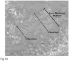
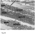
- a biocomposite layer can be defined as a continuous or semi-continuous stratum running through part or all of a medical implant, wherein the layer is comprised of reinforcing fibers that aligned uni-directionally. Layers can be seen in several figures showing the internal structure of reinforced biocomposite medical implants, including in figure 7 , 10 , and 20 .
- each biocomposite layer there are between 1-100 reinforcing fibers forming the thickness of each biocomposite layer.
- the directional fiber orientation between adjacent layers within the implant alternates between layers such that each adjacent layer is out of phase (of a different angle) from the layer that is adjacent to it.
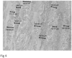
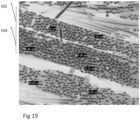
- the average or median angle difference between layers is between 15 to 75 degrees, more preferably between 30 to 60 degrees, and most preferably between 40 to 50 degrees. Microscopic images of such out of phase adjacent biocomposite layers can be seen in figure 26 and 27 .
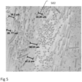
- the biocomposite layers within the medical implant are well approximated to each other. More preferably, the distance between layers, as measured by the distance between the last fiber in one layers and the first fiber in the subsequent layer is between 0-200 ⁇ m, more preferably between 0-60 ⁇ m, 1-40 ⁇ m, and most preferably between 2-30 ⁇ m. Good approximation of the fibers within a layer to the fibers within the adjacent layer allow each layer to mechanically support the adjacent layer. However, some distance between the layers may be desirable to allow for some polymer to remain between the fibers of adjacent layers and thus adhere the layers together, prevent layer dehiscence under high mechanical load.
- the diameter of a majority of reinforcing fiber for use with herein reinforced biocomposite medical implant is in the range of 1-100 ⁇ m.
- fiber diameter is in the range of 1-20 ⁇ m. More preferably, fiber diameter is in the range of 4-16 ⁇ m, and most preferably in the range of 9-14 ⁇ m.
- the average diameter of reinforcing fiber for use with herein reinforced biocomposite medical implant is in the range of 4-16 ⁇ m, and most preferably in the range of 9-14 ⁇ m.
- the standard deviation of fiber diameter between fibers within the medical implant is preferably less than 5 ⁇ m, more preferably less than 3 ⁇ m, and most preferably less than 1.5 ⁇ m. Uniformity of fiber diameter is beneficial for consistent properties throughout the implant.
- reinforcing fibers are fiber segments inside the polymer matrix.
- fiber segments are, on average, of length 0.5-20mm, more preferably the fiber segment length is in the range of 1-15mm, more preferably in the range of 3-10 and most preferably in the range of 4-8mm.
- a majority of reinforcing fiber segments are of length 0.5-20mm, more preferably the fiber segment length is in the range of 1-15mm, more preferably in the range of 3-10 and most preferably in the range of 4-8mm.
- the reinforcing fibers are continuous fibers.
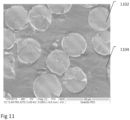
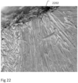
- Said continuous fibers are preferably longer than 5 mm, more preferably longer than 8 mm, 12 mm, 16 mm, and most preferably longer than 20 mm. A microscopic image of such continuous fibers can be seen in figure 22 .
- the reinforcing fiber length can be defined as a function of implant length wherein at least a portion of the reinforcing fibers, and preferably a majority of the reinforcing fibers, are of a continuous length at least 50% the longitudinal length of the medical implant or medical implant component that is comprised of these fibers.
- the portion or majority of the reinforcing fibers are of continuous length at least 60% of the length of the medical implant, and more preferably at least 75% of the length of the medical implant.
- Such continuous reinforcing fibers can provide structural reinforcement to a large part of the implant.
- the distance between adjacent reinforcing fibers within a biocomposite layer is in the range of 0.5-50 ⁇ m, preferably the distance between adjacent fibers is in the range of 1-30 ⁇ m, more preferably in the range of 1-20 ⁇ m, and most preferably in the range of 1-10 ⁇ m.
- the weight percentage of the reinforcing fibers (mineral composition) within the biocomposite medical implant is in the range of 40%-65%, and optionally the weight percentage is in the range of 45%-60%.
- the volume percentage of reinforcing fibers within the biocomposite medical implant is in the range of 30-90%, more preferably the volume percentage is in the range of 40%-70%.
- a plurality of fibers within the implant are uni-directionally aligned.
- the aligned fiber segments are, on average, of length 5-12mm.
- the uni-directionally aligned fibers are aligned in the longitudinal access of the implant (0° alignments in relation to the longitudinal axis).
- a majority of fibers are uni-directionally aligned in the longitudinal axis.
- more than 70%, 80%, 90%, 95% of fibers are uni-directionally aligned in the longitudinal axis.
- a plurality or a majority of fibers within the implant are aligned in the longitudinal axis.
- a plurality of fibers are additionally aligned in up to 3 additional directions.
- a plurality of fibers are aligned in a selection of each of the following alignments in relation to the longitudinal axis: 0°, 30°, -30°, 45°, -45°, 90°.
- a plurality of fibers are aligned in a selection of each of the following alignments in relation to the longitudinal axis: 0°, 45°, -45°, 90°.
- a plurality of fibers are aligned in a selection of each of the following alignments in relation to the longitudinal axis: 0°, 45°, -45°.
- a majority of fibers are aligned in the longitudinal access of the implant and a plurality of fibers are aligned in each of the following alignments in relation to the longitudinal axis: 45°, -45°.
- fiber segments are arranged amorphously.
- the biocomposite composition within the implant is important in determining the mechanical and bulk properties of the implant, the specific composition and structure that comes into contact with the surface edge of the implant has unique significance in that this composition and structure can greatly affect how surrounding cells and tissue interact with the implant following implantation into the body.
- the absorbable polymer part of the biocomposite may be hydrophobic in nature such that it will repel surrounding tissues to a certain degree while the mineral reinforcing fiber part of the biocomposite may be hydrophilic in nature and therefore encourage surrounding tissues to attach to the implant or create tissue ingrowth .
- the surface presence of one of the compositional components by percentage of surface area is greater than the presence of that component in the bulk composition of the implant by volume percentage.
- the amount of mineral on the surface might be greater than the amount of polymer, or vice versa.
- a greater amount of mineral would optionally and preferably be present on the surface.
- a greater amount of polymer would optionally and preferably be present on the surface.
- the percentage of surface area composition of one component is more than 10% greater than the percentage of volume percentage of that component in the overall biocomposite implant. More preferably, the percentage is more than 30% greater, and most preferably more than 50% greater.
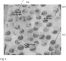
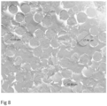
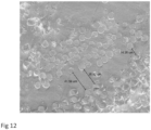
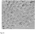
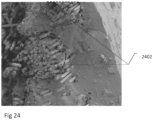
- Fig 25 shows a microscopic image of a biocomposite medical implant with a predominance of mineral reinforcing fiber along the inner surface area edge of the implant.
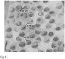
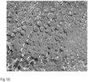
- Fig 29 shows a microscopic image of a biocomposite medical implant with a predominance of bioabsorbable polymer along the outer surface area of the implant .
- one surface of the medical implant may have a local predominance of one of the biocomposite components while a different surface, or different part of the same surface, may have a local predominance of a different biocomposite component
- mineral content is not present in a majority of the surface area (i.e. a majority of the surface of the implant is covered with a polymer film).
- the surface polymer film is, on average, 0.5-50 ⁇ m in thickness, more preferably 5-50 ⁇ m and most preferably 10-40 ⁇ m..
- exposed fibers there are fibers exposed at the surface of the implant.
- exposed fibers comprise 1-60% of implant surface.
- exposed fibers comprise 10-50% of implant surface.
- exposed fibers comprise 15-30% of implant surface.
- the medical implant is a threaded screw or other threaded implant.
- the outer layer of the implant will be directionally aligned such that the direction of the fibers approximates the helix angle of the threading.
- the alignment angle of the fiber direction is within 45 degrees of the helix angle. More preferably, the alignment angle is within 30 degrees, and most preferably the alignment angle is within 15 degrees of the helix angle. Approximating the fiber alignment angle to the helix angle in this manner can improve the robustness of the threading and prevent dehiscence of the reinforcing fibers within the threading .
- the reinforcing fibers may optionally take the full circular shape of the implant and curve around the circle shape of the implant without deviation from its circumference.
- a portion or a majority of the reinforcing fibers deviate from the circle shape of the implant such that a tangential angle is formed.
- the tangential angle is defined as the deviation from the direction of the curve at a fixed starting point, where the fixed starting point is the point where the fiber touches or is closest to coming into contact with the center of the cross-sectional circular area.
- Figure 23 depicts the tangential angle of reinforcing fibers to a cannulated circular pin .
- the tangential angle between reinforcing fibers within the circular medical implant and the curvature of the implant is less than 90 degrees, more preferably less than 45 degrees.
- the density of the biocomposite composition for use in herein invention is between 1 to 2 g/mL. More preferentially, density is between 1.2 to 1.9 g/mL. Most preferentially between 1.4 to 1.8 g/mL.
- the medical implant described herein comprises a polymer selected from the group consisting of polylactide (PLA), poly-L-lactide (PLLA), poly-DL-lactide (PDLLA), poly-LD-lactide (PLDLA), polyglycolide (PGA), poly-lactide-glycolic acid (PLGA), polycaprolactone (PCL), poly-L-lactide-polycaprolactone (PLLA-LCL) and a combination thereof, wherein residual monomer content in the medical implant following production is less than 3%.
- the medical implant comprises a reinforced bioabsorbable polymer (i.e. a bioabsorbable composite that includes the previously described polymer and also incorporates a reinforcing filler, in fiber form, to increase the mechanical strength of the polymer).
- a reinforced bioabsorbable polymer i.e. a bioabsorbable composite that includes the previously described polymer and also incorporates a reinforcing filler, in fiber form, to increase the mechanical strength of the polymer.
- the reinforcing filler are mineral materials.
- Reinforcing filler may be a biodegradable glass, a nano-diamond, or any other filler known in the art to increase the mechanical properties of a bioabsorbable polymer.
- a biocompatible and resorbable melt derived glass composition where glass fibers can be embedded in a continuous polymer matrix EP 2 243 749 A1
- Biodegradable composite comprising a biodegradable polymer and 20-70 vol% glass fibers
- Resorbable and biocompatible fiber glass that can be embedded in polymer matrix US 2012/0040002 A1
- Biocompatible composite and its use US 2012/0040015 A1
- Absorbable polymer containing poly[succinimide] as a filler EP0 671 177 B1 ).
- the reinforcing filler is bound to the bioabsorbable polymer such that the reinforcing effect is maintained for an extended period.
- a composite material comprising biocompatible glass, a biocompatible matrix polymer and a coupling agent capable of forming covalent bonds.
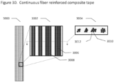
- biodegradable composite and fibers are preferably arranged in the form of biodegradable composite layers, where each layer comprises uni-directionally aligned continuous reinforcement fibers embedded in a polymer matrix comprised of one or more bioabsorbable polymers.
- the biodegradable composite layers are preferably comprised of one or more biodegradable composite tapes, where each tape comprises uni-directionally aligned continuous reinforcement fibers embedded in a polymer matrix comprised of one or more bioabsorbable polymers.
- the biodegradable composite is embodied in a polymer matrix, which comprises a polymer selected from the group consisting of polylactide (PLA), poly-L-lactide (PLLA), poly-DL-lactide (PDLLA), poly-LD-lactide (PLDLA), polyglycolide (PGA), poly-lactide-glycolic acid (PLGA), polycaprolactone (PCL), poly-L-lactide-polycaprolactone (PLLA-LCL) and a combination thereof, wherein residual monomer content in the medical implant following production is less than 3%.
- the matrix preferably comprises at least 30% PLLA, more preferably 50%, and most preferably at least 70% PLLA.
- the matrix preferably comprises at least 5% PDLA, more preferably at least 10%, most preferably at least 20% PDLA.
- the inherent viscosity (IV) of the polymer matrix (independent of the reinforcement fiber) is in the range of 1.2 to 2.4 dl/g, more preferably in the range of 1.5 to 2.1 dl/g, and most preferably in the range of 1.7 to 1.9 dl/g.
- IV Inherent Viscosity is a viscometric method for measuring molecular size. IV is based on the flow time of a polymer solution through a narrow capillary relative to the flow time of the pure solvent through the capillary.
- reinforcement fiber is comprised of silica-based mineral compound such that reinforcement fiber comprises a bioresorbable glass fiber, which can also be termed a bioglass fiber composite.
- Mineral composition may include beta-tricalcium phosphate, calcium phosphate, calcium sulfate, hydroxyapatite, or a bioresorbable glass (also known as bioglass).
- bioresorbable glass compositions are described in the following patent applications: Biocompatible composite and its use ( WO2010122098 ); and Resorbable and biocompatible fibre glass compositions and their uses ( WO2010122019 ).
- the reinforcing filler is bound to the bioabsorbable polymer such that the reinforcing effect is maintained for an extended period.
- a composite material comprising biocompatible glass, a biocompatible matrix polymer and a coupling agent capable of forming covalent bonds.
- Bioresorbable glass fiber may optionally have oxide compositions in the following mol.% ranges:
- bioresorbable glass compositions are described in the following patent applications: Biocompatible composite and its use ( WO2010122098 ); and Resorbable and biocompatible fibre glass compositions and their uses ( WO2010122019 ).
- Tensile strength of the reinforcement fiber is preferably in the range of 1200-2800 MPa, more preferably in the range of 1600-2400 MPa, and most preferably in the range of 1800-2200 MPa.
- Elastic modulus of the reinforcement fiber is preferably in the range of 30-100 GPa, more preferably in the range of 50-80 GPa, and most preferably in the range of 60-70 GPa.
- Fiber diameter is preferably in the range of 6-20 ⁇ m, more preferably in the range of 10-18 ⁇ m, and most preferably in the range of 14-16 ⁇ m.
- a majority of reinforcement fibers aligned to the longitudinal axis of the medical implant are of a length of at least 50% of the total length of the implant, preferably at least 60%, more preferably at least 75%, and most preferably at least 85%.
- fibers may be aligned at an angle to the longitudinal axis (i.e. on a diagonal) such that the length of the fiber may be greater than 100% of the length of the implant.
- a majority of reinforcement fibers are aligned at an angle that is less than 90°, alternatively less than 60°, or optionally less than 45° from the longitudinal axis.
- the implant preferably comprises between 2-20 composite tape layers, more preferably between 2-10 layers, and most preferably between 2-6 layers; wherein each layer may be aligned in a different direction or some of the layers may be aligned in the same direction as the other layers.
- the maximum angle between fibers in at least some of the layers is greater than the angle between the fibers in each layer and the longitudinal axis.
- one layer of reinforcing fibers may be aligned and a right diagonal to the longitudinal axis while another layer may be aligned at a left diagonal to the longitudinal axis.
- the composite composition additionally includes a compatibilizer, which for example be such an agent as described in WO2010122098 .
- biodegradable composite may comprise composite strands comprising continuous reinforcement fibers or fiber bundles impregnated with bioabsorbable polymer.
- strands are less than 1 cm in diameter. More preferably, strands are less than 8 mm, less than 5 mm, less than 3 mm, or less than 2 mm in diameter.
- biodegradable composite may comprise a woven mesh of continuous reinforcement fibers wherein woven mesh is pre-impregnated with bioabsorbable polymer or woven mesh is comprised of reinforcement fibers and subsequently impregnated with bioabsorbable polymer.
- biodegradable composite mesh layer is less than 1 cm in thickness. More preferably, impregnated mesh is less than 8 mm, less than 5 mm, less than 3 mm, or less than 2 mm in thickness.
- the present invention in at least some embodiments, further overcomes the limitations of previous biocomposite medical implants by providing medical implants comprised of a biocomposite material composition with a high percentage of mineral content and yet with superior mechanical properties.
- a biocomposite material composition with a high percentage of mineral content and yet with superior mechanical properties.
- the mineral composition is provided by a reinforcing fiber made from the mineral composition.
- the weight percentage of the mineral composition within the biocomposite medical implant is in the range of 40%-65%, and optionally the weight percentage is in the range of 45%-60%.
- the density of the biocomposite composition for use in present invention is between 1 to 2 g/mL. More preferentially, density is between 1.2 to 1.9 g/mL. Most preferentially density is between 1.4 to 1.8 g/mL.
- the diameter of reinforcing fiber for use with the reinforced biocomposite medical implant is in the range of 1-20 ⁇ m. More preferably, fiber diameter is in the range of 4-16 ⁇ m.
- the standard deviation of fiber diameter between fibers within the medical implant is preferably less than 5 ⁇ m, more preferably less than 3 ⁇ m, and most preferably less than 1.5 ⁇ m. Uniformity of fiber diameter is beneficial for consistent properties throughout the implant.
- the fiber-reinforced biodegradable composite within the implant has a flexural modulus exceeding 5 GPa and flexural strength exceeding 80 MPa .
- the fiber-reinforced biodegradable composite within the implant has flexural strength in range of 150 - 800 MPa, more preferably 150 - 400 MPa.
- Elastic modulus is preferably in range of 5 - 27 GPa, more preferably 10 - 27 GPa.
- the fiber-reinforced composite within the implant has strength retention of Elastic Modulus above 10 GPa after 8 weeks implantation and flexural strength above 150 MPa after 8 weeks.
- the medical implant comprises a polymer selected from the group consisting of polylactide (PLA), poly-L-lactide (PLLA), poly-DL-lactide (PDLLA), poly-LD-lactide (PLDLA), polyglycolide (PGA), poly-lactide-glycolic acid (PLGA), polycaprolactone (PCL), poly-L-lactide-polycaprolactone (PLLA-LCL) and a combination thereof, and wherein residual monomer content in the medical implant following production is less than 3%.
- the matrix preferably comprises at least 30% PLLA, more preferably 50%, and most preferably at least 70% PLLA.
- the matrix preferably comprises at least 5% PDLA, more preferably at least 10%, most preferably at least 20% PDLA .
- the inherent viscosity (IV) of the polymer matrix (independent of the reinforcement fiber) is in the range of 1.2 to 2.4 dl/g, more preferably in the range of 1.5 to 2.1 dl/g, and most preferably in the range of 1.7 to 1.9 dl/g .
- IV Inherent Viscosity is a viscometric method for measuring molecular size. IV is based on the flow time of a polymer solution through a narrow capillary relative to the flow time of the pure solvent through the capillary.
- Mineral composition may optionally include beta-tricalcium phosphate, calcium phosphate, calcium sulfate, hydroxyapatite, or a bioresorbable glass (also known as bioglass).
- Bioresorbable glass fiber may optionally have oxide compositions in the following mol.% ranges:
- bioresorbable glass compositions are described in the following patent applications, which are owned in common with the instant application and which have inventor(s) in common: Biocompatible composite and its use ( WO2010122098 ); and Resorbable and biocompatible fibre glass compositions and their uses ( WO2010122019 ).
- the reinforcing filler is bound to the bioabsorbable polymer such that the reinforcing effect is maintained for an extended period.
- a composite material comprising biocompatible glass, a biocompatible matrix polymer and a coupling agent capable of forming covalent bonds.
- fibers may be aligned at an angle to the longitudinal axis (i.e. on a diagonal) such that and preferably, a majority of reinforcement fibers are aligned at an angle that is less than 90°, alternatively less than 60°, or optionally less than 45° from the longitudinal axis.
- Implant may be selected from a group that includes orthopedic pins, screws, plates, intramedullary rods, hip replacement, knee replacement, meshes, etc.
- the average wall thickness in the implant is preferably in the range of 0.2 to 10 mm, more preferably in the range of 0.4 to 5 mm, more preferably in the range of 0.5 to 2 mm, and most preferably in the range of 0.5 to 1.5 mm.
- the implant preferably comprises between 2-20 composite tape layers, more preferably between 2-10 layers, and most preferably between 2-6 layers.
- implant may comprise reinforcing ribs, gussets, or struts.
- Rib base thickness is preferably less than 100% of the adjoining wall thickness. More preferably, thickness is less than 85%, and most preferably less than 75%. Rib base thickness is preferably more than 20% of adjoining wall thickness, more preferably more than 30%, and most preferably more than 50% of adjoining wall thickness.
- rib height is at least 2.0 times the adjoining wall thickness, more preferably at least 3.0 times the wall thickness.
- Draft angle of reinforcing ribs is preferably between 0.2-0.8°, more preferably between 0.4-0.6°.
- distance between ribs is at least 2 times adjoining wall thickness. More preferably, at least 3 times adjoining wall thickness.
- reinforcing rib or other element increases bending stiffness of implant by at least 20% without increasing compressive or tensile stiffness by more than 10%.
- ribs along one axis are taller than the ribs along the perpendicular axis, for example the latitudinal axis of the implant, in order to facilitate easier insertion of the implant.
- the implant may comprise one or more bosses to accommodate screw insertion.
- the boss is between 2-3 times the screw diameter for self-tapping screw applications.
- Boss may additionally include supportive gusses or ribs.
- one or more sides of implant may be textured.
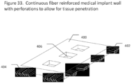
- implant may contain continuous fibers aligned in a circular arrangement around holes, such as screw or pin holes, within the implant.
- such perforations in implant walls comprise at least 10% of the surface area of the implant, more preferably at least 20%, at least 30%, at least 40%, or at least 50% of the surface area of the implant.
- the implant is a screw and the fenestrations of the threading contain perforation.
- the implant contains perforations between composite tapes or between the reinforcement fibers within composite tapes making up the implant.
- a majority of perforations are between reinforcement fibers and do not penetrate reinforcement fibers.
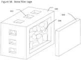
- the implant comprises an orthopedic implant and the implant forms a partial or full container and an osteoconductive or osteoinductive material is contained within the implant container.
- the implant container is additionally perforated so as to allow improved bone ingrowth into the osteoconductive or osteoinductive material contained within the implant cage.
- the implant comprises an opening or door through which bone filler can be introduced and/or bone ingrowth can take place.
- the implant comprises two or more discrete parts or separate parts joined by a joint such that implant cage may be filled with bone filler material and subsequently assembled or closed to trap bone filler inside.
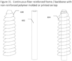
- continuous fiber reinforced bioabsorbable composite structures provide the optimal mechanical strength and stiffness to a medical implant
- the mechanical strength of the continuous fiber reinforced bioabsorbable composite structures can be incorporated into the implant but additional sections or layers of non-reinforced polymer may be added to improve or customize the implant. These sections or layers are preferably added to the implant either by overmolding onto the structure or by 3-D printing onto the structure.
- medical implant comprises a structural support comprised of a continuous fiber-reinforced bioabsorbable composite material and additionally comprises a section or layer comprised of non-reinforced polymer material.
- the second layer functions as a bone interface layer comprised of a non-reinforced absorbable polymer material.
- the structural support and non-reinforced polymer section are each fabricated using a different production technique.
- the structural support is fabricated by machining, compression molding, or composite flow molding and the interface layer is fabricated by injection molding or 3D printing; optionally the interface layer is fabricated on top of the prefabricated structural support.
- the non-reinforced polymer section is a bone interface layer and dimensions of the interface layer are partially or entirely determined by the bone geometry of a specific patient or patient population.
- the bone geometry of patient or patient population is determined by measuring through imaging technique such as X-Ray, CT, MRI.
- the elastic modulus and/or flexural strength of structural support is at least 20% greater than that of the non-reinforced polymer section.
- continuous-fiber reinforced composite material in implant is coated with a polymer resin wherein the polymer resin on fiber in the composite material has a higher or lower melting temp than the flowable matrix resin; or polymer resin on fiber has slower or faster degradation rate than flowable matrix resin; or polymer resin on fiber is more hydrophobic or more hydrophilic than flowable matrix resin
- an additional section or layer is comprised of a reinforced polymer but where polymer is reinforced by non-continuous fibers, preferably fibers less than 10mm in length, and more preferably less than 5mm in length.
- an additional section or layer of non-reinforced or non-continuous fiber reinforced polymer additional comprises an additive.
- additive comprises an osteoconductive material or combination of osteoconductive materials such as beta tricalcium phosphate, calcium phosphate, hydroxyapatite, decellularized bone.
- the additive comprises an anti-microbial agent or bone inducing agent.
- Continuous-fiber reinforced bioabsorbable implants may optionally be produced using any method known in the art. Methods can include compression molding, injection molding, extrusion, machining, or any combination of these methods.
- moisture content of implant following production is less than 1%, even more preferably less than 0.4%, 0.2%.
- Low moisture content is important so as to avoid degradation of the implant during storage.
- residual monomer content in implant following production is less than 2%, and more preferably less than 1%.
- the polymer component be predominantly comprised of polymer, with very low monomer component, since the monomer component does not contribute to the mechanical function of the implant.
- implant surface area is in contact with surrounding tissue. This may be clinically desirable for several reasons:
- implant contains surface protrusion elements of at least 0.1 mm in height and less than 2 mm in height that come into contact with tissue surrounding implant.
- total percentage of surface area of implant that comes into contact with surrounding tissue is less than 80%, more preferably less than 60%, 50% , 40%, 30%.
- implant additionally comprises a balloon.
- Balloon walls are preferably comprised of between 1-3 layers of reinforced composite.
- bioabsorbable polymers or reinforced bioabsorbable polymers may be fabricated into any desired physical form for use with the present invention.
- the polymeric substrate may be fabricated for example, by compression molding, casting, injection molding, pultrusion, extrusion, filament winding, composite flow molding (CFM), machining, or any other fabrication technique known to those skilled in the art.
- CFRM composite flow molding
- the polymer may be made into any shape, such as, for example, a plate, screw, nail, fiber, sheet, rod, staple ,clip, needle, tube, foam, or any other configuration suitable for a medical device.
- the herein invention particularly relates to bioabsorbable composite materials that can be used in medical applications that require high strength and a stiffness compared to the stiffness of bone. These medical applications require the medical implant to bear all or part of the load applied by or to the body and can therefore be referred to generally as "load-bearing" applications. These include fracture fixation, tendon reattachment, joint replacement, spinal fixation, and spinal cages.
- the flexural strength preferred from the herein described load-bearing medical implant is at least 100 MPa, preferably above 400 MPa, more preferably above 600 MPa, and even more preferably above 800 MPa.
- the Elastic Modulus (or Young's Modulus) of the bioabsorbable composite for use with herein invention is preferably at least 6 GPa, more preferably above 15 GPa, and even more preferably above 20 GPa but not exceeding 100 GPa and preferably not exceeding 60 GPa.
- the strength and stiffness preferably remains above the strength and stiffness of cortical bone, approximately 150-250 MPa and 15-25 GPa respectively, for a period of at least 3 months, preferably at least 6 months, and even more preferably for at least 9 months in vivo (i.e. in a physiological environment).
- the flexural strength remains above 400 MPa and even more preferably remains above 600 MPa.
- the mechanical strength degradation rate of the medical implant approximates the material degradation rate of the implant, as measured by weight loss of the biodegradable composite.
- the implant retains greater than 50% of its mechanical strength after 3 months of implantation while greater than 50% of material degradation and hence weight loss occurs within 12 months of implantation.
- the implant retains greater than 70% of its mechanical strength after 3 months of implantation while greater than 70% of material degradation and hence weight loss occurs within 12 months of implantation.
- the implant retains greater than 50% of its mechanical strength after 6 months of implantation while greater than 50% of material degradation and hence weight loss occurs within 9 months of implantation.
- the implant retains greater than 70% of its mechanical strength after 6 months of implantation while greater than 70% of material degradation and hence weight loss occurs within 9 months of implantation.
- the mechanical strength degradation and material degradation (weight loss) rates of the medical implant can be measured after in vivo implantation or after in vitro simulated implantation.
- the simulation may be performed in real time or according to accelerated degradation standards.
- Biodegradable as used herein is a generalized term that includes materials, for example polymers, which break down due to degradation with dispersion in vivo.
- the decrease in mass of the biodegradable material within the body may be the result of a passive process, which is catalyzed by the physicochemical conditions (e.g. humidity, pH value) within the host tissue.
- the decrease in mass of the biodegradable material within the body may also be eliminated through natural pathways either because of simple filtration of degradation by-products or after the material's metabolism ("Bioresorption” or "Bioabsorption”). In either case, the decrease in mass may result in a partial or total elimination of the initial foreign material.
- said biodegradable composite comprises a biodegradable polymer that undergoes a chain cleavage due to macromolecular degradation in an aqueous environment.
- a polymer is "absorbable" within the meaning of this invention if it is capable of breaking down into small, non-toxic segments which can be metabolized or eliminated from the body without harm.
- absorbable polymers swell, hydrolyze, and degrade upon exposure to bodily tissue, resulting in a significant weight loss.
- the hydrolysis reaction may be enzymatically catalyzed in some cases.
- Complete bioabsorption, i.e. complete weight loss may take some time, although preferably complete bioabsorption occurs within 24 months, most preferably within 12 months.
- polymer degradation means a decrease in the molecular weight of the respective polymer. With respect to the polymers, which are preferably used within the scope of the present invention said degradation is induced by free water due to the cleavage of ester bonds.
- the degradation of the polymers as for example used in the biomaterial as described in the examples follows the principle of bulk erosion. Thereby a continuous decrease in molecular weight precedes a highly pronounced mass loss. Said mass loss is attributed to the solubility of the degradation products.
- Methods for determination of water induced polymer degradation are well known in the art such as titration of the degradation products, viscometry, differential scanning calorimetry (DSC).
- Biocomposite is a composite material formed by a matrix and a reinforcement of fibers wherein both the matrix and fibers are biocompatible and optionally bioabsorbable.
- the matrix is a polymer resin, and more specifically a synthetic bioabsorbable polymer.
- the fibers are optionally and preferably of a different class of material (i.e. not a synthetic bioabsorbable polymer), and may optionally comprise mineral, ceramic, cellulosic, or other type of material.
- fixation optionally and preferably includes one or more, and more preferably all, of stable fixation, preservation of blood supply to the bone and surrounding soft tissue, and early, active mobilization of the part and patient.
- Bones of the limbs include a shaft with a head at either end thereof.
- the shaft of the bone is generally elongated and of relatively cylindrical shape.
- Such a bone plate which attaches to the shaft or head and shaft of a fractured bone to maintain two or more pieces of the bone in a substantially stationary position relative to the one another.
- a bone plate generally comprises a shape having opposing substantially parallel sides and a plurality of bores extending between the opposing sides, wherein the bores are suitable for the receipt of pins or screws to attach the plate to the bone fragments.
- the plate For proper function of the bone plate in maintaining different parts of a fractured bone stationary relative to each other, the plate must be of sufficient mechanical strength and stiffness to maintain the position of the bone fragments or pieces. However, it must achieve these mechanical properties within a low profile thickness profile to ensure that there will be sufficient space for the bone plate to fit between bone and the surrounding soft tissue.
- the thickness of the bone plate is generally in the range of 2.0 mm to 8.0 mm and more commonly in the range of 2.0 mm to 4.0 mm.
- Screws are used for internal bone fixation and there are different designs based on the type of fracture and how the screw will be used. Screws come in different sizes for use with bones of different sizes. Screws can be used alone to hold a fracture, as well as with plates, rods, or nails. After the bone heals, screws may be either left in place or removed.
- Screws are threaded, though threading can be either complete or partial. Screws can include compression screws, locking screws, and/or cannulated screws. External screw diameter can be as small as 0.5 or 1.0 mm but is generally less than 3.0mm for smaller bone fixation. Larger bone cortical screws can be up to 5.0mm and cancellous screws can even reach 7-8 mm. Some screws are self-tapping and others require drilling prior to insertion of the screw. For cannulated screws, a hollow section in the middle is generally larger than 1mm diameter in order to accommodate guide wires.
- Wires are often used to pin bones back together. They are often used to hold together pieces of bone that are too small to be fixed with screws. They can be used in conjunction with other forms of internal fixation, but they can be used alone to treat fractures of small bones, such as those found in the hand or foot. Wires or pins may have sharp points on either one side or both sides for insertion or drilling into the bone.
- K-wire is a particular type of wire generally made from stainless steel, titanium, or nitinol and of dimensions in the range of 0.5 - 2.0 mm diameter and 2-25 cm length.
- Stepman pins are general in the range of 2.0 - 5.0 mm diameter and 2-25 cm length. Nonetheless, the terms pin and wire for bone fixation are used herein interchangeably.
- Anchors and particularly suture anchors are fixation devices for fixing tendons and ligaments to bone. They are comprised of an anchor mechanism, which is inserted into the bone, and one or more eyelets, holes or loops in the anchor through which the suture passes. This links the anchor to the suture.
- the anchor which is inserted into the bone may be a screw mechanism or an interference mechanism.
- Anchors are generally in the range of 1.0 - 6.5 mm diameter.
- Cables, ties, or wire ties can be used to perform fixation by cerclage, or binding, bones together.
- Such implants may optionally hold together bone that cannot be fixated using penetration screws or wires/pin, either due to bone damage or presence of implant shaft within bone.
- diameter of such cable or tie implants is optionally in the range of 1.0 mm - 2.0 mm and preferably in the range of 1.25 - 1.75 mm.
- Wire tie width may optionally be in the range of 1 - 10 mm.
- a rod or nail In some fractures of the long bones, medical best practice to hold the bone pieces together is through insertion of a rod or nail through the hollow center of the bone that normally contains some marrow. Screws at each end of the rod are used to keep the fracture from shortening or rotating, and also hold the rod in place until the fracture has healed. Rods and screws may be left in the bone after healing is complete. Nails or rods for bone fixation are generally 20-50 cm in length and 5-20 mm in diameter (preferably 9-16mm). A hollow section in the middle of nail or rod is generally larger than 1mm diameter in order to accommodate guide wires.
- any of the above-described bone fixation implants may optionally be used to fixate various fracture types including but not limited to comminuted fractures, segmental fractures, non-union fractures, fractures with bone loss, proximal and distal fractures, diaphyseal fractures, osteotomy sites, etc.
- Material composite was comprised of PLDLA 70/30 polymer reinforced with 50% w/w, 70%, or 85% w/w continuous mineral fibers.
- Mineral fibers composition was approximately Na 2 O 14%, MgO 5.4%, CaO 9%, B 2 O 3 2.3%, P 2 O 5 1.5%, and SiO 2 67.8% w/w.
- Testing samples were manufactured by compression molding of multiple layers of composite material into a tubular mold, either with or without a 3mm pin insert in the center. Each layer was comprised of the PLDLA polymer with embedded uni-directionally aligned continuous fibers.
- Orientation of layers relative to longitudinal axis of implant were 0° (parallel to implant longitudinal axis), 45°, 0°, -45°, 0°, in a repetitive manner according to number of layers in the implant. Each layer was approximately 0.18 mm thick. Three (3) pin samples were produced for each pin group.
- Implant samples were tested in a tensile testing system (220Q1125-95, TestResources, MN, USA) for flexural strength, flexural modulus and maximum flexural load according to modified standard test method, ASTM D790 (Standard Test Methods for Flexural Properties of Unreinforced and Reinforced Plastics and Electrical Insulating Materials, http://www.astm.org/Standards/D790.htm, ASTM International, PA, USA).
- reinforcing fiber content, diameter, distribution, and arrangement into layers seen in this example were the cause or at least a significantly contributing factor.
- reinforcing fiber may contribute positively to mechanical properties of a medical implant, as seen by the stronger and stiffer samples produced with 70% fiber as compared with those produced with 50% fiber.
- 70% fiber implants seemed to lose mechanical properties at a faster rate.
- Material composite was comprised of PLDLA 70/30 polymer reinforced with 50% w/w or 70% w/w continuous mineral fibers.
- Mineral fiber composition was approximately Na 2 O 14%, MgO 5.4%, CaO 9%, B 2 O 3 2.3%, P 2 O 5 1.5%, and SiO 2 67.8% w/w.
- Testing samples were manufactured by compression molding of multiple layers of composite material into a tubular mold, either with or without a 1mm pin insert in the center. Each layer was comprised of the PLDLA polymer with embedded uni-directionally aligned continuous fibers.
- Orientation of layers relative to longitudinal axis of implant were 0° (parallel to implant longitudinal axis), 45°, 0°, -45°, 0°, in a repetitive manner according to number of layers in the implant. Each layer was approximately 0.18 mm thick. Three (3) pin samples were produced for each pin group.
- Implant samples were tested in a tensile testing system (220Q1125-95, TestResources, MN, USA) for flexural strength, flexural modulus and maximum flexural load according to modified standard test method , ASTM D790 (Standard Test Methods for Flexural Properties of Unreinforced and Reinforced Plastics and Electrical Insulating Materials, http://www.astm.org/Standards/D790.htm, ASTM International, PA, USA).
- Table 2b Measured structural parameters relating the reinforcing fibers and biocomposite layers within a biocomposite pin Fiber diameter range ( ⁇ m) Distance between fibers ( ⁇ m) Fibers in layer thickness Layer thickness ( ⁇ m) Distance between layers ( ⁇ m) Full pin.
- Table 2c Measured volume percentage of fiber as measured from cross-section of biocomposite full pin implant of OD 2mm, 50% w/w fiber (see Fig 11) Area of Entire Cross Section Sum of Fiber Area Remaining Area Percentage of Fiber to Polymer 22579 ⁇ m 11043 ⁇ m 1.1536e+04 ⁇ m 48.90%
- Table 2d Measured volume percentage of fiber as measured from cross-section of biocomposite full plate implant of OD 2mm, ID 1mm, 50% w/w fiber (see Fig 15) Area of Entire Cross Section Sum of Fiber Area Remaining Area Percentage of Fiber to Polymer 14094 ⁇ m 9645.14 ⁇ m 4448.86 ⁇ m 68.43%
- reinforcing fiber content, diameter, distribution, and arrangement into layers seen in this example were the cause or at least a significantly contributing factor.
- This example also suggests a potential structural difference between different implant part geometries (between a full pin and cannulated pin), where it is optionally possible for reinforcing fiber layers in the biocomposite implant to arrange and align themselves in differential manners depending on the shape of the implant and the forces that the implant is exposed to during its production.
- Material composite was comprised of PLDLA 70/30 polymer reinforced with 50% w/w or 70% w/w continuous mineral fibers.
- Mineral fibers composition was approximately Na 2 O 14%, MgO 5.4%, CaO 9%, B 2 O 3 2.3%, P 2 O 5 1.5%, and SiO 2 67.8% w/w.
- Testing samples were manufactured by compression molding of multiple layers of composite material into a rectangle mold. Each layer was comprised of the PLDLA polymer with embedded uni-directionally aligned continuous fibers.
- Orientation of layers relative to longitudinal axis of implant were 0° (parallel to implant longitudinal axis), 45°, 0°, -45°, 0°, in a repetitive manner according to number of layers in the implant. Each layer was approximately 0.18 mm thick.
- For the amorphous plates continuous fibers were cut to small pieces, mixed and molded. Three (3) plate samples were produced for each plate group.
- Implant samples were tested in a tensile testing system (220Q1125-95, TestResources, MN, USA) for flexural strength, flexural modulus and maximum flexural load according to modified standard test method , ASTM D790 (Standard Test Methods for Flexural Properties of Unreinforced and Reinforced Plastics and Electrical Insulating Materials, http://www.astm.org/Standards/D790.htm , ASTM International, PA, USA).
- Table 3a shows the mechanical performance results of three different types of reinforced composites implant pins produced as described above. The structural properties of these implants are described by the production methods discussed above and their internal compositions are seen in the associated images. Quantification of several parameters related to the internal composition structure of the implants can be seen in table 3b.
- Pin and plate implants were produced using reinforced composite material as described in example 1-3.
- Material composite was comprised of PLDLA 70/30 polymer reinforced with 50% w/w or 70% w/w continuous mineral fibers.
- Mineral fibers composition was approximately Na 2 O 14%, MgO 5.4%, CaO 9%, B 2 O 3 2.3%, P 2 O 5 1.5%, and SiO 2 67.8% w/w.
- Testing samples were manufactured by compression molding of multiple layers of composite material into an appropriate mold. Each layer was comprised of the PLDLA polymer with embedded uni-directionally aligned continuous fibers.
- Orientation of layers relative to longitudinal axis of implant were 0° (parallel to implant longitudinal axis), 45°, 0°, -45°, 0°, in a repetitive manner according to number of layers in the implant. Each layer was approximately 0.18 mm thick. Three (3) implant samples were produced for each group.
- Implant samples were weighed initially and following simulated in vitro degradation according to a modified ASTM F1635, wherein samples were incubated in simulated body fluid (SBF), 142 Na + , 5 K + , 1.5 Mg 2+ ,2.5 Ca 2+ , 147.8 Cl - , 4.2 HCO 3 - , 1 HPO 4 3- , 0.5 SO 4 2- mol/m 3 , for 5 days at a temperature of 50°C, while shaking at 30 rpm. Samples were then dried in a vacuum desiccator overnight and weighed again. Material percentage loss was calculated as (initial weight -dried weight)/initial weight * 100. Swelling ratio was calculated as (weight at the end of the incubation - dried weight)/dried weight* 100.
- SBF simulated body fluid
- Table 4 shows the weight measurement results of different types of reinforced composite implants produced as described above.
- biocomposite implant samples are demonstrated that comprise 50, 60 and 70% mineral content. These samples have both high mineral content and high mechanical properties.
- the mechanical properties of implants with higher mineral content (60% or 70%) dropped further than the 50% mineral content implants.
- the long term performance of the 50% mineral content implant would be improved as compared with the higher mineral content implants.
- an initially stronger implant can be achieved with higher mineral contents.
- Orientation of layers relative to longitudinal axis of implant were 0° (parallel to implant longitudinal axis), 45°, 0°, -45°, 0°, in a repetitive manner according to a number of layers in the implant. Each layer was approximately 0.18 mm thick. Three (3) samples were produced for each group. Mechanical properties were tested in a three point bending test.
- Figure 30 shows a continuous fiber-reinforced tape of the type that can be used to form a layer in a medical implant comprised of continuous fiber-reinforced layers.
- the top view (3000) shows a single strip of composite tape comprising reinforcement fibers aligned in a single direction within a bioabsorbable polymer matrix.
- the interspersed reinforcement fibers (3006) within the bioabsorbable polymer matrix (3008) can be seen more clearly in the close-up top view (3002) of the continuous-fiber reinforced composite tape.
- the reinforcement fibers can be present as separate fibers or in bundles of several reinforcement fibers per bundle.
- the cross-sectional view of the continuous fiber reinforced tape (3004) shows the bundles of aligned reinforcement fibers (3010) embedded within the bioabsorbable polymer matrix (3012). Fibers preferably do not breach the surface of the bioabsorbable polymer matrix.
- Figure 31 shows a cut-away, three-dimensional view of a continuous fiber-reinforced tape (200).
- the cut-away view shows the aligned reinforcement fibers (202) embedded within the bioabsorbable polymer matrix (204).
- Figure 32a shows a top-view of a reinforced bioabsorbable composite sheet (300) comprised of three layers of uni-directional fibers at different angles. Each layer can optionally be comprised of continuous fiber reinforced tapes of the type depicted in Figure 30 .
- the expanded view (302) shows layers of uni-directional fibers at different angles within an implant. One layer (304) aligned in the longitudinal axis, one layer (306) aligned at an angle to the right of the longitudinal axis, and one layer (308) aligned at an angle to the left of the longitudinal axis.
- Figure 32b shows a cut-away view of a reinforced bioabsorbable composite structure (310) comprised of three layers of uni-directional fibers at different angles.
- Each layer is comprised of reinforced continuous fibers (318) embedded within bioabsorbable polymer matrix (320).
- Figure 33 shows the wall of a continuous-fiber reinforced composite medical implant.
- the implant wall is comprised of two layers of uni-directional continuous-fiber reinforced composite tape layers (402 & 404) aligned at a perpendicular angle to each other.
- the medical implant wall additional comprises perforations (406) to allow for tissue penetration into or through the implant.
- Figure 34 shows a bone filler cage that consists of continuous-fiber reinforced composite medical implant walls (500) that additionally contains perforations (502) to allow tissue and cellular ingrowth into the bone filler material (504) contained within the bone filler cage.
- the bone filler cage optionally includes a separate door to close the cage (506).
- FIG 35 shows a bioabsorbable cannulated screw (600) that is a medical implant comprised of two parts: a continuous-fiber reinforced bioabsorbable composite cylindrical core (602) and bioabsorbable polymer threading (604) that was subsequently molded or 3D printed on top of the continuous-fiber core.
- a bioabsorbable medical implant where a significant amount or majority of the mechanical strength is provided by a continuous-fiber reinforced part that serves as a mechanical support or structure but where additional implant features are comprised of materials that are not continuous fiber reinforced and yet can be molded or printed directly onto the fiber reinforced composite material.
- biocomposite implant samples are demonstrated that comprise 50, 60 and 70% mineral content. These samples have both high mineral content and high mechanical properties.
- Orientation of layers relative to longitudinal axis of implant were 0° (parallel to implant longitudinal axis), 45°, 0°, - 45°, 0°, in a repetitive manner according to a number of layers in the implant. Each layer was approximately 0.18 mm thick. Three (3) samples were produced for each group. Mechanical properties were tested in a three point bending test.
- Example 8 Mineral Content and Sustained Strength
- biocomposite implant samples are demonstrated that comprise 58% and 68% mineral content. These samples have both high mineral content and high mechanical properties.
- Biocomposite rectangular plate implants were produced of dimensions 12.7x60x2.0mm.
- Material composite was comprised of PLDLA 70/30 polymer reinforced with 58% w/w or 68% w/w mineral fibers.
- Mineral fibers composition was approximately Na 2 O 14%, MgO 5.4%, CaO 9%, B 2 O 3 2.3%, P 2 O 5 1.5%, and SiO 2 67.8% w/w.
- Testing samples were manufactured by compression molding of composite material into a rectangular mold. Reinforcing mineral fibers were of a chopped nature, with fiber segment lengths predominately in the range of 5-10mm. Plate weight was 2.75g on average for each plate. Ten (10) samples were produced for each group.
Landscapes
- Health & Medical Sciences (AREA)
- Life Sciences & Earth Sciences (AREA)
- Orthopedic Medicine & Surgery (AREA)
- Public Health (AREA)
- Veterinary Medicine (AREA)
- Animal Behavior & Ethology (AREA)
- General Health & Medical Sciences (AREA)
- Engineering & Computer Science (AREA)
- Chemical & Material Sciences (AREA)
- Surgery (AREA)
- Heart & Thoracic Surgery (AREA)
- Epidemiology (AREA)
- Transplantation (AREA)
- Oral & Maxillofacial Surgery (AREA)
- Biomedical Technology (AREA)
- Vascular Medicine (AREA)
- Medicinal Chemistry (AREA)
- Dermatology (AREA)
- Neurology (AREA)
- Composite Materials (AREA)
- Nuclear Medicine, Radiotherapy & Molecular Imaging (AREA)
- Medical Informatics (AREA)
- Molecular Biology (AREA)
- Inorganic Chemistry (AREA)
- Materials Engineering (AREA)
- Cardiology (AREA)
- Textile Engineering (AREA)
- Mechanical Engineering (AREA)
- Materials For Medical Uses (AREA)
- Prostheses (AREA)
- Chemical Kinetics & Catalysis (AREA)
- Polymers & Plastics (AREA)
- Organic Chemistry (AREA)
Claims (14)
- Medizinisches Implantat, bestehend aus einem Biokomposit, wobei das Biokomposit aus einem Polymer und einer Vielzahl von verstärkenden Mineralfasern besteht, wobei ein Gewichtsanteil einer mineralischen Verbundstoffe in dem medizinischen Implantat im Bereich von 40 % - 65 %, optional 45 % - 60 % liegt, wobei der mineralische Gehalt durch die verstärkende Mineralfaser aus den mineralischen Verbundstoffen kommt, wobei der durchschnittliche Durchmesser der verstärkenden Mineralfasern liegt im Bereich von 1-20 Mikron; wobei die Fasern in eine Polymermatrix eingebettet sind, aus der das Biokomposit besteht; wobei das Polymer aus der Gruppe ausgewählt ist, die aus Polylactid (PLA), Poly-L-lactid (PLLA), Poly-DL-lactid (PDLLA), Poly-LD-lactid (PLDLA), Polyglycolid (PGA), Poly(lactid-co-Glycoid) (PLGA), Polycaprolacton (PCL), Poly-L-lactid-Polycaprolacton (PLLA-PCL) und einer Kombination davon besteht; und wobei der Restmonomergehalt in dem medizinischen Implantat nach der Herstellung weniger als 3 % beträgt.
- Medizinisches Implantat gemäß Anspruch 1, wobei das medizinische Implantat aus einem mineralischen Verbundstoff auf Siliciumoxidbasis besteht und wobei der mineralische Verbundstoff auf Siliciumdioxidbasis mindestens eine Oxidzusammensetzung in mindestens einem der folgenden Mol- % -Bereiche aufweist:Na2O: 11,0 - 19,0 mol.%CaO: 9,0 - 14,0 mol.%MgO: 1,5 - 8,0 mol.%B2O3: 0,5 - 3,0 mol.%Al2O3. 0 - 0,8 mol.%P2O3: 0,1 - 0,8 mol.%SiO2: 67 - 73 mol%.
- Medizinisches Implantat gemäß Anspruch 2, wobei der mineralische Verbundstoff auf Siliciumdioxidbasis mindestens eine Oxidzusammensetzung in mindestens einem der folgenden Mol- % -Bereiche aufweist:Na2O: 12,0 -13,0 mol.%CaO: 9,0 - 10,0 mol.%MgO: 7,0 - 8,0 mol.%B2O3: 1,4 - 2,0 mol.%P2O3: 0,5 - 0,8 mol.%SiO2: 68 - 70 mol.%.
- Medizinisches Implantat gemäß Anspruch 1, wobei der Durchmesser einer Mehrheit der verstärkenden Fasern im Bereich von 6-20 µm oder 4-16 µm oder 9-14 µm liegt.
- Medizinisches Implantat gemäß Anspruch 4, wobei eine Standardabweichung des Faserdurchmessers zwischen den Fasern in dem medizinischen Implantat weniger als 5 µm oder weniger als 3 µm oder weniger als 1,5 µm beträgt.
- Medizinisches Implantat gemäß Anspruch 5, wobei das Polymer ein PLLA ist und die Matrix mindestens 30 %, mindestens 50 % oder mindestens 70 % PLLA umfasst; oder wobei das Polymer ein POLA ist und die Polymermatrix mindestens 5 %, mindestens 10 % oder mindestens 20 % POLA umfasst.
- Medizinisches Implantat gemäß einem der vorstehend genannten Ansprüche, das eine Vielzahl von Schichten umfassend, wobei jede Schicht eine gerichtete Faserorientierung aufweist und wobei die Faserorientierung zwischen den benachbarten Schichten abwechselnd verläuft, sodass jede benachbarte Schicht einen unterschiedlichen Winkel aufweist, wobei die Winkeldifferenz zwischen den Schichten zwischen 15 bis 75 Grad oder zwischen 30 bis 60 Grad oder zwischen 40 bis 50 Grad beträgt.
- Medizinisches Implantat gemäß einem der vorstehenden Ansprüche, wobei die verstärkenden Fasern Endlosfasern sind, wobei die Endlosfasern länger als 4 mm oder 8 mm, 12 mm, 16 mm oder 20 mm sind.
- Medizinisches Implantat gemäß einem der vorstehenden Ansprüche, wobei die Länge der verstärkenden Fasern von mindestens einer Mehrheit der Fasern mindestens 50 %, 60 % oder 75 % einer längsgerichteten Länge des Implantats beträgt.
- Medizinisches Implantat gemäß einem der vorstehenden Ansprüche, wobei das Implantat einen Biegemodul von mehr als 12 GPa und eine Biegefestigkeit von mehr als 180 MPa nach 5 Tagen simulierter physiologischer Degradation aufweist; oder wobei das Implantat einen Biegemodul von mehr als 10 GPa und eine Biegefestigkeit von mehr als 120 MPa nach 5 Tagen simulierter physiologischer Degradation aufweist.
- Medizinisches Implantat gemäß einem der vorstehenden Ansprüche, wobei der Feuchtigkeitsgehalt des Implantats nach der Herstellung weniger als 1 %, 0,4 % oder 0,2 % beträgt.
- Medizinisches Implantat gemäß einem der vorstehenden Ansprüche, wobei der Restmonomergehalt im Implantat nach der Herstellung weniger als 2 % oder 1 % beträgt.
- Medizinisches Implantat gemäß einem der vorstehenden Ansprüche, wobei das Implantat aus den Gruppen ausgewählt ist, zu denen Knochenfixationsplatten, Marknägel, Gelenkimplantate (Hüfte, Knie, Ellenbogen), Wirbelsäulenimplantate und andere Produkte für solche Anwendungen, wie etwa zur Frakturfixation, Sehnenrefixation, Wirbelsäulenfixateur und Cages zur Wirbelsäulenversteifung, gehören.
- Medizinisches Implantat gemäß Anspruch 13, angepasst an ein Gewindeimplantat, wobei das Gewindeimplantat optional eine Gewindeschraube ist, wobei optional eine äußere Schicht des Gewindeimplantats so ausgerichtet ist, dass eine Richtung der Fasern einem Helix-Gewindewinkel des Gewindeimplantats angenähert ist.
Applications Claiming Priority (2)
| Application Number | Priority Date | Filing Date | Title |
|---|---|---|---|
| US201662354823P | 2016-06-27 | 2016-06-27 | |
| PCT/IL2017/050707 WO2018002917A1 (en) | 2016-06-27 | 2017-06-26 | Fiber reinforced biocomposite medical implants with high mineral content |
Publications (3)
| Publication Number | Publication Date |
|---|---|
| EP3474913A1 EP3474913A1 (de) | 2019-05-01 |
| EP3474913A4 EP3474913A4 (de) | 2020-03-04 |
| EP3474913B1 true EP3474913B1 (de) | 2024-05-08 |
Family
ID=60786965
Family Applications (1)
| Application Number | Title | Priority Date | Filing Date |
|---|---|---|---|
| EP17819487.4A Active EP3474913B1 (de) | 2016-06-27 | 2017-06-26 | Faserverstärkte medizinische biokompositimplantate mit hohem mineralgehalt |
Country Status (11)
| Country | Link |
|---|---|
| US (1) | US11491264B2 (de) |
| EP (1) | EP3474913B1 (de) |
| JP (1) | JP2019518568A (de) |
| KR (1) | KR102456201B1 (de) |
| CN (1) | CN109641081A (de) |
| AU (1) | AU2017287968B2 (de) |
| CA (1) | CA3028635A1 (de) |
| FI (1) | FI3474913T3 (de) |
| IL (1) | IL263882B2 (de) |
| SG (2) | SG11201811090WA (de) |
| WO (1) | WO2018002917A1 (de) |
Families Citing this family (19)
| Publication number | Priority date | Publication date | Assignee | Title |
|---|---|---|---|---|
| US9320601B2 (en) | 2011-10-20 | 2016-04-26 | 206 Ortho, Inc. | Method and apparatus for treating bone fractures, and/or for fortifying and/or augmenting bone, including the provision and use of composite implants |
| WO2015095745A1 (en) | 2010-10-20 | 2015-06-25 | 206 Ortho, Inc. | Method and apparatus for treating bone fractures, and/or for fortifying and/or augmenting bone, including the provision and use of composite implants, and novel composite structures which may be used for medical and non-medical applications |
| US11484627B2 (en) | 2010-10-20 | 2022-11-01 | 206 Ortho, Inc. | Method and apparatus for treating bone fractures, and/or for fortifying and/or augmenting bone, including the provision and use of composite implants, and novel composite structures which may be used for medical and non-medical applications |
| US11058796B2 (en) | 2010-10-20 | 2021-07-13 | 206 Ortho, Inc. | Method and apparatus for treating bone fractures, and/or for fortifying and/or augmenting bone, including the provision and use of composite implants, and novel composite structures which may be used for medical and non-medical applications |
| US10525169B2 (en) | 2010-10-20 | 2020-01-07 | 206 Ortho, Inc. | Method and apparatus for treating bone fractures, and/or for fortifying and/or augmenting bone, including the provision and use of composite implants, and novel composite structures which may be used for medical and non-medical applications |
| US20120101593A1 (en) | 2010-10-20 | 2012-04-26 | BIOS2 Medical, Inc. | Implantable polymer for bone and vascular lesions |
| US11207109B2 (en) | 2010-10-20 | 2021-12-28 | 206 Ortho, Inc. | Method and apparatus for treating bone fractures, and/or for fortifying and/or augmenting bone, including the provision and use of composite implants, and novel composite structures which may be used for medical and non-medical applications |
| US11291483B2 (en) | 2010-10-20 | 2022-04-05 | 206 Ortho, Inc. | Method and apparatus for treating bone fractures, and/or for fortifying and/or augmenting bone, including the provision and use of composite implants |
| US10010609B2 (en) | 2013-05-23 | 2018-07-03 | 206 Ortho, Inc. | Method and apparatus for treating bone fractures, and/or for fortifying and/or augmenting bone, including the provision and use of composite implants |
| MX2017001281A (es) | 2014-09-07 | 2017-10-31 | Ossio Ltd | Material anisotropico biocompuesto, implantes medicos que lo comprenden y metodos para el tratamiento del mismo. |
| EP3236866B1 (de) | 2014-12-26 | 2019-11-13 | Ossio Ltd | Medizinische implantate aus endlosfaserverstärktem bioverbundstoff |
| JP2019518568A (ja) | 2016-06-27 | 2019-07-04 | オッシオ リミテッド | 高い鉱物含有量を有する繊維強化バイオ複合材料の医療用インプラント |
| US10722280B2 (en) * | 2017-03-29 | 2020-07-28 | Bone Solutions, Inc. | Implant of osteostimulative material |
| US11540866B2 (en) * | 2017-03-29 | 2023-01-03 | Bone Solutions, Inc. | Implant of osteostimulative material |
| CN111110404B (zh) * | 2020-01-10 | 2021-04-23 | 苏州诺普再生医学有限公司 | 一种3d打印的多结构骨复合支架 |
| GB2597063A (en) * | 2020-07-09 | 2022-01-19 | Invibio Device Component Mfg Limited | Composite materials |
| CN112370634B (zh) * | 2020-11-03 | 2023-04-25 | 山东华安生物科技有限公司 | 一种复合管及其制备方法和应用 |
| IL294542A (en) | 2021-07-19 | 2023-02-01 | Ossio Ltd | A device with a tube for inserting implants with an adjustable insertion depth |
| WO2023156544A1 (en) * | 2022-02-16 | 2023-08-24 | Purac Biochem B.V. | Biocompatible and resorbable composite material and method for obtaining such |
Family Cites Families (101)
| Publication number | Priority date | Publication date | Assignee | Title |
|---|---|---|---|---|
| US4750905A (en) | 1985-07-10 | 1988-06-14 | Harrington Arthritis Research Center | Beam construction and method |
| US4978360A (en) | 1986-03-03 | 1990-12-18 | Zimmer, Inc. | Method of manufacturing a composite implant prosthesis |
| US5192330A (en) | 1987-01-20 | 1993-03-09 | Smith & Nephew Richards, Inc. | Orthopedic device of biocompatible polymer with oriented fiber reinforcement |
| US5064439A (en) | 1987-01-20 | 1991-11-12 | Richards Medical Company | Orthopedic device of biocompatible polymer with oriented fiber reinforcement |
| DE3734108A1 (de) | 1987-10-06 | 1989-04-20 | Mecron Med Prod Gmbh | Marknagel fuer die behandlung von knochenbruechen |
| US4911718A (en) | 1988-06-10 | 1990-03-27 | University Of Medicine & Dentistry Of N.J. | Functional and biocompatible intervertebral disc spacer |
| DE3831657A1 (de) * | 1988-09-17 | 1990-03-22 | Boehringer Ingelheim Kg | Vorrichtung zur osteosynthese und verfahren zu ihrer herstellung |
| US6171338B1 (en) | 1988-11-10 | 2001-01-09 | Biocon, Oy | Biodegradable surgical implants and devices |
| US5209888A (en) | 1988-12-16 | 1993-05-11 | Fukuvi Chemical Industry Co., Ltd. | Method for producing frp screw-like fastening elements |
| US5522817A (en) | 1989-03-31 | 1996-06-04 | United States Surgical Corporation | Absorbable surgical fastener with bone penetrating elements |
| EP0442256B1 (de) | 1990-02-14 | 1996-05-29 | MAN Ceramics GmbH | Knochenimplantat |
| US5181930A (en) | 1991-04-10 | 1993-01-26 | Pfizer Hospital Products Group, Inc. | Composite orthopedic implant |
| US5312669A (en) | 1992-01-03 | 1994-05-17 | Becker Orthopedic Appliance Company | Thermoplastic composite reinforcement and method for orthotic, prosthetic and other devices |
| FR2709949B1 (fr) | 1993-09-14 | 1995-10-13 | Commissariat Energie Atomique | Prothèse de disque intervertébral. |
| US5522904A (en) | 1993-10-13 | 1996-06-04 | Hercules Incorporated | Composite femoral implant having increased neck strength |
| US5507814A (en) | 1994-03-30 | 1996-04-16 | Northwestern University | Orthopedic implant with self-reinforced mantle |
| WO1996009014A1 (en) | 1994-09-20 | 1996-03-28 | Smith & Nephew Richards, Inc. | Composite threaded component and method of manufacture |
| CN1301756C (zh) | 1995-09-14 | 2007-02-28 | 多喜兰株式会社 | 可用于骨接合术和复合植入物的材料及其制备方法 |
| US6299649B1 (en) | 1996-06-22 | 2001-10-09 | Fu-Kuo Chang | Unbalanced prosthetic device for providing side-dependent twisting-rotational axial-loading coupling |
| US6602293B1 (en) | 1996-11-01 | 2003-08-05 | The Johns Hopkins University | Polymeric composite orthopedic implant |
| US5728098A (en) | 1996-11-07 | 1998-03-17 | Sdgi Holdings, Inc. | Multi-angle bone screw assembly using shape-memory technology |
| US6004650A (en) | 1996-12-31 | 1999-12-21 | Owens Corning Fiberglas Technology, Inc. | Fiber reinforced composite part and method of making same |
| WO1998053768A1 (en) | 1997-05-30 | 1998-12-03 | Osteobiologics, Inc. | Fiber-reinforced, porous, biodegradable implant device |
| US7541049B1 (en) | 1997-09-02 | 2009-06-02 | Linvatec Biomaterials Oy | Bioactive and biodegradable composites of polymers and ceramics or glasses and method to manufacture such composites |
| CN1089009C (zh) | 1997-10-22 | 2002-08-14 | 刘建宇 | 高分子人体植入物及其制备方法产品和用途 |
| DE19823737C2 (de) | 1998-05-27 | 2003-05-15 | Sepitec Foundation Vaduz | Verbundwerkstoff aus Polymer- oder Keramikmaterialien |
| US6200321B1 (en) | 1998-09-10 | 2001-03-13 | Hand Innovations, Inc. | Fracture fixation system |
| US6352667B1 (en) * | 1999-08-24 | 2002-03-05 | Absorbable Polymer Technologies, Inc. | Method of making biodegradable polymeric implants |
| DE60034960T2 (de) | 1999-11-01 | 2008-02-14 | Osteobiologics, Inc., San Antonio | Biologisch abbaubares polymer-/keramik-implantatmaterial mit bimodalemzersetzungsprofil |
| US7947069B2 (en) | 1999-11-24 | 2011-05-24 | University Of Washington | Medical devices comprising small fiber biomaterials, and methods of use |
| US6306140B1 (en) | 2001-01-17 | 2001-10-23 | Synthes (Usa) | Bone screw |
| CN1174716C (zh) | 2001-02-28 | 2004-11-10 | 上海市黄浦区中心医院 | 外覆生物膜的多层结构人工韧带 |
| KR100439871B1 (ko) * | 2001-04-28 | 2004-07-12 | 주식회사 메타바이오메드 | 나노섬유로 강화된 의료기구용 복합재료 및 그의 제조방법 |
| US6471707B1 (en) | 2001-05-11 | 2002-10-29 | Biomet | Bone screw having bioresorbable proximal shaft portion |
| US6916321B2 (en) | 2001-09-28 | 2005-07-12 | Ethicon, Inc. | Self-tapping resorbable two-piece bone screw |
| US6921402B2 (en) | 2001-12-27 | 2005-07-26 | Ethicon, Inc. | Polymer-based orthopedic screw and driver system with increased insertion torque tolerance and associated method for making and using same |
| ATE473695T1 (de) | 2002-02-25 | 2010-07-15 | Jeffrey E Yeung | Spreizbares befestigungselement mit zusammendrückbaren greifelementen |
| WO2005007802A2 (en) | 2002-03-15 | 2005-01-27 | The Trustees Of The University Of Pennsylvania | Fibrous composite for tissue engineering |
| SE523288C2 (sv) | 2002-07-19 | 2004-04-06 | Astra Tech Ab | Ett implantat och ett förfarande för behandling av en implantatyta |
| JP2004160157A (ja) | 2002-09-24 | 2004-06-10 | Unitika Ltd | 骨修復材料およびその製造方法 |
| GB0302026D0 (en) | 2003-01-29 | 2003-02-26 | Biocomposites Ltd | Bioabsorbable implant |
| CN1565396A (zh) | 2003-06-09 | 2005-01-19 | 上海上材生物材料有限公司 | 一种人工椎体 |
| CA2527778C (en) | 2003-06-13 | 2011-11-08 | Tyco Healthcare Group Lp | Multiple member interconnect for surgical instrument and absorbable screw fastener |
| US7153325B2 (en) | 2003-08-01 | 2006-12-26 | Ultra-Kinetics, Inc. | Prosthetic intervertebral disc and methods for using the same |
| AU2004281184C1 (en) | 2003-10-16 | 2012-01-12 | Techcom Group, Llc | Reduced digestible carbohydrate food having reduced blood glucose response |
| CN101420970A (zh) | 2003-11-20 | 2009-04-29 | 血管技术国际股份公司 | 可植入传感器和可植入泵以及抗瘢痕形成剂 |
| EP1537839A1 (de) | 2003-12-02 | 2005-06-08 | Dr. h. c. Robert Mathys Foundation | Prothesenvorrchtung zur Knorpelreparatur |
| US20050177245A1 (en) | 2004-02-05 | 2005-08-11 | Leatherbury Neil C. | Absorbable orthopedic implants |
| CA2562061C (en) | 2004-04-26 | 2011-06-21 | Bioduct Llc | Stent for avascular meniscal repair and regeneration |
| CN1299654C (zh) | 2004-06-24 | 2007-02-14 | 上海交通大学 | 全髋股骨头假体 |
| US8702716B1 (en) | 2009-09-21 | 2014-04-22 | Reva Medical Inc. | Devices, compositions and methods for bone and tissue augmentation |
| CN101437467A (zh) | 2004-08-13 | 2009-05-20 | 斯特根有限公司 | 具有纳米多孔层的医疗装置及其制造方法 |
| US20060095134A1 (en) | 2004-10-28 | 2006-05-04 | Sdgi Holdings, Inc. | Materials, devices and methods for implantation of transformable implants |
| JP2008521560A (ja) | 2004-11-30 | 2008-06-26 | オステオバイオロジクス・インコーポレーテッド | 関節面の欠損を治療するためのインプラント及びそのデリバリーシステム |
| US8535722B2 (en) | 2005-03-16 | 2013-09-17 | North Carolina State University | Functionally graded biocompatible coating and coated implant |
| FI20055194A (fi) | 2005-04-27 | 2006-10-28 | Bioretec Oy | Bioabsorboituva ja bioaktiivinen komposiittimateriaali ja menetelmä komposiitin valmistamiseksi |
| US8992622B2 (en) | 2005-05-06 | 2015-03-31 | Titan Spine, Llc | Interbody spinal implant having a roughened surface topography |
| US20070150059A1 (en) | 2005-12-22 | 2007-06-28 | Depuy Spine, Inc. | Methods and devices for intervertebral augmentation using injectable formulations and enclosures |
| US20070270969A1 (en) | 2006-05-17 | 2007-11-22 | Schmid Steven R | Welded-woven materials |
| FI124017B (fi) | 2006-06-30 | 2014-01-31 | Stick Tech Oy | Kovettavat kuitulujitetut komposiitit ja menetelmä aplikaatio-orientuneiden kuitulujitettujen komposiittien valmistamiseksi |
| WO2008095046A2 (en) | 2007-01-30 | 2008-08-07 | Loma Vista Medical, Inc., | Biological navigation device |
| FI20075246A0 (fi) | 2007-04-12 | 2007-04-12 | Bioretec Oy | Lääketieteellinen väline |
| DE102007020951A1 (de) * | 2007-05-04 | 2008-11-06 | Boehringer Ingelheim Pharma Gmbh & Co. Kg | Verfahren und Vorrichtung zur Reinigung eines resorbierbaren Polyesters |
| US9764057B2 (en) | 2007-06-06 | 2017-09-19 | Innotere Gmbh | Hydraulic cement-based implant material and use thereof |
| FR2918081B1 (fr) | 2007-06-27 | 2009-09-18 | Cabinet Hecke Sa | Procede d'impregnation de fibres continues par une matrice polymerique composite renfermant un polymere thermoplastique |
| JP5179124B2 (ja) | 2007-09-06 | 2013-04-10 | 矢橋工業株式会社 | 骨再生誘導膜、およびその製造方法 |
| US7998499B2 (en) | 2007-10-25 | 2011-08-16 | Collagen Matrix, Inc. | Calcium-containing bone implants |
| CA2718718A1 (en) | 2008-03-21 | 2009-09-24 | Biomimedica, Inc | Methods, devices and compositions for adhering hydrated polymer implants to bone |
| CA2728686C (en) | 2008-07-04 | 2016-04-19 | Dr. H. C. Robert Mathys Stiftung | Implant device |
| JP5939563B2 (ja) | 2008-08-11 | 2016-06-22 | フィブラリン コーポレイションFibralign Corp. | バイオコンポジット及びその製造方法 |
| US20100168798A1 (en) | 2008-12-30 | 2010-07-01 | Clineff Theodore D | Bioactive composites of polymer and glass and method for making same |
| US9456890B2 (en) | 2009-01-15 | 2016-10-04 | The Curators Of The University Of Missouri | Scaffold for bone and tissue repair in mammals |
| ES2645888T3 (es) | 2009-01-16 | 2017-12-11 | Carbofix Orthopedics Ltd. | Implante óseo de material de composite |
| FI20095084A0 (fi) | 2009-01-30 | 2009-01-30 | Pekka Vallittu | Komposiitti ja sen käyttö |
| EP2243500B1 (de) | 2009-04-23 | 2012-01-04 | Vivoxid Oy | Biokompatibler Verbund und seine Verwendung |
| EP2243749B1 (de) * | 2009-04-23 | 2015-04-08 | PURAC Biochem BV | Resorbierbare und biokompatible Glasfaserzusammensetzungen und ihre Verwendung |
| WO2010128039A1 (en) | 2009-05-07 | 2010-11-11 | Dsm Ip Assets B.V. | Biodegradable composite comprising a biodegradable polymer and a glass fiber |
| US20110098826A1 (en) | 2009-10-28 | 2011-04-28 | The Trustees Of The University Of Pennsylvania | Disc-Like Angle-Ply Structures for Intervertebral Disc Tissue Engineering and Replacement |
| US10154867B2 (en) * | 2010-06-07 | 2018-12-18 | Carbofix In Orthopedics Llc | Multi-layer composite material bone screw |
| US20120040137A1 (en) | 2010-07-27 | 2012-02-16 | Arsenal Medical | Fiber composite structure |
| CN101942709B (zh) | 2010-09-26 | 2012-01-04 | 东华大学 | 一种含多壁碳纳米管的cs/pva 复合纳米纤维及其制备方法 |
| WO2012058591A1 (en) * | 2010-10-29 | 2012-05-03 | Curators Of The University Of Missouri | Biomaterial compositions |
| TWI494076B (zh) | 2011-04-18 | 2015-08-01 | Univ Nat Central | 醫療用鑽頭 |
| CA2841336A1 (fr) | 2011-07-13 | 2012-01-19 | Cadorel, Catherine | Piece composite pour implantation endo-osseuse et procede de fabrication d'une telle piece |
| WO2013116624A1 (en) | 2012-02-03 | 2013-08-08 | The Trustees Of The University Of Pennsylvania | Aligned fibrous materials with spatially varying fiber orientation and related methods |
| US9090029B2 (en) | 2012-02-06 | 2015-07-28 | Warsaw Orthopedic, Inc. | Pultrusion process for preparing composites having low percentage of fibers and articles made from same |
| KR102171887B1 (ko) | 2012-02-10 | 2020-11-02 | 신세스 게엠바하 | 다공성 임플란트 재료 및 관련 방법 |
| US9005286B2 (en) | 2012-02-21 | 2015-04-14 | Thierry Giorno | PLGA/HA hydroxyapatite composite bone grafts and method of making |
| US8735504B2 (en) * | 2012-05-02 | 2014-05-27 | Warsaw Orthopedic, Inc. | Methods for preparing polymers having low residual monomer content |
| US20130317555A1 (en) | 2012-05-22 | 2013-11-28 | Benvenue Medical, Inc. | Implant and system for bone repair |
| ES2582610T3 (es) | 2012-11-09 | 2016-09-14 | Karl Leibinger Medizintechnik Gmbh & Co. Kg | Implante óseo de al menos dos materiales distintos reabsorbibles y biodegradables que pueden combinarse como material híbrido o compuesto |
| JP6415040B2 (ja) | 2013-11-29 | 2018-10-31 | キヤノン株式会社 | 画像処理装置、画像処理方法、及びプログラム |
| US9179541B1 (en) | 2014-07-10 | 2015-11-03 | International Business Machines Corporation | Surface-mount connector structure for embedded optical and electrical traces |
| MX2017001281A (es) * | 2014-09-07 | 2017-10-31 | Ossio Ltd | Material anisotropico biocompuesto, implantes medicos que lo comprenden y metodos para el tratamiento del mismo. |
| EP3236866B1 (de) | 2014-12-26 | 2019-11-13 | Ossio Ltd | Medizinische implantate aus endlosfaserverstärktem bioverbundstoff |
| US10617458B2 (en) | 2015-12-23 | 2020-04-14 | Carbofix In Orthopedics Llc | Multi-layer composite material bone screw |
| US10869954B2 (en) | 2016-03-07 | 2020-12-22 | Ossio, Ltd. | Surface treated biocomposite material, medical implants comprising same and methods of treatment thereof |
| JP2019518568A (ja) | 2016-06-27 | 2019-07-04 | オッシオ リミテッド | 高い鉱物含有量を有する繊維強化バイオ複合材料の医療用インプラント |
| EP3320877B1 (de) | 2016-11-14 | 2021-05-26 | Andreas Schwitalla | Implantat aus faserverstärktem kunststoff |
| US20210299332A1 (en) * | 2018-03-06 | 2021-09-30 | Dsm Ip Assets B.V. | Osteoconductive fibers, medical implant comprising such osteoconductive fibers, and methods of making |
| US11896736B2 (en) * | 2020-07-13 | 2024-02-13 | Globus Medical, Inc | Biomaterial implants and methods of making the same |
-
2017
- 2017-06-26 JP JP2018567587A patent/JP2019518568A/ja active Pending
- 2017-06-26 KR KR1020197001863A patent/KR102456201B1/ko active IP Right Grant
- 2017-06-26 CN CN201780053086.6A patent/CN109641081A/zh active Pending
- 2017-06-26 CA CA3028635A patent/CA3028635A1/en active Pending
- 2017-06-26 US US16/311,784 patent/US11491264B2/en active Active
- 2017-06-26 IL IL263882A patent/IL263882B2/en unknown
- 2017-06-26 WO PCT/IL2017/050707 patent/WO2018002917A1/en unknown
- 2017-06-26 SG SG11201811090WA patent/SG11201811090WA/en unknown
- 2017-06-26 FI FIEP17819487.4T patent/FI3474913T3/fi active
- 2017-06-26 AU AU2017287968A patent/AU2017287968B2/en active Active
- 2017-06-26 EP EP17819487.4A patent/EP3474913B1/de active Active
- 2017-06-27 SG SG10201913457VA patent/SG10201913457VA/en unknown
Also Published As
| Publication number | Publication date |
|---|---|
| AU2017287968A1 (en) | 2019-01-03 |
| US11491264B2 (en) | 2022-11-08 |
| IL263882B1 (en) | 2024-04-01 |
| CN109641081A (zh) | 2019-04-16 |
| IL263882B2 (en) | 2024-08-01 |
| JP2019518568A (ja) | 2019-07-04 |
| EP3474913A1 (de) | 2019-05-01 |
| CA3028635A1 (en) | 2018-01-04 |
| KR20190023079A (ko) | 2019-03-07 |
| SG10201913457VA (en) | 2020-03-30 |
| SG11201811090WA (en) | 2019-01-30 |
| IL263882A (en) | 2019-01-31 |
| EP3474913A4 (de) | 2020-03-04 |
| US20190282736A1 (en) | 2019-09-19 |
| FI3474913T3 (fi) | 2024-07-29 |
| AU2017287968B2 (en) | 2022-01-20 |
| KR102456201B1 (ko) | 2022-10-19 |
| WO2018002917A1 (en) | 2018-01-04 |
Similar Documents
| Publication | Publication Date | Title |
|---|---|---|
| EP3474913B1 (de) | Faserverstärkte medizinische biokompositimplantate mit hohem mineralgehalt | |
| US11730866B2 (en) | Continuous-fiber reinforced biocomposite medical implants | |
| EP3678566B1 (de) | Gewindeimplantate aus faserverstärktem bioverbundstoff | |
| JP7307960B2 (ja) | 繊維束補強生体複合医療用インプラント |
Legal Events
| Date | Code | Title | Description |
|---|---|---|---|
| STAA | Information on the status of an ep patent application or granted ep patent |
Free format text: STATUS: THE INTERNATIONAL PUBLICATION HAS BEEN MADE |
|
| PUAI | Public reference made under article 153(3) epc to a published international application that has entered the european phase |
Free format text: ORIGINAL CODE: 0009012 |
|
| STAA | Information on the status of an ep patent application or granted ep patent |
Free format text: STATUS: REQUEST FOR EXAMINATION WAS MADE |
|
| STAA | Information on the status of an ep patent application or granted ep patent |
Free format text: STATUS: REQUEST FOR EXAMINATION WAS MADE |
|
| 17P | Request for examination filed |
Effective date: 20190123 |
|
| AK | Designated contracting states |
Kind code of ref document: A1 Designated state(s): AL AT BE BG CH CY CZ DE DK EE ES FI FR GB GR HR HU IE IS IT LI LT LU LV MC MK MT NL NO PL PT RO RS SE SI SK SM TR |
|
| AX | Request for extension of the european patent |
Extension state: BA ME |
|
| DAV | Request for validation of the european patent (deleted) | ||
| DAX | Request for extension of the european patent (deleted) | ||
| A4 | Supplementary search report drawn up and despatched |
Effective date: 20200204 |
|
| RIC1 | Information provided on ipc code assigned before grant |
Ipc: A61L 27/58 20060101ALI20200129BHEP Ipc: B29C 70/22 20060101ALI20200129BHEP Ipc: A61L 27/14 20060101ALI20200129BHEP Ipc: A61B 17/68 20060101ALI20200129BHEP Ipc: A61L 27/44 20060101AFI20200129BHEP Ipc: A61F 2/28 20060101ALI20200129BHEP |
|
| STAA | Information on the status of an ep patent application or granted ep patent |
Free format text: STATUS: EXAMINATION IS IN PROGRESS |
|
| 17Q | First examination report despatched |
Effective date: 20210621 |
|
| STAA | Information on the status of an ep patent application or granted ep patent |
Free format text: STATUS: EXAMINATION IS IN PROGRESS |
|
| GRAP | Despatch of communication of intention to grant a patent |
Free format text: ORIGINAL CODE: EPIDOSNIGR1 |
|
| STAA | Information on the status of an ep patent application or granted ep patent |
Free format text: STATUS: GRANT OF PATENT IS INTENDED |
|
| INTG | Intention to grant announced |
Effective date: 20231206 |
|
| GRAS | Grant fee paid |
Free format text: ORIGINAL CODE: EPIDOSNIGR3 |
|
| GRAA | (expected) grant |
Free format text: ORIGINAL CODE: 0009210 |
|
| STAA | Information on the status of an ep patent application or granted ep patent |
Free format text: STATUS: THE PATENT HAS BEEN GRANTED |
|
| AK | Designated contracting states |
Kind code of ref document: B1 Designated state(s): AL AT BE BG CH CY CZ DE DK EE ES FI FR GB GR HR HU IE IS IT LI LT LU LV MC MK MT NL NO PL PT RO RS SE SI SK SM TR |
|
| REG | Reference to a national code |
Ref country code: GB Ref legal event code: FG4D |
|
| REG | Reference to a national code |
Ref country code: CH Ref legal event code: EP |
|
| REG | Reference to a national code |
Ref country code: DE Ref legal event code: R096 Ref document number: 602017081808 Country of ref document: DE |
|
| REG | Reference to a national code |
Ref country code: IE Ref legal event code: FG4D |
|
| 111Z | Information provided on other rights and legal means of execution |
Free format text: AL AT BE BG CH CY CZ DE DK EE ES FI FR GB GR HR HU IE IS IT LT LU LV MC MK MT NL NO PL PT RO RS SE SI SK SM TR Effective date: 20240423 |
|
| REG | Reference to a national code |
Ref country code: NL Ref legal event code: FP |
|
| P01 | Opt-out of the competence of the unified patent court (upc) registered |
Free format text: CASE NUMBER: APP_41903/2024 Effective date: 20240716 |
|
| REG | Reference to a national code |
Ref country code: LT Ref legal event code: MG9D |
|
| PG25 | Lapsed in a contracting state [announced via postgrant information from national office to epo] |
Ref country code: IS Free format text: LAPSE BECAUSE OF FAILURE TO SUBMIT A TRANSLATION OF THE DESCRIPTION OR TO PAY THE FEE WITHIN THE PRESCRIBED TIME-LIMIT Effective date: 20240908 |
|
| PG25 | Lapsed in a contracting state [announced via postgrant information from national office to epo] |
Ref country code: BG Free format text: LAPSE BECAUSE OF FAILURE TO SUBMIT A TRANSLATION OF THE DESCRIPTION OR TO PAY THE FEE WITHIN THE PRESCRIBED TIME-LIMIT Effective date: 20240508 |
|
| PG25 | Lapsed in a contracting state [announced via postgrant information from national office to epo] |
Ref country code: HR Free format text: LAPSE BECAUSE OF FAILURE TO SUBMIT A TRANSLATION OF THE DESCRIPTION OR TO PAY THE FEE WITHIN THE PRESCRIBED TIME-LIMIT Effective date: 20240508 |
|
| PG25 | Lapsed in a contracting state [announced via postgrant information from national office to epo] |
Ref country code: GR Free format text: LAPSE BECAUSE OF FAILURE TO SUBMIT A TRANSLATION OF THE DESCRIPTION OR TO PAY THE FEE WITHIN THE PRESCRIBED TIME-LIMIT Effective date: 20240809 |
|
| PG25 | Lapsed in a contracting state [announced via postgrant information from national office to epo] |
Ref country code: PT Free format text: LAPSE BECAUSE OF FAILURE TO SUBMIT A TRANSLATION OF THE DESCRIPTION OR TO PAY THE FEE WITHIN THE PRESCRIBED TIME-LIMIT Effective date: 20240909 |
|
| REG | Reference to a national code |
Ref country code: AT Ref legal event code: MK05 Ref document number: 1684476 Country of ref document: AT Kind code of ref document: T Effective date: 20240508 |