EP3470528B1 - Verfahren zur prognose der wirkung einer pharmakotherapie bei krebs - Google Patents
Verfahren zur prognose der wirkung einer pharmakotherapie bei krebs Download PDFInfo
- Publication number
- EP3470528B1 EP3470528B1 EP17809918.0A EP17809918A EP3470528B1 EP 3470528 B1 EP3470528 B1 EP 3470528B1 EP 17809918 A EP17809918 A EP 17809918A EP 3470528 B1 EP3470528 B1 EP 3470528B1
- Authority
- EP
- European Patent Office
- Prior art keywords
- cancer
- cpg
- methylation level
- treatment
- methylation
- Prior art date
- Legal status (The legal status is an assumption and is not a legal conclusion. Google has not performed a legal analysis and makes no representation as to the accuracy of the status listed.)
- Active
Links
Images

Classifications
-
- C—CHEMISTRY; METALLURGY
- C12—BIOCHEMISTRY; BEER; SPIRITS; WINE; VINEGAR; MICROBIOLOGY; ENZYMOLOGY; MUTATION OR GENETIC ENGINEERING
- C12Q—MEASURING OR TESTING PROCESSES INVOLVING ENZYMES, NUCLEIC ACIDS OR MICROORGANISMS; COMPOSITIONS OR TEST PAPERS THEREFOR; PROCESSES OF PREPARING SUCH COMPOSITIONS; CONDITION-RESPONSIVE CONTROL IN MICROBIOLOGICAL OR ENZYMOLOGICAL PROCESSES
- C12Q1/00—Measuring or testing processes involving enzymes, nucleic acids or microorganisms; Compositions therefor; Processes of preparing such compositions
- C12Q1/68—Measuring or testing processes involving enzymes, nucleic acids or microorganisms; Compositions therefor; Processes of preparing such compositions involving nucleic acids
- C12Q1/6876—Nucleic acid products used in the analysis of nucleic acids, e.g. primers or probes
- C12Q1/6883—Nucleic acid products used in the analysis of nucleic acids, e.g. primers or probes for diseases caused by alterations of genetic material
-
- C—CHEMISTRY; METALLURGY
- C12—BIOCHEMISTRY; BEER; SPIRITS; WINE; VINEGAR; MICROBIOLOGY; ENZYMOLOGY; MUTATION OR GENETIC ENGINEERING
- C12Q—MEASURING OR TESTING PROCESSES INVOLVING ENZYMES, NUCLEIC ACIDS OR MICROORGANISMS; COMPOSITIONS OR TEST PAPERS THEREFOR; PROCESSES OF PREPARING SUCH COMPOSITIONS; CONDITION-RESPONSIVE CONTROL IN MICROBIOLOGICAL OR ENZYMOLOGICAL PROCESSES
- C12Q1/00—Measuring or testing processes involving enzymes, nucleic acids or microorganisms; Compositions therefor; Processes of preparing such compositions
- C12Q1/68—Measuring or testing processes involving enzymes, nucleic acids or microorganisms; Compositions therefor; Processes of preparing such compositions involving nucleic acids
- C12Q1/6876—Nucleic acid products used in the analysis of nucleic acids, e.g. primers or probes
- C12Q1/6883—Nucleic acid products used in the analysis of nucleic acids, e.g. primers or probes for diseases caused by alterations of genetic material
- C12Q1/6886—Nucleic acid products used in the analysis of nucleic acids, e.g. primers or probes for diseases caused by alterations of genetic material for cancer
-
- A—HUMAN NECESSITIES
- A61—MEDICAL OR VETERINARY SCIENCE; HYGIENE
- A61K—PREPARATIONS FOR MEDICAL, DENTAL OR TOILETRY PURPOSES
- A61K39/00—Medicinal preparations containing antigens or antibodies
- A61K39/395—Antibodies; Immunoglobulins; Immune serum, e.g. antilymphocytic serum
-
- A—HUMAN NECESSITIES
- A61—MEDICAL OR VETERINARY SCIENCE; HYGIENE
- A61K—PREPARATIONS FOR MEDICAL, DENTAL OR TOILETRY PURPOSES
- A61K45/00—Medicinal preparations containing active ingredients not provided for in groups A61K31/00 - A61K41/00
-
- A—HUMAN NECESSITIES
- A61—MEDICAL OR VETERINARY SCIENCE; HYGIENE
- A61P—SPECIFIC THERAPEUTIC ACTIVITY OF CHEMICAL COMPOUNDS OR MEDICINAL PREPARATIONS
- A61P35/00—Antineoplastic agents
-
- C—CHEMISTRY; METALLURGY
- C12—BIOCHEMISTRY; BEER; SPIRITS; WINE; VINEGAR; MICROBIOLOGY; ENZYMOLOGY; MUTATION OR GENETIC ENGINEERING
- C12Q—MEASURING OR TESTING PROCESSES INVOLVING ENZYMES, NUCLEIC ACIDS OR MICROORGANISMS; COMPOSITIONS OR TEST PAPERS THEREFOR; PROCESSES OF PREPARING SUCH COMPOSITIONS; CONDITION-RESPONSIVE CONTROL IN MICROBIOLOGICAL OR ENZYMOLOGICAL PROCESSES
- C12Q1/00—Measuring or testing processes involving enzymes, nucleic acids or microorganisms; Compositions therefor; Processes of preparing such compositions
- C12Q1/68—Measuring or testing processes involving enzymes, nucleic acids or microorganisms; Compositions therefor; Processes of preparing such compositions involving nucleic acids
-
- G—PHYSICS
- G01—MEASURING; TESTING
- G01N—INVESTIGATING OR ANALYSING MATERIALS BY DETERMINING THEIR CHEMICAL OR PHYSICAL PROPERTIES
- G01N33/00—Investigating or analysing materials by specific methods not covered by groups G01N1/00 - G01N31/00
- G01N33/48—Biological material, e.g. blood, urine; Haemocytometers
- G01N33/50—Chemical analysis of biological material, e.g. blood, urine; Testing involving biospecific ligand binding methods; Immunological testing
- G01N33/53—Immunoassay; Biospecific binding assay; Materials therefor
-
- G—PHYSICS
- G01—MEASURING; TESTING
- G01N—INVESTIGATING OR ANALYSING MATERIALS BY DETERMINING THEIR CHEMICAL OR PHYSICAL PROPERTIES
- G01N33/00—Investigating or analysing materials by specific methods not covered by groups G01N1/00 - G01N31/00
- G01N33/48—Biological material, e.g. blood, urine; Haemocytometers
- G01N33/50—Chemical analysis of biological material, e.g. blood, urine; Testing involving biospecific ligand binding methods; Immunological testing
- G01N33/53—Immunoassay; Biospecific binding assay; Materials therefor
- G01N33/575—Immunoassay; Biospecific binding assay; Materials therefor for cancer
-
- G—PHYSICS
- G01—MEASURING; TESTING
- G01N—INVESTIGATING OR ANALYSING MATERIALS BY DETERMINING THEIR CHEMICAL OR PHYSICAL PROPERTIES
- G01N33/00—Investigating or analysing materials by specific methods not covered by groups G01N1/00 - G01N31/00
- G01N33/48—Biological material, e.g. blood, urine; Haemocytometers
- G01N33/50—Chemical analysis of biological material, e.g. blood, urine; Testing involving biospecific ligand binding methods; Immunological testing
- G01N33/53—Immunoassay; Biospecific binding assay; Materials therefor
- G01N33/575—Immunoassay; Biospecific binding assay; Materials therefor for cancer
- G01N33/57515—Immunoassay; Biospecific binding assay; Materials therefor for cancer of the breast
-
- C—CHEMISTRY; METALLURGY
- C12—BIOCHEMISTRY; BEER; SPIRITS; WINE; VINEGAR; MICROBIOLOGY; ENZYMOLOGY; MUTATION OR GENETIC ENGINEERING
- C12Q—MEASURING OR TESTING PROCESSES INVOLVING ENZYMES, NUCLEIC ACIDS OR MICROORGANISMS; COMPOSITIONS OR TEST PAPERS THEREFOR; PROCESSES OF PREPARING SUCH COMPOSITIONS; CONDITION-RESPONSIVE CONTROL IN MICROBIOLOGICAL OR ENZYMOLOGICAL PROCESSES
- C12Q2600/00—Oligonucleotides characterized by their use
- C12Q2600/154—Methylation markers
Definitions
- the present invention relates to a method of predicting an effect of a medicinal therapy on a cancer.
- the presence or absence or the change in dose of the expression of a particular gene or protein, the presence or absence or the change in dose of a particular metabolite, and the presence or absence or the degree of a genetic polymorphism (e.g., SNP, haplotype), a translocation, or the methylation of genomic DNA are known as factors of diseases such as cancers (e.g., diagnosis markers, markers for evaluation of disease risk).
- a genetic polymorphism e.g., SNP, haplotype
- a translocation e.g., a translocation, or the methylation of genomic DNA
- genomic DNA methylation was analyzed in HER2-positive breast cancer, and multiple methylation sites capable of being associated with behavior of the HER2-positive breast cancer have been found (Non-patent Literature 1).
- Non-patent Literature 1 Oncology, 2015; 88: pp. 377-384 .
- a methylation level of a particular CpG site in a non-coding region of HSD17B4 gene can be utilized as a factor capable of predicting an effect of a medicinal therapy on cancers, and have completed the present invention.
- the present inventions are as follows.
- the prediction method of the present invention is, for example, useful as the factor for determining whether the surgical operation is required or not for the patient with the HER2-positive cancer and in judgement of selection of the treatment.
- the kit which does not form part of the invention, is, for example, useful for simply performing the prediction method of the present invention.
- the medicinal therapy which does not form part of the invention is useful for the effective treatment with an anticancer agent.
- the present invention provides a method of predicting an effect of a medicinal therapy on a cancer- as further defined in the claims.
- the cancer is HER2-positive breast cancer.
- the treatment can be in combination with an anti-cancer agent other than trastuzumab.
- anticancer agent examples include microtubule inhibitors, anticancer antibiotics, topoisomerase inhibitors, platinum formulations, and alkylating agents.
- the anticancer agent may be a molecular targeted drug.
- HER2 inhibitors examples include HER2 inhibitors, EGFR inhibitors (e.g., gefitinib, lapatinib, erlotinib, cetuximab), c-MET inhibitors (e.g., PHA-665752, SU11274, XL-880), ALK inhibitors (e.g., WHI-P154, TAE684, PF-2341066), PDGFR inhibitors (e.g., imatinib, desatinib, valatinib), and c-KIT inhibitors (e.g., sunitinib, masitinib, motesanib).
- EGFR inhibitors e.g., gefitinib, lapatinib, erlotinib, cetuximab
- c-MET inhibitors e.g., PHA-665752, SU11274, XL-880
- ALK inhibitors e.g., WHI-P154, TAE
- the medicinal therapy on the cancer is a treatment with the HER2 inhibitor namely the antibody.
- a plurality of anticancer agents can be used in combination.
- the combination of a plurality of anticancer agents includes, for example, a combination of two or more aforementioned anticancer agents, and a combination of one or more aforementioned anticancer agents with other anticancer agents.
- the other anticancer agents include microtubule inhibitors such as taxane-based anticancer agents (e.g., paclitaxel), anthracycline-based anticancer agents (e.g., ADRIACIN), and aromatase inhibitors (e.g., letrozole).
- the medicinal therapy on the cancer includes uses of pharmaceuticals capable of suppressing or promoting the expression of HSD17B4 gene, particularly pharmaceuticals capable of suppressing the expression, and pharmaceuticals capable of methylating, particularly, a non-coding region of HSD17B4 gene, in addition to uses of the known anticancer agents described above.
- step (1) as the samples, one or two or more samples selected from the group consisting of blood, body fluid, tissue and a cell can be used and a tissue sample corresponding to the aforementioned cancer can be suitably used.
- a genomic DNA contained in such a cancer tissue sample a specific CpG site is analyzed.
- the genomic DNA can be extracted as appropriate from the cancer tissue sample.
- those collected by biopsy such as core needle biopsy can be used.
- the cancer tissue sample may be subjected to a pretreatment.
- a pretreatment includes, for example, extraction, cell fixation, tissue fixation, tissue slicing, cell dissociation treatment, heating, freezing, cold storage, and liquefaction.
- the cancer tissue sample is typically a sample of a cell population comprising cancer cells and normal cells (non-cancer cells). Therefore, in order to actually measure the methylation level of genomic DNA in a pure cancer cell population, the cancer cell population may be sorted out and collected from the cancer tissue sample.
- An example of a preferred method which enables such collection is laser microdissection (LMD) method.
- LMD method the cancer cell population contained in a tissue section can be collected by dissection with laser (e.g., infrared laser, ultraviolet laser).
- LMD method can be carried out, for example, by using an apparatus such as LMD6500/7000 (Leica), PALM MicroBeam (ZEISS).
- the other method which enables such a collection includes, for example, any method (e.g., FACS) which can sort out the cancer cell utilizing a cancer cell marker (e.g., protein marker) and, as needed, a label (e.g., fluorescent label).
- a cancer cell marker e.g., protein marker
- a label e.g., fluorescent label
- the CpG site to be analyzed in the present invention is one or more CpG sites present within a nucleotide sequence in an untranslated region of HSD17B4 gene.
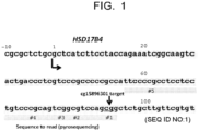
- the CpG site comprises a CpG site normally present between positions 56 and 94 on the basis of the transcription start site of HSD17B4 gene (see cg15896301 in Fig. 1 ).
- the "transcription start site” is meant to be determined based on GRCh37 (Genome Reference Consortium human build 37) and Hg19 (UCSC human genome 19).
- GRCh37 Gene Reference Consortium human build 37
- Hg19 UCSC human genome 19
- nucleotide sequence starting from a nucleotide residue at position -10 from the transcription start site is present at a location of nucleotide residues at positions 1092 and 1093 in the nucleotide sequence of SEQ ID NO:2 that describes a surrounding nucleotide sequence of the transcription start site in HSD17B4 gene.
- a mutation in genomic DNA e.g., deletion, insertion, substitution of DNA
- the above location and the nucleotide sequences of the above SEQ ID NOs may vary.
- the CpG site to be analyzed in the present invention can be also defined as a CG portion in cagcggct (e.g., nucleotide residues at positions 99 to 106 in the nucleotide sequence of SEQ ID NO:1, or nucleotide residues at positions 1089 to 1096 in the nucleotide sequence of SEQ ID NO:2) present within the non-coding region downstream of the transcription start site in HSD17B4 gene (when the cag portion on the 5' side and the get portion on the 3' side are not mutated, as far as the CpG site is at a position normally present between positions 56 and 94 on the basis of a transcription start site.
- a person skilled in the art can appropriately determine the CpG site corresponding to the CpG site present at the above location by considering the mutation in the genomic DNA for the human subject having the mutation in the genomic DNA.
- the methylation to be analyzed in the present invention is the methylation at position 5 in the cytosine residue.
- Analysis of the methylation level can be performed by any method known in the art.
- the analysis can be performed using one or two or more analytical means selected from the group consisting of bisulfite, one or more primers, one or more nucleic acid probes, a restriction enzyme, an anti-methylated cytosine antibody, and a nanopore.
- Bisulfite converts non-methylated cytosine to uracil whereas it does not convert methylated cytosine to uracil. Thus, due to such nature, bisulfite is commonly used for analysis of methylated cytosine in combination of other analytical means.
- the one or more primers are primers for sequencing or primers for gene amplification (e.g., PCR primers) or a combination thereof.
- the primer can be appropriately designed so as to analyze an objective CpG site.
- the primer can be designed to anneal to a downstream region of the objective CpG site (e.g., any region within the portion consisting of nucleotide residues at positions 1094 to 2001 in the nucleotide sequence of SEQ ID NO:2), or can be designed so that a sense strand and an antisense strand are annealed in an upstream region of the objective CpG site (e.g., any region within the portion consisting of nucleotide residues at positions 1 to 1091 in the nucleotide sequence of SEQ ID NO:2) and a downstream region of the objective CpG site (e.g., any region within the portion consisting of nucleotide residues at positions 1094 to 2001 in the nucleotide sequence of SEQ ID NO:2) in order to amplify a gene region including the objective CpG site.
- a downstream region of the objective CpG site e.g., any region within the portion consisting of nucleotide residues at positions 1094 to 2001
- the one or more nucleic acid probes can be designed to hybridize with a region including the objective CpG site (portion including the objective CpG site within the portion consisting of nucleotide residues at positions 1 to 120 in the nucleotide sequence of SEQ ID NO:1), or an upstream region thereof (e.g., any region within the portion consisting of nucleotide residues at positions 1 to 1091 in the nucleotide sequence of SEQ ID NO:2), or a downstream region thereof (e.g., any region within the portion consisting of nucleotide residues at positions 1094 to 2001 in the nucleotide sequence of SEQ ID NO:2).
- a region including the objective CpG site portion including the objective CpG site within the portion consisting of nucleotide residues at positions 1 to 120 in the nucleotide sequence of SEQ ID NO:1
- an upstream region thereof e.g., any region within the portion consisting of nucleotide residues at positions 1 to 1091
- the nucleic acid probe can be used in a free form or in a form immobilized to a solid phase.
- the solid phase include particles (e.g., magnetic particles); supports such as array, membrane (e.g., nitrocellulose membrane, filter), columns; and containers such as plates (e.g., multiwell plates) and tubes.
- Materials for the solid phase include, for example, glasses, plastics, and metals.
- the nucleic acid probe may be the nucleic acid probe described in detail in the analysis of methylated cytosine using the anti-methylated cytosine antibody.
- the restriction enzyme is a methylation-specific or - nonspecific restriction enzyme, and can be used appropriately in combination with the bisulfite, the one or more primers, or the one or more nucleic acid probes as described above.
- analytical methods using the analytical means as described above include, for example, a bisulfite sequencing method, a bisulfite pyrosequencing method, a methylation specific PCR method, a restriction enzyme landmark genome scanning (RLGS) method, a single nucleotide primer extension (SNuPE) method, a CpG island microarray method, a MethyLight method, a COBRA method, a mass spectroscopy (mass array) method, use of a methylation specific restriction enzyme, a high resolution melting analysis (HRM) method, a nanopore analysis method, an ICON probe method, and a methylation specific MLPA method.
- RGS restriction enzyme landmark genome scanning
- SNuPE single nucleotide primer extension
- CpG island microarray method a MethyLight method
- COBRA method a mass spectroscopy (mass array) method
- HRM high resolution melting analysis
- nanopore analysis method an ICON probe method
- ICON probe method e.g.
- the analysis can be also performed using an anti-methylated cytosine antibody.
- the analysis of methylated cytosine using the anti-methylated cytosine antibody is well-known in the art (e.g., WO2015/025862 ; WO2015/025863 ; WO2015/025864 ; WO2016/052368 ; Japanese Patent Application Publication No. 2012-230019 ; DNA Research 13, 37-42 (2006 ); Anal. Chem. 2012 ,84, 7533-7538 ).
- the anti-methylated cytosine antibody may be used in combination with one or two or more analytical means as described above.
- methods using such analytical means include, for example, a method of using the anti-methylated cytosine antibody and heterogeneous nucleic acid probes in combination (e.g., normal RNA probes, modified RNA probes) (e.g., WO2015/025862 ); a method of using the anti-methylated cytosine antibody and a solid phase probe and a capture probe in combination (e.g., WO2015/025863 ); a method of using the anti-methylated cytosine antibody and an absorbent polynucleotide and a capture probe in combination (e.g., WO2015/025864 ); and a method of using the anti-methylated cytosine antibody and a modified nucleobase paired heterogeneous nucleic acid probe in combination (e.g., WO2016/052368 ).
- a method of using the anti-methylated cytosine antibody and heterogeneous nucleic acid probes in combination e.g., normal RNA probe
- the nucleic acid probe used in combination with the anti-methylated cytosine antibody may be designed so that a methylcytosine residue in the CpG site forms a portion unpaired with the nucleic acid probe (in other words, not bound in a complementary manner) when the nucleic acid probe is hybridized with a DNA strand including the above CpG site to be analyzed in the present invention to form a double strand structure consisting of the DNA strand and the nucleic acid probe (e.g., WO2015/025862 ).
- the nucleic acid probe may have a nucleotide residue (e.g., cytosine residue, thymine residue, adenine residue, uracil residue) other than a guanine residue that can bind complementarily to the methylcytosine residue as a nucleotide residue corresponding to the unpaired portion.
- the nucleic acid probe may be designed so that such an unpaired portion is not formed ( WO2016/052368 ).
- the method of using the anti-methylated cytosine antibody can be performed by any immunological method known in the art.
- a method include an enzyme immunoassay (EIA) (e.g., CLEIA, ELISA), a fluorescent immunoassay, a chemiluminescent immunoassay, electrochemical luminescent immunoassay, an aggregation method, immunostaining, a flow cytometry method, a biolayer interferometry method, an In Situ PLA method, a chemical amplification type luminescence proximity homogeneous assay, a line blot method, and a western blot method.
- EIA enzyme immunoassay
- CLEIA e.g., CLEIA, ELISA
- fluorescent immunoassay e.g., CLEIA, ELISA
- electrochemical luminescent immunoassay e.g., electrochemical luminescent immunoassay
- an aggregation method e.g., an
- the methylation level to be analyzed in the present invention is a ratio of the methylation of the cytosine residue in the above CpG in the cancer cells. Measurement of the methylation level is well-known in the art (e.g., Japanese Patent Application Publication No. 2012-090555 , Japanese Patent Application Publication No. 2014-036672 , Japanese Patent Application Publication No. 2010-538638 , Domestic re-publication of WO 2009/136501 ).
- the methylation level is analyzed without sorting out cancer cells from the cancer tissue sample.
- the cancer tissue sample typically includes cancer cells and normal cells.
- the methylation level in the cancer cells can be calculated by correcting an actually measured methylation level with a cancer cell ratio [cancer cells / (cancer cells + normal cells)].
- the cancer cell ratio in the cancer tissue sample can be obtained by any method in the art.
- the cancer cell ratio can be obtained by microscopic examination or a method using a cancer cell marker and/or a normal cell marker (e.g., a protein marker) and a label (e.g., fluorescent label).
- the methylation level when the methylation level is analyzed in such a cancer tissue sample, the methylation level may further be corrected by considering a normal cell existence ratio [normal cells / (cancer cells + normal cells)] in the cancer tissue sample and the methylation level in the normal cells.
- the methylation level is analyzed after sorting out cancer cells from the cancer tissue sample.
- the actually measured methylation level may directly be used as the methylation level. Sorting out cancer cells from the cancer tissue sample can be performed by the method as described above.
- step (2) the effect of the medicinal therapy on the cancer is predicted based on the analyzed methylation level.
- the analyzed methylation level is used as a factor for comparing with a standard value.
- the methylation level of the above CpG site is significantly high in a subject group in which the medicinal therapy on the cancer is effective (effective group) compared with a subject group in which the medicinal therapy on the cancer is not effective (ineffective group). Therefore, according to the present invention, when the methylation level of the above CpG site is equal to or higher than the standard value, it can be predicted that a subject has a high possibility that the medicinal therapy on the cancer is effective, and/or a low possibility that the medicinal therapy on the cancer is not effective. In addition, when the methylation level of the above CpG site is less than the standard value, it can be predicted that the subject has a low possibility that the medicinal therapy on the cancer is effective, and/or a high possibility that the medicinal therapy on the cancer is not effective.
- the effective group and the ineffective group can be configured by properly defining a basis for effectiveness.
- the effectiveness may be configured on the basis of pathological complete response (pCR) or good response.
- pCR pathological complete response
- good response For details of the pathological complete response or good response, see Examples.
- a cutoff value appropriately configured to be able to distinguish the effective group from the ineffective group can be utilized as the above baseline.
- the cutoff value is a value that satisfies both high diagnostic sensitivity (specificity true positive rate) and high diagnostic specificity (specificity true negative rate) when a disease is determined using that value as a basis.
- the diagnostic sensitivity (or simply sensitivity) is a ratio of subjects diagnosed properly among subjects having a particular status. When a "positive" result is obtained for all subjects having the particular status, the sensitivity is 100%.
- the diagnostic specificity (or simply specificity) is a ratio of subjects properly diagnosed among subjects having no particular status. When a "negative" result is obtained for all subjects having no particular status, the specificity is 100%.
- a method of calculating the cutoff value is well-known in the present field. For example, the methylation level of the above CpG site is analyzed in the effective group and the ineffective group, the diagnostic sensitivity and the diagnostic specificity in any methylation level are obtained in the effective group and the ineffective group, and an ROC (receiver operating characteristic) curve is made based on these values. Then, the methylation level which gives the maximum diagnostic sensitivity and diagnostic specificity is sought, and this value can be used as the cutoff value.
- a diagnostic efficiency (a ratio of the total number of true positive cases and true negative cases to the number of all cases) in any methylation level is sought, and the methylation level by which the highest diagnostic efficiency is calculated can also be used as the cutoff value.
- the diagnostic specificity may be more emphasized to determine the cutoff value.
- the method of the present invention may further comprise measuring other markers associated with nature of the cancer in the cancer tissue sample.
- the other markers include the presence or absence or the change in dose of the expression of a certain gene, non-coding RNA or a protein, the presence or absence or the change in dose of a certain metabolite (e.g., modified protein such as phosphorylated protein), genetic polymorphism (e.g., SNP, haplotype), translocation, methylation of genomic DNA or their degree.
- the diagnostic specificity can be enhanced by further measuring the other cancer marker. For example, it has been confirmed that in the case of breast cancer, if an estrogen receptor is negative in pathological diagnosis, the diagnostic specificity can be further enhanced (the Examples).
- Estrogen receptor herein refers to ER ⁇ unless otherwise described.
- a method of examining the presence or absence of the expression of estrogen receptor is not particularly limited, and a typically used enzyme antibody method (LSAB: labeled streptavidin biotinylated antibody) can be suitably used.
- LSAB labeled streptavidin biotinylated antibody
- Disclosed herein but not forming part of the invention is a diagnostic kit for diagnosing an effect of the medicinal therapy on a cancer.
- Such analytical means include one or two or more means selected from the group consisting of bisulfite, one or more primers, one or more nucleic acid probes, a restriction enzyme, an anti-methylated cytosine antibody, and a nanopore.
- the diagnostic kit can be used for conducting the analytical method as described above.
- the diagnostic kit not forming part of the present invention comprises a combination of means for enabling a specific analysis of the CpG site described above and means for enabling an analysis of methylated cytosine (e.g., bisulfite, anti-methylated cytosine antibody, or methylation specific restriction enzyme).
- the means for enabling the specific analysis of the CpG site described above include, for example, one or more primers, one or more nucleic acid probes, a restriction enzyme, and a nanopore. One or more primers or one or more nucleic acid probes are preferred.
- the nucleic acid probe may be immobilized to the solid phase.
- the means for enabling the analysis of methylated cytosine include, for example, bisulfite, a primer for sequencing, an anti-methylated cytosine antibody, a methylation specific restriction enzyme, and an ICON probe method.
- Bisulfite or the anti-methylated cytosine antibody is preferred.
- the diagnostic kit not forming part of the present invention may comprise auxiliary means in addition to the above analytical means.
- auxiliary means include, for example, deoxyribonucleoside triphosphate (dNTP), buffer, a stabilizing agent, an enzyme (e.g., DNA polymerase, sulfurylase, apyrase), a labeling substance, and a substrate for the enzyme.
- the diagnostic kit comprises the anti-methylated cytosine antibody
- it may further comprise a secondary antibody (e.g., anti-IgG antibody) and a detection reagent for the secondary antibody.
- a secondary antibody e.g., anti-IgG antibody
- the diagnostic kit may further comprise a device capable of collecting a biological sample.
- the device capable of collecting the biological sample is not particularly limited as long as the biological sample can be obtained from the subject, and includes, for example, a biopsy device such as a biopsy needle.
- a medicinal therapy which comprises:
- the items (1) and (2) in the medicinal therapy can be performed in the same way as in (1) and (2) in the prediction method of the present invention.
- the item (3) in the medicinal therapy can be performed by selecting the human predicted to receive the effect of the medicinal therapy on the cancer based on the prediction in (2).
- the medicinal therapy is given to the selected human.
- the medicinal therapy is the treatment with the anticancer agent as described above.
- the cancer is the HER2-positive cancer
- the medicinal therapy with the HER2 inhibitor is given.
- the treatment with the anticancer agent can be performed in a typical mode employed for that anticancer agent.
- a method of selecting a treatment on a cancer comprising:
- the analysis of the methylation level in (2) in the selection method can be performed in the same way as the analysis in (2) in the prediction method of the present invention.
- an existing anticancer agent e.g., trastuzumab
- the drug can be changed to the other anticancer agent, or the other anticancer agent can be combined.
- a pharmaceutical capable of suppressing or promoting the expression of HSD17B4 gene or a pharmaceutical capable of methylating, in particular, a non-coding region of HSD17B4 gene can be also combined.
- the surgical therapy such as a radiation therapy can be also combined.
- the method of the present invention enables to select a more effective treatment method of a human being which is a therapeutic objective based on the methylation level analyzed in (2).
- Tissue samples were collected from 67 patients enrolled in a neoadjuvant clinical trial. All the samples for this study were obtained from patients who received weekly paclitaxel (80 mg/m 2 per week) and trastuzumab (a loading dose of 4 mg/kg followed by 2 mg/kg per week) for a total of 12 doses. The patients underwent mastectomy, and the response was pathologically assessed. The study protocol was approved by the National Cancer Center Ethics Committee, and was registered at the UMIN Clinical Trial Registry. All the patients were provided written informed consent.
- Two specimens were obtained from a patient by core needle biopsy of a primary tumor before starting the neoadjuvant therapy, and fixed with two kinds of method.
- One specimen was fixed with neutral buffered formalin for microscopic examination using thin section stained with H&E, and the other was fixed with the PAXgene Tissue System (Qiagen, Germany) and embedded in low-melting paraffin for DNA/RNA extraction using 10 slices of sections with thickness of 10 ⁇ m.
- a certificated and experienced pathologist conducted microscopic examination of biopsy specimens to obtain the fraction of cancer cells to all the cells and to select specimens contained a sufficient number of tumor cells for molecular analyses. The pathologist also analyzed surgical specimens to determine the response.
- pathological complete response corresponds to Grade 3 in response determination criteria in Table 1 (cited from "General rules for clinical and pathological recording of breast cancer” edited by Japanese Breast Cancer Society, the 17th edition, page 84 (2012), Kanehara & Co., Ltd ) and refers to the case in which no residual cancer cell is found in breast duct.
- patients of Grade 2 or upper in Table 1 can be predicted to receive a highly effective neoadjuvant therapy even when they cannot be classified into pCR, and thus these patients are addressed as a "good response case” separately from pCR in this specification.
- Table 1 Grade 0 No response Almost no change in cancer cells by treatment is observed Grade 1 slightly efficacious 1a) Mild effect Mild change of cancer cells is observed regardless of areas.
- high level change is observed in about less than 1/3 of cancer cells 1b) Moderate effect High level change is observed in about 1/3 or more and less than 2/3 of cancer cells Grade 2 Considerably efficacious 2a) High level effect High level change is observed in about 2/3 or more of cancer cells. But, clear focus is observed. 2b) Extremely high level effect Effect significantly close to complete response (Grade 3) is observed, but tiny amount of cancer cells remains. Grade 3 Complete response All cancer cells are in necrosis or disappear. Cancer tissue has been replaced with granuloma-like tissue or fibrotic focus.
- the 67 HER2-positive breast cancers were classified into 64 cases of invasive ductal carcinoma, 2 cases of invasive lobular carcinoma and 1 case of other histological type. For hormones, 34 cases (50.7%) were positive for estrogen receptor, and 26 cases (38.8%) were positive for progesterone receptor. Only samples with a tumor cell fraction (percentage of cancer cells in tissue sample) of 40% or more were used. Detailed Clinicopathological characteristics in the 67 cases of HER2-positive breast cancer patients are shown in Table 2. Table 2. Clinicopathological characteristics of HER2-positive breast cancer patients.
- FIG. 1 A partial nucleotide sequence of HSD17B4 gene is shown in Fig. 1 .
- the nucleotide sequence consisting of nucleotide residues at positions 56 to 94 including five CpG sites (#1 to #5) assigning "g" at the transcription start site as position 1 was read by pyrosequence.
- the methylation levels of five CpG sites were measured and calculated by the following procedures.
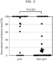
- the methylation level was calculated as a ⁇ value in the range of 0 (completely no methylation) to 1 (fully methylated), and this was expressed as a percent.
- the measured methylation level was corrected using the fraction of cancer cells in a sample.
- a specific correction formula is as follows. An upper limit of a normalized methylation level is determined to be 100. When a calculated value by the following formula exceeded 100, this was analyzed as "100% of normalized methylation level".
- the normalized methylation level 100 ⁇ (the measured methylation level (%)) / (the fraction of cancer cells in the sample (%))].
- the fraction of cancer cells (%) was obtained by microscopic examination.
- the Fisher's exact test was used to calculate the significant difference in relative frequency between two groups. Significant differences in the normalized methylation levels between the responders and non-responders were evaluated by the Mann-Whitney U test. In the univariate analysis, the odds ratios (ORs) and 95% confidence interval (95% CI) were calculated. The multivariate logistic regression analyses were also performed to detect the factors independently associated with the apparent likelihood of the response to the combined trastuzumab and chemotherapy after excluding confounders. All statistical analyses were conducted by PASW statistics version 18.0.0 (SPSS Japan Inc.).
- the "methylation of HSD17B4 gene” indicates the methylation of the CpG site of #1 in HSD17B4 gene unless otherwise described.
- a cutoff of the methylation level of HSD17B4 gene was configured to be 50%, a value of 50% or higher was determined to be “positive”, and a value lower than 50% was determined to be "negative”.
- Clinical diagnosis results when pCR was determined to be positive and the other cases were determined to be negative are shown in Table 4.
- Clinical diagnosis results when the good response case was determined to be positive and the non-response case was determined to be negative are shown in Table 5.
- the sensitivity was 77.2% and the specificity was 84.4% in the clinical diagnosis results of the methylation level when pCR was determined to be positive.
- Results of the clinicopathological characteristics of the 67 cases compiled by dividing into a positive group and a negative group of the methylation level of HSD17B4 gene are shown in Table 6.
- Table 6 results of the clinicopathological characteristics of the 67 cases compiled by dividing into a positive group and a negative group of the methylation level of HSD17B4 gene are shown in Table 6.
- Table 6 results of the clinicopathological characteristics of the 67 cases compiled by dividing into a positive group and a negative group of the methylation level of HSD17B4 gene are shown in Table 6.
- Table 6 results of the clinicopathological characteristics of the 67 cases compiled by dividing into a positive group and a negative group of the methylation level of HSD17B4 gene are shown in Table 6.
- Table 6 results of the clinicopathological characteristics of the 67 cases compiled by dividing into a positive group and a negative group of the methylation level of HSD17B4 gene are shown in Table 6.
- the clinical diagnosis results when the good response case was determined to be positive and the non-response case was determined to be negative are shown in Table 9.
- the clinical diagnosis results in combination of the methylation level and estrogen receptor when the good response case was determined to be positive exhibited the sensitivity of 32.7% and the specificity of 94.4%.
- One case determined to be "positive” in Table 9 was a case in which a tumor size was very large and the surgical operation was strongly recommended with or without the effect of the neoadjuvant therapy. Table 8.
- Clinical diagnosis results positive: pCR; negative: other cases) pCR Total Positive Negative Methylation level (+) / ER* (-) Positive 13 4 17 Negative 9 41 50 Total 22 45 67 *ER: estrogen receptor Sensitivity: 59.1% Specificity: 91.1% Positive indicative value: 76.5% Table 9. Clinical diagnosis results (positive: good response case; negative: non-response case) Good response case Total Positive Negative Methylation level (+) / ER* (-) Positive 16 1 17 Negative 33 17 50 Total 49 18 67 *ER: estrogen receptor Sensitivity: 32.7% Specificity: 94.4% Positive indicative value: 94.1%
Landscapes
- Health & Medical Sciences (AREA)
- Life Sciences & Earth Sciences (AREA)
- Chemical & Material Sciences (AREA)
- Engineering & Computer Science (AREA)
- Immunology (AREA)
- General Health & Medical Sciences (AREA)
- Organic Chemistry (AREA)
- Molecular Biology (AREA)
- Medicinal Chemistry (AREA)
- Analytical Chemistry (AREA)
- Microbiology (AREA)
- Proteomics, Peptides & Aminoacids (AREA)
- Urology & Nephrology (AREA)
- Hematology (AREA)
- Biomedical Technology (AREA)
- Physics & Mathematics (AREA)
- Biochemistry (AREA)
- Biotechnology (AREA)
- Zoology (AREA)
- Wood Science & Technology (AREA)
- Pathology (AREA)
- Genetics & Genomics (AREA)
- Bioinformatics & Cheminformatics (AREA)
- Animal Behavior & Ethology (AREA)
- Public Health (AREA)
- Veterinary Medicine (AREA)
- Pharmacology & Pharmacy (AREA)
- General Physics & Mathematics (AREA)
- Cell Biology (AREA)
- Food Science & Technology (AREA)
- General Engineering & Computer Science (AREA)
- Biophysics (AREA)
- Epidemiology (AREA)
- Nuclear Medicine, Radiotherapy & Molecular Imaging (AREA)
- Mycology (AREA)
- Chemical Kinetics & Catalysis (AREA)
- General Chemical & Material Sciences (AREA)
- Oncology (AREA)
- Hospice & Palliative Care (AREA)
- Measuring Or Testing Involving Enzymes Or Micro-Organisms (AREA)
Claims (9)
- Verfahren zur Vorhersage eines Effekts einer Behandlung mit Trastuzumab auf einen HER2-positiven Brustkrebs, umfassend:(1) Analysieren eines Methylierungsgrades eines Cytosinrests in einer oder mehreren CpG-Stellen, die innerhalb einer Nukleotidsequenz in einer untranslatierten Region des HSD17B4-Gens in einer Probe vorhanden sind, die einem menschlichen Subjekt entnommen wurde, wobei die CpG-Stelle eine CpG-Stelle umfasst, die normalerweise zwischen den Positionen 56 und 94 auf der Basis einer Transkriptionsstartstelle vorhanden ist; und(2) Vorhersagen des Effekts der Behandlung auf den HER2-positiven Brustkrebs auf der Grundlage des analysierten Methylierungsgrades.
- Verfahren gemäß Anspruch 1, wobei die Behandlung mit Trastuzumab eine Behandlung mit Trastuzumab in Kombination mit einem anderen Anti-Krebsmittel als Trastuzumab ist.
- Verfahren gemäß Anspruch 1 oder 2, wobei es sich bei der dem menschlichen Subjekt entnommenen Probe um eine oder zwei oder mehr Proben handelt, die aus der Gruppe ausgewählt sind, die aus einem Blut, einer Körperflüssigkeit, einem Gewebe und einer Zelle besteht.
- Verfahren gemäß irgendeinem der Ansprüche 1 bis 3, wobei die CpG-Stelle eine CpG-Stelle ist, die normalerweise zwischen den Positionen 56 und 94 auf der Basis einer Transkriptionsstartstelle vorhanden ist.
- Verfahren gemäß Anspruch 4, wobei es sich bei der CpG-Stelle um eine CpG-Stelle handelt, die normalerweise an den Positionen 92 und 93 auf der Basis der Transkriptionsstartstelle vorhanden ist.
- Verfahren gemäß Anspruch 1, ferner umfassend das Messen der Expression des Östrogenrezeptors in der Probe.
- Verfahren gemäß irgendeinem der Ansprüche 1 bis 6, wobei die Analyse unter Verwendung eines oder zweier oder mehrerer Mittel durchgeführt wird, die aus der Gruppe ausgewählt sind, die aus Bisulfit, einem oder mehreren Primern, einer oder mehreren Nukleinsäuresonden, einem Restriktionsenzym, einem anti-methylierten Cytosin-Antikörper und einer Nanopore besteht.
- Verfahren gemäß irgendeinem der Ansprüche 1 bis 7, wobei die Analyse durch ein Bisulfit-Sequenzierungsverfahren, ein Bisulfit-Pyrosequenzierungsverfahren, ein methylierungsspezifisches PCR-Verfahren, ein Restriktionsenzym-Landmark-Genom-Scanning-Verfahren (RLGS), ein Einzelnukleotid-Primer-Extension-Verfahren (SNuPE), eine CpG-Insel-Microarray-Methode, eine MethyLight-Methode, eine COBRA-Methode, eine Massenspektroskopie (Massenarray)-Methode, die Verwendung eines methylierungsspezifischen Restriktionsenzyms, eine hochauflösende Schmelzanalyse (HRM), eine Nanopore-Analyse-Methode, eine ICON-Sonden-Methode, eine methylierungsspezifische MLPA-Methode oder ein Immunoassay, durchgeführt wird.
- Anti-Krebsmittel zur Verwendung in einer Behandlung mit Trastuzumab bei HER2-positivem Brustkrebs, wobei die Therapie umfasst:(1) Analysieren eines Methylierungsgrades eines Cytosinrests in einer oder mehreren CpG-Stellen, die innerhalb einer Nukleotidsequenz in einer untranslatierten Region des HSD17B4-Gens in einer Probe vorhanden sind, die einem menschlichen Subjekt entnommen wurde, wobei die CpG-Stelle eine CpG-Stelle umfasst, die normalerweise zwischen den Positionen 56 und 94 auf der Basis einer Transkriptionsstartstelle vorhanden ist; und(2) Vorhersage eines Effekts der Behandlung auf den HER2-positiven Brustkrebs auf der Grundlage des analysierten Methylierungsgrades;(3) Auswählen eines Menschen, von dem vorhergesagt wird, dass er die Wirkung der Behandlung auf den HER2-positiven Brustkrebs erhält; und(4) Verabreichung der Behandlung an den ausgewählten Menschen.
Applications Claiming Priority (2)
| Application Number | Priority Date | Filing Date | Title |
|---|---|---|---|
| JP2016116367 | 2016-06-10 | ||
| PCT/JP2017/010523 WO2017212734A1 (ja) | 2016-06-10 | 2017-03-15 | 癌に対する薬物療法の効果の予測方法 |
Publications (3)
| Publication Number | Publication Date |
|---|---|
| EP3470528A1 EP3470528A1 (de) | 2019-04-17 |
| EP3470528A4 EP3470528A4 (de) | 2019-12-25 |
| EP3470528B1 true EP3470528B1 (de) | 2024-09-25 |
Family
ID=60577653
Family Applications (1)
| Application Number | Title | Priority Date | Filing Date |
|---|---|---|---|
| EP17809918.0A Active EP3470528B1 (de) | 2016-06-10 | 2017-03-15 | Verfahren zur prognose der wirkung einer pharmakotherapie bei krebs |
Country Status (5)
| Country | Link |
|---|---|
| US (1) | US11214838B2 (de) |
| EP (1) | EP3470528B1 (de) |
| JP (1) | JP6876304B2 (de) |
| CN (1) | CN109642257B (de) |
| WO (1) | WO2017212734A1 (de) |
Families Citing this family (2)
| Publication number | Priority date | Publication date | Assignee | Title |
|---|---|---|---|---|
| WO2020054782A1 (ja) * | 2018-09-13 | 2020-03-19 | 国立研究開発法人国立がん研究センター | 乳癌細胞存在率の推定方法 |
| CN117778389B (zh) * | 2023-12-22 | 2024-11-12 | 深圳新合睿恩生物医疗科技有限公司 | 高效表达的mRNA的5′UTR序列及其应用 |
Family Cites Families (11)
| Publication number | Priority date | Publication date | Assignee | Title |
|---|---|---|---|---|
| EP1649052A1 (de) * | 2003-08-01 | 2006-04-26 | The University Of Western Australia | Verfahren und reagentiensatz zur vorhersage der wahrscheinlichkeit einer erfolgreichen behandlung von krebs |
| US7504214B2 (en) * | 2003-09-19 | 2009-03-17 | Biotheranostics, Inc. | Predicting outcome with tamoxifen in breast cancer |
| DE10348407A1 (de) | 2003-10-17 | 2005-05-19 | Widschwendter, Martin, Prof. | Prognostische und diagnostische Marker für Zell-proliferative Erkrankungen von Brustgeweben |
| WO2006004597A2 (en) * | 2004-06-01 | 2006-01-12 | University Of Southern California | Association of breast cancer dna methylation profiles with hormone receptor status and response to tamoxifen |
| US20070134688A1 (en) * | 2005-09-09 | 2007-06-14 | The Board Of Regents Of The University Of Texas System | Calculated index of genomic expression of estrogen receptor (er) and er-related genes |
| EP2233590A1 (de) * | 2009-01-28 | 2010-09-29 | AIT Austrian Institute of Technology GmbH | Methylierungsassay |
| WO2013012781A2 (en) * | 2011-07-15 | 2013-01-24 | The Johns Hopkins University | Genome-wide methylation analysis and use to identify genes specific to breast cancer hormone receptor status and risk of recurrence |
| EP2751285B2 (de) * | 2011-08-31 | 2020-04-01 | Genentech, Inc. | Methode zur bestimmung der empfindlichkeit eines tumors für egfr kinase-inhibitoren |
| WO2013049926A1 (en) * | 2011-10-06 | 2013-04-11 | UNIVERSITé LAVAL | PROGNOSTIC MARKERS OF INHERITED VARIATIONS IN THE 17β-HYDROXYSTEROID DEHYDROGENASE (HSD17B) GENES FOR PROSTATE CANCER |
| CN104152556B (zh) * | 2014-08-07 | 2016-01-13 | 中国医学科学院肿瘤医院 | 一种判断曲妥珠单抗辅助治疗乳腺癌疗效的试剂盒及应用 |
| US20190226031A1 (en) * | 2016-09-02 | 2019-07-25 | Fujifilm Wako Pure Chemical Corporation | Method for amplifying methylated dna, method for determining methylation of dna, and method for determining cancer |
-
2017
- 2017-03-15 CN CN201780035484.5A patent/CN109642257B/zh active Active
- 2017-03-15 EP EP17809918.0A patent/EP3470528B1/de active Active
- 2017-03-15 JP JP2018522336A patent/JP6876304B2/ja active Active
- 2017-03-15 WO PCT/JP2017/010523 patent/WO2017212734A1/ja not_active Ceased
- 2017-03-15 US US16/308,210 patent/US11214838B2/en active Active
Non-Patent Citations (1)
| Title |
|---|
| ANONYMOUS: "Infinium? HumanMethylation450 BeadChip", 9 March 2012 (2012-03-09), XP055401052, Retrieved from the Internet <URL:https://support.illumina.com/content/dam/illumina-marketing/documents/products/datasheets/datasheet_humanmethylation450.pdf> [retrieved on 20170824] * |
Also Published As
| Publication number | Publication date |
|---|---|
| EP3470528A1 (de) | 2019-04-17 |
| CN109642257B (zh) | 2022-08-12 |
| US20190169701A1 (en) | 2019-06-06 |
| CN109642257A (zh) | 2019-04-16 |
| EP3470528A4 (de) | 2019-12-25 |
| US11214838B2 (en) | 2022-01-04 |
| WO2017212734A1 (ja) | 2017-12-14 |
| JPWO2017212734A1 (ja) | 2019-05-09 |
| JP6876304B2 (ja) | 2021-05-26 |
Similar Documents
| Publication | Publication Date | Title |
|---|---|---|
| KR102941150B1 (ko) | 범-암 백금 반응 예측기 | |
| Amaria et al. | Neoadjuvant immune checkpoint blockade in high-risk resectable melanoma | |
| Sheng et al. | HER2 status in gastric cancers: a retrospective analysis from four Chinese representative clinical centers and assessment of its prognostic significance | |
| Van Schaeybroeck et al. | Implementing prognostic and predictive biomarkers in CRC clinical trials | |
| Gifford et al. | The acquisition of hMLH1 methylation in plasma DNA after chemotherapy predicts poor survival for ovarian cancer patients | |
| Zhuo et al. | Elevated THBS2, COL1A2, and SPP1 expression levels as predictors of gastric cancer prognosis | |
| Bordi et al. | Circulating DNA in diagnosis and monitoring EGFR gene mutations in advanced non-small cell lung cancer | |
| CN107267598B (zh) | 用于评估杂合性丢失的方法与材料 | |
| Voutsina et al. | Combined analysis of KRAS and PIK3CA mutations, MET and PTEN expression in primary tumors and corresponding metastases in colorectal cancer | |
| Navrkalova et al. | ATM mutations uniformly lead to ATM dysfunction in chronic lymphocytic leukemia: application of functional test using doxorubicin | |
| EP2764364B1 (de) | Vorhersage einer tumorreaktion auf anti-erbb3-antikörper | |
| Xu et al. | KRAS mutations in tumor tissue and plasma by different assays predict survival of patients with metastatic colorectal cancer | |
| CN106233143B (zh) | 癌症治疗 | |
| US20110217701A1 (en) | Prognostic Marker for Endometrial Carcinoma | |
| CN112210605B (zh) | 用于评估组织免疫反应和诊断预后的dna甲基化检测试剂盒 | |
| EP3152560A1 (de) | Nichtinvasiver nachweis von genmutationen bei lungenkrebspatienten | |
| Kwon et al. | Low frequency of KRAS mutation in pancreatic ductal adenocarcinomas in Korean patients and its prognostic value | |
| US20120277110A1 (en) | Parp and adjuvant cisplatin-based chemotherapy in non-small-cell lung cancer | |
| EP3541958A1 (de) | Zusammensetzungen und verfahren zur prognose behandlung und kolorektalkarzinom | |
| JP7398712B2 (ja) | 乳癌細胞存在率の推定方法 | |
| JP2020529870A (ja) | がんを層別化し、処置するための材料及び方法 | |
| EP3470528B1 (de) | Verfahren zur prognose der wirkung einer pharmakotherapie bei krebs | |
| JP5858405B2 (ja) | 肺腺がんの予後予測方法、および、肺腺がんの検出キット | |
| WO2017119510A1 (ja) | 乳がんの診断のための検査方法、遺伝子マーカー、および検査薬 | |
| JP2023537870A (ja) | 肺がんを診断及び監視するためのバイオマーカー |
Legal Events
| Date | Code | Title | Description |
|---|---|---|---|
| STAA | Information on the status of an ep patent application or granted ep patent |
Free format text: STATUS: THE INTERNATIONAL PUBLICATION HAS BEEN MADE |
|
| PUAI | Public reference made under article 153(3) epc to a published international application that has entered the european phase |
Free format text: ORIGINAL CODE: 0009012 |
|
| STAA | Information on the status of an ep patent application or granted ep patent |
Free format text: STATUS: REQUEST FOR EXAMINATION WAS MADE |
|
| 17P | Request for examination filed |
Effective date: 20181128 |
|
| AK | Designated contracting states |
Kind code of ref document: A1 Designated state(s): AL AT BE BG CH CY CZ DE DK EE ES FI FR GB GR HR HU IE IS IT LI LT LU LV MC MK MT NL NO PL PT RO RS SE SI SK SM TR |
|
| AX | Request for extension of the european patent |
Extension state: BA ME |
|
| DAV | Request for validation of the european patent (deleted) | ||
| DAX | Request for extension of the european patent (deleted) | ||
| RIC1 | Information provided on ipc code assigned before grant |
Ipc: A61K 39/395 20060101ALI20191113BHEP Ipc: A61P 35/00 20060101ALI20191113BHEP Ipc: G01N 33/53 20060101ALI20191113BHEP Ipc: G01N 33/574 20060101ALI20191113BHEP Ipc: C12Q 1/6883 20180101ALI20191113BHEP Ipc: A61K 45/00 20060101ALI20191113BHEP Ipc: C12Q 1/68 20180101AFI20191113BHEP |
|
| A4 | Supplementary search report drawn up and despatched |
Effective date: 20191127 |
|
| RIC1 | Information provided on ipc code assigned before grant |
Ipc: A61P 35/00 20060101ALI20191121BHEP Ipc: G01N 33/53 20060101ALI20191121BHEP Ipc: C12Q 1/68 20180101AFI20191121BHEP Ipc: G01N 33/574 20060101ALI20191121BHEP Ipc: A61K 39/395 20060101ALI20191121BHEP Ipc: C12Q 1/6883 20180101ALI20191121BHEP Ipc: A61K 45/00 20060101ALI20191121BHEP |
|
| STAA | Information on the status of an ep patent application or granted ep patent |
Free format text: STATUS: EXAMINATION IS IN PROGRESS |
|
| 17Q | First examination report despatched |
Effective date: 20200708 |
|
| RAP1 | Party data changed (applicant data changed or rights of an application transferred) |
Owner name: H.U. GROUP RESEARCH INSTITUTE G.K. Owner name: NATIONAL CANCER CENTER |
|
| RAP3 | Party data changed (applicant data changed or rights of an application transferred) |
Owner name: H.U. GROUP RESEARCH INSTITUTE G.K. Owner name: NATIONAL CANCER CENTER |
|
| REG | Reference to a national code |
Ref country code: DE Ref legal event code: R079 Free format text: PREVIOUS MAIN CLASS: C12Q0001680000 Ipc: C12Q0001688300 Ref document number: 602017085091 Country of ref document: DE |
|
| RIC1 | Information provided on ipc code assigned before grant |
Ipc: G01N 33/574 20060101ALI20230713BHEP Ipc: G01N 33/53 20060101ALI20230713BHEP Ipc: A61P 35/00 20060101ALI20230713BHEP Ipc: A61K 45/00 20060101ALI20230713BHEP Ipc: A61K 39/395 20060101ALI20230713BHEP Ipc: C12Q 1/6883 20180101AFI20230713BHEP |
|
| GRAP | Despatch of communication of intention to grant a patent |
Free format text: ORIGINAL CODE: EPIDOSNIGR1 |
|
| STAA | Information on the status of an ep patent application or granted ep patent |
Free format text: STATUS: GRANT OF PATENT IS INTENDED |
|
| INTG | Intention to grant announced |
Effective date: 20230818 |
|
| GRAJ | Information related to disapproval of communication of intention to grant by the applicant or resumption of examination proceedings by the epo deleted |
Free format text: ORIGINAL CODE: EPIDOSDIGR1 |
|
| STAA | Information on the status of an ep patent application or granted ep patent |
Free format text: STATUS: EXAMINATION IS IN PROGRESS |
|
| GRAP | Despatch of communication of intention to grant a patent |
Free format text: ORIGINAL CODE: EPIDOSNIGR1 |
|
| STAA | Information on the status of an ep patent application or granted ep patent |
Free format text: STATUS: GRANT OF PATENT IS INTENDED |
|
| INTC | Intention to grant announced (deleted) | ||
| INTG | Intention to grant announced |
Effective date: 20231218 |
|
| GRAJ | Information related to disapproval of communication of intention to grant by the applicant or resumption of examination proceedings by the epo deleted |
Free format text: ORIGINAL CODE: EPIDOSDIGR1 |
|
| GRAP | Despatch of communication of intention to grant a patent |
Free format text: ORIGINAL CODE: EPIDOSNIGR1 |
|
| INTG | Intention to grant announced |
Effective date: 20240417 |
|
| RAP1 | Party data changed (applicant data changed or rights of an application transferred) |
Owner name: NATIONAL CANCER CENTER |
|
| GRAS | Grant fee paid |
Free format text: ORIGINAL CODE: EPIDOSNIGR3 |
|
| RAP1 | Party data changed (applicant data changed or rights of an application transferred) |
Owner name: USHIJIMA, TOSHIKAZU Owner name: MUKAI, HIROFUMI |
|
| GRAA | (expected) grant |
Free format text: ORIGINAL CODE: 0009210 |
|
| STAA | Information on the status of an ep patent application or granted ep patent |
Free format text: STATUS: THE PATENT HAS BEEN GRANTED |
|
| AK | Designated contracting states |
Kind code of ref document: B1 Designated state(s): AL AT BE BG CH CY CZ DE DK EE ES FI FR GB GR HR HU IE IS IT LI LT LU LV MC MK MT NL NO PL PT RO RS SE SI SK SM TR |
|
| REG | Reference to a national code |
Ref country code: GB Ref legal event code: FG4D |
|
| REG | Reference to a national code |
Ref country code: CH Ref legal event code: EP |
|
| REG | Reference to a national code |
Ref country code: DE Ref legal event code: R096 Ref document number: 602017085091 Country of ref document: DE |
|
| REG | Reference to a national code |
Ref country code: IE Ref legal event code: FG4D |
|
| REG | Reference to a national code |
Ref country code: LT Ref legal event code: MG9D |
|
| PG25 | Lapsed in a contracting state [announced via postgrant information from national office to epo] |
Ref country code: NO Free format text: LAPSE BECAUSE OF FAILURE TO SUBMIT A TRANSLATION OF THE DESCRIPTION OR TO PAY THE FEE WITHIN THE PRESCRIBED TIME-LIMIT Effective date: 20241225 |
|
| PG25 | Lapsed in a contracting state [announced via postgrant information from national office to epo] |
Ref country code: GR Free format text: LAPSE BECAUSE OF FAILURE TO SUBMIT A TRANSLATION OF THE DESCRIPTION OR TO PAY THE FEE WITHIN THE PRESCRIBED TIME-LIMIT Effective date: 20241226 Ref country code: FI Free format text: LAPSE BECAUSE OF FAILURE TO SUBMIT A TRANSLATION OF THE DESCRIPTION OR TO PAY THE FEE WITHIN THE PRESCRIBED TIME-LIMIT Effective date: 20240925 |
|
| PG25 | Lapsed in a contracting state [announced via postgrant information from national office to epo] |
Ref country code: BG Free format text: LAPSE BECAUSE OF FAILURE TO SUBMIT A TRANSLATION OF THE DESCRIPTION OR TO PAY THE FEE WITHIN THE PRESCRIBED TIME-LIMIT Effective date: 20240925 |
|
| PG25 | Lapsed in a contracting state [announced via postgrant information from national office to epo] |
Ref country code: LV Free format text: LAPSE BECAUSE OF FAILURE TO SUBMIT A TRANSLATION OF THE DESCRIPTION OR TO PAY THE FEE WITHIN THE PRESCRIBED TIME-LIMIT Effective date: 20240925 |
|
| PG25 | Lapsed in a contracting state [announced via postgrant information from national office to epo] |
Ref country code: RS Free format text: LAPSE BECAUSE OF FAILURE TO SUBMIT A TRANSLATION OF THE DESCRIPTION OR TO PAY THE FEE WITHIN THE PRESCRIBED TIME-LIMIT Effective date: 20241225 |
|
| REG | Reference to a national code |
Ref country code: NL Ref legal event code: MP Effective date: 20240925 |
|
| PG25 | Lapsed in a contracting state [announced via postgrant information from national office to epo] |
Ref country code: RS Free format text: LAPSE BECAUSE OF FAILURE TO SUBMIT A TRANSLATION OF THE DESCRIPTION OR TO PAY THE FEE WITHIN THE PRESCRIBED TIME-LIMIT Effective date: 20241225 Ref country code: NO Free format text: LAPSE BECAUSE OF FAILURE TO SUBMIT A TRANSLATION OF THE DESCRIPTION OR TO PAY THE FEE WITHIN THE PRESCRIBED TIME-LIMIT Effective date: 20241225 Ref country code: LV Free format text: LAPSE BECAUSE OF FAILURE TO SUBMIT A TRANSLATION OF THE DESCRIPTION OR TO PAY THE FEE WITHIN THE PRESCRIBED TIME-LIMIT Effective date: 20240925 Ref country code: GR Free format text: LAPSE BECAUSE OF FAILURE TO SUBMIT A TRANSLATION OF THE DESCRIPTION OR TO PAY THE FEE WITHIN THE PRESCRIBED TIME-LIMIT Effective date: 20241226 Ref country code: FI Free format text: LAPSE BECAUSE OF FAILURE TO SUBMIT A TRANSLATION OF THE DESCRIPTION OR TO PAY THE FEE WITHIN THE PRESCRIBED TIME-LIMIT Effective date: 20240925 Ref country code: BG Free format text: LAPSE BECAUSE OF FAILURE TO SUBMIT A TRANSLATION OF THE DESCRIPTION OR TO PAY THE FEE WITHIN THE PRESCRIBED TIME-LIMIT Effective date: 20240925 |
|
| REG | Reference to a national code |
Ref country code: AT Ref legal event code: MK05 Ref document number: 1726678 Country of ref document: AT Kind code of ref document: T Effective date: 20240925 |
|
| PG25 | Lapsed in a contracting state [announced via postgrant information from national office to epo] |
Ref country code: NL Free format text: LAPSE BECAUSE OF FAILURE TO SUBMIT A TRANSLATION OF THE DESCRIPTION OR TO PAY THE FEE WITHIN THE PRESCRIBED TIME-LIMIT Effective date: 20240925 |
|
| PG25 | Lapsed in a contracting state [announced via postgrant information from national office to epo] |
Ref country code: IS Free format text: LAPSE BECAUSE OF FAILURE TO SUBMIT A TRANSLATION OF THE DESCRIPTION OR TO PAY THE FEE WITHIN THE PRESCRIBED TIME-LIMIT Effective date: 20250125 Ref country code: PT Free format text: LAPSE BECAUSE OF FAILURE TO SUBMIT A TRANSLATION OF THE DESCRIPTION OR TO PAY THE FEE WITHIN THE PRESCRIBED TIME-LIMIT Effective date: 20250127 |
|
| PG25 | Lapsed in a contracting state [announced via postgrant information from national office to epo] |
Ref country code: RO Free format text: LAPSE BECAUSE OF FAILURE TO SUBMIT A TRANSLATION OF THE DESCRIPTION OR TO PAY THE FEE WITHIN THE PRESCRIBED TIME-LIMIT Effective date: 20240925 Ref country code: SM Free format text: LAPSE BECAUSE OF FAILURE TO SUBMIT A TRANSLATION OF THE DESCRIPTION OR TO PAY THE FEE WITHIN THE PRESCRIBED TIME-LIMIT Effective date: 20240925 |
|
| PG25 | Lapsed in a contracting state [announced via postgrant information from national office to epo] |
Ref country code: ES Free format text: LAPSE BECAUSE OF FAILURE TO SUBMIT A TRANSLATION OF THE DESCRIPTION OR TO PAY THE FEE WITHIN THE PRESCRIBED TIME-LIMIT Effective date: 20240925 |
|
| PG25 | Lapsed in a contracting state [announced via postgrant information from national office to epo] |
Ref country code: AT Free format text: LAPSE BECAUSE OF FAILURE TO SUBMIT A TRANSLATION OF THE DESCRIPTION OR TO PAY THE FEE WITHIN THE PRESCRIBED TIME-LIMIT Effective date: 20240925 Ref country code: EE Free format text: LAPSE BECAUSE OF FAILURE TO SUBMIT A TRANSLATION OF THE DESCRIPTION OR TO PAY THE FEE WITHIN THE PRESCRIBED TIME-LIMIT Effective date: 20240925 |
|
| PG25 | Lapsed in a contracting state [announced via postgrant information from national office to epo] |
Ref country code: PL Free format text: LAPSE BECAUSE OF FAILURE TO SUBMIT A TRANSLATION OF THE DESCRIPTION OR TO PAY THE FEE WITHIN THE PRESCRIBED TIME-LIMIT Effective date: 20240925 Ref country code: CZ Free format text: LAPSE BECAUSE OF FAILURE TO SUBMIT A TRANSLATION OF THE DESCRIPTION OR TO PAY THE FEE WITHIN THE PRESCRIBED TIME-LIMIT Effective date: 20240925 |
|
| PG25 | Lapsed in a contracting state [announced via postgrant information from national office to epo] |
Ref country code: IT Free format text: LAPSE BECAUSE OF FAILURE TO SUBMIT A TRANSLATION OF THE DESCRIPTION OR TO PAY THE FEE WITHIN THE PRESCRIBED TIME-LIMIT Effective date: 20240925 Ref country code: SK Free format text: LAPSE BECAUSE OF FAILURE TO SUBMIT A TRANSLATION OF THE DESCRIPTION OR TO PAY THE FEE WITHIN THE PRESCRIBED TIME-LIMIT Effective date: 20240925 |
|
| REG | Reference to a national code |
Ref country code: DE Ref legal event code: R097 Ref document number: 602017085091 Country of ref document: DE |
|
| PG25 | Lapsed in a contracting state [announced via postgrant information from national office to epo] |
Ref country code: DK Free format text: LAPSE BECAUSE OF FAILURE TO SUBMIT A TRANSLATION OF THE DESCRIPTION OR TO PAY THE FEE WITHIN THE PRESCRIBED TIME-LIMIT Effective date: 20240925 |
|
| PLBE | No opposition filed within time limit |
Free format text: ORIGINAL CODE: 0009261 |
|
| STAA | Information on the status of an ep patent application or granted ep patent |
Free format text: STATUS: NO OPPOSITION FILED WITHIN TIME LIMIT |
|
| 26N | No opposition filed |
Effective date: 20250626 |
|
| PG25 | Lapsed in a contracting state [announced via postgrant information from national office to epo] |
Ref country code: SE Free format text: LAPSE BECAUSE OF FAILURE TO SUBMIT A TRANSLATION OF THE DESCRIPTION OR TO PAY THE FEE WITHIN THE PRESCRIBED TIME-LIMIT Effective date: 20240925 |
|
| PG25 | Lapsed in a contracting state [announced via postgrant information from national office to epo] |
Ref country code: MC Free format text: LAPSE BECAUSE OF FAILURE TO SUBMIT A TRANSLATION OF THE DESCRIPTION OR TO PAY THE FEE WITHIN THE PRESCRIBED TIME-LIMIT Effective date: 20240925 |
|
| REG | Reference to a national code |
Ref country code: CH Ref legal event code: H13 Free format text: ST27 STATUS EVENT CODE: U-0-0-H10-H13 (AS PROVIDED BY THE NATIONAL OFFICE) Effective date: 20251023 |
|
| PG25 | Lapsed in a contracting state [announced via postgrant information from national office to epo] |
Ref country code: LU Free format text: LAPSE BECAUSE OF NON-PAYMENT OF DUE FEES Effective date: 20250315 |
|
| REG | Reference to a national code |
Ref country code: BE Ref legal event code: MM Effective date: 20250331 |
|
| PG25 | Lapsed in a contracting state [announced via postgrant information from national office to epo] |
Ref country code: HR Free format text: LAPSE BECAUSE OF FAILURE TO SUBMIT A TRANSLATION OF THE DESCRIPTION OR TO PAY THE FEE WITHIN THE PRESCRIBED TIME-LIMIT Effective date: 20240925 |
|
| PG25 | Lapsed in a contracting state [announced via postgrant information from national office to epo] |
Ref country code: BE Free format text: LAPSE BECAUSE OF NON-PAYMENT OF DUE FEES Effective date: 20250331 |
|
| PG25 | Lapsed in a contracting state [announced via postgrant information from national office to epo] |
Ref country code: CH Free format text: LAPSE BECAUSE OF NON-PAYMENT OF DUE FEES Effective date: 20250331 |
|
| PG25 | Lapsed in a contracting state [announced via postgrant information from national office to epo] |
Ref country code: IE Free format text: LAPSE BECAUSE OF NON-PAYMENT OF DUE FEES Effective date: 20250315 |
|
| PGFP | Annual fee paid to national office [announced via postgrant information from national office to epo] |
Ref country code: GB Payment date: 20260323 Year of fee payment: 10 |
|
| PGFP | Annual fee paid to national office [announced via postgrant information from national office to epo] |
Ref country code: DE Payment date: 20260323 Year of fee payment: 10 |
|
| PGFP | Annual fee paid to national office [announced via postgrant information from national office to epo] |
Ref country code: FR Payment date: 20260323 Year of fee payment: 10 |