EP3467110A1 - Domaine de liaison à l'adn, protéines fucosylées et partiellement non fucosylées et procédés associés - Google Patents
Domaine de liaison à l'adn, protéines fucosylées et partiellement non fucosylées et procédés associés Download PDFInfo
- Publication number
- EP3467110A1 EP3467110A1 EP18192111.5A EP18192111A EP3467110A1 EP 3467110 A1 EP3467110 A1 EP 3467110A1 EP 18192111 A EP18192111 A EP 18192111A EP 3467110 A1 EP3467110 A1 EP 3467110A1
- Authority
- EP
- European Patent Office
- Prior art keywords
- cell
- seq
- crispr
- gmd
- dna
- Prior art date
- Legal status (The legal status is an assumption and is not a legal conclusion. Google has not performed a legal analysis and makes no representation as to the accuracy of the status listed.)
- Pending
Links
Images


Classifications
-
- C—CHEMISTRY; METALLURGY
- C12—BIOCHEMISTRY; BEER; SPIRITS; WINE; VINEGAR; MICROBIOLOGY; ENZYMOLOGY; MUTATION OR GENETIC ENGINEERING
- C12Y—ENZYMES
- C12Y402/00—Carbon-oxygen lyases (4.2)
- C12Y402/01—Hydro-lyases (4.2.1)
- C12Y402/01047—GDP-mannose 4,6-dehydratase (4.2.1.47), i.e. GMD
-
- C—CHEMISTRY; METALLURGY
- C07—ORGANIC CHEMISTRY
- C07K—PEPTIDES
- C07K16/00—Immunoglobulins [IGs], e.g. monoclonal or polyclonal antibodies
- C07K16/06—Immunoglobulins [IGs], e.g. monoclonal or polyclonal antibodies from serum
- C07K16/065—Purification, fragmentation
-
- A—HUMAN NECESSITIES
- A61—MEDICAL OR VETERINARY SCIENCE; HYGIENE
- A61K—PREPARATIONS FOR MEDICAL, DENTAL OR TOILETRY PURPOSES
- A61K48/00—Medicinal preparations containing genetic material which is inserted into cells of the living body to treat genetic diseases; Gene therapy
- A61K48/0008—Medicinal preparations containing genetic material which is inserted into cells of the living body to treat genetic diseases; Gene therapy characterised by an aspect of the 'non-active' part of the composition delivered, e.g. wherein such 'non-active' part is not delivered simultaneously with the 'active' part of the composition
- A61K48/0016—Medicinal preparations containing genetic material which is inserted into cells of the living body to treat genetic diseases; Gene therapy characterised by an aspect of the 'non-active' part of the composition delivered, e.g. wherein such 'non-active' part is not delivered simultaneously with the 'active' part of the composition wherein the nucleic acid is delivered as a 'naked' nucleic acid, i.e. not combined with an entity such as a cationic lipid
-
- C—CHEMISTRY; METALLURGY
- C07—ORGANIC CHEMISTRY
- C07K—PEPTIDES
- C07K16/00—Immunoglobulins [IGs], e.g. monoclonal or polyclonal antibodies
-
- C—CHEMISTRY; METALLURGY
- C12—BIOCHEMISTRY; BEER; SPIRITS; WINE; VINEGAR; MICROBIOLOGY; ENZYMOLOGY; MUTATION OR GENETIC ENGINEERING
- C12N—MICROORGANISMS OR ENZYMES; COMPOSITIONS THEREOF; PROPAGATING, PRESERVING, OR MAINTAINING MICROORGANISMS; MUTATION OR GENETIC ENGINEERING; CULTURE MEDIA
- C12N15/00—Mutation or genetic engineering; DNA or RNA concerning genetic engineering, vectors, e.g. plasmids, or their isolation, preparation or purification; Use of hosts therefor
- C12N15/09—Recombinant DNA-technology
- C12N15/10—Processes for the isolation, preparation or purification of DNA or RNA
- C12N15/102—Mutagenizing nucleic acids
-
- C—CHEMISTRY; METALLURGY
- C12—BIOCHEMISTRY; BEER; SPIRITS; WINE; VINEGAR; MICROBIOLOGY; ENZYMOLOGY; MUTATION OR GENETIC ENGINEERING
- C12N—MICROORGANISMS OR ENZYMES; COMPOSITIONS THEREOF; PROPAGATING, PRESERVING, OR MAINTAINING MICROORGANISMS; MUTATION OR GENETIC ENGINEERING; CULTURE MEDIA
- C12N15/00—Mutation or genetic engineering; DNA or RNA concerning genetic engineering, vectors, e.g. plasmids, or their isolation, preparation or purification; Use of hosts therefor
- C12N15/09—Recombinant DNA-technology
- C12N15/11—DNA or RNA fragments; Modified forms thereof; Non-coding nucleic acids having a biological activity
- C12N15/113—Non-coding nucleic acids modulating the expression of genes, e.g. antisense oligonucleotides; Antisense DNA or RNA; Triplex- forming oligonucleotides; Catalytic nucleic acids, e.g. ribozymes; Nucleic acids used in co-suppression or gene silencing
- C12N15/1137—Non-coding nucleic acids modulating the expression of genes, e.g. antisense oligonucleotides; Antisense DNA or RNA; Triplex- forming oligonucleotides; Catalytic nucleic acids, e.g. ribozymes; Nucleic acids used in co-suppression or gene silencing against enzymes
-
- C—CHEMISTRY; METALLURGY
- C12—BIOCHEMISTRY; BEER; SPIRITS; WINE; VINEGAR; MICROBIOLOGY; ENZYMOLOGY; MUTATION OR GENETIC ENGINEERING
- C12N—MICROORGANISMS OR ENZYMES; COMPOSITIONS THEREOF; PROPAGATING, PRESERVING, OR MAINTAINING MICROORGANISMS; MUTATION OR GENETIC ENGINEERING; CULTURE MEDIA
- C12N5/00—Undifferentiated human, animal or plant cells, e.g. cell lines; Tissues; Cultivation or maintenance thereof; Culture media therefor
- C12N5/06—Animal cells or tissues; Human cells or tissues
- C12N5/0601—Invertebrate cells or tissues, e.g. insect cells; Culture media therefor
-
- C—CHEMISTRY; METALLURGY
- C12—BIOCHEMISTRY; BEER; SPIRITS; WINE; VINEGAR; MICROBIOLOGY; ENZYMOLOGY; MUTATION OR GENETIC ENGINEERING
- C12N—MICROORGANISMS OR ENZYMES; COMPOSITIONS THEREOF; PROPAGATING, PRESERVING, OR MAINTAINING MICROORGANISMS; MUTATION OR GENETIC ENGINEERING; CULTURE MEDIA
- C12N5/00—Undifferentiated human, animal or plant cells, e.g. cell lines; Tissues; Cultivation or maintenance thereof; Culture media therefor
- C12N5/10—Cells modified by introduction of foreign genetic material
- C12N5/12—Fused cells, e.g. hybridomas
- C12N5/16—Animal cells
- C12N5/163—Animal cells one of the fusion partners being a B or a T lymphocyte
-
- C—CHEMISTRY; METALLURGY
- C12—BIOCHEMISTRY; BEER; SPIRITS; WINE; VINEGAR; MICROBIOLOGY; ENZYMOLOGY; MUTATION OR GENETIC ENGINEERING
- C12Y—ENZYMES
- C12Y204/00—Glycosyltransferases (2.4)
- C12Y204/01—Hexosyltransferases (2.4.1)
- C12Y204/01068—Glycoprotein 6-alpha-L-fucosyltransferase (2.4.1.68), i.e. FUT8
-
- C—CHEMISTRY; METALLURGY
- C07—ORGANIC CHEMISTRY
- C07K—PEPTIDES
- C07K2317/00—Immunoglobulins specific features
- C07K2317/40—Immunoglobulins specific features characterized by post-translational modification
- C07K2317/41—Glycosylation, sialylation, or fucosylation
-
- C—CHEMISTRY; METALLURGY
- C12—BIOCHEMISTRY; BEER; SPIRITS; WINE; VINEGAR; MICROBIOLOGY; ENZYMOLOGY; MUTATION OR GENETIC ENGINEERING
- C12N—MICROORGANISMS OR ENZYMES; COMPOSITIONS THEREOF; PROPAGATING, PRESERVING, OR MAINTAINING MICROORGANISMS; MUTATION OR GENETIC ENGINEERING; CULTURE MEDIA
- C12N2310/00—Structure or type of the nucleic acid
- C12N2310/10—Type of nucleic acid
- C12N2310/20—Type of nucleic acid involving clustered regularly interspaced short palindromic repeats [CRISPRs]
Definitions
- the present disclosure pertains to the field of biotechnology, genetic engineering and immunology. Particularly, the present disclosure relates to developing cell lines where specific biological pathways are modified. Such modifications are in the enzymes of the cell, particularly in enzymes involved in glycosylation of proteins.
- the present disclosure develops protein expression systems wherein specific modification of glycan chain of the protein is achieved. The specific modification of the glycan chain produces partially fucosylated and non-fucosylated proteins, including antibodies. Such products are used in developing therapeutics and biomarkers, and in diagnosis and prognosis of diseases.
- the present disclosure employs the Clustered Regularly Interspaced Short Palindromic Repeats (CRISPR) technology.
- CRISPR Clustered Regularly Interspaced Short Palindromic Repeats
- Glycosylation in eukaryotes has been studied intensively for decades as the most common covalent post translational protein modification mechanism .
- About 1-2% of the human transcriptome (about 250-500 glycogenes) is predicted to translate proteins which are responsible for glycosylation (Campbell and Yarema 2005).
- Glycosylation of cellular proteins plays many key biological functions such as protein folding, stability, intracellular and inter-cellular trafficking, cell-cell and cell matrix Interaction.
- N-linked glycosylation occurs through the side chain amide nitrogen of asparagine residues, while O-linked glycosylation uses the oxygen atom in the side chain of serine or threonine residues.
- N-linked glycosylation takes place in the amino acid sequence of Asn-X-Ser/Thr, where X can be any amino acid except proline and aspartic acid (Helenius and Aebi 2004).
- Fucose (6-deoxy-L-galactose) is a monosaccharide that is present in many glycoproteins and glycolipids present in vertebrates, invertebrates, plants, and bacteria. Fucosylation is the process of transferring a fucose residue to various proteins and oligosaccharides. Fucosylation is regulated by several molecules, including fucosyltransferases, guanosine diphosphate (GDP)-fucose synthetic enzymes, and GDP-fucose transporter(s). A large number of fucosylated glycoproteins are secretary proteins or membrane proteins on the cell surface.
- GDP guanosine diphosphate
- Human IgG1 antibody is a highly fucosylated glycoprotein.
- Two N -linked biantennary oligosaccharides consisting of core hepta-saccharide with variable addition of fucose, galactose, bisecting N -acetylglucosamine and sialic acid are present at Asn-297 of IgG1.
- Antibody glycosylation leads to unique biological functions known as "effector functions" - Antibody Dependent Cellular Cytotoxicity (ADCC) and Complement Dependent Cytotoxicity (CDC).
- ADCC is a cell mediated immune system where immune cells (like natural killer cells) lyse the target cells identified through antibodies against cell surface antigens.
- the effector function of IgG molecule is defined by the interaction of antibody Fc region with leukocyte receptors, known as Fc ⁇ Rs, or interactions with complement components.
- Fc ⁇ Rs leukocyte receptors
- the composition of the oligosaccharide structure is critically important for effector function through Fc ⁇ R binding (Shields et al. 2002; Shinkawa et al. 2003; Niwa et al. 2004; Niwa, Shoji-Hosaka, et al. 2004; Yamane-Ohnuki et al. 2004;). Crystal structure analysis of human IgG1 has revealed intricate interaction of the oligosaccharide chains with the CH2 domain (Harris et al. 1998; Radaev et al. 2001).
- the efficiency of the ADCC mechanism is considerably dependent on the level of antibody fucosylation.
- the absence of fucose from the primary N -acetylglucosamine results in the IgG1 antibody having increased binding affinity for the Fc ⁇ RIII ⁇ receptor, with consequent increase of 50 - 100 times higher efficacy of ADCC .
- the enzyme ⁇ 1,6- fucosyltransferase encoded by the Fut8 gene is responsible for transferring fucose moiety from GDP-fucose to N -acetylglucosamine of N- glycan chain in proteins (Miyoshi, 1999). Disruption of this gene function through various means leads to production of non-fucosylated proteins, including antibodies (Naoko Yamane-Ohnuki, 2004).
- GDP-D-mannose 4,6-dehydratase is a member of the sugar nucleotide-modifying subfamily of the short-chain dehydrogenase/reductase (SDR) family (Webb, Mulichak et al. 2004).
- GDP-fucose an essential substrate of fucosylation
- cytoplasm In mammalian expression systems, GDP-fucose, an essential substrate of fucosylation, is synthesized in the cytoplasm through de novo and salvage pathways.
- GDP-fucose In the de novo pathway of fucosylation, GDP-fucose is synthesized through conversion of GDP-mannose to GDP-4-keto-6-deoxy-mannose, catalyzed by the enzyme GDP-mannose 4,6-dehydratase (GMD).
- GDP-Fucose is then transported inside the golgi and used as a substrate for protein fucosylation by the enzyme ⁇ 1-6 fucosyltransferase (FUT8).
- the enzyme transfers the fucose moiety from GDP-fucose to N-acetylglucosamine of the N-glycan chain (Miyoshi, 1999). These critical enzymes, GDP-mannose 4,6-dehydratase and ⁇ ,1-6 fucosyltransferase are encoded by GMD and FUT8 genes respectively.
- Non-fucosylated forms of therapeutic antibodies developed in mammalian platforms, where fucose biosynthesis is impaired, may have clinical advantage over the fucosylated forms due to the enhanced efficiency of ADCC towards target tumor cells.
- Zinc Finger Nuclease is one of the most frequently used techniques for gene disruption. It requires three bases at the DNA level for each zinc finger tandem array. Moreover, target site overlap and cross-talk between individual fingers in a zinc-finger array considerably complicate the production of sequence-specific ZFNs. Additionally, major drawback of ZFNs includes elaborate and time-consuming experimental selection process to identify the ZFN motifs for specific DNA sequence recognition.
- the present disclosure overcomes the disadvantages or limitations associated with methods of the prior art by using the CRISPR technology to target a specific location on the FUT8 genomic loci or the GMD genomic loci, which results in complete disruption of the gene and related function, providing a cell that produces non-fucosylated proteins.
- the present disclosure relates to a DNA-binding domain of CRISPR system, wherein the DNA-binding domain comprises sequence selected from the group consisting of SEQ ID No. 13, SEQ ID No. 15, SEQ ID No. 17 to SEQ ID No. 37, SEQ ID No. 39, SEQ ID No. 41, SEQ ID No. 43, SEQ ID No.45, SEQ ID No. 47 to SEQ ID No.
- a CRISPR-nuclease complex comprising the DNA- binding domain as mentioned above and nuclease; a vector comprising a DNA binding domain as mentioned above; a cell comprising a vector as mentioned above; a method of obtaining a fucose knockout cell, said method comprising steps of - a) Obtaining a CRISPR-nuclease construct, and b) Transfecting a cell with the construct of step (a) to obtain a fucose knockout cell; a method of obtaining protein with fucosylation ranging from 0% to 100%, said method comprising steps of - a) Obtaining a CRISPR- nuclease construct, b) Transfecting a cell with the construct of step (a) to obtain a cell with fucosylation activity ranging from 0% to 100%, and c) Obtaining the protein expressed by the cell of step (b); a protein with 0% to 100 % fucosylation, obtained by the method as mentioned
- the present disclosure relates to a DNA- binding domain of CRISPR system, wherein the DNA-binding domain comprises sequence selected from the group consisting of SEQ ID No. 13, SEQ ID No. 15, SEQ ID No.17 to SEQ ID No. 37, SEQ ID No. 39, SEQ ID No. 41, SEQ ID No. 43, SEQ ID No.45, SEQ ID No. 47 to SEQ ID No. 93 and combinations thereof.
- SEQ ID No. 13, SEQ ID No. 15, SEQ ID No. 39 and SEQ ID No. 17 to SEQ ID No. 37 bind to Fut8 gene sequence; and SEQ ID No. 41, SEQ ID No. 43, SEQ ID No. 45 and SEQ ID No. 47 to SEQ ID No.93 bind to GMD gene sequence.
- SEQ ID No. 13 transcribes to SEQ ID No. 14; SEQ ID No. 15 transcribes to SEQ ID No. 16; SEQ ID No. 37 transcribes to SEQ ID No.38; SEQ ID No. 39 transcribes to SEQ ID No.40; SEQ ID No. 41 transcribes to SEQ ID No. 42; SEQ ID No. 43 transcribes to SEQ ID No.44 and SEQ ID No. 45 transcribes to SEQ ID No. 46.
- the present disclosure also relates to a CRISPR-nuclease complex comprising the DNA- binding domain as mentioned above and nuclease.
- the nuclease is Cas9 endonuclease.
- the nuclease is Cas9n endonuclease.
- the present disclosure also relates to a vector comprising a DNA binding domain as mentioned above.
- the vector further comprises nuclease.
- the present disclosure also relates to a cell comprising a vector as mentioned above.
- the cell is selected from the group consisting of COS, CHO-S, CHO-K1, CHO-K1 GS (-/-), CHO-DG44, CHO - DUXB11, CHO-DUKX, CHOK1SV, VERO, MDCK, W138, V79, B14AF28-G3, BHK, HaK, NS0, SP2/0-Ag14, HeLa, HEK293-F, HEK293-H, HEK293 -T, YB23HL.P2.G11.16Ag.20, perC6, antibody producing Hybridoma cell, embryonic stem cell, Namalwa cell, insect cell line from Spodoptera fugiperda (Sf), Pichia, Saccharomyces and Schizosaccharomyces.
- the present disclosure also relates to a method of obtaining a fucose knockout cell, said method comprising steps of:
- the present disclosure also relates to a method of obtaining protein with fucosylation ranging from 0% to 100%, said method comprising steps of:
- the CRISPR- nuclease construct provides the complex as mentioned above; and the complex cleaves gene sequence in cell, said gene selected from group the group consisting of Fut8, GMD and combination thereof.
- the Fut8 gene sequence coding for ⁇ -1,6 Fucosyltransferase enzyme is cleaved at Exon 7.
- the GMD gene sequence coding for ⁇ GDP-D-mannose 4,6-dehydratase enzyme is cleaved at Exon selected from the group consisting of Exon 3, Exon 4 and combination thereof.
- the cell is selected from the group consisting of COS, CHO-S, CHO-K1, CHO-K1 GS (-/-), CHO-DG44, CHO -DUXB11, CHO-DUKX, CHOK1SV, VERO, MDCK, W138, V79, B14AF28-G3, BHK, HaK, NS0, SP2/0-Ag14, HeLa, HEK293-F, HEK293-H, HEK293 -T, YB23HL.P2.G11.16Ag.20, perC6, antibody producing Hybridoma cell, embryonic stem cell, Namalwa cell, insect cell line from Spodoptera fugiperda (Sf), Pichia, Saccharomyces and Schizosaccharomyces.
- the protein is 0 % fucosylated, and the protein is obtained by disruption of Fut8 gene in the cell.
- the protein has 0% to 100 % fucosylation, and the protein is obtained by disruption of GMD gene in the cell; and the method further comprises addition of L-Fucose in growth medium.
- the protein is an antibody.
- the antibody is a monoclonal antibody.
- the cell produces an endogenous protein.
- the method further comprises a step of introducing a protein encoding gene into the cell and obtaining the protein.
- the present disclosure also relates to a protein with 0% to 100 % fucosylation, obtained by the method as mentioned above.
- the protein is an antibody.
- the present disclosure also relates to a composition
- a composition comprising the protein as mentioned above, optionally along with pharmaceutically acceptable excipient.
- the protein is an antibody.
- the present disclosure relates to production of non-fucosylated proteins, including non-fucosylated antibodies, from cell.
- the present disclosure relates to production of partially fucosylated proteins, including partially fucosylated antibodies, from cell.
- the present disclosure also relates to targeting and disrupting of genes upstream and downstream of the key biochemical steps involving GDP-Fucose.
- the present disclosure employs the CRISPR technology to produce non-fucosylated proteins.
- a cell without fucosylation activity is also referred to as "Fucose Knockout" of "FKO" cell.
- the CRISPR (Clustered, Regularly Interspaced, Short Palindromic Repeat) system is an adaptable, naturally occurring immune mechanism, used by many bacteria to protect themselves from foreign nucleic acids, such as viruses or plasmids.
- CRISPRs are segments of prokaryotic DNA containing short repetitions of base sequences, followed by short segments of "spacer DNA". This Spacer DNA is foreign DNA obtained from previous exposures to a bacterial virus or plasmid.
- Cas CRISPR-associated proteins
- the bacterium copies the genetic material in each spacer DNA into an RNA molecule.
- Cas enzymes then take up one of the RNA molecules, which are referred to as the guide RNAs (gRNA). Together they form the CRISPR-Cas system.
- gRNA guide RNAs
- Cas9 nuclease which is derived from Streptococcus pyogenes . Together, they form the CRISPR/Cas9 system, called the type II CRISPR system.
- Cas9 has been shown to be a key player in certain CRISPR mechanisms, specifically type II CRISPR systems where only one Cas protein is required.
- the endonuclease Cas9 participates in the processing of crRNAs which results in destruction of the target DNA.
- the Cas9 function is dependent on presence of two nuclease domains, a RuvC-like nuclease domain located at the amino terminus and a HNH-like nuclease domain that resides in the mid-region of the protein.
- the nuclease Cas9 For site specific DNA recognition and cleavage, the nuclease Cas9 must complex with two RNA sequences, a crRNA (CRISPR RNA) and a separate trans-activating crRNA (tracrRNA or trRNA), that is partially complementary to the crRNA.
- CRISPR RNA CRISPR RNA
- tracrRNA or trRNA trans-activating crRNA
- the tracrRNA is required for crRNA maturation from a primary transcript encoding multiple pre-crRNAs. This occurs in the presence of RNase III and Cas9.
- the HNH and RuvC-like nuclease domains of the Cas9 nuclease cut both DNA strands, generating double-stranded breaks (DSBs).
- the recognition sites are defined by 20-nucleotide target sequence within an associated crRNA transcript.
- the HNH domain cleaves the complementary strand, while the RuvC domain cleaves the non-complementary strand.
- the double-stranded endonuclease activity of Cas9 also requires that a short conserved sequence, (2-5 nts) known as Protospacer-Associated Motif (PAM), follows immediately 3'- of the crRNA complementary sequence in the target DNA.
- PAM Protospacer-Associated Motif
- a two vector system is used for CRISPR mediated gene editing, 1) a Cas9 endonuclease and 2) a complex of crRNA (CRISPR RNA) and tracrRNA (trans-activating crRNA).
- CRISPR RNA Cas9 endonuclease
- tracrRNA trans-activating crRNA
- Homologous recombination mediated gene editing technologies are the first of its kind to be used for gene editing. However, frequency of successful events are very rare using HR, 1 in every 3 ⁇ 10 4 cells.
- Zink finger nuclease is becoming popular as they allow higher specificity of targeting with higher frequency of successful mutant events. It uses DNA binding proteins with nuclease activity that bind to DNA and create site-specific DSBs. While effective, these methods require extensive protein engineering tools to be successful and thereby limit flexibility in targeting complex genome sequences.
- the adaptation of CRISPR for mammalian cells has revolutionized genome editing with higher accuracy and ease of designing. Unlike ZFN, CRISPR/Cas does not require protein engineering for every gene being targeted.
- the CRISPR system only requires a few simple DNA constructs to encode the gRNA and Cas9. In addition, multiple genes are targeted simultaneously.
- CRISPR/Cas system is applied to target two separate genes, FUT8 and GMD, in fucose biosynthetic pathway. Although information is produced for knock out CHOK cell line development with individual CRISPR/Cas complex for FUT8 and GMD gene, it is clear that the complex could be used together to simultaneously knock out both genes in CHOK cell lines and other relevant cell lines. Although it is rare for a 20 bp gRNA sequence to have 100% homology at multiple sites throughout the genome, sgRNA-Cas9 complexes are tolerant of several mismatches in their targets.
- Cas9 has been reported to bind multiple locations in genome nonspecifically, however it creates DNA double strand break only at a handful of those sites. Experimental data also suggest certain levels of mismatch at the DNA target site allows DNA double strand break. Therefore, strategies for increasing CRISPR/Cas specificity are pursued.
- Cas9n Using Cas9n at two neighbouring DNA target site allows DNA nicks at close proximity, and if the target sites are appropriately spaced, it creates a double strand break.
- Nonspecifically bound Cas9n creates only nicks which is generally repaired through HR mediated repair and rarely causes mutation or off target effects.
- Cas9n and CRISPR are used to knockout both Fut8 and GMD genes.
- wild type Cas9 endonuclease is also used.
- CRISPR-Cas construct upon expression in a cell provides CRISPR-Cas complex.
- CRISPR-Cas complex and CRISPR-Cas system and are used interchangeably.
- the present disclosure relates to a method for obtaining non-fucosylated protein, by disruption or inactivation of the fucosylating machinery in a cell.
- the present disclosure relates to a method for obtaining partially fucosylated protein, by disruption or inactivation of the fucosylating machinery in a cell.
- the protein is an antibody.
- the antibody is a monoclonal antibody.
- non-fucosylated antibody afucosylated antibody
- fucosylated antibody 0 % fucosylated antibody
- 100% non-fucosylated antibody are used interchangeably and have the same meaning and scope.
- the present disclosure particularly relates to disruption or inactivation of the FUT8 gene or GMD gene in a cell. It is understood for anyone skilled in the art that both FUT8 and GMD genes could be disrupted together in the same cell line to achieve fucose knock out cell line using the CRISPR/Cas constructs described in this disclosure.
- the FUT8 gene encodes the enzyme ⁇ -1,6 fucosyltransferase.
- the GMD gene encodes GDP-D-mannose 4,6-dehydratase.
- the cell is a cell that naturally produces a protein.
- the cell is a cell that naturally produces an antibody.
- the cell is a cell that does not naturally produce a given protein, and a gene encoding the protein is introduced into the cell.
- the cell is a cell that does not naturally produce an antibody, and a gene encoding an antibody is introduced into the cell. In an embodiment of the present disclosure, the cell is a cell that naturally produces an antibody, and a gene encoding an antibody is introduced into the cell.
- the cell is a eukaryotic cell.
- the cell is mammalian cell.
- the cell is Chinese Hamster Ovary cell.
- the cell is Chinese Hamster Ovary K1 (CHOK1) cell.
- the CHOK1 cell is an antibody producing cell.
- the antibody produced by the method of the present disclosure is a therapeutic antibody.
- the CHOK1 cell is not an antibody producing cell, and a gene encoding an antibody is introduced into the cell.
- the cell line is selected from the group consisting of COS, CHO-S, CHO-K1, CHO-DG44, CHO -DUXB11, CHO-DUKX, CHOK1SV, VERO, MDCK, W138, V79, B14AF28-G3, BHK, HaK, NS0, SP2/0-Ag14, HeLa, HEK293-F, HEK293-H, HEK293 -T, YB23HL.P2.G11.16Ag.20, perC6, Hybridoma cell which produces antibody, embryonic stem cell, Namalwa cell, insect cell line from Spodoptera fugiperda (Sf), Pichia, Saccharomyces and Schizosaccharomyces.
- the cell is a cell with Glutamine synthetase knockout (GS-/-), preferably a CHOK1 cell with Glutamine synthetase knockout (GS-/-).
- the cell is referred to as a "Fucose Knockout” cell or "FKO” cell or "Fucose Knockout” platform or “FKO” platform.
- the cell is referred to as a Recombinant cell.
- CRISPR Clustered Regularly Interspaced Short Palindromic Repeats
- Cas complex is used to disrupt or inactivate the Fucosylation pathway of a cell.
- CRISPR Clustered Regularly Interspaced Short Palindromic Repeats
- Cas complex is used to disrupt or inactivate one or more genes of the Fucosylation pathway of a cell.
- CRISPR Clustered Regularly Interspaced Short Palindromic Repeats
- Cas complex is used to disrupt or inactivate or mutate gene selected from the group comprising ⁇ 1,6 Fucosyl transferase (Fut8 gene), GDP mannose 4, 6 dehydratase (GMD gene), GDP-keto-6 deoxymannose 3,5 epimerase 4-reductase (FX gene), GDP-beta-L-fucose pyrophosphorylase (GEPP gene), and Fucose kinase gene.
- the present disclosure relates to disruption of a combination of Fut8 gene and GMD gene in a cell by CRISPR/Cas complex of the present disclosure.
- GDP-fucose is synthesized through conversion of GDP-mannose to GDP-4-keto-6-deoxy-mannose, catalyzed by the enzyme GDP-mannose 4, 6-dehydratase (GMD).
- GDP-mannose 4, 6-dehydratase GMD
- This GDP-Fucose is then transported inside the golgi and used as a substrate for protein fucosylation by the enzyme ⁇ -(1-6) fucosyltransferase.
- the enzyme transfers the fucose moiety from GDP-fucose to N -acetyl glucosamine of the N -glycan chain.
- CRISPR Clustered Regularly Interspaced Short Palindromic Repeats
- Cas complex is used to disrupt the Fut8 gene encoding the ⁇ -1,6 fucosyltransferase enzyme.
- CRISPR Clustered Regularly Interspaced Short Palindromic Repeats
- Cas complex is used to disrupt the GMD gene encoding the GDP-mannose 4, 6-dehydratase enzyme.
- the N-terminal catalytic region of fucosyl transferase enzyme is targeted by CRISPR/Cas complex
- the active site of the GDP-mannose 4, 6-dehydratase enzyme is targeted by CRISPR/Cas complex.
- Exon 7 of the gene sequence of Fut8 is targeted by CRISPR (Clustered Regularly Interspaced Short Palindromic Repeats) Cas complex.
- the Fucosyltransferase enzyme is mutated at an amino acid position selected from the amino acid sequences at the beta 2 strand and the 3H2 helix region coded by exon 7 coding sequence.
- the resulting clones may result in premature translation stop therefore absence of downstream sequences such as Arg- 365, Arg -366, Asp-368, Lys-369, Glu-373, Tyr-382, Asp-409, Asp-410, Asp-453, Ser-469 and combinations thereof.
- Exon 3 or Exon 4 of the gene sequence of GMD is targeted by CRISPR/Cas (Clustered Regularly Interspaced Short Palindromic Repeats) constructs.
- the CRISPR/Cas constructs are designed as a two vector system, in general.
- One construct codes for the Cas9 endonuclease expression and the second vector expresses the gRNA - which is made up of the crRNA and tracrRNA.
- the crRNA is usually designed as 20 nucleotide long fragment that recognizes the target sequence depending on proper positioning of tracrRNA, PAM sequence and the functional complex of crRNA-Cas9-tracrRNA.
- one single vector expresses both gRNA and the Cas9 protein for higher activity and ease of use. Target recognition specificity comes from the crRNA design.
- the DNA binding domain is also referred to as the DNA recognition domain.
- polynucleotides encoding said CRISPR/Cas complex are also provided, as are cells comprising said polynucleotides.
- nucleotides encoding for the DNA binding domain of CRISPR Cas9 complex are provided. In another embodiment, nucleotides encoding for the nuclease domain of CRISPR Cas9 complex are provided.
- the nuclease is Cas9.
- the nuclease is Cas9n (nickase) D10A mutant.
- the CRISPR/Cas complex recognizes target site in FUT8 gene or GMD gene.
- the nuclease is a homing endonuclease.
- the nuclease is a meganuclease. It is also known that the specificity of homing endonucleases and meganucleases can be engineered to bind non-natural target sites.
- homing endonucleases include I-Scel, I-CeuI, PI-PspI, PI-Sce, I-ScelY, I-CsmI, I-PanI, I-SceII, I-PpoI, I-SceIII, I-CreI, I-TevI, I-TevII and I-TevIII. Their recognition sequences are known.
- a combination of one or more of the above-mentioned nucleases is used with the DNA binding domain of the CRISPR-Cas protein complex.
- transfection is used to introduce a CRISPR/Cas complex into a cell.
- a lipofection protocol is provided as an exemplary embodiment, any method of transfection known to one skilled in the art is equally applicable to the methods of the present disclosure.
- the present disclosure provides methodologies for producing recombinant proteins in any host cell where the host cell has endogenous FUT8 gene or GMD expression which is targeted through CRISPR/Cas technology to disrupt endogenous FUT8 or GMD gene as described herein.
- the resulting cell line is null for FUT8 gene or GMD gene expression and is further used for expression of gene of interest.
- FUT8 knock out clonal cell lines are created from a screen of less than 60 clonal cell lines generated after transfection with pD1401 (gRNA 514-553) CRISPR/Cas complex .
- pD1401 gRNA 514-553
- CRISPR/Cas complex gRNA 514-553
- thirty GMD knock out clonal cell lines are created from a screen of less than 200 clonal cell lines after transfection with pD1401 (gRNA 167-207) and pD1301 (gRNA 404) CRISPR/Cas complex.
- CRISPR/Cas complex provides a unique advantage of specificity of target locus that allows customized CRISPR/Cas complex to recognize user defined target sequence of any complexity.
- CRISPR/Cas complex are more effective than ZFN in terms of genome editing efficiency and significantly less toxic, thereby allowing higher efficiency in generating mutant clones against a particular locus.
- FUT8 genomic loci and GMD genomic loci are targeted for sequence specific modification through CRISPR gRNAs.
- the methodology described herein has achieved an efficiency of more than 28% success rate of generating CHOK1 FUT8 knock out cell lines (17 CHOK1 knock out cell lines from a screen of less than 60 clonal cell populations) and more than 15% success rate of generating CHOK1 GMD knock out cell lines (30 CHOK1 GMD knock out cell line out of 200 clonal cell population).
- This unanticipated achievement following the methodology and the specific CRISPR constructs of the present disclosure has vastly improved the FUT8 and GMD knock out cell line development.
- the present disclosure has used only one set of CRISPR constructs targeting a very specific genomic location in the CHOK1 FUT8 DNA sequence and two separate sites at the CHOK1 GMD genomic loci.
- the CRISPR/Cas complex results in not only disrupting the targeted amino acids but also produced long deletions which introduced frame shift mutations and premature stop codon.
- the present disclosure has achieved many CHOK1 FUT8 knock out cell lines and multiple GMD knock out cell lines with very minimal DNA modifications at the target locus as well as large genome level modifications at the targeted FUT8 and GMD loci.
- the gene of interest is introduced in the resulting cell line through an expression vector comprising DNA sequences encoding the protein of interest, thereby producing recombinant protein.
- the expressed protein of interest includes antibodies, including monoclonal antibodies.
- inactivating a FUT8 gene results in a cell line which produces recombinant proteins at higher levels.
- inactivating a GMD gene results in a cell line which produces recombinant proteins at higher levels.
- inactivating a FUT8 gene provides a cell line in which one or more activities (functions) of a protein is increased, as compared to proteins produced in cells where the FUT8 gene is not inactivated.
- inactivating a GMD gene provides a cell line in which one or more activities (functions) of a protein is increased, as compared to proteins produced in cells where the GMD gene is not inactivated.
- the non-fucosylated protein produced by the cell is a non-fucosylated antibody.
- the non-fucosylated protein is a non-fucosylated IgG1 antibody, and preferably a non-fucosylated IgG1 monoclonal antibody.
- the non-fucosylated antibody exhibits greater effector function than a corresponding fucosylated antibody.
- the non-fucosylated antibody exhibits more efficacious therapeutic properties than a corresponding fucosylated antibody.
- the non-fucosylated antibody exhibits higher Antibody dependent Cellular Toxicity (ADCC) than a corresponding fucosylated antibody.
- ADCC Antibody dependent Cellular Toxicity
- the methods, preparation and use of the proteins disclosed employ, unless otherwise indicated, conventional techniques in molecular biology, biochemistry, computational chemistry, cell culture, recombinant DNA technology, Polymerase Chain Reaction (PCR) and related fields. These techniques, their principles, and requirements are explained in the literature and known to a person skilled in the art.
- the techniques for determining nucleic acid and amino acid sequence identity are known to one skilled in the art.
- the cell with the disrupted fucosylation machinery is a cell that naturally produces antibodies, or a cell in which a gene encoding an antibody is introduced before or after disruption of fucosylation.
- a "functional fragment" of a protein, polypeptide or nucleic acid is a protein, polypeptide or nucleic acid whose sequence is not identical to the full-length protein, polypeptide or nucleic acid, yet retains the same function as the full-length protein, polypeptide or nucleic acid.
- antibody used here includes both polyclonal and monoclonal antibody preparations and also includes the following: Chimeric antibody molecules, F(ab')2 and F(ab) fragments, Fv molecules, single chain Fv molecules (ScFv), dimeric and trimeric antibody fragments, minibodies, humanized monoclonal antibody molecules, human antibodies, fusion proteins comprising Fc region of antibody and any functional fragments arising out of these molecules, where derivative molecules retain immunological functionality of the parent antibody molecule.
- the term "monoclonal antibody” in the present disclosure refers to an antibody composition having a homogeneous antibody population.
- the antibody is not limited to the species or source of the antibody or by the manner in which it is made.
- the term encompasses whole immunoglobulins as well as fragments such as Fab, F(ab')2, Fv, and other fragments, as well as chimeric and humanized homogeneous antibody populations that exhibit immunological binding properties of the parent monoclonal antibody molecule.
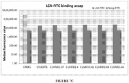
- clones/cells of the present disclosure are referred to by terms such as CR1KOT1#06, CR1KOT1#23 etc., which are internal denominations and do not represent any particular feature of the cell.
- CR1KOT1#06 CR1KOT1#23 etc.
- pD1401 gRNA 514-553
- clones/cells of the present disclosure are referred to by terms such as C1GMD1.12, C1GMD1.27 etc.,which are internal denominations and do not represent any particular feature of the cell.
- C1GMD1.12, C1GMD1.27 etc. which are internal denominations and do not represent any particular feature of the cell.
- These cell lines are developed using pD1401 (gRNA 167-207) CRISPR/Cas complex.
- clones/cells of the present disclosure are referred to by terms such as CIGMD2.30, CIGMD2.34 etc.,which are internal denominations and do not represent any particular feature of the cell.
- CIGMD2.30 CIGMD2.34 etc.
- pD1301 gRNA 404 CRISPR/Cas complex.
- clones/cells of the present disclosure are referred to by terms such as CIGMD3.36, CIGMD3.43 etc.,which are internal denominations and do not represent any particular feature of the cell.
- CIGMD3.36, CIGMD3.43 etc. which are internal denominations and do not represent any particular feature of the cell.
- These cell lines are developed using a combination of pD1401 (gRNA 167-207) and pD1301 (gRNA 404) CRISPR/Cas complex.
- composition comprising the non-fucosylated antibody, optionally along with a pharmaceutically acceptable carrier or additive or excipient.
- Pharmaceutically acceptable carrier or additive or excipient is determined by the composition being administered, as well as by the particular method used to administer the composition and is known to a person skilled in the art.
- Excipients are important for achieving protein stabilization and improving other qualities of biologics.
- a variety of excipients are added to compositions to stabilize proteins, act as antimicrobials, aid in the manufacture of the dosage form, control or target drug delivery, and minimize pain upon injection.
- Excipients can be broadly divided into five categories based on their modes of action:
- the biological material used in the present disclosure is obtained from outside India.
- FUT8 is comprised of three domains, an N-terminal coiled-coil domain, a catalytic domain, and a C-terminal SH3 domain.
- Fut8 protein structure isstudied extensively to understand the functional domain of the enzyme amino acid sequence.
- Three dimensional crystal structure of FUT8 enzyme revealed 15 strands and 16 helices. There are at least three regions, N terminus (residues 68-107), C-terminus (573-575) and residues 368-372 which are disordered.
- the putative catalytic domain of the FUT8 enzyme is consisted of two domains, an open sheet alpha/beta domain and the Rossmann fold widely known for nucleotide binding region.
- the alpha/beta domain consisted of five helices and three beta strands, which are alpha 4, 3H1, 3H2, 3H3, beta 1, beta 2 and beta3 strands.
- the domain is located in the N terminus of the protein sequence. There is no clear evidence how the N terminus catalytic domain is responsible for enzyme functionality.
- the Rossmann fold is located downstream at residue 359-492 and contains several alpha helix and beta strands.
- a series of residues Arg 365, Arg 366, Asp-368, Lys-369, Glu-373, Tyr-382, Asp-409, Asp-410, Asp-453, and Ser-469 play an important role in catalytic domain of FUT8 enzyme.
- regions of FUT8 amino acid sequence are compared among multiple species.
- the alignment shows that the enzyme sequences constitute highly conserved amino acid residues at the beta 2 strand and the 3H2 helix region.
- these amino acid positions are the target of CRISPR/Cas complex in the method of the present disclosure.
- Fucose knock out platform is useful to achieve non fucosylated monoclonal antibody molecule development. In many instances, developing completely non fucosylated antibody is a preferred outcome and therefore strategies are made in this disclosure to create complete knock out of Fucose biosynthetic pathway genes. In certain cases, the monoclonal antibody therapeutic drug product may require partial fucosylation which is not available naturally. To create designed versions of fucosylated monoclonal antibodies for therapeutic purposes, the GMD knock out CHOK1 cell line is very useful.
- GMD gene is involved in the fucosylation pathway, upstream of FUT8 gene and responsible for GDP-fucose synthesis through conversion of GDP-mannose to GDP-4-keto-6-deoxy-mannose. This step is one of the critical steps of de novo fucose biosynthetic pathway.
- GDP fucose is also produced in cells through salvage pathway and is used for fucosylation of cellular proteins. In salvage pathway, cells uptake fucose from growth media.
- the de novo pathway for fucose biosynthesis is completely stopped if the GMD gene is knocked out and completely non - functional.
- the GDP-Fucose biosynthesis still remains active through salvage pathway if the growth media is supplemented with Fucose. Therefore, fucose biosynthetic pathway and cellular protein fucosylation still remains active.
- the GMD knock out CHOK1 cell lines provide a unique advantage wherein if the monoclonal antibody need to be 100% defucosylated, GMD double knock out cellular platform is used.
- the salvage pathway to generate GDP-Fucose is utilized through supplementation of growth media with L-Fucose.
- the level of monoclonal antibody fucosylation is achieved through titrating levels of L-Fucose in growth medium. Therefore, GMD KO strategy provides 100% non fucosylated product to variable levels of fucosylation by simple titration of L-Fucose in CHOK1 culture media. This is a unique strategy to control fucosylation of monoclonal antibody production in CHOK1 cells.
- Fut8 enzyme functions downstream of GDP-Fucose biosynthesis step and is the last enzymatic step for fucosylation of cellular proteins in golgi. Fucosylation precursors from both de novo and salvage pathway use FUT8 enzyme for final fucose moiety transfer. Therefore, knocking out Fut8 gene essentially stops both de novo and salvage pathway of cellular protein fucosylation. This approach results in 100% defucosylation of monoclonal antibodies produced in the Fut8 knock out CHOK1 cell line.
- GDP-D-mannose 4,6-dehydratase catalyzes the conversion of GDP-D-mannose to the intermediate GDP-4- keto-6-deoxy-D-mannose.
- GMD The enzyme GDP-D-mannose 4,6-dehydratase (GMD) catalyzes the conversion of GDP-D-mannose to the intermediate GDP-4- keto-6-deoxy-D-mannose.
- This serves as a branching point to several different deoxyhexoses, including GDP-D-rhamnose, GDP-L-fucose, GDP-6-deoxy-D-talose, and the GDPdideoxy amino sugar GDP-D-perosamine.
- GDP-L-fucose is an important intermediary in fucose biosynthetic pathway.
- GMD is a member of the NDP-sugar modifying subfamily of the short-chain dehydrogenases/reductases (SDR).
- GMD binds its cofactor NADP(H) in the N-terminal portion of the molecule in which a common glycine-rich region is present.
- the catalytic triad has been identified as Tyr-XXX-Lys and Ser/Thr, which are all important for catalysis. Although there is significant amino acid sequence variability in members of this group of enzyme, three dimensional structural similarities exist.
- GMD cofactor binding sites
- Ser85 plays a crucial role in hydrogen bonding to the pyrophosphate at the active site.
- the nicotinamide ribose hydroxyls are within hydrogen bonding distance to the catalytic residues Tyr150 and Lys154, interactions that are highly conserved in SDR enzymes.
- the RR loop a segment of nine residues (Arg35- Arg43), stretches into the neighboring monomer making protein-protein interactions and contacts to the neighboring cofactor. Protein-protein interactions include Arg35 hydrogen bonding to Ser85 and Glu188.
- GDP-D-mannose interaction could depend on the ability to make potential hydrogen bonds to Thr126, Ser127, and Glu128.
- both catalytic residues Thr126 and Tyr150 as well as Ser85 could hydrogen bond to the hexose O4 hydroxyl.
- the catalytic mechanism proposed for GMD involves few key residues like Thr126, Ser127, Glu128, Tyr150 among others.
- CRISPR constructs are designed in proposed dimeric interface of amino acid sequence ADVDGVGTLRLL. This region is part of Exon4 of the GMD gene.
- the CRISPR construct targets Cas9 endonuclease to create double stand DNA break in exon 4. The break site is positioned before key amino acid residues in the motif ADVDGVGTLRLL with the assumption that any modification in these amino acids directly affects catalytic mechanism of the GMD enzyme.
- a second set of CRISPR/Cas complex is designed in exon 3 of the GMD gene.
- This CRISPR design is unique for high specificity, where a mutant Cas9, known as D10A Cas9 nickase mutant (Cas9n) is chosen, causing DNA single strand break.
- the two CRISPR/Cas complexes designed for two single strand DNA break allow high level of specificity.
- the constructs are designed at proposed tertrameric interface amino acid sequence motif YGDLTDSTCLVK.
- the two single stand breaks allow DNA repair by the NHEJ mechanism and it introduces mutations in this region. These mutations affect the important Ser85 residue involved in maintaining the interactions of monomers in the tetrameric configuration.
- Fut8 protein structure is studied extensively to understand the functional domain of the enzyme amino acid sequence.
- Three dimensional crystal structure of FUT8 enzyme revealed 15 strands and 16 helices. There are at least three regions, N terminus (residues 68-107), C-terminus (573-575) and residues 368-372 are disordered.
- the putative catalytic domain of the FUT8 enzyme consists of two domains, an open sheet alpha/beta domain and the Rossmann fold widely known for nucleotide binding region.
- the alpha/beta domain consists of five helices and three beta strands, which are alpha 4, 3H1, 3H2, 3H3, beta 1, beta 2 and beta 3 strands.
- the domain is located in the N terminus of the protein sequence. There is no clear evidence how the N terminus catalytic domain is responsible for enzyme functionality.
- the CRISPR/Cas target sequences are targeted in this region. Fut 8 exon7 genomic locus, respective amino acid sequence and position of important structural motifs and CRISPR target locations are depicted in figure 9A .
- This targeting is not a random selection, but has been arrived at, in the present disclosure, by experimentation to determine the highly specific location on the gene or enzyme, the disruption of which ensures that partial fucosylation that is caused by truncated or partially functional enzyme is avoided.
- the Rossmann fold on the other hand, is located downstream at residue 359-492 and contains several alpha helix and beta strands.
- a series of residues Arg 365, Arg 366, Asp-368, Lys-369, Glu-373, Tyr-382, Asp-409, Asp-410, Asp-453, and Ser-469 play an important role in catalytic domain of FUT8 enzyme.
- targeting the region equivalent to the active site of the enzyme ensures complete disruption of the Fut8 gene and provides efficacious results in comparison to either a technique that is unable to target a precise location on the Fut8 gene or a technique that targets another location on the Fut8 gene, which might result in partial disruption of Fut8 gene and enzyme activity.
- a cell with partially functional fucosylated machinery produces partially fucosylated proteins, which exhibits lower therapeutic functions as compared to non-fucosylated proteins.
- the cells produced by the method of the present disclosure produce completely or 100 % non-fucosylated proteins, including 100 % non-fucosylated antibodies.
- the present disclosure introduces mutations at critical amino acid positions at the catalytic site of the FUT8 codon sequence through CRISPR/Cas complex.
- the CRISPR design is aimed to primarily target the N-terminal catalytic domain, specifically the beta 2 strand and the 3H2 helix region by incorporating single stranded breaks.
- the cellular DNA repair system introduces nucleotide changes while carrying out the single stand break repair and creates non-functional FUT8 enzyme.
- the CRISPR system is well known for deletion and insertion in a localized manner and therefore creates frame-shift mutation at the targeted exon7 and inserts stop codons. Introduction of stop codons ensures premature translation termination and the downstream Rossmann fold is excluded from enzyme structure, resulting in non-functional FUT8 enzyme.
- the subsequent genomic DNA analysis of the modified CHOK1 cell lines reveals deletion, insertion, stop codon as well as frame shift mutations.
- the present disclosure envisages disruption of Fut8 gene and Fucosyltransferase enzyme by targeting amino acid positions in the beta 2 strand and the 3H2 helix through deletions, insertions and /or frame shift mutations.
- the resulting clones may result in premature translation stop therefore causing extensive changes in critical downstream sequences such as Arg- 365, Arg -366, Asp-368, Lys-369, Glu-373, Tyr-382, Asp-409, Asp-410, Asp-453, Ser-469 and combinations thereof.
- Figure 19 of the present disclosure depicts alignment of FUT8 amino acid sequence of rat, human, mouse, cattle and Chinese hamster.
- Amino acids in the Rossmann fold 365, 366, 368, 369 and 373 are marked with asterisks.
- Amino acids in shaded box indicate residues not aligned with consensus sequence.
- Amino acids in the exon 7 region are also marked.
- CRISPR recognition sequences are marked by thick lines.
- the FUT8 amino acid sequence from CHOK1 genomic database is analyzed and it is confirmed that these critical amino acids are conserved in the FUT8 gene derived from CHOK1 cell line as well.
- Sequence specific CRISPR/Cas complex is designed, targeting gene sequences upstream of these amino acid motifs to introduce genomic modifications. It is analysed how altering amino acid sequences upstream of the critical FUT8 enzyme catalytic domain disrupts the enzyme function.
- CRISPR/Cas technology provides complete disruption of FUT8 gene functionality.
- Gene targeting using CRISPR/Cas technology is a novel approach to create a Fucose knock out cell line platform.
- CRISPR/Cas transfected cells are screened through FUT8 gene functionality assays. Selected clones are confirmed through sequencing of genomic FUT8 loci for mutations.
- the mutant fucose knock out CHOK1 cell line is then used for expressing non-fucosylated therapeutic proteins, including non-fucosylated therapeutic monoclonal antibodies or part of antibody.
- CRISPR/Cas contructs specifically targeting the amino acid codon sequences in genomic locations are designed, and cloned in expression vectors, for e.g. pD1401 or pD1301 depending on the type of Cas9 gene.
- the CRISPR/Cas complex is transiently transfected in CHOK1 cells; the cells are plated in 96 well plates for single colony generation. Each clone is then screened for fucosylation of cellular proteins using fluorescence based Lens Culinaris Agglutinin assay (LCA). Clones positive for FUT8 or GMD gene disruption are further tested through enzymatic assays and kinetic analysis of mutant alleles of FUT8 gene or GMD gene.
- genomic sequence at the FUT8 and GMD loci is analyzed for any mutation carried out through CRISPR/Cas. These mutations involve deletions or insertions, thereby introducing frame shift mutations of the FUT8 and GMD codon sequence, and rendering the sequence disrupted and the enzymes non-functional.
- the fucose knock out CHOK1 cell line derived from above mentioned process is used as a cell line platform for expressing proteins, monoclonal antibodies, peptides, fusion proteins of therapeutic purposes, biomarker development, diagnostic and prognosis uses.
- the objective of this example is to design CRISPR/Cas complex for specific inactivation of FUT8 and the GMD alleles.
- CRISPR is based on a class of RNA-guided endonucleases known as Cas9 from the microbial adaptive immune system found in Streptococcus pyogenes.
- Cas9 nuclease is directed to specific sites on the genome by guide RNAs (gRNAs).
- gRNAs guide RNAs
- the Cas9/gRNA complex binds to a 20bp target sequence that is followed by a 3bp protospacer activation motif (PAM) NGG or NAG on the specific gene that needs to be edited (Jinek, 2012; Mali, 2013).
- PAM protospacer activation motif
- DSBs A crucial step in targeted genome editing at genomic loci that need to be modified, is the introduction of these DSBs. Once, DSBs are introduced, they are repaired either by non-homologous end joining (NHEJ) or homology directed repair (HDR).
- NHEJ non-homologous end joining
- HDR homology directed repair
- NHEJ is known for the efficient introduction of insertion/deletion mutations (indels) that in turn cause disruption of the translational reading frame of the target coding sequence or at binding sites of trans-acting factors in promoters or enhancers.
- Indels insertion/deletion mutations
- HDR mediated repair can insert specific point mutations or sequences at the target locus.
- co-transfection of cell types with vectors that express the Cas9 nuclease and the gRNAs targeted to a specific gene locus can efficiently knock down the expression of target genes.
- the expected frequency of mutations at these specific sites ranges from >1% to 50% (Sander 2014).
- mutants are performed by simple screening using sequencing, without the use of drug resistance marker selection.
- the present disclosure uses mutant Cas9 (D10A) that is guided by two guide RNAs for a single gene locus and that introduces two single stranded breaks or nicks. This also reduces the chances of nonspecific binding at other random sites.
- a vector encoding Cas9-D10A and the 2 gRNAs are used to cause efficient gene knock-out.
- the GMD and Fut8 genomic loci are targeted for sequence specific deletions through CRISPR/CAS9 technology and generate defucosylated mammalian expression systems.
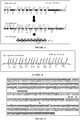
- Figure 1A of the present disclosure depicts the Fut8 coding sequence and protein sequence.
- FUT8 genomic sequence is analyzed from database sequence, sequence ID NW_003613860.
- FUT8 genomic sequence spans from 570171 - 731804 bases and contains eleven exons depicted as E1 to E11 in the figure. Base pairs locations for each exon are also indicated.
- E1, E2 and part of E3 constitute un-translated region in the upstream sequence, and part of E11 is also part of un-translated region.
- Translated regions are described as CDS 1 to CDS 9. Length of each CDS is indicated below the CDS number.
- CDS1 to CDS9 code for amino acid sequences varying from 38 amino acids to 105 amino acids.
- Cricetulus griseus or Chinese Hamster fucosyltransferase 8 (Fut8) mRNA (3126 bp) is derived from NCBI Reference Sequence: XM_003501735.1,also represented by SEQ ID No.1 of the present disclosure. Alternative exons are represented in upper and lower case letters.
- Fut8 protein structure is studied extensively to understand the functional domain of the enzyme. Three dimensional crystal structure of FUT8 enzyme revealed 15 strands and 16 helices.
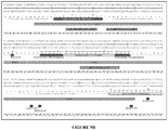
- Amino acid sequence of FUT8 gene is provided in Figure 2A .
- the CRISPR/Cas binding regions are designed in such a way that the specificity of site recognition is high and at the same time the CRISPR/Cas complex carries out the intended DNA single strand break.
- Cas9n (D10A mutant of Cas9 endonuclease) is used for the CRISPR/Cas complex.
- the Cas9n endonuclease causes single strand DNA break.
- the two CRISPR recognition sites (5' recognition site and 3' recognition site are spaced at 5 base pair distance, allowing two single stand breaks at close proximity. The resulting breaks allow the NHEJ process of DNA break repair and that introduces mutations in this region.
- the CRISPR construct has two unique 20 basepair CRISPR recognition sequences flanked by gRNA scaffolds in tandem with U6 promoter elements for efficient expression of the gRNA sequences.
- the unique design allows one single vector to express two separate gRNA scaffolds and two unique CRISPR recognition sequences on the genomic DNA.
- the nucleotide and amino acid sequence of wild type Cas9 gene is provided in Seq ID Nos. 3 and 4 respectively.
- the nucleotide and amino acid sequence of the Cas9n endonuclease is provided in Seq ID Nos. 5 and 6 respectively.
- the CRISPR/Cas design is uniquely positioned to target beta 2 strand and the 3H2 helix region by incorporating single stranded breaks.
- the design is compatible with two single strand breaks at close proximity, thereby imparting higher specificity of target recognition as NHEJ repair mechanism occurs only at these targeted genomic locations.
- Nonspecific single stand breaks, if created are usually repaired by homologous recombination which is accurate and rarely creates any mutation.
- the primary target of the present disclosure is to create mutations at the N-terminal catalytic domain, the beta 2 strand and 3H2 helix. Insertion and deletions through CRISPR/Cas at this location makes the FUT8 enzyme non-functional. In addition, frame shift mutations also cause premature translation stop codons, the Rossmann fold which is downstream of this region does not express then. Amino acid residues at Rossmann fold such as Arg 365, Arg 366, Asp-368, Lys-369, Glu-373, Tyr-382, Asp-409, Asp-410, Asp-453, and Ser-469 are very important for FUT8 functionality. The truncated enzyme will be non -functional and leads to Fucose knock out cell line.
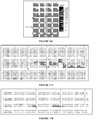
- Figure 2A of the present disclosure depicts the CHOK1 Fut8 amino- acid sequence. Complete amino acid sequence of FUT8 gene is provided. Amino acid sequence from each CDS is indicated with large arrowheads. Small arrows indicate critical amino acids present in Exon 7 (CDS5) which are targeted in the Fut8 gene by CRISPR constructs.
- CDS5 Exon 7
- the CHO whole cell genome shotgun sequencing data with accession number NW_003613860 for the Fut8 gene corresponds to a total of 161634 bp.
- the Pubmed accession number for the coding region or mRNA of the Fut8 gene is XM_003501735.1.
- the mRNA sequence, as shown in figure 1A encompasses the complete coding sequence for expression of the FUT8 gene product, which is ⁇ -1,6 fucosyltransferase.
- the Spidey alignment tool http://www.ncbi.nlm.nih.gov/spidey/spideyweb.cgi ) is used to identify the exons in the genomic DNA by aligning the mRNA sequence with the genomic DNA sequence. A total of 11 exons with the boundaries as shown in Table 3 are identified.
- Table 3 Characterization of Fut8 mRNA EXON Genomic coordinates mRNA coordinates Length (nucleotides) Exon 1 570171-570263 1-93 93 Exon 2 593436-593529 94-187 94 Exon 3 608623-609050 188-615 428 Exon 4 634802-634917 616-731 116 Exon 5 635025-635187 732-894 163 Exon 6 657197-657311 895-1009 115 Exon 7 673387-673624 1010-1247 238 Exon 8 709381-709627 1248-1494 247 Exon 9 710960-711136 1495-1671 177 Exon 10 721655-721805 1672-1822 151 Exon 11 730501-731804 1823-3126 1304
- FIG. 1A A 100% identity between the genomic DNA and mRNA sequence is observed.
- Organization of the Fut8 gene showing all the 11 exons and position of the gRNAs targeting exon7 is shown in figure 1A of the present disclosure.
- the construct with mutant Cas9 nuclease (Cas9n) is designed, which creates single strand break (nick) at the target site.
- Two separate gRNAs are designed at close proximity in exon7 to create two nicks for eventual DNA repair.
- CRISPR/Cas9 technology target sites are localized to the first few exons of the Fut8 gene. This is done to avoid partial fucosylation that can be caused by truncated or partially functional enzyme.
- Exon-7 (CDS-5) nucleotide sequence of Fut8 is represented by SEQ ID No. 7 of the present disclosure.
- Exon-7 (CDS-5) amino acid sequence of Fut8 of CHO cell is represented by SEQ ID No. 8 of the present disclosure.
- the targeted amino acid positions in the protein/peptide sequence are underlined.
- the Spidey alignment tool (httn://www.ncbi.nlm.nih.gov/spidey/spideyweb.cgi ) is used to identify the GMD gene exons in the genomic DNA by aligning the GMD mRNA sequence with the genomic DNA sequence. A total of 10 exons with a 5' untranslated region and a poly A tail are identified and tabulated in Table 4.
- CRISPR recognition sequences designed for FUT8 gene sequence Exon location SEQ ID Nos. Locus name CRISPR/Cas recognition DNA sequence (5' to 3') Exon3 SEQ ID No. 1 7 gRNA520-558 Exon 3 SEQ ID No. 18 gRNA 549-590 Exon 4 SEQ ID No. 19 gRNA 687-731 Exon 7 SEQ ID No. 20 gRNA1019 -1057 Exon 7 SEQ ID No. 21 gRNA 1128-1168 Exon 7 SEQ ID No. 22 gRNA 1199-1238 Exon 7 SEQ ID No.
- Table 5of the present disclosure lists different Fut8 target sequences that are considered for CRISPR knock out targeting. A total of twenty different sequences are considered initially. It is made sure that none of the gRNAs span onto an exon-intron boundary as this may render the gRNAs inactive. Based on this approach, a 57bp stretch on exon 7 is chosen as the target for CRISPR/Cas mediated knock out target. This includes two gRNAs, one on each strand that causes two single stranded breaks.
- the target sequence in Fut8 gene that is used in an embodiment of the present method is shown in below figure 3B of the present disclosure as gRNA 1120-1176.
- the sites of cleavage are indicated with an arrow.
- the distance between the two gRNAs is 5 bases.
- the gRNA 1120-1176 recognition sequence is underlined in Figure 3B ..
- the corresponding synthesized fragment is incorporated into the pD1401 vector and named as pD1401 gRNA 514-553, the features of which are described subsequently in the disclosure.
- This method of the present disclosure uses Cas9n (nickase mutant) in targeting Fut8 genomic sequence, exon 7 with CRISPR/Cas system.
- the Cas9n endonuclease makes single stand break (SSB) in opposite strand of DNA.
- the CRISPR/Cas recognition sequences in the upper and lower strands are underlined.
- Corresponding single strand break sites are indicated as black arrow heads.
- the three nucleotide PAM sequences are indicated in bold letters.
- one of the designs is used for targeting at exon 7 gRNA 1120-1176.
- the CRISPR/Cas vector construct for this design is termed as pD1401 gRNA (514-553).
- the 5' and 3' CRISPR recognition sequence is indicated in small and italicized, two separate sites complementary to this recognition sequence are recognized at the FUT8 genomic sequence.
- the sequence represented with bold letters indicate gRNA scaffold sequence for CRISPR/Cas complex to get engaged.
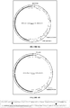
- the construct map is provided in figure 3A and important sequence regions are marked.
- Figure 9B of the present disclosure provides GMD genomic locus and CRISPR recognition sequences.
- the CHO whole cell genome shotgun sequencing data with accession number NW 003613635.1 for the GMD gene locus consisting of 442215 bp is obtained from Pubmed.
- the Pubmed accession number for the coding region or mRNA of the GMD gene is NM_001246696.1.
- GMD is a member of the NDP-sugar modifying subfamily of the short-chain dehydrogenases/reductases (SDR). As a member of this subfamily, GMD binds its cofactor NADP (H) in the N-terminal portion of the molecule in which a common glycine-rich region is present.
- the catalytic triad has been identified as Tyr-XXX-Lys and Ser/Thr, which are all important for catalysis. Structure analysis of GMD from E.coli suggests the active molecule is in dimeric configuration. Whereas homolog from Arabidopsis thaliana is tetrameric, and that the NADP (H) binding site is intimately involved in creating the tetramer interface. It is most probable that the functional form of GMD enzyme in eukaryotes consists of tetrameric configuration.
- the CRISPR/Cas binding regions are designed in such a way that the specificity of site recognition is high and at the same time the CRISPR/Cas complex carries out the intended DNA single strand break.
- Cas9n (D10A mutant of Cas9 endonuclease) is used for the CRISPR/Cas complex.
- the Cas9n endonuclease causes single strand DNA break.
- the two CRISPR recognition sites (5' recognition site and 3' recognition site are spaced at 5 base pair distance, allowing two single stand breaks at close proximity. The resulting breaks allow the NHEJ process of DNA break repair and that introduces mutations in this region.
- the CRISPR construct has two unique 20 base pair CRISPR recognition sequences flanked by gRNA scaffolds in tandem with U6 promoter elements for efficient expression of the gRNA sequences.
- the unique design allows one single vector to express two separate gRNA scaffolds and two unique CRISPR recognition sequences on the genomic DNA.
- the CRISPR/Cas design is uniquely positioned to target the YGDLTDSTCLVK motif and DLAEYT motif responsible for tetrameric interface of the GMD multimeric functional protein structure.
- Two single strand breaks induced by the Cas9n endonuclease at this region allow NHEJ mediated DNA repair. Mutations incorporated during DNA repair result in frame shift mutation, deletion, insertion as well as premature stop codons.
- Such mutation not only alters the critical motif for tertramerization but also creates mutations in the downstream Ser85 residue, which is involved in involved in maintaining the interactions of monomers in the tetrameric configuration.
- the design is compatible with two single strand breaks at close proximity, thereby imparting higher specificity of target recognition as NHEJ repair mechanism occurs only at these targeted genomic locations.
- Nonspecific single stand breaks, if created are usually repaired by homologous recombination which is accurate and rarely creates any mutation.
- Figure 1B of the present disclosure depicts the CHOK1 GMD genome organization.
- the method of the present disclosure uses Cas9n (nickase mutant) in targeting GMD genomic sequence, exon 3 with CRISPR/Cas system.
- the Cas9n endonuclease makes single stand break (SSB) in opposite strand of DNA.
- the construct is named as pD1401 (gRNA 167-207) and is represented by Figure 4A of the present disclosure.
- the 5' and 3' CRISPR recognition sequence is indicated in small and italicized, two separate sites complementary to this sequence are recognized at the GMD genomic sequence.
- the sequence represented with bold letters indicates gRNA scaffold sequence for CRISPR/Cas complex to get engaged.
- Exon 4 of GMD gene is targeted with wild type Cas9 endonuclease.
- This wild type Cas9 makes double strand break (DSB) at the target site.
- the construct is named as pD1301 (gRNA 404) and is represented by Figure 4B of the present disclosure.
- the CRISPR recognition sequence is indicated in small and italicized.
- the double stranded genomic DNA sequence is recognized based on this sequence by CRISPR/Cas system.
- the sequence represented with bold letters indicates gRNA scaffold sequence for CRISPR/Cas complex to get engaged.
- the following table represents CRISPR/Cas recognition sequences throughout GMD coding sequences for potential single strand break sites. Any of these recognition sequences is used for Cas9n endonuclease mediated single strand break and repair strategy of CRISPR/Cas system to knock out GMD gene.
- Table 6 provides all CRISPR sequences for GMD Exon location SEQ ID Nos. Locus name CRISPR/Cas recognition DNA sequence (5' to 3') Exon3 SEQ ID No. 47 gRNA 394-434 Exon 3 SEQ ID No. 48 gRNA 394-436 Exon 3 SEQ ID No. 49 gRNA 384-422 Exon 4 SEQ ID No. 50 gRNA 541-580 Exon 4 SEQ ID No.
- target sequences in GMD gene sequence are designed for CRISPR recognition sites. All sequences are represented in 5' to 3' direction; the corresponding 20 base pair target specific crRNA sequence will be derived from CRISPR recognition sequence provided in each design mentioned in above table 6.
- gRNA 394-434 is used to create CRISPR/Cas complex pD1401 (gRNA 167-207) for transfection of CHOK1 cells.
- the CRISPR/Cas complex creates two single stranded DNA breaks at the complementary strand of the recognized coding sequence of GMD gene.
- Successful DNA repair at the target site creates non-functional GMD gene and thereby fucose knock out CHOK1 cell lines are developed.
- pD1401 gRNA 167-207 are described subsequently in the disclosure.
- any one of the above mentioned CRISPR recognition site creates a non-functional GMD gene. Therefore, any of these potential sites alone or in combination is used for fucose knock out CHOK1 cell line development.
- the target sequence in GMD exon 3 that is used in an embodiment of the present method is mentioned in the table 6 as gRNA 394-434.
- Figure 4C describes the CRISPR recognition sequence. The sites of cleavage are indicated with an arrow. The distance between the two gRNAs is 6 bases.
- the gRNA 394-434 sequence is underlined in Figure 4C .
- the corresponding synthesized fragment is incorporated into the pD1401 vector and named as pD1401 gRNA 167-207, the features of which are described subsequently in the disclosure.
- This method of the present disclosure uses Cas9n (nickase mutant) in targeting GMD genomic sequence, exon 3 with CRISPR/Cas system.
- the Cas9n endonuclease makes single stand break (SSB) in opposite strand of DNA.
- the following table represents CRISPR/Cas recognition sequences throughout GMD coding sequences for potential double strand break sites. Any of these recognition sequences are used for wild type Cas9 endonuclease mediated double strand break and repair strategy of CRISPR/Cas system to knock out GMD gene.
- Table 7 - CRISPR/Cas recognition sequences throughout GMD coding sequences for potential double strand break sites Exon location SEQ ID Nos.
- gRNA Twenty three unique CRISPR recognition sequences (gRNA) are designed throughout the GMD gene sequence. All sequences are represented in 5' to 3' direction; the corresponding 20 base pair target specific crRNA sequence will be derived from CRISPR recognition sequence provided in each design mentioned in above table 7.
- gRNA 306 is used to create CRISPR/Cas complex pD1301 (gRNA 404) for transfection of CHOK1 cells.
- the CRISPR/Cas complex creates one double stranded DNA break at the recognized coding sequence of GMD gene.
- Successful DNA repair at the target site creates non-functional GMD gene and thereby fucose knock out CHOK1 cell lines are developed.
- any one of the above mentioned CRISPR recognition site creates a non-functional GMD gene. Therefore, any of these potential sites alone or in combination are used for fucose knock out CHOK1 cell line development.
- the target sequence in GMD exon 4 that is used in an embodiment of the present method is shown in the above table 7 as gRNA 306.
- Figure 4D describes the CRISPR recognition sequence. The sites of cleavage are indicated with an arrow. The corresponding synthesized fragment is incorporated into the pD1301 vector and named as pD1301 gRNA 404, the features of which are described subsequently in the disclosure.
- This method of the present disclosure uses wild type Cas9 endonuclease in targeting GMD genomic sequence, exon 4 with CRISPR/Cas system.
- the wild type Cas9 endonuclease makes Double stand break (DSB) in both strands of DNA.
- CRISPR technology is based on a class of RNA-guided endonucleases known as Cas9 from the microbial adaptive immune system found in Streptococcus pyogenes .
- Cas9 nuclease is directed to specific sites on the genome by guide RNAs (gRNAs).
- gRNAs guide RNAs
- Two components must be introduced and/or expressed in cells or an organism to perform CRISPR based genome editing: the Cas9 nuclease; and a 'guide RNA' (gRNA).
- gRNA Twenty nucleotides recognition sequence at the 5' end of the gRNA direct Cas9 to a specific target DNA site using standard RNA-DNA complementarity base pairing rules. These target sites must lie immediately 5' of a PAM sequence that matches the canonical form 5-NGG.
- the present disclosure uses two different kinds of Cas9 endonuclease in this disclosure as described below. In both cases a single transfection vector encoding gRNA and nuclease is used, thereby increasing the transfection efficiency of the CHOK1 cells.
- This example contains procedure for CHOK1 cell transfection with CRISPR constructs. It also provides for selection and confirmation of single cell stable cell lines for developing FUT8 knock out CHOK1 cell line using CRISPR technology, and selection of positive clones by flow-cytometry based functional assay.
- Transfection isoptimized using CHOK1 cells of both adherent and suspension type.
- Liposome and modified liposome mediated transfection reagents are tested for e.g., Lipofectamine 2000, Lipofectamine 3000, Lipofectamine LTX with PlusTM reagent, MIRUS TransIT X2, MIRUS TransIT 2020, MIRUS TransIT 293, MIRUS TransIT CHO transfection kit.
- DNA concentration ranging from 0.5 ⁇ g to 5 ⁇ g are tested for various incubation times for e.g., 4hrs, 24hrs and 48hrs. Multiple DNA to transfection reagent ratios ( ⁇ g : ⁇ l) are also tested. The optimum transfection efficiency is achieved using 1:3 DNA to transfection reagent ratio, 24 hrs incubation and Lipofectamine LTX with PlusTM reagent. Optimization experiments performed with GFP expressing plasmid DNA.
- FIG. 20 depicts transfection efficiency of CHOK1 cell line using the protocol described in the disclosure. Transfection efficiency is determined using a Green Fluorescent Protein expressing plasmid construct. Number of green cells observed after transfection compared to the total number of viable cells determines transfection efficiency of the protocol established. Panel A represents the bright field image and panel B represents the same microscopic field for red channel fluorescence.
- Transfection efficiency Number of GFP expressing cells / Total number of cells * 100 Optimized transient transfection efficiency is 40-50% in CHOK1 cells.
- CHOK1 cells are seeded at more than 90% viability and at a density of 0.25 X 10 6 cells/well in a 6 well tissue culture plate and allowed to adhere for 24 hrs.
- CRISPR constructs pD1401 (gRNA 514-553), pD1401 (gRNA 167-207), pD1301 (gRNA 404), combination of pD1401 (gRNA 167-207) + pD1301 (gRNA 404) are used for transfection using Lipofectamine LTX with PlusTM reagent. 2.5 ⁇ g of construct is used with 1:3 DNA to transfection reagent ratio. The cells are incubated for 20-24 hrs after transfection. Prior to transfection, DNA quantity and quality is estimated by UV spectrophotometry.
- DNA represents quality and protein contamination.
- the ratio of absorbance at 260 nm and 280 nm is used to assess the purity of DNA.
- a 260/280 >1.8 is generally accepted as "pure” or good quality DNA.
- 3-4 ⁇ l of DNA sample is placed on the micro cuvette and DNA concentration is estimated using Eppendorf Biophotometer D30 against suitable blank.
- Lipofectamine LTX dilution Lipofectamine LTX 15 ⁇ l*n Media without serum Up to 0.5ml*n
- the cells are incubated for 4 hours at 37°C in a 5% CO 2 Incubator.
- the complete media is added at 1.5 ml /well and incubated for 20-24 hours at 37°C in a 5% CO 2 Incubator. After 20-24 hours of transfection, cells are trypsinized and to single cell dilution is prepared.
- Single cell dilution is obtained by serial dilution of the cells to a concentration of 0.5cell/100 ⁇ l. Cell count is taken using hemocytometer. The cells are allowed to grow for few days at 37°C in a 5% CO 2 Incubator. Plate scanning is done to identify single cell colonies under the inverted phase contrast microscope. Cells growing into distinctly small single colonies are marked for further amplification. After 2-3 weeks, single cell clones are amplified from one well of 96 well plate to one well of 6 well plate by trypsinization. Cells are allowed to grow for 2-3 days at 37°C at 5% CO 2 in a CO 2 incubator. Cells are further amplified from one well to two wells in a 24 well plate (replica plating) for further screening.
- LCA-FITC Lilinaris Agglutin-Fluorescein isothiocyanate
- Fluorescein isothiocyanate is a fluorochrome conjugated to LCA. Therefore, presence of fucosylated proteins on cell membrane of control CHOK1 cells is recognized by fluorescein conjugated LCA. These cells fluoresce brighter in specific flow cytometer channel. The fluorescence observed is represented as fluorescence unit. The cells where fucose pathway is disrupted, the knockout lines are not able to produce fucosylated cellular proteins and hence the cell membrane proteins are non fucosylated. Testing these cells with Fluorescein-LCA conjugate results in fluorescence comparable to background. Therefore, the Fucose knock out cells fluoresce at a much lower level (less than 100RFU) compared to control CHOK1 cell line.
- Cells are trypsinized, transferred to a microfuge tube and spun at 1500 rpm (revolution per minute) for 5 minutes using Eppendorf minispin centrifuge. The media is removed and fresh media is added in the tubes. Both transfected and untransfected CHOK1 cells are processed simultaneously. The cells are tested for LCA-FITC flow cytometry based analysis using "Millipore GUAVA 8 easyCyte HT" benchtop flow cytometer.
- clones from transfection with pD1401 are screened for fucose knockout profile.
- 200 clones from transfection with pD1401 (gRNA 167-207) or pD1301 (gRNA 404) or a combination of pD1401 (gRNA 167-207) + pD1301 (gRNA 404) are screened for fucose knock out profile.
- Fluorescein-streptavidin (Strep-FITC) negative staining is also performed.
- Fluorescein lens culinaris agglutinin (LCA-FITC) stock 5mg/ml is diluted to get 2 ⁇ g/ml final concentration in assay buffer (DPBS containing 2% BSA).
- Fluorescein streptavidin (Strep-FITC) stock 1mg/ml is diluted to get 2 ⁇ g/ml final concentration in assay buffer (DPBS containing 2% BSA).
- Cell suspensions are taken in duplicates and are spun at 1500 rpm for 5 minutes using Eppendorf minispin centrifuge.
- CHOK1 cells are transfected with pD1401 (gRNA 514-553) construct targeting Fut8 locus and the results are provided below.
- Median RFU refers to median value of relative fluorescence unit.
- the graphical results and fluorescence profile provided in the table above are also depicted in figure 7A and figure 8A of the present disclosure.
- the figures depict the graphical result and fluorescence profile observed for the CHOK1 cell lines CR1KOT1#6, CR1KOT1#18, CR1KOT1#22, CR1KOT1#23, CR1KOT1#26, CR1KOT1#31, CR1KOT1#34, CR1KOT1#37, in Flow cytometry based LCA-FITC Binding Assay.
- This flow cytometry assay detects fucosylated proteins present on cell surface.
- CHOK1 control cells fluoresce highly as many fucosylated proteins are present in Control CHOK1 cell line.
- the LCA FITC fluorescence is minimized as there is no fucosylated protein on surface of these cell lines.
- the figure reveals significant fluorescence loss when CR1KOT1#6, CR1KOT1#18, CR1KOT1#22, CR1KOT1#23, CR1KOT1#26, CR1KOT1#31, CR1KOT1#34 andCR1KOT1#37, are tested in this assay, indicating these cell lines are CHOK1 FUT8 knock out cell lines.
- CHOK1 cells are transfected with pD1401 (gRNA 167-207), pD1301 (gRNA 404), pD1401 (gRNA 167-207)+ pD1301 (gRNA 404) constructs targeting GMD locus, and the results are provided in the table below.
- the graphical results and fluorescence profile provided in the table above are also depicted in figures 7B and 8B of the present disclosure.
- the figures depict the graphical result and fluorescence profile observed for the CHOK1, C1GMD1.12, C1GMD1.27, C1GMD2.30, C1GMD2.34, C1GMD3.4, C1GMD3.36, C1GMD3.43, C1GMD3.49, C1GMD3.51 in Flow cytometry based LCA-FITC Binding Assay.
- This flow cytometry assay detects fucosylated proteins present on cell surface.
- CHOK1 control cells fluoresce highly as many fucosylated proteins are present in Control CHOK1 cell line.
- the LCA FITC fluorescence is minimized as there is no fucosylated protein on surface of these cell lines.
- the figure reveals significant fluorescence loss when C1GMD1.12, C1GMD1.27, C1GMD2.30, C1GMD3.4, C1GMD3.36, C1GMD3.43, C1GMD3.49, C1GMD3.51 are tested in this assay indicating these cell lines are potential CHOK1 GMD knock out cell lines.
- the graphical results and fluorescence profile provided in the table above are also depicted in figures 7C and 8C of the present disclosure.
- the figures depict the graphical result and fluorescence profile observed for the CHOK1, C1GMD1.37, C1GMD1.4, C1GMD1.41, C1GMD1.43 and C1GMD1.44, in Flow cytometry based LCA-FITC Binding Assay.
- This flow cytometry assay detects fucosylated proteins present on cell surface.
- CHOK1 control cells fluoresce highly as many fucosylated proteins are present in Control CHOK1 cell line.
- the LCA FITC fluorescence is minimized as there is no fucosylated protein on surface of these cell lines.
- the figure reveals significant fluorescence loss when C1GMD1.37, C1GMD1.4, C1GMD1.41, C1GMD 1.43, C1GMD1.44 are tested in this assay indicating these cell lines are potential CHOK1 GMD knock out cell lines.
- the cell lines indicated here show resistance against LCA by day one which has multiplied and grown into large colonies of cells after Day 4 and Day 6 of culture in presence of LCA reagent.
- Cell morphology is observed with microscope and observations are recorded on days 1, 4 and 6.
- Cells are regularly observed under the inverted phase contrast microscope and monitored for colony morphology.
- the photographs taken at different time points of LCA selection assay with 200 ⁇ g/ml LCA clearly show that CHOK1 control cells are completely dead at Day 4 of culture, whereas the selected clones show continuous cell growth and healthy cell morphology even after 4 days of culture.
- Cells are spun at 1500 rpm for 5 minutes using Eppendorf minispin centrifuge. The media is aspirated and the pellet re-suspended in 0.25 - 1ml of assay buffer containing 2 ⁇ g/ml LCA-FITC.
- CHOK1 control cells are re-suspended in 0.25-1ml of assay buffer alone (unstained control) and 0.25-1ml of assay buffer containing 2 ⁇ g/ml LCA-FITC (stained control). All samples are diluted to get 0.1-0.2 X 10 6 cells/ml in final assay buffer. The samples are then incubated in dark on ice for 30 minutes. Then 200/ ⁇ l of each sample is aliquoted in a 96 well plate. The plate is then loaded in the Millipore GUAVA easyCyte 8HT benchtop flow cytometer for data acquisition and analysis. Data analysis is done using Incyte software. Table 12 Sl. No.
- Figure 10 depict the graphical result observed for the CHOK1 cell lines CR1KOT1#44, CR1KOT1#46, CR1KOT1#48, CR1KOT1#49, CR1KOT1#51, CR1KOT1#52, CR1KOT1#55, CR1KOT1#59, CR1KOT1#61, CR1KOT1#66, CR1KOT1#67 in Flow cytometry based LCA-FITC Binding Assay.
- Figure 11 depicts representative fluorescence profiles observed in cell lines CR1KOT1#44, CR1KOT1#49, CR1KOT1#51, CR1KOT1#52, CR1KOT1#55, CR1KOT1#59, CR1KOT1#61, CR1KOT1#67. All clones are passaged for additional days before being analyzed in this experiment. This flow cytometry assay detects fucosylated proteins present on cell surface. Thus CHOK1 control cells fluoresce highly as many fucosylated proteins are present in Control CHOK1 cell line.
- the LCA FITC fluorescence is minimized as there is no fucosylated protein on surface of these cell lines.
- the figure reveals significant fluorescence loss when CR1KOT1#44, CR1KOT1#46, CR1KOT1#48, CR1KOT1#49, CR1KOT1#51, CR1KOT1#52, CR1KOT1#55, CR1KOT1#59, CR1KOT1#61, CR1KOT1#66, CR1KOT1#67 are tested in this assay indicating these cell lines are CHOK1 FUT8 knock out cell lines.
- Fluorescein Lens Culinaris Agglutinin (LCA-FITC) stock of 5mg/ml is diluted to get 2 ⁇ g/ml final concentration in assay buffer (DPBS containing 2% BSA).
- assay buffer DPBS containing 2% BSA
- Cells are spun at 1500 rpm for 5 minutes using Eppendorf minispin centrifuge. The media is aspirated and the pellet re-suspended in 0.25 - 1ml of assay buffer containing 2 ⁇ g/ml LCA-FITC.
- CHOK1 control cells are re-suspended in 0.25-1ml of assay buffer alone (unstained control) and 0.25-1ml of assay buffer containing 2 ⁇ g/ml LCA-FITC (stained control).
- Pictorial depiction of the LCA FITC binding assay is presented in figure 12 .
- Data analysis suggests highly reproducible LCA FITC binding pattern for the clonal fucose knock out CHOK1 cell lines. These fucose knock out CHOK1 cell lines are further tested for growth characteristics and compared to untransfected parental CHOK1 cell line.
- Streptavidin-FITC conjugate Clonal fucose knock out CHOK1 cell lines are tested with Streptavidin-FITC conjugate to ensure specific interaction of LCA-FITC binding: Streptavidin conjugated FITC (Strep-FITC) staining of the clones is carried out to ensure that there is no non-specific binding of FITC dye.
- Cell membrane proteins do not bind to Streptavidin-FITC conjugate whereas fucosylated membrane proteins bind specifically to LCA-FITC conjugate.
- Control CHOK1 cells are stained with both LCA-FITC and Strep-FITC in separate reactions to confirm this specificity. All clones are similarly stained with Streptavidin-FITC and LCA-FITC conjugates to determine non-specific binding.
- Figure 14 of the present disclosure depicts the LCA Flow cytometry assay, and comparison of the clones through LCA-FITC and Strep-FITC assay respectively.
- Fucose knockout phenotype is observed,CR1KOT1#006, CR1KOT1#018, CR1KOT1#022, CR1KOT1#023, CR1KOT1#026, CR1KOT1#031, CR1KOT1#034, CR1KOT1#036, CR1KOT1#037, CR1KOT1#044, CR1KOT1#049, CR1KOT1#051, CR1KOT1#052, CR1KOT1#055, CR1KOT1#059, CR1KOT1#061 and CR1KOT1#067.
- Fluorescein streptavidin (Streptavidin -FITC) stock 1mg/ml is diluted to get 2 ⁇ g/ml final concentration in assay buffer (DPBS containing 2% BSA). Cells are spun at 1500 rpm for 5 minutes using Eppendorf minispin centrifuge. The media is aspirated and the pellet re-suspended in 0.25 - 1ml of assay buffer containing 2 ⁇ g/ml Streptavidin -FITC. CHOK1 control cells are re-suspended in 0.25-1ml of assay buffer alone (unstained control) and 0.25-1ml of assay buffer containing 2 ⁇ g/ml Streptavidin -FITC (stained control).
- Streptavidin-FITC Comparison with Streptavidin-FITC is carried out to ensure specific interaction of LCA-FITC conjugate. The data suggests only background fluorescence observed with Streptavidin-FITC conjugate when tested with CHOK1 control cell line and any of the CRISPR/Cas transfected cell lines.
- Figure 14 depicts the specificity of the LCA -FITC flow cytometry assay developed to screen the CHOK1 Fucose knock out cell lines.
- Flow cytometry experiments are carried out with Streptavidin-FITC conjugate.
- Streptavidin-FITC does not recognize cell surface proteins on CHOK1 control cell line indicating specific interaction of LCA-FITC conjugate.
- the Streptavidin-FITC conjugate reveals similar levels of background fluorescence between positive and negative cell lines identified by LCA-FITC conjugate.
- Figure 14 shows no non-specific interaction of LCA-FITC conjugate used in this study when compared to Streptavidin-FITC conjugate.
- Growth curve determination of selected clones are performed to ensure that growth profile is not altered significantly compared to wild type CHOK1 cells during the process of knockout cell line development.
- 0.1 X 10 6 CHOK1 cells are seeded in 6 well tissue culture plates. Seeding is done for 5 time points for each clone. For each time point, triplicate seeding is done (e.g., 15 wells for 5 time points). At each time, point cell counts are taken in triplicates. Viable cell count is performed using either hemocytometer or Vi-cell XR cell viability analyser. Respective growth curves are generated with SEM as error bar. Table 14 describes representative growth data from one of the FUT8 knock out cell lines.
- Table 14 Mean viable cell count (X10 ⁇ 6) Time (hrs) CHOK1 control CR1KOT1#52 CR1KOT1#55 CR1KOT1#61 CR1KOT1#67 0 0.100 0.100 0.100 0.200 0.100 24 0.313 0.261 0.344 0.370 0.411 48 0.858 0.671 0.925 0.992 1.226 72 2.295 1.439 2.031 2.411 2.875 96 2.680 3.398 3.603 5.272 4.738 Time (hrs) CHOK1 control CR1KOT1#06 CR1KOT1#18 CR1KOT1#22 CR1KOT1#23 CR1KOT1#26 0 0.100 0.100 0.100 0.100 0.100 0.100 0.100 24 0.313 0.270 0.217 0.167 0.183 0.243 48 0.858 0.790 0.567 0.540 0.680 0.457 96 2.680 4.357 2.477 3.857 3.867 1.997 Time (hrs) CHOK1 control CR1K
- Viable cell count for each cell lines are tested and used for growth curve determination. Following clonal fucose knock out CHOK1 cell lines are used for growth curve development, CR1KOT1#006, CR1KOT1#018, CR1KOT1#022, CR1KOT1#023, CR1KOT1#026, CR1KOT1#031, CR1KOT1#034, CR1KOT1#036, CR1KOT1#037, CR1KOT1#044, CR1KOT1#049, CR1KOT1#051, CR1KOT1#052, CR1KOT1#055, CR1KOT1#061 and CR1KOT1#067.
- the data is analyzed and plotted in growth curve.
- the growth curves of respective cell line are provided in figures 13A , 13B and13C of the present disclosure.
- clonal cell lines have comparable growth potential with respect to CHOK1 parental cell line. These clonal cell lines are used for over-expression of therapeutic proteins and/or monoclonal antibodies. Viable cell count is taken every day for 5 days in optimal growth conditions, using Vi-Cell counter. Few clonal fucose knock out CHOK1 cell lines CR1KOT1#018, CR1KOT1#026, CR1KOT1#034, CR1KOT1#052 and CR1KOT1#055 have shown slightly slower growth potential compared to other cell lines.
- CRISPR transfected clones selected through functional assay namely LCA-FITC flow cytometry assay are used for genomic sequence analysis.
- the FUT 8 genomic locus of Chinese Hamster is well reported in literature (NW 003613860) and is used as wild type sequence to understand type of gene modification in each cell line clone.
- GMD genomic locus of Chinese Hamster is obtained from sequence database, NW_003613635.1, NP_001233625.1, NM_001246696.1 and is used as wild type sequence to understand the type gene modification in each cell line.
- the objective of this example is to analyse genomic DNA sequencing results obtained from CRISPR/Cas transfected CHOK1 FUT8 knock out cell lines and CHOK1 GMD knock out cell lines. All cell lines reported here are clonal cell lines and are selected from LCA media selection assay and LCA-FITC flow cytometry assay.
- the selected clonal cell lines are grown in appropriate growth conditions for genomic DNA isolation, purified genomic DNA is used for PCR amplification using primers flanking the FUT8 and GMD target loci, the PCR amplified product is then purified and cloned in a suitable vector using E.coli competent cells, resulting ampicillin resistant E.coli colonies are selected and cultured, plasmid DNA are isolated from each bacterial clone, approximately 5-10 individual bacterial colonies are tested per clonal cell lines through automated sequencing to understand the type of modification at the FUT8 target genomic locus.
- Clonal CHOK1 cell lines are grown in Advanced DMEM media with 10% Fetal bovine serum, 4mM glutamine, 100 units/ml Penicillin and 100 ⁇ g/ml Streptomycin in T175 flasks at 37°C in presence of 5% CO 2 and 75% relative humidity in controlled condition incubators. The cell growth is observed every day and viability is monitored. Cells are harvested at 80% confluency and greater than 95% viability with trypsinization. On the day of isolation, culture media is removed and adherent cells are first washed with 10 ml of DPBS followed by addition of 4 ml of 0.05% trypsin EDTA solution for trypsinization. The cells are incubated at 37°C for 2-3 minutes and harvested.
- Cells are then mixed with 10 ml of DPBS and centrifuged at 1500rpm for 5 min. The spent media is removed and cell pellet is resuspended in 10 ml DPBS. Cells are washed again using centrifugation at 1500 rpm for 5 min. DPBS is removed completely by aspiration. The final cell pellet is used for genomic DNA isolation.
- Genomic DNA is isolated from CHOK1 control cells and CHOK1 CRSPR/Cas transfected clonal cell lines showing LCA resistance and selected through LCA flow cytometry assay.
- Commercially available QIAGEN gDNA extraction kit is used for isolating genomic DNA following manufacturers protocol.
- Genomic DNA sequence of Chinese Hamster is analysed from publicly available database sequence NW_003613860.
- FUT8 Exon 7 DNA sequences and partial intron sequence is used for designing PCR strategy to amplify the FUT8 target locus.
- GMD exon and intron sequences are obtained from NW_003613635.1 and NM_001246696.1 and sequence analysis are carried out for Exon 3 and Exon 4.
- Primers are designed based on primer length, PCR product length, GC content, melting temperature and potential homoduplex and heteroduplex formation. Primers are designed flanking the FUT8 target locus as provided below.
- the amplified PCR product is intended for mutation analysis due to CRISPR mediated SSB and subsequent DNA repair. Following nucleotide sequence represents the region of interest with primer sequences in bold letters.
- Introns are represented from base 21 to base 106 and from base 345 to base 371 in lower case letters.
- Exon 7 is represented from base 107 to base 344 in upper case letters.
- the primer binding sites of Left and Right are underlined.
- Introns are represented from base 23 to base 166 and from base 277 to base 447 in lower case letters.
- Exon 3 is represented from base 167 to base 276 in upper case letters.
- the primer binding sites of Left and Right are underlined.
- Introns are represented from base 21 to base 187 and from base 381 to base 432 in lower case letters.
- Exon 4 is represented from base 188 to base 380 in upper case letters.
- the primer binding sites of Left and Right are underlined.
- Genomic PCR is performed using QIAGEN gDNA extraction kit using the following primers mentioned in table 15.
- Table 15 PCR Sets Primer Name Primer sequence (5' to 3') PCR product size Base Tm %GC CRISPR primers at FUT8 Exon 7 CRP_P1_Fw 394 bp 26 55.2 38 CRP_P1_Rv 23 54.7 47.8 CRISPR primers at GMD Exon 3
- GMD_P03_Fw 453 bp 20 49.9 45 GMD_P03_Rv 21 52.6 42.9
- the following section provides experimental details for PCR product generation from CHOK1 genomic DNA from control cell lines and LCA selected clonal cell lines, cloning of PCR products in E coli competent cells and sequencing of cloned PCR products.
- the experiments are designed to standardize PCR conditions.
- the parameters tested include, genomic DNA concentration (from 100ng to 1000ng), primer concentrations (2nmole to 20nmole), PCR annealing temperature (from 55.8°C to 62.9°C) and time (20 secs to 50 secs), PCR product extention time (30 secs to 60 secs) and PCR cycle number is set at 30 cycles. Arrived optimized condition is described in following section.
- PCR reactions are carried out using proof reading polymerase Phusion polymerase to ensure that PCR mediated mutations are limited.
- Taq polymerase enzyme is added in the mix for tailing.
- the tailing step is important as the extra base added to the PCR products allows direct cloning in sequencing vector described in the next section.
- the Phusion polymerase amplified product is incubated with Taq DNA polymerase for 20 minutes at 72 °C.
- Genomic DNA PCR products are analysed in agarose gel electrophoresis and the product length is confirmed using a molecular weight standard. PCR samples with clear amplification profile are used in the next processing step.
- the amplified PCR products are loaded in freshly prepared 1% agarose gel and electrophoresed at 100V for one hour to separate amplified PCR products from unused primers and any other dimers produced during the amplification process.
- the amplified products are excised from gel and eluted using commercially available Qiagen gel elution kit. DNA is eluted with highly pure molecular biology grade water.
- E.coli DH5alpha competent cells available commercially. Transformation protocol as described by manufacturer is followed to achieve high level transformation efficiency. After transformation, the E.coli cells are grown in presence of Ampicillin antibiotic for growth of transformed colonies.
- Each separate colony is inoculated in LB + Ampicillin broth in 5 ml culture volume and grown overnight for plasmid DNA isolation.
- Isolation of plasmid DNA (pTZ57R/T+CRISPR(PCR) from DH5alpha transformed cells - 4.5 ml of overnight grown cultures are used for plasmid DNA isolation using commercially available QIAGEN plasmid DNA isolation kit following manufacturers protocol.
- the plasmid DNA is eluted with highly pure molecular biology grade water.
- Sequencing The confirmed plasmids are then sequenced with specific sequencing primers present in the pTZ57R/T vector backbone. Sequence data is generated in automated DNA sequencing instruments following appropriate protocols. Sequencing is carried out with both forward and reverse sequencing primers to ensure proper sequence information.
- DNA sequence analysis DNA sequencing data from all plasmids are analyzed. DNA sequence from plasmid DNA derived from CHOK1 control cell line and various CRISPR mediated FUT8 knock out CHOK1 clonal cell lines and GMD knock out CHOK1 cell lines are compared and differences in DNA sequences are identified. From each CHOK1 cell line clone, PCR products are generated and cloned in E. coli. Multiple E. coli clones are sequenced to confirm nucleotide sequence modification at the target genomic locus.
- Figure 17A to 17G depicts the extent of nucleotide modification observed in the CHOK1 FUT8 knock out cell lines and figure 17H to 17L in CHOK1 GMD knock out cell lines when compared to CHOK1 control cell line.
- the data provides a representation of FUT8 and GMD genomic DNA organization among multiple CHOK1 FUT8 and GMD knock out cell lines.
- the double-stranded DNA template is denatured at a high temperature at 94°C.
- Sequence-specific primers mentioned in the Table 15 are then annealed (60.4°C) to sites flanking the target sequence.
- a thermostable DNA polymerase (Phusion polymerase) extends (72°C) the annealed primers, thereby doubling the amount of the original DNA sequence.
- This newly synthesized product then becomes an additional template for subsequent cycles of amplification. These three steps are repeated for 30 cycles, resulting in a 10 9 fold increase in target DNA concentration.
- Figure 15A of the present disclosure depicts representative figure of the PCR amplified product of representative CRISPR/Cas Fut8 clone (CR1-KO-T1#022) when run on 1 % agarose gel.
- Genomic DNA is isolated and amplified with CRP_P1_Fw and CRP_P01_Rv primers at standardized PCR conditions.
- the amplified product is electrophoresed in 1% agarose gel.
- Lane 1 provides Genomic DNA of CR1-KO-T1#022 clone amplified with specific primers and Lane 2 provides DNA molecular weight standard.
- the result reveals expected product size of amplified product.
- the PCR amplified product is gel purified and cloned in bacterial clones and sequenced to confirm the status of genomic FUT8.
- Figure 15B of the present disclosure depicts representative figure of the PCR amplified product of representative CRISPR/Cas GMD clones (GMD_1.12 andGMD_1.27) when run on 1 % agarose gel.
- Genomic DNA is isolated and amplified with GMD_P01_Fw and GMD_P01_Rv primer set and GMD_P03_Fw and GMD_P03_Rv primer set, respectively at standardized PCR conditions.
- the amplified product is electrophoresed in 1% agarose gel.
- the result reveals expected product size of amplified product.
- the PCR amplified product is gel purified and cloned in bacterial clones and sequenced to confirm the status of genomic GMD Exon 3 and Exon 4 sequences.
- Lane 1 depicts genomic DNA of GMD_1.12 clone amplified with specific primers
- Lane 2 depicts genomic DNA of GMD_1.27 clone amplified with specific primers
- Lane 3 depicts DNA molecular weight standard 1 kb DNA Ladder
- Lane 4 depicts DNA molecular weight standard 100bp DNA Ladder.
- This representative figure describes PCR amplification of target FUT8 and GMD genomic loci using the primer sequences in Table 15 and Phusion polymerase.
- the PCR product is further modified with Taq DNA polymerase for tailing.
- Final PCR product is then electrophoresed in agarose gel for elution of amplified fragment.
- Figure 15C depicts representative 1% agarose gel run with PCR amplification of genomic DNA of GMD 2.30 clonal cell line with primers specific for GMD Exon 4 locus.
- Lane 1 depicts DNA molecular weight standard 1 kb DNA Ladder
- Lane 2 depicts DNA molecular weight standard 100bp DNA Ladder
- Lane 3 depicts Genomic DNA of GMD_2.30 clone amplified with specific primers.
- Figures 15A , 15B and 15C reveal amplified PCR products of appropriate size electrophorezed for gel elution. The same process is applied to amplify PCR amplified product from CHOK1 control and FUT8 knock out and GMD knock out CHOK1 clonal cell lines, which are gel extracted using QIAEX II Gel extraction kit.
- PCR amplified and gel eluted products are ligated in commercially available pTZ57R/T vector.
- Ligation protocol is described as follows Table 18 - Ligation mix DNA (pTZ57R/T) 1 ⁇ L DNA (CRISPR(PCR product)) 4 ⁇ L T4 DNA ligase 1 ⁇ L T4 DNA ligase buffer(10X) 1 ⁇ L Purified water to 10 ⁇ L Total 10 ⁇ L
- the above ligation mix is incubated at 4°C overnight and 50% of ligated mix is transformed into DH5alpha E.coli competent cells by heat shock method.
- the purpose to transform bacterial cells is to clone and propagate the plasmid DNA.
- 20 ⁇ L aliquot of competent E.coli cells (DH5alpha) are taken from -80°C freezer and thawed on ice for 5 minutes.
- 50% of ligated sample (pTZ57R/T+CRISPR(PCR) is added to the competent cells and gently mixed and incubated on ice for 20 minutes.
- the mix containing tube is placed on water bath/ dry bath at 42°C for 50 seconds.
- the tube is placed back on ice for 2 minutes.
- 0.950 ml of 37°C warmed LB broth (without ampicillin antibiotic) is incubated at 37 °C, 220 rpm for 1 hour, in shaker.
- 100 ⁇ L of the resulting culture is spread on warmed LB+ ampicillin culture plates. The plates are incubated overnight at 37 °C incubator.
- the purpose of this procedure is to grow/culture bacteria that contain a specific DNA plasmid, which is used in following experiments.
- 5 mL of LB+ ampicillin broth is added into autoclaved tubes, isolated bacterial colonies are inoculated from the culture plates to the LB broth + Ampicillin culture tubes. Tubes are incubated at 220 rpm, at 37 °C overnight (approximately 16-18 hours depending on the growth of the bacterium). Overnight culture of 4.5 mL is centrifuged at 13 rpm for 1 minute.
- Plasmid DNA is isolated using commercially available QIAGEN plasmid isolation kit. Plasmid DNA is eluted with highly pure molecular biology grade water and stored at -20°C freezer until further use.
- Plasmid DNA thus isolated is tested for presence of insert, in this case the PCR amplified fragment.
- the pTZ57R/T vector contains multiple restriction enzyme sites flanking the cloned PCR product.
- the restriction sites EcoRI and HindIII are selected for restriction digestion as described in below table.
- the reaction is carried out at 37°C for 2 hours for complete digestion of the plasmid DNA.
- the mixture is electrophoresed in 1% agarose gel for 1 hour.
- the PCR product insert if present, separates from pTZ57R/T vector backbone and the confirmed bacterial clones are used for DNA sequencing.
- Figures 16A, 16B and 16C of the present disclosure depicts the representative restriction enzyme digestion of PCR amplified product in pTZ57R/T vector to confirm presence of inserts from different knock out cell lines.
- Plasmid DNA preparations from independent bacterial clones are digested with EcoRI and HindIII restriction enzymes flanking the PCR fragment cloned in the vector and the mixture is electrophoresed in 1% agarose gel. The size of resulting DNA fragments are estimated from the DNA molecular weight standards.
- Figure 16A depicts representative 1% agarose gel run of restriction digestion of pTZ57R/T plasmid with PCR product insert. Restriction digestion with EcoRI and HindIII results in 500bp insert. The insert represents the PCR product obtained from amplification of CR1-KO-T1#022 cell line genomic DNA with primers specific for FUT8 exon 7 locus.
- Figure 16B depicts representative 1% agarose gel run of restriction digestion of pTZ57R/T plasmid with PCR product insert. Restriction digestion with EcoRI and HindIII resulted in ⁇ 500bp insert. The insert represents the PCR product obtained from amplification of GMD 1.27 cell line genomic DNA with primers specific for GMD exon 3 locus.
- Lane-1 GeneRuler 1 kb DNA Ladder (Thermoscientific) Lane-2 pTZ57R/T+(CHO_GMD_1.27) # a [BamHI-HF & XbaI] Lane-3 pTZ57R/T+(CHO_GMD_1.27) # b [BamHI-HF & XbaI] Lane-4 pTZ57R/T+(CHO_GMD_1.27) # c [BamHI-HF & XbaI] Lane-5 pTZ57R/T+(CHO_GMD_1.27) # d [BamHI-HF & XbaI] Lane-6 pTZ57R/T+(CHO_GMD_1.27) # d [Uncut]
- Figure 16C depicts representative 1% agarose gel run of restriction digestion of pTZ57R/T plasmid with PCR product insert. Restriction digestion with EcoRI and HindIII resulted in ⁇ 500bp insert. The insert represents the PCR product obtained from amplification of GMD 2.30 cell line genomic DNA with primers specific for GMD exon 4 locus.
- the pTZ57R/T vector backbone is represented by the fragment observed at approximately 5.4Kb band position. Based on this data, individual plasmid DNA samples are selected and used for DNA sequencing. The same process is applied to all PCR products cloned in pTZ57R/T vector and confirmed clones are selected for DNA sequencing. The result indicates presence of insert which is sequenced with sequencing primers present in the vector backbone.
- DNA sequencing of the selected bacterial plasmid DNA is performed with upstream and downstream sequencing primers located in the pTZ57R/T vector backbone. Sequencing data is gathered using both primers and is analysed for proper DNA sequence information. Multiple bacterial plasmids are sequenced to generate composite DNA sequence information at the FUT8 and GMD target genomic loci for CHOK1 control cell line and clonal CHOK1 FUT8 and GMD knock out cell lines achieved through CRISPR/Cas complex.
- genomic DNA sequences from CHOK1 control cell line and CHOK1 FUT8 and GMD knock out clonal cell lines confirming the presence of insertion and/or deletion mutations in Fut8 gene and GMD gene, respectively by CRISPR constructs, as per the method of the present disclosure.
- Amplified targeted genomic locus from each cell line including the CHOK1 control cell line is cloned as PCR products in multiple independent bacterial clones. Sequence verification is carried out with both forward and reverse sequencing primers from multiple independent bacterial clones (ranging from 5-15) to understand allelic variability of the FUT8 and GMD target loci.
- the DNA sequence data below is representative of genomic sequences at the targeted FUT8 locus from various FUT8 knock out cell lines.
- CHOK1 control cell line (wild type) - sequence of Exon-7 of FUT8 gene is in upper case. Intron sequence is in lower case and underlined.
- CHOK1 FUT8 knockout clonal cell line sequences are provided below. It is observed that the Exon 7 sequence is mutated in the cell lines.
- FIG. 17A to 17G depict nucleotide sequence analysis at the Fut8 exon 7 target locus.
- Genomic DNA of CRISPR/Cas transfected selected CHOK1 FUT8 knock out clones and CHOK1 control cell lines are used to PCR amplify targeted genomic FUT8 locus.
- Sequence data is collected from analysis of 5-15 independent bacterial clones sequenced with both forward and reverse sequencing primers. The sequencing data suggests deletions of variable lengths in multiple clones compared to CHOK1 control cell line. Largest deletion of bases is observed in clone CR1KOT1#061 and smallest deletion is just 2 bases in clone CR1KOT1#055. All deletions are located at the CRISPR/Cas target site.
- FIG 17A the left and right CRISPR/Cas DNA binding sites are denoted by open boxes.
- Figure 17B to 17G indicate multiple clones where the sequence data revealed insertion of new DNA sequence compared to CHOK1 control cell line.
- Figure 17B , and Figure 17F representing clone numbers CR1KOT1#023 and CR1KOT1#052 revealed both insertion and deletion of sequences compared to CHOK1 genomic sequence.
- Clone number CR1KOT1#037 ( figure 17E ) revealed unique insertion and extensive mismatch of bases compared to CHOK1 genomic sequence. Insertion of bases also varied in length as shown in figures 17B to 17G .
- CHOK1 control cell line (wild type) - sequence of Exon-3 of GMD gene is in upper case. Intron sequence is in lower case and underlined.
- CHOK1 GMD knockout clonal cell line sequences are provided below. It is observed that the Exon 3 sequence is mutated in the cell lines.
- CHOK1 control cell line (wild type) - sequence of Exon-4 of GMD gene is in upper case. Intron sequence is in lower case and the primer locations are underlined.
- CHOK1 GMD knockout clonal cell line sequence is provided below. It is observed that the Exon 4 sequence is mutated in the clonal cell line.
- CHOK1 GMD knockout clonal cell line sequence is provided below. It is observed that the although both CRISPR/Cas complex were transfected, nucleotide mutations are observed in Exon 4 only. Analysis of Exon3 sequence revealed wild type Exon 3 in the clonal knock out cell line.
- Table 21 Cell line information Genetic makeup at GMD target genomic locus DNA sequencing data CHOK1 control Wild type GMD_2.30 Frame-shift & STOP codon
- Table 22 Cell line information Genetic makeup at GMD target genomic locus Exon number DNA sequencing data CHOK1 control Wild type Exon 3 Wild type Exon 4 GMD_3.51 Wild type Exon 3 DELETION Exon 4
- FIG. 17H to 17L depict nucleotide sequence analysis at the GMD target locus.
- Genomic DNA of CRISPR/Cas transfected selected CHOK1 GMD knock out clones and CHOK1 control cell lines are used to PCR amplify targeted genomic GMD loci, both exon 3 and exon 4 sequences.
- Sequence data is collected from analysis of 5-15 independent bacterial clones sequenced with both forward and reverse sequencing primers. The sequencing data suggests deletions of variable lengths in multiple clones compared to CHOK1 control cell line. Largest deletion of bases is observed in clone GMD 1.41 and smallest deletion of 8 bases in clone GMD 2.30. All deletions are located at the CRISPR/Cas target site.
- FIG 17H the left and right CRISPR/Cas DNA binding sites are denoted by open boxes.
- FIG 17K the CRISPR/Cas binding site is denoted by open box.
- Figure 17I to 17J indicate clones where the sequence data revealed insertion of new DNA sequence compared to CHOK1 control cell line.
- Figure 17K reveals small deletion at the GMD exon4 target site with pD1301 (gRNA 404) CRISPR/Cas insert which is the result of double strand DNA break.
- Figure 17L represents the sequence data from GMD knock out CHOK1 cell line transfected with two CRISPR/Cas constructs, pD1401 (gRNA 167-207) and pD1301 (gRNA 404) where only GMD exon4 sequence is modified with 36 base pair deletion.
- the data suggests various INDELs present at the GMD genomic locus in CHOK1 GMD knock out cell lines. In many cases, it is observed that there are very specific modifications at the targeted bases, and in other cases the changes are broad and involve longer stretches of DNA. Such diversity of genomic modification through CRISPR/Cas complex is possible due to endogenous DNA single strand breaks and DNA double strand break and subsequent DNA repair. All of these cell lines are selected through functional screening assay, namely LCA-FITC flow cytometry assay. The results also imply high efficiency of the functional assays to isolate and identify CHOK1 GMD knock out cell line.
- FUT8 genomic DNA sequence CHOK1 control and CHOK1 FUT8 knock out cell lines are further analyzed to understand the impact of DNA sequence INDEL on FUT8 protein status.
- DNA sequences at the targeted FUT8 locus is translated into amino acid sequences using vertebrate codon bias.
- the amino acid sequence of exon7 region is studied closely and the results are summarized in table 23.
- the FUT8 knock out cell lines revealed modifications involving deletions and insertion of amino acids as well as introduction of stop codons and frame shift mutations. It is observed that deletions of 10 amino acids or larger stretches of amino acid sequences compared to the CHOK1 FUT8 protein sequence is obtained.
- frame shift mutations are observed, which alter the C-terminal region of the FUT8 protein to make it non-functional enzyme.
- stop codon is introduced as an effect of frame shift mutation and thereby the FUT8 protein is truncated and non-functional in these clones.
- Table 23 Cell line information Genetic makeup at FUT8 target genomic locus Amino acid sequence derived from DNA sequencing data CHOK1 control cell line Wild type FUT8 amino acid sequence CR1KOT1# 018 Deletion mutant CR1KOT1# 055 Frame shift with STOP CODON CR1KOT1# 044 Deletion mutant CR1KOT1# 022 Frame shift with STOP CODON CR1KOT1# 036 Frame shift with STOP CODON CR1KOT1# 037 Frame shift with STOP CODON CR1KOT1# 051 Frame shift with STOP CODON CR1KOT1# 052 INSERTION with frame CR1KOT1# 059 Frame shift with STOP CODON CR1KOT1# 061 DELETION CR1KOT1# 067 Frame shift with STOP CODON CR1KOT1# 023 Frame shift with STOP CODON
- FIG. 18A and 18B of the present disclosure Representative amino acid sequence alignment in CHOK1 control and CHOK1 CRISPR/Cas transfected cell lines showing deletion in FUT8 gene sequence is provided in Figure 18A and 18B of the present disclosure.
- the translated amino acid sequence is predicted using standard codon usage.
- the data indicates various effects on FUT8 amino acid sequence due to the nucleotide deletion and/or insertion observed.
- Clones CR1KOT1#018, CR1KOT1#044, CR1KOT1#061, CR1KOT1#055, CR1KOT1#067 and CR1KOT1#051 revealed targeted deletion of specific amino acid positions.
- the CRISPR/Cas complex creates frame shift mutations followed by stop codons which disrupted the c-terminal region of the FUT8 enzyme which contains important motif II and motif III in the Rossmann fold.
- the specific amino acids positions Tyr-382, Asp-409, Asp-410, Asp-453, and Ser-469 which are involved in the catalytic domain of the FUT8 enzyme are therefore not expressed in these truncated versions of FUT8 gene.
- the end result of these critical mutations is non-functional ⁇ -1,6 fucosyltransferase enzyme, the protein product of FUT8 gene in the CHOK1 FUT8knock out cell lines.
- the clonal knock out lines reveal different kinds of mutations in the GMD gene protein sequence at the targeted region.
- Table 24 Cell line information Genetic makeup at GMD target genomic locus Amino acid sequence derived from DNA sequencing data CHOK1 control Wild type GMD_1.12 Frame-shift & STOP codon GMD_1.27 Frame-shift MKLHYGDLTDSTCLVKSNLQRSTILVPRAMS GMD_1.37 Frame-shift & STOP codon GMD_1.41 Stop & Deletion *ISVGLTSLMKIYNLGAQSHVK GMD_1.43 Frame-shift & STOP codon VTSPTAPA**KIINEVKPTEIYNLGAQSHVK GMD_1.44 Deletion MKLHYGDLTD EVKPTEIYNLGAQSHVK
- the clonal knock out cell line revealed mutations in the GMD gene protein sequence at the targeted region. Below table lists all mutations observed in the GMD knock out clonal cell line. Table 25 Cell line information Genetic makeup at GMD target genomic locus Amino acid sequence derived from DNA sequencing data CHOK1 control Wild type GMD_2.30 Frame-shift & STOP codon
- the clonal knock out cell line revealed mutations only in the GMD gene exon 4 protein sequence among the targeted regions. Below table lists all mutations observed in the GMD knock out clonal cell line. Table 26 Cell line information Genetic makeup at GMD target genomic locus Exon number Amino acid sequence derived from DNA sequencing data CHOK1 control Wild type Exon 3 Wild type Exon 4 GMD_3.51 Wild type Exon 3 Deletion Exon 4
- Figure 18C represent amino acid analysis of GMD knock out CHOK1 clones generated by using pD1401 (gRNA 167-207). Multiple types of mutations at the target GMD exon3 locus are observed which include deletion of amino acid residues, substitution as well as premature stop codons. Such modifications render the GMD gene non-functional and thereby resulted in Fucose knock out cell lines. Clone number GMD 1.12 and GMD 1.37 reveal insertion of amino acid residues and frame shift mutations that introduced premature stop codons ( figure 18D and 18E ).
- the fucose knock out CHOK1 cell expression platform is used for expression of non-fucosylated antibody, particularly non-fucosylated monoclonal antibody.
- Antibody genes encoding heavy chain and light chain of monoclonal antibody is cloned in suitable gene expression plasmids and is transfected in the fucose knock out CHOK1 cell platform described in the examples above.
- the monoclonal antibody produced using this platform/method is expressed as non-fucosylated antibody.
- the product is purified following established protocols and guidelines to develop biobetter monoclonal antibody product for therapeutic use. Nonfucosylated biobetter antibody produced using this platform results in higher level of ADCC and thereby better therapeutic outcome.
- LCA-FITC flow cytometry data and further sequencing experiments of the present disclosure confirm that the FKO lines are unable to fucosylate membrane proteins.
- the cell obtained in the present disclosure produces non-fucosylated proteins, specifically non-fucosylated antibody.
- the characteristic features and therapeutic advantages of non-fucosylated antibodies, such as higher ADCC, are known to one of skill in the art.
- the GMD knock out CHOK1 cell lines are useful in unique applications in non fucosylated monoclonal antibody development programs.
- GMD gene is upstream of the critical GDP-Fucose step in the fucose biosynthetic pathway.
- GDP fucose can be produced in CHOK1 cell either by de novo pathway which is completely depended on GMD gene function or through salvage pathway which is independent of GMD gene function but requires presence of L-Fucose in growth media. Therefore, it is possible to achieve a conditional regulation of fucosylation of monoclonal antibodies produced in the GMD knock out cell lines.
- Scenario 1 Monoclonal antibody gene expression in GMD knock out CHOK1 cells without any L-Fucose in growth media.
- the monoclonal antibody produced is 100% afucosylated. In this case both de novo and salvage fucose biosynthetic pathways are non-functional.
- Scenario 2 Monoclonal antibody gene expression in GMD knock out CHOK1 cells with optimal L-Fucose in growth media.
- the monoclonal antibody produced is 100% fucosylated. this case, the de novo pathway is completely blocked but the salvage pathway is functional. This allows complete fucosylation of the monoclonal antibody gene produced in the GMD knock out CHOK1 cell line
- Scenario 3 Monoclonal antibody gene expression in GMD knock out CHOK1 cells with various levels of L-Fucose in growth media.
- the monoclonal antibody produced in this condition is partially fucosylated.
- the dosage of L-Fucose in the growth medium determines the level of fucosylation of the monoclonal antibody. This dosage is titrated during culture condition to ensure level of monoclonal antibody fucosylation and thereafter titrated again to fine tune to achieve critical levels of fucosylation of target monoclonal antibody.
Landscapes
- Health & Medical Sciences (AREA)
- Chemical & Material Sciences (AREA)
- Life Sciences & Earth Sciences (AREA)
- Engineering & Computer Science (AREA)
- Genetics & Genomics (AREA)
- Organic Chemistry (AREA)
- Biomedical Technology (AREA)
- Bioinformatics & Cheminformatics (AREA)
- Zoology (AREA)
- Wood Science & Technology (AREA)
- General Health & Medical Sciences (AREA)
- Biochemistry (AREA)
- General Engineering & Computer Science (AREA)
- Biotechnology (AREA)
- Molecular Biology (AREA)
- Biophysics (AREA)
- Microbiology (AREA)
- Immunology (AREA)
- Medicinal Chemistry (AREA)
- Physics & Mathematics (AREA)
- Plant Pathology (AREA)
- Proteomics, Peptides & Aminoacids (AREA)
- Cell Biology (AREA)
- Virology (AREA)
- Crystallography & Structural Chemistry (AREA)
- Public Health (AREA)
- Epidemiology (AREA)
- Animal Behavior & Ethology (AREA)
- Pharmacology & Pharmacy (AREA)
- Veterinary Medicine (AREA)
- Micro-Organisms Or Cultivation Processes Thereof (AREA)
- Enzymes And Modification Thereof (AREA)
- Preparation Of Compounds By Using Micro-Organisms (AREA)
- Peptides Or Proteins (AREA)
- Measuring Or Testing Involving Enzymes Or Micro-Organisms (AREA)
- Medicines That Contain Protein Lipid Enzymes And Other Medicines (AREA)
Applications Claiming Priority (3)
| Application Number | Priority Date | Filing Date | Title |
|---|---|---|---|
| IN5767CH2014 | 2014-11-15 | ||
| EP15816521.7A EP3218490B1 (fr) | 2014-11-15 | 2015-11-13 | Domaine de liaison à l'adn de système crispr pour production des protéines non fucosylées et partiellement fucosylées |
| PCT/IB2015/058777 WO2016075662A2 (fr) | 2014-11-15 | 2015-11-13 | Domaine de liaison à l'adn, protéines non fucosylées et partiellement fucosylées, et procédés associés |
Related Parent Applications (2)
| Application Number | Title | Priority Date | Filing Date |
|---|---|---|---|
| EP15816521.7A Division-Into EP3218490B1 (fr) | 2014-11-15 | 2015-11-13 | Domaine de liaison à l'adn de système crispr pour production des protéines non fucosylées et partiellement fucosylées |
| EP15816521.7A Division EP3218490B1 (fr) | 2014-11-15 | 2015-11-13 | Domaine de liaison à l'adn de système crispr pour production des protéines non fucosylées et partiellement fucosylées |
Publications (1)
| Publication Number | Publication Date |
|---|---|
| EP3467110A1 true EP3467110A1 (fr) | 2019-04-10 |
Family
ID=55024178
Family Applications (2)
| Application Number | Title | Priority Date | Filing Date |
|---|---|---|---|
| EP15816521.7A Active EP3218490B1 (fr) | 2014-11-15 | 2015-11-13 | Domaine de liaison à l'adn de système crispr pour production des protéines non fucosylées et partiellement fucosylées |
| EP18192111.5A Pending EP3467110A1 (fr) | 2014-11-15 | 2015-11-13 | Domaine de liaison à l'adn, protéines fucosylées et partiellement non fucosylées et procédés associés |
Family Applications Before (1)
| Application Number | Title | Priority Date | Filing Date |
|---|---|---|---|
| EP15816521.7A Active EP3218490B1 (fr) | 2014-11-15 | 2015-11-13 | Domaine de liaison à l'adn de système crispr pour production des protéines non fucosylées et partiellement fucosylées |
Country Status (6)
| Country | Link |
|---|---|
| US (2) | US10752674B2 (fr) |
| EP (2) | EP3218490B1 (fr) |
| JP (2) | JP7090421B2 (fr) |
| DK (1) | DK3218490T3 (fr) |
| ES (1) | ES2712303T3 (fr) |
| WO (1) | WO2016075662A2 (fr) |
Families Citing this family (31)
| Publication number | Priority date | Publication date | Assignee | Title |
|---|---|---|---|---|
| EP3613852A3 (fr) | 2011-07-22 | 2020-04-22 | President and Fellows of Harvard College | Évaluation et amélioration de la spécificité de clivage des nucléases |
| US9163284B2 (en) | 2013-08-09 | 2015-10-20 | President And Fellows Of Harvard College | Methods for identifying a target site of a Cas9 nuclease |
| US9359599B2 (en) | 2013-08-22 | 2016-06-07 | President And Fellows Of Harvard College | Engineered transcription activator-like effector (TALE) domains and uses thereof |
| US9526784B2 (en) | 2013-09-06 | 2016-12-27 | President And Fellows Of Harvard College | Delivery system for functional nucleases |
| US9388430B2 (en) | 2013-09-06 | 2016-07-12 | President And Fellows Of Harvard College | Cas9-recombinase fusion proteins and uses thereof |
| US9228207B2 (en) | 2013-09-06 | 2016-01-05 | President And Fellows Of Harvard College | Switchable gRNAs comprising aptamers |
| US9068179B1 (en) | 2013-12-12 | 2015-06-30 | President And Fellows Of Harvard College | Methods for correcting presenilin point mutations |
| CA2956224A1 (fr) | 2014-07-30 | 2016-02-11 | President And Fellows Of Harvard College | Proteines cas9 comprenant des inteines dependant de ligands |
| WO2016075662A2 (fr) | 2014-11-15 | 2016-05-19 | Zumutor Biologics, Inc. | Domaine de liaison à l'adn, protéines non fucosylées et partiellement fucosylées, et procédés associés |
| US11267899B2 (en) * | 2015-05-13 | 2022-03-08 | Zumutor Biologics Inc. | Afucosylated protein, cell expressing said protein and associated methods |
| ES2948559T3 (es) * | 2015-07-25 | 2023-09-14 | Habib Frost | Un sistema, un dispositivo y un método para proporcionar una terapia o una cura para el cáncer y otros estados patológicos |
| AU2016342380B2 (en) | 2015-10-23 | 2022-04-07 | President And Fellows Of Harvard College | Nucleobase editors and uses thereof |
| BR112018007356A2 (pt) * | 2015-11-02 | 2018-10-23 | Genentech Inc | métodos de produção de formas fucosiladas e não fucosiladas de uma proteína |
| GB2568182A (en) | 2016-08-03 | 2019-05-08 | Harvard College | Adenosine nucleobase editors and uses thereof |
| JP7201153B2 (ja) | 2016-08-09 | 2023-01-10 | プレジデント アンド フェローズ オブ ハーバード カレッジ | プログラム可能cas9-リコンビナーゼ融合タンパク質およびその使用 |
| US11542509B2 (en) | 2016-08-24 | 2023-01-03 | President And Fellows Of Harvard College | Incorporation of unnatural amino acids into proteins using base editing |
| SG11201903089RA (en) | 2016-10-14 | 2019-05-30 | Harvard College | Aav delivery of nucleobase editors |
| WO2018119359A1 (fr) | 2016-12-23 | 2018-06-28 | President And Fellows Of Harvard College | Édition du gène récepteur ccr5 pour protéger contre l'infection par le vih |
| US11898179B2 (en) | 2017-03-09 | 2024-02-13 | President And Fellows Of Harvard College | Suppression of pain by gene editing |
| WO2018165629A1 (fr) | 2017-03-10 | 2018-09-13 | President And Fellows Of Harvard College | Éditeur de base cytosine à guanine |
| CA3057192A1 (fr) | 2017-03-23 | 2018-09-27 | President And Fellows Of Harvard College | Editeurs de nucleobase comprenant des proteines de liaison a l'adn programmable par acides nucleiques |
| US11560566B2 (en) | 2017-05-12 | 2023-01-24 | President And Fellows Of Harvard College | Aptazyme-embedded guide RNAs for use with CRISPR-Cas9 in genome editing and transcriptional activation |
| CN111801345A (zh) | 2017-07-28 | 2020-10-20 | 哈佛大学的校长及成员们 | 使用噬菌体辅助连续进化(pace)的进化碱基编辑器的方法和组合物 |
| WO2019139645A2 (fr) | 2017-08-30 | 2019-07-18 | President And Fellows Of Harvard College | Éditeurs de bases à haut rendement comprenant une gam |
| CA3082251A1 (fr) | 2017-10-16 | 2019-04-25 | The Broad Institute, Inc. | Utilisations d'editeurs de bases adenosine |
| US20240229096A1 (en) | 2017-10-31 | 2024-07-11 | Hub Biotech Co., Ltd. | Recombinant cell and method for producing endogenous polypeptide |
| EP3797160A1 (fr) * | 2018-05-23 | 2021-03-31 | The Broad Institute Inc. | Éditeurs de bases et leurs utilisations |
| SG11202109679VA (en) | 2019-03-19 | 2021-10-28 | Broad Inst Inc | Methods and compositions for editing nucleotide sequences |
| MX2022014008A (es) | 2020-05-08 | 2023-02-09 | Broad Inst Inc | Métodos y composiciones para la edición simultánea de ambas cadenas de una secuencia de nucleótidos de doble cadena objetivo. |
| CN113061605B (zh) * | 2020-07-13 | 2022-11-29 | 中山大学 | 一种岩藻糖转移酶8(fut8)功能缺失细胞株的构建方法及其应用 |
| CA3222409A1 (fr) | 2021-06-07 | 2022-12-15 | Amgen Inc. | Utilisation de fucosidase pour controler le taux d'afucosylation de proteines glycosylees |
Citations (4)
| Publication number | Priority date | Publication date | Assignee | Title |
|---|---|---|---|---|
| WO2003035835A2 (fr) * | 2001-10-25 | 2003-05-01 | Genentech, Inc. | Compositions de glycoproteine |
| WO2013013013A2 (fr) * | 2011-07-21 | 2013-01-24 | Alnylam Pharmaceuticals, Inc. | Compositions et procédés de production de glycoprotéines modifiées |
| WO2015010114A1 (fr) * | 2013-07-19 | 2015-01-22 | Larix Bioscience, Llc | Procédés et compositions permettant de produire des doubles knock outs d'allèles |
| WO2015052231A2 (fr) * | 2013-10-08 | 2015-04-16 | Technical University Of Denmark | Système d'édition multiplex |
Family Cites Families (5)
| Publication number | Priority date | Publication date | Assignee | Title |
|---|---|---|---|---|
| JPWO2005035778A1 (ja) * | 2003-10-09 | 2006-12-21 | 協和醗酵工業株式会社 | α1,6−フコシルトランスフェラーゼの機能を抑制するRNAを用いた抗体組成物の製造法 |
| ES2383371T3 (es) | 2003-10-09 | 2012-06-20 | Kyowa Hakko Kirin Co., Ltd. | Proceso para producir una composición de antitrombina III |
| US20060223147A1 (en) | 2004-08-05 | 2006-10-05 | Kyowa Hakko Kogyo Co., Ltd., | Process for producing glycoprotein composition |
| CN109797138A (zh) * | 2011-03-06 | 2019-05-24 | 默克雪兰诺有限公司 | 低岩藻糖细胞系及其应用 |
| WO2016075662A2 (fr) | 2014-11-15 | 2016-05-19 | Zumutor Biologics, Inc. | Domaine de liaison à l'adn, protéines non fucosylées et partiellement fucosylées, et procédés associés |
-
2015
- 2015-11-13 WO PCT/IB2015/058777 patent/WO2016075662A2/fr active Application Filing
- 2015-11-13 ES ES15816521T patent/ES2712303T3/es active Active
- 2015-11-13 JP JP2017545006A patent/JP7090421B2/ja active Active
- 2015-11-13 DK DK15816521.7T patent/DK3218490T3/en active
- 2015-11-13 EP EP15816521.7A patent/EP3218490B1/fr active Active
- 2015-11-13 EP EP18192111.5A patent/EP3467110A1/fr active Pending
- 2015-11-13 US US15/526,971 patent/US10752674B2/en active Active
-
2020
- 2020-07-06 US US16/921,491 patent/US11548937B2/en active Active
- 2020-09-28 JP JP2020162750A patent/JP7134207B2/ja active Active
Patent Citations (4)
| Publication number | Priority date | Publication date | Assignee | Title |
|---|---|---|---|---|
| WO2003035835A2 (fr) * | 2001-10-25 | 2003-05-01 | Genentech, Inc. | Compositions de glycoproteine |
| WO2013013013A2 (fr) * | 2011-07-21 | 2013-01-24 | Alnylam Pharmaceuticals, Inc. | Compositions et procédés de production de glycoprotéines modifiées |
| WO2015010114A1 (fr) * | 2013-07-19 | 2015-01-22 | Larix Bioscience, Llc | Procédés et compositions permettant de produire des doubles knock outs d'allèles |
| WO2015052231A2 (fr) * | 2013-10-08 | 2015-04-16 | Technical University Of Denmark | Système d'édition multiplex |
Non-Patent Citations (3)
| Title |
|---|
| CARLOTTA RONDA ET AL: "Accelerating genome editing in CHO cells using CRISPR Cas9 and CRISPy, a web-based target finding tool", BIOTECHNOLOGY AND BIOENGINEERING., vol. 111, no. 8, 22 August 2014 (2014-08-22), US, pages 1604 - 1616, XP055244605, ISSN: 0006-3592, DOI: 10.1002/bit.25233 * |
| LAETITIA MALPHETTES ET AL: "Highly efficient deletion of FUT8 in CHO cell lines using zinc-finger nucleases yields cells that produce completely nonfucosylated antibodies", BIOTECHNOLOGY AND BIOENGINEERING, vol. 106, no. 5, 8 April 2010 (2010-04-08), pages 774 - 783, XP055170568, ISSN: 0006-3592, DOI: 10.1002/bit.22751 * |
| O. SHALEM ET AL: "Genome-Scale CRISPR-Cas9 Knockout Screening in Human Cells", SCIENCE, vol. 343, no. 6166, 12 December 2013 (2013-12-12), pages 84 - 87, XP055115506, ISSN: 0036-8075, DOI: 10.1126/science.1247005 * |
Also Published As
| Publication number | Publication date |
|---|---|
| DK3218490T3 (en) | 2019-02-18 |
| JP2018502595A (ja) | 2018-02-01 |
| EP3218490A2 (fr) | 2017-09-20 |
| US10752674B2 (en) | 2020-08-25 |
| US20190112358A1 (en) | 2019-04-18 |
| ES2712303T3 (es) | 2019-05-10 |
| JP7090421B2 (ja) | 2022-06-24 |
| EP3218490B1 (fr) | 2018-10-31 |
| JP7134207B2 (ja) | 2022-09-09 |
| WO2016075662A2 (fr) | 2016-05-19 |
| US20200339666A1 (en) | 2020-10-29 |
| WO2016075662A3 (fr) | 2016-07-07 |
| US11548937B2 (en) | 2023-01-10 |
| JP2021007397A (ja) | 2021-01-28 |
Similar Documents
| Publication | Publication Date | Title |
|---|---|---|
| US11548937B2 (en) | DNA-binding domain of CRISPR system, non-fucosylated and partially fucosylated proteins, and methods thereof | |
| EP3294774B1 (fr) | Protéines afucosylées, cellule exprimant cette protéine et methodes associées | |
| EP3022304B1 (fr) | Procédés et compositions permettant de produire des doubles knock outs d'allèles | |
| US10377990B2 (en) | Viral resistant cells and uses thereof | |
| EP3194583B1 (fr) | Protéine non fucosylée et procédés associés | |
| US9670271B2 (en) | Production of recombinant proteins with simple glycoforms | |
| US11060083B2 (en) | Methods and compositions for producing double allele knock outs | |
| JP2020202832A (ja) | 非フコシル化タンパク質および方法 |
Legal Events
| Date | Code | Title | Description |
|---|---|---|---|
| PUAI | Public reference made under article 153(3) epc to a published international application that has entered the european phase |
Free format text: ORIGINAL CODE: 0009012 |
|
| STAA | Information on the status of an ep patent application or granted ep patent |
Free format text: STATUS: THE APPLICATION HAS BEEN PUBLISHED |
|
| AC | Divisional application: reference to earlier application |
Ref document number: 3218490 Country of ref document: EP Kind code of ref document: P |
|
| AK | Designated contracting states |
Kind code of ref document: A1 Designated state(s): AL AT BE BG CH CY CZ DE DK EE ES FI FR GB GR HR HU IE IS IT LI LT LU LV MC MK MT NL NO PL PT RO RS SE SI SK SM TR |
|
| STAA | Information on the status of an ep patent application or granted ep patent |
Free format text: STATUS: REQUEST FOR EXAMINATION WAS MADE |
|
| 17P | Request for examination filed |
Effective date: 20191010 |
|
| RBV | Designated contracting states (corrected) |
Designated state(s): AL AT BE BG CH CY CZ DE DK EE ES FI FR GB GR HR HU IE IS IT LI LT LU LV MC MK MT NL NO PL PT RO RS SE SI SK SM TR |
|
| STAA | Information on the status of an ep patent application or granted ep patent |
Free format text: STATUS: EXAMINATION IS IN PROGRESS |
|
| 17Q | First examination report despatched |
Effective date: 20200323 |
|
| STAA | Information on the status of an ep patent application or granted ep patent |
Free format text: STATUS: EXAMINATION IS IN PROGRESS |
|
| STAA | Information on the status of an ep patent application or granted ep patent |
Free format text: STATUS: EXAMINATION IS IN PROGRESS |