EP3426203B1 - Apparatus for subretinal administration of therapeutic agent via a curved needle - Google Patents
Apparatus for subretinal administration of therapeutic agent via a curved needle Download PDFInfo
- Publication number
- EP3426203B1 EP3426203B1 EP17712635.6A EP17712635A EP3426203B1 EP 3426203 B1 EP3426203 B1 EP 3426203B1 EP 17712635 A EP17712635 A EP 17712635A EP 3426203 B1 EP3426203 B1 EP 3426203B1
- Authority
- EP
- European Patent Office
- Prior art keywords
- needle
- cannula
- distal
- approximately
- choroid
- Prior art date
- Legal status (The legal status is an assumption and is not a legal conclusion. Google has not performed a legal analysis and makes no representation as to the accuracy of the status listed.)
- Active
Links
Images


Classifications
-
- A—HUMAN NECESSITIES
- A61—MEDICAL OR VETERINARY SCIENCE; HYGIENE
- A61F—FILTERS IMPLANTABLE INTO BLOOD VESSELS; PROSTHESES; DEVICES PROVIDING PATENCY TO, OR PREVENTING COLLAPSING OF, TUBULAR STRUCTURES OF THE BODY, e.g. STENTS; ORTHOPAEDIC, NURSING OR CONTRACEPTIVE DEVICES; FOMENTATION; TREATMENT OR PROTECTION OF EYES OR EARS; BANDAGES, DRESSINGS OR ABSORBENT PADS; FIRST-AID KITS
- A61F9/00—Methods or devices for treatment of the eyes; Devices for putting in contact-lenses; Devices to correct squinting; Apparatus to guide the blind; Protective devices for the eyes, carried on the body or in the hand
- A61F9/0008—Introducing ophthalmic products into the ocular cavity or retaining products therein
- A61F9/0026—Ophthalmic product dispenser attachments to facilitate positioning near the eye
-
- A—HUMAN NECESSITIES
- A61—MEDICAL OR VETERINARY SCIENCE; HYGIENE
- A61F—FILTERS IMPLANTABLE INTO BLOOD VESSELS; PROSTHESES; DEVICES PROVIDING PATENCY TO, OR PREVENTING COLLAPSING OF, TUBULAR STRUCTURES OF THE BODY, e.g. STENTS; ORTHOPAEDIC, NURSING OR CONTRACEPTIVE DEVICES; FOMENTATION; TREATMENT OR PROTECTION OF EYES OR EARS; BANDAGES, DRESSINGS OR ABSORBENT PADS; FIRST-AID KITS
- A61F9/00—Methods or devices for treatment of the eyes; Devices for putting in contact-lenses; Devices to correct squinting; Apparatus to guide the blind; Protective devices for the eyes, carried on the body or in the hand
- A61F9/007—Methods or devices for eye surgery
- A61F9/00736—Instruments for removal of intra-ocular material or intra-ocular injection, e.g. cataract instruments
-
- A—HUMAN NECESSITIES
- A61—MEDICAL OR VETERINARY SCIENCE; HYGIENE
- A61F—FILTERS IMPLANTABLE INTO BLOOD VESSELS; PROSTHESES; DEVICES PROVIDING PATENCY TO, OR PREVENTING COLLAPSING OF, TUBULAR STRUCTURES OF THE BODY, e.g. STENTS; ORTHOPAEDIC, NURSING OR CONTRACEPTIVE DEVICES; FOMENTATION; TREATMENT OR PROTECTION OF EYES OR EARS; BANDAGES, DRESSINGS OR ABSORBENT PADS; FIRST-AID KITS
- A61F9/00—Methods or devices for treatment of the eyes; Devices for putting in contact-lenses; Devices to correct squinting; Apparatus to guide the blind; Protective devices for the eyes, carried on the body or in the hand
- A61F9/007—Methods or devices for eye surgery
- A61F9/008—Methods or devices for eye surgery using laser
-
- A—HUMAN NECESSITIES
- A61—MEDICAL OR VETERINARY SCIENCE; HYGIENE
- A61M—DEVICES FOR INTRODUCING MEDIA INTO, OR ONTO, THE BODY; DEVICES FOR TRANSDUCING BODY MEDIA OR FOR TAKING MEDIA FROM THE BODY; DEVICES FOR PRODUCING OR ENDING SLEEP OR STUPOR
- A61M25/00—Catheters; Hollow probes
- A61M25/0067—Catheters; Hollow probes characterised by the distal end, e.g. tips
- A61M25/008—Strength or flexibility characteristics of the catheter tip
-
- A—HUMAN NECESSITIES
- A61—MEDICAL OR VETERINARY SCIENCE; HYGIENE
- A61M—DEVICES FOR INTRODUCING MEDIA INTO, OR ONTO, THE BODY; DEVICES FOR TRANSDUCING BODY MEDIA OR FOR TAKING MEDIA FROM THE BODY; DEVICES FOR PRODUCING OR ENDING SLEEP OR STUPOR
- A61M25/00—Catheters; Hollow probes
- A61M25/0067—Catheters; Hollow probes characterised by the distal end, e.g. tips
- A61M25/0082—Catheter tip comprising a tool
- A61M25/0084—Catheter tip comprising a tool being one or more injection needles
-
- A—HUMAN NECESSITIES
- A61—MEDICAL OR VETERINARY SCIENCE; HYGIENE
- A61M—DEVICES FOR INTRODUCING MEDIA INTO, OR ONTO, THE BODY; DEVICES FOR TRANSDUCING BODY MEDIA OR FOR TAKING MEDIA FROM THE BODY; DEVICES FOR PRODUCING OR ENDING SLEEP OR STUPOR
- A61M25/00—Catheters; Hollow probes
- A61M25/01—Introducing, guiding, advancing, emplacing or holding catheters
- A61M25/0105—Steering means as part of the catheter or advancing means; Markers for positioning
- A61M25/0113—Mechanical advancing means, e.g. catheter dispensers
-
- A—HUMAN NECESSITIES
- A61—MEDICAL OR VETERINARY SCIENCE; HYGIENE
- A61M—DEVICES FOR INTRODUCING MEDIA INTO, OR ONTO, THE BODY; DEVICES FOR TRANSDUCING BODY MEDIA OR FOR TAKING MEDIA FROM THE BODY; DEVICES FOR PRODUCING OR ENDING SLEEP OR STUPOR
- A61M5/00—Devices for bringing media into the body in a subcutaneous, intra-vascular or intramuscular way; Accessories therefor, e.g. filling or cleaning devices, arm-rests
- A61M5/14—Infusion devices, e.g. infusing by gravity; Blood infusion; Accessories therefor
- A61M5/158—Needles for infusions; Accessories therefor, e.g. for inserting infusion needles, or for holding them on the body
-
- A—HUMAN NECESSITIES
- A61—MEDICAL OR VETERINARY SCIENCE; HYGIENE
- A61M—DEVICES FOR INTRODUCING MEDIA INTO, OR ONTO, THE BODY; DEVICES FOR TRANSDUCING BODY MEDIA OR FOR TAKING MEDIA FROM THE BODY; DEVICES FOR PRODUCING OR ENDING SLEEP OR STUPOR
- A61M5/00—Devices for bringing media into the body in a subcutaneous, intra-vascular or intramuscular way; Accessories therefor, e.g. filling or cleaning devices, arm-rests
- A61M5/14—Infusion devices, e.g. infusing by gravity; Blood infusion; Accessories therefor
- A61M5/168—Means for controlling media flow to the body or for metering media to the body, e.g. drip meters, counters ; Monitoring media flow to the body
-
- A—HUMAN NECESSITIES
- A61—MEDICAL OR VETERINARY SCIENCE; HYGIENE
- A61M—DEVICES FOR INTRODUCING MEDIA INTO, OR ONTO, THE BODY; DEVICES FOR TRANSDUCING BODY MEDIA OR FOR TAKING MEDIA FROM THE BODY; DEVICES FOR PRODUCING OR ENDING SLEEP OR STUPOR
- A61M5/00—Devices for bringing media into the body in a subcutaneous, intra-vascular or intramuscular way; Accessories therefor, e.g. filling or cleaning devices, arm-rests
- A61M5/178—Syringes
- A61M5/31—Details
- A61M5/32—Needles; Details of needles pertaining to their connection with syringe or hub; Accessories for bringing the needle into, or holding the needle on, the body; Devices for protection of needles
- A61M5/329—Needles; Details of needles pertaining to their connection with syringe or hub; Accessories for bringing the needle into, or holding the needle on, the body; Devices for protection of needles characterised by features of the needle shaft
- A61M5/3291—Shafts with additional lateral openings
-
- A—HUMAN NECESSITIES
- A61—MEDICAL OR VETERINARY SCIENCE; HYGIENE
- A61F—FILTERS IMPLANTABLE INTO BLOOD VESSELS; PROSTHESES; DEVICES PROVIDING PATENCY TO, OR PREVENTING COLLAPSING OF, TUBULAR STRUCTURES OF THE BODY, e.g. STENTS; ORTHOPAEDIC, NURSING OR CONTRACEPTIVE DEVICES; FOMENTATION; TREATMENT OR PROTECTION OF EYES OR EARS; BANDAGES, DRESSINGS OR ABSORBENT PADS; FIRST-AID KITS
- A61F9/00—Methods or devices for treatment of the eyes; Devices for putting in contact-lenses; Devices to correct squinting; Apparatus to guide the blind; Protective devices for the eyes, carried on the body or in the hand
- A61F9/0008—Introducing ophthalmic products into the ocular cavity or retaining products therein
- A61F9/0017—Introducing ophthalmic products into the ocular cavity or retaining products therein implantable in, or in contact with, the eye, e.g. ocular inserts
-
- A—HUMAN NECESSITIES
- A61—MEDICAL OR VETERINARY SCIENCE; HYGIENE
- A61M—DEVICES FOR INTRODUCING MEDIA INTO, OR ONTO, THE BODY; DEVICES FOR TRANSDUCING BODY MEDIA OR FOR TAKING MEDIA FROM THE BODY; DEVICES FOR PRODUCING OR ENDING SLEEP OR STUPOR
- A61M5/00—Devices for bringing media into the body in a subcutaneous, intra-vascular or intramuscular way; Accessories therefor, e.g. filling or cleaning devices, arm-rests
- A61M5/14—Infusion devices, e.g. infusing by gravity; Blood infusion; Accessories therefor
- A61M5/158—Needles for infusions; Accessories therefor, e.g. for inserting infusion needles, or for holding them on the body
- A61M2005/1585—Needle inserters
-
- A—HUMAN NECESSITIES
- A61—MEDICAL OR VETERINARY SCIENCE; HYGIENE
- A61M—DEVICES FOR INTRODUCING MEDIA INTO, OR ONTO, THE BODY; DEVICES FOR TRANSDUCING BODY MEDIA OR FOR TAKING MEDIA FROM THE BODY; DEVICES FOR PRODUCING OR ENDING SLEEP OR STUPOR
- A61M2205/00—General characteristics of the apparatus
- A61M2205/58—Means for facilitating use, e.g. by people with impaired vision
- A61M2205/582—Means for facilitating use, e.g. by people with impaired vision by tactile feedback
-
- A—HUMAN NECESSITIES
- A61—MEDICAL OR VETERINARY SCIENCE; HYGIENE
- A61M—DEVICES FOR INTRODUCING MEDIA INTO, OR ONTO, THE BODY; DEVICES FOR TRANSDUCING BODY MEDIA OR FOR TAKING MEDIA FROM THE BODY; DEVICES FOR PRODUCING OR ENDING SLEEP OR STUPOR
- A61M2205/00—General characteristics of the apparatus
- A61M2205/58—Means for facilitating use, e.g. by people with impaired vision
- A61M2205/587—Lighting arrangements
-
- A—HUMAN NECESSITIES
- A61—MEDICAL OR VETERINARY SCIENCE; HYGIENE
- A61M—DEVICES FOR INTRODUCING MEDIA INTO, OR ONTO, THE BODY; DEVICES FOR TRANSDUCING BODY MEDIA OR FOR TAKING MEDIA FROM THE BODY; DEVICES FOR PRODUCING OR ENDING SLEEP OR STUPOR
- A61M2210/00—Anatomical parts of the body
- A61M2210/06—Head
- A61M2210/0612—Eyes
-
- A—HUMAN NECESSITIES
- A61—MEDICAL OR VETERINARY SCIENCE; HYGIENE
- A61M—DEVICES FOR INTRODUCING MEDIA INTO, OR ONTO, THE BODY; DEVICES FOR TRANSDUCING BODY MEDIA OR FOR TAKING MEDIA FROM THE BODY; DEVICES FOR PRODUCING OR ENDING SLEEP OR STUPOR
- A61M5/00—Devices for bringing media into the body in a subcutaneous, intra-vascular or intramuscular way; Accessories therefor, e.g. filling or cleaning devices, arm-rests
- A61M5/178—Syringes
- A61M5/31—Details
- A61M5/32—Needles; Details of needles pertaining to their connection with syringe or hub; Accessories for bringing the needle into, or holding the needle on, the body; Devices for protection of needles
- A61M5/329—Needles; Details of needles pertaining to their connection with syringe or hub; Accessories for bringing the needle into, or holding the needle on, the body; Devices for protection of needles characterised by features of the needle shaft
Definitions
- the human eye comprises several layers.
- the white outer layer is the sclera, which surrounds the choroid layer.
- the retina is interior to the choroid layer.
- the sclera contains collagen and elastic fiber, providing protection to the choroid and retina.
- the choroid layer includes vasculature providing oxygen and nourishment to the retina.
- the retina comprises light sensitive tissue, including rods and cones.
- the macula is located at the center of the retina at the back of the eye, generally centered on an axis passing through the centers of the lens and cornea of the eye (i.e., the optic axis). The macula provides central vision, particularly through cone cells.
- Macular degeneration is a medical condition that affects the macula, such that people suffering from macular degeneration may experience lost or degraded central vision while retaining some degree of peripheral vision.
- Macular degeneration may be caused by various factors such as age (also known as "AMD") and genetics.
- Age also known as "AMD”
- Macular degeneration may occur in a "dry” (nonexudative) form, where cellular debris known as drusen accumulates between the retina and the choroid, resulting in an area of geographic atrophy.
- Macular degeneration may also occur in a "wet" (exudative) form, where blood vessels grow up from the choroid behind the retina. Even though people having macular degeneration may retain some degree of peripheral vision, the loss of central vision may have a significant negative impact on the quality of life.
- the quality of the remaining peripheral vision may be degraded and in some cases may disappear as well. It may therefore be desirable to provide treatment for macular degeneration in order to prevent or reverse the loss of vision caused by macular degeneration. In some cases it may be desirable to provide such treatment in a highly localized fashion, such as by delivering a therapeutic substance in the subretinal layer (under the neurosensory layer of the retina and above the retinal pigment epithelium) directly adjacent to the area of geographic atrophy, near the macula. However, since the macula is at the back of the eye and underneath the delicate layer of the retina, it may be difficult to access the macula in a practical fashion.
- WO 2015/187629 A1 discloses a sub-retinal tangential needle catheter guide and introducer including a body, a cannula and a needle slidably disposed in the cannula.
- US 6,190,353 B1 discloses catheter devices which are useable to form extravascular passageways from a blood vessel to a vascular or non-vascular target location.
- the device comprises a retractable outer catheter sheath and an elongate inner member with a pre-bent resilient tube formed within the distal portion thereof.
- the elongate inner member has a blunt distal tip and an elongate side opening formed therein such that when the outer catheter sheath is retracted in the proximal direction, the pre-bent resilient tubular member will spring outwardly to its pre-bent, laterally-curved configuration.
- a pre-bent, resilient tissue penetrating element can then advance out of a distal end of the inner tube member.
- US 2004/0138562 A1 discloses devices for delivery of substances to extravascular treatment sites, including a vessel wall penetrating catheter, a penetrator which is advanced from the catheter so as to penetrate outwardly through the wall of the blood vessel, and a delivery catheter which is passed through a lumen of the penetrator to the target site.
- proximal and distal are defined herein relative to a surgeon or other operator grasping a surgical instrument having a distal surgical end effector.
- proximal refers the position of an element closer to the surgeon or other operator and the term “distal” refers to the position of an element closer to the surgical end effector of the surgical instrument and further away from the surgeon or other operator.
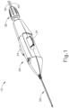
- FIG. 1 shows an exemplary instrument (10) that is configured for use in a procedure for the subretinal administration of a therapeutic agent to an eye of a patient from a suprachoroidal approach.
- Instrument (10) comprises a body (20) and a flexible cannula (50) extending distally from body (20).
- Cannula (50) of the present example has a generally rectangular cross section, though any other suitable cross-sectional profile (e.g., elliptical, etc.) may be used.
- Cannula (50) is generally configured to support a needle (100) that is slidable within cannula (50), as will be described in greater detail below.
- cannula (50) comprises a flexible material such as Polyether block amide (PEBA), which may be manufactured under the trade name PEBAX.
- PEBA Polyether block amide
- cannula (50) has a cross-sectional profile dimension of approximately 2.0 mm by 0.8 mm, with a length of approximately 80 mm. Altematively, any other suitable dimensions may be used.
- cannula (50) is flexible enough to conform to specific structures and contours of the patient's eye, yet cannula (50) has sufficient column strength to permit advancement of cannula (50) between the sclera and choroid of patient's eye without buckling.
- cannula (50) may be configured and operable in accordance with at least some of the teachings of U.S. Pub. No. 2015/0223977 .
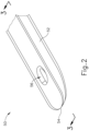
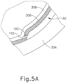
- cannula (50) comprises a body (52), a closed distal end (54), and a lateral opening (56) that is located proximal to distal end (54).
- distal end (54) has a rounded configuration. It should be understood that distal end (54) may have any suitable kind of curvature. It should also be understood that distal end (54) may have any other sui table kind of configuration (e.g., beveled, etc.).
- distal end (54) is configured to provide separation between the sclera and choroid layers to enable cannula (50) to be advanced between such layers while not inflicting trauma to the sclera or choroid layers.
- the region of body (52) that defines lateral opening (56) is beveled, as best seen in FIGS. 3A-3B .
- the edge of lateral opening (S6) may have any other suitable configuration.
- a needle guide (60) is disposed within the hollow interior of cannula (50).
- needle guide (60) may be secured within cannula (50) by a press or interference fit, by adhesives, by mechanical locking mechanisms, and/or in any other suitable fashion.
- Needle guide (60) includes a curved distal end (62) that leads to lateral opening (56) of cannula (50), such that a lumen (64) of needle guide (60) distally terminates at lateral opening (56).
- the portion of needle guide (60) that is proximal to distal end (62) is substantially straight.
- Needle guide (60) may be formed of plastic, stainless steel, and/or any other suitable biocompatible material(s).
- Needle (100) of the present example has a sharp distal tip (102) and defines a lumen (104).
- Distal tip (102) of the present example has a lancet configuration.
- distal tip (102) has a tri-bevel configuration or any other configuration as described in U.S. Pub. No. 2015/0223977 .
- Needle (100) of the present example comprises a stainless steel hypodermic needle that is sized to deliver the therapeutic agent while being small enough to minimize incidental trauma as needle (100) penetrates tissue structures of the patient's eye, as will be described in greater detail below. While stainless steel is used in the present example, it should be understood that any other suitable material(s) may be used, including but not limited to nitinol, etc.
- needle (100) may be 35 gauge with a 100 ⁇ m inner diameter, although other suitable sizes may be used.
- the outer diameter of needle (100) may fall within the range of 27 gauge to 45 gauge: or more particularly within the range of 30 gauge to 42 gauge: or more particularly within the range of 32 gauge to 39 gauge.
- the inner diameter of needle (100) may fall within the range of approximately 50 ⁇ m to approximately 200 ⁇ m; or more particularly within the range of approximately 50 ⁇ m to approximately 150 ⁇ m; or more particularly within the range of approximately 75 ⁇ m to approximately 125 ⁇ m.
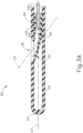
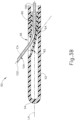
- Needle (100) is slidably disposed within lumen (64) of needle guide (60). Needle guide (60) is generally configured to direct needle (100) upwardly along an exit axis (EA) that is obliquely oriented relative to the longitudinal axis (LA) of cannula (50) through lateral opening (56) of cannula (50). This is shown in the sequence depicted in FIGS. 3A-3B , in which FIG. 3A shows needle (100) in a proximal position (where distal tip (102) of needle (100) is fully contained in lumen (64) of needle guide (60)), and FIG.
- EA exit axis
- FIG. 3B shows needle (100) in a distal position (where distal tip (102) of needle (100) is outside of needle guide (60)). While needle (100) is flexible, needle (100) of the present example, which is not under the scope of the present invention, is resiliently biased to assume a straight configuration.
- the portion of needle (100) that extends outside of cannula (50) and needle guide (60) is substantially straight, extending along exit axis (EA).
- exit axis (EA) exit axis
- exit axis (EA) in FIGS. 3A-3B may be somewhat exaggerated, for illustrative purposes only.
- curved distal end (62) is configured to direct needle (100) along an exit axis (EA) that extends distally from cannula (50) at an angle of approximately 7° to approximately 9° relative to the longitudinal axis (LA) of cannula (50).
- such an angle may be desirable to deflect needle (100) in a direction to ensure penetration of needle into the choroid and to minimize the possibility of needle (100) continuing beneath the choroid through the suprachoroidal space (as opposed to penetrating through the choroid) and the possibility of retinal perforation.
- curved distal portion (88) may urge needle (100) to exit cannula (50) along an exit axis (EA) that is oriented at an angle within the range of approximately 5° to approximately 30° relative to the longitudinal axis (LA) of cannula (50); or more particularly within the range of approximately 5° to approximately 20° relative to the longitudinal axis (LA) of cannula (50); or more particularly within the range of approximately 5° to approximately 10° relative to the longitudinal axis (LA) of cannula (50).
- EA exit axis
- instrument (10) of the present example further comprises an actuation knob (26) located at the proximal end of body (20).
- Actuation knob (26) is rotatable relative to body (20) to thereby selectively translate needle (100) longitudinally relative to cannula (50).
- actuation knob (26) is rotatable in a first angular direction to drive needle (100) distally relative to cannula (50); and in a second angular direction to drive needle (100) proximally relative to cannula (50).
- instrument (10) may provide such functionality through knob (26) in accordance with at least some of the teachings of U.S. Pub. No. 2015/0223977 .
- any other suitable kind of actuation feature(s) may be used to drive needle (100) longitudinally relative to cannula (50).
- knob (26) is rotatable through a complete range of motion that corresponds to advancement of needle (100) to a position relative to cannula (50) to a predetermined amount of penetration within an eye of a patient.
- instrument (10) is configured such that an operator rotates knob (26) until knob (26) can no longer rotate, or until knob (26) begins to slip or "freewheel” in a clutch assembly, to properly position needle (100) within an eye of a patient.
- the predetermined amount of advancement of needle (100) relative to cannula (50) is between approximately 0.25 mm to approximately 10 mm; or more particularly within the range of approximately 0.1 mm to approximately 10 mm; or more particularly within the range of approximately 2 mm to approximately 6 mm, or more particularly to approximately 4 mm.
- instrument (10) may be equipped with certain tactile feedback features to indicate to an operator when needle (100) has been advanced to certain predetermined distances relative to cannula (50). Accordingly, an operator may determine the desired depth of penetration of needle (100) into a patient's eye based on direct visualization of indicia on instrument and/or based on tactile feedback from instrument (10).
- tactile feedback features may be combined with the present example, as will be apparent to those of ordinary skill in the art in view of the teachings herein.
- first supply tube (30) is configured to couple with a source of bleb fluid (340) (e.g., BSS); while second supply tube (40) is configured to couple with a source of therapeutic agent (341).
- BSS bleb fluid
- each fluid supply tube (30, 40) may include a conventional luer feature and/or other structures permitting fluid supply tubes (30, 40) to be coupled with respective fluid sources.
- Fluid supply tubes (30, 40) lead to a valve assembly that includes actuation arms (24). Actuation arms (24) are pivotable to selectively change the state of the valve assembly.
- valve assembly is operable to selectively pinch or otherwise open/close the supply of fluid from fluid supply tubes (30, 40) to lumen (104) of needle (100).
- actuation arms (24) are operable to selectively control the delivery of bleb fluid (340) and therapeutic agent (341) via needle (100).
- the valve assembly may be configured and operable in accordance with at least some of the teachings of U.S. Pub. No. 2015/0223977 .
- Other suitable features and configurations that may be used to control fluid delivery via needle (100) will be apparent to those of ordinary skill in the art in view of the teachings herein.
- instrument (10) may be varied in numerous ways.
- needle (100) may be replaced with needle (200) as described in greater detail below.
- cannula (50) may be replaced with cannula (400) as will be described in greater detail below.
- instrument (10) may be modified in accordance with at least some of the teachings of U.S. Pub. No. 2015/0223977 ; U.S. Pub. No. 2015/0351958 ; U.S. Pub. No. 2015/0351959 ; U.S. Pub. No. 2016/0074212 ; U.S. Pub. No. 2016/0074217 ; U.S. Pub. No. 2016/0074211 ; and/or U.S. Pub. No. 2016/0081849 .
- Other suitable modifications will be apparent to those of ordinary skill in the art in view of the teachings herein.
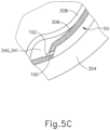
- FIGS. 4A-5C show an exemplary procedure for subretinal delivery of therapeutic agent from a suprachoroidal approach using instrument (10) described above.
- the method described herein may be employed to treat macular degeneration and/or other ocular conditions.
- the procedure described herein is discussed in the context of the treatment of age-related macular degeneration, it should be understood that no such limitation is intended or implied.
- the same techniques described herein may be used to treat retinitis pigmentosa, diabetic retinopathy, and/or other ocular conditions.
- the procedure described herein may be used to treat either dry or wet age-related macular degeneration.
- the procedure begins by an operator immobilizing tissue surrounding a patient's eye (301) (e.g., the eyelids) using a speculum, and/or any other instrument suitable for immobilization. While immobilization described herein with reference to tissue surrounding eye (301), it should be understood that eye (301) itself may remain free to move.
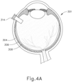
- an eye chandelier port (314) is inserted into eye (301), as shown in FIG. 4A , to provide intraocular illumination when the interior of eye (301) is viewed through the pupil.
- eye chandelier port (314) is positioned in the inferior medial quadrant such that a superior temporal quadrant sclerotomy may be preformed.
- Eye chandelier port (314) is positioned to direct light onto the interior of eye (301) to illuminate at least a portion of the retina (e.g., including at least a portion of the macula). As will be understood, such illumination corresponds to an area of eye (301) that is being targeted for delivery of therapeutic agent.
- FIG. 4A shows a particular positioning of eye chandelier port (314), it should be understood that eye chandelier port (314) may have any other positioning as will be apparent to those of ordinary skill in the art in view of the teachings herein.
- the sclera (304) may be accessed by dissecting the conjunctiva by incising a flap in the conjunctiva and pulling the flap posteriorly. After such a dissection is completed, the exposed surface (305) of the sclera (304) may optionally be blanched using a cautery tool to minimize bleeding. Once conjunctiva dissection is complete, the exposed surface (305) of the sclera (304) may optionally be dried using a WECK-CEL or other suitable absorbent device. A template may then be used to mark eye (301), as described in U.S. Pub. No. 2015/0223977 .
- An operator may then use a visual guide created using the template to attach a suture loop assembly (332) and to perform a sclerotomy, as shown in FIG. 4B , using a conventional scalpel (313) or other suitable cutting instrument.
- the sclerotomy procedure forms a small incision through sclera (304) of eye (301).
- the sclerotomy is preformed with particular care to avoid penetration of the choroid (306).
- the sclerotomy procedure provides access to the space between sclera (304) and choroid (306).
- a blunt dissection may optionally be performed to locally separate sclera (304) from choroid (306).
- Such a dissection may be performed using a small blunt elongate instrument, as will be apparent to those of ordinary skill in the art in view of the teachings herein.
- cannula (50) of instrument (10) may insert cannula (50) of instrument (10) through incision (316) and into the space between sclera (304) and choroid (306).
- cannula (50) is directed through suture loop assembly (332) and into the incision.
- Suture loop assembly (332) may stabilize cannula (50) during insertion.
- suture loop assembly (332) maintains cannula (50) in a generally tangential orientation relative to the incision. Such tangential orientation may reduce trauma as cannula (50) is guided through the incision.
- cannula (50) is inserted into the incision through suture loop assembly (332), an operator may use forceps or other instruments to further guide cannula (50) along an atraumatic path.
- forceps or other instruments is merely optional, and may be omitted in some examples.
- cannula (50) may include one or more markers on the surface of cannula (50) to indicate various depths of insertion. While merely optional, such markers may be desirable to aid an operator in identifying the proper depth of insertion as cannula (50) is guided along an atraumatic path. For instance, the operator may visually observe the position of such markers in relation to suture loop assembly (332) and/or in relation to the incision in the sclera (304) as an indication of the depth to which cannula (50) is inserted in eye (301). By way of example only, one such marker may correspond to an approximately 6 mm depth of insertion of cannula (50).
- an operator may insert an optical fiber (315) into eye chandelier port (314) if the fiber (315) had not yet been inserted at this stage.
- eye chandelier port (314) With eye chandelier port (314) in place and assembled with optical fiber (315), an operator may activate eye chandelier port (314) by directing light through optical fiber (315) to provide illumination of eye (301) and thereby visualize the interior of eye (301). Further adjustments to the positioning of cannula (50) may optionally be made at this point to ensure proper positioning relative to the area of geographic atrophy of retina (308).
- the operator may wish to rotate the eye (301), such as by pulling on suture loop assembly (332), to direct the pupil of the eye (301) toward the operator in order to optimize visualization of the interior of the eye (301) via the pupil.
- FIGS. 4C-4D show cannula (50) as it is guided between sclera (304) and choroid (306) to the delivery site for the therapeutic agent.
- the delivery site corresponds to a generally posterior region of eye (301) adjacent to an area of geographic atrophy of retina (308).
- the delivery site of the present example is superior to the macula, in the potential space between the neurosensory retina and the retinal pigment epithelium layer.
- the operator may rely on direct visualization through a microscope directed through the pupil of eye (301) as cannula (50) is being advanced through the range of motion shown in FIGS. 4C-4D , with illumination provided through fiber (315) and port (314).
- Cannula (50) may be at least partially visible through a retina (308) and choroid (306) of eye (301).
- Visual tracking may be enhanced in versions where an optical fiber is used to emit visible light through the distal end of cannula (50).
- needle (100) of instrument (10) may advance needle (100) of instrument (10) as described above by actuating knob (26).
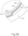
- needle (100) is advanced relative to cannula (50) such that needle (100) pierces through choroid (306) without penetrating retina (308).
- needle (100) may appear under direct visualization as "tenting" the surface of choroid (306).
- needle (100) may deform choroid (306) by pushing upwardly on choroid (306), providing an appearance similar to a tent pole deforming the roof of a tent.
- Such a visual phenomenon may be used by an operator to identify whether choroid (306) is about to be pierced and the location of any eventual piercing.
- the particular amount of needle (100) advancement sufficient to initiate "tenting" and subsequent piercing of choroid (306) may be of any suitable amount as may be determined by a number of factors such as, but not limited to, general patient anatomy, local patient anatomy, operator preference, and/or other factors. As described above, a merely exemplary range of needle (100) advancement may be between approximately 0.25 mm and approximately 10 mm; or more particularly between approximately 2 mm and approximately 6 mm.
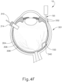
- leading bleb (340) may be desirable for two reasons. First, as shown in FIGS. 4F and 5B , leading bleb (340) may provide a further visual indicator to an operator to indicate when needle (100) is properly positioned at the delivery site.
- leading bleb (340) may provide a barrier between needle (100) and retina (308) once needle (100) has penetrated choroid (306). Such a barrier may push the retinal wall outwardly, thereby minimizing the risk of retinal perforation as needle (100) is advanced to the delivery site.
- a foot pedal is actuated in order to drive leading bleb (340) out from needle (100).
- other suitable features that may be used to drive leading bleb (340) out from needle (100) will be apparent to those of ordinary skill in the art in view of the teachings herein.
- a therapeutic agent (341) may be infused by actuating a syringe or other fluid delivery device as described in various references cited herein.
- the particular therapeutic agent (341) delivered may be any suitable therapeutic agent configured to treat an ocular condition.
- suitable therapeutic agents may include, but are not necessarily limited to, drugs having smaller or large molecules, therapeutic cell solutions, certain gene therapy solutions, tissue plasminogen activators, and/or any other suitable therapeutic agent as will be apparent to those of ordinary skill in the art in view of the teachings herein.
- the therapeutic agent (341) may be provided in accordance with at least some of the teachings of U.S. Patent No. 7,413,734 .
- instrument (10) and variations thereof may be used to provide drainage and/or perform other operations.
- the amount of therapeutic agent (341) that is ultimately delivered to the delivery site is approximately 50uL, although any other suitable amount may be delivered.
- a foot pedal is actuated in order to drive agent 341) out from needle (100).
- other suitable features that may be used to drive agent (341) out from needle (100) will be apparent to those of ordinary skill in the art in view of the teachings herein. Delivery of therapeutic agent (341) may be visualized by an expansion of the pocket of fluid as can be seen in FIGS. 4G and 5C . As shown, therapeutic agent (341) essentially mixes with the fluid of leading bleb (340) as therapeutic agent (341) is injected into the surprachoroidal, subretinal space.
- needle (100) may be retracted by rotating knob (26) in a direction opposite to that used to advance needle (100); and cannula (50) may then be withdrawn from eye (301). It should be understood that because of the size of needle (100), the site where needle (100) penetrated through choroid (306) is self sealing, such that no further steps need be taken to seal the delivery site through choroid (306). Suture loop assembly (332) and chandelier (314) may be removed, and the incision in the sclera (304) may be closed using any suitable conventional techniques.
- the therapeutic agent (341) that is delivered by needle (100) may comprise cells that are derived from postpartum umbilicus and placenta.
- the therapeutic agent (341) may be provided in accordance with at least some of the teachings of U.S. Patent No. 7,413,734 .
- needle (100) may be used to deliver any other suitable substance or substances, in addition to or in lieu of those described in U.S. Patent No. 7,413,734 and/or elsewhere herein.
- therapeutic agent (341) may comprise various kinds of drugs including but not limited to small molecules, large molecules, cells, and/or gene therapies. It should also be understood that macular degeneration is just one merely illustrative example of a condition that may be treated through the procedure described herein. Other biological conditions that may be addressed using the instruments and procedures described herein will be apparent to those of ordinary skill in the art.
- cannula (50) may tend to provide substantial separation between the choroid (306) and the sclera (304) as cannula (50) is inserted between the choroid (306) and the sclera (304).
- the degree of separation may vary from patient to patient (e.g., based on normal anatomical variation and/or based on the patient's disease state, etc.).
- the exit angle (EA) of needle (100) may be insufficient to result in distal tip (102) passing fully through the choroid (306). In other words, needle (100) may continue through the suprachoroidal space without fully penetrating the choroid (306).
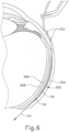
- FIG. 6 shows an exemplary scenario where cannula (50) has elevated the choroid (306) and retina (308) away from the sclera (304) to the point where a substantial gap (305) is defined between the sclera (304) and the choroid (306).
- the exit angle (EA) is oriented such that needle (100) would not penetrate the choroid (306); and further such that needle (100) would eventually engage the sclera (30-4).
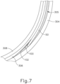
- FIG. 7 shows needle (100) advanced distally along this exit angle (EA). As shown, needle (100) passes tangentially along the choroid (306) without ever breaching the choroid (306). In some other instances, needle (100) may pass partially through the choroid (306) and immediately exit the choroid (306) without ever reaching the subretinal space between the choroid (306) and the retina (308).
- the operator may retract needle (100) proximally, slightly reposition cannula (50) and/or another portion of instrument (10) in order to provide a better orientation for the exit angle (EA), and then try advancing needle (100) distally again. Even with such efforts, it may still be very difficult or even impossible in some cases to successfully penetrate the choroid (306) with needle (100).
- the success rate may be highly dependent on the skill of the operator, and the repositioning efforts will add time to the procedure.
- the repositioning may increase the risk of tissue trauma, increase the risk of bleb collapse, and/or increase the risk of cell egress into the suprachoroidal space.
- needle guide (60) may be modified to provide a steeper exit angle (EA).
- this kind of modification may be unsuitable for many patients.
- increasing the exit angle (EA) by providing a more pronounced bend in distal end (62) of needle guide (60) may increase the risk of needle (100) perforating the retina (308) in some patients, particularly in those where the gap (305) created by cannula (50) between the sclera (304) and the choroid (306) is less pronounced than the gap (305) shown in FIGS. 6-7 ; including cases where the gap (305) is non-existent.
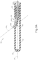
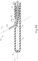
- FIG. 8 shows an exemplary alternative needle (200) that may be incorporated into instrument (10) in place of needle (100).
- needle (200) may be substituted for needle (100) without modifying any other aspects of instrument (10).
- Needle (200) of the present example has a distal tip (202) that is configured and operable just like distal tip (102) described above.
- needle (200) also defines a lumen (204) that is configured and operable just like lumen (104) described above.
- needle (200) of the present example includes a substantially straight proximal portion (210), a substantially straight distal portion (212), and a bent portion (214) located between proximal and distal portions (210, 212).
- needle (200) is formed of nitinol, though it should be understood that any other suitable material(s) (e.g., stainless steel, etc.) may be used.
- Needle (200) is configured to provide bent portion (214) as a preformed feature, such that needle (200) is resiliently biased to assume the configuration shown in FIG. 6 .
- bent portion (214) may be configured to have a constant radius of curvature between approximately 4 mm and approximately 15 mm; a constant radius of curvature between approximately 7 mm and approximately 12 mm; a constant radius of curvature between approximately 8 mm and approximately 11 mm; or a constant radius of curvature between approximately 9 mm and approximately 10 mm.
- bent portion (214) has a radius of curvature of approximately 10.5 mm. In some other versions, bent portion (214) has a radius of curvature of approximately 10.0 mm.
- bent portion (214) has a radius of curvature of approximately 9.5 mm. It should be understood that the radius of curvature must be carefully selected because if the radius is too small, there may be an increased risk of perforating the retina (308); and if the radius is too large, the needle (200) may still fail to fully penetrate the choroid (306).
- radius of curvature of bent portion (214) is constant in the present example, in some other versions the radius of curvature may be variable.
- some variations of needle (200) may provide a larger radius of curvature in a region of needle (200) that remains disposed in cannula (50), even when needle (200) is in a distally extended position; with a smaller radius of curvature in a region of needle (200) that extends distally from cannula (50) when needle (200) is in a distally extended position.
- This kind of configuration may impart a slight precurvature to cannula (50), which may further assist in cannula (50) conforming to the curved inner wall of sclera (304), which may in turn reduce the occurrence (or magnitude) of gap (305).
- needle (200) is slidably disposed in needle guide (60) within cannula (50). While FIG. 9A shows needle (200) in a partially advanced state, it should be understood that needle (200) may be retracted further proximally in needle guide (60) such that distal tip (202) does not protrude through lateral opening (56). As shown in FIG. 9A , as needle (200) begins to exit cannula (50) via lateral opening (56), the distally protruding portion of needle (200) is oriented along a first exit axis (EA 1 ). At this stage, bent portion (214) and part of distal portion (212) are still contained within needle guide (60), such that needle guide (60) prevents needle (200) from reaching the configuration shown in FIG. 8 .
- EA 1 first exit axis
- Second exit axis (EA 2 ) defines an angle with the longitudinal axis (LA) that is larger than the angle defined between first exit axis (EA 1 ) and the longitudinal axis (LA).
- needle (200) may be particularly useful in cases where cannula creates a substantial gap (305) between the sclera (304) and the choroid (306). It should be understood that the gap (305) in FIG. 10 is substantially the same as the gap (305) in FIG. 7 . As noted above, due to the gap (305) in FIG. 7 and the associated relationships between the anatomical structures and the instrument (10) structures, needle (100) is unable to penetrate choroid (306). However, as shown in FIG. 10 , the curvature of needle (200) allows needle (200) to penetrate choroid (306) despite the presence of gap (305) and the associated relationships between the anatomical structures and the instrument (10) structures.
- the exit angle (EA) of needle (200) varies based on the extent to which needle (200) is extended from cannula (50). It should be understood that this variation in the exit angle (EA) will allow the operator to control the optimal exit angle (EA) by controlling the amount of needle (200) extension. This may allow for shallower angles (less extension) for some patients and steeper angles (more extension) for other patients, to more consistently be able to achieve penetration of the choroid (306) in a relatively safe and efficient manner, eliminating the need for other mitigations or workarounds that would otherwise be required from the scenario depicted in FIG. 7 .
- cannula (50) includes a closed distal end (54) and a lateral opening (56) that is located proximal to distal end (54).
- FIG. 11A shows an exemplary alternative cannula (400) that may be readily incorporated into instrument (10) in place of cannula (50).
- Cannula (400) of this example has a flexible body (402) and a distal opening (406).
- Distal opening (406) is coaxially positioned on the longitudinal axis of cannula (400) in this example. In some other versions, distal opening (406) is offset from the longitudinal axis of cannula (400).
- cannula (400) may be formed of Polyether block amide (PEBA) and/or any other suitable kind(s) of material(s).
- PEBA Polyether block amide
- cannula (400) of the present example has sufficient column strength to be advanced distally between the sclera (306) and choroid (308) of patient's eye without buckling.
- Insert (408) is positioned within cannula (400). Insert (408) may be secured within cannula (400) by a press or interference fit, by adhesives, by mechanical locking mechanisms, and/or in any other suitable fashion.
- insert (408) is formed of a polyimide material, though it should be understood that any other suitable biocompatible material(s) may be used.
- Insert (408) of the present example is substantially straight yet may bend with cannula (400).
- Needle (200) is slidably disposed in a lumen (410) defined by insert (408). When needle (200) is in a proximal position as shown in FIG. 11A , distal tip (202) of needle (200) is fully contained within lumen (410).
- insert (408) constrains needle (200) such that needle (200) is held under stress in a substantially straight configuration.
- distal tip (202) of needle is positioned distally of cannula (400).
- curved portion (214) is exposed such that the distal portion (212) of needle (200) is oriented along an exit axis that is oblique io the longitudinal axis of cannula (400). It should be understood that this configuration and orientation may position distal tip (202) at the subretinal space (i.e., between the choroid (306) and the retina 308)).
- any of the versions of the instruments described herein may include various other features in addition to or in lieu of those described above.
- any of the devices herein may also include one or more of the various features disclosed in any of the various references mentioned herein.
- Versions described above may be designed to be disposed of after a single use, or they can be designed to be used multiple times. Versions may, in either or both cases, be reconditioned for reuse after at least one use. Reconditioning may include any combination of the steps of disassembly of the device, followed by cleaning or replacement of particular pieces, and subsequent reassembly. In particular, some versions of the device may be disassembled, and any number of the particular pieces or parts of the device may be selectively replaced or removed in any combination. Upon cleaning and/or replacement of particular parts, some versions of the device may be reassembled for subsequent use either at a reconditioning facility, or by an operator immediately prior to a procedure.
- reconditioning of a device may utilize a variety of techniques for disassembly, cleaning/replacement, and reassembly. Use of such techniques, and the resulting reconditioned device, are all within the scope of the present application.
- versions described herein may be sterilized before and/or after a procedure.
- the device is placed in a closed and sealed container, such as a plastic or TYVEK bag.
- the container and device may then be placed in a field of radiation that can penetrate the container, such as gamma radiation, x-rays, or high-energy electrons.
- the radiation may kill bacteria on the device and in the container.
- the sterilized device may then be stored in the sterile container for later use.
- a device may also be sterilized using any other technique known in the art, including but not limited to beta or gamma radiation, ethylene oxide, or steam.
Landscapes
- Health & Medical Sciences (AREA)
- Life Sciences & Earth Sciences (AREA)
- Public Health (AREA)
- Veterinary Medicine (AREA)
- Biomedical Technology (AREA)
- Heart & Thoracic Surgery (AREA)
- General Health & Medical Sciences (AREA)
- Engineering & Computer Science (AREA)
- Animal Behavior & Ethology (AREA)
- Vascular Medicine (AREA)
- Ophthalmology & Optometry (AREA)
- Hematology (AREA)
- Anesthesiology (AREA)
- Biophysics (AREA)
- Pulmonology (AREA)
- Surgery (AREA)
- Nuclear Medicine, Radiotherapy & Molecular Imaging (AREA)
- Physics & Mathematics (AREA)
- Optics & Photonics (AREA)
- Infusion, Injection, And Reservoir Apparatuses (AREA)
- Medicines That Contain Protein Lipid Enzymes And Other Medicines (AREA)
- Medicinal Preparation (AREA)
Applications Claiming Priority (3)
| Application Number | Priority Date | Filing Date | Title |
|---|---|---|---|
| US201662305767P | 2016-03-09 | 2016-03-09 | |
| US15/438,918 US10478553B2 (en) | 2016-03-09 | 2017-02-22 | Apparatus for subretinal administration of therapeutic agent via a curved needle |
| PCT/US2017/021508 WO2017156227A1 (en) | 2016-03-09 | 2017-03-09 | Apparatus for subretinal administration of therapeutic agent via a curved needle |
Publications (3)
| Publication Number | Publication Date |
|---|---|
| EP3426203A1 EP3426203A1 (en) | 2019-01-16 |
| EP3426203C0 EP3426203C0 (en) | 2025-05-28 |
| EP3426203B1 true EP3426203B1 (en) | 2025-05-28 |
Family
ID=59788385
Family Applications (1)
| Application Number | Title | Priority Date | Filing Date |
|---|---|---|---|
| EP17712635.6A Active EP3426203B1 (en) | 2016-03-09 | 2017-03-09 | Apparatus for subretinal administration of therapeutic agent via a curved needle |
Country Status (14)
| Country | Link |
|---|---|
| US (3) | US10478553B2 (enExample) |
| EP (1) | EP3426203B1 (enExample) |
| JP (3) | JP7023873B2 (enExample) |
| KR (3) | KR102608333B1 (enExample) |
| CN (2) | CN113616417B (enExample) |
| AU (1) | AU2017229763B2 (enExample) |
| BR (1) | BR112018067896A2 (enExample) |
| CA (2) | CA3175975A1 (enExample) |
| ES (1) | ES3031816T3 (enExample) |
| IL (1) | IL261251B (enExample) |
| RU (1) | RU2740165C2 (enExample) |
| SA (1) | SA518392352B1 (enExample) |
| SG (2) | SG11201807191YA (enExample) |
| WO (1) | WO2017156227A1 (enExample) |
Families Citing this family (25)
| Publication number | Priority date | Publication date | Assignee | Title |
|---|---|---|---|---|
| US10478553B2 (en) | 2016-03-09 | 2019-11-19 | Orbit Biomedical Limited | Apparatus for subretinal administration of therapeutic agent via a curved needle |
| US10646374B2 (en) | 2016-06-17 | 2020-05-12 | Orbit Biomedical Limited | Apparatus and method to form entry bleb for subretinal delivery of therapeutic agent |
| US10806629B2 (en) | 2016-06-17 | 2020-10-20 | Gyroscope Therapeutics Limited | Injection device for subretinal delivery of therapeutic agent |
| US11076984B2 (en) | 2017-03-13 | 2021-08-03 | Gyroscope Therapeutics Limited | Method of performing subretinal drainage and agent delivery |
| US11033427B2 (en) | 2017-04-13 | 2021-06-15 | Alcon Inc. | Vitreoretinal instruments for fluid aspiration |
| GB201714392D0 (en) | 2017-09-07 | 2017-10-25 | Marsteller Laurence | Methods and devices for treating glaucoma |
| US10960147B2 (en) * | 2018-04-20 | 2021-03-30 | Flextronics Ap, Llc | Flex needle |
| EP3829501B1 (en) * | 2018-08-03 | 2025-12-31 | The Johns Hopkins University | RETINAL IMPLANTATION DEVICE |
| WO2020069217A1 (en) * | 2018-09-28 | 2020-04-02 | Radiance Therapeutics, Inc. | Methods, systems, and compositions for maintaining functioning drainage blebs associated with minimally invasive micro sclerostomy |
| EP3920859B1 (en) * | 2019-02-06 | 2025-09-03 | The Johns Hopkins University | External and internal intraoperative optical coherence tomography imaging for subretinal material delivery |
| US11759355B1 (en) | 2019-02-26 | 2023-09-19 | Gyroscope Therapeutics Limited | Method of delivering leading blebs and agent to subretinal space |
| EP4041147A1 (en) | 2019-10-11 | 2022-08-17 | Gyroscope Therapeutics Limited | Dose clip assembly for syringe |
| WO2022136913A1 (en) | 2020-12-22 | 2022-06-30 | Gyroscope Therapeutics Limited | Ocular cannula guide |
| USD1076086S1 (en) | 2021-11-23 | 2025-05-20 | Radiance Therapeutics, Inc. | Opthalmic brachytherapy device |
| USD1076085S1 (en) | 2021-11-23 | 2025-05-20 | Radiance Therapeutics, Inc. | Opthalmic brachytherapy device |
| KR20250065626A (ko) | 2022-09-06 | 2025-05-13 | 제넨테크, 인크. | 이중-곡선 니들을 통해 치료제를 망막하 투여하기 위한 장치 |
| KR20250138741A (ko) | 2023-01-13 | 2025-09-22 | 제넨테크, 인크. | 주사기용 용량 도킹 장치 |
| WO2024249805A1 (en) * | 2023-05-31 | 2024-12-05 | The Johns Hopkins University | Methods and systems for accessing anatomical spaces |
| US20240423836A1 (en) * | 2023-06-22 | 2024-12-26 | Alcon Inc. | Methods and apparatus for subretinal delivery |
| AU2024311612A1 (en) * | 2023-06-22 | 2025-11-13 | Alcon Inc. | Methods and apparatus for subretinal delivery |
| US20240423835A1 (en) * | 2023-06-22 | 2024-12-26 | Alcon Inc. | Methods and apparatus for subretinal delivery |
| TW202529711A (zh) | 2023-09-29 | 2025-08-01 | 美商建南德克公司 | 眼插管導引器、施用器及標記器械 |
| WO2025207699A1 (en) | 2024-03-27 | 2025-10-02 | Genentech, Inc. | Torque ring for subretinal injection device |
| WO2025217214A2 (en) | 2024-04-08 | 2025-10-16 | Regenxbio Inc. | Recombinant adeno-associated viruses and uses thereof |
| WO2025217230A1 (en) | 2024-04-08 | 2025-10-16 | Regenxbio Inc. | Vectorized anti-complement antibodies and complement agents and administration thereof |
Citations (3)
| Publication number | Priority date | Publication date | Assignee | Title |
|---|---|---|---|---|
| US6190353B1 (en) * | 1995-10-13 | 2001-02-20 | Transvascular, Inc. | Methods and apparatus for bypassing arterial obstructions and/or performing other transvascular procedures |
| US20040138562A1 (en) * | 2002-01-17 | 2004-07-15 | Joshua Makower | Devices, systems and methods for acute or chronic delivery of substances or apparatus to extravascular treatment sites |
| WO2015098645A1 (ja) * | 2013-12-24 | 2015-07-02 | 公立大学法人横浜市立大学 | 注射針 |
Family Cites Families (63)
| Publication number | Priority date | Publication date | Assignee | Title |
|---|---|---|---|---|
| US5273530A (en) | 1990-11-14 | 1993-12-28 | The University Of Rochester | Intraretinal delivery and withdrawal instruments |
| DE4235506A1 (de) * | 1992-10-21 | 1994-04-28 | Bavaria Med Tech | Katheter zur Injektion von Arzneimitteln |
| US5964740A (en) * | 1996-07-09 | 1999-10-12 | Asahi Kogaku Kogyo Kabushiki Kaisha | Treatment accessory for an endoscope |
| CN1204242A (zh) * | 1995-10-13 | 1999-01-06 | 血管转换公司 | 为动脉阻塞物加设旁路和/或其他经血管操作的方法和装置 |
| DE19741487C2 (de) | 1997-09-19 | 2000-08-31 | Univ Eberhard Karls | Vorrichtung für einen Zugang in den Subretinalraum eines Auges |
| US6231546B1 (en) * | 1998-01-13 | 2001-05-15 | Lumend, Inc. | Methods and apparatus for crossing total occlusions in blood vessels |
| JP3055504U (ja) * | 1998-07-02 | 1999-01-22 | 株式会社八光電機製作所 | 眼球針 |
| US6217554B1 (en) * | 1999-02-12 | 2001-04-17 | Pharmaspec Corporation | Methods and apparatus for delivering substances into extravascular tissue |
| US6368315B1 (en) | 1999-06-23 | 2002-04-09 | Durect Corporation | Composite drug delivery catheter |
| US6413245B1 (en) * | 1999-10-21 | 2002-07-02 | Alcon Universal Ltd. | Sub-tenon drug delivery |
| US6989004B2 (en) * | 2001-02-28 | 2006-01-24 | Rex Medical, L.P. | Apparatus for delivering ablation fluid to treat lesions |
| AU2003213075A1 (en) | 2002-02-14 | 2003-09-04 | Henry J. Kaplan | Subretinal implantation device and surgical cannulas for use therewith |
| US7316676B2 (en) * | 2002-08-20 | 2008-01-08 | Gholam A. Peyman | Treatment of retinal detachment |
| JP2006507368A (ja) | 2002-09-29 | 2006-03-02 | サーモディックス,インコーポレイティド | ステロイド含有治療剤の網膜下投与方法;脈絡膜及び網膜に薬力学作用を局在化するための方法;並びに網膜疾患の治療及び/又は予防のための関連する方法 |
| US20040199130A1 (en) * | 2003-04-03 | 2004-10-07 | Chornenky Victor I. | Apparatus and method for treatment of macular degeneration |
| JP4289919B2 (ja) * | 2003-05-06 | 2009-07-01 | 朝日インテック株式会社 | 薬液注入装置 |
| IL156582A0 (en) * | 2003-06-22 | 2004-01-04 | Sergey Popov | Safety trocar obturator |
| AU2004252568B2 (en) | 2003-06-27 | 2011-06-30 | Ethicon, Incorporated | Regeneration and repair of neural tissue using postpartum-derived cells |
| GB0327136D0 (en) * | 2003-11-21 | 2003-12-24 | Nmt Group Plc | Safety needle |
| KR101195052B1 (ko) | 2004-01-23 | 2012-10-29 | 아이싸이언스 인터벤셔날 코포레이션 | 복합체 안과 마이크로캐뉼라 |
| KR20070036044A (ko) * | 2004-04-29 | 2007-04-02 | 아이싸이언스 인터벤셔날 코포레이션 | 안과 치료를 위한 장치와 방법 |
| US20080058704A1 (en) | 2004-04-29 | 2008-03-06 | Michael Hee | Apparatus and Method for Ocular Treatment |
| WO2007002304A2 (en) * | 2005-06-22 | 2007-01-04 | Vnus Medical Technologies, Inc. | Methods and apparatus for introducing tumescent fluid to body tissue |
| US20070202186A1 (en) | 2006-02-22 | 2007-08-30 | Iscience Interventional Corporation | Apparatus and formulations for suprachoroidal drug delivery |
| US8197435B2 (en) | 2006-05-02 | 2012-06-12 | Emory University | Methods and devices for drug delivery to ocular tissue using microneedle |
| US20080004596A1 (en) * | 2006-05-25 | 2008-01-03 | Palo Alto Institute | Delivery of agents by microneedle catheter |
| KR20090057984A (ko) * | 2006-09-19 | 2009-06-08 | 더 트러스티이스 오브 콜롬비아 유니버시티 인 더 시티 오브 뉴욕 | 중공형 해부학적 부유 기관 상에서의 수술을 위한 시스템, 디바이스 및 방법 |
| US20080281292A1 (en) | 2006-10-16 | 2008-11-13 | Hickingbotham Dyson W | Retractable Injection Port |
| US8002743B2 (en) * | 2007-06-15 | 2011-08-23 | Kyphon Sarl | Systems and methods for needle access to an intervertebral disc |
| EP2227257B1 (en) * | 2008-01-07 | 2013-06-26 | Salutaris Medical Devices, Inc. | Devices for minimally-invasive extraocular delivery of radiation to the posterior portion of the eye |
| WO2009089409A2 (en) | 2008-01-10 | 2009-07-16 | Bausch & Lomb Incorporated | Intravitreal injection system having coaxial cannulae and use thereof |
| US8021332B2 (en) * | 2008-12-04 | 2011-09-20 | Abbott Cardiovascular Systems Inc. | Agent delivery catheter having articulating arms |
| US8425473B2 (en) | 2009-01-23 | 2013-04-23 | Iscience Interventional Corporation | Subretinal access device |
| WO2010132751A1 (en) | 2009-05-15 | 2010-11-18 | Iscience Interventional Corporation | Methods and apparatus for sub-retinal catheterization |
| WO2010141837A1 (en) * | 2009-06-05 | 2010-12-09 | Vance Products Incorporated, D/B/A/ Cook Urological Incorporated | Access sheath and needle assembly for delivering therapeutic material |
| CN106214321B (zh) | 2010-10-15 | 2018-08-28 | 科尼尔赛德生物医学公司 | 用于进入眼睛的装置 |
| GB2487899A (en) * | 2011-02-01 | 2012-08-15 | Olberon Ltd | Needle holder with grip means |
| JP5717182B2 (ja) * | 2011-02-25 | 2015-05-13 | Hoya株式会社 | 眼科手術器具 |
| US8852165B2 (en) * | 2011-06-16 | 2014-10-07 | II Edward G. Mackay | Endoluminal drug delivery devices and methods |
| US9414816B2 (en) * | 2011-06-23 | 2016-08-16 | Devicor Medical Products, Inc. | Introducer for biopsy device |
| US8938285B2 (en) * | 2011-08-08 | 2015-01-20 | Devicor Medical Products, Inc. | Access chamber and markers for biopsy device |
| US9370447B2 (en) * | 2011-10-10 | 2016-06-21 | Cygnus LP | Probes for use in ophthalmic and vitreoretinal surgery |
| WO2013059737A2 (en) * | 2011-10-19 | 2013-04-25 | Nitinol Devices And Components, Inc. | Tissue treatment device and related methods |
| ES2533576T3 (es) * | 2011-12-30 | 2015-04-13 | Q-Med Ab | Cánula sin hematomas |
| WO2013123309A1 (en) * | 2012-02-15 | 2013-08-22 | The Cleveland Clinic Foundation | Catheter assembly and method of treating a vascular disease |
| US20150209180A1 (en) | 2012-08-27 | 2015-07-30 | Clearside Biomedical, Inc. | Apparatus and Methods for Drug Delivery Using Microneedles |
| US9301795B2 (en) * | 2012-10-29 | 2016-04-05 | Ablative Solutions, Inc. | Transvascular catheter for extravascular delivery |
| EP3721872B1 (en) | 2012-11-08 | 2025-01-22 | Clearside Biomedical Inc. | Methods for the treatment of ocular disease in human subjects |
| KR102086103B1 (ko) * | 2013-11-14 | 2020-03-06 | 아큐시스, 인코포레이티드 | 안구내 션트 삽입기 |
| US10010447B2 (en) * | 2013-12-18 | 2018-07-03 | Novartis Ag | Systems and methods for subretinal delivery of therapeutic agents |
| EP4555981A3 (en) * | 2014-02-12 | 2025-08-06 | Genentech, Inc. | Method and apparatus for suprachoroidal administration of therapeutic agent |
| US9949874B2 (en) * | 2014-06-06 | 2018-04-24 | Janssen Biotech, Inc. | Therapeutic agent delivery device with convergent lumen |
| US9925088B2 (en) | 2014-06-06 | 2018-03-27 | Janssen Biotech, Inc. | Sub-retinal tangential needle catheter guide and introducer |
| CA2952822A1 (en) | 2014-06-17 | 2015-12-23 | Clearside Biomedical, Inc. | Methods and devices for treating posterior ocular disorders |
| MX2016017028A (es) | 2014-06-20 | 2017-08-07 | Clearside Biomedical Inc | Canula de diametro variable y metodos para el control de la profundidad de insercion para administracion de medicamentos. |
| US9949875B1 (en) * | 2014-07-08 | 2018-04-24 | Jeffrey N. Weiss | Retinal and optic nerve autologous bone-marrow derived stem cell surgery |
| US10456557B2 (en) * | 2014-08-14 | 2019-10-29 | Invatec S.P.A. | Occlusion bypassing apparatus with varying flexibility and methods for bypassing an occlusion in a blood vessel |
| US10064752B2 (en) | 2014-09-11 | 2018-09-04 | Orbit Biomedical Limited | Motorized suprachoroidal injection of therapeutic agent |
| US10219936B2 (en) | 2014-09-11 | 2019-03-05 | Orbit Biomedical Limited | Therapeutic agent delivery device with advanceable cannula and needle |
| US10322028B2 (en) | 2014-09-11 | 2019-06-18 | Orbit Biomedical Limited | Method and apparatus for sensing position between layers of an eye |
| US10258502B2 (en) | 2014-09-18 | 2019-04-16 | Orbit Biomedical Limited | Therapeutic agent delivery device |
| GB201516066D0 (en) | 2015-09-10 | 2015-10-28 | Young & Co Llp D | Treatment of retinitis pigmentosa |
| US10478553B2 (en) * | 2016-03-09 | 2019-11-19 | Orbit Biomedical Limited | Apparatus for subretinal administration of therapeutic agent via a curved needle |
-
2017
- 2017-02-22 US US15/438,918 patent/US10478553B2/en active Active
- 2017-03-09 SG SG11201807191YA patent/SG11201807191YA/en unknown
- 2017-03-09 CN CN202110993165.3A patent/CN113616417B/zh active Active
- 2017-03-09 WO PCT/US2017/021508 patent/WO2017156227A1/en not_active Ceased
- 2017-03-09 BR BR112018067896-1A patent/BR112018067896A2/pt not_active Application Discontinuation
- 2017-03-09 JP JP2018567011A patent/JP7023873B2/ja active Active
- 2017-03-09 RU RU2018135286A patent/RU2740165C2/ru active
- 2017-03-09 SG SG10202008679RA patent/SG10202008679RA/en unknown
- 2017-03-09 AU AU2017229763A patent/AU2017229763B2/en active Active
- 2017-03-09 CA CA3175975A patent/CA3175975A1/en active Pending
- 2017-03-09 ES ES17712635T patent/ES3031816T3/es active Active
- 2017-03-09 CN CN201780016408.XA patent/CN108712894B/zh active Active
- 2017-03-09 KR KR1020217001164A patent/KR102608333B1/ko active Active
- 2017-03-09 KR KR1020187028153A patent/KR102205295B1/ko active Active
- 2017-03-09 EP EP17712635.6A patent/EP3426203B1/en active Active
- 2017-03-09 CA CA3015529A patent/CA3015529C/en active Active
- 2017-03-09 KR KR1020237040321A patent/KR20230163593A/ko not_active Ceased
-
2018
- 2018-08-20 IL IL261251A patent/IL261251B/en unknown
- 2018-09-05 SA SA518392352A patent/SA518392352B1/ar unknown
-
2019
- 2019-10-11 US US16/599,206 patent/US11338084B2/en active Active
-
2021
- 2021-11-02 JP JP2021179357A patent/JP2022017454A/ja active Pending
-
2022
- 2022-05-02 US US17/734,162 patent/US20220288306A1/en not_active Abandoned
-
2023
- 2023-09-21 JP JP2023155577A patent/JP2023169335A/ja active Pending
Patent Citations (4)
| Publication number | Priority date | Publication date | Assignee | Title |
|---|---|---|---|---|
| US6190353B1 (en) * | 1995-10-13 | 2001-02-20 | Transvascular, Inc. | Methods and apparatus for bypassing arterial obstructions and/or performing other transvascular procedures |
| US20040138562A1 (en) * | 2002-01-17 | 2004-07-15 | Joshua Makower | Devices, systems and methods for acute or chronic delivery of substances or apparatus to extravascular treatment sites |
| WO2015098645A1 (ja) * | 2013-12-24 | 2015-07-02 | 公立大学法人横浜市立大学 | 注射針 |
| EP3088027A1 (en) * | 2013-12-24 | 2016-11-02 | Yokohama City University | Injection needle |
Also Published As
Similar Documents
| Publication | Publication Date | Title |
|---|---|---|
| US20220288306A1 (en) | Apparatus for subretinal administration of therapeutic agent via a curved needle | |
| US20250295522A1 (en) | Method and apparatus for suprachoroidal administration of therapeutic agent | |
| US11723798B2 (en) | Sub-retinal tangential needle catheter guide and introducer | |
| EP3191031B1 (en) | Therapeutic agent delivery device with advanceable cannula and needle | |
| EP3191033B1 (en) | Motorized suprachoroidal injection of therapeutic agent | |
| US11759355B1 (en) | Method of delivering leading blebs and agent to subretinal space | |
| US20250195268A1 (en) | Apparatus for subretinal administration of therapeutic agent via dual-curved needle | |
| HK40047835B (en) | Apparatus for suprachoroidal administration of therapeutic agent | |
| HK40047835A (en) | Apparatus for suprachoroidal administration of therapeutic agent |
Legal Events
| Date | Code | Title | Description |
|---|---|---|---|
| STAA | Information on the status of an ep patent application or granted ep patent |
Free format text: STATUS: UNKNOWN |
|
| STAA | Information on the status of an ep patent application or granted ep patent |
Free format text: STATUS: THE INTERNATIONAL PUBLICATION HAS BEEN MADE |
|
| PUAI | Public reference made under article 153(3) epc to a published international application that has entered the european phase |
Free format text: ORIGINAL CODE: 0009012 |
|
| STAA | Information on the status of an ep patent application or granted ep patent |
Free format text: STATUS: REQUEST FOR EXAMINATION WAS MADE |
|
| 17P | Request for examination filed |
Effective date: 20181009 |
|
| AK | Designated contracting states |
Kind code of ref document: A1 Designated state(s): AL AT BE BG CH CY CZ DE DK EE ES FI FR GB GR HR HU IE IS IT LI LT LU LV MC MK MT NL NO PL PT RO RS SE SI SK SM TR |
|
| AX | Request for extension of the european patent |
Extension state: BA ME |
|
| DAV | Request for validation of the european patent (deleted) | ||
| DAX | Request for extension of the european patent (deleted) | ||
| STAA | Information on the status of an ep patent application or granted ep patent |
Free format text: STATUS: EXAMINATION IS IN PROGRESS |
|
| 17Q | First examination report despatched |
Effective date: 20200211 |
|
| RAP1 | Party data changed (applicant data changed or rights of an application transferred) |
Owner name: GYROSCOPE THERAPEUTICS LIMITED |
|
| RAP3 | Party data changed (applicant data changed or rights of an application transferred) |
Owner name: GYROSCOPE THERAPEUTICS LIMITED |
|
| RAP1 | Party data changed (applicant data changed or rights of an application transferred) |
Owner name: GENENTECH, INC. |
|
| GRAP | Despatch of communication of intention to grant a patent |
Free format text: ORIGINAL CODE: EPIDOSNIGR1 |
|
| STAA | Information on the status of an ep patent application or granted ep patent |
Free format text: STATUS: GRANT OF PATENT IS INTENDED |
|
| INTG | Intention to grant announced |
Effective date: 20250103 |
|
| RIN1 | Information on inventor provided before grant (corrected) |
Inventor name: KEANE, MICHAEL, F. Inventor name: OBERKIRCHER, BRENDAN, J. Inventor name: PRICE, DANIEL, W. Inventor name: KHAN, ISAAC, J. Inventor name: KO, BENJAMIN, L. Inventor name: MEYER, THOMAS, E. |
|
| GRAS | Grant fee paid |
Free format text: ORIGINAL CODE: EPIDOSNIGR3 |
|
| GRAA | (expected) grant |
Free format text: ORIGINAL CODE: 0009210 |
|
| STAA | Information on the status of an ep patent application or granted ep patent |
Free format text: STATUS: THE PATENT HAS BEEN GRANTED |
|
| AK | Designated contracting states |
Kind code of ref document: B1 Designated state(s): AL AT BE BG CH CY CZ DE DK EE ES FI FR GB GR HR HU IE IS IT LI LT LU LV MC MK MT NL NO PL PT RO RS SE SI SK SM TR |
|
| REG | Reference to a national code |
Ref country code: GB Ref legal event code: FG4D |
|
| REG | Reference to a national code |
Ref country code: CH Ref legal event code: EP |
|
| REG | Reference to a national code |
Ref country code: DE Ref legal event code: R096 Ref document number: 602017089661 Country of ref document: DE |
|
| REG | Reference to a national code |
Ref country code: IE Ref legal event code: FG4D |
|
| REG | Reference to a national code |
Ref country code: ES Ref legal event code: FG2A Ref document number: 3031816 Country of ref document: ES Kind code of ref document: T3 Effective date: 20250711 |
|
| U01 | Request for unitary effect filed |
Effective date: 20250613 |
|
| U07 | Unitary effect registered |
Designated state(s): AT BE BG DE DK EE FI FR IT LT LU LV MT NL PT RO SE SI Effective date: 20250626 |
|
| PG25 | Lapsed in a contracting state [announced via postgrant information from national office to epo] |
Ref country code: GR Free format text: LAPSE BECAUSE OF FAILURE TO SUBMIT A TRANSLATION OF THE DESCRIPTION OR TO PAY THE FEE WITHIN THE PRESCRIBED TIME-LIMIT Effective date: 20250829 Ref country code: NO Free format text: LAPSE BECAUSE OF FAILURE TO SUBMIT A TRANSLATION OF THE DESCRIPTION OR TO PAY THE FEE WITHIN THE PRESCRIBED TIME-LIMIT Effective date: 20250828 |
|
| PG25 | Lapsed in a contracting state [announced via postgrant information from national office to epo] |
Ref country code: PL Free format text: LAPSE BECAUSE OF FAILURE TO SUBMIT A TRANSLATION OF THE DESCRIPTION OR TO PAY THE FEE WITHIN THE PRESCRIBED TIME-LIMIT Effective date: 20250528 |
|
| PG25 | Lapsed in a contracting state [announced via postgrant information from national office to epo] |
Ref country code: HR Free format text: LAPSE BECAUSE OF FAILURE TO SUBMIT A TRANSLATION OF THE DESCRIPTION OR TO PAY THE FEE WITHIN THE PRESCRIBED TIME-LIMIT Effective date: 20250528 |
|
| PG25 | Lapsed in a contracting state [announced via postgrant information from national office to epo] |
Ref country code: RS Free format text: LAPSE BECAUSE OF FAILURE TO SUBMIT A TRANSLATION OF THE DESCRIPTION OR TO PAY THE FEE WITHIN THE PRESCRIBED TIME-LIMIT Effective date: 20250828 |
|
| PG25 | Lapsed in a contracting state [announced via postgrant information from national office to epo] |
Ref country code: IS Free format text: LAPSE BECAUSE OF FAILURE TO SUBMIT A TRANSLATION OF THE DESCRIPTION OR TO PAY THE FEE WITHIN THE PRESCRIBED TIME-LIMIT Effective date: 20250928 |