EP3405566B1 - Stem cells for healing of skin ulcers - Google Patents
Stem cells for healing of skin ulcers Download PDFInfo
- Publication number
- EP3405566B1 EP3405566B1 EP16886758.8A EP16886758A EP3405566B1 EP 3405566 B1 EP3405566 B1 EP 3405566B1 EP 16886758 A EP16886758 A EP 16886758A EP 3405566 B1 EP3405566 B1 EP 3405566B1
- Authority
- EP
- European Patent Office
- Prior art keywords
- cells
- wound
- cell
- use according
- mice
- Prior art date
- Legal status (The legal status is an assumption and is not a legal conclusion. Google has not performed a legal analysis and makes no representation as to the accuracy of the status listed.)
- Active
Links
Images


Classifications
-
- A—HUMAN NECESSITIES
- A61—MEDICAL OR VETERINARY SCIENCE; HYGIENE
- A61K—PREPARATIONS FOR MEDICAL, DENTAL OR TOILETRY PURPOSES
- A61K35/00—Medicinal preparations containing materials or reaction products thereof with undetermined constitution
- A61K35/12—Materials from mammals; Compositions comprising non-specified tissues or cells; Compositions comprising non-embryonic stem cells; Genetically modified cells
- A61K35/28—Bone marrow; Haematopoietic stem cells; Mesenchymal stem cells of any origin, e.g. adipose-derived stem cells
-
- A—HUMAN NECESSITIES
- A61—MEDICAL OR VETERINARY SCIENCE; HYGIENE
- A61K—PREPARATIONS FOR MEDICAL, DENTAL OR TOILETRY PURPOSES
- A61K35/00—Medicinal preparations containing materials or reaction products thereof with undetermined constitution
- A61K35/12—Materials from mammals; Compositions comprising non-specified tissues or cells; Compositions comprising non-embryonic stem cells; Genetically modified cells
-
- A—HUMAN NECESSITIES
- A61—MEDICAL OR VETERINARY SCIENCE; HYGIENE
- A61K—PREPARATIONS FOR MEDICAL, DENTAL OR TOILETRY PURPOSES
- A61K9/00—Medicinal preparations characterised by special physical form
- A61K9/0012—Galenical forms characterised by the site of application
- A61K9/0019—Injectable compositions; Intramuscular, intravenous, arterial, subcutaneous administration; Compositions to be administered through the skin in an invasive manner
-
- A—HUMAN NECESSITIES
- A61—MEDICAL OR VETERINARY SCIENCE; HYGIENE
- A61P—SPECIFIC THERAPEUTIC ACTIVITY OF CHEMICAL COMPOUNDS OR MEDICINAL PREPARATIONS
- A61P17/00—Drugs for dermatological disorders
- A61P17/02—Drugs for dermatological disorders for treating wounds, ulcers, burns, scars, keloids, or the like
-
- C—CHEMISTRY; METALLURGY
- C12—BIOCHEMISTRY; BEER; SPIRITS; WINE; VINEGAR; MICROBIOLOGY; ENZYMOLOGY; MUTATION OR GENETIC ENGINEERING
- C12N—MICROORGANISMS OR ENZYMES; COMPOSITIONS THEREOF; PROPAGATING, PRESERVING, OR MAINTAINING MICROORGANISMS; MUTATION OR GENETIC ENGINEERING; CULTURE MEDIA
- C12N5/00—Undifferentiated human, animal or plant cells, e.g. cell lines; Tissues; Cultivation or maintenance thereof; Culture media therefor
- C12N5/06—Animal cells or tissues; Human cells or tissues
- C12N5/0602—Vertebrate cells
- C12N5/0607—Non-embryonic pluripotent stem cells, e.g. MASC
-
- A—HUMAN NECESSITIES
- A01—AGRICULTURE; FORESTRY; ANIMAL HUSBANDRY; HUNTING; TRAPPING; FISHING
- A01K—ANIMAL HUSBANDRY; AVICULTURE; APICULTURE; PISCICULTURE; FISHING; REARING OR BREEDING ANIMALS, NOT OTHERWISE PROVIDED FOR; NEW BREEDS OF ANIMALS
- A01K2207/00—Modified animals
- A01K2207/12—Animals modified by administration of exogenous cells
-
- A—HUMAN NECESSITIES
- A01—AGRICULTURE; FORESTRY; ANIMAL HUSBANDRY; HUNTING; TRAPPING; FISHING
- A01K—ANIMAL HUSBANDRY; AVICULTURE; APICULTURE; PISCICULTURE; FISHING; REARING OR BREEDING ANIMALS, NOT OTHERWISE PROVIDED FOR; NEW BREEDS OF ANIMALS
- A01K2227/00—Animals characterised by species
- A01K2227/10—Mammal
- A01K2227/105—Murine
-
- A—HUMAN NECESSITIES
- A01—AGRICULTURE; FORESTRY; ANIMAL HUSBANDRY; HUNTING; TRAPPING; FISHING
- A01K—ANIMAL HUSBANDRY; AVICULTURE; APICULTURE; PISCICULTURE; FISHING; REARING OR BREEDING ANIMALS, NOT OTHERWISE PROVIDED FOR; NEW BREEDS OF ANIMALS
- A01K2267/00—Animals characterised by purpose
- A01K2267/03—Animal model, e.g. for test or diseases
Definitions
- the present disclosure provides a method for treating wounds by applying cells as described in this application.
- the method provides treatment for cutaneous wounds.
- the cells may be delivered to the wound without being attached to a functionalized substrate in the delivery vehicle.
- the language of the claims states the cells for use according to the claimed invention are not delivered from a functionalized substrate.
- the skin is the body's first line of defense from injury and microorganisms and plays an important role in the physical function. Traumatic injuries, burns and chronic ulcers may cause severe damage of the skin, which affects the primary immune function of the skin barrier and then may be accompanied with systemic risk.
- Optimum healing of a cutaneous wound requires the processes of inflammation, re-epithelialization, granulation tissue formation, angiogenesis, wound contraction and extracellular matrix (ECM) reconstruction, which contribute to skin tissue regeneration after traumatic injury.
- ECM extracellular matrix
- Wound healing is an intricate process in which the skin tissue repairs itself after injury.
- the epidermis surface layer
- dermis deeper layer
- an orchestrated cascade of biochemical events is quickly set into motion to repair the damage.
- This process is divided into predictable phases: blood clotting (hemostasis), inflammation, the growth of new tissue (proliferation), and the remodeling of tissue (maturation).
- blood clotting is considered to be part of the inflammation stage instead of its own stage.
- the wound healing process is not only complex but also fragile, and it is susceptible to interruption or failure leading to the formation of non-healing chronic wounds.
- Factors that contribute to non-healing chronic wounds are diabetes, venous or arterial disease, infection, and metabolic deficiencies of old age.
- Wounds can result from a variety of causes, including for example trauma, disease, action of microorganisms and exposure to foreign materials. Wound healing it not only important to achieve wound closure, but is also important to restore tissue functionality and to provide a barrier function against infection. Delayed wound healing is a significant contributor to morbidity in subjects. In some situations, the wound healing process is dysfunctional, leading to the development of chronic wounds. Chronic wounds have major impacts on the physical and mental health, productivity, morbidity, mortality and cost of care for affected individuals.
- Chronic wounds are defined as wounds that fail to heal after 3 months. Venous stasis ulcers, diabetic ulcers, pressure ulcers, and ischemic ulcers are the most common chronic wounds. Many of the dressing options that attempt to heal venous stasis ulcers are a variation on the classic paste compression bandage, Unna's boot. These wounds can sometimes have large amounts of exudates that require frequent debridement. Alginates, foams and other absorptive can be used in this situation. Because chronic wounds heal by slightly different mechanisms than those of acute wounds, experimentation with growth factors is being investigated. Regranex ® and Procuren ® (Curative Health Services, Inc., Hauppauge, N.Y.) are the only medications approved by the U.S. Food and Drug Administration (FDA).
- FDA U.S. Food and Drug Administration
- Wound care encourages and speeds wound healing via cleaning and protection from reinjury or infection. Depending on each patient's needs, it can range from the simplest first aid to entire nursing specialties such as wound, ostomy, and continence nursing and burn center care.
- tissue wound healing involves fibroblast secretion of a provisional matrix, a process that usually begins 7 days post-injury.
- the currently available tissue engineered skin substitutes are decellularized human skin, such as Alloderm ® , which are used for humans in cases of chronic skin wounds (e.g., due to diabetes, vasculitis, malnutrition, infection), acute skin wounds (e.g., burns, skin cancer), skin malformation, etc.
- Such decellularized skin substitutes lack adnexal structures (e.g., sebaceous glands, hair follicles, melanocytes), a rete ridge pattern at the epidermal-dermal junction, and other vital living components that promote wound healing.
- high risk of infection remains in heterologous transplantation of the currently available skin substitutes.
- stem cells from different sources to treat traumatic skin injury, to accelerate the regeneration and reconstruction of the skin defects ( Yaojiong et al., Stem Cells, 25(10): 2648-59, 2007 ).
- stem cell therapies such as limited sources of stem cells. Accordingly, there is a continuing need to identify new cells and/or means for delivery of cells, for therapeutic purposes.
- the invention provides cells (I) for use in a method of promoting wound healing in a subject, wherein the wound is a wound to the skin and is an ulcer and wherein the cells (I) are not delivered from a functionalized substrate, wherein the cells (I) are isolated multipotent adult progenitor cells (MAPCs) characterised in that they are non-embryonic non-germ cells that express oct4 or telomerase, are not transformed, are not tumorigenic, and have a normal karyotype.
- MPCs multipotent adult progenitor cells
- the cells may express telomerase.
- the cells may be able to differentiate into at least one cell type of at least two of the endodermal, ectodermal, and mesodermal embryonic lineages.
- the cells may be able to differentiate into at least one cell type of each of the endodermal, ectodermal, and mesodermal embryonic lineages.
- the ulcer may be selected from the group consisting of dermal ulcers found in feet, hand, legs, or arms and venous leg ulcers.
- the dermal ulcer may be caused by a disease selected from the group consisting of diabetes and sickle-cell anemia.
- the cells may not be genetically manipulated.
- the cells may be derived from bone marrow.
- the cells may be derived from a human.
- the subject may be a human.
- the wound may be caused by insufficient circulation of peripheral blood or lymphatic system.
- the cells may be administered to the would topically.
- the cells may be delivered subcutaneously.
- the cells may be administered to the wound by injection.
- the cells may be administered in liquid cell suspension.
- the cells may be administered to the wound using a reservoir.
- the cells may be allogeneic.
- the cells may have undergone at least 10-40 cell doublings in culture.
- the claims relate to cells for use in a method of promoting wound healing in a subject as defined in the claims. References herein to methods of treatment are therefore to be understood as references to cells for use in a method of promoting wound healing in a subject.
- Also disclosed herein is a method for treating certain wounds by applying to those wounds certain cells as described herein for healing the wound.
- Routes of delivery include, but are not limited to, topical administration forms.
- forms of topical administration include delivery by way of a gel, an ointment, a cream, a lotion, a foam, an emulsion, a suspension, a spray, an aerosol, a solution, a liquid, a powder, a semi-solid, a gel, a jelly; a solid, a paste, a tincture, a liniment, a degradable carrier, a pharmaceutically acceptable carrier, a fluid, a reservoir, a liquid, a gel, an implant, such as a PVA-loaded sponge, collagen gel solution, membrane preparation, such as placental membranes, amniotic membranes, collagen sponge, fibrin or other protein glue, in fluid communication suspension in a pharmaceutically acceptable carrier, for example, saline, sugars, for example, dextrose, isotonic aqueous diluent solution, powder, a skin substitute, such as a protein, e.g
- Administration may also be by means of a patch, bandage, gauze, or dressing, wherein the bandage, patch, gauze, or dressing does not contain a functionalized substrate to which the cells are attached and from which they migrate to the wound, such as, chemical modification with an alkyl group, such as, an alkylamine group.
- a functionalized substrate to which the cells are attached and from which they migrate to the wound such as, chemical modification with an alkyl group, such as, an alkylamine group.
- Other forms of topical delivery are contemplated.
- Delivery may also be intradermal or subcutaneous with any of the forms mentioned above with respect to topical delivery.
- the cells may be delivered by local injection to the wound in any of the appropriate carriers, such as those mentioned above, with respect to topical administration.
- the cells may be implanted in a wound with any of the above delivery vehicles as appropriate, for example, in a PVA-loaded sponge.
- the cells may not be delivered in a bandage, gauze, patch, or dressing. In some instances the cells are not delivered in any of these vehicles wherein the vehicles comprise a functionalized substrate. In some instances the vehicles do not include a functionalized substrate that is a chemical modification, such as with an alkyl group, such as an alkylamine group.
- cells that may be delivered by means of functionalized substrates that do not include chemical modifications with alkyl groups.
- the cells could be delivered by way of substrates functionalized with protein or other biological material that is derived from tissues or mimic those found in tissues such as membrane preparations, including, but not limited to, amniotic membrane.
- the cells are not delivered by means of a device (such as bandage, gauze, dressing, or patch) that is chemically modified with an alkyl group and, particularly, an alkylamine group.
- a device such as bandage, gauze, dressing, or patch
- cells that are delivered to the wound but not in a cell-laden patch, bandage, or dressing. In a specific instance the cells are not attached to a functionalized substrate.
- the cells described herein may be administered to the wound in a pharmaceutically acceptable carrier.
- pharmaceutically-acceptable carriers include, but are not limited to, water, glucose, glycerol, saline, ethanol, liquid oil, such as palmitates, polyethylene glycol, tween, and SDS, among others.
- the pharmaceutical composition is suitable for delivery to a subject by one or more of intravenous administration, by aerosolized administration, by parenteral administration, by implant, by subcutaneous injection, intraarticularly, rectally, intranasally, intraocularly, vaginally, or transdermally.
- the pharmaceutical composition comprises other compounds that enhance, stabilize or maintain the activity of the cells for delivery and/or their delivery or transfer.
- solutions or suspensions can be prepared in water suitably mixed with a surfactant such as hydroxy-propylcellulose.
- Dispersions can also be prepared in glycerol, liquid polyethylene glycols and mixtures thereof in oils.
- compositions suitable for injectable use include sterile aqueous solutions or dispersions and sterile powders for the extemporaneous preparation of sterile injectable solutions or dispersions.
- a carrier can be a solvent or dispersion medium containing, for example, water, ethanol, polyol (e.g., glycerol, propylene glycol and liquid polyethylene glycol), suitable mixtures thereof, and vegetable oils.
- compositions containing the composition described herein suitable for intravenous administration may be formulated by a skilled person.
- composition may be administered by injection, e.g., as a cell suspension, in a foam or paste, i.e., by 3D support consisting of polymers or other molecules, meshes, or micro-carriers.
- the method is for treating a wound to the skin, which comprises administering to the skin wound a composition comprising stem cells.
- the wound to the skin can be limited or extensive. It can be confined to the epidermis or can also involve the dermis, fatty layer, muscle, and even bone. Thus, the wound can extend to cutaneous and subcutaneous tissues.
- the wound may be selected from the group consisting of lacerations, scrapes, burns, incisions, punctures, wounds caused by a projectile and epidermal wounds, skin wound, chronic wound, acute wound, external wound, internal wound, congenital wound, ulcer, pressure ulcer, diabetic ulcer, tunnel wound, wound caused during or as an adjunct to a surgical procedure, venous skin ulcer, and avascular necrosis.
- the wounds are of a class that arise because of insufficient blood and/or lymphatic circulation.
- species include, in particular, chronic wounds that result from this insufficient circulation, such as, diabetic ulcers, venous skin ulcers, and avascular necrosis.
- cutaneous wounds may be treated by the methods of the invention. It is understood, however, that these cutaneous wounds, particularly when chronic, can affect the subcutaneous layers and may actually expose deeper muscle and even bone tissue. This can be the case with diabetic foot ulcers, venous leg ulcers and burns.
- wound includes, for example, an injury to a tissue, including open wounds, delayed or difficult to heal wounds, and chronic wounds. Examples of wounds may include both open and closed wounds.
- wound also includes, for example, injuries to the skin and subcutaneous tissue and injuries initiated in different ways and with varying characteristics.
- the wound comprises an external wound. In some instances, the wound comprises an open wound. According to the language of the claims the wound is a wound to the skin and is an ulcer. The wound may comprise a chronic wound. The wound may comprise a chronic wound or an ulcer.
- these wounds are classified into one of four grades depending on the depth of the wound: i) Grade I wounds limited to the epithelium; ii) Grade II wounds extending into the dermis; iii) Grade III wounds extending into the subcutaneous tissue; and iv) Grade IV (or full-thickness wounds) wounds wherein bones are exposed.
- the disclosure is directed to methods of promoting cutaneous wound healing, including, administering to a patient an effective amount of stem cells, thereby resulting in at least one of accelerated wound closure, rapid re-epithelialization, improved (lymph)angiogenesis and improved tissue remodeling, relative to untreated controls.
- Positive results in wound healing include, but are not limited to, enhanced epithelialization, granulation tissue formation and angiogenesis, accelerated wound closure, deposition of granulation tissue, increased wound bursting strength with increased collagen content, increased wound tensile strength, reduced scarring, and reduced wound size.
- Wounds include cutaneous wounds. They also include wounds that reach all layers of the dermis, including, the subcutaneous and fat layers, i.e., the underlying tissues as well.
- the disclosure applies to chronic wounds, wounds that result from obesity or diabetes, non-healing diabetic wounds, diabetic wounds in general, diabetic foot ulcers, burns, neuropathic foot ulcers, diabetic neuropathic ulcers, and chronic cutaneous ulcers. Wounds may result in the cutaneous and subcutaneous tissues by underlying causes, such as, lack of sufficient blood circulation or lymphatic circulation. Cells for use in a method as defined by the claims may, thus, promote re-epithelialization, i.e., wound closure whether full or partial.
- the method comprises administering to the subject stem cells in an amount effective to promote wound healing in the subject.
- the subject is human.
- the disclosure includes veterinary subjects (e.g., dogs, cats, pigs, horses, etc.).
- phase of normal wound healing including, bleeding and coagulation, acute inflammation, cell migration, proliferation, differentiation, angiogenesis, re-epithelialization, and synthesis and remodeling of extracellular matrix. All of these events occur in three overlapping phases, specifically, inflammatory, proliferative, and remodeling.
- the cells in the present application can be used in one or more of these phases. They need not be used, but may be used, in all three of these phases.
- Chronic wounds are those that fail to progress through the three normal stages of healing. This results in tissue injury that is not repaired within the typical time period. These may result from various underlying disorders that include, but are not limited to, diabetes, pressure, vascular insufficiency, burns, and vasculitis ( Borne, et al.; Am J Pathol (2004) 165: 1767-1772 ).
- the cells in the present application can be used in one or more of these stages.
- the stem cells are administered to the animal in an amount effective to promote wound healing in the animal.
- the animal may be a mammal, and the mammal may be a primate, including human and non-human primates.
- the stem cells are administered in an amount of from about 1 ⁇ 10 5 cells/kg to about 1 ⁇ 10 7 cells/kg, preferably from about 1 ⁇ 10 6 cells/kg to about 5 ⁇ 10 6 cells/kg. In instances 2-4 ⁇ 10 7 cells/kg are administered.
- the exact amount of stem cells to be administered is dependent upon a variety of factors, including the age, weight, and sex of the patient, and the extent and severity of the wound being treated.
- the stem cells may be administered in conjunction with an acceptable pharmaceutical carrier.
- the stem cells may be administered systemically.
- the stem cells may be administered directly to a wound, a fluid or reservoir containing the stem cells such as PBS, buffered salts, cell media, PlasmaLyte.
- the cells are delivered with additional factors.
- additional factors include, but are not limited to, one or more of antiflammatory and antimicrobial factors, including defensins, N-Gal, IL-1RA, angiogenic factors, such as, VEGF, bFGF, PDGF, epithelial cell stimulatory proteins, including KGF and EGF and antiscarring proteins TGF ⁇ 3, IFN ⁇ 2, and HGF.
- the cells to which the invention is directed may express pluripotency markers, such as oct4. They may also express markers associated with extended replicative capacity, such as telomerase. Other characteristics of pluripotency can include the ability to differentiate into cell types of more than one germ layer, such as two or three of ectodermal, endodermal, and mesodermal embryonic germ layers. Such cells may or may not be immortalized or transformed in culture. The cells may be highly expanded without being transformed and also maintain a normal karyotype. For example, in some instances, the non-embryonic stem, non-germ cells may have undergone at least 10-40 cell doublings in culture, such as 50, 60, or more, wherein the cells are not transformed and have a normal karyotype.
- the cells may differentiate into at least one cell type of each of two of the endodermal, ectodermal, and mesodermal embryonic lineages and may include differentiation into all three. Further, the cells may not be tumorigenic, such as, not producing teratomas. If cells are transformed or tumorigenic, and it is desirable to use them for infusion, such cells may be disabled so they cannot form tumors in vivo, as by treatment that prevents cell proliferation into tumors. Such treatments are well known in the art.
- Cells include, but are not limited to, the following illustrative instances of the disclosure: Also described herein are isolated expanded non-embryonic stem, non-germ cells, the cells having undergone at least 10-40 cell doublings in culture, wherein the cells express oct4, are not transformed, and have a normal karyotype. Said non-embryonic stem, non-germ cells may further express one or more of telomerase, rex-1, rox-1, or sox-2. Said non-embryonic stem, non-germ cells may be able to differentiate into at least one cell type of at least two of the endodermal, ectodermal, and mesodermal embryonic lineages.
- Said non-embryonic stem, non-germ cells may further express one or more of telomerase, rex-1, rox-1, or sox-2.
- Said non-embryonic stem, non-germ cells may be able to differentiate into at least one cell type of each of the endodermal, ectodermal, and mesodermal embryonic lineages.
- Said non-embryonic stem, non-germ cells may further express one or more of telomerase, rex-1, rox-1, or sox-2.
- isolated expanded non-embryonic stem, non-germ cells that are obtained by culture of non-embryonic, non-germ tissue, the cells having undergone at least 40 cell doublings in culture, wherein the cells are not transformed and have a normal karyotype.
- Said non-embryonic stem, non-germ cells may express one or more of oct4, telomerase, rex-1, rox-1, or sox-2.
- Said non-embryonic stem, non-germ cells may be able to differentiate into at least one cell type of at least two of the endodermal, ectodermal, and mesodermal embryonic lineages.
- Said non-embryonic stem, non-germ cells may express one or more of oct4, telomerase, rex-1, rox-1, or sox-2.
- Said non-embryonic stem, non-germ cells may be able to differentiate into at least one cell type of each of the endodermal, ectodermal, and mesodermal embryonic lineages.
- Said non-embryonic stem, non-germ cells may express one or more of oct4, telomerase, rex-1, rox-1, or sox-2.
- isolated expanded non-embryonic stem, non-germ cells the cells having undergone at least 10-40 cell doublings in culture, wherein the cells express telomerase, are not transformed, and have a normal karyotype.
- Said non-embryonic stem, non-germ cells may further express one or more of oct4, rex-1, rox-1, or sox-2.
- Said non-embryonic stem, non-germ cells may be able to differentiate into at least one cell type of at least two of the endodermal, ectodermal, and mesodermal embryonic lineages.
- Said non-embryonic stem, non-germ cells may further express one or more of oct4, rex-1, rox-1, or sox-2.
- Said non-embryonic stem, non-germ cells may be able to differentiate into at least one cell type of each of the endodermal, ectodermal, and mesodermal embryonic lineages.
- Said non-embryonic stem, non-germ cells may further express one or more of oct4, rex-1, rox-1, or sox-2.
- isolated expanded non-embryonic stem, non-germ cells that can differentiate into at least one cell type of at least two of the endodermal, ectodermal, and mesodermal embryonic lineages, said cells having undergone at least 10-40 cell doublings in culture.
- Said non-embryonic stem, non-germ cells may express one or more of oct4, telomerase, rex-1, rox-1, or sox-2.
- Said non-embryonic stem, non-germ cells may be able to differentiate into at least one cell type of each of the endodermal, ectodermal, and mesodermal embryonic lineages.
- Said non-embryonic stem, non-germ cells may express one or more of oct4, telomerase, rex-1, rox-1, or sox-2.
- the cells described above can be prepared from any desirable tissue source, including, but not limited to, bone marrow, umbilical cord blood, umbilical cord matrix, peripheral blood, placenta, placental blood, muscle, brain, kidney, and other solid organs. They can also be derived from excreted fluids, such as urine and menstrual blood.
- the cells are derived from human tissue.
- the wound contains epithelial damage.
- the therapeutic effects may be achieved by factors that are secreted by the cells.
- the beneficial factors may be secreted into the cell culture medium. Therefore, the medium, itself, may be used in the various embodiments disclosed in the application.
- extracts of the conditioned medium may be used, the extracts containing the beneficial factors by which the cells provide a therapeutic result in wound healing as described in this application.
- the conditioned medium or extracts thereof may be substituted or added.
- a or “an” means herein one or more than one; at least one. Where the plural form is used herein, it generally includes the singular.
- bandage as used in this application is synonymous with the terms "dressing” or "patch” as they refer to a functionalized substrate to which cells are attached.
- These devices have been referred to as cell-laden bandages, cell-laden patches, and cell-laden dressings.
- the cells that are attached to the substrate when applied in operable proximity to the wound, leave the patch, dressing, or bandage and migrate to the wound.
- these bandages/patches may be comprised of a coating of plasma polymer. As mentioned this can be comprised of a functionalized substrate to which the cells are attached.
- a "clinically-relevant" number of cells refers to a number of cells that is sufficient to effect a clinical response; that is, a prevention, reduction, amelioration, etc. of an undesirable pathological condition in a subject.
- a particular instance pertains to a number of cells that is sufficient to create a master cell bank.
- Co-administer means to administer in conjunction with one another, together, coordinately, including simultaneous or sequential administration of two or more agents.
- composition comprising x and y
- a method comprising the step of x encompasses any method in which x is carried out, whether x is the only step in the method or it is only one of the steps, no matter how many other steps there may be and no matter how simple or complex x is in comparison to them.
- “Comprised of and similar phrases using words of the root "comprise” are used herein as synonyms of "comprising” and have the same meaning.
- Conditioned cell culture medium is a term well-known in the art and refers to medium in which cells have been grown. Herein this means that the cells are grown for a sufficient time to secrete the factors that are effective to achieve any of the results described in this application.
- Conditioned cell culture medium refers to medium in which cells have been cultured so as to secrete factors into the medium.
- cells can be grown through a sufficient number of cell divisions so as to produce effective amounts of such factors so that the medium has the effects.
- Cells are removed from the medium by any of the known methods in the art, including, but not limited to, centrifugation, filtration, immunodepletion (e.g., via tagged antibodies and magnetic columns), and FACS sorting.
- Effective amount generally means an amount which provides the desired local or systemic effect.
- an effective amount is an amount sufficient to effectuate a beneficial or desired clinical result.
- the effective amounts can be provided all at once in a single administration or in fractional amounts that provide the effective amount in several administrations. The precise determination of what would be considered an effective amount may be based on factors individual to each subject, including their size, age, injury, and/or disease or injury being treated, and amount of time since the injury occurred or the disease began. One skilled in the art will be able to determine the effective amount for a given subject based on these considerations which are routine in the art.
- effective dose means the same as "effective amount.”
- Effective route generally means a route which provides for delivery of an agent to a desired compartment, system, or location.
- an effective route is one through which an agent can be administered to provide at the desired site of action an amount of the agent sufficient to effectuate a beneficial or desired clinical result.
- isolated refers to a cell or cells which are not associated with one or more cells or one or more cellular components that are associated with the cell or cells in vivo.
- An "enriched population” means a relative increase in numbers of a desired cell relative to one or more other cell types in vivo or in primary culture.
- an "isolated” cell population may further include cell types in addition to stem cells and may include additional tissue components. This also can be expressed in terms of cell doublings, for example.
- a cell may have undergone 10, 20, 30, 40 or more doublings in vitro or ex vivo so that it is enriched compared to its original numbers in vivo or in its original tissue environment (e.g., bone marrow, peripheral blood, adipose tissue, etc.).
- MAPC multipotent adult progenitor cell
- It refers to a cell that is not an embryonic stem cell or germ cell but has some characteristics of these.
- MAPC can be characterized in a number of alternative descriptions, each of which conferred novelty to the cells when they were discovered. They can, therefore, be characterized by one or more of those descriptions.
- MAPCs may express one or more of Oct 3/4 (aka, Oct 3A or Oct 4), rex-1, and rox-1. They may also express one or more of sox-2 and SSEA-4.
- MAPCs may express one or more of Oct 3/4 (aka, Oct 3A or Oct 4), rex-1, and rox-1. They may also express one or more of sox-2 and SSEA-4.
- they may self-renew, that is, have an extended replication capacity without being transformed. This means that these cells express telomerase (i.e., have telomerase activity).
- the cell type that was designated "MAPC" may be characterized by alternative basic characteristics that describe the cell via some of its novel properties.
- MAPC adult in MAPC is non-restrictive. It refers to a non-embryonic somatic cell.
- MAPCs are karyotypically normal and do not form teratomas or other tumors in vivo. This acronym was first used in U.S. Patent No. 7,015,037 to describe a pluripotent cell isolated from bone marrow. However, cells with pluripotential markers and/or differentiation potential have been discovered subsequently and, for purposes of this disclosure, may be equivalent to those cells first designated "MAPC.” Descriptions of the MAPC type of cell are provided in the Summary of the Invention above.
- MAPC represents a more primitive progenitor cell population than MSC ( Verfaillie, C.M., Trends Cell Biol 12:502-8 (2002 ), Jahagirdar, B.N., et al., Exp Hematol, 29:543-56 (2001 ); Reyes, M. and C.M. Verfaillie, Ann N Y Acad Sci, 938:231-233 (2001 ); Jiang, Y. et al., Exp Hematol, 30896-904 (2002 ); and Jiang, Y. et al., Nature, 418:41-9. (2002 ).
- Progenitor cells are cells produced during differentiation of a stem cell that have some, but not all, of the characteristics of their terminally-differentiated progeny. Defined progenitor cells, such as “cardiac progenitor cells,” are committed to a lineage, but not to a specific or terminally differentiated cell type. The term “progenitor” as used in the acronym “MAPC” does not limit these cells to a particular lineage. A progenitor cell can form a progeny cell that is more highly differentiated than the progenitor cell.
- Selection could be from cells in a tissue.
- cells would be isolated from a desired tissue, expanded in culture, selected for a desired characteristic, and the selected cells further expanded.
- Self-renewal refers to the ability to produce replicate daughter stem cells having differentiation potential that is identical to those from which they arose. A similar term used in this context is "proliferation.”
- “Serum-free medium” refers to medium in which serum is not present or, if present, is at levels at which the components of the serum have no effect on the growth or variability of the cells (i.e., are not actually necessary, such as residual or trace amounts).
- “Stem cell” means a cell that can undergo self-renewal (i.e., progeny with the same differentiation potential) and also produce progeny cells that are more restricted in differentiation potential.
- Subject means a vertebrate, such as a mammal, such as a human. Mammals include, but are not limited to, humans, dogs, cats, horses, cows, and pigs.
- wound means a breach in the integrity of a tissue, e.g., skin, which can be caused by acute trauma or underlying pathological causes such as the cutaneous and subcutaneous wounds that have been described in this application.
- Wounds may be derived from sources including, but not limited to, autoimmune-disease, rejection of transplanted organs, burns, cuts, lacerations, and ulcerations, including skin ulcerations and diabetic ulcerations.
- the stem cells may be administered to an animal to repair epithelial damage caused by burns, cuts, lacerations, and ulcerations, including, but not limited to, skin ulcerations and diabetic ulcerations.
- wounds may include both open and closed wounds.
- the wound comprises an external wound.
- the wound comprises an open wound.
- the wound is a wound to the skin and is an ulcer.
- the wound may comprise a chronic wound.
- Tthe wound may comprise a chronic wound or an ulcer.
- composition is suitable for topical application, topical administration or topical delivery to a subject.
- Topical formulations are as described herein. Other forms of delivery of cells are contemplated.
- the dose and frequency of topical administration may be determined by one of skill in the art.
- Examples of forms for topical administration include delivery by way of a gel, an ointment, a cream, a lotion, a foam, an emulsion, a suspension, a spray, an aerosol, a solution, a liquid, a powder, a semi-solid, a gel, a jelly, a suppository; a solid, an ointment, a paste, a tincture, a liniment, a patch, or release from a bandage, gauze or dressing.
- Other forms of topical delivery are contemplated.
- the composition is suitable for delivery to a subject by one or more of intravenous administration, by aerosolized administration, by parenteral administration, by implant, by subcutaneous injection, intraarticularly, rectally, intranasally, intraocularly, vaginally, or transdermally.
- the composition comprises other compounds that enhance, stabilize or maintain the activity of the cells for delivery and/or their delivery or transfer.
- compositions suitable for injectable use include sterile aqueous solutions or dispersions and sterile powders for the extemporaneous preparation of sterile injectable solutions or dispersions.
- a carrier can be a solvent or dispersion medium containing, for example, water, ethanol, polyol (e.g., glycerol, propylene glycol and liquid polyethylene glycol), suitable mixtures thereof, and vegetable oils.
- compositions containing the composition described herein suitable for intravenous administration may be formulated by a skilled person.
- the subject is a human or animal subject. In certain embodiments, the subject is a human subject.
- the subject is a mammalian subject, a livestock animal (such as a horse, a cow, a sheep, a goat, a pig), a domestic animal (such as a dog or a cat) and other types of animals such as monkeys, rabbits, mice, laboratory animals, birds and fish. Other types of animals are contemplated. Veterinary applications of the present disclosure are contemplated. Use of any of the aforementioned animals as animal models is also contemplated.
- the present disclosure describes a method of healing or treating a wound, the method comprising delivering cells to the wound using a product or a composition as described herein.
- MAPCs Human MAPCs are described in U.S. Patent 7,015,037 . MAPCs have been identified in other mammals. Murine MAPCs, for example, are also described in U.S. Patent 7,015,037 . Rat MAPCs are also described in U.S. Patent No. 7,838,289 .
- MAPC isolation methods of MAPC isolation are known in the art. See, for example, U.S. Patent 7,015,037 .
- MAPCs can be isolated from multiple sources, including, but not limited to, bone marrow, placenta, umbilical cord and cord blood, muscle, brain, liver, spinal cord, blood or skin. It is, therefore, possible to obtain bone marrow aspirates, brain or liver biopsies, and other organs, and isolate the cells using positive or negative selection techniques available to those of skill in the art, relying upon the genes that are expressed (or not expressed) in these cells (e.g., by functional or morphological assays such as those disclosed in the above-referenced applications).
- Rodent MAPCs have also been obtained by improved methods described in Breyer et al., Experimental Hematology, 34: 1596-1601 (2006 ) and Subramanian et al., Cellular Programming and Reprogramming: Methods and Protocols; S. Ding (ed.), Methods in Molecular Biology, 636:55-78 (2010 ).
- Human MAPCs have been obtained by improved methods that are described in Roobrouck et al. Stem Cells 29:871-882 (2011 ).
- MAPCs do not express the common leukocyte antigen CD45 or erythroblast specific glycophorin-A (Gly-A).
- the mixed population of cells was subjected to a Ficoll Hypaque separation.
- the cells were then subjected to negative selection using anti-CD45 and anti-Gly-A antibodies, depleting the population of CD45 + and Gly-A + cells, and the remaining approximately 0.1% of marrow mononuclear cells were then recovered.
- Cells could also be plated in fibronectin-coated wells and cultured as described below for 2-4 weeks to deplete the cells of CD45 + and Gly-A + cells.
- adherent bone marrow cells many adherent stromal cells undergo replicative senescence around cell doubling 30 and a more homogenous population of cells continues to expand and maintains long telomeres.
- positive selection could be used to isolate cells via a combination of cell-specific markers.
- Both positive and negative selection techniques are available to those of skill in the art, and numerous monoclonal and polyclonal antibodies suitable for negative selection purposes are also available in the art (see, for example, Leukocyte Typing V, Schlossman, et al., Eds. (1995) Oxford University Press ) and are commercially available from a number of sources.
- Cells may be cultured in low-serum or serum-free culture medium.
- Serum-free medium used to culture MAPCs is described in U.S. Patent 7,015,037 .
- Commonly-used growth factors include but are not limited to platelet-derived growth factor and epidermal growth factor. See, for example, U.S. Patent Nos. 7,169,610 ; 7,109,032 ; 7,037,721 ; 6,617,161 ; 6,617,159 ; 6,372,210 ; 6,224,860 ; 6,037,174 ; 5,908,782 ; 5,766,951 ; 5,397,706 ; and 4,657,866 ; for teaching growing cells in serum-free medium.
- the density at which MAPCs are seeded can vary from about 100 cells/cm 2 or about 150 cells/cm 2 to about 10,000 cells/cm 2 , including about 200 cells/cm 2 to about 1500 cells/cm 2 to about 2000 cells/cm 2 .
- the density can vary between species.
- optimal density can vary depending on culture conditions and source of cells. It is within the skill of the ordinary artisan to determine the optimal seeding density for a given set of culture conditions.
- effective atmospheric oxygen concentrations of less than about 10%, including about 1-5% and, especially, 3-5% can be used at any time during the isolation, growth and differentiation of MAPCs in culture.
- Cells may be cultured under various serum concentrations, e.g., about 2-20%. Fetal bovine serum may be used. Higher serum may be used in combination with lower oxygen tensions, for example, about 15-20%. Cells need not be selected prior to adherence to culture dishes. For example, after a Ficoll gradient, cells can be directly plated, e.g., 250,000-500,000/cm 2 . Adherent colonies can be picked, possibly pooled, and expanded.
- serum concentrations e.g., about 2-20%.
- Fetal bovine serum may be used. Higher serum may be used in combination with lower oxygen tensions, for example, about 15-20%.
- Cells need not be selected prior to adherence to culture dishes. For example, after a Ficoll gradient, cells can be directly plated, e.g., 250,000-500,000/cm 2 . Adherent colonies can be picked, possibly pooled, and expanded.
- high serum (around 15-20%) and low oxygen (around 3-5%) conditions are used for the cell culture.
- adherent cells from colonies can be plated and passaged at densities of about 1700-2300 cells/cm 2 in 18% serum and 3% oxygen (with PDGF and EGF).
- supplements are cellular factors or components that allow MAPCs to retain the ability to differentiate into cell types of more than one embryonic lineage, such as, all three lineages. This may be indicated by the expression of specific markers of the undifferentiated state, such as Oct 3 ⁇ 4 (a.k.a. Oct4 or Oct 3A) and/or markers of high expansion capacity, such as, telomerase.
- fibronectin a solid support
- fibronectin a solid support
- Kirouac et al. Cell Stem Cell, 3:369-381 (2008 ); Chua et al., Biomaterials, 26:2537-2547 (2005 ); Drobinskaya et al., Stem Cells, 26:2245-2256 (2008 ); Dvir-Ginzberg et al., FASEB J, 22:1440-1449 (2008 ); Turner et al., J Biomed Mater Res Part B: Appl Biomater, 82B: 156-168 (2007 ); and Miyazawa et al., Journal of Gastroenterology and Hepatology, 22:1959-19
- cells can be used fresh or frozen and stored as frozen stocks, using, for example, DMEM with 20%-40% FCS and 10% DMSO. In some instances, 20% FCS is used. Other methods for preparing frozen stocks for cultured cells are also available to those of skill in the art.
- the additional culture methods as well as the other culture methods also apply to bioreactor methods, with respect to the medium components and conditions described above.
- the oxygen concentration is 5%
- serum is about 19% and both EGF and PDGF are added to the medium.
- U.S. 7,015,037 teaches pharmaceutical formulations.
- the cell populations are present within a composition adapted for and suitable for delivery, i.e., physiologically compatible.
- the purity of the cells (or conditioned medium) for administration to a subject is about 100% (substantially homogeneous). In other instances it is 95% to 100%. In some instances it is 85% to 95%. Particularly, in the case of admixtures with other cells, the percentage can be about 10%-15%, 15%-20%, 20%-25%, 25%-30%, 30%-35%, 35%-40%, 40%-45%, 45%-50%, 60%-70%, 70%-80%, 80%-90%, or 90%-95%. Or isolation/purity can be expressed in terms of cell doublings where the cells have undergone, for example, 10-20, 20-30, 30-40, 40-50 or more cell doublings.
- the choice of formulation for administering the cells for a given application will depend on a variety of factors. Prominent among these will be the species of subject, the nature of the condition being treated, its state and distribution in the subject, the nature of other therapies and agents that are being administered, the optimum route for administration, survivability via the route, the dosing regimen, and other factors that will be apparent to those skilled in the art. For instance, the choice of suitable carriers and other additives will depend on the exact route of administration and the nature of the particular dosage form.
- Final formulations of the aqueous suspension of cells/medium will typically involve adjusting the ionic strength of the suspension to isotonicity (i.e., about 0.1 to 0.2) and to physiological pH (i.e., about pH 6.8 to 7.5).
- the final formulation will also typically contain a fluid lubricant.
- cells/medium are formulated in a unit dosage injectable form, such as a solution, suspension, or emulsion.
- Pharmaceutical formulations suitable for injection of cells/medium typically are sterile aqueous solutions and dispersions.
- Carriers for injectable formulations can be a solvent or dispersing medium containing, for example, water, saline, phosphate buffered saline, polyol (for example, glycerol, propylene glycol, liquid polyethylene glycol, and the like), and suitable mixtures thereof.
- any additives are present in an amount of 0.001 to 50 wt % in solution, such as in phosphate buffered saline.
- the active ingredient is present in the order of micrograms to milligrams, such as about 0.0001 to about 5 wt %, preferably about 0.0001 to about 1 wt %, most preferably about 0.0001 to about 0.05 wt % or about 0.001 to about 20 wt %, preferably about 0.01 to about 10 wt %, and most preferably about 0.05 to about 5 wt %.
- MAPCs have lymphvasculogenic and lymphangiogenic potential
- additional lymphatic genes i.e., Pdpn and Itg9a
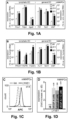
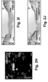
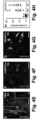
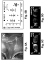
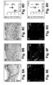
- a fraction (21 ⁇ 6%) of MAPCs exposed to VEGF-A also expressed LYVE1 (shown at the protein level for mMAPCs; Figure 1C ). Notably, induction of lymphatic marker gene expression in hMAPCs was not further improved in the presence of lymphangiogenic GF VEGF-C (shown for LYVE1 in Figure1D ; PROX1 fold-induction versus d0 was also comparable upon exposure to VEGF-A, VEGF-C or a combination: 26 ⁇ 10, 26 ⁇ 14 and 26 ⁇ 11, respectively; n 4).
- COUP-TFII a transcription factor co-determining lymphatic competence of ECs(36,37), was expressed at high relatively constant levels throughout the differentiation process (not shown). Thus, MAPCs have the inherent capacity to initiate a LEC differentiation program.
- MAPCs might have an effect on lymphangiogenesis by cross-talking to LECs, as MAPCs are known to secrete VEGF-A, which is responsible for the trophic effects of MSCs on LECs.
- MAPCs may support the formation of lymphatic vessels by a combination of direct and indirect effects.
- MAPCs contribute to physiological lymphangiogenesis during wound healing
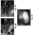
- hMAPCs applied onto circular wounds in athymic nude mice significantly promoted healing.
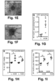
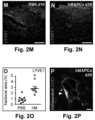
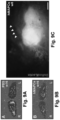
- Live imaging and cross-sections through the wound area upon transplantation of hMAPCs showed their homogenous distribution in the wound bed ( FIGS. 7G,H ).
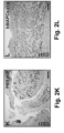
- hMAPCs significantly boosted lymphangiogenesis as evidenced by the three-fold increased LYVE1 + fractional area ( FIGS. 2M-O ).
- IF immunofluorescence
- hMAPCs did not affect wound healing in part by boosting capillary lymphangiogenesis mostly indirectly through a trophic effect on host LECs.
- MAPCs regenerate lymphatic vessels in a secondary lymphedema model
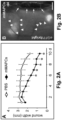
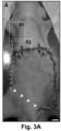
- lymph drainage to the axillary lymph nodes was discontinued by means of a full-thickness skin incision in the abdomen ( FIG. 3A ) ( Saaristo, A., et al. FASEB J (2004) 18: 1707-1709 ).
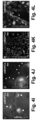
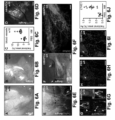
- MAPC transplantation around the wound border almost completely (in 5/6 and 6/6 cases for mMAPC- or hMAPC-treated mice, respectively) restored lymph drainage across this border ( FIGS. 3C ,D; Table 1).
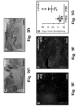
- MAPC-injected mice had a ⁇ two-three-fold increase in Flt4 + (VEGFR3 + ) and LYVE1 + fractional area in the wound borders ( FIGS. 4A-D + 8E-H , respectively) 2w after skin incision.
- the average number of functional lymphatic vessels per cross-section filled with fluorescently-labeled dextran around the incision at 2w was significantly increased by MAPC injection ( FIGS. 4E-H ).
- some mMAPCs persisted until 2-4w, lodged in the vicinity of draining lymphatic vessels and occasionally became part of their endothelial lining ( FIGS. 4I-L ).
- MAPCs reconnect transplanted lymph nodes to the host lymphatic network
- hMAPCs To test the potential of hMAPCs, they were applied in Matrigel ® around a transplanted LN derived from mice ubiquitously expressing dsRed or eGFP in the right axillary cavity ( FIG. 5A ). Transplantation of the LN alone (and covering it with Matrigel ® containing PBS) failed to resolve inflammation-induced edema in the right upper limb, evident from the accumulation of interstitial fluid measured by magnetic resonance imaging (MRI) 4w and 16w after surgery upon challenge of the paw with mustard oil - an inflammatory agent ( FIGS. 5B,C + 9A ).
- MRI magnetic resonance imaging
- FIGS. 5B,D + 9B lymphangiography revealed that lymph fluid drainage was significantly improved in hMAPC-treated mice and that the injected fluorescent dye reached the transplanted LN in ⁇ 35% and 50-60% of hMAPC-treated mice, 8w and 16w after transplantation, respectively, a result that was reproduced with two hMAPC clones and not at all with PBS-treated mice ( FIGS. 5E-G + 9C ; Table 2).
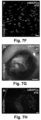
- FIGS. 6A-C show significantly more blood vessels in the immediate surroundings of the LNs, compared to PBS-injected mice ( FIGS. 6D + 9D,E).
- Some hMAPCs persisted until 16w and were found in the vicinity of the transplanted LN ( FIG. 9F ).
- All transplanted LNs in hMAPC-treated mice showed signs of (outward) branching of their internal (lymph)vascular network from 4w onwards, while this was never observed in PBS-treated mice ( FIGS. 6E-G + 9G,H ; Table 2).
- hMAPC transplantation resulted in a significant 4-fold expansion of LYVE1 + lymphatic vessels in the area surrounding the LN as compared to PBS-treatment ( FIGS.
- hMAPCs restored lymph drainage following LN transplantation by promoting LN survival and outward branching and by reconnecting the transplanted LN to the endogenous vessel network through collector vessels.
- the mMAPC clone was derived from BM of adult C57B1/6 mice with ubiquitous eGFP expression (C57B1/6-Tg-eGFP). mMAPCs were derived and maintained under low O 2 (5%) and low-serum (2%) conditions, as described ( Aranguren, X.L., et al. 2008. J ClinInvest (2008) 118:505-514 .). hMAPC clones were established according to derivation and culture methods described earlier ( Roobrouck, V.D., et al. Stem Cells (2011) 29:871-882 .). Cell cultures were routinely tested for mycoplasma contamination.
- Endothelial differentiation was performed by exposure to recombinant (r)hVEGF-A 165 or rhVEGF-C (R&D Systems), as described ( Roobrouck, V.D., et al. Stem Cells (2011) 29:871-882 ).
- hMAPCs Human MAPCs were isolated from bone fragments (femur) and hMab isolated from skeletal muscle fragments (quadriceps femoris) of children (5- to 15-year old) undergoing orthopedic surgery, after obtaining informed consent in accordance with the guidelines of the Medical Ethics Committee of the University Hospitals Leuven.
- hMAPCs were generated by flushing the bone fragment and plating the total cell fraction at 0.5 ⁇ 10 6 cells per centimeter square in medium consisting of 60% Dulbecco's modified Eagle's medium (DMEM) low glucose (Gibco, Invitrogen, Carlsbad, CA, www.invitrogen.com ), 40% MCDB-201 (Sigma-Aldrich, St.
- DMEM Dulbecco's modified Eagle's medium
- PDGF-BB platelet derived growth factor BB
- EGF human EGF
- human MAPC cultures were maintained under hypoxic conditions (5% O 2 ) in a 5.5% CO 2 humidified incubator at a density of 400 cells per centimeter square and were passaged every 2-3 days. Clonal populations were obtained by plating 5 cells per well in a 96-well or 48-well plate between passages 2 and 12.
- Bone marrow is obtained from healthy donors. Bone marrow mononuclear cells obtained by Ficoll-Paque density gradient centrifugation are depleted of CD45 + and glycophorinA + cells by means of micromagnetic beads. The eluted cells are 99.5% negative for both CD45 and glyA. Cells are plated into 96-well plates at a concentration of 5 ⁇ 10 3 cells/200 ⁇ l. This is done in the same medium described above.
- Murine cells were derived from BM of C57BL/6 mice with ubiquitous GFP expression.
- mMAPCs were derived and maintained under low O 2 (5%) and low-serum (2%) conditions ( Ulloa-Montoya, F., et al. Genome Biol. (2007) 8:R163 .).
- the mMAPCs can also be derived according to Breyer et al. Experimental Hematology 34:1596-1601 (2006 ).
- RNA from cell lysates was extracted using Trizol ® reagent (Invitrogen) or RLT lysis buffer (Qiagen). mRNA was reverse transcribed using Superscript III Reverse Transcriptase (Invitrogen) and cDNA underwent 40 amplification rounds on an ABI PRISM 7700 cycler, PerkinElmer/Applied Biosystems) for SYBR-Green-based qRT-PCR, as described ( Aranguren, X.L., et al. J Cell Sci (2013) 126:1165-1175 ). mRNA levels were normalized using GAPDH as housekeeping gene. To analyze LYVE1 expression on the surface of differentiated mMAPCs, cells were harvested by gentle trypsinization and analyzed by FACS as described in the extended methods.
- CM Cell culture and CM collection.
- Human lung LECs were purchased from Lonza (Merelbeke, Belgium) and cultured in EBM2 supplemented with EGM-2-MV bulletkit (Lonza).
- EGM-2-MV bulletkit LiGM-2-MV bulletkit
- MAPCs were seeded at high density in serum-free basal media and CM was collected after 72h and frozen in aliquots at -80°C until further use.
- LEC proliferation LECs were seeded at 2,000 cells/cm 2 in regular LEC growth medium onto gelatin-coated 96-well plates. Following their attachment, medium was replaced by a 1:1 mix of serum-free LEC medium and MAPC-CM or 100% serum-free LEC medium as reference condition. After 24h, cell proliferation was assessed with the WST-1 cell proliferation assay kit (Cayman Chemical).
- LEC sprouting LEC spheroids were allowed to form by applying 25 ⁇ l droplets (containing 1,000 LECs in a 20% methylcellulose/EGM-2-MV mixture) onto non-attachment plates and incubating them upside down at 37°C/5%CO 2 . The next day, spheroids were carefully washed in PBS/2%FBS, collected by gentle centrifugation, resuspended in methylcellulose/FBS/collagen (Purecol Advanced Biomatrix) and seeded into 24-well plates. Following incubation for 30min at 37°C/5% CO 2 , mMAPC-CM (1:1 mix with serum-free LEC media) or 100% serum-free LEC media as reference condition was added on top of the collagen/spheroid gel. Pictures were taken 24h later and the number of sprouts per spheroid was determined by manual counting.
- Linear wound model At day 0, a 12-mm linear skin incision was inflicted on the back of anesthetized 12-w-old C57B1/6 male mice. Immediately after wounding, mice were injected in the muscle fascia underneath the skin wound with 1 ⁇ 10 6 mMAPCs (resuspended in PBS) or PBS alone divided over three equally spaced injection spots. To avoid wound infection, mice were housed individually in cages without bedding. Wound dimensions were measured daily under anesthesia using digital calipers (VWRI819-0012, VWR). At d4, brightfield and fluorescence pictures of the wound area were taken and at d10, mice were euthanized, the residual skin wound and underlying muscle tissue were dissected out, fixed and prepared for embedding.
- VWRI819-0012, VWR digital calipers
- Circular wound model At day 0, 12-w-old athymic nude Foxn1 male mice (Harlan) were anesthetized and under sterile and temperature-controlled (37°C) conditions, standardized full-thickness wounds were made with a 0.5 cm biopsy puncher (Stiefel Laboratories, Offenbach am Main, Germany) on the back of the mouse. A silicone ring was sutured around the wound and wounds were treated with PBS or 5 ⁇ 10 5 hMAPCs. In a subset of mice, hMAPCs were transduced with an eGFP-encoding lentivirus before transplantation.
- mice were euthanized, skin wounds were dissected out, rinsed and post-fixed. Following fixation, skin fragments were separated in two equal pieces at the midline of the wound and processed for embedding.
- FITC-dextran MW 2,000 kDa, Sigma-Aldrich; hMAPCs
- Rhodamin-B-isothiocyanate-dextran MW 70 kDa, Sigma-Aldrich; mMAPCs
- Brightfield and fluorescence pictures were taken at 15 min and mice were subsequently euthanized, the skin wound area around the cell engraftment/microlymphangiography areas excised, fixed and processed for embedding.
- LN transplantation model At day 0, 12-w-old athymic nude Foxn1 female mice (Harlan) were anesthetized and to visualize the LNs, the right axilla region was exposed and mice were injected with a 3% Evans Blue solution in the palm of the right paw after which LNs were removed (along with the surrounding lymphatic (collector) vessels). A pocket just caudal of the axillary vessels was prepared.
- Donor LNs were dissected from mice ubiquitously expressing DsRed (B6.Cg-Tg(CAG-DsRed ⁇ MST)1Nagy/J; for mice receiving hMAPCs or PBS and followed up for 4w or 8w) or enhanced (e)GFP (C57BL/6-Tg(CAG-EGFP)10sb/J; for mice receiving hMAPCs or PBS and followed up for 4w or 16w) and cut in two halves through the hilus. The cut LN was subsequently implanted into the recipient pocket and fixed in place with permanent sutures (Monosof TM ).
- mice were anesthetized and subjected to microlymphangiography following injection of FITC-conjugated L. esculentum lectin (Vector Laboratories; in DsRed + LN recipients) or Texas Red-conjugated L. esculentum lectin (in eGFP + LN recipients) in the palm of the right paw.
- Lymphatic determined on LYVE1-, Flt4- or Prox1/ ⁇ SMA-stained sections
- blood determined on CD31-stained sections
- vessel density and epithelial coverage determined on pancytokeratin-stained sections
- the fractional area of the blood vessel network leading up to the transplanted LNs was determined on digitally reconstructed images of the entire region of interest.
- For stainings on paraffin sections slides were deparaffinated and rehydrated, cryosections were incubated in PBS for five min prior to the staining procedure. H&E staining was performed as previously described ( Aranguren, X.L., et al. J Clin Invest (2008) 118:505-514 ).
- n in results text and Figure/Table legends designates the number of replicates ( i.e., each performed on different passages of a given MAPC clone; in vitro ) or separate animals ( in vivo ) .
- Data normality was tested by the Shapiro-Wilk test. Comparisons among two groups was performed by Student's t-test in case of normal distribution or by Mann-Whitney-U test in cases where data were not normally distributed or normality could not be assumed. Multiple-group comparisons were done by 1-way ANOVA with Tuckey's post-hoc test (normal distribution) or Kruskal-Wallis test with Dunn's post-hoc test (no normality assumption). Wound size was evaluated by repeated measure ANOVA, followed by Fisher least-significant-difference test. All analyses were performed with Graphpad Prism (version 6.0).
- the murine (m)MAPC clone was derived from BM of adult C57B1/6 mice with ubiquitous eGFP expression (C57B1/6-Tg-eGFP). mMAPCs were derived and maintained under low O 2 (5%) and low-serum (2%) conditions, as previously described ( Aranguren, X.L., et al. J Clin Invest (2008) 118:505-514 ).
- hMAPC clones were established at KU Leuven (clone 1 or hMAPC1 at the Endothelial Cell Biology Unit; clone 2 or hMAPC2 at the Stem Cell Institute Leuven), according to derivation and culture methods described earlier ( Roobrouck, V.D., et al. Stem Cells (2011) 29:871-882 ). Cell cultures were routinely tested for mycoplasma contamination. Endothelial differentiation was performed by exposure of the cells to recombinant (r)hVEGF-A 165 or rhVEGF-C (both from R&DSystems), as described ( Roobrouck, V.D., et al. Stem Cells (2011) 29:871-882 ).
- RNA from cell lysates was extracted using Trizol ® reagent (Invitrogen) or RLT lysis buffer (Qiagen). mRNA was reverse transcribed using Superscript III Reverse Transcriptase (Invitrogen) and cDNA underwent 40 amplification rounds on an ABI PRISM 7700 cycler PerkinElmer/Applied Biosystems) for SYBR-Green-based qRT-PCR, as described ( Aranguren, X.L.et al. J Cell Sci (2013) 126:1164-1175 .). mRNA levels were normalized using GAPDH as housekeeping gene.
- FACS staining buffer PBS+1mmol/L EDTA+25mmol/L HEPES+1% BSA
- primary antibody Upstate
- FACS buffer cells were incubated with biotinylated goat-anti-rabbit secondary antibodies for 20 min at room temperature in the dark.
- samples were washed and incubated in the dark for 20 min with allophycocyanin (APC)-labeled streptavidin.
- APC allophycocyanin
- CM Cell culture and conditioned media collection.
- Human lung LECs were purchased from Lonza (Merelbeke, Belgium) and cultured in EBM2 supplemented with EGM-2-MV bulletkit (Lonza).
- EGM-2-MV bulletkit LiGM-2-MV bulletkit
- MAPCs were seeded at high density in serum-free basal media and CM was collected after 72h and frozen in aliquots at -80°C until further use.
- LEC proliferation To test the effect of MAPC-CM on LEC proliferation, LECs were seeded at a density of 2,000 cells/cm 2 in regular LEC growth medium onto gelatin-coated 96-well plates. Following their attachment, medium was replaced by a 1:1 mix of serum-free LEC medium and MAPC-CM or 100% serum-free LEC medium as reference condition. After 24h, cell proliferation was assessed with the WST-1 Cell Proliferation Assay kit. Briefly, 10 ⁇ l of WST-1 mixture was added to each well, cells were incubated at 37°C for 2h and the absorbance of each well was measured on a Bio-Tek microplate reader (BRS, Belgium) at a wavelength of 450 nm.
- WST-1 Cell Proliferation Assay kit Briefly, 10 ⁇ l of WST-1 mixture was added to each well, cells were incubated at 37°C for 2h and the absorbance of each well was measured on a Bio-Tek microplate reader (BRS, Belgium) at a wavelength of 450 nm.
- LEC migration To estimate the effect of MAPC-CM on LEC migration, a Boyden chamber assay was performed. Briefly, transwell inserts (containing polycarbonate filters with 8 ⁇ m pore size; Costar, Corning) were coated overnight with 0.2% gelatin. The bottom compartment of a 24-well plate was filled with 0.3 ml NCM or with 0.3 ml of mMAPC or hMAPC-CM. Following rehydration for 1h with deionized water, inserts were placed into the 24-well plate and each was loaded with 0.3 ml EGM-2-MV/0.5% FBS containing 5 ⁇ 10 4 LECs. Following incubation for 24h at 37°C/5% CO 2 , cells were fixed in methanol for 30 min at -20°C.
- LEC sprouting To test the effect of mMAPC-CM on LEC sprouting, LEC spheroids were allowed to form by applying 25 ⁇ l droplets (containing 1,000 LECs in a 20% methylcellulose/EGM-2-MV mixture) onto non-attachment plates and incubating them upside down at 37°C/5%CO 2 . The next day, spheroids were carefully washed in PBS/2%FBS, collected by gentle centrifugation, carefully resuspended in methylcellulose/FBS/collagen (Purecol Advanced Biomatrix) and seeded into 24-well plates (0.5 ml/well).
- Linear wound model At day 0, a 12-mm linear skin incision was inflicted with a scalpel on the back of 12-w-old C57B1/6 male mice after they were anesthetized with a mixture of 100 mg/kg ketamine and 10 mg/kg xylazine. Immediately after wounding, mice were injected in the muscle fascia underneath the skin wound with 1 ⁇ 10 6 mMAPCs (resuspended in PBS) or PBS alone divided over three equally spaced injection spots. To avoid wound infection, mice were housed individually in cages without bedding. Wound dimensions were measured daily under isoflurane anesthesia using digital calipers (VWRI819-0012, VWR).
- mice were euthanized, the residual skin wound and underlying muscle tissue were dissected out, fixed in zinc-paraformaldehyde and prepared for embedding in paraffin or optimal cutting temperature (OCT) and sectioning.
- OCT optimal cutting temperature
- Circular wound model At day 0, 12-w-old athymic nude Foxn1 male mice (Harlan) were anesthetized with an i.p. injection of ketamine/xylazine. Atropine (0.01 mg/kg) was administered i.p. as premedication. Under sterile and temperature-controlled (37°C) conditions, standardized full-thickness wounds were made with a 0.5 cm biopsy puncher (Stiefel Laboratories, Offenbach am Main, Germany) on the back of the mouse in the mid-dorsal region. A silicone ring was fixed (using Histoacryl tissue adhesive, Braun, Diegem, Belgium) and sutured around the wound and wounds were treated with saline or 5 ⁇ 10 5 hMAPCs.
- mice In a separate subset of mice, hMAPCs were transduced with an eGFP-encoding lentivirus before transplantation.
- An occlusive dressing (Tegaderm TM , 3M, Diegem, Belgium) was used to keep the wound moist. All wounded mice were housed individually to avoid fighting and to prevent removal of the occlusive wound dressing. Every other day, the occlusive dressing was renewed under isoflurane anesthesia.
- mice were euthanized and square skin fragments including the circular wound area and a rim of normal skin were dissected out, rinsed in PBS and post-fixed overnight at 4°C using zinc-paraformaldehyde. Following fixation, skin fragments were separated in two equal pieces at the midline of the wound and processed for paraffin or OCT embedding and sectioning.
- FIG. 3A One day after resuturing the skin flap, 1 ⁇ 10 6 mMAPCs, 1 ⁇ 10 6 hMAPCs or PBS (divided over 4 injection spots; FIG. 3A ) were injected around the wound edges. Two or four weeks later, the axillary regions were exposed and axillary lymph node drainage was monitored by microlymphangiography for 15 min after intradermal injection of 10 ⁇ l FITC-dextran (MW 2,000 kDa, Sigma-Aldrich; hMAPCs) or 10 ⁇ l Rhodamin-B-isothiocyanate-dextran (MW 70 kDa, Sigma-Aldrich; mMAPCs) under the wound border ( FIG. 3A ).
- 10 ⁇ l FITC-dextran MW 2,000 kDa, Sigma-Aldrich; hMAPCs
- 10 ⁇ l Rhodamin-B-isothiocyanate-dextran MW 70 kDa
- Lymph node transplantation model At day 0, 12-w-old athymic nude Foxn1 female recipient mice (Harlan) were anesthetized with an i.p. injection of ketamine (100 mg/kg) and xylazine (10 mg/kg). To visualize the lymph nodes, the right axilla region was exposed and mice were injected with a 3% Evans Blue solution in the palm of the right paw after which lymph nodes were removed along with the surrounding lymphatic (collector) vessels. A pocket just caudal of the axillary vessels, aligned by the lateral axillary fat pad, the M. pectoralis and the M. l ⁇ tissimus dorsi was prepared.
- Donor lymph nodes were dissected from mice ubiquitously expressing DsRed (B6.Cg-Tg(CAG-DsRed ⁇ MST)1Nagy/J; for mice receiving hMAPCs or PBS and followed up for 4w or 8w) or enhanced (e)GFP (C57BL/6-Tg(CAG-EGFP)10sb/J; for mice receiving hMAPCs or PBS and followed up for 4w or 16w) and cut in two halves through the hilus.
- the cut lymph node was subsequently implanted into the recipient pocket (hilus oriented medially and cut surface facing upwards) and fixed in place with two permanent sutures (using 9-0 nylon non-absorbable suture, Monosof TM ).
- mice were anesthetized with a ketamine/xylazine mixture and subjected to microlymphangiography following injection of 10 ⁇ l FITC-conjugated L. esculentum lectin (Vector Laboratories; in recipients of DsRed + donor lymph nodes) or 10 ⁇ l Texas Red-conjugated L.
- esculentum lectin in recipients of eGFP + lymph nodes
- Drainage of the implanted lymph node was monitored for 15 min and brightfield and fluorescence pictures were taken at the end with a Zeiss MRc5 camera mounted onto a Zeiss Lumar microscope.
- Mice were subsequently euthanized, the axilla regions containing the transplanted lymph node excised, fixed and processed for paraffin or OCT embedding and sectioning.
- Two additional sets of mice were subjected to in vivo magnetic resonance imaging (MRI; as described ( Tammela, T., et al. Nat Med (2007) 13: 1458-1466 ) at 4w or 16w after lymph node transplantation.
- MRI magnetic resonance imaging
- mice were anesthetized with isoflurane and mustard oil (diluted 1/5 in mineral oil) was applied with a cotton stick on both fore limbs for 2 x 15 min to elicit vascular hyperpermeability and aggravate edema. Mice were allowed to recover for another 30 min before MRI recording. Temperature and respiration were monitored throughout the experiment and maintained at 37°C and 100 - 120 breaths per min.
- MR images were acquired with a 9.4T Biospec small animal MR scanner (Bruker Biospin, Ettlingen, Germany) equipped with a horizontal bore magnet and an actively shielded gradient set of 600mT per m (117 mm inner diameter) using a 7 cm linearly polarized resonator for transmission and an actively decoupled dedicated 2 cm diameter surface coil for receiving (Rapid Biomedical, Rimpar, Germany).
- 3D T 2 weighted images, 2D T 2 parameter maps and 2D diffusion weighted images were acquired to determine the level of edema.
- Processing of the 3D T 2 weighted images was done by determining the volume with a signal intensity above a common threshold value using home-written software developed with Mevislab (Mevis Medical Solutions, Bremen, Germany) reported as ratio's between the lymph node implanted site versus the control site.
- Mevislab Mevis Medical Solutions, Bremen, Germany
- Calculation of the T 2 parameter maps of the manually delineated edema of the paws (or an area of the same size and located in the same region in the absence of edema) was done using Paravison 5.1 (Bruker Biospin).
- Lymphatic determined on LYVE1-, Flt4- or Prox1/ ⁇ SMA-stained sections
- blood determined on CD31-stained sections
- vessel density and epithelial coverage determined on pancytokeratin-stained sections
- the fractional area of the blood vessel network leading up to the transplanted lymph nodes was determined on digitally reconstructed images of the entire region of interest.
- stainings on paraffin sections slides were deparaffinated and rehydrated, cryosections were incubated in PBS for five min prior to the staining procedure. H&E staining was performed as previously described (1).
- antigen retrieval was performed by boiling in target retrieval solution s1699 (Sigma). After cooling down in TBS, endogenous peroxidase activity was quenched in 0.3% H 2 O 2 in methanol. Slides were incubated with primary Ab overnight. A list of primary Ab's is provided in Table 4.
- IF-stained slides were sealed with ProLong Gold Antifade Reagent with DAPI (Life Technologies; P36931) or without in case nuclei were revealed by Hoechst staining. All Images were recorded on a Zeiss Axiovert 200M microscope, a Zeiss Axio Imager Z1 or a Zeiss LSM510 confocal microscope equipped with a Zeiss MRc5 camera and Axiovision 4.8 software.
- n in results text and Figure/Table legends designates the number of replicates ( i.e., each performed on different passages of a given MAPC clone; in vitro ) or separate animals ( in vivo ) .
- Normality of the data was tested by the Shapiro-Wilk test. Comparisons among two groups was performed by Student's t -test in case of normal distribution or by Mann-Whitney-U test in cases where data were not normally distributed or normality could not be assumed. Multiple-group comparisons were done by 1-way ANOVA with Tuckey's post-hoc test (normal distribution) or Kruskal-Wallis test followed by Dunn's post-hoc test (no normality assumption). Wound size was evaluated by repeated measures ANOVA, followed by Fisher least-significant-difference test. All analyses were performed with Graphpad Prism (version 6.0).
Landscapes
- Health & Medical Sciences (AREA)
- Life Sciences & Earth Sciences (AREA)
- Engineering & Computer Science (AREA)
- Chemical & Material Sciences (AREA)
- Biomedical Technology (AREA)
- General Health & Medical Sciences (AREA)
- Cell Biology (AREA)
- Developmental Biology & Embryology (AREA)
- Biotechnology (AREA)
- Zoology (AREA)
- Animal Behavior & Ethology (AREA)
- Pharmacology & Pharmacy (AREA)
- Public Health (AREA)
- Veterinary Medicine (AREA)
- Medicinal Chemistry (AREA)
- Organic Chemistry (AREA)
- Bioinformatics & Cheminformatics (AREA)
- Immunology (AREA)
- Epidemiology (AREA)
- Genetics & Genomics (AREA)
- Wood Science & Technology (AREA)
- Virology (AREA)
- Dermatology (AREA)
- Hematology (AREA)
- Chemical Kinetics & Catalysis (AREA)
- Microbiology (AREA)
- General Chemical & Material Sciences (AREA)
- Biochemistry (AREA)
- General Engineering & Computer Science (AREA)
- Nuclear Medicine, Radiotherapy & Molecular Imaging (AREA)
- Medicines Containing Material From Animals Or Micro-Organisms (AREA)
- Medicines That Contain Protein Lipid Enzymes And Other Medicines (AREA)
- Materials For Medical Uses (AREA)
- Medicinal Preparation (AREA)
- Acyclic And Carbocyclic Compounds In Medicinal Compositions (AREA)
Applications Claiming Priority (2)
| Application Number | Priority Date | Filing Date | Title |
|---|---|---|---|
| US201662281334P | 2016-01-21 | 2016-01-21 | |
| PCT/US2016/017848 WO2017127123A1 (en) | 2016-01-21 | 2016-02-12 | Stem cells for wound healing |
Publications (3)
| Publication Number | Publication Date |
|---|---|
| EP3405566A1 EP3405566A1 (en) | 2018-11-28 |
| EP3405566A4 EP3405566A4 (en) | 2019-07-31 |
| EP3405566B1 true EP3405566B1 (en) | 2023-10-18 |
Family
ID=59360064
Family Applications (1)
| Application Number | Title | Priority Date | Filing Date |
|---|---|---|---|
| EP16886758.8A Active EP3405566B1 (en) | 2016-01-21 | 2016-02-12 | Stem cells for healing of skin ulcers |
Country Status (11)
| Country | Link |
|---|---|
| US (2) | US10967006B2 (enExample) |
| EP (1) | EP3405566B1 (enExample) |
| JP (3) | JP6918003B2 (enExample) |
| KR (1) | KR20180102661A (enExample) |
| CN (1) | CN108884437A (enExample) |
| AU (2) | AU2016387339B2 (enExample) |
| CA (1) | CA3012330A1 (enExample) |
| HK (1) | HK1256413A1 (enExample) |
| NZ (1) | NZ745530A (enExample) |
| SG (2) | SG11201806245TA (enExample) |
| WO (1) | WO2017127123A1 (enExample) |
Families Citing this family (8)
| Publication number | Priority date | Publication date | Assignee | Title |
|---|---|---|---|---|
| EP2568991B3 (en) | 2010-05-12 | 2018-11-28 | ABT Holding Company | Modulation of splenocytes in cell therapy |
| EP3795159A1 (en) | 2013-04-12 | 2021-03-24 | Houston Methodist Hospital | Improving organs for transplantation |
| SG11201806245TA (en) * | 2016-01-21 | 2018-08-30 | Abt Holding Co | Stem cells for wound healing |
| KR102188572B1 (ko) * | 2017-09-25 | 2020-12-09 | 사회복지법인 삼성생명공익재단 | 뇌척수액을 포함하는 줄기세포 투여 제형 및 그의 제조방법 |
| US20240389931A1 (en) * | 2020-01-23 | 2024-11-28 | Precision Healing LLC | Wound care diagnosis with fluorescence signatures |
| US20220117491A1 (en) * | 2020-01-23 | 2022-04-21 | Precision Healing, Inc. | Multispectral sample analysis using fluorescence signatures |
| US20240245346A1 (en) * | 2020-01-23 | 2024-07-25 | Precision Healing, Inc. | Image analysis using skin wound factors |
| EP4638696A1 (en) | 2022-12-27 | 2025-10-29 | AVITA Medical Americas, LLC | System for automated preparation of a regenerative epidermal suspension and related methods of use |
Family Cites Families (103)
| Publication number | Priority date | Publication date | Assignee | Title |
|---|---|---|---|---|
| US4657866A (en) | 1982-12-21 | 1987-04-14 | Sudhir Kumar | Serum-free, synthetic, completely chemically defined tissue culture media |
| US7037721B1 (en) | 1990-01-29 | 2006-05-02 | Hy-Gene Biomedical, Inc. | Protein-free defined media for the growth of normal human keratinocytes |
| US5486359A (en) | 1990-11-16 | 1996-01-23 | Osiris Therapeutics, Inc. | Human mesenchymal stem cells |
| CA2123094C (en) | 1991-11-06 | 1999-08-10 | Paulo N. Correa | Cell culture medium |
| AU4543193A (en) | 1992-06-22 | 1994-01-24 | Henry E. Young | Scar inhibitory factor and use thereof |
| US5766951A (en) | 1992-11-12 | 1998-06-16 | Quality Biological, Inc. | Serum-free medium supporting growth and proliferation of normal bone marrow cells |
| US6037174A (en) | 1993-08-23 | 2000-03-14 | Nexell Therapeutics, Inc. | Preparation of serum-free suspensions of human hematopoietic cells or precursor cells |
| US5409813A (en) | 1993-09-30 | 1995-04-25 | Systemix, Inc. | Method for mammalian cell separation from a mixture of cell populations |
| GB9502022D0 (en) | 1995-02-02 | 1995-03-22 | Abuljadayel Ilham M S | A method for preparing lymphohaematopoietic progenitor cells |
| US5906934A (en) | 1995-03-14 | 1999-05-25 | Morphogen Pharmaceuticals, Inc. | Mesenchymal stem cells for cartilage repair |
| US6653134B2 (en) | 1995-03-28 | 2003-11-25 | Cp Hahnemann University | Isolated stromal cells for use in the treatment of diseases of the central nervous system |
| US5908782A (en) | 1995-06-05 | 1999-06-01 | Osiris Therapeutics, Inc. | Chemically defined medium for human mesenchymal stem cells |
| US5945337A (en) | 1996-10-18 | 1999-08-31 | Quality Biological, Inc. | Method for culturing CD34+ cells in a serum-free medium |
| AU7984498A (en) | 1997-06-25 | 1999-01-04 | Government Of The United States Of America, As Represented By The Secretary Of The Department Of Health And Human Services, The | Serum-free cell growth medium |
| US6077987A (en) | 1997-09-04 | 2000-06-20 | North Shore-Long Island Jewish Research Institute | Genetic engineering of cells to enhance healing and tissue regeneration |
| AU758073B2 (en) | 1998-11-09 | 2003-03-13 | Consorzio Per La Gestione Del Centro Di Biotecnologie Avanzate | Serum free medium for chondrocyte-like cells |
| US6777231B1 (en) | 1999-03-10 | 2004-08-17 | The Regents Of The University Of California | Adipose-derived stem cells and lattices |
| US20050169896A1 (en) | 1999-05-14 | 2005-08-04 | Henry Ford Health System | Bone marrow transplantation for treatment of stroke |
| US8147824B2 (en) | 1999-08-05 | 2012-04-03 | Athersys, Inc. | Immunomodulatory properties of multipotent adult progenitor cells and uses thereof |
| US7015037B1 (en) | 1999-08-05 | 2006-03-21 | Regents Of The University Of Minnesota | Multiponent adult stem cells and methods for isolation |
| US8609412B2 (en) | 1999-08-05 | 2013-12-17 | Regents Of The University Of Minnesota | Mapc generation of lung tissue |
| US8075881B2 (en) | 1999-08-05 | 2011-12-13 | Regents Of The University Of Minnesota | Use of multipotent adult stem cells in treatment of myocardial infarction and congestive heart failure |
| US10638734B2 (en) | 2004-01-05 | 2020-05-05 | Abt Holding Company | Multipotent adult stem cells, sources thereof, methods of obtaining and maintaining same, methods of differentiation thereof, methods of use thereof and cells derived thereof |
| JP5398941B2 (ja) | 1999-08-05 | 2014-01-29 | エイビーティー ホールディング カンパニー | 多能性成体幹細胞およびそれを単離する方法 |
| US8252280B1 (en) | 1999-08-05 | 2012-08-28 | Regents Of The University Of Minnesota | MAPC generation of muscle |
| US7927587B2 (en) | 1999-08-05 | 2011-04-19 | Regents Of The University Of Minnesota | MAPC administration for the treatment of lysosomal storage disorders |
| DE60041821D1 (de) | 1999-09-24 | 2009-04-30 | Cybios Llc | Pluripotent embryonale stammzellen-ähnliche zellen, zusammensetzungen und ihre vervendungen |
| WO2001021767A2 (en) | 1999-09-24 | 2001-03-29 | Morphogen Pharmaceuticals, Inc. | Pluripotent embryonic-like stem cells, compositions, methods and uses thereof |
| US20030161817A1 (en) | 2001-03-28 | 2003-08-28 | Young Henry E. | Pluripotent embryonic-like stem cells, compositions, methods and uses thereof |
| CA2392615A1 (en) | 1999-12-06 | 2001-06-07 | The General Hospital Corporation | Pancreatic stem cells and their use in transplantation |
| ES2300320T3 (es) | 2000-02-26 | 2008-06-16 | Artecel, Inc. | Celulas madre pluripotentes generadas a partir de celulas del estroma derivadas de tejido adiposo y sus usos. |
| WO2002009650A2 (en) | 2000-07-31 | 2002-02-07 | New York Medical College | Methods and compositions for the repair and/or regeneration of damaged myocardium |
| US7560280B2 (en) | 2000-11-03 | 2009-07-14 | Kourion Therapeutics Gmbh | Human cord blood derived unrestricted somatic stem cells (USSC) |
| US7311905B2 (en) | 2002-02-13 | 2007-12-25 | Anthrogenesis Corporation | Embryonic-like stem cells derived from post-partum mammalian placenta, and uses and methods of treatment using said cells |
| EP2305795B1 (en) | 2000-12-06 | 2019-07-03 | Celularity, Inc. | Method of collecting placental stem cells |
| JP2004529621A (ja) | 2001-02-14 | 2004-09-30 | ティー ファークト,レオ | 多能性成体幹細胞、その起源、それを得る方法および維持する方法、それを分化させる方法、その使用法、ならびにそれ由来の細胞 |
| WO2002064755A2 (en) | 2001-02-14 | 2002-08-22 | Anthrogenesis Corporation | Post-partum mammalian placenta, its use and placental stem cells therefrom |
| US7056738B2 (en) | 2001-03-23 | 2006-06-06 | Tulane University | Early stage multipotential stem cells in colonies of bone marrow stromal cells |
| KR100449141B1 (ko) | 2001-04-19 | 2004-09-21 | (주)라이프코드 | 간엽 간세포를 신경세포로 분화시키는 방법 |
| EP1383869B1 (en) | 2001-04-24 | 2009-06-10 | Dolores Baksh | Progenitor cell populations, expansion thereof, and growth of non-hematopoietic cell types and tissues therefrom |
| US20030003090A1 (en) | 2001-05-31 | 2003-01-02 | Prockop Darwin J. | Directed in vitro differentiation of marrow stromal cells into neural cell progenitors |
| US9969980B2 (en) | 2001-09-21 | 2018-05-15 | Garnet Biotherapeutics | Cell populations which co-express CD49c and CD90 |
| WO2003064598A2 (en) | 2002-01-25 | 2003-08-07 | Genzyme Corporation | Serum-free media for chondrocytes and methods of use thereof |
| US20060228798A1 (en) | 2002-11-27 | 2006-10-12 | Catherine Verfaillie | Homologous recombination in multipotent adult progenitor cells |
| CA2809195C (en) | 2003-04-01 | 2014-05-20 | United States Of America Department Of Veteran's Affairs | Stem-cell, precursor cell, or target cell-based treatment of multi-organ failure and renal dysfunction |
| US20040235165A1 (en) | 2003-05-19 | 2004-11-25 | Darwin Prockop | In vitro differentiation of adult stem cells |
| US8192348B2 (en) | 2003-07-01 | 2012-06-05 | Regents Of The University Of Minnesota | Engineered blood vessels |
| WO2005003320A2 (en) | 2003-07-02 | 2005-01-13 | Regents Of The University Of Minnesota | Neuronal differentiation of stem cells |
| CA2536909A1 (en) | 2003-08-29 | 2005-03-10 | Regents Of The University Of Minnesota | Kidney derived stem cells and methods for their isolation, differentiation and use |
| US7794706B2 (en) | 2003-10-14 | 2010-09-14 | Medivas, Llc | Bioactive wound dressings and implantable devices and methods of use |
| AU2004296848A1 (en) | 2003-12-04 | 2005-06-23 | Regents Of The University Of Minnesota | Compositions and methods for the treatment of lysosomal storage disorders |
| CA2558520C (en) | 2004-03-05 | 2015-02-24 | John E. Davies | Serum-free suspension culture system for mesenchymal progenitor cells |
| EP2298863B1 (en) | 2004-03-22 | 2015-07-22 | Mesoblast International Sàrl | Mesenchymal stem cells and uses therefor |
| EP1737947B9 (en) | 2004-04-21 | 2012-04-25 | Regents Of The University Of Minnesota | Mapc generation of lung tissue |
| WO2006047743A2 (en) * | 2004-10-26 | 2006-05-04 | Regents Of The University Of Minnesota | Swine multipotent adult progenitor cells |
| KR100593397B1 (ko) | 2004-10-27 | 2006-06-28 | 한국원자력연구소 | 중배엽 줄기세포 및/또는 p 물질을 함유하는 상처 치유또는 상처 치유 촉진제, 또는 세포 치료제 |
| EP2428563A1 (en) | 2005-02-10 | 2012-03-14 | Regents Of The University Of Minnesota | Vascular/lymphatic endothelial cells |
| CA2607213A1 (en) | 2005-05-05 | 2006-11-16 | Regents Of The University Of Minnesota | Use of nk cell inhibition to facilitate persistence of engrafted mhc- i negative cells |
| AU2005331534B2 (en) | 2005-05-05 | 2011-12-08 | Regents Of The University Of Minnesota | Use of MAPC or progeny therefrom to populate lymphohematopoietic tissues |
| WO2007016485A2 (en) | 2005-07-29 | 2007-02-08 | Athersys, Inc. | Use of a gsk-3 inhibitor to maintain potency of cultured cells |
| US20080194024A1 (en) | 2005-07-29 | 2008-08-14 | Mays Robert W | Culture of Non-Embryonic Cells at High Cell Density |
| NZ567082A (en) | 2005-10-14 | 2012-08-31 | Univ Minnesota | Differentiation of non-embryonic stem cells to cells having a pancreatic phenotype |
| US10117900B2 (en) | 2005-11-09 | 2018-11-06 | Athersys, Inc. | MAPC treatment of brain injuries and diseases |
| US20220110982A1 (en) | 2005-11-09 | 2022-04-14 | Abt Holding Company | Mapc treatment of brain injuries and diseases |
| US11000546B2 (en) | 2005-11-09 | 2021-05-11 | Athersys, Inc. | Immunomodulatory properties of MAPCs and uses thereof |
| US8871198B2 (en) * | 2006-03-29 | 2014-10-28 | Stemnion, Inc. | Methods related to wound healing |
| NZ744465A (en) * | 2006-01-23 | 2023-04-28 | Abt Holding Co | Mapc therapeutics without adjunctive immunosuppressive treatment |
| US9005964B2 (en) | 2006-11-24 | 2015-04-14 | Regents Of The University Of Minnesota | Endodermal progenitor cells |
| AU2008261901A1 (en) * | 2007-06-08 | 2008-12-18 | University Of Virginia Patent Foundation | Topical poloxamer formulations for enhancing microvascular flow: compositions and uses thereof |
| CA2712496C (en) | 2008-01-18 | 2021-01-12 | Wei-Shou Hu | Stem cell aggregates and methods for making and using |
| NZ587598A (en) | 2008-02-29 | 2012-10-26 | Univ New York State Res Found | Methods of increasing proliferation of a cell by administering a low magnitude, high frequency mechanical signal |
| US20110212062A1 (en) | 2008-03-27 | 2011-09-01 | Neostem, Inc. | Compositions and methods using stem cells in cutaneous wound healing |
| GB2467982A (en) * | 2008-05-08 | 2010-08-25 | Coretherapix Slu | Pluripotent adult stem cells |
| US9382514B2 (en) | 2008-06-20 | 2016-07-05 | Escape Therapeutics, Inc. | Compositions comprising mesenchymal stem cell-derived fibroblasts |
| SG10201807935SA (en) | 2008-09-04 | 2018-10-30 | Abt Holding Co | Use of stem cells to prevent neuronal dieback |
| EP2362899A1 (en) | 2008-10-31 | 2011-09-07 | Katholieke Universiteit Leuven | Optimized methods for differentiation of cells into cells with hepatocyte and hepatocyte progenitor phenotypes, cells produced by the methods, and methods for using the cells |
| ES2690199T3 (es) | 2009-07-21 | 2018-11-19 | Abt Holding Company | Uso de células madre para reducir la extravasación de leucocitos |
| EP3831930A1 (en) | 2009-07-21 | 2021-06-09 | ABT Holding Company | Use of stem cells to reduce leukocyte extravasation |
| WO2011060244A1 (en) | 2009-11-12 | 2011-05-19 | The Texas A & M University System | Spheroidal aggregates of mesenchymal stem cells |
| WO2011062584A1 (en) | 2009-11-19 | 2011-05-26 | Regents Of The University Of Minnesota | Reducing inflammation using cell therapy |
| US20150010610A1 (en) | 2010-02-18 | 2015-01-08 | Osiris Therapeutics, Inc. | Immunocompatible amniotic membrane products |
| US20110206647A1 (en) | 2010-02-25 | 2011-08-25 | Abt Holding Company | Modulation of Angiogenesis |
| SG183497A1 (en) | 2010-02-25 | 2012-09-27 | Abt Holding Co | Modulation of macrophage activation |
| WO2011106476A1 (en) | 2010-02-25 | 2011-09-01 | Abt Holding Company | Modulation of microglia activation |
| CN102198156A (zh) | 2010-03-26 | 2011-09-28 | 傅毓秀 | 用于治疗皮肤创伤之医药组合物 |
| EP2568991B3 (en) | 2010-05-12 | 2018-11-28 | ABT Holding Company | Modulation of splenocytes in cell therapy |
| WO2011143411A1 (en) | 2010-05-12 | 2011-11-17 | Abt Holding Company | Modulation of splenocytes in cell therapy for traumatic brain injury |
| US9090878B2 (en) | 2010-06-17 | 2015-07-28 | Katholieke Universiteit Leuven | Methods for differentiating cells into hepatic stellate cells and hepatic sinusoidal endothelial cells, cells produced by the methods, and methods for using the cells |
| WO2012009581A2 (en) | 2010-07-14 | 2012-01-19 | University Of Southern California | Pharmaceutical compositions comprising gingiva-derived mesenchymal stem cells and methods of treating inflammation, wound healing and contact hypersensitivity |
| IL207586A0 (en) | 2010-08-12 | 2010-12-30 | Omrix Biopharmaceuticals Ltd | A fibrin based therapeutic preparation and use thereof |
| CA2809245C (en) | 2010-08-24 | 2020-07-14 | Regents Of The University Of Minnesota | Non-static suspension culture of cell aggregates |
| EP2670842A2 (en) | 2011-01-31 | 2013-12-11 | Katholieke Universiteit Leuven KU Leuven Research & Development | Methods for making cells with an extra-embryonic endodermal precursor phenotype |
| SG10201914092XA (en) | 2011-06-06 | 2020-03-30 | ReGenesys BVBA | Expansion of stem cells in hollow fiber bioreactors |
| EP3795159A1 (en) | 2013-04-12 | 2021-03-24 | Houston Methodist Hospital | Improving organs for transplantation |
| ES2753323T3 (es) | 2013-04-30 | 2020-04-08 | Univ Leuven Kath | Terapia celular para síndromes mielodisplásicos |
| US20160129043A1 (en) | 2013-06-24 | 2016-05-12 | University Of Southern California | Composition of mesenchymal stem cells |
| WO2015017772A1 (en) | 2013-08-01 | 2015-02-05 | Parcell Laboratories, Llc | Stem cell-based preparations and methods for dermal and connective tissue reconstruction and rejuvenation |
| US20160175485A1 (en) | 2013-08-27 | 2016-06-23 | The Regents Of The University Of California | Combination therapy to promote wound healing |
| US20160069903A1 (en) | 2014-09-10 | 2016-03-10 | Fundació Institute De Ciències Foròniques | Method for detecting cells |
| EP3291822A1 (en) | 2015-05-05 | 2018-03-14 | Katholieke Universiteit Leuven | Improved methods for pancreatic islet transplantation |
| WO2017062035A1 (en) | 2015-10-09 | 2017-04-13 | Abt Holding Company | Methods for enhancing proliferation of t regulatory cells |
| SG11201806245TA (en) * | 2016-01-21 | 2018-08-30 | Abt Holding Co | Stem cells for wound healing |
| US20200246390A1 (en) | 2019-02-01 | 2020-08-06 | Abt Holding Company | Multipotent adult projenitor cells for treatment of ich |
-
2016
- 2016-02-12 SG SG11201806245TA patent/SG11201806245TA/en unknown
- 2016-02-12 NZ NZ745530A patent/NZ745530A/en not_active IP Right Cessation
- 2016-02-12 US US15/043,171 patent/US10967006B2/en active Active
- 2016-02-12 WO PCT/US2016/017848 patent/WO2017127123A1/en not_active Ceased
- 2016-02-12 AU AU2016387339A patent/AU2016387339B2/en not_active Ceased
- 2016-02-12 KR KR1020187024104A patent/KR20180102661A/ko not_active Ceased
- 2016-02-12 CA CA3012330A patent/CA3012330A1/en active Pending
- 2016-02-12 CN CN201680083874.5A patent/CN108884437A/zh active Pending
- 2016-02-12 JP JP2018538137A patent/JP6918003B2/ja not_active Expired - Fee Related
- 2016-02-12 HK HK18115545.8A patent/HK1256413A1/zh unknown
- 2016-02-12 SG SG10201914102UA patent/SG10201914102UA/en unknown
- 2016-02-12 EP EP16886758.8A patent/EP3405566B1/en active Active
-
2020
- 2020-07-07 JP JP2020116882A patent/JP2020172536A/ja not_active Withdrawn
-
2021
- 2021-04-05 US US17/222,460 patent/US11918609B2/en active Active
-
2022
- 2022-08-01 AU AU2022211794A patent/AU2022211794A1/en not_active Abandoned
- 2022-08-17 JP JP2022129977A patent/JP2022163207A/ja active Pending
Also Published As
| Publication number | Publication date |
|---|---|
| CA3012330A1 (en) | 2017-07-27 |
| HK1256413A1 (zh) | 2019-09-20 |
| US10967006B2 (en) | 2021-04-06 |
| US20170209493A1 (en) | 2017-07-27 |
| JP2022163207A (ja) | 2022-10-25 |
| US11918609B2 (en) | 2024-03-05 |
| AU2022211794A1 (en) | 2022-08-25 |
| KR20180102661A (ko) | 2018-09-17 |
| JP6918003B2 (ja) | 2021-08-11 |
| NZ745530A (en) | 2023-03-31 |
| SG11201806245TA (en) | 2018-08-30 |
| SG10201914102UA (en) | 2020-03-30 |
| WO2017127123A1 (en) | 2017-07-27 |
| JP2020172536A (ja) | 2020-10-22 |
| US20220031756A1 (en) | 2022-02-03 |
| AU2016387339B2 (en) | 2022-11-17 |
| CN108884437A (zh) | 2018-11-23 |
| AU2016387339A1 (en) | 2018-08-23 |
| EP3405566A4 (en) | 2019-07-31 |
| EP3405566A1 (en) | 2018-11-28 |
| JP2019506396A (ja) | 2019-03-07 |
Similar Documents
| Publication | Publication Date | Title |
|---|---|---|
| US11918609B2 (en) | Stem cells for wound healing | |
| JP7535070B2 (ja) | 虚血性疾患または虚血性損傷を処置するための心臓線維芽細胞由来細胞外マトリックスおよびその注射可能製剤 | |
| Pelizzo et al. | Extracellular vesicles derived from mesenchymal cells: perspective treatment for cutaneous wound healing in pediatrics | |
| JP7303612B2 (ja) | 熱傷の進行の緩和ならびに皮膚移植片の取り込みおよび治癒の改善における再生細胞の使用 | |
| KR102136450B1 (ko) | 당뇨병성 피부 궤양 치료를 위한 다능성 줄기 세포 | |
| Hussein et al. | Efficacy of xenogeneic fresh and lyophilized amniotic membranes on the healing of experimentally induced full-thickness skin wounds in dogs | |
| WO2019035925A1 (en) | COMPOSITION AND METHOD FOR TREATING SKIN AFFECTION | |
| KR101816964B1 (ko) | 피부 또는 혈관조직 손상 치료 보조용 약학 조성물 | |
| US20220030851A1 (en) | Modulating ischemic injury | |
| Wang et al. | Positive effects of subdermal lipotransfer on pig’s upper eyelid skin: Mature adipocytes or adipose-derived stem cells alone contribute little and only cotransplantation of them can generate benefits | |
| Vriend | Adipose tissue-derived components: therapeutic modalities to treat dermal damage | |
| WO2024009990A1 (ja) | 放射線障害防護剤 | |
| Winter | Equine Endothelial Colony Forming Cells: In Vitro and In Vivo Analysis | |
| Jadalannagari | Decellularized Wharton's jelly matrix as a three dimensional scaffold for wound healing and hair regeneration applications | |
| Warde | The Influence of the Hair Follicle Cycle on Wound Healing and Hypodermal Adipose Tissue, and Its Implications for Skin Grafting | |
| Al-Fouadi | Wound Healing Potential of Human Neonatal Mesenchymal Cells in an Animal Model of Hyperglycemia |
Legal Events
| Date | Code | Title | Description |
|---|---|---|---|
| STAA | Information on the status of an ep patent application or granted ep patent |
Free format text: STATUS: THE INTERNATIONAL PUBLICATION HAS BEEN MADE |
|
| PUAI | Public reference made under article 153(3) epc to a published international application that has entered the european phase |
Free format text: ORIGINAL CODE: 0009012 |
|
| STAA | Information on the status of an ep patent application or granted ep patent |
Free format text: STATUS: REQUEST FOR EXAMINATION WAS MADE |
|
| 17P | Request for examination filed |
Effective date: 20180808 |
|
| AK | Designated contracting states |
Kind code of ref document: A1 Designated state(s): AL AT BE BG CH CY CZ DE DK EE ES FI FR GB GR HR HU IE IS IT LI LT LU LV MC MK MT NL NO PL PT RO RS SE SI SK SM TR |
|
| AX | Request for extension of the european patent |
Extension state: BA ME |
|
| DAV | Request for validation of the european patent (deleted) | ||
| DAX | Request for extension of the european patent (deleted) | ||
| A4 | Supplementary search report drawn up and despatched |
Effective date: 20190703 |
|
| RIC1 | Information provided on ipc code assigned before grant |
Ipc: C12N 5/07 20100101AFI20190627BHEP Ipc: A61K 35/00 20060101ALI20190627BHEP Ipc: A61P 17/02 20060101ALI20190627BHEP Ipc: A61P 9/02 20060101ALI20190627BHEP |
|
| REG | Reference to a national code |
Ref country code: HK Ref legal event code: DE Ref document number: 1256413 Country of ref document: HK |
|
| STAA | Information on the status of an ep patent application or granted ep patent |
Free format text: STATUS: EXAMINATION IS IN PROGRESS |
|
| 17Q | First examination report despatched |
Effective date: 20200430 |
|
| GRAP | Despatch of communication of intention to grant a patent |
Free format text: ORIGINAL CODE: EPIDOSNIGR1 |
|
| STAA | Information on the status of an ep patent application or granted ep patent |
Free format text: STATUS: GRANT OF PATENT IS INTENDED |
|
| INTG | Intention to grant announced |
Effective date: 20230601 |
|
| GRAS | Grant fee paid |
Free format text: ORIGINAL CODE: EPIDOSNIGR3 |
|
| GRAA | (expected) grant |
Free format text: ORIGINAL CODE: 0009210 |
|
| STAA | Information on the status of an ep patent application or granted ep patent |
Free format text: STATUS: THE PATENT HAS BEEN GRANTED |
|
| AK | Designated contracting states |
Kind code of ref document: B1 Designated state(s): AL AT BE BG CH CY CZ DE DK EE ES FI FR GB GR HR HU IE IS IT LI LT LU LV MC MK MT NL NO PL PT RO RS SE SI SK SM TR |
|
| REG | Reference to a national code |
Ref country code: GB Ref legal event code: FG4D |
|
| REG | Reference to a national code |
Ref country code: CH Ref legal event code: EP |
|
| REG | Reference to a national code |
Ref country code: DE Ref legal event code: R096 Ref document number: 602016083579 Country of ref document: DE |
|
| P01 | Opt-out of the competence of the unified patent court (upc) registered |
Effective date: 20230929 |
|
| REG | Reference to a national code |
Ref country code: IE Ref legal event code: FG4D |
|
| REG | Reference to a national code |
Ref country code: NL Ref legal event code: FP |
|
| REG | Reference to a national code |
Ref country code: LT Ref legal event code: MG9D |
|
| REG | Reference to a national code |
Ref country code: AT Ref legal event code: MK05 Ref document number: 1622484 Country of ref document: AT Kind code of ref document: T Effective date: 20231018 |
|
| PG25 | Lapsed in a contracting state [announced via postgrant information from national office to epo] |
Ref country code: GR Free format text: LAPSE BECAUSE OF FAILURE TO SUBMIT A TRANSLATION OF THE DESCRIPTION OR TO PAY THE FEE WITHIN THE PRESCRIBED TIME-LIMIT Effective date: 20240119 |
|
| PG25 | Lapsed in a contracting state [announced via postgrant information from national office to epo] |
Ref country code: IS Free format text: LAPSE BECAUSE OF FAILURE TO SUBMIT A TRANSLATION OF THE DESCRIPTION OR TO PAY THE FEE WITHIN THE PRESCRIBED TIME-LIMIT Effective date: 20240218 |
|
| PG25 | Lapsed in a contracting state [announced via postgrant information from national office to epo] |
Ref country code: LT Free format text: LAPSE BECAUSE OF FAILURE TO SUBMIT A TRANSLATION OF THE DESCRIPTION OR TO PAY THE FEE WITHIN THE PRESCRIBED TIME-LIMIT Effective date: 20231018 |
|
| PG25 | Lapsed in a contracting state [announced via postgrant information from national office to epo] |
Ref country code: AT Free format text: LAPSE BECAUSE OF FAILURE TO SUBMIT A TRANSLATION OF THE DESCRIPTION OR TO PAY THE FEE WITHIN THE PRESCRIBED TIME-LIMIT Effective date: 20231018 |
|
| PG25 | Lapsed in a contracting state [announced via postgrant information from national office to epo] |
Ref country code: ES Free format text: LAPSE BECAUSE OF FAILURE TO SUBMIT A TRANSLATION OF THE DESCRIPTION OR TO PAY THE FEE WITHIN THE PRESCRIBED TIME-LIMIT Effective date: 20231018 |
|
| PG25 | Lapsed in a contracting state [announced via postgrant information from national office to epo] |
Ref country code: LT Free format text: LAPSE BECAUSE OF FAILURE TO SUBMIT A TRANSLATION OF THE DESCRIPTION OR TO PAY THE FEE WITHIN THE PRESCRIBED TIME-LIMIT Effective date: 20231018 Ref country code: IS Free format text: LAPSE BECAUSE OF FAILURE TO SUBMIT A TRANSLATION OF THE DESCRIPTION OR TO PAY THE FEE WITHIN THE PRESCRIBED TIME-LIMIT Effective date: 20240218 Ref country code: GR Free format text: LAPSE BECAUSE OF FAILURE TO SUBMIT A TRANSLATION OF THE DESCRIPTION OR TO PAY THE FEE WITHIN THE PRESCRIBED TIME-LIMIT Effective date: 20240119 Ref country code: ES Free format text: LAPSE BECAUSE OF FAILURE TO SUBMIT A TRANSLATION OF THE DESCRIPTION OR TO PAY THE FEE WITHIN THE PRESCRIBED TIME-LIMIT Effective date: 20231018 Ref country code: BG Free format text: LAPSE BECAUSE OF FAILURE TO SUBMIT A TRANSLATION OF THE DESCRIPTION OR TO PAY THE FEE WITHIN THE PRESCRIBED TIME-LIMIT Effective date: 20240118 Ref country code: AT Free format text: LAPSE BECAUSE OF FAILURE TO SUBMIT A TRANSLATION OF THE DESCRIPTION OR TO PAY THE FEE WITHIN THE PRESCRIBED TIME-LIMIT Effective date: 20231018 Ref country code: PT Free format text: LAPSE BECAUSE OF FAILURE TO SUBMIT A TRANSLATION OF THE DESCRIPTION OR TO PAY THE FEE WITHIN THE PRESCRIBED TIME-LIMIT Effective date: 20240219 |
|
| PG25 | Lapsed in a contracting state [announced via postgrant information from national office to epo] |
Ref country code: SE Free format text: LAPSE BECAUSE OF FAILURE TO SUBMIT A TRANSLATION OF THE DESCRIPTION OR TO PAY THE FEE WITHIN THE PRESCRIBED TIME-LIMIT Effective date: 20231018 Ref country code: RS Free format text: LAPSE BECAUSE OF FAILURE TO SUBMIT A TRANSLATION OF THE DESCRIPTION OR TO PAY THE FEE WITHIN THE PRESCRIBED TIME-LIMIT Effective date: 20231018 Ref country code: PL Free format text: LAPSE BECAUSE OF FAILURE TO SUBMIT A TRANSLATION OF THE DESCRIPTION OR TO PAY THE FEE WITHIN THE PRESCRIBED TIME-LIMIT Effective date: 20231018 Ref country code: NO Free format text: LAPSE BECAUSE OF FAILURE TO SUBMIT A TRANSLATION OF THE DESCRIPTION OR TO PAY THE FEE WITHIN THE PRESCRIBED TIME-LIMIT Effective date: 20240118 Ref country code: LV Free format text: LAPSE BECAUSE OF FAILURE TO SUBMIT A TRANSLATION OF THE DESCRIPTION OR TO PAY THE FEE WITHIN THE PRESCRIBED TIME-LIMIT Effective date: 20231018 Ref country code: HR Free format text: LAPSE BECAUSE OF FAILURE TO SUBMIT A TRANSLATION OF THE DESCRIPTION OR TO PAY THE FEE WITHIN THE PRESCRIBED TIME-LIMIT Effective date: 20231018 |
|
| PG25 | Lapsed in a contracting state [announced via postgrant information from national office to epo] |
Ref country code: DK Free format text: LAPSE BECAUSE OF FAILURE TO SUBMIT A TRANSLATION OF THE DESCRIPTION OR TO PAY THE FEE WITHIN THE PRESCRIBED TIME-LIMIT Effective date: 20231018 |
|
| REG | Reference to a national code |
Ref country code: DE Ref legal event code: R097 Ref document number: 602016083579 Country of ref document: DE |
|
| PG25 | Lapsed in a contracting state [announced via postgrant information from national office to epo] |
Ref country code: CZ Free format text: LAPSE BECAUSE OF FAILURE TO SUBMIT A TRANSLATION OF THE DESCRIPTION OR TO PAY THE FEE WITHIN THE PRESCRIBED TIME-LIMIT Effective date: 20231018 |
|
| PG25 | Lapsed in a contracting state [announced via postgrant information from national office to epo] |
Ref country code: SK Free format text: LAPSE BECAUSE OF FAILURE TO SUBMIT A TRANSLATION OF THE DESCRIPTION OR TO PAY THE FEE WITHIN THE PRESCRIBED TIME-LIMIT Effective date: 20231018 |
|
| PG25 | Lapsed in a contracting state [announced via postgrant information from national office to epo] |
Ref country code: SM Free format text: LAPSE BECAUSE OF FAILURE TO SUBMIT A TRANSLATION OF THE DESCRIPTION OR TO PAY THE FEE WITHIN THE PRESCRIBED TIME-LIMIT Effective date: 20231018 Ref country code: SK Free format text: LAPSE BECAUSE OF FAILURE TO SUBMIT A TRANSLATION OF THE DESCRIPTION OR TO PAY THE FEE WITHIN THE PRESCRIBED TIME-LIMIT Effective date: 20231018 Ref country code: RO Free format text: LAPSE BECAUSE OF FAILURE TO SUBMIT A TRANSLATION OF THE DESCRIPTION OR TO PAY THE FEE WITHIN THE PRESCRIBED TIME-LIMIT Effective date: 20231018 Ref country code: IT Free format text: LAPSE BECAUSE OF FAILURE TO SUBMIT A TRANSLATION OF THE DESCRIPTION OR TO PAY THE FEE WITHIN THE PRESCRIBED TIME-LIMIT Effective date: 20231018 Ref country code: EE Free format text: LAPSE BECAUSE OF FAILURE TO SUBMIT A TRANSLATION OF THE DESCRIPTION OR TO PAY THE FEE WITHIN THE PRESCRIBED TIME-LIMIT Effective date: 20231018 Ref country code: DK Free format text: LAPSE BECAUSE OF FAILURE TO SUBMIT A TRANSLATION OF THE DESCRIPTION OR TO PAY THE FEE WITHIN THE PRESCRIBED TIME-LIMIT Effective date: 20231018 Ref country code: CZ Free format text: LAPSE BECAUSE OF FAILURE TO SUBMIT A TRANSLATION OF THE DESCRIPTION OR TO PAY THE FEE WITHIN THE PRESCRIBED TIME-LIMIT Effective date: 20231018 |
|
| PLBE | No opposition filed within time limit |
Free format text: ORIGINAL CODE: 0009261 |
|
| STAA | Information on the status of an ep patent application or granted ep patent |
Free format text: STATUS: NO OPPOSITION FILED WITHIN TIME LIMIT |
|
| REG | Reference to a national code |
Ref country code: DE Ref legal event code: R119 Ref document number: 602016083579 Country of ref document: DE |
|
| 26N | No opposition filed |
Effective date: 20240719 |
|
| PG25 | Lapsed in a contracting state [announced via postgrant information from national office to epo] |
Ref country code: MC Free format text: LAPSE BECAUSE OF FAILURE TO SUBMIT A TRANSLATION OF THE DESCRIPTION OR TO PAY THE FEE WITHIN THE PRESCRIBED TIME-LIMIT Effective date: 20231018 |
|
| REG | Reference to a national code |
Ref country code: CH Ref legal event code: PL |
|
| REG | Reference to a national code |
Ref country code: NL Ref legal event code: MM Effective date: 20240301 |
|
| PG25 | Lapsed in a contracting state [announced via postgrant information from national office to epo] |
Ref country code: LU Free format text: LAPSE BECAUSE OF NON-PAYMENT OF DUE FEES Effective date: 20240212 |
|
| PG25 | Lapsed in a contracting state [announced via postgrant information from national office to epo] |
Ref country code: CH Free format text: LAPSE BECAUSE OF NON-PAYMENT OF DUE FEES Effective date: 20240229 |
|
| PG25 | Lapsed in a contracting state [announced via postgrant information from national office to epo] |
Ref country code: SI Free format text: LAPSE BECAUSE OF FAILURE TO SUBMIT A TRANSLATION OF THE DESCRIPTION OR TO PAY THE FEE WITHIN THE PRESCRIBED TIME-LIMIT Effective date: 20231018 |
|
| PG25 | Lapsed in a contracting state [announced via postgrant information from national office to epo] |
Ref country code: SI Free format text: LAPSE BECAUSE OF FAILURE TO SUBMIT A TRANSLATION OF THE DESCRIPTION OR TO PAY THE FEE WITHIN THE PRESCRIBED TIME-LIMIT Effective date: 20231018 Ref country code: LU Free format text: LAPSE BECAUSE OF NON-PAYMENT OF DUE FEES Effective date: 20240212 Ref country code: CH Free format text: LAPSE BECAUSE OF NON-PAYMENT OF DUE FEES Effective date: 20240229 |
|
| PG25 | Lapsed in a contracting state [announced via postgrant information from national office to epo] |
Ref country code: NL Free format text: LAPSE BECAUSE OF NON-PAYMENT OF DUE FEES Effective date: 20240301 |
|
| PG25 | Lapsed in a contracting state [announced via postgrant information from national office to epo] |
Ref country code: NL Free format text: LAPSE BECAUSE OF NON-PAYMENT OF DUE FEES Effective date: 20240301 |
|
| REG | Reference to a national code |
Ref country code: BE Ref legal event code: MM Effective date: 20240229 |
|
| PG25 | Lapsed in a contracting state [announced via postgrant information from national office to epo] |
Ref country code: DE Free format text: LAPSE BECAUSE OF NON-PAYMENT OF DUE FEES Effective date: 20240903 |
|
| PG25 | Lapsed in a contracting state [announced via postgrant information from national office to epo] |
Ref country code: BE Free format text: LAPSE BECAUSE OF NON-PAYMENT OF DUE FEES Effective date: 20240229 |
|
| PG25 | Lapsed in a contracting state [announced via postgrant information from national office to epo] |
Ref country code: FR Free format text: LAPSE BECAUSE OF NON-PAYMENT OF DUE FEES Effective date: 20240229 |
|
| PG25 | Lapsed in a contracting state [announced via postgrant information from national office to epo] |
Ref country code: IE Free format text: LAPSE BECAUSE OF NON-PAYMENT OF DUE FEES Effective date: 20240212 |
|
| PG25 | Lapsed in a contracting state [announced via postgrant information from national office to epo] |
Ref country code: IE Free format text: LAPSE BECAUSE OF NON-PAYMENT OF DUE FEES Effective date: 20240212 Ref country code: FR Free format text: LAPSE BECAUSE OF NON-PAYMENT OF DUE FEES Effective date: 20240229 Ref country code: DE Free format text: LAPSE BECAUSE OF NON-PAYMENT OF DUE FEES Effective date: 20240903 Ref country code: BE Free format text: LAPSE BECAUSE OF NON-PAYMENT OF DUE FEES Effective date: 20240229 |
|
| PGFP | Annual fee paid to national office [announced via postgrant information from national office to epo] |
Ref country code: GB Payment date: 20250123 Year of fee payment: 10 |
|
| PG25 | Lapsed in a contracting state [announced via postgrant information from national office to epo] |
Ref country code: CY Free format text: LAPSE BECAUSE OF FAILURE TO SUBMIT A TRANSLATION OF THE DESCRIPTION OR TO PAY THE FEE WITHIN THE PRESCRIBED TIME-LIMIT; INVALID AB INITIO Effective date: 20160212 |
|
| PG25 | Lapsed in a contracting state [announced via postgrant information from national office to epo] |
Ref country code: HU Free format text: LAPSE BECAUSE OF FAILURE TO SUBMIT A TRANSLATION OF THE DESCRIPTION OR TO PAY THE FEE WITHIN THE PRESCRIBED TIME-LIMIT; INVALID AB INITIO Effective date: 20160212 |
|
| REG | Reference to a national code |
Ref country code: HK Ref legal event code: WD Ref document number: 1256413 Country of ref document: HK |
|
| PG25 | Lapsed in a contracting state [announced via postgrant information from national office to epo] |
Ref country code: FI Free format text: LAPSE BECAUSE OF FAILURE TO SUBMIT A TRANSLATION OF THE DESCRIPTION OR TO PAY THE FEE WITHIN THE PRESCRIBED TIME-LIMIT Effective date: 20231019 |
|
| PG25 | Lapsed in a contracting state [announced via postgrant information from national office to epo] |
Ref country code: TR Free format text: LAPSE BECAUSE OF FAILURE TO SUBMIT A TRANSLATION OF THE DESCRIPTION OR TO PAY THE FEE WITHIN THE PRESCRIBED TIME-LIMIT Effective date: 20231018 |