EP3374394B1 - Molekül mit kombination von targeting spezifischer tumore und lokaler immunprüfpunkthemmung - Google Patents
Molekül mit kombination von targeting spezifischer tumore und lokaler immunprüfpunkthemmung Download PDFInfo
- Publication number
- EP3374394B1 EP3374394B1 EP16794597.1A EP16794597A EP3374394B1 EP 3374394 B1 EP3374394 B1 EP 3374394B1 EP 16794597 A EP16794597 A EP 16794597A EP 3374394 B1 EP3374394 B1 EP 3374394B1
- Authority
- EP
- European Patent Office
- Prior art keywords
- cell
- molecule
- cells
- binding
- binding site
- Prior art date
- Legal status (The legal status is an assumption and is not a legal conclusion. Google has not performed a legal analysis and makes no representation as to the accuracy of the status listed.)
- Not-in-force
Links
Images














Classifications
-
- C—CHEMISTRY; METALLURGY
- C07—ORGANIC CHEMISTRY
- C07K—PEPTIDES
- C07K16/00—Immunoglobulins [IG], e.g. monoclonal or polyclonal antibodies
- C07K16/18—Immunoglobulins [IG], e.g. monoclonal or polyclonal antibodies against material from animals or humans
- C07K16/28—Immunoglobulins [IG], e.g. monoclonal or polyclonal antibodies against material from animals or humans against receptors, cell surface antigens or cell surface determinants
- C07K16/2803—Immunoglobulins [IG], e.g. monoclonal or polyclonal antibodies against material from animals or humans against receptors, cell surface antigens or cell surface determinants against the immunoglobulin superfamily
-
- A—HUMAN NECESSITIES
- A61—MEDICAL OR VETERINARY SCIENCE; HYGIENE
- A61K—PREPARATIONS FOR MEDICAL, DENTAL OR TOILETRY PURPOSES
- A61K39/00—Medicinal preparations containing antigens or antibodies
- A61K39/395—Antibodies; Immunoglobulins; Immune serum, e.g. antilymphocytic serum
- A61K39/39533—Antibodies; Immunoglobulins; Immune serum, e.g. antilymphocytic serum against materials from animals
- A61K39/39541—Antibodies; Immunoglobulins; Immune serum, e.g. antilymphocytic serum against materials from animals against normal tissues, cells
-
- A—HUMAN NECESSITIES
- A61—MEDICAL OR VETERINARY SCIENCE; HYGIENE
- A61K—PREPARATIONS FOR MEDICAL, DENTAL OR TOILETRY PURPOSES
- A61K39/00—Medicinal preparations containing antigens or antibodies
- A61K39/395—Antibodies; Immunoglobulins; Immune serum, e.g. antilymphocytic serum
- A61K39/39533—Antibodies; Immunoglobulins; Immune serum, e.g. antilymphocytic serum against materials from animals
- A61K39/3955—Antibodies; Immunoglobulins; Immune serum, e.g. antilymphocytic serum against materials from animals against proteinaceous materials, e.g. enzymes, hormones, lymphokines
-
- A—HUMAN NECESSITIES
- A61—MEDICAL OR VETERINARY SCIENCE; HYGIENE
- A61K—PREPARATIONS FOR MEDICAL, DENTAL OR TOILETRY PURPOSES
- A61K39/00—Medicinal preparations containing antigens or antibodies
- A61K39/395—Antibodies; Immunoglobulins; Immune serum, e.g. antilymphocytic serum
- A61K39/39533—Antibodies; Immunoglobulins; Immune serum, e.g. antilymphocytic serum against materials from animals
- A61K39/39558—Antibodies; Immunoglobulins; Immune serum, e.g. antilymphocytic serum against materials from animals against tumor tissues, cells, antigens
-
- A—HUMAN NECESSITIES
- A61—MEDICAL OR VETERINARY SCIENCE; HYGIENE
- A61K—PREPARATIONS FOR MEDICAL, DENTAL OR TOILETRY PURPOSES
- A61K39/00—Medicinal preparations containing antigens or antibodies
- A61K39/395—Antibodies; Immunoglobulins; Immune serum, e.g. antilymphocytic serum
- A61K39/39533—Antibodies; Immunoglobulins; Immune serum, e.g. antilymphocytic serum against materials from animals
- A61K39/39566—Antibodies; Immunoglobulins; Immune serum, e.g. antilymphocytic serum against materials from animals against immunoglobulins, e.g. anti-idiotypic antibodies
-
- A—HUMAN NECESSITIES
- A61—MEDICAL OR VETERINARY SCIENCE; HYGIENE
- A61K—PREPARATIONS FOR MEDICAL, DENTAL OR TOILETRY PURPOSES
- A61K48/00—Medicinal preparations containing genetic material which is inserted into cells of the living body to treat genetic diseases; Gene therapy
- A61K48/005—Medicinal preparations containing genetic material which is inserted into cells of the living body to treat genetic diseases; Gene therapy characterised by an aspect of the 'active' part of the composition delivered, i.e. the nucleic acid delivered
-
- A—HUMAN NECESSITIES
- A61—MEDICAL OR VETERINARY SCIENCE; HYGIENE
- A61P—SPECIFIC THERAPEUTIC ACTIVITY OF CHEMICAL COMPOUNDS OR MEDICINAL PREPARATIONS
- A61P35/00—Antineoplastic agents
- A61P35/02—Antineoplastic agents specific for leukemia
-
- C—CHEMISTRY; METALLURGY
- C07—ORGANIC CHEMISTRY
- C07K—PEPTIDES
- C07K14/00—Peptides having more than 20 amino acids; Gastrins; Somatostatins; Melanotropins; Derivatives thereof
- C07K14/435—Peptides having more than 20 amino acids; Gastrins; Somatostatins; Melanotropins; Derivatives thereof from animals; from humans
- C07K14/705—Receptors; Cell surface antigens; Cell surface determinants
-
- C—CHEMISTRY; METALLURGY
- C07—ORGANIC CHEMISTRY
- C07K—PEPTIDES
- C07K16/00—Immunoglobulins [IG], e.g. monoclonal or polyclonal antibodies
- C07K16/18—Immunoglobulins [IG], e.g. monoclonal or polyclonal antibodies against material from animals or humans
- C07K16/28—Immunoglobulins [IG], e.g. monoclonal or polyclonal antibodies against material from animals or humans against receptors, cell surface antigens or cell surface determinants
- C07K16/2803—Immunoglobulins [IG], e.g. monoclonal or polyclonal antibodies against material from animals or humans against receptors, cell surface antigens or cell surface determinants against the immunoglobulin superfamily
- C07K16/2809—Immunoglobulins [IG], e.g. monoclonal or polyclonal antibodies against material from animals or humans against receptors, cell surface antigens or cell surface determinants against the immunoglobulin superfamily against the T-cell receptor (TcR)-CD3 complex
-
- C—CHEMISTRY; METALLURGY
- C07—ORGANIC CHEMISTRY
- C07K—PEPTIDES
- C07K16/00—Immunoglobulins [IG], e.g. monoclonal or polyclonal antibodies
- C07K16/18—Immunoglobulins [IG], e.g. monoclonal or polyclonal antibodies against material from animals or humans
- C07K16/28—Immunoglobulins [IG], e.g. monoclonal or polyclonal antibodies against material from animals or humans against receptors, cell surface antigens or cell surface determinants
- C07K16/2803—Immunoglobulins [IG], e.g. monoclonal or polyclonal antibodies against material from animals or humans against receptors, cell surface antigens or cell surface determinants against the immunoglobulin superfamily
- C07K16/283—Immunoglobulins [IG], e.g. monoclonal or polyclonal antibodies against material from animals or humans against receptors, cell surface antigens or cell surface determinants against the immunoglobulin superfamily against Fc-receptors, e.g. CD16, CD32, CD64
-
- C—CHEMISTRY; METALLURGY
- C07—ORGANIC CHEMISTRY
- C07K—PEPTIDES
- C07K16/00—Immunoglobulins [IG], e.g. monoclonal or polyclonal antibodies
- C07K16/18—Immunoglobulins [IG], e.g. monoclonal or polyclonal antibodies against material from animals or humans
- C07K16/28—Immunoglobulins [IG], e.g. monoclonal or polyclonal antibodies against material from animals or humans against receptors, cell surface antigens or cell surface determinants
- C07K16/2878—Immunoglobulins [IG], e.g. monoclonal or polyclonal antibodies against material from animals or humans against receptors, cell surface antigens or cell surface determinants against the NGF-receptor/TNF-receptor superfamily, e.g. CD27, CD30, CD40, CD95
-
- C—CHEMISTRY; METALLURGY
- C07—ORGANIC CHEMISTRY
- C07K—PEPTIDES
- C07K16/00—Immunoglobulins [IG], e.g. monoclonal or polyclonal antibodies
- C07K16/18—Immunoglobulins [IG], e.g. monoclonal or polyclonal antibodies against material from animals or humans
- C07K16/28—Immunoglobulins [IG], e.g. monoclonal or polyclonal antibodies against material from animals or humans against receptors, cell surface antigens or cell surface determinants
- C07K16/2887—Immunoglobulins [IG], e.g. monoclonal or polyclonal antibodies against material from animals or humans against receptors, cell surface antigens or cell surface determinants against CD20
-
- C—CHEMISTRY; METALLURGY
- C07—ORGANIC CHEMISTRY
- C07K—PEPTIDES
- C07K19/00—Hybrid peptides, i.e. peptides covalently bound to nucleic acids, or non-covalently bound protein-protein complexes
-
- C—CHEMISTRY; METALLURGY
- C07—ORGANIC CHEMISTRY
- C07K—PEPTIDES
- C07K2317/00—Immunoglobulins specific features
- C07K2317/30—Immunoglobulins specific features characterized by aspects of specificity or valency
- C07K2317/31—Immunoglobulins specific features characterized by aspects of specificity or valency multispecific
-
- C—CHEMISTRY; METALLURGY
- C07—ORGANIC CHEMISTRY
- C07K—PEPTIDES
- C07K2317/00—Immunoglobulins specific features
- C07K2317/60—Immunoglobulins specific features characterized by non-natural combinations of immunoglobulin fragments
- C07K2317/62—Immunoglobulins specific features characterized by non-natural combinations of immunoglobulin fragments comprising only variable region components
- C07K2317/622—Single chain antibody (scFv)
-
- C—CHEMISTRY; METALLURGY
- C07—ORGANIC CHEMISTRY
- C07K—PEPTIDES
- C07K2317/00—Immunoglobulins specific features
- C07K2317/90—Immunoglobulins specific features characterized by (pharmaco)kinetic aspects or by stability of the immunoglobulin
- C07K2317/92—Affinity (KD), association rate (Ka), dissociation rate (Kd) or EC50 value
-
- C—CHEMISTRY; METALLURGY
- C07—ORGANIC CHEMISTRY
- C07K—PEPTIDES
- C07K2319/00—Fusion polypeptide
-
- C—CHEMISTRY; METALLURGY
- C07—ORGANIC CHEMISTRY
- C07K—PEPTIDES
- C07K2319/00—Fusion polypeptide
- C07K2319/33—Fusion polypeptide fusions for targeting to specific cell types, e.g. tissue specific targeting, targeting of a bacterial subspecies
Definitions
- the present invention relates to a novel molecule comprising three binding sites with specificity for a tumor cell, for an effector cell and for a checkpoint molecule, respectively. Moreover, the present invention relates to a pharmaceutical composition comprising such a molecule and to uses of such a molecule.
- mABs monoclonal antibodies
- FDA US Food and Drug Administration
- the mABs can specifically bind to the surface of target proteins and recruit immune effector cells via their Fc domain to eliminate the tumor cell.
- Table 1 exemplarily shows antibody formats and their most important drawbacks and advantages.
- the conventional IgG format is still predominantly used in therapeutic applications. These molecules are highly stable and very specific for their tumor antigen. However, conventional IgGs lack specificity in binding to the desired effector cells as many immune cells express various Fc receptors. Thus, higher doses are needed which in turn could lead to side effects. Moreover, mABs target cancer cells monospecifically (i.e. through a single tumor antigen). Thus, cancer cells can develop "escape" mechanisms by downregulating the target antigen.
- Another way of escaping the recognition by the immune system is to upregulate immune checkpoint receptors/ligands.
- IgGs have also been used to inhibit immune checkpoints. Furthermore, studies combining a tumor specific IgG together with an independently administered immune checkpoint inhibiting IgG show more efficient tumor cell killing compared to single application.
- Immune checkpoints are fundamental for the maintenance of self-tolerance under normal physiologic conditions. Tumor cells utilize certain immune checkpoints as a main mechanism to escape the immune system and gain immune resistance (Pardoll, 2012).
- the first mAB interfering with the immune checkpoint, called ipilimumab was approved by the FDA in 2011 for the treatment of metastatic melanoma. Ipilimumab targets the cytotoxic T-lymphocyte-associated antigen 4 (CTLA4), thereby preventing T cell inhibition.
- CTLA4 cytotoxic T-lymphocyte-associated antigen 4
- systemic immune checkpoint inhibition leads to severe systemic side effects due to a general reduction in self-tolerance.
- the present invention relates to a trispecific molecule comprising
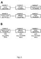
- the first, second and third binding sites function as three autonomous modules each binding a different target site (see Figure 2A ). Due to the inclusion of the first binding site which is capable of specifically binding to a cell surface molecule at the cell surface of the tumor cell (a tumor antigen on the surface of a tumor cell to be eliminated that is accessible to molecules from the exterior of the cell), molecules according to the invention specifically attach to the tumor cells carrying said tumor antigen. In addition, molecules according to the invention also bind specifically to the immune cells carrying the cell surface molecule to which the second binding site specifically binds (e.g. T cells). By this combination of first and second binding site, the molecule according to the invention is capable of recruiting immune cells specifically to the tumor cells to be eliminated.
- the molecule according to the invention also comprises a binding site capable of binding to a checkpoint molecule (third binding site).
- a binding site capable of binding to a checkpoint molecule third binding site.
- the binding interaction between said third binding site and said checkpoint molecule must be such that, if the molecule according to the invention binds to said checkpoint molecule through its third binding site, this precludes binding of said checkpoint molecule to its endogenous ligand or receptor (i.e. binding of said third binding site to said checkpoint molecule prevents checkpoint signaling through said checkpoint molecule).
- binding of the molecule according to the invention through its third binding site to the checkpoint molecule has the effect that if said checkpoint molecule is a checkpoint receptor ligand present on the tumor cell, said checkpoint receptor ligand cannot bind to its corresponding checkpoint receptor on the immune cell anymore and thus the immune cell cannot receive an antiphagocytic checkpoint signal (a "Do not eat me” signal) from said tumor cell.
- the third binding site is brought into close proximity of the tumor cell to be eliminated and the immune cell.
- the checkpoint molecule to which said third binding site specifically binds is a checkpoint receptor ligand.
- the third binding site binds, due to the spatial proximity, preferably to a checkpoint receptor ligand on the tumor cell (rather than on some other cell).
- the first binding site of the molecule according to the invention should be capable of specifically binding to a cell surface molecule on the tumor cells to be eliminated.
- said cell surface molecule at the cell surface of said tumor cell is specific for said tumor cell.
- CD33 is a cell surface molecule that is highly expressed on AML (acute myeloid leukemia) cells.
- CD20 is a cell surface molecule that is expressed on B cell lymphomas and leukemias (but also on normal B cells). Further examples of tumor markers are shown in Table 2 below. Moreover, an extensive overview of tumor markers is available e.g. from http://www.proteinatlas.org/. The present invention is focused on the cell surface molecule CD33.
- cell surface molecules that are specific for certain tumor cells can be identified by immunostaining in combination with flow cytometry or immunofluorescence staining of histological sections.
- the second binding site of the molecule according to the invention should, as indicated above, have binding specificity for a cell surface molecule at the cell surface of immune cells to be recruited to the tumor cells.
- Said cell surface molecule at the cell surface of said immune cell/immune cells is specific for said immune cell(s).
- CD16 is a cell surface molecule that is expressed specifically on natural killer cells (NK cells) and macrophages, monocytes and dendritic cells.
- CD3 is expressed on T cells. Further examples are shown in Table 3. The present invention is focused on CD3.
- the third binding site of the molecule according to the invention should, as discussed above, have binding specificity for a checkpoint receptor ligand present at the cell surface of the tumor cell to be eliminated (which carries the cell surface molecule to which said first binding site binds).
- this interaction should prevent checkpoint signaling mediated by said checkpoint molecule (i.e. upon binding of said third binding site to said checkpoint molecule, said checkpoint molecule is e.g. not capable of transmitting to said immune cell an inhibitory signal that prevents the immune cell from attacking/destroying said tumor cell) or of transmitting a regulatory suppression signal to said immune cell (such as a T cell suppression signal)).
- the present invention relates to PD-L1 as checkpoint molecule at the cell surface of said tumor cell.
- the present invention relates to PD-1 as checkpoint molecule at the cell surface of said immune cell(s).
- SIRP ⁇ signal regulatory protein ⁇ , also known as CD172a
- CD172a checkpoint receptor that is specifically expressed on myeloid cells (e.g. macrophages).
- the endogenous checkpoint receptor ligand that binds to SIRP ⁇ and activates the receptor is the membrane protein CD47, which is broadly expressed on various cell types as marker of "self'.
- CD47 binding to SIRP ⁇ functions as an antiphagocytic checkpoint signal (a "Don't eat me” signal) to phagocytic cells, in particular macrophages, i.e.
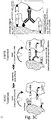
- the checkpoint receptor SIRP ⁇ upon binding to its checkpoint receptor ligand CD47, the checkpoint receptor SIRP ⁇ provides a signal to the immune cell that prevents the immune cell from phagocytosing the cell carrying the CD47 molecule (see Figure 3 A) .
- CD47 is for example present on red blood cells and prevents the clearance of these cells by phagocytic cells.
- CD47 is also often highly overexpressed on cancer cells and correlates with a poor prognosis (Majeti et al., 2009; Chao et al., 2010).
- a blockade of the SIRP ⁇ -CD47 interaction by an anti-CD47 antibody increases phagocytosis of tumor cells tremendously and hinders the tumor cells to evade phagocytic destruction by the immune cell through overexpression of CD47 (Willingham et al., 2012).
- an anti-CD47 antibody and a tumor-specific mAB e.g. rituximab, anti-CD20 mAB
- a tumor-specific mAB e.g. rituximab, anti-CD20 mAB
- the tumor marker could e.g. be CD20 and the tumor-specific monoclonal antibody rituximab.
- a molecule that combines a binding site that specifically binds to a marker of the tumor cell to be eliminated, a binding site that specifically binds to a marker of an immune cell and a binding site that specifically binds to a checkpoint molecule efficiently eliminates the tumor cell while at the same time reducing side effects due to systemic blockage of checkpoint signaling. It is further disclosed that a molecule that combines a binding site that is specific for e.g.
- CD33 (which is found on AML cells), a binding site that binds to CD16 (a marker for immune cells, in particular macrophages, NK cells, monocytes and dendritic cells) and a binding site that binds to the checkpoint receptor ligand CD47 (thus inhibiting antiphagocytic checkpoint signaling from the tumor cell through SIRP ⁇ to the immune cell) efficiently eliminates the AML blasts while at the same time avoiding side effects due to systemic blockage of CD47-SIRP ⁇ checkpoint signaling that otherwise may occur (see Figure 3 C) .
- the concept underlying the present invention was to provide for a combination of tumor directed targeting using a high affinity domain ("first binding site”), with the recruitment of an immune cell using a second domain capable of specifically binding to an immune cell (“second binding site”), together with a simultaneous interference with an immune checkpoint, using a third domain that is capable of specifically binding to a checkpoint molecule on the cell surface of the tumor cell (“third binding site”).
- first binding site a high affinity domain
- second binding site capable of specifically binding to an immune cell
- third domain that is capable of specifically binding to a checkpoint molecule on the cell surface of the tumor cell
- the binding through the third binding site to an immune checkpoint allows for the activation of the immune cells by blocking inhibitory checkpoint signals at the tumor site. Because it is typically only a local interference with the checkpoint(s), there is no targeting effect to non-tumorigenic cells, and there is also a decrease of effects caused by binding to cells that lack the ligand for the "first binding site", but contain the ligand for the "third binding site”.
- the third binding site does not have a targeting effect and thus delivers a checkpoint blockade specifically to cells expressing the tumor antigens, since the native checkpoint domains interact weekly with their receptors.
- strong binding to the tumor cells depends on the binding (through the first binding site) to the tumor antigen. Therefore, the endogenous extracellular domain of an immune checkpoint receptor/co-receptor (“third binding site”) fused to the antibody or derivative thereof (“first binding site” and "second binding”) will not target cells on their own but will activate immune cells at the tumor site.
- the molecule as described herein can easily be designed to target different tumor cells by exchanging the domain including the first binding site (module 1 in Figure 2A ) for a domain having a first binding site with a different binding specificity, namely a binding specificity for a cell surface molecule that is specific for (other) tumor cells to be targeted.
- a further module with binding affinity for a second cell surface molecule of said tumor cell may be included in the molecule according to the invention.
- the effector-recruiting module (module 2 in Figure 2A ) can be chosen such that the most suitable type of immune cell is recruited as effector cell, depending on the field of application.
- T cells may be recruited as effector cells in melanoma (cf. treatment with ipilimumab, pembrolizumab or nivolumab).
- CD3/T cells may be recruited as effector cells in NHL and relapsed or refractory B-cell precursor acute lymphoblastic leukemia (cf. treatment with blinatumomab).
- the immune modulation achieved by the binding of the third binding site (included in module 3 of Figure 2A ) to the checkpoint molecule can be regulated.
- a higher binding affinity and a higher local concentration of the third binding site can be achieved.
- the molecule according to the invention can be specifically adapted for different indications.
- said tumor cell is an AML (acute myeloid leukemia) cell.
- said cell surface molecule at the cell surface of said tumor cell is specific for said tumor cell.
- said immune cell is/immune cells are T cells. More preferably, said immune cell is/immune cells are gamma delta T cells. T cells have the advantage of a strong cell proliferation after activation due to clonal expansion.
- said cell surface molecule at the cell surface of said tumor cell is CD33
- said cell surface molecule at the cell surface of said immune cell(s) is CD3
- said checkpoint molecule is PD-L1 (programmed death-ligand 1).
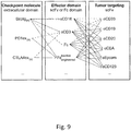
- said first binding site, said second binding site and said third binding site are one of the following combinations (see also Figure 9 ): said first binding site is formed by an ⁇ CD33 scFv, said second binding site is formed by an ⁇ CD3 scFv and said third binding site is formed by PDlex ("PD1ex" designates the extracellular domain of PD1), wherein, preferably, only one or more than one copy of PDlex is present in the molecule according to the invention;
- said cell surface molecule at the cell surface of said immune cell(s) is specific for said immune cell(s).
- said checkpoint molecule is a checkpoint receptor ligand and/or said checkpoint molecule is present at the cell surface of said tumor cell (i.e. of the cells carrying the cell surface molecule to which the first binding site binds).
- said tumor cell overexpresses said checkpoint molecule.
- the affinity of said first binding site for said cell surface molecule at the cell surface of said tumor cell is higher than the affinity of said third binding site for said checkpoint molecule, preferably by at least a factor of 10, more preferably by at least a factor of 25, even more preferably by at least a factor of 50.
- the affinity of said second binding site for said cell surface molecule at the cell surface of said immune cell(s) is higher than the affinity of said third binding site for said checkpoint molecule, preferably by at least a factor of 10, more preferably by at least a factor of 25, even more preferably by at least a factor of 50.
- the affinity of said first binding site for said cell surface molecule at the cell surface of said tumor cell is within a range of from 0.1 to 200 nM, preferably within a range of from 1 nM to 50 nM
- the affinity of said second binding site for said cell surface molecule at the cell surface of said immune cell/immune cells is within a range of from 0.1 to 200 nM, preferably within a range of from 1 to 50 nM
- the affinity of said third binding site for said checkpoint molecule is within a range of from 100 nM to 5 ⁇ M, preferably within a range of from 500 nM to 3 ⁇ M.
- the relative affinities of the different binding sites defined above, and the low to medium affinity of the third binding site for its target as defined above have the effect that the molecule according to the invention is capable of binding tightly and specifically to the tumor cell to be eliminated and the effector cell; the much lower affinity of the third binding site for its target is not sufficient to cause stable association with other cells that the molecule may encounter before or after binding to the tumor cell and/or the effector cell, but only allows the molecule according to the invention to locally inhibit checkpoint signaling between the tumor cell and the effector cell by competitively inhibiting the interaction between the checkpoint receptor and the checkpoint receptor ligand. As a consequence, side effects due to inhibition of checkpoint signaling on other cells are reduced.
- FIG. 2B illustrates how a molecule with affinities as described above may be achieved.
- the depicted molecule comprises an scFv as first module (which includes the first binding site providing specificity for the tumor cell to be eliminated), an scFv as second module (which includes the second binding site for recruitment of the immune cell) and a third module formed from an endogenous extracellular domain (EED) of a checkpoint receptor, e.g. of SIRP ⁇ , with only a low to medium physiological affinity for its checkpoint receptor ligand (in the example of SIRP ⁇ : for CD47).
- EED extracellular domain
- said cell surface molecule at the cell surface of said tumor cell is CD33 (see Table 2).
- said cell surface molecule at the cell surface of said immune cell(s) should not be an immune checkpoint molecule.
- said cell surface molecule at the cell surface of said immune cell(s) is CD3 (see Table 3).
- binding of said second binding site to said cell surface molecule at the cell surface of said immune cell/immune cells sends an activation-inducing signal to said immune cell.
- said second binding site is capable of binding to CD3.
- said checkpoint molecule is PD-L1.
- CD33 serves as a well-established target in AML therapy (Krupka et al., 2014; Petersdorf et al., 2013). It has further been used in a BiTE format demonstrating significant inhibition of tumor growth (Aigner et al., 2013).
- the molecule as defined above can be used as a successful tool in AML therapy engaging NK cells and macrophages instead of T cells. Locally blocking the "Don't eat me" immune checkpoint of macrophages is beneficial for tumor clearance.
- said molecule is a protein.
- said third binding site is located N-terminally of said first binding site and said second binding site or C-terminally of said first binding site and said second binding site. This has the advantage that, if the third binding site has a lower binding affinity for its target molecule than the first binding site for its target molecule and than the second binding site for its target molecule, the mechanical forces exerted on the linkage between the tumor cell to be eliminated and the immune cell cannot disrupt the interaction of the third binding site with its target molecule.
- said molecule further comprises a fourth binding site, wherein said fourth binding site is capable of specifically binding to a cell surface molecule at the cell surface of said tumor cell.
- said cell surface molecule to which said fourth binding site is capable of specifically binding is the same as the cell surface molecule to which said first binding site is capable of specifically binding.
- Inclusion of two binding sites which specifically bind to the same cell surface molecule at the cell surface of the tumor cell to be eliminated allows to increase the affinity of the molecule according to the invention for the tumor cells to be eliminated.
- cell surface molecule to which said fourth binding site is capable of specifically binding is a different cell surface molecule than the cell surface molecule to which said first binding site is capable of specifically binding.
- the combination of said cell surface molecule to which said first binding site is capable of specifically binding and said cell surface molecule to which said fourth binding site is capable of specifically binding is specific for said tumor cell.
- the molecule comprises an scFv (single chain fragment variable) which constitutes a part of said molecule, wherein said first binding site of said molecule (i.e. of said molecule according to the invention) is formed by said scFv.
- scFv single chain fragment variable
- said molecule further comprises a second copy of said second binding site (i.e. another "second binding site” of the same structure and further characteristics as the first "second binding site”).
- a second copy of said second binding site i.e. another "second binding site” of the same structure and further characteristics as the first "second binding site”.
- the molecule comprises an scFv (single chain fragment variable) which constitutes a part of said molecule (i.e. of said molecule according to the invention), wherein said second binding site of said molecule (i.e. of said molecule according to the invention) is formed by said scFv.
- scFv single chain fragment variable
- said molecule further comprises a second copy (and, possibly, even a third copy) of said third binding site (i.e. another "third binding site” having the same structure as the first "third binding site”).
- a second copy and, possibly, even a third copy of said third binding site (i.e. another "third binding site” having the same structure as the first "third binding site”).
- said third binding site of said molecule is not formed by an antibody or a fragment of an antibody.
- said third binding site of said molecule (i.e. of said molecule according to the invention) is not formed by an scFv.
- said molecule comprises a fragment of a checkpoint receptor capable of binding to said checkpoint molecule, which fragment of a checkpoint receptor constitutes a part of said molecule (i.e. of said molecule according to the invention), wherein said third binding site of said molecule (i.e. of said molecule according to the invention) is formed by said fragment of a checkpoint receptor capable of binding to said checkpoint molecule.
- said third binding site is formed by a fragment of an immune cell checkpoint receptor, preferably by an endogenous extracellular domain (EED) of an immune cell checkpoint receptor, wherein, preferably, said molecule does not comprise any other part of said immune cell checkpoint receptor besides said endogenous extracellular domain.
- EED endogenous extracellular domain
- a molecule according to the invention may be referred to as a "local inhibitory checkpoint antibody derivative" (liCAD).
- said tumor cell is a cell of an acute myeloid leukemia (AML).
- AML acute myeloid leukemia
- said molecule is for use as a medicament.
- said molecule is for use in the treatment of a cancer selected from AML (acute myeloid leukemia).
- said molecule is for administration to a patient in need thereof, wherein, preferably, said patient is a human.
- said patient is suffering from a tumor, more preferably from cancer (i.e. from a malignant tumor), more preferably from a blood cancer, even more preferably from AML (acute myeloid leukemia).
- cancer i.e. from a malignant tumor
- AML acute myeloid leukemia
- said tumor cell is a cell of the tumor which said patient is suffering from.
- said tumor cell is a cancer cell of the cancer which said patient is suffering from.
- said patient is suffering from AML and either the AML of said patient has returned after previous treatment (i.e. there was a relapse) or the patient did not respond to a previous treatment (i.e. the cancer was refractory).
- said previous treatment was not a treatment with the molecule according to the invention.
- said cancer is AML
- the present invention relates to a pharmaceutical composition
- a pharmaceutical composition comprising the molecule as defined in any of the embodiments above, wherein, preferably, said pharmaceutical composition further comprises a pharmaceutically acceptable carrier.
- the present invention relates to a molecule as defined in any of the embodiments for use in the treatment of cancer (i.e. from a malignant tumor).
- said patient, said tumor and said cancer are preferably as defined in any of the embodiments above.
- binding site refers to a part or region of a molecule that is responsible for selective binding of said molecule to a target molecule of interest (e.g. an antigen, ligand, receptor or inhibitor).
- a molecule or binding site A is "capable of specifically binding to" a certain binding partner, for example a molecule B. This is meant to designate that molecule or binding site A, in the presence of molecule B and other molecules, binds to molecule B, but does not bind in a significant amount to other molecules that lack the structural motive which molecule or binding site A interacts with in the structure of molecule B.
- a "cell surface molecule at the cell surface of a cell” is a molecule that is present (or at least part of which is present) at the exterior surface of said cell, such that the molecule may undergo a binding interaction with e.g. a soluble molecule that exists in the exterior environment of said cell or with a cell surface molecule at the cell surface of another cell that is spatially close to said cell.
- An example for a cell surface molecule at the cell surface of a cell is a transmembrane protein in the cell membrane of said cell or an integral membrane protein in the cell membrane of said cell with an extracellular domain that is accessible to other molecules from outside of said cell.
- the present application indicates that a certain cell surface molecule/molecule at the cell surface of a cell or combination of cell surface molecules "is specific" for a certain cell type.
- it means that said cell surface molecule/molecule at the cell surface of a cell or combination of cell surface molecules is present at the cell surface of said cell type and at the cell surface of one or a few other cell types besides said cell type, but is not present (or only present at a negligible level) at the cell surface of other cell types.
- a cell surface molecule that is specific for a certain type of tumor cell is known as a "tumor marker”.
- tumor cell this may be a cell of a malignant or benign tumor (i.e. of a malignant or benign neoplasm).
- tumor cell refers to a cell of a malignant tumor.
- Tumor cells can form a solid tumor, in which the tumor cells are massed together, or exist as dispersed cells, thus forming a non-solid tumor, as in leukemia.
- Cancer refers to a diseases caused by the uncontrolled, abnormal growth of cells that can spread to adjoining tissues or other parts of the body.
- the cells of a cancer (such as the cells of a solid cancer, e.g. a breast cancer, or the cells of a blood cancer, e.g. a malignant B cell tumor) are malignant tumor cells.
- an "immune cell” is a cell of hematopoietic origin that plays a role in the immune response.
- the term includes lymphocytes (such as B cells and T cells), natural killer cells and myeloid cells (such as monocytes, macrophages, eosinophils, mast cells, basophils, and granulocytes).
- CD16 refers to the Fc receptor Fc ⁇ RIIIa as well as the Fc receptor Fc ⁇ RIIIb.
- the term refers to the Fc receptor FcyRIIIa.
- the Fc receptor FcyRIIIa (CD16a) is present in particular at the cell surface of NK cells and macrophages, the Fc receptor FcyRIIIb (CD16b) at the cell surface of neutrophils.
- checkpoint molecule is a molecule involved in immune checkpoint signaling of an immune cell, wherein a checkpoint molecule may either be a "checkpoint receptor” or “immune checkpoint receptor” (i.e. a receptor at the cell surface of an immune cell regulated by said immune checkpoint signaling) or a "checkpoint receptor ligand” (i.e. a ligand of such a checkpoint receptor, typically at the cell surface of another cell). Binding of the checkpoint receptor ligand to the immune checkpoint receptor sends an inhibitory/antiphagocytic signal to the immune cell and thus prevents the immune cell from becoming activated and attacking/phagocytosing the cell carrying the checkpoint receptor ligand at its cell surface.
- checkpoint receptors are SIRP ⁇ (signal regulatory protein alpha), PD-1, CTLA4, CD40L and TIM3.
- checkpoint receptor ligands are CD47, PD-L1, CD80/CD86, CD40, Gal9.
- An endogenous extracellular domain (EED) of an immune cell checkpoint receptor is a domain or part of said immune checkpoint receptor that is located outside of the immune cell and that is able to bind to the corresponding checkpoint receptor ligand of said immune checkpoint receptor.
- Endogenous SIRP ⁇ checkpoint receptor for example, comprises three extracellular immunoglobulin-like domains which form the EED of SIRP ⁇ checkpoint receptor; this EED of SIRP ⁇ checkpoint receptor mediates binding of SIRP ⁇ checkpoint receptor to its checkpoint receptor ligand CD47. Specifically, binding of SIRP ⁇ checkpoint receptor to its checkpoint receptor ligand CD47 is mediated by the N-terminal immunoglobulin-like domain of SIRP ⁇ .
- the designations "SIRP ⁇ -Ig", “SIRP-Ig” and “SirpIg” are used interchangeably and refer to a CD47-binding immunoglobulin-like domain of SIRP ⁇ .
- single chain fragment variable refers to an antibody derivative in which the variable domains of the heavy chain and of the light chain of a conventional antibody have been joined to form one chain.
- a linker peptide is inserted between the two chains to allow for proper folding and creation of an active binding site.
- the term "overexpress”, as used e.g. in the context of a certain tumor cell that "overexpresses" a certain molecule means that said molecule is expressed by said tumor cell (such as a malignant B-cell tumor cell) at a higher rate or amount than by a cell of the corresponding non-tumoral cell type (such as a normal, non-malignant B-cell), wherein the cell of the corresponding non-tumoral cell type does not (or only to a negligible extent) express that molecule or, in some embodiments, has a small but detectable expression of said molecule which, however, is lower than the expression of said molecule by said tumor cell.
- said tumor cell such as a malignant B-cell tumor cell
- a cell of the corresponding non-tumoral cell type such as a normal, non-malignant B-cell
- the present application defines that the affinity of a certain binding site A for a certain molecule X is "higher” than the affinity of another binding site B for a certain molecule Y by a certain factor.
- “higher” affinity refers to stronger binding, and thus to a lower K d value (dissociation constant).
- affinity of binding site A for a molecule X is higher than the affinity of binding site B for molecule Y by at least a factor of 10
- affinities measured by flow cytometry This may be done with cells that express only the target of the domain to be measured, not the targets of the other domains of the molecule according to the invention.
- Molecules according to the invention bound to the cells may for example be detected by inclusion of a his tag in the molecule according to the invention and detection of the his tag with a labelled anti-his-tag antibody.
- the K d can be determined by classical methods of K d determination known to the skilled person upon measuring binding at different concentrations of the molecule according to the invention.) or, if this is not possible, surface plasmon resonance measurements (immobilization of the target on the surface, molecule according to the invention as ligand).
- any binding specificities or affinities referred to in the present application refer to the binding specificity/affinity measured in PBS (phosphate buffered saline), pH 7.4, comprising 1% bovine serum albumin and 0.1% sodium azide, at room temperature.
- PBS phosphate buffered saline
- pH 7.4 comprising 1% bovine serum albumin and 0.1% sodium azide
- binding of a certain binding site or molecule to a certain cell surface molecule at the cell surface of an immune cell causes activation of said immune cell, this refers to a situation where upon binding of said binding site or molecule to said cell surface molecule the cytotoxic activity of said immune cell is increased.
- a certain molecule is a protein
- the term designates that the said entire molecule consists of a polymeric chain of amino acids.
- pharmaceutically acceptable carrier refers to a non-toxic, inert, solid, semi-solid, or liquid diluent material or formulation auxiliary of any type. "Pharmaceutically acceptable” in this context is meant to designate that said carrier is compatible with the other ingredients of the pharmaceutical composition and not harmful to the patient that the pharmaceutical composition is administered to.
- pharmaceutically acceptable carriers include, but are not limited to, water-propylene glycol solutions, or aqueous polyethylene glycol solutions.
- patient who is suffering from a tumor refers to a subject who has been tested and found to have tumor cells in his/her body.
- patient who is suffering from cancer refers to a subject who has been tested and found to have cancer cells in his/her body.
- the presence of tumor/cancer cells may be diagnosed using any suitable method, including but not limited to, biopsy, x-ray, blood test, etc.
- a molecule according to the invention that comprises a binding site that allows for specific targeting of tumor cells, a binding site that allows to recruit an immune cell as effector cell and a binding site that allows for local checkpoint inhibition (e.g. in a multi-specific antibody derivative including such three binding sites) provides for tumor specific effector cell recruitment and efficient killing of said tumor cells while keeping the systemic blocking of said checkpoint at a minimum, and thus allows for treatment of cancer while reducing the side effects.
- the present inventors have found that if the molecule according to the invention includes a third binding site with only a low affinity for its checkpoint molecule target and/or includes a third binding site which has a much weaker affinity for its target molecule than the affinity of the first and second binding sites for their respective target molecules, this leads to surprising effects with regard to a further reduction of systemic side effects (which otherwise may still occur to some degree through immune cells attacking other cells besides the tumor cells to be targeted).
- exemplary molecules according to the present invention is described as well as details on cell lines, patients, the preparation of various cells, assays for thermal stability, the detection of binding by flow cytometry, the quantitative determination of cell surface antigens, assays for K D determination, internalization assays by flow cytometry and/or confocal microscopy, assays for determining antibody-dependent cellular cytotoxicity (ADCC) and antibody-dependent cellular phagocytosis (ADCP) and antibody-dependent cellular cytotoxicity in primary AML patient samples.
- ADCC antibody-dependent cellular cytotoxicity
- ADCP antibody-dependent cellular phagocytosis
- SIRP ⁇ -antiCD33 licMAB SIRP ⁇ -antiCD33 licMAB, 2xSIRP ⁇ -antiCD33 licMAB and antiCD33 mAb construction and production
- the antiCD33 variable light (VL) and variable heavy (VH) were generated using custom gene synthesis (GeneArt, Thermo Fisher Scientific).
- the antiCD33 VL was subcloned into the pFUSE2-CLIg-hk vector (InvivoGen) and the antiCD33 VH into the pFUSE-CHIg-hG1 vector (InvivoGen).
- the N-terminal Ig-like V-type domain of SIRP ⁇ (residues 1-120) was synthesized using custom gene synthesis (GeneArt, Thermo Fisher Scientific) and subcloned into the N-terminus of the antiCD33 light chain (LC) together with a (G 4 S) 4 linker.
- a second SIRP ⁇ -(G 4 S) 4 linker cassette was cloned N-terminal of the SIRP ⁇ -antiCD33 LC to obtain the 2xSIRP ⁇ -antiCD33 light chain.
- the corresponding plasmids were transfected into Expi293F cells (Thermo Fisher Scientific) according to the manufacturer's protocol. After five to seven days, the cell culture supernatant was harvested and licMABs were purified by protein A affinity chromatography.
- SIRP ⁇ -PreSc-antiCD33 was incubated with PreScission protease for 4 h followed by a second round of protein A affinity chromatography.
- LicMABs and mAb were dialyzed against Phosphate Buffered Saline (PBS) and size exclusion chromatography (SEC) of the purified molecules was performed using a Superdex 200 increase 10/300 column (GE Healthcare Life Sciences, Little Chalfont, Buckinghamshire, United Kingdom). LicMABs and mAb were then analyzed by 4-20 % SDS-PAGE (Expedeon) under reducing conditions and visualized by Coomassie Brilliant Blue staining. Protein concentration was measured with a spectrophotometer (NanoDrop, GE Healthcare Life Sciences, Little Chalfont, Buckinghamshire, United Kingdom) and aliquots were stored at -80 °C.
- the MOLM-13 cell line was purchased from the 'Deutsche Sammlung von Mikroorganismen und Zellkulturen' (DSMZ, Leibniz-Institut DSMZ, Braunschweig, Germany) and the Flp-INTM-CHO and Flp-INTM T-RexTM-293 cell lines from Thermo Fisher Scientific (Waltham, Massachusetts, USA).
- the THP-1 and the Jurkat cell line was kindly provided by collaborators and cultured in RPMI 1640 + GlutaMAX (Gibco, Thermo Fisher Scientific) and supplemented with 10% fetal bovine serum (FBS, Gibco, Thermo Fisher Scientific).
- MOLM-13 cells were cultured in RPMI 1640 + GlutaMAX (Gibco, Thermo Fisher Scientific) and supplemented with 20% FBS.
- Flp-INTM-CHO was maintained in Ham's F-12 (Biochrom) media supplemented with 10% FBS and Flp-INTM T-RexTM-293 cell line was cultured in DMEM + GlutaMAX and supplemented with 10% FBS and 2 mM L-glutamine.
- the Flp-INTM-CHO or Flp-INTM T-RexTM-293 cell lines were engineered to stably express either human CD33 (referred to as CHO_CD33 or CHO.exCD33, HEK.exCD33 or HEK293_CD33), human CD16 (referred to as CHO.exCD16), human CD47 (referred to as CHO_CD47 or CHO.exCD47) or human PD-L1 (referred to as HEK293 PD-L1) and maintained in selection media according to manufacturers' instruction.
- the HEK293_CD33 cell line has further been engineered to additionally stably express human PD-L1 (HEK_CD33_PD-L1 cell line).
- MOLM-13 cell line was engineered to stably expressed human PD-L1 and is referred to MOLM-13 _PD-L1 cell line.
- the Expi293FTM and FreestyleTM 293-F cell line was obtained from Thermo Fisher Scientific and cultured in Expi293TM Expression Medium or FreeStryleTM 293 Expression Medium, respectively.
- peripheral blood (PB) or bone marrow (BM) samples were collected from healthy donors (HDs) and patients with AML at initial diagnosis or relapse.
- PB or BM samples from AML patients were cryoconserved at ⁇ - 80°C in 80% FCS and 20% dimethyl sulfoxide (Serva Electrophoresis) until usage.
- PB from HDs was obtained on the day of the experiment.
- PBMCs peripheral blood mononuclear cells
- NK cells NK cells
- monocytes from whole human blood
- PBMCs from AML patients and HDs were separated from PB by density gradient using the Biocoll separating solution (Biochrom), according to the manufacturer's protocol.
- NK cells were either expanded ex vivo by culturing PBMCs under IL-2 stimulus as described previously ( Carlens et al., 2001; Hum.Immunol.; 62(10); 1092 - 1098 ) or freshly isolated by magnetic separation using a human NK cell isolation kit (MACS Miltenyi Biotech) according to the manufacturer's protocol.
- Monocytes were isolated from PBMCs with human CD14 MicroBeads (MACS Miltenyi Biotech) by magnetic separation following the manufacturer's instructions.
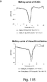
- the thermal stability of the licMABs and mAb was determined by fluorescence thermal shift assays using the CFX96 Touch Real-Time PCR Detection System (Bio-Rad, Kunststoff, Germany) ( Boivin et al., 2013; Protein Expr. Purif.; 91(2); 192 - 206 ). 10 ⁇ g of protein containing 1x SYPRO Orange (Thermo Fisher Scientific) were measured using FAM and SYBR Green I filter pairs.
- MOLM-13, SEM, CHO_CD33 and CHO_CD47 cells were stained with 15 ⁇ g/ml of licMABs or mAb followed by staining with a secondary FITC antiHuman IgG Fc antibody (clone HP6017, BioLegend).
- the median fluorescence intensity (MFI) ratio was calculated dividing MFI of the antibody by the MFI of the isotype control.
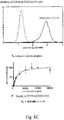
- CD33 equilibrium binding constants (K D , as an avidity measurement) of the licMABs and mAb were determined by calibrated flow cytometry analyses as previously described ( Benedict et al., 1997; J. Immunol. Methods; 201(2); 223-231 ). Briefly, MOLM-13 cells were incubated with licMABs or mAb in concentrations ranging from 0.01 to 15 ⁇ g/ml and stained with a FITC antiHuman IgG Fc (clone HP6017, BioLegend) secondary antibody by flow cytometry. The instrument was calibrated with 3.0-3.4 ⁇ m Rainbow Calibration particles of 8 peaks (BioLegend), the maximum MFI value was set to 100% and all data points were normalized accordingly. The assay was performed in quadruplicates and the values were analyzed by non-linear regression using a one-site specific binding model.
- MOLM-13 cells were grown on a poly-L-lysine (Sigma-Aldrich) coated 96-well plate. Subsequently, cells were incubated with 15 ⁇ g/ml of licMABs or mAb directly labeled with Alexa Fluor 488 (Antibody Labeling Kit, Thermo Fisher Scientific), either on ice-cold water for 2 h or at 37°C for 30, 60 or 120 min. Then, cells were fixed and permeabilized in 20 mM PIPES pH 6.8, 4% formaldehyde, 0.2% Triton X-100, 10 mM EGTA, 1 mM MgCl 2 at room temperature for 10 min, followed by incubation in blocking solution (3% bovine serum albumin in PBS).
- blocking solution 3% bovine serum albumin in PBS
- ADCC Antibody-dependent cellular cytotoxicity
- Target cells MOLM-13 or SEM were labeled with calcein AM (Thermo Fisher Scientific) according to manufacturer's protocol. Calcein-labeled target cells were incubated with freshly isolated or IL-2 expanded NK cells in an effector-to-target (E:T) ratio of 2:1 and licMABs or mAb at different concentrations for 4 h. Target cells were cultured in 10% Triton X-100 to assess the maximum unspecific lysis.
- E:T effector-to-target
- Averaged specific lysis of triplicates or quadruplicates were plotted according to a dose-response curve and analyzed using the integrated four parameter non-linear fit model.
- ADCP Antibody-dependent cellular phagocytosis
- Phagocytosis assay was performed as described previously ( Blume et al., 2009; J. Immunol.; 183(12); 8138-8147 ). Briefly, isolated monocytes were stained with PKH67 (Sigma-Aldrich) according to the manufacturer's instructions and differentiated to macrophages by 20 ng/ml Macrophage-Colony Stimulator Factor (M-CSF) (R&D Systems) in X-VIVO 10 medium (Lonza) supplemented with 10% autologous serum.
- M-CSF Macrophage-Colony Stimulator Factor
- MOLM-13 cells were stained with PKH26 (Sigma-Aldrich) following the manufacturer's instructions and incubated in a 1:2 E:T ratio with licMABs or mAb concentrations ranging from 0.01 nM to 100 nM for 2 h.
- Polybead® Carboxylate Red Dye Microspheres 6 ⁇ m (Polysciences) were used as a positive control and incubation either at 4°C or at 37°C in the presence of 10 ⁇ M Cytochlasin D (Sigma-Aldrich) served as a negative control.
- Ex vivo expanded primary AML cells were co-cultured with freshly isolated healthy donor NK cells, at an E:T ratio of 5:1 in an ex vivo long term culture system as described by Krupka and coworkers ( Krupka et al., 2016; Leukemia; 30(2); 484-491 ) ( Krupka et al., 2014; Blood; 123(3); 356-365 ).
- Antibodies were added at a final concentration of 10 nM.
- CD16 clone B73.1
- CD56 clone HCD 56
- CD33 clone WM53
- CD123 clone 6H6; all antibodies from Biolegend
- flow cytometry with a BD LSR II (Becton Dickinson, Heidelberg, Germany). The percentage of residual CD33 or CD123 positive cells in treated cultures relative to control cultures was used to determine licMAB-mediated cellular cytotoxicity.
- a molecule comprising a binding site with specificity for CD33 (to bind to CD33-positive leukemic cells), a binding site with specificity for CD16 (for recruitment of immune cells as effector cells), and a binding site with specificity for the checkpoint molecule CD47 (for inhibition of antiphagocytic checkpoint signaling) and several related constructs were generated and tested. Since these molecules included antibody-derived binding domains, they are referred to as a "local inhibitory checkpoint antibody derivatives" (liCADs).
- SIRP-Ig the extracellular N-terminal Ig variable domain of SIRP ⁇ (herein called SIRP-Ig or SirpIg) was used, which has been shown to be sufficient for CD47 binding (Barclay et al., 2009).
- SIRP-Ig the extracellular N-terminal Ig variable domain of SIRP ⁇
- SirpIg the extracellular N-terminal Ig variable domain of SIRP ⁇
- molecules carrying two copies of SIRP-Ig were designed.
- CD16 also known as Fc gamma receptor IIIa (FcyRIIIa) is expressed on NK cells, dendritic cells (DCs) and macrophages and mediates antibody dependent cellular cytotoxicity (ADCC) or antibody dependent phagocytosis (ADCP), respectively (Guilliams et al., 2014).
- FcyRIIIa Fc gamma receptor IIIa
- DCs dendritic cells
- ADCC antibody dependent cellular cytotoxicity
- ADCP antibody dependent phagocytosis
- an anti-CD33 scFv (derived from gemtuzumab ozogamicin) is expressed (module 3).
- CD33 is a tumor specific marker that is highly overexpressed in acute myeloid leukemia (AML) and has successfully been used as tumor target before (Larson et al., 2005; Krupka et al., 2014).
- AML acute myeloid leukemia
- an anti-CD47 scFv triplebody control
- SirpIg_CV1 binds to CD47 with much higher affinity (1 pM) compared to SIRP-Ig (1 ⁇ M) (Weiskopf et al., 2013).
- the protein domains used had the following sequences:
- Vh CD16scFv (3G8 clone) (SEQ ID NO: 2):
- Vh CD33scFv Amino acid sequence of Vh CD33scFv (SEQ ID NO: 5):
- checkpoint binding module third binding site, e.g. Sirp-Ig, PDlex, CTLA4ex, ⁇ PDL1
- Vh und VI domains and between the scFvs, GGGS-based linkers were included.
- the used mutations in this example were S239D and I332E, but others may also be used.
- Fc-engineered mutations may be produced and used for example in accordance with: Engineered antibody Fc variants with enhanced effector function Greg A. Lazar, Wei Dang, Sher Karki, Omid Vafa, Judy S. Peng, Linus Hyun, Cheryl Chan, Helen S. Chung, Araz Eivazi, Sean C. Yoder, Jost Learnmetter, David F. Carmichael, Robert J. Hayes, and Bassil I. Dahiyat
- SEQ ID NO: 28 is the combination of SEQ ID NO: 26 and 27 with a (GGGGS) 4 -linker in between.
- SEQ ID NO: 22 is the sequence for the constant regions of the IgGI format and includes the domains CH1, hinge, CH2 and CH3; this is the wildtype sequence.
- SEQ ID NO: 24 shows the same sequence for the constant regions of the IgG1 format but with mutations S239D and I332E; this is the mutated sequence for producing an Fc-engineered fragment. This is also the sequence which was used in embodiments of the present disclosure; the mutations are located in the CH2 domain.
- the Fc-fragment and the Fc-engineered fragment consist of domains CH2 and CH3 only. In all of these constant regions of the IgG1 format, the numbering according to Kabat is used.
- SEQ ID NO: 24 the entire constant region including domains CH1, hinge, CH2 and CH3 is shown; however, it is clear to a person skilled in the art that the corresponding Fc-engineered fragment only contains the respective CH2 and CH3 domains.
- linkers For the various molecules according to the present invention a number of linkers were used, shown herein as SEQ ID NO: 6 - SEQ ID NO: 21.
- SEQ ID NO: 6 is a GGGS-linker and SEQ ID NO: 7-13 are tandem repeats thereof, wherein the linker occurs 2-8 times
- SEQ ID NO: 14 is a GGGGS-linker and SEQ ID NO: 15-21, again, are tandem repeats thereof, wherein the linker occurs 2-8 times.
- These linkers allow for an occurrence of respective domains/binding sites (first binding site, second binding site and third binding site) within molecules (liCADs and licMABs) of the present invention.
- PDL1 refers to the programmed death-ligand 1 which binds to its corresponding receptor PD1.
- PDlex is the extracellular domain of PD1.
- the liCADs were expressed in Drosophila melanogaster Schneider 2 (S2) cells and purified from the insect cell medium after secretion.
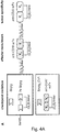
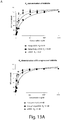
- the purification strategy included a capture step (Ni-NTA affinity chromatography) via the N-terminal hexa-histidine (6xHIS) tag, followed by anion exchange (IEC) and size exclusion chromatography (SEC), resulting in monomeric soluble protein ( Figure 4B ).
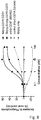
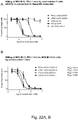
- a redirected lysis (RDL) assay was carried out with the MOLM 13 cell line, which expresses CD33 and CD47 at high level.
- the RDL assay functions analogous to an antibody dependent cellular cytotoxicity (ADCC) assay, but recruitment and activation of NK cells is not mediated by the Fc domain of an antibody, but by the scFv against CD16.
- ADCC antibody dependent cellular cytotoxicity
- effector cells isolated peripheral blood mononuclear cells (PBMCs) that had been expanded as described previously (Alici et al., 2008) were used.
- Effector cells and calcein labeled target cells were mixed in a ratio of 2:1 and incubated with increasing protein concentrations for 4 hours at 37 °C / 5 % CO 2 . Afterwards fluorescence intensity of calcein was measured from the cell supernatant using the Infinite M1000 PRO (Tecan) plate reader.
- CD47 is a marker of self and thus expressed on every cell, it is necessary to avoid killing all CD47 positive cells.
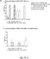
- a preferential RDL assays was carried out to show that liCADs preferentially eliminate CD47/CD33 double positive cells over CD47 single positive cells ( Figure 7B, C ).
- the preferential lysis assay was carried out using CD47+ single positive HEK cells mixed with CD47+, CD33+ double positive HEK cells. Effector cells and calcein stained target cells (one reaction with single positive stained and one reaction with double positive stained) were mixed in a 2:1 ratio again and incubated with the maximal used protein concentration in the redirected lysis assay or with the evaluted EC50 value for 4 hours at 37 °C / 5 % CO 2 . Afterwards fluorescence intensity of calcein was measured from the cell supernatant using the Infinite M1000 PRO (Tecan) plate reader.
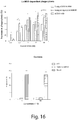
- CD47+ CD33+ HEK cells are preferentially killed in case of the Sirp-Ig-CD16-CD33 and Sirp-Ig-Sirp-Ig-CD16-CD33 liCAD, but not in case of a control triplebody that targets CD47 with high affinity.
- our low affinity molecules are comparable to a bispecific control that does not target CD47 and at the EC50 value these molecules do not redirect killing of CD47+ cells at all in contrast to the triplebody control.
- CD16 Besides expression on NK cells, CD16 is also expressed on macrophages. Therefore, it was investigated if the liCADs can recruit macrophages as effector cells. To support the results seen in the RDL assays, it was analyzed if the prepared liCADs affect phagocytosis.
- a phagocytosis assay was performed generating M2 macrophages for 5 days in culture and incubation of macrophages with MOLM13 target cells in a 1:2 ratio. Cells were mixed and incubated with increasing amount of LiCAD concentration in serum free conditions for 2 hours at 37 °C / 5 % CO 2 . Afterwards cells were collected and FACS analyzed for macrophages that had taken up target cells.
- the Sirp-Ig-Sirp-Ig-CD16-CD33 liCAD is better in mediating phagocytosis compared to the Sirp-Ig-CD16-CD33 molecule, which suggests indeed an additive affect of the blocking by Sirp-Ig.
- Overall both liCAD molecules perform considerably better than a conventional used mAB against CD47.
- Sirp-Ig only was used to test if blocking of the immune checkpoint alone is enough to induce phagocytosis, which is not the case.
- Table 1 Exemplary antibody formats used in clinical therapy or studies, respectively Format Effector function Tumor specific Checkpoint inhibition
- Rituximab ( ⁇ -CD20) IgG targeting immune checkpoint + - + Ipilimumab, Tremelimumab ( ⁇ -CTLA4)
- CT-011 ⁇ -PD1 BMS-936558, MPDL3280A ( ⁇ -PDL1) IgG + + + Ipilimumab + Bevacizumab ( ⁇ -CTLA4 + ⁇ -VEGF) Galiximab + Rituximab ( ⁇ -PDL1 + ⁇ -CD20)
- CT-011 + Rituximab ( ⁇ -PD1 + ⁇ -CD20) bisepcific IgG + + - Catumaxomab ( ⁇ -CD3 x ⁇ -EpCAM) Ertumaxumab ( ⁇ -CD3 x ⁇ -Her2) F(ab')2
- Antibody constant domains are shown as white rectangles. Variable heavy and light chains are shown in white (tumor antigen specific), dotted (effector cell specific) and dashed line rounded rectangles (immune checkpoint specific). White spikes represent endogenous extracellular domains of immune checkpoint receptors. BiTE, bispecific T cell engager; F(ab')2, Fragment antigen binding; IgG, immunoglobulin G. Single chain fragment variables (scFv) are depicted as two rounded rectangles with a diagonal black line across (e.g. the triplebody at the bottom of the table includes three scFv).
- Table 2 Examples of tumor specific makers, related disease and available immunotherapy format Antigen Disease CD19 NHL, B-ALL CD20 B cell lymphoma Her2/neu Breast cancer CD123 AML CEA Gastrointestinal cancer, lung cancer EPCAM Ovarian cancer, colorectal cancer Table 3 Cell surface molecules on different immune effector cells Immune cells Surface receptors T cells CD3, TCR ⁇ , Nk cells CD16, NKG2D, NKp30, NKp40, LFA1 Macrophages CD89, CD64, CD32a, CD15a, CD16 Monocytes CD89, CD64, CD32a, CD15a, CD16 Neutrophilic Granulocytes CD89, CD64, CD32a, CD16 Table 4: Examples for molecules according to the invention Format Effector activation Tumor specifity Impact on Chekpoint Effector recruitment liCAD -/+ ++ + + liCAD -/+ ++ ++ + licMAB + ++ ++ + licMAB Fc engineered ++ ++ + licMAB +
- variable heavy and light chains are shown in white (tumor antigen specific) and black (effector cell specific).
- white spikes represent endogenous extracellular domains of immune checkpoint receptors.
- the first molecule comprises a SIRP ⁇ -Ig linked to a tumor cell-specific and an immune cell-specific scFv.
- the second molecule comprises two SIRP ⁇ -Igs linked to a tumor cell-specific and an immune cell-specific scFv.
- the third molecule consists of an IgG antibody with variable domains having binding specificity for the tumor cell and further linked to two SIRP ⁇ -Igs.
Landscapes
- Health & Medical Sciences (AREA)
- Chemical & Material Sciences (AREA)
- Immunology (AREA)
- Life Sciences & Earth Sciences (AREA)
- Medicinal Chemistry (AREA)
- Organic Chemistry (AREA)
- General Health & Medical Sciences (AREA)
- Molecular Biology (AREA)
- Genetics & Genomics (AREA)
- Biochemistry (AREA)
- Proteomics, Peptides & Aminoacids (AREA)
- Biophysics (AREA)
- Engineering & Computer Science (AREA)
- Veterinary Medicine (AREA)
- Public Health (AREA)
- Animal Behavior & Ethology (AREA)
- Pharmacology & Pharmacy (AREA)
- Epidemiology (AREA)
- Bioinformatics & Cheminformatics (AREA)
- Mycology (AREA)
- Microbiology (AREA)
- Oncology (AREA)
- Biomedical Technology (AREA)
- Nuclear Medicine, Radiotherapy & Molecular Imaging (AREA)
- Hematology (AREA)
- General Chemical & Material Sciences (AREA)
- Chemical Kinetics & Catalysis (AREA)
- Gastroenterology & Hepatology (AREA)
- Toxicology (AREA)
- Zoology (AREA)
- Cell Biology (AREA)
- Endocrinology (AREA)
- Biotechnology (AREA)
- Medicines That Contain Protein Lipid Enzymes And Other Medicines (AREA)
- Peptides Or Proteins (AREA)
- Medicines Containing Antibodies Or Antigens For Use As Internal Diagnostic Agents (AREA)
- Pharmaceuticals Containing Other Organic And Inorganic Compounds (AREA)
Claims (15)
- Trispezifisches Molekül, umfassend(i) eine erste Bindestelle, geformt durch ein αCD33 scFv, welche in der Lage ist, spezifisch an ein Zelloberflächenmolekül auf der Zelloberfläche einer Tumorzelle zu binden;(ii) eine zweite Bindestelle, gebildet durch ein αCD3 scFv, welche in der Lage ist, spezifisch an ein Zelloberflächenmolekül auf der Zelloberfläche einer Immunzelle/von Immunzellen zu binden, und(iii) eine dritte Bindestelle, gebildet durch die extrazelluläre Domäne von PD1, welche in der Lage ist, spezifisch an ein Checkpoint-Molekül auf der Zelloberfläche der Tumorzelle zu binden, wobei eine oder mehr als eine Kopie der extrazellulären Domäne von PD1 im Molekül vorhanden ist.
- Molekül gemäß Anspruch 1, wobei das Zelloberflächenmolekül auf der Zelloberfläche der Tumorzelle spezifisch für die Tumorzelle ist und/oder wobei das Zelloberflächenmolekül auf der Zelloberfläche der Immunzelle/der Immunzellen spezifisch für die Immunzelle(n) ist.
- Molekül gemäß einem der Ansprüche 1 oder 2, wobei die Immunzelle eine AML (akute myeloide Leukämie)-Zelle ist und/oder wobei die Immunzelle/n (eine) T-Zelle(n) ist/sind.
- Molekül gemäß einem der vorherigen Ansprüche, wobei die Affinität der ersten Bindestelle für das Zelloberflächenmolekül auf der Zelloberfläche der Tumorzelle höher ist als die Affinität der dritten Bindestelle für das Checkpoint-Molekül, vorzugsweise um mindestens den Faktor 10, weiter bevorzugt um mindestens den Faktor 50 und/oder die Affinität der zweiten Bindestelle für das Zelloberflächenmolekül auf der Zelloberfläche der Immunzelle(n) höher ist als die Affinität der dritten Bindestelle für das Checkpoint-Molekül, vorzugsweise um mindestens den Faktor 10, weiter bevorzugt um mindestens den Faktor 25.
- Molekül gemäß einem der vorherigen Ansprüche, wobeidie Affinität der ersten Bindestelle für das Zelloberflächenmolekül auf der Zelloberfläche der Tumorzelle in einem Bereich von 0,1 bis 200 nM liegt, vorzugsweise in einem Bereich von 1 bis 50 nM; und/oderdie Affinität der zweiten Bindestelle für das Zelloberflächenmolekül auf der Zelloberfläche der Immunzelle(n) in einem Bereich von 0,1 bis 200 nM liegt, vorzugsweise in einem Bereich von 1 bis 50 nM; und/oderdie Affinität der dritten Bindestelle für das Checkpoint-Molekül in einem Bereich von 100 nM bis 5 µM liegt, vorzugsweise in einem Bereich von 500 nM bis 3 µM.
- Molekül gemäß einem der vorherigen Ansprüche,wobei das Molekül zusätzlich eine vierte Bindestelle umfasst, wobei die vierte Bindestelle in der Lage ist, spezifisch an ein Zelloberflächenmolekül auf der Zelloberfläche der Tumorzelle zu binden,und/oder wobei das Molekül zusätzlich eine zweite Kopie der zweiten Bindestelle umfasstund/oder das Molekül zusätzlich eine zweite Kopie der dritten Bindestelle umfasst.
- Molekül gemäß einem der vorherigen Ansprüche, wobei das Molekül keinen anderen Teil des Immunzellcheckpointrezeptors neben der extrazellulären Domäne von PD1 umfasst.
- Molekül gemäß einem der vorherigen Ansprüche, wobei die Tumorzelle eine Zelle einer akuten myeloiden Leukämie (AML) ist.
- Molekül gemäß einem der vorherigen Ansprüche, wobei das Molekül als Medikament verwendet wird.
- Molekül zur Verwendung gemäß Anspruch 9, wobei das Molekül für die Behandlung von Krebs verwendet wird.
- Molekül zur Verwendung gemäß Anspruch 10, wobei der Krebs AML (akute myeloide Leukämie) ist.
- Molekül gemäß einem der vorherigen Ansprüche, wobei das Molekül zur Verabreichung an einen Patienten, der dies benötigt, ist.
- Molekül gemäß Anspruch 12, wobei der Patient an Krebs, vorzugsweise an AML (akuter myeloider Leukämie) leidet.
- Pharmazeutische Zusammensetzung, umfassend das Molekül gemäß einem der Ansprüche 1 bis 13.
- Pharmazeutische Zusammensetzung gemäß Anspruch 14, zusätzlich umfassend einen pharmazeutisch verträglichen Trägerstoff.
Priority Applications (1)
| Application Number | Priority Date | Filing Date | Title |
|---|---|---|---|
| EP20165971.1A EP3699197B1 (de) | 2015-11-09 | 2016-11-09 | Trispezifischer antikörper mit kombination von targeting spezifischer tumore und lokaler immunprüfpunkthemmung |
Applications Claiming Priority (2)
| Application Number | Priority Date | Filing Date | Title |
|---|---|---|---|
| EP15193711.7A EP3165536A1 (de) | 2015-11-09 | 2015-11-09 | Neuartiges molekül mit kombination von targeting spezifischer tumore und lokaler immunprüfpunkthemmung |
| PCT/EP2016/077174 WO2017081101A1 (en) | 2015-11-09 | 2016-11-09 | Trispecific molecule combining specific tumor targeting and local immune checkpoint inhibition |
Related Child Applications (2)
| Application Number | Title | Priority Date | Filing Date |
|---|---|---|---|
| EP20165971.1A Division EP3699197B1 (de) | 2015-11-09 | 2016-11-09 | Trispezifischer antikörper mit kombination von targeting spezifischer tumore und lokaler immunprüfpunkthemmung |
| EP20165971.1A Division-Into EP3699197B1 (de) | 2015-11-09 | 2016-11-09 | Trispezifischer antikörper mit kombination von targeting spezifischer tumore und lokaler immunprüfpunkthemmung |
Publications (2)
| Publication Number | Publication Date |
|---|---|
| EP3374394A1 EP3374394A1 (de) | 2018-09-19 |
| EP3374394B1 true EP3374394B1 (de) | 2021-10-20 |
Family
ID=54540891
Family Applications (3)
| Application Number | Title | Priority Date | Filing Date |
|---|---|---|---|
| EP15193711.7A Withdrawn EP3165536A1 (de) | 2015-11-09 | 2015-11-09 | Neuartiges molekül mit kombination von targeting spezifischer tumore und lokaler immunprüfpunkthemmung |
| EP16794597.1A Not-in-force EP3374394B1 (de) | 2015-11-09 | 2016-11-09 | Molekül mit kombination von targeting spezifischer tumore und lokaler immunprüfpunkthemmung |
| EP20165971.1A Active EP3699197B1 (de) | 2015-11-09 | 2016-11-09 | Trispezifischer antikörper mit kombination von targeting spezifischer tumore und lokaler immunprüfpunkthemmung |
Family Applications Before (1)
| Application Number | Title | Priority Date | Filing Date |
|---|---|---|---|
| EP15193711.7A Withdrawn EP3165536A1 (de) | 2015-11-09 | 2015-11-09 | Neuartiges molekül mit kombination von targeting spezifischer tumore und lokaler immunprüfpunkthemmung |
Family Applications After (1)
| Application Number | Title | Priority Date | Filing Date |
|---|---|---|---|
| EP20165971.1A Active EP3699197B1 (de) | 2015-11-09 | 2016-11-09 | Trispezifischer antikörper mit kombination von targeting spezifischer tumore und lokaler immunprüfpunkthemmung |
Country Status (5)
| Country | Link |
|---|---|
| US (1) | US11229700B2 (de) |
| EP (3) | EP3165536A1 (de) |
| AU (1) | AU2016353468B2 (de) |
| CA (1) | CA3000597C (de) |
| WO (1) | WO2017081101A1 (de) |
Families Citing this family (10)
| Publication number | Priority date | Publication date | Assignee | Title |
|---|---|---|---|---|
| CN111448323B (zh) * | 2017-06-07 | 2023-12-01 | 长源赋能(上海)生命科技有限公司 | 精确制导的多功能治疗抗体 |
| US20220298254A1 (en) * | 2017-11-01 | 2022-09-22 | Hummingbird Bioscience Holdings Pte. Ltd. | Cd47 antigen-binding molecules |
| GB201720426D0 (en) * | 2017-12-07 | 2018-01-24 | Hummingbird Bioscience Pte Ltd | CD47 and BCMA antigen-binding molecules |
| WO2019086574A1 (en) * | 2017-11-01 | 2019-05-09 | Hummingbird Bioscience Holdings Pte. Ltd. | Cd47 and cd33 antigen-binding molecules |
| CN112119099A (zh) * | 2018-03-02 | 2020-12-22 | Cdr-生物科技股份有限公司 | 三特异性抗原结合蛋白 |
| EP3802599B1 (de) | 2018-06-03 | 2023-12-20 | LamKap Bio beta AG | Bispezifische antikörper gegen ceacam5 und cd47 |
| TW202028237A (zh) | 2018-09-27 | 2020-08-01 | 美商西建公司 | SIRPα結合蛋白及其使用方法 |
| US11591390B2 (en) | 2018-09-27 | 2023-02-28 | Celgene Corporation | SIRP-α binding proteins and methods of use thereof |
| CA3138058A1 (en) * | 2019-05-20 | 2020-11-26 | Matthew T. Burger | Mcl-1 inhibitor antibody-drug conjugates and methods of use |
| WO2022127901A1 (en) * | 2020-12-18 | 2022-06-23 | Lanova Medicines Development Co., Ltd. | BISPECIFIC ANTIBODIES TARGETING SIRPα AND PD-L1 |
Family Cites Families (4)
| Publication number | Priority date | Publication date | Assignee | Title |
|---|---|---|---|---|
| ES2692268T5 (en) | 2011-03-29 | 2025-02-26 | Roche Glycart Ag | Antibody fc variants |
| US9382329B2 (en) * | 2012-08-14 | 2016-07-05 | Ibc Pharmaceuticals, Inc. | Disease therapy by inducing immune response to Trop-2 expressing cells |
| JOP20170091B1 (ar) * | 2016-04-19 | 2021-08-17 | Amgen Res Munich Gmbh | إعطاء تركيبة ثنائية النوعية ترتبط بـ cd33 وcd3 للاستخدام في طريقة لعلاج اللوكيميا النخاعية |
| EP3330289A1 (de) * | 2016-12-02 | 2018-06-06 | Friedrich-Alexander-Universität Erlangen-Nürnberg | Cd33-, cd16- und cd123-spezifischer einkettiger tribody |
-
2015
- 2015-11-09 EP EP15193711.7A patent/EP3165536A1/de not_active Withdrawn
-
2016
- 2016-11-09 AU AU2016353468A patent/AU2016353468B2/en active Active
- 2016-11-09 US US15/764,788 patent/US11229700B2/en active Active
- 2016-11-09 EP EP16794597.1A patent/EP3374394B1/de not_active Not-in-force
- 2016-11-09 WO PCT/EP2016/077174 patent/WO2017081101A1/en not_active Ceased
- 2016-11-09 CA CA3000597A patent/CA3000597C/en active Active
- 2016-11-09 EP EP20165971.1A patent/EP3699197B1/de active Active
Non-Patent Citations (4)
| Title |
|---|
| C. KRUPKA ET AL: "CD33 target validation and sustained depletion of AML blasts in long-term cultures by the bispecific T-cell-engaging antibody AMG 330", BLOOD, vol. 123, no. 3, 25 August 2015 (2015-08-25), pages 356 - 365, XP055172965, ISSN: 0006-4971, DOI: 10.1182/blood-2013-08-523548 * |
| HERRMANN MONIKA ET AL: "Bifunctional PD-1 x alpha CD3 x alpha CD33 fusion protein reverses adaptive immune escape in acute myeloid leukemia", BLOOD, vol. 132, no. 23, 6 December 2018 (2018-12-06), pages 2484 - 2494, XP002793583 * |
| LASZLO G S ET AL: "T-cell ligands modulate the cytolytic activity of the CD33/CD3 BiTE antibody construct, AMG 330", BLOOD CANCER JOURNAL, vol. 5, August 2015 (2015-08-01), XP002793582 * |
| THOMAS KÖHNKE ET AL: "Increase of PD-L1 expressing B-precursor ALL cells in a patient resistant to the CD19/CD3-bispecific T cell engager antibody blinatumomab", JOURNAL OF HEMATOLOGY & ONCOLOGY, BIOMED CENTRAL LTD, LONDON UK, vol. 8, no. 1, 8 October 2015 (2015-10-08), pages 111, XP021229658, ISSN: 1756-8722, DOI: 10.1186/S13045-015-0213-6 * |
Also Published As
| Publication number | Publication date |
|---|---|
| WO2017081101A1 (en) | 2017-05-18 |
| US20180311348A1 (en) | 2018-11-01 |
| CA3000597A1 (en) | 2017-05-18 |
| EP3699197B1 (de) | 2022-08-03 |
| EP3699197A1 (de) | 2020-08-26 |
| CA3000597C (en) | 2023-04-25 |
| AU2016353468B2 (en) | 2023-07-27 |
| US11229700B2 (en) | 2022-01-25 |
| AU2016353468A1 (en) | 2018-04-26 |
| EP3374394A1 (de) | 2018-09-19 |
| EP3165536A1 (de) | 2017-05-10 |
Similar Documents
| Publication | Publication Date | Title |
|---|---|---|
| EP3699197B1 (de) | Trispezifischer antikörper mit kombination von targeting spezifischer tumore und lokaler immunprüfpunkthemmung | |
| ES2910365T3 (es) | Nuevos polipéptidos biespecíficos contra CD137 | |
| EP3378487B1 (de) | Kombinationstherapie mit t-zell-neuausrichtendem antigenbindendem molekül gegen zelle mit immunsuppressiver funktion | |
| CN109963873B (zh) | 新颖抗cd137抗体和其用途 | |
| ES3046543T3 (en) | Anti cd25 fc gamma receptor antibodies for tumor specific cell depletion in combination with pd-1 antagonists | |
| JP6840682B2 (ja) | 多重特異的抗原結合タンパク質 | |
| EP3144388B1 (de) | T-zell-umleitendes antigenbindendes molekül für zellen mit immunsuppressionsfunktion | |
| JP6822849B2 (ja) | 多重特異的NKp46結合タンパク質 | |
| KR20220041161A (ko) | Mage-a에 특이적으로 결합하는 항원 결합 단백질 | |
| CN110869388A (zh) | 用于肿瘤特异性细胞消耗的Fc优化的抗CD25 | |
| AU2018241625A1 (en) | Improved antigen binding receptor formats | |
| CN116898963A (zh) | 治疗癌症的联合疗法 | |
| CN107922938A (zh) | 抗体 | |
| JP7503215B2 (ja) | Cd30及びcd3に結合する抗体 | |
| CN119278213A (zh) | 针对ctla-4的抗体及其使用方法 | |
| US12410258B2 (en) | Antibodies capable of binding to OX40, variants thereof and uses thereof | |
| RU2839380C1 (ru) | Fc-оптимизированные антитела к cd25 для истощения опухолеспецифических клеток | |
| KR20260022341A (ko) | 항 garp 항체 및 이의 사용 방법 | |
| HK40091509A (zh) | 特异性结合mage-a的抗原结合蛋白 | |
| HK40101561A (zh) | 人ccr8结合剂 | |
| CN116888156A (zh) | 非阻断性人ccr8结合剂 | |
| EA051954B1 (ru) | Мультиспецифические антитела, которые связывают как mait, так и опухолевые клетки | |
| EA052120B1 (ru) | Анти-cd24 композиции и их применения | |
| HK40016585B (zh) | 用於肿瘤特异性细胞消耗的fc优化的抗cd25 |
Legal Events
| Date | Code | Title | Description |
|---|---|---|---|
| STAA | Information on the status of an ep patent application or granted ep patent |
Free format text: STATUS: UNKNOWN |
|
| STAA | Information on the status of an ep patent application or granted ep patent |
Free format text: STATUS: THE INTERNATIONAL PUBLICATION HAS BEEN MADE |
|
| PUAI | Public reference made under article 153(3) epc to a published international application that has entered the european phase |
Free format text: ORIGINAL CODE: 0009012 |
|
| STAA | Information on the status of an ep patent application or granted ep patent |
Free format text: STATUS: REQUEST FOR EXAMINATION WAS MADE |
|
| 17P | Request for examination filed |
Effective date: 20180409 |
|
| AK | Designated contracting states |
Kind code of ref document: A1 Designated state(s): AL AT BE BG CH CY CZ DE DK EE ES FI FR GB GR HR HU IE IS IT LI LT LU LV MC MK MT NL NO PL PT RO RS SE SI SK SM TR |
|
| AX | Request for extension of the european patent |
Extension state: BA ME |
|
| DAV | Request for validation of the european patent (deleted) | ||
| DAX | Request for extension of the european patent (deleted) | ||
| STAA | Information on the status of an ep patent application or granted ep patent |
Free format text: STATUS: EXAMINATION IS IN PROGRESS |
|
| PUAG | Search results despatched under rule 164(2) epc together with communication from examining division |
Free format text: ORIGINAL CODE: 0009017 |
|
| 17Q | First examination report despatched |
Effective date: 20190827 |
|
| RIC1 | Information provided on ipc code assigned before grant |
Ipc: C07K 14/705 20060101ALI20190822BHEP Ipc: C07K 16/28 20060101AFI20190822BHEP |
|
| B565 | Issuance of search results under rule 164(2) epc |
Effective date: 20190827 |
|
| GRAP | Despatch of communication of intention to grant a patent |
Free format text: ORIGINAL CODE: EPIDOSNIGR1 |
|
| STAA | Information on the status of an ep patent application or granted ep patent |
Free format text: STATUS: GRANT OF PATENT IS INTENDED |
|
| RIC1 | Information provided on ipc code assigned before grant |
Ipc: C07K 14/705 20060101ALI20210202BHEP Ipc: C07K 16/28 20060101AFI20210202BHEP Ipc: A61P 35/02 20060101ALI20210202BHEP |
|
| INTG | Intention to grant announced |
Effective date: 20210301 |
|
| GRAJ | Information related to disapproval of communication of intention to grant by the applicant or resumption of examination proceedings by the epo deleted |
Free format text: ORIGINAL CODE: EPIDOSDIGR1 |
|
| STAA | Information on the status of an ep patent application or granted ep patent |
Free format text: STATUS: EXAMINATION IS IN PROGRESS |
|
| GRAS | Grant fee paid |
Free format text: ORIGINAL CODE: EPIDOSNIGR3 |
|
| STAA | Information on the status of an ep patent application or granted ep patent |
Free format text: STATUS: GRANT OF PATENT IS INTENDED |
|
| GRAP | Despatch of communication of intention to grant a patent |
Free format text: ORIGINAL CODE: EPIDOSNIGR1 |
|
| INTC | Intention to grant announced (deleted) | ||
| INTG | Intention to grant announced |
Effective date: 20210628 |
|
| GRAA | (expected) grant |
Free format text: ORIGINAL CODE: 0009210 |
|
| STAA | Information on the status of an ep patent application or granted ep patent |
Free format text: STATUS: THE PATENT HAS BEEN GRANTED |
|
| AK | Designated contracting states |
Kind code of ref document: B1 Designated state(s): AL AT BE BG CH CY CZ DE DK EE ES FI FR GB GR HR HU IE IS IT LI LT LU LV MC MK MT NL NO PL PT RO RS SE SI SK SM TR |
|
| REG | Reference to a national code |
Ref country code: GB Ref legal event code: FG4D |
|
| REG | Reference to a national code |
Ref country code: CH Ref legal event code: EP |
|
| REG | Reference to a national code |
Ref country code: DE Ref legal event code: R096 Ref document number: 602016065183 Country of ref document: DE |
|
| REG | Reference to a national code |
Ref country code: IE Ref legal event code: FG4D |
|
| REG | Reference to a national code |
Ref country code: AT Ref legal event code: REF Ref document number: 1439896 Country of ref document: AT Kind code of ref document: T Effective date: 20211115 |
|
| REG | Reference to a national code |
Ref country code: LT Ref legal event code: MG9D |
|
| REG | Reference to a national code |
Ref country code: NL Ref legal event code: MP Effective date: 20211020 |
|
| REG | Reference to a national code |
Ref country code: AT Ref legal event code: MK05 Ref document number: 1439896 Country of ref document: AT Kind code of ref document: T Effective date: 20211020 |
|
| PG25 | Lapsed in a contracting state [announced via postgrant information from national office to epo] |
Ref country code: RS Free format text: LAPSE BECAUSE OF FAILURE TO SUBMIT A TRANSLATION OF THE DESCRIPTION OR TO PAY THE FEE WITHIN THE PRESCRIBED TIME-LIMIT Effective date: 20211020 Ref country code: LT Free format text: LAPSE BECAUSE OF FAILURE TO SUBMIT A TRANSLATION OF THE DESCRIPTION OR TO PAY THE FEE WITHIN THE PRESCRIBED TIME-LIMIT Effective date: 20211020 Ref country code: FI Free format text: LAPSE BECAUSE OF FAILURE TO SUBMIT A TRANSLATION OF THE DESCRIPTION OR TO PAY THE FEE WITHIN THE PRESCRIBED TIME-LIMIT Effective date: 20211020 Ref country code: BG Free format text: LAPSE BECAUSE OF FAILURE TO SUBMIT A TRANSLATION OF THE DESCRIPTION OR TO PAY THE FEE WITHIN THE PRESCRIBED TIME-LIMIT Effective date: 20220120 Ref country code: AT Free format text: LAPSE BECAUSE OF FAILURE TO SUBMIT A TRANSLATION OF THE DESCRIPTION OR TO PAY THE FEE WITHIN THE PRESCRIBED TIME-LIMIT Effective date: 20211020 |
|
| PG25 | Lapsed in a contracting state [announced via postgrant information from national office to epo] |
Ref country code: IS Free format text: LAPSE BECAUSE OF FAILURE TO SUBMIT A TRANSLATION OF THE DESCRIPTION OR TO PAY THE FEE WITHIN THE PRESCRIBED TIME-LIMIT Effective date: 20220220 Ref country code: SE Free format text: LAPSE BECAUSE OF FAILURE TO SUBMIT A TRANSLATION OF THE DESCRIPTION OR TO PAY THE FEE WITHIN THE PRESCRIBED TIME-LIMIT Effective date: 20211020 Ref country code: PT Free format text: LAPSE BECAUSE OF FAILURE TO SUBMIT A TRANSLATION OF THE DESCRIPTION OR TO PAY THE FEE WITHIN THE PRESCRIBED TIME-LIMIT Effective date: 20220221 Ref country code: PL Free format text: LAPSE BECAUSE OF FAILURE TO SUBMIT A TRANSLATION OF THE DESCRIPTION OR TO PAY THE FEE WITHIN THE PRESCRIBED TIME-LIMIT Effective date: 20211020 Ref country code: NO Free format text: LAPSE BECAUSE OF FAILURE TO SUBMIT A TRANSLATION OF THE DESCRIPTION OR TO PAY THE FEE WITHIN THE PRESCRIBED TIME-LIMIT Effective date: 20220120 Ref country code: NL Free format text: LAPSE BECAUSE OF FAILURE TO SUBMIT A TRANSLATION OF THE DESCRIPTION OR TO PAY THE FEE WITHIN THE PRESCRIBED TIME-LIMIT Effective date: 20211020 Ref country code: LV Free format text: LAPSE BECAUSE OF FAILURE TO SUBMIT A TRANSLATION OF THE DESCRIPTION OR TO PAY THE FEE WITHIN THE PRESCRIBED TIME-LIMIT Effective date: 20211020 Ref country code: HR Free format text: LAPSE BECAUSE OF FAILURE TO SUBMIT A TRANSLATION OF THE DESCRIPTION OR TO PAY THE FEE WITHIN THE PRESCRIBED TIME-LIMIT Effective date: 20211020 Ref country code: GR Free format text: LAPSE BECAUSE OF FAILURE TO SUBMIT A TRANSLATION OF THE DESCRIPTION OR TO PAY THE FEE WITHIN THE PRESCRIBED TIME-LIMIT Effective date: 20220121 Ref country code: ES Free format text: LAPSE BECAUSE OF FAILURE TO SUBMIT A TRANSLATION OF THE DESCRIPTION OR TO PAY THE FEE WITHIN THE PRESCRIBED TIME-LIMIT Effective date: 20211020 |
|
| REG | Reference to a national code |
Ref country code: CH Ref legal event code: PL |
|
| REG | Reference to a national code |
Ref country code: DE Ref legal event code: R097 Ref document number: 602016065183 Country of ref document: DE |
|
| PG25 | Lapsed in a contracting state [announced via postgrant information from national office to epo] |
Ref country code: SM Free format text: LAPSE BECAUSE OF FAILURE TO SUBMIT A TRANSLATION OF THE DESCRIPTION OR TO PAY THE FEE WITHIN THE PRESCRIBED TIME-LIMIT Effective date: 20211020 Ref country code: SK Free format text: LAPSE BECAUSE OF FAILURE TO SUBMIT A TRANSLATION OF THE DESCRIPTION OR TO PAY THE FEE WITHIN THE PRESCRIBED TIME-LIMIT Effective date: 20211020 Ref country code: RO Free format text: LAPSE BECAUSE OF FAILURE TO SUBMIT A TRANSLATION OF THE DESCRIPTION OR TO PAY THE FEE WITHIN THE PRESCRIBED TIME-LIMIT Effective date: 20211020 Ref country code: MC Free format text: LAPSE BECAUSE OF FAILURE TO SUBMIT A TRANSLATION OF THE DESCRIPTION OR TO PAY THE FEE WITHIN THE PRESCRIBED TIME-LIMIT Effective date: 20211020 Ref country code: LU Free format text: LAPSE BECAUSE OF NON-PAYMENT OF DUE FEES Effective date: 20211109 Ref country code: EE Free format text: LAPSE BECAUSE OF FAILURE TO SUBMIT A TRANSLATION OF THE DESCRIPTION OR TO PAY THE FEE WITHIN THE PRESCRIBED TIME-LIMIT Effective date: 20211020 Ref country code: DK Free format text: LAPSE BECAUSE OF FAILURE TO SUBMIT A TRANSLATION OF THE DESCRIPTION OR TO PAY THE FEE WITHIN THE PRESCRIBED TIME-LIMIT Effective date: 20211020 Ref country code: CZ Free format text: LAPSE BECAUSE OF FAILURE TO SUBMIT A TRANSLATION OF THE DESCRIPTION OR TO PAY THE FEE WITHIN THE PRESCRIBED TIME-LIMIT Effective date: 20211020 Ref country code: BE Free format text: LAPSE BECAUSE OF NON-PAYMENT OF DUE FEES Effective date: 20211130 |
|
| REG | Reference to a national code |
Ref country code: BE Ref legal event code: MM Effective date: 20211130 |
|
| PLBE | No opposition filed within time limit |
Free format text: ORIGINAL CODE: 0009261 |
|
| STAA | Information on the status of an ep patent application or granted ep patent |
Free format text: STATUS: NO OPPOSITION FILED WITHIN TIME LIMIT |
|
| PG25 | Lapsed in a contracting state [announced via postgrant information from national office to epo] |
Ref country code: LI Free format text: LAPSE BECAUSE OF NON-PAYMENT OF DUE FEES Effective date: 20211130 Ref country code: CH Free format text: LAPSE BECAUSE OF NON-PAYMENT OF DUE FEES Effective date: 20211130 |
|
| 26N | No opposition filed |
Effective date: 20220721 |
|
| PG25 | Lapsed in a contracting state [announced via postgrant information from national office to epo] |
Ref country code: IE Free format text: LAPSE BECAUSE OF NON-PAYMENT OF DUE FEES Effective date: 20211109 Ref country code: AL Free format text: LAPSE BECAUSE OF FAILURE TO SUBMIT A TRANSLATION OF THE DESCRIPTION OR TO PAY THE FEE WITHIN THE PRESCRIBED TIME-LIMIT Effective date: 20211020 |
|
| PG25 | Lapsed in a contracting state [announced via postgrant information from national office to epo] |
Ref country code: SI Free format text: LAPSE BECAUSE OF FAILURE TO SUBMIT A TRANSLATION OF THE DESCRIPTION OR TO PAY THE FEE WITHIN THE PRESCRIBED TIME-LIMIT Effective date: 20211020 |
|
| PGFP | Annual fee paid to national office [announced via postgrant information from national office to epo] |
Ref country code: GB Payment date: 20221123 Year of fee payment: 7 Ref country code: FR Payment date: 20221118 Year of fee payment: 7 Ref country code: DE Payment date: 20221116 Year of fee payment: 7 |
|
| PG25 | Lapsed in a contracting state [announced via postgrant information from national office to epo] |
Ref country code: IT Free format text: LAPSE BECAUSE OF FAILURE TO SUBMIT A TRANSLATION OF THE DESCRIPTION OR TO PAY THE FEE WITHIN THE PRESCRIBED TIME-LIMIT Effective date: 20211020 Ref country code: HU Free format text: LAPSE BECAUSE OF FAILURE TO SUBMIT A TRANSLATION OF THE DESCRIPTION OR TO PAY THE FEE WITHIN THE PRESCRIBED TIME-LIMIT; INVALID AB INITIO Effective date: 20161109 |
|
| PG25 | Lapsed in a contracting state [announced via postgrant information from national office to epo] |
Ref country code: CY Free format text: LAPSE BECAUSE OF FAILURE TO SUBMIT A TRANSLATION OF THE DESCRIPTION OR TO PAY THE FEE WITHIN THE PRESCRIBED TIME-LIMIT Effective date: 20211020 |
|
| PG25 | Lapsed in a contracting state [announced via postgrant information from national office to epo] |
Ref country code: MK Free format text: LAPSE BECAUSE OF FAILURE TO SUBMIT A TRANSLATION OF THE DESCRIPTION OR TO PAY THE FEE WITHIN THE PRESCRIBED TIME-LIMIT Effective date: 20211020 |
|
| REG | Reference to a national code |
Ref country code: DE Ref legal event code: R119 Ref document number: 602016065183 Country of ref document: DE |
|
| GBPC | Gb: european patent ceased through non-payment of renewal fee |
Effective date: 20231109 |
|
| PG25 | Lapsed in a contracting state [announced via postgrant information from national office to epo] |
Ref country code: MT Free format text: LAPSE BECAUSE OF FAILURE TO SUBMIT A TRANSLATION OF THE DESCRIPTION OR TO PAY THE FEE WITHIN THE PRESCRIBED TIME-LIMIT Effective date: 20211020 |
|
| PG25 | Lapsed in a contracting state [announced via postgrant information from national office to epo] |
Ref country code: DE Free format text: LAPSE BECAUSE OF NON-PAYMENT OF DUE FEES Effective date: 20240601 |
|
| PG25 | Lapsed in a contracting state [announced via postgrant information from national office to epo] |
Ref country code: GB Free format text: LAPSE BECAUSE OF NON-PAYMENT OF DUE FEES Effective date: 20231109 |
|
| PG25 | Lapsed in a contracting state [announced via postgrant information from national office to epo] |
Ref country code: FR Free format text: LAPSE BECAUSE OF NON-PAYMENT OF DUE FEES Effective date: 20231130 |
|
| PG25 | Lapsed in a contracting state [announced via postgrant information from national office to epo] |
Ref country code: GB Free format text: LAPSE BECAUSE OF NON-PAYMENT OF DUE FEES Effective date: 20231109 Ref country code: FR Free format text: LAPSE BECAUSE OF NON-PAYMENT OF DUE FEES Effective date: 20231130 Ref country code: DE Free format text: LAPSE BECAUSE OF NON-PAYMENT OF DUE FEES Effective date: 20240601 |
|
| PG25 | Lapsed in a contracting state [announced via postgrant information from national office to epo] |
Ref country code: TR Free format text: LAPSE BECAUSE OF FAILURE TO SUBMIT A TRANSLATION OF THE DESCRIPTION OR TO PAY THE FEE WITHIN THE PRESCRIBED TIME-LIMIT Effective date: 20211020 |