EP3313876B1 - Multispecific antigen binding proteins - Google Patents
Multispecific antigen binding proteins Download PDFInfo
- Publication number
- EP3313876B1 EP3313876B1 EP16733394.7A EP16733394A EP3313876B1 EP 3313876 B1 EP3313876 B1 EP 3313876B1 EP 16733394 A EP16733394 A EP 16733394A EP 3313876 B1 EP3313876 B1 EP 3313876B1
- Authority
- EP
- European Patent Office
- Prior art keywords
- domain
- protein
- nkp46
- cells
- polypeptide
- Prior art date
- Legal status (The legal status is an assumption and is not a legal conclusion. Google has not performed a legal analysis and makes no representation as to the accuracy of the status listed.)
- Active
Links
Images


Classifications
-
- C—CHEMISTRY; METALLURGY
- C07—ORGANIC CHEMISTRY
- C07K—PEPTIDES
- C07K16/00—Immunoglobulins [IGs], e.g. monoclonal or polyclonal antibodies
- C07K16/18—Immunoglobulins [IGs], e.g. monoclonal or polyclonal antibodies against material from animals or humans
- C07K16/28—Immunoglobulins [IGs], e.g. monoclonal or polyclonal antibodies against material from animals or humans against receptors, cell surface antigens or cell surface determinants
- C07K16/2803—Immunoglobulins [IGs], e.g. monoclonal or polyclonal antibodies against material from animals or humans against receptors, cell surface antigens or cell surface determinants against the immunoglobulin superfamily
- C07K16/283—Immunoglobulins [IGs], e.g. monoclonal or polyclonal antibodies against material from animals or humans against receptors, cell surface antigens or cell surface determinants against the immunoglobulin superfamily against Fc-receptors, e.g. CD16, CD32, CD64
-
- A—HUMAN NECESSITIES
- A61—MEDICAL OR VETERINARY SCIENCE; HYGIENE
- A61P—SPECIFIC THERAPEUTIC ACTIVITY OF CHEMICAL COMPOUNDS OR MEDICINAL PREPARATIONS
- A61P31/00—Antiinfectives, i.e. antibiotics, antiseptics, chemotherapeutics
-
- A—HUMAN NECESSITIES
- A61—MEDICAL OR VETERINARY SCIENCE; HYGIENE
- A61P—SPECIFIC THERAPEUTIC ACTIVITY OF CHEMICAL COMPOUNDS OR MEDICINAL PREPARATIONS
- A61P31/00—Antiinfectives, i.e. antibiotics, antiseptics, chemotherapeutics
- A61P31/04—Antibacterial agents
-
- A—HUMAN NECESSITIES
- A61—MEDICAL OR VETERINARY SCIENCE; HYGIENE
- A61P—SPECIFIC THERAPEUTIC ACTIVITY OF CHEMICAL COMPOUNDS OR MEDICINAL PREPARATIONS
- A61P31/00—Antiinfectives, i.e. antibiotics, antiseptics, chemotherapeutics
- A61P31/12—Antivirals
-
- A—HUMAN NECESSITIES
- A61—MEDICAL OR VETERINARY SCIENCE; HYGIENE
- A61P—SPECIFIC THERAPEUTIC ACTIVITY OF CHEMICAL COMPOUNDS OR MEDICINAL PREPARATIONS
- A61P35/00—Antineoplastic agents
-
- C—CHEMISTRY; METALLURGY
- C07—ORGANIC CHEMISTRY
- C07K—PEPTIDES
- C07K16/00—Immunoglobulins [IGs], e.g. monoclonal or polyclonal antibodies
- C07K16/18—Immunoglobulins [IGs], e.g. monoclonal or polyclonal antibodies against material from animals or humans
- C07K16/28—Immunoglobulins [IGs], e.g. monoclonal or polyclonal antibodies against material from animals or humans against receptors, cell surface antigens or cell surface determinants
- C07K16/2803—Immunoglobulins [IGs], e.g. monoclonal or polyclonal antibodies against material from animals or humans against receptors, cell surface antigens or cell surface determinants against the immunoglobulin superfamily
- C07K16/2809—Immunoglobulins [IGs], e.g. monoclonal or polyclonal antibodies against material from animals or humans against receptors, cell surface antigens or cell surface determinants against the immunoglobulin superfamily against the T-cell receptor (TcR)-CD3 complex
-
- C—CHEMISTRY; METALLURGY
- C07—ORGANIC CHEMISTRY
- C07K—PEPTIDES
- C07K16/00—Immunoglobulins [IGs], e.g. monoclonal or polyclonal antibodies
- C07K16/18—Immunoglobulins [IGs], e.g. monoclonal or polyclonal antibodies against material from animals or humans
- C07K16/28—Immunoglobulins [IGs], e.g. monoclonal or polyclonal antibodies against material from animals or humans against receptors, cell surface antigens or cell surface determinants
- C07K16/2878—Immunoglobulins [IGs], e.g. monoclonal or polyclonal antibodies against material from animals or humans against receptors, cell surface antigens or cell surface determinants against the NGF-receptor/TNF-receptor superfamily, e.g. CD27, CD30, CD40, CD95
-
- C—CHEMISTRY; METALLURGY
- C07—ORGANIC CHEMISTRY
- C07K—PEPTIDES
- C07K16/00—Immunoglobulins [IGs], e.g. monoclonal or polyclonal antibodies
- C07K16/18—Immunoglobulins [IGs], e.g. monoclonal or polyclonal antibodies against material from animals or humans
- C07K16/28—Immunoglobulins [IGs], e.g. monoclonal or polyclonal antibodies against material from animals or humans against receptors, cell surface antigens or cell surface determinants
- C07K16/2887—Immunoglobulins [IGs], e.g. monoclonal or polyclonal antibodies against material from animals or humans against receptors, cell surface antigens or cell surface determinants against CD20
-
- C—CHEMISTRY; METALLURGY
- C07—ORGANIC CHEMISTRY
- C07K—PEPTIDES
- C07K16/00—Immunoglobulins [IGs], e.g. monoclonal or polyclonal antibodies
- C07K16/46—Hybrid immunoglobulins
- C07K16/468—Immunoglobulins having two or more different antigen binding sites, e.g. multifunctional antibodies
-
- A—HUMAN NECESSITIES
- A61—MEDICAL OR VETERINARY SCIENCE; HYGIENE
- A61K—PREPARATIONS FOR MEDICAL, DENTAL OR TOILETRY PURPOSES
- A61K39/00—Medicinal preparations containing antigens or antibodies
- A61K2039/505—Medicinal preparations containing antigens or antibodies comprising antibodies
-
- C—CHEMISTRY; METALLURGY
- C07—ORGANIC CHEMISTRY
- C07K—PEPTIDES
- C07K2317/00—Immunoglobulins specific features
- C07K2317/30—Immunoglobulins specific features characterized by aspects of specificity or valency
- C07K2317/31—Immunoglobulins specific features characterized by aspects of specificity or valency multispecific
-
- C—CHEMISTRY; METALLURGY
- C07—ORGANIC CHEMISTRY
- C07K—PEPTIDES
- C07K2317/00—Immunoglobulins specific features
- C07K2317/30—Immunoglobulins specific features characterized by aspects of specificity or valency
- C07K2317/35—Valency
-
- C—CHEMISTRY; METALLURGY
- C07—ORGANIC CHEMISTRY
- C07K—PEPTIDES
- C07K2317/00—Immunoglobulins specific features
- C07K2317/50—Immunoglobulins specific features characterized by immunoglobulin fragments
- C07K2317/52—Constant or Fc region; Isotype
-
- C—CHEMISTRY; METALLURGY
- C07—ORGANIC CHEMISTRY
- C07K—PEPTIDES
- C07K2317/00—Immunoglobulins specific features
- C07K2317/50—Immunoglobulins specific features characterized by immunoglobulin fragments
- C07K2317/52—Constant or Fc region; Isotype
- C07K2317/522—CH1 domain
-
- C—CHEMISTRY; METALLURGY
- C07—ORGANIC CHEMISTRY
- C07K—PEPTIDES
- C07K2317/00—Immunoglobulins specific features
- C07K2317/50—Immunoglobulins specific features characterized by immunoglobulin fragments
- C07K2317/52—Constant or Fc region; Isotype
- C07K2317/526—CH3 domain
-
- C—CHEMISTRY; METALLURGY
- C07—ORGANIC CHEMISTRY
- C07K—PEPTIDES
- C07K2317/00—Immunoglobulins specific features
- C07K2317/50—Immunoglobulins specific features characterized by immunoglobulin fragments
- C07K2317/55—Fab or Fab'
-
- C—CHEMISTRY; METALLURGY
- C07—ORGANIC CHEMISTRY
- C07K—PEPTIDES
- C07K2317/00—Immunoglobulins specific features
- C07K2317/60—Immunoglobulins specific features characterized by non-natural combinations of immunoglobulin fragments
- C07K2317/62—Immunoglobulins specific features characterized by non-natural combinations of immunoglobulin fragments comprising only variable region components
- C07K2317/622—Single chain antibody (scFv)
-
- C—CHEMISTRY; METALLURGY
- C07—ORGANIC CHEMISTRY
- C07K—PEPTIDES
- C07K2317/00—Immunoglobulins specific features
- C07K2317/70—Immunoglobulins specific features characterized by effect upon binding to a cell or to an antigen
- C07K2317/73—Inducing cell death, e.g. apoptosis, necrosis or inhibition of cell proliferation
- C07K2317/732—Antibody-dependent cellular cytotoxicity [ADCC]
-
- C—CHEMISTRY; METALLURGY
- C07—ORGANIC CHEMISTRY
- C07K—PEPTIDES
- C07K2317/00—Immunoglobulins specific features
- C07K2317/90—Immunoglobulins specific features characterized by (pharmaco)kinetic aspects or by stability of the immunoglobulin
- C07K2317/92—Affinity (KD), association rate (Ka), dissociation rate (Kd) or EC50 value
Definitions
- Multispecific proteins that bind and can be used to specifically redirect effector cells to lyse a target cell of interest are provided.
- the proteins formats have utility in the treatment of disease.
- Cross-linking two different receptors using a bispecific antibody to inhibit a signaling pathway has shown utility in a number of applications (see, e.g., Jackman, et al., (2010) J. Biol. Chem. 285:20850-20859 ).
- Bispecific antibodies have also been used to neutralize two different receptors.
- Bispecific antibodies developed to date also include those which link the CD3 complex on T cells to a tumor-associated antigen.
- a bispecific antibody having one arm which bound FcyRIII and another which bound to the HER2 receptor was developed for therapy of ovarian and breast tumors that overexpress the HER2 antigen.
- WO2011/133886 describes tetrameric antibodies in which both antigen binding domains are N-terminal to the Fc domain.
- WO2011/066501 describes modified Fc domains.
- the proteins possess one antigen binding domain (ABD) formed by immunoglobulin variable regions thereby binding to a target antigen, and a dimeric Fc domain that comprises N-linked glycosylation and binds the activating receptor CD16A.
- the proteins possess two antigen binding domains (ABDs) each formed by immunoglobulin variable regions, thereby binding to two antigens (e.g.
- the protein can for example have up to three ABDs that bind a cancer antigen, or one or two ABDs that bind a cancer antigen and one ABD that binds an effector cell activating receptor other than CD16A.
- Exemplary multispecific proteins can thus bind three antigens, wherein the antigens may be the same or different.
- the present invention relates to the subject-matter defined in the appended claims.
- an isolated or purified heterotimeric protein that binds a first and second antigen, wherein the protein comprises three polypeptide chains each comprising a different V-(CH1/C ⁇ ) unit, wherein two of chains further comprise an Fc domain fused to the C-terminus of the V-(CH1/C ⁇ ) unit, whereby the chains are bound to one another by non-covalent bonds and optionally further by disulfide bonds between CH1 and C ⁇ domains, optionally, whereby the chains are further bound by non-covalent bonds between respective variable regions, CH1 and C ⁇ domains.
- variable and constant regions are selected and configured such that each chain will preferentially associate with its desired complementary partner chain.
- the resulting multimeric protein will therefore be simple to produce using conventional production methods using recombinant host cells.
- the choice of which VH, VL to associate with a CH1 and C ⁇ in a unit is based on affinity between the units to be paired so as to drive the formation of the desired multimer.
- the resulting multimer will be bound by non-covalent bonds between complementary VH and VL domains, by non-covalent bonds between complementary CH1 and C ⁇ domains, and optionally disulfide bonding between complementary CH1 and C ⁇ domains (and/or optionally further disulfide bonds between complementary hinge domains).
- VH-VL associations are stronger than VH-VH or VL-VL, consequently, as shown herein, one can place a VH or a VL next to either a CH1 or a C ⁇ , and the resulting V-C unit will partner preferably with its V-C counterpart.
- VH-C ⁇ will pair with VL-CH1 preferentially over VH-CH1.
- preferred chain pairing is further improved, as the two Fc-containing chains will be bound by non-covalent bonds between CH3 domains of the Fc domains.
- the different V-C combinations, further combined with Fc pairing thereby provides tools to make heteromultimeric proteins.
- a multispecific protein that binds to three antigens, wherein one of the antigens is human CD16.
- the protein comprises a first and a second polypeptide chain each comprising a variable domain fused to a CH1 or C ⁇ domain (a V-(CH1/C ⁇ ) unit), in turn fused at its C-terminus to a human Fc domain, wherein the V-(CH1/C ⁇ ) unit of the first chain has undergone CH1-C ⁇ dimerization with the V-(CH1/C ⁇ ) unit of the second chain thereby forming a first antigen binding domain (ABD 1 ) and a dimeric Fc domain, wherein one of the polypeptide chains further comprises an antigen binding domain that forms a second antigen binding domain (ABD 2 ), and wherein the Fc domain binds to a human CD16 polypeptide.
- the Fc domain comprises N-linked glycosylation at residue N297 (Kabat EU numbering).
- a multispecific protein that binds to three antigens, wherein one of the antigens is human CD16.
- the protein comprises three polypeptide chains, each comprise a variable domain fused to a CH1 or C ⁇ domain (a V-(CH1/C ⁇ ) unit), wherein a first (central) chain comprises two V-(CH1/C ⁇ ) units and a human Fc domain interposed between the units, the second chain comprises one V-(CH1/C ⁇ ) unit and a human Fc domain, and the third chain comprises one V-(CH1/C ⁇ ) unit, wherein one of the V-(CH1/C ⁇ ) units of the central chain has undergone CH1-C ⁇ dimerization with the V-(CH1/C ⁇ ) unit of the second chain thereby forming a first antigen binding domain (ABD 1 ) and a dimeric Fc domain, and wherein the other of the V-(CH1/C ⁇ ) units of the central chain has undergone CH1-C ⁇ dimerization with the V-(CH1/C1/)
- the central chain comprises an Fc domain interposed between the two V-(CH1/C ⁇ ) units.
- the second or third polypeptide comprises an Fc domain, wherein the Fc domain is placed at the C-terminus of a V-(CH1/C ⁇ ) unit in the second or third chain, wherein the Fc domains of the central chain and the Fc domain of the second or third chain associate within the heteromultimeric protein to form a dimeric Fc domain.
- the dimeric Fc domain binds human FcRn and human CD16 polypeptide.
- the Fc domain comprises N-linked glycosylation at residue N297 (Kabat EU numbering).
- variable (V) domains and CH1/C ⁇ will be selected are configured such that each complementary pair of V-(CH1/C ⁇ ) units collectively comprises one VH, one VL, one CH1 and one C ⁇ domain.
- a second additional polypeptide chain will then be configured which will comprise a first immunoglobulin variable domain and a CH1 or CK constant region selected so as to permit CH1-CK heterodimerization with the central polypeptide chain; the immunoglobulin variable domain will be selected so as to complement the variable domain of the central chain that is adjacent to the CH1 or CK domain, whereby the complementary variable domains form an antigen binding domain for a first antigen of interest.
- the antigen binding domain for the second and third antigens of interest can then be formed according to several configurations.
- multimeric proteins that bind specifically to three antigens of interest (where the antigens may be the same or different), comprising a central (first) polypeptide chain comprising at least two variable domains that are part of different antigen binding domains, a CH1 or C ⁇ constant region fused to the C-terminus of one of the variable domains (thereby forming a V-(CH1/C ⁇ ) unit), and an Fc domain interposed between the two variable domains; and a second and/or third polypeptide chain that each comprise at least one V-(CH1/C ⁇ ) unit, wherein the variable domain and CH1 or C ⁇ constant region of the V-(CH1/C ⁇ ) unit of the second polypeptide chain (and, if present, third polypeptides) are complementary to the V and CH1 or C ⁇ constant region of the first polypeptide chain (but not to the V and CH1 or C ⁇ of the other of the second or third chain) such that the second (and, if present third polypeptide) chains preferentially form a CH
- the CH1-C ⁇ heterotrimers will be characterized by non-covalent bonds and optionally further by disulfide bond(s) formed between respective CH1 and C ⁇ domains).
- the second polypeptide comprises an Fc domain (and where the CH1/C ⁇ - Fc domain comprise hinge domains)
- the protein can optionally further be characterized by a disulfide bond formed between hinge domains.
- the multimeric, multispecific protein comprises a dimeric Fc domain that binds a human CD16A polypeptide.
- the subject multispecific proteins are bound by CD16, unexpectedly they do not induce or increase down-modulation or internalization of the antigen of interest, even when targeting antigens of interest known to be susceptible to down-modulation or internalization when bound by conventional antibodies (such as full length human IgG1's).
- the subject multispecific proteins should be well suited for targeting antigens of interest expressed by target cells, e.g., tumor or infected cells, including antigens which are known to be capable of undergoing down-modulation or internalization when bound by conventional antibodies (e.g. antibodies with human IgG1 Fc domains that retain CD16 binding).
- a multispecific protein (or an ABD thereof) binds an antigen expressed by target cell that is known to internalize upon binding to a conventional antibody (e.g. monoclonal monospecific human IgG1), wherein the multispecific protein causes less (or does not cause) induction or increase in internalization of the antigen compared to a conventional antibody.
- a conventional antibody e.g. monoclonal monospecific human IgG1
- the multispecific antibody are designed to bind to human CD16 and therefore can mediate target cell lysis via CD16, optionally in addition to other activating receptors on an effector cell.
- fusions or linkages on the same polypeptide chain between different domains may occur via intervening amino acid sequences, for example via a hinge region or linker peptide.
- one or two of the antigens of interest is a cancer antigen, viral antigen or bacterial antigen, and one or two of the antigens of interest is a polypeptide expressed on the surface of an immune effector cell.
- a protein as described herein for use for the treatment of cancer.
- the term "antigen binding domain” or “ABD” refers to a domain comprising a three-dimensional structure capable of immunospecifically binding to an epitope.
- said domain can comprise a hypervariable region, optionally a VH and/or VL domain of an antibody chain, optionally at least a VH domain.
- the binding domain may comprise at least one complementarity determining region (CDR) of an antibody chain.
- antibody herein is used in the broadest sense and specifically includes full-length monoclonal antibodies, polyclonal antibodies, multispecific antibodies (e.g., bispecific antibodies), and antibody fragments and derivatives, so long as they exhibit the desired biological activity.
- Various techniques relevant to the production of antibodies are provided in, e.g., Harlow, et al., ANTIBODIES: A LABORATORY MANUAL, Cold Spring Harbor Laboratory Press, Cold Spring Harbor, N.Y., (1988 ).
- An "antibody fragment” comprises a portion of a full-length antibody, e.g. antigen-binding or variable regions thereof.
- antibody fragments include Fab, Fab', F(ab) 2 , F(ab') 2 , F(ab) 3 , Fv (typically the VL and VH domains of a single arm of an antibody), single-chain Fv (scFv), dsFv, Fd fragments (typically the VH and CH1 domain), and dAb (typically a VH domain) fragments; VH, VL, VhH, and V-NAR domains; minibodies, diabodies, triabodies, tetrabodies, and kappa bodies (see, e.g., III et al., Protein Eng 1997;10: 949-57 ); camel IgG; IgNAR; and multispecific antibody fragments formed from antibody fragments, and one or more isolated CDRs or a functional paratope, where isolated CDRs or antigen-binding residues or polypeptides can be associated or linked together so as to form a functional antibody fragment.
- Fv
- antibody derivative comprises a full-length antibody or a fragment of an antibody, e.g. comprising at least antigen-binding or variable regions thereof, wherein one or more of the amino acids are chemically modified, e.g., by alkylation, PEGylation, acylation, ester formation or amide formation or the like. This includes, but is not limited to, PEGylated antibodies, cysteine-PEGylated antibodies, and variants thereof.
- hypervariable region when used herein refers to the amino acid residues of an antibody that are responsible for antigen binding.
- the hypervariable region generally comprises amino acid residues from a "complementarity-determining region” or "CDR" (e.g. residues 24-34 (L1), 50-56 (L2) and 89-97 (L3) in the light-chain variable domain and 31-35 (H1), 50-65 (H2) and 95-102 (H3) in the heavy-chain variable domain; Kabat et al. 1991) and/or those residues from a "hypervariable loop" (e.g.
- the numbering of amino acid residues in this region is performed by the method described in Kabat et al., supra. Phrases such as "Kabat position”, “variable domain residue numbering as in Kabat” and “according to Kabat” herein refer to this numbering system for heavy chain variable domains or light chain variable domains.
- a heavy chain variable domain may include a single amino acid insert (residue 52a according to Kabat) after residue 52 of CDR H2 and inserted residues (e.g. residues 82a, 82b, and 82c, etc. according to Kabat) after heavy chain FR residue 82.
- the Kabat numbering of residues may be determined for a given antibody by alignment at regions of homology of the sequence of the antibody with a "standard" Kabat numbered sequence.
- frame or "FR” residues as used herein is meant the region of an antibody variable domain exclusive of those regions defined as CDRs.
- Each antibody variable domain framework can be further subdivided into the contiguous regions separated by the CDRs (FR1, FR2, FR3 and FR4).
- constant region as defined herein is meant an antibody-derived constant region that is encoded by one of the light or heavy chain immunoglobulin constant region genes.
- constant light chain or “light chain constant region” as used herein is meant the region of an antibody encoded by the kappa (C ⁇ ) or lambda (C ) light chains.
- the constant light chain typically comprises a single domain, and as defined herein refers to positions 108-214 of C ⁇ , or C , wherein numbering is according to the EU index ( Kabat et al., 1991, Sequences of Proteins of Immunological Interest, 5th Ed., United States Public Health Service, National Institutes of Health, Bethesda ).
- constant heavy chain or “heavy chain constant region” as used herein is meant the region of an antibody encoded by the mu, delta, gamma, alpha, or epsilon genes to define the antibody's isotype as IgM, IgD, IgG, IgA, or IgE, respectively.
- the constant heavy chain refers to the N-terminus of the CH1 domain to the C-terminus of the CH3 domain, thus comprising positions 118-447, wherein numbering is according to the EU index.
- Fab or "Fab region” as used herein is meant the polypeptide that comprises the VH, CH1, VL, and CL immunoglobulin domains. Fab may refer to this region in isolation, or this region in the context of a polypeptide, multispecific polypeptide or ABD, or any other aspects as outlined herein.
- single-chain Fv or “scFv” as used herein are meant antibody fragments comprising the VH and VL domains of an antibody, wherein these domains are present in a single polypeptide chain.
- the Fv polypeptide further comprises a polypeptide linker between the VH and VL domains which enables the scFv to form the desired structure for antigen binding.
- Methods for producing scFvs are well known in the art. For a review of methods for producing scFvs see Pluckthun in The Pharmacology of Monoclonal Antibodies, vol. 113, Rosenburg and Moore eds. Springer-Verlag, New York, pp. 269-315 (1994 ).
- Fc or “Fc region”, as used herein is meant the polypeptide comprising the constant region of an antibody excluding the first constant region immunoglobulin domain.
- Fc refers to the last two constant region immunoglobulin domains of IgA, IgD, and IgG, and the last three constant region immunoglobulin domains of IgE and IgM, and the flexible hinge N-terminal to these domains.
- IgA and IgM Fc may include the J chain.
- Fc comprises immunoglobulin domains C ⁇ 2 (CH2) and C ⁇ 3 (CH3) and the hinge between C ⁇ 1 and C ⁇ 2.
- Fc polypeptide or “Fc-derived polypeptide” as used herein is meant a polypeptide that comprises all or part of an Fc region.
- Fc polypeptides include but are not limited to antibodies, Fc fusions and Fc fragments.
- Fc regions according to the invention include variants containing at least one modification that alters (enhances or diminishes) an Fc associated effector function.
- Fc regions according to the invention include chimeric Fc regions comprising different portions or domains of different Fc regions, e.g., derived from antibodies of different isotype or species.
- variable region as used herein is meant the region of an antibody that comprises one or more Ig domains substantially encoded by any of the VL (including V ⁇ and VA) and/or VH genes that make up the light chain (including ⁇ and A) and heavy chain immunoglobulin genetic loci respectively.
- a light or heavy chain variable region (VL and VH) consists of a "framework” or “FR” region interrupted by three hypervariable regions referred to as “complementarity determining regions” or "CDRs".
- CDRs complementarity determining regions
- the extent of the framework region and CDRs have been precisely defined, for example as in Kabat (see “ Sequences of Proteins of Immunological Interest,” E. Kabat et al., U.S. Department of Health and Human Services, (1983 )), and as in Chothia.
- the framework regions of an antibody that is the combined framework regions of the constituent light and heavy chains, serves to position and align the CDRs, which are primarily responsible for binding to an antigen.
- the term "specifically binds to” means that an antibody or polypeptide can bind preferably in a competitive binding assay to the binding partner, as assessed using either recombinant forms of the proteins, epitopes therein, or native proteins present on the surface of isolated target cells.
- Competitive binding assays and other methods for determining specific binding are further described below and are well known in the art.
- affinity means the strength of the binding of an antibody or polypeptide to an epitope.
- the affinity of an antibody is given by the dissociation constant K D , defined as [Ab] x [Ag] / [Ab-Ag], where [Ab-Ag] is the molar concentration of the antibody-antigen complex, [Ab] is the molar concentration of the unbound antibody and [Ag] is the molar concentration of the unbound antigen.
- K D dissociation constant
- amino acid modification herein is meant an amino acid substitution, insertion, and/or deletion in a polypeptide sequence.
- An example of amino acid modification herein is a substitution.
- amino acid modification herein is meant an amino acid substitution, insertion, and/or deletion in a polypeptide sequence.
- amino acid substitution or “substitution” herein is meant the replacement of an amino acid at a given position in a protein sequence with another amino acid.
- substitution Y50W refers to a variant of a parent polypeptide, in which the tyrosine at position 50 is replaced with tryptophan.
- a “variant" of a polypeptide refers to a polypeptide having an amino acid sequence that is substantially identical to a reference polypeptide, typically a native or “parent” polypeptide.
- the polypeptide variant may possess one or more amino acid substitutions, deletions, and/or insertions at certain positions within the native amino acid sequence.
- Constant amino acid substitutions are those in which an amino acid residue is replaced with an amino acid residue having a side chain with similar physicochemical properties. Families of amino acid residues having similar side chains are known in the art, and include amino acids with basic side chains (e.g., lysine, arginine, histidine), acidic side chains (e.g., aspartic acid, glutamic acid), uncharged polar side chains (e.g., glycine, asparagine, glutamine, serine, threonine, tyrosine, cysteine, tryptophan), nonpolar side chains (e.g., alanine, valine, leucine, isoleucine, proline, phenylalanine, methionine), beta-branched side chains (e.g., threonine, valine, isoleucine) and aromatic side chains (e.g., tyrosine, phenylalanine, tryptophan, histidine).
- identity refers to the degree of sequence relatedness between polypeptides, as determined by the number of matches between strings of two or more amino acid residues. "Identity” measures the percent of identical matches between the smaller of two or more sequences with gap alignments (if any) addressed by a particular mathematical model or computer program (i.e., "algorithms”). Identity of related polypeptides can be readily calculated by known methods. Such methods include, but are not limited to, those described in Computational Molecular Biology, Lesk, A. M., ed., Oxford University Press, New York, 1988 ; Biocomputing: Informatics and Genome Projects, Smith, D.
- an "isolated" molecule is a molecule that is the predominant species in the composition wherein it is found with respect to the class of molecules to which it belongs (i.e., it makes up at least about 50% of the type of molecule in the composition and typically will make up at least about 70%, at least about 80%, at least about 85%, at least about 90%, at least about 95%, or more of the species of molecule, e.g., peptide, in the composition).
- a composition of a polypeptide will exhibit 98%, 98%, or 99% homogeneity for polypeptides in the context of all present peptide species in the composition or at least with respect to substantially active peptide species in the context of proposed use.
- treatment refers to preventing, alleviating, managing, curing or reducing one or more symptoms or clinically relevant manifestations of a disease or disorder, unless contradicted by context.
- treatment of a patient in whom no symptoms or clinically relevant manifestations of a disease or disorder have been identified is preventive or prophylactic therapy, whereas "treatment” of a patient in whom symptoms or clinically relevant manifestations of a disease or disorder have been identified generally does not constitute preventive or prophylactic therapy.
- intracellular internalization refers to the molecular, biochemical and cellular events associated with the process of translocating a molecule from the extracellular surface of a cell to the intracellular surface of a cell.
- the processes responsible for intracellular internalization of molecules are well-known and can involve, inter alia, the internalization of extracellular molecules (such as hormones, antibodies, and small organic molecules); membrane-associated molecules (such as cell-surface receptors); and complexes of membrane-associated molecules bound to extracellular molecules (for example, a ligand bound to a transmembrane receptor or an antibody bound to a membrane-associated molecule).
- extracellular molecules such as hormones, antibodies, and small organic molecules
- membrane-associated molecules such as cell-surface receptors
- complexes of membrane-associated molecules bound to extracellular molecules for example, a ligand bound to a transmembrane receptor or an antibody bound to a membrane-associated molecule.
- inducing and/or increasing internalization refers to events wherein intracellular internalization is initiated and/or the rate
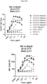
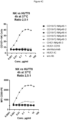
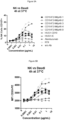
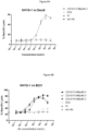
- NK cells refers to a sub-population of lymphocytes that is involved in non-conventional immunity.
- NK cells can be identified by virtue of certain characteristics and biological properties, such as the expression of specific surface antigens including CD56 and/or NKp46 for human NK cells, the absence of the alpha/beta or gamma/delta TCR complex on the cell surface, the ability to bind to and kill cells that fail to express "self" MHC/HLA antigens by the activation of specific cytolytic machinery, the ability to kill tumor cells or other diseased cells that express a ligand for NK activating receptors, and the ability to release protein molecules called cytokines that stimulate or inhibit the immune response.
- NK cells any of these characteristics and activities can be used to identify NK cells, using methods well known in the art. Any subpopulation of NK cells will also be encompassed by the term NK cells.
- active NK cells designate biologically active NK cells, including NK cells having the capacity of lysing target cells or enhancing the immune function of other cells.
- NK cells can be obtained by various techniques known in the art, such as isolation from blood samples, cytapheresis, tissue or cell collections, etc.
- Useful protocols for assays involving NK cells can be found in Natural Killer Cells Protocols (edited by Campbell KS and Colonna M). Humana Press, pp. 219-238 (2000 ).
- active or “activated” T cells designate biologically active T cells, more particularly T cells having the capacity of cytolysis or of stimulating an immune response by, e.g., secreting cytokines. Active cells can be detected in any of a number of well-known methods, including functional assays and expression-based assays such as the expression of cytokines such as TNF-alpha.
- an agent that has "agonist" activity at a cell surface receptor is an agent that can cause or increase signalling by the receptor, e.g., an ability of the receptor to activate or transduce an intracellular signaling pathway.
- Changes in signaling activity can be measured, for example, by assays designed to measure changes in receptor signaling pathways, e.g. by monitoring phosphorylation of signal transduction components, assays to measure the association of certain signal transduction components with other proteins or intracellular structures, or in the biochemical activity of components such as kinases, or assays designed to measure expression of reporter genes under control of receptor-sensitive promoters and enhancers, or indirectly by a downstream effect mediated by the receptor (e.g.
- Immunoglobulin ABDs can be obtained from variable domains derived from antibodies (from immunoglobulin chains), for example in the form of associated V L and V H domains found on two polypeptide chains, or a single chain antigen binding domain such as a scFv, a V H domain, a V L domain, a dAb, a V-NAR domain or a V H H domain.
- the an antigen binding domain e.g,. ABD, and ABD 2
- the an antigen binding domain can also be readily derived from antibodies as a Fab or scFv.
- antibodies are initially obtained by immunization of a non-human animal, e.g., a mouse, rat, guinea pig or rabbit, with an immunogen comprising a polypeptide, or a fragment or derivative thereof, typically an immunogenic fragment, for which it is desired to obtain antibodies (e.g. a human polypeptide).
- a non-human animal e.g., a mouse, rat, guinea pig or rabbit
- an immunogen comprising a polypeptide, or a fragment or derivative thereof, typically an immunogenic fragment, for which it is desired to obtain antibodies (e.g. a human polypeptide).
- the step of immunizing a non-human mammal with an antigen may be carried out in any manner well known in the art for stimulating the production of antibodies in a mouse (see, for example, E. Harlow and D. Lane, Antibodies: A Laboratory Manual., Cold Spring Harbor Laboratory Press, Cold Spring Harbor, NY (1988 ).
- Human antibodies may also be produced by using, for immunization, transgenic animals that have been engineered to express a human antibody repertoire ( Jakobovitz et Nature 362 (1993) 255 ), or by selection of antibody repertoires using phage display methods.
- a XenoMouse (Abgenix, Fremont, CA) can be used for immunization.
- a XenoMouse is a murine host that has had its immunoglobulin genes replaced by functional human immunoglobulin genes.
- antibodies produced by this mouse or in hybridomas made from the B cells of this mouse are already humanized.
- the XenoMouse is described in United States Patent No. 6,162,963 .
- Antibodies may also be produced by selection of combinatorial libraries of immunoglobulins, as disclosed for instance in ( Ward et al. Nature, 341 (1989) p. 544 ).
- Phage display technology McCafferty et al (1990) Nature 348:552-553
- V immunoglobulin variable domain gene repertoires from unimmunized donors. See, e.g., Griffith et al (1993) EMBO J. 12:725- 734 ; US 5,565,332 ; US 5,573,905 ; US 5,567,610 ; and US 5,229,275 ).
- combinatorial libraries comprise variable (V) domain gene repertoires of human origin, selection from combinatorial libraries will yield human antibodies.
- Antibodies will typically be directed to a pre-determined antigen.
- examples of antibodies include antibodies that recognize an antigen expressed by a target cell that is to be eliminated, for example a proliferating cell or a cell contributing to a disease pathology.
- examples include antibodies that recognize tumor antigens, microbial (e.g. bacterial or parasite) antigens or viral antigens.
- the bacterial antigen is derived from a bacterium selected from the group consisting of Helicobacter species, in particular Helicobacter pyloris; Borrelia species, in particular Borrelia burgdorferi; Legionella species, in particular Legionella pneumophilia; Mycobacteria s species, in particular M. tuberculosis, M. avium, M. intracellulare, M. kansasii, M. gordonae; Staphylococcus species, in particular Staphylococcus aureus; Neisseria species, in particular N. gonorrhoeae, N.
- Helicobacter species in particular Helicobacter pyloris
- Borrelia species in particular Borrelia burgdorferi
- Legionella species in particular Legionella pneumophilia
- Mycobacteria s species in particular M. tuberculosis, M. avium, M. intracellulare, M. kansasii, M. gordona
- pneumonae pneumonae; anaerobic Streptococcus species; pathogenic Campylobacter species; Enterococcus species; Haemophilus species, in particular Haemophilus influenzae; Bacillus species, in particular Bacillus anthracis; Corynebacterium species, in particular Corynebacterium diphtheriae; Erysipelothrix species, in particular Erysipelothrix rhusiopathiae; Clostridium species, in particular C. perfringens, C.
- viral antigen includes, but is not limited to, intact, attenuated or killed whole virus, any structural or functional viral protein, or any peptide portion of a viral protein of sufficient length (typically about 8 amino acids or longer) to be antigenic.
- Retroviridae e.g., human immunodeficiency viruses, such as HIV-1 (also referred to as HTLV-III, LAV or HTLV-III/LAV, or HIV-III; and other isolates, such as HIV-LP; Picornaviridae (e.g., polio viruses, hepatitis A virus; enteroviruses, human Coxsackie viruses, rhinoviruses, echoviruses); Calciviridae (e.g., strains that cause gastroenteritis); Togaviridae (e.g., equine encephalitis viruses, rubella viruses); Flaviviridae (e.g., dengue viruses, encephalitis viruses, yellow fever viruses); Coronaviridae (e.g., coronaviruses); Rhabdoviridae (e.g., vesicular stomatitis viruses, rabies viruses); Fil
- cancer antigen and “tumor antigen” are used interchangeably and refer to antigens that are differentially expressed by cancer cells or are expressed by non-tumoral cells (e.g. immune cells) having a pro-tumoral effect (e.g. an immunosuppressive effect), and can thereby be exploited in order to target cancer cells.
- cancer antigens are antigens which can potentially stimulate apparently tumor-specific immune responses. Some of these antigens are encoded, although not necessarily expressed, or expressed at lower levels or less frequently, by normal cells.
- cancer antigens can be characterized as those which are normally silent (i.e., not expressed) in normal cells, those that are expressed only at certain stages of differentiation and those that are temporally expressed such as embryonic and fetal antigens.
- Other cancer antigens are encoded by mutant cellular genes, such as oncogenes (e.g., activated ras oncogene), suppressor genes (e.g., mutant p53), fusion proteins resulting from internal deletions or chromosomal translocations.
- Still other cancer antigens can be encoded by viral genes such as those carried on RNA and DNA tumor viruses.
- Still other cancer antigens can be expressed on immune cells capable of contributing to or mediating a pro-tumoral effect, e.g. cell that contributes to immune evasion, a monocyte or a macrophage, optionally a suppressor T cell, regulatory T cell, or myeloid-derived suppressor cell.
- the cancer antigens are usually normal cell surface antigens which are either over-expressed or expressed at abnormal times, or are expressed by a targeted population of cells.
- the target antigen is expressed only on proliferative cells (e.g., tumor cells) or pro-tumoral cells (e.g. immune cells having an immunosuppressive effect), however this is rarely observed in practice.
- proliferative cells e.g., tumor cells
- pro-tumoral cells e.g. immune cells having an immunosuppressive effect
- Example of cancer antigens include: Receptor Tyrosine Kinase-like Orphan Receptor 1 (ROR1), Crypto, CD4, CD20, CD30, CD19, CD38, CD47, Glycoprotein NMB, CanAg, Her2 (ErbB2/Neu), a Siglec family member, for example CD22 (Siglec2) or CD33 (Siglec3), CD79, CD138, CD171, PSCA, L1-CAM, PSMA (prostate specific membrane antigen), BCMA, CD52, CD56, CD80, CD70, E-selectin, EphB2, Melanotransferrin, Mud 6 and TMEFF2.
- ROR1 Receptor Tyrosine Kinase-like Orphan Receptor 1
- Crypto Crypto
- CD4 CD20
- CD30 CD19
- CD38 CD47
- Glycoprotein NMB Glycoprotein NMB
- CanAg Her2 (ErbB2/Neu)
- a Siglec family member for example CD22 (Sigle
- cancer antigens also include Immunoglobulin superfamily (IgSF) such as cytokine receptors, Killer-Ig Like Receptor, CD28 family proteins, for example, Killer-Ig Like Receptor 3DL2 (KIR3DL2), B7-H3, B7-H4, B7-H6, PD-L1, IL-6 receptor.
- IgSF Immunoglobulin superfamily
- Examples also include MAGE, MART-1/Melan-A, gp100, major histocompatibility complex class I-related chain A and B polypeptides (MICA and MICB), adenosine deaminase-binding protein (ADAbp), cyclophilin b, colorectal associated antigen (CRC)-C017-1A/GA733, protein tyrosine kinase 7(PTK7), receptor protein tyrosine kinase 3 (TYRO-3), nectins (e.g.
- nectin-4 major histocompatibility complex class I-related chain A and B polypeptides
- MICA and MICB proteins of the UL16-binding protein
- RAET1 proteins of the retinoic acid early transcript-1
- CEA carcinoembryonic antigen
- PSA carcinoembryonic antigen
- T-cell receptor/CD3-zeta chain MAGE-family of tumor antigens, GAGE-family of tumor antigens, anti-Müllerian hormone Type II receptor, delta-like ligand 4 (DLL4), DR5, ROR1 (also known as Receptor Tyrosine Kinase-Like Orphan Receptor 1 or NTRKR1 (EC 2.7.10.1), BAGE, RAGE, LAGE-1, NAG, GnT-V, MUM-1, CDK4, MUC family, VEGF, VEGF receptors, Angiopoietin
- the antigen of interest is an antigen (e.g. any one of the antigens listed above) capable of undergoing intracellular internalization, for example when bound by a conventional human IgG1 antibody, either in the presence of absence of Fcy receptor cells.
- an antigen e.g. any one of the antigens listed above
- the antigen of interest is a CD19 or CD20 polypeptide; in one aspect, the multispecific protein comprises a VH and/or VL, or a scFv, or another ABD, that binds CD19 or CD20 comprising an amino acid sequence which is at least 60%, 70%, 80%, 85%, 90% or 95% identical to the sequence of the anti-CD19 or anti- CD20 respective VH, VL or scFv described in the Examples herein, or comprises the heavy and light chain CDR1, -2 and -3 of the anti-CD19 or anti-CD20 heavy and light chain variable regions disclosed herein.
- the multispecific protein competes for binding to a human CD19 or CD20 polypeptide with an antibody, or a F5 or T6 protein, comprising the respective anti-CD19 or anti-CD20 VH, VL or scFv disclosed in the Examples herein.
- the ABD that binds an antigen of interest is derived from (e.g. comprises the hypervariable region of, or comprises one, two, three, four, five or six of the CDRs of) a parental antibody that binds an antigen of interest (e.g. a murine antibody, a human antibody) which, when bound to its antigenic target (the antigen of interest on cells), increases or induces down-modulation or intracellular internalization of the antigen of interest.
- the antigen of interest is a cancer antigen, e.g. one of the cancer antigens listed above known to internalize (e.g.
- Immunoglobulin superfamily (IgSF) members for example cytokine receptor ⁇ or ⁇ chains, Killer-Ig Like Receptors, CD28 family proteins, B7-H3, B7-H4, B7-H6, KIR3DL2, PTK7, ROR1, L1-CAM, Siglec family members, EGF receptor and EGF-like receptor family members, EGFR, HER-2, integrins, anti-Müllerian hormone Type II receptor, CSF-1R, and others
- the antigen target is a polypeptide present on an immune cell capable of mediating a pro-tumoral effect, e.g. a monocyte or a macrophage, optionally a suppressor T cell, regulatory T cell, or myeloid-derived suppressor cell.
- an ABD, variable domain or pair of complementary variable domains will bind an antigen expressed by a target cell that is to be eliminated (e.g., a tumor antigen, microbial (e.g. bacterial or parasitic) antigen, viral antigen, or antigen expressed on an immune cell that is contributing to inflammatory or autoimmune disease, and another ABD, variable domain or pair of complementary variable domains will bind to an antigen expressed on an immune cell, for example an immune effector cell, e.g. a cell surface receptor of an effector cells such as a T or NK cell.
- an immune effector cell e.g. a cell surface receptor of an effector cells such as a T or NK cell.
- antigens expressed on immune cells, optionally immune effector cells include antigens expressed on a member of the human lymphoid cell lineage, e.g.
- a human T cell a human B cell or a human natural killer (NK) cell
- a human monocyte e.g., a tumor antigen, microbial antigen, viral antigen, or antigen expressed on an immune cell that is contributing to inflammatory or autoimmune disease
- the human lymphoid cell is a cytotoxic T cell or NK cell which, when activated, exerts a cytotoxic effect on the target cell.
- the cytotoxic activity of the human effector cells is recruited.
- the human effector cell is a member of the human myeloid lineage.
- Antigens expressed on an immune cell to which antibodies of fragments that make up multispecific protein can bind also include NK and/or T cell receptors, e.g. any molecule on the surface of NK cells or T cells, respectively, that can serve to direct the NK or T cell to the intended target cell to be eliminated, and preferably to permit the NK and/or T cell to mediate the elimination or lysis of the target cell.
- NK and/or T cell receptors e.g. any molecule on the surface of NK cells or T cells, respectively, that can serve to direct the NK or T cell to the intended target cell to be eliminated, and preferably to permit the NK and/or T cell to mediate the elimination or lysis of the target cell.
- Examples include, e.g., members of the immunoglobulin superfamily, members of the killer-cell immunoglobulin-like receptor (KIR) family, the leukocyte immunoglobulin-like receptors (LILR) family, or the lectin family or the NK cell lectin
- Activity can be measured for example by bringing target cells and effector cells into contact in presence of the multispecific polypeptide.
- the immune cell receptor is an immune effector cell activating receptor, e.g. an activating NK cell or T cell receptor.
- activating NK cell receptor and “activating T cell receptor” refers to any molecule on the surface of NK cells or T cells, respectively, that, when stimulated, causes a measurable increase in any property or activity known in the art as associated with NK cell or T cell activity, respectively, such as cytokine (for example IFN- ⁇ or TNF- ⁇ ) production, increases in intracellular free calcium levels, the ability to lyse target cells in a redirected killing assay as described, e.g.
- cytokine for example IFN- ⁇ or TNF- ⁇
- activating NK receptor includes but is not limited to DNAX accessory molecule-1 (DNAM-1), 2B4, activating forms of KIR proteins (for example KIR2DS receptors, KIR2DS2, KIR2DS4), NKG2D, NKp30, CD137, CD69, NKp80, NKp44, NKp46, IL-2R, IL-12R, IL-15R, IL-18R and IL-21R.
- the activating NK cell receptor is a receptor other than an Fcy receptor.
- the activating NK cell receptor is a receptor other than NKp46.
- Activation of cytotoxic T cells may occur via binding of the CD3 antigen as effector antigen on the surface of the cytotoxic T cell by a multispecific (e.g. bispecific) polypeptide of this aspect.
- the human CD3 antigen is present on both helper T cells and cytotoxic T cells.
- Human CD3 denotes an antigen which is expressed on T cells as part of the multimolecular T cell complex and which comprises three different chains: CD3-epsilon, CD3-delta and CD3-gamma.
- Other effector cell antigens that can be bound by an ABD are the human CD8 antigen, the human CD2 antigen, the human CD28 antigen or the human CD25 antigen.
- the multispecific protein comprises one ABD that binds specifically to CD8, and one ABD that bind to CD3. In one aspect, the multispecific protein comprises one ABD that binds specifically to an activating receptor present on effector NK cells, and one ABD that bind to an activating receptor present on effector T cells. In one aspect, the multispecific comprises one ABD that binds to a cancer antigen, a viral antigen or a bacterial antigen.
- variable domains which are incorporated into the polypeptides can be tested for any desired activity prior to inclusion in a polypeptide.
- DNA encoding each variable domain can be placed, in suitable arrangements, in an appropriate expression vector(s), together with DNA encoding any elements such as an enzymatic recognition tag, or CH2 and CH3 domains and any other optional elements (e.g. DNA encoding a linker or hinge region) for transfection into an appropriate host(s). The host is then used for the recombinant production of the polypeptide chains that make up the multispecific protein.
- An ABD or variable region derived from an antibody will generally comprise at minimum a hypervariable region sufficient to confer binding activity when present in the multimeric polypeptide. It will be appreciated that an ABD or variable region may comprise linker elements (e.g. linker peptides, constant domain derived sequences, hinges, or fragments thereof, each of which can be placed between a variable domain and a CH1, C ⁇ , CH2 or CH3 domain, or between other domains as needed).
- linker elements e.g. linker peptides, constant domain derived sequences, hinges, or fragments thereof, each of which can be placed between a variable domain and a CH1, C ⁇ , CH2 or CH3 domain, or between other domains as needed).
- ABDs or variable regions can be obtained from a humanized antibody in which residues from a complementary-determining region (CDR) of a human antibody are replaced by residues from a CDR of the original antibody (the parent or donor antibody, e.g. a murine or rat antibody) while maintaining the desired specificity, affinity, and capacity of the original antibody.
- CDRs of the parent antibody some or all of which are encoded by nucleic acids originating in a non-human organism, are grafted in whole or in part into the beta-sheet framework of a human antibody variable region to create an antibody, the specificity of which is determined by the engrafted CDRs.
- An antigen binding domain can thus have non-human hypervariable regions or CDRs and human frameworks region sequences (optionally with back mutations).
- Polypeptide chains will be arranged in one or more expression vectors so as to produce the polypeptides having the desired domains operably linked to one another.
- the host cell may be of mammalian origin or may be selected from COS-1, COS-7, HEK293, BHK21, CHO, BSC-1, Hep G2, 653, SP2/0, 293, HeLa, myeloma, lymphoma, yeast, insect or plant cells, or any derivative, immortalized or transformed cell thereof.
- polypeptide can then be produced in an appropriate host cell or by any suitable synthetic process and brought into contact under appropriate conditions for the multimeric (e.g. dimer or trimer) polypeptide to form.
- An isolated hetero-multimeric protein that binds a first and second antigen of interest can be prepared according to different configurations, in each case involving at least a central (first) polypeptide chain and a second polypeptide chain, and a third polypeptide chain.
- the first (central) polypeptide chain will provide one variable domain that will, together with a complementary variable domain on a second polypeptide chain, form an antigen binding domain specific for one (e.g. a first) antigen of interest.
- the first (central) polypeptide chain will also provide a second variable domain that will be paired with a complementary variable domain to form an antigen binding domain specific for another (e.g. a second) antigen of interest; the variable domain that is complementary to the second variable domain is placed on a third polypeptide chain.
- the second and third polypeptide chains will associate with the central polypeptide chain by CH1-C ⁇ heterodimerization, forming non-covalent interactions and optionally further interchain disulfide bonds between respective hinge domains and between complementary CH1 and CK domains, with a single multimeric polypeptide being formed so long as CH/C ⁇ and VH/VK domains are chosen to give rise to a sole dimerization configuration.
- a trimer or when polypeptides are constructed for preparation of a trimer, there will generally be one polypeptide chain that comprises a non-naturally occurring VH-C ⁇ or VL-CH1 domain arrangement.
- the first (central) polypeptide chain comprises a first variable domain (V) fused to a CH1 of CL constant region (e.g. the V domain is fused at its C-terminus to the N-terminus of a CH1 or CK constant region), a second variable domain, and an Fc domain interposed between the first and second variable domains may have the Examples of domain arrangement for the first polypeptide include: (VH or VK) - CH1 - Fc domain - VH - CH1 (VH or VK) - CK - Fc domain - VK - CK (VH or VK) - CK - Fc domain - VK - CH1 (VH or VK) - CH1 - Fc domain - VH - CK (VH or VK) - CH1 - Fc domain - VH - CH1 (VH or VK) - CH1 - Fc domain - VH - CH1 (VH or VK) - CH1
- a second polypeptide chain comprises a first variable domain (V) fused (e.g. at its C-terminus) to a CH1 or CL (e.g. CK) constant region selected to be complementary to the CH1 or CL constant region of the first polypeptide chain such that the first and second polypeptides form a CH1-CL (e.g., CH1-CK) heterodimer.
- the second polypeptide chain further comprises an Fc domain fused to the C- terminus of the CH1 or CL domain. Examples of domain arrangement for the second polypeptide include (VH or VK) - (CH1) - Fc domain (VH or VK) - (CK) - Fc domain
- a third polypeptide chain has the domain arrangement: (VH or VK) - (CH1 or (CK) (VH or VK) - (CH1 or (CK) - scFV
- Heterotrimeric proteins are formed by using a central (first) polypeptide chain comprising a first variable domain (V) fused to a first CH1 or CK constant region, a second variable domain (V) fused to a second CH1 or CK constant region, and an Fc domain interposed between the first and second variable domains (i.e. the Fc domain is interposed between the first and second (V-(CH1/CK)) units.
- a central polypeptide chain for use in a heterotrimeric protein has the domain arrangements (N- to C- terminal) as follows: V a1 - (CH1 or CK) a - Fc domain - V a2 - (CH1 or CK) b .
- a second polypeptide chain comprises a domain arrangement (N- to C- terminal): V b1 - (CH1 or CK) c - Fc domain such that the (CH1 or CK) c dimerizes with the (CH1 or CK) a on the central chain, and the V a1 and V b1 form an antigen binding domain.
- a third polypeptide chain comprises a domain arrangement (N- to C- terminal): V b2 - (CH1 or CK) d , such that the (CH1 or CK) d dimerizes with the (CH1 or CK) b unit on the central chain, and the V a2 and V b2 form an antigen binding domain.
- the first polypeptide can have two variable domains that each form an antigen binding domain with a variable domain on a separate polypeptide chain (i.e. the variable domain of the second and third chains), the second polypeptide chain has one variable domain, and the third polypeptide has one variable domain, and wherein the trimer has a dimeric Fc domain that binds CD16.
- domain arrangement for the trimeric bispecific polypeptide formed from examples include: (second polypeptide) (first polypeptide) (third polypeptide) (second polypeptide) (first polypeptide) (third polypeptide) (second polypeptide) (first polypeptide) (third polypeptide) (third polypeptide) (first polypeptide) (third polypeptide)
- Heterotrimeric proteins can for example be formed by using a central (first) polypeptide chain comprising a first variable domain (V) fused to a first CH1 or CK constant region, a second variable domain (V) fused to a second CH1 or CK constant region, and an Fc domain interposed between the first and second variable domains (i.e. the Fc domain is interposed between the first and second (V-(CH1/CK)) units.
- a central polypeptide chain for use in a heterotrimeric protein has the domain arrangements (N- to C- terminal) as follows: V 1 - (CH1 or CK) a - Fc domain - V 2 - (CH1 or CK) b .
- a second polypeptide chain comprises a domain arrangement (N- to C- terminal): V 1 - (CH1 or CK) c - Fc domain such that the (CH1 or CK) c dimerizes with the (CH1 or CK) a on the central chain, and the V a1 and V b1 form an antigen binding domain.
- a third polypeptide chain can then comprise a domain arrangement (N- to C- terminal): V 2 - (CH1 or CK) d - scFv, such that the (CH1 or CK) d dimerizes with the (CH1 or CK) b unit on the central chain, and the V a2 and V b2 form an antigen binding domain.
- a hinge region will typically be present on a polypeptide chain between a CH1 domain and a CH2 domain of an Fc domain, and/or can be present between a CK domain and a CH2 domain.
- a hinge region can optionally be replaced for example by a suitable linker peptide.
- proteins domains described in the present disclosure can optionally be specified as being from N- to C- terminal. Protein arrangements of the disclosure for purposes of illustration are shown from N-terminus (on the left) to C-terminus. Domains can be referred to as fused to one another (e.g. a domain can be said to be fused to the C-terminus of the domain on its left, and/or a domain can be said to be fused to the N-terminus of the domain on its right).
- proteins domains described in the present disclosure can be fused to one another directly or via intervening amino acid sequences.
- a CH1 or CK domain will be fused to an Fc domain via a linker peptide, optionally a hinge region or a fragment thereof.
- a VH or VK domain will be fused to a CH3 domain via a linker peptide.
- VH and VL domains linked to another in tandem will be fused via a linker peptide (e.g. as an scFv).
- VH and VL domains linked to an Fc domain will be fused via a linker peptide.
- Two polypeptide chains will be bound to one another (indicated by "
- VK domain can be replaced by a V ⁇ variable domain.
- the Fc domain comprises a CH2-CH3 unit (a full length CH2 and CH3 domain).
- the CH3 domain will be capable of CH3-CH3 dimerization (e.g. a wild-type CH3 domain).
- the multimeric polypeptide is capable of binding to human FcRn with intermediate affinity, e.g. retains binding to FcRn but has decreased binding to a human FcRn receptor compared to a full-length wild type human IgG1 antibody.
- the Fc moiety may further comprise one or more amino acid modifications, e.g. in the CH2 domain, that decreases further (e.g. abolishes) binding to one or more Fcy receptors.
- the CH3 domain comprises an amino acid substitution at 1, 2, 3, 4, 5, 6 or 7 of the positions L351, T366, L368, P395, F405, T407 (or Y407) and/or K409 (EU numbering as in Kabat).
- linkers include, for example, linkers comprising glycine and serine residues, e.g., the amino acid sequence GEGTSTGS(G 2 S) 2 GGAD.
- the VH domain and VL domains of an svFv are linked together by the amino acid sequence (G 4 S) 3 .
- An ABD can be linked to a constant domain or Fc domain via a linker (e.g. a flexible polypeptide linker) that permits the ABD to be positioned such that it binds to its target antigen and exhibits the desired functionality, e.g. it possesses a sufficient range of motion relative to the rest of the multispecific protein (the Fc domain and/or other ABD) and thereby mediates signaling at a cell surface activating receptor.
- linkers include, for example, linkers derived from antibody hinge regions, an amino sequence RTVA, or linkers comprising glycine and serine residues, e.g., the amino acid sequence GEGTSTGS(G 2 S) 2 GGAD.
- V H domain and V L domains of a scFv are linked together by the amino acid sequence (GaS)s.
- GaS amino acid sequence
- Such linkers can be used particularly advantageously to link and ABD to a constant region or Fc domain when the ABD comprises two variable regions that are placed on the same polypeptide chain (e.g., an scFv)
- any of the peptide linkers contained in the subject multispecific proteins may comprise a length of at least 4 residues, at least 5 residues, at least 10 residues, at least 15 residues, at least 20 residues, at least 25 residues, at least 30 residues or more.
- the linkers comprise a length of between 2-4 residues, between 2-4 residues, between 2-6 residues, between 2-8 residues, between 2-10 residues, between 2-12 residues, between 2-14 residues, between 2-16 residues, between 2-18 residues, between 2- 20 residues, between 2-22 residues, between 2-24 residues, between 2-26 residues, between 2-28 residues, between 2-30 residues, between 2 and 50 residues, or between 10 and 50 residues.
- An ABD (e.g. an immunoglobulin variable region) can optionally be linked to a constant domain or Fc domain via a flexible linker (e.g. polypeptide linker) that leads to less structural rigidity or stiffness (e.g. between or amongst the ABD and Fc domain) compared to a conventional (e.g. wild-type full length human IgG) antibody.
- the multispecific protein may have a structure or a flexible linker between the ABD and constant domain or Fc domain that permits an increased range of domain motion a compared to ABD in a conventional (e.g. wild-type full length human IgG) antibody.
- the structure or a flexible linker can be configured to confer on the antigen binding sites greater intrachain domain movement compared to antigen binding sites in a conventional human IgG1 antibody.
- Rigidity or domain motion/interchain domain movement can be determined, e.g., by computer modeling, electron microscopy, spectroscopy such as Nuclear Magnetic Resonance (NMR), X-ray crystallography (B-factors), or Sedimentation Velocity Analytical ultracentrifugation (AUC) to measure or compare the radius of gyration of proteins comprising the linker or hinge.
- NMR Nuclear Magnetic Resonance
- B-factors X-ray crystallography
- AUC Sedimentation Velocity Analytical ultracentrifugation
- a test protein or linker may have lower rigidity relative to a comparator protein if the test protein has a value obtained from one of the tests described in the previous sentence differs from the value of the comparator, e.g., an IgG1 antibody or a hinge, by at least 5%, 10%, 25%, 50%, 75%, or 100%.
- the comparator e.g., an IgG1 antibody or a hinge
- the multispecific protein may have a structure or a flexible linker between the ABD and constant domain or Fc domain that permits two ABDs to have a spacing between said two ABDs comprising less than about 80 angstroms, less than about 60 angstroms or ranges from about 40-60 angstroms.
- a CH1 or CK domain is linked or fused to an Fc domain via a linker that comprises a fragment of a CH1 domain and/or hinge region.
- a N-terminal amino acid sequence of CH1 can be fused to a variable domain in order to mimic as closely as possible the natural structure of a wild-type antibody.
- the linker comprises an amino acid sequence from a hinge domain or an N-terminal CH1 amino acid.
- linker comprises or consists of the amino acid sequence RTVA.
- a CH1 or CK domain is linked or fused to an Fc domain via a hinge region (or fragment thereof) derived form a hinge domain of a human IgG1 antibody.
- a hinge domain may comprise the amino acid sequence: T-H-T-C-S-S-C-P-A-P-E-L-L (one letter code), or an amino acid sequence at least 60%, 70%, 80% or 90% identical thereto, optionally wherein one or both cysteines are deleted or substituted by a different amino acid residue.
- the hinge region (or fragment thereof) is derived from a C ⁇ 2-C C ⁇ 3 hinge domain of a human IgM antibody.
- a hinge domain may comprise the amino acid sequence: N-A-S-S-M-C-V-P-S-P-A-P-E-L-L (one letter code), or an amino acid sequence at least 60%, 70%, 80% or 90% identical thereto, optionally wherein one or both cysteines are deleted or substituted by a different amino acid residue.
- Polypeptide chains that dimerize and associate with one another via non-covalent bonds may or may not additionally be bound by an interchain disulfide bond formed between respective CH1 and C ⁇ domains, and/or between respective hinge domains on the chains.
- CH1, C ⁇ and/or hinge domains (or other suitable linking amino acid sequences) can optionally be configured such that interchain disulfide bonds are formed between chains such that the desired pairing of chains is favored and undesired or incorrect disulfide bond formation is avoided.
- polypeptide chains to be paired each possess a CH1 or C ⁇ adjacent to a hinge domain
- the polypeptide chains can be configured such that the number of available cysteines for interchain disulfide bond formation between respective CH1/C ⁇ -hinge segments is reduced (or is entirely eliminated).
- the amino acid sequences of respective CH1, C ⁇ and/or hinge domains can be modified to remove cysteine residues in both the CH1/C ⁇ and the hinge domain of a polypeptide; thereby the CH1 and C ⁇ domains of the two chains that dimerize will associate via non-covalent interaction(s).
- the CH1 or C ⁇ domain adjacent (e.g., N-terminal to) a hinge domain comprises a cysteine capable of interchain disulfide bond formation
- the hinge domain which is placed at the C-terminus of the CH1 or C ⁇ comprises a deletion or substitution of one or both cysteines of the hinge (e.g. Cys 239 and Cys 242, as numbered for human IgG1 hinge according to Kabat).
- the hinge region (or fragment thereof) comprise the amino acid sequence: T-H-T-S-P-P-S-P-A-P-E-L-L (one letter code), or an amino acid sequence at least 60%, 70%, 80% or 90% identical thereto.
- the CH1 or C ⁇ domain adjacent (e.g., N-terminal to) a hinge domain comprises a deletion or substitution at a cysteine residue capable of interchain disulfide bond formation
- the hinge domain placed at the C-terminus of the CH1 or C ⁇ comprises one or both cysteines of the hinge (e.g. Cys 239 and Cys 242, as numbered for human IgG1 hinge according to Kabat).
- the hinge region (or fragment thereof) comprises the amino acid sequence: T-H-T-C-S-S-C-P-A-P-E-L-L (one letter code), or an amino acid sequence at least 60%, 70%, 80% or 90% identical thereto.
- a hinge region is derived from an IgM antibody.
- the CH1/CK pairing mimics the C ⁇ 2 domain homodimerization in IgM antibodies.
- the CH1 or C ⁇ domain adjacent (e.g., N-terminal to) a hinge domain comprises a deletion or substitution at a cysteine capable of interchain disulfide bond formation, and an IgM hinge domain which is placed at the C-terminus of the CH1 or C ⁇ comprises one or both cysteines of the hinge.
- the hinge region (or fragment thereof) comprises the amino acid sequence: T-H-T-C-S-S-C-P-A-P-E-L-L (one letter code), or an amino acid sequence at least 60%, 70%, 80% or 90% identical thereto.
- Constant region domains can be derived from any suitable human antibody, including, the constant heavy (CH1) and light (C ⁇ ) domains, hinge domains, CH2 and CH3 domains.
- CH1 generally refers to positions 118-220 according to the EU index as in Kabat.
- CH2 generally refers to positions 237-340 according to the EU index as in Kabat, and
- CH3 generally refers to positions 341-447 according to the EU index as in Kabat.
- a “hinge” or “hinge region” or “antibody hinge region” herein refers to the flexible polypeptide or linker between the first and second constant domains of an antibody.
- the IgG CH1 domain ends at EU position 220, and the IgG CH2 domain begins at residue EU position 237.
- the hinge generally includes positions 221 (D221 in IgG1) to 236 (G236 in IgG1), wherein the numbering is according to the EU index as in Kabat.
- References to specific amino acid residues within constant region domains found within the polypeptides shall be, unless otherwise indicated or as otherwise dictated by context, be defined according to Kabat, in the context of an IgG antibody.
- CH2 and CH3 domains which may be present in the subject antibodies or multispecific proteins can be derived from any suitable antibody.
- Such CH2 and CH3 domains can be used as wild-type domains or may serve as the basis for a modified CH2 or CH3 domain.
- the CH2 and/or CH3 domain is of human origin or may comprise that of another species (e.g., rodent, rabbit, non-human primate) or may comprise a modified or chimeric CH2 and/or CH3 domain, e.g., one comprising residues from different CH2 or CH3 domains, e.g., from different antibody isotypes or species antibodies .
- a CH2 and/or CH3 domain may be wild-type domains or may comprise one or more amino acid modifications (e.g. amino acid substitutions) which increase binding to human CD16 and optionally another receptor such as FcRn.
- the modifications will not substantially decrease or abolish the ability of the Fc-derived polypeptide to bind to neonatal Fc receptor (FcRn), e.g. human FcRn.
- Typical modifications include modified human IgG1-derived constant regions comprising at least one amino acid modification (e.g. substitution, deletions, insertions), and/or altered types of glycosylation, e.g., hypofucosylation.
- Fc ⁇ RI CD64
- Fc ⁇ RII CD32
- Fc ⁇ RIII CD16
- Fc ⁇ RI CD64
- Fc ⁇ RIIA CD32A
- Fc ⁇ RIII CD 16
- a modification may, for example, increase binding of the Fc domain to Fc ⁇ RIIIa on effector (e.g. NK) cells and/or decrease binding to Fc ⁇ RIIB. Examples of modifications are provided in PCT publication no. WO2014/044686 .
- IgG1 which affect (enhance) Fc ⁇ RIIIa or FcRn binding are also set forth below.
- Isotype Species Modification Effector Function Effect of Modification IgG1 Human T250Q/M428L Increased binding to FcRn Increased half-life IgG1 Human 1M252Y/S254T/T256E + H433K/N434F Increased binding to FcRn Increased half-life IgG1 Human E333A Increased binding to Fc ⁇ RIIIa Increased ADCC and CDC IgG1 Human S239D/A330L/I332E Increased binding to Fc ⁇ RIIIa Increased ADCC IgG1 Human P257I/Q311 Increased binding to FcRn Unchanged half-life IgG1 Human S239D/I332E/G236A Increased Fc ⁇ RIIa/Fc ⁇ RIIb ratio Increased macrophage phagocytosis
- the multispecific protein comprises a variant Fc region comprise at least one amino acid modification (for example, possessing 1, 2, 3, 4, 5, 6, 7, 8, 9, or more amino acid modifications) in the CH2 and/or CH3 domain of the Fc region, wherein the modification enhances binding to a human CD16 polypeptide.
- the multispecific protein comprises at least one amino acid modification (for example, 1, 2, 3, 4, 5, 6, 7, 8, 9, or more amino acid modifications) in the CH2 domain of the Fc region from amino acids 237-341, or within the lower hinge-CH2 region that comprises residues 231-341.
- the multispecific protein comprises at least two amino acid modifications (for example, 2, 3, 4, 5, 6, 7, 8, 9, or more amino acid modifications), wherein at least one of such modifications is within the CH3 region and at least one such modifications is within the CH2 region.
- amino acid modifications in the hinge region encompassed are amino acid modifications in the CH1 domain, optionally in the upper hinge region that comprises residues 216-230 (Kabat EU numbering). Any suitable functional combination of Fc modifications can be made, for example any combination of the different Fc modifications which are disclosed in any of United States Patents Nos.
- WO2011/109400 WO 2008/105886 ; WO 2008/002933 ; WO 2007/021841 ; WO 2007/106707 ; WO 06/088494 ; WO 05/115452 ; WO 05/110474 ; WO 04/1032269 ; WO 00/42072 ; WO 06/088494 ; WO 07/024249 ; WO 05/047327 ; WO 04/099249 and WO 04/063351 ; and/or in Lazar et al. (2006) Proc. Nat. Acad. Sci. USA 103(11): 405-410 ; Presta, L.G. et al. (2002) Biochem. Soc. Trans.
- the multispecific protein comprises an Fc domain comprising at least one amino acid modification (for example, 1, 2, 3, 4, 5, 6, 7, 8, 9, or more amino acid modifications) relative to a wild-type Fc region, such that the molecule has an enhanced binding affinity for human CD16 relative to the same molecule comprising a wild-type Fc region, optionally wherein the variant Fc region comprises a substitution at any one or more of positions 221, 239, 243, 247, 255, 256, 258, 267, 268, 269, 270, 272, 276, 278, 280, 283, 285, 286, 289, 290, 292, 293, 294, 295, 296, 298, 300, 301, 303, 305, 307, 308, 309, 310, 311, 312, 316, 320, 322, 326, 329, 330, 332, 331, 332, 333, 334, 335, 337, 338, 339, 340, 359, 360, 370, 373, 376, 378, 392,
- the multispecific protein comprises an Fc domain comprising at least one amino acid modification (for example, 1, 2, 3, 4, 5, 6, 7, 8, 9, or more amino acid modifications) relative to a wild-type Fc region, such that the molecule has enhanced binding affinity for human CD16 relative to a molecule comprising a wild-type Fc region, optionally wherein the variant Fc region comprises a substitution at any one or more of positions 239, 298, 330, 332, 333 and/or 334 (e.g. S239D, S298A, A330L, I332E, E333A and/or K334A substitutions), optionally wherein the variant Fc region comprises a substitution at residues S239 and I332, e.g. a S239D and I332E substitution (Kabat EU numbering).
- amino acid modification for example, 1, 2, 3, 4, 5, 6, 7, 8, 9, or more amino acid modifications
- the multispecific protein comprises an Fc domain comprising altered glycosylation patterns that increase binding affinity for human CD16.
- carbohydrate modifications can be accomplished by, for example, by expressing a nucleic acid encoding the multispecific protein in a host cell with altered glycosylation machinery.
- Cells with altered glycosylation machinery are known in the art and can be used as host cells in which to express recombinant antibodies to thereby produce an antibody with altered glycosylation. See, for example, Shields, R.L. et al. (2002) J. Biol. Chem. 277:26733-26740 ; Umana et al. (1999) Nat. Biotech.
- the multispecific protein contains one or more hypofucosylated constant regions.
- Such multispecific protein may comprise an amino acid alteration or may not comprise an amino acid alteration and/or may be expressed or synthesized or treated under conditions that result in hypofucosylation.
- a multispecific protein composition comprises a multispecific protein described herein, wherein at least 20, 30, 40, 50, 60, 75, 85, 90, 95% or substantially all of the antibody species in the composition have a constant region comprising a core carbohydrate structure (e.g.
- a multispecific protein composition which is free of antibodies comprising a core carbohydrate structure having fucose.
- the core carbohydrate will preferably be a sugar chain at Asn297.
- a multispecific protein comprising a dimeric Fc domain can be characterized by having a binding affinity to a human CD16 polypeptide that is within 1-log of that of a conventional human IgG1 antibody, e.g., as assessed by surface plasmon resonance.
- the multispecific protein comprising a dimeric Fc domain engineered to enhance Fc receptor binding can be characterized by having a binding affinity to a human CD16 polypeptide that is at least 1-log greater than that of a conventional or wild-type human IgG1 antibody, e.g., as assessed by surface plasmon resonance.
- a multispecific protein comprising a dimeric Fc domain can be characterized by a Kd for binding (monovalent) to a human CD16 polypeptide of less than 10 -5 M (10 ⁇ molar), optionally less than 10 -6 M (1 ⁇ molar), as assessed by surface plasmon resonance (e.g. as in Example 13, SPR measurements performed on a Biacore T100 apparatus (Biacore GE Healthcare), with bispecific antibodies immobilized on a Sensor Chip CM5 and serial dilutions of soluble CD16 polypeptide injected over the immobilized bispecific antibodies.
- the invention provides methods of making a heterotrimeric protein (e.g. any heterotrimeric protein described herein), comprising:
- the present disclosure provides a method for identifying or evaluating candidate variable regions for use in a heterotrimeric protein, comprising the steps of:
- the candidate variable regions can be for example from an anti-activating receptor antibody or from an antigen that binds an antigen of interest. It will also be appreciated that the position of the respective ABDs for the candidate variable region pair and the other variable region pair can be inverted. For example, in a trimeric protein the methods can be modified such that the heavy and light chain candidate variable domains are formed by the second V region of the first polypeptide and the V region of the second polypeptide, and the other variable region pair are formed by the first V region of the first polypeptide and the V region of the third polypeptide.
- the second variable domain of the first polypeptide and the variable domain of the third polypeptide form an antigen binding domain that binds an activating receptor on an immune cell.
- the protein formats of the disclosure can be used in a panel to screen proteins configurations or formats to identify the most effective configurations or formats for a given antigen of interest, or combination of first and second antigen of interest. Different protein formats may access or engage their antigen targets differently.
- the present disclosure provides a library of at least 5, 10, 20, 30, 50 hetero-multimeric proteins of the disclosure, wherein the proteins share domain arrangements but differ in the amino acid sequence of the variable domain of one or both of their antigen binding domains.
- the present disclosure provides a library of at least 2, 3, 4, 5 or 10 hetero-multimeric proteins of the disclosure, wherein the proteins share the amino acid sequence of the variable domain of one or both of their antigen binding domains, but differ in domain arrangements.
- recovering a heterotrimer protein can comprise introducing the protein to a solid phase so as to immobilize the protein.
- the immobilized protein can then subsequently be eluted.
- the solid support may be any suitable insoluble, functionalized material to which the proteins can be reversibly attached, either directly or indirectly, allowing them to be separated from unwanted materials, for example, excess reagents, contaminants, and solvents.
- solid supports include, for example, functionalized polymeric materials, e.g., agarose, or its bead form Sepharose ® , dextran, polystyrene and polypropylene, or mixtures thereof; compact discs comprising microfluidic channel structures; protein array chips; pipet tips; membranes, e.g., nitrocellulose or PVDF membranes; and microparticles, e.g., paramagnetic or non-paramagnetic beads.
- an affinity medium will be bound to the solid support and the protein will be indirectly attached to solid support via the affinity medium.
- the solid support comprises a protein A affinity medium or protein G affinity medium.
- a “protein A affinity medium” and a “protein G affinity medium” each refer to a solid phase onto which is bound a natural or synthetic protein comprising an Fc-binding domain of protein A or protein G, respectively, or a mutated variant or fragment of an Fc-binding domain of protein A or protein G, respectively, which variant or fragment retains the affinity for an Fc-portion of an antibody.
- Protein A and Protein G are bacterial cell wall proteins that have binding sites for the Fc portion of mammalian IgG. The capacity of these proteins for IgG varies with the species. In general, IgGs have a higher affinity for Protein G than for Protein A, and Protein G can bind IgG from a wider variety of species.
- Protein A can, therefore, be used to prepare isotypically pure IgG from some species.
- proteins When covalently attached to a solid matrix, such as cross-linked agarose, these proteins can be used to capture and purify antigen-protein complexes from biochemical solutions.
- EZview TM Red Protein G Affinity Gel is Protein G covalently bonded to 4% Agarose beads (Sigma Aldrich Co); or POROS ® A, G, and CaptureSelect ® HPLC columns (Invitrogen Inc.).Affinity capture reagents are also described, for example, in the Antibody Purification Handbook, Biosciences, publication No. 18-1037-46, Edition AC).
- a multispecific protein is capable of inducing activation of an immune effector cell (e.g. an NK cell, a T cell) when the protein is incubated in the presence of the effector cell and a target cell that expresses the antigen of interest).
- an immune effector cell e.g. an NK cell, a T cell
- a multispecific protein is capable of inducing signaling at an immune effector cell activating receptor when the protein is incubated in the presence of the effector cell and a target cell that expresses the antigen of interest).
- effector cell activation or signaling in characterized by increased expression of a cell surface marker of activation, e.g. CD107, CD69, etc.
- Activity can be measured for example by bringing target cells and effector cells into contact with one another, in presence of the multispecific polypeptide.
- aggregation of target cells and effector cells is measured.
- the multispecific protein may, for example, be assessed for the ability to cause a measurable increase in any property or activity known in the art as associated with NK cell activity, respectively, such as marker of cytotoxicity (CD107) or cytokine production (for example IFN- ⁇ or TNF- ⁇ ), increases in intracellular free calcium levels, the ability to lyse target cells in a redirected killing assay, etc.
- the method comprises a step of evaluating the multispecific protein for its ability to induce or increase the activity immune cells that express an activating receptor bound by an ABD multispecific protein (e.g. a marker of activation or cytotoxicity, cytokine production, ability to lyse a target cell, etc.), when incubated in the presence of the immune cells and target cells expressing an antigen of interest bound by an ABD of the multispecific protein (e.g. the cancer antigen).
- the immune cells express NKp46, CD16 and/or CD137.
- the cells are NKp46 + NK cells.
- the immune cells are CD16 + cells.
- the immune cells are CD137 + cells.
- the multispecific protein will be capable of causing an increase in a property or activity associated with effector (e.g. NK cell, T cell) cell activity (e.g. activation of NK cell cytotoxicity, CD107 expression, IFN ⁇ production) in vitro.
- effector e.g. NK cell, T cell
- a multispecific protein of the disclosure can be selected for the ability to increase an NK or T cell activity by more than about 20%, preferably with at least about 30%, at least about 40%, at least about 50%, or more compared to that achieved with the same effector: target cell ratio with the same NK or T cells and target cells that are not brought into contact with the multispecific protein, as measured by an assay of NK or T cell activity, e.g., a marker of activation of NK cell cytotoxicity, CD107 or CD69 expression, IFNy production, a classical in vitro chromium release test of cytotoxicity. Examples of protocols for activation and cytotoxicity assays are described in the Examples herein, as well as for example, in Pessino et al, J.
- evaluating heterotrimeric proteins for a characteristic of interest comprises evaluating the proteins for one or more properties selected from the group consisting of: binding to an antigen of interest, binding to an antigen on an immune effector cell, binding to a tumor, viral or bacterial antigen, binding to an FcRn receptor, binding to human CD16 and/or other Fc-domain mediated effector function(s), agonistic or antagonistic activity at a polypeptide to which the multimeric proteins binds, ability to modulate the activity (e.g.
- a cell expressing the antigen of interest ability to direct a lymphocyte to a cell expressing the antigen of interest, ability to activate a lymphocyte in the presence and/or absence of a cell expressing the antigen of interest, NK cell activation, stability or half-life in vitro or in vivo, production yield, purity within a composition, and susceptibility to aggregate in solution.
- the present disclosure provides a method for identifying or evaluating a protein, comprising the steps of:
- the step (c) comprises: (i) testing the ability of the protein to cause effector cells (e.g. NK cells, T cells) that express an activating receptor bound by the protein to mediate the lysis of target cells, when incubated with such effector cells in the presence of target cells (that express antigen of interest).
- step (i) is followed by a step comprising: selecting a protein (e.g., for further development, for use as a medicament) that mediates the lysis of target cells.
- a multispecific protein described herein that comprises and ABD that binds an activating receptor on an immune effector cell can for example be characterized by:
- any of the compounds defined herein particularly the inventive multispecific proteins or antibodies and/or cells which express same for use for the treatment, prevention or diagnosis of a disease in a mammal in need thereof.
- any of the compounds defined above for use as a medicament or an active component or active substance in a medicament.
- the invention provides methods for preparing a pharmaceutical composition containing a compound as defined herein, to provide a solid or a liquid formulation for administration orally, topically, or by injection. Such a method or process at least comprises the step of mixing the compound with a pharmaceutically acceptable carrier.
- polypeptides described herein can be used to prevent or treat disorders that can be treated with antibodies, such as cancers, solid and non-solid tumors, hematological malignancies, infections such as viral or microbial/bacterial infections, and inflammatory or autoimmune disorders.
- disorders that can be treated with antibodies, such as cancers, solid and non-solid tumors, hematological malignancies, infections such as viral or microbial/bacterial infections, and inflammatory or autoimmune disorders.
- the inventive multispecific polypeptides described herein can be used to prevent or treat a cancer selected from the group consisting of: carcinoma, including that of the bladder, head and neck, breast, colon, kidney, liver, lung, ovary, prostate, pancreas, stomach, cervix, thyroid and skin, including squamous cell carcinoma; hematopoietic tumors of lymphoid lineage, including leukemia, acute lymphocytic leukemia, acute lymphoblastic leukemia, B-cell lymphoma, T-cell lymphoma, Hodgkin's lymphoma, non-Hodgkin's lymphoma, hairy cell lymphoma and Burkett's lymphoma; hematopoietic tumors of myeloid lineage, including acute and chronic myelogenous leukemias and promyelocytic leukemia; tumors of mesenchymal origin, including fibrosarcoma and rhabdomyosarcoma
- T-cell disorders such as T-prolymphocytic leukemia (T-PLL), including of the small cell and cerebriform cell type; large granular lymphocyte leukemia (LGL) preferably of the T-cell type; Sézary syndrome (SS); Adult T-cell leukemia lymphoma (ATLL); aid T-NHL hepatosplenic lymphoma; peripheral/post-thymic T cell lymphoma (pleomorphic and immunoblastic subtypes); angio immunoblastic T-cell lymphoma; angiocentric (nasal) T-cell lymphoma; anaplastic (Ki 1+) large cell lymphoma; intestinal T-cell lymphoma; T-lymphoblastic; and lymphoma/leukaemia (T-Lbly/T-ALL).
- T-PLL T-prolymphocytic leukemia
- LGL large granular lymphocyte leukemia
- SS Sézary syndrome
- ATLL Adult T-cell
- the invention provides a multispecific protein for use for restoring or potentiating the activity of immune effector cells (e.g. NK cells or T cells) in a patient in need thereof (e.g. a patient having a cancer, or a viral, parasite or bacterial infection).
- immune effector cells e.g. NK cells or T cells
- this use is directed at increasing the activity of lymphocytes expressing the activating receptor bound by the multispecific protein.
- the multispecific protein of the disclosure is for use for a treatment in a therapeutically effective amount.
- a therapeutically effective amount may be any amount that has a therapeutic effect in a patient having a disease or disorder (or promotes, enhances, and/or induces such an effect in at least a substantial proportion of patients with the disease or disorder and substantially similar characteristics as the patient).
- kits can be included in kits.
- the kits may optionally further contain any number of polypeptides and/or other compounds, e.g., 1, 2, 3, 4, or any other number of multispecific proteins and/or other compounds. It will be appreciated that this description of the contents of the kits is not limiting in any way.
- the kit may contain other types of therapeutic compounds.
- the kits also include instructions for using the polypeptides, e.g., detailing the herein-described methods.
- the invention also provides pharmaceutical compositions comprising the subject multispecific proteins and optionally other compounds as defined above.
- a multispecific protein and optionally another compound may be to be administered in purified form together with a pharmaceutical carrier as a pharmaceutical composition.
- the form depends on the intended mode of administration and therapeutic or diagnostic application.
- the pharmaceutical carrier can be any compatible, nontoxic substance suitable to deliver the compounds to the patient.
- Pharmaceutically acceptable carriers include, for example, aqueous solutions such as (sterile) water or physiologically buffered saline or other solvents or vehicles such as glycols, glycerol, oils such as olive oil or injectable organic esters, alcohol, fats, waxes, and inert solids
- a pharmaceutically acceptable carrier may further contain physiologically acceptable compounds that act for example to stabilize or to increase the absorption of the compounds
- physiologically acceptable compounds include, for example, carbohydrates, such as glucose, sucrose or dextrans, antioxidants, such as ascorbic acid or glutathione, chelating agents, low molecular weight proteins or other stabilizers or excipients
- a pharmaceutically acceptable carrier including a physiologically acceptable compound, depends, for example, on the route of administration of the composition
- Pharmaceutically acceptable adjuvants, buffering agents, dispersing agents, and the like may also be incorporated into the pharmaceutical compositions.
- Non-limiting examples of such adjuvants include by way of example inorganic and organic adjuvants such as alum, aluminum phosphate and aluminum hydroxide, squalene, liposomes, lipopolysaccharides, double stranded (ds) RNAs, single stranded(s-s) DNAs, and TLR agonists such as unmethylated CpG's.
- inorganic and organic adjuvants such as alum, aluminum phosphate and aluminum hydroxide, squalene, liposomes, lipopolysaccharides, double stranded (ds) RNAs, single stranded(s-s) DNAs, and TLR agonists such as unmethylated CpG's.
- a typical composition for intravenous infusion could be made up to contain 100 to 500 ml of sterile 0.9% NaCl or 5% glucose optionally supplemented with a 20% albumin solution and 1 mg to 10 g of the compound, depending on the particular type of compound and its required dosing regimen.
- Methods for preparing parenterally administrable compositions are well known in the art.
- mice were immunized with a recombinant human NKp46 extracellular domain recombinant-Fc protein.
- Mice received one primo-immunization with an emulsion of 50 ⁇ g NKp46 protein and Complete Freund Adjuvant, intraperitoneally, a 2nd immunization with an emulsion of 50 ⁇ g NKp46 protein and Incomplete Freund Adjuvant, intraperitoneally, and finally a boost with 10 ⁇ g NKp46 protein, intravenously.
- Immune spleen cells were fused 3 days after the boost with X63.Ag8.653 immortalized B cells, and cultured in the presence of irradiated spleen cells.
- SN Supernatant of growing clones were tested in a primary screen by flow cytometry using a cell line expressing the human NKp46 construct at the cell surface. Briefly, for FACS screening, the presence of reactive antibodies in supernatants was revealed by Goat anti-mouse polyclonal antibody (pAb) labeled with PE.
- pAb Goat anti-mouse polyclonal antibody
- a panel of antibodies that bound NKp46 was selected, produced and their variable regions sequenced and these antibodies and derivatives thereof further evaluated for their activity in the context of a bispecific molecule.
- bispecific protein format that places an Fc domain on a polypeptide together with an anti-NKp46 binding domain and an anti-target antigen binding domain.
- Such bispecific proteins should bind to NKp46 monovalently via its anti-NKp46 binding domain.
- the monomeric Fc domain should retain at least partial binding to the human neonatal Fc receptor (FcRn), yet not substantially bind human CD16 and/or other human Fcy receptors. Consequently, such bispecific proteins should not induce Fcy-mediated (e.g. CD16-mediated) target cell lysis.
- CD3 was used as a model antigen in place of NKp46 in order to investigate the possible functionality of a new monovalent bispecific protein format prior to targeting NK cells via NKp46.
- a bispecific Fc-based on a scFv specific for tumor antigen CD19 (anti-CD19 scFv) and a scFv specific for activating receptor CD3 on a T cell (anti-CD3 scFv) was used to assess FcRn binding and CD19-binding functions of a new monomeric bispecific polypeptide format.
- the domain arrangement of the final polypeptide is referred to as the "F1" format (the star in the CH2 domain indicates an optional N297S mutation, not included in the polypeptide tested here). (See Figure 2 )
- a bispecific monomeric Fc-containing polypeptide was constructed based on a scFv specific for the tumor antigen CD19 (anti-CD19 scFv) and a scFv specific for an activating receptor CD3 on a T cell (anti-CD3 scFv).
- the CH3 domain incorporated the mutations (EU numbering) L351 K, T366S, P395V, F405R, T407A and K409Y.
- the polypeptide has domains arranged as follows: anti-CD19-CH2-CH3-anti-CD3.
- a DNA sequence coding for a CH3/VH linker peptide having the amino acid sequence STGS was also designed in order to insert a specific Sall restriction site at the CH3-VH junction.
- This CH3 domain incorporated the mutations (EU numbering) L351 K, T366S, P395V, F405R, T407A and K409Y.
- the selected CH2 domain was a wild-type CH2.
- DNA and amino acid sequences for the monomeric CH2-CH3 Fc portion and the anti-CD19 are shown below.
- the light chain and heavy chain DNA and amino acid sequences for the anti-CD19 scFv were as follows:
- Coding sequences were generated by direct synthesis and/or by PCR. PCR was performed using the PrimeSTAR MAX DNA polymerase (Takara, #R045A) and PCR products were purified from 1% agarose gel using the NucleoSpin gel and PCR clean-up kit (Macherey-Nagel, #740609.250). Once purified the PCR products were quantified prior to the In-Fusion ligation reaction which was performed as described in the manufacturer's protocol (ClonTech, #ST0345). The plasmids were obtained after a miniprep preparation run on an EVO200 (Tecan) using the Nucleospin 96 plasmid kit (Macherey-Nagel, #740625.4). Plasmids were then sequenced for sequence confirmation before to transfecting the CHO cell line.
- CHO cells were grown in the CD-CHO medium (Invitrogen) complemented with phenol red and 6 mM GlutaMax. The day before the transfection, cells were counted and seeded at 175,000 cells/ml.
- cells (200.000 cells/transfection) were prepared as described in the AMAXA SF cell line kit (AMAXA, #V4XC-2032) and nucleofected using the DS137 protocol with the Nucleofector 4D device. All the transfections were performed using 300 ng of verified plasmids. After transfection, cells were seeded into 24 well plates in prewarmed culture medium. After 24 hours, hygromycin B was added in the culture medium (200 ⁇ g/ml).
- Protein expression was monitored after one week in culture. Cells expressing the proteins were then sub-cloned to obtain the best producers. Sub-cloning was performed using 96 flat-bottom well plates in which the cells are seeded at one cell per well into 200 ⁇ l of culture medium complemented with 200 ⁇ g/ml of hygromycin B. Cells were left for three weeks before testing the clone's productivity.
- Recombinant proteins which contain an IgG1-Fc fragment were purified using Protein-A beads (- rProteinA Sepharose fast flow, GE Healthcare). Briefly, cell culture supernatants were concentrated, clarified by centrifugation and injected onto Protein-A columns to capture the recombinant Fc containing proteins. Proteins were eluted at acidic pH (citric acid 0.1M pH 3), and the eluate immediately neutralized using TRIS-HCL pH 8.5 and dialyzed against 1X PBS. Recombinant scFvs which contain a "six his" tag were purified by affinity chromatography using Cobalt resin. Other recombinant scFvs were purified by size exclusion chromatography (SEC).
- SEC size exclusion chromatography
- Example 2-2 Binding analysis of Anti-CD19-IgG1-Fcmono-Anti-CD3 to B221, JURKAT, HUT78 and CHO cell lines
- CD3 and CD19 expression were also controlled by flow cytometry: Cells were harvested and stained in PBS1X / BSA 0.2% / EDTA 2mM buffer during 30 min at 4°C using 5 ⁇ l of the anti-CD3-APC and 5 ⁇ l of the anti-CD19-FITC antibodies. After two washes, stainings were conducted on a BD FACS Canto II and analyzed using the FlowJo software.
- Example 2-3 T- and B- cell aggregation by purified Anti-CD19-F1-Anti-CD3
- Purified Anti-CD19-F1-Anti-CD3 was tested in a T/B cell aggregation assay to evaluate whether the antibody promotes the aggregation of CD19 and CD3 expressing cells.
- the results of this assay are shown in Figure 1 .
- the top panel shows that Anti-CD19-F1-Anti-CD3 does not cause aggregation in the presence of B221 (CD19) or JURKAT (CD3) cell lines, but it does cause aggregation of cells when both B221 and JURKAT cells are co-incubated, indicating that the bispecific antibody is functional.
- the lower panel shows the results of the control experiment conducted without antibody.
- FcRn proteins were immobilized covalently to carboxyl groups in the dextran layer on a Sensor Chip CM5.
- the chip surface was activated with EDC/NHS (N-ethyl-N'-(3-dimethylaminopropyl) carbodiimidehydrochloride and N-hydroxysuccinimide (Biacore GE Healthcare)).
- FcRn proteins were diluted to 10 ⁇ g/ml in coupling buffer (10 mM acetate, pH 5.6) and injected until the appropriate immobilization level was reached (i.e. 2500 RU). Deactivation of the remaining activated groups was performed using 100 mM ethanolamine pH 8 (Biacore GE Healthcare).
- Anti-CD19-F1-Anti-CD3 having its CH2-CH3 domains placed between two antigen binding domains, particularly two scFvs, was evaluated to assess whether such bispecific monomeric Fc protein could retain binding to FcRn and possess an improved in vivo half-life compared to conventional bispecific antibodies.
- the results of these experiments showed that FcRn binding was retained, the model suggesting a 1:1 ratio (1 FcRn for each monomeric Fc) instead of a 2:1 ratio (2 FcRn for each antibody) for a regular or wild-type IgG.
- the binding affinity of this multispecific protein was evaluated using SPR, and was compared to a chimeric full length antibody containing intact human IgG1 constant regions.
- Activating receptors on effector cells can contribute to the activation of the effector cell and/or lysis of target cells, yet conventional antibodies can block the activating receptor, exemplifieid by anti-NKp46 antibodies which as full length IgG1 block NKp46 signalling.
- a new bispecific protein format was developed as a single chain protein which binds to FcRn but not FcyR. Additionally, multimeric proteins that comprise two or three polypeptide chains, wherein the Fc domain remains monomeric, were developed that are compatible for use with antibody variable regions that do not maintain binding to their target when converted to scFv format.
- the latter formats can be used conveniently for antibody screening; by incorporating at least one binding region as a F(ab) structure, any anti-target (e.g. anti-tumor) antibody variable region can be directly expressed in a bispecific construct as the F(ab) format within the bispecific protein and tested, irrespective of whether the antibody would retain binding as an scFv, thereby simplifying screening and enhancing the number of antibodies available.
- These formats in which the Fc domain remains monomeric have the advantage of maintaining maximum conformational flexibility and as shown infra may permit optimal binding to activating receptor (e.g. NKp46) or target antigens.
- CH3-CH3 dimerization was prevented through two different strategies: (1) through the use of CH3 domain incorporating specific mutations (EU numbering), i.e., L351K, T366S, P395V, F405R, T407A and K409Y; or (2) through the use of a tandem CH3 domain in which the tandem CH3 domains are separated by a flexible linker associated with one another, which prevents interchain CH3-CH3 dimerization.
- EU numbering i.e., L351K, T366S, P395V, F405R, T407A and K409Y
- tandem CH3 domain in which the tandem CH3 domains are separated by a flexible linker associated with one another, which prevents interchain CH3-CH3 dimerization.
- the DNA and amino acid sequences for the monomeric CH2-CH3 Fc portion containing the above-identified point mutations were the same as in Example 2-1.
- the light chain and heavy chain DNA and amino acid sequences for the anti-CD19 scFv were also the same as in Example 2-1. Proteins were cloned, produced and purified as in Example 2-1. Shown below are an exemplary light chain and heavy chain DNA and amino acid sequences for an anti-NKp46 scFv referred to as "NKp46-3". scFv anti-N Kp46 scFv sequence (VHVK) / - stop NKp46-3
- Format 1 The domain structure of Format 1 (F1) is shown in Figure 2A .
- a bispecific Fc-containing polypeptide was constructed based on a scFv specific for the tumor antigen CD19 (anti-CD19 scFv) and an scFV specific for the NKp46 receptor.
- the polypeptide is a single chain polypeptide having domains arranged (N- to C- termini) as follows: (V ⁇ -V H ) anti-CD19 - CH2 - CH3 - (V H -V ⁇ ) anti-NKp46
- a DNA sequence coding for a CH3/VH linker peptide having the amino acid sequence STGS was designed in order to insert a specific Sall restriction site at the CH3-VH junction.
- the domain arrangement of the final polypeptide in shown in Figure 2 star in the CH2 domain indicates an optional N297S mutation).
- the (V ⁇ -V H ) units include a linker between the V H and V ⁇ domains. Proteins were cloned, produced and purified as in Example 2-1.
- the amino acid sequence is shown as follows:
- the domain structure of F2 polypeptides is shown in Figure 2A .
- the DNA and amino acid sequences for the monomeric CH2-CH3 Fc portion were as in Example 2-1 and it similarly contains CH3 domain mutations (the mutations (EU numbering) L351K, T366S, P395V, F405R, T407A and K409Y.
- the heterodimer is made up of:
- the (VK-VH) unit was made up of a VH domain, a linker and a VK unit (i.e. an scFv).
- a VK unit i.e. an scFv
- the amino acid sequences for the CD19-F2-NKp46-3 Polypeptide chain 1 is shown in SEQ ID NO: 11 and CD19-F2-NKp46-3 Polypeptide chain 2 in SEQ ID NO: 12.
- the domain structure of F8 polypeptides is shown in Figure 2B .
- the DNA and amino acid sequences for the monomeric CH2-CH3 Fc portion were as in Format F2 and it similarly contains CH3 domain mutations (the mutations (EU numbering) L351K, T366S, P395V, F405R, T407A and K409Y, as well as a N297S mutation which prevents N-linked glycosylation and moreover abolishes FcyR binding.
- F8 proteins Three variants of F8 proteins were produced: (a) one wherein the cysteine residues in the hinge region were left intact (wild-type, referred to as F8A), (b) a second wherein the cysteine residues in the hinge region were replaced by serine residues (F8B), and (c) a third including a linker sequence GGGSS replacing residues DKTHTCPPCP in the hinge (F8C).
- F8A cysteine residues in the hinge region were left intact (wild-type, referred to as F8A)
- F8B cysteine residues in the hinge region were replaced by serine residues
- F8C a third including a linker sequence GGGSS replacing residues DKTHTCPPCP in the hinge
- Proteins were cloned, produced and purified as in Example 2-1. Bispecific proteins was purified from cell culture supernatant by affinity chromatography using prot-A beads and analysed and purified by SEC. The protein showed a high production yield of 3.7 mg/L (F8C) and with a simple SEC profile.
- the amino acid sequences for the three F8 protein chains for the F8 "C" variant are shown in SEQ ID NOS 13, 14 and 15.
- the F9 polypeptide is a trimeric polypeptide having a central polypeptide chain and two polypeptide chains each of which associate with the central chain via CH1-C ⁇ dimerization.
- the domain structure of the trimeric F9 protein is shown in Figure 2B , wherein the bonds between the CH1 and C ⁇ domains are interchain disulfide bonds.
- the two antigen binding domains have a F(ab) structure permitting the use of these antibodies irrespective of whether they remain functional in a scFv format.
- the DNA and amino acid sequences for the CH2-CH3 Fc portion comprise a tandem CH3 domain as in Format F4 and comprise a CH2 domain comprising a N297S substitution.
- F9A cysteine residues in the hinge region left intact (wild-type, referred to as F9A)
- F9B cysteine residues in the hinge region were replaced by serine residues
- F9C a linker sequence GGGSS which replaces residues DKTHTCPPCP in the hinge
- Proteins were cloned, produced and purified as in Example 2-1. Bispecific proteins was purified from cell culture supernatant by affinity chromatography using prot-A beads and analysed and purified by SEC. The protein showed a high production yield of 8.7 mg/L (F9A) and 3.0 mg/L (F9B), and with a simple SEC profile.
- the amino acid sequences for the three F9 protein chains for each of variants F9A, F9B and F9C are shown in the SEQ ID NOS listed in the table below. Protein SEQ ID NOS F9A 16, 17, 18 F9B 19, 20, 21 F9C 22, 23, 24
- the F10 polypeptide is a dimeric protein having a central polypeptide chain and a second polypeptide chain which associates with the central chain via CH1-C ⁇ dimerization.
- the domain structure of the dimeric F10 protein is shown in Figure 2B wherein the bonds between the CH1 and C ⁇ domains are interchain disulfide bonds.
- One of the two antigen binding domains has a Fab structure, and the other is a scFv.
- the DNA and amino acid sequences for the CH2-CH3 Fc portion comprise a tandem CH3 domain as shown in Format F4 and comprise a CH2 domain containing a N297S substitution.
- F10A cysteine residues in the hinge region were left intact (wild-type, referred to as F10A)
- F10B cysteine residues in the hinge region were replaced by serine residues
- F10C serine residues
- Variants F10B and F10C provided advantages in production as they avoid the formation of homodimers of the central chain.
- the (V ⁇ -V H ) unit was made up of a V H domain, a linker and a V ⁇ unit (scFv).
- the heterodimer is made up of:
- Proteins were cloned, produced and purified as in Example 2-1. Bispecific proteins was purified from cell culture supernatant by affinity chromatography using prot-A beads and analysed and purified by SEC. The protein showed a good production yield of 2 mg/L (F10A) and with a simple SEC profile.
- the amino acid sequences for the three F9 protein chains for each of variants F10A, F10B and F10C are shown in the SEQ ID NOS listed in the table below. Protein SEQ ID NOS F10A 25, 26 F10B 27, 28 F10C 29, 30
- the domain structure of F11 polypeptides is shown in Figure 2C .
- the heterodimeric protein is similar to F10 except that the structures of the antigen binding domains are reversed.
- One of the two antigen binding domains has a Fab-like structure, and the other is a scFv.
- the heterodimer is made up of
- Proteins were cloned, produced and purified as in Example 2-1. Bispecific proteins was purified from cell culture supernatant by affinity chromatography using prot-A beads and analysed and purified by SEC. The protein showed a good production yield of 2 mg/L and with a simple SEC profile.
- the amino acid sequences for the two chains of the F11 protein are shown in SEQ ID NOS 31 and 32.
- the domain structure of the dimeric F12 polypeptides is shown in Figure 2C , wherein the bonds between the CH1 and C ⁇ domains are disulfide bonds.
- the heterodimeric protein is similar to F11 but the CH1 and C ⁇ domains within the F(ab) structure are inversed.
- the heterodimer is made up of:
- Proteins were cloned, produced and purified as in Example 2-1. Bispecific proteins was purified from cell culture supernatant by affinity chromatography using prot-A beads and analysed and purified by SEC. The protein showed a good production yield of 2.8 mg/L and with a simple SEC profile.
- the amino acid sequences for the two chains of the F12 protein are shown in SEQ ID NOS: 33 and 34.
- the domain structure of the trimeric F17 polypeptides is shown in Figure 2C , wherein the bonds between the CH1 and C ⁇ domains are disulfide bonds.
- the heterodimeric protein is similar to F9 but the V H and V ⁇ domains, and the CH1 and C ⁇ , domains within the C-terminal F(ab) structure are each respectively inversed with their partner.
- the heterotrimer is made up of:
- F17A cysteine residues in the hinge region were left intact (wild-type, referred to as F17A), (b) a second wherein the cysteine residues in the hinge region were replaced by serine residues (F10B, and (c) a third containing a linker sequence GGGSS which replaces residues DKTHTCPPCP in the hinge (F17C).
- F17A cysteine residues in the hinge region were left intact (wild-type, referred to as F17A)
- F10B serine residues
- F17C serine residues
- Proteins were cloned, produced and purified as in Example 2-1. The amino acid sequences for the three chains of the F17B protein chains are shown in SEQ ID NOS: 35, 36 and 37.
- the domain structure of the trimeric F5 polypeptide is shown in Figure 2D , wherein the interchain bonds between hinge domains (indicated in the figures between CH1/C ⁇ and CH2 domains on a chain) and interchain bonds between the CH1 and C ⁇ domains are interchain disulfide bonds.
- the heterotrimer is made up of:
- Proteins were cloned, produced and purified as in Example 2-1. Bispecific proteins was purified from cell culture supernatant by affinity chromatography using prot-A beads and analysed and purified by SEC. The protein showed a high production yield of 37 mg/L and with a simple SEC profile.
- the amino acid sequences of the three chains are shown in SEQ ID NOS: 38, 39 and 40.
- the domain structure of heterotrimeric F6 polypeptides is shown in Figure 2D .
- the F6 protein is the same as F5, but contains a N297S substitution to avoid N-linked glycosylation. Proteins were cloned, produced and purified as in Example 2-1. Bispecific proteins were purified from cell culture supernatant by affinity chromatography using prot-A beads and analyzed and purified by SEC. The protein showed a high production yield of 12 mg/L and the purified proteins exhibited a simple SEC profile.
- the amino acid sequences of the three chains of the F6 protein are shown in SEQ ID NOS: 41, 42 and 43.
- the domain structure of heterotrimeric F7 polypeptides is shown in Figure 2D .
- the F7 protein is the same as F6, except for cysteine to serine substitutions in the CH1 and C ⁇ domains that are linked at their C-termini to Fc domains, in order to prevent formation of a minor population of dimeric species of the central chain with the V ⁇ anti-NKp46 - CH1 chain.
- Proteins were cloned, produced and purified as in Example 2-1.
- Bispecific proteins were purified from the cell culture supernatant by affinity chromatography using prot-A beads and analyzed and purified by SEC. The protein showed a high production yield of 11 mg/L and the purified proteins exhibited a simple SEC profile.
- the amino acid sequences of the three chains of the 76 protein are shown in SEQ ID NOS: 44, 45 and 46.
- Proteins were cloned, produced and purified as in Example 2-1. Bispecific proteins were purified from the cell culture supernatant by affinity chromatography using prot-A beads and analyzed and purified by SEC. The protein showed a high production yield of 6.4 mg/L and the purified proteins exhibited a simple SEC profile.
- the amino acid sequences of the two chains of the F13 protein are shown in SEQ ID NOS: 47 and 48.
- the domain structure of the trimeric F15 polypeptides is shown in Figure 2E .
- the F15 polypeptide is a dimeric polypeptide which shares the structure of the F6 format, but differs by inversion of the N-terminal V H -CH1 and V ⁇ -C ⁇ units between the central and second chains.
- Proteins were cloned, produced and purified as in Example 2-1.
- Bispecific proteins were purified from the cell culture supernatant by affinity chromatography using prot-A beads and analyzed and purified by SEC. The protein showed a good production yield of 0.9 mg/L and the purified proteins possessed a simple SEC profile.
- the amino acid sequences of the three chains of the F15 protein are shown in SEQ ID NOS: 51, 52 and 53.
- the domain structure of the trimeric F16 polypeptide is shown in Figure 2E .
- the F16 polypeptide is a dimeric polypeptide which shares the structure of the F6 format, but differs by inversion of the C-terminal V H -CK and V ⁇ -CH1 units between the central and second chains. Proteins were cloned, produced and purified as in Example 2-1. The amino acid sequences of the three chains of the F16 protein are shown in SEQ ID NOS: 54, 55 and 56.
- T5 polypeptide The domain structure of a trimeric T5 polypeptide is shown in Figure 2F .
- the T5 polypeptide is a trimeric polypeptide which shares the structure of the F5 format, but differs by fusion of an scFv unit at the C-terminus of the third chain (the chain lacking the Fc domain).
- This protein will therefore have two antigen binding domains for antigens of interest, and one for NKp46, and will bind CD16 via its Fc domain. Proteins were cloned, produced and purified as in Example 2-1. Two different T5 proteins were produced, as follows.
- a first T5 protein had one antigen binding domain that binds human CD137, one antigen binding domain that binds human NKp46, and one antigen binding domain that binds CD19.
- the amino acid sequences of the three chains of the T5 CD137-T5-NKp46-CD19 protein are shown in below (anti-CD137 in bold and underlined, anti-CD19 underlined, anti-NKp46 in italics).
- Residues 1-121 are anti-CD137 binding domain; 122-454 and 571-677 are T5 sequences; 455-570 are anti-NKp46 binding domain
- Residues 1-109 (anti-CD137); 110-443 are T5 sequences
- a second T5 protein had two antigen binding domains that bind human CD20, originating from different antibodies (and binding to different epitopes on CD20).
- the first anti-CD20 ABD contained the VH and VL of the parent antibody GA101 (GAZYVA@, Gazyvaro ® , obinutuzumab, Roche Pharmaceuticals).
- the second anti-CD20 ABD contained the VH and VL of the parent antibody rituximab (Rituxan ® , Mabthera ® , Roche Pharmaceuticals).
- the third antigen binding domain binds human NKp46.
- the amino acid sequences of the three chains of the T5 protein are shown in below (Rituximab sequences in bold and underlined, anti-GA101 sequences underlined, anti-NKp46 in italics).
- the domain structure of the trimeric T6 polypeptide is shown in Figure 2F .
- the T6 polypeptide is a trimeric polypeptide which shares the structure of the F6 format, but differs by fusion of an scFv unit at the C-terminus of the third chain (the chain lacking the Fc domain).
- This protein has two antigen binding domains for antigens of interest, and one for NKp46, but does not bind CD16 via its Fc domain due to the N297 substitution. Proteins were cloned, produced and purified as in Example 2-1. Two different T6 proteins were produced.
- a first T6 protein had one antigen binding domain that binds human CD137, one antigen binding domain that binds human NKp46, and one antigen binding domain that binds CD19.
- the second T6 protein had two antigen binding domains that bind human CD20 (CD137-T6-NKp46-CD19).
- the first anti-CD20 ABD contained the VH and VL of the parent antibody GA101 and the second anti-CD20 ABD contained the VH and VL of the parent antibody rituximab.
- the amino acid sequences of the three chains of the T6 proteins are shown in below.
- the domain structure of the trimeric T9B polypeptide is shown in Figure 2F .
- the T9B polypeptide is a trimeric polypeptide which shares the structure of the F9 format (F9B variant), but differs by fusion of an scFv unit at the C-terminus of the free CH1 domain (on the third chain).
- This protein will therefore have two antigen binding domains for antigen of interest, and one for NKp46, but will not bind CD16 via its Fc domain due to the monomeric Fc domain and/or the N297 substitution. Proteins were cloned, produced and purified as in Example 2-1. Two different T9B proteins were produced.
- a first T9B protein had one antigen binding domain that binds human CD137, one antigen binding domain that binds human NKp46, and one antigen binding domain that binds CD19 (CD137-T9B-NKp46-CD19).
- the second T9B protein had two antigen binding domains that bind human CD20.
- the first anti-CD20 ABD contained the VH and VL of the parent antibody GA101 and the second anti-CD20 ABD contained the VH and VL of the parent antibody rituximab.
- the amino acid sequences of the three chains of the T9B proteins are shown in below.
- the domain structure of the dimeric T11 polypeptide is shown in Figure 2F .
- the T11 polypeptide is a trimeric polypeptide which shares the structure of the F11 format, but differs by fusion of an scFv unit at the C-terminus of the free CH1 domain.
- This protein therefore has antigen binding domains for antigen of interest, and one for NKp46, but does not bind CD16 via its Fc domain due to the monomeric Fc domain and/or the N297 substitution. Proteins were cloned, produced and purified as in Example 2-1. Two different T11B proteins were produced.
- a first T11B protein had one antigen binding domain that binds human CD137, one antigen binding domain that binds human NKp46, and one antigen binding domain that binds CD19 (CD137-T9B-NKp46-CD19).
- the second T9B protein had two antigen binding domains that bind human CD20.
- the first anti-CD20 ABD contained the VH and VL of the parent antibody GA101 and the second anti-CD20 ABD contained the VH and VL of the parent antibody rituximab.
- the amino acid sequences of the three chains of the T11 protein are shown in below.
- Protein-A proteins were immobilized covalently to carboxyl groups in the dextran layer on a Sensor Chip CM5.
- the chip surface was activated with EDC/NHS (N-ethyl-N'-(3-dimethylaminopropyl) carbodiimidehydrochloride and N-hydroxysuccinimide (Biacore GE Healthcare)).
- Protein-A was diluted to 10 ⁇ g/ml in coupling buffer (10 mM acetate, pH 5.6) and injected until the appropriate immobilization level was reached (i.e. 2000 RU). Deactivation of the remaining activated groups was performed using 100 mM ethanolamine pH 8 (Biacore GE Healthcare).
- Antibodies were tested as different formats F5, F6, F9, F10, F11, F13, F14 and compared to the single chain format (F1), and an anti-NKp46 antibody as a full-length human IgG1.
- Bispecific proteins at 1 ⁇ g/mL were captured onto Protein-A chip and recombinant human NKp46 proteins were injected at 5 ⁇ g/mL over captured bispecific antibodies. For blank subtraction, cycles were performed again replacing NKp46 proteins with running buffer.
- Example 6 Engagement of NK cells against Daudi tumor target with Fc-containing NKp46 x CD19 bispecific protein
- Bispecific antibodies having a monomeric Fc domain and a domain arrangement according to the single chain F1 or dimeric F2 formats described in Example 3, and a NKp46 binding region based on different anti-NKp46 variable domains (NKp46-1, NKp46-2, NKp46-3 or NKp46-4) were tested for functional ability to direct NK cells to lyse CD19-positive tumor target cells (Daudi, a well characterized B lymphoblast cell line).
- the F2 proteins additionally included variable regions of a further antibody (NKp46-9) which lost binding to NKp46 in the scFv format but which retained binding in the F(ab)-like format of F2.
- each bi-specific antibody NKp46-1, NKp46-2, NKp46-3, NKp46-4 or NKp46-9 induced specific lysis of Daudi cells by human KHYG-1 hNKp46 NK cell line compared to negative controls (Human IgG1 isotype control (IC) and CD19/CD3 bi-specific antibodies), thereby showing that these antibodies induce Daudi target cell lysis by KHYG-1 hNKp46 through CD19/NKp46 cross-linking.
- each bi-specific antibody NKp46-1, NKp46-2, NKp46-3, NKp46-4 or NKp46-9 again induced specific lysis of Daudi cells by human NK cells compared to negative control (Human IgG1 isotype control (IC) antibody), thereby showing that these antibodies induce Daudi target cell lysis by human NK cells through CD19/NKp46 cross-linking.
- Rituximab RTX, chimeric IgG1 was used as a positive control of ADCC (Antibody-Dependent Cell Cytotoxicity) by resting human NK cells.
- Example 7 Comparison with full length anti-NKp46 mAbs and depleting anti-tumor mAbs: only NKp46 x CD19 bispecific proteins prevent non-specific NK activation
- bispecific antibodies possessing a specific bispecific format were produced in order to assess whether such bispecific antibodies can mediate NKp46-mediated NK activation toward cancer target cells without triggering non-specific NK cell activation.
- NKp46 x CD19 bispecific proteins having an arrangement according to the F2 format described in Example 3 with anti-NKp46 variable domains from NKp46-1, NKp46-2, NKp46-3, NKp46-4 or NKp46-9 were compared to:
- mice an anti-CD20 ADCC inducing antibody control for a target antigen with high expression levels
- anti-CD52 antibody alemtuzumab a human IgG1
- NK cells a human IgG1 that does not bind a target present on the target cells
- negative control isotype control therapeutic antibody a human IgG1 that does not bind a target present on the target cells (HUG1-IC).
- NK cell activation in the presence of CD19-positive tumor target cells (Daudi cells), in the presence of CD19-negative, CD16-positive target cells (HUT78 T-lymphoma cells), and in the absence of target cells.
- NK activation was tested by assessing CD69 and CD107 expression on NK cells by flow cytometry.
- the assay was carried out in 96 U well plates in completed RPMI, 150 ⁇ L final/well. Effector cells were fresh NK cells purified from donors. Target cells were Daudi (CD19-positive), HUT78 (CD19-negative) or K562 (NK activation control cell line). In addition to K562 positive control, three conditions were tested, as follows:
- Antibodies, target cells and effector cells were mixed; spun 1 min at 300g; incubated 4h at 37°C; spun 3 min at 500g; washed twice with Staining Buffer (SB); added 50 ⁇ L of staining Ab mix; incubated 30 min at 300g; washed twice with SB resuspended pellet with CellFix ; stored overnight at 4°C; and fluorescence detected with Canto II (HTS).
- SB Staining Buffer
- Alemtuzumab also induced the activation of NK cells at a similar level of activation to what was observed in the presence of NK cells alone, and at higher antibody concentrations in this setting (ET 2.5 : 1) the activation was greater than with the bispecific anti-NKp46 x anti-CD19 antibody.
- the isotype control antibody did not induce activation.
- the inventive bispecific anti-NKp46 proteins are able to activate NK cells in a target-cell specific manner, unlike full-length monospecific anti-NKp46 antibodies and further unlike full-length antibodies of depleting IgG isotypes which also activate NK cells in the absence of target cells.
- the NK cell activation achieved with anti-NKp46 bispecific proteins remarkably was higher than that observed with full length anti-CD19 IgG1 antibodies. Therefore these bispecific antibodies should elicit less non-specific cytotoxicity and may be more potent when used in therapy.
- Example 8 Comparative efficacy with depleting anti-tumor mAbs: NKp46 x CD19 bispecific proteins at low ET ratio
- NKp46 x CD19 bispecific proteins having an arrangement according to the F2 format described in Example 3 with anti-NKp46 variable domains from NKp46-1, NKp46-2, NKp46-3, NKp46-4 or NKp46-9 were compared to:
- rituximab an anti-CD20 ADCC inducing antibody control for a target antigen with high expression levels
- anti-CD52 antibody alemtuzumab a human IgG1, binds CD52 target present on both targets and NK cells
- negative control isotype control therapeutic antibody a human IgG1 that does not bind a target present on the target cells (HUG1-IC).
- the different proteins were tested for functional effect on NK cell activation as assessed by CD69 or CD107 expression in the presence of CD19-positive tumor target cells (Daudi cells), in the presence of CD19-negative, CD16-positive target cells (HUT78 T-lymphoma cells), and in the absence of target cells.
- the experiments were carried out as in Example 7 except that the ET ratio was 1:1.
- each of the bispecific anti-NKp46 x anti-CD19 antibodies activated NK cells in the presence of Daudi cells.
- bispecific anti-NKp46 x anti-CD19 antibody The activation induced by bispecific anti-NKp46 x anti-CD19 antibody in the presence of Daudi cells was far more potent than that elicited by the full-length human IgG1 anti-CD19 antibody. This ADCC inducing antibody had low activity in this setting. Furthermore, in this low E:T ratio setting the activation induced by the bispecific anti-NKp46 x anti-CD19 antibody was as potent as the anti-CD20 antibody rituximab, with a difference being observed only at the highest concentrations that were 10 fold higher than concentrations in which differences were observed at the 2.5:1 ET ratio.
- bispecific anti-NKp46 proteins of the invention are able to activate NK cells in a target-cell specific manner and at lower effector: target ratios and are more effective in mediating NK cell activation that traditional human IgG1 antibodies.
- NKp46 x CD19 bispecific proteins having an arrangement according to the F2, F3, F5 or F6 formats described in Examples 3 or 4 with anti-NKp46 variable domains from NKp46-3 were compared to rituximab (anti-CD20 ADCC inducing antibody), and to a human IgG1 isotype control antibody for their functional ability to direct CD16-/NKp46+ NK cell lines to lyse CD19-positive tumor target cells.
- KIR3DL2 x NKp46 bispecific Bispecific proteins targeting human KIR3DL2 (KIR3DL2 x NKp46 bispecific) were constructed as F6 formats and tested for activity.
- KIR3DL2 CD158k; killer cell immunoglobulin like receptor, three Ig domains and long cytoplasmic tail 2 is a disulphide-linked homodimer of three-Ig domain molecules of about 140 kD, described in Pende et al. (1996) J. Exp. Med. 184: 505-518 .
- allelic variants have been reported for KIR3DL2 polypeptides, each of these are encompassed by the term KIR3DL2.
- the amino acid sequence of the mature human KIR3DL2 (allele *002) is shown in Genbank accession no. AAB52520.
- NK cells from Buffy coat from donors were assessed in a classic 4-h 51 Cr-release assay in U-bottom 96 well plates.
- Assays were in cRPMI, 150 ⁇ L final/well, in triplicates.
- results are shown in Figure 6C .
- the F6 protein structure produced as an NKp46 x KIR3DL2 bispecific protein surprisingly exhibited comparable ability to lyse target cells as a known anti-KIR3DL2 human IgG1 antibody that contained the same variable regions and which binds KIR3DL2 bivalently.
- NKp46 bispecific proteins to promote NKp46-mediated lysis of target cells may be affected by the distance between the two antigen binding domains in the bispecific protein which may impact the ability of one or both of the NKp46 antigen binding domain and the antigen binding domain which interacts with an antigen of interest to interact with their respective targets.
- NKp46 mediated lysis of target cells may be impacted by the structure of the two antigen binding domains and/or their respective conformation, freedom of motion or flexibility which may be impacted by the structure of the two antigen binding domains as well as the manner by which they are associated with each other, e.g., by a linker peptide and its particular length and chemical composition.
- a linker peptide and its particular length and chemical composition e.g., it was theorized that a lytic NKp46-target cell synapse may vary as a function of the size and structure of the bispecific protein. Therefore, the inventors posited that bispecific proteins wherein the antigen binding domains are in a format whereby the antigen binding domains more closely mimics or approximates the conformation, spacing and flexibility of the antigen binding domains of
- conformational flexibility notably intrachain domain motion or movement
- the inventors evaluated the lytic function of multimeric proteins of different bispecific protein formats and which comprise more or less freedom of motion of the antigen binding domains based on the structure of the antigen binding sites and the specific linkers separating these antigen binding sites.
- NKp46 x tumor antigen bispecific proteins of different formats such as the F3, F4, F9, F10 and F11 format that bound different tumor antigens were evaluated for their relative ability to induce NKp46- mediated lysis of tumor target cells by KHYG-1 NK cells (NKp46 + CD16 - ).
- F5 and F6 bispecific protein formats have distances between the NKp46 binding site and the antigen of interest binding site that are less than that of full-length antibodies.
- bispecific proteins targeting human CD19 (CD19 x NKp46 bispecific) in F9 format have binding sites that are spaced farther apart, similar to distances in the two binding sites in conventional full-length antibodies.
- Bispecific proteins were therefore constructed as F9 formats and compared to F10 and F11 formats. Structurally speaking, format F9, F10 and F11 are very close to one another, however formats F10 and F11 are characterized by one antigen binding domain with a Fab structure and the other antigen binding domain with a tandem variable domain structure (two variable domains separated by a flexible linker). F10 and F11 therefore have greater intrachain domain motion and/or less local steric hindrance, as well as possibly less distance between binding sites than in the F9 proteins.
- F9 format proteins have distances between the NKp46 binding site and the antigen of interest binding site which is similar to full-length antibodies or about 80 ⁇ , and the F10 and F11 proteins comprise a single chain domain connected to the Fc by a flexible linker and have substantially less than 80 ⁇ between the antigen binding sites (in the case of F10, about 55 ⁇ ).
- Example 12 Combining NKp46 and CD16 triggering
- NKp46 x CD19 bispecific proteins that bind human CD16 having an arrangement according to the F5 format with anti-NKp46 variable domains from NKp46-3 were compared to the same bispecific antibody in a F6 format (which lacks CD16 binding), and to a human IgG1 isotype anti-CD19 antibody, as well as to a human IgG1 isotype control antibody for functional ability to direct purified NK cells to lyse CD19-positive Daudi tumor target cells.
- the F5 format was far more potent in mediating Daudi target cell lysis that the full-length IgG1 anti-CD19 antibody or the F6 format bispecific protein.
- NKp46 can enhance target cell lysis even when CD16 is triggered.
- the CD19-F5-NKp46 was at least 1000 times more potent than the full-length anti-CD19 IgG1.
- Lysis of Daudi cells by NKp46 x CD19 bispecific F5 and F6 were compared to a conventional human IgG1 antibody. As a control, lysis was also tested on HUT78 cells that lack CD19; positive control for HUT78 cell lysis was an anti-KIR3DL2 of human IgG1 isotype (HUT78 are KIR3DL2-positive). Cytotoxicity assays were carried out as in Example 10. Flow cytometry staining of NK cell surface markers was carried out as in Example 7.
- the CD19-F6-NKp46-3 which functions via NKp46 but not via CD16 did not elicit any detectable CD137 upregulation. It is hypothesized that the remarkable potency of the F5 format may arise from a particularly strong CD137 upregulation on NK cells which may be mediated by the dual targeting of NKp46 and CD16.
- New bispecific proteins were further constructed in an attempt to generate an agent that could improve on the most potent new generation of Fc enhanced antibodies.
- the comparison antibody we selected the commercial antibody GA101 (GAZYVA@, Gazyvaro ® , obinutuzumab, Roche Pharmaceuticals), which is an Fc-modified human IgG1 antibody having enhanced CD16A binding as a result of hypofucosylated N-linked glycosylation.
- NKp46 x CD20 bispecific proteins were produced as proteins without CD16 binding (F6 format), with CD16 binding (F5 format), or as Fc-engineered format based on F5 but comprising two amino acid substitutions in the CH2 domain of the heavy chain that increase binding affinity for human CD16A (referred to as "F5+").
- the anti-CD20 ABDs comprise the V H and V L of GA101.
- Results for the cytotoxicity assays are shown in Figure 10 .
- the GA101-F5+-NKp46-1 bispecific protein demonstrated a far higher potency (approximately 10-fold increase in EC 50 ) in mediating cytotoxicity toward Daudi cells that GA101.
- agents capable of inducing ADCC via CD16 can be improved by further conferring on them the ability to induce NKp46-mediated lysis, and also that the potency of bispecific anti-NKp46 agents can be improved by enhancing affinity for CD16 via Fc engineering.
- a chimeric full length anti-CD19 antibody having intact human IgG1 constant regions and NKp46 x CD19 bispecific proteins having an arrangement according to the F3, F4, F5, F6, F9, F10, F11, F13 or F14 formats described in Examples 3 or 4 with anti-NKp46 variable domains from NKp46-3 (NKp46-2 for F2) were tested; for each analyte, the entire sensorgram was fitted using the steady state or 1:1 SCK binding model.
- F5 and F6 bispecific antibodies CD19-F5-NKp46-3 or CD19-F6-NKp46-3 were immobilized covalently to carboxyl groups in the dextran layer on a Sensor Chip CM5.
- the chip surface was activated with EDC/NHS (N-ethyl-N'-(3-dimethylaminopropyl) carbodiimidehydrochloride and N-hydroxysuccinimide (Biacore GE Healthcare)).
- Bispecific antibodies were diluted to 10 ⁇ g/ml in coupling buffer (10 mM acetate, pH 5.6) and injected until the appropriate immobilization level was reached (i.e. 800 to 900 RU). Deactivation of the remaining activated groups was performed using 100 mM ethanolamine pH 8 (Biacore GE Healthcare).
- Conventional human anti-lgG1 antibodies have comparable binding to these Fc receptors (KD shown in the table below).
- Human Fc ⁇ receptor CD19-F5-NKp46-3 KD (nM) Full length human IgG1 antibody KD (nM) CD64 0.7 0.24 CD32a 846 379 CD32b 1850 1180 CD16a 1098 630 CD16b 2426 2410
- Example 17 Improved product profile and yield of different bispecific formats compared to existing formats
- Blinatumomab and two bispecific antibodies having NKp46 and CD19 binding regions based on F1 to F17 formats and NKp46-3, and blinatumomab, respectively were cloned and produced under format 6 (F6), DART and BITE formats following the same protocol and using the same expression system.
- F6 DART and BITE bispecific proteins were purified from cell culture supernatant by affinity chromatography using prot-A beads for F6 or Ni-NTA beads for DART and BITE. Purified proteins were further analyzed and purified by SEC. BITE and DART showed a very low production yield compared to F6 and the purified proteins have a very complex SEC profile.
- proteins F1 to F17 have the advantage of being suitable for usage in affinity chromatography without the need for the incorporation of peptide tags. This is desirable as such tags are undesirable in a therapeutic product as they may potentially elicit undesired immunogenicity.
- BiTe and DART antibodies cannot be purified using protein A, whereas F1 to F17 antibodies are all bound by protein A. Table 6 below shows the productivity of different formats.
Landscapes
- Health & Medical Sciences (AREA)
- Chemical & Material Sciences (AREA)
- Immunology (AREA)
- Organic Chemistry (AREA)
- Life Sciences & Earth Sciences (AREA)
- Medicinal Chemistry (AREA)
- General Health & Medical Sciences (AREA)
- Proteomics, Peptides & Aminoacids (AREA)
- Molecular Biology (AREA)
- Genetics & Genomics (AREA)
- Biophysics (AREA)
- Biochemistry (AREA)
- Pharmacology & Pharmacy (AREA)
- Public Health (AREA)
- Animal Behavior & Ethology (AREA)
- Nuclear Medicine, Radiotherapy & Molecular Imaging (AREA)
- General Chemical & Material Sciences (AREA)
- Chemical Kinetics & Catalysis (AREA)
- Veterinary Medicine (AREA)
- Oncology (AREA)
- Communicable Diseases (AREA)
- Virology (AREA)
- Peptides Or Proteins (AREA)
- Medicines Containing Antibodies Or Antigens For Use As Internal Diagnostic Agents (AREA)
- Micro-Organisms Or Cultivation Processes Thereof (AREA)
- Measuring Or Testing Involving Enzymes Or Micro-Organisms (AREA)
- Preparation Of Compounds By Using Micro-Organisms (AREA)
- Medicines That Contain Protein Lipid Enzymes And Other Medicines (AREA)
Applications Claiming Priority (3)
| Application Number | Priority Date | Filing Date | Title |
|---|---|---|---|
| PCT/EP2015/064070 WO2015197598A2 (en) | 2014-06-27 | 2015-06-23 | Multispecific antigen binding proteins |
| US201562271491P | 2015-12-28 | 2015-12-28 | |
| PCT/EP2016/064528 WO2016207273A2 (en) | 2015-06-23 | 2016-06-23 | Multispecific antigen binding proteins |
Publications (2)
| Publication Number | Publication Date |
|---|---|
| EP3313876A2 EP3313876A2 (en) | 2018-05-02 |
| EP3313876B1 true EP3313876B1 (en) | 2025-01-22 |
Family
ID=57584754
Family Applications (1)
| Application Number | Title | Priority Date | Filing Date |
|---|---|---|---|
| EP16733394.7A Active EP3313876B1 (en) | 2015-06-23 | 2016-06-23 | Multispecific antigen binding proteins |
Country Status (7)
Families Citing this family (37)
| Publication number | Priority date | Publication date | Assignee | Title |
|---|---|---|---|---|
| WO2015197593A1 (en) | 2014-06-27 | 2015-12-30 | Innate Pharma | MULTISPECIFIC NKp46 BINDING PROTEINS |
| CA2952532A1 (en) | 2014-06-27 | 2015-12-30 | Innate Pharma | Multispecific antigen binding proteins |
| AU2016284871B2 (en) | 2015-06-23 | 2022-09-29 | Innate Pharma | Multispecific NK engager proteins |
| JP6944925B2 (ja) | 2015-07-24 | 2021-10-06 | イナート・ファルマ・ソシエテ・アノニムInnate Pharma Pharma S.A. | 組織浸潤nk細胞を検出する方法 |
| EP3515951B1 (en) | 2016-09-23 | 2024-02-28 | Merus N.V. | Binding molecules that modulate a biological activity expressed by a cell |
| US11834506B2 (en) * | 2017-02-08 | 2023-12-05 | Dragonfly Therapeutics, Inc. | Multi-specific binding proteins that bind NKG2D, CD16, and a tumor-associated antigen for activation of natural killer cells and therapeutic uses thereof to treat cancer |
| WO2018148610A1 (en) * | 2017-02-10 | 2018-08-16 | Adimab, Llc | Proteins binding psma, nkg2d and cd16 |
| CA3054078A1 (en) * | 2017-02-20 | 2018-08-23 | Dragonfly Therapeutics, Inc. | Proteins binding cd33, nkg2d and cd16 |
| EP4273258A3 (en) * | 2017-02-20 | 2024-01-17 | Dragonfly Therapeutics, Inc. | Proteins binding her2, nkg2d and cd16 |
| JP2020510644A (ja) * | 2017-02-20 | 2020-04-09 | ドラゴンフライ セラピューティクス, インコーポレイテッド | Gd2、nkg2dおよびcd16に結合するタンパク質 |
| WO2018157147A1 (en) * | 2017-02-27 | 2018-08-30 | Dragonfly Therapeutics, Inc. | Multispecific binding proteins targeting caix, ano1, mesothelin,trop2, cea, or claudin-18.2 |
| WO2018217947A1 (en) * | 2017-05-23 | 2018-11-29 | Dragonfly Therapeutics, Inc. | A protein binding nkg2d, cd16 and a tumor-associated antigen |
| AU2018271872A1 (en) * | 2017-05-23 | 2019-12-12 | Dragonfly Therapeutics, Inc. | A protein binding NKG2D, CD16 and ROR1 or ROR2 |
| KR20200010429A (ko) * | 2017-05-23 | 2020-01-30 | 드래곤플라이 쎄라퓨틱스, 인크. | Nkg2d, cd16 및 종양-연관 항원에 결합하는 단백질 |
| IL271818B2 (en) * | 2017-07-21 | 2024-10-01 | Trianni Inc | Single chain vh and heavy chain antibodies |
| US20200231679A1 (en) * | 2017-08-23 | 2020-07-23 | Dragonfly Therapeutics, Inc. | Proteins binding nkg2d, cd16 and a tumor-associated antigen |
| EP3713959A1 (en) * | 2017-11-21 | 2020-09-30 | Innate Pharma | Multispecific antigen binding proteins |
| ES2986967T3 (es) | 2018-02-08 | 2024-11-13 | Dragonfly Therapeutics Inc | Combinaciones de dominios variables de anticuerpos dirigidas al receptor NKG2D |
| SG11202007579TA (en) * | 2018-02-08 | 2020-09-29 | Dragonfly Therapeutics Inc | Combination therapy of cancer involving multi-specific binding proteins that activate natural killer cells |
| KR102832460B1 (ko) | 2018-02-20 | 2025-07-11 | 드래곤플라이 쎄라퓨틱스, 인크. | Cd33, nkg2d, 및 cd16에 결합하는 다중-특이적 결합 단백질, 및 이의 사용 방법 |
| CN112437672A (zh) * | 2018-05-07 | 2021-03-02 | 蜻蜓疗法股份有限公司 | 结合nkg2d、cd16和肿瘤相关抗原的蛋白质 |
| SG11202011139YA (en) * | 2018-05-16 | 2020-12-30 | Dragonfly Therapeutics Inc | Protein binding nkg2d, cd16 and a fibroblast activation protein |
| AU2019272575A1 (en) * | 2018-05-21 | 2020-12-10 | Compass Therapeutics Llc | Compositions and methods for enhancing the killing of target cells by NK cells |
| EA202091977A1 (ru) * | 2018-05-28 | 2021-02-09 | Драгонфлай Терапьютикс, Инк. | Мультиспецифические связывающие белки, которые связывают cd33, nkg2d и cd16, и способы применения |
| EA202091888A1 (ru) | 2018-08-08 | 2020-10-23 | Драгонфлай Терапьютикс, Инк. | Вариабельные домены антител, нацеленные на рецептор nkg2d |
| WO2020033630A1 (en) | 2018-08-08 | 2020-02-13 | Dragonfly Therapeutics, Inc. | Multi-specific binding proteins that bind bcma, nkg2d and cd16, and methods of use |
| CA3108646A1 (en) | 2018-08-08 | 2020-02-13 | Dragonfly Therapeutics, Inc. | Proteins binding nkg2d, cd16 and a tumor-associated antigen |
| CA3177024A1 (en) | 2020-05-06 | 2021-11-11 | Dragonfly Therapeutics, Inc. | Proteins binding nkg2d, cd16 and clec12a |
| CN116368154A (zh) * | 2020-10-08 | 2023-06-30 | 阿菲姆德股份有限公司 | 三特异性结合剂 |
| CA3206883A1 (en) | 2020-12-31 | 2022-07-07 | Sanofi | Multifunctional natural killer (nk) cell engagers binding to nkp46 and cd123 |
| CN117222663A (zh) | 2021-03-03 | 2023-12-12 | 蜻蜓疗法股份有限公司 | 使用结合nkg2d、cd16和肿瘤相关抗原的多特异性结合蛋白治疗癌症的方法 |
| IL305847A (en) | 2021-03-26 | 2023-11-01 | Innate Pharma | Multispecific proteins that contain a binding site for NKP46, a binding site for a cancer antigen that is fused to a cytokine to attract NK cells |
| WO2022258678A1 (en) * | 2021-06-09 | 2022-12-15 | Innate Pharma | Multispecific proteins binding to nkp30, a cytokine receptor, a tumour antigen and cd16a |
| CA3218793A1 (en) * | 2021-06-09 | 2022-12-15 | Laurent Gauthier | Multispecific proteins binding to nkp46, a cytokine receptor, a tumour antigen and cd16a |
| CN113838523A (zh) * | 2021-09-17 | 2021-12-24 | 深圳太力生物技术有限责任公司 | 一种抗体蛋白cdr区域氨基酸序列预测方法及系统 |
| KR20250017240A (ko) | 2022-05-27 | 2025-02-04 | 사노피 | FC-조작을 갖는 NKp46 및 BCMA 변이체에 결합하는 자연 살해(NK) 세포 관여자 |
| CN120322463A (zh) * | 2022-09-15 | 2025-07-15 | 艾维迪治疗知识产权有限公司 | 用于nk细胞的肿瘤靶向的多特异性抗原结合蛋白及其用途 |
Citations (1)
| Publication number | Priority date | Publication date | Assignee | Title |
|---|---|---|---|---|
| US20100256338A1 (en) * | 2009-04-02 | 2010-10-07 | Ulrich Brinkmann | Multispecific antibodies comprising full length antibodies and single chain fab fragments |
Family Cites Families (35)
| Publication number | Priority date | Publication date | Assignee | Title |
|---|---|---|---|---|
| US5567610A (en) | 1986-09-04 | 1996-10-22 | Bioinvent International Ab | Method of producing human monoclonal antibodies and kit therefor |
| US5530101A (en) | 1988-12-28 | 1996-06-25 | Protein Design Labs, Inc. | Humanized immunoglobulins |
| US5229275A (en) | 1990-04-26 | 1993-07-20 | Akzo N.V. | In-vitro method for producing antigen-specific human monoclonal antibodies |
| ES2136092T3 (es) | 1991-09-23 | 1999-11-16 | Medical Res Council | Procedimientos para la produccion de anticuerpos humanizados. |
| US5573905A (en) | 1992-03-30 | 1996-11-12 | The Scripps Research Institute | Encoded combinatorial chemical libraries |
| US6277375B1 (en) | 1997-03-03 | 2001-08-21 | Board Of Regents, The University Of Texas System | Immunoglobulin-like domains with increased half-lives |
| US6323322B1 (en) | 1997-04-30 | 2001-11-27 | Enzon, Inc. | Single-chain antigen-binding proteins capable of glycosylation, production and uses thereof |
| US7183387B1 (en) | 1999-01-15 | 2007-02-27 | Genentech, Inc. | Polypeptide variants with altered effector function |
| US6737056B1 (en) | 1999-01-15 | 2004-05-18 | Genentech, Inc. | Polypeptide variants with altered effector function |
| NZ539776A (en) | 1999-01-15 | 2006-12-22 | Genentech Inc | Polypeptide variants with altered effector function |
| US20030133939A1 (en) | 2001-01-17 | 2003-07-17 | Genecraft, Inc. | Binding domain-immunoglobulin fusion proteins |
| US20040132101A1 (en) | 2002-09-27 | 2004-07-08 | Xencor | Optimized Fc variants and methods for their generation |
| ATE536188T1 (de) | 2002-08-14 | 2011-12-15 | Macrogenics Inc | Fcgammariib-spezifische antikörper und verfahren zur verwendung davon |
| US7960512B2 (en) | 2003-01-09 | 2011-06-14 | Macrogenics, Inc. | Identification and engineering of antibodies with variant Fc regions and methods of using same |
| AU2004204494B2 (en) | 2003-01-09 | 2011-09-29 | Macrogenics, Inc. | Identification and engineering of antibodies with variant Fc regions and methods of using same |
| DE20308171U1 (de) | 2003-05-21 | 2003-07-31 | Aesculap AG & Co. KG, 78532 Tuttlingen | Wirbelkörperersatzimplantat |
| WO2005000086A2 (en) * | 2003-06-30 | 2005-01-06 | Yissum Research Development Company Of The Hebrew University Of Jerusalem | FRAGMENTS OF NKp44 AND NKp46 FOR TARGETING VIRAL-INFECTED AND TUMOR CELLS |
| JP2007534631A (ja) | 2003-10-28 | 2007-11-29 | ノボ ノルディスク アクティーゼルスカブ | ラミニン−5γ2結合性ペプチド、その関連組成物およびその使用 |
| WO2005047327A2 (en) | 2003-11-12 | 2005-05-26 | Biogen Idec Ma Inc. | NEONATAL Fc RECEPTOR (FcRn)-BINDING POLYPEPTIDE VARIANTS, DIMERIC Fc BINDING PROTEINS AND METHODS RELATED THERETO |
| SG173322A1 (en) | 2004-04-16 | 2011-08-29 | Macrogenics Inc Dw Us | Fc gammad riib - specific antibodies and methods of use thereof |
| KR101297146B1 (ko) | 2004-05-10 | 2013-08-21 | 마크로제닉스, 인크. | 인간화 FcγRIIB 특이적 항체 및 그의 사용 방법 |
| EP1810035A4 (en) | 2004-11-10 | 2010-03-17 | Macrogenics Inc | GENERATION OF FC ANTIBODY REGIONS FOR EFFECTOR FUNCTION |
| EP1919503B1 (en) | 2005-08-10 | 2014-09-24 | MacroGenics, Inc. | Identification and engineering of antibodies with variant fc regions and methods of using same |
| JP2009529331A (ja) | 2006-03-10 | 2009-08-20 | マクロジェニクス,インコーポレーテッド | 変異型重鎖を有する抗体の同定および工学的改変ならびにその使用方法 |
| WO2008105886A2 (en) | 2006-05-26 | 2008-09-04 | Macrogenics, Inc. | HUMANIZED FCγRIIB-SPECIFIC ANTIBODIES AND METHODS OF USE THEREOF |
| CA2656224C (en) | 2006-06-26 | 2018-01-09 | Macrogenics, Inc. | Combination of fc.gamma.riib antibodies and cd20-specific antibodies and methods of use thereof |
| LT2506871T (lt) * | 2009-11-30 | 2016-12-12 | Janssen Biotech, Inc. | Antikūno fc mutantai su pašalinta efektoriaus funkcija |
| RS59269B1 (sr) | 2010-03-04 | 2019-10-31 | Macrogenics Inc | Antitela reaktivna na b7-h3, njihovi imunološki aktivni fragmenti i njihove upotrebe |
| MX346731B (es) * | 2010-04-23 | 2017-03-30 | Genentech Inc * | Producción de proteínas heteromultiméricas. |
| WO2012088302A2 (en) * | 2010-12-22 | 2012-06-28 | Abbott Laboratories | Half immunoglobulin binding proteins and uses thereof |
| AU2013320360B2 (en) | 2012-09-19 | 2018-06-14 | Innate Pharma | KIR3DL2 binding agents |
| WO2014100490A1 (en) * | 2012-12-19 | 2014-06-26 | Adimab, Llc | Multivalent antibody analogs, and methods of their preparation and use |
| WO2015197593A1 (en) * | 2014-06-27 | 2015-12-30 | Innate Pharma | MULTISPECIFIC NKp46 BINDING PROTEINS |
| AU2016284871B2 (en) * | 2015-06-23 | 2022-09-29 | Innate Pharma | Multispecific NK engager proteins |
| CA3206883A1 (en) * | 2020-12-31 | 2022-07-07 | Sanofi | Multifunctional natural killer (nk) cell engagers binding to nkp46 and cd123 |
-
2016
- 2016-06-23 DK DK16733394.7T patent/DK3313876T3/da active
- 2016-06-23 EP EP16733394.7A patent/EP3313876B1/en active Active
- 2016-06-23 US US15/738,506 patent/US20200048345A1/en not_active Abandoned
- 2016-06-23 AU AU2016284866A patent/AU2016284866B2/en active Active
- 2016-06-23 CA CA2990511A patent/CA2990511A1/en active Pending
- 2016-06-23 JP JP2017566323A patent/JP6840682B2/ja active Active
- 2016-06-23 WO PCT/EP2016/064528 patent/WO2016207273A2/en active Application Filing
Patent Citations (1)
| Publication number | Priority date | Publication date | Assignee | Title |
|---|---|---|---|---|
| US20100256338A1 (en) * | 2009-04-02 | 2010-10-07 | Ulrich Brinkmann | Multispecific antibodies comprising full length antibodies and single chain fab fragments |
Non-Patent Citations (3)
| Title |
|---|
| ANNABELLE PATRICIA HERRINGTON-SYMES ET AL: "Antibody fragments: Prolonging circulation half-life special issue-antibody research", ADVANCES IN BIOSCIENCE AND BIOTECHNOLOGY, vol. 04, no. 05, 1 January 2013 (2013-01-01), pages 689 - 698, XP055282656, ISSN: 2156-8456, DOI: 10.4236/abb.2013.45090 * |
| KONTERMANN ROLAND E: "Dual targeting strategies with bispecific antibodies", MABS, LANDES BIOSCIENCE, US, vol. 4, no. 2, 1 March 2012 (2012-03-01), pages 182 - 197, XP002698995, ISSN: 1942-0862, DOI: 10.4161/MABS.4.2.19000 * |
| MATHIAS LINDH JØRGENSEN ET AL: "Expression of single-chain variable fragments fused with the Fc-region of rabbit IgG in Leishmania tarentolae", MICROBIAL CELL FACTORIES,, vol. 13, no. 1, 15 January 2014 (2014-01-15), pages 9, XP021175885, ISSN: 1475-2859, DOI: 10.1186/1475-2859-13-9 * |
Also Published As
| Publication number | Publication date |
|---|---|
| US20200048345A1 (en) | 2020-02-13 |
| CA2990511A1 (en) | 2016-12-29 |
| WO2016207273A2 (en) | 2016-12-29 |
| AU2016284866A1 (en) | 2018-02-15 |
| AU2016284866B2 (en) | 2022-09-29 |
| WO2016207273A3 (en) | 2017-02-16 |
| DK3313876T3 (da) | 2025-04-22 |
| EP3313876A2 (en) | 2018-05-02 |
| JP2018519296A (ja) | 2018-07-19 |
| JP6840682B2 (ja) | 2021-03-10 |
Similar Documents
| Publication | Publication Date | Title |
|---|---|---|
| AU2021200972B2 (en) | MULTISPECIFIC NKp46 BINDING PROTEINS | |
| EP3313876B1 (en) | Multispecific antigen binding proteins | |
| US20220135676A1 (en) | Multispecific antigen binding proteins | |
| US20250215107A1 (en) | Multispecific nk engager protein | |
| WO2015197582A1 (en) | Monomeric multispecific antigen binding proteins | |
| US20200325232A1 (en) | Multispecific antigen binding proteins | |
| US20190322767A1 (en) | Heterodimeric antigen binding proteins |
Legal Events
| Date | Code | Title | Description |
|---|---|---|---|
| STAA | Information on the status of an ep patent application or granted ep patent |
Free format text: STATUS: THE INTERNATIONAL PUBLICATION HAS BEEN MADE |
|
| PUAI | Public reference made under article 153(3) epc to a published international application that has entered the european phase |
Free format text: ORIGINAL CODE: 0009012 |
|
| STAA | Information on the status of an ep patent application or granted ep patent |
Free format text: STATUS: REQUEST FOR EXAMINATION WAS MADE |
|
| 17P | Request for examination filed |
Effective date: 20180122 |
|
| AK | Designated contracting states |
Kind code of ref document: A2 Designated state(s): AL AT BE BG CH CY CZ DE DK EE ES FI FR GB GR HR HU IE IS IT LI LT LU LV MC MK MT NL NO PL PT RO RS SE SI SK SM TR |
|
| AX | Request for extension of the european patent |
Extension state: BA ME |
|
| DAV | Request for validation of the european patent (deleted) | ||
| DAX | Request for extension of the european patent (deleted) | ||
| STAA | Information on the status of an ep patent application or granted ep patent |
Free format text: STATUS: EXAMINATION IS IN PROGRESS |
|
| 17Q | First examination report despatched |
Effective date: 20181218 |
|
| STAA | Information on the status of an ep patent application or granted ep patent |
Free format text: STATUS: EXAMINATION IS IN PROGRESS |
|
| P01 | Opt-out of the competence of the unified patent court (upc) registered |
Effective date: 20230413 |
|
| GRAP | Despatch of communication of intention to grant a patent |
Free format text: ORIGINAL CODE: EPIDOSNIGR1 |
|
| STAA | Information on the status of an ep patent application or granted ep patent |
Free format text: STATUS: GRANT OF PATENT IS INTENDED |
|
| INTG | Intention to grant announced |
Effective date: 20240823 |
|
| GRAS | Grant fee paid |
Free format text: ORIGINAL CODE: EPIDOSNIGR3 |
|
| GRAA | (expected) grant |
Free format text: ORIGINAL CODE: 0009210 |
|
| STAA | Information on the status of an ep patent application or granted ep patent |
Free format text: STATUS: THE PATENT HAS BEEN GRANTED |
|
| AK | Designated contracting states |
Kind code of ref document: B1 Designated state(s): AL AT BE BG CH CY CZ DE DK EE ES FI FR GB GR HR HU IE IS IT LI LT LU LV MC MK MT NL NO PL PT RO RS SE SI SK SM TR |
|
| REG | Reference to a national code |
Ref country code: GB Ref legal event code: FG4D |
|
| REG | Reference to a national code |
Ref country code: CH Ref legal event code: EP |
|
| REG | Reference to a national code |
Ref country code: IE Ref legal event code: FG4D |
|
| REG | Reference to a national code |
Ref country code: DE Ref legal event code: R096 Ref document number: 602016091003 Country of ref document: DE |
|
| REG | Reference to a national code |
Ref country code: DK Ref legal event code: T3 Effective date: 20250415 |
|
| REG | Reference to a national code |
Ref country code: NL Ref legal event code: FP |
|
| REG | Reference to a national code |
Ref country code: SE Ref legal event code: TRGR |
|
| PG25 | Lapsed in a contracting state [announced via postgrant information from national office to epo] |
Ref country code: RS Free format text: LAPSE BECAUSE OF FAILURE TO SUBMIT A TRANSLATION OF THE DESCRIPTION OR TO PAY THE FEE WITHIN THE PRESCRIBED TIME-LIMIT Effective date: 20250422 |
|
| PG25 | Lapsed in a contracting state [announced via postgrant information from national office to epo] |
Ref country code: FI Free format text: LAPSE BECAUSE OF FAILURE TO SUBMIT A TRANSLATION OF THE DESCRIPTION OR TO PAY THE FEE WITHIN THE PRESCRIBED TIME-LIMIT Effective date: 20250122 |
|
| PG25 | Lapsed in a contracting state [announced via postgrant information from national office to epo] |
Ref country code: PL Free format text: LAPSE BECAUSE OF FAILURE TO SUBMIT A TRANSLATION OF THE DESCRIPTION OR TO PAY THE FEE WITHIN THE PRESCRIBED TIME-LIMIT Effective date: 20250122 |
|
| PGFP | Annual fee paid to national office [announced via postgrant information from national office to epo] |
Ref country code: DE Payment date: 20250627 Year of fee payment: 10 |
|
| PG25 | Lapsed in a contracting state [announced via postgrant information from national office to epo] |
Ref country code: ES Free format text: LAPSE BECAUSE OF FAILURE TO SUBMIT A TRANSLATION OF THE DESCRIPTION OR TO PAY THE FEE WITHIN THE PRESCRIBED TIME-LIMIT Effective date: 20250122 |
|
| PGFP | Annual fee paid to national office [announced via postgrant information from national office to epo] |
Ref country code: GB Payment date: 20250627 Year of fee payment: 10 Ref country code: DK Payment date: 20250625 Year of fee payment: 10 |
|
| REG | Reference to a national code |
Ref country code: LT Ref legal event code: MG9D |
|
| PG25 | Lapsed in a contracting state [announced via postgrant information from national office to epo] |
Ref country code: IS Free format text: LAPSE BECAUSE OF FAILURE TO SUBMIT A TRANSLATION OF THE DESCRIPTION OR TO PAY THE FEE WITHIN THE PRESCRIBED TIME-LIMIT Effective date: 20250522 Ref country code: NO Free format text: LAPSE BECAUSE OF FAILURE TO SUBMIT A TRANSLATION OF THE DESCRIPTION OR TO PAY THE FEE WITHIN THE PRESCRIBED TIME-LIMIT Effective date: 20250422 |
|
| PGFP | Annual fee paid to national office [announced via postgrant information from national office to epo] |
Ref country code: NL Payment date: 20250626 Year of fee payment: 10 Ref country code: BE Payment date: 20250627 Year of fee payment: 10 |
|
| REG | Reference to a national code |
Ref country code: AT Ref legal event code: MK05 Ref document number: 1761421 Country of ref document: AT Kind code of ref document: T Effective date: 20250122 |
|
| PG25 | Lapsed in a contracting state [announced via postgrant information from national office to epo] |
Ref country code: HR Free format text: LAPSE BECAUSE OF FAILURE TO SUBMIT A TRANSLATION OF THE DESCRIPTION OR TO PAY THE FEE WITHIN THE PRESCRIBED TIME-LIMIT Effective date: 20250122 |
|
| PG25 | Lapsed in a contracting state [announced via postgrant information from national office to epo] |
Ref country code: LV Free format text: LAPSE BECAUSE OF FAILURE TO SUBMIT A TRANSLATION OF THE DESCRIPTION OR TO PAY THE FEE WITHIN THE PRESCRIBED TIME-LIMIT Effective date: 20250122 Ref country code: PT Free format text: LAPSE BECAUSE OF FAILURE TO SUBMIT A TRANSLATION OF THE DESCRIPTION OR TO PAY THE FEE WITHIN THE PRESCRIBED TIME-LIMIT Effective date: 20250522 |
|
| PGFP | Annual fee paid to national office [announced via postgrant information from national office to epo] |
Ref country code: FR Payment date: 20250625 Year of fee payment: 10 |
|
| PG25 | Lapsed in a contracting state [announced via postgrant information from national office to epo] |
Ref country code: GR Free format text: LAPSE BECAUSE OF FAILURE TO SUBMIT A TRANSLATION OF THE DESCRIPTION OR TO PAY THE FEE WITHIN THE PRESCRIBED TIME-LIMIT Effective date: 20250423 Ref country code: BG Free format text: LAPSE BECAUSE OF FAILURE TO SUBMIT A TRANSLATION OF THE DESCRIPTION OR TO PAY THE FEE WITHIN THE PRESCRIBED TIME-LIMIT Effective date: 20250122 |
|
| PG25 | Lapsed in a contracting state [announced via postgrant information from national office to epo] |
Ref country code: AT Free format text: LAPSE BECAUSE OF FAILURE TO SUBMIT A TRANSLATION OF THE DESCRIPTION OR TO PAY THE FEE WITHIN THE PRESCRIBED TIME-LIMIT Effective date: 20250122 |
|
| PGFP | Annual fee paid to national office [announced via postgrant information from national office to epo] |
Ref country code: SE Payment date: 20250627 Year of fee payment: 10 |