EP3303635B1 - Compositions and methods for screening t cells with antigens for specific populations - Google Patents
Compositions and methods for screening t cells with antigens for specific populations Download PDFInfo
- Publication number
- EP3303635B1 EP3303635B1 EP16804369.3A EP16804369A EP3303635B1 EP 3303635 B1 EP3303635 B1 EP 3303635B1 EP 16804369 A EP16804369 A EP 16804369A EP 3303635 B1 EP3303635 B1 EP 3303635B1
- Authority
- EP
- European Patent Office
- Prior art keywords
- antigen
- polynucleotide
- artificial sequence
- cells
- mhc
- Prior art date
- Legal status (The legal status is an assumption and is not a legal conclusion. Google has not performed a legal analysis and makes no representation as to the accuracy of the status listed.)
- Active
Links
Images








Classifications
-
- G—PHYSICS
- G01—MEASURING; TESTING
- G01N—INVESTIGATING OR ANALYSING MATERIALS BY DETERMINING THEIR CHEMICAL OR PHYSICAL PROPERTIES
- G01N33/00—Investigating or analysing materials by specific methods not covered by groups G01N1/00 - G01N31/00
- G01N33/48—Biological material, e.g. blood, urine; Haemocytometers
- G01N33/50—Chemical analysis of biological material, e.g. blood, urine; Testing involving biospecific ligand binding methods; Immunological testing
- G01N33/53—Immunoassay; Biospecific binding assay; Materials therefor
- G01N33/569—Immunoassay; Biospecific binding assay; Materials therefor for microorganisms, e.g. protozoa, bacteria, viruses
- G01N33/56966—Animal cells
- G01N33/56972—White blood cells
-
- G—PHYSICS
- G01—MEASURING; TESTING
- G01N—INVESTIGATING OR ANALYSING MATERIALS BY DETERMINING THEIR CHEMICAL OR PHYSICAL PROPERTIES
- G01N33/00—Investigating or analysing materials by specific methods not covered by groups G01N1/00 - G01N31/00
- G01N33/48—Biological material, e.g. blood, urine; Haemocytometers
- G01N33/50—Chemical analysis of biological material, e.g. blood, urine; Testing involving biospecific ligand binding methods; Immunological testing
- G01N33/53—Immunoassay; Biospecific binding assay; Materials therefor
- G01N33/543—Immunoassay; Biospecific binding assay; Materials therefor with an insoluble carrier for immobilising immunochemicals
- G01N33/54313—Immunoassay; Biospecific binding assay; Materials therefor with an insoluble carrier for immobilising immunochemicals the carrier being characterised by its particulate form
- G01N33/54346—Nanoparticles
-
- C—CHEMISTRY; METALLURGY
- C07—ORGANIC CHEMISTRY
- C07H—SUGARS; DERIVATIVES THEREOF; NUCLEOSIDES; NUCLEOTIDES; NUCLEIC ACIDS
- C07H21/00—Compounds containing two or more mononucleotide units having separate phosphate or polyphosphate groups linked by saccharide radicals of nucleoside groups, e.g. nucleic acids
- C07H21/04—Compounds containing two or more mononucleotide units having separate phosphate or polyphosphate groups linked by saccharide radicals of nucleoside groups, e.g. nucleic acids with deoxyribosyl as saccharide radical
-
- C—CHEMISTRY; METALLURGY
- C12—BIOCHEMISTRY; BEER; SPIRITS; WINE; VINEGAR; MICROBIOLOGY; ENZYMOLOGY; MUTATION OR GENETIC ENGINEERING
- C12Q—MEASURING OR TESTING PROCESSES INVOLVING ENZYMES, NUCLEIC ACIDS OR MICROORGANISMS; COMPOSITIONS OR TEST PAPERS THEREFOR; PROCESSES OF PREPARING SUCH COMPOSITIONS; CONDITION-RESPONSIVE CONTROL IN MICROBIOLOGICAL OR ENZYMOLOGICAL PROCESSES
- C12Q1/00—Measuring or testing processes involving enzymes, nucleic acids or microorganisms; Compositions therefor; Processes of preparing such compositions
- C12Q1/68—Measuring or testing processes involving enzymes, nucleic acids or microorganisms; Compositions therefor; Processes of preparing such compositions involving nucleic acids
- C12Q1/6804—Nucleic acid analysis using immunogens
-
- G—PHYSICS
- G01—MEASURING; TESTING
- G01N—INVESTIGATING OR ANALYSING MATERIALS BY DETERMINING THEIR CHEMICAL OR PHYSICAL PROPERTIES
- G01N15/00—Investigating characteristics of particles; Investigating permeability, pore-volume or surface-area of porous materials
- G01N15/10—Investigating individual particles
- G01N15/14—Optical investigation techniques, e.g. flow cytometry
-
- G—PHYSICS
- G01—MEASURING; TESTING
- G01N—INVESTIGATING OR ANALYSING MATERIALS BY DETERMINING THEIR CHEMICAL OR PHYSICAL PROPERTIES
- G01N33/00—Investigating or analysing materials by specific methods not covered by groups G01N1/00 - G01N31/00
- G01N33/48—Biological material, e.g. blood, urine; Haemocytometers
- G01N33/50—Chemical analysis of biological material, e.g. blood, urine; Testing involving biospecific ligand binding methods; Immunological testing
- G01N33/5005—Chemical analysis of biological material, e.g. blood, urine; Testing involving biospecific ligand binding methods; Immunological testing involving human or animal cells
- G01N33/5008—Chemical analysis of biological material, e.g. blood, urine; Testing involving biospecific ligand binding methods; Immunological testing involving human or animal cells for testing or evaluating the effect of chemical or biological compounds, e.g. drugs, cosmetics
- G01N33/5044—Chemical analysis of biological material, e.g. blood, urine; Testing involving biospecific ligand binding methods; Immunological testing involving human or animal cells for testing or evaluating the effect of chemical or biological compounds, e.g. drugs, cosmetics involving specific cell types
- G01N33/5047—Cells of the immune system
- G01N33/505—Cells of the immune system involving T-cells
-
- G—PHYSICS
- G01—MEASURING; TESTING
- G01N—INVESTIGATING OR ANALYSING MATERIALS BY DETERMINING THEIR CHEMICAL OR PHYSICAL PROPERTIES
- G01N33/00—Investigating or analysing materials by specific methods not covered by groups G01N1/00 - G01N31/00
- G01N33/48—Biological material, e.g. blood, urine; Haemocytometers
- G01N33/50—Chemical analysis of biological material, e.g. blood, urine; Testing involving biospecific ligand binding methods; Immunological testing
- G01N33/53—Immunoassay; Biospecific binding assay; Materials therefor
- G01N33/569—Immunoassay; Biospecific binding assay; Materials therefor for microorganisms, e.g. protozoa, bacteria, viruses
-
- G—PHYSICS
- G01—MEASURING; TESTING
- G01N—INVESTIGATING OR ANALYSING MATERIALS BY DETERMINING THEIR CHEMICAL OR PHYSICAL PROPERTIES
- G01N33/00—Investigating or analysing materials by specific methods not covered by groups G01N1/00 - G01N31/00
- G01N33/48—Biological material, e.g. blood, urine; Haemocytometers
- G01N33/50—Chemical analysis of biological material, e.g. blood, urine; Testing involving biospecific ligand binding methods; Immunological testing
- G01N33/53—Immunoassay; Biospecific binding assay; Materials therefor
- G01N33/569—Immunoassay; Biospecific binding assay; Materials therefor for microorganisms, e.g. protozoa, bacteria, viruses
- G01N33/56966—Animal cells
- G01N33/56977—HLA or MHC typing
-
- G—PHYSICS
- G01—MEASURING; TESTING
- G01N—INVESTIGATING OR ANALYSING MATERIALS BY DETERMINING THEIR CHEMICAL OR PHYSICAL PROPERTIES
- G01N33/00—Investigating or analysing materials by specific methods not covered by groups G01N1/00 - G01N31/00
- G01N33/48—Biological material, e.g. blood, urine; Haemocytometers
- G01N33/50—Chemical analysis of biological material, e.g. blood, urine; Testing involving biospecific ligand binding methods; Immunological testing
- G01N33/53—Immunoassay; Biospecific binding assay; Materials therefor
- G01N33/575—Immunoassay; Biospecific binding assay; Materials therefor for cancer
-
- G—PHYSICS
- G01—MEASURING; TESTING
- G01N—INVESTIGATING OR ANALYSING MATERIALS BY DETERMINING THEIR CHEMICAL OR PHYSICAL PROPERTIES
- G01N33/00—Investigating or analysing materials by specific methods not covered by groups G01N1/00 - G01N31/00
- G01N33/48—Biological material, e.g. blood, urine; Haemocytometers
- G01N33/50—Chemical analysis of biological material, e.g. blood, urine; Testing involving biospecific ligand binding methods; Immunological testing
- G01N33/68—Chemical analysis of biological material, e.g. blood, urine; Testing involving biospecific ligand binding methods; Immunological testing involving proteins, peptides or amino acids
- G01N33/6878—Chemical analysis of biological material, e.g. blood, urine; Testing involving biospecific ligand binding methods; Immunological testing involving proteins, peptides or amino acids in epitope analysis
-
- C—CHEMISTRY; METALLURGY
- C12—BIOCHEMISTRY; BEER; SPIRITS; WINE; VINEGAR; MICROBIOLOGY; ENZYMOLOGY; MUTATION OR GENETIC ENGINEERING
- C12Q—MEASURING OR TESTING PROCESSES INVOLVING ENZYMES, NUCLEIC ACIDS OR MICROORGANISMS; COMPOSITIONS OR TEST PAPERS THEREFOR; PROCESSES OF PREPARING SUCH COMPOSITIONS; CONDITION-RESPONSIVE CONTROL IN MICROBIOLOGICAL OR ENZYMOLOGICAL PROCESSES
- C12Q2563/00—Nucleic acid detection characterized by the use of physical, structural and functional properties
- C12Q2563/131—Nucleic acid detection characterized by the use of physical, structural and functional properties the label being a member of a cognate binding pair, i.e. extends to antibodies, haptens, avidin
-
- C—CHEMISTRY; METALLURGY
- C12—BIOCHEMISTRY; BEER; SPIRITS; WINE; VINEGAR; MICROBIOLOGY; ENZYMOLOGY; MUTATION OR GENETIC ENGINEERING
- C12Q—MEASURING OR TESTING PROCESSES INVOLVING ENZYMES, NUCLEIC ACIDS OR MICROORGANISMS; COMPOSITIONS OR TEST PAPERS THEREFOR; PROCESSES OF PREPARING SUCH COMPOSITIONS; CONDITION-RESPONSIVE CONTROL IN MICROBIOLOGICAL OR ENZYMOLOGICAL PROCESSES
- C12Q2563/00—Nucleic acid detection characterized by the use of physical, structural and functional properties
- C12Q2563/143—Magnetism, e.g. magnetic label
-
- C—CHEMISTRY; METALLURGY
- C12—BIOCHEMISTRY; BEER; SPIRITS; WINE; VINEGAR; MICROBIOLOGY; ENZYMOLOGY; MUTATION OR GENETIC ENGINEERING
- C12Q—MEASURING OR TESTING PROCESSES INVOLVING ENZYMES, NUCLEIC ACIDS OR MICROORGANISMS; COMPOSITIONS OR TEST PAPERS THEREFOR; PROCESSES OF PREPARING SUCH COMPOSITIONS; CONDITION-RESPONSIVE CONTROL IN MICROBIOLOGICAL OR ENZYMOLOGICAL PROCESSES
- C12Q2563/00—Nucleic acid detection characterized by the use of physical, structural and functional properties
- C12Q2563/155—Particles of a defined size, e.g. nanoparticles
-
- C—CHEMISTRY; METALLURGY
- C12—BIOCHEMISTRY; BEER; SPIRITS; WINE; VINEGAR; MICROBIOLOGY; ENZYMOLOGY; MUTATION OR GENETIC ENGINEERING
- C12Q—MEASURING OR TESTING PROCESSES INVOLVING ENZYMES, NUCLEIC ACIDS OR MICROORGANISMS; COMPOSITIONS OR TEST PAPERS THEREFOR; PROCESSES OF PREPARING SUCH COMPOSITIONS; CONDITION-RESPONSIVE CONTROL IN MICROBIOLOGICAL OR ENZYMOLOGICAL PROCESSES
- C12Q2565/00—Nucleic acid analysis characterised by mode or means of detection
- C12Q2565/50—Detection characterised by immobilisation to a surface
- C12Q2565/514—Detection characterised by immobilisation to a surface characterised by the use of the arrayed oligonucleotides as identifier tags, e.g. universal addressable array, anti-tag or tag complement array
-
- G—PHYSICS
- G01—MEASURING; TESTING
- G01N—INVESTIGATING OR ANALYSING MATERIALS BY DETERMINING THEIR CHEMICAL OR PHYSICAL PROPERTIES
- G01N15/00—Investigating characteristics of particles; Investigating permeability, pore-volume or surface-area of porous materials
- G01N15/10—Investigating individual particles
- G01N15/14—Optical investigation techniques, e.g. flow cytometry
- G01N15/149—Optical investigation techniques, e.g. flow cytometry specially adapted for sorting particles, e.g. by their size or optical properties
-
- G—PHYSICS
- G01—MEASURING; TESTING
- G01N—INVESTIGATING OR ANALYSING MATERIALS BY DETERMINING THEIR CHEMICAL OR PHYSICAL PROPERTIES
- G01N15/00—Investigating characteristics of particles; Investigating permeability, pore-volume or surface-area of porous materials
- G01N15/01—Investigating characteristics of particles; Investigating permeability, pore-volume or surface-area of porous materials specially adapted for biological cells, e.g. blood cells
- G01N2015/016—White blood cells
-
- G—PHYSICS
- G01—MEASURING; TESTING
- G01N—INVESTIGATING OR ANALYSING MATERIALS BY DETERMINING THEIR CHEMICAL OR PHYSICAL PROPERTIES
- G01N2333/00—Assays involving biological materials from specific organisms or of a specific nature
- G01N2333/435—Assays involving biological materials from specific organisms or of a specific nature from animals; from humans
- G01N2333/705—Assays involving receptors, cell surface antigens or cell surface determinants
- G01N2333/70503—Immunoglobulin superfamily, e.g. VCAMs, PECAM, LFA-3
- G01N2333/70539—MHC-molecules, e.g. HLA-molecules
-
- G—PHYSICS
- G01—MEASURING; TESTING
- G01N—INVESTIGATING OR ANALYSING MATERIALS BY DETERMINING THEIR CHEMICAL OR PHYSICAL PROPERTIES
- G01N2458/00—Labels used in chemical analysis of biological material
- G01N2458/10—Oligonucleotides as tagging agents for labelling antibodies
Definitions
- Neoantigens from tumor cells are peptide fragments of mutated proteins that contain a mutation, and are capable of being presented in the cleft of Major Histocompatibility Complex (MHC) Class I molecules on the surfaces of cells within the tumor, where they can be surveyed by CD8-positive T cells.
- MHC Major Histocompatibility Complex
- T cell receptors (TCRs) that recognize specific neoantigens are candidates for TCR-engineered cell based therapies for targeting infections that produce neoantigens.
- Putative neoantigens have been previously identified by tumor exome analysis, and subsequently rank-ordered according to their MHC binding strength using in silico analysis. However, finding which candidate neoantigens are actually promoting T cell tumor infiltration is challenging for several reasons. First, a patient tumor that has a mutation density of 10 per 1 million expressed DNA base pairs might be predicted to have on the order of 50 putative neoantigens that exhibit a binding constant (K d ) to a given human leukocyte antigen (HLA) genotype MHC of 500 nM or lower.
- K d binding constant
- HLA human leukocyte antigen
- any particular neoantigen-specific T cell population is likely to exist in very low abundance in a patient, rendering isolation and/or identification of neoantigen-specific T cells very difficult.
- Related challenges to these issues include the pairing of neoantigens to their cognate TCRs. Nevertheless, these neoantigen-T cell pairing interactions are at the core of cancer immunotherapy, and so there has been significant effort towards meeting these challenges.
- Previous approaches for neoantigen-specific T cell pairing have shown to be laborious, non-quantitative, and/or they may only identify one or two T cell populations per HLA genotype due to limited sensitivity.
- US8394590 discloses a method for sorting cells by combining a sample having a plurality of target cells with a plurality of capture agent sets, each capture agent set comprising a plurality of capture agents comprising a tetramer of an antigen-presenting MHC attached to an optimized streptavidin scaffold molecule.
- the antigen complex of the present invention includes a nanoparticle sorting agent including a nanoparticle, a polynucleotide detection tag having at least one coding region forming a barcode, the polynucleotide detection tag being conjugated to the nanoparticle, and a first polynucleotide hybridization domain conjugated to the polynucleotide detection tag.
- the antigen complex also includes a peptide-loaded streptavidin major histocompatability complex (MHC) tetramer, including a modified streptavidin protein, four biotin-modified MHC proteins each independently conjugated to the modified streptavidin protein, an antigen peptide bound to the biotin-modified MHC proteins, and a second polynucleotide hybridization domain different from the first polynucleotide hybridization domain and conjugated to the modified streptavidin protein, where the nanoparticle sorting agent is linked to the peptide-loaded streptavidin MHC tetramer by hybridization of the first polynucleotide hybridization domain to the second polynucleotide hybridization domain.
- MHC major histocompatability complex
- the at least one coding region is a set of nucleotides with a unique and specific sequence that is separate from another polynucleotide, and provides a unique antigen-specific sequence for identification of the barcoded nanoparticle-antigen-MHC complex.
- a library of antigen complexes includes a plurality of the antigen complexes disclosed above where each of the antigen complexes has a different antigen peptide and different polynucleotide detection tag than any other of the antigen complexes in the plurality of antigen complexes.
- kits for detecting neoantigen-specific T cells the antigen complex also includes a decoding polynucleotide that is capable of hybridizing to the at least one coding region of the polynucleotide detection tag.
- the kit also includes a displacement polynucleotide capable of hybridizing to the decoding polynucleotide.
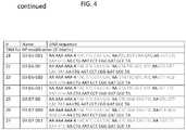
- the kit includes at least one of the polynucleotide detection tag sequences of FIG. 4 .
- the kit includes at least one of the decoding polynucleotide sequences of FIG. 8A .
- the kit includes at least one of the displacement polynucleotide sequences of FIG.
- the kit also includes at least one peptide-loaded streptavidin major histocompatability complex (MHC) tetramer, including a modified streptavidin protein, four biotin-modified MHC proteins each independently conjugated to the modified streptavidin protein, and an antigen peptide bound to the biotin-modified MHC proteins.
- MHC major histocompatability complex
- an ex vivo method for isolating neo-antigen-specific T cells for a tumor in a subject includes identifying candidate T cell epitopes for the tumor using a major histocompatiblity complex (MHC) binding algorithm, synthesizing antigen peptides corresponding to the candidate T cell epitopes, preparing the library of antigen complexes as detailed above using the antigen peptides, incubating the library of antigen complexes with TILs or PBMCs from the subject, and separating paired T cells from unpaired T cells, the paired T cells comprising those T cells that have paired with any of the antigen peptides of any of the antigen complexes in the library of antigen complexes.
- MHC major histocompatiblity complex
- the method for isolating neo-antigen-specific T cells for a tumor in a subject as above also includes adding the paired T cells to a microfluidic device to separate the paired T cells into individual paired T cells, and detecting the sequence of the at least one coding region of the polynucleotide detection tag of the antigen complex of each individual paired T cell.
- the detecting the sequence of the at least one coding region of the polynucleotide detection tag of the antigen complex of each individual paired T cell includes incubating the polynucleotide detection tag of each individual paired T cell with at least two labeled decoding polynucleotides, and detecting presence of a hybridized labeled decoding polynucleotide to thereby determine the sequence of the at least one coding region of the polynucleotide detection tag.
- the method for isolating neo-antigen-specific T cells for a tumor in a subject is as above, in which the least one coding region of the polynucleotide detection tag of the antigen complex includes at least two coding regions, and detecting the sequence of the at least two coding regions of the polynucleotide detection tag of the antigen complex of each individual paired T cell includes incubating the polynucleotide detection tag of each individual paired T cell with at least two first labeled decoding polynucleotides, detecting presence of one or more first hybridized labeled decoding polynucleotides to thereby determine the sequence of a first one of the at least two coding regions of the polynucleotide detection tag, incubating the one or more first hybridized labeled decoding polynucleotides with one or more displacement polynucleotides to remove the first labeled decoding polynucleotides from the first hybridized label
- T-cell mediated immunity is characterized by the activation of antigen-specific cytotoxic T cells that are able to induce apoptosis in cells that display epitopes of foreign antigen in a major histocompatibility complex (MHC) on their surface, as depicted in FIG. 1 .
- MHC major histocompatibility complex
- These cells displaying an MHC complex loaded with foreign antigen include virus-infected cells, cells with intracellular bacteria, and cancer cells displaying tumor antigens.
- one of the initial steps includes identification of the patient's tumor-specific antigens (i.e., neoantigens).
- HLA human leukocyte antigen
- antigen-specific T cells refer to cells that are distinguished from one another by their T cell receptors (TCRs), which give them their antigen specificity.
- Embodiments of the present invention include recombinant antigen-loaded MHC compositions and facile decoding methods for high fidelity, rapid, and non-destructive isolation and identification of patient-specific T cell populations targeted to patient-specific antigens, e.g., neoantigens.
- Embodiments of the present invention include a nanoparticle (NP) having a unique polynucleotide barcode (NP) linked to a unique recombinant antigen-MHC complex.
- T cells that pair with the antigen-MHC complex are then isolated in this antigen-MHC-T cell complex by selective isolation of the nanoparticle.
- the isolated antigen-MHC-T cell complex may then be transferred to a cell trapping platform to separate individual antigen-MHC complex-T cells.
- the unique barcode for each isolated NP is identified by in situ amplification or using fluorescently labeled barcode sense strand "readers”. Using this nanoparticle isolation and barcode identification methodology, the individual antigen-MHC-T cell complex is identified, but is not destroyed.
- the isolated T cell is available after identification as a valuable source for further characterization (e.g., tumor biomarker analysis) or for further propagation. Further growth of the T cell results in an enriched population of patient-derived T cells targeted to the patient-specific antigens. This population of patient-derived T cells targeted to the patient-specific antigens may be used for adoptive cell transfer into the patient as a means of immunotherapy targeting the tumor or pathogen.
- Antigen complexes include a recombinant antigen-MHC complexed with a barcoded nanoparticle (NP) sorting agent to form a barcoded NP-antigen-MHC complex.
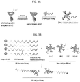
- the barcoded NP-antigen-MHC complex is modular in form as schematically shown in FIGs. 3A and 3B .
- the barcoded NP-antigen-MHC complex is made of an antigen-MHC complex and a barcoded NP complex linked together by polynucleotide hybridization domains (shown in black in FIG. 3B ).
- the modular form of the barcoded-NP-antigen-MHC complex allows for the barcoded coding regions to be easily modified to add specificity and to increase the number of different complexes that can be assayed together and subsequently isolated and identified.
- each antigen is associated with a unique n -position barcode polynucleotide sequence allowing for n possible sequences per position.
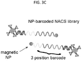
- An example of a 3-position barcode is shown as different colors (yellow, red, or green) in FIGs. 3B and 3C .
- This 3-position barcode yields a 3 3 , or 27-plex antigen library with each of the 27 different antigens having a specific barcode that can be readily determined upon isolation of the complex.
- the ability to screen 27 different antigens with one T cell suspension is a significant advantage over the current more time consuming methods.
- Each of the modular components of the barcoded NP-antigen-MHC complex is described in more detail below.
- In silico analysis can be used to identify neoantigens.
- the in silico analysis for identifying putative neoantigens requires the genomic (DNA) or exomic (RNA) sequence of the tumor, virus, or bacteria in the patient.
- DNA genomic
- RNA exomic
- the genome sequences of these pathogens may be known and the available sequence data may be used. Nonetheless, both unknown (new) and previously identified viruses and bacteria may be isolated from a patient (e.g., from a blood sample) for DNA or RNA sequencing.
- tumor cancers include patient-specific mutations
- DNA or RNA sequencing of tumor cells from a patient's tumor biopsy or blood sample is carried out as an initial step.
- identification of tumor neoantigens specific to a patient includes DNA and/or RNA sequencing as well as HLA-typing of tumor cells from a tumor biopsy or a blood sample of the patient.
- somatic mutation identification i.e., somatic mutation calling
- DNA or RNA sequencing is also performed on a matched normal patient tumor or blood sample.
- the collective tumor-normal sequencing data are then input into at least one (e.g., two or three) algorithmic program (e.g., MuTect v1.1.7, Varscan2 Somatic (V2.3.6), and/or the GATK-HaplotypeCaller (HC, v3.3)) to identify somatic mutations that putatively express (correspond to) neoantigens.
- algorithmic program e.g., MuTect v1.1.7, Varscan2 Somatic (V2.3.6), and/or the GATK-HaplotypeCaller (HC, v3.3)
- all of the in silico identified somatic mutations may be utilized to form a set of candidate tumor neoantigens for T cell screening.
- the somatic mutations identified in silico are ranked with the highest ranked somatic mutations being observed by at least three algorithmic programs, followed by at least 2 algorithmic programs.
- the patient's HLA type is determined from the tumor exome sequencing data using software for HLA typing (e.g., ATHLATES).
- software e.g., the NetMHC3.4 server
- putative neoantigenic sequences from the patent's biopsied tumor or blood sample are identified.
- Embodiments of the present invention include a recombinant antigen-MHC complex that is capable of pairing with cognate T cells.
- antigen complex As used herein, “antigen complex,” “antigen-MHC,” “antigen-MHC complex,” “recombinant antigen-MHC complex,” “peptide MHC,” and “p/MHC,” are used interchangeably to refer to a recombinant major histocompatibility complex with a peptide in the antigen binding groove.
- antigen includes any antigen including patient-specific neoantigens.
- a recombinant antigen-MHC complex includes a recombinant MHC molecule.
- the MHC complex may be an MHC Class I (MHC I) complex that pairs with CD8-positive (CD8+) T "killer” cells.
- the MHC complex may be an MHC Class II (MHC II) complex that pairs with CD4+ "helper" T cells.
- the antigens presented by MHC Class I complexes are cytosolic proteins, while antigens presented by MHC Class II complexes are derived from extracellular proteins.
- MHC Class I molecules As tumor cells are endogenously derived cells expressing mutations, tumor antigens are commonly presented by MHC Class I molecules, thereby pairing with CD8+ T cells. Similarly, viral proteins are endogenously expressed from a patient's cells, and are also presented by MHC Class I molecules to CD8+ T cells. Bacterial cells, however, express proteins using their own cellular machinery, which upon infection in a patient, are considered exogenously expressed and are presented by the patient's MHC Class II molecules to CD4+ T helper cells. MHC class III molecules include other immune components, such as complement components (e.g., C2, C4, factor B) and some that encode cytokines (e.g., TNF-a) and also heat shock proteins (hsps).
- complement components e.g., C2, C4, factor B
- cytokines e.g., TNF-a
- hsps heat shock proteins
- the MHC I or MHC II molecule of the recombinant antigen MHC complex is synthesized to correspond to the patient's HLA type. Polymorphisms are found in both MHC I and MHC II molecules.
- the MHC Class I molecules are heterodimers made of two polypeptide chains, ⁇ and ⁇ 2-microglobulin (b2m).
- the ⁇ chain has three domains including ⁇ 1, ⁇ 2, and ⁇ 3.
- the ⁇ and b2m chains are linked noncovalently via interaction of b2m and the ⁇ 3 domain of the ⁇ chain.
- the ⁇ chain is polymorphic and is encoded by the HLA gene complex while the b2m subunit is not polymorphic and is encoded by the b2m gene. Assembly of the ⁇ and b2m chains is stabilized by the presence of a 9-11 amino acid peptide antigen loaded in the antigen binding groove on the surface of the ⁇ 1 and ⁇ 2 domains.
- patient-specific MHC class I antigens are presented on recombinant MHC class I complexes corresponding to the patient's HLA type.
- the MHC I ⁇ chain is encoded by the HLA-A, HLA-B, or HLA-C gene.
- Each of the HLA-A, HLA-B, and HLA-C genes express allele-specific subtypes some of which are shown in Table 7 in Example 19.
- the MHC Class II molecules are heterodimers made of two polypeptide chains, ⁇ and ⁇ .
- the ⁇ and b chains each have two domains: ⁇ 1 and ⁇ 2, and ⁇ 1 and ⁇ 2, respectively.
- Both the ⁇ and ⁇ chains are polymorphic and are encoded by the HLA gene complex. Assembly of the ⁇ and ⁇ chains forms an antigen binding groove on the surface of both the ⁇ 1 and ⁇ 2 domains with antigen peptide lengths from 11 to 30 amino acids.
- patient-specific MHC class II antigens are presented on recombinant MHC class II complexes corresponding to the patient's HLA type.
- the HLA genes include HLA-DM, HLA-DO, HLA-DQ, and HLA in which the MHC II ⁇ chain is encoded by the HLA-A-DMA, HLA-DOA, HLA-DPA1, HLA-DQA2, or HLA-DRA gene and the ⁇ chain is encoded by HLA-DMB, HLA-DOB, HLA-DPB1, HLA-DQB1, HLA-DQB2, HLA-DRB1, HLA-DRB3, HLA-DRB4, orHLA-DRB5 gene.
- the recombinant MHC molecule is an MHC Class II molecule expressed and loaded with a candidate antigen peptide as described in Novak et al., 1999, J. Clin. Invest. 104:R63-R67 .
- the recombinant MHC molecule is an MHC Class I molecule expressed as a conditional ligand.
- MHC class I molecule is unstable in the absence of peptide (i.e. antigen peptide)
- a recombinant MHC Class I molecule is expressed with a peptide having a cleavable moiety, that upon irradiation with UV light dissociates from the complex and disintegrates.
- the UV disintegration of the cleavable peptide is performed in the presence of a "rescue peptide," the rescue peptide will readily replace the UV irradiated peptide in the binding groove, as depicted in FIG.
- MHC Class I molecules can be easily loaded with candidate neoantigens to form a MHC class I neoantigen library for screening T cells.
- the recombinant MHC molecule is a tetramer complex of four MHC molecules each loaded with the same candidate antigen peptide. Since most neoantigens have low binding affinities (K d ) for MHC proteins (e.g., 500 nM or lower) a tetrameric MHC complex allows for increased binding avidity, thereby increasing the sensitivity of this antigen-MHC tetrameric probe for pairing with low abundant cognate T cells.
- an MHC tetramer is formed using modified streptavidin conjugated with four biotin-modified MHC molecules.
- the streptavidin is modified to enable binding of a polynucleotide (e.g., DNA or RNA) linker.
- Modification of the streptavidin includes a binding moiety that can pair with (e.g., covalently bind to) a corresponding cognate binding moiety linked to the polynucleotide molecule. Any suitable pair of binding moieties may be used to modify streptavidin and the polynucleotide for linkage.
- Non-limiting examples of binding moiety pairs include a thiol group (e.g., cysteine) and maleimide, adamantane and cyclodextrin, an amino group and a carboxy group, and an azido group and alkynl group (i.e., click chemistry).
- a cysteine-modified streptavidin linked to a maleimide-modified DNA hybridization domain is shown in FIG. 3A .
- Embodiments of the present invention include a modified nanoparticle linked to a polynucleotide detection tag (i.e., the barcode), where the polynucleotide detection tag includes at least one coding region.
- a polynucleotide detection tag i.e., the barcode
- This complex as well as the method of using the complex, is also referred to as nanoparticle-barcoded nucleic acid cell sorting (NP-barcoded NACS) and a nanoparticle sorting agent.
- the nanoparticle is magnetic for isolation using a magnet.
- the nanoparticle is a 1 um to 15 um polystyrene particle isolated by gravity.
- the nanoparticle is modified with a binding moiety for linking to the polynucleotide coding region. Modification of the nanoparticle includes a binding moiety that can pair with (e.g., covalently bind to) a corresponding cognate binding moiety linked to the polynucleotide molecule. Any suitable pair of binding moieties may be used to modify the nanoparticle and the polynucleotide detection tag for linkage.
- Non-limiting examples of binding moiety pairs include a thiol group (e.g., cysteine) and maleimide, adamantane and cyclodextrin, an amino group and a carboxy group, and an azido group and alkynl group.
- the antigen complex of the present invention includes a barcoded-nanoparticle complex where the barcode is a polynucleotide detection tag made of coding regions that provide a unique antigen-specific sequence for identification after T cell isolation.
- a "coding region" is a set of nucleotides with a unique and specific sequence that is separate from another polynucleotide.
- one polynucleotide coding region is made of 5 to 25 nucleotide basepairs.
- one polynucleotide coding region is made of 7 to 25 nucleotide basepairs, 8 to 25 nucleotide basepairs, 9 to 25 nucleotide basepairs, 10 to 25 nucleotide basepairs, 10 to 20 nucleotide basepairs, 10 to 19 nucleotide basepairs, 10 to 18 nucleotide basepairs, 10 to 17 nucleotide basepairs, or 10 to 16 nucleotide basepairs.
- the barcoded nanoparticle complex has a polynucleotide detection tag with at least one coding region (i.e., a 1-position barcode).
- the number of coding regions ( n ) corresponds to the number of different antigens that can be screened together and subsequently identified.
- the number of different coding regions is only limited by the number of different colored fluorescent dyes.
- the coding regions will be "read" starting with coding region attached to the nanoparticle which is referred to as the first coding region or coding region 1. Any coding region thereafter in the direction away from the nanoparticle is the second coding region (coding region 2), followed by the third coding region (coding region 3) and so on.
- each polynucleotide detection tag sequence is unique from all other polynucleotide detection tag sequences.
- each coding region sequence may only occur in one coding region position.
- a 3-position coding region library of ssDNA includes coding regions D1, D2, and D3 in coding region 1, D5, D6, and D7 in coding region 2, and D9, D10, and D11 in coding region 3, resulting in 27 different detection tag sequences as listed in the table of FIG. 4 .
- a polynucleotide hybridization domain is linked to the 3' end of the polynucleotide detection tag of the barcoded NP, and a second polynucleotide hybridization domain is linked to the streptavidin scaffold of the antigen-MHC complex. Accordingly, the antigen-MHC complex is linked to a barcoded nanoparticle through hybridization of complementary hybridization domains.
- the first polynucleotide hybridization domain and the second polynucleotide hybridization domain may be single stranded DNA (ssDNA) having a first and a second hybridization sequence, respectively, where the first and second hybridization sequences are complementary, resulting in a linker of hybridized double stranded DNA (dsDNA), shown as overlapping black lines in FIGs. 3B , 3C .
- the first and second hybridization domains are "universal" and are the same for all barcoded NP complexes and antigen-MHC complexes in a library. Using a universal hybridization domain allows for synthesis of one streptavidin molecule with the attached hybridization domain.
- the first and second hybridization domains are different for each barcoded NP-antigen-MHC complex.
- each unique antigen-MHC complex is linked (i.e., hybridized) to a unique barcode sequence.
- the preparation of a barcoded NP-antigen-MHC complex library includes a record (e.g., a key or a legend) of the candidate antigens and their corresponding barcode (i.e., polynucleotide detection tag sequence). This coding key aides in efficiently identifying the specific antigens that pair with T cells. Examples of an antigen and barcode key for the list of candidate antigens (patient-specific candidate neoantigens and MART-1 tumor antigen) of FIG. 4 is shown in FIGs. 5A-5B .
- aspects include a barcoded nanoparticle-antigen-MHC complex for use in screening antigen-specific T cells.
- a single antigen may be assayed using the complex in the presence of T cells.
- assaying one candidate antigen is not as efficient as screening multiple candidate antigens.
- Different barcoded NP antigen-MHC complexes are prepared using different candidate antigens with corresponding unique coding regions forming a library of barcoded NP-antigen-MHC complexes. The following methods are described using a library of the barcoded NP-antigen-MHC complexes; however the method may be followed or easily adapted for screening a single candidate antigen.
- isolation and identification of patient-derived and antigen-specific T cells using a library of barcoded-NP-antigen-MHC complexes includes incubating the candidate antigen complexes with patient-derived T cells.
- the patient-derived T cells are isolated from the patient's peripheral blood mononuclear cells (PBMCs) or tumor infiltrating lymphocytes (TILs).
- PBMCs peripheral blood mononuclear cells
- TILs tumor infiltrating lymphocytes
- both CD4+ and CD8+ T cells are labeled and sorted from PBMCs or TILS using anti-CD4 and anti-CD8 fluorescent antibodies with live populations of CD4+ and CD8+ single-positive cells sorted using fluorescence-activated cell sorting (FACS), in order to isolate only CD4+ or CD8+ cells.
- FACS fluorescence-activated cell sorting
- T cells that are positive for both CD4 and CD8 may be isolated using an anti-CD3 fluorescent antibody followed by FACS.
- FACS fluorescence-activated cell sorting
- the T cell population may be only CD8+, whereas MCH II molecules would require CD4+ T cells.
- an antigen library having both MHC I and MHC II molecules may be incubated with T cells that are both CD4 and CD8 positive.
- an antigen library having either MHC I or MHC II molecules may be screened with T cells that are positive for both CD4 and CD8.
- Embodiments of the present invention include incubating a barcoded NP-antigen-MHC complex library with a suspension of CD4+, CD8+ or CD4+/CD8+ T cells.
- Incubation of the nanoparticle library with the T cell suspension allows for a complete and thorough exposure of the nanoparticle-bound antigen to the various T-cell receptors. This method may include rocking or rotation of the cells.
- the nanoparticle is selectively isolated or selectively collected. For example, if the nanoparticle is magnetic, applying a magnet to the suspension allows for separation of nanoparticles in a complex with antigen paired T cells and removal of unpaired T cells.
- the isolated magnetic nanoparticles with paired T cells is shown schematically in FIG. 6 .
- the unpaired T cells may be separated by gravity (e.g., centrifugation). After separation of unpaired T cells, in some embodiments, the isolated nanoparticles are washed at least once to remove any non-specifically associated T cells.
- the cells are loaded into a microfluidic device that separates and posits single cells in individual locations ("traps") in the channels of the device.
- a paired T cell and “a T cell paired antigen MHC complex” refers to the complex of a T cell having a T cell receptor epitope that binds to an antigen peptide in a barcoded nanoparticle antigen-MHC complex. Accordingly, with the paired T cells separated in the cell trapping device, the coding regions (barcodes) of the paired antigen-MHC complex are identified in situ to determine the sequence of the cognate peptide antigen. Accordingly, each coding region is read starting with coding region 1.
- coding region 1 All possible complementary polynucleotide sequences for coding region 1 are linked to a distinguishable fluorescent dye forming a dye-polynucleotide decoder sequence that hybridizes the fluorescence to the complementary coding region on the nanoparticle, thereby "reading" coding region 1.
- a distinguishable fluorescent dye and “distinguishable dye” refer to a dye of a color that is visually distinct from another dye. Any suitable dye may be used that is capable of being linked to a polynucleotide.
- a "displacement" polynucleotide is added to remove the dye-polynucleotide decoder of coding region 1 followed by or together with all of the possible dye-polynucleotide decoder sequences corresponding to coding region 2.
- the polynucleotide detection tag is partially decoded.” That is, a "partially decoded polynucleotide detection tag" has at least one, but not all coding regions decoded. Subsequent coding regions are read using this read-displace/read-displace/read format.
- the polynucleotide decoder sequences are ssDNA or RNA. For example, as listed in the table in FIG.
- ssDNA decoding sequences M1, M2, M3, M5, M6, M7, M9, M10, and M11 are fluorescent dye-linked decoding sequences that hybridize to respective coding region of D1, D2, D3, D5, D6, D7, D9, D10, and D11 ( FIG. 4 ), with each decoding sequence shown in the color of the corresponding fluorescent dye.
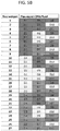
- Example displacement ssDNA sequences corresponding to (i.e., that hybridize with) each decoding sequence in FIG. 8A include the ssDNA sequences M1 comp, M2 comp, M3 comp, M5 comp, M6 comp, M7 comp, M9 comp, M10 comp, and M11 comp as listed in the table of FIG. 8B .
- the coding regions (barcodes) of the T cell paired-antigen MHC complex are identified in situ using the fluorescent dye-polynucleotide decoding "reader" sequences in combination with the "displacement" polynucleotide sequences.
- This method was carried out using a barcoded NP-antigen-MHC library of the 27 antigens in FIG. 5A , having the NP barcode (detection tag) sequences as listed in FIG. 4 .
- TILs tumor infiltrating lymphocytes
- FIG. 9 in an optical micrograph of a microfluidic chamber equipped with 10 cell traps. As shown, only 9 of the cell traps contain single barcoded T cells. These 9 barcoded T cells were read and displaced using the fluorescent dye-linked decoding ssDNA sequences listed in FIGs. 8A and 8B , respectively.
- the fluorescent images for each coding region read are shown left to right in FIG. 9 with the coding region 1 image labeled as 1st, coding region 2 image labeled as 2nd, and coding region 3 labeled as 3rd.
- the cell at position 8 does not provide a clean read, and the cell at position 9 was lost.
- this method according to embodiments of the present invention only the trapped cells providing clear unambiguous readouts are considered, thereby allowing for control of the fidelity of the screening process.
- the corresponding neoantigens and the tumor antigen MART-1 were determined.
- the cell at position 3 in FIG. 9 reads YRG corresponding to neoantigen 12 listed in FIG. 5A
- the cell at position 4 reads RGY corresponding to the MART-1 tumor antigen.
- kits for detecting neoantigen-specific T-cells are detailed in the appended claims. Some aspects include a kit for preparing a library of barcoded nanoparticles, the kit including barcodes (polynucleotide detection tag) sequences, the corresponding fluorescent dye-linked polynucleotide decoder sequences for reading the barcodes, and the corresponding displacement polynucleotide sequences for removing the decoder sequences.
- the polynucleotide sequences include ssDNA or RNA.
- the polynucleotide sequences are ssDNA.
- the polynucleotide detection tag sequences are modified at their 5' end to a binding moiety for attachment to a nanoparticle.
- the polynucleotide detection tag (ssDNA barcode) sequences in FIG. 4 are conjugated to a biotin molecule for binding to a streptavidin-nanoparticle; however any suitable binding moiety may be used.
- suitable binding moiety pairs are known in the art.
- binding moieties include thiol, maleimide, adamantane, cyclodextrin, amine, carboxy, azide, and alkyne.
- the kit may also include modified nanoparticles modified with the cognate binding moiety corresponding to the binding moiety on the modified polynucleotide detection tags.
- a kit for preparing a library of barcoded nanoparticles also includes a recombinant conditional MHC tetramer complex capable of being loaded with any MHC antigen peptide.
- the MHC may be MHC Class I or MHC Class II and includes any specific haplotype corresponding to the patient's haplotype.
- the kit may include MHC Class I molecules corresponding to an HLA type listed in Table 7.
- the kit may include MHC Class II molecules corresponding to HLA-DM, HLA-DO, HLA-DQ, and HLA in which the MHC II ⁇ chain is encoded by the HLA-A-DMA, HLA-DOA, HLA-DPA1, HLA-DQA2, or HLA-DRA gene and the ⁇ chain is encoded by HLA-DMB, HLA-DOB, HLA-DPB 1, HLA-DQB 1, HLA-DQB2, HLA-DRB 1, HLA-DRB3, HLA-DRB4, or HLA-DRB5 gene.
- MHC Class II ⁇ chain is encoded by the HLA-A-DMA, HLA-DOA, HLA-DPA1, HLA-DQA2, or HLA-DRA gene and the ⁇ chain is encoded by HLA-DMB, HLA-DOB, HLA-DPB 1, HLA-DQB 1, HLA-DQB2, HLA-DRB 1, HLA-DRB3, HLA-DRB4, or H
- a kit for preparing a library of barcoded nanoparticles includes modified polynucleotide detection tag sequences for attachment to nanoparticles.
- Alternative methods for identifying the trapped barcoded T cell may be used.
- the polynucleotide detection tag sequences may be amplified and sequenced from the microfluidic device.
- the trapped T cell may be removed from the microfluidic device for further analysis and/or growth.
- Example 1 Capture Efficiency of antigen-specific T cells.
- the barcoded NP-antigen-MHC complex library was validated by employing selective capture of Mart-1 antigen specific T cells from a mixture of cells.
- FIG. 10A shows a schematic illustration of NP-Mart-1 tetramer for capture of Jurkat cells transduced with a Mart-1 specific T-cell receptor.
- FIG. 10B shows mixed Jurkat (stained green) and GBM U87 cells (stained blue). The NP-Mart-1 tetramer is incubated with the mixed cell population and magnetically enriched with cells from the supernatant shown in FIG. 10C , and from the magnetic pulldown shown in FIG. 10D .
- Example 2. Use of barcoded NP-antigen-MHC complex library.
- the NP-antigen-MHC complex library tool was used to analyze CD8+ cells expanded from tumor infiltrating lymphocytes (TILs) collected from on-treatment tumor biopsies of two metastatic melanoma cancer patients who were positively responding to anti-PD-1 checkpoint inhibitor therapy at the time of biopsy as shown in FIG. 11 . These two patients were treated with the anti-PD-1 antibody pembrolizumab within a phase I trial, and responses to this therapy are known to be mediated by tumor infiltrating CD8+ cells. For each patient, a pre-treatment biopsy was also collected ( FIG. 11 ), and analyzed for both the tumor exome and gene expression.
- TILs tumor infiltrating lymphocytes
- the transcripts of the mutated proteins corresponding to neoantigen numbers 3, 5, 11, 26, 31, 34, 36, 37, 46 and 48 yielded zero mutation reads ( Table 3 ) and were included in all assays as controls that spanned the sampled range of neoantigen/MHC binding affinities. from the microfluidic device for further analysis and/or growth. Table 1. Putative neo-antigens for Patient #1. Letter in the bracket indicates the native peptide.
- Neo-antigen K d (nM) # Neo-antigen K d (nM) 1 F(S)LGSLILV 5 27 KMGKTIYKY(H)V 76 2 RLS(P)SCFDYV 9 28 NLFNTYL(P)CL 77 3 S(P)LMNEDFIL 11 29 RLSEV(A)MARM 82 4 F(S)LGSLLILVV 11 30 VLTEIF(S)LGSL 105 5 S(P)LMNEDFILA 11 31 ALYKEE(G)EQEPV 133 6 SLHB(G)LTDGV 12 32 VLIDLIQRTKV(D) 134 7 KAWENF(S)PNV 14 33 MVC(R)TFCPPPL 138 8 LLSEFF(S)SCL 18 34 LLFHS(P)PRAHL 139 9 KLLSEFF(S)SCL 20 35 V(M)LLHAFEGYNV 147 10 LQ(R)DSGLWFPV 21 36 VTSSIVTL(P)V
- Neo-antigen K d (nM) # Neo-antigen K d (nM) 1 LLTDLLFL(S)I 4 15 TVLGTPF(S)EV 26 2 RLMERSIY(H)SA 7 16 SIY(H)SARYIFV 26 3 FLVNDWLL(S)V 8 17 YLAHWLYV(H)Y 27 4 FLDSC(S)PHLPL 8 18 KIS(P)SQPPPV 28 5 KMNELNY(H)CI 10 19 KL(S)STFLMKA 29 6 RLLTDLLFL(S)I 10 20 FTLFELAF(S)L 31 7 RLANLIW(R)RA 11 21 FLPRKENI(S)SV 31 8 RLLTDLLFL(S) 12 22 VLQGC(R)LPCPV 31 9 RIYTEMC(R)FTV 13 23 F(S)LANSGGWV 33 10 VL(P)FEKK
- the corresponding NP-barcoded NACS libraries captured 4.5-5% of the CD8+ TILs with a 0.5% non-selective capture, as gauged by testing the library against CD4+ T cells from the same patient tumors ( FIG. 12 ).
- the barcoded T cells were loaded into the microfluidic chip and sequential fluorescent reads were performed to identify, in parallel, the same 8 neoantigen-specific T cell populations (plus MART-1 specific cells) ( FIG. 13 ). All but very low-abundance populations (1 or 2 incidents per analysis) were detected in both assays.
- the most abundant population was specific for neoantigen #12, and accounted for about 12 cells per 1000 expanded TILs.
- the NP-barcoded NACS library was further expanded to analyze the TILS for specificities against neoantigens ranked 28-50 (Kd values up to 500 nM). Two additional populations were detected (#'s 38 and 45).
- the value of the barcoded, fluorescent read-out approach is that for each T cell captured, there is either a high fidelity read, or, occasionally, a nonsense read (see, for example, cell #8 in FIG. 9 ). Thus, the noise level of this approach is very low.
- the detected neoantigen-specific T cell populations are defined as those that were identified by 3 or more unambiguous reads, or were detected in both analyses of Patient #1 TILs.
- the summed analysis of Patient #1 TILS for the 50-element library is provided in FIG. 14B with the corresponding lesion analysis shown in FIG. 14A .
- Example 3. Screening the barcoded NP-antigen-MHC complex library in PBMCs. Some biospecimens were analyzed using a serial approach, in which each NP-barcoded NACS library element was used to query for a specific T cell population.
- NP-barcoded NACS library designed for patient #1 was used to analyze expanded TILs from an additional unrelated melanoma patient on the same clinical trial, as well as PBMCs from a healthy volunteer.
- the list of neoantigens is unique for every patient, and so the library designed for patient #1 should not capture T cell populations for the two control patient samples.
- Example 5 Analysis with PMBCs from Patient #1.
- the sensitivity of the NP-barcoded NACS method prompted analysis of peripheral blood from patient #1 for neoantigen-specific T cell populations. These T cells were not expanded in vitro, so as to avoid any population bias that can accompany such expansion.
- the full 50-element neoantigen library was used to interrogate PBMCs collected at four time points.
- Two time points (Days 187 and 208, FIG. 11 ) were collected during response to therapy while tumors were shrinking, and corresponded closely to the date of the tumor biopsy (Day 187) from which the TILs were collected.
- the neoantigen-specific T cell levels were below those found in the TILs (1% vs 7% of all CD8+ T cells), but several populations at levels >0.05% were positively detected. Most populations coincided with those detected from the TILs, with two new populations (specific to neoantigens #15 and #33) identified.
- Analysis of PBMCs at Day 439 detected zero neoantigen-specific populations as shown in FIG. 18 .
- PBMCs correspond to a time close to the biopsy from which the exome and transcriptome measurements were taken, but also during a period of pseudo-progression for patient #1 prior to response ( FIG. 11 ). T cells were observed at this early time point, and those populations were present in substantially higher numbers (about 2% of CD8+ PBMCs) relative to later time points when the tumors were regressing. The same populations detected at Days 187 and 208 were also detected at Day 41, as well as additional populations. Several of the detected neoantigen-specific T cell populations from PBMCs correspond to relatively weak (Kd > 100 nM) neoantigen/MHC binding affinities.
- the number of mutation-containing reads from the mRNAs associated with the mutated proteins from the pre-treatment transcriptome analysis are provided in the bottom graph of FIG. 14 .
- An advantage to the NP-barcoded NACS method is that the process is non-destructive and single T-cells, now with known antigen-specificity, are individually isolated by the microfluidic device and avalailbe for further analysis, including TCR ⁇ and ⁇ gene sequencing.
- paired TCR ⁇ and TCR ⁇ genes from a single T cell from patient #1 were cloned, both as proof of capability and validation of the barcoded neoantigen specificity.
- a CD8+ T cell was captured in the single microfluidic trapping device ( FIGs. 19A , 19B ) and barcoded to identify the neo-antigen identity ( FIG. 20A ).
- the captured single T cell was punched out to perform RT-PCR to obtain TCR ⁇ and ⁇ gene sequences.
- Jurkat cells transduced to express that TCR were found to bind to the cognate neoantigen tetramer ( FIG. 20B ).
- Analysis of T cells collected from two tumors of immunotherapy patients responding to anti-PD1 therapy reveals that around 20% of the top 50 predicted neoantigens account for a large fraction ( ⁇ 7% for the HLA A*02.01) of CD8+ T cells in the tumor.
- the two patients analyzed here each express 6 HLA genotypes.
- Tumor responses were evaluated starting at 12 weeks, confirmed 4 weeks after first response, and imaged every 12 weeks thereafter. Response was characterized by both the Response Evaluation Criteria in Solid Tumors (RECIST) and the immune-related response criteria (irRC). Tumour biopsy and peripheral blood cell collection and analyses were approved by UCLA IRBs 11-001918 and 11-003066.
- RECIST Response Evaluation Criteria in Solid Tumors
- irRC immune-related response criteria
- Tumor biopsies from the patients analyzed were obtained at baseline and on therapy and were processed with one aliquot immediately fixed in formalin followed by paraffin embedding for pathological analyses, a second aliquot snap frozen by immediate immersion in liquid nitrogen for genetic analyses, and a third aliquot minced fresh under sterile condition followed by DNAse/collagenase digestion to create single cell suspensions (s.c.s) before cryopreservation in liquid nitrogen.
- Peripheral blood mononuclear cells (PBMCs) were prepared from fresh whole blood by Ficoll-Paque density gradient centrifugation and cryopreserved.
- PBMCs Peripheral blood mononuclear cells
- Tumor infiltrating lymphocytes were expanded from cryopreserved s.c.s using anti-CD3 antibody (OKT3, 50ng/mL, 48hr exposure) and IL-2 (300IU/mL) and re-cyropreserved at 5x10⁶ cells/mL after 2-4 weeks.
- TILs were thawed and treated with DNAse for 45 min the morning of use, and stained with antibodies to CD4 (BV510, BioLegend, San Diego, CA) and CD8+ (BV605, BioLegend, San Diego, CA). Live (7AAD-negative) populations of CD4 and CD8+ single-positive cells were sorted using a FACS Cell Sorter (BD Biosciences, San Jose, CA).
- Example 9 Example 9
- RNA sequencing was performed using the Illumina HiSeq 2500 platform on 100-bp paired-end libraries prepared using the IlluminaTruSeq RNA sample preparation kit per the manufacturer's instructions.
- RNA reads were mapped to hg19 using TopHat2 v2.0,5 and were quantified and normalized using Cufflinks v2.2.16 program and CuffNorm to generate normalized expression tables by library size (fragments per kilobase of exon per million fragments mapped, FPKM) using the geometric normalization method.
- Mutation-containing RNA reads were identified by a custom Python (v2.7.3) script utilizing the Biopython and pysam packages, and verified by visual inspection in the Integrated Genomics Viewer (IGV).
- IGV Integrated Genomics Viewer
- Peptide binding predictions to HLA-A02:01 were generated by netMHC3.47 for 9-mer and 10-mer peptides in a sliding window around each non-synonymous amino acid-altering mutation.
- Peptide sequences were derived from Ensembl GRCh37 release 74.
- Candidate peptides were binned by 1) those with mutations-containing reads identified by RNA-seq, 2) those with RNA expression (FPKM > 0) but no identified mutated reads, and 3) all others without detectable RNA-seq expression.
- Peptides were ranked and sorted by HLA binding affinity within each bin.
- Example 12 Production of ssDNA-SAC conjugates.
- the ssDNA-SAC (strepatavidin antigen complex) conjugate was produced following previous published protocol as described in Kwong et al., 2009, J. Am. Chem Soc., 131:9695-9703 . Briefly, SAC was first expressed from the pTSA-C plasmid containing the SAC gene (Addgene), as described in Sano et al., 1990, PNAS, 87:142-146 . Before conjugation to DNA, SAC (1 mg/ml) was buffer exchanged to PBS containing Tris(2-Carboxyethyl) phosphine Hydrochloride (TCEP, 5 mM) using zeba desalting columns (Pierce).
- SAC Tris(2-Carboxyethyl) phosphine Hydrochloride
- MHPH-labeled SAC and SFB-labeled DNA were buffer exchanged to citrated (50 mM sodium citrate, 150 mM NaCl, pH 6.0), and then mixed in a 20:1 ratio of DNA to SAC to react at rt overnight.
- DNA-SAC conjugate was purified using the Superdex 200 gel filtration column (GE health) and concentrated with 10K MWCO ultra-centrifuge filters (Millipore).
- Example 13 Human MHC class I neo-antigen library construction. MHC library was generated using the UV-mediated peptide exchange method as described in Rodenko et al., 2006, Nat. Protoc. 1:1120-1132 .
- the photo-labile peptide KILGFVFJV and other neo-antigen peptides were synthesized with standard automated Fmoc-peptide synthesis methodology (J, (S)-3-(Fmoc-amino)-3-(2-nitrophenyl)propionic acid, is the photo-labile amino acid residue).
- Plasmids encoding human MHC class I heavy chain and human b2m containing bacterial strain were kind gifts from Ton N M Schumacher.
- MHC photo-labile protein was folded from MHC heavy chain inclusion body, b2m inclusion body and photo-labile peptide according to the previously published protocol 11 and then biotinylated using the BirA biotin ligase.
- MHC photo-labile protein 0.5 uM
- neo-antigen peptide 50 uM
- Example 14 NP MHC neo-antigen library construction. Streptavidin magnetic NP (1 um, ThermoFisher scientific) was mixed with biotin-DNA at 1:20 ratio to obtain NP-DNA. Excess DNA was removed by washing the magnetic NP for 3 times. In parallel, MHC neo-antigen library was added to ssDNA-SAC at 4:1 ratio to form the DNA-MHC tetramer.
- NP-DNA and DNA-MHC tetramer were hybridized at 37°C for 30 min to generate the NP MHC neo-antigen library.
- NP MHC neo-antigen library was added to CD8+ human T cells for 30 min at rt.
- the NP-bound T cells were magnetically enriched and washed with PBS to remove any non-specifically pulled T cells.
- the cells were then loaded into costar transwell polycarbonate membrane (5 um pore) to remove free NPs. Then, the cells were loaded into the cell-trapping microfluidic device and sequentially barcoded.
- Neo-antigen specific cells were enriched by magnet pulldown.
- the non-captured T cells in the supernatant was collected for further incubation with other NP-NACS library element.
- the enriched T cells were washed by PBS once to remove any non-specific cell pulldown. Cells were then loaded into the cell hemocytometer. The whole area in the hemocytometer chip was imaged to obtain the total pulldown cell number. Healthy donor PBMC and PBMC from an unrelated male melanoma patient were used as control to obtain the background.
- Example 18 Single cell TCR cloning. Neo-antigen specific T cells were trapped in the microfluidic device with single cell traps ( FIGs. 19A-19B ). The cells were then recovered by PDMS puncher.
- V ⁇ and V ⁇ domain genes were cloned from single cells using a OneStep RT PCR kit (Qiagen) with multiplexed forward primers that bind TRAV and TRBV gene segments (Tables 5-6) and reverse primers that bind the constant C ⁇ (5'-GCCACAGCACTGTTGCTCTTGAAGTCC-3') and C ⁇ (5'-CCACCAGCTCAGCTCCACGTG-3') domain genes.
- cDNA products were used as templates in a second semi-nested amplification with a universal set of primers (alpha forward primer: 5'-TGGCCTGCTTTGTTTGCCGTGGTTACAGGAAGCCTCAGCA-3', alpha reverse primer: 5'-GCCACAGCACTGTTGCTCTTGAAGTCCATAG-3'; beta forward primer: 5'-CGGGCTCCTTTGCCTACCGTGCCTGCAGGAGGGCTCGGCA-3', beta reverse primer: 5'-CGTGCTGACCCCACTGTGCACCTCCTTCCCATTCACCCACCAGCTCAGCTCCACGTG GTC-3').
- V ⁇ and V ⁇ cDNA were sequenced and reamplified using single TRAV/TRBV forward primers to correct mispriming artifacts introduced through multiplexed PCR.
- Retroviral vectors were constructed for functional testing through PCR assembly.
- the V ⁇ and V ⁇ domain genes were assembled with human growth hormone (HGH) signal peptides, constant regions of the TCR ⁇ and TCR ⁇ chains, and a 2A ribosomal skipping sequence, then digested with restriction enzymes and ligated into a MSCV-based non-replicative retroviral backbone.
- HGH human growth hormone
- TCR ⁇ cloning primers V ⁇ -gene-specific primers for cloning TCR ⁇ genes TRAV gene Signal peptide sequence TRAV gene-specific sequence TRAV1-1*01 5'-TACAGGAAGCCTCAGCA GGACAAAGCCTTGAGCAGCCCTC-3' TRAV1-2*01 5'-T ACAGGAAGCCTCAGCA GGACAAAACATTGACCAGCCCACTG-3' TRAV2*01 5'-TACAGGAAGCCTCAGCA AAGGACCAAGTGTTTCAGCCTTCCAC-3' TRAV3*01 5'-TACAGGAAGCCTCAGCA GCTCAGTCAGTGGCTCAGCCGGA-3' TRAV4*01 5'-TACAGGAAGCCTCAGCA CTTGCTAAGACCACCCAGCCCATC-3' TRAV5*01 5'-TACAGGAAGCCTCAGCA GGAGAGGATGTGGAGCAGAGTCTTTTCC-3' TRAV6*01 5'-T
- TRBV gene-specific sequence TRBV1*01 5'-CAGGAGGGCTCGGCA GATACTGGAATTACCCAGACACCAAAATACCTG-3' TRBV2*01 5'-CAGGAGGGCTCGGCA GAACCTGAAGTCACCCAGACTCCCAG-3' TRBV3-1*01 5'-CAGGAGGGCTCGGCA GACACAGCTGTTTCCCAGACTCCAAAATAC-3' TRBV3-2*01 5'-CAGGAGGGCTCGGCA GACACAGCCGTTTCCCAGACTCCA-3' TRBV4-1*01 5'-CAGGAGGGCTCGGCA GACACTGAAGTTACCCAGACACCAAAACAC-3' TRBV4-1*02 5'-CAGGAGGGCTCGGCA CACCTGGTCATGGGAATGACAAATAAGAAG-3' TRBV4-2*01 5'-CAGGAGGGCTCGGCA GAAACGGGAGTTACGCAG
Landscapes
- Health & Medical Sciences (AREA)
- Life Sciences & Earth Sciences (AREA)
- Chemical & Material Sciences (AREA)
- Engineering & Computer Science (AREA)
- Immunology (AREA)
- Molecular Biology (AREA)
- Hematology (AREA)
- Biomedical Technology (AREA)
- Urology & Nephrology (AREA)
- Analytical Chemistry (AREA)
- Biochemistry (AREA)
- Cell Biology (AREA)
- General Health & Medical Sciences (AREA)
- Pathology (AREA)
- Physics & Mathematics (AREA)
- Biotechnology (AREA)
- Microbiology (AREA)
- General Physics & Mathematics (AREA)
- Organic Chemistry (AREA)
- Food Science & Technology (AREA)
- Medicinal Chemistry (AREA)
- Zoology (AREA)
- Proteomics, Peptides & Aminoacids (AREA)
- Wood Science & Technology (AREA)
- Tropical Medicine & Parasitology (AREA)
- Genetics & Genomics (AREA)
- Bioinformatics & Cheminformatics (AREA)
- Virology (AREA)
- General Engineering & Computer Science (AREA)
- Biophysics (AREA)
- Dispersion Chemistry (AREA)
- Nanotechnology (AREA)
- Toxicology (AREA)
- Measuring Or Testing Involving Enzymes Or Micro-Organisms (AREA)
- Peptides Or Proteins (AREA)
- Hospice & Palliative Care (AREA)
- Oncology (AREA)
- Apparatus Associated With Microorganisms And Enzymes (AREA)
- Micro-Organisms Or Cultivation Processes Thereof (AREA)
Description
-
- Neoantigens from tumor cells are peptide fragments of mutated proteins that contain a mutation, and are capable of being presented in the cleft of Major Histocompatibility Complex (MHC) Class I molecules on the surfaces of cells within the tumor, where they can be surveyed by CD8-positive T cells. The tumor-specificity of neoantigens, coupled with the ability of neoantigen-specific T cells to specifically kill cancer cells, has made tumor neoantigens increasingly important for cancer immunotherapy. Additionally, T cell receptors (TCRs) that recognize specific neoantigens are candidates for TCR-engineered cell based therapies for targeting infections that produce neoantigens.
- Putative neoantigens have been previously identified by tumor exome analysis, and subsequently rank-ordered according to their MHC binding strength using in silico analysis. However, finding which candidate neoantigens are actually promoting T cell tumor infiltration is challenging for several reasons. First, a patient tumor that has a mutation density of 10 per 1 million expressed DNA base pairs might be predicted to have on the order of 50 putative neoantigens that exhibit a binding constant (Kd) to a given human leukocyte antigen (HLA) genotype MHC of 500 nM or lower. Furthermore, any particular neoantigen-specific T cell population is likely to exist in very low abundance in a patient, rendering isolation and/or identification of neoantigen-specific T cells very difficult. Related challenges to these issues include the pairing of neoantigens to their cognate TCRs. Nevertheless, these neoantigen-T cell pairing interactions are at the core of cancer immunotherapy, and so there has been significant effort towards meeting these challenges. Previous approaches for neoantigen-specific T cell pairing have shown to be laborious, non-quantitative, and/or they may only identify one or two T cell populations per HLA genotype due to limited sensitivity.
-
US8394590 discloses a method for sorting cells by combining a sample having a plurality of target cells with a plurality of capture agent sets, each capture agent set comprising a plurality of capture agents comprising a tetramer of an antigen-presenting MHC attached to an optimized streptavidin scaffold molecule. - The antigen complex of the present invention includes a nanoparticle sorting agent including a nanoparticle, a polynucleotide detection tag having at least one coding region forming a barcode, the polynucleotide detection tag being conjugated to the nanoparticle, and a first polynucleotide hybridization domain conjugated to the polynucleotide detection tag. The antigen complex also includes a peptide-loaded streptavidin major histocompatability complex (MHC) tetramer, including a modified streptavidin protein, four biotin-modified MHC proteins each independently conjugated to the modified streptavidin protein, an antigen peptide bound to the biotin-modified MHC proteins, and a second polynucleotide hybridization domain different from the first polynucleotide hybridization domain and conjugated to the modified streptavidin protein, where the nanoparticle sorting agent is linked to the peptide-loaded streptavidin MHC tetramer by hybridization of the first polynucleotide hybridization domain to the second polynucleotide hybridization domain. The at least one coding region is a set of nucleotides with a unique and specific sequence that is separate from another polynucleotide, and provides a unique antigen-specific sequence for identification of the barcoded nanoparticle-antigen-MHC complex.
- According to the present invention, a library of antigen complexes includes a plurality of the antigen complexes disclosed above where each of the antigen complexes has a different antigen peptide and different polynucleotide detection tag than any other of the antigen complexes in the plurality of antigen complexes.
- According to the present invention, a kit for detecting neoantigen-specific T cells the antigen complex disclosed above, the kit also includes a decoding polynucleotide that is capable of hybridizing to the at least one coding region of the polynucleotide detection tag. In some embodiments, the kit also includes a displacement polynucleotide capable of hybridizing to the decoding polynucleotide. In some aspects, the kit includes at least one of the polynucleotide detection tag sequences of
FIG. 4 . In some aspects, the kit includes at least one of the decoding polynucleotide sequences ofFIG. 8A . In some aspects, the kit includes at least one of the displacement polynucleotide sequences ofFIG. 8B . The kit also includes at least one peptide-loaded streptavidin major histocompatability complex (MHC) tetramer, including a modified streptavidin protein, four biotin-modified MHC proteins each independently conjugated to the modified streptavidin protein, and an antigen peptide bound to the biotin-modified MHC proteins. - According to the present invention, an ex vivo method for isolating neo-antigen-specific T cells for a tumor in a subject includes identifying candidate T cell epitopes for the tumor using a major histocompatiblity complex (MHC) binding algorithm, synthesizing antigen peptides corresponding to the candidate T cell epitopes, preparing the library of antigen complexes as detailed above using the antigen peptides, incubating the library of antigen complexes with TILs or PBMCs from the subject, and separating paired T cells from unpaired T cells, the paired T cells comprising those T cells that have paired with any of the antigen peptides of any of the antigen complexes in the library of antigen complexes.
- In some embodiments of the present invention, the method for isolating neo-antigen-specific T cells for a tumor in a subject as above, also includes adding the paired T cells to a microfluidic device to separate the paired T cells into individual paired T cells, and detecting the sequence of the at least one coding region of the polynucleotide detection tag of the antigen complex of each individual paired T cell. In some embodiments, the detecting the sequence of the at least one coding region of the polynucleotide detection tag of the antigen complex of each individual paired T cell includes incubating the polynucleotide detection tag of each individual paired T cell with at least two labeled decoding polynucleotides, and detecting presence of a hybridized labeled decoding polynucleotide to thereby determine the sequence of the at least one coding region of the polynucleotide detection tag.
- In some embodiments of the present invention, the method for isolating neo-antigen-specific T cells for a tumor in a subject is as above, in which the least one coding region of the polynucleotide detection tag of the antigen complex includes at least two coding regions, and detecting the sequence of the at least two coding regions of the polynucleotide detection tag of the antigen complex of each individual paired T cell includes incubating the polynucleotide detection tag of each individual paired T cell with at least two first labeled decoding polynucleotides, detecting presence of one or more first hybridized labeled decoding polynucleotides to thereby determine the sequence of a first one of the at least two coding regions of the polynucleotide detection tag, incubating the one or more first hybridized labeled decoding polynucleotides with one or more displacement polynucleotides to remove the first labeled decoding polynucleotides from the first hybridized labeled decoding polynucleotide to yield a partially decoded polynucleotide detection tag, incubating the partially decoded polynucleotide detection tag with one or more second labeled decoding polynucleotides, and detecting presence of a second hybridized labeled decoding polynucleotide to thereby determine the sequence of a second one of the at least two coding regions of the polynucleotide detection tag of the antigen complex of each individual paired T cell.
-
-
FIG. 1 is a schematic depicting the interaction between a T cell receptor (TCR) on a T cell (shown in blue) and an antigen presented by the Major Histocompatibility Complex (MHC) on an antigen presenting cell (APC) (shown in red) as described by Coulie et al., 2014, Nat Rev Cancer 14: 135-146. -
FIGs. 2A-2B show the specific binding interactions between the peptide antigen (yellow) in the MHC molecule (green) interacting with the TCR (purple), and the predicted IC50 binding of mutated epitope residues compared to native epitope residues, as described by Fritsch et al., 2014, Cancer Immunol Res., 2: 522-529. -
FIG. 3A is a schematic showing construction of a conditional antigen MHC molecule (green and purple) for loading different candidate antigen peptides using DNA-labeled (black) and cysteine-modified streptavidin (brown) bound by four biotin-MHC molecules to form a library of DNA-labeled antigen-MHC complexes, according to embodiments of the present invention. -
FIG. 3B is a schematic showing the assembly and linking of DNA barcoded nanoparticles (NP) with DNA-labeled antigen-MHC complexes to form a library of barcoded NP-antigen-MHC complexes, in which the DNA barcode (i.e., polynucleotide detection tag) includes three distinct coding regions (red, green, or yellow) attached to the nanoparticle and with a hybridization DNA (black) at the 3' end of the barcode that complexes with the hybridization DNA of the DNA-labeled antigen-MHC complexes to form a barcoded NP-antigen-MHC complex library, according to embodiments of the present invention. -
FIG. 3C is a schematic showing an example of a 3-position coding region barcode, (red, green, or yellow) according to embodiments of the present invention. -
FIG. 4 is a table listing examples of 3-position DNA barcode (polynucleotide detection tag) (red, green, or yellow) sequences for preparing a library of barcoded NP-antigen-MHC complexes for identification of antigen-specific T cells, according to embodiments of the present invention. -
FIG. 5A is an antigen barcode key listing the 27 candidate antigens and corresponding barcode sequences and fluorescent dye color for the decoding sequence (red, yellow, or green) in a barcoded-NP-antigen-MHC complex library, according to embodiments of the present invention. -
FIG. 5B is an antigen barcode key showing the corresponding readout for each antigen with the corresponding coding region sequence (D1, D2, D3, D5, D6, D7, D9, D10, or D11) identified along with the corresponding fluorescent dye for the decoding sequence (red, yellow or green), according to embodiments of the present invention. -
FIG. 6 is a schematic depicting a barcoded magnetic NP-antigen-MHC complex paired with a cognate T cell receptor (TCR) on a T cell, and isolation of the magnetic NP with a magnet, according to embodiments of the present invention. -
FIG. 7 shows a schematic (A) and fluorescent images (B-G) of DNA sequential barcode readouts, in which NP-DNA is first hybridized to Read 1 DNA to give red fluorescence as shown in image B; Displace 1 DNA and Read 2 DNA are introduced to remove Read 1 DNA as shown in image C and yield green fluorescence as show in image D; a similar process is followed to remove Read 2 DNA as shown in image E and generate the third yellow fluorescence signal as shown in image F, with a scale bar of 50 um, according to embodiments of the present invention. -
FIG. 8A is a list of fluorescent dye labeled DNA (polynucleotide decoding sequences) (M1, M2, M3, M5, M6, M7, M9, M10, and M11) with the sequence shown in the color of the attached dye (red, yellow, or green) for decoding each of the coding regions ofFIG. 4 andFIG. 5B , according to embodiments of the present invention. -
FIG. 8B is a list of displacement DNA (polynucleotide displacement sequences) (M1 comp, M2 comp, M3 comp, M5 comp, M6 comp, M7 comp, M9 comp, M10 comp, and M11 comp) for displacing each the decoding sequences listed inFIG. 9A , according to embodiments of the present invention. -
FIG. 9 shows single cell analysis of barcoded and precipitated neoantigen-specific T cells collected from a sample of expanded tumor infiltrating lymphocytes (TILs) from a patient (patient #1), in which the far left image is an optical micrograph of a microfluidic chamber equipped with 10 cell traps, 9 of which contain single barcoded T cells; with the fluorescent images (left to right), showing the readout of coding region 1 (1st), the readout of coding region 2 (2nd), and the readout ofcoding region 3, with the results of the readouts shown below the images with R=red, Y=yellow, and G=green, according to embodiments of the present invention. -
FIG. 10A is a schematic illustration of a barcoded NP-Mart-1 MHC tetramer for capture of Jurkats cells transduced with a Mart-1 specific T-cell receptor (green cell) from a mix of Jurkat and GBM U876 cells (blue cell), according to embodiments of the present invention. -
FIG. 10B shows a fluorescence image of mixed Jurkat cells (stained green) and GBM U87 cells (stained blue), according to embodiments of the present invention. -
FIG. 10C shows an image of the cell supernatant after isolation of the barcoded NP-Mart-1 MHC tetramer from the Jurkat (green cells) and GBM U876 (blue cells) cell mixture ofFIG. 10B , according to embodiments of the present invention. -
FIG. 10D shows an image of the cells associated with the magnetic pulldown of the barcoded NP-Mart-1 MHC tetramer from the Jurkat (green cells) and GBM U876 (blue cells) cell mixture ofFIG. 10B , according to embodiments of the present invention. -
FIG. 11 shows a computerized axial tomography (CAT) scan of the melanoma tumor in the chest wall, pleura and lungs ofpatient # 1. -
FIG. 12 is a graph showing the capture percentage of neoantigen-specific CD8+ T cells forpatient # 1 using two independent cell captures (expansion # 1 in orange andexpansion # 2 in blue) with barcoded NP-antigen-MHC complex library of 27 putative antigens as listed inFIG. 5 , with non-selective capture about 0.5% as estimated by the number of CD4+ positive T cells that were captured using the same method, according to embodiments of the present invention. -
FIG. 13 is a graph showing the neoantigen population detected from expanded TILs from the tumor ofpatient # 1, in which for each run of the three runs shown (Run # 1 = green,Run # 2=red, andRun # 3 = blue), approximately 10,000 CD8+ TILs were analyzed and isolated within the cell capture chambers of the microfluidic chip for fluorescence-based readouts of the attached NP-barcodes; where Runs #1 and #2 utilized the same 27-element NP barcoded antigen-MHC library with the 27 neoantigens ofFIG. 5 andRun # 3 utilized a NP-barcoded antigen-MHC library designed to capture CD8+ T cells specific to neoantigens having a rank order of 28-50, according to embodiments of the present invention. -
FIG. 14A shows a timeline of lesion size inPatient # 1, whereDay 0 corresponds to the start of anti-PD1 therapy and a baseline tumor biopsy (indicated by the red star) was collected for genomic and transcriptomic analysis atday 28 with the black dots corresponding to the CT scan measurement dates as shown inFIG. 11 , with colored arrows (orange, purple/pink, blue, green, and black corresponding to same colored data bars inFIG. 14B , according to embodiments of the present invention. -
FIG. 14B shows graphs of neoantigen-specific T cell populations detected from TILs collected at Day 187 (top graph)(purple/pink bars) and PBMCs over the course of the therapy for Patient #1 (with the corresponding lesion size shown inFIG. 14A with orange, green, and blue bars matching same colored arrows), along with mutation-containing mRNA read counts for the mutant proteins (bottom graph) from the baseline RNA-sequencing, where the horizontal dashed lines in the PBMC analysis graphs represent the signal threshold above which the identification of a T cell population is statistically significant; the vertical gray dashed lines indicate T cell populations detected across the different time points and patient materials, while the vertical orange dashed lines correspond to transcripts and corresponding T cell populations detected atday 41; and for the TILs analysis, the MART-1 tumor antigen was included aslibrary element # 50, while for the PBMCs, the library element containing the conditional antigen (MHC-J) was included as that element, according to embodiments of the present invention. -
FIG. 15A is a graph of a control experiment showing the average number of T cells isolated using the NP-barcoded antigen-MHC library designed forpatient # 1 to capture CD8+ T cells from a healthy donor PBMCs (orange) and PBMCs from an unrelated melanoma patient (blue) on the same clinical trial, according to embodiments of the present invention. -
FIG. 15B is a graph in which the numerical labels on the x-axis correspond to the rank-order of the putative neoantigens, illustrating that there is no correlation between the two controls inFIG. 15A , with orange bars representing healthy PMBCs and blue bars representing melanoma PMBCs from a different patient (not Patient #1), according to embodiments of the present invention. -
FIG. 16 shows graphical analysis ofpatient # 2 TILs (upper graph with green bars) for neoantigen-specific CD8+ T cell populations using a NP-barcode NACS library based upon the top 28 putative neoantigens predicted forpatient # 2, plus the MART-1 tumor antigen in which only the T cell populations detected at more than 5 cells per 3000 TILs were considered statistically significant, and the bottom graph (gray bars) shows the mRNA copies measured for the mutated proteins from which the neoantigens were derived, according to embodiments of the present invention. -
FIG. 17 shows data from a multiplex flow analysis ofpatient # 1 TILs in which the T cells are stained with fluorescent-streptavidin conjugated neo-antigen multimers, with two-color barcodes as indicated in the legend, with the flow data positively confirming thatneoantigen # 12, is in only two colors in > 10 cells, and that with this multiplex method, other neo-antigens, such as #7, Mart-1, #19 and #27 (dashed boxes), were ambiguous, due to the fact that the flow data cannot resolve individual cells. -
FIG. 18 is a graph showing the number of T cells analyzed inPatient # 1 PBMCs using thePatient # 1 specific barcoded NP-antigen-MHC library screened with a blood draw collected 439 days following the start of anti-PD-1 therapy, in which no T cell populations were isolated above two standard deviations of the mean (2.2±1.4 cells per library element) were recorded, according to embodiments of the present invention. -
FIG. 19A is a schematic illustration of a single cell trap device with single cell traps designed to be 2 mm apart from each other (indicated in the top channel with black arrows), allowing for the isolation of specific trapped cells by 1 mm cell puncher without disturbing other trapped cells with a scale bar showing 2 mm, according to embodiments of the present invention. -
FIG. 19B is a microscope image of a single cell trap with a scale bar showing 50 um, according to embodiments of the present invention. -
FIG. 20A depicts optical micrographs showing (left to right) a captured barcoded T cell frompatient # 1, followed by the 3 sequential fluorescent readout steps (yellow, red, and green, respectively) to identify specificity againstneoantigen # 12, with scale bars at 20 um; with the captured single T cell subsequently punched out to perform RT-PCR to obtain TCRα and TCRβ gene sequences (DNA ladder: 100bp), and the obtained TCR gene was then assembled and transferred into a retroviral vector (schematic shown) and delivered into Jurkat T cells (schematically shown in green) for analysis by flow cytometry, according to embodiments of the present invention. -
FIG. 20B shows the flow cytometry results for untransduced Jurkat cells (left), Jurkat cells transduced with LNGFR expression reporter (center) and Jurkat cells transduced with both NGFR and the TCR specific for neo-antigen 12 (right), according to embodiments of the present invention. - T-cell mediated immunity is characterized by the activation of antigen-specific cytotoxic T cells that are able to induce apoptosis in cells that display epitopes of foreign antigen in a major histocompatibility complex (MHC) on their surface, as depicted in
FIG. 1 . These cells displaying an MHC complex loaded with foreign antigen include virus-infected cells, cells with intracellular bacteria, and cancer cells displaying tumor antigens. In order to utilize the T-cell mediated immunity process, e.g., for patient-specific cancer immunotherapy, one of the initial steps includes identification of the patient's tumor-specific antigens (i.e., neoantigens). For identification of a patient's putative neoantigens (tumor or pathogen), in silico predictive algorithmic programs are utilized that analyze the tumor, viral, or bacterial sequencing data to identify somatic mutations corresponding to putatively expressed neoantigens. Additionally, human leukocyte antigen (HLA) typing is determined from a tumor or blood sample of the patient, and this HLA information is utilized together with the identified putative neoantigen peptide sequences in a predictive algorithm for MHC binding, as verified by Fritsch et al., 2014, supra, and depicted inFIG. 2A-2B . These in silico analyses result in a ranked list of the patient's candidate neoantigen peptides which can be readily synthesized for screening of cognate antigen-specific T cells. As used herein, "antigen-specific T cells" refer to cells that are distinguished from one another by their T cell receptors (TCRs), which give them their antigen specificity. - Currently known methods for screening neoantigen-specific T cells are time consuming and/or have low sensitivity, and most methods identify only a few T cells per HLA genotype. The invention is in its broadest sense defined by the appended claims. Embodiments of the present invention include recombinant antigen-loaded MHC compositions and facile decoding methods for high fidelity, rapid, and non-destructive isolation and identification of patient-specific T cell populations targeted to patient-specific antigens, e.g., neoantigens. Embodiments of the present invention include a nanoparticle (NP) having a unique polynucleotide barcode (NP) linked to a unique recombinant antigen-MHC complex. Utilizing the barcoded nanoparticle-antigen-MHC complex, T cells that pair with the antigen-MHC complex are then isolated in this antigen-MHC-T cell complex by selective isolation of the nanoparticle. The isolated antigen-MHC-T cell complex may then be transferred to a cell trapping platform to separate individual antigen-MHC complex-T cells. According to embodiments of the present invention, the unique barcode for each isolated NP is identified by in situ amplification or using fluorescently labeled barcode sense strand "readers". Using this nanoparticle isolation and barcode identification methodology, the individual antigen-MHC-T cell complex is identified, but is not destroyed. Accordingly, the isolated T cell is available after identification as a valuable source for further characterization (e.g., tumor biomarker analysis) or for further propagation. Further growth of the T cell results in an enriched population of patient-derived T cells targeted to the patient-specific antigens. This population of patient-derived T cells targeted to the patient-specific antigens may be used for adoptive cell transfer into the patient as a means of immunotherapy targeting the tumor or pathogen.
- Antigen complexes according to embodiments of the present invention include a recombinant antigen-MHC complexed with a barcoded nanoparticle (NP) sorting agent to form a barcoded NP-antigen-MHC complex. The barcoded NP-antigen-MHC complex is modular in form as schematically shown in
FIGs. 3A and 3B . The barcoded NP-antigen-MHC complex is made of an antigen-MHC complex and a barcoded NP complex linked together by polynucleotide hybridization domains (shown in black inFIG. 3B ). The modular form of the barcoded-NP-antigen-MHC complex allows for the barcoded coding regions to be easily modified to add specificity and to increase the number of different complexes that can be assayed together and subsequently isolated and identified. With the barcoded coding region forming a polynucleotide detection tag, each antigen is associated with a unique n-position barcode polynucleotide sequence allowing for n possible sequences per position. An example of a 3-position barcode is shown as different colors (yellow, red, or green) inFIGs. 3B and3C . This 3-position barcode yields a 33, or 27-plex antigen library with each of the 27 different antigens having a specific barcode that can be readily determined upon isolation of the complex. The ability to screen 27 different antigens with one T cell suspension is a significant advantage over the current more time consuming methods. Each of the modular components of the barcoded NP-antigen-MHC complex is described in more detail below. - In silico analysis can be used to identify neoantigens. The in silico analysis for identifying putative neoantigens requires the genomic (DNA) or exomic (RNA) sequence of the tumor, virus, or bacteria in the patient. As the majority of viruses and bacteria found in patients are not unique to one patient, the genome sequences of these pathogens may be known and the available sequence data may be used. Nonetheless, both unknown (new) and previously identified viruses and bacteria may be isolated from a patient (e.g., from a blood sample) for DNA or RNA sequencing.
- As tumor cancers include patient-specific mutations, DNA or RNA sequencing of tumor cells from a patient's tumor biopsy or blood sample is carried out as an initial step. Using the example of tumor neoantigens, according to embodiments of the present invention, identification of tumor neoantigens specific to a patient includes DNA and/or RNA sequencing as well as HLA-typing of tumor cells from a tumor biopsy or a blood sample of the patient. For somatic mutation identification (i.e., somatic mutation calling), DNA or RNA sequencing is also performed on a matched normal patient tumor or blood sample. The collective tumor-normal sequencing data are then input into at least one (e.g., two or three) algorithmic program (e.g., MuTect v1.1.7, Varscan2 Somatic (V2.3.6), and/or the GATK-HaplotypeCaller (HC, v3.3)) to identify somatic mutations that putatively express (correspond to) neoantigens. In some embodiments of the present invention, for patients having a tumor with low mutation burden, all of the in silico identified somatic mutations may be utilized to form a set of candidate tumor neoantigens for T cell screening. Alternatively, while all identified somatic mutations may be assayed, numerous somatic mutations may be identified in patients having a tumor with a high mutation burden, and therefore, it may not be efficient or necessarily effective to screen all of the identified mutations. Accordingly, in some embodiments of the present invention, the somatic mutations identified in silico are ranked with the highest ranked somatic mutations being observed by at least three algorithmic programs, followed by at least 2 algorithmic programs. In order to screen a manageable number of candidate neoantigen peptides, the patient's HLA type is determined from the tumor exome sequencing data using software for HLA typing (e.g., ATHLATES). Using software (e.g., the NetMHC3.4 server) for the patient-specific HLA type, putative neoantigenic sequences from the patent's biopsied tumor or blood sample are identified.
- Embodiments of the present invention include a recombinant antigen-MHC complex that is capable of pairing with cognate T cells. As used herein, "antigen complex," "antigen-MHC," "antigen-MHC complex," "recombinant antigen-MHC complex," "peptide MHC," and "p/MHC," are used interchangeably to refer to a recombinant major histocompatibility complex with a peptide in the antigen binding groove. As used herein, "antigen" includes any antigen including patient-specific neoantigens.
- A recombinant antigen-MHC complex according to embodiments of the present invention includes a recombinant MHC molecule. In some embodiments of the present invention, the MHC complex may be an MHC Class I (MHC I) complex that pairs with CD8-positive (CD8+) T "killer" cells. In other embodiments of the present invention, the MHC complex may be an MHC Class II (MHC II) complex that pairs with CD4+ "helper" T cells. The antigens presented by MHC Class I complexes are cytosolic proteins, while antigens presented by MHC Class II complexes are derived from extracellular proteins. As tumor cells are endogenously derived cells expressing mutations, tumor antigens are commonly presented by MHC Class I molecules, thereby pairing with CD8+ T cells. Similarly, viral proteins are endogenously expressed from a patient's cells, and are also presented by MHC Class I molecules to CD8+ T cells. Bacterial cells, however, express proteins using their own cellular machinery, which upon infection in a patient, are considered exogenously expressed and are presented by the patient's MHC Class II molecules to CD4+ T helper cells. MHC class III molecules include other immune components, such as complement components (e.g., C2, C4, factor B) and some that encode cytokines (e.g., TNF-a) and also heat shock proteins (hsps).
- According to embodiments of the present invention, the MHC I or MHC II molecule of the recombinant antigen MHC complex is synthesized to correspond to the patient's HLA type. Polymorphisms are found in both MHC I and MHC II molecules. The MHC Class I molecules are heterodimers made of two polypeptide chains, α and β2-microglobulin (b2m). The α chain has three domains including α1, α2, and α3. The α and b2m chains are linked noncovalently via interaction of b2m and the α3 domain of the α chain. The α chain is polymorphic and is encoded by the HLA gene complex while the b2m subunit is not polymorphic and is encoded by the b2m gene. Assembly of the α and b2m chains is stabilized by the presence of a 9-11 amino acid peptide antigen loaded in the antigen binding groove on the surface of the α1 and α2 domains. According to embodiments of the present invention, patient-specific MHC class I antigens are presented on recombinant MHC class I complexes corresponding to the patient's HLA type. For example, the MHC I α chain is encoded by the HLA-A, HLA-B, or HLA-C gene. Each of the HLA-A, HLA-B, and HLA-C genes express allele-specific subtypes some of which are shown in Table 7 in Example 19.
- The MHC Class II molecules are heterodimers made of two polypeptide chains, α and β. The α and b chains each have two domains: α1 and α2, and β1 and β2, respectively. Both the α and β chains are polymorphic and are encoded by the HLA gene complex. Assembly of the α and β chains forms an antigen binding groove on the surface of both the α1 and β2 domains with antigen peptide lengths from 11 to 30 amino acids. According to embodiments of the present invention, patient-specific MHC class II antigens are presented on recombinant MHC class II complexes corresponding to the patient's HLA type. For example, the HLA genes include HLA-DM, HLA-DO, HLA-DQ, and HLA in which the MHC II α chain is encoded by the HLA-A-DMA, HLA-DOA, HLA-DPA1, HLA-DQA2, or HLA-DRA gene and the β chain is encoded by HLA-DMB, HLA-DOB, HLA-DPB1, HLA-DQB1, HLA-DQB2, HLA-DRB1, HLA-DRB3, HLA-DRB4, orHLA-DRB5 gene.
- In some embodiments of the present invention, the recombinant MHC molecule is an MHC Class II molecule expressed and loaded with a candidate antigen peptide as described in Novak et al., 1999, J. Clin. Invest. 104:R63-R67.
- In some embodiments of the present invention, the recombinant MHC molecule is an MHC Class I molecule expressed as a conditional ligand. As the MHC class I molecule is unstable in the absence of peptide (i.e. antigen peptide), a recombinant MHC Class I molecule is expressed with a peptide having a cleavable moiety, that upon irradiation with UV light dissociates from the complex and disintegrates. However, if the UV disintegration of the cleavable peptide is performed in the presence of a "rescue peptide," the rescue peptide will readily replace the UV irradiated peptide in the binding groove, as depicted in
FIG. 3A and described in Toebes et al., 2006, Nat. Med. 12:246-251 and Bakker et al., PNAS, 2008, 105:3825-3830. Using this technology, several assembled MHC Class I molecules can be easily loaded with candidate neoantigens to form a MHC class I neoantigen library for screening T cells. - In some embodiments of the present invention, the recombinant MHC molecule is a tetramer complex of four MHC molecules each loaded with the same candidate antigen peptide. Since most neoantigens have low binding affinities (Kd) for MHC proteins (e.g., 500 nM or lower) a tetrameric MHC complex allows for increased binding avidity, thereby increasing the sensitivity of this antigen-MHC tetrameric probe for pairing with low abundant cognate T cells. In some embodiments of the present invention, an MHC tetramer is formed using modified streptavidin conjugated with four biotin-modified MHC molecules. The streptavidin is modified to enable binding of a polynucleotide (e.g., DNA or RNA) linker. Modification of the streptavidin includes a binding moiety that can pair with (e.g., covalently bind to) a corresponding cognate binding moiety linked to the polynucleotide molecule. Any suitable pair of binding moieties may be used to modify streptavidin and the polynucleotide for linkage. Non-limiting examples of binding moiety pairs include a thiol group (e.g., cysteine) and maleimide, adamantane and cyclodextrin, an amino group and a carboxy group, and an azido group and alkynl group (i.e., click chemistry). An example of a cysteine-modified streptavidin linked to a maleimide-modified DNA hybridization domain (the "DNA-labeled tetramer") is shown in
FIG. 3A . - One of the current challenges with screening multiple antigen-MHC -T cell pairings is the isolation and identification of the T cell receptor epitope corresponding to the paired peptide antigen. Embodiments of the present invention include a modified nanoparticle linked to a polynucleotide detection tag (i.e., the barcode), where the polynucleotide detection tag includes at least one coding region. This complex, as well as the method of using the complex, is also referred to as nanoparticle-barcoded nucleic acid cell sorting (NP-barcoded NACS) and a nanoparticle sorting agent. In some embodiments, the nanoparticle is magnetic for isolation using a magnet. In some embodiments, the nanoparticle is a 1 um to 15 um polystyrene particle isolated by gravity. According to embodiments of the present invention, the nanoparticle is modified with a binding moiety for linking to the polynucleotide coding region. Modification of the nanoparticle includes a binding moiety that can pair with (e.g., covalently bind to) a corresponding cognate binding moiety linked to the polynucleotide molecule. Any suitable pair of binding moieties may be used to modify the nanoparticle and the polynucleotide detection tag for linkage. Non-limiting examples of binding moiety pairs include a thiol group (e.g., cysteine) and maleimide, adamantane and cyclodextrin, an amino group and a carboxy group, and an azido group and alkynl group.
- The antigen complex of the present invention includes a barcoded-nanoparticle complex where the barcode is a polynucleotide detection tag made of coding regions that provide a unique antigen-specific sequence for identification after T cell isolation. As used herein, a "coding region" is a set of nucleotides with a unique and specific sequence that is separate from another polynucleotide. In some embodiments, one polynucleotide coding region is made of 5 to 25 nucleotide basepairs. In some embodiments, one polynucleotide coding region is made of 7 to 25 nucleotide basepairs, 8 to 25 nucleotide basepairs, 9 to 25 nucleotide basepairs, 10 to 25 nucleotide basepairs, 10 to 20 nucleotide basepairs, 10 to 19 nucleotide basepairs, 10 to 18 nucleotide basepairs, 10 to 17 nucleotide basepairs, or 10 to 16 nucleotide basepairs. According to the invention, the barcoded nanoparticle complex has a polynucleotide detection tag with at least one coding region (i.e., a 1-position barcode). As understood by a person skilled in the art, the number of coding regions (n) corresponds to the number of different antigens that can be screened together and subsequently identified. For analysis of the coding regions in situ, the number of different coding regions is only limited by the number of different colored fluorescent dyes. For decoding of the polynucleotide detection tag, the coding regions will be "read" starting with coding region attached to the nanoparticle which is referred to as the first coding region or
coding region 1. Any coding region thereafter in the direction away from the nanoparticle is the second coding region (coding region 2), followed by the third coding region (coding region 3) and so on. An example of a nanoparticle linked to a polynucleotide detection tag having three coding regions with a polynucleotide (e.g., ssDNA) hybridization domain (black) is depicted inFIG. 3B as the "NP Barcode library". In a library of barcoded-NP complexes, according to the present invention, each polynucleotide detection tag sequence is unique from all other polynucleotide detection tag sequences. In some embodiments of the present invention, while a coding region sequence may be found in more than one detection tag sequence, each coding region sequence may only occur in one coding region position. For example, a 3-position coding region library of ssDNA includes coding regions D1, D2, and D3 incoding region 1, D5, D6, and D7 incoding region 2, and D9, D10, and D11 incoding region 3, resulting in 27 different detection tag sequences as listed in the table ofFIG. 4 . - According to the present invention, a polynucleotide hybridization domain is linked to the 3' end of the polynucleotide detection tag of the barcoded NP, and a second polynucleotide hybridization domain is linked to the streptavidin scaffold of the antigen-MHC complex. Accordingly, the antigen-MHC complex is linked to a barcoded nanoparticle through hybridization of complementary hybridization domains. In the present invention, the first polynucleotide hybridization domain and the second polynucleotide hybridization domain may be single stranded DNA (ssDNA) having a first and a second hybridization sequence, respectively, where the first and second hybridization sequences are complementary, resulting in a linker of hybridized double stranded DNA (dsDNA), shown as overlapping black lines in
FIGs. 3B ,3C . In some embodiments, the first and second hybridization domains are "universal" and are the same for all barcoded NP complexes and antigen-MHC complexes in a library. Using a universal hybridization domain allows for synthesis of one streptavidin molecule with the attached hybridization domain. Additionally, using a universal hybridization domain that encodes a restriction site or a UV cleavable sequence allows for rapid cleavage of the antigen-MHC complex from the NP-barcode. In other embodiments of the present invention, the first and second hybridization domains are different for each barcoded NP-antigen-MHC complex. - As understood by a person having ordinary skill in the art, each unique antigen-MHC complex is linked (i.e., hybridized) to a unique barcode sequence. In some embodiments of the present invention, the preparation of a barcoded NP-antigen-MHC complex library includes a record (e.g., a key or a legend) of the candidate antigens and their corresponding barcode (i.e., polynucleotide detection tag sequence). This coding key aides in efficiently identifying the specific antigens that pair with T cells. Examples of an antigen and barcode key for the list of candidate antigens (patient-specific candidate neoantigens and MART-1 tumor antigen) of
FIG. 4 is shown inFIGs. 5A-5B . - Aspects include a barcoded nanoparticle-antigen-MHC complex for use in screening antigen-specific T cells. As understood by a person skilled in the art, a single antigen may be assayed using the complex in the presence of T cells. However, assaying one candidate antigen is not as efficient as screening multiple candidate antigens. Different barcoded NP antigen-MHC complexes are prepared using different candidate antigens with corresponding unique coding regions forming a library of barcoded NP-antigen-MHC complexes. The following methods are described using a library of the barcoded NP-antigen-MHC complexes; however the method may be followed or easily adapted for screening a single candidate antigen.
- According to embodiments of the present invention, isolation and identification of patient-derived and antigen-specific T cells using a library of barcoded-NP-antigen-MHC complexes includes incubating the candidate antigen complexes with patient-derived T cells. In some embodiments, the patient-derived T cells are isolated from the patient's peripheral blood mononuclear cells (PBMCs) or tumor infiltrating lymphocytes (TILs). In some embodiments of the present invention, both CD4+ and CD8+ T cells are labeled and sorted from PBMCs or TILS using anti-CD4 and anti-CD8 fluorescent antibodies with live populations of CD4+ and CD8+ single-positive cells sorted using fluorescence-activated cell sorting (FACS), in order to isolate only CD4+ or CD8+ cells. In some embodiments of the present invention, T cells that are positive for both CD4 and CD8 may be isolated using an anti-CD3 fluorescent antibody followed by FACS. A person skilled in the art is able to determine the type of T cells to isolate for the type or types of antigen-MHC complex being used. For example, if the library is made exclusively of MHC I molecules, the T cell population may be only CD8+, whereas MCH II molecules would require CD4+ T cells. In other embodiments, an antigen library having both MHC I and MHC II molecules may be incubated with T cells that are both CD4 and CD8 positive. In still other embodiments, an antigen library having either MHC I or MHC II molecules may be screened with T cells that are positive for both CD4 and CD8.
- Embodiments of the present invention include incubating a barcoded NP-antigen-MHC complex library with a suspension of CD4+, CD8+ or CD4+/CD8+ T cells. Incubation of the nanoparticle library with the T cell suspension allows for a complete and thorough exposure of the nanoparticle-bound antigen to the various T-cell receptors. This method may include rocking or rotation of the cells. Following incubation of the antigen complex and the T cells, the nanoparticle is selectively isolated or selectively collected. For example, if the nanoparticle is magnetic, applying a magnet to the suspension allows for separation of nanoparticles in a complex with antigen paired T cells and removal of unpaired T cells. An example of isolated magnetic nanoparticles with paired T cells is shown schematically in
FIG. 6 . Alternatively, if the nanoparticle is a polystyrene nanoparticle, the unpaired T cells may be separated by gravity (e.g., centrifugation). After separation of unpaired T cells, in some embodiments, the isolated nanoparticles are washed at least once to remove any non-specifically associated T cells. - For analysis of the paired T cells isolated with the nanoparticle complex, the cells are loaded into a microfluidic device that separates and posits single cells in individual locations ("traps") in the channels of the device.
- As used herein with respect to the barcoded nanoparticle antigen-MHC complex, "a paired T cell" and "a T cell paired antigen MHC complex" refers to the complex of a T cell having a T cell receptor epitope that binds to an antigen peptide in a barcoded nanoparticle antigen-MHC complex. Accordingly, with the paired T cells separated in the cell trapping device, the coding regions (barcodes) of the paired antigen-MHC complex are identified in situ to determine the sequence of the cognate peptide antigen. Accordingly, each coding region is read starting with
coding region 1. All possible complementary polynucleotide sequences forcoding region 1 are linked to a distinguishable fluorescent dye forming a dye-polynucleotide decoder sequence that hybridizes the fluorescence to the complementary coding region on the nanoparticle, thereby "reading"coding region 1. As used herein, a "distinguishable fluorescent dye," and "distinguishable dye" refer to a dye of a color that is visually distinct from another dye. Any suitable dye may be used that is capable of being linked to a polynucleotide. In order to read the second coding region, a "displacement" polynucleotide is added to remove the dye-polynucleotide decoder ofcoding region 1 followed by or together with all of the possible dye-polynucleotide decoder sequences corresponding tocoding region 2. In the decoding of more than one coding region, after codingregion 1 is read, the polynucleotide detection tag is partially decoded." That is, a "partially decoded polynucleotide detection tag" has at least one, but not all coding regions decoded. Subsequent coding regions are read using this read-displace/read-displace/read format. This step-wise readout technology was validated using barcoded particles as depicted and shown inFIG. 7 . As understood by those skilled in the art, for this fluorescent reading of the coding regions in a library of barcoded nanoparticles, one distinguishable fluorescent dye may only be associated with one decoder sequence for each coding region position. For example, for readingcoding region 2, a red fluorescent dye may only correspond to one specific decoding sequence for decodingcoding region 2. In some embodiments of the present invention, the polynucleotide decoder sequences are ssDNA or RNA. For example, as listed in the table inFIG. 8A , ssDNA decoding sequences M1, M2, M3, M5, M6, M7, M9, M10, and M11 are fluorescent dye-linked decoding sequences that hybridize to respective coding region of D1, D2, D3, D5, D6, D7, D9, D10, and D11 (FIG. 4 ), with each decoding sequence shown in the color of the corresponding fluorescent dye. Example displacement ssDNA sequences corresponding to (i.e., that hybridize with) each decoding sequence inFIG. 8A include the ssDNA sequences M1 comp, M2 comp, M3 comp, M5 comp, M6 comp, M7 comp, M9 comp, M10 comp, and M11 comp as listed in the table ofFIG. 8B . - In some embodiments of the present invention, the coding regions (barcodes) of the T cell paired-antigen MHC complex are identified in situ using the fluorescent dye-polynucleotide decoding "reader" sequences in combination with the "displacement" polynucleotide sequences. This method was carried out using a barcoded NP-antigen-MHC library of the 27 antigens in
FIG. 5A , having the NP barcode (detection tag) sequences as listed inFIG. 4 . After incubation with T cells derived from the patient's tumor infiltrating lymphocytes (TILs), the cells associated with the isolated magnetic nanoparticle were separated in a microfluidic cell trapping device as shown on the left inFIG. 9 in an optical micrograph of a microfluidic chamber equipped with 10 cell traps. As shown, only 9 of the cell traps contain single barcoded T cells. These 9 barcoded T cells were read and displaced using the fluorescent dye-linked decoding ssDNA sequences listed inFIGs. 8A and 8B , respectively. The fluorescent images for each coding region read are shown left to right inFIG. 9 with thecoding region 1 image labeled as 1st,coding region 2 image labeled as 2nd, andcoding region 3 labeled as 3rd. The colorimetric read outs for the 9 trapped cells is provided below the images (R=red, G=green, Y=yellow). It is noted that the cell at position 8 does not provide a clean read, and the cell atposition 9 was lost. Using this method according to embodiments of the present invention, only the trapped cells providing clear unambiguous readouts are considered, thereby allowing for control of the fidelity of the screening process. Using the barcode key ofFIG. 5 , the corresponding neoantigens and the tumor antigen MART-1 were determined. For example, the cell atposition 3 inFIG. 9 reads YRG corresponding to neoantigen 12 listed inFIG. 5A , and the cell atposition 4 reads RGY corresponding to the MART-1 tumor antigen. - The kit for detecting neoantigen-specific T-cells is detailed in the appended claims. Some aspects include a kit for preparing a library of barcoded nanoparticles, the kit including barcodes (polynucleotide detection tag) sequences, the corresponding fluorescent dye-linked polynucleotide decoder sequences for reading the barcodes, and the corresponding displacement polynucleotide sequences for removing the decoder sequences. The polynucleotide sequences include ssDNA or RNA. The polynucleotide sequences are ssDNA. The polynucleotide detection tag sequences are modified at their 5' end to a binding moiety for attachment to a nanoparticle. For example, the polynucleotide detection tag (ssDNA barcode) sequences in
FIG. 4 are conjugated to a biotin molecule for binding to a streptavidin-nanoparticle; however any suitable binding moiety may be used. As described herein and as understood by a person skilled in the art, suitable binding moiety pairs are known in the art. Non-limiting examples of binding moieties include thiol, maleimide, adamantane, cyclodextrin, amine, carboxy, azide, and alkyne. The kit may also include modified nanoparticles modified with the cognate binding moiety corresponding to the binding moiety on the modified polynucleotide detection tags. - A kit for preparing a library of barcoded nanoparticles also includes a recombinant conditional MHC tetramer complex capable of being loaded with any MHC antigen peptide. The MHC may be MHC Class I or MHC Class II and includes any specific haplotype corresponding to the patient's haplotype. For example, the kit may include MHC Class I molecules corresponding to an HLA type listed in Table 7. For example, the kit may include MHC Class II molecules corresponding to HLA-DM, HLA-DO, HLA-DQ, and HLA in which the MHC II α chain is encoded by the HLA-A-DMA, HLA-DOA, HLA-DPA1, HLA-DQA2, or HLA-DRA gene and the β chain is encoded by HLA-DMB, HLA-DOB, HLA-
DPB 1, HLA-DQB 1, HLA-DQB2, HLA-DRB 1, HLA-DRB3, HLA-DRB4, or HLA-DRB5 gene. - A kit for preparing a library of barcoded nanoparticles includes modified polynucleotide detection tag sequences for attachment to nanoparticles. Alternative methods for identifying the trapped barcoded T cell may be used. For example, the polynucleotide detection tag sequences may be amplified and sequenced from the microfluidic device.
- Using the in situ decoding readout method for identifying the polynucleotide detection tag and the corresponding antigen paired with the trapped T cell, does not destroy or adversely affect the T cell. Accordingly, after the antigen has been identified, the trapped T cell may be removed from the microfluidic device for further analysis and/or growth.
- The following Examples are presented for illustrative purposes only, and do not limit the scope or content of the present application.
-
Example 1. Capture Efficiency of antigen-specific T cells.
The barcoded NP-antigen-MHC complex library was validated by employing selective capture of Mart-1 antigen specific T cells from a mixture of cells.FIG. 10A shows a schematic illustration of NP-Mart-1 tetramer for capture of Jurkat cells transduced with a Mart-1 specific T-cell receptor.FIG. 10B shows mixed Jurkat (stained green) and GBM U87 cells (stained blue). The NP-Mart-1 tetramer is incubated with the mixed cell population and magnetically enriched with cells from the supernatant shown inFIG. 10C , and from the magnetic pulldown shown inFIG. 10D .
Example 2. Use of barcoded NP-antigen-MHC complex library.
The NP-antigen-MHC complex library tool was used to analyze CD8+ cells expanded from tumor infiltrating lymphocytes (TILs) collected from on-treatment tumor biopsies of two metastatic melanoma cancer patients who were positively responding to anti-PD-1 checkpoint inhibitor therapy at the time of biopsy as shown inFIG. 11 . These two patients were treated with the anti-PD-1 antibody pembrolizumab within a phase I trial, and responses to this therapy are known to be mediated by tumor infiltrating CD8+ cells. For each patient, a pre-treatment biopsy was also collected (FIG. 11 ), and analyzed for both the tumor exome and gene expression. In silico analysis was carried out to prepare lists of putative neoantigens, rank ordered according to predicted Kd for HLA A*02.01 (Tables 1-4 below). Those lists, which were unique for each patient, informed the construction of the NP-barcoded NACS libraries constructed to present the top 27 putative neoantigens plus the MART-1 melanosomal antigen (neo-antigen #8 was excluded as it results in NP aggregation). Forpatient # 1, the transcripts of the mutated proteins corresponding toneoantigen numbers Table 1. Putative neo-antigens for Patient # 1. Letter in the bracket indicates the native peptide.# Neo-antigen Kd (nM) # Neo-antigen Kd (nM) 1 F(S)LGSLILV 5 27 KMGKTIYKY(H)V 76 2 RLS(P)SCFDYV 9 28 NLFNTYL(P)CL 77 3 S(P)LMNEDFIL 11 29 RLSEV(A)MARM 82 4 F(S)LGSLLILVV 11 30 VLTEIF(S)LGSL 105 5 S(P)LMNEDFILA 11 31 ALYKEE(G)EQEPV 133 6 SLHB(G)LTDGV 12 32 VLIDLIQRTKV(D) 134 7 KAWENF(S)PNV 14 33 MVC(R)TFCPPPL 138 8 LLSEFF(S)SCL 18 34 LLFHS(P)PRAHL 139 9 KLLSEFF(S)SCL 20 35 V(M)LLHAFEGYNV 147 10 LQ(R)DSGLWFPV 21 36 VTSSIVTL(P)V 158 11 S(P)LMNEDFILAV 25 37 SL(P)APPRTPEL 212 12 Y(D)LYHRVDVI 28 38 F(S)FVEASMSV 223 13 FVANLFNTYL(P) 29 39 S(C)MLTARSWDSV 248 14 GLFH(R)SLYRSV 29 40 FVLE(D)HEDGLNL 261 15 GLSE(G)KCSLV 36 41 SLQT(A)NVQRL 273 16 HLQ(R)DSGLWFPV 39 42 KVKCIPF(Y)AV 313 17 TLANRFS(P)AV 45 43 FVFSKYC(R)HRA 366 18 FLVIV(A)PLSTI 48 44 S(N)LVPEDEANI 368 19 GLSE(G)KCSLVV 57 45 ILPFF(L)YLGSA 380 20 F(P)LHGNSLYQKV 61 46 RI(N)AGEEVTLTV 416 21 F(S)LRESQETL 65 47 VLT(A)RLALLQL 418 22 LLSEFF(S)SCLA 66 48 LLEYRI(S)SENPV 440 23 QLDS(P)GTLIV 67 49 MQQPSPQ(P)IPPV 449 24 WMGLL(P)DLEV 67 50 GLFH(R)SLYRS 463 25 FVLE(D)HEDGL 71 26 TL(P)VSLATETV 75 Table 2. Putative neo-antigens for Patient # 2. Letter in the bracket indicates the native peptide.# Neo-antigen Kd (nM) # Neo-antigen Kd (nM) 1 LLTDLLFL(S)I 4 15 TVLGTPF(S) EV 26 2 RLMERSIY(H) SA 7 16 SIY(H)SARYIFV 26 3 FLVNDWLL(S)V 8 17 YLAHWLYV(H) Y 27 4 FLDSC(S)PHLPL 8 18 KIS(P)SQPPPV 28 5 KMNELNY(H) CI 10 19 KL(S)STFLMKA 29 6 RLLTDLLFL(S)I 10 20 FTLFELAF(S) L 31 7 RLANLIW(R) RA 11 21 FLPRKENI(S) SV 31 8 RLLTDLLFL(S) 12 22 VLQGC(R) LPCPV 31 9 RIYTEMC(R) FTV 13 23 F(S)LANSGGWV 33 10 VL(P)FEKKDFV 16 24 YILL YKN(D) GV 37 11 KIFCASF(S) RI 17 25 F(S)LYKSQGEI 40 12 FL(S)IETDIQM 17 26 FIS(P)WGPASI 44 13 QLFQL(P)YNFEL 23 27 LL(S)YELQPGT 44 14 VLYFN(H)HYFPA 24 28 NL Y(H)EEDALL 45 Table 3. Full analysis of neoantigens and mutated genes for Patient # 1.neo-antigen Gene Protein_Change FPKM Mut_RNA_Reads pepLen mutantpepPosition peptide Tumor Affinity (nM) peptide Normal Affinity (nM) (nM) DeltaAbs 1 DYM p.S434F 20.5835 53 9 1 FLGSLLILV 5 SLGSLLILV 13 -8 2 SLC8A2 p.P719S 0.025223 4 9 3 RLSSCFDYV 9 RLPSCFDYV 15 -6 3 IFNA7 p.P133S 0 0 9 SLMNEDFIL 11 PLMNEDFIL 820 -809 4 DYM p.S434F 20.5835 53 10 FLGSLLILVV 11 SLGSLLILVV 33 -22 5 IFNA7 p.P133S 0 0 10 SLMNEDFILA 11 PLMNEDFILA 550 -539 6 RFTN1 p.G348D 5.27924 36 9 4 SLHDLTDGV 12 SLHGLTDGV 36 -24 7 PRRC2C p.S2260F 27.70395 179 9: 6 KAWENFPNV 14 KAWENSPNV 22 -8 8 FASTKD1 p.S159F 10.8846 26 9 6 LLSEFFSCL 18 LLSEFSSCL 26 -8 9 FASTKD1 p.S159F 10.8846 26 10 7 KLLSEFFSCL 20 KLLSEFSSCL 32 -12 10 LDHD p.R148Q 4.41145 20 10 2 LQDSGLWFPV 21 LRDSGLWFPV 2629 -2608 11 IFNA7 p.P133S 0 0 11 1 SLMNEDFILAV 25 PLMNEDFILAV 94 -69 12 USP7 p.D789Y 41.2267 259 9 1 YLYHRVDVI 28 DLYHRVDVI 10865 -10837 13 PLS1 p.P376L 4.53658 19 10 10 FVANLFNTYL 29 FVANLFNTYP 3399 -3370 14 CPVL p.R25H 5.87024 2 10 4 GLFHSLYRSV 29 GLFRSLYRSV 49 -20 15 IPCDHB6 p.G327E 11.7725 91 9 4 GLSEKCSLV 36 GLSGKCSLV 132 -96 16 LDHD p.R148Q 4.41145 20 11 3 HLQDSGLWFPV 39 HLRDSGLWFPV 126 -87 17 GOSR1 p.P213S 14.3655 84 9 7 TLANRFSAV 45 TLANRFPAV 13 32 18 CHD8 p.A866V 10.04205 64 10 5 FLVIVPLSTI 48 FLVAPLSTI 38 10 19 PCDHB6 p.G327E 11.7725 91 10 4 GLSEKCSLVV 57 GLSGKCSLVV 107 -50 20 ENTHD2 p.P110F 2.401075 47 11 1 FLHGNSLYQKV 61 PLHGNSLYQKV 1934 -1873 21 PPFIA4 p.S43F 0.305613 6 9 1 FLRESQETL 65 SLRESQETL 522 -457 22 FASTKD1 p.S159F 10.8846 26 10 6 LLSEFFSCLA 66 LLSEFSSCLA 133 -67 23 LGALS8 p.P29S 11.86285 6 9: 4 QLDSGTLIV 67 QLDPGTLIV 70 -3 24 OSGIN1 p.P233L 7.949295 84 9 5 WMGLLDLEV 67 WMGLPDLEV 202 -135 25 INUMA1 p.D136E 29.5143 144 9: 4 FVLEHEDGL 71 FVLDHEDGL 79 -8 26 PCLO p.P2787L 0.07751 0 10 2 TLVSLATETV 75 TPVSLATETV 20558 -20483 27 SNRNP20 p.H1175Y 51.15825 540 10 7 KMGKTIYKYV 76 KMGKTIHKYV 117 -41 28 PLS1 p.P376L 4.53658 19 9: 7 NLFNTYLCL 77 NLFNTYPCL 42 35 29 MCM6 p.A630V 11.7303 33 9 5 RLSEVMARM 82 RLSEAMARM 117 -35 30 DYM p.S434F 20.5835 53 10 6 VLTEIFLGSL 105 VLTEISLGSL 185 -80 31 IGSF1 p.G718E 0.029302 0 11 6 ALYKEEEQEPV 133 ALYKEGEOEPV 133 0 32 TAOK1 p.D299V 9.740685: 32 11 VLIDLIQRTKV 134 VLIDLIQRTKD 21537 -21403 33 QSER1 p.R1185C 9.301975 41 10 3 MVCTFCPPPL 138 MVRTFCPPPL 1307 -1169 34 FAM83B p.P127S 0.003497 0 10 5 LLFHSPRAHL 139 ILLFHPPRAHL 222 -83 35 KF1C p.M81V 26.47415 744 11 VLLHAFEGYNV 147 MLLHAFEGYNV 73 74 36 PCLO p.P2787L 0.07751 0 9 8 VTSSIVTLV 158 VTSSIVTPV 57 101 37 C8orf86 p.P192L 0 0 10 2 SLAPPRTPEL 212 SPAPPRTPEL 26625 -26413 38 RETSAT p.S492F 55.40045 200 9: 1 FFVEASMSV 223 SFVEASMSV 2721 -2498 39 ADCK1 p.C379S 4.45008 22 11 SMLTARSWDSV 248 CMLTARSWDSV 729 -481 40 NUMA1 p.D136E 29.5143 144 11 4 FVLEHEDGLNL 261 FVLDHEDGLN L 358 -97 41 RPL13 p.A112T 403.4245: 23 9: 4 SLQTNVQRL 273 SLQANVQRL 152 121 42 COPS7A p.Y113F 48.0616 372 9 7 KVKCIPFAV 313 KVKCIPYAV 465 -152 43 LYST p.R395C 36.5952 55 10 7 FVFSKYCHRA 366 FVFSKYRHRA 1426 -1060 44 SLC39A10 p.N393S 20.23725 86 10 1 SLVPEDEANI 368 NLVPEDEANI 1530 -1162 45 DUSP4 p.L203F 54.64275 803 10 ILPFFYLGSA 380 ILPFLYLGSA 786 -406 46 SNTG1 p.N1271 0 0 11 2 RIAGEEVRLTV 416 RNAGEEVTLTV 7235 -6819 47 TTC34 p.A88T 0.016071 1089 10 3 VLTRLALLQL 418 VLARLALLQL 146 272 48 F13A1 p.S305I 0.51986 0 11 6 LLEYRISENPV 440 LLEYRSSENPV 813 -373 49 PIAS2 p.P138Q 7.271005 27 11 7 MQQPSPQIPPV 449 MQQPSPPIPPV 367 82 50 CPVL p.R25H 5.87024 2 9 4 GLFHSLYRS 463 GLFRSLYRS 1048 -585 Table 4. Full analysis of neoantigens and mutated genes for Patient # 2.neo-antigen Gene ProteinChange FPKM Mut_RNA_Reads pepLen mutantpepPosition peptideTumor Affinity (nM) peptideNormal Affinity (nM) (nM)DeltaAbs 1 LRBA p.S1325L 1.881915 6 9 8 LLTDLLFLI 4 LLTDLLFSI 4 0 2 TK2 p.H137Y 4.86209 18 10 8 RLMERSIYSA 7 RLMERSIHSA 19 -12 3 PKD1 p.S3220L 6.81945 7 9 8 FLVNDWLLV 8 FLVNDWLSV 7 1 4 ATMIN p.S715C 8.58103 43 10 5 FLDSCPHLPL 8 FLDSSPHLPL 9 -1 5 CASK p.H512Y 2.38613 9 9 7 KMNELNYCI 10 KMNELNHCI 24 -14 6 LRBA p.S1325L 1.88192 6 10 9 RLLTDLLFLI 10 RLLTDLLFSI 8 2 7 PIGT p.R570W 161.945 228 9 7 RLANLIWRA 11 RLANLIRRA 763 -752 8 LRBA p.S1325L 1.881915 6 9 9 RLLTDLLFL 12 RLLTDLLFS 909 -897 9 EPHB1 p.R90C 1.78296 2 10 7 RIYTEMCFTV 13 RIYTEMRFTV 41 -28 10 TRAPPC9 5.145625 5 9 2 VLFEKKDFV 16 VPFEKKDFV 18017 -18001 11 MROH2A 0.257644 18 9 7 KIFCASFRI 17 KIFCASSRI 261 -244 12 LRBA p.S1325L 1.881915 6 9 2 FLIETDIQM 17 FSIETDIQM 4758 -4741 13 ELOVL3 p.P17L 0.64389 1 10 5 QLFQLYNFEL 23 QLFQPYNFEL 35 -12 14 POMT2 p.H664N 8.53243 135 10 5 VLYFNHYFPA 24 VLYFHHYFPA 22 2 15 SLC5A8 p.S76F 0.112721 1 9 7 TVLGTPFEV 26 TVLGTPSEV 391 -365 16 TK2 p.H137Y 4.86209 18 10 3 SIYSARYIFV 26 SIHSARYIFV 205 -179 17 DYDC2 p.H39Y 0.328192 1 9 8 YLAHWLYYY 27 YLAHWLYHY 99 -72 18 SASH1 p.P981S 3.694035 10 9 3 KISSQPPPV 28 KIPSQPPPV 51 -23 19 TERF1 p.S252L 7.794875 20 9 2 KLSTFLMKA 29 KSSTFLMKA 9473 -9444 20 TTC39B p.S634F 1.13525 4 9 8 FTLFELAFL 31 FTLFELASL 55 -24 21 CDK12 p.S433I 3.62697 1 10 8 FLPRKENISV 31 FLPRKENSSV 75 -44 22 EPB41L4 3.31517 3 10 5 VLQGCLPCPV 31 VLQGRLPCPV 75 -44 23 CCDC61 p.S469F 10.9458 51 9 1 FLANSGGWV 33 SLANSGGWV 300 -267 24 EPDR1 p.D100N 18.869 27 9 7 YILLYKNGV 37 YILLYKDGV 59 -22 25 TTC39B p.S634F 1.13525 4 9 1 FLYKSQGEI 40 SLYKSQGEI 286 -246 26 TUBG1 p.P350S 30.40205 39 9 3 FISWGPASI 44 FIPWGPASI 74 -30 27 PCDHA4 p.S608L 0.590446 3 9 2 LLYELQPGT 44 LSYELQPGT 11654 -11610 28 B3GALNT 5.16678 14 9 3 NLYEEDALL 45 NLHEEDALL 273 -228
The strongest binding 27 neoantigens forpatient # 1 are listed inFIG. 5A . Forpatient # 1, the corresponding NP-barcoded NACS libraries captured 4.5-5% of the CD8+ TILs with a 0.5% non-selective capture, as gauged by testing the library against CD4+ T cells from the same patient tumors (FIG. 12 ). For thepatient # 1 sample, in two separate runs on two separate vials of expanded TILs, the barcoded T cells were loaded into the microfluidic chip and sequential fluorescent reads were performed to identify, in parallel, the same 8 neoantigen-specific T cell populations (plus MART-1 specific cells) (FIG. 13 ). All but very low-abundance populations (1 or 2 incidents per analysis) were detected in both assays. The most abundant population was specific forneoantigen # 12, and accounted for about 12 cells per 1000 expanded TILs. Forpatient # 1, the NP-barcoded NACS library was further expanded to analyze the TILS for specificities against neoantigens ranked 28-50 (Kd values up to 500 nM). Two additional populations were detected (#'s 38 and 45). The value of the barcoded, fluorescent read-out approach is that for each T cell captured, there is either a high fidelity read, or, occasionally, a nonsense read (see, for example, cell #8 inFIG. 9 ). Thus, the noise level of this approach is very low. The detected neoantigen-specific T cell populations are defined as those that were identified by 3 or more unambiguous reads, or were detected in both analyses ofPatient # 1 TILs. The summed analysis ofPatient # 1 TILS for the 50-element library is provided inFIG. 14B with the corresponding lesion analysis shown inFIG. 14A .
Example 3. Screening the barcoded NP-antigen-MHC complex library in PBMCs. Some biospecimens were analyzed using a serial approach, in which each NP-barcoded NACS library element was used to query for a specific T cell population. This method is most useful for analysis of PBMCs, where neoantigen-specific T cell populations particularly rare scarce, and it can be challenging to remove all of the unbound magnetic NPs, which can interfere with the fluorescence read-out. To characterize the non-specific pulldown rate of this approach, the NP-barcoded NACS library designed forpatient # 1 was used to analyze expanded TILs from an additional unrelated melanoma patient on the same clinical trial, as well as PBMCs from a healthy volunteer. The list of neoantigens is unique for every patient, and so the library designed forpatient # 1 should not capture T cell populations for the two control patient samples. The results for both controls were similar: Thepatient # 1 neo-antigen library captured on average 2 cells per library element, with a standard deviation of 1.4 as shown inFIG. 15 . Accordingly, a noise level was set at 5 cells (2 standard deviations above the mean). In the analysis of the two controls, no library element captured more than 5 cells, and there was no correlation between the controls (FIG. 15 ), indicating that none of thepatient # 1 library elements are intrinsically low-selectivity. Using the 5-cell cut-off the NP-barcoded NACS library forpatient # 2, serial analysis of that patient's TILs yielded a similar number of neoantigen-specific T cell populations as were found for the corresponding analysis ofpatient # 1's TILs as shown inFIG. 16 .
Example 4. Comparison with Multiplex Flow Analysis. The analysis of both patient's TILs revealed a larger number neoantigen-specific T cell populations than have been reported using multiplex flow analysis of similar patient samples. The expandedpatient # 1 TILs was analyzed using the multiplex flow method as described in Andersen et al., 2012, Nat. Protocols, 7:891-902. For this analysis, a 14-element tetramer library was prepared presenting putative neoantigens 1-8, 12, 14, 15, 19, 27 and Mart-1 (8 of which were detected using NP-barcoded NACS). Only T cells specific toneoantigen # 12 were detected in only two colors and in > 10 cells (FIG. 17 ). This was the most abundant population from TILs analyzed by the NP-barcoded NACS method. However, the results for other neo-antigens, such as #7, #19, #27 and Mart-1, are ambiguous, due to the fact that the flow data cannot resolve individual cells which is now possible with the NP-NACS technology.
Example 5. Analysis with PMBCs fromPatient # 1. The sensitivity of the NP-barcoded NACS method prompted analysis of peripheral blood frompatient # 1 for neoantigen-specific T cell populations. These T cells were not expanded in vitro, so as to avoid any population bias that can accompany such expansion. For the blood analysis, the full 50-element neoantigen library was used to interrogate PBMCs collected at four time points. Two time points (Days FIG. 11 ) were collected during response to therapy while tumors were shrinking, and corresponded closely to the date of the tumor biopsy (Day 187) from which the TILs were collected. For those 2 PBMC samples, the neoantigen-specific T cell levels were below those found in the TILs (1% vs 7% of all CD8+ T cells), but several populations at levels >0.05% were positively detected. Most populations coincided with those detected from the TILs, with two new populations (specific to neoantigens #15 and #33) identified. Analysis of PBMCs atDay 439 detected zero neoantigen-specific populations as shown inFIG. 18 .
Day 41 PBMCs correspond to a time close to the biopsy from which the exome and transcriptome measurements were taken, but also during a period of pseudo-progression forpatient # 1 prior to response (FIG. 11 ). T cells were observed at this early time point, and those populations were present in substantially higher numbers (about 2% of CD8+ PBMCs) relative to later time points when the tumors were regressing. The same populations detected atDays Day 41, as well as additional populations.
Several of the detected neoantigen-specific T cell populations from PBMCs correspond to relatively weak (Kd > 100 nM) neoantigen/MHC binding affinities. The number of mutation-containing reads from the mRNAs associated with the mutated proteins from the pre-treatment transcriptome analysis are provided in the bottom graph ofFIG. 14 . There is a striking correspondence between the appearance of T cell populations specific to the weaker binding neoantigens, and the expression levels of the corresponding mutated proteins. For example, 8 of the top 12 neoantigens, as ranked by mRNA expression level, were detected (Table 3). Further, for the 10 neoantigens in the panel for which corresponding mRNA levels were measured to be zero, only the strong bindingneoantigen # 5 was associated with any detected populations (TILs andDay 41 PBMCs).
Example 6. Captured T cells are Not Destroyed. An advantage to the NP-barcoded NACS method is that the process is non-destructive and single T-cells, now with known antigen-specificity, are individually isolated by the microfluidic device and avalailbe for further analysis, including TCR α and β gene sequencing. To this end, paired TCRα and TCRβ genes from a single T cell frompatient # 1 were cloned, both as proof of capability and validation of the barcoded neoantigen specificity. A CD8+ T cell was captured in the single microfluidic trapping device (FIGs. 19A ,19B ) and barcoded to identify the neo-antigen identity (FIG. 20A ). The captured single T cell was punched out to perform RT-PCR to obtain TCR α and β gene sequences. Jurkat cells transduced to express that TCR were found to bind to the cognate neoantigen tetramer (FIG. 20B ).
Analysis of T cells collected from two tumors of immunotherapy patients responding to anti-PD1 therapy reveals that around 20% of the top 50 predicted neoantigens account for a large fraction (∼7% for the HLA A*02.01) of CD8+ T cells in the tumor. The two patients analyzed here each express 6 HLA genotypes. Although the actual representation of additional HLA genotypes still needs to be tested, the implication is that 40% or more of the CD8+ TILs within these patient tumors are neoantigen specific. This number is significantly larger than would have been inferred using alternative analytic methods, and emphatically highlights the importance of neoantigen-specific T cell populations as major effectors in anti-PD-1 cancer immunotherapy. A second observation is that the measured spectrum of neoantigen-specific T cell populations only correlates loosely with the predicted rank-order of putative neoantigens. However, an additional correlation with mRNA expression levels was found, especially for the weaker binding neoantigens. A third observation is that the same neoantigen-specific populations detected in the tumor are also detected in the blood, albeit at a lower relative abundance to all CD8+ PBMCs. In the analysis reported here forpatient # 1, those populations are detected a full 2 months prior to traditional clinical measures (via in vivo imaging) of actual tumor shrinkage.
Example 7. Patients, treatment, and specimen collection.
Patients with metastatic melanoma were selected for the current analysis by being HLA-A*02:01 positive, having an adequate baseline biopsy as well as an on-treatment biopsy, and exhibiting an objective tumor response while participating in aphase 1 trial of pembrolizumab.Patients # 1 and #2 received single agent pembrolizumab intravenously 10 mg/kg every 3 weeks (10Q3W). Tumor responses were evaluated starting at 12 weeks, confirmed 4 weeks after first response, and imaged every 12 weeks thereafter. Response was characterized by both the Response Evaluation Criteria in Solid Tumors (RECIST) and the immune-related response criteria (irRC). Tumour biopsy and peripheral blood cell collection and analyses were approved by UCLA IRBs 11-001918 and 11-003066. Tumor biopsies from the patients analyzed were obtained at baseline and on therapy and were processed with one aliquot immediately fixed in formalin followed by paraffin embedding for pathological analyses, a second aliquot snap frozen by immediate immersion in liquid nitrogen for genetic analyses, and a third aliquot minced fresh under sterile condition followed by DNAse/collagenase digestion to create single cell suspensions (s.c.s) before cryopreservation in liquid nitrogen. Peripheral blood mononuclear cells (PBMCs) were prepared from fresh whole blood by Ficoll-Paque density gradient centrifugation and cryopreserved.
Example 8. TIL isolation and expansion.
Tumor infiltrating lymphocytes were expanded from cryopreserved s.c.s using anti-CD3 antibody (OKT3, 50ng/mL, 48hr exposure) and IL-2 (300IU/mL) and re-cyropreserved at 5x10⁶ cells/mL after 2-4 weeks. TILs were thawed and treated with DNAse for 45 min the morning of use, and stained with antibodies to CD4 (BV510, BioLegend, San Diego, CA) and CD8+ (BV605, BioLegend, San Diego, CA). Live (7AAD-negative) populations of CD4 and CD8+ single-positive cells were sorted using a FACS Cell Sorter (BD Biosciences, San Jose, CA).
Example 9. Whole exome sequencing (WES), mutation calling and HLA-typing.
Both DNA and RNA were extracted simultaneously from snap-frozen tumor biopsies (Qiagen AllPrep Kit). DNA from tumors and matched normal blood samples were sequenced at the UCLA Clinical Microarray Core. Paired-end 2x100bp sequencing was carried out on theHiSeq 2000 platform (Illumina, San Diego, CA) following exon capture using the Nimblegen SeqCap EZ Human Exome Library v3.0 (Roche), which targets 65 Mb of genome. Sequencing generated 6-10 billion reads per sample, with each targeted base covered by an average of 90-150 reads. Sequences were aligned to the UCSC hg19 human genome reference using BWA-mem algorithm (v0.7.9). Preprocessing followed the GATK Best Practices Workflow v3, including duplicate removal (Picard Tools), indel realignment, and base quality score recalibration. Somatic mutations were called with methods modified from1, using MuTect (v1.1.7)2, Varscan2 Somatic (v2.3.6)3, and the GATK-HaplotypeCaller (HC, v3.3). Only high-confidence mutations were retained, defined as those identified by at least two out of three programs. For the GATK-HC, somatic variants were determined using one-sided Fisher's Exact Test (P value cut-off ≤ 0.01) between tumor/normal pairs. Variants were annotated by Oncotator4, with non-synonymous mutations being those classified as Nonsense, Missense, Splice_Site, or Nonstop Mutations, as well as Frame_Shift, In_Frame, or Start_Codon altering insertions/deletions. HLA-typing was performed by ATHLATES from the whole exome sequencing data.
Example 10. RNA sequencing.
RNA sequencing was performed using the Illumina HiSeq 2500 platform on 100-bp paired-end libraries prepared using the IlluminaTruSeq RNA sample preparation kit per the manufacturer's instructions. Reads were mapped to hg19 using TopHat2 v2.0,5 and were quantified and normalized using Cufflinks v2.2.16 program and CuffNorm to generate normalized expression tables by library size (fragments per kilobase of exon per million fragments mapped, FPKM) using the geometric normalization method. Mutation-containing RNA reads were identified by a custom Python (v2.7.3) script utilizing the Biopython and pysam packages, and verified by visual inspection in the Integrated Genomics Viewer (IGV).
Example 11. Peptide HLA binding prediction and neoantigen candidate identification.
Peptide binding predictions to HLA-A02:01 were generated by netMHC3.47 for 9-mer and 10-mer peptides in a sliding window around each non-synonymous amino acid-altering mutation. (Peptide sequences were derived from Ensembl GRCh37 release 74.) Candidate peptides were binned by 1) those with mutations-containing reads identified by RNA-seq, 2) those with RNA expression (FPKM > 0) but no identified mutated reads, and 3) all others without detectable RNA-seq expression. Peptides were ranked and sorted by HLA binding affinity within each bin.
Example 12. Production of ssDNA-SAC conjugates.
The ssDNA-SAC (strepatavidin antigen complex) conjugate was produced following previous published protocol as described in Kwong et al., 2009, J. Am. Chem Soc., 131:9695-9703. Briefly, SAC was first expressed from the pTSA-C plasmid containing the SAC gene (Addgene), as described in Sano et al., 1990, PNAS, 87:142-146. Before conjugation to DNA, SAC (1 mg/ml) was buffer exchanged to PBS containing Tris(2-Carboxyethyl) phosphine Hydrochloride (TCEP, 5 mM) using zeba desalting columns (Pierce). Then MHPH (3-N-Maleimido-6-hydraziniumpyridine hydrochloride, 100 mM, Solulink) in DMF was added to SAC at a molar excess of 300:1. In the meantime, SFB (succinimidyl 4-formylbenzoate, 100mM, Solulink) in DMF was added to 5'-amine modified ssDNA (500 uM) (5'-NH2-AAA AAA AAA A TAG GCA TCC CGA GGA TTC AG) in a 40:1 molar ratio. After reacting at rt for 4 hours, MHPH-labeled SAC and SFB-labeled DNA were buffer exchanged to citrated (50 mM sodium citrate, 150 mM NaCl, pH 6.0), and then mixed in a 20:1 ratio of DNA to SAC to react at rt overnight. DNA-SAC conjugate was purified using theSuperdex 200 gel filtration column (GE health) and concentrated with 10K MWCO ultra-centrifuge filters (Millipore).
Example 13. Human MHC class I neo-antigen library construction.
MHC library was generated using the UV-mediated peptide exchange method as described in Rodenko et al., 2006, Nat. Protoc. 1:1120-1132. The photo-labile peptide KILGFVFJV and other neo-antigen peptides were synthesized with standard automated Fmoc-peptide synthesis methodology (J, (S)-3-(Fmoc-amino)-3-(2-nitrophenyl)propionic acid, is the photo-labile amino acid residue). Plasmids encoding human MHC class I heavy chain and human b2m containing bacterial strain were kind gifts from Ton N M Schumacher. MHC photo-labile protein was folded from MHC heavy chain inclusion body, b2m inclusion body and photo-labile peptide according to the previously publishedprotocol 11 and then biotinylated using the BirA biotin ligase. Mixture of MHC photo-labile protein (0.5 uM) and neo-antigen peptide (50 uM) was exposed to 365 nm UV light for 1 hour to generate the MHC neo-antigen library.
Example 14. NP MHC neo-antigen library construction. Streptavidin magnetic NP (1 um, ThermoFisher scientific) was mixed with biotin-DNA at 1:20 ratio to obtain NP-DNA. Excess DNA was removed by washing the magnetic NP for 3 times. In parallel, MHC neo-antigen library was added to ssDNA-SAC at 4:1 ratio to form the DNA-MHC tetramer. Equal amount (in terms of DNA ratio) of NP-DNA and DNA-MHC tetramer were hybridized at 37°C for 30 min to generate the NP MHC neo-antigen library.
Example 15. Cell-trapping microfluidic device fabrication.
First, a master mold with cell traps (multiple traps or single traps) was prepared using the SU-8 2025 photoresist. Sylgard 184 (A:B = 10:1) mixture was then poured onto the mold, degassed and cured at 80°C for 2 hours. In the meantime, a thin layer of PDMS was spun coated onto a glass slide at 2000 rpm/min and cured at 80 oC for 1 hour. The PDMS device and PDMS-coated glass were treated with O2 plasma for 1 min and bound together to get the final cell-trapping microfluidic device.
Example 16. TIL pulldown and barcode.
NP MHC neo-antigen library was added to CD8+ human T cells for 30 min at rt. The NP-bound T cells were magnetically enriched and washed with PBS to remove any non-specifically pulled T cells. The cells were then loaded into costar transwell polycarbonate membrane (5 um pore) to remove free NPs. Then, the cells were loaded into the cell-trapping microfluidic device and sequentially barcoded. First, 3 different DNA-dyes (cy3, cy5 and Alex 488) were loaded to the device to hybridize with the DNA on the NP at 37 °C for 15 min. After a brief washing, fluorescent images were taken to obtain the first round barcode. Displacement DNAs were added to the device at 37 °C for 15 min to remove the first round DNA dyes. Similar procedures were employed to obtain the second and third round barcoding images.
Example 17. CD8+ T cell pulldown from PBMC.
CD8+ T cells from PBMC were sorted by FACS. CD8+ T cells were then (10K) stained with Calcein AM (ThermoFisher) and incubated with each individual NP-NACS library at rt for 30min. Neo-antigen specific cells were enriched by magnet pulldown. The non-captured T cells in the supernatant was collected for further incubation with other NP-NACS library element. The enriched T cells were washed by PBS once to remove any non-specific cell pulldown. Cells were then loaded into the cell hemocytometer. The whole area in the hemocytometer chip was imaged to obtain the total pulldown cell number. Healthy donor PBMC and PBMC from an unrelated male melanoma patient were used as control to obtain the background.
Example 18. Single cell TCR cloning.
Neo-antigen specific T cells were trapped in the microfluidic device with single cell traps (FIGs. 19A-19B ). The cells were then recovered by PDMS puncher. Brief sonication was applied to release the cell from the punched-out PDMS to cell lysis buffer (10mM Tris, pH=8, with 1U/ul RNAse inhibitor, Promega). Rearranged Vα and Vβ domain genes were cloned from single cells using a OneStep RT PCR kit (Qiagen) with multiplexed forward primers that bind TRAV and TRBV gene segments (Tables 5-6) and reverse primers that bind the constant Cα (5'-GCCACAGCACTGTTGCTCTTGAAGTCC-3') and Cβ (5'-CCACCAGCTCAGCTCCACGTG-3') domain genes. cDNA products were used as templates in a second semi-nested amplification with a universal set of primers (alpha forward primer: 5'-TGGCCTGCTTTGTTTGCCGTGGTTACAGGAAGCCTCAGCA-3', alpha reverse primer: 5'-GCCACAGCACTGTTGCTCTTGAAGTCCATAG-3'; beta forward primer: 5'-CGGGCTCCTTTGCCTACCGTGCCTGCAGGAGGGCTCGGCA-3', beta reverse primer: 5'-CGTGCTGACCCCACTGTGCACCTCCTTCCCATTCACCCACCAGCTCAGCTCCACGTG GTC-3'). Vα and Vβ cDNA were sequenced and reamplified using single TRAV/TRBV forward primers to correct mispriming artifacts introduced through multiplexed PCR. Retroviral vectors were constructed for functional testing through PCR assembly. The Vα and Vβ domain genes were assembled with human growth hormone (HGH) signal peptides, constant regions of the TCRα and TCRβ chains, and a 2A ribosomal skipping sequence, then digested with restriction enzymes and ligated into a MSCV-based non-replicative retroviral backbone.Table 5. single cell TCRα cloning primers Vα-gene-specific primers for cloning TCRα genes TRAV gene Signal peptide sequence TRAV gene-specific sequence TRAV1-1*01 5'-TACAGGAAGCCTCAGCA GGACAAAGCCTTGAGCAGCCCTC-3' TRAV1-2*01 5'-T ACAGGAAGCCTCAGCA GGACAAAACATTGACCAGCCCACTG-3' TRAV2*01 5'-TACAGGAAGCCTCAGCA AAGGACCAAGTGTTTCAGCCTTCCAC-3' TRAV3*01 5'-TACAGGAAGCCTCAGCA GCTCAGTCAGTGGCTCAGCCGGA-3' TRAV4*01 5'-TACAGGAAGCCTCAGCA CTTGCTAAGACCACCCAGCCCATC-3' TRAV5*01 5'-TACAGGAAGCCTCAGCA GGAGAGGATGTGGAGCAGAGTCTTTTCC-3' TRAV6*01 5'-TACAGGAAGCCTCAGCA AGCCAAAAGATAGAACAGAATTCCGAGGC-3' TRAV6*03 5'-TACAGGAAGCCTCAGCA GAGGCCCTGAACATTCAGGAGGG-3' TRAV7*01 5'-TACAGGAAGCCTCAGCA GAAAACCAGGTGGAGCACAGCCC-3' TRAV8-1*01 5'-TACAGGAAGCCTCAGCA GCCCAGTCTGTGAGCCAGCATAACC-3' TRAV8-2*01 5'-TACAGGAAGCCTCAGCA GCCCAGTCGGTGACCCAGCTTG-3' TRAV8-2*02 5'-TACAGGAAGCCTCAGCA GCCCAGTCGGTGACCCAGCTTAG-3' TRAV8-3*01 5'-TACAGGAAGCCTCAGCA GCCCAGTCAGTGACCCAGCCTG-3' TRAV8-4*06 5'-TACAGGAAGCCTCAGCA CTCTTCTGGTATGTGCAATACCCCAACC-3' TRAV8-4*07 5'-TACAGGAAGCCTCAGCA GTTGAACCATATCTCTTCTGGTATGTGCAATACC-3' TRAV8-6*01 5'-TACAGGAAGCCTCAGCA GCCCAGTCTGTGACCCAGCTTGAC-3' TRAV8-7*01 5'-TACAGGAAGCCTCAGCA ACCCAGTCGGTGACCCAGCTTG-3' TRAV9-1*01 5'-TACAGGAAGCCTCAGCA GGAGATTCAGTGGTCCAGACAGAAGGC-3' TRAV9-2*01 5'-TACAGGAAGCCTCAGCA GGAAATTCAGTGACCCAGATGGAAGG-3' TRAV9-2*02 5'-TACAGGAAGCCTCAGCA GGAGATTCAGTGACCCAGATGGAAGG-3' TRAV10*01 5'-TACAGGAAGCCTCAGCA AAAAACCAAGTGGAGCAGAGTCCTCAGTC-3' TRAV11*01 5'-TACAGGAAGCCTCAGCA CTACATACACTGGAGCAGAGTCCTTCATTCC-3' TRAV12-1*01 5'-TACAGGAAGCCTCAGCA CGGAAGGAGGTGGAGCAGGATCC-3' TRAV12-2*01 5'-TACAGGAAGCCTCAGCA CAGAAGGAGGTGGAGCAGAATTCTGG-3' TRAV12-2*03 5'-TACAGGAAGCCTCAGCA GGACCCCTCAGTGTTCCAGAGGG-3' TRAV12-3*01 5'-TACAGGAAGCCTCAGCA CAGAAGGAGGTGGAGCAGGATCCTG-3' TRAV13-1*02 5'-TACAGGAAGCCTCAGCA GGAGAGAATGTGGAGCAGCATCCTTC-3' TRAV13-2*01 5'-TACAGGAAGCCTCAGCA GGAGAGAGTGTGGGGCTGCATCTTC-3' TRAV14/DV4*01 5'-TACAGGAAGCCTCAGCA GCCCAGAAGATAACTCAAACCCAACCAG-3' TRAV14/DV4*04 5'-T ACAGGAAGCCTCAGCA CAGAAGATAACTCAAACCCAACCAGGAATG-3' TRAV16*01 5'-TACAGGAAGCCTCAGCA GCCCAGAGAGTGACTCAGCCCGA-3' TRAV17*01 5'-TACAGGAAGCCTCAGCA AGTCAACAGGGAGAAGAGGATCCTCAGG-3' TRAV18*01 5'-TACAGGAAGCCTCAGCA GGAGACTCGGTTACCCAGACAGAAGG-3' TRAV19*01 5'-T ACAGGAAGCCTCAGCA GCTCAGAAGGTAACTCAAGCGCAGACTG-3' TRAV20*01 5'-TACAGGAAGCCTCAGCA GAAGACCAGGTGACGCAGAGTCCC-3' TRAV21*01 5'-TACAGGAAGCCTCAGCA AAACAGGAGGTGACGCAGATTCCTGC-3' TRAV22*01 5'-TACAGGAAGCCTCAGCA GGAATACAAGTGGAGCAGAGTCCTCCAG-3' TRAV23/DV6*01 5'-TACAGGAAGCCTCAGCA CAGCAGCAGGTGAAACAAAGTCCTCA-3' TRAV23/DV6*04 5'-TACAGGAAGCCTCAGCA CAGCAGGTGAAACAAAGTCCTCAA TCTTTG-3' TRAV24*01 5'-TACAGGAAGCCTCAGCA ATACTGAACGTGGAACAAAGTCCTCAGTCAC-3' TRAV25*01 5'-TACAGGAAGCCTCAGCA GGACAACAGGTAATGCAAATTCCTCAGTACC-3' TRAV26-1*01 5'-TACAGGAAGCCTCAGCA GATGCTAAGACCACCCAGCCCCC-3' TRAV26-1*02 5'-TACAGGAAGCCTCAGCA GATGCTAAGACCACCCAGCCCACC-3' TRAV26-2*01 5'-TACAGGAAGCCTCAGCA GATGCTAAGACCACACAGCCAAATTCAATG-3' TRAV27*01 5'-TACAGGAAGCCTCAGCA ACCCAGCTGCTGGAGCAGAGCC-3' TRAV29/DV5*01 5'-TACAGGAAGCCTCAGCA GACCAGCAAGTTAAGCAAAATTCACCATC-3' TRAV30*01 5'-TACAGGAAGCCTCAGCA CAACAACCAGTGCAGAGTCCTCAAGC-3' TRAV34*01 5'-TACAGGAAGCCTCAGCA AGCCAAGAACTGGAGCAGAGTCCTCAG-3' TRAV35*01 5'-TACAGGAAGCCTCAGCA GGTCAACAGCTGAATCAGAGTCCTCAATC-3' TRAV36/DV7*01 5'-TACAGGAAGCCTCAGCA GAAGACAAGGTGGTACAAAGCCCTCTATCTC-3' TRAV36/DV7*02 5'-TACAGGAAGCCTCAGCA GAAGACAAGGTGGTACAAAGCCCTCAATC-3' TRAV38-1*01 5'-TACAGGAAGCCTCAGCA GCCCAGACAGTCACTCAGTCTCAACCAG-3' TRAV38-1*04 5'-TACAGGAAGCCTCAGCA GCCCAGACAGTCACTCAGTCCCAGC-3' TRAV38-2/DV8*01 5'-TACAGGAAGCCTCAGCA GCTCAGACAGTCACTCAGTCTCAACCAGAG-3' TRAV39*01 5'-TACAGGAAGCCTCAGCA GAGCTGAAAGTGGAACAAAACCCTCTGTTC-3' TRAV40*01 5'-TACAGGAAGCCTCAGCA AGCAATTCAGTCAAGCAGACGGGC-3' TRAV41*01 5'-TACAGGAAGCCTCAGCA AAAAATGAAGTGGAGCAGAGTCCTCAGAAC-3' Table 6: single cell TCRβ cloning primers. Vβ-gene-specific primers for cloning TCRα genes TRBV gene Signal peptide sequence TRBV gene-specific sequence TRBV1*01 5'-CAGGAGGGCTCGGCA GATACTGGAATTACCCAGACACCAAAATACCTG-3' TRBV2*01 5'-CAGGAGGGCTCGGCA GAACCTGAAGTCACCCAGACTCCCAG-3' TRBV3-1*01 5'-CAGGAGGGCTCGGCA GACACAGCTGTTTCCCAGACTCCAAAATAC-3' TRBV3-2*01 5'-CAGGAGGGCTCGGCA GACACAGCCGTTTCCCAGACTCCA-3' TRBV4-1*01 5'-CAGGAGGGCTCGGCA GACACTGAAGTTACCCAGACACCAAAACAC-3' TRBV4-1*02 5'-CAGGAGGGCTCGGCA CACCTGGTCATGGGAATGACAAATAAGAAG-3' TRBV4-2*01 5'-CAGGAGGGCTCGGCA GAAACGGGAGTTACGCAGACACCAAG-3' TRBV4-3*04 5'-CAGGAGGGCTCGGCA AAGAAGTCTTTGAAATGTGAACAACATCTGGG-3' TRBV5-1*01 5'-CAGGAGGGCTCGGCA AAGGCTGGAGTCACTCAAACTCCAAGATATC-3' TRBV5-1*02 5'-CAGGAGGGCTCGGCA AGGGCTGGGGTCACTCAAACTCC-3' TRBV5-3*01 5'-CAGGAGGGCTCGGCA GAGGCTGGAGTCACCCAAAGTCCC-3' TRBV5-4*01 5'-CAGGAGGGCTCGGCA GAGACTGGAGTCACCCAAAGTCCCAC-3' TRBV5-4*03 5'-CAGGAGGGCTCGGCA CAGCAAGTGACACTGAGATGCTCTTCTCAG-3' TRBV5-4*04 5'-CAGGAGGGCTCGGCA ACTGTGTCCTGGTACCAACAGGCCCT-3' TRBV5-5*01 5'-CAGGAGGGCTCGGCA GACGCTGGAGTCACCCAAAGTCC-3' TRBV5-8*01 5'-CAGGAGGGCTCGGCA GAGGCTGGAGTCACACAAAGTCCCAC-3' TRBV5-8*02 5'-CAGGAGGGCTCGGCA AGGACAGCAAGCGACTCTGAGATGC-3' TRBV6-1*01 5'-CAGGAGGGCTCGGCA AATGCTGGTGTCACTCAGACCCCA-3' TRBV6-4*01 5'-CAGGAGGGCTCGGCA ATTGCTGGGATCACCCAGGCAC-3' TRBV6-4*02 5'-CAGGAGGGCTCGGCA ACTGCTGGGATCACCCAGGCAC-3' TRBV7-1*01 5'-CAGGAGGGCTCGGCA GGTGCTGGAGTCTCCCAGTCCCTG-3' TRBV7-2*01 5'-CAGGAGGGCTCGGCA GGAGCTGGAGTCTCCCAGTCCCC-3' TRBV7-2*04 5'-CAGGAGGGCTCGGCA GGAGCTGGAGTTTCCCAGTCCCC-3' TRBV7-3*01 5'-CAGGAGGGCTCGGCA GGTGCTGGAGTCTCCCAGACCC-3' TRBV7-3*05 5'-CAGGAGGGCTCGGCA TGGGAGCTCAGGTGTGATCCAATTTC-3' TRBV7-4*01 5'-CAGGAGGGCTCGGCA GGTGCTGGAGTCTCCCAGTCCC-3' TRBV7-6*01 5'-CAGGAGGGCTCGGCA GGTGCTGGAGTCTCCCAGTCTCCC-3' TRBV7-9*01 5'-CAGGAGGGCTCGGCA GATACTGGAGTCTCCCAGAACCCCAG-3' TRBV7-9*03 5'-CAGGAGGGCTCGGCA GATACTGGAGTCTCCCAGGACCCCAG-3' TRBV7-9*04 5'-CAGGAGGGCTCGGCA ATATCTGGAGTCTCCCACAACCCCAGAC-3' TRBV7-9*07 5'-CAGGAGGGCTCGGCA CACAACCGCCTTTATTGGTACCGACAG-3' TRBV9*01 5'-CAGGAGGGCTCGGCA GATTCTGGAGTCACACAAACCCCAAAGC-3' TRBV10-1*01 5'-CAGGAGGGCTCGGCA GATGCTGAAATCACCCAGAGCCCAAG-3' TRBV10-2*01 5'-CAGGAGGGCTCGGCA GATGCTGGAATCACCCAGAGCCCA-3' TRBV10-2*02 5'-CAGGAGGGCTCGGCA AAGGCAGGTGACCTTGATGTGTCACC-3' TRBV11-1*01 5'-CAGGAGGGCTCGGCA GAAGCTGAAGTTGCCCAGTCCCC-3' TRBV11-2*01 5'-CAGGAGGGCTCGGCA GAAGCTGGAGTTGCCCAGTCTCCCAG-3' TRBV11-3*01 5'-CAGGAGGGCTCGGCA GAAGCTGGAGTGGTTCAGTCTCCCAGA-3' TRBV11-3*03 5'-CAGGAGGGCTCGGCA GGTCTCCCAGATATAAGATTATAGAGAAGAAACAGC-3' TRBV12-1*01 5'-CAGGAGGGCTCGGCA GATGCTGGTGTTATCCAGTCACCCAGG-3' TRBV12-2*01 5'-CAGGAGGGCTCGGCA GATGCTGGCATTATCCAGTCACCCAAG-3' TRBV12-3*01 5'-CAGGAGGGCTCGGCA GATGCTGGAGTTATCCAGTCACCCC-3' TRBV12-5*01 5'-CAGGAGGGCTCGGCA GATGCTAGAGTCACCCAGACACCAAGG-3' TRBV13*01 5'-CAGGAGGGCTCGGCA GCTGCTGGAGTCATCCAGTCCCC-3' TRBV14*01 5'-CAGGAGGGCTCGGCA GAAGCTGGAGTTACTCAGTTCCCCAGC-3' TRBV15*01 5'-CAGGAGGGCTCGGCA GATGCCATGGTCATCCAGAACCCAAG-3' TRBV16*01 5'-CAGGAGGGCTCGGCA GGTGAAGAAGTCGCCCAGACTCCA-3' TRBV17*01 5'-CAGGAGGGCTCGGCA GAGCCTGGAGTCAGCCAGACCC-3' TRBV18*01 5'-CAGGAGGGCTCGGCA AATGCCGGCGTCATGCAGAAC-3' TRBV19*01 5'-CAGGAGGGCTCGGCA GATGGTGGAATCACTCAGTCCCCAAAG-3' TRBV20-1*01 5'-CAGGAGGGCTCGGCA GGTGCTGTCGTCTCTCAACATCCGAG-3' TRBV20/0R9-2*01 5'-CAGGAGGGCTCGGCA AGTGCTGTCGTCTCTCAACATCCGAG-3' TRBV21-1*01 5'-CAGGAGGGCTCGGCA GACACCAAGGTCACCCAGAGACCTAGAC-3' TRBV21/OR9-2*01 5'-CAGGAGGGCTCGGCA GACACCAAGGTCACCCAGAGACCTAGATTTC-3' TRBV23-1*01 5'-CAGGAGGGCTCGGCA CATGCCAAAGTCACACAGACTCCAGG-3' TRBV24-1*01 5'-CAGGAGGGCTCGGCA GATGCTGATGTTACCCAGACCCCAAG-3' TRBV25-1*01 5'-CAGGAGGGCTCGGCA GAAGCTGACATCTACCAGACCCCAAGATAC-3' TRBV26*01 5'-CAGGAGGGCTCGGCA GATGCTGTAGTTACACAATTCCCAAGACACAG-3' TRBV26/OR9-2*01 5'-CAGGAGGGCTCGGCA GATGCTGTAGTTACACAATTCTCAAGACACAGAATC-3' TRBV27*01 5'-CAGGAGGGCTCGGCA GAAGCCCAAGTGACCCAGAACCC-3' TRBV28*01 5'-CAGGAGGGCTCGGCA GATGTGAAAGTAACCCAGAGCTCGAGATATC-3' TRBV29-1*01 5'-CAGGAGGGCTCGGCA AGTGCTGTCATCTCTCAAAAGCCAAGC-3' TRBV29-1*03 5'-CAGGAGGGCTCGGCA ACGATCCAGTGTCAAGTCGATAGCCAAG-3' TRBV30*01 5'-CAGGAGGGCTCGGCA TCTCAGACTATTCATCAATGGCCAGCG-3' TRBV30*04 5'-CAGGAGGGCTCGGCA ACTATTCATCAATGGCCAGCGACCC-3'
To produce retrovirus, HEK-293T/17 cells were transfected via calcium phosphate precipitation with the TCR vector, a packaging vector encoding gag-pol, and a pseudotyping vector encoding RD114 envelope glycoprotein. Media was replaced 24 hours following transfection and viral supernatant was collected 48 hours following transfection. An equal volume of viral supernatant was added to Jurkat T cells in RPMI-based medium (final density: 0.5 x 10⁶ cells/mL) and polybrene was added to a final concentration of 5 µg/mL. Cells were spinfected at 1350xg for 90 minutes at 30 °C, and then incubated with virus overnight at 37 °C, 5% CO2. Half of the media was replaced 24 hours following infection and cells were assayed for TCR specificity 48 hours following infection via flow cytometry using cognate fluorescent peptide-MHC tetramers.
Example 19. MHC I HLA subtypes.Table 7. MHC I HLA Allelic Subtypes A*02:01 C*04:01 A*02:01 A*11:01 C*07:01 C*07:01 C*04:01 A*24:02 A*01:01 C*06:02 A*24:02 C*07:02 A*03:01 A*02:01 C*07:02 C*01:02 C*07:02 A*23:01 C*07:01 A*33:03 C*04:01 C*02:02 C*03:04 C*08:01 B*44:02 A*03:01 A*03:01 C*03:04 B*07:02 C*07:02 B*07:02 A*02:01 B*08:01 B*53:01 B*35:01 B*40:01 C*05:01 B*07:02 C*06:02 C*04:01 C*03:04 C*16:01 C*05:01 B*58:01 C*06:02 B*15:03 A*01:01 B*46:01 A*11:01 B*58:01 A*11:01 B*51:01 B*40:01 A*68:02 B*51:01 C*03:02 A*24:02 C*17:01 C*16:01 B*38:02 B*35:01 B*45:01 B*44:03 A*02:07 C*03:03 B*42:01 C*01:02 B*15:01 B*51:01 A*30:01 A*29:02 A*02:06 C*12:03 B*35:01 C*08:02 C*03:03 B*15:01 A*01:01 B*18:01 B*15:02 A*29:02 C*03:04 A*31:01 A*02:03 A*26:01 A*30:02 B*52:01 B*44:03 A*32:01 B*08:01 B*14:02 C*14:02 C*08:02 A*34:02 C*02:02 B*35:01 A*25:01 A*74:01 C*12:03 C*06:02 B*57:01 A*33:03 A*26:01 B*54:01 B*14:02 C*18:01 A*68:01 B*13:01 C*02:02 A*29:02 B*08:01 B*40:02 B*18:01 B*44:03 A*30:02 B*55:02 B*44:03 B*49:01 B*44:02 A*26:01 - While the present invention has been illustrated and described with reference to certain exemplary embodiments, those of ordinary skill in the art will understand that various modifications and changes may be made to the described embodiments without departing from the spirit and scope of the present invention, as defined in the following claims.
-
- <110> CALIFORNIA INSTITUTE OF TECHNOLOGY
- <120> COMPOSITIONS AND METHODS FOR SCREENING T CELLS WITH ANTIGENS FOR SPECIFIC POPULATIONS
- <130> 122720WO
- <140>
PCT/US2016/035357
<141> 2016-06-01 - <150>
62/169,337
<151> 2015-06-01 - <160> 331
- <170> PatentIn version 3.5
- <210> 1
<211> 9
<212> PRT
<213> Homo sapiens - <400> 1

- <210> 2
<211> 9
<212> PRT
<213> Homo sapiens - <400> 2

- <210> 3
<211> 9
<212> PRT
<213> Homo sapiens - <400> 3

- <210> 4
<211> 10
<212> PRT
<213> Homo sapiens - <400> 4

- <210> 5
<211> 10
<212> PRT
<213> Homo sapiens - <400> 5

- <210> 6
<211> 9
<212> PRT
<213> Homo sapiens - <400> 6

- <210> 7
<211> 9
<212> PRT
<213> Homo sapiens - <400> 7

- <210> 8
<211> 9
<212> PRT
<213> Homo sapiens - <400> 8

- <210> 9
<211> 10
<212> PRT
<213> Homo sapiens - <400> 9

- <210> 10
<211> 10
<212> PRT
<213> Homo sapiens - <400> 10

- <210> 11
<211> 11
<212> PRT
<213> Homo sapiens - <400> 11


- <210> 12
<211> 9
<212> PRT
<213> Homo sapiens - <400> 12

- <210> 13
<211> 10
<212> PRT
<213> Homo sapiens - <400> 13

- <210> 14
<211> 10
<212> PRT
<213> Homo sapiens - <400> 14

- <210> 15
<211> 9
<212> PRT
<213> Homo sapiens - <400> 15

- <210> 16
<211> 11
<212> PRT
<213> Homo sapiens - <400> 16

- <210> 17
<211> 9
<212> PRT
<213> Homo sapiens - <400> 17

- <210> 18
<211> 10
<212> PRT
<213> Homo sapiens - <400> 18

- <210> 19
<211> 10
<212> PRT
<213> Homo sapiens - <400> 19

- <210> 20
<211> 11
<212> PRT
<213> Homo sapiens - <400> 20

- <210> 21
<211> 9
<212> PRT
<213> Homo sapiens - <400> 21

- <210> 22
<211> 10
<212> PRT
<213> Homo sapiens - <400> 22

- <210> 23
<211> 9
<212> PRT
<213> Homo sapiens - <400> 23

- <210> 24
<211> 9
<212> PRT
<213> Homo sapiens - <400> 24

- <210> 25
<211> 9
<212> PRT
<213> Homo sapiens - <400> 25

- <210> 26
<211> 10
<212> PRT
<213> Homo sapiens - <400> 26

- <210> 27
<211> 10
<212> PRT
<213> Homo sapiens - <400> 27

- <210> 28
<211> 9
<212> PRT
<213> Homo sapiens - <400> 28

- <210> 29
<211> 9
<212> PRT
<213> Homo sapiens - <400> 29

- <210> 30
<211> 10
<212> PRT
<213> Homo sapiens - <400> 30

- <210> 31
<211> 11
<212> PRT
<213> Homo sapiens - <400> 31

- <210> 32
<211> 11
<212> PRT
<213> Homo sapiens - <400> 32

- <210> 33
<211> 10
<212> PRT
<213> Homo sapiens - <400> 33

- <210> 34
<211> 10
<212> PRT
<213> Homo sapiens - <400> 34

- <210> 35
<211> 11
<212> PRT
<213> Homo sapiens - <400> 35

- <210> 36
<211> 9
<212> PRT
<213> Homo sapiens - <400> 36

- <210> 37
<211> 10
<212> PRT
<213> Homo sapiens - <400> 37

- <210> 38
<211> 9
<212> PRT
<213> Homo sapiens - <400> 38

- <210> 39
<211> 11
<212> PRT
<213> Homo sapiens - <400> 39

- <210> 40
<211> 11
<212> PRT
<213> Homo sapiens - <400> 40

- <210> 41
<211> 9
<212> PRT
<213> Homo sapiens - <400> 41

- <210> 42
<211> 9
<212> PRT
<213> Homo sapiens - <400> 42

- <210> 43
<211> 10
<212> PRT
<213> Homo sapiens - <400> 43

- <210> 44
<211> 10
<212> PRT
<213> Homo sapiens - <400> 44

- <210> 45
<211> 10
<212> PRT
<213> Homo sapiens - <400> 45

- <210> 46
<211> 11
<212> PRT
<213> Homo sapiens - <400> 46

- <210> 47
<211> 10
<212> PRT
<213> Homo sapiens - <400> 47

- <210> 48
<211> 11
<212> PRT
<213> Homo sapiens - <400> 48

- <210> 49
<211> 11
<212> PRT
<213> Homo sapiens - <400> 49

- <210> 50
<211> 9
<212> PRT
<213> Homo sapiens - <400> 50

- <210> 51
<211> 9
<212> PRT
<213> Homo sapiens - <400> 51

- <210> 52
<211> 9
<212> PRT
<213> Homo sapiens - <400> 52

- <210> 53
<211> 9
<212> PRT
<213> Homo sapiens - <400> 53

- <210> 54
<211> 10
<212> PRT
<213> Homo sapiens - <400> 54

- <210> 55
<211> 10
<212> PRT
<213> Homo sapiens - <400> 55

- <210> 56
<211> 9
<212> PRT
<213> Homo sapiens - <400> 56

- <210> 57
<211> 9
<212> PRT
<213> Homo sapiens - <400> 57

- <210> 58
<211> 9
<212> PRT
<213> Homo sapiens - <400> 58

- <210> 59
<211> 10
<212> PRT
<213> Homo sapiens - <400> 59

- <210> 60
<211> 10
<212> PRT
<213> Homo sapiens - <400> 60

- <210> 61
<211> 11
<212> PRT
<213> Homo sapiens - <400> 61

- <210> 62
<211> 9
<212> PRT
<213> Homo sapiens - <400> 62

- <210> 63
<211> 10
<212> PRT
<213> Homo sapiens - <400> 63

- <210> 64
<211> 10
<212> PRT
<213> Homo sapiens - <400> 64

- <210> 65
<211> 9
<212> PRT
<213> Homo sapiens - <400> 65

- <210> 66
<211> 11
<212> PRT
<213> Homo sapiens - <400> 66

- <210> 67
<211> 9
<212> PRT
<213> Homo sapiens - <400> 67

- <210> 68
<211> 10
<212> PRT
<213> Homo sapiens - <400> 68

- <210> 69
<211> 10
<212> PRT
<213> Homo sapiens - <400> 69

- <210> 70
<211> 11
<212> PRT
<213> Homo sapiens - <400> 70

- <210> 71
<211> 9
<212> PRT
<213> Homo sapiens - <400> 71

- <210> 72
<211> 10
<212> PRT
<213> Homo sapiens - <400> 72

- <210> 73
<211> 9
<212> PRT
<213> Homo sapiens - <400> 73

- <210> 74
<211> 9
<212> PRT
<213> Homo sapiens - <400> 74


- <210> 75
<211> 9
<212> PRT
<213> Homo sapiens - <400> 75

- <210> 76
<211> 10
<212> PRT
<213> Homo sapiens - <400> 76

- <210> 77
<211> 10
<212> PRT
<213> Homo sapiens - <400> 77

- <210> 78
<211> 9
<212> PRT
<213> Homo sapiens - <400> 78

- <210> 79
<211> 9
<212> PRT
<213> Homo sapiens - <400> 79

- <210> 80
<211> 10
<212> PRT
<213> Homo sapiens - <400> 80

- <210> 81
<211> 11
<212> PRT
<213> Homo sapiens - <400> 81

- <210> 82
<211> 11
<212> PRT
<213> Homo sapiens - <400> 82

- <210> 83
<211> 10
<212> PRT
<213> Homo sapiens - <400> 83

- <210> 84
<211> 10
<212> PRT
<213> Homo sapiens - <400> 84

- <210> 85
<211> 11
<212> PRT
<213> Homo sapiens - <400> 85

- <210> 86
<211> 9
<212> PRT
<213> Homo sapiens - <400> 86

- <210> 87
<211> 10
<212> PRT
<213> Homo sapiens - <400> 87

- <210> 88
<211> 9
<212> PRT
<213> Homo sapiens - <400> 88

- <210> 89
<211> 11
<212> PRT
<213> Homo sapiens - <400> 89

- <210> 90
<211> 11
<212> PRT
<213> Homo sapiens - <400> 90

- <210> 91
<211> 9
<212> PRT
<213> Homo sapiens - <400> 91

- <210> 92
<211> 9
<212> PRT
<213> Homo sapiens - <400> 92

- <210> 93
<211> 10
<212> PRT
<213> Homo sapiens - <400> 93

- <210> 97
<211> 10
<212> PRT
<213> Homo sapiens - <400> 97

- <210> 95
<211> 10
<212> PRT
<213> Homo sapiens - <400> 95

- <210> 96
<211> 11
<212> PRT
<213> Homo sapiens - <400> 96

- <210> 94
<211> 10
<212> PRT
<213> Homo sapiens - <400> 94

- <210> 98
<211> 11
<212> PRT
<213> Homo sapiens - <400> 98

- <210> 99
<211> 11
<212> PRT
<213> Homo sapiens - <400> 99

- <210> 100
<211> 9
<212> PRT
<213> Homo sapiens - <400> 100

- <210> 101
<211> 9
<212> PRT
<213> Homo sapiens - <400> 101

- <210> 102
<211> 10
<212> PRT
<213> Homo sapiens - <400> 102

- <210> 103
<211> 9
<212> PRT
<213> Homo sapiens - <400> 103

- <210> 104
<211> 10
<212> PRT
<213> Homo sapiens - <400> 104

- <210> 105
<211> 9
<212> PRT
<213> Homo sapiens - <400> 105

- <210> 106
<211> 10
<212> PRT
<213> Homo sapiens - <400> 106

- <210> 107
<211> 9
<212> PRT
<213> Homo sapiens - <400> 107

- <210> 108
<211> 9
<212> PRT
<213> Homo sapiens - <400> 108

- <210> 109
<211> 10
<212> PRT
<213> Homo sapiens - <400> 109

- <210> 110
<211> 9
<212> PRT
<213> Homo sapiens - <400> 110

- <210> 111
<211> 9
<212> PRT
<213> Homo sapiens - <400> 111

- <210> 112
<211> 9
<212> PRT
<213> Homo sapiens - <400> 112

- <210> 113
<211> 10
<212> PRT
<213> Homo sapiens - <400> 113

- <210> 114
<211> 10
<212> PRT
<213> Homo sapiens - <400> 114

- <210> 115
<211> 9
<212> PRT
<213> Homo sapiens - <400> 115

- <210> 116
<211> 10
<212> PRT
<213> Homo sapiens - <400> 116

- <210> 117
<211> 9
<212> PRT
<213> Homo sapiens - <400> 117

- <210> 118
<211> 9
<212> PRT
<213> Homo sapiens - <400> 118

- <210> 119
<211> 9
<212> PRT
<213> Homo sapiens - <400> 119

- <210> 120
<211> 9
<212> PRT
<213> Homo sapiens - <400> 120

- <210> 121
<211> 10
<212> PRT
<213> Homo sapiens - <400> 121

- <210> 122
<211> 10
<212> PRT
<213> Homo sapiens - <400> 122

- <210> 123
<211> 9
<212> PRT
<213> Homo sapiens - <400> 123

- <210> 124
<211> 9
<212> PRT
<213> Homo sapiens - <400> 124

- <210> 125
<211> 9
<212> PRT
<213> Homo sapiens - <400> 125

- <210> 126
<211> 9
<212> PRT
<213> Homo sapiens - <400> 126

- <210> 127
<211> 9
<212> PRT
<213> Homo sapiens - <400> 127

- <210> 128
<211> 9
<212> PRT
<213> Homo sapiens - <400> 128

- <210> 129
<211> 9
<212> PRT
<213> Homo sapiens - <400> 129

- <210> 130
<211> 10
<212> PRT
<213> Homo sapiens - <400> 130

- <210> 131
<211> 9
<212> PRT
<213> Homo sapiens - <400> 131

- <210> 132
<211> 10
<212> PRT
<213> Homo sapiens - <400> 132

- <210> 133
<211> 9
<212> PRT
<213> Homo sapiens - <400> 133

- <210> 134
<211> 10
<212> PRT
<213> Homo sapiens - <400> 134

- <210> 135
<211> 9
<212> PRT
<213> Homo sapiens - <400> 135

- <210> 136
<211> 9
<212> PRT
<213> Homo sapiens - <400> 136

- <210> 137
<211> 10
<212> PRT
<213> Homo sapiens - <400> 137


- <210> 138
<211> 9
<212> PRT
<213> Homo sapiens - <400> 138

- <210> 139
<211> 9
<212> PRT
<213> Homo sapiens - <400> 139

- <210> 140
<211> 9
<212> PRT
<213> Homo sapiens - <400> 140

- <210> 141
<211> 10
<212> PRT
<213> Homo sapiens - <400> 141

- <210> 142
<211> 10
<212> PRT
<213> Homo sapiens - <400> 142

- <210> 143
<211> 9
<212> PRT
<213> Homo sapiens - <400> 143

- <210> 144
<211> 10
<212> PRT
<213> Homo sapiens - <400> 144

- <210> 145
<211> 9
<212> PRT
<213> Homo sapiens - <400> 145

- <210> 146
<211> 9
<212> PRT
<213> Homo sapiens - <400> 146

- <210> 147
<211> 9
<212> PRT
<213> Homo sapiens - <400> 147

- <210> 148
<211> 9
<212> PRT
<213> Homo sapiens - <400> 148

- <210> 149
<211> 10
<212> PRT
<213> Homo sapiens - <400> 149

- <210> 150
<211> 10
<212> PRT
<213> Homo sapiens - <400> 150

- <210> 151
<211> 9
<212> PRT
<213> Homo sapiens - <400> 151

- <210> 152
<211> 9
<212> PRT
<213> Homo sapiens - <400> 152

- <210> 153
<211> 9
<212> PRT
<213> Homo sapiens - <400> 153

- <210> 154
<211> 9
<212> PRT
<213> Homo sapiens - <400> 154

- <210> 155
<211> 9
<212> PRT
<213> Homo sapiens - <400> 155

- <210> 156
<211> 9
<212> PRT
<213> Homo sapiens - <400> 156

- <210> 157
<211> 30
<212> DNA
<213> Artificial Sequence - <220>
<223> Description of Artificial Sequence: Synthetic oligonucleotide - <400> 157
aaaaaaaaaa taggcatccc gaggattcag 30 - <210> 158
<211> 9
<212> PRT
<213> Artificial Sequence - <220>
<223> Description of Artificial Sequence: Synthetic peptide - <220>
<221> MOD_RES
<222> (8)..(8)
<223> (S)-3-(Fmoc-amino)-3-(2-nitrophenyl)propionic acid - <400> 158

- <210> 159
<211> 27
<212> DNA
<213> Artificial Sequence - <220>
<223> Description of Artificial Sequence: Synthetic primer - <400> 159
gccacagcac tgttgctctt gaagtcc 27 - <210> 160
<211> 21
<212> DNA
<213> Artificial Sequence - <220>
<223> Description of Artificial Sequence: Synthetic primer - <400> 160
ccaccagctc agctccacgtg 21 - <210> 161
<211> 40
<212> DNA
<213> Artificial Sequence - <220>
<223> Description of Artificial Sequence: Synthetic primer - <400> 161
tggcctgctt tgtttgccgt ggttacagga agcctcagca 40 - <210> 162
<211> 31
<212> DNA
<213> Artificial Sequence - <220>
<223> Description of Artificial Sequence: Synthetic primer - <400> 162
gccacagcac tgttgctctt gaagtccatag 31 - <210> 163
<211> 40
<212> DNA
<213> Artificial Sequence - <220>
<223> Description of Artificial Sequence: Synthetic primer - <400> 163
cgggctcctt tgcctaccgt gcctgcagga gggctcggca 40 - <210> 164
<211> 60
<212> DNA
<213> Artificial Sequence - <220>
<223> Description of Artificial Sequence: Synthetic primer - <400> 164
cgtgctgacc ccactgtgca cctccttccc attcacccac cagctcagct ccacgtggtc 60 - <210> 165
<211> 40
<212> DNA
<213> Artificial Sequence - <220>
<223> Description of Artificial Sequence: Synthetic primer - <400> 165
tacaggaagc ctcagcagga caaagccttg agcagccctc 40 - <210> 166
<211> 42
<212> DNA
<213> Artificial Sequence - <220>
<223> Description of Artificial Sequence: Synthetic primer - <400> 166
tacaggaagc ctcagcagga caaaacattg accagcccac tg 42 - <210> 167
<211> 43
<212> DNA
<213> Artificial Sequence - <220>
<223> Description of Artificial Sequence: Synthetic primer - <400> 167
tacaggaagc ctcagcaaag gaccaagtgt ttcagccttccac 43 - <210> 168
<211> 40
<212> DNA
<213> Artificial Sequence - <220>
<223> Description of Artificial Sequence: Synthetic primer - <400> 168
tacaggaagc ctcagcagct cagtcagtgg ctcagccgga 40 - <210> 169
<211> 41
<212> DNA
<213> Artificial Sequence - <220>
<223> Description of Artificial Sequence: Synthetic primer - <400> 169
tacaggaagc ctcagcactt gctaagacca cccagcccatc 41 - <210> 170
<211> 45
<212> DNA
<213> Artificial Sequence - <220>
<223> Description of Artificial Sequence: Synthetic primer - <400> 170
tacaggaagc ctcagcagga gaggatgtgg agcagagtct tttcc 45 - <210> 171
<211> 46
<212> DNA
<213> Artificial Sequence - <220>
<223> Description of Artificial Sequence: Synthetic primer - <400> 171
tacaggaagc ctcagcaagc caaaagatag aacagaattc cgaggc 46 - <210> 172
<211> 40
<212> DNA
<213> Artificial Sequence - <220>
<223> Description of Artificial Sequence: Synthetic primer - <400> 172
tacaggaagc ctcagcagag gccctgaaca ttcaggaggg 40 - <210> 173
<211> 40
<212> DNA
<213> Artificial Sequence - <220>
<223> Description of Artificial Sequence: Synthetic primer - <400> 173
tacaggaagc ctcagcagaa aaccaggtgg agcacagccc 40 - <210> 174
<211> 42
<212> DNA
<213> Artificial Sequence - <220>
<223> Description of Artificial Sequence: Synthetic primer - <400> 174
tacaggaagc ctcagcagcc cagtctgtga gccagcataacc 42 - <210> 175
<211> 39
<212> DNA
<213> Artificial Sequence - <220>
<223> Description of Artificial Sequence: Synthetic primer - <400> 175
tacaggaagc ctcagcagcc cagtcggtga cccagcttg 39 - <210> 176
<211> 40
<212> DNA
<213> Artificial Sequence - <220>
<223> Description of Artificial Sequence: Synthetic primer - <400> 176
tacaggaagc ctcagcagcc cagtcggtga cccagcttag 40 - <210> 177
<211> 39
<212> DNA
<213> Artificial Sequence - <220>
<223> Description of Artificial Sequence: Synthetic primer - <400> 177
tacaggaagc ctcagcagcc cagtcagtga cccagcctg 39 - <210> 178
<211> 45
<212> DNA
<213> Artificial Sequence - <220>
<223> Description of Artificial Sequence: Synthetic primer - <400> 178
tacaggaagc ctcagcactc ttctggtatg tgcaataccc caacc 45 - <210> 179
<211> 51
<212> DNA
<213> Artificial Sequence - <220>
<223> Description of Artificial Sequence: Synthetic primer - <400> 179
tacaggaagc ctcagcagtt gaaccatatc tcttctggta tgtgcaatac c 51 - <210> 180
<211> 41
<212> DNA
<213> Artificial Sequence - <220>
<223> Description of Artificial Sequence: Synthetic primer - <400> 180
tacaggaagc ctcagcagcc cagtctgtga cccagcttgac 41 - <210> 181
<211> 39
<212> DNA
<213> Artificial Sequence - <220>
<223> Description of Artificial Sequence: Synthetic primer - <400> 181
tacaggaagc ctcagcaacc cagtcggtga cccagcttg 39 - <210> 182
<211> 44
<212> DNA
<213> Artificial Sequence - <220>
<223> Description of Artificial Sequence: Synthetic primer - <400> 182
tacaggaagc ctcagcagga gattcagtgg tccagacaga aggc 44 - <210> 183
<211> 43
<212> DNA
<213> Artificial Sequence - <220>
<223> Description of Artificial Sequence: Synthetic primer - <400> 183
tacaggaagc ctcagcagga aattcagtga cccagatgga agg 43 - <210> 184
<211> 43
<212> DNA
<213> Artificial Sequence - <220>
<223> Description of Artificial Sequence: Synthetic primer - <400> 184
tacaggaagc ctcagcagga gattcagtga cccagatgga agg 43 - <210> 185
<211> 46
<212> DNA
<213> Artificial Sequence - <220>
<223> Description of Artificial Sequence: Synthetic primer - <400> 185
tacaggaagc ctcagcaaaa aaccaagtgg agcagagtcc tcagtc 46 - <210> 186
<211> 48
<212> DNA
<213> Artificial Sequence - <220>
<223> Description of Artificial Sequence: Synthetic primer - <400> 186
tacaggaagc ctcagcacta catacactgg agcagagtcc ttcattcc 48 - <210> 187
<211> 40
<212> DNA
<213> Artificial Sequence - <220>
<223> Description of Artificial Sequence: Synthetic primer - <400> 187
tacaggaagc ctcagcacgg aaggaggtgg agcaggatcc 40 - <210> 188
<211> 43
<212> DNA
<213> Artificial Sequence - <220>
<223> Description of Artificial Sequence: Synthetic primer - <400> 188
tacaggaagc ctcagcacag aaggaggtgg agcagaattc tgg 43 - <210> 189
<211> 40
<212> DNA
<213> Artificial Sequence - <220>
<223> Description of Artificial Sequence: Synthetic primer - <400> 189
tacaggaagc ctcagcagga cccctcagtg ttccagaggg 40 - <210> 190
<211> 42
<212> DNA
<213> Artificial Sequence - <220>
<223> Description of Artificial Sequence: Synthetic primer - <400> 190
tacaggaagc ctcagcacag aaggaggtgg agcaggatcc tg 42 - <210> 191
<211> 43
<212> DNA
<213> Artificial Sequence - <220>
<223> Description of Artificial Sequence: Synthetic primer - <400> 191
tacaggaagc ctcagcagga gagaatgtgg agcagcatccttc 43 - <210> 192
<211> 42
<212> DNA
<213> Artificial Sequence - <220>
<223> Description of Artificial Sequence: Synthetic primer - <400> 192
tacaggaagc ctcagcagga gagagtgtgg ggctgcatcttc 42 - <210> 193
<211> 45
<212> DNA
<213> Artificial Sequence - <220>
<223> Description of Artificial Sequence: Synthetic primer - <400> 193
tacaggaagc ctcagcagcc cagaagataa ctcaaaccca accag 45 - <210> 194
<211> 47
<212> DNA
<213> Artificial Sequence - <220>
<223> Description of Artificial Sequence: Synthetic primer - <400> 194
tacaggaagc ctcagcacag aagataactc aaacccaacc aggaatg 47 - <210> 195
<211> 40
<212> DNA
<213> Artificial Sequence - <220>
<223> Description of Artificial Sequence: Synthetic primer - <400> 195
tacaggaagc ctcagcagcc cagagagtga ctcagcccga 40 - <210> 196
<211> 45
<212> DNA
<213> Artificial Sequence - <220>
<223> Description of Artificial Sequence: Synthetic primer - <400> 196
tacaggaagc ctcagcaagt caacagggag aagaggatcc tcagg 45 - <210> 197
<211> 43
<212> DNA
<213> Artificial Sequence - <220>
<223> Description of Artificial Sequence: Synthetic primer - <400> 197
tacaggaagc ctcagcagga gactcggtta cccagacaga agg 43 - <210> 198
<211> 45
<212> DNA
<213> Artificial Sequence - <220>
<223> Description of Artificial Sequence: Synthetic primer - <400> 198
tacaggaagc ctcagcagct cagaaggtaa ctcaagcgca gactg 45 - <210> 199
<211> 41
<212> DNA
<213> Artificial Sequence - <220>
<223> Description of Artificial Sequence: Synthetic primer - <400> 199
tacaggaagc ctcagcagaa gaccaggtga cgcagagtccc 41 - <210> 200
<211> 43
<212> DNA
<213> Artificial Sequence - <220>
<223> Description of Artificial Sequence: Synthetic primer - <400> 200
tacaggaagc ctcagcaaaa caggaggtga cgcagattcc tgc 43 - <210> 201
<211> 45
<212> DNA
<213> Artificial Sequence - <220>
<223> Description of Artificial Sequence: Synthetic primer - <400> 201
tacaggaagc ctcagcagga atacaagtgg agcagagtcc tccag 45 - <210> 202
<211> 43
<212> DNA
<213> Artificial Sequence - <220>
<223> Description of Artificial Sequence: Synthetic primer - <400> 202
tacaggaagc ctcagcacag cagcaggtga aacaaagtcctca 43 - <210> 203
<211> 47
<212> DNA
<213> Artificial Sequence - <220>
<223> Description of Artificial Sequence: Synthetic primer - <400> 203
tacaggaagc ctcagcacag caggtgaaac aaagtcctca atctttg 47 - <210> 204
<211> 48
<212> DNA
<213> Artificial Sequence - <220>
<223> Description of Artificial Sequence: Synthetic primer - <400> 204
tacaggaagc ctcagcaata ctgaacgtgg aacaaagtcc tcagtcac 48 - <210> 205
<211> 48
<212> DNA
<213> Artificial Sequence - <220>
<223> Description of Artificial Sequence: Synthetic primer - <400> 205
tacaggaagc ctcagcagga caacaggtaa tgcaaattcc tcagtacc 48 - <210> 206
<211> 40
<212> DNA
<213> Artificial Sequence - <220>
<223> Description of Artificial Sequence: Synthetic primer - <400> 206
tacaggaagc ctcagcagat gctaagacca cccagccccc 40 - <210> 207
<211> 41
<212> DNA
<213> Artificial Sequence - <220>
<223> Description of Artificial Sequence: Synthetic primer - <400> 207
tacaggaagc ctcagcagat gctaagacca cccagcccacc 41 - <210> 208
<211> 47
<212> DNA
<213> Artificial Sequence - <220>
<223> Description of Artificial Sequence: Synthetic primer - <400> 208
tacaggaagc ctcagcagat gctaagacca cacagccaaa ttcaatg 47 - <210> 209
<211> 39
<212> DNA
<213> Artificial Sequence - <220>
<223> Description of Artificial Sequence: Synthetic primer - <400> 209
tacaggaagc ctcagcaacc cagctgctgg agcagagcc 39 - <210> 210
<211> 46
<212> DNA
<213> Artificial Sequence - <220>
<223> Description of Artificial Sequence: Synthetic primer - <400> 210
tacaggaagc ctcagcagac cagcaagtta agcaaaattc accatc 46 - <210> 211
<211> 43
<212> DNA
<213> Artificial Sequence - <220>
<223> Description of Artificial Sequence: Synthetic primer - <400> 211
tacaggaagc ctcagcacaa caaccagtgc agagtcctcaage 43 - <210> 212
<211> 44
<212> DNA
<213> Artificial Sequence - <220>
<223> Description of Artificial Sequence: Synthetic primer - <400> 212
tacaggaagc ctcagcaagc caagaactgg agcagagtcc tcag 44 - <210> 213
<211> 46
<212> DNA
<213> Artificial Sequence - <220>
<223> Description of Artificial Sequence: Synthetic primer - <400> 213
tacaggaagc ctcagcaggt caacagctga atcagagtcc tcaatc 46 - <210> 214
<211> 48
<212> DNA
<213> Artificial Sequence - <220>
<223> Description of Artificial Sequence: Synthetic primer - <400> 214
tacaggaagc ctcagcagaa gacaaggtgg tacaaagccc tctatctc 48 - <210> 215
<211> 46
<212> DNA
<213> Artificial Sequence - <220>
<223> Description of Artificial Sequence: Synthetic primer - <400> 215
tacaggaagc ctcagcagaa gacaaggtgg tacaaagccc tcaatc 46 - <210> 216
<211> 45
<212> DNA
<213> Artificial Sequence - <220>
<223> Description of Artificial Sequence: Synthetic primer - <400> 216
tacaggaagc ctcagcagcc cagacagtca ctcagtctca accag 45 - <210> 217
<211> 42
<212> DNA
<213> Artificial Sequence - <220>
<223> Description of Artificial Sequence: Synthetic primer - <400> 217
tacaggaagc ctcagcagcc cagacagtca ctcagtccca gc 42 - <210> 218
<211> 47
<212> DNA
<213> Artificial Sequence - <220>
<223> Description of Artificial Sequence: Synthetic primer - <400> 218
tacaggaagc ctcagcagct cagacagtca ctcagtctca accagag 47 - <210> 219
<211> 47
<212> DNA
<213> Artificial Sequence - <220>
<223> Description of Artificial Sequence: Synthetic primer - <400> 219
tacaggaagc ctcagcagag ctgaaagtgg aacaaaaccc tctgttc 47 - <210> 220
<211> 41
<212> DNA
<213> Artificial Sequence - <220>
<223> Description of Artificial Sequence: Synthetic primer - <400> 220
tacaggaagc ctcagcaagc aattcagtca agcagacgggc 41 - <210> 221
<211> 47
<212> DNA
<213> Artificial Sequence - <220>
<223> Description of Artificial Sequence: Synthetic primer - <400> 221
tacaggaagc ctcagcaaaa aatgaagtgg agcagagtcc tcagaac 47 - <210> 222
<211> 48
<212> DNA
<213> Artificial Sequence - <220>
<223> Description of Artificial Sequence: Synthetic primer - <400> 222
caggagggct cggcagatac tggaattacc cagacaccaa aatacctg 48 - <210> 223
<211> 41
<212> DNA
<213> Artificial Sequence - <220>
<223> Description of Artificial Sequence: Synthetic primer - <400> 223
caggagggct cggcagaacc tgaagtcacc cagactcccag 41 - <210> 224
<211> 45
<212> DNA
<213> Artificial Sequence - <220>
<223> Description of Artificial Sequence: Synthetic primer - <400> 224
caggagggct cggcagacac agctgtttcc cagactccaa aatac 45 - <210> 225
<211> 39
<212> DNA
<213> Artificial Sequence - <220>
<223> Description of Artificial Sequence: Synthetic primer - <400> 225
caggagggct cggcagacac agccgtttcc cagactcca 39 - <210> 226
<211> 45
<212> DNA
<213> Artificial Sequence - <220>
<223> Description of Artificial Sequence: Synthetic primer - <400> 226
caggagggct cggcagacac tgaagttacc cagacaccaa aacac 45 - <210> 227
<211> 45
<212> DNA
<213> Artificial Sequence - <220>
<223> Description of Artificial Sequence: Synthetic primer - <400> 227
caggagggct cggcacacct ggtcatggga atgacaaata agaag 45 - <210> 228
<211> 41
<212> DNA
<213> Artificial Sequence - <220>
<223> Description of Artificial Sequence: Synthetic primer - <400> 228
caggagggct cggcagaaac gggagttacg cagacaccaag 41 - <210> 229
<211> 47
<212> DNA
<213> Artificial Sequence - <220>
<223> Description of Artificial Sequence: Synthetic primer - <400> 229
caggagggct cggcaaagaa gtctttgaaa tgtgaacaac atctggg 47 - <210> 230
<211> 46
<212> DNA
<213> Artificial Sequence - <220>
<223> Description of Artificial Sequence: Synthetic primer - <400> 230
caggagggct cggcaaaggc tggagtcact caaactccaa gatatc 46 - <210> 231
<211> 38
<212> DNA
<213> Artificial Sequence - <220>
<223> Description of Artificial Sequence: Synthetic primer - <400> 231
caggagggct cggcaagggc tggggtcact caaactcc 38 - <210> 232
<211> 39
<212> DNA
<213> Artificial Sequence - <220>
<223> Description of Artificial Sequence: Synthetic primer - <400> 232
caggagggct cggcagaggc tggagtcacc caaagtccc 39 - <210> 233
<211> 41
<212> DNA
<213> Artificial Sequence - <220>
<223> Description of Artificial Sequence: Synthetic primer - <400> 233
caggagggct cggcagagac tggagtcacc caaagtcccac 41 - <210> 234
<211> 45
<212> DNA
<213> Artificial Sequence - <220>
<223> Description of Artificial Sequence: Synthetic primer - <400> 234
caggagggct cggcacagca agtgacactg agatgctctt ctcag 45 - <210> 235
<211> 41
<212> DNA
<213> Artificial Sequence - <220>
<223> Description of Artificial Sequence: Synthetic primer - <400> 235
caggagggct cggcaactgt gtcctggtac caacaggccct 41 - <210> 236
<211> 38
<212> DNA
<213> Artificial Sequence - <220>
<223> Description of Artificial Sequence: Synthetic primer - <400> 236
caggagggct cggcagacgc tggagtcacc caaagtcc 38 - <210> 237
<211> 41
<212> DNA
<213> Artificial Sequence - <220>
<223> Description of Artificial Sequence: Synthetic primer - <400> 237
caggagggct cggcagaggc tggagtcaca caaagtcccac 41 - <210> 238
<211> 40
<212> DNA
<213> Artificial Sequence - <220>
<223> Description of Artificial Sequence: Synthetic primer - <400> 238
caggagggct cggcaaggac agcaagcgac tctgagatgc 40 - <210> 239
<211> 39
<212> DNA
<213> Artificial Sequence - <220>
<223> Description of Artificial Sequence: Synthetic primer - <400> 239
caggagggct cggcaaatgc tggtgtcact cagacccca 39 - <210> 240
<211> 37
<212> DNA
<213> Artificial Sequence - <220>
<223> Description of Artificial Sequence: Synthetic primer - <400> 240
caggagggct cggcaattgc tgggatcacc caggcac 37 - <210> 241
<211> 37
<212> DNA
<213> Artificial Sequence - <220>
<223> Description of Artificial Sequence: Synthetic primer - <400> 241
caggagggct cggcaactgc tgggatcacc caggcac 37 - <210> 242
<211> 39
<212> DNA
<213> Artificial Sequence - <220>
<223> Description of Artificial Sequence: Synthetic primer - <400> 242
caggagggct cggcaggtgc tggagtctcc cagtccctg 39 - <210> 243
<211> 38
<212> DNA
<213> Artificial Sequence - <220>
<223> Description of Artificial Sequence: Synthetic primer - <400> 243
caggagggct cggcaggagc tggagtctcc cagtcccc 38 - <210> 244
<211> 38
<212> DNA
<213> Artificial Sequence - <220>
<223> Description of Artificial Sequence: Synthetic primer - <400> 244
caggagggct cggcaggagc tggagtttcc cagtcccc 38 - <210> 245
<211> 37
<212> DNA
<213> Artificial Sequence - <220>
<223> Description of Artificial Sequence: Synthetic primer - <400> 245
caggagggct cggcaggtgc tggagtctcc cagaccc 37 - <210> 246
<211> 41
<212> DNA
<213> Artificial Sequence - <220>
<223> Description of Artificial Sequence: Synthetic primer - <400> 246
caggagggct cggcatggga gctcaggtgt gatccaatttc 41 - <210> 247
<211> 37
<212> DNA
<213> Artificial Sequence - <220>
<223> Description of Artificial Sequence: Synthetic primer - <400> 247
caggagggct cggcaggtgc tggagtctcc cagtccc 37 - <210> 248
<211> 39
<212> DNA
<213> Artificial Sequence - <220>
<223> Description of Artificial Sequence: Synthetic primer - <400> 248
caggagggct cggcaggtgc tggagtctcc cagtctccc 39 - <210> 249
<211> 41
<212> DNA
<213> Artificial Sequence - <220>
<223> Description of Artificial Sequence: Synthetic primer - <400> 249
caggagggct cggcagatac tggagtctcc cagaaccccag 41 - <210> 250
<211> 41
<212> DNA
<213> Artificial Sequence - <220>
<223> Description of Artificial Sequence: Synthetic primer - <400> 250
caggagggct cggcagatac tggagtctcc caggaccccag 41 - <210> 251
<211> 43
<212> DNA
<213> Artificial Sequence - <220>
<223> Description of Artificial Sequence: Synthetic primer - <400> 251
caggagggct cggcaatatc tggagtctcc cacaacccca gac 43 - <210> 252
<211> 42
<212> DNA
<213> Artificial Sequence - <220>
<223> Description of Artificial Sequence: Synthetic primer - <400> 252
caggagggct cggcacacaa ccgcctttat tggtaccgacag 42 - <210> 253
<211> 43
<212> DNA
<213> Artificial Sequence - <220>
<223> Description of Artificial Sequence: Synthetic primer - <400> 253
caggagggct cggcagattc tggagtcaca caaaccccaaage 43 - <210> 254
<211> 41
<212> DNA
<213> Artificial Sequence - <220>
<223> Description of Artificial Sequence: Synthetic primer - <400> 254
caggagggct cggcagatgc tgaaatcacc cagagcccaag 41 - <210> 255
<211> 39
<212> DNA
<213> Artificial Sequence - <220>
<223> Description of Artificial Sequence: Synthetic primer - <400> 255
caggagggct cggcagatgc tggaatcacc cagagccca 39 - <210> 256
<211> 41
<212> DNA
<213> Artificial Sequence - <220>
<223> Description of Artificial Sequence: Synthetic primer - <400> 256
caggagggct cggcaaaggc aggtgacctt gatgtgtcacc 41 - <210> 257
<211> 38
<212> DNA
<213> Artificial Sequence - <220>
<223> Description of Artificial Sequence: Synthetic primer - <400> 257
caggagggct cggcagaagc tgaagttgcc cagtcccc 38 - <210> 258
<211> 41
<212> DNA
<213> Artificial Sequence - <220>
<223> Description of Artificial Sequence: Synthetic primer - <400> 258
caggagggct cggcagaagc tggagttgcc cagtctcccag 41 - <210> 259
<211> 42
<212> DNA
<213> Artificial Sequence - <220>
<223> Description of Artificial Sequence: Synthetic primer - <400> 259
caggagggct cggcagaagc tggagtggtt cagtctcccaga 42 - <210> 260
<211> 51
<212> DNA
<213> Artificial Sequence - <220>
<223> Description of Artificial Sequence: Synthetic primer - <400> 260
caggagggct cggcaggtct cccagatata agattataga gaagaaacag c 51 - <210> 261
<211> 42
<212> DNA
<213> Artificial Sequence - <220>
<223> Description of Artificial Sequence: Synthetic primer - <400> 261
caggagggct cggcagatgc tggtgttatc cagtcaccca gg 42 - <210> 262
<211> 42
<212> DNA
<213> Artificial Sequence - <220>
<223> Description of Artificial Sequence: Synthetic primer - <400> 262
caggagggct cggcagatgc tggcattatc cagtcacccaag 42 - <210> 263
<211> 40
<212> DNA
<213> Artificial Sequence - <220>
<223> Description of Artificial Sequence: Synthetic primer - <400> 263
caggagggct cggcagatgc tggagttatc cagtcacccc 40 - <210> 264
<211> 42
<212> DNA
<213> Artificial Sequence - <220>
<223> Description of Artificial Sequence: Synthetic primer - <400> 264
caggagggct cggcagatgc tagagtcacc cagacaccaa gg 42 - <210> 265
<211> 38
<212> DNA
<213> Artificial Sequence - <220>
<223> Description of Artificial Sequence: Synthetic primer - <400> 265
caggagggct cggcagctgc tggagtcatc cagtcccc 38 - <210> 266
<211> 42
<212> DNA
<213> Artificial Sequence - <220>
<223> Description of Artificial Sequence: Synthetic primer - <400> 266
caggagggct cggcagaagc tggagttact cagttcccca gc 42 - <210> 267
<211> 41
<212> DNA
<213> Artificial Sequence - <220>
<223> Description of Artificial Sequence: Synthetic primer - <400> 267
caggagggct cggcagatgc catggtcatc cagaacccaag 41 - <210> 268
<211> 39
<212> DNA
<213> Artificial Sequence - <220>
<223> Description of Artificial Sequence: Synthetic primer - <400> 268
caggagggct cggcaggtga agaagtcgcc cagactcca 39 - <210> 269
<211> 37
<212> DNA
<213> Artificial Sequence - <220>
<223> Description of Artificial Sequence: Synthetic primer - <400> 269
caggagggct cggcagagcc tggagtcagc cagaccc 37 - <210> 270
<211> 36
<212> DNA
<213> Artificial Sequence - <220>
<223> Description of Artificial Sequence: Synthetic primer - <400> 270
caggagggct cggcaaatgc cggcgtcatg cagaac 36 - <210> 271
<211> 42
<212> DNA
<213> Artificial Sequence - <220>
<223> Description of Artificial Sequence: Synthetic primer - <400> 271
caggagggct cggcagatgg tggaatcact cagtccccaaag 42 - <210> 272
<211> 41
<212> DNA
<213> Artificial Sequence - <220>
<223> Description of Artificial Sequence: Synthetic primer - <400> 272
caggagggct cggcaggtgc tgtcgtctct caacatccgag 41 - <210> 273
<211> 41
<212> DNA
<213> Artificial Sequence - <220>
<223> Description of Artificial Sequence: Synthetic primer - <400> 273
caggagggct cggcaagtgc tgtcgtctct caacatccgag 41 - <210> 274
<211> 43
<212> DNA
<213> Artificial Sequence - <220>
<223> Description of Artificial Sequence: Synthetic primer - <400> 274
caggagggct cggcagacac caaggtcacc cagagaccta gac 43 - <210> 275
<211> 46
<212> DNA
<213> Artificial Sequence - <220>
<223> Description of Artificial Sequence: Synthetic primer - <400> 275
caggagggct cggcagacac caaggtcacc cagagaccta gatttc 46 - <210> 276
<211> 41
<212> DNA
<213> Artificial Sequence - <220>
<223> Description of Artificial Sequence: Synthetic primer - <400> 276
caggagggct cggcacatgc caaagtcaca cagactccagg 41 - <210> 277
<211> 41
<212> DNA
<213> Artificial Sequence - <220>
<223> Description of Artificial Sequence: Synthetic primer - <400> 277
caggagggct cggcagatgc tgatgttacc cagaccccaag 41 - <210> 278
<211> 45
<212> DNA
<213> Artificial Sequence - <220>
<223> Description of Artificial Sequence: Synthetic primer - <400> 278
caggagggct cggcagaagc tgacatctac cagaccccaa gatac 45 - <210> 279
<211> 47
<212> DNA
<213> Artificial Sequence - <220>
<223> Description of Artificial Sequence: Synthetic primer - <400> 279
caggagggct cggcagatgc tgtagttaca caattcccaa gacacag 47 - <210> 280
<211> 51
<212> DNA
<213> Artificial Sequence - <220>
<223> Description of Artificial Sequence: Synthetic primer - <400> 280
caggagggct cggcagatgc tgtagttaca caattctcaa gacacagaat c 51 - <210> 281
<211> 38
<212> DNA
<213> Artificial Sequence - <220>
<223> Description of Artificial Sequence: Synthetic primer - <400> 281
caggagggct cggcagaagc ccaagtgacc cagaaccc 38 - <210> 282
<211> 46
<212> DNA
<213> Artificial Sequence - <220>
<223> Description of Artificial Sequence: Synthetic primer - <400> 282
caggagggct cggcagatgt gaaagtaacc cagagctcga gatatc 46 - <210> 283
<211> 42
<212> DNA
<213> Artificial Sequence - <220>
<223> Description of Artificial Sequence: Synthetic primer - <400> 283
caggagggct cggcaagtgc tgtcatctct caaaagccaa gc 42 - <210> 284
<211> 43
<212> DNA
<213> Artificial Sequence - <220>
<223> Description of Artificial Sequence: Synthetic primer - <400> 284
caggagggct cggcaacgat ccagtgtcaa gtcgatagcc aag 43 - <210> 285
<211> 42
<212> DNA
<213> Artificial Sequence - <220>
<223> Description of Artificial Sequence: Synthetic primer - <400> 285
caggagggct cggcatctca gactattcat caatggccagcg 42 - <210> 286
<211> 40
<212> DNA
<213> Artificial Sequence - <220>
<223> Description of Artificial Sequence: Synthetic primer - <400> 286
caggagggct cggcaactat tcatcaatgg ccagcgaccc 40 - <210> 287
<211> 71
<212> DNA
<213> Artificial Sequence - <220>
<223> Description of Artificial Sequence: Synthetic oligonucleotide - <400> 287

- <210> 288
<211> 71
<212> DNA
<213> Artificial Sequence - <220>
<223> Description of Artificial Sequence: Synthetic oligonucleotide - <400> 288

- <210> 289
<211> 71
<212> DNA
<213> Artificial Sequence - <220>
<223> Description of Artificial Sequence: Synthetic oligonucleotide - <400> 289

- <210> 290
<211> 71
<212> DNA
<213> Artificial Sequence - <220>
<223> Description of Artificial Sequence: Synthetic oligonucleotide - <400> 290

- <210> 291
<211> 71
<212> DNA
<213> Artificial Sequence - <220>
<223> Description of Artificial Sequence: Synthetic oligonucleotide - <400> 291

- <210> 292
<211> 71
<212> DNA
<213> Artificial Sequence - <220>
<223> Description of Artificial Sequence: Synthetic oligonucleotide - <400> 292

- <210> 293
<211> 71
<212> DNA
<213> Artificial Sequence - <220>
<223> Description of Artificial Sequence: Synthetic oligonucleotide - <400> 293

- <210> 294
<211> 71
<212> DNA
<213> Artificial Sequence - <220>
<223> Description of Artificial Sequence: Synthetic oligonucleotide - <400> 294

- <210> 295
<211> 71
<212> DNA
<213> Artificial Sequence - <220>
<223> Description of Artificial Sequence: Synthetic oligonucleotide - <400> 295

- <210> 296
<211> 71
<212> DNA
<213> Artificial Sequence - <220>
<223> Description of Artificial Sequence: Synthetic oligonucleotide - <400> 296

- <210> 297
<211> 71
<212> DNA
<213> Artificial Sequence - <220>
<223> Description of Artificial Sequence: Synthetic oligonucleotide - <400> 297

- <210> 298
<211> 71
<212> DNA
<213> Artificial Sequence - <220>
<223> Description of Artificial Sequence: Synthetic oligonucleotide - <400> 298

- <210> 299
<211> 71
<212> DNA
<213> Artificial Sequence - <220>
<223> Description of Artificial Sequence: Synthetic oligonucleotide - <400> 299

- <210> 300
<211> 71
<212> DNA
<213> Artificial Sequence - <220>
<223> Description of Artificial Sequence: Synthetic oligonucleotide - <400> 300

- <210> 301
<211> 71
<212> DNA
<213> Artificial Sequence - <220>
<223> Description of Artificial Sequence: Synthetic oligonucleotide - <400> 301

- <210> 302
<211> 71
<212> DNA
<213> Artificial Sequence - <220>
<223> Description of Artificial Sequence: Synthetic oligonucleotide - <400> 302

- <210> 303
<211> 71
<212> DNA
<213> Artificial Sequence - <220>
<223> Description of Artificial Sequence: Synthetic oligonucleotide - <400> 303


- <210> 304
<211> 71
<212> DNA
<213> Artificial Sequence - <220>
<223> Description of Artificial Sequence: Synthetic oligonucleotide - <400> 304

- <210> 305
<211> 71
<212> DNA
<213> Artificial Sequence - <220>
<223> Description of Artificial Sequence: Synthetic oligonucleotide - <400> 305

- <210> 306
<211> 71
<212> DNA
<213> Artificial Sequence - <220>
<223> Description of Artificial Sequence: Synthetic oligonucleotide - <400> 306

- <210> 307
<211> 71
<212> DNA
<213> Artificial Sequence - <220>
<223> Description of Artificial Sequence: Synthetic oligonucleotide - <400> 307

- <210> 308
<211> 71
<212> DNA
<213> Artificial Sequence - <220>
<223> Description of Artificial Sequence: Synthetic oligonucleotide - <400> 308

- <210> 309
<211> 71
<212> DNA
<213> Artificial Sequence - <220>
<223> Description of Artificial Sequence: Synthetic oligonucleotide - <400> 309

- <210> 310
<211> 71
<212> DNA
<213> Artificial Sequence - <220>
<223> Description of Artificial Sequence: Synthetic oligonucleotide - <400> 310

- <210> 311
<211> 71
<212> DNA
<213> Artificial Sequence - <220>
<223> Description of Artificial Sequence: Synthetic oligonucleotide - <400> 311

- <210> 312
<211> 71
<212> DNA
<213> Artificial Sequence - <220>
<223> Description of Artificial Sequence: Synthetic oligonucleotide - <400> 312

- <210> 313
<211> 71
<212> DNA
<213> Artificial Sequence - <220>
<223> Description of Artificial Sequence: Synthetic oligonucleotide - <400> 313

- <210> 314
<211> 20
<212> DNA
<213> Artificial Sequence - <220>
<223> Description of Artificial Sequence: Synthetic oligonucleotide - <400> 314
agcacaggga aactcatcac 20 - <210> 315
<211> 20
<212> DNA
<213> Artificial Sequence - <220>
<223> Description of Artificial Sequence: Synthetic oligonucleotide - <400> 315
gcatcatcgt atcgacatag 20 - <210> 316
<211> 20
<212> DNA
<213> Artificial Sequence - <220>
<223> Description of Artificial Sequence: Synthetic oligonucleotide - <400> 316
atggttcggt cttggatgta 20 - <210> 317
<211> 20
<212> DNA
<213> Artificial Sequence - <220>
<223> Description of Artificial Sequence: Synthetic oligonucleotide - <400> 317
cgccaatgct cttgactgat 20 - <210> 318
<211> 20
<212> DNA
<213> Artificial Sequence - <220>
<223> Description of Artificial Sequence: Synthetic oligonucleotide - <400> 318
aggacttcga tgacgaatac 20 - <210> 319
<211> 20
<212> DNA
<213> Artificial Sequence - <220>
<223> Description of Artificial Sequence: Synthetic oligonucleotide - <400> 319
atccttgcga actatctgac 20 - <210> 320
<211> 20
<212> DNA
<213> Artificial Sequence - <220>
<223> Description of Artificial Sequence: Synthetic oligonucleotide - <400> 320
gccgtatcat agtgaacgag 20 - <210> 321
<211> 20
<212> DNA
<213> Artificial Sequence - <220>
<223> Description of Artificial Sequence: Synthetic oligonucleotide - <400> 321
ccagcgatta cactcgtaag 20 - <210> 322
<211> 20
<212> DNA
<213> Artificial Sequence - <220>
<223> Description of Artificial Sequence: Synthetic oligonucleotide - <400> 322
cagacctgca cttagagaca 20 - <210> 323
<211> 20
<212> DNA
<213> Artificial Sequence - <220>
<223> Description of Artificial Sequence: Synthetic oligonucleotide - <400> 323
gtgatgagtt tccctgtgct 20 - <210> 324
<211> 20
<212> DNA
<213> Artificial Sequence - <220>
<223> Description of Artificial Sequence: Synthetic oligonucleotide - <400> 324
ctatgtcgat acgatgatgc 20 - <210> 325
<211> 20
<212> DNA
<213> Artificial Sequence - <220>
<223> Description of Artificial Sequence: Synthetic oligonucleotide - <400> 325
tacatccaag accgaaccat 20 - <210> 326
<211> 20
<212> DNA
<213> Artificial Sequence - <220>
<223> Description of Artificial Sequence: Synthetic oligonucleotide - <400> 326
atcagtcaag agcattggcg 20 - <210> 327
<211> 20
<212> DNA
<213> Artificial Sequence - <220>
<223> Description of Artificial Sequence: Synthetic oligonucleotide - <400> 327
gtattcgtca tcgaagtcct 20 - <210> 328
<211> 20
<212> DNA
<213> Artificial Sequence - <220>
<223> Description of Artificial Sequence: Synthetic oligonucleotide - <400> 328
gtcagatagt tcgcaaggat 20 - <210> 329
<211> 20
<212> DNA
<213> Artificial Sequence - <220>
<223> Description of Artificial Sequence: Synthetic oligonucleotide - <400> 329
ctcgttcact atgatacggc 20 - <210> 330
<211> 20
<212> DNA
<213> Artificial Sequence - <220>
<223> Description of Artificial Sequence: Synthetic oligonucleotide - <400> 330
cttacgagtg taatcgctgg 20 - <210> 331
<211> 20
<212> DNA
<213> Artificial Sequence - <220>
<223> Description of Artificial Sequence: Synthetic oligonucleotide - <400> 331
tgtctctaag tgcaggtctg 20
Claims (15)
- An antigen complex, comprising:a nanoparticle sorting agent comprising:a nanoparticle;a polynucleotide detection tag having at least one coding region forming a barcode, the polynucleotide detection tag being conjugated to the nanoparticle; anda first polynucleotide hybridization domain conjugated to the polynucleotide detection tag; anda peptide-loaded streptavidin major histocompatability complex (MHC) tetramer, comprising:a modified streptavidin protein;four biotin-modified MHC proteins each independently conjugated to the modified streptavidin protein;an antigen peptide bound to the biotin-modified MHC proteins; anda second polynucleotide hybridization domain different from the first polynucleotide hybridization domain and conjugated to the modified streptavidin protein,the nanoparticle sorting agent being linked to the peptide-loaded streptavidin MHC tetramer by hybridization of the first polynucleotide hybridization domain to the second polynucleotide hybridization domain,wherein the at least one coding region:is a set of nucleotides with a unique and specific sequence that is separate from another polynucleotide, andprovides a unique antigen-specific sequence for identification of the barcoded nanoparticle-antigen-MHC complex.
- The antigen complex of claim 1, wherein the modified streptavidin protein is modified with a binding moiety selected from the group consisting of a cysteine, a thiol group, maleimide group, an adamantane group, a cyclodextrin group, an amine group, a carboxy group, an azide group, and an alkyne group.
- The antigen complex of claim 1, wherein the modified streptavidin protein is modified with cysteine.
- The antigen complex of claim 1, wherein the second polynucleotide hybridization domain is modified with a binding moiety selected from the group consisting of a cysteine, a thiol group, maleimide group, an adamantane group, a cyclodextrin group, an amine group, a carboxy group, an azide group, and an alkyne group.
- The antigen complex of claim 1, wherein the second polynucleotide hybridization domain is modified with maleimide.
- The antigen complex of claim 1, wherein the nanoparticle is a magnetic nanoparticle or a polystyrene nanoparticle.
- The antigen complex of claim 1, wherein the at least one coding region of the polynucleotide detection tag comprises at least three different coding regions.
- The antigen complex of claim 1, wherein the nanoparticle is modified with a binding moiety selected from the group consisting of streptavidin, biotin, a cysteine, a thiol group, maleimide group, an adamantane group, a cyclodextrin group, an amine group, a carboxy group, an azide group, and an alkyne group.
- The antigen complex of claim 1, wherein the nanoparticle is modified with streptavidin and the polynucleotide detection tag is modified with biotin.
- The antigen complex of claim 1, wherein the antigen peptide is selected from the group consisting of tumor antigens, tumor neoantigens, viral antigens, phosphoantigens, and combinations thereof.
- A library of antigen complexes, comprising:
a plurality of antigen complexes of claim 1, each of the antigen complexes having a different antigen peptide and different polynucleotide detection tag than any other of the antigen complexes in the plurality of antigen complexes. - A kit for detecting neoantigen-specific T cells, comprising:an antigen complex comprising:a nanoparticle sorting agent comprising:a nanoparticle;a polynucleotide detection tag comprising at least one coding region forming a barcode, the polynucleotide detection tag being conjugated to a nanoparticle; anda first polynucleotide hybridization domain conjugated to the polynucleotide detection tag;anda peptide-loaded streptavidin major histocompatability complex (MHC) tetramer, comprising:a modified streptavidin protein;four biotin-modified MHC proteins each independently conjugated to the modified streptavidin protein;an antigen peptide bound to the biotin-modified MHC proteins; anda second polynucleotide hybridization domain different from the first polynucleotide hybridization domain and conjugated to the modified streptavidin protein,the nanoparticle sorting agent being linked to the peptide-loaded streptavidin MHC tetramer by hybridization of the first polynucleotide hybridization domain to the second polynucleotide hybridization domain,wherein the at least one coding region:is a set of nucleotides with a unique and specific sequence that is separate from another polynucleotide, andprovides a unique antigen-specific sequence for identification of the barcoded NP-antigen-MHC complex; anda decoding polynucleotide that is capable of hybridizing to the at least one coding region of the polynucleotide detection tag.
- The kit of claim 12, wherein the decoding polynucleotide is a labeled decoding polynucleotide, the kit further comprising:a displacement polynucleotide capable of hybridizing to the decoding polynucleotide;optionally wherein the labeled decoding polynucleotide comprises a nucleotide sequence conjugated to a distinguishable fluorescent dye.
- An ex vivo method for isolating neo-antigen-specific T cells for a tumor in a subject, comprising:identifying candidate T cell epitopes for the tumor using a major histocompatiblity complex (MHC) binding algorithm;synthesizing antigen peptides corresponding to the candidate T cell epitopes;preparing a library of antigen complexes using the antigen peptides, each of the antigen complexes comprising:a nanoparticle sorting agent comprising:a nanoparticle;a polynucleotide detection tag having at least one coding region forming a barcode, the polynucleotide detection tag being conjugated to the nanoparticle; anda first polynucleotide hybridization domain conjugated to the polynucleotide detection tag; anda peptide-loaded streptavidin major histocompatability complex (MHC) tetramer, comprising:a modified streptavidin protein;four biotin-modified MHC proteins each independently conjugated to the modified streptavidin protein;the antigen peptide bound to the biotin-modified MHC proteins; anda second polynucleotide hybridization domain different from the first polynucleotide hybridization domain and conjugated to the modified streptavidin protein,the nanoparticle sorting agent being linked to the peptide-loaded streptavidin MHC tetramer by hybridization of the first polynucleotide hybridization domain to the second polynucleotide hybridization domain,wherein the at least one coding region:is a set of nucleotides with a unique and specific sequence that is separate from another polynucleotide, andprovides a unique antigen-specific sequence for identification of the barcoded NP-antigen-MHC complex;incubating the library of antigen complexes with TILs or PBMCs from the subject; andseparating paired T cells from unpaired T cells, the paired T cells comprising those T cells that have paired with any of the antigen peptides of any of the antigen complexes in the library of antigen complexes.
- The ex vivo method of claim 14, wherein identifying candidate T cell epitopes comprises acquiring the genome or exome sequence of the tumor; or
wherein the method further comprises:adding the paired T cells to a microfluidic device to separate the paired T cells into individual paired T cells; anddetecting the sequence of the at least one coding region of the polynucleotide detection tag of the antigen complex of each individual paired T cell, optionally wherein:i. the detecting the sequence of the at least one coding region of the polynucleotide detection tag of the antigen complex of each individual paired T cell comprises:incubating the polynucleotide detection tag of each individual paired T cell with at least two labeled decoding polynucleotides; anddetecting presence of a hybridized labeled decoding polynucleotide to thereby determine the sequence of the at least one coding region of the polynucleotide detection tag; orii. the at least one coding region of the polynucleotide detection tag of the antigen complex comprises at least two coding regions, and the detecting the sequence of the at least two coding regions of the polynucleotide detection tag of the antigen complex of each individual paired T cell comprises:incubating the polynucleotide detection tag of each individual paired T cell with at least two first labeled decoding polynucleotides;detecting presence of one or more first hybridized labeled decoding polynucleotides to thereby determine the sequence of a first one of the at least two coding regions of the polynucleotide detection tag;incubating the one or more first hybridized labeled decoding polynucleotides with one or more displacement polynucleotides to remove the first labeled decoding polynucleotides from the first hybridized labeled decoding polynucleotide to yield a partially decoded polynucleotide detection tag;incubating the partially decoded polynucleotide detection tag with one or more second labeled decoding polynucleotides; anddetecting presence of a second hybridized labeled decoding polynucleotide to thereby determine the sequence of a second one of the at least two coding regions of the polynucleotide detection tag of the antigen complex of each individual paired T cell.
Applications Claiming Priority (2)
| Application Number | Priority Date | Filing Date | Title |
|---|---|---|---|
| US201562169337P | 2015-06-01 | 2015-06-01 | |
| PCT/US2016/035357 WO2016196691A2 (en) | 2015-06-01 | 2016-06-01 | Compositions and methods for screening t cells with antigens for specific populations |
Publications (3)
| Publication Number | Publication Date |
|---|---|
| EP3303635A2 EP3303635A2 (en) | 2018-04-11 |
| EP3303635A4 EP3303635A4 (en) | 2019-01-16 |
| EP3303635B1 true EP3303635B1 (en) | 2021-09-01 |
Family
ID=57442169
Family Applications (1)
| Application Number | Title | Priority Date | Filing Date |
|---|---|---|---|
| EP16804369.3A Active EP3303635B1 (en) | 2015-06-01 | 2016-06-01 | Compositions and methods for screening t cells with antigens for specific populations |
Country Status (9)
| Country | Link |
|---|---|
| US (1) | US10481158B2 (en) |
| EP (1) | EP3303635B1 (en) |
| JP (1) | JP6752231B2 (en) |
| KR (1) | KR102489353B1 (en) |
| CN (1) | CN107636162B (en) |
| AU (1) | AU2016270823B2 (en) |
| CA (1) | CA2986072C (en) |
| IL (1) | IL255631B (en) |
| WO (1) | WO2016196691A2 (en) |
Families Citing this family (22)
| Publication number | Priority date | Publication date | Assignee | Title |
|---|---|---|---|---|
| WO2015188839A2 (en) | 2014-06-13 | 2015-12-17 | Immudex Aps | General detection and isolation of specific cells by binding of labeled molecules |
| US11384327B2 (en) | 2016-11-01 | 2022-07-12 | California Institute Of Technology | Microfluidic devices and methods for purifying rare antigen-specific T cell populations |
| US12258613B2 (en) * | 2017-03-08 | 2025-03-25 | California Institute Of Technology | Pairing antigen specificity of a T cell with T cell receptor sequences |
| WO2019023269A1 (en) * | 2017-07-25 | 2019-01-31 | California Institute Of Technology | Trogocytosis mediated epitope discovery |
| KR20190050523A (en) | 2017-11-03 | 2019-05-13 | 광주과학기술원 | Chip for Immunocyte Diagnosis |
| SG11202009761YA (en) * | 2018-04-02 | 2020-10-29 | Pact Pharma Inc | Peptide-mhc compacts |
| CN108865993A (en) * | 2018-06-11 | 2018-11-23 | 深圳市第二人民医院 | The screening technique and kit of tumour neoantigen |
| US11603565B2 (en) | 2018-06-15 | 2023-03-14 | Roche Sequencing Solutions, Inc. | System for identification of antigens recognized by T cell receptors expressed on tumor infiltrating lymphocytes |
| CN110687298A (en) * | 2018-09-06 | 2020-01-14 | 天津美瑞特医疗科技有限公司 | Novel method for preparing MHC antigen peptide multimer detection reagent by using Chitosan polysaccharide as skeleton |
| WO2020056173A1 (en) * | 2018-09-13 | 2020-03-19 | Pact Pharma, Inc. | Antigen specific tcr identification using single-cell sorting |
| SG11202103296RA (en) * | 2018-10-18 | 2021-05-28 | Berkeley Lights Inc | Proto-antigen-presenting synthetic surfaces, activated t cells, and uses thereof |
| US20220042008A1 (en) * | 2018-12-18 | 2022-02-10 | Mbl International Corp. | COMPOSITIONS OF STREPTAVIDIN-OLIGO CONJUGATES OF pMHC OCCUPANCY |
| TWI852977B (en) | 2019-01-10 | 2024-08-21 | 美商健生生物科技公司 | Prostate neoantigens and their uses |
| MX2021009640A (en) | 2019-02-12 | 2021-10-13 | Pact Pharma Inc | Compositions and methods for identification of antigen specific t cells. |
| CN113784725A (en) * | 2019-03-01 | 2021-12-10 | 弗洛制药公司 | Design, manufacture and use of personalized cancer vaccines |
| CN111696628A (en) * | 2019-03-15 | 2020-09-22 | 痕准生物科技有限公司 | Methods for the identification of neoantigens |
| CN110567861B (en) * | 2019-09-09 | 2021-12-21 | 浙江普罗亭健康科技有限公司 | Kit for screening antigenic peptide with immunogenicity based on mass flow detection technology and detection method |
| CN111234002A (en) * | 2020-01-15 | 2020-06-05 | 华东师范大学 | Peptide-major histocompatibility complex polymer DNA-pMHC and its preparation method and application |
| TW202144389A (en) | 2020-02-14 | 2021-12-01 | 美商健生生物科技公司 | Neoantigens expressed in multiple myeloma and their uses |
| TW202144388A (en) | 2020-02-14 | 2021-12-01 | 美商健生生物科技公司 | Neoantigens expressed in ovarian cancer and their uses |
| KR102376443B1 (en) | 2020-02-26 | 2022-03-17 | 포항공과대학교 산학협력단 | A nanostructure, a biosensor including the nanostructure, and a screening method |
| WO2022009052A2 (en) | 2020-07-06 | 2022-01-13 | Janssen Biotech, Inc. | Prostate neoantigens and their uses |
Family Cites Families (166)
| Publication number | Priority date | Publication date | Assignee | Title |
|---|---|---|---|---|
| US4818680A (en) * | 1985-12-18 | 1989-04-04 | Mary Collins | Method and kit involving displacement and rehybridization of labeled polynucleotide |
| US5423778A (en) | 1989-12-14 | 1995-06-13 | Elof Eriksson | System and method for transplantation of cells |
| GB9010138D0 (en) | 1990-05-04 | 1990-06-27 | Slater James H | An ultrasensitive microtrace procedure for monitoring the origin,movement and fate of any liquid or solid material |
| JPH06506056A (en) | 1990-10-30 | 1994-07-07 | イミュロジック ファーマシューティカル コーポレイション | Peptide binding assay using MHC antigen |
| US5635602A (en) | 1993-08-13 | 1997-06-03 | The Regents Of The University Of California | Design and synthesis of bispecific DNA-antibody conjugates |
| US6037167A (en) | 1994-10-03 | 2000-03-14 | Ericomp | Magnetic polynucleotide separation and analysis |
| US6153425A (en) | 1995-07-13 | 2000-11-28 | Xtrana, Inc. | Self-contained device integrating nucleic acid extraction, amplification and detection |
| US5989813A (en) | 1995-07-13 | 1999-11-23 | Molecular Innovations, Inc. | Detection of amplified nucleic acid sequences using bifunctional haptenization and dyed microparticles |
| US20030219752A1 (en) | 1995-12-07 | 2003-11-27 | Diversa Corporation | Novel antigen binding molecules for therapeutic, diagnostic, prophylactic, enzymatic, industrial, and agricultural applications, and methods for generating and screening thereof |
| WO1997031105A1 (en) | 1996-02-25 | 1997-08-28 | Precision System Science Co., Ltd. | Method for treating biopolymers, microorganisms or materials by using more than one type of magnetic particles |
| US7098320B1 (en) | 1996-07-29 | 2006-08-29 | Nanosphere, Inc. | Nanoparticles having oligonucleotides attached thereto and uses therefor |
| US6582921B2 (en) | 1996-07-29 | 2003-06-24 | Nanosphere, Inc. | Nanoparticles having oligonucleotides attached thereto and uses thereof |
| US6750016B2 (en) * | 1996-07-29 | 2004-06-15 | Nanosphere, Inc. | Nanoparticles having oligonucleotides attached thereto and uses therefor |
| US7169556B2 (en) | 1996-07-29 | 2007-01-30 | Nanosphere, Inc. | Nanoparticles having oligonucleotides attached thereto and uses therefor |
| US5994089A (en) | 1997-05-16 | 1999-11-30 | Coulter International Corp. | Simultaneous analyses of white blood cell subsets using multi-color, multi-intensity fluorescent markers in flow cytometry |
| AU734636B2 (en) | 1997-07-11 | 2001-06-21 | Xzillion Gmbh & Co. Kg | Characterising nucleic acids |
| US6974669B2 (en) | 2000-03-28 | 2005-12-13 | Nanosphere, Inc. | Bio-barcodes based on oligonucleotide-modified nanoparticles |
| US6261779B1 (en) | 1998-11-10 | 2001-07-17 | Bio-Pixels Ltd. | Nanocrystals having polynucleotide strands and their use to form dendrimers in a signal amplification system |
| GB9828785D0 (en) | 1998-12-30 | 1999-02-17 | Amersham Pharm Biotech Ab | Sequencing systems |
| JP2004501340A (en) | 2000-01-13 | 2004-01-15 | ナノスフェアー インコーポレイテッド | Oligonucleotide-attached nanoparticles and methods of use |
| AU2010202492A1 (en) | 2000-03-28 | 2010-07-08 | Nanosphere, Inc. | Nanoparticles having oligonucleotides attached thereto and uses therefor |
| CA2409400A1 (en) | 2000-06-14 | 2001-12-20 | Peter R. C. Gascoyne | Dielectrically-engineered microparticles |
| US9709559B2 (en) | 2000-06-21 | 2017-07-18 | Bioarray Solutions, Ltd. | Multianalyte molecular analysis using application-specific random particle arrays |
| US20020127603A1 (en) | 2000-10-12 | 2002-09-12 | Amnis Corporation | Methods for synthesizing reporter labeled beads |
| AU2001297913A1 (en) | 2000-10-13 | 2002-12-23 | Ligocyte Pharmaceuticals, Inc. | Polyvalent nanoparticles |
| JP4099058B2 (en) | 2000-10-20 | 2008-06-11 | シティ・オブ・ホープ | Immunoreactive peptide CTL epitope of human cytomegalovirus PP150 |
| US20020168663A1 (en) | 2001-02-27 | 2002-11-14 | Phan Brigitte Chau | Methods for DNA conjugation onto solid phase including related optical biodiscs and disc drive systems |
| US20060040286A1 (en) | 2001-03-28 | 2006-02-23 | Nanosphere, Inc. | Bio-barcode based detection of target analytes |
| US7094555B2 (en) | 2001-04-05 | 2006-08-22 | Benaroya Research Institute At Virginia Mason | Methods of MHC class II epitope mapping, detection of autoimmune T cells and antigens, and autoimmune treatment |
| DE10117858A1 (en) | 2001-04-10 | 2002-10-24 | Gsf Forschungszentrum Umwelt | New major histocompatibility complex tetramer, useful for studying the antigen-specific immune response, includes DsRed fluorescent protein as tetramerizing agent |
| GB0109545D0 (en) | 2001-04-18 | 2001-06-06 | Scient Generics Ltd | Chemical libraries based on coded particles |
| US7147687B2 (en) | 2001-05-25 | 2006-12-12 | Nanosphere, Inc. | Non-alloying core shell nanoparticles |
| US7902121B2 (en) * | 2001-07-02 | 2011-03-08 | The Board Of Trustees Of The Leland Stanford Junior University | MHC-antigen arrays for detection and characterization of immune responses |
| US20030027234A1 (en) | 2001-07-30 | 2003-02-06 | Pandian Murugan R. | Methods for detecting Down's syndrome |
| AU2002363062B2 (en) | 2001-10-09 | 2007-03-22 | Nanosphere, Inc. | Nanoparticles having oligonucleotides attached thereto and uses therefor |
| ATE403751T1 (en) | 2001-11-09 | 2008-08-15 | Nanosphere Inc | BIOCONJUGATE NANOPARTICLE PROBE |
| JP3897285B2 (en) | 2002-02-05 | 2007-03-22 | 日立ソフトウエアエンジニアリング株式会社 | Biopolymer detection reagent and biopolymer detection method |
| CN1464070A (en) | 2002-06-20 | 2003-12-31 | 中国科学院化学研究所 | Process for improving DNA detecting sensitivity |
| US20050084967A1 (en) | 2002-06-28 | 2005-04-21 | Xcyte Therapies, Inc. | Compositions and methods for eliminating undesired subpopulations of T cells in patients with immunological defects related to autoimmunity and organ or hematopoietic stem cell transplantation |
| US20040151704A1 (en) | 2002-06-28 | 2004-08-05 | Xcyte Therapies, Inc. | Compositions and methods for restoring immune repertoire in patients with immunological defects related to autoimmunity and organ or hematopoietic stem cell transplantation |
| US20040110253A1 (en) | 2002-08-13 | 2004-06-10 | Kappler John W. | Method for identifying MHC-presented peptide epitopes for T cells |
| US7361821B2 (en) | 2002-09-20 | 2008-04-22 | Intel Corporation | Controlled alignment of nanobarcodes encoding specific information for scanning probe microscopy (SPM) reading |
| DE10247014A1 (en) | 2002-10-09 | 2004-04-22 | Erfle, Volker, Prof. Priv.-Doz. Dr. | New multimer of major histocompatibility complex molecules, useful e.g. for sorting specific T cells for therapeutic application, includes a chromoprotein as multimerizing agent |
| US20040102369A1 (en) | 2002-11-27 | 2004-05-27 | The Regents Of The University Of California | Transport of basic fibroblast growth factor across the blood brain barrier |
| JP2004187606A (en) | 2002-12-12 | 2004-07-08 | Institute Of Physical & Chemical Research | Methods for identifying, analyzing and / or cloning nucleic acid isoforms |
| WO2004067742A1 (en) | 2003-01-31 | 2004-08-12 | Keio University | Cleavable assigned molecules and screening method using the same |
| JP2004354164A (en) | 2003-05-28 | 2004-12-16 | Olympus Corp | Specimen inspection method using microparticle and its inspection system |
| US20050250094A1 (en) | 2003-05-30 | 2005-11-10 | Nanosphere, Inc. | Method for detecting analytes based on evanescent illumination and scatter-based detection of nanoparticle probe complexes |
| DE10324912A1 (en) | 2003-05-30 | 2005-01-05 | Siemens Ag | Method for the detection of DNA point mutations (SNP analysis) and associated arrangement |
| WO2005001113A2 (en) | 2003-06-27 | 2005-01-06 | Thomas Jefferson University | Methods for detecting nucleic acid variations |
| JP2007531496A (en) | 2003-07-11 | 2007-11-08 | サロメッド,インコーポレイテッド | Multiplex molecular beacon assay for detecting pathogens |
| US7521184B2 (en) | 2003-08-22 | 2009-04-21 | Sirna Therapeutics, Inc. | Detection and quantitation of nucleic acid molecules in biological samples |
| WO2005108612A2 (en) | 2003-11-28 | 2005-11-17 | Genorx, Inc. | Nanoscale biosensor device, system and technique |
| US7381529B2 (en) | 2003-12-31 | 2008-06-03 | Intel Corporation | Methods and compositions for detecting nucleic acids using scanning probe microscopy and nanocodes |
| EP1766393A4 (en) | 2004-05-07 | 2008-06-18 | Beckman Coulter Inc | MAIN HISTOCOMPATIBILITY COMPLEX BRIDGE FORMATION SYSTEM FOR DETECTING LYMPHOCYTE MEDIATED LYSE OF CELLS HAVING ANTIGENS |
| CA2565166C (en) | 2004-05-12 | 2017-12-12 | Nanosphere, Inc. | Bio-barcode based detection of target analytes |
| WO2006031883A2 (en) | 2004-09-13 | 2006-03-23 | Xencor, Inc. | Analysis of mhc-peptide binding interactions |
| JP2006158276A (en) | 2004-12-06 | 2006-06-22 | Institute Of Physical & Chemical Research | DNA conjugate, method for producing DNA conjugate, and method for detecting DNA |
| WO2006076793A1 (en) | 2005-01-19 | 2006-07-27 | Grade Biosense Inc. | Dna sequence recognition |
| JP2006328032A (en) | 2005-05-30 | 2006-12-07 | Fujifilm Holdings Corp | Nucleic acid probe and method for fluorescence detection of multiple-stranded nucleic acid |
| AU2006272626B2 (en) * | 2005-07-26 | 2010-12-09 | Oregon Health & Science University | Nanoparticle probes for capture, sorting and placement of targets |
| ATE489482T1 (en) | 2005-08-19 | 2010-12-15 | Nanosphere Inc | METHOD FOR PRODUCING HYBRID SUBSTRATES WITH DNA AND ANTIBODIES AND USES THEREOF |
| WO2007024840A2 (en) | 2005-08-22 | 2007-03-01 | Critical Therapeutics, Inc. | Method of quantitating nucleic acids by flow cytometry microparticle-based array |
| ES2324435B1 (en) | 2005-10-27 | 2010-05-31 | Fundacion Para El Estudio De La Hematologia Y Hemoterapia De Aragon (Fehha) | PROCEDURE AND DEVICE OF IN VITRO MRNA ANALYSIS OF GENES INVOLVED IN HEMATOLOGICAL NEOPLASIAS. |
| US7625702B2 (en) | 2005-12-20 | 2009-12-01 | International Business Machines Corporation | Helical wrapping of single-walled carbon nanotubes by genomic DNA |
| JP2007262114A (en) | 2006-03-27 | 2007-10-11 | Jsr Corp | Magnetic particle and method for producing the same, and magnetic particle dispersion |
| US20070264652A1 (en) | 2006-05-10 | 2007-11-15 | National Institute Of Immunology | Method for detection of a sequence |
| EP2019691B1 (en) | 2006-05-15 | 2020-08-12 | Massachusetts Institute of Technology | Polymers for functional particles |
| WO2008001376A2 (en) | 2006-06-28 | 2008-01-03 | Yissum Research Development Company Of The Hebrew University Of Jerusalem | Detection of analytes in a medium |
| WO2008091364A2 (en) | 2006-06-30 | 2008-07-31 | Intel Corporation | Nanoparticles as tags for bioanalyte detection by nuclear magnetic resonance or electron spin resonance |
| US20090017455A1 (en) | 2006-08-02 | 2009-01-15 | Kwong Gabriel A | Methods and systems for detecting and/or sorting targets |
| US8394590B2 (en) | 2006-08-02 | 2013-03-12 | California Institute Of Technology | Capture agents and related methods and systems for detecting and/or sorting targets |
| CN101522915A (en) * | 2006-08-02 | 2009-09-02 | 加州理工学院 | Methods and systems for detecting and/or sorting targets |
| GB2442001A (en) | 2006-08-11 | 2008-03-26 | Chembiotech | Nanoparticle - i-motif nucleic acid bioconjugates |
| KR100904825B1 (en) | 2006-09-09 | 2009-06-25 | 재단법인서울대학교산학협력재단 | Determination of DNA Hybridization Using Scattering Phenomena |
| KR100823684B1 (en) | 2006-12-06 | 2008-04-21 | 한국전자통신연구원 | Detection method of biological target substance using barcode DNA |
| WO2008090557A2 (en) | 2007-01-23 | 2008-07-31 | Yeda Research And Development Co. Ltd. | Microfluidic devices and methods of generating and using same |
| US20080188374A1 (en) | 2007-01-30 | 2008-08-07 | Academia Sinica | In-Solution Microarray Assay |
| AU2008222678B2 (en) | 2007-03-07 | 2013-01-17 | The General Hospital Corporation | Compositions and methods for the prevention and treatment of autoimmune conditions |
| AU2016203838A1 (en) | 2007-03-07 | 2016-06-30 | The General Hospital Corporation | Compositions and methods for the prevention and treatment of autoimmune conditions |
| US7807372B2 (en) | 2007-06-04 | 2010-10-05 | Northwestern University | Screening sequence selectivity of oligonucleotide-binding molecules using nanoparticle based colorimetric assay |
| WO2008157649A1 (en) | 2007-06-18 | 2008-12-24 | Northwestern University | Non-enzymatic detection of bacterial genomic dna |
| EP2167537A2 (en) | 2007-07-03 | 2010-03-31 | Dako Denmark A/S | Compiled methods for analysing and sorting samples |
| AU2008276027B2 (en) | 2007-07-16 | 2014-09-04 | California Institute Of Technology | Arrays, substrates, devices, methods and systems for detecting target molecules |
| JP2009068869A (en) | 2007-09-11 | 2009-04-02 | Panasonic Corp | Biomolecule detection method |
| AU2007100888A4 (en) | 2007-09-13 | 2007-11-01 | Shan, Jianguo Dr | Design and synthesis of streptavidin-DNA conjugates |
| US8993714B2 (en) | 2007-10-26 | 2015-03-31 | Imiplex Llc | Streptavidin macromolecular adaptor and complexes thereof |
| WO2009055068A1 (en) | 2007-10-26 | 2009-04-30 | Imiplex Llc | Streptavidin macromolecular adaptor and complexes thereof |
| WO2009083856A2 (en) | 2007-12-20 | 2009-07-09 | Koninklijke Philips Electronics N.V. | Concentrated unbound magnetic particle assay for biosensors |
| DE102008013715B4 (en) | 2008-02-29 | 2011-12-08 | Eberhard-Karls-Universität Tübingen | Method for DNA analysis |
| US9738888B2 (en) | 2008-03-24 | 2017-08-22 | Universidade Federal de Pernambuco—UFPE | Magnetic nanocomposite retrieval of nucleotide sequence |
| EP2260130B1 (en) | 2008-04-04 | 2013-10-23 | Allopartis Biotechnologies, Inc. | Method of enhancing enzyme activity |
| JP2011518553A (en) * | 2008-04-09 | 2011-06-30 | カリフォルニア インスティチュート オブ テクノロジー | Capture agents and associated methods and systems for detecting and / or classifying targets |
| CA2726598C (en) | 2008-06-02 | 2017-05-09 | Soo-Kwan Lee | Controllable assembly and disassembly of nanoparticle systems via protein and dna agents |
| CN101684503A (en) | 2008-09-23 | 2010-03-31 | 武汉哇哇噻纳技术开发有限公司 | Magnetic particle/microelectrode detection method based on DNA signal conversion |
| US20110318380A1 (en) * | 2008-10-01 | 2011-12-29 | Dako Denmark A/S | MHC Multimers in Cancer Vaccines and Immune Monitoring |
| WO2010037397A1 (en) | 2008-10-01 | 2010-04-08 | Dako Denmark A/S | Mhc multimers in cmv immune monitoring |
| JP2012505243A (en) | 2008-10-09 | 2012-03-01 | ノースイースタン・ユニバーシティ | Multifunctional self-assembled polymer nanosystem |
| US20110171749A1 (en) | 2009-03-02 | 2011-07-14 | Board Of Trustees Of Michigan State University | Nanoparticle tracer-based electrochemical dna sensor for detection of pathogens-amplification by a universal nano-tracer (aunt) |
| US8999263B2 (en) | 2009-05-29 | 2015-04-07 | Ondavia, Inc. | Microfluidic separation device |
| EP2491123B1 (en) | 2009-10-20 | 2018-04-18 | The Regents of The University of California | Single molecule nucleic acid nanoparticles |
| US9340605B2 (en) | 2009-10-22 | 2016-05-17 | Universiteit Twente | VHH for application in tissue repair, organ regeneration, organ replacement and tissue engineering |
| US9023650B2 (en) | 2009-11-02 | 2015-05-05 | Lawrence Livermore National Security, Llc | DNA tagged microparticles |
| US20110237449A1 (en) | 2010-01-07 | 2011-09-29 | Affymetrix, Inc. | Methods and Compositions for Nucleic Acid Purification |
| WO2011090445A1 (en) | 2010-01-22 | 2011-07-28 | Huseyin Avni Oktem | Method for detection of non-labeled pcr products on sandwich hybridization based array platforms |
| WO2011100669A2 (en) | 2010-02-15 | 2011-08-18 | North Carolina State University Office Of Technology Transfer | Lable-free multiplexing bioassays using fluorescent conjugated polymers and barcoded nanoparticles |
| GB201003088D0 (en) | 2010-02-24 | 2010-04-14 | Univ Exeter | Method for the preparation of a novel nanoparticle conjugate |
| KR101100664B1 (en) | 2010-03-25 | 2012-01-03 | 충남대학교산학협력단 | Digital Barcode Nanowires and Biosensing System Using the Same |
| CN102234682B (en) | 2010-04-20 | 2012-11-28 | 中国科学院广州生物医药与健康研究院 | Nucleic acid nano-gold biosensor based on bio-barcode |
| CN101852808A (en) | 2010-05-18 | 2010-10-06 | 中国医学科学院生物医学工程研究所 | A method for detecting protein concentration using biological barcode and gene chip |
| KR101171738B1 (en) | 2010-06-07 | 2012-08-07 | 터프츠 유니버시티 | Fabrication of DNA-conjugated hydrogel microparticles via replica molding and DNA hybridization assay using the microparticles |
| US20150050747A1 (en) | 2010-07-19 | 2015-02-19 | Board Of Trustees Of Michigan State University | DIRECTED DNA CO-POLYMER MASS AMPLIFICATION FOR ssDNA DETECTION |
| US10023657B2 (en) * | 2010-10-01 | 2018-07-17 | Ludwig Institute For Cancer Research Ltd. | Reversible protein multimers, methods for their production and use |
| US9511151B2 (en) | 2010-11-12 | 2016-12-06 | Uti Limited Partnership | Compositions and methods for the prevention and treatment of cancer |
| CN102072931B (en) | 2010-12-09 | 2013-07-17 | 华东师范大学 | Method for preparing biosensor based on silicon nanowires and application of biosensor in detecting DNA |
| CN102147414B (en) | 2010-12-30 | 2013-07-03 | 中国科学院上海微系统与信息技术研究所 | Nano probe based method for detecting trace proteins by using microfluidic chip |
| CN102168066A (en) | 2011-01-31 | 2011-08-31 | 浙江大学 | Method for in vitro induction of specific cytotoxic T lymphocytes of hepatitis B virus (HBV) |
| WO2012111685A1 (en) | 2011-02-15 | 2012-08-23 | 協和メデックス株式会社 | Streptavidin-bonded magnetic particles and manufacturing method for same |
| JPWO2012111686A1 (en) | 2011-02-15 | 2014-07-07 | 協和メデックス株式会社 | Method for producing streptavidin-coupled magnetic particles |
| WO2012111687A1 (en) | 2011-02-15 | 2012-08-23 | 協和メデックス株式会社 | Manufacturing method for streptavidin-bonded magnetic particles |
| WO2012148960A2 (en) | 2011-04-25 | 2012-11-01 | University Of Notre Dame Du Lac | Systems and methods for detecting and quantifying a sequence of nucleotides |
| KR20140041505A (en) | 2011-04-29 | 2014-04-04 | 셀렉타 바이오사이언시즈, 인크. | Tolerogenic Synthetic Nanocarriers for Regulatory B Cell Induction |
| SG188068A1 (en) | 2011-08-24 | 2013-03-28 | Agency Science Tech & Res | Method for detecting dna-binding proteins |
| WO2013036826A2 (en) | 2011-09-09 | 2013-03-14 | The Trustees Of Columbia University In The City Of New York | Streptavidin complexes and uses thereof |
| CN102321762B (en) | 2011-09-14 | 2014-07-30 | 上海德诺产品检测有限公司 | Method for rapid detection of listeria monocytogenes with high sensitivity and kit thereof |
| US9279159B2 (en) * | 2011-10-21 | 2016-03-08 | Adaptive Biotechnologies Corporation | Quantification of adaptive immune cell genomes in a complex mixture of cells |
| JP2013092501A (en) | 2011-10-27 | 2013-05-16 | Toyohashi Univ Of Technology | Detection method for biological material |
| CN103998625B (en) | 2011-11-18 | 2017-06-06 | 华大生物科技(武汉)有限公司 | Method and system for virus detection |
| KR101355994B1 (en) | 2011-11-22 | 2014-01-29 | 한국과학기술원 | Microdevice for pathogen detection |
| WO2013119793A1 (en) | 2012-02-07 | 2013-08-15 | University Of Kansas | Chemiluminescent nanoparticles and uses thereof |
| US10988516B2 (en) | 2012-03-26 | 2021-04-27 | Uti Limited Partnership | Methods and compositions for treating inflammation |
| KR101360405B1 (en) | 2012-04-10 | 2014-02-11 | 서강대학교산학협력단 | Sensor for Detecting Neurotransmission-related substance |
| KR20130121550A (en) | 2012-04-27 | 2013-11-06 | 서강대학교산학협력단 | Sensor for detecting of tyrosine hydroxylase |
| FR2998899B1 (en) | 2012-11-30 | 2015-07-17 | Commissariat Energie Atomique | HIGH-THROUGH SCREENING METHOD FOR THE IDENTIFICATION OF BIOMARKERS, THERAPEUTIC TARGETS OR THERAPEUTIC AGENTS |
| WO2014070652A1 (en) | 2012-10-29 | 2014-05-08 | New York University | Colloids with valence: fabrication, functionalization and directional bonding |
| KR101488436B1 (en) | 2013-01-28 | 2015-02-04 | 고려대학교 산학협력단 | Method for Detecting Single Mismatches in DNA Hybridization Events Using Gold Nanoparticles |
| CN105121593A (en) | 2013-02-15 | 2015-12-02 | 多伦多大学理事会 | Metal nanoshell-coated barcodes |
| US9914960B2 (en) | 2013-03-15 | 2018-03-13 | Boise State University | Enhanced DNA sensing via catalytic aggregation of gold nanoparticles by DNA hybridization chain reaction |
| AU2014228412B2 (en) | 2013-03-15 | 2018-03-15 | Massachusetts Institute Of Technology | Rare earth spatial/spectral microparticle barcodes for labeling of objects and tissues |
| CN103266170A (en) | 2013-05-13 | 2013-08-28 | 江南大学 | Method for carrying out DNA (deoxyribose nucleic acid) detection based on PCR (polymerase chain reaction) assembled magnetic nanoparticles |
| KR101580360B1 (en) | 2013-05-22 | 2015-12-28 | 김성천 | Micro Magnetic System for Screening Ligand and Uses Thereof |
| WO2014192937A1 (en) | 2013-05-30 | 2014-12-04 | 公立大学法人大阪府立大学 | Target-substance detection apparatus and method |
| CN103276087B (en) | 2013-05-30 | 2015-03-18 | 杭州金溪生物技术有限公司 | High-sensitivity protein detection method |
| WO2014197840A1 (en) | 2013-06-07 | 2014-12-11 | Massachusetts Institute Of Technology | Affinity-based detection of ligand-encoded synthetic biomarkers |
| CN103333967B (en) | 2013-07-12 | 2016-06-22 | 湖南工程学院 | A nucleic acid detection method based on microfluidic bead array chip |
| EP3064934B1 (en) | 2013-10-25 | 2020-06-03 | The Foundation for the Promotion of Industrial Science | Dna detection method |
| KR101537165B1 (en) | 2013-10-30 | 2015-07-15 | 서울대학교산학협력단 | A method for analysis of biological materials based on gel electrophoresis using barcode DNA |
| US10168271B2 (en) | 2013-11-01 | 2019-01-01 | The General Hospital Corporation | Microparticles having reference markers arranged in different concentrations |
| KR102333635B1 (en) | 2013-12-23 | 2021-11-30 | 일루미나, 인코포레이티드 | Structured substrates for improving detection of light emissions and methods relating to the same |
| JP2015173626A (en) | 2014-03-14 | 2015-10-05 | ジェノダイブファーマ株式会社 | Method of preparing dna library for next generation sequencer |
| GB201404552D0 (en) | 2014-03-14 | 2014-04-30 | Philochem Ag | Purification of dna-conjugate products |
| WO2015161173A1 (en) | 2014-04-18 | 2015-10-22 | William Marsh Rice University | Competitive compositions of nucleic acid molecules for enrichment of rare-allele-bearing species |
| US10788483B2 (en) | 2014-05-15 | 2020-09-29 | Victoria Link Limited | Optimization method of a polynucleotide sequence |
| KR20150144644A (en) | 2014-06-17 | 2015-12-28 | 건국대학교 산학협력단 | Biosensor for DNA Detection and method for manufacturing the same |
| WO2015200384A1 (en) | 2014-06-23 | 2015-12-30 | Abreos Biosciences, Inc. | Multiplex dna nanoparticle protein assay and quantitation |
| EP3172569A1 (en) | 2014-07-25 | 2017-05-31 | Selux Diagnostics Inc. | Assay methods involving dissociable nanoparticles |
| DE102014116567A1 (en) | 2014-11-12 | 2016-05-12 | Fraunhofer-Gesellschaft zur Förderung der angewandten Forschung e.V. | Method and apparatus for sorting microparticles in a fluid stream |
| WO2016090115A1 (en) | 2014-12-04 | 2016-06-09 | The Regents Of The University Of California | Novel multivalent bioassay reagents |
| AR103428A1 (en) | 2015-01-16 | 2017-05-10 | Hope City | CELLULAR PENETRATION ANTIBODIES |
| EP3254087B1 (en) | 2015-02-06 | 2020-06-17 | Board of Regents, The University of Texas System | Systems and methods for high-resolution imaging |
| US20160289769A1 (en) | 2015-03-30 | 2016-10-06 | Verily Life Sciences Llc | Methods for Combining Single Cell Profiling with Combinatorial Nanoparticle Conjugate Library Screening and In Vivo Diagnostic System |
| WO2016160822A1 (en) | 2015-03-30 | 2016-10-06 | Verily Life Sciences Llc | Functionalized nanoparticles, methods and in vivo diagnostic system |
| CN104962570A (en) | 2015-06-03 | 2015-10-07 | 湖北文理学院 | Method for preparing high-uniformity single-order streptavidin tetramer |
| CN105651992B (en) | 2016-02-16 | 2018-10-12 | 中国农业科学院农业质量标准与检测技术研究所 | Hostathion bio-barcode immune analytic reagent kit and its application |
| CN105648069A (en) | 2016-02-25 | 2016-06-08 | 青岛科技大学 | Method for SPR detection of protein through target-activated cycle amplification on basis of aptamer |
| CN105755118B (en) | 2016-03-10 | 2020-01-21 | 杭州市疾病预防控制中心 | Rapid detection of Vibrio parahaemolyticus by immunomagnetic ring-mediated isothermal amplification |
-
2016
- 2016-06-01 JP JP2017562249A patent/JP6752231B2/en active Active
- 2016-06-01 EP EP16804369.3A patent/EP3303635B1/en active Active
- 2016-06-01 CN CN201680032506.8A patent/CN107636162B/en active Active
- 2016-06-01 WO PCT/US2016/035357 patent/WO2016196691A2/en not_active Ceased
- 2016-06-01 US US15/170,919 patent/US10481158B2/en active Active
- 2016-06-01 AU AU2016270823A patent/AU2016270823B2/en active Active
- 2016-06-01 CA CA2986072A patent/CA2986072C/en active Active
- 2016-06-01 KR KR1020177037686A patent/KR102489353B1/en active Active
-
2017
- 2017-11-13 IL IL255631A patent/IL255631B/en active IP Right Grant
Also Published As
| Publication number | Publication date |
|---|---|
| US20170003288A1 (en) | 2017-01-05 |
| JP6752231B2 (en) | 2020-09-09 |
| US10481158B2 (en) | 2019-11-19 |
| JP2018516922A (en) | 2018-06-28 |
| AU2016270823A1 (en) | 2017-12-21 |
| EP3303635A2 (en) | 2018-04-11 |
| CA2986072A1 (en) | 2016-12-08 |
| HK1245350A1 (en) | 2018-08-24 |
| CA2986072C (en) | 2023-05-02 |
| WO2016196691A3 (en) | 2017-01-26 |
| CN107636162A (en) | 2018-01-26 |
| CN107636162B (en) | 2021-12-10 |
| IL255631A (en) | 2018-01-31 |
| KR20180014769A (en) | 2018-02-09 |
| IL255631B (en) | 2020-06-30 |
| WO2016196691A2 (en) | 2016-12-08 |
| AU2016270823B2 (en) | 2020-09-03 |
| KR102489353B1 (en) | 2023-01-17 |
| EP3303635A4 (en) | 2019-01-16 |
Similar Documents
| Publication | Publication Date | Title |
|---|---|---|
| US10481158B2 (en) | Compositions and methods for screening T cells with antigens for specific populations | |
| Peng et al. | Sensitive detection and analysis of neoantigen-specific T cell populations from tumors and blood | |
| Bigot et al. | Splicing patterns in SF3B1-mutated uveal melanoma generate shared immunogenic tumor-specific neoepitopes | |
| Pritchard et al. | Exome sequencing to predict neoantigens in melanoma | |
| JP2020109114A (en) | Judgment of antigen recognition by barcode labeling of MHC multimers | |
| AU2019204503A1 (en) | Receptor gene for peptide cancer antigen-specific T cell | |
| US12066430B2 (en) | Trogocytosis mediated epitope discovery methods | |
| CN110997711A (en) | Methods and compositions for identifying epitopes | |
| US20210230544A1 (en) | Antigen specific tcr identification using single-cell sorting | |
| WO2022226055A1 (en) | Personalized allogeneic immunotherapy | |
| Peng et al. | Sensitive, Non‐Destructive Detection and Analysis of Neoantigen‐Specific T Cell Populations from Tumors and Blood | |
| HK1245350B (en) | Compositions and methods for screening t cells with antigens for specific populations | |
| KR20240124908A (en) | How to analyze a sample for cancer-specific immune cells | |
| CA3127279A1 (en) | Reverse immunosuppression | |
| WO2026089611A1 (en) | Circulating cancer catalog | |
| RU2799341C2 (en) | Methods of predicting the applicability of disease-specific amino acid modifications for immunotherapy | |
| Burel et al. | Exome sequencing to predict neoantigens in melanoma | |
| WO2025248260A1 (en) | Reporters constructs, systems and assays | |
| JP2024511799A (en) | Methods and compounds for neoantigen vaccines | |
| HK40028159A (en) | Methods and compositions for identifying epitopes | |
| HK40020601B (en) | Methods for predicting the usefulness of disease specific amino acid modifications for immunotherapy | |
| HK40020601A (en) | Methods for predicting the usefulness of disease specific amino acid modifications for immunotherapy |
Legal Events
| Date | Code | Title | Description |
|---|---|---|---|
| STAA | Information on the status of an ep patent application or granted ep patent |
Free format text: STATUS: THE INTERNATIONAL PUBLICATION HAS BEEN MADE |
|
| PUAI | Public reference made under article 153(3) epc to a published international application that has entered the european phase |
Free format text: ORIGINAL CODE: 0009012 |
|
| STAA | Information on the status of an ep patent application or granted ep patent |
Free format text: STATUS: REQUEST FOR EXAMINATION WAS MADE |
|
| 17P | Request for examination filed |
Effective date: 20171123 |
|
| AK | Designated contracting states |
Kind code of ref document: A2 Designated state(s): AL AT BE BG CH CY CZ DE DK EE ES FI FR GB GR HR HU IE IS IT LI LT LU LV MC MK MT NL NO PL PT RO RS SE SI SK SM TR |
|
| AX | Request for extension of the european patent |
Extension state: BA ME |
|
| RIN1 | Information on inventor provided before grant (corrected) |
Inventor name: HEATH, JAMES R. Inventor name: PENG, SONGMING |
|
| REG | Reference to a national code |
Ref country code: HK Ref legal event code: DE Ref document number: 1245350 Country of ref document: HK |
|
| RIN1 | Information on inventor provided before grant (corrected) |
Inventor name: HEATH, JAMES R. Inventor name: PENG, SONGMING |
|
| DAV | Request for validation of the european patent (deleted) | ||
| DAX | Request for extension of the european patent (deleted) | ||
| A4 | Supplementary search report drawn up and despatched |
Effective date: 20181213 |
|
| RIC1 | Information provided on ipc code assigned before grant |
Ipc: C12Q 1/6804 20180101AFI20181207BHEP Ipc: G01N 33/68 20060101ALI20181207BHEP Ipc: G01N 33/569 20060101ALI20181207BHEP |
|
| STAA | Information on the status of an ep patent application or granted ep patent |
Free format text: STATUS: EXAMINATION IS IN PROGRESS |
|
| 17Q | First examination report despatched |
Effective date: 20200626 |
|
| REG | Reference to a national code |
Ref country code: DE Ref legal event code: R079 Ref document number: 602016063173 Country of ref document: DE Free format text: PREVIOUS MAIN CLASS: C12Q0001680000 Ipc: C07H0021040000 |
|
| RIC1 | Information provided on ipc code assigned before grant |
Ipc: C12Q 1/6804 20180101ALI20200804BHEP Ipc: G01N 33/569 20060101ALI20200804BHEP Ipc: C07H 21/04 20060101AFI20200804BHEP Ipc: G01N 33/68 20060101ALI20200804BHEP |
|
| GRAP | Despatch of communication of intention to grant a patent |
Free format text: ORIGINAL CODE: EPIDOSNIGR1 |
|
| STAA | Information on the status of an ep patent application or granted ep patent |
Free format text: STATUS: GRANT OF PATENT IS INTENDED |
|
| INTG | Intention to grant announced |
Effective date: 20200917 |
|
| GRAJ | Information related to disapproval of communication of intention to grant by the applicant or resumption of examination proceedings by the epo deleted |
Free format text: ORIGINAL CODE: EPIDOSDIGR1 |
|
| STAA | Information on the status of an ep patent application or granted ep patent |
Free format text: STATUS: EXAMINATION IS IN PROGRESS |
|
| INTC | Intention to grant announced (deleted) | ||
| GRAP | Despatch of communication of intention to grant a patent |
Free format text: ORIGINAL CODE: EPIDOSNIGR1 |
|
| STAA | Information on the status of an ep patent application or granted ep patent |
Free format text: STATUS: GRANT OF PATENT IS INTENDED |
|
| INTG | Intention to grant announced |
Effective date: 20210312 |
|
| GRAS | Grant fee paid |
Free format text: ORIGINAL CODE: EPIDOSNIGR3 |
|
| GRAA | (expected) grant |
Free format text: ORIGINAL CODE: 0009210 |
|
| STAA | Information on the status of an ep patent application or granted ep patent |
Free format text: STATUS: THE PATENT HAS BEEN GRANTED |
|
| AK | Designated contracting states |
Kind code of ref document: B1 Designated state(s): AL AT BE BG CH CY CZ DE DK EE ES FI FR GB GR HR HU IE IS IT LI LT LU LV MC MK MT NL NO PL PT RO RS SE SI SK SM TR |
|
| REG | Reference to a national code |
Ref country code: GB Ref legal event code: FG4D |
|
| REG | Reference to a national code |
Ref country code: CH Ref legal event code: EP Ref country code: AT Ref legal event code: REF Ref document number: 1426177 Country of ref document: AT Kind code of ref document: T Effective date: 20210915 |
|
| REG | Reference to a national code |
Ref country code: DE Ref legal event code: R096 Ref document number: 602016063173 Country of ref document: DE |
|
| REG | Reference to a national code |
Ref country code: IE Ref legal event code: FG4D |
|
| REG | Reference to a national code |
Ref country code: LT Ref legal event code: MG9D |
|
| REG | Reference to a national code |
Ref country code: NL Ref legal event code: MP Effective date: 20210901 |
|
| PG25 | Lapsed in a contracting state [announced via postgrant information from national office to epo] |
Ref country code: BG Free format text: LAPSE BECAUSE OF FAILURE TO SUBMIT A TRANSLATION OF THE DESCRIPTION OR TO PAY THE FEE WITHIN THE PRESCRIBED TIME-LIMIT Effective date: 20211201 Ref country code: LT Free format text: LAPSE BECAUSE OF FAILURE TO SUBMIT A TRANSLATION OF THE DESCRIPTION OR TO PAY THE FEE WITHIN THE PRESCRIBED TIME-LIMIT Effective date: 20210901 Ref country code: FI Free format text: LAPSE BECAUSE OF FAILURE TO SUBMIT A TRANSLATION OF THE DESCRIPTION OR TO PAY THE FEE WITHIN THE PRESCRIBED TIME-LIMIT Effective date: 20210901 Ref country code: ES Free format text: LAPSE BECAUSE OF FAILURE TO SUBMIT A TRANSLATION OF THE DESCRIPTION OR TO PAY THE FEE WITHIN THE PRESCRIBED TIME-LIMIT Effective date: 20210901 Ref country code: HR Free format text: LAPSE BECAUSE OF FAILURE TO SUBMIT A TRANSLATION OF THE DESCRIPTION OR TO PAY THE FEE WITHIN THE PRESCRIBED TIME-LIMIT Effective date: 20210901 Ref country code: NO Free format text: LAPSE BECAUSE OF FAILURE TO SUBMIT A TRANSLATION OF THE DESCRIPTION OR TO PAY THE FEE WITHIN THE PRESCRIBED TIME-LIMIT Effective date: 20211201 Ref country code: SE Free format text: LAPSE BECAUSE OF FAILURE TO SUBMIT A TRANSLATION OF THE DESCRIPTION OR TO PAY THE FEE WITHIN THE PRESCRIBED TIME-LIMIT Effective date: 20210901 Ref country code: RS Free format text: LAPSE BECAUSE OF FAILURE TO SUBMIT A TRANSLATION OF THE DESCRIPTION OR TO PAY THE FEE WITHIN THE PRESCRIBED TIME-LIMIT Effective date: 20210901 |
|
| REG | Reference to a national code |
Ref country code: AT Ref legal event code: MK05 Ref document number: 1426177 Country of ref document: AT Kind code of ref document: T Effective date: 20210901 |
|
| PG25 | Lapsed in a contracting state [announced via postgrant information from national office to epo] |
Ref country code: PL Free format text: LAPSE BECAUSE OF FAILURE TO SUBMIT A TRANSLATION OF THE DESCRIPTION OR TO PAY THE FEE WITHIN THE PRESCRIBED TIME-LIMIT Effective date: 20210901 Ref country code: LV Free format text: LAPSE BECAUSE OF FAILURE TO SUBMIT A TRANSLATION OF THE DESCRIPTION OR TO PAY THE FEE WITHIN THE PRESCRIBED TIME-LIMIT Effective date: 20210901 Ref country code: GR Free format text: LAPSE BECAUSE OF FAILURE TO SUBMIT A TRANSLATION OF THE DESCRIPTION OR TO PAY THE FEE WITHIN THE PRESCRIBED TIME-LIMIT Effective date: 20211202 |
|
| PG25 | Lapsed in a contracting state [announced via postgrant information from national office to epo] |
Ref country code: AT Free format text: LAPSE BECAUSE OF FAILURE TO SUBMIT A TRANSLATION OF THE DESCRIPTION OR TO PAY THE FEE WITHIN THE PRESCRIBED TIME-LIMIT Effective date: 20210901 |
|
| PG25 | Lapsed in a contracting state [announced via postgrant information from national office to epo] |
Ref country code: IS Free format text: LAPSE BECAUSE OF FAILURE TO SUBMIT A TRANSLATION OF THE DESCRIPTION OR TO PAY THE FEE WITHIN THE PRESCRIBED TIME-LIMIT Effective date: 20220101 Ref country code: SM Free format text: LAPSE BECAUSE OF FAILURE TO SUBMIT A TRANSLATION OF THE DESCRIPTION OR TO PAY THE FEE WITHIN THE PRESCRIBED TIME-LIMIT Effective date: 20210901 Ref country code: SK Free format text: LAPSE BECAUSE OF FAILURE TO SUBMIT A TRANSLATION OF THE DESCRIPTION OR TO PAY THE FEE WITHIN THE PRESCRIBED TIME-LIMIT Effective date: 20210901 Ref country code: RO Free format text: LAPSE BECAUSE OF FAILURE TO SUBMIT A TRANSLATION OF THE DESCRIPTION OR TO PAY THE FEE WITHIN THE PRESCRIBED TIME-LIMIT Effective date: 20210901 Ref country code: PT Free format text: LAPSE BECAUSE OF FAILURE TO SUBMIT A TRANSLATION OF THE DESCRIPTION OR TO PAY THE FEE WITHIN THE PRESCRIBED TIME-LIMIT Effective date: 20220103 Ref country code: NL Free format text: LAPSE BECAUSE OF FAILURE TO SUBMIT A TRANSLATION OF THE DESCRIPTION OR TO PAY THE FEE WITHIN THE PRESCRIBED TIME-LIMIT Effective date: 20210901 Ref country code: EE Free format text: LAPSE BECAUSE OF FAILURE TO SUBMIT A TRANSLATION OF THE DESCRIPTION OR TO PAY THE FEE WITHIN THE PRESCRIBED TIME-LIMIT Effective date: 20210901 Ref country code: CZ Free format text: LAPSE BECAUSE OF FAILURE TO SUBMIT A TRANSLATION OF THE DESCRIPTION OR TO PAY THE FEE WITHIN THE PRESCRIBED TIME-LIMIT Effective date: 20210901 Ref country code: AL Free format text: LAPSE BECAUSE OF FAILURE TO SUBMIT A TRANSLATION OF THE DESCRIPTION OR TO PAY THE FEE WITHIN THE PRESCRIBED TIME-LIMIT Effective date: 20210901 |
|
| REG | Reference to a national code |
Ref country code: DE Ref legal event code: R097 Ref document number: 602016063173 Country of ref document: DE |
|
| PLBE | No opposition filed within time limit |
Free format text: ORIGINAL CODE: 0009261 |
|
| STAA | Information on the status of an ep patent application or granted ep patent |
Free format text: STATUS: NO OPPOSITION FILED WITHIN TIME LIMIT |
|
| PG25 | Lapsed in a contracting state [announced via postgrant information from national office to epo] |
Ref country code: IT Free format text: LAPSE BECAUSE OF FAILURE TO SUBMIT A TRANSLATION OF THE DESCRIPTION OR TO PAY THE FEE WITHIN THE PRESCRIBED TIME-LIMIT Effective date: 20210901 Ref country code: DK Free format text: LAPSE BECAUSE OF FAILURE TO SUBMIT A TRANSLATION OF THE DESCRIPTION OR TO PAY THE FEE WITHIN THE PRESCRIBED TIME-LIMIT Effective date: 20210901 |
|
| 26N | No opposition filed |
Effective date: 20220602 |
|
| PG25 | Lapsed in a contracting state [announced via postgrant information from national office to epo] |
Ref country code: SI Free format text: LAPSE BECAUSE OF FAILURE TO SUBMIT A TRANSLATION OF THE DESCRIPTION OR TO PAY THE FEE WITHIN THE PRESCRIBED TIME-LIMIT Effective date: 20210901 |
|
| PG25 | Lapsed in a contracting state [announced via postgrant information from national office to epo] |
Ref country code: MC Free format text: LAPSE BECAUSE OF FAILURE TO SUBMIT A TRANSLATION OF THE DESCRIPTION OR TO PAY THE FEE WITHIN THE PRESCRIBED TIME-LIMIT Effective date: 20210901 |
|
| REG | Reference to a national code |
Ref country code: CH Ref legal event code: PL |
|
| REG | Reference to a national code |
Ref country code: BE Ref legal event code: MM Effective date: 20220630 |
|
| PG25 | Lapsed in a contracting state [announced via postgrant information from national office to epo] |
Ref country code: LU Free format text: LAPSE BECAUSE OF NON-PAYMENT OF DUE FEES Effective date: 20220601 Ref country code: LI Free format text: LAPSE BECAUSE OF NON-PAYMENT OF DUE FEES Effective date: 20220630 Ref country code: IE Free format text: LAPSE BECAUSE OF NON-PAYMENT OF DUE FEES Effective date: 20220601 Ref country code: CH Free format text: LAPSE BECAUSE OF NON-PAYMENT OF DUE FEES Effective date: 20220630 |
|
| PG25 | Lapsed in a contracting state [announced via postgrant information from national office to epo] |
Ref country code: BE Free format text: LAPSE BECAUSE OF NON-PAYMENT OF DUE FEES Effective date: 20220630 |
|
| PG25 | Lapsed in a contracting state [announced via postgrant information from national office to epo] |
Ref country code: HU Free format text: LAPSE BECAUSE OF FAILURE TO SUBMIT A TRANSLATION OF THE DESCRIPTION OR TO PAY THE FEE WITHIN THE PRESCRIBED TIME-LIMIT; INVALID AB INITIO Effective date: 20160601 |
|
| PG25 | Lapsed in a contracting state [announced via postgrant information from national office to epo] |
Ref country code: MK Free format text: LAPSE BECAUSE OF FAILURE TO SUBMIT A TRANSLATION OF THE DESCRIPTION OR TO PAY THE FEE WITHIN THE PRESCRIBED TIME-LIMIT Effective date: 20210901 Ref country code: CY Free format text: LAPSE BECAUSE OF FAILURE TO SUBMIT A TRANSLATION OF THE DESCRIPTION OR TO PAY THE FEE WITHIN THE PRESCRIBED TIME-LIMIT Effective date: 20210901 |
|
| PG25 | Lapsed in a contracting state [announced via postgrant information from national office to epo] |
Ref country code: TR Free format text: LAPSE BECAUSE OF FAILURE TO SUBMIT A TRANSLATION OF THE DESCRIPTION OR TO PAY THE FEE WITHIN THE PRESCRIBED TIME-LIMIT Effective date: 20210901 |
|
| PG25 | Lapsed in a contracting state [announced via postgrant information from national office to epo] |
Ref country code: MT Free format text: LAPSE BECAUSE OF FAILURE TO SUBMIT A TRANSLATION OF THE DESCRIPTION OR TO PAY THE FEE WITHIN THE PRESCRIBED TIME-LIMIT Effective date: 20210901 |
|
| PGFP | Annual fee paid to national office [announced via postgrant information from national office to epo] |
Ref country code: GB Payment date: 20250627 Year of fee payment: 10 |
|
| PGFP | Annual fee paid to national office [announced via postgrant information from national office to epo] |
Ref country code: FR Payment date: 20250626 Year of fee payment: 10 |
|
| PGFP | Annual fee paid to national office [announced via postgrant information from national office to epo] |
Ref country code: DE Payment date: 20250630 Year of fee payment: 10 |