EP3284750B1 - Mutanten des nukleophosminproteins (npm), zugehörige gensequenzen und verwendungen davon - Google Patents
Mutanten des nukleophosminproteins (npm), zugehörige gensequenzen und verwendungen davon Download PDFInfo
- Publication number
- EP3284750B1 EP3284750B1 EP17181518.6A EP17181518A EP3284750B1 EP 3284750 B1 EP3284750 B1 EP 3284750B1 EP 17181518 A EP17181518 A EP 17181518A EP 3284750 B1 EP3284750 B1 EP 3284750B1
- Authority
- EP
- European Patent Office
- Prior art keywords
- npm
- seq
- protein
- aml
- cells
- Prior art date
- Legal status (The legal status is an assumption and is not a legal conclusion. Google has not performed a legal analysis and makes no representation as to the accuracy of the status listed.)
- Expired - Lifetime
Links
Images


Classifications
-
- C—CHEMISTRY; METALLURGY
- C12—BIOCHEMISTRY; BEER; SPIRITS; WINE; VINEGAR; MICROBIOLOGY; ENZYMOLOGY; MUTATION OR GENETIC ENGINEERING
- C12Q—MEASURING OR TESTING PROCESSES INVOLVING ENZYMES, NUCLEIC ACIDS OR MICROORGANISMS; COMPOSITIONS OR TEST PAPERS THEREFOR; PROCESSES OF PREPARING SUCH COMPOSITIONS; CONDITION-RESPONSIVE CONTROL IN MICROBIOLOGICAL OR ENZYMOLOGICAL PROCESSES
- C12Q1/00—Measuring or testing processes involving enzymes, nucleic acids or microorganisms; Compositions therefor; Processes of preparing such compositions
- C12Q1/68—Measuring or testing processes involving enzymes, nucleic acids or microorganisms; Compositions therefor; Processes of preparing such compositions involving nucleic acids
- C12Q1/6876—Nucleic acid products used in the analysis of nucleic acids, e.g. primers or probes
- C12Q1/6883—Nucleic acid products used in the analysis of nucleic acids, e.g. primers or probes for diseases caused by alterations of genetic material
- C12Q1/6886—Nucleic acid products used in the analysis of nucleic acids, e.g. primers or probes for diseases caused by alterations of genetic material for cancer
-
- C—CHEMISTRY; METALLURGY
- C07—ORGANIC CHEMISTRY
- C07K—PEPTIDES
- C07K14/00—Peptides having more than 20 amino acids; Gastrins; Somatostatins; Melanotropins; Derivatives thereof
- C07K14/435—Peptides having more than 20 amino acids; Gastrins; Somatostatins; Melanotropins; Derivatives thereof from animals; from humans
- C07K14/46—Peptides having more than 20 amino acids; Gastrins; Somatostatins; Melanotropins; Derivatives thereof from animals; from humans from vertebrates
- C07K14/47—Peptides having more than 20 amino acids; Gastrins; Somatostatins; Melanotropins; Derivatives thereof from animals; from humans from vertebrates from mammals
-
- G—PHYSICS
- G01—MEASURING; TESTING
- G01N—INVESTIGATING OR ANALYSING MATERIALS BY DETERMINING THEIR CHEMICAL OR PHYSICAL PROPERTIES
- G01N33/00—Investigating or analysing materials by specific methods not covered by groups G01N1/00 - G01N31/00
- G01N33/48—Biological material, e.g. blood, urine; Haemocytometers
- G01N33/50—Chemical analysis of biological material, e.g. blood, urine; Testing involving biospecific ligand binding methods; Immunological testing
- G01N33/53—Immunoassay; Biospecific binding assay; Materials therefor
- G01N33/575—Immunoassay; Biospecific binding assay; Materials therefor for cancer
- G01N33/57505—Immunoassay; Biospecific binding assay; Materials therefor for cancer of the blood, e.g. leukaemia
-
- C—CHEMISTRY; METALLURGY
- C12—BIOCHEMISTRY; BEER; SPIRITS; WINE; VINEGAR; MICROBIOLOGY; ENZYMOLOGY; MUTATION OR GENETIC ENGINEERING
- C12Q—MEASURING OR TESTING PROCESSES INVOLVING ENZYMES, NUCLEIC ACIDS OR MICROORGANISMS; COMPOSITIONS OR TEST PAPERS THEREFOR; PROCESSES OF PREPARING SUCH COMPOSITIONS; CONDITION-RESPONSIVE CONTROL IN MICROBIOLOGICAL OR ENZYMOLOGICAL PROCESSES
- C12Q2600/00—Oligonucleotides characterized by their use
- C12Q2600/106—Pharmacogenomics, i.e. genetic variability in individual responses to drugs and drug metabolism
-
- C—CHEMISTRY; METALLURGY
- C12—BIOCHEMISTRY; BEER; SPIRITS; WINE; VINEGAR; MICROBIOLOGY; ENZYMOLOGY; MUTATION OR GENETIC ENGINEERING
- C12Q—MEASURING OR TESTING PROCESSES INVOLVING ENZYMES, NUCLEIC ACIDS OR MICROORGANISMS; COMPOSITIONS OR TEST PAPERS THEREFOR; PROCESSES OF PREPARING SUCH COMPOSITIONS; CONDITION-RESPONSIVE CONTROL IN MICROBIOLOGICAL OR ENZYMOLOGICAL PROCESSES
- C12Q2600/00—Oligonucleotides characterized by their use
- C12Q2600/156—Polymorphic or mutational markers
-
- C—CHEMISTRY; METALLURGY
- C12—BIOCHEMISTRY; BEER; SPIRITS; WINE; VINEGAR; MICROBIOLOGY; ENZYMOLOGY; MUTATION OR GENETIC ENGINEERING
- C12Q—MEASURING OR TESTING PROCESSES INVOLVING ENZYMES, NUCLEIC ACIDS OR MICROORGANISMS; COMPOSITIONS OR TEST PAPERS THEREFOR; PROCESSES OF PREPARING SUCH COMPOSITIONS; CONDITION-RESPONSIVE CONTROL IN MICROBIOLOGICAL OR ENZYMOLOGICAL PROCESSES
- C12Q2600/00—Oligonucleotides characterized by their use
- C12Q2600/158—Expression markers
-
- G—PHYSICS
- G01—MEASURING; TESTING
- G01N—INVESTIGATING OR ANALYSING MATERIALS BY DETERMINING THEIR CHEMICAL OR PHYSICAL PROPERTIES
- G01N2333/00—Assays involving biological materials from specific organisms or of a specific nature
- G01N2333/435—Assays involving biological materials from specific organisms or of a specific nature from animals; from humans
- G01N2333/46—Assays involving biological materials from specific organisms or of a specific nature from animals; from humans from vertebrates
- G01N2333/47—Assays involving proteins of known structure or function as defined in the subgroups
- G01N2333/4701—Details
- G01N2333/4703—Regulators; Modulating activity
- G01N2333/4704—Inhibitors; Supressors
Definitions
- the present invention relates to the detection of new nucleophosmin protein mutants, corresponding gene sequences and diagnosis use thereof, monitoring of the minimal residual disease, prognostic evaluation and therapy of the acute myeloid leukaemia (AML).
- AML acute myeloid leukaemia
- the invention relates to the detection of new mutants of the cytoplasmic nucleophosmin protein (NPM) and corresponding gene sequences and use thereof as markers for the diagnosis, prognosis and therapy of the acute myeloid leukaemia with normal karyotype.
- NPM cytoplasmic nucleophosmin protein
- the primary acute myeloid leukaemia is a disease originated from the bone marrow, more frequently from a pluripotent or multipotent stem cell, already "committed” for the myelopoiesis.
- the neoplastic transformation modifies the mechanisms regulating the proliferation and differentiation of the stem cell by preventing the maturation of its progeny. The consequence of this event is an accumulation, mainly in the bone marrow and then in the peripheral blood and in other organs and tissues, of leukaemic cells (or blastic) that proliferate autonomously.
- AMLs are divided into different prognostic groups based on cytogenetic analyses and molecular biology, in order to program the best treatment.
- AML therapy primarily is based on the sequential administration of different active chemotherapeutic drugs against the leukaemic cells.
- the first step aims to destroy the most part of the leukaemic cells with the objective to lead the patients to the attainment of the so-called complete hematological remission, namely disappearance of the leukaemic cells with normalization of the peripheral and medullary hematological data.
- the destruction of the leukaemic cells remaining after this first step of therapy can be achieved through the continuation of the chemotherapy (maintenance or intensification or re-induction), followed or not by autologous or allogenic transplant (according to the presence or absence of negative prognostic factors and availability of a donor).
- AML gene expression with normal karyotype has been proposed as means for characterizing different prognosis subgroups (Bullinger et al, 2004; Valk et al, 2004), but it has not allowed to identify genetic lesions specifically associated with the normal karyotype.
- Unique genetic lesions till now associated with the normal karyotype map at level of FLT3 (Schnittger et al., 2002; Frohling et al., 2002), CEBP ⁇ (Pabst et al., 2001), and MLL (Steudel ef to the 2003) genes.
- NPM nucleophosmin gene
- the nucleophosmin is a protein largely restricted at the nucleolus (Cordell et al., 1999) that acts as shuttle from the nucleus to cytoplasm (Borer et al., 1989). It is a "chaperon" molecule (Dumbar et al., 1989), probably involved in the prevention of the aggregation of the proteins in the nucleolus and regulation of the assemblage and transport of the pre-ribosomal structures through the nuclear membrane. It is also a target of CDK2/cyclin E.
- the NPM gene is involved in the chromosomal translocations of leukaemias and lymphomas resulting in the formation of fusion proteins, such as, for example, NPM-ALK (Morris et al., 1994), NPM RAR ⁇ (Redner et al, 1996), and NPM-MLF1 (Yoneda-Kato et al., 1996), that preserve only the N-terminal region of NPM molecule (Falini et al., 1999; Falini et al., 2002).
- the nucleophosmin is supposed to contribute to oncogenesis by activating the oncogenic potential of the fusion partner (ALK, MLF1, RARa) (Bischof et al, 1997).
- NPM is presumed to have a role in the tumour suppression mediated by Arf/p53
- the physiological traffic alterations from the nucleus to the cytoplasm could be critical during the transformation.
- Alterations in the subcellular distribution of NPM and/or fusion proteins containing NPM can be revealed by immuno-histochemistry studies. For example, in the so-called ALK positive lymphomas with t(2;5) (Falini et al., 1999) and acute leukaemia with t(3;5) is observed a cytoplasmic delocalization of NPM protein (Falini et al., 1999; Falini et al., 2002) with respect to the expected nucleolus location of the same NPM (Cordell et al., 1999).
- NPM-ALK fusion protein the product of the t(2;5)
- NPM-MLF1 the product of the t(2;5)
- NPM-MLF1 the product of the t(3;5)
- NPMc+ normally nucleus-restricted
- the acute leukaemia expressing NPM protein mutants and corresponding gene sequences represents a well-distinct entity that is characterized by wide morphological spectrum, normal karyotype, elevated frequency of mutations of FLT3 gene ("internal tandem duplication") and good response to induction therapy.
- the mutations of NPM gene and consequent distribution of the mutated NPM protein in the cytoplasm of leukaemic cells represent the more specific and frequent molecular events till now found in normal karyotype AML.
- the authors of the present invention have also shown that NPM mutations represent an excellent marker for prognosis (Schnittger et al, 2005) and monitoring the minimal residual disease normal karyotype AMLs.
- mutant sequences of nucleophosmin protein (NPM) and mutants of NPM gene encoding for them which advantageously can be employed as: markers in the preparation of diagnostic kits and prognostic markers and for monitoring minimal residual disease, in the primary normal karyotype AMLs.
- the present invention provides a specific method for diagnosing, within the heterogeneous category of normal karyotype AMLs, a new subtype, called AML NPMc+, through immunohistochemical studies with anti-NPM antibodies (identification of cytoplasmic NPM) and/or analysis of mutations of NPM gene. This observation has important diagnostic implications, because, till now, the normal karyotype AML was classified only based on fallacious morphological criterions (Jaffe et al, 2001).
- NPM molecule monoclonal antibodies against epitopes resistant to the fixatives of NPM molecule (Cordell et al, 1999; Falini et al, 2002) that make them applicable to analysis of routine biopsy samples fixed and included in paraffin such as, for example, osteomedullary biopsies or bone marrow clots. Since the NPM mutations are always heterozygote, leukaemic cells contain both wild-type and mutated NPM protein. These two types of proteins cannot be distinguish each other using the actual anti-NPM monoclonal antibodies.
- the cytoplasmic location of NPM (and the mutation of NPM gene) is specifically associated to the normal karyotype. Therefore, it represents an excellent immunohistochemical marker for prognosis, since it allows the assignment to the "intermediary risk" AML category (in which are included the patients with normal karyotype) of those leukaemic patients that cannot be otherwise classified cytogenetically because of insufficient material, deterioration of the biological sample, absence of mitosis, difficulty to interpret surely the karyotype.
- the immunohistochemical test for the demonstration of cytoplasmic NPM (predictive at 100% of mutations of exon-12 of NPM gene) can be considered as predictive factor for prognosis in normal karyotype AMLs, by identifying survival differences in the AML NPMc+ cases compared with NPMc-. More specifically, the presence of exon 12 mutations of NPM in absence of mutations of FLT3 gene has allowed us to identify a new group of good prognosis myeloid leukaemias with normal karyotype (Schnittger et al. 2005).
- NPM assays at cytoplasmic level or for mutation at gene level are highly sensitive, specific, simple, economic and rapid diagnostic tests (48-72 hours to achieve the result) and they use immunohistochemical and biomolecular techniques well known to those skilled in the art.
- assays can advantageously be employed for monitoring the minimal residual disease in a situation (normal karyotype) for which until today are not available molecular or immunophenotypical markers.
- intracellular antibodies Stocks MR, 2005
- small molecules peptides or like
- NPM molecule mutated and wild-type
- alterations of routes of the cellular signal specifically associated with the presence of mutated NPM proteins must be included also those that interfere with post-translational changes (acetylation, phosphorylation, ubiquitination, etc.) of NPM molecule (mutated and wild-type) and molecules interacting with them or alterations of routes of the cellular signal specifically associated with the presence of mutated NPM proteins.
- mutant NPM protein or portions thereof e.g. peptides
- nucleotide sequences encoding for the protein or portions thereof for the induction of anti-tumour immunity can advantageously be employed for preventive or therapeutic use.
- the present invention provides an in vitro method comprising detecting mutant human nucleophosmin (NPM) protein or an oligonucleotide sequence encoding the mutant human NPM protein in a sample, the mutant human NPM protein characterized by having a cytoplasmic location and differing from wild-type human nucleophosmin by comprising a mutation that results in a loss of a tryptophan residue at positions 288 and 290 and a signal motif of nuclear export (NES) in the C-terminal region of the protein comprising an amino acid sequence selected from the group consisting of LxxxVxxVxL (SEQ ID No 1), LxxxLxxVxL (SEQ ID No 2), LxxxFxxVxL (SEQ ID No 3), LxxxMxxVxL (SEQ ID No 4), and LxxxCxxVxL (SEQ ID No 5).
- NPM human nucleophosmin
- NES signal motif of nuclear export
- mutated nucleophosmin proteins characterized in that they have a cytoplasmic location and comprise an amino acid sequence mutated at level of at least one of the tryptophan residues 288 and/or 290, and/or a signal motif of nuclear export (NES), present in the C-terminal region of the sequence of the human nucleophosmin (NP_002511).
- the mutation interests both two tryptophan 288 and 290 (67.5% of all NPM mutants) or only tryptophan 290.
- Said signal motif of nuclear export comprises an amino acid sequence YxxxYxxYxY where Y is a hydrophobic amino acid selected from the group usually consisting of leucine, isoleucine, methionine, valine, phenylalanine, and x can be any amino acid or fragments and variants thereof.
- the sequence YxxxYxxYxY is selected from the group consisting of LxxxVxxVxL (SEQ ID No 1), LxxxLxxVxL (SEQ ID No 2), LxxxFxxVxL (SEQ ID No 3), LxxxMxxVxL (SEQ ID No 4), LxxxCxxVxL (SEQ ID No 5).
- LxxxVxxVxL (SEQ ID No 1) (the most common NES motif) it is selected from the group consisting of LCLAVEEVSL (SEQ ID No 6); LCMAVEEVSL (SEQ ID No 7); LCVAVEEVSL (SEQ ID No 8); LSRAVEEVSL (SEQ ID No 9); LCTAVEEVSL (SEQ ID No 10); LSQAVEEVSL (SEQ ID No 11); LCHAVEEVSL (SEQ ID No 12); LCRAVEEVSL(SEQ ID No 13); LCRGVEEVSL (SEQ ID No 14); LCQAVEEVSL (SEQ ID No 15); LCAAVEEVSL (SEQ ID No 16); LCKAVEEVSL (SEQ ID No 17).
- LxxxLxxVxL is selected from the group consisting of LWQSLAQVSL (SEQ ID No 18); LWQSLEKVSL (SEQ ID No 19); LWQSLSKVSL (SEQ ID No 20); LCTFLEEVSL (SEQ ID No 21).
- LxxxFxxVxL is selected from the group consisting of LWQCFAQVSL (SEQ ID No 22); LWQCFSKVSL (SEQ ID No 23); LWQRFQEVSL (SEQ ID No 24); LWQDFLNRL (SEQ ID No 25).
- LxxxMxxVxL is LWQSMEEVSL (SEQ ID No 26) or LWQRMEEVSL (SEQ ID No 27).
- LxxxCxxVxL is LWQCCSQVSL (SEQ ID No 28).
- the C-terminal region of the mutated proteins detected in the method of the invention can include also the VSLRK peptide (SEQ ID No 29) in which the L-amino acid represents the last amino acid of NES motifs as above defined.
- the amino acid sequences of NES motif can comprise further a D-amino acid upstream of L-amino acid at the N-terminal end of said NES motif (for instance DLCLAVEEVSLRK (SEQ ID No 30); DLCMAVEEVSLRK (SEQ ID No 31); DLCVAVEEVSLRK (SEQ ID No 32); DLCLAVEEVSLRK (SEQ ID No 33); LWQSLAQVSLRK (SEQ ID No 34); DLWQSLEKVSLRK (SEQ ID No 35).
- DLCLAVEEVSLRK SEQ ID No 30
- DLCMAVEEVSLRK SEQ ID No 31
- DLCVAVEEVSLRK SEQ ID No 32
- DLCLAVEEVSLRK SEQ ID No 33
- LWQSLAQVSLRK SEQ ID No 34
- DLWQSLEKVSLRK SEQ ID No 35
- the mutated NPM proteins can be fused to a reporter protein that in turn can be selected from the group consisting of EGFP, ⁇ -gatactosidase, luciferase, GFP.
- the fusion proteins can be prepared by melting DNA encoding for the aforesaid proteins, commercially available, with the peptide of the present disclosure and then expressing the so prepared fusion product.
- the present disclosure refers to a mutated or fusion NPM protein as above described, conjugated with a nanoparticle (i.e. Quantum Dot).
- oligonucleotide sequences encoding for the mutated proteins or fragments and variants thereof, as above defined.
- the oligonucleotide sequences according to the disclosure can be deoxyribonucleotide or ribonucleotide sequences or their complementary sequences.
- the deoxyribonucleotide sequences are detected using oligonucleotide probes or primers, which include the sequences having the following deposit numbers of GenBank: AY740634, AY740635, AY740636, AY740637, AY740638, AY740639.
- the oligonucleotide probes or primers can be labelled with an agent selected from the group consisting of fluorescent substance, biotin, radioisotope, nanoparticle.
- the labelling can be useful for the detection of the aforesaid oligonucleotide sequences as markers for in vitro diagnosis and/or monitoring the minimal residual disease and/or prognosis of AMLs characterized by normal karyotype.
- the marker consists in at least a couple of primers, that can be labelled or not in one or in both the primers with an agent selected from the group consisting of fluorescent substance, biotin, radioisotope, nanoparticle (Quantumdot).
- the marker consists in at least an oligonucleotide probe that can be labelled with an agent selected from the group consisting of fluorescent substance, biotin, radioisotope, nanoparticle.
- the method of the present invention may comprise the use of the primers having the following sequences:
- the primers i) can be employed for the amplification, preferably by RT-PCR, of the encoding region of NPM gene (NM_002520).
- the primers ii) can be used alone for the amplification that produces a construct with a pCRII-mouse vector (Invitrogen).
- the primers iii), iv), v), vi) above mentioned have been designed respectively for the amplification by RT-PCR of oligonucleotide sequences encoding for the mutated proteins:
- plasmid comprising one of the above described oligonucleotide sequences.
- said plasmid can be selected from the group consisting of pGEM-T and pGEM-T Easy (Promega).
- an expression vector (such as, for instance, a viral vector or pEGFP-c1) comprising one of the above described oligonucleotide sequences.
- said vector is pEGFP-c1, that comprises the sequence of EGFP reporter gene.
- the present disclosure also refers to a host cell able to express at least one of the proteins mutated as above described, said cell comprising the plasmid with above defined characteristics.
- the host cell is prokaryotic.
- a cellular line preferably mammal (yet more preferably murine such as NIH 3T3 and BaF3), comprising the expression vector to be employed as experimental study model of AML NPMc+ or as screening model to test new molecules to be used as potential drugs for such type of leukaemia.
- NPM mutated nucleophosmin proteins
- said at least couple of primers of the step a) is:
- the methods of the present invention refer to a monoclonal or polyclonal antibody or a fragment thereof (such as, for instance, a scFv fragment) able to recognize and join selectively at least an antigenic epitope of the C-terminal region (that can comprise VSLRK) of mutated NPM proteins .
- the monoclonal and polyclonal antibodies can be conjugated with an agent selected from the group consisting of fluorescent substance, enzyme, radioisotope, nanoparticle, medicine.
- the monoclonal antibodies and the polyclonal antibodies used in the method of the invention are specifically directed against the leukaemia-specific epitopes of the C-terminal of NPM that can comprise the peptide VSLRK, and the mutation of tryptophan residues 288 and 290 and the NES motif as above defined.
- the monoclonal or polyclonal antibodies are used as markers for in vitro diagnosis, prognosis or monitoring minimal residual disease.
- a diagnostic kit for the detection of normal karyotype AMLs starting from a biological sample comprising the antibody or a fragment thereof.
- Said antibody or fragment can be directed against epitopes resistant to the fixatives of the native NPM protein or against the C-terminal mutated portion of NPM.
- Said detection can be achieved by immunohistochemical, immunoenzymatic, immunoblotting, immunoprecipitation assays or a combination thereof.
- oligonucleotide sequences or of the nucleophosmin proteins encoded by them for the preparation of means for in vitro diagnosis or prognosis of normal karyotype AMLs and/or monitoring minimal residual disease.
- means for diagnosis are preferably selected from the group consisting of oligonucleotide probes, primers, epitopes, antibodies.
- a particular object of the method of the present invention is represented by the leukaemia-specific antigenic epitope that is created at level of the C-terminal region of mutated nucleophosmin proteins, characterized by comprising the VSLRK peptide, the mutation of tryptophan residues 288 and 290 and the NES motif as above defined.
- a diagnostic kit for the detection of the normal karyotype AMLs in a biological sample and/or for monitoring minimal residual disease comprising at least one of the oligonucleotide sequences according to the disclosure or portion thereof. Said detection can occur through PCR, RT-PCR, Real-time or in situ hybridization, Reverse Dot Blot (RBO), Multiple Tissue Array (My) techniques or a combination thereof.
- RBO Reverse Dot Blot
- My Multiple Tissue Array
- the disclosure refers to a Real-Time PCR diagnostic kit for monitoring minimal residual disease comprising the following components:
- a solid support such as for example a membrane, or an array, comprising at least one of the oligonucleotide sequences according to the disclosure.
- a in vitro method for the detection of the oligonucleotide sequences encoding for above described mutated nucleophosmin proteins comprising the following steps of:
- the primers employed in the step b) are the couples of forward and reverse primers from i) to ix) above listed.
- the primers of the step c) are selected from the group consisting of:
- primers i) taken singly as also the primers ii) can be used in the step c) of the aforesaid method (for the sequencing).
- the "Hybridization probe” or “SYBR green detection” or “Hydrolysis probe” system can alternatively be employed.
- step b) employs a mutation-specific Real Time PCR system.
- the primers used are
- oligonucleotide probe having the following oligonucleotide sequence 5'FAM-ACCAAGAGGCTATTCAA-MGB-3'(SEQ ID No 55).
- the amplification occurs through diagnostic PCR or Real-time PCR.
- the "Hybridization probe” or “SYBR green detection” or “Hydrolisis probe” system can alternatively be employed.
- the primers employed in the step b) are selected from the group consisting of:
- an in vitro method for the detection of the cytoplasmic location of NPM in a biological sample comprising the following steps of:
- Said biological sample of step a) of the aforesaid method is selected from the group consisting of: paraffin embedded tissue sections, preferably of bone marrow (osteomedullary biopsy or haematic clot) or medullary smears and blood or liquor cytological samples.
- said antibody of the step a) of the method is a monoclonal antibody.
- a monoclonal antibody can be used monoclonal antibodies directed against epitopes resistant to fixatives of NPM (Cordell et al., 1999; Falini et al., 2002) that are able to detect the cytoplasmic location of the protein.
- the aforesaid method can subsequently include another step c) for the detection of mutated NPM proteins in the above described pathological samples (sections, smears, cytological samples) through antibodies able to recognize and join selectively at least an antigenic epitope of mutated NPM proteins according to the invention or c1) for the detection of oligonucleotide sequences mutated, for example, by amplification methods with specific primers.
- the primers of the step c1) can be:
- the antigenic epitope of step c) can comprise the VSLRK peptide, and the mutation of at least one of tryptophan residues 288 and 290 (preferably both tryptophan residues contemporarily or only tryptophan residue 290), and the NES motif as above defined.
- anti-sense oligonucleotides able of hybridize to at least one of the oligonucleotide sequences encoding for mutated nucleophosmin proteins disclosed herein and to interfere with their transcription process.
- the anti-sense oligonucleotides disclosed herein are used for the preparation of a drug for the treatment ofprimary AMLs.
- RNAi ribonucleotide sequence
- RNAi a ribonucleotide sequence able to hybridize selectively to at least one of the oligonucleotide sequences encoding for mutated nucleophosmin proteins disclosed herein and to interfere with their expression.
- RNAi is a double strand RNA molecule, comprising a combination of sense and antisense oligonucleotide strands, which prevents the translation of the target mRNAs.
- RNAi also in the form of microRNA, is used for the preparation of a drug for the treatment of primary AMLs.
- the molecules interfering with the post-translational change processes acetylation, phosphorylation, ubiquitination
- NPM molecule mutated and wild-type
- routes of the cellular signal specifically associated with the presence of one of mutated NPM proteins that are disclosed herein.
- the present disclosure suggests the use of specific inhibitors of the C-terminal portion of mutant NPM proteins by small molecules (peptides or others) identified using "SAR by NMR” (Shuker et al., 1996) or other methods.
- nucleophosmin proteins or peptides are usable as vaccines against AML NPMc+ leukaemias.
- compositions comprising at least one of the oligonucleotide sequences or nucleophosmin proteins encoded by them according to the disclosure, or portions (peptides) thereof, along with pharmacologically acceptable excipients and adjuvants.
- an anti-tumour vaccine comprising at least one of the oligonucleotide sequences or nucleophosmin proteins encoded by them according to the disclosure, or portions (peptides) thereof, along with pharmacologically acceptable excipients and adjuvants.
- Said anti-tumour vaccines are specific for the preventive and therapeutic treatment of AML NPMc+.
- non human transgenic animal comprising a oligonucleotide sequence encoding for a mutated nucleophosmin protein as above described.
- Said non human transgenic animal can be employed as experimental study model for the study of new drugs for the treatment of AML NPMc+.
- EXAMPLE 1 Study of gene expression of NPM and of diagnostic and prognostic value thereof in AMLs
- AMLs include: 591 primary AMLs (age 15-60, excluded M3 subgroup) of GIMEMA/EORTC study AML12 and 70 acute promyelocytic leukaemias, 69 primary and 135 secondary AMLs out protocol. The remaining samples represent haemopoietic and extra-haemopoietic tumours other than AML.
- the cytogenetic investigations have been carried out after short-term cultures.
- the karyotypes have been analyzed after G-banding and have been described according to Human Cytogenetic Nomenclature of the International System (Mitelman, 1995).
- the FISH investigations have been carried out as previously described (Crescenzi et al., 2000).
- RT-PCR for the major fusion transcripts PML-RARa, AML1-ETO, CBFB-MYH11, BCR-ABL and DEK-CAN
- Southern Blotting FISH analysis for MLL re-arrangements and analysis for FLT3 (ITD and 0835) and MLL-ITD mutations have been carried out as previously described (Van Dongen et al., 1999; Soekarman et al., 1992; Noguera et al., 2002; tip et al., 1995).
- NPMc+ AML the mutation analysis of NPM gene has been carried out in 161 cases of lympho-haemopoietic malignant tumours: 52 NPMc+ AMLs, NPMc- 56 AMLs, 9 chronic myeloid leukaemias (CML), 7 acute lymphoblastic leukaemias (ALL) and 37 lymphoid neoplasias.
- CML chronic myeloid leukaemia
- ALL acute lymphoblastic leukaemias
- 37 lymphoid neoplasias the mutation analysis of NPM gene has been carried out in 161 cases of lympho-haemopoietic malignant tumours: 52 NPMc+ AMLs, NPMc- 56 AMLs, 9 chronic myeloid leukaemias (CML), 7 acute lymphoblastic leukaemias (ALL) and 37 lymphoid neoplasias.
- CML chronic myeloid leukaemias
- ALL acute lymphoblastic le
- RNA was retrotranscribed using the RT-PCR Thermoscript System (Invitrogen Corporation, Carlsbad, CA, USA) and cDNA sequences were amplified with primers NPM1_25F, 5'-GGT TGT TCT CTG GAG GAG CGT TC-3' and NPM1_1112R, 5'-CCT GGA CAA CAT TTA TCA AAC ACG GTA-3' using Expand High-Fidelity Plus PCR system (Roche Applied Science, Mannheim, Germany).
- PCR products were purified according to standard methods and sequenced directly from both ends. All mutations were confirmed in independent PCR products and, in representative cases, by cloning in pGEM-T Easy (Promega, Madison, WI, USA) and sequencing.
- the normal karyotype AML cases with NPM mutations of AMLCG 99 protocol were analyzed for the presence of other mutations, particularly FLT3-ITD, FLT3-D835, MLL-PTD, CEBPA, NRAS, and KIT.
- plasmids expressing wild-type (pEGFPd-NPMwt) or mutant (pEGFPd-NPMmA) NPM alleles fused to the Enhanced Green Fluorescent Protein (EGFP) were generated.
- NPM cDNA sequences were amplified from a NPMc+ AML patient (code 497A130) carrying a heterozygous mutation in the gene exon 12 with primers NPM1_89F_BamHI, 5'GCC ACG GAT CCG AAG ATT CGA TGG AC3', and NPM1_1044R_EcoRI, 5'ATC AAG AAT TCC AGA AAT GAA ATA AGA CG3', and cloned in frame into pEGFP-C1 vector (BD Biosciences Clontech, Palo Alto, CA, USA). Sequencing analysis confirmed the absence of Taq-introduced errors in both plasmids.
- NIH 3T3 cells were transiently transfected with pEGFPd-NPMwt, pEGFPd-NPMmA and empty pEGFP-C1 vector using the Lipofectamin 2000 reagents (Invitrogen). Transfection efficiency was monitored by Western blotting. Images were obtained with a BioRad MRC 1024 confocal microscope after nuclei counter-staining with propidium iodide. The confocal slices were obtained with a SCI Octane workstation and the reconstruction of the images starting from confocal slices was been carried out using "Shadow Projection" module of Imaris software (Bitplane, Zurich, CH).
- Chi-square analysis in two-way contingency tables discloses the association between categorical variables. Statistical differences between means were analyzed by t-test. The relationship between NPM localization and FLT3-ITD/D835 mutations, adjusted for age and cytogenetics, was investigated applying a logistic regression model with these factors as independent variables and NPM as dependent variable. Cases with major translocations were excluded because their absolute association with nucleus-restricted NPM expression don't provide a valid parameter estimate.
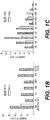
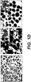
- Cytoplasmic NPM expression defined as NPMc+ ( figure 1A ), was found in 208/591 (35,2%) primary AML cases of the GIMEMA/EORTC study ( figure 1B ). Such find appears specific for primary AMLs, not being detectable in secondary AMLs and in other human tumours that show always an exclusive nuclear NPM expression ( figure 1B ).
- nucleolin C23
- another nucleolar antigen retains its nucleus-restricted localization as showed in figure 1A .
- NPMc+ AMLs the anomalous NPM expression is usually found in almost all leukaemic cells; except in cases of M5b (monocytic leukaemia), where cytoplasmic NPM is expressed only in a variable percent of the neoplastic population, namely 30-60% of leukaemic cells, preferentially those more immature (monoblasts).
- M5b monocytic leukaemia
- NPMc- AMLs rarely contain NPMc+ leukaemic cells, mostly mitotic elements as shown in figure 1A on the right.
- the anomalous cytoplasmic NPM expression of leukaemic cells must be considered a long-term event, as documented by its comparison in 25 patients with NPMc+ AML in relapse of disease up to 3 years after the initial diagnosis.
- the anomalous cytoplasmic NPM expression was found in all FAB categories, except M3 (acute promyelocytic leukaemia), M4eo (acute myelomonocytic leukaemia with eosinophilia), and M7 (acute megakaryocytic leukaemia) as shown in figure 1C .
- M3 acute promyelocytic leukaemia
- M4eo acute myelomonocytic leukaemia with eosinophilia
- M7 acute megakaryocytic leukaemia
- cytoplasmic NPM expression ranged from 13.6% in M0 (minimally differentiated AML) to 93.7% in M5b (acute monocytic leukaemia).
- M5b and M6 acute erythroid leukaemia
- M1 AML without maturation
- M2 AML with maturation
- M4 cases are characterized for the presence of cytoplasmic NPM not only in myeloid blasts but also in erythroid precursors, particularly in the proerythroblasts ( figure 1D ) and, less frequently, in the megakaryocytes (not shown data).
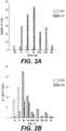
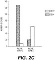
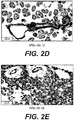
- CD34-positivity (defined as 20% positive cells) was detected in 12/159 (7.5%) NMPc+ AML and in 227/317 (71.6%) NMPc- AML (p ⁇ 0,001) as shown in figures 2A-2E . Therefore NPMc+ appears mutually exclusive with CD34 and CD133 expression.
- CD34-negative NMPc+ AMLs characteristically also lacking CD133, as shown in figure 2F .
- CD34-/CD38- rare hematopoietic stem cells which were identified in several animal species, included murine and human species (Goodell et al., 1997; Engelhardt et al., 2002).
- CD34 could be unregulated as effect of the leukaemic transformation.
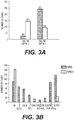
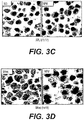
- Cytogenetic data are available in 493/591 cases of AML (166 NPMc+ and 327 NPMc-). Over 85% (142/166) of NPMc+ AML has a normal karyotype as shown in figure 3A . In contrast, only 26.9% (88/327 cases) of NPMc- AML has a normal karyotype (p ⁇ 0.001). Overall, 61.7% of normal karyotype AML are NPMc+ (142/230 cases) as shown in figure 3B . 24 NPMc+ AML cases show the presence of smaller chromosomal translocations and, among these, 12 (50%) cases have both normal and abnormal metaphases.
- NPMc+ None of the AML cases carrying major genetic abnormalities is NPMc+, as shown in figure 3B , 3C and 3D .
- FLT3, ITD or D835 mutations were detected in 59/219 (26.9%) and 13/202 (6.4%) AML cases with a normal karyotype, respectively; one case is carrier of both ITD and D835 mutations.
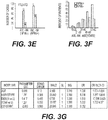
- FLT3-ITD mutation was 2,5 fold more frequent in NPMc+ cases than NPMc- cases (p ⁇ 0.003), as shown in histograms of figure 3E .
- normal karyotype AMLs can be divided in 4 subgroups: mutated NPM/mutated FLT3, mutated NPM/not mutated FLT3, not mutated NPM/mutated FLT3 and not mutated NPM/not mutated FLT3.
- the analysis carried out by the authors on 401 AML cases with normal karyotype shows that the presence of a NPM mutation in absence of a FLT3 mutation identifies a subgroup of leukaemias with more favorable prognosis. Similar results were reported subsequently also in other two studies (Ohoner et al., 2005; Verhaak et al., 2005).
- NPMc+ AML doesn't express NPM-ALK, NPM-RAR ⁇ , NPM-MLF1 fusion proteins, or other fusion proteins containing NPM, as shown by absence of respective fusion genes upon FISH, absence of respective fusion transcripts upon RT-PCR, absence of said fusion proteins upon immunohistochemistry, exclusive presence of 38 kDa NPM polypeptide upon Western Blotting, and demonstration of cytoplasmic NPM through four different anti-NPM monoclonal antibodies.
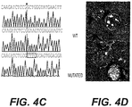
- Figure 4A is a schematic representation of the NPM gene, and mutations are summarized in figure 4B . In total, six different sequence variants were observed, all of them leading to a frameshift in the region encoding for the carboxyl-terminus domain of the NPM protein.
- the most frequent mutation (type A: gatctctgTCTGgcagtggagga agtctctttaagaaatag) is a duplication of a TCTG tetranucleotide at positions 956-959 of the reference oligonucleotide sequence NM_002520 (GenBank) as shown in figure 4C ; the resulting shift in the reading frame is predicted to alter the C-terminal portion of the NPM protein by substituting the last seven amino acids (WQWRKSL) with 11 different residues (CLAVEEVSLRK).
- Three additional mutations include distinct 4-base pair insertion at position 960, all resulting in the same frameshift as mutation A.
- nucleotides 965-969 are deleted and in place of them, two different 9-base pair sequences are inserted, not modifying the frameshift and creating a new carboxyl-terminus domain of 9 amino acids.
- the mutants are characterized by replacement of at least one of the two tryptophan residues (W) that in wild-type sequence are at position 288 and 290.
- NPM mutations in the NPM exon 12 and their specific association with NPMc+ AML was confirmed in 11 samples also by sequencing analysis of genomic DNA.
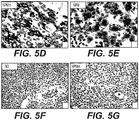
- NPM exon 12 mutations are responsive for cytoplasmic dislocation of the NPM protein
- NIH 3T3 cells were transiently transfected with expression vectors encoding for the wild-type and mutant allele fused with EGFP. Confocal microscopy showed the expected nucleolar localization for the EGFP-NPM wild-type protein, whereas the NPM mutant isoform is clearly dislocated into the cytoplasm as shown in figure 4D .
- NPM mutations are closely associated with the normal karyotype and is not seen in leukaemias with major cytogenetic abnormalities.
- the mutation might interfere with the normal function of NPM, such as, for example, interaction with the Arf or p53 protein, (Colombo et al., 2002; Kurki et al., 2004; Horn et al., 2004).
- Mutations might also perturb other NPM functions that have been mapped within C-terminal domain, such as nucleic acid binding (Hingorani et al., 2000), ATP (Chang et al., 1998), DNA-polymerase alpha-stimulatory activity, or binding with the tumour suppressor gene Arf (Bertwistle et al, 2004).
- EXAMPLE 2 Production of specific antibodies against polypeptide sequences of C-terminal of the leukaemia-specific NPM
- polypeptide sequences represent ideal immunogenes for the production of specific antibodies that include all types of polyclonal and monoclonal antibodies, human monoclonal antibodies, and humanized antibodies produced by genetic recombination techniques.
- the peptides corresponding to A., B, C, D, E and F sequences of figure 4B can be synthesized chemically according to standard procedures. Every animal species is employable for antibody preparation. Methods for the antibody production and immunization procedure of animal species (inoculation routes of antigen, use of Freund adjuvant to increase the immunogenetics of injected mixture, frequency of immunizations, etc) are largely described in the scientific literature.
- Balb/c Mice can be inoculated by intraperitoneal route with specific peptides bound to KLH (3 immunizations of 150 micro grams of peptide every 10 days). Such immunization program is followed by an intravenous booster (150 micro grams), three days before the fusion, with the purpose to increase immune response to the maximum.
- Monoclonal antibodies can be produced with the "hybridoma method” (Goding J., 1983) that consists in the formation of hybrid cells resulting from spleen normal cells with myeloma cells.
- the authors of the present invention use the P3-NS1-Ag-4-1 myeloma lineage (abbreviated as NS-1) provided them from Prof. David Y Mason laboratory, Oxford, and deriving from P3K lineage, which synthesizes only 1 ⁇ light chains that are not secreted but degraded internally and lack HGPRT enzyme.
- the hybrid selection is carried out with addition of hypoxanthine, aminopterin and thymidine (HAT) to the medium.
- HAT hypoxanthine, aminopterin and thymidine
- the hybridoma supernatant can be tested directly on cytological samples or on paraffin embedded sections of AML samples containing mutated form of NPM (NPMc+ AML) and normal form (wild-type) of NPM(NPMc- AML).
- the rational criterion of screening is the identification of hybridomas producing specific monoclonal antibodies.
- peptides of the disclosure corresponding to C-terminal of NPM
- Such antibodies have the advantage that can be used both on cytological samples (smears and cytocentrifugates) and on paraffin embedded tissue sections and, in addiction, both for diagnostic purpose and for monitoring leukaemic minimal residual disease.
- hybrids can be cloned according to well known methods ("limiting dilution") and grown in vitro in large amounts. Clones can be then cryostored in liquid nitrogen so that to have an available "bank” of the aforesaid antibodies.
- Polyclonal antibodies against peptides of the present disclosure can be produced in several animal species. Polyclonal antibodies are referred both to whole animal serum containing such antibodies and to serum fractions enriched in antibody.
- IgG or IgM serum fractions that contain only the specific antibodies for peptides of the present disclosure, can be obtained by eluting serum through a column containing bonded peptides of the present disclosure (affinity chromatography) and subsequently purifying this fraction by a column containing protein A or protein G.
- affinity chromatography affinity chromatography
- proteins have the advantage that can also be used on cytological samples (smears and cytocentrifugates) and not only on paraffin embedded tissue sections, and for monitoring leukaemic minimal residual disease in addiction to diagnostic purpose.
- the Sil-C antibody was produced by immunizing the rabbits with a 11 amino acid synthetic peptide (NHCOCH3-CLAVEEVSLRK-COOH) (Inbio Ltd, Tallin, Estonia) which comprises two mutated tryptophans, a NES portion and the VSLRK peptide of mutant A.
- the rabbit sera were purified by affinity chromatography using columns containing the peptide used as immunogen (elution with 0,23 M Tris, 0,2 M Na 2 HP0 4 , pH 0,8).
- the reactivity against the peptide was shown by ELISA technique. Particularly, the ELISA plates were adsorbed overnight at 4°C with 50 ⁇ l of peptide at a concentration of 10 ⁇ g/ml in TBS.
- the rabbit serums were added at increasing dilutions to the wells (from 1/100 up to 1/72900). After the washing in PBS-Tween 20 (0,05%), to each well were added 50 I!I of a secondary goat antibody conjugated with peroxidase (dilution 1/5000 in PBS).
- the antibody-antigen reaction was observed adding to each well 50 ⁇ l of O-phenylenediamine dihydrochloride. The reading was carried out at 492 nm. Pre-immune serums were used as negative control. The biochemical characterization of antibodies was carried out with standard Western blotting, immunoprecipitation and co-precipitation techniques.
- a polyclonal antibody (named Sil-C), which recognizes specifically an antigenic epitope comprising two mutated tryptophans, a NES portion and the VSLRK peptide belonging to mutated nucleophosmin proteins (NPM) as defined in attached claims 1-6, was produced.
- Sil-C antibody recognizes a 37 kDa band only in the cytoplasmic fraction of leukaemic cells ( Figure 5B , on top in the right).
- the picture, as expected, differs from that of the monoclonal 376 antibody which identifies a band with the same molecular weight, both in lysed cytoplasmic fraction and in those of the nuclear fraction ( Figure 5B , in bottom in the right).
- these reagents could be employed for the diagnosis at the beginning and also for monitoring minimal residual disease in combination with techniques of quantitative PCR (see below).
- intracellular antibodies From a therapeutic standpoint, it is supposable the use of intracellular antibodies (“intrabodies”) able to inhibit the leukaemic NPM mutated proteins without damaging the function of physiological NPM protein.
- the vaccines can be administered in formulations recognized by "T cell receptor” (mononuclear cells from peripheral blood) or presented by anti-gen-presenting cells (e.g. dendritic cells, cells B, macrophages).
- T cell receptor monoonuclear cells from peripheral blood
- anti-gen-presenting cells e.g. dendritic cells, cells B, macrophages.
- anti-tumour immunity means any substance or compound that serves to induce anti-tumour immunity.
- anti-tumour immunity means cytotoxic responses (T cellular), induction of antibodies that recognizes tumour cells and production of cytokines with antineoplastic activity.
- the anti-tumour activity can be measured in vitro (cytotoxicity) or in vivo in experimental animal models.
- anti-tumour vaccines Efficacy of the anti-tumour vaccines is known to be increased when various polypeptides, in combination, having different structures, are used. Therefore to that end anti-AML NPMc+ vaccines can contain different synthesis polypeptides with different specificity and sequences provided they induce the recognition of tumour cells containing NPM gene mutations.
- the vaccine of this disclosure can be conjugated with immunogenic molecules universally known as carriers.
- inoculum physiological saline, various saline solutions
- excipients physiological saline, various saline solutions
- the vaccine can contain or be administered with adjuvants, namely any molecule with immunostimulant activity.
- adjuvant administration can be carried out in any time point preceding or following inoculum of anti-tumour vaccine.
- the vaccine of the present disclosure can be administered by systemic or local route in single or multiple dose.
- anti-gen epitopes are presented to B and T cells by antigen-presenting cells.
- cytotoxic responses can be carried out on both CD4+ and CD8+ cells T, and all cellular populations able to induce cytolysis or apoptosis (e.g. neutrophiles, NK cells).
- the activation, (immunophenotype), proliferation ability, (by methods of incorporation of radioactive markers), cytotoxic ability on opportunely prepared targets, ability to secrete cytokines (ELISA, ELISPOT methods) thereof can be measured.
- the efficacy in vivo in animal models can be measured according to statistic significance criterions verifying the anti-tumour response in opportunely designed control groups.
- Object of the evaluation will be survival, measurement of tumour biomarkers, inhibition of the growth of tumour cells, regression of tumour masses, reduction of tumour-induction ability.
- EXAMPLE 4 Study of mechanism of cytoplasmic accumulation of NPM mutants.
- the present applicants elucidated the molecular mechanism that leads to the aberrant cytoplasmic accumulation of NPM in NPMc+ acute myeloid leukaemia.
- NIH-3T3 and H1299 cells were used for transfection experiments.
- Leukaemic cells from 5 leukaemic patients (3 of which carrying the mutation A of NPM and 2 carrying wild-type NPM gene), were isolated by separation with Ficoll-Hypaque and used for biochemical studies and confocal microscope analysis.
- OCI/AML3 cellular line which we identified as the only containing the mutation A in exon 12 of NPM gene (among 79 tested myeloid human lines) (Quentmeier et al., 2005) was grown in alpha-MEM with 10% FBS plus glutamine and antibiotics at standard concentrations.
- mutants A, B, C, and D of NPM were produced by PCR using NPMwt as template; the same forward primer (5' CGC CAC GCT AGC GAA GAT TCG ATG GAC) was used and a different reverse primer for each mutant (mutant A-5': CTA TTT TCT TAA AGA GAC TTC CTC CAC TGC CAG ACA GAG ATC TTG AAT AGC CTC TTG G; mutant B-5': CTA TTT TCT TAA AGA GAC TTC CTC CAC TGC CAT GCA GAG ATC TTG AAT AGC CTC TTG G; mutant C-5'; CTA TTT TCT TAA AGA GAC TTC CTC CAC TGC CAC GCA GAG ATC TTG AAT AGC CTC TTG G; mutant D-5': CTA TTT TCT TAA AGA GAC TTC CTC CAC TGC CAG GCA GAG ATC TTG AAT AGC CTC TTG G).
- PCR The products of the respective PCRs were cloned in pcDNA3.1/NT-GFP-TOPO (Invitrogen, Carlbad, CA, USA), and checked with insert sequencing.
- pcDNA3.1/NT-GFP-TOPO Invitrogen, Carlbad, CA, USA
- forward and reverse primer were respectively used 5' CGC CAC GCT AGC GAA GAT TCG ATG GAC and 5' TCA AGA ATT CCA GAA ATG AAA TAA GAC.
- the PCR product was digested using Nhel and ECORI, and the fragment was sub-cloned in PCIN4 vector, containing the Flag-HA tag at N-terminal end of the fragment.
- the precision of Flag-HA-NPM-wt and Flag-HA,NPM-mutant A sequences was confirmed by sequencing.
- NPM mutants E, G and R were produced through QuikChange Fine Site-Directed Mutagenesis Kit (Stratagene, You Jolla, CA), using as template pEGFP-C1-A/PMwt, and according to manufacturer instructions. Primers were designed on the followings sequences:
- NPM_MUT_A_NO_NES plasmids were produced using pEGFP-C1-NPMmA as template, exploiting the localization of mutagenesis sites between cutting sites of BgIII and EcoRI enzymes.
- a partially complementary primer couple containing the desired mutation and protruding ends compatible with ends produced by the digestion with BgIII-EcoR, it was possible to ligate the double strand DNA produced by annealing primers to pEGFP-C1-NPMmA vector previously digested using the two above restriction enzymes.
- sequences of the used primers are:
- the H1299 cells were seeded on the surface of six-well plates 24 hours before transfection. 5 ⁇ g of expression vector encoding for wild type HA-NPM, GFP-NPM-mutant A, or both, were transfected using the precipitation method with calcium-phosphate. After 24 hours, the cells were treated with 20 nM leptomycin B, a Crm1 specific inhibitor (Sigma, St. Louis, MO, USA) for 8 hours or not treated, respectively. The cells then were fixed in 4% paraformaldehyde for the immunofluorescence study.
- NIH-3T3 cells were transfected using Lipofectamin 2000 (Invitrogen Carlsbad, CA, USA) following the manufacturer instructions. After 24 hours, the cells grown on the slide were incubated with cycloeximide (Merck Bioscences Ltd, Nottingham UK) 10 micro grams/ml (30 minutes) and leptomycin B (Merck Bioscences Ltd, Nottingham UK) 20 ng/ml (5 hours), or other Crm1 inhibitors such as ratjadons A and C (Alexis Biochemicals, Carisbad, CA, USA) 20 ng/ml (5 hours).
- cycloeximide Merck Bioscences Ltd, Nottingham UK
- leptomycin B Merck Bioscences Ltd, Nottingham UK
- Crm1 inhibitors such as ratjadons A and C (Alexis Biochemicals, Carisbad, CA, USA) 20 ng/ml (5 hours).
- the cells were fixed in 4% paraformaldehyde (10 minutes) for immunofluorescence and confocal microscope analysis.
- transfected cells were transferred inside an Attofluor chamber (Molecular Probes, Eugene, OR, USA) and were observed using a MRC-1024 confocal apparatus (Biorad Cambridge, UK) assembled on an Olympus IMT-2 microscope.
- the images of a single section were recorded before and after the addition of leptomicin B, at 60 second intervals, using the time-series function of Laser-Sharp program (BioRad).
- the excitation wavelength was 488 nm and images were detected using a filter from 505 to 550 nm on the PMT2.
- Cells deriving from two patients with NPMc+ AML (carrying the mutation A) and from OCI/AML3 cellular line were harvested in medium (10 6 cell/ml in 24-well plates) and incubated at 37°C with 5% C0 2 for 5 hours. After the incubation overnight with leptomycin B (20 ng/ml), the cells were washed with PBS and centrifuged. The cell pellet was fixed in B5 and paraffin for the immunostaining.
- the DAPI staining was used to visualise nucleuses of H1299 cells transfected with the GFP-NPM-mutant A and with GFP-NPM wt.
- the fixed cells were made permeable with 0.2% Triton-X 100 (10 minutes) followed by blocking with 10% anti-goat serum (30 minutes). Then primary anti-HA antibody was added (1:1000, Roche Indianapolis, In, USA) for 1 hour followed by incubation for 30 minutes with the secondary Alexa-568 antibody (1:1000, Molecular Probes, Oregon, USA). The images were taken using a digital camera with the Spot 4.09 program (Diagnostic Instruments, Sterling Heights. MI, USA).
- the nuclei of NIH 3T3 cells transfected with GFP-NPMwt and GFP-NPM mutant A were stained with propidium iodide.
- the nucleolin staining (C23) of NIH-3T3 cells was carried out with the primary anti-nucleolin antibody purchased from DakoCytomation (Glostrup, Denmark), followed by Texas Red conjugated secondary antibody (Southern Biotechnology Associates, Birmingham, AL); the nuclei were contra-stained with the TO-PRO-3 (Molecular Probes, Oregon, USA).
- the NPM coloration of paraffin embedded sections of AML cases containing the mutation A was carried out with monoclonal anti-NPM antibodies followed by a Alexa 488 conjugated secondary (Molecular Probes, Oregon, USA); the nuclei were contra-stained with propidium iodide.
- the images were taken with a confocal microscope Zeiss LSM 510 (Carl Zeiss, Jena, DE), using laser excitation wavelengths at 488 nm (for ALexa 488), 543 nm (for Texas Red and the propidium iodide) and 633 nm (for TO-PRO-3), respectively.
- the laser intensity tuning, diameter of pinholes, and configuration of the light detection were set to achieve the best signal/noise ratio and avoid fluorescence crossover.
- the images were then transferred to a SGI Octane workstation (Silicon graphics, Mountain View, CA, USA) for further elaboration; 3D reconstruction was made with the shade or iso-surface method using the Imaris program (Bitplane, Zurich, CH).
- NPM research on paraffin embedded sections using alkaline phosphatase method was done for 393 patients as previously described.
- the samples were classified as cytoplasmic or nuclear NPM without knowing the results of mutational analysis.
- the nuclear and cytoplasmic extracts were prepared according to the method of Schreiber et al. (1989).
- the cells were lysed in 1 ml of ice-cold lysis buffer (1% NP-40, 150 mM NaCl, 25 mM Tris, pH 7,5, 1 mM EOTA, 1 mM Na3V04 1 ⁇ g/ml leupeptine, 1 ⁇ g/ml aprotinin and 1 mM PMSF).
- lysed products were centrifuged at 14,000 x g for 10 minutes 4°C and incubated with 4 ⁇ g of an unspecific control IgG or specific polyclonal rabbit anti-NPM, named Sil-C, or monoclonal mouse anti-NPM (Clone 376) antibody, respectively, and 30 ⁇ l of the Protein A/G Plus-agarose beads (Saint Cruz Biotechnology, Inc.) in incubation overnight at 4°C.
- the beads then were washed at least thrice with the washing buffer (0.1% NP-40, 150 mM NaCl, 25 mM Tris pH 7.5, 1 mM EDTA and inhibitors).
- the proteins were separated on a SOS-polyacrylamide gel (SDS-PAGE) and transferred on a PVDF membrane (Millipore), where were incubated with primary antibodies, namely rabbit polyclonal anti-Crm1 (Saint Cruz Biotechnology, Inc.) or mouse monoclonal anti-Crm1 (BD Transduction laboratories); respectively; after incubation with a HRP conjugated secondary antibody, peptides recognized in Western blot were detected using ECL method according to manufacturer instructions (Amersham Bioscience).
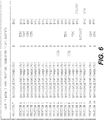
- the Figure 6 shows the changes in the tryptophans 288 and 290 and the creation of a NES motif in 40 mutant NPM proteins identified in leukaemic patients; the mutation frequency (%) is present only for 393 AML cases here studied for which were available, in addition to the molecular data, also the results of the immunohistochemical (IH) staining. Alterations are of two types: i) the mutation of both the tryptophans 288 and 290 (or only 290) and ii) the creation a new motif named NES ("Nuclear Export Signal motif). NES is a protein structure that is specifically recognized by Crm1 (or Exportin 1), the protein physiologically delegated to the transport of other proteins from nucleus to cytoplasm.
- NES motif is defined as a sequence of about 10 amino acids of type YxxxYxxYxY where Y indicates a hydrophobic amino acid of leucine, isoleucine, methionine, valine or phenylalanine type and x is equivalent to others amino acids.
- the NES type can vary from each other NPM mutant.
- the type and the frequency of NES in the various leukaemic NPM mutants are illustrated in Figure 6 .
- the remaining 35% of NPM mutant proteins contains rarer NES variants, in which the valine in second position of NES is replaced with another hydrophobic amino acid.
- NES motif (namely LxxxVxxVxL type) is always associated to mutations of both tryptophans 288 and 290, while tryptophan 288 is preserved only in NPM mutant proteins that contain a NES variant of type above-indicted namely those in which valine in second position of NES is replaced with another hydrophobic amino acid ( Figure 6 and Table 3).
- Table 3 shows the correlation between NES motif and the tryptophans 288 and 290 in 40 NPM mutant proteins of leukaemic patients.
- the most common NES motif is the variant 1; the other NES variants (type 2-6) are less frequent and they differentiate from the variant 1 for the presence, in the place of Valine (V) in NES second position, of a Leucine (L), Phenylalanine (F), Methionine (M), or Cisteine (C).
- V Valine
- F Phenylalanine
- M Methionine
- C Cisteine
- Cytoplasmic expression of NPM mutants is a NES-dependent event
- NPM mutants contain a new NES motif in their carboxyl-terminal portion suggests that the cytoplasmic removal of NPM can result from an active transport of NPM mutants by means of Crm1, the receptor delegated to the protein transport from nucleus to cytoplasm.
- Figure 7 shows as the nuclear export of NPM mutants is NES-dependent. Under basal conditions, HI299 or NIH3T3 cells transfected with cDNAs that encode for labelled NPM mutant proteins show the expected aberrant cytoplasmic localization of mutants. In contrast, in the presence of Leptomicin B (LMB), NPM mutant proteins are relocalized from the cytoplasmic to the nuclear compartment (nucleoplasm) (panels 7A and 7B).
- LMB Leptomicin B
- Figure 7C-E shows the analysis at different time points of the LMB effects on the mutant A associated to eGFP (eGFP-NPMmutA) in NIH-3T3 cells: the addition of LMB results in a reduction of fluorescence in cytoplasm and Golgi area and concomitant fluorescence increase in nucleoplasm.
- mutant type A (the more common one) is re-localized in nucleus in 20 minutes and the whole process is completed in 1 hour (panels C-D).
- the purity of the sub-cellular fractions was measured by removal of the antibody and blotting with an anti- ⁇ -tubulin antibody (inferior panel Figure 7E ). A not significant contamination was measured for the over-expressed proteins (as it is clear in GFP blotting) (middle panel).
- eGFP-NPMmutA was found only in the cytoplasmic fractions (C). The experiment was carried out in the absence of dicycloeximide so that the continuous presence of GFP-NPMmA in the cytoplasmic fractions during the treatment with LMB has been shown over time.
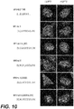
- NPM mutants after Leptomicin B treatment, are re-localized in the nucleus and, specifically, in nucleoplasm ( Figure 8A , on top), rather than in the nucleolus, that is the place in which physiologically the wild-type NPM protein localizes ( Figure 8A , middle).
- nucleoplasmic relocalization of mutants by Leptomicin B is also shown through double staining at confocal microscope that underlines the presence of a mutual exclusiveness between the localization places of NPM mutant, displaced in nucleoplasm, and nucleolin (C23) that, as expected, is selectively expressed at nucleolar level ( Figure 8A , in bottom).
- Cytoplasmic accumulation of mutants depends on the coordinated action of NES and mutations of tryptophans 288 and 290
- mutant E A nuclear compartment distribution very similar to that of mutant E, is also observed with an artificial construct of type A NPM mutant in which the mutated tryptophan in position 290 was re-inserted by site-directed mutagenesis (A290W).
- A290W site-directed mutagenesis
- NPM mutants dislocate the NPM wild-type protein from its physiological place (nucleolus) to the cytoplasm
- NPM mutated proteins preserve the dimerization domain in the N-terminal site, it can be hypothesized that they can form heterodimers with NPM wild-type protein, like among the fusion proteins (NPM-ALK and NPM-MLF1) and same NPM wild-type protein.
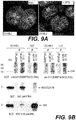
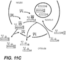
- Figure 11A shows that, by heterodimerization mechanism, the mutants are able to bind and dislocate the NPM wild-type protein in cytoplasm.
- the mutant and the wild-type NPM protein co-locate in cytoplasm.
- plasmids encoding for FH-NPMwt and FH-NPM mutant A were used.
- 5% of whole lysed cells derived from stably transfected cells with FH-NPMwt, FH-NPM mutant A or H1299 control cells was subjected to Western blot with a-NPM or a-HA.
- the 95 % remaining of lysed cells was immuno-precipitated with an anti-Flag antibody (M2), and used for Western blot with a-NPM or it a-HA.
- M2 anti-Flag antibody
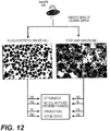
- the mechanism responsible of the accumulation of NPM mutant proteins in the cytoplasm of leukaemic cells depend on the mutations of tryptophans 288 and 290 and creation of NES. Because these alterations are present in all till now identified leukaemic NPM mutants, it is hypothesized that the immunohistochemical staining with anti-NPM antibodies is able to predict, by demonstrating cytoplasmic delocalization of NPM, all mutations occurring at level of exon 12 of NPM gene.
- Immunohistochemical test can be used for diagnostic purpose as indicated in Figure 12 .
- the test is rapid, economic, easily interpretable, highly sensitive and specific. For all these reasons it could be used as first step in the molecular characterization of AMLs.
- NPMc+ AMLs it isn't necessary to carry out cytogenetic, FISH and molecular analysis, for the major chromosomal alterations, such as t(15;17), t(8;21), inv16, t(6;9) and 11q23/MLL because they are mutually exclusive with the cytoplasmic positivity for NPM.
- these analyses are compulsory.
- Cytogenetics helps for the identification of rare translocations with potential prognostic impact in 14% of NPMc+ AML with minor chromosomal anomalies. Mutations of FLT3 gene should be searched in all AML patients AML (independently on NPM) because its correlation with the sub-cellular expression of NPM can help to identify new prognostic subgroups in normal karyotype AML (Schnittger et al., 2005; Dohner et al., 2005; Verhaak et al., 2005).
- NPMc-AML normal karyotype without mutation of NPM gene
- the above illustrated data explain mechanism through which the exon 12 specific mutations of NPM gene alter the nucleus-cytoplasm transport of mutated and wild-type NPM proteins.
- the mechanism is identical both in transfected and leukaemic cells of patients with NPMc+ AML and in OCI/AML3 human leukaemic line.
- the mutations lead to two fundamental changes in the carboxyl-terminated region of NPM mutants: 1) a NES is produced that potentiates the expulsion of mutant proteins by Crm1; and 2) two tryptophans 288 and 290 (or only tryptophan 290) are lost which, under normal conditions, are essential for the bond of NPM protein to nucleoli.
- wild-type NPM protein allowed to detect an hypothetical physiological NES of LxxPxxLxL type, which is localized in the zone between residues 94 and 102 of NPM (Wang et al., 2005). Despite the presence of this NES, the wild-type NPM protein, under physiological conditions, locates mainly in nucleoli and this suggests that the part of NPM protein that is normally expelled from nucleus to cytoplasm through physiological NES, is decidedly inferior to that of the same protein that, by two NLSs ("nuclear localization signals "), is able to go back from cytoplasm into nucleus.
- the murine and human artificial wild-type NPM mutants without the two tryptophans 288 and 290 locate exclusively in nucleoplasm (Nishimura et al., 2002), it is very likely that the additional NES, created by the mutation at level of C-terminal region, confers to leukaemic NPM mutant a greater ability to be exported out of nucleus; this could be due to the additive effect and/or increased Crm1 affinity of the second NES.
- tryptophan 290 could be more important, because it is constantly altered in all till now identified leukaemic NPM mutants.
- the mutation of both two tryptophans allows the maximum inhibition of the nucleolar bond and nucleoplasmic delocalization of mutants to be achieved in leukaemic NPMc+. cell of great importance is the observation that NES motif more commonly found in NPM mutants (LxxxVxxVxL) is always associated with mutations of both ⁇ tryptophans.
- tryptophan 288 appears to be maintained only in those NPM mutants including less common variants of NES, namely those characterized by the presence of leucine phenylalanine, cysteine or methionine in the second position of NES, in place of valine (Table 3).
- NPM mutants mutated leukaemic NPM proteins preserving two nuclear localization signals (NLS), enter into nucleus; ii) their ability to bind the nucleoli is completely inhibited when the tryptophans are mutated, or partially inhibited when only tryptophan 290 is altered, resulting in the accumulation of mutants in nucleoplasm; iii) nucleoplasmic mutants are caught by Crm1 that determines rapid expulsion thereof in cytoplasm where they progressively accumulate.
- NLS nuclear localization signals
- NPMc+ leukaemia suggests, as possible therapeutic intervention area, the "relocalization" of leukaemic NPM mutants and wild-type NPM protein in their physiological sites, through the use of Crm1 inhibitors or small synthesis molecules that interfere with the NPM mutant-Crm1 bond or Wild-type NPM protein or other molecules able to interact with NPM (ARF, etc.).
- a specific evaluation method uses a forward primer designed on exon 11 (cNPM1-F:5'-5'-GAAGAATTGCTTCCGGATGACT-3'), a probe on the junction exon 11/exon 12 (c.Probe:5'-FAM-ACCAAGAGGCTATTCAA-MGB-3') and mutation-specific reverse primers (cNPM mut.A-R:5'CTTCCTCCACTGCCAGACAGA-3' and cNPM mut.B-R:5'-TTCCTCCACTGCCATGCAG-3').
- Forward primer and probe are the same regardless of different mutations ( Figure 13 ).
- Step 1 Retro-transcription reaction according to EAC protocol (Gabert et al ., Leukaemia 2003).
- Step 2 Amplification reaction uses a mixture containing 12,5 ⁇ l of Taq Man universal PCR Master Mix (Applied Biosystem), 300 nM Primers, 200 nM of probe and 5 ⁇ l of cDNA in a total volume of 25 ⁇ l. Conditions: 2 minutes at 50°C (UNG enzyme activation), 10 min at 95°C (UNG enzyme inactivation and AmpliTaq polymerase activation) followed by 50 cycles at 95°C for 15 seconds, at 62°C for 1 minute for mutation A and at 59° C for 1 minute for mutation B. As quantitative and qualitative RNA control ABL gene can be amplified.
- the analysis setting of instrument includes a "threshold" of 0.1 with "a baseline” from 3 to 15 both for ABL and NPM.
- System sensitivity and specificity are analyzed using sequential dilutions, by a factor equal to 10, by mixing RNA extracted from medullary leukaemic cells with NPM mutation type A or B and RNA obtained from a pool of medullary cells from patients without NPM mutations (verified by sequencing).
- the standard plot of absolute quantitative evaluation for mutation A is constructed using a plasmid construct.
- a construct consists of plasmid vector pCRII-TOPO, (Invitrogen, Groningen, Netherlands) plus a portion of NPM1 gene containing mutation A.
- the amplification of mutation A is obtained by RT-PCR with the primers NPM1_390_F (5'-GTCTTAAGGTTGAAGTGTGGT-3') and NPM1_1043_R (5'-TCAACTGTTACAGAAATGAAATAAGACG-3').
- the plasmid is prepared in five sequential dilutions: 10 5 , 10 4 , 10 3 , 10 2 , 10 copies.
- the results of RQ-PCR for mutation A normalized on ABL transcripts are expressed as copy number of NPM with mutation A every 104 copies of ABL.
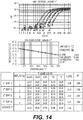
- the “maximum reproducible sensitivity” is defined as the lowest dilution in which all sample replicates are positive within a Ct (Cycle threshold) of 1.5, and the highest Ct of replicates is at least of 3,0 Ct lower than the lowest value of background.
- the “maximum sensitivity” is defined as minimum dilution in which at least a sample is positive and at least 1,0 Ct lower than the smallest Ct of background. With these definitions, a result is defined as "positive, not quantifiable” in the presence of amplified in 1 of 2 replicates below the maximum reproducible sensitivity, but still 1,0 Ct lower than the lowest background.
- the standard plasmid plots show a "mean slope" of -0.38 and an “intercept” of 39.5 ⁇ 0.45 Ct.
- the correlation coefficient is high (>0.99 in all experiments) and demonstrates the accurate identification of the presence of copies of mutation A in unknown samples.
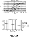
- the maximum reproducible sensitivity of RQ-PCR corresponds to 10 plasmid molecules ( Figure 14 ).
- Sequential dilutions based on a factor equal to 10 were carried out to simulate the sensitivity and reproducibility of RQ-PCR in monitoring of minimal residual disease in 5 patient diagnosed as afflicted with (acute myeloblastic leukaemia) NPMc+ AML (4 patients with mutation A and 1 with mutation B).
- RNA extracted from medullary leukaemic cells with type A or B NPM mutation was diluted with RNA achieved from a pool of medullary cells from patients without NPM mutations.
- the cDNA specific RQ-PCR showed a copy number ⁇ 10 after the first or the second cycle of consolidation therapy. A different kinetics in the diminution of the copy number was observed in the three samples as shown in Figure 17 .
- a small but persistent number of mutated copies is associated with the haematological remission in a patient (square). In one of the remaining cases the number of mutated copies markedly decreases after consolidation treatment (diamonds); such diminution is less pronounced in the second patient (triangles). In the last two patients the copy number again increases and in both cases haematological relapse occurs ( Figure 17 ).
Landscapes
- Health & Medical Sciences (AREA)
- Chemical & Material Sciences (AREA)
- Life Sciences & Earth Sciences (AREA)
- Organic Chemistry (AREA)
- Proteomics, Peptides & Aminoacids (AREA)
- Immunology (AREA)
- Engineering & Computer Science (AREA)
- Molecular Biology (AREA)
- Genetics & Genomics (AREA)
- Zoology (AREA)
- General Health & Medical Sciences (AREA)
- Biochemistry (AREA)
- Pathology (AREA)
- Analytical Chemistry (AREA)
- Biophysics (AREA)
- Medicinal Chemistry (AREA)
- Wood Science & Technology (AREA)
- Physics & Mathematics (AREA)
- Microbiology (AREA)
- Biotechnology (AREA)
- Hematology (AREA)
- Toxicology (AREA)
- Gastroenterology & Hepatology (AREA)
- Urology & Nephrology (AREA)
- Biomedical Technology (AREA)
- Hospice & Palliative Care (AREA)
- Oncology (AREA)
- Bioinformatics & Cheminformatics (AREA)
- General Engineering & Computer Science (AREA)
- General Physics & Mathematics (AREA)
- Food Science & Technology (AREA)
- Cell Biology (AREA)
- Measuring Or Testing Involving Enzymes Or Micro-Organisms (AREA)
- Peptides Or Proteins (AREA)
- Medicines That Contain Protein Lipid Enzymes And Other Medicines (AREA)
- Micro-Organisms Or Cultivation Processes Thereof (AREA)
- Pharmaceuticals Containing Other Organic And Inorganic Compounds (AREA)
Claims (15)
- In vitro-Verfahren, umfassend
Detektieren eines mutierten menschlichen Nukleophosmin-Protein (NPM-Protein) oder einer Oligonukleotidsequenz, die das mutierte humane NPM-Protein in einer Probe kodiert,
wobei das mutierte menschliche NPM-Protein dadurch gekennzeichnet ist, dass es eine zytoplasmatische Stelle aufweist und sich von dem menschlichen Wildtyp-Nukleophosmin unterscheidet, indem es eine Mutation, die in einem Verlust eines Tryptophanrests an den Positionen 288 und 290 resultiert, und ein Signalmotiv von nukleärem Export (NES) in dem C-terminalen Bereich des Proteins, umfassend eine Aminosäuresequenz, ausgewählt aus der Gruppe, bestehend aus LxxxVxxVxL (SEQ ID NO 1), LxxxLxxVxL (SEQ ID NO 2), LxxxFxxVxL (SEQ ID NO 3), LxxxMxxVxL (SEQ ID NO 4) und LxxxCxxVxL (SEQ ID NO 5) umfasst. - Verfahren nach Anspruch 1, wobei die NES-Sequenz LCLAVEEVSL (SEQ ID NO 6); LCMAVEEVSL (SEQ ID NO 7); LCVAVEEVSL (SEQ ID NO 8); LWQSLAQVSL (SEQ ID NO 18); oder LWQSLEKVSL (SEQ ID NO 19) umfasst.
- Verfahren nach Anspruch 1 oder 2, wobei das mutierte NPM-Protein detektiert wird, indem das Protein in dem Zytoplasma einer Zelle einer akuten myeloischen Leukämie (AML) durch immunhistochemische Untersuchung identifiziert wird.
- Verfahren nach einem der vorherigen Ansprüche, wobei das mutierte NPM-Protein durch Folgendes detektiert wird
identifizieren des Proteins unter Verwendung eines monoklonalen oder polyklonalen Antikörpers oder eines Fragments davon, der/das in der Lage ist, mindestens ein antigenes Epitop der C-terminalen Region des mutierten menschlichen NPM-Proteins wie definiert Anspruch 1 oder 2 zu erkennen und selektiv daran zu binden. - Verfahren nach Anspruch 4, wobei das Epitop VSLRK-Peptid (SEQ ID NO: 29) beinhaltet.
- Verfahren nach Anspruch 1 oder 2, wobei die Oligonukleotidsequenz detektiert wird.
- Verfahren nach Anspruch 6, wobei die Oligonukleotidsequenz mit Oligonukleotidsonden und/oder einem Primer detektiert wird.
- Verfahren nach Anspruch 6, wobei die Oligonukleotidsequenz unter Verwendung von PCR, RT-PCR, Echtzeit-PCR, Reverse Dot Blot (RBD), Multiple Tissue Array, Sequenzierung oder einer Kombination davon detektiert wird.
- Verfahren nach Anspruch 6 oder 7, wobei die Oligonukleotidsequenz unter Verwendung von Primern in einem Polymerase-Kettenreaktionstest detektiert wird, der die Oligonukleotidsequenz verstärkt, aber keine Wildtyp-NPM-Sequenz amplifiziert.
- Verfahren nach Anspruch 6, wobei die Oligonukleotidsequenz unter Verwendung einer Oligonukleotidsonde nachgewiesen wird, die an die Oligonukleotidsequenz bindet, aber nicht an eine Wildtyp-NPM-Sequenz bindet.
- Verfahren nach Anspruch 6 oder 7, wobei die Oligonukleotidsequenz unter Verwendung von Folgendem detektiert wird
Primern in einem Polymerase-Kettenreaktionstest detektiert wird, der die Oligonukleotidsequenz verstärkt, aber keine Wildtyp-NPM-Sequenz amplifiziert, und
einer Oligonukleotidsonde, die an die Oligonukleotidsequenz bindet, aber nicht an eine Wildtyp-NPM-Sequenz bindet. - Verfahren nach Anspruch 8, wobei die Polymerase-Kettenreaktion eine Echtzeit-PCR ist.
- Verfahren nach einem vorherigen Anspruch, wobei die Probe Nukleinsäuren aus einer Zelle akuter myeloischer Leukämie umfasst.
- Verfahren nach einem vorherigen Anspruch, wobei die Probe von einem Patienten mit akuter myeloischer Leukämie (AML) stammt.
- Verfahren nach Anspruch 14, wobei der Patient einen normalen Karyotyp aufweist.
Applications Claiming Priority (5)
| Application Number | Priority Date | Filing Date | Title |
|---|---|---|---|
| IT000534A ITRM20040534A1 (it) | 2004-10-29 | 2004-10-29 | Mutanti della proteina nucleofosmina (npm), sequenze geniche corrispondenti e relativi usi. |
| EP05813231.7A EP1807448B1 (de) | 2004-10-29 | 2005-10-28 | Nukleophosmin protein (npm) mutanten, korrespondierende gensequenzen und deren verwendungen |
| PCT/IT2005/000634 WO2006046270A2 (en) | 2004-10-29 | 2005-10-28 | Nucleophosmin protein (npm) mutants, corresponding gene sequences and uses thereof |
| EP07021463.0A EP1944316B1 (de) | 2004-10-29 | 2005-10-28 | Mutanten des Nukleophosminproteins (NPM), entsprechende Gensequenzen und ihre Verwendungen |
| EP10184598.0A EP2319865B1 (de) | 2004-10-29 | 2005-10-28 | Mutanten des Nukleophosminproteins (NPM), entsprechende Gensequenzen und ihre Verwendungen |
Related Parent Applications (4)
| Application Number | Title | Priority Date | Filing Date |
|---|---|---|---|
| EP05813231.7A Division EP1807448B1 (de) | 2004-10-29 | 2005-10-28 | Nukleophosmin protein (npm) mutanten, korrespondierende gensequenzen und deren verwendungen |
| EP10184598.0A Division-Into EP2319865B1 (de) | 2004-10-29 | 2005-10-28 | Mutanten des Nukleophosminproteins (NPM), entsprechende Gensequenzen und ihre Verwendungen |
| EP10184598.0A Division EP2319865B1 (de) | 2004-10-29 | 2005-10-28 | Mutanten des Nukleophosminproteins (NPM), entsprechende Gensequenzen und ihre Verwendungen |
| EP07021463.0A Division EP1944316B1 (de) | 2004-10-29 | 2005-10-28 | Mutanten des Nukleophosminproteins (NPM), entsprechende Gensequenzen und ihre Verwendungen |
Publications (2)
| Publication Number | Publication Date |
|---|---|
| EP3284750A1 EP3284750A1 (de) | 2018-02-21 |
| EP3284750B1 true EP3284750B1 (de) | 2019-09-11 |
Family
ID=36096144
Family Applications (4)
| Application Number | Title | Priority Date | Filing Date |
|---|---|---|---|
| EP07021463.0A Expired - Lifetime EP1944316B1 (de) | 2004-10-29 | 2005-10-28 | Mutanten des Nukleophosminproteins (NPM), entsprechende Gensequenzen und ihre Verwendungen |
| EP10184598.0A Expired - Lifetime EP2319865B1 (de) | 2004-10-29 | 2005-10-28 | Mutanten des Nukleophosminproteins (NPM), entsprechende Gensequenzen und ihre Verwendungen |
| EP05813231.7A Expired - Lifetime EP1807448B1 (de) | 2004-10-29 | 2005-10-28 | Nukleophosmin protein (npm) mutanten, korrespondierende gensequenzen und deren verwendungen |
| EP17181518.6A Expired - Lifetime EP3284750B1 (de) | 2004-10-29 | 2005-10-28 | Mutanten des nukleophosminproteins (npm), zugehörige gensequenzen und verwendungen davon |
Family Applications Before (3)
| Application Number | Title | Priority Date | Filing Date |
|---|---|---|---|
| EP07021463.0A Expired - Lifetime EP1944316B1 (de) | 2004-10-29 | 2005-10-28 | Mutanten des Nukleophosminproteins (NPM), entsprechende Gensequenzen und ihre Verwendungen |
| EP10184598.0A Expired - Lifetime EP2319865B1 (de) | 2004-10-29 | 2005-10-28 | Mutanten des Nukleophosminproteins (NPM), entsprechende Gensequenzen und ihre Verwendungen |
| EP05813231.7A Expired - Lifetime EP1807448B1 (de) | 2004-10-29 | 2005-10-28 | Nukleophosmin protein (npm) mutanten, korrespondierende gensequenzen und deren verwendungen |
Country Status (13)
| Country | Link |
|---|---|
| US (8) | US8222370B2 (de) |
| EP (4) | EP1944316B1 (de) |
| CN (1) | CN101160320B (de) |
| CA (1) | CA2585965C (de) |
| CY (1) | CY1116527T1 (de) |
| DK (2) | DK1944316T3 (de) |
| ES (4) | ES2755875T3 (de) |
| HU (1) | HUE025263T2 (de) |
| IT (1) | ITRM20040534A1 (de) |
| PL (1) | PL1944316T3 (de) |
| PT (1) | PT1944316E (de) |
| SI (1) | SI1944316T1 (de) |
| WO (1) | WO2006046270A2 (de) |
Families Citing this family (9)
| Publication number | Priority date | Publication date | Assignee | Title |
|---|---|---|---|---|
| ITRM20040534A1 (it) | 2004-10-29 | 2005-01-29 | Brunangelo Falini | Mutanti della proteina nucleofosmina (npm), sequenze geniche corrispondenti e relativi usi. |
| KR101660322B1 (ko) * | 2011-04-28 | 2016-09-29 | 아크레이 가부시키가이샤 | NPM1 유전자의 exon12 변이의 검출용 프로브 및 그 용도 |
| CN102382896A (zh) * | 2011-12-06 | 2012-03-21 | 苏州苏大赛尔免疫生物技术有限公司 | Npm1基因突变检测方法及其试剂盒 |
| WO2017049002A1 (en) * | 2015-09-15 | 2017-03-23 | Massachusetts Institute Of Technology | A humanized mouse model of de novo human acute myeloid leukemia with a matching human immune system |
| US20220305102A1 (en) * | 2017-06-30 | 2022-09-29 | Academisch Ziekenhuis Leiden (H.O.D.N. Leids Universitair Medisch Centrum) | Treatment of haematological malignancies |
| US11352389B2 (en) | 2017-06-30 | 2022-06-07 | Academisch Ziekenhuis Leiden (H.O.D.N. Leids Universitair Medisch Centrum) | Treatment of haematological malignancies |
| NL2019156B1 (en) * | 2017-06-30 | 2019-01-10 | Academisch Ziekenhuis Leiden H O D N Leids Univ Medisch Centrum | Treatment of haematological malignancies |
| WO2019214058A1 (zh) * | 2018-05-11 | 2019-11-14 | 南京先声医学检验有限公司 | 引物、引物组合、试剂盒及其应用 |
| CN114591410B (zh) * | 2022-03-25 | 2022-09-20 | 东北农业大学 | 玉米miRNA的靶基因序列及其编码蛋白在抗植物粗缩病中的应用 |
Family Cites Families (10)
| Publication number | Priority date | Publication date | Assignee | Title |
|---|---|---|---|---|
| US5529925A (en) * | 1993-12-03 | 1996-06-25 | St. Jude Children's Research Hospital | Nucleic acid sequences and fusion proteins present in human t(2;5) lymphoma |
| US7041490B1 (en) * | 1997-11-28 | 2006-05-09 | Serono Genetics Institute, S.A. | Chlamydia trachomatis polynucleotides and vectors, recombinant host cells, DNA chips or kits containing the same |
| CN1331117A (zh) * | 2000-06-30 | 2002-01-16 | 上海博德基因开发有限公司 | 一种新的多肽——nucleophosmin9.68和编码这种多肽的多核苷酸 |
| WO2004005517A2 (en) | 2002-07-09 | 2004-01-15 | Degussa Ag | L-amidase from rhizobium huautlense |
| GB0223341D0 (en) * | 2002-10-08 | 2002-11-13 | Groningen Acad Ziekenhuis | Organic compounds |
| US7250496B2 (en) * | 2002-11-14 | 2007-07-31 | Rosetta Genomics Ltd. | Bioinformatically detectable group of novel regulatory genes and uses thereof |
| US7314750B2 (en) * | 2002-11-20 | 2008-01-01 | Affymetrix, Inc. | Addressable oligonucleotide array of the rat genome |
| US7413851B2 (en) * | 2002-12-13 | 2008-08-19 | Aurelium Biopharma, Inc. | Nucleophosmin directed diagnostics and therapeutics for multidrug resistant neoplastic disease |
| US7288529B2 (en) * | 2003-04-24 | 2007-10-30 | New York University | ALK protein tyrosine kinase, cells and methods embodying and using same |
| ITRM20040534A1 (it) * | 2004-10-29 | 2005-01-29 | Brunangelo Falini | Mutanti della proteina nucleofosmina (npm), sequenze geniche corrispondenti e relativi usi. |
-
2004
- 2004-10-29 IT IT000534A patent/ITRM20040534A1/it unknown
-
2005
- 2005-10-28 DK DK07021463.0T patent/DK1944316T3/en active
- 2005-10-28 CA CA2585965A patent/CA2585965C/en not_active Expired - Lifetime
- 2005-10-28 ES ES17181518T patent/ES2755875T3/es not_active Expired - Lifetime
- 2005-10-28 ES ES07021463.0T patent/ES2542339T3/es not_active Expired - Lifetime
- 2005-10-28 PL PL07021463T patent/PL1944316T3/pl unknown
- 2005-10-28 ES ES10184598.0T patent/ES2654564T3/es not_active Expired - Lifetime
- 2005-10-28 EP EP07021463.0A patent/EP1944316B1/de not_active Expired - Lifetime
- 2005-10-28 EP EP10184598.0A patent/EP2319865B1/de not_active Expired - Lifetime
- 2005-10-28 EP EP05813231.7A patent/EP1807448B1/de not_active Expired - Lifetime
- 2005-10-28 DK DK10184598.0T patent/DK2319865T3/en active
- 2005-10-28 HU HUE07021463A patent/HUE025263T2/en unknown
- 2005-10-28 PT PT70214630T patent/PT1944316E/pt unknown
- 2005-10-28 ES ES05813231.7T patent/ES2607987T3/es not_active Expired - Lifetime
- 2005-10-28 SI SI200531981T patent/SI1944316T1/sl unknown
- 2005-10-28 US US11/666,542 patent/US8222370B2/en active Active
- 2005-10-28 CN CN200580045036.0A patent/CN101160320B/zh not_active Expired - Fee Related
- 2005-10-28 WO PCT/IT2005/000634 patent/WO2006046270A2/en not_active Ceased
- 2005-10-28 EP EP17181518.6A patent/EP3284750B1/de not_active Expired - Lifetime
-
2007
- 2007-11-02 US US11/982,679 patent/US8501924B2/en active Active
-
2013
- 2013-08-06 US US13/959,739 patent/US9725767B2/en not_active Expired - Lifetime
-
2015
- 2015-06-25 US US14/750,331 patent/US10155989B2/en not_active Expired - Lifetime
- 2015-07-10 CY CY20151100605T patent/CY1116527T1/el unknown
-
2017
- 2017-12-18 US US15/844,912 patent/US10487366B2/en not_active Expired - Lifetime
-
2019
- 2019-10-10 US US16/598,935 patent/US20200032354A1/en not_active Abandoned
-
2022
- 2022-04-26 US US17/729,970 patent/US20230212680A1/en not_active Abandoned
-
2024
- 2024-06-28 US US18/759,252 patent/US20250154599A1/en active Pending
Non-Patent Citations (1)
| Title |
|---|
| None * |
Also Published As
Similar Documents
| Publication | Publication Date | Title |
|---|---|---|
| US20250154599A1 (en) | Nucleophosmin protein (npm) mutants, corresponding gene sequences and uses thereof | |
| US7153700B1 (en) | Methods and compositions for diagnosing and predicting the behavior of cancer | |
| JP2010057497A (ja) | 抗癌剤の開発および癌の診断のための、腫瘍の発生に関与することが同定されたマウスゲノム領域の使用 | |
| WO2004016317A1 (en) | Use of murine genomic regions identified to be involved in tumor development for the development of anti-cancer drugs and diagnosis of cancer | |
| HK1247222B (en) | Nucleophosmin protein (npm) mutants, corresponding gene sequences and uses thereof | |
| CN102443056B (zh) | 表皮生长因子受体的外显子缺失变异体 | |
| HK1121171B (en) | Nucleophosmin protein (npm) mutants, corresponding gene sequences and uses thereof | |
| EP1490398B1 (de) | Tumor assoziierte antigene | |
| US20050158737A1 (en) | Tumour associated antigens | |
| WO1999049084A1 (en) | Methods and compositions for diagnosing and predicting the behavior of cancer | |
| Cohen et al. | Etk |
Legal Events
| Date | Code | Title | Description |
|---|---|---|---|
| PUAI | Public reference made under article 153(3) epc to a published international application that has entered the european phase |
Free format text: ORIGINAL CODE: 0009012 |
|
| STAA | Information on the status of an ep patent application or granted ep patent |
Free format text: STATUS: THE APPLICATION HAS BEEN PUBLISHED |
|
| STAA | Information on the status of an ep patent application or granted ep patent |
Free format text: STATUS: REQUEST FOR EXAMINATION WAS MADE |
|
| AC | Divisional application: reference to earlier application |
Ref document number: 2319865 Country of ref document: EP Kind code of ref document: P Ref document number: 1807448 Country of ref document: EP Kind code of ref document: P Ref document number: 1944316 Country of ref document: EP Kind code of ref document: P |
|
| AK | Designated contracting states |
Kind code of ref document: A1 Designated state(s): AT BE BG CH CY CZ DE DK EE ES FI FR GB GR HU IE IS IT LI LT LU LV MC NL PL PT RO SE SI SK TR |
|
| 17P | Request for examination filed |
Effective date: 20180118 |
|
| RBV | Designated contracting states (corrected) |
Designated state(s): AT BE BG CH CY CZ DE DK EE ES FI FR GB GR HU IE IS IT LI LT LU LV MC NL PL PT RO SE SI SK TR |
|
| STAA | Information on the status of an ep patent application or granted ep patent |
Free format text: STATUS: EXAMINATION IS IN PROGRESS |
|
| 17Q | First examination report despatched |
Effective date: 20180508 |
|
| REG | Reference to a national code |
Ref country code: HK Ref legal event code: DE Ref document number: 1247222 Country of ref document: HK |
|
| REG | Reference to a national code |
Ref country code: DE Ref legal event code: R079 Ref document number: 602005056256 Country of ref document: DE Free format text: PREVIOUS MAIN CLASS: C07K0014470000 Ipc: C12Q0001688600 |
|
| GRAP | Despatch of communication of intention to grant a patent |
Free format text: ORIGINAL CODE: EPIDOSNIGR1 |
|
| STAA | Information on the status of an ep patent application or granted ep patent |
Free format text: STATUS: GRANT OF PATENT IS INTENDED |
|
| RIC1 | Information provided on ipc code assigned before grant |
Ipc: G01N 33/574 20060101ALI20190430BHEP Ipc: C12Q 1/6886 20180101AFI20190430BHEP Ipc: C07K 14/47 20060101ALI20190430BHEP |
|
| INTG | Intention to grant announced |
Effective date: 20190521 |
|
| GRAS | Grant fee paid |
Free format text: ORIGINAL CODE: EPIDOSNIGR3 |
|
| GRAA | (expected) grant |
Free format text: ORIGINAL CODE: 0009210 |
|
| STAA | Information on the status of an ep patent application or granted ep patent |
Free format text: STATUS: THE PATENT HAS BEEN GRANTED |
|
| AC | Divisional application: reference to earlier application |
Ref document number: 2319865 Country of ref document: EP Kind code of ref document: P Ref document number: 1944316 Country of ref document: EP Kind code of ref document: P Ref document number: 1807448 Country of ref document: EP Kind code of ref document: P |
|
| AK | Designated contracting states |
Kind code of ref document: B1 Designated state(s): AT BE BG CH CY CZ DE DK EE ES FI FR GB GR HU IE IS IT LI LT LU LV MC NL PL PT RO SE SI SK TR |
|
| REG | Reference to a national code |
Ref country code: GB Ref legal event code: FG4D |
|
| REG | Reference to a national code |
Ref country code: CH Ref legal event code: EP |
|
| REG | Reference to a national code |
Ref country code: AT Ref legal event code: REF Ref document number: 1178513 Country of ref document: AT Kind code of ref document: T Effective date: 20190915 |
|
| REG | Reference to a national code |
Ref country code: DE Ref legal event code: R096 Ref document number: 602005056256 Country of ref document: DE Ref country code: IE Ref legal event code: FG4D |
|
| REG | Reference to a national code |
Ref country code: NL Ref legal event code: MP Effective date: 20190911 |
|
| REG | Reference to a national code |
Ref country code: LT Ref legal event code: MG4D |
|
| PG25 | Lapsed in a contracting state [announced via postgrant information from national office to epo] |
Ref country code: BG Free format text: LAPSE BECAUSE OF FAILURE TO SUBMIT A TRANSLATION OF THE DESCRIPTION OR TO PAY THE FEE WITHIN THE PRESCRIBED TIME-LIMIT Effective date: 20191211 Ref country code: LT Free format text: LAPSE BECAUSE OF FAILURE TO SUBMIT A TRANSLATION OF THE DESCRIPTION OR TO PAY THE FEE WITHIN THE PRESCRIBED TIME-LIMIT Effective date: 20190911 Ref country code: SE Free format text: LAPSE BECAUSE OF FAILURE TO SUBMIT A TRANSLATION OF THE DESCRIPTION OR TO PAY THE FEE WITHIN THE PRESCRIBED TIME-LIMIT Effective date: 20190911 Ref country code: FI Free format text: LAPSE BECAUSE OF FAILURE TO SUBMIT A TRANSLATION OF THE DESCRIPTION OR TO PAY THE FEE WITHIN THE PRESCRIBED TIME-LIMIT Effective date: 20190911 |
|
| PG25 | Lapsed in a contracting state [announced via postgrant information from national office to epo] |
Ref country code: GR Free format text: LAPSE BECAUSE OF FAILURE TO SUBMIT A TRANSLATION OF THE DESCRIPTION OR TO PAY THE FEE WITHIN THE PRESCRIBED TIME-LIMIT Effective date: 20191212 Ref country code: LV Free format text: LAPSE BECAUSE OF FAILURE TO SUBMIT A TRANSLATION OF THE DESCRIPTION OR TO PAY THE FEE WITHIN THE PRESCRIBED TIME-LIMIT Effective date: 20190911 |
|
| REG | Reference to a national code |
Ref country code: AT Ref legal event code: MK05 Ref document number: 1178513 Country of ref document: AT Kind code of ref document: T Effective date: 20190911 |
|
| REG | Reference to a national code |
Ref country code: ES Ref legal event code: FG2A Ref document number: 2755875 Country of ref document: ES Kind code of ref document: T3 Effective date: 20200424 |
|
| PG25 | Lapsed in a contracting state [announced via postgrant information from national office to epo] |
Ref country code: NL Free format text: LAPSE BECAUSE OF FAILURE TO SUBMIT A TRANSLATION OF THE DESCRIPTION OR TO PAY THE FEE WITHIN THE PRESCRIBED TIME-LIMIT Effective date: 20190911 Ref country code: EE Free format text: LAPSE BECAUSE OF FAILURE TO SUBMIT A TRANSLATION OF THE DESCRIPTION OR TO PAY THE FEE WITHIN THE PRESCRIBED TIME-LIMIT Effective date: 20190911 Ref country code: AT Free format text: LAPSE BECAUSE OF FAILURE TO SUBMIT A TRANSLATION OF THE DESCRIPTION OR TO PAY THE FEE WITHIN THE PRESCRIBED TIME-LIMIT Effective date: 20190911 Ref country code: RO Free format text: LAPSE BECAUSE OF FAILURE TO SUBMIT A TRANSLATION OF THE DESCRIPTION OR TO PAY THE FEE WITHIN THE PRESCRIBED TIME-LIMIT Effective date: 20190911 Ref country code: PT Free format text: LAPSE BECAUSE OF FAILURE TO SUBMIT A TRANSLATION OF THE DESCRIPTION OR TO PAY THE FEE WITHIN THE PRESCRIBED TIME-LIMIT Effective date: 20200113 Ref country code: PL Free format text: LAPSE BECAUSE OF FAILURE TO SUBMIT A TRANSLATION OF THE DESCRIPTION OR TO PAY THE FEE WITHIN THE PRESCRIBED TIME-LIMIT Effective date: 20190911 |
|
| PG25 | Lapsed in a contracting state [announced via postgrant information from national office to epo] |
Ref country code: SK Free format text: LAPSE BECAUSE OF FAILURE TO SUBMIT A TRANSLATION OF THE DESCRIPTION OR TO PAY THE FEE WITHIN THE PRESCRIBED TIME-LIMIT Effective date: 20190911 Ref country code: IS Free format text: LAPSE BECAUSE OF FAILURE TO SUBMIT A TRANSLATION OF THE DESCRIPTION OR TO PAY THE FEE WITHIN THE PRESCRIBED TIME-LIMIT Effective date: 20200224 Ref country code: CZ Free format text: LAPSE BECAUSE OF FAILURE TO SUBMIT A TRANSLATION OF THE DESCRIPTION OR TO PAY THE FEE WITHIN THE PRESCRIBED TIME-LIMIT Effective date: 20190911 |
|
| REG | Reference to a national code |
Ref country code: CH Ref legal event code: PL |
|
| REG | Reference to a national code |
Ref country code: DE Ref legal event code: R097 Ref document number: 602005056256 Country of ref document: DE |
|
| PLBE | No opposition filed within time limit |
Free format text: ORIGINAL CODE: 0009261 |
|
| STAA | Information on the status of an ep patent application or granted ep patent |
Free format text: STATUS: NO OPPOSITION FILED WITHIN TIME LIMIT |
|
| PG2D | Information on lapse in contracting state deleted |
Ref country code: IS |
|
| PG25 | Lapsed in a contracting state [announced via postgrant information from national office to epo] |
Ref country code: CH Free format text: LAPSE BECAUSE OF NON-PAYMENT OF DUE FEES Effective date: 20191031 Ref country code: LU Free format text: LAPSE BECAUSE OF NON-PAYMENT OF DUE FEES Effective date: 20191028 Ref country code: DK Free format text: LAPSE BECAUSE OF FAILURE TO SUBMIT A TRANSLATION OF THE DESCRIPTION OR TO PAY THE FEE WITHIN THE PRESCRIBED TIME-LIMIT Effective date: 20190911 Ref country code: LI Free format text: LAPSE BECAUSE OF NON-PAYMENT OF DUE FEES Effective date: 20191031 Ref country code: IS Free format text: LAPSE BECAUSE OF FAILURE TO SUBMIT A TRANSLATION OF THE DESCRIPTION OR TO PAY THE FEE WITHIN THE PRESCRIBED TIME-LIMIT Effective date: 20200112 |
|
| 26N | No opposition filed |
Effective date: 20200615 |
|
| PG25 | Lapsed in a contracting state [announced via postgrant information from national office to epo] |
Ref country code: SI Free format text: LAPSE BECAUSE OF FAILURE TO SUBMIT A TRANSLATION OF THE DESCRIPTION OR TO PAY THE FEE WITHIN THE PRESCRIBED TIME-LIMIT Effective date: 20190911 Ref country code: MC Free format text: LAPSE BECAUSE OF FAILURE TO SUBMIT A TRANSLATION OF THE DESCRIPTION OR TO PAY THE FEE WITHIN THE PRESCRIBED TIME-LIMIT Effective date: 20190911 |
|
| PG25 | Lapsed in a contracting state [announced via postgrant information from national office to epo] |
Ref country code: IE Free format text: LAPSE BECAUSE OF NON-PAYMENT OF DUE FEES Effective date: 20191028 |
|
| PG25 | Lapsed in a contracting state [announced via postgrant information from national office to epo] |
Ref country code: CY Free format text: LAPSE BECAUSE OF FAILURE TO SUBMIT A TRANSLATION OF THE DESCRIPTION OR TO PAY THE FEE WITHIN THE PRESCRIBED TIME-LIMIT Effective date: 20190911 |
|
| PG25 | Lapsed in a contracting state [announced via postgrant information from national office to epo] |
Ref country code: HU Free format text: LAPSE BECAUSE OF FAILURE TO SUBMIT A TRANSLATION OF THE DESCRIPTION OR TO PAY THE FEE WITHIN THE PRESCRIBED TIME-LIMIT; INVALID AB INITIO Effective date: 20051028 |
|
| PG25 | Lapsed in a contracting state [announced via postgrant information from national office to epo] |
Ref country code: TR Free format text: LAPSE BECAUSE OF FAILURE TO SUBMIT A TRANSLATION OF THE DESCRIPTION OR TO PAY THE FEE WITHIN THE PRESCRIBED TIME-LIMIT Effective date: 20190911 |
|
| PGFP | Annual fee paid to national office [announced via postgrant information from national office to epo] |
Ref country code: GB Payment date: 20240905 Year of fee payment: 20 |
|
| PGFP | Annual fee paid to national office [announced via postgrant information from national office to epo] |
Ref country code: BE Payment date: 20240917 Year of fee payment: 20 |
|
| PGFP | Annual fee paid to national office [announced via postgrant information from national office to epo] |
Ref country code: FR Payment date: 20240909 Year of fee payment: 20 |
|
| PGFP | Annual fee paid to national office [announced via postgrant information from national office to epo] |
Ref country code: IT Payment date: 20240910 Year of fee payment: 20 |
|
| PGFP | Annual fee paid to national office [announced via postgrant information from national office to epo] |
Ref country code: DE Payment date: 20240904 Year of fee payment: 20 |
|
| PGFP | Annual fee paid to national office [announced via postgrant information from national office to epo] |
Ref country code: ES Payment date: 20241106 Year of fee payment: 20 |
|
| REG | Reference to a national code |
Ref country code: DE Ref legal event code: R071 Ref document number: 602005056256 Country of ref document: DE |
|
| REG | Reference to a national code |
Ref country code: GB Ref legal event code: PE20 Expiry date: 20251027 |
|
| REG | Reference to a national code |
Ref country code: BE Ref legal event code: MK Effective date: 20251028 |