EP3277186B1 - Medical imaging apparatus - Google Patents
Medical imaging apparatus Download PDFInfo
- Publication number
- EP3277186B1 EP3277186B1 EP16713927.8A EP16713927A EP3277186B1 EP 3277186 B1 EP3277186 B1 EP 3277186B1 EP 16713927 A EP16713927 A EP 16713927A EP 3277186 B1 EP3277186 B1 EP 3277186B1
- Authority
- EP
- European Patent Office
- Prior art keywords
- image data
- ultrasound
- ultrasound probe
- calibration
- transfer functions
- Prior art date
- Legal status (The legal status is an assumption and is not a legal conclusion. Google has not performed a legal analysis and makes no representation as to the accuracy of the status listed.)
- Active
Links
- 238000002059 diagnostic imaging Methods 0.000 title claims description 30
- 238000002604 ultrasonography Methods 0.000 claims description 147
- 239000000523 sample Substances 0.000 claims description 72
- 230000009466 transformation Effects 0.000 claims description 18
- 238000000034 method Methods 0.000 claims description 10
- 230000001360 synchronised effect Effects 0.000 claims description 3
- 230000000875 corresponding effect Effects 0.000 description 16
- 230000001419 dependent effect Effects 0.000 description 4
- 230000002596 correlated effect Effects 0.000 description 3
- 238000004458 analytical method Methods 0.000 description 2
- 238000002591 computed tomography Methods 0.000 description 2
- 238000010586 diagram Methods 0.000 description 2
- 238000003384 imaging method Methods 0.000 description 2
- 230000004927 fusion Effects 0.000 description 1
- 230000005855 radiation Effects 0.000 description 1
- 238000001959 radiotherapy Methods 0.000 description 1
- 238000003325 tomography Methods 0.000 description 1
Images

Classifications
-
- A—HUMAN NECESSITIES
- A61—MEDICAL OR VETERINARY SCIENCE; HYGIENE
- A61B—DIAGNOSIS; SURGERY; IDENTIFICATION
- A61B8/00—Diagnosis using ultrasonic, sonic or infrasonic waves
- A61B8/52—Devices using data or image processing specially adapted for diagnosis using ultrasonic, sonic or infrasonic waves
- A61B8/5215—Devices using data or image processing specially adapted for diagnosis using ultrasonic, sonic or infrasonic waves involving processing of medical diagnostic data
- A61B8/5238—Devices using data or image processing specially adapted for diagnosis using ultrasonic, sonic or infrasonic waves involving processing of medical diagnostic data for combining image data of patient, e.g. merging several images from different acquisition modes into one image
- A61B8/5261—Devices using data or image processing specially adapted for diagnosis using ultrasonic, sonic or infrasonic waves involving processing of medical diagnostic data for combining image data of patient, e.g. merging several images from different acquisition modes into one image combining images from different diagnostic modalities, e.g. ultrasound and X-ray
-
- A—HUMAN NECESSITIES
- A61—MEDICAL OR VETERINARY SCIENCE; HYGIENE
- A61B—DIAGNOSIS; SURGERY; IDENTIFICATION
- A61B8/00—Diagnosis using ultrasonic, sonic or infrasonic waves
- A61B8/42—Details of probe positioning or probe attachment to the patient
- A61B8/4245—Details of probe positioning or probe attachment to the patient involving determining the position of the probe, e.g. with respect to an external reference frame or to the patient
-
- A—HUMAN NECESSITIES
- A61—MEDICAL OR VETERINARY SCIENCE; HYGIENE
- A61B—DIAGNOSIS; SURGERY; IDENTIFICATION
- A61B8/00—Diagnosis using ultrasonic, sonic or infrasonic waves
- A61B8/46—Ultrasonic, sonic or infrasonic diagnostic devices with special arrangements for interfacing with the operator or the patient
- A61B8/461—Displaying means of special interest
- A61B8/463—Displaying means of special interest characterised by displaying multiple images or images and diagnostic data on one display
-
- A—HUMAN NECESSITIES
- A61—MEDICAL OR VETERINARY SCIENCE; HYGIENE
- A61B—DIAGNOSIS; SURGERY; IDENTIFICATION
- A61B8/00—Diagnosis using ultrasonic, sonic or infrasonic waves
- A61B8/48—Diagnostic techniques
- A61B8/483—Diagnostic techniques involving the acquisition of a 3D volume of data
-
- A—HUMAN NECESSITIES
- A61—MEDICAL OR VETERINARY SCIENCE; HYGIENE
- A61B—DIAGNOSIS; SURGERY; IDENTIFICATION
- A61B8/00—Diagnosis using ultrasonic, sonic or infrasonic waves
- A61B8/52—Devices using data or image processing specially adapted for diagnosis using ultrasonic, sonic or infrasonic waves
- A61B8/5215—Devices using data or image processing specially adapted for diagnosis using ultrasonic, sonic or infrasonic waves involving processing of medical diagnostic data
- A61B8/5238—Devices using data or image processing specially adapted for diagnosis using ultrasonic, sonic or infrasonic waves involving processing of medical diagnostic data for combining image data of patient, e.g. merging several images from different acquisition modes into one image
- A61B8/5246—Devices using data or image processing specially adapted for diagnosis using ultrasonic, sonic or infrasonic waves involving processing of medical diagnostic data for combining image data of patient, e.g. merging several images from different acquisition modes into one image combining images from the same or different imaging techniques, e.g. color Doppler and B-mode
-
- A—HUMAN NECESSITIES
- A61—MEDICAL OR VETERINARY SCIENCE; HYGIENE
- A61B—DIAGNOSIS; SURGERY; IDENTIFICATION
- A61B8/00—Diagnosis using ultrasonic, sonic or infrasonic waves
- A61B8/58—Testing, adjusting or calibrating the diagnostic device
-
- A—HUMAN NECESSITIES
- A61—MEDICAL OR VETERINARY SCIENCE; HYGIENE
- A61B—DIAGNOSIS; SURGERY; IDENTIFICATION
- A61B6/00—Apparatus or devices for radiation diagnosis; Apparatus or devices for radiation diagnosis combined with radiation therapy equipment
- A61B6/52—Devices using data or image processing specially adapted for radiation diagnosis
- A61B6/5211—Devices using data or image processing specially adapted for radiation diagnosis involving processing of medical diagnostic data
- A61B6/5229—Devices using data or image processing specially adapted for radiation diagnosis involving processing of medical diagnostic data combining image data of a patient, e.g. combining a functional image with an anatomical image
- A61B6/5247—Devices using data or image processing specially adapted for radiation diagnosis involving processing of medical diagnostic data combining image data of a patient, e.g. combining a functional image with an anatomical image combining images from an ionising-radiation diagnostic technique and a non-ionising radiation diagnostic technique, e.g. X-ray and ultrasound
Definitions
- the present invention relates to a medical imaging apparatus for evaluating medical image data.
- the present invention further relates to a medical imaging method.
- ultrasound systems which combine ultrasound images and preoperative image data of a patient derived from different analytic systems like MRT or CT.
- a position tracking system is usually utilized to spatially align the different image data.
- the position tracking systems usually rely on a position calibration, e.g. based on artificial markers or feature alignment of anatomical features which can be identified in the preoperative and in the ultrasound data and which can be correlated to each other so that the alignment of the data can be determined.
- the alignment of the ultrasound image data and the medical image data can be based on ultrasound probe calibration information to provide a spatial alignment of the image data as e.g. known from US 2010/0286517 A1 .
- the accuracy of the spatial synchronization of the ultrasound data and the medical image data is dependent on the distance of the ultrasound probe and the calibration position so that a misalignment of the synchronization increases with a distance from the calibration position.
- a misalignment occurs, a recalibration of the image data and the tracking system is performed, however, a misalignment at the previously calibrated position will occur again.
- US 2014/193053 A1 describes a system and a method for automated initialization and registration of a navigation system.
- US 2012/215093 A1 relates to a system and a method for providing patient registration without fiducials.
- EP 2 279 695 A1 relates to sensor coordinate calibration in an ultrasound system.
- a medical imaging apparatus comprising:
- a medical imaging method for combining different medical image data comprising the steps of:
- the present invention is based on the idea to determine a plurality of transfer functions linking the ultrasound image data and the 3D medical image data with each other for a plurality of positions of the ultrasound probe in order to provide corresponding calibration positions of the ultrasound probe on the basis of which the ultrasound image data and the 3D medical image data is synchronized. Since different calibration positions are determined by means of the calibration unit, the distance of the ultrasound probe to one of the calibration positions can be considered so that the accuracy of the spatial synchronization of the ultrasound image data and the 3D medical image data can be improved. Since the different calibration positions and the determined plurality of calibrated transfer functions are simultaneously or at the same time utilized for the synchronization of the image data, the spatial alignment of the different image data can be improved for a large area of the patient's body.
- the computation unit is adapted to weight the calibrated transfer functions on the basis of a position of the ultrasound probe. This is a possibility to utilize the different calibrated transfer functions depending on the position of the ultrasound probe, so that the optimal calibrated transfer function can be considered for synchronizing the image data.
- the computation unit is adapted to weight the calibrated transfer functions on the basis of a distance between the ultrasound probe and a plurality of the calibration positions. This is a possibility to improve the synchronization, since the accuracy of the synchronization depends on the distance to the calibration positions.
- the computation unit is adapted to weight the calibrated transfer functions on the basis of a distance between a predefined position at a tip of the ultrasound probe and a plurality of the calibration positions. This is a possibility to determine the calibrated transfer functions on the basis of a defined reference position at the ultrasound probe, so that a precise definition of the transfer function can be achieved with low technical effort.
- the computation unit is adapted to weight the calibrated transfer functions on the basis of a distance between a predefined position within the ultrasound image data and a plurality of the calibration positions. This is a possibility to determine the calibrated transfer functions on the basis of a defined reference position within the ultrasound image data, so that a precise definition of the transfer function can be achieved with low technical effort.
- the predefined position within the ultrasound image data to a plurality of the calibration positions is a center of the ultrasound image data.
- the center of the ultrasound image data corresponds to a center of the field of view of the ultrasound probe or an axis of symmetry of the ultrasound cone. This is a possibility to define a reference point of the ultrasound image data so that the calibrated transfer functions can be determined precisely with low technical effort.
- the computation unit is adapted to weight the calibrated transfer functions by means of relative weight factors.
- the relative weight factors have preferably a value between 1 and 0. In other words, the relative weight factors are normalized. This is a possibility to achieve a weighting of the calibrated transfer functions, wherein each transfer function can be considered on the basis of the respective relevance.
- the computation unit is adapted to weight the calibrated transfer functions on the basis of absolute weight factors.
- the absolute weight factors have the value of either 1 or 0. This is a possibility to further reduce the computation effort of the synchronization of the image data, since only those calibration transfer functions having the highest relevance are considered.
- the transfer function having the closest calibration position to the current position is considered as the only calibrated transfer function and the other calibrated transfer functions are not considered.
- the calibration unit is adapted to determine a 3D rigid transformation for each calibration position.
- the transfer function is determined on the basis of the rigid transformation or a 3D fixed transformation between the different image data for each calibration position. This is a possibility to determine the spatial synchronization precisely with low technical effort, since a fixed transformation is provided for each calibration position.
- the computation unit is adapted to determine a general transfer function for a current position of the ultrasound probe on the basis of the calibrated transfer functions weighted by means of weight factors. This is a possibility to precisely determine the synchronization of the ultrasound image data and the medical image data, since a single general transfer function is determined on the basis of different calibration information.
- the medical imaging apparatus further comprises an image interface for providing fused image data on the basis of the synchronized ultrasound image data and the 3D medical image data to a display unit. This is a possibility to display the combined image data.
- the medical imaging apparatus for combining different medical image data and the corresponding method can improve the accuracy of the spatial synchronization of the ultrasound and medical image data since the synchronization is based upon a plurality of calibrated transfer functions, which correspond to the calibration at different positions of the ultrasound probe.
- This is a possibility to utilize at least one transfer function which provides the best calibration and synchronization so that the spatial alignment of the image data can be improved.
- the definition of the different calibrated transfer functions corresponding to different calibration positions can further be utilized for a weighting based on the position of the ultrasound probe and/or a distance to the calibration positions so that a resulting overall or general transfer function can be determined on the basis of different calibration positions and the respective position of the ultrasound probe.
- the accuracy and the technical effort for fusing the ultrasound and medical image data can be improved.
- Fig. 1 shows a schematic illustration of a medical imaging apparatus generally denoted by 10.
- the medical imaging apparatus 10 is applied to inspect a volume of an anatomical site, in particular an anatomical site of a patient 12.
- the medical imaging apparatus 10 comprises an ultrasound probe 14 having at least one transducer array including a multitude of transducer elements for transmitting and receiving ultrasound waves.
- the transducer elements are preferably arranged in a 2D array, in particular for providing multidimensional image data.
- the medical imaging apparatus 10 comprises in general an image processing apparatus 16 connected to the ultrasound probe 14 for evaluating the ultrasound data received from the ultrasound probe and for combining or correlating the ultrasound images with preoperative images of the patient 12.
- the image processing apparatus 16 comprises an image interface 18 for receiving the preoperative 3D medical image data from a data base 20 or an external analysis and imaging apparatus 20.
- the preoperative image data is preferably computer tomography image data (CT), magnetic resonance tomography image data (MRT), X-ray image data or preoperative 3D ultrasound image data.
- the image processing apparatus 16 comprises an image processing unit 22 connected to the ultrasound probe 14 and to the image interface 18 for evaluating the ultrasound data and for providing ultrasound image data from the volume or object of the patient which is analyzed by the ultrasound probe 14 and for combining the preoperative 3D medical image data received from the image interface 18 with the ultrasound data received from the ultrasound probe 14.
- the medical imaging apparatus 10 further comprises a position determining unit 28 attached to the ultrasound probe 14 for determining a position of the ultrasound probe 14.
- the position determining unit determines the absolute position of the ultrasound probe, e.g. by means of electromagnetic tracking in order to determine a position and/or a movement of the ultrasound probe 14.
- An image processing unit 22 comprises a calibration unit 24 for calibrating different positions of the ultrasound probe with respect to the 3D medical image data and to provide corresponding calibration positions of the ultrasound probe 14.
- the calibration unit 24 determines at the different calibration positions transfer functions between the ultrasound image data determined by the ultrasound probe 14 at the calibration position and the corresponding 3D medical image data.
- the transfer function between the ultrasound image data and the 3D medical image data is a mathematic transformation between corresponding spatial positions in the ultrasound image data and the 3D medical image data such as voxels so that each spatial or three-dimensional position in the ultrasound image data and the 3D medical image data can be correlated with each other.
- the calibration unit determines a transfer function at each different calibration position so that the calibration unit 22 provides a plurality of transfer functions for calibrating the medical imaging apparatus 10.
- the calibration may be based on artificial markers, which are disposed at the patient's body and which can be identified in the ultrasound image data and the 3D medical image data so that a pairing of the markers and corresponding three-dimensional or spatial positions of the markers in the different image data allow to determine a transfer function as a three-dimensional transformation in order to calibrate the position and the image data with each other.
- Another possibility to calibrate the medical imaging apparatus 10 is to identify anatomical features within the patient's body which are detectable in the ultrasound image data and the 3D medical image data such as vessel bifurcations so that the spatial or three-dimensional position of these identified anatomical features can be used to determine a three dimensional transformation between the different image data in order to determine the respective transfer function.
- the so determined transfer functions can be stored for the different calibration positions and can be utilized for a synchronization of the ultrasound image data and the three-dimensional medical image data.
- the image processing unit 22 further comprises a computation unit 26 for synchronizing (real time registration) the ultrasound image data and the 3D medical image data on the basis of a position of the ultrasound probe and the plurality of calibrated transfer functions.
- the computation unit 26 receives the current position of the ultrasound probe 14 from the position determining unit 28 and calculates a general transfer function or a general three-dimensional transformation between the ultrasound image data at the current position and the corresponding three-dimensional medical image data in order to synchronize the ultrasound image data determined at the current position with the corresponding 3D medical image data received from the data base 20 or the imaging apparatus 10.
- the image processing unit 22 determines fused image data as correlated or superimposed ultrasound image data and 3D medical image data on the basis of the synchronization received from the computation unit 26 at the current position of the ultrasound probe 14.
- the image processing unit 22 is connected to a display unit 28 and provides the fused image data to the display unit 28 for displaying the respectively fused image data.
- the synchronization of the ultrasound image data and the 3D medical image data for the current position of the ultrasound probe 14 is based on the plurality of transfer functions determined at the different calibration positions of the ultrasound probe 14, the synchronization and the calibration in general can be improved so that the alignment and the synchronization of the different image data can be provided with an improved precision.
- the calibration unit 24 determines at the different calibration positions a fixed or rigid three-dimensional transformation, wherein the computation unit 26 determines a general transfer function for the current position of the ultrasound probe 14 with respect to each calibration position so that the synchronization is based on the plurality of calibration transfer functions.
- the calibration transfer functions are weighted by means of weight factors corresponding to the respective distance of the current position of the ultrasound probe to the respective calibration positions to the general transfer function.
- the different weight factors are preferably relative weight factors and have a value between 1 and 0, wherein the sum of the relative weight factors is 1.
- the weight factors are absolute weight factors having a value of 1 or 0, wherein merely one of the weight factors is 1 and the others are 0 so that merely the transfer function of the closest calibration position is utilized.
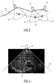
- Fig. 2 shows a schematic sectional diagram illustrating the calibration at different positions of the ultrasound probe 14 and a current position of the ultrasound probe 14.
- the ultrasound probe 14 is shown together with a field of view 32 within the patient's body 12 from which the ultrasound image data is acquired as three-dimensional ultrasound data.
- the different calibration positions of the ultrasound probe 14', 14" are determined with respect to a center position C 0 , C 1 of the ultrasound data cone or in the center of the field of view 32', 32".
- any predefined positions within the ultrasound data cone can be defined as reference position or any position at a tip of the ultrasound probe 14 preferably a center position at the tip can be defined as reference position.
- a transfer function T(t 0 ) linking the ultrasound image data and the 3D medical image data to each other are determined, corresponding to a spatial synchronization between the ultrasound image data and the 3D medical image data.
- the relationship i.e.
- T t Q i * P t
- T(t) is the transfer function
- Q i is the rigid or fixed transformation for the respective calibration position
- P(t) is the current position of the ultrasound probe 14.
- the weight factors a can be computed based on a distance from the center positions C 0 , C 1 in the field of view 32', 32" from the current position C t of the field of view 32. On the basis of the distance, the calibration transfer function of a calibration position which is closer to the current position used is considered more and has as a large weight in the general transformation function T(t).
- a such absolute weighting would merely consider the calibration which has the smallest distance to the current position and would not consider all other calibration positions.
- a relative weighting can be utilized, whereas the transfer function of the closer calibration position is considered more with a higher weight factor a and the calibration transfer function of a calibration position with a higher distance is less considered with a lower weight factor a.
- an ultrasound image corresponding to the ultrasound image data is schematically shown in a field of view 32 of the ultrasound probe 14.
- the ultrasound probe 14 determines the ultrasound image data in the field of view 32, wherein in the center of the field of view 32 or the ultrasound data cone, the central position C i is defined as a reference position of the ultrasound image data.
- a center position 34 at the tip of the ultrasound probe 14 can be used as a reference position for the calibration and the synchronization.
Landscapes
- Health & Medical Sciences (AREA)
- Life Sciences & Earth Sciences (AREA)
- Engineering & Computer Science (AREA)
- Radiology & Medical Imaging (AREA)
- Heart & Thoracic Surgery (AREA)
- Biophysics (AREA)
- Nuclear Medicine, Radiotherapy & Molecular Imaging (AREA)
- Pathology (AREA)
- Veterinary Medicine (AREA)
- Biomedical Technology (AREA)
- Physics & Mathematics (AREA)
- Medical Informatics (AREA)
- Molecular Biology (AREA)
- Surgery (AREA)
- Animal Behavior & Ethology (AREA)
- General Health & Medical Sciences (AREA)
- Public Health (AREA)
- Computer Vision & Pattern Recognition (AREA)
- Ultra Sonic Daignosis Equipment (AREA)
Applications Claiming Priority (2)
| Application Number | Priority Date | Filing Date | Title |
|---|---|---|---|
| EP15305462 | 2015-03-31 | ||
| PCT/EP2016/057393 WO2016169759A1 (en) | 2015-03-31 | 2016-04-05 | Medical imaging apparatus |
Publications (2)
| Publication Number | Publication Date |
|---|---|
| EP3277186A1 EP3277186A1 (en) | 2018-02-07 |
| EP3277186B1 true EP3277186B1 (en) | 2018-09-26 |
Family
ID=53682613
Family Applications (1)
| Application Number | Title | Priority Date | Filing Date |
|---|---|---|---|
| EP16713927.8A Active EP3277186B1 (en) | 2015-03-31 | 2016-04-05 | Medical imaging apparatus |
Country Status (6)
Families Citing this family (4)
| Publication number | Priority date | Publication date | Assignee | Title |
|---|---|---|---|---|
| EP3508132A1 (en) | 2018-01-04 | 2019-07-10 | Koninklijke Philips N.V. | Ultrasound system and method for correcting motion-induced misalignment in image fusion |
| EP3923293A1 (en) * | 2020-06-09 | 2021-12-15 | Koninklijke Philips N.V. | System and method for analysis of medical image data based on an interaction of quality metrics |
| DE102023203405A1 (de) | 2023-04-14 | 2024-10-17 | Fraunhofer-Gesellschaft zur Förderung der angewandten Forschung eingetragener Verein | Folie zum Transport einer Flüssigkeit, Rahmen, Fallfilm-Durchflussreaktor und Verwendungen hiervon |
| US12213774B1 (en) | 2024-01-02 | 2025-02-04 | nference, inc. | Apparatus and method for locating a position of an electrode on an organ model |
Family Cites Families (23)
| Publication number | Priority date | Publication date | Assignee | Title |
|---|---|---|---|---|
| AU2001291175A1 (en) * | 2000-09-21 | 2002-04-02 | Md Online Inc. | Medical image processing systems |
| US7945304B2 (en) * | 2001-11-20 | 2011-05-17 | Feinberg David A | Ultrasound within MRI scanners for guidance of MRI pulse sequences |
| ATE523141T1 (de) * | 2004-02-17 | 2011-09-15 | Philips Electronics Ltd | Verfahren und vorrichtung zur registrierung, verifizierung von und bezugnahme auf körperorgane(n) |
| KR20070110965A (ko) * | 2006-05-16 | 2007-11-21 | 주식회사 메디슨 | 초음파 영상과 외부 의료영상의 합성 영상을디스플레이하기 위한 초음파 시스템 |
| EP2104919A2 (en) * | 2006-11-27 | 2009-09-30 | Koninklijke Philips Electronics N.V. | System and method for fusing real-time ultrasound images with pre-acquired medical images |
| BRPI0819439A8 (pt) * | 2007-11-16 | 2015-11-10 | Koninklijke Philips Electronics Nv | Método e sistema para navegação intervencional usando formação de imagem por ultrassom realçada por contraste em 3d |
| JP2009101184A (ja) * | 2009-01-06 | 2009-05-14 | Toshiba Corp | 診断装置 |
| US9521994B2 (en) * | 2009-05-11 | 2016-12-20 | Siemens Healthcare Gmbh | System and method for image guided prostate cancer needle biopsy |
| KR101121286B1 (ko) | 2009-07-31 | 2012-03-23 | 한국과학기술원 | 센서의 교정을 수행하는 초음파 시스템 및 방법 |
| US9179888B2 (en) * | 2009-08-28 | 2015-11-10 | Dartmouth College | System and method for providing patient registration without fiducials |
| EP2680778B1 (en) * | 2011-03-03 | 2019-07-31 | Koninklijke Philips N.V. | System and method for automated initialization and registration of navigation system |
| JP5685133B2 (ja) * | 2011-04-13 | 2015-03-18 | キヤノン株式会社 | 画像処理装置、画像処理装置の制御方法、およびプログラム |
| JP5862571B2 (ja) * | 2011-05-30 | 2016-02-16 | コニカミノルタ株式会社 | 超音波画像生成装置および超音波画像生成方法 |
| WO2013179224A1 (en) * | 2012-05-31 | 2013-12-05 | Koninklijke Philips N.V. | Ultrasound imaging system and method for image guidance procedure |
| JP6081301B2 (ja) * | 2012-06-27 | 2017-02-15 | 東芝メディカルシステムズ株式会社 | 超音波診断装置及び画像データの補正方法 |
| EP2706372A1 (en) * | 2012-09-10 | 2014-03-12 | Esaote S.p.A. | Method and apparatus for ultrasound image acquisition |
| US10631829B2 (en) * | 2013-02-28 | 2020-04-28 | Koninklijke Philips N.V. | Segmentation of large objects from multiple three-dimensional views |
| EP2807978A1 (en) * | 2013-05-28 | 2014-12-03 | Universität Bern | Method and system for 3D acquisition of ultrasound images |
| JP6430498B2 (ja) * | 2013-06-26 | 2018-11-28 | コーニンクレッカ フィリップス エヌ ヴェKoninklijke Philips N.V. | 超音波剪断波エラストグラフィ測定のマッピングのためのシステムおよび方法 |
| CN104574329B (zh) * | 2013-10-09 | 2018-03-09 | 深圳迈瑞生物医疗电子股份有限公司 | 超声融合成像方法、超声融合成像导航系统 |
| EP3142587B1 (en) * | 2014-05-16 | 2019-07-10 | Koninklijke Philips N.V. | Reconstruction-free automatic multi-modality ultrasound registration |
| WO2015193441A1 (en) * | 2014-06-18 | 2015-12-23 | Koninklijke Philips N.V. | Ultrasound imaging apparatus |
| EP3190973A1 (en) * | 2014-09-08 | 2017-07-19 | Koninklijke Philips N.V. | Medical imaging apparatus |
-
2016
- 2016-04-05 RU RU2017134276A patent/RU2017134276A/ru not_active Application Discontinuation
- 2016-04-05 EP EP16713927.8A patent/EP3277186B1/en active Active
- 2016-04-05 US US15/559,460 patent/US10828014B2/en active Active
- 2016-04-05 JP JP2017549518A patent/JP6405058B2/ja active Active
- 2016-04-05 CN CN201680020335.7A patent/CN107548294B/zh active Active
- 2016-04-05 WO PCT/EP2016/057393 patent/WO2016169759A1/en active Application Filing
-
2020
- 2020-10-02 US US17/062,443 patent/US11903770B2/en active Active
Non-Patent Citations (1)
| Title |
|---|
| None * |
Also Published As
| Publication number | Publication date |
|---|---|
| US20180110498A1 (en) | 2018-04-26 |
| US11903770B2 (en) | 2024-02-20 |
| JP2018509990A (ja) | 2018-04-12 |
| CN107548294B (zh) | 2021-11-09 |
| WO2016169759A8 (en) | 2018-03-08 |
| US20210015465A1 (en) | 2021-01-21 |
| EP3277186A1 (en) | 2018-02-07 |
| US10828014B2 (en) | 2020-11-10 |
| RU2017134276A (ru) | 2019-04-03 |
| WO2016169759A1 (en) | 2016-10-27 |
| JP6405058B2 (ja) | 2018-10-17 |
| CN107548294A (zh) | 2018-01-05 |
Similar Documents
| Publication | Publication Date | Title |
|---|---|---|
| US11903770B2 (en) | Medical imaging apparatus | |
| EP3157436B1 (en) | Ultrasound imaging apparatus | |
| EP3238649B1 (en) | Self-localizing medical device | |
| EP3393367B1 (en) | Medical imaging apparatus and medical imaging method for inspecting a volume of a subject | |
| US20180214129A1 (en) | Medical imaging apparatus | |
| KR20140148247A (ko) | 모바일 x 선 장치의 x 선 튜브와 디텍터를 정렬하기 위한 정보 제공 방법 및 정보 제공 장치, 및 무선 디텍터 | |
| US20150182187A1 (en) | System and method for tracking an invasive device using ultrasound position signals | |
| US20180092626A1 (en) | Ultrasound imaging apparatus | |
| US20240398375A1 (en) | Spatial registration method for imaging devices | |
| US20160345937A1 (en) | System and method for imaging using ultrasound | |
| US20120053463A1 (en) | Providing ultrasound spatial compound images in an ultrasound system | |
| EP3024408B1 (en) | Wrong level surgery prevention |
Legal Events
| Date | Code | Title | Description |
|---|---|---|---|
| STAA | Information on the status of an ep patent application or granted ep patent |
Free format text: STATUS: THE INTERNATIONAL PUBLICATION HAS BEEN MADE |
|
| PUAI | Public reference made under article 153(3) epc to a published international application that has entered the european phase |
Free format text: ORIGINAL CODE: 0009012 |
|
| STAA | Information on the status of an ep patent application or granted ep patent |
Free format text: STATUS: REQUEST FOR EXAMINATION WAS MADE |
|
| 17P | Request for examination filed |
Effective date: 20171102 |
|
| AK | Designated contracting states |
Kind code of ref document: A1 Designated state(s): AL AT BE BG CH CY CZ DE DK EE ES FI FR GB GR HR HU IE IS IT LI LT LU LV MC MK MT NL NO PL PT RO RS SE SI SK SM TR |
|
| AX | Request for extension of the european patent |
Extension state: BA ME |
|
| GRAP | Despatch of communication of intention to grant a patent |
Free format text: ORIGINAL CODE: EPIDOSNIGR1 |
|
| STAA | Information on the status of an ep patent application or granted ep patent |
Free format text: STATUS: GRANT OF PATENT IS INTENDED |
|
| INTG | Intention to grant announced |
Effective date: 20180412 |
|
| GRAS | Grant fee paid |
Free format text: ORIGINAL CODE: EPIDOSNIGR3 |
|
| GRAA | (expected) grant |
Free format text: ORIGINAL CODE: 0009210 |
|
| STAA | Information on the status of an ep patent application or granted ep patent |
Free format text: STATUS: THE PATENT HAS BEEN GRANTED |
|
| DAV | Request for validation of the european patent (deleted) | ||
| DAX | Request for extension of the european patent (deleted) | ||
| AK | Designated contracting states |
Kind code of ref document: B1 Designated state(s): AL AT BE BG CH CY CZ DE DK EE ES FI FR GB GR HR HU IE IS IT LI LT LU LV MC MK MT NL NO PL PT RO RS SE SI SK SM TR |
|
| REG | Reference to a national code |
Ref country code: GB Ref legal event code: FG4D |
|
| REG | Reference to a national code |
Ref country code: CH Ref legal event code: EP |
|
| REG | Reference to a national code |
Ref country code: AT Ref legal event code: REF Ref document number: 1045063 Country of ref document: AT Kind code of ref document: T Effective date: 20181015 |
|
| REG | Reference to a national code |
Ref country code: IE Ref legal event code: FG4D |
|
| REG | Reference to a national code |
Ref country code: DE Ref legal event code: R096 Ref document number: 602016006081 Country of ref document: DE |
|
| REG | Reference to a national code |
Ref country code: DE Ref legal event code: R084 Ref document number: 602016006081 Country of ref document: DE |
|
| REG | Reference to a national code |
Ref country code: GB Ref legal event code: 746 Effective date: 20181114 |
|
| REG | Reference to a national code |
Ref country code: NL Ref legal event code: MP Effective date: 20180926 |
|
| PG25 | Lapsed in a contracting state [announced via postgrant information from national office to epo] |
Ref country code: BG Free format text: LAPSE BECAUSE OF FAILURE TO SUBMIT A TRANSLATION OF THE DESCRIPTION OR TO PAY THE FEE WITHIN THE PRESCRIBED TIME-LIMIT Effective date: 20181226 Ref country code: LT Free format text: LAPSE BECAUSE OF FAILURE TO SUBMIT A TRANSLATION OF THE DESCRIPTION OR TO PAY THE FEE WITHIN THE PRESCRIBED TIME-LIMIT Effective date: 20180926 Ref country code: FI Free format text: LAPSE BECAUSE OF FAILURE TO SUBMIT A TRANSLATION OF THE DESCRIPTION OR TO PAY THE FEE WITHIN THE PRESCRIBED TIME-LIMIT Effective date: 20180926 Ref country code: RS Free format text: LAPSE BECAUSE OF FAILURE TO SUBMIT A TRANSLATION OF THE DESCRIPTION OR TO PAY THE FEE WITHIN THE PRESCRIBED TIME-LIMIT Effective date: 20180926 Ref country code: GR Free format text: LAPSE BECAUSE OF FAILURE TO SUBMIT A TRANSLATION OF THE DESCRIPTION OR TO PAY THE FEE WITHIN THE PRESCRIBED TIME-LIMIT Effective date: 20181227 Ref country code: NO Free format text: LAPSE BECAUSE OF FAILURE TO SUBMIT A TRANSLATION OF THE DESCRIPTION OR TO PAY THE FEE WITHIN THE PRESCRIBED TIME-LIMIT Effective date: 20181226 |
|
| REG | Reference to a national code |
Ref country code: LT Ref legal event code: MG4D |
|
| PG25 | Lapsed in a contracting state [announced via postgrant information from national office to epo] |
Ref country code: HR Free format text: LAPSE BECAUSE OF FAILURE TO SUBMIT A TRANSLATION OF THE DESCRIPTION OR TO PAY THE FEE WITHIN THE PRESCRIBED TIME-LIMIT Effective date: 20180926 Ref country code: LV Free format text: LAPSE BECAUSE OF FAILURE TO SUBMIT A TRANSLATION OF THE DESCRIPTION OR TO PAY THE FEE WITHIN THE PRESCRIBED TIME-LIMIT Effective date: 20180926 Ref country code: AL Free format text: LAPSE BECAUSE OF FAILURE TO SUBMIT A TRANSLATION OF THE DESCRIPTION OR TO PAY THE FEE WITHIN THE PRESCRIBED TIME-LIMIT Effective date: 20180926 |
|
| REG | Reference to a national code |
Ref country code: AT Ref legal event code: MK05 Ref document number: 1045063 Country of ref document: AT Kind code of ref document: T Effective date: 20180926 |
|
| PG25 | Lapsed in a contracting state [announced via postgrant information from national office to epo] |
Ref country code: NL Free format text: LAPSE BECAUSE OF FAILURE TO SUBMIT A TRANSLATION OF THE DESCRIPTION OR TO PAY THE FEE WITHIN THE PRESCRIBED TIME-LIMIT Effective date: 20180926 Ref country code: IT Free format text: LAPSE BECAUSE OF FAILURE TO SUBMIT A TRANSLATION OF THE DESCRIPTION OR TO PAY THE FEE WITHIN THE PRESCRIBED TIME-LIMIT Effective date: 20180926 Ref country code: AT Free format text: LAPSE BECAUSE OF FAILURE TO SUBMIT A TRANSLATION OF THE DESCRIPTION OR TO PAY THE FEE WITHIN THE PRESCRIBED TIME-LIMIT Effective date: 20180926 Ref country code: EE Free format text: LAPSE BECAUSE OF FAILURE TO SUBMIT A TRANSLATION OF THE DESCRIPTION OR TO PAY THE FEE WITHIN THE PRESCRIBED TIME-LIMIT Effective date: 20180926 Ref country code: RO Free format text: LAPSE BECAUSE OF FAILURE TO SUBMIT A TRANSLATION OF THE DESCRIPTION OR TO PAY THE FEE WITHIN THE PRESCRIBED TIME-LIMIT Effective date: 20180926 Ref country code: CZ Free format text: LAPSE BECAUSE OF FAILURE TO SUBMIT A TRANSLATION OF THE DESCRIPTION OR TO PAY THE FEE WITHIN THE PRESCRIBED TIME-LIMIT Effective date: 20180926 Ref country code: ES Free format text: LAPSE BECAUSE OF FAILURE TO SUBMIT A TRANSLATION OF THE DESCRIPTION OR TO PAY THE FEE WITHIN THE PRESCRIBED TIME-LIMIT Effective date: 20180926 Ref country code: IS Free format text: LAPSE BECAUSE OF FAILURE TO SUBMIT A TRANSLATION OF THE DESCRIPTION OR TO PAY THE FEE WITHIN THE PRESCRIBED TIME-LIMIT Effective date: 20190126 Ref country code: PL Free format text: LAPSE BECAUSE OF FAILURE TO SUBMIT A TRANSLATION OF THE DESCRIPTION OR TO PAY THE FEE WITHIN THE PRESCRIBED TIME-LIMIT Effective date: 20180926 |
|
| PG25 | Lapsed in a contracting state [announced via postgrant information from national office to epo] |
Ref country code: SK Free format text: LAPSE BECAUSE OF FAILURE TO SUBMIT A TRANSLATION OF THE DESCRIPTION OR TO PAY THE FEE WITHIN THE PRESCRIBED TIME-LIMIT Effective date: 20180926 Ref country code: PT Free format text: LAPSE BECAUSE OF FAILURE TO SUBMIT A TRANSLATION OF THE DESCRIPTION OR TO PAY THE FEE WITHIN THE PRESCRIBED TIME-LIMIT Effective date: 20190126 Ref country code: SM Free format text: LAPSE BECAUSE OF FAILURE TO SUBMIT A TRANSLATION OF THE DESCRIPTION OR TO PAY THE FEE WITHIN THE PRESCRIBED TIME-LIMIT Effective date: 20180926 |
|
| REG | Reference to a national code |
Ref country code: DE Ref legal event code: R097 Ref document number: 602016006081 Country of ref document: DE |
|
| PG25 | Lapsed in a contracting state [announced via postgrant information from national office to epo] |
Ref country code: DK Free format text: LAPSE BECAUSE OF FAILURE TO SUBMIT A TRANSLATION OF THE DESCRIPTION OR TO PAY THE FEE WITHIN THE PRESCRIBED TIME-LIMIT Effective date: 20180926 |
|
| PLBE | No opposition filed within time limit |
Free format text: ORIGINAL CODE: 0009261 |
|
| STAA | Information on the status of an ep patent application or granted ep patent |
Free format text: STATUS: NO OPPOSITION FILED WITHIN TIME LIMIT |
|
| 26N | No opposition filed |
Effective date: 20190627 |
|
| REG | Reference to a national code |
Ref country code: CH Ref legal event code: PL |
|
| REG | Reference to a national code |
Ref country code: BE Ref legal event code: MM Effective date: 20190430 |
|
| PG25 | Lapsed in a contracting state [announced via postgrant information from national office to epo] |
Ref country code: MC Free format text: LAPSE BECAUSE OF FAILURE TO SUBMIT A TRANSLATION OF THE DESCRIPTION OR TO PAY THE FEE WITHIN THE PRESCRIBED TIME-LIMIT Effective date: 20180926 Ref country code: LU Free format text: LAPSE BECAUSE OF NON-PAYMENT OF DUE FEES Effective date: 20190405 |
|
| PG25 | Lapsed in a contracting state [announced via postgrant information from national office to epo] |
Ref country code: LI Free format text: LAPSE BECAUSE OF NON-PAYMENT OF DUE FEES Effective date: 20190430 Ref country code: CH Free format text: LAPSE BECAUSE OF NON-PAYMENT OF DUE FEES Effective date: 20190430 |
|
| PG25 | Lapsed in a contracting state [announced via postgrant information from national office to epo] |
Ref country code: BE Free format text: LAPSE BECAUSE OF NON-PAYMENT OF DUE FEES Effective date: 20190430 Ref country code: FR Free format text: LAPSE BECAUSE OF NON-PAYMENT OF DUE FEES Effective date: 20190430 |
|
| PG25 | Lapsed in a contracting state [announced via postgrant information from national office to epo] |
Ref country code: TR Free format text: LAPSE BECAUSE OF FAILURE TO SUBMIT A TRANSLATION OF THE DESCRIPTION OR TO PAY THE FEE WITHIN THE PRESCRIBED TIME-LIMIT Effective date: 20180926 |
|
| PG25 | Lapsed in a contracting state [announced via postgrant information from national office to epo] |
Ref country code: IE Free format text: LAPSE BECAUSE OF NON-PAYMENT OF DUE FEES Effective date: 20190405 |
|
| PG25 | Lapsed in a contracting state [announced via postgrant information from national office to epo] |
Ref country code: CY Free format text: LAPSE BECAUSE OF FAILURE TO SUBMIT A TRANSLATION OF THE DESCRIPTION OR TO PAY THE FEE WITHIN THE PRESCRIBED TIME-LIMIT Effective date: 20180926 |
|
| PG25 | Lapsed in a contracting state [announced via postgrant information from national office to epo] |
Ref country code: SE Free format text: LAPSE BECAUSE OF NON-PAYMENT OF DUE FEES Effective date: 20180926 |
|
| PG25 | Lapsed in a contracting state [announced via postgrant information from national office to epo] |
Ref country code: HU Free format text: LAPSE BECAUSE OF FAILURE TO SUBMIT A TRANSLATION OF THE DESCRIPTION OR TO PAY THE FEE WITHIN THE PRESCRIBED TIME-LIMIT; INVALID AB INITIO Effective date: 20160405 Ref country code: MT Free format text: LAPSE BECAUSE OF FAILURE TO SUBMIT A TRANSLATION OF THE DESCRIPTION OR TO PAY THE FEE WITHIN THE PRESCRIBED TIME-LIMIT Effective date: 20180926 |
|
| PG25 | Lapsed in a contracting state [announced via postgrant information from national office to epo] |
Ref country code: SI Free format text: LAPSE BECAUSE OF FAILURE TO SUBMIT A TRANSLATION OF THE DESCRIPTION OR TO PAY THE FEE WITHIN THE PRESCRIBED TIME-LIMIT Effective date: 20180926 |
|
| PG25 | Lapsed in a contracting state [announced via postgrant information from national office to epo] |
Ref country code: MK Free format text: LAPSE BECAUSE OF FAILURE TO SUBMIT A TRANSLATION OF THE DESCRIPTION OR TO PAY THE FEE WITHIN THE PRESCRIBED TIME-LIMIT Effective date: 20180926 |
|
| PGFP | Annual fee paid to national office [announced via postgrant information from national office to epo] |
Ref country code: GB Payment date: 20240423 Year of fee payment: 9 |
|
| PGFP | Annual fee paid to national office [announced via postgrant information from national office to epo] |
Ref country code: DE Payment date: 20240429 Year of fee payment: 9 |