EP3275471A1 - Dreidimensionale struktur zur herzmuskelgeweberegeneration und herstellungsverfahren dafür - Google Patents
Dreidimensionale struktur zur herzmuskelgeweberegeneration und herstellungsverfahren dafür Download PDFInfo
- Publication number
- EP3275471A1 EP3275471A1 EP16769138.5A EP16769138A EP3275471A1 EP 3275471 A1 EP3275471 A1 EP 3275471A1 EP 16769138 A EP16769138 A EP 16769138A EP 3275471 A1 EP3275471 A1 EP 3275471A1
- Authority
- EP
- European Patent Office
- Prior art keywords
- construct
- preparation
- bioprinting composition
- bioprinting
- composition
- Prior art date
- Legal status (The legal status is an assumption and is not a legal conclusion. Google has not performed a legal analysis and makes no representation as to the accuracy of the status listed.)
- Withdrawn
Links
Images


Classifications
-
- C—CHEMISTRY; METALLURGY
- C12—BIOCHEMISTRY; BEER; SPIRITS; WINE; VINEGAR; MICROBIOLOGY; ENZYMOLOGY; MUTATION OR GENETIC ENGINEERING
- C12N—MICROORGANISMS OR ENZYMES; COMPOSITIONS THEREOF; PROPAGATING, PRESERVING, OR MAINTAINING MICROORGANISMS; MUTATION OR GENETIC ENGINEERING; CULTURE MEDIA
- C12N5/00—Undifferentiated human, animal or plant cells, e.g. cell lines; Tissues; Cultivation or maintenance thereof; Culture media therefor
- C12N5/06—Animal cells or tissues; Human cells or tissues
- C12N5/0602—Vertebrate cells
- C12N5/0652—Cells of skeletal and connective tissues; Mesenchyme
- C12N5/0657—Cardiomyocytes; Heart cells
-
- C—CHEMISTRY; METALLURGY
- C12—BIOCHEMISTRY; BEER; SPIRITS; WINE; VINEGAR; MICROBIOLOGY; ENZYMOLOGY; MUTATION OR GENETIC ENGINEERING
- C12N—MICROORGANISMS OR ENZYMES; COMPOSITIONS THEREOF; PROPAGATING, PRESERVING, OR MAINTAINING MICROORGANISMS; MUTATION OR GENETIC ENGINEERING; CULTURE MEDIA
- C12N5/00—Undifferentiated human, animal or plant cells, e.g. cell lines; Tissues; Cultivation or maintenance thereof; Culture media therefor
- C12N5/06—Animal cells or tissues; Human cells or tissues
- C12N5/0697—Artificial constructs associating cells of different lineages, e.g. tissue equivalents
-
- A—HUMAN NECESSITIES
- A61—MEDICAL OR VETERINARY SCIENCE; HYGIENE
- A61L—METHODS OR APPARATUS FOR STERILISING MATERIALS OR OBJECTS IN GENERAL; DISINFECTION, STERILISATION OR DEODORISATION OF AIR; CHEMICAL ASPECTS OF BANDAGES, DRESSINGS, ABSORBENT PADS OR SURGICAL ARTICLES; MATERIALS FOR BANDAGES, DRESSINGS, ABSORBENT PADS OR SURGICAL ARTICLES
- A61L27/00—Materials for grafts or prostheses or for coating grafts or prostheses
- A61L27/36—Materials for grafts or prostheses or for coating grafts or prostheses containing ingredients of undetermined constitution or reaction products thereof, e.g. transplant tissue, natural bone, extracellular matrix
- A61L27/3604—Materials for grafts or prostheses or for coating grafts or prostheses containing ingredients of undetermined constitution or reaction products thereof, e.g. transplant tissue, natural bone, extracellular matrix characterised by the human or animal origin of the biological material, e.g. hair, fascia, fish scales, silk, shellac, pericardium, pleura, renal tissue, amniotic membrane, parenchymal tissue, fetal tissue, muscle tissue, fat tissue, enamel
- A61L27/3633—Extracellular matrix [ECM]
-
- A—HUMAN NECESSITIES
- A61—MEDICAL OR VETERINARY SCIENCE; HYGIENE
- A61L—METHODS OR APPARATUS FOR STERILISING MATERIALS OR OBJECTS IN GENERAL; DISINFECTION, STERILISATION OR DEODORISATION OF AIR; CHEMICAL ASPECTS OF BANDAGES, DRESSINGS, ABSORBENT PADS OR SURGICAL ARTICLES; MATERIALS FOR BANDAGES, DRESSINGS, ABSORBENT PADS OR SURGICAL ARTICLES
- A61L27/00—Materials for grafts or prostheses or for coating grafts or prostheses
- A61L27/36—Materials for grafts or prostheses or for coating grafts or prostheses containing ingredients of undetermined constitution or reaction products thereof, e.g. transplant tissue, natural bone, extracellular matrix
- A61L27/3641—Materials for grafts or prostheses or for coating grafts or prostheses containing ingredients of undetermined constitution or reaction products thereof, e.g. transplant tissue, natural bone, extracellular matrix characterised by the site of application in the body
- A61L27/367—Muscle tissue, e.g. sphincter
-
- A—HUMAN NECESSITIES
- A61—MEDICAL OR VETERINARY SCIENCE; HYGIENE
- A61L—METHODS OR APPARATUS FOR STERILISING MATERIALS OR OBJECTS IN GENERAL; DISINFECTION, STERILISATION OR DEODORISATION OF AIR; CHEMICAL ASPECTS OF BANDAGES, DRESSINGS, ABSORBENT PADS OR SURGICAL ARTICLES; MATERIALS FOR BANDAGES, DRESSINGS, ABSORBENT PADS OR SURGICAL ARTICLES
- A61L27/00—Materials for grafts or prostheses or for coating grafts or prostheses
- A61L27/36—Materials for grafts or prostheses or for coating grafts or prostheses containing ingredients of undetermined constitution or reaction products thereof, e.g. transplant tissue, natural bone, extracellular matrix
- A61L27/38—Materials for grafts or prostheses or for coating grafts or prostheses containing ingredients of undetermined constitution or reaction products thereof, e.g. transplant tissue, natural bone, extracellular matrix containing added animal cells
- A61L27/3804—Materials for grafts or prostheses or for coating grafts or prostheses containing ingredients of undetermined constitution or reaction products thereof, e.g. transplant tissue, natural bone, extracellular matrix containing added animal cells characterised by specific cells or progenitors thereof, e.g. fibroblasts, connective tissue cells, kidney cells
- A61L27/3826—Muscle cells, e.g. smooth muscle cells
-
- A—HUMAN NECESSITIES
- A61—MEDICAL OR VETERINARY SCIENCE; HYGIENE
- A61L—METHODS OR APPARATUS FOR STERILISING MATERIALS OR OBJECTS IN GENERAL; DISINFECTION, STERILISATION OR DEODORISATION OF AIR; CHEMICAL ASPECTS OF BANDAGES, DRESSINGS, ABSORBENT PADS OR SURGICAL ARTICLES; MATERIALS FOR BANDAGES, DRESSINGS, ABSORBENT PADS OR SURGICAL ARTICLES
- A61L27/00—Materials for grafts or prostheses or for coating grafts or prostheses
- A61L27/36—Materials for grafts or prostheses or for coating grafts or prostheses containing ingredients of undetermined constitution or reaction products thereof, e.g. transplant tissue, natural bone, extracellular matrix
- A61L27/38—Materials for grafts or prostheses or for coating grafts or prostheses containing ingredients of undetermined constitution or reaction products thereof, e.g. transplant tissue, natural bone, extracellular matrix containing added animal cells
- A61L27/3804—Materials for grafts or prostheses or for coating grafts or prostheses containing ingredients of undetermined constitution or reaction products thereof, e.g. transplant tissue, natural bone, extracellular matrix containing added animal cells characterised by specific cells or progenitors thereof, e.g. fibroblasts, connective tissue cells, kidney cells
- A61L27/3834—Cells able to produce different cell types, e.g. hematopoietic stem cells, mesenchymal stem cells, marrow stromal cells, embryonic stem cells
-
- A—HUMAN NECESSITIES
- A61—MEDICAL OR VETERINARY SCIENCE; HYGIENE
- A61L—METHODS OR APPARATUS FOR STERILISING MATERIALS OR OBJECTS IN GENERAL; DISINFECTION, STERILISATION OR DEODORISATION OF AIR; CHEMICAL ASPECTS OF BANDAGES, DRESSINGS, ABSORBENT PADS OR SURGICAL ARTICLES; MATERIALS FOR BANDAGES, DRESSINGS, ABSORBENT PADS OR SURGICAL ARTICLES
- A61L27/00—Materials for grafts or prostheses or for coating grafts or prostheses
- A61L27/36—Materials for grafts or prostheses or for coating grafts or prostheses containing ingredients of undetermined constitution or reaction products thereof, e.g. transplant tissue, natural bone, extracellular matrix
- A61L27/38—Materials for grafts or prostheses or for coating grafts or prostheses containing ingredients of undetermined constitution or reaction products thereof, e.g. transplant tissue, natural bone, extracellular matrix containing added animal cells
- A61L27/3886—Materials for grafts or prostheses or for coating grafts or prostheses containing ingredients of undetermined constitution or reaction products thereof, e.g. transplant tissue, natural bone, extracellular matrix containing added animal cells comprising two or more cell types
- A61L27/3891—Materials for grafts or prostheses or for coating grafts or prostheses containing ingredients of undetermined constitution or reaction products thereof, e.g. transplant tissue, natural bone, extracellular matrix containing added animal cells comprising two or more cell types as distinct cell layers
-
- A—HUMAN NECESSITIES
- A61—MEDICAL OR VETERINARY SCIENCE; HYGIENE
- A61L—METHODS OR APPARATUS FOR STERILISING MATERIALS OR OBJECTS IN GENERAL; DISINFECTION, STERILISATION OR DEODORISATION OF AIR; CHEMICAL ASPECTS OF BANDAGES, DRESSINGS, ABSORBENT PADS OR SURGICAL ARTICLES; MATERIALS FOR BANDAGES, DRESSINGS, ABSORBENT PADS OR SURGICAL ARTICLES
- A61L27/00—Materials for grafts or prostheses or for coating grafts or prostheses
- A61L27/36—Materials for grafts or prostheses or for coating grafts or prostheses containing ingredients of undetermined constitution or reaction products thereof, e.g. transplant tissue, natural bone, extracellular matrix
- A61L27/38—Materials for grafts or prostheses or for coating grafts or prostheses containing ingredients of undetermined constitution or reaction products thereof, e.g. transplant tissue, natural bone, extracellular matrix containing added animal cells
- A61L27/3895—Materials for grafts or prostheses or for coating grafts or prostheses containing ingredients of undetermined constitution or reaction products thereof, e.g. transplant tissue, natural bone, extracellular matrix containing added animal cells using specific culture conditions, e.g. stimulating differentiation of stem cells, pulsatile flow conditions
-
- B—PERFORMING OPERATIONS; TRANSPORTING
- B33—ADDITIVE MANUFACTURING TECHNOLOGY
- B33Y—ADDITIVE MANUFACTURING, i.e. MANUFACTURING OF THREE-DIMENSIONAL [3-D] OBJECTS BY ADDITIVE DEPOSITION, ADDITIVE AGGLOMERATION OR ADDITIVE LAYERING, e.g. BY 3-D PRINTING, STEREOLITHOGRAPHY OR SELECTIVE LASER SINTERING
- B33Y10/00—Processes of additive manufacturing
-
- B—PERFORMING OPERATIONS; TRANSPORTING
- B33—ADDITIVE MANUFACTURING TECHNOLOGY
- B33Y—ADDITIVE MANUFACTURING, i.e. MANUFACTURING OF THREE-DIMENSIONAL [3-D] OBJECTS BY ADDITIVE DEPOSITION, ADDITIVE AGGLOMERATION OR ADDITIVE LAYERING, e.g. BY 3-D PRINTING, STEREOLITHOGRAPHY OR SELECTIVE LASER SINTERING
- B33Y70/00—Materials specially adapted for additive manufacturing
-
- B—PERFORMING OPERATIONS; TRANSPORTING
- B33—ADDITIVE MANUFACTURING TECHNOLOGY
- B33Y—ADDITIVE MANUFACTURING, i.e. MANUFACTURING OF THREE-DIMENSIONAL [3-D] OBJECTS BY ADDITIVE DEPOSITION, ADDITIVE AGGLOMERATION OR ADDITIVE LAYERING, e.g. BY 3-D PRINTING, STEREOLITHOGRAPHY OR SELECTIVE LASER SINTERING
- B33Y80/00—Products made by additive manufacturing
-
- C—CHEMISTRY; METALLURGY
- C07—ORGANIC CHEMISTRY
- C07K—PEPTIDES
- C07K14/00—Peptides having more than 20 amino acids; Gastrins; Somatostatins; Melanotropins; Derivatives thereof
- C07K14/435—Peptides having more than 20 amino acids; Gastrins; Somatostatins; Melanotropins; Derivatives thereof from animals; from humans
- C07K14/475—Growth factors; Growth regulators
-
- A—HUMAN NECESSITIES
- A61—MEDICAL OR VETERINARY SCIENCE; HYGIENE
- A61F—FILTERS IMPLANTABLE INTO BLOOD VESSELS; PROSTHESES; DEVICES PROVIDING PATENCY TO, OR PREVENTING COLLAPSING OF, TUBULAR STRUCTURES OF THE BODY, e.g. STENTS; ORTHOPAEDIC, NURSING OR CONTRACEPTIVE DEVICES; FOMENTATION; TREATMENT OR PROTECTION OF EYES OR EARS; BANDAGES, DRESSINGS OR ABSORBENT PADS; FIRST-AID KITS
- A61F2240/00—Manufacturing or designing of prostheses classified in groups A61F2/00 - A61F2/26 or A61F2/82 or A61F9/00 or A61F11/00 or subgroups thereof
- A61F2240/001—Designing or manufacturing processes
-
- A—HUMAN NECESSITIES
- A61—MEDICAL OR VETERINARY SCIENCE; HYGIENE
- A61L—METHODS OR APPARATUS FOR STERILISING MATERIALS OR OBJECTS IN GENERAL; DISINFECTION, STERILISATION OR DEODORISATION OF AIR; CHEMICAL ASPECTS OF BANDAGES, DRESSINGS, ABSORBENT PADS OR SURGICAL ARTICLES; MATERIALS FOR BANDAGES, DRESSINGS, ABSORBENT PADS OR SURGICAL ARTICLES
- A61L2430/00—Materials or treatment for tissue regeneration
- A61L2430/20—Materials or treatment for tissue regeneration for reconstruction of the heart, e.g. heart valves
-
- C—CHEMISTRY; METALLURGY
- C12—BIOCHEMISTRY; BEER; SPIRITS; WINE; VINEGAR; MICROBIOLOGY; ENZYMOLOGY; MUTATION OR GENETIC ENGINEERING
- C12N—MICROORGANISMS OR ENZYMES; COMPOSITIONS THEREOF; PROPAGATING, PRESERVING, OR MAINTAINING MICROORGANISMS; MUTATION OR GENETIC ENGINEERING; CULTURE MEDIA
- C12N2501/00—Active agents used in cell culture processes, e.g. differentation
- C12N2501/10—Growth factors
- C12N2501/165—Vascular endothelial growth factor [VEGF]
-
- C—CHEMISTRY; METALLURGY
- C12—BIOCHEMISTRY; BEER; SPIRITS; WINE; VINEGAR; MICROBIOLOGY; ENZYMOLOGY; MUTATION OR GENETIC ENGINEERING
- C12N—MICROORGANISMS OR ENZYMES; COMPOSITIONS THEREOF; PROPAGATING, PRESERVING, OR MAINTAINING MICROORGANISMS; MUTATION OR GENETIC ENGINEERING; CULTURE MEDIA
- C12N2502/00—Coculture with; Conditioned medium produced by
- C12N2502/13—Coculture with; Conditioned medium produced by connective tissue cells; generic mesenchyme cells, e.g. so-called "embryonic fibroblasts"
- C12N2502/1329—Cardiomyocytes
-
- C—CHEMISTRY; METALLURGY
- C12—BIOCHEMISTRY; BEER; SPIRITS; WINE; VINEGAR; MICROBIOLOGY; ENZYMOLOGY; MUTATION OR GENETIC ENGINEERING
- C12N—MICROORGANISMS OR ENZYMES; COMPOSITIONS THEREOF; PROPAGATING, PRESERVING, OR MAINTAINING MICROORGANISMS; MUTATION OR GENETIC ENGINEERING; CULTURE MEDIA
- C12N2502/00—Coculture with; Conditioned medium produced by
- C12N2502/13—Coculture with; Conditioned medium produced by connective tissue cells; generic mesenchyme cells, e.g. so-called "embryonic fibroblasts"
- C12N2502/1352—Mesenchymal stem cells
-
- C—CHEMISTRY; METALLURGY
- C12—BIOCHEMISTRY; BEER; SPIRITS; WINE; VINEGAR; MICROBIOLOGY; ENZYMOLOGY; MUTATION OR GENETIC ENGINEERING
- C12N—MICROORGANISMS OR ENZYMES; COMPOSITIONS THEREOF; PROPAGATING, PRESERVING, OR MAINTAINING MICROORGANISMS; MUTATION OR GENETIC ENGINEERING; CULTURE MEDIA
- C12N2513/00—3D culture
-
- C—CHEMISTRY; METALLURGY
- C12—BIOCHEMISTRY; BEER; SPIRITS; WINE; VINEGAR; MICROBIOLOGY; ENZYMOLOGY; MUTATION OR GENETIC ENGINEERING
- C12N—MICROORGANISMS OR ENZYMES; COMPOSITIONS THEREOF; PROPAGATING, PRESERVING, OR MAINTAINING MICROORGANISMS; MUTATION OR GENETIC ENGINEERING; CULTURE MEDIA
- C12N2533/00—Supports or coatings for cell culture, characterised by material
- C12N2533/90—Substrates of biological origin, e.g. extracellular matrix, decellularised tissue
-
- C—CHEMISTRY; METALLURGY
- C12—BIOCHEMISTRY; BEER; SPIRITS; WINE; VINEGAR; MICROBIOLOGY; ENZYMOLOGY; MUTATION OR GENETIC ENGINEERING
- C12N—MICROORGANISMS OR ENZYMES; COMPOSITIONS THEREOF; PROPAGATING, PRESERVING, OR MAINTAINING MICROORGANISMS; MUTATION OR GENETIC ENGINEERING; CULTURE MEDIA
- C12N2537/00—Supports and/or coatings for cell culture characterised by physical or chemical treatment
- C12N2537/10—Cross-linking
Definitions
- the present invention relates to a three-dimensional construct for regenerating a cardiac muscle tissue and a method for preparing thereof. More specifically, the present invention relates to a preparation method of a three-dimensional construct for regenerating a cardiac muscle tissue comprising; a step of forming a three-dimensional construct by printing and crosslinking the first bioprinting composition comprising a tissue engineering construct forming solution containing decellularized extracellular matrix and a crosslinking agent, and cardiac progenitor cells, and the second bioprinting composition comprising the tissue engineering construct forming solution, mesenchymal stem cells and a vascular endothelial growth factor, to arrange the first bioprint layer and the second bioprint layer alternately; and a step of obtaining a crosslink-gelated three-dimensional construct by thermally gelating the crosslinked three-dimensional construct, and a three-dimensional construct for regenerating a cardiac muscle tissue.
- a three-dimensional printing means constructing complex skeletal structure through a layer-by-layer process, after converting arbitrary shape information obtained from medical data of tissues or organs with complicated shapes into G-cods.
- This three-dimensional printing is also referred to 'three-dimensional bioprinting (3D bioprinting).
- 'Multi-headtissue and organ printing system' is one of the representative three-dimensional printing technologies, and it is composed of two pneumatic syringes which spray materials pneumatically and two piston-type syringes which precisely spray in units of nanoliter using a step motor, and various materials can be used at the same time.
- a construct is prepared by attaching a thermoplastic biocompatible polymer such as PLA (Polylactic Acid), PGA (Poly-glycolic Acid), PLGA (Poly-lactic-co-glycolid Acid), PCL (Polycaprolactone), or a mixture thereof to pneumatic syringes.
- a three-dimensional construct is prepared using piston-type syringes having a hydrogel consisting of collagen, hyaluronic acid, gelatin, alginate, chitosan or fibrin, etc.
- bioprinting requires distribution of cell-containing media
- an important aspect of bioprinting is that the printing process must be cytocompatible. This limitation reduces the choice of materials, due to the need to perform in an aqueous or aqueous gel environment.
- a hydrogel using materials such as gelatin, gelatin/chitosan, gelatin/alginate, gelatin/fibronectin, Lutrol F127/alginate, and alginate, etc. has been used for bioprinting for preparing various tissues from liver to bone.
- the hydrogel or a mixture of the hydrogel and cells, etc. used for bioprinting is also referred to 'bioink'.
- dECM decellularized extracellular matrix
- the ECM of each tissue is unique in terms of composition and topology, and the composition and anatomy characteristics are generated through dynamic and interactive interactions between resident cells and the microenvironment.
- Recent studies of cells and ECM isolated from tissues and organs emphasize the need for tissue specificity to preserve selected cell functions and phenotypes ( Sellaro, T. L. et al. Tissue Eng. Part A 16, 1075-1082 (2010 ); Petersen, T. H. et al. Science 329, 538-541 (2010 ); Uygun, B. E. et al. Nat. Med. 16, 814-821 (2010 ); Ott, H. C. et al. Nat. Med. 16, 927-933 (2010 ); Flynn, L. E. Biomaterials 31, 4715-4724 (2010 )).
- the collected dECM materials are typically processed as a tow-dimensional (2D) scaffold from various tissues including skin, small intestinal submucosa, and in the early stage, permeable or seeded cells depend on diffusion of oxygen and nutrients for survival until a supporting vascular network is established.
- 2D tow-dimensional
- printed tissue analogue structure requires a preparation method that devises an open porous 3D structure to allow flow of nutrients.
- the present inventors have developed a three-dimensional printing method for printing of cell-laden structures using dECM bioink which can provide optimal microenvironment appropriate to growth of 3D structure tissue, and the cell-laden structure can reproduce unique cell morphology and function ( Falguni Pati, et al., Nat Commun. 5,3935 (2014 )).
- ischemic heart disease has a very high prevalence worldwide and is the number one cause of death in the whole single disease. Korea is also experiencing rapid increases in ischemic heart disease patients due to socioeconomic development and westernized lifestyle. Furthermore, in severe end-stage heart failure patient, there is no cure method other than cardiac transplantation or mechanical left ventricular assist device. However, there are problems such as shortage of donor organs, difficulty in securing organs, high mortality rate and expensive treatment cost, etc.
- stem cell multipotency will fundamentally treat the regeneration of damaged tissues.
- various studies on increasing efficiency of heart disease treatment by mixing adult stem cells with vascular endothelial cells and other stromal cells or various hydrogels and injecting them, thereby promoting differentiation into myocardial cells and preventing cells from separating from lesions.
- studies on stimulating biological environment of stem cells and maximizing interactions among cells by preparing cell sheets using stem cells or stem cells and various adult cells have been actively carried out.
- One embodiment of the present invention relates to a preparation method of a three-dimensional construct for regenerating a cardiac muscle tissue comprising; a step of forming a three-dimensional construct by printing and crosslinking the first bioprinting composition comprising a tissue engineering construct forming solution containing decellularized extracellular matrix and a crosslinking agent, and cardiac progenitor cells, and the second bioprinting composition comprising the tissue engineering construct forming solution, mesenchymal stem cells and a vascular endothelial growth factor, to arrange the first bioprint layer and the second bioprint layer alternately; and a step of obtaining a crosslink-gelated three-dimensional construct by thermally gelating the crosslinked three-dimensional construct.
- Another embodiment of the present invention relates to a three-dimensional construct for regenerating a cardiac muscle tissue formed by crosslinking and thermally gelating the first bioprint layer and the second bioprint layer which are prepared by printing the first bioprinting composition and the second bioprinting composition.
- the present inventors have conducted various studies to develop a method for preparing a three-dimensional construct in which microenvironment of cardiac muscle tissue is realized. Accordingly, the present inventors have found that a microenvironment of the cardiac muscle tissue can be effectively implemented, when a three-dimensional printing is performed through layer-by-layer process using a bioprinting composition comprising decellularized extracellular matrix and cardiac progenitor cells and a bioprinting composition comprising decellularized extracellular matrix, mesenchymal stem cells and vascular endothelial growth factor (VEGF) alternately.
- a bioprinting composition comprising decellularized extracellular matrix and cardiac progenitor cells
- VEGF vascular endothelial growth factor
- this three-dimensional printing method not only equally positions cardiac progenitor cells in the construct but also implement vascular network consisting of vascular cells in the construct, thereby maintaining the cell viability for a long time and remarkably enhancing cell transfer efficacy into the myocardium.
- one example of the present invention relates to a preparation method of a three-dimensional construct for regenerating a cardiac muscle tissue comprising,
- the three-dimensional construct prepared by performing layer-by-layer process through printing using the first bioprinting composition comprising decellularized extracellular matrix and cardiac progenitor cells; and the second bioprinting composition comprising decellularized extracellular matrix, mesenchymal stem cells and vascular endothelial growth factor alternately has an effect of remarkably enhancing cell transfer efficacy into the myocardium, since microenvironment of cardiac muscle tissue can be effectively implemented and the cell viability can be maintained for a long time by not only equally positioning cardiac progenitor cells in the construct but also implementing vascular network consisting of vascular cells in the construct.
- the decellularized extracellular matrix may be derived from a cardiac tissue. Specifically, it may be obtained by decellularizing cardiac tissues excreted in vitro, and may be obtained by decellularizing tissues released from mammals such as human, pig, cow, rabbit, dog, goat, sheep, chicken, horse, etc., and it may be obtained by decellularizing preferably tissues obtained from pig, more preferably cardiac tissues obtained from pig, but not limited thereto.
- the decellularization may be carried out by using or slightly modifying a known method, for example, a method disclosed in Ott, H. C. et al. Nat. Med. 14, 213-221 (2008 ), Yang, Z. et al. Tissue Eng. Part C Methods 16, 865-876 (2010 ), or the like. Preferably, it may be performed using the decellularizing method which the present inventors have reported, in that disclosed in Falguni Pati, et al., Nat Commun. 5, 3935 (2014 ).
- the decellularized extracellular matrix may be typically stored in the form of a lyophilized powder.
- the amount of the decellularized extracellular matrix used is not particularly limited, but for example, it may be used in an amount of 1 to 4 % by weight, 1 to 3 % by weight, 2 to 4 % by weight, preferably 2 to 3 % by weight, based on the total weight of composition.
- the decellularized extracellular matrix of the first bioprinting composition may be contained in an amount of 1 to 4 % by weight based on the total weight of the first bioprinting composition; and the decellularized extracellular matrix of the second bioprinting composition may be contained in an amount of 1 to 4 % by weight based on the total weight of the second bioprinting composition. If the content is low, out of the above range, there is a problem that materials may not be laminated, and if the content is high, there is a problem that cells encapsulated therein may be killed.
- the cardiac progenitor cells and vascular endothelial cells are derived from animals including human and commercially available, and they may be cultured and propagated by known methods.
- the cardiac progenitor cells may be contained in a range of 10 5 to 10 8 cells/ml, 10 6 to 10 8 cells/ml, 10 5 to 5 X 10 7 cells/ml, preferably 10 6 to 5 X 10 7 cells/ml, in the first bioprinting composition. If the content is low, out of the above range, there is a problem that their efficacy is low, and if the content is high, there is a problem that the three-dimensional shape is not maintained, because the decellularized extracellular matrix cannot be crosslinked, even though bioink-decellularized extracellular matrix that covers cells is crosslinked.
- the mesenchymal stem cells may be contained in a range of 10 5 to 10 8 cells/ml, 10 6 to 10 8 cells/ml, 10 5 to 5 X 10 7 cells/ml, preferably 10 6 to 5 X 10 7 cells/ml, in the second bioprinting composition. If the content is low, out of the above range, there is a problem that their efficacy is low, and if the content is high, there is a problem that the three-dimensional shape is not maintained, because the decellularized extracellular matrix cannot be crosslinked, even though bioink-decellularized extracellular matrix that covers cells is crosslinked.
- the vascular endothelial growth factor may be contained in a range of 50 to 1000ng/ml, 50 to 900ng/ml, 50 to 800ng/ml, 50 to 700ng/ml, 50 to 600ng/ml, 50 to 500ng/ml, 50 to 400ng/ml, 50 to 300ng/ml, 50 to 200ng/ml, 100 to 1000ng/ml, 100 to 900ng/ml, 100 to 800ng/ml, 100 to 700ng/ml, 100 to 600ng/ml, 100 to 500ng/ml, 100 to 400ng/ml, 100 to 300ng/ml, preferably 100 to 200 ng/ml, in the second bioprinting composition. If the content is low, out of the above range, there is a problem that their efficacy is low, and if the content is high, there is a problem due to excessive angiogenesis.
- the first bioprinting composition and the second bioprinting composition has a form of viscoelastic homogeneous solution having a pH in a range of 6.5 to 7.5.
- the tissue engineering construct forming solution may further comprise an acid; a protease; and a pH control agent.
- the acid, protease, and pH control agent may be comprised in an aqueous medium, but not limited thereto.
- the acid may be one or more kinds of acids selected from the group consisting of acetic acid and hydrochloric acid, but not limited thereto.
- the acid conducts a function of dissolving the decellularized extracellular matrix, and preferably acetic acid, hydrochloric acid, etc.
- acetic acid aqueous solution for example, approximately 0.5M acetic acid aqueous solution, or 0.01 to 10 M, 0.01 to 9 M, 0.01 to 8 M, 0.01 to 7 M, 0.01 to 6 M, 0.01 to 5 M, 0.01 to 4 M, 0.01 to 3 M, 0.01 to 2 M, 0.01 to 1 M, 0.1 to 10 M, 0.1 to 9 M, 0.1 to 8 M, 0.1 to 7 M, 0.1 to 6 M, 0.1 to 5 M, 0.1 to 4 M, 0.1 to 3 M, 0.1 to 2 M, 0.1 to 1 M acetic acid aqueous solution, for example, approximately 0.5M acetic acid aqueous solution, or 0.01 to 10 M, 0.01 to 9 M, 0.01 to 8 M, 0.01 to 7 M, 0.01 to 6 M, 0.01 to 5 M, 0.01 to 4 M, 0.01 to 3 M, 0.01 to 2 M, 0.01 to 1 M, 0.01 to 0.1 M hydrochloric acid aqueous solution, for example,
- the protease conducts digestion function of telopeptides of the decellularized extracellular matrix, and it may be one or more kinds of selected from the group consisting of pepsin and matrix metalloproteinase, but not limited thereto.
- the amount of the protease used differs depending on the content of decellularized extracellular matrix, but it may be used as a ratio of 5 to 30 mg, 5 to 25 mg, 10 to 30 mg, preferably 10 to 25 mg based on 100 mg of decellularized extracellular matrix.
- the pH control agent is for controlling the pH of composition as pH 6.5 to 7.5, pH 6.6 to 7.4, pH 6.7 to 7.3, pH 6.8 to 7.2, pH 6.9 to 7.1, preferably pH 7 by neutralizing the acid used for dissolution of the decellularized extracellular matrix, and for example, it may be sodium hydroxide, but not limited thereto.
- the first bioprinting composition and the second bioprinting composition independently comprise, based on the total weight of each composition, 0.03 to 30 % by weight of one or more kinds of acids selected from the group consisting of acetic acid and hydrochloric acid; 0.1 to 0.4 % by weight of one or more kinds of proteases selected from the group consisting of pepsin and matrix metalloproteinase; and a pH control agent additionally.
- the first bioprinting composition and the second bioprinting composition respectively independently comprise one or more kinds selected from the group consisting of cell, growth factor and enzyme additionally.
- the cell may be one or more kinds selected from the group consisting of endothelial progenitor cells, endothelial cells and cardiomyocytes.
- the growth factor may be one or more kinds selected from the group consisting of fibroblast growth factor (FGF), platelet-derived growth factor (PDGF), angiopoietin-1, transforming growth factor beta (TGF- ⁇ ), erythropoietin (EPO), stem cell factor (SCF), epidermal growth factor (EGF) and colony stimulating factor (CSF).
- FGF fibroblast growth factor
- PDGF platelet-derived growth factor
- TGF- ⁇ transforming growth factor beta
- EPO erythropoietin
- SCF stem cell factor
- EGF epidermal growth factor
- CSF colony stimulating factor
- the enzyme may be one or more kinds selected from the group consisting of matrix metalloproteinase (MMP) and tissue inhibitor of matrix metalloproteinase (TIMP).
- MMP matrix metalloproteinase
- TRIP tissue inhibitor of matrix metalloproteinase
- the first bioprinting composition and the second bioprinting composition may comprise additionally; one or more kinds of selected from the cell group consisting of endothelial progenitor cells, endothelial cells and cardiomyocytes; and one or more kinds of growth factors selected from the growth factor group consisting of fibroblast growth factor (FGF), platelet-derived growth factor (PDGF), angiopoietin-1, transforming growth factor beta (TGF- ⁇ ), erythropoietin (EPO), stem cell factor (SCF), epidermal growth factor (EGF) and colony stimulating factor (CSF).
- FGF fibroblast growth factor
- PDGF platelet-derived growth factor
- angiopoietin-1 transforming growth factor beta
- EPO erythropoietin
- SCF stem cell factor
- EGF epidermal growth factor
- CSF colony stimulating factor
- first bioprinting composition and the second bioprinting composition further comprising the one or more kinds of cells and one or more kinds of growth factors, respectively independently may further comprise additionally; one or more kinds of enzymes selected from the enzyme group consisting of matrix metalloproteinase (MMP) and tissue inhibitor matrix metalloproteinase (TIMP).
- MMP matrix metalloproteinase
- TRIP tissue inhibitor matrix metalloproteinase
- the growth factor may be comprised as a form of endothelial cell growth supplement (ECGS), but not limited thereto.
- ECGS endothelial cell growth supplement
- the first bioprinting composition and the second bioprinting composition is a viscoelastic material in which the viscosity lowers as the shear rate increases, and for example, it is preferable the viscosity at a shear rate of 1 s -1 measured at approximately 15°C in the range of 1 to 30 Pa ⁇ S.
- the viscosity may be controlled by properly controlling the amount of an aqueous medium (for example, water, distilled water, PBS, physiological saline solution, etc.).
- the printing using the first bioprinting composition and the second bioprinting composition alternately may be carried out using a known three-dimensional printing method (for example, a printing method using 'Multi-headtissue and organ printing system'), according to a method disclosed, Falguni Pati, et al., Nat Commun. 5, 3935 (2014 ), etc. and for example, the printing may be conducted using two syringes of multi-headtissue and organ printing system.
- a known three-dimensional printing method for example, a printing method using 'Multi-headtissue and organ printing system'
- a polycaprolactone (PCL) framework is loaded into a syringe, and heated to approximately 80°C to melt a polymer.
- the above-described pre-gel type of three-dimensional printing composition is loaded into another syringe, and the temperature is maintained at approximately below 15°C, preferably approximately 4 to 10°C.
- Pneumatic pressure is applied in the range of 400 to 650 kPa for fabrication of the PCL framework.
- the pre-gel form of composition is sprayed using a plunger-based low-dosage dispensing system.
- the printing using the first bioprinting composition and the second bioprinting composition alternately may be conducted by spraying only the pre-gel form of composition using a plunger-based low-dosage dispensing system without the polycaprolactone framework.
- laminates using the first bioprinting composition and the second bioprinting composition alternately may laminate the first bioprinting composition and the second bioprinting composition so as to have a certain crossing angle with each other.
- first bioprinting composition and the second bioprinting composition may be printed in a form of fibers, tapes, or fabrics.
- the first bioprinting composition and the second bioprinting composition may be printed on one layer in the form of fibers respectively, and the one layer may be a layer which is prepared in the form of unidirectional fibers in which the first bioprinting composition-printed fiber and the second bioprinting composition-printed fiber are alternated each other, and another layer may be laminated so as to have a certain crossing angle with the layer. That is, it may be a structure laminated by stacking 0-degree direction 1 layer and 90-degree direction 1 layer, and one example of this laminated form is shown in FIG. 3 .
- the crosslinking agent may be riboflavin.
- the riboflavin may be contained in an amount of 0.001 to 0.1 % by weight, preferably 0.01 to 0.1 % by weight, based on the first bioprinting composition or the second bioprinting composition. The reason for establishing the range is because there is an effect of showing similar mechanical properties to cardiac tissues.
- the respective layer-by-layer process may be conducted through printing and crosslinking under UVA.
- the printing and crosslinking under UVA that is conducting a layer-by-layer process, a three-dimensional construct shape is formed.
- the crosslinking under UVA may use 315 to 400 nm, 325 to 390 nm, 335 to 380 nm, 345 to 370 nm, 355 to 365 nm, preferably approximately 360 nm wavelength of UVA.
- the crosslinking under UVA may be conducted for 1 to 10 min, 1 to 9 min, 1 to 8 min, 1 to 7 min, 1 to 6 min, 1 to 5 min, 1 to 4 min, 2 to 10 min, 2 to 9 min, 2 to 8 min, 2 to 7 min, 2 to 6 min, 2 to 5 min, 2 to 4 min, preferably approximately 3 min.
- a three-dimensional construct having uniformly high mechanical strength can be prepared by a crosslinking-thermal gelation including a use of riboflavin by the present invention.
- a crosslinking-thermal gelation using riboflavin not only high mechanical strength can be provided by the crosslinking-thermal gelation using riboflavin, but also a problem of post-print crosslinking which requires a use of toxic crosslinking agents such as glutaraldehyde and causes heterogeneous crosslinking in the three-dimensional construct can be avoided.
- the three-dimensional construct may have a modulus at 1 rad/s of 1 to 100 kPa, 1 to 90 kPa, 1 to 80 kPa, 1 to 70 kPa, 1 to 60 kPa, 1 to 50 kPa, 1 to 40 kPa, 1 to 30 kPa, 1 to 20 kPa, for example, 10.58 kPa.
- the reason for establishing the range is because there is an effect of stimulating surrounding environment with similar mechanical strength to cardiac muscle.
- the first bioprinting composition and the second bioprinting composition independently, may further comprise, based on the total weight of each composition, 0.001 to 0.1 % by weight of riboflavin; 0.03 to 30 % by weight of one or more kinds of acids selected from the group consisting of acetic acid and hydrochloric acid; 0.1 to 0.4 % by weight of one or more kinds of proteases selected from the group consisting of pepsin and matrix metalloproteinase (MMP); and a pH control agent.
- MMP matrix metalloproteinase
- the first bioprinting composition and the second bioprinting composition prepared by the above embodiment may be prepared by a preparation method comprising; a step of adding decellularized extracellular matrix to a solution of one or more kinds of acids selected from the group consisting of acetic acid and hydrochloric acid; a step of adding one or more kinds of proteases selected from the group consisting of pepsin and matrix metalloproteinase; and a step of adding riboflavin, pH control agent and cardiac progenitor cells (in case of the first bioprinting composition), or riboflavin, pH control agent, mesenchymal stem cells and vascular endothelial growth factor (in case of the second bioprinting composition).
- the step of obtaining a solution in which the protease is added may conduct stirring after adding the protease, and the stirring may be carried out until a complete dissolution of decellularized extracellular matrix is achieved, and for example, it may be carried out typically for 24 to 48 hrs, but not limited thereto.
- the printing and crosslinking may be conducted at below 15°C to prevent gelation, and for example, it is preferable that it is conducted at a low temperature of 4 to 15°C, preferably 4 to 10°C.
- the thermal gelation may be carried out over 15°C, and for example, it may be conducted at a temperature of 20°C and higher, 25°C and higher, preferably, 20°C to 50°C, 20 °C to 40 °C, 20 °C to 37 °C, 25°C to 50°C, 25°C to 40°C, more preferably, 25°C to 37°C.
- the first bioprinting composition and the second bioprinting composition prepared by the preparation method is a form of pH-adjusted pre-gel, and it is preferable to refrigerate them at approximately 4°C.
- the thermal gelation may be carried out over 15°C, and for example, it may be conducted at a temperature of 20°C and higher, 25°C and higher, preferably, 20°C to 50°C, 20 °C to 40 °C, 20 °C to 37°C, 25°C to 50 °C, 25°C to 40°C, more preferably, 25°C to 37°C.
- the reason for establishing the range is because there is an effect of physically crosslinking decellularized extracellular matrix.
- the thermal gelation can be conducted in an incubator (humid incubator).
- the thermal gelation may be carried out for 5 to 60 min, 10 to 50 min, 15 to 40 min, preferably 20 to 30 min.
- the reason for establishing the range is because it is a range required to causing physical crosslinking reaction.
- the thickness of the three-dimensional construct prepared by the preparation method may be controlled depending on the amount of cells to be transferred, and it may have a thickness of 50 to 1000 ⁇ m, 50 to 900 ⁇ m, 50 to 800 ⁇ m, 50 to 700 ⁇ m, 50 to 600 ⁇ m, 50 to 500 ⁇ m, 100 to 1000 ⁇ m, 100 to 900 ⁇ m, 100 to 800 ⁇ m, 100 to 700 ⁇ m, 100 to 600 ⁇ m, 100 to 500 ⁇ m, 200 to 1000 ⁇ m, 200 to 900 ⁇ m, 200 to 800 ⁇ m, 200 to 700 ⁇ m, 200 to 600 ⁇ m, 200 to 500 ⁇ m, 300 to 1000 ⁇ m, 300 to 900 ⁇ m, 300 to 800 ⁇ m, 300 to 700 ⁇ m, 300 to 600 ⁇ m, 300 to 500 ⁇ m, 400 to 1000 ⁇ m, 400 to 900 ⁇ m, 400 to 800 ⁇ m
- the preparation method not only equally positions the cardiac progenitor cells in the construct but also implements a vascular network composed of vascular cells in the construct, so that the viability of cells can be maintained for a long time and the cell transfer efficiency into the myocardium can be significantly improved, and the figure that the cardiac progenitor cells are equally positioned in the construct is shown in FIG. 9 .
- Another example of the present invention relates to a three-dimensional construct for regenerating a cardiac muscle tissue.
- the three-dimensional construct for regenerating a cardiac muscle tissue may comprise one or more structures in which the first bioprinting composition containing a tissue engineering construct forming solution containing decellularized extracellular matrix and a crosslinking agent, and cardiac progenitor cells, and the second bioprinting composition containing the tissue engineering construct forming solution, mesenchymal stem cells and a vascular endothelial growth factor are alternately laminated by printing and crosslinking.
- the matters relating to the first bioprinting composition, the second bioprinting composition, decellularized extracellular matrix, cardiac progenitor cells, and vascular endothelial growth factor are the same as described above.
- the structure in which the first bioprinting composition and the second bioprinting composition are alternately laminated by printing and crosslinking may be a structure in which the first bioprinting composition and the second bioprinting composition are laminated so as to have a certain crossing angle with each other.
- the first bioprinting composition and the second bioprinting composition may be printed in a form of fibers, tapes, or fabrics.
- the first bioprinting composition and the second bioprinting composition may be printed on one layer in the form of fibers respectively, and the one layer may be a layer which is prepared in the form of unidirectional fibers in which the first bioprinting composition-printed fiber and the second bioprinting composition-printed fiber are alternated each other, and another layer may be laminated so as to have a certain crossing angle with the layer. That is, it may be a structure laminated by stacking 0-degree direction 1 layer and 90-degree direction 1 layer. The structure in which each layer is laminated once may have a thickness of approximately 50 ⁇ m.
- a thickness of the three-dimensional construct may be controlled depending on the amount of cells to be transferred, and it can have a thickness of 50 to 1000 ⁇ m, 50 to 900 ⁇ m, 50 to 800 ⁇ m, 50 to 700 ⁇ m, 50 to 600 ⁇ m, 50 to 500 ⁇ m, 100 to 1000 ⁇ m, 100 to 900 ⁇ m, 100 to 800 ⁇ m, 100 to 700 ⁇ m, 100 to 600 ⁇ m, 100 to 500 ⁇ m, 200 to 1000 ⁇ m, 200 to 900 ⁇ m, 200 to 800 ⁇ m, 200 to 700 ⁇ m, 200 to 600 ⁇ m, 200 to 500 ⁇ m, 300 to 1000 ⁇ m, 300 to 900 ⁇ m, 300 to 800 ⁇ m, 300 to 700 ⁇ m, 300 to 600 ⁇ m, 300 to 500 ⁇ m, 400 to 1000 ⁇ m, 400 to 900 ⁇ m, 400 to 800 ⁇ m, 400 to 700
- the present invention performs a three-dimensional printing using a bioprinting composition(s) comprising certain cells, in that cardiac progenitor cells and/or mesenchymal stem cells, and vascular endothelial growth factor (VEGF), thereby effectively implementing micro-environment of the cardiac muscle tissue.
- a bioprinting composition(s) comprising certain cells, in that cardiac progenitor cells and/or mesenchymal stem cells, and vascular endothelial growth factor (VEGF), thereby effectively implementing micro-environment of the cardiac muscle tissue.
- VEGF vascular endothelial growth factor
- the preparation method of the present invention not only equally positions the cardiac progenitor cells in the construct but also implements a vascular network composed of vascular cells in the construct, so that the viability of cells can be maintained for a long time and the cell transfer efficiency into the myocardium can be significantly improved. Therefore, the preparation method of the present invention can be usefully applied to preparation of a three-dimensional construct for treating various heart diseases including ischemic heart diseases.
- Decellularized extracellular matrix was prepared according to a method disclosed in Falguni Pati, et al., Nat Commun. 5, 3935 (2014 ) using porcine cardiac tissues (hereinafter, 'hdECM').
- the prepared hdECM was finally lyophilized and kept frozen before used.
- the optical microscope photograph and tissue staining photograph were shown in FIG. 2 .
- cardiac progenitor cells were obtained from human cardiac muscle tissue-derived cardiac progenitor cells, Pusan National University School of Medicine, Department of Physiology.
- Decellularized extracellular matrix was prepared according to a method disclosed in Falguni Pati, et al., Nat Commun. 5, 3935 (2014 ) using porcine cardiac tissues (hereinafter, 'hdECM').
- the prepared hdECM was finally lyophilized and kept frozen before used.
- the optical microscope photograph and tissue staining photograph were shown in FIG. 2 .
- mesenchymal stem cells 5 X 10 6 cells/ml of mesenchymal stem cells and vascular endothelial growth factor (serial number: 293-VE-10, R&D systems company) (100 ng/ml) were added respectively to the pre-gel form of the tissue engineering construct forming solution, thereby preparing the second bioprinting composition.
- the mesenchymal stem cells were inferior turbinate-derived mesenchymal stem cells obtained from Catholic University of Korea, Department of Otolaryngology Medical Science.
- a three-dimensional construct was fabricated according to the method disclosed in Falguni Pati, et al., Nat Commun. 5, 3935 (2014 ) using the first bioprinting composition obtained from the preparation example 1.
- PCL polycaprolactone
- syringe the first syringe
- multi-head tissue and organ printing system Jin-Hyung Shim et al., J. Micromech. Microeng. 22 085014 (2012 )
- PCL polycaprolactone
- the pre-gel form of first bioprinting composition obtained from the preparation example 1 was loaded on another syringe (the second syringe), and the temperature was maintained below approximately 10°C.
- the obtained three-dimensional construct shape was thermally gelated by putting into approximately 37°C of incubator (humid incubator) and maintaining for 30 min, thereby preparing a three-dimensional construct (refer to 'CPC printed').
- the obtained three-dimensional construct has approximately 300 to 400 um thickness.
- Example 1 Preparation of a three-dimensional construct for tissue engineering
- a three-dimensional construct was fabricated using the first bioprinting composition and the second bioprinting composition obtained from the preparation examples 1 and 2.
- PCL polycaprolactone
- syringe the first syringe
- multi-head tissue and organ printing system Jin-Hyung Shim et al., J. Micromech. Microeng. 22 085014 (2012 )
- PCL polycaprolactone
- the pre-gel form of the first bioprinting composition obtained from the preparation example 1 and the second bioprinting composition obtained from the preparation example 2 were loaded on another syringe (the second and third syringes, respectively), and the temperature was maintained below approximately 10°C.
- a thin PCL framework having below approximately 100 um line width, approximately 300 um of gap and 120 um of thickness by putting approximately 600 kPa of pneumatic pressure to the first syringe, and after spraying the contents of the second syringe on the PCL framework, approximately 360 nm of UVA was radiated for 3 min, thereby crosslinking. Then, a layer-by-layer process through spraying the contents of the second and third syringes and the crosslinking was conducted, thereby forming a three-dimensional construct shape.
- the obtained three-dimensional construct shape was thermally gelated by putting into approximately 37°C of incubator (humid incubator) and maintaining for 30 min, thereby preparing a three-dimensional construct for tissue engineering (refer to 'CPC/MSC printed').
- the obtained three-dimensional construct has approximately 300 to 400 um thickness, and its shape was same as FIG. 3 .
- Test Example 1 Measurement of complex modulus with or without riboflavin addition
- the pre-gel form of solution obtained from the preparation example 1 After crosslinking the pre-gel form of solution obtained from the preparation example 1 by irradiating approximately 360 nm of UVA for 3 min, it was put into approximately 37°C of incubator (humid incubator) and maintained for 30 min, thereby inducing a thermal gelation and forming a hydrogel (crosslinked hydrogel A).
- a pre-gel form of a tissue engineering construct forming solution prepared according to the same method as the preparation example 1 except using no riboflavin was put into approximately 37°C of incubator (humid incubator) and maintained for 30 min, thereby inducing a thermal gelation and forming a hydrogel (hydrogel B).
- the crosslinked hydrogel A according to the present invention has 10.58 kPa modulus at 1 rad/s, and it was demonstrated that it has approximately over 30-fold strength enhancement by crosslinking than the hydrogel B where only gelation treatment was carried out without crosslinking.
- the chest of 4 to 5-week-old rats (weight: 300 ⁇ 30g) was incised and permanent ligation of Left Anterior Descending artery (LAD) was performed, thereby inducing myocardial infarction.
- LAD Left Anterior Descending artery
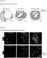
- the result of confirming left ventricle diameter (LV diameter) by echocardiography at 4 and 8 weeks after transplantation was shown in FIG. 5
- the result of performing significance evaluation between experimental samples by the student T-test by calculating left ventricle ejection fraction (LVEF) and fractional shortening (FS) which show myocardial functions was shown in FIG. 6 .
- the left ventricular internal diameter was significantly reduced compared with the experimental group (control group) which had not been subjected to any treatment after 4 and 8 weeks after transplantation of the three-dimensional construct according to example 1.
- the left ventricular systolic function was significantly increased in the CPC/MSC group compared to the CPC group, and this shows that a cardiac remodeling was inhibited by the construct.
- Masson's trichrome staining was carried out.
- the heart muscle (red) and the scar tissue of the myocardial infarction region (blue) were discriminated using the staining method, and the result was shown in FIG. 7 .
- the thickness of the inner wall of the left ventricle after 8 weeks of the three-dimensional construct transplantation prepared according to the present invention was kept thicker than the control group, and the formation of scar tissues of the myocardial infarction region was less.
- the formation of scar tissues was more effectively inhibited, and the thickness of the inner wall of the left ventricle was not thinned and was maintained.
- ⁇ -myosin heavy chain representing contractibility of cardiac muscle tissue and cluster of differentiation 31 (CD31) representing angiogenesis on the myocardial infarction region at which the three-dimensional construct prepared according to the present invention was transplanted was significantly increased, and in particular, when the three-dimensional construct (CPC/MSC printed) (example 1) formed by the first bioprinting composition and the second bioprinting composition was transplanted, the effect was more maximized.
- the three-dimensional construct prepared by the preparation method of the present invention can maintain the viability of cells for a long time by implementing a vascular network in the construct (that is, by implementing a microenvironment of the cardiac muscle tissue effectively), thereby significantly improving the cell transfer efficiency into the myocardium.
Applications Claiming Priority (2)
| Application Number | Priority Date | Filing Date | Title |
|---|---|---|---|
| KR1020150042418A KR102311639B1 (ko) | 2015-03-26 | 2015-03-26 | 심근조직 재생용 3차원 구조체의 제조방법 |
| PCT/KR2016/003079 WO2016153320A1 (ko) | 2015-03-26 | 2016-03-25 | 심근조직 재생용 3차원 구조체 및 이의 제조방법 |
Publications (2)
| Publication Number | Publication Date |
|---|---|
| EP3275471A1 true EP3275471A1 (de) | 2018-01-31 |
| EP3275471A4 EP3275471A4 (de) | 2018-12-05 |
Family
ID=56978759
Family Applications (1)
| Application Number | Title | Priority Date | Filing Date |
|---|---|---|---|
| EP16769138.5A Withdrawn EP3275471A4 (de) | 2015-03-26 | 2016-03-25 | Dreidimensionale struktur zur herzmuskelgeweberegeneration und herstellungsverfahren dafür |
Country Status (6)
| Country | Link |
|---|---|
| US (1) | US20180037870A1 (de) |
| EP (1) | EP3275471A4 (de) |
| JP (1) | JP6490869B2 (de) |
| KR (1) | KR102311639B1 (de) |
| CN (1) | CN107406828A (de) |
| WO (1) | WO2016153320A1 (de) |
Cited By (1)
| Publication number | Priority date | Publication date | Assignee | Title |
|---|---|---|---|---|
| GB2609532A (en) * | 2021-06-06 | 2023-02-08 | Copner Biotech Ltd | Additive manufacturing using low viscosity materials |
Families Citing this family (8)
| Publication number | Priority date | Publication date | Assignee | Title |
|---|---|---|---|---|
| KR102072665B1 (ko) * | 2017-11-28 | 2020-02-03 | 포항공과대학교 산학협력단 | 건조형 스캐폴드 및 건조형 스캐폴드 제조 방법 |
| PE20211587A1 (es) * | 2018-01-31 | 2021-08-18 | Rokit Healthcare Inc | Composicion de tinta biologica para lamina para la regeneracion de la dermis, metodo de fabricacion de lamina personalizada para regeneracion de la dermis usando la misma, y lamina personalizada para regeneracion de la dermis fabricada con el metodo de fabricacion |
| WO2019191111A1 (en) * | 2018-03-26 | 2019-10-03 | The Trustees Of The University Of Pennsylvania | Systems and methods for multilane vasculature |
| US11197945B2 (en) | 2018-09-14 | 2021-12-14 | BioSapien Inc. | Biovessels for use in tissue engineering |
| KR102306236B1 (ko) * | 2019-03-15 | 2021-09-29 | 가톨릭대학교 산학협력단 | 유도만능줄기세포 유래 세포치료제의 치료 효율 및 성능 향상을 위한 중간엽 줄기세포 기반 패치 적용 기술 |
| US20230119663A1 (en) * | 2020-03-26 | 2023-04-20 | Osaka University | Muscle tissue produced by bioprinting |
| CN113274555B (zh) * | 2021-05-31 | 2022-05-03 | 清华大学 | 一种具有仿生螺旋取向化微结构的人工心室及其制备方法 |
| CN114949359B (zh) * | 2022-06-27 | 2024-01-23 | 西安臻研生物科技有限公司 | 一种脱细胞基质微粒填充剂及其制备方法 |
Family Cites Families (10)
| Publication number | Priority date | Publication date | Assignee | Title |
|---|---|---|---|---|
| KR101109125B1 (ko) * | 2009-03-24 | 2012-02-15 | 한국과학기술연구원 | 줄기세포를 혈관세포로 분화시키는 방법 및 이를 이용한 생체 내 혈관신생 유도 |
| KR101135709B1 (ko) | 2009-04-16 | 2012-04-13 | 서울대학교산학협력단 | 탈세포화된 세포외 기질을 이용한 줄기세포 이식용 고분자 지지체의 표면 개질 방법 |
| CN103260606A (zh) * | 2010-08-24 | 2013-08-21 | 加利福尼亚大学董事会 | 用于心脏治疗的组合物和方法 |
| US10233420B2 (en) * | 2010-09-01 | 2019-03-19 | Regents Of The University Of Minnesota | Methods of recellularizing a tissue or organ for improved transplantability |
| EP3620185A1 (de) * | 2010-10-06 | 2020-03-11 | President and Fellows of Harvard College | Injizierbare porenbildende hydrogele für materialbasierte zelltherapien |
| CA2848043C (en) * | 2011-09-12 | 2022-07-12 | Organovo, Inc. | Engineered tissues for in vitro research uses, arrays thereof, and methods of making the same |
| US20140099709A1 (en) * | 2012-06-19 | 2014-04-10 | Organovo, Inc. | Engineered three-dimensional connective tissue constructs and methods of making the same |
| KR20220093401A (ko) | 2012-09-04 | 2022-07-05 | 셀룰래리티 인코포레이티드 | 조직 생성 방법 |
| DK3028042T3 (da) * | 2013-07-31 | 2021-10-04 | Organovo Inc | Automatiserede anordninger, systemer og fremgangsmåder til fremstilling af væv |
| KR20160115204A (ko) * | 2015-03-26 | 2016-10-06 | 포항공과대학교 산학협력단 | 3차원 프린팅용 조성물, 이의 제조방법, 및 이를 사용한 3차원 구조체의 제조방법 |
-
2015
- 2015-03-26 KR KR1020150042418A patent/KR102311639B1/ko active IP Right Grant
-
2016
- 2016-03-25 WO PCT/KR2016/003079 patent/WO2016153320A1/ko active Application Filing
- 2016-03-25 EP EP16769138.5A patent/EP3275471A4/de not_active Withdrawn
- 2016-03-25 US US15/556,386 patent/US20180037870A1/en not_active Abandoned
- 2016-03-25 JP JP2018501841A patent/JP6490869B2/ja active Active
- 2016-03-25 CN CN201680018417.8A patent/CN107406828A/zh active Pending
Cited By (2)
| Publication number | Priority date | Publication date | Assignee | Title |
|---|---|---|---|---|
| GB2609532A (en) * | 2021-06-06 | 2023-02-08 | Copner Biotech Ltd | Additive manufacturing using low viscosity materials |
| GB2609532B (en) * | 2021-06-06 | 2023-10-25 | Copner Biotech Ltd | Additive manufacturing using low viscosity biomaterials |
Also Published As
| Publication number | Publication date |
|---|---|
| EP3275471A4 (de) | 2018-12-05 |
| WO2016153320A1 (ko) | 2016-09-29 |
| JP6490869B2 (ja) | 2019-03-27 |
| KR20160115208A (ko) | 2016-10-06 |
| US20180037870A1 (en) | 2018-02-08 |
| JP2018515129A (ja) | 2018-06-14 |
| KR102311639B1 (ko) | 2021-10-12 |
| CN107406828A (zh) | 2017-11-28 |
Similar Documents
| Publication | Publication Date | Title |
|---|---|---|
| EP3275471A1 (de) | Dreidimensionale struktur zur herzmuskelgeweberegeneration und herstellungsverfahren dafür | |
| JP4943844B2 (ja) | 三次元組織構造体 | |
| JP4709393B2 (ja) | 三次元的間質組織 | |
| Fujimoto et al. | An elastic, biodegradable cardiac patch induces contractile smooth muscle and improves cardiac remodeling and function in subacute myocardial infarction | |
| JP2024015176A (ja) | 臓器および組織を脱細胞化および再細胞化する方法 | |
| US20220306992A1 (en) | Cellular seeding and co-culture of a three dimensional fibroblast construct | |
| RU2470611C2 (ru) | Окклюдер для чрезкожной транслюминальной процедуры (варианты), способ чрезкожного транслюминального закрытия отверстия в сердце, способ активизации васкуляризации ткани млекопитающего in vivo и способ активизации заживления места анастомоза | |
| US11331348B2 (en) | Compositions comprising extracellular matrix of primitive animal species and related methods | |
| Liao et al. | Tissue engineering to repair diaphragmatic defect in a rat model | |
| JP2005278711A (ja) | ハニカムフィルムを用いた機能的人工組織の生産 | |
| US20230174942A1 (en) | Novel fabrication of coronary based decellularized heart flaps to treat aneurysm following myocardial infarction | |
| US20230020862A1 (en) | Proangiogenic protein cocktails delivered in custom biomaterials to revascularize ischemic tissue | |
| Akbarzadeh et al. | Coronary-Based Right Heart Flap Recellularization by Rat Neonatal Whole Cardiac Cells: A Viable Sheep Cardiac Patch Model for Possible Management of Heart Aneurysm | |
| US20220323510A1 (en) | Compositions Comprising Extracellular Matrix of Primitive Animal Species and Related Methods | |
| Grikscheit | Tissue engineering of the gastrointestinal tract for surgical replacement: a nutrition tool of the future? | |
| US20190321518A1 (en) | Flexible Tissue Regeneration Implant | |
| Morgan | The effect of biomimetic electromechanical stimulation on the development and function of engineered myocardial tissue |
Legal Events
| Date | Code | Title | Description |
|---|---|---|---|
| STAA | Information on the status of an ep patent application or granted ep patent |
Free format text: STATUS: THE INTERNATIONAL PUBLICATION HAS BEEN MADE |
|
| PUAI | Public reference made under article 153(3) epc to a published international application that has entered the european phase |
Free format text: ORIGINAL CODE: 0009012 |
|
| STAA | Information on the status of an ep patent application or granted ep patent |
Free format text: STATUS: REQUEST FOR EXAMINATION WAS MADE |
|
| 17P | Request for examination filed |
Effective date: 20171026 |
|
| AK | Designated contracting states |
Kind code of ref document: A1 Designated state(s): AL AT BE BG CH CY CZ DE DK EE ES FI FR GB GR HR HU IE IS IT LI LT LU LV MC MK MT NL NO PL PT RO RS SE SI SK SM TR |
|
| AX | Request for extension of the european patent |
Extension state: BA ME |
|
| DAV | Request for validation of the european patent (deleted) | ||
| DAX | Request for extension of the european patent (deleted) | ||
| A4 | Supplementary search report drawn up and despatched |
Effective date: 20181030 |
|
| RIC1 | Information provided on ipc code assigned before grant |
Ipc: B33Y 70/00 20150101ALI20181024BHEP Ipc: C07K 14/475 20060101ALI20181024BHEP Ipc: B33Y 10/00 20150101ALI20181024BHEP Ipc: A61L 27/36 20060101AFI20181024BHEP Ipc: B33Y 80/00 20150101ALI20181024BHEP Ipc: A61L 27/38 20060101ALI20181024BHEP Ipc: C12N 5/077 20100101ALI20181024BHEP Ipc: C12N 5/071 20100101ALI20181024BHEP Ipc: A61F 2/24 20060101ALI20181024BHEP |
|
| STAA | Information on the status of an ep patent application or granted ep patent |
Free format text: STATUS: THE APPLICATION IS DEEMED TO BE WITHDRAWN |
|
| 18D | Application deemed to be withdrawn |
Effective date: 20190528 |