EP3242947B1 - Gene therapy and electroporation for the treatment of malignancies - Google Patents
Gene therapy and electroporation for the treatment of malignancies Download PDFInfo
- Publication number
- EP3242947B1 EP3242947B1 EP16735524.7A EP16735524A EP3242947B1 EP 3242947 B1 EP3242947 B1 EP 3242947B1 EP 16735524 A EP16735524 A EP 16735524A EP 3242947 B1 EP3242947 B1 EP 3242947B1
- Authority
- EP
- European Patent Office
- Prior art keywords
- tumor
- cells
- plasmid
- electroporation
- gitrl
- Prior art date
- Legal status (The legal status is an assumption and is not a legal conclusion. Google has not performed a legal analysis and makes no representation as to the accuracy of the status listed.)
- Active
Links
Images

Classifications
-
- C—CHEMISTRY; METALLURGY
- C07—ORGANIC CHEMISTRY
- C07K—PEPTIDES
- C07K14/00—Peptides having more than 20 amino acids; Gastrins; Somatostatins; Melanotropins; Derivatives thereof
- C07K14/435—Peptides having more than 20 amino acids; Gastrins; Somatostatins; Melanotropins; Derivatives thereof from animals; from humans
- C07K14/52—Cytokines; Lymphokines; Interferons
- C07K14/525—Tumour necrosis factor [TNF]
-
- A—HUMAN NECESSITIES
- A61—MEDICAL OR VETERINARY SCIENCE; HYGIENE
- A61K—PREPARATIONS FOR MEDICAL, DENTAL OR TOILETRY PURPOSES
- A61K38/00—Medicinal preparations containing peptides
- A61K38/16—Peptides having more than 20 amino acids; Gastrins; Somatostatins; Melanotropins; Derivatives thereof
- A61K38/17—Peptides having more than 20 amino acids; Gastrins; Somatostatins; Melanotropins; Derivatives thereof from animals; from humans
- A61K38/177—Receptors; Cell surface antigens; Cell surface determinants
-
- A—HUMAN NECESSITIES
- A61—MEDICAL OR VETERINARY SCIENCE; HYGIENE
- A61K—PREPARATIONS FOR MEDICAL, DENTAL OR TOILETRY PURPOSES
- A61K38/00—Medicinal preparations containing peptides
- A61K38/16—Peptides having more than 20 amino acids; Gastrins; Somatostatins; Melanotropins; Derivatives thereof
- A61K38/17—Peptides having more than 20 amino acids; Gastrins; Somatostatins; Melanotropins; Derivatives thereof from animals; from humans
- A61K38/19—Cytokines; Lymphokines; Interferons
- A61K38/191—Tumor necrosis factors [TNF], e.g. lymphotoxin [LT], i.e. TNF-beta
-
- A—HUMAN NECESSITIES
- A61—MEDICAL OR VETERINARY SCIENCE; HYGIENE
- A61K—PREPARATIONS FOR MEDICAL, DENTAL OR TOILETRY PURPOSES
- A61K38/00—Medicinal preparations containing peptides
- A61K38/16—Peptides having more than 20 amino acids; Gastrins; Somatostatins; Melanotropins; Derivatives thereof
- A61K38/17—Peptides having more than 20 amino acids; Gastrins; Somatostatins; Melanotropins; Derivatives thereof from animals; from humans
- A61K38/19—Cytokines; Lymphokines; Interferons
- A61K38/20—Interleukins [IL]
- A61K38/208—IL-12
-
- A—HUMAN NECESSITIES
- A61—MEDICAL OR VETERINARY SCIENCE; HYGIENE
- A61K—PREPARATIONS FOR MEDICAL, DENTAL OR TOILETRY PURPOSES
- A61K38/00—Medicinal preparations containing peptides
- A61K38/16—Peptides having more than 20 amino acids; Gastrins; Somatostatins; Melanotropins; Derivatives thereof
- A61K38/17—Peptides having more than 20 amino acids; Gastrins; Somatostatins; Melanotropins; Derivatives thereof from animals; from humans
- A61K38/19—Cytokines; Lymphokines; Interferons
- A61K38/20—Interleukins [IL]
- A61K38/2086—IL-13 to IL-16
-
- A—HUMAN NECESSITIES
- A61—MEDICAL OR VETERINARY SCIENCE; HYGIENE
- A61K—PREPARATIONS FOR MEDICAL, DENTAL OR TOILETRY PURPOSES
- A61K48/00—Medicinal preparations containing genetic material which is inserted into cells of the living body to treat genetic diseases; Gene therapy
- A61K48/0075—Medicinal preparations containing genetic material which is inserted into cells of the living body to treat genetic diseases; Gene therapy characterised by an aspect of the delivery route, e.g. oral, subcutaneous
-
- A—HUMAN NECESSITIES
- A61—MEDICAL OR VETERINARY SCIENCE; HYGIENE
- A61N—ELECTROTHERAPY; MAGNETOTHERAPY; RADIATION THERAPY; ULTRASOUND THERAPY
- A61N1/00—Electrotherapy; Circuits therefor
- A61N1/18—Applying electric currents by contact electrodes
- A61N1/20—Applying electric currents by contact electrodes continuous direct currents
-
- A—HUMAN NECESSITIES
- A61—MEDICAL OR VETERINARY SCIENCE; HYGIENE
- A61N—ELECTROTHERAPY; MAGNETOTHERAPY; RADIATION THERAPY; ULTRASOUND THERAPY
- A61N1/00—Electrotherapy; Circuits therefor
- A61N1/18—Applying electric currents by contact electrodes
- A61N1/32—Applying electric currents by contact electrodes alternating or intermittent currents
- A61N1/327—Applying electric currents by contact electrodes alternating or intermittent currents for enhancing the absorption properties of tissue, e.g. by electroporation
-
- A—HUMAN NECESSITIES
- A61—MEDICAL OR VETERINARY SCIENCE; HYGIENE
- A61P—SPECIFIC THERAPEUTIC ACTIVITY OF CHEMICAL COMPOUNDS OR MEDICINAL PREPARATIONS
- A61P35/00—Antineoplastic agents
-
- A—HUMAN NECESSITIES
- A61—MEDICAL OR VETERINARY SCIENCE; HYGIENE
- A61K—PREPARATIONS FOR MEDICAL, DENTAL OR TOILETRY PURPOSES
- A61K38/00—Medicinal preparations containing peptides
- A61K38/16—Peptides having more than 20 amino acids; Gastrins; Somatostatins; Melanotropins; Derivatives thereof
- A61K38/17—Peptides having more than 20 amino acids; Gastrins; Somatostatins; Melanotropins; Derivatives thereof from animals; from humans
- A61K38/177—Receptors; Cell surface antigens; Cell surface determinants
- A61K38/1793—Receptors; Cell surface antigens; Cell surface determinants for cytokines; for lymphokines; for interferons
-
- A—HUMAN NECESSITIES
- A61—MEDICAL OR VETERINARY SCIENCE; HYGIENE
- A61K—PREPARATIONS FOR MEDICAL, DENTAL OR TOILETRY PURPOSES
- A61K48/00—Medicinal preparations containing genetic material which is inserted into cells of the living body to treat genetic diseases; Gene therapy
-
- C—CHEMISTRY; METALLURGY
- C07—ORGANIC CHEMISTRY
- C07K—PEPTIDES
- C07K2319/00—Fusion polypeptide
-
- C—CHEMISTRY; METALLURGY
- C07—ORGANIC CHEMISTRY
- C07K—PEPTIDES
- C07K2319/00—Fusion polypeptide
- C07K2319/01—Fusion polypeptide containing a localisation/targetting motif
- C07K2319/03—Fusion polypeptide containing a localisation/targetting motif containing a transmembrane segment
-
- C—CHEMISTRY; METALLURGY
- C07—ORGANIC CHEMISTRY
- C07K—PEPTIDES
- C07K2319/00—Fusion polypeptide
- C07K2319/20—Fusion polypeptide containing a tag with affinity for a non-protein ligand
- C07K2319/21—Fusion polypeptide containing a tag with affinity for a non-protein ligand containing a His-tag
-
- C—CHEMISTRY; METALLURGY
- C07—ORGANIC CHEMISTRY
- C07K—PEPTIDES
- C07K2319/00—Fusion polypeptide
- C07K2319/20—Fusion polypeptide containing a tag with affinity for a non-protein ligand
- C07K2319/22—Fusion polypeptide containing a tag with affinity for a non-protein ligand containing a Strep-tag
-
- C—CHEMISTRY; METALLURGY
- C07—ORGANIC CHEMISTRY
- C07K—PEPTIDES
- C07K2319/00—Fusion polypeptide
- C07K2319/30—Non-immunoglobulin-derived peptide or protein having an immunoglobulin constant or Fc region, or a fragment thereof, attached thereto
-
- C—CHEMISTRY; METALLURGY
- C12—BIOCHEMISTRY; BEER; SPIRITS; WINE; VINEGAR; MICROBIOLOGY; ENZYMOLOGY; MUTATION OR GENETIC ENGINEERING
- C12N—MICROORGANISMS OR ENZYMES; COMPOSITIONS THEREOF; PROPAGATING, PRESERVING, OR MAINTAINING MICROORGANISMS; MUTATION OR GENETIC ENGINEERING; CULTURE MEDIA
- C12N2800/00—Nucleic acids vectors
- C12N2800/10—Plasmid DNA
- C12N2800/106—Plasmid DNA for vertebrates
- C12N2800/107—Plasmid DNA for vertebrates for mammalian
Definitions
- the present invention provides for the intratumoral delivery of immunomodulators.
- it provides delivery of co-stimulatory molecules by intratumoral electroporation.
- Immunotherapy has recently drawn attention as a fourth method following surgery, chemotherapy and radiation therapy for treating tumors. Since immunotherapy utilizes the immunity inherent to humans, it is said that the physical burden on patients are less in immunotherapy than those in other therapies.
- the therapeutic approaches known as immunotherapies include: cell transfer therapy in which cells such as lymphokine-activated cells, natural killer T-cells or ⁇ T cells obtained, for example, from exogenously-induced cytotoxic T-lymphocytes (CTLs) or peripheral blood lymphocytes by expansion culture using various method are transferred; dendritic cell-transfer therapy or peptide vaccine therapy by which in vivo induction of antigen-specific CTLs is expected; Th1 cell therapy; and immune gene therapy in which genes expected to have various effects are introduced ex vivo into the above-mentioned cells to transfer them in vivo.
- CTLs exogenously-induced cytotoxic T-lymphocytes
- Th1 cell therapy Th1 cell therapy
- immune gene therapy in which genes expected to have various effects are introduced ex
- CD8-positive T cells are major effector cells that are capable of directly destroying tumor cells in vivo and in vitro. These cells are strictly specific to antigen peptides presented by MHC Class 1 molecules. In contrast, antigen specificities of NKT cells are not so strict, and they are considered to be effector cells that show intrinsic immune responses.
- CD4-positive T cells are considered to have a fundamental role to regulate anti-tumor immune responses through a plurality of mechanisms although they do not destroy tumors directly.
- CD4-positive T cells that have recognized a tumor-antigen peptide represented by MHC Class II molecules promote the activation and proliferation of CTL through the interaction with antigen-presenting cells (APCs).
- APCs antigen-presenting cells
- CD25-positive/CD4-positive cells have been shown to inhibit the anti-tumor immune responses and progression of various autoimmune diseases (see US 2003049696 and S. Sakaguchi et al., Immunol. Rev. 182 (2001), pp 18-32 ).
- regulatory T cells suppress the activity of cytotoxic CD8-positive T cells through the control of the helper function by targeting CD4-positive T cells, some tumors are considered to utilize this system for their proliferation, thereby avoiding attack of the immune system.
- GITR which has been found as a gene expressed in regulatory T cells ("Tregs”; see S. Sakaguchi et al., Immunol. Rev. 182 (2001), pp 18-32 ), is a cell surface transmembrane protein receptor and a member of the tumor necrosis factor receptor (TNFR) superfamily. GITR has been shown to be constitutively present on Tregs and activated T cells. GITR binds to another transmembrane protein referred to as GITR ligand (hereinafter referred to as "GITR-L").
- In vivo electroporation is a gene delivery technique that has been used successfully for efficient delivery of plasmid DNA to many different tissues. Studies have reported the administration of in vivo electroporation for delivery of plasmid DNA to B16 melanomas and other tumor tissues. Systemic and local expression of a gene or cDNA encoded by a plasmid can be obtained with administration of in vivo electroporation. Use of in vivo electroporation enhances plasmid DNA uptake in tumor tissue, resulting in expression within the tumor, and delivers plasmids to muscle tissue, resulting in systemic expression of secreted proteins, such as cytokines (see, e.g., US8026223 ).
- electroporation can be used to transfect cells in vivo with plasmid DNA. Recent studies have shown that electroporation is capable of enhancing delivery of plasmid DNA as an antitumor agent. Electroporation has been administered for treatment of hepatocellular carcinomas, adenocarcinoma, breast tumors, squamous cell carcinoma and B16.F10 melanoma in rodent models. The B16.F10, CT-26, and MC-38 murine syngeneic tumor models has been used extensively for testing potential immunotherapy protocols for the delivery of an immudulatory molecule including cytokines either as recombinant protein or by gene therapy.
- protocols known in the art can be utilized for the delivery of plasmid encoding a co-stimulatory agonist utilizing in vivo electroporation for the treatment of cancer.
- the protocols known in the art describe in vivo electroporation mediated cytokine based gene therapy, both intratumor and intramuscular, utilizing low-voltage and long-pulse currents.
- US 8,802,643 describes a method of treating cancerous tumors including injecting an effective dose of a plasmid encoded for IL-12, B7-1 or IL-15 into a cancerous tumor and subsequently administering at least one high voltage, short duration pulse to the tumor. It was found that the intratumor treatments using electroporation not only resulted in tumor regression but also induced an immune memory response which prevented the formation of new tumors. Stone Geoffrey William et al., The Faseb Journal, Federation of American Societies for Experimental Biology, US, vol. 19, no. 5, Suppl. S, Part 2, 28 February 2005, pages A1403-1404 describes treatment of established murine tumors by peritumoral injections of plasmid DNA for soluble, multimeric CD40 ligand and GITR ligand.
- the present invention provides a plasmid for use in a method for the treatment of malignancies, wherein the administration of the plasmid encoding for a therapeutic co-stimulatory protein and a nucleic acid encoding at least one immunostimulatory cytokine, in combination with electroporation has a therapeutic effect on primary tumors as well as distant tumors and metastases.
- the present invention provides the plasmid for use in a method of treating a subject having a cancerous tumor, the method comprising: injecting the cancerous tumor with an effective dose of the plasmid coding for a therapeutic protein and a nucleic acid encoding at least one immunostimulatory cytokine; and administering electroporation therapy to the tumor, the electroporation therapy further comprising the administration of at least one voltage pulse of 200 V/cm to 1500 V/cm over a pulse width of 100 microseconds to 20 milliseconds.
- the cancerous tumor is melanoma.
- the plasmid coding for the therapeutic protein is a plasmid coding for a soluble form of a co-stimulatory molecule, and the co-stimulatory molecule is selected from GITR, CD137, CD134, CD40L, and CD27 agonists.
- the plasmid further encodes the at least one immunostimulatory cytokine, which can be selected from IL-12, IL-15, and a combination of IL-12 and IL-15.
- the voltage pulse delivered to the tumor is from 200V/cm to 1500V/cm.
- the present invention provides the plasmid for use in a method of treating a subject having a cancerous tumor, the method comprising: administering a first treatment at time T1, wherein the first treatment further comprises injecting the cancerous tumor with a first effective dose of the plasmid coding for the therapeutic protein and administering a first electroporation therapy to the tumor at time T1, the first electroporation therapy further comprising the administration of at least one voltage pulse having a duration of 100 microseconds to 20 milliseconds; and administering a second treatment at time T2, wherein time T2 is a time later than time T1, wherein the second treatment further comprises injecting the cancerous tumor with a second effective dose of the plasmid coding for the therapeutic protein and administering a second electroporation therapy to the tumor at time T2, the second electroporation therapy further comprising the administration of at least one voltage pulse having a duration of 100 microseconds to 20 milliseconds.
- the cancerous tumor is melanoma.
- the therapeutic protein is a co-stimulatory molecule selected from a soluble form of GITR-L, CD137, CD134, CD40L, and CD27 agonists.
- the plasmid further encodes the at least one immunostimulatory cytokine, chosen from IL-12, IL-15, and a combination of IL-12 and IL-15.
- the voltage pulse delivered to the tumor is from 200V/cm to 1500V/cm.
- a third treatment is added at time T3, wherein time T3 is a time later than time T2, wherein the third treatment further comprises injecting the cancerous tumor with a third effective dose of the plasmid coding for the therapeutic protein and administering a third electroporation therapy to the tumor, the third electroporation therapy further comprising the administration of at least one voltage pulse having a duration of 100 microseconds to 20 milliseconds.
- Certain embodiments involve injecting an effective dose of the plasmid encoding for a therapeutic protein into the cancerous tumor of the subject; and administering electroporation to the subject intratumorally using at least one low voltage pulse having a pulse width of 100 microseconds to 20 milliseconds and the voltage pulse delivered to the tumor is from 200V/cm to 1500V/cm.
- the present invention provides the plasmid for use in a method of treating a subject having a cancerous tumor, the method comprising: administering a first treatment at time T1, wherein the first treatment further comprises injecting the cancerous tumor with a first effective dose of the plasmid coding for the therapeutic protein and administering a first electroporation therapy to the tumor at time T1, the first electroporation therapy further comprising the administration of at least one voltage pulse having a duration of 100 microseconds to 0 milliseconds; administering a second treatment at time T2, wherein time T2 is a time later than time T1, wherein the second treatment further comprises injecting the cancerous tumor with a second effective dose of the plasmid coding for the therapeutic protein and administering a second electroporation therapy to the tumor at time T2, the second electroporation therapy further comprising the administration of at least one voltage pulse having a duration of 100 microseconds to 20 milliseconds; and administering a third treatment at time T3, wherein time T3 is a time later
- the cancerous tumor is melanoma.
- the therapeutic protein is a co-stimulatory molecule, selected from a soluble form GITR-L, CD137, CD134, CD40L, and CD27 agonists.
- the plasmid encodes at least one immunostimulatory cytokine, selected from IL-12, IL-15, and a combination of IL-12 and IL-15.
- the voltage pulse delivered to the tumor is from 200V/cm to 1500V/cm.
- co-stimulatory molecule refers to a group of immune cell surface receptor/ligands which engage between T cells and antigen presenting cells and generate a stimulatory signal in T cells which combines with the stimulatory signal (i.e., "co-stimulation") in T cells that results from T cell receptor (“TCR”) recognition of antigen on antigen presenting cells.
- Co-stimulatory molecules include, but are not limited to, agonists of GITR, CD137, CD134, CD40L, CD27, and the like.
- co-stimulator of T cells activation refers to the ability of a co-stimulatory ligand to bind and to activate T cells that have been activated via TCR. Co-stimulatory activation can be measured for T cells by the production of cytokines as is well known and by proliferation assays such as are well known and described in the examples. The soluble form of a co-stimulatory molecule which is biologically active also may be tested for binding to the cognate receptor on activated T cells.
- a soluble form of a co-stimulatory molecule "derived from an antigen presenting cell” refers to a co-stimulatory molecule normally expressed by B cells, macrophages, monocytes, dendritic cells and other such antigen presenting cells, which has been engineered as described herein to render it soluble.
- Preferred soluble co-stimulatory molecules derived from an antigen presenting cell include any of GITR-L, CD137-L, CD134-L (a.k.a. OX40-L), CD40, CD28.
- the soluble form of a co-stimulatory molecule derived from an antigen presenting cell retains the ability of the native co-stimulatory molecule to bind to its cognate receptor/ligand on T cells and stimulate T cell activation.
- co-stimulatory molecule agonist includes soluble co-stimulatory molecules and agonists of co-stimulatory molecule binding partners.
- the binding partner of GITR-L is GITR.
- Agonists of GITR can include agonistic GITR antibodies, GITR-L polypeptides, including multimeric soluble and transmembrane forms, GITRL mimetics, or other molecules that engage and induce biological activity of GITR.
- GITR tumor necrosis factor receptor
- CD137-L or "agonist of CD137” (a.k.a. 4-1 BB ligand or TNFL9) refers to a specific molecule associated with this name and any other molecules that have biological function as co-stimulatory molecules that share at least 80% amino acid sequence identity, preferably at least 90% sequence identity, more preferably at least 95% sequence identity and even more preferably at least 98% sequence identity with human CD137-L as defined in Swiss Prot Id. no. P41273.
- Human CD137-L is a type II membrane protein that contains 254 amino acids (no signal sequence) (see sequence in Swiss Prot Id no. P41273). The protein contains a cytoplasmic domain at residues 1-28, a transmembrane domain at resides 29-49 and an extracellular domain at residues 50-254.
- the nucleotide sequence of CD137-L (1645 bp) is available in public databases (see Genbank accession no. NM 003811). CD137-L is described by Alderson et al. (1994) Eur J. Immunol. 24(9):2219-27 .
- CD137-L is expressed on antigen presenting cells including B cells, monocytes, and splenic dendritic cells and T lymphocytes. CD137-L interacts with CD137 on activated T cells.
- CD134-L or "agonist of CD134” (a.k.a. OX40 ligand or TNRSF4) refers to a specific molecule associated with this name and any other molecules that have biological function as co-stimulatory molecules that share at least 80% amino acid sequence identity, preferably at least 90% sequence identity, more preferably at least 95% sequence identity and even more preferably at least 98% sequence identity with CD134-L, as defined in Swiss Prot Id. no. P23510.
- Human CD134-L is a type II membrane protein that contains 183 amino acids (no signal sequence). The protein contains a cytoplasmic domain at residues 1-23, a transmembrane domain at residues 24-50 and an extracellular domain at residues 51-183.
- the nucleotide sequence of CD134-L (3510 bp, with the coding sequence being 157-708) is available in public databases (see. Genbank accession no. NM_003326.2).
- CD134-L is described by Godfry et al., J Exp Med. Aug. 1, 1994; 180(2):757-62 .
- CD134-L is expressed by dendritic cells and other APC and binds to CD134, which is present on activated T cells.
- CD40 refers to a specific molecule associated with this name and any other molecules that have biological function as co-stimulatory molecules that share at least 80% amino acid sequence identity, preferably at least 90% sequence identity, more preferably at least 95% sequence identity and even more preferably at least 98% sequence identity with CD40 as defined in Swiss Prot Id. no. P25942).
- the sequence of human CD40 contains 277 amino acids of which 20 amino acids at the N terminus represent the signal sequence (see sequence in Swiss Prot Id no. P25942). A transmembrane domain is located at resides 194-215 and the cytoplasmic domain is located at residues 216-277.
- the nucleotide sequence of CD40 (1177 bp) is available in public databases (see Genbank accession no. NM_001250). CD40 and various isoforms are described by Tone et al. Proc. Natl. Acad. Sci. U.S.A. 98 (4), 1751-1756 (2001 ). CD40 is expressed by monocytes and B cells binds to CD40-L (a.k.a. CD40 ligand or CD153) expressed by activated T cells.
- CD28 is a type I transmembrane glycoprotein and is a member of the Immunoglobulin family by virtue of its single Ig variable-like extracellular domain which has a MYPPPY (SEQ ID NO: 639) motif required for binding CD80 and CD86 ( Peach et al. (1994) J. Exp. Med. 180: 2049-2058 ).
- CD28 has a cysteine residue located after the Ig variable-like domain, which is involved in its homodimerization.
- the protein sequence of CD28 and a nucleic acid encoding a human CD28 are disclosed, for example, in Harper et al. J. Immunol. (1991) 147: 1037-44 .
- the sequence of a human mRNA encoding CD28 also is disclosed in NCBI Accession No. NM_006139, last updated Apr. 19, 2009, for example.
- the complete protein sequence of a human CD28 also is disclosed in NCBI Accession No. NP_006130.
- cancer includes a myriad of diseases generally characterized by inappropriate cellular proliferation, abnormal or excessive cellular proliferation.
- diseases include but are not limited to, breast cancer, colon cancer, prostate cancer, pancreatic cancer, melanoma, lung cancer, ovarian cancer, kidney cancer, brain cancer, or sarcomas.
- Such cancers may be caused by, chromosomal abnormalities, degenerative growth and developmental disorders, mitogenic agents, ultraviolet radiation (UV), viral infections, inappropriate tissue expression of a gene, alterations in expression of a gene, or carcinogenic agents.
- UV ultraviolet radiation
- treatment includes, but is not limited to, inhibition or reduction of proliferation of cancer cells, destruction of cancer cells, prevention of proliferation of cancer cells or prevention of initiation of malignant cells or arrest or reversal of the progression of transformed premalignant cells to malignant disease or amelioration of the disease.
- subject refers to any animal, preferably a mammal such as a human. Veterinary uses are also intended to be encompassed by this invention.
- electro-kinetic enhancement refers to the use of a transmembrane electric field pulse to induce microscopic pathways (pores) in a bio-membrane; their presence allows biomolecules such as plasmids, oligonucleotides, siRNA, drugs, ions, and water to pass from one side of the cellular membrane to the other.
- biomolecule encompasses plasmid encoded antibodies, antibody fragments, full length immunomodulatory proteins, soluble domains of membrane anchored molecules, fusion proteins, and the like.
- the present invention provides an immunotherapeutic approach for reducing the size of a tumor or inhibiting the growth of cancer cells in an individual, or reducing or inhibiting the development of metastatic cancer in an individual suffering from cancer.
- Therapy is achieved by intratumoral delivery of plasmids encoding various soluble forms of co-stimulatory molecules, or agonists thereof, using electroporation.
- Co-stimulatory agonists may be in the form of antibodies or antibody fragments, both of which can be encoded in a plasmid and delivered to the tumor by electroporation.
- antibody as used herein includes immunoglobulins, which are the product of B cells and variants thereof as well as the T cell receptor (TcR), which is the product of T cells, and variants thereof.
- An immunoglobulin is a protein comprising one or more polypeptides substantially encoded by the immunoglobulin kappa and lambda, alpha, gamma, delta, epsilon and mu constant region genes, as well as myriad immunoglobulin variable region genes. Light chains are classified as either kappa or lambda.
- Heavy chains are classified as gamma, mu, alpha, delta, or epsilon, which in turn define the immunoglobulin classes, IgG, IgM, IgA, IgD and IgE, respectively. Also subclasses of the heavy chain are known. For example, IgG heavy chains in humans can be any of IgG1, IgG2, IgG3 and IgG4 subclass.
- a typical immunoglobulin structural unit is known to comprise a tetramer.
- Each tetramer is composed of two identical pairs of polypeptide chains, each pair having one "light” (about 25 kD) and one "heavy” chain (about 50-70 kD).
- the N-terminus of each chain defines a variable region of about 100 to 110 or more amino acids primarily responsible for antigen recognition.
- the terms variable light chain (VL) and variable heavy chain (VH) refer to these light and heavy chains, respectively.
- Antibodies exist as full-length intact antibodies or as a number of well-characterized fragments produced by digestion with various peptidases or chemicals.
- pepsin digests an antibody below the disulfide linkages in the hinge region to produce F(ab')2, a dimer of Fab which itself is a light chain joined to VH-CH 1 by a disulfide bond.
- the F(ab')2 may be reduced under mild conditions to break the disulfide linkage in the hinge region thereby converting the F(ab')2 dimer into an Fab' monomer.
- the Fab' monomer is essentially a Fab fragment with the hinge region (see, Fundamental Immunology, W. E. Paul, ed., Raven Press, N.Y.
- a Fab fragment and Fc fragment are generated by digesting IgG with papain. Papain cleaves in the hinge region just above the residues involved in interchain S-S bonding, resulting in monovalent Fab fragments and the Fc fragment, which includes two constant region fragments, each containing the lower part of the hinge, CH2 and CH3 domains. The constant region fragments of the Fc are stabilized as a dimer though interchain S-S bonding of the lower residues of the hinge region.
- Immunoglobulin "Fc” classically refers to the portion of the constant region generated by digestion with papain. Includes the lower hinge which has the interchain S-S bonds.
- the term “Fc” as used herein refers to a dimeric protein comprising a pair of immunoglobulin constant region polypeptides, each containing the lower part of the hinge, CH2 and CH3 domain. Such "Fc" fragment may or may not contain S-S interchain bridging in the hinge region. It should be understood that an Fc may be from any Ig class and, as such, may include a CH4 domain such as in the case of IgM. Mutant sequences of an Fc are known such as described by Wines et al., J. Immunol. 2000 May 15; 164(10):5313-8 and may be used herein.
- antibody fragments are defined in terms of the digestion of an intact antibody, one of skill will appreciate that any of a variety of antibody fragments may be synthesized de novo either chemically or by utilizing recombinant DNA methodology.
- antibody as used herein also includes antibody fragments either produced by the modification of whole antibodies or synthesized de novo or antibodies and fragments obtained by using recombinant DNA methodologies.
- Recombinant antibodies may be conventional full length antibodies, antibody fragments known from proteolytic digestion, unique antibody fragments such as Fv or single chain Fv (scFv), domain deleted antibodies, and the like. Fragments may include domains or polypeptides with as little as one or a few amino acid deleted or mutated while more extensive deletion is possible such as deletion of one or more domains.
- An Fv antibody is about 50 Kd in size and comprises the variable regions of the light and heavy chain.
- a single chain Fv (“scFv”) polypeptide is a covalently linked VH::VL heterodimer which may be expressed from a nucleic acid including VH- and VL-encoding sequences either joined directly or joined by a peptide-encoding linker. See e.g., Huston, et al. (1988) Proc. Nat. Acad. Sci. USA, 85:5879-5883 .
- a number of structures for converting the naturally aggregated, but chemically separated light and heavy polypeptide chains from an antibody V region into an scFv molecule which will fold into a three dimensional structure substantially similar to the structure of an antigen-binding site.
- Agonists of co-stimulatory molecules may be soluble molecules such as soluble GITR-L, which comprises at least the extracellular domain (ECD) of GITR-L.
- Other co-stimulatory molecules will similarly lack transmembrane and intracellular domains, but are capable of binding to their binding partners and eliciting a biological effect.
- ECD's will be encoded in an expression vector and will be expressed when delivered to the tumor.
- the soluble encoded form of the co-stimulatory molecule may be linked in the expression vector to DNA encoding another protein or polypeptide.
- Such other polypeptide may be the Fc portion of an immunoglobulin, albumin, or any other type of serum protein or fragment thereof which maintains the solubility of the co-stimulatory molecule.
- the soluble form of the co-stimulatory molecule may be linked to an immunoglobulin via the heavy and/or light chain, which may be a fragment or a full length heavy or light chain.
- the immunoglobulin may be an antibody that can target an antigen associated with a cancer cell or tumor.
- the co-stimulatory molecule may also be expressed on the cell surface of cells, may have the addition of helerologous trimerization domains (e.g. GCN4) or may have point mutations in receptor binding domain that render the ligand more potent (see, e.g., Chattopadhyay et al. (2007) Proc. Natl. Acad. Sci., 104:19452-19457 ).
- helerologous trimerization domains e.g. GCN4
- point mutations in receptor binding domain that render the ligand more potent see, e.g., Chattopadhyay et al. (2007) Proc. Natl. Acad. Sci., 104:19452-19457 .
- the co-stimulatory molecule agonist is delivered as a nucleic acid.
- Nucleic acid refers to a polynucleotide compound, which includes oligonucleotides, comprising nucleosides or nucleoside analogs that have nitrogenous heterocyclic bases or base analogs, covalently linked by standard phosphodiester bonds or other linkages.
- Nucleic acids can include RNA, DNA, chimeric DNA-RNA polymers, or analogs thereof.
- the DNA can be a plasmid expressing a particular co-stimulatory molecule agonist of interest.
- the DNA plasmid used for electroporation of encoded co-stimulatory molecules is one that includes an encoding sequence of a recombinant antigen that is capable of being expressed in a mammalian cell, upon said DNA plasmid entering after electroporation.
- the encoding sequence is a consensus co-stimulatory molecule agonist that elicits an immune response in the target tumor.
- the encoding sequence constructs are optimized for mammalian expression, which can include one or more of the following: including the addition of a Kozak sequence, codon optimization, and RNA optimization. These optimized encoding sequences can be subcloned into various commercially available vectors.
- intratumoral electroporation of DNA encoding co-stimulatory agonists can be administered with other therapeutic entities.
- Table 1 provides possible combinations. Administration of the combination therapies can be achieved by electroporation alone or a combination of electroporation and systemic delivery.
- Table 1: Combination therapies with co-stimulatory agonists Combination Proposed delivery methods Method in reference References *GITRL + CTLA4 agonist antibody ("Ab”) or ligand 1.
- EP of GITRL gene + cytotoxic agent to create local tumor antigen pool System delivery of agonist Ab + alphavirus replicon particles encoding melanoma antigen Avogadri et al.,_Cancer Immunol. Res. 2014 May;2(5):448-58 Vergati et al., 2010. J. Biomed. Biotechnol. 2010:Article ID 596432 2.
- EP of GITRL gene + system delivery of tumor vaccine i.e gp100 peptide vaccine for melanoma
- EP of GITRL + system delivery of drug GITRL + small molecule inhibitors i.e. Sunitiinib, Imatinib, Vemurafenib, Trastuzumab, Bevacizumab, Cetuximb, rapamycin, Bortezom ib, PI3K-AKT inhibitors, IAP inhibitors 1.
- EP of GITRL gene combined with local drug delivery Systemic delivery of therapeutic antibodies with targeted therapy drugs Hu-Lieskovan et al., (2014) J. Clin. Oncol. 32(21):2248-54 2.
- TLR agonists e.g., Flagellin, CpG
- IL-10 antagonists e.g., anti-IL-10 or anti-IL-10R antibodies
- TGF ⁇ antagonists e.g., anti-TGFp antibodies
- PGE2 inhibitors e.g., Cbl-b (E3 ligase) inhibitors
- CD3 agonists e.g., telomerase antagonists, etc.
- IL-12, IL-15/IL-15Ra, and/or GITR-L are contemplated.
- IL-12 and IL-15 have been shown to have synergistic anti-tumor effects (see, e.g., Kimura et al. (2000) Cancer Immunol. Immunother. 49:71-77 ).
- DNA encoding each molecule can be on separate expression plasmids or combined with appropriate promotors on a single expression plasmid, and delivered to the tumor by various techniques described below.
- the devices are contemplated for use in patients afflicted with cancer or other non-cancerous (benign) growths.
- These growths may manifest themselves as any of a lesion, polyp, neoplasm (e.g. papillary urothelial neoplasm), papilloma, malignancy, tumor (e.g. Klatskin tumor, hilar tumor, noninvasive papillary urothelial tumor, germ cell tumor, Ewing's tumor, Askin's tumor, primitive neuroectodermal tumor, Leydig cell tumor, Wilms' tumor, Sertoli cell tumor), sarcoma, carcinoma (e.g.
- Tumors treated with the devices and methods described herein may be any of noninvasive, invasive, superficial, papillary, flat, metastatic, localized, unicentric, multicentric, low grade, and high grade.
- the devices are contemplated for use in numerous types of malignant tumors (i.e. cancer) and benign tumors.
- the devices and methods described herein are contemplated for use in adrenal cortical cancer, anal cancer, bile duct cancer (e.g. periphilar cancer, distal bile duct cancer, intrahepatic bile duct cancer) bladder cancer, benign and cancerous bone cancer (e.g.
- osteoma osteoid osteoma
- osteoblastoma osteochrondroma
- hemangioma chondromyxoid fibroma
- osteosarcoma chondrosarcoma
- fibrosarcoma malignant fibrous histiocytoma, giant cell tumor of the bone, chordoma, lymphoma, multiple myeloma
- brain and central nervous system cancer e.g.
- meningioma astocytoma, oligodendrogliomas, ependymoma, gliomas, medulloblastoma, ganglioglioma, Schwannoma, germinoma, craniopharyngioma), breast cancer (e.g. ductal carcinoma in situ, infiltrating ductal carcinoma, infiltrating lobular carcinoma, lobular carcinoma in situ, gynecomastia), Castleman disease (e.g. giant lymph node hyperplasia, angiofollicular lymph node hyperplasia), cervical cancer, colorectal cancer, endometrial cancer (e.g.
- esophagus cancer gallbladder cancer (mucinous adenocarcinoma, small cell carcinoma), gastrointestinal carcinoid tumors (e.g. choriocarcinoma, chorioadenoma destruens), Hodgkin's disease, non-Hodgkin's lymphoma, Kaposi's sarcoma, kidney cancer (e.g. renal cell cancer), laryngeal and hypopharyngeal cancer, liver cancer (e.g.
- lung cancer e.g. small cell lung cancer, non-small cell lung cancer
- mesothelioma plasmacytoma, nasal cavity and paranasal sinus cancer (e.g. esthesioneuroblastoma, midline granuloma), nasopharyngeal cancer, neuroblastoma, oral cavity and oropharyngeal cancer, ovarian cancer, pancreatic cancer, penile cancer, pituitary cancer, prostate cancer, retinoblastoma, rhabdomyosarcoma (e.g.
- rhabdomyosarcoma embryonal rhabdomyosarcoma, alveolar rhabdomyosarcoma, pleomorphic rhabdomyosarcoma), salivary gland cancer, skin cancer, both melanoma and non-melanoma skin cancer), stomach cancer, testicular cancer (e.g. seminoma, nonseminoma germ cell cancer), thymus cancer, thyroid cancer (e.g. follicular carcinoma, anaplastic carcinoma, poorly differentiated carcinoma, medullary thyroid carcinoma, thyroid lymphoma), vaginal cancer, vulvar cancer, and uterine cancer (e.g. uterine leiomyosarcoma).
- testicular cancer e.g. seminoma, nonseminoma germ cell cancer
- thymus cancer thyroid cancer
- thyroid cancer e.g. follicular carcinoma, anaplastic carcinoma, poorly differentiated carcinoma, medullary thyroid carcinoma, thyroid lymphoma
- the devices and methods described herein work to treat cancerous tumors by delivering electrical therapy continuously and/or in pulses for a period of time ranging from a fraction of a second to several days, weeks, and/or months to tumors.
- electrical therapy is direct current electrical therapy.
- electroporation i.e. rendering cellular membranes permeable
- electroporation may be caused by any amount of coulombs, voltage, and/or current delivered to a patient in any period of time sufficient to open holes in cellular membranes (e.g. to allow diffusion of molecules such as pharmaceuticals, solutions, genes, and other agents into a viable cell).
- Delivering electrical therapy to tissue causes a series of biological and electrochemical reactions. At a high enough voltage, cellular structures and cellular metabolism are severely disturbed by the application of electrical therapy. Although both cancerous and non-cancerous cells are destroyed at certain levels of electrical therapy tumor cells are more sensitive to changes in their microenvironment than are non-cancerous cells. Distributions of macroelements and microelements are changed as a result of electrical therapy.
- voltage may be applied for fractions of seconds to hours between a lead electrode and the generator housing, to begin destruction of cancerous tissue.
- Application of a given voltage may be in a series of pulses, with each pulse lasting fractions of a second to several minutes.
- Low voltage may also be applied for of a duration of fractions of seconds to minutes, which may attract white blood cells to the tumor site.
- the cell mediated immune system may remove dead tumor cells and may develop antibodies against tumor cells.
- the stimulated immune system may attack borderline tumor cells and metastases.
- adjuvants may be used to increase any immunological response, depending on the host species, including but not limited to Freund's adjuvant (complete and incomplete), mineral salts such as aluminum hydroxide or aluminum phosphate, various cytokines, surface active substances such as lysolecithin, pluronic polyols, polyanions, peptides, oil emulsions, and potentially useful human adjuvants such as BCG (bacille Calmette-Guerin) and Corynebacterium parvum.
- BCG Bacille Calmette-Guerin
- Corynebacterium parvum bacille Calmette-Guerin
- the immune response could be enhanced by combination and or coupling with molecules such as keyhole limpet hemocyanin, tetanus toxoid, diptheria toxoid, ovalbumin, cholera toxin or fragments thereof.
- molecules such as keyhole limpet hemocyanin, tetanus toxoid, diptheria toxoid, ovalbumin, cholera toxin or fragments thereof.
- U.S. Patent No. 7,245,963 by Draghia-Akli, et al. describes modular electrode systems and their use for facilitating the introduction of a biomolecule into cells of a selected tissue in a body or plant.
- the modular electrode systems comprise a plurality of needle electrodes; a hypodermic needle; an electrical connector that provides a conductive link from a programmable constant-current pulse controller to the plurality of needle electrodes; and a power source.
- An operator can grasp the plurality of needle electrodes that are mounted on a support structure and firmly insert them into the selected tissue in a body or plant.
- the biomolecules are then delivered via the hypodermic needle into the selected tissue.
- the programmable constant-current pulse controller is activated and constant-current electrical pulse is applied to the plurality of needle electrodes.
- the applied constant-current electrical pulse facilitates the introduction of the biomolecule into the cell between the plurality of electrodes.
- U.S. Patent Pub. 2005/0052630 describes an electroporation device which may be used to effectively facilitate the introduction of a biomolecule into cells of a selected tissue in a body or plant.
- the electroporation device comprises an electro-kinetic device ("EKD device") whose operation is specified by software or firmware.
- EKD device produces a series of programmable constant-current pulse patterns between electrodes in an array based on user control and input of the pulse parameters, and allows the storage and acquisition of current waveform data.
- the electroporation device also comprises a replaceable electrode disk having an array of needle electrodes, a central injection channel for an injection needle, and a removable guide disk.
- the electrode arrays and methods described in U.S. Patent No. 7,245,963 and U.S. Patent Pub. 2005/0052630 are adapted for deep penetration into not only tissues such as muscle, but also other tissues or organs. Because of the configuration of the electrode array, the injection needle (to deliver the biomolecule of choice) is also inserted completely into the target organ, and the injection is administered perpendicular to the target issue, in the area that is pre-delineated by the electrodes.
- the electric fields needed for in vivo cell electroporation are generally similar in magnitude to the fields required for cells in vitro.
- the magnitude of the electric field may range from approximately 10 V/cm to about 1500 V/cm, preferably from about 300 V/cm to 1500 V/cm and preferably from about 1000 V/cm to 1500 V/cm.
- lower field strengths from about 10 V/cm to 100 V/cm, and more preferably from about 25 V/cm to 75 V/cm
- the pulse length is long.
- the nominal electric field is about 25-75 V/cm, it is preferred that the pulse length is about 10 msec.
- the electroporation therapy for use in the invention comprises the administration of at least one voltage pulse of 200 V/cm to 1500 V/cm over a pulse width of 100 microseconds to 20 milliseconds.
- the pulse length can be about 10 ⁇ s to about 100 ms.
- the delay between pulses sets can be any desired time, such as one second.
- the waveform, electric field strength and pulse duration may also depend upon the type of cells and the type of molecules that are to enter the cells via electroporation.
- the electroporation therapy for use in the invention comprises the administration of at least one voltage pulse of 200 V/cm to 1500 V/cm over a pulse width of 100 microseconds to 20 milliseconds.
- Uptake of the non-viral delivery vectors for use in the present invention may also be enhanced by plasma electroporation, also termed avalanche transfection.
- plasma electroporation also termed avalanche transfection.
- microsecond discharges create cavitation microbubbles at electrode surface.
- the mechanical force created by the collapsing microbubbles combined with the magnetic field serve to increase transport efficiency across the cell membrane as compared with the diffusion mediated transport associated with conventional electroporation.
- the technique of plasma electroporation is described in Vankov, et al. United States Patent No. 7,923,251 issued April 12, 2011 and Vankov, et al United States Patent No 8,283,171 issued October 9, 2012 . This technique may also be employed in vivo for the transformation of cells. Chalberg, et al (2006) Investigative Ophthalmology & Visual Science 47:4083-4090 ; Chalberg, et al United States Patent No 8,101169 Issued January 24, 2012 .
- CT-26.WT cells obtained from ATCC (CRL2638) were thawed, and minimally passaged prior to implantation.
- Adherent CT-26.WT cells were removed from the plate with trypsin and cell viability was determined by AO/PI staining, and live/dead counting was performed using a Cellometer Auto2000 (Nexelcom Biosciences). Cells were resuspended in DPBS at a density of either 1.0 x 10 7 live cells/ml or 0.5 x 10 7 /ml and kept on ice until implantation.
- CT-26.WT cells were drawn into a 1.0 ml sterile syringe using an 18 Ga needle, which was replaced with a 26 Ga needle for implantation.
- CT-26.WT cell were injected subcutaneously, rostral to the hind flank in a volume of 0.1 ml resulting in implantation of 1.0 x 10 6 cells (for primary tumor) or 0.5 x 10 6 cells (for contralateral tumor).
- Tumor growth was monitored starting 4 days after implantation by measuring long and short axes of tumors with digital calipers.
- Tumor volume was calculated using the formula for estimating ellipsoid volume (A 2 X B)/2, where "A" is the short axis and "B” is the long axis. Mice bearing bi-lobed tumors, or tumors implanted deeper than subcutaneous were discarded. When primary tumor volumes reached 40-90 mm 3 , mice were randomized into cohorts containing similar average tumor volumes. Mice are housed in accordance with AALAM guidelines.
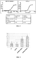
- pUMVC3 plasmids for expression of both wild type, as well as recombinant soluble and membrane-bound forms of human GITRL, OX40L, or 4-1BB (structures illustrated in Figure 1 ) were generated.
- the extracellular domain (ECD) for each ligand was added (Genbank ID AF125303 [GITRL], D90224 [OX40L], and U03398 [4-1BBL].)
- GITRL4, OX40L4 and 4-1BBL4 a heterologous trimerization domain is added ( Harbury, P.B.
- the constant region (Fc) from human or mouse antibodies were added C terminal to the ECDs (human Fc-IgG1 [from pFUSE-hIgG1-Fc, InvivoGen], mouse Fc-IgG1 [from pFUSE-mIgG1-Fc, InvivoGen], mouse Fc-IgG2a [from pFUSE-mIgG2a-Fc, InvivoGen].)
- Mouse and human GITRL are reported not to cross-react (see, e.g., Bossen et al., (2006) J. Biol. Chem. 281:13964-13971 ), thus mouse homologs for recombinant human GITRL were constructed for preclinical studies in mice.
- Mouse GITRL is naturally a dimer.
- Recombinant mouse GITRL constructs were made as both dimers and trimers using GCN4 multimerization motifs ( Harbury, P.B. Nature 371: 80 .) The relative binding of dimer and trimer forms to mouse GITR-Fc was compared using a functional ELISA.
- the trimer form showed higher binding affinity and was used for all further studies (EC50 for dimer, 39.24 uM; EC50 for trimer, 12.38 uM).
- Substituting the mouse GITRL ECD domain (Genbank Accession #NM_183391) for the human ECD domains in all constructs described above made mouse versions of the SCT forms used in pre-clinical studies.
- pUMVC3 containing a co-stimulatory molecule agonist i.e. GITRL
- All plasmid DNA was diluted in sterile injectable saline (0.9%) and stored at -20°C.
- GITRL plasmids into HEK293 (ATCC) cells with Mirus TransIT-LT1 reagent (Cat.#: MIR 2300).
- Cell to be transduced are seated at 400,000 cells in a 6-well dish. 24 hours later, Cells were overlaid with 4 ⁇ L Mirus and 1ug DNA). Cell were harvested 4-7 days later for analysis by Western blot, flow cytometry, For activity assays, secreted proteins were harvested from cellular supernatants, and purified using Ni + -resin according to the manufacturers instruction (Novagen)
- laemmli SDS sample buffer (Alfa Aesar J61337) was added to each sample and boiled at 100 C for 10 minutes and samples were centrifuged. 20 ⁇ l of protein + buffer was loaded per well and gel was run at 150 volts for about an hour until the smallest standard reached the bottom of the gel. Gel proteins were transferred to PVDF membranes at 100 volts for 1 hour on ice, rinsed with PVDF 3X with 1X TBST, and then blocked for 1 hour at room temperature on a rocker with 5% BSA in TBST.
- FC buffer 5% filtered FBS + 0.1% NaN 3 in PBS without Ca++ or Mg++.
- PE-labeled anti-GITRL clone 109101 R&D systems
- isotype control clone 11711 R&D systems
- anti-FC and corresponding isotype control Biolegend
- the cells were first incubated with anti-GITR-FC fusion protein in FC buffer for 1 hour on ice. Cells were analyzed using a Becton-Dickenson FACScan, GUAVA 12HT flow cytometer (Millipore), or LSR-II (Beckman).
- Table 2 Mean Fluorescence Intensities of transfected HEK 293 cells expressing cell surface GITR proteins demonstrating cell surface expression and binding to soluble GITR-Fc fusion protein (R&D Systems) Protein Anti-GITRL GITR-Fc + Anti-Fc Anti-Fc TM1 159.5 192.9 13.5 TM-SCT 982.5 771.4 13.0
- Anti human-Fc antibody (Pierce 31125) was added to flat-bottomed 96 well ELISA plates (Costar cat# 3690), diluted to 1 ⁇ g/ml diluted with PBS, and incubated at room temperature for 1 hour. Wells were washed with 150 ⁇ l PBST 3X, decanted and dried. Wells were blocked by adding 150 ⁇ l/well Superblock (Scytek AAA999) and incubated at room temperature for 1 hr. After washing, 50 ⁇ l/well of recombinant human GITR/TNFRSF18 Fc chimeric protein (R&D 689-GR-100) was added at a concentration of 500 ng/ml.
- Samples were incubated at room temperature for 1 hour, and subsequently washed. The following were added in triplicate: 50 ⁇ l of 5000 ng/ml rhGITRL standard Recombinant Human GITR Ligand/TNFSF18 (R&D 6987-GL-025) diluted fivefold with PBS down to zero; 50 ⁇ l of neat HEK 293 culture supernatant (negative control with no gene expression) diluted fivefold with PBS down to zero; and the soluble human GITRL protein, also titrated. Samples were incubated at room temperature for 1 hour and then washed again.
- Soluble GITRL4 and GITRL-SCT-Fc proteins bind to GITR-FC fusion protein in an ELISA with greater than a 2-fold higher affinity than that of commercially available GITRL protein ((R&D 6987-GL-025; Table 3), and a greater than 2-fold higher specific activity (activity units per ng; Figure 2 ).
- Soluble recombinant GITRL proteins demonstrated comparable GITR binding affinity to cross-linked MK-4166.
- Table 3 Soluble GITRL protein binding affinity to plate-bound GITR-Fc fusion protein and comparison with cross-linked MK-4166 GITR agonist antibody (see, e.g, US8709424).
- Protein R&D GITRL GITRL4 GITRL-SCT GITRL-SCT-Fc MK-4166 EC50 1.495 0.5806 4.759 0.5355 0.2912
- GITRL plasmids were transduced into HEK 293 human cells together with plasmids encoding firefly luciferase under the control of an NFkB-driven promoter (Promega).
- the plasmid expressing the human GITR gene (Origene), and a plasmid expressing Renilla luciferase were under the control of a constitutive CMV promoter (Promega; control for transduction variability).
- cells were lysed and analyzed for both firefly and Renilla luciferase activity using a Dual Luciferase assay kit (Promega, E1910).
- Soluble GITRL proteins stimulated GITR signaling to NFkB in a reporter cell line system ( Figure 4 ). Soluble GITRL proteins were purified from HEK 293 culture supernatants in serum-free conditions using Ni+-Resin as per the manufactures instructions (Novagen). Purified proteins were normalized by A280 absorbance and by ELISA. Molar equivalents of protein were added to engineered Jurkat cells expressing human GITR and an NF-kB-driven secreted luciferase (GITR potency assay, Promega CS184002). A dose response was measured for each protein and compared with commercially available soluble human GITRL (R&D systems). Assay was carried out as per the manufactured instruction with and without the addition of cross-linking antibodies: Anti-HA for R&D GITRL standard (R&D Systems) and Anti-NWSHPQFEK Tag (Genscript) for our GITRL proteins.
- Cell surface GITRL proteins were also tested in the GITR potency assay (Promega CS184002) by adapting the assay protocol to co-culture stimulation.
- HEK 293 cells were transduced with plasmids encoding cell surface GITRL proteins were removed from the dish with PBS-Mg++-Ca++ after 4 days in culture, and plated in a serial dilution from 100,000 down to 1 cell/well in flat bottom 96-well tissue-culture trays (Corning) and left overnight to adhere to substrate.
- Thaw and use Jurkat cells (Promega CS184002) were overlaid according to the manufacturer instruction onto GITRL-expressing HEK 293 cells and co-cultured for 7 hours.
- Soluble GITRL4 and GITRL-SCT-FC had greater than 3-fold and 10-fold better potency in this GITR activity assay, respectively, than did commercially available human GITRL (R&D Systems).
- Cell surface GITRL also showed strong activation when co-cultured with Jurkat reporter cells in GITR potency assay ( Figure 4 ).
- FC buffer 5% filtered FBS + 0.1% NaN3 in PBS without magnesium or calcium
- FC buffer 5% filtered FBS + 0.1% NaN3 in PBS without magnesium or calcium
- CD4+ T-cells and Pan T-cells were costimulated with 0.1, 0.2, or 0.5 ug/ml of anti-human CD3 (Cat #: 100207) from Biolegend and soluble agonist GITRL molecules.
- Anti-human CD28 (Cat #: 102111) was used as a positive control.
- Costimulated T-cells were incubated for 72 hours at 37 °C before supernatants were collected. The level of human IFN-y and IL-2 in cell supernatants was measured by R&D ELISA kits (Cat #: DY485-05 and Cat #: DY402-05).
- plasmids encoding the recombinant GITRL proteins are expressed when introduced into cells and can bind to and stimulate cell surface endogenous GITR on T cells.
- stimulation of cell surface GITR on T lymphocytes is predicted to enhance activation of these lymphocytes within the tumor microenvironment and facilitate an immune response against tumor cells.
- Mouse recombinant GITRL proteins were expressed in HEK 293 cells and the retention in the cell (those containing TM domains) or secretion into the culture medium was verified by Western Blot. Binding to GITR on the surface of primary mouse splenocytes was verified by flow cytometry using anti-StrepTAG II antibody (Genscript). Further, an increase in stimulation of primary mouse T cells with a sub-optimal dose of anti-CD3 (1452C11) with addition of purified soluble mouse GITRL proteins was observed in both purified CD4+ and CD8+ T cells, and well as in whole splenocytes cultures.
- recombinant mouse GITRL showed comparable or better T cell co-stimulatory activity than the GITR agonist antibody, DTA-1 (BXcell BE0063).
- DTA-1 BXcell BE0063
- DTA-1 had an EC50 of 1.29 uM
- mGITRL-SCT-FclgG2a had an EC50 of 0.473 uM
- mGITRL4 had an EC50 of 3.32 uM.
- Circular plasmid DNA was diluted to 1 ug/ul in sterile 0.9% saline. 50 ul of plasmid DNA was injected centrally into primary tumors using a 1 ml syringe with a 26 Ga needle. Electroporation was performed immediately after injection. Electroporation of DNA was achieved using a BTX ECM 830 square wave electroporator providing 8 pulses of 350 V/cm, 10 msec each spaced 1 second apart. Electroporation was delivered by a Medpulser handle with needles spaced 0.5 cm apart, configured to deliver current uni-directionally from 2 needles to 2 opposing needles.
- the electroporation needles were inserted into the tumor where tumor size permitted. Treatments were performed on days 0,4, and 7, or days 0 and 4 depending on the study. Following treatment, tumor volume was determined every 2-3 days using digital calipers as described previously. Mice were euthanized when the volume of either the primary or contralateral tumor reached 1000 mm 3 .
- mice cohort were electroporated with plasmids encoding recombinant mouse GITRL proteins and compared with a cohort treated i.p with 500 ugs DTA-1 GITR agonist antibody on Day 1 or left untreated.
- Tumors and spleen were excised from sacrificed mice and placed in RPMI + 10% FBS. Splenocytes were isolated by pressing spleen though a 70 micron strainer, subjecting them to hypotonic red blood cell lysis (ACK buffer; ThermoFisher). Isolate splenocytes were purified using lympholyte M (Cedarlane) prior to staining. Tumors were dissociated using Gentle-MACS for tumors (Miltenyi tumor dissociation kit 130-096-730, C-tubes, 130-093-237) and homogenized using an Miltenyi gentleMACSTM Octo Dissociator with Heaters (130-096-427). Samples were stored on ice until all samples were ready.
- Lympholyte-M Cells were pelleted at 1200 rpm (800 x g) for 5 min at 4C and resuspended in PBS + 2% FBS + 1 mM EDTA (PFB) and overlaid onto 5 mL of Lympholyte-M (Cedarlane) in 15-mL conical centrifuge tubes. Lympholyte columns were spun in centrifuge at 2000 rpm (1500 x g) for 20 min at room temperature with no brake. Lymphocyte layer was transferred to 10 mL of PBF, then centrifuge 1200 rpm (800 x g) for 5 min at 4C.
- PBS + 2% FBS + 1 mM EDTA PBS + 1 mM EDTA
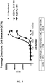
- Intratumoral electroporation of GITRL increased AH1 -dextramer - binding CD8+CD44+ T cells in the spleen ( Figure 6 ).
- RT-PCR was used to measure gene expression changes within the treated tumor.
- Flash frozen tumors were resuspended in PBS and homogenized using gentle MACS Dissociator (Miltenyl Biotech). The homogenate was then transferred into Trizol (Life Technologies Corp.). Total RNA was isolated according to manufacturer's protocol followed by DNase treatment. 1ug RNA was used to prepare cDNA (Maxima H Minus First Strand cDNA Synthesis Kit with dsDNase, Thermo Fisher Scientific).
- RT-PCR was performed using TaqMan® Fast Advanced Master Mix (Thermo Fisher Scientific.) and a CFX96 (Biorad). Relative mRNA levels were normalized to 18s and the cycle number was used to calculate the amount of each product using the 2- ⁇ CT method ( Livak et al, Methods, 2001 ).
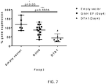
- Intratumoral electroporation of GITRL decreased Foxp3 gene expression within the treated tumor ( Figure 7 ).
- Expression of the Foxp3 transcription factor is a marker for regulatory T cells (Treg), which perform an immunosuppressive function within the tumor microenvironment.
- DTA-1 a GITR agonist antibody is known to reduce the number of Tregs within tumors, and has also been shown to reduce the levels of Foxp3 gene present in tumors ( Schaer et al., (2013) Cancer Immunol Res. 1:320-331 )
- pUMVC3-GITR4 a marker for regulatory T cells within the tumor microenvironment.
- T cells from spleens and tumors of treated mice suggest that intratumoral electroporation of plasmid encoding for recombinant GITRL can alter the Treg levels within the treated tumor, and result in an increase in tumor antigen-reactive effector T cells systemically.
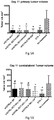
- pUMVC3 plasmid encoding GITRL4-TM1 was electroporated intratumorally along with pUMVC3 encoding for the p35 and p40 subunits of the IL-12 cytokine ( A Daud et al., (2008) J. Clin. Oncol. 26:5896-5903 ).
- a statistically significant decrease in mean tumor volume of the untreated, contralateral tumor was seen as compared with pUMVC3 vector control or pUMVC3-IL12 plasmid alone as measured 6, 8, 11 and 13 days after first EP treatment.
- complete tumor regression of the untreated tumors had faster kinetics. For example, 19 days after treatment began, 7/15 mice had completed regressed contralateral tumors while the IL-12 treated cohort had only 1/15.
- an increase in the number of mice with a complete response (CR) on both the primary (treated) and contralateral (untreated) was observed as compared with pUMVC3-IL-12 alone.
- Treatment tumor volume mm 3 (mean+/-SEM) Number of mice with CR out of 15 Number of mice with PR out of 15 Number of mice with PD out of 15 C P C P C P C Untreated 751.7 +/- 175.5 0 0 1 2 14 13 pUMVC3 675.7 +/- 36.5 2 0 1 3 12 12 pUMVC3-IL-12 176.6 +/- 115.8 13 8 0 3 2 5 pUMVC3-GITRL-TM1/pUM VC3-IL-12 64.25 +/- 61.8 13 11 0 1 2 3
- IL-12, IL-15 and GITRL all have separate effect on stimulation of the immune system. Plasmids encoding IL-12 ⁇ and ⁇ subunits, IL-15 and IL-15Ra, and recombinant GITRL are electroporated together (on separate plasmids or in a single plasmid) into CT26 tumors in a contralateral tumor model to assess whether the combination of these 5 genes has more efficacy in regressing established tumors in BALB/c mice. Intratumoral electroporation of a single, large plasmid encoding all 5 genes is also tested for efficacy as compared with untreated mice. In addition, testing of these genes in combination and separately is done in a B16F10 contralateral tumor model in C57/BI6 mice. Tumor volumes are measured over time, and the percent of mice with complete response are measured.
- mice are humanely sacrificed by CO 2 asphyxiation. Tumors are excised and placed in 50-ml conical tubes containing 10 ml of 10% formalin. The tissue is stained with H&E after fixation, as follows: after fixation in 10% neutral buffered formalin for 6 hours, representative tissue samples are processed into paraffin blocks using a Miles VIP tissue processor (Miles Inc., Mishawaka, IN). Briefly, tissues are dehydrated in ascending grades of ethanol, cleared in xylene, and infiltrated in paraffin (Tissue Prep 2; Fisher Scientific). Following embedding, tissues are sectioned on a standard rotatory microtome and 4 mm sections are retrieved from a waterbath and mounted on glass slides. Three sections per tumor are examined. Sections are heat-dried and stained with H&E (Richard-Allen Scientific, Kalamazoo, MI) using standard histologic techniques.
- Immunohistochemical staining is conducted to examine the tumors for the presence of CD4+ lymphocytes, CD8+ lymphocytes, and blood vessels using the following antibodies: rat anti-mouse CD4, rat anti-mouse CD8 (Ly2), and rat anti-mouse CD31 (PECAM-1), respectively (PharMingen, Cambridge, MA). Mice are humanely sacrificed by CO 2 asphyxiation. Tumors are excised with scissors and the skin removed, then immediately frozen in a mixture of dry ice and ethanol, and stored at (80°C). Frozen sections of 5 m are obtained.
- rat anti-mouse CD4, rat anti-mouse CD8 (Ly2), or rat anti-mouse CD31 (PECAM-1) is applied to tissue sections at a dilution of 1:50 and incubated for 30 minutes, followed by detection with the Vector Elite Rat IgG Peroxidase kit at 2X concentration (15 minutes each in biotinylated anti-rat IgG and ABC complex). Immunostaining is carried out on the Dako autostainer. Sections are analyzed at 400X magnification.
Landscapes
- Health & Medical Sciences (AREA)
- Life Sciences & Earth Sciences (AREA)
- Chemical & Material Sciences (AREA)
- General Health & Medical Sciences (AREA)
- Veterinary Medicine (AREA)
- Public Health (AREA)
- Animal Behavior & Ethology (AREA)
- Medicinal Chemistry (AREA)
- Engineering & Computer Science (AREA)
- Pharmacology & Pharmacy (AREA)
- Gastroenterology & Hepatology (AREA)
- Proteomics, Peptides & Aminoacids (AREA)
- Epidemiology (AREA)
- Zoology (AREA)
- Bioinformatics & Cheminformatics (AREA)
- Immunology (AREA)
- Organic Chemistry (AREA)
- Genetics & Genomics (AREA)
- Molecular Biology (AREA)
- Nuclear Medicine, Radiotherapy & Molecular Imaging (AREA)
- Biophysics (AREA)
- Biomedical Technology (AREA)
- Radiology & Medical Imaging (AREA)
- Toxicology (AREA)
- Biotechnology (AREA)
- Biochemistry (AREA)
- Cell Biology (AREA)
- Chemical Kinetics & Catalysis (AREA)
- General Chemical & Material Sciences (AREA)
- Medicines That Contain Protein Lipid Enzymes And Other Medicines (AREA)
- Medicines Containing Antibodies Or Antigens For Use As Internal Diagnostic Agents (AREA)
- Pharmaceuticals Containing Other Organic And Inorganic Compounds (AREA)
- Medicines Containing Material From Animals Or Micro-Organisms (AREA)
- Medicinal Preparation (AREA)
- Peptides Or Proteins (AREA)
Priority Applications (2)
| Application Number | Priority Date | Filing Date | Title |
|---|---|---|---|
| EP20176001.4A EP3799879B1 (en) | 2015-01-09 | 2016-01-08 | Gene therapy using co-stimulatory molecule and cytokine for the treatment of malignancies |
| PL16735524T PL3242947T3 (pl) | 2015-01-09 | 2016-01-08 | Terapia genowa i elektroporacja do leczenia zmian złośliwych |
Applications Claiming Priority (3)
| Application Number | Priority Date | Filing Date | Title |
|---|---|---|---|
| US201562101850P | 2015-01-09 | 2015-01-09 | |
| US201562126300P | 2015-02-27 | 2015-02-27 | |
| PCT/US2016/012759 WO2016112359A1 (en) | 2015-01-09 | 2016-01-08 | Method for the treatment of malignancies |
Related Child Applications (1)
| Application Number | Title | Priority Date | Filing Date |
|---|---|---|---|
| EP20176001.4A Division EP3799879B1 (en) | 2015-01-09 | 2016-01-08 | Gene therapy using co-stimulatory molecule and cytokine for the treatment of malignancies |
Publications (3)
| Publication Number | Publication Date |
|---|---|
| EP3242947A1 EP3242947A1 (en) | 2017-11-15 |
| EP3242947A4 EP3242947A4 (en) | 2018-08-08 |
| EP3242947B1 true EP3242947B1 (en) | 2020-05-27 |
Family
ID=56356515
Family Applications (2)
| Application Number | Title | Priority Date | Filing Date |
|---|---|---|---|
| EP16735524.7A Active EP3242947B1 (en) | 2015-01-09 | 2016-01-08 | Gene therapy and electroporation for the treatment of malignancies |
| EP20176001.4A Active EP3799879B1 (en) | 2015-01-09 | 2016-01-08 | Gene therapy using co-stimulatory molecule and cytokine for the treatment of malignancies |
Family Applications After (1)
| Application Number | Title | Priority Date | Filing Date |
|---|---|---|---|
| EP20176001.4A Active EP3799879B1 (en) | 2015-01-09 | 2016-01-08 | Gene therapy using co-stimulatory molecule and cytokine for the treatment of malignancies |
Country Status (9)
| Country | Link |
|---|---|
| US (1) | US20180000895A1 (enExample) |
| EP (2) | EP3242947B1 (enExample) |
| JP (3) | JP2018504403A (enExample) |
| CA (1) | CA2973390A1 (enExample) |
| DK (1) | DK3242947T3 (enExample) |
| ES (1) | ES2809734T3 (enExample) |
| MX (1) | MX383277B (enExample) |
| PL (1) | PL3242947T3 (enExample) |
| WO (1) | WO2016112359A1 (enExample) |
Families Citing this family (16)
| Publication number | Priority date | Publication date | Assignee | Title |
|---|---|---|---|---|
| ES2809734T3 (es) * | 2015-01-09 | 2021-03-05 | Oncosec Medical Inc | Terapia génica y electroporación para el tratamiento de neoplasias malignas |
| EP4209190A1 (en) | 2016-06-27 | 2023-07-12 | Galvanize Therapeutics, Inc. | System comprising a generator and a catheter with an electrode for treating a lung passageway |
| US12403305B2 (en) | 2016-06-27 | 2025-09-02 | Galvanize Therapeutics, Inc. | Immunostimulation in the treatment of viral infection |
| GB201710973D0 (en) | 2017-07-07 | 2017-08-23 | Avacta Life Sciences Ltd | Scaffold proteins |
| JP2021508533A (ja) | 2017-12-26 | 2021-03-11 | ギャラリー,インコーポレイテッド | 様々な用途のためのエネルギー送達の最適化 |
| MX2020011618A (es) * | 2018-05-02 | 2021-02-16 | Oncosec Medical Inc | Sistemas, métodos y aparatos de electroporación. |
| KR102753455B1 (ko) * | 2018-08-08 | 2025-01-10 | 세다르스-신나이 메디칼 센터 | 암 및 자가면역 질환을 치료하기 위한 조성물 및 방법 |
| JP7586890B2 (ja) | 2019-07-16 | 2024-11-19 | ガルヴァナイズ セラピューティクス,インコーポレイテッド | パルス電界による生殖器の治療 |
| CN114127121B (zh) | 2019-08-12 | 2025-04-11 | 北京恩瑞尼生物科技股份有限公司 | 用于通过cd39表达细胞的adcc靶向促进和增强t细胞介导的免疫反应的方法和组合物 |
| TW202128775A (zh) | 2019-10-16 | 2021-08-01 | 英商阿法克塔生命科學有限公司 | PD-L1抑制劑-TGFβ抑制劑雙特異性藥物部分 |
| GB202101299D0 (en) | 2020-06-09 | 2021-03-17 | Avacta Life Sciences Ltd | Diagnostic polypetides and methods |
| WO2022234003A1 (en) | 2021-05-07 | 2022-11-10 | Avacta Life Sciences Limited | Cd33 binding polypeptides with stefin a protein |
| EP4413038A1 (en) | 2021-10-07 | 2024-08-14 | Avacta Life Sciences Limited | Pd-l1 binding affimers |
| TW202332694A (zh) | 2021-10-07 | 2023-08-16 | 英商阿凡克塔生命科學公司 | 血清半衰期延長之pd-l1結合多肽 |
| CN119213021A (zh) | 2022-02-10 | 2024-12-27 | 艾菲赛尔治疗株式会社 | 与CD40L特异性结合的Stefin A蛋白变体及其用途 |
| KR20250006965A (ko) | 2022-04-29 | 2025-01-13 | 퓨리노미아 바이오테크, 아이엔씨. | 호산구에 의해 유발되는 질환 및 장애의 치료를 위한 방법 및 조성물 |
Family Cites Families (13)
| Publication number | Priority date | Publication date | Assignee | Title |
|---|---|---|---|---|
| US5993434A (en) * | 1993-04-01 | 1999-11-30 | Genetronics, Inc. | Method of treatment using electroporation mediated delivery of drugs and genes |
| US20030049696A1 (en) | 2001-06-07 | 2003-03-13 | Norment Anne M. | Regulatory T cells and uses thereof |
| US8209006B2 (en) | 2002-03-07 | 2012-06-26 | Vgx Pharmaceuticals, Inc. | Constant current electroporation device and methods of use |
| US7245963B2 (en) | 2002-03-07 | 2007-07-17 | Advisys, Inc. | Electrode assembly for constant-current electroporation and use |
| US20110142887A1 (en) * | 2009-12-15 | 2011-06-16 | Immunovative Therapies Ltd. | Methods and compositions for liquidation of tumors |
| US8802643B1 (en) * | 2003-05-30 | 2014-08-12 | University Of South Florida | Method for the treatment of malignancies |
| EA200501937A1 (ru) * | 2003-05-30 | 2006-08-25 | Университет Южной Флориды | Способ лечения злокачественных опухолей |
| US7923251B2 (en) | 2005-02-23 | 2011-04-12 | The Board Of Trustees Of The Leland Stanford Junior University | Method and apparatus for avalanche-mediated transfer of agents into cells |
| US8101169B2 (en) | 2005-02-23 | 2012-01-24 | The Board Of Trustees Of The Leland Stanford Junior University | Ocular gene therapy using avalanche-mediated transfection |
| WO2007120368A2 (en) * | 2006-01-09 | 2007-10-25 | The Regents Of The University Of California | Immunostimulatory combinations for vaccine adjuvants |
| WO2010030002A1 (ja) * | 2008-09-12 | 2010-03-18 | 国立大学法人三重大学 | 外来性gitrリガンド発現細胞 |
| KR102445129B1 (ko) * | 2011-12-12 | 2022-09-20 | 더 트러스티스 오브 더 유니버시티 오브 펜실바니아 | 개선된 il-12 유전적 컨스트럭트 및 백신을 포함하는 조성물, 면역치료제 및 이를 이용하는 방법 |
| ES2809734T3 (es) * | 2015-01-09 | 2021-03-05 | Oncosec Medical Inc | Terapia génica y electroporación para el tratamiento de neoplasias malignas |
-
2016
- 2016-01-08 ES ES16735524T patent/ES2809734T3/es active Active
- 2016-01-08 EP EP16735524.7A patent/EP3242947B1/en active Active
- 2016-01-08 PL PL16735524T patent/PL3242947T3/pl unknown
- 2016-01-08 DK DK16735524.7T patent/DK3242947T3/da active
- 2016-01-08 WO PCT/US2016/012759 patent/WO2016112359A1/en not_active Ceased
- 2016-01-08 EP EP20176001.4A patent/EP3799879B1/en active Active
- 2016-01-08 JP JP2017536882A patent/JP2018504403A/ja active Pending
- 2016-01-08 MX MX2017009044A patent/MX383277B/es unknown
- 2016-01-08 CA CA2973390A patent/CA2973390A1/en active Pending
- 2016-01-08 US US15/542,415 patent/US20180000895A1/en not_active Abandoned
-
2020
- 2020-09-29 JP JP2020164124A patent/JP7111384B2/ja active Active
-
2022
- 2022-07-13 JP JP2022112196A patent/JP2022137203A/ja active Pending
Non-Patent Citations (1)
| Title |
|---|
| None * |
Also Published As
| Publication number | Publication date |
|---|---|
| EP3242947A4 (en) | 2018-08-08 |
| JP7111384B2 (ja) | 2022-08-02 |
| DK3242947T3 (da) | 2020-08-03 |
| EP3799879A1 (en) | 2021-04-07 |
| ES2809734T3 (es) | 2021-03-05 |
| JP2018504403A (ja) | 2018-02-15 |
| JP2021008481A (ja) | 2021-01-28 |
| WO2016112359A1 (en) | 2016-07-14 |
| MX383277B (es) | 2025-03-13 |
| MX2017009044A (es) | 2018-01-11 |
| EP3799879B1 (en) | 2025-06-04 |
| CA2973390A1 (en) | 2016-07-14 |
| PL3242947T3 (pl) | 2021-01-11 |
| US20180000895A1 (en) | 2018-01-04 |
| JP2022137203A (ja) | 2022-09-21 |
| EP3242947A1 (en) | 2017-11-15 |
Similar Documents
| Publication | Publication Date | Title |
|---|---|---|
| EP3242947B1 (en) | Gene therapy and electroporation for the treatment of malignancies | |
| US20210369813A9 (en) | Modulating responses to checkpoint inhibitor therapy | |
| EP3286311B1 (en) | Method for the treatment of malignancies | |
| EP2766035B1 (en) | Combination medicament comprising il-12 and an agent for blockade of t-cell inhibitory molecules for tumour therapy | |
| EP1490407B1 (en) | Induction of anti-tumor ctl immunity through in vivo triggering of 4-1bb and/or cd40 | |
| AU2009226077B2 (en) | Heat shock protein gp96 vaccination and methods of using same | |
| US10934331B2 (en) | Methods for enhancing immune responsiveness in an individual toward a target cancer cell population comprising apoptotic cells | |
| US20200123566A1 (en) | Multigene construct for immune-modulatory protein expression and methods of use | |
| JP2020503252A (ja) | T細胞応答を促進するための方法 | |
| CN115003322A (zh) | 癌症疫苗 | |
| KR20220139915A (ko) | Il-10 및 그의 용도 | |
| CA3084190A1 (en) | Methods for enhancing and maintaining car-t cell efficacy | |
| US20220040328A1 (en) | Multigene construct for immune-modulatory protein expression and methods of use | |
| US20230272043A1 (en) | Method for the treatment of malignancies | |
| JP2022525921A (ja) | 免疫エフェクタ細胞の特異的活性化のためのインターロイキン2受容体(il2r)およびインターロイキン2(il2)バリアント | |
| HK40049266A (en) | Gene therapy using co-stimulatory molecule and cytokine for the treatment of malignancies | |
| HK40049266B (en) | Gene therapy using co-stimulatory molecule and cytokine for the treatment of malignancies | |
| HK1237821A1 (en) | Gene therapy and electroporation for the treatment of malignancies | |
| HK1237821B (en) | Gene therapy and electroporation for the treatment of malignancies | |
| WO2025080338A1 (en) | Cell-surface anchored cytokine therapy | |
| HK1251614B (en) | Method for the treatment of malignancies |
Legal Events
| Date | Code | Title | Description |
|---|---|---|---|
| STAA | Information on the status of an ep patent application or granted ep patent |
Free format text: STATUS: THE INTERNATIONAL PUBLICATION HAS BEEN MADE |
|
| PUAI | Public reference made under article 153(3) epc to a published international application that has entered the european phase |
Free format text: ORIGINAL CODE: 0009012 |
|
| STAA | Information on the status of an ep patent application or granted ep patent |
Free format text: STATUS: REQUEST FOR EXAMINATION WAS MADE |
|
| 17P | Request for examination filed |
Effective date: 20170719 |
|
| AK | Designated contracting states |
Kind code of ref document: A1 Designated state(s): AL AT BE BG CH CY CZ DE DK EE ES FI FR GB GR HR HU IE IS IT LI LT LU LV MC MK MT NL NO PL PT RO RS SE SI SK SM TR |
|
| AX | Request for extension of the european patent |
Extension state: BA ME |
|
| DAV | Request for validation of the european patent (deleted) | ||
| DAX | Request for extension of the european patent (deleted) | ||
| REG | Reference to a national code |
Ref country code: HK Ref legal event code: DE Ref document number: 1237821 Country of ref document: HK |
|
| REG | Reference to a national code |
Ref country code: DE Ref legal event code: R079 Ref document number: 602016037040 Country of ref document: DE Free format text: PREVIOUS MAIN CLASS: C12N0015870000 Ipc: A61K0038190000 |
|
| A4 | Supplementary search report drawn up and despatched |
Effective date: 20180710 |
|
| RIC1 | Information provided on ipc code assigned before grant |
Ipc: A61K 38/19 20060101AFI20180703BHEP Ipc: A61K 48/00 20060101ALI20180703BHEP Ipc: A61P 35/00 20060101ALI20180703BHEP Ipc: C07K 14/525 20060101ALI20180703BHEP Ipc: A61K 38/20 20060101ALI20180703BHEP Ipc: C12N 15/87 20060101ALI20180703BHEP |
|
| STAA | Information on the status of an ep patent application or granted ep patent |
Free format text: STATUS: EXAMINATION IS IN PROGRESS |
|
| 17Q | First examination report despatched |
Effective date: 20190718 |
|
| GRAP | Despatch of communication of intention to grant a patent |
Free format text: ORIGINAL CODE: EPIDOSNIGR1 |
|
| STAA | Information on the status of an ep patent application or granted ep patent |
Free format text: STATUS: GRANT OF PATENT IS INTENDED |
|
| INTG | Intention to grant announced |
Effective date: 20200124 |
|
| RAP1 | Party data changed (applicant data changed or rights of an application transferred) |
Owner name: ONCOSEC MEDICAL INCORPORATED |
|
| GRAS | Grant fee paid |
Free format text: ORIGINAL CODE: EPIDOSNIGR3 |
|
| GRAA | (expected) grant |
Free format text: ORIGINAL CODE: 0009210 |
|
| STAA | Information on the status of an ep patent application or granted ep patent |
Free format text: STATUS: THE PATENT HAS BEEN GRANTED |
|
| AK | Designated contracting states |
Kind code of ref document: B1 Designated state(s): AL AT BE BG CH CY CZ DE DK EE ES FI FR GB GR HR HU IE IS IT LI LT LU LV MC MK MT NL NO PL PT RO RS SE SI SK SM TR |
|
| REG | Reference to a national code |
Ref country code: GB Ref legal event code: FG4D |
|
| REG | Reference to a national code |
Ref country code: CH Ref legal event code: EP |
|
| REG | Reference to a national code |
Ref country code: AT Ref legal event code: REF Ref document number: 1273838 Country of ref document: AT Kind code of ref document: T Effective date: 20200615 |
|
| REG | Reference to a national code |
Ref country code: DE Ref legal event code: R096 Ref document number: 602016037040 Country of ref document: DE |
|
| REG | Reference to a national code |
Ref country code: DK Ref legal event code: T3 Effective date: 20200728 |
|
| REG | Reference to a national code |
Ref country code: SE Ref legal event code: TRGR |
|
| REG | Reference to a national code |
Ref country code: NL Ref legal event code: FP |
|
| REG | Reference to a national code |
Ref country code: NO Ref legal event code: T2 Effective date: 20200527 |
|
| REG | Reference to a national code |
Ref country code: CH Ref legal event code: NV Representative=s name: NOVAGRAAF INTERNATIONAL SA, CH |
|
| REG | Reference to a national code |
Ref country code: NO Ref legal event code: CREP Representative=s name: CURO AS, VESTRE ROSTEN 81, 7075 TILLER, NORGE |
|
| REG | Reference to a national code |
Ref country code: LT Ref legal event code: MG4D |
|
| PG25 | Lapsed in a contracting state [announced via postgrant information from national office to epo] |
Ref country code: GR Free format text: LAPSE BECAUSE OF FAILURE TO SUBMIT A TRANSLATION OF THE DESCRIPTION OR TO PAY THE FEE WITHIN THE PRESCRIBED TIME-LIMIT Effective date: 20200828 Ref country code: FI Free format text: LAPSE BECAUSE OF FAILURE TO SUBMIT A TRANSLATION OF THE DESCRIPTION OR TO PAY THE FEE WITHIN THE PRESCRIBED TIME-LIMIT Effective date: 20200527 Ref country code: IS Free format text: LAPSE BECAUSE OF FAILURE TO SUBMIT A TRANSLATION OF THE DESCRIPTION OR TO PAY THE FEE WITHIN THE PRESCRIBED TIME-LIMIT Effective date: 20200927 Ref country code: PT Free format text: LAPSE BECAUSE OF FAILURE TO SUBMIT A TRANSLATION OF THE DESCRIPTION OR TO PAY THE FEE WITHIN THE PRESCRIBED TIME-LIMIT Effective date: 20200928 Ref country code: LT Free format text: LAPSE BECAUSE OF FAILURE TO SUBMIT A TRANSLATION OF THE DESCRIPTION OR TO PAY THE FEE WITHIN THE PRESCRIBED TIME-LIMIT Effective date: 20200527 |
|
| PG25 | Lapsed in a contracting state [announced via postgrant information from national office to epo] |
Ref country code: LV Free format text: LAPSE BECAUSE OF FAILURE TO SUBMIT A TRANSLATION OF THE DESCRIPTION OR TO PAY THE FEE WITHIN THE PRESCRIBED TIME-LIMIT Effective date: 20200527 Ref country code: HR Free format text: LAPSE BECAUSE OF FAILURE TO SUBMIT A TRANSLATION OF THE DESCRIPTION OR TO PAY THE FEE WITHIN THE PRESCRIBED TIME-LIMIT Effective date: 20200527 Ref country code: BG Free format text: LAPSE BECAUSE OF FAILURE TO SUBMIT A TRANSLATION OF THE DESCRIPTION OR TO PAY THE FEE WITHIN THE PRESCRIBED TIME-LIMIT Effective date: 20200827 Ref country code: RS Free format text: LAPSE BECAUSE OF FAILURE TO SUBMIT A TRANSLATION OF THE DESCRIPTION OR TO PAY THE FEE WITHIN THE PRESCRIBED TIME-LIMIT Effective date: 20200527 |
|
| REG | Reference to a national code |
Ref country code: AT Ref legal event code: MK05 Ref document number: 1273838 Country of ref document: AT Kind code of ref document: T Effective date: 20200527 |
|
| PG25 | Lapsed in a contracting state [announced via postgrant information from national office to epo] |
Ref country code: AL Free format text: LAPSE BECAUSE OF FAILURE TO SUBMIT A TRANSLATION OF THE DESCRIPTION OR TO PAY THE FEE WITHIN THE PRESCRIBED TIME-LIMIT Effective date: 20200527 |
|
| PG25 | Lapsed in a contracting state [announced via postgrant information from national office to epo] |
Ref country code: AT Free format text: LAPSE BECAUSE OF FAILURE TO SUBMIT A TRANSLATION OF THE DESCRIPTION OR TO PAY THE FEE WITHIN THE PRESCRIBED TIME-LIMIT Effective date: 20200527 Ref country code: RO Free format text: LAPSE BECAUSE OF FAILURE TO SUBMIT A TRANSLATION OF THE DESCRIPTION OR TO PAY THE FEE WITHIN THE PRESCRIBED TIME-LIMIT Effective date: 20200527 Ref country code: EE Free format text: LAPSE BECAUSE OF FAILURE TO SUBMIT A TRANSLATION OF THE DESCRIPTION OR TO PAY THE FEE WITHIN THE PRESCRIBED TIME-LIMIT Effective date: 20200527 Ref country code: SM Free format text: LAPSE BECAUSE OF FAILURE TO SUBMIT A TRANSLATION OF THE DESCRIPTION OR TO PAY THE FEE WITHIN THE PRESCRIBED TIME-LIMIT Effective date: 20200527 Ref country code: CZ Free format text: LAPSE BECAUSE OF FAILURE TO SUBMIT A TRANSLATION OF THE DESCRIPTION OR TO PAY THE FEE WITHIN THE PRESCRIBED TIME-LIMIT Effective date: 20200527 |
|
| PG25 | Lapsed in a contracting state [announced via postgrant information from national office to epo] |
Ref country code: SK Free format text: LAPSE BECAUSE OF FAILURE TO SUBMIT A TRANSLATION OF THE DESCRIPTION OR TO PAY THE FEE WITHIN THE PRESCRIBED TIME-LIMIT Effective date: 20200527 |
|
| REG | Reference to a national code |
Ref country code: DE Ref legal event code: R097 Ref document number: 602016037040 Country of ref document: DE |
|
| REG | Reference to a national code |
Ref country code: ES Ref legal event code: FG2A Ref document number: 2809734 Country of ref document: ES Kind code of ref document: T3 Effective date: 20210305 |
|
| PLBE | No opposition filed within time limit |
Free format text: ORIGINAL CODE: 0009261 |
|
| STAA | Information on the status of an ep patent application or granted ep patent |
Free format text: STATUS: NO OPPOSITION FILED WITHIN TIME LIMIT |
|
| PGFP | Annual fee paid to national office [announced via postgrant information from national office to epo] |
Ref country code: IT Payment date: 20210128 Year of fee payment: 6 |
|
| 26N | No opposition filed |
Effective date: 20210302 |
|
| PG25 | Lapsed in a contracting state [announced via postgrant information from national office to epo] |
Ref country code: SI Free format text: LAPSE BECAUSE OF FAILURE TO SUBMIT A TRANSLATION OF THE DESCRIPTION OR TO PAY THE FEE WITHIN THE PRESCRIBED TIME-LIMIT Effective date: 20200527 |
|
| PG25 | Lapsed in a contracting state [announced via postgrant information from national office to epo] |
Ref country code: MC Free format text: LAPSE BECAUSE OF FAILURE TO SUBMIT A TRANSLATION OF THE DESCRIPTION OR TO PAY THE FEE WITHIN THE PRESCRIBED TIME-LIMIT Effective date: 20200527 |
|
| PG25 | Lapsed in a contracting state [announced via postgrant information from national office to epo] |
Ref country code: LU Free format text: LAPSE BECAUSE OF NON-PAYMENT OF DUE FEES Effective date: 20210108 |
|
| PG25 | Lapsed in a contracting state [announced via postgrant information from national office to epo] |
Ref country code: IT Free format text: LAPSE BECAUSE OF NON-PAYMENT OF DUE FEES Effective date: 20220108 |
|
| PGFP | Annual fee paid to national office [announced via postgrant information from national office to epo] |
Ref country code: PL Payment date: 20221206 Year of fee payment: 8 |
|
| PGFP | Annual fee paid to national office [announced via postgrant information from national office to epo] |
Ref country code: NO Payment date: 20230130 Year of fee payment: 8 Ref country code: IE Payment date: 20230126 Year of fee payment: 8 Ref country code: ES Payment date: 20230206 Year of fee payment: 8 Ref country code: DK Payment date: 20230130 Year of fee payment: 8 |
|
| PG25 | Lapsed in a contracting state [announced via postgrant information from national office to epo] |
Ref country code: HU Free format text: LAPSE BECAUSE OF FAILURE TO SUBMIT A TRANSLATION OF THE DESCRIPTION OR TO PAY THE FEE WITHIN THE PRESCRIBED TIME-LIMIT; INVALID AB INITIO Effective date: 20160108 |
|
| PGFP | Annual fee paid to national office [announced via postgrant information from national office to epo] |
Ref country code: SE Payment date: 20230126 Year of fee payment: 8 Ref country code: BE Payment date: 20230126 Year of fee payment: 8 |
|
| P01 | Opt-out of the competence of the unified patent court (upc) registered |
Effective date: 20230510 |
|
| PG25 | Lapsed in a contracting state [announced via postgrant information from national office to epo] |
Ref country code: CY Free format text: LAPSE BECAUSE OF FAILURE TO SUBMIT A TRANSLATION OF THE DESCRIPTION OR TO PAY THE FEE WITHIN THE PRESCRIBED TIME-LIMIT Effective date: 20200527 |
|
| PGFP | Annual fee paid to national office [announced via postgrant information from national office to epo] |
Ref country code: NL Payment date: 20230126 Year of fee payment: 8 |
|
| REG | Reference to a national code |
Ref country code: GB Ref legal event code: 732E Free format text: REGISTERED BETWEEN 20240328 AND 20240403 |
|
| PG25 | Lapsed in a contracting state [announced via postgrant information from national office to epo] |
Ref country code: MK Free format text: LAPSE BECAUSE OF FAILURE TO SUBMIT A TRANSLATION OF THE DESCRIPTION OR TO PAY THE FEE WITHIN THE PRESCRIBED TIME-LIMIT Effective date: 20200527 |
|
| REG | Reference to a national code |
Ref country code: DE Ref legal event code: R082 Ref document number: 602016037040 Country of ref document: DE Representative=s name: AAP PATENTANWALTSKANZLEI GRABOVAC, DE Ref country code: DE Ref legal event code: R081 Ref document number: 602016037040 Country of ref document: DE Owner name: GRAND DECADE DEVELOPMENTS LTD., HK Free format text: FORMER OWNER: ONCOSEC MEDICAL INCORPORATED, PENNINGTON, NJ, US |
|
| PG25 | Lapsed in a contracting state [announced via postgrant information from national office to epo] |
Ref country code: TR Free format text: LAPSE BECAUSE OF FAILURE TO SUBMIT A TRANSLATION OF THE DESCRIPTION OR TO PAY THE FEE WITHIN THE PRESCRIBED TIME-LIMIT Effective date: 20200527 |
|
| REG | Reference to a national code |
Ref country code: DK Ref legal event code: EBP Effective date: 20240131 |
|
| REG | Reference to a national code |
Ref country code: SE Ref legal event code: EUG |
|
| REG | Reference to a national code |
Ref country code: NL Ref legal event code: MM Effective date: 20240201 |
|
| PG25 | Lapsed in a contracting state [announced via postgrant information from national office to epo] |
Ref country code: MT Free format text: LAPSE BECAUSE OF FAILURE TO SUBMIT A TRANSLATION OF THE DESCRIPTION OR TO PAY THE FEE WITHIN THE PRESCRIBED TIME-LIMIT Effective date: 20200527 |
|
| PG25 | Lapsed in a contracting state [announced via postgrant information from national office to epo] |
Ref country code: BE Free format text: LAPSE BECAUSE OF NON-PAYMENT OF DUE FEES Effective date: 20240131 |
|
| PG25 | Lapsed in a contracting state [announced via postgrant information from national office to epo] |
Ref country code: NL Free format text: LAPSE BECAUSE OF NON-PAYMENT OF DUE FEES Effective date: 20240201 |
|
| PG25 | Lapsed in a contracting state [announced via postgrant information from national office to epo] |
Ref country code: NO Free format text: LAPSE BECAUSE OF NON-PAYMENT OF DUE FEES Effective date: 20240131 Ref country code: NL Free format text: LAPSE BECAUSE OF NON-PAYMENT OF DUE FEES Effective date: 20240201 Ref country code: BE Free format text: LAPSE BECAUSE OF NON-PAYMENT OF DUE FEES Effective date: 20240131 |
|
| REG | Reference to a national code |
Ref country code: BE Ref legal event code: MM Effective date: 20240131 |
|
| PG25 | Lapsed in a contracting state [announced via postgrant information from national office to epo] |
Ref country code: DK Free format text: LAPSE BECAUSE OF NON-PAYMENT OF DUE FEES Effective date: 20240131 |
|
| PG25 | Lapsed in a contracting state [announced via postgrant information from national office to epo] |
Ref country code: IE Free format text: LAPSE BECAUSE OF NON-PAYMENT OF DUE FEES Effective date: 20240108 |
|
| PG25 | Lapsed in a contracting state [announced via postgrant information from national office to epo] |
Ref country code: IE Free format text: LAPSE BECAUSE OF NON-PAYMENT OF DUE FEES Effective date: 20240108 Ref country code: DK Free format text: LAPSE BECAUSE OF NON-PAYMENT OF DUE FEES Effective date: 20240131 |
|
| REG | Reference to a national code |
Ref country code: ES Ref legal event code: FD2A Effective date: 20250228 |
|
| PGFP | Annual fee paid to national office [announced via postgrant information from national office to epo] |
Ref country code: DE Payment date: 20250121 Year of fee payment: 10 |
|
| PG25 | Lapsed in a contracting state [announced via postgrant information from national office to epo] |
Ref country code: ES Free format text: LAPSE BECAUSE OF NON-PAYMENT OF DUE FEES Effective date: 20240109 |
|
| PGFP | Annual fee paid to national office [announced via postgrant information from national office to epo] |
Ref country code: CH Payment date: 20250205 Year of fee payment: 10 |
|
| PG25 | Lapsed in a contracting state [announced via postgrant information from national office to epo] |
Ref country code: PL Free format text: LAPSE BECAUSE OF NON-PAYMENT OF DUE FEES Effective date: 20240108 |
|
| PGFP | Annual fee paid to national office [announced via postgrant information from national office to epo] |
Ref country code: FR Payment date: 20250121 Year of fee payment: 10 |
|
| PGFP | Annual fee paid to national office [announced via postgrant information from national office to epo] |
Ref country code: GB Payment date: 20250123 Year of fee payment: 10 |
|
| PG25 | Lapsed in a contracting state [announced via postgrant information from national office to epo] |
Ref country code: SE Free format text: LAPSE BECAUSE OF NON-PAYMENT OF DUE FEES Effective date: 20240109 |