EP3225891A1 - Nervenüberwachungssysteme zur behandlung von erkrankungen des rachens - Google Patents
Nervenüberwachungssysteme zur behandlung von erkrankungen des rachens Download PDFInfo
- Publication number
- EP3225891A1 EP3225891A1 EP17167891.5A EP17167891A EP3225891A1 EP 3225891 A1 EP3225891 A1 EP 3225891A1 EP 17167891 A EP17167891 A EP 17167891A EP 3225891 A1 EP3225891 A1 EP 3225891A1
- Authority
- EP
- European Patent Office
- Prior art keywords
- reflex
- stimulation
- activity
- reflux
- neural
- Prior art date
- Legal status (The legal status is an assumption and is not a legal conclusion. Google has not performed a legal analysis and makes no representation as to the accuracy of the status listed.)
- Granted
Links
- 230000001537 neural effect Effects 0.000 title claims description 212
- 238000012544 monitoring process Methods 0.000 title description 50
- 208000023668 Pharyngeal disease Diseases 0.000 title description 4
- 201000007100 Pharyngitis Diseases 0.000 title description 4
- 208000033420 disorder of pharynx Diseases 0.000 title description 4
- 230000000638 stimulation Effects 0.000 claims abstract description 263
- 230000011514 reflex Effects 0.000 claims abstract description 165
- 208000037265 diseases, disorders, signs and symptoms Diseases 0.000 claims abstract description 138
- 208000035475 disorder Diseases 0.000 claims abstract description 135
- 238000010992 reflux Methods 0.000 claims abstract description 114
- 208000008784 apnea Diseases 0.000 claims abstract description 88
- 208000021302 gastroesophageal reflux disease Diseases 0.000 claims abstract description 70
- 208000019505 Deglutition disease Diseases 0.000 claims abstract description 41
- 208000003417 Central Sleep Apnea Diseases 0.000 claims abstract description 40
- 238000011282 treatment Methods 0.000 claims abstract description 35
- 230000009747 swallowing Effects 0.000 claims abstract description 28
- 206010041235 Snoring Diseases 0.000 claims abstract description 26
- 230000004044 response Effects 0.000 claims abstract description 20
- 230000000414 obstructive effect Effects 0.000 claims abstract description 19
- 208000004166 Obesity Hypoventilation Syndrome Diseases 0.000 claims abstract description 12
- 206010035004 Pickwickian syndrome Diseases 0.000 claims abstract description 12
- 230000000747 cardiac effect Effects 0.000 claims abstract description 3
- 230000033764 rhythmic process Effects 0.000 claims abstract description 3
- 230000000694 effects Effects 0.000 claims description 249
- 230000000241 respiratory effect Effects 0.000 claims description 139
- 210000005036 nerve Anatomy 0.000 claims description 115
- 210000003205 muscle Anatomy 0.000 claims description 83
- 230000009748 deglutition Effects 0.000 claims description 64
- 230000029058 respiratory gaseous exchange Effects 0.000 claims description 60
- 210000002265 sensory receptor cell Anatomy 0.000 claims description 37
- 108091008691 sensory receptors Proteins 0.000 claims description 37
- 102000027509 sensory receptors Human genes 0.000 claims description 37
- 238000012545 processing Methods 0.000 claims description 35
- 206010021079 Hypopnoea Diseases 0.000 claims description 24
- 208000037656 Respiratory Sounds Diseases 0.000 claims description 16
- 206010047924 Wheezing Diseases 0.000 claims description 11
- 206010043089 tachypnoea Diseases 0.000 claims description 10
- 206010011224 Cough Diseases 0.000 claims description 8
- 206010008501 Cheyne-Stokes respiration Diseases 0.000 claims description 7
- 206010021133 Hypoventilation Diseases 0.000 claims description 7
- 206010006102 Bradypnoea Diseases 0.000 claims description 6
- 208000024336 bradypnea Diseases 0.000 claims description 6
- 206010003591 Ataxia Diseases 0.000 claims description 5
- 208000000059 Dyspnea Diseases 0.000 claims description 5
- 206010013975 Dyspnoeas Diseases 0.000 claims description 5
- 206010023499 Kussmaul respiration Diseases 0.000 claims description 5
- 206010038669 Respiratory arrest Diseases 0.000 claims description 5
- 206010042241 Stridor Diseases 0.000 claims description 5
- 230000001977 ataxic effect Effects 0.000 claims description 5
- 208000000122 hyperventilation Diseases 0.000 claims description 5
- 230000001788 irregular Effects 0.000 claims description 5
- 208000008203 tachypnea Diseases 0.000 claims description 5
- 238000007726 management method Methods 0.000 claims description 2
- 238000000034 method Methods 0.000 description 159
- 239000000835 fiber Substances 0.000 description 58
- 208000001797 obstructive sleep apnea Diseases 0.000 description 33
- 238000001514 detection method Methods 0.000 description 31
- 230000007958 sleep Effects 0.000 description 29
- 230000008569 process Effects 0.000 description 25
- 239000007788 liquid Substances 0.000 description 24
- 239000012530 fluid Substances 0.000 description 23
- 210000003800 pharynx Anatomy 0.000 description 23
- 102000005962 receptors Human genes 0.000 description 18
- 108020003175 receptors Proteins 0.000 description 18
- 210000003238 esophagus Anatomy 0.000 description 17
- 210000000412 mechanoreceptor Anatomy 0.000 description 16
- 230000001953 sensory effect Effects 0.000 description 16
- 108091008704 mechanoreceptors Proteins 0.000 description 15
- 238000004422 calculation algorithm Methods 0.000 description 14
- 210000000867 larynx Anatomy 0.000 description 14
- 210000004877 mucosa Anatomy 0.000 description 13
- 238000002560 therapeutic procedure Methods 0.000 description 12
- 230000001965 increasing effect Effects 0.000 description 11
- 238000005259 measurement Methods 0.000 description 11
- 210000004126 nerve fiber Anatomy 0.000 description 11
- 230000035515 penetration Effects 0.000 description 11
- 210000001186 vagus nerve Anatomy 0.000 description 11
- 230000004913 activation Effects 0.000 description 10
- 239000008280 blood Substances 0.000 description 10
- 210000004369 blood Anatomy 0.000 description 10
- 238000010304 firing Methods 0.000 description 10
- 230000008904 neural response Effects 0.000 description 10
- 238000010586 diagram Methods 0.000 description 9
- 210000000876 intercostal muscle Anatomy 0.000 description 9
- 230000000670 limiting effect Effects 0.000 description 9
- 238000000926 separation method Methods 0.000 description 9
- 230000002123 temporal effect Effects 0.000 description 9
- 206010019280 Heart failures Diseases 0.000 description 8
- 239000002253 acid Substances 0.000 description 8
- 210000003801 laryngeal nerve Anatomy 0.000 description 8
- 201000002859 sleep apnea Diseases 0.000 description 8
- 210000001584 soft palate Anatomy 0.000 description 8
- 238000004458 analytical method Methods 0.000 description 7
- 208000006673 asthma Diseases 0.000 description 7
- 210000000256 facial nerve Anatomy 0.000 description 7
- 235000013305 food Nutrition 0.000 description 7
- 230000006870 function Effects 0.000 description 7
- 210000001932 glossopharyngeal nerve Anatomy 0.000 description 7
- 230000000737 periodic effect Effects 0.000 description 7
- 208000010444 Acidosis Diseases 0.000 description 6
- 208000006545 Chronic Obstructive Pulmonary Disease Diseases 0.000 description 6
- 208000010496 Heart Arrest Diseases 0.000 description 6
- 206010027417 Metabolic acidosis Diseases 0.000 description 6
- 206010053159 Organ failure Diseases 0.000 description 6
- 208000002193 Pain Diseases 0.000 description 6
- 230000036772 blood pressure Effects 0.000 description 6
- 208000024891 symptom Diseases 0.000 description 6
- 208000037157 Azotemia Diseases 0.000 description 5
- 206010066131 Congenital central hypoventilation syndrome Diseases 0.000 description 5
- 208000010378 Pulmonary Embolism Diseases 0.000 description 5
- 230000036982 action potential Effects 0.000 description 5
- 230000037007 arousal Effects 0.000 description 5
- 230000008859 change Effects 0.000 description 5
- 208000036970 congenital 1 with or without Hirschsprung disease central hypoventilation syndrome Diseases 0.000 description 5
- 238000011161 development Methods 0.000 description 5
- 210000002409 epiglottis Anatomy 0.000 description 5
- 230000000977 initiatory effect Effects 0.000 description 5
- 230000003434 inspiratory effect Effects 0.000 description 5
- 210000004072 lung Anatomy 0.000 description 5
- 210000000214 mouth Anatomy 0.000 description 5
- 239000007787 solid Substances 0.000 description 5
- 230000001960 triggered effect Effects 0.000 description 5
- 208000009852 uremia Diseases 0.000 description 5
- XLYOFNOQVPJJNP-UHFFFAOYSA-N water Substances O XLYOFNOQVPJJNP-UHFFFAOYSA-N 0.000 description 5
- 210000003169 central nervous system Anatomy 0.000 description 4
- 108091008690 chemoreceptors Proteins 0.000 description 4
- 230000003247 decreasing effect Effects 0.000 description 4
- 230000002496 gastric effect Effects 0.000 description 4
- 230000037023 motor activity Effects 0.000 description 4
- 210000003928 nasal cavity Anatomy 0.000 description 4
- 230000003287 optical effect Effects 0.000 description 4
- 230000002265 prevention Effects 0.000 description 4
- 210000003296 saliva Anatomy 0.000 description 4
- 210000003051 thermoreceptor Anatomy 0.000 description 4
- 108091008689 thermoreceptors Proteins 0.000 description 4
- 210000001519 tissue Anatomy 0.000 description 4
- 210000002105 tongue Anatomy 0.000 description 4
- 210000003901 trigeminal nerve Anatomy 0.000 description 4
- 208000000884 Airway Obstruction Diseases 0.000 description 3
- 230000006978 adaptation Effects 0.000 description 3
- 230000000996 additive effect Effects 0.000 description 3
- QVGXLLKOCUKJST-UHFFFAOYSA-N atomic oxygen Chemical compound [O] QVGXLLKOCUKJST-UHFFFAOYSA-N 0.000 description 3
- 230000006854 communication Effects 0.000 description 3
- 238000004891 communication Methods 0.000 description 3
- 238000003745 diagnosis Methods 0.000 description 3
- 230000004064 dysfunction Effects 0.000 description 3
- 239000012636 effector Substances 0.000 description 3
- 210000004051 gastric juice Anatomy 0.000 description 3
- 230000036541 health Effects 0.000 description 3
- 210000001169 hypoglossal nerve Anatomy 0.000 description 3
- 210000002698 mandibular nerve Anatomy 0.000 description 3
- 238000012986 modification Methods 0.000 description 3
- 230000004048 modification Effects 0.000 description 3
- 230000007383 nerve stimulation Effects 0.000 description 3
- 210000000929 nociceptor Anatomy 0.000 description 3
- 108091008700 nociceptors Proteins 0.000 description 3
- 239000001301 oxygen Substances 0.000 description 3
- 229910052760 oxygen Inorganic materials 0.000 description 3
- 230000002093 peripheral effect Effects 0.000 description 3
- 230000008855 peristalsis Effects 0.000 description 3
- 230000006461 physiological response Effects 0.000 description 3
- 210000002416 recurrent laryngeal nerve Anatomy 0.000 description 3
- 230000004936 stimulating effect Effects 0.000 description 3
- 210000002784 stomach Anatomy 0.000 description 3
- 230000001360 synchronised effect Effects 0.000 description 3
- 208000011580 syndromic disease Diseases 0.000 description 3
- 238000012360 testing method Methods 0.000 description 3
- 230000001225 therapeutic effect Effects 0.000 description 3
- 210000000115 thoracic cavity Anatomy 0.000 description 3
- 230000001256 tonic effect Effects 0.000 description 3
- 210000001942 upper esophageal sphincter Anatomy 0.000 description 3
- 241000282693 Cercopithecidae Species 0.000 description 2
- 241000167880 Hirundinidae Species 0.000 description 2
- 241000282412 Homo Species 0.000 description 2
- 208000005206 Laryngopharyngeal Reflux Diseases 0.000 description 2
- 206010035669 Pneumonia aspiration Diseases 0.000 description 2
- 206010067869 Reflux laryngitis Diseases 0.000 description 2
- FAPWRFPIFSIZLT-UHFFFAOYSA-M Sodium chloride Chemical compound [Na+].[Cl-] FAPWRFPIFSIZLT-UHFFFAOYSA-M 0.000 description 2
- 230000003187 abdominal effect Effects 0.000 description 2
- 230000002159 abnormal effect Effects 0.000 description 2
- 230000002378 acidificating effect Effects 0.000 description 2
- 230000003044 adaptive effect Effects 0.000 description 2
- 238000007792 addition Methods 0.000 description 2
- 230000002411 adverse Effects 0.000 description 2
- 230000000202 analgesic effect Effects 0.000 description 2
- 201000009807 aspiration pneumonia Diseases 0.000 description 2
- 230000001746 atrial effect Effects 0.000 description 2
- 230000005540 biological transmission Effects 0.000 description 2
- 210000004027 cell Anatomy 0.000 description 2
- 238000012512 characterization method Methods 0.000 description 2
- 230000006835 compression Effects 0.000 description 2
- 238000007906 compression Methods 0.000 description 2
- 230000001143 conditioned effect Effects 0.000 description 2
- 230000008602 contraction Effects 0.000 description 2
- 230000001276 controlling effect Effects 0.000 description 2
- 230000002596 correlated effect Effects 0.000 description 2
- 230000000875 corresponding effect Effects 0.000 description 2
- 238000005520 cutting process Methods 0.000 description 2
- 201000010099 disease Diseases 0.000 description 2
- 238000009826 distribution Methods 0.000 description 2
- 238000012377 drug delivery Methods 0.000 description 2
- 239000000975 dye Substances 0.000 description 2
- 230000005484 gravity Effects 0.000 description 2
- 230000002990 hypoglossal effect Effects 0.000 description 2
- 230000001771 impaired effect Effects 0.000 description 2
- 230000001939 inductive effect Effects 0.000 description 2
- 230000002045 lasting effect Effects 0.000 description 2
- 210000002859 lingual nerve Anatomy 0.000 description 2
- 239000000314 lubricant Substances 0.000 description 2
- 238000004519 manufacturing process Methods 0.000 description 2
- 239000000463 material Substances 0.000 description 2
- 235000012054 meals Nutrition 0.000 description 2
- 230000008035 nerve activity Effects 0.000 description 2
- 210000003300 oropharynx Anatomy 0.000 description 2
- 238000006213 oxygenation reaction Methods 0.000 description 2
- 210000002741 palatine tonsil Anatomy 0.000 description 2
- 210000000578 peripheral nerve Anatomy 0.000 description 2
- 210000003019 respiratory muscle Anatomy 0.000 description 2
- 230000036387 respiratory rate Effects 0.000 description 2
- 208000023504 respiratory system disease Diseases 0.000 description 2
- 230000000284 resting effect Effects 0.000 description 2
- 230000008667 sleep stage Effects 0.000 description 2
- 239000011780 sodium chloride Substances 0.000 description 2
- 238000001228 spectrum Methods 0.000 description 2
- 210000001032 spinal nerve Anatomy 0.000 description 2
- 210000001260 vocal cord Anatomy 0.000 description 2
- 230000002747 voluntary effect Effects 0.000 description 2
- 235000002198 Annona diversifolia Nutrition 0.000 description 1
- 206010003504 Aspiration Diseases 0.000 description 1
- 201000001320 Atherosclerosis Diseases 0.000 description 1
- 241000283690 Bos taurus Species 0.000 description 1
- 241000282836 Camelus dromedarius Species 0.000 description 1
- 241000282465 Canis Species 0.000 description 1
- 241000283707 Capra Species 0.000 description 1
- 206010007559 Cardiac failure congestive Diseases 0.000 description 1
- 241000700199 Cavia porcellus Species 0.000 description 1
- 208000010693 Charcot-Marie-Tooth Disease Diseases 0.000 description 1
- 241000699800 Cricetinae Species 0.000 description 1
- 206010013952 Dysphonia Diseases 0.000 description 1
- 241000283073 Equus caballus Species 0.000 description 1
- 241000282326 Felis catus Species 0.000 description 1
- 208000032974 Gagging Diseases 0.000 description 1
- 208000010473 Hoarseness Diseases 0.000 description 1
- 206010020772 Hypertension Diseases 0.000 description 1
- 206010020852 Hypertonia Diseases 0.000 description 1
- 241000282842 Lama glama Species 0.000 description 1
- 201000008197 Laryngitis Diseases 0.000 description 1
- 241000282560 Macaca mulatta Species 0.000 description 1
- 241000124008 Mammalia Species 0.000 description 1
- 241001465754 Metazoa Species 0.000 description 1
- 241000699666 Mus <mouse, genus> Species 0.000 description 1
- 241000283973 Oryctolagus cuniculus Species 0.000 description 1
- 241000282577 Pan troglodytes Species 0.000 description 1
- 241001494479 Pecora Species 0.000 description 1
- 241000009328 Perro Species 0.000 description 1
- 206010062519 Poor quality sleep Diseases 0.000 description 1
- 241000700159 Rattus Species 0.000 description 1
- 208000006011 Stroke Diseases 0.000 description 1
- 208000034972 Sudden Infant Death Diseases 0.000 description 1
- 206010042440 Sudden infant death syndrome Diseases 0.000 description 1
- 241000282898 Sus scrofa Species 0.000 description 1
- 241000251539 Vertebrata <Metazoa> Species 0.000 description 1
- 210000001015 abdomen Anatomy 0.000 description 1
- 239000000654 additive Substances 0.000 description 1
- 230000003321 amplification Effects 0.000 description 1
- 210000003484 anatomy Anatomy 0.000 description 1
- 238000013459 approach Methods 0.000 description 1
- 238000013528 artificial neural network Methods 0.000 description 1
- 210000003050 axon Anatomy 0.000 description 1
- 210000004556 brain Anatomy 0.000 description 1
- 238000004364 calculation method Methods 0.000 description 1
- 230000002802 cardiorespiratory effect Effects 0.000 description 1
- 210000001715 carotid artery Anatomy 0.000 description 1
- 230000001413 cellular effect Effects 0.000 description 1
- 210000004889 cervical nerve Anatomy 0.000 description 1
- 210000000038 chest Anatomy 0.000 description 1
- 238000000546 chi-square test Methods 0.000 description 1
- 208000013116 chronic cough Diseases 0.000 description 1
- 230000001684 chronic effect Effects 0.000 description 1
- 238000007621 cluster analysis Methods 0.000 description 1
- 208000020020 complex sleep apnea Diseases 0.000 description 1
- 150000001875 compounds Chemical class 0.000 description 1
- 230000003750 conditioning effect Effects 0.000 description 1
- 208000013176 decreased pharyngeal tone Diseases 0.000 description 1
- 238000013461 design Methods 0.000 description 1
- 206010012601 diabetes mellitus Diseases 0.000 description 1
- 230000001079 digestive effect Effects 0.000 description 1
- 230000003292 diminished effect Effects 0.000 description 1
- 239000003814 drug Substances 0.000 description 1
- 229940079593 drug Drugs 0.000 description 1
- 238000002001 electrophysiology Methods 0.000 description 1
- 230000007831 electrophysiology Effects 0.000 description 1
- 238000005516 engineering process Methods 0.000 description 1
- 230000002708 enhancing effect Effects 0.000 description 1
- 210000000981 epithelium Anatomy 0.000 description 1
- 238000011156 evaluation Methods 0.000 description 1
- 239000000284 extract Substances 0.000 description 1
- 230000001815 facial effect Effects 0.000 description 1
- 238000001914 filtration Methods 0.000 description 1
- 235000011389 fruit/vegetable juice Nutrition 0.000 description 1
- 239000007789 gas Substances 0.000 description 1
- 210000004211 gastric acid Anatomy 0.000 description 1
- 210000001035 gastrointestinal tract Anatomy 0.000 description 1
- 238000009499 grossing Methods 0.000 description 1
- 108091005708 gustatory receptors Proteins 0.000 description 1
- 210000003026 hypopharynx Anatomy 0.000 description 1
- 238000002347 injection Methods 0.000 description 1
- 239000007924 injection Substances 0.000 description 1
- 238000002955 isolation Methods 0.000 description 1
- 238000009533 lab test Methods 0.000 description 1
- WABPQHHGFIMREM-UHFFFAOYSA-N lead(0) Chemical compound [Pb] WABPQHHGFIMREM-UHFFFAOYSA-N 0.000 description 1
- 230000004807 localization Effects 0.000 description 1
- 210000000111 lower esophageal sphincter Anatomy 0.000 description 1
- 230000018984 mastication Effects 0.000 description 1
- 238000010077 mastication Methods 0.000 description 1
- 210000001595 mastoid Anatomy 0.000 description 1
- 201000006646 mixed sleep apnea Diseases 0.000 description 1
- 230000004899 motility Effects 0.000 description 1
- 210000003097 mucus Anatomy 0.000 description 1
- 210000004699 muscle spindle Anatomy 0.000 description 1
- 108091008709 muscle spindles Proteins 0.000 description 1
- 230000003387 muscular Effects 0.000 description 1
- 208000010125 myocardial infarction Diseases 0.000 description 1
- 210000001989 nasopharynx Anatomy 0.000 description 1
- 238000003291 neural activity measurement Methods 0.000 description 1
- 230000002232 neuromuscular Effects 0.000 description 1
- 230000007604 neuronal communication Effects 0.000 description 1
- 238000003199 nucleic acid amplification method Methods 0.000 description 1
- 230000008520 organization Effects 0.000 description 1
- 210000003607 pacinian corpuscle Anatomy 0.000 description 1
- 210000003254 palate Anatomy 0.000 description 1
- 230000037361 pathway Effects 0.000 description 1
- 238000000059 patterning Methods 0.000 description 1
- 210000001428 peripheral nervous system Anatomy 0.000 description 1
- 230000002572 peristaltic effect Effects 0.000 description 1
- 230000001144 postural effect Effects 0.000 description 1
- 230000000750 progressive effect Effects 0.000 description 1
- 230000002685 pulmonary effect Effects 0.000 description 1
- 238000002106 pulse oximetry Methods 0.000 description 1
- 230000036385 rapid eye movement (rem) sleep Effects 0.000 description 1
- 230000009467 reduction Effects 0.000 description 1
- 238000010079 rubber tapping Methods 0.000 description 1
- 239000000523 sample Substances 0.000 description 1
- 238000011896 sensitive detection Methods 0.000 description 1
- 230000035945 sensitivity Effects 0.000 description 1
- 230000004622 sleep time Effects 0.000 description 1
- 230000003595 spectral effect Effects 0.000 description 1
- 230000002269 spontaneous effect Effects 0.000 description 1
- 239000008229 sterile water for irrigation Substances 0.000 description 1
- 238000011477 surgical intervention Methods 0.000 description 1
- 230000009885 systemic effect Effects 0.000 description 1
- 238000010998 test method Methods 0.000 description 1
- 230000006411 tonic activation Effects 0.000 description 1
- 229940034610 toothpaste Drugs 0.000 description 1
- 239000000606 toothpaste Substances 0.000 description 1
- 230000002463 transducing effect Effects 0.000 description 1
- 230000009466 transformation Effects 0.000 description 1
- 230000001052 transient effect Effects 0.000 description 1
- 208000011293 voice disease Diseases 0.000 description 1
- 230000002618 waking effect Effects 0.000 description 1
- 230000004580 weight loss Effects 0.000 description 1
Images


Classifications
-
- A—HUMAN NECESSITIES
- A61—MEDICAL OR VETERINARY SCIENCE; HYGIENE
- A61B—DIAGNOSIS; SURGERY; IDENTIFICATION
- A61B5/00—Measuring for diagnostic purposes; Identification of persons
- A61B5/24—Detecting, measuring or recording bioelectric or biomagnetic signals of the body or parts thereof
-
- A—HUMAN NECESSITIES
- A61—MEDICAL OR VETERINARY SCIENCE; HYGIENE
- A61B—DIAGNOSIS; SURGERY; IDENTIFICATION
- A61B5/00—Measuring for diagnostic purposes; Identification of persons
- A61B5/08—Detecting, measuring or recording devices for evaluating the respiratory organs
- A61B5/0826—Detecting or evaluating apnoea events
-
- A—HUMAN NECESSITIES
- A61—MEDICAL OR VETERINARY SCIENCE; HYGIENE
- A61B—DIAGNOSIS; SURGERY; IDENTIFICATION
- A61B5/00—Measuring for diagnostic purposes; Identification of persons
- A61B5/72—Signal processing specially adapted for physiological signals or for diagnostic purposes
- A61B5/7271—Specific aspects of physiological measurement analysis
- A61B5/7278—Artificial waveform generation or derivation, e.g. synthesising signals from measured signals
-
- A—HUMAN NECESSITIES
- A61—MEDICAL OR VETERINARY SCIENCE; HYGIENE
- A61B—DIAGNOSIS; SURGERY; IDENTIFICATION
- A61B7/00—Instruments for auscultation
- A61B7/003—Detecting lung or respiration noise
-
- A—HUMAN NECESSITIES
- A61—MEDICAL OR VETERINARY SCIENCE; HYGIENE
- A61B—DIAGNOSIS; SURGERY; IDENTIFICATION
- A61B7/00—Instruments for auscultation
- A61B7/008—Detecting noise of gastric tract, e.g. caused by voiding
-
- A—HUMAN NECESSITIES
- A61—MEDICAL OR VETERINARY SCIENCE; HYGIENE
- A61H—PHYSICAL THERAPY APPARATUS, e.g. DEVICES FOR LOCATING OR STIMULATING REFLEX POINTS IN THE BODY; ARTIFICIAL RESPIRATION; MASSAGE; BATHING DEVICES FOR SPECIAL THERAPEUTIC OR HYGIENIC PURPOSES OR SPECIFIC PARTS OF THE BODY
- A61H31/00—Artificial respiration or heart stimulation, e.g. heart massage
-
- A—HUMAN NECESSITIES
- A61—MEDICAL OR VETERINARY SCIENCE; HYGIENE
- A61N—ELECTROTHERAPY; MAGNETOTHERAPY; RADIATION THERAPY; ULTRASOUND THERAPY
- A61N1/00—Electrotherapy; Circuits therefor
- A61N1/18—Applying electric currents by contact electrodes
- A61N1/32—Applying electric currents by contact electrodes alternating or intermittent currents
- A61N1/36—Applying electric currents by contact electrodes alternating or intermittent currents for stimulation
- A61N1/36007—Applying electric currents by contact electrodes alternating or intermittent currents for stimulation of urogenital or gastrointestinal organs, e.g. for incontinence control
-
- A—HUMAN NECESSITIES
- A61—MEDICAL OR VETERINARY SCIENCE; HYGIENE
- A61N—ELECTROTHERAPY; MAGNETOTHERAPY; RADIATION THERAPY; ULTRASOUND THERAPY
- A61N1/00—Electrotherapy; Circuits therefor
- A61N1/18—Applying electric currents by contact electrodes
- A61N1/32—Applying electric currents by contact electrodes alternating or intermittent currents
- A61N1/36—Applying electric currents by contact electrodes alternating or intermittent currents for stimulation
- A61N1/3601—Applying electric currents by contact electrodes alternating or intermittent currents for stimulation of respiratory organs
-
- A—HUMAN NECESSITIES
- A61—MEDICAL OR VETERINARY SCIENCE; HYGIENE
- A61B—DIAGNOSIS; SURGERY; IDENTIFICATION
- A61B5/00—Measuring for diagnostic purposes; Identification of persons
- A61B5/02—Detecting, measuring or recording pulse, heart rate, blood pressure or blood flow; Combined pulse/heart-rate/blood pressure determination; Evaluating a cardiovascular condition not otherwise provided for, e.g. using combinations of techniques provided for in this group with electrocardiography or electroauscultation; Heart catheters for measuring blood pressure
- A61B5/0205—Simultaneously evaluating both cardiovascular conditions and different types of body conditions, e.g. heart and respiratory condition
-
- A—HUMAN NECESSITIES
- A61—MEDICAL OR VETERINARY SCIENCE; HYGIENE
- A61B—DIAGNOSIS; SURGERY; IDENTIFICATION
- A61B5/00—Measuring for diagnostic purposes; Identification of persons
- A61B5/08—Detecting, measuring or recording devices for evaluating the respiratory organs
- A61B5/0816—Measuring devices for examining respiratory frequency
-
- A—HUMAN NECESSITIES
- A61—MEDICAL OR VETERINARY SCIENCE; HYGIENE
- A61B—DIAGNOSIS; SURGERY; IDENTIFICATION
- A61B5/00—Measuring for diagnostic purposes; Identification of persons
- A61B5/08—Detecting, measuring or recording devices for evaluating the respiratory organs
- A61B5/0823—Detecting or evaluating cough events
-
- A—HUMAN NECESSITIES
- A61—MEDICAL OR VETERINARY SCIENCE; HYGIENE
- A61B—DIAGNOSIS; SURGERY; IDENTIFICATION
- A61B5/00—Measuring for diagnostic purposes; Identification of persons
- A61B5/08—Detecting, measuring or recording devices for evaluating the respiratory organs
- A61B5/085—Measuring impedance of respiratory organs or lung elasticity
-
- A—HUMAN NECESSITIES
- A61—MEDICAL OR VETERINARY SCIENCE; HYGIENE
- A61B—DIAGNOSIS; SURGERY; IDENTIFICATION
- A61B5/00—Measuring for diagnostic purposes; Identification of persons
- A61B5/42—Detecting, measuring or recording for evaluating the gastrointestinal, the endocrine or the exocrine systems
- A61B5/4205—Evaluating swallowing
-
- A—HUMAN NECESSITIES
- A61—MEDICAL OR VETERINARY SCIENCE; HYGIENE
- A61B—DIAGNOSIS; SURGERY; IDENTIFICATION
- A61B5/00—Measuring for diagnostic purposes; Identification of persons
- A61B5/42—Detecting, measuring or recording for evaluating the gastrointestinal, the endocrine or the exocrine systems
- A61B5/4211—Diagnosing or evaluating reflux
-
- A—HUMAN NECESSITIES
- A61—MEDICAL OR VETERINARY SCIENCE; HYGIENE
- A61B—DIAGNOSIS; SURGERY; IDENTIFICATION
- A61B5/00—Measuring for diagnostic purposes; Identification of persons
- A61B5/48—Other medical applications
- A61B5/4806—Sleep evaluation
- A61B5/4818—Sleep apnoea
-
- A—HUMAN NECESSITIES
- A61—MEDICAL OR VETERINARY SCIENCE; HYGIENE
- A61B—DIAGNOSIS; SURGERY; IDENTIFICATION
- A61B5/00—Measuring for diagnostic purposes; Identification of persons
- A61B5/48—Other medical applications
- A61B5/4836—Diagnosis combined with treatment in closed-loop systems or methods
-
- A—HUMAN NECESSITIES
- A61—MEDICAL OR VETERINARY SCIENCE; HYGIENE
- A61B—DIAGNOSIS; SURGERY; IDENTIFICATION
- A61B5/00—Measuring for diagnostic purposes; Identification of persons
- A61B5/72—Signal processing specially adapted for physiological signals or for diagnostic purposes
- A61B5/7271—Specific aspects of physiological measurement analysis
- A61B5/7282—Event detection, e.g. detecting unique waveforms indicative of a medical condition
-
- A—HUMAN NECESSITIES
- A61—MEDICAL OR VETERINARY SCIENCE; HYGIENE
- A61N—ELECTROTHERAPY; MAGNETOTHERAPY; RADIATION THERAPY; ULTRASOUND THERAPY
- A61N1/00—Electrotherapy; Circuits therefor
- A61N1/18—Applying electric currents by contact electrodes
- A61N1/32—Applying electric currents by contact electrodes alternating or intermittent currents
- A61N1/36—Applying electric currents by contact electrodes alternating or intermittent currents for stimulation
- A61N1/3605—Implantable neurostimulators for stimulating central or peripheral nerve system
- A61N1/36053—Implantable neurostimulators for stimulating central or peripheral nerve system adapted for vagal stimulation
-
- A—HUMAN NECESSITIES
- A61—MEDICAL OR VETERINARY SCIENCE; HYGIENE
- A61N—ELECTROTHERAPY; MAGNETOTHERAPY; RADIATION THERAPY; ULTRASOUND THERAPY
- A61N1/00—Electrotherapy; Circuits therefor
- A61N1/18—Applying electric currents by contact electrodes
- A61N1/32—Applying electric currents by contact electrodes alternating or intermittent currents
- A61N1/36—Applying electric currents by contact electrodes alternating or intermittent currents for stimulation
- A61N1/3605—Implantable neurostimulators for stimulating central or peripheral nerve system
- A61N1/36057—Implantable neurostimulators for stimulating central or peripheral nerve system adapted for stimulating afferent nerves
Definitions
- the invention generally relates to neural monitoring methods and systems for detecting, identifying and treating upper airway disorders such as sleep apnea/hypopnea, dysphagia, reflux, and/or snoring.
- the pharynx serves multiple and diverse roles - mastication, breathing, swallowing, speaking, taste and smell, heat, humidify and filter air, protect airway.
- This single structure serves diverse and highly complex functions, many of which may not be carried out simultaneously.
- the pharynx is a structure shared by both the respiratory and digestive pathways and acts as a mechanical "switch" to direct incoming air and solids to the appropriate anatomical systems during breathing and swallowing.
- swallowing is a coordinated pattern of activity involving more than 50 muscles throughout the upper airway and is generally divided into oral, pharyngeal, and esophageal phases.
- pharyngeal structural and/or postural dysfunction may result in a variety of disorders including obstructive sleep apnea, dysphagia, snoring, and acid reflux/GERD.
- obstructive sleep apnea apnea
- dysphagia apnea
- snoring a variety of disorders including obstructive sleep apnea, dysphagia, snoring, and acid reflux/GERD.
- many of these disorders are associated with an increased risk of additional comorbidities such as heart attack, stroke, hypertension, diabetes, development of carotid artery atherosclerosis, pulmonary aspiration and aspiration pneumonia, among others.
- pharyngeal disorders such as apnea
- CPAP continuous positive air pressure
- Implantable monitor devices are under development that monitor thoracic pressure, blood oxygenation, or the bioelectric activity of the diaphragm, intercostal muscles, upper airway muscles, or the efferent nerves associated with these muscles.
- Other implantable devices have been described that terminate apnea using drug delivery, atrial overdrive pacing or electrical stimulation of the nerves or muscles that control respiratory activities. To date, the potential for the development of effective methods of preventing and/or treating disorders associated with pharyngeal dysfunction remains unfulfilled.
- a method for monitoring a condition in a subject comprises obtaining one or more neural signals from one or more upper airway afferents of the subject; processing each of the one or more neural signals to obtain at least one neural activity profile; comparing each of the at least one neural activity profiles to one or more activity criteria to associate each neural activity profile with an associated activity type; and processing each of the at least one neural activity profiles to determine an activity state characterizing the associated activity type.
- Each neural activity profile may be characterized by at least one of: a neural signal timing, a neural signal amplitude, a neural signal phase, a neural signal position, a neural signal conduction velocity, and any combination thereof.
- An associated activity type may be chosen from a respiratory activity type, a deglutition activity type, a vibration activity type, a reflux activity type, and any combination thereof.
- the activity state may comprise: a respiratory state comprising respiratory timing, respiratory amplitude, respiratory phase, respiratory location, and any combination thereof; a deglutition state comprising solid contact, fluid contact, contact velocity, contact timing, contact amplitude, contact pressure, contact texture, contact temperature, a presence of a bolus, and any combination thereof; a vibration state comprising vibration timing, vibration amplitude, vibration phase, vibration location, vibration pattern, and any combination thereof; and a reflux state comprising reflux timing, reflux pH, reflux location, and any combination thereof.
- the one or more upper airway afferents may be chosen from pharyngeal afferents, laryngeal afferents, oral cavity afferents and nasal cavity afferents.
- the one or more activity criteria may comprise: a respiratory criterion indicating a respiratory activity, a deglutition criterion indicating a deglutition activity, a vibration criterion indicating a vibration activity, and a reflux criterion.
- the respiratory criterion may comprise a time separation between peak neural signal amplitudes ranging from about 1 seconds to about 5 seconds, a periodically repeating pattern of neural signals with a period ranging from about 12 patterns per minute to about 60 patterns per minute, and any combination thereof.
- the deglutition criterion may comprise an anterior to posterior neural activation pattern, a stereotyped neural activation pattern with a duration of less than about 1 second, and any combination thereof.
- the vibration criterion may comprise a neural signal frequency ranging from about 10 Hz to about 400 Hz, a time separation between peak neural signal amplitudes ranging from about 1 second to about 5 seconds, and any combination thereof.
- the reflux criterion may comprise a signal conduction velocity of less than about 2 m/s.
- Processing the one or more neural signals may further comprise analyzing a timing sequence of two or more activity patterns, wherein each of the two or more activity, patterns is obtained from different upper airway afferents.
- the method for monitoring a condition in a subject may further comprise processing the at least one activity state to obtain at least one condition of the subject.
- the at least one condition of the subject may be chosen from a respiratory condition, a deglutition condition, a vibration condition, a reflux condition, and any combination thereof.
- the respiratory condition may comprise apnea, tachypnea, hyperpnea, hypopnea polypnea, dyspnea, bradypnea, cough, Cheyne-Stokes respiration, Biot's respiration, ataxic respiration, Kussmaul respiration, wheezing, irregular respiration, respiratory arrest, restrictive respiration, shallow breathing, hypoventilation and any combination thereof.
- the deglutition condition may comprise presence of bolus, occurrence of swallow, occurrence of dysphagic swallow, presence of acid reflux, and any combination thereof.
- the vibration condition may comprise snoring, stridor, wheezing vocalization, and any combination thereof.
- the reflux condition may comprise esophageal reflux, pharyngeal reflux, laryngeal reflux and any combination thereof.
- the method may further comprise assessing the at least one condition to predict a disorder.
- the disorder may be chosen from obstructive apnea, central apnea, dysphagia, heart failure, uremia, asthma, cardiac arrest, organ failure, metabolic acidosis, COPD, pulmonary embolism, Ondine's curse, obesity hypoventilation syndrome, laryngeal penetration, aspiration, esophageal reflux, laryngeal reflux, presence of bolus in esophagus, acid reflux, GERD, laryngeal penetration, aspiration, and any combination thereof.
- Any one or more of the at least one states, the at least one conditions, the at least one disorders, and any combination thereof may be displayed on a patient monitor device.
- Any one or more of the at least one states, the at least one conditions, the at least one disorders, and any combination thereof may be communicated to a treatment system.
- the invention also provides a system for monitoring a condition in a subject.
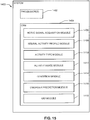
- the system may comprise at least one processor; a CRM containing a subject monitor application comprising a plurality of modules executable on the at least one processor; and a GUI module for generating one or more forms used to receive inputs to the system and to deliver output from the system.
- the plurality of modules may comprise: a neural signal acquisition module for obtaining one or more neural signals in one or more upper airway afferents of the subject; a neural activity profile module for processing each of the one or more neural signals to obtain at least one neural activity profile; an activity type module for comparing each of the at least one neural activity profiles to one or more activity criteria to associate each neural activity profile with an associated activity type; and an activity state module for processing each of the at least one neural activity profiles to determine an activity state characterizing the associated activity type.
- Each neural activity profile, activity type, and activity state may be characterized as described above. Suitable activity criteria are also described above.
- the neural activity profile module may further analyze a timing sequence of two or more activity patterns, wherein each of the two or more activity patterns is obtained from different upper airway afferents.
- the plurality of modules may further comprise a condition module for processing the at least one activity state to obtain at least one condition of the subject.
- the at least one condition of the subject may be chosen from a respiratory condition, a deglutition condition, a vibration condition, a reflux condition, and any combination thereof. Suitable respiratory, deglutition, vibration, and reflux conditions are described above.
- the system may further comprise a disorder prediction module for assessing the at least one condition to predict a disorder.
- the disorder may be chosen from obstructive apnea, central apnea, dysphagia, heart failure, uremia, asthma, cardiac arrest, organ failure, metabolic acidosis, COPD, pulmonary embolism, Ondine's curse, obesity hypoventilation syndrome, laryngeal penetration, aspiration, esophageal reflux, laryngeal reflux, presence of bolus in esophagus, acid reflux, GERD, laryngeal penetration, aspiration, and any combination thereof.
- the invention also provides a first method for treating and/or preventing a disorder in a subject in need thereof.
- the method comprises delivering at least one stimulation to modulate at least one reflex chosen from a swallowing reflex, a negative-pressure reflex, and any combination thereof.
- the disorder comprises at least one of: obstructive apnea, central apnea, obesity hypoventilation syndrome, dysphagia, esophageal reflux, presence of bolus in esophagus, acid reflux, GERD, and any combination thereof.
- Each of the at least one stimulations is delivered at a subthreshold intensity insufficient to elicit the reflex or at a suprathreshold intensity sufficient to elicit the reflex.
- the at least one stimulation is delivered according to a delivery schedule chosen from periodic, random, and continuous.
- Each of the at least one stimulations may comprise an electrical stimulation or a mechanical stimulation.
- Each electrical stimulation may be delivered to a reflex-related nerve, a reflex-related muscle, a reflex-related sensory receptor, and any combination thereof.
- Each mechanical stimulation may be delivered to a reflex -related sensory receptor.
- the reflex -related nerve may comprise an afferent or an efferent.
- An afferent may be chosen from: iSLN branch of vagus nerve, pharyngeal branch of vagus nerve, pharyngeal branch of glossopharangeal nerve, tonsular branch of glossopharangeal nerve, lingual branch of glossopharangeal nerve, intermediate nerve, palantine nerve, greater petrosal nerve, any branch of facial nerve, and pterygopalatine nerve of trigeminal nerve.
- An efferent may be chosen from: recurrent laryngeal nerve, external branch of superior laryngeal nerve, brancial motor branch of glossopharyngeal nerve and proximal fibers, mandibular nerve, medial pterygoid nerve, pharyngeal branch of vagus nerve and proximal fibers; branch of facial nerve and proximal fibers, and branch of hypoglossal nerve and proximal fibers.
- the reflex -related sensory receptor may be situated in skin or mucosa of the subject, and may be chosen from: a mechanoreceptor sensitive to negative airway pressure, positive airway pressure, stretch, position, shear, slip, vibration, texture, touch, mechanical compression, muscle stretch, muscle drive, air flow, blood pressure or blood osmolarity; a chemoreceptor sensitive to C02, 02, or pH; a thermoreceptor sensitive to temperature or airflow; and a nociceptor sensitive to polymodal pain.
- Each of the at least one stimulations may be chosen from: a subthreshold electrical stimulation delivered to the reflex -related nerve or to the reflex -related sensory receptor to reduce the threshold of the reflex, to maintain muscle tone, and any combination thereof; a subthreshold electrical stimulation delivered to the reflex -related muscle to maintain muscle tone; a subthreshold mechanical stimulation delivered to the reflex-related sensory receptor to reduce the threshold of the at least one reflex; a suprathreshold electrical stimulation delivered to the reflex -related nerve, the reflex-related sensory receptor, the reflex -related muscle, or any combination thereof to maintain muscle tone, position and/or posture of one or more respiratory and/or deglutition structures of the subject; a suprathreshold mechanical stimulation delivered to the reflex -related sensory receptor to maintain muscle tone, position and/or posture of one or more respiratory and/or deglutition structures of the subject; a suprathreshold electrical stimulation delivered to the reflex-related nerve, the reflex -related sensory receptor, the reflex-related muscle, or any combination thereof to treat the disorder
- Each of the at least one stimulations is delivered either according to a predetermined schedule, in response to at least one stimulation signal, and any combination thereof.
- the at least one stimulation signal may be received from a patient monitor device.
- the first method for treating and/or preventing a disorder in a subject in need thereof may further comprise assessing at least one condition of the subject chosen from a respiratory condition, a deglutition condition, a vibration condition, a reflux condition, and any combination thereof to predict the occurrence of the disorder in the subject. Suitable respiratory, deglutition, vibration, and reflux conditions are described above.
- the first method for treating and/or preventing a disorder in a subject in need thereof may further comprise obtaining one or more neural signals from one or more upper airway afferents of the subject; processing each of the one or more neural signals to obtain at least one neural activity profile; comparing each of the at least one neural activity profiles to one or more activity criteria to associate each neural activity profile with an associated activity type; processing each of the at least one neural activity profiles to determine an activity state characterizing the associated activity type; and processing the activity state of the subject to obtain the at least one condition of the subject.
- Each neural activity profile, activity type, and activity state may be characterized as described above.
- the first method for treating and/or preventing a disorder in a subject in need thereof may further comprise generating the at least one stimulation signal when: the disorder is predicted to time the delivery of the at least one stimulation to coincide with an occurrence of the disorder; the respiratory phase is an exhalation phase to time the delivery of the at least one stimulation to coincide with an exhalation of the subject; and any combination thereof.
- the invention also provides a first system for treating and/or preventing a disorder in a subject.
- the system may comprise at least one processor and a CRM containing a disorder treatment application comprising a plurality of modules executable on the at least one processor.
- the plurality of modules may comprise: a reflex stimulation module for delivering at least one stimulation to modulate at least one reflex chosen from a swallowing reflex, a negative-pressure reflex, and any combination thereof; and a GUI module for generating one or more forms used to receive inputs to the system and to deliver output from the system.
- the disorder may be chosen from obstructive apnea, central apnea, obesity hypoventilation syndrome, dysphagia, esophageal reflux, presence of bolus in esophagus, acid reflux, GERD, and any combination thereof.
- Each of the at least one stimulations is delivered at an intensity chosen from a subthreshold intensity insufficient to elicit the reflex and a suprathreshold intensity sufficient to elicit the reflex.
- the at least one stimulation is delivered according to a delivery schedule chosen from periodic, random, and continuous.
- Each of the one stimulations may comprise an electrical stimulation or a mechanical stimulation, as described above.
- the plurality of modules may further comprise a stimulation timing module for timing the delivery of each of the at least one stimulations according to a predetermined schedule, in response to at least one stimulation signal, and any combination thereof.
- the at least one stimulation signal may be received from a patient monitor system.
- the plurality of modules may further comprise a disorder prediction module for assessing at least one condition of the subject chosen from a respiratory condition, a deglutition condition, a vibration condition, a reflux condition, and any combination thereof to predict the occurrence of the disorder in the subject. Suitable respiratory, deglutition, vibration, and reflux conditions are described above.
- the plurality of modules may further comprise a neural signal acquisition module for obtaining one or more neural signals from one or more upper airway afferents of the subject; a neural activity profile module for processing each of the one or more neural signals to obtain at least one neural activity profile; and an activity type module for comparing each of the at least one neural activity profiles to one or more activity criteria to associate each neural activity profile with an associated activity type.
- a neural signal acquisition module for obtaining one or more neural signals from one or more upper airway afferents of the subject
- a neural activity profile module for processing each of the one or more neural signals to obtain at least one neural activity profile
- an activity type module for comparing each of the at least one neural activity profiles to one or more activity criteria to associate each neural activity profile with an associated activity type.
- Each neural activity profile and activity type maybe characterized as described above.
- the stimulation timing module may generate the at least one stimulation signal when: the disorder prediction module predicts the disorder in order to time the delivery of the at least one stimulation to coincide with an occurrence of the disorder; the activity, state module determines that the respiratory phase is an exhalation phase, to time the delivery of the at least one stimulation to coincide with an exhalation of the subject; and any combination thereof.
- the invention also provides a second method for treating and/or preventing a disorder in a subject in need thereof.
- the method may comprise obtaining one or more neural signals from one or more upper airway afferents of the subject; processing each of the one or more neural signals to obtain at least one neural activity profile; comparing each of the at least one neural activity profiles to one or more activity criteria to associate each neural activity profile with an associated activity type; processing each of the at least one neural activity profiles to determine an activity state characterizing the associated activity type; processing the activity state of the subject to obtain the at least one condition of the subject; assessing the at least one condition to predict a disorder chosen from obstructive apnea, central apnea, obesity hypoventilation syndrome, dysphagia, esophageal reflux, presence of bolus in esophagus, acid reflux, GERD, and any combination thereof; and delivering at least one stimulation to modulate at least one reflex chosen from a swallowing reflex, a negative-pressure reflex, and any combination thereof
- Each of the one stimulations may comprise an electrical stimulation or a mechanical stimulation, as described above.
- Each neural activity profile, activity type, and activity state may be characterized as described above.
- the at least one condition of the subject may be chosen from a respiratory condition, a deglutition condition, a vibration condition, a reflux condition, and any combination thereof. Suitable respiratory, deglutition, vibrations, and reflux conditions are described above, as are suitable activity criteria.
- Processing the one or more neural signals further comprises analyzing a timing sequence of two or more activity patterns, wherein each of the two or more activity patterns is obtained from different upper airway afferents.
- Each of the at least one stimulations is delivered either according to a predetermined schedule, in response to at least one stimulation signal, and any combination thereof.
- the second method for treating and/or preventing a disorder in a subject in need thereof may further comprise displaying any one or more of the at least one states, the at least one conditions, the at least one disorders, and any combination thereof on a patient monitor device.
- the second method for treating and/or preventing a disorder in a subject in need thereof may further comprise generating the at least one stimulation signal when: the disorder is predicted, to time the delivery of the at least one stimulation to coincide with an occurrence of the disorder; the respiratory phase is an exhalation phase, to time the delivery of the at least one stimulation to coincide with an exhalation of the subject; and any combination thereof.
- the invention also provides a second system for treating and/or preventing a disorder in a subject
- the system may comprise at least one processor; a CRM containing a disorder treatment application comprising a plurality of modules executable on the at least one processor; and a GUI module for generating one or more forms used to receive inputs to the system and to deliver output from the system.
- the plurality of modules may comprise: (i) a neural signal acquisition module for obtaining one or more neural signals in one or more upper airway afferents of the subject; (ii) a neural activity profile module for processing each of the one or more neural signals to obtain at least one neural activity profile; (iii) an activity type module for comparing each of the at least one neural activity profiles to one or more activity criteria to associate each neural activity profile with an associated activity type; (iv) an activity state module for processing each of the at least one neural activity profiles to determine an activity state characterizing the associated activity type; (v) a condition module for processing the at least one activity states to obtain at least one condition of the subject chosen from a respiratory condition, a deglutition condition, a vibration condition, a reflux condition, and any combination thereof; (vi) a disorder prediction module for assessing the at least one condition to predict a disorder chosen from: from obstructive apnea, central apnea, obesity hypoventilation syndrome, dysphagia, es
- the neural activity profile module may further analyze a timing sequence of two or more activity patterns, wherein each of the two or more activity patterns is obtained from different upper airway afferents.
- Each of the one stimulations may comprise an electrical stimulation or a mechanical stimulation, as described above.
- the at least one stimulation signal may be received from a monitor system.
- the stimulation timing module may generate the at least one stimulation signal when: the disorder prediction module predicts the disorder in order to time the delivery of the at least one stimulation to coincide with an occurrence of the disorder; the activity state module determines that the respiratory phase is an exhalation phase, to time the delivery of the at least one stimulation to coincide with an exhalation of the subject; and any combination thereof.
- a novel method of monitoring an upper airway condition in a patient including, but not limited to a respiratory condition such as apnea, a deglutition (swallowing) condition such as dysphagia, a vibration condition such as snoring, and/or a reflux condition such as GERD is provided that includes processing one or more neural signals obtained from one or more upper airway afferents.
- a respiratory condition such as apnea
- a deglutition (swallowing) condition such as dysphagia
- a vibration condition such as snoring
- a reflux condition such as GERD
- GERD a reflux condition
- neural signals carried by upper airway afferent nerves including, but not limited to, the internal branch of the superior laryngeal nerve (iSLN) may be processed to extract information that may be used to monitor the respiratory, deglutition, vibratory, and/or reflux status of the pharynx and to detect and characterize adverse conditions.
- Upper airway afferent neural signals may be obtained and processed using aspects of the method described herein below to detect and characterize such diverse conditions as sleep apnea, heart failure, hypoventilation syndrome, dysphagia, acid reflux, and snoring.
- Embodiments of the method exploit the normal function and organization of the peripheral nervous system by monitoring the activity of sensory nerve fibers. By tapping into the neural communication between the body's own biological sensors and central nervous system, the method can directly monitor the intrinsic sensor set of the subject that gives rise to the sensory percepts and the physiological responses to stimulation of the innervated area.
- upper airway afferents including, but not limited to glossopharyngeal afferents (pharyngeal, tonsilar and lingual branches of glossopharyngeal nerve), and other vagus afferents (pharyngeal branch of vagus nerve) may be used to monitor could be used to monitor upper airway conditions.
- two or more upper airway afferents may be monitored simultaneously. In these other methods, the processing of neural signals from multiple upper airway afferents increases the surface area of pharyngeal mucosa monitored, potentially resulting in more sensitive detection and localization of any obstructions or other anomalies.
- the simultaneous monitoring of multiple afferents may allow for spatial and/or temporal patterns of activity associated with upper airway conditions such as apnea and/or dysphagia/swallowing to be characterized and to further allow for the development of a tailored therapy based on the measured spatial/temporal pattern.
- the expanded selection of upper airway afferents available for use in various aspects of the method may result in enhanced surgical access for placement of neural activity measurement devices including, but not limited to nerve cuffs.
- apnea encompasses any form of involuntary apnea, bradypnea or hypopnea of obstructive, central or mixed origin, including sleep apnea and sleep hypopnea, and also includes any complex episode of apnea or hypopnea occurring during sleep or wakefulness, as in Cheyne-Stokes respiration.
- swallow-related refers to the nerve or a muscle as one for which normal function includes activity that effects, or contributes to effecting, all or any part of a normal oropharyngeal swallow sequence, wherein a swallow sequence refers to that reflexive and centrally programmed series of muscle movements beginning with muscle movements in an oral phase under voluntary muscular control and proceeding with pharyngeal and esophageal phases under involuntary neuromuscular control.
- the terms “subject” and “patient” are used interchangeably irrespective of whether the subject has or is currently undergoing any form of treatment.
- the terms “subject” and “subjects” refer to any vertebrate, including, but not limited to, a mammal (e.g., cow, pig, camel, llama, horse, goat, rabbit, sheep, hamster, guinea pig, cat, dog, rat, mouse, non-human primate (including but not limited to a monkey, such as a cynomolgous monkey, rhesus monkey, and chimpanzee), and a human).
- a mammal e.g., cow, pig, camel, llama, horse, goat, rabbit, sheep, hamster, guinea pig, cat, dog, rat, mouse
- non-human primate including but not limited to a monkey, such as a cynomolgous monkey, rhesus monkey, and chimpanze
- apnea is defined to mean either an obstructive, central, mixed, or complex episode of apnea or hypopnea, occurring during sleep or when awake as in Cheyne-Stolzes respiration.
- sensing refers to a pharyngeal vibratory state.
- a system and method of monitoring an upper airway condition or disorder processes at least one neural signal obtained from an upper airway afferent of the subject and extracts information characterizing an upper airway condition. In other aspects, this information may be further analyzed to predict an upper airway disorder.
- the parameters resulting from the implementation of this system and method in various aspects may be communicated to a display, and/or these parameters may be transferred to a display devices, a patient monitor, and/or a treatment device.
- the parameters characterizing the upper airway condition and/or disorder may be communicated to a system and method of preventing and/or treating an upper airway condition and/or disorder for use in generating a treatment.
- the system and method of preventing and/or treating an upper airway condition and/or disorder delivers at least one stimulation to modulate at least one reflex including, but not limited to, a swallowing reflex, a negative pressure reflex, or any combination thereof.
- the stimulation may be delivered to a reflex-related nerve, a reflex-related muscle, a reflex -related sensory receptor, and any combination thereof.
- the delivery of the stimulation may reduce the threshold of the reflex by enhancing the intensity of the neural signal delivered by an upper airway afferent in one aspect.
- the delivery of the stimulation may maintain the muscle tone of upper airway muscles involved in a preselected activity.
- the delivery of the stimulation may trigger the reflex, which may include but is not limited to a swallow reflex and a negative pressure reflex.
- the stimulation may be delivered autonornously according to a predetermined schedule.
- the stimulation may be delivered in response to a stimulation signal generated using parameters characterizing the upper airway condition and/or disorder. These parameters may be received from an independent device including, but not limited to, a patient monitor device in one aspect.
- the parameters may be generated by the system and method of monitoring an upper airway condition described herein in various aspects.
- system and method of monitoring an upper airway condition and the system and method of preventing and/or treating an upper airway condition and/or disorder may be combined into a system and method for monitoring, preventing, and/or treating an upper respiratory condition and/or disorder in other additional aspects.
- FIG. 11 is a flow chart illustrating the method 1100 in an aspect.
- at least one neural signal is obtained from an upper airway afferent such as an iSLN using a measurement device such as a nerve electrode at step 1102.
- the at least one neural signal may be amplified and processed using an algorithm such as a rectification and bin-integration (RBI) algorithm to obtain one or more neural activity profiles at step 1104.
- the one or more neural activity profiles may include information characterizing aspects of the one or more neural signals including, but not limited to, the neural signal's timing, phase, amplitude, conduction velocity, and position.
- the one or more neural activity profiles may be compared to one or more activity criteria to associate each neural activity profile with an associated activity type at step 1106.
- Each activity criteria may include one or more predetermined reference values uniquely characterizing the associated activity type. For example, a reflux criterion, which typically involves a pain signal generated by a "C" type, may be characterized by a conduction velocity of less than about 2 m/s. Thus, if a neural activity profile is determined to include a conduction velocity of less than about 2 m/s during the comparison of step 1106, this neural activity profile's associated activity type would be a reflux activity type.
- the associated activity type for a particular neural activity profile may be further used to guide subsequent analysis of the profile.
- each neural activity profile is processed at step 1108 to determine one or more activity states characterizing the profile.
- one or more reflux states may be obtained at step 1108 including, but not limited to: reflux timing, reflux pH, reflux location, and any combination thereof.
- the one or more activity states represent parameters that characterize and/or quantify a particular activity prior to a diagnosis of the subject.
- the one or more activity states determined at step 1108 may be displayed and/or communicated to another device such as a patient monitor device or treatment device.
- the one or more activity states may be processed to obtain at least one condition of the subject at step 1110.
- the one or more conditions obtained at step 1110 represent a diagnosis regarding the healthy or appropriate function of the subject with respect to one or more activities. For example, if the associated activity type of a neural activity was a reflux activity type, the one or more reflux activity states may be processed to obtain one or more reflux conditions including, but not limited to, esophageal reflux, pharyngeal reflux, laryngeal reflux, and any combination thereof.
- the at least one condition of the subject obtained at step 1110 may be displayed and/or communicated to another device such as a patient monitor device or treatment device in an aspect.
- the at least one condition may be assessed at step 1112 to predict a disorder.
- the disorder may represent a broader characterization of the subject's health or physiological status. For example, if one or more reflux conditions were obtained at step 1110, a related disorder including, but not limited to GERD or acid reflex may be predicted at step 1112.
- the one or more disorders determined at step 1112 may be displayed and/or communicated to another device such as a patient monitor device or treatment device.
- the one or more disorders may be further processed to determine a treatment for the disorder by a combined method of monitoring, preventing, and/or treating an upper airway disorder as described herein below.
- the method 1100 obtains conditions at step 1110 and predicts disorders at step 1112.
- the conditions represent a diagnosis regarding the healthy or appropriate function of the subject with respect to one or more activities.
- Disorders by contrast may represent a more systemic dysfunction of the subject with respect to one or more activities including, but not limited to a respiratory activity, a deglutition activity, vibration activity, a reflux activity, and any combination thereof.
- a respiratory activity a respiratory activity, a deglutition activity, vibration activity, a reflux activity, and any combination thereof.
- the method 1100 may obtain one or more respiratory conditions at step 1110.
- respiratory conditions include normal breathing, apnea, tachypnea, hyperpnea, hypopnea, polypnea, dyspnea, bradypnea, cough, Cheyne-Stokes respiration, Biot's respiration, ataxic respiration, Kussmaul respiration, wheezing, irregular respiration, respiratory arrest, restrictive respiration, shallow breathing, and hypoventilation.
- the method 1100 may predict one or more disorders, including one or more respiratory disorders and/or respiratory-related disorders, at step 1112.
- respiratory disorders and respiratory-related disorders include obstructive apnea, central apnea, heart failure, asthma, cardiac arrest, organ failure, metabolic acidosis, COPD, hypoventilation syndrome, laryngeal penetration, and aspiration.
- the diaphragm and intercostal muscles contract, creating a negative pressure in the airway and drawing air into the lungs.
- Expiration is typically passive, resulting from relaxation of the diaphragm and intercostal muscles back to their resting position, and elastic recoil of the lungs.
- the amount of air flow produced by a given inspiratory pressure is influenced by resistance from the structures of the upper airway, including the soft palate, tongue, and epiglottis.
- FIG. 1 A schematic illustration of the human airway 100, in particular the upper airway 110, is provided in FIG. 1 .
- the diaphragm and intercostal muscles 120 contract to a flattened position 122, inducing a negative pressure in the airway 100 and drawing air into the lungs 104.
- Expiration is typically passive, resulting from relaxation of the diaphragm and intercostal muscles 120 back to a upward domed resting position 124, and elastic recoil of the lungs 104.
- the amount of outward air flow through the larynx 102 produced by the change in airway 100 pressure may be influenced by resistance from the structures of the upper airway 110, including the soft palate 112, tongue 114, pharynx 116, and epiglottis 118.
- FIG. 2 is a graph 200 summarizing the airway pressure 201 measured at the larynx 102 (see FIG. 1 ) during a normal breathing process, comprising regular inspiration 202 and expiration 204 peaks of comparable amplitude and frequency.
- Airway pressure at the larynx 102 is perceived by mucosal mechanoreceptors that are sensitive to pressure; this airway pressure is communicated to the central nervous system via the internal branch of the superior laryngeal nerve (iSLN).
- iSLN superior laryngeal nerve
- FIG. 3 is a graph 300 summarizing the activity profile 302 measured during the normal breathing process.
- the activity profile 302 exhibits a similar regularly-spaced neural activity surges with little variation in activity surge width 304, peak surge amplitude 306, time between bursts 308, and/or separation of surge peaks 310.
- the amplitude of these bursts in the activity profile during each breath occurs within a normal range of amplitudes which may be determined using a calibration process during normal respiration of a given subject using, for example, polysomnographic techniques. This range of amplitudes may be used to determine upper and lower thresholds for normal breath detection using the method 1100. Bursts with peaks outside of this normal range may be detected using simple fixed-level thresholds and defined as abnormal respiratory events.
- the pattern of changing respiratory pressures may be used to identify respiratory pattern, respiratory timing, respiratory phase, and the amplitude of airway pressure.
- sleep apnea The principal forms of sleep apnea are: 1) obstructive sleep apnea (OSA), characterized by a physical blockage of the upper airway during sleep, 2) central sleep apnea (CSA), caused by a decreased central respiratory drive during sleep, and 3) mixed sleep apnea, which includes components of both OSA and CSA.
- OSA obstructive sleep apnea
- CSA central sleep apnea
- mixed sleep apnea which includes components of both OSA and CSA.
- OSA obstructive sleep apnea
- CSA central sleep apnea
- mixed sleep apnea which includes components of both OSA and CSA.
- OSA obstructive sleep apnea
- CSA central sleep apnea
- mixed sleep apnea which includes components of both OSA and CSA.
- OSA obstruct
- the obstructive component in OSA is related to decreased pharyngeal tone as the muscles relax during sleep.
- upper airway patency is maintained by the negative pressure reflex, which activates pharyngeal dilators in response to negative transthoracic pressure during inspiration.
- the negative pressure reflex is insufficient to maintain patency during sleep.
- the negative pressure created during inspiration in tandem with gravitational force acting on the surrounding tissues is sufficient to constrict or collapse the lumen of the flaccid airway.
- FIG. 4 is a schematic representation of the human airway during an OSA event.
- a lack of muscle tone in the upper airway 110 allows pharyngeal structures 116 to partially or completely block the lumen 119 of the airway 100, particularly when subjects sleep on their back. Respiratory drive continues during the OSA event, the diaphragm and intercostal muscles 120 contract 122, creating a negative pressure in the airway 100 that draws flaccid pharyngeal structures 116 into the airway lumen 119.
- FIG. 5 is a graph 400 of airway pressure 401 measured at the larynx 102 (see FIG. 3 ) at the outset of an OSA event, comprising normal breathing process inspiration 402a and expiration 404a peaks before the OSA event and then inspiration 402b and expiration 404b peaks of a greater amplitude during the OSA event.
- This increase in the amplitude of the airway pressure 401 reflects continuing attempts on the part of the subject to breathe after airway obstruction, generating greater than normal airway pressures 401.
- the outset of the OSA event can then be identified by the sudden increase in amplitude of the inspiration 402 and expiration 404 peaks of the airway pressure 401.
- FIG. 6 A schematic representation of the human airway 100 during a CSA event is illustrated in FIG. 6 .
- the upper airway 110 remains open, but diminished central respiratory drive reduces or eliminates diaphragm 120 movement, thereby reducing or halting air flow during the CSA event.
- FIG. 7 is a graph 600 of airway pressure 601 measured at the larynx 102 (see FIG. 5 ) at the outset of a CSA event, comprising normal breathing process inspiration 602 and expiration 604 peaks before the CSA event and then an absence of, or very low amplitude, inspiration and expiration peaks 606 during the CSA event.
- upper airway pressure 601 is not fully modulated after the onset of the CSA event and diminution of diaphragm movement.
- the outset of the CSA event 603 can then be identified by the sudden drop 606 in the amplitude of the inspiration 602 and expiration 604 peaks of the airway pressure 601.
- the method 1100 may obtain one or more deglutition conditions at step 1110.
- deglutition conditions include presence of bolus, occurrence of swallow, occurrence of dysphagic swallow, and presence of acid reflux.
- the method 1100 may predict one or more disorders, including one or more deglutition disorders and/or deglutition-related disorders, at step 1112.
- deglutition disorders and deglutition-related disorders include obstructive apnea, dysphagia, presence of bolus in esophagus, and aspiration.
- Deglutition or swallowing is a stereotyped reflex that exhibits a consistent pattern of activation of 50 muscles throughout the upper airway. This sequence acts to propel food and fluid caudally at speeds of about IM/sec with the pharyngeal stage of the swallow taking about 1 sec to complete.
- FIG. 8 is a schematic illustration showing the upper airway structures relevant to deglutition.
- the swallow sequence is essentially a progressive anterior-to-posterior wave of pharyngeal contact that acts to squeeze the bolus from the soft palate 112 posteriorly to the pharynx 116, further posteriorly to the epiglottis 118 and ultimately toward the esophagus 902 like a tube of toothpaste while simultaneously protecting the airway from entry of material.
- FIG. 9 A schematic diagram showing the anterior-to-posterior activation pattern in the activity profile is provided as FIG. 9 .
- the actual pattern of the activity profile is influenced by the pattern of mechanical contact and pressure on a given mucosal receptor and by the adaptation properties of the afferent fibers.
- FIG. 10 is a graph summarizing a variety of activity profiles associated with different types of individual fibers.
- the activity profile may be a "tonic" profile ( FIG. 10A ), a "buildup” profile ( FIG. 10B ) characterizing relatively slowly adapting receptors, an "on-sustained" profile (FIG.
- Spontaneously active fibers may exhibit, for example, a "tonically- inhibited" activity profile during applied pressure, as illustrated schematically in FIG. 10G .
- the area of and location of contact between the soft palate and posterior pharyngeal wall may also exhibit a stereotyped pattern during swallow, creating a spatial activity pattern across a population of fibers, in addition to the temporal activity pattern within individual fibers.
- the anterior to posterior pharyngeal contact pattern would act to create a stereotypical spatial activity pattern, with most anterior fibers being activated at the beginning of the swallow sequence and the most posterior fibers being activated about a second later, as illustrated schematically in FIG. 9 .
- the neural signals recorded from iSLN receptors are relevant to the gastrointestinal (GI) condition of a subject.
- GI gastrointestinal
- the iSLN mechanoreceptors normally indicate bolus contact and trigger a swallow sequence.
- Dysphagia refers to the medical symptom of difficulty in swallowing, and is frequently diagnosed in subjects also presenting with sleep apnea. Subjects may have a great deal of difficulty in controlling even saliva in the mouth, or difficulty in initiating a swallow, or a cough. Dysphagia thus represents a further example of a high medical risk due to impaired pharyngeal motor control.
- the activity profile within and between individual fibers and fiber populations may be determined using a calibration process during normal deglutition of a given subject, created for example, during volitional "dry" swallowing or in the presence of an administered bolus of food or fluid.
- the activity profile for dysphagic swallow and/or presence of an unswallowed bolus may be similarly determined.
- the range of normal temporal and/or spatial activity patterns observed during the calibration process can computed and be used to set for example matched-filter templates and upper and lower thresholds for detection of normal swallow and dysphagic swallow. Peaks within this range may be detected using simple fixed- level thresholds and used to assign a deglutition activity as the associated activity type of the neural activity profile.
- the method 1100 may obtain one or more vibration conditions at step 1110.
- vibration conditions include snoring, stridor, wheezing and vocalization.
- the method 1100 may predict one or more disorders, including one or more vibration disorders and/or vibration -related disorders, at step 1112. Detailed descriptions of selected deglutition conditions and disorders are provided herein below.
- Snoring is caused by the vibration of flaccid pharyngeal tissues during sleep, and snoring may an early indicator of the development of an obstructive sleep apnea (OSA).
- OSA obstructive sleep apnea
- the walls of the mucosa are known to contain specialized mechanoreceptors that are sensitive to vibration.
- Three different vibration receptor types are known, each responding best to vibration over a different range of frequencies. Merkel disks, for example, respond best to vibrations from about 5-15 Hz, while Meissner corpuscles have a best frequency of about 50 Hz. Both of these receptors types have been histologically identified in the airway mucosa.
- a third class of mucosal mechanoreceptors is known to respond to vibration up to 300 Hz, with a best frequency of about 150 Hz; these response properties correspond to those known for Pacinian corpuscle receptors.
- FIG. 11A is graph 1000 illustrated schematically an activity profile 1002 of a vibration- sensitive mechanoreceptor.
- the activity profile 1002 shown at a zoomed in time scale these vibration- sensitive mechanoreceptors may exhibit phase- locked activity at the frequency of the vibration, thereby encoding the stimulus frequency by a single action potential 1004 on every cycle, or at higher frequencies, at integer multiples of the interval between cycles. This produces a characteristic interspike interval 1006 for these phase locking fibers that matches or is a multiple of the period of the vibration.
- the envelope of the activity profile during pharyngeal vibration may exhibit amplitude modulations as a result of phase locking, as illustrated by the graph
- the interspike interval 1006, amplitude modulation frequency, vector strength and modulation depth occurring at a given vibration frequency may be determined using a calibration process during normal pharyngeal vibration of a given subject, created for example, by volitional vocalization, snoring, or wheezing. Artificially induced vibration using, for example, a piezoelectric vibrator placed on the skin over the pharynx may also be used.
- the range of interspike intervals, amplitude modulation frequencies, vector strengths and modulation depths observed during the calibration process can computed and be used to set upper and lower thresholds for vibration detection. Peaks within this range can be detected using simple fixed- level thresholds and defined as vibration events.
- the pattern of pharyngeal vibration encoded by characteristic interspike intervals and/or amplitude modulation of the activity profile of pharyngeal afferent nerves, can be used to identify vibration pattern, vibration timing, vibration phase, and the amplitude of vibration.
- Snoring is an upper airway condition that is characterized by vibration of the pharyngeal walls, tongue base, soft palate, and tonsils. It has been discovered that the principal frequency range of human snoring occupies a spectrum from 40-300Hz with a peak spectral power at about 100 Hz. The frequency spectrum of snoring vibration activity is well-matched to the frequency range of pharyngeal vibration sensitive mechanoreceptors and makes monitoring of pharyngeal afferents a particularly well-suited method for snoring detection.
- Specific airway structures are known to vibrate at characteristic frequencies, for example, the tonsils and soft palate vibrate at about 170 and 140 Hz, respectively. This characteristic may be used to pinpoint the structural source of snoring by monitoring pharyngeal vibration receptors. If multiple upper airway afferents and/or multiple fibers within a single nerve are monitored, the location of the source of pharyngeal vibration may also be pinpointed based on the receptive fields of active afferent fibers, either by comparing activity across multiple nerves, or by comparing activity across fibers within a single nerve.
- the method 1100 may obtain one or more reflux conditions at step 1110.
- reflux conditions include esophageal reflux, pharyngeal reflux, and laryngeal reflux.
- the method 1100 may predict one or more disorders, including one or more reflux disorders and/or reflux-related disorders, at step 1112.
- reflux disorders and reflux-related disorders from esophageal reflux, laryngeal reflux, acid reflux, and GERD.
- Gastroesophageal reflux disease gastric reflux disease, or acid reflux disease is a chronic symptom of mucosal damage caused by stomach acid coming up from the stomach into the esophagus. GERD may be divided into esophageal and extraesophageal syndromes. Acid reflux allowed by a transient relaxation of the lower esophageal sphincter that allows acid to pass into esophagus. Once in the esophagus, gastric acid can travel along the length of the esophagus, reaching or passing the level of the upper esophageal sphincter.
- Extraesophageal symptoms are caused by the entry of gastric juices in the larynx and pharynx through the upper esophageal sphincter.
- Laryngeal and pharyngeal symptoms are also known as laryngopharyngeal reflux (LPR) or extraesophageal reflux disease (EERD).
- Extraesophageal symptoms include dysphagia, voice disorders, asthma, hoarseness, laryngitis, chronic cough, pain, vocal fold nodules, unstable voice during speaking or singing.
- the mucosa of the upper airway is known to contain afferent fibers of different diameters and conduction velocities.
- cutaneous nerves there exist three populations of afferent fibers, each population having a characteristic signal conduction velocity.
- "A ⁇ " type fibers have the fastest signal conduction velocity and typically conduct action potentials along afferent fibers at a rate ranging from about 35 m/s to about 75 m/s.
- "A ⁇ " type fibers have an intermediate signal conduction velocity and typically conduct action potentials along afferent fibers at a rate ranging from about 5 m/s to about 30 m/s.
- C have the slowest signal conduction velocity and typically conduct action potentials along afferent fibers at a rate ranging from about 0.5 m/s to about 2 m/s.
- FIG. 17 A schematic illustration of this isolation technique is shown in FIG. 17 .
- a peripheral recording device 1702 and a central recording device 1704 are spaced a known separation distance along the length of a given set of fibers within an afferent nerve 1706 and used to record two separate neural signals 1708 and 1710, respectively.
- the recording devices 1702 and 1704 may be spaced about 1 mm apart along the length of the nerve and thus record the activity from the same set of nerve fibers at two different points along their length.
- a 1 ms delay may be introduced in the signal 1708 from the peripheral recording device 1702 which is then summed with the neural signal 1710 obtained from the central recording device 1704.
- the "C" fiber activity covers the distance between the electrodes in about 1 ms, and thus the signals 1708 and 1710 from the recording devices 1702 and 1704 overlap in time and are added together, as schematically illustrated in graph 1714.
- the signals associated with activity from A ⁇ type fibers and A ⁇ type fibers have moved well past the central recording device 1704 during the 1 ms delay as illustrated in graphs 1716 and 1718, respectively.
- the portion of the signals 1708 and 1712 associated with activity from A ⁇ type fibers and A ⁇ type fibers are not added together.
- the activity of only the combined "C" fiber signals may be isolated.
- the activity of acid-sensitive "C" fibers may be separated from the activity of more rapidly conducting mechanoreceptors in upper airway afferents in one aspect.
- the activity profile of acid-sensitive "C" fibers may then be determined using a calibration process during acid reflux of a given subject, created for example during a normally occurring acid reflux, or created by artificial application of a low pH fluid to the pharynx.
- the range of normal temporal and/or spatial activity patterns observed during the calibration process may computed and be used to set, for example, upper and lower thresholds for detection of reflux in the airway in an aspect. Peaks within this range may be detected using simple fixed- level thresholds and the activity profile may be associated with a reflux activity type in another aspect.
- the temporal and spatial activity profiles during naturally occurring or artificial reflux conditions may also be determined in an additional aspect.
- the method obtains neural signals from upper airway afferents in order to monitor a condition and/or a disorder including, but not limited to a respiratory condition, a deglutition condition, a reflux condition, and a vibration condition.
- a condition and/or a disorder including, but not limited to a respiratory condition, a deglutition condition, a reflux condition, and a vibration condition.
- the upper airway afferents include nerves associated with mucosal sensory receptors situated throughout the upper airway of the subject.
- the neural signals produced by these mucosal sensory receptors provide a rich source of information to identify and characterize a variety of upper airway conditions and disorders, as described herein previously.
- Non-limiting examples of upper airway afferents include pharyngeal afferents, laryngeal afferents, oral cavity afferents and nasal cavity afferents.
- Pharyngeal afferents are known to transmit information from sensory receptors in the mucosa lining the upper airway to the brain. As used herein, pharyngeal afferents innervating additional areas of the upper airway, such as the larynx, are included in the term "pharyngeal afferents".
- Non-limiting examples of pharyngeal afferent include the iSLN branch of the vagus nerve, the pharyngeal branch of the vagus nerve, the pharyngeal branch of the glossopharangeal nerve, the tonsular branch of the glossopharangeal nerve, the lingual branch of the glossopharangeal nerve, the intermediate nerve, the palantine nerve, the greater petrosal nerve, any branch of the facial nerve, the pterygopalatine nerve of the trigeminal nerve, and any combination thereof.
- Various aspects of the methods described herein are intended to include both the pharyngeal and extrapharyngeal sensory receptor transmission functions.
- the iSLN innervates vocal folds in the larynx as well as mucosal sensory endings in the pharynx.
- mucosal receptors may be sensitive to stimuli including, but not limited to airway pressure characterizing respiratory conditions such as apnea, contact with food or fluid characterizing deglutitation conditions such as dysphagia, vibrations characterizing vibration conditions such as snoring, and pH characterizing reflux conditions.
- airway pressure characterizing respiratory conditions such as apnea
- contact with food or fluid characterizing deglutitation conditions such as dysphagia
- vibrations characterizing vibration conditions such as snoring
- pH characterizing reflux conditions such as apnea
- additional neural signals carried by upper airway afferents include chemoreceptors modulated by pH, taste receptors modulated by various chemical compounds, thermoreceptors modulated by temperature or airflow, or noiceptors modulated by polymodal pain.
- neural activity profile may be calculated for example by applying a rectification and bin-integration (RBI) algorithm to an amplified pharyngeal afferent signal and used to detect and/or monitor an upper airway condition and/or disorder.
- RBI rectification and bin-integration
- Neural response characteristics such as the number of elicited spikes, the interval between spikes, and the temporal spike pattern in response to continued stimulation may vary with stimulus variables such as intensity and duration.
- Each sensory nerve fiber responds to adequate stimuli applied within their "receptive field", defined herein as the specific area of the sensory epithelium innervated by a sensory nerve fiber.
- the spatial extent of sensory stimuli may be determined by comparing neural response between fibers with different receptive fields, which are often organized in a topographic map.
- spinal nerves are organized segmentally and typically innervate specific regions of skin (dermatomes) in an organized fashion, with cervical nerves innervating the upper body and sacral nerves innervating the lower body.
- stratomes specific regions of skin
- individual spinal nerves can also be somatotopically organized, with fibers from receptors close to one another within a dermatome grouped together into bundles called fascicles within the nerve.
- the receptive field characteristics associated with one or more sensory fibers within an upper airway afferent may be used to determine the spatial extent, distribution, patterning, or any other spatially-related characteristic of a stimulus associated with an upper airway condition and/or disorder monitored using neural signals from one or more upper airway afferents.
- Various aspects of the method are designed to monitor and interpret respiratory, reflux, vibration, and/or deglutitation conditions based on the neural signal characteristics including, but not limited to a firing pattern, an active fiber population, a signal conduction speed, and any combination thereof in pharyngeal afferent nerves.
- these the neural signal may be compared to previously analyzed responses to calibration stimuli in order to detect pharyngeal disorders such as apnea, snoring, dysphagia or GERD, as described previously herein.
- the method may obtain one or more neural signals from the iSLN. In other aspects, the method may obtain one or more neural signals from other pharyngeal afferents including, but not limited to trigeminal nerve afferents, facial nerve afferents associated with the from the oropharynx or nasopharynx cavity to monitor respiratory conditions and/or disorders, and glossopharyngeal afferents to monitor deglutitation conditions including, but not limited to the introduction of a bolus into the oropharynx and/or hypopharynx.
- pharyngeal afferents including, but not limited to trigeminal nerve afferents, facial nerve afferents associated with the from the oropharynx or nasopharynx cavity to monitor respiratory conditions and/or disorders, and glossopharyngeal afferents to monitor deglutitation conditions including, but not limited to the introduction of a bolus into the oropharynx and/or hypo
- upper airway conditions and/or disorders may also be monitored by obtaining neural signals from nerves carrying efferent signals to muscles of the upper airway, diaphragm, or intercostal muscles, or by monitoring the activity of these respiratory muscles themselves, alone, or in some combination with other nerves or muscles modulated by respiratory activity.
- any known device capable of detecting and recording neural signals from nerves including, but not limited to, upper airway afferents may be used to obtain the neural signals used to obtain the at least one neural activity profile in the method.
- suitable neural signal measurement devices include electrical sensors such as nerve cuffs; optical sensors such as optic fibers used in combination with voltage or current sensitive dyes injected into the nerve to be monitored; biological sensors; and mechanical sensors.
- any known method of recording peripheral nerve activity such as percutaneous microneurography or optical recording of nerve activity based on voltage sensitive dyes may be used without limitation.
- the size of the neural signal measurement devices may have a form factor ranging from about the size of a pacemaker to about the size of a cell phone application.
- an electroneurogram (ENG) of pharyngeal afferents may be monitored recording electrodes placed in, around, or near the nerve.
- a "cuff electrode may be situated around the nerve to record the aggregate activity of all nerve fibers in the vicinity of a single recording site.
- electrode designs and signal processing techniques may facilitate the selective recording of neural signals from smaller groups of nerve fibers ("multi units"), recording from individual nerve fibers ("single units"), discriminating the direction of an action potential propagation to differentiate between motor nerve fiber activity and sensory nerve fiber activity, and/or isolating a subset of neural signals based on the signal conduction velocity (isolating nerve fibers with a specific diameter).
- multiple recording electrodes may be used, depending in the application and anatomical location to be monitored, in order to simultaneously or sequentially monitor multiple signal sources.
- the recording electrode may also target other nerves carrying afferent signals from peripheral receptors that exhibit modulations of bioelectric potentials correlated with upper airway conditions.
- Non-limiting examples of receptors that may be monitored to characterize an upper airway condition respiratory condition include: mechanoreceptors sensitive to negative airway pressure, positive airway pressure, stretch, position, shear or slip, vibration, texture, touch, touch and pressure, muscle stretch, muscle "drive", air flow, blood pressure or osmolality; chemoreceptors sensitive to C02, 02, or pH; thermoreceptors sensitive to temperature or airflow; nociceptors sensitive to polymodal pain, or any combination thereof.
- the method may include at least one electrode sensitive to at least one upper airway characteristic including, but not limited to upper airway pressure, upper airway stretch, and upper airway airway temperature, or alternatively may include multiple electrodes sensitive to a combination of the upper airway characteristics.
- the monitoring of an upper airway condition may include monitoring the upper airway-related activity of other nerves, or monitoring other physical indicators of upper airway state, including, but not limited to airway pressure, muscle activity or airway flow as described in further detail herein below.
- the monitoring in this aspect may be achieved using any means capable of detecting a physical signal and transducing the signal to an electrical signal suitable for analysis.
- various physical indicators of respiration and respiratory state are amenable to detection and monitoring, including but not limited to airway pressure, air flow, muscle stretch, muscle position, muscle "drive”, blood pressure, blood osmolarity, blood gas (C02 and 02), heart rate, and blood pH.
- multiple electrodes may be placed in or on the body to measure, for example, breathing rate and heart rate. Changes in abdominal or thoracic circumference related to respiration can be measured using belt-based systems with sensors based on piezoelectric or impedance sensors.
- An oximeter can be used to detect and monitor blood oxygen levels in the blood.
- a blood pressure cuff or arterial catheter may also be used, to detect and monitor blood pressure.
- EMG leads can be used to detect breathing muscle activity.
- a manometer can be placed in the nasal cavity to detect airway pressure.
- upper airway conditions may be monitored using any of a number of anatomical elements involved in respiration and control of respiration.
- respiratory activity may also be monitored from nerves carrying efferent signals to muscles of the upper airway, diaphragm, or intercostal muscles, or by monitoring the activity of these respiratory muscles themselves, alone, or in some combination with other nerves or muscles modulated by respiratory activity.
- one or more neural activity profiles characterizing aspects of the neural signals including, but not limited to, neural signal timing, neural signal amplitude, neural signal phase, neural signal position, neural signal conduction velocity, and any combination thereof, may be obtained using one or more signal processing techniques described herein above in connection with the upper airway conditions. Additional signal processing techniques are described in detail herein below.
- the neural signals obtained from the one or more upper airway afferents are processed to obtain at least one neural activity profile.
- the neural signals obtained from the one or more upper airway afferents may be conditioned by any known method including but not limited to amplification prior to performing any additional signal processing.
- the neural activity profile may be obtained by applying a rectification and bin-integration (RBI) algorithm to the conditioned (i.e. amplified) neural signals.
- RBI rectification and bin-integration
- a pressure in the upper airway may be monitored by obtaining an electroneurogram (ENG) of the iSLN, which is correlated with pressure in the upper airway.
- An index of respiratory activity (IRA) may be calculated by applying a rectification and bin-integration (RBI) algorithm to the amplified iSLN neural signal.
- the amplitude of peaks in the IRA during each breath that occur within a normal range of amplitudes may be determined using a calibration process during normal respiration of a given subject using, for example, polysomnographic techniques. This range of "normal" amplitudes can be used to define upper and lower thresholds for the detection of one or more respiratory conditions.
- Peak amplitudes fallingO outside of this normal range may be detected using simple fixed-level thresholds and defined as a respiratory conditions.
- the defined upper and lower thresholds may further be used to classify, in real-time, a detected apneic event as being either an OSA event or a CSA event, as described herein previously.
- the signal conditioning may be implemented by any known signal processing circuitry including, but not limited to, a signal amplifier and a rectifier circuit.
- suitable amplifiers and rectifier circuits are disclosed in U.S. Patent Application Publication No. 2006/0189881 entitled “IMPLANTABLE SIGNAL AMPLIFYING CIRCUIT FOR ELECTRONEUROGRAPHIC RECORDING", published Aug. 24, 2006, by Baru Fassio and U.S. Pat. No. 7,282,980 entitled “PRECISION RECTIFIER CIRCUIT FOR HIGH- DENSITY, LOW-POWER IMPLANTABLE MEDICAL DEVICE", issued Oct. 16, 2007, to Baru Fassio, both of which are incorporated by reference in their entirety.
- the activity profile may be calculated by applying a rectification and bin-integration (RBI) algorithm to the amplified pharyngeal afferent signal in one aspect
- RBI rectification and bin-integration
- other signal processing algorithms may also be applied to calculate the activity profile including, but not limited to: high pass filter, low pass filter, bandpass filter, notch filter, FIR filter, IIR filter, smoothing, moving average, Wiener (optimal) filter, matched filter, rectification, bin-integration, multichannel noise reduction, principal components analysis, independent components analysis, wavelet analysis, Fourier transformation, matched filtering, variance/variance ratio calculations, signal-to-noise ratios, cross-correlation, auto-correlation, Rayleigh statistic, and any combination thereof.
- RBI rectification and bin-integration
- the raw pharyngeal afferent ENG waveform may also be used directly.
- Activity profiles based on neural network analyses, cluster analysis in multidimensional feature space, cluster cutting using k-means, Bayesian expectation-maximization, likelihood ratios, closest centers, or manual cluster cutting methods may also be used in various aspects.
- an activity profile could be computed from any number of other pharyngeal afferent ENG signal features that vary with pharyngeal state such as event or waveform timing, interval, amplitude, duration, rise time, fall time, slope, presence, absence, pattern, 1st derivative, 2nd derivative, 3rd derivative, root mean square amplitude, peak-to-peak amplitude, variance, statistical probability or probability relative to baseline or running average.
- detection of pharyngeal events in the activity profile using methods other than fixed-level thresholding may be used, for example noise-tracking or other adaptive thresholds, energy or non-linear energy thresholds, or any variety of other detection operations on the raw or processed data.
- the signal processing of the one or more neural signals may further include analyzing a timing sequence of two or more activity patterns, wherein each of the two or more activity patterns is obtained from different upper airway afferents.
- the signal processing of the one or more neural signals may further include assessing the spatial extent or spatial location of the one or more detected neural signals using information characterizing the receptive fields associated with the one or more detected neural signals.
- the at least one neural activity profiles may be compared to one or more activity criteria to associate each neural activity profile with an associated activity type chosen from a respiratory activity type, a deglutition activity type, a vibration activity type, a reflux activity type, and any combination thereof.
- activity criteria chosen from a respiratory activity type, a deglutition activity type, a vibration activity type, a reflux activity type, and any combination thereof.
- the activity criteria describe various characteristics of a neural activity profile that may uniquely associate the neural activity profile with a particular upper airway activity including, but not limited to a respiratory activity, a deglutition activity, a vibration activity, and a reflux activity.
- the characteristics may include ranges, and/or threshold values of neural activity profile characteristics including, but not limited to a time separation of a neural signal feature such as a peak amplitude, a pattern of neural signals, and a signal conduction speed.
- the respiratory criterion indicating a respiratory activity may include: a time separation between peak neural signal amplitudes ranging from about 1 seconds to about 5 seconds; a periodically repeating pattern of neural signals with a period ranging from about 12 patterns per minute to about 60 patterns per minute; and any combination thereof.
- a deglutition criterion indicating a deglutition activity may include: an anterior to posterior neural activation pattern; a stereotyped neural activation pattern with a duration of less than about 1 second; and any combination thereof.
- a vibration criterion indicating a vibration activity may include a neural signal frequency ranging from about 10 Hz to about 400 Hz; a time separation between peak neural signal amplitudes ranging from about 1 second to about 5 seconds, and any combination thereof.
- a reflux criterion indicating a reflux activity may include a signal conduction velocity of less than about 2 m/s.
- the at least one neural activity profiles may be processed to determine an activity state characterizing the associated activity type.
- the activity state may include a respiratory state, a deglutition state, a vibration state, and a reflux state.
- the respiratory state may include respiratory timing, respiratory amplitude, respiratory phase, respiratory location, and any combination thereof.
- the respiratory phase may include either an inspiratory phase, expiratory phase, or zero flow phase between inspiratory phase, and expiratory phases.
- the respiratory phase may be defined for example with reference to peak amplitude in the IRA during each breath, as determined based on a calibration of normal respiration of a given subject using, for example, polysomnographic techniques.
- the deglutition state may include solid contact, fluid contact, contact, velocity, contact timing, contact amplitude, contact pressure, contact texture, contact temperature, a presence of a bolus, and any combination thereof;
- the vibration state may include vibration timing, vibration amplitude, vibration phase, vibration location, vibration pattern, and any combination thereof.
- the reflux state may include reflux timing, reflux pH, reflux location, and any combination thereof.
- the at least one activity states may be processed to obtain at least one condition of the subject chosen from a respiratory condition, a deglutition condition, a vibration condition, a reflux condition, and any combination thereof.
- the respiratory condition may include apnea, tachypnea, hyperpnea, hypopnea, polypnea, dyspnea, bradypnea, cough, Cheyne-Stokes respiration, Biot's respiration, ataxic respiration, Kussmaul respiration, wheezing, irregular respiration, respiratory arrest, restrictive respiration, shallow breathing, hypoventilation and any combination thereof.
- the deglutition condition may include presence of bolus, occurrence of swallow, occurrence of dysphagic swallow, presence of acid reflux, and any combination thereof.
- the vibration condition may include snoring, stridor, wheezing vocalization, and any combination thereof.
- the reflux condition may include esophageal reflux, pharyngeal reflux, laryngeal reflux and any combination thereof.
- any one or more of the at least one states, the at least one conditions, the at least one disorders, and any combination thereof may be displayed on a patient monitor device, and/or communicated to a treatment system.
- the at least one condition may be assessed to predict a disorder chosen from obstructive apnea, central apnea, dysphagia, heart failure, uremia, asthma, cardiac arrest, organ failure, metabolic acidosis, COPD, pulmonary embolism, Ondine's curse, obesity hypoventilation syndrome, laryngeal penetration, aspiration, esophageal reflux, laryngeal reflux, presence of bolus in esophagus, acid reflux, GERD, laryngeal penetration, aspiration, aspiration pneumonia, SIDS, Charcot-Marie-Tooth disease and any combination thereof.
- a disorder chosen from obstructive apnea, central apnea, dysphagia, heart failure, uremia, asthma, cardiac arrest, organ failure, metabolic acidosis, COPD, pulmonary embolism, Ondine's curse, obesity hypoventilation syndrome, laryngeal penetration, aspiration, esophage
- the detection and classification of apnea events as described herein is consistent with the detection and classification of apnea events as described in U.S. Patent Application Publication No. 2010/0125310 , i.e. involves calculating an index of respiratory activity (IRA) that is indicative of the amplitude and timing of respiratory activity based on the amplitude and timing of a respiratory signal, such as an electroneurogram (ENG) signal from a nerve such as the internal branch of the superior laryngeal nerve (iSLN), or another sensor of respiratory activity as described elsewhere herein.
- a respiratory signal such as an electroneurogram (ENG) signal from a nerve such as the internal branch of the superior laryngeal nerve (iSLN), or another sensor of respiratory activity as described elsewhere herein.
- ENG electroneurogram
- the algorithm executed by the apnea monitoring and detection module implements steps in the processes as discussed in further detail herein below.
- the apnea monitoring and detection module Upon the detection of an apnea event, the apnea monitoring and detection module sends a trigger to the therapy control module along with an identification of the type of apnea event, i.e. obstructive, central, or mixed; and apnea or hypopnea, depending on the implemented algorithm, which generates a stimulus appropriate for the type of apnea event.
- the apnea monitoring and detection module may also send an indication of the severity level of the apnea event, as well as timing information of previous or continuing respiration patterns, to the therapy control module 1106.
- the outset of an OSA event or a CSA event may be identified by features of the IRA, for example with reference to an upper and a lower threshold as described above.
- the first instance of a crossing of the upper threshold by inspiration related peaks of the IRA can be used as a criterion for identifying the outset of an OSA event.
- the peak durations of the RBI ENG may be used to identify the outset of an OSA event by setting an appropriate threshold.
- the outset of the CSA event can be identified, for example, by noting the first absence of crossing of the lower threshold by inspiration related peaks, in a set time period.
- This period of time may be set, for example, to represent the average time between one or more respiration cycles. It should be understood that for both OSA and CSA events, other IRAs maybe calculated in order to identify the outset of such an event. For example, peak durations and interpeak intervals of the RBI ENG can be used, by setting appropriate levels and thresholds. It is to be understood that the absence of measurements at a specified level may indicate a CSA event.
- apnea event severity can be determined from the IRA.
- severity of the apnea event may be determined by comparing the amplitude of the apneic IRA to that observed during normal breathing. More severe apnea is characterized by IRA peaks having amplitudes far from the upper and lower thresholds, while less severe apnea or hypopnea is characterized by IRA peaks having amplitudes just above or below the upper and lower thresholds. The level of apnea thus determined can be used to adjust the parameters and characteristics of the applied neurostimulation treatment.
- Severity levels may be assigned predetermined thresholds. It is to be understood that the number of OSA and CSA severity levels may vary depending on the precision of the circuitry and/or algorithm used.
- Apneic events may be further identified as hypopnea events, i.e. OSA events can be distinguished from obstructive sleep hypopnea (OSH) events, and CSA events can be distinguished from central sleep hypopnea (CSH) events with reference to the IRA.
- OSH obstructive sleep hypopnea
- CSH central sleep hypopnea
- an IRA value between a first upper threshold and a second upper threshold, wherein the second upper threshold is higher than the first upper threshold may be associated with OSH
- an IRA value greater than the second upper threshold may be associated with OSA.
- IRA peaks between the two upper thresholds can be identified as OSH while IRA peaks above the second, higher upper threshold can be identified as OSA.
- an IRA value between a first lower threshold and a second lower threshold, wherein the second lower threshold is lower than the first lower threshold may be associated with CSH, while an IRA value lower than the second lower threshold may be associated with OSA.
- the range of values for which IRA peaks are defined as OSH as opposed to OSA, as well as CSH as opposed to CSA, may be determined using a calibration process during abnormal respiration of a given subject using, for example, polysomnographic techniques.
- OSH, OSA, CSH and CSA may be subdivided into multiple severity levels depending on the precision of the circuitry and/or algorithm used.
- the variation in IRAs calculated using algorithms other than RBI ENG may also be used to determine the severity of the apneic or hypopneic event.
- Apneic events may be further identified by the location(s) of the airway obstruction using, for example, the temporal profile of the IRA activity pattern acquired from a single electrode or sensor.
- an apneic event may be further identified by the location(s) of the airway obstruction using, for example, the temporal pattern of IRA activity acquired across multiple electrodes or sensors, indicating, for example, the instantaneous pressure at multiple locations in the upper airway.
- the upper airway states, conditions, and/or disorders obtained using the method may be displayed or used to drive an alarm or alert.
- the upper airway states, conditions, and/or disorders obtained using the method may be used in the implementation of condition prevention/treatment methods as described herein below, or may be transmitted to other devices including but not limited to monitor devices and/or treatment devices.
- the method in various aspects may detect respiration rate, phase, and timing. This capability provides for general monitoring of vital signs, aside from apnea detection, and may be used to provide respiration-related or other upper airway-related parameters to other devices such as external monitoring equipment, or implanted devices such as pacemakers or implantable defibrillators.
- the upper airway states, conditions, and/or disorders obtained using the method may be transferred for use to any known implantable apnea treatment devices that terminate apnea using drug delivery, atrial overdrive pacing or electrical stimulation of the nerves or muscles that control respiratory activities.
- FIG. 13 is a flow chart illustrating a method 1200 of preventing and/or treating an upper airway disorder.
- the method 1200 includes delivering at least one stimulation to a modulate a reflex including, but not limited to a swallowing reflex and/or a negative-pressure reflex.
- the method 1200 prevents and/or treats an upper airway disorder including, but not limited to: obstructive apnea, central apnea, obesity hypoventilation syndrome, dysphagia, esophageal reflux, presence of bolus in esophagus, acid reflux, GERD, and any combination thereof;
- an upper airway disorder including, but not limited to: obstructive apnea, central apnea, obesity hypoventilation syndrome, dysphagia, esophageal reflux, presence of bolus in esophagus, acid reflux, GERD, and any combination thereof;
- the method 1200 delivers at least one stimulation to a modulate a reflex including, but not limited to a swallowing reflex and/or a negative-pressure reflex.
- Swallow refers to all or part of swallow sequence. Swallow stimuli are at least one of: electrical stimulation to at least one swallow-related nerve, electrical stimulation to at least one swallow related muscle, and mechanical stimulation to at least one swallow-related sensory receptor.
- the act of swallowing activates and repositions airway structures that are commonly involved in obstructive sleep apnea.
- stimulation of the swallow reflex may be used effectively to reposition airway structures, between breaths, to reestablish airway patency.
- pharyngeal reflexes are exploited to treat upper airway disorders.
- a swallow activates all of the structures that are commonly involved in OSA including the tongue, soft palate, epiglottis, and pharyngeal walls.
- the swallow includes active components at the end of the sequence that return pharyngeal structures to their "normal" positions. Triggering the swallow reflex in OSA subjects may activate and reposition any dysfunctioning pharyngeal structures and return the airway to a patent state.
- a swallow stimulus may include an electrical or mechanical stimulus to a reflex -related nerve, muscle, or sensory receptor in the subject that is sufficient to elicit all or part of the reflexive and pre-programmed coordinated activity of a swallow.
- the swallow stimulus may include electrical stimulation to at least one swallow-related nerve, electrical stimulation to at least one swallow-related muscle, mechanical stimulation to at least one swallow-related sensory receptor in the skin or mucosa of the subject, or any combination thereof provided that the swallow stimulus is sufficient to elicit all or part of a swallow sequence in the subject.
- Stimulation of multiple targets may be delivered simultaneously, or in a sequence designed to elicit natural activation patterns in all or part of the 50 muscles normally involved in the swallow sequence.
- the stimulus target may be an afferent nerve or an efferent nerve, and may include at least two swallow- related nerves wherein each swallow-related nerve is independently an afferent nerve or an efferent nerve.
- An afferent target is selected based on the ability of the afferent nerve, when stimulated, to elicit all or part of reflexive swallow pattern activity from the central nervous system of the subject.
- the target nerve may be, for example, the internal branch of the superior laryngeal nerve (iSLN), or the pharyngeal branch of the glossopharyngeal nerve.
- the swallow-related nerve may be an efferent nerve.
- An efferent target is selected based on the ability of the efferent nerve, when stimulated, to elicit motor activity in at least one effector in a swallow sequence, the motor activity comprising all or part of a swallow sequence in the subject.
- Mechanical stimulation may comprise stimulation to at least one swallow-related sensory receptor in the skin or mucosa of the subject, such as for example delivery of a liquid to at least one of the oral, nasal, or pharyngeal cavity of the subject that is sufficient to elicit all or part of a swallow sequence in a subject.
- the method 1200 may be used to reposition and hold the pharynx in a patent state by triggering a swallowing reflex in order to treat apnea.
- Any system and method described herein which involves detection of a swallow may be used to deliver a swallow stimulus during or concurrent with detection of a swallow, for example to augment a spontaneously occurring swallow.
- Any system and method described herein which involves detection of a dysphagic swallow may be used to deliver a swallow stimulus during or concurrent with detection of the dysphagic swallow, for example to assist with initiation of a non-dysphagic swallow.
- dysphagia may be treated by delivering a swallow stimulus.
- the methods and systems may be suitably configured to deliver preventative mechanical or electrical stimulation of swallow in order to prevent dysphagia before dysphagia occurs using an open loop system that is not configured to rely on a dysphagia or swallow detection event.
- the methods and systems may also be suitably configured to deliver preventative mechanical or electrical stimulation of swallow in order to prevent dysphagia using a closed loop system that is configured to detect a bolus or attempted swallow in a dysphagic patient.
- the method may also used to deliver therapeutic mechanical or electrical stimulation of a swallow in order to treat dysphagia during a dysphagic swallow using a closed loop system that is configured to detect a dysphagic swallow in a dysphagic patient.
- the stimulated swallow may propel the bolus, clear the airway and prevent penetration or aspiration of saliva, mucus, and/or a bolus of fluid.
- the method may also be used to train, strengthen and coordinate spontaneous (i.e. unstimulated) patient-initiated swallows.
- the swallow stimulus may be triggered by means of a manual input by the subject; for example, the subject may trigger a swallow stimulus to enhance a dysphagic swallow.
- the method may be used to treat GERD using stimulation of the swallow reflex.
- Esophageal exposure to gastric juice is normally minimized chemically by saliva and mechanically by esophageal peristalsis.
- esophageal motility, particularly secondary peristalsis is impaired and results in increased duration of exposure to acid. "Also, sleeping people tend to swallow less frequently. This slows the regular esophageal contractions that normally keep food moving down the esophagus and prevent acid from moving back up.
- the present invention describes neurostimulation of pharyngeal afferents to decrease both esophageal and extraesophageal symptoms of acid reflux, by inducing swallow and thus induce primary esophageal peristalsis, clear the upper digestive tract, and return acidic gastric juices to the stomach.
- a negative pressure stimulus is at least one of: electrical stimulation to at least one negative pressure reflex-related nerve, electrical stimulation to at least one negative pressure reflex related muscle, and mechanical stimulation to at least one negative pressure reflex -related sensory receptor. Any of these stimuli may be delivered as one or more (as series of) discrete stimulus bursts designed to elicit at least one negative pressure reflex per each burst.
- Mechanical stimulation for eliciting the negative pressure reflex includes, in non limiting example, a pulling vacuum.
- Electrical nerve stimulation for eliciting the negative pressure reflex may include, in non limiting example, stimulating the iSLN.
- Electrical muscle stimulation for eliciting the negative pressure reflex includes but is not limited to electrical stimulation of the tensor palatini, hypoglossal, and/or superior pharyngeal constrictor.
- Efferent nerve stimulation for stimulating the negative pressure reflex include the nerves innervating the tensor palatini, genioglossus, and/or pharyngeal constrictor muscles.
- signals from the iSLN may be monitored and the monitored signals may be used to trigger negative pressure reflex stimulation and thereby trigger the reflex, and/or to synchronize delivery of the stimulation to occur during a certain phase of the respiratory cycle.
- monitoring of iSLN signals can be used for synchronization and/or triggering of the negative pressure reflex stimulus.
- signals from sensors, or from nerves other than iSLN may be monitored for synchronization and/or triggering of the stimulus, as described herein above with respect to delivery of a negative pressure reflex stimulus.
- negative pressure reflex stimulation may be used for as a therapy for apnea, wherein negative pressure reflex stimulation is any stimulation sufficient to elicit all or part of the negative pressure reflex, which stiffens, repositions and/or holds the pharynx in a patent state.
- negative pressure reflex stimulation serves to stiffen, reposition and/or hold the pharynx in a patent state.
- the swallow reflex and/or negative pressure reflex may be stimulated by delivering an electrical stimulation and/or a mechanical stimulation, defined herein below.
- a burst of electrical or mechanical stimulation is defined here as a temporally discrete occurrence of one (a single), or more (a series) of stimulus pulse(s), defined by a total duration from burst start to burst end of about 200 ⁇ 8 ⁇ to about 3 seconds.
- Nerve stimulation means may be accomplished by any means including, but not limited to direct, transcutaneous, magnetic, electrical, optical, mechanical or any other means.
- Receptor stimulation may be accomplished by any means including, but not limited to direct, transcutaneous, magnetic, electrical, optical, mechanical or any other means.
- Access to the nerve or receptor by any means, electrode and lead, injectable bion-like platform, light through skin, magnetic stimulation through skin, electrical stimulation through skin, communication transcutaneously, percutaneously, a fully implanted, partially implanted, or fully external system.
- Biological, mechanical or electrical sensors may be used to provide information for the configuration of the stimulation.
- each electrical stimulation may be delivered to a reflex-related nerve, a reflex-related muscle, a reflex-related sensory receptor, and any combination thereof.
- the reflex -related nerve comprises:
- an afferent chosen from: iSLN branch of vagus nerve, pharyngeal branch of vagus nerve, pharyngeal branch of glossopharangeal nerve, tonsular branch of glossopharangeal nerve, lingual branch of glossopharangeal nerve, intermediate nerve, palantine nerve, greater petrosal nerve, any branch of facial nerve, and pterygopalatine nerve of trigeminal nerve; or an efferent chosen from: recurrent laryngeal nerve, external branch of superior laryngeal nerve, brancial motor branch of glossopharyngeal nerve and proximal fibers, mandibular nerve, medial pterygoid nerve, pharyngeal branch of vagus nerve and proximal fibers; branch of facial nerve and proximal fibers, and branch of hypoglossal nerve and proximal fibers.
- the stimulus target may be an afferent nerve or an efferent nerve, and may include at least two swallow-related nerves wherein each swallow-related nerve is independently an afferent nerve or an efferent nerve.
- An afferent target is selected based on the ability of the afferent nerve, when stimulated, to elicit all or part of reflexive swallow pattern activity from the central nervous system of the subject.
- the target nerve can be, for example, the internal branch of the superior laryngeal nerve (iSLN), or the pharyngeal branch of the glossopharyngeal nerve.
- the swallow-related nerve can be an efferent nerve.
- An efferent target is selected based on the ability of the efferent nerve, when stimulated, to elicit motor activity in at least one effector in a swallow sequence, the motor activity comprising all or part of a swallow sequence in the subject.
- the target nerve can be, for example, the recurrent laryngeal nerve, the external branch of the superior laryngeal nerve, the brancial motor branch of the glossopharyngeal nerve, the mandibular nerve, the medial pterygoid nerve, or pharyngeal branch of the vagus nerve.
- An electrical swallow stimulus may comprise electrical stimulation to at least one swallow-related nerve or at least one swallow-related muscle, provided that the stimulation is sufficient to elicit all or part of a swallow sequence in the subject.
- a swallow stimulus may therefore, alternatively or in addition to, comprise mechanical stimulation to at least one swallow-related sensory receptor, such as a mechanoreceptor, in the skin or mucosa of the subject.
- a swallow-related sensory receptor such as a mechanoreceptor
- each mechanical stimulation may be delivered to a reflex -related sensory receptor.
- the reflex-related sensory receptor may be situated in the skin or mucosa of the subject.
- Non-limiting examples of reflex -related sensory receptors include: a mechanoreceptor sensitive to negative airway pressure, positive airway pressure, stretch, position, shear, slip, vibration, texture, touch, mechanical compression, muscle stretch, muscle drive, air flow, blood pressure or blood osmolality; a chemoreceptor sensitive to C0 2 , 0 2 , or pH; a thermoreceptor sensitive to temperature or airflow; and a nociceptor sensitive to polymodal pain.
- Swallow stimuli may include temporally discrete stimulus bursts configured to elicit at least one swallow per each burst, but also a continuous stimulus delivery, such as but not limited to delivery to the oral cavity of a continuous "dribble" of fluid such as water or juice.
- a “dribble” is a continuous but slow rate of fluid flow, as determined by one of average skill, but in non-limiting example is about 1 ml/minute or within the range of 0.5 ml/minute to about 5 ml/minute.
- the stimulation output device is configured to generate one or more stimuli that target at least one swallow-related nerve or muscle, or swallow-related receptor, to elicit all or part of the reflexive and pre-programmed coordinated activity of a swallow.
- a stimulation electrode may be placed in, around or near a peripheral nerve that carries afferent and/or efferent neural activity.
- an alternative system may include a therapy output device including an electrical stimulation output device.
- Any system may be further configured to control, or to control and deliver a swallow stimulus to multiple targets.
- Selection of targets for stimulation may vary depending on the identified apneic event and the type (mechanical, electrical or combination thereof) of stimulation used.
- the system may be configured for example with a single electrode that is used as both a recording and stimulation electrode, for example when the iSLN is used for both recording and stimulation.
- multiple electrodes may be used, some or all of them being used both as recording and stimulation electrodes while others are used only as recording or stimulation electrodes.
- the electrodes may be, for example, cuff electrodes such as, but limited to, that described in U.S. Pat. No. 5,824,027 .
- Other types of electrodes, leads, probes, cuff-electrodes, etc. may be used as well.
- Other examples of cuff electrodes that may be used are disclosed in U.S. Patent Application Publication No. 2008/0065184 entitled “NERVE CUFF, METHOD AND APPARATUS FOR MANUFACTURING SAME", published Mar. 13, 2008, by Hoffer et al. and PCT Patent Application Publication No. WO 2008/025155 entitled "NERVE CUFF INJECTION MOLD AND METHOD OF MAKING A NERVE CUFF", filed Aug. 29, 2007, by Imbeau et al. both of which are hereby incorporated by reference in their entirety.
- a mechanical stimulation device may be used for oral, nasal or pharyngeal delivery of a mechanical stimulus to the subject.
- the mechanical stimulation entails delivery of an amount of a liquid of relatively low viscosity such as water or saline, to the oral, nasal, or pharyngeal cavity of the subject.
- the amount of liquid maybe delivered as a continuous flow, or may delivered as a small discrete bolus, for example about 0.1 ml up to about 10 ml, preferably about 0.5 ml to about 2 ml, delivered as short a burst with an overall duration between about 200 ⁇ 8 ⁇ to about 3 seconds.
- the mechanical stimulus may comprise a continuous delivery of a liquid at a flow rate of about 1 ml/minute over the course of the entire night.
- the liquid may be delivered as discrete bursts of liquid, as described further below.
- a liquid delivery device may be operatively coupled to the stimulation module via a wire lead or wireless communication (not shown), and the stimulation module may be configured to generate the mechanical swallow stimulus through the liquid delivery device.
- the liquid delivery device may include a gravity-fed spout or a tube coupled to a liquid reservoir via a solenoid valve configured to open and close in response to electrical signals from the stimulation module. It should be understood that any device or apparatus can be used for liquid delivery device, provided that it is capable of containing or providing a volume of liquid of at least about 0.5 ml, and includes an element such as the solenoid valve that can control the timing and volume of liquid delivery.
- each of the at least one stimulations may be delivered at a subthreshold intensity insufficient to elicit the reflex or at a suprathreshold intensity sufficient to elicit the reflex.
- the method in an aspect may be used to provide sensory enhancement to augment the detection of weak sensory signals by adding noise to the signal that is configured to improve the ability of spontaneously occurring sensory signals to trigger neural responses.
- Such systems and methods use stimulation configured using the principles of stochastic resonance phenomena, wherein the stimulation is can provide afferent facilitative stimulation to the iSLN to improve negative pressure reflex for apnea patients.
- Each of the at least one stimulations may include: a subthreshold electrical stimulation delivered to the reflex -related nerve or to the reflex -related sensory receptor to reduce the threshold of the reflex, to maintain muscle tone, and any combination thereof; a subthreshold electrical stimulation delivered to the reflex-related muscle to maintain muscle tone; a subthreshold mechanical stimulation delivered to the reflex-related sensory receptor to reduce the threshold of the at least one reflex; a suprathreshold electrical stimulation delivered to the reflex-related nerve, the reflex-related sensory receptor, the reflex -related muscle, or any combination thereof to maintain muscle tone, position and/or posture of one or more respiratory and/or deglutition structures of the subject; a suprathreshold mechanical stimulation delivered to the reflex -related sensory receptor to maintain muscle tone, position and/or posture of one or more respiratory and/or deglutition structures of the subject; a suprathreshold electrical stimulation delivered to the reflex -related nerve, the reflex-related sensory receptor, the reflex- related muscle, or any combination thereof to treat the disorder; and a
- the method may augment neural signal initiating, and/or control negative pressure reflex in apnea patients using a "background pulse train" delivered to iSLN.
- the firing rate of the background pulse train is intentionally subthreshold and so as not to trigger the reflex independently. Rather, the background pulse train merely augments the weak firing rate resulting from naturally occurring sensory stimuli such as the negative pressure signal on iSLN during sleep in apneic patients.
- the additive effect of the weak firing rate due to the naturally occurring stimuli, with the added background pulse train results in a suprathreshold firing rate which elicits the desired response, for example the negative pressure reflex.
- the background pulse train may be individual pulses or bursts of pulses.
- An interpulse interval which refers to the time between the end of one delivered pulse and the beginning of the next pulse, may be approximated by white noise or noise filtered using one or more of a band pass, high pass or low pass filter.
- Such stimuli may be delivered with or without regard to respiratory phase or apnea event, or may be adjusted to occur during desirable phases of respiratory cycle or during period when apnea is likely to occur.
- a background pulse train can be composed for example of single pulses delivered with an interpulse interval approximated by a band pass noise centered around, e.g. about 1 second.
- a background pulse train could be composed for example of bursts of pulses, each burst lasting tens of seconds ⁇ e.g. 10 sec, 20, 30 sec or more), which are delivered at an interburst interval approximated by a band pass noise centered around a predetermined time period of one or more minutes ⁇ e.g.
- the method described herein may provide a background pulse train as described herein above to be delivered to the iSLN, to augment neural signal initiating or to control swallow in dysphagia patients using "background pulse train" delivered to iSLN.
- the firing rate of the background pulse train is purposefully subthreshold and not designed to trigger the reflex independently.
- the background pulse train may be delivered with or without regard to respiratory phase or presence of a bolus event, or may be adjusted to occur during desirable phases of respiratory cycle and/or during meals.
- Such stimuli may be delivered with or without regard to respiratory phase or apnea event. Or may be adjusted to occur during desirable phases of respiratory cycle or during period when apnea is likely to occur.
- the method described herein can be configured to provide stimulation that augments neural signal initiating or controlling muscle tone (e.g. stretch reflex) in the UAW of apnea or dysphagia patients using "background pulse train" delivered to UAW afferents.
- the firing rate of the background pulse train is purposefully subthreshold and not configured to trigger movement of the UAW muscles independently. Rather, the background pulse train is configured to augment the weak firing rate of naturally occurring sensory stimuli (e.g.
- the background pulse train may be provided in individual pulses or bursts of pulses.
- the interpulse interval of the background pulse train may be approximated by white noise or noise filtered using one or more of a band pass, high pass or low pass filter.
- the background pulse train may be delivered with or without regard to respiratory phase or apnea or dysphagia event.
- Stimulation may comprise selective stimulation of la afferents or may include other afferent or efferent fibers. It will be appreciated that increased UAW muscle tone acts to prevent collapse of the UAW. Increased muscle tone also acts to enhance the physiological response to naturally occurring muscle control signals from efferent fibers. Nerves that innervate the UAW muscles include branches of the Facial, Hypoglossal, Vagus and Glossopharyngeal nerves and their proximal fibers. The stimulation may be delivered with or without regard to respiratory phase or apnea or presence of bolus or dysphagia event, or may be adjusted to occur during desirable phases of respiratory cycle and/or during meals for dysphagia or during sleep for apnea or snoring.
- the method described herein can be further configured to provide stimulation of UAW muscles by continuous direct stimulation of UAW efferents, thereby improving muscle tone for treating apnea or dysphagia.
- stimuli are configured to remain subthreshold and are not capable of eliciting discrete movements but rather only a continuous change in muscle tone.
- the resulting increase in muscle tone also enhances the physiological response to any naturally occurring efferent control signal(s), and also prevents UAW collapse.
- Systems and methods described herein can be further configured to stimulate co- contraction for UAW muscle tone by continuous direct stimulation of UAW efferents to opposing muscle groups, to elicit co-contraction of muscles.
- Such stimulation may be used to treat apnea or dysphagia.
- Such stimuli remain subthreshold and are not capable of eliciting discrete movements but rather only a continuous change in muscle tone.
- the method may deliver a burst of stimulation to a reflex-related nerve or muscle, wherein the stimulation may be electrical or mechanical.
- a burst of stimulation is understood to be one (a single) pulse, or multiple stimulus pulses, wherein the single or multiple pulses together have a minimum duration of about 100-200 ⁇ sec, and a maximum duration of about 3 seconds, or about the maximum duration of an inter-breath interval. Amplitude of any stimulus pulse may vary depending on the type of stimulus being used and sensitivity of the individual subject as previously determined. For example, a burst comprising a single pulse of electrical stimulation may have a total duration of about 100-200 ⁇ sec A burst comprising multiple electrical pulses may have a total duration of about 500 ⁇ sec to about 3 seconds.
- a burst comprising multiple electrical pulses may include 3, 5, 10, 20, 30, 40, 50, 60, 70, 80, 90, 100 or more individual electrical pulses.
- individual stimulus pulses can have for example an amplitude of at least about 0.1 mA, and a duration of about 100 ⁇ sec to about 500 ⁇ sec, preferably about 200 ⁇ sec, presented as a single pulse, or multiple pulses.
- Two or more individual pulses can be presented, for example, at a frequency of at least about 20 Hz to about 40 Hz, preferably at about 30Hz.
- the produced stimulation signals may be square pulses or arbitrary waveforms, constant voltage, constant current, single stimuli or bursts of signal pulses.
- Stimulation location, amplitude, and/or waveform may be adjusted in a closed-loop based on current respiratory conditions such as respiratory phase, or based on conditions relayed by the apnea monitoring and detection module 1104 in response to previous stimulation.
- Stimulation waveforms may also contain features allowing for selective stimulation using current steering, directionally selective stimulation of efferent or afferent fibers, selectivity for stimulating axons of a particular diameter, or features designed to block transmission of undesired bioelectric activity.
- the therapy control module can be configured to generate a signal to the stimulation module to deliver a burst of electrical stimulation to a swallow-related nerve or muscle, wherein a burst is understood to be any series of stimulus pulses delivered at a frequency of between about 20Hz to about 40Hz, with a pulse amplitude of greater than about 0.1 mA, a pulse duration of about 200 ⁇ sec, and a total burst duration of between about 200 ⁇ sec to about 3 seconds; or to deliver a burst of mechanical or electrical stimulation to a swallow-related mechanoreceptor in the skin or mucosa of the subject, wherein a burst is understood to be any series of one or more mechanical stimuli with a total burst duration of between about 200 ⁇ sec to about 3 seconds.
- the method mat deliver a burst of mechanical stimulation to a mechanoreceptor in the skin or mucosa of the subject, and may include a single pulse, or multiple stimulus pulses. It will be understood that the minimum achievable duration of each single mechanical pulse will be longer than the minimum achievable duration of each single electrical pulse due to physical limitations inherent in actuating mechanical stimulus delivery.
- An exemplary burst of mechanical stimulation is one comprised of a single stimulus pulse lasting about 0.5 seconds, the burst having a total duration of about 0.5 seconds.
- a burst comprised of multiple mechanical stimuli may have a total duration of between about 0.5 and 3 seconds, or up to about the maximum the duration of an inter-breath interval in the subject.
- a burst can comprise a series of one (a single) or more (a series) of mechanical stimulus pulses with a total duration from burst start to burst end of about 200 ⁇ sec to about 3 seconds.
- two or more individual stimulus pulses may be presented at a frequency of at least about 0.1 Hz to about 10 Hz, preferably about 0.33 Hz. It should be understood however that mechanical stimulation at a frequency approaching the physical limits of the physical apparatus may be faster than 10 Hz and can be used, particularly when pulses of small amplitude arc being used.
- the characteristics of an individual stimulus pulse are determined by the nature of the mechanical stimulus being used.
- a fluid mechanical stimulus pulse delivered to a mechanoreceptor in the skin or mucosa of the subject would have a total volume determined by the flow rate multiplied by the duration of the stimulus pulse.
- a fluid pulse may have a volume of about 0.5 ml to about 5 ml.
- the mechanical stimulation may include delivery of an amount of a liquid of relatively low viscosity such as water or saline, to the oral, nasal, or pharyngeal cavity of the subject.
- the amount of liquid may be delivered as a continuous flow, or may delivered as a small discrete bolus, for example about 0.1 ml up to about 10 ml, preferably about 0.5 ml to about 2 ml, delivered as short a burst with an overall duration between about 200 ⁇ sec to about 3 seconds.
- the mechanical stimulus may comprise a continuous delivery of a liquid at a flow rate of about 1 ml/minute over the course of the entire night.
- the liquid may be delivered as discrete bursts of liquid, as described further below.
- the liquid delivery device may be operatively coupled to a controller via a lead wire or wireless communication link (not shown.
- the liquid delivery device 1340 may include a gravity-fed spout or tube coupled to a liquid reservoir via a solenoid valve configured to open and close in response to electrical signals from the stimulation module. It should be understood that any device or apparatus can be used for liquid delivery device, provided that it is capable of containing or providing a volume of liquid of at least about 0.5 ml, and includes an element such as the solenoid valve that can control the timing and volume of liquid delivery to the subject.
- a swallow initiated during or prior to expiration is considered the safest respiratory phase for swallowing in adult humans and to minimize the potential for food or fluid entering the airway.
- Each of the at least one stimulations may be delivered either according to a predetermined schedule (open loop) such as random or periodic, or in response to at least one stimulation signal (Partially open loop and closed loop), and any combination thereof.
- the at least one stimulation signal may be received from a patient monitor device or using methods described herein above to identify event and generate stimulation signal.
- the at least one stimulation signal may be generated when: the disorder is predicted to time the delivery of the at least one stimulation to coincide with an occurrence of the disorder; the respiratory phase is an exhalation phase to time the delivery of the at least one stimulation to coincide with an exhalation of the subject; and any combination thereof.
- the methods described herein above may determine an expiration phase and generate a signal to deliver the stimulus burst such that delivery of the stimulus is timed to coincide with the occurrence of expiration or zero flow phase, i.e. between and not during inspiratory phases.
- a pulse generator may providing current and/or voltage stimulation signals to muscles, nerves or tissue. Examples of pulse generators that may be used but are not limited to those described in U.S. Patent Application Ser. No. 11/920,814 entitled “IMPLANTABLE PULSE GENERATOR", filed on Oct. 9, 2007, by Roy et al. which is hereby incorporated by reference in its entirety.
- Another exemplary process involves application of bursts of electrical or mechanical stimulation as the swallow stimulus, and further involves a timing requirement such that the delivery of the burst stimulation is timed to coincide with the expiratory phase or zero flow phase of respiration in the subject.
- the result is that the stimulus burst is delivered between inspiratory phases of the subject.
- This method is advantageous in constraining elicited swallow to respiratory phases considered safest for swallow in adult human subjects, and also to avoid undesirable side effects of iSLN stimulation, including central apnea.
- the reflex stimulation may be delivered according to a delivery schedule chosen from periodic, random, and continuous to implement an open loop stimulation.
- the method may deliver preventative mechanical or electrical stimulation of swallow for prevention of apnea before apnea occurs, or in an open loop system that is not configured to rely on an apnea detection event.
- the system may be configured, for example, to deliver or mechanical or electrical stimulation capable of eliciting swallow, continuously or at regular or random intervals.
- the methods and system may thus be configured to reposition and hold the pharynx in a patent (open) state. Such a configuration is thus preventative and delivered without regard to presence or absence of apnea and without regard to respiratory phase.
- the system may thus be deemed fully open loop.
- the methods and systems also encompass those in which detection of apnea is not performed prior to delivery of a therapy such as swallow therapy.
- delivery of swallow therapy may be decoupled from apnea detection, such that the swallow therapy is simply delivered continuously, e.g. as a swallow stimulus consisting of continuous fluid flow to the oral cavity, or in periodic bursts of fluid or as periodic burst of electrical stimuli to a nerve or effector muscle.
- swallow therapy that is decoupled as described from apnea detection may be used to treat other indications, such as but not limited to snoring and dysphagia.
- the methods and system may also use mechanical or electrical stimulation of swallow for apnea, which is capable of eliciting swallow delivered at semi-regular or semi-random intervals. Such a configuration also repositions and holds the pharynx in a patent (open) state. In such a system, stimulation is still preventative and delivered without regard to presence or absence of apnea.
- the system may be deemed partially open loop, in that stimuli are synchronized to occur during desirable phase of respiratory cycle.
- the methods and system may use triggered mechanical or electrical stimulation of swallow for apnea, wherein stimuli that is capable of eliciting swallow is delivered following apnea detection.
- stimulation repositions and hold pharynx in patent state, stimulation may be considered therapeutic because it follows apnea detection, but once stimulation is triggered it is delivered without regard to respiratory phase.
- the system may be deemed partially closed loop, in that stimuli are triggered by apnea detection but then delivered without regard to respiratory phase.
- expiration commonly occurs at a fixed interval after the offset of iSLN stimulus burst, for example at a threshold of about 2 seconds in an unanesthetized canine, which in part determines breathing rate.
- Systems and methods as described herein can be configured to promote faster breathing rate by reducing the interval between the offset of the iSLN stimulus burst and the beginning of expiration, thereby increasing blood oxygen level more quickly than otherwise following occurrence of an apneic event. Such an approach may also be used to induce breathing during a central apnea.
- a fully closed loop system as described in detail herein above includes triggered and synchronized mechanical or electrical stimulation of the swallow, i.e., stimuli capable of eliciting swallow delivered during identified apnea.
- This system is also configured to reposition and hold the pharynx in a patent state, however the stimulation is therapeutic and also synchronized to occur during a certain phase of the respiratory cycle. The system is thus deemed fully closed loop.
- monitoring of iSLN signals can be used for synchronization and/or triggering of the stimulus.
- signals from sensors, or from nerves other than iSLN may be monitored for synchronization and/or triggering of the stimulus.
- various implantable devices have been described which detect apnea by monitoring the bioelectric activity of the diaphragm, intercostal muscles, or their efferent nerves.
- Other devices monitor the bioelectric activity of upper airway muscles or their efferent nerves.
- Still others monitor implanted sensors for indications of, for example, thoracic pressure or blood oxygenation. Any of these and comparable devices can be used for monitoring of signals which are then used to trigger and/or synchronize the stimulus.
- FIG. 14 is a flow chart illustrating the method in an aspect.
- at least one neural signal is obtained from an upper airway afferent such as an iSLN using a measurement device such as a nerve electrode at step 1102.
- the at least one neural signal may be amplified and processed using an algorithm such as a rectification and bin-integration (RBI) algorithm to obtain one or more neural activity profiles at step 1104.
- the one or more neural activity profiles may be compared to one or more activity criteria to associate each neural activity profile with an associated activity type at step 1106. Based on its associated activity type, each neural activity profile is processed at step 1108 to determine one or more activity states characterizing the profile.
- one or more reflux states may be obtained at step 1108 including, but not limited to: reflux timing, reflux pH, reflux location, and any combination thereof.
- the one or more activity states may be processed to obtain at least one condition of the subject at step 1110.
- the at least one condition may be assessed at step 1112 to predict a disorder.
- the disorder may represent a broader characterization of the subject's health or physiological status. For example, if one or more reflux conditions were obtained at step 1110, a related disorder including, but not limited to GERD or acid reflex may be predicted at step 1112.
- the one or more disorders may be further processed to trigger the delivery of at least one stimulation at step 1202.
- a system 1400 for monitoring a upper airway condition is illustrated schematically in FIG. 15 .
- the system 1400 includes one or more processors 1402 and a CRM 1404 containing a condition monitor application including a plurality of modules.
- a neural signal acquisition module 1406 obtains one or more neural signals in one or more upper airway afferents of the subject.
- a neural activity profile module 1408 processes each of the one or more neural signals to obtain at least one neural activity profile, each neural activity profile characterized by at least one of: a neural signal timing, a neural signal amplitude, a neural signal phase, a neural signal position, a neural signal conduction velocity, and any combination thereof;
- an activity type module 1410 compares each of the at least one neural activity profiles to one or more activity criteria to associate each neural activity profile with an associated activity type chosen from a respiratory activity type, a deglutition activity type, a vibration activity type, a reflux activity type, and any combination thereof;
- an activity state module 1412 processes each of the at least one neural activity profiles to determine an activity state characterizing the associated activity type.
- the activity state may include, but is not limited to: a respiratory state comprising respiratory timing, respiratory amplitude, respiratory phase, respiratory location, and any combination thereof; a deglutition state comprising solid contact, fluid contact, contact velocity, contact timing, contact amplitude, contact pressure, contact texture, contact temperature, a presence of a bolus, and any combination thereof; a vibration state comprising vibration timing, vibration amplitude, vibration phase, vibration location, vibration pattern, and any combination thereof; and a reflux state comprising reflux timing, reflux pH, reflux location, and any combination thereof; and iii.
- condition module 1408 processes the at least one activity states to obtain at least one condition of the subject chosen from a respiratory condition, a deglutition condition, a vibration condition, a reflux condition, and any combination thereof.
- a disorder prediction 1416 assesses the at least one condition to predict a disorder chosen from: obstructive apnea, central apnea, dysphagia, heart failure, uremia, asthma, cardiac arrest, organ failure, metabolic acidosis, COPD, pulmonary embolism, Ondine's curse, obesity hypoventilation syndrome, laryngeal penetration, aspiration, esophageal reflux, laryngeal reflux, presence of bolus in esophagus, acid reflux, GERD, laryngeal penetration, aspiration, and any combination thereof.
- a GIU module 1418 generate a one or more forms used to receive inputs to the system and to deliver output from the system.
- a system 1500 for preventing and/or treating a upper airway condition or disorder condition is illustrated schematically in FIG. 16 .
- the system 1500 includes one or more processors 1402 and a CRM 1404 containing a disorder treatment application including a plurality of modules.
- a reflex stimulation module 1506 delivers at least one stimulation to modulate at least one reflex chosen from a swallowing reflex, a negative-pressure reflex, and any combination thereof.
- a stimulation timing module 1508 times the delivery of each of the at least one stimulations according to a predetermined schedule or in response to at least one stimulation signal, and any combination thereof.
- the stimulation timing module 1508 may receive a signal from patient monitor system or from an integrated condition monitor system as described herein previously.
- FIG. 17 is a block diagram of a combined monitoring and prevention/treatment device 1600.
- the device 1600 combines the modules of the systems 1400 and 1500 illustrated in FIGS. 15 and 16 and described herein above.
- the nasal catheter is a commercially available, Luer-lock, one-eyed, pediatric feeding tube with an outer 4 French diameter.
- the catheter is lubricated with a non- analgesic lubricant and advanced transnasally into the pharynx.
- the fluid delivery port of the catheter is positioned ⁇ 2 cm rostral to the upper esophageal sphincter (Dua et al., 2007) and oriented toward the posterior pharyngeal wall. Catheter position is verified laryngoscopically before being fixed in place using tape at the nostrils.
- a small diameter catheter is chosen to minimize possible increases in airway resistance which may influence swallowing patterns relative to respiration.
- a small catheter may also eliminate the need for analgesic lubricants, which have been shown to influence swallow function.
- the optimal parameters for pharyngeal swallow stimulation in any given subject using fluid delivery are determined.
- Stimulus flow, volume, and timing are controlled in using a high accuracy peristaltic pump (Harvard Instruments, model 77).
- the pump is capable of flow rates from 0.01 - 750 ml/minute and can be controlled remotely using TTL logic.
- the pump is controlled using control logic from a digital signal processing workstation (Tucker-Davis Technologies RX5). To reduce acoustic and electrical noise, the pump and digital control unit are isolated in an adjacent room and connected to the nasal catheter by a length of tubing.
- Inspiration is detected using an abdominal piezoelectric belt and used to control stimulation in real-time.
- Stimuli can be appropriately timed for delivery between breaths to elicit swallow during the between breath interval while maintaining normal respiratory drive. Stimulation begins shortly after the end of inspiration and is timed (based on respiration rate) to end before the onset of the subsequent inspiration.
- Swallow threshold measurements are carried out in awake subjects in the upright position. Subjects are fully instrumented for stimulation and recording, and stimulation is timed to occur in bursts between successive inspirations. All fluid stimuli consist of room- temperature, bottled "Sterile Water for Irrigation, USP" obtained from a medical supplier.
- Thresholds are determined at a number of preselected flow rates. For each measurement, a flow rate is fixed and stimulus duration changed between successive stimuli until threshold is determined. The resulting stimulus volumes are calculated as flow X duration. Stimuli are delivered in discrete bursts between successive inspirations. Threshold events are defined as swallow, laryngeal reflex, or subject indication of discomfort.
- One goal of threshold measurements is to define the shortest duration/smallest volume that will reliably elicit swallow to single stimulus bursts. Another goal is to define stimuli that minimize the potential for discomfort, expulsive reflexes, or sensory arousal during sleep.
- a minimum flow rate of 1 ml/minute is used. Additional flow rates are selected at increasing 2x intervals up to the limits of the equipment or subject acceptance. To obtain an upper estimate of acceptable flow rates, informal testing in adult humans has been performed, using water delivered orally through an 8 French catheter. Stimulation at a flow rate of ⁇ 5 ml/sec (-300 ml/minute) did not produce discomfort. Subject feedback is collected during the threshold measurement process and stimuli eliciting discomfort (e.g. at high flow rates or volumes) are eliminated from further testing.
- Stimulus durations start at a minimum of 0.5 seconds and selected at increasing 0.5 second intervals to a maximum of 3.0 seconds.
- the maximum 3.0 second duration is estimated from normal waking respiration of 10-12 breaths/minute (5-6 second interval) (Dozier et al, 2006) and assuming a 50% duty cycle for inspiration.
- Preselected flow rates and durations result in the stimulus volumes shown in the table below. These volumes include the full range of threshold volumes reported for single swallows in previous studies (0.1 ml - 2.0 ml) (Teramato et al., 1999; Jobin et al., 2007).
- Stimuli at low flow rates or volumes are not always sufficient to elicit a swallow to a single stimulus burst. Nonetheless, these sub-threshold stimuli deliver a bolus that remains in the pharynx until swallowed. To avoid any additive influence of preceding stimuli, the pharynx should be cleared by voluntary swallow or suction after each sub-threshold stimulus before a new stimulus can be delivered. This process is cumbersome and time consuming.
- an adaptive Bekesy-type threshold determination method is used, using a 1 up-1 down staircase to determine swallow threshold at each flow rate. The stimulus sequence begins at 0.5 sec, and stepped up between successive stimuli at 0.5 sec increments until a swallow or other threshold event occurred.
- thresholds After thresholds have been determined in the upright position, subjects assume a supine position and threshold stimuli are redelivered to the awake subject. Additional subject feedback is collected to determine which flow rates and volumes are most comfortable while supine and considered by the subject to be least likely arouse them during sleep.
- the sleep EEG is derived by default from positions C 3 and C 4 , using the contralateral mastoid (Ml) as reference. Additional electrodes at F 4 and 02, also relative to Ml, are recommended by AASM guidelines.
- the electrooculogram (EOG) are derived from electrodes at El (lower left canthus) and E2 (upper right canthus) relative to M2. Submental EMG is recorded using one electrode placed at midline above the chin and 2 lateral electrodes placed below the chin. The subject is monitored at all times by experienced sleep laboratory personnel.
- Event - by - event ( stimulus-related ) variables Event - by - event ( stimulus-related ) variables :
- Event-by-event analysis is comparable to that used by Page and Jeffrey (1998).
- Each stimulus is classified according to the sleep stage in the 1 minute epoch immediately prior to delivery.
- the epoch immediately before the stimulus serves as a control and the epoch immediately after as a treatment period for each stimulus.
- the effect of stimulation is made by comparing events in these epochs. For example, respiratory rate, heart rate, Sp02 are averaged for the control period and treatment periods and quantified as % change.
- Categorical events observed in the treatment period such as swallow, arousal, or expiratory reflex are expressed as % of total number of stimuli.
- the effect of treatment group and sleep state on occurrence of categorical events in the treatment period is determined using ⁇ 2 test.
- the effect of treatment group and sleep state on categorical events in the control period is determined in the same manner.
Landscapes
- Health & Medical Sciences (AREA)
- Life Sciences & Earth Sciences (AREA)
- Engineering & Computer Science (AREA)
- Animal Behavior & Ethology (AREA)
- Veterinary Medicine (AREA)
- Public Health (AREA)
- General Health & Medical Sciences (AREA)
- Biomedical Technology (AREA)
- Heart & Thoracic Surgery (AREA)
- Surgery (AREA)
- Molecular Biology (AREA)
- Medical Informatics (AREA)
- Physics & Mathematics (AREA)
- Pathology (AREA)
- Biophysics (AREA)
- Pulmonology (AREA)
- Nuclear Medicine, Radiotherapy & Molecular Imaging (AREA)
- Radiology & Medical Imaging (AREA)
- Physiology (AREA)
- Gastroenterology & Hepatology (AREA)
- Computer Vision & Pattern Recognition (AREA)
- Signal Processing (AREA)
- Psychiatry (AREA)
- Artificial Intelligence (AREA)
- Rehabilitation Therapy (AREA)
- Emergency Medicine (AREA)
- Epidemiology (AREA)
- Pain & Pain Management (AREA)
- Physical Education & Sports Medicine (AREA)
- Cardiology (AREA)
- Electrotherapy Devices (AREA)
- Measurement Of The Respiration, Hearing Ability, Form, And Blood Characteristics Of Living Organisms (AREA)
- Neurology (AREA)
- Measuring And Recording Apparatus For Diagnosis (AREA)
- Endocrinology (AREA)
- Orthopedics, Nursing, And Contraception (AREA)
- Neurosurgery (AREA)
Priority Applications (1)
| Application Number | Priority Date | Filing Date | Title |
|---|---|---|---|
| EP20177883.4A EP3739250B1 (de) | 2012-01-26 | 2013-01-28 | Nervenüberwachungssysteme zur behandlung von erkrankungen des rachens |
Applications Claiming Priority (3)
| Application Number | Priority Date | Filing Date | Title |
|---|---|---|---|
| US201261591078P | 2012-01-26 | 2012-01-26 | |
| EP13740943.9A EP2807412B1 (de) | 2012-01-26 | 2013-01-28 | Nervenüberwachungssysteme zur behandlung von erkrankungen des rachens |
| PCT/US2013/023488 WO2013113023A1 (en) | 2012-01-26 | 2013-01-28 | Neural monitoring methods and systems for treating pharyngeal disorders |
Related Parent Applications (2)
| Application Number | Title | Priority Date | Filing Date |
|---|---|---|---|
| EP13740943.9A Division-Into EP2807412B1 (de) | 2012-01-26 | 2013-01-28 | Nervenüberwachungssysteme zur behandlung von erkrankungen des rachens |
| EP13740943.9A Division EP2807412B1 (de) | 2012-01-26 | 2013-01-28 | Nervenüberwachungssysteme zur behandlung von erkrankungen des rachens |
Related Child Applications (1)
| Application Number | Title | Priority Date | Filing Date |
|---|---|---|---|
| EP20177883.4A Division EP3739250B1 (de) | 2012-01-26 | 2013-01-28 | Nervenüberwachungssysteme zur behandlung von erkrankungen des rachens |
Publications (2)
| Publication Number | Publication Date |
|---|---|
| EP3225891A1 true EP3225891A1 (de) | 2017-10-04 |
| EP3225891B1 EP3225891B1 (de) | 2020-06-03 |
Family
ID=48870812
Family Applications (3)
| Application Number | Title | Priority Date | Filing Date |
|---|---|---|---|
| EP17167891.5A Active EP3225891B1 (de) | 2012-01-26 | 2013-01-28 | Nervenüberwachungssysteme zur behandlung von erkrankungen des rachens |
| EP13740943.9A Active EP2807412B1 (de) | 2012-01-26 | 2013-01-28 | Nervenüberwachungssysteme zur behandlung von erkrankungen des rachens |
| EP20177883.4A Active EP3739250B1 (de) | 2012-01-26 | 2013-01-28 | Nervenüberwachungssysteme zur behandlung von erkrankungen des rachens |
Family Applications After (2)
| Application Number | Title | Priority Date | Filing Date |
|---|---|---|---|
| EP13740943.9A Active EP2807412B1 (de) | 2012-01-26 | 2013-01-28 | Nervenüberwachungssysteme zur behandlung von erkrankungen des rachens |
| EP20177883.4A Active EP3739250B1 (de) | 2012-01-26 | 2013-01-28 | Nervenüberwachungssysteme zur behandlung von erkrankungen des rachens |
Country Status (6)
| Country | Link |
|---|---|
| US (2) | US9706934B2 (de) |
| EP (3) | EP3225891B1 (de) |
| CN (1) | CN104508343B (de) |
| AU (1) | AU2013211861A1 (de) |
| CA (2) | CA2961902C (de) |
| WO (1) | WO2013113023A1 (de) |
Families Citing this family (62)
| Publication number | Priority date | Publication date | Assignee | Title |
|---|---|---|---|---|
| US8794235B2 (en) * | 2007-06-08 | 2014-08-05 | Ric Investments, Llc | System and method for treating ventilatory instability |
| US10842653B2 (en) | 2007-09-19 | 2020-11-24 | Ability Dynamics, Llc | Vacuum system for a prosthetic foot |
| US9821159B2 (en) | 2010-11-16 | 2017-11-21 | The Board Of Trustees Of The Leland Stanford Junior University | Stimulation devices and methods |
| US9999767B2 (en) | 2011-06-27 | 2018-06-19 | E-Motion Medical, Ltd. | Esophageal stimulation system |
| US9949651B2 (en) | 2011-11-01 | 2018-04-24 | DePuy Synthes Products, Inc. | Intraoperative neurophysiological monitoring system |
| WO2013113023A1 (en) | 2012-01-26 | 2013-08-01 | Neurostream Technologies G.P. | Neural monitoring methods and systems for treating pharyngeal disorders |
| CA2896309A1 (en) | 2012-12-24 | 2014-07-03 | E-Motion Medical, Ltd. | Gi tract stimulation devices and methods |
| US10252058B1 (en) * | 2013-03-12 | 2019-04-09 | Eco-Fusion | System and method for lifestyle management |
| US10155108B2 (en) | 2013-04-19 | 2018-12-18 | Oculeve, Inc. | Nasal stimulation devices and methods |
| US10478096B2 (en) | 2013-08-13 | 2019-11-19 | Innovative Surgical Solutions. | Neural event detection |
| US10478097B2 (en) | 2013-08-13 | 2019-11-19 | Innovative Surgical Solutions | Neural event detection |
| US10376208B2 (en) | 2013-09-20 | 2019-08-13 | Innovative Surgical Solutions, Llc | Nerve mapping system |
| US10376209B2 (en) | 2013-09-20 | 2019-08-13 | Innovative Surgical Solutions, Llc | Neural locating method |
| US10449002B2 (en) | 2013-09-20 | 2019-10-22 | Innovative Surgical Solutions, Llc | Method of mapping a nerve |
| WO2015090980A1 (en) * | 2013-12-16 | 2015-06-25 | Koninklijke Philips N.V. | Medical device for improving airway patency |
| ES2809599T3 (es) | 2014-10-22 | 2021-03-04 | Oculeve Inc | Dispositivos de estimulación para tratar la sequedad ocular |
| GB2532044A (en) | 2014-11-06 | 2016-05-11 | Phagenesis Ltd | Catheter for recovery of dysphagia |
| US20170258369A1 (en) * | 2014-11-19 | 2017-09-14 | The University Of Toledo | Laryngeal mechanosensor stimulator |
| FR3028745B1 (fr) | 2014-11-24 | 2021-07-23 | Inst Nat Sante Rech Med | Dispositif de stimulation vibrotactile |
| FR3028742B1 (fr) * | 2014-11-24 | 2016-12-30 | Inst Nat Sante Rech Med | Dispositif de stimulation vibrotactile |
| WO2016149176A1 (en) * | 2015-03-13 | 2016-09-22 | Case Western Reserve University | System for ensuring airway patency when asleep |
| EP3318195B1 (de) | 2015-06-30 | 2019-12-18 | Hyogo College Of Medicine | Testvorrichtung für dysphagie, testverfahren für dysphagie und behandlungsvorrichtung für dysphagie |
| GB201513989D0 (en) * | 2015-08-07 | 2015-09-23 | Phagenesis Ltd | Method of diagnosis of dysphagia |
| LU92886B1 (de) * | 2015-11-25 | 2017-06-20 | Hochschule Trier Trier Univ Of Applied Sciences | Kontrollanordnung zur intraoperativen Stimulierbarkeit von durch das Nervensystem gesteuerten Funktionen |
| US11013457B2 (en) | 2015-12-01 | 2021-05-25 | Koninklijke Philips N.V. | Method and device for treating ataxic breathing |
| US10252048B2 (en) | 2016-02-19 | 2019-04-09 | Oculeve, Inc. | Nasal stimulation for rhinitis, nasal congestion, and ocular allergies |
| US11406558B2 (en) * | 2016-04-25 | 2022-08-09 | Preactive Technologies Inc. | Reducing brain injury by limiting brain motion during sudden deceleration or acceleration of the head |
| AU2017260237A1 (en) | 2016-05-02 | 2018-11-22 | Oculeve, Inc. | Intranasal stimulation for treatment of meibomian gland disease and blepharitis |
| JP6540891B2 (ja) * | 2016-05-13 | 2019-07-10 | 株式会社村田製作所 | 嚥下センサおよびそれを備える嚥下能力診断システム |
| US10321833B2 (en) | 2016-10-05 | 2019-06-18 | Innovative Surgical Solutions. | Neural locating method |
| WO2018070522A1 (ja) * | 2016-10-14 | 2018-04-19 | 公立大学法人大阪府立大学 | 嚥下診断装置およびプログラム |
| WO2018089789A1 (en) | 2016-11-10 | 2018-05-17 | The Research Foundation For The State University Of New York | System, method and biomarkers for airway obstruction |
| CN106730336B (zh) * | 2016-12-21 | 2023-05-02 | 北京品驰医疗设备有限公司 | 一种癫痫睡眠窒息预防系统 |
| CN106669031B (zh) * | 2016-12-21 | 2023-05-02 | 北京品驰医疗设备有限公司 | 一种鼾症睡眠窒息预防系统 |
| CN106669033B (zh) * | 2016-12-22 | 2023-05-02 | 北京品驰医疗设备有限公司 | 一种可安全快速充电的癫痫睡眠窒息预防系统 |
| CN106669032B (zh) * | 2016-12-22 | 2023-05-02 | 北京品驰医疗设备有限公司 | 一种可快速充电的鼾症睡眠窒息预防系统 |
| KR20190016385A (ko) * | 2017-08-08 | 2019-02-18 | 사단법인 허브테크미래기술연구소 | 수면자의 무호흡 감시 장치 및 방법 |
| CN107595294B (zh) * | 2017-09-06 | 2020-12-25 | 深圳先进技术研究院 | 检测吞咽功能的方法、装置、设备及存储介质 |
| WO2019060298A1 (en) | 2017-09-19 | 2019-03-28 | Neuroenhancement Lab, LLC | METHOD AND APPARATUS FOR NEURO-ACTIVATION |
| WO2019094432A1 (en) * | 2017-11-07 | 2019-05-16 | Cheu Dwight | Respiratory therapy device and system with integrated gaming capabilities and method of using the same |
| US11717686B2 (en) | 2017-12-04 | 2023-08-08 | Neuroenhancement Lab, LLC | Method and apparatus for neuroenhancement to facilitate learning and performance |
| US11672979B2 (en) * | 2017-12-05 | 2023-06-13 | David Buck | Device to induce electrical muscle relaxation for airway management |
| US11478603B2 (en) | 2017-12-31 | 2022-10-25 | Neuroenhancement Lab, LLC | Method and apparatus for neuroenhancement to enhance emotional response |
| US11364361B2 (en) | 2018-04-20 | 2022-06-21 | Neuroenhancement Lab, LLC | System and method for inducing sleep by transplanting mental states |
| US11376428B2 (en) * | 2018-05-04 | 2022-07-05 | Purdue Research Foundation | Prevention of reflux induced laryngospasm |
| JP7403129B2 (ja) * | 2018-05-23 | 2023-12-22 | パナソニックIpマネジメント株式会社 | 摂食嚥下機能評価方法、プログラム、摂食嚥下機能評価装置および摂食嚥下機能評価システム |
| US10869616B2 (en) | 2018-06-01 | 2020-12-22 | DePuy Synthes Products, Inc. | Neural event detection |
| CN108836273B (zh) * | 2018-06-27 | 2019-11-22 | 重庆金山医疗器械有限公司 | 食管动力功能评价系统及方法 |
| CN108742540B (zh) * | 2018-06-27 | 2019-11-22 | 重庆金山医疗器械有限公司 | 自动识别胃食道吞咽装置及方法 |
| CN108964663B (zh) * | 2018-08-23 | 2021-08-17 | 电子科技大学 | 一种基于预测算法的心电信号特征参数提取方法 |
| US11565112B2 (en) * | 2018-09-05 | 2023-01-31 | Vanderbilt University | Active titration of one or more nerve stimulators to treat obstructive sleep apnea |
| WO2020056418A1 (en) | 2018-09-14 | 2020-03-19 | Neuroenhancement Lab, LLC | System and method of improving sleep |
| US10870002B2 (en) | 2018-10-12 | 2020-12-22 | DePuy Synthes Products, Inc. | Neuromuscular sensing device with multi-sensor array |
| CN109528476A (zh) * | 2019-01-07 | 2019-03-29 | 慈溪市人民医院 | 压力感知智能自动心肺复苏器 |
| CN109589492B (zh) * | 2019-02-20 | 2021-03-19 | 青岛大学附属医院 | 一种重症护理用锻炼吞咽的酸刺激装置 |
| US11786694B2 (en) | 2019-05-24 | 2023-10-17 | NeuroLight, Inc. | Device, method, and app for facilitating sleep |
| US20220401728A1 (en) * | 2019-06-28 | 2022-12-22 | ResMed Pty Ltd | Band with built-in stimulator |
| US11399777B2 (en) | 2019-09-27 | 2022-08-02 | DePuy Synthes Products, Inc. | Intraoperative neural monitoring system and method |
| CN110974176A (zh) * | 2019-12-20 | 2020-04-10 | 首都医科大学附属北京同仁医院 | 一种咽部感觉神经电流感觉阈值测定方法及设备 |
| US11273305B2 (en) | 2020-01-24 | 2022-03-15 | Medtronic Xomed, Inc. | Medical lead for treating obstructive sleep apnea (OSA) with electrical stimulation |
| US11464977B2 (en) | 2020-03-23 | 2022-10-11 | Medtronic, Inc. | Obstructive and central sleep apnea combination therapy control |
| WO2023192607A2 (en) * | 2022-04-01 | 2023-10-05 | Vanderbilt University | Systems and methods for pharyngeal phenotyping in obstructive sleep apnea |
Citations (9)
| Publication number | Priority date | Publication date | Assignee | Title |
|---|---|---|---|---|
| US5824027A (en) | 1997-08-14 | 1998-10-20 | Simon Fraser University | Nerve cuff having one or more isolated chambers |
| US5913831A (en) * | 1997-05-09 | 1999-06-22 | James C. Breneman | Method for testing reflexes using electrocardiograph |
| US20060189881A1 (en) | 2001-11-19 | 2006-08-24 | Neurostream Technologies, Inc. | Implantable signal amplifying circuit for electroneurographic recording |
| US7282980B2 (en) | 2003-02-24 | 2007-10-16 | Neurostream Technologies, Inc. | Precision rectifier circuit for high-density, low-power implantable medical device |
| WO2008025155A1 (en) | 2006-08-29 | 2008-03-06 | Victhom Human Bionics Inc. | Nerve cuff injection mold and method of making a nerve cuff |
| US20080065184A1 (en) | 2006-06-02 | 2008-03-13 | Hoffer Joaquin A | Nerve cuff, method and apparatus for manufacturing same |
| US20080195179A1 (en) * | 2006-07-17 | 2008-08-14 | Advanced Bionics Corporation | Systems and methods for determining a threshold current level required to evoke a stapedial muscle reflex |
| US20090187124A1 (en) * | 2005-07-01 | 2009-07-23 | The Government Of The Usa, As Represented By The Secretary, Dept. Of Health & Human Services | Systems and methods for recovery from motor control via stimulation to a substituted site to an affected area |
| US20100125310A1 (en) | 2008-11-18 | 2010-05-20 | Willard Wilson | Method and Device For the Detection, Identification and Treatment of Sleep Apnea/Hypopnea |
Family Cites Families (44)
| Publication number | Priority date | Publication date | Assignee | Title |
|---|---|---|---|---|
| US4289142A (en) | 1978-11-24 | 1981-09-15 | Kearns Kenneth L | Physiological occurrence, such as apnea, monitor and X-ray triggering device |
| US4702254A (en) | 1983-09-14 | 1987-10-27 | Jacob Zabara | Neurocybernetic prosthesis |
| US4830008A (en) | 1987-04-24 | 1989-05-16 | Meer Jeffrey A | Method and system for treatment of sleep apnea |
| US5146918A (en) | 1991-03-19 | 1992-09-15 | Medtronic, Inc. | Demand apnea control of central and obstructive sleep apnea |
| US5485851A (en) | 1994-09-21 | 1996-01-23 | Medtronic, Inc. | Method and apparatus for arousal detection |
| WO2000006249A2 (en) | 1998-07-27 | 2000-02-10 | Case Western Reserve University | Method and apparatus for closed-loop stimulation of the hypoglossal nerve in human patients to treat obstructive sleep apnea |
| US6758216B1 (en) | 1999-09-15 | 2004-07-06 | Resmed Limited | Ventilatory assistance using an external effort sensor |
| EP1365832A1 (de) | 2001-02-23 | 2003-12-03 | BMR Research & Development Limited | Muskelreizgerät |
| US6775573B2 (en) * | 2001-03-01 | 2004-08-10 | Science Medicus Inc. | Electrical method to control autonomic nerve stimulation of gastrointestinal tract |
| US8359097B2 (en) * | 2001-10-01 | 2013-01-22 | Eckhard Alt | Method of detecting sleep apnea and treatment thereof |
| EP1551281A4 (de) * | 2002-10-09 | 2007-11-21 | Bodymedia Inc | Verfahren und gerät zur automatischen aufzeichnung von kontinuierlichen oder diskreten körperzuständen anhand von physiologischen und/oder kontext-parametern |
| IL154801A0 (en) * | 2003-03-06 | 2003-10-31 | Karotix Internat Ltd | Multi-channel and multi-dimensional system and method |
| CN1819855A (zh) * | 2003-05-13 | 2006-08-16 | 萨瓦柯公司 | 检测、诊断和治疗心血管疾病的系统和方法 |
| US20050131288A1 (en) | 2003-08-15 | 2005-06-16 | Turner Christopher T. | Flexible, patient-worn, integrated, self-contained sensor systems for the acquisition and monitoring of physiologic data |
| WO2005027720A2 (en) * | 2003-09-12 | 2005-03-31 | Bodymedia, Inc. | Method and apparatus for measuring heart related parameters |
| US8467876B2 (en) | 2003-10-15 | 2013-06-18 | Rmx, Llc | Breathing disorder detection and therapy delivery device and method |
| EP1680010A4 (de) * | 2003-11-04 | 2009-07-01 | Quantum Intech Inc | Systeme und verfahren zur erleichterung der physiologischen kohärenz unter verwendung von atemtraining |
| US8403865B2 (en) | 2004-02-05 | 2013-03-26 | Earlysense Ltd. | Prediction and monitoring of clinical episodes |
| US20050209512A1 (en) | 2004-03-16 | 2005-09-22 | Heruth Kenneth T | Detecting sleep |
| US7371220B1 (en) | 2004-06-30 | 2008-05-13 | Pacesetter, Inc. | System and method for real-time apnea/hypopnea detection using an implantable medical system |
| US7340302B1 (en) | 2004-09-27 | 2008-03-04 | Pacesetter, Inc. | Treating sleep apnea in patients using phrenic nerve stimulation |
| JP4960246B2 (ja) * | 2004-10-20 | 2012-06-27 | レスメド・リミテッド | 患者と人工呼吸器の相互作用における無効呼気努力を検出するシステム |
| ATE479387T1 (de) | 2004-11-02 | 2010-09-15 | Medtronic Inc | Techniken für die anwenderaktivierte datenretention in einem implantierbaren medizinprodukt |
| ATE527016T1 (de) | 2004-12-17 | 2011-10-15 | Medtronic Inc | System zur überwachung oder behandlung von erkrankungen des nervensystems |
| US7805195B2 (en) | 2005-03-24 | 2010-09-28 | Vanderbilt University | Respiratory triggered, bilateral laryngeal stimulator to restore normal ventilation in vocal fold paralysis |
| US7680538B2 (en) | 2005-03-31 | 2010-03-16 | Case Western Reserve University | Method of treating obstructive sleep apnea using electrical nerve stimulation |
| US7493161B2 (en) * | 2005-05-10 | 2009-02-17 | Cardiac Pacemakers, Inc. | System and method to deliver therapy in presence of another therapy |
| US7672728B2 (en) * | 2005-12-28 | 2010-03-02 | Cardiac Pacemakers, Inc. | Neural stimulator to treat sleep disordered breathing |
| EP1991297B1 (de) * | 2006-03-06 | 2019-12-04 | ResMed Pty Ltd | Bestimmung von apnoe/hypopnoe während einer cpap-behandlung |
| US7912537B2 (en) | 2006-04-27 | 2011-03-22 | Medtronic, Inc. | Telemetry-synchronized physiological monitoring and therapy delivery systems |
| US7359837B2 (en) | 2006-04-27 | 2008-04-15 | Medtronic, Inc. | Peak data retention of signal data in an implantable medical device |
| US7660632B2 (en) | 2006-06-30 | 2010-02-09 | Ric Investments, Llc | Method and apparatus for hypoglossal nerve stimulation |
| US8121692B2 (en) | 2006-08-30 | 2012-02-21 | Cardiac Pacemakers, Inc. | Method and apparatus for neural stimulation with respiratory feedback |
| US20100016749A1 (en) * | 2006-09-19 | 2010-01-21 | Victhom Human Bionics, Inc. | Method and System for the Monitoring of Respiratory Acitivity and for the Treatment of Breathing Disorders Such as Sleep Apnea |
| KR101543811B1 (ko) * | 2007-07-13 | 2015-08-11 | 메드-엘 엘렉트로메디지니쉐 게라에테 게엠베하 | 전극 자극 신호들의 생성 방법 및 달팽이관 임플랜트 장치 |
| US8221323B2 (en) * | 2007-08-03 | 2012-07-17 | Cardiac Pacemakers, Inc. | Using acoustic energy to compute a lung edema fluid status indication |
| BRPI0910626A2 (pt) | 2008-04-15 | 2017-05-16 | Trudell Medical Int | bocal para terapia de deglutição com pulso de ar e método para sua utilização |
| CA2744269C (en) * | 2008-11-18 | 2020-04-14 | Willard Wilson | Device and method for detecting sleep apnea events by monitoring the internal branch of the superior laryngeal nerve |
| EP2376189B1 (de) * | 2008-12-09 | 2018-02-07 | Nephera Ltd. | Stimulation des harnwegssystems |
| ES2548505T3 (es) * | 2009-03-17 | 2015-10-19 | Nasolex | Dispositivo para apnea del sueño con derivación nasofaríngea |
| CN101569527B (zh) * | 2009-05-26 | 2011-05-04 | 西安交通大学苏州研究院 | 用于治疗睡眠呼吸暂停综合症的机器人设备系统 |
| WO2011016864A1 (en) | 2009-08-05 | 2011-02-10 | Ndi Medical, Llc | Systems and methods for maintaining airway patency |
| CN101716394B (zh) * | 2009-12-21 | 2012-11-14 | 四川大学 | 治疗吞咽障碍的低频脉冲电磁仪 |
| WO2013113023A1 (en) | 2012-01-26 | 2013-08-01 | Neurostream Technologies G.P. | Neural monitoring methods and systems for treating pharyngeal disorders |
-
2013
- 2013-01-28 WO PCT/US2013/023488 patent/WO2013113023A1/en active Application Filing
- 2013-01-28 CN CN201380007021.XA patent/CN104508343B/zh active Active
- 2013-01-28 AU AU2013211861A patent/AU2013211861A1/en not_active Abandoned
- 2013-01-28 EP EP17167891.5A patent/EP3225891B1/de active Active
- 2013-01-28 CA CA2961902A patent/CA2961902C/en active Active
- 2013-01-28 EP EP13740943.9A patent/EP2807412B1/de active Active
- 2013-01-28 US US13/752,273 patent/US9706934B2/en active Active
- 2013-01-28 EP EP20177883.4A patent/EP3739250B1/de active Active
- 2013-01-28 CA CA2863049A patent/CA2863049C/en active Active
-
2017
- 2017-06-15 US US15/623,712 patent/US10463266B2/en active Active
Patent Citations (9)
| Publication number | Priority date | Publication date | Assignee | Title |
|---|---|---|---|---|
| US5913831A (en) * | 1997-05-09 | 1999-06-22 | James C. Breneman | Method for testing reflexes using electrocardiograph |
| US5824027A (en) | 1997-08-14 | 1998-10-20 | Simon Fraser University | Nerve cuff having one or more isolated chambers |
| US20060189881A1 (en) | 2001-11-19 | 2006-08-24 | Neurostream Technologies, Inc. | Implantable signal amplifying circuit for electroneurographic recording |
| US7282980B2 (en) | 2003-02-24 | 2007-10-16 | Neurostream Technologies, Inc. | Precision rectifier circuit for high-density, low-power implantable medical device |
| US20090187124A1 (en) * | 2005-07-01 | 2009-07-23 | The Government Of The Usa, As Represented By The Secretary, Dept. Of Health & Human Services | Systems and methods for recovery from motor control via stimulation to a substituted site to an affected area |
| US20080065184A1 (en) | 2006-06-02 | 2008-03-13 | Hoffer Joaquin A | Nerve cuff, method and apparatus for manufacturing same |
| US20080195179A1 (en) * | 2006-07-17 | 2008-08-14 | Advanced Bionics Corporation | Systems and methods for determining a threshold current level required to evoke a stapedial muscle reflex |
| WO2008025155A1 (en) | 2006-08-29 | 2008-03-06 | Victhom Human Bionics Inc. | Nerve cuff injection mold and method of making a nerve cuff |
| US20100125310A1 (en) | 2008-11-18 | 2010-05-20 | Willard Wilson | Method and Device For the Detection, Identification and Treatment of Sleep Apnea/Hypopnea |
Also Published As
| Publication number | Publication date |
|---|---|
| EP2807412A1 (de) | 2014-12-03 |
| US20170347904A1 (en) | 2017-12-07 |
| EP2807412B1 (de) | 2017-08-02 |
| US10463266B2 (en) | 2019-11-05 |
| EP3225891B1 (de) | 2020-06-03 |
| EP3739250B1 (de) | 2023-03-29 |
| CA2863049C (en) | 2017-08-29 |
| CA2863049A1 (en) | 2013-08-01 |
| US20130197321A1 (en) | 2013-08-01 |
| EP3739250A1 (de) | 2020-11-18 |
| CN104508343A (zh) | 2015-04-08 |
| CN104508343B (zh) | 2016-11-16 |
| CA2961902A1 (en) | 2013-08-01 |
| CA2961902C (en) | 2020-08-04 |
| EP2807412A4 (de) | 2016-01-13 |
| US9706934B2 (en) | 2017-07-18 |
| WO2013113023A1 (en) | 2013-08-01 |
| AU2013211861A1 (en) | 2014-07-31 |
Similar Documents
| Publication | Publication Date | Title |
|---|---|---|
| US10463266B2 (en) | Neural monitoring methods and systems for treating pharyngeal disorders | |
| EP2691149B1 (de) | System zur behandlung von apnoe mit hervorgerufenem schlucken | |
| US8644939B2 (en) | Method and device for the detection, identification and treatment of sleep apnea/hypopnea | |
| DAVIS | An experimental study of hiccup | |
| Laghi et al. | Comparison of magnetic and electrical phrenic nerve stimulation in assessment of diaphragmatic contractility | |
| CN105854181B (zh) | 呼吸矫正 | |
| US20160206875A1 (en) | Neurostimulation method and system to treat apnea | |
| JP2020518300A (ja) | 呼吸機能を可能にするための、脊髄ネットワークへのアクセス | |
| EP3527178B1 (de) | Vorrichtung zum nachweis von schlafapnoe-ereignissen durch überwachung des inneren zweigs des oberen kehlkopfnervs | |
| US20170135629A1 (en) | Determining a level of sleep or a level of consciousness | |
| US20170143280A1 (en) | Determining a type of disordered breathing | |
| US20170143257A1 (en) | Determining a level of compliance using a device for treatment of disordered breathing | |
| US20170135604A1 (en) | Monitoring respiration of a patient | |
| CN109069821A (zh) | 神经调节设备 | |
| Sahin et al. | Closed-loop stimulation of hypoglossal nerve in a dog model of upper airway obstruction | |
| EP4119038A1 (de) | Verfahren zur quantifizierung des reaktionsgrades auf die stimulation des hypoglossalen nervs | |
| WO2024108078A1 (en) | Method and system to stimulate phrenic nerve to treat sleep apnea | |
| McBain | The control of human respiratory muscles after spinal cord injury | |
| Hudson | Strategies of motor unit recruitment in human inspiratory muscles | |
| CN106901740A (zh) | 一种气道重开放时呼吸气流刺激因素的评估方法及装置 |
Legal Events
| Date | Code | Title | Description |
|---|---|---|---|
| PUAI | Public reference made under article 153(3) epc to a published international application that has entered the european phase |
Free format text: ORIGINAL CODE: 0009012 |
|
| STAA | Information on the status of an ep patent application or granted ep patent |
Free format text: STATUS: THE APPLICATION HAS BEEN PUBLISHED |
|
| AC | Divisional application: reference to earlier application |
Ref document number: 2807412 Country of ref document: EP Kind code of ref document: P |
|
| AK | Designated contracting states |
Kind code of ref document: A1 Designated state(s): AL AT BE BG CH CY CZ DE DK EE ES FI FR GB GR HR HU IE IS IT LI LT LU LV MC MK MT NL NO PL PT RO RS SE SI SK SM TR |
|
| RIN1 | Information on inventor provided before grant (corrected) |
Inventor name: WILSON, WILLARD |
|
| STAA | Information on the status of an ep patent application or granted ep patent |
Free format text: STATUS: REQUEST FOR EXAMINATION WAS MADE |
|
| 17P | Request for examination filed |
Effective date: 20180404 |
|
| RBV | Designated contracting states (corrected) |
Designated state(s): AL AT BE BG CH CY CZ DE DK EE ES FI FR GB GR HR HU IE IS IT LI LT LU LV MC MK MT NL NO PL PT RO RS SE SI SK SM TR |
|
| STAA | Information on the status of an ep patent application or granted ep patent |
Free format text: STATUS: EXAMINATION IS IN PROGRESS |
|
| 17Q | First examination report despatched |
Effective date: 20181113 |
|
| GRAP | Despatch of communication of intention to grant a patent |
Free format text: ORIGINAL CODE: EPIDOSNIGR1 |
|
| STAA | Information on the status of an ep patent application or granted ep patent |
Free format text: STATUS: GRANT OF PATENT IS INTENDED |
|
| INTG | Intention to grant announced |
Effective date: 20200110 |
|
| GRAJ | Information related to disapproval of communication of intention to grant by the applicant or resumption of examination proceedings by the epo deleted |
Free format text: ORIGINAL CODE: EPIDOSDIGR1 |
|
| STAA | Information on the status of an ep patent application or granted ep patent |
Free format text: STATUS: EXAMINATION IS IN PROGRESS |
|
| GRAR | Information related to intention to grant a patent recorded |
Free format text: ORIGINAL CODE: EPIDOSNIGR71 |
|
| GRAS | Grant fee paid |
Free format text: ORIGINAL CODE: EPIDOSNIGR3 |
|
| STAA | Information on the status of an ep patent application or granted ep patent |
Free format text: STATUS: GRANT OF PATENT IS INTENDED |
|
| GRAA | (expected) grant |
Free format text: ORIGINAL CODE: 0009210 |
|
| STAA | Information on the status of an ep patent application or granted ep patent |
Free format text: STATUS: THE PATENT HAS BEEN GRANTED |
|
| INTC | Intention to grant announced (deleted) | ||
| INTG | Intention to grant announced |
Effective date: 20200423 |
|
| AC | Divisional application: reference to earlier application |
Ref document number: 2807412 Country of ref document: EP Kind code of ref document: P |
|
| AK | Designated contracting states |
Kind code of ref document: B1 Designated state(s): AL AT BE BG CH CY CZ DE DK EE ES FI FR GB GR HR HU IE IS IT LI LT LU LV MC MK MT NL NO PL PT RO RS SE SI SK SM TR |
|
| REG | Reference to a national code |
Ref country code: GB Ref legal event code: FG4D |
|
| REG | Reference to a national code |
Ref country code: AT Ref legal event code: REF Ref document number: 1277362 Country of ref document: AT Kind code of ref document: T Effective date: 20200615 Ref country code: CH Ref legal event code: EP |
|
| REG | Reference to a national code |
Ref country code: DE Ref legal event code: R096 Ref document number: 602013069713 Country of ref document: DE |
|
| REG | Reference to a national code |
Ref country code: LT Ref legal event code: MG4D |
|
| PG25 | Lapsed in a contracting state [announced via postgrant information from national office to epo] |
Ref country code: FI Free format text: LAPSE BECAUSE OF FAILURE TO SUBMIT A TRANSLATION OF THE DESCRIPTION OR TO PAY THE FEE WITHIN THE PRESCRIBED TIME-LIMIT Effective date: 20200603 Ref country code: SE Free format text: LAPSE BECAUSE OF FAILURE TO SUBMIT A TRANSLATION OF THE DESCRIPTION OR TO PAY THE FEE WITHIN THE PRESCRIBED TIME-LIMIT Effective date: 20200603 Ref country code: LT Free format text: LAPSE BECAUSE OF FAILURE TO SUBMIT A TRANSLATION OF THE DESCRIPTION OR TO PAY THE FEE WITHIN THE PRESCRIBED TIME-LIMIT Effective date: 20200603 Ref country code: NO Free format text: LAPSE BECAUSE OF FAILURE TO SUBMIT A TRANSLATION OF THE DESCRIPTION OR TO PAY THE FEE WITHIN THE PRESCRIBED TIME-LIMIT Effective date: 20200903 Ref country code: GR Free format text: LAPSE BECAUSE OF FAILURE TO SUBMIT A TRANSLATION OF THE DESCRIPTION OR TO PAY THE FEE WITHIN THE PRESCRIBED TIME-LIMIT Effective date: 20200904 |
|
| REG | Reference to a national code |
Ref country code: NL Ref legal event code: MP Effective date: 20200603 |
|
| PG25 | Lapsed in a contracting state [announced via postgrant information from national office to epo] |
Ref country code: BG Free format text: LAPSE BECAUSE OF FAILURE TO SUBMIT A TRANSLATION OF THE DESCRIPTION OR TO PAY THE FEE WITHIN THE PRESCRIBED TIME-LIMIT Effective date: 20200903 Ref country code: LV Free format text: LAPSE BECAUSE OF FAILURE TO SUBMIT A TRANSLATION OF THE DESCRIPTION OR TO PAY THE FEE WITHIN THE PRESCRIBED TIME-LIMIT Effective date: 20200603 Ref country code: RS Free format text: LAPSE BECAUSE OF FAILURE TO SUBMIT A TRANSLATION OF THE DESCRIPTION OR TO PAY THE FEE WITHIN THE PRESCRIBED TIME-LIMIT Effective date: 20200603 Ref country code: HR Free format text: LAPSE BECAUSE OF FAILURE TO SUBMIT A TRANSLATION OF THE DESCRIPTION OR TO PAY THE FEE WITHIN THE PRESCRIBED TIME-LIMIT Effective date: 20200603 |
|
| REG | Reference to a national code |
Ref country code: AT Ref legal event code: MK05 Ref document number: 1277362 Country of ref document: AT Kind code of ref document: T Effective date: 20200603 |
|
| PG25 | Lapsed in a contracting state [announced via postgrant information from national office to epo] |
Ref country code: NL Free format text: LAPSE BECAUSE OF FAILURE TO SUBMIT A TRANSLATION OF THE DESCRIPTION OR TO PAY THE FEE WITHIN THE PRESCRIBED TIME-LIMIT Effective date: 20200603 Ref country code: AL Free format text: LAPSE BECAUSE OF FAILURE TO SUBMIT A TRANSLATION OF THE DESCRIPTION OR TO PAY THE FEE WITHIN THE PRESCRIBED TIME-LIMIT Effective date: 20200603 |
|
| PG25 | Lapsed in a contracting state [announced via postgrant information from national office to epo] |
Ref country code: AT Free format text: LAPSE BECAUSE OF FAILURE TO SUBMIT A TRANSLATION OF THE DESCRIPTION OR TO PAY THE FEE WITHIN THE PRESCRIBED TIME-LIMIT Effective date: 20200603 Ref country code: SM Free format text: LAPSE BECAUSE OF FAILURE TO SUBMIT A TRANSLATION OF THE DESCRIPTION OR TO PAY THE FEE WITHIN THE PRESCRIBED TIME-LIMIT Effective date: 20200603 Ref country code: RO Free format text: LAPSE BECAUSE OF FAILURE TO SUBMIT A TRANSLATION OF THE DESCRIPTION OR TO PAY THE FEE WITHIN THE PRESCRIBED TIME-LIMIT Effective date: 20200603 Ref country code: IT Free format text: LAPSE BECAUSE OF FAILURE TO SUBMIT A TRANSLATION OF THE DESCRIPTION OR TO PAY THE FEE WITHIN THE PRESCRIBED TIME-LIMIT Effective date: 20200603 Ref country code: ES Free format text: LAPSE BECAUSE OF FAILURE TO SUBMIT A TRANSLATION OF THE DESCRIPTION OR TO PAY THE FEE WITHIN THE PRESCRIBED TIME-LIMIT Effective date: 20200603 Ref country code: PT Free format text: LAPSE BECAUSE OF FAILURE TO SUBMIT A TRANSLATION OF THE DESCRIPTION OR TO PAY THE FEE WITHIN THE PRESCRIBED TIME-LIMIT Effective date: 20201006 Ref country code: CZ Free format text: LAPSE BECAUSE OF FAILURE TO SUBMIT A TRANSLATION OF THE DESCRIPTION OR TO PAY THE FEE WITHIN THE PRESCRIBED TIME-LIMIT Effective date: 20200603 Ref country code: EE Free format text: LAPSE BECAUSE OF FAILURE TO SUBMIT A TRANSLATION OF THE DESCRIPTION OR TO PAY THE FEE WITHIN THE PRESCRIBED TIME-LIMIT Effective date: 20200603 |
|
| PG25 | Lapsed in a contracting state [announced via postgrant information from national office to epo] |
Ref country code: IS Free format text: LAPSE BECAUSE OF FAILURE TO SUBMIT A TRANSLATION OF THE DESCRIPTION OR TO PAY THE FEE WITHIN THE PRESCRIBED TIME-LIMIT Effective date: 20201003 Ref country code: SK Free format text: LAPSE BECAUSE OF FAILURE TO SUBMIT A TRANSLATION OF THE DESCRIPTION OR TO PAY THE FEE WITHIN THE PRESCRIBED TIME-LIMIT Effective date: 20200603 Ref country code: PL Free format text: LAPSE BECAUSE OF FAILURE TO SUBMIT A TRANSLATION OF THE DESCRIPTION OR TO PAY THE FEE WITHIN THE PRESCRIBED TIME-LIMIT Effective date: 20200603 |
|
| REG | Reference to a national code |
Ref country code: DE Ref legal event code: R097 Ref document number: 602013069713 Country of ref document: DE |
|
| PLBE | No opposition filed within time limit |
Free format text: ORIGINAL CODE: 0009261 |
|
| STAA | Information on the status of an ep patent application or granted ep patent |
Free format text: STATUS: NO OPPOSITION FILED WITHIN TIME LIMIT |
|
| PG25 | Lapsed in a contracting state [announced via postgrant information from national office to epo] |
Ref country code: DK Free format text: LAPSE BECAUSE OF FAILURE TO SUBMIT A TRANSLATION OF THE DESCRIPTION OR TO PAY THE FEE WITHIN THE PRESCRIBED TIME-LIMIT Effective date: 20200603 |
|
| 26N | No opposition filed |
Effective date: 20210304 |
|
| PG25 | Lapsed in a contracting state [announced via postgrant information from national office to epo] |
Ref country code: SI Free format text: LAPSE BECAUSE OF FAILURE TO SUBMIT A TRANSLATION OF THE DESCRIPTION OR TO PAY THE FEE WITHIN THE PRESCRIBED TIME-LIMIT Effective date: 20200603 |
|
| PG25 | Lapsed in a contracting state [announced via postgrant information from national office to epo] |
Ref country code: MC Free format text: LAPSE BECAUSE OF FAILURE TO SUBMIT A TRANSLATION OF THE DESCRIPTION OR TO PAY THE FEE WITHIN THE PRESCRIBED TIME-LIMIT Effective date: 20200603 |
|
| REG | Reference to a national code |
Ref country code: CH Ref legal event code: PL |
|
| PG25 | Lapsed in a contracting state [announced via postgrant information from national office to epo] |
Ref country code: LU Free format text: LAPSE BECAUSE OF NON-PAYMENT OF DUE FEES Effective date: 20210128 |
|
| REG | Reference to a national code |
Ref country code: BE Ref legal event code: MM Effective date: 20210131 |
|
| PG25 | Lapsed in a contracting state [announced via postgrant information from national office to epo] |
Ref country code: LI Free format text: LAPSE BECAUSE OF NON-PAYMENT OF DUE FEES Effective date: 20210131 Ref country code: CH Free format text: LAPSE BECAUSE OF NON-PAYMENT OF DUE FEES Effective date: 20210131 |
|
| PG25 | Lapsed in a contracting state [announced via postgrant information from national office to epo] |
Ref country code: IE Free format text: LAPSE BECAUSE OF NON-PAYMENT OF DUE FEES Effective date: 20210128 |
|
| PG25 | Lapsed in a contracting state [announced via postgrant information from national office to epo] |
Ref country code: BE Free format text: LAPSE BECAUSE OF NON-PAYMENT OF DUE FEES Effective date: 20210131 |
|
| PGFP | Annual fee paid to national office [announced via postgrant information from national office to epo] |
Ref country code: FR Payment date: 20230124 Year of fee payment: 11 |
|
| PG25 | Lapsed in a contracting state [announced via postgrant information from national office to epo] |
Ref country code: HU Free format text: LAPSE BECAUSE OF FAILURE TO SUBMIT A TRANSLATION OF THE DESCRIPTION OR TO PAY THE FEE WITHIN THE PRESCRIBED TIME-LIMIT; INVALID AB INITIO Effective date: 20130128 |
|
| PG25 | Lapsed in a contracting state [announced via postgrant information from national office to epo] |
Ref country code: CY Free format text: LAPSE BECAUSE OF FAILURE TO SUBMIT A TRANSLATION OF THE DESCRIPTION OR TO PAY THE FEE WITHIN THE PRESCRIBED TIME-LIMIT Effective date: 20200603 |
|
| PG25 | Lapsed in a contracting state [announced via postgrant information from national office to epo] |
Ref country code: MK Free format text: LAPSE BECAUSE OF FAILURE TO SUBMIT A TRANSLATION OF THE DESCRIPTION OR TO PAY THE FEE WITHIN THE PRESCRIBED TIME-LIMIT Effective date: 20200603 |
|
| PGFP | Annual fee paid to national office [announced via postgrant information from national office to epo] |
Ref country code: DE Payment date: 20240129 Year of fee payment: 12 Ref country code: GB Payment date: 20240123 Year of fee payment: 12 |