EP3224622B1 - Method for determining analyte-ligand binding on a sensor surface - Google Patents
Method for determining analyte-ligand binding on a sensor surface Download PDFInfo
- Publication number
- EP3224622B1 EP3224622B1 EP15802025.5A EP15802025A EP3224622B1 EP 3224622 B1 EP3224622 B1 EP 3224622B1 EP 15802025 A EP15802025 A EP 15802025A EP 3224622 B1 EP3224622 B1 EP 3224622B1
- Authority
- EP
- European Patent Office
- Prior art keywords
- analyte
- seconds
- sensor surface
- ligand
- sample solution
- Prior art date
- Legal status (The legal status is an assumption and is not a legal conclusion. Google has not performed a legal analysis and makes no representation as to the accuracy of the status listed.)
- Active
Links
Images


Classifications
-
- G—PHYSICS
- G01—MEASURING; TESTING
- G01N—INVESTIGATING OR ANALYSING MATERIALS BY DETERMINING THEIR CHEMICAL OR PHYSICAL PROPERTIES
- G01N33/00—Investigating or analysing materials by specific methods not covered by groups G01N1/00 - G01N31/00
- G01N33/48—Biological material, e.g. blood, urine; Haemocytometers
- G01N33/50—Chemical analysis of biological material, e.g. blood, urine; Testing involving biospecific ligand binding methods; Immunological testing
- G01N33/53—Immunoassay; Biospecific binding assay; Materials therefor
- G01N33/543—Immunoassay; Biospecific binding assay; Materials therefor with an insoluble carrier for immobilising immunochemicals
- G01N33/54366—Apparatus specially adapted for solid-phase testing
- G01N33/54373—Apparatus specially adapted for solid-phase testing involving physiochemical end-point determination, e.g. wave-guides, FETS, gratings
-
- G—PHYSICS
- G01—MEASURING; TESTING
- G01N—INVESTIGATING OR ANALYSING MATERIALS BY DETERMINING THEIR CHEMICAL OR PHYSICAL PROPERTIES
- G01N33/00—Investigating or analysing materials by specific methods not covered by groups G01N1/00 - G01N31/00
- G01N33/48—Biological material, e.g. blood, urine; Haemocytometers
- G01N33/50—Chemical analysis of biological material, e.g. blood, urine; Testing involving biospecific ligand binding methods; Immunological testing
- G01N33/53—Immunoassay; Biospecific binding assay; Materials therefor
- G01N33/543—Immunoassay; Biospecific binding assay; Materials therefor with an insoluble carrier for immobilising immunochemicals
- G01N33/54393—Improving reaction conditions or stability, e.g. by coating or irradiation of surface, by reduction of non-specific binding, by promotion of specific binding
Definitions
- the present invention relates to a method of determining molecular binding interactions at a sensor surface, and more particularly to a method of determining an interaction between an analyte and a ligand using a biosensor.
- Analytical sensor systems that can monitor interactions between molecules, such as biomolecules, in real time are gaining increasing interest. These systems are often based on optical biosensors and usually referred to as interaction analysis sensors or biospecific interaction analysis sensors.
- a representative such biosensor system is the BIACORE® instrumentation sold by GE Healthcare, which uses surface plasmon resonance (SPR) for detecting interactions between molecules in a sample and molecular structures immobilized on a sensing surface. As sample is passed over the sensor surface, the progress of binding directly reflects the rate at which the interaction occurs. Injection of sample is followed by a buffer flow during which the detector response reflects the rate of dissociation of the complex on the surface.
- SPR surface plasmon resonance
- a typical output from the BIACORE® system is a graph or curve describing the progress of the molecular interaction with time, including an association phase part and a dissociation phase part.
- This binding curve which is usually displayed on a computer screen, is often referred to as a "sensorgram”.
- association rate constant (k a ) and the dissociation rate constant (k d ) can be obtained by fitting the resulting kinetic data for a number of different sample analyte concentrations to mathematical descriptions of interaction models in the form of differential equations.
- the affinity (expressed as the affinity constant K A or the dissociation constant K D ) can be calculated from the association and dissociation rate constants. It is also possible to measure affinity values by equilibrium binding analysis, which involves determining, for a series of analyte concentrations, the level of binding at equilibrium, or steady state, which is presumed to have been reached at or near the end of the association phase of the binding interaction.
- the present invention at least partially aims to overcome the problems associated with current methods of assessing interactions between an analyte and a ligand using a biosensor.
- a method of assaying a sample solution for the presence of a first analyte comprising:(a) providing a sensor surface having a ligand immobilized thereto;(b) flowing the sample solution over the sensor surface; and(c) detecting the presence or absence of binding of the analyte to the ligand on the sensor surface; wherein the contact time between the sample solution and the immobilized ligand is less than 15 seconds and wherein following step (c), steps (b) and (c) are repeated by flowing a second sample solution over the surface to detect the presence or absence of the binding between ligand and a further analyte, and wherein further the time between initiating flowing of at least one of the sample solutions over the sensor surface and initiating flowing of a next sequential sample solution over the sensor surface (cycle time) is less than 40 seconds, wherein the ligand is provided in a sufficiently high density that the need to regenerate the sensor surface is eliminated, or wherein dissociation of an
- the present application demonstrates that it is possible to provide meaningful and repeatable binding data relating to the binding of the surface-bound ligand and analyte whilst significantly reducing the contact time between the solution comprising the analyte and the ligand.
- the present inventor has identified that significantly reducing the contact time, for example to two seconds or below, still produces useful and repeatable information.
- This discovery provides significant advantages in the field of analyte analysis, for example in areas of drug discovery, including antibody screening, and fragment-based screening. In particular, reducing contact times can significantly enhance throughput.
- washing the fluidics system employed to avoid disturbances
- carry-over control injection to monitor response contributions from pollutions of the fluidics
- regeneration to bring the sensor surface to the same start condition in every cycle.
- the contact time is less than 10 seconds. In a further embodiment, the contact time is less than 5 seconds. In a further embodiment, the contact time is less than 3 seconds. In a further embodiment, the contact time is less than 2 seconds.
- detection at the sensor surface is based on evanescent wave sensing.
- detection at the sensor surface is based surface plasmon resonance (SPR).
- SPR surface plasmon resonance
- steps (b) and (c) are repeated by flowing a second sample solution over the surface to detect the presence or absence of the binding between ligand and a further analyte.
- the further anlayte may be the same as the first analyte. Alternatively, the further analyte may be different to the first analyte.
- steps (b) and (c) are repeated for a third sample solution.
- steps (b) and (c) are repeated at least ten times to assay at least ten further solutions.
- the time between initiating flowing of at least one of the sample solutions over the sensor surface and initiating flowing of a next sequential sample solution over the sensor surface is less than 40 seconds. In a further embodiment, the cycle time is less than 15 seconds.
- no regeneration step is carried out between initiating the flowing of at least one of the sample solutions over the sensor surface and initiating flowing of a next sequential sample solution over the sensor surface.
- no washing step and/or no carry-over control injection step is carried out between initiating the flowing of at least one of the sample solutions over the sensor surface and initiating flowing of a next sequential sample solution over the sensor surface.
- the ligand density on the sensor surface is up to 50000 RU. In a further embodiment, the ligand density on the sensor surface is in the range 5000 RU to 15000 RU.
- step (c) comprises determining the analyte concentration in the sample solution.
- step (c) comprises determining the affinity of analyte to the ligand.
- the method comprises determining differences (relative or absolute) in analyte concentrations and/or analyte affinities between different sample solutions.
- the ligand or analyte is an antibody or fragment thereof.
- the method is used to screen antibodies.
- the method is used as a fragment-based screen to identify non-specific binders or aggregators.
- the method is used as a fragment-based screen to identify specific binders to the ligand.
- a computer program comprising program code means for operating a sensor device to detect an interaction between an analyte and a ligand on a sensor surface according to a method comprising (a) providing a sensor surface having a ligand immobilized thereto; (b) flowing the sample solution over the sensor surface; and (c) detecting the presence or absence of binding of the analyte to the ligand on the sensor surface; wherein the contact time between the sample solution and the immobilized ligand is less than 15 seconds; and wherein the program is run on a computer, with all features defined in independent claim 15.
- the method may have the features described in any of the embodiments described above with respect to the first aspect of the present invention.
- the present invention relates to a method of assaying a sample solution for the presence of a first analyte comprising: (a) providing a sensor surface having a ligand immobilized thereto; (b) flowing the sample solution over the sensor surface; and (c) detecting the presence or absence of binding of the analyte to the ligand on the sensor surface; wherein the contact time between the sample solution and the immobilized ligand is less than 15 seconds, as defined in claim 1.
- the experimental binding data is obtained by sensor-based technology, which studies the molecular interactions and presents the results in real time as the interactions progress.
- Chemical sensors or biosensors are typically based on label-free techniques, detecting a change in a property of a sensor surface, such as e.g. mass, refractive index, or thickness for the immobilised layer, but there are also sensors relying on some kind of labelling.
- Typical sensor detection techniques include, but are not limited to, mass detection methods, such as optical, thermo-optical and piezoelectric or acoustic wave methods (including e.g. surface acoustic wave (SAW) and quartz crystal microbalance (QCM) methods), and electrochemical methods, such as potentiometric, conductometric, amperometric and capacitance/impedance methods.
- representative methods include those that detect mass surface concentration, such as reflection-optical methods, including both external and internal reflection methods, which are angle, wavelength, polarization, or phase resolved, for example evanescent wave ellipsometry and evanescent wave spectroscopy (EWS, or Internal Reflection Spectroscopy), both of which may include evanescent field enhancement via surface plasmon resonance (SPR), Brewster angle refractometry, bio-layer interferometry (BLI), critical angle refractometry, frustrated total reflection (FTR), scattered total internal reflection (STIR) (which may include scatter enhancing labels), optical wave guide sensors; external reflection imaging, evanescent wave-based imaging such as critical angle resolved imaging, Brewster angle resolved imaging, SPR-angle resolved imaging, and the like.
- SPR surface plasmon resonance
- BHI bio-layer interferometry
- FTR frustrated total reflection
- TMR scattered total internal reflection
- optical wave guide sensors external reflection imaging, evanescent wave-based imaging such as critical angle resolved imaging,
- photometric and imaging/microscopy methods “per se” or combined with reflection methods, based on for example surface enhanced Raman spectroscopy (SERS), surface enhanced resonance Raman spectroscopy (SERRS), evanescent wave fluorescence (TIRF) and phosphorescence may be mentioned, as well as waveguide interferometers, waveguide leaky mode spectroscopy, reflective interference spectroscopy (RIfS), transmission interferometry, holographic spectroscopy, and atomic force microscopy (AFR).
- SERS surface enhanced Raman spectroscopy
- SERRS surface enhanced resonance Raman spectroscopy
- TIRF evanescent wave fluorescence
- phosphorescence phosphorescence
- waveguide interferometers waveguide leaky mode spectroscopy
- RfS reflective interference spectroscopy
- transmission interferometry holographic spectroscopy
- AFR atomic force microscopy
- biosensors include the afore-mentioned BIACORE® system instruments, manufactured and marketed by GE Healthcare, Uppsala, Sweden, which are based on surface plasmon resonance (SPR) and permit monitoring of surface binding interactions in real time between a bound ligand and an analyte of interest.
- ligand is a molecule that has a known or unknown affinity for a given analyte and includes any capturing or catching agent immobilized on the surface
- analyte includes any specific binding partner thereto.
- Typical ligands that can be used in the present invention include, but are not limited to, proteins (e.g., antibodies, affibodies, or aptamers), enzymes, receptors, antigens, haptens, peptides, or chemical molecules (e.g. drug candidates or fragments thereof).
- proteins e.g., antibodies, affibodies, or aptamers
- enzymes e.g., enzymes, receptors, antigens, haptens, peptides, or chemical molecules (e.g. drug candidates or fragments thereof).
- Typical analytes that can be used in the present invention include, but are not limited to, proteins and glycoproteins (e.g., antibodies or fragments thereof, affibodies, or aptamers), lipids, carbohydrates, enzymes, receptors, antigens, haptens, peptides, or chemical molecules (e.g. drug candidates or fragments thereof, specific or non-specific binders, chelators or aggregators).
- proteins and glycoproteins e.g., antibodies or fragments thereof, affibodies, or aptamers
- lipids e.g., carbohydrates, enzymes, receptors, antigens, haptens, peptides, or chemical molecules (e.g. drug candidates or fragments thereof, specific or non-specific binders, chelators or aggregators).
- antibody describes an immunoglobulin whether natural or partly or wholly synthetically produced.
- the antibody may be monoclonal or polyclonal and may be prepared by techniques that are well-known in the art such as immunization of a host and collection of sera (polyclonal), or by preparing continuous hybrid cell lines and collecting the secreted protein (monoclonal), or by cloning and expressing nucleotide sequences or mutagenized versions thereof, coding at least for the amino acid sequences required for specific binding of natural antibodies.
- the term “antibody” also covers any polypeptide or protein comprising an antibody antigen-binding site. Antibody fragments that comprise an antibody antigen-binding site include, but are not limited to molecules such as Fab, Fab', Fab'-SH, scFv, Fv, dAb, Fd; and diabodies.
- the present invention is illustrated in the context of SPR spectroscopy, and more particularly the BIACORE® system, it is to be understood that the present invention is not limited to this detection method. Rather, any affinity-based detection method where an analyte binds to a ligand immobilised on a sensing surface may be employed, provided that a change at the sensing surface can be measured which is quantitatively indicative of binding of the analyte to the immobilised ligand thereon.
- SPR The phenomenon of SPR is well known, suffice it to say that SPR arises when light is reflected under certain conditions at the interface between two media of different refractive indices, and the interface is coated by a metal film, typically silver or gold.
- the media are the sample and the glass of a sensor chip, which is contacted with the sample by a microfluidic flow system.
- the metal film is a thin layer of gold on the chip surface.
- SPR causes a reduction in the intensity of the reflected light at a specific angle of reflection. This angle of minimum reflected light intensity varies with the refractive index close to the surface on the side opposite from the reflected light, in the BIACORE® system the sample side.
- FIG. 1 A schematic illustration of the BIACORE® system is shown in Fig. 1 .
- Sensor chip 1 has a gold film 2 supporting capturing molecules (ligands) 3, e.g. antibodies, exposed to a sample flow with analytes 4, e.g. an antigen, through a flow channel 5.
- capturing molecules ligands
- analytes e.g. an antigen
- Monochromatic p-polarised light 6 from a light source 7 (LED) is coupled by a prism 8 to the glass/metal interface 9 where the light is totally reflected.
- the intensity of the reflected light beam 10 is detected by an optical detection unit 11 (photodetector array).
- the concentration, and therefore the refractive index at the surface changes and an SPR response is detected. Plotting the response against time during the course of an interaction will provide a quantitative measure of the progress of the interaction.
- Such a plot, or kinetic or binding curve (binding isotherm) is usually called a sensorgram, also sometimes referred to in the art as "affinity trace” or "affinogram”.
- affinity trace or affinityogram.
- the SPR response values are expressed in resonance units (RU).
- One RU represents a change of 0.0001° in the angle of minimum reflected light intensity, which for most proteins and other biomolecules correspond to a change in concentration of about 1 pg/mm 2 on the sensor surface.
- association As sample containing an analyte contacts the sensor surface, the capturing molecule (ligand) bound to the sensor surface interacts with the analyte in a step referred to as "association.” This step is indicated on the sensorgram by an increase in RU as the sample is initially brought into contact with the sensor surface. Conversely, “dissociation” normally occurs when the sample flow is replaced by, for example, a buffer flow. This step is indicated on the sensorgram by a drop in RU over time as analyte dissociates from the surface-bound ligand.
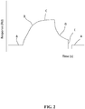
- a representative sensorgram (binding curve) for a reversible interaction at the sensor chip surface is presented in Fig. 2 , the sensing surface having an immobilised capturing molecule, or ligand, for example an antibody, interacting with a binding partner therefore, or analyte, in a sample.
- the binding curves produced by biosensor systems based on other detection principles mentioned above will have a similar appearance.
- the vertical axis (y-axis) indicates the response (here in resonance units, RU) and the horizontal axis (x-axis) indicates the time (here in seconds).
- buffer is passed over the sensing surface giving the baseline response A in the sensorgram.
- an increase in signal is observed due to binding of the analyte.
- association phase This part B of the binding curve is usually referred to as the "association phase".
- association phase Eventually, a steady state condition is reached at or near the end of the association phase where the resonance signal plateaus at C (this state may, however, not always be achieved).
- steady state is used synonymously with the term “equilibrium” (in other contexts the term “equilibrium” may be reserved to describe the ideal interaction model, since in practice binding could be constant over time even if a system is not in equilibrium).
- the sample is replaced with a continuous flow of buffer and a decrease in signal reflects the dissociation, or release, of analyte from the surface.
- This part D of the binding curve is usually referred to as the "dissociation phase".
- the analysis is ended by a regeneration step where a solution capable of removing bound analyte from the surface, while (ideally) maintaining the activity of the ligand, is injected over the sensor surface. This is indicated in part E of the sensorgram.
- regeneration can be avoided where dissociation is already complete or is expected to become completed before the next analysis, which has the advantage of enhancing ligand preservation and reducing the number of operations. Injection of buffer restores the baseline A and the surface is now ready for a new analysis.
- Determining affinity constants from measured steady state binding levels with the BIAevaluation software may, for example, involve the following steps:
- the contact time between the sample solution and the immobilized ligand is less than 15 seconds.
- the term "contact time” is well known in the art and, as used herein, means the total time that it takes the sample solution to flow over any one fixed point of the ligand-bound sensor surface. For example, the time can be determined from the start time from when the first part of the detection surface is contacted by the sample solution to the stop time when the first part of the surface is contacted with a following buffer solution.
- the present application demonstrates that it is possible to provide meaningful binding data relating to the binding of the surface-bound ligand and analyte whilst significantly reducing the contact time between the solution comprising the analyte and the ligand.
- the contact time may be below 15 seconds, below 14 seconds, below 13 seconds, below 12 seconds, below 11 seconds, below 10 seconds, below 9 seconds, below 8 seconds, below 7 seconds, below 6 seconds, below 5 seconds, below 4 seconds, below 3 seconds, below 2 seconds, or below 1 second.
- the contact time may be in the range 15 seconds to 2 seconds, 14 seconds to 2 seconds, 13 seconds to 2 seconds, 12 seconds to 2 seconds, 11 seconds to 2 seconds, 10 seconds to 2 seconds, 9 seconds to 2 seconds, 8 seconds to 2 seconds, 7 seconds to 2 seconds, 6 seconds to 2 seconds, 5 seconds to 2 seconds, 4 seconds to 2 seconds, 3 seconds to 2 seconds, or about 2 seconds.
- the contact time may be in the range 3 seconds to 0.1 seconds, 3 seconds to 0.2 seconds, 3 seconds to 0.3 seconds, 3 seconds to 0.4 seconds, 3 seconds to 0.5 seconds, 3 seconds to 0.6 seconds, 3 seconds to 0.7 seconds, 3 seconds to 0.8 seconds, 3 seconds to 0.9 seconds, 3 seconds to 1 second.
- the lower limit on the contact time may be 0.1 seconds, 0.2 seconds, 0.3 seconds, 0.4 seconds, 0.5 seconds, 0.6 seconds, 0.7 seconds, 0.8 seconds, 0.9 seconds, 0.1 seconds, 1 second, 1.1 seconds, 1.2 seconds, 1.3 seconds, 1.4 seconds, 1.5 seconds, 1.6 seconds, 1.7 seconds, 1.8 seconds, 1.9 seconds, or 2 seconds.
- steps (b) and (c) are repeated over the same surface by flowing a second sample solution over the surface to detect the presence or absence of the binding between ligand and a further analyte.
- the further anlayte may be the same as the first analyte. Alternatively, the further analyte may be different to the first analyte.
- Steps (b) and (c) may then be repeated for a third sample solution.
- steps (b) and (c) are repeated at least two, three, four, five, six, seven, eight, nice, ten, eleven twelve, thirteen, fourteen, fifteen, sixteen, seventeen, eighteen, nineteen, or twenty times to assay the corresponding number of further solutions.
- the time between initiating (e.g. injecting) flowing of at least one of the sample solutions over the sensor surface and initiating flowing of a next sequential sample solution over the sensor surface is less than 40 seconds. In a further embodiment, the cycle time is less than 30 seconds. In a further embodiment, the cycle time is less than 20 seconds. In a further embodiment, the cycle time is less than 15 seconds. In a further embodiment, the cycle time is less than 10 seconds. In a further embodiment, the cycle time is in the range of 10 seconds to 40 seconds, preferably in the range of 10 seconds to 30 seconds.
- the time between initiating (e.g. injecting) flowing of each of the sample solutions over the sensor surface and initiating flowing of a next sequential sample solution over the sensor surface is less than 40 seconds. In a further embodiment, each cycle time is less than 30 seconds. In a further embodiment, each cycle time is less than 20 seconds. In a further embodiment, each cycle time is less than 15 seconds. In a further embodiment, each cycle time is less than 10 seconds. In a further embodiment, each cycle time is in the range of 10 seconds to 40 seconds, preferably in the range of 10 seconds to 30 seconds.
- the cycle time between analyte solutions can be further reduced by eliminating further time consuming measures that are typically employed in biosensor-based analyte binding techniques, for example, washing the fluidics system, carry-over control and/or regeneration. As demonstrated in the Examples of the present application, useful and repeatable binding data was obtained for multiple analyte-biding cycles without employing such washing, carry-over control or regeneration steps.
- Washing steps are typically carried out to ensure that there are not left over analytes stuck to the fluidics system which may contribute to disturbances in the binding data.
- Carry-over control is a typical step administered to address the fact that some analytes are “sticky” and can be difficult to wash out of the fluidic system, causing "carry-over” of material to the next analysis cycle. This can be detected by routinely including a "carry-over injection” of buffer after the sample injection: the response from a "sticky” compound will be carried over into this buffer injection. Thus, a carry-over control injection can monitor response contribution from pollutions of the fluidics system.
- Regeneration is the process of removing bound analyte from the surface after an analysis cycle without damaging the ligand, in preparation for a new cycle. Regeneration techniques are well known in the art, and the specific method employed may vary depending on the ligand and/or anlayte employed. For assay development using custom antibodies, regeneration at low pH (glycine-HCl, pH 1.5 to 3) is usually effective.
- no washing step is carried out between initiating the flowing of at least one of the sample solutions over the sensor surface and initiating flowing of a next sequential sample solution over the sensor surface.
- no carry-over control injection step is carried out between initiating the flowing of at least one of the sample solutions over the sensor surface and initiating flowing of a next sequential sample solution over the sensor surface.
- no regeneration step is carried out between initiating the flowing of at least one of the sample solutions over the sensor surface and initiating flowing of a next sequential sample solution over the sensor surface.
- no regeneration step and no carry-over control and/or system washing step is carried out between initiating the flowing of at least one of the sample solutions over the sensor surface and initiating flowing of a next sequential sample solution over the sensor surface.
- no regeneration step and/or no carry-over control and/or no system washing step is carried out between initiating the flowing of the first sample solution over the sensor surface and initiating flowing of each subsequent sample solution over the sensor surface.
- a high density of ligand can maximise the amount of analyte that can contact the ligand, which can be particularly advantageous where the contact time (and hence time available for binding) is short, thus ensuring a good level of ligand-analyte binding. Furthermore, by employing a high density of ligand, in combination with a short contact time, only a small fraction of the sensor capacity (available binding sites for the analyte) is used for each analyte injection, thereby circumventing the requirement for regeneration.
- ligand density can also depend on the analyte, and in particular the molecular weight ratio of the ligand and analyte.
- step (c) comprises determining the analyte concentration in the sample solution. In a further embodiment, step (c) comprises determining the affinity (e.g. binding constants) of analyte to the ligand.
- the use of short contact times and/or short cycle times as described above means that that the present method is particularly advantageous in methods where large numbers of sample analyte solutions need to be assayed for ligand binding over short or more manageable periods of time.
- the user can e.g. quickly determine the presence or absence of analyte in a given sample solution, or e.g. determine qualitative or quantitative differences in analyte concentrations and/or analyte affinities between difference sample solutions.
- the level of response can be used to draw conclusions regarding the relative affinity of the antibodies and/or the concentration of the antibody.
- the ligand or analyte is an antibody or fragment thereof.
- the ligand may be an antibody, and the analyte may be a molecule capable of (or considered to be potentially capable of) binding to the antibody.
- the ligand may be a molecule capable of binding to a target antibody (e.g. an antigen), and the analyte solutions may comprise candidate antibodies that are capable of binding (or are to be tested for their ability to bind) the ligand.
- the method of the invention may be employed in antibody screening programs e.g. primary screens, to test different hybridomas (present e.g.
- the level of response can be used to draw conclusions regarding the relative affinity of the antibodies and/or the concentration of the antibody.
- the method of the present invention can also be advantageously employed in a fragment-based screen to identify non-specific binders or aggregators.
- Fragment-based drug discovery is a powerful method for discovering high-affinity binders for e.g. target proteins. Although there is no strict size requirement in order for a compound to be designated as a "fragment”, the term is customarily used for small organic molecules with less than about 25 heavy atoms, or a molecular weight of less than 300 Da. However, before beginning a fragment-based screen, it is important to remove fragments that may misbehave, as the presence of such molecules can obscure or disturb the screening results. Such a screen is sometimes referred to as a "Clean-Screen", and is the recommended first step in a fragment screening campaign.
- misbehaving molecules can include nonspecific binders, chelators or aggregators. Sorting out such misbehaving molecules can be a challenging task as not all such compounds are known in advance of the screen.
- the method of the present invention can be used to effectively "pre-screen” a fragment library for such misbehaving molecules. These can be identified by monitoring the signal associated with the binding of the molecules. For example, such molecules would be identifiable due to their slow dissociation due to their unwanted binding interactions. The method of the present invention may then be further used to screen the optimized fragment-based library.
- a computer program comprising program code means for operating a sensor device to detect an interaction between an analyte and a ligand on a sensor surface according to a method comprising (a) providing a sensor surface having a ligand immobilized thereto; (b) flowing the sample solution over the sensor surface; and (c) detecting the presence or absence of binding of the analyte to the ligand on the sensor surface; wherein the contact time between the sample solution and the immobilized ligand is less than 15 seconds; and wherein the program is run on a computer as defined in claim 15.
- the method may have the features described in any of the embodiments described above with respect to the first aspect of the present invention.
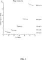
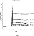
- FIG. 3 and FIG. 4 The results of the above experiments are shown in FIG. 3 and FIG. 4 . These figures demonstrate that a 2 second injection time is enough to detect a protein binder in a sample down to at least 100 ng/ml.
- FIG. 3 shows the response after the injection versus cycle number, demonstrating that the responses are reproducible across multiple cycles for each concentration of analyte and that the relative responses reflect the increase in concentration of analyte.
- FIG. 4 shows the corresponding sensograms with cycle times when no commands are added to the injection. The data demonstrates that extremely short contact times can produce highly useful and repeatable information.
- FIG. 5 is an information plot showing the calculated total experiment time on a 384 well plate with a 4-needle system at various times for a given cycle. If a contact time of only 2 seconds is employed, and all other commands are avoided, the sample time can be significantly lowered. For example, a 12 seconds cycle time would allow a 4-needle system to run a 384 plate in under 20 minutes (96 x 12s).
Landscapes
- Health & Medical Sciences (AREA)
- Immunology (AREA)
- Life Sciences & Earth Sciences (AREA)
- Engineering & Computer Science (AREA)
- Chemical & Material Sciences (AREA)
- Molecular Biology (AREA)
- Biomedical Technology (AREA)
- Hematology (AREA)
- Urology & Nephrology (AREA)
- Biotechnology (AREA)
- Microbiology (AREA)
- Cell Biology (AREA)
- Food Science & Technology (AREA)
- Medicinal Chemistry (AREA)
- Physics & Mathematics (AREA)
- Analytical Chemistry (AREA)
- Biochemistry (AREA)
- General Health & Medical Sciences (AREA)
- General Physics & Mathematics (AREA)
- Pathology (AREA)
- Chemical Kinetics & Catalysis (AREA)
- Investigating Or Analysing Materials By Optical Means (AREA)
Applications Claiming Priority (2)
| Application Number | Priority Date | Filing Date | Title |
|---|---|---|---|
| SE1451456 | 2014-11-28 | ||
| PCT/EP2015/077568 WO2016083417A1 (en) | 2014-11-28 | 2015-11-24 | Method for determining analyte-ligand binding on a sensor surface |
Publications (2)
| Publication Number | Publication Date |
|---|---|
| EP3224622A1 EP3224622A1 (en) | 2017-10-04 |
| EP3224622B1 true EP3224622B1 (en) | 2019-08-07 |
Family
ID=54754613
Family Applications (1)
| Application Number | Title | Priority Date | Filing Date |
|---|---|---|---|
| EP15802025.5A Active EP3224622B1 (en) | 2014-11-28 | 2015-11-24 | Method for determining analyte-ligand binding on a sensor surface |
Country Status (5)
| Country | Link |
|---|---|
| US (2) | US10725030B2 (https=) |
| EP (1) | EP3224622B1 (https=) |
| JP (3) | JP2017535783A (https=) |
| CN (1) | CN107430123B (https=) |
| WO (1) | WO2016083417A1 (https=) |
Families Citing this family (5)
| Publication number | Priority date | Publication date | Assignee | Title |
|---|---|---|---|---|
| US12486293B2 (en) | 2018-07-30 | 2025-12-02 | Georgetown University | Chirality sensing with molecular click chemistry probes |
| WO2020056012A1 (en) | 2018-09-11 | 2020-03-19 | Georgetown University | Quantitative auxiliary-free chirality sensing with a metal probe |
| US12398424B2 (en) | 2019-05-17 | 2025-08-26 | Creoptix Ag | Methods for determining kinetic parameters of a reaction between analyte and ligands |
| DE102020107645B4 (de) * | 2020-03-19 | 2024-07-18 | Bruker Daltonics GmbH & Co. KG | Betrieb einer Mikrofluidik-Vorrichtung bei der Analyse von Probesubstanzen |
| US20240410826A1 (en) * | 2021-09-28 | 2024-12-12 | Arizona Board Of Regents On Behalf Of Arizona State University | Evanescent scattering imaging of single molecules |
Citations (4)
| Publication number | Priority date | Publication date | Assignee | Title |
|---|---|---|---|---|
| WO1996009532A1 (en) | 1994-09-22 | 1996-03-28 | Abbott Laboratories | Optical waveguide method for detecting specific binding events by light scattering |
| WO2004109284A1 (en) | 2003-06-06 | 2004-12-16 | Biacore Ab | Method and system for determination of molecular interaction parameters |
| EP2770058A1 (en) | 2013-02-26 | 2014-08-27 | Université de Perpignan | Ligand and method for detection of okadaic acid |
| WO2015147915A1 (en) | 2013-03-24 | 2015-10-01 | Development Center For Biotechnology | Methods for suppressing cancer by inhibition of tmcc3 |
Family Cites Families (10)
| Publication number | Priority date | Publication date | Assignee | Title |
|---|---|---|---|---|
| GB9816441D0 (en) | 1998-07-28 | 1998-09-23 | Hartley Frank R | Analysis of liquids |
| WO2003091706A1 (en) * | 2002-04-24 | 2003-11-06 | Hitachi, Ltd. | Microreactor for solid-liquid interface reaction and method for conducting meaurement concerning solid-liquid interface reaction |
| JP2004251807A (ja) | 2003-02-21 | 2004-09-09 | Nipro Corp | エンドトキシン測定用センサ、測定方法、診断方法、製造方法およびセンサ再生方法 |
| WO2004092840A1 (ja) | 2003-04-17 | 2004-10-28 | Nissan Chemical Industries, Ltd. | 多孔質下層膜及び多孔質下層膜を形成するための下層膜形成組成物 |
| AU2008242664B2 (en) * | 2007-04-19 | 2011-10-20 | Sru Biosystems, Inc. | Method for employing a biosensor to detect small molecules that bind directly to immobilized targets |
| BR112012012912A2 (pt) | 2009-11-30 | 2016-10-25 | Biotest Ag | anticorpo ou fragmento do mesmo, ácido nucleico, vetor, célula hospedeira, método para a produção de um anticorpo ou um fragmento do mesmo, composição farmacêutica, método para tratar ou prevenir uma condição médica em um indivíduo, uso de um anticorpo ou fragmento do mesmo e métodos in vitro para neutralizar il-10 em uma amostra e para detectar a presença de il-10 em uma amostra. |
| JP5683606B2 (ja) * | 2009-11-30 | 2015-03-11 | ジーイー・ヘルスケア・バイオサイエンス・アクチボラグ | 相互作用の分析のための方法及びシステム |
| JP5937019B2 (ja) * | 2010-03-12 | 2016-06-22 | ザ ボード オブ トラスティーズ オブ ザ レランド スタンフォード ジュニア ユニバーシティー | 磁気センサに基づく結合反応速度の定量的な分析 |
| EP2707715A1 (en) * | 2011-05-11 | 2014-03-19 | Ssens B.V. | Method for determining intrinsic binding parameters of an analyte to a ligand, a method for selecting an analyte from a group of analyts, the selected ligand or analyte, and sensor |
| JP2020191572A (ja) * | 2019-05-23 | 2020-11-26 | ビーコア株式会社 | 撮像装置、方法及びプログラム |
-
2015
- 2015-11-24 WO PCT/EP2015/077568 patent/WO2016083417A1/en not_active Ceased
- 2015-11-24 EP EP15802025.5A patent/EP3224622B1/en active Active
- 2015-11-24 JP JP2017528219A patent/JP2017535783A/ja active Pending
- 2015-11-24 CN CN201580074734.7A patent/CN107430123B/zh active Active
- 2015-11-24 US US15/529,466 patent/US10725030B2/en active Active
-
2020
- 2020-07-17 US US16/932,453 patent/US11796536B2/en active Active
- 2020-11-18 JP JP2020191572A patent/JP7345790B2/ja active Active
-
2022
- 2022-08-18 JP JP2022130703A patent/JP7456653B2/ja active Active
Patent Citations (5)
| Publication number | Priority date | Publication date | Assignee | Title |
|---|---|---|---|---|
| WO1996009532A1 (en) | 1994-09-22 | 1996-03-28 | Abbott Laboratories | Optical waveguide method for detecting specific binding events by light scattering |
| US5599668A (en) | 1994-09-22 | 1997-02-04 | Abbott Laboratories | Light scattering optical waveguide method for detecting specific binding events |
| WO2004109284A1 (en) | 2003-06-06 | 2004-12-16 | Biacore Ab | Method and system for determination of molecular interaction parameters |
| EP2770058A1 (en) | 2013-02-26 | 2014-08-27 | Université de Perpignan | Ligand and method for detection of okadaic acid |
| WO2015147915A1 (en) | 2013-03-24 | 2015-10-01 | Development Center For Biotechnology | Methods for suppressing cancer by inhibition of tmcc3 |
Non-Patent Citations (6)
| Title |
|---|
| BO. OLSSON, HANS. LUNDBAECK, GILLIS. JOHANSSON, FRIEDER. SCHELLER, JUERGEN. NENTWIG: "Theory and application of diffusion-limited amperometric enzyme electrode detection in flow injection determination of glucose", ANALYTICAL CHEMISTRY, AMERICAN CHEMICAL SOCIETY, vol. 58, no. 6, 1 May 1986 (1986-05-01), pages 1046 - 1052, XP055394923, ISSN: 0003-2700, DOI: 10.1021/ac00297a014 |
| FENG-SHENG KAO, GER WAYLON, PAN YUN-RU, YU HUI-CHEN, HSU RAY-QUEN, CHEN HUEIH-MIN: "Chip-Based Protein–Protein Interaction Studied by Atomic Force Microscopy", BIOTECHNOLOGY & BIOENGINEERING, JOHN WILEY & SONS, INC., vol. 109, no. 10, 1 October 2012 (2012-10-01), pages 2460 - 2467, XP055241484, ISSN: 1097-0290, DOI: 10.1002/bit.24521 |
| GIANNETTI ANTHONY M: "From experimental design to validated hits a comprehensive walk-through of fragment lead identification using surface plasmon resonance.", METHODS IN ENZYMOLOGY, ACADEM. PRESS, USA, vol. 493, USA, pages 169 - 218, XP008178598, ISSN: 1557-7988, DOI: 10.1016/B978-0-12-381274-2.00008-X |
| M. A. KALININA, O. A. RAITMAN, D. S. TURYGIN, S. L. SELEKTOR, N. V. GOLUBEV, V. V. ARSLANOV: "Langmuir-Blodgett composite films for the selective determination of calcium in aqueous solutions", RUSSIAN JOURNAL OF PHYSICAL CHEMISTRY A, CHEMICAL SOCIETY, LONDON., GB, vol. 82, no. 8, 1 August 2008 (2008-08-01), GB, pages 1334 - 1342, XP055240974, ISSN: 0036-0244, DOI: 10.1134/S0036024408080165 |
| MCLAUGHLIN C,ET AL: "Quantitative analysis of mitoxantrone by surface-enhanced resonance Raman scattering.", ANALYTICAL CHEMISTRY, AMERICAN CHEMICAL SOCIETY, vol. 74, no. 13, 1 July 2002 (2002-07-01), pages 3160 - 3167, XP002237472, ISSN: 0003-2700, DOI: 10.1021/ac010067k |
| MYSZKA D G, WOOD S J, BIERE A L: "ANALYSIS OF FIBRIL ELONGATION USING SURFACE PLASMON RESONANCE BIOSENSORS", BIOMEMBRANES: TRANSPORT THEORY : CELLS AND MODEL MEMBRANES, ELSEVIER, ACADEMIC PRESS, NL, vol. 309, 1 January 1999 (1999-01-01), NL, pages 386 - 402, XP008028557, ISBN: 978-0-12-805382-9, DOI: 10.1016/S0076-6879(99)09027-8 |
Also Published As
| Publication number | Publication date |
|---|---|
| JP2022171673A (ja) | 2022-11-11 |
| US10725030B2 (en) | 2020-07-28 |
| JP2021036245A (ja) | 2021-03-04 |
| JP7345790B2 (ja) | 2023-09-19 |
| EP3224622A1 (en) | 2017-10-04 |
| US20170261502A1 (en) | 2017-09-14 |
| CN107430123B (zh) | 2020-04-14 |
| CN107430123A (zh) | 2017-12-01 |
| JP7456653B2 (ja) | 2024-03-27 |
| US20200348295A1 (en) | 2020-11-05 |
| US11796536B2 (en) | 2023-10-24 |
| WO2016083417A1 (en) | 2016-06-02 |
| JP2017535783A (ja) | 2017-11-30 |
Similar Documents
| Publication | Publication Date | Title |
|---|---|---|
| US11796536B2 (en) | Method for determining analyte-ligand binding on a sensor surface | |
| US20210215613A1 (en) | Concentration assay | |
| US20220137039A1 (en) | Method and system for evaluation of an interaction between an analyte and a ligand using a biosensor | |
| JP5855246B2 (ja) | 校正不要分析による活性濃度の決定方法 | |
| US10545090B2 (en) | Method and system for more reliable determination of interaction parameters for low affinity analytes | |
| JP2017504805A (ja) | 相互作用分析のための方法及びシステム | |
| EP2507619B1 (en) | Method and system for binding behavior analysis | |
| WO2006135309A9 (en) | Method and system for affinity analysis | |
| US20140147937A1 (en) | Method of determining active concentration | |
| US10658072B2 (en) | Method and system for interaction analysis | |
| US20070016378A1 (en) | Method and system for affinity analysis | |
| JP2007506967A (ja) | 分子相互作用分析の方法及びシステム | |
| US20110152120A1 (en) | method of characterizing antibodies | |
| KR20250051753A (ko) | Spr 분석을 위한 유동 셀에서의 유체의 공기-세그먼트화된 주입 |
Legal Events
| Date | Code | Title | Description |
|---|---|---|---|
| STAA | Information on the status of an ep patent application or granted ep patent |
Free format text: STATUS: THE INTERNATIONAL PUBLICATION HAS BEEN MADE |
|
| PUAI | Public reference made under article 153(3) epc to a published international application that has entered the european phase |
Free format text: ORIGINAL CODE: 0009012 |
|
| STAA | Information on the status of an ep patent application or granted ep patent |
Free format text: STATUS: REQUEST FOR EXAMINATION WAS MADE |
|
| 17P | Request for examination filed |
Effective date: 20170530 |
|
| AK | Designated contracting states |
Kind code of ref document: A1 Designated state(s): AL AT BE BG CH CY CZ DE DK EE ES FI FR GB GR HR HU IE IS IT LI LT LU LV MC MK MT NL NO PL PT RO RS SE SI SK SM TR |
|
| AX | Request for extension of the european patent |
Extension state: BA ME |
|
| DAV | Request for validation of the european patent (deleted) | ||
| DAX | Request for extension of the european patent (deleted) | ||
| STAA | Information on the status of an ep patent application or granted ep patent |
Free format text: STATUS: EXAMINATION IS IN PROGRESS |
|
| 17Q | First examination report despatched |
Effective date: 20180605 |
|
| GRAP | Despatch of communication of intention to grant a patent |
Free format text: ORIGINAL CODE: EPIDOSNIGR1 |
|
| STAA | Information on the status of an ep patent application or granted ep patent |
Free format text: STATUS: GRANT OF PATENT IS INTENDED |
|
| INTG | Intention to grant announced |
Effective date: 20190403 |
|
| GRAS | Grant fee paid |
Free format text: ORIGINAL CODE: EPIDOSNIGR3 |
|
| GRAA | (expected) grant |
Free format text: ORIGINAL CODE: 0009210 |
|
| STAA | Information on the status of an ep patent application or granted ep patent |
Free format text: STATUS: THE PATENT HAS BEEN GRANTED |
|
| AK | Designated contracting states |
Kind code of ref document: B1 Designated state(s): AL AT BE BG CH CY CZ DE DK EE ES FI FR GB GR HR HU IE IS IT LI LT LU LV MC MK MT NL NO PL PT RO RS SE SI SK SM TR |
|
| REG | Reference to a national code |
Ref country code: GB Ref legal event code: FG4D |
|
| REG | Reference to a national code |
Ref country code: CH Ref legal event code: EP Ref country code: AT Ref legal event code: REF Ref document number: 1164653 Country of ref document: AT Kind code of ref document: T Effective date: 20190815 |
|
| REG | Reference to a national code |
Ref country code: DE Ref legal event code: R096 Ref document number: 602015035435 Country of ref document: DE |
|
| REG | Reference to a national code |
Ref country code: IE Ref legal event code: FG4D |
|
| REG | Reference to a national code |
Ref country code: NL Ref legal event code: MP Effective date: 20190807 |
|
| REG | Reference to a national code |
Ref country code: LT Ref legal event code: MG4D |
|
| PG25 | Lapsed in a contracting state [announced via postgrant information from national office to epo] |
Ref country code: BG Free format text: LAPSE BECAUSE OF FAILURE TO SUBMIT A TRANSLATION OF THE DESCRIPTION OR TO PAY THE FEE WITHIN THE PRESCRIBED TIME-LIMIT Effective date: 20191107 Ref country code: NL Free format text: LAPSE BECAUSE OF FAILURE TO SUBMIT A TRANSLATION OF THE DESCRIPTION OR TO PAY THE FEE WITHIN THE PRESCRIBED TIME-LIMIT Effective date: 20190807 Ref country code: HR Free format text: LAPSE BECAUSE OF FAILURE TO SUBMIT A TRANSLATION OF THE DESCRIPTION OR TO PAY THE FEE WITHIN THE PRESCRIBED TIME-LIMIT Effective date: 20190807 Ref country code: NO Free format text: LAPSE BECAUSE OF FAILURE TO SUBMIT A TRANSLATION OF THE DESCRIPTION OR TO PAY THE FEE WITHIN THE PRESCRIBED TIME-LIMIT Effective date: 20191107 Ref country code: LT Free format text: LAPSE BECAUSE OF FAILURE TO SUBMIT A TRANSLATION OF THE DESCRIPTION OR TO PAY THE FEE WITHIN THE PRESCRIBED TIME-LIMIT Effective date: 20190807 Ref country code: FI Free format text: LAPSE BECAUSE OF FAILURE TO SUBMIT A TRANSLATION OF THE DESCRIPTION OR TO PAY THE FEE WITHIN THE PRESCRIBED TIME-LIMIT Effective date: 20190807 Ref country code: PT Free format text: LAPSE BECAUSE OF FAILURE TO SUBMIT A TRANSLATION OF THE DESCRIPTION OR TO PAY THE FEE WITHIN THE PRESCRIBED TIME-LIMIT Effective date: 20191209 Ref country code: SE Free format text: LAPSE BECAUSE OF FAILURE TO SUBMIT A TRANSLATION OF THE DESCRIPTION OR TO PAY THE FEE WITHIN THE PRESCRIBED TIME-LIMIT Effective date: 20190807 |
|
| REG | Reference to a national code |
Ref country code: AT Ref legal event code: MK05 Ref document number: 1164653 Country of ref document: AT Kind code of ref document: T Effective date: 20190807 |
|
| PG25 | Lapsed in a contracting state [announced via postgrant information from national office to epo] |
Ref country code: AL Free format text: LAPSE BECAUSE OF FAILURE TO SUBMIT A TRANSLATION OF THE DESCRIPTION OR TO PAY THE FEE WITHIN THE PRESCRIBED TIME-LIMIT Effective date: 20190807 Ref country code: GR Free format text: LAPSE BECAUSE OF FAILURE TO SUBMIT A TRANSLATION OF THE DESCRIPTION OR TO PAY THE FEE WITHIN THE PRESCRIBED TIME-LIMIT Effective date: 20191108 Ref country code: IS Free format text: LAPSE BECAUSE OF FAILURE TO SUBMIT A TRANSLATION OF THE DESCRIPTION OR TO PAY THE FEE WITHIN THE PRESCRIBED TIME-LIMIT Effective date: 20191207 Ref country code: RS Free format text: LAPSE BECAUSE OF FAILURE TO SUBMIT A TRANSLATION OF THE DESCRIPTION OR TO PAY THE FEE WITHIN THE PRESCRIBED TIME-LIMIT Effective date: 20190807 Ref country code: LV Free format text: LAPSE BECAUSE OF FAILURE TO SUBMIT A TRANSLATION OF THE DESCRIPTION OR TO PAY THE FEE WITHIN THE PRESCRIBED TIME-LIMIT Effective date: 20190807 Ref country code: ES Free format text: LAPSE BECAUSE OF FAILURE TO SUBMIT A TRANSLATION OF THE DESCRIPTION OR TO PAY THE FEE WITHIN THE PRESCRIBED TIME-LIMIT Effective date: 20190807 |
|
| PG25 | Lapsed in a contracting state [announced via postgrant information from national office to epo] |
Ref country code: TR Free format text: LAPSE BECAUSE OF FAILURE TO SUBMIT A TRANSLATION OF THE DESCRIPTION OR TO PAY THE FEE WITHIN THE PRESCRIBED TIME-LIMIT Effective date: 20190807 |
|
| REG | Reference to a national code |
Ref country code: DE Ref legal event code: R026 Ref document number: 602015035435 Country of ref document: DE |
|
| PLBI | Opposition filed |
Free format text: ORIGINAL CODE: 0009260 |
|
| PG25 | Lapsed in a contracting state [announced via postgrant information from national office to epo] |
Ref country code: IT Free format text: LAPSE BECAUSE OF FAILURE TO SUBMIT A TRANSLATION OF THE DESCRIPTION OR TO PAY THE FEE WITHIN THE PRESCRIBED TIME-LIMIT Effective date: 20190807 Ref country code: RO Free format text: LAPSE BECAUSE OF FAILURE TO SUBMIT A TRANSLATION OF THE DESCRIPTION OR TO PAY THE FEE WITHIN THE PRESCRIBED TIME-LIMIT Effective date: 20190807 Ref country code: EE Free format text: LAPSE BECAUSE OF FAILURE TO SUBMIT A TRANSLATION OF THE DESCRIPTION OR TO PAY THE FEE WITHIN THE PRESCRIBED TIME-LIMIT Effective date: 20190807 Ref country code: DK Free format text: LAPSE BECAUSE OF FAILURE TO SUBMIT A TRANSLATION OF THE DESCRIPTION OR TO PAY THE FEE WITHIN THE PRESCRIBED TIME-LIMIT Effective date: 20190807 Ref country code: AT Free format text: LAPSE BECAUSE OF FAILURE TO SUBMIT A TRANSLATION OF THE DESCRIPTION OR TO PAY THE FEE WITHIN THE PRESCRIBED TIME-LIMIT Effective date: 20190807 Ref country code: PL Free format text: LAPSE BECAUSE OF FAILURE TO SUBMIT A TRANSLATION OF THE DESCRIPTION OR TO PAY THE FEE WITHIN THE PRESCRIBED TIME-LIMIT Effective date: 20190807 |
|
| 26 | Opposition filed |
Opponent name: CREOPTIX AG Effective date: 20200414 |
|
| PG25 | Lapsed in a contracting state [announced via postgrant information from national office to epo] |
Ref country code: CZ Free format text: LAPSE BECAUSE OF FAILURE TO SUBMIT A TRANSLATION OF THE DESCRIPTION OR TO PAY THE FEE WITHIN THE PRESCRIBED TIME-LIMIT Effective date: 20190807 Ref country code: SK Free format text: LAPSE BECAUSE OF FAILURE TO SUBMIT A TRANSLATION OF THE DESCRIPTION OR TO PAY THE FEE WITHIN THE PRESCRIBED TIME-LIMIT Effective date: 20190807 Ref country code: IS Free format text: LAPSE BECAUSE OF FAILURE TO SUBMIT A TRANSLATION OF THE DESCRIPTION OR TO PAY THE FEE WITHIN THE PRESCRIBED TIME-LIMIT Effective date: 20200224 Ref country code: SM Free format text: LAPSE BECAUSE OF FAILURE TO SUBMIT A TRANSLATION OF THE DESCRIPTION OR TO PAY THE FEE WITHIN THE PRESCRIBED TIME-LIMIT Effective date: 20190807 |
|
| PLAX | Notice of opposition and request to file observation + time limit sent |
Free format text: ORIGINAL CODE: EPIDOSNOBS2 |
|
| REG | Reference to a national code |
Ref country code: CH Ref legal event code: PL |
|
| PG2D | Information on lapse in contracting state deleted |
Ref country code: IS |
|
| PG25 | Lapsed in a contracting state [announced via postgrant information from national office to epo] |
Ref country code: CH Free format text: LAPSE BECAUSE OF NON-PAYMENT OF DUE FEES Effective date: 20191130 Ref country code: LI Free format text: LAPSE BECAUSE OF NON-PAYMENT OF DUE FEES Effective date: 20191130 Ref country code: LU Free format text: LAPSE BECAUSE OF NON-PAYMENT OF DUE FEES Effective date: 20191124 Ref country code: MC Free format text: LAPSE BECAUSE OF FAILURE TO SUBMIT A TRANSLATION OF THE DESCRIPTION OR TO PAY THE FEE WITHIN THE PRESCRIBED TIME-LIMIT Effective date: 20190807 |
|
| RAP2 | Party data changed (patent owner data changed or rights of a patent transferred) |
Owner name: CYTIVA SWEDEN AB |
|
| REG | Reference to a national code |
Ref country code: BE Ref legal event code: MM Effective date: 20191130 |
|
| PG25 | Lapsed in a contracting state [announced via postgrant information from national office to epo] |
Ref country code: SI Free format text: LAPSE BECAUSE OF FAILURE TO SUBMIT A TRANSLATION OF THE DESCRIPTION OR TO PAY THE FEE WITHIN THE PRESCRIBED TIME-LIMIT Effective date: 20190807 |
|
| PLBB | Reply of patent proprietor to notice(s) of opposition received |
Free format text: ORIGINAL CODE: EPIDOSNOBS3 |
|
| PG25 | Lapsed in a contracting state [announced via postgrant information from national office to epo] |
Ref country code: IE Free format text: LAPSE BECAUSE OF NON-PAYMENT OF DUE FEES Effective date: 20191124 |
|
| PG25 | Lapsed in a contracting state [announced via postgrant information from national office to epo] |
Ref country code: BE Free format text: LAPSE BECAUSE OF NON-PAYMENT OF DUE FEES Effective date: 20191130 |
|
| REG | Reference to a national code |
Ref country code: DE Ref legal event code: R081 Ref document number: 602015035435 Country of ref document: DE Owner name: CYTIVA SWEDEN AB, SE Free format text: FORMER OWNER: GE HEALTHCARE BIO-SCIENCES AB, UPPSALA, SE |
|
| PG25 | Lapsed in a contracting state [announced via postgrant information from national office to epo] |
Ref country code: CY Free format text: LAPSE BECAUSE OF FAILURE TO SUBMIT A TRANSLATION OF THE DESCRIPTION OR TO PAY THE FEE WITHIN THE PRESCRIBED TIME-LIMIT Effective date: 20190807 |
|
| PG25 | Lapsed in a contracting state [announced via postgrant information from national office to epo] |
Ref country code: HU Free format text: LAPSE BECAUSE OF FAILURE TO SUBMIT A TRANSLATION OF THE DESCRIPTION OR TO PAY THE FEE WITHIN THE PRESCRIBED TIME-LIMIT; INVALID AB INITIO Effective date: 20151124 Ref country code: MT Free format text: LAPSE BECAUSE OF FAILURE TO SUBMIT A TRANSLATION OF THE DESCRIPTION OR TO PAY THE FEE WITHIN THE PRESCRIBED TIME-LIMIT Effective date: 20190807 |
|
| REG | Reference to a national code |
Ref country code: DE Ref legal event code: R100 Ref document number: 602015035435 Country of ref document: DE |
|
| PLCK | Communication despatched that opposition was rejected |
Free format text: ORIGINAL CODE: EPIDOSNREJ1 |
|
| PLBN | Opposition rejected |
Free format text: ORIGINAL CODE: 0009273 |
|
| STAA | Information on the status of an ep patent application or granted ep patent |
Free format text: STATUS: OPPOSITION REJECTED |
|
| 27O | Opposition rejected |
Effective date: 20210928 |
|
| PG25 | Lapsed in a contracting state [announced via postgrant information from national office to epo] |
Ref country code: MK Free format text: LAPSE BECAUSE OF FAILURE TO SUBMIT A TRANSLATION OF THE DESCRIPTION OR TO PAY THE FEE WITHIN THE PRESCRIBED TIME-LIMIT Effective date: 20190807 |
|
| P01 | Opt-out of the competence of the unified patent court (upc) registered |
Effective date: 20230526 |
|
| PGFP | Annual fee paid to national office [announced via postgrant information from national office to epo] |
Ref country code: DE Payment date: 20251126 Year of fee payment: 11 |
|
| PGFP | Annual fee paid to national office [announced via postgrant information from national office to epo] |
Ref country code: GB Payment date: 20251125 Year of fee payment: 11 |
|
| PGFP | Annual fee paid to national office [announced via postgrant information from national office to epo] |
Ref country code: FR Payment date: 20251125 Year of fee payment: 11 |