EP3186280B9 - Antibodies and chimeric antigen receptors specific for cd19 - Google Patents
Antibodies and chimeric antigen receptors specific for cd19 Download PDFInfo
- Publication number
- EP3186280B9 EP3186280B9 EP15760045.3A EP15760045A EP3186280B9 EP 3186280 B9 EP3186280 B9 EP 3186280B9 EP 15760045 A EP15760045 A EP 15760045A EP 3186280 B9 EP3186280 B9 EP 3186280B9
- Authority
- EP
- European Patent Office
- Prior art keywords
- antibody
- instances
- cells
- seq
- amino acid
- Prior art date
- Legal status (The legal status is an assumption and is not a legal conclusion. Google has not performed a legal analysis and makes no representation as to the accuracy of the status listed.)
- Active
Links
- 108010019670 Chimeric Antigen Receptors Proteins 0.000 title claims description 204
- 210000004027 cell Anatomy 0.000 claims description 581
- 241000282414 Homo sapiens Species 0.000 claims description 384
- 125000003275 alpha amino acid group Chemical group 0.000 claims description 316
- 230000027455 binding Effects 0.000 claims description 264
- 102100024222 B-lymphocyte antigen CD19 Human genes 0.000 claims description 243
- 101000980825 Homo sapiens B-lymphocyte antigen CD19 Proteins 0.000 claims description 239
- 239000012634 fragment Substances 0.000 claims description 176
- 239000000427 antigen Substances 0.000 claims description 97
- 238000000034 method Methods 0.000 claims description 97
- 102000036639 antigens Human genes 0.000 claims description 96
- 108091007433 antigens Proteins 0.000 claims description 96
- 239000000203 mixture Substances 0.000 claims description 86
- 210000001744 T-lymphocyte Anatomy 0.000 claims description 85
- 208000037265 diseases, disorders, signs and symptoms Diseases 0.000 claims description 63
- 150000007523 nucleic acids Chemical class 0.000 claims description 58
- 108020004707 nucleic acids Proteins 0.000 claims description 57
- 102000039446 nucleic acids Human genes 0.000 claims description 57
- 201000010099 disease Diseases 0.000 claims description 51
- 102000005962 receptors Human genes 0.000 claims description 48
- 108020003175 receptors Proteins 0.000 claims description 48
- 101100383038 Homo sapiens CD19 gene Proteins 0.000 claims description 46
- 101000914514 Homo sapiens T-cell-specific surface glycoprotein CD28 Proteins 0.000 claims description 36
- 102100027213 T-cell-specific surface glycoprotein CD28 Human genes 0.000 claims description 36
- 230000004068 intracellular signaling Effects 0.000 claims description 36
- 206010028980 Neoplasm Diseases 0.000 claims description 33
- 230000011664 signaling Effects 0.000 claims description 32
- 210000001266 CD8-positive T-lymphocyte Anatomy 0.000 claims description 25
- 230000000139 costimulatory effect Effects 0.000 claims description 22
- 201000011510 cancer Diseases 0.000 claims description 16
- 101000851370 Homo sapiens Tumor necrosis factor receptor superfamily member 9 Proteins 0.000 claims description 15
- 102100036856 Tumor necrosis factor receptor superfamily member 9 Human genes 0.000 claims description 15
- 210000003719 b-lymphocyte Anatomy 0.000 claims description 14
- 208000010839 B-cell chronic lymphocytic leukemia Diseases 0.000 claims description 13
- 208000031422 Lymphocytic Chronic B-Cell Leukemia Diseases 0.000 claims description 13
- 208000032852 chronic lymphocytic leukemia Diseases 0.000 claims description 13
- 208000024893 Acute lymphoblastic leukemia Diseases 0.000 claims description 12
- 208000035475 disorder Diseases 0.000 claims description 12
- FWMNVWWHGCHHJJ-SKKKGAJSSA-N 4-amino-1-[(2r)-6-amino-2-[[(2r)-2-[[(2r)-2-[[(2r)-2-amino-3-phenylpropanoyl]amino]-3-phenylpropanoyl]amino]-4-methylpentanoyl]amino]hexanoyl]piperidine-4-carboxylic acid Chemical compound C([C@H](C(=O)N[C@H](CC(C)C)C(=O)N[C@H](CCCCN)C(=O)N1CCC(N)(CC1)C(O)=O)NC(=O)[C@H](N)CC=1C=CC=CC=1)C1=CC=CC=C1 FWMNVWWHGCHHJJ-SKKKGAJSSA-N 0.000 claims description 10
- 208000014697 Acute lymphocytic leukaemia Diseases 0.000 claims description 9
- 208000006664 Precursor Cell Lymphoblastic Leukemia-Lymphoma Diseases 0.000 claims description 9
- 230000036210 malignancy Effects 0.000 claims description 7
- 206010012818 diffuse large B-cell lymphoma Diseases 0.000 claims description 6
- 208000003950 B-cell lymphoma Diseases 0.000 claims description 5
- 239000000546 pharmaceutical excipient Substances 0.000 claims description 4
- 208000017604 Hodgkin disease Diseases 0.000 claims description 3
- 208000021519 Hodgkin lymphoma Diseases 0.000 claims description 3
- 208000010747 Hodgkins lymphoma Diseases 0.000 claims description 3
- 208000025205 Mantle-Cell Lymphoma Diseases 0.000 claims description 3
- 208000034578 Multiple myelomas Diseases 0.000 claims description 3
- 208000015914 Non-Hodgkin lymphomas Diseases 0.000 claims description 3
- 208000033766 Prolymphocytic Leukemia Diseases 0.000 claims description 3
- 201000003444 follicular lymphoma Diseases 0.000 claims description 3
- 201000009277 hairy cell leukemia Diseases 0.000 claims description 3
- 201000007924 marginal zone B-cell lymphoma Diseases 0.000 claims description 3
- 208000021937 marginal zone lymphoma Diseases 0.000 claims description 3
- 230000003393 splenic effect Effects 0.000 claims description 3
- 235000001014 amino acid Nutrition 0.000 description 142
- 229940024606 amino acid Drugs 0.000 description 137
- 150000001413 amino acids Chemical class 0.000 description 126
- MTCFGRXMJLQNBG-REOHCLBHSA-N (2S)-2-Amino-3-hydroxypropansäure Chemical compound OC[C@H](N)C(O)=O MTCFGRXMJLQNBG-REOHCLBHSA-N 0.000 description 93
- 229910052717 sulfur Inorganic materials 0.000 description 83
- DHMQDGOQFOQNFH-UHFFFAOYSA-N Glycine Chemical compound NCC(O)=O DHMQDGOQFOQNFH-UHFFFAOYSA-N 0.000 description 53
- DCXYFEDJOCDNAF-REOHCLBHSA-N L-asparagine Chemical compound OC(=O)[C@@H](N)CC(N)=O DCXYFEDJOCDNAF-REOHCLBHSA-N 0.000 description 51
- AYFVYJQAPQTCCC-GBXIJSLDSA-N L-threonine Chemical compound C[C@@H](O)[C@H](N)C(O)=O AYFVYJQAPQTCCC-GBXIJSLDSA-N 0.000 description 51
- 125000003295 alanine group Chemical group N[C@@H](C)C(=O)* 0.000 description 47
- 101000716102 Homo sapiens T-cell surface glycoprotein CD4 Proteins 0.000 description 44
- 102100036011 T-cell surface glycoprotein CD4 Human genes 0.000 description 44
- 239000003550 marker Substances 0.000 description 44
- 229910052727 yttrium Inorganic materials 0.000 description 43
- 229910052757 nitrogen Inorganic materials 0.000 description 41
- 108010047041 Complementarity Determining Regions Proteins 0.000 description 39
- 102100035360 Cerebellar degeneration-related antigen 1 Human genes 0.000 description 37
- 108010021625 Immunoglobulin Fragments Proteins 0.000 description 37
- 102000008394 Immunoglobulin Fragments Human genes 0.000 description 37
- 125000006850 spacer group Chemical group 0.000 description 35
- 101710117290 Aldo-keto reductase family 1 member C4 Proteins 0.000 description 34
- 108090000623 proteins and genes Proteins 0.000 description 33
- 102000017420 CD3 protein, epsilon/gamma/delta subunit Human genes 0.000 description 32
- 108050005493 CD3 protein, epsilon/gamma/delta subunit Proteins 0.000 description 32
- ROHFNLRQFUQHCH-YFKPBYRVSA-N L-leucine Chemical compound CC(C)C[C@H](N)C(O)=O ROHFNLRQFUQHCH-YFKPBYRVSA-N 0.000 description 31
- -1 e.g. Proteins 0.000 description 31
- 229910052698 phosphorus Inorganic materials 0.000 description 31
- 229910052731 fluorine Inorganic materials 0.000 description 30
- 230000014509 gene expression Effects 0.000 description 30
- 239000013598 vector Substances 0.000 description 30
- OUYCCCASQSFEME-QMMMGPOBSA-N L-tyrosine Chemical compound OC(=O)[C@@H](N)CC1=CC=C(O)C=C1 OUYCCCASQSFEME-QMMMGPOBSA-N 0.000 description 29
- 238000003556 assay Methods 0.000 description 29
- 239000002773 nucleotide Substances 0.000 description 29
- 125000003729 nucleotide group Chemical group 0.000 description 29
- 241001529936 Murinae Species 0.000 description 28
- 238000000926 separation method Methods 0.000 description 28
- 101000946843 Homo sapiens T-cell surface glycoprotein CD8 alpha chain Proteins 0.000 description 27
- 102100034922 T-cell surface glycoprotein CD8 alpha chain Human genes 0.000 description 27
- 108090000765 processed proteins & peptides Proteins 0.000 description 27
- 210000004602 germ cell Anatomy 0.000 description 26
- 229910052721 tungsten Inorganic materials 0.000 description 26
- 229910052720 vanadium Inorganic materials 0.000 description 26
- 239000000523 sample Substances 0.000 description 25
- ZDXPYRJPNDTMRX-VKHMYHEASA-N L-glutamine Chemical compound OC(=O)[C@@H](N)CCC(N)=O ZDXPYRJPNDTMRX-VKHMYHEASA-N 0.000 description 24
- 238000006467 substitution reaction Methods 0.000 description 24
- 102000004127 Cytokines Human genes 0.000 description 23
- 108090000695 Cytokines Proteins 0.000 description 23
- 238000002347 injection Methods 0.000 description 22
- 239000007924 injection Substances 0.000 description 22
- KZSNJWFQEVHDMF-UHFFFAOYSA-N Valine Natural products CC(C)C(N)C(O)=O KZSNJWFQEVHDMF-UHFFFAOYSA-N 0.000 description 21
- 239000008194 pharmaceutical composition Substances 0.000 description 21
- 229910052700 potassium Inorganic materials 0.000 description 21
- 102000004196 processed proteins & peptides Human genes 0.000 description 21
- 210000001519 tissue Anatomy 0.000 description 21
- 238000011282 treatment Methods 0.000 description 21
- 230000000694 effects Effects 0.000 description 20
- 238000000684 flow cytometry Methods 0.000 description 20
- NFGXHKASABOEEW-UHFFFAOYSA-N 1-methylethyl 11-methoxy-3,7,11-trimethyl-2,4-dodecadienoate Chemical compound COC(C)(C)CCCC(C)CC=CC(C)=CC(=O)OC(C)C NFGXHKASABOEEW-UHFFFAOYSA-N 0.000 description 19
- 125000000174 L-prolyl group Chemical group [H]N1C([H])([H])C([H])([H])C([H])([H])[C@@]1([H])C(*)=O 0.000 description 19
- 229920001184 polypeptide Polymers 0.000 description 19
- 108091008874 T cell receptors Proteins 0.000 description 18
- 102000016266 T-Cell Antigen Receptors Human genes 0.000 description 18
- 229910052805 deuterium Inorganic materials 0.000 description 18
- 238000011534 incubation Methods 0.000 description 18
- 108020004414 DNA Proteins 0.000 description 17
- KDXKERNSBIXSRK-UHFFFAOYSA-N Lysine Natural products NCCCCC(N)C(O)=O KDXKERNSBIXSRK-UHFFFAOYSA-N 0.000 description 17
- 239000003795 chemical substances by application Substances 0.000 description 17
- 239000013642 negative control Substances 0.000 description 17
- 238000002360 preparation method Methods 0.000 description 17
- 102000004169 proteins and genes Human genes 0.000 description 17
- 102000006306 Antigen Receptors Human genes 0.000 description 16
- 108010083359 Antigen Receptors Proteins 0.000 description 16
- KDXKERNSBIXSRK-YFKPBYRVSA-N L-lysine Chemical compound NCCCC[C@H](N)C(O)=O KDXKERNSBIXSRK-YFKPBYRVSA-N 0.000 description 16
- 239000012636 effector Substances 0.000 description 16
- 238000009472 formulation Methods 0.000 description 16
- 239000002245 particle Substances 0.000 description 16
- 235000018102 proteins Nutrition 0.000 description 16
- 230000004913 activation Effects 0.000 description 15
- 239000003814 drug Substances 0.000 description 15
- 229910052739 hydrogen Inorganic materials 0.000 description 15
- 238000003780 insertion Methods 0.000 description 15
- 230000037431 insertion Effects 0.000 description 15
- 230000001225 therapeutic effect Effects 0.000 description 15
- 238000012546 transfer Methods 0.000 description 15
- 229910052722 tritium Inorganic materials 0.000 description 15
- 238000011467 adoptive cell therapy Methods 0.000 description 14
- 230000006870 function Effects 0.000 description 14
- 229910052740 iodine Inorganic materials 0.000 description 14
- 238000004519 manufacturing process Methods 0.000 description 14
- 230000000717 retained effect Effects 0.000 description 14
- 238000002560 therapeutic procedure Methods 0.000 description 14
- CKLJMWTZIZZHCS-REOHCLBHSA-N L-aspartic acid Chemical compound OC(=O)[C@@H](N)CC(O)=O CKLJMWTZIZZHCS-REOHCLBHSA-N 0.000 description 13
- 108700010039 chimeric receptor Proteins 0.000 description 13
- 230000001086 cytosolic effect Effects 0.000 description 13
- 238000001727 in vivo Methods 0.000 description 13
- 230000028327 secretion Effects 0.000 description 13
- 230000004936 stimulating effect Effects 0.000 description 13
- 210000004369 blood Anatomy 0.000 description 12
- 239000008280 blood Substances 0.000 description 12
- 238000003501 co-culture Methods 0.000 description 12
- 238000002955 isolation Methods 0.000 description 12
- 235000004400 serine Nutrition 0.000 description 12
- MTCFGRXMJLQNBG-UHFFFAOYSA-N Serine Natural products OCC(N)C(O)=O MTCFGRXMJLQNBG-UHFFFAOYSA-N 0.000 description 11
- 229910052799 carbon Inorganic materials 0.000 description 11
- 238000002659 cell therapy Methods 0.000 description 11
- 238000010828 elution Methods 0.000 description 11
- 230000003834 intracellular effect Effects 0.000 description 11
- 230000004048 modification Effects 0.000 description 11
- 238000012986 modification Methods 0.000 description 11
- 210000003819 peripheral blood mononuclear cell Anatomy 0.000 description 11
- 238000010361 transduction Methods 0.000 description 11
- 230000026683 transduction Effects 0.000 description 11
- 241000699670 Mus sp. Species 0.000 description 10
- 125000000539 amino acid group Chemical group 0.000 description 10
- 229940127089 cytotoxic agent Drugs 0.000 description 10
- 239000013641 positive control Substances 0.000 description 10
- 230000002829 reductive effect Effects 0.000 description 10
- 241000894007 species Species 0.000 description 10
- 101001018097 Homo sapiens L-selectin Proteins 0.000 description 9
- 108060003951 Immunoglobulin Proteins 0.000 description 9
- XUJNEKJLAYXESH-REOHCLBHSA-N L-Cysteine Chemical compound SC[C@H](N)C(O)=O XUJNEKJLAYXESH-REOHCLBHSA-N 0.000 description 9
- COLNVLDHVKWLRT-QMMMGPOBSA-N L-phenylalanine Chemical compound OC(=O)[C@@H](N)CC1=CC=CC=C1 COLNVLDHVKWLRT-QMMMGPOBSA-N 0.000 description 9
- 102100033467 L-selectin Human genes 0.000 description 9
- 125000000510 L-tryptophano group Chemical group [H]C1=C([H])C([H])=C2N([H])C([H])=C(C([H])([H])[C@@]([H])(C(O[H])=O)N([H])[*])C2=C1[H] 0.000 description 9
- 239000011324 bead Substances 0.000 description 9
- 239000000872 buffer Substances 0.000 description 9
- 238000003018 immunoassay Methods 0.000 description 9
- 102000018358 immunoglobulin Human genes 0.000 description 9
- 230000001976 improved effect Effects 0.000 description 9
- 125000002496 methyl group Chemical group [H]C([H])([H])* 0.000 description 9
- 239000000047 product Substances 0.000 description 9
- 238000000159 protein binding assay Methods 0.000 description 9
- 239000000243 solution Substances 0.000 description 9
- 230000009870 specific binding Effects 0.000 description 9
- 239000006228 supernatant Substances 0.000 description 9
- 239000004471 Glycine Substances 0.000 description 8
- WHUUTDBJXJRKMK-VKHMYHEASA-N L-glutamic acid Chemical compound OC(=O)[C@@H](N)CCC(O)=O WHUUTDBJXJRKMK-VKHMYHEASA-N 0.000 description 8
- 241001465754 Metazoa Species 0.000 description 8
- 238000013459 approach Methods 0.000 description 8
- 230000004071 biological effect Effects 0.000 description 8
- 238000010353 genetic engineering Methods 0.000 description 8
- 230000005847 immunogenicity Effects 0.000 description 8
- 238000000338 in vitro Methods 0.000 description 8
- 229920000642 polymer Polymers 0.000 description 8
- 239000003755 preservative agent Substances 0.000 description 8
- 238000012545 processing Methods 0.000 description 8
- 230000035755 proliferation Effects 0.000 description 8
- 230000004044 response Effects 0.000 description 8
- 230000001177 retroviral effect Effects 0.000 description 8
- 238000010186 staining Methods 0.000 description 8
- 238000005406 washing Methods 0.000 description 8
- 102220644534 Cytoglobin_T2A_mutation Human genes 0.000 description 7
- ODKSFYDXXFIFQN-BYPYZUCNSA-N L-arginine Chemical compound OC(=O)[C@@H](N)CCCN=C(N)N ODKSFYDXXFIFQN-BYPYZUCNSA-N 0.000 description 7
- AGPKZVBTJJNPAG-WHFBIAKZSA-N L-isoleucine Chemical compound CC[C@H](C)[C@H](N)C(O)=O AGPKZVBTJJNPAG-WHFBIAKZSA-N 0.000 description 7
- 239000002202 Polyethylene glycol Substances 0.000 description 7
- 230000001461 cytolytic effect Effects 0.000 description 7
- 239000002254 cytotoxic agent Substances 0.000 description 7
- 238000001514 detection method Methods 0.000 description 7
- 239000000499 gel Substances 0.000 description 7
- 230000013595 glycosylation Effects 0.000 description 7
- 238000006206 glycosylation reaction Methods 0.000 description 7
- 239000007788 liquid Substances 0.000 description 7
- 239000006249 magnetic particle Substances 0.000 description 7
- 230000035772 mutation Effects 0.000 description 7
- 229920001223 polyethylene glycol Polymers 0.000 description 7
- 238000000746 purification Methods 0.000 description 7
- 238000003127 radioimmunoassay Methods 0.000 description 7
- 229940124597 therapeutic agent Drugs 0.000 description 7
- 108700012359 toxins Proteins 0.000 description 7
- IAZDPXIOMUYVGZ-UHFFFAOYSA-N Dimethylsulphoxide Chemical compound CS(C)=O IAZDPXIOMUYVGZ-UHFFFAOYSA-N 0.000 description 6
- AOJJSUZBOXZQNB-TZSSRYMLSA-N Doxorubicin Chemical compound O([C@H]1C[C@@](O)(CC=2C(O)=C3C(=O)C=4C=CC=C(C=4C(=O)C3=C(O)C=21)OC)C(=O)CO)[C@H]1C[C@H](N)[C@H](O)[C@H](C)O1 AOJJSUZBOXZQNB-TZSSRYMLSA-N 0.000 description 6
- 102000004190 Enzymes Human genes 0.000 description 6
- 108090000790 Enzymes Proteins 0.000 description 6
- PEDCQBHIVMGVHV-UHFFFAOYSA-N Glycerine Chemical compound OCC(O)CO PEDCQBHIVMGVHV-UHFFFAOYSA-N 0.000 description 6
- 101000946889 Homo sapiens Monocyte differentiation antigen CD14 Proteins 0.000 description 6
- 239000004472 Lysine Substances 0.000 description 6
- 241000282560 Macaca mulatta Species 0.000 description 6
- 102100035877 Monocyte differentiation antigen CD14 Human genes 0.000 description 6
- 241000699666 Mus <mouse, genus> Species 0.000 description 6
- DNIAPMSPPWPWGF-UHFFFAOYSA-N Propylene glycol Chemical compound CC(O)CO DNIAPMSPPWPWGF-UHFFFAOYSA-N 0.000 description 6
- 239000002253 acid Substances 0.000 description 6
- 230000003213 activating effect Effects 0.000 description 6
- 230000000259 anti-tumor effect Effects 0.000 description 6
- 230000002860 competitive effect Effects 0.000 description 6
- 230000006957 competitive inhibition Effects 0.000 description 6
- 239000000562 conjugate Substances 0.000 description 6
- 108091008034 costimulatory receptors Proteins 0.000 description 6
- 238000012217 deletion Methods 0.000 description 6
- 230000037430 deletion Effects 0.000 description 6
- LOKCTEFSRHRXRJ-UHFFFAOYSA-I dipotassium trisodium dihydrogen phosphate hydrogen phosphate dichloride Chemical compound P(=O)(O)(O)[O-].[K+].P(=O)(O)([O-])[O-].[Na+].[Na+].[Cl-].[K+].[Cl-].[Na+] LOKCTEFSRHRXRJ-UHFFFAOYSA-I 0.000 description 6
- 229940079593 drug Drugs 0.000 description 6
- 229940088598 enzyme Drugs 0.000 description 6
- 229940127121 immunoconjugate Drugs 0.000 description 6
- 238000007912 intraperitoneal administration Methods 0.000 description 6
- 238000001990 intravenous administration Methods 0.000 description 6
- 210000000265 leukocyte Anatomy 0.000 description 6
- 230000005291 magnetic effect Effects 0.000 description 6
- 238000002826 magnetic-activated cell sorting Methods 0.000 description 6
- 239000000463 material Substances 0.000 description 6
- 239000002953 phosphate buffered saline Substances 0.000 description 6
- 230000000069 prophylactic effect Effects 0.000 description 6
- 230000002285 radioactive effect Effects 0.000 description 6
- 210000000130 stem cell Anatomy 0.000 description 6
- 208000024891 symptom Diseases 0.000 description 6
- 239000003053 toxin Substances 0.000 description 6
- 231100000765 toxin Toxicity 0.000 description 6
- 238000002965 ELISA Methods 0.000 description 5
- LYCAIKOWRPUZTN-UHFFFAOYSA-N Ethylene glycol Chemical compound OCCO LYCAIKOWRPUZTN-UHFFFAOYSA-N 0.000 description 5
- 108010002350 Interleukin-2 Proteins 0.000 description 5
- 150000007513 acids Chemical class 0.000 description 5
- 239000004480 active ingredient Substances 0.000 description 5
- 230000010056 antibody-dependent cellular cytotoxicity Effects 0.000 description 5
- 239000012472 biological sample Substances 0.000 description 5
- 230000029918 bioluminescence Effects 0.000 description 5
- 238000005415 bioluminescence Methods 0.000 description 5
- 230000008859 change Effects 0.000 description 5
- 238000012875 competitive assay Methods 0.000 description 5
- 230000004540 complement-dependent cytotoxicity Effects 0.000 description 5
- 230000009089 cytolysis Effects 0.000 description 5
- 231100000599 cytotoxic agent Toxicity 0.000 description 5
- 238000001943 fluorescence-activated cell sorting Methods 0.000 description 5
- 230000004927 fusion Effects 0.000 description 5
- 230000012010 growth Effects 0.000 description 5
- 210000005260 human cell Anatomy 0.000 description 5
- 239000002502 liposome Substances 0.000 description 5
- 238000005259 measurement Methods 0.000 description 5
- 125000001360 methionine group Chemical group N[C@@H](CCSC)C(=O)* 0.000 description 5
- 230000009467 reduction Effects 0.000 description 5
- 230000002441 reversible effect Effects 0.000 description 5
- 108010087819 Fc receptors Proteins 0.000 description 4
- 102000009109 Fc receptors Human genes 0.000 description 4
- 102000009786 Immunoglobulin Constant Regions Human genes 0.000 description 4
- 108010009817 Immunoglobulin Constant Regions Proteins 0.000 description 4
- 102100037850 Interferon gamma Human genes 0.000 description 4
- 108010074328 Interferon-gamma Proteins 0.000 description 4
- 241000699660 Mus musculus Species 0.000 description 4
- ISWSIDIOOBJBQZ-UHFFFAOYSA-N Phenol Natural products OC1=CC=CC=C1 ISWSIDIOOBJBQZ-UHFFFAOYSA-N 0.000 description 4
- ONIBWKKTOPOVIA-UHFFFAOYSA-N Proline Natural products OC(=O)C1CCCN1 ONIBWKKTOPOVIA-UHFFFAOYSA-N 0.000 description 4
- 240000004808 Saccharomyces cerevisiae Species 0.000 description 4
- 108010003723 Single-Domain Antibodies Proteins 0.000 description 4
- AYFVYJQAPQTCCC-UHFFFAOYSA-N Threonine Natural products CC(O)C(N)C(O)=O AYFVYJQAPQTCCC-UHFFFAOYSA-N 0.000 description 4
- 239000004473 Threonine Substances 0.000 description 4
- 230000004075 alteration Effects 0.000 description 4
- 238000004458 analytical method Methods 0.000 description 4
- 239000002246 antineoplastic agent Substances 0.000 description 4
- 230000037396 body weight Effects 0.000 description 4
- 239000006172 buffering agent Substances 0.000 description 4
- 210000004899 c-terminal region Anatomy 0.000 description 4
- 238000004113 cell culture Methods 0.000 description 4
- 210000004671 cell-free system Anatomy 0.000 description 4
- 239000003153 chemical reaction reagent Substances 0.000 description 4
- 210000004978 chinese hamster ovary cell Anatomy 0.000 description 4
- 230000000295 complement effect Effects 0.000 description 4
- 150000001875 compounds Chemical class 0.000 description 4
- 235000018417 cysteine Nutrition 0.000 description 4
- XUJNEKJLAYXESH-UHFFFAOYSA-N cysteine Natural products SCC(N)C(O)=O XUJNEKJLAYXESH-UHFFFAOYSA-N 0.000 description 4
- 230000003247 decreasing effect Effects 0.000 description 4
- 238000011161 development Methods 0.000 description 4
- 230000018109 developmental process Effects 0.000 description 4
- 239000003937 drug carrier Substances 0.000 description 4
- 210000003743 erythrocyte Anatomy 0.000 description 4
- 239000013604 expression vector Substances 0.000 description 4
- 238000001415 gene therapy Methods 0.000 description 4
- 229930195712 glutamate Natural products 0.000 description 4
- 210000002443 helper t lymphocyte Anatomy 0.000 description 4
- HNDVDQJCIGZPNO-UHFFFAOYSA-N histidine Natural products OC(=O)C(N)CC1=CN=CN1 HNDVDQJCIGZPNO-UHFFFAOYSA-N 0.000 description 4
- 238000003384 imaging method Methods 0.000 description 4
- 230000001900 immune effect Effects 0.000 description 4
- 238000001114 immunoprecipitation Methods 0.000 description 4
- 230000001965 increasing effect Effects 0.000 description 4
- 210000004263 induced pluripotent stem cell Anatomy 0.000 description 4
- 230000002401 inhibitory effect Effects 0.000 description 4
- 239000003446 ligand Substances 0.000 description 4
- 210000004698 lymphocyte Anatomy 0.000 description 4
- 210000003071 memory t lymphocyte Anatomy 0.000 description 4
- 229930182817 methionine Natural products 0.000 description 4
- QCAWEPFNJXQPAN-UHFFFAOYSA-N methoxyfenozide Chemical compound COC1=CC=CC(C(=O)NN(C(=O)C=2C=C(C)C=C(C)C=2)C(C)(C)C)=C1C QCAWEPFNJXQPAN-UHFFFAOYSA-N 0.000 description 4
- 230000003278 mimic effect Effects 0.000 description 4
- 210000001616 monocyte Anatomy 0.000 description 4
- 210000000056 organ Anatomy 0.000 description 4
- 210000002381 plasma Anatomy 0.000 description 4
- QELSKZZBTMNZEB-UHFFFAOYSA-N propylparaben Chemical compound CCCOC(=O)C1=CC=C(O)C=C1 QELSKZZBTMNZEB-UHFFFAOYSA-N 0.000 description 4
- 150000003839 salts Chemical class 0.000 description 4
- 239000000126 substance Substances 0.000 description 4
- 230000004083 survival effect Effects 0.000 description 4
- 239000000725 suspension Substances 0.000 description 4
- 230000008685 targeting Effects 0.000 description 4
- 238000012384 transportation and delivery Methods 0.000 description 4
- STQGQHZAVUOBTE-UHFFFAOYSA-N 7-Cyan-hept-2t-en-4,6-diinsaeure Natural products C1=2C(O)=C3C(=O)C=4C(OC)=CC=CC=4C(=O)C3=C(O)C=2CC(O)(C(C)=O)CC1OC1CC(N)C(O)C(C)O1 STQGQHZAVUOBTE-UHFFFAOYSA-N 0.000 description 3
- 102100038080 B-cell receptor CD22 Human genes 0.000 description 3
- WVDDGKGOMKODPV-UHFFFAOYSA-N Benzyl alcohol Chemical compound OCC1=CC=CC=C1 WVDDGKGOMKODPV-UHFFFAOYSA-N 0.000 description 3
- 102100036301 C-C chemokine receptor type 7 Human genes 0.000 description 3
- 102000000844 Cell Surface Receptors Human genes 0.000 description 3
- 108010001857 Cell Surface Receptors Proteins 0.000 description 3
- 241000233866 Fungi Species 0.000 description 3
- WQZGKKKJIJFFOK-GASJEMHNSA-N Glucose Natural products OC[C@H]1OC(O)[C@H](O)[C@@H](O)[C@@H]1O WQZGKKKJIJFFOK-GASJEMHNSA-N 0.000 description 3
- 101000884305 Homo sapiens B-cell receptor CD22 Proteins 0.000 description 3
- 101000716065 Homo sapiens C-C chemokine receptor type 7 Proteins 0.000 description 3
- 101100166600 Homo sapiens CD28 gene Proteins 0.000 description 3
- 101001057504 Homo sapiens Interferon-stimulated gene 20 kDa protein Proteins 0.000 description 3
- 101001055144 Homo sapiens Interleukin-2 receptor subunit alpha Proteins 0.000 description 3
- 101000917858 Homo sapiens Low affinity immunoglobulin gamma Fc region receptor III-A Proteins 0.000 description 3
- 101000917839 Homo sapiens Low affinity immunoglobulin gamma Fc region receptor III-B Proteins 0.000 description 3
- 102000008100 Human Serum Albumin Human genes 0.000 description 3
- 108091006905 Human Serum Albumin Proteins 0.000 description 3
- 102100026878 Interleukin-2 receptor subunit alpha Human genes 0.000 description 3
- FFEARJCKVFRZRR-BYPYZUCNSA-N L-methionine Chemical compound CSCC[C@H](N)C(O)=O FFEARJCKVFRZRR-BYPYZUCNSA-N 0.000 description 3
- FBOZXECLQNJBKD-ZDUSSCGKSA-N L-methotrexate Chemical compound C=1N=C2N=C(N)N=C(N)C2=NC=1CN(C)C1=CC=C(C(=O)N[C@@H](CCC(O)=O)C(O)=O)C=C1 FBOZXECLQNJBKD-ZDUSSCGKSA-N 0.000 description 3
- 102100029185 Low affinity immunoglobulin gamma Fc region receptor III-B Human genes 0.000 description 3
- 108010052285 Membrane Proteins Proteins 0.000 description 3
- 206010027476 Metastases Diseases 0.000 description 3
- KDLHZDBZIXYQEI-UHFFFAOYSA-N Palladium Chemical compound [Pd] KDLHZDBZIXYQEI-UHFFFAOYSA-N 0.000 description 3
- 241000288906 Primates Species 0.000 description 3
- 108010090804 Streptavidin Proteins 0.000 description 3
- 230000006044 T cell activation Effects 0.000 description 3
- 241000700605 Viruses Species 0.000 description 3
- 235000004279 alanine Nutrition 0.000 description 3
- 230000000735 allogeneic effect Effects 0.000 description 3
- 239000003242 anti bacterial agent Substances 0.000 description 3
- WQZGKKKJIJFFOK-VFUOTHLCSA-N beta-D-glucose Chemical compound OC[C@H]1O[C@@H](O)[C@H](O)[C@@H](O)[C@@H]1O WQZGKKKJIJFFOK-VFUOTHLCSA-N 0.000 description 3
- 239000011230 binding agent Substances 0.000 description 3
- 210000001772 blood platelet Anatomy 0.000 description 3
- 239000000969 carrier Substances 0.000 description 3
- 238000005119 centrifugation Methods 0.000 description 3
- KRKNYBCHXYNGOX-UHFFFAOYSA-N citric acid Chemical compound OC(=O)CC(O)(C(O)=O)CC(O)=O KRKNYBCHXYNGOX-UHFFFAOYSA-N 0.000 description 3
- 229920001577 copolymer Polymers 0.000 description 3
- 206010052015 cytokine release syndrome Diseases 0.000 description 3
- 230000001472 cytotoxic effect Effects 0.000 description 3
- STQGQHZAVUOBTE-VGBVRHCVSA-N daunorubicin Chemical compound O([C@H]1C[C@@](O)(CC=2C(O)=C3C(=O)C=4C=CC=C(C=4C(=O)C3=C(O)C=21)OC)C(C)=O)[C@H]1C[C@H](N)[C@H](O)[C@H](C)O1 STQGQHZAVUOBTE-VGBVRHCVSA-N 0.000 description 3
- 238000010494 dissociation reaction Methods 0.000 description 3
- 230000005593 dissociations Effects 0.000 description 3
- 229960004679 doxorubicin Drugs 0.000 description 3
- 210000003162 effector t lymphocyte Anatomy 0.000 description 3
- 230000008014 freezing Effects 0.000 description 3
- 238000007710 freezing Methods 0.000 description 3
- 102000037865 fusion proteins Human genes 0.000 description 3
- 108020001507 fusion proteins Proteins 0.000 description 3
- 210000003714 granulocyte Anatomy 0.000 description 3
- 239000001963 growth medium Substances 0.000 description 3
- 230000036541 health Effects 0.000 description 3
- RAXXELZNTBOGNW-UHFFFAOYSA-N imidazole Natural products C1=CNC=N1 RAXXELZNTBOGNW-UHFFFAOYSA-N 0.000 description 3
- 210000002865 immune cell Anatomy 0.000 description 3
- 238000001802 infusion Methods 0.000 description 3
- 238000007918 intramuscular administration Methods 0.000 description 3
- 238000007885 magnetic separation Methods 0.000 description 3
- 210000004962 mammalian cell Anatomy 0.000 description 3
- 230000009401 metastasis Effects 0.000 description 3
- 229960000485 methotrexate Drugs 0.000 description 3
- LXCFILQKKLGQFO-UHFFFAOYSA-N methylparaben Chemical compound COC(=O)C1=CC=C(O)C=C1 LXCFILQKKLGQFO-UHFFFAOYSA-N 0.000 description 3
- 210000000822 natural killer cell Anatomy 0.000 description 3
- 210000005259 peripheral blood Anatomy 0.000 description 3
- 239000011886 peripheral blood Substances 0.000 description 3
- 108091033319 polynucleotide Proteins 0.000 description 3
- 102000040430 polynucleotide Human genes 0.000 description 3
- 239000002157 polynucleotide Substances 0.000 description 3
- 230000002265 prevention Effects 0.000 description 3
- 230000005180 public health Effects 0.000 description 3
- 230000035945 sensitivity Effects 0.000 description 3
- 210000002966 serum Anatomy 0.000 description 3
- 238000001542 size-exclusion chromatography Methods 0.000 description 3
- 239000007787 solid Substances 0.000 description 3
- 230000002269 spontaneous effect Effects 0.000 description 3
- 230000000638 stimulation Effects 0.000 description 3
- 238000007920 subcutaneous administration Methods 0.000 description 3
- 231100000331 toxic Toxicity 0.000 description 3
- 230000002588 toxic effect Effects 0.000 description 3
- 238000001890 transfection Methods 0.000 description 3
- 241001430294 unidentified retrovirus Species 0.000 description 3
- XLYOFNOQVPJJNP-UHFFFAOYSA-N water Substances O XLYOFNOQVPJJNP-UHFFFAOYSA-N 0.000 description 3
- 238000001262 western blot Methods 0.000 description 3
- 108091032973 (ribonucleotides)n+m Proteins 0.000 description 2
- VPFUWHKTPYPNGT-UHFFFAOYSA-N 3-(3,4-dihydroxyphenyl)-1-(5-hydroxy-2,2-dimethylchromen-6-yl)propan-1-one Chemical compound OC1=C2C=CC(C)(C)OC2=CC=C1C(=O)CCC1=CC=C(O)C(O)=C1 VPFUWHKTPYPNGT-UHFFFAOYSA-N 0.000 description 2
- 102100029457 Adenine phosphoribosyltransferase Human genes 0.000 description 2
- 108010024223 Adenine phosphoribosyltransferase Proteins 0.000 description 2
- CIWBSHSKHKDKBQ-JLAZNSOCSA-N Ascorbic acid Chemical compound OC[C@H](O)[C@H]1OC(=O)C(O)=C1O CIWBSHSKHKDKBQ-JLAZNSOCSA-N 0.000 description 2
- DCXYFEDJOCDNAF-UHFFFAOYSA-N Asparagine Natural products OC(=O)C(N)CC(N)=O DCXYFEDJOCDNAF-UHFFFAOYSA-N 0.000 description 2
- IJGRMHOSHXDMSA-UHFFFAOYSA-N Atomic nitrogen Chemical compound N#N IJGRMHOSHXDMSA-UHFFFAOYSA-N 0.000 description 2
- 208000023275 Autoimmune disease Diseases 0.000 description 2
- 102100022005 B-lymphocyte antigen CD20 Human genes 0.000 description 2
- 241000894006 Bacteria Species 0.000 description 2
- 102100027207 CD27 antigen Human genes 0.000 description 2
- CURLTUGMZLYLDI-UHFFFAOYSA-N Carbon dioxide Chemical compound O=C=O CURLTUGMZLYLDI-UHFFFAOYSA-N 0.000 description 2
- 241000282693 Cercopithecidae Species 0.000 description 2
- VYZAMTAEIAYCRO-BJUDXGSMSA-N Chromium-51 Chemical compound [51Cr] VYZAMTAEIAYCRO-BJUDXGSMSA-N 0.000 description 2
- 241000702421 Dependoparvovirus Species 0.000 description 2
- 229920002307 Dextran Polymers 0.000 description 2
- 241000588724 Escherichia coli Species 0.000 description 2
- 108090000331 Firefly luciferases Proteins 0.000 description 2
- 108010010803 Gelatin Proteins 0.000 description 2
- BCCRXDTUTZHDEU-VKHMYHEASA-N Gly-Ser Chemical compound NCC(=O)N[C@@H](CO)C(O)=O BCCRXDTUTZHDEU-VKHMYHEASA-N 0.000 description 2
- 102000006354 HLA-DR Antigens Human genes 0.000 description 2
- 108010058597 HLA-DR Antigens Proteins 0.000 description 2
- 241000282412 Homo Species 0.000 description 2
- 101000897405 Homo sapiens B-lymphocyte antigen CD20 Proteins 0.000 description 2
- 101000914511 Homo sapiens CD27 antigen Proteins 0.000 description 2
- 101100005713 Homo sapiens CD4 gene Proteins 0.000 description 2
- 101000777628 Homo sapiens Leukocyte antigen CD37 Proteins 0.000 description 2
- 101000878605 Homo sapiens Low affinity immunoglobulin epsilon Fc receptor Proteins 0.000 description 2
- 101000934338 Homo sapiens Myeloid cell surface antigen CD33 Proteins 0.000 description 2
- 101000914484 Homo sapiens T-lymphocyte activation antigen CD80 Proteins 0.000 description 2
- 101000610605 Homo sapiens Tumor necrosis factor receptor superfamily member 10A Proteins 0.000 description 2
- 101000610604 Homo sapiens Tumor necrosis factor receptor superfamily member 10B Proteins 0.000 description 2
- QNAYBMKLOCPYGJ-REOHCLBHSA-N L-alanine Chemical compound C[C@H](N)C(O)=O QNAYBMKLOCPYGJ-REOHCLBHSA-N 0.000 description 2
- KZSNJWFQEVHDMF-BYPYZUCNSA-N L-valine Chemical compound CC(C)[C@H](N)C(O)=O KZSNJWFQEVHDMF-BYPYZUCNSA-N 0.000 description 2
- 102100031586 Leukocyte antigen CD37 Human genes 0.000 description 2
- 102100038007 Low affinity immunoglobulin epsilon Fc receptor Human genes 0.000 description 2
- 206010025323 Lymphomas Diseases 0.000 description 2
- 241000829100 Macaca mulatta polyomavirus 1 Species 0.000 description 2
- 241000124008 Mammalia Species 0.000 description 2
- 102000018697 Membrane Proteins Human genes 0.000 description 2
- 241000713869 Moloney murine leukemia virus Species 0.000 description 2
- 102100025243 Myeloid cell surface antigen CD33 Human genes 0.000 description 2
- 241000713883 Myeloproliferative sarcoma virus Species 0.000 description 2
- NWIBSHFKIJFRCO-WUDYKRTCSA-N Mytomycin Chemical compound C1N2C(C(C(C)=C(N)C3=O)=O)=C3[C@@H](COC(N)=O)[C@@]2(OC)[C@@H]2[C@H]1N2 NWIBSHFKIJFRCO-WUDYKRTCSA-N 0.000 description 2
- 108091007491 NSP3 Papain-like protease domains Proteins 0.000 description 2
- 108091028043 Nucleic acid sequence Proteins 0.000 description 2
- 108700020796 Oncogene Proteins 0.000 description 2
- 229930012538 Paclitaxel Natural products 0.000 description 2
- NBIIXXVUZAFLBC-UHFFFAOYSA-N Phosphoric acid Chemical compound OP(O)(O)=O NBIIXXVUZAFLBC-UHFFFAOYSA-N 0.000 description 2
- 239000004372 Polyvinyl alcohol Substances 0.000 description 2
- NBBJYMSMWIIQGU-UHFFFAOYSA-N Propionic aldehyde Chemical compound CCC=O NBBJYMSMWIIQGU-UHFFFAOYSA-N 0.000 description 2
- 238000012300 Sequence Analysis Methods 0.000 description 2
- 241000713880 Spleen focus-forming virus Species 0.000 description 2
- 102100025244 T-cell surface glycoprotein CD5 Human genes 0.000 description 2
- 102100027222 T-lymphocyte activation antigen CD80 Human genes 0.000 description 2
- 102100040247 Tumor necrosis factor Human genes 0.000 description 2
- 102100040113 Tumor necrosis factor receptor superfamily member 10A Human genes 0.000 description 2
- 102100040112 Tumor necrosis factor receptor superfamily member 10B Human genes 0.000 description 2
- JXLYSJRDGCGARV-WWYNWVTFSA-N Vinblastine Natural products O=C(O[C@H]1[C@](O)(C(=O)OC)[C@@H]2N(C)c3c(cc(c(OC)c3)[C@]3(C(=O)OC)c4[nH]c5c(c4CCN4C[C@](O)(CC)C[C@H](C3)C4)cccc5)[C@@]32[C@H]2[C@@]1(CC)C=CCN2CC3)C JXLYSJRDGCGARV-WWYNWVTFSA-N 0.000 description 2
- 238000010521 absorption reaction Methods 0.000 description 2
- 239000000654 additive Substances 0.000 description 2
- 230000002411 adverse Effects 0.000 description 2
- 230000009824 affinity maturation Effects 0.000 description 2
- 229940088710 antibiotic agent Drugs 0.000 description 2
- 239000000611 antibody drug conjugate Substances 0.000 description 2
- 229940049595 antibody-drug conjugate Drugs 0.000 description 2
- 239000003963 antioxidant agent Substances 0.000 description 2
- 235000006708 antioxidants Nutrition 0.000 description 2
- 238000002617 apheresis Methods 0.000 description 2
- 239000007864 aqueous solution Substances 0.000 description 2
- 235000009582 asparagine Nutrition 0.000 description 2
- 229960001230 asparagine Drugs 0.000 description 2
- 230000001363 autoimmune Effects 0.000 description 2
- 230000001580 bacterial effect Effects 0.000 description 2
- 229960000686 benzalkonium chloride Drugs 0.000 description 2
- CADWTSSKOVRVJC-UHFFFAOYSA-N benzyl(dimethyl)azanium;chloride Chemical compound [Cl-].C[NH+](C)CC1=CC=CC=C1 CADWTSSKOVRVJC-UHFFFAOYSA-N 0.000 description 2
- 238000001574 biopsy Methods 0.000 description 2
- 210000000601 blood cell Anatomy 0.000 description 2
- 210000001185 bone marrow Anatomy 0.000 description 2
- YCIMNLLNPGFGHC-UHFFFAOYSA-N catechol Chemical compound OC1=CC=CC=C1O YCIMNLLNPGFGHC-UHFFFAOYSA-N 0.000 description 2
- 230000001413 cellular effect Effects 0.000 description 2
- 230000005754 cellular signaling Effects 0.000 description 2
- 239000002738 chelating agent Substances 0.000 description 2
- OSASVXMJTNOKOY-UHFFFAOYSA-N chlorobutanol Chemical compound CC(C)(O)C(Cl)(Cl)Cl OSASVXMJTNOKOY-UHFFFAOYSA-N 0.000 description 2
- 230000002759 chromosomal effect Effects 0.000 description 2
- 238000010367 cloning Methods 0.000 description 2
- 230000001268 conjugating effect Effects 0.000 description 2
- 238000009295 crossflow filtration Methods 0.000 description 2
- 238000012258 culturing Methods 0.000 description 2
- 238000007822 cytometric assay Methods 0.000 description 2
- 231100000433 cytotoxic Toxicity 0.000 description 2
- 210000001151 cytotoxic T lymphocyte Anatomy 0.000 description 2
- 229960000975 daunorubicin Drugs 0.000 description 2
- 239000003405 delayed action preparation Substances 0.000 description 2
- 230000003111 delayed effect Effects 0.000 description 2
- 238000003745 diagnosis Methods 0.000 description 2
- 230000004069 differentiation Effects 0.000 description 2
- 239000003085 diluting agent Substances 0.000 description 2
- 208000037765 diseases and disorders Diseases 0.000 description 2
- 238000004520 electroporation Methods 0.000 description 2
- 239000000839 emulsion Substances 0.000 description 2
- 210000003527 eukaryotic cell Anatomy 0.000 description 2
- 238000005194 fractionation Methods 0.000 description 2
- 239000008273 gelatin Substances 0.000 description 2
- 229920000159 gelatin Polymers 0.000 description 2
- 235000019322 gelatine Nutrition 0.000 description 2
- 235000011852 gelatine desserts Nutrition 0.000 description 2
- 239000008103 glucose Substances 0.000 description 2
- 229940093915 gynecological organic acid Drugs 0.000 description 2
- 238000011134 hematopoietic stem cell transplantation Methods 0.000 description 2
- 229920001519 homopolymer Polymers 0.000 description 2
- FDGQSTZJBFJUBT-UHFFFAOYSA-N hypoxanthine Chemical compound O=C1NC=NC2=C1NC=N2 FDGQSTZJBFJUBT-UHFFFAOYSA-N 0.000 description 2
- 210000000987 immune system Anatomy 0.000 description 2
- 229940072221 immunoglobulins Drugs 0.000 description 2
- 238000009169 immunotherapy Methods 0.000 description 2
- 238000010348 incorporation Methods 0.000 description 2
- 230000036512 infertility Effects 0.000 description 2
- 208000027866 inflammatory disease Diseases 0.000 description 2
- 239000003112 inhibitor Substances 0.000 description 2
- 230000005764 inhibitory process Effects 0.000 description 2
- 238000010253 intravenous injection Methods 0.000 description 2
- 238000001155 isoelectric focusing Methods 0.000 description 2
- 238000005304 joining Methods 0.000 description 2
- 238000011068 loading method Methods 0.000 description 2
- 230000004807 localization Effects 0.000 description 2
- 230000007774 longterm Effects 0.000 description 2
- 210000003563 lymphoid tissue Anatomy 0.000 description 2
- 239000006166 lysate Substances 0.000 description 2
- RLSSMJSEOOYNOY-UHFFFAOYSA-N m-cresol Chemical compound CC1=CC=CC(O)=C1 RLSSMJSEOOYNOY-UHFFFAOYSA-N 0.000 description 2
- 239000011572 manganese Substances 0.000 description 2
- 230000035800 maturation Effects 0.000 description 2
- 230000001404 mediated effect Effects 0.000 description 2
- 239000002609 medium Substances 0.000 description 2
- 238000002844 melting Methods 0.000 description 2
- 230000008018 melting Effects 0.000 description 2
- 239000012528 membrane Substances 0.000 description 2
- 239000004292 methyl p-hydroxybenzoate Substances 0.000 description 2
- 235000010270 methyl p-hydroxybenzoate Nutrition 0.000 description 2
- 229960002216 methylparaben Drugs 0.000 description 2
- 239000003094 microcapsule Substances 0.000 description 2
- 239000011859 microparticle Substances 0.000 description 2
- 210000004877 mucosa Anatomy 0.000 description 2
- 210000000066 myeloid cell Anatomy 0.000 description 2
- 239000002105 nanoparticle Substances 0.000 description 2
- 231100000252 nontoxic Toxicity 0.000 description 2
- 230000003000 nontoxic effect Effects 0.000 description 2
- 150000007524 organic acids Chemical class 0.000 description 2
- 235000005985 organic acids Nutrition 0.000 description 2
- 229960001592 paclitaxel Drugs 0.000 description 2
- 230000005298 paramagnetic effect Effects 0.000 description 2
- AQIXEPGDORPWBJ-UHFFFAOYSA-N pentan-3-ol Chemical compound CCC(O)CC AQIXEPGDORPWBJ-UHFFFAOYSA-N 0.000 description 2
- 230000002572 peristaltic effect Effects 0.000 description 2
- 238000002823 phage display Methods 0.000 description 2
- 229920002451 polyvinyl alcohol Polymers 0.000 description 2
- 239000001267 polyvinylpyrrolidone Substances 0.000 description 2
- 229920000036 polyvinylpyrrolidone Polymers 0.000 description 2
- 235000013855 polyvinylpyrrolidone Nutrition 0.000 description 2
- 230000002335 preservative effect Effects 0.000 description 2
- 238000004393 prognosis Methods 0.000 description 2
- 235000010232 propyl p-hydroxybenzoate Nutrition 0.000 description 2
- 239000004405 propyl p-hydroxybenzoate Substances 0.000 description 2
- 229960003415 propylparaben Drugs 0.000 description 2
- GHMLBKRAJCXXBS-UHFFFAOYSA-N resorcinol Chemical compound OC1=CC=CC(O)=C1 GHMLBKRAJCXXBS-UHFFFAOYSA-N 0.000 description 2
- 238000012552 review Methods 0.000 description 2
- 238000012163 sequencing technique Methods 0.000 description 2
- 230000009450 sialylation Effects 0.000 description 2
- 238000002741 site-directed mutagenesis Methods 0.000 description 2
- 239000002904 solvent Substances 0.000 description 2
- 238000003860 storage Methods 0.000 description 2
- 238000010254 subcutaneous injection Methods 0.000 description 2
- 239000007929 subcutaneous injection Substances 0.000 description 2
- 239000000758 substrate Substances 0.000 description 2
- 201000000596 systemic lupus erythematosus Diseases 0.000 description 2
- RCINICONZNJXQF-MZXODVADSA-N taxol Chemical compound O([C@@H]1[C@@]2(C[C@@H](C(C)=C(C2(C)C)[C@H](C([C@]2(C)[C@@H](O)C[C@H]3OC[C@]3([C@H]21)OC(C)=O)=O)OC(=O)C)OC(=O)[C@H](O)[C@@H](NC(=O)C=1C=CC=CC=1)C=1C=CC=CC=1)O)C(=O)C1=CC=CC=C1 RCINICONZNJXQF-MZXODVADSA-N 0.000 description 2
- 238000012360 testing method Methods 0.000 description 2
- 230000009261 transgenic effect Effects 0.000 description 2
- LWIHDJKSTIGBAC-UHFFFAOYSA-K tripotassium phosphate Chemical compound [K+].[K+].[K+].[O-]P([O-])([O-])=O LWIHDJKSTIGBAC-UHFFFAOYSA-K 0.000 description 2
- 230000004614 tumor growth Effects 0.000 description 2
- 210000003171 tumor-infiltrating lymphocyte Anatomy 0.000 description 2
- 239000004474 valine Substances 0.000 description 2
- 229960003048 vinblastine Drugs 0.000 description 2
- JXLYSJRDGCGARV-XQKSVPLYSA-N vincaleukoblastine Chemical compound C([C@@H](C[C@]1(C(=O)OC)C=2C(=CC3=C([C@]45[C@H]([C@@]([C@H](OC(C)=O)[C@]6(CC)C=CCN([C@H]56)CC4)(O)C(=O)OC)N3C)C=2)OC)C[C@@](C2)(O)CC)N2CCC2=C1NC1=CC=CC=C21 JXLYSJRDGCGARV-XQKSVPLYSA-N 0.000 description 2
- 229960004528 vincristine Drugs 0.000 description 2
- OGWKCGZFUXNPDA-XQKSVPLYSA-N vincristine Chemical compound C([N@]1C[C@@H](C[C@]2(C(=O)OC)C=3C(=CC4=C([C@]56[C@H]([C@@]([C@H](OC(C)=O)[C@]7(CC)C=CCN([C@H]67)CC5)(O)C(=O)OC)N4C=O)C=3)OC)C[C@@](C1)(O)CC)CC1=C2NC2=CC=CC=C12 OGWKCGZFUXNPDA-XQKSVPLYSA-N 0.000 description 2
- OGWKCGZFUXNPDA-UHFFFAOYSA-N vincristine Natural products C1C(CC)(O)CC(CC2(C(=O)OC)C=3C(=CC4=C(C56C(C(C(OC(C)=O)C7(CC)C=CCN(C67)CC5)(O)C(=O)OC)N4C=O)C=3)OC)CN1CCC1=C2NC2=CC=CC=C12 OGWKCGZFUXNPDA-UHFFFAOYSA-N 0.000 description 2
- HDTRYLNUVZCQOY-UHFFFAOYSA-N α-D-glucopyranosyl-α-D-glucopyranoside Natural products OC1C(O)C(O)C(CO)OC1OC1C(O)C(O)C(O)C(CO)O1 HDTRYLNUVZCQOY-UHFFFAOYSA-N 0.000 description 1
- MFRNYXJJRJQHNW-DEMKXPNLSA-N (2s)-2-[[(2r,3r)-3-methoxy-3-[(2s)-1-[(3r,4s,5s)-3-methoxy-5-methyl-4-[methyl-[(2s)-3-methyl-2-[[(2s)-3-methyl-2-(methylamino)butanoyl]amino]butanoyl]amino]heptanoyl]pyrrolidin-2-yl]-2-methylpropanoyl]amino]-3-phenylpropanoic acid Chemical compound CN[C@@H](C(C)C)C(=O)N[C@@H](C(C)C)C(=O)N(C)[C@@H]([C@@H](C)CC)[C@H](OC)CC(=O)N1CCC[C@H]1[C@H](OC)[C@@H](C)C(=O)N[C@H](C(O)=O)CC1=CC=CC=C1 MFRNYXJJRJQHNW-DEMKXPNLSA-N 0.000 description 1
- MZOFCQQQCNRIBI-VMXHOPILSA-N (3s)-4-[[(2s)-1-[[(2s)-1-[[(1s)-1-carboxy-2-hydroxyethyl]amino]-4-methyl-1-oxopentan-2-yl]amino]-5-(diaminomethylideneamino)-1-oxopentan-2-yl]amino]-3-[[2-[[(2s)-2,6-diaminohexanoyl]amino]acetyl]amino]-4-oxobutanoic acid Chemical compound OC[C@@H](C(O)=O)NC(=O)[C@H](CC(C)C)NC(=O)[C@H](CCCN=C(N)N)NC(=O)[C@H](CC(O)=O)NC(=O)CNC(=O)[C@@H](N)CCCCN MZOFCQQQCNRIBI-VMXHOPILSA-N 0.000 description 1
- WNXJIVFYUVYPPR-UHFFFAOYSA-N 1,3-dioxolane Chemical compound C1COCO1 WNXJIVFYUVYPPR-UHFFFAOYSA-N 0.000 description 1
- VSNHCAURESNICA-NJFSPNSNSA-N 1-oxidanylurea Chemical compound N[14C](=O)NO VSNHCAURESNICA-NJFSPNSNSA-N 0.000 description 1
- FBUTXZSKZCQABC-UHFFFAOYSA-N 2-amino-1-methyl-7h-purine-6-thione Chemical compound S=C1N(C)C(N)=NC2=C1NC=N2 FBUTXZSKZCQABC-UHFFFAOYSA-N 0.000 description 1
- NDMPLJNOPCLANR-UHFFFAOYSA-N 3,4-dihydroxy-15-(4-hydroxy-18-methoxycarbonyl-5,18-seco-ibogamin-18-yl)-16-methoxy-1-methyl-6,7-didehydro-aspidospermidine-3-carboxylic acid methyl ester Natural products C1C(CC)(O)CC(CC2(C(=O)OC)C=3C(=CC4=C(C56C(C(C(O)C7(CC)C=CCN(C67)CC5)(O)C(=O)OC)N4C)C=3)OC)CN1CCC1=C2NC2=CC=CC=C12 NDMPLJNOPCLANR-UHFFFAOYSA-N 0.000 description 1
- BZTDTCNHAFUJOG-UHFFFAOYSA-N 6-carboxyfluorescein Chemical compound C12=CC=C(O)C=C2OC2=CC(O)=CC=C2C11OC(=O)C2=CC=C(C(=O)O)C=C21 BZTDTCNHAFUJOG-UHFFFAOYSA-N 0.000 description 1
- ZCYVEMRRCGMTRW-UHFFFAOYSA-N 7553-56-2 Chemical compound [I] ZCYVEMRRCGMTRW-UHFFFAOYSA-N 0.000 description 1
- 102100031585 ADP-ribosyl cyclase/cyclic ADP-ribose hydrolase 1 Human genes 0.000 description 1
- 108010066676 Abrin Proteins 0.000 description 1
- 102000009027 Albumins Human genes 0.000 description 1
- 108010088751 Albumins Proteins 0.000 description 1
- 102000002260 Alkaline Phosphatase Human genes 0.000 description 1
- 108020004774 Alkaline Phosphatase Proteins 0.000 description 1
- 102100035248 Alpha-(1,3)-fucosyltransferase 4 Human genes 0.000 description 1
- 102100021266 Alpha-(1,6)-fucosyltransferase Human genes 0.000 description 1
- 101001007348 Arachis hypogaea Galactose-binding lectin Proteins 0.000 description 1
- 239000004475 Arginine Substances 0.000 description 1
- 102000015790 Asparaginase Human genes 0.000 description 1
- 108010024976 Asparaginase Proteins 0.000 description 1
- 101000669426 Aspergillus restrictus Ribonuclease mitogillin Proteins 0.000 description 1
- 241000271566 Aves Species 0.000 description 1
- 108091008875 B cell receptors Proteins 0.000 description 1
- 208000004736 B-Cell Leukemia Diseases 0.000 description 1
- 201000004891 B-cell adult acute lymphocytic leukemia Diseases 0.000 description 1
- 230000003844 B-cell-activation Effects 0.000 description 1
- 108700003860 Bacterial Genes Proteins 0.000 description 1
- 102100037086 Bone marrow stromal antigen 2 Human genes 0.000 description 1
- COVZYZSDYWQREU-UHFFFAOYSA-N Busulfan Chemical compound CS(=O)(=O)OCCCCOS(C)(=O)=O COVZYZSDYWQREU-UHFFFAOYSA-N 0.000 description 1
- 102100024217 CAMPATH-1 antigen Human genes 0.000 description 1
- 108010029697 CD40 Ligand Proteins 0.000 description 1
- 101150013553 CD40 gene Proteins 0.000 description 1
- 102100032937 CD40 ligand Human genes 0.000 description 1
- 108010065524 CD52 Antigen Proteins 0.000 description 1
- 102100037904 CD9 antigen Human genes 0.000 description 1
- OYPRJOBELJOOCE-UHFFFAOYSA-N Calcium Chemical compound [Ca] OYPRJOBELJOOCE-UHFFFAOYSA-N 0.000 description 1
- 101710158575 Cap-specific mRNA (nucleoside-2'-O-)-methyltransferase Proteins 0.000 description 1
- 229920002134 Carboxymethyl cellulose Polymers 0.000 description 1
- 102100025466 Carcinoembryonic antigen-related cell adhesion molecule 3 Human genes 0.000 description 1
- 102000014914 Carrier Proteins Human genes 0.000 description 1
- 102000019034 Chemokines Human genes 0.000 description 1
- 108010012236 Chemokines Proteins 0.000 description 1
- KRKNYBCHXYNGOX-UHFFFAOYSA-K Citrate Chemical compound [O-]C(=O)CC(O)(CC([O-])=O)C([O-])=O KRKNYBCHXYNGOX-UHFFFAOYSA-K 0.000 description 1
- 108020004705 Codon Proteins 0.000 description 1
- 102100032768 Complement receptor type 2 Human genes 0.000 description 1
- 108700032819 Croton tiglium crotin II Proteins 0.000 description 1
- 229920000858 Cyclodextrin Polymers 0.000 description 1
- 241000701022 Cytomegalovirus Species 0.000 description 1
- 102000000311 Cytosine Deaminase Human genes 0.000 description 1
- 108010080611 Cytosine Deaminase Proteins 0.000 description 1
- FBPFZTCFMRRESA-FSIIMWSLSA-N D-Glucitol Natural products OC[C@H](O)[C@H](O)[C@@H](O)[C@H](O)CO FBPFZTCFMRRESA-FSIIMWSLSA-N 0.000 description 1
- IGXWBGJHJZYPQS-SSDOTTSWSA-N D-Luciferin Chemical compound OC(=O)[C@H]1CSC(C=2SC3=CC=C(O)C=C3N=2)=N1 IGXWBGJHJZYPQS-SSDOTTSWSA-N 0.000 description 1
- FBPFZTCFMRRESA-KVTDHHQDSA-N D-Mannitol Chemical compound OC[C@@H](O)[C@@H](O)[C@H](O)[C@H](O)CO FBPFZTCFMRRESA-KVTDHHQDSA-N 0.000 description 1
- FBPFZTCFMRRESA-JGWLITMVSA-N D-glucitol Chemical compound OC[C@H](O)[C@@H](O)[C@H](O)[C@H](O)CO FBPFZTCFMRRESA-JGWLITMVSA-N 0.000 description 1
- WQZGKKKJIJFFOK-QTVWNMPRSA-N D-mannopyranose Chemical compound OC[C@H]1OC(O)[C@@H](O)[C@@H](O)[C@@H]1O WQZGKKKJIJFFOK-QTVWNMPRSA-N 0.000 description 1
- WEAHRLBPCANXCN-UHFFFAOYSA-N Daunomycin Natural products CCC1(O)CC(OC2CC(N)C(O)C(C)O2)c3cc4C(=O)c5c(OC)cccc5C(=O)c4c(O)c3C1 WEAHRLBPCANXCN-UHFFFAOYSA-N 0.000 description 1
- CYCGRDQQIOGCKX-UHFFFAOYSA-N Dehydro-luciferin Natural products OC(=O)C1=CSC(C=2SC3=CC(O)=CC=C3N=2)=N1 CYCGRDQQIOGCKX-UHFFFAOYSA-N 0.000 description 1
- 239000004375 Dextrin Substances 0.000 description 1
- 229920001353 Dextrin Polymers 0.000 description 1
- BWGNESOTFCXPMA-UHFFFAOYSA-N Dihydrogen disulfide Chemical compound SS BWGNESOTFCXPMA-UHFFFAOYSA-N 0.000 description 1
- 102000016607 Diphtheria Toxin Human genes 0.000 description 1
- 108010053187 Diphtheria Toxin Proteins 0.000 description 1
- 206010061818 Disease progression Diseases 0.000 description 1
- KCXVZYZYPLLWCC-UHFFFAOYSA-N EDTA Chemical compound OC(=O)CN(CC(O)=O)CCN(CC(O)=O)CC(O)=O KCXVZYZYPLLWCC-UHFFFAOYSA-N 0.000 description 1
- 102000001301 EGF receptor Human genes 0.000 description 1
- 108060006698 EGF receptor Proteins 0.000 description 1
- 241000196324 Embryophyta Species 0.000 description 1
- 101710082714 Exotoxin A Proteins 0.000 description 1
- 102100037362 Fibronectin Human genes 0.000 description 1
- 108010067306 Fibronectins Proteins 0.000 description 1
- 229920001917 Ficoll Polymers 0.000 description 1
- BJGNCJDXODQBOB-UHFFFAOYSA-N Fivefly Luciferin Natural products OC(=O)C1CSC(C=2SC3=CC(O)=CC=C3N=2)=N1 BJGNCJDXODQBOB-UHFFFAOYSA-N 0.000 description 1
- PXGOKWXKJXAPGV-UHFFFAOYSA-N Fluorine Chemical compound FF PXGOKWXKJXAPGV-UHFFFAOYSA-N 0.000 description 1
- GHASVSINZRGABV-UHFFFAOYSA-N Fluorouracil Chemical compound FC1=CNC(=O)NC1=O GHASVSINZRGABV-UHFFFAOYSA-N 0.000 description 1
- 229910052688 Gadolinium Inorganic materials 0.000 description 1
- 108700004714 Gelonium multiflorum GEL Proteins 0.000 description 1
- 102000001398 Granzyme Human genes 0.000 description 1
- 108060005986 Granzyme Proteins 0.000 description 1
- 102100030595 HLA class II histocompatibility antigen gamma chain Human genes 0.000 description 1
- 102100029360 Hematopoietic cell signal transducer Human genes 0.000 description 1
- 102100031573 Hematopoietic progenitor cell antigen CD34 Human genes 0.000 description 1
- 102100026122 High affinity immunoglobulin gamma Fc receptor I Human genes 0.000 description 1
- 229910052689 Holmium Inorganic materials 0.000 description 1
- 101000777636 Homo sapiens ADP-ribosyl cyclase/cyclic ADP-ribose hydrolase 1 Proteins 0.000 description 1
- 101000690301 Homo sapiens Aldo-keto reductase family 1 member C4 Proteins 0.000 description 1
- 101001022185 Homo sapiens Alpha-(1,3)-fucosyltransferase 4 Proteins 0.000 description 1
- 101000819490 Homo sapiens Alpha-(1,6)-fucosyltransferase Proteins 0.000 description 1
- 101000740785 Homo sapiens Bone marrow stromal antigen 2 Proteins 0.000 description 1
- 101000914337 Homo sapiens Carcinoembryonic antigen-related cell adhesion molecule 3 Proteins 0.000 description 1
- 101000941929 Homo sapiens Complement receptor type 2 Proteins 0.000 description 1
- 101000935587 Homo sapiens Flavin reductase (NADPH) Proteins 0.000 description 1
- 101001082627 Homo sapiens HLA class II histocompatibility antigen gamma chain Proteins 0.000 description 1
- 101000990188 Homo sapiens Hematopoietic cell signal transducer Proteins 0.000 description 1
- 101000777663 Homo sapiens Hematopoietic progenitor cell antigen CD34 Proteins 0.000 description 1
- 101000913074 Homo sapiens High affinity immunoglobulin gamma Fc receptor I Proteins 0.000 description 1
- 101001046686 Homo sapiens Integrin alpha-M Proteins 0.000 description 1
- 101000599852 Homo sapiens Intercellular adhesion molecule 1 Proteins 0.000 description 1
- 101000599048 Homo sapiens Interleukin-6 receptor subunit alpha Proteins 0.000 description 1
- 101000961414 Homo sapiens Membrane cofactor protein Proteins 0.000 description 1
- 101001001487 Homo sapiens Phosphatidylinositol-glycan biosynthesis class F protein Proteins 0.000 description 1
- 101000595923 Homo sapiens Placenta growth factor Proteins 0.000 description 1
- 101001116548 Homo sapiens Protein CBFA2T1 Proteins 0.000 description 1
- 101000738771 Homo sapiens Receptor-type tyrosine-protein phosphatase C Proteins 0.000 description 1
- 101000874179 Homo sapiens Syndecan-1 Proteins 0.000 description 1
- 101000934341 Homo sapiens T-cell surface glycoprotein CD5 Proteins 0.000 description 1
- 108010001336 Horseradish Peroxidase Proteins 0.000 description 1
- UGQMRVRMYYASKQ-UHFFFAOYSA-N Hypoxanthine nucleoside Natural products OC1C(O)C(CO)OC1N1C(NC=NC2=O)=C2N=C1 UGQMRVRMYYASKQ-UHFFFAOYSA-N 0.000 description 1
- DGAQECJNVWCQMB-PUAWFVPOSA-M Ilexoside XXIX Chemical class C[C@@H]1CC[C@@]2(CC[C@@]3(C(=CC[C@H]4[C@]3(CC[C@@H]5[C@@]4(CC[C@@H](C5(C)C)OS(=O)(=O)[O-])C)C)[C@@H]2[C@]1(C)O)C)C(=O)O[C@H]6[C@@H]([C@H]([C@@H]([C@H](O6)CO)O)O)O.[Na+] DGAQECJNVWCQMB-PUAWFVPOSA-M 0.000 description 1
- 108091008036 Immune checkpoint proteins Proteins 0.000 description 1
- 102000037982 Immune checkpoint proteins Human genes 0.000 description 1
- 102000006496 Immunoglobulin Heavy Chains Human genes 0.000 description 1
- 108010019476 Immunoglobulin Heavy Chains Proteins 0.000 description 1
- 108010067060 Immunoglobulin Variable Region Proteins 0.000 description 1
- 102000017727 Immunoglobulin Variable Region Human genes 0.000 description 1
- 102100022338 Integrin alpha-M Human genes 0.000 description 1
- 102100037877 Intercellular adhesion molecule 1 Human genes 0.000 description 1
- 102000000588 Interleukin-2 Human genes 0.000 description 1
- 108090001005 Interleukin-6 Proteins 0.000 description 1
- 102100037792 Interleukin-6 receptor subunit alpha Human genes 0.000 description 1
- ODKSFYDXXFIFQN-BYPYZUCNSA-P L-argininium(2+) Chemical compound NC(=[NH2+])NCCC[C@H]([NH3+])C(O)=O ODKSFYDXXFIFQN-BYPYZUCNSA-P 0.000 description 1
- HNDVDQJCIGZPNO-YFKPBYRVSA-N L-histidine Chemical compound OC(=O)[C@@H](N)CC1=CN=CN1 HNDVDQJCIGZPNO-YFKPBYRVSA-N 0.000 description 1
- LRQKBLKVPFOOQJ-YFKPBYRVSA-N L-norleucine Chemical compound CCCC[C@H]([NH3+])C([O-])=O LRQKBLKVPFOOQJ-YFKPBYRVSA-N 0.000 description 1
- QIVBCDIJIAJPQS-VIFPVBQESA-N L-tryptophane Chemical compound C1=CC=C2C(C[C@H](N)C(O)=O)=CNC2=C1 QIVBCDIJIAJPQS-VIFPVBQESA-N 0.000 description 1
- 108090001090 Lectins Proteins 0.000 description 1
- 102000004856 Lectins Human genes 0.000 description 1
- ROHFNLRQFUQHCH-UHFFFAOYSA-N Leucine Natural products CC(C)CC(N)C(O)=O ROHFNLRQFUQHCH-UHFFFAOYSA-N 0.000 description 1
- 108060001084 Luciferase Proteins 0.000 description 1
- 239000005089 Luciferase Substances 0.000 description 1
- DDWFXDSYGUXRAY-UHFFFAOYSA-N Luciferin Natural products CCc1c(C)c(CC2NC(=O)C(=C2C=C)C)[nH]c1Cc3[nH]c4C(=C5/NC(CC(=O)O)C(C)C5CC(=O)O)CC(=O)c4c3C DDWFXDSYGUXRAY-UHFFFAOYSA-N 0.000 description 1
- 229910052765 Lutetium Inorganic materials 0.000 description 1
- 239000004907 Macro-emulsion Substances 0.000 description 1
- FYYHWMGAXLPEAU-UHFFFAOYSA-N Magnesium Chemical compound [Mg] FYYHWMGAXLPEAU-UHFFFAOYSA-N 0.000 description 1
- PWHULOQIROXLJO-UHFFFAOYSA-N Manganese Chemical compound [Mn] PWHULOQIROXLJO-UHFFFAOYSA-N 0.000 description 1
- 229930195725 Mannitol Natural products 0.000 description 1
- 102100039373 Membrane cofactor protein Human genes 0.000 description 1
- ZOKXTWBITQBERF-AKLPVKDBSA-N Molybdenum Mo-99 Chemical compound [99Mo] ZOKXTWBITQBERF-AKLPVKDBSA-N 0.000 description 1
- 244000302512 Momordica charantia Species 0.000 description 1
- 235000009811 Momordica charantia Nutrition 0.000 description 1
- 102100034256 Mucin-1 Human genes 0.000 description 1
- 108010008707 Mucin-1 Proteins 0.000 description 1
- 101100180399 Mus musculus Izumo1r gene Proteins 0.000 description 1
- ZDZOTLJHXYCWBA-VCVYQWHSSA-N N-debenzoyl-N-(tert-butoxycarbonyl)-10-deacetyltaxol Chemical compound O([C@H]1[C@H]2[C@@](C([C@H](O)C3=C(C)[C@@H](OC(=O)[C@H](O)[C@@H](NC(=O)OC(C)(C)C)C=4C=CC=CC=4)C[C@]1(O)C3(C)C)=O)(C)[C@@H](O)C[C@H]1OC[C@]12OC(=O)C)C(=O)C1=CC=CC=C1 ZDZOTLJHXYCWBA-VCVYQWHSSA-N 0.000 description 1
- 108010032605 Nerve Growth Factor Receptors Proteins 0.000 description 1
- 108020005187 Oligonucleotide Probes Proteins 0.000 description 1
- 108010038807 Oligopeptides Proteins 0.000 description 1
- 102000015636 Oligopeptides Human genes 0.000 description 1
- 102000043276 Oncogene Human genes 0.000 description 1
- 238000012408 PCR amplification Methods 0.000 description 1
- 229910019142 PO4 Inorganic materials 0.000 description 1
- 102000035195 Peptidases Human genes 0.000 description 1
- 108091005804 Peptidases Proteins 0.000 description 1
- 206010057249 Phagocytosis Diseases 0.000 description 1
- 108010004729 Phycoerythrin Proteins 0.000 description 1
- 101100413173 Phytolacca americana PAP2 gene Proteins 0.000 description 1
- 102100035194 Placenta growth factor Human genes 0.000 description 1
- 229910052777 Praseodymium Inorganic materials 0.000 description 1
- 229910052773 Promethium Inorganic materials 0.000 description 1
- 102000001708 Protein Isoforms Human genes 0.000 description 1
- 108010029485 Protein Isoforms Proteins 0.000 description 1
- 241000589517 Pseudomonas aeruginosa Species 0.000 description 1
- 102100037422 Receptor-type tyrosine-protein phosphatase C Human genes 0.000 description 1
- 108020004511 Recombinant DNA Proteins 0.000 description 1
- 102000007056 Recombinant Fusion Proteins Human genes 0.000 description 1
- 108010008281 Recombinant Fusion Proteins Proteins 0.000 description 1
- 102220492414 Ribulose-phosphate 3-epimerase_H35A_mutation Human genes 0.000 description 1
- 108010039491 Ricin Proteins 0.000 description 1
- 241000283984 Rodentia Species 0.000 description 1
- 229910052772 Samarium Inorganic materials 0.000 description 1
- BUGBHKTXTAQXES-UHFFFAOYSA-N Selenium Chemical compound [Se] BUGBHKTXTAQXES-UHFFFAOYSA-N 0.000 description 1
- 206010070834 Sensitisation Diseases 0.000 description 1
- 108010071390 Serum Albumin Proteins 0.000 description 1
- 102000007562 Serum Albumin Human genes 0.000 description 1
- 241000700584 Simplexvirus Species 0.000 description 1
- FAPWRFPIFSIZLT-UHFFFAOYSA-M Sodium chloride Chemical compound [Na+].[Cl-] FAPWRFPIFSIZLT-UHFFFAOYSA-M 0.000 description 1
- 229930006000 Sucrose Natural products 0.000 description 1
- CZMRCDWAGMRECN-UGDNZRGBSA-N Sucrose Chemical compound O[C@H]1[C@H](O)[C@@H](CO)O[C@@]1(CO)O[C@@H]1[C@H](O)[C@@H](O)[C@H](O)[C@@H](CO)O1 CZMRCDWAGMRECN-UGDNZRGBSA-N 0.000 description 1
- NINIDFKCEFEMDL-UHFFFAOYSA-N Sulfur Chemical compound [S] NINIDFKCEFEMDL-UHFFFAOYSA-N 0.000 description 1
- QAOWNCQODCNURD-UHFFFAOYSA-N Sulfuric acid Chemical class OS(O)(=O)=O QAOWNCQODCNURD-UHFFFAOYSA-N 0.000 description 1
- 239000005864 Sulphur Substances 0.000 description 1
- 239000012505 Superdex™ Substances 0.000 description 1
- 102100035721 Syndecan-1 Human genes 0.000 description 1
- 230000006052 T cell proliferation Effects 0.000 description 1
- 210000000662 T-lymphocyte subset Anatomy 0.000 description 1
- 229940123237 Taxane Drugs 0.000 description 1
- 102100038126 Tenascin Human genes 0.000 description 1
- 108010008125 Tenascin Proteins 0.000 description 1
- 102000006601 Thymidine Kinase Human genes 0.000 description 1
- 108020004440 Thymidine kinase Proteins 0.000 description 1
- ATJFFYVFTNAWJD-UHFFFAOYSA-N Tin Chemical compound [Sn] ATJFFYVFTNAWJD-UHFFFAOYSA-N 0.000 description 1
- HDTRYLNUVZCQOY-WSWWMNSNSA-N Trehalose Natural products O[C@@H]1[C@@H](O)[C@@H](O)[C@@H](CO)O[C@@H]1O[C@@H]1[C@H](O)[C@@H](O)[C@@H](O)[C@@H](CO)O1 HDTRYLNUVZCQOY-WSWWMNSNSA-N 0.000 description 1
- YZCKVEUIGOORGS-NJFSPNSNSA-N Tritium Chemical compound [3H] YZCKVEUIGOORGS-NJFSPNSNSA-N 0.000 description 1
- QIVBCDIJIAJPQS-UHFFFAOYSA-N Tryptophan Natural products C1=CC=C2C(CC(N)C(O)=O)=CNC2=C1 QIVBCDIJIAJPQS-UHFFFAOYSA-N 0.000 description 1
- 108060008682 Tumor Necrosis Factor Proteins 0.000 description 1
- 102100033725 Tumor necrosis factor receptor superfamily member 16 Human genes 0.000 description 1
- 102100022153 Tumor necrosis factor receptor superfamily member 4 Human genes 0.000 description 1
- 101710165473 Tumor necrosis factor receptor superfamily member 4 Proteins 0.000 description 1
- 102100040245 Tumor necrosis factor receptor superfamily member 5 Human genes 0.000 description 1
- 102000005789 Vascular Endothelial Growth Factors Human genes 0.000 description 1
- 108010019530 Vascular Endothelial Growth Factors Proteins 0.000 description 1
- 240000001866 Vernicia fordii Species 0.000 description 1
- 229940122803 Vinca alkaloid Drugs 0.000 description 1
- FHNFHKCVQCLJFQ-NJFSPNSNSA-N Xenon-133 Chemical compound [133Xe] FHNFHKCVQCLJFQ-NJFSPNSNSA-N 0.000 description 1
- 229910052769 Ytterbium Inorganic materials 0.000 description 1
- VWQVUPCCIRVNHF-OUBTZVSYSA-N Yttrium-90 Chemical compound [90Y] VWQVUPCCIRVNHF-OUBTZVSYSA-N 0.000 description 1
- IEDXPSOJFSVCKU-HOKPPMCLSA-N [4-[[(2S)-5-(carbamoylamino)-2-[[(2S)-2-[6-(2,5-dioxopyrrolidin-1-yl)hexanoylamino]-3-methylbutanoyl]amino]pentanoyl]amino]phenyl]methyl N-[(2S)-1-[[(2S)-1-[[(3R,4S,5S)-1-[(2S)-2-[(1R,2R)-3-[[(1S,2R)-1-hydroxy-1-phenylpropan-2-yl]amino]-1-methoxy-2-methyl-3-oxopropyl]pyrrolidin-1-yl]-3-methoxy-5-methyl-1-oxoheptan-4-yl]-methylamino]-3-methyl-1-oxobutan-2-yl]amino]-3-methyl-1-oxobutan-2-yl]-N-methylcarbamate Chemical compound CC[C@H](C)[C@@H]([C@@H](CC(=O)N1CCC[C@H]1[C@H](OC)[C@@H](C)C(=O)N[C@H](C)[C@@H](O)c1ccccc1)OC)N(C)C(=O)[C@@H](NC(=O)[C@H](C(C)C)N(C)C(=O)OCc1ccc(NC(=O)[C@H](CCCNC(N)=O)NC(=O)[C@@H](NC(=O)CCCCCN2C(=O)CCC2=O)C(C)C)cc1)C(C)C IEDXPSOJFSVCKU-HOKPPMCLSA-N 0.000 description 1
- 230000021736 acetylation Effects 0.000 description 1
- 238000006640 acetylation reaction Methods 0.000 description 1
- 230000002378 acidificating effect Effects 0.000 description 1
- 230000009471 action Effects 0.000 description 1
- 239000013543 active substance Substances 0.000 description 1
- 230000003044 adaptive effect Effects 0.000 description 1
- 230000004721 adaptive immunity Effects 0.000 description 1
- 230000001464 adherent effect Effects 0.000 description 1
- 238000012867 alanine scanning Methods 0.000 description 1
- 125000000217 alkyl group Chemical group 0.000 description 1
- HDTRYLNUVZCQOY-LIZSDCNHSA-N alpha,alpha-trehalose Chemical compound O[C@@H]1[C@@H](O)[C@H](O)[C@@H](CO)O[C@@H]1O[C@@H]1[C@H](O)[C@@H](O)[C@H](O)[C@@H](CO)O1 HDTRYLNUVZCQOY-LIZSDCNHSA-N 0.000 description 1
- 108010001818 alpha-sarcin Proteins 0.000 description 1
- 229910000147 aluminium phosphate Inorganic materials 0.000 description 1
- 239000002269 analeptic agent Substances 0.000 description 1
- 239000002870 angiogenesis inducing agent Substances 0.000 description 1
- 238000010171 animal model Methods 0.000 description 1
- 229940045799 anthracyclines and related substance Drugs 0.000 description 1
- 230000000844 anti-bacterial effect Effects 0.000 description 1
- 230000000845 anti-microbial effect Effects 0.000 description 1
- 230000009830 antibody antigen interaction Effects 0.000 description 1
- 229940121375 antifungal agent Drugs 0.000 description 1
- 239000003429 antifungal agent Substances 0.000 description 1
- 230000000890 antigenic effect Effects 0.000 description 1
- ODKSFYDXXFIFQN-UHFFFAOYSA-N arginine Natural products OC(=O)C(N)CCCNC(N)=N ODKSFYDXXFIFQN-UHFFFAOYSA-N 0.000 description 1
- 125000003118 aryl group Chemical group 0.000 description 1
- 235000010323 ascorbic acid Nutrition 0.000 description 1
- 229960005070 ascorbic acid Drugs 0.000 description 1
- 239000011668 ascorbic acid Substances 0.000 description 1
- 229960003272 asparaginase Drugs 0.000 description 1
- DCXYFEDJOCDNAF-UHFFFAOYSA-M asparaginate Chemical compound [O-]C(=O)C(N)CC(N)=O DCXYFEDJOCDNAF-UHFFFAOYSA-M 0.000 description 1
- QVGXLLKOCUKJST-UHFFFAOYSA-N atomic oxygen Chemical compound [O] QVGXLLKOCUKJST-UHFFFAOYSA-N 0.000 description 1
- 108010044540 auristatin Proteins 0.000 description 1
- VSRXQHXAPYXROS-UHFFFAOYSA-N azanide;cyclobutane-1,1-dicarboxylic acid;platinum(2+) Chemical compound [NH2-].[NH2-].[Pt+2].OC(=O)C1(C(O)=O)CCC1 VSRXQHXAPYXROS-UHFFFAOYSA-N 0.000 description 1
- 210000003651 basophil Anatomy 0.000 description 1
- 230000006399 behavior Effects 0.000 description 1
- 230000008901 benefit Effects 0.000 description 1
- 229960001950 benzethonium chloride Drugs 0.000 description 1
- UREZNYTWGJKWBI-UHFFFAOYSA-M benzethonium chloride Chemical compound [Cl-].C1=CC(C(C)(C)CC(C)(C)C)=CC=C1OCCOCC[N+](C)(C)CC1=CC=CC=C1 UREZNYTWGJKWBI-UHFFFAOYSA-M 0.000 description 1
- WPYMKLBDIGXBTP-UHFFFAOYSA-N benzoic acid group Chemical group C(C1=CC=CC=C1)(=O)O WPYMKLBDIGXBTP-UHFFFAOYSA-N 0.000 description 1
- 235000019445 benzyl alcohol Nutrition 0.000 description 1
- 230000001588 bifunctional effect Effects 0.000 description 1
- 108091008324 binding proteins Proteins 0.000 description 1
- 239000012620 biological material Substances 0.000 description 1
- 239000000090 biomarker Substances 0.000 description 1
- 230000015572 biosynthetic process Effects 0.000 description 1
- HUTDDBSSHVOYJR-UHFFFAOYSA-H bis[(2-oxo-1,3,2$l^{5},4$l^{2}-dioxaphosphaplumbetan-2-yl)oxy]lead Chemical compound [Pb+2].[Pb+2].[Pb+2].[O-]P([O-])([O-])=O.[O-]P([O-])([O-])=O HUTDDBSSHVOYJR-UHFFFAOYSA-H 0.000 description 1
- 210000001124 body fluid Anatomy 0.000 description 1
- 239000010839 body fluid Substances 0.000 description 1
- 210000000988 bone and bone Anatomy 0.000 description 1
- 210000000481 breast Anatomy 0.000 description 1
- 229960002092 busulfan Drugs 0.000 description 1
- LRHPLDYGYMQRHN-UHFFFAOYSA-N butyl alcohol Substances CCCCO LRHPLDYGYMQRHN-UHFFFAOYSA-N 0.000 description 1
- 125000000484 butyl group Chemical group [H]C([*])([H])C([H])([H])C([H])([H])C([H])([H])[H] 0.000 description 1
- ZTQSAGDEMFDKMZ-UHFFFAOYSA-N butyric aldehyde Natural products CCCC=O ZTQSAGDEMFDKMZ-UHFFFAOYSA-N 0.000 description 1
- 239000011575 calcium Substances 0.000 description 1
- 229910052791 calcium Inorganic materials 0.000 description 1
- 239000001506 calcium phosphate Substances 0.000 description 1
- 229910000389 calcium phosphate Inorganic materials 0.000 description 1
- 235000011010 calcium phosphates Nutrition 0.000 description 1
- HXCHCVDVKSCDHU-LULTVBGHSA-N calicheamicin Chemical compound C1[C@H](OC)[C@@H](NCC)CO[C@H]1O[C@H]1[C@H](O[C@@H]2C\3=C(NC(=O)OC)C(=O)C[C@](C/3=C/CSSSC)(O)C#C\C=C/C#C2)O[C@H](C)[C@@H](NO[C@@H]2O[C@H](C)[C@@H](SC(=O)C=3C(=C(OC)C(O[C@H]4[C@@H]([C@H](OC)[C@@H](O)[C@H](C)O4)O)=C(I)C=3C)OC)[C@@H](O)C2)[C@@H]1O HXCHCVDVKSCDHU-LULTVBGHSA-N 0.000 description 1
- 229930195731 calicheamicin Natural products 0.000 description 1
- 238000005251 capillar electrophoresis Methods 0.000 description 1
- 150000001720 carbohydrates Chemical class 0.000 description 1
- 235000014633 carbohydrates Nutrition 0.000 description 1
- 239000001569 carbon dioxide Substances 0.000 description 1
- 229910002092 carbon dioxide Inorganic materials 0.000 description 1
- 229960004562 carboplatin Drugs 0.000 description 1
- 239000001768 carboxy methyl cellulose Substances 0.000 description 1
- 235000010948 carboxy methyl cellulose Nutrition 0.000 description 1
- 239000008112 carboxymethyl-cellulose Substances 0.000 description 1
- 125000002091 cationic group Chemical group 0.000 description 1
- 150000001768 cations Chemical class 0.000 description 1
- 239000006143 cell culture medium Substances 0.000 description 1
- 230000024245 cell differentiation Effects 0.000 description 1
- 230000032823 cell division Effects 0.000 description 1
- 230000010261 cell growth Effects 0.000 description 1
- 239000013592 cell lysate Substances 0.000 description 1
- 238000003320 cell separation method Methods 0.000 description 1
- 239000002458 cell surface marker Substances 0.000 description 1
- 210000001175 cerebrospinal fluid Anatomy 0.000 description 1
- 210000003679 cervix uteri Anatomy 0.000 description 1
- 238000002512 chemotherapy Methods 0.000 description 1
- 229940044683 chemotherapy drug Drugs 0.000 description 1
- JCKYGMPEJWAADB-UHFFFAOYSA-N chlorambucil Chemical compound OC(=O)CCCC1=CC=C(N(CCCl)CCCl)C=C1 JCKYGMPEJWAADB-UHFFFAOYSA-N 0.000 description 1
- 229960004630 chlorambucil Drugs 0.000 description 1
- 229960004926 chlorobutanol Drugs 0.000 description 1
- 210000000349 chromosome Anatomy 0.000 description 1
- 230000001684 chronic effect Effects 0.000 description 1
- DQLATGHUWYMOKM-UHFFFAOYSA-L cisplatin Chemical compound N[Pt](N)(Cl)Cl DQLATGHUWYMOKM-UHFFFAOYSA-L 0.000 description 1
- 229960004316 cisplatin Drugs 0.000 description 1
- 238000000975 co-precipitation Methods 0.000 description 1
- 239000011248 coating agent Substances 0.000 description 1
- 238000000576 coating method Methods 0.000 description 1
- 229910017052 cobalt Inorganic materials 0.000 description 1
- 239000010941 cobalt Substances 0.000 description 1
- GUTLYIVDDKVIGB-UHFFFAOYSA-N cobalt atom Chemical compound [Co] GUTLYIVDDKVIGB-UHFFFAOYSA-N 0.000 description 1
- 210000001072 colon Anatomy 0.000 description 1
- 239000003086 colorant Substances 0.000 description 1
- 238000002648 combination therapy Methods 0.000 description 1
- 238000011284 combination treatment Methods 0.000 description 1
- 238000004891 communication Methods 0.000 description 1
- 230000000052 comparative effect Effects 0.000 description 1
- 238000004590 computer program Methods 0.000 description 1
- 230000021615 conjugation Effects 0.000 description 1
- 238000011109 contamination Methods 0.000 description 1
- 239000007822 coupling agent Substances 0.000 description 1
- 238000005138 cryopreservation Methods 0.000 description 1
- 239000013078 crystal Substances 0.000 description 1
- HPXRVTGHNJAIIH-UHFFFAOYSA-N cyclohexanol Chemical compound OC1CCCCC1 HPXRVTGHNJAIIH-UHFFFAOYSA-N 0.000 description 1
- 125000000151 cysteine group Chemical group N[C@@H](CS)C(=O)* 0.000 description 1
- 230000003013 cytotoxicity Effects 0.000 description 1
- 231100000135 cytotoxicity Toxicity 0.000 description 1
- 238000002784 cytotoxicity assay Methods 0.000 description 1
- 231100000263 cytotoxicity test Toxicity 0.000 description 1
- 230000006378 damage Effects 0.000 description 1
- 230000007423 decrease Effects 0.000 description 1
- 210000004443 dendritic cell Anatomy 0.000 description 1
- 230000001419 dependent effect Effects 0.000 description 1
- 238000001212 derivatisation Methods 0.000 description 1
- 239000003599 detergent Substances 0.000 description 1
- 235000019425 dextrin Nutrition 0.000 description 1
- 239000008121 dextrose Substances 0.000 description 1
- 238000002405 diagnostic procedure Methods 0.000 description 1
- 229930191339 dianthin Natural products 0.000 description 1
- 230000029087 digestion Effects 0.000 description 1
- UGMCXQCYOVCMTB-UHFFFAOYSA-K dihydroxy(stearato)aluminium Chemical compound CCCCCCCCCCCCCCCCCC(=O)O[Al](O)O UGMCXQCYOVCMTB-UHFFFAOYSA-K 0.000 description 1
- 238000010790 dilution Methods 0.000 description 1
- 239000012895 dilution Substances 0.000 description 1
- 230000003292 diminished effect Effects 0.000 description 1
- 206010013023 diphtheria Diseases 0.000 description 1
- 150000002016 disaccharides Chemical class 0.000 description 1
- 230000005750 disease progression Effects 0.000 description 1
- 239000002270 dispersing agent Substances 0.000 description 1
- 239000006185 dispersion Substances 0.000 description 1
- 229960003668 docetaxel Drugs 0.000 description 1
- AMRJKAQTDDKMCE-UHFFFAOYSA-N dolastatin Chemical compound CC(C)C(N(C)C)C(=O)NC(C(C)C)C(=O)N(C)C(C(C)C)C(OC)CC(=O)N1CCCC1C(OC)C(C)C(=O)NC(C=1SC=CN=1)CC1=CC=CC=C1 AMRJKAQTDDKMCE-UHFFFAOYSA-N 0.000 description 1
- 229930188854 dolastatin Natural products 0.000 description 1
- 230000003828 downregulation Effects 0.000 description 1
- 238000012377 drug delivery Methods 0.000 description 1
- 230000008030 elimination Effects 0.000 description 1
- 238000003379 elimination reaction Methods 0.000 description 1
- 210000001671 embryonic stem cell Anatomy 0.000 description 1
- 239000003995 emulsifying agent Substances 0.000 description 1
- 238000005516 engineering process Methods 0.000 description 1
- 230000002708 enhancing effect Effects 0.000 description 1
- 108010028531 enomycin Proteins 0.000 description 1
- 238000001976 enzyme digestion Methods 0.000 description 1
- 210000003979 eosinophil Anatomy 0.000 description 1
- 150000002148 esters Chemical class 0.000 description 1
- BEFDCLMNVWHSGT-UHFFFAOYSA-N ethenylcyclopentane Chemical compound C=CC1CCCC1 BEFDCLMNVWHSGT-UHFFFAOYSA-N 0.000 description 1
- VJJPUSNTGOMMGY-MRVIYFEKSA-N etoposide Chemical compound COC1=C(O)C(OC)=CC([C@@H]2C3=CC=4OCOC=4C=C3[C@@H](O[C@H]3[C@@H]([C@@H](O)[C@@H]4O[C@H](C)OC[C@H]4O3)O)[C@@H]3[C@@H]2C(OC3)=O)=C1 VJJPUSNTGOMMGY-MRVIYFEKSA-N 0.000 description 1
- 229960005420 etoposide Drugs 0.000 description 1
- 238000011156 evaluation Methods 0.000 description 1
- 238000001914 filtration Methods 0.000 description 1
- 239000000796 flavoring agent Substances 0.000 description 1
- 239000012530 fluid Substances 0.000 description 1
- GNBHRKFJIUUOQI-UHFFFAOYSA-N fluorescein Chemical compound O1C(=O)C2=CC=CC=C2C21C1=CC=C(O)C=C1OC1=CC(O)=CC=C21 GNBHRKFJIUUOQI-UHFFFAOYSA-N 0.000 description 1
- MHMNJMPURVTYEJ-UHFFFAOYSA-N fluorescein-5-isothiocyanate Chemical compound O1C(=O)C2=CC(N=C=S)=CC=C2C21C1=CC=C(O)C=C1OC1=CC(O)=CC=C21 MHMNJMPURVTYEJ-UHFFFAOYSA-N 0.000 description 1
- 238000002875 fluorescence polarization Methods 0.000 description 1
- 239000011737 fluorine Substances 0.000 description 1
- 229960002949 fluorouracil Drugs 0.000 description 1
- 230000003325 follicular Effects 0.000 description 1
- 235000013355 food flavoring agent Nutrition 0.000 description 1
- 239000012595 freezing medium Substances 0.000 description 1
- 230000005714 functional activity Effects 0.000 description 1
- 230000002538 fungal effect Effects 0.000 description 1
- UIWYJDYFSGRHKR-UHFFFAOYSA-N gadolinium atom Chemical compound [Gd] UIWYJDYFSGRHKR-UHFFFAOYSA-N 0.000 description 1
- IRSCQMHQWWYFCW-UHFFFAOYSA-N ganciclovir Chemical compound O=C1NC(N)=NC2=C1N=CN2COC(CO)CO IRSCQMHQWWYFCW-UHFFFAOYSA-N 0.000 description 1
- 229960002963 ganciclovir Drugs 0.000 description 1
- 238000002523 gelfiltration Methods 0.000 description 1
- SDUQYLNIPVEERB-QPPQHZFASA-N gemcitabine Chemical compound O=C1N=C(N)C=CN1[C@H]1C(F)(F)[C@H](O)[C@@H](CO)O1 SDUQYLNIPVEERB-QPPQHZFASA-N 0.000 description 1
- 229960005277 gemcitabine Drugs 0.000 description 1
- 230000002068 genetic effect Effects 0.000 description 1
- 229910052732 germanium Inorganic materials 0.000 description 1
- GNPVGFCGXDBREM-UHFFFAOYSA-N germanium atom Chemical compound [Ge] GNPVGFCGXDBREM-UHFFFAOYSA-N 0.000 description 1
- 239000011521 glass Substances 0.000 description 1
- WHUUTDBJXJRKMK-VKHMYHEASA-L glutamate group Chemical group N[C@@H](CCC(=O)[O-])C(=O)[O-] WHUUTDBJXJRKMK-VKHMYHEASA-L 0.000 description 1
- ZDXPYRJPNDTMRX-UHFFFAOYSA-N glutamine Natural products OC(=O)C(N)CCC(N)=O ZDXPYRJPNDTMRX-UHFFFAOYSA-N 0.000 description 1
- 150000002333 glycines Chemical class 0.000 description 1
- 210000004837 gut-associated lymphoid tissue Anatomy 0.000 description 1
- 210000003630 histaminocyte Anatomy 0.000 description 1
- 125000000487 histidyl group Chemical group [H]N([H])C(C(=O)O*)C([H])([H])C1=C([H])N([H])C([H])=N1 0.000 description 1
- KJZYNXUDTRRSPN-UHFFFAOYSA-N holmium atom Chemical compound [Ho] KJZYNXUDTRRSPN-UHFFFAOYSA-N 0.000 description 1
- 102000054751 human RUNX1T1 Human genes 0.000 description 1
- 210000004408 hybridoma Anatomy 0.000 description 1
- BHEPBYXIRTUNPN-UHFFFAOYSA-N hydridophosphorus(.) (triplet) Chemical compound [PH] BHEPBYXIRTUNPN-UHFFFAOYSA-N 0.000 description 1
- 229920001477 hydrophilic polymer Polymers 0.000 description 1
- 230000002209 hydrophobic effect Effects 0.000 description 1
- 125000001165 hydrophobic group Chemical group 0.000 description 1
- 229920001600 hydrophobic polymer Polymers 0.000 description 1
- WGCNASOHLSPBMP-UHFFFAOYSA-N hydroxyacetaldehyde Natural products OCC=O WGCNASOHLSPBMP-UHFFFAOYSA-N 0.000 description 1
- 239000012216 imaging agent Substances 0.000 description 1
- 210000002861 immature t-cell Anatomy 0.000 description 1
- 229940126546 immune checkpoint molecule Drugs 0.000 description 1
- 230000028993 immune response Effects 0.000 description 1
- 230000002163 immunogen Effects 0.000 description 1
- 230000003308 immunostimulating effect Effects 0.000 description 1
- 238000011503 in vivo imaging Methods 0.000 description 1
- 229910052738 indium Inorganic materials 0.000 description 1
- APFVFJFRJDLVQX-UHFFFAOYSA-N indium atom Chemical compound [In] APFVFJFRJDLVQX-UHFFFAOYSA-N 0.000 description 1
- 208000015181 infectious disease Diseases 0.000 description 1
- 230000002458 infectious effect Effects 0.000 description 1
- 239000004615 ingredient Substances 0.000 description 1
- 230000000977 initiatory effect Effects 0.000 description 1
- 230000015788 innate immune response Effects 0.000 description 1
- 229910052500 inorganic mineral Inorganic materials 0.000 description 1
- 230000003993 interaction Effects 0.000 description 1
- 239000000138 intercalating agent Substances 0.000 description 1
- 210000000936 intestine Anatomy 0.000 description 1
- 238000001361 intraarterial administration Methods 0.000 description 1
- 230000031146 intracellular signal transduction Effects 0.000 description 1
- 239000007928 intraperitoneal injection Substances 0.000 description 1
- 238000011835 investigation Methods 0.000 description 1
- 239000011630 iodine Substances 0.000 description 1
- 238000004255 ion exchange chromatography Methods 0.000 description 1
- 150000002500 ions Chemical class 0.000 description 1
- 230000000366 juvenile effect Effects 0.000 description 1
- 210000003734 kidney Anatomy 0.000 description 1
- 238000002372 labelling Methods 0.000 description 1
- 229950005692 larotaxel Drugs 0.000 description 1
- SEFGUGYLLVNFIJ-QDRLFVHASA-N larotaxel dihydrate Chemical compound O.O.O([C@@H]1[C@@]2(C[C@@H](C(C)=C(C2(C)C)[C@H](C([C@@]23[C@H]1[C@@]1(CO[C@@H]1C[C@@H]2C3)OC(C)=O)=O)OC(=O)C)OC(=O)[C@H](O)[C@@H](NC(=O)OC(C)(C)C)C=1C=CC=CC=1)O)C(=O)C1=CC=CC=C1 SEFGUGYLLVNFIJ-QDRLFVHASA-N 0.000 description 1
- 239000002523 lectin Substances 0.000 description 1
- 208000032839 leukemia Diseases 0.000 description 1
- 230000000670 limiting effect Effects 0.000 description 1
- 210000004185 liver Anatomy 0.000 description 1
- 210000004072 lung Anatomy 0.000 description 1
- OHSVLFRHMCKCQY-UHFFFAOYSA-N lutetium atom Chemical compound [Lu] OHSVLFRHMCKCQY-UHFFFAOYSA-N 0.000 description 1
- 210000002751 lymph Anatomy 0.000 description 1
- 210000001165 lymph node Anatomy 0.000 description 1
- 210000005210 lymphoid organ Anatomy 0.000 description 1
- 125000003588 lysine group Chemical group [H]N([H])C([H])([H])C([H])([H])C([H])([H])C([H])([H])C([H])(N([H])[H])C(*)=O 0.000 description 1
- 230000002934 lysing effect Effects 0.000 description 1
- 210000002540 macrophage Anatomy 0.000 description 1
- 239000011777 magnesium Substances 0.000 description 1
- 229910052749 magnesium Inorganic materials 0.000 description 1
- 229910052748 manganese Inorganic materials 0.000 description 1
- 239000000594 mannitol Substances 0.000 description 1
- 235000010355 mannitol Nutrition 0.000 description 1
- 238000013507 mapping Methods 0.000 description 1
- 239000011159 matrix material Substances 0.000 description 1
- SGDBTWWWUNNDEQ-LBPRGKRZSA-N melphalan Chemical compound OC(=O)[C@@H](N)CC1=CC=C(N(CCCl)CCCl)C=C1 SGDBTWWWUNNDEQ-LBPRGKRZSA-N 0.000 description 1
- 229960001924 melphalan Drugs 0.000 description 1
- 229910052751 metal Inorganic materials 0.000 description 1
- 239000002184 metal Chemical class 0.000 description 1
- 229920000609 methyl cellulose Polymers 0.000 description 1
- 239000001923 methylcellulose Substances 0.000 description 1
- 239000004530 micro-emulsion Substances 0.000 description 1
- 244000005700 microbiome Species 0.000 description 1
- 239000004005 microsphere Substances 0.000 description 1
- 239000011707 mineral Substances 0.000 description 1
- 229960004857 mitomycin Drugs 0.000 description 1
- 108010010621 modeccin Proteins 0.000 description 1
- 238000012544 monitoring process Methods 0.000 description 1
- 150000002772 monosaccharides Chemical class 0.000 description 1
- 210000002894 multi-fate stem cell Anatomy 0.000 description 1
- 201000006417 multiple sclerosis Diseases 0.000 description 1
- 238000002703 mutagenesis Methods 0.000 description 1
- 231100000350 mutagenesis Toxicity 0.000 description 1
- 239000002088 nanocapsule Substances 0.000 description 1
- 230000017074 necrotic cell death Effects 0.000 description 1
- 230000010807 negative regulation of binding Effects 0.000 description 1
- 230000007935 neutral effect Effects 0.000 description 1
- 210000000440 neutrophil Anatomy 0.000 description 1
- 239000002736 nonionic surfactant Substances 0.000 description 1
- 230000001293 nucleolytic effect Effects 0.000 description 1
- 235000015097 nutrients Nutrition 0.000 description 1
- 230000009437 off-target effect Effects 0.000 description 1
- 239000002751 oligonucleotide probe Substances 0.000 description 1
- 229920001542 oligosaccharide Polymers 0.000 description 1
- 150000002482 oligosaccharides Chemical class 0.000 description 1
- BWKDAMBGCPRVPI-ZQRPHVBESA-N ortataxel Chemical compound O([C@@H]1[C@]23OC(=O)O[C@H]2[C@@H](C(=C([C@@H](OC(C)=O)C(=O)[C@]2(C)[C@@H](O)C[C@H]4OC[C@]4([C@H]21)OC(C)=O)C3(C)C)C)OC(=O)[C@H](O)[C@@H](NC(=O)OC(C)(C)C)CC(C)C)C(=O)C1=CC=CC=C1 BWKDAMBGCPRVPI-ZQRPHVBESA-N 0.000 description 1
- 229950001094 ortataxel Drugs 0.000 description 1
- 210000001672 ovary Anatomy 0.000 description 1
- 230000002018 overexpression Effects 0.000 description 1
- 229910052760 oxygen Inorganic materials 0.000 description 1
- 239000001301 oxygen Substances 0.000 description 1
- 239000006179 pH buffering agent Substances 0.000 description 1
- 210000002741 palatine tonsil Anatomy 0.000 description 1
- 229910052763 palladium Inorganic materials 0.000 description 1
- 210000000496 pancreas Anatomy 0.000 description 1
- 238000007911 parenteral administration Methods 0.000 description 1
- 230000036961 partial effect Effects 0.000 description 1
- 230000001575 pathological effect Effects 0.000 description 1
- 230000007170 pathology Effects 0.000 description 1
- 230000037361 pathway Effects 0.000 description 1
- 238000010647 peptide synthesis reaction Methods 0.000 description 1
- 230000000737 periodic effect Effects 0.000 description 1
- 210000005105 peripheral blood lymphocyte Anatomy 0.000 description 1
- 230000002093 peripheral effect Effects 0.000 description 1
- 230000002688 persistence Effects 0.000 description 1
- 230000002085 persistent effect Effects 0.000 description 1
- 230000008782 phagocytosis Effects 0.000 description 1
- 230000003285 pharmacodynamic effect Effects 0.000 description 1
- 229960003742 phenol Drugs 0.000 description 1
- 108010076042 phenomycin Proteins 0.000 description 1
- COLNVLDHVKWLRT-UHFFFAOYSA-N phenylalanine Natural products OC(=O)C(N)CC1=CC=CC=C1 COLNVLDHVKWLRT-UHFFFAOYSA-N 0.000 description 1
- NBIIXXVUZAFLBC-UHFFFAOYSA-K phosphate Chemical compound [O-]P([O-])([O-])=O NBIIXXVUZAFLBC-UHFFFAOYSA-K 0.000 description 1
- 239000010452 phosphate Substances 0.000 description 1
- 230000026731 phosphorylation Effects 0.000 description 1
- 238000006366 phosphorylation reaction Methods 0.000 description 1
- 239000002504 physiological saline solution Substances 0.000 description 1
- 229920003023 plastic Polymers 0.000 description 1
- 239000004033 plastic Substances 0.000 description 1
- 210000001778 pluripotent stem cell Anatomy 0.000 description 1
- 229920000191 poly(N-vinyl pyrrolidone) Polymers 0.000 description 1
- 229920001308 poly(aminoacid) Polymers 0.000 description 1
- 229920001583 poly(oxyethylated polyols) Polymers 0.000 description 1
- 230000004481 post-translational protein modification Effects 0.000 description 1
- 229910000160 potassium phosphate Inorganic materials 0.000 description 1
- 235000011009 potassium phosphates Nutrition 0.000 description 1
- 239000000843 powder Substances 0.000 description 1
- PUDIUYLPXJFUGB-UHFFFAOYSA-N praseodymium atom Chemical compound [Pr] PUDIUYLPXJFUGB-UHFFFAOYSA-N 0.000 description 1
- 230000003449 preventive effect Effects 0.000 description 1
- 210000004986 primary T-cell Anatomy 0.000 description 1
- 230000008569 process Effects 0.000 description 1
- 230000002035 prolonged effect Effects 0.000 description 1
- 125000001500 prolyl group Chemical group [H]N1C([H])(C(=O)[*])C([H])([H])C([H])([H])C1([H])[H] 0.000 description 1
- VQMWBBYLQSCNPO-UHFFFAOYSA-N promethium atom Chemical compound [Pm] VQMWBBYLQSCNPO-UHFFFAOYSA-N 0.000 description 1
- 230000001737 promoting effect Effects 0.000 description 1
- 230000001902 propagating effect Effects 0.000 description 1
- 238000011321 prophylaxis Methods 0.000 description 1
- 210000002307 prostate Anatomy 0.000 description 1
- 235000019833 protease Nutrition 0.000 description 1
- 231100000654 protein toxin Toxicity 0.000 description 1
- 230000002797 proteolythic effect Effects 0.000 description 1
- 210000001938 protoplast Anatomy 0.000 description 1
- 230000002685 pulmonary effect Effects 0.000 description 1
- 230000001698 pyrogenic effect Effects 0.000 description 1
- 239000002096 quantum dot Substances 0.000 description 1
- 230000005855 radiation Effects 0.000 description 1
- 229920005604 random copolymer Polymers 0.000 description 1
- 230000001105 regulatory effect Effects 0.000 description 1
- 238000011160 research Methods 0.000 description 1
- 230000004043 responsiveness Effects 0.000 description 1
- 238000004007 reversed phase HPLC Methods 0.000 description 1
- 229910052702 rhenium Inorganic materials 0.000 description 1
- WUAPFZMCVAUBPE-UHFFFAOYSA-N rhenium atom Chemical compound [Re] WUAPFZMCVAUBPE-UHFFFAOYSA-N 0.000 description 1
- 206010039073 rheumatoid arthritis Diseases 0.000 description 1
- PYWVYCXTNDRMGF-UHFFFAOYSA-N rhodamine B Chemical compound [Cl-].C=12C=CC(=[N+](CC)CC)C=C2OC2=CC(N(CC)CC)=CC=C2C=1C1=CC=CC=C1C(O)=O PYWVYCXTNDRMGF-UHFFFAOYSA-N 0.000 description 1
- 229910052703 rhodium Inorganic materials 0.000 description 1
- 239000010948 rhodium Substances 0.000 description 1
- MHOVAHRLVXNVSD-UHFFFAOYSA-N rhodium atom Chemical compound [Rh] MHOVAHRLVXNVSD-UHFFFAOYSA-N 0.000 description 1
- 210000003705 ribosome Anatomy 0.000 description 1
- 229960004641 rituximab Drugs 0.000 description 1
- KZUNJOHGWZRPMI-UHFFFAOYSA-N samarium atom Chemical compound [Sm] KZUNJOHGWZRPMI-UHFFFAOYSA-N 0.000 description 1
- 229910052706 scandium Inorganic materials 0.000 description 1
- SIXSYDAISGFNSX-UHFFFAOYSA-N scandium atom Chemical compound [Sc] SIXSYDAISGFNSX-UHFFFAOYSA-N 0.000 description 1
- HFHDHCJBZVLPGP-UHFFFAOYSA-N schardinger α-dextrin Chemical compound O1C(C(C2O)O)C(CO)OC2OC(C(C2O)O)C(CO)OC2OC(C(C2O)O)C(CO)OC2OC(C(O)C2O)C(CO)OC2OC(C(C2O)O)C(CO)OC2OC2C(O)C(O)C1OC2CO HFHDHCJBZVLPGP-UHFFFAOYSA-N 0.000 description 1
- 238000012216 screening Methods 0.000 description 1
- 229910052711 selenium Inorganic materials 0.000 description 1
- 239000011669 selenium Substances 0.000 description 1
- 230000008313 sensitization Effects 0.000 description 1
- 150000003355 serines Chemical class 0.000 description 1
- 125000003607 serino group Chemical group [H]N([H])[C@]([H])(C(=O)[*])C(O[H])([H])[H] 0.000 description 1
- 230000019491 signal transduction Effects 0.000 description 1
- 150000003384 small molecules Chemical class 0.000 description 1
- 239000011734 sodium Substances 0.000 description 1
- 229910052708 sodium Inorganic materials 0.000 description 1
- WXMKPNITSTVMEF-UHFFFAOYSA-M sodium benzoate Chemical compound [Na+].[O-]C(=O)C1=CC=CC=C1 WXMKPNITSTVMEF-UHFFFAOYSA-M 0.000 description 1
- 239000004299 sodium benzoate Substances 0.000 description 1
- 235000010234 sodium benzoate Nutrition 0.000 description 1
- 229960003885 sodium benzoate Drugs 0.000 description 1
- 239000011780 sodium chloride Substances 0.000 description 1
- 239000001509 sodium citrate Substances 0.000 description 1
- NLJMYIDDQXHKNR-UHFFFAOYSA-K sodium citrate Chemical compound O.O.[Na+].[Na+].[Na+].[O-]C(=O)CC(O)(CC([O-])=O)C([O-])=O NLJMYIDDQXHKNR-UHFFFAOYSA-K 0.000 description 1
- 238000002415 sodium dodecyl sulfate polyacrylamide gel electrophoresis Methods 0.000 description 1
- 239000008247 solid mixture Substances 0.000 description 1
- 230000000392 somatic effect Effects 0.000 description 1
- 235000010199 sorbic acid Nutrition 0.000 description 1
- 239000004334 sorbic acid Substances 0.000 description 1
- 229940075582 sorbic acid Drugs 0.000 description 1
- 239000000600 sorbitol Substances 0.000 description 1
- 210000000952 spleen Anatomy 0.000 description 1
- 239000003381 stabilizer Substances 0.000 description 1
- 239000012128 staining reagent Substances 0.000 description 1
- 238000010561 standard procedure Methods 0.000 description 1
- SFVFIFLLYFPGHH-UHFFFAOYSA-M stearalkonium chloride Chemical compound [Cl-].CCCCCCCCCCCCCCCCCC[N+](C)(C)CC1=CC=CC=C1 SFVFIFLLYFPGHH-UHFFFAOYSA-M 0.000 description 1
- 238000011146 sterile filtration Methods 0.000 description 1
- 239000008223 sterile water Substances 0.000 description 1
- 210000002784 stomach Anatomy 0.000 description 1
- 239000005720 sucrose Substances 0.000 description 1
- 235000000346 sugar Nutrition 0.000 description 1
- 150000008163 sugars Chemical class 0.000 description 1
- 235000011149 sulphuric acid Nutrition 0.000 description 1
- 239000000829 suppository Substances 0.000 description 1
- 230000001629 suppression Effects 0.000 description 1
- 238000013268 sustained release Methods 0.000 description 1
- 239000012730 sustained-release form Substances 0.000 description 1
- 210000004243 sweat Anatomy 0.000 description 1
- 210000001179 synovial fluid Anatomy 0.000 description 1
- 238000012385 systemic delivery Methods 0.000 description 1
- 230000009885 systemic effect Effects 0.000 description 1
- DKPFODGZWDEEBT-QFIAKTPHSA-N taxane Chemical class C([C@]1(C)CCC[C@@H](C)[C@H]1C1)C[C@H]2[C@H](C)CC[C@@H]1C2(C)C DKPFODGZWDEEBT-QFIAKTPHSA-N 0.000 description 1
- 229910052713 technetium Inorganic materials 0.000 description 1
- GKLVYJBZJHMRIY-UHFFFAOYSA-N technetium atom Chemical compound [Tc] GKLVYJBZJHMRIY-UHFFFAOYSA-N 0.000 description 1
- MODVSQKJJIBWPZ-VLLPJHQWSA-N tesetaxel Chemical compound O([C@H]1[C@@H]2[C@]3(OC(C)=O)CO[C@@H]3CC[C@@]2(C)[C@H]2[C@@H](C3=C(C)[C@@H](OC(=O)[C@H](O)[C@@H](NC(=O)OC(C)(C)C)C=4C(=CC=CN=4)F)C[C@]1(O)C3(C)C)O[C@H](O2)CN(C)C)C(=O)C1=CC=CC=C1 MODVSQKJJIBWPZ-VLLPJHQWSA-N 0.000 description 1
- 229950009016 tesetaxel Drugs 0.000 description 1
- 210000001550 testis Anatomy 0.000 description 1
- 229910052716 thallium Inorganic materials 0.000 description 1
- BKVIYDNLLOSFOA-UHFFFAOYSA-N thallium Chemical compound [Tl] BKVIYDNLLOSFOA-UHFFFAOYSA-N 0.000 description 1
- 125000003396 thiol group Chemical group [H]S* 0.000 description 1
- 210000001541 thymus gland Anatomy 0.000 description 1
- JOXIMZWYDAKGHI-UHFFFAOYSA-N toluene-4-sulfonic acid Chemical compound CC1=CC=C(S(O)(=O)=O)C=C1 JOXIMZWYDAKGHI-UHFFFAOYSA-N 0.000 description 1
- 238000012876 topography Methods 0.000 description 1
- 230000009466 transformation Effects 0.000 description 1
- 102000035160 transmembrane proteins Human genes 0.000 description 1
- 108091005703 transmembrane proteins Proteins 0.000 description 1
- 230000017105 transposition Effects 0.000 description 1
- QORWJWZARLRLPR-UHFFFAOYSA-H tricalcium bis(phosphate) Chemical compound [Ca+2].[Ca+2].[Ca+2].[O-]P([O-])([O-])=O.[O-]P([O-])([O-])=O QORWJWZARLRLPR-UHFFFAOYSA-H 0.000 description 1
- LZAJKCZTKKKZNT-PMNGPLLRSA-N trichothecene Chemical compound C12([C@@]3(CC[C@H]2OC2C=C(CCC23C)C)C)CO1 LZAJKCZTKKKZNT-PMNGPLLRSA-N 0.000 description 1
- 229930013292 trichothecene Natural products 0.000 description 1
- JOPDZQBPOWAEHC-UHFFFAOYSA-H tristrontium;diphosphate Chemical compound [Sr+2].[Sr+2].[Sr+2].[O-]P([O-])([O-])=O.[O-]P([O-])([O-])=O JOPDZQBPOWAEHC-UHFFFAOYSA-H 0.000 description 1
- 210000004881 tumor cell Anatomy 0.000 description 1
- WFKWXMTUELFFGS-UHFFFAOYSA-N tungsten Chemical compound [W] WFKWXMTUELFFGS-UHFFFAOYSA-N 0.000 description 1
- 239000010937 tungsten Substances 0.000 description 1
- OUYCCCASQSFEME-UHFFFAOYSA-N tyrosine Natural products OC(=O)C(N)CC1=CC=C(O)C=C1 OUYCCCASQSFEME-UHFFFAOYSA-N 0.000 description 1
- 241000701161 unidentified adenovirus Species 0.000 description 1
- 210000002700 urine Anatomy 0.000 description 1
- 239000012808 vapor phase Substances 0.000 description 1
- 230000035899 viability Effects 0.000 description 1
- 229960004355 vindesine Drugs 0.000 description 1
- UGGWPQSBPIFKDZ-KOTLKJBCSA-N vindesine Chemical compound C([C@@H](C[C@]1(C(=O)OC)C=2C(=CC3=C([C@]45[C@H]([C@@]([C@H](O)[C@]6(CC)C=CCN([C@H]56)CC4)(O)C(N)=O)N3C)C=2)OC)C[C@@](C2)(O)CC)N2CCC2=C1N=C1[C]2C=CC=C1 UGGWPQSBPIFKDZ-KOTLKJBCSA-N 0.000 description 1
- 239000013603 viral vector Substances 0.000 description 1
- 230000003612 virological effect Effects 0.000 description 1
- 229920003169 water-soluble polymer Polymers 0.000 description 1
- 238000009736 wetting Methods 0.000 description 1
- 239000000080 wetting agent Substances 0.000 description 1
- NAWDYIZEMPQZHO-UHFFFAOYSA-N ytterbium Chemical compound [Yb] NAWDYIZEMPQZHO-UHFFFAOYSA-N 0.000 description 1
Images


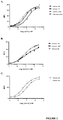
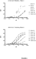


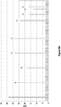
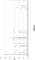

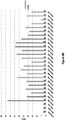
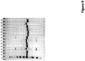

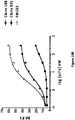
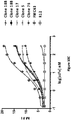


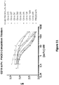


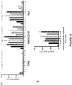
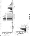
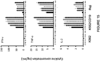
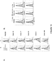
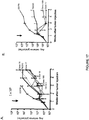
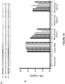
Classifications
-
- A—HUMAN NECESSITIES
- A61—MEDICAL OR VETERINARY SCIENCE; HYGIENE
- A61K—PREPARATIONS FOR MEDICAL, DENTAL OR TOILETRY PURPOSES
- A61K39/00—Medicinal preparations containing antigens or antibodies
- A61K39/395—Antibodies; Immunoglobulins; Immune serum, e.g. antilymphocytic serum
-
- C—CHEMISTRY; METALLURGY
- C07—ORGANIC CHEMISTRY
- C07K—PEPTIDES
- C07K16/00—Immunoglobulins [IGs], e.g. monoclonal or polyclonal antibodies
- C07K16/18—Immunoglobulins [IGs], e.g. monoclonal or polyclonal antibodies against material from animals or humans
- C07K16/28—Immunoglobulins [IGs], e.g. monoclonal or polyclonal antibodies against material from animals or humans against receptors, cell surface antigens or cell surface determinants
- C07K16/2896—Immunoglobulins [IGs], e.g. monoclonal or polyclonal antibodies against material from animals or humans against receptors, cell surface antigens or cell surface determinants against molecules with a "CD"-designation, not provided for elsewhere
-
- A—HUMAN NECESSITIES
- A61—MEDICAL OR VETERINARY SCIENCE; HYGIENE
- A61K—PREPARATIONS FOR MEDICAL, DENTAL OR TOILETRY PURPOSES
- A61K35/00—Medicinal preparations containing materials or reaction products thereof with undetermined constitution
- A61K35/12—Materials from mammals; Compositions comprising non-specified tissues or cells; Compositions comprising non-embryonic stem cells; Genetically modified cells
- A61K35/14—Blood; Artificial blood
- A61K35/17—Lymphocytes; B-cells; T-cells; Natural killer cells; Interferon-activated or cytokine-activated lymphocytes
-
- A—HUMAN NECESSITIES
- A61—MEDICAL OR VETERINARY SCIENCE; HYGIENE
- A61K—PREPARATIONS FOR MEDICAL, DENTAL OR TOILETRY PURPOSES
- A61K39/00—Medicinal preparations containing antigens or antibodies
- A61K39/46—Cellular immunotherapy
- A61K39/461—Cellular immunotherapy characterised by the cell type used
- A61K39/4611—T-cells, e.g. tumor infiltrating lymphocytes [TIL], lymphokine-activated killer cells [LAK] or regulatory T cells [Treg]
-
- A—HUMAN NECESSITIES
- A61—MEDICAL OR VETERINARY SCIENCE; HYGIENE
- A61K—PREPARATIONS FOR MEDICAL, DENTAL OR TOILETRY PURPOSES
- A61K39/00—Medicinal preparations containing antigens or antibodies
- A61K39/46—Cellular immunotherapy
- A61K39/463—Cellular immunotherapy characterised by recombinant expression
- A61K39/4631—Chimeric Antigen Receptors [CAR]
-
- A—HUMAN NECESSITIES
- A61—MEDICAL OR VETERINARY SCIENCE; HYGIENE
- A61K—PREPARATIONS FOR MEDICAL, DENTAL OR TOILETRY PURPOSES
- A61K39/00—Medicinal preparations containing antigens or antibodies
- A61K39/46—Cellular immunotherapy
- A61K39/464—Cellular immunotherapy characterised by the antigen targeted or presented
- A61K39/4643—Vertebrate antigens
- A61K39/4644—Cancer antigens
- A61K39/464402—Receptors, cell surface antigens or cell surface determinants
- A61K39/464411—Immunoglobulin superfamily
- A61K39/464412—CD19 or B4
-
- A—HUMAN NECESSITIES
- A61—MEDICAL OR VETERINARY SCIENCE; HYGIENE
- A61P—SPECIFIC THERAPEUTIC ACTIVITY OF CHEMICAL COMPOUNDS OR MEDICINAL PREPARATIONS
- A61P35/00—Antineoplastic agents
-
- A—HUMAN NECESSITIES
- A61—MEDICAL OR VETERINARY SCIENCE; HYGIENE
- A61P—SPECIFIC THERAPEUTIC ACTIVITY OF CHEMICAL COMPOUNDS OR MEDICINAL PREPARATIONS
- A61P35/00—Antineoplastic agents
- A61P35/02—Antineoplastic agents specific for leukemia
-
- C—CHEMISTRY; METALLURGY
- C07—ORGANIC CHEMISTRY
- C07K—PEPTIDES
- C07K14/00—Peptides having more than 20 amino acids; Gastrins; Somatostatins; Melanotropins; Derivatives thereof
- C07K14/435—Peptides having more than 20 amino acids; Gastrins; Somatostatins; Melanotropins; Derivatives thereof from animals; from humans
- C07K14/705—Receptors; Cell surface antigens; Cell surface determinants
- C07K14/70503—Immunoglobulin superfamily
- C07K14/7051—T-cell receptor (TcR)-CD3 complex
-
- C—CHEMISTRY; METALLURGY
- C07—ORGANIC CHEMISTRY
- C07K—PEPTIDES
- C07K14/00—Peptides having more than 20 amino acids; Gastrins; Somatostatins; Melanotropins; Derivatives thereof
- C07K14/435—Peptides having more than 20 amino acids; Gastrins; Somatostatins; Melanotropins; Derivatives thereof from animals; from humans
- C07K14/705—Receptors; Cell surface antigens; Cell surface determinants
- C07K14/70503—Immunoglobulin superfamily
- C07K14/70521—CD28, CD152
-
- C—CHEMISTRY; METALLURGY
- C07—ORGANIC CHEMISTRY
- C07K—PEPTIDES
- C07K16/00—Immunoglobulins [IGs], e.g. monoclonal or polyclonal antibodies
- C07K16/18—Immunoglobulins [IGs], e.g. monoclonal or polyclonal antibodies against material from animals or humans
- C07K16/28—Immunoglobulins [IGs], e.g. monoclonal or polyclonal antibodies against material from animals or humans against receptors, cell surface antigens or cell surface determinants
- C07K16/2803—Immunoglobulins [IGs], e.g. monoclonal or polyclonal antibodies against material from animals or humans against receptors, cell surface antigens or cell surface determinants against the immunoglobulin superfamily
-
- C—CHEMISTRY; METALLURGY
- C12—BIOCHEMISTRY; BEER; SPIRITS; WINE; VINEGAR; MICROBIOLOGY; ENZYMOLOGY; MUTATION OR GENETIC ENGINEERING
- C12N—MICROORGANISMS OR ENZYMES; COMPOSITIONS THEREOF; PROPAGATING, PRESERVING, OR MAINTAINING MICROORGANISMS; MUTATION OR GENETIC ENGINEERING; CULTURE MEDIA
- C12N5/00—Undifferentiated human, animal or plant cells, e.g. cell lines; Tissues; Cultivation or maintenance thereof; Culture media therefor
- C12N5/06—Animal cells or tissues; Human cells or tissues
- C12N5/0602—Vertebrate cells
- C12N5/0634—Cells from the blood or the immune system
- C12N5/0636—T lymphocytes
-
- A—HUMAN NECESSITIES
- A61—MEDICAL OR VETERINARY SCIENCE; HYGIENE
- A61K—PREPARATIONS FOR MEDICAL, DENTAL OR TOILETRY PURPOSES
- A61K39/00—Medicinal preparations containing antigens or antibodies
- A61K2039/505—Medicinal preparations containing antigens or antibodies comprising antibodies
-
- A—HUMAN NECESSITIES
- A61—MEDICAL OR VETERINARY SCIENCE; HYGIENE
- A61K—PREPARATIONS FOR MEDICAL, DENTAL OR TOILETRY PURPOSES
- A61K2239/00—Indexing codes associated with cellular immunotherapy of group A61K39/46
- A61K2239/31—Indexing codes associated with cellular immunotherapy of group A61K39/46 characterized by the route of administration
-
- A—HUMAN NECESSITIES
- A61—MEDICAL OR VETERINARY SCIENCE; HYGIENE
- A61K—PREPARATIONS FOR MEDICAL, DENTAL OR TOILETRY PURPOSES
- A61K2239/00—Indexing codes associated with cellular immunotherapy of group A61K39/46
- A61K2239/38—Indexing codes associated with cellular immunotherapy of group A61K39/46 characterised by the dose, timing or administration schedule
-
- A—HUMAN NECESSITIES
- A61—MEDICAL OR VETERINARY SCIENCE; HYGIENE
- A61K—PREPARATIONS FOR MEDICAL, DENTAL OR TOILETRY PURPOSES
- A61K2239/00—Indexing codes associated with cellular immunotherapy of group A61K39/46
- A61K2239/46—Indexing codes associated with cellular immunotherapy of group A61K39/46 characterised by the cancer treated
- A61K2239/48—Blood cells, e.g. leukemia or lymphoma
-
- C—CHEMISTRY; METALLURGY
- C07—ORGANIC CHEMISTRY
- C07K—PEPTIDES
- C07K2317/00—Immunoglobulins specific features
- C07K2317/20—Immunoglobulins specific features characterized by taxonomic origin
- C07K2317/21—Immunoglobulins specific features characterized by taxonomic origin from primates, e.g. man
-
- C—CHEMISTRY; METALLURGY
- C07—ORGANIC CHEMISTRY
- C07K—PEPTIDES
- C07K2317/00—Immunoglobulins specific features
- C07K2317/30—Immunoglobulins specific features characterized by aspects of specificity or valency
- C07K2317/34—Identification of a linear epitope shorter than 20 amino acid residues or of a conformational epitope defined by amino acid residues
-
- C—CHEMISTRY; METALLURGY
- C07—ORGANIC CHEMISTRY
- C07K—PEPTIDES
- C07K2317/00—Immunoglobulins specific features
- C07K2317/50—Immunoglobulins specific features characterized by immunoglobulin fragments
- C07K2317/52—Constant or Fc region; Isotype
- C07K2317/53—Hinge
-
- C—CHEMISTRY; METALLURGY
- C07—ORGANIC CHEMISTRY
- C07K—PEPTIDES
- C07K2317/00—Immunoglobulins specific features
- C07K2317/50—Immunoglobulins specific features characterized by immunoglobulin fragments
- C07K2317/56—Immunoglobulins specific features characterized by immunoglobulin fragments variable (Fv) region, i.e. VH and/or VL
- C07K2317/565—Complementarity determining region [CDR]
-
- C—CHEMISTRY; METALLURGY
- C07—ORGANIC CHEMISTRY
- C07K—PEPTIDES
- C07K2317/00—Immunoglobulins specific features
- C07K2317/60—Immunoglobulins specific features characterized by non-natural combinations of immunoglobulin fragments
- C07K2317/62—Immunoglobulins specific features characterized by non-natural combinations of immunoglobulin fragments comprising only variable region components
- C07K2317/622—Single chain antibody (scFv)
-
- C—CHEMISTRY; METALLURGY
- C07—ORGANIC CHEMISTRY
- C07K—PEPTIDES
- C07K2317/00—Immunoglobulins specific features
- C07K2317/70—Immunoglobulins specific features characterized by effect upon binding to a cell or to an antigen
- C07K2317/73—Inducing cell death, e.g. apoptosis, necrosis or inhibition of cell proliferation
-
- C—CHEMISTRY; METALLURGY
- C07—ORGANIC CHEMISTRY
- C07K—PEPTIDES
- C07K2317/00—Immunoglobulins specific features
- C07K2317/90—Immunoglobulins specific features characterized by (pharmaco)kinetic aspects or by stability of the immunoglobulin
- C07K2317/92—Affinity (KD), association rate (Ka), dissociation rate (Kd) or EC50 value
-
- C—CHEMISTRY; METALLURGY
- C07—ORGANIC CHEMISTRY
- C07K—PEPTIDES
- C07K2317/00—Immunoglobulins specific features
- C07K2317/90—Immunoglobulins specific features characterized by (pharmaco)kinetic aspects or by stability of the immunoglobulin
- C07K2317/94—Stability, e.g. half-life, pH, temperature or enzyme-resistance
-
- C—CHEMISTRY; METALLURGY
- C07—ORGANIC CHEMISTRY
- C07K—PEPTIDES
- C07K2319/00—Fusion polypeptide
-
- C—CHEMISTRY; METALLURGY
- C07—ORGANIC CHEMISTRY
- C07K—PEPTIDES
- C07K2319/00—Fusion polypeptide
- C07K2319/01—Fusion polypeptide containing a localisation/targetting motif
- C07K2319/02—Fusion polypeptide containing a localisation/targetting motif containing a signal sequence
-
- C—CHEMISTRY; METALLURGY
- C07—ORGANIC CHEMISTRY
- C07K—PEPTIDES
- C07K2319/00—Fusion polypeptide
- C07K2319/01—Fusion polypeptide containing a localisation/targetting motif
- C07K2319/03—Fusion polypeptide containing a localisation/targetting motif containing a transmembrane segment
-
- C—CHEMISTRY; METALLURGY
- C07—ORGANIC CHEMISTRY
- C07K—PEPTIDES
- C07K2319/00—Fusion polypeptide
- C07K2319/33—Fusion polypeptide fusions for targeting to specific cell types, e.g. tissue specific targeting, targeting of a bacterial subspecies
Definitions
- the present disclosure relates in some aspects to CD19 binding molecules, in particular, to anti-CD19 antibodies, including antibody fragments.
- the present disclosure further relates to recombinant receptors containing such antibodies, including chimeric antigen receptors (CARs), which contain such antibodies.
- CARs chimeric antigen receptors
- the disclosure further relates to genetically engineered cells expressing such receptors and antibodies, and use thereof in adoptive cell therapy.
- CD19 is expressed on normal B cells and by cells and tissues of various diseases and conditions, including most B cell malignancies. Most patients with B cell malignancies are not cured by available therapies, including therapies targeting CD19 and/or other B cell markers.
- Various CD19-binding molecules including anti-CD19 antibodies, and chimeric antigen receptors containing anti-CD19 antibody portions, and cells expressing such chimeric receptors, are available.
- Improved CD19-binding molecules and engineered CD19-targeting cells are needed. For example, there is a need for molecules and cells with reduced immunogenicity and/or human antibodies, including antibody fragments that specifically bind to CD19 and chimeric receptors expressing such human antibodies for use in adoptive cell therapy. Provided are instances that meet such needs.
- WO2012170807 describes anti-Pseudomonas PSL binding molecules and uses thereof.
- WO2009054863 describes human antibodies that bind CD19 and uses thereof.
- WO2009091826 describes compositions and methods related to a human CD19-specific chimeric antigen receptor.
- the invention provides antibody or antigen binding fragment thereof comprising: a VH region comprising the amino acid sequence of SEQ ID NO: 11, and a VL region comprising the amino acid sequence of SEQ ID NO: 13; or a VH region comprising the amino acid sequence of SEQ ID NO: 11, and a VL region comprising the amino acid sequence of SEQ ID NO: 14; or a VH region comprising the amino acid sequence of SEQ ID NO: 11, and a VL region comprising the amino acid sequence of SEQ ID NO: 16; or a VH region comprising the amino acid sequence of SEQ ID NO: 12, and a VL region comprising the amino acid sequence of SEQ ID NO: 15; or a VH region comprising the amino acid sequence of SEQ ID NO: 12, and a VL region comprising the amino acid sequence of SEQ ID NO: 17.
- CD19-binding molecules including polypeptides, such as anti-CD19 antibodies, including antigen-binding antibody fragments such as single-chain fragments including scFv fragments, and polypeptides containing such antibodies, including fusion proteins, receptors, e.g., recombinant receptors, including chimeric receptors such as chimeric antigen receptors (CARs) containing the antibody as an antigen-recognition component.
- the antibodies are human antibodies, such as human single-chain fragments including scFvs.
- antibodies or antigen-binding fragments thereof including those that specifically bind to CD19.
- the antibodies contain particular complementarity determining regions (CDRs), including heavy chain CDRs (CDR-Hs) and light chain CDRs (CDR-Ls).
- CDRs have or include amino acid sequences of CDRs of a reference antibody or chain or sequence thereof.
- the antibody or antigen-binding fragment thereof includes a heavy chain variable (VH) region and a light chain variable (VL) region.
- the antibody e.g., the VH region thereof, includes a heavy chain complementarity determining region 3 (CDR-H3) comprising the amino acid sequence set forth as SEQ ID NO: 20.
- the VH region comprises at least at or about 90 % sequence identity to the VH region amino acid sequence set forth in SEQ ID NO: 11, 12, 60, 61, 63, or 62, e.g., at least at or about 91%, 92%, 93%, 94%, 95%, 96%, 97%, 98%, or 99% sequence identity thereto.
- the antibody or fragment includes a CDR-H1 of SEQ ID NO: 18 and a CDR-H3 of SEQ ID NO: 20. In some instances, the antibody or fragment further includes a CDR-H2 sequence comprising SEQ ID NO: 81, 82, 19 or 72.
- the antibody has a CDR-H1, a CDR-H2, and/or a CDR-H3 that respectively include the amino acid sequences of CDR 1, 2, and 3 sequences contained within the heavy chain variable (V H ) region of a reference antibody.
- V H region of the reference antibody has the amino acid sequence set forth in SEQ ID NO: 11 or 12. In some instances, it has the amino acid sequence set forth in SEQ ID NO: 11, 12, 60, 61, 63, or 62.
- the antibody has, e.g., further includes, a CDR-L1, a CDR-L2, and/or a CDR-L3, respectively comprising the amino acid sequences of CDR 1, 2, and 3 sequences contained within the light chain variable (V L ) region of a reference antibody.
- V L of the reference antibody has the amino acid sequence set forth in SEQ ID NO: 13, 14, 15, 16, or 17.
- the VL of the reference antibody has the amino acid sequence set forth in SEQ ID NO: 13, 14, 15, 16, 17, 71, 65, 64, 66, 70, 69, 67, 90 or 91.
- the CDR within the reference antibody, VH, or VL refers to the CDR as defined by any numbering scheme, e.g., those defined herein.
- the CDR in the reference antibody or VH or VL refers to the CDR as defined by Kabat numbering scheme as described herein, the CDR as defined by the Chothia scheme as described herein, or the Contact scheme as described herein.
- the antibody contains a VH chain that includes a CDR-H1, CDR-H2 and/or CDR-H3 in which the CDR-H1 comprises the amino acid sequence of DYAMH (SEQ ID NO: 18) or a sequence having at least or at least about 90%, 91%, 92%, 93%, 94%, 95%, 96%, 97%, 98%, or 99% amino acid sequence identity to SEQ ID NO: 18;
- the CDR-H2 comprises the amino acid sequence of SEQ ID NO: 81 or 82 or 19 or 72 or a sequence having at least or at least about 90%, 91%, 92%, 93%, 94%, 95%, 96%, 97%, 98%, or 99% amino acid sequence identity to SEQ ID NO: 81 or to SEQ ID NO: 82 or to SEQ ID NO: 19 or to SEQ ID NO: 72; and/or the CDR-H3 comprises the amino acid sequence of SEQ ID NO: 20 or a sequence having at least or at least about 90%, 91%, 92%,
- the antibody comprises a CDR-H1 comprising the amino acid sequence of SEQ ID NO: 18, a CDR-H2 comprising the amino acid sequence of SEQ ID NO: 81 or 82, and a CDR-H3 comprising the amino acid sequence set forth as SEQ ID NO: 20.
- the antibody has a CDR-1 comprising the amino acid sequence of X 1 X 2 X 3 X 4 X 5 X 6 X 7 X 8 X 9 X 10 X 11 X 12 X 13 X 14 (SEQ ID NO: 110), wherein X 1 is T, W, S or R; X 2 is G or A; X 3 is I, T, D or S; X 4 is S, R, T or Q; X 5 is null or S; X 6 is null, D, N or G; X 7 is null, V or L; X 8 is X or null; X 9 is X or null; X 10 is X; X 11 is X; X 12 is Y, F, D or W; X 13 is V, A or L and X 14 is S, N or A.
- the antibody has a CDR-L1 comprising the amino acid sequence of X 1 X 2 X 3 X 4 X 5 X6X 7 X 8 X 9 X 10 X 11 X 12 X 13 X 14 (SEQ ID NO: 111), wherein X 1 is T, Q, S, or R; X 2 is G or A; X 3 is I, T, D, or S; X 4 is S, R, T, or Q; X 5 is null or S; X 6 is G, D, N, or null; X 7 is null, V, or L; X 8 is D, G, I, L, S, or null; X 9 is S, G, A, I, R, or null; X 10 is H, Y, F, S, or N; X 11 is R, N, D, H, or Y; X 12 is Y, F, D, or W; X 13 is V, A, or L; and X 14
- the antibody has a CDR-L3 comprising the amino acid sequence X 1 X 2 X 3 X 4 X 5 X6X 7 X 8 X 9 X 10 X 11 X 12 (SEQ ID NO: 114), wherein X 1 is S, G, T, A, Q, C or N; X 2 is S, Q, A, or T; X 3 is Y, S, W, R; X 4 is A, D, R, T, or Y; X 5 is A, S, P, G, N or D; X 6 is I, S, G, T, A, L, H, R, N; X 7 is S, P, L, Y, G; X 8 is P, T, S, Q, M, R, N or null; X 9 is S, L, N, A, M or null; X 10 is L or null; X 11 is Y, W, F, V, A or L; and X 12 is V, T,
- X 1 is S, G, Q, or N
- X 2 is S, Q, or T
- X 4 is A, D, T, or Y
- X 5 is A, S, or G
- X 6 is I, S, N, R, A, H, or T.
- the CDR-H2 comprises the amino acid sequence set forth in SEQ ID NO: 19 (GISWNSGRIGYADSVKG); or the CDR-H2 comprises the amino acid sequence set forth in SEQ ID NO: 72 (GISWNSGSIGYADSVKG).
- the CDR-L1 comprises the amino acid sequence set forth in SEQ ID NO: 80, 77, 74, 73, 75, 79, 78, 76, 21, 25, 28, or 31. In some instances, the CDR-L1 comprises the amino acid sequence set forth in SEQ ID NO: 80, 77, 74, 73, 78, 21, or 28.
- the CDR-L2 comprises the amino acid sequence set forth in SEQ ID NO: 100, 97, 94, 93, 95, 99, 98, 96, 22, 26, 29, or 32. In some instances, the CDR-L2 comprises the amino acid sequence set forth in SEQ ID NO: 100, 97, 94, 93, 98, 22, or 29.
- the CDR-L3 comprises the amino acid sequence set forth in SEQ ID NO: 109, 106, 103, 101, 104, 108, 107, 105, 102, 23, 24, 27, 30, or 33. In some instances, the CDR-L3 comprises the amino acid sequence set forth in SEQ ID NO: 109, 106, 103, 101, 107, 24 or 30.
- the CDR-L1, CDR-L2, and CDR-L3 comprise the sequences of SEQ ID NOs: 21, 22, and 23, respectively or sequences having at least or at least about 90%, 91%, 92%, 93%, 94%, 95%, 96%, 97%, 98%, or 99% amino acid sequence identity, respectively, thereto;
- the CDR-L1, CDR-L2, and CDR-L3 comprise the sequences of SEQ ID NOs: 21, 22, and 24 or sequences having at least or at least about 90%, 91%, 92%, 93%, 94%, 95%, 96%, 97%, 98%, or 99% amino acid sequence identity, respectively, thereto, respectively;
- the CDR-L1, CDR-L2, and CDR-L3 comprise the sequences of SEQ ID NOs: 25, 26, and 27, respectively or sequences having at least or at least about 90%, 91%, 92%, 93%, 94%, 95%, 96%, 97%, 98%, or 99% amino acid sequence identity, respectively, thereto;
- the CDR-L3 comprises the amino acid sequence set forth as SEQ ID NO: 116, 117, 118, 119, 120, or 121, or a sequence having at least or at least about 90%, 91%, 92%, 93%, 94%, 95%, 96%, 97%, 98%, or 99% amino acid sequence identity, respectively, thereto.
- the CDR-H1, CDR-H2, and CDR-H3 comprise the sequences of SEQ ID NOs: 18, 81, and 20, respectively or sequences having at least or at least about 90%, 91%, 92%, 93%, 94%, 95%, 96%, 97%, 98%, or 99% amino acid sequence identity, respectively, thereto;
- the CDR-H1, CDR-H2, and CDR-H3 comprise the sequences of SEQ ID NOs: 18, 19, and 20, respectively or sequences having at least or at least about 90%, 91%, 92%, 93%, 94%, 95%, 96%, 97%, 98%, or 99% amino acid sequence identity, respectively, thereto;
- the CDR-H1, CDR-H2, and CDR-H3 comprise the sequences of SEQ ID NOs: 18, 82, and 20, respectively or sequences having at least or at least about 90%, 91%, 92%, 93%, 94%, 95%, 96%, 97%, 98%, or 99% amino acid sequence identity, respectively, thereto;
- the antibody has a CDR-L1 comprising the amino acid sequence X 1 GX 3 X 4 X 5 X 6 X 7 X 8 X 9 X 10 X 11 X 12 X 13 S (SEQ ID NO: 36), wherein X1 is T, S, or Q, X3 is T, S, or D, X4 is T or S, X5 is null or S, X6 is null, D, or N, X7 is null or V, X8 is null, G, or I, X9 is null, G, or R, X10 is S, Y, or N, X11 is D or N, X12 is D or Y, X13 is V or A; the CDR-L2 comprises the amino acid sequence X 1 X 2 X 3 X 4 RPS (SEQ ID NO: 37), wherein X1 is D or S, X2 is V, N, or K, X3 is S, N, or D, and
- the antibody has a CDR-L1 comprising the amino acid sequence X 1 GX 3 X 4 X 5 X 6 X 7 X 8 X 9 X 10 X 11 X 12 X 13 S (SEQ ID NO: 36), wherein X1 is T, S, or Q, X3 is T, S, or D, X4 is T or S, X5 is null or S, X6 is null, D, or N, X7 is null or V, X8 is null, G, or I, X9 is null, G, or R, X10 is S, Y, or N, X11 is D or N, X12 is D or Y, X13 is V or A; the CDR-L2 comprises the amino acid sequence X 1 X 2 X 3 X 4 RPS (SEQ ID NO: 37), wherein X1 is D or S, X2 is V, N, or K, X3 is S, N, or D, and
- X1 is T or S
- X3 is T or S
- X11 is D or N
- X13 is V
- X2 is V or N
- X4 is K or Q.
- the CDR-H2 comprises the amino acid sequence set forth in SEQ ID NO: 19 (GISWNSGRIGYADSVKG) or a sequence having at least or at least about 90%, 91%, 92%, 93%, 94%, 95%, 96%, 97%, 98%, or 99% amino acid sequence identity to SEQ ID NO: 19.
- the CDR-L1 comprises the sequence set forth in SEQ ID NO: 21, 25, 28, or 31 or a sequence having at least or at least about 90%, 91%, 92%, 93%, 94%, 95%, 96%, 97%, 98%, or 99% amino acid sequence identity thereto; and/or the CDR-L2 comprises the sequence set forth in SEQ ID NO: 22, 26, 29, or 32 or a sequence having at least or at least about 90%, 91%, 92%, 93%, 94%, 95%, 96%, 97%, 98%, or 99% amino acid sequence identity thereto; and/or the CDR-L3 comprises the sequence set forth in SEQ ID NO: 23, 24, 27, 30, or 33 or a sequence having at least or at least about 90%, 91%, 92%, 93%, 94%, 95%, 96%, 97%, 98%, or 99% amino acid sequence identity thereto.
- the CDR-L1, CDR-L2, and/or CDR-L3 comprise the sequences of SEQ ID NOs: 21, 22, and/or 23, respectively or sequences having at least or at least about 90%, 91%, 92%, 93%, 94%, 95%, 96%, 97%, 98%, or 99% amino acid sequence identity thereto, respectively.
- the CDR-L1, CDR-L2, and CDR-L3 comprise the sequences of SEQ ID NOs: 21, 22, and 24, respectively, or sequences having at least or at least about 90%, 91%, 92%, 93%, 94%, 95%, 96%, 97%, 98%, or 99% amino acid sequence identity thereto, respectively.
- the CDR-L1, CDR-L2, and CDR-L3 comprise the sequences of SEQ ID NOs: 25, 26, and 27, respectively or sequences having at least or at least about 90%, 91%, 92%, 93%, 94%, 95%, 96%, 97%, 98%, or 99% amino acid sequence identity thereto, respectively.
- the CDR-L1, CDR-L2, and CDR-L3 comprise the sequences of SEQ ID NOs: 28, 29, and 30, respectively, or sequences having at least or at least about 90%, 91%, 92%, 93%, 94%, 95%, 96%, 97%, 98%, or 99% amino acid sequence identity thereto, respectively.
- the CDR-L1, CDR-L2, and CDR-L3 comprise the sequences of SEQ ID NOs: 31, 32, and 33, respectively, or sequences having at least or at least about 90%, 91%, 92%, 93%, 94%, 95%, 96%, 97%, 98%, or 99% amino acid sequence identity thereto.
- the heavy and light chain CDRs are any combination of the aforementioned CDR-L and CDR-H sequences, including sequences having at least or at least about 90%, 91%, 92%, 93%, 94%, 95%, 96%, 97%, 98%, or 99% amino acid sequence identity thereto.
- the antibody or fragment comprises a VH region comprising the amino acid sequence of SEQ ID NO: 11 or a sequence having at least or at least about 90%, 91%, 92%, 93%, 94%, 95%, 96%, 97%, 98%, or 99% amino acid sequence identity thereto.
- the antibody or fragment comprises a VH region comprising the amino acid sequence of SEQ ID NO: 12 or a sequence having at least or at least about 90%, 91%, 92%, 93%, 94%, 95%, 96%, 97%, 98%, or 99% amino acid sequence identity thereto.
- the antibody or fragment comprises a VL region comprising the amino acid sequence of SEQ ID NO: 13 or a sequence having at least or at least about 90%, 91%, 92%, 93%, 94%, 95%, 96%, 97%, 98%, or 99% amino acid sequence identity thereto.
- the antibody or fragment comprises a VL region comprising the amino acid sequence of SEQ ID NO: 14 or a sequence having at least or at least about 90%, 91%, 92%, 93%, 94%, 95%, 96%, 97%, 98%, or 99% amino acid sequence identity thereto.
- the antibody or fragment comprises a VL region comprising the amino acid sequence of SEQ ID NO: 15 or a sequence having at least or at least about 90%, 91%, 92%, 93%, 94%, 95%, 96%, 97%, 98%, or 99% amino acid sequence identity thereto.
- the antibody or fragment comprises a VL region comprising the amino acid sequence of SEQ ID NO: 16 or a sequence having at least or at least about 90%, 91%, 92%, 93%, 94%, 95%, 96%, 97%, 98%, or 99% amino acid sequence identity thereto.
- the antibody or fragment comprises a VL region comprising the amino acid sequence of SEQ ID NO: 17 or a sequence having at least or at least about 90%, 91%, 92%, 93%, 94%, 95%, 96%, 97%, 98%, or 99% amino acid sequence identity thereto.
- the antibody or fragment comprises a VH region comprising the amino acid sequence of SEQ ID NO: 63 or a sequence having at least or at least about 90%, 91%, 92%, 93%, 94%, 95%, 96%, 97%, 98%, or 99% amino acid sequence identity thereto.
- the antibody or fragment comprises a VH region comprising the amino acid sequence of SEQ ID NO: 60 or a sequence having at least or at least about 90%, 91%, 92%, 93%, 94%, 95%, 96%, 97%, 98%, or 99% amino acid sequence identity thereto.
- the antibody or fragment comprises a VH region comprising the amino acid sequence of SEQ ID NO: 61 or a sequence having at least or at least about 90%, 91%, 92%, 93%, 94%, 95%, 96%, 97%, 98%, or 99% amino acid sequence identity thereto.
- the antibody or fragment comprises a VH region comprising the amino acid sequence of SEQ ID NO: 63 or a sequence having at least or at least about 90%, 91%, 92%, 93%, 94%, 95%, 96%, 97%, 98%, or 99% amino acid sequence identity thereto.
- the antibody or fragment comprises a VH region comprising the amino acid sequence of SEQ ID NO: 62 or a sequence having at least or at least about 90%, 91%, 92%, 93%, 94%, 95%, 96%, 97%, 98%, or 99% amino acid sequence identity thereto.
- the antibody or fragment comprises a VL region comprising the amino acid sequence of SEQ ID NO: 71 or a sequence having at least or at least about 90%, 91%, 92%, 93%, 94%, 95%, 96%, 97%, 98%, or 99% amino acid sequence identity thereto.
- the antibody or fragment comprises a VL region comprising the amino acid sequence of SEQ ID NO: 90 or a sequence having at least or at least about 90%, 91%, 92%, 93%, 94%, 95%, 96%, 97%, 98%, or 99% amino acid sequence identity thereto.
- the antibody or fragment comprises a VL region comprising the amino acid sequence of SEQ ID NO: 91 or a sequence having at least or at least about 90%, 91%, 92%, 93%, 94%, 95%, 96%, 97%, 98%, or 99% amino acid sequence identity thereto.
- the antibody or fragment comprises a VL region comprising the amino acid sequence of SEQ ID NO: 68 or a sequence having at least or at least about 90%, 91%, 92%, 93%, 94%, 95%, 96%, 97%, 98%, or 99% amino acid sequence identity thereto.
- the antibody or fragment comprises a VL region comprising the amino acid sequence of SEQ ID NO: 65 or a sequence having at least or at least about 90%, 91%, 92%, 93%, 94%, 95%, 96%, 97%, 98%, or 99% amino acid sequence identity thereto.
- the antibody or fragment comprises a VL region comprising the amino acid sequence of SEQ ID NO: 64 or a sequence having at least or at least about 90%, 91%, 92%, 93%, 94%, 95%, 96%, 97%, 98%, or 99% amino acid sequence identity thereto.
- the antibody or fragment comprises a VL region comprising the amino acid sequence of SEQ ID NO: 66 or a sequence having at least or at least about 90%, 91%, 92%, 93%, 94%, 95%, 96%, 97%, 98%, or 99% amino acid sequence identity thereto.
- the antibody or fragment comprises a VL region comprising the amino acid sequence of SEQ ID NO: 70 or a sequence having at least or at least about 90%, 91%, 92%, 93%, 94%, 95%, 96%, 97%, 98%, or 99% amino acid sequence identity thereto.
- the antibody or fragment comprises a VL region comprising the amino acid sequence of SEQ ID NO: 69 or a sequence having at least or at least about 90%, 91%, 92%, 93%, 94%, 95%, 96%, 97%, 98%, or 99% amino acid sequence identity thereto.
- the antibody or fragment comprises a VL region comprising the amino acid sequence of SEQ ID NO: 67 or a sequence having at least or at least about 90%, 91%, 92%, 93%, 94%, 95%, 96%, 97%, 98%, or 99% amino acid sequence identity thereto.
- the VH region of the antibody or fragment comprises the amino acid sequence of SEQ ID NO: 11, 60, 63, or 62 or a sequence having at least or at least about 90%, 91%, 92%, 93%, 94%, 95%, 96%, 97%, 98%, or 99% amino acid sequence identity thereto; and/or the VL region of the antibody or fragment comprises the amino acid sequence of SEQ ID NO: 14, 16, 71, 90, 65, 64, or 69 or a sequence having at least or at least about 90%, 91%, 92%, 93%, 94%, 95%, 96%, 97%, 98%, or 99% amino acid sequence identity thereto.
- the VH and VL regions of the antibody or fragment comprise the amino acid sequences of SEQ ID NOs: 12 and 17, respectively, or sequences having at least or at least about 90%, 91%, 92%, 93%, 94%, 95%, 96%, 97%, 98%, or 99% amino acid sequence identity thereto, respectively;
- the VH and VL regions of the antibody or fragment comprise the amino acid sequences of SEQ ID NOs: 12 and 15, respectively, or sequences having at least or at least about 90%, 91%, 92%, 93%, 94%, 95%, 96%, 97%, 98%, or 99% amino acid sequence identity thereto, respectively;
- the VH and VL regions of the antibody or fragment comprise the amino acid sequences of SEQ ID NOs: 11 and 13, respectively, or sequences having at least or at least about 90%, 91%, 92%, 93%, 94%, 95%, 96%, 97%, 98%, or 99% amino acid sequence identity thereto, respectively;
- the VH and VL regions of the antibody or fragment comprise the
- the VH region comprises SEQ ID NO: 11 and the VL region comprises SEQ ID NO: 13; in some instances, the VH region comprises SEQ ID NO: 11 and the VL region comprises SEQ ID NO: 14; in some instances, the VH region comprises SEQ ID NO: 11 and the VL region comprises SEQ ID NO: 15; in some instances, the VH region comprises SEQ ID NO: 11 and the VL region comprises SEQ ID NO: 16, or sequences having at least or at least about 90%, 91%, 92%, 93%, 94%, 95%, 96%, 97%, 98%, or 99% amino acid sequence identity thereto; in some instances, the VH region comprises SEQ ID NO: 11 and the VL region comprises SEQ ID NO: 17, or sequences having at least or at least about 90%, 91%, 92%, 93%, 94%, 95%, 96%, 97%, 98%, or 99% amino acid sequence identity thereto.
- the VH region comprises SEQ ID NO: 12 and the VL region comprises SEQ ID NO: 13; in some instances, the VH region comprises SEQ ID NO: 12 and the VL region comprises SEQ ID NO: 14; in some instances, the VH region comprises SEQ ID NO: 12 and the VL region comprises SEQ ID NO: 15; in some instances, the VH region comprises SEQ ID NO: 12 and the VL region comprises SEQ ID NO: 16, or sequences having at least or at least about 90%, 91%, 92%, 93%, 94%, 95%, 96%, 97%, 98%, or 99% amino acid sequence identity thereto; in some instances, the VH region comprises SEQ ID NO: 12 and the VL region comprises SEQ ID NO: 16, or sequences having at least or at least about 90%, 91%, 92%, 93%, 94%, 95%, 96%, 97%, 98%, or 99% amino acid sequence identity thereto.
- the antibody is a single chain fragment, such as one with two or more variable regions joined by one or more flexible immunoglobulin linker.
- the antibody is an scFv.
- the scFv comprises a linker that is rich in serine and/or glycine, such as a linker comprising GGGS (SEQ ID NO: 122) or GGGGS (SEQ ID NO:123) repeats, such as one comprising the sequence set forth SEQ ID NO: 34.
- the linker comprises a sequence of SEQ ID NO: 43.
- the antibody fragment e.g., scFv
- the antibody fragment, e.g., scFv contains a V L region or portion thereof followed by a linker, followed by a V H region or portion thereof.
- the scFv comprises the amino acid sequence set forth in SEQ ID NO: 2, 4, 6, 8, or 10, or a sequence having at least or at least about 90%, 91%, 92%, 93%, 94%, 95%, 96%, 97%, 98%, or 99% amino acid sequence identity thereto.
- the scFv comprises the amino acid sequence set forth in SEQ ID NO: 2, 4, 6, 8, 10, 45, 47, 49, 51, 53, 55, 57, 59, 87, or 89, or has a sequence at least at or about 90, 91, 92, 93, 94, 95, 96, 97, 98, or 99% identical to such a sequence.
- the antibody or fragment specifically binds to the same, similar, and/or an overlapping epitope of CD19 as the epitope specifically bound by a reference antibody, and/or the antibody competes for binding to CD19 with the reference antibody.
- the reference antibody is a murine or chimeric or human or humanized anti-CD 19 antibody, FMC63, SJ25C1, an antibody having a variable region sequence of SEQ ID NO: 39 and/or 40, or an antibody having a variable region sequence of SEQ ID NO: 41 and/or 42.
- the reference antibody is an antibody including a sequence as described herein, including sequence(s) of any of the aforementioned instances.
- the reference antibody can be an scFv that contains the amino acid sequence set forth in SEQ ID NO: 2, 4, 6, 8, 10, 45, 47, 49, 51, 53, 55, 57, 59, 87, or 89.
- the provided antibody or fragment contains one or more or all CDRs that are distinct from those in the reference antibody.
- the provided antibody or fragment contains one or more or all CDRs that are distinct from the corresponding CDRs in the antibody designated FMC63 or SJ25C1.
- human antibody and antigen-binding fragments that specifically bind to the same or an overlapping epitope of CD19 as the epitope specifically bound by the reference antibody, such as FMC63, SJ25C1, an antibody having a variable region sequence of SEQ ID NO: 39 and/or 40, or an antibody having a variable region sequence of SEQ ID NO: 41 and/or 42, and comprising heavy and light chain CDRs that are distinct from the CDRs present in the reference antibody.
- the antibody competes for binding with the reference antibody to at least the same degree as the reference antibody competes for binding with itself to CD19, or a degree of competition that is no more than 1.5-fold or 2-fold lower, 3-fold lower, 4-fold lower, 5-fold lower, or 10-fold lower than the competition by the reference antibody, and/or a measured IC50 that is no more than 1.5-fold or 2-fold or 3-fold or 4-fold or 5-fold or 10-fold higher than the IC50 of the reference antibody competing for binding with itself, for example, as measured in the same assay.
- the antibody has a binding affinity that is at least as high or substantially as high as the binding affinity for CD19 of the reference antibody. In some aspects, the antibody has a binding affinity of an EC50 that is about the same or lower than the EC50 reference antibody or no more than about 1.5-fold or no more than about 2-fold greater, no more than 3-fold greater, and/or no more than 10-fold greater, than the EC50 of the reference antibody. In some instances, binding affinity of the antibody is compared to the corresponding form of the reference antibody. Comparison is generally by the same or a similar assay.
- CD19 is a human CD19.
- the antibody or fragment specifically binds, exhibits binding affinity and/or competes for binding to human CD19.
- the antibody is human. In some instances, the antibody is recombinant. In some instances, the antibody is monoclonal. In some instances, the antibody is isolated.
- the antibody or fragment further includes at least a portion of an immunoglobulin constant region.
- the constant region may include any one or more of CH1, CH2, CH3, and/or CH4, and/or CL, of a human or other antibody, and be of any class, including IgG, IgM, IgA, IgE, and IgD, for example, including human IgG, e.g., IgG1 or IgG4, constant region domains.
- the constant region comprises or is an Fc region, such as a human IgG Fc region.
- molecules such as chimeric and/or fusion molecules, including receptors, such as recombinant receptors, that include the antibody of any of the instances (e.g., contained in or part of an extracellular domain) and additional domains, such as intracellular signaling domains, spacers, linkers, and/or transmembrane domains.
- the receptor is a chimeric antigen receptor, comprising an extracellular portion comprising the antibody or fragment of any of the instances and an intracellular signaling domain.
- the antibody or fragment comprises an scFv.
- the intracellular signaling domain comprises an ITAM and/or signaling domain capable of delivering a signal approximating that of natural ligation of an ITAM-containing molecule or receptor complex such as a TCR receptor complex.
- the intracellular signaling domain comprises a signaling domain of a zeta chain of a CD3-zeta (CD3 ⁇ ) chain.
- the receptor further includes one or more domains, such as a transmembrane domain, linking the antibody transmembrane domain linking the extracellular domain and the intracellular signaling domain.
- the transmembrane domain comprises a transmembrane portion of a costimulatory molecule, such as a T cell costimulatory molecule, e.g., CD28 and/or 41BB.
- a costimulatory molecule such as a T cell costimulatory molecule, e.g., CD28 and/or 41BB.
- the T cell costimulatory molecule is selected from the group consisting of CD28 and 41BB, and in some instances, the receptor includes signaling domains from CD28 and 41BB.
- nucleic acids encoding the antibody (including fragments) of any of instances or the receptor, e.g., chimeric antigen receptor of any of the instances, vectors including such nucleic acids, and cells containing the vectors and/or nucleic acids, for example, for expression of the antibodies and/or molecules.
- cells and vectors for producing and expressing the molecules including the antibodies and molecules such as receptors, e.g., chimeric antigen receptors (CARs).
- CARs chimeric antigen receptors
- engineered cells expressing the chimeric antigen receptor of any of the instances are provided.
- the cell is a T cell.
- the cell is an NK cell.
- the cell is a stem cell.
- compositions comprising the antibodies, receptors, molecules, and/or cells, including pharmaceutical compositions, e.g., further including pharmaceutically acceptable substances such as carriers.
- a disease or disorder associated with CD19 such as a B cell malignancy, such as B cell chronic lymphocytic leukemia (CLL), acute lymphocytic leukemia (ALL), pro-lymphocytic leukemias, hairy cell leukemias, common acute lymphocytic leukemias, Null-acute lymphoblastic leukemias, non-Hodgkin lymphomas, diffuse large B cell lymphomas (DLBCLs), multiple myelomas, follicular lymphoma, splenic, marginal zone lymphoma, mantle cell lymphoma, indolent B cell lymphoma, or Hodgkin lymphoma, or an autoimmune or a disease or disorder associated with CD19, such as a B cell malignancy, such as B cell chronic lymphocytic leukemia (CLL), acute lymphocytic leukemia (ALL), pro-lymphocytic leukemias, hairy cell leukemias, common acute lymphoc
- administration of the antibody or receptor is associated with a lower degree of immunogenicity as compared to administration of a reference antibody (or receptor containing the reference antibody) that competes for binding with the antibody or binds to an overlapping epitope.
- the reference antibody is a humanized, chimeric, or non-human antibody.
- CD19-binding molecules including antibodies (including antigen-binding antibody fragments, such as single chain fragments, including scFvs) and recombinant receptors, including chimeric receptors containing such antibodies and fragments, nucleic acids encoding such antibodies and fragments, and cells, such as recombinant cells, expressing and for production of these antibodies and fragments. Also provided are methods of making and using the antibodies and fragments as well as cells expressing or containing the antibodies and fragments.
- CD19 binding molecules such as CD19-binding polypeptides.
- Such binding molecules include antibodies that specifically bind to CD19, such as a human CD19 molecule, including antigen-binding fragments thereof.
- recombinant receptors such as chimeric antigen receptors containing such antibodies.
- anti-CD19 antibodies including functional antibody fragments, including those comprising a variable heavy chain and a variable light chain, such as scFvs.
- molecules containing such antibodies e.g., fusion proteins and/or recombinant receptors such as chimeric receptors, including antigen receptors.
- the provided anti-CD19 antibodies are human antibodies.
- the antibodies, such as the human antibodies specifically bind to a particular epitope or region of CD19, generally an extracellular epitope or region.
- the antibodies bind to the same or a similar epitope or region of CD19 as bound by another antibody, such as one or more of the mouse antibodies, FMC63 or SJ25C1.
- the antibodies bind to an overlapping epitope of CD19 as bound by one of these known antibodies and/or compete for binding with such an antibody.
- the antibodies include isolated antibodies.
- the molecules include isolated molecules.
- antibody herein is used in the broadest sense and includes polyclonal and monoclonal antibodies, including intact antibodies and functional (antigen-binding) antibody fragments, including fragment antigen binding (Fab) fragments, F(ab') 2 fragments, Fab' fragments, Fv fragments, recombinant IgG (rIgG) fragments, single chain antibody fragments, including single chain variable fragments (scFv), and single domain antibodies (e.g., sdAb, sdFv, nanobody) fragments.
- Fab fragment antigen binding
- rIgG recombinant IgG
- scFv single chain variable fragments
- single domain antibodies e.g., sdAb, sdFv, nanobody
- the term encompasses genetically engineered and/or otherwise modified forms of immunoglobulins, such as intrabodies, peptibodies, chimeric antibodies, fully human antibodies, humanized antibodies, and heteroconjugate antibodies, multispecific, e.g., bispecific, antibodies, diabodies, triabodies, and tetrabodies, tandem di-scFv, tandem tri-scFv.
- antibody should be understood to encompass functional antibody fragments thereof.
- the term also encompasses intact or full-length antibodies, including antibodies of any class or subclass, including IgG and sub-classes thereof, IgM, IgE, IgA, and IgD.
- CDR complementarity determining region
- HVR hypervariable region
- FR-H1, FR-H2, FR-H3, and FR-H4 there are four FRs in each full-length heavy chain variable region (FR-H1, FR-H2, FR-H3, and FR-H4), and four FRs in each full-length light chain variable region (FR-L1, FR-L2, FR-L3, and FR-L4).
- the boundaries of a given CDR or FR may vary depending on the scheme used for identification.
- the Kabat scheme is based structural alignments
- the Chothia scheme is based on structural information. Numbering for both the Kabat and Chothia schemes is based upon the most common antibody region sequence lengths, with insertions accommodated by insertion letters, for example, "30a,” and deletions appearing in some antibodies. The two schemes place certain insertions and deletions ("indels") at different positions, resulting in differential numbering.
- the Contact scheme is based on analysis of complex crystal structures and is similar in many respects to the Chothia numbering scheme.
- Table 1 lists exemplary position boundaries of CDR-L1, CDR-L2, CDR-L3 and CDR-H1, CDR-H2, CDR-H3 as identified by Kabat, Chothia, and Contact schemes, respectively.
- residue numbering is listed using both the Kabat and Chothia numbering schemes.
- FRs are located between CDRs, for example, with FR-L1 located between CDR-L1 and CDR-L2, and so forth. It is noted that because the shown Kabat numbering scheme places insertions at H35A and H35B, the end of the Chothia CDR-H1 loop when numbered using the shown Kabat numbering convention varies between H32 and H34, depending on the length of the loop.
- a “CDR” or “complementary determining region,” or individual specified CDRs (e.g., “CDR-H1, CDR-H2), of a given antibody or region thereof, such as a variable region thereof, should be understood to encompass a (or the specific) complementary determining region as defined by any of the aforementioned schemes.
- a particular CDR e.g., a CDR-H3
- a CDR-H3 contains the amino acid sequence of a corresponding CDR in a given V H or V L amino acid sequence
- such a CDR has a sequence of the corresponding CDR (e.g., CDR-H3) within the variable region, as defined by any of the aforementioned schemes.
- specified CDR sequences are specified.
- FR or individual specified FR(s) e.g., FR-H1, FR-H2
- FR-H1, FR-H2 FR-H2
- FR-H1 FR-H2
- FR-H2 FR-H2
- FR-H2 FR-H2
- FR-H2 FR-H2
- FR-H2 FR-H2
- FR-H2 FR-H1
- FR-H2 FR-H2
- FR-H2 FR-H2
- the scheme for identification of a particular CDR, FR, or FRs or CDRs is specified, such as the CDR as defined by the Kabat, Chothia, or Contact method.
- the particular amino acid sequence of a CDR or FR is given.
- variable region refers to the domain of an antibody heavy or light chain that is involved in binding the antibody to antigen.
- the variable domains of the heavy chain and light chain (V H and V L , respectively) of a native antibody generally have similar structures, with each domain comprising four conserved framework regions (FRs) and three CDRs.
- FRs conserved framework regions
- a single V H or V L domain may be sufficient to confer antigen-binding specificity.
- antibodies that bind a particular antigen may be isolated using a V H or V L domain from an antibody that binds the antigen to screen a library of complementary V L or V H domains, respectively. See, e.g., Portolano et al., J. Immunol. 150:880-887 (1993 ); Clarkson et al., Nature 352:624-628 (1991 ).
- antibody fragments refers to a molecule other than an intact antibody that comprises a portion of an intact antibody that binds the antigen to which the intact antibody binds.
- antibody fragments include but are not limited to Fv, Fab, Fab', Fab'-SH, F(ab') 2 ; diabodies; linear antibodies; single-chain antibody molecules ( e.g. scFv); and multispecific antibodies formed from antibody fragments.
- the antibodies are single-chain antibody fragments comprising a variable heavy chain region and/or a variable light chain region, such as scFvs.
- Single-domain antibodies are antibody fragments comprising all or a portion of the heavy chain variable domain or all or a portion of the light chain variable domain of an antibody.
- a single-domain antibody is a human single-domain antibody.
- Antibody fragments can be made by various techniques, including but not limited to proteolytic digestion of an intact antibody as well as production by recombinant host cells.
- the antibodies are recombinantly-produced fragments, such as fragments comprising arrangements that do not occur naturally, such as those with two or more antibody regions or chains joined by synthetic linkers, e.g., peptide linkers, and/or that are may not be produced by enzyme digestion of a naturally-occurring intact antibody.
- the antibody fragments are scFvs.
- a “humanized” antibody is an antibody in which all or substantially all CDR amino acid residues are derived from non-human CDRs and all or substantially all FR amino acid residues are derived from human FRs.
- a humanized antibody optionally may include at least a portion of an antibody constant region derived from a human antibody.
- a "humanized form" of a non-human antibody refers to a variant of the non-human antibody that has undergone humanization, typically to reduce immunogenicity to humans, while retaining the specificity and affinity of the parental non-human antibody.
- some FR residues in a humanized antibody are substituted with corresponding residues from a non-human antibody (e.g., the antibody from which the CDR residues are derived), e.g., to restore or improve antibody specificity or affinity.
- a non-human antibody e.g., the antibody from which the CDR residues are derived
- human antibodies are human antibodies.
- a "human antibody” is an antibody with an amino acid sequence corresponding to that of an antibody produced by a human or a human cell, or non-human source that utilizes human antibody repertoires or other human antibody-encoding sequences, including human antibody libraries.
- the term excludes humanized forms of non-human antibodies comprising non-human antigen-binding regions, such as those in which all or substantially all CDRs are non-human.
- Human antibodies may be prepared by administering an immunogen to a transgenic animal that has been modified to produce intact human antibodies or intact antibodies with human variable regions in response to antigenic challenge. Such animals typically contain all or a portion of the human immunoglobulin loci, which replace the endogenous immunoglobulin loci, or which are present extrachromosomally or integrated randomly into the animal's chromosomes. In such transgenic animals, the endogenous immunoglobulin loci have generally been inactivated. Human antibodies also may be derived from human antibody libraries, including phage display and cell-free libraries, containing antibody-encoding sequences derived from a human repertoire.
- monoclonal antibodies including monoclonal antibody fragments.
- the term "monoclonal antibody” as used herein refers to an antibody obtained from or within a population of substantially homogeneous antibodies, i.e., the individual antibodies comprising the population are identical, except for possible variants containing naturally occurring mutations or arising during production of a monoclonal antibody preparation, such variants generally being present in minor amounts.
- polyclonal antibody preparations which typically include different antibodies directed against different epitopes
- each monoclonal antibody of a monoclonal antibody preparation is directed against a single epitope on an antigen.
- a monoclonal antibody may be made by a variety of techniques, including but not limited to generation from a hybridoma, recombinant DNA methods, phage-display and other antibody display methods.
- polypeptide and protein are used interchangeably to refer to a polymer of amino acid residues, and are not limited to a minimum length.
- Polypeptides including the provided antibodies and antibody chains and other peptides, e.g., linkers and CD19-binding peptides, may include amino acid residues including natural and/or non-natural amino acid residues.
- the terms also include post-expression modifications of the polypeptide, for example, glycosylation, sialylation, acetylation, phosphorylation, and the like.
- the polypeptides may contain modifications with respect to a native or natural sequence, as long as the protein maintains the desired activity. These modifications may be deliberate, as through site-directed mutagenesis, or may be accidental, such as through mutations of hosts which produce the proteins or errors due to PCR amplification.
- the anti-CD19 antibody e.g., antigen-binding antibody fragment
- V H or V L heavy and/or light chain variable region sequences.
- Also among the provided antibodies are those having sequences at least at or about 90, 91, 92, 93, 94, 95, 96, 97, 98, or 99% identical to such a sequence.
- the antibody e.g., antigen-binding fragment thereof, includes a heavy chain complementarity determining region 3 (CDR-H3) comprising an amino acid sequence of a CDR-H3 present in a reference antibody, such as one present in a reference antibody having a VH region with the amino acid sequence set forth in set forth in SEQ ID NO: 11, 12, 60, 61, 63 62, 167 or 185, such as set forth in SEQ ID NO: 11, 12, 60, 61, 63, or 62.
- the CDR-H3 comprises SEQ ID NO: 20.
- the antibody e.g., antigen-binding fragment thereof, has a VH region having at least 90, 91, 92, 93, 94, 95, 96, 97, 98, or 99% sequence identity to (or 100 % identity thereto) the VH region amino acid sequence of the reference antibody, such as to the VH region amino acid sequence set forth in SEQ ID NO: 11, 12, 60, 61, 63 62, 167 or 185, such as set forth in SEQ ID NO: 11, 12, 60, 61, 63, or 62.
- the CDR-H1 contains the amino acid sequence DYAMH (SEQ ID NO: 18)
- the CDR-H2 contains the amino acid sequence GISWNSGRIG (SEQ ID NO: 81), GISWNSGSIG (SEQ ID NO: 82), the amino acid sequence set forth in SEQ ID NO: 19 (GISWNSGRIGYADSVKG), or the amino acid sequence set forth in SEQ ID NO: 72 (GISWNSGSIGYADSVKG)
- the CDR-H3 contains the amino acid sequence of SEQ ID NO: 20.
- the provided antibody contains a CDR-H3 having the amino acid sequence of SEQ ID NO: 20.
- the antibody contains a V H having the amino acid sequence set forth in SEQ ID NO: 11 or 12, or has a sequence at least at or about 90, 91, 92, 93, 94, 95, 96, 97, 98, or 99% identical to such a sequence.
- the antibody e.g., antigen-binding fragment thereof, contains a VH region having the amino acid sequence set forth in SEQ ID NO: 11, 12, 60, 61, 63, or 62, or a sequence at least at or about 90, 91, 92, 93, 94, 95, 96, 97, 98, or 99% identical to such a sequence.
- the antibody e.g., antigen-binding fragment thereof, contains a VH region having the amino acid sequence set forth in SEQ ID NO: 11, 12, 60, 61, 63, 62, 167 or 185, or a sequence at least at or about 90, 91, 92, 93, 94, 95, 96, 97, 98, or 99% identical to such a sequence.
- the antibody contains the sequence of residues 1-119 of SEQ ID NO: 11,12, 60, 61, 63, 62, 167 or 185 or a sequence comprising the portion of SEQ ID NO: 11, 12, 60, 61, 63, 62, 167 or 185 including the first three framework regions and the three heavy chain CDRs. In some instances, the antibody contains the sequence of residues 1-119 of SEQ ID NO: 11,12, 60, 61, 63 or 62 or a sequence comprising the portion of SEQ ID NO: 11, 12, 60, 61, 63, or 62 including the first three framework regions and the three heavy chain CDRs.
- the anti-CD19 antibody includes light chain complementarity determining regions 1, 2, and/or 3 (CDR-L1, CDR-L2, and/or CDR-L3), respectively, having the amino acid sequences of CDR 1, 2, and/or 3 sequences contained within the light chain variable (V L ) region amino acid sequence set forth in SEQ ID NO: 13, 14, 15, 16, 17, 71, 65, 64, 66, 70, 69, 67, 90, 91 or 187-205, such as set forth in SEQ ID NO: 13, 14, 15, 16, or 17, or in SEQ ID NO: 13, 14, 15, 16, 17, 71, 90, 91, 68, 65, 64, 66, 70, 69, or 67.
- V L light chain variable
- the anti-CD19 antibody includes a CDR-L1, CDR-L2, and/or CDR-L3 in which:
- the CDR-L1 contains the amino acid sequence: X 1 X 2 X 3 X 4 X 5 X 6 X 7 X 8 X 9 X 10 X 11 X 12 X 13 X 14 (SEQ ID NO: 110), wherein X 1 is T, W, S or R; X 2 is G or A; X 3 is I, T, D or S; X 4 is S, R, T or Q; X 5 is null or S; X 6 is null, D, N or G; X 7 is null, V or L; X 8 is X or null; X 9 is X or null; X 10 is X; X 11 is X; X 12 is Y, F, D or W; X 13 is V, A or L and X 14 is S, N or A.
- the CDR-L1 contains the amino acid sequence of X 1 X 2 X 3 X 4 X 5 X 6 X 7 X 8 X 9 X 10 X 11 X 12 X 13 X 14 (SEQ ID NO: 226), wherein X 1 is T, Q, S, or R; X 2 is G, A or E; X 3 is I, T, A, D, or S; X 4 is S, R, T Q, G or I; X 5 is null, S, R or T; X 6 is G, D, N, or null; X 7 is null, V, L or I; X 8 is D, G, I, L, S, or null; X 9 is S, G, A, I, D, R, or null; X 10 is H, Y, F, S, or N; X 11 is R, N, D, H, Y or T; X 12 is Y, F, D, W, H, T or S;
- the CDR-L1 contains the amino acid sequence X 1 X 2 X 3 X 4 X 5 X 6 X 7 X 8 X 9 X 10 X 11 X 12 X 13 X 14 (SEQ ID NO: 111), wherein X 1 is T, Q, S, or R; X 2 is G or A; X 3 is I, T, D, or S; X 4 is S, R, T, or Q; X 5 is null or S; X 6 is G, D, N, or null; X 7 is null, V, or L; X 8 is D, G, I, L, S, or null; X 9 is S, G, A, I, R, or null; X 10 is H, Y, F, S, or N; X 11 is R, N, D, H, or Y; X 12 is Y, F, D, or W; X 13 is V, A, or L; and X 14 is S, N, or A.
- the CDR-L2 contains the amino acid sequence of X 1 X 2 X 3 X 4 X 5 X 6 X 7 (SEQ ID NO: 227), wherein X 1 is D,S or G; X 2 is F, V, N, K, or A; X 3 is S, T, D, or N; X 4 is K, V, N, Q, or R; X 5 is R, V, or L; X 6 is P, K, A, or E; and X 7 is S, P, A, or T.
- X 1 is D,S or G
- X 2 is F, V, N, K, or A
- X 3 is S, T, D, or N
- X 4 is K, V, N, Q, or R
- X 5 is R, V, or L
- X 6 is P, K, A, or E
- X 7 is S, P, A, or T.
- the CDR-L2 contains the amino acid sequence of X 1 X 2 X 3 X 4 X 5 X 6 X 7 (SEQ ID NO: 112), wherein X 1 is D or S; X 2 is F, V, N, K, or A; X 3 is S, T, D, or N; X 4 is K, V, N, Q, or R; X 5 is R, V, or L; X 6 is P, K, A, or E; and X 7 is S, P, A, or T.
- SEQ ID NO: 112 amino acid sequence of X 1 X 2 X 3 X 4 X 5 X 6 X 7 (SEQ ID NO: 112), wherein X 1 is D or S; X 2 is F, V, N, K, or A; X 3 is S, T, D, or N; X 4 is K, V, N, Q, or R; X 5 is R, V, or L; X 6 is P, K, A, or E; and
- the CDR-L3 contains the amino acid sequence of X 1 X 2 X 3 X 4 X 5 X6X 7 X 8 X 9 X 10 X 11 X 12 (SEQ ID NO: 228), wherein X 1 is S, G, T, A, Q, C, or N; X 2 is S, Q, A, or T; X 3 is Y, S, W, R; X 4 is A, D, R, T, or Y; X 5 is A, S, P, G, N, or D; X 6 is I, S, G, T, A, L, H, R, or N; X 7 is S, P, L, Y, G; X 8 is P, T, S, Q, M, R, N or null; X 9 is S, L, N, A, M, R or null; X 10 is L, D or null; X 11 is Y, W, F, V, A, or L; and X 12 is V, T
- the CDR-L3 contains the amino acid sequence of X 1 X 2 X 3 X 4 X 5 X6X 7 X 8 X 9 X 10 X 11 X 12 (SEQ ID NO: 115), wherein X 1 is X; X 2 is S, Q, A, or T; X 3 is Y, S, W, R; X 4 is A, D, R, T, or Y; X 5 is X; X 6 is X; X 7 is S, P, L, Y, G; X 8 is X or null; X 9 is X or null; X 10 is L or null; X 11 is X; and X 12 is V, T, or L.
- the antibody has a CDR-L3 comprising the amino acid sequence X 1 X 2 X 3 X 4 X 5 X6X 7 X 8 X 9 X 10 X 11 X 12 (SEQ ID NO: 114), wherein X 1 is S, G, T, A, Q, C or N; X 2 is S, Q, A, or T; X 3 is Y, S, W, R; X 4 is A, D, R, T, or Y; X 5 is A, S, P, G, N or D; X 6 is I, S, G, T, A, L, H, R, N; X 7 is S, P, L, Y, G; X 8 is P, T, S, Q, M, R, N or null; X 9 is S, L, N, A, M or null; X 10 is L or null; X 11 is Y, W, F, V, A or L; and X 12 is V, T,
- X 1 is S, G, Q, or N
- X 2 is S, Q, or T
- X 4 is A, D, T, or Y
- X 5 is A, S, or G
- X 6 is I, S, N, R, A, H, or T.
- the antibody includes a sequence of amino acids that contains a CDR-L1 set forth in SEQ ID NO:83, a CDR-L2 set forth in SEQ ID NO:84 and/or a CDR-L3 set forth in SEQ ID NO:85.
- the antibody e.g., the antibody fragment contains a CDR-L1 that contains the amino acid sequence set forth in SEQ ID NO: 21, 25, 28, or 31.
- the antibody or fragment contains a CDR-L1 that contains the amino acid sequence set forth in SEQ ID NO: 80, 77, 74, 73, 75, 79, 78, 76, 21, 25, 28, 31 or 146 to 152, such as contains the amino acid sequence set forth in SEQ ID NO: 80, 77, 74, 73, 75, 79, 78, 76, 21, 25, 28, or 31.
- the antibody or fragment contains a CDR-L1 that contains the amino acid sequence set forth in SEQ ID NO: 80, 77, 74, 73, 78, 21, or 28.
- the antibody or fragment contains a CDR-L2 that contains the amino acid sequence set forth in SEQ ID NO: 22, 26, 29, or 32. In some instances, the antibody or fragment contains a CDR-L2 that contains the amino acid sequence SEQ ID NO: 100, 97, 94, 93, 95, 99, 98, 96, 22, 26, 29, 32 or 153 to 157, such as contains the amino acid sequence set forth in SEQ ID NO: 100, 97, 94, 93, 95, 99, 98, 96, 22, 26, 29, or 32. In some instances, the antibody or fragment contains a CDR-L2 that contains the amino acid sequence set forth in SEQ ID NO: 100, 97, 94, 93, 98, 22, or 29.
- the antibody or fragment contains a CDR-L3 that includes the sequence set forth in SEQ ID NO: 23, 24, 27, 30, or 33. In some instances, the antibody or fragment contains a CDR-L3 that includes the sequence set forth in SEQ ID NO: 109, 106, 103, 101, 104, 108, 107, 105, 102, 23, 24, 27, 30, 33, 158 or 159, such as contains the amino acid sequence set forth in SEQ ID NO: 109, 106, 103, 101, 104, 108, 107, 105, 102, 23, 24, 27, 30, or 33. In some instances, the antibody or fragment contains a CDR-L3 that includes the sequence set forth in SEQ ID NO: 109, 106, 103, 101, 107, 24 or 30.
- the CDR-L1, CDR-L2, and CDR-L3 contain the sequences of SEQ ID NOs: 21, 22, and 23, respectively; the CDR-L1, CDR-L2, and CDR-L3 include the sequences of SEQ ID NOs: 21, 22, and 24, respectively; the CDR-L1, CDR-L2, and CDR-L3 include the sequences of SEQ ID NOs: 25, 26, and 27, respectively; the CDR-L1, CDR-L2, and CDR-L3 contain the sequences of SEQ ID NOs: 28, 29, and 30, respectively; or the CDR-L1, CDR-L2, and CDR-L3 contain the sequences of SEQ ID NOs: 31, 32, and 33, respectively.
- the CDR-L1, CDR-L2, and CDR-L3 comprise the sequences of SEQ ID NOs: 21, 22, and 23, respectively; the CDR-L1, CDR-L2, and CDR-L3 comprise the sequences of SEQ ID NOs: 21, 22, and 24, respectively; the CDR-L1, CDR-L2, and CDR-L3 comprise the sequences of SEQ ID NOs: 25, 26, and 27, respectively; the CDR-L1, CDR-L2, and CDR-L3 comprise the sequences of SEQ ID NOs: 28, 29, and 30, respectively; the CDR-L1, CDR-L2, and CDR-L3 comprise the sequences of SEQ ID NOs: 31, 32, and 33, respectively; the CDR-L1, CDR-L2, and CDR-L3 comprise the sequences of SEQ ID NOs: 80, 100, and 109, respectively; the CDR-L1, CDR-L2, and CDR-L3 comprise the sequences of SEQ ID NOs:77, 97, and 106, respectively; the CDR-L
- antibodies having sequences at least at or about 90, 91, 92, 93, 94, 95, 96, 97, 98, or 99% identical to such sequences.
- the CDR-H1, CDR-H2, and CDR-H3 comprise the sequences of SEQ ID NOs: 18, 81, and 20, respectively; the CDR-H1, CDR-H2, and CDR-H3 comprise the sequences of SEQ ID NOs: 18, 19, and 20, respectively; the CDR-H1, CDR-H2, and CDR-H3 comprise the sequences of SEQ ID NOs: 18, 82, and 20, respectively; or the CDR-H1, CDR-H2, and CDR-H3 comprise the sequences of SEQ ID NOs: 18, 72, and 20, respectively.
- antibodies having sequences at least at or about 90, 91, 92, 93, 94, 95, 96, 97, 98, or 99% identical to such sequences.
- the VH region of the antibody or fragment comprises the amino acid sequence of SEQ ID NO: 11, 12, 60, 61, 6362, 167 or 185, such as SEQ ID NO: 11, 12, 60, 61, 63, or 62; and/or the VL region of the antibody or fragment comprises the amino acid sequence of SEQ ID NO: 13, 14, 15, 16, 17, 71, 90, 91, 68, 65, 64, 66, 70, 69 67 or 187 to 205, such as SEQ ID NO: 13, 14, 15, 16, 17, 71, 90, 91, 68, 65, 64, 66, 70, 69, or 67.
- the VH region of the antibody or fragment comprises the amino acid sequence of SEQ ID NO: 11, 60, 63, or 62; and/or the VL region of the antibody or fragment comprises the amino acid sequence of SEQ ID NO: 14, 16, 71, 90, 65, 64, or 69.
- antibodies having sequences at least at or about 90, 91, 92, 93, 94, 95, 96, 97, 98, or 99% identical to such sequences.
- the VH and VL regions of the antibody or fragment comprise the amino acid sequences of SEQ ID NOs: 12 and 17, respectively; the VH and VL regions of the antibody or fragment comprise the amino acid sequences of SEQ ID NOs: 12 and 15, respectively; the VH and VL regions of the antibody or fragment comprise the amino acid sequences of SEQ ID NOs: 11 and 13, respectively; the VH and VL regions of the antibody or fragment comprise the amino acid sequences of SEQ ID NOs: 11 and 14, respectively; the VH and VL regions of the antibody or fragment comprise the amino acid sequences of SEQ ID NOs: 11 and 16, respectively; the VH and VL regions of the antibody or fragment comprise the amino acid sequences of SEQ ID NOs: 63 and 71, respectively; the VH and VL regions of the antibody or fragment comprise the amino acid sequences of SEQ ID NOs: 62 and 68, respectively; the VH and VL regions of the antibody or fragment comprise the amino acid sequences of SEQ ID NOs: 11 and 65, respectively; the VH and
- the VH and VL regions of the antibody or fragment comprise the amino acid sequences of SEQ ID NOs: 11 and 14, respectively; the VH and VL regions of the antibody or fragment comprise the amino acid sequences of SEQ ID NOs: 11 and 16, respectively; the VH and VL regions of the antibody or fragment comprise the amino acid sequences of SEQ ID NOs: 63 and 71, respectively; the VH and VL regions of the antibody or fragment comprise the amino acid sequences of SEQ ID NOs: 11 and 65, respectively; the VH and VL regions of the antibody or fragment comprise the amino acid sequences of SEQ ID NOs: 60 and 64, respectively; the VH and VL regions of the antibody or fragment comprise the amino acid sequences of SEQ ID NOs: 62 and 69, respectively; the VH and VL regions of the antibody or fragment comprise the amino acid sequences of SEQ ID NOs: 63 and 90, respectively; the VH and VL regions of the antibody or fragment comprise the amino acid sequences of SEQ ID NOs: 167 and 207,
- antibodies having sequences at least at or about 90, 91, 92, 93, 94, 95, 96, 97, 98, or 99% identical to such sequences.
- the antibody or fragment contains a V H region including the amino acid sequence of SEQ ID NO: 11 or 12 or residues 1-119 of such a sequence or a sequence having at least at or about 90, 91, 92, 93, 94, 95, 96, 97, 98, or 99% identical to such a sequence.
- the antibodies include or further include a V L region including the amino acid sequence of SEQ ID NO: 13, 14, 15, 16, 17, or a sequence having at least at or about 90, 91, 92, 93, 94, 95, 96, 97, 98, or 99% identical to such a sequence.
- the antibody is a single-chain antibody fragment, such as an scFv or diabody.
- the single-chain antibody includes one or more linkers joining two antibody domains or regions, such as a variable heavy chain (V H ) region and a variable light chain (V L ).
- the linker typically is a peptide linker, e.g., a flexible and/or soluble peptide linker.
- the linkers are those rich in glycine and serine and/or in some cases threonine.
- the linkers further include charged residues such as lysine and/or glutamate, which can improve solubility.
- the linkers further include one or more proline.
- single-chain antibody fragments such as scFvs and diabodies, particularly human single-chain fragments, typically comprising linker(s) joining two antibody domains or regions, such V H and V L domains.
- the linker typically is a peptide linker, e.g., a flexible and/or soluble peptide linker, such as one rich in glycine and serine.
- the linkers rich in glycine and serine (and/or threonine) include at least 80, 85, 90, 91, 92, 93, 94, 95, 96, 97, 98, or 99% such amino acid(s). In some instances, they include at least at or about 50%, 55%, 60%, 70%, or 75%, glycine, serine, and/or threonine. In some instances, the linker is comprised substantially entirely of glycine, serine, and/or threonine.
- the linkers generally are between about 5 and about 50 amino acids in length, typically between at or about 10 and at or about 30, e.g., 10, 11, 12, 13, 14, 15, 16, 17, 18, 19, 20, 21, 22, 23, 24, 25, 26, 27, 28, 29, or 30, and in some examples between 10 and 25 amino acids in length.
- Exemplary linkers include linkers having various numbers of repeats of the sequence GGGGS (4GS; SEQ ID NO:123) or GGGS (3GS; SEQ ID NO:122), such as between 2, 3, 4, and 5 repeats of such a sequence.
- Exemplary linkers include those having or consisting of a sequence set forth in SEQ ID NO: 34 (GGGGSGGGGSGGGGS).
- Exemplary linkers further include those having or consisting of the sequence set forth in SEQ ID NO: 43 (GSTSGSGKPGSGEGSTKG).
- single-chain fragments comprising one or more of the aforementioned linkers, such as glycine/serine rich linkers, including linkers having repeats of GGGS (SEQ ID NO:122) or GGGGS (SEQ ID NO: 123), such as the linker set forth as SEQ ID NO: 34.
- the linker has an amino acid sequence containing the sequence set forth SEQ ID NO: 34.
- the fragment e.g., scFv
- the fragment may include a V H region or portion thereof, followed by the linker, followed by a V L or portions thereof.
- the fragment, e.g., the scFv may include the V L , followed by the linker, followed by the V H .
- the scFv has the amino acid sequence set forth in SEQ ID NO: 2, 4, 6, 8, or 10, or has a sequence at least at or about 90, 91, 92, 93, 94, 95, 96, 97, 98, or 99% identical to such a sequence.
- the scFv has the amino acid sequence set forth set forth in SEQ ID NO: 2, 4, 6, 8, 10, 45, 47, 49, 51, 53, 55, 57, 59, 87, or 89, or has a sequence at least at or about 90, 91, 92, 93, 94, 95, 96, 97, 98, or 99% identical to such a sequence.
- the scFv has the amino acid sequence set forth in SEQ ID NO: 2, 4, 6, 8, 10, 45, 47, 49, 51, 53, 55, 57, 59, 87, 89, or 207 to 225 or has a sequence at least at or about 90, 91, 92, 93, 94, 95, 96, 97, 98, or 99% identical to such a sequence.
- the scFv contains the VH, linker and VL as set forth in SEQ ID NO: 2, 4, 6, 8, 10, 45, 47, 49, 51, 53, 55, 57, 59, 87 89 or 207 to 225, or a sequence at least at or about 90, 91, 92, 93, 94, 95, 96, 97, 98, or 99% identical to such a sequence, but in which the VH and VL are configured in the opposite orientation, i.e. VL-VH, as compared to such sequence.
- the antibody may contain at least a portion of an immunoglobulin constant region, such as one or more constant region domain.
- the constant regions include a light chain constant region and/or a heavy chain constant region 1 (CH1).
- the antibody includes a CH2 and/or CH3 domain, such as an Fc region.
- the Fc region is an Fc region of a human IgG, such as an IgG1 or IgG4.
- any of the above antibodies e.g., antibody fragments is human.
- human anti-CD 19 antibodies that specifically bind CD19, such as specifically bind human CD19.
- the human antibody contains a V H region that contains a portion having at least 95%, 96%, 97%, 98%, 99%, or 100% sequence identity to an amino acid sequence encoded by a germline nucleotide human heavy chain V segment, a portion with at least 95%, 96%, 97%, 98%, 99%, or 100 % identity to an amino acid sequence encoded by a germline nucleotide human heavy chain D segment, and/or a portion having at least 95%, 96%, 97%, 98%, 99%, or 100 % identity to an amino acid sequence encoded by a germline nucleotide human heavy chain J segment; and/or contains a V L region that contains a portion with at least 95%, 96%, 97%, 98%, 99%, or 100 % identity to an amino acid sequence encoded by a germline nucleotide human kappa or lambda chain V segment, and/or a portion with at least 95%, 96%
- the portion of the V H region corresponds to the CDR-H1, CDR-H2 and/or CDR-H3. In some instances, the portion of the V H region corresponds to the framework region 1 (FR1), FR2, FR2 and/or FR4. In some instances, the portion of the V L region corresponds to the CDR-L1, CDR-L2 and/or CDR-L3. In some instances, the portion of the V L region corresponds to the FR1, FR2, FR2 and/or FR4.
- the human antibody contains a CDR-H1 having at least 95%, 96%, 97%, 98%, 99%, or 100% sequence identity to an amino acid sequence of the corresponding CDR-H1 region within a sequence encoded by a germline nucleotide human heavy chain V segment.
- the human antibody in some instances contains a CDR-H1 having a sequence that is 100% identical or with no more than one, two or three amino acid differences as compared to the corresponding CDR-H1 region within a sequence encoded by a germline nucleotide human heavy chain V segment.
- the human antibody contains a CDR-H2 having at least 95%, 96%, 97%, 98%, 99%, or 100% sequence identity to an amino acid sequence of the corresponding CDR-H2 region within a sequence encoded by a germline nucleotide human heavy chain V segment.
- the human antibody in some instances contains a CDR-H2 having a sequence that is 100% identical or with no more than one, two or three amino acid difference as compared to the corresponding CDR-H2 region within a sequence encoded by a germline nucleotide human heavy chain V segment.
- the human antibody contains a CDR-H3 having at least 95%, 96%, 97%, 98%, 99%, or 100% sequence identity to an amino acid sequence of the corresponding CDR-H3 region within a sequence encoded by a germline nucleotide human heavy chain V segment, D segment and J segment.
- the human antibody in some instances contains a CDR-H3 having a sequence that is 100% identical or with no more than one, two or three amino acid differences as compared to the corresponding CDR-H3 region within a sequence encoded by a germline nucleotide human heavy chain V segment, D segment and J segment.
- the human antibody contains a CDR-L1 having at least 95%, 96%, 97%, 98%, 99%, or 100% sequence identity to an amino acid sequence of the corresponding CDR-L1 region within a sequence encoded by a germline nucleotide human light chain V segment.
- the human antibody in some instances contains a CDR-L1 having a sequence that is 100% identical or with no more than one, two or three amino acid differences as compared to the corresponding CDR-L1 region within a sequence encoded by a germline nucleotide human light chain V segment.
- the human antibody contains a CDR-L2 having at least 95%, 96%, 97%, 98%, 99%, or 100% sequence identity to an amino acid sequence of the corresponding CDR-L2 region within a sequence encoded by a germline nucleotide human light chain V segment.
- the human antibody in some instances contains a CDR-L2 having a sequence that is 100% identical or with no more than one, two or three amino acid difference as compared to the corresponding CDR-L2 region within a sequence encoded by a germline nucleotide human light chain V segment.
- the human antibody contains a CDR-L3 having at least 95%, 96%, 97%, 98%, 99%, or 100% sequence identity to an amino acid sequence of the corresponding CDR-L3 region within a sequence encoded by a germline nucleotide human light chain V segment and J segment.
- the human antibody in some instances contains a CDR-L3 having a sequence that is 100% identical or with no more than one, two or three amino acid differences as compared to the corresponding CDR-L3 region within a sequence encoded by a germline nucleotide human light chain V segment and J segment.
- the human antibody contains a framework region that contains human germline gene segment sequences.
- the human antibody contains a V H region in which the framework region, e.g. FR1, FR2, FR3 and FR4, has at least 95%, 96%, 97%, 98%, 99%, or 100% sequence identity to a framework region encoded by a human germline antibody segment, such as a V and/or J segment.
- the human antibody contains a V L region in which the framework region e.g.
- FR1, FR2, FR3 and FR4 has at least 95%, 96%, 97%, 98%, 99%, or 100% sequence identity to a framework region encoded by a human germline antibody segment, such as a V and/or segment.
- a human germline antibody segment such as a V and/or segment.
- the framework sequence of the V H and/or V L sequence differs by no more than 10 amino acids, such as no more than 9, 8, 7, 6, 5, 4, 3, 2 or 1 amino acid, compared to the framework region encoded by a human germline antibody segment.
- the antibody may contain at least a portion of an immunoglobulin constant region, such as one or more constant region domain.
- the constant regions include a light chain constant region and/or a heavy chain constant region 1 (CH1).
- the antibody includes a CH2 and/or CH3 domain, such as an Fc region.
- the Fc region is an Fc region of a human IgG, such as an IgG1 or IgG4.
- nucleic acids encoding the antibodies and/or portions, e.g., chains, thereof.
- the nucleic acids may include those encompassing natural and/or non-naturally occurring nucleotides and bases, e.g., including those with backbone modifications.
- the terms "nucleic acid molecule", “nucleic acid” and “polynucleotide” may be used interchangeably, and refer to a polymer of nucleotides.
- Such polymers of nucleotides may contain natural and/or non-natural nucleotides, and include, but are not limited to, DNA, RNA, and PNA.
- Nucleic acid sequence refers to the linear sequence of nucleotides that comprise the nucleic acid molecule or polynucleotide.
- Exemplary nucleic acids and vectors are those having the sequences set forth as SEQ ID NOs: 1, 3, 5, 7, 9, 44, 46, 48, 50, 52, 54, 56, 58, 86, and 88, and CDR-encoding portions thereof, as well as sequences containing at least at or about 90, 91, 92, 93, 94, 95, 96, 97, 98, or 99% identity thereto.
- the nucleic acid may encode an amino acid sequence comprising the V L and/or an amino acid sequence comprising the V H of the antibody (e.g., the light and/or heavy chains of the antibody).
- vectors containing the nucleic acids are also provided.
- host cells containing the vectors e.g., for producing the antibodies.
- methods for producing the antibodies e.g., one or more vectors (e.g., expression vectors) comprising such nucleic acid are provided.
- a host cell comprising such nucleic acid is provided.
- a host cell comprises (e.g., has been transformed with): (1) a vector comprising a nucleic acid that encodes an amino acid sequence comprising the V L of the antibody and an amino acid sequence comprising the V H of the antibody, or (2) a first vector comprising a nucleic acid that encodes an amino acid sequence comprising the V L of the antibody and a second vector comprising a nucleic acid that encodes an amino acid sequence comprising the V H of the antibody.
- a method of making the anti-CD19 antibody comprises culturing a host cell comprising a nucleic acid encoding the antibody, as provided above, under conditions suitable for expression of the antibody, and optionally recovering the antibody from the host cell (or host cell culture medium).
- nucleic acid encoding an antibody may be isolated and inserted into one or more vectors for further cloning and/or expression in a host cell.
- nucleic acid may be readily isolated and sequenced using conventional procedures (e.g., by using oligonucleotide probes that are capable of binding specifically to genes encoding the heavy and light chains of the antibody).
- eukaryotic microbes such as filamentous fungi or yeast are suitable cloning or expression hosts for antibody-encoding vectors, including fungi and yeast strains whose glycosylation pathways have been modified to mimic or approximate those in human cells, resulting in the production of an antibody with a partially or fully human glycosylation pattern. See Gerngross, Nat. Biotech. 22:1409-1414 (2004 ), and Li et al., Nat. Biotech. 24:210-215 (2006 ).
- Exemplary eukaryotic cells that may be used to express polypeptides include, but are not limited to, COS cells, including COS 7 cells; 293 cells, including 293-6E cells; CHO cells, including CHO-S, DG44. Lec13 CHO cells, and FUT8 CHO cells; PER.C6® cells; and NSO cells.
- the antibody heavy chains and/or light chains may be expressed in yeast. See, e.g. , U.S. Publication No. US 2006/0270045 A1 .
- a particular eukaryotic host cell is selected based on its ability to make desired post-translational modifications to the heavy chains and/or light chains. For example, in some instances, CHO cells produce polypeptides that have a higher level of sialylation than the same polypeptide produced in 293 cells.
- the antibody is produced in a cell-free system.
- a cell-free system Exemplary cell-free systems are described, e.g. , in Sitaraman et al., Methods Mol. Biol. 498: 229-44 (2009 ); Spirin, Trends Biotechnol. 22: 538-45 (2004 ); Endo et al., Biotechnol. Adv. 21: 695-713 (2003 ).
- the provided instances further include vectors and host cells and other expression systems for expressing and producing the antibodies and other binding proteins, including eukaryotic and prokaryotic host cells, including bacteria, filamentous fungi, and yeast, as well as mammalian cells such as human cells, as well as cell-free expression systems.
- eukaryotic and prokaryotic host cells including bacteria, filamentous fungi, and yeast
- mammalian cells such as human cells, as well as cell-free expression systems.
- the provided antibodies including antigen-binding fragments, have one or more specified functional features, such as binding properties, including binding to particular epitopes, such as epitopes that are similar to or overlap with those of other antibodies, the ability to compete for binding with other antibodies, and/or particular binding affinities.
- the antibodies specifically bind to CD19 protein.
- CD19 refers to human CD19.
- features of binding to CD19 such as the ability to specifically bind thereto and/or to compete for binding thereto with a reference antibody, and/or to bind with a particular affinity or compete to a particular degree, in some instances, refers to the ability with respect to human CD19 protein and the antibody many not have this feature with respect to a CD19 of another species, such as monkey or mouse.
- the provided antibodies including antigen-binding fragments, bind to human CD19, such as to an epitope or region of human CD19, such as to human CD19 set forth in 92 (Accession No. P15391), or an allelic variant or splice variant thereof.
- the anti-CD 19 antibody binds to an epitope of CD19 that is conserved among CD19 from different species.
- the anti-CD19 antibody binds to an epitope of CD19 that is not conserved or not entirely conserved among CD19 from different species, such as among human and Macaca mulatta (rhesus macaque (rhesus)) CD19.
- the antibody binds to an epitope containing one or more amino acids within (or is entirely within) an extracellular domain of a CD19 and/or within (or is entirely within) a membrane-proximal region of the extracellular portion of CD19.
- the antibody binds to an epitope containing one or more amino acids within, or is entirely within, the Ig-like domain 2 of CD19, a portion encoded by the fourth exon of the CD19, a portion corresponding to positions 176-277 of the human CD19 sequence set forth in SEQ ID NO: 92, and/or the membrane-proximal-most 100, 90, 80, 75, 70, 65, 60, 55, 50, 45, 44, 43, 43, 41, or 40 amino acid portion of the extracellular portion of the CD19. In some instances, such a portion or domain is required for binding of the antibody to CD19.
- the epitope contains (or further contains) one or more amino acids that is within, or is entirely within, the Ig-like domain 1 of CD19, a portion encoded by the second exon of the CD19 and/or a portion corresponding to positions 20-117 of the human CD19 sequence set forth in SEQ ID NO: 92. In some instances, such a portion or domain is required for binding of the antibody to CD19. In some instances, the antibody specifically binds to a peptide comprising or consisting of or consisting essentially of the sequence of such a portion, and not containing the entire sequence of full-length CD19.
- the epitope contains one or more amino acids within, is within, or includes a portion of CD19 corresponding to residues 218-249 of the human CD19 sequence set forth in SEQ ID NO: 92, such as a portion having the sequence set forth in SEQ ID NO: 143.
- the epitope includes an amino acid at a position corresponding to one or more of the positions of CD19 corresponding to the following amino acids at the following positions of the human CD19 sequence set forth in SEQ ID NO: 92: the histidine (H) at position 218, the alanine (A) at position 236, the methionine (M) at position 242, the glutamate (E) at position 243, the proline (P) at position 249, and/or the lysine (K) and/or serine (S) at positions 223 and 224.
- an amino acid at one or more such a position is important or necessary for binding of the antibody to CD19.
- the amino acid in the epitope at such one or more position corresponds to the amino acid at the respective position in the human CD19 sequence set forth in SEQ ID NO: 92.
- the epitope includes an amino acid (such as a histidine) at a position of CD19 corresponding to the histidine at position 218 of the human CD19 sequence set forth in SEQ ID NO: 92; in some instances, such amino acid is important for binding of the antibody to CD19.
- an amino acid such as a histidine
- the epitope includes an amino acid (such as an alanine) at a position of CD19 corresponding to the alanine at position 236 of the human CD19 sequence set forth in SEQ ID NO: 92; in some instances, such amino acid is important for binding of the antibody to CD19.
- an amino acid such as an alanine
- the epitope includes an amino acid (such as a methionine) at a position of CD19 corresponding to the methionine at position 242 of the human CD19 sequence set forth in SEQ ID NO: 92; in some instances, such amino acid is important for binding of the antibody to CD19.
- an amino acid such as a methionine
- the epitope includes an amino acid (such as a glutamate) at a position of CD19 corresponding to the glutamate at position 243 of the human CD19 sequence set forth in SEQ ID NO: 92; in some instances, such amino acid is important for binding of the antibody to CD19.
- an amino acid such as a glutamate
- the epitope includes an amino acid (such as a proline) at a position of CD19 corresponding to the proline at position 249 of the human CD19 sequence set forth in SEQ ID NO: 92; in some instances, such amino acid is important for binding of the antibody to CD19.
- amino acid such as a proline
- the epitope contains amino acid(s) (such as lysine and/or serine) at one or two positions corresponding to the lysine and/or serine at positions 223 and 224 of the human CD19 sequence set forth in SEQ ID NO: 92; in some instances, such amino acid(s) are important for binding of the antibody to CD19.
- amino acid(s) such as lysine and/or serine
- the epitope is the same as, similar to, overlapping with, or contains one or more of the same amino acids as an epitope that is specifically bound to by a reference antibody, such as FMC63 or SJ25C1. In some instances, the same one or more amino acids is important for the binding of the provided antibody and the reference antibody.
- the extent of binding of an anti-CD 19 antibody to an unrelated, non-CD19 protein, such as non-human CD19 or other non-CD19 protein is less than about 40% of the binding of the antibody to human CD19 as measured, for example, by a radioimmunoassay (RIA).
- RIA radioimmunoassay
- among provided antibodies are antibodies in which binding to a non-human CD19 or other non-CD19 protein is less than or about 30%, less than or about 20% or less than or about 10% of the binding of the antibody to human CD19.
- the reference antibody is a non-human anti-CD 19 antibody, such as a murine or chimeric or humanized anti-CD 19 antibody.
- the reference antibody is the antibody designated FMC63 or the antibody designated SJ25C1 (see, e.g., Zola H et al., Immunol Cell Biol. 1991 Dec; 69 (Pt 6):411-22 ; U.S. Patent 7,446,190 ), and/or a fragment derived therefrom such as an scFv fragment thereof, and/or an antibody containing the V H and V L sequences of such an antibody and/or the heavy and light chain CDRs of such an antibody.
- the reference antibody has a V H region containing the sequence set forth in SEQ ID NO: 39 or 41, or comprises CDR1, CDR2, and/or CDR3 within such a sequence, and/or has a V L containing the sequence set forth in SEQ ID NO: 40 or 42, or comprises CDR1, CDR2, and/or CDR3 within such a sequence.
- the antibody competes for binding with, and/or binds to the same or an overlapping epitope of CD19 as, FMC63 or SJ25C1 or an antigen-binding fragment thereof.
- the reference antibody has a sequence present in an antibody or portion thereof as described herein.
- the reference antibody has a light chain variable (V L ) region amino acid sequence set forth in SEQ ID NO: 13, 14, 15, 16, or 17 and/or set forth in SEQ ID NO: 13, 14, 15, 16, 17, 71, 90, 91, 68, 65, 64, 66, 70, 69, or 67, and/or has a heavy chain variable (VH) region set forth in SEQ ID NO: 11, 12, 60, 61, 63, or 62.
- the antibody has heavy and/or light chain CDRs 1, 2, and/or 3 as present in such an antibody.
- the reference antibody can be an scFv that contains the amino acid sequence set forth in SEQ ID NO: 2, 4, 6, 8, 10, 45, 47, 49, 51, 53,55, 57, 59, 87 or 89.
- the antibody nonetheless contains heavy and light chain CDRs that are distinct from the CDRs present in the reference antibody or antibodies, such as FMC63 and SJ25C1.
- the provided antibodies are those that compete for binding with and/or bind to the same or overlapping epitopes of CD19 as those bound by a reference antibody or antibody, but nonetheless contain distinct CDRs, e.g., distinct heavy and/or light chain CDR1, CDR2, and CDR3.
- the provided antibody contains heavy and light chain CDRs that are distinct from the CDRs present in the antibody designated FMC63, such as present in the VH region set forth in SEQ ID NO:39 and/or the VL region set forth in SEQ ID NO:40.
- the provided antibody contains heavy and light chain CDRs that are distinct from the CDRs present in the antibody designated SJ25C1, such as present in the VH region set forth in SEQ ID NO:41 and/or the VL region set forth in SEQ ID NO:42.
- the antibody specifically binds to an epitope that overlaps with the epitope of CD19 bound by a reference antibody, such as antibodies that bind to the same or a similar epitope as the reference antibody. In some instances, the antibody competes for binding to CD19 with the reference antibody.
- the antibodies display a binding preference for CD19-expressing cells as compared to CD19-negative cells, such as particular cells known in the art and/or described herein. In some instances, the binding preference is observed where a significantly greater degree of binding is measured to the CD19-expressing, as compared to the non-expressing, cells.
- the fold change in degree of binding detected is at least at or about 1.5, 2, 3, 4, 5, 6, or more, and/or is about as great, about the same, at least as great or at least about as great, or greater, than the fold change observed for the reference antibody, such as the corresponding form of the reference antibody.
- the total degree of observed binding to CD19 or to the CD19-expressing cells is approximately the same, at least as great, or greater than that observed for the reference antibody.
- comparison of binding properties, such as affinities or competition may be via measurement by the same or similar assay.
- An antibody competes for binding" to CD19 with a reference antibody if it competitively inhibits binding of the reference antibody to CD19, and/or if the reference antibody competitively inhibits binding of the antibody to CD19.
- An antibody competitively inhibits binding of a reference antibody to an antigen if the presence of the antibody in excess detectably inhibits (blocks) binding of the other antibody to its antigen. A particular degree of inhibition may be specified.
- the inhibition of binding is by at least 50%, and in some instances by at least 75%, 90% or 99%.
- the competitive inhibition is as measured in a competitive binding assay (see, e.g., Junghans et al., Cancer Res. 1990:50:1495-1502 ).
- the provided antibody inhibits binding of the reference antibody with an IC50 of less than at or about 100, 50, 40, 30, 25, 20, 19, 18, 17, 16, 15, 14, 13, 12, 11, or 10 nM, or less than at or about 9, 8, 7, 6, or 5 nM. In some instances, where the provided antibody is present at a concentration of 10 nM, the reference antibody inhibits binding of the provided antibody with an IC50 of less than at or about 100, 50, 40, 30, 25, 20, 19, 18, 17, 16, 15, 14, 13, 12, 11, or 10 nM, or less than at or about 9, 8, 7, 6, or 5 nM.
- competitive inhibition of the reference antibody's binding by the provided antibody is at or about or least at or about the same degree as the degree of competitive inhibition of the reference antibody's binding by the reference antibody itself, e.g., unlabeled reference antibody.
- the provided antibody inhibits binding of the reference antibody, such as binding of FMC63 or SJ25C1, to human CD19 by at least 70%, 75%, 80%, 85%, 90%, 91%, 92%, 93%, 94%, 95%, 96%, 97%, 98%, or 99%.
- Competitive inhibition assays are known and include ELISA-based, flow cytometry-based assays, and RIA-based assays.
- competitive inhibition assays are carried out by incorporating an excess of an unlabeled form of one of the antibodies and assessing its ability to block binding of the other antibody, which is labeled with a detectable marker, such that degree of binding and reduction thereof can be assessed by detection of the label or marker.
- two antibodies specifically bind to the same epitope if all or essentially all amino acid mutations in the antigen that reduce or eliminate binding of one antibody reduce or eliminate binding of the other antibody. In some instances, two antibodies specifically bind to an overlapping epitope if at least some of the amino acid mutations in the antigen that reduce binding or eliminate binding to the antigen by one antibody also reduce or eliminate binding to the antigen by the other antibody.
- the provided antibodies are capable of binding CD19, such as human CD19, with at least a certain affinity, as measured by any of a number of known methods.
- the affinity is represented by a dissociation constant (Kd); in some instances, the affinity is represented by EC50.
- the binding affinity (EC50) and/or the dissociation constant of the antibody to CD19 is at or about or less than at or about 100 nM, 50 nM, 40 nM, 30 nM, 25 nM, 20 nM, 19, 18, 17, 16, 15, 14, 13, 12, 11, 10, 9, 8, 7, 6, 5, 4, 3, 2, or 1 nM, such as between at or about 1 nM and at or about 15 nM, e.g., between at or about 5 and at or about 10 nM.
- the extent of binding of an anti-CD 19 antibody to an unrelated, non-CD19 protein is less than at or about 10% of the binding of the antibody to CD19 as measured, e.g., by a radioimmunoassay (RIA).
- the affinity is at or about the same degree or substantially the same degree of affinity compared to the reference antibody, such as murine CD19 antibody, for example FMC63 or SJ25C1.
- the affinity is at least 80, 85, 90, 95, or 99% the same as that of the reference antibody.
- binding affinity is compared with respect to the corresponding form of the reference antibody.
- the antibody has an affinity, e.g., EC50 or Kd, about the same as or lower than that of the reference antibody, such as of the corresponding form of the reference antibody, e.g., no more than about 1.5-fold or no more than about 2-fold greater, no more than 3-fold greater, and/or no more than 10-fold greater, than the EC50 of the reference antibody, e.g., as measured in the same or similar assay.
- affinity e.g., EC50 or Kd
- Anti-CD19 antibodies provided herein may be identified, screened for, or characterized for their physical/chemical properties and/or biological activities by various known assays.
- the antibody is tested for its antigen binding activity, e.g., by known methods such as ELISA, Western blotting, and/or flow cytometric assays, including cell-based binding assays, for example, assessing binding of the antibody (e.g., conjugated to a fluorescent marker or tagged) to a cell expressing the target antigen, e.g., CD19, in some cases compared to results using cells that do not express the target antigen, e.g., CD19. Binding affinity may be measured as Kd or EC50.
- Competition assays may be used to identify an antibody that competes with any of the antibodies described herein.
- Assays for mapping epitopes bound by the antibodies and reference antibodies also may be used and are known.
- the antibody is or is part of an immunoconjugate, in which the antibody is conjugated to one or more heterologous molecule(s), such as, but not limited to, a cytotoxic agent, an imaging agent, a detectable moiety a multimerization domain or other heterologous molecule.
- heterologous molecule(s) such as, but not limited to, a cytotoxic agent, an imaging agent, a detectable moiety a multimerization domain or other heterologous molecule.
- Cytotoxic agents include, but are not limited to, radioactive isotopes (e.g., At211, I131, I125, Y90, Re186, Re188, Sm153, Bi212, P32, Pb212 and radioactive isotopes of Lu); chemotherapeutic agents (e.g., methotrexate, adriamicin, vinca alkaloids (vincristine, vinblastine, etoposide), doxorubicin, melphalan, mitomycin C, chlorambucil, daunorubicin or other intercalating agents); growth inhibitory agents; enzymes and fragments thereof such as nucleolytic enzymes; antibiotics; toxins such as small molecule toxins or enzymatically active toxins.
- radioactive isotopes e.g., At211, I131, I125, Y90, Re186, Re188, Sm153, Bi212, P32, Pb212 and radioactive isotopes of Lu
- the antibody is conjugated to one or more cytotoxic agents, such as chemotherapeutic agents or drugs, growth inhibitory agents, toxins (e.g. , protein toxins, enzymatically active toxins of bacterial, fungal, plant, or animal origin, or fragments thereof), or radioactive isotopes.
- cytotoxic agents such as chemotherapeutic agents or drugs, growth inhibitory agents, toxins (e.g. , protein toxins, enzymatically active toxins of bacterial, fungal, plant, or animal origin, or fragments thereof), or radioactive isotopes.
- ADCs antibody-drug conjugates
- an antibody is conjugated to one or more drugs, including but not limited to a maytansinoid (see U.S. Patent Nos. 5,208,020 , 5,416,064 and European Patent EP 0 425 235 B1 ); an auristatin such as monomethylauristatin drug moieties DE and DF (MMAE and MMAF) (see U.S. Patent Nos. 5,635,483 and 5,780,588 , and 7,498,298 ); a dolastatin; a calicheamicin or derivative thereof (see U.S. Patent Nos.
- immunoconjugates those in which the antibody is conjugated to an enzymatically active toxin or fragment thereof, including but not limited to diphtheria A chain, nonbinding active fragments of diphtheria toxin, exotoxin A chain (from Pseudomonas aeruginosa), ricin A chain, abrin A chain, modeccin A chain, alpha-sarcin, Aleurites fordii proteins, dianthin proteins, Phytolaca americana proteins (PAPI, PAPII, and PAP-S), momordica charantia inhibitor, curcin, crotin, sapaonaria officinalis inhibitor, gelonin, mitogellin, restrictocin, phenomycin, enomycin, and the tricothecenes.
- an enzymatically active toxin or fragment thereof including but not limited to diphtheria A chain, nonbinding active fragments of diphtheria toxin, exotoxin A chain (from
- radioactive isotopes include At 211 , I 131 , I 125 , Y 90 , Re 186 , Re 188 , Sm 153 , Bi 212 , P 32 , Pb 212 and radioactive isotopes of Lu.
- Conjugates of an antibody and cytotoxic agent may be made using any of a number of known protein coupling agents, e.g., linkers, (see Vitetta et al., Science 238:1098 (1987 )), WO94/11026 .
- the linker may be a "cleavable linker" facilitating release of a cytotoxic drug in the cell, such as acid-labile linkers, peptidase-sensitive linkers, photolabile linkers, dimethyl linkers, and disulfide-containing linkers ( Chari et al., Cancer Res. 52:127-131 (1992 ); U.S. Patent No. 5,208,020 ).
- Conjugates may also include fusion proteins such as Fc-fusions and chimeric molecules.
- the CD19-binding molecules are multispecific.
- the multispecific binding molecules are multispecific antibodies, including, e.g. bispecific.
- Multispecific binding partners e.g., antibodies, have binding specificities for at least two different sites, which may be in the same or different antigens. In certain instances, one of the binding specificities is for CD19 and the other is for another antigen.
- bispecific antibodies may bind to two different epitopes of CD19. Bispecific antibodies may also be used to localize cytotoxic agents to cells which express CD19. Bispecific antibodies can be prepared as full length antibodies or antibody fragments.
- bispecific antibodies are multispecific single-chain antibodies, e.g., diabodies, triabodies, and tetrabodies, tandem di-scFvs, and tandem tri-scFvs.
- multispecific chimeric receptors such as multispecific CARs, containing the antibodies.
- Exemplary additional antigens include other B cell specific antigens and antigens expressed on T cells.
- Exemplary antigens include CD4, CD5, CD8, CD14, CD15, CD20, CD21, CD22, CD23, CD25, CD33, CD37, CD38, CD40, CD40L, CD46, CD52, CD54, CD74, CD80, CD126, CD138, B7, MUC-1, Ia, HM1.24, HLA-DR, tenascin, an angiogenesis factor, VEGF, PIGF, ED-B fibronectin, an oncogene, an oncogene product, CD66a-d, necrosis antigens, Ii, IL-2, T101, TAC, IL-6, TRAIL-R1 (DR4) and TRAIL-R2 (DR5).
- the antibodies include one or more amino acid variations, e.g., substitutions, deletions, insertions, and/or mutations, compared to the sequence of an antibody described herein.
- Exemplary variants include those designed to improve the binding affinity and/or other biological properties of the antibody.
- Amino acid sequence variants of an antibody may be prepared by introducing appropriate modifications into the nucleotide sequence encoding the antibody, or by peptide synthesis. Such modifications include, for example, deletions from, and/or insertions into and/or substitutions of residues within the amino acid sequences of the antibody. Any combination of deletion, insertion, and substitution can be made to arrive at the final construct, provided that the final construct possesses the desired characteristics, e.g., antigen-binding.
- the antibodies include one or more amino acid substitutions, e.g., as compared to an antibody sequence described herein and/or compared to a sequence of a natural repertoire, e.g., human repertoire.
- Sites of interest for substitutional mutagenesis include the CDRs and FRs.
- Amino acid substitutions may be introduced into an antibody of interest and the products screened for a desired activity, e.g., retained/improved antigen binding, decreased immunogenicity, improved half-life, and/or improved effector function, such as the ability to promote antibody-dependent cellular cytotoxicity (ADCC) or complement-dependent cytotoxicity (CDC).
- the variant antibody exhibits retained or improved binding to CD19.
- one or more residues within a CDR of a parent antibody is/are substituted.
- the substitution is made to revert a sequence or position in the sequence to a germline sequence, such as an antibody sequence found in the germline (e.g., human germline), for example, to reduce the likelihood of immunogenicity, e.g., upon administration to a human subject.
- alterations are made in CDR "hotspots," residues encoded by codons that undergo mutation at high frequency during the somatic maturation process (see, e.g., Chowdhury, Methods Mol. Biol. 207:179-196 (2008 )), and/or residues that contact antigen, with the resulting variant V H or V L being tested for binding affinity.
- Affinity maturation by constructing and reselecting from secondary libraries has been described, e.g., in Hoogenboom et al. in Methods in Molecular Biology 178:1-37 (O'Brien et al., ed., Human Press, Totowa, NJ, (2001 )).
- affinity maturation diversity is introduced into the variable genes chosen for maturation by any of a variety of methods (e.g., error-prone PCR, chain shuffling, or oligonucleotide-directed mutagenesis).
- a secondary library is then created. The library is then screened to identify any antibody variants with the desired affinity.
- Another method to introduce diversity involves CDR-directed approaches, in which several CDR residues (e.g., 4-6 residues at a time) are randomized.
- CDR residues involved in antigen binding may be specifically identified, e.g., using alanine scanning mutagenesis or modeling.
- CDR-H3 and CDR-L3 in particular are often targeted.
- substitutions, insertions, or deletions may occur within one or more CDRs so long as such alterations do not substantially reduce the ability of the antibody to bind antigen.
- conservative alterations e.g., conservative substitutions as provided herein
- Such alterations may, for example, be outside of antigen contacting residues in the CDRs.
- each CDR either is unaltered, or contains no more than one, two or three amino acid substitutions.
- Amino acid sequence insertions include amino- and/or carboxyl-terminal fusions ranging in length from one residue to polypeptides containing a hundred or more residues, as well as intrasequence insertions of single or multiple amino acid residues.
- terminal insertions include an antibody with an N-terminal methionyl residue.
- Other insertional variants of the antibody molecule include the fusion to the N- or C-terminus of the antibody to an enzyme or a polypeptide which increases the serum half-life of the antibody.
- the antibody is altered to increase or decrease the extent to which the antibody is glycosylated, for example, by removing or inserting one or more glycosylation sites by altering the amino acid sequence and/or by modifying the oligosaccharide(s) attached to the glycosylation sites, e.g., using certain cell lines.
- Glycosylation sites include asparagine 297 of the heavy chain (according to Kabat numbering).
- modified antibodies are those having one or more amino acid modifications in the Fc region, such as those having a human Fc region sequence or other portion of a constant region (e.g., a human IgG1, IgG2, IgG3 or IgG4 Fc region) comprising an amino acid modification (e.g. a substitution) at one or more amino acid positions.
- a human Fc region sequence or other portion of a constant region e.g., a human IgG1, IgG2, IgG3 or IgG4 Fc region
- an amino acid modification e.g. a substitution
- Such modifications can be made, e.g., to improve half-life, alter binding to one or more types of Fc receptors, and/or alter effector functions.
- cysteine engineered antibodies such as "thioMAbs" and other cysteine engineered variants, in which one or more residues of an antibody are substituted with cysteine residues, in order to generate reactive thiol groups at accessible sites, e.g., for use in conjugation of agents and linker-agents, to produce immunoconjugates.
- Cysteine engineered antibodies are described, e.g., in U.S. Patent Nos. 7,855,275 and 7,521,541 .
- the antibodies are modified to contain additional nonproteinaceous moieties, including water soluble polymers.
- exemplary polymers include, but are not limited to, polyethylene glycol (PEG), copolymers of ethylene glycol/propylene glycol, carboxymethylcellulose, dextran, polyvinyl alcohol, polyvinyl pyrrolidone, poly-1, 3-dioxolane, poly-1,3,6-trioxane, ethylene/maleic anhydride copolymer, polyaminoacids (either homopolymers or random copolymers), and dextran or poly(n-vinyl pyrrolidone)polyethylene glycol, propropylene glycol homopolymers, prolypropylene oxide/ethylene oxide co-polymers, polyoxyethylated polyols (e.g., glycerol), polyvinyl alcohol, and mixtures thereof.
- PEG polyethylene glycol
- Polyethylene glycol propionaldehyde may have advantages in manufacturing due to its stability in water.
- the polymer may be of any molecular weight, and may be branched or unbranched.
- the number of polymers attached to the antibody may vary, and if more than one polymer is attached, they can be the same or different molecules. In general, the number and/or type of polymers used for derivatization can be determined based on considerations including, but not limited to, the particular properties or functions of the antibody to be improved, whether the antibody derivative will be used in a therapy under defined conditions, etc.
- CD19 binding molecules are recombinant receptors, such as antigen receptors and other chimeric receptors, that specifically bind to CD19, such as receptors containing the provided anti-CD19 antibodies, e.g., antibody fragments.
- antigen receptors include functional non-TCR antigen receptors, such as chimeric antigen receptors (CARs).
- CARs chimeric antigen receptors
- Exemplary antigen receptors including CARs, and methods for engineering and introducing such receptors into cells, include those described, for example, in international patent application publication numbers WO200014257 , WO2013126726 , WO2012/129514 , WO2014031687 , WO2013/166321 , WO2013/071154 , WO2013/123061 U.S. patent application publication numbers US2002131960 , US2013287748 , US20130149337 , U.S.
- the antigen receptors include a CAR as described in U.S. Patent No.: 7,446,190 , and those described in International Patent Application Publication No.: WO/2014055668 A1 .
- Exemplary of the CARs include CARs as disclosed in any of the aforementioned publications, such as WO2014031687 , US 8,339,645 , US 7,446,179 , US 2013/0149337 , U.S.
- Patent No.: 7,446,190 US Patent No.: 8,389,282 , e.g., and in which the antigen-binding portion, e.g., scFv, is replaced by an antibody, e.g., as provided herein.
- the antigen-binding portion e.g., scFv
- the chimeric receptors are chimeric antigen receptors (CARs).
- CARs chimeric antigen receptors
- the chimeric receptors such as CARs, generally include an extracellular antigen binding domain that includes, is, or is comprised within, one of the provided anti-CD19 antibodies.
- the chimeric receptors e.g., CARs, typically include in their extracellular portions one or more CD19-binding molecules, such as one or more antigen-binding fragment, domain, or portion, or one or more antibody variable domains, and/or antibody molecules, such as those described herein.
- the CAR includes a CD19-binding portion or portions of the antibody molecule, such as a variable heavy (V H ) chain region and/or variable light (V L ) chain region of the antibody, e.g., an scFv antibody fragment.
- V H variable heavy
- V L variable light
- CD19-targeting CARs are described, for example, by Kochenderfer et al., 2013, Nature Reviews Clinical Oncology, 10, 267-276 (2013 ); Wang et al. (2012) J. Immunother. 35(9): 689-701 ; and Brentjens et al., Sci Transl Med. 2013 5(177 ). See also WO2014031687 , US 8,339,645 , US 7,446,179 , US 2013/0149337 , U.S. Patent No.: 7,446,190 , and US Patent No.: 8,389,282 .
- the recombinant receptor such as a CAR, such as the antibody portion thereof, further includes a spacer, which may be or include at least a portion of an immunoglobulin constant region or variant or modified version thereof, such as a hinge region, e.g., an IgG4 hinge region, and/or a CH1/CL and/or Fc region.
- a hinge region e.g., an IgG4 hinge region
- a CH1/CL and/or Fc region e.g., the portion of the constant region serves as a spacer region between the antigen-recognition component, e.g., scFv, and transmembrane domain.
- the spacer can be of a length that provides for increased responsiveness of the cell following antigen binding, as compared to in the absence of the spacer.
- the spacer is at or about 12 amino acids in length or is no more than 12 amino acids in length.
- Exemplary spacers include those having at least about 10 to 229 amino acids, about 10 to 200 amino acids, about 10 to 175 amino acids, about 10 to 150 amino acids, about 10 to 125 amino acids, about 10 to 100 amino acids, about 10 to 75 amino acids, about 10 to 50 amino acids, about 10 to 40 amino acids, about 10 to 30 amino acids, about 10 to 20 amino acids, or about 10 to 15 amino acids, and including any integer between the endpoints of any of the listed ranges.
- a spacer region has about 12 amino acids or less, about 119 amino acids or less, or about 229 amino acids or less.
- Exemplary spacers include IgG4 hinge alone, IgG4 hinge linked to CH2 and CH3 domains, or IgG4 hinge linked to the CH3 domain.
- Exemplary spacers include, but are not limited to, those described in Hudecek et al. (2013) Clin. Cancer Res., 19:3153 , international patent application publication number WO2014031687 , U.S. Patent No. 8,822,647 or published app. No. US2014/0271635 ..
- the constant region or portion is of a human IgG, such as IgG4 or IgG1.
- the spacer has the sequence ESKYGPPCPPCP (set forth in SEQ ID NO: 124), and is encoded by the sequence set forth in SEQ ID NO: 125.
- the spacer has the sequence set forth in SEQ ID NO: 126.
- the spacer has the sequence set forth in SEQ ID NO: 127.
- the constant region or portion is of IgD.
- the spacer has the sequence set forth in SEQ ID NO:128.
- the spacer has a sequence of amino acids that exhibits at least 85%, 86%, 87%, 88%, 89%, 90%, 91%, 92%, 93%, 94%, 95%, 96%, 97%, 98%, 99% or more sequence identity to any of SEQ ID NOS: 124, 126, 127 or 128.
- the antigen recognition domain generally is linked to one or more intracellular signaling components, such as signaling components that mimic activation through an antigen receptor complex, such as a TCR complex, in the case of a CAR, and/or signal via another cell surface receptor.
- the CD19-specific binding component e.g., antibody
- the transmembrane domain is fused to the extracellular domain.
- a transmembrane domain that naturally is associated with one of the domains in the receptor, e.g., CAR is used.
- the transmembrane domain is selected or modified by amino acid substitution to avoid binding of such domains to the transmembrane domains of the same or different surface membrane proteins to minimize interactions with other members of the receptor complex.
- transmembrane domain in some instances is derived either from a natural or from a synthetic source. Where the source is natural, the domain in some aspects is derived from any membrane-bound or transmembrane protein. Transmembrane regions include those derived from (i.e.
- the transmembrane domain is a transmembrane domain derived from CD4, CD28, or CD8, e.g., CD8alpha, or functional variant thereof.
- the transmembrane domain in some instances is synthetic.
- the synthetic transmembrane domain comprises predominantly hydrophobic residues such as leucine and valine.
- a triplet of phenylalanine, tryptophan and valine will be found at each end of a synthetic transmembrane domain.
- the linkage is by linkers, spacers, and/or transmembrane domain(s).
- intracellular signaling domains are those that mimic or approximate a signal through a natural antigen receptor, a signal through such a receptor in combination with a costimulatory receptor, and/or a signal through a costimulatory receptor alone.
- a short oligo- or polypeptide linker for example, a linker of between 2 and 10 amino acids in length, such as one containing glycines and serines, e.g., glycine-serine doublet, is present and forms a linkage between the transmembrane domain and the cytoplasmic signaling domain of the CAR.
- the receptor e.g., the CAR
- the receptor generally includes at least one intracellular signaling component or components.
- the receptor includes an intracellular component of a TCR complex, such as a TCR CD3 chain that mediates T-cell activation and cytotoxicity, e.g., CD3 zeta chain.
- the CD19-binding antibody is linked to one or more cell signaling modules.
- cell signaling modules include CD3 transmembrane domain, CD3 intracellular signaling domains, and/or other CD transmembrane domains.
- the receptor, e.g., CAR further includes a portion of one or more additional molecules such as Fc receptor ⁇ , CD8, CD4, CD25, or CD16.
- the CAR includes a chimeric molecule between CD3-zeta (CDS- ⁇ ) or Fc receptor ⁇ and CD8, CD4, CD25 or CD16.
- the cytoplasmic domain or intracellular signaling domain of the CAR activates at least one of the normal effector functions or responses of the immune cell, e.g., T cell engineered to express the CAR.
- the CAR induces a function of a T cell such as cytolytic activity or T-helper activity, such as secretion of cytokines or other factors.
- a truncated portion of an intracellular signaling domain of an antigen receptor component or costimulatory molecule is used in place of an intact immunostimulatory chain, for example, if it transduces the effector function signal.
- the intracellular signaling domain or domains include the cytoplasmic sequences of the T cell receptor (TCR), and in some aspects also those of co-receptors that in the natural context act in concert with such receptor to initiate signal transduction following antigen receptor engagement, and/or any derivative or variant of such molecules, and/or any synthetic sequence that has the same functional capability.
- TCR T cell receptor
- full activation In the context of a natural TCR, full activation generally requires not only signaling through the TCR, but also a costimulatory signal. Thus, in some instances, to promote full activation, a component for generating secondary or co-stimulatory signal is also included in the CAR. In other instances, the CAR does not include a component for generating a costimulatory signal. In some aspects, an additional CAR is expressed in the same cell and provides the component for generating the secondary or costimulatory signal.
- T cell activation is in some aspects described as being mediated by two classes of cytoplasmic signaling sequences: those that initiate antigen-dependent primary activation through the TCR (primary cytoplasmic signaling sequences), and those that act in an antigen-independent manner to provide a secondary or co-stimulatory signal (secondary cytoplasmic signaling sequences).
- primary cytoplasmic signaling sequences those that initiate antigen-dependent primary activation through the TCR
- secondary cytoplasmic signaling sequences those that act in an antigen-independent manner to provide a secondary or co-stimulatory signal.
- the CAR includes one or both of such signaling components.
- the CAR includes a primary cytoplasmic signaling sequence that regulates primary activation of the TCR complex.
- Primary cytoplasmic signaling sequences that act in a stimulatory manner may contain signaling motifs which are known as immunoreceptor tyrosine-based activation motifs or ITAMs.
- ITAM containing primary cytoplasmic signaling sequences include those derived from TCR zeta, FcR gamma, FcR beta, CD3 gamma, CD3 delta, CD3 epsilon, CD8, CD22, CD79a, CD79b, and CD66d.
- cytoplasmic signaling molecule(s) in the CAR contain(s) a cytoplasmic signaling domain, portion thereof, or sequence derived from CD3 zeta.
- the CAR includes a signaling domain and/or transmembrane portion of a costimulatory receptor, such as CD28, 4-1BB, OX40, DAP10, or ICOS, or CD27.
- a costimulatory receptor such as CD28, 4-1BB, OX40, DAP10, or ICOS, or CD27.
- the same CAR includes both the activating and costimulatory components.
- the activating domain e.g. CD3 zeta
- the costimulatory component e.g. CD28 or 4-1BB
- the CARs include activating or stimulatory CARs, costimulatory CARs, both expressed on the same cell ( see WO2014/055668 ).
- the CD19-targeting CAR is the stimulatory or activating CAR; in other aspects, it is the costimulatory CAR.
- the cells further include inhibitory CARs (iCARs, see Fedorov et al., Sci. Transl.
- the intracellular signaling component of the recombinant receptor comprises a CD3 zeta intracellular domain and a costimulatory signaling region.
- the intracellular signaling domain comprises a CD28 transmembrane and signaling domain linked to a CD3 (e.g., CD3-zeta) intracellular domain.
- the intracellular signaling domain comprises a chimeric CD28 and/or CD137 (4-1BB, TNFRSF9) co-stimulatory domains, linked to a CD3 zeta intracellular domain.
- the CAR encompasses one or more, e.g., two or more, costimulatory domains and an activation domain, e.g., primary activation domain, in the cytoplasmic portion.
- exemplary CARs include intracellular components of CD3-zeta, CD28, and 4-1BB.
- the CAR or other antigen receptor further includes a marker, such as a cell surface marker, which may be used to confirm transduction or engineering of the cell to express the receptor, such as a truncated version of a cell surface receptor, such as truncated EGFR (tEGFR).
- a marker such as a cell surface marker, which may be used to confirm transduction or engineering of the cell to express the receptor, such as a truncated version of a cell surface receptor, such as truncated EGFR (tEGFR).
- the marker includes all or part (e.g., truncated form) of CD34, a NGFR, or epidermal growth factor receptor (e.g., tEGFR) or a functional variant thereof.
- the nucleic acid encoding the marker is operably linked to a polynucleotide encoding for a linker sequence, such as a cleavable linker sequence, e.g., T2A.
- a marker, and optionally a linker sequence can be any as disclosed in published patent application No. WO2014031687 .
- the marker can be a truncated EGFR (tEGFR) that is, optionally, linked to a linker sequence, such as a T2A cleavable linker sequence.
- tEGFR truncated EGFR
- An exemplary polypeptide for a truncated EGFR e.g.
- tEGFR comprises the sequence of amino acids set forth in SEQ ID NO: 138 or a sequence of amino acids that exhibits at least 85%, 86%, 87%, 88%, 89%, 90%, 91%, 92%, 93%, 94%, 95%, 96%, 97%, 98%, 99% or more sequence identity to SEQ ID NO:138.
- An exemplary T2A linker sequence comprises the sequence of amino acids set forth in SEQ ID NO:137 or a sequence of amino acids that exhibits at least 85%, 86%, 87%, 88%, 89%, 90%, 91%, 92%, 93%, 94%, 95%, 96%, 97%, 98%, 99% or more sequence identity to SEQ ID NO:137.
- the marker is a molecule, e.g., cell surface protein, not naturally found on T cells or not naturally found on the surface of T cells, or a portion thereof.
- the molecule is a non-self molecule, e.g., non-self protein, i.e., one that is not recognized as "self' by the immune system of the host into which the cells will be adoptively transferred.
- the marker serves no therapeutic function and/or produces no effect other than to be used as a marker for genetic engineering, e.g., for selecting cells successfully engineered.
- the marker may be a therapeutic molecule or molecule otherwise exerting some desired effect, such as a ligand for a cell to be encountered in vivo, such as a costimulatory or immune checkpoint molecule to enhance and/or dampen responses of the cells upon adoptive transfer and encounter with ligand.
- CARs are referred to as first, second, and/or third generation CARs.
- a first generation CAR is one that solely provides a CD3-chain induced signal upon antigen binding;
- a second-generation CARs is one that provides such a signal and costimulatory signal, such as one including an intracellular signaling domain from a costimulatory receptor such as CD28 or CD137;
- a third generation CAR in some aspects is one that includes multiple costimulatory domains of different costimulatory receptors.
- the chimeric antigen receptor includes an extracellular portion containing the antibody or fragment described herein. In some aspects, the chimeric antigen receptor includes an extracellular portion containing the antibody or fragment described herein and an intracellular signaling domain. In some instances, the antibody or fragment includes an scFv and the intracellular domain contains an ITAM. In some aspects, the intracellular signaling domain includes a signaling domain of a zeta chain of a CD3-zeta (CD3 ⁇ ) chain. In some instances, the chimeric antigen receptor includes a transmembrane domain linking the extracellular domain and the intracellular signaling domain. In some aspects, the transmembrane domain contains a transmembrane portion of CD28.
- the extracellular domain and transmembrane domain can be linked directly or indirectly.
- the extracellular domain and transmembrane are linked by a spacer, such as any described herein.
- the receptor contains extracellular portion of the molecule from which the transmembrane domain is derived, such as a CD28 extracellular portion.
- the chimeric antigen receptor contains an intracellular domain derived from a T cell costimulatory molecule or a functional variant thereof, such as between the transmembrane domain and intracellular signaling domain.
- the T cell costimulatory molecule is CD28 or 41BB.
- the CAR contains an antibody, e.g., an antibody fragment, as provided herein, a transmembrane domain that is or contains a transmembrane portion of CD28 or a functional variant thereof, and an intracellular signaling domain containing a signaling portion of CD28 or functional variant thereof and a signaling portion of CD3 zeta or functional variant thereof.
- an antibody e.g., an antibody fragment, as provided herein
- a transmembrane domain that is or contains a transmembrane portion of CD28 or a functional variant thereof
- an intracellular signaling domain containing a signaling portion of CD28 or functional variant thereof and a signaling portion of CD3 zeta or functional variant thereof.
- the CAR contains an antibody, e.g.,antibody fragment, as provided herein, a transmembrane domain that is or contains a transmembrane portion of CD28 or a functional variant thereof, and an intracellular signaling domain containing a signaling portion of a 4-1BB or functional variant thereof and a signaling portion of CD3 zeta or functional variant thereof.
- the receptor further includes a spacer containing a portion of an Ig molecule, such as a human Ig molecule, such as an Ig hinge, e.g. an IgG4 hinge, such as a hinge-only spacer.
- the transmembrane domain of the recombinant receptor is or includes a transmembrane domain of human CD28 (e.g. Accession No. P01747.1) or variant thereof, such as a transmembrane domain that comprises the sequence of amino acids set forth in SEQ ID NO: 129 or a sequence of amino acids that exhibits at least 85%, 86%, 87%, 88%, 89%, 90%, 91%, 92%, 93%, 94%, 95%, 96%, 97%, 98%, 99% or more sequence identity to SEQ ID NO:129; in some instances, the transmembrane-domain containing portion of the recombinant receptor comprises the sequence of amino acids set forth in SEQ ID NO: 130 or a sequence of amino acids having at least at or about 85%, 86%, 87%, 88%, 89%, 90%, 91%, 92%, 93%, 94%, 95%, 96%, 97%, 98%, 99% or more sequence identity
- the intracellular signaling component(s) of the recombinant receptor e.g. the CAR
- the intracellular signaling domain can comprise the sequence of amino acids set forth in SEQ ID NO:131 or 132 or a sequence of amino acids that exhibits at least 85%, 86%, 87%, 88%, 89%, 90%, 91%, 92%, 93%, 94%, 95%, 96%, 97%, 98%, 99% or more sequence identity to SEQ ID NO:131 or 132.
- the intracellular domain comprises an intracellular costimulatory signaling domain of 4-1BB (e.g. (Accession No. Q07011.1) or functional variant or portion thereof, such as the sequence of amino acids set forth in SEQ ID NO:133 or a sequence of amino acids that exhibits at least 85%, 86%, 87%, 88%, 89%, 90%, 91%, 92%, 93%, 94%, 95%, 96%, 97%, 98%, 99% or more sequence identity to SEQ ID NO: 133.
- 4-1BB e.g. (Accession No. Q07011.1
- functional variant or portion thereof such as the sequence of amino acids set forth in SEQ ID NO:133 or a sequence of amino acids that exhibits at least 85%, 86%, 87%, 88%, 89%, 90%, 91%, 92%, 93%, 94%, 95%, 96%, 97%, 98%, 99% or more sequence identity to SEQ ID NO: 133.
- the intracellular signaling domain of the recombinant receptor comprises a human CD3 zeta stimulatory signaling domain or functional variant thereof, such as an 112 AA cytoplasmic domain of isoform 3 of human CD3 ⁇ (Accession No.: P20963.2) or a CD3 zeta signaling domain as described in U.S. Patent No.: 7,446,190 or U.S. Patent No. 8,911,993 .
- the intracellular signaling domain comprises the sequence of amino acids 134, 135 or 136 or a sequence of amino acids that exhibits at least 85%, 86%, 87%, 88%, 89%, 90%, 91%, 92%, 93%, 94%, 95%, 96%, 97%, 98%, 99% or more sequence identity to SEQ ID NO:134, 135 or 136.
- the spacer contains only a hinge region of an IgG, such as only a hinge of IgG4 or IgG1, such as the hinge only spacer set forth in SEQ ID NO:124.
- the spacer is or contains an Ig hinge, e.g., an IgG4-derived hinge, optionally linked to a CH2 and/or CH3 domains.
- the spacer is an Ig hinge, e.g., an IgG4 hinge, linked to CH2 and CH3 domains, such as set forth in SEQ ID NO:127.
- the spacer is an Ig hinge, e.g., an IgG4 hinge, linked to a CH3 domain only, such as set forth in SEQ ID NO:126.
- the spacer is or comprises a glycine-serine rich sequence or other flexible linker such as known flexible linkers.
- the CAR includes an anti-CD 19 antibody such as an anti-CD19 antibody fragment, such as any of the provided human anti-CD19 antibodies, e.g., single-chain antibodies including scFvs, described herein, a spacer, such as a spacer containing a portion of an immunoglobulin molecule, such as a hinge region and/or one or more constant regions of a heavy chain molecule, such as an Ig-hinge containing spacer, a transmembrane domain containing all or a portion of a CD28-derived transmembrane domain, a CD28-derived intracellular signaling domain, and a CD3 zeta signaling domain.
- an anti-CD 19 antibody such as an anti-CD19 antibody fragment, such as any of the provided human anti-CD19 antibodies, e.g., single-chain antibodies including scFvs, described herein
- a spacer such as a spacer containing a portion of an immunoglobulin molecule, such as a hinge region
- the CAR includes an anti-CD19 antibody or fragment, such as any of the human anti-CD 19 antibodies, including scFvs described herein, a spacer such as any of the Ig-hinge containing spacers, a CD28-derived transmembrane domain, a 4-1BB-derived intracellular signaling domain, and a CD3 zeta-derived signaling domain.
- an anti-CD19 antibody or fragment such as any of the human anti-CD 19 antibodies, including scFvs described herein, a spacer such as any of the Ig-hinge containing spacers, a CD28-derived transmembrane domain, a 4-1BB-derived intracellular signaling domain, and a CD3 zeta-derived signaling domain.
- such CAR constructs further includes a T2A ribosomal skip element and/or a tEGFR sequence, e.g., downstream of the CAR, such as set forth in SEQ ID NO: 137 and/or 138, respectively, or a sequence of amino acids that exhibits at least 85%, 86%, 87%, 88%, 89%, 90%, 91%, 92%, 93%, 94%, 95%, 96%, 97%, 98%, 99% or more sequence identity to SEQ ID NO: 137 or 138
- compositions containing the cells e.g., the engineered cells, e.g. that contain an engineered antigen receptor, e.g., that contains an extracellular domain including the anti-CD19 antibody or fragment, described herein.
- the compositions are pharmaceutical compositions and formulations for administration, such as for adoptive cell therapy.
- therapeutic methods for administering the cells and compositions to subjects e.g., patients.
- the cells generally are eukaryotic cells, such as mammalian cells, and typically are human cells.
- the cells are derived from the blood, bone marrow, lymph, or lymphoid organs, are cells of the immune system, such as cells of the innate or adaptive immunity, e.g ., myeloid or lymphoid cells, including lymphocytes, typically T cells and/or NK cells.
- Other exemplary cells include stem cells, such as multipotent and pluripotent stem cells, including induced pluripotent stem cells (iPSCs).
- the cells typically are primary cells, such as those isolated directly from a subject and/or isolated from a subject and frozen.
- the cells include one or more subsets of T cells or other cell types, such as whole T cell populations, CD4+ cells, CD8+ cells, and subpopulations thereof, such as those defined by function, activation state, maturity, potential for differentiation, expansion, recirculation, localization, and/or persistence capacities, antigen-specificity, type of antigen receptor, presence in a particular organ or compartment, marker or cytokine secretion profile, and/or degree of differentiation.
- the cells may be allogeneic and/or autologous.
- the methods include off-the-shelf methods.
- the cells are pluripotent and/or multipotent, such as stem cells, such as induced pluripotent stem cells (iPSCs).
- the methods include isolating cells from the subject, preparing, processing, culturing, and/or engineering them, as described herein, and reintroducing them into the same patient, before or after cryopreservation.
- T N naive T
- T EFF effector T cells
- memory T cells and sub-types thereof such as stem cell memory T (T SCM ), central memory T (T CM ), effector memory T (T EM ), or terminally differentiated effector memory T cells, tumor-infiltrating lymphocytes (TIL), immature T cells, mature T cells, helper T cells, cytotoxic T cells, mucosa-associated invariant T (MAIT) cells, naturally occurring and adaptive regulatory T (Treg) cells, helper T cells, such as TH1 cells, TH2 cells, TH3 cells, TH17 cells, TH9 cells, TH22 cells, follicular helper T cells, alpha/beta T cells, and delta/gamma T cells.
- T N naive T
- T SCM stem cell memory T
- T CM central memory T
- T EM effector memory T
- T EM tumor-infiltrating lymphocytes
- TIL tumor-infiltra
- the cells are natural killer (NK) cells.
- the cells are monocytes or granulocytes, e.g., myeloid cells, macrophages, neutrophils, dendritic cells, mast cells, eosinophils, and/or basophils.
- the cells include one or more nucleic acids introduced via genetic engineering, and thereby express recombinant or genetically engineered products of such nucleic acids.
- the nucleic acids are heterologous, i.e., normally not present in a cell or sample obtained from the cell, such as one obtained from another organism or cell, which for example, is not ordinarily found in the cell being engineered and/or an organism from which such cell is derived.
- the nucleic acids are not naturally occurring, such as a nucleic acid not found in nature, including one comprising chimeric combinations of nucleic acids encoding various domains from multiple different cell types.
- nucleic acids, compositions, and kits for expressing the binding molecules, including receptors comprising the antibodies, and for producing the genetically engineered cells expressing such binding molecules.
- the genetic engineering generally involves introduction of a nucleic acid encoding the recombinant or engineered component into the cell, such as by retroviral transduction, transfection, or transformation.
- gene transfer is accomplished by first stimulating the cell, such as by combining it with a stimulus that induces a response such as proliferation, survival, and/or activation, e.g., as measured by expression of a cytokine or activation marker, followed by transduction of the activated cells, and expansion in culture to numbers sufficient for clinical applications.
- a stimulus such as proliferation, survival, and/or activation, e.g., as measured by expression of a cytokine or activation marker
- the engineered cells include gene segments that cause the cells to be susceptible to negative selection in vivo, such as upon administration in adoptive immunotherapy.
- the cells are engineered so that they can be eliminated as a result of a change in the in vivo condition of the patient to which they are administered.
- the negative selectable phenotype may result from the insertion of a gene that confers sensitivity to an administered agent, for example, a compound.
- Negative selectable genes include the Herpes simplex virus type I thymidine kinase (HSV-I TK) gene (Wigler et al., Cell II :223, I977) which confers ganciclovir sensitivity; the cellular hypoxanthine phosphribosyltransferase (HPRT) gene, the cellular adenine phosphoribosyltransferase (APRT) gene, bacterial cytosine deaminase, ( Mullen et al., Proc. Natl. Acad. Sci. USA. 89:33 (1992 )).
- HSV-I TK Herpes simplex virus type I thymidine kinase
- HPRT hypoxanthine phosphribosyltransferase
- APRT cellular adenine phosphoribosyltransferase
- the cells further are engineered to promote expression of cytokines or other factors.
- cytokines e.g., antigen receptors, e.g., CARs
- exemplary methods include those for transfer of nucleic acids encoding the receptors, including via viral, e.g., retroviral or lentiviral, transduction, transposons, and electroporation.
- recombinant nucleic acids are transferred into cells using recombinant infectious virus particles, such as, e.g., vectors derived from simian virus 40 (SV40), adenoviruses, adeno-associated virus (AAV).
- recombinant nucleic acids are transferred into T cells using recombinant lentiviral vectors or retroviral vectors, such as gamma-retroviral vectors (see, e.g., Koste et al. (2014) Gene Therapy 2014 Apr 3. doi: 10.1038/gt.2014.25 ; Carlens et al.
- the retroviral vector has a long terminal repeat sequence (LTR), e.g., a retroviral vector derived from the Moloney murine leukemia virus (MoMLV), myeloproliferative sarcoma virus (MPSV), murine embryonic stem cell virus (MESV), murine stem cell virus (MSCV), spleen focus forming virus (SFFV), or adeno-associated virus (AAV).
- LTR long terminal repeat sequence
- MoMLV Moloney murine leukemia virus
- MPSV myeloproliferative sarcoma virus
- MMV murine embryonic stem cell virus
- MSCV murine stem cell virus
- SFFV spleen focus forming virus
- AAV adeno-associated virus
- retroviral vectors are derived from murine retroviruses.
- the retroviruses include those derived from any avian or mammalian cell source.
- the retroviruses typically are amphotropic, meaning that they are capable of
- the gene to be expressed replaces the retroviral gag, pol and/or env sequences.
- retroviral systems e.g., U.S. Pat. Nos. 5,219,740 ; 6,207,453 ; 5,219,740 ; Miller and Rosman (1989) BioTechniques 7:980-990 ; Miller, A. D. (1990) Human Gene Therapy 1:5-14 ; Scarpa et al. (1991) Virology 180:849-852 ; Burns et al. (1993) Proc. Natl. Acad. Sci. USA 90:8033-8037 ; and Boris-Lawrie and Temin (1993) Cur. Opin. Genet. Develop. 3:102-109 .
- recombinant nucleic acids are transferred into T cells via electroporation (see, e.g., Chicaybam et al, (2013) PLoS ONE 8(3): e60298 and Van Tedeloo et al. (2000) Gene Therapy 7(16): 1431-1437 ).
- recombinant nucleic acids are transferred into T cells via transposition (see, e.g., Manuri et al. (2010) Hum Gene Ther 21(4): 427-437 ; Sharma et al. (2013) Molec Ther Nucl Acids 2, e74 ; and Huang et al. (2009) Methods Mol Biol 506: 115-126 ).
- genes for introduction are those to improve the efficacy of therapy, such as by promoting viability and/or function of transferred cells; genes to provide a genetic marker for selection and/or evaluation of the cells, such as to assess in vivo survival or localization; genes to improve safety, for example, by making the cell susceptible to negative selection in vivo as described by Lupton S. D. et al., Mol. and Cell Biol., 11:6 (1991 ); and Riddell et al., Human Gene Therapy 3:319-338 (1992 ); see also the publications of PCT/US91/08442 and PCT/US94/05601 by Lupton et al.
- preparation of the engineered cells includes one or more culture and/or preparation steps.
- the cells for introduction of the CD19-binding molecule, e.g., CAR may be isolated from a sample, such as a biological sample, e.g., one obtained from or derived from a subject.
- the subject from which the cell is isolated is one having the disease or condition or in need of a cell therapy or to which cell therapy will be administered.
- the subject in some instances is a human in need of a particular therapeutic intervention, such as the adoptive cell therapy for which cells are being isolated, processed, and/or engineered.
- the cells in some instances are primary cells, e.g., primary human cells.
- the samples include tissue, fluid, and other samples taken directly from the subject, as well as samples resulting from one or more processing steps, such as separation, centrifugation, genetic engineering (e.g. transduction with viral vector), washing, and/or incubation.
- the biological sample can be a sample obtained directly from a biological source or a sample that is processed.
- Biological samples include, but are not limited to, body fluids, such as blood, plasma, serum, cerebrospinal fluid, synovial fluid, urine and sweat, tissue and organ samples, including processed samples derived therefrom.
- the sample from which the cells are derived or isolated is blood or a blood-derived sample, or is or is derived from an apheresis or leukapheresis product.
- exemplary samples include whole blood, peripheral blood mononuclear cells (PBMCs), leukocytes, bone marrow, thymus, tissue biopsy, tumor, leukemia, lymphoma, lymph node, gut associated lymphoid tissue, mucosa associated lymphoid tissue, spleen, other lymphoid tissues, liver, lung, stomach, intestine, colon, kidney, pancreas, breast, bone, prostate, cervix, testes, ovaries, tonsil, or other organ, and/or cells derived therefrom.
- Samples include, in the context of cell therapy, e.g., adoptive cell therapy, samples from autologous and allogeneic sources.
- the cells are derived from cell lines, e.g., T cell lines.
- the cells in some instances are obtained from a xenogeneic source, for example, from mouse, rat, non-human primate, and pig.
- isolation of the cells includes one or more preparation and/or non-affinity based cell separation steps.
- cells are washed, centrifuged, and/or incubated in the presence of one or more reagents, for example, to remove unwanted components, enrich for desired components, lyse or remove cells sensitive to particular reagents.
- cells are separated based on one or more property, such as density, adherent properties, size, sensitivity and/or resistance to particular components.
- cells from the circulating blood of a subject are obtained, e.g., by apheresis or leukapheresis.
- the samples contain lymphocytes, including T cells, monocytes, granulocytes, B cells, other nucleated white blood cells, red blood cells, and/or platelets, and in some aspects contains cells other than red blood cells and platelets.
- the blood cells collected from the subject are washed, e.g., to remove the plasma fraction and to place the cells in an appropriate buffer or media for subsequent processing steps.
- the cells are washed with phosphate buffered saline (PBS).
- PBS phosphate buffered saline
- the wash solution lacks calcium and/or magnesium and/or many or all divalent cations.
- a washing step is accomplished a semi-automated "flow-through" centrifuge (for example, the Cobe 2991 cell processor, Baxter) according to the manufacturer's instructions.
- a washing step is accomplished by tangential flow filtration (TFF) according to the manufacturer's instructions.
- the cells are resuspended in a variety of biocompatible buffers after washing, such as, for example, Ca ++ /Mg ++ free PBS.
- components of a blood cell sample are removed and the cells directly resuspended in culture media.
- the methods include density-based cell separation methods, such as the preparation of white blood cells from peripheral blood by lysing the red blood cells and centrifugation through a Percoll or Ficoll gradient.
- the isolation methods include the separation of different cell types based on the expression or presence in the cell of one or more specific molecules, such as surface markers, e.g., surface proteins, intracellular markers, or nucleic acid. In some instances, any known method for separation based on such markers may be used. In some instances, the separation is affinity- or immunoaffinity-based separation.
- the isolation in some aspects includes separation of cells and cell populations based on the cells' expression or expression level of one or more markers, typically cell surface markers, for example, by incubation with an antibody or binding partner that specifically binds to such markers, followed generally by washing steps and separation of cells having bound the antibody or binding partner, from those cells having not bound to the antibody or binding partner.
- Such separation steps can be based on positive selection, in which the cells having bound the reagents are retained for further use, and/or negative selection, in which the cells having not bound to the antibody or binding partner are retained. In some examples, both fractions are retained for further use. In some aspects, negative selection can be particularly useful where no antibody is available that specifically identifies a cell type in a heterogeneous population, such that separation is best carried out based on markers expressed by cells other than the desired population.
- the separation need not result in 100% enrichment or removal of a particular cell population or cells expressing a particular marker.
- positive selection of or enrichment for cells of a particular type refers to increasing the number or percentage of such cells, but need not result in a complete absence of cells not expressing the marker.
- negative selection, removal, or depletion of cells of a particular type refers to decreasing the number or percentage of such cells, but need not result in a complete removal of all such cells.
- multiple rounds of separation steps are carried out, where the positively or negatively selected fraction from one step is subjected to another separation step, such as a subsequent positive or negative selection.
- a single separation step can deplete cells expressing multiple markers simultaneously, such as by incubating cells with a plurality of antibodies or binding partners, each specific for a marker targeted for negative selection.
- multiple cell types can simultaneously be positively selected by incubating cells with a plurality of antibodies or binding partners expressed on the various cell types.
- T cells such as cells positive or expressing high levels of one or more surface markers, e.g., CD28 + , CD62L + , CCR7 + , CD27 + , CD127 + , CD4 + , CD8 + , CD45RA + , and/or CD45RO + T cells, are isolated by positive or negative selection techniques.
- surface markers e.g., CD28 + , CD62L + , CCR7 + , CD27 + , CD127 + , CD4 + , CD8 + , CD45RA + , and/or CD45RO + T cells.
- CD3 + , CD28 + T cells can be positively selected using CD3/CD28 conjugated magnetic beads (e.g., DYNABEADS® M-450 CD3/CD28 T Cell Expander).
- CD3/CD28 conjugated magnetic beads e.g., DYNABEADS® M-450 CD3/CD28 T Cell Expander
- isolation is carried out by enrichment for a particular cell population by positive selection, or depletion of a particular cell population, by negative selection.
- positive or negative selection is accomplished by incubating cells with one or more antibodies or other binding agent that specifically bind to one or more surface markers expressed or expressed (marker + ) at a relatively higher level (marker high ) on the positively or negatively selected cells, respectively.
- T cells are separated from a PBMC sample by negative selection of markers expressed on non-T cells, such as B cells, monocytes, or other white blood cells, such as CD14.
- a CD4 + or CD8 + selection step is used to separate CD4 + helper and CD8 + cytotoxic T cells.
- Such CD4 + and CD8 + populations can be further sorted into sub-populations by positive or negative selection for markers expressed or expressed to a relatively higher degree on one or more naive, memory, and/or effector T cell subpopulations.
- CD8 + cells are further enriched for or depleted of naive, central memory, effector memory, and/or central memory stem cells, such as by positive or negative selection based on surface antigens associated with the respective subpopulation.
- enrichment for central memory T (T CM ) cells is carried out to increase efficacy, such as to improve long-term survival, expansion, and/or engraftment following administration, which in some aspects is particularly robust in such sub-populations. See Terakuraet al. (2012) Blood. 1:72-82 ; Wang et al. (2012) J Immunother. 35(9):689-701 .
- combining T CM -enriched CD8 + T cells and CD4 + T cells further enhances efficacy.
- memory T cells are present in both CD62L + and CD62L - subsets of CD8 + peripheral blood lymphocytes.
- PBMC can be enriched for or depleted of CD62L - CD8 + and/or CD62L + CD8 + fractions, such as using anti-CD8 and anti-CD62L antibodies.
- the enrichment for central memory T (T CM ) cells is based on positive or high surface expression of CD45RO, CD62L, CCR7, CD28, CD3, and/or CD 127; in some aspects, it is based on negative selection for cells expressing or highly expressing CD45RA and/or granzyme B.
- isolation of a CD8 + population enriched for T CM cells is carried out by depletion of cells expressing CD4, CD14, CD45RA, and positive selection or enrichment for cells expressing CD62L.
- enrichment for central memory T (T CM ) cells is carried out starting with a negative fraction of cells selected based on CD4 expression, which is subjected to a negative selection based on expression of CD14 and CD45RA, and a positive selection based on CD62L.
- Such selections in some aspects are carried out simultaneously and in other aspects are carried out sequentially, in either order.
- the same CD4 expression-based selection step used in preparing the CD8 + cell population or subpopulation also is used to generate the CD4 + cell population or sub-population, such that both the positive and negative fractions from the CD4-based separation are retained and used in subsequent steps of the methods, optionally following one or more further positive or negative selection steps.
- a sample of PBMCs or other white blood cell sample is subjected to selection of CD4 + cells, where both the negative and positive fractions are retained.
- the negative fraction then is subjected to negative selection based on expression of CD14 and CD45RA or CD19, and positive selection based on a marker characteristic of central memory T cells, such as CD62L or CCR7, where the positive and negative selections are carried out in either order.
- CD4 + T helper cells are sorted into naive, central memory, and effector cells by identifying cell populations that have cell surface antigens.
- CD4 + lymphocytes can be obtained by standard methods.
- naive CD4 + T lymphocytes are CD45RO - , CD45RA + , CD62L + , CD4 + T cells.
- central memory CD4 + cells are CD62L + and CD45RO + .
- effector CD4 + cells are CD62L - and CD45RO - .
- a monoclonal antibody cocktail typically includes antibodies to CD14, CD20, CD11b, CD16, HLA-DR, and CD8.
- the antibody or binding partner is bound to a solid support or matrix, such as a magnetic bead or paramagnetic bead, to allow for separation of cells for positive and/or negative selection.
- the cells and cell populations are separated or isolated using immunomagnetic (or affinitymagnetic) separation techniques (reviewed in Methods in Molecular Medicine, vol. 58: Metastasis Research Protocols , Vol. 2: Cell Behavior In Vitro and In Vivo, p 17-25 Edited by: S. A. Brooks and U. Schumacher ⁇ Humana Press Inc., Totowa, NJ ).
- the sample or composition of cells to be separated is incubated with small, magnetizable or magnetically responsive material, such as magnetically responsive particles or microparticles, such as paramagnetic beads (e.g., such as Dynalbeads or MACS beads).
- the magnetically responsive material, e.g., particle generally is directly or indirectly attached to a binding partner, e.g., an antibody, that specifically binds to a molecule, e.g., surface marker, present on the cell, cells, or population of cells that it is desired to separate, e.g., that it is desired to negatively or positively select.
- the magnetic particle or bead comprises a magnetically responsive material bound to a specific binding member, such as an antibody or other binding partner.
- a magnetically responsive material used in magnetic separation methods. Suitable magnetic particles include those described in Molday, U.S. Pat. No. 4,452,773 , and in European Patent Specification EP 452342 B . Colloidal sized particles, such as those described in Owen U.S. Pat. No. 4,795,698 , and Liberti et al., U.S. Pat. No. 5,200,084 are other examples.
- the incubation generally is carried out under conditions whereby the antibodies or binding partners, or molecules, such as secondary antibodies or other reagents, which specifically bind to such antibodies or binding partners, which are attached to the magnetic particle or bead, specifically bind to cell surface molecules if present on cells within the sample.
- the antibodies or binding partners, or molecules, such as secondary antibodies or other reagents which specifically bind to such antibodies or binding partners, which are attached to the magnetic particle or bead, specifically bind to cell surface molecules if present on cells within the sample.
- the sample is placed in a magnetic field, and those cells having magnetically responsive or magnetizable particles attached thereto will be attracted to the magnet and separated from the unlabeled cells.
- positive selection cells that are attracted to the magnet are retained; for negative selection, cells that are not attracted (unlabeled cells) are retained.
- negative selection cells that are not attracted (unlabeled cells) are retained.
- a combination of positive and negative selection is performed during the same selection step, where the positive and negative fractions are retained and further processed or subject to further separation steps.
- the magnetically responsive particles are coated in primary antibodies or other binding partners, secondary antibodies, lectins, enzymes, or streptavidin.
- the magnetic particles are attached to cells via a coating of primary antibodies specific for one or more markers.
- the cells, rather than the beads are labeled with a primary antibody or binding partner, and then cell-type specific secondary antibody- or other binding partner (e.g., streptavidin)-coated magnetic particles, are added.
- streptavidin-coated magnetic particles are used in conjunction with biotinylated primary or secondary antibodies.
- the magnetically responsive particles are left attached to the cells that are to be subsequently incubated, cultured and/or engineered; in some aspects, the particles are left attached to the cells for administration to a patient.
- the magnetizable or magnetically responsive particles are removed from the cells. Methods for removing magnetizable particles from cells are known and include, e.g., the use of competing non-labeled antibodies, magnetizable particles or antibodies conjugated to cleavable linkers, etc. In some instances, the magnetizable particles are biodegradable.
- the affinity-based selection is via magnetic-activated cell sorting (MACS) (Miltenyi Biotech, Auburn, CA). Magnetic Activated Cell Sorting (MACS) systems are capable of high-purity selection of cells having magnetized particles attached thereto.
- MACS operates in a mode wherein the non-target and target species are sequentially eluted after the application of the external magnetic field. That is, the cells attached to magnetized particles are held in place while the unattached species are eluted. Then, after this first elution step is completed, the species that were trapped in the magnetic field and were prevented from being eluted are freed in some manner such that they can be eluted and recovered.
- the non-target cells are labelled and depleted from the heterogeneous population of cells.
- the isolation or separation is carried out using a system, device, or apparatus that carries out one or more of the isolation, cell preparation, separation, processing, incubation, culture, and/or formulation steps of the methods.
- the system is used to carry out each of these steps in a closed or sterile environment, for example, to minimize error, user handling and/or contamination.
- the system is a system as described in International Patent Application, Publication Number WO2009/072003 , or US 20110003380 A1 .
- the system or apparatus carries out one or more, e.g., all, of the isolation, processing, engineering, and formulation steps in an integrated or self-contained system, and/or in an automated or programmable fashion.
- the system or apparatus includes a computer and/or computer program in communication with the system or apparatus, which allows a user to program, control, assess the outcome of, and/or adjust various aspects of the processing, isolation, engineering, and formulation steps.
- the separation and/or other steps is carried out using CliniMACS system (Miltenyi Biotic), for example, for automated separation of cells on a clinical-scale level in a closed and sterile system.
- Components can include an integrated microcomputer, magnetic separation unit, peristaltic pump, and various pinch valves.
- the integrated computer in some aspects controls all components of the instrument and directs the system to perform repeated procedures in a standardized sequence.
- the magnetic separation unit in some aspects includes a movable permanent magnet and a holder for the selection column.
- the peristaltic pump controls the flow rate throughout the tubing set and, together with the pinch valves, ensures the controlled flow of buffer through the system and continual suspension of cells.
- the CliniMACS system in some aspects uses antibody-coupled magnetizable particles that are supplied in a sterile, non-pyrogenic solution.
- the cells after labelling of cells with magnetic particles the cells are washed to remove excess particles.
- a cell preparation bag is then connected to the tubing set, which in turn is connected to a bag containing buffer and a cell collection bag.
- the tubing set consists of pre-assembled sterile tubing, including a pre-column and a separation column, and are for single use only. After initiation of the separation program, the system automatically applies the cell sample onto the separation column. Labelled cells are retained within the column, while unlabeled cells are removed by a series of washing steps.
- the cell populations for use with the methods described herein are unlabeled and are not retained in the column. In some instances, the cell populations for use with the methods described herein are labeled and are retained in the column. In some instances, the cell populations for use with the methods described herein are eluted from the column after removal of the magnetic field, and are collected within the cell collection bag.
- the CliniMACS Prodigy system in some aspects is equipped with a cell processing unity that permits automated washing and fractionation of cells by centrifugation.
- the CliniMACS Prodigy system can also include an onboard camera and image recognition software that determines the optimal cell fractionation endpoint by discerning the macroscopic layers of the source cell product. For example, peripheral blood is automatically separated into erythrocytes, white blood cells and plasma layers.
- the CliniMACS Prodigy system can also include an integrated cell cultivation chamber which accomplishes cell culture protocols such as, e.g., cell differentiation and expansion, antigen loading, and long-term cell culture.
- Input ports can allow for the sterile removal and replenishment of media and cells can be monitored using an integrated microscope. See, e.g., Klebanoff et al. (2012) J Immunother. 35(9): 651-660 , Terakuraet al. (2012) Blood. 1:72-82 , and Wang et al. (2012) J Immunother. 35(9):689-701 .
- a cell population described herein is collected and enriched (or depleted) via flow cytometry, in which cells stained for multiple cell surface markers are carried in a fluidic stream.
- a cell population described herein is collected and enriched (or depleted) via preparative scale (FACS)-sorting.
- FACS preparative scale
- a cell population described herein is collected and enriched (or depleted) by use of microelectromechanical systems (MEMS) chips in combination with a FACS-based detection system (see, e.g., WO 2010/033140 , Cho et al. (2010) Lab Chip 10, 1567-1573 ; and Godin et al. (2008) J Biophoton. 1(5):355-376 . In both cases, cells can be labeled with multiple markers, allowing for the isolation of well-defined T cell subsets at high purity.
- MEMS microelectromechanical systems
- the antibodies or binding partners are labeled with one or more detectable marker, to facilitate separation for positive and/or negative selection.
- separation may be based on binding to fluorescently labeled antibodies.
- separation of cells based on binding of antibodies or other binding partners specific for one or more cell surface markers are carried in a fluidic stream, such as by fluorescence-activated cell sorting (FACS), including preparative scale (FACS) and/or microelectromechanical systems (MEMS) chips, e.g., in combination with a flow-cytometric detection system.
- FACS fluorescence-activated cell sorting
- MEMS microelectromechanical systems
- the preparation methods include steps for freezing, e.g., cryopreserving, the cells, either before or after isolation, incubation, and/or engineering.
- the freeze and subsequent thaw step removes granulocytes and, to some extent, monocytes in the cell population.
- the cells are suspended in a freezing solution, e.g., following a washing step to remove plasma and platelets. Any of a variety of known freezing solutions and parameters in some aspects may be used.
- a freezing solution e.g., following a washing step to remove plasma and platelets.
- Any of a variety of known freezing solutions and parameters in some aspects may be used.
- PBS containing 20% DMSO and 8% human serum albumin (HSA), or other suitable cell freezing media. This is then diluted 1:1 with media so that the final concentration of DMSO and HSA are 10% and 4%, respectively.
- the cells are then frozen to -80° C. at a rate of 1° per minute and stored in the vapor phase of a liquid nitrogen storage tank.
- the provided methods include cultivation, incubation, culture, and/or genetic engineering steps.
- the cell populations are incubated in a culture-initiating composition.
- the incubation and/or engineering may be carried out in a culture vessel, such as a unit, chamber, well, column, tube, tubing set, valve, vial, culture dish, bag, or other container for culture or cultivating cells.
- the cells are incubated and/or cultured prior to or in connection with genetic engineering.
- the incubation steps can include culture, cultivation, stimulation, activation, and/or propagation.
- the compositions or cells are incubated in the presence of stimulating conditions or a stimulatory agent. Such conditions include those designed to induce proliferation, expansion, activation, and/or survival of cells in the population, to mimic antigen exposure, and/or to prime the cells for genetic engineering, such as for the introduction of a recombinant antigen receptor.
- the conditions can include one or more of particular media, temperature, oxygen content, carbon dioxide content, time, agents, e.g., nutrients, amino acids, antibiotics, ions, and/or stimulatory factors, such as cytokines, chemokines, antigens, binding partners, fusion proteins, recombinant soluble receptors, and any other agents designed to activate the cells.
- agents e.g., nutrients, amino acids, antibiotics, ions, and/or stimulatory factors, such as cytokines, chemokines, antigens, binding partners, fusion proteins, recombinant soluble receptors, and any other agents designed to activate the cells.
- the stimulating conditions or agents include one or more agent, e.g., ligand, which is capable of activating an intracellular signaling domain of a TCR complex.
- the agent turns on or initiates TCR/CD3 intracellular signaling cascade in a T cell.
- agents can include antibodies, such as those specific for a TCR component and/or costimulatory receptor, e.g., anti-CD3, anti-CD28, for example, bound to solid support such as a bead, and/or one or more cytokines.
- the expansion method may further comprise the step of adding anti-CD3 and/or anti CD28 antibody to the culture medium (e.g., at a concentration of at least about 0.5 ng/ml).
- the stimulating agents include IL-2 and/or IL-15, for example, an IL-2 concentration of at least about 10 units/mL.
- incubation is carried out in accordance with techniques such as those described in US Patent No. 6,040,1 77 to Riddell et al. , Klebanoff et al.(2012) J Immunother. 35(9): 651-660 , Terakuraet al. (2012) Blood. 1:72-82 , and/or Wang et al. (2012) J Immunother. 35(9):689-701 .
- the T cells are expanded by adding to the culture-initiating composition feeder cells, such as non-dividing peripheral blood mononuclear cells (PBMC), (e.g., such that the resulting population of cells contains at least about 5, 10, 20, or 40 or more PBMC feeder cells for each T lymphocyte in the initial population to be expanded); and incubating the culture (e.g. for a time sufficient to expand the numbers of T cells).
- the non-dividing feeder cells can comprise gamma-irradiated PBMC feeder cells.
- the PBMC are irradiated with gamma rays in the range of about 3000 to 3600 rads to prevent cell division.
- the feeder cells are added to culture medium prior to the addition of the populations of T cells.
- the stimulating conditions include temperature suitable for the growth of human T lymphocytes, for example, at least about 25 degrees Celsius, generally at least about 30 degrees, and generally at or about 37 degrees Celsius.
- the incubation may further comprise adding non-dividing EBV-transformed lymphoblastoid cells (LCL) as feeder cells.
- LCL can be irradiated with gamma rays in the range of about 6000 to 10,000 rads.
- the LCL feeder cells in some aspects is provided in any suitable amount, such as a ratio of LCL feeder cells to initial T lymphocytes of at least about 10:1.
- antigen-specific T cells such as antigen-specific CD4+ and/or CD8+ T cells
- antigen-specific T cell lines or clones can be generated to cytomegalovirus antigens by isolating T cells from infected subjects and stimulating the cells in vitro with the same antigen.
- compositions including the CD19 binding molecules and engineered cells including pharmaceutical compositions and formulations, and methods of using and uses of the molecules and compositions, such as in the treatment of diseases, conditions, and disorders in which CD19 is expressed, and/or detection, diagnostic, and prognostic methods.
- compositions including the CD19-binding molecule, e.g., antibody or chimeric receptor, and/or the engineered cells expressing the molecules.
- the pharmaceutical compositions and formulations generally include one or more optional pharmaceutically acceptable carrier or excipient. In some instances, the composition includes at least one additional therapeutic agent.
- pharmaceutical formulation refers to a preparation which is in such form as to permit the biological activity of an active ingredient contained therein to be effective, and which contains no additional components which are unacceptably toxic to a subject to which the formulation would be administered.
- a “pharmaceutically acceptable carrier” refers to an ingredient in a pharmaceutical formulation, other than an active ingredient, which is nontoxic to a subject.
- a pharmaceutically acceptable carrier includes, but is not limited to, a buffer, excipient, stabilizer, or preservative.
- the choice of carrier is determined in part by the particular cell, binding molecule, and/or antibody, and/or by the method of administration. Accordingly, there are a variety of suitable formulations.
- the pharmaceutical composition can contain preservatives. Suitable preservatives may include, for example, methylparaben, propylparaben, sodium benzoate, and benzalkonium chloride. In some aspects, a mixture of two or more preservatives is used. The preservative or mixtures thereof are typically present in an amount of about 0.0001% to about 2% by weight of the total composition. Carriers are described, e.g., by Remington's Pharmaceutical Sciences 16th edition, Osol, A. Ed. (1980 ).
- Pharmaceutically acceptable carriers are generally nontoxic to recipients at the dosages and concentrations employed, and include, but are not limited to: buffers such as phosphate, citrate, and other organic acids; antioxidants including ascorbic acid and methionine; preservatives (such as octadecyldimethylbenzyl ammonium chloride; hexamethonium chloride; benzalkonium chloride; benzethonium chloride; phenol, butyl or benzyl alcohol; alkyl parabens such as methyl or propyl paraben; catechol; resorcinol; cyclohexanol; 3-pentanol; and m-cresol); low molecular weight (less than about 10 residues) polypeptides; proteins, such as serum albumin, gelatin, or immunoglobulins; hydrophilic polymers such as polyvinylpyrrolidone; amino acids such as glycine, glutamine, asparagine, histidine, arg
- Buffering agents in some aspects are included in the compositions. Suitable buffering agents include, for example, citric acid, sodium citrate, phosphoric acid, potassium phosphate, and various other acids and salts. In some aspects, a mixture of two or more buffering agents is used. The buffering agent or mixtures thereof are typically present in an amount of about 0.001% to about 4% by weight of the total composition. Methods for preparing administrable pharmaceutical compositions are known. Exemplary methods are described in more detail in, for example, Remington: The Science and Practice of Pharmacy, Lippincott Williams & Wilkins; 21st ed. (May 1, 2005 ).
- Formulations of the antibodies can include lyophilized formulations and aqueous solutions.
- the formulation or composition may also contain more than one active ingredients useful for the particular indication, disease, or condition being treated with the binding molecules or cells, preferably those with activities complementary to the binding molecule or cell, where the respective activities do not adversely affect one another.
- active ingredients are suitably present in combination in amounts that are effective for the purpose intended.
- the pharmaceutical composition further includes other pharmaceutically active agents or drugs, such as chemotherapeutic agents, e.g., asparaginase, busulfan, carboplatin, cisplatin, daunorubicin, doxorubicin, fluorouracil, gemcitabine, hydroxyurea, methotrexate, paclitaxel, rituximab, vinblastine, vincristine, etc.
- the cells or antibodies are administered in the form of a salt, e.g., a pharmaceutically acceptable salt.
- Suitable pharmaceutically acceptable acid addition salts include those derived from mineral acids, such as hydrochloric, hydrobromic, phosphoric, metaphosphoric, nitric, and sulphuric acids, and organic acids, such as tartaric, acetic, citric, malic, lactic, fumaric, benzoic, glycolic, gluconic, succinic, and arylsulphonic acids, for example, p-toluenesulphonic acid.
- Active ingredients may be entrapped in microcapsules, in colloidal drug delivery systems (for example, liposomes, albumin microspheres, microemulsions, nano-particles and nanocapsules) or in macroemulsions.
- the pharmaceutical composition is formulated as an inclusion complex, such as cyclodextrin inclusion complex, or as a liposome.
- Liposomes can serve to target the host cells (e.g., T-cells or NK cells) to a particular tissue.
- Many methods are available for preparing liposomes, such as those described in, for example, Szoka et al., Ann. Rev. Biophys. Bioeng., 9: 467 (1980 ), and U.S. Patents 4,235,871 , 4,501,728 , 4,837,028 , and 5,019,369 .
- the pharmaceutical composition in some aspects can employ time-released, delayed release, and sustained release delivery systems such that the delivery of the composition occurs prior to, and with sufficient time to cause, sensitization of the site to be treated.
- Many types of release delivery systems are available and known. Such systems can avoid repeated administrations of the composition, thereby increasing convenience to the subject and the physician.
- the pharmaceutical composition in some instances contains the binding molecules and/or cells in amounts effective to treat or prevent the disease or condition, such as a therapeutically effective or prophylactically effective amount.
- Therapeutic or prophylactic efficacy in some instances is monitored by periodic assessment of treated subjects. For repeated administrations over several days or longer, depending on the condition, the treatment is repeated until a desired suppression of disease symptoms occurs.
- other dosage regimens may be useful and can be determined.
- the desired dosage can be delivered by a single bolus administration of the composition, by multiple bolus administrations of the composition, or by continuous infusion administration of the composition.
- a subject is administered the range of about one million to about 100 billion cells, such as, e.g., 1 million to about 50 billion cells (e.g., about 5 million cells, about 25 million cells, about 500 million cells, about 1 billion cells, about 5 billion cells, about 20 billion cells, about 30 billion cells, about 40 billion cells, or a range defined by any two of the foregoing values), such as about 10 million to about 100 billion cells (e.g., about 20 million cells, about 30 million cells, about 40 million cells, about 60 million cells, about 70 million cells, about 80 million cells, about 90 million cells, about 10 billion cells, about 25 billion cells, about 50 billion cells, about 75 billion cells, about 90 billion cells, or a range defined by any two of the foregoing values), and in some cases about 100 million cells to about 50 billion cells (e.g., about 120 million cells, about 250 million cells, about 350 million cells, about 450 million cells, about 650 million cells, about 800 million cells,
- The may be administered using standard administration techniques, formulations, and/or devices.
- formulations and devices such as syringes and vials, for storage and administration of the compositions.
- Administration of the cells can be autologous or heterologous.
- immunoresponsive cells or progenitors can be obtained from one subject, and administered to the same subject or a different, compatible subject.
- Peripheral blood derived immunoresponsive cells or their progeny e.g., in vivo, ex vivo or in vitro derived
- a therapeutic composition e.g., a pharmaceutical composition containing a genetically modified immunoresponsive cell
- it will generally be formulated in a unit dosage injectable form (solution, suspension, emulsion).
- Formulations include those for oral, intravenous, intraperitoneal, subcutaneous, pulmonary, transdermal, intramuscular, intranasal, buccal, sublingual, or suppository administration.
- the cell populations are administered parenterally.
- parenteral includes intravenous, intramuscular, subcutaneous, rectal, vaginal, and intraperitoneal administration.
- the cell populations are administered to a subject using peripheral systemic delivery by intravenous, intraperitoneal, or subcutaneous injection.
- compositions in some instances are provided as sterile liquid preparations, e.g., isotonic aqueous solutions, suspensions, emulsions, dispersions, or viscous compositions, which may in some aspects be buffered to a selected pH.
- sterile liquid preparations e.g., isotonic aqueous solutions, suspensions, emulsions, dispersions, or viscous compositions, which may in some aspects be buffered to a selected pH.
- Liquid preparations are normally easier to prepare than gels, other viscous compositions, and solid compositions. Additionally, liquid compositions are somewhat more convenient to administer, especially by injection. Viscous compositions, on the other hand, can be formulated within the appropriate viscosity range to provide longer contact periods with specific tissues.
- Liquid or viscous compositions can comprise carriers, which can be a solvent or dispersing medium containing, for example, water, saline, phosphate buffered saline, polyoi (for example, glycerol, propylene glycol, liquid polyethylene glycol) and suitable mixtures thereof.
- carriers can be a solvent or dispersing medium containing, for example, water, saline, phosphate buffered saline, polyoi (for example, glycerol, propylene glycol, liquid polyethylene glycol) and suitable mixtures thereof.
- Sterile injectable solutions can be prepared by incorporating the binding molecule in a solvent, such as in admixture with a suitable carrier, diluent, or excipient such as sterile water, physiological saline, glucose, dextrose, or the like.
- a suitable carrier such as a suitable carrier, diluent, or excipient
- the compositions can also be lyophilized.
- the compositions can contain auxiliary substances such as wetting, dispersing, or emulsifying agents (e.g., methylcellulose), pH buffering agents, gelling or viscosity enhancing additives, preservatives, flavoring agents, colors, and the like, depending upon the route of administration and the preparation desired. Standard texts may in some aspects be consulted to prepare suitable preparations.
- compositions including antimicrobial preservatives, antioxidants, chelating agents, and buffers, can be added.
- antimicrobial preservatives for example, parabens, chlorobutanol, phenol, sorbic acid, and the like.
- Prolonged absorption of the injectable pharmaceutical form can be brought about by the use of agents delaying absorption, for example, aluminum monostearate and gelatin.
- Sustained-release preparations may be prepared. Suitable examples of sustained-release preparations include semipermeable matrices of solid hydrophobic polymers containing the antibody, which matrices are in the form of shaped articles, e.g. films, or microcapsules.
- the formulations to be used for in vivo administration are generally sterile. Sterility may be readily accomplished, e.g., by filtration through sterile filtration membranes.
- Such methods and uses include therapeutic methods and uses, for example, involving administration of the molecules, cells, or compositions containing the same, to a subject having a disease, condition, or disorder expressing or associated with CD19 expression, and/or in which cells or tissues express CD19.
- the molecule, cell, and/or composition is administered in an effective amount to effect treatment of the disease or disorder.
- Uses include uses of the antibodies and cells in such methods and treatments, and in the preparation of a medicament in order to carry out such therapeutic methods.
- the methods are carried out by administering the antibodies or cells, or compositions comprising the same, to the subject having or suspected of having the disease or condition. In some instances, the methods thereby treat the disease or condition or disorder in the subject.
- treatment refers to complete or partial amelioration or reduction of a disease or condition or disorder, or a symptom, adverse effect or outcome, or phenotype associated therewith. Desirable effects of treatment include, but are not limited to, preventing occurrence or recurrence of disease, alleviation of symptoms, diminishment of any direct or indirect pathological consequences of the disease, preventing metastasis, decreasing the rate of disease progression, amelioration or palliation of the disease state, and remission or improved prognosis. The terms do not imply complete curing of a disease or complete elimination of any symptom or effect(s) on all symptoms or outcomes.
- delay development of a disease means to defer, hinder, slow, retard, stabilize, suppress and/or postpone development of the disease (such as cancer). This delay can be of varying lengths of time, depending on the history of the disease and/or individual being treated. As is evident to one skilled in the art, a sufficient or significant delay can, in effect, encompass prevention, in that the individual does not develop the disease. For example, a late stage cancer, such as development of metastasis, may be delayed.
- Preventing includes providing prophylaxis with respect to the occurrence or recurrence of a disease in a subject that may be predisposed to the disease but has not yet been diagnosed with the disease.
- the provided molecules and compositions are used to delay development of a disease or to slow the progression of a disease.
- a function or activity is to reduce the function or activity when compared to otherwise same conditions except for a condition or parameter of interest, or alternatively, as compared to another condition.
- an antibody or composition or cell which suppresses tumor growth reduces the rate of growth of the tumor compared to the rate of growth of the tumor in the absence of the antibody or composition or cell.
- an "effective amount" of an agent e.g ., a pharmaceutical formulation, binding molecule, antibody, or cells, or composition, in the context of administration, refers to an amount effective, at dosages/amounts and for periods of time necessary, to achieve a desired result, such as a therapeutic or prophylactic result.
- a “therapeutically effective amount” of an agent refers to an amount effective, at dosages and for periods of time necessary, to achieve a desired therapeutic result, such as for treatment of a disease, condition, or disorder, and/or pharmacokinetic or pharmacodynamic effect of the treatment.
- the therapeutically effective amount may vary according to factors such as the disease state, age, sex, and weight of the subject, and the populations of cells administered.
- the provided methods involve administering the molecules, cells, and/or compositions at effective amounts, e.g., therapeutically effective amounts.
- prophylactically effective amount refers to an amount effective, at dosages and for periods of time necessary, to achieve the desired prophylactic result. Typically but not necessarily, since a prophylactic dose is used in subjects prior to or at an earlier stage of disease, the prophylactically effective amount will be less than the therapeutically effective amount.
- a "subject” is a mammal, such as a human or other animal, and typically is human.
- the diseases and disorders include B cell malignancies, such as B cell leukemias and lymphomas, including B cell chronic lymphocytic leukemia (CLL), acute lymphocytic leukemia (ALL), pro-lymphocytic leukemias, hairy cell leukemias, common acute lymphocytic leukemias, Null-acute lymphoblastic leukemias, non-Hodgkin lymphomas, diffuse large B cell lymphomas (DLBCLs), multiple myelomas, follicular lymphoma, splenic, marginal zone lymphoma, mantle cell lymphoma, indolent B cell lymphoma, Hodgkin lymphoma.
- B cell malignancies such as B cell leukemias and lymphomas, including B cell chronic lymphocytic leukemia (CLL), acute lymphocytic leukemia (ALL
- autoimmune and inflammatory diseases including those associated with inappropriate or enhanced B cell numbers and/or activation.
- exemplary diseases and conditions include multiple sclerosis, rheumatoid arthritis, and systemic lupus erythematosus (SLE).
- the subject has persistent or relapsed disease, e.g., following treatment with another CD19-specific antibody and/or cells expressing a CD19-targeting chimeric receptor and/or other therapy, including chemotherapy, radiation, and/or hematopoietic stem cell transplantation (HSCT), e.g., allogenic HSCT.
- HSCT hematopoietic stem cell transplantation
- the administration effectively treats the subject despite the subject having become resistant to another CD19-targetetd therapy.
- the subject has not relapsed but is determined to be at risk for relapse, such as at a high risk of relapse, and thus the compound or composition is administered prophylactically, e.g., to reduce the likelihood of or prevent relapse.
- the treatment does not induce an immune response by the subject to the therapy, and/or does not induce such a response to a degree that prevents effective treatment of the disease or condition.
- the degree of immunogenicity and/or graft versus host response is less than that observed with a different but comparable treatment.
- the degree of immunogenicity is reduced compared to CARs including a different antibody that binds to a similar, e.g., overlapping epitope and/or that competes for binding to CD19 with the provided antibody, such as a mouse antibody.
- the methods include adoptive cell therapy, whereby genetically engineered cells expressing the provided anti-CD19-containing receptors (e.g., CD19-targeted CARs) are administered to subjects.
- Such administration can promote activation of the cells (e.g., T cell activation) in a CD19-targeted manner, such that the cells of the disease or disorder are targeted for destruction.
- the provided methods and uses include methods and uses for adoptive cell therapy.
- the methods include administration of the cells or a composition containing the cells to a subject, tissue, or cell, such as one having, at risk for, or suspected of having the disease, condition or disorder.
- the cells, populations, and compositions are administered to a subject having the particular disease or condition to be treated, e.g., via adoptive cell therapy, such as adoptive T cell therapy.
- the cells or compositions are administered to the subject, such as a subject having or at risk for the disease or condition.
- the methods thereby treat, e.g., ameliorate one or more symptom of the disease or condition, such as by lessening tumor burden in a CD19-expressing cancer.
- the cell therapy e.g., adoptive cell therapy, e.g., adoptive T cell therapy
- the cells are isolated and/or otherwise prepared from the subject who is to receive the cell therapy, or from a sample derived from such a subject.
- the cells are derived from a subject, e.g., patient, in need of a treatment and the cells, following isolation and processing are administered to the same subject.
- the cell therapy e.g., adoptive cell therapy, e.g., adoptive T cell therapy
- the cells are isolated and/or otherwise prepared from a subject other than a subject who is to receive or who ultimately receives the cell therapy, e.g., a first subject.
- the cells then are administered to a different subject, e.g., a second subject, of the same species.
- the first and second subjects are genetically identical.
- the first and second subjects are genetically similar.
- the second subject expresses the same HLA class or supertype as the first subject.
- the subject, to whom the cells, cell populations, or compositions are administered is a primate, such as a human.
- the primate is a monkey or an ape.
- the subject can be male or female and can be any suitable age, including infant, juvenile, adolescent, adult, and geriatric subjects.
- the subject is a non-primate mammal, such as a rodent.
- the patient or subject is a validated animal model for disease, adoptive cell therapy, and/or for assessing toxic outcomes such as cytokine release syndrome (CRS).
- CRS cytokine release syndrome
- the CD19-binding molecules can be administered by any suitable means, for example, by injection, e.g., intravenous or subcutaneous injections, intraocular injection, periocular injection, subretinal injection, intravitreal injection, trans-septal injection, subscleral injection, intrachoroidal injection, intracameral injection, subconjectval injection, subconjuntival injection, sub-Tenon's injection, retrobulbar injection, peribulbar injection, or posterior juxtascleral delivery. In some instances, they are administered by parenteral, intrapulmonary, and intranasal, and, if desired for local treatment, intralesional administration.
- injection e.g., intravenous or subcutaneous injections, intraocular injection, periocular injection, subretinal injection, intravitreal injection, trans-septal injection, subscleral injection, intrachoroidal injection, intracameral injection, subconjectval injection, subconjuntival injection, sub-Tenon's injection, retrobulbar injection,
- Parenteral infusions include intramuscular, intravenous, intraarterial, intraperitoneal, or subcutaneous administration. Dosing and administration may depend in part on whether the administration is brief or chronic. Various dosing schedules include but are not limited to single or multiple administrations over various time-points, bolus administration, and pulse infusion.
- the appropriate dosage of the binding molecule or cell may depend on the type of disease to be treated, the type of binding molecule, the severity and course of the disease, whether the binding molecule is administered for preventive or therapeutic purposes, previous therapy, the patient's clinical history and response to the binding molecule, and the discretion of the attending physician.
- the compositions and molecules and cells are in some instances suitably administered to the patient at one time or over a series of treatments.
- dosages of antibodies may include about 1 ⁇ g/kg to 15 mg/kg ( e.g. 0.1mg/kg-10mg/kg), about 1 ⁇ g/kg to 100 mg/kg or more, about 0.05 mg/kg to about 10 mg/kg, 0.5 mg/kg, 2.0 mg/kg, 4.0 mg/kg or 10 mg/kg.
- Multiple doses may be administered intermittently, e.g. every week or every three weeks. An initial higher loading dose, followed by one or more lower doses may be administered.
- a subject is administered the range of about one million to about 100 billion cells and/or that amount of cells per kilogram of body weight, such as, e.g., 1 million to about 50 billion cells (e.g., about 5 million cells, about 25 million cells, about 500 million cells, about 1 billion cells, about 5 billion cells, about 20 billion cells, about 30 billion cells, about 40 billion cells, or a range defined by any two of the foregoing values), such as about 10 million to about 100 billion cells (e.g., about 20 million cells, about 30 million cells, about 40 million cells, about 60 million cells, about 70 million cells, about 80 million cells, about 90 million cells, about 10 billion cells, about 25 billion cells, about 50 billion cells, about 75 billion cells, about 90 billion cells, or a range defined by any two of the foregoing values), and in some cases about 100 million cells to about 50 billion cells (e.g., about 120 million cells, about 250 million cells, about 350 million cells, about 450 million cells
- the cells or antibodies are administered as part of a combination treatment, such as simultaneously with or sequentially with, in any order, another therapeutic intervention, such as another antibody or engineered cell or receptor or agent, such as a cytotoxic or therapeutic agent.
- another therapeutic intervention such as another antibody or engineered cell or receptor or agent, such as a cytotoxic or therapeutic agent.
- the cells or antibodies in some instances are co-administered with one or more additional therapeutic agents or in connection with another therapeutic intervention, either simultaneously or sequentially in any order.
- the cells are co-administered with another therapy sufficiently close in time such that the cell populations enhance the effect of one or more additional therapeutic agents, or vice versa.
- the cells or antibodies are administered prior to the one or more additional therapeutic agents. In some instances, the cells or antibodies are administered after to the one or more additional therapeutic agents.
- the biological activity of the engineered cell populations and/or antibodies in some aspects is measured by any of a number of known methods.
- Parameters to assess include specific binding of an engineered or natural T cell or other immune cell to antigen, in vivo, e.g., by imaging, or ex vivo, e.g., by ELISA or flow cytometry.
- the ability of the engineered cells to destroy target cells can be measured using any suitable method known in the art, such as cytotoxicity assays described in, for example, Kochenderfer et al., J. Immunotherapy, 32(7): 689-702 (2009 ), and Herman et al. J.
- the biological activity of the cells also can be measured by assaying expression and/or secretion of certain cytokines, such as CD 107a, IFN ⁇ , IL-2, and TNF. In some aspects the biological activity is measured by assessing clinical outcome, such as reduction in tumor burden or load.
- engineered cells are modified in any number of ways, such that their therapeutic or prophylactic efficacy is increased.
- the engineered CAR or TCR expressed by the population can be conjugated either directly or indirectly through a linker to a targeting moiety.
- the practice of conjugating compounds, e.g., the CAR or TCR, to targeting moieties is known in the art. See, for instance, Wadwa et al., J. Drug Targeting 3: 1 1 1 (1995 ), and U.S. Patent 5,087,616 .
- the methods are diagnostic and/or prognostic methods in association with a CD19-expressing disease or condition. The methods in some instances include incubating and/or probing a biological sample with the antibody and/or administering the antibody to a subject.
- a biological sample includes a cell or tissue or portion thereof, such as tumor or cancer tissue or biopsy or section thereof.
- the contacting is under conditions permissive for binding of the anti-CD 19 antibody to CD19 present in the sample.
- the methods further include detecting whether a complex is formed between the anti-CD 19 antibody and CD19 in the sample, such as detecting the presence or absence or level of such binding.
- a method may be an in vitro or in vivo method.
- an anti-CD 19 antibody is used to select subjects eligible for therapy with an anti-CD19 antibody or engineered antigen receptor, e.g. where CD19 is a biomarker for selection of patients.
- a sample such as a cell, tissue sample, lysate, composition, or other sample derived therefrom is contacted with the anti-CD19 antibody and binding or formation of a complex between the antibody and the sample (e.g., CD19 in the sample) is determined or detected.
- binding in the test sample is demonstrated or detected as compared to a reference cell of the same tissue type, it may indicate the presence of an associated disease or condition, and/or that a therapeutic containing the antibody (e.g., antibody fragment) will specifically bind to a tissue or cell that is the same as or is of the same type as the tissue or cell or other biological material from which the sample is derived.
- the sample is from human tissues and may be from diseased and/or normal tissue, e.g., from a subject having the disease or condition to be treated and/or from a subject of the same species as such subject but that does not have the disease or condition to be treated.
- the normal tissue or cell is from a subject having the disease or condition to be treated but is not itself a diseased cell or tissue, such as a normal tissue from the same or a different organ than a cancer that is present in a given subject.
- immunoassays include fluorescence polarization immunoassay (FPIA), fluorescence immunoassay (FIA), enzyme immunoassay (EIA), nephelometric inhibition immunoassay (NIA), enzyme linked immunosorbent assay (ELISA), and radioimmunoassay (RIA).
- FPIA fluorescence polarization immunoassay
- FIA fluorescence immunoassay
- EIA enzyme immunoassay
- NIA nephelometric inhibition immunoassay
- ELISA enzyme linked immunosorbent assay
- RIA radioimmunoassay
- An indicator moiety, or label group can be attached to the subject antibodies and is selected so as to meet the needs of various uses of the method which are often dictated by the availability of assay equipment and compatible immunoassay procedures.
- exemplary labels include radionuclides ( e.g .
- chromium 51 Cr, cobalt ( 57 Co), fluorine ( 18 F), gadolinium ( 153 Gd, 159 Gd), germanium ( 68 Ge), holmium ( 166 Ho), indium ( 115 In, 113 In, 112 In, 111 In), iodine ( 125 I, 123 I 121 I), lanthanium ( 140 La), lutetium ( 177 Lu), manganese ( 54 Mn), molybdenum ( 99 Mo), palladium ( 103 Pd), phosphorous ( 32 P), praseodymium ( 142 Pr), promethium ( 149 Pm), rhenium (186Re, 188Re), rhodium (105Rh), rutheroium (97Ru), samarium ( 153 Sm), scandium ( 47 Sc), selenium ( 75 Se), ( 85 Sr), sulphur ( 35 S), technetium ( 99 T
- the antibodies can be labeled with a detectable moiety including but not limited to radioisotopes, fluorescent labels, and various enzyme-substrate labels know in the art. Methods of conjugating labels to an antibody are known in the art.
- antibodies need not be labeled, and the presence thereof can be detected using a labeled antibody which binds to any of the antibodies.
- the antibodies provided herein can be employed in any known assay method, such as competitive binding assays, direct and indirect sandwich assays, and immunoprecipitation assays. Zola, Monoclonal Antibodies: A Manual of Techniques, pp. 147-158 (CRC Press, Inc. 1987 ).
- the antibodies and polypeptides can also be used for in vivo diagnostic assays, such as in vivo imaging.
- the antibody is labeled with a radionuclide (such as 111 In, 99 Tc, 14 C, 131 I, 125 I, or 3 H) so that the cells or tissue of interest can be localized in vivo following administration to a subject.
- a radionuclide such as 111 In, 99 Tc, 14 C, 131 I, 125 I, or 3 H
- the antibody may also be used as staining reagent in pathology, e.g., using known techniques.
- the articles of manufacture may include a container and a label or package insert on or associated with the container.
- Suitable containers include, for example, bottles, vials, syringes, IV solution bags, etc.
- the containers may be formed from a variety of materials such as glass or plastic.
- the container in some instances holds a composition which is by itself or combined with another composition effective for treating, preventing and/or diagnosing the condition. In some instances, the container has a sterile access port.
- Exemplary containers include an intravenous solution bags, vials, including those with stoppers pierceable by a needle for injection.
- the label or package insert may indicate that the composition is used for treating the CD19-expressing or -associated disease or condition.
- the article of manufacture may include (a) a first container with a composition contained therein, wherein the composition includes the antibody or engineered antigen receptor; and (b) a second container with a composition contained therein, wherein the composition includes a further agent, such as a cytotoxic or otherwise therapeutic agent.
- the article of manufacture may further include a package insert indicating that the compositions can be used to treat a particular condition.
- the article of manufacture may further include another or the same container comprising a pharmaceutically-acceptable buffer. It may further include other materials such as other buffers, diluents, filters, needles, and/or syringes.
- a "corresponding form" of an antibody means that when comparing a property or activity of two antibodies, the property is compared using the same form of the antibody. For example, if it is stated that an antibody has greater activity compared to the activity of the corresponding form of a first antibody, that means that a particular form, such as a scFv of that antibody, has greater activity compared to the scFv form of the first antibody.
- nucleotides or amino acid positions "correspond to" nucleotides or amino acid positions in a disclosed sequence refers to nucleotides or amino acid positions identified upon alignment with the disclosed sequence to maximize identity using a standard alignment algorithm, such as the GAP algorithm.
- exemplary corresponding residues of a CD19 protein such as a human CD19 protein
- SEQ ID NO:92 By aligning the sequences, one skilled in the art can identify corresponding residues, for example, using conserved and identical amino acid residues as guides.
- Antibody effector functions refer to those biological activities attributable to the Fc region of an antibody, which vary with the antibody isotype. Examples of antibody effector functions include: C1q binding and complement dependent cytotoxicity (CDC); Fc receptor binding; antibody-dependent cell-mediated cytotoxicity (ADCC); phagocytosis; down regulation of cell surface receptors (e.g. B cell receptor); and B cell activation.
- Fc region herein is used to define a C-terminal region of an immunoglobulin heavy chain that contains at least a portion of the constant region.
- the term includes native sequence Fc regions and variant Fc regions.
- a human IgG heavy chain Fc region extends from Cys226, or from Pro230, to the carboxyl-terminus of the heavy chain.
- the C-terminal lysine (Lys447) of the Fc region may or may not be present.
- numbering of amino acid residues in the Fc region or constant region is according to the EU numbering system, also called the EU index, as described in Kabat et al., Sequences of Proteins of Immunological Interest, 5th Ed. Public Health Service, National Institutes of Health, Bethesda, MD, 1991 .
- full length antibody “intact antibody,” and “whole antibody” are used herein interchangeably to refer to an antibody having a structure substantially similar to a native antibody structure or having heavy chains that contain an Fc region as defined herein.
- an “isolated” antibody is one which has been separated from a component of its natural environment.
- an antibody is purified to greater than 95% or 99% purity as determined by, for example, electrophoretic (e.g., SDS-PAGE, isoelectric focusing (IEF), capillary electrophoresis) or chromatographic (e.g., ion exchange or reverse phase HPLC).
- electrophoretic e.g., SDS-PAGE, isoelectric focusing (IEF), capillary electrophoresis
- chromatographic e.g., ion exchange or reverse phase HPLC
- nucleic acid refers to a nucleic acid molecule that has been separated from a component of its natural environment.
- An isolated nucleic acid includes a nucleic acid molecule contained in cells that ordinarily contain the nucleic acid molecule, but the nucleic acid molecule is present extrachromosomally or at a chromosomal location that is different from its natural chromosomal location.
- isolated nucleic acid encoding an anti-CD 19 antibody refers to one or more nucleic acid molecules encoding antibody heavy and light chains (or fragments thereof), including such nucleic acid molecule(s) in a single vector or separate vectors, and such nucleic acid molecule(s) present at one or more locations in a host cell.
- host cell refers to cells into which exogenous nucleic acid has been introduced, including the progeny of such cells.
- Host cells include “transformants” and “transformed cells,” which include the primary transformed cell and progeny derived therefrom without regard to the number of passages. Progeny may not be completely identical in nucleic acid content to a parent cell, but may contain mutations. Mutant progeny that have the same function or biological activity as screened or selected for in the originally transformed cell are included herein.
- percent (%) amino acid sequence identity and “percent identity” when used with respect to an amino acid sequence (reference polypeptide sequence) is defined as the percentage of amino acid residues in a candidate sequence (e.g., the subject antibody or fragment) that are identical with the amino acid residues in the reference polypeptide sequence, after aligning the sequences and introducing gaps, if necessary, to achieve the maximum percent sequence identity, and not considering any conservative substitutions as part of the sequence identity. Alignment for purposes of determining percent amino acid sequence identity can be achieved in various ways that are within the skill in the art, for instance, using publicly available computer software such as BLAST, BLAST-2, ALIGN or Megalign (DNASTAR) software. Those skilled in the art can determine appropriate parameters for aligning sequences, including any algorithms needed to achieve maximal alignment over the full length of the sequences being compared.
- amino acid substitution may include replacement of one amino acid in a polypeptide with another amino acid. Exemplary substitutions are shown in Table 1. Amino acid substitutions may be introduced into a binding molecule, e.g., antibody, of interest and the products screened for a desired activity, e.g ., retained/improved antigen binding, decreased immunogenicity, or improved ADCC or CDC.
- a binding molecule e.g., antibody
- desired activity e.g ., retained/improved antigen binding, decreased immunogenicity, or improved ADCC or CDC.
- Amino acids generally can be grouped according to the following common side-chain properties:
- Non-conservative amino acid substitutions will involve exchanging a member of one of these classes for another class.
- vector refers to a nucleic acid molecule capable of propagating another nucleic acid to which it is linked.
- the term includes the vector as a self-replicating nucleic acid structure as well as the vector incorporated into the genome of a host cell into which it has been introduced.
- Certain vectors are capable of directing the expression of nucleic acids to which they are operatively linked. Such vectors are referred to herein as "expression vectors.”
- package insert is used to refer to instructions customarily included in commercial packages of therapeutic products, that contain information about the indications, usage, dosage, administration, combination therapy, contraindications and/or warnings concerning the use of such therapeutic products.
- composition refers to any mixture of two or more products, substances, or compounds, including cells. It may be a solution, a suspension, liquid, powder, a paste, aqueous, non-aqueous or any combination thereof.
- a statement that a cell or population of cells is "positive" for a particular marker refers to the detectable presence on or in the cell of a particular marker, typically a surface marker.
- a surface marker refers to the presence of surface expression as detected by flow cytometry, for example, by staining with an antibody that specifically binds to the marker and detecting said antibody, wherein the staining is detectable by flow cytometry at a level substantially above the staining detected carrying out the same procedure with an isotype-matched control under otherwise identical conditions and/or at a level substantially similar to that for cell known to be positive for the marker, and/or at a level substantially higher than that for a cell known to be negative for the marker.
- a statement that a cell or population of cells is "negative" for a particular marker refers to the absence of substantial detectable presence on or in the cell of a particular marker, typically a surface marker.
- a surface marker refers to the absence of surface expression as detected by flow cytometry, for example, by staining with an antibody that specifically binds to the marker and detecting said antibody, wherein the staining is not detected by flow cytometry at a level substantially above the staining detected carrying out the same procedure with an isotype-matched control under otherwise identical conditions, and/or at a level substantially lower than that for cell known to be positive for the marker, and/or at a level substantially similar as compared to that for a cell known to be negative for the marker.
- anti-CD 19 antibodies that specifically bind to CD19-expressing cells with similar binding properties to murine anti-CD 19 reference antibodies, and/or compete for binding with murine anti-CD19 reference antibodies were generated and assessed.
- Exemplary anti-CD19 antibodies were generated through a series of selection steps carried out on dsDNA-encoded human naive antibody libraries displayed in a cell-free system.
- Members of a V H library were selected for binding to live cells through three successive rounds, enriching for members that bind specifically to stably-transfected CD19-expressing HEK293 cells, but not parental HEK293 cells and/or to CHOK1 cells that did not expresses CD19.
- three separate elution pools were generated by (a) surface stripping to recover binders from target cells, (b) competitive elution using a murine anti-CD19 antibody, FMC63 IgG, and (c) competitive elution using another murine anti-CD19 antibody, SJ25C1 ((b) and (c) carried out to enrich for binders that compete with FMC63 and/or SJ25C1 for binding to CD19).
- V H libraries were then converted to scFv libraries by shuffling V H members of these respective pools and a naive human V L library in V H -(G4S) 3 -V L format.
- the resulting scFv libraries were subjected to a fourth round, enriching for members that bound specifically to CD19-expressing HEK293 cells and not to parental cells, followed by surface stripping.
- a fifth round was carried out to further enrich for members that bound to other CD19-expressing cells (CD19/ K562). Selections were followed by the generation of separate elution pools using either (a) surface stripping, (b) FMC63 competitive elution, or (c) SJ25C1 competitive elution. In a sixth round, these three pools were individually further enriched by negative selection for members that did not bind parental cells (HEK293, twice, K562), followed by positive selection for members that bound CD19-expressing HEK293 cells and immunoprecipitation with an anti-Myc antibody that recognized a C-terminal tag on CD19 expressed on HEK293 cells.
- scFv binding pools were cloned into E.coli expression vectors and were produced as HIS-tagged scFvs which were detected with anti-HIS-Alexa647 conjugate in flow-cytometric assays.
- Murine anti-CD19 antibodies FMC63 scFv and FMC63 IgG were used as positive controls; a control scFv also was used.
- Mean fluorescence intensity MFI was assessed by flow cytometry. The results are shown in Figures 1A and 1B , demonstrating binding of identified clones to CD19-expressing cells.
- scFvs including clones 5, 17, 18 (identified with in vitro translated lysates), and 76 (identified with bacterial supernatant), that displayed clear binding preference for CD19-expressing cells as compared to CD19-negative cells.
- the fold change in degree of binding detected, in this case as measured by mean fluorescence intensity, to the CD19-expressing cells as compared to the non-CD 19-expressing cells was about as great, at least as great, or greater than the fold change observed for the positive control reference antibodies, murine anti-CD19 antibodies FMC63 scFv and/or FMC63 IgG.
- the total degree of observed binding to the CD19-expressing cells was approximately the same, at least as great, or greater than that observed for one or more of the positive control reference antibody.
- clone 18B A germline variant of clone 18 (deemed “clone 18B") was generated by a cysteine (C) to serine (S) substitution at Kabat position 89; sequences for this clone also are listed in Table 2. Each of the clones had a V H 3 chain sequence. Clone 18 included a light chain framework derived from a V ⁇ 2 sequence (with clone 18B having the V ⁇ 2 germline framework sequence); clones 17 and 76 had V ⁇ 1 sequences, and clone 5 included a V ⁇ 3 sequence. Clones 18 and 17 were derived from multiple branches and libraries, including V H -V L shuffling and scFv.
- Clone 76 was derived from V H -V L SJ25C1 competitive elution (Round 6); clone 5 was derived from V H -V L FMC63 competitive elution (Round 6).
- Table 2 Sequences for Exemplary Clones (SEQ ID NO.) Clone # Heavy Chain Variable (VH) Region (Amino Acid) Light Chain Variable (VL) Region (Amino Acid) ScFv Sequence (Amino Acid, Nucleotide) CDR-H (1,2,3) (Kabat) (Amino Acid) CDR-L (1,2,3) (Kabat) (Amino Acid) 5 12 17 10 18, 19, 20 31, 32, 33 17 12 15 6 18, 19, 20 25, 26, 27 18 11 13 2 18, 19, 20 21, 22, 23 18B 11 14 4 18, 19, 20 21, 22, 24 76 11 16 8 18, 19, 20 28, 29, 30
- Clones 5, 17, 18, 18B, and 76 were purified by single-step purification and purification assessed via SDS gel.
- isoelectric points were measured as 5.36, 5.32, 7.11, and 5.32, respectively for clones 5, 17, 18, and 76.
- T m Melting temperature
- Clones were titrated, and their binding affinities (EC 50 ) to CD19-expressing K562 cells assessed by flow cytometry, with a reference murine CD19 antibody, FMC63 scFv, used as a positive control. Results from three separate assays, each including and comparing other binding affinities to that for clone 18, are shown in Figures 3A-3C .
- CD19-expressing cells were incubated with labeled FMC63 IgG in the presence of various concentrations of (or absence of) clone 18 scFv, FMC63 scFv (positive control) and a control scFv (negative control). Results are shown in Figure 5B .
- both the clone 18 scFv and FMC63 scFv were observed to compete with the FMC63 IgG for binding to CD19-expressing cells, with comparable IC 50 values (24.0 nM and 19.8 nM, respectively), indicating that clone 18 bound to an epitope of CD19 that overlaps with the epitope recognized by FMC63, and competed for binding with the reference antibody to a similar degree.
- Additional exemplary anti-CD19 antibodies having similar binding properties to (and/or that compete for binding with) murine anti-CD19 reference antibodies were generated and assessed.
- Additional exemplary anti-CD 19 scFvs were generated by two different selection approaches, each involving a series of selection steps carried out on dsDNA-encoded human antibody libraries displayed in a cell-free system.
- CDR-H3 heavy chain CDR3 sequence present in clones identified in Example 1 (SEQ ID NO: 20, DQGYHYYDSAEHAFDI) was grafted into human naive V H library frameworks.
- Members of the resulting CDR3-grafted V H library were shuffled with members of a naive human V L library to generate an scFv library as V H -(G4S)3-V L format.
- the resulting scFv library was subjected to three rounds of selection, to enrich for members that bound specifically to CD19-expressing HEK293 cells and not to parental cells, followed by surface stripping for round (R1), immunoprecipitation and off-rate for round 2 (R2).
- two initial scFv libraries were generated, respectively, by (a) shuffling members of a naive V H library with the V L region of FMC63 and (b) shuffling members of a naive V L library with the V H region of FMC63. After two and three rounds of selection, respectively, to enrich the library members from (a) and (b) for CD19-binding with the guidance of the parental FMC63 V H or V L .
- the binding molecules were eluted off by surface stripping from CD19/HEK293 cells (R1) and FMC63 elution from CD19/K562 cells (R2 and R3).
- a third scFv library was generated by shuffling the V H sequences from the selection in (a) with the V L sequences resulting from the selection in (b). Three further rounds of selection were carried out on CD19/HEK293 cells with surface stripping (R1), followed by CD19/K562 cells with FMC63 elution (R2) and CD19/HEK293 cells with immunoprecipitation (R3). Binding by the selected scFv clones to CD19-expressing cells was confirmed by flow cytometry using bacterially-produced supernatant. The selected scFv pools were cloned into E.coli expression vectors and produced as HIS-tagged scFvs.
- CD19-specific scFv clones generated by the selection approaches in Examples 1 and 2 were further assessed. Sequencing revealed several CD19-specific binding antibodies (scFvs) with various different light chain sequences and sharing a common CDR-H3 sequence (SEQ ID NO: 20) also present in scFvs described in Example 1. Sequence identifiers corresponding to various sequences of additional CD19-binding scFvs are listed in Table 6, including for scFv, V H , V L , and CDR (Kabat) amino acid sequences (and the encoding scFv nucleotide sequences).
- CD19-specific scFv clones were those having several different light chain variable and CDR sequences, some of which had CDR-H1, CDR-H2, and/or CDR-H3 present in SEQ ID NO: 11, CDR-H1, CDR-H2, and/or CDR-H3 having sequences of SEQ ID NOs: 18,19, and/or 20, and/or CDR-H1, CDR-H2, and/or CDR-H3 having sequences of 18, 72, and 20.
- Each of the clones listed in Table 6 was derived from a human V H 3 framework (with kappa and lambda gene V segments from which the clones are derived indicated).
- T m Melting temperature
- clones were observed with EC 50 values that were less than, about the same as, or no more than at or about 1.5-fold greater, 2-fold greater, or 3-fold greater than those for the reference antibody.
- scFvs anti-CD19 antibodies
- IC 50 values that were lower than the IC 50 values observed for FMC63 scFv, about the same as the IC 50 values observed for FMC63, or no more than 1.5-fold or 2-fold or 3-fold higher (e.g., a degree of competition that is no more than 1.5-fold or 2-fold or 3-fold lower than the competition by the reference antibody).
- CARs chimeric antigen receptors
- nucleic acid molecules were generated that encoded CARs with scFvs (in the VH-VL format) derived from the following clones and having the amino acid sequences set forth in the indicated sequence identifiers: Clone 18 (SEQ ID NO:2), Clone 18B (SEQ ID NO:4), Clone 17 (SEQ ID NO:6), Clone 76 (SEQ ID NO: 8), and Clone 5 (SEQ ID NO: 10).
- constructs encoding a CAR having the same VH and VL sequences, but present in the reverse orientation were generated.
- a CAR containing a murine anti-CD19 scFv derived from FMC63 (in the VH-VL orientation) was used as a control.
- Each CAR further contained an Ig-derived spacer; a human CD28-derived transmembrane domain; a human 4-1BB-derived intracellular signaling domain; and a human CD3 zeta-derived signaling domain, a truncated EGFR (EGFRt) sequence, for use as a transduction marker, separated from the CAR sequence by a self-cleaving T2A sequence.
- EGFRt truncated EGFR
- FIG. 12A provides representative results for expression of the various CARs in CD8+ cells; similar results were observed for CD4+ cells.
- CAR protein expression was confirmed by western blotting using an anti-CD247 (CD3 zeta) antibody (which in each case detected a band at approximately 50 kD, representing the CAR, and a band at approximately 18 kDa, representing the endogenous CD3 zeta chain present in the cells) ( Figure 12B ).
- T cell populations were successfully enriched for transduced cells (at or close to 100 % EGFRt+ as confirmed by flow cytometry) by staining with an anti-EGFR antibody, sorting on a flow cytometer, and stimulation in the presence of irradiated (8,000 rad) cells from a CD19+ B-lymphoblastoid cell line (B-LCL) essentially as described by Yam et al. (2002) Mol. Ther. 5:479 ; WO2015/095895 .
- B-LCL B-lymphoblastoid cell line
- CD19-expressing target cells were incubated with CD8+ T cells expressing the various CARs and separately with cells transduced with EGFRt alone (negative control). Following incubation, lysis of target cells was monitored. Specifically, lysis of CD19-transduced K562 cells (K562/CD19), Raji (CD19+ B cell lymphoma line) cells, and non-transduced K562 control cells (negative control) ( Figure 13A ) and primary human chronic lymphocytic leukemia cells (CLL; Figure 13B ) were tested.
- the target cells (K562/CD19 Raji non-transduced K562 control cells or CLL) were labeled overnight with 51 Cr. Labeled cells were washed and incubated in triplicate with effector T cells (CAR-expressing and negative control CD8+ cells) at an effector to target (E:T) ratio of 30:1.
- effector T cells CAR-expressing and negative control CD8+ cells
- E:T effector to target ratio of 30:1.
- target cells were incubated with an equal volume of media but without effector cells and maximum lysis was determined following incubation of target cells with detergent to completely lyse the target cells. Supernatants were harvested for ⁇ -counting after a 4 hour incubation. The percent specific lysis for the experimental conditions was calculated as: Experimental Release ⁇ Spontaneous Release / Maximum Release ⁇ Spontaneous Release ⁇ 100 .
- the degree of cytolytic activity observed for cells expressing a CAR with a given human scFv in the VH-VL (HL) orientation was generally greater than that observed for cells expressing a CAR with the corresponding scFv in the reverse VL-VH orientation (LH).
- the results also demonstrated antigen-specific cytolytic activity against the primary human CLL cells by the engineered CD8+ cells expressing the various human anti-CD19 scFv-containing CARs (VH-VL orientation) also was observed.
- Cytokine release was assessed following incubation of the CAR-expressing cells with antigen-expressing and control target cells.
- Transduced CD8+ and CD4+ T cells were co-cultured in triplicate with target cells (K562, K562/CD19, Raji) at an effector to target (E:T) ratio of 2:1.
- Cytokine secretion following co-culture of transduced CD8+ cells with primary human chronic lymphocytic leukemia cells (CLL) also was similarly tested.
- the co-cultured cells were incubated for about 24 hours, and then supernatants were collected for measurement of IFN- ⁇ (CD8+ cells) or IFN- ⁇ , TNF- ⁇ , or IL-2 (CD4+ cells) using a multiplex cytokine immunoassay (Luminex®).
- CD8+ T cells expressing the various human anti-CD19 scFv-containing CARs were observed to secrete IFN- ⁇ in an antigen-specific manner following incubation with CD19+ cells, to a comparable degree as that observed for cells expressing CARs containing the murine anti-CD19 (FMC63) scFv.
- FMC63 murine anti-CD19
- the cytokine secretion was not observed following incubation control K562 cells not expressing CD19.
- the levels of cytokine secretion observed for cells expressing CARs with the tested human anti-CD19 scFvs in the VH-VL orientation were comparable and in some cases greater than that observed for cells expressing the murine anti-CD19 scFv-containing CAR.
- the degree of IFNy secretion observed for cells expressing a CAR with a given human scFv in the VH-VL orientation was generally greater that observed for cells expressing a CAR with the corresponding scFv in the reverse (VL-VH) orientation.
- CD4+ CAR-expressing T cells The results for CD4+ CAR-expressing T cells are set forth in Figure 15 .
- Engineered CD4+ T cells expressing the various human anti-CD19 scFv-containing CARs (VH-VL orientation) were observed to secrete cytokines in an antigen-specific manner following incubation with CD19+ target cells, at levels comparable to and in general greater than those observed for cells expressing the murine-scFv (FMC63)-containing CAR.
- the cytokine secretion was not observed following CD19-negative control cells.
- CD8+ or CD4+ CAR-expressing T cells were labeled with 0.2 ⁇ M carboxyfluorescein succinmidyl ester (CFSE). Cells were washed and incubated for 72 hours in triplicate with target cells (K562, K562/CD19 or Raji) in serum-containing medium without exogenous cytokines. Division of live T cells was indicated by CFSE dilution, as assessed by flow cytometry.
- CFSE carboxyfluorescein succinmidyl ester
- FIG. 16A and 16B The results are set forth in Figure 16A and 16B for CD8+ CAR-expressing T cells and CD4+ CAR-expressing T cells, respectively.
- CD8+ T cells expressing each of the tested human anti-CD 19 scFv-containing CAR constructs proliferated after co-culture with CD19-expressing K562/CD19 or Raji target cells, but generally not with K562 control cells.
- the degree of proliferation observed for T cells expressing CARs with the tested human anti-CD 19 scFv was comparable to that observed for cells expressing the murine anti-CD19 scFv-containing CAR.
- the degree of proliferation of cells expressing a CAR with a given human scFv in the VH-VL orientation was generally observed to be greater than observed for cells expressing a CAR with the corresponding scFv in the reverse (VL-VH) orientation.
- the degree of proliferation observed for CD4+ T cells expressing CARs with the tested human anti-CD19 scFv was comparable to that observed for cells expressing the murine anti-CD19 scFv-containing CAR.
- the anti-tumor effects of CAR-expressing engineered primary human T cells was assessed by monitoring tumors following adoptive transfer of cells to patient-derived xenograft (PDX) tumor model animal subjects.
- PDX patient-derived xenograft
- Six- to eight-week old female NOD.Cg.Prkdc scid IL2rg t m1Wjl /SzJ (NSG) mice were injected intravenously (i.v.) with 0.5 x 10 6 Raji lymphoma tumor cells transfected with firefly luciferase (Raji-ffluc). Tumor engraftment was allowed to occur for 6 days and verified using bioluminescence imaging.
- mice received a single intravenous (i.v.) injection of a sub-optimal dose (1 x 10 6 CAR-expressing T cells in this study) of the various engineered primary human T cells (CD8+ cells alone ( Figure 17A ) or combined CD4+ and CD8+ cells at a 1:1 ratio ( Figure 17B )) described in Example 3.
- a sub-optimal dose (1 x 10 6 CAR-expressing T cells in this study) of the various engineered primary human T cells (CD8+ cells alone ( Figure 17A ) or combined CD4+ and CD8+ cells at a 1:1 ratio ( Figure 17B )) described in Example 3.
- mice were administered cells that were transduced with EGFRt alone (negative control).
- the sub-optimal dose was used in order to better visualize differences in anti-tumor effects.
- mice received intraperitoneal (i.p.) injections of luciferin substrate (CaliperLife Sciences, Hopkinton, MA) resuspended in PBS (15 ⁇ g/g body weight). Mice were anesthetized and imaged essentially as described in WO2015/095895 . The average radiance (p/s/cm 2 /sr) was determined.
- mice continued to grow over the course of the study following adoptive transfer of control T cells (CD8+ cells alone ( Figure 17A ) or combination of CD4+ and CD8+ cells ( Figure 17B ) transduced with EGFRt alone).
- control T cells CD8+ cells alone ( Figure 17A ) or combination of CD4+ and CD8+ cells ( Figure 17B ) transduced with EGFRt alone.
- mice having been administered adoptive transfer of engineered T cells expressing each of the various tested anti-CD19 scFv-containing CARs were observed to have a lower degree of bioluminescence signal, indicating a reduction in tumor size over time and/or a lower degree of tumor growth in the treated animals.
- CARs containing certain anti-CD19 antibodies (scFvs) described in Example 1, or the murine anti-CD19 (FMC63) scFv were assessed for binding to various CD19 molecules.
- K562 cells were engineered to express (a) a human CD19 (having the amino acid sequence set forth in SEQ ID NO:92), (b) a Macaca mulatta (rhesus macaque (rhesus)) CD19 (having the amino acid sequence set forth in SEQ ID NO:139; Accession No. F7F486), or (c) one the three different human/rhesus chimeric CD19 molecules, VI, V2, and V3, which contained membrane-proximal regions having the sequences depicted in Figure 18A . Aside from the region depicted in Figure 18A , the remaining regions of each chimeric molecule were identical in sequence to the corresponding regions of the rhesus CD19.
- Chimeric CD19 V1 The 74-amino acid membrane-proximal region depicted in Figure 18A of the chimeric molecule designated V1 had the amino acid sequence set forth in SEQ ID NO: 140, which was identical to the sequence of the corresponding region (residues 218 to 291) of the human CD19 molecule having the sequence set forth in SEQ ID NO: 92.
- Chimeric CD19 V2 The 75- amino acid membrane-proximal region depicted in Figure 18A of the chimeric CD19 molecule designated V2 had the amino acid sequence set forth in SEQ ID NO: 141. Within this region, the 27-amino acid membrane-proximal portion was identical in sequence to the corresponding portion (residues 265 to 291) of human CD19. The remaining portion of the shown region was identical in sequence to the corresponding portion of the rhesus CD19 sequence set forth in SEQ ID NO: 139. Positions in this remaining portion having a substitution or an insertion compared to the corresponding human sequence are underlined.
- Chimeric CD19 V3 The 74- amino acid region depicted in Figure 18A of the chimeric CD19 molecule designated V3 had the amino acid sequence set forth in SEQ ID NO: 142. Within this depicted region, a 47-amino acid portion was identical in sequence to the corresponding portion (residues 218-264) of the human CD19 sequence set forth in SEQ ID NO: 92. The remaining 27-amino acid membrane-proximal portion was identical in sequence to the corresponding portion of rhesus CD19 sequence set forth in SEQ ID NO: 139. Positions in this remaining 27-amino acid portion having a substitution compared to the corresponding human sequence are underlined.
- Each of the tested anti-CD19 CARs exhibited detectable levels of cytokine following co-culture with cells expressing the human CD19 molecule (indicating functional binding thereto), but not following co-culture with cells expressing the rhesus CD19.
- detectable levels of secretion were observed following co-culture with cells expressing the rhesus/human chimeric molecules designated V1 (entire membrane-proximal 74-amino acid region human-derived) and V3 (27-amino acid membrane-proximal portion rhesus-derived), but not to cells expressing the rhesus/human chimeric molecule designated V2 (27-amino acid membrane-proximal portion human-derived).
- V1 and V3 contained this 32-amino acid sequence (set forth in bold in Figure 18A )
- the corresponding portion of V2 contained the 33-residue amino acid sequence set forth in SEQ ID NO: 144 (RPKGPKSSLLSLELKDDRPDRDMWVVDTGLLLT), which was identical in sequence to the corresponding portion of the rhesus CD19 molecule, but contained five amino acid substitutions (at positions 218, 236, 242, 243, and 249 of the human CD19 sequence of SEQ ID NO: 92) and one insertion (between positions 223 and 224 of the human CD19 sequence of SEQ ID NO: 92) compared with the corresponding human sequence, each underlined in Figure 18A .
- the results indicate that the amino acid(s) present at at least one of these position(s) in the human sequence (positions 218, 236, 242, 243, 249 and/or 223-224 of SEQ ID NO: 92) was important for the ability of each CAR tested to specifically bind to human CD19.
- the results support a conclusion that each of the tested human scFv-containing CARs bound to a similar and/or overlapping epitope as compared to the CAR containing the mouse scFv, FMC63.
- X 1 S, G, T, A, Q, C, or N;
- X 2 S, Q, A, or T;
- X 3 Y, S, W, R;
- X 4 A, D, R, T, or Y;
- X 5 A, S, P, G, N, or D;
- X 6 I, S, G, T, A, L, H, R, N;
- X 7 S, P, L, Y, G;
- X 8 P, T, S, Q, M, R, N or null
- X 9 S, L, N, A, M or null;
- X 10 L or null;
- X 11 Y, W, F, V, A, or L;
- X 12 V, T, or L CDR-L3 consensus 115 X 1 X 2 X 3 X 4 X 5 X 6 X 7
- Homo sapien Homo sapien 131 RSKRSRLLHSDYMNMTPRRPGPTRKHYQPYAPPRDFAAYRS CD28 (amino acids 180-220 of P10747) Homo sapien 132 RSKRSRGGHSDYMNMTPRRPGPTRKHYQPYAPPRDFAAYRS CD28 (LL to GG) Homo sapien 133 KRGRKKL L YIFKQPFMRPVQTTQEEDGCSCRFPEEEEGGCEL 4-1BB (amino acids 214-255 of Q07011.1) Homo sapien 134 CD3 zeta Homo sapien 135 CD3 zeta Homo sapien 136 CD3 zeta Homo sapien 137 LEGGGEGRGSLLTCGDVEENPGPR T2A artificial 138 tEGFR artificial 139 Rhesus macaque CD19 Accession No.
- F7F486 140 V1 chimeric/rhesus human corresponding to residues 218 to 291 of human CD19 141 V2 chimeric/rhesus human corresponding to residues 218 to 291 of human CD19 142 V3 chimeric/rhesus human corresponding to residues 218 to 291 of human CD19 143 HPKGPKSLLSLELKDDRPARDMWVMETGLLLP Artificial 144 RPKGPKSSLLSLELKDDRPDRDMWVVDTGLLLT Artificial 145 DQGXHXYDSAEHAFXI CDR-H3 clone 305 146 QASQDISNYLN CDR-L1 Clone 255 147 TGTGRDIGAYDYVS CDR-L1 Clone 305 148 TETSSDLGGYNYVS CDR-L1 Clone 327 149 TGASTDVGGYNYVS CDR-L1 Clone 505 150 TGASSDVGGYDHVS CDR-L1 Clone 506 151 SGSSSNIGSNTVN
Description
- This application claims priority from
U.S. provisional application No. 62/043,273 filed August 28, 2014 U.S. provisional application No. 62/078,942 filed November 12, 2014 - The present application is being filed with a Sequence Listing in electronic format. The Sequence Listing is provided as a file entitled 735042000740seqlist.txt, created August 28, 2015, with is 215 kilobytes in size.
- The present disclosure relates in some aspects to CD19 binding molecules, in particular, to anti-CD19 antibodies, including antibody fragments. The present disclosure further relates to recombinant receptors containing such antibodies, including chimeric antigen receptors (CARs), which contain such antibodies. The disclosure further relates to genetically engineered cells expressing such receptors and antibodies, and use thereof in adoptive cell therapy.
- CD19 is expressed on normal B cells and by cells and tissues of various diseases and conditions, including most B cell malignancies. Most patients with B cell malignancies are not cured by available therapies, including therapies targeting CD19 and/or other B cell markers. Various CD19-binding molecules, including anti-CD19 antibodies, and chimeric antigen receptors containing anti-CD19 antibody portions, and cells expressing such chimeric receptors, are available. Improved CD19-binding molecules and engineered CD19-targeting cells are needed. For example, there is a need for molecules and cells with reduced immunogenicity and/or human antibodies, including antibody fragments that specifically bind to CD19 and chimeric receptors expressing such human antibodies for use in adoptive cell therapy. Provided are instances that meet such needs.
-
WO2012170807 describes anti-Pseudomonas PSL binding molecules and uses thereof. -
WO2009054863 describes human antibodies that bind CD19 and uses thereof. -
WO2009091826 describes compositions and methods related to a human CD19-specific chimeric antigen receptor. -
US2005070693 describes therapy using anti-CD-19 antibodies. - Kochenderfer et al. (2012) describe B-cell depletion and remissions of malignancy along with cytokine-associated toxicity in a clinical trial of anti-CD19 chimeric-antigen-receptor-tarnsduced T cells (Blood, vol.119(12):2709-2720).
- Turtle et al. (2016) describe CD19 CAR-T cells of defined CD4+:CD8+ composition in adult B cell ALL patients (The Journal of Clinical Investigation, vol.126(6):2123-2138).
- The subject-matter of the invention is defined by the appended claims.
- The invention provides antibody or antigen binding fragment thereof comprising: a VH region comprising the amino acid sequence of SEQ ID NO: 11, and a VL region comprising the amino acid sequence of SEQ ID NO: 13; or a VH region comprising the amino acid sequence of SEQ ID NO: 11, and a VL region comprising the amino acid sequence of SEQ ID NO: 14; or a VH region comprising the amino acid sequence of SEQ ID NO: 11, and a VL region comprising the amino acid sequence of SEQ ID NO: 16; or a VH region comprising the amino acid sequence of SEQ ID NO: 12, and a VL region comprising the amino acid sequence of SEQ ID NO: 15; or a VH region comprising the amino acid sequence of SEQ ID NO: 12, and a VL region comprising the amino acid sequence of SEQ ID NO: 17. Embodiments of the invention are described in some of the instances below. The following disclosure provides CD19-binding molecules, including polypeptides, such as anti-CD19 antibodies, including antigen-binding antibody fragments such as single-chain fragments including scFv fragments, and polypeptides containing such antibodies, including fusion proteins, receptors, e.g., recombinant receptors, including chimeric receptors such as chimeric antigen receptors (CARs) containing the antibody as an antigen-recognition component. In particular instances, the antibodies are human antibodies, such as human single-chain fragments including scFvs.
- Provided are antibodies or antigen-binding fragments thereof, including those that specifically bind to CD19. In some instances, the antibodies contain particular complementarity determining regions (CDRs), including heavy chain CDRs (CDR-Hs) and light chain CDRs (CDR-Ls). In some instances, the CDRs have or include amino acid sequences of CDRs of a reference antibody or chain or sequence thereof.
- In some instances, the antibody or antigen-binding fragment thereof includes a heavy chain variable (VH) region and a light chain variable (VL) region. In some instances, the antibody, e.g., the VH region thereof, includes a heavy chain complementarity determining region 3 (CDR-H3) comprising the amino acid sequence set forth as SEQ ID NO: 20. In some instances, the VH region comprises at least at or about 90 % sequence identity to the VH region amino acid sequence set forth in SEQ ID NO: 11, 12, 60, 61, 63, or 62, e.g., at least at or about 91%, 92%, 93%, 94%, 95%, 96%, 97%, 98%, or 99% sequence identity thereto. In some instances, the antibody or fragment includes a CDR-H1 of SEQ ID NO: 18 and a CDR-H3 of SEQ ID NO: 20. In some instances, the antibody or fragment further includes a CDR-H2 sequence comprising SEQ ID NO: 81, 82, 19 or 72.
- In some instances, the antibody has a CDR-H1, a CDR-H2, and/or a CDR-H3 that respectively include the amino acid sequences of
CDR - In some instances, the antibody has, e.g., further includes, a CDR-L1, a CDR-L2, and/or a CDR-L3, respectively comprising the amino acid sequences of
CDR - In some instances, the CDR within the reference antibody, VH, or VL refers to the CDR as defined by any numbering scheme, e.g., those defined herein. In some instances, the CDR in the reference antibody or VH or VL refers to the CDR as defined by Kabat numbering scheme as described herein, the CDR as defined by the Chothia scheme as described herein, or the Contact scheme as described herein.
- In some instances, the antibody contains a VH chain that includes a CDR-H1, CDR-H2 and/or CDR-H3 in which the CDR-H1 comprises the amino acid sequence of DYAMH (SEQ ID NO: 18) or a sequence having at least or at least about 90%, 91%, 92%, 93%, 94%, 95%, 96%, 97%, 98%, or 99% amino acid sequence identity to SEQ ID NO: 18; the CDR-H2 comprises the amino acid sequence of SEQ ID NO: 81 or 82 or 19 or 72 or a sequence having at least or at least about 90%, 91%, 92%, 93%, 94%, 95%, 96%, 97%, 98%, or 99% amino acid sequence identity to SEQ ID NO: 81 or to SEQ ID NO: 82 or to SEQ ID NO: 19 or to SEQ ID NO: 72; and/or the CDR-H3 comprises the amino acid sequence of SEQ ID NO: 20 or a sequence having at least or at least about 90%, 91%, 92%, 93%, 94%, 95%, 96%, 97%, 98%, or 99% amino acid sequence identity to SEQ ID NO: 20.
- In some instances, the antibody comprises a CDR-H1 comprising the amino acid sequence of SEQ ID NO: 18, a CDR-H2 comprising the amino acid sequence of SEQ ID NO: 81 or 82, and a CDR-H3 comprising the amino acid sequence set forth as SEQ ID NO: 20.
- In some instances, the antibody has a CDR-1 comprising the amino acid sequence of X1X2X3X4X5X6X7X8X9X10X11X12X13X14 (SEQ ID NO: 110), wherein X1 is T, W, S or R; X2 is G or A; X3 is I, T, D or S; X4 is S, R, T or Q; X5 is null or S; X6 is null, D, N or G; X7 is null, V or L; X8 is X or null; X9 is X or null; X10 is X; X11 is X; X12 is Y, F, D or W; X13 is V, A or L and X14 is S, N or A. For example, in some instances, the antibody has a CDR-L1 comprising the amino acid sequence of X1X2X3X4X5X6X7X8X9X10X11X12X13X14(SEQ ID NO: 111), wherein X1 is T, Q, S, or R; X2 is G or A; X3 is I, T, D, or S; X4 is S, R, T, or Q; X5 is null or S; X6 is G, D, N, or null; X7 is null, V, or L; X8 is D, G, I, L, S, or null; X9 is S, G, A, I, R, or null; X10 is H, Y, F, S, or N; X11 is R, N, D, H, or Y; X12 is Y, F, D, or W; X13 is V, A, or L; and X14 is S, N, or A; and/or
a CDR-L2 comprising the amino acid sequence of X1X2X3X4X5X6X7 (SEQ ID NO: 112), wherein X1 is D or S; X2 is F, V, N, K, or A; X3 is S, T, D, or N; X4 is K, V, N, Q, or R; X5 is R, V, or L; X6 is P, K, A, or E; and X7 is S, P, A, or T, and/or
a CDR-L3 comprising the amino acid sequence of X1X2X3X4X5X6X7X8X9X10X11X12 (SEQ ID NO: 115), wherein X1 is X; X2 is S, Q, A, or T; X3 is Y, S, W, R; X4 is A, D, R, T, or Y; X5 is X; X6 is X; X7 is S, P, L, Y, G; X8 is X or null; X9 is X or null; X10 is L or null; X11 is X; and X12 is V, T, or L. For example, in some instances, the antibody has a CDR-L3 comprising the amino acid sequence X1X2X3X4X5X6X7X8X9X10X11X12 (SEQ ID NO: 114), wherein X1 is S, G, T, A, Q, C or N; X2 is S, Q, A, or T; X3 is Y, S, W, R; X4 is A, D, R, T, or Y; X5 is A, S, P, G, N or D; X6 is I, S, G, T, A, L, H, R, N; X7 is S, P, L, Y, G; X8 is P, T, S, Q, M, R, N or null; X9 is S, L, N, A, M or null; X10 is L or null; X11 is Y, W, F, V, A or L; and X12 is V, T, or L. - In some such instances, in said CDR-L1, X3 is I, T, or S; X4 is S, T, or Q; X8 is D, G, I, S, or null; X9 is S, G, I, or null; X10 is H, Y, S, or N; X11 is R, N, D, or H; X12 is Y or D; and X13 is V or L; and/or in said CDR-L2, X1 is D; X4 is K, V, N, Q, or R; X6 is P, K, or A; and X7 is S, A, or T ; and/or in said CDR-L3, X1 is S, G, T, A, Q, C, or N; X5 is A, S, P, G, N, or D; X6 is I, S, G, T, A, L, H, R, or N; X8 is P, T, S, Q, M, R, N, or null; X9 is S, L, N, A, M or null; and X11 is Y, W, F, V, A, or L. In some instances, in said CDR-L3, X1 is S, G, Q, or N; X2 is S, Q, or T; X4 is A, D, T, or Y; X5 is A, S, or G; and X6 is I, S, N, R, A, H, or T.
- In some instances, the CDR-H2 comprises the amino acid sequence set forth in SEQ ID NO: 19 (GISWNSGRIGYADSVKG); or the CDR-H2 comprises the amino acid sequence set forth in SEQ ID NO: 72 (GISWNSGSIGYADSVKG).
- In some instances, the CDR-L1 comprises the amino acid sequence set forth in SEQ ID NO: 80, 77, 74, 73, 75, 79, 78, 76, 21, 25, 28, or 31. In some instances, the CDR-L1 comprises the amino acid sequence set forth in SEQ ID NO: 80, 77, 74, 73, 78, 21, or 28.
- In some instances, the CDR-L2 comprises the amino acid sequence set forth in SEQ ID NO: 100, 97, 94, 93, 95, 99, 98, 96, 22, 26, 29, or 32. In some instances, the CDR-L2 comprises the amino acid sequence set forth in SEQ ID NO: 100, 97, 94, 93, 98, 22, or 29.
- In some instances, the CDR-L3 comprises the amino acid sequence set forth in SEQ ID NO: 109, 106, 103, 101, 104, 108, 107, 105, 102, 23, 24, 27, 30, or 33. In some instances, the CDR-L3 comprises the amino acid sequence set forth in SEQ ID NO: 109, 106, 103, 101, 107, 24 or 30.
- In some instances, the CDR-L1, CDR-L2, and CDR-L3 comprise the sequences of SEQ ID NOs: 21, 22, and 23, respectively or sequences having at least or at least about 90%, 91%, 92%, 93%, 94%, 95%, 96%, 97%, 98%, or 99% amino acid sequence identity, respectively, thereto; the CDR-L1, CDR-L2, and CDR-L3 comprise the sequences of SEQ ID NOs: 21, 22, and 24 or sequences having at least or at least about 90%, 91%, 92%, 93%, 94%, 95%, 96%, 97%, 98%, or 99% amino acid sequence identity, respectively, thereto, respectively; the CDR-L1, CDR-L2, and CDR-L3 comprise the sequences of SEQ ID NOs: 25, 26, and 27, respectively or sequences having at least or at least about 90%, 91%, 92%, 93%, 94%, 95%, 96%, 97%, 98%, or 99% amino acid sequence identity, respectively, thereto; the CDR-L1, CDR-L2, and CDR-L3 comprise the sequences of SEQ ID NOs: 28, 29 and 30, respectively or sequences having at least or at least about 90%, 91%, 92%, 93%, 94%, 95%, 96%, 97%, 98%, or 99% amino acid sequence identity, respectively, thereto; the CDR-L1, CDR-L2, and CDR-L3 comprise the sequences of SEQ ID NOs: 31, 32, and 33, respectively or sequences having at least or at least about 90%, 91%, 92%, 93%, 94%, 95%, 96%, 97%, 98%, or 99% amino acid sequence identity, respectively, thereto; the CDR-L1, CDR-L2, and CDR-L3 comprise the sequences of SEQ ID NOs: 80, 100, and 109, respectively or sequences having at least or at least about 90%, 91%, 92%, 93%, 94%, 95%, 96%, 97%, 98%, or 99% amino acid sequence identity, respectively, thereto; the CDR-L1, CDR-L2, and CDR-L3 comprise the sequences of SEQ ID NOs:77, 97, and 106, respectively or sequences having at least or at least about 90%, 91%, 92%, 93%, 94%, 95%, 96%, 97%, 98%, or 99% amino acid sequence identity, respectively, thereto; the CDR-L1, CDR-L2, and CDR-L3 comprise the sequences of SEQ ID NOs: 74, 94, and 103, respectively, or sequences having at least or at least about 90%, 91%, 92%, 93%, 94%, 95%, 96%, 97%, 98%, or 99% amino acid sequence identity, respectively, thereto; the CDR-L1, CDR-L2, and CDR-L3 comprise the sequences of SEQ ID NOs: 73, 93, and 101, respectively or sequences having at least or at least about 90%, 91%, 92%, 93%, 94%, 95%, 96%, 97%, 98%, or 99% amino acid sequence identity, respectively, thereto; the CDR-L1, CDR-L2, and CDR-L3 comprise the sequences of SEQ ID NOs:75, 95, and 104, respectively or sequences having at least or at least about 90%, 91%, 92%, 93%, 94%, 95%, 96%, 97%, 98%, or 99% amino acid sequence identity, respectively; the CDR-L1, CDR-L2, and CDR-L3 comprise the sequences of SEQ ID NOs: 79, 99, and 108, respectively or sequences having at least or at least about 90%, 91%, 92%, 93%, 94%, 95%, 96%, 97%, 98%, or 99% amino acid sequence identity, respectively, thereto; the CDR-L1, CDR-L2, and CDR-L3 comprise the sequences of SEQ ID NOs: 78, 98, and 107, respectively or sequences having at least or at least about 90%, 91%, 92%, 93%, 94%, 95%, 96%, 97%, 98%, or 99% amino acid sequence identity, respectively, thereto; the CDR-L1, CDR-L2, and CDR-L3 comprise the sequences of SEQ ID NOs: 76, 96, and 105, respectively or sequences having at least or at least about 90%, 91%, 92%, 93%, 94%, 95%, 96%, 97%, 98%, or 99% amino acid sequence identity, respectively, thereto; the CDR-L1, CDR-L2, and CDR-L3 comprise the sequences of SEQ ID NOs: 73, 93, and 102, respectively or sequences having at least or at least about 90%, 91%, 92%, 93%, 94%, 95%, 96%, 97%, 98%, or 99% amino acid sequence identity, respectively, thereto; or the CDR-L1, CDR-L2, and CDR-L3 comprise the sequences of SEQ ID NOs: 77, 97, and 106, respectively or sequences having at least or at least about 90%, 91%, 92%, 93%, 94%, 95%, 96%, 97%, 98%, or 99% amino acid sequence identity, respectively, thereto.
- In some instances, the CDR-L3 comprises the amino acid sequence set forth as SEQ ID NO: 116, 117, 118, 119, 120, or 121, or a sequence having at least or at least about 90%, 91%, 92%, 93%, 94%, 95%, 96%, 97%, 98%, or 99% amino acid sequence identity, respectively, thereto.
- In some instances, the CDR-H1, CDR-H2, and CDR-H3 comprise the sequences of SEQ ID NOs: 18, 81, and 20, respectively or sequences having at least or at least about 90%, 91%, 92%, 93%, 94%, 95%, 96%, 97%, 98%, or 99% amino acid sequence identity, respectively, thereto; the CDR-H1, CDR-H2, and CDR-H3 comprise the sequences of SEQ ID NOs: 18, 19, and 20, respectively or sequences having at least or at least about 90%, 91%, 92%, 93%, 94%, 95%, 96%, 97%, 98%, or 99% amino acid sequence identity, respectively, thereto; the CDR-H1, CDR-H2, and CDR-H3 comprise the sequences of SEQ ID NOs: 18, 82, and 20, respectively or sequences having at least or at least about 90%, 91%, 92%, 93%, 94%, 95%, 96%, 97%, 98%, or 99% amino acid sequence identity, respectively, thereto; or the CDR-H1, CDR-H2, and CDR-H3 comprise the sequences of SEQ ID NOs: 18, 72, and 20, respectively or sequences having at least or at least about 90%, 91%, 92%, 93%, 94%, 95%, 96%, 97%, 98%, or 99% amino acid sequence identity, respectively, thereto.
- In some instances, the antibody has a CDR-L1 comprising the amino acid sequence X1GX3X4X5X6X7X8X9X10X11X12X13S (SEQ ID NO: 36), wherein X1 is T, S, or Q, X3 is T, S, or D, X4 is T or S, X5 is null or S, X6 is null, D, or N, X7 is null or V, X8 is null, G, or I, X9 is null, G, or R, X10 is S, Y, or N, X11 is D or N, X12 is D or Y, X13 is V or A; the CDR-L2 comprises the amino acid sequence X1X2X3X4RPS (SEQ ID NO: 37), wherein X1 is D or S, X2 is V, N, or K, X3 is S, N, or D, and X4 is K, Q, or N; and/or the CDR-L3 comprises the amino acid sequence X1X2X3X4X5X6X7X8X9X10X11X12 (SEQ ID NO: 113), wherein X1 is C, S, A, G, or N; X2 is S, A, or T; X3 is Y, W, or R; X4 is A or D; X5 is G, D, or S; X6 is R, S, or N; X7 is Y, L, or G; X8 is N or S; X9 is S, N, or null; X10 is null; X11 is V, A, or W; and X12 is L or V.
- In some instances, the antibody has a CDR-L1 comprising the amino acid sequence X1GX3X4X5X6X7X8X9X10X11X12X13S (SEQ ID NO: 36), wherein X1 is T, S, or Q, X3 is T, S, or D, X4 is T or S, X5 is null or S, X6 is null, D, or N, X7 is null or V, X8 is null, G, or I, X9 is null, G, or R, X10 is S, Y, or N, X11 is D or N, X12 is D or Y, X13 is V or A; the CDR-L2 comprises the amino acid sequence X1X2X3X4RPS (SEQ ID NO: 37), wherein X1 is D or S, X2 is V, N, or K, X3 is S, N, or D, and X4 is K, Q, or N; and/or the CDR-L3 comprises the amino acid sequence X1X2X3X4X5X6X7X8X9X10X11X12 (SEQ ID NO: 38), wherein X1 is C, S, A, G, or N; X2 is S, A, or T; X3 is Y, W, or R; X4 is A or D; X5 is G, D, or S; X6 is R, S, or N; X7 is Y, L, or G; X8 is N or S; X9 is S or null; X10 is V, A or N; X11 is W or null; and X12 is L or V.
- In some such instances, in the CDR-L1, X1 is T or S, X3 is T or S, X11 is D or N, and X13 is V; and/or in the CDR-L2, X2 is V or N and X4 is K or Q.
- In some instances, the CDR-H2 comprises the amino acid sequence set forth in SEQ ID NO: 19 (GISWNSGRIGYADSVKG) or a sequence having at least or at least about 90%, 91%, 92%, 93%, 94%, 95%, 96%, 97%, 98%, or 99% amino acid sequence identity to SEQ ID NO: 19.
- In some instances, the CDR-L1 comprises the sequence set forth in SEQ ID NO: 21, 25, 28, or 31 or a sequence having at least or at least about 90%, 91%, 92%, 93%, 94%, 95%, 96%, 97%, 98%, or 99% amino acid sequence identity thereto; and/or the CDR-L2 comprises the sequence set forth in SEQ ID NO: 22, 26, 29, or 32 or a sequence having at least or at least about 90%, 91%, 92%, 93%, 94%, 95%, 96%, 97%, 98%, or 99% amino acid sequence identity thereto; and/or the CDR-L3 comprises the sequence set forth in SEQ ID NO: 23, 24, 27, 30, or 33 or a sequence having at least or at least about 90%, 91%, 92%, 93%, 94%, 95%, 96%, 97%, 98%, or 99% amino acid sequence identity thereto.
- In some instances, the CDR-L1, CDR-L2, and/or CDR-L3 comprise the sequences of SEQ ID NOs: 21, 22, and/or 23, respectively or sequences having at least or at least about 90%, 91%, 92%, 93%, 94%, 95%, 96%, 97%, 98%, or 99% amino acid sequence identity thereto, respectively.
- In some instances, the CDR-L1, CDR-L2, and CDR-L3 comprise the sequences of SEQ ID NOs: 21, 22, and 24, respectively, or sequences having at least or at least about 90%, 91%, 92%, 93%, 94%, 95%, 96%, 97%, 98%, or 99% amino acid sequence identity thereto, respectively.
- In some instances, the CDR-L1, CDR-L2, and CDR-L3 comprise the sequences of SEQ ID NOs: 25, 26, and 27, respectively or sequences having at least or at least about 90%, 91%, 92%, 93%, 94%, 95%, 96%, 97%, 98%, or 99% amino acid sequence identity thereto, respectively.
- In some instances, the CDR-L1, CDR-L2, and CDR-L3 comprise the sequences of SEQ ID NOs: 28, 29, and 30, respectively, or sequences having at least or at least about 90%, 91%, 92%, 93%, 94%, 95%, 96%, 97%, 98%, or 99% amino acid sequence identity thereto, respectively. In some instances, the CDR-L1, CDR-L2, and CDR-L3 comprise the sequences of SEQ ID NOs: 31, 32, and 33, respectively, or sequences having at least or at least about 90%, 91%, 92%, 93%, 94%, 95%, 96%, 97%, 98%, or 99% amino acid sequence identity thereto.
- In some instances, the heavy and light chain CDRs are any combination of the aforementioned CDR-L and CDR-H sequences, including sequences having at least or at least about 90%, 91%, 92%, 93%, 94%, 95%, 96%, 97%, 98%, or 99% amino acid sequence identity thereto.
- In particular instances, the antibody or fragment comprises a VH region comprising the amino acid sequence of SEQ ID NO: 11 or a sequence having at least or at least about 90%, 91%, 92%, 93%, 94%, 95%, 96%, 97%, 98%, or 99% amino acid sequence identity thereto.
- In particular instances, the antibody or fragment comprises a VH region comprising the amino acid sequence of SEQ ID NO: 12 or a sequence having at least or at least about 90%, 91%, 92%, 93%, 94%, 95%, 96%, 97%, 98%, or 99% amino acid sequence identity thereto.
- In particular instances, the antibody or fragment comprises a VL region comprising the amino acid sequence of SEQ ID NO: 13 or a sequence having at least or at least about 90%, 91%, 92%, 93%, 94%, 95%, 96%, 97%, 98%, or 99% amino acid sequence identity thereto.
- In particular instances, the antibody or fragment comprises a VL region comprising the amino acid sequence of SEQ ID NO: 14 or a sequence having at least or at least about 90%, 91%, 92%, 93%, 94%, 95%, 96%, 97%, 98%, or 99% amino acid sequence identity thereto.
- In particular instances, the antibody or fragment comprises a VL region comprising the amino acid sequence of SEQ ID NO: 15 or a sequence having at least or at least about 90%, 91%, 92%, 93%, 94%, 95%, 96%, 97%, 98%, or 99% amino acid sequence identity thereto.
- In particular instances, the antibody or fragment comprises a VL region comprising the amino acid sequence of SEQ ID NO: 16 or a sequence having at least or at least about 90%, 91%, 92%, 93%, 94%, 95%, 96%, 97%, 98%, or 99% amino acid sequence identity thereto.
- In particular instances, the antibody or fragment comprises a VL region comprising the amino acid sequence of SEQ ID NO: 17 or a sequence having at least or at least about 90%, 91%, 92%, 93%, 94%, 95%, 96%, 97%, 98%, or 99% amino acid sequence identity thereto.
- In particular instances, the antibody or fragment comprises a VH region comprising the amino acid sequence of SEQ ID NO: 63 or a sequence having at least or at least about 90%, 91%, 92%, 93%, 94%, 95%, 96%, 97%, 98%, or 99% amino acid sequence identity thereto.
- In particular instances, the antibody or fragment comprises a VH region comprising the amino acid sequence of SEQ ID NO: 60 or a sequence having at least or at least about 90%, 91%, 92%, 93%, 94%, 95%, 96%, 97%, 98%, or 99% amino acid sequence identity thereto.
- In particular instances, the antibody or fragment comprises a VH region comprising the amino acid sequence of SEQ ID NO: 61 or a sequence having at least or at least about 90%, 91%, 92%, 93%, 94%, 95%, 96%, 97%, 98%, or 99% amino acid sequence identity thereto.
- In particular instances, the antibody or fragment comprises a VH region comprising the amino acid sequence of SEQ ID NO: 63 or a sequence having at least or at least about 90%, 91%, 92%, 93%, 94%, 95%, 96%, 97%, 98%, or 99% amino acid sequence identity thereto.
- In particular instances, the antibody or fragment comprises a VH region comprising the amino acid sequence of SEQ ID NO: 62 or a sequence having at least or at least about 90%, 91%, 92%, 93%, 94%, 95%, 96%, 97%, 98%, or 99% amino acid sequence identity thereto.
- In particular instances, the antibody or fragment comprises a VL region comprising the amino acid sequence of SEQ ID NO: 71 or a sequence having at least or at least about 90%, 91%, 92%, 93%, 94%, 95%, 96%, 97%, 98%, or 99% amino acid sequence identity thereto.
- In particular instances, the antibody or fragment comprises a VL region comprising the amino acid sequence of SEQ ID NO: 90 or a sequence having at least or at least about 90%, 91%, 92%, 93%, 94%, 95%, 96%, 97%, 98%, or 99% amino acid sequence identity thereto.
- In particular instances, the antibody or fragment comprises a VL region comprising the amino acid sequence of SEQ ID NO: 91 or a sequence having at least or at least about 90%, 91%, 92%, 93%, 94%, 95%, 96%, 97%, 98%, or 99% amino acid sequence identity thereto.
- In particular instances, the antibody or fragment comprises a VL region comprising the amino acid sequence of SEQ ID NO: 68 or a sequence having at least or at least about 90%, 91%, 92%, 93%, 94%, 95%, 96%, 97%, 98%, or 99% amino acid sequence identity thereto.
- In particular instances, the antibody or fragment comprises a VL region comprising the amino acid sequence of SEQ ID NO: 65 or a sequence having at least or at least about 90%, 91%, 92%, 93%, 94%, 95%, 96%, 97%, 98%, or 99% amino acid sequence identity thereto.
- In particular instances, the antibody or fragment comprises a VL region comprising the amino acid sequence of SEQ ID NO: 64 or a sequence having at least or at least about 90%, 91%, 92%, 93%, 94%, 95%, 96%, 97%, 98%, or 99% amino acid sequence identity thereto.
- In particular instances, the antibody or fragment comprises a VL region comprising the amino acid sequence of SEQ ID NO: 66 or a sequence having at least or at least about 90%, 91%, 92%, 93%, 94%, 95%, 96%, 97%, 98%, or 99% amino acid sequence identity thereto.
- In particular instances, the antibody or fragment comprises a VL region comprising the amino acid sequence of SEQ ID NO: 70 or a sequence having at least or at least about 90%, 91%, 92%, 93%, 94%, 95%, 96%, 97%, 98%, or 99% amino acid sequence identity thereto.
- In particular instances, the antibody or fragment comprises a VL region comprising the amino acid sequence of SEQ ID NO: 69 or a sequence having at least or at least about 90%, 91%, 92%, 93%, 94%, 95%, 96%, 97%, 98%, or 99% amino acid sequence identity thereto.
- In particular instances, the antibody or fragment comprises a VL region comprising the amino acid sequence of SEQ ID NO: 67 or a sequence having at least or at least about 90%, 91%, 92%, 93%, 94%, 95%, 96%, 97%, 98%, or 99% amino acid sequence identity thereto.
- In particular instances, the VH region of the antibody or fragment comprises the amino acid sequence of SEQ ID NO: 11, 60, 63, or 62 or a sequence having at least or at least about 90%, 91%, 92%, 93%, 94%, 95%, 96%, 97%, 98%, or 99% amino acid sequence identity thereto; and/or the VL region of the antibody or fragment comprises the amino acid sequence of SEQ ID NO: 14, 16, 71, 90, 65, 64, or 69 or a sequence having at least or at least about 90%, 91%, 92%, 93%, 94%, 95%, 96%, 97%, 98%, or 99% amino acid sequence identity thereto.
- In some instances, the VH and VL regions of the antibody or fragment comprise the amino acid sequences of SEQ ID NOs: 12 and 17, respectively, or sequences having at least or at least about 90%, 91%, 92%, 93%, 94%, 95%, 96%, 97%, 98%, or 99% amino acid sequence identity thereto, respectively;
the VH and VL regions of the antibody or fragment comprise the amino acid sequences of SEQ ID NOs: 12 and 15, respectively, or sequences having at least or at least about 90%, 91%, 92%, 93%, 94%, 95%, 96%, 97%, 98%, or 99% amino acid sequence identity thereto, respectively;
the VH and VL regions of the antibody or fragment comprise the amino acid sequences of SEQ ID NOs: 11 and 13, respectively, or sequences having at least or at least about 90%, 91%, 92%, 93%, 94%, 95%, 96%, 97%, 98%, or 99% amino acid sequence identity thereto, respectively;
the VH and VL regions of the antibody or fragment comprise the amino acid sequences of SEQ ID NOs: 11 and 14, respectively, or sequences having at least or at least about 90%, 91%, 92%, 93%, 94%, 95%, 96%, 97%, 98%, or 99% amino acid sequence identity thereto, respectively;
the VH and VL regions of the antibody or fragment comprise the amino acid sequences of SEQ ID NOs: 11 and 16, respectively, or sequences having at least or at least about 90%, 91%, 92%, 93%, 94%, 95%, 96%, 97%, 98%, or 99% amino acid sequence identity thereto, respectively;
the VH and VL regions of the antibody or fragment comprise the amino acid sequences of SEQ ID NOs: 63 and 71, respectively, or sequences having at least or at least about 90%, 91%, 92%, 93%, 94%, 95%, 96%, 97%, 98%, or 99% amino acid sequence identity thereto, respectively;
the VH and VL regions of the antibody or fragment comprise the amino acid sequences of SEQ ID NOs: 62 and 68, respectively, or sequences having at least or at least about 90%, 91%, 92%, 93%, 94%, 95%, 96%, 97%, 98%, or 99% amino acid sequence identity thereto, respectively;
the VH and VL regions of the antibody or fragment comprise the amino acid sequences of SEQ ID NOs: 11 and 65, respectively, or sequences having at least or at least about 90%, 91%, 92%, 93%, 94%, 95%, 96%, 97%, 98%, or 99% amino acid sequence identity thereto, respectively;
the VH and VL regions of the antibody or fragment comprise the amino acid sequences of SEQ ID NOs: 60 and 64, respectively, or sequences having at least or at least about 90%, 91%, 92%, 93%, 94%, 95%, 96%, 97%, 98%, or 99% amino acid sequence identity thereto, respectively;
the VH and VL regions of the antibody or fragment comprise the amino acid sequences of SEQ ID NOs: 61 and 66, respectively, or sequences having at least or at least about 90%, 91%, 92%, 93%, 94%, 95%, 96%, 97%, 98%, or 99% amino acid sequence identity thereto, respectively;
the VH and VL regions of the antibody or fragment comprise the amino acid sequences of SEQ ID NOs: 63 and 70, respectively, or sequences having at least or at least about 90%, 91%, 92%, 93%, 94%, 95%, 96%, 97%, 98%, or 99% amino acid sequence identity thereto, respectively;
the VH and VL regions of the antibody or fragment comprise the amino acid sequences of SEQ ID NOs: 62 and 69, respectively, or sequences having at least or at least about 90%, 91%, 92%, 93%, 94%, 95%, 96%, 97%, 98%, or 99% amino acid sequence identity thereto, respectively;
the VH and VL regions of the antibody or fragment comprise the amino acid sequences of SEQ ID NOs: 12 and 67, respectively, or sequences having at least or at least about 90%, 91%, 92%, 93%, 94%, 95%, 96%, 97%, 98%, or 99% amino acid sequence identity thereto, respectively;
the VH and VL regions of the antibody or fragment comprise the amino acid sequences of SEQ ID NOs: 12 and 91, respectively, or sequences having at least or at least about 90%, 91%, 92%, 93%, 94%, 95%, 96%, 97%, 98%, or 99% amino acid sequence identity thereto, respectively; or
the VH and VL regions of the antibody or fragment comprise the amino acid sequences of SEQ ID NOs: 63 and 90, respectively, or sequences having at least or at least about 90%, 91%, 92%, 93%, 94%, 95%, 96%, 97%, 98%, or 99% amino acid sequence identity thereto, respectively. - In some instances, the VH region comprises SEQ ID NO: 11 and the VL region comprises SEQ ID NO: 13; in some instances, the VH region comprises SEQ ID NO: 11 and the VL region comprises SEQ ID NO: 14; in some instances, the VH region comprises SEQ ID NO: 11 and the VL region comprises SEQ ID NO: 15; in some instances, the VH region comprises SEQ ID NO: 11 and the VL region comprises SEQ ID NO: 16, or sequences having at least or at least about 90%, 91%, 92%, 93%, 94%, 95%, 96%, 97%, 98%, or 99% amino acid sequence identity thereto; in some instances, the VH region comprises SEQ ID NO: 11 and the VL region comprises SEQ ID NO: 17, or sequences having at least or at least about 90%, 91%, 92%, 93%, 94%, 95%, 96%, 97%, 98%, or 99% amino acid sequence identity thereto.
- In some instances, the VH region comprises SEQ ID NO: 12 and the VL region comprises SEQ ID NO: 13; in some instances, the VH region comprises SEQ ID NO: 12 and the VL region comprises SEQ ID NO: 14; in some instances, the VH region comprises SEQ ID NO: 12 and the VL region comprises SEQ ID NO: 15; in some instances, the VH region comprises SEQ ID NO: 12 and the VL region comprises SEQ ID NO: 16, or sequences having at least or at least about 90%, 91%, 92%, 93%, 94%, 95%, 96%, 97%, 98%, or 99% amino acid sequence identity thereto; in some instances, the VH region comprises SEQ ID NO: 12 and the VL region comprises SEQ ID NO: 16, or sequences having at least or at least about 90%, 91%, 92%, 93%, 94%, 95%, 96%, 97%, 98%, or 99% amino acid sequence identity thereto.
- In some instances, the antibody is a single chain fragment, such as one with two or more variable regions joined by one or more flexible immunoglobulin linker. In some instances, the antibody is an scFv. In some instances, the scFv comprises a linker that is rich in serine and/or glycine, such as a linker comprising GGGS (SEQ ID NO: 122) or GGGGS (SEQ ID NO:123) repeats, such as one comprising the sequence set forth SEQ ID NO: 34. In some instances the linker comprises a sequence of SEQ ID NO: 43.
- In some instances, the antibody fragment, e.g., scFv, contains a VH region or portion thereof, followed by a linker, followed by a VL or portions thereof. In some instances, the antibody fragment, e.g., scFv, contains a VL region or portion thereof followed by a linker, followed by a VH region or portion thereof.
- In some instances, the scFv comprises the amino acid sequence set forth in SEQ ID NO: 2, 4, 6, 8, or 10, or a sequence having at least or at least about 90%, 91%, 92%, 93%, 94%, 95%, 96%, 97%, 98%, or 99% amino acid sequence identity thereto.
- In some instances, the scFv comprises the amino acid sequence set forth in SEQ ID NO: 2, 4, 6, 8, 10, 45, 47, 49, 51, 53, 55, 57, 59, 87, or 89, or has a sequence at least at or about 90, 91, 92, 93, 94, 95, 96, 97, 98, or 99% identical to such a sequence.
- In some instances, the antibody or fragment specifically binds to the same, similar, and/or an overlapping epitope of CD19 as the epitope specifically bound by a reference antibody, and/or the antibody competes for binding to CD19 with the reference antibody. In some aspects, the reference antibody is a murine or chimeric or human or humanized anti-CD 19 antibody, FMC63, SJ25C1, an antibody having a variable region sequence of SEQ ID NO: 39 and/or 40, or an antibody having a variable region sequence of SEQ ID NO: 41 and/or 42. In some aspect, the reference antibody is an antibody including a sequence as described herein, including sequence(s) of any of the aforementioned instances. For example, in some instances, the reference antibody can be an scFv that contains the amino acid sequence set forth in SEQ ID NO: 2, 4, 6, 8, 10, 45, 47, 49, 51, 53, 55, 57, 59, 87, or 89. In some instances, the provided antibody or fragment contains one or more or all CDRs that are distinct from those in the reference antibody. For example, in some instances, the provided antibody or fragment contains one or more or all CDRs that are distinct from the corresponding CDRs in the antibody designated FMC63 or SJ25C1.
- For example, provided are human antibody and antigen-binding fragments that specifically bind to the same or an overlapping epitope of CD19 as the epitope specifically bound by the reference antibody, such as FMC63, SJ25C1, an antibody having a variable region sequence of SEQ ID NO: 39 and/or 40, or an antibody having a variable region sequence of SEQ ID NO: 41 and/or 42, and comprising heavy and light chain CDRs that are distinct from the CDRs present in the reference antibody.
- In some instances, the antibody competes for binding with the reference antibody to at least the same degree as the reference antibody competes for binding with itself to CD19, or a degree of competition that is no more than 1.5-fold or 2-fold lower, 3-fold lower, 4-fold lower, 5-fold lower, or 10-fold lower than the competition by the reference antibody, and/or a measured IC50 that is no more than 1.5-fold or 2-fold or 3-fold or 4-fold or 5-fold or 10-fold higher than the IC50 of the reference antibody competing for binding with itself, for example, as measured in the same assay.
- In some instances, the antibody has a binding affinity that is at least as high or substantially as high as the binding affinity for CD19 of the reference antibody. In some aspects, the antibody has a binding affinity of an EC50 that is about the same or lower than the EC50 reference antibody or no more than about 1.5-fold or no more than about 2-fold greater, no more than 3-fold greater, and/or no more than 10-fold greater, than the EC50 of the reference antibody. In some instances, binding affinity of the antibody is compared to the corresponding form of the reference antibody. Comparison is generally by the same or a similar assay.
- In some of any such instances, CD19 is a human CD19. In some of any such instances, the antibody or fragment specifically binds, exhibits binding affinity and/or competes for binding to human CD19.
- In some instances, the antibody is human. In some instances, the antibody is recombinant. In some instances, the antibody is monoclonal. In some instances, the antibody is isolated.
- In some instances, the antibody or fragment further includes at least a portion of an immunoglobulin constant region. The constant region may include any one or more of CH1, CH2, CH3, and/or CH4, and/or CL, of a human or other antibody, and be of any class, including IgG, IgM, IgA, IgE, and IgD, for example, including human IgG, e.g., IgG1 or IgG4, constant region domains. In some instances, the constant region comprises or is an Fc region, such as a human IgG Fc region.
- Also provided are molecules such as chimeric and/or fusion molecules, including receptors, such as recombinant receptors, that include the antibody of any of the instances (e.g., contained in or part of an extracellular domain) and additional domains, such as intracellular signaling domains, spacers, linkers, and/or transmembrane domains. In some instances, the receptor is a chimeric antigen receptor, comprising an extracellular portion comprising the antibody or fragment of any of the instances and an intracellular signaling domain.
- In some instances, the antibody or fragment comprises an scFv. In some instances, the intracellular signaling domain comprises an ITAM and/or signaling domain capable of delivering a signal approximating that of natural ligation of an ITAM-containing molecule or receptor complex such as a TCR receptor complex. In some aspects, the intracellular signaling domain comprises a signaling domain of a zeta chain of a CD3-zeta (CD3ζ) chain.
- In some instances, the receptor further includes one or more domains, such as a transmembrane domain, linking the antibody transmembrane domain linking the extracellular domain and the intracellular signaling domain. In some aspects, the transmembrane domain comprises a transmembrane portion of a costimulatory molecule, such as a T cell costimulatory molecule, e.g., CD28 and/or 41BB. In some instances, the T cell costimulatory molecule is selected from the group consisting of CD28 and 41BB, and in some instances, the receptor includes signaling domains from CD28 and 41BB.
- Also provided are nucleic acids encoding the antibody (including fragments) of any of instances or the receptor, e.g., chimeric antigen receptor of any of the instances, vectors including such nucleic acids, and cells containing the vectors and/or nucleic acids, for example, for expression of the antibodies and/or molecules.
- Thus, also provided are cells and vectors for producing and expressing the molecules, including the antibodies and molecules such as receptors, e.g., chimeric antigen receptors (CARs). For example, provided are engineered cells expressing the chimeric antigen receptor of any of the instances. In some aspects, the cell is a T cell. In some aspects, the cell is an NK cell. In some aspects, the cell is a stem cell.
- Also provided are compositions comprising the antibodies, receptors, molecules, and/or cells, including pharmaceutical compositions, e.g., further including pharmaceutically acceptable substances such as carriers.
- Also provided are methods of administration, including methods of treatment, carried out by administering the cell, antibody, receptor, composition, or other molecule, of any of the instances, to a subject, for example, in an effective, e.g., therapeutically effective, amount. In some instances, the subject has or is suspected of having a disease or disorder associated with CD19, such as a B cell malignancy, such as B cell chronic lymphocytic leukemia (CLL), acute lymphocytic leukemia (ALL), pro-lymphocytic leukemias, hairy cell leukemias, common acute lymphocytic leukemias, Null-acute lymphoblastic leukemias, non-Hodgkin lymphomas, diffuse large B cell lymphomas (DLBCLs), multiple myelomas, follicular lymphoma, splenic, marginal zone lymphoma, mantle cell lymphoma, indolent B cell lymphoma, or Hodgkin lymphoma, or an autoimmune or inflammatory disease in which B cells are implicated.
- In some instances, administration of the antibody or receptor is associated with a lower degree of immunogenicity as compared to administration of a reference antibody (or receptor containing the reference antibody) that competes for binding with the antibody or binds to an overlapping epitope. In some aspects, the reference antibody is a humanized, chimeric, or non-human antibody.
-
-
Figure 1 :Figures 1A and1B show results from a binding assay comparing binding of exemplary human scFvs to CD19-expressing HEK293 cells as compared to binding to non-CD 19-expressing HEK293 cells. MFI=mean fluorescence intensity. -
Figure 2 shows an SDS gel assessing purification of exemplary anti-CD19 antibodies (scFv fragments). -
Figure 3: Figures 3A, 3B, and 3C show results from studies assessing binding affinities of various exemplary scFv antibodies (scFv fragments), including anti-CD19 antibodies. MFI=mean fluorescence intensity. -
Figure 4 show results from studies assessing binding affinities of various exemplary scFv antibodies, including anti-CD19 scFv antibody fragments. MFI=mean fluorescence intensity. -
Figure 5: Figures 5A and 5B show results from competitive binding assays, assessing binding of respective labeled antibody in the presence of varying concentrations of competing antibodies. MFI=mean fluorescence intensity. -
Figure 6 shows results from competitive binding assays, assessing binding of a labeled reference scFv antibody in the presence of varying concentrations of competing scFv antibodies. MFI=mean fluorescence intensity. -
Figure 7: Figure 7A shows results from size-exclusion chromatography; a column was calibrated, standard proteins injected, and fractions collected to generate references.Figure 7B shows results following injection of an anti-CD19 scFv (clone 18B) into the same column and collection of fraction under the same conditions. -
Figure 8A show results from a binding assay assessing binding of exemplary human scFv clones to CD19-expressing cells in order from left to right as follows: cells only, mock supernatant (Moc. Supe.) negative control antibody (Neg. Ctrl.),Clone 18, Clones 200 to 287, cells only, Moc. Supe, Neg. Ctrl. andClone 18. Exemplary hits that show CD19-specific binding (indicated by an asterisk) are (in order from left to right):Clone 213,Clone 227,Clone 241,Clone 255,Clone 272,Clone 278,Clone 283 andClone 285. MFI=mean fluorescence intensity. -
Figure 8B shows results from a binding assay assessing binding of exemplary human scFv clones to CD19-expressing cells in order from left to right as follows: cells only, mock supernatant (Moc. Supe.) negative control antibody (Neg. Ctrl.),Clone 18B, Clones 300-387, cells only, Moc. Supe., Neg. Ctrl. andClone 18B. Exemplary hits that show CD19-specific binding (indicated by an asterisk) are (in order from left to right):Clone 302,Clone 305,Clone 313,Clone 314,Clone 318,Clone 324, Clone 327,Clone 328,Clone 336,Clone 339,Clone 377,Clone 379 andClone 382. MFI=mean fluorescence intensity. -
Figure 8C shows results from a binding assay assessing binding of exemplary human scFv clones to CD19-expressing cells in order from left to right as follows: cells only, mock supernatant (Moc. Supe.) negative control antibody (Neg. Ctrl.),Clone 18B, Clones 400-487, cells only, Moc. Supe., Neg. Ctrl. andClone 18B. Exemplary hits that show CD19-specific binding (indicated by an asterisk) are (in order from left to right):Clone 440 andClone 448. -
Figure 8D shows results from a binding assay comparing binding of exemplary human scFvs to CD19-expressing K562 cells as compared to non-CD19-expressing K562 cells. MFI=mean fluorescence intensity. -
Figure 9 shows an SDS gel assessing purification of exemplary anti-CD19 antibodies (scFv fragments). -
Figure 10 :Figures 10A-E show results from separate binding assays assessing binding affinities of various exemplary scFv antibodies, including anti-CD19 scFv antibody fragments. MFI=mean fluorescence intensity. -
Figure 11 shows results from competitive binding assays, assessing binding of a labeled reference scFv antibody in the presence of varying concentrations of competing scFv antibodies. MFI=mean fluorescence intensity. -
Figure 12A shows cell surface expression of the various CARs, in either VH-VL (HL) orientation HL; dark line) or VL-VH orientation (LH; grey line), in transduced CD8+ T cells as measured by expression of EGFRt for cells before enrichment (pre) and after enrichment following sorting with an anti-EGFR antibody and expansion by stimulation with CD19+ B-LCL (post). -
Figure 12B shows an SDS gel assessing expression of exemplary human anti-CD19 CARs in transduced primary human T cells. -
Figures 13A and 13B show cytolytic activity of primary human CD8+ T cells expressing various anti-CD19 specific CARs against CD19-expressing cells. C is EGFRt alone (negative control); FM is FMC63 scFv CAR, 18 isClone 18 scFv CAR, 17 isClone 17 scFv CAR, 76 isClone 76 scFv CAR, 5 isClone 5 scFv CAR and 18B isClone 18B scFv CAR. -
Figures 14A and 14B show cytokine secretion of primary human CD8+ T cells expressing various anti-CD19 specific CARs after co-culture with CD19-expressing cells. C is EGFRt alone (negative control); FM is FMC63 scFv CAR, 18 isClone 18 scFv CAR, 17 isClone 17 scFv CAR, 76 isClone 76 scFv CAR, 5 isClone 5 scFv CAR and 18B isClone 18B scFv CAR. -
Figure 15 shows cytokine secretion of primary human CD4+ T cells expressing various anti-CD19 specific CARs after co-culture with CD19-expressing cells. C is EGFRt alone (negative control); FM is FMC63 scFv CAR, 18 isClone 18 scFv CAR, 17 isClone 17 scFv CAR, 76 isClone 76 scFv CAR, 5 isClone 5 scFv CAR and 18B isClone 18B scFv CAR. -
Figure 16A and 16B show proliferation of primary human CD8+ T cells or CD4+ T cells, respectively, expressing various anti-CD19 specific CARs against CD19-expressing cells after co-culture with CD19-expressing cells. -
Figure 17: Figure 17A shows the antitumor activity of primary human CD8+ T cells expressing various anti-CD19 specific CARs following administration to NSG mice engrafted with Raji cells that express firefly luciferase.Figure 17B shows antitumor activity of primary human CD4+ and CD8+ T cells expressing various anti-CD 19 specific CARs and administered at a 1:1 ratio to NSG mice engrafted with Raji cells. -
Figure 18: Figure 18A shows the amino acid sequence of a 74-residue or 75-residue membrane-proximal region for each of the three different chimeric CD19 molecules. Below all three sequences shown inFigure 18A , each aligned position of the depicted region at which the human and rhesus sequences contain an identical amino acid is marked with an asterisk ("*"). Positions at which the rhesus sequence contains a non-identical but conservative amino acid substitution compared to the human sequence are marked with a ":". Positions at which the rhesus sequence contains a non-identical but semi-conservative amino acid substitution compared to the human sequences are marked with a ".". Positions at which the rhesus sequence contains an insertion or a non-identical, non-conservative/semi-conservative substitution compared with the human sequence are not marked with a symbol.Figure 18B show cytokine secretion of primary human CD8+ T cells expressing various anti-CD19 specific CARs after co-culture with cells expressing human CD19, rhesus CD19 or chimeric rhesus/human CD19 molecules (V1, V2 or V3). C is EGFRt alone (negative control); FM is FMC63 scFv CAR, 18 isClone 18 scFv CAR, 17 isClone 17 scFv CAR, 76 isClone 76 scFv CAR, 5 isClone 5 scFv CAR and 18B isClone 18B scFv CAR. - Provided are CD19-binding molecules, including antibodies (including antigen-binding antibody fragments, such as single chain fragments, including scFvs) and recombinant receptors, including chimeric receptors containing such antibodies and fragments, nucleic acids encoding such antibodies and fragments, and cells, such as recombinant cells, expressing and for production of these antibodies and fragments. Also provided are methods of making and using the antibodies and fragments as well as cells expressing or containing the antibodies and fragments.
- Provided in some aspects are CD19 binding molecules, such as CD19-binding polypeptides. Such binding molecules include antibodies that specifically bind to CD19, such as a human CD19 molecule, including antigen-binding fragments thereof. Also among the binding molecules are recombinant receptors such as chimeric antigen receptors containing such antibodies.
- Provided are anti-CD19 antibodies, including functional antibody fragments, including those comprising a variable heavy chain and a variable light chain, such as scFvs. Also provided are molecules containing such antibodies, e.g., fusion proteins and/or recombinant receptors such as chimeric receptors, including antigen receptors. Among the provided anti-CD19 antibodies are human antibodies. In some instances, the antibodies, such as the human antibodies, specifically bind to a particular epitope or region of CD19, generally an extracellular epitope or region. In some instances, the antibodies bind to the same or a similar epitope or region of CD19 as bound by another antibody, such as one or more of the mouse antibodies, FMC63 or SJ25C1. In some instances, the antibodies bind to an overlapping epitope of CD19 as bound by one of these known antibodies and/or compete for binding with such an antibody. The antibodies include isolated antibodies. The molecules include isolated molecules.
- The term "antibody" herein is used in the broadest sense and includes polyclonal and monoclonal antibodies, including intact antibodies and functional (antigen-binding) antibody fragments, including fragment antigen binding (Fab) fragments, F(ab')2 fragments, Fab' fragments, Fv fragments, recombinant IgG (rIgG) fragments, single chain antibody fragments, including single chain variable fragments (scFv), and single domain antibodies (e.g., sdAb, sdFv, nanobody) fragments. The term encompasses genetically engineered and/or otherwise modified forms of immunoglobulins, such as intrabodies, peptibodies, chimeric antibodies, fully human antibodies, humanized antibodies, and heteroconjugate antibodies, multispecific, e.g., bispecific, antibodies, diabodies, triabodies, and tetrabodies, tandem di-scFv, tandem tri-scFv. Unless otherwise stated, the term "antibody" should be understood to encompass functional antibody fragments thereof. The term also encompasses intact or full-length antibodies, including antibodies of any class or subclass, including IgG and sub-classes thereof, IgM, IgE, IgA, and IgD.
- The terms "complementarity determining region," and "CDR," synonymous with "hypervariable region" or "HVR," are known in the art to refer to non-contiguous sequences of amino acids within antibody variable regions, which confer antigen specificity and/or binding affinity. In general, there are three CDRs in each heavy chain variable region (CDR-H1, CDR-H2, CDR-H3) and three CDRs in each light chain variable region (CDR-L1, CDR-L2, CDR-L3). "Framework regions" and "FR" are known in the art to refer to the non-CDR portions of the variable regions of the heavy and light chains. In general, there are four FRs in each full-length heavy chain variable region (FR-H1, FR-H2, FR-H3, and FR-H4), and four FRs in each full-length light chain variable region (FR-L1, FR-L2, FR-L3, and FR-L4).
- The precise amino acid sequence boundaries of a given CDR or FR can be readily determined using any of a number of well-known schemes, including those described by Kabat et al. (1991), "Sequences of Proteins of Immunological Interest," 5th Ed. Public Health Service, National Institutes of Health, Bethesda, MD ("Kabat" numbering scheme), Al-Lazikani et al., (1997) JMB 273,927-948 ("Chothia" numbering scheme), MacCallum et al., J. Mol. Biol. 262:732-745 (1996), "Antibody-antigen interactions: Contact analysis and binding site topography," J. Mol. Biol. 262, 732-745." ("Contact" numbering scheme), Lefranc MP et al., "IMGT unique numbering for immunoglobulin and T cell receptor variable domains and Ig superfamily V-like domains," Dev Comp Immunol, 2003 Jan;27(1):55-77 ("IMGT" numbering scheme), and Honegger A and Plückthun A, "Yet another numbering scheme for immunoglobulin variable domains: an automatic modeling and analysis tool," J Mol Biol, 2001 , ("Aho" numbering scheme).
- The boundaries of a given CDR or FR may vary depending on the scheme used for identification. For example, the Kabat scheme is based structural alignments, while the Chothia scheme is based on structural information. Numbering for both the Kabat and Chothia schemes is based upon the most common antibody region sequence lengths, with insertions accommodated by insertion letters, for example, "30a," and deletions appearing in some antibodies. The two schemes place certain insertions and deletions ("indels") at different positions, resulting in differential numbering. The Contact scheme is based on analysis of complex crystal structures and is similar in many respects to the Chothia numbering scheme.
- Table 1, below, lists exemplary position boundaries of CDR-L1, CDR-L2, CDR-L3 and CDR-H1, CDR-H2, CDR-H3 as identified by Kabat, Chothia, and Contact schemes, respectively. For CDR-H1, residue numbering is listed using both the Kabat and Chothia numbering schemes. FRs are located between CDRs, for example, with FR-L1 located between CDR-L1 and CDR-L2, and so forth. It is noted that because the shown Kabat numbering scheme places insertions at H35A and H35B, the end of the Chothia CDR-H1 loop when numbered using the shown Kabat numbering convention varies between H32 and H34, depending on the length of the loop.
Table 1 CDR Kabat Chothia Contact CDR-L1 L24--L34 L24--L34 L30--L36 CDR-L2 L50--L56 L50--L56 L46--L55 CDR-L3 L89--L97 L89--L97 L89--L96 CDR-H1 (Kabat Numbering1) H31--H35B H26--H32..34 H30--H35B CDR-H1 (Chothia Numbering2) H31--H35 H26--H32 H30--H35 CDR-H2 H50--H65 H52--H56 H47--H58 CDR-H3 H95--H102 H95--H102 H93-H101 1 - Kabat et al. (1991), "Sequences of Proteins of Immunological Interest," 5th Ed. Public Health Service, National Institutes of Health, Bethesda, MD
2 - Al-Lazikani et al., (1997) JMB 273,927-948 - Thus, unless otherwise specified, a "CDR" or "complementary determining region," or individual specified CDRs (e.g., "CDR-H1, CDR-H2), of a given antibody or region thereof, such as a variable region thereof, should be understood to encompass a (or the specific) complementary determining region as defined by any of the aforementioned schemes. For example, where it is stated that a particular CDR (e.g., a CDR-H3) contains the amino acid sequence of a corresponding CDR in a given VH or VL amino acid sequence, it is understood that such a CDR has a sequence of the corresponding CDR (e.g., CDR-H3) within the variable region, as defined by any of the aforementioned schemes. In some instances, specified CDR sequences are specified.
- Likewise, unless otherwise specified, a FR or individual specified FR(s) (e.g., FR-H1, FR-H2), of a given antibody or region thereof, such as a variable region thereof, should be understood to encompass a (or the specific) framework region as defined by any of the known schemes. In some instances, the scheme for identification of a particular CDR, FR, or FRs or CDRs is specified, such as the CDR as defined by the Kabat, Chothia, or Contact method. In other cases, the particular amino acid sequence of a CDR or FR is given.
- The term "variable region" or "variable domain" refers to the domain of an antibody heavy or light chain that is involved in binding the antibody to antigen. The variable domains of the heavy chain and light chain (VH and VL, respectively) of a native antibody generally have similar structures, with each domain comprising four conserved framework regions (FRs) and three CDRs. (See, e.g., Kindt et al. Kuby Immunology, 6th ed., W.H. Freeman and Co., page 91 (2007). A single VH or VL domain may be sufficient to confer antigen-binding specificity. Furthermore, antibodies that bind a particular antigen may be isolated using a VH or VL domain from an antibody that binds the antigen to screen a library of complementary VL or VH domains, respectively. See, e.g., Portolano et al., J. Immunol. 150:880-887 (1993); Clarkson et al., Nature 352:624-628 (1991).
- Among the provided antibodies are antibody fragments. An "antibody fragment" refers to a molecule other than an intact antibody that comprises a portion of an intact antibody that binds the antigen to which the intact antibody binds. Examples of antibody fragments include but are not limited to Fv, Fab, Fab', Fab'-SH, F(ab')2; diabodies; linear antibodies; single-chain antibody molecules (e.g. scFv); and multispecific antibodies formed from antibody fragments. In particular instances, the antibodies are single-chain antibody fragments comprising a variable heavy chain region and/or a variable light chain region, such as scFvs.
- Single-domain antibodies are antibody fragments comprising all or a portion of the heavy chain variable domain or all or a portion of the light chain variable domain of an antibody. In certain instances, a single-domain antibody is a human single-domain antibody.
- Antibody fragments can be made by various techniques, including but not limited to proteolytic digestion of an intact antibody as well as production by recombinant host cells. In some instances, the antibodies are recombinantly-produced fragments, such as fragments comprising arrangements that do not occur naturally, such as those with two or more antibody regions or chains joined by synthetic linkers, e.g., peptide linkers, and/or that are may not be produced by enzyme digestion of a naturally-occurring intact antibody. In some aspects, the antibody fragments are scFvs.
- A "humanized" antibody is an antibody in which all or substantially all CDR amino acid residues are derived from non-human CDRs and all or substantially all FR amino acid residues are derived from human FRs. A humanized antibody optionally may include at least a portion of an antibody constant region derived from a human antibody. A "humanized form" of a non-human antibody, refers to a variant of the non-human antibody that has undergone humanization, typically to reduce immunogenicity to humans, while retaining the specificity and affinity of the parental non-human antibody. In some instances, some FR residues in a humanized antibody are substituted with corresponding residues from a non-human antibody (e.g., the antibody from which the CDR residues are derived), e.g., to restore or improve antibody specificity or affinity.
- Among the provided anti-CD19 antibodies are human antibodies. A "human antibody" is an antibody with an amino acid sequence corresponding to that of an antibody produced by a human or a human cell, or non-human source that utilizes human antibody repertoires or other human antibody-encoding sequences, including human antibody libraries. The term excludes humanized forms of non-human antibodies comprising non-human antigen-binding regions, such as those in which all or substantially all CDRs are non-human.
- Human antibodies may be prepared by administering an immunogen to a transgenic animal that has been modified to produce intact human antibodies or intact antibodies with human variable regions in response to antigenic challenge. Such animals typically contain all or a portion of the human immunoglobulin loci, which replace the endogenous immunoglobulin loci, or which are present extrachromosomally or integrated randomly into the animal's chromosomes. In such transgenic animals, the endogenous immunoglobulin loci have generally been inactivated. Human antibodies also may be derived from human antibody libraries, including phage display and cell-free libraries, containing antibody-encoding sequences derived from a human repertoire.
- Among the provided antibodies are monoclonal antibodies, including monoclonal antibody fragments. The term "monoclonal antibody" as used herein refers to an antibody obtained from or within a population of substantially homogeneous antibodies, i.e., the individual antibodies comprising the population are identical, except for possible variants containing naturally occurring mutations or arising during production of a monoclonal antibody preparation, such variants generally being present in minor amounts. In contrast to polyclonal antibody preparations, which typically include different antibodies directed against different epitopes, each monoclonal antibody of a monoclonal antibody preparation is directed against a single epitope on an antigen. The term is not to be construed as requiring production of the antibody by any particular method. A monoclonal antibody may be made by a variety of techniques, including but not limited to generation from a hybridoma, recombinant DNA methods, phage-display and other antibody display methods.
- The terms "polypeptide" and "protein" are used interchangeably to refer to a polymer of amino acid residues, and are not limited to a minimum length. Polypeptides, including the provided antibodies and antibody chains and other peptides, e.g., linkers and CD19-binding peptides, may include amino acid residues including natural and/or non-natural amino acid residues. The terms also include post-expression modifications of the polypeptide, for example, glycosylation, sialylation, acetylation, phosphorylation, and the like. In some aspects, the polypeptides may contain modifications with respect to a native or natural sequence, as long as the protein maintains the desired activity. These modifications may be deliberate, as through site-directed mutagenesis, or may be accidental, such as through mutations of hosts which produce the proteins or errors due to PCR amplification.
- In some instances, the anti-CD19 antibody, e.g., antigen-binding antibody fragment, contains particular heavy and/or light chain CDR sequences and/or heavy and/or light chain variable (VH or VL) region sequences. Also among the provided antibodies are those having sequences at least at or about 90, 91, 92, 93, 94, 95, 96, 97, 98, or 99% identical to such a sequence.
- In some instances, the antibody, e.g., antigen-binding fragment thereof, includes a heavy chain complementarity determining region 3 (CDR-H3) comprising an amino acid sequence of a CDR-H3 present in a reference antibody, such as one present in a reference antibody having a VH region with the amino acid sequence set forth in set forth in SEQ ID NO: 11, 12, 60, 61, 63 62, 167 or 185, such as set forth in SEQ ID NO: 11, 12, 60, 61, 63, or 62. In some instances, the CDR-H3 comprises SEQ ID NO: 20. In some instances, the antibody, e.g., antigen-binding fragment thereof, has a VH region having at least 90, 91, 92, 93, 94, 95, 96, 97, 98, or 99% sequence identity to (or 100 % identity thereto) the VH region amino acid sequence of the reference antibody, such as to the VH region amino acid sequence set forth in SEQ ID NO: 11, 12, 60, 61, 63 62, 167 or 185, such as set forth in SEQ ID NO: 11, 12, 60, 61, 63, or 62.
- In some instances, the CDR-H1 contains the amino acid sequence DYAMH (SEQ ID NO: 18), the CDR-H2 contains the amino acid sequence GISWNSGRIG (SEQ ID NO: 81), GISWNSGSIG (SEQ ID NO: 82), the amino acid sequence set forth in SEQ ID NO: 19 (GISWNSGRIGYADSVKG), or the amino acid sequence set forth in SEQ ID NO: 72 (GISWNSGSIGYADSVKG), and/or the CDR-H3 contains the amino acid sequence of SEQ ID NO: 20.
- In some instances, the provided antibody contains a CDR-H3 having the amino acid sequence of SEQ ID NO: 20.
- In some instances, the antibody contains a VH having the amino acid sequence set forth in SEQ ID NO: 11 or 12, or has a sequence at least at or about 90, 91, 92, 93, 94, 95, 96, 97, 98, or 99% identical to such a sequence. In some instances, the antibody, e.g., antigen-binding fragment thereof, contains a VH region having the amino acid sequence set forth in SEQ ID NO: 11, 12, 60, 61, 63, or 62, or a sequence at least at or about 90, 91, 92, 93, 94, 95, 96, 97, 98, or 99% identical to such a sequence. In some instances, the antibody, e.g., antigen-binding fragment thereof, contains a VH region having the amino acid sequence set forth in SEQ ID NO: 11, 12, 60, 61, 63, 62, 167 or 185, or a sequence at least at or about 90, 91, 92, 93, 94, 95, 96, 97, 98, or 99% identical to such a sequence.
- In some instances, the antibody contains the sequence of residues 1-119 of SEQ ID NO: 11,12, 60, 61, 63, 62, 167 or 185 or a sequence comprising the portion of SEQ ID NO: 11, 12, 60, 61, 63, 62, 167 or 185 including the first three framework regions and the three heavy chain CDRs. In some instances, the antibody contains the sequence of residues 1-119 of SEQ ID NO: 11,12, 60, 61, 63 or 62 or a sequence comprising the portion of SEQ ID NO: 11, 12, 60, 61, 63, or 62 including the first three framework regions and the three heavy chain CDRs.
- In some instances, the anti-CD19 antibody includes light chain
complementarity determining regions CDR - In some instances, the anti-CD19 antibody includes a CDR-L1, CDR-L2, and/or CDR-L3 in which:
- In some instances, the CDR-L1 contains the amino acid sequence: X1X2X3X4X5X6X7X8X9X10X11X12X13X14 (SEQ ID NO: 110), wherein X1 is T, W, S or R; X2 is G or A; X3 is I, T, D or S; X4 is S, R, T or Q; X5 is null or S; X6 is null, D, N or G; X7 is null, V or L; X8 is X or null; X9 is X or null; X10 is X; X11 is X; X12 is Y, F, D or W; X13 is V, A or L and X14 is S, N or A. For example, in some instances, the CDR-L1 contains the amino acid sequence of X1X2X3X4X5X6X7X8X9X10X11X12X13X14 (SEQ ID NO: 226), wherein X1 is T, Q, S, or R; X2 is G, A or E; X3 is I, T, A, D, or S; X4 is S, R, T Q, G or I; X5 is null, S, R or T; X6 is G, D, N, or null; X7 is null, V, L or I; X8 is D, G, I, L, S, or null; X9 is S, G, A, I, D, R, or null; X10 is H, Y, F, S, or N; X11 is R, N, D, H, Y or T; X12 is Y, F, D, W, H, T or S; X13 is V, A, or L; and X14 is S, N, or A. In some instances, the CDR-L1 contains the amino acid sequence X1X2X3X4X5X6X7X8X9X10X11X12X13X14 (SEQ ID NO: 111), wherein X1 is T, Q, S, or R; X2 is G or A; X3 is I, T, D, or S; X4 is S, R, T, or Q; X5 is null or S; X6 is G, D, N, or null; X7 is null, V, or L; X8 is D, G, I, L, S, or null; X9 is S, G, A, I, R, or null; X10 is H, Y, F, S, or N; X11 is R, N, D, H, or Y; X12 is Y, F, D, or W; X13 is V, A, or L; and X14 is S, N, or A.
- In some instances, the CDR-L2 contains the amino acid sequence of X1X2X3X4X5X6X7 (SEQ ID NO: 227), wherein X1 is D,S or G; X2 is F, V, N, K, or A; X3 is S, T, D, or N; X4 is K, V, N, Q, or R; X5 is R, V, or L; X6 is P, K, A, or E; and X7 is S, P, A, or T. In some instances, the CDR-L2 contains the amino acid sequence of X1X2X3X4X5X6X7 (SEQ ID NO: 112), wherein X1 is D or S; X2 is F, V, N, K, or A; X3 is S, T, D, or N; X4 is K, V, N, Q, or R; X5 is R, V, or L; X6 is P, K, A, or E; and X7 is S, P, A, or T.
- In some instances, the CDR-L3 contains the amino acid sequence of X1X2X3X4X5X6X7X8X9X10X11X12 (SEQ ID NO: 228), wherein X1 is S, G, T, A, Q, C, or N; X2 is S, Q, A, or T; X3 is Y, S, W, R; X4 is A, D, R, T, or Y; X5 is A, S, P, G, N, or D; X6 is I, S, G, T, A, L, H, R, or N; X7 is S, P, L, Y, G; X8 is P, T, S, Q, M, R, N or null; X9 is S, L, N, A, M, R or null; X10 is L, D or null; X11 is Y, W, F, V, A, or L; and X12 is V, T, P or L. In some instances, the CDR-L3 contains the amino acid sequence of X1X2X3X4X5X6X7X8X9X10X11X12 (SEQ ID NO: 115), wherein X1 is X; X2 is S, Q, A, or T; X3 is Y, S, W, R; X4 is A, D, R, T, or Y; X5 is X; X6 is X; X7 is S, P, L, Y, G; X8 is X or null; X9 is X or null; X10 is L or null; X11 is X; and X12 is V, T, or L. For example, in some instances, the antibody has a CDR-L3 comprising the amino acid sequence X1X2X3X4X5X6X7X8X9X10X11X12 (SEQ ID NO: 114), wherein X1 is S, G, T, A, Q, C or N; X2 is S, Q, A, or T; X3 is Y, S, W, R; X4 is A, D, R, T, or Y; X5 is A, S, P, G, N or D; X6 is I, S, G, T, A, L, H, R, N; X7 is S, P, L, Y, G; X8 is P, T, S, Q, M, R, N or null; X9 is S, L, N, A, M or null; X10 is L or null; X11 is Y, W, F, V, A or L; and X12 is V, T, or L
- In some instances, in the CDR-L1, such as set forth in SEQ ID NO:110, 226 or 111, X3 is I, T, or S; X4 is S, T, or Q; X8 is D, G, I, S, or null; X9 is S, G, I, or null; X10 is H, Y, S, or N; X11 is R, N, D, or H; X12 is Y or D; and X13 is V or L; and/or in the CDR-L2, such as set forth in SEQ ID NO:227 or 112, X1 is D; X4 is K, V, N, Q, or R; X6 is P, K, or A; and X7 is S, A, or T ; and/or in the CDR-L3, such as set forth in SEQ ID NO:228, 114 or 115, X1 is S, G, T, A, Q, C, or N; X5 is A, S, P, G, N, or D; X6 is I, S, G, T, A, L, H, R, or N; X8 is P, T, S, Q, M, R, N or null; X9 is S, L, N, A, M or null; and X11 is Y, W, F, V, A, or L. In some instances, in the CDR-L3, X1 is S, G, Q, or N; X2 is S, Q, or T; X4 is A, D, T, or Y; X5 is A, S, or G; and X6 is I, S, N, R, A, H, or T.
- In some instances, the antibody includes a sequence of amino acids that contains a CDR-L1 set forth in SEQ ID NO:83, a CDR-L2 set forth in SEQ ID NO:84 and/or a CDR-L3 set forth in SEQ ID NO:85.
- In some instances, the antibody, e.g., the antibody fragment contains a CDR-L1 that contains the amino acid sequence set forth in SEQ ID NO: 21, 25, 28, or 31. In some instances, the antibody or fragment contains a CDR-L1 that contains the amino acid sequence set forth in SEQ ID NO: 80, 77, 74, 73, 75, 79, 78, 76, 21, 25, 28, 31 or 146 to 152, such as contains the amino acid sequence set forth in SEQ ID NO: 80, 77, 74, 73, 75, 79, 78, 76, 21, 25, 28, or 31. In some instances, the antibody or fragment contains a CDR-L1 that contains the amino acid sequence set forth in SEQ ID NO: 80, 77, 74, 73, 78, 21, or 28.
- In some instances, the antibody or fragment contains a CDR-L2 that contains the amino acid sequence set forth in SEQ ID NO: 22, 26, 29, or 32. In some instances, the antibody or fragment contains a CDR-L2 that contains the amino acid sequence SEQ ID NO: 100, 97, 94, 93, 95, 99, 98, 96, 22, 26, 29, 32 or 153 to 157, such as contains the amino acid sequence set forth in SEQ ID NO: 100, 97, 94, 93, 95, 99, 98, 96, 22, 26, 29, or 32. In some instances, the antibody or fragment contains a CDR-L2 that contains the amino acid sequence set forth in SEQ ID NO: 100, 97, 94, 93, 98, 22, or 29.
- In some instances, the antibody or fragment contains a CDR-L3 that includes the sequence set forth in SEQ ID NO: 23, 24, 27, 30, or 33. In some instances, the antibody or fragment contains a CDR-L3 that includes the sequence set forth in SEQ ID NO: 109, 106, 103, 101, 104, 108, 107, 105, 102, 23, 24, 27, 30, 33, 158 or 159, such as contains the amino acid sequence set forth in SEQ ID NO: 109, 106, 103, 101, 104, 108, 107, 105, 102, 23, 24, 27, 30, or 33. In some instances, the antibody or fragment contains a CDR-L3 that includes the sequence set forth in SEQ ID NO: 109, 106, 103, 101, 107, 24 or 30.
- In some instances, the CDR-L1, CDR-L2, and CDR-L3 contain the sequences of SEQ ID NOs: 21, 22, and 23, respectively; the CDR-L1, CDR-L2, and CDR-L3 include the sequences of SEQ ID NOs: 21, 22, and 24, respectively; the CDR-L1, CDR-L2, and CDR-L3 include the sequences of SEQ ID NOs: 25, 26, and 27, respectively; the CDR-L1, CDR-L2, and CDR-L3 contain the sequences of SEQ ID NOs: 28, 29, and 30, respectively; or the CDR-L1, CDR-L2, and CDR-L3 contain the sequences of SEQ ID NOs: 31, 32, and 33, respectively.
- In some instances, the CDR-L1, CDR-L2, and CDR-L3 comprise the sequences of SEQ ID NOs: 21, 22, and 23, respectively; the CDR-L1, CDR-L2, and CDR-L3 comprise the sequences of SEQ ID NOs: 21, 22, and 24, respectively; the CDR-L1, CDR-L2, and CDR-L3 comprise the sequences of SEQ ID NOs: 25, 26, and 27, respectively; the CDR-L1, CDR-L2, and CDR-L3 comprise the sequences of SEQ ID NOs: 28, 29, and 30, respectively; the CDR-L1, CDR-L2, and CDR-L3 comprise the sequences of SEQ ID NOs: 31, 32, and 33, respectively; the CDR-L1, CDR-L2, and CDR-L3 comprise the sequences of SEQ ID NOs: 80, 100, and 109, respectively; the CDR-L1, CDR-L2, and CDR-L3 comprise the sequences of SEQ ID NOs:77, 97, and 106, respectively; the CDR-L1, CDR-L2, and CDR-L3 comprise the sequences of SEQ ID NOs: 74, 94, and 103, respectively; the CDR-L1, CDR-L2, and CDR-L3 comprise the sequences of SEQ ID NOs: 73, 93, and 101, respectively; the CDR-L1, CDR-L2, and CDR-L3 comprise the sequences of SEQ ID NOs:75, 95, and 104, respectively; the CDR-L1, CDR-L2, and CDR-L3 comprise the sequences of SEQ ID NOs: 79, 99, and 108, respectively; the CDR-L1, CDR-L2, and CDR-L3 comprise the sequences of SEQ ID NOs: 78, 98, and 107, respectively; the CDR-L1, CDR-L2, and CDR-L3 comprise the sequences of SEQ ID NOs: 76, 96, and 105, respectively; the CDR-L1, CDR-L2, and CDR-L3 comprise the sequences of SEQ ID NOs: 73, 93, and 102, respectively; the CDR-L1, CDR-L2, and CDR-L3 comprise the sequences of SEQ ID NOs: 77, 97, and 106, respectively; the CDR-L1, CDR-L2, and CDR-L3 comprise the sequences of SEQ ID NOs: 163, 164, and 165, respectively; the CDR-L1, CDR-L2, and CDR-L3 comprise the sequences of SEQ ID NOs: 80, 100, and 109, respectively; the CDR-L1, CDR-L2, and CDR-L3 comprise the sequences of SEQ ID NOs: 146, 97, and 106, respectively; the CDR-L1, CDR-L2, and CDR-L3 comprise the sequences of SEQ ID NOs: 28, 153 and 158, respectively; the CDR-L1, CDR-L2, and CDR-L3 comprise the sequences of SEQ ID NOs: 74, 94, and 103, respectively; the CDR-L1, CDR-L2, and CDR-L3 comprise the sequences of SEQ ID NOs: 147, 154 and 121, respectively; the CDR-L1, CDR-L2, and CDR-L3 comprise the sequences of SEQ ID NOs: 148, 94 and 103, respectively; the CDR-L1, CDR-L2, and CDR-L3 comprise the sequences of SEQ ID NOs: 75, 95 and 104, respectively; the CDR-L1, CDR-L2, and CDR-L3 comprise the sequences of SEQ ID NOs: 149, 155 and 119, respectively; the CDR-L1, CDR-L2, and CDR-L3 comprise the sequences of SEQ ID NOs: 150, 22, and 120, respectively; the CDR-L1, CDR-L2, and CDR-L3 comprise the sequences of SEQ ID NOs: 21, 22 and 159, respectively; the CDR-L1, CDR-L2, and CDR-L3 comprise the sequences of SEQ ID NOs: 151, 26 and 118, respectively; the CDR-L1, CDR-L2, and CDR-L3 comprise the sequences of SEQ ID NOs: 28, 156 and 116, respectively; or the CDR-L1, CDR-L2, and CDR-L3 comprise the sequences of SEQ ID NOs: 152, 157 and 117, respectively.
- Also provided are antibodies having sequences at least at or about 90, 91, 92, 93, 94, 95, 96, 97, 98, or 99% identical to such sequences.
- In some instances, the CDR-H1, CDR-H2, and CDR-H3 comprise the sequences of SEQ ID NOs: 18, 81, and 20, respectively; the CDR-H1, CDR-H2, and CDR-H3 comprise the sequences of SEQ ID NOs: 18, 19, and 20, respectively; the CDR-H1, CDR-H2, and CDR-H3 comprise the sequences of SEQ ID NOs: 18, 82, and 20, respectively; or the CDR-H1, CDR-H2, and CDR-H3 comprise the sequences of SEQ ID NOs: 18, 72, and 20, respectively.
- Also provided are antibodies having sequences at least at or about 90, 91, 92, 93, 94, 95, 96, 97, 98, or 99% identical to such sequences.
- In some instances, the VH region of the antibody or fragment comprises the amino acid sequence of SEQ ID NO: 11, 12, 60, 61, 6362, 167 or 185, such as SEQ ID NO: 11, 12, 60, 61, 63, or 62; and/or the VL region of the antibody or fragment comprises the amino acid sequence of SEQ ID NO: 13, 14, 15, 16, 17, 71, 90, 91, 68, 65, 64, 66, 70, 69 67 or 187 to 205, such as SEQ ID NO: 13, 14, 15, 16, 17, 71, 90, 91, 68, 65, 64, 66, 70, 69, or 67. In some instances, the VH region of the antibody or fragment comprises the amino acid sequence of SEQ ID NO: 11, 60, 63, or 62; and/or the VL region of the antibody or fragment comprises the amino acid sequence of SEQ ID NO: 14, 16, 71, 90, 65, 64, or 69.
- Also provided are antibodies having sequences at least at or about 90, 91, 92, 93, 94, 95, 96, 97, 98, or 99% identical to such sequences.
- In some instances, the VH and VL regions of the antibody or fragment comprise the amino acid sequences of SEQ ID NOs: 12 and 17, respectively; the VH and VL regions of the antibody or fragment comprise the amino acid sequences of SEQ ID NOs: 12 and 15, respectively; the VH and VL regions of the antibody or fragment comprise the amino acid sequences of SEQ ID NOs: 11 and 13, respectively; the VH and VL regions of the antibody or fragment comprise the amino acid sequences of SEQ ID NOs: 11 and 14, respectively; the VH and VL regions of the antibody or fragment comprise the amino acid sequences of SEQ ID NOs: 11 and 16, respectively; the VH and VL regions of the antibody or fragment comprise the amino acid sequences of SEQ ID NOs: 63 and 71, respectively; the VH and VL regions of the antibody or fragment comprise the amino acid sequences of SEQ ID NOs: 62 and 68, respectively; the VH and VL regions of the antibody or fragment comprise the amino acid sequences of SEQ ID NOs: 11 and 65, respectively; the VH and VL regions of the antibody or fragment comprise the amino acid sequences of SEQ ID NOs: 60 and 64, respectively; the VH and VL regions of the antibody or fragment comprise the amino acid sequences of SEQ ID NOs: 61 and 66, respectively; the VH and VL regions of the antibody or fragment comprise the amino acid sequences of SEQ ID NOs: 63 and 70, respectively; the VH and VL regions of the antibody or fragment comprise the amino acid sequences of SEQ ID NOs: 62 and 69, respectively; the VH and VL regions of the antibody or fragment comprise the amino acid sequences of SEQ ID NOs: 12 and 67, respectively; the VH and VL regions of the antibody or fragment comprise the amino acid sequences of SEQ ID NOs: 12 and 91, respectively; or the VH and VL regions of the antibody or fragment comprise the amino acid sequences of SEQ ID NOs: 63 and 90, respectively. In some instances, the VH and VL regions of the antibody or fragment comprise the amino acid sequences of SEQ ID NOs: 11 and 14, respectively; the VH and VL regions of the antibody or fragment comprise the amino acid sequences of SEQ ID NOs: 11 and 16, respectively; the VH and VL regions of the antibody or fragment comprise the amino acid sequences of SEQ ID NOs: 63 and 71, respectively; the VH and VL regions of the antibody or fragment comprise the amino acid sequences of SEQ ID NOs: 11 and 65, respectively; the VH and VL regions of the antibody or fragment comprise the amino acid sequences of SEQ ID NOs: 60 and 64, respectively; the VH and VL regions of the antibody or fragment comprise the amino acid sequences of SEQ ID NOs: 62 and 69, respectively; the VH and VL regions of the antibody or fragment comprise the amino acid sequences of SEQ ID NOs: 63 and 90, respectively; the VH and VL regions of the antibody or fragment comprise the amino acid sequences of SEQ ID NOs: 167 and 207, respectively; the VH and VL regions of the antibody or fragment comprise the amino acid sequences of SEQ ID NOs: 168 or 63 and 208, respectively; the VH and VL regions of the antibody or fragment comprise the amino acid sequences of SEQ ID NOs: 169 or 11 and 209, respectively; the VH and VL regions of the antibody or fragment comprise the amino acid sequences of SEQ ID NOs: 170 or 61 and 210, respectively; the VH and VL regions of the antibody or fragment comprise the amino acid sequences of SEQ ID NOs: 171 or 61 and 211, respectively; the VH and VL regions of the antibody or fragment comprise the amino acid sequences of SEQ ID NOs: 172 and 212, respectively; the VH and VL regions of the antibody or fragment comprise the amino acid sequences of SEQ ID NOs: 173 or 11 and 213, respectively; the VH and VL regions of the antibody or fragment comprise the amino acid sequences of SEQ ID NOs: 174 or 11 and 214, respectively; the VH and VL regions of the antibody or fragment comprise the amino acid sequences of SEQ ID NOs: 175 or 11 and 215, respectively; the VH and VL regions of the antibody or fragment comprise the amino acid sequences of SEQ ID NOs: 176 or 61 and 216, respectively; the VH and VL regions of the antibody or fragment comprise the amino acid sequences of SEQ ID NOs: 177 or 61 and 217, respectively; the VH and VL regions of the antibody or fragment comprise the amino acid sequences of SEQ ID NOs: 178 or 61 and 218, respectively; the VH and VL regions of the antibody or fragment comprise the amino acid sequences of SEQ ID NOs: 179 or 61 and 219, respectively; the VH and VL regions of the antibody or fragment comprise the amino acid sequences of SEQ ID NOs: 180 or 12 and 220, respectively; the VH and VL regions of the antibody or fragment comprise the amino acid sequences of SEQ ID NOs: 181 or 12 and 221, respectively; the VH and VL regions of the antibody or fragment comprise the amino acid sequences of SEQ ID NOs: 182 or 11 and 222, respectively; the VH and VL regions of the antibody or fragment comprise the amino acid sequences of SEQ ID NOs: 183 or 60 and 223, respectively; the VH and VL regions of the antibody or fragment comprise the amino acid sequences of SEQ ID NOs: 184 or 11 and 224, respectively; or the VH and VL regions of the antibody or fragment comprise the amino acid sequences of SEQ ID NOs: 185 and 225, respectively..
- Also provided are antibodies having sequences at least at or about 90, 91, 92, 93, 94, 95, 96, 97, 98, or 99% identical to such sequences.
- In some instances, the antibody or fragment contains a VH region including the amino acid sequence of SEQ ID NO: 11 or 12 or residues 1-119 of such a sequence or a sequence having at least at or about 90, 91, 92, 93, 94, 95, 96, 97, 98, or 99% identical to such a sequence.
- In some instances, the antibodies include or further include a VL region including the amino acid sequence of SEQ ID NO: 13, 14, 15, 16, 17, or a sequence having at least at or about 90, 91, 92, 93, 94, 95, 96, 97, 98, or 99% identical to such a sequence.
- In some instances, the antibody is a single-chain antibody fragment, such as an scFv or diabody. In some instances, the single-chain antibody includes one or more linkers joining two antibody domains or regions, such as a variable heavy chain (VH) region and a variable light chain (VL). The linker typically is a peptide linker, e.g., a flexible and/or soluble peptide linker. Among the linkers are those rich in glycine and serine and/or in some cases threonine. In some instances, the linkers further include charged residues such as lysine and/or glutamate, which can improve solubility. In some instances, the linkers further include one or more proline.
- Accordingly, also provided are single-chain antibody fragments, such as scFvs and diabodies, particularly human single-chain fragments, typically comprising linker(s) joining two antibody domains or regions, such VH and VL domains. The linker typically is a peptide linker, e.g., a flexible and/or soluble peptide linker, such as one rich in glycine and serine.
- In some aspects, the linkers rich in glycine and serine (and/or threonine) include at least 80, 85, 90, 91, 92, 93, 94, 95, 96, 97, 98, or 99% such amino acid(s). In some instances, they include at least at or about 50%, 55%, 60%, 70%, or 75%, glycine, serine, and/or threonine. In some instances, the linker is comprised substantially entirely of glycine, serine, and/or threonine. The linkers generally are between about 5 and about 50 amino acids in length, typically between at or about 10 and at or about 30, e.g., 10, 11, 12, 13, 14, 15, 16, 17, 18, 19, 20, 21, 22, 23, 24, 25, 26, 27, 28, 29, or 30, and in some examples between 10 and 25 amino acids in length. Exemplary linkers include linkers having various numbers of repeats of the sequence GGGGS (4GS; SEQ ID NO:123) or GGGS (3GS; SEQ ID NO:122), such as between 2, 3, 4, and 5 repeats of such a sequence. Exemplary linkers include those having or consisting of a sequence set forth in SEQ ID NO: 34 (GGGGSGGGGSGGGGS). Exemplary linkers further include those having or consisting of the sequence set forth in SEQ ID NO: 43 (GSTSGSGKPGSGEGSTKG).
- Accordingly, in some instances, also provided are single-chain fragments, e.g., scFvs, comprising one or more of the aforementioned linkers, such as glycine/serine rich linkers, including linkers having repeats of GGGS (SEQ ID NO:122) or GGGGS (SEQ ID NO: 123), such as the linker set forth as SEQ ID NO: 34.In some instances, the linker has an amino acid sequence containing the sequence set forth SEQ ID NO: 34.
- The fragment, e.g., scFv, may include a VH region or portion thereof, followed by the linker, followed by a VL or portions thereof. The fragment, e.g., the scFv, may include the VL, followed by the linker, followed by the VH.
- In some aspects, the scFv has the amino acid sequence set forth in SEQ ID NO: 2, 4, 6, 8, or 10, or has a sequence at least at or about 90, 91, 92, 93, 94, 95, 96, 97, 98, or 99% identical to such a sequence.
- In some aspects, the scFv has the amino acid sequence set forth set forth in SEQ ID NO: 2, 4, 6, 8, 10, 45, 47, 49, 51, 53, 55, 57, 59, 87, or 89, or has a sequence at least at or about 90, 91, 92, 93, 94, 95, 96, 97, 98, or 99% identical to such a sequence.
- In some aspects, the scFv has the amino acid sequence set forth in SEQ ID NO: 2, 4, 6, 8, 10, 45, 47, 49, 51, 53, 55, 57, 59, 87, 89, or 207 to 225 or has a sequence at least at or about 90, 91, 92, 93, 94, 95, 96, 97, 98, or 99% identical to such a sequence.
- In some aspects, the scFv contains the VH, linker and VL as set forth in SEQ ID NO: 2, 4, 6, 8, 10, 45, 47, 49, 51, 53, 55, 57, 59, 87 89 or 207 to 225, or a sequence at least at or about 90, 91, 92, 93, 94, 95, 96, 97, 98, or 99% identical to such a sequence, but in which the VH and VL are configured in the opposite orientation, i.e. VL-VH, as compared to such sequence.
- The antibody, e.g., antibody fragment, may contain at least a portion of an immunoglobulin constant region, such as one or more constant region domain. In some instances, the constant regions include a light chain constant region and/or a heavy chain constant region 1 (CH1). In some instances, the antibody includes a CH2 and/or CH3 domain, such as an Fc region. In some instances, the Fc region is an Fc region of a human IgG, such as an IgG1 or IgG4.
- In some instances, any of the above antibodies, e.g., antibody fragments is human. For example, provided herein are human anti-CD 19 antibodies that specifically bind CD19, such as specifically bind human CD19.
- In some instances of a provided human anti-CD 19 antibody, the human antibody contains a VH region that contains a portion having at least 95%, 96%, 97%, 98%, 99%, or 100% sequence identity to an amino acid sequence encoded by a germline nucleotide human heavy chain V segment, a portion with at least 95%, 96%, 97%, 98%, 99%, or 100 % identity to an amino acid sequence encoded by a germline nucleotide human heavy chain D segment, and/or a portion having at least 95%, 96%, 97%, 98%, 99%, or 100 % identity to an amino acid sequence encoded by a germline nucleotide human heavy chain J segment; and/or contains a VL region that contains a portion with at least 95%, 96%, 97%, 98%, 99%, or 100 % identity to an amino acid sequence encoded by a germline nucleotide human kappa or lambda chain V segment, and/or a portion with at least 95%, 96%, 97%, 98%, 99%, or 100 % identity to an amino acid sequence encoded by a germline nucleotide human kappa or lambda chain J segment. In some instances, the portion of the VH region corresponds to the CDR-H1, CDR-H2 and/or CDR-H3. In some instances, the portion of the VH region corresponds to the framework region 1 (FR1), FR2, FR2 and/or FR4. In some instances, the portion of the VL region corresponds to the CDR-L1, CDR-L2 and/or CDR-L3. In some instances, the portion of the VL region corresponds to the FR1, FR2, FR2 and/or FR4.
- In some instances, the human antibody contains a CDR-H1 having at least 95%, 96%, 97%, 98%, 99%, or 100% sequence identity to an amino acid sequence of the corresponding CDR-H1 region within a sequence encoded by a germline nucleotide human heavy chain V segment. For example, the human antibody in some instances contains a CDR-H1 having a sequence that is 100% identical or with no more than one, two or three amino acid differences as compared to the corresponding CDR-H1 region within a sequence encoded by a germline nucleotide human heavy chain V segment.
- In some instances, the human antibody contains a CDR-H2 having at least 95%, 96%, 97%, 98%, 99%, or 100% sequence identity to an amino acid sequence of the corresponding CDR-H2 region within a sequence encoded by a germline nucleotide human heavy chain V segment. For example, the human antibody in some instances contains a CDR-H2 having a sequence that is 100% identical or with no more than one, two or three amino acid difference as compared to the corresponding CDR-H2 region within a sequence encoded by a germline nucleotide human heavy chain V segment.
- In some instances, the human antibody contains a CDR-H3 having at least 95%, 96%, 97%, 98%, 99%, or 100% sequence identity to an amino acid sequence of the corresponding CDR-H3 region within a sequence encoded by a germline nucleotide human heavy chain V segment, D segment and J segment. For example, the human antibody in some instances contains a CDR-H3 having a sequence that is 100% identical or with no more than one, two or three amino acid differences as compared to the corresponding CDR-H3 region within a sequence encoded by a germline nucleotide human heavy chain V segment, D segment and J segment.
- In some instances, the human antibody contains a CDR-L1 having at least 95%, 96%, 97%, 98%, 99%, or 100% sequence identity to an amino acid sequence of the corresponding CDR-L1 region within a sequence encoded by a germline nucleotide human light chain V segment. For example, the human antibody in some instances contains a CDR-L1 having a sequence that is 100% identical or with no more than one, two or three amino acid differences as compared to the corresponding CDR-L1 region within a sequence encoded by a germline nucleotide human light chain V segment.
- In some instances, the human antibody contains a CDR-L2 having at least 95%, 96%, 97%, 98%, 99%, or 100% sequence identity to an amino acid sequence of the corresponding CDR-L2 region within a sequence encoded by a germline nucleotide human light chain V segment. For example, the human antibody in some instances contains a CDR-L2 having a sequence that is 100% identical or with no more than one, two or three amino acid difference as compared to the corresponding CDR-L2 region within a sequence encoded by a germline nucleotide human light chain V segment.
- In some instances, the human antibody contains a CDR-L3 having at least 95%, 96%, 97%, 98%, 99%, or 100% sequence identity to an amino acid sequence of the corresponding CDR-L3 region within a sequence encoded by a germline nucleotide human light chain V segment and J segment. For example, the human antibody in some instances contains a CDR-L3 having a sequence that is 100% identical or with no more than one, two or three amino acid differences as compared to the corresponding CDR-L3 region within a sequence encoded by a germline nucleotide human light chain V segment and J segment.
- In some instances, the human antibody contains a framework region that contains human germline gene segment sequences. For example, in some instances, the human antibody contains a VH region in which the framework region, e.g. FR1, FR2, FR3 and FR4, has at least 95%, 96%, 97%, 98%, 99%, or 100% sequence identity to a framework region encoded by a human germline antibody segment, such as a V and/or J segment. In some instances, the human antibody contains a VL region in which the framework region e.g. FR1, FR2, FR3 and FR4, has at least 95%, 96%, 97%, 98%, 99%, or 100% sequence identity to a framework region encoded by a human germline antibody segment, such as a V and/or segment. For example, in some such instances, the framework sequence of the VH and/or VL sequence differs by no more than 10 amino acids, such as no more than 9, 8, 7, 6, 5, 4, 3, 2 or 1 amino acid, compared to the framework region encoded by a human germline antibody segment.
- The antibody, e.g., antibody fragment, may contain at least a portion of an immunoglobulin constant region, such as one or more constant region domain. In some instances, the constant regions include a light chain constant region and/or a heavy chain constant region 1 (CH1). In some instances, the antibody includes a CH2 and/or CH3 domain, such as an Fc region. In some instances, the Fc region is an Fc region of a human IgG, such as an IgG1 or IgG4.
- Also provided are nucleic acids encoding the antibodies and/or portions, e.g., chains, thereof. Among the provided nucleic acids are those encoding the anti-CD19 antibodies described herein. The nucleic acids may include those encompassing natural and/or non-naturally occurring nucleotides and bases, e.g., including those with backbone modifications. The terms "nucleic acid molecule", "nucleic acid" and "polynucleotide" may be used interchangeably, and refer to a polymer of nucleotides. Such polymers of nucleotides may contain natural and/or non-natural nucleotides, and include, but are not limited to, DNA, RNA, and PNA. "Nucleic acid sequence" refers to the linear sequence of nucleotides that comprise the nucleic acid molecule or polynucleotide. Exemplary nucleic acids and vectors are those having the sequences set forth as SEQ ID NOs: 1, 3, 5, 7, 9, 44, 46, 48, 50, 52, 54, 56, 58, 86, and 88, and CDR-encoding portions thereof, as well as sequences containing at least at or about 90, 91, 92, 93, 94, 95, 96, 97, 98, or 99% identity thereto. The nucleic acid may encode an amino acid sequence comprising the VL and/or an amino acid sequence comprising the VH of the antibody (e.g., the light and/or heavy chains of the antibody).
- Also provided are vectors containing the nucleic acids, host cells containing the vectors, e.g., for producing the antibodies. Also provided are methods for producing the antibodies. In a further instance, one or more vectors (e.g., expression vectors) comprising such nucleic acid are provided. In a further instance, a host cell comprising such nucleic acid is provided. In one such instance, a host cell comprises (e.g., has been transformed with): (1) a vector comprising a nucleic acid that encodes an amino acid sequence comprising the VL of the antibody and an amino acid sequence comprising the VH of the antibody, or (2) a first vector comprising a nucleic acid that encodes an amino acid sequence comprising the VL of the antibody and a second vector comprising a nucleic acid that encodes an amino acid sequence comprising the VH of the antibody. In some instances, a method of making the anti-CD19 antibody is provided, wherein the method comprises culturing a host cell comprising a nucleic acid encoding the antibody, as provided above, under conditions suitable for expression of the antibody, and optionally recovering the antibody from the host cell (or host cell culture medium).
- Also provided are methods of making the anti-CD 19 antibodies (including antigen-binding fragments). For recombinant production of the anti-CD 19 antibody, nucleic acid encoding an antibody, e.g., as described above, may be isolated and inserted into one or more vectors for further cloning and/or expression in a host cell. Such nucleic acid may be readily isolated and sequenced using conventional procedures (e.g., by using oligonucleotide probes that are capable of binding specifically to genes encoding the heavy and light chains of the antibody).
- In addition to prokaryotes, eukaryotic microbes such as filamentous fungi or yeast are suitable cloning or expression hosts for antibody-encoding vectors, including fungi and yeast strains whose glycosylation pathways have been modified to mimic or approximate those in human cells, resulting in the production of an antibody with a partially or fully human glycosylation pattern. See Gerngross, Nat. Biotech. 22:1409-1414 (2004), and Li et al., Nat. Biotech. 24:210-215 (2006).
- Exemplary eukaryotic cells that may be used to express polypeptides include, but are not limited to, COS cells, including
COS 7 cells; 293 cells, including 293-6E cells; CHO cells, including CHO-S, DG44. Lec13 CHO cells, and FUT8 CHO cells; PER.C6® cells; and NSO cells. In some instances, the antibody heavy chains and/or light chains may be expressed in yeast. See, e.g., U.S. Publication No.US 2006/0270045 A1 . In some instances, a particular eukaryotic host cell is selected based on its ability to make desired post-translational modifications to the heavy chains and/or light chains. For example, in some instances, CHO cells produce polypeptides that have a higher level of sialylation than the same polypeptide produced in 293 cells. - In some instances, the antibody is produced in a cell-free system. Exemplary cell-free systems are described, e.g., in Sitaraman et al., Methods Mol. Biol. 498: 229-44 (2009); Spirin, Trends Biotechnol. 22: 538-45 (2004); Endo et al., Biotechnol. Adv. 21: 695-713 (2003).
- The provided instances further include vectors and host cells and other expression systems for expressing and producing the antibodies and other binding proteins, including eukaryotic and prokaryotic host cells, including bacteria, filamentous fungi, and yeast, as well as mammalian cells such as human cells, as well as cell-free expression systems.
- In some aspects, the provided antibodies, including antigen-binding fragments, have one or more specified functional features, such as binding properties, including binding to particular epitopes, such as epitopes that are similar to or overlap with those of other antibodies, the ability to compete for binding with other antibodies, and/or particular binding affinities.
- In some instances, the antibodies specifically bind to CD19 protein. In some aspects of any of the instances herein, CD19 refers to human CD19. Generally, the observation that an antibody or other binding molecule binds to CD19 or specifically binds to CD19 does not necessarily mean that it binds to CD19 of every species. For example, in some instances, features of binding to CD19, such as the ability to specifically bind thereto and/or to compete for binding thereto with a reference antibody, and/or to bind with a particular affinity or compete to a particular degree, in some instances, refers to the ability with respect to human CD19 protein and the antibody many not have this feature with respect to a CD19 of another species, such as monkey or mouse.
- In some instances, the provided antibodies, including antigen-binding fragments, bind to human CD19, such as to an epitope or region of human CD19, such as to human CD19 set forth in 92 (Accession No. P15391), or an allelic variant or splice variant thereof. In certain instances, the anti-CD 19 antibody binds to an epitope of CD19 that is conserved among CD19 from different species. In some instances, the anti-CD19 antibody binds to an epitope of CD19 that is not conserved or not entirely conserved among CD19 from different species, such as among human and Macaca mulatta (rhesus macaque (rhesus)) CD19.
- In some instances, the antibody binds to an epitope containing one or more amino acids within (or is entirely within) an extracellular domain of a CD19 and/or within (or is entirely within) a membrane-proximal region of the extracellular portion of CD19. In some instances, the antibody binds to an epitope containing one or more amino acids within, or is entirely within, the Ig-
like domain 2 of CD19, a portion encoded by the fourth exon of the CD19, a portion corresponding to positions 176-277 of the human CD19 sequence set forth in SEQ ID NO: 92, and/or the membrane-proximal-most like domain 1 of CD19, a portion encoded by the second exon of the CD19 and/or a portion corresponding to positions 20-117 of the human CD19 sequence set forth in SEQ ID NO: 92. In some instances, such a portion or domain is required for binding of the antibody to CD19. In some instances, the antibody specifically binds to a peptide comprising or consisting of or consisting essentially of the sequence of such a portion, and not containing the entire sequence of full-length CD19. - In some instances, the epitope contains one or more amino acids within, is within, or includes a portion of CD19 corresponding to residues 218-249 of the human CD19 sequence set forth in SEQ ID NO: 92, such as a portion having the sequence set forth in SEQ ID NO: 143.
- In some instances, the epitope includes an amino acid at a position corresponding to one or more of the positions of CD19 corresponding to the following amino acids at the following positions of the human CD19 sequence set forth in SEQ ID NO: 92: the histidine (H) at position 218, the alanine (A) at position 236, the methionine (M) at position 242, the glutamate (E) at position 243, the proline (P) at position 249, and/or the lysine (K) and/or serine (S) at positions 223 and 224. In some instances, an amino acid at one or more such a position is important or necessary for binding of the antibody to CD19. In some instances, the amino acid in the epitope at such one or more position corresponds to the amino acid at the respective position in the human CD19 sequence set forth in SEQ ID NO: 92.
- In some instances, the epitope includes an amino acid (such as a histidine) at a position of CD19 corresponding to the histidine at position 218 of the human CD19 sequence set forth in SEQ ID NO: 92; in some instances, such amino acid is important for binding of the antibody to CD19.
- In some instances, the epitope includes an amino acid (such as an alanine) at a position of CD19 corresponding to the alanine at position 236 of the human CD19 sequence set forth in SEQ ID NO: 92; in some instances, such amino acid is important for binding of the antibody to CD19.
- In some instances, the epitope includes an amino acid (such as a methionine) at a position of CD19 corresponding to the methionine at position 242 of the human CD19 sequence set forth in SEQ ID NO: 92; in some instances, such amino acid is important for binding of the antibody to CD19.
- In some instances, the epitope includes an amino acid (such as a glutamate) at a position of CD19 corresponding to the glutamate at position 243 of the human CD19 sequence set forth in SEQ ID NO: 92; in some instances, such amino acid is important for binding of the antibody to CD19.
- In some instances, the epitope includes an amino acid (such as a proline) at a position of CD19 corresponding to the proline at position 249 of the human CD19 sequence set forth in SEQ ID NO: 92; in some instances, such amino acid is important for binding of the antibody to CD19.
- In some instances, the epitope contains amino acid(s) (such as lysine and/or serine) at one or two positions corresponding to the lysine and/or serine at positions 223 and 224 of the human CD19 sequence set forth in SEQ ID NO: 92; in some instances, such amino acid(s) are important for binding of the antibody to CD19.
- In some instances, the epitope is the same as, similar to, overlapping with, or contains one or more of the same amino acids as an epitope that is specifically bound to by a reference antibody, such as FMC63 or SJ25C1. In some instances, the same one or more amino acids is important for the binding of the provided antibody and the reference antibody.
- In some instances, the extent of binding of an anti-CD 19 antibody to an unrelated, non-CD19 protein, such as non-human CD19 or other non-CD19 protein, is less than about 40% of the binding of the antibody to human CD19 as measured, for example, by a radioimmunoassay (RIA). In some instances, among provided antibodies are antibodies in which binding to a non-human CD19 or other non-CD19 protein is less than or about 30%, less than or about 20% or less than or about 10% of the binding of the antibody to human CD19.
- In some instances, such properties of provided antibodies, including antigen-binding fragments, are described in relation to properties observed for another antibody, e.g., a reference antibody. In some instances, the reference antibody is a non-human anti-CD 19 antibody, such as a murine or chimeric or humanized anti-CD 19 antibody. In some aspects, the reference antibody is the antibody designated FMC63 or the antibody designated SJ25C1 (see, e.g., Zola H et al., Immunol Cell Biol. 1991 Dec; 69 (Pt 6):411-22;
U.S. Patent 7,446,190 ), and/or a fragment derived therefrom such as an scFv fragment thereof, and/or an antibody containing the VH and VL sequences of such an antibody and/or the heavy and light chain CDRs of such an antibody. - For example, in some instances, the reference antibody has a VH region containing the sequence set forth in SEQ ID NO: 39 or 41, or comprises CDR1, CDR2, and/or CDR3 within such a sequence, and/or has a VL containing the sequence set forth in SEQ ID NO: 40 or 42, or comprises CDR1, CDR2, and/or CDR3 within such a sequence. Thus, in some instances, the antibody competes for binding with, and/or binds to the same or an overlapping epitope of CD19 as, FMC63 or SJ25C1 or an antigen-binding fragment thereof.
- In some instances, the reference antibody has a sequence present in an antibody or portion thereof as described herein. For example, in some instances, the reference antibody has a light chain variable (VL) region amino acid sequence set forth in SEQ ID NO: 13, 14, 15, 16, or 17 and/or set forth in SEQ ID NO: 13, 14, 15, 16, 17, 71, 90, 91, 68, 65, 64, 66, 70, 69, or 67, and/or has a heavy chain variable (VH) region set forth in SEQ ID NO: 11, 12, 60, 61, 63, or 62. In some instances, the antibody has heavy and/or
light chain CDRs - In some instances, the antibody nonetheless contains heavy and light chain CDRs that are distinct from the CDRs present in the reference antibody or antibodies, such as FMC63 and SJ25C1. For example, among the provided antibodies are those that compete for binding with and/or bind to the same or overlapping epitopes of CD19 as those bound by a reference antibody or antibody, but nonetheless contain distinct CDRs, e.g., distinct heavy and/or light chain CDR1, CDR2, and CDR3. In some instances, the provided antibody contains heavy and light chain CDRs that are distinct from the CDRs present in the antibody designated FMC63, such as present in the VH region set forth in SEQ ID NO:39 and/or the VL region set forth in SEQ ID NO:40. In some instances, the provided antibody contains heavy and light chain CDRs that are distinct from the CDRs present in the antibody designated SJ25C1, such as present in the VH region set forth in SEQ ID NO:41 and/or the VL region set forth in SEQ ID NO:42.
- For example, in some instances, the antibody specifically binds to an epitope that overlaps with the epitope of CD19 bound by a reference antibody, such as antibodies that bind to the same or a similar epitope as the reference antibody. In some instances, the antibody competes for binding to CD19 with the reference antibody.
- In some instances, the antibodies display a binding preference for CD19-expressing cells as compared to CD19-negative cells, such as particular cells known in the art and/or described herein. In some instances, the binding preference is observed where a significantly greater degree of binding is measured to the CD19-expressing, as compared to the non-expressing, cells. In some instances, the fold change in degree of binding detected, for example, as measured by mean fluorescence intensity in a flow cytometry-based assay and/or dissociation constant or EC50, to the CD19-expressing cells as compared to the non-CD 19-expressing cells, is at least at or about 1.5, 2, 3, 4, 5, 6, or more, and/or is about as great, about the same, at least as great or at least about as great, or greater, than the fold change observed for the reference antibody, such as the corresponding form of the reference antibody. In some cases, the total degree of observed binding to CD19 or to the CD19-expressing cells is approximately the same, at least as great, or greater than that observed for the reference antibody. In any of the provided instances, comparison of binding properties, such as affinities or competition, may be via measurement by the same or similar assay.
- An antibody "competes for binding" to CD19 with a reference antibody if it competitively inhibits binding of the reference antibody to CD19, and/or if the reference antibody competitively inhibits binding of the antibody to CD19. An antibody competitively inhibits binding of a reference antibody to an antigen if the presence of the antibody in excess detectably inhibits (blocks) binding of the other antibody to its antigen. A particular degree of inhibition may be specified.
- In some instances, addition of the provided antibody in excess, e.g., 1-, 2-, 5-, 10-, 50- or 100-fold excess, as compared to the amount or concentration of the reference antibody, inhibits binding to the antigen by the reference antibody (or vice versa). In some instances, the inhibition of binding is by at least 50%, and in some instances by at least 75%, 90% or 99%. In some aspects, the competitive inhibition is as measured in a competitive binding assay (see, e.g., Junghans et al., Cancer Res. 1990:50:1495-1502).
- In some instances, where the reference antibody is present at a concentration of 10 nM, the provided antibody inhibits binding of the reference antibody with an IC50 of less than at or about 100, 50, 40, 30, 25, 20, 19, 18, 17, 16, 15, 14, 13, 12, 11, or 10 nM, or less than at or about 9, 8, 7, 6, or 5 nM. In some instances, where the provided antibody is present at a concentration of 10 nM, the reference antibody inhibits binding of the provided antibody with an IC50 of less than at or about 100, 50, 40, 30, 25, 20, 19, 18, 17, 16, 15, 14, 13, 12, 11, or 10 nM, or less than at or about 9, 8, 7, 6, or 5 nM.
- In some instances, competitive inhibition of the reference antibody's binding by the provided antibody (or vice versa) is at or about or least at or about the same degree as the degree of competitive inhibition of the reference antibody's binding by the reference antibody itself, e.g., unlabeled reference antibody. In some instances, the provided antibody inhibits binding of the reference antibody, such as binding of FMC63 or SJ25C1, to human CD19 by at least 70%, 75%, 80%, 85%, 90%, 91%, 92%, 93%, 94%, 95%, 96%, 97%, 98%, or 99%. Competitive inhibition assays are known and include ELISA-based, flow cytometry-based assays, and RIA-based assays. In some aspects, competitive inhibition assays are carried out by incorporating an excess of an unlabeled form of one of the antibodies and assessing its ability to block binding of the other antibody, which is labeled with a detectable marker, such that degree of binding and reduction thereof can be assessed by detection of the label or marker.
- In some instances, two antibodies specifically bind to the same epitope if all or essentially all amino acid mutations in the antigen that reduce or eliminate binding of one antibody reduce or eliminate binding of the other antibody. In some instances, two antibodies specifically bind to an overlapping epitope if at least some of the amino acid mutations in the antigen that reduce binding or eliminate binding to the antigen by one antibody also reduce or eliminate binding to the antigen by the other antibody.
- In some instances, the provided antibodies are capable of binding CD19, such as human CD19, with at least a certain affinity, as measured by any of a number of known methods. In some instances, the affinity is represented by a dissociation constant (Kd); in some instances, the affinity is represented by EC50. In certain instances, the binding affinity (EC50) and/or the dissociation constant of the antibody to CD19 is at or about or less than at or about 100 nM, 50 nM, 40 nM, 30 nM, 25 nM, 20 nM, 19, 18, 17, 16, 15, 14, 13, 12, 11, 10, 9, 8, 7, 6, 5, 4, 3, 2, or 1 nM, such as between at or about 1 nM and at or about 15 nM, e.g., between at or about 5 and at or about 10 nM. In one instance, the extent of binding of an anti-CD 19 antibody to an unrelated, non-CD19 protein is less than at or about 10% of the binding of the antibody to CD19 as measured, e.g., by a radioimmunoassay (RIA).
- In some aspects, the affinity is at or about the same degree or substantially the same degree of affinity compared to the reference antibody, such as murine CD19 antibody, for example FMC63 or SJ25C1. In some aspects, the affinity is at least 80, 85, 90, 95, or 99% the same as that of the reference antibody. In some instances, binding affinity is compared with respect to the corresponding form of the reference antibody.
- In some instances, the antibody has an affinity, e.g., EC50 or Kd, about the same as or lower than that of the reference antibody, such as of the corresponding form of the reference antibody, e.g., no more than about 1.5-fold or no more than about 2-fold greater, no more than 3-fold greater, and/or no more than 10-fold greater, than the EC50 of the reference antibody, e.g., as measured in the same or similar assay.
- Anti-CD19 antibodies provided herein may be identified, screened for, or characterized for their physical/chemical properties and/or biological activities by various known assays. In one aspect, the antibody is tested for its antigen binding activity, e.g., by known methods such as ELISA, Western blotting, and/or flow cytometric assays, including cell-based binding assays, for example, assessing binding of the antibody (e.g., conjugated to a fluorescent marker or tagged) to a cell expressing the target antigen, e.g., CD19, in some cases compared to results using cells that do not express the target antigen, e.g., CD19. Binding affinity may be measured as Kd or EC50.
- Competition assays may be used to identify an antibody that competes with any of the antibodies described herein. Assays for mapping epitopes bound by the antibodies and reference antibodies also may be used and are known.
- In some instances, the antibody is or is part of an immunoconjugate, in which the antibody is conjugated to one or more heterologous molecule(s), such as, but not limited to, a cytotoxic agent, an imaging agent, a detectable moiety a multimerization domain or other heterologous molecule. Cytotoxic agents include, but are not limited to, radioactive isotopes (e.g., At211, I131, I125, Y90, Re186, Re188, Sm153, Bi212, P32, Pb212 and radioactive isotopes of Lu); chemotherapeutic agents (e.g., methotrexate, adriamicin, vinca alkaloids (vincristine, vinblastine, etoposide), doxorubicin, melphalan, mitomycin C, chlorambucil, daunorubicin or other intercalating agents); growth inhibitory agents; enzymes and fragments thereof such as nucleolytic enzymes; antibiotics; toxins such as small molecule toxins or enzymatically active toxins. In some instances, the antibody is conjugated to one or more cytotoxic agents, such as chemotherapeutic agents or drugs, growth inhibitory agents, toxins (e.g., protein toxins, enzymatically active toxins of bacterial, fungal, plant, or animal origin, or fragments thereof), or radioactive isotopes.
- Among the immunoconjugates are antibody-drug conjugates (ADCs), in which an antibody is conjugated to one or more drugs, including but not limited to a maytansinoid (see
U.S. Patent Nos. 5,208,020 ,5,416,064 andEuropean Patent EP 0 425 235 B1 ); an auristatin such as monomethylauristatin drug moieties DE and DF (MMAE and MMAF) (seeU.S. Patent Nos. 5,635,483 and5,780,588 , and7,498,298 ); a dolastatin; a calicheamicin or derivative thereof (seeU.S. Patent Nos. 5,712,374 ,5,714,586 ,5,739,116 ,5,767,285 ,5,770,701 ,5,770,710 ,5,773,001 , and5,877,296 ; Hinman et al., Cancer Res. 53:3336-3342 (1993); and Lode et al., Cancer Res. 58:2925-2928 (1998)); an anthracycline such as daunomycin or doxorubicin (see Kratz et al., Current Med. Chem. 13:477-523 (2006); Jeffrey et al., Bioorganic & Med. Chem. Letters 16:358-362 (2006); Torgov et al., Bioconj. Chem. 16:717-721 (2005); Nagy et al., Proc. Natl. Acad. Sci. USA 97:829-834 (2000); Dubowchik et al., Bioorg. & Med. Chem. Letters 12:1529-1532 (2002); King et al., J. Med. Chem. 45:4336-4343 (2002); andU.S. Patent No. 6,630,579 ); methotrexate; vindesine; a taxane such as docetaxel, paclitaxel, larotaxel, tesetaxel, and ortataxel; a trichothecene; and CC1065. - Also among the immunoconjugates are those in which the antibody is conjugated to an enzymatically active toxin or fragment thereof, including but not limited to diphtheria A chain, nonbinding active fragments of diphtheria toxin, exotoxin A chain (from Pseudomonas aeruginosa), ricin A chain, abrin A chain, modeccin A chain, alpha-sarcin, Aleurites fordii proteins, dianthin proteins, Phytolaca americana proteins (PAPI, PAPII, and PAP-S), momordica charantia inhibitor, curcin, crotin, sapaonaria officinalis inhibitor, gelonin, mitogellin, restrictocin, phenomycin, enomycin, and the tricothecenes.
- Also among the immunoconjugates are those in which the antibody is conjugated to a radioactive atom to form a radioconjugate. Exemplary radioactive isotopes include At211, I131, I125, Y90, Re186, Re188, Sm153, Bi212, P32, Pb212 and radioactive isotopes of Lu.
- Conjugates of an antibody and cytotoxic agent may be made using any of a number of known protein coupling agents, e.g., linkers, (see Vitetta et al., Science 238:1098 (1987)),
WO94/11026 U.S. Patent No. 5,208,020 ). - Conjugates may also include fusion proteins such as Fc-fusions and chimeric molecules.
- In certain instances, the CD19-binding molecules, e.g., antibodies are multispecific. Among the multispecific binding molecules are multispecific antibodies, including, e.g. bispecific. Multispecific binding partners, e.g., antibodies, have binding specificities for at least two different sites, which may be in the same or different antigens. In certain instances, one of the binding specificities is for CD19 and the other is for another antigen. In certain instances, bispecific antibodies may bind to two different epitopes of CD19. Bispecific antibodies may also be used to localize cytotoxic agents to cells which express CD19. Bispecific antibodies can be prepared as full length antibodies or antibody fragments. Among the bispecific antibodies are multispecific single-chain antibodies, e.g., diabodies, triabodies, and tetrabodies, tandem di-scFvs, and tandem tri-scFvs. Also provided are multispecific chimeric receptors, such as multispecific CARs, containing the antibodies.
- Exemplary additional antigens include other B cell specific antigens and antigens expressed on T cells. Exemplary antigens include CD4, CD5, CD8, CD14, CD15, CD20, CD21, CD22, CD23, CD25, CD33, CD37, CD38, CD40, CD40L, CD46, CD52, CD54, CD74, CD80, CD126, CD138, B7, MUC-1, Ia, HM1.24, HLA-DR, tenascin, an angiogenesis factor, VEGF, PIGF, ED-B fibronectin, an oncogene, an oncogene product, CD66a-d, necrosis antigens, Ii, IL-2, T101, TAC, IL-6, TRAIL-R1 (DR4) and TRAIL-R2 (DR5).
- In certain instances, the antibodies include one or more amino acid variations, e.g., substitutions, deletions, insertions, and/or mutations, compared to the sequence of an antibody described herein. Exemplary variants include those designed to improve the binding affinity and/or other biological properties of the antibody. Amino acid sequence variants of an antibody may be prepared by introducing appropriate modifications into the nucleotide sequence encoding the antibody, or by peptide synthesis. Such modifications include, for example, deletions from, and/or insertions into and/or substitutions of residues within the amino acid sequences of the antibody. Any combination of deletion, insertion, and substitution can be made to arrive at the final construct, provided that the final construct possesses the desired characteristics, e.g., antigen-binding.
- In certain instances, the antibodies include one or more amino acid substitutions, e.g., as compared to an antibody sequence described herein and/or compared to a sequence of a natural repertoire, e.g., human repertoire. Sites of interest for substitutional mutagenesis include the CDRs and FRs. Amino acid substitutions may be introduced into an antibody of interest and the products screened for a desired activity, e.g., retained/improved antigen binding, decreased immunogenicity, improved half-life, and/or improved effector function, such as the ability to promote antibody-dependent cellular cytotoxicity (ADCC) or complement-dependent cytotoxicity (CDC). In some instances, the variant antibody exhibits retained or improved binding to CD19.
- In some instances, one or more residues within a CDR of a parent antibody (e.g. a humanized or human antibody) is/are substituted. In some instances, the substitution is made to revert a sequence or position in the sequence to a germline sequence, such as an antibody sequence found in the germline (e.g., human germline), for example, to reduce the likelihood of immunogenicity, e.g., upon administration to a human subject.
- In some instances, alterations are made in CDR "hotspots," residues encoded by codons that undergo mutation at high frequency during the somatic maturation process (see, e.g., Chowdhury, Methods Mol. Biol. 207:179-196 (2008)), and/or residues that contact antigen, with the resulting variant VH or VL being tested for binding affinity. Affinity maturation by constructing and reselecting from secondary libraries has been described, e.g., in Hoogenboom et al. in Methods in Molecular Biology 178:1-37 (O'Brien et al., ed., Human Press, Totowa, NJ, (2001)). In some instances of affinity maturation, diversity is introduced into the variable genes chosen for maturation by any of a variety of methods (e.g., error-prone PCR, chain shuffling, or oligonucleotide-directed mutagenesis). A secondary library is then created. The library is then screened to identify any antibody variants with the desired affinity. Another method to introduce diversity involves CDR-directed approaches, in which several CDR residues (e.g., 4-6 residues at a time) are randomized. CDR residues involved in antigen binding may be specifically identified, e.g., using alanine scanning mutagenesis or modeling. CDR-H3 and CDR-L3 in particular are often targeted.
- In certain instances, substitutions, insertions, or deletions may occur within one or more CDRs so long as such alterations do not substantially reduce the ability of the antibody to bind antigen. For example, conservative alterations (e.g., conservative substitutions as provided herein) that do not substantially reduce binding affinity may be made in CDRs. Such alterations may, for example, be outside of antigen contacting residues in the CDRs. In certain instances of the variant VH and VL sequences provided above, each CDR either is unaltered, or contains no more than one, two or three amino acid substitutions.
- Amino acid sequence insertions include amino- and/or carboxyl-terminal fusions ranging in length from one residue to polypeptides containing a hundred or more residues, as well as intrasequence insertions of single or multiple amino acid residues. Examples of terminal insertions include an antibody with an N-terminal methionyl residue. Other insertional variants of the antibody molecule include the fusion to the N- or C-terminus of the antibody to an enzyme or a polypeptide which increases the serum half-life of the antibody.
- In certain instances, the antibody is altered to increase or decrease the extent to which the antibody is glycosylated, for example, by removing or inserting one or more glycosylation sites by altering the amino acid sequence and/or by modifying the oligosaccharide(s) attached to the glycosylation sites, e.g., using certain cell lines. Glycosylation sites include asparagine 297 of the heavy chain (according to Kabat numbering).
- Exemplary modifications, variants, and cell lines are described, e.g., in Patent Publication Nos.
US 2003/0157108 ,US 2004/0093621 ,US 2003/0157108 ;WO 2000/61739 WO 2001/29246 US 2003/0115614 ;US 2002/0164328 ;US 2004/0093621 ;US 2004/0132140 ;US 2004/0110704 ;US 2004/0110282 ;US 2004/0109865 ;WO 2003/085119 ;WO 2003/084570 ;WO 2005/035586 ;WO 2005/035778 ;WO2005/053742 ;WO2002/031140 ; Okazaki et al. J. Mol. Biol. 336:1239-1249 (2004); Yamane-Ohnuki et al. Biotech. Bioeng. 87: 614 (2004). Ripka et al. Arch. Biochem. Biophys. 249:533-545 (1986); US Pat Appl NoUS 2003/0157108 A1, Presta, L ; andWO 2004/056312 A1 , Yamane-Ohnuki et al. Biotech. Bioeng. 87: 614 (2004); Kanda, Y. et al., Biotechnol. Bioeng., 94(4):680-688 (2006); andWO2003/085107 );WO 2003/011878 (Jean-Mairet et al. );US Patent No. 6,602,684 (Umana et al. ); andUS 2005/0123546 (Umana et al. );WO 1997/30087 (Patel et al. WO 1998/58964 (Raju, S. WO 1999/22764 (Raju, S. - Among the modified antibodies are those having one or more amino acid modifications in the Fc region, such as those having a human Fc region sequence or other portion of a constant region (e.g., a human IgG1, IgG2, IgG3 or IgG4 Fc region) comprising an amino acid modification (e.g. a substitution) at one or more amino acid positions.
- Such modifications can be made, e.g., to improve half-life, alter binding to one or more types of Fc receptors, and/or alter effector functions.
- Also among the variants are cysteine engineered antibodies such as "thioMAbs" and other cysteine engineered variants, in which one or more residues of an antibody are substituted with cysteine residues, in order to generate reactive thiol groups at accessible sites, e.g., for use in conjugation of agents and linker-agents, to produce immunoconjugates. Cysteine engineered antibodies are described, e.g., in
U.S. Patent Nos. 7,855,275 and7,521,541 . - In some instances, the antibodies are modified to contain additional nonproteinaceous moieties, including water soluble polymers. Exemplary polymers include, but are not limited to, polyethylene glycol (PEG), copolymers of ethylene glycol/propylene glycol, carboxymethylcellulose, dextran, polyvinyl alcohol, polyvinyl pyrrolidone, poly-1, 3-dioxolane, poly-1,3,6-trioxane, ethylene/maleic anhydride copolymer, polyaminoacids (either homopolymers or random copolymers), and dextran or poly(n-vinyl pyrrolidone)polyethylene glycol, propropylene glycol homopolymers, prolypropylene oxide/ethylene oxide co-polymers, polyoxyethylated polyols (e.g., glycerol), polyvinyl alcohol, and mixtures thereof. Polyethylene glycol propionaldehyde may have advantages in manufacturing due to its stability in water. The polymer may be of any molecular weight, and may be branched or unbranched. The number of polymers attached to the antibody may vary, and if more than one polymer is attached, they can be the same or different molecules. In general, the number and/or type of polymers used for derivatization can be determined based on considerations including, but not limited to, the particular properties or functions of the antibody to be improved, whether the antibody derivative will be used in a therapy under defined conditions, etc.
- Among the provided CD19 binding molecules are recombinant receptors, such as antigen receptors and other chimeric receptors, that specifically bind to CD19, such as receptors containing the provided anti-CD19 antibodies, e.g., antibody fragments. Among the antigen receptors are functional non-TCR antigen receptors, such as chimeric antigen receptors (CARs). Also provided are cells expressing the recombinant receptors and uses thereof in adoptive cell therapy, such as treatment of diseases and disorders associated with CD19 expression.
- Exemplary antigen receptors, including CARs, and methods for engineering and introducing such receptors into cells, include those described, for example, in international patent application publication numbers
WO200014257 WO2013126726 ,WO2012/129514 ,WO2014031687 ,WO2013/166321 ,WO2013/071154 ,WO2013/123061 U.S. patent application publication numbersUS2002131960 ,US2013287748 ,US20130149337 ,U.S. Patent Nos.: 6,451,995 ,7,446,190 ,8,252,592 , ,8,339,645 ,8,398,282 ,7,446,179 ,6,410,319 ,7,070,995 ,7,265,209 ,7,354,762 ,7,446,191 ,8,324,353 , and8,479,118 , and European patent application numberEP2537416 ,and/or those described by Sadelain et al., Cancer Discov. 2013 April; 3(4): 388-398; Davila et al. (2013) PLoS ONE 8(4): e61338; Turtle et al., Curr. Opin. Immunol., 2012 October; 24(5): 633-39; Wu et al., Cancer, 2012 March 18(2): 160-75. In some aspects, the antigen receptors include a CAR as described inU.S. Patent No.: 7,446,190 , and those described in International Patent Application Publication No.:WO/2014055668 A1 . Exemplary of the CARs include CARs as disclosed in any of the aforementioned publications, such asWO2014031687 ,US 8,339,645 ,US 7,446,179 ,US 2013/0149337 ,U.S. Patent No.: 7,446,190 ,US Patent No.: 8,389,282 , e.g., and in which the antigen-binding portion, e.g., scFv, is replaced by an antibody, e.g., as provided herein. - Among the chimeric receptors are chimeric antigen receptors (CARs). The chimeric receptors, such as CARs, generally include an extracellular antigen binding domain that includes, is, or is comprised within, one of the provided anti-CD19 antibodies. Thus, the chimeric receptors, e.g., CARs, typically include in their extracellular portions one or more CD19-binding molecules, such as one or more antigen-binding fragment, domain, or portion, or one or more antibody variable domains, and/or antibody molecules, such as those described herein. In some instances, the CAR includes a CD19-binding portion or portions of the antibody molecule, such as a variable heavy (VH) chain region and/or variable light (VL) chain region of the antibody, e.g., an scFv antibody fragment.
- CD19-targeting CARs are described, for example, by Kochenderfer et al., 2013, Nature Reviews Clinical Oncology, 10, 267-276 (2013); Wang et al. (2012) J. Immunother. 35(9): 689-701; and Brentjens et al., Sci Transl Med. 2013 5(177). See also
WO2014031687 ,US 8,339,645 ,US 7,446,179 ,US 2013/0149337 ,U.S. Patent No.: 7,446,190 , andUS Patent No.: 8,389,282 . - In some instances, the recombinant receptor, such as a CAR, such as the antibody portion thereof, further includes a spacer, which may be or include at least a portion of an immunoglobulin constant region or variant or modified version thereof, such as a hinge region, e.g., an IgG4 hinge region, and/or a CH1/CL and/or Fc region. In some aspects, the portion of the constant region serves as a spacer region between the antigen-recognition component, e.g., scFv, and transmembrane domain. The spacer can be of a length that provides for increased responsiveness of the cell following antigen binding, as compared to in the absence of the spacer. In some examples, the spacer is at or about 12 amino acids in length or is no more than 12 amino acids in length. Exemplary spacers include those having at least about 10 to 229 amino acids, about 10 to 200 amino acids, about 10 to 175 amino acids, about 10 to 150 amino acids, about 10 to 125 amino acids, about 10 to 100 amino acids, about 10 to 75 amino acids, about 10 to 50 amino acids, about 10 to 40 amino acids, about 10 to 30 amino acids, about 10 to 20 amino acids, or about 10 to 15 amino acids, and including any integer between the endpoints of any of the listed ranges. In some instances, a spacer region has about 12 amino acids or less, about 119 amino acids or less, or about 229 amino acids or less. Exemplary spacers include IgG4 hinge alone, IgG4 hinge linked to CH2 and CH3 domains, or IgG4 hinge linked to the CH3 domain. Exemplary spacers include, but are not limited to, those described in Hudecek et al. (2013) Clin. Cancer Res., 19:3153, international patent application publication number
WO2014031687 ,U.S. Patent No. 8,822,647 or published app. No.US2014/0271635 .. - In some instances, the constant region or portion is of a human IgG, such as IgG4 or IgG1. In some instances, the spacer has the sequence ESKYGPPCPPCP (set forth in SEQ ID NO: 124), and is encoded by the sequence set forth in SEQ ID NO: 125. In some instances, the spacer has the sequence set forth in SEQ ID NO: 126. In some instances, the spacer has the sequence set forth in SEQ ID NO: 127. In some instances, the constant region or portion is of IgD. In some instances, the spacer has the sequence set forth in SEQ ID NO:128. In some instances, the spacer has a sequence of amino acids that exhibits at least 85%, 86%, 87%, 88%, 89%, 90%, 91%, 92%, 93%, 94%, 95%, 96%, 97%, 98%, 99% or more sequence identity to any of SEQ ID NOS: 124, 126, 127 or 128.
- The antigen recognition domain generally is linked to one or more intracellular signaling components, such as signaling components that mimic activation through an antigen receptor complex, such as a TCR complex, in the case of a CAR, and/or signal via another cell surface receptor. Thus, in some instances, the CD19-specific binding component (e.g., antibody) is linked to one or more transmembrane and intracellular signaling domains. In some instances, the transmembrane domain is fused to the extracellular domain. In one instance, a transmembrane domain that naturally is associated with one of the domains in the receptor, e.g., CAR, is used. In some instances, the transmembrane domain is selected or modified by amino acid substitution to avoid binding of such domains to the transmembrane domains of the same or different surface membrane proteins to minimize interactions with other members of the receptor complex.
- The transmembrane domain in some instances is derived either from a natural or from a synthetic source. Where the source is natural, the domain in some aspects is derived from any membrane-bound or transmembrane protein. Transmembrane regions include those derived from (i.e. comprise at least the transmembrane region(s) of) the alpha, beta or zeta chain of the T-cell receptor, CD28, CD3 epsilon, CD45, CD4, CD5, CD8, CD9,
CD 16, CD22, CD33, CD37, CD64, CD80, CD86, CD 134, CD137, CD 154, and/or transmembrane regions containing functional variants thereof such as those retaining a substantial portion of the structural, e.g., transmembrane, properties thereof. In some instances, the transmembrane domain is a transmembrane domain derived from CD4, CD28, or CD8, e.g., CD8alpha, or functional variant thereof. In some instances the transmembrane domain in some instances is synthetic. In some aspects, the synthetic transmembrane domain comprises predominantly hydrophobic residues such as leucine and valine. In some aspects, a triplet of phenylalanine, tryptophan and valine will be found at each end of a synthetic transmembrane domain. In some instances, the linkage is by linkers, spacers, and/or transmembrane domain(s). - Among the intracellular signaling domains are those that mimic or approximate a signal through a natural antigen receptor, a signal through such a receptor in combination with a costimulatory receptor, and/or a signal through a costimulatory receptor alone. In some instances, a short oligo- or polypeptide linker, for example, a linker of between 2 and 10 amino acids in length, such as one containing glycines and serines, e.g., glycine-serine doublet, is present and forms a linkage between the transmembrane domain and the cytoplasmic signaling domain of the CAR.
- The receptor, e.g., the CAR, generally includes at least one intracellular signaling component or components. In some instances, the receptor includes an intracellular component of a TCR complex, such as a TCR CD3 chain that mediates T-cell activation and cytotoxicity, e.g., CD3 zeta chain. Thus, in some aspects, the CD19-binding antibody is linked to one or more cell signaling modules. In some instances, cell signaling modules include CD3 transmembrane domain, CD3 intracellular signaling domains, and/or other CD transmembrane domains. In some instances, the receptor, e.g., CAR, further includes a portion of one or more additional molecules such as Fc receptor γ, CD8, CD4, CD25, or CD16. For example, in some aspects, the CAR includes a chimeric molecule between CD3-zeta (CDS-ζ) or Fc receptor γ and CD8, CD4, CD25 or CD16.
- In some instances, upon ligation of the CAR, the cytoplasmic domain or intracellular signaling domain of the CAR activates at least one of the normal effector functions or responses of the immune cell, e.g., T cell engineered to express the CAR. For example, in some contexts, the CAR induces a function of a T cell such as cytolytic activity or T-helper activity, such as secretion of cytokines or other factors. In some instances, a truncated portion of an intracellular signaling domain of an antigen receptor component or costimulatory molecule is used in place of an intact immunostimulatory chain, for example, if it transduces the effector function signal. In some instances, the intracellular signaling domain or domains include the cytoplasmic sequences of the T cell receptor (TCR), and in some aspects also those of co-receptors that in the natural context act in concert with such receptor to initiate signal transduction following antigen receptor engagement, and/or any derivative or variant of such molecules, and/or any synthetic sequence that has the same functional capability.
- In the context of a natural TCR, full activation generally requires not only signaling through the TCR, but also a costimulatory signal. Thus, in some instances, to promote full activation, a component for generating secondary or co-stimulatory signal is also included in the CAR. In other instances, the CAR does not include a component for generating a costimulatory signal. In some aspects, an additional CAR is expressed in the same cell and provides the component for generating the secondary or costimulatory signal.
- T cell activation is in some aspects described as being mediated by two classes of cytoplasmic signaling sequences: those that initiate antigen-dependent primary activation through the TCR (primary cytoplasmic signaling sequences), and those that act in an antigen-independent manner to provide a secondary or co-stimulatory signal (secondary cytoplasmic signaling sequences). In some aspects, the CAR includes one or both of such signaling components.
- In some aspects, the CAR includes a primary cytoplasmic signaling sequence that regulates primary activation of the TCR complex. Primary cytoplasmic signaling sequences that act in a stimulatory manner may contain signaling motifs which are known as immunoreceptor tyrosine-based activation motifs or ITAMs. Examples of ITAM containing primary cytoplasmic signaling sequences include those derived from TCR zeta, FcR gamma, FcR beta, CD3 gamma, CD3 delta, CD3 epsilon, CD8, CD22, CD79a, CD79b, and CD66d. In some instances, cytoplasmic signaling molecule(s) in the CAR contain(s) a cytoplasmic signaling domain, portion thereof, or sequence derived from CD3 zeta.
- In some instances, the CAR includes a signaling domain and/or transmembrane portion of a costimulatory receptor, such as CD28, 4-1BB, OX40, DAP10, or ICOS, or CD27. In some aspects, the same CAR includes both the activating and costimulatory components.
- In some instances, the activating domain (e.g. CD3 zeta) is included within one CAR, whereas the costimulatory component (e.g. CD28 or 4-1BB) is provided by another CAR recognizing another antigen. In some instances, the CARs include activating or stimulatory CARs, costimulatory CARs, both expressed on the same cell (see
WO2014/055668 ). In some aspects, the CD19-targeting CAR is the stimulatory or activating CAR; in other aspects, it is the costimulatory CAR. In some instances, the cells further include inhibitory CARs (iCARs, see Fedorov et al., Sci. Transl. Medicine, 5(215) (December, 2013), such as a CAR recognizing an antigen other than CD19, whereby an activating signal delivered through the CD19-targeting CAR is diminished or inhibited by binding of the inhibitory CAR to its ligand, e.g., to reduce off-target effects. - In some instances, the intracellular signaling component of the recombinant receptor, such as CAR, comprises a CD3 zeta intracellular domain and a costimulatory signaling region. In certain instances, the intracellular signaling domain comprises a CD28 transmembrane and signaling domain linked to a CD3 (e.g., CD3-zeta) intracellular domain. In some instances, the intracellular signaling domain comprises a chimeric CD28 and/or CD137 (4-1BB, TNFRSF9) co-stimulatory domains, linked to a CD3 zeta intracellular domain.
- In some instances, the CAR encompasses one or more, e.g., two or more, costimulatory domains and an activation domain, e.g., primary activation domain, in the cytoplasmic portion. Exemplary CARs include intracellular components of CD3-zeta, CD28, and 4-1BB.
- In some instances, the CAR or other antigen receptor further includes a marker, such as a cell surface marker, which may be used to confirm transduction or engineering of the cell to express the receptor, such as a truncated version of a cell surface receptor, such as truncated EGFR (tEGFR). In some aspects, the marker includes all or part (e.g., truncated form) of CD34, a NGFR, or epidermal growth factor receptor (e.g., tEGFR) or a functional variant thereof. In some instances, the nucleic acid encoding the marker is operably linked to a polynucleotide encoding for a linker sequence, such as a cleavable linker sequence, e.g., T2A. For example, a marker, and optionally a linker sequence, can be any as disclosed in published patent application No.
WO2014031687 . For example, the marker can be a truncated EGFR (tEGFR) that is, optionally, linked to a linker sequence, such as a T2A cleavable linker sequence. An exemplary polypeptide for a truncated EGFR (e.g. tEGFR) comprises the sequence of amino acids set forth in SEQ ID NO: 138 or a sequence of amino acids that exhibits at least 85%, 86%, 87%, 88%, 89%, 90%, 91%, 92%, 93%, 94%, 95%, 96%, 97%, 98%, 99% or more sequence identity to SEQ ID NO:138. An exemplary T2A linker sequence comprises the sequence of amino acids set forth in SEQ ID NO:137 or a sequence of amino acids that exhibits at least 85%, 86%, 87%, 88%, 89%, 90%, 91%, 92%, 93%, 94%, 95%, 96%, 97%, 98%, 99% or more sequence identity to SEQ ID NO:137. - In some instances, the marker is a molecule, e.g., cell surface protein, not naturally found on T cells or not naturally found on the surface of T cells, or a portion thereof.
- In some instances, the molecule is a non-self molecule, e.g., non-self protein, i.e., one that is not recognized as "self' by the immune system of the host into which the cells will be adoptively transferred.
- In some instances, the marker serves no therapeutic function and/or produces no effect other than to be used as a marker for genetic engineering, e.g., for selecting cells successfully engineered. In other instances, the marker may be a therapeutic molecule or molecule otherwise exerting some desired effect, such as a ligand for a cell to be encountered in vivo, such as a costimulatory or immune checkpoint molecule to enhance and/or dampen responses of the cells upon adoptive transfer and encounter with ligand.
- In some cases, CARs are referred to as first, second, and/or third generation CARs. In some aspects, a first generation CAR is one that solely provides a CD3-chain induced signal upon antigen binding; in some aspects, a second-generation CARs is one that provides such a signal and costimulatory signal, such as one including an intracellular signaling domain from a costimulatory receptor such as CD28 or CD137; in some aspects, a third generation CAR in some aspects is one that includes multiple costimulatory domains of different costimulatory receptors.
- In some instances, the chimeric antigen receptor includes an extracellular portion containing the antibody or fragment described herein. In some aspects, the chimeric antigen receptor includes an extracellular portion containing the antibody or fragment described herein and an intracellular signaling domain. In some instances, the antibody or fragment includes an scFv and the intracellular domain contains an ITAM. In some aspects, the intracellular signaling domain includes a signaling domain of a zeta chain of a CD3-zeta (CD3ζ) chain. In some instances, the chimeric antigen receptor includes a transmembrane domain linking the extracellular domain and the intracellular signaling domain. In some aspects, the transmembrane domain contains a transmembrane portion of CD28. The extracellular domain and transmembrane domain can be linked directly or indirectly. In some instances, the extracellular domain and transmembrane are linked by a spacer, such as any described herein. In some instances, the receptor contains extracellular portion of the molecule from which the transmembrane domain is derived, such as a CD28 extracellular portion. In some instances, the chimeric antigen receptor contains an intracellular domain derived from a T cell costimulatory molecule or a functional variant thereof, such as between the transmembrane domain and intracellular signaling domain. In some aspects, the T cell costimulatory molecule is CD28 or 41BB.
- For example, in some instances, the CAR contains an antibody, e.g., an antibody fragment, as provided herein, a transmembrane domain that is or contains a transmembrane portion of CD28 or a functional variant thereof, and an intracellular signaling domain containing a signaling portion of CD28 or functional variant thereof and a signaling portion of CD3 zeta or functional variant thereof. In some instances, the CAR contains an antibody, e.g.,antibody fragment, as provided herein, a transmembrane domain that is or contains a transmembrane portion of CD28 or a functional variant thereof, and an intracellular signaling domain containing a signaling portion of a 4-1BB or functional variant thereof and a signaling portion of CD3 zeta or functional variant thereof. In some such instances, the receptor further includes a spacer containing a portion of an Ig molecule, such as a human Ig molecule, such as an Ig hinge, e.g. an IgG4 hinge, such as a hinge-only spacer.
- In some instances, the transmembrane domain of the recombinant receptor, e.g., the CAR, is or includes a transmembrane domain of human CD28 (e.g. Accession No. P01747.1) or variant thereof, such as a transmembrane domain that comprises the sequence of amino acids set forth in SEQ ID NO: 129 or a sequence of amino acids that exhibits at least 85%, 86%, 87%, 88%, 89%, 90%, 91%, 92%, 93%, 94%, 95%, 96%, 97%, 98%, 99% or more sequence identity to SEQ ID NO:129; in some instances, the transmembrane-domain containing portion of the recombinant receptor comprises the sequence of amino acids set forth in SEQ ID NO: 130 or a sequence of amino acids having at least at or about 85%, 86%, 87%, 88%, 89%, 90%, 91%, 92%, 93%, 94%, 95%, 96%, 97%, 98%, 99% or more sequence identity thereto.
- In some instances, the intracellular signaling component(s) of the recombinant receptor, e.g. the CAR, contains an intracellular costimulatory signaling domain of human CD28 or a functional variant or portion thereof, such as a domain with an LL to GG substitution at positions 186-187 of a native CD28 protein. For example, the intracellular signaling domain can comprise the sequence of amino acids set forth in SEQ ID NO:131 or 132 or a sequence of amino acids that exhibits at least 85%, 86%, 87%, 88%, 89%, 90%, 91%, 92%, 93%, 94%, 95%, 96%, 97%, 98%, 99% or more sequence identity to SEQ ID NO:131 or 132. In some instances, the intracellular domain comprises an intracellular costimulatory signaling domain of 4-1BB (e.g. (Accession No. Q07011.1) or functional variant or portion thereof, such as the sequence of amino acids set forth in SEQ ID NO:133 or a sequence of amino acids that exhibits at least 85%, 86%, 87%, 88%, 89%, 90%, 91%, 92%, 93%, 94%, 95%, 96%, 97%, 98%, 99% or more sequence identity to SEQ ID NO: 133.
- In some instances, the intracellular signaling domain of the recombinant receptor, e.g. the CAR, comprises a human CD3 zeta stimulatory signaling domain or functional variant thereof, such as an 112 AA cytoplasmic domain of
isoform 3 of human CD3ζ (Accession No.: P20963.2) or a CD3 zeta signaling domain as described inU.S. Patent No.: 7,446,190 orU.S. Patent No. 8,911,993 . For example, in some instances, the intracellular signaling domain comprises the sequence of amino acids 134, 135 or 136 or a sequence of amino acids that exhibits at least 85%, 86%, 87%, 88%, 89%, 90%, 91%, 92%, 93%, 94%, 95%, 96%, 97%, 98%, 99% or more sequence identity to SEQ ID NO:134, 135 or 136. - In some aspects, the spacer contains only a hinge region of an IgG, such as only a hinge of IgG4 or IgG1, such as the hinge only spacer set forth in SEQ ID NO:124. In other instances, the spacer is or contains an Ig hinge, e.g., an IgG4-derived hinge, optionally linked to a CH2 and/or CH3 domains. In some instances, the spacer is an Ig hinge, e.g., an IgG4 hinge, linked to CH2 and CH3 domains, such as set forth in SEQ ID NO:127. In some instances, the spacer is an Ig hinge, e.g., an IgG4 hinge, linked to a CH3 domain only, such as set forth in SEQ ID NO:126. In some instances, the spacer is or comprises a glycine-serine rich sequence or other flexible linker such as known flexible linkers.
- For example, in some instances, the CAR includes an anti-CD 19 antibody such as an anti-CD19 antibody fragment, such as any of the provided human anti-CD19 antibodies, e.g., single-chain antibodies including scFvs, described herein, a spacer, such as a spacer containing a portion of an immunoglobulin molecule, such as a hinge region and/or one or more constant regions of a heavy chain molecule, such as an Ig-hinge containing spacer, a transmembrane domain containing all or a portion of a CD28-derived transmembrane domain, a CD28-derived intracellular signaling domain, and a CD3 zeta signaling domain. In some instances, the CAR includes an anti-CD19 antibody or fragment, such as any of the human anti-CD 19 antibodies, including scFvs described herein, a spacer such as any of the Ig-hinge containing spacers, a CD28-derived transmembrane domain, a 4-1BB-derived intracellular signaling domain, and a CD3 zeta-derived signaling domain.
- In some instances, such CAR constructs further includes a T2A ribosomal skip element and/or a tEGFR sequence, e.g., downstream of the CAR, such as set forth in SEQ ID NO: 137 and/or 138, respectively, or a sequence of amino acids that exhibits at least 85%, 86%, 87%, 88%, 89%, 90%, 91%, 92%, 93%, 94%, 95%, 96%, 97%, 98%, 99% or more sequence identity to SEQ ID NO: 137 or 138
- Also provided are cells, cell populations, and compositions containing the cells, e.g., the engineered cells, e.g. that contain an engineered antigen receptor, e.g., that contains an extracellular domain including the anti-CD19 antibody or fragment, described herein. Among the compositions are pharmaceutical compositions and formulations for administration, such as for adoptive cell therapy. Also provided are therapeutic methods for administering the cells and compositions to subjects, e.g., patients.
- Thus also provided are genetically engineered cells expressing the recombinant receptors containing the antibodies, e.g., cells containing the CARs. The cells generally are eukaryotic cells, such as mammalian cells, and typically are human cells. In some instances, the cells are derived from the blood, bone marrow, lymph, or lymphoid organs, are cells of the immune system, such as cells of the innate or adaptive immunity, e.g., myeloid or lymphoid cells, including lymphocytes, typically T cells and/or NK cells. Other exemplary cells include stem cells, such as multipotent and pluripotent stem cells, including induced pluripotent stem cells (iPSCs). The cells typically are primary cells, such as those isolated directly from a subject and/or isolated from a subject and frozen. In some instances, the cells include one or more subsets of T cells or other cell types, such as whole T cell populations, CD4+ cells, CD8+ cells, and subpopulations thereof, such as those defined by function, activation state, maturity, potential for differentiation, expansion, recirculation, localization, and/or persistence capacities, antigen-specificity, type of antigen receptor, presence in a particular organ or compartment, marker or cytokine secretion profile, and/or degree of differentiation. With reference to the subject to be treated, the cells may be allogeneic and/or autologous. Among the methods include off-the-shelf methods. In some aspects, such as for off-the-shelf technologies, the cells are pluripotent and/or multipotent, such as stem cells, such as induced pluripotent stem cells (iPSCs). In some instances, the methods include isolating cells from the subject, preparing, processing, culturing, and/or engineering them, as described herein, and reintroducing them into the same patient, before or after cryopreservation.
- Among the sub-types and subpopulations of T cells and/or of CD4+ and/or of CD8+ T cells are naive T (TN) cells, effector T cells (TEFF), memory T cells and sub-types thereof, such as stem cell memory T (TSCM), central memory T (TCM), effector memory T (TEM), or terminally differentiated effector memory T cells, tumor-infiltrating lymphocytes (TIL), immature T cells, mature T cells, helper T cells, cytotoxic T cells, mucosa-associated invariant T (MAIT) cells, naturally occurring and adaptive regulatory T (Treg) cells, helper T cells, such as TH1 cells, TH2 cells, TH3 cells, TH17 cells, TH9 cells, TH22 cells, follicular helper T cells, alpha/beta T cells, and delta/gamma T cells.
- In some instances, the cells are natural killer (NK) cells. In some instances, the cells are monocytes or granulocytes, e.g., myeloid cells, macrophages, neutrophils, dendritic cells, mast cells, eosinophils, and/or basophils.
- In some instances, the cells include one or more nucleic acids introduced via genetic engineering, and thereby express recombinant or genetically engineered products of such nucleic acids. In some instances, the nucleic acids are heterologous, i.e., normally not present in a cell or sample obtained from the cell, such as one obtained from another organism or cell, which for example, is not ordinarily found in the cell being engineered and/or an organism from which such cell is derived. In some instances, the nucleic acids are not naturally occurring, such as a nucleic acid not found in nature, including one comprising chimeric combinations of nucleic acids encoding various domains from multiple different cell types.
- Also provided are methods, nucleic acids, compositions, and kits, for expressing the binding molecules, including receptors comprising the antibodies, and for producing the genetically engineered cells expressing such binding molecules. The genetic engineering generally involves introduction of a nucleic acid encoding the recombinant or engineered component into the cell, such as by retroviral transduction, transfection, or transformation.
- In some instances, gene transfer is accomplished by first stimulating the cell, such as by combining it with a stimulus that induces a response such as proliferation, survival, and/or activation, e.g., as measured by expression of a cytokine or activation marker, followed by transduction of the activated cells, and expansion in culture to numbers sufficient for clinical applications.
- In some contexts, overexpression of a stimulatory factor (for example, a lymphokine or a cytokine) may be toxic to a subject. Thus, in some contexts, the engineered cells include gene segments that cause the cells to be susceptible to negative selection in vivo, such as upon administration in adoptive immunotherapy. For example in some aspects, the cells are engineered so that they can be eliminated as a result of a change in the in vivo condition of the patient to which they are administered. The negative selectable phenotype may result from the insertion of a gene that confers sensitivity to an administered agent, for example, a compound. Negative selectable genes include the Herpes simplex virus type I thymidine kinase (HSV-I TK) gene (Wigler et al., Cell II :223, I977) which confers ganciclovir sensitivity; the cellular hypoxanthine phosphribosyltransferase (HPRT) gene, the cellular adenine phosphoribosyltransferase (APRT) gene, bacterial cytosine deaminase, (Mullen et al., Proc. Natl. Acad. Sci. USA. 89:33 (1992)).
- In some aspects, the cells further are engineered to promote expression of cytokines or other factors. Various methods for the introduction of genetically engineered components, e.g., antigen receptors, e.g., CARs, are well known and may be used with the provided methods and compositions. Exemplary methods include those for transfer of nucleic acids encoding the receptors, including via viral, e.g., retroviral or lentiviral, transduction, transposons, and electroporation.
- In some instances, recombinant nucleic acids are transferred into cells using recombinant infectious virus particles, such as, e.g., vectors derived from simian virus 40 (SV40), adenoviruses, adeno-associated virus (AAV). In some instances, recombinant nucleic acids are transferred into T cells using recombinant lentiviral vectors or retroviral vectors, such as gamma-retroviral vectors (see, e.g., Koste et al. (2014) Gene Therapy 2014 ; Carlens et al. (2000) Exp Hematol 28(10): 1137-46; Alonso-Camino et al. (2013) Mol ; Park et al., Trends Biotechnol. 2011 November 29(11): 550-557.
- In some instances, the retroviral vector has a long terminal repeat sequence (LTR), e.g., a retroviral vector derived from the Moloney murine leukemia virus (MoMLV), myeloproliferative sarcoma virus (MPSV), murine embryonic stem cell virus (MESV), murine stem cell virus (MSCV), spleen focus forming virus (SFFV), or adeno-associated virus (AAV). Most retroviral vectors are derived from murine retroviruses. In some instances, the retroviruses include those derived from any avian or mammalian cell source. The retroviruses typically are amphotropic, meaning that they are capable of infecting host cells of several species, including humans. In one instance, the gene to be expressed replaces the retroviral gag, pol and/or env sequences. A number of illustrative retroviral systems have been described (e.g.,
U.S. Pat. Nos. 5,219,740 ;6,207,453 ;5,219,740 ; Miller and Rosman (1989) BioTechniques 7:980-990; Miller, A. D. (1990) Human Gene Therapy 1:5-14; Scarpa et al. (1991) Virology 180:849-852; Burns et al. (1993) Proc. Natl. Acad. Sci. USA 90:8033-8037; and Boris-Lawrie and Temin (1993) Cur. Opin. Genet. Develop. 3:102-109. - Methods of lentiviral transduction are known. Exemplary methods are described in, e.g., Wang et al. (2012) J. Immunother. 35(9): 689-701; Cooper et al. (2003) Blood. 101:1637-1644; Verhoeyen et al. (2009) Methods Mol Biol. 506: 97-114; and Cavalieri et al. (2003) Blood. 102(2): 497-505.
- In some instances, recombinant nucleic acids are transferred into T cells via electroporation (see, e.g., Chicaybam et al, (2013) PLoS ONE 8(3): e60298 and Van Tedeloo et al. (2000) Gene Therapy 7(16): 1431-1437). In some instances, recombinant nucleic acids are transferred into T cells via transposition (see, e.g., Manuri et al. (2010) Hum Gene Ther 21(4): 427-437; Sharma et al. (2013) Molec ; and Huang et al. (2009) Methods Mol Biol 506: 115-126). Other methods of introducing and expressing genetic material in immune cells include calcium phosphate transfection (e.g., as described in Current Protocols in Molecular Biology, John Wiley & Sons, New York. N.Y.), protoplast fusion, cationic liposome-mediated transfection; tungsten particle-facilitated microparticle bombardment (Johnston, Nature, 346: 776-777 (1990)); and strontium phosphate DNA co-precipitation (Brash et al., Mol. Cell Biol., 7: 2031-2034 (1987)).
- Other approaches and vectors for transfer of the nucleic acids encoding the recombinant products are those described, e.g., in international patent application, Publication No.:
WO2014055668 , andU.S. Patent No. 7,446,190 . - Among additional nucleic acids, e.g., genes for introduction are those to improve the efficacy of therapy, such as by promoting viability and/or function of transferred cells; genes to provide a genetic marker for selection and/or evaluation of the cells, such as to assess in vivo survival or localization; genes to improve safety, for example, by making the cell susceptible to negative selection in vivo as described by Lupton S. D. et al., Mol. and Cell Biol., 11:6 (1991); and Riddell et al., Human Gene Therapy 3:319-338 (1992); see also the publications of
PCT/US91/08442 PCT/US94/05601 by Lupton et al. Riddell et al., US Patent No. 6,040,177 , at columns 14-17. - In some instances, preparation of the engineered cells includes one or more culture and/or preparation steps. The cells for introduction of the CD19-binding molecule, e.g., CAR, may be isolated from a sample, such as a biological sample, e.g., one obtained from or derived from a subject. In some instances, the subject from which the cell is isolated is one having the disease or condition or in need of a cell therapy or to which cell therapy will be administered. The subject in some instances is a human in need of a particular therapeutic intervention, such as the adoptive cell therapy for which cells are being isolated, processed, and/or engineered.
- Accordingly, the cells in some instances are primary cells, e.g., primary human cells. The samples include tissue, fluid, and other samples taken directly from the subject, as well as samples resulting from one or more processing steps, such as separation, centrifugation, genetic engineering (e.g. transduction with viral vector), washing, and/or incubation. The biological sample can be a sample obtained directly from a biological source or a sample that is processed. Biological samples include, but are not limited to, body fluids, such as blood, plasma, serum, cerebrospinal fluid, synovial fluid, urine and sweat, tissue and organ samples, including processed samples derived therefrom.
- In some aspects, the sample from which the cells are derived or isolated is blood or a blood-derived sample, or is or is derived from an apheresis or leukapheresis product. Exemplary samples include whole blood, peripheral blood mononuclear cells (PBMCs), leukocytes, bone marrow, thymus, tissue biopsy, tumor, leukemia, lymphoma, lymph node, gut associated lymphoid tissue, mucosa associated lymphoid tissue, spleen, other lymphoid tissues, liver, lung, stomach, intestine, colon, kidney, pancreas, breast, bone, prostate, cervix, testes, ovaries, tonsil, or other organ, and/or cells derived therefrom. Samples include, in the context of cell therapy, e.g., adoptive cell therapy, samples from autologous and allogeneic sources.
- In some instances, the cells are derived from cell lines, e.g., T cell lines. The cells in some instances are obtained from a xenogeneic source, for example, from mouse, rat, non-human primate, and pig.
- In some instances, isolation of the cells includes one or more preparation and/or non-affinity based cell separation steps. In some examples, cells are washed, centrifuged, and/or incubated in the presence of one or more reagents, for example, to remove unwanted components, enrich for desired components, lyse or remove cells sensitive to particular reagents. In some examples, cells are separated based on one or more property, such as density, adherent properties, size, sensitivity and/or resistance to particular components.
- In some examples, cells from the circulating blood of a subject are obtained, e.g., by apheresis or leukapheresis. The samples, in some aspects, contain lymphocytes, including T cells, monocytes, granulocytes, B cells, other nucleated white blood cells, red blood cells, and/or platelets, and in some aspects contains cells other than red blood cells and platelets.
- In some instances, the blood cells collected from the subject are washed, e.g., to remove the plasma fraction and to place the cells in an appropriate buffer or media for subsequent processing steps. In some instances, the cells are washed with phosphate buffered saline (PBS). In some instances, the wash solution lacks calcium and/or magnesium and/or many or all divalent cations. In some aspects, a washing step is accomplished a semi-automated "flow-through" centrifuge (for example, the Cobe 2991 cell processor, Baxter) according to the manufacturer's instructions. In some aspects, a washing step is accomplished by tangential flow filtration (TFF) according to the manufacturer's instructions. In some instances, the cells are resuspended in a variety of biocompatible buffers after washing, such as, for example, Ca++/Mg++ free PBS. In certain instances, components of a blood cell sample are removed and the cells directly resuspended in culture media.
- In some instances, the methods include density-based cell separation methods, such as the preparation of white blood cells from peripheral blood by lysing the red blood cells and centrifugation through a Percoll or Ficoll gradient.
- In some instances, the isolation methods include the separation of different cell types based on the expression or presence in the cell of one or more specific molecules, such as surface markers, e.g., surface proteins, intracellular markers, or nucleic acid. In some instances, any known method for separation based on such markers may be used. In some instances, the separation is affinity- or immunoaffinity-based separation. For example, the isolation in some aspects includes separation of cells and cell populations based on the cells' expression or expression level of one or more markers, typically cell surface markers, for example, by incubation with an antibody or binding partner that specifically binds to such markers, followed generally by washing steps and separation of cells having bound the antibody or binding partner, from those cells having not bound to the antibody or binding partner.
- Such separation steps can be based on positive selection, in which the cells having bound the reagents are retained for further use, and/or negative selection, in which the cells having not bound to the antibody or binding partner are retained. In some examples, both fractions are retained for further use. In some aspects, negative selection can be particularly useful where no antibody is available that specifically identifies a cell type in a heterogeneous population, such that separation is best carried out based on markers expressed by cells other than the desired population.
- The separation need not result in 100% enrichment or removal of a particular cell population or cells expressing a particular marker. For example, positive selection of or enrichment for cells of a particular type, such as those expressing a marker, refers to increasing the number or percentage of such cells, but need not result in a complete absence of cells not expressing the marker. Likewise, negative selection, removal, or depletion of cells of a particular type, such as those expressing a marker, refers to decreasing the number or percentage of such cells, but need not result in a complete removal of all such cells.
- In some examples, multiple rounds of separation steps are carried out, where the positively or negatively selected fraction from one step is subjected to another separation step, such as a subsequent positive or negative selection. In some examples, a single separation step can deplete cells expressing multiple markers simultaneously, such as by incubating cells with a plurality of antibodies or binding partners, each specific for a marker targeted for negative selection. Likewise, multiple cell types can simultaneously be positively selected by incubating cells with a plurality of antibodies or binding partners expressed on the various cell types.
- For example, in some aspects, specific subpopulations of T cells, such as cells positive or expressing high levels of one or more surface markers, e.g., CD28+, CD62L+, CCR7+, CD27+, CD127+, CD4+, CD8+, CD45RA+, and/or CD45RO+ T cells, are isolated by positive or negative selection techniques.
- For example, CD3+, CD28+ T cells can be positively selected using CD3/CD28 conjugated magnetic beads (e.g., DYNABEADS® M-450 CD3/CD28 T Cell Expander).
- In some instances, isolation is carried out by enrichment for a particular cell population by positive selection, or depletion of a particular cell population, by negative selection. In some instances, positive or negative selection is accomplished by incubating cells with one or more antibodies or other binding agent that specifically bind to one or more surface markers expressed or expressed (marker+) at a relatively higher level (markerhigh) on the positively or negatively selected cells, respectively.
- In some instances, T cells are separated from a PBMC sample by negative selection of markers expressed on non-T cells, such as B cells, monocytes, or other white blood cells, such as CD14. In some aspects, a CD4+ or CD8+ selection step is used to separate CD4+ helper and CD8+ cytotoxic T cells. Such CD4+ and CD8+ populations can be further sorted into sub-populations by positive or negative selection for markers expressed or expressed to a relatively higher degree on one or more naive, memory, and/or effector T cell subpopulations.
- In some instances, CD8+ cells are further enriched for or depleted of naive, central memory, effector memory, and/or central memory stem cells, such as by positive or negative selection based on surface antigens associated with the respective subpopulation. In some instances, enrichment for central memory T (TCM) cells is carried out to increase efficacy, such as to improve long-term survival, expansion, and/or engraftment following administration, which in some aspects is particularly robust in such sub-populations. See Terakuraet al. (2012) Blood. 1:72-82; Wang et al. (2012) J Immunother. 35(9):689-701. In some instances, combining TCM-enriched CD8+ T cells and CD4+T cells further enhances efficacy.
- In instances, memory T cells are present in both CD62L+ and CD62L- subsets of CD8+ peripheral blood lymphocytes. PBMC can be enriched for or depleted of CD62L-CD8+ and/or CD62L+CD8+ fractions, such as using anti-CD8 and anti-CD62L antibodies.
- In some instances, the enrichment for central memory T (TCM) cells is based on positive or high surface expression of CD45RO, CD62L, CCR7, CD28, CD3, and/or CD 127; in some aspects, it is based on negative selection for cells expressing or highly expressing CD45RA and/or granzyme B. In some aspects, isolation of a CD8+ population enriched for TCM cells is carried out by depletion of cells expressing CD4, CD14, CD45RA, and positive selection or enrichment for cells expressing CD62L. In one aspect, enrichment for central memory T (TCM) cells is carried out starting with a negative fraction of cells selected based on CD4 expression, which is subjected to a negative selection based on expression of CD14 and CD45RA, and a positive selection based on CD62L. Such selections in some aspects are carried out simultaneously and in other aspects are carried out sequentially, in either order. In some aspects, the same CD4 expression-based selection step used in preparing the CD8+ cell population or subpopulation, also is used to generate the CD4+ cell population or sub-population, such that both the positive and negative fractions from the CD4-based separation are retained and used in subsequent steps of the methods, optionally following one or more further positive or negative selection steps.
- In a particular example, a sample of PBMCs or other white blood cell sample is subjected to selection of CD4+ cells, where both the negative and positive fractions are retained. The negative fraction then is subjected to negative selection based on expression of CD14 and CD45RA or CD19, and positive selection based on a marker characteristic of central memory T cells, such as CD62L or CCR7, where the positive and negative selections are carried out in either order.
- CD4+ T helper cells are sorted into naive, central memory, and effector cells by identifying cell populations that have cell surface antigens. CD4+ lymphocytes can be obtained by standard methods. In some instances, naive CD4+ T lymphocytes are CD45RO-, CD45RA+, CD62L+, CD4+ T cells. In some instances, central memory CD4+ cells are CD62L+ and CD45RO+. In some instances, effector CD4+ cells are CD62L- and CD45RO-.
- In one example, to enrich for CD4+ cells by negative selection, a monoclonal antibody cocktail typically includes antibodies to CD14, CD20, CD11b, CD16, HLA-DR, and CD8. In some instances, the antibody or binding partner is bound to a solid support or matrix, such as a magnetic bead or paramagnetic bead, to allow for separation of cells for positive and/or negative selection. For example, in some instances, the cells and cell populations are separated or isolated using immunomagnetic (or affinitymagnetic) separation techniques (reviewed in Methods in Molecular Medicine, vol. 58: Metastasis Research Protocols, Vol. 2: Cell Behavior In Vitro and In Vivo, p 17-25 Edited by: S. A. Brooks and U. Schumacher © Humana Press Inc., Totowa, NJ).
- In some aspects, the sample or composition of cells to be separated is incubated with small, magnetizable or magnetically responsive material, such as magnetically responsive particles or microparticles, such as paramagnetic beads (e.g., such as Dynalbeads or MACS beads). The magnetically responsive material, e.g., particle, generally is directly or indirectly attached to a binding partner, e.g., an antibody, that specifically binds to a molecule, e.g., surface marker, present on the cell, cells, or population of cells that it is desired to separate, e.g., that it is desired to negatively or positively select.
- In some instances, the magnetic particle or bead comprises a magnetically responsive material bound to a specific binding member, such as an antibody or other binding partner. There are many well-known magnetically responsive materials used in magnetic separation methods. Suitable magnetic particles include those described in
Molday, U.S. Pat. No. 4,452,773 , and in European Patent SpecificationEP 452342 B Owen U.S. Pat. No. 4,795,698 , andLiberti et al., U.S. Pat. No. 5,200,084 are other examples. - The incubation generally is carried out under conditions whereby the antibodies or binding partners, or molecules, such as secondary antibodies or other reagents, which specifically bind to such antibodies or binding partners, which are attached to the magnetic particle or bead, specifically bind to cell surface molecules if present on cells within the sample.
- In some aspects, the sample is placed in a magnetic field, and those cells having magnetically responsive or magnetizable particles attached thereto will be attracted to the magnet and separated from the unlabeled cells. For positive selection, cells that are attracted to the magnet are retained; for negative selection, cells that are not attracted (unlabeled cells) are retained. In some aspects, a combination of positive and negative selection is performed during the same selection step, where the positive and negative fractions are retained and further processed or subject to further separation steps.
- In certain instances, the magnetically responsive particles are coated in primary antibodies or other binding partners, secondary antibodies, lectins, enzymes, or streptavidin. In certain instances, the magnetic particles are attached to cells via a coating of primary antibodies specific for one or more markers. In certain instances, the cells, rather than the beads, are labeled with a primary antibody or binding partner, and then cell-type specific secondary antibody- or other binding partner (e.g., streptavidin)-coated magnetic particles, are added. In certain instances, streptavidin-coated magnetic particles are used in conjunction with biotinylated primary or secondary antibodies.
- In some instances, the magnetically responsive particles are left attached to the cells that are to be subsequently incubated, cultured and/or engineered; in some aspects, the particles are left attached to the cells for administration to a patient. In some instances, the magnetizable or magnetically responsive particles are removed from the cells. Methods for removing magnetizable particles from cells are known and include, e.g., the use of competing non-labeled antibodies, magnetizable particles or antibodies conjugated to cleavable linkers, etc. In some instances, the magnetizable particles are biodegradable.
- In some instances, the affinity-based selection is via magnetic-activated cell sorting (MACS) (Miltenyi Biotech, Auburn, CA). Magnetic Activated Cell Sorting (MACS) systems are capable of high-purity selection of cells having magnetized particles attached thereto. In certain instances, MACS operates in a mode wherein the non-target and target species are sequentially eluted after the application of the external magnetic field. That is, the cells attached to magnetized particles are held in place while the unattached species are eluted. Then, after this first elution step is completed, the species that were trapped in the magnetic field and were prevented from being eluted are freed in some manner such that they can be eluted and recovered. In certain instances, the non-target cells are labelled and depleted from the heterogeneous population of cells.
- In certain instances, the isolation or separation is carried out using a system, device, or apparatus that carries out one or more of the isolation, cell preparation, separation, processing, incubation, culture, and/or formulation steps of the methods. In some aspects, the system is used to carry out each of these steps in a closed or sterile environment, for example, to minimize error, user handling and/or contamination. In one example, the system is a system as described in International Patent Application, Publication Number
WO2009/072003 , orUS 20110003380 A1 . - In some instances, the system or apparatus carries out one or more, e.g., all, of the isolation, processing, engineering, and formulation steps in an integrated or self-contained system, and/or in an automated or programmable fashion. In some aspects, the system or apparatus includes a computer and/or computer program in communication with the system or apparatus, which allows a user to program, control, assess the outcome of, and/or adjust various aspects of the processing, isolation, engineering, and formulation steps.
- In some aspects, the separation and/or other steps is carried out using CliniMACS system (Miltenyi Biotic), for example, for automated separation of cells on a clinical-scale level in a closed and sterile system. Components can include an integrated microcomputer, magnetic separation unit, peristaltic pump, and various pinch valves. The integrated computer in some aspects controls all components of the instrument and directs the system to perform repeated procedures in a standardized sequence. The magnetic separation unit in some aspects includes a movable permanent magnet and a holder for the selection column. The peristaltic pump controls the flow rate throughout the tubing set and, together with the pinch valves, ensures the controlled flow of buffer through the system and continual suspension of cells.
- The CliniMACS system in some aspects uses antibody-coupled magnetizable particles that are supplied in a sterile, non-pyrogenic solution. In some instances, after labelling of cells with magnetic particles the cells are washed to remove excess particles. A cell preparation bag is then connected to the tubing set, which in turn is connected to a bag containing buffer and a cell collection bag. The tubing set consists of pre-assembled sterile tubing, including a pre-column and a separation column, and are for single use only. After initiation of the separation program, the system automatically applies the cell sample onto the separation column. Labelled cells are retained within the column, while unlabeled cells are removed by a series of washing steps. In some instances, the cell populations for use with the methods described herein are unlabeled and are not retained in the column. In some instances, the cell populations for use with the methods described herein are labeled and are retained in the column. In some instances, the cell populations for use with the methods described herein are eluted from the column after removal of the magnetic field, and are collected within the cell collection bag.
- In certain instances, separation and/or other steps are carried out using the CliniMACS Prodigy system (Miltenyi Biotec). The CliniMACS Prodigy system in some aspects is equipped with a cell processing unity that permits automated washing and fractionation of cells by centrifugation. The CliniMACS Prodigy system can also include an onboard camera and image recognition software that determines the optimal cell fractionation endpoint by discerning the macroscopic layers of the source cell product. For example, peripheral blood is automatically separated into erythrocytes, white blood cells and plasma layers. The CliniMACS Prodigy system can also include an integrated cell cultivation chamber which accomplishes cell culture protocols such as, e.g., cell differentiation and expansion, antigen loading, and long-term cell culture. Input ports can allow for the sterile removal and replenishment of media and cells can be monitored using an integrated microscope. See, e.g., Klebanoff et al. (2012) J Immunother. 35(9): 651-660, Terakuraet al. (2012) Blood. 1:72-82, and Wang et al. (2012) J Immunother. 35(9):689-701.
- In some instances, a cell population described herein is collected and enriched (or depleted) via flow cytometry, in which cells stained for multiple cell surface markers are carried in a fluidic stream. In some instances, a cell population described herein is collected and enriched (or depleted) via preparative scale (FACS)-sorting. In certain instances, a cell population described herein is collected and enriched (or depleted) by use of microelectromechanical systems (MEMS) chips in combination with a FACS-based detection system (see, e.g.,
WO 2010/033140 , Cho et al. (2010) ; and Godin et al. (2008) J Biophoton. 1(5):355-376. In both cases, cells can be labeled with multiple markers, allowing for the isolation of well-defined T cell subsets at high purity. - In some instances, the antibodies or binding partners are labeled with one or more detectable marker, to facilitate separation for positive and/or negative selection. For example, separation may be based on binding to fluorescently labeled antibodies. In some examples, separation of cells based on binding of antibodies or other binding partners specific for one or more cell surface markers are carried in a fluidic stream, such as by fluorescence-activated cell sorting (FACS), including preparative scale (FACS) and/or microelectromechanical systems (MEMS) chips, e.g., in combination with a flow-cytometric detection system. Such methods allow for positive and negative selection based on multiple markers simultaneously.
- In some instances, the preparation methods include steps for freezing, e.g., cryopreserving, the cells, either before or after isolation, incubation, and/or engineering. In some instances, the freeze and subsequent thaw step removes granulocytes and, to some extent, monocytes in the cell population. In some instances, the cells are suspended in a freezing solution, e.g., following a washing step to remove plasma and platelets. Any of a variety of known freezing solutions and parameters in some aspects may be used. One example involves using PBS containing 20% DMSO and 8% human serum albumin (HSA), or other suitable cell freezing media. This is then diluted 1:1 with media so that the final concentration of DMSO and HSA are 10% and 4%, respectively. The cells are then frozen to -80° C. at a rate of 1° per minute and stored in the vapor phase of a liquid nitrogen storage tank.
- In some instances, the provided methods include cultivation, incubation, culture, and/or genetic engineering steps. For example, in some instances, provided are methods for incubating and/or engineering the depleted cell populations and culture-initiating compositions.
- Thus, in some instances, the cell populations are incubated in a culture-initiating composition. The incubation and/or engineering may be carried out in a culture vessel, such as a unit, chamber, well, column, tube, tubing set, valve, vial, culture dish, bag, or other container for culture or cultivating cells.
- In some instances, the cells are incubated and/or cultured prior to or in connection with genetic engineering. The incubation steps can include culture, cultivation, stimulation, activation, and/or propagation. In some instances, the compositions or cells are incubated in the presence of stimulating conditions or a stimulatory agent. Such conditions include those designed to induce proliferation, expansion, activation, and/or survival of cells in the population, to mimic antigen exposure, and/or to prime the cells for genetic engineering, such as for the introduction of a recombinant antigen receptor.
- The conditions can include one or more of particular media, temperature, oxygen content, carbon dioxide content, time, agents, e.g., nutrients, amino acids, antibiotics, ions, and/or stimulatory factors, such as cytokines, chemokines, antigens, binding partners, fusion proteins, recombinant soluble receptors, and any other agents designed to activate the cells.
- In some instances, the stimulating conditions or agents include one or more agent, e.g., ligand, which is capable of activating an intracellular signaling domain of a TCR complex. In some aspects, the agent turns on or initiates TCR/CD3 intracellular signaling cascade in a T cell. Such agents can include antibodies, such as those specific for a TCR component and/or costimulatory receptor, e.g., anti-CD3, anti-CD28, for example, bound to solid support such as a bead, and/or one or more cytokines. Optionally, the expansion method may further comprise the step of adding anti-CD3 and/or anti CD28 antibody to the culture medium (e.g., at a concentration of at least about 0.5 ng/ml). In some instances, the stimulating agents include IL-2 and/or IL-15, for example, an IL-2 concentration of at least about 10 units/mL.
- In some aspects, incubation is carried out in accordance with techniques such as those described in
US Patent No. 6,040,1 77 to Riddell et al. , Klebanoff et al.(2012) J Immunother. 35(9): 651-660, Terakuraet al. (2012) Blood. 1:72-82, and/or Wang et al. (2012) J Immunother. 35(9):689-701. - In some instances, the T cells are expanded by adding to the culture-initiating composition feeder cells, such as non-dividing peripheral blood mononuclear cells (PBMC), (e.g., such that the resulting population of cells contains at least about 5, 10, 20, or 40 or more PBMC feeder cells for each T lymphocyte in the initial population to be expanded); and incubating the culture (e.g. for a time sufficient to expand the numbers of T cells). In some aspects, the non-dividing feeder cells can comprise gamma-irradiated PBMC feeder cells. In some instances, the PBMC are irradiated with gamma rays in the range of about 3000 to 3600 rads to prevent cell division. In some aspects, the feeder cells are added to culture medium prior to the addition of the populations of T cells.
- In some instances, the stimulating conditions include temperature suitable for the growth of human T lymphocytes, for example, at least about 25 degrees Celsius, generally at least about 30 degrees, and generally at or about 37 degrees Celsius. Optionally, the incubation may further comprise adding non-dividing EBV-transformed lymphoblastoid cells (LCL) as feeder cells. LCL can be irradiated with gamma rays in the range of about 6000 to 10,000 rads. The LCL feeder cells in some aspects is provided in any suitable amount, such as a ratio of LCL feeder cells to initial T lymphocytes of at least about 10:1.
- In instances, antigen-specific T cells, such as antigen-specific CD4+ and/or CD8+ T cells, are obtained by stimulating naive or antigen specific T lymphocytes with antigen. For example, antigen-specific T cell lines or clones can be generated to cytomegalovirus antigens by isolating T cells from infected subjects and stimulating the cells in vitro with the same antigen.
- Also provided are compositions including the CD19 binding molecules and engineered cells, including pharmaceutical compositions and formulations, and methods of using and uses of the molecules and compositions, such as in the treatment of diseases, conditions, and disorders in which CD19 is expressed, and/or detection, diagnostic, and prognostic methods.
- Provided are pharmaceutical formulations including the CD19-binding molecule, e.g., antibody or chimeric receptor, and/or the engineered cells expressing the molecules. The pharmaceutical compositions and formulations generally include one or more optional pharmaceutically acceptable carrier or excipient. In some instances, the composition includes at least one additional therapeutic agent.
- The term "pharmaceutical formulation" refers to a preparation which is in such form as to permit the biological activity of an active ingredient contained therein to be effective, and which contains no additional components which are unacceptably toxic to a subject to which the formulation would be administered.
- A "pharmaceutically acceptable carrier" refers to an ingredient in a pharmaceutical formulation, other than an active ingredient, which is nontoxic to a subject. A pharmaceutically acceptable carrier includes, but is not limited to, a buffer, excipient, stabilizer, or preservative.
- In some aspects, the choice of carrier is determined in part by the particular cell, binding molecule, and/or antibody, and/or by the method of administration. Accordingly, there are a variety of suitable formulations. For example, the pharmaceutical composition can contain preservatives. Suitable preservatives may include, for example, methylparaben, propylparaben, sodium benzoate, and benzalkonium chloride. In some aspects, a mixture of two or more preservatives is used. The preservative or mixtures thereof are typically present in an amount of about 0.0001% to about 2% by weight of the total composition. Carriers are described, e.g., by Remington's Pharmaceutical Sciences 16th edition, Osol, A. Ed. (1980). Pharmaceutically acceptable carriers are generally nontoxic to recipients at the dosages and concentrations employed, and include, but are not limited to: buffers such as phosphate, citrate, and other organic acids; antioxidants including ascorbic acid and methionine; preservatives (such as octadecyldimethylbenzyl ammonium chloride; hexamethonium chloride; benzalkonium chloride; benzethonium chloride; phenol, butyl or benzyl alcohol; alkyl parabens such as methyl or propyl paraben; catechol; resorcinol; cyclohexanol; 3-pentanol; and m-cresol); low molecular weight (less than about 10 residues) polypeptides; proteins, such as serum albumin, gelatin, or immunoglobulins; hydrophilic polymers such as polyvinylpyrrolidone; amino acids such as glycine, glutamine, asparagine, histidine, arginine, or lysine; monosaccharides, disaccharides, and other carbohydrates including glucose, mannose, or dextrins; chelating agents such as EDTA; sugars such as sucrose, mannitol, trehalose or sorbitol; salt-forming counter-ions such as sodium; metal complexes (e.g. Zn-protein complexes); and/or non-ionic surfactants such as polyethylene glycol (PEG).
- Buffering agents in some aspects are included in the compositions. Suitable buffering agents include, for example, citric acid, sodium citrate, phosphoric acid, potassium phosphate, and various other acids and salts. In some aspects, a mixture of two or more buffering agents is used. The buffering agent or mixtures thereof are typically present in an amount of about 0.001% to about 4% by weight of the total composition. Methods for preparing administrable pharmaceutical compositions are known. Exemplary methods are described in more detail in, for example, Remington: The Science and Practice of Pharmacy, Lippincott Williams & Wilkins; 21st ed. (May 1, 2005).
- Formulations of the antibodies can include lyophilized formulations and aqueous solutions.
- The formulation or composition may also contain more than one active ingredients useful for the particular indication, disease, or condition being treated with the binding molecules or cells, preferably those with activities complementary to the binding molecule or cell, where the respective activities do not adversely affect one another. Such active ingredients are suitably present in combination in amounts that are effective for the purpose intended. Thus, in some instances, the pharmaceutical composition further includes other pharmaceutically active agents or drugs, such as chemotherapeutic agents, e.g., asparaginase, busulfan, carboplatin, cisplatin, daunorubicin, doxorubicin, fluorouracil, gemcitabine, hydroxyurea, methotrexate, paclitaxel, rituximab, vinblastine, vincristine, etc. In some instances, the cells or antibodies are administered in the form of a salt, e.g., a pharmaceutically acceptable salt. Suitable pharmaceutically acceptable acid addition salts include those derived from mineral acids, such as hydrochloric, hydrobromic, phosphoric, metaphosphoric, nitric, and sulphuric acids, and organic acids, such as tartaric, acetic, citric, malic, lactic, fumaric, benzoic, glycolic, gluconic, succinic, and arylsulphonic acids, for example, p-toluenesulphonic acid.
- Active ingredients may be entrapped in microcapsules, in colloidal drug delivery systems (for example, liposomes, albumin microspheres, microemulsions, nano-particles and nanocapsules) or in macroemulsions. In certain instances, the pharmaceutical composition is formulated as an inclusion complex, such as cyclodextrin inclusion complex, or as a liposome. Liposomes can serve to target the host cells (e.g., T-cells or NK cells) to a particular tissue. Many methods are available for preparing liposomes, such as those described in, for example, Szoka et al., Ann. Rev. Biophys. Bioeng., 9: 467 (1980), and
U.S. Patents 4,235,871 ,4,501,728 ,4,837,028 , and5,019,369 . - The pharmaceutical composition in some aspects can employ time-released, delayed release, and sustained release delivery systems such that the delivery of the composition occurs prior to, and with sufficient time to cause, sensitization of the site to be treated. Many types of release delivery systems are available and known. Such systems can avoid repeated administrations of the composition, thereby increasing convenience to the subject and the physician.
- The pharmaceutical composition in some instances contains the binding molecules and/or cells in amounts effective to treat or prevent the disease or condition, such as a therapeutically effective or prophylactically effective amount. Therapeutic or prophylactic efficacy in some instances is monitored by periodic assessment of treated subjects. For repeated administrations over several days or longer, depending on the condition, the treatment is repeated until a desired suppression of disease symptoms occurs. However, other dosage regimens may be useful and can be determined. The desired dosage can be delivered by a single bolus administration of the composition, by multiple bolus administrations of the composition, or by continuous infusion administration of the composition.
- In certain instances, in the context of genetically engineered cells containing the binding molecules, a subject is administered the range of about one million to about 100 billion cells, such as, e.g., 1 million to about 50 billion cells (e.g., about 5 million cells, about 25 million cells, about 500 million cells, about 1 billion cells, about 5 billion cells, about 20 billion cells, about 30 billion cells, about 40 billion cells, or a range defined by any two of the foregoing values), such as about 10 million to about 100 billion cells (e.g., about 20 million cells, about 30 million cells, about 40 million cells, about 60 million cells, about 70 million cells, about 80 million cells, about 90 million cells, about 10 billion cells, about 25 billion cells, about 50 billion cells, about 75 billion cells, about 90 billion cells, or a range defined by any two of the foregoing values), and in some cases about 100 million cells to about 50 billion cells (e.g., about 120 million cells, about 250 million cells, about 350 million cells, about 450 million cells, about 650 million cells, about 800 million cells, about 900 million cells, about 3 billion cells, about 30 billion cells, about 45 billion cells) or any value in between these ranges, and/or such a number of cells per kilogram of body weight of the subject.
- The may be administered using standard administration techniques, formulations, and/or devices. Provided are formulations and devices, such as syringes and vials, for storage and administration of the compositions. Administration of the cells can be autologous or heterologous. For example, immunoresponsive cells or progenitors can be obtained from one subject, and administered to the same subject or a different, compatible subject. Peripheral blood derived immunoresponsive cells or their progeny (e.g., in vivo, ex vivo or in vitro derived) can be administered via localized injection, including catheter administration, systemic injection, localized injection, intravenous injection, or parenteral administration. When administering a therapeutic composition (e.g., a pharmaceutical composition containing a genetically modified immunoresponsive cell), it will generally be formulated in a unit dosage injectable form (solution, suspension, emulsion).
- Formulations include those for oral, intravenous, intraperitoneal, subcutaneous, pulmonary, transdermal, intramuscular, intranasal, buccal, sublingual, or suppository administration. In some instances, the cell populations are administered parenterally. The term "parenteral," as used herein, includes intravenous, intramuscular, subcutaneous, rectal, vaginal, and intraperitoneal administration. In some instances, the cell populations are administered to a subject using peripheral systemic delivery by intravenous, intraperitoneal, or subcutaneous injection.
- Compositions in some instances are provided as sterile liquid preparations, e.g., isotonic aqueous solutions, suspensions, emulsions, dispersions, or viscous compositions, which may in some aspects be buffered to a selected pH. Liquid preparations are normally easier to prepare than gels, other viscous compositions, and solid compositions. Additionally, liquid compositions are somewhat more convenient to administer, especially by injection. Viscous compositions, on the other hand, can be formulated within the appropriate viscosity range to provide longer contact periods with specific tissues. Liquid or viscous compositions can comprise carriers, which can be a solvent or dispersing medium containing, for example, water, saline, phosphate buffered saline, polyoi (for example, glycerol, propylene glycol, liquid polyethylene glycol) and suitable mixtures thereof.
- Sterile injectable solutions can be prepared by incorporating the binding molecule in a solvent, such as in admixture with a suitable carrier, diluent, or excipient such as sterile water, physiological saline, glucose, dextrose, or the like. The compositions can also be lyophilized. The compositions can contain auxiliary substances such as wetting, dispersing, or emulsifying agents (e.g., methylcellulose), pH buffering agents, gelling or viscosity enhancing additives, preservatives, flavoring agents, colors, and the like, depending upon the route of administration and the preparation desired. Standard texts may in some aspects be consulted to prepare suitable preparations.
- Various additives which enhance the stability and sterility of the compositions, including antimicrobial preservatives, antioxidants, chelating agents, and buffers, can be added. Prevention of the action of microorganisms can be ensured by various antibacterial and antifungal agents, for example, parabens, chlorobutanol, phenol, sorbic acid, and the like. Prolonged absorption of the injectable pharmaceutical form can be brought about by the use of agents delaying absorption, for example, aluminum monostearate and gelatin.
- Sustained-release preparations may be prepared. Suitable examples of sustained-release preparations include semipermeable matrices of solid hydrophobic polymers containing the antibody, which matrices are in the form of shaped articles, e.g. films, or microcapsules.
- The formulations to be used for in vivo administration are generally sterile. Sterility may be readily accomplished, e.g., by filtration through sterile filtration membranes.
- Also provided are methods for using and uses of the CD19 binding molecules, including the anti-CD19 antibodies, e.g., antibody fragments, and/or engineered cells expressing the recombinant receptors. Such methods and uses include therapeutic methods and uses, for example, involving administration of the molecules, cells, or compositions containing the same, to a subject having a disease, condition, or disorder expressing or associated with CD19 expression, and/or in which cells or tissues express CD19. In some instances, the molecule, cell, and/or composition is administered in an effective amount to effect treatment of the disease or disorder. Uses include uses of the antibodies and cells in such methods and treatments, and in the preparation of a medicament in order to carry out such therapeutic methods. In some instances, the methods are carried out by administering the antibodies or cells, or compositions comprising the same, to the subject having or suspected of having the disease or condition. In some instances, the methods thereby treat the disease or condition or disorder in the subject.
- As used herein, "treatment" (and grammatical variations thereof such as "treat" or "treating") refers to complete or partial amelioration or reduction of a disease or condition or disorder, or a symptom, adverse effect or outcome, or phenotype associated therewith. Desirable effects of treatment include, but are not limited to, preventing occurrence or recurrence of disease, alleviation of symptoms, diminishment of any direct or indirect pathological consequences of the disease, preventing metastasis, decreasing the rate of disease progression, amelioration or palliation of the disease state, and remission or improved prognosis. The terms do not imply complete curing of a disease or complete elimination of any symptom or effect(s) on all symptoms or outcomes.
- As used herein, "delaying development of a disease" means to defer, hinder, slow, retard, stabilize, suppress and/or postpone development of the disease (such as cancer). This delay can be of varying lengths of time, depending on the history of the disease and/or individual being treated. As is evident to one skilled in the art, a sufficient or significant delay can, in effect, encompass prevention, in that the individual does not develop the disease. For example, a late stage cancer, such as development of metastasis, may be delayed.
- "Preventing," as used herein, includes providing prophylaxis with respect to the occurrence or recurrence of a disease in a subject that may be predisposed to the disease but has not yet been diagnosed with the disease. In some instances, the provided molecules and compositions are used to delay development of a disease or to slow the progression of a disease.
- As used herein, to "suppress" a function or activity is to reduce the function or activity when compared to otherwise same conditions except for a condition or parameter of interest, or alternatively, as compared to another condition. For example, an antibody or composition or cell which suppresses tumor growth reduces the rate of growth of the tumor compared to the rate of growth of the tumor in the absence of the antibody or composition or cell.
- An "effective amount" of an agent, e.g., a pharmaceutical formulation, binding molecule, antibody, or cells, or composition, in the context of administration, refers to an amount effective, at dosages/amounts and for periods of time necessary, to achieve a desired result, such as a therapeutic or prophylactic result.
- A "therapeutically effective amount" of an agent, e.g., a pharmaceutical formulation, antibody, or cells, refers to an amount effective, at dosages and for periods of time necessary, to achieve a desired therapeutic result, such as for treatment of a disease, condition, or disorder, and/or pharmacokinetic or pharmacodynamic effect of the treatment. The therapeutically effective amount may vary according to factors such as the disease state, age, sex, and weight of the subject, and the populations of cells administered. In some instances, the provided methods involve administering the molecules, cells, and/or compositions at effective amounts, e.g., therapeutically effective amounts.
- A "prophylactically effective amount" refers to an amount effective, at dosages and for periods of time necessary, to achieve the desired prophylactic result. Typically but not necessarily, since a prophylactic dose is used in subjects prior to or at an earlier stage of disease, the prophylactically effective amount will be less than the therapeutically effective amount.
- As used herein, a "subject" is a mammal, such as a human or other animal, and typically is human. The diseases and disorders include B cell malignancies, such as B cell leukemias and lymphomas, including B cell chronic lymphocytic leukemia (CLL), acute lymphocytic leukemia (ALL), pro-lymphocytic leukemias, hairy cell leukemias, common acute lymphocytic leukemias, Null-acute lymphoblastic leukemias, non-Hodgkin lymphomas, diffuse large B cell lymphomas (DLBCLs), multiple myelomas, follicular lymphoma, splenic, marginal zone lymphoma, mantle cell lymphoma, indolent B cell lymphoma, Hodgkin lymphoma. Also among the diseases and conditions are autoimmune and inflammatory diseases, including those associated with inappropriate or enhanced B cell numbers and/or activation. Exemplary diseases and conditions include multiple sclerosis, rheumatoid arthritis, and systemic lupus erythematosus (SLE).
- In some instances, the subject has persistent or relapsed disease, e.g., following treatment with another CD19-specific antibody and/or cells expressing a CD19-targeting chimeric receptor and/or other therapy, including chemotherapy, radiation, and/or hematopoietic stem cell transplantation (HSCT), e.g., allogenic HSCT. In some instances, the administration effectively treats the subject despite the subject having become resistant to another CD19-targetetd therapy. In some instances, the subject has not relapsed but is determined to be at risk for relapse, such as at a high risk of relapse, and thus the compound or composition is administered prophylactically, e.g., to reduce the likelihood of or prevent relapse.
- In some instances, the treatment does not induce an immune response by the subject to the therapy, and/or does not induce such a response to a degree that prevents effective treatment of the disease or condition. In some aspects, the degree of immunogenicity and/or graft versus host response is less than that observed with a different but comparable treatment. For example, in the case of adoptive cell therapy using cells expressing CARs including the provided anti-CD19 antibodies, the degree of immunogenicity is reduced compared to CARs including a different antibody that binds to a similar, e.g., overlapping epitope and/or that competes for binding to CD19 with the provided antibody, such as a mouse antibody.
- In some instances, the methods include adoptive cell therapy, whereby genetically engineered cells expressing the provided anti-CD19-containing receptors (e.g., CD19-targeted CARs) are administered to subjects. Such administration can promote activation of the cells (e.g., T cell activation) in a CD19-targeted manner, such that the cells of the disease or disorder are targeted for destruction.
- Thus, the provided methods and uses include methods and uses for adoptive cell therapy. In some instances, the methods include administration of the cells or a composition containing the cells to a subject, tissue, or cell, such as one having, at risk for, or suspected of having the disease, condition or disorder. In some instances, the cells, populations, and compositions are administered to a subject having the particular disease or condition to be treated, e.g., via adoptive cell therapy, such as adoptive T cell therapy. In some instances, the cells or compositions are administered to the subject, such as a subject having or at risk for the disease or condition. In some aspects, the methods thereby treat, e.g., ameliorate one or more symptom of the disease or condition, such as by lessening tumor burden in a CD19-expressing cancer.
- Methods for administration of cells for adoptive cell therapy are known and may be used in connection with the provided methods and compositions. For example, adoptive T cell therapy methods are described, e.g., in
US Patent Application Publication No. 2003/0170238 to Gruenberg et al ;US Patent No. 4,690,915 to Rosenberg ; Rosenberg (2011) Nat Rev Clin Oncol. 8(10):577-85). See, e.g., Themeli et al. (2013) Nat Biotechnol. 31(10): 928-933; Tsukahara et al. (2013) Biochem Biophys Res Commun 438(1): 84-9; Davila et al. (2013) PLoS ONE 8(4): e61338. - In some instances, the cell therapy, e.g., adoptive cell therapy, e.g., adoptive T cell therapy, is carried out by autologous transfer, in which the cells are isolated and/or otherwise prepared from the subject who is to receive the cell therapy, or from a sample derived from such a subject. Thus, in some aspects, the cells are derived from a subject, e.g., patient, in need of a treatment and the cells, following isolation and processing are administered to the same subject.
- In some instances, the cell therapy, e.g., adoptive cell therapy, e.g., adoptive T cell therapy, is carried out by allogeneic transfer, in which the cells are isolated and/or otherwise prepared from a subject other than a subject who is to receive or who ultimately receives the cell therapy, e.g., a first subject. In such instances, the cells then are administered to a different subject, e.g., a second subject, of the same species. In some instances, the first and second subjects are genetically identical. In some instances, the first and second subjects are genetically similar. In some instances, the second subject expresses the same HLA class or supertype as the first subject.
- In some instances, the subject, to whom the cells, cell populations, or compositions are administered is a primate, such as a human. In some instances, the primate is a monkey or an ape. The subject can be male or female and can be any suitable age, including infant, juvenile, adolescent, adult, and geriatric subjects. In some instances, the subject is a non-primate mammal, such as a rodent. In some examples, the patient or subject is a validated animal model for disease, adoptive cell therapy, and/or for assessing toxic outcomes such as cytokine release syndrome (CRS).
- The CD19-binding molecules, such as antibodies and chimeric receptors containing the antibodies and cells expressing the same, can be administered by any suitable means, for example, by injection, e.g., intravenous or subcutaneous injections, intraocular injection, periocular injection, subretinal injection, intravitreal injection, trans-septal injection, subscleral injection, intrachoroidal injection, intracameral injection, subconjectval injection, subconjuntival injection, sub-Tenon's injection, retrobulbar injection, peribulbar injection, or posterior juxtascleral delivery. In some instances, they are administered by parenteral, intrapulmonary, and intranasal, and, if desired for local treatment, intralesional administration. Parenteral infusions include intramuscular, intravenous, intraarterial, intraperitoneal, or subcutaneous administration. Dosing and administration may depend in part on whether the administration is brief or chronic. Various dosing schedules include but are not limited to single or multiple administrations over various time-points, bolus administration, and pulse infusion.
- For the prevention or treatment of disease, the appropriate dosage of the binding molecule or cell may depend on the type of disease to be treated, the type of binding molecule, the severity and course of the disease, whether the binding molecule is administered for preventive or therapeutic purposes, previous therapy, the patient's clinical history and response to the binding molecule, and the discretion of the attending physician. The compositions and molecules and cells are in some instances suitably administered to the patient at one time or over a series of treatments.
- Depending on the type and severity of the disease, dosages of antibodies may include about 1 µg/kg to 15 mg/kg (e.g. 0.1mg/kg-10mg/kg), about 1 µg/kg to 100 mg/kg or more, about 0.05 mg/kg to about 10 mg/kg, 0.5 mg/kg, 2.0 mg/kg, 4.0 mg/kg or 10 mg/kg. Multiple doses may be administered intermittently, e.g. every week or every three weeks. An initial higher loading dose, followed by one or more lower doses may be administered.
- In certain instances, in the context of genetically engineered cells containing the binding molecules, a subject is administered the range of about one million to about 100 billion cells and/or that amount of cells per kilogram of body weight, such as, e.g., 1 million to about 50 billion cells (e.g., about 5 million cells, about 25 million cells, about 500 million cells, about 1 billion cells, about 5 billion cells, about 20 billion cells, about 30 billion cells, about 40 billion cells, or a range defined by any two of the foregoing values), such as about 10 million to about 100 billion cells (e.g., about 20 million cells, about 30 million cells, about 40 million cells, about 60 million cells, about 70 million cells, about 80 million cells, about 90 million cells, about 10 billion cells, about 25 billion cells, about 50 billion cells, about 75 billion cells, about 90 billion cells, or a range defined by any two of the foregoing values), and in some cases about 100 million cells to about 50 billion cells (e.g., about 120 million cells, about 250 million cells, about 350 million cells, about 450 million cells, about 650 million cells, about 800 million cells, about 900 million cells, about 3 billion cells, about 30 billion cells, about 45 billion cells) or any value in between these ranges and/or per kilogram of body weight. Again, dosages may vary depending on attributes particular to the disease or disorder and/or patient and/or other treatments.
- In some instances, the cells or antibodies are administered as part of a combination treatment, such as simultaneously with or sequentially with, in any order, another therapeutic intervention, such as another antibody or engineered cell or receptor or agent, such as a cytotoxic or therapeutic agent.
- The cells or antibodies in some instances are co-administered with one or more additional therapeutic agents or in connection with another therapeutic intervention, either simultaneously or sequentially in any order. In some contexts, the cells are co-administered with another therapy sufficiently close in time such that the cell populations enhance the effect of one or more additional therapeutic agents, or vice versa. In some instances, the cells or antibodies are administered prior to the one or more additional therapeutic agents. In some instances, the cells or antibodies are administered after to the one or more additional therapeutic agents.
- Once the cells are administered to a mammal (e.g., a human), the biological activity of the engineered cell populations and/or antibodies in some aspects is measured by any of a number of known methods. Parameters to assess include specific binding of an engineered or natural T cell or other immune cell to antigen, in vivo, e.g., by imaging, or ex vivo, e.g., by ELISA or flow cytometry. In certain instances, the ability of the engineered cells to destroy target cells can be measured using any suitable method known in the art, such as cytotoxicity assays described in, for example, Kochenderfer et al., J. Immunotherapy, 32(7): 689-702 (2009), and Herman et al. J. Immunological Methods, 285(1): 25-40 (2004). In certain instances, the biological activity of the cells also can be measured by assaying expression and/or secretion of certain cytokines, such as CD 107a, IFNγ, IL-2, and TNF. In some aspects the biological activity is measured by assessing clinical outcome, such as reduction in tumor burden or load.
- In certain instances, engineered cells are modified in any number of ways, such that their therapeutic or prophylactic efficacy is increased. For example, the engineered CAR or TCR expressed by the population can be conjugated either directly or indirectly through a linker to a targeting moiety. The practice of conjugating compounds, e.g., the CAR or TCR, to targeting moieties is known in the art. See, for instance, Wadwa et al., J. Drug Targeting 3: 1 1 1 (1995), and
U.S. Patent 5,087,616 . - Also provided are methods involving use of the provided binding molecules, e.g., antibodies, including antibody fragments, and molecules (such as conjugates and complexes) containing one or more of such antibodies, for detection, prognosis, diagnosis, staging, determining binding of a particular treatment to one or more tissues or cell types, and/or informing treatment decisions in a subject, such as by the detection of CD19 and/or the presence of an epitope thereof recognized by the antibody. In some instances, the methods are diagnostic and/or prognostic methods in association with a CD19-expressing disease or condition. The methods in some instances include incubating and/or probing a biological sample with the antibody and/or administering the antibody to a subject. In certain instances, a biological sample includes a cell or tissue or portion thereof, such as tumor or cancer tissue or biopsy or section thereof. In certain instances, the contacting is under conditions permissive for binding of the anti-CD 19 antibody to CD19 present in the sample. In some instances, the methods further include detecting whether a complex is formed between the anti-CD 19 antibody and CD19 in the sample, such as detecting the presence or absence or level of such binding. Such a method may be an in vitro or in vivo method. In one instance, an anti-CD 19 antibody is used to select subjects eligible for therapy with an anti-CD19 antibody or engineered antigen receptor, e.g. where CD19 is a biomarker for selection of patients.
- In some instances, a sample, such as a cell, tissue sample, lysate, composition, or other sample derived therefrom is contacted with the anti-CD19 antibody and binding or formation of a complex between the antibody and the sample (e.g., CD19 in the sample) is determined or detected. When binding in the test sample is demonstrated or detected as compared to a reference cell of the same tissue type, it may indicate the presence of an associated disease or condition, and/or that a therapeutic containing the antibody (e.g., antibody fragment) will specifically bind to a tissue or cell that is the same as or is of the same type as the tissue or cell or other biological material from which the sample is derived. In some instances, the sample is from human tissues and may be from diseased and/or normal tissue, e.g., from a subject having the disease or condition to be treated and/or from a subject of the same species as such subject but that does not have the disease or condition to be treated. In some cases, the normal tissue or cell is from a subject having the disease or condition to be treated but is not itself a diseased cell or tissue, such as a normal tissue from the same or a different organ than a cancer that is present in a given subject.
- Various methods known in the art for detecting specific antibody-antigen binding can be used. Exemplary immunoassays include fluorescence polarization immunoassay (FPIA), fluorescence immunoassay (FIA), enzyme immunoassay (EIA), nephelometric inhibition immunoassay (NIA), enzyme linked immunosorbent assay (ELISA), and radioimmunoassay (RIA). An indicator moiety, or label group, can be attached to the subject antibodies and is selected so as to meet the needs of various uses of the method which are often dictated by the availability of assay equipment and compatible immunoassay procedures. Exemplary labels include radionuclides (e.g. 125I, 131I, 35S, 3H, or 32P and/or chromium (51Cr), cobalt (57Co), fluorine (18F), gadolinium (153Gd, 159Gd), germanium (68Ge), holmium (166Ho), indium (115In, 113In, 112In, 111In), iodine (125I, 123I 121I), lanthanium (140La), lutetium (177Lu), manganese (54Mn), molybdenum (99Mo), palladium (103Pd), phosphorous (32P), praseodymium (142Pr), promethium (149Pm), rhenium (186Re, 188Re), rhodium (105Rh), rutheroium (97Ru), samarium (153Sm), scandium (47Sc), selenium (75Se), (85Sr), sulphur (35S), technetium (99Tc), thallium (201Ti), tin (113Sn, 117Sn), tritium (3H), xenon (133Xe), ytterbium (169Yb, 175Yb), yttrium (90Y),), enzymes (e.g., alkaline phosphatase, horseradish peroxidase, luciferase, or β-glactosidase), fluorescent moieties or proteins (e.g., fluorescein, rhodamine, phycoerythrin, GFP, or BFP), or luminescent moieties (e.g., Qdot™ nanoparticles supplied by the Quantum Dot Corporation, Palo Alto, Calif.). Various general techniques to be used in performing the various immunoassays noted above are known.
- For purposes of diagnosis, the antibodies can be labeled with a detectable moiety including but not limited to radioisotopes, fluorescent labels, and various enzyme-substrate labels know in the art. Methods of conjugating labels to an antibody are known in the art.
- In some instances, antibodies need not be labeled, and the presence thereof can be detected using a labeled antibody which binds to any of the antibodies.
- The antibodies provided herein can be employed in any known assay method, such as competitive binding assays, direct and indirect sandwich assays, and immunoprecipitation assays. Zola, Monoclonal Antibodies: A Manual of Techniques, pp. 147-158 (CRC Press, Inc. 1987).
- The antibodies and polypeptides can also be used for in vivo diagnostic assays, such as in vivo imaging. Generally, the antibody is labeled with a radionuclide (such as 111In, 99Tc, 14C, 131I, 125I, or 3H) so that the cells or tissue of interest can be localized in vivo following administration to a subject.
- The antibody may also be used as staining reagent in pathology, e.g., using known techniques.
- Also provided are articles of manufacture containing the provided binding molecules, e.g., antibodies and CARs and/or genetically engineered cells, and/or compositions. The articles of manufacture may include a container and a label or package insert on or associated with the container. Suitable containers include, for example, bottles, vials, syringes, IV solution bags, etc. The containers may be formed from a variety of materials such as glass or plastic. The container in some instances holds a composition which is by itself or combined with another composition effective for treating, preventing and/or diagnosing the condition. In some instances, the container has a sterile access port. Exemplary containers include an intravenous solution bags, vials, including those with stoppers pierceable by a needle for injection. The label or package insert may indicate that the composition is used for treating the CD19-expressing or -associated disease or condition. The article of manufacture may include (a) a first container with a composition contained therein, wherein the composition includes the antibody or engineered antigen receptor; and (b) a second container with a composition contained therein, wherein the composition includes a further agent, such as a cytotoxic or otherwise therapeutic agent. The article of manufacture may further include a package insert indicating that the compositions can be used to treat a particular condition. Alternatively, or additionally, the article of manufacture may further include another or the same container comprising a pharmaceutically-acceptable buffer. It may further include other materials such as other buffers, diluents, filters, needles, and/or syringes.
- As used herein, reference to a "corresponding form" of an antibody means that when comparing a property or activity of two antibodies, the property is compared using the same form of the antibody. For example, if it is stated that an antibody has greater activity compared to the activity of the corresponding form of a first antibody, that means that a particular form, such as a scFv of that antibody, has greater activity compared to the scFv form of the first antibody.
- As used herein, recitation that nucleotides or amino acid positions "correspond to" nucleotides or amino acid positions in a disclosed sequence, such as set forth in the Sequence listing, refers to nucleotides or amino acid positions identified upon alignment with the disclosed sequence to maximize identity using a standard alignment algorithm, such as the GAP algorithm. For example, in some instances, exemplary corresponding residues of a CD19 protein, such as a human CD19 protein, can be identified by alignment of a sequence with an exemplary Vpx sequence set forth in SEQ ID NO:92. By aligning the sequences, one skilled in the art can identify corresponding residues, for example, using conserved and identical amino acid residues as guides. In general, to identify corresponding positions, the sequences of amino acids are aligned so that the highest order match is obtained (see, e.g. : Computational Molecular Biology, Lesk, A.M., ed., Oxford University Press, New York, 1988; Biocomputing: Informatics and Genome Projects, Smith, D.W., ed., Academic Press, New York, 1993; Computer Analysis of Sequence Data, Part I, Griffin, A.M., and Griffin, H.G., eds., Humana Press, New.Jersey, 1994; Sequence Analysis in Molecular Biology, von Heinje, G., Academic Press, 1987; and Sequence Analysis Primer, Gribskov, M. and Devereux, J., eds., M Stockton Press, New York, 1991; Carrillo et al. (1988) SIAM J Applied Math 48: 1073).
- "Effector functions" refer to those biological activities attributable to the Fc region of an antibody, which vary with the antibody isotype. Examples of antibody effector functions include: C1q binding and complement dependent cytotoxicity (CDC); Fc receptor binding; antibody-dependent cell-mediated cytotoxicity (ADCC); phagocytosis; down regulation of cell surface receptors (e.g. B cell receptor); and B cell activation.
- The term "Fc region" herein is used to define a C-terminal region of an immunoglobulin heavy chain that contains at least a portion of the constant region. The term includes native sequence Fc regions and variant Fc regions. In one instance, a human IgG heavy chain Fc region extends from Cys226, or from Pro230, to the carboxyl-terminus of the heavy chain. However, the C-terminal lysine (Lys447) of the Fc region may or may not be present. Unless otherwise specified herein, numbering of amino acid residues in the Fc region or constant region is according to the EU numbering system, also called the EU index, as described in Kabat et al., Sequences of Proteins of Immunological Interest, 5th Ed. Public Health Service, National Institutes of Health, Bethesda, MD, 1991.
- The terms "full length antibody," "intact antibody," and "whole antibody" are used herein interchangeably to refer to an antibody having a structure substantially similar to a native antibody structure or having heavy chains that contain an Fc region as defined herein.
- An "isolated" antibody is one which has been separated from a component of its natural environment. In some instances, an antibody is purified to greater than 95% or 99% purity as determined by, for example, electrophoretic (e.g., SDS-PAGE, isoelectric focusing (IEF), capillary electrophoresis) or chromatographic (e.g., ion exchange or reverse phase HPLC). For review of methods for assessment of antibody purity, see, e.g., Flatman et al., J. Chromatogr. B 848:79-87 (2007).
- An "isolated" nucleic acid refers to a nucleic acid molecule that has been separated from a component of its natural environment. An isolated nucleic acid includes a nucleic acid molecule contained in cells that ordinarily contain the nucleic acid molecule, but the nucleic acid molecule is present extrachromosomally or at a chromosomal location that is different from its natural chromosomal location.
- "Isolated nucleic acid encoding an anti-CD 19 antibody" refers to one or more nucleic acid molecules encoding antibody heavy and light chains (or fragments thereof), including such nucleic acid molecule(s) in a single vector or separate vectors, and such nucleic acid molecule(s) present at one or more locations in a host cell.
- The terms "host cell," "host cell line," and "host cell culture" are used interchangeably and refer to cells into which exogenous nucleic acid has been introduced, including the progeny of such cells. Host cells include "transformants" and "transformed cells," which include the primary transformed cell and progeny derived therefrom without regard to the number of passages. Progeny may not be completely identical in nucleic acid content to a parent cell, but may contain mutations. Mutant progeny that have the same function or biological activity as screened or selected for in the originally transformed cell are included herein.
- As used herein, "percent (%) amino acid sequence identity" and "percent identity" when used with respect to an amino acid sequence (reference polypeptide sequence) is defined as the percentage of amino acid residues in a candidate sequence (e.g., the subject antibody or fragment) that are identical with the amino acid residues in the reference polypeptide sequence, after aligning the sequences and introducing gaps, if necessary, to achieve the maximum percent sequence identity, and not considering any conservative substitutions as part of the sequence identity. Alignment for purposes of determining percent amino acid sequence identity can be achieved in various ways that are within the skill in the art, for instance, using publicly available computer software such as BLAST, BLAST-2, ALIGN or Megalign (DNASTAR) software. Those skilled in the art can determine appropriate parameters for aligning sequences, including any algorithms needed to achieve maximal alignment over the full length of the sequences being compared.
- An amino acid substitution may include replacement of one amino acid in a polypeptide with another amino acid. Exemplary substitutions are shown in Table 1. Amino acid substitutions may be introduced into a binding molecule, e.g., antibody, of interest and the products screened for a desired activity, e.g., retained/improved antigen binding, decreased immunogenicity, or improved ADCC or CDC.
- Amino acids generally can be grouped according to the following common side-chain properties:
- (1) hydrophobic: Norleucine, Met, Ala, Val, Leu, Ile;
- (2) neutral hydrophilic: Cys, Ser, Thr, Asn, Gln;
- (3) acidic: Asp, Glu;
- (4) basic: His, Lys, Arg;
- (5) residues that influence chain orientation: Gly, Pro;
- (6) aromatic: Trp, Tyr, Phe.
- Non-conservative amino acid substitutions will involve exchanging a member of one of these classes for another class.
- The term "vector," as used herein, refers to a nucleic acid molecule capable of propagating another nucleic acid to which it is linked. The term includes the vector as a self-replicating nucleic acid structure as well as the vector incorporated into the genome of a host cell into which it has been introduced. Certain vectors are capable of directing the expression of nucleic acids to which they are operatively linked. Such vectors are referred to herein as "expression vectors."
- The term "package insert" is used to refer to instructions customarily included in commercial packages of therapeutic products, that contain information about the indications, usage, dosage, administration, combination therapy, contraindications and/or warnings concerning the use of such therapeutic products.
- As used herein, the singular forms "a," "an," and "the" include plural referents unless the context clearly dictates otherwise. For example, "a" or "an" means "at least one" or "one or more." It is understood that aspects and variations described herein include "consisting" and/or "consisting essentially of' aspects and variations.
- Throughout this disclosure, various aspects of the claimed subject matter are presented in a range format. It should be understood that the description in range format is merely for convenience and brevity and should not be construed as an inflexible limitation on the scope of the claimed subject matter. Accordingly, the description of a range should be considered to have specifically disclosed all the possible sub-ranges as well as individual numerical values within that range. For example, where a range of values is provided, it is understood that each intervening value, between the upper and lower limit of that range and any other stated or intervening value in that stated range is encompassed within the claimed subject matter. The upper and lower limits of these smaller ranges may independently be included in the smaller ranges, and are also encompassed within the claimed subject matter, subject to any specifically excluded limit in the stated range. Where the stated range includes one or both of the limits, ranges excluding either or both of those included limits are also included in the claimed subject matter. This applies regardless of the breadth of the range.
- The term "about" as used herein refers to the usual error range for the respective value readily known to the skilled person in this technical field. Reference to "about" a value or parameter herein includes (and describes) instances that are directed to that value or parameter per se. For example, description referring to "about X" includes description of "X".
- As used herein, a composition refers to any mixture of two or more products, substances, or compounds, including cells. It may be a solution, a suspension, liquid, powder, a paste, aqueous, non-aqueous or any combination thereof.
- As used herein, a statement that a cell or population of cells is "positive" for a particular marker refers to the detectable presence on or in the cell of a particular marker, typically a surface marker. When referring to a surface marker, the term refers to the presence of surface expression as detected by flow cytometry, for example, by staining with an antibody that specifically binds to the marker and detecting said antibody, wherein the staining is detectable by flow cytometry at a level substantially above the staining detected carrying out the same procedure with an isotype-matched control under otherwise identical conditions and/or at a level substantially similar to that for cell known to be positive for the marker, and/or at a level substantially higher than that for a cell known to be negative for the marker.
- As used herein, a statement that a cell or population of cells is "negative" for a particular marker refers to the absence of substantial detectable presence on or in the cell of a particular marker, typically a surface marker. When referring to a surface marker, the term refers to the absence of surface expression as detected by flow cytometry, for example, by staining with an antibody that specifically binds to the marker and detecting said antibody, wherein the staining is not detected by flow cytometry at a level substantially above the staining detected carrying out the same procedure with an isotype-matched control under otherwise identical conditions, and/or at a level substantially lower than that for cell known to be positive for the marker, and/or at a level substantially similar as compared to that for a cell known to be negative for the marker.
- Unless defined otherwise, all terms of art, notations and other technical and scientific terms or terminology used herein are intended to have the same meaning as is commonly understood by one of ordinary skill in the art to which the claimed subject matter pertains. In some cases, terms with commonly understood meanings are defined herein for clarity and/or for ready reference, and the inclusion of such definitions herein should not necessarily be construed to represent a substantial difference over what is generally understood in the art.
- The section headings used herein are for organizational purposes only and are not to be construed as limiting the subject matter described.
- The following examples are included for illustrative purposes only and are not intended to limit the scope of the invention.
- Exemplary anti-CD 19 antibodies that specifically bind to CD19-expressing cells with similar binding properties to murine anti-CD 19 reference antibodies, and/or compete for binding with murine anti-CD19 reference antibodies were generated and assessed.
- Exemplary anti-CD19 antibodies (scFvs) were generated through a series of selection steps carried out on dsDNA-encoded human naive antibody libraries displayed in a cell-free system. Members of a VH library were selected for binding to live cells through three successive rounds, enriching for members that bind specifically to stably-transfected CD19-expressing HEK293 cells, but not parental HEK293 cells and/or to CHOK1 cells that did not expresses CD19. At the end of each selection round, three separate elution pools were generated by (a) surface stripping to recover binders from target cells, (b) competitive elution using a murine anti-CD19 antibody, FMC63 IgG, and (c) competitive elution using another murine anti-CD19 antibody, SJ25C1 ((b) and (c) carried out to enrich for binders that compete with FMC63 and/or SJ25C1 for binding to CD19).
- At end of 3 rounds of selections, these enriched VH libraries were then converted to scFv libraries by shuffling VH members of these respective pools and a naive human VL library in VH-(G4S)3-VL format. The resulting scFv libraries were subjected to a fourth round, enriching for members that bound specifically to CD19-expressing HEK293 cells and not to parental cells, followed by surface stripping.
- A fifth round was carried out to further enrich for members that bound to other CD19-expressing cells (CD19/ K562). Selections were followed by the generation of separate elution pools using either (a) surface stripping, (b) FMC63 competitive elution, or (c) SJ25C1 competitive elution. In a sixth round, these three pools were individually further enriched by negative selection for members that did not bind parental cells (HEK293, twice, K562), followed by positive selection for members that bound CD19-expressing HEK293 cells and immunoprecipitation with an anti-Myc antibody that recognized a C-terminal tag on CD19 expressed on HEK293 cells.
- In one study, forty-eight (48) clones from each of the three R6 scFv resulting pools were sequenced using forward and reverse primers to determine amino acid sequences. 130 of the determined scFv sequences showed full length reading. Convergence was observed among the sequences. Eighteen (18) replicates were identified among the 130 scFv sequences (representing forty-six (46) of the 130 clones). In this study, one VH portion sequence containing CDRs 1-3 and FRs 1-3 was detected fourteen (14) times in two of the different pools (10 copies from one and 4 copies from another), paired with 5 different VLs. Other replicates were identified between 2 and 5 times in different pools; others were single-copy sequences. In another study, additional CD19-binding clones were identified and sequenced. The same VH portion appeared among them, with different VL sequences.
- Binding of the sequenced clones to CD19-expressing and control HEK293 cells, as compared to cells that did not express CD19, was assessed by flow cytometry either with in vitro translated crude cell lysate or with bacterially-produced supernatant. Briefly, RNA of each clone was normalized and in vitro translated as crude scFv with a C-terminal FLAG tag. CD19-expressing HEK293 and control (mock transfected) HEK293 cells were used in the assay. Binding of the individual scFvs to CD19 and control cells was measured with a secondary anti-FLAG-Alexa647 conjugate. Alternatively, scFv binding pools were cloned into E.coli expression vectors and were produced as HIS-tagged scFvs which were detected with anti-HIS-Alexa647 conjugate in flow-cytometric assays. Murine anti-CD19 antibodies (FMC63 scFv and FMC63 IgG) were used as positive controls; a control scFv also was used. Mean fluorescence intensity (MFI) was assessed by flow cytometry. The results are shown in
Figures 1A and1B , demonstrating binding of identified clones to CD19-expressing cells. Among the clones assessed were scFvs, includingclones - As shown in
Figures 1A and1B , for some clones, the fold change in degree of binding detected, in this case as measured by mean fluorescence intensity, to the CD19-expressing cells as compared to the non-CD 19-expressing cells, was about as great, at least as great, or greater than the fold change observed for the positive control reference antibodies, murine anti-CD19 antibodies FMC63 scFv and/or FMC63 IgG. In some cases, the total degree of observed binding to the CD19-expressing cells was approximately the same, at least as great, or greater than that observed for one or more of the positive control reference antibody. - Four (4) scFv clones that displayed clear binding preference for CD19-expressing cells compared to non-CD19-expressing cells ("
clone 18," "clone 17," "clone 5," and "clone 76") were further analyzed. Sequencing revealed that the clones shared common CDR sequences within their VH sequences, with different VL sequences and different CDR-Ls. Sequence identifiers corresponding to sequences, including exemplary scFv, VH, VL, and CDR (Kabat) amino acid sequences and encoding nucleotide scFv sequences, for the four clones are listed Table 2. A germline variant of clone 18 (deemed "clone 18B") was generated by a cysteine (C) to serine (S) substitution at Kabat position 89; sequences for this clone also are listed in Table 2. Each of the clones had aV H3 chain sequence.Clone 18 included a light chain framework derived from a Vλ2 sequence (withclone 18B having the Vλ2 germline framework sequence);clones clone 5 included a Vλ3 sequence.Clones Clone 76 was derived from VH-VL SJ25C1 competitive elution (Round 6);clone 5 was derived from VH-VL FMC63 competitive elution (Round 6).Table 2: Sequences for Exemplary Clones (SEQ ID NO.) Clone # Heavy Chain Variable (VH) Region (Amino Acid) Light Chain Variable (VL) Region (Amino Acid) ScFv Sequence (Amino Acid, Nucleotide) CDR-H (1,2,3) (Kabat) (Amino Acid) CDR-L (1,2,3) (Kabat) (Amino Acid) 5 12 17 10 18, 19, 20 31, 32, 33 17 12 15 6 18, 19, 20 25, 26, 27 18 11 13 2 18, 19, 20 21, 22, 23 18B 11 14 4 18, 19, 20 21, 22, 24 76 11 16 8 18, 19, 20 28, 29, 30 -
Clones Figure 2 (lanes clone 5, non-reduced, reduced;lanes clone 17, non-reduced, reduced;lanes clone 18, non-reduced and reduced;lanes clone 76, non-reduced and reduced). In this study, isoelectric points were measured as 5.36, 5.32, 7.11, and 5.32, respectively forclones - Melting temperature (Tm) measurements were made using BioTad CFX96 instrument to analyze sypro orange protein incorporation at incremental temperatures, revealing similar Tm values as those observed for the reference antibody FMC63 scFv. The results are presented in Table 3.
Table 3: Assessment of Tm Clone, Condition Tm (°C) 5, Imidazole 53 5, pH 661 5, pH 757 5, pH 857 17 51 18 59 18B 59 FMC63 scFv 56 - Clones were titrated, and their binding affinities (EC50) to CD19-expressing K562 cells assessed by flow cytometry, with a reference murine CD19 antibody, FMC63 scFv, used as a positive control. Results from three separate assays, each including and comparing other binding affinities to that for
clone 18, are shown inFigures 3A-3C . - In the assay the results of which are shown in
Figure 3A , EC50 values forclone 18,clone 17, another clone identified by the study (deemed clone 192; see sequences in Table 6), and the reference antibody (FMC63 scFv) were measured as 3.79 nM, 14.86 nM, 12.80 nM, and 7.37 nM, respectively. In the assay the results of which are shown inFigure 3B , EC50 values forclone 18,clone 18B, and clone 76 were measured as 7.1 nM and 9.3 nM, and 7.9 nM, respectively. In the assay the results of which are shown inFigure 3C , EC50 values forclone 18 andclone 76 were measured as 4.1 nM and 8.8 nM respectively. - Thus, each of the clones assayed specifically bound to the CD19-expressing cells with affinities similar to that of the reference antibody, e.g., having EC50s about the same as or lower than that of the reference antibody, or no more than about 1.5-fold or no more than about 2-fold, or no more than about 3-fold greater than the EC50 of the reference antibody.
- In another assay,
clones clone 18, other clones identified (177, 184, 192, 198), and the positive control reference antibody FMC63 scFv (another plate) were assessed by the same assay. Results are presented inFigure 4 . EC50 values observed for the two plates are presented in Tables 4A and 4B. As shown, clones were observed to have comparable binding affinities with that of the reference antibody.Table 4A Clone 18 Clone 5Clone 17Exemplary Additional Clones (clones 161,170, 1) FMC63 scFv EC50(nM) 4.79 15.84 8.32 52.26, 96.68, 213.80 5.06 Table 4B Clone 18 Exemplary Additional Clones ( clones 177, 184, 192, 198)FMC63 scFv EC50(nM) 3.11 53.33, 113.90, 12.02, 13.21 5.83 - Competition binding assays were performed to assess competition of various antibodies for binding to CD19-expressing cells. In one assay, binding of 0.5nM (∼EC50) FITC-labeled SJ25C1 to Ramos cells was assessed in the presence or absence of various concentrations of unconjugated competitor FMC63 IgG or a control IgG; binding was assessed by flow cytometry (mean fluorescence intensity). The results are shown in
Figure 5A , indicating that FMC63 IgG competed for binding to CD19 with SJ25C1 IgG1 in this study, suggesting that SJ25C1 and FMC63 bound to overlapping epitopes e.g., a common epitope, of CD19. In another assay, CD19-expressing cells were incubated with labeled FMC63 IgG in the presence of various concentrations of (or absence of)clone 18 scFv, FMC63 scFv (positive control) and a control scFv (negative control). Results are shown inFigure 5B . As shown, both theclone 18 scFv and FMC63 scFv (but not the negative control scFv) were observed to compete with the FMC63 IgG for binding to CD19-expressing cells, with comparable IC50 values (24.0 nM and 19.8 nM, respectively), indicating thatclone 18 bound to an epitope of CD19 that overlaps with the epitope recognized by FMC63, and competed for binding with the reference antibody to a similar degree. - In another assay, 10 nM (EC50) Alexa647-labeled FMC63 scFv was incubated with CD19-expressing K562 cells in the presence or absence of varying concentrations of
clone 18 scFv,clone 18B scFv, clone 17 scFv, clone 76 scFv, a reference antibody (FMC63 scFv) and a negative control antibody (R12). Results are presented inFigure 6 . The clones and reference antibody, but not the negative control antibody, displayed competition for binding to CD19 with the FMC63 scFv, and competition by the reference antibody with itself was similar to competition observed for the tested clones. - Collectively, in a number of studies, the following EC50 (binding affinity) and IC50 (competition) values were observed for the various clones, as listed in Table 5. As shown, among the identified human CD19 antibodies were those having similar degrees of binding affinity for CD19 and similar degrees of competitive inhibition for a murine anti-CD 19 reference antibody, as compared to the reference antibody itself, for example, about the same, less than, or no more than 1.5-fold, 2-fold, or 3-fold greater EC50 and/or IC50.
Table 5: Summary of Data from Exemplary Binding Studies Clone/Antibody EC50 (CD19-Expressing Cells) (nM) IC50 (nM) (competition for binding with FMC63) Clone 184.1 ± .57 (n=7) 20.1 ± 9.8 (n=3) Clone 18B5.4 ± 1.3 (n=5) 28 (n=1) Clone 768.04 ± 0.3 18.2 ± 1.5 (n=2) Clone 1711.7 ± 1.9 35.4 ± 3.9 (n=2) Clone 515.8 (n=1) 50 (n=1) FMC63 6.1 ± 1.2 (n=6) 20.5 ± 6.7 (n=3) - Biophysical properties of
clone 18B were assessed via size-exclusion chromatography. AHiLoad 16/600 Superdex 200 column was calibrated and Bio-Rad gel filtration standard 150-1901 kDa proteins were injected, and fractions collected at 1.5 mL/min to generate references. 770 ug ofclone 18B scFv was injected into the column and fraction collected under the same conditions. The results are shown inFigure 7 (Figure 7A =standard;Figure 7B =Clone 18B). The results forclone 18B scFv revealed a single peak, with minimal large size aggregates observed. - Additional exemplary anti-CD19 antibodies (scFv fragments) having similar binding properties to (and/or that compete for binding with) murine anti-CD19 reference antibodies were generated and assessed.
- Additional exemplary anti-CD 19 scFvs were generated by two different selection approaches, each involving a series of selection steps carried out on dsDNA-encoded human antibody libraries displayed in a cell-free system.
- In one approach (deemed "
clone 18 CDR3 grafting"), a heavy chain CDR3 (CDR-H3) sequence present in clones identified in Example 1 (SEQ ID NO: 20, DQGYHYYDSAEHAFDI) was grafted into human naive VH library frameworks. Members of the resulting CDR3-grafted VH library were shuffled with members of a naive human VL library to generate an scFv library as VH-(G4S)3-VL format. The resulting scFv library was subjected to three rounds of selection, to enrich for members that bound specifically to CD19-expressing HEK293 cells and not to parental cells, followed by surface stripping for round (R1), immunoprecipitation and off-rate for round 2 (R2). - In another approach (deemed "FMC63 guided selection"), two initial scFv libraries were generated, respectively, by (a) shuffling members of a naive VH library with the VL region of FMC63 and (b) shuffling members of a naive VL library with the VH region of FMC63. After two and three rounds of selection, respectively, to enrich the library members from (a) and (b) for CD19-binding with the guidance of the parental FMC63 VH or VL. The binding molecules were eluted off by surface stripping from CD19/HEK293 cells (R1) and FMC63 elution from CD19/K562 cells (R2 and R3). A third scFv library was generated by shuffling the VH sequences from the selection in (a) with the VL sequences resulting from the selection in (b). Three further rounds of selection were carried out on CD19/HEK293 cells with surface stripping (R1), followed by CD19/K562 cells with FMC63 elution (R2) and CD19/HEK293 cells with immunoprecipitation (R3). Binding by the selected scFv clones to CD19-expressing cells was confirmed by flow cytometry using bacterially-produced supernatant. The selected scFv pools were cloned into E.coli expression vectors and produced as HIS-tagged scFvs. Binding of individual clones to CD19-transfected HEK293 cells was detected with anti-HIS-Alexa647 conjugate by flow cytometry.
Clone 18 orClone 18B were used as positive controls, along with various negative controls. The results are shown inFigures 8A-C (MFI=mean fluorescence intensity). - The results shown in
Figure 8D confirm CD19-specific binding by an exemplary twenty-three (23) of the hits (marked with asterisks inFigures 8A-C , representing 4 hits identified via the CDR3 grafting approach and 19 via FMC63-guided selection). Binding of in vitro-translated FLAG-tagged scFvs to CD19-expressing K562 cells, as compared to control (mock transfected) K562 cells, was assessed by flow cytometry as described in Example 1. As shown, the clones specifically bound to CD19-expressing cells. - These and additional CD19-specific scFv clones generated by the selection approaches in Examples 1 and 2 were further assessed. Sequencing revealed several CD19-specific binding antibodies (scFvs) with various different light chain sequences and sharing a common CDR-H3 sequence (SEQ ID NO: 20) also present in scFvs described in Example 1. Sequence identifiers corresponding to various sequences of additional CD19-binding scFvs are listed in Table 6, including for scFv, VH, VL, and CDR (Kabat) amino acid sequences (and the encoding scFv nucleotide sequences). Among the CD19-specific scFv clones were those having several different light chain variable and CDR sequences, some of which had CDR-H1, CDR-H2, and/or CDR-H3 present in SEQ ID NO: 11, CDR-H1, CDR-H2, and/or CDR-H3 having sequences of SEQ ID NOs: 18,19, and/or 20, and/or CDR-H1, CDR-H2, and/or CDR-H3 having sequences of 18, 72, and 20. Each of the clones listed in Table 6 was derived from a
human V H3 framework (with kappa and lambda gene V segments from which the clones are derived indicated).Table 6: Sequences for Exemplary Clones (SEQ ID NO.) Clone # Heavy Chain Variable (VH) Region (Amino Acid) Light Chain Variable (VL) Region (Amino Acid) ScFv Sequence (Amino Acid, Nucleotide) CDR-H (1,2,3) (Kabat) (Amino Acid) CDR-L (1,2,3) (Kabat) (Amino Acid) Light Chain Framework Derived From 488 63 71 45, 44 18, 72, 20 80, 100, 109 Vκ3 1304 62 68 47, 46 18, 72, 20 77, 97, 106 Vκ1 285 11 65 49, 48 18, 19, 20 74, 94, 103 Vλ2 192B 60 64 51,50 18, 19, 20 73, 93, 101 Vλ2 328 61 66 53, 52 18, 19, 20 75, 95, 104 V12 227 63 70 55, 54 18, 72, 20 79, 99, 108 Vκ1 1300 62 69 57, 56 18, 72, 20 78, 98, 107 Vκ1 1 12 67 59, 58 18, 19, 20 76, 96, 105 Vλ1 192 12 91 87, 86 18, 19, 20 73, 93, 102 Vλ2 241 63 90 89, 88 18, 72, 20 77, 97, 106 Vκ1 - Clones described above, including clones listed in Table 6 and/or described in Example 1, were purified by single-step purification and purification assessed via SDS gel. The results are presented in
Figure 9 (lane 1=MW marker;lanes lane 3=clone 18B (660 µg/mL);lanes lane 5=clone 192B (1580 µg/mL);lanes lane 7=clone 835 (470 µg/mL);lane 8=clone 488 (340 µg/mL)). Melting temperature (Tm) measurements were made as described in Example 1, revealing similar Tm values as those observed for the reference antibody and clones in Example 1 (Table 7).Table 7: Assessment of Tm Clone Tm (°C) 5 58 18B 57 17 52 192B 64 76 51/59 488 63 285 68 227 60 - Various clones were titrated and their binding affinity (EC50) to various CD19-expressing cells assessed by flow cytometry. The FMC63 scFv reference antibody was used as a positive control. The results from five separate assays assessing binding affinities for various CD19-specific scFv clones are shown in
Figures 10A-10E . As shown, the selections resulted in several CD19-specific scFv clones with various binding affinities and a range of saturatable binding activity. - Competition binding assays were performed as described in Example 1 to assess the ability of various identified antibodies (scFv clones) to compete for binding with a murine reference antibody for binding to CD19-expressing cells. In one example, CD19-expressing cells were incubated with 10 nM labeled FMC63 scFv in the presence of various concentrations of the indicated scFv clones having different light chain sequences and sharing a common heavy chain CDR3 (or FMC63 scFv (positive control)). The results, shown in
Figure 11 , demonstrated that the clones competed with FMC63 scFv for binding to CD19-expressing cells, with various IC50 values. Similar studies were carried out to assess properties of other clones identified in the screening approaches described in Examples 1 and 2. EC50 (binding affinity) and IC50 (competition) values observed for various CD19-binding antibodies (scFvs) are listed in Table 8. CDR-L3 sequences forclones Table 8: Results from Various Binding and Competition Assays Clone/Antibody EC50 (CD19-Expressing Cells) (nM) IC50 (nM) (competition for binding with FMC63) Clone 18B 4.9 ± 0.8 (n=7) 32.9± 3.2 (n=3) Clone 17 11.6 ± 1.1 (n=5) 35.4 ± 3.9 (n=2) Clone 76 7.0 ± 1.4 (n=5) 18.2 ± 1.5 (n=2) Clone 5 15.8 (n=1) 50 (n=1) Clone 192B 7.7 ± 1.4 (n=3) 15.7 ± 2.5 (n=3) Clone 488 2.9 ± 0.4 (n=4) 6.1 ± 0.7 (n=6) Clone 79 65.7 (n=1) 102.5 (n=1) Clone 835 71.8 (n=1) >200 Clone 184 113.9 (n=1) N/A Clone 505 138.9 (n=1) N/A Clone 506 179.3 (n=1) N/A Clone 1 213.8 (n=1) N/A Clone241 5.2 ± 0.1 (n=2) 14.6 ± 2.7 (n=3) 1300 1.3 ± 0.1 (n=3) 3.9 ± 0.5 (n=2) 227 31.8 ± 5.3 (n=3) 56.1 ± 3.9 (n=2) 285 2.5 ± 0.5 (n=4) 9.4 ± 1.4 (n=3) 305 32.2 ± 6.9 (n=2) > 500 (n=2) 328 10.9 ± 4.6 (n=4) 32.9 (n=1) FMC63 6.0 ± 0.8 (n=9) 15.0 ± 2.8 (n=10) - Among the identified human CD19 antibodies (scFv fragments), many demonstrated similar or greater degrees of binding affinity (e.g., similar or lower EC50 values) for CD19 as compared to a murine anti-CD 19 reference antibody, FMC63. Many also demonstrated similar or greater degrees of competition (e.g., similar or lower IC50 values) with a murine anti-CD19 reference antibody for CD19 binding, as compared to the reference antibody's ability to compete with itself.
- For example, clones were observed with EC50 values that were less than, about the same as, or no more than at or about 1.5-fold greater, 2-fold greater, or 3-fold greater than those for the reference antibody. Likewise, several of the identified anti-CD19 antibodies (scFvs) were observed to compete with labeled FMC63 scFv for binding to CD19-expressing cells with IC50 values that were lower than the IC50 values observed for FMC63 scFv, about the same as the IC50 values observed for FMC63, or no more than 1.5-fold or 2-fold or 3-fold higher (e.g., a degree of competition that is no more than 1.5-fold or 2-fold or 3-fold lower than the competition by the reference antibody). The results indicated that these studies identified a plurality of antibodies that bind to an epitope of CD19 that overlaps with the epitope specifically bound by FMC63.
- Various exemplary chimeric antigen receptors (CARs) were generated, with antigen-binding regions containing human anti-CD 19 scFvs as described in Example 1. Specifically, nucleic acid molecules were generated that encoded CARs with scFvs (in the VH-VL format) derived from the following clones and having the amino acid sequences set forth in the indicated sequence identifiers: Clone 18 (SEQ ID NO:2),
Clone 18B (SEQ ID NO:4), Clone 17 (SEQ ID NO:6), Clone 76 (SEQ ID NO: 8), and Clone 5 (SEQ ID NO: 10). Additionally, for each clone, constructs encoding a CAR having the same VH and VL sequences, but present in the reverse orientation (VL-VH), also were generated. A CAR containing a murine anti-CD19 scFv derived from FMC63 (in the VH-VL orientation) was used as a control. Each CAR further contained an Ig-derived spacer; a human CD28-derived transmembrane domain; a human 4-1BB-derived intracellular signaling domain; and a human CD3 zeta-derived signaling domain, a truncated EGFR (EGFRt) sequence, for use as a transduction marker, separated from the CAR sequence by a self-cleaving T2A sequence. - Primary human T cell populations expressing the various CARs were generated. Nucleic acid molecules encoding each CAR were individually cloned into a lentiviral vector, which was used to transduce CD4+ and CD8+ T cells in populations isolated from human PBMC samples obtained from healthy donors (essentially as described by Yam et al. (2002) Mol. Ther. 5:479;
WO2015/095895 ). - After transduction and expansion, staining with anti-EGFR antibody was used to verify expression of the EGFRt transduction marker on the surface of CD4+ and CD8+ T cells by flow cytometry.
Figure 12A provides representative results for expression of the various CARs in CD8+ cells; similar results were observed for CD4+ cells. CAR protein expression was confirmed by western blotting using an anti-CD247 (CD3 zeta) antibody (which in each case detected a band at approximately 50 kD, representing the CAR, and a band at approximately 18 kDa, representing the endogenous CD3 zeta chain present in the cells) (Figure 12B ). The results demonstrated comparable degrees of transduction and CAR protein expression for each of the various human scFv-containing CAR constructs (including VH-VL and VL-VH orientations) and control (murine, FMC63-derived) CAR constructs in primary T cell populations. No EGFRt expression was detected in cells not subjected to transduction. Results from western blotting confirmed that the CAR derived fromclone 76, in the VH-VL orientation, was present in different glycosylation forms. - As shown in
Figure 12A , T cell populations were successfully enriched for transduced cells (at or close to 100 % EGFRt+ as confirmed by flow cytometry) by staining with an anti-EGFR antibody, sorting on a flow cytometer, and stimulation in the presence of irradiated (8,000 rad) cells from a CD19+ B-lymphoblastoid cell line (B-LCL) essentially as described by Yam et al. (2002) Mol. Ther. 5:479;WO2015/095895 . - Genetically engineered human T cells (either CD8+ or CD4+) expressing various CARs containing human anti-CD19 scFvs, produced as described in Example 3, were assessed for various responses following co-culture with CD19-expressing cells.
- CD19-expressing target cells were incubated with CD8+ T cells expressing the various CARs and separately with cells transduced with EGFRt alone (negative control). Following incubation, lysis of target cells was monitored. Specifically, lysis of CD19-transduced K562 cells (K562/CD19), Raji (CD19+ B cell lymphoma line) cells, and non-transduced K562 control cells (negative control) (
Figure 13A ) and primary human chronic lymphocytic leukemia cells (CLL;Figure 13B ) were tested. - The target cells (K562/CD19 Raji non-transduced K562 control cells or CLL) were labeled overnight with 51Cr. Labeled cells were washed and incubated in triplicate with effector T cells (CAR-expressing and negative control CD8+ cells) at an effector to target (E:T) ratio of 30:1. To measure spontaneous lysis, target cells were incubated with an equal volume of media but without effector cells and maximum lysis was determined following incubation of target cells with detergent to completely lyse the target cells. Supernatants were harvested for γ-counting after a 4 hour incubation. The percent specific lysis for the experimental conditions was calculated as:

- The results are set forth in
Figure 13A and 13B . As shown inFigure 13A , engineered CD8+ T cells expressing the various human anti-CD 19 scFv-containing CARs exhibited antigen-specific cytolytic activity against CD19+ cells, to a comparable degree as cells expressing CARs containing the murine anti-CD19 (FMC63) scFv. This cytotoxic activity was not observed against control K562 cells not expressing CD19. The degree of cytolytic activity observed for cells expressing CARs with the human scFvs in the VH-VL orientation (HL) was observed to be comparable or greater than that observed for cells expressing the murine scFv-containing CAR. The degree of cytolytic activity observed for cells expressing a CAR with a given human scFv in the VH-VL (HL) orientation was generally greater than that observed for cells expressing a CAR with the corresponding scFv in the reverse VL-VH orientation (LH). As shown inFigure 13B , the results also demonstrated antigen-specific cytolytic activity against the primary human CLL cells by the engineered CD8+ cells expressing the various human anti-CD19 scFv-containing CARs (VH-VL orientation) also was observed. - Cytokine release was assessed following incubation of the CAR-expressing cells with antigen-expressing and control target cells. Transduced CD8+ and CD4+ T cells were co-cultured in triplicate with target cells (K562, K562/CD19, Raji) at an effector to target (E:T) ratio of 2:1. Cytokine secretion following co-culture of transduced CD8+ cells with primary human chronic lymphocytic leukemia cells (CLL) also was similarly tested. The co-cultured cells were incubated for about 24 hours, and then supernatants were collected for measurement of IFN-γ (CD8+ cells) or IFN-γ, TNF-α, or IL-2 (CD4+ cells) using a multiplex cytokine immunoassay (Luminex®).
- The results for CD8+ cells are set forth in
Figures 14A and 14B . Engineered CD8+ T cells expressing the various human anti-CD19 scFv-containing CARs were observed to secrete IFN-γ in an antigen-specific manner following incubation with CD19+ cells, to a comparable degree as that observed for cells expressing CARs containing the murine anti-CD19 (FMC63) scFv. The cytokine secretion was not observed following incubation control K562 cells not expressing CD19. The levels of cytokine secretion observed for cells expressing CARs with the tested human anti-CD19 scFvs in the VH-VL orientation were comparable and in some cases greater than that observed for cells expressing the murine anti-CD19 scFv-containing CAR. The degree of IFNy secretion observed for cells expressing a CAR with a given human scFv in the VH-VL orientation was generally greater that observed for cells expressing a CAR with the corresponding scFv in the reverse (VL-VH) orientation. As shown inFigure 14B , antigen-specific cytokine secretion by CD8+ engineered T cells expressing the various human anti-CD19 scFv-containing CARs (VH-VL orientation) also was observed following co-culture with the CLL cells. - The results for CD4+ CAR-expressing T cells are set forth in
Figure 15 . Engineered CD4+ T cells expressing the various human anti-CD19 scFv-containing CARs (VH-VL orientation) were observed to secrete cytokines in an antigen-specific manner following incubation with CD19+ target cells, at levels comparable to and in general greater than those observed for cells expressing the murine-scFv (FMC63)-containing CAR. The cytokine secretion was not observed following CD19-negative control cells. - Proliferation of the various CAR-expressing T cells following incubation with CD19-expressing target cells was assessed by flow cytometry. CD8+ or CD4+ CAR-expressing T cells were labeled with 0.2 µM carboxyfluorescein succinmidyl ester (CFSE). Cells were washed and incubated for 72 hours in triplicate with target cells (K562, K562/CD19 or Raji) in serum-containing medium without exogenous cytokines. Division of live T cells was indicated by CFSE dilution, as assessed by flow cytometry.
- The results are set forth in
Figure 16A and 16B for CD8+ CAR-expressing T cells and CD4+ CAR-expressing T cells, respectively. As shown inFigure 16A , CD8+ T cells expressing each of the tested human anti-CD 19 scFv-containing CAR constructs proliferated after co-culture with CD19-expressing K562/CD19 or Raji target cells, but generally not with K562 control cells. The degree of proliferation observed for T cells expressing CARs with the tested human anti-CD 19 scFv was comparable to that observed for cells expressing the murine anti-CD19 scFv-containing CAR. The degree of proliferation of cells expressing a CAR with a given human scFv in the VH-VL orientation was generally observed to be greater than observed for cells expressing a CAR with the corresponding scFv in the reverse (VL-VH) orientation. - Antigen-specific proliferation of CAR-expressing T cells also was observed for CD4+ cells. As shown in
Figure 16B , CD4+ T cell expressing each of the tested human anti-CD19 scFv-containing CAR constructs proliferated after co-culture with CD19-expressing K562/CD19 or Raji target cells. The degree of proliferation observed for CD4+ T cells expressing CARs with the tested human anti-CD19 scFv was comparable to that observed for cells expressing the murine anti-CD19 scFv-containing CAR. - The anti-tumor effects of CAR-expressing engineered primary human T cells was assessed by monitoring tumors following adoptive transfer of cells to patient-derived xenograft (PDX) tumor model animal subjects. Six- to eight-week old female NOD.Cg.PrkdcscidIL2rgtm1Wjl /SzJ (NSG) mice were injected intravenously (i.v.) with 0.5 x 10⁶ Raji lymphoma tumor cells transfected with firefly luciferase (Raji-ffluc). Tumor engraftment was allowed to occur for 6 days and verified using bioluminescence imaging. On
day 7, mice received a single intravenous (i.v.) injection of a sub-optimal dose (1 x 10⁶ CAR-expressing T cells in this study) of the various engineered primary human T cells (CD8+ cells alone (Figure 17A ) or combined CD4+ and CD8+ cells at a 1:1 ratio (Figure 17B )) described in Example 3. As a control, mice were administered cells that were transduced with EGFRt alone (negative control). The sub-optimal dose was used in order to better visualize differences in anti-tumor effects. - Anti-tumor activity of the adoptively transferred CAR-expressing cells was monitored by bioluminescence imaging on
days WO2015/095895 . The average radiance (p/s/cm2/sr) was determined. - As shown in
Figures 17A and 17B , tumors in control mice continued to grow over the course of the study following adoptive transfer of control T cells (CD8+ cells alone (Figure 17A ) or combination of CD4+ and CD8+ cells (Figure 17B ) transduced with EGFRt alone). Compared to the control mice, mice having been administered adoptive transfer of engineered T cells expressing each of the various tested anti-CD19 scFv-containing CARs were observed to have a lower degree of bioluminescence signal, indicating a reduction in tumor size over time and/or a lower degree of tumor growth in the treated animals. In general, as shown inFigure 17A , adoptive transfer of CD8+ T cells expressing the tested human anti-CD 19 scFv CARs alone led to a comparative reduction in tumor size to at least the same degree as adoptive transfer of cells expressing a CAR containing the mouse anti-CD19 scFv (FMC63). As shown inFigure 17B , adoptive transfer of the combination of CD8+ and CD4+ T cells expressing the tested human anti-CD19 CARs was observed to reduce tumor size over time. Tumor size (as indicated by bioluminescence signal) following adoptive transfer of such human anti-CD19 CAR-expressing cells was observed to be comparatively lower than that detected following adoptive transfer of the mouse-scFv-derived CAR-expressing cells. - CARs containing certain anti-CD19 antibodies (scFvs) described in Example 1, or the murine anti-CD19 (FMC63) scFv, were assessed for binding to various CD19 molecules. K562 cells were engineered to express (a) a human CD19 (having the amino acid sequence set forth in SEQ ID NO:92), (b) a Macaca mulatta (rhesus macaque (rhesus)) CD19 (having the amino acid sequence set forth in SEQ ID NO:139; Accession No. F7F486), or (c) one the three different human/rhesus chimeric CD19 molecules, VI, V2, and V3, which contained membrane-proximal regions having the sequences depicted in
Figure 18A . Aside from the region depicted inFigure 18A , the remaining regions of each chimeric molecule were identical in sequence to the corresponding regions of the rhesus CD19. - Chimeric CD19 V1: The 74-amino acid membrane-proximal region depicted in
Figure 18A of the chimeric molecule designated V1 had the amino acid sequence set forth in SEQ ID NO: 140, which was identical to the sequence of the corresponding region (residues 218 to 291) of the human CD19 molecule having the sequence set forth in SEQ ID NO: 92. - Chimeric CD19 V2: The 75- amino acid membrane-proximal region depicted in
Figure 18A of the chimeric CD19 molecule designated V2 had the amino acid sequence set forth in SEQ ID NO: 141. Within this region, the 27-amino acid membrane-proximal portion was identical in sequence to the corresponding portion (residues 265 to 291) of human CD19. The remaining portion of the shown region was identical in sequence to the corresponding portion of the rhesus CD19 sequence set forth in SEQ ID NO: 139. Positions in this remaining portion having a substitution or an insertion compared to the corresponding human sequence are underlined. - Chimeric CD19 V3: The 74- amino acid region depicted in
Figure 18A of the chimeric CD19 molecule designated V3 had the amino acid sequence set forth in SEQ ID NO: 142. Within this depicted region, a 47-amino acid portion was identical in sequence to the corresponding portion (residues 218-264) of the human CD19 sequence set forth in SEQ ID NO: 92. The remaining 27-amino acid membrane-proximal portion was identical in sequence to the corresponding portion of rhesus CD19 sequence set forth in SEQ ID NO: 139. Positions in this remaining 27-amino acid portion having a substitution compared to the corresponding human sequence are underlined. - Primary human T cells expressing various human anti-CD19 scFv-containing CARs or a murine anti-CD19 scFv (FMC63)-containing CAR were generated as described in Example 3 and co-cultured with the various K562 target cells transfected with nucleic acid molecules encoding the various CD19 molecules, at an effector to target (E:T) ratio of 2:1. The cells were incubated for 24 hours, and supernatants were collected for measurement of IFN-γ, using a cytokine immunoassay, as an indicator of functional binding by the anti-CD 19 scFv-containing CARs to the respective CD19 molecules on the surface of the target cells. The results are shown in
Figure 18B . - Each of the tested anti-CD19 CARs exhibited detectable levels of cytokine following co-culture with cells expressing the human CD19 molecule (indicating functional binding thereto), but not following co-culture with cells expressing the rhesus CD19. For each of the tested anti-CD19 CARs, detectable levels of secretion were observed following co-culture with cells expressing the rhesus/human chimeric molecules designated V1 (entire membrane-proximal 74-amino acid region human-derived) and V3 (27-amino acid membrane-proximal portion rhesus-derived), but not to cells expressing the rhesus/human chimeric molecule designated V2 (27-amino acid membrane-proximal portion human-derived).
- These results indicated that at least part of a 32-amino acid portion (SEQ ID NO: 143 (HPKGPKSLLSLELKDDRPARDMWVMETGLLLP) of the human CD19 molecule (corresponding to residues 218-249 of SEQ ID NO: 92), was important for functional binding to CD19 by each of the tested anti-CD19 CARs. Specifically, whereas each of V1 and V3 contained this 32-amino acid sequence (set forth in bold in
Figure 18A ), the corresponding portion of V2 contained the 33-residue amino acid sequence set forth in SEQ ID NO: 144 (RPKGPKSSLLSLELKDDRPDRDMWVVDTGLLLT), which was identical in sequence to the corresponding portion of the rhesus CD19 molecule, but contained five amino acid substitutions (at positions 218, 236, 242, 243, and 249 of the human CD19 sequence of SEQ ID NO: 92) and one insertion (between positions 223 and 224 of the human CD19 sequence of SEQ ID NO: 92) compared with the corresponding human sequence, each underlined inFigure 18A . Thus, the results indicate that the amino acid(s) present at at least one of these position(s) in the human sequence (positions 218, 236, 242, 243, 249 and/or 223-224 of SEQ ID NO: 92) was important for the ability of each CAR tested to specifically bind to human CD19. Thus, the results support a conclusion that each of the tested human scFv-containing CARs bound to a similar and/or overlapping epitope as compared to the CAR containing the mouse scFv, FMC63.SEQUENCES TABLE 9 SEQ ID NO. Sequence Description 1 
Clone 18 scFv (nt) 2 
Clone 18 scFv (aa) 3 
Clone 18B scFv (nt) 4 
Clone 18B scFv (aa) 5 
Clone 17 scFv 
(nt) 6 
Clone 17 scFv (aa) 7 
Clone 76 scFv (nt) 8 
Clone 76 scFv (aa) 9 
Clone 5 scFv (nt) 
10 
Clone 5 scFv (aa) 11 
VH (clones 18, 18B reversion, 76, 285) (aa) 12 
VH (clones 17, 5, 1, 192) (aa) 13 
VL Clone 18 (aa) 14 
VL, Clone 18B (aa) 15 
VL, Clone 17 (aa) 16 
VL, Clone 76 (aa) 17 
VL, Clone 5 (aa) 18 DYAMH CDR-H1 (aa) 19 GISWNSGRIGY ADSVKG CDR-H2 (aa) 20 DQGYHYYDSAEHAFDI CDR-H3 (aa) 21 TGTTSDDVS Clones 18, 18B CDR-L1 (aa) 22 DVSKRPS Clones 18, 18B CDR-L2 (aa) 23 CSYAGRYNSVL Clone 18 CDR-L3 (aa) 24 SSYAGRYNSVL Clone 18B CDR-L3 (aa) 25 TGTSSDVGGYNYVS Clone 17 CDR-L1 (aa) 26 SNNQRPS Clone 17 CDR-L2 (aa) 27 AAWDDSLSVV Clone 17 CDR-L3 (aa) 28 SGSSSNIGNNYVS Clone 76 CDR-L1 (aa) 29 DNDKRPS Clone 76 CDR-L2 (aa) 30 GTWDGNLSAV Clone 76 CDR-L3 (aa) 31 QGDSLRSYYAS Clone 5 CDR-L1 (aa) 32 DKNNRPS Clone 5 CDR-L2 (aa) 33 NSRDSSGNNWV Clone 5 CDR-L3 (aa) 34 GGGGSGGGGSGGGGS Linker (aa) 35 GISWNSGRIGY CDR-H2 36 X1GX3X4X5X6X7X8X9X10X11X12X13S X1 = T, S, or Q; X3 = T, S, or D; X4 = T or S; X5 = null or S; X6 = null, D, or N; X7 = null or V; X8 = null, G, or I; X9 = null, G, or R; X10 = S, Y, or N; X11 = D or N; X12=D or Y; X13 = V or A CDR-L1 consensus 37 X1X2X3X4RPS X1 = D or S; X2 = V, N, or K; X3 = S, N, or D; X4 = K, Q, or N CDR-L2 consensus 38 X1X2X3X4X5X6X7X8X9X10X11X12 X1 = C, S, A, G, or N; X2 = S, A, or T; X3= Y, W, or R; X4 = A or D; X5 = G, D, or S; X6 = R, S, or N; X7 = Y, L, or G; X8 = N or S; X9 = S or null; X10 = V, A, or N; X11 = W or null;X12 = L or V. CDR-L3 39 
FMC63 VH 40 
FMC63VL 41 
SJ25C1 VH 42 
SJ25C1 VL 43 GSTSGSGKPGSGEGSTKG Linker 44 
Clone 488 scFv (nt) 
45 
Clone 488 scFv (aa) 46 
Clone 1304 scFv (nt) 47 
Clone 1304 scFv (aa) 48 
Clone 285 scFv (nt) 
49 
Clone 285 scFv (aa) 50 
Clone 192B scFv (nt) 51 
Clone 192B scFv (aa) 52 
Clone 328 scFv (nt) 53 
Clone 328 scFv (aa) 
54 
Clone 227 scFv (nt) 55 
Clone 227 scFv (aa) 56 
Clone 1300 scFv (nt) 57 
Clone 1300 scFv (aa) 58 
Clone 1 scFv (nt) 
59 
Clone 1 scFv (aa) 60 
VH Clone 192B (aa) 61 
VH Clone 328 (aa) 62 
VH Clone 1304 Clone 1300 (aa) 63 
VH Clones 227, 488, 241 (aa) 64 
VL Clone 192B (aa) 65 
VL Clone 285 (aa) 66 
VL Clone 328 (aa) 67 
VL Clone 1 (aa) 68 
VL Clone 1304 (aa) 69 
VL Clone 1300 (aa) 70 
VL 

71 
VL Clone 488 (aa) 72 GISWNSGSIGYADSVKG CDR-H2 Clone 1304 Clone 1300 Clone 227 Clone 488 Clone 241 (aa) 73 TGISSGVDSHRYVS CDR-L1 Clone 192B Clone 192 (aa) 74 TGTSSDLGGYNYVS CDR-L1 Clone 285 (aa) 75 TGTRSDVGGFDYVS CDR-L1 Clone 328 (aa) 76 TGTSSDVGAYNFVS CDR-L1 Clone 1 (aa) 77 QASQDISNYLN CDR-L1 Clone 1304 Clone 241 (aa) 78 RASQSISHYLA CDR-L1 Clone 1300 (aa) 79 RASQSISRWLA CDR-L1 Clone 227 (aa) 80 RASQSINHYLA CDR-L1 Clone 488 (aa) 81 GISWNSGRIG CDR-H2 82 GISWNSGSIG CDR-H2 83 X1X2X3X4X5X6X7X8X9X10X11X12X13X14 X1 = T, Q, or R; X2 = G or A; X3 = I, T, or S; X4 = S, R, or Q; X5 = null or S; X6 = null, D, or G; X7 = null, V, or L; X8 = D, G, or S; X9 = S, G, A, or I; X10 = H, Y, F, S, or N; X11 = R, N, D, or H; X12 = Y, F, or W; X13 = V or L; X14 = S, N, or A CDR-L1 Consensus 84 DX2X3X4X5X6X7 X2 = F, V, N, or A; X3 = S, T, or N; X4 = K, V, N, or R; X5 =R, V, or L; X6 = P, K, A, or E; X7 = S, P, A, or T CDR-L2 Consensus 85 X1X2X3X4X5X6X7X8X9X10X11X12X13X14 X1 = S, G, T, A, or Q; X2 = S, T, or Q; X3 = Y, W, or S; X4 = A, T, R, D, or Y; X5 = A, S, P, G, or N; X6 = I, S, G, T, L, A, or H; X7 = S, P, or L; X8 = P, T, S, Q, M, R, or null; X9 = N, L, A, M, or null; X10= L or null; X11= Y, W, F, V, or L; X12 = V or T CDR-L3 Consensus 86 
Clone 192 scFv (nt) 87 
Clone 192 scFv (aa) 88 
Clone 241 scFv (nt) 89 
Clone 241 scFv (aa) 90 
VL Clone 241 (aa) 91 
VL Clone 192 92 
CD19 Accession No. P15391 Homo Sapiens 93 DFSKRPS CDR-L2 Clone 192B, Clone 192 94 DVTVRPS CDR-L2 Clone 285 95 DVTKRPS CDR-L2 Clone 328 96 DNNKRPP CDR-L2 Clone 1 97 DASNVKA CDR-L2 Clone 1304 Clone 241 98 DASRLAS CDR-L2 Clone 1300 99 DASNLET CDR-L2 Clone 227 100 DASNRAT CDR-L2 Clone 488 101 SSYAAISPNYV CDR-L3 Clone 192B 102 CSYAAISPNYV CDR-L3 Clone 192 103 GSYTSSSTLLWV CDR-L3 Clone 285 104 TSYRPGPTFV CDR-L3 Clone 328 105 ATWDSGLSAVV CDR-L3 Clone 1 106 QQSYSTPQAYT CDR-L3 Clone 1304 Clone 241 107 QQSYGAPMFT CDR-L3 Clone 1300 108 QQYDNLPLT CDR-L3 Clone 227 109 QQSYSHPRMYT CDR-L3 Clone 488 110 X1X2X3X4X5X6X7X8X9X10X11X12X13X14 X1 = T, Q, S, or R; X2 = G or A; X3 = I, T, D, or S; X4 = S, R, T, or Q; X5 = null or S; X6 = null, D,D, or G; X7 = null, V, or L; X8 = X or null; X9 = X or null; X10 = X; X11 = X; X12 = Y, F, D, or W; X13 = V, A, or L; X14 = S, N, or A CDR-L1 consensus 111 X1X2X3X4X5X6X7X8X9X10X11X12X13X14 X1 = T, Q, S, or R; X2 = G or A; X3 = I, T, D, or S; X4 = S, R, T, or Q; X5 = null or S; X6 = G, D, N, or null; X7 = null, V, or L; X8 = D, G, I, L, S, or null; X9 = S, G, A, I, R, or null; X10 = H, Y, F, S, or N; X11 = R, N, D, H, or Y; X12 = Y, F, D, or W; X13 = V, A, or L; X14 = S, N, or A CDR-L1 consensus 112 X1X2X3X4X5X6X7 X1=D or S; X2 = F, V, N, K, or A; X3 = S, T, D, or N; X4= K, V, N, Q, or R; X5 =R, V, or L; X6 = P, K, A, or E; X7 = S, P, A, or T CDR-L2 Consensus 113 X1X2X3X4X5X6X7X8X9X10X11X12 X1 = C, S, A, G, or N; CDR-L3 X2 = S, A, or T; X3 = Y, W, or R; X4 = A or D; X5 = G, D, or S; X6 = R, S, or N; X7 = Y, L, or G; X8 = N or S; X9 = S, N, or null; X10 =null; X11=V, A, or W; X12 = L or V. 114 X1X2X3X4X5X6X7X8X9X10X11X12 X1= S, G, T, A, Q, C, or N; X2= S, Q, A, or T; X3= Y, S, W, R; X4= A, D, R, T, or Y; X5=A, S, P, G, N, or D; X6= I, S, G, T, A, L, H, R, N; X7= S, P, L, Y, G; X8= P, T, S, Q, M, R, N or null X9= S, L, N, A, M or null; X10=L or null; X11=Y, W, F, V, A, or L; X12=V, T, or L CDR-L3 consensus 115 X1X2X3X4X5X6X7X8X9X10X11X12 X1= X; X2= S, Q, A, or T; X3= Y, S, W, R; X4= A, D, R, T, or Y; X5=X X6= XX7= S, P, L, Y, G; X8=X or null X9= X or null X10=L or null; X11=X X12=V, T, or L CDR-L3 consensus 116 GTWDISLRFGV CDR-L3 Clone 79 117 CSYEAPTHTYV CDR-L3 Clone 835 118 AAWDDSLNVV CDR-L3 Clone 184 119 CSYAGSYTFEV CDR-L3 Clone 505 120 CSFAGYYTYWL CDR-L3 Clone 506 121 SSXAGRKYV CDR-L3 Clone 305 122 GGGS Linker artificial 123 GGGGS Linker artificial 124 ESKYGPPCPPCP spacer (IgG4hinge) (aa) homo sapien 125 GAATCTAAGTACGGACCGCCCTGCCCCCCTTGCCCT spacer (IgG4hinge) (nt) homo sapien 126 
Hinge-CH3 spacer Homo sapien 127 
Hinge-CH2-CH3 spacer Homo sapien 128 
IgD-hinge-Fc Homo sapien 129 FWVLVVVGGVLACYSLLVTVAFIIFWV CD28 (amino acids 153-179 of Accession No. P10747) Homo sapien 130 IEVMYPPPYLDNEKSNGTIIHVKGKHLCPSPLFPGPSKP FWVLVVVGGVLACYSLLVTVAFIIFWV CD28 (amino acids 114-179 of Accession No. P10747) Homo sapien Homo sapien 131 RSKRSRLLHSDYMNMTPRRPGPTRKHYQPYAPPRDFAAYRS CD28 (amino acids 180-220 of P10747) Homo sapien 132 RSKRSRGGHSDYMNMTPRRPGPTRKHYQPYAPPRDFAAYRS CD28 (LL to GG) Homo sapien 133 KRGRKKL L YIFKQPFMRPVQTTQEEDGCSCRFPEEEEGGCEL 4-1BB (amino acids 214-255 of Q07011.1) Homo sapien 134 
CD3 zeta Homo sapien 135 
CD3 zeta Homo sapien 136 
CD3 zeta Homo sapien 137 LEGGGEGRGSLLTCGDVEENPGPR T2A artificial 138 
tEGFR artificial 139 
Rhesus macaque CD19 Accession No. F7F486 140 
V1 chimeric/rhesus human corresponding to residues 218 to 291 of human CD19 141 
V2 chimeric/rhesus human corresponding to residues 218 to 291 of human CD19 142 
V3 chimeric/rhesus human corresponding to residues 218 to 291 of human CD19 143 HPKGPKSLLSLELKDDRPARDMWVMETGLLLP Artificial 144 RPKGPKSSLLSLELKDDRPDRDMWVVDTGLLLT Artificial 145 DQGXHXYDSAEHAFXI CDR-H3 clone 305 146 QASQDISNYLN CDR-L1 Clone 255 147 TGTGRDIGAYDYVS CDR-L1 Clone 305 148 TETSSDLGGYNYVS CDR-L1 Clone 327 149 TGASTDVGGYNYVS CDR-L1 Clone 505 150 TGASSDVGGYDHVS CDR-L1 Clone 506 151 SGSSSNIGSNTVN CDR-L1 Clone 184 152 TGPISGVGDYTSVS CDR-L1 Clone 835 153 DNNKRPS CDR-L2 Clone 272 154 GVNKRPS CDR-L2 Clone 305 155 DVNKRPS CDR-L2 Clone 505 156 DNNKRPS CDR-L2 Clone 79 157 DVTQRPS CDR-L2 Clone 835 158 GTWDSSLNRDWV CDR-L3 Clone 272 159 CSYAGRYNSVP CDR-L3 Clone 508 160 TSGVGVG CDR-H1 Clone 1265 161 LIYWDDDKRYSPSLKS CDR-H2 Clone 1265 162 IDYGSGSYSPRTSYYYYMSV CDR-H3 Clone 1265 163 RASQGISSYLN CDR-L1 Clone 1265 164 AASNLQS CDR-L2 Clone 1265 165 QQGDAFPLT CDR-L3 Clone 1265 166 
VH Clone 1265 167 
VH Clone 213 168 
VH Clone 255 169 
VH Clone 272 170 
VH Clone 283 171 
VH Clone 302 172 
VH Clone 305 173 
VH Clone 314 174 
VH Clone 379 175 
VH Clone 324 176 
VH Clone 327 177 
VH Clone 336 178 
VH Clone 440 179 
VH Clone 448 180 
VH Clone 505 181 
VH Clone 506 182 
VH Clone 508 183 
VH Clone 184 184 
VH Clone 79 185 
VH Clone 835 186 
VL Clone 1265 187 
VL Clone 213 188 
VL Clone 255 189 
VL Clone 272 190 
VL Clone 283 191 
VL Clone 302 192 
VL Clone 305 193 
VL Clone 314 194 
VL Clone 379 195 
VL Clone 324 196 
VL Clone 327 197 
VL Clone 336 198 
VL Clone 440 199 
VL Clone 448 200 
VL Clone 505 201 
VL Clone 506 202 
VL Clone 508 203 
VL Clone 184 204 
VL Clone 79 205 
VL Clone 835 206 
scFv Clone 1265 207 
scFv Clone 213 GTDFTLTISSLEPEDFATYYCQQSYSHPRMYTFGQGTKLEIK 208 
scFv Clone 255 209 
scFv Clone 272 210 
scFv Clone 283 211 
scFv Clone 302 212 
scFv Clone 305 213 
scFv Clone 314 214 
scFv Clone 379 215 
scFv Clone 324 216 
scFv Clone 327 217 QVQLVESGGGLVQPGRSLRLSCAASGFTFDDYAMHWVRQAPGKGLEWVSGI scFv 
Clone 336 218 
scFv Clone 440 219 
scFv Clone 448 220 
scFv Clone 505 221 
scFv Clone 506 222 
scFv Clone 508 223 
scFv Clone 184 224 
scFv Clone 79 225 
scFv Clone 835 226 X1X2X3X4X5X6X7X8X9X10X11X12X13X14 X1= T, Q, R, or S; X2= G, A, or E; CDR-L1 consensus X3= I, T, S, D, A, or P; X4= S, R, Q, G, or I; X5= null, S, R, or T; X6= G, D, N, or null; X7= V, L, null, or I; X8= D, G, S, I, L, or null; X9= S, G, A, I, null, or D; X10= H, Y, F, S, or N; X11= R, N, D, H, Y, or T; X12= Y, F, W, D, H, T, or S; X13= V, A, or L; X14= S, N, or A 227 X1X2X3X4X5X6X7 X1= D, S, or G; X2= F, V, N, K, or A; X3= S, T, N, or D; X4= K, V, N, R, or Q; X5= R, V, or L; X6= P, K, A, or E; X7= S, P, A, or T CDR-L2 consensus 228 X1X2X3X4X5X6X7X8X9X10X11X12 X1= S, G, T, A, Q, C, or N; X2= S, Q, A, or T; X3= Y, S, W, or R; X4= A, D, R, T, or Y; X5= A, S, P, G, N, or D; X6= I, S, G, T, A, L, H, R, or N; X7= S, P, L, Y, or G; X8= P, T, S, Q, M, R, or N; X9= S, L, N, A, M, null, or R; X10= L, null, or D; X11= Y, W, F, V, A, or L; X12= V, T, L, or P CDR-L3 consensus -
- <110> Juno Therapeutics, Inc.
CHEN, Yan
SHAMAH, Steve
PAZMANY, Csaba
DUTTA-SIMMONS, Jui - <120> ANTIBODIES AND CHIMERIC ANTIGEN RECEPTORS SPECIFIC FOR CD19
- <130> 735042000740
- <140> Not Yet Assigned
<141> Concurrently Herewith - <150>
US 62/043,273
<151> 2014-08-28 - <150>
US 62/078,942
<151> 2014-11-12 - <160> 228
- <170> FastSEQ for Windows Version 4.0
- <210> 1
<211> 738
<212> DNA
<213> Homo sapiens - <220>
<223>Clone 18 scFv (nt) - <400> 1

- <210> 2
<211> 246
<212> PRT
<213> Homo sapiens - <220>
<223>Clone 18 scFv (aa) - <400> 2


- <210> 3
<211> 738
<212> DNA
<213> Homo sapiens - <220>
<223>Clone 18B scFv (nt) - <400> 3

- <210> 4
<211> 246
<212> PRT
<213> Homo sapiens - <220>
<223>Clone 18B scFv (aa) - <400> 4


- <210> 5
<211> 750
<212> DNA
<213> Homo sapiens - <220>
<223>Clone 17 scFv (nt) - <400> 5

- <210> 6
<211> 250
<212> PRT
<213> Homo sapiens - <220>
<223>Clone 17 scFv (aa) - <400> 6

- <210> 7
<211> 747
<212> DNA
<213> Homo sapiens - <220>
<223>Clone 76 scFv (nt) - <400> 7

- <210> 8
<211> 249
<212> PRT
<213> Homo sapiens - <220>
<223>Clone 76 scFv (aa) - <400> 8

- <210> 9
<211> 744
<212> DNA
<213> Homo sapiens - <220>
<223>Clone 5 scFv (nt) - <400> 9

- <210> 10
<211> 248
<212> PRT
<213> Homo sapiens - <220>
<223>Clone 5 scFv (aa) - <400> 10

- <210> 11
<211> 125
<212> PRT
<213> Homo sapiens - <220>
<223> VH (clones - <400> 11


- <210> 12
<211> 125
<212> PRT
<213> Homo sapiens - <220>
<223> VH (clones - <400> 12

- <210> 13
<211> 106
<212> PRT
<213> Homo sapiens - <220>
<223> VL Clone 18 (aa) - <400> 13

- <210> 14
<211> 106
<212> PRT
<213> Homo sapiens - <220>
<223> VL,Clone 18B (aa) - <400> 14

- <210> 15
<211> 110
<212> PRT
<213> Homo sapiens - <220>
<223> VL, Clone 17 (aa) - <400> 15

- <210> 16
<211> 109
<212> PRT
<213> Homo sapiens - <220>
<223> VL, Clone 76 (aa) - <400> 16


- <210> 17
<211> 108
<212> PRT
<213> Homo sapiens - <220>
<223> VL, Clone 5 (aa) - <400> 17

- <210> 18
<211> 5
<212> PRT
<213> Artificial Sequence - <220>
<223> Synthetic - <220>
<223> CDR-H1 (aa) - <400> 18

- <210> 19
<211> 17
<212> PRT
<213> Artificial Sequence - <220>
<223> Synthetic - <220>
<223> CDR-H2 (aa) - <400> 19

- <210> 20
<211> 16
<212> PRT
<213> Artificial Sequence - <220>
<223> Synthetic - <220>
<223> CDR-H3 (aa) - <400> 20

- <210> 21
<211> 9
<212> PRT
<213> Homo sapiens - <220>
<223>Clones - <400> 21

- <210> 22
<211> 7
<212> PRT
<213> Homo sapiens - <220>
<223>Clones - <400> 22

- <210> 23
<211> 11
<212> PRT
<213> Homo sapiens - <220>
<223>Clone 18 CDR-L3 (aa) - <400> 23

- <210> 24
<211> 11
<212> PRT
<213> Homo sapiens - <220>
<223>Clone 18B CDR-L3 (aa) - <400> 24

- <210> 25
<211> 14
<212> PRT
<213> Homo sapiens - <220>
<223>Clone 17 CDR-L1 (aa) - <400> 25

- <210> 26
<211> 7
<212> PRT
<213> Homo sapiens - <220>
<223>Clone 17 CDR-L2 (aa) - <400> 26

- <210> 27
<211> 10
<212> PRT
<213> Homo sapiens - <220>
<223>Clone 17 CDR-L3 (aa) - <400> 27

- <210> 28
<211> 13
<212> PRT
<213> Homo sapiens - <220>
<223>Clone 76 CDR-L1 (aa) - <400> 28

- <210> 29
<211> 7
<212> PRT
<213> Homo sapiens - <220>
<223>Clone 76 CDR-L2 (aa) - <400> 29

- <210> 30
<211> 10
<212> PRT
<213> Homo sapiens - <220>
<223>Clone 76 CDR-L3 (aa) - <400> 30

- <210> 31
<211> 11
<212> PRT
<213> Homo sapiens - <220>
<223>Clone 5 CDR-L1 (aa) - <400> 31

- <210> 32
<211> 7
<212> PRT
<213> Homo sapiens - <220>
<223>Clone 5 CDR-L2 (aa) - <400> 32

- <210> 33
<211> 11
<212> PRT
<213> Homo sapiens - <220>
<223>Clone 5 CDR-L3 (aa) - <400> 33

- <210> 34
<211> 15
<212> PRT
<213> Artificial Sequence - <220>
<223> Synthetic - <220>
<223> Linker (aa) - <400> 34

- <210> 35
<211> 11
<212> PRT
<213> Artificial Sequence - <220>
<223> Synthetic - <220>
<223> CDR-H2 - <400> 35

- <210> 36
<211> 14
<212> PRT
<213> Artificial Sequence - <220>
<223> Synthetic - <220>
<223> CDR-L1 consensus - <220>
<221> VARIANT
<222> 1
<223> Xaa = Thr, Ser or Gln - <220>
<221> VARIANT
<222> 3
<223> Xaa = Thr, Ser or Asp - <220>
<221> VARIANT
<222> 4
<223> Xaa = Thr or Ser - <220>
<221> VARIANT
<222> 5
<223> Xaa = null or Ser - <220>
<221> VARIANT
<222> 6
<223> Xaa = null, Asp or Asn - <220>
<221> VARIANT
<222> 7
<223> Xaa = null or Val - <220>
<221> VARIANT
<222> 8
<223> Xaa = null, Gly or Ile - <220>
<221> VARIANT
<222> 9
<223> Xaa = null, Gly or Arg - <220>
<221> VARIANT
<222> 10
<223> Xaa = Ser, Tyr or Asn - <220>
<221> VARIANT
<222> 11
<223> Xaa = Asp or Asn - <220>
<221> VARIANT
<222> 12
<223> Xaa = Asp or Tyr - <220>
<221> VARIANT
<222> 13
<223> Xaa = Val or Ala - <400> 36

- <210> 37
<211> 7
<212> PRT
<213> Artificial Sequence - <220>
<223> Synthetic - <220>
<223> CDR-L2 consensus - <220>
<221> VARIANT
<222> 1
<223> Xaa = Asp or Ser - <220>
<221> VARIANT
<222> 2
<223> Xaa = Val, Asn or Lys - <220>
<221> VARIANT
<222> 3
<223> Xaa = Ser, Asn or Asp - <220>
<221> VARIANT
<222> 4
<223> Xaa = Lys, Gln or Asn - <400> 37

- <210> 38
<211> 12
<212> PRT
<213> Artificial Sequence - <220>
<223> Synthetic - <220>
<223> CDR-L3 - <220>
<221> VARIANT
<222> 1
<223> Xaa = Cys, Ser, Ala, Gly or Asn - <220>
<221> VARIANT
<222> 2
<223> Xaa = Ser, Ala or Thr - <220>
<221> VARIANT
<222> 3
<223> Xaa = Tyr, Trp or Arg - <220>
<221> VARIANT
<222> 4
<223> Xaa = Ala or Asp - <220>
<221> VARIANT
<222> 5
<223> Xaa = Gly, Asp or Ser - <220>
<221> VARIANT
<222> 6
<223> Xaa = Arg, Ser or Asn - <220>
<221> VARIANT
<222> 7
<223> Xaa = Tyr, Leu or Gly - <220>
<221> VARIANT
<222> 8
<223> Xaa = Asn or Ser - <220>
<221> VARIANT
<222> 9
<223> Xaa = Ser or null - <220>
<221> VARIANT
<222> 10
<223> Xaa = Val, Ala or Asn - <220>
<221> VARIANT
<222> 11
<223> Xaa = Trp or null - <220>
<221> VARIANT
<222> 12
<223> Xaa = Leu or Val - <400> 38

- <210> 39
<211> 120
<212> PRT
<213> Mus musculus - <220>
<223> FMC63 VH - <400> 39

- <210> 40
<211> 107
<212> PRT
<213> Mus musculus - <220>
<223> FMC63VL - <400> 40

- <210> 41
<211> 122
<212> PRT
<213> Mus musculus - <220>
<223> SJ25C1VH - <400> 41


- <210> 42
<211> 106
<212> PRT
<213> Mus musculus - <220>
<223> SJ25C1 VL - <400> 42

- <210> 43
<211> 18
<212> PRT
<213> Artificial Sequence - <220>
<223> Synthetic - <220>
<223> Linker - <400> 43

- <210> 44
<211> 747
<212> DNA
<213> Homo sapiens - <220>
<223> Clone 488 scFv (nt) - <400> 44


- <210> 45
<211> 249
<212> PRT
<213> Homo sapiens - <220>
<223> Clone 488 scFv (aa) - <400> 45

- <210> 46
<211> 747
<212> DNA
<213> Homo sapiens - <220>
<223> Clone 1304 scFv (nt) - <400> 46

- <210> 47
<211> 249
<212> PRT
<213> Homo sapiens - <220>
<223> Clone 1304 scFv (aa) - <400> 47

- <210> 48
<211> 756
<212> DNA
<213> Homo sapiens - <220>
<223>Clone 285 scFv (nt) - <400> 48

- <210> 49
<211> 252
<212> PRT
<213> Homo sapiens - <220>
<223>Clone 285 scFv (aa) - <400> 49

- <210> 50
<211> 753
<212> DNA
<213> Homo sapiens - <220>
<223> Clone 192B scFv (nt) - <400> 50

- <210> 51
<211> 251
<212> PRT
<213> Homo sapiens - <220>
<223> Clone 192B scFv (aa) - <400> 51

- <210> 52
<211> 750
<212> DNA
<213> Homo sapiens - <220>
<223>Clone 328 scFv (nt) - <400> 52

- <210> 53
<211> 250
<212> PRT
<213> Homo sapiens - <400> 53

- <210> 54
<211> 741
<212> DNA
<213> Homo sapiens - <220>
<223>Clone 227 scFv (nt) - <400> 54

- <210> 55
<211> 247
<212> PRT
<213> Homo sapiens - <220>
<223>Clone 227 scFv (aa) - <400> 55


- <210> 56
<211> 744
<212> DNA
<213> Homo sapiens - <220>
<223> Clone 1300 scFv (nt) - <400> 56

- <210> 57
<211> 248
<212> PRT
<213> Homo sapiens - <220>
<223> Clone 1300 scFv (aa) - <400> 57


- <210> 58
<211> 753
<212> DNA
<213> Homo sapiens - <220>
<223>Clone 1 scFv (nt) - <400> 58

- <210> 59
<211> 251
<212> PRT
<213> Homo sapiens - <220>
<223>Clone 1 scFv (aa) - <400> 59


- <210> 60
<211> 125
<212> PRT
<213> Homo sapiens - <220>
<223> VH Clone 192B (aa) - <400> 60

- <210> 61
<211> 125
<212> PRT
<213> Homo sapiens - <220>
<223> VH Clone 328 (aa) - <400> 61


- <210> 62
<211> 125
<212> PRT
<213> Homo sapiens - <220>
<223> VH Clone 1304 Clone 1300 (aa) - <400> 62

- <210> 63
<211> 125
<212> PRT
<213> Homo sapiens - <220>
<223>VH Clones 227, 488, 241 (aa) - <400> 63

- <210> 64
<211> 111
<212> PRT
<213> Homo sapiens - <220>
<223> VL Clone 192B (aa) - <400> 64

- <210> 65
<211> 112
<212> PRT
<213> Homo sapiens - <220>
<223> VL Clone 285 (aa) - <400> 65

- <210> 66
<211> 110
<212> PRT
<213> Homo sapiens - <220>
<223> VL Clone 328 (aa) - <400> 66


- <210> 67
<211> 111
<212> PRT
<213> Homo sapiens - <220>
<223> VL Clone 1 (aa) - <400> 67

- <210> 68
<211> 109
<212> PRT
<213> Homo sapiens - <220>
<223> VL Clone 1304 (aa) - <400> 68

- <210> 69
<211> 108
<212> PRT
<213> Homo sapiens - <220>
<223> VL Clone 1300 (aa) - <400> 69

- <210> 70
<211> 107
<212> PRT
<213> Homo sapiens - <220>
<223> VL Clone 227 (aa) - <400> 70

- <210> 71
<211> 109
<212> PRT
<213> Homo sapiens - <220>
<223> VL Clone 488 (aa) - <400> 71


- <210> 72
<211> 17
<212> PRT
<213> Homo sapiens - <220>
<223> CDR-H2 Clone 1304 Clone 1300Clone 227 Clone 488 Clone 241 (aa) - <400> 72

- <210> 73
<211> 14
<212> PRT
<213> Homo sapiens - <220>
<223> CDR-L1 Clone 192B Clone 192 (aa) - <400> 73

- <210> 74
<211> 14
<212> PRT
<213> Homo sapiens - <220>
<223> CDR-L1 Clone 285 (aa) - <400> 74

- <210> 75
<211> 14
<212> PRT
<213> Homo sapiens - <220>
<223> CDR-L1 Clone 328 (aa) - <400> 75

- <210> 76
<211> 14
<212> PRT
<213> Homo sapiens - <220>
<223> CDR-L1 Clone 1 (aa) - <400> 76

- <210> 77
<211> 11
<212> PRT
<213> Homo sapiens - <220>
<223> CDR-L1 Clone 1304 Clone 241 (aa) - <400> 77

- <210> 78
<211> 11
<212> PRT
<213> Homo sapiens - <220>
<223> CDR-L1 Clone 1300 (aa) - <400> 78

- <210> 79
<211> 11
<212> PRT
<213> Homo sapiens - <220>
<223> CDR-L1 Clone 227 (aa) - <400> 79


- <210> 80
<211> 11
<212> PRT
<213> Homo sapiens - <220>
<223> CDR-L1 Clone 488 (aa) - <400> 80

- <210> 81
<211> 10
<212> PRT
<213> Artificial Sequence - <220>
<223> Synthetic - <220>
<223> CDR-H2 - <400> 81

- <210> 82
<211> 10
<212> PRT
<213> Artificial Sequence - <220>
<223> Synthetic - <220>
<223> CDR-H2 - <400> 82

- <210> 83
<211> 14
<212> PRT
<213> Artificial Sequence - <220>
<223> Synthetic - <220>
<223> CDR-L1 Consensus - <220>
<221> VARIANT
<222> 1
<223> Xaa = Thr, Gln or Arg - <220>
<221> VARIANT
<222> 2
<223> Xaa = Gly or Ala - <220>
<221> VARIANT
<222> 3
<223> Xaa = Ile, Thr or Ser - <220>
<221> VARIANT
<222> 4
<223> Xaa = Ser, Arg or Gln - <220>
<221> VARIANT
<222> 5
<223> Xaa = null or Ser - <220>
<221> VARIANT
<222> 6
<223> Xaa = null, Asp or Gly - <220>
<221> VARIANT
<222> 7
<223> Xaa = null, Val or Leu - <220>
<221> VARIANT
<222> 8
<223> Xaa = Asp, Gly or Ser - <220>
<221> VARIANT
<222> 9
<223> Xaa = Ser, Gly, Ala or Ile - <220>
<221> VARIANT
<222> 10
<223> Xaa = His, Tyr, Phe, Ser or Asn - <220>
<221> VARIANT
<222> 11
<223> Xaa = Arg, Asn, Asp or His - <220>
<221> VARIANT
<222> 12
<223> Xaa = Tyr, Phe or Trp - <220>
<221> VARIANT
<222> 13
<223> Xaa = Val or Leu - <220>
<221> VARIANT
<222> 14
<223> Xaa = Ser, Asn or Ala - <400> 83

- <210> 84
<211> 7
<212> PRT
<213> Artificial Sequence - <220>
<223> Synthetic - <220>
<223> CDR-L2 Consensus - <220>
<221> VARIANT
<222> 2
<223> Xaa = Phe, Val, Asn or Ala - <220>
<221> VARIANT
<222> 3
<223> Xaa = Ser, Thr or Asn - <220>
<221> VARIANT
<222> 4
<223> Xaa = Lys, Val, Asn or Arg - <220>
<221> VARIANT
<222> 5
<223> Xaa = Arg, Val or Leu - <220>
<221> VARIANT
<222> 6
<223> Xaa = Pro, Lys, Ala or Glu - <220>
<221> VARIANT
<222> 7
<223> Xaa = Ser, Pro, Ala or Thr - <400> 84

- <210> 85
<211> 14
<212> PRT
<213> Artificial Sequence - <220>
<223> Synthetic - <220>
<223> CDR-L3 Consensus - <220>
<221> VARIANT
<222> 1
<223> Xaa = Ser, Gly, Thr, Ala or Gln - <220>
<221> VARIANT
<222> 2
<223> Xaa = Ser, Thr or Gln - <220>
<221> VARIANT
<222> 3
<223> Xaa = Tyr, Trp or Ser - <220>
<221> VARIANT
<222> 4
<223> Xaa = Ala, Thr, Arg, Asp or Tyr - <220>
<221> VARIANT
<222> 5
<223> Xaa = Ala, Ser, Pro, Gly or Asn - <220>
<221> VARIANT
<222> 6
<223> Xaa = Ile, Ser, Gly, Thr, Leu, Ala or His - <220>
<221> VARIANT
<222> 7
<223> Xaa = Ser, Pro or Leu - <220>
<221> VARIANT
<222> 8
<223> Xaa = Pro, Thr, Ser, Gln, Met, Arg or null - <220>
<221> VARIANT
<222> 9
<223> Xaa = Asn, Leu, Ala, Met or null - <220>
<221> VARIANT
<222> 10
<223> Xaa = Leu or null - <220>
<221> VARIANT
<222> 11
<223> Xaa = Tyr, Trp, Phe, Val or Leu - <220>
<221> VARIANT
<222> 12
<223> Xaa = Val or Thr - <400> 85
Xaa Xaa Xaa Xaa Xaa Xaa Xaa Xaa Xaa Xaa Xaa Xaa Xaa Xaa - 1 5 10
- <210> 86
<211> 753
<212> DNA
<213> Homo sapiens - <220>
<223> Clone 192 scFv (nt) - <400> 86

- <210> 87
<211> 251
<212> PRT
<213> Homo sapiens - <220>
<223> Clone 192 scFv (aa) - <400> 87

- <210> 88
<211> 747
<212> DNA
<213> Homo sapiens - <220>
<223>Clone 241 scFv (nt) - <400> 88

- <210> 89
<211> 249
<212> PRT
<213> Homo sapiens - <220>
<223>Clone 241 scFv (aa) - <400> 89

- <210> 90
<211> 109
<212> PRT
<213> Homo sapiens - <220>
<223> VL Clone 241 (aa) - <400> 90

- <210> 91
<211> 111
<212> PRT
<213> Homo sapiens - <220>
<223> VL Clone 192 - <400> 91

- <210> 92
<211> 556
<212> PRT
<213> Homo sapiens - <220>
<223> CD19 Accession No. P15391 - <400> 92


- <210> 93
<211> 7
<212> PRT
<213> Homo sapiens - <220>
<223> CDR-L2 Clone 192B, Clone 192 - <400> 93

- <210> 94
<211> 7
<212> PRT
<213> Homo sapiens - <220>
<223> CDR-L2 Clone 285 - <400> 94

- <210> 95
<211> 7
<212> PRT
<213> Homo sapiens - <220>
<223> CDR-L2 Clone 328 - <400> 95

- <210> 96
<211> 7
<212> PRT
<213> Homo sapiens - <220>
<223> CDR-L2 Clone 1 - <400> 96

- <210> 97
<211> 7
<212> PRT
<213> Homo sapiens - <220>
<223> CDR-L2 Clone 1304Clone 241 - <400> 97

- <210> 98
<211> 7
<212> PRT
<213> Homo sapiens - <220>
<223> CDR-L2 Clone 1300 - <400> 98


- <210> 99
<211> 7
<212> PRT
<213> Homo sapiens - <220>
<223> CDR-L2 Clone 227 - <400> 99

- <210> 100
<211> 7
<212> PRT
<213> Homo sapiens - <220>
<223> CDR-L2 Clone 488 - <400> 100

- <210> 101
<211> 11
<212> PRT
<213> Homo sapiens - <220>
<223> CDR-L3 Clone 192B - <400> 101

- <210> 102
<211> 11
<212> PRT
<213> Homo sapiens - <220>
<223> CDR-L3 Clone 192 - <400> 102

- <210> 103
<211> 12
<212> PRT
<213> Homo sapiens - <220>
<223> CDR-L3 Clone 285 - <400> 103

- <210> 104
<211> 10
<212> PRT
<213> Homo sapiens - <220>
<223> CDR-L3 Clone 328 - <400> 104

- <210> 105
<211> 11
<212> PRT
<213> Homo sapiens - <220>
<223> CDR-L3 Clone 1 - <400> 105

- <210> 106
<211> 11
<212> PRT
<213> Homo sapiens - <220>
<223> CDR-L3 Clone 1304Clone 241 - <400> 106

- <210> 107
<211> 10
<212> PRT
<213> Homo sapiens - <220>
<223> CDR-L3 Clone 1300 - <400> 107


- <210> 108
<211> 9
<212> PRT
<213> Homo sapiens - <220>
<223> CDR-L3 Clone 227 - <400> 108

- <210> 109
<211> 11
<212> PRT
<213> Homo sapiens - <220>
<223> CDR-L3 Clone 488 - <400> 109

- <210> 110
<211> 14
<212> PRT
<213> Artificial Sequence - <220>
<223> Synthetic - <220>
<223> CDR-L1 consensus - <220>
<221> VARIANT
<222> 1
<223> Xaa = Thr, Gln, Ser or Arg - <220>
<221> VARIANT
<222> 2
<223> Xaa = Gly or Ala - <220>
<221> VARIANT
<222> 3
<223> Xaa = Ile, Thr, Asp or Ser - <220>
<221> VARIANT
<222> 4
<223> Xaa = Ser, Arg, Thr or Gln - <220>
<221> VARIANT
<222> 5
<223> Xaa = null or Ser - <220>
<221> VARIANT
<222> 6
<223> Xaa = null, Asp, Asn or Gly - <220>
<221> VARIANT
<222> 7
<223> Xaa = null, Val or Leu - <220>
<221> VARIANT
<222> 8
<223> Xaa = any amino acid or null - <220>
<221> VARIANT
<222> 9
<223> Xaa = any amino acid or null - <220>
<221> VARIANT
<222> 10
<223> Xaa = any amino acid - <220>
<221> VARIANT
<222> 11
<223> Xaa = any amino acid - <220>
<221> VARIANT
<222> 12
<223> Xaa = Tyr, Phe, Asp or Trp - <220>
<221> VARIANT
<222> 13
<223> Xaa = Val, Ala or Leu - <220>
<221> VARIANT
<222> 14
<223> Xaa = Ser, Asn or Ala - <400> 110

- <210> 111
<211> 14
<212> PRT
<213> Artificial Sequence - <220>
<223> Synthetic - <220>
<223> CDR-L1 consensus - <220>
<221> VARIANT
<222> 1
<223> Xaa = Thr, Gln, Ser or Arg - <220>
<221> VARIANT
<222> 2
<223> Xaa = Gly or Ala - <220>
<221> VARIANT
<222> 3
<223> Xaa = Ile, Thr, Asp or Ser - <220>
<221> VARIANT
<222> 4
<223> Xaa = Ser, Arg, Thr or Gln - <220>
<221> VARIANT
<222> 5
<223> Xaa = null or Ser - <220>
<221> VARIANT
<222> 6
<223> Xaa = Gly, Asp, Asn or null - <220>
<221> VARIANT
<222> 7
<223> Xaa = null, Val or Leu - <220>
<221> VARIANT
<222> 8
<223> Xaa = Asp, Gly, Ile, Leu, Ser or null - <220>
<221> VARIANT
<222> 9
<223> Xaa = Ser, Gly, Ala, Ile, Arg or null - <220>
<221> VARIANT
<222> 10
<223> Xaa = His, Tyr, Phe, Ser or Asn - <220>
<221> VARIANT
<222> 11
<223> Xaa = Arg, Asn, Asp, His or Tyr - <220>
<221> VARIANT
<222> 12
<223> Xaa = Tyr, Phe, Asp or Trp - <220>
<221> VARIANT
<222> 13
<223> Xaa = Val, Ala, Leu - <220>
<221> VARIANT
<222> 14
<223> Xaa = Ser, Asn or Ala - <400> 111

- <210> 112
<211> 7
<212> PRT
<213> Artificial Sequence - <220>
<223> Synthetic - <220>
<223> CDR-L2 Consensus - <220>
<221> VARIANT
<222> 1
<223> Xaa = Asp or Ser - <220>
<221> VARIANT
<222> 2
<223> Xaa = Phe, Val, Asn, Lys or Ala - <220>
<221> VARIANT
<222> 3
<223> Xaa = Ser, Thr, Asp or Asn - <220>
<221> VARIANT
<222> 4
<223> Xaa = Lys, Val, Asn, Gln or Arg - <220>
<221> VARIANT
<222> 5
<223> Xaa = Arg, Val or Leu - <220>
<221> VARIANT
<222> 6
<223> Xaa = Pro, Lys, Ala or Glu - <220>
<221> VARIANT
<222> 7
<223> Xaa = Ser, Pro, Ala or Thr - <400> 112

- <210> 113
<211> 12
<212> PRT
<213> Artificial Sequence - <220>
<223> Synthetic - <220>
<223> CDR-L3 - <220>
<221> VARIANT
<222> 1
<223> Xaa = Cys, Ser, Ala, Gly or Asn - <220>
<221> VARIANT
<222> 2
<223> Xaa = Ser, Ala or Thr - <220>
<221> VARIANT
<222> 3
<223> Xaa = Tyr, Trp or Arg - <220>
<221> VARIANT
<222> 4
<223> Xaa = Ala or Asp - <220>
<221> VARIANT
<222> 5
<223> Xaa = Gly, Asp or Ser - <220>
<221> VARIANT
<222> 6
<223> Xaa = Arg, Ser or Asn - <220>
<221> VARIANT
<222> 7
<223> Xaa = Tyr, Leu or Gly - <220>
<221> VARIANT
<222> 8
<223> Xaa = Asn or Ser - <220>
<221> VARIANT
<222> 9
<223> Xaa = Ser, Asn or null - <220>
<221> VARIANT
<222> 10
<223> Xaa = null - <220>
<221> VARIANT
<222> 11
<223> Xaa = Val, Ala or Trp - <220>
<221> VARIANT
<222> 12
<223> Xaa = Leu or Val - <400> 113

- <210> 114
<211> 12
<212> PRT
<213> Artificial Sequence - <220>
<223> Synthetic - <220>
<223> CDR-L3 consensus - <220>
<221> VARIANT
<222> 1
<223> Xaa = Ser, Gly, Thr, Ala, Gln, Cys or Asn - <220>
<221> VARIANT
<222> 2
<223> Xaa = Ser, Gln, Ala or Thr - <220>
<221> VARIANT
<222> 3
<223> Xaa = Tyr, Ser, Trp or Arg - <220>
<221> VARIANT
<222> 4
<223> Xaa = Ala, Asp, Arg, Thr or Tyr - <220>
<221> VARIANT
<222> 5
<223> Xaa = Ala, Ser, Pro, Gly, Asn or Asp - <220>
<221> VARIANT
<222> 6
<223> Xaa = Ile, Ser, Gly, Thr, Ala, Leu, His, Arg or Asn - <220>
<221> VARIANT
<222> 7
<223> Xaa = Ser, Pro, Leu, Tyr or Gly - <220>
<221> VARIANT
<222> 8
<223> Xaa = Pro, Thr, Ser, Gln, Met, Arg, Asn or null - <220>
<221> VARIANT
<222> 9
<223> Xaa = Ser, Leu, Asn, Ala, Met or null - <220>
<221> VARIANT
<222> 10
<223> Xaa = Leu or null - <220>
<221> VARIANT
<222> 11
<223> Xaa = Tyr, Trp, Phe, Val, Ala or Leu - <220>
<221> VARIANT
<222> 12
<223> Xaa = Val, Thr or Leu - <400> 114

- <210> 115
<211> 12
<212> PRT
<213> Artificial Sequence - <220>
<223> Synthetic - <220>
<223> CDR-L3 consensus - <220>
<221> VARIANT
<222> 1
<223> Xaa = any amino acid - <220>
<221> VARIANT
<222> 2
<223> Xaa = Ser, Gln, Ala or Thr - <220>
<221> VARIANT
<222> 3
<223> Xaa = Tyr, Ser, Trp or Arg - <220>
<221> VARIANT
<222> 4
<223> Xaa = Ala, Asp, Arg, Thr or Tyr - <220>
<221> VARIANT
<222> 5
<223> Xaa = any amino acid - <220>
<221> VARIANT
<222> 6
<223> Xaa = any amino acid - <220>
<221> VARIANT
<222> 7
<223> Xaa = Ser, Pro, Leu, Tyr or Gly - <220>
<221> VARIANT
<222> 8
<223> Xaa = any amino acid or null - <220>
<221> VARIANT
<222> 9
<223> Xaa = any amino acid or null - <220>
<221> VARIANT
<222> 10
<223> Xaa = Leu or null - <220>
<221> VARIANT
<222> 11
<223> Xaa = any amino acid - <220>
<221> VARIANT
<222> 12
<223> Xaa = Val, Thr or Leu - <400> 115

- <210> 116
<211> 11
<212> PRT
<213> Homo sapiens - <220>
<223> CDR-L3 Clone 79 - <400> 116

- <210> 117
<211> 11
<212> PRT
<213> Homo sapiens - <220>
<223> CDR-L3 Clone 835 - <400> 117

- <210> 118
<211> 10
<212> PRT
<213> Homo sapiens - <220>
<223> CDR-L3 Clone 184 - <400> 118

- <210> 119
<211> 11
<212> PRT
<213> Homo sapiens - <220>
<223> CDR-L3 Clone 505 - <400> 119

- <210> 120
<211> 11
<212> PRT
<213> Homo sapiens - <220>
<223> CDR-L3 Clone 506 - <400> 120

- <210> 121
<211> 9
<212> PRT
<213> Homo sapiens - <220>
<223> CDR-L3 Clone 305 - <220>
<221> VARIANT
<222> 3
<223> Xaa = any amino acid - <400> 121

- <210> 122
<211> 4
<212> PRT
<213> Artificial Sequence - <220>
<223> Synthetic - <220>
<223> Linker - <400> 122

- <210> 123
<211> 5
<212> PRT
<213> Artificial Sequence - <220>
<223> Synthetic - <220>
<223> Linker - <400> 123

- <210> 124
<211> 12
<212> PRT
<213> Homo sapiens - <220>
<223> spacer (IgG4hinge) (aa) - <400> 124

- <210> 125
<211> 36
<212> PRT
<213> Homo sapiens - <220>
<223> spacer (IgG4hinge) (nt) - <400> 125

- <210> 126
<211> 119
<212> PRT
<213> Homo sapiens - <220>
<223> Hinge-CH3 spacer - <400> 126

- <210> 127
<211> 229
<212> PRT
<213> Homo sapiens - <220>
<223> Hinge-CH2-CH3 spacer - <400> 127

- <210> 128
<211> 282
<212> PRT
<213> Homo sapiens - <220>
<223> IgD-hinge-Fc - <400> 128

- <210> 129
<211> 27
<212> PRT
<213> Homo sapiens - <220>
<223> CD28 (amino acids 153-179 of Accession No. P10747) - <400> 129

- <210> 130
<211> 66
<212> PRT
<213> Homo sapiens - <220>
<223> CD28 (amino acids 114-179 of Accession No. P10747) - <400> 130

- <210> 131
<211> 41
<212> PRT
<213> Homo sapiens - <220>
<223> CD28 (amino acids 180-220 of P10747) - <400> 131

- <210> 132
<211> 41
<212> PRT
<213> Homo sapiens - <220>
<223> CD28 (LL to GG) - <400> 132

- <210> 133
<211> 42
<212> PRT
<213> Homo sapiens - <220>
<223> 4-1BB (amino acids 214-255 of Q07011.1) - <400> 133

- <210> 134
<211> 112
<212> PRT
<213> Homo sapiens - <220>
<223> CD3 zeta - <400> 134

- <210> 135
<211> 112
<212> PRT
<213> Homo sapiens - <220>
<223> CD3 zeta - <400> 135

- <210> 136
<211> 112
<212> PRT
<213> Homo sapiens - <220>
<223> CD3 zeta - <400> 136

- <210> 137
<211> 24
<212> PRT
<213> Artificial Sequence - <220>
<223> Synthetic - <220>
<223> T2A - <400> 137

- <210> 138
<211> 357
<212> PRT
<213> Artificial Sequence - <220>
<223> Synthetic - <220>
<223> tEGFR - <400> 138


- <210> 139
<211> 557
<212> PRT
<213> Macaca mulatta - <220>
<223> Accession No. F7F486 - <400> 139


- <210> 140
<211> 74
<212> PRT
<213> Artificial Sequence - <220>
<223> Synthetic - <220>
<223> V1 chimeric rhesus/human - <400> 140

- <210> 141
<211> 75
<212> PRT
<213> Artificial Sequence - <220>
<223> Synthetic - <220>
<223> V2 chimeric rhesus/human - <400> 141

- <210> 142
<211> 74
<212> PRT
<213> Artificial Sequence - <220>
<223> Synthetic - <220>
<223> V3 chimeric rhesus/human - <400> 142

- <210> 143
<211> 32
<212> PRT
<213> Artificial Sequence - <220>
<223> Synthetic - <400> 143

- <210> 144
<211> 33
<212> PRT
<213> Artificial Sequence - <220>
<223> Synthetic - <400> 144

- <210> 145
<211> 16
<212> PRT
<213> Homo sapiens - <220>
<223> CDR-H3 clone 305 - <220>
<221> VARIANT
<222> 4
<223> Xaa = any amino acid - <220>
<221> VARIANT
<222> 6
<223> Xaa = any amino acid - <220>
<221> VARIANT
<222> 15
<223> Xaa = any amino acid - <400> 145

- <210> 146
<211> 11
<212> PRT
<213> Homo sapiens - <220>
<223> CDR-L1 Clone 255 - <400> 146

- <210> 147
<211> 14
<212> PRT
<213> Homo sapiens - <220>
<223> CDR-L1 Clone 305 - <400> 147

- <210> 148
<211> 14
<212> PRT
<213> Homo sapiens - <220>
<223> CDR-L1 Clone 327 - <400> 148

- <210> 149
<211> 14
<212> PRT
<213> Homo sapiens - <220>
<223> CDR-L1 Clone 505 - <400> 149

- <210> 150
<211> 14
<212> PRT
<213> Homo sapiens - <220>
<223> CDR-L1 Clone 506 - <400> 150

- <210> 151
<211> 13
<212> PRT
<213> Homo sapiens - <220>
<223> CDR-L1 Clone 184 - <400> 151

- <210> 152
<211> 14
<212> PRT
<213> Homo sapiens - <220>
<223> CDR-L1 Clone 835 - <400> 152

- <210> 153
<211> 7
<212> PRT
<213> Homo sapiens - <220>
<223> CDR-L2 Clone 272 - <400> 153

- <210> 154
<211> 7
<212> PRT
<213> Homo sapiens - <220>
<223> CDR-L2 Clone 305 - <400> 154

- <210> 155
<211> 7
<212> PRT
<213> Homo sapiens - <220>
<223> CDR-L2 Clone 505 - <400> 155

- <210> 156
<211> 7
<212> PRT
<213> Homo sapiens - <220>
<223> CDR-L2 Clone 79 - <400> 156

- <210> 157
<211> 7
<212> PRT
<213> Homo sapiens - <220>
<223> CDR-L2 Clone 835 - <400> 157

- <210> 158
<211> 12
<212> PRT
<213> Homo sapiens - <220>
<223> CDR-L3 Clone 272 - <400> 158

- <210> 159
<211> 11
<212> PRT
<213> Homo sapiens - <220>
<223> CDR-L3 Clone 508 - <400> 159

- <210> 160
<211> 7
<212> PRT
<213> Homo sapiens - <220>
<223> CDR-H1 Clone 1265 - <400> 160

- <210> 161
<211> 16
<212> PRT
<213> Homo sapiens - <220>
<223> CDR-H2 Clone 1265 - <400> 161

- <210> 162
<211> 20
<212> PRT
<213> Homo sapiens - <220>
<223> CDR-H3 Clone 1265 - <400> 162

- <210> 163
<211> 11
<212> PRT
<213> Homo sapiens - <220>
<223> CDR-L1 Clone 1265 - <400> 163

- <210> 164
<211> 7
<212> PRT
<213> Homo sapiens - <220>
<223> CDR-L2 Clone 1265 - <400> 164

- <210> 165
<211> 9
<212> PRT
<213> Homo sapiens - <220>
<223> CDR-L3 Clone 1265 - <400> 165

- <210> 166
<211> 130
<212> PRT
<213> Homo sapiens - <220>
<223>VH Clone 1265 - <400> 166

- <210> 167
<211> 125
<212> PRT
<213> Homo sapiens - <220>
<223>VH Clone 213 - <400> 167

- <210> 168
<211> 125
<212> PRT
<213> Homo sapiens - <220>
<223>VH Clone 255 - <400> 168

- <210> 169
<211> 125
<212> PRT
<213> Homo sapiens - <220>
<223>VH Clone 272 - <400> 169

- <210> 170
<211> 125
<212> PRT
<213> Homo sapiens - <220>
<223>VH Clone 283 - <400> 170

- <210> 171
<211> 125
<212> PRT
<213> Homo sapiens - <220>
<223>VH Clone 302 - <400> 171


- <210> 172
<211> 98
<212> PRT
<213> Homo sapiens - <220>
<223>VH Clone 305 - <220>
<221> VARIANT
<222> 64
<223> Xaa = any amino acid - <220>
<221> VARIANT
<222> 65
<223> Xaa = any amino acid - <220>
<221> VARIANT
<222> 68
<223> Xaa = any amino acid - <220>
<221> VARIANT
<222> 69
<223> Xaa = any amino acid - <220>
<221> VARIANT
<222> 93
<223> Xaa = any amino acid - <220>
<221> VARIANT
<222> (98) ... (0)
<223> Xaa = any amino acid - <400> 172


- <210> 173
<211> 125
<212> PRT
<213> Homo sapiens - <220>
<223>VH Clone 314 - <400> 173

- <210> 174
<211> 125
<212> PRT
<213> Homo sapiens - <220>
<223>VH Clone 379 - <400> 174

- <210> 175
<211> 125
<212> PRT
<213> Homo sapiens - <220>
<223>VH Clone 324 - <400> 175

- <210> 176
<211> 125
<212> PRT
<213> Homo sapiens - <220>
<223> VH Clone 327 - <220>
<221> VARIANT
<222> 76
<223> Xaa = any amino acid - <400> 176

- <210> 177
<211> 125
<212> PRT
<213> Homo sapiens - <220>
<223>VH Clone 336 - <400> 177

- <210> 178
<211> 125
<212> PRT
<213> Homo sapiens - <220>
<223>VH Clone 440 - <400> 178

- <210> 179
<211> 125
<212> PRT
<213> Homo sapiens - <220>
<223>VH Clone 448 - <400> 179

- <210> 180
<211> 125
<212> PRT
<213> Homo sapiens - <220>
<223>VH Clone 505 - <400> 180

- <210> 181
<211> 125
<212> PRT
<213> Homo sapiens - <220>
<223>VH Clone 506 - <400> 181


- <210> 182
<211> 125
<212> PRT
<213> Homo sapiens - <220>
<223>VH Clone 508 - <400> 182

- <210> 183
<211> 125
<212> PRT
<213> Homo sapiens - <220>
<223> VH Clone 184 - <400> 183


- <210> 184
<211> 125
<212> PRT
<213> Homo sapiens - <220>
<223> VH Clone 79 - <400> 184

- <210> 185
<211> 125
<212> PRT
<213> Homo sapiens - <220>
<223>VH Clone 835 - <400> 185

- <210> 186
<211> 107
<212> PRT
<213> Homo sapiens - <220>
<223>VL Clone 1265 - <400> 186

- <210> 187
<211> 109
<212> PRT
<213> Homo sapiens - <220>
<223>VL Clone 213 - <400> 187

- <210> 188
<211> 109
<212> PRT
<213> Homo sapiens - <220>
<223>VL Clone 255 - <400> 188


- <210> 189
<211> 111
<212> PRT
<213> Homo sapiens - <220>
<223>VL Clone 272 - <400> 189

- <210> 190
<211> 112
<212> PRT
<213> Homo sapiens - <220>
<223>VL Clone 283 - <400> 190

- <210> 191
<211> 112
<212> PRT
<213> Homo sapiens - <220>
<223>VL Clone 302 - <400> 191

- <210> 192
<211> 109
<212> PRT
<213> Homo sapiens - <220>
<223>VL Clone 305 - <220>
<221> VARIANT
<222> 5, 6, 8, 9, 93, 104
<223> Xaa = any amino acid - <400> 192

- <210> 193
<211> 112
<212> PRT
<213> Homo sapiens - <220>
<223>VL Clone 314 - <400> 193

- <210> 194
<211> 112
<212> PRT
<213> Homo sapiens - <220>
<223>VL Clone 379 - <400> 194

- <210> 195
<211> 112
<212> PRT
<213> Homo sapiens - <220>
<223>VL Clone 324 - <400> 195


- <210> 196
<211> 112
<212> PRT
<213> Homo sapiens - <220>
<223> VL Clone 327 - <400> 196

- <210> 197
<211> 112
<212> PRT
<213> Homo sapiens - <220>
<223>VL Clone 336 - <400> 197

- <210> 198
<211> 110
<212> PRT
<213> Homo sapiens - <220>
<223>VL Clone 440 - <400> 198

- <210> 199
<211> 112
<212> PRT
<213> Homo sapiens - <220>
<223>VL Clone 448 - <400> 199

- <210> 200
<211> 111
<212> PRT
<213> Homo sapiens - <220>
<223>VL Clone 505 - <400> 200


- <210> 201
<211> 111
<212> PRT
<213> Homo sapiens - <220>
<223>VL Clone 506 - <400> 201

- <210> 202
<211> 106
<212> PRT
<213> Homo sapiens - <220>
<223>VL Clone 508 - <400> 202


- <210> 203
<211> 99
<212> PRT
<213> Homo sapiens - <220>
<223> VL Clone 184 - <400> 203

- <210> 204
<211> 110
<212> PRT
<213> Homo sapiens - <220>
<223> VL Clone 79 - <400> 204

- <210> 205
<211> 111
<212> PRT
<213> Homo sapiens - <220>
<223>VL Clone 835 - <400> 205

- <210> 206
<211> 252
<212> PRT
<213> Homo sapiens - <220>
<223>scFv Clone 1265 - <400> 206

- <210> 207
<211> 249
<212> PRT
<213> Homo sapiens - <220>
<223>scFv Clone 213 - <400> 207

- <210> 208
<211> 249
<212> PRT
<213> Homo sapiens - <220>
<223>scFv Clone 255 - <400> 208


- <210> 209
<211> 251
<212> PRT
<213> Homo sapiens - <220>
<223>scFv Clone 272 - <400> 209


- <210> 210
<211> 252
<212> PRT
<213> Homo sapiens - <220>
<223>scFv Clone 283 - <400> 210

- <210> 211
<211> 252
<212> PRT
<213> Homo sapiens - <220>
<223>scFv Clone 302 - <400> 211


- <210> 212
<211> 249
<212> PRT
<213> Homo sapiens - <220>
<223>scFv Clone 305 - <220>
<221> VARIANT
<222> 64, 65, 68, 69, 93, 98, 102, 104, 113, 145, 146, 148, 149, 233, 244
<223> Xaa = any amino acid - <400> 212


- <210> 213
<211> 252
<212> PRT
<213> Homo sapiens - <220>
<223>scFv Clone 314 - <400> 213

- <210> 214
<211> 252
<212> PRT
<213> Homo sapiens - <220>
<223>scFv Clone 379 - <400> 214

- <210> 215
<211> 252
<212> PRT
<213> Homo sapiens - <220>
<223>scFv Clone 324 - <400> 215


- <210> 216
<211> 252
<212> PRT
<213> Homo sapiens - <220>
<223> scFv Clone 327 - <220>
<221> VARIANT
<222> 76, 201
<223> Xaa = any amino acid - <400> 216


- <210> 217
<211> 252
<212> PRT
<213> Homo sapiens - <220>
<223>scFv Clone 336 - <400> 217

- <210> 218
<211> 250
<212> PRT
<213> Homo sapiens - <220>
<223>scFv Clone 440 - <400> 218

- <210> 219
<211> 252
<212> PRT
<213> Homo sapiens - <220>
<223>scFv Clone 448 - <400> 219


- <210> 220
<211> 236
<212> PRT
<213> Homo sapiens - <220>
<223>scFv Clone 505 - <400> 220

- <210> 221
<211> 251
<212> PRT
<213> Homo sapiens - <220>
<223>scFv Clone 506 - <400> 221

- <210> 222
<211> 246
<212> PRT
<213> Homo sapiens - <220>
<223>scFv Clone 508 - <400> 222


- <210> 223
<211> 239
<212> PRT
<213> Homo sapiens - <220>
<223> scFv Clone 184 - <400> 223

- <210> 224
<211> 250
<212> PRT
<213> Homo sapiens - <220>
<223> scFv Clone 79 - <400> 224

- <210> 225
<211> 251
<212> PRT
<213> Homo sapiens - <220>
<223>scFv Clone 835 - <400> 225


- <210> 226
<211> 14
<212> PRT
<213> Artificial Sequence - <220>
<223> Synthetic - <220>
<223> CDR-L1 consensus - <220>
<221> VARIANT
<222> 1
<223> Xaa = Thr, Gln, Arg or Ser - <220>
<221> VARIANT
<222> 2
<223> Xaa = Gly, Ala or Glu - <220>
<221> VARIANT
<222> 3
<223> Xaa = Ile, Thr, Ser, Asp, Ala or Pro - <220>
<221> VARIANT
<222> 4
<223> Xaa = Ser, Arg, Gln, Thr, Gly or Ile - <220>
<221> VARIANT
<222> 5
<223> Xaa = null, Ser, Arg or Thr - <220>
<221> VARIANT
<222> 6
<223> Xaa = Gly, Asp, Asn or null - <220>
<221> VARIANT
<222> 7
<223> Xaa = Val, Leu, null or Ile - <220>
<221> VARIANT
<222> 8
<223> Xaa = Asp, Gly, Ser, Ile, Leu or null - <220>
<221> VARIANT
<222> 9
<223> Xaa = Ser, Gly, Ala, Ile, null or Asp - <220>
<221> VARIANT
<222> 10
<223> Xaa = His, Tyr, Phe, Ser or Asn - <220>
<221> VARIANT
<222> 11
<223> Xaa = Arg, Asn, Asp, His, Tyr or Thr - <220>
<221> VARIANT
<222> 12
<223> Xaa = Tyr, Phe, Trp, Asp, His, Thr or Ser - <220>
<221> VARIANT
<222> 13
<223> Xaa = Val, Ala or Leu - <220>
<221> VARIANT
<222> 14
<223> Xaa = Ser, Asn or Ala - <400> 226

- <210> 227
<211> 7
<212> PRT
<213> Artificial Sequence - <220>
<223> Synthetic - <220>
<223> CDR-L2 consensus - <220>
<221> VARIANT
<222> 1
<223> Xaa = Asp, Ser or Gly - <220>
<221> VARIANT
<222> 2
<223> Xaa = Phe, Val, Asn, Lys or Ala - <220>
<221> VARIANT
<222> 3
<223> Xaa = Ser, Thr, Asn or Asp - <220>
<221> VARIANT
<222> 4
<223> Xaa = Lys, Val, Asn, Arg or Gln - <220>
<221> VARIANT
<222> 5
<223> Xaa = Arg, Val or Leu - <220>
<221> VARIANT
<222> 6
<223> Xaa = Pro, Lys, Ala or Glu - <220>
<221> VARIANT
<222> 7
<223> Xaa = Ser, Pro, Ala or Thr - <400> 227

- <210> 228
<211> 12
<212> PRT
<213> Artificial Sequence - <220>
<223> Synthetic - <220>
<223> CDR-L3 consensus - <220>
<221> VARIANT
<222> 1
<223> Xaa = Ser, Gly, Thr, Ala, Gln, Cys or Asn - <220>
<221> VARIANT
<222> 2
<223> Xaa = Ser, Gln, Ala or Thr - <220>
<221> VARIANT
<222> 3
<223> Xaa = Tyr, Ser, Trp or Arg - <220>
<221> VARIANT
<222> 4
<223> Xaa = Ala, Asp, Arg, Thr or Tyr - <220>
<221> VARIANT
<222> 5
<223> Xaa = Ala, Ser, Pro, Gly, Asn or Asp - <220>
<221> VARIANT
<222> 6
<223> Xaa = Ile, Ser, Gly, Thr, Ala, Leu, His, Arg or Asn - <220>
<221> VARIANT
<222> 7
<223> Xaa = Ser, Pro, Leu, Tyr or Gly - <220>
<221> VARIANT
<222> 8
<223> Xaa = Pro, Thr, Ser, Gln, Met, Arg or Asn - <220>
<221> VARIANT
<222> 9
<223> Xaa = Ser, Leu, Asn, Ala, Met, null or Arg - <220>
<221> VARIANT
<222> 10
<223> Xaa = Leu, null or Asp - <220>
<221> VARIANT
<222> 11
<223> Xaa = Tyr, Trp, Phe, Val, Ala, Leu - <220>
<221> VARIANT
<222> 12
<223> Xaa = Val, Thr, Leu or Pro - <400> 228

Claims (15)
- A CD19 binding antibody or CD19 binding antigen binding fragment thereof comprising:a VH region comprising the amino acid sequence of SEQ ID NO: 11, and a VL region comprising the amino acid sequence of SEQ ID NO: 13; ora VH region comprising the amino acid sequence of SEQ ID NO: 11, and a VL region comprising the amino acid sequence of SEQ ID NO: 14; ora VH region comprising the amino acid sequence of SEQ ID NO: 11, and a VL region comprising the amino acid sequence of SEQ ID NO: 16; ora VH region comprising the amino acid sequence of SEQ ID NO: 12, and a VL region comprising the amino acid sequence of SEQ ID NO: 15; ora VH region comprising the amino acid sequence of SEQ ID NO: 12, and a VL region comprising the amino acid sequence of SEQ ID NO: 17.
- The antibody or antigen binding fragment thereof of claim 1, wherein the antibody or antigen binding fragment thereof specifically binds to human CD19.
- The antibody or antigen binding fragment thereof of claim 1 or claim 2, wherein the antibody or antigen binding fragment thereof is human.
- The antibody or antigen binding fragment thereof of any of claims 1-3, wherein the antibody or antigen binding fragment thereof is monoclonal.
- The antibody or antigen binding fragment thereof of any of claims 1-4, wherein the antibody or antigen binding fragment thereof is a single chain fragment that comprises a scFv.
- The antibody or antigen binding fragment thereof of claim 5, wherein the scFv comprises the amino acid sequence set forth in SEQ ID NO: 2, 4, 6, 8, 10 or a sequence that exhibits at least 95% sequence identity to the amino acid sequence set forth in SEQ ID NO: 2, 4, 6, 8, 10.
- A conjugate, comprising the antibody or antigen binding fragment thereof of any of claims 1-6 and a heterologous molecule or moiety.
- A chimeric antigen receptor (CAR) comprising an extracellular portion comprising the antibody or antigen binding fragment thereof of any of claims 1-6 and an intracellular signaling domain, optionally wherein the antibody or fragment comprises an scFv and the intracellular signaling domain comprises an ITAM, optionally wherein the intracellular signaling domain comprises a signaling domain of a CD3-zeta (CD3ζ) chain.
- The chimeric antigen receptor of claim 8, further comprising a transmembrane domain linking the extracellular domain and the intracellular signaling domain and/or further comprising an intracellular signaling domain of a T cell costimulatory molecule, optionally wherein the T cell costimulatory molecule is selected from the group consisting of 4-1BB or CD28.
- A nucleic acid encoding the antibody or antigen binding fragment thereof of any of claims 1-6, conjugate of claim 7 or the chimeric antigen receptor of claim 8 or claim 9.
- Engineered cells expressing a receptor comprising the antibody or antigen binding fragment thereof of any of claims 1-6, conjugate of claim 7 or the chimeric antigen receptor of claim 8 or claim 9
- The engineered cells of claim 11, wherein the engineered cells are T cells, optionally wherein the T cells are CD4+ and/ or CD8+ T cells.
- A composition, comprising the antibody or fragment thereof of any of claims 1-6, conjugate of claim 7 or the chimeric antigen receptor of claim 8 or claim 9 or the cells of claim 11 or claim 12, and a pharmaceutically acceptable excipient.
- A composition of claim 13 for use in a method of treating a disease or disorder associated with CD19, wherein the disease or disorder is a B cell malignancy.
- The composition of claim 13 for use according to claim 14, wherein the B cell malignancy is selected from the group consisting of B cell chronic lymphocytic leukemia (CLL), acute lymphocytic leukemia (ALL), prolymphocytic leukemias, hairy cell leukemias, common acute lymphocytic leukemias, Null-acute lymphoblastic leukemias, non-Hodgkin lymphomas, diffuse large B cell lymphomas (DLBCLs), multiple myelomas, follicular lymphoma, splenic, marginal zone lymphoma, mantle cell lymphoma, indolent B cell lymphoma, and Hodgkin lymphoma.
Priority Applications (1)
| Application Number | Priority Date | Filing Date | Title |
|---|---|---|---|
| EP20193007.0A EP3805267A1 (en) | 2014-08-28 | 2015-08-28 | Antibodies and chimeric antigen receptors specific for cd19 |
Applications Claiming Priority (3)
| Application Number | Priority Date | Filing Date | Title |
|---|---|---|---|
| US201462043273P | 2014-08-28 | 2014-08-28 | |
| US201462078942P | 2014-11-12 | 2014-11-12 | |
| PCT/US2015/047606 WO2016033570A1 (en) | 2014-08-28 | 2015-08-28 | Antibodies and chimeric antigen receptors specific for cd19 |
Related Child Applications (2)
| Application Number | Title | Priority Date | Filing Date |
|---|---|---|---|
| EP20193007.0A Division-Into EP3805267A1 (en) | 2014-08-28 | 2015-08-28 | Antibodies and chimeric antigen receptors specific for cd19 |
| EP20193007.0A Division EP3805267A1 (en) | 2014-08-28 | 2015-08-28 | Antibodies and chimeric antigen receptors specific for cd19 |
Publications (3)
| Publication Number | Publication Date |
|---|---|
| EP3186280A1 EP3186280A1 (en) | 2017-07-05 |
| EP3186280B1 EP3186280B1 (en) | 2020-09-23 |
| EP3186280B9 true EP3186280B9 (en) | 2021-03-31 |
Family
ID=54064628
Family Applications (2)
| Application Number | Title | Priority Date | Filing Date |
|---|---|---|---|
| EP20193007.0A Pending EP3805267A1 (en) | 2014-08-28 | 2015-08-28 | Antibodies and chimeric antigen receptors specific for cd19 |
| EP15760045.3A Active EP3186280B9 (en) | 2014-08-28 | 2015-08-28 | Antibodies and chimeric antigen receptors specific for cd19 |
Family Applications Before (1)
| Application Number | Title | Priority Date | Filing Date |
|---|---|---|---|
| EP20193007.0A Pending EP3805267A1 (en) | 2014-08-28 | 2015-08-28 | Antibodies and chimeric antigen receptors specific for cd19 |
Country Status (21)
| Country | Link |
|---|---|
| US (3) | US10533055B2 (en) |
| EP (2) | EP3805267A1 (en) |
| JP (3) | JP6727192B2 (en) |
| KR (2) | KR20170057298A (en) |
| CN (2) | CN106922147B (en) |
| AU (2) | AU2015308648B2 (en) |
| BR (1) | BR112017003505A2 (en) |
| CA (1) | CA2962915A1 (en) |
| CU (1) | CU24494B1 (en) |
| ES (1) | ES2833010T3 (en) |
| IL (1) | IL250831B2 (en) |
| MA (1) | MA40497A (en) |
| MX (2) | MX2017002459A (en) |
| MY (1) | MY186032A (en) |
| NZ (1) | NZ729478A (en) |
| PH (1) | PH12017500268A1 (en) |
| RU (2) | RU2741105C2 (en) |
| SA (1) | SA517380971B1 (en) |
| SG (1) | SG11201701297WA (en) |
| TW (2) | TWI751102B (en) |
| WO (1) | WO2016033570A1 (en) |
Families Citing this family (172)
| Publication number | Priority date | Publication date | Assignee | Title |
|---|---|---|---|---|
| CN105121467B (en) | 2012-12-03 | 2019-07-05 | 诺夫免疫股份有限公司 | Anti-cd 47 antibody and its application method |
| CA2948462A1 (en) | 2014-05-15 | 2015-11-19 | National University Of Singapore | Modified natural killer cells and uses thereof |
| KR101628872B1 (en) | 2014-05-28 | 2016-06-09 | 주식회사 레고켐 바이오사이언스 | Compounds comprising self-immolative group |
| TWI751102B (en) | 2014-08-28 | 2022-01-01 | 美商奇諾治療有限公司 | Antibodies and chimeric antigen receptors specific for cd19 |
| SG11201703203RA (en) | 2014-10-20 | 2017-05-30 | Juno Therapeutics Inc | Methods and compositions for dosing in adoptive cell therapy |
| US10493139B2 (en) | 2015-07-24 | 2019-12-03 | Innovative Cellular Therapeutics CO., LTD. | Humanized anti-CD19 antibody and use thereof with chimeric antigen receptor |
| EP3331920A4 (en) | 2015-08-07 | 2019-04-03 | Seattle Children's Hospital, dba Seattle Children's Research Institute | Bispecific car t-cells for solid tumor targeting |
| JP7120765B2 (en) | 2015-11-25 | 2022-08-17 | レゴケム バイオサイエンシズ, インク. | CONJUGATES CONTAINING PEPTIDE GROUPS AND RELATED METHODS |
| KR20180078329A (en) | 2015-11-25 | 2018-07-09 | 주식회사 레고켐 바이오사이언스 | Antibody-drug conjugates comprising branched linkers and methods for their preparation |
| EP3380124B1 (en) | 2015-11-25 | 2024-04-03 | LegoChem Biosciences, Inc. | Conjugates comprising self-immolative groups and methods related thereto |
| JP2019527537A (en) * | 2016-06-07 | 2019-10-03 | マックス−デルブリュック−セントルム フュール モレキュラー メディツィン イン デア ヘルムホルツ−ゲマインシャフト | Chimeric antigen receptor and CAR-T cell binding to BCMA |
| CN107793479B (en) * | 2016-09-06 | 2024-02-06 | 上海吉倍生物技术有限公司 | anti-CD 19 antibody and preparation method and application thereof |
| US20180142018A1 (en) * | 2016-11-04 | 2018-05-24 | Novimmune Sa | Anti-cd19 antibodies and methods of use thereof |
| US11427633B2 (en) * | 2016-12-13 | 2022-08-30 | Crage Medical Co., Limited | Anti-CD19 humanized antibody and immune effector cell targeting cd 19 |
| EP4032550A3 (en) | 2017-01-05 | 2022-10-19 | Innovative Cellular Therapeutics Holdings, Ltd. | Humanized anti-cd19 antibody and use thereof with chimeric antigen receptor |
| IL267865B1 (en) | 2017-01-10 | 2024-03-01 | Intrexon Corp | Modulating expression of polypeptides via new gene switch expression systems |
| WO2018152452A1 (en) * | 2017-02-17 | 2018-08-23 | Mapp Biopharmaceutical, Inc. | Monoclonal antibodies and cocktails for treatment of ebola infections |
| CN110913690A (en) | 2017-03-14 | 2020-03-24 | 朱诺治疗学股份有限公司 | Method for cryogenic storage |
| BR112019020001A2 (en) | 2017-03-27 | 2020-04-28 | Nat Univ Singapore | stimulating cell lines for ex vivo expansion and activation of natural killer cells |
| US11654197B2 (en) | 2017-03-29 | 2023-05-23 | Legochem Biosciences, Inc. | Pyrrolobenzodiazepine dimer prodrug and ligand-linker conjugate compound of the same |
| JOP20180040A1 (en) | 2017-04-20 | 2019-01-30 | Gilead Sciences Inc | Pd-1/pd-l1 inhibitors |
| JOP20180042A1 (en) * | 2017-04-24 | 2019-01-30 | Kite Pharma Inc | Humanized Antigen-Binding Domains and Methods of Use |
| MX2019014268A (en) | 2017-06-02 | 2020-08-03 | Juno Therapeutics Inc | Articles of manufacture and methods for treatment using adoptive cell therapy. |
| KR20230115343A (en) | 2017-06-07 | 2023-08-02 | 프레시전 인코포레이티드 | Expression of novel cell tags |
| EP3441471A1 (en) * | 2017-08-08 | 2019-02-13 | CEVEC Pharmaceuticals GmbH | Use of constitutively active variants of growth factor receptors as selection makers for the generation of stable producer cell lines |
| JP7275104B2 (en) | 2017-08-09 | 2023-05-17 | ジュノー セラピューティクス インコーポレイテッド | Methods for Producing Genetically Engineered Cell Compositions and Related Compositions |
| BR112020001601A2 (en) | 2017-08-09 | 2020-08-11 | Juno Therapeutics Inc | methods and compositions for preparing genetically engineered cells |
| US20210071258A1 (en) | 2017-09-01 | 2021-03-11 | Juno Therapeutics, Inc. | Gene expression and assessment of risk of developing toxicity following cell therapy |
| WO2019051335A1 (en) | 2017-09-07 | 2019-03-14 | Juno Therapeutics, Inc. | Methods of identifying cellular attributes related to outcomes associated with cell therapy |
| US20200207844A1 (en) * | 2017-09-12 | 2020-07-02 | Igc Bio, Inc. | Anti-vegf antibody |
| CA3075915A1 (en) * | 2017-09-15 | 2019-03-21 | Lentigen Technology, Inc. | Compositions and methods for treating cancer with anti-cd19 immunotherapy |
| RU2020113680A (en) | 2017-09-19 | 2021-10-20 | Массачусетс Инститьют Оф Текнолоджи | COMPOSITIONS FOR T-CELL THERAPY WITH A CHIMERIC ANTIGENIC RECEPTOR AND THEIR APPLICATION |
| KR20200055729A (en) | 2017-09-22 | 2020-05-21 | 우시 바이올로직스 아일랜드 리미티드 | Novel bispecific polypeptide complex |
| KR20230007557A (en) * | 2017-09-22 | 2023-01-12 | 카이트 파마 인코포레이티드 | Chimeric polypeptides and uses thereof |
| BR112020005675A2 (en) * | 2017-09-22 | 2020-10-20 | WuXi Biologics Ireland Limited | new bispecific cd3 / cd19 polypeptide complexes |
| CN107759699A (en) * | 2017-10-18 | 2018-03-06 | 银丰生物工程集团有限公司 | Target transgenic T cells of CD30 antigens and preparation method and application |
| CN107759700A (en) * | 2017-10-18 | 2018-03-06 | 银丰生物工程集团有限公司 | Target transgenic T cells of CD19 antigens and preparation method and application |
| BR112020008340A2 (en) | 2017-11-01 | 2020-11-17 | Juno Therapeutics Inc | process for generating therapeutic compositions of modified cells |
| CA3080509A1 (en) | 2017-11-01 | 2019-05-09 | Juno Therapeutics, Inc. | Process for producing a t cell composition |
| WO2019089858A2 (en) | 2017-11-01 | 2019-05-09 | Juno Therapeutics, Inc. | Methods of assessing or monitoring a response to a cell therapy |
| WO2019089848A1 (en) | 2017-11-01 | 2019-05-09 | Juno Therapeutics, Inc. | Methods associated with tumor burden for assessing response to a cell therapy |
| KR20200076732A (en) | 2017-11-03 | 2020-06-29 | 소렌토 쎄라퓨틱스, 인코포레이티드 | CD38-directed chimeric antigen receptor construct |
| US11826385B2 (en) | 2017-11-10 | 2023-11-28 | Jura Bio, Inc. | Major histocompatibility complex-based chimeric receptors and uses thereof for treating autoimmune diseases |
| US20210198372A1 (en) | 2017-12-01 | 2021-07-01 | Juno Therapeutics, Inc. | Methods for dosing and for modulation of genetically engineered cells |
| AU2018379502A1 (en) * | 2017-12-06 | 2020-06-18 | Abclon Inc. | Antibody or antigen binding fragment thereof for specifically recognizing B cell malignancy, chimeric antigen receptor comprising same, and use thereof |
| SG11202005272SA (en) | 2017-12-08 | 2020-07-29 | Juno Therapeutics Inc | Process for producing a composition of engineered t cells |
| CN112041430A (en) | 2017-12-08 | 2020-12-04 | 朱诺治疗学股份有限公司 | Serum-free medium formulations for culturing cells and methods of use thereof |
| WO2019113559A2 (en) | 2017-12-08 | 2019-06-13 | Juno Therapeutics, Inc. | Phenotypic markers for cell therapy and related methods |
| CN111566120B (en) | 2017-12-20 | 2023-09-29 | 捷克共和国有机化学与生物化学研究所 | 3' cyclic dinucleotides with phosphonate bonds of activated STING adaptor protein |
| US11203610B2 (en) | 2017-12-20 | 2021-12-21 | Institute Of Organic Chemistry And Biochemistry Ascr, V.V.I. | 2′3′ cyclic dinucleotides with phosphonate bond activating the sting adaptor protein |
| KR102165934B1 (en) * | 2017-12-22 | 2020-10-15 | 앱클론(주) | B Cell Malignancy Recognizing Antibody or Antigen Binding Fragment Thereof, Chimeric Antigen Receptor Comprising The Same and Uses Thereof |
| CN107903326B (en) * | 2018-01-02 | 2020-06-30 | 广东省人民医院(广东省医学科学院) | Chimeric antigen receptor comprising C3aR intracellular domain, lentiviral vector, expression cell and drug |
| CN108047332B (en) * | 2018-01-15 | 2021-08-24 | 阿思科力(苏州)生物科技有限公司 | Specific antibody with CD19 as target, CAR-NK cell, and preparation and application thereof |
| MA54118A (en) | 2018-01-31 | 2021-09-15 | Celgene Corp | MULTIPLE THERAPY USING ADOPTIVE CELL THERAPY AND A CHECKPOINT INHIBITOR |
| PL3752501T3 (en) | 2018-02-13 | 2023-08-21 | Gilead Sciences, Inc. | Pd-1/pd-l1 inhibitors |
| SG11202008795SA (en) * | 2018-03-14 | 2020-10-29 | Seattle Childrens Hospital Dba Seattle Childrens Res Inst | Il-13 receptor alpha 2 (il13ra2) chimeric antigen receptor for tumor specific t cell immunotherapy |
| US11292812B2 (en) | 2018-04-06 | 2022-04-05 | Institute Of Organic Chemistry And Biochemistry Ascr, V.V.I. | 3′3′-cyclic dinucleotides |
| TW202005654A (en) | 2018-04-06 | 2020-02-01 | 捷克科學院有機化學與生物化學研究所 | 2'2'-cyclic dinucleotides |
| TWI818007B (en) | 2018-04-06 | 2023-10-11 | 捷克科學院有機化學與生物化學研究所 | 2'3'-cyclic dinucleotides |
| AR114789A1 (en) * | 2018-04-18 | 2020-10-14 | Hoffmann La Roche | ANTI-HLA-G ANTIBODIES AND THE USE OF THEM |
| US10899735B2 (en) | 2018-04-19 | 2021-01-26 | Gilead Sciences, Inc. | PD-1/PD-L1 inhibitors |
| MX2020011527A (en) | 2018-05-03 | 2021-02-26 | Juno Therapeutics Inc | Combination therapy of a chimeric antigen receptor (car) t cell therapy and a kinase inhibitor. |
| US20190359645A1 (en) | 2018-05-03 | 2019-11-28 | Institute Of Organic Chemistry And Biochemistry Ascr, V.V.I. | 2'3'-cyclic dinucleotides comprising carbocyclic nucleotide |
| MX2020011554A (en) * | 2018-05-09 | 2020-11-24 | Legochem Biosciences Inc | Compositions and methods related to anti-cd19 antibody drug conjugates. |
| EP3793565B1 (en) | 2018-05-14 | 2022-01-05 | Gilead Sciences, Inc. | Mcl-1 inhibitors |
| JP7105359B2 (en) | 2018-07-13 | 2022-07-22 | ギリアード サイエンシーズ, インコーポレイテッド | PD-1/PD-L1 inhibitor |
| EP3856763A1 (en) | 2018-09-28 | 2021-08-04 | Massachusetts Institute of Technology | Collagen-localized immunomodulatory molecules and methods thereof |
| WO2020072656A1 (en) | 2018-10-03 | 2020-04-09 | Gilead Sciences, Inc. | Imidozopyrimidine derivatives |
| AU2019366355B2 (en) | 2018-10-24 | 2022-10-13 | Gilead Sciences, Inc. | PD-1/PD-L1 inhibitors |
| KR102658602B1 (en) | 2018-10-31 | 2024-04-19 | 길리애드 사이언시즈, 인코포레이티드 | Substituted 6-azabenzimidazole compounds with HPK1 inhibitory activity |
| WO2020089343A1 (en) | 2018-10-31 | 2020-05-07 | Juno Therapeutics Gmbh | Methods for selection and stimulation of cells and apparatus for same |
| TWI721623B (en) | 2018-10-31 | 2021-03-11 | 美商基利科學股份有限公司 | Substituted 6-azabenzimidazole compounds |
| SG11202104183RA (en) | 2018-11-06 | 2021-05-28 | Juno Therapeutics Inc | Process for producing genetically engineered t cells |
| US20200140815A1 (en) * | 2018-11-07 | 2020-05-07 | Crispr Therapeutics Ag | Anti-ptk7 immune cell cancer therapy |
| US20220008465A1 (en) | 2018-11-16 | 2022-01-13 | Juno Therapeutics, Inc. | Methods of dosing engineered t cells for the treatment of b cell malignancies |
| EP3886894B1 (en) | 2018-11-30 | 2024-03-13 | Juno Therapeutics, Inc. | Methods for dosing and treatment of b cell malignancies in adoptive cell therapy |
| JP2022513685A (en) | 2018-11-30 | 2022-02-09 | ジュノー セラピューティクス インコーポレイテッド | Methods for Treatment with Adoptive Cell Therapy |
| CN111253487B (en) * | 2018-12-03 | 2024-02-02 | 广东东阳光药业股份有限公司 | CD19 antibodies and uses thereof |
| MX2021006968A (en) | 2018-12-12 | 2021-10-13 | Kite Pharma Inc | Chimeric antigen receptors and car-t cells and methods of use. |
| CN109734813B (en) * | 2019-01-28 | 2022-06-17 | 广东昭泰体内生物医药科技有限公司 | Chimeric antigen receptor and application thereof |
| EP3912992A4 (en) * | 2019-02-04 | 2022-11-23 | National University Corporation Ehime University | Car library and production method for scfv |
| CN113874391A (en) | 2019-02-20 | 2021-12-31 | 希望之城公司 | Chimeric antigen receptor modified T cells targeting BAFF-R/CD19 and uses thereof |
| CN113766956B (en) | 2019-03-05 | 2024-05-07 | 恩卡尔塔公司 | CD 19-directed chimeric antigen receptor and use thereof in immunotherapy |
| AU2020231115A1 (en) | 2019-03-07 | 2021-08-26 | Institute Of Organic Chemistry And Biochemistry Ascr, V.V.I. | 3'3'-cyclic dinucleotides and prodrugs thereof |
| JP7350871B2 (en) | 2019-03-07 | 2023-09-26 | インスティチュート オブ オーガニック ケミストリー アンド バイオケミストリー エーエスシーアール,ヴイ.ヴイ.アイ. | 2'3'-cyclic dinucleotide and its prodrug |
| EP3935065A1 (en) | 2019-03-07 | 2022-01-12 | Institute of Organic Chemistry and Biochemistry ASCR, V.V.I. | 3'3'-cyclic dinucleotide analogue comprising a cyclopentanyl modified nucleotide as sting modulator |
| US20230065784A1 (en) | 2019-03-21 | 2023-03-02 | City Of Hope | Composition and method for reducing expression of checkpoint inhibitors in t cells expressing a car or ctl |
| TW202210480A (en) | 2019-04-17 | 2022-03-16 | 美商基利科學股份有限公司 | Solid forms of a toll-like receptor modulator |
| TWI751517B (en) | 2019-04-17 | 2022-01-01 | 美商基利科學股份有限公司 | Solid forms of a toll-like receptor modulator |
| WO2020223571A1 (en) | 2019-05-01 | 2020-11-05 | Juno Therapeutics, Inc. | Cells expressing a chimeric receptor from a modified cd247 locus, related polynucleotides and methods |
| US11453681B2 (en) | 2019-05-23 | 2022-09-27 | Gilead Sciences, Inc. | Substituted eneoxindoles and uses thereof |
| SG11202113356XA (en) | 2019-06-12 | 2021-12-30 | Juno Therapeutics Inc | Combination therapy of a cell-mediated cytotoxic therapy and an inhibitor of a prosurvival bcl2 family protein |
| US20220298240A1 (en) * | 2019-06-21 | 2022-09-22 | Gan & Lee Pharmaceuticals Co., Ltd. | Bispecific Chimeric Antigen Receptor |
| CA3142513A1 (en) | 2019-06-25 | 2020-12-30 | Gilead Sciences, Inc. | Flt3l-fc fusion proteins and methods of use |
| US11642409B2 (en) | 2019-06-26 | 2023-05-09 | Massachusetts Insttute of Technology | Immunomodulatory fusion protein-metal hydroxide complexes and methods thereof |
| CN112210007B (en) * | 2019-07-11 | 2022-07-12 | 华夏英泰(北京)生物技术有限公司 | CD19 antigen high-affinity antibody and chimeric antigen receptor comprising CD19 single-chain antibody region thereof |
| WO2021035194A1 (en) | 2019-08-22 | 2021-02-25 | Juno Therapeutics, Inc. | Combination therapy of a t cell therapy and an enhancer of zeste homolog 2 (ezh2) inhibitor and related methods |
| KR20210028544A (en) | 2019-09-04 | 2021-03-12 | 주식회사 레고켐 바이오사이언스 | Antibody-drug conjugate comprising antibody binding to antibody against human ROR1 and its use |
| WO2021061648A1 (en) | 2019-09-23 | 2021-04-01 | Massachusetts Institute Of Technology | Methods and compositions for stimulation of endogenous t cell responses |
| US20220347298A1 (en) | 2019-10-04 | 2022-11-03 | Ultragenyx Pharmaceutical Inc. | Methods for improved therapeutic use of recombinant aav |
| EP4045083B1 (en) | 2019-10-18 | 2024-01-10 | Forty Seven, Inc. | Combination therapies for treating myelodysplastic syndromes and acute myeloid leukemia |
| KR20220101641A (en) | 2019-10-30 | 2022-07-19 | 주노 테라퓨틱스 게엠베하 | Cell selection and/or stimulation devices and methods of use |
| CN114599392A (en) | 2019-10-31 | 2022-06-07 | 四十七公司 | anti-CD 47 and anti-CD 20 based treatment of leukemia |
| MX2022005524A (en) | 2019-11-07 | 2022-08-25 | Juno Therapeutics Inc | Combination of a t cell therapy and (s)-3-[4-(4-morpholin-4 ylmethyl-benzyloxy)-l-oxo-l,3-dihydro-isoindol-2-yl]- piperidine-2,6-dione. |
| TWI778443B (en) | 2019-11-12 | 2022-09-21 | 美商基利科學股份有限公司 | Mcl1 inhibitors |
| AU2020395318A1 (en) | 2019-12-06 | 2022-06-09 | Juno Therapeutics, Inc. | Methods related to toxicity and response associated with cell therapy for treating B cell malignancies |
| JP2023509881A (en) | 2019-12-24 | 2023-03-10 | カルナバイオサイエンス株式会社 | Diacylglycerol kinase modulating compounds |
| AU2021211713A1 (en) | 2020-01-23 | 2022-08-25 | The Children's Medical Center Corporation | Stroma-free T cell differentiation from human pluripotent stem cells |
| AU2021209940A1 (en) | 2020-01-24 | 2022-08-04 | Juno Therapeutics, Inc. | Methods for dosing and treatment of follicular lymphoma and marginal zone lymphoma in adoptive cell therapy |
| AU2021214142A1 (en) | 2020-01-28 | 2022-09-08 | Juno Therapeutics, Inc. | Methods for T cell transduction |
| CN115768443A (en) | 2020-02-12 | 2023-03-07 | 朱诺治疗学股份有限公司 | CD19 directed chimeric antigen receptor T cell compositions and methods and uses thereof |
| CN117964757A (en) | 2020-02-14 | 2024-05-03 | 吉利德科学公司 | Antibodies and fusion proteins that bind CCR8 and uses thereof |
| EP4118112A1 (en) | 2020-03-10 | 2023-01-18 | Massachusetts Institute of Technology | Compositions and methods for immunotherapy of npm1c-positive cancer |
| WO2021183675A2 (en) | 2020-03-10 | 2021-09-16 | Massachusetts Institute Of Technology | Methods for generating engineered memory-like nk cells and compositions thereof |
| CN113402612A (en) | 2020-03-17 | 2021-09-17 | 西比曼生物科技(香港)有限公司 | Combined chimeric antigen receptor targeting CD19 and CD20 and application thereof |
| US20210340524A1 (en) | 2020-05-01 | 2021-11-04 | Massachusetts Institute Of Technology | Methods for identifying chimeric antigen receptor-targeting ligands and uses thereof |
| US20210338833A1 (en) | 2020-05-01 | 2021-11-04 | Massachusetts Institute Of Technology | Chimeric antigen receptor-targeting ligands and uses thereof |
| EP4143194A1 (en) | 2020-05-01 | 2023-03-08 | Gilead Sciences, Inc. | Cd73 inhibiting 2,4-dioxopyrimidine compounds |
| CN115803824A (en) | 2020-05-13 | 2023-03-14 | 朱诺治疗学股份有限公司 | Methods of identifying characteristics associated with clinical response and uses thereof |
| US20230181641A1 (en) | 2020-05-13 | 2023-06-15 | Juno Therapeutics, Inc. | Process for producing donor-batched cells expressing a recombinant receptor |
| CN116234558A (en) | 2020-06-26 | 2023-06-06 | 朱诺治疗学有限公司 | Engineered T cells conditionally expressing recombinant receptors, related polynucleotides and methods |
| CA3184940A1 (en) | 2020-07-07 | 2022-01-13 | Jennifer Donglan WU | Mic antibodies and binding agents and methods of using the same |
| EP4240756A1 (en) | 2020-11-04 | 2023-09-13 | Juno Therapeutics, Inc. | Cells expressing a chimeric receptor from a modified invariant cd3 immunoglobulin superfamily chain locus and related polynucleotides and methods |
| WO2022105811A1 (en) | 2020-11-20 | 2022-05-27 | 江苏先声药业有限公司 | Humanized cd19 antibody and use thereof |
| WO2022133030A1 (en) | 2020-12-16 | 2022-06-23 | Juno Therapeutics, Inc. | Combination therapy of a cell therapy and a bcl2 inhibitor |
| JP2024500847A (en) | 2020-12-18 | 2024-01-10 | センチュリー セラピューティクス,インコーポレイテッド | Chimeric antigen receptor systems with compatible receptor specificities |
| CN117396607A (en) | 2020-12-30 | 2024-01-12 | 阿劳诺斯治疗公司 | Recombinant vectors comprising polycistronic expression cassettes and methods of use thereof |
| TW202242121A (en) | 2021-01-11 | 2022-11-01 | 美商薩那生物科技公司 | Use of cd8-targeted viral vectors |
| JP2024509853A (en) | 2021-03-03 | 2024-03-05 | ジュノー セラピューティクス インコーポレイテッド | Combination of T cell therapy and DGK inhibitor |
| KR20230158573A (en) | 2021-03-22 | 2023-11-20 | 주노 쎄러퓨티크스 인코퍼레이티드 | Methods for Assessing Potency of Viral Vector Particles |
| KR20230159851A (en) | 2021-03-22 | 2023-11-22 | 주노 쎄러퓨티크스 인코퍼레이티드 | How to Determine the Potency of a Therapeutic Cell Composition |
| BR112023020012A2 (en) | 2021-03-29 | 2023-11-14 | Juno Therapeutics Inc | COMBINATION OF A CAR-EXPRESSING T-CELL THERAPY AND AN IMMUNOMODULATOR COMPOUND FOR THE TREATMENT OF LYMPHOMA |
| EP4313127A1 (en) | 2021-03-29 | 2024-02-07 | Juno Therapeutics, Inc. | Methods for dosing and treatment with a combination of a checkpoint inhibitor therapy and a car t cell therapy |
| TW202302145A (en) | 2021-04-14 | 2023-01-16 | 美商基利科學股份有限公司 | Co-inhibition of cd47/sirpα binding and nedd8-activating enzyme e1 regulatory subunit for the treatment of cancer |
| CN117916256A (en) | 2021-05-06 | 2024-04-19 | 朱诺治疗学有限公司 | Methods for stimulating and transducing T cells |
| TW202313094A (en) | 2021-05-18 | 2023-04-01 | 美商基利科學股份有限公司 | Methods of using flt3l-fc fusion proteins |
| CN117396478A (en) | 2021-06-23 | 2024-01-12 | 吉利德科学公司 | Diacylglycerol kinase modulating compounds |
| EP4359411A1 (en) | 2021-06-23 | 2024-05-01 | Gilead Sciences, Inc. | Diacylglyercol kinase modulating compounds |
| CA3220923A1 (en) | 2021-06-23 | 2022-12-29 | Gilead Sciences, Inc. | Diacylglyercol kinase modulating compounds |
| US11976072B2 (en) | 2021-06-23 | 2024-05-07 | Gilead Sciences, Inc. | Diacylglycerol kinase modulating compounds |
| CN117616048A (en) | 2021-07-09 | 2024-02-27 | 先声再明医药有限公司 | CD19 antibodies and uses thereof |
| IL309957A (en) | 2021-07-14 | 2024-03-01 | 2Seventy Bio Inc | Engineered t cell receptors fused to binding domains from antibodies |
| TW202321457A (en) | 2021-08-04 | 2023-06-01 | 美商薩那生物科技公司 | Use of cd4-targeted viral vectors |
| WO2023015310A2 (en) * | 2021-08-06 | 2023-02-09 | Seattle Children's Hospital D/B/A Seattle Children's Research Institute | T-cell manufacturing methods |
| TW202330504A (en) | 2021-10-28 | 2023-08-01 | 美商基利科學股份有限公司 | Pyridizin-3(2h)-one derivatives |
| US11919869B2 (en) | 2021-10-29 | 2024-03-05 | Gilead Sciences, Inc. | CD73 compounds |
| WO2023081715A1 (en) | 2021-11-03 | 2023-05-11 | Viracta Therapeutics, Inc. | Combination of car t-cell therapy with btk inhibitors and methods of use thereof |
| WO2023115039A2 (en) | 2021-12-17 | 2023-06-22 | Sana Biotechnology, Inc. | Modified paramyxoviridae fusion glycoproteins |
| TW202342757A (en) | 2021-12-17 | 2023-11-01 | 美商薩那生物科技公司 | Modified paramyxoviridae attachment glycoproteins |
| WO2023122581A2 (en) | 2021-12-22 | 2023-06-29 | Gilead Sciences, Inc. | Ikaros zinc finger family degraders and uses thereof |
| US20230242508A1 (en) | 2021-12-22 | 2023-08-03 | Gilead Sciences, Inc. | Ikaros zinc finger family degraders and uses thereof |
| WO2023147515A1 (en) | 2022-01-28 | 2023-08-03 | Juno Therapeutics, Inc. | Methods of manufacturing cellular compositions |
| TW202340168A (en) | 2022-01-28 | 2023-10-16 | 美商基利科學股份有限公司 | Parp7 inhibitors |
| WO2023150518A1 (en) | 2022-02-01 | 2023-08-10 | Sana Biotechnology, Inc. | Cd3-targeted lentiviral vectors and uses thereof |
| TW202346277A (en) | 2022-03-17 | 2023-12-01 | 美商基利科學股份有限公司 | Ikaros zinc finger family degraders and uses thereof |
| US20230355796A1 (en) | 2022-03-24 | 2023-11-09 | Gilead Sciences, Inc. | Combination therapy for treating trop-2 expressing cancers |
| WO2023192321A2 (en) * | 2022-03-28 | 2023-10-05 | Vygen-Bio, Inc. | Genetically engineered antibody resistant (gear) cells for adoptive cellular therapy |
| WO2023193015A1 (en) | 2022-04-01 | 2023-10-05 | Sana Biotechnology, Inc. | Cytokine receptor agonist and viral vector combination therapies |
| TW202345901A (en) | 2022-04-05 | 2023-12-01 | 美商基利科學股份有限公司 | Combination therapy for treating colorectal cancer |
| WO2023196997A2 (en) | 2022-04-08 | 2023-10-12 | 2Seventy Bio, Inc. | Multipartite receptor and signaling complexes |
| CN114716558B (en) * | 2022-04-12 | 2023-05-05 | 骏蓦(北京)生物科技有限公司 | Bispecific antibody and application thereof in treating cancers |
| TW202400138A (en) | 2022-04-21 | 2024-01-01 | 美商基利科學股份有限公司 | Kras g12d modulating compounds |
| WO2023213969A1 (en) | 2022-05-05 | 2023-11-09 | Juno Therapeutics Gmbh | Viral-binding protein and related reagents, articles, and methods of use |
| WO2023220655A1 (en) | 2022-05-11 | 2023-11-16 | Celgene Corporation | Methods to overcome drug resistance by re-sensitizing cancer cells to treatment with a prior therapy via treatment with a t cell therapy |
| WO2023225569A1 (en) | 2022-05-17 | 2023-11-23 | Umoja Biopharma, Inc. | Manufacturing viral particles |
| WO2023230581A1 (en) | 2022-05-25 | 2023-11-30 | Celgene Corporation | Methods of manufacturing t cell therapies |
| WO2023250400A1 (en) | 2022-06-22 | 2023-12-28 | Juno Therapeutics, Inc. | Treatment methods for second line therapy of cd19-targeted car t cells |
| WO2024006960A1 (en) | 2022-06-29 | 2024-01-04 | Juno Therapeutics, Inc. | Lipid nanoparticles for delivery of nucleic acids |
| WO2024006929A1 (en) | 2022-07-01 | 2024-01-04 | Gilead Sciences, Inc. | Cd73 compounds |
| WO2024054944A1 (en) | 2022-09-08 | 2024-03-14 | Juno Therapeutics, Inc. | Combination of a t cell therapy and continuous or intermittent dgk inhibitor dosing |
| WO2024064668A1 (en) | 2022-09-21 | 2024-03-28 | Gilead Sciences, Inc. | FOCAL IONIZING RADIATION AND CD47/SIRPα DISRUPTION ANTICANCER COMBINATION THERAPY |
| WO2024081820A1 (en) | 2022-10-13 | 2024-04-18 | Sana Biotechnology, Inc. | Viral particles targeting hematopoietic stem cells |
Family Cites Families (96)
| Publication number | Priority date | Publication date | Assignee | Title |
|---|---|---|---|---|
| US4235871A (en) | 1978-02-24 | 1980-11-25 | Papahadjopoulos Demetrios P | Method of encapsulating biologically active materials in lipid vesicles |
| US4452773A (en) | 1982-04-05 | 1984-06-05 | Canadian Patents And Development Limited | Magnetic iron-dextran microspheres |
| US4501728A (en) | 1983-01-06 | 1985-02-26 | Technology Unlimited, Inc. | Masking of liposomes from RES recognition |
| US5019369A (en) | 1984-10-22 | 1991-05-28 | Vestar, Inc. | Method of targeting tumors in humans |
| US4690915A (en) | 1985-08-08 | 1987-09-01 | The United States Of America As Represented By The Department Of Health And Human Services | Adoptive immunotherapy as a treatment modality in humans |
| US4795698A (en) | 1985-10-04 | 1989-01-03 | Immunicon Corporation | Magnetic-polymer particles |
| IN165717B (en) | 1986-08-07 | 1989-12-23 | Battelle Memorial Institute | |
| US4837028A (en) | 1986-12-24 | 1989-06-06 | Liposome Technology, Inc. | Liposomes with enhanced circulation time |
| US5219740A (en) | 1987-02-13 | 1993-06-15 | Fred Hutchinson Cancer Research Center | Retroviral gene transfer into diploid fibroblasts for gene therapy |
| US5606040A (en) | 1987-10-30 | 1997-02-25 | American Cyanamid Company | Antitumor and antibacterial substituted disulfide derivatives prepared from compounds possessing a methyl-trithio group |
| US5770701A (en) | 1987-10-30 | 1998-06-23 | American Cyanamid Company | Process for preparing targeted forms of methyltrithio antitumor agents |
| ES2067018T3 (en) | 1988-12-28 | 1995-03-16 | Stefan Miltenyi | PROCEDURE AND MATERIALS FOR THE SEPARATION IN HIGH MAGNETIC GRADIENT OF BIOLOGICAL MATERIALS. |
| CA2026147C (en) | 1989-10-25 | 2006-02-07 | Ravi J. Chari | Cytotoxic agents comprising maytansinoids and their therapeutic use |
| US5208020A (en) | 1989-10-25 | 1993-05-04 | Immunogen Inc. | Cytotoxic agents comprising maytansinoids and their therapeutic use |
| US5200084A (en) | 1990-09-26 | 1993-04-06 | Immunicon Corporation | Apparatus and methods for magnetic separation |
| EP1005870B1 (en) | 1992-11-13 | 2009-01-21 | Biogen Idec Inc. | Therapeutic application of chimeric antibodies to human B lymphocyte restricted differentiation antigen for treatment of B cell lymphoma |
| US5635483A (en) | 1992-12-03 | 1997-06-03 | Arizona Board Of Regents Acting On Behalf Of Arizona State University | Tumor inhibiting tetrapeptide bearing modified phenethyl amides |
| US5780588A (en) | 1993-01-26 | 1998-07-14 | Arizona Board Of Regents | Elucidation and synthesis of selected pentapeptides |
| US5773001A (en) | 1994-06-03 | 1998-06-30 | American Cyanamid Company | Conjugates of methyltrithio antitumor agents and intermediates for their synthesis |
| US5827642A (en) | 1994-08-31 | 1998-10-27 | Fred Hutchinson Cancer Research Center | Rapid expansion method ("REM") for in vitro propagation of T lymphocytes |
| US5712374A (en) | 1995-06-07 | 1998-01-27 | American Cyanamid Company | Method for the preparation of substantiallly monomeric calicheamicin derivative/carrier conjugates |
| US5714586A (en) | 1995-06-07 | 1998-02-03 | American Cyanamid Company | Methods for the preparation of monomeric calicheamicin derivative/carrier conjugates |
| GB9603256D0 (en) | 1996-02-16 | 1996-04-17 | Wellcome Found | Antibodies |
| DE19608753C1 (en) | 1996-03-06 | 1997-06-26 | Medigene Gmbh | Transduction system based on rep-negative adeno-associated virus vector |
| US6451995B1 (en) | 1996-03-20 | 2002-09-17 | Sloan-Kettering Institute For Cancer Research | Single chain FV polynucleotide or peptide constructs of anti-ganglioside GD2 antibodies, cells expressing same and related methods |
| PT994903E (en) | 1997-06-24 | 2005-10-31 | Genentech Inc | METHODS AND COMPOSITIONS FOR GALACTOSILED GLICOPROTEINS |
| AU759779B2 (en) | 1997-10-31 | 2003-05-01 | Genentech Inc. | Methods and compositions comprising glycoprotein glycoforms |
| ATE458007T1 (en) | 1998-04-20 | 2010-03-15 | Glycart Biotechnology Ag | GLYCOSYLATION ENGINEERING OF ANTIBODIES TO IMPROVE ANTIBODIES-DEPENDENT CELL-MEDIATED CYTOTOXICITY |
| WO2000014257A1 (en) | 1998-09-04 | 2000-03-16 | Sloan-Kettering Institute For Cancer Research | Fusion receptors specific for prostate-specific membrane antigen and uses thereof |
| WO2000023573A2 (en) | 1998-10-20 | 2000-04-27 | City Of Hope | Cd20-specific redirected t cells and their use in cellular immunotherapy of cd20+ malignancies |
| EP2264166B1 (en) | 1999-04-09 | 2016-03-23 | Kyowa Hakko Kirin Co., Ltd. | Method for controlling the activity of immunologically functional molecule |
| US7504256B1 (en) | 1999-10-19 | 2009-03-17 | Kyowa Hakko Kogyo Co., Ltd. | Process for producing polypeptide |
| PT1242438E (en) | 1999-12-29 | 2007-02-28 | Immunogen Inc | Cytotoxic agents comprising modified doxorubicins and daunorubicins and their therapeutic use |
| US20020131960A1 (en) | 2000-06-02 | 2002-09-19 | Michel Sadelain | Artificial antigen presenting cells and methods of use thereof |
| US7064191B2 (en) | 2000-10-06 | 2006-06-20 | Kyowa Hakko Kogyo Co., Ltd. | Process for purifying antibody |
| US6946292B2 (en) | 2000-10-06 | 2005-09-20 | Kyowa Hakko Kogyo Co., Ltd. | Cells producing antibody compositions with increased antibody dependent cytotoxic activity |
| DK2314686T4 (en) | 2000-10-06 | 2023-08-21 | Kyowa Kirin Co Ltd | Cells that form antibody complexes |
| WO2002077029A2 (en) | 2000-11-07 | 2002-10-03 | City Of Hope | Cd19-specific redirected immune cells |
| US7070995B2 (en) | 2001-04-11 | 2006-07-04 | City Of Hope | CE7-specific redirected immune cells |
| US20090257994A1 (en) | 2001-04-30 | 2009-10-15 | City Of Hope | Chimeric immunoreceptor useful in treating human cancers |
| WO2003011878A2 (en) | 2001-08-03 | 2003-02-13 | Glycart Biotechnology Ag | Antibody glycosylation variants having increased antibody-dependent cellular cytotoxicity |
| WO2003035835A2 (en) | 2001-10-25 | 2003-05-01 | Genentech, Inc. | Glycoprotein compositions |
| US20040093621A1 (en) | 2001-12-25 | 2004-05-13 | Kyowa Hakko Kogyo Co., Ltd | Antibody composition which specifically binds to CD20 |
| US20030170238A1 (en) | 2002-03-07 | 2003-09-11 | Gruenberg Micheal L. | Re-activated T-cells for adoptive immunotherapy |
| AU2003236018A1 (en) | 2002-04-09 | 2003-10-20 | Kyowa Hakko Kirin Co., Ltd. | METHOD OF ENHANCING ACTIVITY OF ANTIBODY COMPOSITION OF BINDING TO FcGamma RECEPTOR IIIa |
| EP1502603A4 (en) | 2002-04-09 | 2006-12-13 | Kyowa Hakko Kogyo Kk | DRUG CONTAINING ANTIBODY COMPOSITION APPROPRIATE FOR PATIENT SUFFERING FROM Fc gamma RIIIa POLYMORPHISM |
| US20040132140A1 (en) | 2002-04-09 | 2004-07-08 | Kyowa Hakko Kogyo Co., Ltd. | Production process for antibody composition |
| CA2481656A1 (en) | 2002-04-09 | 2003-10-16 | Kyowa Hakko Kogyo Co., Ltd. | Cells in which activity of the protein involved in transportation of gdp-fucose is reduced or lost |
| US20040110704A1 (en) | 2002-04-09 | 2004-06-10 | Kyowa Hakko Kogyo Co., Ltd. | Cells of which genome is modified |
| US7691568B2 (en) | 2002-04-09 | 2010-04-06 | Kyowa Hakko Kirin Co., Ltd | Antibody composition-containing medicament |
| US7446190B2 (en) | 2002-05-28 | 2008-11-04 | Sloan-Kettering Institute For Cancer Research | Nucleic acids encoding chimeric T cell receptors |
| CN103833854B (en) | 2002-12-16 | 2017-12-12 | 健泰科生物技术公司 | Immunoglobulin variants and application thereof |
| US20050129671A1 (en) | 2003-03-11 | 2005-06-16 | City Of Hope | Mammalian antigen-presenting T cells and bi-specific T cells |
| US7109304B2 (en) * | 2003-07-31 | 2006-09-19 | Immunomedics, Inc. | Humanized anti-CD19 antibodies |
| CA2542046A1 (en) | 2003-10-08 | 2005-04-21 | Kyowa Hakko Kogyo Co., Ltd. | Fused protein composition |
| CA2542125A1 (en) | 2003-10-09 | 2005-04-21 | Kyowa Hakko Kogyo Co., Ltd. | Process for producing antibody composition by using rna inhibiting the function of .alpha.1,6-fucosyltransferase |
| HUE033349T2 (en) | 2003-10-22 | 2017-11-28 | Keck Graduate Inst | Methods of synthesizing heteromultimeric polypeptides in yeast using a haploid mating strategy |
| SI2380911T1 (en) | 2003-11-05 | 2018-07-31 | Roche Glycart Ag | Antigen binding molecules with increased Fc receptor binding affinity and effector function |
| US20130266551A1 (en) | 2003-11-05 | 2013-10-10 | St. Jude Children's Research Hospital, Inc. | Chimeric receptors with 4-1bb stimulatory signaling domain |
| US7435596B2 (en) | 2004-11-04 | 2008-10-14 | St. Jude Children's Research Hospital, Inc. | Modified cell line and method for expansion of NK cell |
| SG195524A1 (en) | 2003-11-06 | 2013-12-30 | Seattle Genetics Inc | Monomethylvaline compounds capable of conjugation to ligands |
| WO2005053742A1 (en) | 2003-12-04 | 2005-06-16 | Kyowa Hakko Kogyo Co., Ltd. | Medicine containing antibody composition |
| SI1791565T1 (en) | 2004-09-23 | 2016-08-31 | Genentech, Inc. | Cysteine engineered antibodies and conjugates |
| CN101218351A (en) * | 2005-02-15 | 2008-07-09 | 杜克大学 | Anti-CD19 antibodies and uses in oncology |
| CN101233156B (en) * | 2005-06-20 | 2012-06-27 | 米德列斯公司 | Cd19 antibodies and their uses |
| PL2066349T3 (en) | 2006-09-08 | 2012-09-28 | Medimmune Llc | Humanized anti-cd19 antibodies and their use in treatment of tumors, transplantation and autoimmune diseases |
| CN101636502B (en) * | 2006-12-13 | 2014-06-04 | 梅达雷克斯有限责任公司 | Human antibodies that bind cd19 and uses thereof |
| CL2007003622A1 (en) * | 2006-12-13 | 2009-08-07 | Medarex Inc | Human anti-cd19 monoclonal antibody; composition comprising it; and tumor cell growth inhibition method. |
| HUE038506T2 (en) | 2007-03-30 | 2018-10-29 | Memorial Sloan Kettering Cancer Center | Constitutive expression of costimulatory ligands on adoptively transferred T lymphocytes |
| EP2433713B1 (en) | 2007-12-07 | 2017-07-26 | Miltenyi Biotec GmbH | Cell processing systems and methods |
| US8479118B2 (en) | 2007-12-10 | 2013-07-02 | Microsoft Corporation | Switching search providers within a browser search box |
| WO2009091826A2 (en) * | 2008-01-14 | 2009-07-23 | The Board Of Regents Of The University Of Texas System | Compositions and methods related to a human cd19-specific chimeric antigen receptor (h-car) |
| US20090324630A1 (en) | 2008-04-21 | 2009-12-31 | Jensen Michael C | Fusion multiviral chimeric antigen |
| US20120164718A1 (en) | 2008-05-06 | 2012-06-28 | Innovative Micro Technology | Removable/disposable apparatus for MEMS particle sorting device |
| JP5173594B2 (en) | 2008-05-27 | 2013-04-03 | キヤノン株式会社 | Management apparatus, image forming apparatus, and processing method thereof |
| ES2752025T3 (en) | 2008-07-25 | 2020-04-02 | Wagner Richard W | Protein screening methods |
| EP4032552B1 (en) | 2008-08-26 | 2023-10-04 | City of Hope | Method and compositions for enhanced anti-tumor effector functioning of t cells |
| WO2010095031A2 (en) | 2009-02-23 | 2010-08-26 | Glenmark Pharmaceuticals S.A. | Humanized antibodies that bind to cd19 and their uses |
| JP5947311B2 (en) | 2010-12-09 | 2016-07-06 | ザ トラスティーズ オブ ザ ユニバーシティ オブ ペンシルバニア | Use of chimeric antigen receptor modified T cells for the treatment of cancer |
| MX359513B (en) | 2011-03-23 | 2018-10-01 | Hutchinson Fred Cancer Res | METHOD and COMPOSITIONS FOR CELLULAR IMMUNOTHERAPY. |
| US8398282B2 (en) | 2011-05-12 | 2013-03-19 | Delphi Technologies, Inc. | Vehicle front lighting assembly and systems having a variable tint electrowetting element |
| BR112013031485B1 (en) * | 2011-06-10 | 2022-06-14 | Medimmune, Llc | ISOLATED MONOCLONAL ANTIBODY OR ANTIGEN-BINDING FRAGMENT THEREOF, PHARMACEUTICAL COMPOSITION, USES AND IN VITRO METHOD FOR BLOCKING OR PREVENTING P. AERUGINOSA BINDING TO EPITHELIAL CELLS |
| CN107011426B (en) | 2011-11-11 | 2021-05-14 | 弗雷德哈钦森癌症研究中心 | T cell immunotherapy targeting cyclin A1 against cancer |
| EP2814846B1 (en) | 2012-02-13 | 2020-01-08 | Seattle Children's Hospital d/b/a Seattle Children's Research Institute | Bispecific chimeric antigen receptors and therapeutic uses thereof |
| WO2013126726A1 (en) | 2012-02-22 | 2013-08-29 | The Trustees Of The University Of Pennsylvania | Double transgenic t cells comprising a car and a tcr and their methods of use |
| NZ702108A (en) | 2012-05-03 | 2016-09-30 | Hutchinson Fred Cancer Res | Enhanced affinity t cell receptors and methods for making the same |
| RU2700765C2 (en) | 2012-08-20 | 2019-09-19 | Фред Хатчинсон Кансэр Рисёч Сентер | Method and compositions for cell immunotherapy |
| EP3597215A1 (en) | 2012-10-02 | 2020-01-22 | Memorial Sloan-Kettering Cancer Center | Compositions and methods for immunotherapy |
| DE112012007250T5 (en) | 2012-12-20 | 2015-10-08 | Mitsubishi Electric Corp. | Vehicle internal device and program |
| HUE050787T2 (en) | 2013-02-26 | 2021-01-28 | Memorial Sloan Kettering Cancer Center | Compositions and methods for immunotherapy |
| TWI654206B (en) | 2013-03-16 | 2019-03-21 | 諾華公司 | Treatment of cancer with a humanized anti-CD19 chimeric antigen receptor |
| US9108442B2 (en) | 2013-08-20 | 2015-08-18 | Ricoh Company, Ltd. | Image forming apparatus |
| BR112016009898A2 (en) | 2013-10-31 | 2017-12-05 | Hutchinson Fred Cancer Res | unmodified hematopoietic and effector stem cells / progenitors and their uses |
| CN113354744A (en) | 2013-12-20 | 2021-09-07 | 弗雷德哈钦森癌症研究中心 | Tagged chimeric effector molecules and receptors thereof |
| JP2017504601A (en) | 2013-12-20 | 2017-02-09 | セレクティスCellectis | Method for manipulating multi-input signal sensitive T cells for immunotherapy |
| TWI751102B (en) * | 2014-08-28 | 2022-01-01 | 美商奇諾治療有限公司 | Antibodies and chimeric antigen receptors specific for cd19 |
-
2015
- 2015-08-27 TW TW104128254A patent/TWI751102B/en active
- 2015-08-27 TW TW110145262A patent/TWI805109B/en active
- 2015-08-28 KR KR1020177008529A patent/KR20170057298A/en not_active IP Right Cessation
- 2015-08-28 IL IL250831A patent/IL250831B2/en unknown
- 2015-08-28 US US14/839,911 patent/US10533055B2/en active Active
- 2015-08-28 SG SG11201701297WA patent/SG11201701297WA/en unknown
- 2015-08-28 WO PCT/US2015/047606 patent/WO2016033570A1/en active Application Filing
- 2015-08-28 RU RU2017110044A patent/RU2741105C2/en active
- 2015-08-28 NZ NZ729478A patent/NZ729478A/en unknown
- 2015-08-28 ES ES15760045T patent/ES2833010T3/en active Active
- 2015-08-28 MX MX2017002459A patent/MX2017002459A/en unknown
- 2015-08-28 CN CN201580058785.0A patent/CN106922147B/en active Active
- 2015-08-28 BR BR112017003505A patent/BR112017003505A2/en active Search and Examination
- 2015-08-28 RU RU2020144342A patent/RU2020144342A/en unknown
- 2015-08-28 CU CU2017000023A patent/CU24494B1/en unknown
- 2015-08-28 MY MYPI2017000243A patent/MY186032A/en unknown
- 2015-08-28 AU AU2015308648A patent/AU2015308648B2/en active Active
- 2015-08-28 CA CA2962915A patent/CA2962915A1/en active Pending
- 2015-08-28 MA MA040497A patent/MA40497A/en unknown
- 2015-08-28 CN CN202111521126.XA patent/CN114106181A/en active Pending
- 2015-08-28 KR KR1020237040317A patent/KR20230167766A/en active Application Filing
- 2015-08-28 EP EP20193007.0A patent/EP3805267A1/en active Pending
- 2015-08-28 EP EP15760045.3A patent/EP3186280B9/en active Active
- 2015-08-28 JP JP2017511691A patent/JP6727192B2/en active Active
-
2017
- 2017-02-13 PH PH12017500268A patent/PH12017500268A1/en unknown
- 2017-02-23 MX MX2021013336A patent/MX2021013336A/en unknown
- 2017-02-26 SA SA517380971A patent/SA517380971B1/en unknown
-
2019
- 2019-12-02 US US16/700,937 patent/US11827714B2/en active Active
-
2020
- 2020-04-24 JP JP2020077010A patent/JP7258821B2/en active Active
-
2021
- 2021-05-13 AU AU2021203056A patent/AU2021203056A1/en active Pending
-
2022
- 2022-12-19 JP JP2022201746A patent/JP2023021414A/en active Pending
-
2023
- 2023-07-05 US US18/347,532 patent/US20240018256A1/en active Pending
Also Published As
Similar Documents
| Publication | Publication Date | Title |
|---|---|---|
| US11827714B2 (en) | Antibodies and chimeric antigen receptors specific for CD19 | |
| US11919970B2 (en) | Antibodies and chimeric antigen receptors specific for ROR1 |
Legal Events
| Date | Code | Title | Description |
|---|---|---|---|
| STAA | Information on the status of an ep patent application or granted ep patent |
Free format text: STATUS: THE INTERNATIONAL PUBLICATION HAS BEEN MADE |
|
| PUAI | Public reference made under article 153(3) epc to a published international application that has entered the european phase |
Free format text: ORIGINAL CODE: 0009012 |
|
| STAA | Information on the status of an ep patent application or granted ep patent |
Free format text: STATUS: REQUEST FOR EXAMINATION WAS MADE |
|
| 17P | Request for examination filed |
Effective date: 20170321 |
|
| AK | Designated contracting states |
Kind code of ref document: A1 Designated state(s): AL AT BE BG CH CY CZ DE DK EE ES FI FR GB GR HR HU IE IS IT LI LT LU LV MC MK MT NL NO PL PT RO RS SE SI SK SM TR |
|
| AX | Request for extension of the european patent |
Extension state: BA ME |
|
| STAA | Information on the status of an ep patent application or granted ep patent |
Free format text: STATUS: EXAMINATION IS IN PROGRESS |
|
| 17Q | First examination report despatched |
Effective date: 20171206 |
|
| RAP1 | Party data changed (applicant data changed or rights of an application transferred) |
Owner name: JUNO THERAPEUTICS, INC. |
|
| REG | Reference to a national code |
Ref country code: HK Ref legal event code: DE Ref document number: 1240239 Country of ref document: HK |
|
| GRAP | Despatch of communication of intention to grant a patent |
Free format text: ORIGINAL CODE: EPIDOSNIGR1 |
|
| STAA | Information on the status of an ep patent application or granted ep patent |
Free format text: STATUS: GRANT OF PATENT IS INTENDED |
|
| INTG | Intention to grant announced |
Effective date: 20200402 |
|
| GRAS | Grant fee paid |
Free format text: ORIGINAL CODE: EPIDOSNIGR3 |
|
| GRAA | (expected) grant |
Free format text: ORIGINAL CODE: 0009210 |
|
| STAA | Information on the status of an ep patent application or granted ep patent |
Free format text: STATUS: THE PATENT HAS BEEN GRANTED |
|
| AK | Designated contracting states |
Kind code of ref document: B1 Designated state(s): AL AT BE BG CH CY CZ DE DK EE ES FI FR GB GR HR HU IE IS IT LI LT LU LV MC MK MT NL NO PL PT RO RS SE SI SK SM TR |
|
| AX | Request for extension of the european patent |
Extension state: BA ME |
|
| REG | Reference to a national code |
Ref country code: GB Ref legal event code: FG4D |
|
| REG | Reference to a national code |
Ref country code: CH Ref legal event code: EP |
|
| REG | Reference to a national code |
Ref country code: IE Ref legal event code: FG4D |
|
| REG | Reference to a national code |
Ref country code: DE Ref legal event code: R096 Ref document number: 602015059529 Country of ref document: DE Ref country code: AT Ref legal event code: REF Ref document number: 1316314 Country of ref document: AT Kind code of ref document: T Effective date: 20201015 |
|
| GRAT | Correction requested after decision to grant or after decision to maintain patent in amended form |
Free format text: ORIGINAL CODE: EPIDOSNCDEC |
|
| PG25 | Lapsed in a contracting state [announced via postgrant information from national office to epo] |
Ref country code: FI Free format text: LAPSE BECAUSE OF FAILURE TO SUBMIT A TRANSLATION OF THE DESCRIPTION OR TO PAY THE FEE WITHIN THE PRESCRIBED TIME-LIMIT Effective date: 20200923 Ref country code: NO Free format text: LAPSE BECAUSE OF FAILURE TO SUBMIT A TRANSLATION OF THE DESCRIPTION OR TO PAY THE FEE WITHIN THE PRESCRIBED TIME-LIMIT Effective date: 20201223 Ref country code: SE Free format text: LAPSE BECAUSE OF FAILURE TO SUBMIT A TRANSLATION OF THE DESCRIPTION OR TO PAY THE FEE WITHIN THE PRESCRIBED TIME-LIMIT Effective date: 20200923 Ref country code: BG Free format text: LAPSE BECAUSE OF FAILURE TO SUBMIT A TRANSLATION OF THE DESCRIPTION OR TO PAY THE FEE WITHIN THE PRESCRIBED TIME-LIMIT Effective date: 20201223 Ref country code: GR Free format text: LAPSE BECAUSE OF FAILURE TO SUBMIT A TRANSLATION OF THE DESCRIPTION OR TO PAY THE FEE WITHIN THE PRESCRIBED TIME-LIMIT Effective date: 20201224 Ref country code: HR Free format text: LAPSE BECAUSE OF FAILURE TO SUBMIT A TRANSLATION OF THE DESCRIPTION OR TO PAY THE FEE WITHIN THE PRESCRIBED TIME-LIMIT Effective date: 20200923 |
|
| REG | Reference to a national code |
Ref country code: AT Ref legal event code: MK05 Ref document number: 1316314 Country of ref document: AT Kind code of ref document: T Effective date: 20200923 |
|
| PG25 | Lapsed in a contracting state [announced via postgrant information from national office to epo] |
Ref country code: LV Free format text: LAPSE BECAUSE OF FAILURE TO SUBMIT A TRANSLATION OF THE DESCRIPTION OR TO PAY THE FEE WITHIN THE PRESCRIBED TIME-LIMIT Effective date: 20200923 Ref country code: RS Free format text: LAPSE BECAUSE OF FAILURE TO SUBMIT A TRANSLATION OF THE DESCRIPTION OR TO PAY THE FEE WITHIN THE PRESCRIBED TIME-LIMIT Effective date: 20200923 |
|
| REG | Reference to a national code |
Ref country code: NL Ref legal event code: MP Effective date: 20200923 |
|
| REG | Reference to a national code |
Ref country code: CH Ref legal event code: PK Free format text: BERICHTIGUNG B9 |
|
| REG | Reference to a national code |
Ref country code: LT Ref legal event code: MG4D |
|
| PG25 | Lapsed in a contracting state [announced via postgrant information from national office to epo] |
Ref country code: CZ Free format text: LAPSE BECAUSE OF FAILURE TO SUBMIT A TRANSLATION OF THE DESCRIPTION OR TO PAY THE FEE WITHIN THE PRESCRIBED TIME-LIMIT Effective date: 20200923 Ref country code: LT Free format text: LAPSE BECAUSE OF FAILURE TO SUBMIT A TRANSLATION OF THE DESCRIPTION OR TO PAY THE FEE WITHIN THE PRESCRIBED TIME-LIMIT Effective date: 20200923 Ref country code: EE Free format text: LAPSE BECAUSE OF FAILURE TO SUBMIT A TRANSLATION OF THE DESCRIPTION OR TO PAY THE FEE WITHIN THE PRESCRIBED TIME-LIMIT Effective date: 20200923 Ref country code: RO Free format text: LAPSE BECAUSE OF FAILURE TO SUBMIT A TRANSLATION OF THE DESCRIPTION OR TO PAY THE FEE WITHIN THE PRESCRIBED TIME-LIMIT Effective date: 20200923 Ref country code: PT Free format text: LAPSE BECAUSE OF FAILURE TO SUBMIT A TRANSLATION OF THE DESCRIPTION OR TO PAY THE FEE WITHIN THE PRESCRIBED TIME-LIMIT Effective date: 20210125 Ref country code: SM Free format text: LAPSE BECAUSE OF FAILURE TO SUBMIT A TRANSLATION OF THE DESCRIPTION OR TO PAY THE FEE WITHIN THE PRESCRIBED TIME-LIMIT Effective date: 20200923 |
|
| PG25 | Lapsed in a contracting state [announced via postgrant information from national office to epo] |
Ref country code: AL Free format text: LAPSE BECAUSE OF FAILURE TO SUBMIT A TRANSLATION OF THE DESCRIPTION OR TO PAY THE FEE WITHIN THE PRESCRIBED TIME-LIMIT Effective date: 20200923 Ref country code: AT Free format text: LAPSE BECAUSE OF FAILURE TO SUBMIT A TRANSLATION OF THE DESCRIPTION OR TO PAY THE FEE WITHIN THE PRESCRIBED TIME-LIMIT Effective date: 20200923 Ref country code: PL Free format text: LAPSE BECAUSE OF FAILURE TO SUBMIT A TRANSLATION OF THE DESCRIPTION OR TO PAY THE FEE WITHIN THE PRESCRIBED TIME-LIMIT Effective date: 20200923 Ref country code: IS Free format text: LAPSE BECAUSE OF FAILURE TO SUBMIT A TRANSLATION OF THE DESCRIPTION OR TO PAY THE FEE WITHIN THE PRESCRIBED TIME-LIMIT Effective date: 20210123 |
|
| REG | Reference to a national code |
Ref country code: ES Ref legal event code: FG2A Ref document number: 2833010 Country of ref document: ES Kind code of ref document: T3 Effective date: 20210614 |
|
| REG | Reference to a national code |
Ref country code: DE Ref legal event code: R097 Ref document number: 602015059529 Country of ref document: DE |
|
| PG25 | Lapsed in a contracting state [announced via postgrant information from national office to epo] |
Ref country code: SK Free format text: LAPSE BECAUSE OF FAILURE TO SUBMIT A TRANSLATION OF THE DESCRIPTION OR TO PAY THE FEE WITHIN THE PRESCRIBED TIME-LIMIT Effective date: 20200923 |
|
| PLBE | No opposition filed within time limit |
Free format text: ORIGINAL CODE: 0009261 |
|
| STAA | Information on the status of an ep patent application or granted ep patent |
Free format text: STATUS: NO OPPOSITION FILED WITHIN TIME LIMIT |
|
| PG25 | Lapsed in a contracting state [announced via postgrant information from national office to epo] |
Ref country code: SI Free format text: LAPSE BECAUSE OF FAILURE TO SUBMIT A TRANSLATION OF THE DESCRIPTION OR TO PAY THE FEE WITHIN THE PRESCRIBED TIME-LIMIT Effective date: 20200923 Ref country code: DK Free format text: LAPSE BECAUSE OF FAILURE TO SUBMIT A TRANSLATION OF THE DESCRIPTION OR TO PAY THE FEE WITHIN THE PRESCRIBED TIME-LIMIT Effective date: 20200923 |
|
| 26N | No opposition filed |
Effective date: 20210624 |
|
| REG | Reference to a national code |
Ref country code: CH Ref legal event code: PL |
|
| PG25 | Lapsed in a contracting state [announced via postgrant information from national office to epo] |
Ref country code: MC Free format text: LAPSE BECAUSE OF FAILURE TO SUBMIT A TRANSLATION OF THE DESCRIPTION OR TO PAY THE FEE WITHIN THE PRESCRIBED TIME-LIMIT Effective date: 20200923 |
|
| REG | Reference to a national code |
Ref country code: BE Ref legal event code: MM Effective date: 20210831 |
|
| PG25 | Lapsed in a contracting state [announced via postgrant information from national office to epo] |
Ref country code: LI Free format text: LAPSE BECAUSE OF NON-PAYMENT OF DUE FEES Effective date: 20210831 Ref country code: CH Free format text: LAPSE BECAUSE OF NON-PAYMENT OF DUE FEES Effective date: 20210831 |
|
| PG25 | Lapsed in a contracting state [announced via postgrant information from national office to epo] |
Ref country code: LU Free format text: LAPSE BECAUSE OF NON-PAYMENT OF DUE FEES Effective date: 20210828 |
|
| PG25 | Lapsed in a contracting state [announced via postgrant information from national office to epo] |
Ref country code: IE Free format text: LAPSE BECAUSE OF NON-PAYMENT OF DUE FEES Effective date: 20210828 Ref country code: BE Free format text: LAPSE BECAUSE OF NON-PAYMENT OF DUE FEES Effective date: 20210831 |
|
| PG25 | Lapsed in a contracting state [announced via postgrant information from national office to epo] |
Ref country code: HU Free format text: LAPSE BECAUSE OF FAILURE TO SUBMIT A TRANSLATION OF THE DESCRIPTION OR TO PAY THE FEE WITHIN THE PRESCRIBED TIME-LIMIT; INVALID AB INITIO Effective date: 20150828 |
|
| P01 | Opt-out of the competence of the unified patent court (upc) registered |
Effective date: 20230516 |
|
| PG25 | Lapsed in a contracting state [announced via postgrant information from national office to epo] |
Ref country code: NL Free format text: LAPSE BECAUSE OF NON-PAYMENT OF DUE FEES Effective date: 20200923 Ref country code: CY Free format text: LAPSE BECAUSE OF FAILURE TO SUBMIT A TRANSLATION OF THE DESCRIPTION OR TO PAY THE FEE WITHIN THE PRESCRIBED TIME-LIMIT Effective date: 20200923 |
|
| PGFP | Annual fee paid to national office [announced via postgrant information from national office to epo] |
Ref country code: IT Payment date: 20230711 Year of fee payment: 9 Ref country code: GB Payment date: 20230706 Year of fee payment: 9 Ref country code: ES Payment date: 20230905 Year of fee payment: 9 |
|
| PGFP | Annual fee paid to national office [announced via postgrant information from national office to epo] |
Ref country code: FR Payment date: 20230703 Year of fee payment: 9 Ref country code: DE Payment date: 20230703 Year of fee payment: 9 |
|
| PG25 | Lapsed in a contracting state [announced via postgrant information from national office to epo] |
Ref country code: MK Free format text: LAPSE BECAUSE OF FAILURE TO SUBMIT A TRANSLATION OF THE DESCRIPTION OR TO PAY THE FEE WITHIN THE PRESCRIBED TIME-LIMIT Effective date: 20200923 |
|
| VS25 | Lapsed in a validation state [announced via postgrant information from nat. office to epo] |
Ref country code: MA Free format text: LAPSE BECAUSE OF FAILURE TO SUBMIT A TRANSLATION OF THE DESCRIPTION OR TO PAY THE FEE WITHIN THE PRESCRIBED TIME-LIMIT Effective date: 20200923 |