JP2017504601A - Method for manipulating multi-input signal sensitive T cells for immunotherapy - Google Patents
Method for manipulating multi-input signal sensitive T cells for immunotherapy Download PDFInfo
- Publication number
- JP2017504601A JP2017504601A JP2016541194A JP2016541194A JP2017504601A JP 2017504601 A JP2017504601 A JP 2017504601A JP 2016541194 A JP2016541194 A JP 2016541194A JP 2016541194 A JP2016541194 A JP 2016541194A JP 2017504601 A JP2017504601 A JP 2017504601A
- Authority
- JP
- Japan
- Prior art keywords
- domain
- car
- cells
- cell
- seq
- Prior art date
- Legal status (The legal status is an assumption and is not a legal conclusion. Google has not performed a legal analysis and makes no representation as to the accuracy of the status listed.)
- Pending
Links
Images


Classifications
-
- C—CHEMISTRY; METALLURGY
- C07—ORGANIC CHEMISTRY
- C07K—PEPTIDES
- C07K16/00—Immunoglobulins [IGs], e.g. monoclonal or polyclonal antibodies
- C07K16/18—Immunoglobulins [IGs], e.g. monoclonal or polyclonal antibodies against material from animals or humans
- C07K16/28—Immunoglobulins [IGs], e.g. monoclonal or polyclonal antibodies against material from animals or humans against receptors, cell surface antigens or cell surface determinants
- C07K16/30—Immunoglobulins [IGs], e.g. monoclonal or polyclonal antibodies against material from animals or humans against receptors, cell surface antigens or cell surface determinants from tumour cells
-
- A—HUMAN NECESSITIES
- A61—MEDICAL OR VETERINARY SCIENCE; HYGIENE
- A61P—SPECIFIC THERAPEUTIC ACTIVITY OF CHEMICAL COMPOUNDS OR MEDICINAL PREPARATIONS
- A61P35/00—Antineoplastic agents
-
- C—CHEMISTRY; METALLURGY
- C07—ORGANIC CHEMISTRY
- C07K—PEPTIDES
- C07K14/00—Peptides having more than 20 amino acids; Gastrins; Somatostatins; Melanotropins; Derivatives thereof
- C07K14/435—Peptides having more than 20 amino acids; Gastrins; Somatostatins; Melanotropins; Derivatives thereof from animals; from humans
- C07K14/46—Peptides having more than 20 amino acids; Gastrins; Somatostatins; Melanotropins; Derivatives thereof from animals; from humans from vertebrates
- C07K14/47—Peptides having more than 20 amino acids; Gastrins; Somatostatins; Melanotropins; Derivatives thereof from animals; from humans from vertebrates from mammals
-
- C—CHEMISTRY; METALLURGY
- C07—ORGANIC CHEMISTRY
- C07K—PEPTIDES
- C07K16/00—Immunoglobulins [IGs], e.g. monoclonal or polyclonal antibodies
- C07K16/18—Immunoglobulins [IGs], e.g. monoclonal or polyclonal antibodies against material from animals or humans
- C07K16/28—Immunoglobulins [IGs], e.g. monoclonal or polyclonal antibodies against material from animals or humans against receptors, cell surface antigens or cell surface determinants
- C07K16/2863—Immunoglobulins [IGs], e.g. monoclonal or polyclonal antibodies against material from animals or humans against receptors, cell surface antigens or cell surface determinants against receptors for growth factors, growth regulators
-
- C—CHEMISTRY; METALLURGY
- C07—ORGANIC CHEMISTRY
- C07K—PEPTIDES
- C07K16/00—Immunoglobulins [IGs], e.g. monoclonal or polyclonal antibodies
- C07K16/46—Hybrid immunoglobulins
- C07K16/468—Immunoglobulins having two or more different antigen binding sites, e.g. multifunctional antibodies
-
- C—CHEMISTRY; METALLURGY
- C07—ORGANIC CHEMISTRY
- C07K—PEPTIDES
- C07K2317/00—Immunoglobulins specific features
- C07K2317/60—Immunoglobulins specific features characterized by non-natural combinations of immunoglobulin fragments
- C07K2317/62—Immunoglobulins specific features characterized by non-natural combinations of immunoglobulin fragments comprising only variable region components
- C07K2317/622—Single chain antibody (scFv)
-
- C—CHEMISTRY; METALLURGY
- C07—ORGANIC CHEMISTRY
- C07K—PEPTIDES
- C07K2319/00—Fusion polypeptide
- C07K2319/01—Fusion polypeptide containing a localisation/targetting motif
- C07K2319/03—Fusion polypeptide containing a localisation/targetting motif containing a transmembrane segment
-
- C—CHEMISTRY; METALLURGY
- C07—ORGANIC CHEMISTRY
- C07K—PEPTIDES
- C07K2319/00—Fusion polypeptide
- C07K2319/30—Non-immunoglobulin-derived peptide or protein having an immunoglobulin constant or Fc region, or a fragment thereof, attached thereto
-
- C—CHEMISTRY; METALLURGY
- C12—BIOCHEMISTRY; BEER; SPIRITS; WINE; VINEGAR; MICROBIOLOGY; ENZYMOLOGY; MUTATION OR GENETIC ENGINEERING
- C12N—MICROORGANISMS OR ENZYMES; COMPOSITIONS THEREOF; PROPAGATING, PRESERVING, OR MAINTAINING MICROORGANISMS; MUTATION OR GENETIC ENGINEERING; CULTURE MEDIA
- C12N2510/00—Genetically modified cells
- C12N2510/02—Cells for production
Abstract
本発明は、免疫療法のために免疫細胞を操作する方法に関する。特に、前記免疫細胞は、インプットシグナルとして低酸素とリガンド細胞外結合の組み合わせによって活性化されるキメラ抗原受容体を用いて操作される。本発明はまた、リガンド結合ドメイン特性と低酸素状態を利用して、選択された標的に免疫細胞の特異性および反応性を方向付け直すことができる新たに設計されたキメラ抗原受容体にも関する。本発明はまた、癌処置において使用するための、本方法によって得られた細胞、特に、前記キメラ抗原受容体を含むT細胞に関する。The present invention relates to a method of manipulating immune cells for immunotherapy. In particular, the immune cells are engineered with a chimeric antigen receptor that is activated by a combination of hypoxia and ligand extracellular binding as an input signal. The present invention also relates to newly engineered chimeric antigen receptors that can utilize ligand binding domain properties and hypoxia to redirect the specificity and reactivity of immune cells to selected targets. . The present invention also relates to cells obtained by this method for use in cancer treatment, in particular T cells comprising said chimeric antigen receptor.
Description
説明の分野
本発明は、免疫療法のためにT細胞を操作する方法に関する。特に、前記T細胞は、インプットシグナルの組み合わせによって活性化されるように操作される。本発明は、リガンド結合ドメイン特性を利用して、選択された標的に免疫細胞の特異性および反応性を方向付け直すことができる新たな設計されたキメラ抗原受容体に関する。本発明はまた、治療的処置または予防的処置において使用するための、本方法によって得られた細胞、好ましくは、前記キメラ抗原受容体を含む細胞にも関する。
Field of Description The present invention relates to a method of manipulating T cells for immunotherapy. In particular, the T cells are engineered to be activated by a combination of input signals. The present invention relates to new designed chimeric antigen receptors that can utilize ligand binding domain properties to redirect the specificity and reactivity of immune cells to selected targets. The invention also relates to cells obtained by this method, preferably cells containing said chimeric antigen receptor, for use in therapeutic or prophylactic treatment.
発明の背景
養子免疫治療は、エクスビボで作製された自己由来抗原特異的T細胞を導入することを伴い、ウイルス感染症および癌を処置する有望な戦略である。養子免疫治療に用いられるT細胞は、抗原特異的T細胞を増殖させることによって、または遺伝子工学によってT細胞を方向付け直すことによって作製することができる(Park, Rosenberg et al. 2011)。ウイルス抗原特異的T細胞の導入は、移植に関連するウイルス感染症と稀なウイルス関連新生物を処置するのに用いられる十分に確立した手順である。同様に、腫瘍特異的T細胞の単離および導入は黒色腫を処置することに成功したことが示されている。
BACKGROUND OF THE INVENTION Adoptive immunotherapy is a promising strategy for treating viral infections and cancer, involving the introduction of autologous antigen-specific T cells generated ex vivo. T cells used for adoptive immunotherapy can be generated by expanding antigen-specific T cells or by redirecting T cells by genetic engineering (Park, Rosenberg et al. 2011). Introduction of viral antigen-specific T cells is a well-established procedure used to treat viral infections associated with transplants and rare virus-related neoplasms. Similarly, the isolation and introduction of tumor-specific T cells has been shown to be successful in treating melanoma.
T細胞における新規の特異性は、トランスジェニックT細胞受容体またはキメラ抗原受容体(CAR)を遺伝子導入することによって首尾良く生み出されている(Jena, Dotti et al. 2010)。CARは、1つの融合分子の中に、1つまたは複数のシグナル伝達ドメインと結び付けられた標的化部分からなる合成受容体である。一般的に、CARの結合部分は、可動性リンカーによってつなぎ合わされた、モノクローナル抗体の軽鎖可変断片および重鎖可変断片を含む単鎖抗体(scFv)の抗原結合ドメインからなる。受容体またはリガンドドメインに基づく結合部分も首尾良く用いられてきた。第一世代CARのシグナル伝達ドメインはCD3ζの細胞質領域またはFc受容体γ鎖に由来する。第一世代CARはT細胞の細胞傷害性を首尾良く方向付け直すことが示されているが、インビボで長期間の増殖および抗腫瘍活性を示さなかった。CARによって改変されたT細胞の生存率を高め、増殖を増やすために、CD28、OX-40(CD134)、ICOS、および4-1BB(CD137)を含む共刺激分子に由来するシグナル伝達ドメインが単独で(第二世代)、または組み合わされて(第三世代)加えられている。CARを用いることよって、リンパ腫および固形腫瘍を含む様々な新生物に由来する腫瘍細胞の表面に発現している抗原に対してT細胞を首尾良く方向付け直すことができた(Jena, Dotti et al. 2010)。しかしながら、例えば、癌細胞は不安定であり、一部の細胞はもはや標的抗原をもたない場合もある。これらの細胞は抗原消失エスケープ変種(antigen loss escape variant)と呼ばれ、療法による破壊から逃れ、野放しで増殖および拡大し続ける場合がある。癌および健常細胞は異なるレベルであるが同じ抗原を発現することがある。このような場合、操作されたT細胞が健常組織と癌細胞を区別するために少なくとも2種類の抗原を組み合わせる可能性をもつことは、治療目的で、現行の技術より極めて価値のある利点をもたらすだろう。二重特異性タンデムCARは既に述べられている(国際出願:WO2013123061、米国特許出願:US20130280220)。しかしながら、この設計では、二重特異性キメラ抗原受容体は、(a)少なくとも2種類の抗原特異的標的化領域、(b)細胞外スペーサードメイン、(c)膜貫通ドメイン、(d)少なくとも1種類の共刺激ドメイン、および(e)細胞内シグナル伝達ドメインを含み、それぞれの抗原特異的標的化領域は抗原特異的単鎖Fv(scFv)断片を含み、異なる抗原に結合する。このような設計は、一方の単鎖Fvの結合によって活性化が誘発される可能性があることを排除できないので、依然として、理論上は、両抗原の認識および結合とは無関係にT細胞を活性化する可能性がある。Kloss, Condomines et al. 2013は別の組み合わせ抗原認識アプローチについて述べた。シグナル伝達ドメインを含むCARが、ある抗原の認識を媒介し、第二の抗原に特異的な共刺激ドメインを含む別の受容体は、T細胞の表面に発現している。この二重標的化アプローチを用いると、2種類の抗原がポジティブな腫瘍に対するT細胞反応性を増加することが容易になる。しかしながら、このアプローチだけでは、シングルポジティブ腫瘍に対するT細胞反応性を阻止することができない。この失敗を直すために、適合したCAR構造を探索することが必要である。 A novel specificity in T cells has been successfully generated by gene transfer of transgenic T cell receptors or chimeric antigen receptors (CAR) (Jena, Dotti et al. 2010). CAR is a synthetic receptor that consists of a targeting moiety linked to one or more signaling domains in one fusion molecule. In general, the binding portion of a CAR consists of the antigen binding domain of a single chain antibody (scFv) comprising a light chain variable fragment and a heavy chain variable fragment of a monoclonal antibody, joined by a flexible linker. Binding moieties based on receptor or ligand domains have also been used successfully. The signaling domain of the first generation CAR is derived from the cytoplasmic region of CD3ζ or the Fc receptor γ chain. Although first generation CAR has been shown to successfully redirect T cell cytotoxicity, it did not show long-term proliferation and anti-tumor activity in vivo. A single signaling domain derived from costimulatory molecules including CD28, OX-40 (CD134), ICOS, and 4-1BB (CD137) to increase survival and increase proliferation of T cells modified by CAR (2nd generation) or combined (3rd generation). By using CAR, T cells could be successfully redirected against antigens expressed on the surface of tumor cells derived from various neoplasms, including lymphomas and solid tumors (Jena, Dotti et al 2010). However, for example, cancer cells are unstable and some cells may no longer have the target antigen. These cells, called antigen loss escape variants, can escape from destruction by therapy and continue to grow and expand uncontrolled. Cancer and healthy cells may express the same antigen at different levels. In such cases, the ability of engineered T cells to combine at least two different antigens to distinguish between healthy tissue and cancer cells provides a very valuable advantage over current technology for therapeutic purposes. right. Bispecific tandem CARs have already been described (international application: WO2013123061, US patent application: US20130280220). However, in this design, the bispecific chimeric antigen receptor has (a) at least two antigen-specific targeting regions, (b) an extracellular spacer domain, (c) a transmembrane domain, (d) at least 1 It contains a class of costimulatory domains, and (e) an intracellular signaling domain, each antigen-specific targeting region contains an antigen-specific single chain Fv (scFv) fragment and binds to a different antigen. Since such a design cannot rule out that activation can be induced by the binding of one single chain Fv, it still theoretically activates T cells independent of recognition and binding of both antigens. There is a possibility of becoming. Kloss, Condomines et al. 2013 described another combined antigen recognition approach. A CAR containing a signaling domain mediates recognition of one antigen and another receptor containing a costimulatory domain specific for a second antigen is expressed on the surface of T cells. Using this dual targeting approach, it is easier for the two antigens to increase T cell reactivity against positive tumors. However, this approach alone cannot prevent T cell reactivity against single positive tumors. To correct this failure, it is necessary to search for a suitable CAR structure.
組み合わせ抗原認識に用いられるCARの同調(tuning)を避けるために、本発明者らは、少なくとも2種類のシグナルが組み合わせされた時にしかT細胞活性化が誘導されない系を開発した。それぞれのインプットシグナルだけではT細胞活性化は誘導されない。CAR設計の中にモジュラーANDゲートによって環境シグナルが組み込まれると、安全性を確保し、治療目的で利用可能な表面抗原の数を増やす究極の戦略が得られる可能性がある。 In order to avoid the tuning of the CAR used for combined antigen recognition, we developed a system in which T cell activation is only induced when at least two signals are combined. Each input signal alone does not induce T cell activation. Incorporating environmental signals with a modular AND gate into the CAR design may provide the ultimate strategy to ensure safety and increase the number of surface antigens available for therapeutic purposes.
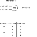
論理ゲートは、論理演算を行う電子回路中の基本構成要素である。これらはインプットシグナルとアウトプットシグナルを0と1の形で有する。「0」はシグナルが存在しないことを表すのに対して、「1」はシグナルが存在することを示す。電子的論理ゲートと同様に、細胞シグナルが論理ゲートとして働くことができる。 A logic gate is a basic component in an electronic circuit that performs logic operations. They have input and output signals in the form of 0 and 1. “0” indicates that no signal is present, whereas “1” indicates that a signal is present. Similar to electronic logic gates, cellular signals can act as logic gates.
合成生物学では、最終的に、もっと複雑な系に統合することができる生物学的装置を作り出すために工学の原理の多くが生物学分野に適用される。これらの原理には、部品の規格化、モジュール性、抽象化(abstraction)、信頼性、予測性、および均一性が含まれる(Andrianantoandro, Basu et al. 2006)。細胞環境内にある簡単な装置およびモジュールの機能を予測できないと、工学の原理を生物学に適用することが困難になる。交絡因子の一部は、遺伝子発現ノイズ、変異、細胞死、定まっておらずかつ変動する細胞外環境、ならびに細胞内容物との相互作用である(Andrianantoandro, Basu et al. 2006)。従って、合成生物学は、製造、環境、および持続可能性の分野、ならびに健康および医学において直面している課題に取り組む系の開発において大いに有望であるが、この可能性を実現することは、現在、利用可能な部品の多様性と効果的な設計の枠組みによって制限されている(Wang, Wei et al. 2013)。 In synthetic biology, many engineering principles are applied in the biological field to ultimately create biological devices that can be integrated into more complex systems. These principles include part normalization, modularity, abstraction, reliability, predictability, and uniformity (Andrianantoandro, Basu et al. 2006). Failure to predict the function of simple devices and modules within the cellular environment makes it difficult to apply engineering principles to biology. Some of the confounders are gene expression noise, mutations, cell death, undefined and fluctuating extracellular environments, and interactions with cell contents (Andrianantoandro, Basu et al. 2006). Synthetic biology is therefore very promising in the field of manufacturing, the environment, and sustainability, as well as in developing systems that address the challenges faced in health and medicine. Limited by the diversity of available parts and an effective design framework (Wang, Wei et al. 2013).
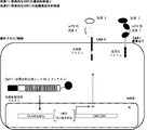
本発明は、論理「ANDゲート」などの合成生物学の原理を免疫細胞技術に適用して、少なくとも2種類のインプットシグナルが組み合わされた時にしか免疫細胞が刺激および/または活性化されないようにするために作成された(図1)。特に、本発明は、免疫細胞を少なくとも2種類のインプットシグナルの組み合わせに対して感受性にすることによって、免疫療法のために免疫細胞を操作する方法に関する。前記インプットシグナルは低酸素などの外部刺激でもよく、リガンドの認識、好ましくは、前記リガンドを認識することができる特異的キメラ抗原受容体を細胞表面に発現させることによるリガンドの認識でもよい。本発明によれば、インプットシグナルが認識されると、免疫細胞応答を活性化する、好ましくは、シグナル伝達タンパク質を介して免疫細胞応答を活性化する少なくとも2種類のトランスミッタードメインの組み合わせが可能になる。それぞれのトランスミッタータンパク質は単独では不活性であり、従って、免疫細胞応答を活性化しない。これらの2種類のトランスミッタードメインが組み合わされた時だけ、T細胞が活性化される。トランスミッタードメインは、非限定的な例として、プロテアーゼと、シグナル伝達タンパク質に連結されているプロテアーゼ切断部位を含む固定された膜基質ドメイン、スプリットタンパク質、スキャフォールディングタンパク質、二量体化可能なドメイン、抑制を回復することができる化合物を伴う自己抑制タンパク質、前もって不活化された遺伝子の補完でもよい。本発明はまた、キメラ抗原受容体の新たな設計、前記キメラ抗原受容体を含む細胞または本発明の方法によって得られた細胞、および前記操作された免疫細胞を用いた治療的処置にも関する。 The present invention applies synthetic biology principles such as logical “AND gates” to immune cell technology to ensure that immune cells are stimulated and / or activated only when at least two input signals are combined. Created for (Figure 1). In particular, the invention relates to a method of manipulating immune cells for immunotherapy by sensitizing immune cells to a combination of at least two input signals. The input signal may be an external stimulus such as hypoxia, or may be a ligand recognition, preferably a ligand recognition by expressing a specific chimeric antigen receptor capable of recognizing the ligand on the cell surface. According to the present invention, when an input signal is recognized, a combination of at least two transmitter domains that activates an immune cell response, preferably activates an immune cell response via a signaling protein, is possible. . Each transmitter protein is inactive alone and therefore does not activate the immune cell response. T cells are activated only when these two transmitter domains are combined. Transmitter domains include, but are not limited to, proteases and immobilized membrane substrate domains that contain protease cleavage sites linked to signaling proteins, split proteins, scaffolding proteins, dimerizable domains, and repression Self-suppressing protein with a compound that can restore the protein may be complemented with a previously inactivated gene. The invention also relates to a new design of a chimeric antigen receptor, a cell containing said chimeric antigen receptor or a cell obtained by the method of the invention, and a therapeutic treatment using said engineered immune cell.
(図1)論理「ANDゲート」合成生物学の原理。インプット(1,2)は、腫瘍細胞(および/もしくは健常細胞)ならびに/または腫瘍微小環境によって発現された抗原でもよい。アウトプットは、結果として生じた免疫細胞活性化に対応する。
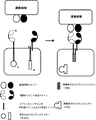
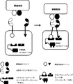
(図2)ANDゲート:腫瘍抗原によって動かされる、受容体型チロシンキナーゼ(RTK)をベースとするキメラ抗原受容体の二量体化および活性化。腫瘍細胞表面に共存する2種類の腫瘍細胞リガンドが同時に存在すると、2種類のヘテロ二量体受容体型チロシンキナーゼベースキメラ抗原受容体が二量体化し、トランスリン酸化を介して活性化される。両CAR上では、トランスミッタードメインが自己抑制によって不活性状態に維持されている(例えば、キナーゼ活性部位は自己抑制ドメインによって隠されている)。腫瘍細胞表面に共存する2種類の腫瘍細胞リガンドが同時に存在すると、2種類のCARは二量体化して、キナーゼ自己抑制は解除され、非限定的な例としてトランスリン酸化または他の分子との相互作用を介してトランスミッタードメインは活性化される。
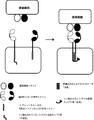
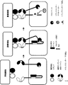
(図3)ANDゲート:前に不活化された遺伝子の補完:簡略化した例では、2つの異なるドメインを含む細胞質ドメインをもつ2つの異なるCARによって2つの異なる腫瘍細胞リガンドを認識することができる。同時に、T細胞のシグナル経路にある重要な遺伝子(GOI)のノックアウトが行われている。2種類のCARが共存し、その後に、腫瘍リガンド細胞が認識されると、第1のCARは、第2のCARによって媒介されるシグナルの伝達に必要なGOIの再活性化を可能にする因子を活性化することができる。
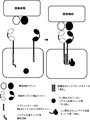
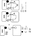
(図4)ANDゲート:プロテアーゼ系。2種類の腫瘍細胞リガンドが同時に存在すると、2種類のCARが活性化される。第1のCARの細胞内ドメインは、シグナル伝達タンパク質に連結されているプロテアーゼ標的配列を含む。第2のCARの細胞内ドメインはプロテアーゼを有する。それぞれのCARは独立して1種類の腫瘍リガンド細胞の存在によって活性化されず、活性化は、両腫瘍リガンド細胞の存在による2種類のCARの共存に由来する。2種類のCARが共存すると、標的配列プロテアーゼの切断とその後のシグナル伝達タンパク質の放出によって媒介される、2種類のCARの活性化が可能になる。
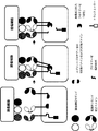
(図5)ANDゲート:スプリットタンパク質系。2種類の腫瘍細胞リガンドが同時に存在すると、2種類のCARが活性化される。第1のCARの細胞内ドメインは、「シグナル伝達ドメイン」の断片とインテインのCドメインまたはNドメインを含む。第2のCARの細胞内ドメインは、相補インテインドメインと相補シグナル伝達ドメイン断片を有する。それぞれのCARは独立して1種類の腫瘍リガンド細胞の存在によって活性化されず、活性化は、両腫瘍リガンド細胞の存在による2種類のCARの共存に由来する。2種類のCARが共存すると、T細胞の様々な活性化経路を開始することができる完全に活性な形のシグナル伝達タンパク質を再構成する完全活性スプリットインテインが再構成されることで2種類のCARの活性化が可能になる。シグナル伝達タンパク質の例は、ZAP70、SH2ドメイン、およびキナーゼドメインである。
(図6)ANDゲート:スプリットタンパク質系およびシグナル伝達タンパク質の放出。2種類の腫瘍細胞リガンドが同時に存在すると、2種類のCARが活性化される。第1のCARの細胞内ドメインは、「シグナル伝達タンパク質」のC末端不活性断片とインテインのCドメインまたはNドメインを含む。第2のCARの細胞内ドメインは、さらなるマルチドメインとホモ二量体化することができる二量体化ドメインを有する。このマルチドメインは、第2のインテインドメインとシグナル伝達タンパク質断片のNドメインによって構成される。それぞれのCARは独立して1種類の腫瘍リガンド細胞の存在によって活性化されず、活性化は、両腫瘍リガンド細胞の存在による2種類のCARの共存に由来する。2種類のCARが共存すると、細胞質に放出されてT細胞活性化を開始することができる完全に活性な形のシグナル伝達タンパク質を再構成する完全活性スプリットインテインが再構成されることで2種類のCARの活性化が可能になる。
(図7)ANDゲート:キナーゼをベースとするスプリットタンパク質系。2種類の腫瘍細胞リガンドが同時に存在すると、2種類のCARが活性化される。第1のCARの細胞内ドメインはシグナル伝達タンパク質結合領域とスプリットキナーゼのCドメインまたはNドメインを含む。第2のCARの細胞内ドメインは相補キナーゼドメインを有する。それぞれのCARは独立して1種類の腫瘍リガンド細胞の存在によって活性化されず、活性化は、両腫瘍リガンド細胞の存在による2種類のCARの共存に由来する。2種類のCARが共存すると、リン酸化することができる、従って、T細胞活性化を開始することができる完全活性キナーゼが再構成されることで2種類のCARの活性化が可能になる。スプリットキナーゼの例はLCKでもよい。
(図8)ANDゲート:キナーゼをベースとするスプリットタンパク質系のトランス活性化。2種類の腫瘍細胞リガンドが同時に存在すると、2種類のCARが活性化される。第1のCARの細胞内ドメインはシグナル伝達結合領域とスプリットキナーゼのCドメインまたはNドメインを含む。第2のCARの細胞内ドメインは相補キナーゼドメインを有する。それぞれのCARは独立して1種類の腫瘍リガンド細胞の存在によって活性化されず、活性化は、両腫瘍リガンド細胞の存在による2種類のCARの共存に由来する。2種類のCARが共存すると、シグナル伝達タンパク質結合領域上でのコンホメーション改変を引き起こすことができ、それによって、トランス活性化によって活性化することができるシグナル伝達タンパク質に結合することができる完全活性キナーゼが再構成されることで2種類のCARの活性化が可能になる。
(図9)ANDゲート:プロテアーゼをベースとするスプリット系およびシグナル伝達タンパク質の再局在化。2種類の腫瘍細胞リガンドが同時に存在すると、2種類のCARが活性化される。第1のCARの細胞内ドメインは、スプリットプロテアーゼのCドメインまたはNドメインと、プロテアーゼ標的配列と、シグナル伝達タンパク質を含む。第2のCARの細胞内ドメインは相補スプリットプロテアーゼドメインを有する。それぞれのCARは独立して1種類の腫瘍リガンド細胞の存在によって活性化されず、活性化は、両腫瘍リガンド細胞の存在による2種類のCARの共存に由来する。2種類のCARが共存すると、プロテアーゼ標的配列を切断し、シグナル伝達タンパク質を放出することができる完全活性プロテアーゼが再構成されることで2種類のCARの活性化が可能になる。
(図10)ANDゲート:3種類のCARを用いた、プロテアーゼをベースとするスプリット系。3種類の腫瘍細胞リガンドが同時に存在すると、CARが活性化される。第1のCARの細胞内ドメインはプロテアーゼ標的配列とシグナル伝達タンパク質を含む。第2のCARおよび第3のCARの細胞内ドメインは2種類の相補スプリットプロテアーゼドメインによって形成される。それぞれのCARは独立して1種類の腫瘍リガンド細胞の存在によって活性化されず、活性化は、3種類の腫瘍リガンド細胞の存在による3種類のCARの共存に由来する。3種類のCARが共存すると、プロテアーゼ標的配列を切断し、シグナル伝達タンパク質を放出することができる完全活性プロテアーゼが再構成されることで3種類のCARが活性化される。
(図11)ANDゲート:スキャフォールディングプロテアーゼをベースとする系。2種類の腫瘍細胞リガンドが同時に存在すると、2種類のCARが活性化される。第1のCARの細胞内ドメインは第1のタンパク質ドメインを含む。第2のCARの細胞内ドメインは第2のタンパク質ドメインを有する。それぞれのCARは独立して1種類の腫瘍リガンド細胞の存在によって活性化されず、活性化は、両腫瘍リガンド細胞の存在による2種類のCARの共存に由来する。2種類のCARが共存すると、タンパク質ドメイン1およびタンパク質ドメイン2が不活性スキャフォールディングタンパク質に結合することによって2種類のCARの活性化が可能になる。複合体が結合すると、活性型スキャフォールディングタンパク質が再構成され、T細胞を活性化することができる。スキャフォールディングタンパク質の例は、Carma1、SP76、hemITAM、DLG1、KSRである。
(図12)ANDゲート:操作されたヘテロ二量体ドメインの二重活性化に基づく。2種類の腫瘍細胞リガンドが同時に存在すると、2種類のCARが活性化される。第1のCARの細胞内ドメインは第1のトランスミッター結合ドメインを含む。第2のCARの細胞内ドメインは第2のトランスミッター結合ドメインを有する。それぞれのCarは独立して1種類の腫瘍リガンド細胞の存在によって活性化されず、活性化は、両腫瘍リガンド細胞の存在による2種類のCARの共存に由来する。2種類のCARが共存すると、2種類のトランスミッター結合ドメインの活性化(例えば、リン酸化および翻訳後修飾)が可能になり、これにより、T細胞を活性化することができるトランスミッターの動員を誘発することができる。
(図13)ANDゲート:自己抑制系:競合的結合が起こると活性化が誘導される。2種類の腫瘍細胞リガンドが同時に存在するとトランスミッターが活性化される。第1のCAR上では、トランスミッタードメインは自己抑制によって(例えば、「遮蔽(shielding)」タンパク質または抗体と相互作用することによって)不活性状態に維持されている。リガンド結合時に第2のCARが共存すると、遮蔽分子は、それ自身の、さらに高い親和性のドメインに移る(分子間移動)。次いで、「遮蔽されていない」トランスミッターは(例えば、翻訳後修飾または他の分子との相互作用によって)活性化することができる。
(図14)ANDゲート:自己抑制系:抑制ドメインの酵素的切断が起こると活性化が誘導される。2種類の腫瘍細胞リガンドが同時に存在するとトランスミッターが活性化される。第1のCAR上では、トランスミッタードメインは自己抑制によって(例えば、「遮蔽」タンパク質または抗体と相互作用することによって)不活性状態に維持されている。リガンド結合時に第2のCARが共存すると、プロテアーゼドメインが第1のcar上に存在するプロテアーゼ標的配列に接近し、従って、遮蔽分子は移動される。次いで、「遮蔽されていない」トランスミッターは(例えば、翻訳後修飾または他の分子との相互作用によって)活性化することができる。
(図15)ANDゲート:トランスミッタータンパク質の活性化を誘導するための受容体結合および外部刺激。CARとその腫瘍細胞リガンドとの結合と、操作されたT細胞の腫瘍細胞の細胞外刺激への曝露が同時に起こると、トランスミッターが活性化される。外部刺激は、代謝産物、低分子、ペプチド、低分子タンパク質(ケモカイン、サイトカイン)の濃度および物理的状態/化学的状態(pH、低酸素、酸化還元電位)の変化を包含する。
(図16)ANDゲート:1種類の腫瘍抗原の存在下での低酸素依存的活性化系。酸素枯渇環境で、操作されたT細胞と1種類の腫瘍細胞リガンドが同時に存在するとT細胞活性化が誘発される。このような論理ANDゲート活性化系を可能にするために、酸素誘導性の合成活性化経路を有するようにT細胞が操作される。このような合成経路は、操作された酸素濃度感受性転写因子(OxiTF)、第3の要素を発現させるOxiTF特異的合成プロモーター、キメラ抗原受容体(CARI)を含む3つの異なる要素から作られる。OxiTFは、操作されたT細胞の中で、腫瘍抗原に特異的なCARをコードする合成遺伝因子を活性化するように設計されている。固形腫瘍に遭遇したら、操作されたT細胞は酸素枯渇を検出し、CARIの産生を誘発する。CARIが細胞表面に露出したら腫瘍抗原の認識が可能になり、最終的に、CARIの中に存在する活性化ドメインおよび共刺激ドメインを介してT細胞の活性化および増殖が誘発される。
(図17)ANDゲート:2種類の腫瘍抗原の存在下での低酸素依存的活性化系。酸素枯渇環境で、操作されたT細胞と2種類の腫瘍細胞リガンドが同時に存在するとT細胞活性化が誘発される。このような論理ANDゲート活性化系を可能にするために、酸素誘導性の合成活性化経路を有するようにT細胞が操作される。このような合成経路は、操作された酸素濃度感受性転写因子(OxiTF)、第3の要素を発現させるOxiTF特異的合成プロモーター、腫瘍抗原IIに特異的なキメラ抗原受容体(CARII)を含む4つの異なる要素から作られる。この系は、腫瘍抗原Iに特異的な、構成的に発現しているCARIにある第4の要素を加えて完成する。OxiTFは、操作されたT細胞の中で、CARIIをコードする合成遺伝因子を活性化するように設計されている。固形腫瘍に遭遇したら、操作されたT細胞は酸素枯渇を検出し、CARIの産生を誘発する。既に存在するCARI-腫瘍抗原I複合体に加えて、CARIIとCARIが細胞表面に露出したら腫瘍抗原IIの認識が可能になる。最終的に、両方のCAR/腫瘍抗原複合体が同時に存在すると、CARIおよびIIの中に存在する活性化ドメインおよび共刺激ドメインを介してT細胞の活性化と増殖が誘発される。
(図18)ANDゲート:T細胞系活性化に適用されたANDゲート原理の図。CARの細胞外は、2つのコンホメーション(「活性」および「不活性」)で存在する2種類のリガンド結合ドメインを含む。2種類の腫瘍細胞リガンドの非存在下では、平衡は「不活性型」に強く向けられる。2種類のリガンド結合ドメインがそれぞれの腫瘍細胞リガンドに同時に結合する時にしか(2種類のインプット)、CARの細胞内ドメインへのプラスのシグナル(アウトプット)は誘発されない。
(図19)AND NOTゲート:一般的なスキーム。T細胞系活性化に適用されたAND NOTゲート原理の図。2種類の腫瘍細胞リガンドが同時に存在し、健常細胞リガンドが存在しない時にプラスのアウトプットが誘発される。インプット1およびインプット2は腫瘍細胞リガンドの存在に対応するのに対して、第3のインプットは健常細胞リガンドが存在しないことでなければならない。第1のCARおよび第2のCARは共刺激細胞質ドメインを有するのに対して、第3のCARは、健常細胞リガンドが認識されない場合に抑制作用がブロックされる抑制ドメインを有する。
(図20)T細胞シグナル伝達カスケードを抑制および刺激するための2のタイプのLCKの作製。第1のCARは、構成的に負に調節された形のLCK(-)の転写を刺激する抑制ドメインを用いて健常細胞の抗原を認識する。この第1のCARは、LCK(+)型の転写を活性化して、高レベルのT細胞活性化を生じる共刺激ドメインを含む第2のCARと対になっている。
(図21)CARを介したCARMA1タンパク質調節によるT細胞活性化の制御。抗原認識後のTCR刺激はCD28動員と結びつけられ、CD28動員によってPKCθ活性化が起こり、次に、PKCθ活性化によってCARMA1がリン酸化および活性化される。CARMA1は、T細胞受容体(TCR)シグナル伝達、一般的にはT細胞活性化のための重要なシグナロソームを構成する。CARMA1は様々なタンパク質を動員して、最終的に、2つの異なるシグナル伝達カスケード:NF-κBおよびc-jun N末端キナーゼ(JNK)を活性化することができる多タンパク質複合体を形成する。
(図22)HIF低酸素系の機能。酸素正常状態(高O2)では、HIFαは、酸素を検知するHIFα特異的プロリルヒドロキシラーゼ(PHD1-3)によって加水分解される。ヒドロキシル化によってHIFαポリユビキチン化が誘発され、HIFαはE3ユビキチンリガーゼによるプロテオソーム分解の標的とされる。低酸素(低O2)では、TCAサイクル中間体を介したヒドロキシル化の抑制、HIFαタンパク質の安定化、およびHIF転写活性の低下が起こる。
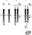
(図23)HIFαなどの酸素感受性ドメインを有する異なるキメラ抗原受容体(CAR)構造。左側には、単鎖CAR(scCAR)は、同じただ一つしかない鎖に、細胞外結合ドメイン(ここではscFv)と、酸素ドメイン(例えば、HIF1)と、活性化ドメインと同時活性化ドメインを有する。右側には、例示的なコンホメーションとして、α鎖がscFvと酸素ドメインを有し、β鎖が共刺激ドメインを有し、γ鎖が活性化ドメインを有する多鎖CAR(mcCAR)を示す。
(図24)(A)酸素正常状態または低酸素における鎖-HIF1(a.a.380-630)対α鎖WTの表面提示。低酸素では、HIF1を有するCAR T細胞の表面露出は(α-HIF1を有さない)対照CAR T細胞の表面露出と似ているが、酸素正常状態では表面露出はかなり少ない。このことは、CAR-αHIF1が良好に発現していることを示す。(B)低酸素から酸素正常状態に戻した後のα鎖-HIF1(a.a.380-630)対α鎖WTの表面提示。低酸素から酸素正常状態に戻すとCARαHIF1の発現は低下する。これは可逆的かつ動的な系である。すなわち、酸素正常状態では、CAR発現はα-HIF1およびα鎖ポリペプチドの分解によって一時的に抑制され、低酸素状態(すなわち、腫瘍環境)では、α-HIF1およびα鎖ポリペプチドは発現される。
(図25)(A)酸素正常状態または低酸素におけるα-HIF mcCAR対対照CARの表面検出。この実験では、さらに少ない全RNAが用いられ、得られた結果は図23の結果と同様である。(B)酸素正常状態における細胞傷害性の誘導。酸素正常状態では、対照多鎖CAR(α-HIF1を有さない)は多量の標的細胞死滅を示すのに対して、HIF-mcCARの場合、標的細胞は死滅しない。これらの細胞傷害性の結果と表面露出の結果を考慮すると、このことはHIF系がキメラ抗原受容体の中で完全に機能することを示している。
(図26)酸素正常状態または低酸素での様々なα鎖-HIF対WTα鎖の表面提示。(A)-EA-リンカーを有するHIF1-mcCAR(a.a.380-630)構築物;(B)HIF1-mcCAR(a.a.344-417);(C)HIF3-mcCAR(a.a.480-571);(D-E-F):(A-B-C)と同じであるが、低酸素から酸素正常状態に戻した。レンチウイルス送達によって、ここで得られた全ての結果から、HIF1系およびHIF3系はいずれも機能し、同じように振る舞うことが証明された。同様に、リンカーを用いて、またはリンカーを用いずにHIFタンパク質の様々な部分を使用できることが示された。
説明文:

(図28)両ゲート受容体の組成物の模式図。
(図29)両ゲートの成分の分子組み立て戦略の模式図。この図にはスペーサーが示されている。
(図30)7種類の膜タンパク質パートナーの表面発現。GG83、GG111、GG121、GG152、GG153、GG155、GG156、およびGG158を試験した。シグナル強度(++: 非常に強い、+: 強い)。
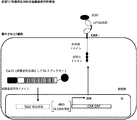
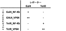
(図31)様々なトランスアクチベーターのmRNAトランスフェクションによる、レンチウイルスによって送達されたRQR8カセットの発現。これらの構築物はDNA結合ドメイン(TetOまたはGal4)と転写活性化ドメイン(VP64またはNF-κB)で構成され、トランスフェクトおよび試験される。得られたデータから、適切なトランスアクチベーターのmRNAトランスフェクションによって、レンチウイルスによって送達されたRQR8カセットが発現したことがはっきりと分かった。
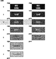
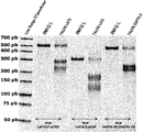
(図32)設計されたTALENを用いた内因性遺伝子座における標的変異誘発を証明するT7エンドヌクレアーゼアッセイ。3つのパネルA、B、Cは全て、LAT、LCK、ZAP70、LFA、TRAT、またはCD28などのT細胞のシグナル伝達および/または機能に関与する酵素のノックアウト(KO)を示す。得られたデータから、設計されたTALENを用いた全標的遺伝子座における標的変異誘発レベルが高いことがはっきりと分かる。
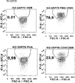
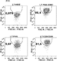
(図33)ZAP70をノックアウトした後の脱顆粒実験。得られたデータから、WT T細胞と比べて、ノックアウト操作されたT細胞は濃い染色が減少したことがはっきりと分かる。
(図34)二重特異性CAR(biCAR)機能の模式図。二重特異性CAR(biCAR)は、2つの異なる標的細胞抗原に対して特異的親和性のあるscFvをもつ2種類のCAR(biCAR1およびbiCAR2)で構成される。これらのscFcの一方だけが特異的抗原に結合した時にCARは活性化されず、従って、細胞は死滅しない。両方のscFvが特異的抗原に結合した時にCARは活性化され、標的細胞は死滅する。
(Figure 1) Logic "AND Gate" The principle of synthetic biology. The input (1,2) may be an antigen expressed by tumor cells (and / or healthy cells) and / or tumor microenvironment. The output corresponds to the resulting immune cell activation.
(FIG. 2) AND gate: Dimerization and activation of a chimeric antigen receptor based on receptor tyrosine kinase (RTK) driven by tumor antigens. When two types of tumor cell ligands coexisting on the tumor cell surface are present simultaneously, two types of heterodimeric receptor tyrosine kinase-based chimeric antigen receptors are dimerized and activated via transphosphorylation. On both CARs, the transmitter domain is maintained in an inactive state by self-inhibition (eg, the kinase active site is hidden by the self-inhibition domain). When two tumor cell ligands coexist on the tumor cell surface are present at the same time, the two CARs dimerize and the kinase self-inhibition is released, and as a non-limiting example, transphosphorylation or with other molecules. Via the interaction, the transmitter domain is activated.
(Figure 3) AND gate: complementation of previously inactivated genes: in a simplified example, two different CAR cell ligands can be recognized by two different CARs with a cytoplasmic domain containing two different domains . At the same time, a critical gene (GOI) in the signal pathway of T cells is knocked out. When two types of CAR coexist, and then tumor ligand cells are recognized, the first CAR is a factor that enables the reactivation of the GOI required for signal transduction mediated by the second CAR Can be activated.
(FIG. 4) AND gate: protease system. When two types of tumor cell ligands are present simultaneously, two types of CAR are activated. The intracellular domain of the first CAR contains a protease target sequence that is linked to a signaling protein. The intracellular domain of the second CAR has a protease. Each CAR is independently not activated by the presence of one type of tumor ligand cell, and activation is derived from the coexistence of two types of CAR due to the presence of both tumor ligand cells. The coexistence of two types of CARs allows the activation of the two types of CARs mediated by cleavage of the target sequence protease and subsequent release of signaling proteins.
(FIG. 5) AND gate: Split protein system. When two types of tumor cell ligands are present simultaneously, two types of CAR are activated. The intracellular domain of the first CAR comprises a “signal transduction domain” fragment and the intein C or N domain. The intracellular domain of the second CAR has a complementary intein domain and a complementary signaling domain fragment. Each CAR is independently not activated by the presence of one type of tumor ligand cell, and activation is derived from the coexistence of two types of CAR due to the presence of both tumor ligand cells. When two types of CAR coexist, two CARs are reconstituted by reconstitution of a fully active split intein that reconstitutes a fully active form of signaling protein that can initiate various activation pathways of T cells. Can be activated. Examples of signal transducing proteins are ZAP70, SH2 domain, and kinase domain.
(FIG. 6) AND gate: Release of split protein system and signaling protein. When two types of tumor cell ligands are present simultaneously, two types of CAR are activated. The intracellular domain of the first CAR comprises the C-terminal inactive fragment of the “signaling protein” and the intein C or N domain. The intracellular domain of the second CAR has a dimerization domain that can homodimerize with additional multidomains. This multidomain is composed of the second intein domain and the N domain of the signaling protein fragment. Each CAR is independently not activated by the presence of one type of tumor ligand cell, and activation is derived from the coexistence of two types of CAR due to the presence of both tumor ligand cells. The coexistence of two types of CARs results in the reconstitution of a fully active split intein that reconstitutes a fully active form of signaling protein that can be released into the cytoplasm and initiate T cell activation. CAR can be activated.
(FIG. 7) AND gate: a split protein system based on kinases. When two types of tumor cell ligands are present simultaneously, two types of CAR are activated. The intracellular domain of the first CAR includes the signaling protein binding region and the C or N domain of split kinase. The intracellular domain of the second CAR has a complementary kinase domain. Each CAR is independently not activated by the presence of one type of tumor ligand cell, and activation is derived from the coexistence of two types of CAR due to the presence of both tumor ligand cells. When two types of CAR coexist, two types of CAR can be activated by reconstitution of a fully active kinase that can be phosphorylated and thus initiate T cell activation. An example of a split kinase may be LCK.
(FIG. 8) AND gate: Transactivation of a kinase-based split protein system. When two types of tumor cell ligands are present simultaneously, two types of CAR are activated. The intracellular domain of the first CAR includes a signaling binding region and a C or N domain of split kinase. The intracellular domain of the second CAR has a complementary kinase domain. Each CAR is independently not activated by the presence of one type of tumor ligand cell, and activation is derived from the coexistence of two types of CAR due to the presence of both tumor ligand cells. The coexistence of two types of CAR can cause conformational alterations on the signaling protein binding region, thereby allowing full activity to bind to signaling proteins that can be activated by transactivation. Two types of CAR can be activated by reconstitution of the kinase.
(FIG. 9) AND gate: protease-based split system and relocalization of signaling proteins. When two types of tumor cell ligands are present simultaneously, two types of CAR are activated. The intracellular domain of the first CAR includes a C or N domain of a split protease, a protease target sequence, and a signaling protein. The intracellular domain of the second CAR has a complementary split protease domain. Each CAR is independently not activated by the presence of one type of tumor ligand cell, and activation is derived from the coexistence of two types of CAR due to the presence of both tumor ligand cells. When two types of CARs coexist, the two types of CARs can be activated by reconstructing a fully active protease that can cleave the protease target sequence and release the signaling protein.
(FIG. 10) AND gate: A protease-based split system using three types of CAR. The presence of three tumor cell ligands simultaneously activates CAR. The intracellular domain of the first CAR contains a protease target sequence and a signaling protein. The intracellular domains of the second and third CARs are formed by two complementary split protease domains. Each CAR is not independently activated by the presence of one type of tumor ligand cell, and the activation is derived from the coexistence of three types of CAR due to the presence of three types of tumor ligand cells. When three types of CAR coexist, the three types of CAR are activated by reconstructing a fully active protease that can cleave the protease target sequence and release the signaling protein.
(FIG. 11) AND gate: a system based on scaffolding proteases. When two types of tumor cell ligands are present simultaneously, two types of CAR are activated. The intracellular domain of the first CAR includes the first protein domain. The intracellular domain of the second CAR has a second protein domain. Each CAR is independently not activated by the presence of one type of tumor ligand cell, and activation is derived from the coexistence of two types of CAR due to the presence of both tumor ligand cells. When two types of CAR coexist,
(FIG. 12) AND gate: based on dual activation of engineered heterodimeric domains. When two types of tumor cell ligands are present simultaneously, two types of CAR are activated. The intracellular domain of the first CAR includes a first transmitter binding domain. The intracellular domain of the second CAR has a second transmitter binding domain. Each Car is not independently activated by the presence of one type of tumor ligand cell, and activation is derived from the coexistence of two types of CAR due to the presence of both tumor ligand cells. The coexistence of two CARs allows activation of two transmitter-binding domains (eg, phosphorylation and post-translational modification), thereby inducing transmitter mobilization that can activate T cells. be able to.
(FIG. 13) AND gate: self-inhibiting system: activation occurs when competitive binding occurs. The transmitter is activated when two tumor cell ligands are present simultaneously. On the first CAR, the transmitter domain is maintained in an inactive state by autoinhibition (eg, by interacting with a “shielding” protein or antibody). When a second CAR coexists during ligand binding, the shielding molecule moves to its own, higher affinity domain (intermolecular transfer). The “unmasked” transmitter can then be activated (eg, by post-translational modification or interaction with other molecules).
(FIG. 14) AND gate: self-repression system: activation is induced when enzymatic cleavage of the repression domain occurs. The transmitter is activated when two tumor cell ligands are present simultaneously. On the first CAR, the transmitter domain is maintained in an inactive state by autoinhibition (eg, by interacting with a “screening” protein or antibody). When the second CAR coexists upon ligand binding, the protease domain approaches the protease target sequence present on the first car, thus moving the shielding molecule. The “unmasked” transmitter can then be activated (eg, by post-translational modification or interaction with other molecules).
(FIG. 15) AND gate: receptor binding and external stimuli to induce transmitter protein activation. The transmitter is activated when the binding of CAR to its tumor cell ligand and exposure of the engineered T cell to extracellular stimulation of the tumor cell occur simultaneously. External stimuli include changes in concentrations of metabolites, small molecules, peptides, small proteins (chemokines, cytokines) and physical / chemical states (pH, hypoxia, redox potential).
(FIG. 16) AND gate: Hypoxia-dependent activation system in the presence of one type of tumor antigen. In an oxygen-depleted environment, the presence of engineered T cells and one tumor cell ligand simultaneously induces T cell activation. To enable such a logical AND gate activation system, T cells are engineered to have an oxygen-induced synthetic activation pathway. Such a synthetic pathway is made up of three distinct elements including an engineered oxygen concentration sensitive transcription factor (OxiTF), an OxiTF specific synthetic promoter that expresses a third element, and a chimeric antigen receptor (CARI). OxiTF is designed to activate synthetic genetic factors that encode CARs specific for tumor antigens in engineered T cells. When encountering a solid tumor, the engineered T cells detect oxygen depletion and trigger the production of CARI. Once CARI is exposed on the cell surface, tumor antigens can be recognized, and ultimately T cell activation and proliferation is induced through activation domains and costimulatory domains present in CARI.
(FIG. 17) AND gate: hypoxia-dependent activation system in the presence of two tumor antigens. T cell activation is induced by the presence of engineered T cells and two tumor cell ligands simultaneously in an oxygen-depleted environment. To enable such a logical AND gate activation system, T cells are engineered to have an oxygen-induced synthetic activation pathway. Such synthetic pathways include four engineered oxygen concentration sensitive transcription factors (OxiTF), an OxiTF-specific synthetic promoter that expresses a third element, and a chimeric antigen receptor (CARII) specific for tumor antigen II. Made from different elements. This system is completed by adding a fourth element in the constitutively expressed CARI specific for tumor antigen I. OxiTF is designed to activate synthetic genetic factors encoding CARI in engineered T cells. When encountering a solid tumor, the engineered T cells detect oxygen depletion and trigger the production of CARI. In addition to the existing CARI-tumor antigen I complex, tumor antigen II can be recognized once CARII and CARI are exposed on the cell surface. Finally, the simultaneous presence of both CAR / tumor antigen complexes induces T cell activation and proliferation through the activation and costimulatory domains present in CARI and II.
(FIG. 18) AND gate: Diagram of the AND gate principle applied to T cell line activation. The extracellular side of CAR contains two ligand-binding domains that exist in two conformations (“active” and “inactive”). In the absence of two tumor cell ligands, the equilibrium is strongly directed to the “inactive form”. Only when two ligand-binding domains bind to each tumor cell ligand simultaneously (two inputs), a positive signal (output) to the intracellular domain of CAR is elicited.
(FIG. 19) AND NOT Gate: General scheme. Diagram of AND NOT gate principle applied to T cell line activation. Two types of tumor cell ligands are present simultaneously, and a positive output is induced when no healthy cell ligand is present.
FIG. 20: Generation of two types of LCK to suppress and stimulate the T cell signaling cascade. The first CAR recognizes healthy cell antigens using a repressive domain that stimulates transcription of LCK (-) in a constitutively negatively regulated form. This first CAR is paired with a second CAR containing a costimulatory domain that activates LCK (+) type transcription resulting in high levels of T cell activation.
(FIG. 21) Regulation of T cell activation by CARMA1 protein regulation via CAR. TCR stimulation following antigen recognition is linked to CD28 mobilization, which leads to PKCθ activation, which in turn phosphorylates and activates CARMA1. CARMA1 constitutes an important signalosome for T cell receptor (TCR) signaling, generally T cell activation. CARMA1 recruits a variety of proteins, ultimately forming a multiprotein complex that can activate two different signaling cascades: NF-κB and c-jun N-terminal kinase (JNK).
(Fig. 22) Functions of HIF hypoxia system. Under normal oxygen conditions (high O 2 ), HIFα is hydrolyzed by HIFα-specific prolyl hydroxylase (PHD1-3) that detects oxygen. Hydroxylation induces HIFα polyubiquitination, which is targeted for proteosomal degradation by E3 ubiquitin ligase. Hypoxia (low O 2 ) results in suppression of hydroxylation through TCA cycle intermediates, stabilization of HIFα protein, and reduction of HIF transcriptional activity.
FIG. 23: Different chimeric antigen receptor (CAR) structures with oxygen sensitive domains such as HIFα. On the left side, a single-chain CAR (scCAR) contains an extracellular binding domain (here scFv), an oxygen domain (eg HIF1), an activation domain and a co-activation domain in the same single chain. Have. On the right side, an exemplary conformation shows a multi-chain CAR (mcCAR) in which the α chain has scFv and oxygen domains, the β chain has a costimulatory domain, and the γ chain has an activation domain.
(FIG. 24) (A) Surface presentation of chain-HIF1 (aa380-630) versus α chain WT in normoxia or hypoxia. In hypoxia, the surface exposure of CAR T cells with HIF1 is similar to that of control CAR T cells (without α-HIF1), but the surface exposure is much less in normoxia. This indicates that CAR-αHIF1 is well expressed. (B) Surface presentation of alpha chain-HIF1 (aa380-630) versus alpha chain WT after returning from hypoxia to normoxia. Returning from hypoxia to normoxia reduces CARαHIF1 expression. This is a reversible and dynamic system. That is, in normoxia, CAR expression is temporarily suppressed by degradation of α-HIF1 and α-chain polypeptide, and in hypoxia (ie, tumor environment), α-HIF1 and α-chain polypeptide are expressed .
(FIG. 25) (A) Surface detection of α-HIF mcCAR versus control CAR in normoxia or hypoxia. In this experiment, even less total RNA was used and the results obtained are similar to the results of FIG. (B) Induction of cytotoxicity under normoxic conditions. In normoxia, the control multi-chain CAR (without α-HIF1) shows a large amount of target cell kill, whereas in the case of HIF-mcCAR, the target cell is not killed. Considering these cytotoxicity and surface exposure results, this indicates that the HIF system is fully functional among the chimeric antigen receptors.
(FIG. 26) Surface presentation of various α-HIF versus WT α-chains in normoxia or hypoxia. (A) HIF1-mcCAR (aa380-630) construct with -EA-linker; (B) HIF1-mcCAR (aa344-417); (C) HIF3-mcCAR (aa480-571); (DEF): (ABC) But returned from hypoxia to normal oxygen. All results obtained here by lentiviral delivery demonstrated that both the HIF1 and HIF3 systems function and behave similarly. Similarly, it has been shown that various portions of the HIF protein can be used with or without a linker.
Explanatory text:
(FIG. 28) A schematic diagram of the composition of both gate receptors.
FIG. 29 is a schematic diagram of a molecular assembly strategy for components of both gates. This figure shows the spacer.
(FIG. 30) Surface expression of seven membrane protein partners. GG83, GG111, GG121, GG152, GG153, GG155, GG156, and GG158 were tested. Signal strength (++: very strong, +: strong).
(FIG. 31) Expression of the RRQ8 cassette delivered by lentivirus by mRNA transfection of various transactivators. These constructs are composed of a DNA binding domain (TetO or Gal4) and a transcription activation domain (VP64 or NF-κB) and are transfected and tested. From the data obtained, it was clearly found that mRNA transfection of the appropriate transactivator expressed the RRQ8 cassette delivered by lentivirus.
(FIG. 32) T7 endonuclease assay demonstrating targeted mutagenesis at the endogenous locus using the designed TALEN. All three panels A, B, C show the knockout (KO) of enzymes involved in signaling and / or functioning of T cells such as LAT, LCK, ZAP70, LFA, TRAT, or CD28. The data obtained clearly show that the target mutagenesis level is high at all target loci using the designed TALEN.
(FIG. 33) A degranulation experiment after knocking out ZAP70. From the obtained data, it can be clearly seen that the dark staining decreased in the knockout T cells compared to the WT T cells.
(FIG. 34) Schematic diagram of bispecific CAR (biCAR) function. Bispecific CAR (biCAR) is composed of two types of CAR (biCAR1 and biCAR2) with scFv with specific affinity for two different target cell antigens. When only one of these scFc binds to a specific antigen, CAR is not activated and therefore the cell does not die. When both scFvs bind to a specific antigen, CAR is activated and the target cell is killed.
(表1)CARMA1シグナロソームと相互作用するタンパク質
(表2)CARMA1リン酸化部位
(Table 1) Proteins that interact with CARMA1 signalosome (Table 2) CARMA1 phosphorylation sites
発明の詳細な説明
養子T細胞療法において機能的応答を制御する能力は重要な問題である。このような治療方針では、T細胞は、高度に腫瘍特異的に標的細胞を認識する、表面に露出したキメラ抗原受容体(CAR)を発現させることによって操作される。しかしながら、潜在的な毒性オフターゲット作用を制御および最小化するにはマルチインプット系を設計することが非常に望ましい。
Detailed Description of the Invention The ability to control functional responses in adoptive T cell therapy is an important issue. In such therapeutic strategies, T cells are engineered by expressing surface-exposed chimeric antigen receptors (CARs) that recognize target cells highly tumor-specifically. However, it is highly desirable to design a multi-input system to control and minimize potential toxic off-target effects.
「インプットシグナル」のタイプに応じて、直接的または間接的な手段によって「少なくとも2種類のトランスミッタードメインの組み合わせ」を行うことができる。 Depending on the type of “input signal”, “a combination of at least two transmitter domains” can be performed by direct or indirect means.
例えば、スプリット-ユビキチン系の場合、2種類のCARに由来するscFvによる2つの異なる細胞標的リガンドの認識である2種類のインプットシグナルの組み合わせがあると、2種類のトランスミッタードメイン、すなわち、ユビキチン酵素のC末端部分とN末端部分の共存が可能になり、従って、ユビキチン酵素の活性が発生し、シグナルが生じる。 For example, in the case of the split-ubiquitin system, if there is a combination of two input signals, which are recognition of two different cell targeting ligands by scFvs derived from two CARs, two transmitter domains, ie, ubiquitin enzymes The coexistence of the C-terminal part and the N-terminal part becomes possible, so that the activity of the ubiquitin enzyme occurs and a signal is generated.
低酸素HIFα系は、特に「トランスミッターの組み合わせ」に関して、さらに間接的なやり方で機能する。一方の低酸素外部シグナルと、CARのscFvによる細胞標的リガンド認識に由来する他方のシグナルとの間で2種類のインプットシグナルの組み合わせが発生する。この段階で、HIFプロリル-ヒドロキシラーゼリン酸化の抑制、HIF-1αサブユニットの安定化、低酸素状態での生存を促進する、いくつかの遺伝子のアップレギュレーションなどのカスケード反応が発生し、最終的には、HIF-1とHIF応答配列(HRE)とが結合する。 The hypoxic HIFα system functions in a more indirect manner, especially with respect to “transmitter combinations”. A combination of two input signals occurs between one hypoxic external signal and the other signal derived from cell target ligand recognition by the CAR scFv. At this stage, cascade reactions such as suppression of HIF prolyl-hydroxylase phosphorylation, stabilization of HIF-1α subunit, promotion of hypoxic survival, up-regulation of several genes, etc. occur, and finally HIF-1 binds to HIF response element (HRE).
「免疫細胞の活性化」とは、2種類のインプットシグナルの組み合わせによって、2種類のトランスミッタードメインの組み合わせが(直接的または間接的に)誘発され、その結果として、好ましくは伝達手段によって、CARをもつ免疫細胞への(プラスまたはマイナスの)シグナルが発生することを意味する。 “Immune cell activation” refers to the combination of two types of input signals that induces a combination of two types of transmitter domains (directly or indirectly), resulting in CAR It means that a signal (positive or negative) is generated to the immune cells.
結果として、CAR発現のシグナルが出され、最終的に、腫瘍細胞の溶解が起こる可能性がある。 As a result, a signal for CAR expression is signaled, and eventually tumor cell lysis may occur.
免疫細胞を操作する方法
本発明者らは、論理ゲートに基づく複雑な2進法計算を含む、タスクを実行するための人工回路内の調節モジュールの合理的な組み合わせに基づいて、このような免疫細胞を操作する方法を開発した。「ゲート」という用語は、2種類以上のインプットシグナルに応答して特定の(予め決められた)アウトプットを生じる装置または分子機構を指すために用いられる。本発明によれば、論理ANDゲートとは、少なくとも2種類のインプットシグナルの組み合わせに起因する、異なるトランスミッタードメインの組み合わせと特定のタンパク質(シグナル伝達タンパク質)の活性化による免疫細胞活性化、特に、標的細胞に対するT細胞の細胞傷害性を指す。非限定的な例として、タンパク質機能を活性化するために、または抑制タンパク質を除去するために、それぞれのシグナルは一緒に働いてもよく、別々に働いてもよい。別の特定の態様において、前記インプットシグナルは、前のインプットシグナルに起因するアウトプットシグナルでもよい。特に、本発明は、免疫療法のために免疫細胞を操作する方法、特に、細胞を特異的に標的とするために免疫細胞を操作する方法に関し、本方法は、
(a)免疫細胞を準備する工程;
(b)インプットシグナルの組み合わせによって、前記免疫細胞を活性化する少なくとも2種類のトランスミッタードメインの組み合わせが誘導されるように、前記細胞が少なくとも2種類のインプットシグナルに対して感受性になるように前記免疫細胞を操作する工程であって、それぞれのトランスミッタードメインが単独では前記免疫細胞を活性化しない、工程
を含む。
Methods for Manipulating Immune Cells We have developed such immunity based on a reasonable combination of regulatory modules in an artificial circuit to perform a task, including complex binary calculations based on logic gates. A method for manipulating cells was developed. The term “gate” is used to refer to a device or molecular mechanism that produces a specific (predetermined) output in response to two or more input signals. According to the present invention, a logical AND gate is a combination of different transmitter domains resulting from a combination of at least two types of input signals and immune cell activation by activation of a specific protein (signal transduction protein), in particular a target Refers to cytotoxicity of T cells to cells. As a non-limiting example, each signal may work together or separately to activate protein function or remove inhibitory proteins. In another specific embodiment, the input signal may be an output signal resulting from a previous input signal. In particular, the invention relates to a method of manipulating immune cells for immunotherapy, in particular to a method of manipulating immune cells to specifically target cells,
(A) preparing immune cells;
(B) the immunity is such that the cells are sensitive to at least two input signals such that a combination of input signals induces a combination of at least two transmitter domains that activate the immune cells. Manipulating cells, each transmitter domain alone does not activate the immune cell.
この方法は、前記トランスミッタードメインが下記のようなキメラ抗原受容体のシグナル伝達ドメインおよび共刺激ドメインであり、前記シグナル伝達ドメインが単独でT細胞活性化を活性化することができる、(Kloss, Condomines et al.2013)に記載の組み合わせ抗原認識系とは異なる。 In this method, the transmitter domain is the signaling domain and costimulatory domain of a chimeric antigen receptor as described below, and the signaling domain alone can activate T cell activation (Kloss, Condomines et al. 2013), which is different from the combined antigen recognition system.
インプットシグナル:免疫細胞によるリガンドの認識
特定の態様において、前記インプットシグナルは、前記操作された免疫細胞、特に、前記操作された免疫細胞の表面に発現しているキメラ抗原受容体によるリガンドの認識でもよい。
Input signal: Recognition of ligand by immune cells In a particular embodiment, the input signal may also be recognized by the engineered immune cell, particularly ligand recognition by a chimeric antigen receptor expressed on the surface of the engineered immune cell. Good.
本発明によるキメラ抗原受容体(CAR)は、細胞外リガンド結合ドメインと細胞内ドメイン、さらに詳細には、細胞外リガンド結合ドメインと膜貫通ドメインと細胞内ドメインを含む。 The chimeric antigen receptor (CAR) according to the present invention comprises an extracellular ligand binding domain and an intracellular domain, more particularly an extracellular ligand binding domain, a transmembrane domain and an intracellular domain.
本明細書で使用する「細胞外リガンド結合ドメイン」という用語は、リガンドに結合することができるオリゴペプチドまたはポリペプチドと定義される。好ましくは、このドメインは細胞表面分子と相互作用することができる。例えば、細胞外リガンド結合ドメインは、特定の疾患状態に関連する標的細胞上の細胞表面マーカーとして働くリガンドを認識するように選択されてもよい。従って、リガンドとして働き得る細胞表面マーカーの例には、ウイルス感染症、細菌感染症、および寄生生物感染症、自己免疫疾患、ならびに癌細胞に関連する細胞表面マーカーが含まれる。特に、細胞外リガンド結合ドメインは、標的の抗原に対する抗体に由来する抗原結合ドメインを含んでもよい。非限定的な例として、標的の抗原は前記のような腫瘍関連表面抗原でもよい。 As used herein, the term “extracellular ligand binding domain” is defined as an oligopeptide or polypeptide capable of binding to a ligand. Preferably, this domain can interact with cell surface molecules. For example, the extracellular ligand binding domain may be selected to recognize a ligand that acts as a cell surface marker on target cells associated with a particular disease state. Thus, examples of cell surface markers that can serve as ligands include viral infections, bacterial infections, and parasitic infections, autoimmune diseases, and cell surface markers associated with cancer cells. In particular, the extracellular ligand binding domain may comprise an antigen binding domain derived from an antibody against the target antigen. As a non-limiting example, the target antigen may be a tumor associated surface antigen as described above.
細胞外リガンド結合ドメインはまた、標的の抗原に結合するペプチド、標的の抗原に結合する抗体に結合するペプチドまたはタンパク質、標的の表面にある受容体に結合するペプチドもしくはタンパク質リガンド、例えば、非限定的な例として増殖因子、サイトカイン、もしくはホルモン、または標的の表面にあるペプチドもしくはタンパク質リガンドに結合する受容体、例えば、非限定的な例として増殖因子受容体、サイトカイン受容体、もしくはホルモン受容体に由来するドメインも含んでもよい。好ましくは、標的は細胞またはウイルスである。 Extracellular ligand binding domains also include peptides that bind to the target antigen, peptides or proteins that bind to antibodies that bind to the target antigen, peptides or protein ligands that bind to receptors on the surface of the target, such as, but not limited to For example, a growth factor, cytokine, or hormone, or a receptor that binds to a peptide or protein ligand on the surface of the target, eg, as a non-limiting example, derived from a growth factor receptor, a cytokine receptor, or a hormone receptor Domain may also be included. Preferably the target is a cell or a virus.
好ましい態様において、前記細胞外リガンド結合ドメインは、可動性リンカーによってつなぎ合わされた標的抗原特異的モノクローナル抗体の軽鎖(VL)可変断片および重鎖(VH)可変断片を含む単鎖抗体断片(scFv)である。リンパ球の予め規定された標的化のために、scFv以外の結合ドメイン、例えば、非限定的な例として、ラクダ科シングルドメイン抗体断片、血管内皮増殖因子ポリペプチドのような受容体リガンド、インテグリン結合ペプチド、ヘレグリンまたはIL-13ムテイン、抗体結合ドメイン、抗体超可変ループまたはCDRも使用することができる。 In a preferred embodiment, the extracellular ligand binding domain is a single chain antibody fragment comprising a light chain (V L ) variable fragment and a heavy chain (V H ) variable fragment of a target antigen-specific monoclonal antibody joined by a flexible linker ( scFv). For pre-defined targeting of lymphocytes, binding domains other than scFv, such as, but not limited to, camelid single domain antibody fragments, receptor ligands such as vascular endothelial growth factor polypeptide, integrin binding Peptides, heregulin or IL-13 muteins, antibody binding domains, antibody hypervariable loops or CDRs can also be used.
別の好ましい態様において、前記細胞外結合ドメインは、DARPin(設計されたアンキリン反復タンパク質(designed ankyrin repeat protein))でもよい。DARPinは、典型的に高特異性および高親和性の標的タンパク質結合を示す、遺伝子操作された抗体ミメティックタンパク質である。DARPinは天然アンキリンタンパク質に由来し、これらのタンパク質の少なくとも3個、通常は4個または5個の反復モチーフを含む。DARPinは、高い親和性および特異性で任意の標的タンパク質に結合するように選択することができる小さなシングルドメインのタンパク質である(Epa, Dolezal et al. 2013; Friedrich, Hanauer et al. 2013; Jost, Schilling et al. 2013)。本発明によれば、複数の抗原認識部位を含むようにDARPinを操作することができる。従って、前記DARPinを用いて、一連の連続した異なる抗原ならびにユニークな抗原を認識することができる。従って、本発明は、免疫細胞を準備する工程と、前記免疫細胞の表面に、少なくとも1種類の特異的リガンド、好ましくは2種類の特異的リガンドを認識することができる設計されたアンキリン反復タンパク質を含むキメラ抗原受容体を発現させる工程を含む方法に関する。 In another preferred embodiment, the extracellular binding domain may be DARPin (designed ankyrin repeat protein). DARPin is an engineered antibody mimetic protein that typically exhibits high specificity and high affinity target protein binding. DARPin is derived from natural ankyrin proteins and contains at least three, usually four or five repeat motifs of these proteins. DARPin is a small single domain protein that can be selected to bind to any target protein with high affinity and specificity (Epa, Dolezal et al. 2013; Friedrich, Hanauer et al. 2013; Jost, Schilling et al. 2013). According to the present invention, DARPin can be manipulated to include a plurality of antigen recognition sites. Thus, the DARPin can be used to recognize a series of different antigens as well as unique antigens. Therefore, the present invention comprises a step of preparing immune cells and an ankyrin repeat protein designed to recognize at least one specific ligand, preferably two specific ligands, on the surface of the immune cells. And a method comprising expressing a chimeric antigen receptor.
非限定的な例として、標的のリガンドは、腫瘍関連表面抗原、例えば、ErbB2(HER2/neu)、癌胎児抗原(CEA)、上皮細胞接着分子(EpCAM)、上皮増殖因子受容体(EGFR)、EGFRバリアントIII(EGFRvIII)、CD19、CD20、CD30、CD40、ジシアロガングリオシドGD2、GD3、C型レクチン様分子-1(CLL-1)、管上皮ムチン(mucine)、gp36、TAG-72、スフィンゴ糖脂質、神経膠腫関連抗原、β-ヒト絨毛性ゴナドトロピン、αフェトプロテイン(AFP)、レクチン反応性AFP、チログロブリン、RAGE-1、MN-CA IX、ヒトテロメラーゼ逆転写酵素、RU1、RU2(AS)、腸カルボキシルエステラーゼ、mut hsp70-2、M-CSF、プロスターゼ(prostase)、プロスターゼ特異的抗原(PSA)、PAP、NY-ESO-1、LAGA-1a、p53、プロステイン(prostein)、PSMA、生存(surviving)およびテロメラーゼ、前立腺癌腫瘍抗原-1(PCTA-1)、MAGE、ELF2M、好中球エラスターゼ、エフリンB2、CD22、インシュリン増殖因子(IGF1)-I、IGF-II、IGFI受容体、メソテリン、腫瘍特異的ペプチドエピトープを提示する主要組織適合遺伝子複合体(MHC)分子、5T4、ROR1、Nkp30、NKG2D、腫瘍間質抗原、フィブロネクチンのエクストラドメイン(extra domain)A(EDA)およびエクストラドメインB(EDB)、ならびにテネイシン-CのA1ドメイン(TnC A1)および線維芽細胞関連タンパク質(fap);LRP6、黒色腫関連コンドロイチン硫酸プロテオグリカン(MCSP)、CD38/CS1、MARTI、WT1、MUC1、LMP2、イディオタイプ、NY-ESO-1、Ras変異体、gp100、プロテイナーゼ3、bcr-abl、チロシナーゼ、hTERT、EphA2、ML-TAP、ERG、NA17、PAX3、ALK、アンドロゲン 受容体; 細胞系列特異的抗原または組織特異的抗原、例えば、CD3、CD4、CD8、CD24、CD25、CD33、CD34、CD79、CD116、CD117、CD135、CD123、CD133、CD138、CTLA-4、B7-1(CD80)、B7-2(CD86)、エンドグリン、主要組織適合遺伝子複合体(MHC)分子、BCMA(CD269、TNFRSF17)、またはウイルス特異的表面抗原、例えば、HIV特異的抗原(例えば、HIV gp120); EBV特異的抗原、CMV特異的抗原、HPV特異的抗原、ラッセ(Lasse)ウイルス特異的抗原、インフルエンザウイルス特異的抗原、ならびにこれらの表面マーカーの任意の誘導体または変種でもよい。特定の場合において、キメラ抗原受容体が認識するリガンドは、標的細胞、特に、癌細胞またはウイルス細胞の表面に存在する。一部の態様において、キメラ抗原受容体が認識するリガンドは腫瘍微小環境内に存在する。本発明の一部の局面において、キメラ抗原受容体が認識するリガンドは増殖因子である。 As non-limiting examples, target ligands include tumor-associated surface antigens such as ErbB2 (HER2 / neu), carcinoembryonic antigen (CEA), epithelial cell adhesion molecule (EpCAM), epidermal growth factor receptor (EGFR), EGFR variant III (EGFRvIII), CD19, CD20, CD30, CD40, disialoganglioside GD2, GD3, C-type lectin-like molecule-1 (CLL-1), ductal epithelial mucin (mucine), gp36, TAG-72, sphingosaccharide Lipid, glioma-related antigen, β-human chorionic gonadotropin, α-fetoprotein (AFP), lectin-reactive AFP, thyroglobulin, RAGE-1, MN-CA IX, human telomerase reverse transcriptase, RU1, RU2 (AS) , Gut carboxylesterase, mut hsp70-2, M-CSF, prostase, prostase specific antigen (PSA), PAP, NY-ESO-1, LAGA-1a, p53, prostein, PSMA, survival (Surviving) and telomerase, prostate cancer tumor antigen-1 (PCTA-1) , MAGE, ELF2M, neutrophil elastase, ephrin B2, CD22, insulin growth factor (IGF1) -I, IGF-II, IGFI receptor, mesothelin, major histocompatibility complex that presents tumor-specific peptide epitopes (MHC) ) Molecule, 5T4, ROR1, Nkp30, NKG2D, tumor stromal antigen, fibronectin extra domain A (EDA) and extra domain B (EDB), and tenascin-C A1 domain (TnC A1) and fibroblasts Cell-related protein (fap): LRP6, melanoma-related chondroitin sulfate proteoglycan (MCSP), CD38 / CS1, MARTI, WT1, MUC1, LMP2, idiotype, NY-ESO-1, Ras mutant, gp100, proteinase 3, bcr -abl, tyrosinase, hTERT, EphA2, ML-TAP, ERG, NA17, PAX3, ALK, androgen receptor; cell line specific antigen or tissue specific antigen, eg CD3, CD4, CD8, CD24, CD25, CD33, CD34, CD79, CD116, CD117, CD135, CD123, CD133, CD138, CTLA-4, B7-1 (CD80), B7-2 (CD86), endoglin, major histocompatibility complex (MHC) molecule, BCMA ( CD269, TNFRSF17), or virus-specific surface antigen, such as HIV-specific antigen (eg, HIV gp120); EBV-specific antigen, CMV-specific antigen, HPV-specific antigen, Lasse virus-specific antigen, influenza It may be a virus-specific antigen, as well as any derivative or variant of these surface markers. In certain cases, the ligand recognized by the chimeric antigen receptor is present on the surface of the target cell, particularly a cancer cell or a viral cell. In some embodiments, the ligand recognized by the chimeric antigen receptor is present in the tumor microenvironment. In some aspects of the invention, the ligand recognized by the chimeric antigen receptor is a growth factor.
好ましい態様において、前記インプットシグナルは、操作された免疫細胞の表面に発現しているキメラ抗原受容体による少なくとも2種類の異なるリガンドの認識でもよい。従って、それぞれが、異なるリガンドを認識することができる細胞外ドメインと、トランスミッタードメインを含む細胞内ドメインを含む、少なくとも2種類のキメラ抗原受容体(CAR)を前記免疫細胞の表面に発現させることにより、本方法の免疫細胞を操作してもよい。前記CARのそれぞれの細胞外ドメインによる異なるリガンドの認識に対応する少なくとも2種類のインプットシグナルの組み合わせによって、少なくとも2種類のトランスミッタードメインの組み合わせが可能になり、従って、前記免疫細胞が活性化される。 In a preferred embodiment, the input signal may be the recognition of at least two different ligands by a chimeric antigen receptor expressed on the surface of the engineered immune cell. Thus, by expressing on the surface of the immune cells at least two types of chimeric antigen receptors (CARs), each containing an extracellular domain capable of recognizing a different ligand and an intracellular domain containing a transmitter domain. The immune cells of this method may be manipulated. The combination of at least two input signals corresponding to recognition of different ligands by each extracellular domain of the CAR allows for the combination of at least two transmitter domains and thus activates the immune cells.
特定の態様において、本方法は、いくつかのリガンドの組み合わせ、例えば、非限定的な例として、乳癌を標的とするにはHER2、MUC1、CD44、CD49f、および/またはepCAMの組み合わせ、卵巣癌細胞を標的とするにはメソテリン、葉酸受容体-α、CD44、および/またはCD133の組み合わせ、グリア芽細胞腫を処置するにはHER2およびIL13R-α2の組み合わせ、CD19およびCD20、Cd19およびCD22、CD20およびLI-CAM、LI-CAMおよびGD2、EGFRおよびLICAM、EGFRおよびC-MAT、EGFRおよびHER2、C-METおよびHER2、EGFRおよびROR1を認識することができる細胞外リガンド結合ドメインを含む少なくとも2種類のCARの発現を含む。特定の場合において、キメラ抗原受容体が認識するリガンドの少なくとも1つは、標的細胞、特に癌細胞の表面に存在する。一部の態様において、キメラ抗原受容体が認識するリガンドの少なくとも1つは腫瘍微小環境内に存在する。本発明の一部の局面において、キメラ抗原受容体が認識するリガンドの少なくとも1つは増殖因子である。一部の態様において、第1のリガンドは、癌細胞表面に存在する抗原に特異的であり、第2のリガンドは腫瘍微小環境内に存在する。 In certain embodiments, the method comprises several ligand combinations, for example, as a non-limiting example, HER2, MUC1, CD44, CD49f, and / or epCAM combinations to target breast cancer, ovarian cancer cells A combination of mesothelin, folate receptor-α, CD44, and / or CD133, to treat glioblastoma, a combination of HER2 and IL13R-α2, CD19 and CD20, Cd19 and CD22, CD20 and At least two types of extracellular ligand binding domains that can recognize LI-CAM, LI-CAM and GD2, EGFR and LICAM, EGFR and C-MAT, EGFR and HER2, C-MET and HER2, EGFR and ROR1 Includes CAR expression. In certain cases, at least one of the ligands recognized by the chimeric antigen receptor is present on the surface of target cells, particularly cancer cells. In some embodiments, at least one of the ligands recognized by the chimeric antigen receptor is present in the tumor microenvironment. In some aspects of the invention, at least one of the ligands recognized by the chimeric antigen receptor is a growth factor. In some embodiments, the first ligand is specific for an antigen present on the surface of a cancer cell and the second ligand is present in the tumor microenvironment.
本発明によるCARは細胞の表面膜上に発現される。従って、CARは膜貫通ドメインを含む。適切な膜貫通ドメインの際立った特徴は、細胞、好ましくは本発明では免疫細胞、特に、リンパ球細胞またはナチュラルキラー(NK)細胞の表面に発現できることと、免疫細胞の細胞応答を予め規定された標的細胞に向けるように一緒に相互作用できることを含む。膜貫通ドメインは天然供給源に由来してもよく、合成供給源に由来してもよい。膜貫通ドメインは任意の膜結合タンパク質または膜貫通タンパク質に由来してよい。非限定的な例として、膜貫通ポリペプチドは、α、β、γ、またはδなどのT細胞受容体のサブユニット、CD3複合体を構成するポリペプチド、IL2受容体p55(α鎖)、p75(β鎖)、またはγ鎖、Fc受容体、特に、Fcγ受容体IIIまたはCDタンパク質のサブユニット鎖でもよい。または、膜貫通ドメインは合成でもよく、主として疎水性残基、例えばロイシンおよびバリンを含んでもよい。好ましい態様において、前記膜貫通ドメインはヒトCD8α鎖(例えば、NP_001139345.1)に由来する。前記膜貫通ドメインはまたCD8膜貫通ドメイン(α鎖およびβ鎖)でもよい。前記膜貫通ドメインは絶対(obligated)ヘテロ二量体またはホモ二量体を作り出すように操作されてもよい。特定の態様において、前記CARは、リガンド認識後でしか二量化することができない、膜貫通ドメインまたは細胞内ドメインを含んでもよい。別の膜貫通ドメインの例はNKG2-D受容体でもよい。NKG2D(ナチュラルキラー細胞グループ2D)は、NK細胞、γδ-TcR+T細胞、およびCD8+αβ-TcR+T細胞の表面に発現しているC型レクチン様受容体である(Bauer, Groh et al. 1999)。NKG2Dは、PI-3キナーゼのp85サブユニットに結合する細胞質ドメインをもつ膜貫通アダプタータンパク質DAP10と会合する(Wu, Song et al. 1999)。好ましい態様において、2つの異なるリガンドを認識するCARの2つの補完構造である下記のITAMモチーフを含む第1のCARと、代替シグナル伝達経路を誘発するNKG2-Dを含む第2のCARが免疫細胞の表面に発現されてもよい。 The CAR according to the present invention is expressed on the surface membrane of cells. Thus, CAR contains a transmembrane domain. The distinguishing feature of a suitable transmembrane domain is that it can be expressed on the surface of cells, preferably immune cells, particularly lymphocyte cells or natural killer (NK) cells, in the present invention, and that the cellular response of immune cells is predefined. Including being able to interact together to be directed to a target cell. The transmembrane domain may be derived from a natural source or a synthetic source. The transmembrane domain may be derived from any membrane bound protein or transmembrane protein. As a non-limiting example, a transmembrane polypeptide is a subunit of a T cell receptor such as α, β, γ, or δ, a polypeptide that constitutes a CD3 complex, IL2 receptor p55 (α chain), p75 (Β chain), or γ chain, Fc receptor, in particular Fcγ receptor III or a subunit chain of a CD protein. Alternatively, the transmembrane domain may be synthetic and may include primarily hydrophobic residues such as leucine and valine. In a preferred embodiment, the transmembrane domain is derived from a human CD8α chain (eg, NP_001139345.1). The transmembrane domain may also be a CD8 transmembrane domain (α chain and β chain). The transmembrane domain may be engineered to create an obligated heterodimer or homodimer. In certain embodiments, the CAR may comprise a transmembrane domain or an intracellular domain that can dimerize only after ligand recognition. Another example of a transmembrane domain may be the NKG2-D receptor. NKG2D (natural killer cell group 2D) is a C-type lectin-like receptor expressed on the surface of NK cells, γδ-TcR + T cells, and CD8 + αβ-TcR + T cells (Bauer, Groh et al 1999). NKG2D associates with the transmembrane adapter protein DAP10, which has a cytoplasmic domain that binds to the p85 subunit of PI-3 kinase (Wu, Song et al. 1999). In a preferred embodiment, the immune cell comprises two CARs that recognize two different ligands, a first CAR comprising the following ITAM motif, and a second CAR comprising NKG2-D that induces an alternative signaling pathway It may be expressed on the surface.
膜貫通ドメインの別の例は受容体型チロシンキナーゼでもよい。受容体型チロシンキナーゼは、細胞増殖、細胞分化、細胞生存、細胞遊走、ならびに細胞周期の制御を含む様々な重要な細胞調節プロセスに関与する細胞表面受容体である。受容体型チロシンキナーゼは、細胞外ドメインと、1回膜貫通ヘリックスと、ほとんどの場合、さらなるカルボキシ末端ドメインおよび膜近傍ドメインによって自己調節されるチロシンキナーゼ機能を含む細胞内ドメインを含む。受容体型チロシンキナーゼの活性化は、一般的に、リガンドを介した二量体化によって誘発される。二価であるので、成長ホルモンリガンドは、2つの受容体単量体と同時に相互作用し、二量体化を促進する能力を有する。このような二量体化によって、細胞内キナーゼドメインの活性化がコンホメーション変化を介して誘導され、その後に、細胞内ドメインに位置する異なるチロシンがトランスリン酸化される。最終的に生じた異なるホスホチロシンは、細胞調節経路を活性化する下流シグナル伝達パートナーを動員するためのドッキング部位として働く。好ましい態様において、前記CARは、好ましくは、TrkA、c-Kit、FGFR、およびEGFR/Erbからなる群より選択される受容体型チロシンキナーゼの細胞外ドメイン、膜貫通、および/または細胞内ドメインを含んでもよい。前記チロシンキナーゼ膜貫通ドメインおよび/または細胞内ドメインを、本発明に従って細胞外リガンド結合ドメインおよび細胞内ドメインに連結することができる(図2)。特定の態様において、前記操作された細胞は、異なる膜貫通ドメインを含む異なるCARを含む。 Another example of a transmembrane domain may be a receptor tyrosine kinase. Receptor tyrosine kinases are cell surface receptors that are involved in a variety of important cell regulatory processes including cell proliferation, cell differentiation, cell survival, cell migration, and cell cycle control. Receptor tyrosine kinases contain an intracellular domain that includes an extracellular domain, a single transmembrane helix, and a tyrosine kinase function that is most often self-regulated by additional carboxy-terminal and near-membrane domains. Activation of receptor tyrosine kinases is generally induced by ligand-mediated dimerization. Because it is divalent, the growth hormone ligand has the ability to interact simultaneously with the two receptor monomers and promote dimerization. Such dimerization induces intracellular kinase domain activation through conformational changes, followed by transphosphorylation of different tyrosine located in the intracellular domain. The resulting different phosphotyrosine serves as a docking site to recruit downstream signaling partners that activate cellular regulatory pathways. In a preferred embodiment, the CAR preferably comprises an extracellular domain, a transmembrane, and / or an intracellular domain of a receptor tyrosine kinase selected from the group consisting of TrkA, c-Kit, FGFR, and EGFR / Erb. But you can. Said tyrosine kinase transmembrane domain and / or intracellular domain can be linked to an extracellular ligand binding domain and an intracellular domain according to the present invention (FIG. 2). In certain embodiments, the engineered cells comprise different CARs that comprise different transmembrane domains.
前記膜貫通ドメインはインテグリンでもよい。インテグリンは、一緒に組み合わされると、全白血球の表面に発現するLFA-1(インテグリンリンパ球機能関連抗原-1)を形成するα鎖およびβ鎖で構成されるヘテロ二量体内在性膜タンパク質である。LFA-1は、リガンドであるICAM1-3(細胞間接着分子1〜3)と相互作用することによって白血球細胞間接着において中心的な役割を果たし、リンパ球共刺激シグナル伝達においても重要な役割を有する(Chen and Flies 2013)。LAF-1とその免疫グロブリンICAM-1が結合する分子的な詳しい内容はかなり分かっており、そのため、LAF-1結合部位を注意深く操作することができる。ICAM-1に対するαLドメインの親和性は、LAF-1の中にある特定のループの変化とコンホメーション上、関連するC末端ヘリックスを置換することによって調節される。活性コンホメーションと不十分な(low)コンホメーションでは、500倍および10,000倍異なる。LFA-1について2つのタイプのアンタゴニストが既知であり、その作用機序が既知であることに注目することも興味深い。インテグリン細胞表面接着受容体は外部から内部にシグナルを伝達することができ、逆の場合も同様である。内部から外部にメッセージを伝えるために、インテグリンテールLFA-1に結合する細胞骨格タンパク質、例えば、タリンがある。 The transmembrane domain may be an integrin. Integrins are heterodimeric integral membrane proteins composed of α and β chains that, when combined together, form LFA-1 (integrin lymphocyte function-related antigen-1) expressed on the surface of whole leukocytes. is there. LFA-1 plays a central role in leukocyte cell adhesion by interacting with the ligand ICAM1-3 (intercellular adhesion molecules 1-3) and plays an important role in lymphocyte costimulatory signaling Have (Chen and Flies 2013). The molecular details of binding between LAF-1 and its immunoglobulin ICAM-1 are well known, and therefore the LAF-1 binding site can be carefully manipulated. The affinity of the α L domain for ICAM-1 is regulated by replacing the C-terminal helix associated with a specific loop change and conformation within LAF-1. There is a 500-fold and 10,000-fold difference between the active and low conformations. It is also interesting to note that two types of antagonists are known for LFA-1 and their mechanism of action is known. Integrin cell surface adhesion receptors can transmit signals from the outside to the inside, and vice versa. There are cytoskeletal proteins, such as talin, that bind to the integrin tail LFA-1 to convey messages from inside to outside.
インテグリンは免疫シナプスに欠くことのできない要素であり、シナプスにおけるインテグリンの空間/場所は、抗原提示細胞上にある曝露された抗原の認識によって引き起こされるT細胞刺激に対して効果的な応答を作り出すことに戦略上重要なように思われる(Singleton, Roybal et al. 2009)。 Integrins are an integral part of the immune synapse, and the space / location of integrins at the synapse creates an effective response to T cell stimulation caused by recognition of exposed antigens on antigen presenting cells Seems to be strategically important (Singleton, Roybal et al. 2009).
実際には本明細書において、本発明者らは、インテグリンスキャフォールドを用いて、CARを露出しているT細胞の応答を調整するという考えを公表する。インテグリンは、CARが操作されたT細胞の活性を増大させ、T細胞と腫瘍細胞を接着させる天然でのインテグリンの役割を強化し、免疫シナプスにおけるパーフォリンおよびグランザイムの濃度を上昇させるのに使用することができる。さらに、本発明者らは、インテグリンを用いて、インテグリンスキャフォールド(すなわち、α鎖およびβ鎖だけではなく他の鎖も)とscFVドメイン(または他の任意のタイプの抗原受容体)との融合でもよいスキャフォールドをもつ新世代のCARを作り出すことを想像することができる。細胞質内の低分子の存在下でインテグリンの3Dコンホメーションを調整する可能性から素晴らしい機会が得られる。実際に、インテグリンは天然では2つの形、すなわち、1つは、膜表面にある活性ドメイン(天然リガンド、すなわち、ICAMの結合を担うドメイン)を妨害する低親和性型と、1つは、細胞外環境に活性ドメインを露出している天然リガンドに対して極めて高い親和性を有する活性型で存在する。 In fact, herein, we publish the idea of using an integrin scaffold to modulate the response of T cells that are exposed to CAR. Integrins can be used to increase the activity of CAR-engineered T cells, enhance the role of natural integrins to attach T cells to tumor cells, and increase perforin and granzyme concentrations at the immune synapse Can do. In addition, we used integrins to fuse integrin scaffolds (ie other chains as well as α and β chains) and scFV domains (or any other type of antigen receptor). But you can imagine creating a new generation of CAR with a good scaffold. The possibility to adjust the 3D conformation of integrins in the presence of small molecules in the cytoplasm offers great opportunities. In fact, integrins naturally occur in two forms: one is a low-affinity form that interferes with the active domain on the membrane surface (the natural ligand, ie, the domain responsible for ICAM binding) and one is the cell. It exists in an active form with a very high affinity for natural ligands that expose the active domain to the outside environment.
膜貫通ドメインは、前記細胞外リガンド結合ドメインと前記膜貫通ドメインとの間にストーク(stalk)領域をさらに含んでもよい。本明細書において用いられる「ストーク領域」(ヒンジ領域とも呼ばれる)という用語は、一般的に、膜貫通ドメインを細胞外リガンド結合ドメインに連結するように機能する任意のオリゴペプチドまたはポリペプチドを意味する。特に、ストーク領域は、細胞外リガンド結合ドメインに、さらに大きな可動性と到達性をもたらすために用いられる。ストーク領域は300個までのアミノ酸、好ましくは10〜100個のアミノ酸、最も好ましくは25〜50個のアミノ酸を含んでもよい。ストーク領域は、天然分子の全てもしくは一部、例えば、CD8、CD4、CD28、もしくはRTKの細胞外領域の全てもしくは一部、または抗体定常領域の全てもしくは一部に由来してもよい。または、ストーク領域は、天然ストーク配列に対応する合成配列でもよく、完全に合成のストーク配列でもよい。 The transmembrane domain may further include a stalk region between the extracellular ligand binding domain and the transmembrane domain. As used herein, the term “stalk region” (also called the hinge region) generally refers to any oligopeptide or polypeptide that functions to link the transmembrane domain to the extracellular ligand binding domain. . In particular, the stalk region is used to provide greater mobility and accessibility to the extracellular ligand binding domain. The stalk region may comprise up to 300 amino acids, preferably 10 to 100 amino acids, most preferably 25 to 50 amino acids. The stalk region may be derived from all or part of a natural molecule, eg, all or part of the extracellular region of CD8, CD4, CD28, or RTK, or all or part of an antibody constant region. Alternatively, the stalk region may be a synthetic sequence corresponding to a natural stalk sequence or a completely synthetic stalk sequence.
本発明によるCARの細胞内ドメインはトランスミッタードメインを含む。実際に、本発明によれば、インプットシグナルは、免疫細胞活性化につながるトランスミッタードメインの組み合わせを誘導する。特定の態様において、前記トランスミッタードメインはシグナル伝達タンパク質であり、シグナル伝達タンパク質機能の組み合わせは免疫細胞活性化を誘導する。別の特定の態様において、前記トランスミッタードメインは、一緒に相互作用することができる少なくとも2種類の分子であり、この相互作用によって免疫細胞活性化が誘導される。 The intracellular domain of the CAR according to the present invention comprises a transmitter domain. Indeed, according to the present invention, the input signal induces a combination of transmitter domains that leads to immune cell activation. In certain embodiments, the transmitter domain is a signaling protein and the combination of signaling protein functions induces immune cell activation. In another specific embodiment, the transmitter domain is at least two types of molecules that can interact together, and this interaction induces immune cell activation.
特定の態様において、前記CARは、少なくとも1つの細胞外リガンド結合ドメインを含む膜貫通ポリペプチドと、少なくとも1つのトランスミッタードメインを含む膜貫通ポリペプチドが一緒に集合して多鎖キメラ抗原受容体を形成するような、少なくとも前記ポリペプチドを含む多鎖CARでもよい(PCT/US2013/058005)。前記多鎖CARは、異なるリガンドに同時に結合するように、いくつかの細胞外リガンド結合ドメインを含んでもよい。特に、前記の異なる細胞外リガンド結合ドメインは、多鎖CARを構成する異なる膜貫通ポリペプチド上に置かれてもよい。別の態様において、本発明は、それぞれの多鎖CARが異なる細胞外リガンド結合ドメインを含む多鎖CAR集団に関する。 In certain embodiments, the CAR comprises a transmembrane polypeptide comprising at least one extracellular ligand binding domain and a transmembrane polypeptide comprising at least one transmitter domain assembled together to form a multi-chain chimeric antigen receptor. As such, it may be a multi-chain CAR containing at least the polypeptide (PCT / US2013 / 058005). The multi-chain CAR may include several extracellular ligand binding domains so as to bind to different ligands simultaneously. In particular, the different extracellular ligand binding domains may be placed on different transmembrane polypeptides that comprise a multi-chain CAR. In another embodiment, the present invention relates to a multi-chain CAR population wherein each multi-chain CAR comprises a different extracellular ligand binding domain.
特定の態様において、前記キメラ抗原受容体は、少なくとも、
-前記特異的リガンドを認識することができる細胞外リガンド結合ドメイン;
-膜貫通ドメイン;
-少なくとも、活性化ドメインおよび同時活性化ドメインならびに酸素感受性ドメインを含む細胞内ドメイン
を含む。
In certain embodiments, the chimeric antigen receptor is at least
An extracellular ligand binding domain capable of recognizing said specific ligand;
-Transmembrane domain;
At least an intracellular domain including an activation domain and a co-activation domain and an oxygen sensitive domain.
別の態様によれば、前記細胞外ドメインはヒンジをさらに含む。 According to another aspect, the extracellular domain further comprises a hinge.
別の態様によれば、前記細胞外結合ドメインの中に含まれるscFvは、CD19、5T4、ROR1、CD123、またはCD33細胞標的抗原に対して向けられ、それぞれ、少なくともSEQ ID NO:32、35、38;SEQ ID NO:33;SEQ ID NO:34;SEQ ID NO:36、およびSEQ ID NO:37と80%超、好ましくは90%、またはより好ましくは95%の同一性を有する。 According to another embodiment, the scFv comprised in said extracellular binding domain is directed against a CD19, 5T4, ROR1, CD123, or CD33 cell target antigen, respectively, at least SEQ ID NO: 32, 35, SEQ ID NO: 33; SEQ ID NO: 34; and SEQ ID NO: 36 and SEQ ID NO: 37 have greater than 80%, preferably 90%, or more preferably 95% identity.
別の態様によれば、前記ヒンジは、CD8a、IgG1、またはEpoR-D2より選択され、それぞれSEQ ID NO:39、40、および41と80%超、好ましくは90%、またはより好ましくは95%の同一性を有する。 According to another embodiment, said hinge is selected from CD8a, IgG1, or EpoR-D2, SEQ ID NOs: 39, 40, and 41 and more than 80%, preferably 90%, or more preferably 95%, respectively. Have the same identity.
別の態様によれば、前記膜貫通ドメインは、CD8a、4-1BB、DAP10、CD28、またはFceRIαより選択され、それぞれSEQ ID NO:42、43、44、45、および46と80%超、好ましくは90%、またはより好ましくは95%の同一性を有する。 According to another embodiment, said transmembrane domain is selected from CD8a, 4-1BB, DAP10, CD28, or FceRIα, SEQ ID NOs: 42, 43, 44, 45, and 46 and more than 80% respectively, preferably Have 90%, or more preferably 95% identity.
別の態様によれば、前記酸素感受性ドメインはHIF1αまたはHIF3αの間で選択され、それぞれSEQ ID NO:22、23、85、およびSEQ ID NO:26、27と80%超、好ましくは90%、またはより好ましくは95%の同一性を有する。 According to another embodiment, the oxygen sensitive domain is selected between HIF1α or HIF3α, SEQ ID NO: 22, 23, 85 and SEQ ID NO: 26, 27 and more than 80%, preferably 90%, respectively. Or more preferably 95% identity.
別の態様によれば、前記細胞内ドメインは、CD3ζ、FceRIg、CD28、4-1BB、OX40、DAP10、CD28、CD275、HVEM、LIGHT、CD40L、GITR、TIM1、SLAM、CD2、TLT-2、LAG3、DAP12、CD84、CD244、CD229、LTBR、およびCD278の間で選択されるリンカーを含み、それぞれSEQ ID NO:47-70と80%超、好ましくは90%、またはより好ましくは95%の同一性を有する。 According to another embodiment, the intracellular domain is CD3ζ, FceRIg, CD28, 4-1BB, OX40, DAP10, CD28, CD275, HVEM, LIGHT, CD40L, GITR, TIM1, SLAM, CD2, TLT-2, LAG3 , DAP12, CD84, CD244, CD229, LTBR, and CD278, each comprising a linker selected from SEQ ID NOs: 47-70 and greater than 80%, preferably 90%, or more preferably 95% identity Have
別の態様によれば、前記前記活性化ドメインはCD3ζであり、前記活性化ドメインは4-1BBまたはCD28の間で選択される。 According to another embodiment, the activation domain is CD3ζ and the activation domain is selected between 4-1BB or CD28.
前に不活化された遺伝子の補完
トランスミッタータンパク質はまた、前に不活化された遺伝子を補完するか、または前に不活化された遺伝子を補完するように核内の遺伝子を活性化することもできる。従って、インプットシグナルが組み合わされた後に、2種類のトランスミッターシグナルが組み合わされると、不活化された遺伝子が補完され、従って、T細胞が活性化される。
Complement transmitter gene for previously inactivated genes can also complement previously inactivated genes or activate genes in the nucleus to complement previously inactivated genes . Thus, when the input signals are combined and then the two transmitter signals are combined, the inactivated gene is complemented and thus T cells are activated.
遺伝子不活性化の標的として、従って、この遺伝子を補完するために、免疫シナプスの形成に関与するドメインを使用することができる。この遺伝子が不活化された前記免疫細胞を用いて、本発明に従って細胞を操作することができる。従って、インプットシグナルが組み合わされた後に、トランスミッタードメインの組み合わせによって、前記不活化遺伝子を補完することができる遺伝子の発現が誘導される。非限定的な例として、免疫シナプスの形成またはシグナルの伝達に関与するドメインには、非限定的な例として、LCK、ZAP70、Itk、LAT、SLP76、GADS、GRB2、PLC-γ1、またはVAV1が含まれる。他の例は、RAS GTP、SHIP1、およびCSKなどの他の負の制御因子を動員することによってT細胞受容体シグナル伝達を負に制御するDOK1およびDOK2タンパク質でもよい。免疫シナプスドメインによって調整される転写因子を活性化して、不活化された細胞を補完することもできる。前記転写因子には、非限定的な例として、NFAT(活性化T細胞核内因子)、NF-κb(活性化B細胞核内因子κ軽鎖エンハンサー(nuclear factor kappa-light chain enhancer of activated B cells))、mTOR、AP1/2、ERK1/2、C-MAFが含まれる。例えば、ZAP-70(ζ鎖関連プロテインキナーゼ70)は、通常、T細胞の表面膜の近くで発現しているタンパク質である。これはT細胞受容体に欠くことのできない要素であり、T細胞シグナル伝達において重要な役割を果たしている。免疫細胞においてCD3ζシグナル伝達ドメインを含むCARによって抗原が認識された後に、ZAP70はCD3ζドメインに結合して、免疫細胞応答の活性化を誘導する。従って、ZAP70遺伝子が不活化されたT細胞では、CD3ζドメインを含むCARにより1種類の抗原だけ抗原認識されてもT細胞活性化は誘導されない。しかしながら、細胞内ZAP70ドメインを含む、別のCARにより第二のリガンドが認識されると、前に不活化されたZAP70遺伝子を補完することができ、従って、CD3ζを介してT細胞を活性化することができる(図3)。 Domains involved in the formation of immune synapses can be used as targets for gene inactivation and thus to complement this gene. Cells can be manipulated according to the present invention using the immune cells inactivated by this gene. Thus, after the input signals are combined, the transmitter domain combination induces the expression of a gene that can complement the inactivated gene. As a non-limiting example, domains involved in immune synapse formation or signal transduction include, but are not limited to, LCK, ZAP70, Itk, LAT, SLP76, GADS, GRB2, PLC-γ1, or VAV1 included. Another example may be DOK1 and DOK2 proteins that negatively regulate T cell receptor signaling by recruiting other negative regulators such as RAS GTP, SHIP1, and CSK. Transcription factors regulated by immune synaptic domains can be activated to complement inactivated cells. Non-limiting examples of the transcription factor include NFAT (activated T cell nuclear factor), NF-κb (activated nuclear factor kappa-light chain enhancer of activated B cells) ), MTOR, AP1 / 2, ERK1 / 2, C-MAF. For example, ZAP-70 (ζ chain-related protein kinase 70) is a protein that is normally expressed near the surface membrane of T cells. This is an essential element of the T cell receptor and plays an important role in T cell signaling. After the antigen is recognized by the CAR containing the CD3ζ signaling domain in immune cells, ZAP70 binds to the CD3ζ domain and induces activation of the immune cell response. Therefore, in T cells in which the ZAP70 gene is inactivated, T cell activation is not induced even if only one type of antigen is recognized by the CAR containing the CD3ζ domain. However, when a second ligand is recognized by another CAR containing an intracellular ZAP70 domain, it can complement the previously inactivated ZAP70 gene and thus activate T cells via CD3ζ. (Figure 3).
プロテアーゼ系
本発明によるトランスミッタードメインは、プロテアーゼと、プロテアーゼ切断部位を介して膜アンカードメインに連結されているシグナル伝達タンパク質を含む基質タンパク質でもよい。2つのトランスミッタードメインの組み合わせによって免疫細胞が活性化される。実際には、プロテアーゼによって基質タンパク質が切断されるとシグナル伝達タンパク質が放出され、従って、免疫細胞が活性化される(図4)。前記膜アンカードメインは、基質タンパク質を細胞の膜に繋ぎ止める末端伸長部分でもよい。特定の態様において、前記基質タンパク質はキメラ抗原受容体の細胞内ドメインの一部である。前記プロテアーゼは、非限定的な例として、TEVプロテアーゼ、第Xa因子、トロンビン、操作されたウイルスポテアーゼ(potease)、エンテロキナーゼ、およびHRV3Cでもよい。
Protease system The transmitter domain according to the invention may be a substrate protein comprising a protease and a signaling protein linked to the membrane anchor domain via a protease cleavage site. The combination of the two transmitter domains activates immune cells. In fact, when the substrate protein is cleaved by the protease, the signaling protein is released, thus activating the immune cells (FIG. 4). The membrane anchor domain may be a terminal extension that anchors the substrate protein to the cell membrane. In certain embodiments, the substrate protein is part of the intracellular domain of a chimeric antigen receptor. The protease may be, by way of non-limiting example, TEV protease, factor Xa, thrombin, engineered viral potease, enterokinase, and HRV3C.
スプリットタンパク質をベースとする系
別の態様において、トランスミッタードメインはスプリットタンパク質である。この系は、機能分子が2つの非機能断片に切断されているタンパク質補完アッセイに基づいている。取り付けられているタンパク質間相互作用ドメインによって断片が再構成された時に、機能が回復する。タンパク質補完アッセイにおいて用いられる機能分子は活性酵素でもよく、シグナル伝達タンパク質でもよい。前記スプリットタンパク質は、非限定的な例として、スプリットキナーゼ、スプリットプロテアーゼ、およびスプリットインテインを包含する。
In another embodiment of a split protein based system , the transmitter domain is a split protein. This system is based on a protein complementation assay in which the functional molecule is cleaved into two non-functional fragments. Function is restored when the fragment is reconstituted by the attached protein-protein interaction domain. The functional molecule used in the protein complementation assay may be an active enzyme or a signaling protein. The split protein includes, by way of non-limiting example, split kinase, split protease, and split intein.
特定の態様において、前記スプリットタンパク質は、一緒に再集合し、インテインの機能を回復することができるスプリットインテインである。インテインは、タンパク質スプライシング反応を触媒するタンパク質内部配列である。タンパク質スプライシング反応によってインテイン配列が正確に切り出され、隣接配列がペプチド結合でつなぎ合わされる。スプリットインテインは、インテインのN末端ドメインとインテインのC末端ドメインがペプチド結合を介して直接連結していない任意のインテインである。ラン藻類および古細菌において天然のスプリットインテインが特定されているが、インテイン配列を2つの断片に分けることによってスプリットインテインを人工的に作り出すこともできる(国際出願WO2013/045632)。本発明によれば、タンパク質スプライシング反応によってインテイン配列が正確に切り出され、隣接配列がつなぎ合わされて、免疫細胞活性化を誘導するシグナル伝達タンパク質が再構成される(図5)。特定の態様では、スプリットインテインが再集合した時に、前記シグナル伝達タンパク質が放出されてもよい(図6)。 In a particular embodiment, the split protein is a split intein that can reassemble together and restore the function of the intein. Inteins are internal protein sequences that catalyze protein splicing reactions. The intein sequence is accurately excised by the protein splicing reaction, and adjacent sequences are joined together by peptide bonds. A split intein is any intein in which the N-terminal domain of the intein and the C-terminal domain of the intein are not directly linked via a peptide bond. Although natural split inteins have been identified in cyanobacteria and archaea, split inteins can also be created artificially by dividing the intein sequence into two fragments (international application WO2013 / 045632). According to the present invention, the intein sequence is accurately excised by the protein splicing reaction, and adjacent sequences are joined together to reconstruct a signaling protein that induces immune cell activation (FIG. 5). In certain embodiments, the signaling protein may be released when the split intein reassembles (FIG. 6).
別の特定の態様において、前記スプリットタンパク質は、一緒に集まって、機能的なキナーゼを再構成することができるスプリットキナーゼでもよい(図7および図8)。前記キナーゼはシグナル伝達タンパク質をリン酸化して、免疫細胞活性化を誘導することができる。特定の態様において、前記キナーゼは、非限定的な例として、CARMA1タンパク質のリンカー領域上にあるセリン残基をリン酸化して、NF-κβおよびJNKシグナル伝達経路を誘導する、CaMKII、Lck、PKCq、HPK1、PKθ、IKKβ、CK1αでもよい。 In another specific embodiment, the split protein may be a split kinase that can assemble together to reconstitute a functional kinase (FIGS. 7 and 8). The kinase can phosphorylate signaling proteins and induce immune cell activation. In certain embodiments, the kinase, as a non-limiting example, phosphorylates a serine residue on the linker region of the CARMA1 protein to induce NF-κβ and JNK signaling pathways, CaMKII, Lck, PKCq , HPK1, PKθ, IKKβ, and CK1α.
別の特定の態様において、前記スプリットタンパク質は、前記のように一緒に相互作用して機能的なプロテアーゼを形成することができるスプリットプロテアーゼでもよい(図9および図10)。前記プロテアーゼは基質タンパク質と相互作用し、標的プロテアーゼ部位を切断してシグナル伝達タンパク質を放出することができる。 In another specific embodiment, the split protein may be a split protease that can interact together to form a functional protease as described above (FIGS. 9 and 10). The protease can interact with the substrate protein and cleave the target protease site to release the signaling protein.
スキャフォールディング系
スキャフォールドタンパク質は多くの主要なシグナル伝達経路の重要な制御因子である。スキャフォールドタンパク質とは、シグナル伝達経路の複数のメンバーと相互作用および/または結合して、これらを繋ぎとめて複合体にすることができるタンパク質を意味する。本発明において、トランスミッタードメインは、スキャフォールドタンパク質を動員することができるシグナル伝達経路のメンバーでもよい。この組み立ては、スキャフォールド複合体内での成分の近さおよび有効濃度を増加することによってシグナル伝達の特異性および効率を高めて、免疫細胞を活性化することができるかもしれない。非限定的な例として、スキャフォールドタンパク質はプロテインキナーゼとその基質に結合し、それによって、特異的キナーゼリン酸化を確かなものにすることができる。または、前記スキャフォールドタンパク質はシグナル伝達メンバーのアロステリック変化をもたらすことができる。前記スキャフォールドタンパク質はシグナル伝達を調節してもよく、(複合体内で組織化される)経路成分が原形質膜などの特定の細胞領域に局在化するのを助けてもよく、シグナル伝達フィードバックを調整してもよく、活性化された分子を不活性化から保護してもよい。本発明による前記スキャフォールドタンパク質は、非限定的な例として、SYKチロシンキナーゼにあるようなSH2ドメイン、またはC型レクチンおよびhemITAM(図11)について述べたような様々なITAMドメイン(トランスミッタードメイン)を認識および結合することができるZAP70、または実施例2に記載のCARMA-1でもよい。
Scaffolding scaffold proteins are important regulators of many major signaling pathways. By scaffold protein is meant a protein that can interact and / or bind to multiple members of a signal transduction pathway to join them into a complex. In the present invention, the transmitter domain may be a member of a signal transduction pathway that can mobilize the scaffold protein. This assembly may increase the specificity and efficiency of signaling by increasing the proximity and effective concentration of the components within the scaffold complex, and may activate immune cells. As a non-limiting example, the scaffold protein can bind to a protein kinase and its substrate, thereby ensuring specific kinase phosphorylation. Alternatively, the scaffold protein can cause an allosteric change in a signaling member. The scaffold protein may regulate signaling, may help pathway components (organized in the complex) to localize to specific cellular regions such as the plasma membrane, and signaling feedback May be adjusted and the activated molecule may be protected from inactivation. Said scaffold proteins according to the invention include, as non-limiting examples, SH2 domains as in SYK tyrosine kinase, or various ITAM domains (transmitter domains) as described for C-type lectins and hemITAM (FIG. 11). It may be ZAP70 that can recognize and bind, or CARMA-1 as described in Example 2.
二重活性化系
トランスミッタードメインはまた、ホモ二量体タンパク質またはヘテロ二量体タンパク質、特に、別のトランスミッタードメイン、例えば、別の受容体細胞内ドメインまたはサイトゾルタンパク質と二量化することができる受容体細胞内ドメインでもよい。これらのトランスミッタードメインが二量体化するとシグナルが下流に伝達される。タンパク質のホモ二量体化またはヘテロ二量体化が関与するシグナル伝達タンパク質の一例は、JAK/Statシグナル伝達経路が関与する前記のチロシンキナーゼ受容体でもよい。このような成分の活性化は、一般的に、リガンドを介した二量体化によって誘発される。ホモ二量体化する前記トランスミッタードメインは絶対ヘテロ二量体を形成するように操作されてもよい。特定の態様において、前記CARは、好ましくは、TrkA、c-Kit、FGFR、およびEGFR/Erbからなる群より選択される受容体型チロシンキナーゼの膜貫通ドメイン、任意で細胞内ドメインを含んでもよい。リガンドが認識されると受容体の二量体化が誘導され、従って、シグナル伝達タンパク質が活性化され、その結果、免疫細胞が活性化される(図12)。
Dual activation system transmitter domains are also homodimeric or heterodimeric proteins, particularly receptors that can dimerize with another transmitter domain, eg, another receptor intracellular domain or cytosolic protein. It may be a somatic domain. When these transmitter domains dimerize, signals are transmitted downstream. An example of a signaling protein involving protein homodimerization or heterodimerization may be the tyrosine kinase receptor described above involving the JAK / Stat signaling pathway. Activation of such components is generally triggered by ligand-mediated dimerization. The transmitter domain that homodimerizes may be engineered to form an absolute heterodimer. In certain embodiments, the CAR may preferably comprise a transmembrane domain of a receptor tyrosine kinase, optionally an intracellular domain, selected from the group consisting of TrkA, c-Kit, FGFR, and EGFR / Erb. Recognizing the ligand induces receptor dimerization, thus activating signaling proteins and, as a result, activating immune cells (FIG. 12).
自己抑制系
トランスミッタードメインはまた自己抑制分子の非活性化型でもよい。自己抑制化合物は自己抑制された状態で存在してもよく、活性状態で存在してもよい。自己抑制された状態はタンパク質の触媒機能を混乱させるか、またはタンパク質が別のリガンドと相互作用する能力を混乱させる。自己抑制された状態は典型的にはキナーゼリン酸化の非存在下で起こる。このような自己抑制タンパク質の活性化は化合物のコンホメーション変化を伴ってもよい。このコンホメーション変化は別の化合物と相互作用した結果でもよい。前記抑制化合物はアロステリック抑制化合物でもよい。アロステリック抑制化合物は、自己抑制化合物の保存された(conserve)非活性化コンホメーション状態を保つように、自己抑制化合物と結合し、特異的な会合を形成する。自己抑制は、さらに高親和性の結合を有することができる(図13)、または、例えば、相互作用領域の共有結合修飾(例えば、脱リン酸/リン酸化)もしくはタンパク質分解を誘導することができる(図14)、別のトランスミッタードメインと相互作用することによって解放することができる。非限定的な例として、前記阻害剤は、クラスIおよびIIのp21活性化キナーゼ(pak)阻害剤、Rho活性化タンパク質阻害剤、自己抑制非受容体型セリン/スレオニンキナーゼ阻害剤、ホスファターゼ阻害剤、および自己抑制低分子GTPアーゼエフェクター阻害剤でもよい。
The self-inhibiting transmitter domain may also be an inactivated form of a self-inhibiting molecule. The self-inhibiting compound may exist in a self-suppressed state or may exist in an active state. The self-suppressed state disrupts the protein's catalytic function or disrupts the ability of the protein to interact with another ligand. Self-suppressed conditions typically occur in the absence of kinase phosphorylation. Such activation of self-inhibiting proteins may be accompanied by a change in the conformation of the compound. This conformational change may be the result of interacting with another compound. The inhibitor compound may be an allosteric inhibitor compound. The allosteric inhibitory compound binds with the self-inhibiting compound to form a specific association so as to maintain the conserved, non-activated conformational state of the self-inhibiting compound. Autoinhibition can have even higher affinity binding (Figure 13) or can induce, for example, covalent modification (eg, dephosphorylation / phosphorylation) or proteolysis of the interaction region (Figure 14), can be released by interacting with another transmitter domain. By way of non-limiting example, the inhibitors include class I and II p21 activated kinase (pak) inhibitors, Rho activated protein inhibitors, self-inhibiting non-receptor serine / threonine kinase inhibitors, phosphatase inhibitors, And may be a self-inhibiting small GTPase effector inhibitor.
本発明によるトランスミッタードメインの組み合わせは、細胞外リガンド結合ドメインが結合した後の細胞内シグナル伝達を担い、免疫細胞および免疫応答を活性化する。言い換えると、シグナル伝達タンパク質は、操作された免疫細胞の正常機能の少なくとも1つの活性化を担う。例えば、T細胞の機能は細胞溶解活性でもよく、サイトカイン分泌を含むヘルパー活性でもよい。従って、「シグナル伝達タンパク質」という用語は、トランスミッタードメイン機能シグナルを伝達し、特化した機能を果たすように細胞に命じるタンパク質を指す。特定の態様において、前記トランスミッタードメインはシグナル伝達タンパク質でもよい。シグナル伝達は、タンパク質間相互作用、タンパク質/DNA相互作用、タンパク質/RNA相互作用、タンパク質/低分子相互作用、翻訳後タンパク質修飾、コンホメーション変化、細胞下再局在化に起因してもよい。 The combination of transmitter domains according to the present invention is responsible for intracellular signaling after the extracellular ligand binding domain is bound and activates immune cells and immune responses. In other words, signaling proteins are responsible for at least one activation of the normal function of the engineered immune cell. For example, the function of T cells may be cytolytic activity or helper activity including cytokine secretion. Thus, the term “signal transduction protein” refers to a protein that transmits transmitter domain functional signals and directs cells to perform specialized functions. In certain embodiments, the transmitter domain may be a signaling protein. Signal transduction may result from protein-protein interactions, protein / DNA interactions, protein / RNA interactions, protein / small molecule interactions, post-translational protein modifications, conformational changes, subcellular relocalization .
特に、シグナル伝達タンパク質は、前に不活化された遺伝子を補完してもよく、核内の遺伝子を活性化して、前に不活化された遺伝子を補完してもよい。遺伝子不活性化の標的として、従って、この遺伝子を補完するために、免疫シナプスの形成に関与するドメインが用いられてもよい。この遺伝子が不活化された前記免疫細胞を用いて、本発明に従って細胞を操作することができる。従って、インプットシグナルが組み合わされた後に、トランスミッタードメインの組み合わせによって、前記不活化遺伝子を補完することができる遺伝子の発現が誘導される。 In particular, signal transduction proteins may complement previously inactivated genes, or may activate genes in the nucleus and complement previously inactivated genes. Domains involved in the formation of immune synapses may be used as targets for gene inactivation and thus to complement this gene. Cells can be manipulated according to the present invention using the immune cells inactivated by this gene. Thus, after the input signals are combined, the transmitter domain combination induces the expression of a gene that can complement the inactivated gene.
別の特定の態様において、シグナル伝達タンパク質は核内の遺伝子を活性化してもよい。シグナル伝達タンパク質の例は、活性化T細胞においてインターロイキン-2プロモーターに結合することができる誘導性因子であるNFAT転写因子ファミリーのメンバーでもよい。NFATタンパク質の調節には、カルシウム、カルシニューリン、およびホーマー(Homer)スキャフォールディングタンパク質などの代謝産物およびタンパク質が関与する。前記シグナル伝達タンパク質は、カルシニューリンおよびホーマータンパク質による調節を回避する、活性化された操作された形のNFATでもよい。前記シグナル伝達タンパク質は、Iκbによる細胞質内での隔離を回避して、T細胞を活性化するように操作されたNF-κBでもよい。前記シグナル伝達タンパク質はまた、3種類のIKKサブユニット(IKKα、IKKβ、IKKγ)の発現でもよい。再構成されたIKK複合体は、IκBのユビキチン結合を誘発することによってNF-κB経路を活性化した。同様に、シグナル伝達タンパク質AP-1(転写因子)を直接的に発現させることで、JNKシグナル伝達活性化を誘発することもできた。別の特定の態様では、前記シグナル伝達タンパク質は、NFATおよびNF-κbの場合と同じ遺伝子を特異的に標的とし、これらの遺伝子の転写を活性化する、操作された転写アクチベーター様エフェクター(TALE)結合ドメインでもよい。 In another specific embodiment, the signaling protein may activate a gene in the nucleus. An example of a signaling protein may be a member of the NFAT transcription factor family, an inducible factor that can bind to the interleukin-2 promoter in activated T cells. Regulation of NFAT proteins involves metabolites and proteins such as calcium, calcineurin, and Homer scaffolding proteins. The signaling protein may be an activated engineered form of NFAT that avoids regulation by calcineurin and homer protein. The signaling protein may be NF-κB engineered to activate T cells avoiding sequestration in the cytoplasm by Iκb. The signaling protein may also be the expression of three types of IKK subunits (IKKα, IKKβ, IKKγ). The reconstituted IKK complex activated the NF-κB pathway by inducing ubiquitin binding of IκB. Similarly, JNK signaling activation could be induced by directly expressing the signaling protein AP-1 (transcription factor). In another specific embodiment, the signaling protein specifically targets the same genes as in NFAT and NF-κb and activates an engineered transcriptional activator-like effector (TALE) that activates transcription of these genes. ) It may be a binding domain.
別の特定の態様において、前記シグナル伝達タンパク質は、タンパク質間相互作用を介してシグナル伝達経路を抑制してもよく、核内の遺伝子を活性化してシグナル伝達経路を抑制してもよい。前記シグナル伝達タンパク質は、細胞外シグナル制御キナーゼ(ERK)シグナル伝達タンパク質を脱リン酸および不活化するマイトジェン活性化プロテインキナーゼホスファターゼ(MKP)ファミリーのメンバーであるワクシニアH1関連タンパク質(VHR)でもよい。 In another specific embodiment, the signaling protein may suppress a signal transduction pathway through protein-protein interaction, or may activate a gene in the nucleus to suppress the signal transduction pathway. The signaling protein may be vaccinia H1-related protein (VHR), a member of the mitogen-activated protein kinase phosphatase (MKP) family that dephosphorylates and inactivates extracellular signal-regulated kinase (ERK) signaling proteins.
本発明によれば、前記トランスミッタードメインまたはトランスミッタードメインによって誘導されるシグナル伝達タンパク質はシグナル伝達ドメインでもよい。CARにおいて使用するためのシグナル伝達ドメインの好ましい例は、抗原受容体結合後にシグナル伝達を開始するようにと協調して働く、T細胞受容体および補助受容体の細胞質配列、ならびにこれらの配列の任意の誘導体または変種、ならびに同じ機能的能力を有する任意の合成配列でもよい。シグナル伝達ドメインは、2つの別々のクラスの細胞質シグナル伝達配列である、抗原依存的一次活性化を開始する細胞質シグナル伝達配列と、二次シグナルまたは共刺激シグナルをもたらすように抗原非依存的に働く細胞質シグナル伝達配列を含む。一次細胞質シグナル伝達配列は、ITAMの免疫受容体チロシンをベースとする活性化モチーフとして知られるシグナル伝達モチーフを含んでもよい。ITAMは、syk/zap70クラスチロシンキナーゼまたはlckの結合部位として働く様々な受容体の細胞質内テールに見られる詳細に明らかにされたシグナル伝達モチーフである。本発明において用いられるITAMの例には、非限定的な例として、TCRζ、FcRγ、FcRβ、FcRε、CD3γ、CD3δ、CD3ε、CD5、CD22、CD79a、CD79b、およびCD66dに由来するITAMが含まれ得る。 According to the invention, the transmitter domain or the signaling protein induced by the transmitter domain may be a signaling domain. Preferred examples of signal transduction domains for use in CAR include cytoplasmic sequences of T cell receptors and co-receptors that work in concert to initiate signal transduction after antigen receptor binding, and any of these sequences As well as any synthetic sequence having the same functional ability. The signaling domain acts in an antigen-independent manner to provide secondary or costimulatory signals with two separate classes of cytoplasmic signaling sequences, cytoplasmic signaling sequences that initiate antigen-dependent primary activation Contains cytoplasmic signaling sequences. The primary cytoplasmic signaling sequence may include a signaling motif known as an activation motif based on ITAM immunoreceptor tyrosine. ITAM is a well-defined signaling motif found in the cytoplasmic tail of various receptors that act as binding sites for syk / zap70 class tyrosine kinases or lck. Examples of ITAMs used in the present invention may include, as non-limiting examples, ITAMs derived from TCRζ, FcRγ, FcRβ, FcRε, CD3γ, CD3δ, CD3ε, CD5, CD22, CD79a, CD79b, and CD66d .
特定の態様において、本発明のCARのシグナル伝達ドメインは共刺激シグナル分子を含む。共刺激分子は、効率的な免疫応答に必要とされる、抗原受容体またはそのリガンド以外の細胞表面分子である。「共刺激リガンド」とは、T細胞上のコグネイト共刺激分子に特異的に結合し、それによって、例えば、TCR/CD3複合体と、ペプチドが搭載されたMHC分子との結合によって供給される一次シグナルに加えて増殖活性化、分化などを含むが、これに限定されないT細胞応答を媒介するシグナルを供給する、抗原提示細胞上の分子を指す。共刺激リガンドには、CD7、B7-1(CD80)、B7-2(CD86)、PD-L1、PD-L2、4-1BBL、OX40L、誘導性共刺激リガンド(inducible costimulatory ligand)(ICOS-L)、細胞間接着分子(ICAM、CD30L、CD40、CD70、CD83、HLA-G、MICA、M1CB、HVEM、リンホトキシンβ受容体、3/TR6、ILT3、ILT4、Tollリガンド受容体に結合するアゴニストまたは抗体、およびB7-H3と特異的に結合するリガンドが含まれ得るが、これに限定されない。「共刺激分子」とは、共刺激リガンドに特異的に結合し、それによって、増殖などがあるが、これに限定されない細胞による共刺激応答を媒介する、T細胞上のコグネイト結合パートナーを指す。共刺激分子には、MHCクラスI分子、BTLA、およびTollリガンド受容体が含まれるが、これに限定されない。共刺激分子の例には、CD27、CD28、CD8、4-1BB(CD137)、OX40、CD30、CD40、PD-1、ICOS、リンパ球機能関連抗原-1(LFA-1)、CD2、CD7、LIGHT、NKG2C、B7-H3、およびCD83に特異的に結合するリガンドなどが含まれる。 In certain embodiments, the signaling domain of the CAR of the invention comprises a costimulatory signal molecule. A costimulatory molecule is a cell surface molecule other than an antigen receptor or its ligand that is required for an efficient immune response. A “costimulatory ligand” specifically binds to a cognate costimulatory molecule on a T cell, thereby providing, for example, a primary supplied by binding of a TCR / CD3 complex to a peptide loaded MHC molecule. In addition to signals, refers to molecules on antigen presenting cells that supply signals that mediate T cell responses including but not limited to proliferation activation, differentiation, and the like. The costimulatory ligands include CD7, B7-1 (CD80), B7-2 (CD86), PD-L1, PD-L2, 4-1BBL, OX40L, inducible costimulatory ligand (ICOS-L ), Intercellular adhesion molecules (ICAM, CD30L, CD40, CD70, CD83, HLA-G, MICA, M1CB, HVEM, lymphotoxin β receptor, 3 / TR6, ILT3, ILT4, agonist or antibody that binds to Toll ligand receptor And ligands that specifically bind to B7-H3, including but not limited to, “costimulatory molecules” specifically bind to costimulatory ligands, thereby including proliferation, Cognate binding partner on T cells that mediates costimulatory responses by but not limited to cells, including, but not limited to, MHC class I molecules, BTLA, and Toll ligand receptors Examples of costimulatory molecules include CD27, CD28, CD8, 4-1BB (CD137), OX40, CD30, CD40, Examples include ligands that specifically bind to PD-1, ICOS, lymphocyte function-related antigen-1 (LFA-1), CD2, CD7, LIGHT, NKG2C, B7-H3, and CD83.
外部刺激
本発明の別の局面において、インプットシグナルは外部刺激でもよい(図15)。前記外部刺激は、非限定的な例として、操作されたT細胞の微小環境に腫瘍細胞が存在する時には低分子、ペプチド、低分子タンパク質、(ケモカイン、サイトカイン)、および物理化学的状態、例えば、pH、低酸素、酸化還元電位の変化を包含する。酸化還元調節要素は、非限定的な例として、酸素または窒素、例えば、活性酸素種または活性窒素種(NSおよびRNS)でもよい。
External Stimulation In another aspect of the invention, the input signal may be an external stimulus (FIG. 15). Said external stimuli are, as non-limiting examples, small molecules, peptides, small proteins, (chemokines, cytokines), and physicochemical states when tumor cells are present in the engineered T cell microenvironment, e.g. Includes changes in pH, hypoxia, redox potential. The redox regulatory element may be, as a non-limiting example, oxygen or nitrogen, such as reactive oxygen species or reactive nitrogen species (NS and RNS).
特定の態様において、前記外部刺激は低酸素環境でもよい。 In certain embodiments, the external stimulus may be a hypoxic environment.
好ましい態様において、前記低酸素状態に対する応答はα低酸素誘導因子1(HIF-1α)またはα低酸素誘導因子3(HIF-3α)によって誘発される。 In a preferred embodiment, the response to hypoxia is triggered by α-hypoxia-inducible factor 1 (HIF-1α) or α-hypoxia-inducible factor 3 (HIF-3α).
さらに好ましい態様において、前記HIF-1αポリペプチド配列は、SEQ ID NO:5またはSEQ ID NO:22〜23と80%超の同一性、好ましくは90%の同一性、またはより好ましくは95%の同一性を有する。または、前記HIF-3αポリペプチド配列は、SEQ ID NO:26〜27と80%超の同一性、好ましくは90%の同一性またはより好ましくは95%の同一性を有する。 In a further preferred embodiment, said HIF-1α polypeptide sequence has greater than 80% identity, preferably 90% identity, or more preferably 95% with SEQ ID NO: 5 or SEQ ID NO: 22-23 Have identity. Alternatively, the HIF-3α polypeptide sequence has greater than 80% identity, preferably 90% identity or more preferably 95% identity with SEQ ID NO: 26-27.
実際に、局所組織低酸素は、ある特定の腫瘍、ある特定の炎症プロセスを含む多くの異なる疾患状態、ならびに血管新生状態に関連する。特に、固体腫瘍は、灌流を断続的に発生させて/灌流を無くして低酸素を引き起こす比較的異常な血管新生を示す。低酸素に対する多面的な適応応答は低酸素誘導因子1(HIF-1)のαサブユニットの安定化および蓄積によって促進される。酸素正常状態の下では、HIF1αは、C末端領域に位置する特定のプロリン残基がヒドロキシル化することで抑制されている。このようなヒドロキシル化は、HIF1αのユビキチン化とプロテアソームによるHIF1αの分解を誘発するE3ユビキチンリガーゼであるVHLの動員を促進することが知られている。低酸素状態では、HIF1αは、その結合パートナーであるアリール炭化水素受容体核内輸送体(ARNT)、ならびに低酸素により調節される遺伝子の非翻訳領域にある低酸素応答エレメント(HRE)に結合するp300/CBP転写コアクチベーターと複合体を形成する。この複合体は、低酸素状態に直面して細胞ホメオスタシスを維持するのに役立つ遺伝子の転写を誘導する。例えば、HIF-1/p300/CBP複合体は、遺伝子、例えば、赤血球形成につながるエリスロポエチン;血管形成の一次メディエーターである血管内皮細胞増殖因子(VEGF);血管拡張において役割を果たしているiNOSおよびヘムオキシゲナーゼ;ならびに嫌気代謝において役割を果たしているグルコース輸送体および解糖系酵素をコードする遺伝子の発現の誘導において役割を果たしている。HIF-1に加えて、HIF-3α系(ref Uniprot: ヒト配列についてはQ9Y2N7)も本発明において意図される。HIF-3αは低酸素に対する適応応答にも関与し、その作用は、HIF1AおよびEPASの低酸素誘導性発現を抑制し、標的遺伝子プロモーターの低酸素応答エレメント(HRE)内にあるコアDNA配列5'-TACGTG-3'に結合することが知られている。ヒト腎臓におけるその発現および特徴付けはHara et al.(2001)に示されている。 Indeed, local tissue hypoxia is associated with certain tumors, many different disease states including certain inflammatory processes, as well as angiogenic conditions. In particular, solid tumors exhibit relatively abnormal angiogenesis that causes perfusion to intermittently occur / no perfusion and causes hypoxia. The multifaceted adaptive response to hypoxia is facilitated by stabilization and accumulation of the hypoxia-inducible factor 1 (HIF-1) α subunit. Under normal oxygen conditions, HIF1α is suppressed by hydroxylation of a specific proline residue located in the C-terminal region. Such hydroxylation is known to promote the recruitment of VHL, an E3 ubiquitin ligase that induces ubiquitination of HIF1α and degradation of HIF1α by the proteasome. In hypoxia, HIF1α binds to its binding partner, the aryl hydrocarbon receptor nuclear transporter (ARNT), as well as the hypoxia response element (HRE) in the untranslated region of genes regulated by hypoxia Forms a complex with the p300 / CBP transcriptional coactivator. This complex induces transcription of genes that help maintain cellular homeostasis in the face of hypoxia. For example, the HIF-1 / p300 / CBP complex is a gene such as erythropoietin that leads to erythropoiesis; vascular endothelial growth factor (VEGF), the primary mediator of angiogenesis; iNOS and heme oxygenase that play a role in vasodilation And plays a role in inducing the expression of genes encoding glucose transporters and glycolytic enzymes that play a role in anaerobic metabolism. In addition to HIF-1, the HIF-3α system (ref Uniprot: Q9Y2N7 for human sequences) is also contemplated in the present invention. HIF-3α is also involved in the adaptive response to hypoxia, its action suppresses hypoxia-induced expression of HIF1A and EPAS, and the core DNA sequence 5 'within the hypoxia response element (HRE) of the target gene promoter -It is known to bind to TACGTG-3 '. Its expression and characterization in human kidney is shown in Hara et al. (2001).
本発明の特定の局面において、前記インプットシグナルは、操作された細胞によって検出される低酸素微小環境でもよい。従って、本発明の方法によれば、前記免疫細胞は低酸素環境に対して感受性になるように操作されてもよい。前記免疫細胞は、合成低酸素依存的活性化経路の活性化を介して細胞傷害性を誘発するように操作される。特に、前記免疫細胞は、低酸素誘導性プロモーター下でトランスミッタードメインの発現を誘導するように操作されてもよい。前記低酸素誘導性プロモーターはHREで構成されてもよい。HREのコンセンサス配列は(G/C/T)ACGTGC(G/C)である。通常、複数のHREコピーが低酸素誘導性プロモーターに現れる。好ましい態様において、前記低酸素誘導性プロモーターは、HREと、SV40プロモーターなどの基本プロモーターで構成される。例えば、前記複数のHREコピーは、PGK-1プロモーター、EPO、GAPDH、VEGF、およびサバイビンプロモーターに由来してもよい。 In certain aspects of the invention, the input signal may be a hypoxic microenvironment detected by the engineered cell. Thus, according to the method of the present invention, the immune cells may be engineered to be sensitive to a hypoxic environment. The immune cells are engineered to induce cytotoxicity through activation of a synthetic hypoxia-dependent activation pathway. In particular, the immune cell may be engineered to induce expression of a transmitter domain under a hypoxia-inducible promoter. The hypoxia-inducible promoter may be composed of HRE. The consensus sequence for HRE is (G / C / T) ACGTGC (G / C). Usually, multiple HRE copies appear in the hypoxia-inducible promoter. In a preferred embodiment, the hypoxia-inducible promoter is composed of HRE and a basic promoter such as SV40 promoter. For example, the plurality of HRE copies may be derived from the PGK-1 promoter, EPO, GAPDH, VEGF, and survivin promoter.
別の特定の態様において、前記免疫細胞は、酸素感受性転写因子(oxiTF)を発現させ、トランスミッタードメインを発現させるOxiTF特異的合成プロモーターを細胞内に組み込むことによって操作される(図16)。前記OxiTFは、HIF1α C末端ドメインと融合された、操作された転写因子、例えば、非限定的な例として、TALエフェクター、ジンクフィンガーエフェクター、CRISPエフェクターでもよい。低酸素下では、操作された免疫細胞は、酸素枯渇、特に腫瘍環境での酸素枯渇を感知し、免疫細胞活性化を誘導するトランスミッタードメインの発現を誘発する。前記トランスミッタードメインはトランスミッタードメインでもよく、さらに詳細には、前記のトランスミッタードメインを含むキメラ抗原受容体でもよい。前記キメラ抗原受容体は、特異的リガンドを認識することができる細胞外リガンド結合ドメインを含み、前記CARによる前記リガンドの認識は第2のインプットシグナルである。前記免疫細胞を操作する方法は、前記免疫細胞の表面に別のキメラ抗原受容体を発現させる工程をさらに含んでもよい。さらに好ましい態様において、このような操作された細胞は、免疫細胞の表面に構成的に発現している第1のCARと、低酸素状態でしか発現しない第2のCARを含んでもよく、第1のCARおよび第2のCARは、2つの異なるリガンドを認識することができる細胞外結合ドメインを含む(図17)。 In another specific embodiment, the immune cell is engineered by incorporating into the cell an OxiTF specific synthetic promoter that expresses an oxygen sensitive transcription factor (oxiTF) and expresses a transmitter domain (FIG. 16). The OxiTF may be an engineered transcription factor fused to the HIF1α C-terminal domain, such as, but not limited to, a TAL effector, a zinc finger effector, a CRISP effector. Under hypoxia, engineered immune cells sense oxygen depletion, particularly in the tumor environment, and induce the expression of transmitter domains that induce immune cell activation. The transmitter domain may be a transmitter domain, and more specifically a chimeric antigen receptor comprising the transmitter domain. The chimeric antigen receptor includes an extracellular ligand binding domain capable of recognizing a specific ligand, and recognition of the ligand by the CAR is a second input signal. The method for manipulating the immune cell may further comprise a step of expressing another chimeric antigen receptor on the surface of the immune cell. In a further preferred embodiment, such engineered cells may comprise a first CAR that is constitutively expressed on the surface of the immune cell and a second CAR that is expressed only in hypoxia. The CAR and the second CAR contain extracellular binding domains that can recognize two different ligands (FIG. 17).
別の特定の態様において、本発明は、キメラ抗原受容体が多鎖CARである、免疫細胞を操作する方法を包含する。好ましい態様において、前記多鎖CARのα鎖、β鎖、およびγ鎖はそれぞれ、SEQ ID NO:7、3、および4と80%超の同一性、好ましくは90%の同一性、またはより好ましくは95%の同一性を有する。 In another specific embodiment, the present invention encompasses a method of engineering an immune cell, wherein the chimeric antigen receptor is a multi-chain CAR. In a preferred embodiment, the α chain, β chain, and γ chain of said multi-chain CAR are respectively greater than 80% identity, preferably 90% identity, or more preferably with SEQ ID NO: 7, 3, and Has 95% identity.
別の特定の態様において、本発明は、免疫細胞を操作する方法であって、少なくとも2種類の細胞外リガンド結合ドメインを含むCARを発現させる工程を含む、方法に関する。以前に述べられた二重特異性タンデムCAR(国際出願: WO2013123061、米国特許出願: US20130280220)は、一方の単鎖Fvの結合によって活性化が誘発される可能性があることを排除できないので、依然として、理論上は、両抗原の認識および結合とは無関係にT細胞を活性化する可能性がある。従って、これらの欠点を回避するために、本発明者らは、一方のリガンドが結合しただけではT細胞活性化を誘導できない、少なくとも2種類の細胞外リガンド結合ドメインを含む新たな二重特異性CARを設計しようと試みた。前記CARは、CARのシグナル伝達機能の能力を混乱させる別のドメイン、またはこのタンパク質が別のリガンドと相互作用する能力を混乱させる別のドメインを含んでもよい。細胞外リガンド結合ドメインによる少なくとも2種類のリガンドの認識は、CARのコンホメーション変化、従って、CARのシグナル伝達を伴ってもよい。例えば、高親和性結合を有することができるリガンド、例えば、イディオタイプ抗体と相互作用することによってコンホメーション変化が起こってもよい(図18)。 In another specific embodiment, the present invention relates to a method of manipulating immune cells, comprising the step of expressing a CAR comprising at least two extracellular ligand binding domains. The previously described bispecific tandem CAR (international application: WO2013123061, US patent application: US20130280220) still cannot be ruled out that activation can be induced by the binding of one single chain Fv, so In theory, it is possible to activate T cells independently of the recognition and binding of both antigens. Therefore, to circumvent these shortcomings, we have developed a new bispecificity containing at least two extracellular ligand-binding domains that cannot induce T cell activation just by binding one ligand. Tried to design CAR. The CAR may include another domain that disrupts the ability of the CAR's signaling function, or another domain that disrupts the ability of the protein to interact with another ligand. Recognition of at least two types of ligands by the extracellular ligand binding domain may be accompanied by a change in CAR conformation and thus CAR signaling. For example, conformational changes may occur by interacting with a ligand that can have high affinity binding, such as an idiotype antibody (FIG. 18).
他のAND論理ゲート系
本発明の別の局面において、本発明者らはまた、一方のある特定のインプットシグナルだけが存在し、他方が存在しない時に特定の応答アウトプットが生じない論理ゲートに基づいて、このような免疫細胞を操作する方法も開発した(図19)。本発明によれば、免疫細胞の活性化、特に、標的細胞に対するT細胞の細胞傷害性は、いくつかのインプットシグナルの1つが認識された後に、特に、癌細胞上にあるリガンドが認識された後に誘導され、健常細胞上に存在するリガンドが認識された後に誘導されない。特に、本発明は、免疫療法のために免疫細胞を操作する方法、特に、細胞を特異的に標的とするために免疫細胞を操作する方法であって、
(a)免疫細胞を準備する工程;
(b)特定のインプットシグナルでしか前記免疫細胞応答の活性化が誘導されないように、前記細胞がいくつかのインプットシグナルのうちの少なくとも1つに対して感受性になるように前記免疫細胞を操作する工程
を含む、方法に関する。
Other AND Logic Gate Systems In another aspect of the present invention, we are also based on logic gates where only one particular input signal is present and no particular response output is produced when the other is absent. We have also developed a method to manipulate such immune cells (Figure 19). According to the present invention, the activation of immune cells, in particular the cytotoxicity of T cells against target cells, was recognized in particular after ligands on one of several cancer cells were recognized. Later induced and not induced after the ligand present on healthy cells is recognized. In particular, the present invention is a method of manipulating immune cells for immunotherapy, in particular a method of manipulating immune cells to specifically target cells,
(A) preparing immune cells;
(B) Manipulating the immune cell such that the cell is sensitive to at least one of several input signals such that activation of the immune cell response is only induced by a specific input signal The method includes a process.
特定の態様において、本発明は、健常細胞の表面にあるリガンドが認識されることで、抑制トランスミッタードメインを介して免疫細胞の活性化が抑制されるのに対して、標的細胞の表面にあるリガンドが認識されることで、トランスミッタードメインを介して免疫細胞の活性化が誘導されるように、腫瘍細胞の表面にあるリガンドを認識することができる細胞外リガンド結合ドメインを含む少なくとも1つの第1のCARと、健常細胞の表面にあるリガンドを認識することができる細胞外リガンド結合ドメインを含む別のCARを細胞表面に発現させることによって免疫細胞を操作する方法に関する。抑制トランスミッタードメインまたは活性化トランスミッタードメインはSRCファミリーキナーゼ(SFK)メンバーLCKに由来してもよい。さらに好ましい態様において、抑制トランスミッタードメインは、好ましくは、位置Y394に変異を含む、構成的に負に調節されたLCK(NCBI参照配列:NP_005347.3)であり、活性化ドメインは、好ましくは、位置Y505に変異を含む、構成的に活性なLCK型(NCBI参照配列: NP_005347.3)である(実施例2および図20を参照されたい)。 In certain embodiments, the present invention recognizes ligands on the surface of healthy cells, which suppresses immune cell activation via inhibitory transmitter domains whereas ligands on the surface of target cells Is recognized so that activation of immune cells is induced via the transmitter domain so that at least one first comprising an extracellular ligand binding domain capable of recognizing a ligand on the surface of the tumor cell. The present invention relates to a method for manipulating immune cells by expressing on the cell surface CAR and another CAR containing an extracellular ligand-binding domain capable of recognizing a ligand on the surface of a healthy cell. The inhibitory transmitter domain or the activated transmitter domain may be derived from the SRC family kinase (SFK) member LCK. In a further preferred embodiment, the inhibitory transmitter domain is preferably a constitutively negatively regulated LCK (NCBI reference sequence: NP_005347.3) containing a mutation at position Y394, and the activation domain is preferably located It is a constitutively active LCK form (NCBI reference sequence: NP_005347.3) containing a mutation in Y505 (see Example 2 and FIG. 20).
送達方法
前記の様々な方法は細胞表面にCARを発現させることを伴う。非限定的な例として、前記CARは、CARを細胞に導入することによって発現されてもよい。CARは、1つのプラスミドベクターによってコードされるトランスジーンとして導入されてもよい。前記プラスミドベクターはまた、前記ベクターを収容した細胞を特定および/または選択する選択マーカーも含有してよい。
Delivery Methods The various methods described above involve expressing CAR on the cell surface. As a non-limiting example, the CAR may be expressed by introducing the CAR into a cell. CAR may be introduced as a transgene encoded by one plasmid vector. The plasmid vector may also contain a selectable marker that identifies and / or selects cells containing the vector.
ポリペプチドは、前記ポリペプチドをコードするポリヌクレオチドを細胞に導入した結果として細胞内でインサイチューで合成されてもよい。または、前記ポリペプチドを細胞外で産生し、次いで、細胞に導入することができる。ポリヌクレオチド構築物を細胞内に導入する方法は当技術分野において公知であり、非限定的な例として、ポリヌクレオチド構築物が細胞ゲノムに組み込まれる安定形質転換法、ポリヌクレオチド構築物が細胞ゲノムに組み込まれない一過的形質転換法、およびウイルスを介した方法を含む。前記ポリヌクレオチドは、例えば、組換えウイルスベクター(例えば、レトロウイルス、アデノウイルス)、リポソームなどによって細胞に導入されてもよい。例えば、一過的形質転換法には、例えば、マイクロインジェクション、エレクトロポレーション、またはパーティクルボンバードメントが含まれる。前記ポリヌクレオチドは、細胞内で発現されることを考えてベクター、さらに詳細にはプラスミドまたはウイルスに含まれてもよい。 The polypeptide may be synthesized in situ within the cell as a result of introducing a polynucleotide encoding said polypeptide into the cell. Alternatively, the polypeptide can be produced extracellularly and then introduced into the cell. Methods for introducing polynucleotide constructs into cells are known in the art, and as non-limiting examples, stable transformation methods in which the polynucleotide construct is integrated into the cell genome, polynucleotide constructs are not integrated into the cell genome Includes transient transformation methods and virus-mediated methods. The polynucleotide may be introduced into the cell by, for example, a recombinant viral vector (eg, retrovirus, adenovirus), liposome or the like. For example, transient transformation methods include, for example, microinjection, electroporation, or particle bombardment. The polynucleotide may be contained in a vector, more specifically in a plasmid or virus in view of being expressed in a cell.
キメラ抗原受容体、ポリヌクレオチド、およびベクター
本発明はまた、前記のように細胞外リガンド結合ドメインと、トランスミッタードメインを含む細胞内ドメインを含むキメラ抗原受容体に関する。特に、前記トランスミッタードメインは、プロテアーゼ、スプリットタンパク質、スキャフォールドタンパク質を動員するシグナル伝達経路のメンバー、二量体ドメインのうちの一方の単量体、自己抑制化合物からなる群より選択される。
Chimeric antigen receptor, polynucleotide and vector The present invention also relates to a chimeric antigen receptor comprising an extracellular ligand binding domain and an intracellular domain comprising a transmitter domain as described above. In particular, the transmitter domain is selected from the group consisting of proteases, split proteins, members of signal transduction pathways that recruit scaffold proteins, monomers of one of the dimer domains, and self-inhibiting compounds.
一態様によれば、キメラ抗原受容体は、
細胞外にCD8ヒンジを含み、膜貫通ドメインとしてFcRαを含み、細胞内にHIF1αまたはHIF3αサブユニットと組み合わされたFcRαの一部を含む、α鎖;
細胞外ドメインおよび膜貫通ドメインとしてFcRβを含み、細胞内共刺激ドメインとしてΔITAM-41BBを含む、β鎖;
膜貫通ドメインとしてFcRγを含み、細胞内活性化ドメインとしてΔITAM-CD3ζを含む、γ鎖
を含む。
According to one aspect, the chimeric antigen receptor is
An α chain comprising a CD8 hinge outside the cell, FcRα as a transmembrane domain, and a portion of FcRα combined with HIF1α or HIF3α subunits within the cell;
A β chain comprising FcRβ as an extracellular and transmembrane domain and ΔITAM-41BB as an intracellular costimulatory domain;
It contains FcRγ as a transmembrane domain and a γ chain containing ΔITAM-CD3ζ as an intracellular activation domain.
本発明はまた、本発明による前記CARをコードするポリヌクレオチド、ベクターに関する。このポリヌクレオチドは、発現カセットまたは発現ベクター(例えば、細菌宿主細胞に導入するためのプラスミド、または昆虫宿主細胞をトランスフェクトするためのウイルスベクター、例えば、バキュロウイルスベクター、または哺乳動物宿主細胞をトランスフェクトするためのプラスミドもしくはウイルスベクター、例えば、レンチウイルス)の中にあってもよい。 The present invention also relates to a polynucleotide and a vector encoding the CAR according to the present invention. The polynucleotide may be an expression cassette or expression vector (eg, a plasmid for introduction into a bacterial host cell, or a viral vector for transfecting an insect host cell, eg, a baculovirus vector, or a mammalian host cell. Or a viral vector (eg, lentivirus).
特定の態様において、異なる核酸配列が、リボソームスキップ(ribosomal skip)配列をコードする核酸配列、例えば、2Aペプチドをコードする配列を含む1つのポリヌクレオチドまたはベクターに入れられてもよい。ピコルナウイルスのアフトウイルス属(Aphthovirus)サブグループにおいて特定された2Aペプチドがあると、リボソームは、コドンによってコードされる2個のアミノ酸間にペプチド結合を形成することなく、あるコドンから次のコドンに「スキップ」する(Doronina, Wu et al. 2008)。「コドン」とは、リボソームによって、1個のアミノ酸残基に翻訳される、mRNA上にある(またはDNA分子センス鎖上にある)3個のヌクレオチドを意味する。従って、ポリペプチドが、インフレームの2Aオリゴペプチド配列で分けられた時に、mRNAの中にある1つの連続したオープンリーディングフレームから2つのポリペプチドを合成することができる。このようなリボソームスキップ機構は当技術分野において周知であり、1つのメッセンジャーRNAによってコードされるいくつかのタンパク質を発現させるために、いくつかのベクターによって用いられることが知られている。 In certain embodiments, different nucleic acid sequences may be placed in a single polynucleotide or vector comprising a nucleic acid sequence encoding a ribosomal skip sequence, eg, a sequence encoding a 2A peptide. With the 2A peptide identified in the Aphthovirus subgroup of picornaviruses, the ribosomes do not form a peptide bond between the two amino acids encoded by the codon, but from one codon to the next. “Skip” to (Doronina, Wu et al. 2008). "Codon" means three nucleotides on the mRNA (or on the sense strand of the DNA molecule) that are translated into one amino acid residue by the ribosome. Thus, when polypeptides are separated by an in-frame 2A oligopeptide sequence, two polypeptides can be synthesized from one continuous open reading frame in the mRNA. Such ribosome skipping mechanisms are well known in the art and are known to be used by several vectors to express several proteins encoded by one messenger RNA.
膜貫通ポリペプチドを宿主細胞の分泌経路に向けるために、ポリヌクレオチド配列またはベクター配列の中に分泌シグナル配列(リーダー配列、プレプロ配列、またはプレ配列とも知られる)が設けられる。分泌シグナル配列は膜貫通核酸配列に機能的に連結される、すなわち、2つの配列は正しい読み枠でつなげられ、新たに合成されたポリペプチドを宿主細胞の分泌経路に向けるように配置される。分泌シグナル配列は、一般的に、関心対象のポリペプチドをコードする核酸配列の5'側に配置されるが、ある特定の分泌シグナル配列は関心対象の核酸配列の他の場所に配置されることがある(例えば、Welch et al., 米国特許第5,037,743号; Holland et al., 米国特許第5,143,830号を参照されたい)。 To direct the transmembrane polypeptide into the secretory pathway of the host cell, a secretory signal sequence (also known as a leader sequence, prepro sequence, or presequence) is provided in the polynucleotide or vector sequence. The secretory signal sequence is operably linked to the transmembrane nucleic acid sequence, i.e., the two sequences are linked in the correct reading frame and positioned to direct the newly synthesized polypeptide into the secretory pathway of the host cell. The secretory signal sequence is generally located 5 'to the nucleic acid sequence encoding the polypeptide of interest, but certain secretory signal sequences may be located elsewhere in the nucleic acid sequence of interest. (See, eg, Welch et al., US Pat. No. 5,037,743; Holland et al., US Pat. No. 5,143,830).
当業者は、遺伝暗号の縮重を考慮して、これらのポリヌクレオチド分子間で、かなりの配列変化が起こり得ることを認めるだろう。好ましくは、本発明の核酸配列は、哺乳動物細胞において発現させるために、好ましくは、ヒト細胞において発現させるためにコドン最適化されている。コドン最適化とは、関心対象の配列中で、ある特定の種の高発現遺伝子では一般的にまれなコドンを、このような種の高発現遺伝子では一般的によくあるコドンと交換することを指し、このようなコドンは、交換されているコドンとしてアミノ酸をコードする。 One skilled in the art will appreciate that considerable sequence variation can occur between these polynucleotide molecules, taking into account the degeneracy of the genetic code. Preferably, the nucleic acid sequences of the invention are codon optimized for expression in mammalian cells, preferably for expression in human cells. Codon optimization is the replacement of codons that are typically rare in a high-expression gene of a particular species in the sequence of interest with a codon that is common in such a high-expression gene. Such codons code for amino acids as codons that have been exchanged.
操作された免疫細胞
本発明はまた、細胞を操作する前記の方法によって入手しやすい、単離された細胞または細胞株にも関する。特に、前記単離された細胞は、前記の少なくとも1つのCARを含む。好ましい態様において、前記単離された細胞は、それぞれのCARが異なる細胞外リガンド結合ドメインを含むCAR集団を含む。特に、前記単離された細胞は、CARをコードする外因性ポリヌクレオチド配列を含む。
Engineered immune cells The present invention also relates to isolated cells or cell lines that are accessible by the above-described methods of manipulating cells. In particular, the isolated cell comprises the at least one CAR. In a preferred embodiment, the isolated cells comprise a population of CARs, each CAR comprising a different extracellular ligand binding domain. In particular, said isolated cell comprises an exogenous polynucleotide sequence encoding CAR.
本発明の範囲には、以前に述べられた方法のいずれか1つに従って得られた、単離された免疫細胞、好ましくは、T細胞も包含される。前記免疫細胞とは、自然免疫応答および/または適応(adaptative)免疫応答の開始および/または遂行に機能上関与する造血由来細胞を指す。本発明による前記免疫細胞は幹細胞に由来してもよい。幹細胞は、成人幹細胞、非ヒト胚性幹細胞、さらに詳細には、非ヒト幹細胞、臍帯血幹細胞、前駆細胞、骨髄幹細胞、人工多能性幹細胞、全能性幹細胞、または造血幹細胞でもよい。代表的なヒト細胞はCD34+細胞である。前記単離された細胞はまた、樹状細胞、キラー樹状細胞、マスト細胞、NK細胞、B細胞、または炎症性Tリンパ球、細胞傷害性Tリンパ球、制御性Tリンパ球、もしくはヘルパーTリンパ球からなる群より選択されるT細胞でもよい。別の態様において、前記細胞は、CD4+Tリンパ球およびCD8+Tリンパ球からなる群に由来してもよい。本発明の細胞の増殖および遺伝子組換えの前に、様々な非限定的な方法によって対象から細胞供給源を得ることができる。細胞は、末梢血単核球、骨髄、リンパ節組織、臍帯血、胸腺組織、感染部位に由来する組織、腹水、胸水、脾臓組織、および腫瘍を含む多数の非限定的な供給源から得ることができる。本発明のある特定の態様では、入手可能であり、かつ当業者に公知の任意の数のT細胞株を使用することができる。別の態様において、前記細胞は健常ドナーから得られてもよく、癌と診断された患者または感染症と診断された患者から得られてもよい。別の態様において、前記細胞は、異なる表現型特徴を示す細胞の混合集団に欠くことのできない要素である。本発明の範囲には、以前に述べられた方法による形質転換T細胞から得られた細胞株も包含される。 The scope of the present invention also includes isolated immune cells, preferably T cells, obtained according to any one of the previously described methods. The immune cells refer to hematopoietic derived cells that are functionally involved in the initiation and / or performance of innate and / or adaptive immune responses. Said immune cells according to the present invention may be derived from stem cells. The stem cells may be adult stem cells, non-human embryonic stem cells, and more particularly non-human stem cells, umbilical cord blood stem cells, progenitor cells, bone marrow stem cells, induced pluripotent stem cells, totipotent stem cells, or hematopoietic stem cells. A typical human cell is a CD34 + cell. The isolated cells may also be dendritic cells, killer dendritic cells, mast cells, NK cells, B cells, or inflammatory T lymphocytes, cytotoxic T lymphocytes, regulatory T lymphocytes, or helper T It may be a T cell selected from the group consisting of lymphocytes. In another embodiment, the cells may be derived from the group consisting of CD4 + T lymphocytes and CD8 + T lymphocytes. Prior to cell growth and genetic recombination of the invention, a cell source can be obtained from a subject by various non-limiting methods. Cells are obtained from a number of non-limiting sources including peripheral blood mononuclear cells, bone marrow, lymph node tissue, umbilical cord blood, thymus tissue, tissue from the site of infection, ascites, pleural effusion, spleen tissue, and tumors Can do. In certain embodiments of the invention, any number of T cell lines that are available and known to those skilled in the art can be used. In another embodiment, the cells may be obtained from a healthy donor and may be obtained from a patient diagnosed with cancer or a patient diagnosed with an infection. In another embodiment, the cells are an integral part of a mixed population of cells that exhibit different phenotypic characteristics. Included within the scope of the invention are cell lines obtained from transformed T cells by the methods previously described.
別の態様において、本発明による前記単離された細胞は、CARをコードするポリヌクレオチドを含む。 In another embodiment, said isolated cell according to the present invention comprises a polynucleotide encoding CAR.
治療用途
別の態様において、以前に述べられた様々な方法によって得られた単離された細胞または前記単離された細胞に由来する細胞株は医用薬剤として使用することができる。別の態様において、前記医用薬剤は、癌、自己免疫疾患、または感染症の処置を必要とする患者において癌、自己免疫疾患、または感染症を処置するのに使用することができる。別の態様において、本発明による前記単離された細胞または前記単離された細胞に由来する細胞株は、癌、ウイルス感染症、または自己免疫疾患の処置を必要とする患者において癌、ウイルス感染症、または自己免疫疾患を処置するための医用薬剤の製造において使用することができる。
In another embodiment according to therapeutic use, isolated cells obtained by various methods previously described or cell lines derived from said isolated cells can be used as pharmaceutical agents. In another embodiment, the pharmaceutical agent can be used to treat cancer, autoimmune disease, or infection in a patient in need of treatment for cancer, autoimmune disease, or infection. In another embodiment, said isolated cell or cell line derived from said isolated cell according to the present invention is a cancer, viral infection in a patient in need of treatment for cancer, viral infection, or autoimmune disease. Can be used in the manufacture of a medicinal agent for the treatment of diseases or autoimmune diseases.
別の局面において、本発明は、
(a)以前に述べられた方法のいずれか1つによって入手可能な免疫細胞を準備する工程;
(b)前記形質転換された免疫細胞を前記患者に投与する工程
の少なくとも1つを含む、それを必要とする患者を処置するための方法に依拠する。
In another aspect, the present invention provides:
(A) providing immune cells obtainable by any one of the previously described methods;
(B) relying on a method for treating a patient in need thereof, comprising at least one of administering the transformed immune cells to the patient.
一態様において、本発明の前記T細胞はインビボで活発にT細胞増殖することができ、長期間にわたって持続することができる。 In one embodiment, the T cells of the present invention can actively proliferate in vivo and can persist for long periods of time.
前記処置は、寛解させる処置でもよく、根治的処置でもよく、予防的処置でもよい。前記処置は自己由来免疫療法の一環でもよく、同種異系免疫療法処置の一環でもよい。自己由来とは、患者の処置に用いられる細胞、細胞株、または細胞集団が前記患者またはヒト白血球抗原(HLA)適合ドナーに由来することを意味する。同種異系とは、患者の処置に用いられる細胞または細胞集団が前記患者に由来しないが、ドナーに由来することを意味する。 The treatment may be a ameliorating treatment, a radical treatment, or a prophylactic treatment. The treatment may be part of autologous immunotherapy or may be part of allogeneic immunotherapy treatment. By autologous is meant that the cell, cell line, or cell population used to treat the patient is derived from said patient or a human leukocyte antigen (HLA) compatible donor. Allogeneic means that the cell or cell population used to treat the patient is not derived from the patient, but is derived from a donor.
開示された方法と共に使用することができる細胞は前の項において説明されている。前記処置は、癌、ウイルス感染症、自己免疫障害、または移植片対宿主病(GvHD)と診断された患者を処置するのに使用することができる。処置され得る癌には、血管新生していない腫瘍、またはまだ実質的に血管新生していない腫瘍、ならびに血管新生した腫瘍が含まれる。癌は、非固形腫瘍(例えば、血液腫瘍、例えば、白血病およびリンパ腫)を含んでもよく、固形腫瘍を含んでもよい。本発明の多鎖CARを用いて処置される癌のタイプには、癌腫、芽腫、および肉腫、ならびにある特定の白血病またはリンパ系腫瘍、良性腫瘍および悪性腫瘍、ならびに悪性疾患、例えば、肉腫、癌腫、および黒色腫が含まれるが、これに限定されない。成人腫瘍/癌および小児科腫瘍/癌も含まれる。 Cells that can be used with the disclosed methods are described in the previous section. The treatment can be used to treat patients diagnosed with cancer, viral infections, autoimmune disorders, or graft-versus-host disease (GvHD). Cancers that can be treated include tumors that are not vascularized, or have not yet been substantially vascularized, as well as vascularized tumors. The cancer may include non-solid tumors (eg, blood tumors such as leukemias and lymphomas) and may include solid tumors. Types of cancer treated with the multi-chain CARs of the present invention include carcinomas, blastomas, and sarcomas, as well as certain leukemia or lymphoid tumors, benign and malignant tumors, and malignant diseases such as sarcomas, Cancers, and melanomas are included, but are not limited to these. Adult tumor / cancer and pediatric tumor / cancer are also included.
前記処置は、抗体療法、化学療法、サイトカイン療法、樹状細胞療法、遺伝子療法、ホルモン療法、レーザー光療法、および放射線療法の群より選択される癌に対する1つまたは複数の療法と組み合わせた処置でもよい。 The treatment may also be a treatment combined with one or more therapies for cancer selected from the group of antibody therapy, chemotherapy, cytokine therapy, dendritic cell therapy, gene therapy, hormone therapy, laser phototherapy, and radiation therapy. Good.
本発明による細胞または細胞集団の投与は、エアロゾル吸入、注射、摂取、輸血、移植(implantation)、または移植(transplantation)を含む任意の従来のやり方で行われてもよい。本明細書に記載の組成物は、患者に、皮下に、皮内に、腫瘍内に、結節内に、髄内に、筋肉内に、静脈内注射もしくはリンパ内注射によって、または腹腔内に投与されてもよい。一態様において、本発明の細胞組成物は、好ましくは、静脈内注射によって投与される。 Administration of cells or cell populations according to the present invention may be performed in any conventional manner including aerosol inhalation, injection, ingestion, blood transfusion, implantation, or transplantation. The compositions described herein are administered to a patient subcutaneously, intradermally, intratumorally, intranodally, intramedullary, intramuscularly, by intravenous or intralymphatic injection, or intraperitoneally. May be. In one embodiment, the cell composition of the invention is preferably administered by intravenous injection.
細胞または細胞集団の投与は、体重1kgにつき104〜109個の細胞を、好ましくは、体重1kgにつき105〜106個の細胞を、この範囲内にある全ての整数値の細胞数を含めて投与することからなってもよい。前記の細胞または細胞集団は1回の投与または複数回の投与で投与されてもよい。別の態様において、前記有効量の細胞は単回投与として投与される。別の態様において、前記有効量の細胞は、ある期間にわたって複数回投与として投与される。投与のタイミングは、管理を行っている医師によって判断され、患者の臨床状態に左右される。前記の細胞または細胞集団は、血液バンクまたはドナーなどの任意の供給源から得ることができる。個々のニーズは異なるが、特定の疾患または状態に対する特定の細胞タイプの有効量の最適範囲の決定は当技術分野の技術の範囲内にある。有効量とは、治療的利益または予防的利益をもたらす量を意味する。投与される投与量は、レシピエントの年齢、健康状態、および体重、併用処置がもしあれば、その種類、処置の頻度、ならびに望ましい効果がどういったものかに左右される。 Administration of cells or cell populations is 10 4 to 10 9 cells / kg body weight, preferably 10 5 to 10 6 cells / kg body weight, the total number of cells within this range. It may consist of administering. The cells or cell population may be administered in a single dose or multiple doses. In another embodiment, the effective amount of cells is administered as a single dose. In another embodiment, the effective amount of cells is administered as multiple doses over a period of time. The timing of administration is determined by the managing physician and depends on the clinical condition of the patient. The cells or cell population can be obtained from any source, such as a blood bank or a donor. Although individual needs vary, determination of the optimal range of effective amounts of a particular cell type for a particular disease or condition is within the skill of the art. An effective amount means an amount that provides a therapeutic or prophylactic benefit. The dose administered will depend on the age, health and weight of the recipient, the type of combination treatment, if any, the frequency of treatment, and what the desired effect will be.
別の態様において、前記有効量の細胞または細胞を含む組成物が非経口投与される。前記投与は静脈内投与でもよい。前記投与は腫瘍内に注射することによって直接行われてもよい。 In another embodiment, the effective amount of cells or compositions comprising cells are administered parenterally. The administration may be intravenous. Said administration may be performed directly by injection into the tumor.
本発明のある特定の態様において、細胞は、薬剤、例えば、抗ウイルス療法、シドホビルおよびインターロイキン-2、シタラビン(ARA-Cとも知られる)を用いた処置、またはMS患者の場合はナタリジマブ(nataliziimab)処置もしくは乾癬患者の場合はエファリズチマブ(efaliztimab)処置もしくはPML患者の場合は他の処置を含むが、これに限定されない任意の数の関係のある治療モダリティーと一緒に(例えば、その前に、それと同時に、またはその後に)患者に投与される。さらなる態様において、本発明のT細胞は、化学療法、放射線、免疫抑制剤、例えば、シクロスポリン、アザチオプリン、メトトレキセート、ミコフェノール酸、およびFK506、抗体、または他の免疫除去剤(immunoablative agent)、例えば、CAMPATH、抗CD3抗体、または他の抗体療法、サイトキシン(cytoxin)、フルダリビン(fludaribine)、シクロスポリン、FK506、ラパマイシン、ミコプリノール酸(mycoplienolic acid)、ステロイド、FR901228、サイトカイン、ならびに放射線照射と併用されてもよい。これらの薬物はカルシウム依存性ホスファターゼカルシニューリンを抑制するか(シクロスポリンおよびFK506)、または増殖因子誘導性シグナル伝達に重要なp70S6キナーゼを抑制する(ラパマイシン)(Henderson, Naya et al. 1991; Liu, Albers et al. 1992; Bierer, Hollander et al. 1993)。さらなる態様において、本発明の細胞組成物は、骨髄移植、化学療法剤、例えば、フルダラビン、外部ビーム放射線療法(XRT)、シクロホスファミド、または抗体、例えば、OKT3もしくはCAMPATHのいずれかを用いたT細胞除去療法(ablative therapy)と共に(例えば、その前に、それと同時に、またはその後に)患者に投与される。別の態様において、本発明の細胞組成物は、B細胞切除療法、例えば、CD20と反応する薬剤、例えば、リツキサンの後に投与される。例えば、一態様において、対象は、高用量化学療法を用いた標準的な処置を受けた後に、末梢血幹細胞移植を受けてもよい。ある特定の態様において、移植後に、対象には、増殖された本発明の免疫細胞が注入される。さらなる態様では、増殖された細胞が外科手術前または外科手術後に投与される。 In certain embodiments of the invention, the cells are treated with drugs such as antiviral therapy, cidofovir and interleukin-2, cytarabine (also known as ARA-C), or natalizimab in the case of MS patients. ) With any number of relevant therapeutic modalities (for example, before, including, but not limited to, efaliztimab treatment for psoriasis patients or other treatment for PML patients) Administered to the patient (simultaneously or subsequently). In further embodiments, the T cells of the present invention may comprise chemotherapy, radiation, immunosuppressive agents such as cyclosporine, azathioprine, methotrexate, mycophenolic acid, and FK506, antibodies, or other immunooablative agents such as Can be used in combination with CAMPATH, anti-CD3 antibodies, or other antibody therapies, cytoxin, fludaribine, cyclosporine, FK506, rapamycin, mycoplienolic acid, steroids, FR901228, cytokines, and radiation Good. These drugs inhibit the calcium-dependent phosphatase calcineurin (cyclosporine and FK506) or inhibit the p70S6 kinase important for growth factor-induced signaling (rapamycin) (Henderson, Naya et al. 1991; Liu, Albers et al. 1992; Bierer, Hollander et al. 1993). In further embodiments, the cell compositions of the invention used bone marrow transplantation, chemotherapeutic agents such as fludarabine, external beam radiation therapy (XRT), cyclophosphamide, or antibodies such as either OKT3 or CAMPATH. It is administered to a patient with (eg, before, simultaneously with, or after) T cell ablation therapy. In another embodiment, the cell composition of the invention is administered after B cell ablation therapy, eg, an agent that reacts with CD20, eg, Rituxan. For example, in one embodiment, a subject may receive a peripheral blood stem cell transplant after undergoing standard treatment with high dose chemotherapy. In certain embodiments, following transplantation, the subject is injected with expanded immune cells of the invention. In a further aspect, the expanded cells are administered before or after surgery.
他の定義
ポリペプチド配列中のアミノ酸残基は一文字表記に従って本明細書において指定される。例えば、QはGlnまたはグルタミン残基を意味し、RはArgまたはアルギニン残基を意味し、DはAspまたはアスパラギン酸残基を意味する。
Other Definitions Amino acid residues in a polypeptide sequence are designated herein according to the single letter code. For example, Q means Gln or glutamine residue, R means Arg or arginine residue, D means Asp or aspartic acid residue.
ヌクレオチドは以下の通りに指定される。ヌクレオシドの塩基を指定するのに一文字表記が用いられる。すなわち、aはアデニンであり、tはチミンであり、cはシトシンであり、gはグアニンである。縮重ヌクレオチドの場合、rはgまたはa(プリンヌクレオチド)を表し、kはgまたはtを表し、sはgまたはcを表し、wはaまたはtを表し、mはaまたはcを表し、yはtまたはc(ピリミジンヌクレオチド)を表し、dはg、a、またはtを表し、vはg、a、またはcを表し、bはg、t、またはcを表し、hはa、t、またはcを表し、nはg、a、t、またはcを表す。 Nucleotides are designated as follows: Single letter notation is used to specify the base of the nucleoside. That is, a is adenine, t is thymine, c is cytosine, and g is guanine. For degenerate nucleotides, r represents g or a (purine nucleotide), k represents g or t, s represents g or c, w represents a or t, m represents a or c, y represents t or c (pyrimidine nucleotide), d represents g, a, or t, v represents g, a, or c, b represents g, t, or c, h represents a, t Or c, and n represents g, a, t, or c.
本明細書で使用する「核酸」または「ポリヌクレオチド」とは、ヌクレオチドおよび/またはポリヌクレオチド、例えば、デオキシリボ核酸(DNA)またはリボ核酸(RNA)、オリゴヌクレオチド、ポリメラーゼ連鎖反応(PCR)によって生じた断片、ならびに連結、切断、エンドヌクレアーゼ作用、およびエキソヌクレアーゼ作用のいずれかによって生じた断片を指す。核酸分子は、天然ヌクレオチド(例えば、DNAおよびRNA)もしくは天然ヌクレオチドの類似体(例えば、天然ヌクレオチドのエナンチオマー型)の単量体で構成されてもよく、両方の組み合わせで構成されてもよい。改変ヌクレオチドは糖部分および/またはピリミジン塩基部分もしくはプリン塩基部分の変化を有することがある。糖の改変には、例えば、1つまたは複数のヒドロキシル基と、ハロゲン、アルキル基、アミン、およびアジド基との交換が含まれる。または、糖はエーテルまたはエステルとして官能化されてもよい。さらに、糖部分全体が、アザ糖および炭素環式糖類似体などの立体的および電子的に似た構造と交換されてもよい。塩基部分の改変の例には、アルキル化されたプリンおよびピリミジン、アシル化されたプリンもしくはピリミジン、または他の周知の複素環式置換基が含まれる。核酸単量体がホスホジエステル結合またはこのような連結の類似体によって連結されてもよい。核酸は一本鎖でも二本鎖でもよい。 As used herein, “nucleic acid” or “polynucleotide” refers to nucleotides and / or polynucleotides, such as deoxyribonucleic acid (DNA) or ribonucleic acid (RNA), oligonucleotides, polymerase chain reaction (PCR) It refers to fragments and fragments generated by any of ligation, cleavage, endonuclease action, and exonuclease action. Nucleic acid molecules may be composed of monomers of natural nucleotides (eg, DNA and RNA) or analogs of natural nucleotides (eg, enantiomeric forms of natural nucleotides) or a combination of both. Modified nucleotides may have changes in the sugar moiety and / or pyrimidine base moiety or purine base moiety. Sugar modifications include, for example, the exchange of one or more hydroxyl groups with halogens, alkyl groups, amines, and azide groups. Alternatively, the sugar may be functionalized as an ether or ester. Furthermore, the entire sugar moiety may be exchanged for sterically and electronically similar structures such as azasugars and carbocyclic sugar analogs. Examples of base moiety modifications include alkylated purines and pyrimidines, acylated purines or pyrimidines, or other well-known heterocyclic substituents. Nucleic acid monomers may be linked by phosphodiester bonds or analogs of such linkages. The nucleic acid may be single stranded or double stranded.
キメラ抗原受容体(CAR)とは、特定の抗標的細胞免疫活性を示すキメラタンパク質を作製するために、標的細胞上に存在する成分に対する結合ドメイン、例えば、抗体をベースとする、望ましい抗原(例えば、腫瘍抗原)に対する特異性と、T細胞受容体活性化細胞内ドメインを組み合わせた分子が意図される。一般的に、CARは、T細胞抗原受容体複合体ζ鎖(scFv:ζ)の細胞内シグナル伝達ドメインと融合した細胞外単鎖抗体(scFv)からなり、T細胞内で発現された時に、モノクローナル抗体の特異性に基づいて抗原認識を方向付け直す能力を有する。 A chimeric antigen receptor (CAR) is a desired antigen (e.g., an antibody-based binding domain for a component present on a target cell, e.g., an antibody, to produce a chimeric protein that exhibits specific anti-target cell immune activity. Molecules that combine specificity for tumor antigens) and T cell receptor activated intracellular domains are contemplated. In general, CAR consists of an extracellular single chain antibody (scFv) fused to the intracellular signaling domain of the T cell antigen receptor complex ζ chain (scFv: ζ), and when expressed in T cells, Has the ability to redirect antigen recognition based on the specificity of the monoclonal antibody.
「送達ベクター」とは、細胞との接触が起こる(すなわち、「接触する」)ために、または本発明において必要とされる薬剤/化学物質および分子(タンパク質もしくは核酸)を細胞内もしくは細胞下区画内に送達する(すなわち、「導入する」)ために本発明において使用することができる任意の送達ベクターが意図される。「送達ベクター」には、リポソーム送達ベクター、ウイルス送達ベクター、薬物送達ベクター、化学的担体、ポリマー担体、リポプレックス、ポリプレックス、デンドリマー、マイクロバブル(超音波造影剤)、ナノ粒子、エマルジョン、または他の適切な導入ベクターが含まれるが、これに限定されない。これらの送達ベクターによって、分子、化学物質、高分子(遺伝子、タンパク質)、または他のベクター、例えば、プラスミド、Diatosによって開発されたペプチドの送達が可能になる。これらの場合、送達ベクターは分子担体である。「送達ベクター」とは、トランスフェクションを行うための送達方法も意図される。 A “delivery vector” refers to an intracellular or subcellular compartment where drugs / chemicals and molecules (proteins or nucleic acids) are required for contact with a cell (ie, “contact”) or in the present invention. Any delivery vector that can be used in the present invention for delivery into (ie, “introducing”) is contemplated. “Delivery vector” includes liposomal delivery vectors, viral delivery vectors, drug delivery vectors, chemical carriers, polymeric carriers, lipoplexes, polyplexes, dendrimers, microbubbles (ultrasound contrast agents), nanoparticles, emulsions, or others However, the present invention is not limited thereto. These delivery vectors allow delivery of molecules, chemicals, macromolecules (genes, proteins), or other vectors such as plasmids, peptides developed by Diatos. In these cases, the delivery vector is a molecular carrier. By “delivery vector” is also intended a delivery method for performing transfection.
「ベクター」という用語は、連結している別の核酸を輸送することができる核酸分子を指す。本発明における「ベクター」には、染色体核酸、非染色体核酸、半合成核酸、または合成核酸からなってもよい、ウイルスベクター、プラスミド、RNAベクター、または直鎖もしくは環状のDNA分子もしくはRNA分子が含まれるが、これに限定されない。好ましいベクターは、自己複製することができるベクター(エピソームベクター)、および/または連結された核酸を発現することができるベクター(発現ベクター)である。多数の適切なベクターが当業者に公知であり、市販されている。 The term “vector” refers to a nucleic acid molecule capable of transporting another nucleic acid to which it has been linked. The “vector” in the present invention includes viral vectors, plasmids, RNA vectors, or linear or circular DNA molecules or RNA molecules, which may be composed of chromosomal nucleic acids, non-chromosomal nucleic acids, semi-synthetic nucleic acids, or synthetic nucleic acids. However, it is not limited to this. Preferred vectors are vectors capable of self-replication (episomal vectors) and / or vectors capable of expressing linked nucleic acids (expression vectors). A number of suitable vectors are known to those of skill in the art and are commercially available.
ウイルスベクターには、レトロウイルス、アデノウイルス、パルボウイルス(例えば、アデノ随伴ウイルス)、コロナウイルス、マイナス鎖RNAウイルス、例えば、オルソミクソウイルス(例えば、インフルエンザウイルス)、ラブドウイルス(例えば、狂犬病ウイルスおよび水疱性口内炎ウイルス)、パラミクソウイルス(例えば、麻疹およびセンダイ)、プラス鎖RNAウイルス、例えば、ピコルナウイルスおよびアルファウイルス、ならびにアデノウイルス、ヘルペスウイルス(例えば、単純ヘルペスウイルス1型および2型、エプスタイン-バーウイルス、サイトメガロウイルス)ならびにポックスウイルス(例えば、ワクシニア、鶏痘、およびカナリアポックス)を含む二本鎖DNAウイルスが含まれる。他のウイルスには、例えば、ノーウォークウイルス、トガウイルス、フラビウイルス、レオウイルス、パポバウイルス、ヘパドナウイルス、および肝炎ウイルスが含まれる。レトロウイルスの例には、トリ白血病肉腫ウイルス、哺乳動物C型、B型ウイルス、D型ウイルス、HTLV-BLV群、レンチウイルス、スプーマウイルスが含まれる(Coffin, J. M., Retroviridae: The viruses and their replication, In Fundamental Virology, Third Edition, B. N. Fields, et al., Eds., Lippincott-Raven Publishers, Philadelphia, 1996)。
Viral vectors include retroviruses, adenoviruses, parvoviruses (eg, adeno-associated viruses), coronaviruses, minus-strand RNA viruses such as orthomyxoviruses (eg, influenza viruses), rhabdoviruses (eg, rabies viruses and blisters). Stomatitis virus), paramyxovirus (eg measles and Sendai), plus-strand RNA viruses such as picornavirus and alphavirus, and adenovirus, herpesvirus (eg herpes
「レンチウイルスベクター」とは、パッケージング能が比較的大きく、免疫原性が低く、高効率で広範囲の異なる細胞タイプに安定して形質導入を生じさせる能力があるために遺伝子送達に非常に有望な、HIVをベースとするレンチウイルスベクターを意味する。レンチウイルスベクターは、通常、3つのプラスミド(パッケージング、エンベロープ、および導入)またはそれより多いプラスミドをプロデューサー細胞に一過的にトランスフェクトした後に作製される。HIVと同様に、レンチウイルスベクターは、ウイルス表面糖タンパク質と、細胞表面にある受容体との相互作用を介して標的細胞に入る。ウイルスRNAが入ったら、ウイルス逆転写酵素複合体によって媒介される逆転写を受ける。逆転写産物は、感染細胞のDNAにウイルスが組み込まれるための基質である二本鎖直鎖ウイルスDNAである。「組込みレンチウイルスベクター(またはLV)」とは、非限定的な例として、標的細胞のゲノムに組み込むことができるベクターを意味する。反対に、「非組込みレンチウイルスベクター(またはNILV)」とは、ウイルスインテグラーゼの働きによって標的細胞のゲノムに組み込まれない効率的な遺伝子送達ベクターを意味する。 “Lentiviral vectors” are very promising for gene delivery due to their relatively high packaging capacity, low immunogenicity, high efficiency, and the ability to stably transduce a wide range of different cell types It means a lentiviral vector based on HIV. Lentiviral vectors are usually made after transient transfection of producer cells with three plasmids (packaging, envelope, and transfer) or more. Like HIV, lentiviral vectors enter target cells through the interaction of viral surface glycoproteins with receptors on the cell surface. Once viral RNA enters, it undergoes reverse transcription mediated by the viral reverse transcriptase complex. The reverse transcript is double-stranded linear viral DNA that is a substrate for the virus to be incorporated into the DNA of infected cells. “Integrated lentiviral vector (or LV)” means, by way of non-limiting example, a vector that can be integrated into the genome of a target cell. Conversely, “non-integrating lentiviral vector (or NILV)” means an efficient gene delivery vector that does not integrate into the genome of the target cell by the action of viral integrase.
送達ベクターはソノポレーション(sonoporation)もしくはエレクトロポレーションまたはこれらの技法の派生物などの任意の細胞透過処理法と関連づけられてもよく、組み合わされてもよい。 The delivery vector may be associated or combined with any cell permeabilization method such as sonoporation or electroporation or derivatives of these techniques.
「変異」とは、ポリヌクレオチド(cDNA、遺伝子)またはポリペプチド配列への1個、2個、3個、4個、5個、6個、7個、8個、9個、10個、11個、12個、13個、14個、15個、20個、25個、30個、40個、50個までの、またはそれより多いヌクレオチド/アミノ酸の置換、欠失、挿入が意図される。変異は遺伝子のコード配列に影響を及ぼしてもよく、その制御配列に影響を及ぼしてもよい。変異はまたゲノム配列の構造に影響を及ぼしてもよく、コードされるmRNAの構造/安定性に影響を及ぼしてもよい。 “Mutation” refers to one, two, three, four, five, six, seven, eight, nine, ten, 11 to a polynucleotide (cDNA, gene) or polypeptide sequence. , 12, 13, 14, 15, 20, 25, 30, 40, up to 50 or more nucleotide / amino acid substitutions, deletions, insertions are contemplated. Mutations may affect the coding sequence of the gene and may affect its regulatory sequences. Mutations may also affect the structure of the genomic sequence and may affect the structure / stability of the encoded mRNA.
「機能的変種」とは、タンパク質またはタンパク質ドメインの酵素活性のある変異体が意図される。このような変異体は、その親タンパク質またはタンパク質ドメインと比較して同じ活性を有してもよく、追加の特性を有してもよく、さらに高い活性または低い活性を有してもよい。 By “functional variant” is intended an enzymatically active variant of a protein or protein domain. Such a variant may have the same activity as compared to its parent protein or protein domain, may have additional properties, and may have higher or lower activity.
「同一性」とは、2つの核酸分子間または2つのポリペプチド間の配列同一性を指す。同一性は、比較目的でアラインメントされ得る各配列中の位置を比較することによって求めることができる。比較配列中の位置が同じ塩基を占める時には、これらの分子はその位置で同一である。核酸配列間またはアミノ酸配列間の類似性または同一性の程度は、核酸配列が共有する位置にある同一のヌクレオチドまたはマッチしたヌクレオチドの数の関数である。2つの配列間の同一性を計算するために、GCG配列解析パッケージ(University of Wisconsin, Madison, Wis.)の一部として利用可能であり、例えば、デフォルト設定で使用することができるFASTAまたはBLASTを含む様々なアラインメントアルゴリズムおよび/またはプログラムが用いられる場合がある。例えば、本明細書に記載の特定のポリペプチドと少なくとも70%、85%、90%、95%、98%、または99%の同一性を有する、好ましくは、実質的に同じ機能を示すポリペプチド、ならびにこのようなポリペプチドをコードするポリヌクレオチドが意図される。 “Identity” refers to sequence identity between two nucleic acid molecules or between two polypeptides. Identity can be determined by comparing the position in each sequence that can be aligned for comparison purposes. When a position in the comparison sequence occupies the same base, these molecules are identical at that position. The degree of similarity or identity between nucleic acid sequences or between amino acid sequences is a function of the number of identical or matched nucleotides at positions shared by the nucleic acid sequences. To calculate identity between two sequences, available as part of the GCG sequence analysis package (University of Wisconsin, Madison, Wis.), Eg FASTA or BLAST, which can be used with default settings Various alignment algorithms and / or programs may be used, including. For example, a polypeptide having at least 70%, 85%, 90%, 95%, 98%, or 99% identity, preferably exhibiting substantially the same function, with a particular polypeptide described herein As well as polynucleotides encoding such polypeptides.
本明細書で使用する「対象」または「患者」という用語は、非ヒト霊長類およびヒトを含む動物界の全員を含む。 As used herein, the term “subject” or “patient” includes all members of the animal kingdom, including non-human primates and humans.
「低酸素」という用語は、特定の組織(例えば、腫瘍)の酸素濃度が正常値より低い状態を指す。周囲組織と比較した時の特定の組織における低酸素は相対的組織低酸素と呼ばれる。相対的組織低酸素の一例は、腫瘍のpO2レベルが周囲の非腫瘍組織より低い腫瘍低酸素である。開示された方法のいくつかの例では、酸素レベルは、例えば、10%以下(例えば、9%、8%、7%、6%、5%、4%、3%、2%、もしくは1%)、または例えば50mmHg以下(例えば、45mmHg、40mmHg、35mmHg、30mmHg、25mmHg、20mmHg、15mmHg、10mmHg、5mmHg、4mmHg、3mmHg、2mmHg、もしくは1mmHg)である。十分な酸素が全身から奪われてもよく(全身性低酸素)、身体の一領域から奪われてもよい(組織低酸素)。当業者であれば、生物系における酸素レベルの測定と、酸素の測定値は「mmHg」で表すことができることに精通しているであろう。例えば、10%のO2は76mmHgに等しく、1%のO2は7.6mmHgに等しい。 The term “hypoxia” refers to a condition where the oxygen concentration of a particular tissue (eg, a tumor) is lower than normal. Hypoxia in a particular tissue when compared to the surrounding tissue is called relative tissue hypoxia. An example of relative tissue hypoxia is tumor hypoxia where the tumor pO 2 level is lower than the surrounding non-tumor tissue. In some examples of the disclosed methods, the oxygen level is, for example, 10% or less (eg, 9%, 8%, 7%, 6%, 5%, 4%, 3%, 2%, or 1% ), Or for example, 50 mmHg or less (for example, 45 mmHg, 40 mmHg, 35 mmHg, 30 mmHg, 25 mmHg, 20 mmHg, 15 mmHg, 10 mmHg, 5 mmHg, 4 mmHg, 3 mmHg, 2 mmHg, or 1 mmHg). Sufficient oxygen may be deprived from the whole body (systemic hypoxia) or from a region of the body (tissue hypoxia). One skilled in the art will be familiar with the measurement of oxygen levels in biological systems and the measurement of oxygen can be expressed in “mmHg”. For example, 10% O 2 is equal to 76 mmHg and 1% O 2 is equal to 7.6 mmHg.
「転写アクチベーター様エフェクター(TALE)」とは、配列特異性がザントモナス属(Xanthomonas)またはラルストニア属(Ralstonia)の細菌タンパク質に由来する一連の33〜35アミノ酸の反復によって動かされる結合ドメインタンパク質を意味する。これらの反復は、本質的に、塩基対との相互作用を特定する2つのアミノ酸位置分だけ異なる(Boch, Scholze et al. 2009; Moscou and Bogdanove 2009)。DNA標的内にあるそれぞれの塩基対には1個の反復が接触し、この特異性は反復の中の2個の変種アミノ酸(いわゆる、反復可変ジペプチド(repeat variable dipeptide), RVD)に起因する。TALE結合ドメインは、標的配列の1番目のチミン塩基(T0)の必要性を担うN末端移行ドメイン(N-terminal translocation domain)と、核移行シグナル(NLS)を含有するC末端ドメインをさらに含んでもよい。TALE核酸結合ドメインは、一般的に、複数のTALE反復配列を含む操作されたコアTALEスキャフォールドに対応し、それぞれの反復は、TALE認識部位のそれぞれのヌクレオチド塩基に特異的なRVDを含む。本発明では、前記コアスキャフォールドのそれぞれのTALE反復配列は30〜42個のアミノ酸、より好ましくは33個または34個のアミノ酸で作られ、位置12および13に位置する2個の重要なアミノ酸(いわゆる、反復可変ジペプチド、RVD)が前記TALE結合部位配列の1個のヌクレオチドの認識を媒介し、特に、33アミノ酸長または34アミノ酸長より長いTALE反復配列では12以外および13以外の位置に、等価な2個の重要なアミノ酸が位置することがある。好ましくは、異なるヌクレオチドの認識に関連するRVDは、Cを認識する場合はHD、Tを認識する場合はNG、Aを認識する場合はNI、GまたはAを認識する場合はNNである。別の態様において、ヌクレオチドA、T、C、およびGに対する特異性を調整するために、特に、この特異性を増強するために、重要なアミノ酸12および13を他のアミノ酸残基に変異することができる。TALE核酸結合ドメインは、通常、8〜30個のTALE反復配列を含む。より好ましくは、本発明の前記コアスキャフォールドは8〜20個のTALE反復配列を含み、より好ましくは15個のTALE反復配列も含む。本発明の前記コアスキャフォールドはまた、前記TALE反復配列セットのC末端に位置する、20個のアミノ酸で作られた、さらなる1個の切断TALE反復配列、すなわち、さらなるC末端半TALE反復配列も含んでもよい。 “Transcriptional activator-like effector (TALE)” means a binding domain protein whose sequence specificity is driven by a series of 33-35 amino acid repeats derived from a bacterial protein of the genus Xanthomonas or Ralstonia To do. These repeats differ essentially by two amino acid positions that specify base pair interactions (Boch, Scholze et al. 2009; Moscou and Bogdanove 2009). Contacting the one iteration for each base pair in the DNA target, two variants acids in this specificity is repeated (so-called iterative variable dipeptide (r epeat v ariable d ipeptide) , RVD) to to cause. The TALE binding domain further includes an N-terminal translocation domain responsible for the need for the first thymine base (T 0 ) of the target sequence and a C-terminal domain containing a nuclear translocation signal (NLS) But you can. A TALE nucleic acid binding domain generally corresponds to an engineered core TALE scaffold comprising a plurality of TALE repeats, each repeat containing an RVD specific for each nucleotide base of the TALE recognition site. In the present invention, each TALE repeat of the core scaffold is made up of 30 to 42 amino acids, more preferably 33 or 34 amino acids, and two important amino acids (positions 12 and 13) ( So-called repetitive variable dipeptides (RVD) mediate recognition of one nucleotide of the TALE binding site sequence, especially at positions other than 12 and other than 13 for TALE repeats longer than 33 amino acids or longer than 34 amino acids Two important amino acids may be located. Preferably, the RVD associated with the recognition of different nucleotides is HD for recognizing C, NG for recognizing T, NI for recognizing A, NN for recognizing G or A. In another embodiment, key amino acids 12 and 13 are mutated to other amino acid residues to adjust the specificity for nucleotides A, T, C, and G, and in particular to enhance this specificity. Can do. A TALE nucleic acid binding domain usually comprises 8-30 TALE repeat sequences. More preferably, the core scaffold of the present invention comprises 8-20 TALE repeats, more preferably 15 TALE repeats. The core scaffold of the present invention also has an additional one truncated TALE repeat made of 20 amino acids, ie, an additional C-terminal half-TALE repeat, located at the C-terminus of the TALE repeat set. May be included.
本発明はさらに詳細には以下の目的に関する。
1. 細胞を特異的に標的とするために免疫細胞を操作する方法であって、
(a)免疫細胞を準備する工程;
(b)インプットシグナルの組み合わせによって、前記免疫細胞を活性化する少なくとも2種類のトランスミッタードメインの組み合わせが誘導されるように、前記細胞が少なくとも2種類のインプットシグナルに対して感受性になるように前記免疫細胞を操作する工程であって、それぞれのトランスミッタードメインが単独では前記免疫細胞を活性化しない、工程
を含む、方法。
2. 少なくとも1種類の前記インプットシグナルが、特異的リガンドを認識することができる細胞外リガンド結合ドメインと、任意でシグナル伝達タンパク質を介して、別のトランスミッタードメインと組み合わせて前記免疫細胞を活性化することができるトランスミッタードメインを含む細胞内ドメインを含むキメラ抗原受容体(CAR)を細胞表面に発現させることによって操作された前記免疫細胞による特異的リガンドの認識である、請求項1記載の方法。
3. 前記少なくとも2種類のインプットシグナルが、前記異なる特異的リガンドを認識することができる細胞外リガンド結合ドメインと、組み合わせて免疫細胞を活性化することができるトランスミッタードメインを含む細胞内ドメインを含むキメラ抗原受容体(CAR)を細胞表面に発現させることによって操作された前記免疫細胞による異なる特異的リガンドの認識である、請求項1記載の方法。
4. 前記インプットシグナルが、特定の低分子、ケモカイン、サイトカイン、物理化学的状態、低酸素の存在からなる群より選択される外部刺激である、請求項1〜3のいずれか一項記載の方法。
5. 外部刺激が低酸素であり、前記免疫細胞が、低酸素誘導性プロモーターの制御下で少なくとも1つのトランスミッタードメインを発現するように操作されている、請求項4記載の方法。
6. 外部刺激が低酸素であり、前記免疫細胞が、低酸素誘導性プロモーターの制御下でトランスミッタードメインを含む少なくとも1つのキメラ抗原受容体を発現するように操作されており、前記キメラ抗原受容体のリガンド認識が別のインプットシグナルである、請求項4記載の方法。
7.プロテアーゼ切断によって、前記免疫細胞を活性化することができるシグナル伝達タンパク質の放出が誘導されるように、前記トランスミッタードメインが、プロテアーゼと、プロテアーゼ切断部位を介して膜アンカードメインに連結されているシグナル伝達タンパク質を含む基質タンパク質である、請求項1〜6のいずれか一項記載の方法。
8. 前記プロテアーゼが、TEVプロテアーゼ、第Xa因子、トロンビン、操作されたウイルスプロテアーゼからなる群より選択される、請求項7記載の方法。
9. 前記トランスミッタードメインが、集合して、前記免疫細胞を活性化することができるシグナル伝達タンパク質を再構成するスプリットタンパク質である、請求項1〜8のいずれか一項記載の方法。
10. 前記スプリットタンパク質が、一緒に集合して、基質タンパク質を切断し、前記免疫細胞を活性化することができるシグナル伝達タンパク質を放出することができるプロテアーゼを再構成するスプリットプロテアーゼである、請求項9記載の方法。
11. 前記スプリットタンパク質が、一緒に集合して、前記免疫細胞を活性化することができるシグナル伝達タンパク質を活性化するキナーゼを再構成するスプリットキナーゼである、請求項9記載の方法。
12. 前記スプリットタンパク質が、一緒に集合して、インテイン配列を切り取り、前記免疫細胞を活性化することができる隣接するシグナル伝達タンパク質配列をペプチド結合でつなげるインテインを再構成するスプリットインテインである、請求項9記載の方法。
13. 前記トランスミッタードメインが、スキャフォールドタンパク質を動員することができるシグナル伝達経路のメンバーである、請求項1〜8のいずれか一項記載の方法。
14. 前記スキャフォールドタンパク質がSYKチロシンキナーゼまたはZAP70である、請求項13記載の方法。
15. 前記トランスミッタードメインが二量体タンパク質である、請求項1〜8のいずれか一項記載の方法。
16. 前記トランスミッタードメインが自己抑制化合物である、請求項1〜8のいずれか一項記載の方法。
17. 前記操作された免疫細胞が最初は遺伝子が不活化されており、前記トランスミッタードメインの組み合わせが、前記不活化された遺伝子を補完することができる、請求項1〜8のいずれか一項記載の方法。
18. 細胞外リガンド結合ドメインと、少なくとも1つのトランスミッタードメインを含む細胞内ドメインを含む、キメラ抗原受容体。
19. 前記トランスミッタードメインが、プロテアーゼ、スプリットタンパク質、二量体タンパク質、スキャフォールドタンパク質を動員することができるシグナル伝達経路のメンバー、および自己抑制化合物からなる群より選択される、請求項18記載のキメラ抗原受容体。
20. 請求項18または19記載のキメラ抗原受容体をコードする、ポリヌクレオチド。
21. 請求項18または19記載のキメラ抗原受容体を含む、単離された免疫細胞。
22. 請求項1〜17のいずれか一項記載の方法によって得られた、単離された免疫細胞。
23. 医用薬剤として使用するための、請求項21または22記載の単離された免疫細胞。
24. 癌、自己免疫状態、または病原体による感染症を処置するための、請求項21〜23のいずれか一項記載の単離された免疫細胞。
25. それを必要とする対象を処置する方法であって、
(a)請求項21または22記載の免疫細胞を準備する工程;
(b)前記免疫細胞を前記患者に投与する工程
を含む、方法。
More specifically, the present invention relates to the following objects.
1. a method of manipulating immune cells to specifically target cells,
(A) preparing immune cells;
(B) the immunity is such that the cells are sensitive to at least two input signals such that a combination of input signals induces a combination of at least two transmitter domains that activate the immune cells. A method comprising manipulating a cell, wherein each transmitter domain alone does not activate the immune cell.
2. At least one of the input signals activates the immune cell in combination with an extracellular ligand binding domain capable of recognizing a specific ligand and optionally with another transmitter domain via a signaling protein The method of
3. A chimera in which the at least two types of input signals comprise an intracellular domain comprising an extracellular ligand binding domain capable of recognizing the different specific ligands and a transmitter domain capable of activating immune cells in combination. 2. The method of
4. The method according to any one of
5. The method of
6. The external stimulus is hypoxia, and the immune cell is engineered to express at least one chimeric antigen receptor comprising a transmitter domain under the control of a hypoxia-inducible promoter, the
7. The transmitter domain is linked to a protease and a membrane anchor domain via a protease cleavage site so that protease cleavage induces the release of a signaling protein that can activate the immune cell The method according to any one of
8. The method of claim 7, wherein the protease is selected from the group consisting of TEV protease, Factor Xa, thrombin, engineered viral protease.
9. The method of any one of claims 1-8, wherein the transmitter domain is a split protein that aggregates and reconstitutes a signaling protein capable of activating the immune cell.
10. The split protein is a split protease that reassembles a protease that can assemble together to cleave a substrate protein and release a signaling protein capable of activating the immune cell. 9. The method according to 9.
11. The method of claim 9, wherein the split protein is a split kinase that reconstitutes a kinase that activates a signaling protein that can assemble together to activate the immune cell.
12. The split protein is a split intein that reassembles an intein that gathers together to excise an intein sequence and connect adjacent signaling protein sequences that can activate the immune cell with peptide bonds. Item 9. The method according to Item 9.
13. The method according to any one of
14. The method of claim 13, wherein the scaffold protein is SYK tyrosine kinase or ZAP70.
15. The method of any one of claims 1-8, wherein the transmitter domain is a dimeric protein.
16. The method of any one of claims 1-8, wherein the transmitter domain is a self-inhibiting compound.
17. The engineered immune cell is initially gene inactivated, and the combination of transmitter domains can complement the inactivated gene. the method of.
18. A chimeric antigen receptor comprising an extracellular ligand binding domain and an intracellular domain comprising at least one transmitter domain.
19. The chimera of claim 18, wherein the transmitter domain is selected from the group consisting of proteases, split proteins, dimeric proteins, members of signal transduction pathways capable of mobilizing scaffold proteins, and autoinhibitory compounds. Antigen receptor.
20. A polynucleotide encoding the chimeric antigen receptor of claim 18 or 19.
21. An isolated immune cell comprising the chimeric antigen receptor of claim 18 or 19.
22. An isolated immune cell obtained by the method of any one of claims 1-17.
23. The isolated immune cell according to claim 21 or 22, for use as a pharmaceutical agent.
24. The isolated immune cell of any one of claims 21 to 23 for treating cancer, an autoimmune condition, or an infection by a pathogen.
25. A method of treating a subject in need thereof,
(A) preparing the immune cell according to claim 21 or 22;
(B) A method comprising the step of administering the immune cell to the patient.
実施例1:CARを介したCARMA1タンパク質調節によるT細胞活性化の制御
スキャフォールドタンパク質であるカスパーゼ動員ドメイン含有膜関連グアニル酸キナーゼタンパク質-1(CARMA1)はMAGUKキナーゼファミリーのメンバーである(Roche, Ramadas et al. 2013)。CARMA1は、T細胞受容体(TCR)シグナル伝達、一般的にはT細胞活性化のための重要なシグナロソームを構成する。CARMA1の細胞内濃度はCARMA1活性の調節における重要な要素である。低濃度および中程度の濃度ではCARMA1シグナル伝達の強化が観察されているのに対して、高濃度では様々な成分が互いに離れて隔離されるために活性が減少することが報告されている(二相性の応答)。TCR結合後に、CARMA1は様々なタンパク質を動員して、最終的に、2つの異なるシグナル伝達カスケード: NF-κBおよびc-jun N末端キナーゼ(JNK)を活性化することができる多タンパク質複合体を形成する(Blonska and Lin 2009)。
Example 1: Regulation of T-cell activation by CAR-mediated regulation of CARMA1 protein Caspase recruitment domain-containing membrane-associated guanylate kinase protein-1 (CARMA1), a scaffold protein, is a member of the MAGUK kinase family (Roche, Ramadas et al. 2013). CARMA1 constitutes an important signalosome for T cell receptor (TCR) signaling, generally T cell activation. The intracellular concentration of CARMA1 is an important factor in the regulation of CARMA1 activity. Increased CARMA1 signaling has been observed at low and moderate concentrations, whereas high concentrations have been reported to reduce activity due to the segregation of various components away from each other (2 Compatibility response). After TCR binding, CARMA1 recruits a variety of proteins, ultimately creating a multiprotein complex that can activate two different signaling cascades: NF-κB and c-jun N-terminal kinase (JNK) Form (Blonska and Lin 2009).
CARMA1は、リンカー領域によって結び付けられた5個の構造ドメインで構成される。これらの5個のドメインのうち3個は、膜グアニル酸キナーゼドメイン(MAGUK): PDZホモロジードメイン(シナプス後肥厚部タンパク質)、SRCホモロジードメイン(SH3)、およびグアニル酸キナーゼドメイン(GUK)を構成する。MAGUKドメインは細胞接着、マルチドメイン複合体の形成、およびシグナル伝達に必要であり、従って、この領域はCARMA1に不可欠であり、CARMA1の膜局在化およびオリゴマー化状態を調節する。CARMA1のN末端ドメインは様々なタンパク質の活性化および動員を担っている(CARD)。実際に、CARDドメインは、それ自体でNF-κBおよびJNKの活性化を媒介するB細胞CLL-リンパ腫10(Bcl10)との相互作用を担っている。構造上、N末端ドメインの後ろにはCARMA1のオリゴマー化状態を担うコイルドコイルドメインが続き、N末端ドメインは、この最後と、(NF-κB活性化に不可欠な)粘膜関連リンパ腫移行遺伝子(mucosa-associated lymphoma translocation gene)1(MALTA1)との結合を調節することができる。最後に、コイルドコイルドメインとMAGUKドメインとの間にあるリンカー領域は、CARMA1のコンホメーションを「閉じた(closed)」(不活性の)形に制限することに重要な役割を果たしているようにみえる。これとは逆に、PKCθ(および他のキナーゼ。表1を参照されたい)によって生じる、この区画のセリン残基のリン酸化によって、高レベルのCARM1活性化と、その後のNF-κBシグナル伝達経路のブーストが促進される。この経路の負の調節は、CARMA1にある特定の残基S645のリン酸化を取り除くPP2Aによって動かされる。 CARMA1 is composed of five structural domains connected by a linker region. Three of these five domains comprise the membrane guanylate kinase domain (MAGUK): PDZ homology domain (post-synaptic thickening protein), SRC homology domain (SH3), and guanylate kinase domain (GUK) . The MAGUK domain is required for cell adhesion, multidomain complex formation, and signal transduction, and thus this region is essential for CARMA1 and regulates the membrane localization and oligomerization state of CARMA1. The N-terminal domain of CARMA1 is responsible for the activation and recruitment of various proteins (CARD). Indeed, the CARD domain is responsible for the interaction with B cell CLL-lymphoma 10 (Bcl10), which itself mediates activation of NF-κB and JNK. Structurally, the N-terminal domain is followed by a coiled-coil domain responsible for the CARMA1 oligomerization state, which ends with the mucosa-associated lymphoma translocation gene (mucosa-associated) (essential for NF-κB activation) It can regulate the binding to lymphoma translocation gene) 1 (MALTA1). Finally, the linker region between the coiled-coil domain and the MAGUK domain appears to play an important role in limiting the CARMA1 conformation to a “closed” (inactive) form. . Conversely, phosphorylation of serine residues in this compartment caused by PKCθ (and other kinases; see Table 1) results in high levels of CARM1 activation and subsequent NF-κB signaling pathway Boost is promoted. Negative regulation of this pathway is driven by PP2A, which removes the phosphorylation of a specific residue S645 in CARMA1.
抗原認識後のTCR刺激はCD28動員と結びつけられ、CD28動員によってPKCθ活性化が起こり、次に、PKCθ活性化によってCARMA1がリン酸化および活性化される。CARMA1は活性化されたら、CARD-CARD相互作用を介してBcl10に結合する。この2成分からなる複合体はMALT1を動員して、3成分からなる複合体:CARMA1-Bcl10-MALT1(CBM)を形成する。CBM複合体はNF-κBおよびJNKの活性化に必要である。表1に報告されたタンパク質は全て、CARMA1との相互作用について異なるレベルおよび異なる役割で特徴付けられている(図21を参照されたい)。 TCR stimulation following antigen recognition is linked to CD28 mobilization, which leads to PKCθ activation, which in turn phosphorylates and activates CARMA1. Once activated, CARMA1 binds to Bcl10 via the CARD-CARD interaction. This two-component complex mobilizes MALT1 to form a three-component complex: CARMA1-Bcl10-MALT1 (CBM). The CBM complex is required for NF-κB and JNK activation. All proteins reported in Table 1 have been characterized at different levels and different roles for interaction with CARMA1 (see FIG. 21).
(表1)CARMA1シグナロソームと相互作用するタンパク質
スプリットタンパク質に基づく系
第1の例では、本発明者は、(2種類のCARが共存した後に)再構成されたら、CARMA1タンパク質のリンカー領域上にあるセリン残基をリン酸化して、NF-κBおよびJNKに開始シグナルを送るスプリットタンパク質系として表2に列挙したキナーゼの1つを用いるつもりである。スプリットキナーゼの作製はチミジンキナーゼに関して言えば既に報告されており、成功を収めている(Massoud, Paulmurugan et al. 2010)。
In the first example of a split protein-based system , when reconstituted (after the coexistence of two CARs), the inventors phosphorylate a serine residue on the linker region of the CARMA1 protein to produce NF- We intend to use one of the kinases listed in Table 2 as a split protein system that sends initiation signals to κB and JNK. The production of split kinases has already been reported with respect to thymidine kinase and has been successful (Massoud, Paulmurugan et al. 2010).
(表2)CARMA1リン酸化部位(Thome, Charton et al. 2010).
スキャフォールディングタンパク質系
CARMA1はカルボキシ末端MAGUK様特徴に加えて、機能上重要なCARDモチーフとコイルドコイルドメインを含有する。CARDモチーフは、2つのCARD含有結合パートナー間のホモタイプCARD/CARD相互作用、ことによると3つのCARD含有結合パートナー間のホモタイプCARD/CARD相互作用でも媒介することができるタンパク質間相互作用ドメインである。CARMA1のCARDは、アミノ末端CARDモチーフと、未知構造のSer/Thrリッチカルボキシル末端を含有するアダプターBcl10とのホモタイプ相互作用を媒介する。Bcl10は、JNKおよびNF-κBの活性化に必要なMALT1との複合体を構成的に形成する(図21を参照されたい)。ここでは、一方がカルボキシ末端MAGUK様特徴を有し、他方がCARDモチーフとコイルドコイルドメインを有する2種類のCARが共存した後に、完全に再構成されたCARMA1を得ることができる。CARMA1が再構成されるとBcl10およびMALT1が集合し、その結果として2つの内因性経路JNKおよびNF-κBが活性化される。
Scaffolding protein system
CARMA1 contains a functionally important CARD motif and a coiled-coil domain in addition to the carboxy-terminal MAGUK-like feature. The CARD motif is a protein-protein interaction domain that can also mediate homotypic CARD / CARD interactions between two CARD-containing binding partners, and possibly even homotypic CARD / CARD interactions between three CARD-containing binding partners. CARMA1 CARD mediates homotypic interactions between the amino-terminal CARD motif and the adapter Bcl10 containing the Ser / Thr rich carboxyl terminus of unknown structure. Bcl10 constitutively forms a complex with MALT1 that is required for JNK and NF-κB activation (see FIG. 21). Here, fully reconstituted CARMA1 can be obtained after two CARs, one having a carboxy-terminal MAGUK-like feature and the other having a CARD motif and a coiled-coil domain, coexist. When CARMA1 is reconstituted, Bcl10 and MALT1 assemble, resulting in activation of two endogenous pathways JNK and NF-κB.
実施例2:シグナル伝達カスケードを抑制および刺激するための2つのタイプのLCKの作製
LCK(NCBI参照配列: NP_005347.3)は、TCR結合後に活性化される最初の分子の1つである(Borger, Filby et al. 2013; Borger, Zamoyska et al. 2013; Brownlie and Zamoyska 2013)。LCKはT細胞において構成的に活性化されており、TCRに会合しているCD3ζ鎖の低レベルリン酸化を維持している。TCRとペプチド-MHCが相互作用した後に、LCKはCD8細胞質ドメインに結合し、補助受容体CD8は、TCRに会合しているCD3ζ鎖の近くにLCKを動かす。LCKの標的は、TCRに会合しているCD3ζ鎖にあるが、CD3δ鎖、CD3ε鎖、およびZAP70にもあるITAM上のチロシン残基である。ZAP70のリン酸化は、そのキナーゼ活性を活性化するコンホメーション変化を促進して、LAT(T細胞を活性化するためのアダプター分子リンカー(adaptor molecule linker for activation T cells))をリン酸化する。次に、Latは複数の下流アダプターとシグナル伝達分子を動員する。
Example 2: Generation of two types of LCK to suppress and stimulate the signaling cascade
LCK (NCBI reference sequence: NP — 005347.3) is one of the first molecules activated after TCR binding (Borger, Filby et al. 2013; Borger, Zamoyska et al. 2013; Brownlie and Zamoyska 2013). LCK is constitutively activated in T cells and maintains low level phosphorylation of the CD3ζ chain associated with TCR. After TCR and peptide-MHC interaction, LCK binds to the CD8 cytoplasmic domain and the co-receptor CD8 moves LCK closer to the CD3ζ chain associated with the TCR. The target of LCK is a tyrosine residue on ITAM that is in the CD3ζ chain associated with the TCR, but also in the CD3δ chain, CD3ε chain, and ZAP70. Phosphorylation of ZAP70 promotes a conformational change that activates its kinase activity and phosphorylates LAT (adapter molecule linker for activation T cells). Lat then recruits multiple downstream adapters and signaling molecules.
LCKは、活性コンホメーションを安定化する、触媒中心にある活性チロシン(394aa)のリン酸化によって正に調節される。これとは逆に、LCKは、C末端ドメインにあるチロシン(505aa)のリン酸化によって負にも調節される。活性化チロシン残基はLCKによって自己リン酸化され、CD45および他のホスファターゼ(例えば、PTPN6、PTPN22)によって脱リン酸される。負に調節するチロシンはCSKによってリン酸化され、CD45によって脱リン酸される。 LCK is positively regulated by phosphorylation of active tyrosine (394aa) in the catalytic center that stabilizes the active conformation. In contrast, LCK is also negatively regulated by phosphorylation of tyrosine (505aa) in the C-terminal domain. Activated tyrosine residues are autophosphorylated by LCK and dephosphorylated by CD45 and other phosphatases (eg, PTPN6, PTPN22). Negatively regulated tyrosine is phosphorylated by CSK and dephosphorylated by CD45.
C末端チロシン(505aa)でリン酸化が起こらないLCK変異体を作り出す可能性は、LCKが構成的にLCK(+)[Y505->X505+Y394]となるように操作する可能性をもたらす。これとは逆に、本発明者らは、Y394を他の任意の残基(Y394->X394およびY505)に変異させれば、構成的に負に調節されたLCK(-)を作り出すことができるだろう。この変異によって、この位置のリン酸化が避けられるはずであり、その結果、LCK(-)が作り出されるはずである。 The possibility of creating an LCK mutant that does not undergo phosphorylation at the C-terminal tyrosine (505aa) offers the possibility to manipulate the LCK to be constitutively LCK (+) [Y505-> X505 + Y394]. Conversely, we can mutate Y394 to any other residue (Y394-> X394 and Y505 ) to create a constitutively negatively regulated LCK (-). I can do it. This mutation should avoid phosphorylation at this position, resulting in the creation of LCK (-) .
従って、本発明者らは、第1のCARが、構成的に負に調節されている形のLCK (-)の転写を刺激する抑制ドメインを用いて健常細胞の抗原を認識するスキームを計画することができる。この第1のCARは、LCK(+)型の転写を活性化して、T細胞の高レベル活性化を生じる共刺激ドメインを含む第2のCARと対になっている(図20)。 Therefore, we plan a scheme in which the first CAR recognizes healthy cell antigens using a repressive domain that stimulates transcription of LCK (-) in a constitutively negatively regulated form. be able to. This first CAR is paired with a second CAR that contains a costimulatory domain that activates LCK (+) type transcription resulting in high-level activation of T cells (FIG. 20).
実施例3: HIF1α(アミノ酸380〜603)mcCAR融合表面提示を制御するための環境条件(低酸素)の使用-mRNA送達
図式化したHIFα系の機能を図22に両状態(酸素正常状態および低酸素)で示した。図23には、異なるCAR構造(単鎖および多鎖)を示した。以下の実験では、多鎖CARコンホメーションを使用した。
Example 3: Use of environmental conditions (hypoxia) to control HIF1α (amino acids 380-603) mcCAR fusion surface presentation-mRNA delivery The function of the HIFα system diagrammed in both states (normal oxygen and low oxygen) Oxygen). FIG. 23 shows different CAR structures (single chain and multi-chain). In the following experiments, a multi-chain CAR conformation was used.
構築物およびmRNAの調製
全構築物が多鎖CAR(mcCAR)のα鎖(SEQ ID NO:2)、β鎖(SEQ ID NO:3)、およびγ鎖(SEQ ID NO:4)をコードするpCLS24707(SEQ ID NO:1)に由来した。低酸素誘導因子1-α(HIF1アクセッション番号Q16665)のアミノ酸380〜603をコードする配列(SEQ ID NO:5)を2つの部分に分けて新規合成し(GeneCust)、古典的な分子生物学技術を用いて、短い-GS-リンカー(SEQ ID NO:6)を用いてα鎖の下流にクローニングし、pCLS26580(SEQ ID NO:7)を得た。
Preparation of Constructs and mRNA pCLS24707 (all constructs encode the α chain (SEQ ID NO: 2), β chain (SEQ ID NO: 3), and γ chain (SEQ ID NO: 4) of a multi-chain CAR (mcCAR)). It was derived from SEQ ID NO: 1). A novel molecular sequence (SEQ ID NO: 5) encoding amino acids 380 to 603 of hypoxia-inducible factor 1-α (HIF1 accession number Q16665) is divided into two parts (GeneCust) and classical molecular biology The technique was used to clone downstream of the α chain using a short -GS-linker (SEQ ID NO: 6) to give pCLS26580 (SEQ ID NO: 7).
mRNAを合成する前に、個々の鎖を全て、オリゴペアα鎖-F/α鎖-R、β鎖-F/β鎖-R、γ鎖-F/γ鎖-R、およびα鎖-F/α鎖-HIF-R(SEQ ID NO:8-9)を用いてPCR増幅した。α鎖、β鎖、γ鎖、またはα鎖-HIF1をコードするmRNAをPCR産物からインビトロ転写し、mMessage mMachine T7 Ultra キット(Life technologies)を用いて製造業者の説明書に従ってポリアデニル化した。RNAをRNeasyカラム(Qiagen)で精製し、cytoporation medium Tに溶出させ、Nanodrop ND-1000分光光度計を用いて260nmでの吸光度を測定することによって定量した。RNAの品質を変性ホルムアルデヒド/MOPSアガロースゲル上で検証した。 Prior to synthesizing the mRNA, all individual strands are combined into oligo pairs α chain-F / α chain-R, β chain-F / β chain-R, γ chain-F / γ chain-R, and α chain-F /. PCR amplification was performed using α chain-HIF-R (SEQ ID NO: 8-9). mRNA encoding α chain, β chain, γ chain, or α chain-HIF1 was transcribed in vitro from PCR products and polyadenylated using the mMessage mMachine T7 Ultra kit (Life technologies) according to the manufacturer's instructions. RNA was purified by RNeasy column (Qiagen), eluted in cytoporation medium T, and quantified by measuring absorbance at 260 nm using a Nanodrop ND-1000 spectrophotometer. RNA quality was verified on a denaturing formaldehyde / MOPS agarose gel.
トランスフェクション
Tリンパ球を、活性化して3〜6日後にAgilePulse MAX系(Harvard Apparatus)を用いたメッセンジャーRNAのエレクトロトランスファー(electrotransfer)によってトランスフェクトした。活性化ビーズを除去した後に、細胞をペレット化し、>28x106細胞/mlでcytoporation medium Tに再懸濁した。5x10⁶個の細胞を27.5μgの全RNA(10μgのα鎖、7.5μgのβ鎖、および10μgのγ鎖)または32.5μgの全RNA(15μgのα鎖-HIF1、7.5μgのβ鎖、および10μgのγ鎖)と混合して0.4cmキュベットに入れた。エレクトロポレーションは2回の1200Vで0.1msのパルスと、それに続く4回の130Vで0.2msのパルスからなった。エレクトロポレーション後に、細胞を2mLの培養培地で希釈し、37℃/5%CO2(酸素正常状態と呼ぶ)または37℃、低O2濃度(低酸素と呼ぶ)で17時間インキュベートした。製造業者によって述べられたように、雰囲気発生システム(atmosphere generation system)(2.5L AnaeroJAR assembly, Anaerogen 2.5L, Anaerobic indicator BR0055 Oxoid)用いて低酸素状態を作り出した。低酸素状態からの細胞の画分を保存し、37℃/5%CO2(酸素正常状態)で4〜6時間インキュベートした。
Transfection
T lymphocytes were transfected by messenger RNA electrotransfer using the AgilePulse MAX system (Harvard Apparatus) 3-6 days after activation. After removing activated beads, cells were pelleted and resuspended in cytoporation medium T at> 28 × 10 6 cells / ml. 5x10⁶ cells with 27.5 μg total RNA (10 μg α chain, 7.5 μg β chain, and 10 μg γ chain) or 32.5 μg total RNA (15 μg α chain-HIF1, 7.5 μg β chain, and 10 μg In a 0.4 cm cuvette. The electroporation consisted of two 1200V, 0.1ms pulses, followed by four 130V, 0.2ms pulses. After electroporation, the cells were diluted with 2 mL culture medium and incubated for 17 hours at 37 ° C./5% CO 2 (referred to as normoxia) or 37 ° C. at a low O 2 concentration (referred to as hypoxia). Hypoxia was created using an atmosphere generation system (2.5L AnaeroJAR assembly, Anaerogen 2.5L, Anaerobic indicator BR0055 Oxoid) as described by the manufacturer. Cell fractions from hypoxia were saved and incubated at 37 ° C./5% CO 2 (normal oxygen) for 4-6 hours.
フローサイトメトリー
α鎖を検出するための1回目の標識を、PBS SVF2%、EDTA 2mM、アジ化物 0.1%に溶解した抗Fab'2-ビオチン(ヤギ抗マウスIgG、Fab'2断片特異的, 115-066-072, Jackson Immunoresearch)を用いて4℃で20分間、行った後に、PBS SVF2% EDTA 2mM アジ化物 0.1%を用いて洗浄工程を行った。2回目の標識を、PBS SVF2% EDTA 2mM アジ化物 0.1%に溶解したストレプトアビジン-APCを用いて4℃で20分間、行った後に、 PBSで洗浄工程を行った。細胞生存率を、PBSに溶解したefluor450(ebioscience 65-0863-14)を用いて4℃で20分間モニタリングした後に、PBS SVF2% EDTA 2mM アジ化物 0.1%で洗浄工程を行った。フローサイトメトリーをMACSQUANT(Miltenyi Biotec)を用いて行い、データ分析をFlowJoソフトウェアを用いて行った。
Flow cytometry The first label for detecting α-chain was anti-Fab'2-biotin (goat anti-mouse IgG, Fab'2 fragment specific, 115% dissolved in PBS SVF2%, EDTA 2mM, azide 0.1%, 115 -066-072, Jackson Immunoresearch) at 4 ° C. for 20 minutes, and then a washing step was performed using
得られたデータから、α鎖をHIF1α断片と融合させた時に、低酸素状態では(酸素正常状態と比べて)表面露出が改善されることがはっきりと分かった(図24)。 The obtained data clearly showed that when the α chain was fused with the HIF1α fragment, surface exposure was improved in hypoxia (compared to normoxia) (FIG. 24).
実施例4: HIF1α(アミノ酸380〜603)mcCAR融合によって誘導される細胞傷害性を阻止するための環境条件(低酸素)の使用
実施例1のように、2μgの全RNA(0.94μgのα鎖、0.47μgのβ鎖、および0.62μgのγ鎖)を用いてT細胞のトランスフェクションを行った。実施例3に記載のように酸素正常状態および低酸素で表面検出を行った(図25A)。
Example 4: Use of environmental conditions (hypoxia) to block cytotoxicity induced by HIF1α (amino acids 380-603) mcCAR fusion As in Example 1, 2 μg total RNA (0.94 μg α chain , 0.47 μg β-chain, and 0.62 μg γ-chain). Surface detection was performed under normal and low oxygen conditions as described in Example 3 (FIG. 25A).
操作されたT細胞の細胞溶解活性および特異性を、(トランスフェクションの1日後に)酸素正常状態で、フローサイトメトリーをベースとする細胞傷害性アッセイを用いて評価した。このアッセイでは、CAR標的抗原を提示する標的細胞(標的+)と、CAR標的抗原を提示しない標的細胞(標的-)をCellTrace(商標)CFSEまたはCellTrace(商標)violetのいずれかで標識する。混合した標的細胞集団(1:1比)を、様々な比の操作されたエフェクターCAR T細胞(10:1のエフェクター/標的比)と、X-Vivo-15培地に溶解した最終体積中、37℃で4時間にわたってコインキュベートした。 The cytolytic activity and specificity of the engineered T cells was assessed using a flow cytometry based cytotoxicity assay in normoxia (1 day after transfection). In this assay, target cells that present the CAR target antigen (target +) and target cells that do not present the CAR target antigen (target-) are labeled with either CellTrace ™ CFSE or CellTrace ™ violet. The mixed target cell population (1: 1 ratio) was combined with various ratios of engineered effector CAR T cells (10: 1 effector / target ratio) in a final volume dissolved in X-Vivo-15 medium. Co-incubated at 4 ° C. for 4 hours.
細胞集団全体を回収し、eFluor780生存マーカーで標識した後に、4%PFAで固定した。固定した細胞をフローサイトメトリーで分析して、生存率(標的+、標的-およびエフェクターT細胞)を求めた。フローサイトメトリーおよびデータ分析は実施例3に記載のように行った(図25B)。 The entire cell population was collected and labeled with eFluor780 survival marker and then fixed with 4% PFA. Fixed cells were analyzed by flow cytometry to determine viability (target +, target- and effector T cells). Flow cytometry and data analysis were performed as described in Example 3 (FIG. 25B).
実施例5: HIF1α(アミノ酸380〜603)mcCAR融合の表面提示および細胞傷害性を制御するための環境条件(低酸素)の使用-レンチウイルス送達
α-HIF鎖、β鎖、γ鎖を、それぞれ、オリゴペアGAα鎖-F/GAα鎖-HIF-R、GAβ鎖-F/GAβ鎖-R、GAγ鎖-F/GAγ鎖-R(SEQ ID NO:15-20)を用いてPCR増幅した。3つの鎖を、Gibbson assembly protocol(New England Biolabs)を用いてレンチウイルスプラスミドの中でSFFVプロモーターの制御下で組み立てて、pCLS26949(SEQ ID NO:21)を得た。pCLS26949(SEQ ID NO:21)と、HIFドメインのないα鎖をコードするpCLS24707(SEQ ID NO:1)から、ウイルスベクターをGIGAウイルスベクター(Belgium)によって作製した。
Example 5: Use of environmental conditions (hypoxia) to control surface presentation and cytotoxicity of HIF1α (amino acids 380-603) mcCAR fusion—Lentiviral delivery α-HIF chain, β chain, γ chain, respectively PCR amplification was performed using oligo pairs GAα chain-F / GAα chain-HIF-R, GAβ chain-F / GAβ chain-R, GAγ chain-F / GAγ chain-R (SEQ ID NO: 15-20). The three strands were assembled under the control of the SFFV promoter in a lentiviral plasmid using the Gibbson assembly protocol (New England Biolabs) to yield pCLS26949 (SEQ ID NO: 21). From pCLS26949 (SEQ ID NO: 21) and pCLS24707 (SEQ ID NO: 1), which encodes the α chain without the HIF domain, a viral vector was generated by the GIGA viral vector (Belgium).
表面標識
レンチウイルス形質導入後に、細胞を37℃/5%CO2(酸素正常状態と呼ぶ)でインキュベートした。形質導入して3〜10日後に、操作されたT細胞を、37℃/5%CO2(酸素正常状態と呼ぶ)または37℃、低O2濃度(低酸素と呼ぶ)で様々な期間(1〜24時間)にわたってインキュベートした。製造業者によって述べられたように、雰囲気発生システム(2.5L AnaeroJAR assembly, Anaerogen 2.5L, Anaerobic indicator BR0055 Oxoid)もしくはOxyrase Enzyme System(EC-Oxyrase)のいずれか、または2つの方法の組み合わせを用いて低酸素状態を作り出した。CARの表面提示の検出は実施例3に記載のように行った。
Surface labeling After lentiviral transduction, cells were incubated at 37 ° C./5% CO 2 (referred to as normoxia). Three to ten days after transduction, engineered T cells can be cultivated at 37 ° C / 5% CO 2 (referred to as normoxia) or 37 ° C at low O 2 concentrations (referred to as hypoxia) for various periods ( 1 to 24 hours). Low by using either an atmosphere generation system (2.5L AnaeroJAR assembly, Anaerogen 2.5L, Anaerobic indicator BR0055 Oxoid) or Oxyrase Enzyme System (EC-Oxyrase), or a combination of the two methods, as described by the manufacturer Created an oxygen state. Detection of CAR surface presentation was performed as described in Example 3.
細胞傷害性の誘導
操作されたT細胞の細胞溶解活性および特異性を、低酸素または酸素正常状態で、フローサイトメトリーをベースとする細胞傷害性アッセイを用いて評価した。このアッセイでは、CAR標的抗原を提示する標的細胞(標的+)と、CAR標的抗原を提示しない標的細胞(標的-)をCellTrace(商標)CFSEまたはCellTrace(商標)violetのいずれかで標識する。混合した標的細胞集団(1:1比)を、様々な比の操作されたエフェクターCAR T細胞(10:1〜1:1のエフェクター/標的比)と、X-Vivo-15培地に溶解した最終体積中、37℃で様々な期間(4時間〜24時間)にわたってコインキュベートした。
Induction of cytotoxicity. Cytolytic activity and specificity of engineered T cells was assessed using a flow cytometry-based cytotoxicity assay in hypoxia or normoxia. In this assay, target cells that present the CAR target antigen (target +) and target cells that do not present the CAR target antigen (target-) are labeled with either CellTrace ™ CFSE or CellTrace ™ violet. Mixed target cell population (1: 1 ratio) was lysed in various ratios of engineered effector CAR T cells (10: 1 to 1: 1 effector / target ratio) in X-Vivo-15 medium Co-incubated in volume at 37 ° C for various periods (4-24 hours).
細胞集団全体を回収し、eFluor780生存マーカーで標識した後に、4%PFAで固定した。固定した細胞をフローサイトメトリーで分析して、生存率(標的+、標的-、およびエフェクターT細胞)を求めた。フローサイトメトリーおよびデータ分析は実施例3に記載のように行った。 The entire cell population was collected and labeled with eFluor780 survival marker and then fixed with 4% PFA. Fixed cells were analyzed by flow cytometry to determine viability (target +, target−, and effector T cells). Flow cytometry and data analysis were performed as described in Example 3.
実施例6:代替のHIF1αドメインまたはHIF3αドメインによってmcCAR表面提示を制御するための環境条件(低酸素)の使用
全構築物が多鎖CAR(mcCAR)のα鎖(SEQ ID NO:2)、β鎖(SEQ ID NO:3)、およびγ鎖(SEQ ID NO:4)をコードするpCLS24707(SEQ ID NO:1)に由来した。低酸素誘導因子1-α(HIF1アクセッション番号Q16665)のアミノ酸344〜417(SEQ ID NO:22)または530〜652(SEQ ID NO:23)をコードする配列を、実施例1のように新規合成した遺伝子(GeneCust)から組み立て、クローニングし、それぞれ、pCLS26959およびpCLS26960(SEQ ID NO:24〜25)を得た。
Example 6: Use of environmental conditions (hypoxia) to control mcCAR surface presentation by alternative HIF1α or HIF3α domains All constructs are multichain CAR (mcCAR) α chain (SEQ ID NO: 2), β chain (SEQ ID NO: 3), and pCLS24707 (SEQ ID NO: 1) encoding the gamma chain (SEQ ID NO: 4). A sequence encoding amino acids 344 to 417 (SEQ ID NO: 22) or 530 to 652 (SEQ ID NO: 23) of hypoxia inducible factor 1-α (HIF1 accession number Q16665) is novel as in Example 1. It was assembled from the synthesized gene (GeneCust) and cloned to obtain pCLS26959 and pCLS26960 (SEQ ID NOs: 24 to 25), respectively.
低酸素誘導因子3-α(HIF3アクセッション番号Q9Y2N7)のアミノ酸480〜571(SEQ ID NO:26)または466〜571(SEQ ID NO:27)をコードする配列を、実施例3のように新規合成した遺伝子(GeneCust)から組み立て、クローニングし、それぞれ、pCLS26961およびpCLS26962(SEQ ID NO:28〜29)を得た。 A sequence encoding amino acids 480 to 571 (SEQ ID NO: 26) or 466 to 571 (SEQ ID NO: 27) of hypoxia-inducing factor 3-α (HIF3 accession number Q9Y2N7) is novel as in Example 3. It was assembled from the synthesized gene (GeneCust) and cloned to obtain pCLS26961 and pCLS26962 (SEQ ID NO: 28-29), respectively.
低酸素誘導因子1-α(HIF1アクセッション番号Q16665)のアミノ酸380〜630をコードする配列(SEQ ID NO:5)を、新規合成した遺伝子(GeneCust)から組み立て、古典的な分子生物学技術を用いて短い-EA-リンカー(SEQ ID NO:30)を用いてα鎖の下流にクローニングして、pCLS26784(SEQ ID NO:31)を得た。 A sequence that encodes amino acids 380-630 (SEQ ID NO: 5) of hypoxia-inducible factor 1-α (HIF1 accession number Q16665) is assembled from a newly synthesized gene (GeneCust) Was used to clone downstream of the α chain using a short-EA-linker (SEQ ID NO: 30) to give pCLS26784 (SEQ ID NO: 31).
実施例3に記載のように、mRNA合成、トランスフェクション、酸素正常状態または低酸素状態、およびフローサイトメトリーを発生させ、実行した。 MRNA synthesis, transfection, normoxia or hypoxia, and flow cytometry were generated and performed as described in Example 3.
得られたデータから、HIF1αおよびHIF3α断片とα鎖との様々な融合を用いて、(酸素正常状態と比べて)低酸素状態では表面露出が改善されることがはっきりと分かった(図26A〜F)。 The obtained data clearly showed that surface exposure was improved in hypoxia (compared to normoxia) using various fusions of HIF1α and HIF3α fragments and α chain (compared to normoxia) (FIGS. 26A-26). F).
実施例7:二重受容体ゲートの設計
一般的な局面では、前記の系は、(2種類の標的抗原に結合することによって誘発される)共存が起こると相互作用し、トランスミッタータンパク質を放出する2種類の膜タンパク質パートナーで構成される(図27)。
Example 7: Dual receptor gate design In a general aspect, the system interacts and releases transmitter proteins when coexistence occurs (triggered by binding to two target antigens) It is composed of two types of membrane protein partners (Figure 27).
膜タンパク質パートナーの組み立て
第1の膜タンパク質パートナーは、(N末端からC末端にかけて)異なるブロック:(a)膜にアドレス指定(addressing)するためのシグナル配列および抗原特異的標的化領域(SEQ ID NO:32〜38)、(b)細胞外スペーサードメイン(いわゆるヒンジ)(SEQ ID NO:39〜41)、(c)膜貫通ドメイン(SEQ ID NO:42〜46)、ならびに(d)細胞内構造および/またはシグナル伝達リンカードメイン(SEQ ID NO:47〜70)、ならびに(e1)相互作用パートナードメインの1つ(SEQ ID NO:71〜77)で構成される(図28)。
Assembly of the membrane protein partner The first membrane protein partner consists of different blocks (from the N-terminus to the C-terminus): (a) a signal sequence for addressing the membrane and an antigen-specific targeting region (SEQ ID NO : 32-38), (b) extracellular spacer domain (so-called hinge) (SEQ ID NO: 39-41), (c) transmembrane domain (SEQ ID NO: 42-46), and (d) intracellular structure And / or a signaling linker domain (SEQ ID NO: 47-70) and (e1) one of the interaction partner domains (SEQ ID NO: 71-77) (FIG. 28).
第2の膜タンパク質パートナーは、(N末端からC末端にかけて)異なるブロック:(a)膜にアドレス指定するためのシグナル配列および抗原特異的標的化領域(SEQ ID NO:32〜38)、(b)細胞外スペーサードメイン(いわゆるヒンジ)(SEQ ID NO:39〜41)、(c)膜貫通ドメイン(SEQ ID NO:42〜46)、(d)細胞内構造および/またはシグナル伝達リンカードメイン(SEQ ID NO:47〜70)、(e2)第2の相互作用パートナードメイン(SEQ ID NO:78〜79)、(f)DNA結合ドメインで構成される転写因子(SEQ ID NO:80〜81)、ならびに(g)トランス活性化ドメイン(SEQ ID NO:82〜82)で構成される(図28)。スプリット-ユビキチン系の2つの部分はヒトにおけるNub/Cubであり、それぞれ、SEQ ID NO:72〜77およびNO:79で示される。タバコエッチ(Etch)ウイルスに由来するプロテアーゼ系の2つの相互作用ドメインTEVをSEQ ID NO:71および78で示した。 The second membrane protein partner has different blocks (from N-terminal to C-terminal): (a) signal sequence and antigen-specific targeting region for addressing the membrane (SEQ ID NO: 32-38), (b ) Extracellular spacer domain (so-called hinge) (SEQ ID NO: 39-41), (c) transmembrane domain (SEQ ID NO: 42-46), (d) intracellular structure and / or signaling linker domain (SEQ (ID NO: 47-70), (e2) second interaction partner domain (SEQ ID NO: 78-79), (f) a transcription factor composed of a DNA binding domain (SEQ ID NO: 80-81), And (g) is composed of a transactivation domain (SEQ ID NO: 82-82) (FIG. 28). The two parts of the split-ubiquitin system are Nub / Cub in humans and are represented by SEQ ID NO: 72-77 and NO: 79, respectively. The two interaction domains TEV of the protease system from tobacco etch (Etch virus) are shown in SEQ ID NOs: 71 and 78.
これらのブロックは、両末端に、それぞれの位置にユニークなオーバーハングを酵素的に作り出すことができるIIs型制限部位(BbsI)を組み込むように設計されている。 These blocks are designed to incorporate type IIs restriction sites (BbsI) at both ends that can enzymatically create unique overhangs at each position.
第1の膜タンパク質パートナーについては、
![]()
![]()
第2の膜タンパク質パートナーについては、
異なるブロックを新規合成するか(GeneCust)、オリゴから組み立てるか、またはPCR増幅し、古典的な分子生物学技術を用いてpUC57またはpJETクローニングベクターに挿入する。関心対象のブロックを含有するインサートを、クローニングされる位置の数十塩基対上流に位置するオリゴヌクレオチドと数十塩基対下流に位置するオリゴヌクレオチド(SEQ ID NO:86〜89)を用いてpUC57またはpJETからPCR増幅する。PCR産物をゲル精製し、塩基対でそのサイズの1/40まで希釈する(ng/μl)。膜タンパク質パートナーをコードする配列を、ワンポット反応で、カナマイシン耐性遺伝子マーカーを含有する予め消化した収容(receiving)プラスミド(各ブロック2μl、10ng/μl収容ベクター1μl、50mM ATP 1μl、BbsI NEB 1μl、T4リガーゼ(5U/μl)1μL、Τ4リガーゼ緩衝液10x 2μl、総体積20μl)(SEQ ID NO:90)の存在下で、制限および連結(1サイクル:37℃、5分、45サイクル:37℃で2分、16℃で5分、1サイクル:37℃で5分、1サイクル:80℃で10分、および1サイクル:25℃で2分)を繰り返すことによって組み立てる。組み立てられる膜タンパク質パートナーの例を(SEQ ID NO:92〜147)に示した。
Different blocks are de novo synthesized (GeneCust), assembled from oligos, or PCR amplified and inserted into pUC57 or pJET cloning vectors using classical molecular biology techniques. The insert containing the block of interest is inserted into pUC57 using oligonucleotides located tens of base pairs upstream and tens of base pairs downstream of the position to be cloned (SEQ ID NO: 86-89) PCR amplification from pJET. The PCR product is gel purified and diluted with base pairs to 1/40 of its size (ng / μl). The sequence encoding the membrane protein partner was transferred to a pre-digested receiving plasmid containing a kanamycin resistance gene marker in a one-pot reaction (each
mRNAの調製
組み立てられる膜タンパク質パートナーをコードする配列を、古典的な分子生物学技術を用いてプラスミドにT7プロモーター(SEQ ID NO:202)の制御下でサブクローニングする(NcoIおよびHindIII)。または、組み立てられる膜タンパク質パートナーをコードする配列を、古典的な分子生物学技術を用いて、T7プロモーター(SEQ ID NO:149〜151)を持ってくるオリゴヌクレオチドペアを用いてPCR増幅する。さらに、mcCARをベースとする膜タンパク質パートナーの場合、オリゴヌクレオチドβ鎖-F/β鎖-Rおよびγ鎖-F/γ鎖-R(SEQ ID NO:153〜156)を用いて、pCLS24707(SEQ ID NO:152)からβ鎖およびγ鎖を増幅する。
Preparation of mRNA The sequence encoding the membrane protein partner to be assembled is subcloned into the plasmid under the control of the T7 promoter (SEQ ID NO: 202) (NcoI and HindIII) using classical molecular biology techniques. Alternatively, the sequence encoding the membrane protein partner to be assembled is PCR amplified using an oligonucleotide pair that carries the T7 promoter (SEQ ID NO: 149-151) using classical molecular biology techniques. Furthermore, in the case of mcCAR-based membrane protein partners, oligonucleotides β-F / β-chain-R and γ-chain / F / γ-chain-R (SEQ ID NO: 153-156) are used to generate pCLS24707 (SEQ The β chain and γ chain are amplified from ID NO: 152).
膜タンパク質パートナーをコードするmRNAをPCR産物からインビトロ転写し、mMessage mMachine T7 Ultra キット(Life technologies)を用いて製造業者の説明書に従ってポリアデニル化する。RNAをRNeasyカラム(Qiagen)で精製し、cytoporation medium Tに溶出させ、Nanodrop ND-1000分光光度計を用いて260nmでの吸光度を測定することによって定量する。RNAの品質を変性ホルムアルデヒド/MOPSアガロースゲル上で検証する。 MRNA encoding the membrane protein partner is transcribed in vitro from the PCR product and polyadenylated using the mMessage mMachine T7 Ultra kit (Life technologies) according to the manufacturer's instructions. RNA is purified by RNeasy column (Qiagen), eluted in cytoporation medium T, and quantified by measuring absorbance at 260 nm using a Nanodrop ND-1000 spectrophotometer. RNA quality is verified on a denaturing formaldehyde / MOPS agarose gel.
トランスフェクション
Tリンパ球を、活性化して3〜6日後にAgilePulse MAX系(Harvard Apparatus)を用いたメッセンジャーRNAのエレクトロトランスファーによってトランスフェクトする。活性化ビーズを除去した後に、細胞をペレット化し、>28x106細胞/mlでcytoporation medium Tに再懸濁する。5x10⁶個の細胞を1〜30μgの全RNAと混合して0.4cmキュベットに入る。エレクトロポレーションは2回の1200Vで0.1msのパルスと、それに続く4回の130Vで0.2msのパルスからなった。エレクトロポレーション後に、細胞を2mLの培養培地で希釈し、37℃/5%CO2でインキュベートする。
Transfection
T lymphocytes are transfected by electrotransfer of messenger RNA using the AgilePulse MAX system (Harvard Apparatus) 3-6 days after activation. After removal of the activated beads, the cells are pelleted and resuspended in cytoporation medium T at> 28 × 10 6 cells / ml. 5 × 10 6 cells are mixed with 1-30 μg of total RNA into a 0.4 cm cuvette. The electroporation consisted of two 1200V, 0.1ms pulses, followed by four 130V, 0.2ms pulses. After electroporation, the cells are diluted with 2 mL of culture medium and incubated at 37 ° C./5% CO 2 .
フローサイトメトリー
膜タンパク質パートナーを検出するための1回目の標識を、PBS SVF2%、EDTA 2mM、アジ化物0.1%に溶解した抗Fab'2-ビオチン結合(ヤギ抗マウスIgG、Fab'2断片特異的, 115-066-072, Jackson Immunoresearch)を用いて4℃で20分間、行った後に、PBS SVF2% EDTA 2mM アジ化物 0.1%を用いて洗浄工程を行う。2回目の標識を、PBS SVF2% EDTA 2mM アジ化物 0.1%に溶解したストレプトアビジン-APCを用いて4℃で20分間、行った後に、 PBSで洗浄工程を行う。細胞生存率を、PBSに溶解したefluor450(ebioscience 65-0863-14)を用いて4℃で20分間モニタリングした後に、PBS SVF2% EDTA 2mM アジ化物 0.1%で洗浄工程を行う。フローサイトメトリーをMACSQUANT(Miltenyi Biotec)を用いて行い、データ分析をFlowJoソフトウェアを用いて行う。
Flow cytometry Anti-Fab'2-biotin binding (specific for goat anti-mouse IgG, Fab'2 fragment specific) dissolved in PBS SVF2%, EDTA 2mM, azide 0.1% to detect membrane protein partners , 115-066-072, Jackson Immunoresearch) for 20 minutes at 4 ° C., followed by a washing step with
様々な膜タンパク質パートナー(SEQ ID NO:96、106、110、125、126、128、129、および131)の表面露出の例を図30に示した。 Examples of surface exposure of various membrane protein partners (SEQ ID NOs: 96, 106, 110, 125, 126, 128, 129, and 131) are shown in FIG.
実施例8:二重受容体ゲートリードアウト-レンチウイルス送達
二重膜タンパク質パートナー戦略の可能性を証明するために、レポーター遺伝子の発現に基づくリードアウトを構築する。これらのレポーター系は、最小CMVプロモーターの上流に配置された、いくつかのTetO(7x)オペレーター配列反復またはGal4(5x)オペレーター配列反復で構成される。これらにより、この人工プロモーターの下流に配置されたRQR8またはレニラ(renilla)レポーター遺伝子の発現が可能になり、pCLS26301、pCLS26303、pCLS27049、およびpCLS27050(SEQ ID NO:157〜160)が得られた。これらの構築物をレンチウイルス発現ベクターにクローニングする。市販のレンチウイルス発現系を用いて製造業者のプロトコールに従って、ウイルスベクターを調製する。
Example 8: Dual receptor gate readout-lentiviral delivery To demonstrate the potential of a dual membrane protein partner strategy, a readout based on expression of a reporter gene is constructed. These reporter systems are composed of several TetO (7x) operator sequence repeats or Gal4 (5x) operator sequence repeats located upstream of the minimal CMV promoter. These allowed expression of RQR8 or renilla reporter gene located downstream of this artificial promoter, resulting in pCLS26301, pCLS26303, pCLS27049, and pCLS27050 (SEQ ID NO: 157-160). These constructs are cloned into lentiviral expression vectors. Viral vectors are prepared using a commercially available lentiviral expression system according to the manufacturer's protocol.
RQR8遺伝子の発現をモニタリングする可能性を評価するために、DNA結合ドメイン(TetOまたはGal4)と転写活性化ドメイン(VP64またはNF-κB)で構成されるトランスアクチベーターを構築する(SEQ ID NO:161〜164)。実施例7に記載のように、対応するmRNAを作製し、レポーター系(リードアウト)が前もって形質導入されたT細胞を、これらのトランスアクチベーターコードmRNAでトランスフェクトする。 To evaluate the possibility of monitoring the expression of the RQR8 gene, a transactivator composed of a DNA binding domain (TetO or Gal4) and a transcription activation domain (VP64 or NF-κB) is constructed (SEQ ID NO: 161-164). As described in Example 7, corresponding mRNAs are generated and T cells previously transduced with a reporter system (readout) are transfected with these transactivator-encoding mRNAs.
得られたデータから、適切なトランスアクチベーターのmRNAトランスフェクションによって、レンチウイルスにより送達されたRQR8カセットが発現したことがはっきりと分かった(図31)。 The data obtained clearly showed that the mRNA transfecting with the appropriate transactivator expressed the RRQ8 cassette delivered by lentivirus (FIG. 31).
膜タンパク質パートナーをSFFVプロモーター(SEQ ID NO:165)の制御下でレンチウイルスプラスミドにサブクローニングする。または、組み立てられる膜タンパク質パートナーを、レンチウイルス産生プラスミドの(SFFVプロモーター(SEQ ID NO:165)の制御下で)2Aシス作用加水分解酵素エレメントの上流にサブクローニングする。2Aシス作用加水分解酵素エレメントの後ろにはレポーターマーカー(例えば、蛍光タンパク質)が続く。全構築物を作り出すために、PCR、酵素制限消化、および連結などの標準的な分子生物学技術を適用する。ウイルスベクターは民間の提供業者から入手するか、または市販のレンチウイルス発現系を用いて製造業者のプロトコールに従って調製する。 The membrane protein partner is subcloned into a lentiviral plasmid under the control of the SFFV promoter (SEQ ID NO: 165). Alternatively, the assembled membrane protein partner is subcloned upstream of the 2A cis-acting hydrolase element (under the control of the SFFV promoter (SEQ ID NO: 165)) of the lentiviral production plasmid. The 2A cis-acting hydrolase element is followed by a reporter marker (eg, a fluorescent protein). Standard molecular biology techniques such as PCR, enzyme restriction digestion, and ligation are applied to create the entire construct. Viral vectors are obtained from commercial providers or prepared according to the manufacturer's protocol using a commercially available lentiviral expression system.
次いで、mRNA(実施例7)もしくはレンチウイルスベクターのいずれかとして、またはその2つの組み合わせとして、レポーター系(リードアウト)が前もって形質導入されたT細胞に、2種類の相互作用膜タンパク質パートナーを送達する。(i)相互作用膜タンパク質パートナーが両方ともある場合、(ii)相互作用膜タンパク質パートナーが一方だけある場合、(iii)相互作用膜タンパク質パートナーがいずれも無い場合について、抗原を提示する標的細胞の存在下でレポーター系の発現を記録する。 The two interacting membrane protein partners are then delivered to T cells previously transduced with a reporter system (readout) as either mRNA (Example 7) or lentiviral vector, or a combination of the two To do. (I) if there are both interacting membrane protein partners, (ii) if there is only one interacting membrane protein partner, (iii) if there is no interacting membrane protein partner, the target cell presenting the antigen Record the expression of the reporter system in the presence.
実施例9:TCRシグナル伝達経路に関与するタンパク質のノックアウト
二重膜タンパク質パートナー戦略用のT細胞カスタムリードアウト系を作り出すために、TALEN(SEQ ID NO:175〜192)を用いて、TCR経路に関与するタンパク質をコードする遺伝子(SEQ ID NO:166〜174)をノックアウトする。実施例7に記載のように、mRNAの調製およびトランスフェクションを行う。T細胞内の内因性遺伝子座にあるTALEN活性を、従来のプロトコールを用いた酵素T7アッセイを用いてモニタリングする。得られたデータから、設計されたTALENを用いた、全標的遺伝子座における標的変異誘発レベルが高いことがはっきりと分かった(図32)。
Example 9: Knockout of proteins involved in the TCR signaling pathway To create a T cell custom readout system for a double membrane protein partner strategy, TALEN (SEQ ID NO: 175-192) was used to The gene encoding the protein involved (SEQ ID NO: 166-174) is knocked out. MRNA is prepared and transfected as described in Example 7. TALEN activity at an endogenous locus in T cells is monitored using an enzyme T7 assay using conventional protocols. The data obtained clearly showed a high level of target mutagenesis at all target loci using the designed TALEN (Figure 32).
操作されたT細胞の誘導性脱顆粒能に及ぼすノックアウトの影響を評価する。操作されたT細胞を、最終体積100μlのX-Vivo(商標)15培地(Lonza)が入っている96ウェルプレート(80,000細胞/ウェル)の中で5%CO2、37℃で6時間、培養する。細胞刺激を、Human T-Activator CD3/CD28ビーズ(Life Technologies, #11132D)またはPMA(20ng/ml)およびイオノマイシン(1uM)またはPHA(1μg/mL)のいずれかを用いて行う。細胞刺激中に、APC結合抗CD107a抗体(BD Biosciences)と1μg/mlの抗CD49d(BD Biosciences)、1μg/mlの抗CD28(Miltenyi)、および1xモネンシン溶液(eBioscience)を添加することによってCD107a染色を行った。6時間のインキュベーション期間後に、細胞を、fixable viability dye(eBioscience)およびPE結合抗CD8(Miltenyi)を用いて染色し、フローサイトメトリーによって分析した。得られたデータから、WT T細胞と比べて、ノックアウト操作されたT細胞は濃い染色が減少したことがはっきりと分かった(図33)。 To evaluate the effect of knockout on the induced degranulation ability of engineered T cells. Engineered T cells are cultured in 96-well plates (80,000 cells / well) with 100 μl final volume of X-Vivo ™ 15 medium (Lonza) for 6 hours at 37 ° C. with 5% CO 2 To do. Cell stimulation is performed using Human T-Activator CD3 / CD28 beads (Life Technologies, # 11132D) or PMA (20 ng / ml) and either ionomycin (1 uM) or PHA (1 μg / mL). CD107a staining by adding APC-conjugated anti-CD107a antibody (BD Biosciences) and 1 μg / ml anti-CD49d (BD Biosciences), 1 μg / ml anti-CD28 (Miltenyi), and 1 × monensin solution (eBioscience) during cell stimulation Went. After a 6 hour incubation period, cells were stained with fixable viability dye (eBioscience) and PE-conjugated anti-CD8 (Miltenyi) and analyzed by flow cytometry. The data obtained clearly showed that the dark staining decreased in knockout engineered T cells compared to WT T cells (FIG. 33).
実施例10:膜タンパク質パートナー戦略を用いたノックアウトの補完
実施例8に記載のリードアウト系にRQR8またはレニラ遺伝子の代わりに、KOタンパク質(例えば、ZAP70)をコードする遺伝子(SEQ ID NO:193)をクローニングする。または、リードアウト内のTetOまたはGal4オペレーター配列を取り替えるために、ヒト転写因子(例えば、HNF1BおよびHNF1A)(SEQ ID NO:195および196)の標的DNA配列(SEQ ID NO:247)をクローニングする。実施例7に記載の膜タンパク質パートナーの組み立てプロセスと適合するブロック(SEQ ID NO:197〜198)を作り出すために、これらのヒト転写因子(例えば、HNF1BおよびHNF1A)をコードするDNA配列を新規合成する。遺伝子(例えば、ZAP70)をノックアウトするのに使用するTALENの設計、レンチウイルスベクターの作製、mRNAの調製、T細胞へのリードアウトおよび膜タンパク質パートナーのトランスフェクションまたは形質導入を実施例7、8、および9に記載のように行う。(i)相互作用膜タンパク質パートナーが両方ともある場合、(ii)相互作用膜タンパク質パートナーが一方だけある場合、(iii)相互作用膜タンパク質パートナーがいずれも無い場合について、ノックアウトの補完を、抗原を提示する標的細胞の存在下で脱顆粒アッセイまたはフローベースの細胞傷害性アッセイを用いてモニタリングする。
Example 10: Complementation of knockout using membrane protein partner strategy Gene encoding KO protein (eg, ZAP70) instead of RQR8 or Renilla gene in the readout system described in Example 8 (SEQ ID NO: 193) To clone. Alternatively, the target DNA sequence (SEQ ID NO: 247) of a human transcription factor (eg, HNF1B and HNF1A) (SEQ ID NO: 195 and 196) is cloned to replace the TetO or Gal4 operator sequence in the readout. Newly synthesized DNA sequences encoding these human transcription factors (eg, HNF1B and HNF1A) to create blocks (SEQ ID NOs: 197-198) that are compatible with the assembly process of membrane protein partners described in Example 7 To do. Example 7, 8, design of TALEN used to knockout genes (eg, ZAP70), lentiviral vector generation, mRNA preparation, T cell readout and membrane protein partner transfection or transduction And as described in 9. (I) if there are both interacting membrane protein partners, (ii) if there is only one interacting membrane protein partner, (iii) if there is no interacting membrane protein partner, complement knockout, Monitor using a degranulation assay or a flow-based cytotoxicity assay in the presence of the target cells presented.
実施例11:二重特異性CAR(biCAR)ゲートの設計
biCARパートナーの組み立て
biCARパートナーは、(N末端からC末端にかけて)異なるブロック:(a)膜にアドレス指定するためのシグナル配列および抗原特異的標的化ドメイン、(b)細胞外スペーサードメイン(いわゆるヒンジ)、(c)膜貫通ドメイン、ならびに(d)細胞内活性化および/または共刺激ドメインで構成される(図23)。
Example 11: Bispecific CAR (biCAR) gate design
Assembling biCAR partners
biCAR partners have different blocks (from N-terminal to C-terminal): (a) signal sequence and antigen-specific targeting domain for membrane addressing, (b) extracellular spacer domain (so-called hinge), (c) It is composed of a transmembrane domain and (d) an intracellular activation and / or costimulatory domain (FIG. 23).
このようなbiCARゲートの機能を図に示した。 The function of such a biCAR gate is shown in the figure.
抗原特異的標的化ドメインは、生化学的な判断基準(例えば、平衡解離定数(KD)、オンレートおよびオフレート(koffおよびkon)に基づいて、またはランダムにコレクションもしくはライブラリーとして候補プールより選択される。 Antigen-specific targeting domains are based on biochemical criteria (eg, equilibrium dissociation constant (K D ), on-rate and off-rate (k off and k on ), or randomly as candidate pools or collections or libraries More selected.
biCARは新規合成されるか、前の実施例のように組み立てられる。mRNAの調製、トランスフェクション、およびフローサイトメトリー実験を前の実施例のように製造業者の推奨に従って行う。 The biCAR is synthesized de novo or assembled as in the previous example. mRNA preparation, transfection, and flow cytometry experiments are performed according to manufacturer's recommendations as in previous examples.
biCARパートナーをレンチウイルスプラスミドに適切なプロモーターの制御下で、または適切なプロモーターの制御下で2Aシス作用加水分解酵素エレメントの上流にサブクローニングする。2Aシス作用加水分解酵素エレメントの後ろには、それぞれのbiCARパートナーについて異なるレポーターマーカー(例えば、蛍光タンパク質)(ライブラリーごとに1種類のレポーターマーカー)が続く。全構築物を作り出すために、PCR、酵素制限消化、および連結などの標準的な分子生物学技術を適用する。 The biCAR partner is subcloned into the lentiviral plasmid under the control of an appropriate promoter or upstream of the 2A cis-acting hydrolase element under the control of an appropriate promoter. The 2A cis-acting hydrolase element is followed by a different reporter marker (eg, fluorescent protein) for each biCAR partner (one reporter marker per library). Standard molecular biology techniques such as PCR, enzyme restriction digestion, and ligation are applied to create the entire construct.
個々のbiCAR、biCARのコレクション、またはbiCARのライブラリーのウイルスベクターは民間の提供業者から入手するか、または市販のレンチウイルス発現系を用いて製造業者のプロトコールに従って調製する。 Viral vectors for individual biCARs, collections of biCARs, or libraries of biCARs are obtained from commercial providers or are prepared using commercially available lentiviral expression systems according to the manufacturer's protocol.
実施例12.不死化T細胞または初代T細胞におけるbiCARゲート系の特徴付け
biCARゲートを構成するbiCARパートナーを両方とも、レンチウイルスベクターとして個々に、またはライブラリーとして不死化ヒトT細胞(Jurkat)または初代T細胞に送達する。
Example 12. Characterization of the biCAR gate system in immortalized or primary T cells
Both biCAR partners that make up the biCAR gate are delivered individually as a lentiviral vector or as a library to immortalized human T cells (Jurkat) or primary T cells.
形質導入T細胞を、一括FACSソーティング(bulk FACS sorting)または磁気分離を用いて、二重陽性表面biCAR発現または二重陽性レポーターマーカー発現を対象にして精製する。 Transduced T cells are purified for double positive surface biCAR expression or double positive reporter marker expression using bulk FACS sorting or magnetic separation.
次いで、標的細胞による活性化(脱顆粒)について、二重陽性biCAR形質導入集団全体を一括して、(i)1番目のCAR標的抗原しか発現しないモデル細胞株と(ii)2番目のCAR標的抗原しか発現しないモデル細胞株を用いて評価する。1種類の抗原しか提示しない標的細胞によって誘導される活性化を示さなかった、または活性化が弱かった集団を、FACSソーティングまたは磁気分離を用いて一括して単離する。 Then, for activation by target cells (degranulation), the entire double-positive biCAR transduced population is collectively displayed as (i) a model cell line that expresses only the first CAR target antigen and (ii) the second CAR target. Evaluation is performed using a model cell line that expresses only the antigen. Populations that showed no or weak activation induced by target cells presenting only one type of antigen are isolated in bulk using FACS sorting or magnetic separation.
次いで、これらの集団を、両CAR標的抗原を発現するモデル細胞株を用いて、標的細胞による活性化(脱顆粒)について評価する。両CAR抗原を提示する標的細胞によって誘導される中程度の、または強力な活性化を示す集団を、FACSソーティングまたは磁気分離を用いて一括して単離する。 These populations are then evaluated for target cell activation (degranulation) using model cell lines that express both CAR target antigens. Populations that show moderate or strong activation induced by target cells presenting both CAR antigens are isolated in bulk using FACS sorting or magnetic separation.
次いで、両CARの同一性を、標準的な分子生物学手順を用いて配列決定(またはライブラリーの場合はディープシークエンシング)によって確認する。 The identity of both CARs is then confirmed by sequencing (or deep sequencing in the case of libraries) using standard molecular biology procedures.
参考文献
![]()
![]()
本発明は、論理「ANDゲート」などの合成生物学の原理を免疫細胞技術に適用して、少なくとも2種類のインプットシグナルが組み合わされた時にしか免疫細胞が刺激および/または活性化されないようにするために作成された(図1)。特に、本発明は、免疫細胞を少なくとも2種類のインプットシグナルの組み合わせに対して感受性にすることによって、免疫療法のために免疫細胞を操作する方法に関する。前記インプットシグナルは低酸素などの外部刺激でもよく、リガンドの認識、好ましくは、前記リガンドを認識することができる特異的キメラ抗原受容体を細胞表面に発現させることによるリガンドの認識でもよい。本発明によれば、インプットシグナルが認識されると、免疫細胞応答を活性化する、好ましくは、シグナル伝達タンパク質を介して免疫細胞応答を活性化する少なくとも2種類のトランスミッタードメインの組み合わせが可能になる。それぞれのトランスミッタータンパク質は単独では不活性であり、従って、免疫細胞応答を活性化しない。これらの2種類のトランスミッタードメインが組み合わされた時だけ、T細胞が活性化される。トランスミッタードメインは、非限定的な例として、プロテアーゼと、シグナル伝達タンパク質に連結されているプロテアーゼ切断部位を含む固定された膜基質ドメイン、スプリットタンパク質、スキャフォールディングタンパク質、二量体化可能なドメイン、抑制を回復することができる化合物を伴う自己抑制タンパク質、前もって不活化された遺伝子の補完でもよい。本発明はまた、キメラ抗原受容体の新たな設計、前記キメラ抗原受容体を含む細胞または本発明の方法によって得られた細胞、および前記操作された免疫細胞を用いた治療的処置にも関する。
[本発明1001]
少なくとも、
腫瘍細胞の表面に発現している特異的リガンドを認識することができる細胞外リガンド結合ドメイン;
膜貫通ドメイン;
少なくとも活性化ドメインを含む細胞内ドメイン;および
酸素感受性ポリペプチドドメイン
を含む、キメラ抗原受容体(CAR)。
[本発明1002]
酸素感受性ドメインが、HIF1αもしくはHIF3αの間で選択されるか、またはそれぞれSEQ ID NO:22、23、85もしくはSEQ ID NO:26、27と80%超、好ましくは90%、もしくはより好ましくは95%の同一性を有する、本発明1001のCAR。
[本発明1003]
細胞外ドメインがヒンジをさらに含む、本発明1001または1002のCAR。
[本発明1004]
ヒンジが、CD8a、IgG1、もしくはEpoR-D2より選択されるか、またはそれぞれSEQ ID NO:39、40、もしくは41と80%超、好ましくは90%、もしくはより好ましくは95%の同一性を有する、本発明1003のCAR。
[本発明1005]
細胞外結合ドメインが、EGFRvIII、CS1、CT83、GD3、MSCP、CD19、5T4、ROR1、CD123、またはCD33細胞標的抗原のエピトープに対して向けられるscFvを含む、本発明1001〜1004のいずれかのCAR。
[本発明1006]
細胞外結合ドメインが、SEQ ID NO:32、35、38;SEQ ID NO:33;SEQ ID NO:34;SEQ ID NO:36またはSEQ ID NO:37と80%超、好ましくは90%、またはより好ましくは95%の同一性を有するポリペプチドを含むscFvを含む、本発明1001〜1005のいずれかのCAR。
[本発明1007]
膜貫通ドメインが、CD8a、4-1BB、DAP10、CD28、またはFceRIαより選択され、それぞれSEQ ID NO:42、43、44、45、および46と80%超、好ましくは90%、またはより好ましくは95%の同一性を有する、本発明1001〜1006のいずれかのCAR。
[本発明1008]
細胞内ドメインが、
CD3ζ、FceRIg、CD28、4-1BB、OX40、DAP10、CD28、CD275、HVEM、LIGHT、CD40L、GITR、TIM1、SLAM、CD2、TLT-2、LAG3、DAP12、CD84、CD244、CD229、LTBR、およびCD278の中で選択されるか、またはそれぞれSEQ ID NO:47〜70と80%超、好ましくは90%、もしくはより好ましくは95%の同一性を有するリンカー
を含む、本発明1001〜1007のいずれかのCAR。
[本発明1009]
活性化ドメインがCD3ζである、本発明1001〜1008のいずれかのCAR。
[本発明1010]
共刺激ドメイン、例えば、4-1BBまたはCD28に由来する共刺激ドメインをさらに含む、本発明1001〜1009のいずれかのCAR。
[本発明1011]
単鎖CARである、本発明1001〜1010のいずれかのCAR。
[本発明1012]
多鎖CARである、本発明1001〜1010のいずれかのCAR。
[本発明1013]
多鎖CARが、
それぞれSEQ ID NO:7、3、および4と80%超の同一性、好ましくは90%の同一性、またはより好ましくは95%の同一性を有する、Fc受容体に由来するα鎖、β鎖、およびγ鎖の一部
を含む、本発明1012のCAR。
[本発明1014]
α鎖が、細胞外にCD8ヒンジ、膜貫通ドメインとしてFcRα、および細胞内にHIF1αまたはHIF3αサブユニットと組み合わされたFcRαの一部を含み、
β鎖が、細胞外ドメインおよび膜貫通ドメインとしてFcRβ、ならびに細胞内共刺激ドメインとしてΔITAM-41BBを含み、
γ鎖が、膜貫通ドメインとしてFcRγ、および細胞内活性化ドメインとしてΔITAM-CD3ζを含む、
本発明1013のCAR。
[本発明1015]
(a)免疫細胞を準備する工程;
(b)少なくとも2種類のインプットシグナルの組み合わせによって少なくとも第1のトランスミッタードメインと第2のトランスミッタードメインの組み合わせが誘導されるように、該免疫細胞が該少なくとも2種類のインプットシグナルに対して感受性になるように該免疫細胞を操作する工程であって、該組み合わせによって該免疫細胞の活性化シグナルが生じ、
第1のインプットシグナルが低酸素であり、
第2のインプットシグナルが、特異的リガンドを認識することができる細胞外リガンド結合ドメイン、および他方のトランスミッタードメインと組み合わせて該免疫細胞を活性化することができるトランスミッタードメインを含む細胞内ドメインを含む、免疫細胞の表面に発現しているキメラ抗原受容体(CAR)による特異的リガンドの認識によって生じ、
それぞれのトランスミッタードメインが単独で該免疫細胞を活性化しない、工程;ならびに
(c)該操作された免疫細胞を増殖させる工程
を含む、細胞を特異的に標的とするために免疫細胞を操作する方法。
[本発明1016]
低酸素誘導性プロモーターの制御下にあることにより、トランスミッタードメインが低酸素に対して感受性になる、本発明1015の方法。
[本発明1017]
低酸素状態に対する感受性が、α低酸素誘導因子1(HIF-Iα)またはα低酸素誘導因子3(HIF-3α)によって誘発される、本発明1015または1016の方法。
[本発明1018]
HIF-Iαポリペプチド配列が、SEQ ID NO:5もしくはSEQ ID NO:22〜23と80%超の同一性、好ましくは90%の同一性、もしくはより好ましくは95%の同一性を有するか、またはHIF-3αポリペプチド配列が、SEQ ID NO:26〜27と80%超の同一性、好ましくは90%の同一性、もしくはより好ましくは95%の同一性を有する、本発明1017の方法。
[本発明1019]
CARが本発明1001〜1014のいずれかのものである、本発明1015〜1018のいずれかの方法。
[本発明1020]
本発明1015〜1019のいずれかの方法によって入手可能な免疫細胞。
[本発明1021]
治療組成物として使用するための、本発明1001〜1008のいずれかによって入手可能な免疫細胞。
[本発明1022]
癌を処置するための、本発明1001〜1011のいずれかの単離された免疫細胞。
[本発明1023]
固形腫瘍を処置するための、本発明1012の単離された免疫細胞。
[本発明1024]
(a)本発明1020〜1023のいずれかの免疫細胞を準備する工程;
(b)該免疫細胞を患者に投与する工程
を含む、それを必要とする対象を処置するための方法。
The present invention applies synthetic biology principles such as logical “AND gates” to immune cell technology to ensure that immune cells are stimulated and / or activated only when at least two input signals are combined. Created for (Figure 1). In particular, the invention relates to a method of manipulating immune cells for immunotherapy by sensitizing immune cells to a combination of at least two input signals. The input signal may be an external stimulus such as hypoxia, or may be a ligand recognition, preferably a ligand recognition by expressing a specific chimeric antigen receptor capable of recognizing the ligand on the cell surface. According to the present invention, when an input signal is recognized, a combination of at least two transmitter domains that activates an immune cell response, preferably activates an immune cell response via a signaling protein, is possible. . Each transmitter protein is inactive alone and therefore does not activate the immune cell response. T cells are activated only when these two transmitter domains are combined. Transmitter domains include, but are not limited to, proteases and immobilized membrane substrate domains that contain protease cleavage sites linked to signaling proteins, split proteins, scaffolding proteins, dimerizable domains, and repression Self-suppressing protein with a compound that can restore the protein may be complemented with a previously inactivated gene. The invention also relates to a new design of a chimeric antigen receptor, a cell containing said chimeric antigen receptor or a cell obtained by the method of the invention, and a therapeutic treatment using said engineered immune cell.
[Invention 1001]
at least,
An extracellular ligand binding domain capable of recognizing specific ligands expressed on the surface of tumor cells;
Transmembrane domain;
An intracellular domain comprising at least an activation domain; and
Oxygen sensitive polypeptide domain
A chimeric antigen receptor (CAR).
[Invention 1002]
The oxygen sensitive domain is selected between HIF1α or HIF3α, or SEQ ID NO: 22, 23, 85 or SEQ ID NO: 26, 27 and more than 80%, preferably 90%, or more preferably 95, respectively. The CAR of the invention 1001 having% identity.
[Invention 1003]
The CAR of the invention 1001 or 1002 wherein the extracellular domain further comprises a hinge.
[Invention 1004]
The hinge is selected from CD8a, IgG1, or EpoR-D2, or has greater than 80%, preferably 90%, or more preferably 95% identity with SEQ ID NO: 39, 40, or 41, respectively The CAR of the present invention 1003.
[Invention 1005]
The CAR of any of the invention 1001-1004, wherein the extracellular binding domain comprises an scFv directed against an epitope of EGFRvIII, CS1, CT83, GD3, MSCP, CD19, 5T4, ROR1, CD123, or CD33 cell target antigen .
[Invention 1006]
The extracellular binding domain is greater than 80%, preferably 90%, with SEQ ID NO: 32, 35, 38; SEQ ID NO: 33; SEQ ID NO: 34; SEQ ID NO: 36 or SEQ ID NO: 37, or More preferably, the CAR of any of the invention 1001-1005 comprising an scFv comprising a polypeptide having 95% identity.
[Invention 1007]
The transmembrane domain is selected from CD8a, 4-1BB, DAP10, CD28, or FceRIα, SEQ ID NOs: 42, 43, 44, 45, and 46, respectively, and more than 80%, preferably 90%, or more preferably The CAR of any of the invention 1001-1006 having 95% identity.
[Invention 1008]
The intracellular domain
CD3ζ, FceRIg, CD28, 4-1BB, OX40, DAP10, CD28, CD275, HVEM, LIGHT, CD40L, GITR, TIM1, SLAM, CD2, TLT-2, LAG3, DAP12, CD84, CD244, CD229, LTBR, and CD278 Or a linker having greater than 80%, preferably 90%, or more preferably 95% identity with SEQ ID NO: 47-70, respectively
A CAR according to any one of the inventions 1001 to 1007, comprising:
[Invention 1009]
The CAR of any of the invention 1001-1008, wherein the activation domain is CD3ζ.
[Invention 1010]
The CAR of any of the invention 1001-1009 further comprising a costimulatory domain, eg, a costimulatory domain derived from 4-1BB or CD28.
[Invention 1011]
The CAR of any of the invention 1001-1010, which is a single chain CAR.
[Invention 1012]
The CAR of any of the invention 1001-1010, which is a multi-chain CAR.
[Invention 1013]
Multi-chain CAR
Α- and β-chains derived from the Fc receptor, each having greater than 80% identity, preferably 90% identity, or more preferably 95% identity with SEQ ID NOs: 7, 3, and 4 respectively , And part of the γ chain
The CAR of the invention 1012 comprising:
[Invention 1014]
the α chain contains a CD8 hinge extracellularly, FcRα as a transmembrane domain, and a portion of FcRα combined with HIF1α or HIF3α subunits intracellularly;
the β chain contains FcRβ as the extracellular and transmembrane domain, and ΔITAM-41BB as the intracellular costimulatory domain,
the γ chain contains FcRγ as a transmembrane domain and ΔITAM-CD3ζ as an intracellular activation domain,
The CAR of the present invention 1013.
[Invention 1015]
(A) preparing immune cells;
(B) the immune cell is sensitive to the at least two input signals such that a combination of at least two input signals induces at least a first transmitter domain and second transmitter domain combination The immune cell activation signal of the immune cell by the combination,
The first input signal is hypoxia,
The second input signal comprises an intracellular domain comprising an extracellular ligand binding domain capable of recognizing a specific ligand and a transmitter domain capable of activating the immune cell in combination with the other transmitter domain; Caused by recognition of a specific ligand by a chimeric antigen receptor (CAR) expressed on the surface of immune cells,
Each transmitter domain alone does not activate the immune cell; and
(C) proliferating the engineered immune cells
A method of manipulating immune cells to specifically target cells, comprising:
[Invention 1016]
The method of the invention 1015, wherein the transmitter domain is susceptible to hypoxia by being under the control of a hypoxia-inducible promoter.
[Invention 1017]
The method of invention 1015 or 1016, wherein susceptibility to hypoxia is induced by alpha hypoxia-inducible factor 1 (HIF-Iα) or alpha hypoxia-inducible factor 3 (HIF-3α).
[Invention 1018]
The HIF-Iα polypeptide sequence has more than 80% identity, preferably 90% identity, or more preferably 95% identity with SEQ ID NO: 5 or SEQ ID NO: 22-23, Or the method of the invention 1017 wherein the HIF-3α polypeptide sequence has greater than 80% identity, preferably 90% identity, or more preferably 95% identity to SEQ ID NO: 26-27.
[Invention 1019]
The method of any of the inventions 1015-1018, wherein the CAR is any of the inventions 1001-1014.
[Invention 1020]
An immune cell obtainable by any one of the methods of the present invention 1015-1019.
[Invention 1021]
Immune cells obtainable according to any of the invention 1001 to 1008 for use as a therapeutic composition.
[Invention 1022]
An isolated immune cell of any of the invention 1001-1011 for treating cancer.
[Invention 1023]
An isolated immune cell of the invention 1012 for treating a solid tumor.
[Invention 1024]
(A) preparing the immune cells of any of the present invention 1020 to 1023;
(B) A step of administering the immune cell to a patient
A method for treating a subject in need thereof, comprising:
Claims (24)
腫瘍細胞の表面に発現している特異的リガンドを認識することができる細胞外リガンド結合ドメイン;
膜貫通ドメイン;
少なくとも活性化ドメインを含む細胞内ドメイン;および
酸素感受性ポリペプチドドメイン
を含む、キメラ抗原受容体(CAR)。 at least,
An extracellular ligand binding domain capable of recognizing specific ligands expressed on the surface of tumor cells;
Transmembrane domain;
A chimeric antigen receptor (CAR) comprising an intracellular domain comprising at least an activation domain; and an oxygen sensitive polypeptide domain.
CD3ζ、FceRIg、CD28、4-1BB、OX40、DAP10、CD28、CD275、HVEM、LIGHT、CD40L、GITR、TIM1、SLAM、CD2、TLT-2、LAG3、DAP12、CD84、CD244、CD229、LTBR、およびCD278の中で選択されるか、またはそれぞれSEQ ID NO:47〜70と80%超、好ましくは90%、もしくはより好ましくは95%の同一性を有するリンカー
を含む、請求項1〜7のいずれか一項記載のCAR。 The intracellular domain
CD3ζ, FceRIg, CD28, 4-1BB, OX40, DAP10, CD28, CD275, HVEM, LIGHT, CD40L, GITR, TIM1, SLAM, CD2, TLT-2, LAG3, DAP12, CD84, CD244, CD229, LTBR, and CD278 Or a linker having greater than 80%, preferably 90%, or more preferably 95% identity with SEQ ID NOs: 47-70, respectively. CAR according to one item.
それぞれSEQ ID NO:7、3、および4と80%超の同一性、好ましくは90%の同一性、またはより好ましくは95%の同一性を有する、Fc受容体に由来するα鎖、β鎖、およびγ鎖の一部
を含む、請求項12記載のCAR。 Multi-chain CAR
Α- and β-chains derived from the Fc receptor, each having greater than 80% identity, preferably 90% identity, or more preferably 95% identity with SEQ ID NOs: 7, 3, and 4 respectively 13. The CAR of claim 12, comprising a portion of the γ chain.
β鎖が、細胞外ドメインおよび膜貫通ドメインとしてFcRβ、ならびに細胞内共刺激ドメインとしてΔITAM-41BBを含み、
γ鎖が、膜貫通ドメインとしてFcRγ、および細胞内活性化ドメインとしてΔITAM-CD3ζを含む、
請求項13記載のCAR。 the α chain contains a CD8 hinge extracellularly, FcRα as a transmembrane domain, and a portion of FcRα combined with HIF1α or HIF3α subunits intracellularly;
the β chain contains FcRβ as the extracellular and transmembrane domain, and ΔITAM-41BB as the intracellular costimulatory domain,
the γ chain contains FcRγ as a transmembrane domain and ΔITAM-CD3ζ as an intracellular activation domain,
14. The CAR of claim 13.
(b)少なくとも2種類のインプットシグナルの組み合わせによって少なくとも第1のトランスミッタードメインと第2のトランスミッタードメインの組み合わせが誘導されるように、該免疫細胞が該少なくとも2種類のインプットシグナルに対して感受性になるように該免疫細胞を操作する工程であって、該組み合わせによって該免疫細胞の活性化シグナルが生じ、
第1のインプットシグナルが低酸素であり、
第2のインプットシグナルが、特異的リガンドを認識することができる細胞外リガンド結合ドメイン、および他方のトランスミッタードメインと組み合わせて該免疫細胞を活性化することができるトランスミッタードメインを含む細胞内ドメインを含む、免疫細胞の表面に発現しているキメラ抗原受容体(CAR)による特異的リガンドの認識によって生じ、
それぞれのトランスミッタードメインが単独で該免疫細胞を活性化しない、工程;ならびに
(c)該操作された免疫細胞を増殖させる工程
を含む、細胞を特異的に標的とするために免疫細胞を操作する方法。 (A) preparing immune cells;
(B) the immune cell is sensitive to the at least two input signals such that a combination of at least two input signals induces at least a first transmitter domain and second transmitter domain combination The immune cell activation signal of the immune cell by the combination,
The first input signal is hypoxia,
The second input signal comprises an intracellular domain comprising an extracellular ligand binding domain capable of recognizing a specific ligand and a transmitter domain capable of activating the immune cell in combination with the other transmitter domain; Caused by recognition of a specific ligand by a chimeric antigen receptor (CAR) expressed on the surface of immune cells,
A method of manipulating immune cells to specifically target cells, comprising: each transmitter domain alone does not activate the immune cells; and (c) proliferating the engineered immune cells .
(b)該免疫細胞を患者に投与する工程
を含む、それを必要とする対象を処置するための方法。 (A) a step of preparing the immune cell according to any one of claims 20 to 23;
(B) A method for treating a subject in need thereof comprising the step of administering said immune cell to a patient.
Applications Claiming Priority (3)
| Application Number | Priority Date | Filing Date | Title |
|---|---|---|---|
| DKPA201370806 | 2013-12-20 | ||
| DKPA201370806 | 2013-12-20 | ||
| PCT/EP2014/078876 WO2015092024A2 (en) | 2013-12-20 | 2014-12-19 | Method of engineering multi-input signal sensitive t cell for immunotherapy |
Publications (2)
| Publication Number | Publication Date |
|---|---|
| JP2017504601A true JP2017504601A (en) | 2017-02-09 |
| JP2017504601A5 JP2017504601A5 (en) | 2018-01-18 |
Family
ID=49884853
Family Applications (1)
| Application Number | Title | Priority Date | Filing Date |
|---|---|---|---|
| JP2016541194A Pending JP2017504601A (en) | 2013-12-20 | 2014-12-19 | Method for manipulating multi-input signal sensitive T cells for immunotherapy |
Country Status (6)
| Country | Link |
|---|---|
| US (1) | US10239948B2 (en) |
| EP (2) | EP3808410A1 (en) |
| JP (1) | JP2017504601A (en) |
| AU (1) | AU2014368383B2 (en) |
| CA (1) | CA2934436A1 (en) |
| WO (1) | WO2015092024A2 (en) |
Cited By (4)
| Publication number | Priority date | Publication date | Assignee | Title |
|---|---|---|---|---|
| JP2020512019A (en) * | 2017-03-31 | 2020-04-23 | セレクティス ソシエテ アノニム | Universal anti-CD22 chimeric antigen receptor engineered immune cells |
| WO2020175366A1 (en) * | 2019-02-25 | 2020-09-03 | 国立大学法人東京医科歯科大学 | Chimeric antigen receptor |
| WO2021131944A1 (en) * | 2019-12-27 | 2021-07-01 | 国立大学法人神戸大学 | Oncogene therapeutic drug |
| JP2022533806A (en) * | 2018-11-06 | 2022-07-26 | イミュニティーバイオ、インコーポレイテッド | NK-92 cells modified with chimeric antigen receptors |
Families Citing this family (70)
| Publication number | Priority date | Publication date | Assignee | Title |
|---|---|---|---|---|
| NZ710810A (en) | 2011-04-08 | 2016-09-30 | Baylor College Medicine | Reversing the effects of the tumor microenvironment using chimeric cytokine receptors |
| US9752113B2 (en) | 2012-03-15 | 2017-09-05 | Flodesign Sonics, Inc. | Acoustic perfusion devices |
| US9458450B2 (en) | 2012-03-15 | 2016-10-04 | Flodesign Sonics, Inc. | Acoustophoretic separation technology using multi-dimensional standing waves |
| US10967298B2 (en) | 2012-03-15 | 2021-04-06 | Flodesign Sonics, Inc. | Driver and control for variable impedence load |
| US9745548B2 (en) | 2012-03-15 | 2017-08-29 | Flodesign Sonics, Inc. | Acoustic perfusion devices |
| US10704021B2 (en) | 2012-03-15 | 2020-07-07 | Flodesign Sonics, Inc. | Acoustic perfusion devices |
| US10689609B2 (en) | 2012-03-15 | 2020-06-23 | Flodesign Sonics, Inc. | Acoustic bioreactor processes |
| US9950282B2 (en) | 2012-03-15 | 2018-04-24 | Flodesign Sonics, Inc. | Electronic configuration and control for acoustic standing wave generation |
| US10322949B2 (en) | 2012-03-15 | 2019-06-18 | Flodesign Sonics, Inc. | Transducer and reflector configurations for an acoustophoretic device |
| US10737953B2 (en) | 2012-04-20 | 2020-08-11 | Flodesign Sonics, Inc. | Acoustophoretic method for use in bioreactors |
| WO2014117121A1 (en) | 2013-01-28 | 2014-07-31 | St. Jude Children's Research Hospital, Inc. | A chimeric receptor with nkg2d specificity for use in cell therapy against cancer and infectious disease |
| US9745569B2 (en) | 2013-09-13 | 2017-08-29 | Flodesign Sonics, Inc. | System for generating high concentration factors for low cell density suspensions |
| CA2935960C (en) | 2014-01-08 | 2023-01-10 | Bart Lipkens | Acoustophoresis device with dual acoustophoretic chamber |
| US9944709B2 (en) | 2014-03-19 | 2018-04-17 | Cellectis | CD123 specific chimeric antigen receptors for cancer immunotherapy |
| AU2015248956B2 (en) * | 2014-04-14 | 2020-06-25 | Cellectis | BCMA (CD269) specific chimeric antigen receptors for cancer immunotherapy |
| KR20210014210A (en) | 2014-05-15 | 2021-02-08 | 내셔널 유니버시티 오브 싱가포르 | Modified natural killer cells and uses thereof |
| US9744483B2 (en) | 2014-07-02 | 2017-08-29 | Flodesign Sonics, Inc. | Large scale acoustic separation device |
| TWI751102B (en) | 2014-08-28 | 2022-01-01 | 美商奇諾治療有限公司 | Antibodies and chimeric antigen receptors specific for cd19 |
| PT3560953T (en) | 2014-12-24 | 2024-03-26 | Autolus Ltd | Cell |
| CN107406516B (en) | 2015-01-26 | 2021-11-23 | 塞勒克提斯公司 | anti-CLL 1 specific single-chain chimeric antigen receptor (scCAR) for cancer immunotherapy |
| WO2016126608A1 (en) * | 2015-02-02 | 2016-08-11 | Novartis Ag | Car-expressing cells against multiple tumor antigens and uses thereof |
| US11708572B2 (en) | 2015-04-29 | 2023-07-25 | Flodesign Sonics, Inc. | Acoustic cell separation techniques and processes |
| US11377651B2 (en) | 2016-10-19 | 2022-07-05 | Flodesign Sonics, Inc. | Cell therapy processes utilizing acoustophoresis |
| US11021699B2 (en) | 2015-04-29 | 2021-06-01 | FioDesign Sonics, Inc. | Separation using angled acoustic waves |
| EP3466967A1 (en) | 2015-05-18 | 2019-04-10 | TCR2 Therapeutics Inc. | Compositions and methods for tcr reprogramming using fusion proteins |
| MA42902A (en) | 2015-07-08 | 2018-05-16 | Univ Johns Hopkins | MARINAL-INFILTRATING LYMPHOCYTES (MIL) AS A SOURCE OF T-LYMPHOCYTES FOR CHEMERIC ANTIGEN RECEPTOR (CAR) THERAPY |
| US11459540B2 (en) | 2015-07-28 | 2022-10-04 | Flodesign Sonics, Inc. | Expanded bed affinity selection |
| US11474085B2 (en) | 2015-07-28 | 2022-10-18 | Flodesign Sonics, Inc. | Expanded bed affinity selection |
| EA202092435A3 (en) | 2015-08-03 | 2021-06-30 | Энгмаб Сарл | MONOCLONAL ANTIBODIES AGAINST BCMA |
| CA2994829A1 (en) * | 2015-08-07 | 2017-02-16 | Seattle Children's Hospital (dba Seattle Children's Research Institute) | Bispecific car t-cells for solid tumor targeting |
| BR112018013663A2 (en) * | 2016-01-11 | 2019-01-22 | Univ Leland Stanford Junior | chimeric proteins and immunotherapy methods |
| WO2017123559A2 (en) * | 2016-01-11 | 2017-07-20 | The Board Of Trustees Of The Leland Stanford Junior University | Chimeric proteins and methods of regulating gene expression |
| EP3405481B1 (en) * | 2016-01-21 | 2023-01-18 | Pfizer Inc. | Chimeric antigen receptors targeting epidermal growth factor receptor variant iii |
| TWI657096B (en) | 2016-01-21 | 2019-04-21 | 輝瑞股份有限公司 | Antibodies specific for epidermal growth factor receptor variant iii and their uses |
| EP3202783A1 (en) * | 2016-02-02 | 2017-08-09 | Ecole Polytechnique Federale de Lausanne (EPFL) | Engineered antigen presenting cells and uses thereof |
| WO2017144630A1 (en) * | 2016-02-26 | 2017-08-31 | Cellectis | Micelle based system nuclease encapsulation for in-vivo gene editing |
| CN107298715B (en) * | 2016-04-15 | 2021-05-04 | 阿思科力(苏州)生物科技有限公司 | Slit2D 2-chimeric antigen receptor and application thereof |
| US11085035B2 (en) | 2016-05-03 | 2021-08-10 | Flodesign Sonics, Inc. | Therapeutic cell washing, concentration, and separation utilizing acoustophoresis |
| US11214789B2 (en) | 2016-05-03 | 2022-01-04 | Flodesign Sonics, Inc. | Concentration and washing of particles with acoustics |
| GB201609604D0 (en) * | 2016-06-01 | 2016-07-13 | Ucl Business Plc | Cell |
| CA3027523A1 (en) * | 2016-06-17 | 2017-12-21 | Osaka University | Intratumoral vein-formation promoting agent |
| WO2018026953A1 (en) | 2016-08-02 | 2018-02-08 | TCR2 Therapeutics Inc. | Compositions and methods for tcr reprogramming using fusion proteins |
| CN108699163B (en) * | 2016-09-28 | 2021-11-19 | 阿思科力(苏州)生物科技有限公司 | Polygene recombinant chimeric antigen receptor molecule and application thereof |
| EP3445787B1 (en) | 2016-10-07 | 2020-12-02 | TCR2 Therapeutics Inc. | Compositions and methods for t-cell receptors reprogramming using fusion proteins |
| WO2018073393A2 (en) | 2016-10-19 | 2018-04-26 | Cellectis | Tal-effector nuclease (talen) -modified allogenic cells suitable for therapy |
| CN110494543A (en) | 2016-10-19 | 2019-11-22 | 弗洛设计声能学公司 | Pass through the affine cell extraction of acoustics |
| US11124577B2 (en) | 2016-11-02 | 2021-09-21 | Engmab Sàrl | Bispecific antibody against BCMA and CD3 and an immunological drug for combined use in treating multiple myeloma |
| US11851491B2 (en) | 2016-11-22 | 2023-12-26 | TCR2 Therapeutics Inc. | Compositions and methods for TCR reprogramming using fusion proteins |
| US11408005B2 (en) | 2016-12-12 | 2022-08-09 | Seattle Children's Hospital | Chimeric transcription factor variants with augmented sensitivity to drug ligand induction of transgene expression in mammalian cells |
| AU2018245749A1 (en) | 2017-03-27 | 2019-10-03 | National University Of Singapore | Stimulatory cell lines for ex vivo expansion and activation of natural killer cells |
| CN117363636A (en) | 2017-03-27 | 2024-01-09 | 新加坡国立大学 | Polynucleotide encoding chimeric receptor |
| ES2905377T3 (en) * | 2017-04-18 | 2022-04-08 | Autolus Ltd | Cell |
| EP3624814A4 (en) | 2017-05-17 | 2021-02-24 | Seattle Children's Hospital (DBA Seattle Children's Research Institute) | Generating mammalian t cell activation inducible synthetic promoters (syn+pro) to improve t cell therapy |
| EP3645036A1 (en) | 2017-06-30 | 2020-05-06 | Cellectis | Cellular immunotherapy for repetitive administration |
| GB201711470D0 (en) * | 2017-07-17 | 2017-08-30 | Univ Oxford Innovation Ltd | Chimeric receptors |
| CA3071495A1 (en) * | 2017-08-02 | 2019-02-07 | Autolus Limited | Cells expressing a chimeric antigen receptor or engineered tcr and comprising a nucleotide sequence which is selectively expressed |
| WO2019072824A1 (en) | 2017-10-09 | 2019-04-18 | Cellectis | Improved anti-cd123 car in universal engineered immune t cells |
| CN111480345B (en) | 2017-12-14 | 2022-04-29 | 弗洛设计声能学公司 | Acoustophoretic system, method for operating acoustophoretic system, and method for controlling acoustic transducer and acoustic system |
| BR112020011893A2 (en) * | 2017-12-15 | 2020-11-24 | The Board Of Trustees Of The Leland Stanford Junior University | compositions and methods for inhibiting t-cell depletion |
| WO2020028744A1 (en) * | 2018-08-02 | 2020-02-06 | Asimov, Inc. | Universal chimeric receptors |
| CN112543651B (en) * | 2018-08-07 | 2023-06-02 | 普渡研究基金会 | Rejuvenating CAR T cells |
| MA54079A (en) | 2018-11-01 | 2021-09-08 | Juno Therapeutics Inc | GPRC5D SPECIFIC CHIMERIC ANTIGEN RECEPTORS (G PROTEIN COUPLED RECEPTOR CLASS C GROUP 5 D ELEMENT) |
| SG11202109057XA (en) | 2019-03-05 | 2021-09-29 | Nkarta Inc | Cd19-directed chimeric antigen receptors and uses thereof in immunotherapy |
| US20230242589A1 (en) * | 2019-04-26 | 2023-08-03 | The University Of North Carolina At Chapel Hill | Chimeric antigen receptor constructs and their use in car-t cells |
| WO2022046755A1 (en) * | 2020-08-28 | 2022-03-03 | Innovative Cellular Therapeutics Holdings, Ltd. | Fusion protein enhancing cell therapy |
| US11883432B2 (en) | 2020-12-18 | 2024-01-30 | Century Therapeutics, Inc. | Chimeric antigen receptor system with adaptable receptor specificity |
| WO2023070527A1 (en) * | 2021-10-29 | 2023-05-04 | 上海鑫湾生物科技有限公司 | Condition-controlled spliceable chimeric antigen receptor molecule and application thereof |
| WO2023087024A1 (en) * | 2021-11-15 | 2023-05-19 | The Board Of Trustees Of The Leland Stanford Junior University | Composition and method of valency controlled receptor systems for cell engineering and therapy |
| WO2024015383A1 (en) * | 2022-07-12 | 2024-01-18 | Northwestern University | Engineered hypoxia biosensors and methods of using the same |
| WO2024031091A2 (en) | 2022-08-05 | 2024-02-08 | Juno Therapeutics, Inc. | Chimeric antigen receptors specific for gprc5d and bcma |
Citations (10)
| Publication number | Priority date | Publication date | Assignee | Title |
|---|---|---|---|---|
| JP2004524848A (en) * | 2001-03-21 | 2004-08-19 | アイシス イノヴェイション リミテッド | Assays, methods and means |
| JP2008509209A (en) * | 2004-08-09 | 2008-03-27 | キャンサー・リサーチ・テクノロジー・リミテッド | Alpha ketoglutarate and its use as a therapeutic agent |
| JP2009545310A (en) * | 2006-07-31 | 2009-12-24 | ヴァスキュラー バイオジェニックス リミテッド | Polypeptides and polynucleotides encoding such polypeptides and their use in the treatment of medical conditions associated with ischemia |
| WO2012099973A2 (en) * | 2011-01-18 | 2012-07-26 | The Trustees Of The University Of Pennsylvania | Compositions and methods for treating cancer |
| WO2013176916A1 (en) * | 2012-05-25 | 2013-11-28 | Roman Galetto | Use of pre t alpha or functional variant thereof for expanding tcr alpha deficient t cells |
| WO2014153270A1 (en) * | 2013-03-16 | 2014-09-25 | Novartis Ag | Treatment of cancer using humanized anti-cd19 chimeric antigen receptor |
| WO2014180306A1 (en) * | 2013-05-08 | 2014-11-13 | 上海益杰生物技术有限公司 | Nucleic acid of coded gpc3 chimeric antigen receptor protein and t lymphocyte expressing gpc3 chimeric antigen receptor protein |
| WO2014184143A1 (en) * | 2013-05-13 | 2014-11-20 | Cellectis | Cd19 specific chimeric antigen receptor and uses thereof |
| WO2014184741A1 (en) * | 2013-05-13 | 2014-11-20 | Cellectis | Methods for engineering allogeneic and highly active t cell for immunotheraphy |
| WO2014184744A1 (en) * | 2013-05-13 | 2014-11-20 | Cellectis | Methods for engineering highly active t cell for immunotherapy |
Family Cites Families (13)
| Publication number | Priority date | Publication date | Assignee | Title |
|---|---|---|---|---|
| GB8611832D0 (en) | 1986-05-15 | 1986-06-25 | Holland I B | Polypeptide |
| US5037743A (en) | 1988-08-05 | 1991-08-06 | Zymogenetics, Inc. | BAR1 secretion signal |
| PT1853718E (en) * | 2005-02-15 | 2015-11-12 | Univ Duke | Anti-cd19 antibodies and uses in oncology |
| WO2009023386A2 (en) * | 2007-07-06 | 2009-02-19 | Trubion Pharmaceuticals, Inc. | Binding peptides having a c-terminally disposed specific binding domain |
| HUE058891T2 (en) * | 2008-08-26 | 2022-09-28 | Hope City | Method and compositions for enhanced anti-tumor effector functioning of t cells |
| LT2649086T (en) * | 2010-12-09 | 2017-11-10 | The Trustees Of The University Of Pennsylvania | Use of chimeric antigen receptor-modified t cells to treat cancer |
| AU2012314355B2 (en) | 2011-09-28 | 2018-01-18 | Zera Intein Protein Solutions, S.L. | Split inteins and uses thereof |
| AU2013221672B2 (en) | 2012-02-13 | 2017-11-09 | Seattle Children's Hospital D/B/A Seattle Children's Research Institute | Bispecific chimeric antigen receptors and therapeutic uses thereof |
| US20130280220A1 (en) | 2012-04-20 | 2013-10-24 | Nabil Ahmed | Chimeric antigen receptor for bispecific activation and targeting of t lymphocytes |
| CN103483452B (en) * | 2012-06-12 | 2021-08-13 | 上海细胞治疗集团有限公司 | Dual signal independent chimeric antigen receptors and uses thereof |
| ES2714523T3 (en) * | 2012-09-04 | 2019-05-28 | Cellectis | Chimeric receptor of multi-chain antigens and uses thereof |
| EP2970426B1 (en) * | 2013-03-15 | 2019-08-28 | Michael C. Milone | Targeting cytotoxic cells with chimeric receptors for adoptive immunotherapy |
| JP6543626B2 (en) * | 2013-07-29 | 2019-07-10 | ブルーバード バイオ, インコーポレイテッド | Multipartite signaling proteins and uses thereof |
-
2014
- 2014-12-19 EP EP20188659.5A patent/EP3808410A1/en active Pending
- 2014-12-19 WO PCT/EP2014/078876 patent/WO2015092024A2/en active Application Filing
- 2014-12-19 US US15/106,783 patent/US10239948B2/en active Active
- 2014-12-19 CA CA2934436A patent/CA2934436A1/en not_active Abandoned
- 2014-12-19 JP JP2016541194A patent/JP2017504601A/en active Pending
- 2014-12-19 EP EP14830959.4A patent/EP3083691A2/en not_active Ceased
- 2014-12-19 AU AU2014368383A patent/AU2014368383B2/en not_active Ceased
Patent Citations (12)
| Publication number | Priority date | Publication date | Assignee | Title |
|---|---|---|---|---|
| JP2004524848A (en) * | 2001-03-21 | 2004-08-19 | アイシス イノヴェイション リミテッド | Assays, methods and means |
| JP2008509209A (en) * | 2004-08-09 | 2008-03-27 | キャンサー・リサーチ・テクノロジー・リミテッド | Alpha ketoglutarate and its use as a therapeutic agent |
| JP2009545310A (en) * | 2006-07-31 | 2009-12-24 | ヴァスキュラー バイオジェニックス リミテッド | Polypeptides and polynucleotides encoding such polypeptides and their use in the treatment of medical conditions associated with ischemia |
| WO2012099973A2 (en) * | 2011-01-18 | 2012-07-26 | The Trustees Of The University Of Pennsylvania | Compositions and methods for treating cancer |
| WO2013176916A1 (en) * | 2012-05-25 | 2013-11-28 | Roman Galetto | Use of pre t alpha or functional variant thereof for expanding tcr alpha deficient t cells |
| WO2013176915A1 (en) * | 2012-05-25 | 2013-11-28 | Roman Galetto | Methods for engineering allogeneic and immunosuppressive resistant t cell for immunotherapy |
| US20140134142A1 (en) * | 2012-05-25 | 2014-05-15 | Cellectis | Multi-Chain Chimeric Antigen Receptor and Uses Thereof |
| WO2014153270A1 (en) * | 2013-03-16 | 2014-09-25 | Novartis Ag | Treatment of cancer using humanized anti-cd19 chimeric antigen receptor |
| WO2014180306A1 (en) * | 2013-05-08 | 2014-11-13 | 上海益杰生物技术有限公司 | Nucleic acid of coded gpc3 chimeric antigen receptor protein and t lymphocyte expressing gpc3 chimeric antigen receptor protein |
| WO2014184143A1 (en) * | 2013-05-13 | 2014-11-20 | Cellectis | Cd19 specific chimeric antigen receptor and uses thereof |
| WO2014184741A1 (en) * | 2013-05-13 | 2014-11-20 | Cellectis | Methods for engineering allogeneic and highly active t cell for immunotheraphy |
| WO2014184744A1 (en) * | 2013-05-13 | 2014-11-20 | Cellectis | Methods for engineering highly active t cell for immunotherapy |
Non-Patent Citations (3)
| Title |
|---|
| ANG SONNY O; OLIVARES SIMON; DENIGER DREW C; LEE DEAN A; CHAMPLIN RICHARD E; COOPER LAURENCE J: "CONDITIONAL ACTIVATION OF T CELLS TO SPECIFICALLY TARGET C-MET UNDER HYPOXIA", MOLECULAR THERAPY, vol. VOL:17, NR:SUPPL.1, JPN5017001555, May 2009 (2009-05-01), GB, pages 25 - 26, ISSN: 0004286322 * |
| ANG SONNY O; OLIVARES SIMON; SHPALL ELIZABETH J; LEE DEAN A; CHAMPLIN RICHARD, ET AL.: "CONDITIONAL T-CELL ACTIVATION FOR TUMOR UNDER HYPOXIA", BLOOD, vol. VOL:112, NR:11, JPN5017001556, November 2008 (2008-11-01), US, pages 1331 - 1332, ISSN: 0004286323 * |
| PLOS ONE, vol. 8(2), JPN6019029963, 2013, pages 57695 - 1, ISSN: 0004286324 * |
Cited By (5)
| Publication number | Priority date | Publication date | Assignee | Title |
|---|---|---|---|---|
| JP2020512019A (en) * | 2017-03-31 | 2020-04-23 | セレクティス ソシエテ アノニム | Universal anti-CD22 chimeric antigen receptor engineered immune cells |
| JP7314115B2 (en) | 2017-03-31 | 2023-07-25 | セレクティス ソシエテ アノニム | Universal anti-CD22 chimeric antigen receptor engineered immune cells |
| JP2022533806A (en) * | 2018-11-06 | 2022-07-26 | イミュニティーバイオ、インコーポレイテッド | NK-92 cells modified with chimeric antigen receptors |
| WO2020175366A1 (en) * | 2019-02-25 | 2020-09-03 | 国立大学法人東京医科歯科大学 | Chimeric antigen receptor |
| WO2021131944A1 (en) * | 2019-12-27 | 2021-07-01 | 国立大学法人神戸大学 | Oncogene therapeutic drug |
Also Published As
| Publication number | Publication date |
|---|---|
| CA2934436A1 (en) | 2015-06-25 |
| WO2015092024A3 (en) | 2015-08-13 |
| WO2015092024A2 (en) | 2015-06-25 |
| AU2014368383A1 (en) | 2016-06-30 |
| EP3083691A2 (en) | 2016-10-26 |
| AU2014368383B2 (en) | 2020-01-16 |
| US10239948B2 (en) | 2019-03-26 |
| US20170073423A1 (en) | 2017-03-16 |
| EP3808410A1 (en) | 2021-04-21 |
Similar Documents
| Publication | Publication Date | Title |
|---|---|---|
| US10239948B2 (en) | Method of engineering multi-input signal sensitive T cell for immunotherapy | |
| AU2021201200B2 (en) | Methods for engineering allogeneic and immunosuppressive resistant T cell for immunotherapy | |
| JP6998917B2 (en) | Methods for Manipulating Allogeneic and Highly Active T Cells for Immunotherapy | |
| EP3341410B1 (en) | Chimeric antigen receptors with integrated controllable functions | |
| KR102170533B1 (en) | Cd33 specific chimeric antigen receptors for cancer immunotherapy | |
| EP2893004B1 (en) | Multi-chain chimeric antigen receptor and uses thereof | |
| US11690873B2 (en) | Universal chimeric antigen receptor T cells specific for CD22 | |
| US20170296623A1 (en) | INHIBITORY CHIMERIC ANTIGEN RECEPTOR (iCAR OR N-CAR) EXPRESSING NON-T CELL TRANSDUCTION DOMAIN | |
| TW201920250A (en) | Methods and compositions for genetically modifying and expanding lymphocytes and regulating the activity thereof | |
| CN110582299A (en) | High affinity MAGE-A1 specific TCRs and uses thereof | |
| KR20160030103A (en) | Methods for engineering highly active t cell for immunotherapy | |
| RU2775453C2 (en) | Constructed universal immune cells with anti-cd22 chimeric antigen receptor | |
| Zhu | Next Generation Receptors for the Enhanced Control of Cell-based Immunotherapies | |
| JP2020519267A (en) | Protease-based switched chimeric antigen receptor for safer cell immunotherapy |
Legal Events
| Date | Code | Title | Description |
|---|---|---|---|
| A521 | Request for written amendment filed |
Free format text: JAPANESE INTERMEDIATE CODE: A523 Effective date: 20160818 |
|
| A521 | Request for written amendment filed |
Free format text: JAPANESE INTERMEDIATE CODE: A523 Effective date: 20161012 |
|
| A521 | Request for written amendment filed |
Free format text: JAPANESE INTERMEDIATE CODE: A523 Effective date: 20171130 |
|
| A621 | Written request for application examination |
Free format text: JAPANESE INTERMEDIATE CODE: A621 Effective date: 20171130 |
|
| A131 | Notification of reasons for refusal |
Free format text: JAPANESE INTERMEDIATE CODE: A131 Effective date: 20181031 |
|
| A601 | Written request for extension of time |
Free format text: JAPANESE INTERMEDIATE CODE: A601 Effective date: 20190115 |
|
| A521 | Request for written amendment filed |
Free format text: JAPANESE INTERMEDIATE CODE: A523 Effective date: 20190507 |
|
| A131 | Notification of reasons for refusal |
Free format text: JAPANESE INTERMEDIATE CODE: A131 Effective date: 20190805 |
|
| A601 | Written request for extension of time |
Free format text: JAPANESE INTERMEDIATE CODE: A601 Effective date: 20191016 |
|
| A601 | Written request for extension of time |
Free format text: JAPANESE INTERMEDIATE CODE: A601 Effective date: 20191219 |
|
| A521 | Request for written amendment filed |
Free format text: JAPANESE INTERMEDIATE CODE: A523 Effective date: 20200205 |
|
| A02 | Decision of refusal |
Free format text: JAPANESE INTERMEDIATE CODE: A02 Effective date: 20200617 |