EP3161796B1 - Visual anonymization of medical datasets against 3d volume rendering - Google Patents
Visual anonymization of medical datasets against 3d volume rendering Download PDFInfo
- Publication number
- EP3161796B1 EP3161796B1 EP15730186.2A EP15730186A EP3161796B1 EP 3161796 B1 EP3161796 B1 EP 3161796B1 EP 15730186 A EP15730186 A EP 15730186A EP 3161796 B1 EP3161796 B1 EP 3161796B1
- Authority
- EP
- European Patent Office
- Prior art keywords
- data set
- image
- randomized
- image data
- region
- Prior art date
- Legal status (The legal status is an assumption and is not a legal conclusion. Google has not performed a legal analysis and makes no representation as to the accuracy of the status listed.)
- Active
Links
Images


Classifications
-
- G—PHYSICS
- G16—INFORMATION AND COMMUNICATION TECHNOLOGY [ICT] SPECIALLY ADAPTED FOR SPECIFIC APPLICATION FIELDS
- G16H—HEALTHCARE INFORMATICS, i.e. INFORMATION AND COMMUNICATION TECHNOLOGY [ICT] SPECIALLY ADAPTED FOR THE HANDLING OR PROCESSING OF MEDICAL OR HEALTHCARE DATA
- G16H30/00—ICT specially adapted for the handling or processing of medical images
- G16H30/40—ICT specially adapted for the handling or processing of medical images for processing medical images, e.g. editing
-
- G—PHYSICS
- G06—COMPUTING OR CALCULATING; COUNTING
- G06T—IMAGE DATA PROCESSING OR GENERATION, IN GENERAL
- G06T15/00—Three-dimensional [3D] image rendering
- G06T15/08—Volume rendering
-
- G—PHYSICS
- G06—COMPUTING OR CALCULATING; COUNTING
- G06F—ELECTRIC DIGITAL DATA PROCESSING
- G06F18/00—Pattern recognition
-
- G—PHYSICS
- G06—COMPUTING OR CALCULATING; COUNTING
- G06F—ELECTRIC DIGITAL DATA PROCESSING
- G06F30/00—Computer-aided design [CAD]
-
- G—PHYSICS
- G06—COMPUTING OR CALCULATING; COUNTING
- G06T—IMAGE DATA PROCESSING OR GENERATION, IN GENERAL
- G06T19/00—Manipulating three-dimensional [3D] models or images for computer graphics
-
- G—PHYSICS
- G06—COMPUTING OR CALCULATING; COUNTING
- G06T—IMAGE DATA PROCESSING OR GENERATION, IN GENERAL
- G06T19/00—Manipulating three-dimensional [3D] models or images for computer graphics
- G06T19/20—Editing of three-dimensional [3D] images, e.g. changing shapes or colours, aligning objects or positioning parts
-
- G—PHYSICS
- G06—COMPUTING OR CALCULATING; COUNTING
- G06T—IMAGE DATA PROCESSING OR GENERATION, IN GENERAL
- G06T3/00—Geometric image transformations in the plane of the image
- G06T3/18—Image warping, e.g. rearranging pixels individually
-
- G—PHYSICS
- G06—COMPUTING OR CALCULATING; COUNTING
- G06T—IMAGE DATA PROCESSING OR GENERATION, IN GENERAL
- G06T5/00—Image enhancement or restoration
-
- G—PHYSICS
- G06—COMPUTING OR CALCULATING; COUNTING
- G06T—IMAGE DATA PROCESSING OR GENERATION, IN GENERAL
- G06T7/00—Image analysis
- G06T7/10—Segmentation; Edge detection
- G06T7/11—Region-based segmentation
-
- G—PHYSICS
- G06—COMPUTING OR CALCULATING; COUNTING
- G06T—IMAGE DATA PROCESSING OR GENERATION, IN GENERAL
- G06T7/00—Image analysis
- G06T7/10—Segmentation; Edge detection
- G06T7/194—Segmentation; Edge detection involving foreground-background segmentation
-
- G—PHYSICS
- G06—COMPUTING OR CALCULATING; COUNTING
- G06V—IMAGE OR VIDEO RECOGNITION OR UNDERSTANDING
- G06V10/00—Arrangements for image or video recognition or understanding
- G06V10/40—Extraction of image or video features
-
- G—PHYSICS
- G16—INFORMATION AND COMMUNICATION TECHNOLOGY [ICT] SPECIALLY ADAPTED FOR SPECIFIC APPLICATION FIELDS
- G16H—HEALTHCARE INFORMATICS, i.e. INFORMATION AND COMMUNICATION TECHNOLOGY [ICT] SPECIALLY ADAPTED FOR THE HANDLING OR PROCESSING OF MEDICAL OR HEALTHCARE DATA
- G16H30/00—ICT specially adapted for the handling or processing of medical images
-
- G—PHYSICS
- G06—COMPUTING OR CALCULATING; COUNTING
- G06T—IMAGE DATA PROCESSING OR GENERATION, IN GENERAL
- G06T2207/00—Indexing scheme for image analysis or image enhancement
- G06T2207/10—Image acquisition modality
- G06T2207/10072—Tomographic images
- G06T2207/10081—Computed x-ray tomography [CT]
-
- G—PHYSICS
- G06—COMPUTING OR CALCULATING; COUNTING
- G06T—IMAGE DATA PROCESSING OR GENERATION, IN GENERAL
- G06T2210/00—Indexing scheme for image generation or computer graphics
- G06T2210/41—Medical
-
- G—PHYSICS
- G16—INFORMATION AND COMMUNICATION TECHNOLOGY [ICT] SPECIALLY ADAPTED FOR SPECIFIC APPLICATION FIELDS
- G16H—HEALTHCARE INFORMATICS, i.e. INFORMATION AND COMMUNICATION TECHNOLOGY [ICT] SPECIALLY ADAPTED FOR THE HANDLING OR PROCESSING OF MEDICAL OR HEALTHCARE DATA
- G16H30/00—ICT specially adapted for the handling or processing of medical images
- G16H30/20—ICT specially adapted for the handling or processing of medical images for handling medical images, e.g. DICOM, HL7 or PACS
Definitions
- the invention relates to image processing methods, to image processors, to computer program products and computer readable media.
- DVR direct volume rendering
- 3D direct volume rendering options can be used to (possibly inadvertently or with malicious intent) produce a display of the outer surface or silhouette of the whole body or a body part such as torso or head. Due to the penetrating properties of e.g. X-rays and MR signals, the patient then appears "naked" in the rendering, and may even be recognizable visually. Some patients may perceive this sort of rendering of their head or torso as inappropriate if seen by unauthorized or non-medical personnel, or even when seen inadvertently by medical personnel when there is no medical necessity for this.
- Abuse may be ripe for instance by publishing this volume renderings through communication networks (internet postings, social media) if the data falls into unauthorized hands, for instance if the mobile storage device on which the data is held gets lost or is stolen or is otherwise procured illegally by hacking the user's computer, etc.
- communication networks internet postings, social media
- the paper "Integrity preservation and privacy protection for medical images with histogram-based reversible data hiding" by Hsiang-Cheh Huang and Wai-Chi Fang discloses a reversible data hiding scheme for hiding the patient's private information and diagnosis data into the medical image in order to maintain the integrity of the medical images, retain the correlation between the medical images and the medical records of the same patient and protect the patient's privacy.
- the method is designed such that the output image is as similar as possible to the original image.
- Yicong Zhou et al: "Selective Object Encryption for Privacy Protection" discloses a method for protecting privacy by encrypting a selected object in either a part of an image or the entire image. Using a segmentation algorithm, the image is separated into a selected object and the image without the selected object. The selected object is encrypted and the encrypted object is combined with the image without the selected object.
- an image processing method comprising:
- a (digital) segmentation algorithm is employed to automatically identify the airspace around the body or body parts in the 3D image volume.
- the identified outer air volume may then be superposed with intensities from a random number generator. Any subsequent direct volume rendering or other manipulation would only yield an unspecific display of the body surface whilst retaining full resolution inside the body to ensure the transformed data set can still be used of medical diagnosis for instance.
- randomizing is not merely blurring by changing resolution but in here the original mage information is combined with by random image intensities to render the body contour unrecognizable.
- the object region comprises an inner core region and an outer layer region, the outer layer region being situated between said inner core region and said background region, wherein the randomizing operation is confined to i) image elements in said background region and ii) image elements in said outer layer region.
- randomization may extend down to a certain "skin depth" (one or more but in general merely a few voxels, 1-5 or instance) into the body region to achieve better anonymization.
- the outer layer region includes the image elements that interface (IF) said background region.
- the randomization extends only one voxel deep into the object region.
- the outer layer region comprises only said interfacing image elements. This avoids saving computation time.
- the randomization operation comprises combining original image information at a given image element with randomized image information. This allows later recovering the original image data if need be.
- a preponderance (or amount) of the randomized image information over the original image information increases for image elements along a direction from the outer layer region towards the background region. This avoids hard transitions between randomized and non-randomized image regions. Better anonymization can be achieved.
- the randomizing operation is reversible to afford substantially restoring the original image data set.
- the reversal of the randomization operation is defined by one or more randomization reversal instructions that are stored in association with the randomized image data set.
- the one or more randomization reversal instructions are stored at least in parts in a DICOM header date of the randomized image data set.
- the one or more randomization reversal instructions are stored separate from and away from the randomized image data set.
- the randomization reversal instructions are executable only by an authorized agent, for instance by querying a password or by having the appropriate user rights conferred in a multi-user operating system (such as UNIX).
- the randomization is applied automatically, that is, is applied after production of image data without a user specifically requesting same although embodiments for user requested randomizations are not excluded herein.
- This method according to the second aspect allows essentially reversing the randomization operation as per the method according to the first aspect to recover the original image data set.
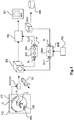
- the imaging system CT (also referred to as “scanner” or “imager” herein) includes a rotatable gantry RG rotatably mounted in a fixed gantry FG.
- the rotatable gantry RG is rotatable about a portion of space, an examination region that is. More specifically rotatable gantry RG is rotatable around an iso-center of said examination region.
- the rotatable gantry RG carries at one of its ends an x-ray tube or source XR and on the other end a detector D.
- the x-ray source XR is configured to emit, during an imaging session, x-ray radiation. More particularly, an X-ray beam passes through the examination region and then impinges on a radiation sensitive surface of the detector D. Within the examination region traversed by the x-ray beam is an examination table T. On the examination table T, a sample/object P to be imaged (for instance, a human or animal patient) is deposited. Yet more particularly, the x-ray beam passes through patient P at a region of interest ROI, e.g. a specific organ or part thereof, such as the patient's chest. During its passage through the sample P, the x-ray beam is modified for instance by absorption interaction within matter in the sample P.
- a region of interest ROI e.g. a specific organ or part thereof, such as the patient's chest.
- the degree of absorption is a direct measure of the absorption co-efficient distribution or of the density distribution in the patient.
- Individual rays of the x-ray beam are therefore differently modified or absorbed depending on where the respective rays pass through the sample P.
- the so modified x-ray beam that emerges at the other end of the patient then interacts with the detector.
- corresponding electrical signals are issued which are then passed to a DAS (data acquisition system - not shown).
- the DAS includes suitable A/D conversion circuitry to convert said electrical signals into digital form, also referred to as detector raw data, essentially an array of numbers.
- the detector raw data produced by the imager IMA are essentially projection images of the imaged sample acquired at a certain projection direction relative to the examination region and the object therein.
- the series of projection images are the forwarded to a reconstructor RECON that uses otherwise known reconstruction algorithms (such as filtered back-projection FBP) to convert the series into a slice image SL that affords a cross-sectional view of the object at a certain position z along an axis running through the object at the scanner's iso-center.
- a relative motion along said axis is then effected by moving the gantry or advancing the examination table T (on which the patient P resides) along said axis to a new z position and the above described procedure is repeated to form a new reconstructed slice for said new z position from new projection images.
- a visualization module VIS such as direct volume renderer (DRV), may then be applied to the 3D image data set to form on screen MT desired 3D views on the anatomy from a view point interactively definable by the user.
- DVR direct volume renderer
- Each image volume element ("voxel") of the 3D image data corresponds to a spatial point in the examination region. If that point was occupied by the object during the imaging then that point will carry the desired information about the amount of accumulated absorption suffered by a ray of the X-ray beam in its passage through said point in the examination region. But because the object P to be imaged does not normally fully occupy the whole of the examination region, there will inevitably be some background voxels that do not encode any attenuation caused by the object but will merely encode the negligible attenuation caused by the surrounding air. These voxels may then be called background voxels. In yet other words the whole of the volume set 3D comprises two complimentary regions: an object region OR and a background region BR. These are shown diagrammatically on pane A of Figure 2 .
- Pane B of Figure 2 shows a cross section along Z direction through 3D data set DS. Where the two regions meet there is an interface IF.
- the interface region is essentially a two dimensional surface that corresponds or defines the outer contour or silhouette of the object P. Because of the high spatial resolution capabilities of modern imagers, the silhouette IF of the object may be represented in quite great detail in DVR viewers. On occasions the silhouette of the head for instance will allow identification of the imaged person. This may be undesirable because of privacy concerns. Although, for diagnostic purposes or other medical purposes, medical staff will mostly be interested in observing 3D renderings of the internals of the object, there is nothing that would prevent for instance an unauthorized person to request a volume rendering of the silhouette and then possibly abuse this potentially embarrassing imagery.
- pane A shows a direct volume rendering DVR of thoracic CT data set.
- pane B for the same data set, a viewpoint outside the object region is chosen and volumetric rendering of the torso silhouette is obtained.
- the DVR of B the same settings are used as in pane A, but this time the view is turned around the cranial-caudal body axis thereby clearly revealing possibly embarrassing details about the outer contours or body surface of the patient.
- an image processing module IP is proposed herein.
- the proposed image processor IP operates to, according to one embodiment, automatically "fill up" the background region BR of the 3D data set DS with random pixel/voxel information so that any inadvertent volume rendering of the body contour is made next to impossible.
- the data set DS is transformed into a "visually protected" data set version DSX.
- this partial randomization of the data set DS is reversible so that the original, unprotected volume data set can be fully recovered.
- the proposed image processor IP is envisaged as a security component interposed between the image output of imager CT and the visualization module (e.g. renderer) VIS. That is, rather than passing the produced 3D data set DS direct to the visualizer VIS as has been done in the past, now the image processor IP is interposed that first transforms the 3D data set DS into the visually protected one DSX. It is only the visually protected or randomized data set DSX that is then made unavailable for rendering operations.
- the visualization module e.g. renderer
- FIG. 2B illustrates in more detail the operation of the image processor IP in terms of what happens to the various image voxels making up data set DS.
- the randomized region RR (that is, those voxels to which the randomization operation has been applied to or is to be applied to) includes all or parts of the background region BG. In fact in certain embodiments the randomization region is restricted to the background region. However, in a preferred embodiment the randomization region RR extends partly into the object region.
- the object region OR to be made up of a core region CR that is surrounded by an outer layer region LR. This outer region LR may be merely a few voxels thick, in the extreme case merely one voxel thick.
- the randomization region RR into the object region to include the outer layer region LR.
- all image elements in the outer layer region LR are randomized. Having the randomization extend into said outer layer region LR, affords a better anonymization outcome. For if, for instance, the randomization were to be applied only to the background region, there may be a tell-tale abrupt transition between background randomization and the non-randomized image information in the object region OR. The body contour may then still be recoverable. It is therefore desirable to at least randomize the interface IF along with at least part of the surrounding background region BR. That is, one randomizes not only the background region BR but also at least those object voxels in the object region OR that border the background region. In this manner the privacy of the patient can be protected better.
- the preferred embodiment is illustrated in Figure 2B , where the randomization RR (shown as hatched) is partly extending from background region BG into outer layer region LR of object region OR.
- the amount of randomization increases in any or at least one direction p from the core region CR towards the background region BR.
- Curves f,g show, respectively, the amount of original image information ( f ) and the amount of randomized image information (g).
- the amount of original information f increases whereas the amount of "mocked" or randomized image information g and this reciprocity carries on up to the interface region IF.
- the randomization is then restricted to image elements in between the object region OR and the convex hull.
- image elements between the convex hull and the object region OR are randomized. In this manner CPU can be saved.
- Image processor IP includes input IN and output OUT ports for respectively receiving the original 3D data set DS and for outputting the randomized data set DSX.
- the image processor includes as its basic components a segmenter SG and a randomizer RA.
- Figure 3C shows the effect of the randomization operation on the data set shown in panes A, B of Figure 3 .
- the breasts of the patient are no longer visible.
- using the randomized data set DSX if one requests a direct volume rendering of the internals of the patient one would still get the image as per Figure 3A .
- the anonymized view as per pane C In other words, the patient's body appears to be wrapped up to in a "virtual blanket" to better protect privacy.
- the original 3D data set DS is received. As mentioned briefly above, this may occur automatically as a matter of course upon output of the volume by imager CT but other embodiments are also envisaged where the user can later effect the anonymization.
- the original set DS may be stored fist in a database or storage DB and is then later anonymized.
- the proposed processor IP may be used to anonymize already existing stock of imagery held in a PACS or other image management system.
- the 3D data set is segmented into the body region OR and the background region BR.
- This allows defining the interface region IF that voxel elements that represent the 2D surface of the body.
- the identity of all the voxels to be randomized can be defined. That is the number and location (in terms of coordinates (x,y,z)) of the voxels (to be) randomized is completely determined once the thickness of the outer layer LR has been set and once it is defined how much of the background region is to be randomized. The latter can be set by defining the thickness of the layer region in the background BR around the object region OR as by computing the convex hull as previously explained.
- These two layers that define the "reach" of the randomized region RR into either side of the interface contour IF are pre-defined settings of the image processor IP and may be user adjustable.
- the voxels making up the region RR are those that are earmarked for the subsequent randomization.
- the randomization is then applied to the segmented data set, that is, is applied to the voxels in the randomized region RR.
- the randomization operation is either restricted to the background information or extends into the outer layer region as previously explained.
- the actual randomization operation is implemented by using, in one embodiment, a pseudo random number generator.
- the random number generator is defined by a seed point, and a mathematical instruction that defines a sequence of numbers that are deterministic but appear as random if one is not in possession of the seed point and the particular functional description of the mathematical instructions.
- the mathematical instruction is in one embodiment an iterative modulo division (known from arithmetic) is carried out starting from the seed point (a number that is). In this process different remainders are generated that are output as the random numbers during the iteration.
- the collection of all the information that is necessary to generate this sequence of random numbers will be referred to herein as the randomization instruction(s).
- the randomization is operation is reversible. This can be achieved by defining a "traversal order" in which the random numbers generated in the sequence are to be applied to the randomized region RR. This can be done by defining a path through the region RR from slice to slice and in each slice row or column wise. Each voxel is then assigned an order number along said path. Because the overall number of voxels in region RR is known, an equal number of random numbers can then be generated by running the random generator for the required number of iterations to produce a sequence of random numbers having as many entries as the region RR has voxels.
- the i -th random number in the random sequence is then applied to the i -th voxel in the traversal path.
- Application of the random numbers to the region RR is by combining the two, or instance by adding the randomized intensity to the respective voxel value at the corresponding position to so form the randomized region.
- Each pixel in the randomization region is visited will then have the random intensity added.
- randomization procedure admits to variety of different implementations all of which are envisaged herein in different embodiments.
- all random numbers are generated beforehand and are then applied to the randomization region.
- the application that is the adding of the randomized intensities can proceed either sequentially or can be done in parallel at once.
- the random intensity values are applied on-the-fly as they are output by the random number generator.
- the generated random numbers are mapped to the admissible range of image intensities and are then added in a linear superposition to respective ones of the voxels in the region RR.
- the random numbers are "wrapped" around admissible range in an arithmetic modulo operation to arrive at the randomized intensity.
- the amount of randomization is on a "sliding scale" so varies within the randomization region.
- This could be implemented by filling the transition region LR up to the surface IF between air and body as per an increasing linear mixture of noise and true image intensities, such that the noise contribution is 100% at e.g. -5 mm before the body surface IF, until 0% at e.g. +5 mm inside the body.
- the percentage slides or is ramped down linearly as one moves toward the background region BR. That is, the true values are gradually phased and the mocked intensity values are phase in with decreasing distance.
- the so randomized 3D data set DSX may then be stored for later reference in a database DB.
- the data DS is made available in the DICOM format. It is then proposed according to one embodiment that all or at least parts of the randomization instructions are included in the DICOM header of the data set DS. As mentioned earlier, in one embodiment, the randomization is executed upon production or generation of the data set at the imager CT. The inclusion of at least parts of the randomization instructions into the DICOM header can be implemented as a new standard for instance. However, in other embodiments the randomization instructions are stored elsewhere and are merely logically linked with the randomized data set DSX. For instance, the mathematical instruction of the pseudo random number generator, its seed number, and the body segmentation are stored together with the protected dataset as text in a DICOM file.
- the image processor IPR includes in and out ports IN, OUT and includes a de-randomizer component DRA. It is understood that although Fig 1 shows both operators IP and IPR, the latter is optional.
- step S505 the randomized data set DSX is received.
- the randomization instructions are accessed.
- the request to access same can be issued by an agent AG.
- the agent AG may be the workstation of a physician in a medical PACS system. If the user at workstation AG has the right credentials, for instance passwords or has been given appropriate user rights, the request to access the randomization instructions is granted, otherwise denied.
- step S5 If access is granted, the flow proceeds to step S515, where reversed randomization instructions are formulated. This may be achieved by re-running the randomization sequence using the same seed number and the same iterative mathematical instruction to recreate the random numbers.
- the reverse instructions are then applied to the randomized data set DSX at the designated voxel position as prescribed by the co-ordinates of the randomized region RR.
- the identity of the voxels in the randomized can be subtracted from the randomized data set DSX at the respective positions to so reveal the original data set DS.
- the randomization instructions include the previously mentioned seed point, the iterative mathematical function that generates the sequence of randomization points when iteratively applied to the seed points , the agreed on traversal order through the randomization region RR and a coordinate definition of the randomized region itself.
- the RR definition is based on the body segmentation at step S410 and can be stored as ASCII text (string data) together with the protected dataset (e.g. a DICOM file) or away from the protected date.
- the randomized region RR can be stored very memory-efficient e.g. as a mesh or as a compressed bit volume that requires typically ⁇ 1% of storage compared to the original image data.
- such items as the traversal order may be part of a pre-agreed standard so may not be specifically stored along with the other information.
- the items may be stored together or any one or more of the items may be stored away and separately from the others to increase security.
- the seed number of the pseudo random number generator (a single integer number that is) is not stored together with the protected image, but for data security reasons is provided in a separate file and/or is provide in separate communication channels, which could be an electronic record, a paper printout, oral communication, telecommunication, etc.
- recovery is allowed only for authorized personnel (e.g. the principal investigator or primary physician). This can be achieved on a password basis.
- the following items are stored with the image data: the randomization instruction for instance the mathematical formula of the pseudo random number generator, its seed, and the coordinate definition of the randomized region R.
- the items may for instance stored be embedded, (in the DICOM header or otherwise). But the randomization instruction so stored is encrypted with a password.
- the image processors IP, IPR are envisaged as modules installed on scanner consoles or medical imaging and PACS workstations. In short the proposed image processors may be used in any system which produce or handle 3D data sets. Processors PR and IR may be consolidated into a single module unlike the embodiment shown in Fig 1 although having them separate as shown in Figure 1 may also be desirable in some embodiments. For instance, IP is installed at on the scanner CT whereas descrambler IPR is made available only at select imager or PACS workstations.
- a computer program or a computer program element is provided that is characterized by being adapted to execute the method steps of the method according to one of the preceding embodiments, on an appropriate system.
- the computer program element might therefore be stored on a computer unit, which might also be part of an embodiment of the present invention.
- This computing unit may be adapted to perform or induce a performing of the steps of the method described above. Moreover, it may be adapted to operate the components of the above-described apparatus.
- the computing unit can be adapted to operate automatically and/or to execute the orders of a user.
- a computer program may be loaded into a working memory of a data processor. The data processor may thus be equipped to carry out the method of the invention.
- This exemplary embodiment of the invention covers both, a computer program that right from the beginning uses the invention and a computer program that by means of an up-date turns an existing program into a program that uses the invention.
- the computer program element might be able to provide all necessary steps to fulfill the procedure of an exemplary embodiment of the method as described above.
- a computer readable medium such as a CD-ROM
- the computer readable medium has a computer program element stored on it which computer program element is described by the preceding section.
- a computer program may be stored and/or distributed on a suitable medium, such as an optical storage medium or a solid-state medium supplied together with or as part of other hardware, but may also be distributed in other forms, such as via the internet or other wired or wireless telecommunication systems.
- a suitable medium such as an optical storage medium or a solid-state medium supplied together with or as part of other hardware, but may also be distributed in other forms, such as via the internet or other wired or wireless telecommunication systems.
- the computer program may also be presented over a network like the World Wide Web and can be downloaded into the working memory of a data processor from such a network.
- a medium for making a computer program element available for downloading is provided, which computer program element is arranged to perform a method according to one of the previously described embodiments of the invention.
Landscapes
- Engineering & Computer Science (AREA)
- Theoretical Computer Science (AREA)
- Physics & Mathematics (AREA)
- General Physics & Mathematics (AREA)
- Computer Graphics (AREA)
- General Engineering & Computer Science (AREA)
- Computer Hardware Design (AREA)
- Health & Medical Sciences (AREA)
- Computer Vision & Pattern Recognition (AREA)
- Software Systems (AREA)
- Medical Informatics (AREA)
- General Health & Medical Sciences (AREA)
- Public Health (AREA)
- Nuclear Medicine, Radiotherapy & Molecular Imaging (AREA)
- Primary Health Care (AREA)
- Epidemiology (AREA)
- Radiology & Medical Imaging (AREA)
- Evolutionary Computation (AREA)
- Architecture (AREA)
- Bioinformatics & Cheminformatics (AREA)
- Artificial Intelligence (AREA)
- Geometry (AREA)
- Bioinformatics & Computational Biology (AREA)
- Multimedia (AREA)
- Evolutionary Biology (AREA)
- Data Mining & Analysis (AREA)
- Life Sciences & Earth Sciences (AREA)
- Apparatus For Radiation Diagnosis (AREA)
- Image Generation (AREA)
- Image Processing (AREA)
- Measuring And Recording Apparatus For Diagnosis (AREA)
- Image Analysis (AREA)
- Magnetic Resonance Imaging Apparatus (AREA)
Applications Claiming Priority (2)
| Application Number | Priority Date | Filing Date | Title |
|---|---|---|---|
| EP14173573 | 2014-06-24 | ||
| PCT/EP2015/063952 WO2015197541A1 (en) | 2014-06-24 | 2015-06-22 | Visual anonymization of medical datasets against 3d volume rendering |
Publications (2)
| Publication Number | Publication Date |
|---|---|
| EP3161796A1 EP3161796A1 (en) | 2017-05-03 |
| EP3161796B1 true EP3161796B1 (en) | 2018-08-29 |
Family
ID=51211511
Family Applications (1)
| Application Number | Title | Priority Date | Filing Date |
|---|---|---|---|
| EP15730186.2A Active EP3161796B1 (en) | 2014-06-24 | 2015-06-22 | Visual anonymization of medical datasets against 3d volume rendering |
Country Status (8)
| Country | Link |
|---|---|
| US (1) | US10332238B2 (enExample) |
| EP (1) | EP3161796B1 (enExample) |
| JP (1) | JP6484650B2 (enExample) |
| CN (1) | CN106462973B (enExample) |
| BR (1) | BR112016030112A2 (enExample) |
| RU (1) | RU2679969C2 (enExample) |
| TR (1) | TR201815866T4 (enExample) |
| WO (1) | WO2015197541A1 (enExample) |
Families Citing this family (9)
| Publication number | Priority date | Publication date | Assignee | Title |
|---|---|---|---|---|
| US9633482B2 (en) * | 2014-08-12 | 2017-04-25 | Toshiba Medical Systems Corporation | Apparatus and method for restricting image data visualization |
| EP3273380B1 (en) | 2016-07-20 | 2018-12-12 | Siemens Healthcare GmbH | Protecting data exchanged between a service user and a service provider |
| CN107657653A (zh) * | 2016-07-25 | 2018-02-02 | 同方威视技术股份有限公司 | 用于对三维表面的图像进行重建的方法、装置和系统 |
| US10679740B2 (en) * | 2018-06-12 | 2020-06-09 | The Chinese University Of Hong Kong | System and method for patient privacy protection in medical images |
| US12061705B2 (en) | 2018-06-18 | 2024-08-13 | Koninklijke Philips N.V. | Secure remote image analysis based on randomized data transformation |
| JP6983124B2 (ja) | 2018-07-26 | 2021-12-17 | 株式会社日立製作所 | 医用画像処理装置及び医用画像処理方法 |
| CN110084870B (zh) * | 2019-05-13 | 2023-03-24 | 武汉轻工大学 | 平面方程的绘图区域的确定方法、装置、设备及存储介质 |
| GB2590992B (en) * | 2020-06-22 | 2022-05-18 | Intelligent Prot Management Group Ltd | Portable site access control unit |
| JP7555224B2 (ja) * | 2020-09-23 | 2024-09-24 | Psp株式会社 | 医用画像処理装置及び医用画像処理方法 |
Family Cites Families (27)
| Publication number | Priority date | Publication date | Assignee | Title |
|---|---|---|---|---|
| US5253192A (en) * | 1991-11-14 | 1993-10-12 | The Board Of Governors For Higher Education, State Of Rhode Island And Providence Plantations | Signal processing apparatus and method for iteratively determining Arithmetic Fourier Transform |
| US5768413A (en) * | 1995-10-04 | 1998-06-16 | Arch Development Corp. | Method and apparatus for segmenting images using stochastically deformable contours |
| US5859928A (en) | 1996-06-21 | 1999-01-12 | Hewlett-Packard Company | Jitter-form background control for minimizing spurious gray cast in scanned images |
| US6915011B2 (en) * | 2001-03-28 | 2005-07-05 | Eastman Kodak Company | Event clustering of images using foreground/background segmentation |
| US20040174998A1 (en) | 2003-03-05 | 2004-09-09 | Xsides Corporation | System and method for data encryption |
| CA2449080A1 (en) * | 2003-11-13 | 2005-05-13 | Centre Hospitalier De L'universite De Montreal - Chum | Apparatus and method for intravascular ultrasound image segmentation: a fast-marching method |
| US7907762B2 (en) * | 2004-05-26 | 2011-03-15 | Guardian Technologies International, Inc. | Method of creating a divergence transform for identifying a feature of interest in hyperspectral data |
| JP2007243256A (ja) | 2006-03-06 | 2007-09-20 | Dainippon Printing Co Ltd | 医療用画像暗号化装置 |
| US8041129B2 (en) * | 2006-05-16 | 2011-10-18 | Sectra Ab | Image data set compression based on viewing parameters for storing medical image data from multidimensional data sets, related systems, methods and computer products |
| JP2008118406A (ja) | 2006-11-06 | 2008-05-22 | Dainippon Printing Co Ltd | コンテンツ暗号方法、コンテンツ暗号装置及びコンピュータプログラム |
| US7796733B2 (en) * | 2007-02-01 | 2010-09-14 | Rapiscan Systems, Inc. | Personnel security screening system with enhanced privacy |
| US8670560B2 (en) | 2008-10-23 | 2014-03-11 | University Of Ulster | Encryption method |
| JP5606832B2 (ja) * | 2010-03-05 | 2014-10-15 | 富士フイルム株式会社 | 画像診断支援装置、方法およびプログラム |
| EP2699164A4 (en) | 2011-04-18 | 2014-10-29 | Pathfinder Therapeutics Inc | IMAGE ORGANIZATION OF ORGANS AND ANATOMICAL STRUCTURES |
| JP2013138800A (ja) | 2012-01-06 | 2013-07-18 | Toshiba Corp | 医用画像処理装置 |
| US8682049B2 (en) * | 2012-02-14 | 2014-03-25 | Terarecon, Inc. | Cloud-based medical image processing system with access control |
| US8553965B2 (en) * | 2012-02-14 | 2013-10-08 | TerraRecon, Inc. | Cloud-based medical image processing system with anonymous data upload and download |
| KR20130126800A (ko) * | 2012-04-23 | 2013-11-21 | 한국전자통신연구원 | 캡춰 영상의 프라이버시 보호를 위한 이미지 처리 방법 및 이미지 처리 장치 |
| RU2505800C2 (ru) * | 2012-05-10 | 2014-01-27 | Федеральное государственное бюджетное образовательное учреждение высшего профессионального образования "Национальный исследовательский Томский государственный университет" (ТГУ) | Способ рентгеновской томографии и устройство для его осуществления |
| US8712137B2 (en) * | 2012-05-29 | 2014-04-29 | General Electric Company | Methods and system for displaying segmented images |
| CN102945542A (zh) * | 2012-11-08 | 2013-02-27 | 崔得龙 | 一种感兴趣区域认证和篡改检测数字水印方法 |
| CN102932644A (zh) * | 2012-11-19 | 2013-02-13 | 海南大学 | 一种基于Arnold置乱变换和DFT的医学图像鲁棒水印方法 |
| EP2759957B1 (de) * | 2013-01-28 | 2018-10-31 | Siemens Healthcare GmbH | Übertragungsmittel für sicherheitskritische medizinische Bildinhalte |
| CN103226802B (zh) * | 2013-03-26 | 2015-09-23 | 中南大学 | 基于混沌加密的医学图像共享方法 |
| US9466129B2 (en) * | 2013-06-13 | 2016-10-11 | Toshiba Medical Systems Corporation | Apparatus and method of processing background image of medical display image |
| US10025479B2 (en) * | 2013-09-25 | 2018-07-17 | Terarecon, Inc. | Advanced medical image processing wizard |
| US9633482B2 (en) * | 2014-08-12 | 2017-04-25 | Toshiba Medical Systems Corporation | Apparatus and method for restricting image data visualization |
-
2015
- 2015-06-22 WO PCT/EP2015/063952 patent/WO2015197541A1/en not_active Ceased
- 2015-06-22 US US15/315,402 patent/US10332238B2/en active Active
- 2015-06-22 RU RU2017102244A patent/RU2679969C2/ru active
- 2015-06-22 JP JP2016573606A patent/JP6484650B2/ja active Active
- 2015-06-22 BR BR112016030112A patent/BR112016030112A2/pt not_active Application Discontinuation
- 2015-06-22 TR TR2018/15866T patent/TR201815866T4/tr unknown
- 2015-06-22 CN CN201580033982.7A patent/CN106462973B/zh active Active
- 2015-06-22 EP EP15730186.2A patent/EP3161796B1/en active Active
Also Published As
| Publication number | Publication date |
|---|---|
| WO2015197541A1 (en) | 2015-12-30 |
| BR112016030112A2 (pt) | 2017-08-22 |
| EP3161796A1 (en) | 2017-05-03 |
| JP6484650B2 (ja) | 2019-03-13 |
| US10332238B2 (en) | 2019-06-25 |
| RU2017102244A (ru) | 2018-07-26 |
| JP2017529110A (ja) | 2017-10-05 |
| RU2017102244A3 (enExample) | 2018-12-03 |
| TR201815866T4 (tr) | 2018-11-21 |
| US20170200256A1 (en) | 2017-07-13 |
| CN106462973A (zh) | 2017-02-22 |
| CN106462973B (zh) | 2021-01-22 |
| RU2679969C2 (ru) | 2019-02-14 |
Similar Documents
| Publication | Publication Date | Title |
|---|---|---|
| EP3161796B1 (en) | Visual anonymization of medical datasets against 3d volume rendering | |
| Dong et al. | Deep learning-based attenuation correction in the absence of structural information for whole-body positron emission tomography imaging | |
| Leng et al. | Streaking artifacts reduction in four‐dimensional cone‐beam computed tomography | |
| JP6688560B2 (ja) | 医用画像処理装置及び医用画像処理方法 | |
| Ma et al. | Low‐dose computed tomography image restoration using previous normal‐dose scan | |
| Schreibmann et al. | MR‐based attenuation correction for hybrid PET‐MR brain imaging systems using deformable image registration | |
| Gao et al. | 4D cone beam CT via spatiotemporal tensor framelet | |
| Montoya et al. | Reconstruction of three‐dimensional tomographic patient models for radiation dose modulation in CT from two scout views using deep learning | |
| US20210224403A1 (en) | Secure remote image analysis based on randomized data transformation | |
| Yang et al. | A hybrid approach for fusing 4D‐MRI temporal information with 3D‐CT for the study of lung and lung tumor motion | |
| JP7642641B2 (ja) | 人工知能のための患者画像の解剖学的暗号化 | |
| CN104202368B (zh) | 基于云平台的医疗影像数据共享的方法、云平台和系统 | |
| Li et al. | Adaptive 3D noise level‐guided restoration network for low‐dose positron emission tomography imaging | |
| Madhushree et al. | An exhaustive review of authentication, tamper detection with localization and recovery techniques for medical images | |
| CN104038543B (zh) | 一种医疗影像设备云重建的方法、云平台及系统 | |
| Fan et al. | Image‐domain shading correction for cone‐beam CT without prior patient information | |
| KR102395251B1 (ko) | 단층 촬영을 위한 관심 영역 설정 방법 및 시스템 | |
| CN105631908B (zh) | 一种pet图像重建方法和装置 | |
| WO2020198560A1 (en) | Systems and methods for non-destructive de-identification of facial data in medical images | |
| Godfrey et al. | The effect of averaging adjacent planes for artifact reduction in matrix inversion tomosynthesis | |
| Komolafe et al. | Comparative analysis of cone‐beam breast computed tomography and digital breast tomosynthesis for breast cancer diagnosis: A comprehensive study on reconstruction algorithms | |
| Wu et al. | Ablation zone visualization enhancement by periodic contrast‐enhancement computed tomography during microwave ablation | |
| Kim et al. | Evaluation of AI-based super-resolution model in CT for dose reduction method: A phantom study | |
| JP7744171B2 (ja) | 医用画像処理装置 | |
| Tang et al. | Radial differential interior tomography and its image reconstruction with differentiated backprojection and projection onto convex sets |
Legal Events
| Date | Code | Title | Description |
|---|---|---|---|
| STAA | Information on the status of an ep patent application or granted ep patent |
Free format text: STATUS: THE INTERNATIONAL PUBLICATION HAS BEEN MADE |
|
| PUAI | Public reference made under article 153(3) epc to a published international application that has entered the european phase |
Free format text: ORIGINAL CODE: 0009012 |
|
| STAA | Information on the status of an ep patent application or granted ep patent |
Free format text: STATUS: REQUEST FOR EXAMINATION WAS MADE |
|
| 17P | Request for examination filed |
Effective date: 20170124 |
|
| AK | Designated contracting states |
Kind code of ref document: A1 Designated state(s): AL AT BE BG CH CY CZ DE DK EE ES FI FR GB GR HR HU IE IS IT LI LT LU LV MC MK MT NL NO PL PT RO RS SE SI SK SM TR |
|
| AX | Request for extension of the european patent |
Extension state: BA ME |
|
| DAV | Request for validation of the european patent (deleted) | ||
| DAX | Request for extension of the european patent (deleted) | ||
| GRAP | Despatch of communication of intention to grant a patent |
Free format text: ORIGINAL CODE: EPIDOSNIGR1 |
|
| STAA | Information on the status of an ep patent application or granted ep patent |
Free format text: STATUS: GRANT OF PATENT IS INTENDED |
|
| RIC1 | Information provided on ipc code assigned before grant |
Ipc: G06T 19/00 20110101ALI20180226BHEP Ipc: G06T 7/194 20170101ALI20180226BHEP Ipc: G06T 7/11 20170101ALI20180226BHEP Ipc: G06T 15/08 20110101AFI20180226BHEP Ipc: G06F 19/00 20110101ALI20180226BHEP Ipc: G06T 3/00 20060101ALI20180226BHEP |
|
| INTG | Intention to grant announced |
Effective date: 20180321 |
|
| GRAS | Grant fee paid |
Free format text: ORIGINAL CODE: EPIDOSNIGR3 |
|
| GRAA | (expected) grant |
Free format text: ORIGINAL CODE: 0009210 |
|
| STAA | Information on the status of an ep patent application or granted ep patent |
Free format text: STATUS: THE PATENT HAS BEEN GRANTED |
|
| AK | Designated contracting states |
Kind code of ref document: B1 Designated state(s): AL AT BE BG CH CY CZ DE DK EE ES FI FR GB GR HR HU IE IS IT LI LT LU LV MC MK MT NL NO PL PT RO RS SE SI SK SM TR |
|
| REG | Reference to a national code |
Ref country code: GB Ref legal event code: FG4D |
|
| REG | Reference to a national code |
Ref country code: CH Ref legal event code: EP |
|
| REG | Reference to a national code |
Ref country code: AT Ref legal event code: REF Ref document number: 1036056 Country of ref document: AT Kind code of ref document: T Effective date: 20180915 |
|
| REG | Reference to a national code |
Ref country code: IE Ref legal event code: FG4D |
|
| REG | Reference to a national code |
Ref country code: DE Ref legal event code: R096 Ref document number: 602015015590 Country of ref document: DE |
|
| REG | Reference to a national code |
Ref country code: NL Ref legal event code: MP Effective date: 20180829 |
|
| REG | Reference to a national code |
Ref country code: LT Ref legal event code: MG4D |
|
| PG25 | Lapsed in a contracting state [announced via postgrant information from national office to epo] |
Ref country code: GR Free format text: LAPSE BECAUSE OF FAILURE TO SUBMIT A TRANSLATION OF THE DESCRIPTION OR TO PAY THE FEE WITHIN THE PRESCRIBED TIME-LIMIT Effective date: 20181130 Ref country code: LT Free format text: LAPSE BECAUSE OF FAILURE TO SUBMIT A TRANSLATION OF THE DESCRIPTION OR TO PAY THE FEE WITHIN THE PRESCRIBED TIME-LIMIT Effective date: 20180829 Ref country code: NL Free format text: LAPSE BECAUSE OF FAILURE TO SUBMIT A TRANSLATION OF THE DESCRIPTION OR TO PAY THE FEE WITHIN THE PRESCRIBED TIME-LIMIT Effective date: 20180829 Ref country code: BG Free format text: LAPSE BECAUSE OF FAILURE TO SUBMIT A TRANSLATION OF THE DESCRIPTION OR TO PAY THE FEE WITHIN THE PRESCRIBED TIME-LIMIT Effective date: 20181129 Ref country code: NO Free format text: LAPSE BECAUSE OF FAILURE TO SUBMIT A TRANSLATION OF THE DESCRIPTION OR TO PAY THE FEE WITHIN THE PRESCRIBED TIME-LIMIT Effective date: 20181129 Ref country code: SE Free format text: LAPSE BECAUSE OF FAILURE TO SUBMIT A TRANSLATION OF THE DESCRIPTION OR TO PAY THE FEE WITHIN THE PRESCRIBED TIME-LIMIT Effective date: 20180829 Ref country code: IS Free format text: LAPSE BECAUSE OF FAILURE TO SUBMIT A TRANSLATION OF THE DESCRIPTION OR TO PAY THE FEE WITHIN THE PRESCRIBED TIME-LIMIT Effective date: 20181229 Ref country code: RS Free format text: LAPSE BECAUSE OF FAILURE TO SUBMIT A TRANSLATION OF THE DESCRIPTION OR TO PAY THE FEE WITHIN THE PRESCRIBED TIME-LIMIT Effective date: 20180829 Ref country code: FI Free format text: LAPSE BECAUSE OF FAILURE TO SUBMIT A TRANSLATION OF THE DESCRIPTION OR TO PAY THE FEE WITHIN THE PRESCRIBED TIME-LIMIT Effective date: 20180829 |
|
| REG | Reference to a national code |
Ref country code: AT Ref legal event code: MK05 Ref document number: 1036056 Country of ref document: AT Kind code of ref document: T Effective date: 20180829 |
|
| PG25 | Lapsed in a contracting state [announced via postgrant information from national office to epo] |
Ref country code: LV Free format text: LAPSE BECAUSE OF FAILURE TO SUBMIT A TRANSLATION OF THE DESCRIPTION OR TO PAY THE FEE WITHIN THE PRESCRIBED TIME-LIMIT Effective date: 20180829 Ref country code: AL Free format text: LAPSE BECAUSE OF FAILURE TO SUBMIT A TRANSLATION OF THE DESCRIPTION OR TO PAY THE FEE WITHIN THE PRESCRIBED TIME-LIMIT Effective date: 20180829 Ref country code: HR Free format text: LAPSE BECAUSE OF FAILURE TO SUBMIT A TRANSLATION OF THE DESCRIPTION OR TO PAY THE FEE WITHIN THE PRESCRIBED TIME-LIMIT Effective date: 20180829 |
|
| PG25 | Lapsed in a contracting state [announced via postgrant information from national office to epo] |
Ref country code: AT Free format text: LAPSE BECAUSE OF FAILURE TO SUBMIT A TRANSLATION OF THE DESCRIPTION OR TO PAY THE FEE WITHIN THE PRESCRIBED TIME-LIMIT Effective date: 20180829 Ref country code: EE Free format text: LAPSE BECAUSE OF FAILURE TO SUBMIT A TRANSLATION OF THE DESCRIPTION OR TO PAY THE FEE WITHIN THE PRESCRIBED TIME-LIMIT Effective date: 20180829 Ref country code: IT Free format text: LAPSE BECAUSE OF FAILURE TO SUBMIT A TRANSLATION OF THE DESCRIPTION OR TO PAY THE FEE WITHIN THE PRESCRIBED TIME-LIMIT Effective date: 20180829 Ref country code: RO Free format text: LAPSE BECAUSE OF FAILURE TO SUBMIT A TRANSLATION OF THE DESCRIPTION OR TO PAY THE FEE WITHIN THE PRESCRIBED TIME-LIMIT Effective date: 20180829 Ref country code: CZ Free format text: LAPSE BECAUSE OF FAILURE TO SUBMIT A TRANSLATION OF THE DESCRIPTION OR TO PAY THE FEE WITHIN THE PRESCRIBED TIME-LIMIT Effective date: 20180829 Ref country code: PL Free format text: LAPSE BECAUSE OF FAILURE TO SUBMIT A TRANSLATION OF THE DESCRIPTION OR TO PAY THE FEE WITHIN THE PRESCRIBED TIME-LIMIT Effective date: 20180829 Ref country code: ES Free format text: LAPSE BECAUSE OF FAILURE TO SUBMIT A TRANSLATION OF THE DESCRIPTION OR TO PAY THE FEE WITHIN THE PRESCRIBED TIME-LIMIT Effective date: 20180829 |
|
| PG25 | Lapsed in a contracting state [announced via postgrant information from national office to epo] |
Ref country code: SM Free format text: LAPSE BECAUSE OF FAILURE TO SUBMIT A TRANSLATION OF THE DESCRIPTION OR TO PAY THE FEE WITHIN THE PRESCRIBED TIME-LIMIT Effective date: 20180829 Ref country code: SK Free format text: LAPSE BECAUSE OF FAILURE TO SUBMIT A TRANSLATION OF THE DESCRIPTION OR TO PAY THE FEE WITHIN THE PRESCRIBED TIME-LIMIT Effective date: 20180829 Ref country code: DK Free format text: LAPSE BECAUSE OF FAILURE TO SUBMIT A TRANSLATION OF THE DESCRIPTION OR TO PAY THE FEE WITHIN THE PRESCRIBED TIME-LIMIT Effective date: 20180829 |
|
| REG | Reference to a national code |
Ref country code: DE Ref legal event code: R097 Ref document number: 602015015590 Country of ref document: DE |
|
| PLBE | No opposition filed within time limit |
Free format text: ORIGINAL CODE: 0009261 |
|
| STAA | Information on the status of an ep patent application or granted ep patent |
Free format text: STATUS: NO OPPOSITION FILED WITHIN TIME LIMIT |
|
| 26N | No opposition filed |
Effective date: 20190531 |
|
| PG25 | Lapsed in a contracting state [announced via postgrant information from national office to epo] |
Ref country code: SI Free format text: LAPSE BECAUSE OF FAILURE TO SUBMIT A TRANSLATION OF THE DESCRIPTION OR TO PAY THE FEE WITHIN THE PRESCRIBED TIME-LIMIT Effective date: 20180829 |
|
| PG25 | Lapsed in a contracting state [announced via postgrant information from national office to epo] |
Ref country code: MC Free format text: LAPSE BECAUSE OF FAILURE TO SUBMIT A TRANSLATION OF THE DESCRIPTION OR TO PAY THE FEE WITHIN THE PRESCRIBED TIME-LIMIT Effective date: 20180829 |
|
| REG | Reference to a national code |
Ref country code: CH Ref legal event code: PL |
|
| REG | Reference to a national code |
Ref country code: BE Ref legal event code: MM Effective date: 20190630 |
|
| PG25 | Lapsed in a contracting state [announced via postgrant information from national office to epo] |
Ref country code: IE Free format text: LAPSE BECAUSE OF NON-PAYMENT OF DUE FEES Effective date: 20190622 |
|
| PG25 | Lapsed in a contracting state [announced via postgrant information from national office to epo] |
Ref country code: CH Free format text: LAPSE BECAUSE OF NON-PAYMENT OF DUE FEES Effective date: 20190630 Ref country code: LI Free format text: LAPSE BECAUSE OF NON-PAYMENT OF DUE FEES Effective date: 20190630 Ref country code: BE Free format text: LAPSE BECAUSE OF NON-PAYMENT OF DUE FEES Effective date: 20190630 Ref country code: LU Free format text: LAPSE BECAUSE OF NON-PAYMENT OF DUE FEES Effective date: 20190622 |
|
| PG25 | Lapsed in a contracting state [announced via postgrant information from national office to epo] |
Ref country code: PT Free format text: LAPSE BECAUSE OF FAILURE TO SUBMIT A TRANSLATION OF THE DESCRIPTION OR TO PAY THE FEE WITHIN THE PRESCRIBED TIME-LIMIT Effective date: 20181229 |
|
| PG25 | Lapsed in a contracting state [announced via postgrant information from national office to epo] |
Ref country code: CY Free format text: LAPSE BECAUSE OF FAILURE TO SUBMIT A TRANSLATION OF THE DESCRIPTION OR TO PAY THE FEE WITHIN THE PRESCRIBED TIME-LIMIT Effective date: 20180829 |
|
| PG25 | Lapsed in a contracting state [announced via postgrant information from national office to epo] |
Ref country code: MT Free format text: LAPSE BECAUSE OF FAILURE TO SUBMIT A TRANSLATION OF THE DESCRIPTION OR TO PAY THE FEE WITHIN THE PRESCRIBED TIME-LIMIT Effective date: 20180829 Ref country code: HU Free format text: LAPSE BECAUSE OF FAILURE TO SUBMIT A TRANSLATION OF THE DESCRIPTION OR TO PAY THE FEE WITHIN THE PRESCRIBED TIME-LIMIT; INVALID AB INITIO Effective date: 20150622 |
|
| PGFP | Annual fee paid to national office [announced via postgrant information from national office to epo] |
Ref country code: TR Payment date: 20210611 Year of fee payment: 7 |
|
| PG25 | Lapsed in a contracting state [announced via postgrant information from national office to epo] |
Ref country code: MK Free format text: LAPSE BECAUSE OF FAILURE TO SUBMIT A TRANSLATION OF THE DESCRIPTION OR TO PAY THE FEE WITHIN THE PRESCRIBED TIME-LIMIT Effective date: 20180829 |
|
| PG25 | Lapsed in a contracting state [announced via postgrant information from national office to epo] |
Ref country code: TR Free format text: LAPSE BECAUSE OF NON-PAYMENT OF DUE FEES Effective date: 20220622 |
|
| REG | Reference to a national code |
Ref country code: DE Ref legal event code: R084 Ref document number: 602015015590 Country of ref document: DE |
|
| PGFP | Annual fee paid to national office [announced via postgrant information from national office to epo] |
Ref country code: DE Payment date: 20250626 Year of fee payment: 11 |
|
| PGFP | Annual fee paid to national office [announced via postgrant information from national office to epo] |
Ref country code: GB Payment date: 20250617 Year of fee payment: 11 |
|
| PGFP | Annual fee paid to national office [announced via postgrant information from national office to epo] |
Ref country code: FR Payment date: 20250624 Year of fee payment: 11 |