EP3130596B1 - Dérivé de glucose, et procédé d'imagerie de cellule et agent d'imagerie utilisant ledit dérivé - Google Patents
Dérivé de glucose, et procédé d'imagerie de cellule et agent d'imagerie utilisant ledit dérivé Download PDFInfo
- Publication number
- EP3130596B1 EP3130596B1 EP15776849.0A EP15776849A EP3130596B1 EP 3130596 B1 EP3130596 B1 EP 3130596B1 EP 15776849 A EP15776849 A EP 15776849A EP 3130596 B1 EP3130596 B1 EP 3130596B1
- Authority
- EP
- European Patent Office
- Prior art keywords
- glucose
- amino
- deoxy
- oxo
- compound
- Prior art date
- Legal status (The legal status is an assumption and is not a legal conclusion. Google has not performed a legal analysis and makes no representation as to the accuracy of the status listed.)
- Active
Links
- 150000002303 glucose derivatives Chemical class 0.000 title claims description 87
- 238000003384 imaging method Methods 0.000 title claims description 46
- 239000012216 imaging agent Substances 0.000 title claims description 17
- 150000001875 compounds Chemical class 0.000 claims description 243
- WQZGKKKJIJFFOK-GASJEMHNSA-N Glucose Natural products OC[C@H]1OC(O)[C@H](O)[C@@H](O)[C@@H]1O WQZGKKKJIJFFOK-GASJEMHNSA-N 0.000 claims description 73
- 238000000034 method Methods 0.000 claims description 64
- 206010028980 Neoplasm Diseases 0.000 claims description 62
- 239000000203 mixture Substances 0.000 claims description 57
- 201000011510 cancer Diseases 0.000 claims description 56
- 239000008103 glucose Substances 0.000 claims description 36
- 125000006527 (C1-C5) alkyl group Chemical group 0.000 claims description 35
- 239000001257 hydrogen Substances 0.000 claims description 19
- 229910052739 hydrogen Inorganic materials 0.000 claims description 19
- 150000002431 hydrogen Chemical class 0.000 claims description 18
- 125000001188 haloalkyl group Chemical group 0.000 claims description 17
- 229910052736 halogen Inorganic materials 0.000 claims description 17
- 150000002367 halogens Chemical class 0.000 claims description 17
- 125000006729 (C2-C5) alkenyl group Chemical group 0.000 claims description 16
- 125000006730 (C2-C5) alkynyl group Chemical group 0.000 claims description 16
- 125000000753 cycloalkyl group Chemical group 0.000 claims description 16
- 125000002541 furyl group Chemical group 0.000 claims description 16
- 125000002791 glucosyl group Chemical group C1([C@H](O)[C@@H](O)[C@H](O)[C@H](O1)CO)* 0.000 claims description 16
- 125000001997 phenyl group Chemical group [H]C1=C([H])C([H])=C(*)C([H])=C1[H] 0.000 claims description 16
- 125000004076 pyridyl group Chemical group 0.000 claims description 16
- 125000000168 pyrrolyl group Chemical group 0.000 claims description 16
- 125000001544 thienyl group Chemical group 0.000 claims description 16
- 125000003282 alkyl amino group Chemical group 0.000 claims description 15
- 229910052760 oxygen Inorganic materials 0.000 claims description 15
- 125000002887 hydroxy group Chemical group [H]O* 0.000 claims description 8
- 150000003839 salts Chemical class 0.000 claims description 8
- WQZGKKKJIJFFOK-UKLRSMCWSA-N dextrose-2-13c Chemical class OC[C@H]1OC(O)[13C@H](O)[C@@H](O)[C@@H]1O WQZGKKKJIJFFOK-UKLRSMCWSA-N 0.000 claims description 5
- IPZOCGBUCLJCQB-CTHBEMJXSA-N (2R,3R,4S,5R)-3,4,5,6-tetrahydroxy-2-[(2-oxo-1H-quinolin-7-yl)amino]hexanal Chemical compound O=C1NC2=CC(=CC=C2C=C1)N[C@@H](C=O)[C@@H](O)[C@H](O)[C@H](O)CO IPZOCGBUCLJCQB-CTHBEMJXSA-N 0.000 claims description 3
- XRRUFVAAURFXDB-RBDSIQFVSA-N (2R,3R,4S,5R)-3,4,5,6-tetrahydroxy-2-[(2-oxochromen-7-yl)amino]hexanal Chemical compound O=C1OC2=CC(=CC=C2C=C1)N[C@@H](C=O)[C@@H](O)[C@H](O)[C@H](O)CO XRRUFVAAURFXDB-RBDSIQFVSA-N 0.000 claims description 3
- ISDHBBAKUUXBIX-GHEDXKAJSA-N (2R,3R,4S,5R)-2-[[1-(difluoromethyl)-3-(18F)fluoranyl-2-oxoquinolin-7-yl]amino]-3,4,5,6-tetrahydroxyhexanal Chemical compound O=C1N(C2=CC(=CC=C2C=C1[18F])N[C@@H](C=O)[C@@H](O)[C@H](O)[C@H](O)CO)C(F)F ISDHBBAKUUXBIX-GHEDXKAJSA-N 0.000 claims 1
- BUBOCKFUFXBDAF-ARQMMUAHSA-N (2R,3R,4S,5R)-2-[[1-(difluoromethyl)-4-(18F)fluoranyl-2-oxoquinolin-7-yl]amino]-3,4,5,6-tetrahydroxyhexanal Chemical compound O=C1N(C2=CC(=CC=C2C(=C1)[18F])N[C@@H](C=O)[C@@H](O)[C@H](O)[C@H](O)CO)C(F)F BUBOCKFUFXBDAF-ARQMMUAHSA-N 0.000 claims 1
- LJLBPZOJMGAVJX-SLUTYSENSA-N (2R,3R,4S,5R)-3,4,5,6-tetrahydroxy-2-[(3-(111C)methyl-2-oxo-1H-quinolin-7-yl)amino]hexanal Chemical compound O=C1NC2=CC(=CC=C2C=C1[11CH3])N[C@@H](C=O)[C@@H](O)[C@H](O)[C@H](O)CO LJLBPZOJMGAVJX-SLUTYSENSA-N 0.000 claims 1
- LJLBPZOJMGAVJX-GBJTYRQASA-N (2R,3R,4S,5R)-3,4,5,6-tetrahydroxy-2-[(3-methyl-2-oxo-1H-quinolin-7-yl)amino]hexanal Chemical compound O=C1NC2=CC(=CC=C2C=C1C)N[C@@H](C=O)[C@@H](O)[C@H](O)[C@H](O)CO LJLBPZOJMGAVJX-GBJTYRQASA-N 0.000 claims 1
- DBIURYWHJOPUIR-GTCPHAQCSA-N (2R,3R,4S,5R)-3,4,5,6-tetrahydroxy-2-[(4-(111C)methyl-2-oxo-1H-quinolin-7-yl)amino]hexanal Chemical compound O=C1NC2=CC(=CC=C2C(=C1)[11CH3])N[C@@H](C=O)[C@@H](O)[C@H](O)[C@H](O)CO DBIURYWHJOPUIR-GTCPHAQCSA-N 0.000 claims 1
- DBIURYWHJOPUIR-SJXGUFTOSA-N (2R,3R,4S,5R)-3,4,5,6-tetrahydroxy-2-[(4-methyl-2-oxo-1H-quinolin-7-yl)amino]hexanal Chemical compound O=C1NC2=CC(=CC=C2C(=C1)C)N[C@@H](C=O)[C@@H](O)[C@H](O)[C@H](O)CO DBIURYWHJOPUIR-SJXGUFTOSA-N 0.000 claims 1
- DFYGGVYRXLEMFU-REWJHTLYSA-N (2R,3R,4S,5R)-3,4,5,6-tetrahydroxy-2-[[2-oxo-3-(trifluoromethyl)-1H-quinolin-7-yl]amino]hexanal Chemical compound O=C1NC2=CC(=CC=C2C=C1C(F)(F)F)N[C@@H](C=O)[C@@H](O)[C@H](O)[C@H](O)CO DFYGGVYRXLEMFU-REWJHTLYSA-N 0.000 claims 1
- XZIQVEFYEOFKEG-CTHBEMJXSA-N (2R,3R,4S,5R)-3,4,5,6-tetrahydroxy-2-[[2-oxo-4-(trifluoromethyl)-1H-quinolin-7-yl]amino]hexanal Chemical compound O=C1NC2=CC(=CC=C2C(=C1)C(F)(F)F)N[C@@H](C=O)[C@@H](O)[C@H](O)[C@H](O)CO XZIQVEFYEOFKEG-CTHBEMJXSA-N 0.000 claims 1
- 210000004027 cell Anatomy 0.000 description 235
- 239000000243 solution Substances 0.000 description 139
- VGEREEWJJVICBM-UHFFFAOYSA-N phloretin Chemical compound C1=CC(O)=CC=C1CCC(=O)C1=C(O)C=C(O)C=C1O VGEREEWJJVICBM-UHFFFAOYSA-N 0.000 description 74
- VYPSYNLAJGMNEJ-UHFFFAOYSA-N Silicium dioxide Chemical compound O=[Si]=O VYPSYNLAJGMNEJ-UHFFFAOYSA-N 0.000 description 57
- WQZGKKKJIJFFOK-VFUOTHLCSA-N beta-D-glucose Chemical compound OC[C@H]1O[C@@H](O)[C@H](O)[C@@H](O)[C@@H]1O WQZGKKKJIJFFOK-VFUOTHLCSA-N 0.000 description 55
- XEKOWRVHYACXOJ-UHFFFAOYSA-N Ethyl acetate Chemical compound CCOC(C)=O XEKOWRVHYACXOJ-UHFFFAOYSA-N 0.000 description 54
- 230000015572 biosynthetic process Effects 0.000 description 42
- 239000002904 solvent Substances 0.000 description 42
- 238000003786 synthesis reaction Methods 0.000 description 42
- 239000011259 mixed solution Substances 0.000 description 41
- QUTFFEUUGHUPQC-ILWYWAAHSA-N (2r,3r,4s,5r)-3,4,5,6-tetrahydroxy-2-[(4-nitro-2,1,3-benzoxadiazol-7-yl)amino]hexanal Chemical compound OC[C@@H](O)[C@@H](O)[C@H](O)[C@H](C=O)NC1=CC=C([N+]([O-])=O)C2=NON=C12 QUTFFEUUGHUPQC-ILWYWAAHSA-N 0.000 description 40
- 150000001040 L-glucose derivatives Chemical class 0.000 description 40
- 238000006243 chemical reaction Methods 0.000 description 39
- FAPWRFPIFSIZLT-UHFFFAOYSA-M Sodium chloride Chemical class [Na+].[Cl-] FAPWRFPIFSIZLT-UHFFFAOYSA-M 0.000 description 38
- ZWTDXYUDJYDHJR-UHFFFAOYSA-N (E)-1-(2,4-dihydroxyphenyl)-3-(2,4-dihydroxyphenyl)-2-propen-1-one Natural products OC1=CC(O)=CC=C1C=CC(=O)C1=CC=C(O)C=C1O ZWTDXYUDJYDHJR-UHFFFAOYSA-N 0.000 description 37
- YQHMWTPYORBCMF-UHFFFAOYSA-N Naringenin chalcone Natural products C1=CC(O)=CC=C1C=CC(=O)C1=C(O)C=C(O)C=C1O YQHMWTPYORBCMF-UHFFFAOYSA-N 0.000 description 37
- 150000000780 D-glucose derivatives Chemical class 0.000 description 32
- QTBSBXVTEAMEQO-UHFFFAOYSA-N Acetic acid Chemical compound CC(O)=O QTBSBXVTEAMEQO-UHFFFAOYSA-N 0.000 description 30
- 102000018711 Facilitative Glucose Transport Proteins Human genes 0.000 description 29
- 239000007787 solid Substances 0.000 description 29
- GBOGMAARMMDZGR-UHFFFAOYSA-N UNPD149280 Natural products N1C(=O)C23OC(=O)C=CC(O)CCCC(C)CC=CC3C(O)C(=C)C(C)C2C1CC1=CC=CC=C1 GBOGMAARMMDZGR-UHFFFAOYSA-N 0.000 description 28
- GBOGMAARMMDZGR-JREHFAHYSA-N cytochalasin B Natural products C[C@H]1CCC[C@@H](O)C=CC(=O)O[C@@]23[C@H](C=CC1)[C@H](O)C(=C)[C@@H](C)[C@@H]2[C@H](Cc4ccccc4)NC3=O GBOGMAARMMDZGR-JREHFAHYSA-N 0.000 description 28
- GBOGMAARMMDZGR-TYHYBEHESA-N cytochalasin B Chemical compound C([C@H]1[C@@H]2[C@@H](C([C@@H](O)[C@@H]3/C=C/C[C@H](C)CCC[C@@H](O)/C=C/C(=O)O[C@@]23C(=O)N1)=C)C)C1=CC=CC=C1 GBOGMAARMMDZGR-TYHYBEHESA-N 0.000 description 28
- 108091052347 Glucose transporter family Proteins 0.000 description 27
- UIIMBOGNXHQVGW-UHFFFAOYSA-M Sodium bicarbonate Chemical compound [Na+].OC([O-])=O UIIMBOGNXHQVGW-UHFFFAOYSA-M 0.000 description 26
- 0 CNC1[C@@](NCO)OC(CC#[Rh])[C@@](*)C1O Chemical compound CNC1[C@@](NCO)OC(CC#[Rh])[C@@](*)C1O 0.000 description 24
- VEXZGXHMUGYJMC-UHFFFAOYSA-N Hydrochloric acid Chemical compound Cl VEXZGXHMUGYJMC-UHFFFAOYSA-N 0.000 description 23
- 239000011541 reaction mixture Substances 0.000 description 22
- ZMXDDKWLCZADIW-UHFFFAOYSA-N N,N-Dimethylformamide Chemical compound CN(C)C=O ZMXDDKWLCZADIW-UHFFFAOYSA-N 0.000 description 21
- 238000002474 experimental method Methods 0.000 description 21
- PMZURENOXWZQFD-UHFFFAOYSA-L Sodium Sulfate Chemical compound [Na+].[Na+].[O-]S([O-])(=O)=O PMZURENOXWZQFD-UHFFFAOYSA-L 0.000 description 20
- 239000012300 argon atmosphere Substances 0.000 description 20
- 125000002496 methyl group Chemical group [H]C([H])([H])* 0.000 description 20
- 239000012044 organic layer Substances 0.000 description 20
- 229910052681 coesite Inorganic materials 0.000 description 19
- 229910052906 cristobalite Inorganic materials 0.000 description 19
- 210000002569 neuron Anatomy 0.000 description 19
- 239000000741 silica gel Substances 0.000 description 19
- 229910002027 silica gel Inorganic materials 0.000 description 19
- 239000000377 silicon dioxide Substances 0.000 description 19
- 229910052682 stishovite Inorganic materials 0.000 description 19
- 229910052905 tridymite Inorganic materials 0.000 description 19
- IAZDPXIOMUYVGZ-UHFFFAOYSA-N Dimethylsulphoxide Chemical compound CS(C)=O IAZDPXIOMUYVGZ-UHFFFAOYSA-N 0.000 description 18
- 125000000332 coumarinyl group Chemical group O1C(=O)C(=CC2=CC=CC=C12)* 0.000 description 18
- 238000000605 extraction Methods 0.000 description 18
- 230000005284 excitation Effects 0.000 description 17
- 125000002943 quinolinyl group Chemical group N1=C(C=CC2=CC=CC=C12)* 0.000 description 17
- 108091006146 Channels Proteins 0.000 description 16
- JUJWROOIHBZHMG-UHFFFAOYSA-N Pyridine Chemical compound C1=CC=NC=C1 JUJWROOIHBZHMG-UHFFFAOYSA-N 0.000 description 16
- 230000010412 perfusion Effects 0.000 description 16
- OKKJLVBELUTLKV-UHFFFAOYSA-N Methanol Chemical compound OC OKKJLVBELUTLKV-UHFFFAOYSA-N 0.000 description 15
- YXFVVABEGXRONW-UHFFFAOYSA-N Toluene Chemical compound CC1=CC=CC=C1 YXFVVABEGXRONW-UHFFFAOYSA-N 0.000 description 15
- MSWZFWKMSRAUBD-UHFFFAOYSA-N beta-D-galactosamine Natural products NC1C(O)OC(CO)C(O)C1O MSWZFWKMSRAUBD-UHFFFAOYSA-N 0.000 description 15
- 230000004700 cellular uptake Effects 0.000 description 15
- 238000002360 preparation method Methods 0.000 description 15
- 229910000030 sodium bicarbonate Inorganic materials 0.000 description 13
- OKKJLVBELUTLKV-MZCSYVLQSA-N Deuterated methanol Chemical compound [2H]OC([2H])([2H])[2H] OKKJLVBELUTLKV-MZCSYVLQSA-N 0.000 description 12
- YMWUJEATGCHHMB-UHFFFAOYSA-N Dichloromethane Chemical compound ClCCl YMWUJEATGCHHMB-UHFFFAOYSA-N 0.000 description 12
- 239000003112 inhibitor Substances 0.000 description 12
- 206010022498 insulinoma Diseases 0.000 description 12
- 239000012528 membrane Substances 0.000 description 12
- 208000021255 pancreatic insulinoma Diseases 0.000 description 12
- 230000002194 synthesizing effect Effects 0.000 description 12
- 230000032258 transport Effects 0.000 description 11
- 229910001868 water Inorganic materials 0.000 description 11
- 238000005160 1H NMR spectroscopy Methods 0.000 description 10
- WQZGKKKJIJFFOK-ZZWDRFIYSA-N L-glucose Chemical compound OC[C@@H]1OC(O)[C@@H](O)[C@H](O)[C@H]1O WQZGKKKJIJFFOK-ZZWDRFIYSA-N 0.000 description 10
- 210000000170 cell membrane Anatomy 0.000 description 10
- 230000008859 change Effects 0.000 description 10
- 238000002330 electrospray ionisation mass spectrometry Methods 0.000 description 10
- 230000002401 inhibitory effect Effects 0.000 description 10
- 238000005259 measurement Methods 0.000 description 10
- WJKHJLXJJJATHN-UHFFFAOYSA-N triflic anhydride Chemical compound FC(F)(F)S(=O)(=O)OS(=O)(=O)C(F)(F)F WJKHJLXJJJATHN-UHFFFAOYSA-N 0.000 description 10
- XLYOFNOQVPJJNP-UHFFFAOYSA-N water Substances O XLYOFNOQVPJJNP-UHFFFAOYSA-N 0.000 description 10
- JKMHFZQWWAIEOD-UHFFFAOYSA-N 2-[4-(2-hydroxyethyl)piperazin-1-yl]ethanesulfonic acid Chemical compound OCC[NH+]1CCN(CCS([O-])(=O)=O)CC1 JKMHFZQWWAIEOD-UHFFFAOYSA-N 0.000 description 9
- 239000007995 HEPES buffer Substances 0.000 description 9
- 239000007864 aqueous solution Substances 0.000 description 9
- 238000002189 fluorescence spectrum Methods 0.000 description 9
- 229920006395 saturated elastomer Polymers 0.000 description 9
- 239000011780 sodium chloride Substances 0.000 description 9
- 210000001519 tissue Anatomy 0.000 description 9
- FJDQFPXHSGXQBY-UHFFFAOYSA-L caesium carbonate Chemical compound [Cs+].[Cs+].[O-]C([O-])=O FJDQFPXHSGXQBY-UHFFFAOYSA-L 0.000 description 8
- 230000003834 intracellular effect Effects 0.000 description 8
- UMJSCPRVCHMLSP-UHFFFAOYSA-N pyridine Natural products COC1=CC=CN=C1 UMJSCPRVCHMLSP-UHFFFAOYSA-N 0.000 description 8
- 238000004007 reversed phase HPLC Methods 0.000 description 8
- 235000017557 sodium bicarbonate Nutrition 0.000 description 8
- KDLHZDBZIXYQEI-UHFFFAOYSA-N Palladium Chemical compound [Pd] KDLHZDBZIXYQEI-UHFFFAOYSA-N 0.000 description 7
- MSWZFWKMSRAUBD-QZABAPFNSA-N beta-D-glucosamine Chemical compound N[C@H]1[C@H](O)O[C@H](CO)[C@@H](O)[C@@H]1O MSWZFWKMSRAUBD-QZABAPFNSA-N 0.000 description 7
- MZSOXGPKUOAXNY-UHFFFAOYSA-N coumarin 6h Chemical compound C1CCC2=C(OC(=O)C=C3)C3=CC3=C2N1CCC3 MZSOXGPKUOAXNY-UHFFFAOYSA-N 0.000 description 7
- 238000001514 detection method Methods 0.000 description 7
- 238000011156 evaluation Methods 0.000 description 7
- 108010043121 Green Fluorescent Proteins Proteins 0.000 description 6
- FZHXIRIBWMQPQF-VANKVMQKSA-N L-glucosamine Chemical compound O=C[C@@H](N)[C@H](O)[C@@H](O)[C@@H](O)CO FZHXIRIBWMQPQF-VANKVMQKSA-N 0.000 description 6
- HEMHJVSKTPXQMS-UHFFFAOYSA-M Sodium hydroxide Chemical compound [OH-].[Na+] HEMHJVSKTPXQMS-UHFFFAOYSA-M 0.000 description 6
- 239000013078 crystal Substances 0.000 description 6
- 210000000805 cytoplasm Anatomy 0.000 description 6
- 239000011521 glass Substances 0.000 description 6
- XLYOFNOQVPJJNP-ZSJDYOACSA-N heavy water Substances [2H]O[2H] XLYOFNOQVPJJNP-ZSJDYOACSA-N 0.000 description 6
- 210000000056 organ Anatomy 0.000 description 6
- 239000008188 pellet Substances 0.000 description 6
- URGAHOPLAPQHLN-UHFFFAOYSA-N sodium aluminosilicate Chemical class [Na+].[Al+3].[O-][Si]([O-])=O.[O-][Si]([O-])=O URGAHOPLAPQHLN-UHFFFAOYSA-N 0.000 description 6
- VNFWTIYUKDMAOP-UHFFFAOYSA-N sphos Chemical compound COC1=CC=CC(OC)=C1C1=CC=CC=C1P(C1CCCCC1)C1CCCCC1 VNFWTIYUKDMAOP-UHFFFAOYSA-N 0.000 description 6
- 125000001424 substituent group Chemical group 0.000 description 6
- 210000004881 tumor cell Anatomy 0.000 description 6
- CYPYTURSJDMMMP-WVCUSYJESA-N (1e,4e)-1,5-diphenylpenta-1,4-dien-3-one;palladium Chemical compound [Pd].[Pd].C=1C=CC=CC=1\C=C\C(=O)\C=C\C1=CC=CC=C1.C=1C=CC=CC=1\C=C\C(=O)\C=C\C1=CC=CC=C1.C=1C=CC=CC=1\C=C\C(=O)\C=C\C1=CC=CC=C1 CYPYTURSJDMMMP-WVCUSYJESA-N 0.000 description 5
- RTZKZFJDLAIYFH-UHFFFAOYSA-N Diethyl ether Chemical compound CCOCC RTZKZFJDLAIYFH-UHFFFAOYSA-N 0.000 description 5
- 239000000126 substance Substances 0.000 description 5
- HDXVFGYNQCBFKD-JGWLITMVSA-N (2r,3s,4r,5r)-6-amino-2,3,4,5-tetrahydroxyhexanal Chemical class NC[C@@H](O)[C@@H](O)[C@H](O)[C@@H](O)C=O HDXVFGYNQCBFKD-JGWLITMVSA-N 0.000 description 4
- RYHBNJHYFVUHQT-UHFFFAOYSA-N 1,4-Dioxane Chemical compound C1COCCO1 RYHBNJHYFVUHQT-UHFFFAOYSA-N 0.000 description 4
- HSHNITRMYYLLCV-UHFFFAOYSA-N 4-methylumbelliferone Chemical compound C1=C(O)C=CC2=C1OC(=O)C=C2C HSHNITRMYYLLCV-UHFFFAOYSA-N 0.000 description 4
- CGXWBRXJCUAMOP-BTAUDXDXSA-N 7-[[(2S,3R,4R,5S,6R)-4,5-dihydroxy-6-(hydroxymethyl)-2-methoxyoxan-3-yl]amino]chromen-2-one Chemical compound O=C1OC2=CC(=CC=C2C=C1)N[C@H]1[C@@H](OC)O[C@@H]([C@H]([C@@H]1O)O)CO CGXWBRXJCUAMOP-BTAUDXDXSA-N 0.000 description 4
- BQENDLAVTKRQMS-SBBGFIFASA-L Carbenoxolone sodium Chemical compound [Na+].[Na+].C([C@H]1C2=CC(=O)[C@H]34)[C@@](C)(C([O-])=O)CC[C@]1(C)CC[C@@]2(C)[C@]4(C)CC[C@@H]1[C@]3(C)CC[C@H](OC(=O)CCC([O-])=O)C1(C)C BQENDLAVTKRQMS-SBBGFIFASA-L 0.000 description 4
- HEDRZPFGACZZDS-UHFFFAOYSA-N Chloroform Chemical compound ClC(Cl)Cl HEDRZPFGACZZDS-UHFFFAOYSA-N 0.000 description 4
- 229940126062 Compound A Drugs 0.000 description 4
- NLDMNSXOCDLTTB-UHFFFAOYSA-N Heterophylliin A Natural products O1C2COC(=O)C3=CC(O)=C(O)C(O)=C3C3=C(O)C(O)=C(O)C=C3C(=O)OC2C(OC(=O)C=2C=C(O)C(O)=C(O)C=2)C(O)C1OC(=O)C1=CC(O)=C(O)C(O)=C1 NLDMNSXOCDLTTB-UHFFFAOYSA-N 0.000 description 4
- DTQVDTLACAAQTR-UHFFFAOYSA-N Trifluoroacetic acid Chemical compound OC(=O)C(F)(F)F DTQVDTLACAAQTR-UHFFFAOYSA-N 0.000 description 4
- XAGFODPZIPBFFR-UHFFFAOYSA-N aluminium Chemical compound [Al] XAGFODPZIPBFFR-UHFFFAOYSA-N 0.000 description 4
- 229910052782 aluminium Inorganic materials 0.000 description 4
- 125000003277 amino group Chemical group 0.000 description 4
- 229910000024 caesium carbonate Inorganic materials 0.000 description 4
- 229960000530 carbenoxolone Drugs 0.000 description 4
- GITFHTZGVMIBGS-UHFFFAOYSA-M chloropalladium(1+);ditert-butyl-[2-[2,4,6-tri(propan-2-yl)phenyl]phenyl]phosphane;2-phenylethanamine Chemical compound [Pd+]Cl.NCCC1=CC=CC=[C-]1.CC(C)C1=CC(C(C)C)=CC(C(C)C)=C1C1=CC=CC=C1P(C(C)(C)C)C(C)(C)C GITFHTZGVMIBGS-UHFFFAOYSA-M 0.000 description 4
- 239000006059 cover glass Substances 0.000 description 4
- 230000003247 decreasing effect Effects 0.000 description 4
- WDVGNXKCFBOKDF-UHFFFAOYSA-N dicyclohexyl-[3,6-dimethoxy-2-[2,4,6-tri(propan-2-yl)phenyl]phenyl]phosphane Chemical compound COC1=CC=C(OC)C(C=2C(=CC(=CC=2C(C)C)C(C)C)C(C)C)=C1P(C1CCCCC1)C1CCCCC1 WDVGNXKCFBOKDF-UHFFFAOYSA-N 0.000 description 4
- SACNIGZYDTUHKB-UHFFFAOYSA-N ditert-butyl-[2-[2,4,6-tri(propan-2-yl)phenyl]phenyl]phosphane Chemical compound CC(C)C1=CC(C(C)C)=CC(C(C)C)=C1C1=CC=CC=C1P(C(C)(C)C)C(C)(C)C SACNIGZYDTUHKB-UHFFFAOYSA-N 0.000 description 4
- 230000000694 effects Effects 0.000 description 4
- 238000001914 filtration Methods 0.000 description 4
- 230000006377 glucose transport Effects 0.000 description 4
- 230000007246 mechanism Effects 0.000 description 4
- 230000035699 permeability Effects 0.000 description 4
- 230000008569 process Effects 0.000 description 4
- 238000001308 synthesis method Methods 0.000 description 4
- DGVVWUTYPXICAM-UHFFFAOYSA-N β‐Mercaptoethanol Chemical compound OCCS DGVVWUTYPXICAM-UHFFFAOYSA-N 0.000 description 4
- RHZZAJVDTLUWRD-UHFFFAOYSA-N (2-oxochromen-7-yl) trifluoromethanesulfonate Chemical compound C1=CC(=O)OC2=CC(OS(=O)(=O)C(F)(F)F)=CC=C21 RHZZAJVDTLUWRD-UHFFFAOYSA-N 0.000 description 3
- MSWZFWKMSRAUBD-IVMDWMLBSA-N 2-amino-2-deoxy-D-glucopyranose Chemical compound N[C@H]1C(O)O[C@H](CO)[C@@H](O)[C@@H]1O MSWZFWKMSRAUBD-IVMDWMLBSA-N 0.000 description 3
- HBAQYPYDRFILMT-UHFFFAOYSA-N 8-[3-(1-cyclopropylpyrazol-4-yl)-1H-pyrazolo[4,3-d]pyrimidin-5-yl]-3-methyl-3,8-diazabicyclo[3.2.1]octan-2-one Chemical class C1(CC1)N1N=CC(=C1)C1=NNC2=C1N=C(N=C2)N1C2C(N(CC1CC2)C)=O HBAQYPYDRFILMT-UHFFFAOYSA-N 0.000 description 3
- CSCPPACGZOOCGX-UHFFFAOYSA-N Acetone Chemical compound CC(C)=O CSCPPACGZOOCGX-UHFFFAOYSA-N 0.000 description 3
- WEVYAHXRMPXWCK-UHFFFAOYSA-N Acetonitrile Chemical compound CC#N WEVYAHXRMPXWCK-UHFFFAOYSA-N 0.000 description 3
- 108050001175 Connexin Proteins 0.000 description 3
- 102000010970 Connexin Human genes 0.000 description 3
- PXGOKWXKJXAPGV-UHFFFAOYSA-N Fluorine Chemical compound FF PXGOKWXKJXAPGV-UHFFFAOYSA-N 0.000 description 3
- 230000002159 abnormal effect Effects 0.000 description 3
- 230000002238 attenuated effect Effects 0.000 description 3
- 239000007853 buffer solution Substances 0.000 description 3
- 210000000172 cytosol Anatomy 0.000 description 3
- CSJLBAMHHLJAAS-UHFFFAOYSA-N diethylaminosulfur trifluoride Chemical compound CCN(CC)S(F)(F)F CSJLBAMHHLJAAS-UHFFFAOYSA-N 0.000 description 3
- 238000004090 dissolution Methods 0.000 description 3
- 238000002073 fluorescence micrograph Methods 0.000 description 3
- 125000004216 fluoromethyl group Chemical group [H]C([H])(F)* 0.000 description 3
- 210000003976 gap junction Anatomy 0.000 description 3
- 229960002442 glucosamine Drugs 0.000 description 3
- 230000001404 mediated effect Effects 0.000 description 3
- -1 methyl glycoside Chemical class 0.000 description 3
- 239000003921 oil Substances 0.000 description 3
- 210000003463 organelle Anatomy 0.000 description 3
- 230000003204 osmotic effect Effects 0.000 description 3
- 230000037361 pathway Effects 0.000 description 3
- 238000012636 positron electron tomography Methods 0.000 description 3
- 230000002829 reductive effect Effects 0.000 description 3
- 238000011160 research Methods 0.000 description 3
- 230000035945 sensitivity Effects 0.000 description 3
- KAVUKAXLXGRUCD-UHFFFAOYSA-M sodium trifluoromethanesulfinate Chemical compound [Na+].[O-]S(=O)C(F)(F)F KAVUKAXLXGRUCD-UHFFFAOYSA-M 0.000 description 3
- 238000001228 spectrum Methods 0.000 description 3
- 238000012360 testing method Methods 0.000 description 3
- QTPHWRGNNRTFMI-UAXWRAGISA-N (2R,3R,4S,5R)-3,4,5,6-tetrahydroxy-2-[(4-methyl-2-oxochromen-7-yl)amino]hexanal Chemical compound O=C1OC2=CC(=CC=C2C(=C1)C)N[C@@H](C=O)[C@@H](O)[C@H](O)[C@H](O)CO QTPHWRGNNRTFMI-UAXWRAGISA-N 0.000 description 2
- QKPLRMLTKYXDST-LIHBTAOOSA-N (3s,4s,5r,6s)-3-amino-6-(hydroxymethyl)oxane-2,4,5-triol;hydrochloride Chemical compound Cl.N[C@@H]1C(O)O[C@@H](CO)[C@H](O)[C@H]1O QKPLRMLTKYXDST-LIHBTAOOSA-N 0.000 description 2
- GYOCIPHJPPKKPQ-UHFFFAOYSA-N (4-methyl-2-oxochromen-7-yl) trifluoromethanesulfonate Chemical compound C1=C(OS(=O)(=O)C(F)(F)F)C=CC2=C1OC(=O)C=C2C GYOCIPHJPPKKPQ-UHFFFAOYSA-N 0.000 description 2
- GSQPWNTWUFOGQQ-BDVNFPICSA-N 2,2,2-trichloroethyl n-[(2r,3r,4s,5r)-3,4,5,6-tetrahydroxy-1-oxohexan-2-yl]carbamate Chemical compound OC[C@@H](O)[C@@H](O)[C@H](O)[C@H](C=O)NC(=O)OCC(Cl)(Cl)Cl GSQPWNTWUFOGQQ-BDVNFPICSA-N 0.000 description 2
- MLWKETJCRZOIML-UHFFFAOYSA-N 7-iodo-1h-quinolin-2-one Chemical compound C1=CC(=O)NC2=CC(I)=CC=C21 MLWKETJCRZOIML-UHFFFAOYSA-N 0.000 description 2
- 102000010637 Aquaporins Human genes 0.000 description 2
- 108091003079 Bovine Serum Albumin Proteins 0.000 description 2
- GQJOKDVZCWTBHR-LKSDJWBHSA-N CO[C@H](C(C1O)N)OC2[C@H]1OC(c1ccccc1)OC2 Chemical compound CO[C@H](C(C1O)N)OC2[C@H]1OC(c1ccccc1)OC2 GQJOKDVZCWTBHR-LKSDJWBHSA-N 0.000 description 2
- UXVMQQNJUSDDNG-UHFFFAOYSA-L Calcium chloride Chemical compound [Cl-].[Cl-].[Ca+2] UXVMQQNJUSDDNG-UHFFFAOYSA-L 0.000 description 2
- 238000010499 C–H functionalization reaction Methods 0.000 description 2
- 125000002353 D-glucosyl group Chemical group C1([C@H](O)[C@@H](O)[C@H](O)[C@H](O1)CO)* 0.000 description 2
- 239000006144 Dulbecco’s modified Eagle's medium Substances 0.000 description 2
- WWQZUXUTYJKWCY-UHFFFAOYSA-N FC(S(=O)(=O)OC1=CC=C2C=C(C(OC2=C1)=O)C(F)(F)F)(F)F Chemical compound FC(S(=O)(=O)OC1=CC=C2C=C(C(OC2=C1)=O)C(F)(F)F)(F)F WWQZUXUTYJKWCY-UHFFFAOYSA-N 0.000 description 2
- UPHXBGDWNZFKHM-UHFFFAOYSA-N FC(S(=O)(=O)OC1=CC=C2C=C(C(OC2=C1)=O)C)(F)F Chemical compound FC(S(=O)(=O)OC1=CC=C2C=C(C(OC2=C1)=O)C)(F)F UPHXBGDWNZFKHM-UHFFFAOYSA-N 0.000 description 2
- 108010027279 Facilitative Glucose Transport Proteins Proteins 0.000 description 2
- 239000007836 KH2PO4 Substances 0.000 description 2
- TWRXJAOTZQYOKJ-UHFFFAOYSA-L Magnesium chloride Chemical compound [Mg+2].[Cl-].[Cl-] TWRXJAOTZQYOKJ-UHFFFAOYSA-L 0.000 description 2
- 238000012879 PET imaging Methods 0.000 description 2
- NQRYJNQNLNOLGT-UHFFFAOYSA-N Piperidine Chemical compound C1CCNCC1 NQRYJNQNLNOLGT-UHFFFAOYSA-N 0.000 description 2
- XUIMIQQOPSSXEZ-UHFFFAOYSA-N Silicon Chemical compound [Si] XUIMIQQOPSSXEZ-UHFFFAOYSA-N 0.000 description 2
- 238000006069 Suzuki reaction reaction Methods 0.000 description 2
- SUJREKBPRMSLRQ-VOFPXKNKSA-N [(2R,3R,4R,5R,6S)-4-(2,2-dimethylpropanoyloxy)-3-hydroxy-6-methoxy-5-[(2-oxochromen-7-yl)amino]oxan-2-yl]methyl 2,2-dimethylpropanoate Chemical compound O=C1OC2=CC(=CC=C2C=C1)N[C@H]1[C@@H](OC)O[C@@H]([C@@H]([C@@H]1OC(C(C)(C)C)=O)O)COC(C(C)(C)C)=O SUJREKBPRMSLRQ-VOFPXKNKSA-N 0.000 description 2
- BHOHEJYNFUVIMZ-IWTFSIHTSA-N [(2R,3S,4R,5R,6S)-4-(2,2-dimethylpropanoyloxy)-3-fluoro-6-methoxy-5-[(2-oxochromen-7-yl)amino]oxan-2-yl]methyl 2,2-dimethylpropanoate Chemical compound F[C@H]1[C@@H]([C@H]([C@@H](OC)O[C@@H]1COC(C(C)(C)C)=O)NC1=CC=C2C=CC(OC2=C1)=O)OC(C(C)(C)C)=O BHOHEJYNFUVIMZ-IWTFSIHTSA-N 0.000 description 2
- SUJREKBPRMSLRQ-FYKMYLNBSA-N [(2R,3S,4R,5R,6S)-4-(2,2-dimethylpropanoyloxy)-3-hydroxy-6-methoxy-5-[(2-oxochromen-7-yl)amino]oxan-2-yl]methyl 2,2-dimethylpropanoate Chemical compound O=C1OC2=CC(=CC=C2C=C1)N[C@H]1[C@@H](OC)O[C@@H]([C@H]([C@@H]1OC(C(C)(C)C)=O)O)COC(C(C)(C)C)=O SUJREKBPRMSLRQ-FYKMYLNBSA-N 0.000 description 2
- DAEBRCHCPYQNKQ-GNVCQQPKSA-N [(2S,3S,4R,5R,6S)-4-benzoyloxy-2-(fluoromethyl)-6-methoxy-5-[(2-oxochromen-7-yl)amino]oxan-3-yl] benzoate Chemical compound C(C1=CC=CC=C1)(=O)O[C@@H]1[C@H]([C@@H](OC)O[C@@H]([C@H]1OC(C1=CC=CC=C1)=O)CF)NC1=CC=C2C=CC(OC2=C1)=O DAEBRCHCPYQNKQ-GNVCQQPKSA-N 0.000 description 2
- VSCWAEJMTAWNJL-UHFFFAOYSA-K aluminium trichloride Chemical compound Cl[Al](Cl)Cl VSCWAEJMTAWNJL-UHFFFAOYSA-K 0.000 description 2
- 238000000540 analysis of variance Methods 0.000 description 2
- IVRMZWNICZWHMI-UHFFFAOYSA-N azide group Chemical group [N-]=[N+]=[N-] IVRMZWNICZWHMI-UHFFFAOYSA-N 0.000 description 2
- 125000000649 benzylidene group Chemical group [H]C(=[*])C1=C([H])C([H])=C([H])C([H])=C1[H] 0.000 description 2
- 238000001574 biopsy Methods 0.000 description 2
- 239000001110 calcium chloride Substances 0.000 description 2
- 229910001628 calcium chloride Inorganic materials 0.000 description 2
- 229940125400 channel inhibitor Drugs 0.000 description 2
- 239000003153 chemical reaction reagent Substances 0.000 description 2
- MVPPADPHJFYWMZ-UHFFFAOYSA-N chlorobenzene Chemical compound ClC1=CC=CC=C1 MVPPADPHJFYWMZ-UHFFFAOYSA-N 0.000 description 2
- RUWSWVWKRCXWAA-UHFFFAOYSA-M chloropalladium(1+) dicyclohexyl-[2-(2,6-dimethoxyphenyl)phenyl]phosphane 2-phenylethanamine Chemical compound [Pd+]Cl.NCCC1=CC=CC=[C-]1.COC1=CC=CC(OC)=C1C1=CC=CC=C1P(C1CCCCC1)C1CCCCC1 RUWSWVWKRCXWAA-UHFFFAOYSA-M 0.000 description 2
- IJOOHPMOJXWVHK-UHFFFAOYSA-N chlorotrimethylsilane Chemical compound C[Si](C)(C)Cl IJOOHPMOJXWVHK-UHFFFAOYSA-N 0.000 description 2
- ZYGHJZDHTFUPRJ-UHFFFAOYSA-N coumarin Chemical compound C1=CC=C2OC(=O)C=CC2=C1 ZYGHJZDHTFUPRJ-UHFFFAOYSA-N 0.000 description 2
- 238000006880 cross-coupling reaction Methods 0.000 description 2
- 239000003814 drug Substances 0.000 description 2
- 229910052564 epsomite Inorganic materials 0.000 description 2
- 239000012091 fetal bovine serum Substances 0.000 description 2
- 238000000684 flow cytometry Methods 0.000 description 2
- 238000012632 fluorescent imaging Methods 0.000 description 2
- 229910052731 fluorine Inorganic materials 0.000 description 2
- 239000011737 fluorine Substances 0.000 description 2
- 125000001153 fluoro group Chemical group F* 0.000 description 2
- 230000006870 function Effects 0.000 description 2
- 238000010438 heat treatment Methods 0.000 description 2
- 230000002706 hydrostatic effect Effects 0.000 description 2
- 239000005457 ice water Substances 0.000 description 2
- 230000004807 localization Effects 0.000 description 2
- 210000004962 mammalian cell Anatomy 0.000 description 2
- ONJSLAKTVIZUQS-UHFFFAOYSA-K manganese(3+);triacetate;dihydrate Chemical compound O.O.[Mn+3].CC([O-])=O.CC([O-])=O.CC([O-])=O ONJSLAKTVIZUQS-UHFFFAOYSA-K 0.000 description 2
- 239000002609 medium Substances 0.000 description 2
- 210000001259 mesencephalon Anatomy 0.000 description 2
- 229910000402 monopotassium phosphate Inorganic materials 0.000 description 2
- ZIOWQQLLQVYBGL-UHFFFAOYSA-N n-(3-iodophenyl)-3-phenylprop-2-enamide Chemical compound IC1=CC=CC(NC(=O)C=CC=2C=CC=CC=2)=C1 ZIOWQQLLQVYBGL-UHFFFAOYSA-N 0.000 description 2
- 210000004798 organs belonging to the digestive system Anatomy 0.000 description 2
- VYNDHICBIRRPFP-UHFFFAOYSA-N pacific blue Chemical compound FC1=C(O)C(F)=C2OC(=O)C(C(=O)O)=CC2=C1 VYNDHICBIRRPFP-UHFFFAOYSA-N 0.000 description 2
- 229910052763 palladium Inorganic materials 0.000 description 2
- BWHMMNNQKKPAPP-UHFFFAOYSA-L potassium carbonate Chemical compound [K+].[K+].[O-]C([O-])=O BWHMMNNQKKPAPP-UHFFFAOYSA-L 0.000 description 2
- GNSKLFRGEWLPPA-UHFFFAOYSA-M potassium dihydrogen phosphate Chemical compound [K+].OP(O)([O-])=O GNSKLFRGEWLPPA-UHFFFAOYSA-M 0.000 description 2
- 125000002924 primary amino group Chemical group [H]N([H])* 0.000 description 2
- 150000003248 quinolines Chemical class 0.000 description 2
- 229910052710 silicon Inorganic materials 0.000 description 2
- 239000010703 silicon Substances 0.000 description 2
- LPXPTNMVRIOKMN-UHFFFAOYSA-M sodium nitrite Chemical compound [Na+].[O-]N=O LPXPTNMVRIOKMN-UHFFFAOYSA-M 0.000 description 2
- 229960005322 streptomycin Drugs 0.000 description 2
- 210000003523 substantia nigra Anatomy 0.000 description 2
- 238000001356 surgical procedure Methods 0.000 description 2
- 239000000725 suspension Substances 0.000 description 2
- 230000008685 targeting Effects 0.000 description 2
- 230000007723 transport mechanism Effects 0.000 description 2
- ITMCEJHCFYSIIV-UHFFFAOYSA-M triflate Chemical compound [O-]S(=O)(=O)C(F)(F)F ITMCEJHCFYSIIV-UHFFFAOYSA-M 0.000 description 2
- 229910021642 ultra pure water Inorganic materials 0.000 description 2
- 239000012498 ultrapure water Substances 0.000 description 2
- 238000012800 visualization Methods 0.000 description 2
- 238000005406 washing Methods 0.000 description 2
- WSGZHGJKAUKEEB-CTHBEMJXSA-N (2R,3R,4S,5R)-3,4,5,6-tetrahydroxy-2-[(3-methyl-2-oxochromen-7-yl)amino]hexanal Chemical compound O=C1OC2=CC(=CC=C2C=C1C)N[C@@H](C=O)[C@@H](O)[C@H](O)[C@H](O)CO WSGZHGJKAUKEEB-CTHBEMJXSA-N 0.000 description 1
- MEPAEKLIDPVLSS-OIMNJJJWSA-N (2R,3R,4S,5R)-3,4,5,6-tetrahydroxy-2-[[2-oxo-3-(trifluoromethyl)chromen-7-yl]amino]hexanal Chemical compound O=C1OC2=CC(=CC=C2C=C1C(F)(F)F)N[C@@H](C=O)[C@@H](O)[C@H](O)[C@H](O)CO MEPAEKLIDPVLSS-OIMNJJJWSA-N 0.000 description 1
- ICZVOMXRDLNIQI-RBDSIQFVSA-N (2R,3R,4S,5R)-4-fluoro-3,5,6-trihydroxy-2-[(2-oxochromen-7-yl)amino]hexanal Chemical compound O=C1OC2=CC(=CC=C2C=C1)N[C@@H](C=O)[C@@H](O)[C@@H]([C@H](O)CO)F ICZVOMXRDLNIQI-RBDSIQFVSA-N 0.000 description 1
- GEOFYDUMDBGDOC-RBDSIQFVSA-N (2R,3R,4S,5S)-6-fluoro-3,4,5-trihydroxy-2-[(2-oxochromen-7-yl)amino]hexanal Chemical compound O=C1OC2=CC(=CC=C2C=C1)N[C@@H](C=O)[C@@H](O)[C@H](O)[C@H](O)CF GEOFYDUMDBGDOC-RBDSIQFVSA-N 0.000 description 1
- DEPMSUUWSGUYKQ-IWXIMVSXSA-N (2r,3s,4r,5r)-2,3,4,5-tetrahydroxy-6-[(4-nitro-2,1,3-benzoxadiazol-7-yl)amino]hexanal Chemical compound O=C[C@H](O)[C@@H](O)[C@H](O)[C@H](O)CNC1=CC=C([N+]([O-])=O)C2=NON=C12 DEPMSUUWSGUYKQ-IWXIMVSXSA-N 0.000 description 1
- OIIOPWHTJZYKIL-PMACEKPBSA-N (5S)-5-[[[5-[2-chloro-3-[2-chloro-3-[6-methoxy-5-[[[(2S)-5-oxopyrrolidin-2-yl]methylamino]methyl]pyrazin-2-yl]phenyl]phenyl]-3-methoxypyrazin-2-yl]methylamino]methyl]pyrrolidin-2-one Chemical compound C1(=C(N=C(C2=C(C(C3=CC=CC(=C3Cl)C3=NC(OC)=C(N=C3)CNC[C@H]3NC(=O)CC3)=CC=C2)Cl)C=N1)OC)CNC[C@H]1NC(=O)CC1 OIIOPWHTJZYKIL-PMACEKPBSA-N 0.000 description 1
- LOOHJLFUKMXKKO-UHFFFAOYSA-N (6,8-difluoro-7-hydroxy-4-methyl-2-oxo-2H-chromen-3-yl)acetic acid Chemical compound FC1=C(O)C(F)=CC2=C1OC(=O)C(CC(O)=O)=C2C LOOHJLFUKMXKKO-UHFFFAOYSA-N 0.000 description 1
- WOGITNXCNOTRLK-VOTSOKGWSA-N (e)-3-phenylprop-2-enoyl chloride Chemical compound ClC(=O)\C=C\C1=CC=CC=C1 WOGITNXCNOTRLK-VOTSOKGWSA-N 0.000 description 1
- LJCZNYWLQZZIOS-UHFFFAOYSA-N 2,2,2-trichlorethoxycarbonyl chloride Chemical compound ClC(=O)OCC(Cl)(Cl)Cl LJCZNYWLQZZIOS-UHFFFAOYSA-N 0.000 description 1
- JVSFQJZRHXAUGT-UHFFFAOYSA-N 2,2-dimethylpropanoyl chloride Chemical compound CC(C)(C)C(Cl)=O JVSFQJZRHXAUGT-UHFFFAOYSA-N 0.000 description 1
- PAYROHWFGZADBR-UHFFFAOYSA-N 2-[[4-amino-5-(5-iodo-4-methoxy-2-propan-2-ylphenoxy)pyrimidin-2-yl]amino]propane-1,3-diol Chemical compound C1=C(I)C(OC)=CC(C(C)C)=C1OC1=CN=C(NC(CO)CO)N=C1N PAYROHWFGZADBR-UHFFFAOYSA-N 0.000 description 1
- 125000003903 2-propenyl group Chemical group [H]C([*])([H])C([H])=C([H])[H] 0.000 description 1
- SKMKJBYBPYBDMN-RYUDHWBXSA-N 3-(difluoromethoxy)-5-[2-(3,3-difluoropyrrolidin-1-yl)-6-[(1s,4s)-2-oxa-5-azabicyclo[2.2.1]heptan-5-yl]pyrimidin-4-yl]pyridin-2-amine Chemical compound C1=C(OC(F)F)C(N)=NC=C1C1=CC(N2[C@H]3C[C@H](OC3)C2)=NC(N2CC(F)(F)CC2)=N1 SKMKJBYBPYBDMN-RYUDHWBXSA-N 0.000 description 1
- WFOVEDJTASPCIR-UHFFFAOYSA-N 3-[(4-methyl-5-pyridin-4-yl-1,2,4-triazol-3-yl)methylamino]-n-[[2-(trifluoromethyl)phenyl]methyl]benzamide Chemical compound N=1N=C(C=2C=CN=CC=2)N(C)C=1CNC(C=1)=CC=CC=1C(=O)NCC1=CC=CC=C1C(F)(F)F WFOVEDJTASPCIR-UHFFFAOYSA-N 0.000 description 1
- FFCSRWGYGMRBGD-UHFFFAOYSA-N 3-iodoaniline Chemical compound NC1=CC=CC(I)=C1 FFCSRWGYGMRBGD-UHFFFAOYSA-N 0.000 description 1
- GVCLNACSYKYUHP-UHFFFAOYSA-N 4-amino-7-(2-hydroxyethoxymethyl)pyrrolo[2,3-d]pyrimidine-5-carbothioamide Chemical compound C1=NC(N)=C2C(C(=S)N)=CN(COCCO)C2=N1 GVCLNACSYKYUHP-UHFFFAOYSA-N 0.000 description 1
- GSDQYSSLIKJJOG-UHFFFAOYSA-N 4-chloro-2-(3-chloroanilino)benzoic acid Chemical compound OC(=O)C1=CC=C(Cl)C=C1NC1=CC=CC(Cl)=C1 GSDQYSSLIKJJOG-UHFFFAOYSA-N 0.000 description 1
- IJRKLHTZAIFUTB-UHFFFAOYSA-N 5-nitro-2-(2-phenylethylamino)benzoic acid Chemical compound OC(=O)C1=CC([N+]([O-])=O)=CC=C1NCCC1=CC=CC=C1 IJRKLHTZAIFUTB-UHFFFAOYSA-N 0.000 description 1
- CFNMUZCFSDMZPQ-GHXNOFRVSA-N 7-[(z)-3-methyl-4-(4-methyl-5-oxo-2h-furan-2-yl)but-2-enoxy]chromen-2-one Chemical group C=1C=C2C=CC(=O)OC2=CC=1OC/C=C(/C)CC1OC(=O)C(C)=C1 CFNMUZCFSDMZPQ-GHXNOFRVSA-N 0.000 description 1
- NJIAKNWTIVDSDA-FQEVSTJZSA-N 7-[4-(1-methylsulfonylpiperidin-4-yl)phenyl]-n-[[(2s)-morpholin-2-yl]methyl]pyrido[3,4-b]pyrazin-5-amine Chemical compound C1CN(S(=O)(=O)C)CCC1C1=CC=C(C=2N=C(NC[C@H]3OCCNC3)C3=NC=CN=C3C=2)C=C1 NJIAKNWTIVDSDA-FQEVSTJZSA-N 0.000 description 1
- CCKWMCUOHJAVOL-UHFFFAOYSA-N 7-hydroxy-4-(trifluoromethyl)chromen-2-one Chemical compound FC(F)(F)C1=CC(=O)OC2=CC(O)=CC=C21 CCKWMCUOHJAVOL-UHFFFAOYSA-N 0.000 description 1
- ZCYVEMRRCGMTRW-UHFFFAOYSA-N 7553-56-2 Chemical compound [I] ZCYVEMRRCGMTRW-UHFFFAOYSA-N 0.000 description 1
- IRBAWVGZNJIROV-SFHVURJKSA-N 9-(2-cyclopropylethynyl)-2-[[(2s)-1,4-dioxan-2-yl]methoxy]-6,7-dihydropyrimido[6,1-a]isoquinolin-4-one Chemical compound C1=C2C3=CC=C(C#CC4CC4)C=C3CCN2C(=O)N=C1OC[C@@H]1COCCO1 IRBAWVGZNJIROV-SFHVURJKSA-N 0.000 description 1
- PLXMOAALOJOTIY-FPTXNFDTSA-N Aesculin Natural products OC[C@@H]1[C@@H](O)[C@H](O)[C@@H](O)[C@H](O)[C@H]1Oc2cc3C=CC(=O)Oc3cc2O PLXMOAALOJOTIY-FPTXNFDTSA-N 0.000 description 1
- 208000007860 Anus Neoplasms Diseases 0.000 description 1
- 238000012935 Averaging Methods 0.000 description 1
- IYHHRZBKXXKDDY-UHFFFAOYSA-N BI-605906 Chemical compound N=1C=2SC(C(N)=O)=C(N)C=2C(C(F)(F)CC)=CC=1N1CCC(S(C)(=O)=O)CC1 IYHHRZBKXXKDDY-UHFFFAOYSA-N 0.000 description 1
- 206010005003 Bladder cancer Diseases 0.000 description 1
- 206010005949 Bone cancer Diseases 0.000 description 1
- 208000018084 Bone neoplasm Diseases 0.000 description 1
- 206010006187 Breast cancer Diseases 0.000 description 1
- 208000026310 Breast neoplasm Diseases 0.000 description 1
- ZRELBHURLGYSPX-ITDFMYJTSA-N C/C=C/C([C@H]1C(C=CC(C)=CC2)=C2N2)=C1C2=O Chemical compound C/C=C/C([C@H]1C(C=CC(C)=CC2)=C2N2)=C1C2=O ZRELBHURLGYSPX-ITDFMYJTSA-N 0.000 description 1
- BGGALFIXXQOTPY-NRFANRHFSA-N C1(=C(C2=C(C=C1)N(C(C#N)=C2)C[C@@H](N1CCN(CC1)S(=O)(=O)C)C)C)CN1CCC(CC1)NC1=NC(=NC2=C1C=C(S2)CC(F)(F)F)NC Chemical compound C1(=C(C2=C(C=C1)N(C(C#N)=C2)C[C@@H](N1CCN(CC1)S(=O)(=O)C)C)C)CN1CCC(CC1)NC1=NC(=NC2=C1C=C(S2)CC(F)(F)F)NC BGGALFIXXQOTPY-NRFANRHFSA-N 0.000 description 1
- NHZPEZQZGDJDRX-UHFFFAOYSA-N CC(C(C(C(C)O)O)N)O Chemical compound CC(C(C(C(C)O)O)N)O NHZPEZQZGDJDRX-UHFFFAOYSA-N 0.000 description 1
- UHNRLQRZRNKOKU-UHFFFAOYSA-N CCN(CC1=NC2=C(N1)C1=CC=C(C=C1N=C2N)C1=NNC=C1)C(C)=O Chemical compound CCN(CC1=NC2=C(N1)C1=CC=C(C=C1N=C2N)C1=NNC=C1)C(C)=O UHNRLQRZRNKOKU-UHFFFAOYSA-N 0.000 description 1
- SCJNYBYSTCRPAO-LXBQGUBHSA-N CN(C)C\C=C\C(=O)NC1=CC=C(N=C1)C(=O)N[C@@]1(C)CCC[C@H](C1)NC1=NC(C2=CNC3=CC=CC=C23)=C(Cl)C=N1 Chemical compound CN(C)C\C=C\C(=O)NC1=CC=C(N=C1)C(=O)N[C@@]1(C)CCC[C@H](C1)NC1=NC(C2=CNC3=CC=CC=C23)=C(Cl)C=N1 SCJNYBYSTCRPAO-LXBQGUBHSA-N 0.000 description 1
- NRVAIQWESIQOEL-DTQRRKOGSA-N CO[C@H](C(C1O)Nc(cc2)cc(O3)c2C=C(C(F)(F)F)C3=O)OC2[C@H]1OC(c1ccccc1)OC2 Chemical compound CO[C@H](C(C1O)Nc(cc2)cc(O3)c2C=C(C(F)(F)F)C3=O)OC2[C@H]1OC(c1ccccc1)OC2 NRVAIQWESIQOEL-DTQRRKOGSA-N 0.000 description 1
- QLVPBCMPYVAWEW-UHFFFAOYSA-N Cc(cc1)cc(N2)c1C=CC2=O Chemical compound Cc(cc1)cc(N2)c1C=CC2=O QLVPBCMPYVAWEW-UHFFFAOYSA-N 0.000 description 1
- WNBCMONIPIJTSB-BGNCJLHMSA-N Cichoriin Natural products O([C@H]1[C@H](O)[C@@H](O)[C@@H](O)[C@@H](CO)O1)c1c(O)cc2c(OC(=O)C=C2)c1 WNBCMONIPIJTSB-BGNCJLHMSA-N 0.000 description 1
- 241000196324 Embryophyta Species 0.000 description 1
- 241000588724 Escherichia coli Species 0.000 description 1
- 208000000461 Esophageal Neoplasms Diseases 0.000 description 1
- 201000001342 Fallopian tube cancer Diseases 0.000 description 1
- 208000013452 Fallopian tube neoplasm Diseases 0.000 description 1
- CRSFLLTWRCYNNX-UHFFFAOYSA-N Fraxin Natural products OC=1C(OC)=CC=2C=CC(=O)OC=2C=1OC1OC(CO)C(O)C(O)C1O CRSFLLTWRCYNNX-UHFFFAOYSA-N 0.000 description 1
- 229940122194 Gap junction inhibitor Drugs 0.000 description 1
- 102000052484 Glucose transporter GLUT Human genes 0.000 description 1
- 108700038106 Glucose transporter GLUT Proteins 0.000 description 1
- UFHFLCQGNIYNRP-UHFFFAOYSA-N Hydrogen Chemical compound [H][H] UFHFLCQGNIYNRP-UHFFFAOYSA-N 0.000 description 1
- 206010061218 Inflammation Diseases 0.000 description 1
- 208000008839 Kidney Neoplasms Diseases 0.000 description 1
- 125000003677 L-glucosyl group Chemical group C1([C@@H](O)[C@H](O)[C@@H](O)[C@@H](O1)CO)* 0.000 description 1
- 206010023825 Laryngeal cancer Diseases 0.000 description 1
- 206010058467 Lung neoplasm malignant Diseases 0.000 description 1
- 206010025323 Lymphomas Diseases 0.000 description 1
- 241000124008 Mammalia Species 0.000 description 1
- 208000003445 Mouth Neoplasms Diseases 0.000 description 1
- LIMFPAAAIVQRRD-BCGVJQADSA-N N-[2-[(3S,4R)-3-fluoro-4-methoxypiperidin-1-yl]pyrimidin-4-yl]-8-[(2R,3S)-2-methyl-3-(methylsulfonylmethyl)azetidin-1-yl]-5-propan-2-ylisoquinolin-3-amine Chemical compound F[C@H]1CN(CC[C@H]1OC)C1=NC=CC(=N1)NC=1N=CC2=C(C=CC(=C2C=1)C(C)C)N1[C@@H]([C@H](C1)CS(=O)(=O)C)C LIMFPAAAIVQRRD-BCGVJQADSA-N 0.000 description 1
- POFVJRKJJBFPII-UHFFFAOYSA-N N-cyclopentyl-5-[2-[[5-[(4-ethylpiperazin-1-yl)methyl]pyridin-2-yl]amino]-5-fluoropyrimidin-4-yl]-4-methyl-1,3-thiazol-2-amine Chemical compound C1(CCCC1)NC=1SC(=C(N=1)C)C1=NC(=NC=C1F)NC1=NC=C(C=C1)CN1CCN(CC1)CC POFVJRKJJBFPII-UHFFFAOYSA-N 0.000 description 1
- QHYVXKNKIFENDQ-UFSSTMQVSA-N NC([C@H](CCCO)OC(CO)[C@H]1O)C1O Chemical compound NC([C@H](CCCO)OC(CO)[C@H]1O)C1O QHYVXKNKIFENDQ-UFSSTMQVSA-N 0.000 description 1
- ORHBXUUXSCNDEV-UHFFFAOYSA-N Oc1ccc(C=CC(O2)=O)c2c1 Chemical compound Oc1ccc(C=CC(O2)=O)c2c1 ORHBXUUXSCNDEV-UHFFFAOYSA-N 0.000 description 1
- 206010030155 Oesophageal carcinoma Diseases 0.000 description 1
- 206010033128 Ovarian cancer Diseases 0.000 description 1
- 206010061535 Ovarian neoplasm Diseases 0.000 description 1
- 206010057249 Phagocytosis Diseases 0.000 description 1
- 208000006994 Precancerous Conditions Diseases 0.000 description 1
- 206010060862 Prostate cancer Diseases 0.000 description 1
- 208000000236 Prostatic Neoplasms Diseases 0.000 description 1
- 102000004245 Proteasome Endopeptidase Complex Human genes 0.000 description 1
- 108090000708 Proteasome Endopeptidase Complex Proteins 0.000 description 1
- 206010038389 Renal cancer Diseases 0.000 description 1
- 240000004808 Saccharomyces cerevisiae Species 0.000 description 1
- 206010039491 Sarcoma Diseases 0.000 description 1
- 208000000453 Skin Neoplasms Diseases 0.000 description 1
- UIIMBOGNXHQVGW-DEQYMQKBSA-M Sodium bicarbonate-14C Chemical compound [Na+].O[14C]([O-])=O UIIMBOGNXHQVGW-DEQYMQKBSA-M 0.000 description 1
- 208000005718 Stomach Neoplasms Diseases 0.000 description 1
- 206010043515 Throat cancer Diseases 0.000 description 1
- 239000007983 Tris buffer Substances 0.000 description 1
- 208000007097 Urinary Bladder Neoplasms Diseases 0.000 description 1
- 208000002495 Uterine Neoplasms Diseases 0.000 description 1
- 208000027418 Wounds and injury Diseases 0.000 description 1
- HCHKCACWOHOZIP-UHFFFAOYSA-N Zinc Chemical compound [Zn] HCHKCACWOHOZIP-UHFFFAOYSA-N 0.000 description 1
- UGZKPDVCSSMSHA-GNVCQQPKSA-N [(2R,3S,4R,5R,6S)-4-benzoyloxy-2-(hydroxymethyl)-6-methoxy-5-[(2-oxochromen-7-yl)amino]oxan-3-yl] benzoate Chemical compound C(C1=CC=CC=C1)(=O)O[C@@H]1[C@H]([C@@H](OC)O[C@@H]([C@H]1OC(C1=CC=CC=C1)=O)CO)NC1=CC=C2C=CC(OC2=C1)=O UGZKPDVCSSMSHA-GNVCQQPKSA-N 0.000 description 1
- DRBWRJPFNOBNIO-KOLCDFICSA-N [(2r)-1-[(2r)-2-(pyridine-4-carbonylamino)propanoyl]pyrrolidin-2-yl]boronic acid Chemical compound N([C@H](C)C(=O)N1[C@@H](CCC1)B(O)O)C(=O)C1=CC=NC=C1 DRBWRJPFNOBNIO-KOLCDFICSA-N 0.000 description 1
- SPEUIVXLLWOEMJ-UHFFFAOYSA-N acetaldehyde dimethyl acetal Natural products COC(C)OC SPEUIVXLLWOEMJ-UHFFFAOYSA-N 0.000 description 1
- 125000002777 acetyl group Chemical group [H]C([H])([H])C(*)=O 0.000 description 1
- 239000004480 active ingredient Substances 0.000 description 1
- QBYJBZPUGVGKQQ-SJJAEHHWSA-N aldrin Chemical compound C1[C@H]2C=C[C@@H]1[C@H]1[C@@](C3(Cl)Cl)(Cl)C(Cl)=C(Cl)[C@@]3(Cl)[C@H]12 QBYJBZPUGVGKQQ-SJJAEHHWSA-N 0.000 description 1
- HSFWRNGVRCDJHI-UHFFFAOYSA-N alpha-acetylene Natural products C#C HSFWRNGVRCDJHI-UHFFFAOYSA-N 0.000 description 1
- 150000001448 anilines Chemical group 0.000 description 1
- 201000011165 anus cancer Diseases 0.000 description 1
- 125000003118 aryl group Chemical group 0.000 description 1
- 210000004957 autophagosome Anatomy 0.000 description 1
- OTKPPUXRIADSGD-PPRNARJGSA-N avoparcina Chemical compound O([C@@H]1C2=CC=C(C(=C2)Cl)OC=2C=C3C=C(C=2O[C@H]2C([C@@H](O)[C@H](O)[C@@H](CO)O2)O[C@@H]2O[C@@H](C)[C@H](O)[C@H](N)C2)OC2=CC=C(C=C2)[C@@H](O)[C@H](C(N[C@H](C(=O)N[C@H]3C(=O)N[C@H]2C(=O)N[C@@H]1C(N[C@@H](C1=CC(O)=CC(O)=C1C=1C(O)=CC=C2C=1)C(O)=O)=O)C=1C=CC(O)=CC=1)=O)NC(=O)[C@H](NC)C=1C=CC(O[C@H]2[C@@H]([C@H](O)[C@@H](O)[C@H](C)O2)O)=CC=1)[C@H]1C[C@@H](N)[C@@H](O)[C@H](C)O1 OTKPPUXRIADSGD-PPRNARJGSA-N 0.000 description 1
- 238000003705 background correction Methods 0.000 description 1
- 230000008901 benefit Effects 0.000 description 1
- PASDCCFISLVPSO-UHFFFAOYSA-N benzoyl chloride Chemical compound ClC(=O)C1=CC=CC=C1 PASDCCFISLVPSO-UHFFFAOYSA-N 0.000 description 1
- MIOPJNTWMNEORI-UHFFFAOYSA-N camphorsulfonic acid Chemical compound C1CC2(CS(O)(=O)=O)C(=O)CC1C2(C)C MIOPJNTWMNEORI-UHFFFAOYSA-N 0.000 description 1
- 229910052799 carbon Inorganic materials 0.000 description 1
- 125000004432 carbon atom Chemical group C* 0.000 description 1
- 238000004113 cell culture Methods 0.000 description 1
- 230000001413 cellular effect Effects 0.000 description 1
- 201000010881 cervical cancer Diseases 0.000 description 1
- 210000003763 chloroplast Anatomy 0.000 description 1
- 230000000052 comparative effect Effects 0.000 description 1
- 230000006957 competitive inhibition Effects 0.000 description 1
- 229960000956 coumarin Drugs 0.000 description 1
- 235000001671 coumarin Nutrition 0.000 description 1
- 239000012043 crude product Substances 0.000 description 1
- 210000004748 cultured cell Anatomy 0.000 description 1
- XPCJYQUUKUVAMI-UHFFFAOYSA-N cyclohex-2-ene-1-carbaldehyde Chemical compound O=CC1CCCC=C1 XPCJYQUUKUVAMI-UHFFFAOYSA-N 0.000 description 1
- 230000006378 damage Effects 0.000 description 1
- 238000011161 development Methods 0.000 description 1
- 238000003745 diagnosis Methods 0.000 description 1
- 238000002059 diagnostic imaging Methods 0.000 description 1
- 238000007865 diluting Methods 0.000 description 1
- 229940079593 drug Drugs 0.000 description 1
- 238000007876 drug discovery Methods 0.000 description 1
- 238000001035 drying Methods 0.000 description 1
- 230000009977 dual effect Effects 0.000 description 1
- 210000000883 ear external Anatomy 0.000 description 1
- 210000000959 ear middle Anatomy 0.000 description 1
- 210000005069 ears Anatomy 0.000 description 1
- 235000013601 eggs Nutrition 0.000 description 1
- 210000002472 endoplasmic reticulum Anatomy 0.000 description 1
- 210000001163 endosome Anatomy 0.000 description 1
- DFBKLUNHFCTMDC-GKRDHZSOSA-N endrin Chemical compound C([C@@H]1[C@H]2[C@@]3(Cl)C(Cl)=C([C@]([C@H]22)(Cl)C3(Cl)Cl)Cl)[C@@H]2[C@H]2[C@@H]1O2 DFBKLUNHFCTMDC-GKRDHZSOSA-N 0.000 description 1
- XHCADAYNFIFUHF-TVKJYDDYSA-N esculin Chemical compound O[C@@H]1[C@@H](O)[C@H](O)[C@@H](CO)O[C@H]1OC(C(=C1)O)=CC2=C1OC(=O)C=C2 XHCADAYNFIFUHF-TVKJYDDYSA-N 0.000 description 1
- 229940093496 esculin Drugs 0.000 description 1
- AWRMZKLXZLNBBK-UHFFFAOYSA-N esculin Natural products OC1OC(COc2cc3C=CC(=O)Oc3cc2O)C(O)C(O)C1O AWRMZKLXZLNBBK-UHFFFAOYSA-N 0.000 description 1
- 201000004101 esophageal cancer Diseases 0.000 description 1
- DQYBDCGIPTYXML-UHFFFAOYSA-N ethoxyethane;hydrate Chemical compound O.CCOCC DQYBDCGIPTYXML-UHFFFAOYSA-N 0.000 description 1
- 125000002534 ethynyl group Chemical group [H]C#C* 0.000 description 1
- 230000003203 everyday effect Effects 0.000 description 1
- 230000001747 exhibiting effect Effects 0.000 description 1
- 210000000744 eyelid Anatomy 0.000 description 1
- 208000028149 female reproductive system neoplasm Diseases 0.000 description 1
- 239000000706 filtrate Substances 0.000 description 1
- 125000002485 formyl group Chemical group [H]C(*)=O 0.000 description 1
- CRSFLLTWRCYNNX-QBNNUVSCSA-N fraxin Chemical compound OC=1C(OC)=CC=2C=CC(=O)OC=2C=1O[C@@H]1O[C@H](CO)[C@@H](O)[C@H](O)[C@H]1O CRSFLLTWRCYNNX-QBNNUVSCSA-N 0.000 description 1
- 206010017758 gastric cancer Diseases 0.000 description 1
- 239000000499 gel Substances 0.000 description 1
- 230000002518 glial effect Effects 0.000 description 1
- 230000004190 glucose uptake Effects 0.000 description 1
- 229930182470 glycoside Natural products 0.000 description 1
- 210000004397 glyoxysome Anatomy 0.000 description 1
- 201000005787 hematologic cancer Diseases 0.000 description 1
- 208000024200 hematopoietic and lymphoid system neoplasm Diseases 0.000 description 1
- IXCSERBJSXMMFS-UHFFFAOYSA-N hydrogen chloride Substances Cl.Cl IXCSERBJSXMMFS-UHFFFAOYSA-N 0.000 description 1
- 229910000041 hydrogen chloride Inorganic materials 0.000 description 1
- 238000000338 in vitro Methods 0.000 description 1
- 230000004054 inflammatory process Effects 0.000 description 1
- 208000014674 injury Diseases 0.000 description 1
- 230000009878 intermolecular interaction Effects 0.000 description 1
- 229910052740 iodine Inorganic materials 0.000 description 1
- 239000011630 iodine Substances 0.000 description 1
- 238000002955 isolation Methods 0.000 description 1
- 125000000654 isopropylidene group Chemical group C(C)(C)=* 0.000 description 1
- 201000010982 kidney cancer Diseases 0.000 description 1
- 210000004561 lacrimal apparatus Anatomy 0.000 description 1
- 210000002429 large intestine Anatomy 0.000 description 1
- 206010023841 laryngeal neoplasm Diseases 0.000 description 1
- 201000004962 larynx cancer Diseases 0.000 description 1
- 208000032839 leukemia Diseases 0.000 description 1
- 208000012987 lip and oral cavity carcinoma Diseases 0.000 description 1
- 210000004072 lung Anatomy 0.000 description 1
- 201000005202 lung cancer Diseases 0.000 description 1
- 208000020816 lung neoplasm Diseases 0.000 description 1
- 210000003712 lysosome Anatomy 0.000 description 1
- 230000001868 lysosomic effect Effects 0.000 description 1
- 229910001629 magnesium chloride Inorganic materials 0.000 description 1
- 230000003211 malignant effect Effects 0.000 description 1
- 238000000691 measurement method Methods 0.000 description 1
- 230000004060 metabolic process Effects 0.000 description 1
- 210000002500 microbody Anatomy 0.000 description 1
- 210000003470 mitochondria Anatomy 0.000 description 1
- 238000012544 monitoring process Methods 0.000 description 1
- 230000003387 muscular Effects 0.000 description 1
- IHCHOVVAJBADAH-UHFFFAOYSA-N n-[2-hydroxy-4-(1h-pyrazol-4-yl)phenyl]-6-methoxy-3,4-dihydro-2h-chromene-3-carboxamide Chemical compound C1C2=CC(OC)=CC=C2OCC1C(=O)NC(C(=C1)O)=CC=C1C=1C=NNC=1 IHCHOVVAJBADAH-UHFFFAOYSA-N 0.000 description 1
- LPOIGVZLNWEGJG-UHFFFAOYSA-N n-benzyl-5-(4-methylpiperazin-1-yl)-2-nitroaniline Chemical compound C1CN(C)CCN1C1=CC=C([N+]([O-])=O)C(NCC=2C=CC=CC=2)=C1 LPOIGVZLNWEGJG-UHFFFAOYSA-N 0.000 description 1
- 210000003928 nasal cavity Anatomy 0.000 description 1
- 210000001331 nose Anatomy 0.000 description 1
- 238000010899 nucleation Methods 0.000 description 1
- 125000004430 oxygen atom Chemical group O* 0.000 description 1
- 230000002093 peripheral effect Effects 0.000 description 1
- 210000002824 peroxisome Anatomy 0.000 description 1
- 230000008782 phagocytosis Effects 0.000 description 1
- 230000000144 pharmacologic effect Effects 0.000 description 1
- 239000002504 physiological saline solution Substances 0.000 description 1
- 229920000729 poly(L-lysine) polymer Polymers 0.000 description 1
- 229910000027 potassium carbonate Inorganic materials 0.000 description 1
- 108090000765 processed proteins & peptides Proteins 0.000 description 1
- 125000004368 propenyl group Chemical group C(=CC)* 0.000 description 1
- WYVAMUWZEOHJOQ-UHFFFAOYSA-N propionic anhydride Chemical compound CCC(=O)OC(=O)CC WYVAMUWZEOHJOQ-UHFFFAOYSA-N 0.000 description 1
- 102000004169 proteins and genes Human genes 0.000 description 1
- 108090000623 proteins and genes Proteins 0.000 description 1
- LISFMEBWQUVKPJ-UHFFFAOYSA-N quinolin-2-ol Chemical group C1=CC=C2NC(=O)C=CC2=C1 LISFMEBWQUVKPJ-UHFFFAOYSA-N 0.000 description 1
- 230000005855 radiation Effects 0.000 description 1
- 238000000163 radioactive labelling Methods 0.000 description 1
- 238000010992 reflux Methods 0.000 description 1
- 230000010076 replication Effects 0.000 description 1
- 208000015608 reproductive system cancer Diseases 0.000 description 1
- 239000000523 sample Substances 0.000 description 1
- 238000012216 screening Methods 0.000 description 1
- UQDJGEHQDNVPGU-UHFFFAOYSA-N serine phosphoethanolamine Chemical compound [NH3+]CCOP([O-])(=O)OCC([NH3+])C([O-])=O UQDJGEHQDNVPGU-UHFFFAOYSA-N 0.000 description 1
- 201000000849 skin cancer Diseases 0.000 description 1
- 210000000813 small intestine Anatomy 0.000 description 1
- KSAVQLQVUXSOCR-UHFFFAOYSA-M sodium lauroyl sarcosinate Chemical compound [Na+].CCCCCCCCCCCC(=O)N(C)CC([O-])=O KSAVQLQVUXSOCR-UHFFFAOYSA-M 0.000 description 1
- 235000010288 sodium nitrite Nutrition 0.000 description 1
- JXKPEJDQGNYQSM-UHFFFAOYSA-M sodium propionate Chemical compound [Na+].CCC([O-])=O JXKPEJDQGNYQSM-UHFFFAOYSA-M 0.000 description 1
- 239000004324 sodium propionate Substances 0.000 description 1
- 235000010334 sodium propionate Nutrition 0.000 description 1
- 229960003212 sodium propionate Drugs 0.000 description 1
- 229910052938 sodium sulfate Inorganic materials 0.000 description 1
- 235000011152 sodium sulphate Nutrition 0.000 description 1
- MFRIHAYPQRLWNB-UHFFFAOYSA-N sodium tert-butoxide Chemical compound [Na+].CC(C)(C)[O-] MFRIHAYPQRLWNB-UHFFFAOYSA-N 0.000 description 1
- 238000011895 specific detection Methods 0.000 description 1
- 239000007858 starting material Substances 0.000 description 1
- 230000000707 stereoselective effect Effects 0.000 description 1
- 201000011549 stomach cancer Diseases 0.000 description 1
- 230000009885 systemic effect Effects 0.000 description 1
- 230000001225 therapeutic effect Effects 0.000 description 1
- 125000005147 toluenesulfonyl group Chemical group C=1(C(=CC=CC1)S(=O)(=O)*)C 0.000 description 1
- 125000002023 trifluoromethyl group Chemical group FC(F)(F)* 0.000 description 1
- 125000001889 triflyl group Chemical group FC(F)(F)S(*)(=O)=O 0.000 description 1
- JBWKIWSBJXDJDT-UHFFFAOYSA-N triphenylmethyl chloride Chemical compound C=1C=CC=CC=1C(C=1C=CC=CC=1)(Cl)C1=CC=CC=C1 JBWKIWSBJXDJDT-UHFFFAOYSA-N 0.000 description 1
- LENZDBCJOHFCAS-UHFFFAOYSA-N tris Chemical compound OCC(N)(CO)CO LENZDBCJOHFCAS-UHFFFAOYSA-N 0.000 description 1
- 238000011870 unpaired t-test Methods 0.000 description 1
- 201000005112 urinary bladder cancer Diseases 0.000 description 1
- 206010046766 uterine cancer Diseases 0.000 description 1
- 210000003934 vacuole Anatomy 0.000 description 1
- 230000002792 vascular Effects 0.000 description 1
- 210000003462 vein Anatomy 0.000 description 1
- 239000011701 zinc Substances 0.000 description 1
- 229910052725 zinc Inorganic materials 0.000 description 1
Images


Classifications
-
- G—PHYSICS
- G01—MEASURING; TESTING
- G01N—INVESTIGATING OR ANALYSING MATERIALS BY DETERMINING THEIR CHEMICAL OR PHYSICAL PROPERTIES
- G01N33/00—Investigating or analysing materials by specific methods not covered by groups G01N1/00 - G01N31/00
- G01N33/48—Biological material, e.g. blood, urine; Haemocytometers
- G01N33/50—Chemical analysis of biological material, e.g. blood, urine; Testing involving biospecific ligand binding methods; Immunological testing
- G01N33/66—Chemical analysis of biological material, e.g. blood, urine; Testing involving biospecific ligand binding methods; Immunological testing involving blood sugars, e.g. galactose
-
- C—CHEMISTRY; METALLURGY
- C07—ORGANIC CHEMISTRY
- C07H—SUGARS; DERIVATIVES THEREOF; NUCLEOSIDES; NUCLEOTIDES; NUCLEIC ACIDS
- C07H17/00—Compounds containing heterocyclic radicals directly attached to hetero atoms of saccharide radicals
- C07H17/04—Heterocyclic radicals containing only oxygen as ring hetero atoms
- C07H17/06—Benzopyran radicals
- C07H17/065—Benzo[b]pyrans
- C07H17/075—Benzo[b]pyran-2-ones
-
- A—HUMAN NECESSITIES
- A61—MEDICAL OR VETERINARY SCIENCE; HYGIENE
- A61K—PREPARATIONS FOR MEDICAL, DENTAL OR TOILETRY PURPOSES
- A61K49/00—Preparations for testing in vivo
- A61K49/0002—General or multifunctional contrast agents, e.g. chelated agents
-
- A—HUMAN NECESSITIES
- A61—MEDICAL OR VETERINARY SCIENCE; HYGIENE
- A61K—PREPARATIONS FOR MEDICAL, DENTAL OR TOILETRY PURPOSES
- A61K49/00—Preparations for testing in vivo
- A61K49/001—Preparation for luminescence or biological staining
- A61K49/0013—Luminescence
- A61K49/0017—Fluorescence in vivo
- A61K49/0019—Fluorescence in vivo characterised by the fluorescent group, e.g. oligomeric, polymeric or dendritic molecules
- A61K49/0021—Fluorescence in vivo characterised by the fluorescent group, e.g. oligomeric, polymeric or dendritic molecules the fluorescent group being a small organic molecule
- A61K49/0039—Coumarin dyes
-
- A—HUMAN NECESSITIES
- A61—MEDICAL OR VETERINARY SCIENCE; HYGIENE
- A61K—PREPARATIONS FOR MEDICAL, DENTAL OR TOILETRY PURPOSES
- A61K49/00—Preparations for testing in vivo
- A61K49/001—Preparation for luminescence or biological staining
- A61K49/0013—Luminescence
- A61K49/0017—Fluorescence in vivo
- A61K49/005—Fluorescence in vivo characterised by the carrier molecule carrying the fluorescent agent
- A61K49/0052—Small organic molecules
-
- A—HUMAN NECESSITIES
- A61—MEDICAL OR VETERINARY SCIENCE; HYGIENE
- A61K—PREPARATIONS FOR MEDICAL, DENTAL OR TOILETRY PURPOSES
- A61K51/00—Preparations containing radioactive substances for use in therapy or testing in vivo
- A61K51/02—Preparations containing radioactive substances for use in therapy or testing in vivo characterised by the carrier, i.e. characterised by the agent or material covalently linked or complexing the radioactive nucleus
- A61K51/04—Organic compounds
- A61K51/0491—Sugars, nucleosides, nucleotides, oligonucleotides, nucleic acids, e.g. DNA, RNA, nucleic acid aptamers
-
- C—CHEMISTRY; METALLURGY
- C07—ORGANIC CHEMISTRY
- C07H—SUGARS; DERIVATIVES THEREOF; NUCLEOSIDES; NUCLEOTIDES; NUCLEIC ACIDS
- C07H17/00—Compounds containing heterocyclic radicals directly attached to hetero atoms of saccharide radicals
- C07H17/02—Heterocyclic radicals containing only nitrogen as ring hetero atoms
-
- C—CHEMISTRY; METALLURGY
- C07—ORGANIC CHEMISTRY
- C07H—SUGARS; DERIVATIVES THEREOF; NUCLEOSIDES; NUCLEOTIDES; NUCLEIC ACIDS
- C07H19/00—Compounds containing a hetero ring sharing one ring hetero atom with a saccharide radical; Nucleosides; Mononucleotides; Anhydro-derivatives thereof
- C07H19/02—Compounds containing a hetero ring sharing one ring hetero atom with a saccharide radical; Nucleosides; Mononucleotides; Anhydro-derivatives thereof sharing nitrogen
- C07H19/04—Heterocyclic radicals containing only nitrogen atoms as ring hetero atom
-
- G—PHYSICS
- G01—MEASURING; TESTING
- G01N—INVESTIGATING OR ANALYSING MATERIALS BY DETERMINING THEIR CHEMICAL OR PHYSICAL PROPERTIES
- G01N33/00—Investigating or analysing materials by specific methods not covered by groups G01N1/00 - G01N31/00
- G01N33/48—Biological material, e.g. blood, urine; Haemocytometers
- G01N33/50—Chemical analysis of biological material, e.g. blood, urine; Testing involving biospecific ligand binding methods; Immunological testing
- G01N33/53—Immunoassay; Biospecific binding assay; Materials therefor
- G01N33/574—Immunoassay; Biospecific binding assay; Materials therefor for cancer
-
- G—PHYSICS
- G01—MEASURING; TESTING
- G01N—INVESTIGATING OR ANALYSING MATERIALS BY DETERMINING THEIR CHEMICAL OR PHYSICAL PROPERTIES
- G01N33/00—Investigating or analysing materials by specific methods not covered by groups G01N1/00 - G01N31/00
- G01N33/48—Biological material, e.g. blood, urine; Haemocytometers
- G01N33/50—Chemical analysis of biological material, e.g. blood, urine; Testing involving biospecific ligand binding methods; Immunological testing
- G01N33/58—Chemical analysis of biological material, e.g. blood, urine; Testing involving biospecific ligand binding methods; Immunological testing involving labelled substances
- G01N33/582—Chemical analysis of biological material, e.g. blood, urine; Testing involving biospecific ligand binding methods; Immunological testing involving labelled substances with fluorescent label
Definitions
- the present invention relates to a novel glucose derivative.
- the present invention also relates to a cell imaging method and imaging agent using the novel glucose derivative.
- the present invention further relates to a method for imaging cancer cells using the novel glucose derivative, and an imaging agent therefor.
- Molecular imaging has been actively conducted which performs imaging for visualization of cells, particularly, living cells, or imaging for visualization targeting molecules in living bodies to reveal molecular kinetics, intermolecular interaction, and molecular positional information, leading to the elucidation of the mechanisms of life science or drug discovery screening.
- imaging for visualization of cells particularly, living cells
- imaging for visualization targeting molecules in living bodies to reveal molecular kinetics, intermolecular interaction, and molecular positional information, leading to the elucidation of the mechanisms of life science or drug discovery screening.
- researches have also been actively made to detect cancer cells or cancer sites by visualizing abnormal cells, for example, cancer cells.
- the group of the present inventors have proposed a method using green fluorescence-emitting 2-[N-(7-nitrobenz-2-oxa-1,3-diazol-4-yl)amino]-2-deoxy-D-glucose (2-NBDG) in which a N-(7-nitrobenz-2-oxa-1,3-diazol-4-yl)amino group is bound as a fluorophore to the 2-position of D-deoxyglucose, as a method that can be used in research on the dynamic uptake process of D-glucose by live cells, and have demonstrated the usefulness of the method using various mammalian cells (Non Patent Literature 1).
- This method utilizes 2-NBDG's property of being selectively taken into live cells and can quantitatively determine dynamic activity as to the cellular uptake of D-glucose by monitoring change in fluorescence intensity caused by the uptake. Therefore, this method is highly regarded by researchers around the world as a breakthrough for studying how organisms take D-glucose into cells and utilize the D-glucose, and is now accepted as a standard protocol indispensable in this research field (Non Patent Literature 2) .
- 2-NBDG and 6-NBDG which are D-glucose derivatives bound to a green fluorescent group NBD are the only substances that have been internationally accepted as being transported into cells via a glucose transporter GLUT and subjected to various replication studies.
- 2-NBDG is the only molecule that has been found to be taken into cells and then phosphorylated like FDG that has been used in the PET examination of cancers. Therefore, since long ago, it has been suggested that 2-NBDG can be utilized not only for the purposes of basic science fields but also for application to tumor cell imaging in clinical medicine (Non Patent Literature 3, Patent Literature 1, etc.). Also, many attempts have been made to apply 2-NBDG to the diagnostic imaging of cancers (Non Patent Literatures 4 and 5) .
- 2-DBDG 2-[N-7-(N',N'-dimethylaminosulfonyl)benz-2-oxa-1,3-diazol-4-yl)amino]-2-deoxy-D-glucose
- Patent Literature 2 Even though 2-DBDG has a fluorescence wavelength slightly shifted to the longer wavelength side as compared with 2-NBDG, it largely overlaps with 2-NBDG in terms of spectra. Thus, when 2-DBDG is used with 2-NBDG at the same time, a problem arises for discriminating them from each other.
- D-glucose derivatives bound to various other fluorescent molecules than NBD have also been proposed so far.
- a molecule bound to a more highly tissue-permeable probe having the maximum fluorescence in the near-infrared region a molecule suitable for excitation using a two-photon microscope, a molecule that emits stronger fluorescence than 2-NBDG, and the like.
- these D-glucose derivative molecules have a much larger molecular size than 2-NBDG, mechanisms underlying their uptake into cells are presumed to be cellular uptake via pathways other than GLUT, for example, uptake by phagocytosis or by internalization in a protein-bound state (Non Patent Literature 6).
- fluorescent glucose derivatives have also been reported which have a D-glucose bound to a coumarin derivative molecule having a blue fluorescence spectrum (Esculin, Fraxin, and compounds described in WO2012/70024 (Patent Literature 3)).
- these derivatives are not taken into cells via GLUT.
- Non Patent Literature 7 As for substances in which a quinoline derivative molecule is bound to glucose, G. Wagner et al. (Non Patent Literature 7) have reported the following substances: and Roman Kimmel et al. (Non Patent Literature 8) have reported the following substance: However, all of these substances have a structure where a quinoline derivative is bound to the 1-position of glucose either directly or via an oxygen atom. It has not been reported that any of them are taken into cells via GLUT.
- a purpose of the present invention is to provide a novel glucose derivative, which is taken into cells via a membrane sugar transport system. Another purpose of the present invention is to provide an imaging method and imaging agent for a cell using such a glucose derivative. Yet another purpose of the present invention is to provide a method for imaging cancer cells with good accuracy, and an imaging agent for use in the method.
- the present inventors have completed the present invention by finding that a novel glucose derivative having a particular structure is taken into cells via a membrane sugar transport system.
- the present inventors have completed the present invention by also finding that cells can be imaged with good accuracy by use of the novel glucose derivative.
- the present invention provides a novel glucose derivative, which is taken into cells via a membrane sugar transport system, and further provides an imaging method and an imaging agent capable of identifying cells or intracellular molecules with high contrast.
- the present invention also provides a method capable of identifying cancer cells with high contrast, and an imaging agent therefor.
- the target cell is used in the meaning including the targeted cell itself and various organs or molecules present within the cell.
- imaging a (target) cell described herein is used in the meaning including imaging any one or more of the targeted cell itself and various organs or molecules present within the cell, etc.
- Examples of the subject to be imaged can include such targets as cell membranes and/or intracellular regions called cytosol, subcellular small organs (so-called organelles examples of which can include the inside of structures such as nucleus, endoplasmic reticulum, Golgi body, endosome, lysosome, mitochondrion, peroxisome, autophagosome, glycosome, proteasome, vacuole, chloroplast, and glyoxysome, and/or biomembranes surrounding these structures), structures in the inside and/or on the surface of the organelles (examples of which can include nucleolus and ribosome), and molecules within the target cell (e.g., molecules present in the inside of the cell, i.e., within cytoplasm or nucleus, molecules present in the cell membrane of the target cell, and molecules present on the cell membrane of the target cell).
- organelles examples of which can include the inside of structures such as nucleus, endoplasmic reticulum, Golgi body
- the glucose derivative of the present invention can be used for imaging at least one of these subjects.
- the subject to be imaged is preferably a cell membrane and/or an intracellular region called cytosol, a subcellular small organ, or a structure in the inside and/or on the surface of subcellular organelles, particularly preferably cytosol or nucleus.
- the glucose derivative of the present invention is a compound represented by the following formula (1): or a salt thereof.
- R 1 and R 2 are each independently selected from the group consisting of hydrogen, halogen, C 1 -C 5 alkyl, C 2 -C 5 alkenyl, C 2 -C 5 alkynyl, C 1 -C 5 haloalkyl, C 1 -C 5 alkylamino, cycloalkyl, phenyl, pyridyl, thiophenyl, pyrrolyl, and furanyl.
- the glucose derivative of the present invention has a fluorophore within its molecule.
- G represents D-amino-glucose or L-amino-glucose.
- the glucose derivative of the present invention has a fluorophore via an amino group at the 2- or 6-position of glucose and preferably has a fluorophore via the amino group at the 2-position.
- G is any of the structures of the following formulas (G1) to (G4):
- Ra and Rb each represent hydroxy or fluorine.
- the other group is hydroxy.
- both of Ra and Rb are hydroxy.
- R 1 and R 2 are each independently selected from the group consisting of hydrogen, halogen, C 1 -C 5 alkyl, C 2 -C 5 alkenyl, C 2 -C 5 alkynyl, C 1 -C 5 haloalkyl, C 1 -C 5 alkylamino, cycloalkyl, phenyl, pyridyl, thiophenyl, pyrrolyl, and furanyl, and, preferably, R 1 and R 2 are each independently hydrogen, halogen, C 1 -C 5 alkyl, C 2 -C 5 alkenyl, C 2 -C 5 alkynyl, or C 1 -C 5 haloalkyl, more preferably, R 1 and R 2 are each independently hydrogen, C 1 -C 5 alkyl, or C 1 -C 5 haloalkyl, further preferably, R 1 and R 2 are each independently hydrogen, methyl, or fluoro
- a feature of the glucose derivative of the present invention is that the fluorophore is directly bound to the 2- or 6-position of glucose via NH.
- the glucose derivative of the present invention is taken into cells via a membrane sugar transport system such as GLUT.
- glucose derivative of the present invention is any of the following compounds:
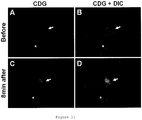
- CDG 2-Deoxy-2-(2-oxo-2H-chromen-7-yl)amino-D-glucose
- CLG 2-deoxy-2-(2-oxo-2H-chromen-7-yl)amino-L-glucose
- Their maximum excitation wavelength (Ex max) and maximum fluorescence wavelength (Em max) are 366.5 nm (Ex max) and 454.5 nm (Em max).
- glucose derivative of the present invention is any of the following compounds:
- QDG 2-Deoxy-2-(2-oxo-1,2-dihydroquinolin-7-yl)amino-D-glucose
- QLG 2-deoxy-2-(2-oxo-1,2-dihydroquinolin-7-yl)amino-L-glucose
- Their maximum excitation wavelength (Ex max) and maximum fluorescence wavelength (Em max) are 353.5 nm (Ex max) and 423.0 nm (Em max).
- glucose derivative of the present invention can include compounds having a substituent at the 3- or 4-position of the coumarin backbone or quinoline backbone in CDG, CLG, QDG, or QLG described above.
- substituent can include halogen, acetyl, formyl, allyl, ethynyl, propenyl, methyl, fluoromethyl, cycloalkyl, phenyl, pyridyl, thiophenyl, pyrrolyl, and furanyl.
- the substituent is particularly preferably methyl or fluoromethyl.
- Examples of the preferred form of the glucose derivative of the present invention can include the following compounds. Table 1 Compound No. Structure Compound No. Structure Compound 101 CDG Compound 201 CLG Compound 102 Compound 202 Compound 103 Compound 203 Compound 104 Compound 204 Compound 105 Compound 205 Compound 106 Compound 206 Compound 107 Compound 207 Compound 108 Compound 208 Table 2 Compound 109 Compound 209 Compound 110 Compound 210 Compound 111 Compound 211 Compound 112 Compound 212 Compound 113 Compound 213 Compound 114 Compound 214 Compound 115 Compound 215 Table 3 Compound 116 Compound 216 Compound 117 Compound 217 Compound 118 Compound 218 Compound 119 Compound 219 Compound 120 Compound 220 Compound 121 Compound 221 Compound 122 Compound 222 Table 4 Compound 123 Compound 223 Compound 124 QDG Compound 224 QLG Compound 125 Compound 225 Compound 126 Compound 226 Compound 127
- the glucose derivative of the present invention is particularly preferably selected from CDG, CLG, QDG, and QLG.
- the glucose derivative of the present invention can be used after being dissolved in an arbitrary solution, for example, a solvent such as water or aqueous dimethyl sulfoxide. Also, the glucose derivative of the present invention is stable even in a solvent or solution used for the imaging of cells or intracellular molecules, particularly, in a buffer solution, and as such, is suitable as an imaging agent.
- the L-glucose derivative of the present invention can be synthesized by using an L-form of sugar as a starting material.
- the binding position of the fluorescent molecular group having a coumarin backbone or a quinoline backbone to glucose is either the 2- or 6-position of glucose, preferably the 2-position.
- the 2-substituted derivative can be synthesized using glucosamine, and the 6-substituted derivative can be synthesized using a 6-deoxy-6-amino-glucose derivative.
- D-Glucosamine or L-glucosamine can be used as the glucosamine.
- Synthesized D-glucosamine or commercially available D-glucosamine can be used as the D-glucosamine.
- the L-glucosamine can be synthesized by a method described in WO2010/16587 (Patent Literature 2) or a method described in WO2012/133688 (Patent Literature 4). The method for synthesizing L-glucosamine described in WO2012/133688 is as follows:
- the 6-deoxy-6-amino-glucose derivative can be synthesized, for example, through 7 stages from glucose as follows, though the synthesis method is not limited thereto.
- the 6-deoxy-6-amino-glucose derivative can be synthesized by converting the hydroxy group at the 1-position of glucose to methyl glycoside, protecting the 4- and 6-positions with benzylidene groups and the 2- and 3-positions with isopropylidene groups, deprotecting the benzylidene groups, converting the 6-position to a toluenesulfonyl group and then to an azide group, and reducing the azide group to an amino group.
- Commercially available D-glucose or commercially available L-glucose can be used as the glucose.
- the D-glucose derivative of the present invention can be synthesized, for example, as follows, though the synthesis method is not limited thereto.
- Compound (A) is synthesized which is a compound having, as a fluorophore, a coumarin backbone substituted at the 7-position with an active group.
- the compound (A) is synthesized by binding a trifluoromethanesulfonyl group to the hydroxy group at the 7-position.
- Compound (B) is synthesized which is a compound having, a fluorophore, a quinolinone backbone substituted at the 7-position with an active group. This compound is synthesized through 2 stages from an aniline derivative substituted at the 3-position with iodine.
- D-glucosamine having protected OH at the 1-, 4-, and 6-positions is synthesized, for example, as follows:
- a synthesis can be performed by condensing the compound A and the D-glucosamine having protected OH at the 1- and 6-positions, for example, by C-N cross-coupling reaction using palladium, so as to bond the compound A to D-glucose via -NH-
- the D-glucose derivative of the present invention having a coumarin backbone or quinoline backbone at the 6-position of glucose can be synthesized by condensing the compound A and the 6-deoxy-6-amino-D-glucose derivative having protected OH at the 1-, 2-, and 3-positions, for example, by C-N cross-coupling reaction using palladium, so as to bond the compound A to D-glucose via -NH-.
- the L-glucose derivative of the present invention can be synthesized in the same way as above by using L-glucosamine instead of D-glucosamine.
- the glucose derivative having a coumarin backbone or quinoline backbone having a substituent at the 3- or 4-position can be synthesized, for example, by a Suzuki coupling method using CDG, CLG, QDG, or QLG synthesized according to a method disclosed herein, or by synthesizing a compound (A) having a coumarin backbone or quinoline backbone having a substituent at the 3- or 4-position and then reacting the compound (A) with glucosamine by use of a direct introduction method by C-H activation, though the synthesis method is not limited thereto.
- a radiolabel can also be added to the glucose derivative of the present invention.
- imaging based on radioactivity for example, PET imaging, can also be achieved.
- such a compound also permits imaging in a dual mode using fluorescence and radiation.
- Examples of the method for adding a radiolabel to the glucose derivative of the present invention can include a method of substituting a hydroxy group of glucose by 18 F, and a method of radiolabeling a substituent in the coumarin backbone or quinoline backbone.
- the position of the hydroxy group to be substituted by 18 F is the 4- or 6-position when the coumarin backbone or quinoline backbone resides at the 2-position of glucose, or is the 2- or 4-position when the coumarin backbone or quinoline backbone resides at the 6-position of glucose.
- Examples of the resulting glucose derivative can include 4- 18 F-CDG, 4- 18 F-CLG, 6- 18 F-CDG, 6- 18 F-CLG, 4- 18 F-QDG, 4- 18 F-QLG, 6- 18 F-QDG, and 6- 18 F-QLG.
- examples of the radiolabeled glucose derivative can include a compound in which the carbon atom of the methyl group bound to the 3- or 4-position of the coumarin backbone or quinoline backbone is substituted by 11 C, and a compound in which one of the fluorine atoms of the trifluoromethyl group bound to the 3- or 4-position of the coumarin backbone or quinoline backbone is substituted by 18 F.
- Examples thereof can include 3-[ 11 C]MCDG, 4-[ 11 C]MCDG, 3-[ 18 F] TFMCDG, 4-[ 18 F] TFMCDG, 3-[ 11 C]MQDG, 4-[ 11 C]MQDG, 3-[ 18 F] TFMQDG, 4-[ 18 F]TFMQDG, 3-[ 11 C]MCLG, 4-[ 11 C]MCLG, 3-[ 18 F]TFMCLG, 4-[ 18 F]TFMCLG, 3-[ 11 C]MQLG, 4-[ 11 C]MQLG, 3-[ 18 F] TFMQLG, and 4-[ 18 F]TFMQLG.
- Such a radiolabeled glucose derivative can be synthesized, for example, as follows, though the synthesis method is not limited thereto.
- the following compound (6- 18 F-CLG) can be synthesized, for example, by steps given below.
- a derivative containing a radioisotope 18 F in a quinoline backbone can be synthesized in the same way as above.
- the following compound 18 F-4-[ 18 F]TFMCDG
- 18 F-4-[ 18 F]TFMCDG can be synthesized, for example, by steps given below.
- a derivative containing a radioisotope 18 F in a quinoline backbone can be synthesized in the same way as above.
- the following compound ( 11 C-3-[ 11 C]MCDG) can be synthesized, for example, by steps given below.
- a derivative containing a radioisotope 11 C in a quinoline backbone can be synthesized in the same way as above.
- a derivative containing a radioisotope 11 C in a quinoline backbone can be synthesized in the same way as above.
- the fluorophore contained in the glucose derivative of the present invention is a fluorophore having a coumarin backbone or quinoline backbone, that is, a fluorophore that emits blue fluorescence. Therefore, this fluorophore generally has a smaller molecular size than a molecule having a fluorescence maximum in the green, red, or near-infrared region and as such, is considered to have a fluorescent group smaller in steric hindrance against the passage through a membrane sugar transport system such as GLUT.
- the glucose derivative of the present invention is featured in that the coumarin backbone or quinoline backbone constituting the fluorophore is directly bound to the glucose backbone via NH. This is considered to provide an advantage that steric hindrance against the passage through a membrane sugar transport system such as GLUT is reduced.
- CDG and QDG blue fluorescent D-glucose derivatives designated as CDG and QDG are taken into cancer cells via GLUT.
- the present inventors have further developed blue fluorescent L-glucose derivative molecules CLG and QLG, which are enantiomers of CDG and QDG, as control compounds.
- CLG, CLG, QDG, and QLG are the first group of blue glucose derivatives that have been quantitatively shown to have a stereoselectivity between D/L-glucose for cellular uptake so as to allow the D form to be taken into cells via GLUT.
- the D-glucose derivative (e.g., CDG or QDG) serving as the glucose derivative of the present invention can emit blue fluorescence as its control compound L-glucose derivative (e.g., CLG or QLG) does. Therefore, the glucose derivative of the present invention is useful equivalently to or more than the green D-glucose derivative 2-NBDG and the green L-glucose derivative 2-NBDLG previously reported by the present inventors (Patent Literature 2) for elucidating the glucose uptake mechanism of cells, and can also be used in combination therewith.
- CDG or QDG can be used in combination with 2-NBDLG
- CLG or QLG can be used in combination with 2-NBDG, for the elucidation of the stereoselective transport mechanism of glucose in such a way that the occurrence of GLUT-mediated D-glucose-like cellular uptake and GLUT-free cellular uptake can be compared at the same time in the same cell selected from various cells, while such combined use can also offer valuable information for understanding the influence of the fluorescent group on the transport mechanism, though how to use the glucose derivative of the present invention is not limited thereto.
- One aspect of the present invention is a method for imaging a cell or an intracellular molecule using the glucose derivative of the present invention.
- Another aspect of the present invention is an imaging agent for imaging a cell or an intracellular molecule, comprising the glucose derivative of the present invention.
- the glucose derivative of the present invention is taken into cells via the membrane sugar transport system of the cells and therefore, can not only image the cells but can image intracellular molecules and organs, for example, cytoplasm and nucleus.
- Yet another aspect of the present invention is an imaging agent for detecting a cancer cell using the glucose derivative of the present invention, preferably the L-glucose derivative, particularly preferably CLG or QLG.
- An alternative aspect of the present invention is a method for imaging a cancer cell using a composition comprising the glucose derivative of the present invention, preferably the L-glucose derivative, particularly preferably CLG or QLG.
- a further alternative aspect of the present invention is a PET imaging agent for detecting a cancer cell using a radiolabeled glucose derivative, which is one form of the glucose derivative of the present invention, preferably a radiolabeled L-glucose derivative, particularly preferably radiolabeled 4-F-CLG, 6-F-CLG, TFMCLG, or TFMQLG.
- a radiolabeled glucose derivative which is one form of the glucose derivative of the present invention, preferably a radiolabeled L-glucose derivative, particularly preferably radiolabeled 4-F-CLG, 6-F-CLG, TFMCLG, or TFMQLG.
- a target cell can be imaged at an individual cell level by contacting a composition comprising the glucose derivative of the present invention (hereinafter, also referred to as the "composition of the present invention” or the “imaging agent of the present invention”) as a reagent with the target cell.
- a composition of the present invention also referred to as the "composition of the present invention” or the “imaging agent of the present invention”
- a cell in a tissue can also be imaged at an individual cell level by contacting the composition of the present invention with the tissue containing the target cell for imaging.
- the glucose derivative of the present invention includes both D-glucose derivatives and L-glucose derivatives.
- the target can be imaged at a cell level depending upon the DL configuration of glucose, thereby making it possible to not only elucidate the functions of the target but discriminate between normal cells and abnormal cells.
- microbes having properties different from mammalian cells in terms of the recognition, transport, and metabolism of glucose associated with the configurations of D and L forms their functions can also be elucidated by imaging at a cell level using the D- or L-glucose derivative of the present invention.
- a target cell is a cancer cell
- a composition comprising the glucose derivative of the present invention, preferably the L-glucose derivative (hereinafter, also referred to as the "composition of the present invention” or the “imaging agent of the present invention") as a reagent with the target cell.
- a cancer cell in a tissue can be detected by contacting the composition of the present invention with the tissue containing the target cell for imaging.
- cancer cells or a tissue containing these cells can be detected by administering the composition of the present invention to a living body for imaging.
- This method is useful as a method for imaging a cancer.
- Cancer imaging can be achieved by PET using a composition comprising the radiolabeled glucose derivative of the present invention, preferably the radiolabeled L-glucose derivative of the present invention.
- the radiolabeled glucose derivative of the present invention can include 4-F-CLG, 6-F-CLG, 4-F-QLG, 6-F-QLG, TFMCDG, TMFCLG, TMFQDG, and TMFQLG and can preferably include 4-F-CLG, 4-F-QLG, TMFCLG, and TMFQLG.
- composition of the present invention includes compositions, comprising the glucose derivative of the present invention, in any form applicable to cells, and may be in any form of a solution, a gel and others without particular limitations as long as the composition is applicable to cells.
- the composition can contain any component without particular limitations as long as the component is suitable for application to cells.
- the glucose derivative of the present invention may be dissolved in a buffer solution or a medium for cell culture for application to cells.
- the target cell to be imaged using the glucose derivative of the present invention is not particularly limited, but can be mammal-derived cells, cells of a microbe such as E. coli or yeast, plant cells, fertilized eggs, or the like. These cells may be in any form including cells isolated from a living body, cells present in a tissue isolated from a living body, cells present in a tissue of a living body, primary cultured cells after isolation from a living body, and established cell lines.
- the cell to be imaged may be a normal cell or may be an abnormal cell (e.g., a cancer cell).
- the detection of the glucose derivative of the present invention taken into the cell can be performed by a commonly-used method for detecting fluorescence.
- this method can be performed as follows: the detection of the glucose derivative present within the cell in the method of the present invention can be performed by measuring the fluorescence of the target cell in advance, subsequently contacting the glucose derivative with the target cell for a given time, washing out the resultant after the given time, measuring again the fluorescence of the target cell, and evaluating an increase in fluorescence intensity relative to the fluorescence intensity of the target cell before the contact.
- the cell may be imaged during the contact with the glucose derivative using an appropriate apparatus, such as a confocal microscope, which is capable of discriminating among the interior of the cell, the cell membrane, and the exterior of the cell.
- an appropriate apparatus such as a confocal microscope
- the fluorescence intensity as an image, the cell intracellularly containing the glucose derivative of the present invention can be imaged, thereby making it possible to detect the cell.
- the evaluation may be conducted by means of total fluorescence intensity or fluorescence intensity distribution exhibited by a large number of cells using a fluorescence plate reader, flow cytometry, or the like.
- glucose derivative of the present invention permits cell detection and/or imaging based on fluorescence (e.g., blue color).
- the glucose derivative of the present invention can be used at the same time with a glucose derivative having a different fluorophore, for example, green fluorescence-emitting 2-[N-(7-nitrobenz-2-oxa-1,3-diazol-4-yl)amino]-2-deoxy-D-glucose (2-NBDG) or 2-[N-(7-nitrobenz-2-oxa-1,3-diazol-4-yl)amino]-2-deoxy-L-glucose (2-NBDLG), and/or red fluorescence-emitting 2-Texas Red-2-amino-2-deoxy-L-glucose (2-TRLG).
- 2-NBDG, 2-NBDLG, and 2-TRLG are described in WO2010/16587 (Patent Literature 2). As a result, two-color or three-color evaluation can be achieved.
- 2-NBDLG has the property of being specifically taken into a cancer and can be used for the detection of the cancer.
- the D-glucose derivative of the present invention e.g., CDG or QDG
- 2-NBDLG or 2-TRLG can be used at the same time with 2-NBDLG or 2-TRLG.
- the concurrent use of the green fluorescent L-glucose derivative 2-NBDLG, which is specifically taken into the cancer cells, with the cell membrane-impermeable red fluorescent 2-TRLG is effective for examining nonspecific uptake ascribable to membrane injury, inflammation, or the like (see WO2012/133688 (Patent Literature 4) filed by the present inventors).
- Patent Literature 4 Patent Literature 4
- a candidate compound therefor is a D-glucose derivative that emits blue fluorescence and is taken into cells via GLUT, to which the D-glucose derivative of the present invention (e.g., CDG or QDG) belong.
- L-glucose derivative of the present invention is a compound in which a particular fluorescence-emitting molecule having a coumarin backbone or quinoline backbone is bound to L-glucose having the property of being not taken into normal cells.
- the uptake of the blue D-glucose derivative CDG or QDG and the blue L-glucose derivative CLG or QLG of the present invention into cancer cells showed D form-dominant uptake similar to the relationship between the green D-glucose derivative 2-NBDG and the green L-glucose derivative 2-NBDLG.
- An experiment using the blue L-glucose derivative of the present invention, for example, CLG confirmed a specific uptake into cancer cells.
- the L-glucose derivative of the present invention can also be used for specific detection of cancer cells.
- types of cancer cells that can be detected are not particularly limited, but include, for example, cancer cells of cancer of the ophthalmologic field such as eyelids and lacrimal glands, cancer of ears such as external ears and middle ears, cancers of nose such as nasal cavities and paranasal cavities, lung cancer, oral cancer, larynx cancer, throat cancer, esophageal cancer, stomach cancer, cancer of digestive organs such as small intestine and large intestine, cancer of the gynecological field including breast cancer, uterine cancer, ovary cancer, and fallopian tube cancer, cancer of reproductive organs, kidney cancer, bladder cancer, prostate cancer, anus cancer, skin cancer, bone cancer, muscular cancer (sarcoma), blood cancer such as leukemia, malignant lymphoma, peripheral and central nervous cancers, and glial cancer.
- cancer of cancer of the ophthalmologic field such as eyelids and lacrimal glands
- cancer of ears such as external ears and middle ears
- cancers of nose such as nasal cavities and par
- the L-glucose derivative of the present invention e.g., CLG or QLG
- an additional fluorescently labeled glucose derivative for example, 2-NBDG, 2-NBDLG, or 2-TRLG, or a combination thereof.
- the method for imaging a cancer according to the present invention and the imaging agent therefor can be used for determination of the presence of tumor cells, the status evaluation thereof, or the discrimination thereof from normal cells with respect to tissues excised at the time of surgery, oral tumors, endoscopically obtained digestive organ tumors, gynecologic tumors such as uterine cervix cancer, or biopsy preparations obtained at the time of biopsy diagnosis, etc., of the lung or various organs.
- detailed cell evaluation at a cell level can be conducted rapidly with a convenient apparatus equipped with fluorescence. This is effective, for example, for determining a guideline for the selection of a treatment method, determining the therapeutic effects of drugs or the like, and determining the appropriate extent of surgery after exposure of an affected part.
- the detection of the L-glucose derivative present within the cancer cell can be performed, for example, by measuring the fluorescence of the target cell in advance, subsequently contacting the fluorescently labeled L-glucose derivative with the target cell for a given time, washing out the resultant after the given time, measuring again the fluorescence of the target cell, and evaluating an increase in fluorescence intensity relative to the fluorescence intensity of the target cell before the contact.
- the fluorescence intensity as an image, the cell intracellularly containing the L-glucose derivative can be imaged, thereby making it possible to detect the cancer cell or the cell at a risk thereof.
- the evaluation may be conducted by means of total fluorescence intensity or fluorescence intensity distribution exhibited by a large number of cells using a fluorescence plate reader, flow cytometry, or the like.
- the L-glucose derivative of the present invention when administered to a vascular vessel such as the vein, permits systemic imaging.
- the L-glucose derivative when locally administered to a tissue to be observed, permits cell imaging.
- a cancer in the case of using the radiolabeled L-glucose derivative of the present invention, a cancer can also be detected by PET.
- the glucose derivative of the present invention is useful for imaging cancer cells and also useful as, for example, an active ingredient for an imaging agent for visualizing cancer cells.
- the L-glucose derivative may be dissolved in a solvent for dissolution thereof (injectable physiological saline, etc.) and provided in the form of a solution, or may be combined with a solvent for dissolution thereof and provided in the form of a kit for preparing a solution by dissolution when used.
- the concentration of the fluorescent L-glucose derivative in the solution may be adjusted in the range of, for example, 1 nM to 100 mM.
- the method for imaging a cancer cell using the L-glucose derivative of the present invention may be used in combination with a method known per se in the art to further improve determination accuracy.
- CDG was synthesized from D-glucosamine hydrochloride as follows:
- D-Glucosamine hydrochloride (10 g, 46.4 mmol) was dissolved in water (100 mL). To the solution, NaHCO 3 (10 g, 119 mmol) was added at room temperature. Then, 2,2,2-trichloroethyl chloroformate (7.7 mL, 55.7 mmol) was added dropwise thereto at 0°C. The mixture was stirred at 0°C for 20 minutes, gradually heated to room temperature, and stirred overnight. The deposited crystals were collected by filtration and washed with water and ether. After overnight drying, the obtained white crystals were recrystallized from warm methanol, and the obtained crystals were collected by filtration to obtain the compound (6.0 g, 36%).
- methyl 4,6-O-benzylidene-2-deoxy-2-(2,2,2-trichloroethoxycarbonylamino)- ⁇ -D-glucopyranoside 1.0 g, 2.19 mmol
- acetic acid 20 mL
- zinc (1.0 g) was added, and the mixture was stirred for 3.5 hours.
- the reaction mixture was filtered, and the solvent was distilled off. The residue was freeze-dried from dioxane to obtain the compound (600 mg, 98%) as white crystals.
- reaction mixture was subjected to extraction with ethyl acetate, and the extract was washed with saturated saline.
- the organic layer was dried over anhydrous sodium sulfate, and the solvent was distilled off.
- the residue was purified using a silica gel column (SiO 2 , 5 g) to obtain the compound (51.2 mg, 35%) as a pale yellow solid.
- the compound methyl 4,6-O-benzylidene-2-deoxy-2-(2-oxo-2H-chromen-7-yl)amino- ⁇ -D-glucopyranoside (40 mg, 94 ⁇ mol) was dissolved in an 80% aqueous acetic acid solution (2 mL), and the solution was heated to 50°C and stirred 2 hours. After the completion of the reaction, the solvent was distilled off. 6 N hydrochloric acid (1.8 mL) was added to the residue, and the mixture was heated to 80°C and stirred for 4 hours. After the completion of the reaction, the solvent was distilled off. The residue was dissolved in dimethylformamide. The solution was purified by reverse-phase HPLC. A fraction containing the compound of interest was freeze-dried to obtain the compound as a pale yellow solid.
- CLG was synthesized in the same way as in CDG using L-glucosamine hydrochloride instead of D-glucosamine hydrochloride.
- the yield was 13.2 mg, and the percent yield was 32%. Also, results of analyzing the obtained compound are as follows.
- QDG was synthesized from D-glucosamine hydrochloride as follows:
- reaction mixture was subjected to extraction with ethyl acetate, and the extract was washed with saturated saline.
- the organic layer was dried over anhydrous sodium sulfate, and the solvent was distilled off.
- the residue was purified using a silica gel column (SiO 2 , 5 g) to obtain the compound (18.3 mg, 84%) as a pale yellow solid.
- the compound methyl 4,6-O-benzylidene-2-deoxy-2-(2-oxo-1,2-dihydroquinolin-7-yl)amino- ⁇ -D-glucopyranoside (46 mg, 108 ⁇ mol) was dissolved in an 80% aqueous acetic acid solution (2 mL), and the solution was heated to 50°C and stirred for 2 hours. After the completion of the reaction, the solvent was distilled off. 6 N hydrochloric acid (1.8 mL) was added to the residue, and the mixture was heated to 80°C and stirred for 4 hours. After the completion of the reaction, the solvent was distilled off. The residue was dissolved in water. The solution was purified by reverse-phase HPLC.
- QLG was synthesized in the same way as in QDG using L-glucosamine hydrochloride instead of D-glucosamine hydrochloride.
- the yield was 23.2 mg, and the percent yield was 33%. Also, results of analyzing the obtained compound are as follows.
- toluene (6.5 mL) was added to activated molecular sieve 4A (pellet), methyl 4,6-0-benzylidene-2-deoxy-2-amino- ⁇ -D-glucopyranoside (166 mg, 0.487 mmol), 2-oxo-4-methyl-2H-chromen-7-yl trifluoromethanesulfonate (100 mg, 0.324 mmol), SPhos Pd G1 (chloro(2-dicyclohexylphosphino-2',6'-dimethoxy-1,1'-biphenyl)[2-(2-aminoethylphenyl)]palladium(II)) (73.9 mg, 97.2 ⁇ mol) (Sigma-Aldrich), SPhos (2-dicyclohexylphosphino-2',6'-dimethoxybiphenyl) (39.9 mg, 97.2 ⁇ mol) (Sigma-Aldrich), and Cs 2
- reaction mixture was subjected to extraction with ethyl acetate, and the extract was washed with saturated saline.
- the organic layer was dried over anhydrous sodium sulfate, and the solvent was distilled off.
- the residue was purified using a silica gel column (SiO 2 , 40 g) to obtain the compound (47 mg, 33%) as a pale yellow solid.
- the compound methyl 4,6-O-benzylidene-2-deoxy-2-(2-oxo-4-methyl-2H-chromen-7-yl)amino- ⁇ -D-glucopyranoside (65 mg, 148 ⁇ mol) was dissolved in an 80% aqueous acetic acid solution (2 mL), and the solution was heated to 50°C and stirred for 2 hours. After the completion of the reaction, the solvent was distilled off. 6 N hydrochloric acid (2 mL) was added to the residue, and the mixture was heated to 80°C and stirred for 12 hours. After the completion of the reaction, the solvent was distilled off. The residue was dissolved in water. The solution was purified by reverse-phase HPLC.
- 4-MCLG can be synthesized in the same way as in 4-MCDG using L-glucosamine.
- the obtained crude product (110 mg, 624 ⁇ mol) was dissolved in pyridine (6.24 mL), and the solution was cooled in ice. Trifluoromethanesulfonic anhydride (115 ⁇ L, 687 ⁇ mol) was added dropwise thereto. Then, the mixture was stirred at room temperature for 1 hour. After the completion of the reaction, the reaction mixture was subjected to extraction with ethyl acetate, and the extract was washed with saturated saline. The organic layer was dried over anhydrous sodium sulfate, and the solvent was distilled off. The residue was purified using a silica gel column (SiO 2 , 20 g) to obtain the compound (120 mg, 62%) as a white solid.
- toluene (0.81 mL) was added to activated molecular sieve 4A (pellet), methyl 4,6-O-benzylidene-2-deoxy-2-amino- ⁇ -D-glucopyranoside (33.2 mg, 97.3 ⁇ mol), 2-oxo-3-methyl-2H-chromen-7-yl trifluoromethanesulfonate (25 mg, 81.1 ⁇ mol), Pd 2 (dba) 3 (tris(dibenzylideneacetone)dipalladium(0)) (7.4 mg, 8.11 ⁇ mol), BrettPhos (2-(dicyclohexylphosphino)-3,6-dimethoxy-2',4',6'-triisopropyl-1,1'-biphenyl) (Sigma-Aldrich) (8.7 mg, 16.2 ⁇ mol), and Cs 2 CO 3 (26.4 mg, 81.1 ⁇ mol), and the mixture was refluxed for 1 hour.
- reaction mixture was subjected to extraction with ethyl acetate, and the extract was washed with saturated saline.
- the organic layer was dried over anhydrous sodium sulfate, and the solvent was distilled off.
- the residue was purified using a silica gel column (SiO 2 , 16 g) to obtain the compound (31.2 mg, 88%) as a pale yellow solid.
- the compound methyl 4,6-O-benzylidene-2-deoxy-2-(2-oxo-3-methyl-2H-chromen-7-yl)amino- ⁇ -D-glucopyranoside (139 mg, 31.6 ⁇ mol) was dissolved in an 80% aqueous acetic acid solution (3 mL), and the solution was heated to 50°C and stirred for 4 hours. After the completion of the reaction, the solvent was distilled off. 6 N hydrochloric acid (3 mL) was added to the residue, and the mixture was heated to 80°C and stirred for 21 hours. After the completion of the reaction, the solvent was distilled off. The residue was dissolved in dimethylformamide. The solution was purified by reverse-phase HPLC. A fraction containing the compound of interest was freeze-dried to obtain the compound as a pale yellow solid.
- 3-MCLG can be synthesized in the same way as in 3-MCDG using L-glucosamine.
- toluene (5.5 mL) was added to activated molecular sieve 4A (pellet), methyl 4,6-0-benzylidene-2-deoxy-2-amino- ⁇ -D-glucopyranoside (141.4 mg, 0.414 mmol), 2-oxo-2H-4-trifluoromethyl-chromen-7-yl trifluoromethanesulfonate (100 mg, 0.276 mmol), SPhos Pd G1 (chloro(2-dicyclohexylphosphino-2',6'-dimethoxy-1,1'-biphenyl)[2-(2-aminoethylphenyl)]palladium(II)) (63 mg, 82.8 ⁇ mol) (Sigma-Aldrich), SPhos (2-dicyclohexylphosphino-2',6'-dimethoxybiphenyl) (34 mg, 82.8 ⁇ mol) (Sigma-Aldrich), and
- reaction mixture was subjected to extraction with ethyl acetate, and the extract was washed with 1 N hydrochloric acid and saturated saline.
- the organic layer was dried over anhydrous sodium sulfate, and the solvent was distilled off.
- the residue was purified using a silica gel column (SiO 2 , 40 g) to obtain the compound (42.3 mg, 31%) as a pale yellow solid.
- the compound methyl 4,6-O-benzylidene-2-deoxy-2-(2-oxo-2H-4-trifluoromethyl-chromen-7-yl)amino- ⁇ -D-glucopyranoside (42 mg, 85.1 ⁇ mol) was dissolved in an 80% aqueous acetic acid solution (2 mL), and the solution was heated to 50°C and stirred for 2 hours. After the completion of the reaction, the solvent was distilled off. 6 N hydrochloric acid (2 mL) was added to the residue, and the mixture was heated to 80°C and stirred for 3 hours. After the completion of the reaction, the solvent was distilled off. The residue was dissolved in water. The solution was purified by reverse-phase HPLC.
- Acetic acid (17.5 mL) was added to 2-oxo-2H-chromen-7-yl trifluoromethanesulfonate (500 mg, 1.71 mmol), sodium trifluoromethanesulfinate (400 mg, 2.56 mmol), and manganese(III) acetate dihydrate (914 mg, 3.41 mmol), and the mixture was stirred at room temperature. After 19 hours, sodium trifluoromethanesulfinate (400 mg, 2.56 mmol) and manganese(III) acetate dihydrate (914 mg, 3.41 mmol) were further added thereto. After another 3 hours, sodium trifluoromethanesulfinate (400 mg, 2.56 mmol) was further added thereto.
- reaction was terminated by the addition of water.
- the reaction mixture was subjected to extraction with ethyl acetate, and the extract was washed with saturated saline.
- the organic layer was dried over anhydrous sodium sulfate, and the solvent was distilled off.
- the residue was purified using a silica gel column (SiO 2 , 40 g) to obtain the compound (258.5 mg, 42%) as a white solid.
- toluene (0.97 mL) was added to activated molecular sieve 4A (pellet), methyl 4,6-0-benzylidene-2-deoxy-2-amino- ⁇ -D-glucopyranoside (39.6 mg, 116 ⁇ mol), 2-oxo-3-trifluoromethyl-2H-chromen-7-yl trifluoromethanesulfonate (35 mg, 96.6 ⁇ mol), Pd 2 (dba) 3 (tris(dibenzylideneacetone)dipalladium(0)) (8.8 mg, 9.66 ⁇ mol), BrettPhos (2-(dicyclohexylphosphino)-3,6-dimethoxy-2',4',6'-triisopropyl-1,1'-biphenyl) (Sigma-Aldrich) (10.4 mg, 19.3 ⁇ mol), and Cs 2 CO 3 (31.4 mg, 96.6 ⁇ mol), and the mixture was refluxed for 1 hour.
- reaction mixture was subjected to extraction with ethyl acetate, and the extract was washed with saturated saline.
- the organic layer was dried over anhydrous sodium sulfate, and the solvent was distilled off.
- the residue was purified using a silica gel column (SiO 2 , 16 g) to obtain the compound (13.8 mg, 29%) as a pale yellow solid.
- the compound methyl 4,6-O-benzylidene-2-deoxy-2-(2-oxo-3-trifluoromethyl-2H-chromen-7-yl)amino- ⁇ -D-glucopyranoside (40 mg, 94 ⁇ mol) was dissolved in an 80% aqueous acetic acid solution (3 mL), and the solution was heated to 50°C and stirred for 6 hours. After the completion of the reaction, the solvent was distilled off. 6 N hydrochloric acid (3 mL) was added to the residue, and the mixture was heated to 80°C and stirred for 5 hours. After the completion of the reaction, the solvent was distilled off. The residue was dissolved in dimethylformamide. The solution was purified by reverse-phase HPLC. A fraction containing the compound of interest was freeze-dried to obtain the compound as a pale yellow solid.
- 6-F-CDG was synthesized as follows:
- methyl 2-deoxy-2-(2-oxo-2H-chromen-7-yl)amino- ⁇ -D-glucopyranoside 50 mg, 148 ⁇ mol
- pyridine 1.5 mL
- triphenylmethyl chloride 206 mg, 740 ⁇ mol
- the reaction mixture was allowed to cool, neutralized with a saturated aqueous solution of sodium bicarbonate, and subjected to extraction with ethyl acetate, and the extract was washed with a saturated aqueous solution of sodium bicarbonate and saturated saline.
- methyl 2-deoxy-2-(2-oxo-2H-chromen-7-yl)amino- ⁇ -D-glucopyranoside (220 mg, 403 ⁇ mol) was dissolved in dichloromethane (10 mL), and the solution was cooled to -17°C.
- N,N-Diethylaminosulfur trifluoride (320 ⁇ L, 2.42 mmol) was added dropwise thereto, and the mixture was heated to room temperature. After 2 hours, the mixture was cooled to -17°C. N,N-Diethylaminosulfur trifluoride (320 ⁇ L, 2.42 mmol) was further added dropwise thereto, and the mixture was heated to room temperature.
- reaction mixture 4 hours after the start of the reaction, the reaction mixture was cooled in ice, and the reaction was terminated by the addition of methanol.
- the reaction mixture was subjected to extraction with ethyl acetate, and the extract was washed with saturated saline.
- the organic layer was dried over anhydrous sodium sulfate, and the solvent was distilled off.
- the residue was purified using a silica gel column (SiO 2 , 40 g) to obtain the compound (104 mg, 47%) as a pale yellow solid.
- Acetic acid (1 mL) and 6 N hydrochloric acid (20 mL) were added to the compound methyl 3,4-di-O-benzoyl-2,6-dideoxy-6-fluoro-2-(2-oxo-2H-chromen-7-yl)amino- ⁇ -D-glucopyranoside (104 mg, 190 ⁇ mol), and the mixture was heated to 80°C and stirred for 4 days. Then, the solvent was distilled off. Trifluoroacetic acid (5 mL) and 6 N hydrochloric acid (10 mL) were added to the residue, and the mixture was heated to 80°C and stirred for 3 days. After the completion of the reaction, the solvent was distilled off. The residue was dissolved in dimethylformamide. The solution was purified by reverse-phase HPLC. A fraction containing the compound of interest was freeze-dried to obtain the compound as a pale yellow solid.
- methyl 2-deoxy-2-(2-oxo-2H-chromen-7-yl)amino- ⁇ -D-glucopyranoside (435 mg, 1.29 mmol) was dissolved by the addition of pyridine (6.5 mL) and dichloromethane (6.5 mL), and the solution was cooled in ice.
- Pivaloyl chloride (565 ⁇ L, 4.59 mmol) was added dropwise thereto, and the mixture was heated to room temperature. After 1 hour, the reaction was terminated by the dropwise addition of a saturated aqueous solution of sodium bicarbonate.
- the reaction mixture was subjected to extraction with ethyl acetate, and the extract was washed with a saturated aqueous solution of sodium bicarbonate and saturated saline.
- the organic layer was dried over anhydrous sodium sulfate, and the solvent was distilled off.
- the residue was purified using a silica gel column (SiO 2 , 40 g) to obtain the compound (431.8 mg, 66%) as a pale yellow solid.
- the compound methyl 2,4-dideoxy-4-fluoro-2-(2-oxo-2H-chromen-7-yl)amino-3,6-di-O-pivaloyl- ⁇ -D-glucopyranoside (170 mg, 335 ⁇ mol) was dissolved in a solution of 4.5 N hydrogen chloride in dioxane (6 mL) and 6 N hydrochloric acid (3 mL), and the solution was heated to 80°C and stirred for 6 hours. After the completion of the reaction, the solvent was distilled off. The residue was dissolved in dimethylformamide. The solution was purified by reverse-phase HPLC. A fraction containing the compound of interest was freeze-dried to obtain the compound as a pale yellow solid.
- Example 11 Influence of cytochalasin B (CB) on increase in fluorescence intensity caused by administration of CDG to mouse insulinoma cell (MIN6)
- a 96-well clear-bottom plate having wells of 8 rows (A to H) and 12 columns (1 to 12) ( ⁇ Clear-PLATE, Greiner Bio-One, BLACK) was used in measurement.
- a culture solution containing MIN6 cells suspended at a density of 60 x 10 4 cells/mL was added dropwise at 10 ⁇ L/well to the central portion of the well, and then, the plate was left standing for 40 minutes in an incubator for fixation.
- the cells were cultured by the addition of a culture solution at 200 ⁇ L/well (6000 cells/well). For layout, the cells were seeded to wells B to H on the third column and wells A to G on the fifth column.
- the MIN6 cells used were cells kindly provided by professor Junichi Miyazaki (Osaka University) and subcultured 6 to 10 times. Half the amount of the culture solution was replaced with a fresh one once every two days.
- the whole amount of the 0.5 mg CDG vial was recovered using 0.73 mL in total of 10% dimethyl sulfoxide (DMSO) and dissolved by addition to 7.0 ml of a KRB solution for image acquisition according to the method described in Non Patent Literature 2.
- DMSO dimethyl sulfoxide
- the solution was applied at a final concentration of 100 ⁇ M to the cells.
- the whole amount of one 0.5 mg 2-NBDG vial was dissolved in 1.83 mL of a KRB solution for data acquisition.
- the solution was applied at a final concentration of 200 ⁇ M to the cells.
- composition of volume used in data acquisition with fluorescence microplate reader Composition of volume used in data acquisition with fluorescence microplate reader
- CDG and 2-NBDG were each independently administered to the wells on the third and fifth columns using an 8-channel pipette such that these derivatives were alternately placed on the same plate.
- the autofluorescence of each well was measured in advance using a fluorescence microplate reader (Flex Station, Molecular Devices, LLC).
- the measurement conditions involved Ex of 367 nm, Em of 455 nm, and cut off of 420 nm on the CDG detector side (blue channel) and Ex of 470 nm, Em of 540 nm, and cut off of 495 nm on the 2-NBDG detector side (green channel).
- the measurement was conducted at Bottom Read, Averaging 3, and Photomultiplier sensitivity high.
- Well Scan Mode was used as a measurement method.
- the Well Scan Mode each independently measures 9 observation regions (diameter: 1.5 mm) divided from one well.
- CB final concentration: 10 ⁇ M
- CB glucose transport inhibitor cytochalasin B
- Example 12 Influence of phloretin (PHT) on increase in fluorescence intensity caused by administration of CDG to MIN6 cell
- PHT final concentration: 150 ⁇ M
- KRB was added to the other wells.
- CDG and 2-NBDG were administered at 37°C for 5 minutes.
- Example 13 Influence of phloretin (PHT) on increase in fluorescence intensity caused by administration of CDG or CLG to MIN6 cell
- MIN6 cells on one 96-well dish were targeted where there were PHT-containing wells and PHT-free wells to which CDG and CLG were alternately administered.
- the administration solutions were concurrently administered using an 8-channel pipette. The dish was left standing for 5 minutes, and then, washout was also performed concurrently for the wells. Change in fluorescence intensity was measured on each observation region. The results are shown in Figure 2C .
- the inhibitory effect of CB (10 ⁇ M) or PHT (150 ⁇ M) on increase in fluorescence intensity caused by the administration of the green fluorescent D-glucose derivative 2-NBDG was confirmed.
- the same MIN6 cell series at 13 days after the start of culture was used, and the green fluorescent D-glucose derivative 2-NBDG was administered thereto to examine the inhibitory effect of CB (10 ⁇ M) or PHT (150 ⁇ M) on increase in fluorescence intensity.
- a KRB solution containing 200 ⁇ M 2-NBDG was administered to the cells, which were then left standing for 5 minute, followed by washout. Increase in the fluorescence intensity of the cells between before and after the administration was measured. Each observation region was measured three times at an excitation light wavelength of 470 nm, a fluorescence acquisition wavelength of 540 nm, and a cutoff filter of 495 nm, and a mean thereof was calculated. The results are shown in Figure 3 .
- the comparison between 2-NBDG and CDG can offer information on the influence of difference in fluorescent group bound to D-glucose on cellular uptake, and important hints about transport pathways.
- Example 14 Influence of excessive D-glucose or excessive L-glucose on increase in fluorescence intensity caused by administration of CDG to MIN6 cell
- the osmotic pressure of a control solution (non-excessive glucose administration group solution) for this experiment was adjusted to 270 mOsm by substituting a portion of NaCl with choline-Cl, in order to equalize charge and osmotic pressure between the control solution and a KRB solution for excessive glucose administration.
- the KRB solution for excessive glucose administration was allowed to contain NaCl and glucose according to the composition given below. In this respect, choline-Cl was added thereto in order to equalize osmotic pressure between this solution and the control solution (non-excessive glucose administration group solution).
- CDG and CLG were placed on different plates for the experiment.
- a CDG solution (or a CLG solution) prepared using each dedicated KRB solution was administered thereto using an 8-channel pipette (37°C, 5 min).
- 8-channel pipette 37°C, 5 min.
- replacement with each dedicated KRB solution was carried out in advance, and the autofluorescence of each well was measured with a fluorescence microplate reader. The time from the start of the replacement to immediately before the administration was approximately 28 minutes.
- the fluorescent solution in each well was temporarily diluted using 200 ⁇ L of the dedicated KRB solution and then diluted using 300 ⁇ L of the usual KRB solution.
- the cellular uptake of D-glucose via GLUT present in the cell membrane requires that D-glucose should bind to a D-glucose-binding site in the GLUT protein.
- the cellular uptake is considered to occur under the mechanism where: D-glucose binds to a D-glucose-binding site in the GLUT protein initially in an outward-open state in the cell membrane, and this binding alters the three-dimensional structure of GLUT to an inward-open state; subsequently, D-glucose is released from the binding site in GLUT and eventually transported into the cell from the exterior because the D-glucose faces the interior of the cell.
- D-glucose occupies D-glucose-binding sites in GLUT.
- the transport rate into the cell does not exceed the fixed level and reaches a plateau.
- CDG binds to a D-glucose-binding site in GLUT prior to passage through GLUT.
- D-glucose occupies D-glucose-binding sites in GLUT.
- Example 15 Influence of cytochalasin B (CB) or phloretin (PHT) on increase in fluorescence intensity caused by administration of QDG to MIN6 cell
- the whole amount of the 0.5 mg QDG vial was recovered using 0.71 mL in total of 10% dimethyl sulfoxide (DMSO) and dissolved by addition to 7.1 ml of KRB solution for image acquisition according to the method described in Non Patent Literature 2.
- DMSO dimethyl sulfoxide
- the solution was applied at a final concentration of 100 ⁇ M to the cells.
- Example 16 Imaging of tumor cell mass consisting of mouse insulinoma cells (MIN6) using CDG/2-NBDLG/2-TRLG
- a fluorescent mixed solution containing 100 ⁇ M CDG, 100 ⁇ M 2-NBDLG, and 20 ⁇ M 2-TRLG (CDG/2-NBDLG/2-TRLG) was administered at 37°C for 3 minutes to MIN6 cell masses (spheroids) at culture day 17, and fluorescent imaging was conducted.
- the MIN6 cells used were cells kindly provided by professor Junichi Miyazaki (Osaka University) and subcultured 6 to 10 times. Half the amount of the culture solution was replaced with a fresh one once every two days.
- the whole amount of the 0.5 mg CDG vial was recovered using 0.73 mL in total of 10% DMSO and dissolved by addition to 7.0 ml of a KRB solution for image acquisition according to the method described in Non Patent Literature 2.
- the whole amount of one 0.5 mg 2-NBDLG vial was dissolved in 7.3 mL of a KRB solution for image acquisition.
- the CDG solution and the 2-NBDLG solution were mixed at a ratio of 1:1.
- the whole amount of the 0.2 mg 2-TRLG vial was recovered using 130 ⁇ L in total of DMSO and mixed with 13 ml of the CDG + 2-NBDLG mixed solution according to the method described in Non Patent Literature 2 (final concentration: 100 ⁇ M CDG + 100 ⁇ M 2-NBDLG + 20 ⁇ M 2-TRLG mixed solution).
- the KRB solution for image acquisition used was a solution having the following composition.
- the glass cover slip with the cultured MIN6 cells was transferred into the KRB solution for image acquisition in a perfusion chamber loaded on a stage of a fluorescence microscope, followed by measurement.
- an aluminum heating control platform Warner Instruments
- the KRB solution for image acquisition was warmed in advance in an aluminum syringe heater and supplied to the perfusion chamber through hydrostatic pressure.
- the perfusion rate was adjusted to 1.3 ⁇ 0.2 mL/min using a flow rate adjuster.
- the solution was rewarmed via an inline heater so that the actually measured temperature of the perfusate in an observation region in the chamber was adjusted to 36 ⁇ 1°C.
- the CDG/2-NBDLG/2-TRLG mixed solution was able to be supplied in the same way as above and was switched with the KRB solution for image acquisition using an electromagnetic valve capable of controlling the opening and closing of the channel.
- the solution was removed using a vacuum pump capable of controlling suction pressure.
- the fluorescence microscope used was NIKON ECLIPS-Ti, and images were acquired with a CCD camera Retiga 2000R.
- CDG, 2-NBDLG, and 2-TRLG were each detected using the following filter cassette.
- the objective lens used in this method was x60 oil lens (Plan Fluor 60x/0.50-1.25 Oil).
- the images were acquired at 1600 x 1200 pixels and a depth of 12 bit.
- Figures 6 and 7 The results of administering the 100 ⁇ M CDG + 100 ⁇ M 2-NBDLG + 20 ⁇ M 2-TRLG mixed solution for 3 minutes to the MIN6 cell masses that formed spheroids are shown in Figures 6 and 7 .
- Figure 6 shows fluorescence microscopic images taken 6 minutes after the start of washout of the mixed solution.
- Figures 6A, 6B, and 6C show blue fluorescence-emitting CDG, green fluorescence-emitting 2-NBDLG, and red fluorescence-emitting 2-TRLG, respectively, taken into cells.
- Figure 6D is a differential interference contrast (DIC) image.
- DIC differential interference contrast
- Figure 7 is a superimposed image of Figures 6A and 6B .
- the uptake of CDG is expressed in red color instead of blue color in order to facilitate visualizing the localization of CDG and 2-NBDLG.
- CDG high density polyethylene glycol
- 2-NBDLG medium density polyethylene glycol
- the results described above indicate the possibility that the presence of cells that take less 2-NBDLG therein, but take CDG therein can be detected by the concurrent administration of CDG and 2-NBDLG to cell masses.
- Example 17 Imaging of tumor cell mass consisting of mouse insulinoma cells (MIN6) using CLG/2-NBDLG/2-TRLG
- Fluorescent imaging was conducted in the same way as in Example 16 except that a CLG/2-NBDLG/2-TRLG mixed solution was used instead of the CDG/2-NBDLG/2-TRLG mixed solution for MIN6 cell masses at culture day 15.
- Figure 8 shows fluorescence microscopic images taken 6 minutes after the start of washout of the 100 ⁇ M CLG + 100 ⁇ M 2-NBDLG + 20 ⁇ M 2-TRLG mixed solution after 3-minute administration of the mixed solution to MIN6 cell masses that formed spheroids.
- Figures 8A, 8B, and 8C show blue fluorescence-emitting CLG, green fluorescence-emitting 2-NBDLG, and red fluorescence-emitting 2-TRLG, respectively, taken into cancer cells.
- Figure 8D is a differential interference contrast (DIC) image. Unlike the case of CDG, all the cells that strongly took CLG therein also exhibited the uptake of 2-NBDLG. Like CDG, CLG was also strongly taken not only into the cytoplasm but into the nucleus.
- DIC differential interference contrast
- a solution having the following composition was used for fluorescence image acquisition.
- Neuronal cells of the substantia nigra pars reticulate of the midbrain of a 21-day-old mouse were acutely isolated according to the method described in the previous reports of the inventors ( WO2010/16587 (Patent Literature 2)) and ( WO2012/133688 (Patent Literature 4)) and attached to each glass cover slip coated with poly-L-lysine. Then, the glass cover slip was transferred into the HEPES solution for image acquisition in a perfusion chamber loaded on a stage of a fluorescence microscope, followed by observation.
- an aluminum heating control platform Warner Instruments
- the HEPES solution for image acquisition was warmed in advance in an aluminum syringe heater and supplied to the perfusion chamber through hydrostatic pressure.
- the perfusion rate was adjusted to 1.3 ⁇ 0.2 mL/min using a flow rate adjuster.
- the solution was rewarmed via an inline heater so that the actually measured temperature of the perfusate in an observation region in the chamber was adjusted to 32.5 ⁇ 1°C.
- the CLG/2-TRLG mixed solution was able to be supplied in the same way as above and was switched with the HEPES solution for image acquisition using an electromagnetic valve capable of controlling the opening and closing of the channel.
- the solution was removed using a vacuum pump capable of controlling suction pressure.
- the fluorescence microscope used was NIKON ECLIPS-Ti, and images were acquired with a CCD camera Retiga 2000R.
- the objective lens used in this method was x20 dry lens (Nikon Plan S Fluor). The images were acquired at 1600 x 1200 pixels.
- Figure 9 shows the case where change in fluorescence intensity exhibited by cells between before and after 3-minute administration of the CLG + 2-TRLG mixed solution to acutely isolated normal neuronal cells (arrow) was observed in the wavelength region of blue fluorescence (blue channel) emitted by CLG. The process of background subtraction was conducted both before and after the administration.
- Figure 9A shows the observed autofluorescence exhibited by the normal neuronal cells (arrow) before the administration of the mixed solution.
- Figure 9B shows a differential interference contrast (DIC) image superimposed on the image of Figure 9A for further understanding.
- DIC differential interference contrast
- Figure 9C is a fluorescence microscopic image taken 8 minutes after the start of washout of the mixed solution.
- Figure 9D is a DIC image superimposed on the image of Figure 9C .
- blue fluorescence-emitting CLG was strongly taken into the cell debris (*) in a nonspecific manner, whereas such uptake was not observed in the normal neuronal cells (arrow).
- Figure 10 shows that change in fluorescence between before and after the administration of the mixed solution in Figure 9 , as observed at the same time as in the wavelength region of red fluorescence (red channel) emitted by 2-TRLG.
- Figure 10A shows an autofluorescence image taken before the administration of the mixed solution.
- Figure 10B shows a differential interference contrast (DIC) image superimposed on the image of Figure 10A in order to facilitate visualizing the positions of cells. The autofluorescence of the cells was very small in the red fluorescence wavelength region.
- DIC differential interference contrast
- Intense red color at the central portion of the image resulted from the leakage of a portion of irradiation light to the detector side through a fluorescence filter because the red fluorescence was photographed with sensitivity enhanced for the purpose of detecting weak fluorescence (see Dichroic mirror and Emission wavelengths of the red channel).
- the image is shown as it is without shading correction.
- Example 17 the L-glucose derivative CLG was strongly taken into the spheroid of the mouse insulinoma cells (MIN6), which are cancer cells.
- Example 19 Application of CDG to acutely isolated normal neuronal cell
- Figure 11 shows the case where change in fluorescence intensity exhibited by cells between before and after 3-minute administration of the CDG + 2-TRLG mixed solution to acutely isolated normal neuronal cells (arrow) was observed in the wavelength region of blue fluorescence (blue channel) emitted by CDG. The process of background subtraction was conducted both before and after the administration.
- Figure 11A shows the observed autofluorescence exhibited by the normal neuronal cells (arrow) before the administration of the mixed solution.
- Figure 11B shows a differential interference contrast (DIC) image superimposed on the image of Figure 11A for further understanding. * depicts the debris of killed cells.
- Figure 11C is a fluorescence microscopic image taken 8 minutes after the start of washout of the mixed solution.
- Figure 11D is a DIC image superimposed on the image of Figure 11C .
- blue fluorescence-emitting CDG was taken into the normal neuronal cells (arrow).
- such uptake was not observed in the cell debris (*).
- Figure 12 shows that change in fluorescence between before and after the administration of the mixed solution in Figure 11 , as observed at the same time in the wavelength region of red fluorescence (red channel) emitted by 2-TRLG.
- Figure 12A shows an autofluorescence image taken before the administration of the mixed solution.
- Figure 12B shows a differential interference contrast (DIC) image superimposed on the image of Figure 12A in order to facilitate visualizing the positions of cells. The autofluorescence of the cells was very small in the red fluorescence wavelength region.
- DIC differential interference contrast
- the present invention provides a novel glucose derivative.
- the present invention also provides an imaging agent and an imaging method for a cell using the glucose derivative.
Landscapes
- Health & Medical Sciences (AREA)
- Life Sciences & Earth Sciences (AREA)
- Chemical & Material Sciences (AREA)
- Engineering & Computer Science (AREA)
- General Health & Medical Sciences (AREA)
- Molecular Biology (AREA)
- Organic Chemistry (AREA)
- Biomedical Technology (AREA)
- Biochemistry (AREA)
- Biotechnology (AREA)
- Animal Behavior & Ethology (AREA)
- Veterinary Medicine (AREA)
- Public Health (AREA)
- Epidemiology (AREA)
- Hematology (AREA)
- Immunology (AREA)
- Genetics & Genomics (AREA)
- Urology & Nephrology (AREA)
- Medicinal Chemistry (AREA)
- Physics & Mathematics (AREA)
- Microbiology (AREA)
- Food Science & Technology (AREA)
- Cell Biology (AREA)
- Analytical Chemistry (AREA)
- General Physics & Mathematics (AREA)
- Pathology (AREA)
- Proteomics, Peptides & Aminoacids (AREA)
- Optics & Photonics (AREA)
- Pharmacology & Pharmacy (AREA)
- Hospice & Palliative Care (AREA)
- Oncology (AREA)
- Diabetes (AREA)
- Plural Heterocyclic Compounds (AREA)
- Saccharide Compounds (AREA)
- Medicines Containing Antibodies Or Antigens For Use As Internal Diagnostic Agents (AREA)
- Pharmaceuticals Containing Other Organic And Inorganic Compounds (AREA)
Claims (13)
- Dérivé de glucose qui est un composé représenté par la formule (1) suivante :
 dans laquelle X-Y-Z représente O-C=O, NH-C=O, NR3-C=O, ou N=C-OR4, où R3 représente alkyle en C1-C5, et R4 représente alkyle en C1-C5 ;R1 et R2 représentent chacun indépendamment un groupe choisi dans le groupe consistant en hydrogène, halogène, alkyle en C1-C5, alcényle en C2-C5, alcynyle en C2-C5, halogénoalkyle en C1-C5, alkylamino en C1-C5, cycloalkyle, phényle, pyridyle, thiophényle, pyrrolyle et furanyle ; etG représente un groupe choisi parmi les formules suivantes (G1) à (G4) :

- Dérivé de glucose selon la revendication 1, qui est un composé représenté par la formule suivante (2) :

ou sel de celui-ci. - Dérivé de glucose selon la revendication 1, qui est un composé représenté par la formule suivante (3) :

ou sel de celui-ci. - Dérivé de glucose selon la revendication 1, qui est un composé représenté par la formule suivante (4) :
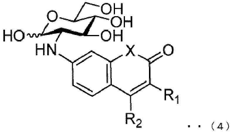
ou sel de celui-ci. - Dérivé de glucose selon la revendication 1, qui est un composé représenté par la formule suivante (5) :

ou sel de celui-ci. - Dérivé de glucose selon la revendication 1, qui est choisi dans le groupe consistant en les composés suivants :
2-désoxy-2-(2-oxo-2H-chromèn-7-yl)amino-D-glucose (CDG), 2-désoxy-2-(2-oxo-1,2-dihydroquinolin-7-yl)amino-D-glucose (QDG), 2-désoxy-2-(2-oxo-2H-3-méthyl-chromèn-7-yl)amino-D-glucose (3-MCDG), 2-désoxy-2-(2-oxo-2H-4-méthyl-chromèn-7-yl)amino-D-glucose (4-MCDG), 2-désoxy-2-(2-oxo-2H-3-trifluorométhyl-chromèn-7-yl)amino-D-glucose (3-TFMCDG), 2-désoxy-2-(2-oxo-2H-4-trifluorométhyl-chromèn-7-yl)amino-D-glucose (4-TFMCDG), 2-désoxy-2-(2-oxo-1,2-dihydro-3-méthyl-quinolin-7-yl)amino-D-glucose (3-MQDG), 2-désoxy-2-(2-oxo-1,2-dihydro-4-méthyl-quinolin-7-yl)amino-D-glucose (4-MQDG), 2-désoxy-2-(2-oxo-1,2-dihydro-3-trifluorométhyl-quinolin-7-yl)amino-D-glucose (3-TFMQDG), 2-désoxy-2-(2-oxo-1,2-dihydro-4-trifluorométhyl-quinolin-7-yl)amino-D-glucose (4-TFMQDG), 2-désoxy-2-(2-oxo-2H-chromèn-7-yl)amino-L-glucose (CLG), 2-désoxy-2-(2-oxo-1,2-dihydroquinolin-7-yl)amino-L-glucose (QLG), 2-désoxy-2-(2-oxo-2H-3-méthyl-chromèn-7-yl)amino-L-glucose (3-MCLG), 2-désoxy-2-(2-oxo-2H-4-méthyl-chromèn-7-yl)amino-L-glucose (4-MCLG), 2-désoxy-2-(2-oxo-2H-3-trifluorométhyl-chromèn-7-yl)amino-L-glucose (3-TFMCLG), 2-désoxy-2-(2-oxo-2H-4-trifluorométhyl-chromèn-7-yl)amino-L-glucose (4-TFMCLG), 2-désoxy-2-(2-oxo-1,2-dihydro-3-méthyl-quinolin-7-yl)amino-L-glucose (3-MQLG), 2-désoxy-2-(2-oxo-1,2-dihydro-4-méthyl-quinolin-7-yl)amino-L-glucose (4-MQLG), 2-désoxy-2-(2-oxo-1,2-dihydro-3-trifluorométhyl-quinolin-7-yl)amino-L-glucose (3-TFMQLG), et 2-désoxy-2-(2-oxo-1,2-dihydro-4-trifluorométhyl-quinolin-7-yl)amino-L-glucose (4-TFMQLG). - Dérivé de glucose radiomarqué comprenant un dérivé de glucose selon l'une quelconque des revendications 1 à 6, dans lequel un groupe hydroxy à l'une quelconque des positions 2, 4 et 6 du glucose est substitué par 18F.
- Dérivé de glucose selon la revendication 1, qui est sous une forme radiomarquée choisie parmi
2-désoxy-2-(2-oxo-2H-3-[11C]méthyl-chromèn-7-yl)amino-D-glucose (3-[11C]MCDG), 2-désoxy-2-(2-oxo-2H-4-[11C]méthyl-chromèn-7-yl)amino-D-glucose (4-[11C]MCDG), 2-désoxy-2-(2-oxo-2H-3-[18F]fluorodifluorométhyl-chromèn-7-yl)amino-D-glucose (3-[18F]TFMCDG), 2-désoxy-2-(2-oxo-2H-4-[18F] fluorodifluorométhyl-chromèn-7-yl)amino-D-glucose (4-[18F]TFMCDG), 2-désoxy-2-(2-oxo-1,2-dihydro-3-[11C]méthyl-quinolin-7-yl)amino-D-glucose (3-[11C]MQDG), 2-désoxy-2-(2-oxo-1,2-dihydro-4-[11C]méthyl-quinolin-7-yl)amino-D-glucose (4-[11C]MQDG), 2-désoxy-2-(2-oxo-1,2-dihydro-3-[18F]fluorodifluorométhyl-quinolin-7-yl)amino-D-glucose (3-[18F]TFMQDG), 2-désoxy-2-(2-oxo-1,2-dihydro-4-[18F]fluorodifluorométhyl-quinolin-7-yl)amino-D-glucose (4-[18F]TFMQDG), 2-désoxy-2-(2-oxo-2H-3-[11C]méthyl-chromèn-7-yl)amino-L-glucose (3-[11C]MCLG), 2-désoxy-2-(2-oxo-2H-4-[11C]méthyl-chromèn-7-yl)amino-L-glucose (4-[11C]MCLG), 2-désoxy-2-(2-oxo-2H-3-[18F]fluorodifluorométhyl-chromèn-7-yl)amino-L-glucose (3-[18F]TFMCLG), 2-désoxy-2-(2-oxo-2H-4-[18F]fluorodifluorométhyl-chromèn-7-yl)amino-L-glucose (4-[18F] TFMCLG), 2-désoxy-2-(2-oxo-1,2-dihydro-3-[11C]méthyl-quinolin-7-yl)amino-L-glucose(3-[11C]MQLG), 2-désoxy-2-(2-oxo-1,2-dihydro-4-[11C]méthyl-quinolin-7-yl)amino-L-glucose (4-[11C]MQLG), 2-désoxy-2-(2-oxo-1,2-dihydro-3-[18F]fluorodifluoro-méthyl-quinolin-7-yl)amino-L-glucose (3-[18F]TFMQLG), et 2-désoxy-2- (2-oxo-1,2-dihydro-4-[18F]fluorodifluorométhyl-quinolin-7-yl)amino-L-glucose (4-[18F] TFMQLG). - Composition pour l'imagerie d'une cellule cible, comprenant un dérivé de glucose selon l'une quelconque des revendications 1 à 8.
- Méthode pour l'imagerie d'une cellule cible, comprenant les étapes suivantes :a. mise en contact d'une composition contenant un dérivé de glucose selon l'une quelconque des revendications 1 à 8 avec la cellule cible ; etb. détection de la fluorescence émise par le dérivé de glucose présent à l'intérieur de la cellule cible.
- Méthode d'imagerie selon la revendication 10, dans laquelle la composition à l'étape a comprend en outre un dérivé de glucose additionnel marqué par fluorescence et l'étape b est l'étape de détection d'au moins l'un des dérivés de glucose présent à l'intérieur de la cellule cible.
- Méthode pour l'imagerie d'un cancer ou d'une cellule cancéreuse, comprenant les étapes suivantes :a. mise en contact d'une composition contenant un dérivé de glucose avec une cellule cible ; etb. détection du dérivé de glucose présent à l'intérieur de la cellule cible,dans laquelle le dérivé de glucose est un composé représenté par les formules suivantes (2), (3), (4) ou (5) ou un sel de celui-ci :
 dans laquelle X représente O, NH, ou NR3, où R3 représente alkyle en C1-C5 ; et R1 et R2 représentent chacun indépendamment un groupe choisi dans le groupe consistant en hydrogène, halogène, alkyle en C1-C5, alcényle en C2-C5, alcynyle en C2-C5, halogénoalkyle en C1-C5, alkylamino en C1-C5, cycloalkyle, phényle, pyridyle, thiophényle, pyrrolyle et furanyle ;
 dans laquelle R1 et R2 représentent chacun indépendamment un groupe choisi dans le groupe consistant en hydrogène, halogène, alkyle en C1-C5, alcényle en C2-C5, alcynyle en C2-C5, halogénoalkyle en C1-C5, alkylamino en C1-C5, cycloalkyle, phényle, pyridyle, thiophényle, pyrrolyle et furanyle ; et R4 représente alkyle en C1-C5 ;
 dans laquelle X représente O, NH, ou NR3, où R3 représente alkyle en C1-C5 ; et R1 et R2 représentent chacun indépendamment un groupe choisi dans le groupe consistant en hydrogène, halogène, alkyle en C1-C5, alcényle en C2-C5, alcynyle en C2-C5, halogénoalkyle en C1-C5, alkylamino en C1-C5, cycloalkyle, phényle, pyridyle, thiophényle, pyrrolyle et furanyle ; ou
 dans laquelle R1 et R2 représentent chacun indépendamment un groupe choisi dans le groupe consistant en hydrogène, halogène, alkyle en C1-C5, alcényle en C2-C5, alcynyle en C2-C5, halogénoalkyle en C1-C5, alkylamino en C1-C5, cycloalkyle, phényle, pyridyle, thiophényle, pyrrolyle et furanyle ; et R4 représente alkyle en C1-C5.
 - Agent d'imagerie pour l'imagerie d'une cellule cancéreuse cible, comprenant un dérivé de glucose,
dans lequel le dérivé de glucose est un composé représenté par les formules suivantes (2), (3), (4) ou (5) ou un sel de celui-ci : dans laquelle X représente O, NH, ou NR3, où R3 représente alkyle en C1-C5 ; et R1 et R2 représentent chacun indépendamment un groupe choisi dans le groupe consistant en hydrogène, halogène, alkyle en C1-C5, alcényle en C2-C5, alcynyle en C2-C5, halogénoalkyle en C1-C5, alkylamino en C1-C5, cycloalkyle, phényle, pyridyle, thiophényle, pyrrolyle et furanyle ;
 dans laquelle R1 et R2 représentent chacun indépendamment un groupe choisi dans le groupe consistant en hydrogène, halogène, alkyle en C1-C5, alcényle en C2-C5, alcynyle en C2-C5, halogénoalkyle en C1-C5, alkylamino en C1-C5, cycloalkyle, phényle, pyridyle, thiophényle, pyrrolyle et furanyle ; et R4 représente alkyle en C1-C5 ;
 dans laquelle X représente O, NH, ou NR3, où R3 représente alkyle en C1-C5 ; et R1 et R2 représentent chacun indépendamment un groupe choisi dans le groupe consistant en hydrogène, halogène, alkyle en C1-C5, alcényle en C2-C5, alcynyle en C2-C5, halogénoalkyle en C1-C5, alkylamino en C1-C5, cycloalkyle, phényle, pyridyle, thiophényle, pyrrolyle et furanyle ; ou
 dans laquelle R1 et R2 représentent chacun indépendamment un groupe choisi dans le groupe consistant en hydrogène, halogène, alkyle en C1-C5, alcényle en C2-C5, alcynyle en C2-C5, halogénoalkyle en C1-C5, alkylamino en C1-C5, cycloalkyle, phényle, pyridyle, thiophényle, pyrrolyle et furanyle ; et R4 représente alkyle en C1-C5.
Applications Claiming Priority (2)
| Application Number | Priority Date | Filing Date | Title |
|---|---|---|---|
| JP2014079071 | 2014-04-08 | ||
| PCT/JP2015/060783 WO2015156264A1 (fr) | 2014-04-08 | 2015-04-06 | Nouveau dérivé de glucose, et procédé d'imagerie de cellule et agent d'imagerie utilisant ledit dérivé |
Publications (3)
| Publication Number | Publication Date |
|---|---|
| EP3130596A1 EP3130596A1 (fr) | 2017-02-15 |
| EP3130596A4 EP3130596A4 (fr) | 2017-12-13 |
| EP3130596B1 true EP3130596B1 (fr) | 2021-01-20 |
Family
ID=54287840
Family Applications (1)
| Application Number | Title | Priority Date | Filing Date |
|---|---|---|---|
| EP15776849.0A Active EP3130596B1 (fr) | 2014-04-08 | 2015-04-06 | Dérivé de glucose, et procédé d'imagerie de cellule et agent d'imagerie utilisant ledit dérivé |
Country Status (6)
| Country | Link |
|---|---|
| US (2) | US10001487B2 (fr) |
| EP (1) | EP3130596B1 (fr) |
| JP (1) | JP6566348B2 (fr) |
| KR (1) | KR20160143774A (fr) |
| CN (1) | CN106459126B (fr) |
| WO (1) | WO2015156264A1 (fr) |
Cited By (1)
| Publication number | Priority date | Publication date | Assignee | Title |
|---|---|---|---|---|
| WO2023058608A1 (fr) | 2021-10-06 | 2023-04-13 | オルバイオ株式会社 | Dérivés de glucose et agent anticancéreux les utilisant |
Families Citing this family (4)
| Publication number | Priority date | Publication date | Assignee | Title |
|---|---|---|---|---|
| JP6566348B2 (ja) * | 2014-04-08 | 2019-08-28 | 国立大学法人弘前大学 | 新規なグルコース誘導体、および該誘導体を用いた細胞イメージング方法およびイメージング剤 |
| EP3435136B1 (fr) | 2016-03-31 | 2021-08-04 | Hirosaki University | Système d`acquisition d`image multi-surfaces |
| CN109369738B (zh) * | 2018-11-16 | 2021-03-30 | 江苏美迪克化学品有限公司 | 一种磺达肝癸钠单糖中间体的制备方法 |
| CN111411137B (zh) * | 2020-02-29 | 2023-04-25 | 浙江农林大学 | 一种脂肪酶催化在线合成8-甲氧基香豆素-3-羧酸-6′-o-d-葡萄糖酯的方法 |
Family Cites Families (12)
| Publication number | Priority date | Publication date | Assignee | Title |
|---|---|---|---|---|
| EP1049807B1 (fr) | 1998-01-22 | 2003-05-07 | Luminex Corporation | Microparticules emettant des signaux fluorescents multiples |
| WO2003059149A2 (fr) | 2001-12-21 | 2003-07-24 | Threshold Pharmaceuticals, Inc. | Procede d'imagerie du cancer |
| JP2006249243A (ja) * | 2005-03-10 | 2006-09-21 | Fuji Photo Film Co Ltd | 静電インクジェットインク組成物およびその製造方法 |
| US20060280687A1 (en) * | 2005-03-18 | 2006-12-14 | Zhen Cheng | Compounds for tissue imaging and methods of use thereof |
| WO2008029281A2 (fr) | 2006-02-10 | 2008-03-13 | Invitrogen Corporation | Marquage et détection de protéines modifiées post-traductionnellement |
| JP2009531289A (ja) * | 2006-02-10 | 2009-09-03 | ライフ テクノロジーズ コーポレーション | オリゴ糖によるタンパク質の修飾及び標識方法 |
| JP2010529421A (ja) * | 2007-05-16 | 2010-08-26 | バックマン・ラボラトリーズ・インターナショナル・インコーポレーテッド | パルプ及び繊維中の有機夾雑物を検出する方法 |
| WO2010016587A1 (fr) * | 2008-08-08 | 2010-02-11 | 国立大学法人弘前大学 | Procédé d'évaluation de l'incorporation spécifique de d-glucose dans des cellules |
| CA2856812A1 (fr) | 2010-11-28 | 2012-05-31 | Metasignal Therapeutics Inc. | Inhibiteurs d'anhydrase carbonique presentant une activite antimetastatique |
| EP2703495B1 (fr) * | 2011-03-31 | 2018-05-09 | Hirosaki University | Procédé pour détecter une cellule cancéreuse en utilisant un dérivé de l-glucose marqué par fluorescence, et agent d'imagerie de cellules cancéreuses comprenant un dérivé de l-glucose marqué par fluorescence |
| US10288604B2 (en) * | 2012-10-03 | 2019-05-14 | Hirosaki University | Method for imaging cell using fluorescence-labeled sugar derivative having coumarin derivative bound thereto, and imaging agent |
| JP6566348B2 (ja) * | 2014-04-08 | 2019-08-28 | 国立大学法人弘前大学 | 新規なグルコース誘導体、および該誘導体を用いた細胞イメージング方法およびイメージング剤 |
-
2015
- 2015-04-06 JP JP2015078062A patent/JP6566348B2/ja active Active
- 2015-04-06 US US15/302,782 patent/US10001487B2/en active Active
- 2015-04-06 CN CN201580030346.9A patent/CN106459126B/zh active Active
- 2015-04-06 WO PCT/JP2015/060783 patent/WO2015156264A1/fr active Application Filing
- 2015-04-06 KR KR1020167031171A patent/KR20160143774A/ko unknown
- 2015-04-06 EP EP15776849.0A patent/EP3130596B1/fr active Active
-
2018
- 2018-04-19 US US15/956,897 patent/US10509041B2/en active Active
Non-Patent Citations (1)
| Title |
|---|
| None * |
Cited By (1)
| Publication number | Priority date | Publication date | Assignee | Title |
|---|---|---|---|---|
| WO2023058608A1 (fr) | 2021-10-06 | 2023-04-13 | オルバイオ株式会社 | Dérivés de glucose et agent anticancéreux les utilisant |
Also Published As
| Publication number | Publication date |
|---|---|
| CN106459126A (zh) | 2017-02-22 |
| KR20160143774A (ko) | 2016-12-14 |
| JP6566348B2 (ja) | 2019-08-28 |
| US20180238899A1 (en) | 2018-08-23 |
| US20170122955A1 (en) | 2017-05-04 |
| JP2015205870A (ja) | 2015-11-19 |
| EP3130596A1 (fr) | 2017-02-15 |
| CN106459126B (zh) | 2019-10-01 |
| US10001487B2 (en) | 2018-06-19 |
| US10509041B2 (en) | 2019-12-17 |
| EP3130596A4 (fr) | 2017-12-13 |
| WO2015156264A1 (fr) | 2015-10-15 |
Similar Documents
| Publication | Publication Date | Title |
|---|---|---|
| US10509041B2 (en) | Glucose derivative, and cell imaging method and imaging agent using said derivative | |
| Kamiya et al. | An enzymatically activated fluorescence probe for targeted tumor imaging | |
| US7572923B2 (en) | Indirubin derivatives having anticancer property against human cancer cell line | |
| JP6019500B2 (ja) | 蛍光標識されたl−グルコース誘導体を用いたがん細胞を検出するための方法及び該誘導体を含むがん細胞のイメージング剤 | |
| US9801960B2 (en) | Probe for a biological specimen and labelling method and screening method using the probe | |
| CN105073761A (zh) | 用于体外和体内成像和检测的取代的硅杂蒽阳离子红至近红外荧光染料 | |
| Li et al. | Development of photocontrolled BRD4 PROTACs for tongue squamous cell carcinoma (TSCC) | |
| CN112694431B (zh) | 超敏感荧光探针检验细菌中硝基还原酶及在细菌感染应用 | |
| Pewklang et al. | Aza-BODIPY probe for selective visualization of cyclooxygenase-2 in cancer cells | |
| WO2014058535A1 (fr) | Sondes de ciblage lysosomal | |
| Bao et al. | NIR absorbing DICPO derivatives applied to wide range of pH and detection of glutathione in tumor | |
| KR20140133730A (ko) | 타이로신 인산화효소를 감지하는 형광 프로브 및 이의 용도 | |
| JP7140398B2 (ja) | ニトロベンゼン誘導体またはその塩およびそれらの用途 | |
| CN102171208A (zh) | 用作体内显像剂的靛红衍生物 | |
| Zhou et al. | Synthesis, biological evaluation and cellular localization study of fluorescent derivatives of Jiyuan Oridonin A | |
| JP2010203966A (ja) | 低酸素領域イメージング用近赤外蛍光プローブ | |
| Zhang et al. | TICT improved NIR emission and lysosome-specific functional dye visualizing viscosity in disease model | |
| KR102174196B1 (ko) | 소수성기가 도입된 실리콘-로다민 형광 프로브 및 이의 용도 | |
| EP4043435A1 (fr) | Composé pour la détection de cellules sénéscentes et utilisation de celui-ci | |
| US12050220B2 (en) | Cell labeling agent and cell labeling kit | |
| CN113597547A (zh) | 细胞和组织内脂滴的荧光成像试剂 | |
| Zhang et al. | The design, synthesis and cellular imaging of a tumor-anchored, potent and cell-permeable BRD4-targeted fluorescent ligands | |
| Ghosh et al. | Synthesis and evaluation of an imidazole derivative–fluorescein conjugate | |
| KR102509732B1 (ko) | 지질 방울 표지를 위한 형광 프로브 | |
| WO2024060196A1 (fr) | Sonde fluorescente de type à réaction d'arn, son procédé de préparation et son utilisation |
Legal Events
| Date | Code | Title | Description |
|---|---|---|---|
| STAA | Information on the status of an ep patent application or granted ep patent |
Free format text: STATUS: THE INTERNATIONAL PUBLICATION HAS BEEN MADE |
|
| PUAI | Public reference made under article 153(3) epc to a published international application that has entered the european phase |
Free format text: ORIGINAL CODE: 0009012 |
|
| STAA | Information on the status of an ep patent application or granted ep patent |
Free format text: STATUS: REQUEST FOR EXAMINATION WAS MADE |
|
| 17P | Request for examination filed |
Effective date: 20161103 |
|
| AK | Designated contracting states |
Kind code of ref document: A1 Designated state(s): AL AT BE BG CH CY CZ DE DK EE ES FI FR GB GR HR HU IE IS IT LI LT LU LV MC MK MT NL NO PL PT RO RS SE SI SK SM TR |
|
| AX | Request for extension of the european patent |
Extension state: BA ME |
|
| DAV | Request for validation of the european patent (deleted) | ||
| DAX | Request for extension of the european patent (deleted) | ||
| A4 | Supplementary search report drawn up and despatched |
Effective date: 20171115 |
|
| RIC1 | Information provided on ipc code assigned before grant |
Ipc: A61K 101/02 20060101ALI20171109BHEP Ipc: G01N 33/48 20060101ALI20171109BHEP Ipc: C07H 17/02 20060101ALI20171109BHEP Ipc: A61K 51/04 20060101ALI20171109BHEP Ipc: G01N 21/78 20060101ALI20171109BHEP Ipc: G01N 33/58 20060101ALI20171109BHEP Ipc: C07H 17/075 20060101ALI20171109BHEP Ipc: A61K 49/00 20060101AFI20171109BHEP |
|
| STAA | Information on the status of an ep patent application or granted ep patent |
Free format text: STATUS: EXAMINATION IS IN PROGRESS |
|
| 17Q | First examination report despatched |
Effective date: 20200115 |
|
| REG | Reference to a national code |
Ref country code: DE Ref legal event code: R079 Ref document number: 602015064978 Country of ref document: DE Free format text: PREVIOUS MAIN CLASS: C07H0017075000 Ipc: A61K0049000000 |
|
| GRAP | Despatch of communication of intention to grant a patent |
Free format text: ORIGINAL CODE: EPIDOSNIGR1 |
|
| STAA | Information on the status of an ep patent application or granted ep patent |
Free format text: STATUS: GRANT OF PATENT IS INTENDED |
|
| RIC1 | Information provided on ipc code assigned before grant |
Ipc: C07H 17/02 20060101ALI20200720BHEP Ipc: G01N 21/78 20060101ALI20200720BHEP Ipc: A61K 51/04 20060101ALI20200720BHEP Ipc: C07H 17/075 20060101ALI20200720BHEP Ipc: A61K 101/02 20060101ALI20200720BHEP Ipc: A61K 49/00 20060101AFI20200720BHEP Ipc: G01N 33/48 20060101ALI20200720BHEP Ipc: C07H 19/04 20060101ALI20200720BHEP Ipc: G01N 33/58 20060101ALI20200720BHEP |
|
| INTG | Intention to grant announced |
Effective date: 20200806 |
|
| RIN1 | Information on inventor provided before grant (corrected) |
Inventor name: YAMADA, KATSUYA Inventor name: OTSUKA, YUJI Inventor name: YAMAMOTO, TOSHIHIRO Inventor name: TESHIMA, TADASHI Inventor name: SASAKI, AYAKO |
|
| GRAS | Grant fee paid |
Free format text: ORIGINAL CODE: EPIDOSNIGR3 |
|
| GRAA | (expected) grant |
Free format text: ORIGINAL CODE: 0009210 |
|
| STAA | Information on the status of an ep patent application or granted ep patent |
Free format text: STATUS: THE PATENT HAS BEEN GRANTED |
|
| AK | Designated contracting states |
Kind code of ref document: B1 Designated state(s): AL AT BE BG CH CY CZ DE DK EE ES FI FR GB GR HR HU IE IS IT LI LT LU LV MC MK MT NL NO PL PT RO RS SE SI SK SM TR |
|
| REG | Reference to a national code |
Ref country code: GB Ref legal event code: FG4D |
|
| REG | Reference to a national code |
Ref country code: CH Ref legal event code: NV Representative=s name: NOVAGRAAF INTERNATIONAL SA, CH Ref country code: CH Ref legal event code: EP |
|
| REG | Reference to a national code |
Ref country code: DE Ref legal event code: R096 Ref document number: 602015064978 Country of ref document: DE |
|
| REG | Reference to a national code |
Ref country code: AT Ref legal event code: REF Ref document number: 1355791 Country of ref document: AT Kind code of ref document: T Effective date: 20210215 |
|
| REG | Reference to a national code |
Ref country code: IE Ref legal event code: FG4D |
|
| REG | Reference to a national code |
Ref country code: NL Ref legal event code: MP Effective date: 20210120 |
|
| REG | Reference to a national code |
Ref country code: LT Ref legal event code: MG9D |
|
| REG | Reference to a national code |
Ref country code: AT Ref legal event code: MK05 Ref document number: 1355791 Country of ref document: AT Kind code of ref document: T Effective date: 20210120 |
|
| PG25 | Lapsed in a contracting state [announced via postgrant information from national office to epo] |
Ref country code: LT Free format text: LAPSE BECAUSE OF FAILURE TO SUBMIT A TRANSLATION OF THE DESCRIPTION OR TO PAY THE FEE WITHIN THE PRESCRIBED TIME-LIMIT Effective date: 20210120 Ref country code: PT Free format text: LAPSE BECAUSE OF FAILURE TO SUBMIT A TRANSLATION OF THE DESCRIPTION OR TO PAY THE FEE WITHIN THE PRESCRIBED TIME-LIMIT Effective date: 20210520 Ref country code: NL Free format text: LAPSE BECAUSE OF FAILURE TO SUBMIT A TRANSLATION OF THE DESCRIPTION OR TO PAY THE FEE WITHIN THE PRESCRIBED TIME-LIMIT Effective date: 20210120 Ref country code: NO Free format text: LAPSE BECAUSE OF FAILURE TO SUBMIT A TRANSLATION OF THE DESCRIPTION OR TO PAY THE FEE WITHIN THE PRESCRIBED TIME-LIMIT Effective date: 20210420 Ref country code: BG Free format text: LAPSE BECAUSE OF FAILURE TO SUBMIT A TRANSLATION OF THE DESCRIPTION OR TO PAY THE FEE WITHIN THE PRESCRIBED TIME-LIMIT Effective date: 20210420 Ref country code: HR Free format text: LAPSE BECAUSE OF FAILURE TO SUBMIT A TRANSLATION OF THE DESCRIPTION OR TO PAY THE FEE WITHIN THE PRESCRIBED TIME-LIMIT Effective date: 20210120 Ref country code: FI Free format text: LAPSE BECAUSE OF FAILURE TO SUBMIT A TRANSLATION OF THE DESCRIPTION OR TO PAY THE FEE WITHIN THE PRESCRIBED TIME-LIMIT Effective date: 20210120 Ref country code: GR Free format text: LAPSE BECAUSE OF FAILURE TO SUBMIT A TRANSLATION OF THE DESCRIPTION OR TO PAY THE FEE WITHIN THE PRESCRIBED TIME-LIMIT Effective date: 20210421 |
|
| PG25 | Lapsed in a contracting state [announced via postgrant information from national office to epo] |
Ref country code: SE Free format text: LAPSE BECAUSE OF FAILURE TO SUBMIT A TRANSLATION OF THE DESCRIPTION OR TO PAY THE FEE WITHIN THE PRESCRIBED TIME-LIMIT Effective date: 20210120 Ref country code: RS Free format text: LAPSE BECAUSE OF FAILURE TO SUBMIT A TRANSLATION OF THE DESCRIPTION OR TO PAY THE FEE WITHIN THE PRESCRIBED TIME-LIMIT Effective date: 20210120 Ref country code: PL Free format text: LAPSE BECAUSE OF FAILURE TO SUBMIT A TRANSLATION OF THE DESCRIPTION OR TO PAY THE FEE WITHIN THE PRESCRIBED TIME-LIMIT Effective date: 20210120 Ref country code: LV Free format text: LAPSE BECAUSE OF FAILURE TO SUBMIT A TRANSLATION OF THE DESCRIPTION OR TO PAY THE FEE WITHIN THE PRESCRIBED TIME-LIMIT Effective date: 20210120 Ref country code: AT Free format text: LAPSE BECAUSE OF FAILURE TO SUBMIT A TRANSLATION OF THE DESCRIPTION OR TO PAY THE FEE WITHIN THE PRESCRIBED TIME-LIMIT Effective date: 20210120 |
|
| PG25 | Lapsed in a contracting state [announced via postgrant information from national office to epo] |
Ref country code: IS Free format text: LAPSE BECAUSE OF FAILURE TO SUBMIT A TRANSLATION OF THE DESCRIPTION OR TO PAY THE FEE WITHIN THE PRESCRIBED TIME-LIMIT Effective date: 20210520 |
|
| REG | Reference to a national code |
Ref country code: DE Ref legal event code: R097 Ref document number: 602015064978 Country of ref document: DE |
|
| PG25 | Lapsed in a contracting state [announced via postgrant information from national office to epo] |
Ref country code: SM Free format text: LAPSE BECAUSE OF FAILURE TO SUBMIT A TRANSLATION OF THE DESCRIPTION OR TO PAY THE FEE WITHIN THE PRESCRIBED TIME-LIMIT Effective date: 20210120 Ref country code: CZ Free format text: LAPSE BECAUSE OF FAILURE TO SUBMIT A TRANSLATION OF THE DESCRIPTION OR TO PAY THE FEE WITHIN THE PRESCRIBED TIME-LIMIT Effective date: 20210120 Ref country code: EE Free format text: LAPSE BECAUSE OF FAILURE TO SUBMIT A TRANSLATION OF THE DESCRIPTION OR TO PAY THE FEE WITHIN THE PRESCRIBED TIME-LIMIT Effective date: 20210120 |
|
| PLBE | No opposition filed within time limit |
Free format text: ORIGINAL CODE: 0009261 |
|
| STAA | Information on the status of an ep patent application or granted ep patent |
Free format text: STATUS: NO OPPOSITION FILED WITHIN TIME LIMIT |
|
| PG25 | Lapsed in a contracting state [announced via postgrant information from national office to epo] |
Ref country code: DK Free format text: LAPSE BECAUSE OF FAILURE TO SUBMIT A TRANSLATION OF THE DESCRIPTION OR TO PAY THE FEE WITHIN THE PRESCRIBED TIME-LIMIT Effective date: 20210120 Ref country code: ES Free format text: LAPSE BECAUSE OF FAILURE TO SUBMIT A TRANSLATION OF THE DESCRIPTION OR TO PAY THE FEE WITHIN THE PRESCRIBED TIME-LIMIT Effective date: 20210120 Ref country code: MC Free format text: LAPSE BECAUSE OF FAILURE TO SUBMIT A TRANSLATION OF THE DESCRIPTION OR TO PAY THE FEE WITHIN THE PRESCRIBED TIME-LIMIT Effective date: 20210120 Ref country code: SK Free format text: LAPSE BECAUSE OF FAILURE TO SUBMIT A TRANSLATION OF THE DESCRIPTION OR TO PAY THE FEE WITHIN THE PRESCRIBED TIME-LIMIT Effective date: 20210120 Ref country code: RO Free format text: LAPSE BECAUSE OF FAILURE TO SUBMIT A TRANSLATION OF THE DESCRIPTION OR TO PAY THE FEE WITHIN THE PRESCRIBED TIME-LIMIT Effective date: 20210120 |
|
| 26N | No opposition filed |
Effective date: 20211021 |
|
| PG25 | Lapsed in a contracting state [announced via postgrant information from national office to epo] |
Ref country code: LU Free format text: LAPSE BECAUSE OF NON-PAYMENT OF DUE FEES Effective date: 20210406 |
|
| PG25 | Lapsed in a contracting state [announced via postgrant information from national office to epo] |
Ref country code: AL Free format text: LAPSE BECAUSE OF FAILURE TO SUBMIT A TRANSLATION OF THE DESCRIPTION OR TO PAY THE FEE WITHIN THE PRESCRIBED TIME-LIMIT Effective date: 20210120 |
|
| PG25 | Lapsed in a contracting state [announced via postgrant information from national office to epo] |
Ref country code: SI Free format text: LAPSE BECAUSE OF FAILURE TO SUBMIT A TRANSLATION OF THE DESCRIPTION OR TO PAY THE FEE WITHIN THE PRESCRIBED TIME-LIMIT Effective date: 20210120 |
|
| PG25 | Lapsed in a contracting state [announced via postgrant information from national office to epo] |
Ref country code: IT Free format text: LAPSE BECAUSE OF FAILURE TO SUBMIT A TRANSLATION OF THE DESCRIPTION OR TO PAY THE FEE WITHIN THE PRESCRIBED TIME-LIMIT Effective date: 20210120 |
|
| PG25 | Lapsed in a contracting state [announced via postgrant information from national office to epo] |
Ref country code: IS Free format text: LAPSE BECAUSE OF FAILURE TO SUBMIT A TRANSLATION OF THE DESCRIPTION OR TO PAY THE FEE WITHIN THE PRESCRIBED TIME-LIMIT Effective date: 20210520 |
|
| REG | Reference to a national code |
Ref country code: DE Ref legal event code: R081 Ref document number: 602015064978 Country of ref document: DE Owner name: ORBIO CORPORATION, JP Free format text: FORMER OWNER: HIROSAKI UNIVERSITY, HIROSAKI-SHI, AOMORI, JP |
|
| REG | Reference to a national code |
Ref country code: BE Ref legal event code: PD Owner name: ORBIO CORPORATION; JP Free format text: DETAILS ASSIGNMENT: CHANGE OF OWNER(S), ASSIGNMENT; FORMER OWNER NAME: HIROSAKI UNIVERSITY Effective date: 20230222 |
|
| REG | Reference to a national code |
Ref country code: GB Ref legal event code: 732E Free format text: REGISTERED BETWEEN 20230316 AND 20230322 |
|
| PG25 | Lapsed in a contracting state [announced via postgrant information from national office to epo] |
Ref country code: HU Free format text: LAPSE BECAUSE OF FAILURE TO SUBMIT A TRANSLATION OF THE DESCRIPTION OR TO PAY THE FEE WITHIN THE PRESCRIBED TIME-LIMIT; INVALID AB INITIO Effective date: 20150406 |
|
| PG25 | Lapsed in a contracting state [announced via postgrant information from national office to epo] |
Ref country code: CY Free format text: LAPSE BECAUSE OF FAILURE TO SUBMIT A TRANSLATION OF THE DESCRIPTION OR TO PAY THE FEE WITHIN THE PRESCRIBED TIME-LIMIT Effective date: 20210120 |
|
| PGFP | Annual fee paid to national office [announced via postgrant information from national office to epo] |
Ref country code: DE Payment date: 20230426 Year of fee payment: 9 |
|
| PG25 | Lapsed in a contracting state [announced via postgrant information from national office to epo] |
Ref country code: MK Free format text: LAPSE BECAUSE OF FAILURE TO SUBMIT A TRANSLATION OF THE DESCRIPTION OR TO PAY THE FEE WITHIN THE PRESCRIBED TIME-LIMIT Effective date: 20210120 |
|
| PG25 | Lapsed in a contracting state [announced via postgrant information from national office to epo] |
Ref country code: TR Free format text: LAPSE BECAUSE OF FAILURE TO SUBMIT A TRANSLATION OF THE DESCRIPTION OR TO PAY THE FEE WITHIN THE PRESCRIBED TIME-LIMIT Effective date: 20210120 |
|
| PGFP | Annual fee paid to national office [announced via postgrant information from national office to epo] |
Ref country code: IE Payment date: 20240418 Year of fee payment: 10 |
|
| PGFP | Annual fee paid to national office [announced via postgrant information from national office to epo] |
Ref country code: GB Payment date: 20240423 Year of fee payment: 10 |
|
| PGFP | Annual fee paid to national office [announced via postgrant information from national office to epo] |
Ref country code: CH Payment date: 20240501 Year of fee payment: 10 |
|
| PGFP | Annual fee paid to national office [announced via postgrant information from national office to epo] |
Ref country code: FR Payment date: 20240419 Year of fee payment: 10 |
|
| PGFP | Annual fee paid to national office [announced via postgrant information from national office to epo] |
Ref country code: BE Payment date: 20240419 Year of fee payment: 10 |