EP3095876A1 - Point of care lateral flow immunoassay for complement activation and methods of use for point-of-care assessment of complement-associated disorders - Google Patents
Point of care lateral flow immunoassay for complement activation and methods of use for point-of-care assessment of complement-associated disorders Download PDFInfo
- Publication number
- EP3095876A1 EP3095876A1 EP15199549.5A EP15199549A EP3095876A1 EP 3095876 A1 EP3095876 A1 EP 3095876A1 EP 15199549 A EP15199549 A EP 15199549A EP 3095876 A1 EP3095876 A1 EP 3095876A1
- Authority
- EP
- European Patent Office
- Prior art keywords
- ic3b
- complement
- intact
- sample
- lateral flow
- Prior art date
- Legal status (The legal status is an assumption and is not a legal conclusion. Google has not performed a legal analysis and makes no representation as to the accuracy of the status listed.)
- Withdrawn
Links
- 230000024203 complement activation Effects 0.000 title claims abstract description 110
- 238000003018 immunoassay Methods 0.000 title claims abstract description 105
- 238000000034 method Methods 0.000 title claims abstract description 76
- 230000000295 complement effect Effects 0.000 title claims abstract description 69
- 210000001124 body fluid Anatomy 0.000 claims abstract description 42
- 239000010839 body fluid Substances 0.000 claims abstract description 42
- 108010078015 Complement C3b Proteins 0.000 claims description 184
- 238000003556 assay Methods 0.000 claims description 65
- 208000014674 injury Diseases 0.000 claims description 60
- 208000037265 diseases, disorders, signs and symptoms Diseases 0.000 claims description 49
- 230000002757 inflammatory effect Effects 0.000 claims description 40
- 208000027418 Wounds and injury Diseases 0.000 claims description 38
- 210000004369 blood Anatomy 0.000 claims description 34
- 239000008280 blood Substances 0.000 claims description 34
- 208000035475 disorder Diseases 0.000 claims description 34
- 230000009429 distress Effects 0.000 claims description 27
- 230000008733 trauma Effects 0.000 claims description 23
- 206010001052 Acute respiratory distress syndrome Diseases 0.000 claims description 14
- 208000015181 infectious disease Diseases 0.000 claims description 14
- 206010035664 Pneumonia Diseases 0.000 claims description 13
- 208000013616 Respiratory Distress Syndrome Diseases 0.000 claims description 13
- 201000000028 adult respiratory distress syndrome Diseases 0.000 claims description 13
- 238000005259 measurement Methods 0.000 claims description 13
- 210000002966 serum Anatomy 0.000 claims description 13
- 208000011341 adult acute respiratory distress syndrome Diseases 0.000 claims description 12
- 206010040047 Sepsis Diseases 0.000 claims description 11
- 206010052428 Wound Diseases 0.000 claims description 11
- 239000012530 fluid Substances 0.000 claims description 11
- 206010051379 Systemic Inflammatory Response Syndrome Diseases 0.000 claims description 10
- 210000001175 cerebrospinal fluid Anatomy 0.000 claims description 10
- 210000002381 plasma Anatomy 0.000 claims description 10
- 210000000416 exudates and transudate Anatomy 0.000 claims description 9
- 210000001138 tear Anatomy 0.000 claims description 9
- 210000002700 urine Anatomy 0.000 claims description 9
- 210000003296 saliva Anatomy 0.000 claims description 8
- 208000028867 ischemia Diseases 0.000 claims description 7
- 230000008736 traumatic injury Effects 0.000 claims description 7
- 206010018985 Haemorrhage intracranial Diseases 0.000 claims description 6
- 208000008574 Intracranial Hemorrhages Diseases 0.000 claims description 6
- 206010063837 Reperfusion injury Diseases 0.000 claims description 6
- 208000019622 heart disease Diseases 0.000 claims description 6
- 208000023275 Autoimmune disease Diseases 0.000 claims description 5
- 208000012902 Nervous system disease Diseases 0.000 claims description 5
- 208000025966 Neurological disease Diseases 0.000 claims description 5
- 208000022873 Ocular disease Diseases 0.000 claims description 5
- 206010052779 Transplant rejections Diseases 0.000 claims description 5
- 230000035935 pregnancy Effects 0.000 claims description 5
- 206010049119 Emotional distress Diseases 0.000 claims description 4
- 230000000977 initiatory effect Effects 0.000 claims description 4
- 206010053159 Organ failure Diseases 0.000 claims description 3
- 238000011282 treatment Methods 0.000 abstract description 41
- 238000012360 testing method Methods 0.000 description 94
- 239000000523 sample Substances 0.000 description 93
- 102100022133 Complement C3 Human genes 0.000 description 54
- 101000901154 Homo sapiens Complement C3 Proteins 0.000 description 51
- 230000004913 activation Effects 0.000 description 40
- 239000012528 membrane Substances 0.000 description 37
- 230000006378 damage Effects 0.000 description 32
- 239000012491 analyte Substances 0.000 description 31
- 239000003550 marker Substances 0.000 description 25
- 238000001514 detection method Methods 0.000 description 22
- 230000037361 pathway Effects 0.000 description 20
- 201000010099 disease Diseases 0.000 description 15
- 230000004054 inflammatory process Effects 0.000 description 14
- 102000016574 Complement C3-C5 Convertases Human genes 0.000 description 13
- 108010067641 Complement C3-C5 Convertases Proteins 0.000 description 13
- 108090000623 proteins and genes Proteins 0.000 description 13
- 102000000989 Complement System Proteins Human genes 0.000 description 12
- 108010069112 Complement System Proteins Proteins 0.000 description 12
- 206010061218 Inflammation Diseases 0.000 description 12
- PCHJSUWPFVWCPO-UHFFFAOYSA-N gold Chemical compound [Au] PCHJSUWPFVWCPO-UHFFFAOYSA-N 0.000 description 11
- 102000004169 proteins and genes Human genes 0.000 description 11
- 239000000090 biomarker Substances 0.000 description 10
- 238000012544 monitoring process Methods 0.000 description 10
- 230000008859 change Effects 0.000 description 9
- 230000004064 dysfunction Effects 0.000 description 9
- 206010038687 Respiratory distress Diseases 0.000 description 8
- 208000009470 Ventilator-Associated Pneumonia Diseases 0.000 description 8
- 210000004027 cell Anatomy 0.000 description 8
- 230000003247 decreasing effect Effects 0.000 description 8
- 239000003814 drug Substances 0.000 description 8
- 230000028709 inflammatory response Effects 0.000 description 8
- 230000008569 process Effects 0.000 description 8
- 230000004044 response Effects 0.000 description 8
- 239000000126 substance Substances 0.000 description 8
- 208000034486 Multi-organ failure Diseases 0.000 description 7
- 208000010718 Multiple Organ Failure Diseases 0.000 description 7
- 239000002260 anti-inflammatory agent Substances 0.000 description 7
- 239000000872 buffer Substances 0.000 description 7
- 239000004074 complement inhibitor Substances 0.000 description 7
- 230000009260 cross reactivity Effects 0.000 description 7
- 239000003112 inhibitor Substances 0.000 description 7
- 239000000463 material Substances 0.000 description 7
- 208000029744 multiple organ dysfunction syndrome Diseases 0.000 description 7
- 230000035945 sensitivity Effects 0.000 description 7
- 201000000596 systemic lupus erythematosus Diseases 0.000 description 7
- 108010034753 Complement Membrane Attack Complex Proteins 0.000 description 6
- 238000002965 ELISA Methods 0.000 description 6
- 206010064930 age-related macular degeneration Diseases 0.000 description 6
- 238000004458 analytical method Methods 0.000 description 6
- 229940121363 anti-inflammatory agent Drugs 0.000 description 6
- 239000000427 antigen Substances 0.000 description 6
- 230000027455 binding Effects 0.000 description 6
- 108010052926 complement C3d,g Proteins 0.000 description 6
- 239000010931 gold Substances 0.000 description 6
- 229910052737 gold Inorganic materials 0.000 description 6
- 238000011068 loading method Methods 0.000 description 6
- 208000002780 macular degeneration Diseases 0.000 description 6
- 239000012723 sample buffer Substances 0.000 description 6
- 102100031506 Complement C5 Human genes 0.000 description 5
- 238000012286 ELISA Assay Methods 0.000 description 5
- 101000941598 Homo sapiens Complement C5 Proteins 0.000 description 5
- 102000004856 Lectins Human genes 0.000 description 5
- 108090001090 Lectins Proteins 0.000 description 5
- 208000000733 Paroxysmal Hemoglobinuria Diseases 0.000 description 5
- 102100036050 Phosphatidylinositol N-acetylglucosaminyltransferase subunit A Human genes 0.000 description 5
- 230000001154 acute effect Effects 0.000 description 5
- 102000036639 antigens Human genes 0.000 description 5
- 108091007433 antigens Proteins 0.000 description 5
- 230000008901 benefit Effects 0.000 description 5
- 238000003776 cleavage reaction Methods 0.000 description 5
- 230000004154 complement system Effects 0.000 description 5
- 230000007423 decrease Effects 0.000 description 5
- 208000017169 kidney disease Diseases 0.000 description 5
- 239000002523 lectin Substances 0.000 description 5
- 230000005291 magnetic effect Effects 0.000 description 5
- 239000000203 mixture Substances 0.000 description 5
- 201000003045 paroxysmal nocturnal hemoglobinuria Diseases 0.000 description 5
- 230000007017 scission Effects 0.000 description 5
- 238000003860 storage Methods 0.000 description 5
- 230000001225 therapeutic effect Effects 0.000 description 5
- 238000002560 therapeutic procedure Methods 0.000 description 5
- 210000001519 tissue Anatomy 0.000 description 5
- 108010047041 Complementarity Determining Regions Proteins 0.000 description 4
- 108060003951 Immunoglobulin Proteins 0.000 description 4
- 102000009112 Mannose-Binding Lectin Human genes 0.000 description 4
- 108010087870 Mannose-Binding Lectin Proteins 0.000 description 4
- 239000003242 anti bacterial agent Substances 0.000 description 4
- 238000004166 bioassay Methods 0.000 description 4
- 229920002678 cellulose Polymers 0.000 description 4
- 239000001913 cellulose Substances 0.000 description 4
- 239000012895 dilution Substances 0.000 description 4
- 238000010790 dilution Methods 0.000 description 4
- 229940079593 drug Drugs 0.000 description 4
- 239000012634 fragment Substances 0.000 description 4
- 102000018358 immunoglobulin Human genes 0.000 description 4
- 230000000670 limiting effect Effects 0.000 description 4
- 230000004048 modification Effects 0.000 description 4
- 238000012986 modification Methods 0.000 description 4
- 238000012545 processing Methods 0.000 description 4
- 239000002464 receptor antagonist Substances 0.000 description 4
- 229940044551 receptor antagonist Drugs 0.000 description 4
- 239000000243 solution Substances 0.000 description 4
- 230000009885 systemic effect Effects 0.000 description 4
- 229940124597 therapeutic agent Drugs 0.000 description 4
- 108010028780 Complement C3 Proteins 0.000 description 3
- 108090000056 Complement factor B Proteins 0.000 description 3
- 102000003712 Complement factor B Human genes 0.000 description 3
- 241000283984 Rodentia Species 0.000 description 3
- 230000010398 acute inflammatory response Effects 0.000 description 3
- 229940088710 antibiotic agent Drugs 0.000 description 3
- 210000003719 b-lymphocyte Anatomy 0.000 description 3
- 239000003795 chemical substances by application Substances 0.000 description 3
- 230000009849 deactivation Effects 0.000 description 3
- 238000011161 development Methods 0.000 description 3
- 230000018109 developmental process Effects 0.000 description 3
- 238000002405 diagnostic procedure Methods 0.000 description 3
- 201000001505 hemoglobinuria Diseases 0.000 description 3
- 238000000338 in vitro Methods 0.000 description 3
- 230000002458 infectious effect Effects 0.000 description 3
- 230000015788 innate immune response Effects 0.000 description 3
- 239000003446 ligand Substances 0.000 description 3
- 206010025135 lupus erythematosus Diseases 0.000 description 3
- 230000007246 mechanism Effects 0.000 description 3
- 230000000813 microbial effect Effects 0.000 description 3
- 201000006417 multiple sclerosis Diseases 0.000 description 3
- 230000001314 paroxysmal effect Effects 0.000 description 3
- 239000002245 particle Substances 0.000 description 3
- 230000004962 physiological condition Effects 0.000 description 3
- 230000009528 severe injury Effects 0.000 description 3
- 208000037974 severe injury Diseases 0.000 description 3
- 230000006641 stabilisation Effects 0.000 description 3
- 238000011105 stabilization Methods 0.000 description 3
- XLYOFNOQVPJJNP-UHFFFAOYSA-N water Substances O XLYOFNOQVPJJNP-UHFFFAOYSA-N 0.000 description 3
- 230000029663 wound healing Effects 0.000 description 3
- RDTRHBCZFDCUPW-KWICJJCGSA-N 2-[(4r,7s,10s,13s,19s,22s,25s,28s,31s,34r)-4-[[(2s,3r)-1-amino-3-hydroxy-1-oxobutan-2-yl]carbamoyl]-34-[[(2s,3s)-2-amino-3-methylpentanoyl]amino]-25-(3-amino-3-oxopropyl)-7-[3-(diaminomethylideneamino)propyl]-10,13-bis(1h-imidazol-5-ylmethyl)-19-(1h-indol Chemical compound C([C@H]1C(=O)N[C@@H](CCCNC(N)=N)C(=O)N[C@@H](CSSC[C@@H](C(N[C@H](C(=O)N[C@H](C(=O)N[C@@H](CCC(N)=O)C(=O)N[C@@H](CC(O)=O)C(=O)N[C@@H](CC=2C3=CC=CC=C3NC=2)C(=O)NCC(=O)N[C@@H](CC=2NC=NC=2)C(=O)N1)C(C)C)C(C)C)=O)NC(=O)[C@@H](N)[C@@H](C)CC)C(=O)N[C@@H]([C@@H](C)O)C(N)=O)C1=CN=CN1 RDTRHBCZFDCUPW-KWICJJCGSA-N 0.000 description 2
- 102000005590 Anaphylatoxin C5a Receptor Human genes 0.000 description 2
- 108010059426 Anaphylatoxin C5a Receptor Proteins 0.000 description 2
- 241000894006 Bacteria Species 0.000 description 2
- 108010074051 C-Reactive Protein Proteins 0.000 description 2
- 102100032752 C-reactive protein Human genes 0.000 description 2
- 101150073986 C3AR1 gene Proteins 0.000 description 2
- 102000016550 Complement Factor H Human genes 0.000 description 2
- 108010053085 Complement Factor H Proteins 0.000 description 2
- 102000003689 Complement Factor I Human genes 0.000 description 2
- 108090000044 Complement Factor I Proteins 0.000 description 2
- 206010018364 Glomerulonephritis Diseases 0.000 description 2
- WQZGKKKJIJFFOK-GASJEMHNSA-N Glucose Natural products OC[C@H]1OC(O)[C@H](O)[C@@H](O)[C@@H]1O WQZGKKKJIJFFOK-GASJEMHNSA-N 0.000 description 2
- 208000010496 Heart Arrest Diseases 0.000 description 2
- 108010067060 Immunoglobulin Variable Region Proteins 0.000 description 2
- 102000017727 Immunoglobulin Variable Region Human genes 0.000 description 2
- 208000022559 Inflammatory bowel disease Diseases 0.000 description 2
- 208000004221 Multiple Trauma Diseases 0.000 description 2
- 102000035195 Peptidases Human genes 0.000 description 2
- 108091005804 Peptidases Proteins 0.000 description 2
- 208000030886 Traumatic Brain injury Diseases 0.000 description 2
- 230000009471 action Effects 0.000 description 2
- 230000021917 activation of membrane attack complex Effects 0.000 description 2
- 230000004721 adaptive immunity Effects 0.000 description 2
- 150000001413 amino acids Chemical class 0.000 description 2
- 239000011230 binding agent Substances 0.000 description 2
- 230000003115 biocidal effect Effects 0.000 description 2
- 210000002421 cell wall Anatomy 0.000 description 2
- 230000001413 cellular effect Effects 0.000 description 2
- 210000003169 central nervous system Anatomy 0.000 description 2
- 238000006243 chemical reaction Methods 0.000 description 2
- 108010027437 compstatin Proteins 0.000 description 2
- 230000021615 conjugation Effects 0.000 description 2
- 230000002596 correlated effect Effects 0.000 description 2
- 230000000994 depressogenic effect Effects 0.000 description 2
- 206010012601 diabetes mellitus Diseases 0.000 description 2
- 230000000694 effects Effects 0.000 description 2
- 238000002474 experimental method Methods 0.000 description 2
- BTCSSZJGUNDROE-UHFFFAOYSA-N gamma-aminobutyric acid Chemical compound NCCCC(O)=O BTCSSZJGUNDROE-UHFFFAOYSA-N 0.000 description 2
- 239000008103 glucose Substances 0.000 description 2
- 150000004676 glycans Chemical class 0.000 description 2
- 210000000987 immune system Anatomy 0.000 description 2
- 229940127121 immunoconjugate Drugs 0.000 description 2
- 229940072221 immunoglobulins Drugs 0.000 description 2
- 230000002452 interceptive effect Effects 0.000 description 2
- 238000002372 labelling Methods 0.000 description 2
- 238000002483 medication Methods 0.000 description 2
- 230000005012 migration Effects 0.000 description 2
- 238000013508 migration Methods 0.000 description 2
- 238000002156 mixing Methods 0.000 description 2
- 208000010125 myocardial infarction Diseases 0.000 description 2
- 210000000056 organ Anatomy 0.000 description 2
- 239000013610 patient sample Substances 0.000 description 2
- 229920001282 polysaccharide Polymers 0.000 description 2
- 239000005017 polysaccharide Substances 0.000 description 2
- 238000002360 preparation method Methods 0.000 description 2
- 239000011541 reaction mixture Substances 0.000 description 2
- 230000029058 respiratory gaseous exchange Effects 0.000 description 2
- 206010039073 rheumatoid arthritis Diseases 0.000 description 2
- 239000012898 sample dilution Substances 0.000 description 2
- 230000035939 shock Effects 0.000 description 2
- 241000894007 species Species 0.000 description 2
- 230000009870 specific binding Effects 0.000 description 2
- 230000000451 tissue damage Effects 0.000 description 2
- 231100000827 tissue damage Toxicity 0.000 description 2
- 230000009529 traumatic brain injury Effects 0.000 description 2
- 230000001960 triggered effect Effects 0.000 description 2
- 230000007306 turnover Effects 0.000 description 2
- 238000012795 verification Methods 0.000 description 2
- 238000011179 visual inspection Methods 0.000 description 2
- 101710131943 40S ribosomal protein S3a Proteins 0.000 description 1
- 208000024827 Alzheimer disease Diseases 0.000 description 1
- 206010002199 Anaphylactic shock Diseases 0.000 description 1
- 108010089414 Anaphylatoxins Proteins 0.000 description 1
- 108091023037 Aptamer Proteins 0.000 description 1
- 208000031729 Bacteremia Diseases 0.000 description 1
- 229920002799 BoPET Polymers 0.000 description 1
- 208000035473 Communicable disease Diseases 0.000 description 1
- 108700040183 Complement C1 Inhibitor Proteins 0.000 description 1
- 102000055157 Complement C1 Inhibitor Human genes 0.000 description 1
- 208000011231 Crohn disease Diseases 0.000 description 1
- 208000003556 Dry Eye Syndromes Diseases 0.000 description 1
- 206010013774 Dry eye Diseases 0.000 description 1
- LFQSCWFLJHTTHZ-UHFFFAOYSA-N Ethanol Chemical compound CCO LFQSCWFLJHTTHZ-UHFFFAOYSA-N 0.000 description 1
- 208000001860 Eye Infections Diseases 0.000 description 1
- 108010008177 Fd immunoglobulins Proteins 0.000 description 1
- 208000006893 Fetal Hypoxia Diseases 0.000 description 1
- 241000233866 Fungi Species 0.000 description 1
- 206010061192 Haemorrhagic fever Diseases 0.000 description 1
- 208000032843 Hemorrhage Diseases 0.000 description 1
- 208000032456 Hemorrhagic Shock Diseases 0.000 description 1
- 208000028523 Hereditary Complement Deficiency disease Diseases 0.000 description 1
- 241000282412 Homo Species 0.000 description 1
- 206010020751 Hypersensitivity Diseases 0.000 description 1
- 108010054477 Immunoglobulin Fab Fragments Proteins 0.000 description 1
- 102000001706 Immunoglobulin Fab Fragments Human genes 0.000 description 1
- 206010023424 Kidney infection Diseases 0.000 description 1
- 208000005777 Lupus Nephritis Diseases 0.000 description 1
- 102100026046 Mannan-binding lectin serine protease 2 Human genes 0.000 description 1
- 101710117460 Mannan-binding lectin serine protease 2 Proteins 0.000 description 1
- 108010052285 Membrane Proteins Proteins 0.000 description 1
- 102000018697 Membrane Proteins Human genes 0.000 description 1
- 241001529936 Murinae Species 0.000 description 1
- 102000006386 Myelin Proteins Human genes 0.000 description 1
- 108010083674 Myelin Proteins Proteins 0.000 description 1
- 108700022034 Opsonin Proteins Proteins 0.000 description 1
- 208000018737 Parkinson disease Diseases 0.000 description 1
- 239000004365 Protease Substances 0.000 description 1
- 206010037596 Pyelonephritis Diseases 0.000 description 1
- 101100006979 Rattus norvegicus C4 gene Proteins 0.000 description 1
- 206010040070 Septic Shock Diseases 0.000 description 1
- 102000012479 Serine Proteases Human genes 0.000 description 1
- 108010022999 Serine Proteases Proteins 0.000 description 1
- 206010049771 Shock haemorrhagic Diseases 0.000 description 1
- 208000009158 Traumatic Intracranial Hemorrhage Diseases 0.000 description 1
- 238000010521 absorption reaction Methods 0.000 description 1
- 239000012190 activator Substances 0.000 description 1
- 230000002411 adverse Effects 0.000 description 1
- 230000007815 allergy Effects 0.000 description 1
- 208000003455 anaphylaxis Diseases 0.000 description 1
- 238000010171 animal model Methods 0.000 description 1
- 229940124599 anti-inflammatory drug Drugs 0.000 description 1
- 206010003246 arthritis Diseases 0.000 description 1
- 239000012131 assay buffer Substances 0.000 description 1
- 230000001580 bacterial effect Effects 0.000 description 1
- 230000004888 barrier function Effects 0.000 description 1
- 150000001720 carbohydrates Chemical class 0.000 description 1
- 235000014633 carbohydrates Nutrition 0.000 description 1
- 230000015556 catabolic process Effects 0.000 description 1
- 230000003915 cell function Effects 0.000 description 1
- 230000006037 cell lysis Effects 0.000 description 1
- 210000000170 cell membrane Anatomy 0.000 description 1
- 239000003153 chemical reaction reagent Substances 0.000 description 1
- 239000005482 chemotactic factor Substances 0.000 description 1
- 230000001684 chronic effect Effects 0.000 description 1
- 201000002388 complement deficiency Diseases 0.000 description 1
- 102000006834 complement receptors Human genes 0.000 description 1
- 108010047295 complement receptors Proteins 0.000 description 1
- 150000001875 compounds Chemical class 0.000 description 1
- 201000003146 cystitis Diseases 0.000 description 1
- 230000009089 cytolysis Effects 0.000 description 1
- 230000034994 death Effects 0.000 description 1
- 230000007123 defense Effects 0.000 description 1
- 230000006735 deficit Effects 0.000 description 1
- 230000008021 deposition Effects 0.000 description 1
- 230000001627 detrimental effect Effects 0.000 description 1
- 235000005911 diet Nutrition 0.000 description 1
- 230000037213 diet Effects 0.000 description 1
- 239000003085 diluting agent Substances 0.000 description 1
- 230000009977 dual effect Effects 0.000 description 1
- 210000002472 endoplasmic reticulum Anatomy 0.000 description 1
- 230000003511 endothelial effect Effects 0.000 description 1
- 239000002158 endotoxin Substances 0.000 description 1
- 238000005516 engineering process Methods 0.000 description 1
- 238000011156 evaluation Methods 0.000 description 1
- 210000000744 eyelid Anatomy 0.000 description 1
- 230000035876 healing Effects 0.000 description 1
- 230000036541 health Effects 0.000 description 1
- 230000003284 homeostatic effect Effects 0.000 description 1
- 230000009097 homeostatic mechanism Effects 0.000 description 1
- 230000013632 homeostatic process Effects 0.000 description 1
- 210000004408 hybridoma Anatomy 0.000 description 1
- 210000002865 immune cell Anatomy 0.000 description 1
- 230000001900 immune effect Effects 0.000 description 1
- 230000028993 immune response Effects 0.000 description 1
- 230000001506 immunosuppresive effect Effects 0.000 description 1
- 239000003018 immunosuppressive agent Substances 0.000 description 1
- 229940124589 immunosuppressive drug Drugs 0.000 description 1
- 239000004615 ingredient Substances 0.000 description 1
- 230000002401 inhibitory effect Effects 0.000 description 1
- 230000005764 inhibitory process Effects 0.000 description 1
- 238000007917 intracranial administration Methods 0.000 description 1
- 239000005001 laminate film Substances 0.000 description 1
- 229920006008 lipopolysaccharide Polymers 0.000 description 1
- 239000007788 liquid Substances 0.000 description 1
- 230000002934 lysing effect Effects 0.000 description 1
- 210000002540 macrophage Anatomy 0.000 description 1
- 238000004519 manufacturing process Methods 0.000 description 1
- 230000001404 mediated effect Effects 0.000 description 1
- 239000002923 metal particle Substances 0.000 description 1
- 206010028417 myasthenia gravis Diseases 0.000 description 1
- 210000005012 myelin Anatomy 0.000 description 1
- 210000000440 neutrophil Anatomy 0.000 description 1
- 230000004768 organ dysfunction Effects 0.000 description 1
- 230000005298 paramagnetic effect Effects 0.000 description 1
- 244000052769 pathogen Species 0.000 description 1
- 230000007170 pathology Effects 0.000 description 1
- 210000001539 phagocyte Anatomy 0.000 description 1
- 230000035790 physiological processes and functions Effects 0.000 description 1
- 230000006461 physiological response Effects 0.000 description 1
- 239000000049 pigment Substances 0.000 description 1
- 230000036470 plasma concentration Effects 0.000 description 1
- 239000004033 plastic Substances 0.000 description 1
- 229920003023 plastic Polymers 0.000 description 1
- 238000012123 point-of-care testing Methods 0.000 description 1
- 229920006267 polyester film Polymers 0.000 description 1
- 238000006116 polymerization reaction Methods 0.000 description 1
- 239000011148 porous material Substances 0.000 description 1
- 208000028173 post-traumatic stress disease Diseases 0.000 description 1
- 230000003389 potentiating effect Effects 0.000 description 1
- 201000011461 pre-eclampsia Diseases 0.000 description 1
- 230000003449 preventive effect Effects 0.000 description 1
- 108090000765 processed proteins & peptides Proteins 0.000 description 1
- 230000017854 proteolysis Effects 0.000 description 1
- 230000002797 proteolythic effect Effects 0.000 description 1
- 230000006337 proteolytic cleavage Effects 0.000 description 1
- 229940024999 proteolytic enzymes for treatment of wounds and ulcers Drugs 0.000 description 1
- 108020003175 receptors Proteins 0.000 description 1
- 102000005962 receptors Human genes 0.000 description 1
- 230000000246 remedial effect Effects 0.000 description 1
- 230000010410 reperfusion Effects 0.000 description 1
- 230000000241 respiratory effect Effects 0.000 description 1
- 230000000284 resting effect Effects 0.000 description 1
- 238000005070 sampling Methods 0.000 description 1
- 201000000980 schizophrenia Diseases 0.000 description 1
- 230000036303 septic shock Effects 0.000 description 1
- 150000003384 small molecules Chemical class 0.000 description 1
- 208000020431 spinal cord injury Diseases 0.000 description 1
- 235000000346 sugar Nutrition 0.000 description 1
- 150000008163 sugars Chemical class 0.000 description 1
- 238000001356 surgical procedure Methods 0.000 description 1
- 208000024891 symptom Diseases 0.000 description 1
- 208000011580 syndromic disease Diseases 0.000 description 1
- 238000010998 test method Methods 0.000 description 1
- 238000011269 treatment regimen Methods 0.000 description 1
- 208000019206 urinary tract infection Diseases 0.000 description 1
Images


Classifications
-
- G—PHYSICS
- G01—MEASURING; TESTING
- G01N—INVESTIGATING OR ANALYSING MATERIALS BY DETERMINING THEIR CHEMICAL OR PHYSICAL PROPERTIES
- G01N33/00—Investigating or analysing materials by specific methods not covered by groups G01N1/00 - G01N31/00
- G01N33/48—Biological material, e.g. blood, urine; Haemocytometers
- G01N33/50—Chemical analysis of biological material, e.g. blood, urine; Testing involving biospecific ligand binding methods; Immunological testing
- G01N33/68—Chemical analysis of biological material, e.g. blood, urine; Testing involving biospecific ligand binding methods; Immunological testing involving proteins, peptides or amino acids
-
- A—HUMAN NECESSITIES
- A61—MEDICAL OR VETERINARY SCIENCE; HYGIENE
- A61P—SPECIFIC THERAPEUTIC ACTIVITY OF CHEMICAL COMPOUNDS OR MEDICINAL PREPARATIONS
- A61P11/00—Drugs for disorders of the respiratory system
-
- A—HUMAN NECESSITIES
- A61—MEDICAL OR VETERINARY SCIENCE; HYGIENE
- A61P—SPECIFIC THERAPEUTIC ACTIVITY OF CHEMICAL COMPOUNDS OR MEDICINAL PREPARATIONS
- A61P13/00—Drugs for disorders of the urinary system
- A61P13/02—Drugs for disorders of the urinary system of urine or of the urinary tract, e.g. urine acidifiers
-
- A—HUMAN NECESSITIES
- A61—MEDICAL OR VETERINARY SCIENCE; HYGIENE
- A61P—SPECIFIC THERAPEUTIC ACTIVITY OF CHEMICAL COMPOUNDS OR MEDICINAL PREPARATIONS
- A61P13/00—Drugs for disorders of the urinary system
- A61P13/12—Drugs for disorders of the urinary system of the kidneys
-
- A—HUMAN NECESSITIES
- A61—MEDICAL OR VETERINARY SCIENCE; HYGIENE
- A61P—SPECIFIC THERAPEUTIC ACTIVITY OF CHEMICAL COMPOUNDS OR MEDICINAL PREPARATIONS
- A61P15/00—Drugs for genital or sexual disorders; Contraceptives
-
- A—HUMAN NECESSITIES
- A61—MEDICAL OR VETERINARY SCIENCE; HYGIENE
- A61P—SPECIFIC THERAPEUTIC ACTIVITY OF CHEMICAL COMPOUNDS OR MEDICINAL PREPARATIONS
- A61P17/00—Drugs for dermatological disorders
- A61P17/02—Drugs for dermatological disorders for treating wounds, ulcers, burns, scars, keloids, or the like
-
- A—HUMAN NECESSITIES
- A61—MEDICAL OR VETERINARY SCIENCE; HYGIENE
- A61P—SPECIFIC THERAPEUTIC ACTIVITY OF CHEMICAL COMPOUNDS OR MEDICINAL PREPARATIONS
- A61P25/00—Drugs for disorders of the nervous system
-
- A—HUMAN NECESSITIES
- A61—MEDICAL OR VETERINARY SCIENCE; HYGIENE
- A61P—SPECIFIC THERAPEUTIC ACTIVITY OF CHEMICAL COMPOUNDS OR MEDICINAL PREPARATIONS
- A61P27/00—Drugs for disorders of the senses
- A61P27/02—Ophthalmic agents
-
- A—HUMAN NECESSITIES
- A61—MEDICAL OR VETERINARY SCIENCE; HYGIENE
- A61P—SPECIFIC THERAPEUTIC ACTIVITY OF CHEMICAL COMPOUNDS OR MEDICINAL PREPARATIONS
- A61P29/00—Non-central analgesic, antipyretic or antiinflammatory agents, e.g. antirheumatic agents; Non-steroidal antiinflammatory drugs [NSAID]
-
- A—HUMAN NECESSITIES
- A61—MEDICAL OR VETERINARY SCIENCE; HYGIENE
- A61P—SPECIFIC THERAPEUTIC ACTIVITY OF CHEMICAL COMPOUNDS OR MEDICINAL PREPARATIONS
- A61P31/00—Antiinfectives, i.e. antibiotics, antiseptics, chemotherapeutics
-
- A—HUMAN NECESSITIES
- A61—MEDICAL OR VETERINARY SCIENCE; HYGIENE
- A61P—SPECIFIC THERAPEUTIC ACTIVITY OF CHEMICAL COMPOUNDS OR MEDICINAL PREPARATIONS
- A61P31/00—Antiinfectives, i.e. antibiotics, antiseptics, chemotherapeutics
- A61P31/04—Antibacterial agents
-
- A—HUMAN NECESSITIES
- A61—MEDICAL OR VETERINARY SCIENCE; HYGIENE
- A61P—SPECIFIC THERAPEUTIC ACTIVITY OF CHEMICAL COMPOUNDS OR MEDICINAL PREPARATIONS
- A61P37/00—Drugs for immunological or allergic disorders
- A61P37/02—Immunomodulators
-
- A—HUMAN NECESSITIES
- A61—MEDICAL OR VETERINARY SCIENCE; HYGIENE
- A61P—SPECIFIC THERAPEUTIC ACTIVITY OF CHEMICAL COMPOUNDS OR MEDICINAL PREPARATIONS
- A61P37/00—Drugs for immunological or allergic disorders
- A61P37/02—Immunomodulators
- A61P37/06—Immunosuppressants, e.g. drugs for graft rejection
-
- A—HUMAN NECESSITIES
- A61—MEDICAL OR VETERINARY SCIENCE; HYGIENE
- A61P—SPECIFIC THERAPEUTIC ACTIVITY OF CHEMICAL COMPOUNDS OR MEDICINAL PREPARATIONS
- A61P43/00—Drugs for specific purposes, not provided for in groups A61P1/00-A61P41/00
-
- A—HUMAN NECESSITIES
- A61—MEDICAL OR VETERINARY SCIENCE; HYGIENE
- A61P—SPECIFIC THERAPEUTIC ACTIVITY OF CHEMICAL COMPOUNDS OR MEDICINAL PREPARATIONS
- A61P7/00—Drugs for disorders of the blood or the extracellular fluid
- A61P7/04—Antihaemorrhagics; Procoagulants; Haemostatic agents; Antifibrinolytic agents
-
- A—HUMAN NECESSITIES
- A61—MEDICAL OR VETERINARY SCIENCE; HYGIENE
- A61P—SPECIFIC THERAPEUTIC ACTIVITY OF CHEMICAL COMPOUNDS OR MEDICINAL PREPARATIONS
- A61P9/00—Drugs for disorders of the cardiovascular system
-
- A—HUMAN NECESSITIES
- A61—MEDICAL OR VETERINARY SCIENCE; HYGIENE
- A61P—SPECIFIC THERAPEUTIC ACTIVITY OF CHEMICAL COMPOUNDS OR MEDICINAL PREPARATIONS
- A61P9/00—Drugs for disorders of the cardiovascular system
- A61P9/10—Drugs for disorders of the cardiovascular system for treating ischaemic or atherosclerotic diseases, e.g. antianginal drugs, coronary vasodilators, drugs for myocardial infarction, retinopathy, cerebrovascula insufficiency, renal arteriosclerosis
-
- G—PHYSICS
- G01—MEASURING; TESTING
- G01N—INVESTIGATING OR ANALYSING MATERIALS BY DETERMINING THEIR CHEMICAL OR PHYSICAL PROPERTIES
- G01N33/00—Investigating or analysing materials by specific methods not covered by groups G01N1/00 - G01N31/00
- G01N33/48—Biological material, e.g. blood, urine; Haemocytometers
- G01N33/50—Chemical analysis of biological material, e.g. blood, urine; Testing involving biospecific ligand binding methods; Immunological testing
- G01N33/53—Immunoassay; Biospecific binding assay; Materials therefor
- G01N33/543—Immunoassay; Biospecific binding assay; Materials therefor with an insoluble carrier for immobilising immunochemicals
- G01N33/54366—Apparatus specially adapted for solid-phase testing
- G01N33/54386—Analytical elements
- G01N33/54387—Immunochromatographic test strips
- G01N33/54388—Immunochromatographic test strips based on lateral flow
-
- G—PHYSICS
- G01—MEASURING; TESTING
- G01N—INVESTIGATING OR ANALYSING MATERIALS BY DETERMINING THEIR CHEMICAL OR PHYSICAL PROPERTIES
- G01N33/00—Investigating or analysing materials by specific methods not covered by groups G01N1/00 - G01N31/00
- G01N33/48—Biological material, e.g. blood, urine; Haemocytometers
- G01N33/50—Chemical analysis of biological material, e.g. blood, urine; Testing involving biospecific ligand binding methods; Immunological testing
- G01N33/53—Immunoassay; Biospecific binding assay; Materials therefor
- G01N33/564—Immunoassay; Biospecific binding assay; Materials therefor for pre-existing immune complex or autoimmune disease, i.e. systemic lupus erythematosus, rheumatoid arthritis, multiple sclerosis, rheumatoid factors or complement components C1-C9
-
- G—PHYSICS
- G01—MEASURING; TESTING
- G01N—INVESTIGATING OR ANALYSING MATERIALS BY DETERMINING THEIR CHEMICAL OR PHYSICAL PROPERTIES
- G01N33/00—Investigating or analysing materials by specific methods not covered by groups G01N1/00 - G01N31/00
- G01N33/48—Biological material, e.g. blood, urine; Haemocytometers
- G01N33/50—Chemical analysis of biological material, e.g. blood, urine; Testing involving biospecific ligand binding methods; Immunological testing
- G01N33/86—Chemical analysis of biological material, e.g. blood, urine; Testing involving biospecific ligand binding methods; Immunological testing involving blood coagulating time or factors, or their receptors
-
- G—PHYSICS
- G01—MEASURING; TESTING
- G01N—INVESTIGATING OR ANALYSING MATERIALS BY DETERMINING THEIR CHEMICAL OR PHYSICAL PROPERTIES
- G01N2333/00—Assays involving biological materials from specific organisms or of a specific nature
- G01N2333/435—Assays involving biological materials from specific organisms or of a specific nature from animals; from humans
- G01N2333/46—Assays involving biological materials from specific organisms or of a specific nature from animals; from humans from vertebrates
- G01N2333/47—Assays involving proteins of known structure or function as defined in the subgroups
- G01N2333/4701—Details
- G01N2333/4716—Complement proteins, e.g. anaphylatoxin, C3a, C5a
-
- G—PHYSICS
- G01—MEASURING; TESTING
- G01N—INVESTIGATING OR ANALYSING MATERIALS BY DETERMINING THEIR CHEMICAL OR PHYSICAL PROPERTIES
- G01N2800/00—Detection or diagnosis of diseases
- G01N2800/12—Pulmonary diseases
- G01N2800/125—Adult respiratory distress syndrome
-
- G—PHYSICS
- G01—MEASURING; TESTING
- G01N—INVESTIGATING OR ANALYSING MATERIALS BY DETERMINING THEIR CHEMICAL OR PHYSICAL PROPERTIES
- G01N2800/00—Detection or diagnosis of diseases
- G01N2800/26—Infectious diseases, e.g. generalised sepsis
-
- G—PHYSICS
- G01—MEASURING; TESTING
- G01N—INVESTIGATING OR ANALYSING MATERIALS BY DETERMINING THEIR CHEMICAL OR PHYSICAL PROPERTIES
- G01N2800/00—Detection or diagnosis of diseases
- G01N2800/70—Mechanisms involved in disease identification
- G01N2800/7095—Inflammation
Definitions
- the presently disclosed subject matter relates to lateral flow immunoassays for the measurement of complement activation. Specifically, the presently disclosed subject matter relates to a lateral flow immunoassay for the quantitative measurement of intact C3 and iC3b in a sample and methods of using the same in the evaluation and treatment of individuals at risk for complement-associated disorders such as inflammatory distress.
- Inflammation is the physiological response of vascularized tissue to injury, infection, and certain diseases.
- the inflammatory process is a biological requirement for wound healing after traumatic injury and for the clearance of infection.
- inflammation can also damage self-tissue. For this reason, inflammation is often considered a double-edged sword.
- Complement is the most ancient arm of the immune system and is deeply rooted in the inflammatory process.
- the complement protein cascade is a first line of defense against invading microbes and a critical player in the wound healing process.
- the complement cascade comprises more than 30 serum and cellular proteins and plays important roles in innate and adaptive immunity.
- Complement activation can occur via three major pathways: the classical, alternative, and lectin pathways. All three major pathways of complement activation converge on the central protein complement component 3 (C3).
- C3 is a central mediator of inflammation and is activated by most factors that cause inflammation.
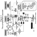
- Figs. 1 and 2 provides schematic overviews of C3 and its activation products.
- Complement, and C3 in particular, is associated with several disease indications, both acute and chronic. Examples include, but are not limited to, trauma, respiratory distress, sepsis, other forms of infection, infectious diseases (e.g., hemorrhagic fevers), multiple organ failure, age-related macular degeneration, rheumatoid arthritis, systemic lupus erythematosus, glomerular nephritis, ischemia/reperfusion injury, inflammatory bowel disease, intracranial hemorrhage, myocardial infarction, and cardiac arrest.
- infectious diseases e.g., hemorrhagic fevers
- multiple organ failure e.g., age-related macular degeneration, rheumatoid arthritis, systemic lupus erythematosus, glomerular nephritis, ischemia/reperfusion injury, inflammatory bowel disease, intracranial hemorrhage, myocardial infarction, and cardiac arrest.
- Severe trauma patients present unique clinical challenges. Accurate assessment of injury severity is important for initial intervention and patient stabilization, as well as patient triage (e.g., following mass casualty incidents). Those patients admitted to the ICU, even after initial stabilization, remain at high risk for secondary life-threatening complications involving organ dysfunction, respiratory distress and sepsis, among others. Many of these conditions involve hyper-inflammatory events that can escalate rapidly and cause significant damage before clinical symptoms are detected. These events are generally preceded by an increasingly unstable homeostasis of the inflammatory response. The ability to monitor inflammation frequently (e.g., every hour or two) and reliably at the earliest time points following injury has tremendous clinical value and would improve clinical outcomes for critical care patients.
- the first hour after injury is sometimes referred to as the "Golden Hour.” While not desiring to be bound by theory, it is generally believed that intervention within the first hour after traumatic injury greatly increases the outcome of the patient. Better diagnostic information provided earlier would help improve the critical care specialist's intuition when making treatment decisions.
- a point-of-care assay for measuring complement activation within the actionable window of treatment has not been known in the art.
- C3 is monitored in only a small number of diseases or conditions. Even in those instances, current assay methods have limitations.
- traditional complement assays are directed to total C3 as the target analyte (for example, via turbidity assays and ELISA).
- Total C3 is a combination of intact (or native) C3 and C3 activation and deactivation products. These tests generally detect decreases in circulating C3 levels. Decreased levels of total C3 therefore only measure C3 depletion due to massive activation.
- a second limitation in current C3 testing is the time required to perform most assays.
- a typical ELISA assay for the detection of complement activation requires hours to perform and the ready availability of a laboratory and a skilled technician. This assay platform is therefore not useful for indications of inflammatory dysfunction, in which biomarkers change on the order of minutes and clinical intervention is required on a similar timescale.
- Complement is notoriously fastidious and can become activated by virtue of standard analysis procedures (handling, storage, and exposure to foreign materials that contact C3 during analysis). Complement is very effective at lysing invading microbes and initiating the wound healing response at sites of injury. This effectiveness is due in part to the ability of C3 to be activated by foreign materials such as bacterial cell wall components. While this property is useful in directing an immune response to new foreign pathogens, this same property presents daunting challenges to experimental and diagnostic study. Materials such as plastics used in sample handling, manipulation of the sample itself, and improper storage conditions can also trigger complement activation.
- C3 has several attractive qualities as a biomarker in inflammation.
- C3 is activated by most stimuli that will cause complement activation.
- C3 activates in proportion to the degree of injury or infection.
- C3 responds in near real-time to a physiological insult. Complement activation occurs in direct response to an agent causing crisis, in contrast to other acute phase inflammatory markers that take hours or days to respond. This rapid response property is not present in other biomarkers frequently used in the clinic.
- intact (or native) C3 is a valuable marker of inflammatory status.
- Intact C3 represents the amount of C3 available for activation.
- Total C3 represents intact C3 as well as all C3 activation products.
- standard complement assays generally measure total C3 via turbidity assays or ELISA. Although technically easier to perform, total C3 assays cannot detect C3 depletion as accurately as intact C3 assays.
- Monitoring intact C3, especially over time, is useful for following massive complement activation events, such as those that occur in trauma and other systemic complement activation indications. Monitoring intact C3 over time allows a clinician to detect the onset of an immunosuppressive state caused by depletion of C3.
- intact C3 may be more useful than total C3 when calculating complement activation indexes (e.g., the C3a:total C3 ratio used by Zilow and Hecke).
- complement activation indexes e.g., the C3a:total C3 ratio used by Zilow and Hecke.
- Intact C3 assays have historically proven difficult to administer or depend upon, in part because intact C3 is very labile and can denature or self-activate if not handled properly.
- the C3 split product, iC3b is also a valuable marker of inflammatory response.
- iC3b has a half-life of 30 to 90 minutes, serving as a less volatile (e.g., compared to C3a), but still rapidly responsive biomarker.
- iC3b is present at much lower levels than intact C3 in patient samples. Even a small degree of cross-talk (for example 1%) between intact C3 protein and the iC3b-specific assay produces a false positive iC3b signal at a level twice that of normal circulating iC3b.
- a desirable marker of inflammation heretofore iC3b has posed significant challenges in diagnostic testing.
- WO 2010/135717 by Zhang et al., published November 25, 2010 , is directed to methods for assessing complement activation via the biomarkers intact C3, iC3b, and total C3.
- Zhang et al. is limited to traditional sandwich-type immunoassays such as ELISA, requiring laboratory processing and the expertise of skilled technicians.
- the assays and methods of Zhang et al. require sample preparation, storage, and handling steps that are known to activate the labile intact C3 produce false positive test results, impeding the ability to accurately measure intact C3.
- the assays and methods of Zhang et al. require hours to process and are thus incapable of providing the near real-time data that can impact patient care in the earliest time points after physiological crisis.
- the present inventors have now developed a point-of-care method and assay for the qualitative and quantitative measurement of intact C3 and iC3b suitable for use in directing patient care at the earliest time points immediately following traumatic injury or other physiological crises.
- the inventors have surprisingly found that it is possible to detect and quantify the biomarkers intact C3 and iC3b, while avoiding the false positive results that have plagued more conventional testing methods for these analytes.
- a method for treating an individual at risk for a complement-associated disorder comprising: (a) obtaining a sample of a body fluid from the individual; (b) measuring a complement activation level in the sample via a point-of-care lateral flow immunoassay; (c) correlating the complement activation level in the sample to a risk of a complement-associated disorder by comparing the complement activation level in the sample to a reference level in a control, wherein a deviation in complement activation level in the sample compared to the reference level in the control indicates the individual is at risk for a complement-associated disorder; (d) selecting a treatment for the individual, based on the correlating of step (c); and (e) treating the individual with the treatment selected in accordance with step (d).
- a lateral flow immunoassay for the point-of-care detection of a marker of complement activation in a body fluid sample comprising complement proteins, the lateral flow immunoassay comprising: a membrane strip; a detecting antibody that binds a first epitope of the marker; a test line comprising a capturing antibody that binds a second epitope of the marker; and a control line comprising an antibody that binds a control analyte, wherein the marker is selected from the group consisting of intact C3 and iC3b.
- a lateral flow immunoassay for the point-of-care detection of markers of complement activation in a body fluid sample comprising complement proteins
- the lateral flow immunoassay comprising: a membrane strip; a first detecting antibody that binds a first epitope of intact C3; a first test line comprising a first capturing antibody that binds a second epitope of intact C3; a second detecting antibody that binds a first epitope of iC3b; a second test line comprising a second capturing antibody that binds a second epitope of iC3b; and at least one control line comprising an antibody that binds a control analyte.
- a method for monitoring an individual who is receiving treatment for a physiological condition and who is suffering from a complement-associated disorder comprising: (a) obtaining serial samples of a body fluid from the individual; (b) determining a complement activation level in each of said samples via a point-of-care lateral flow immunoassay; (c) comparing the complement activation levels in the serial samples to detect a change in a complement activation level over time; and (d) modifying treatment for the individual, based on the correlating of step (c).
- the term "about,” when referring to a value or to an amount of mass, weight, time, volume, concentration or percentage is meant to encompass variations of in some embodiments ⁇ 20%, in some embodiments ⁇ 10%, in some embodiments ⁇ 5%, in some embodiments ⁇ 1%, in some embodiments ⁇ 0.5%, and in some embodiments ⁇ 0.1% from the specified amount, as such variations are appropriate to perform the disclosed method.
- An “Analyte” means any entity, particularly a chemical, biochemical or biological entity to be assessed, e.g., whose amount (e.g., concentration or mass), activity, composition, or other property(ies) is/are to be detected, measured, quantified, evaluated, analyzed, etc.
- An “analyte” can be a single molecular species or can be composed of multiple distinct molecular species.
- Antibody encompasses intact and/or full length immunoglobulins of types IgA, IgG (e.g., IgG1, IgG2, IgG3, IgG4), IgE, IgD, IgM, IgY, antigen-binding fragments or single chains of complete immunoglobulins (e.g., single chain antibodies, Fab fragments, F(ab')2 fragments, Fd fragments, scFv (single-chain variable), and dAb fragments), and other proteins that include at least one antigen-binding immunoglobulin variable region, e.g., a protein that comprises an immunoglobulin variable region, e.g., a heavy (H) chain variable region (VH) and a light (L) chain variable region (VL).
- IgA immunoglobulins of types IgA, IgG (e.g., IgG1, IgG2, IgG3, IgG4), IgE, IgD, I
- the light chains of an antibody may be of type kappa or lambda.
- An antibody may be polyclonal or monoclonal.
- a polyclonal antibody contains immunoglobulin molecules that differ in sequence of their complementarity determining regions (CDRs) and, therefore, typically recognize different epitopes of an antigen.
- CDRs complementarity determining regions
- a polyclonal antibody may be composed largely of several subpopulations of antibodies, each of which is derived from an individual B cell line.
- a monoclonal antibody is composed of individual immunoglobulin molecules that comprise CDRs with the same sequence, and, therefore, recognize the same epitope (i.e., the antibody is monospecific).
- An antibody may be a "humanized” antibody in which for example, a variable domain of rodent origin is fused to a constant domain of human origin or in which some or all of the complementarity-determining region amino acids often along with one or more framework amino acids are "grafted" from a rodent, e.g., murine, antibody to a human antibody, thus retaining the specificity of the rodent antibody.
- a rodent e.g., murine
- the capturing and/or detecting agents include other ligands, such as natural receptors for activated C3 (e.g., complement receptors 1, 2, and 3), aptamers, peptides, other small molecule ligands, and the like.
- ligands such as natural receptors for activated C3 (e.g., complement receptors 1, 2, and 3), aptamers, peptides, other small molecule ligands, and the like.
- An aspect of certain embodiments of the invention is the selection of antibodies for use as capture and detection agents.
- the inventors discovered that many published and commercially available antibodies exhibited crosstalk between intact C3 and various C3 cleavage products or between different C3 cleavage products.
- certain monoclonal antibodies against human C3a show significant and unexpected cross-reactivity with C3b and iC3b. It was recognized that cross-reactivity could be a significant source of inaccuracy in certain situations.
- iC3b was crosstalk between intact C3 and iC3b observed with many of the iC3b antibodies tested. Further testing showed that crosstalk between C3b and iC3b was even more significant with at least some of these antibodies.
- antibodies with specificity for intact C3 or iC3b are not substantially cross-reactive.
- “not substantially cross-reactive” means less than about 0.1% cross-reactive, meaning that a 1 ug/ml solution of C3 must register as less than about 1 ng/ml of iC3b. The about 0,1% threshold is based on the physiological levels of intact C3 and iC3b in a normal individual.
- Normal iC3b levels are approximately 0.5% that of total C3 in circulation. If C3 crosstalk contributes more than about 25% to the iC3b signal in a complement activation assay, the assay can produce false positive results that abrogate the utility of the assay.
- Body fluid means any fluid in the body that may be assayed for complement activation.
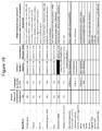
- Body fluids include, but are not limited to, whole blood, serum, plasma, urine, tears, saliva, wound exudate, broncheoalveolar lavage fluid, and cerebrospinal fluid. See Fig. 19 for a non-limiting list of suitable body fluids.
- Complement activation level means the amount of complement (generally C3) that is activated at a given time point. Amounts (i.e., levels) of intact C3, iC3b, and/or total C3 are typically expressed in terms of concentration but may be expressed in terms of mass or weight. Concentration may be expressed in various ways, e.g., in terms of molarity, molality, mole fraction, mass fraction (mass of a substance in a mixture as a fraction of the mass of the entire mixture), mass per unit volume, etc. For purposes of description herein, concentration (e.g., mass per unit volume) will generally be used. Complement activation level can also be described as a ratio of iC3b to intact or total C3, or as a ratio of C3a to total C3.
- Complement-associated disorder refers to a disorder or condition characterized by a modification in complement activation.
- Examples of complement-associated disorders include, but are not limited to, trauma, such as traumatic brain injury, spinal cord injury, surgery, and intracranial pressure; inflammatory distress, such as severe allergies, systemic inflammatory response syndrome (SIRS), multiple organ failure (MOF), acute or adult respiratory distress syndrome (ARDS), septic shock, and shock; paroxysmal nocturnal hemoglobinuria (PNH); hereditary angiodema; renal disease, such as glomerular nephritis, infection, lupus nephritis, and renal disease requiring organ transplant; autoimmune disease, such as diabetes mellitus I, inflammatory bowel disease, Crohn's disease, multiple sclerosis, myasthenia gravis, rheumatoid arthritis, and systemic lupus erythematosus; ischemia/reperfusion injury; heart disease, such as myocardi
- Control refers to a sample having a known reference level of complement activation.
- the control has a complement activation level comparable to that of an individual who is not experiencing a complement-associated disorder, such that a test sample having a complement activation level that is deviated compared to the control is indicative of a complement-associated disorder.
- a complement-associated disorder is indicated when the test sample complement activation level is statistically significantly deviated compared to the control.
- C3 activation signature means changes in C3 activation levels over time.
- Deviated and a deviation refer to statistically significant deviations as compared to a reference level in a control. Depending on the analyte being assayed, a deviated test sample level may be elevated or decreased relative to the control level.
- Decreased as compared to a reference level in a control, means statistically significantly decreased.
- intact C3 levels are depleted as C3 is broken down into its activation products.
- intact C3 levels are considered decreased as compared to a reference level in a control at about 10%.
- “Elevated,” as compared to a reference level in a control, means statistically significantly elevated.
- iC3b levels increase as C3 is broken down into its activation products.
- a ratio of iC3b to intact C3 that is elevated as compared to the normal ratio of 0.005 is indicative of C3 activation.
- Epitope refers to the minimum portion of a molecule that is recognized by, and thus determines the immunospecificity of, an antibody that binds to such epitope.
- the term is also used herein to refer to the minimum portion of a molecule that is recognized by a non- antibody specific binding agent. Unless otherwise indicated, it is assumed herein that a specific binding agent that binds to a complement protein binds to an epitope present and accessible for binding in the native protein, i.e., the epitope is not a neoepitope.
- a complement activation level determined by the assays and methods disclosed herein correlates directly with the severity of inflammatory distress being experienced by an individual. For example, when iC3b concentration is about 1-2.5% of intact C3, the patient's inflammatory distress can be said to be mildly severe. When iC3b concentration is about 2.5-5% of intact C3, the patient's inflammatory distress can be said to be moderately severe. When iC3b concentration is over 5% of intact C3, the patient's inflammatory distress is said to be highly severe.
- Understanding the severity of a patient's inflammatory distress can inform a physician's treatment of the individual. For example, if the individual presents with a highly severe inflammatory distress level, as indicated by the assays and methods disclosed herein, the physician can provide emergency medical treatment within the earliest time points of inflammatory distress, in order to minimize damage from inflammatory response.
- Label refers to a moiety that facilitates the direct or indirect detection and/or quantitative or relative measurement of a molecule to which it is attached.
- a detectable label often produces a signal such as fluorescence, chemiluminescence, radioactivity, color, magnetic or paramagnetic properties, etc., that renders it detectable, e.g., by the use of instruments that detect fluorescence, chemiluminescence, radioactivity, color, magnetic field, magnetic resonance, etc., or in some cases by visual inspection.
- the label may be, e.g., fluorescent substance; pigment; chemiluminescent or luminescent substance; colored substance; magnetic substance; or a non-magnetic metal particle such as gold colloid.
- the detecting antibodies suitable for use in the instant methods and assays are conjugated to a colloidal gold label, which provides a color signal.
- Neoepitope refers to an epitope that is generated or becomes detectable as a result of proteolytic cleavage of a complement component or cleavage product.
- the complement present in the body fluid sample tested is not substantially activated by the assay or method itself.
- “Not substantially activated,” as used in this context, means that the lateral flow immunoassay results are substantially free of in vitro activation caused by the test methods and/or materials. In this way, false positive test results for complement activation are avoided, since the lateral flow immunoassay is rapid and requires less sample manipulation, thus avoiding many of the stimuli that contribute to in vitro complement activation.
- Point-of-care refers to a device or method that can be used or carried out at the bedside or site of injury of the patient. Point-of-care tests generally do not require shipping a sample to a laboratory for processing or the expertise of a skilled laboratory technician. The point-of-care methods and tests described herein allow a clinician to receive critical information at the patient's bedside, or at the site of traumatic injury or triage, which can direct patient care during the critical first moments after a physiological crisis that triggers complement activation.
- Reader refers to an instrument suitable for the detecting of the signal produced by the label.
- the label is colloidal gold and the reader is an instrument suitable for the qualitative and/or quantitative detection of the color signal produced by the label.
- Suitable readers are available commercially from a variety of vendors, including BioAssay Works (Ijamsville, MD), the ESE-Quant from Qiagen (Hilden, Germany), Easterline LRE (Nordlingen, Germany), and Detekt Biomedical (Austin, TX).
- the reader is a handheld reader that quantifies the amount or concentration of intact C3, iC3b, or total C3.
- Treatment encompasses any diagnostic, therapeutic, preventive, or remedial treatment administered to an individual.
- treatment encompasses performing additional diagnostic testing on the individual.
- treatment encompasses therapeutic treatment, such as a administering a therapeutic agent to the individual.
- the therapeutic agent is selected from the group consisting of antibiotics, anti-inflammatory agents, and inhibitors of complement.
- treatment encompasses modifying a treatment the individual has already received or is receiving. For example, in one embodiment treating an individual on a ventilator encompasses optimizing the ventilator.
- the complement system comprises more than 30 serum and cellular proteins and plays important roles in innate and adaptive immunity.
- the classical pathway is primarily activated by immune complexes, specifically IgG/IgM antibodies bound to antigen.
- Other activators include lipopolysaccharide, myelin, polyanionic compounds, C-reactive protein (CRP), and microbial DNA and RNA.
- the lectin pathway is activated by polysaccharides with free-mannose group and other sugars common to fungi and bacteria.
- the alternative pathway is mediated by direct C3 activation by "foreign" substances that often include microbial cell wall components. All three major pathways of complement activation converge on the central protein complement component 3 (C3).
- C3 is a central mediator of inflammation and is activated by most factors that cause inflammation. See Figs. 1 and 2 for a schematic overview of the complement system.
- the classical pathway is typically triggered by immune complexes, which are complexes of antigen bound with antibodies, generally belonging to the IgM or IgG isotypes. Immune complexes in turn bind to complement component C1, which is comprised of C1q, C1r, and C1s. The binding of C1q to an antibody-antigen complex triggers activation of C1r and C1s. Activated C1s then cleaves component C4 to produce C4a and C4b. C4b is capable of covalent attachment to cell surfaces, although only about five percent does so. The remaining 95 percent reacts with water to form a soluble, activated C4b. Component 2 can then associate with C4b, which after which it is activated by C1s to C2a and C2b. C4b and C2a combine to form C4bC2a, the classical pathway (CP) C3 convertase.
- the CP convertase cleaves C3 to form C3a and C3b.
- C3b can covalently bind to cell surfaces or react with H 2 O and stay in solution.
- Activated C3b has multiple roles. By itself, it can serve as an opsonin to make the decorated cell or particle more easily ingested by phagocytes.
- C3b can associate with C4bC2a (the CP C3 convertase) to for a C5 convertase.
- the complex, termed C4bC2aC3b is termed the CP C5 convertase.
- C3b can form the core of another C3 convertase called the alternative pathway (AP) C3 convertase.
- AP alternative pathway
- the alternative pathway (AP) is another mechanism by which C3 can become activated. It is typically activated by targets such as microbial surfaces and various complex polysaccharides and other materials. This alternative pathway can also be initiated spontaneously by the cleavage of the thioester bond in C3 by a water molecule to form C3(H 2 O).
- C3(H 2 O) binds factor B, which allows factor D to cleave factor B to Ba and Bb.
- Bb remains associated with C3(H 2 O) to form C3(H 2 O)Bb complex, which acts as a C3 convertase and cleaves C3, resulting in C3a and C3b.
- C3b formed either via this process or via the classical or lectin pathways binds to targets (e.g., on cell surfaces) and forms a complex with factor B, which is subsequently cleaved by factor D and form Bb, resulting in C3bBb, which is termed the alternative pathway (AP) C3 convertase.
- targets e.g., on cell surfaces
- AP C3 convertase e.g., cleaved by factor D and form Bb
- the lectin complement pathway is initiated by binding of mannose-binding lectin (MBL) and MBL-associated serine protease (MASP) to carbohydrates.
- MBL mannose-binding lectin
- MASP MBL-associated serine protease
- the MB1-1 gene (known as LMAN-1 in humans) encodes a type 1 integral membrane protein localized in the intermediate region between the endoplasmic reticulum and the Golgi.
- the MBL-2 gene encodes the soluble mannose-binding protein found in serum.
- MASp-1 and MASP-2 are involved in proteolysis of C4 and C2, leading to C3 convertase, which lead to production of a C5 convertase as described above for the CP.
- C5 convertase generated via any of the three pathways cleave C5 to produce C5a and C5b.
- C5b then binds to C6, C7, and C8, which catalyses polymerization of C9 to form the C5b-9 membrane attack complex (MAC).
- the assembling MAC inserts itself into target cell membrane, forming a pore delineated by a ring of C9 molecules.
- MAC formation causes cell lysis of invading microbes, MAC formation on host cells can also cause lysis, but not necessarily. Sublytic amounts of MAC on the membrane of cells may affect cell function in a variety of way.
- the small cleavage products C3a, C4a, and C5a are anaphylatoxins and mediate multiple reactions in the acute inflammatory response.
- C3a and C5a are also potent chemotactic factors that attract immune system cells such as neutrophils and macrophages into the area of crisis.
- Complement component C3 is useful as a general alert biomarker that the body is responding to some form of physiological crisis, such as injury, infection, or other disease process.
- Complement has been associated with a wide variety of diseases, including lupus, arthritis, intracranial hemorrhage, diabetes, multiple sclerosis, heart disease, and age-related macular degeneration.
- the severity of disease correlates with the level of complement activation.
- complement can play a role in disease pathology.
- the body is not able to successfully control the cause of inflammation, which goes from local to systemic.
- Complement activation can directly damage tissue or do so indirectly by over-activating cells and recruiting immune cells that in turn cause tissue destruction. Examples of over activation include anaphylactic shock, multiple organ failure (MOF), acute respiratory distress syndrome (ARDS), and systemic inflammatory response syndrome (SIRS).
- MOF multiple organ failure
- ARDS acute respiratory distress syndrome
- SIRS systemic inflammatory response syndrome
- Complement activation in the immediate and early post-trauma period has been well documented and occurs by several different mechanisms, likely involving all three major pathways. Release and activation of proteolytic enzymes may directly activate complement components. Tissue damage and disruption of the endothelial lining expose surfaces that lack the endogenous complement inhibiting molecules that normally protect host tissues. These surfaces are susceptible to deposition of C3b and alternative pathway activation. Complement activation is also triggered by reperfusion of tissues following post-traumatic ischemia.
- complement activation is an important factor in many of the complications of severe trauma, contributing significantly to I/R injury, ARDS, MODS, secondary CNS injury, and sepsis.
- complement activation is a common occurrence in the immediate post-trauma period in human trauma victims, and several studies have provided evidence suggesting that the extent of complement activation correlates positively with poor outcomes.
- complement deficiency or administration of complement inhibitors reduces tissue damage and improves outcomes in a variety of experimental models including hemorrhage, I/R injury, and CNS injury.
- the instant assays and methods provide several advantages over previous complement assays and methods known in the art: First, the instant assays and methods are suitable for point-of-care use, producing results in a matter of minutes, rather than hours. The rapid return of results allows a clinician to act upon changes in C3 activation in near real-time to direct patient care during the critical first moments after traumatic injury or at the onset of physiologic crisis. The assays and methods are relatively easy to use and do not require the availability of an outside laboratory or a skilled lab technician. Second, the instant assays and methods require fewer handling steps, and thus minimize intact C3 activation due to handling and processing, which leads to false positive test results.
- the assays and methods described herein employ antibody pairs carefully selected to allow for measurement of the complement proteins intact C3 and/or iC3b, C3's major activation biomarker. This more precise measurement of complement activation, in comparison to traditional assays of total C3, permits analysis of turnover and actual amount of C3 remaining and available for activation.
- a method for treating an individual at risk for a complement-associated disorder comprising: (a) obtaining a sample of a body fluid from the individual; (b) measuring a complement activation level in the sample via a point-of-care lateral flow immunoassay; (c) correlating the complement activation level in the sample to a risk of a complement-associated disorder by comparing the complement activation level in the sample to a reference level in a control, wherein a deviation in complement activation level in the sample compared to the reference level in the control indicates the individual is at risk for a complement-associated disorder; (d) selecting a treatment for the individual, based on the correlating of step (c); and (e) treating the individual with the treatment selected in accordance with step (d).
- the complement-associated disorder is selected from the group consisting of trauma, inflammatory distress, autoimmune disorders, intracranial hemorrhage, infection, transplant rejection, ocular disease, heart disease, ischemia/reperfusion injury, age-related macular degeneration, paroxysmal noctural hemoglobinuria (PNH), hereditary angiodema, renal disease, pregnancy-associated disorders, and neurological disorders.
- the complement-associated disorder is inflammatory distress.
- Inflammatory distress also known as inflammatory dysfunction, includes a variety of diseases and conditions associated with hyperinflammation. Examples of diseases and conditions associated with inflammatory distress include, but are not limited to, organ failure, systemic inflammatory response syndrome (SIRS), adult respiratory distress syndrome (ARDS), sepsis, and pneumonia.
- the body fluid obtained from the individual is selected from the group consisting of whole blood, serum, plasma, urine, tears, saliva, wound exudate, broncheoalveolar lavage fluid, and cerebrospinal fluid. See Fig. 19 for a non-limiting list of suitable body fluids.
- the body fluid is obtained from the individual within one hour of a physiological event triggering complement activation.
- the body fluid is whole blood.
- the lateral flow immunoassay detects the presence or absence of one or more of intact C3 and iC3b in the sample. In another embodiment of the method, the lateral flow assay detects the presence of total C3. In another embodiment, the lateral flow immunoassay is read by a reader. In a more specific embodiment, the reader quantifies a concentration of one or more of intact C3 and iC3b in the sample. In another specific embodiment, the reader quantifies a concentration of total C3 in the sample.
- Complement activation levels are assessed for deviation from a reference value of a control which indicates complement is activated in the individual.
- the level or concentration of iC3b in the test sample is elevated in comparison to a control, indicating C3 is activated and has been further split into its activation product, iC3b.
- the level or concentration of intact C3 is decreased in comparison to a control, indicating intact C3 has been converted to its breakdown or activation products and is hence depleted in the individual.
- Complement activation level correlates to a severity of inflammatory distress: the higher the complement activation level, the greater the risk of developing inflammatory distress and/or the greater the severity of inflammatory distress experienced by the individual. Therefore, in another embodiment of the method, the complement-associated disorder is inflammatory distress and the concentration of one or more of intact C3 and iC3b correlates to a severity of inflammatory distress.
- the complement activation level determined by the instant method provides point-of-care diagnostic information that can direct patient care. Based on the risk of complement-associated disorder correlated in step (c), the clinician can select the appropriate treatment for the individual.
- the treatment comprises performing additional testing on the individual to determine the cause of inflammatory distress. For example, severe trauma patients that require ventilator assistance for breathing are at risk for acute respiratory distress caused either by Ventilator Associated Pneumonia (VAP) or non-infectious inflammatory dysfunction.
- VAP Ventilator Associated Pneumonia
- a level of complement activation may indicate active or imminent inflammatory dysfunction before clinical signs of respiratory crisis are presented.
- the instant assays and methods may indicate whether the individual is experiencing VAP or non-infectious respiratory distress.
- the instant assays and methods may indicate additional testing (such as broncheoalveolar lavage (BAL)) at a time point earlier than is now standard practice
- additional testing such as broncheoalveolar lavage (BAL)
- the treatment may comprise administering a therapeutic agent such as an antibiotic or set of antibiotics.
- a therapy may be selected from the group consisting of ventilator adjustment, anti-inflammatory agents, and inhibitors of complement.
- the additional testing may comprise obtaining a cerebrospinal fluid sample for additional analysis. If the individual is suffering from a wound, including a non-healing wound, the further testing may comprise obtaining a sample of wound exudate for additional analysis.
- the inhibitor of complement is selected from the group consisting of natural complement inhibitors and derivatives thereof, compstatin and analogs thereof, anti-membrane attack complex (MAC) antibodies, anti-C3 antibodies, anti-C5 antibodies, C3a receptor antagonists, and C5a receptor antagonists.
- MAC anti-membrane attack complex
- additional complement inhibitors can be found, for example, in Emlen et al., Therapeutic complement inhibition: new developments, Semin. Thromb. Hemost. 36(6):660-68 (2101 ); Wagner et al., Therapeutic potential of complement modulation, Nat. Rev. Drug Discov.
- the instant method provides a measurement of the complement activation level in the sample in about 30 minutes or less. In a more specific embodiment, the method provides a complement activation level in the sample in about 30, about 25, about 20, about 15, about 10, about 5, or about 3 minutes or less. The rapidity of the method enables the clinician to determine a complement activation level and select an appropriate therapy in response, during a clinically-meaningful time period.
- the instant methods can be carried out at the bedside or even at the site of traumatic injury - for example, in an ambulance or in triaging a patient on the battlefield - and the complement activation level determined by the assay and method can direct patient care within the critical first hour post-trauma.
- a method for monitoring an individual who has received or is receiving treatment for a physiological condition and who is known to be suffering from a complement-associated disorder comprising: (a) obtaining serial samples of a body fluid from the individual;(b) determining a complement activation level in each of said samples via a point-of-care lateral flow immunoassay; (c) comparing the complement activation levels in the serial samples to detect a change in a complement activation level over time; and (d) modifying treatment for the individual, based on the correlating of step (c).
- Serial blood samples are collected and tested to monitor complement activation levels in response to the treatment.
- the complement-associated disorder is selected from the group consisting of systemic lupus erythematosus, inflammatory distress, autoimmune disorders, intracranial hemorrhage, bacteremia, transplant rejection, ocular disease, heart disease, ischemia/reperfusion injury, age-related macular degeneration, paroxysmal noctural hemoglobinuria (PNH), hereditary angiodema, renal disease, pregnancy-associated disorders, and neurological disorders, and trauma including patients at risk for inflammatory dysfunction such as ventilator associated pneumonia (VAP), respiratory distress, and multiple organ failure.
- VAP ventilator associated pneumonia
- the body fluid is selected from the group consisting of whole blood, serum, plasma, urine, tears, saliva, wound exudate, broncheolar lavage fluid, and cerebrospinal fluid.
- the lateral flow immunoassay detects the presence or absence of one or more of intact C3 and iC3b in the sample.
- the lateral flow immunoassay is read by a reader which is capable of quantifying a concentration of intact C3, iC3b, or total C3.
- the clinician will detect a decrease in complement activation in response to the treatment the patient is receiving. Accordingly, the clinician will modify the individual's treatment by adjusting the dosing of medications administered, such as anti-inflammatory agents or complement inhibitors, or discontinuing treatment once complement levels have returned to normal (i.e., a level in an individual who is not experiencing a complement-associated disorder). In other embodiments, the clinician will detect a rise in complement activation levels in response to the treatment the patient is receiving. Accordingly, the clinician will modify the individual's treatment by increasing the dosage of medications, such as anti-inflammatory agents or complement inhibitors, until a desired stabilization or decrease in complement activation levels is achieved. If no change in complement activation level is detected, the clinician may modify the individual's treatment or may elect to maintain the individual's treatment regimen until a change in complement activation levels is observed.
- medications administered such as anti-inflammatory agents or complement inhibitors
- the lateral flow immunoassay described herein is comprised of a cellulose membrane strip 3, upon which is disposed a sample pad 1 to absorb the sample fluid and allow gradual migration of the sample-and-particle-conjugate immune complexes, a wick 6 at the distal end of the strip that absorbs the liquid sample and conjugate material to facilitate capillary migration through the cellulose membrane strip 3, and a particle conjugate pad 2 comprising a detecting antibody bound to a label, or detection conjugate.
- the cellulose membrane strip 3 is the test zone region, upon which is disposed a test line 4, comprising monoclonal or polyclonal antibodies striped for capturing the detection conjugate and a control line 5, comprising an antibody that binds a control analyte, such as IgG, and indicates to the user that the test was successfully run.
- the lateral flow immunoassay further comprises a polyester film backing 7 attached to the cellulose membrane strip 3, and a pressure-sensitive laminate film backing 8. Each lateral flow immunoassay is packaged in a Mylar® zero-vapor barrier pouch.
- the sample migrates from the sample pad 1 through the particle conjugate pad 2, where any target analyte present will bind to the detecting antibody conjugate.
- the sample then continues to migrate across the membrane 3 until it reaches the test line 4 where the target/conjugate complex will bind to the immobilized antibodies producing a visible line on the membrane.
- the sample migrates further along the membrane strip 3 until it reaches the control line 5, where excess antibody conjugate that did not bind the test line will bind the control line and produce a second visible line on the membrane.
- the control line ligand is often an antibody against the Fc region of the conjugated antibody. This control line indicates that the sample has migrated across the membrane as intended.
- the lateral flow immunoassay comprises a single membrane strip for the detection of a single analyte. In other embodiments, the lateral flow immunoassay detects two or more analytes.
- the test can be configured with multiple membrane strips arranged in parallel (see, for example, the schematic of Fig. 5(B) ), or with multiple test lines arranged in series on a single membrane strip (see, for example, the schematic of Fig. 9(B) ).
- the lateral flow immunoassay membrane strip is enclosed in a test cassette 9 having a port 10 for instilling the test sample and a window 11 for viewing the test results.
- the lateral flow immunoassays of Fig. 6 are configured to assay for a single analyte and each comprise one test line 4 and one control line 5.
- the lateral flow immunoassay is configured to test for two analytes in a single test cassette in parallel.
- the lateral flow immunoassay comprises two ports 10 for instilling the test samples and a separate membrane strip 3 for each analyte (see Fig. 7(A) ).
- the lateral flow immunoassay comprises one port 10 for instilling the sample and a separate membrane strip 3 for each analyte (see Fig. 7(B) ).
- the lateral flow immunoassay is configured to test for three analytes in a single test cassette in parallel.
- the lateral flow immunoassay comprises three ports 10 for instilling the test sample and a separate membrane strip 3 for each analyte (see Fig. 8(A) ).
- the lateral flow immunoassay comprises one port 10 for instilling the test sample and a separate membrane strip 3 for each analyte (see Fig. 8(B) ).
- the lateral flow immunoassay is configured to test for multiple analytes in a single test cassette in series.
- Figure 10(A) depicts a test cassette comprising a membrane strip 3 with two test lines 4 and one control line 5 arranged in series.
- Figure 10(B) depicts a test cassette comprising a membrane strip 3 with three test lines 4 and one control line 5 arranged in series.
- the lateral flow immunoassay presently disclosed provides qualitative and/or quantitative detection of the target markers. Qualitatively, two clear lines on the membrane is a positive result, whereas a single line in the control zone is a negative result.
- a lateral flow immunoassay for the point-of-care detection of a marker of complement activation in a body fluid sample comprising complement proteins
- the lateral flow immunoassay comprising: a membrane strip; a detecting antibody that binds a first epitope of the marker; a test line comprising a capturing antibody that binds a second epitope of the marker; and a control line comprising an antibody that binds a control analyte, wherein the marker is selected from the group consisting of intact C3 and iC3b.
- the detecting antibody comprises a label that provides a signal that can be read visually by a clinician or electronically via a commercial reader.
- Various labels are suitable for use in the instantly disclosed assays.
- the label is colloidal gold.
- Detecting and capturing antibody pairs must be carefully selected to avoid interfering crosstalk between C3 and iC3b.
- the primary concern is intact C3 producing a signal in an assay for the detection of iC3b.
- crosstalk can present a problem.
- C3 is present at levels about 200 times higher than iC3b in normal individuals, even a slight degree of crosstalk can have a major impact on the accurate measurement of iC3b and C3 activation. This is further complicated by the fact that improper handling, improper storage, and even reagents themselves can cause in vitro C3 activation.
- the marker is intact C3 and the detecting antibody binds a first epitope of intact C3, wherein the first epitope is a C3a domain which is present on intact C3 and which is lost upon activation of C3.
- the marker is intact C3 and the capturing antibody binds a second epitope on C3, wherein the second epitope is a region in the C3d domain which is present on intact C3, C3b, iC3b, and C3d. See Fig. 3(A) .
- the marker is iC3b and the detecting antibody binds a first epitope of iC3b, wherein the first epitope is a neoepitope on iC3b which is revealed when C3b is deactivated to iC3b and which is occluded when iC3b is further degraded to C3c and C3d.
- the marker is iC3b and the capturing antibody binds a second epitope on iC3b, wherein the second epitope is a neoepitope present only on C3b, iC3b, and C3dg. See Fig. 3(B) .
- the marker is intact C3, the capturing antibody is Hycult® HM2075 and the detecting antibody is MP Biomedicals® 55237.
- the marker is iC3b, the capturing antibody is Quidel® A209 and the detecting antibody is MP Biomedicals® 55237.
- the marker is iC3b, the capturing antibody is Quidel® A209 and the detecting antibody is Quidel® A250.
- the body fluid is selected from the group consisting of whole blood, serum, plasma, urine, tears, saliva, wound exudate, bronchoalveolar lavage fluid, and cerebrospinal fluid.
- the body fluid is whole blood.
- control analytes are suitable for use in the lateral flow immunoassays of the instant invention to provide verification that the assay was successfully completed.
- the control analyte is IgG.
- Another advantage of the instant lateral flow immunoassay is the avoidance of substantial complement activation in the sample by virtue of the test itself, which can lead to false positive results.
- C3 is a fastidious protein capable of self activation due to sample handling, storage, and contact with foreign materials or substances.
- the instant lateral flow immunoassay avoids such false positives by reducing and/or eliminating sample preparation and handling steps. Accordingly, in one embodiment of the instant lateral flow assay, complement in the body fluid sample is not substantially activated experimentally by the lateral flow immunoassay.
- a lateral flow immunoassay that can detect more than one marker of complement activation in a single assay.
- a dual lateral flow immunoassay that can qualitatively and quantitatively detect both intact C3 and iC3b in the same aliquot of a body fluid is highly desirable.
- a lateral flow immunoassay for the point-of-care detection of markers of complement activation in a body fluid sample comprising complement proteins
- the lateral flow immunoassay comprising: a membrane strip; a first detecting antibody that binds a first epitope of intact C3; a first test line comprising a first capturing antibody that binds a second epitope of the intact C3; a second detecting antibody that binds a first epitope of iC3b; a second test line comprising a second capturing antibody that binds a second epitope of iC3b; and at least one control line comprising an antibody that binds a control analyte.
- the first and second detecting antibodies comprise a label that provides a signal.
- Various labels are suitable for use in the instantly disclosed assays.
- the label is colloidal gold.
- the body fluid is selected from the group consisting of whole blood, serum, plasma, urine, tears, saliva, wound exudate, bronchoalveolar lavage fluid, and cerebrospinal fluid.
- the body fluid is whole blood.
- control analytes are suitable for use in the lateral flow immunoassays of the instant invention to provide verification that the assay was successfully completed.
- the control analyte is IgG.
- complement in the body fluid sample is not substantially activated experimentally by the lateral flow immunoassay itself.
- the first detecting antibody binds a first epitope of intact C3, wherein the first epitope of intact C3 is a C3a domain which is present on intact C3 and which is lost upon activation of C3.
- the first capturing antibody binds a second epitope of intact C3, wherein the second epitope is a region in the C3d domain which is present on intact C3, C3b, iC3b, and C3d.
- the second detecting antibody binds a first epitope of iC3b, wherein the first epitope of iC3b is a neoepitope on iC3b which is revealed when C3b is deactivated to iC3b and which is occluded when iC3b is further degraded to C3c and C3d.
- the second capturing antibody binds a second epitope of iC3b, wherein the second epitope of iC3b is a neoepitope present only on C3b, iC3b, and C3dg.
- the antibodies that bind intact C3 and the antibodies that bind iC3b are not substantially cross-reactive.
- a standard curve is performed using 10 ng/ml 30 ng/ml, 100 ng/ml, 300 ng/ml, and 1000 ng/ml of intact C3 and iC3b standards. Lateral flow immunoassay cassettes are read with an electronic reader after 20 minutes.
- the test is used to gauge injury severity within 15, 30, or 60 minutes of injury. It is most useful for patients who may have suffered injuries not obvious by visual inspection.
- a drop of blood is collected either from an arterial line (A-line) or finger stick.
- a 10 ul sample is drawn up using a fixed volume pipet. The sample then mixed with 990 ul of sample buffer. The blood and sample buffer are mixed.
- 100 ul is drawn up and pipetted onto the lateral flow immunoassay cassette containing integrated intact C3 and iC3b test strips. Alternatively, 100 ul can be applied to separate intact C3 and iC3b lateral flow assay cassettes. After 10 minutes but before 40 minutes, the cassette is read and results recorded, preferably electronically by a reader.
- the first reading has an iC3b level (or equivalent iC3b:intact C3 ratio) higher than 50 ⁇ g/ml in blood, evidence of complement activation and high inflammation exist. Staff assumes severe injury and alerts ER staff. Otherwise, a second reading is taken 5 minutes later. If the iC3b level (or equivalent iC3b:intact C3 ratio) is higher than 50 ⁇ g/ml in blood or the iC3b level has increased by more than 25%, the patient is assumed to have severe injury and ER staff is alerted. A lesser increase or no increase is suggestive, but not conclusive, of less severe injury.
- ICU staff performs a standard curve using 10 ng/ml 30 ng/ml, 100 ng/ml, 300 ng/ml, and 1000 ng/ml of intact C3 and iC3b standards. Lateral flow immunoassay cassettes are read with an electronic reader after 20 minutes.
- the objective of trajectory monitoring is to detect changes in inflammatory and immune status of patients that have been stabilized after severe trauma.
- respiratory distress caused by either pneumonia or inflammatory dysfunction is to be detected.
- the expected patient profile is one who has an injury severity score (ISS) equal or greater to 16 and who requires ventilator assistance for breathing.
- ISS injury severity score
- the patient receives a complement test at frequent intervals, which aligns with the time points for testing glucose levels in blood. This interval between testing is usually about two hours.
- Blood is collected using the same method as for glucose testing, either by A-line or by finger stick.
- a 10 ul sample is drawn up using a fixed volume pipet.
- the sample then mixed with 990 ul of sample buffer.
- the blood and sample buffer are mixed.
- 100 ul is drawn up and pipetted onto the LFA cassette containing integrated intact C3 and iC3b lateral flow immunoassay cassettes.
- 100 ul can be applied to separate intact C3 and iC3b cassettes.
- the cassette or cassettes will be placed in reader at the patient's bedside.
- the reader is set to take a reading after 20 minutes. Data is collected and iC3b, intact C3 and iC3b:(intact C3) values recorded at each time point.
- Changes in intact C3 or iC3b levels over time or changes in the rate of change may indicate a change in inflammatory status.
- a sharp rise in iC3b, accompanied by a decrease in intact C3, indicates imminent respiratory distress.
- a clinician performs a broncheoalveolar lavage (BAL) on the patient to determine whether the patient is experiencing VAP. If bacteria are present at levels of 10 4 per ml or higher, VAP is indicate and the patient is places on antibiotic therapy. Otherwise, noninfectious inflammatory dysfunction is assumed and the patient may be treated with anti-inflammatory agents and/or complement inhibitors. The patient may also have his ventilator setting adjusted.
- BAL broncheoalveolar lavage
- a standard curve is performed using 10 ng/ml 30 ng/ml, 100 ng/ml, 300 ng/ml, and 1000 ng/ml of intact C3 and iC3b standards. Lateral flow immunoassay cassettes are read with an electronic reader after 20 minutes.
- the test is used to gauge the initial severity of disease as well the effectiveness of therapy.
- One of the standard diagnostics performed on SLE patients is measurement of total C3 levels. C3 levels are normally depressed in SLE patients and return to normal (>1 mg/ml) following successful treatment. However, it not known generally whether the C3 activation has been abrogated or only slowed enough to allow normal replenishment mechanisms to restore C3 levels to normal.
- a patient's blood is collected for total C3, intact C3, and iC3b tests. Only one drop is required for the combine 3 tests. Blood is collected by fingerstick unless blood is being drawn for other tests, in which case, the blood will come from that source.
- a 10 ul sample is drawn up using a fixed volume pipet. The sample then mixed with 990 ul of sample buffer. The blood and sample buffer are mixed.
- 100 ul is drawn up and pipetted onto the LFA cassette containing integrated lateral flow cassette that measures total C3, intact C3 and iC3b.
- 100 ul can be applied to separate cassettes for each assay.
- the cassette or cassettes are placed in reader at the doctor's office. The reader is set to take a reading after 20 minutes.
- iC3b and intact C3 tests provide the specialist with more information about the severity of the patient's condition than is now possible. New information becomes available at the time that the specialist would consider the patient's status stable. At this point, the iC3b and intact levels indicate the extent of remaining disease process. If iC3b levels, in particular, are above normal (generally >1%), the underlying disease process is still very active and the specialist may opt to further adjust therapy by increasing anti-inflammatory drug doses or adding additional medication.
- a standard curve is performed using 10 ng/ml 30 ng/ml, 100 ng/ml, 300 ng/ml, 1000 ng/ml, and 3000 ng/ml of intact C3 and iC3b standards. Lateral flow immunoassay cassettes are read with an electronic reader after 20 minutes.
- the lower eyelid is pulled back and briefly dapped with a Kimwipe® to the lower part of the eye.
- the Kimwipe® is then quickly cut where the tear was collected leaving a few millimeters of dry edge surrounding the tear spot.
- the Kimwipe®-tear sample is then placed into 220 ul of BioAssay Works Diluent Buffer and vortexed thoroughly for 10 seconds. After a one minute wait period, the sample is vortexed again briefly. Next, 100ul of sample is transferred to each lateral flow immunoassay (intact C3 and iC3b) and assayed.
- each cassette is inserted into the reader and read after 20 minutes.
- the results ranged between 50-60 ⁇ /ml of intact C3 and between 5-8 ⁇ /ml of iC3b (See Fig. 15 ).
- a standard curve is performed using 10 ng/ml 30 ng/ml, 100 ng/ml, 300 ng/ml, 1000 ng/ml, and 3000 ng/ml of intact C3 and iC3b standards. Lateral flow immunoassay cassettes are read with an electronic reader after 20 minutes.
- the results from the first patient were determined to be approximately 500 ⁇ g/ml for intact C3 and 200 ng/ml for iC3b. This indicates there is 2500 ratio of intact C3 to iC3b in this individual (see Fig. 16 ).
- the second individual's results were approximately 1000 ⁇ g/ml for intact C3 and 300 ng/ml for iC3b (see Fig. 17 ). Both of these values are within the expected normal ranges.
- the iC3b values are in the lower range of what is considered normal.
- iC3b In a 1 milliliter volume, 50 ng/ml of iC3b was mixed with varying mounts of intact C3 (ranging from 0 ng/ml to 100,000 ng/ml). Samples were mixed by inversion 6-8 times and then 0.1 ml was pipetted onto the cassette. Readings were taken at 20 minutes. Reader output was converted to iC3b concentration using a standard curve generated from 10 ng/ml to 100,000 ng/ml. Background from a cassette run only with buffer was subtracted. Fractional contributions were calculated by subtracting the actual iC3b concentration (from iC3b test with no added C3) from apparent concentration of iC3b at each point and then normalizing against actual iC3b concentrations. See Fig. 14 .
- J24K-03 is an assay with mouse anti-C3a monoclonal on the gold conjugate and mouse anti-C3d monoclonal on the test line.
- H08K-01 has mouse anti-iC3b monoclonal on the gold conjugate and Anti-C3 polyclonal on the test line.
- One embodiment of the invention comprises a lateral flow assay strip without the cassette casings. These strips had anti-iC3b monoclonal (Quidel® A209) conjugated to the gold and anti-C3 antibody (MP Biomedical® 55237) conjugated to the strip. Standard curves are shown in Figure 11(A) . The standard curves indicated a linear range of about 10 fold and a sensitivity of about 100 ng/ml. Another embodiment of the invention configures the strips for use in a cassette that allows controlled application of the sample to the assay strip. This improved assay-to-assay reproducibility, although there is still considerable time dependence on the assay. Standard curve results are shown in Figure 11(B) . A third embodiment increases the antibody concentration from 0.5 mg/ml to 1 mg/ml applied to on the gold conjugate and removes BSA from the absorption buffer. Standard curve results are shown in Figure 11(C) .
- Lateral flow immunoassay (LFA) cassettes were prepared by labeling and laying out in groups of three. For each dilution, investigator pipetted 100 ul of first working stock (10 ug/ml for intact C3 and 5 ug/ml for iC3b) into sample port of 1 st LFA. For each concentration, investigator waited 20 seconds before loading 100 ul of same working stock into the 2nd LFA. Cassettes were read after 10, 20, and 30 minutes using BioAssay Works Reader LFDR 101 (Forsite Diagnostics) using the Test line setting followed by the Control line setting, and the data recorded.
- LFA Lateral flow immunoassay
- LFA cassettes were prepared by labeling and laying out in groups of three. For each of dilution, investigator pipetted 100 ul of first working stock (10 ug/ml for intact C3 and 5 ug/ml for iC3b) into sample port of 1 st LFA. For each concentration, investigator waited 20 seconds before loading 100 ul of same working stock into the 2nd LFA. Cassettes were read after 10, 20, and 30 minutes using BioAssay Works Reader LFDR 101 (Forsite Diagnostics) using the Test line setting followed by the Control line setting, and the data recorded.
- BioAssay Works Reader LFDR 101 Form Diagnostics
- a method for treating an individual at risk for a complement-associated disorder comprising:
- the complement-associated disorder is selected from the group consisting of trauma, inflammatory distress, autoimmune disorders, intracranial hemorrhage, infection, transplant rejection, ocular disease, heart disease, ischemia/reperfusion injury, age-related macular degeneration, paroxysmal noctural hemoglobinuria (PNH), hereditary angiodema, renal disease, pregnancy-associated disorders, and neurological disorders.
- the inflammatory distress is selected from the group consisting of organ failure, systemic inflammatory response syndrome (SIRS), adult respiratory distress syndrome (ARDS), sepsis, and pneumonia.
- the body fluid is selected from the group consisting of whole blood, serum, plasma, urine, tears, saliva, wound exudate, bronchoalveolar lavage fluid, and cerebrospinal fluid.
- the lateral flow immunoassay detects the presence or absence of one or more of intact C3 and iC3b in the sample
- the lateral flow immunoassay is read by a reader.
- the reader quantifies a concentration of intact C3, iC3b, or total C3.
- the concentration of C3 is decreased compared to the reference level in the control.
- the concentration of iC3b is elevated compared to the reference level in the control.
- the complement-associated disorder is inflammatory distress and the concentration of intact C3, iC3b, or total C3 correlates to a severity of inflammatory distress.
- the treatment comprises performing additional testing on the individual, optimizing a ventilator, or administering a therapeutic agent selected from the group consisting of antibiotics, anti-inflammatory agents, and inhibitors of complement.
- the additional testing comprises obtaining a broncheoalveolar lavage (BAL) sample from the individual.
- BAL broncheoalveolar lavage
- the inhibitor of complement is selected from the group consisting of natural complement inhibitors and derivatives thereof, compstatin and analogs thereof, anti-membrane attack complex (MAC) antibodies, anti-C3 antibodies, anti-C5 antibodies, C3a receptor antagonists, and C5a receptor antagonists.
- MAC anti-membrane attack complex
- the point-of-care lateral flow immunoassay provides a measurement of the amount of intact C3, iC3b, or total C3 in the sample in about 30 minutes or less.
- a lateral flow immunoassay for the point-of-care detection of a marker of complement activation in a body fluid sample comprising complement proteins comprising:
- the detecting antibody comprises a label that provides a signal.
- the marker is intact C3 and the first epitope is a C3a domain which is present on intact C3 and which is lost upon activation of C3.
- the second epitope is a region in the C3d domain which is present on intact C3, C3b, iC3b, and C3d.
- the marker is iC3b and the first epitope is a neoepitope on iC3b which is revealed when C3b is deactivated to iC3b and which is occluded when iC3b is further degraded to C3c and C3d.
- the second epitope is a neoepitope present only on C3b, iC3b, and C3dg.
- the body fluid is selected from the group consisting of whole blood, serum, plasma, urine, tears, saliva, wound exudate, bronchoalveolar lavage fluid, and cerebrospinal fluid.
- control analyte is IgG.
- complement in the body fluid sample is not substantially activated experimentally by the lateral flow immunoassay.
- a lateral flow immunoassay for the point-of-care detection of markers of complement activation in a body fluid sample comprising complement proteins comprising:
- the first and second detecting antibodies each comprise a label that provides a signal.
- the body fluid is selected from the group consisting of whole blood, serum, plasma, urine, tears, and cerebrospinal fluid.
- control analyte is IgG.
- complement in the body fluid sample is not substantially activated experimentally by the lateral flow immunoassay.
- the first epitope of intact C3 is a C3a domain which is present on intact C3 and which is lost upon activation of C3.
- the second epitope of intact C3 is a region in the C3d domain which is present on intact C3, C3b, iC3b, and C3d.
- the first epitope of iC3b is a neoepitope on iC3b which is revealed when C3b is deactivated to iC3b and which is occluded when iC3b is further degraded to C3c and C3d.
- the second epitope of iC3b is a neoepitope present only on C3b, iC3b, and C3dg.
- the antibodies that bind intact C3 and the antibodies that bind iC3b are not substantially cross -reactive.
- a method for monitoring an individual who is receiving treatment for a physiological condition and who is suffering from a complement- associated disorder comprising:
Landscapes
- Health & Medical Sciences (AREA)
- Life Sciences & Earth Sciences (AREA)
- Engineering & Computer Science (AREA)
- Chemical & Material Sciences (AREA)
- Immunology (AREA)
- General Health & Medical Sciences (AREA)
- Medicinal Chemistry (AREA)
- Hematology (AREA)
- Urology & Nephrology (AREA)
- Organic Chemistry (AREA)
- Nuclear Medicine, Radiotherapy & Molecular Imaging (AREA)
- Pharmacology & Pharmacy (AREA)
- General Chemical & Material Sciences (AREA)
- Animal Behavior & Ethology (AREA)
- Chemical Kinetics & Catalysis (AREA)
- Public Health (AREA)
- Veterinary Medicine (AREA)
- Biomedical Technology (AREA)
- Bioinformatics & Cheminformatics (AREA)
- Molecular Biology (AREA)
- Cell Biology (AREA)
- Biochemistry (AREA)
- Pathology (AREA)
- General Physics & Mathematics (AREA)
- Analytical Chemistry (AREA)
- Physics & Mathematics (AREA)
- Food Science & Technology (AREA)
- Microbiology (AREA)
- Biotechnology (AREA)
- Rheumatology (AREA)
- Rehabilitation Therapy (AREA)
- Cardiology (AREA)
- Heart & Thoracic Surgery (AREA)
- Communicable Diseases (AREA)
- Oncology (AREA)
- Proteomics, Peptides & Aminoacids (AREA)
- Neurosurgery (AREA)
- Dermatology (AREA)
- Transplantation (AREA)
- Pain & Pain Management (AREA)
Applications Claiming Priority (2)
| Application Number | Priority Date | Filing Date | Title |
|---|---|---|---|
| US40929710P | 2010-11-02 | 2010-11-02 | |
| EP11842681.6A EP2635702B1 (en) | 2010-11-02 | 2011-11-02 | Lateral flow immunoassay for complement activation and methods of use for point-of-care assessment of complement-associated disorders |
Related Parent Applications (2)
| Application Number | Title | Priority Date | Filing Date |
|---|---|---|---|
| EP11842681.6A Division-Into EP2635702B1 (en) | 2010-11-02 | 2011-11-02 | Lateral flow immunoassay for complement activation and methods of use for point-of-care assessment of complement-associated disorders |
| EP11842681.6A Division EP2635702B1 (en) | 2010-11-02 | 2011-11-02 | Lateral flow immunoassay for complement activation and methods of use for point-of-care assessment of complement-associated disorders |
Publications (1)
| Publication Number | Publication Date |
|---|---|
| EP3095876A1 true EP3095876A1 (en) | 2016-11-23 |
Family
ID=46146161
Family Applications (2)
| Application Number | Title | Priority Date | Filing Date |
|---|---|---|---|
| EP15199549.5A Withdrawn EP3095876A1 (en) | 2010-11-02 | 2011-11-02 | Point of care lateral flow immunoassay for complement activation and methods of use for point-of-care assessment of complement-associated disorders |
| EP11842681.6A Active EP2635702B1 (en) | 2010-11-02 | 2011-11-02 | Lateral flow immunoassay for complement activation and methods of use for point-of-care assessment of complement-associated disorders |
Family Applications After (1)
| Application Number | Title | Priority Date | Filing Date |
|---|---|---|---|
| EP11842681.6A Active EP2635702B1 (en) | 2010-11-02 | 2011-11-02 | Lateral flow immunoassay for complement activation and methods of use for point-of-care assessment of complement-associated disorders |
Country Status (8)
| Country | Link |
|---|---|
| US (2) | US9182396B2 (enExample) |
| EP (2) | EP3095876A1 (enExample) |
| JP (1) | JP5859557B2 (enExample) |
| AU (2) | AU2011332241B2 (enExample) |
| CA (1) | CA2816423A1 (enExample) |
| ES (1) | ES2564290T3 (enExample) |
| MX (1) | MX340094B (enExample) |
| WO (1) | WO2012071145A1 (enExample) |
Families Citing this family (14)
| Publication number | Priority date | Publication date | Assignee | Title |
|---|---|---|---|---|
| US8865164B2 (en) * | 2010-11-02 | 2014-10-21 | Kypha, Inc. | Detecting complement activation |
| EP3095876A1 (en) | 2010-11-02 | 2016-11-23 | Kypha, Inc. | Point of care lateral flow immunoassay for complement activation and methods of use for point-of-care assessment of complement-associated disorders |
| US9110030B2 (en) | 2012-06-29 | 2015-08-18 | Daniel A. Kerschensteiner | Colorimetric gelatinase assay |
| US9863944B2 (en) | 2013-06-10 | 2018-01-09 | Asahi Kasei Fibers Corporation | Immunochromatographic diagnosis kit |
| ES2842283T3 (es) * | 2014-05-02 | 2021-07-13 | Uab Research Foundation | Métodos y composiciones para el diagnóstico y tratamiento de la meningitis |
| US11029318B2 (en) * | 2015-08-28 | 2021-06-08 | Kypha, Inc. | Methods for predicting and treating patients with increased risk of adverse pregnancy outcome |
| KR102141591B1 (ko) * | 2016-05-13 | 2020-09-14 | 광주과학기술원 | 진단용 키트 |
| WO2019190877A1 (en) * | 2018-03-26 | 2019-10-03 | Alexion Pharmaceuticals, Inc. | High throughput method for measuring the protease activity of complement c3 convertase |
| EP3581934A1 (en) * | 2018-06-14 | 2019-12-18 | Otto-von-Guericke-Universität Magdeburg | Complement factors for diagnosis of low-grade infections in a human subject |
| WO2021222169A1 (en) * | 2020-04-27 | 2021-11-04 | Zinselmeyer Bernd H | Methods and systems for lateral flow immunoassay of various biological materials including sars-cov2 |
| EP4217744A4 (en) * | 2020-09-28 | 2024-11-06 | Allegheny Singer Research Institute | METHOD FOR DETERMINING A LEVEL OF A CELL FRAGMENT-BOUND COMPLEMENT ACTIVATION PRODUCT |
| EP4286850A4 (en) * | 2021-01-26 | 2024-11-27 | Sekisui Medical Co., Ltd. | IMMUNOLOGICAL ASSAY PROCEDURE |
| EP4334719A4 (en) * | 2021-05-07 | 2025-04-16 | Kypha, Inc. | TARGET MEASUREMENT |
| WO2023192436A1 (en) * | 2022-03-31 | 2023-10-05 | Alexion Pharmaceuticals, Inc. | Singleplex or multiplexed assay for complement markers in fresh biological samples |
Citations (3)
| Publication number | Priority date | Publication date | Assignee | Title |
|---|---|---|---|---|
| WO2005078442A1 (en) * | 2004-02-10 | 2005-08-25 | Dantini Daniel C | Comprehensive food allergy test |
| WO2006124888A2 (en) * | 2005-05-16 | 2006-11-23 | Brendan Bioscience, Llc | Detection of antigen specific immunocomplexes |
| WO2010135717A2 (en) | 2009-05-21 | 2010-11-25 | Potentia Pharmaceuticals, Inc. | Complement assays and uses thereof |
Family Cites Families (17)
| Publication number | Priority date | Publication date | Assignee | Title |
|---|---|---|---|---|
| US5212071A (en) * | 1988-04-01 | 1993-05-18 | The Johns Hopkins University | Nucleic acids encoding a human C3b/C4b receptor (CR1) |
| JPS595958B2 (ja) | 1975-12-30 | 1984-02-08 | トウキヨウケイソウ カブシキガイシヤ | 変量測定・伝送装置 |
| EP0097440B1 (en) * | 1982-06-14 | 1986-09-24 | The Upjohn Company | Method and kit for removing and assaying complement system fragments |
| SE452067B (sv) * | 1986-04-11 | 1987-11-09 | Ulf R Nilsson | Reagens for och sett vid immunkemisk bestemning av c3a |
| SE452065B (sv) | 1986-04-11 | 1987-11-09 | Ulf R Nilsson | Antikroppspreparation vars antikroppar er specifika for determinanter i c3 b regionen hos denaturerat humant c3 samt dess anvendning och framstellning |
| IL89790A (en) * | 1988-04-01 | 2002-05-23 | Johns Hopking University | Nucleic acid sequences that encode and cells that produce cr1 protein and methods of production and purification |
| US7371582B2 (en) * | 2002-01-23 | 2008-05-13 | Boditechmed Inc. | Lateral flow quantitative assay method and strip and laser-induced fluorescence detection device therefor |
| WO2004093636A2 (en) * | 2003-04-17 | 2004-11-04 | Rauno Joks | PLASMA C5a LEVELS AS AN INDICATOR OF ASTHMA SEVERITY |
| KR20060015646A (ko) | 2003-06-13 | 2006-02-17 | 유니버시티 오브 피츠버그 | 면역, 혈액 및 염증성 질환의 모니터링 방법 |
| ATE421093T1 (de) | 2003-09-29 | 2009-01-15 | Peas Inst Ab | Schnelle bestimmung verschiedener formen von hgf (heptocyte growth factor) in den körperflüssigkeiten |
| US20060292700A1 (en) * | 2005-06-22 | 2006-12-28 | Naishu Wang | Diffused interrupted lateral flow immunoassay device and method |
| US7910381B2 (en) | 2005-10-13 | 2011-03-22 | BioAssay Works | Immuno gold lateral flow assay |
| US8192742B2 (en) * | 2007-03-23 | 2012-06-05 | NovelMed Therapeutics | Method of inhibiting complement activation with human anti-factor C3 antibodies and use thereof |
| JP2012503170A (ja) * | 2008-07-15 | 2012-02-02 | ラピッド パトゲン スクリーニング,インク. | 側方流動イムノアッセイにおけるインサイツでの細胞の溶解 |
| CA2750155A1 (en) * | 2008-12-30 | 2010-07-08 | Centocor Ortho Biotech Inc. | Serum markers predicting clinical response to anti-tnf.alpha. antibodiesin patients with ankylosing spondylitis |
| US8865164B2 (en) | 2010-11-02 | 2014-10-21 | Kypha, Inc. | Detecting complement activation |
| EP3095876A1 (en) | 2010-11-02 | 2016-11-23 | Kypha, Inc. | Point of care lateral flow immunoassay for complement activation and methods of use for point-of-care assessment of complement-associated disorders |
-
2011
- 2011-11-02 EP EP15199549.5A patent/EP3095876A1/en not_active Withdrawn
- 2011-11-02 CA CA2816423A patent/CA2816423A1/en not_active Abandoned
- 2011-11-02 US US13/287,432 patent/US9182396B2/en active Active
- 2011-11-02 EP EP11842681.6A patent/EP2635702B1/en active Active
- 2011-11-02 ES ES11842681.6T patent/ES2564290T3/es active Active
- 2011-11-02 JP JP2013537784A patent/JP5859557B2/ja active Active
- 2011-11-02 WO PCT/US2011/058945 patent/WO2012071145A1/en not_active Ceased
- 2011-11-02 MX MX2013004960A patent/MX340094B/es active IP Right Grant
- 2011-11-02 AU AU2011332241A patent/AU2011332241B2/en active Active
-
2015
- 2015-08-13 US US14/825,437 patent/US9939446B2/en active Active
-
2016
- 2016-02-05 AU AU2016200772A patent/AU2016200772B2/en active Active
Patent Citations (3)
| Publication number | Priority date | Publication date | Assignee | Title |
|---|---|---|---|---|
| WO2005078442A1 (en) * | 2004-02-10 | 2005-08-25 | Dantini Daniel C | Comprehensive food allergy test |
| WO2006124888A2 (en) * | 2005-05-16 | 2006-11-23 | Brendan Bioscience, Llc | Detection of antigen specific immunocomplexes |
| WO2010135717A2 (en) | 2009-05-21 | 2010-11-25 | Potentia Pharmaceuticals, Inc. | Complement assays and uses thereof |
Non-Patent Citations (10)
| Title |
|---|
| CATANIA ET AL.: "Immunological consequences of trauma and shock", ANN. ACAD. MED. SINGAPORE, vol. 28, 1999, pages 120 - 132 |
| EMLEN ET AL.: "Therapeutic complement inhibition: new developments", SEMIN. THROMB. HEMOST., vol. 36, no. 6, pages 660 - 668, XP009185168, DOI: doi:10.1055/s-0030-1262888 |
| HECKE ET AL.: "Circulating complement proteins in multiple trauma patients--correlation with injury severity, development of sepsis, and outcome", CRIT. CARE MED., vol. 25, no. 12, 1997, pages 2015 - 2024 |
| HUBER-LANG ET AL.: "Complement-induced impairment of innate immunity during sepsis", J, IMMUNOL., vol. 169, 2002, pages 3223 - 3231 |
| KANG ET AL.: "Change of complement system predicts the outcome ofpatients with severe thermal injury", J. BURN CARE REHABIL., vol. 24, 2003, pages 148 - 153 |
| PALARASAH Y ET AL: "Generation of a C3c specific monoclonal antibody and assessment of C3c as a putative inflammatory marker derived from complement factor C3", JOURNAL OF IMMUNOLOGICAL METHODS, ELSEVIER SCIENCE PUBLISHERS B.V.,AMSTERDAM, NL, vol. 362, no. 1-2, 31 October 2010 (2010-10-31), pages 142 - 150, XP027509601, ISSN: 0022-1759, [retrieved on 20100924], DOI: 10.1016/J.JIM.2010.09.024 * |
| RICKLIN ET AL.: "Complement-targeted therapeutics", NAT. BIOTECHNOL., vol. 25, no. 11, 2007, pages 1265 - 1275, XP002546813, DOI: doi:10.1038/NBT1342 |
| WAGNER ET AL.: "Therapeutic potential of complement modulation", NAT. REV. DRUG DISCOV., vol. 9, no. 1, pages 43 - 56, XP055085419, DOI: doi:10.1038/nrd3011 |
| YOUNGER ET AL.: "Detrimental effects of complement activation in hemorrhagic shock", J. APPL. PHYSIOL., vol. 90, 2001, pages 441 - 446 |
| ZILOW ET AL.: "Complement activation and the prognostic value of C3a in patients at risk of adult respiratory distress syndrome", CLIN. EXP. IMMUNOL., vol. 79, 1990, pages 151 - 157 |
Also Published As
| Publication number | Publication date |
|---|---|
| EP2635702A1 (en) | 2013-09-11 |
| AU2016200772B2 (en) | 2018-02-01 |
| AU2011332241A1 (en) | 2013-05-23 |
| US20150346211A1 (en) | 2015-12-03 |
| AU2011332241B2 (en) | 2015-11-26 |
| AU2011332241A2 (en) | 2013-06-13 |
| WO2012071145A1 (en) | 2012-05-31 |
| ES2564290T3 (es) | 2016-03-21 |
| US9182396B2 (en) | 2015-11-10 |
| EP2635702B1 (en) | 2016-02-03 |
| MX2013004960A (es) | 2013-12-16 |
| AU2016200772A1 (en) | 2016-02-25 |
| JP5859557B2 (ja) | 2016-02-10 |
| CA2816423A1 (en) | 2012-05-31 |
| US20120141457A1 (en) | 2012-06-07 |
| EP2635702A4 (en) | 2014-04-02 |
| MX340094B (es) | 2016-06-27 |
| JP2014502347A (ja) | 2014-01-30 |
| US9939446B2 (en) | 2018-04-10 |
Similar Documents
| Publication | Publication Date | Title |
|---|---|---|
| EP2635702B1 (en) | Lateral flow immunoassay for complement activation and methods of use for point-of-care assessment of complement-associated disorders | |
| US9164088B2 (en) | Detecting complement activation | |
| AU2013256496B2 (en) | Detecting complement activation | |
| US10830774B2 (en) | Methods and compositions for diagnosis and treatment of meningitis | |
| CN116547536A (zh) | 用于预测患有covid-19的患者的疾病严重度的gdf-15 | |
| CN111094981A (zh) | Pct和pro-adm作为监测抗生素治疗的标记物 | |
| HK1208267B (en) | Detecting complement activation | |
| JP2020139901A (ja) | スティーブンス・ジョンソン症候群及び/又は中毒性表皮壊死症の評価方法、評価のためのデータ取得方法、及び評価用キット | |
| RU2822664C2 (ru) | ДОЗЫ И ВВЕДЕНИЕ АНТИТЕЛ ПРОТИВ C5 ДЛЯ ЛЕЧЕНИЯ АТИПИЧНОГО ГЕМОЛИТИКО-УРЕМИЧЕСКОГО СИНДРОМА (аГУС) | |
| CN111065927A (zh) | 作为危重患者的治疗监测标记物的pro-adm | |
| WO2010085568A1 (en) | Methods of diagnosing and treating severe dry eye syndrome | |
| HK1234826B (en) | Methods and compositions for diagnosis and treatment of meningitis | |
| HK1234826A1 (en) | Methods and compositions for diagnosis and treatment of meningitis |
Legal Events
| Date | Code | Title | Description |
|---|---|---|---|
| PUAI | Public reference made under article 153(3) epc to a published international application that has entered the european phase |
Free format text: ORIGINAL CODE: 0009012 |
|
| AC | Divisional application: reference to earlier application |
Ref document number: 2635702 Country of ref document: EP Kind code of ref document: P |
|
| AK | Designated contracting states |
Kind code of ref document: A1 Designated state(s): AL AT BE BG CH CY CZ DE DK EE ES FI FR GB GR HR HU IE IS IT LI LT LU LV MC MK MT NL NO PL PT RO RS SE SI SK SM TR |
|
| STAA | Information on the status of an ep patent application or granted ep patent |
Free format text: STATUS: THE APPLICATION HAS BEEN WITHDRAWN |
|
| 18W | Application withdrawn |
Effective date: 20170517 |