EP3092006B1 - Targeted therapy for small cell lung cancer - Google Patents
Targeted therapy for small cell lung cancer Download PDFInfo
- Publication number
- EP3092006B1 EP3092006B1 EP15735468.9A EP15735468A EP3092006B1 EP 3092006 B1 EP3092006 B1 EP 3092006B1 EP 15735468 A EP15735468 A EP 15735468A EP 3092006 B1 EP3092006 B1 EP 3092006B1
- Authority
- EP
- European Patent Office
- Prior art keywords
- antibody
- sirpα
- antibodies
- cell
- sclc
- Prior art date
- Legal status (The legal status is an assumption and is not a legal conclusion. Google has not performed a legal analysis and makes no representation as to the accuracy of the status listed.)
- Active
Links
- 206010041067 Small cell lung cancer Diseases 0.000 title claims description 139
- 208000000587 small cell lung carcinoma Diseases 0.000 title claims description 138
- 238000002626 targeted therapy Methods 0.000 title description 8
- 210000004027 cell Anatomy 0.000 claims description 182
- 101000868279 Homo sapiens Leukocyte surface antigen CD47 Proteins 0.000 claims description 115
- 102100032913 Leukocyte surface antigen CD47 Human genes 0.000 claims description 113
- 101000863873 Homo sapiens Tyrosine-protein phosphatase non-receptor type substrate 1 Proteins 0.000 claims description 100
- 102100029948 Tyrosine-protein phosphatase non-receptor type substrate 1 Human genes 0.000 claims description 100
- 239000003795 chemical substances by application Substances 0.000 claims description 83
- 238000011282 treatment Methods 0.000 claims description 59
- 108090000765 processed proteins & peptides Proteins 0.000 claims description 55
- 230000027455 binding Effects 0.000 claims description 50
- 102000004196 processed proteins & peptides Human genes 0.000 claims description 48
- 239000000427 antigen Substances 0.000 claims description 47
- 108091007433 antigens Proteins 0.000 claims description 47
- 102000036639 antigens Human genes 0.000 claims description 47
- 229920001184 polypeptide Polymers 0.000 claims description 46
- 206010058467 Lung neoplasm malignant Diseases 0.000 claims description 31
- 201000005202 lung cancer Diseases 0.000 claims description 30
- 208000020816 lung neoplasm Diseases 0.000 claims description 30
- 102000018651 Epithelial Cell Adhesion Molecule Human genes 0.000 claims description 20
- 108010066687 Epithelial Cell Adhesion Molecule Proteins 0.000 claims description 20
- 102100027347 Neural cell adhesion molecule 1 Human genes 0.000 claims description 20
- 108060001253 CD99 Proteins 0.000 claims description 17
- 102000024905 CD99 Human genes 0.000 claims description 17
- 101000581981 Homo sapiens Neural cell adhesion molecule 1 Proteins 0.000 claims description 16
- 101000884271 Homo sapiens Signal transducer CD24 Proteins 0.000 claims description 15
- 102100038081 Signal transducer CD24 Human genes 0.000 claims description 15
- 101000935043 Homo sapiens Integrin beta-1 Proteins 0.000 claims description 13
- 102100025304 Integrin beta-1 Human genes 0.000 claims description 13
- 102100024210 CD166 antigen Human genes 0.000 claims description 10
- 102100022002 CD59 glycoprotein Human genes 0.000 claims description 10
- 102100025222 CD63 antigen Human genes 0.000 claims description 10
- 102100037904 CD9 antigen Human genes 0.000 claims description 10
- 102100021260 Galactosylgalactosylxylosylprotein 3-beta-glucuronosyltransferase 1 Human genes 0.000 claims description 10
- 101000980840 Homo sapiens CD166 antigen Proteins 0.000 claims description 10
- 101000897400 Homo sapiens CD59 glycoprotein Proteins 0.000 claims description 10
- 101000934368 Homo sapiens CD63 antigen Proteins 0.000 claims description 10
- 101000738354 Homo sapiens CD9 antigen Proteins 0.000 claims description 10
- 101000894906 Homo sapiens Galactosylgalactosylxylosylprotein 3-beta-glucuronosyltransferase 1 Proteins 0.000 claims description 10
- 101000961414 Homo sapiens Membrane cofactor protein Proteins 0.000 claims description 10
- 101001133085 Homo sapiens Sialomucin core protein 24 Proteins 0.000 claims description 10
- 101000974834 Homo sapiens Sodium/potassium-transporting ATPase subunit beta-3 Proteins 0.000 claims description 10
- 102100039373 Membrane cofactor protein Human genes 0.000 claims description 10
- 102100034258 Sialomucin core protein 24 Human genes 0.000 claims description 10
- 102100022792 Sodium/potassium-transporting ATPase subunit beta-3 Human genes 0.000 claims description 10
- 102100032912 CD44 antigen Human genes 0.000 claims description 3
- 101150084967 EPCAM gene Proteins 0.000 claims description 3
- 101000868273 Homo sapiens CD44 antigen Proteins 0.000 claims description 3
- 206010028980 Neoplasm Diseases 0.000 description 108
- 206010057249 Phagocytosis Diseases 0.000 description 76
- 230000008782 phagocytosis Effects 0.000 description 76
- 210000002540 macrophage Anatomy 0.000 description 71
- 201000011510 cancer Diseases 0.000 description 43
- 241000699670 Mus sp. Species 0.000 description 41
- 230000001225 therapeutic effect Effects 0.000 description 41
- 238000002560 therapeutic procedure Methods 0.000 description 39
- 239000003153 chemical reaction reagent Substances 0.000 description 38
- 239000003814 drug Substances 0.000 description 24
- 208000037265 diseases, disorders, signs and symptoms Diseases 0.000 description 22
- 201000010099 disease Diseases 0.000 description 21
- 239000000203 mixture Substances 0.000 description 21
- 238000003556 assay Methods 0.000 description 20
- 238000000034 method Methods 0.000 description 20
- 230000004044 response Effects 0.000 description 20
- 238000003384 imaging method Methods 0.000 description 19
- 108060003951 Immunoglobulin Proteins 0.000 description 18
- 150000001413 amino acids Chemical class 0.000 description 18
- 102000018358 immunoglobulin Human genes 0.000 description 18
- 241001465754 Metazoa Species 0.000 description 17
- 125000005647 linker group Chemical group 0.000 description 17
- 108090000623 proteins and genes Proteins 0.000 description 17
- 238000001727 in vivo Methods 0.000 description 16
- LOKCTEFSRHRXRJ-UHFFFAOYSA-I dipotassium trisodium dihydrogen phosphate hydrogen phosphate dichloride Chemical compound P(=O)(O)(O)[O-].[K+].P(=O)(O)([O-])[O-].[Na+].[Na+].[Cl-].[K+].[Cl-].[Na+] LOKCTEFSRHRXRJ-UHFFFAOYSA-I 0.000 description 14
- 230000000694 effects Effects 0.000 description 14
- 230000012010 growth Effects 0.000 description 14
- 238000005259 measurement Methods 0.000 description 14
- 239000002953 phosphate buffered saline Substances 0.000 description 14
- 230000004614 tumor growth Effects 0.000 description 14
- 239000003981 vehicle Substances 0.000 description 14
- 231100000433 cytotoxic Toxicity 0.000 description 13
- 230000001472 cytotoxic effect Effects 0.000 description 13
- 102000004169 proteins and genes Human genes 0.000 description 13
- 230000004083 survival effect Effects 0.000 description 13
- 238000004458 analytical method Methods 0.000 description 12
- 238000000684 flow cytometry Methods 0.000 description 12
- 239000012634 fragment Substances 0.000 description 12
- 230000003993 interaction Effects 0.000 description 12
- -1 CD165 Proteins 0.000 description 11
- 241000282412 Homo Species 0.000 description 11
- 229940079593 drug Drugs 0.000 description 11
- 238000000338 in vitro Methods 0.000 description 11
- 210000002966 serum Anatomy 0.000 description 11
- 229940072221 immunoglobulins Drugs 0.000 description 10
- 239000000178 monomer Substances 0.000 description 10
- 238000001959 radiotherapy Methods 0.000 description 10
- 239000000523 sample Substances 0.000 description 10
- 229940124597 therapeutic agent Drugs 0.000 description 10
- 230000004071 biological effect Effects 0.000 description 9
- 210000004369 blood Anatomy 0.000 description 9
- 239000008280 blood Substances 0.000 description 9
- 238000002512 chemotherapy Methods 0.000 description 9
- 230000026731 phosphorylation Effects 0.000 description 9
- 238000006366 phosphorylation reaction Methods 0.000 description 9
- 239000000126 substance Substances 0.000 description 9
- 210000004881 tumor cell Anatomy 0.000 description 9
- 108010029485 Protein Isoforms Proteins 0.000 description 8
- 102000001708 Protein Isoforms Human genes 0.000 description 8
- 101150036449 SIRPA gene Proteins 0.000 description 8
- 238000005415 bioluminescence Methods 0.000 description 8
- 230000029918 bioluminescence Effects 0.000 description 8
- BQRGNLJZBFXNCZ-UHFFFAOYSA-N calcein am Chemical compound O1C(=O)C2=CC=CC=C2C21C1=CC(CN(CC(=O)OCOC(C)=O)CC(=O)OCOC(C)=O)=C(OC(C)=O)C=C1OC1=C2C=C(CN(CC(=O)OCOC(C)=O)CC(=O)OCOC(=O)C)C(OC(C)=O)=C1 BQRGNLJZBFXNCZ-UHFFFAOYSA-N 0.000 description 8
- 230000028993 immune response Effects 0.000 description 8
- 239000003550 marker Substances 0.000 description 8
- 210000001616 monocyte Anatomy 0.000 description 8
- 210000001539 phagocyte Anatomy 0.000 description 8
- 238000013456 study Methods 0.000 description 8
- 208000024891 symptom Diseases 0.000 description 8
- 230000008685 targeting Effects 0.000 description 8
- 210000001519 tissue Anatomy 0.000 description 8
- 239000005089 Luciferase Substances 0.000 description 7
- 238000003491 array Methods 0.000 description 7
- 238000010168 coupling process Methods 0.000 description 7
- 230000001965 increasing effect Effects 0.000 description 7
- 239000007924 injection Substances 0.000 description 7
- 238000002347 injection Methods 0.000 description 7
- 230000035772 mutation Effects 0.000 description 7
- 230000011664 signaling Effects 0.000 description 7
- YBJHBAHKTGYVGT-ZKWXMUAHSA-N (+)-Biotin Chemical compound N1C(=O)N[C@@H]2[C@H](CCCCC(=O)O)SC[C@@H]21 YBJHBAHKTGYVGT-ZKWXMUAHSA-N 0.000 description 6
- 229940049595 antibody-drug conjugate Drugs 0.000 description 6
- 238000013459 approach Methods 0.000 description 6
- 230000008901 benefit Effects 0.000 description 6
- 239000002981 blocking agent Substances 0.000 description 6
- 230000010261 cell growth Effects 0.000 description 6
- 231100000196 chemotoxic Toxicity 0.000 description 6
- 230000002604 chemotoxic effect Effects 0.000 description 6
- 239000000562 conjugate Substances 0.000 description 6
- 230000008878 coupling Effects 0.000 description 6
- 238000005859 coupling reaction Methods 0.000 description 6
- 238000001943 fluorescence-activated cell sorting Methods 0.000 description 6
- 210000000987 immune system Anatomy 0.000 description 6
- 230000007246 mechanism Effects 0.000 description 6
- 238000010186 staining Methods 0.000 description 6
- 238000012360 testing method Methods 0.000 description 6
- 108700012359 toxins Proteins 0.000 description 6
- 108010021625 Immunoglobulin Fragments Proteins 0.000 description 5
- 102000008394 Immunoglobulin Fragments Human genes 0.000 description 5
- 241000699666 Mus <mouse, genus> Species 0.000 description 5
- 239000004365 Protease Substances 0.000 description 5
- 230000006907 apoptotic process Effects 0.000 description 5
- 239000000090 biomarker Substances 0.000 description 5
- 230000006378 damage Effects 0.000 description 5
- 238000002474 experimental method Methods 0.000 description 5
- 108020001507 fusion proteins Proteins 0.000 description 5
- 102000037865 fusion proteins Human genes 0.000 description 5
- 206010061289 metastatic neoplasm Diseases 0.000 description 5
- 238000012216 screening Methods 0.000 description 5
- 239000000243 solution Substances 0.000 description 5
- 239000011885 synergistic combination Substances 0.000 description 5
- 108091003079 Bovine Serum Albumin Proteins 0.000 description 4
- 108010043121 Green Fluorescent Proteins Proteins 0.000 description 4
- 101000946889 Homo sapiens Monocyte differentiation antigen CD14 Proteins 0.000 description 4
- OUYCCCASQSFEME-QMMMGPOBSA-N L-tyrosine Chemical compound OC(=O)[C@@H](N)CC1=CC=C(O)C=C1 OUYCCCASQSFEME-QMMMGPOBSA-N 0.000 description 4
- 206010027476 Metastases Diseases 0.000 description 4
- 102100035877 Monocyte differentiation antigen CD14 Human genes 0.000 description 4
- 108010069196 Neural Cell Adhesion Molecules Proteins 0.000 description 4
- 238000001574 biopsy Methods 0.000 description 4
- 230000000903 blocking effect Effects 0.000 description 4
- 238000004113 cell culture Methods 0.000 description 4
- 239000002771 cell marker Substances 0.000 description 4
- 230000007423 decrease Effects 0.000 description 4
- 238000001514 detection method Methods 0.000 description 4
- 238000003745 diagnosis Methods 0.000 description 4
- 230000004069 differentiation Effects 0.000 description 4
- 238000011156 evaluation Methods 0.000 description 4
- 230000001024 immunotherapeutic effect Effects 0.000 description 4
- 238000009169 immunotherapy Methods 0.000 description 4
- 230000002401 inhibitory effect Effects 0.000 description 4
- 238000004519 manufacturing process Methods 0.000 description 4
- 230000001394 metastastic effect Effects 0.000 description 4
- 239000013610 patient sample Substances 0.000 description 4
- 230000035755 proliferation Effects 0.000 description 4
- 102000005962 receptors Human genes 0.000 description 4
- 108020003175 receptors Proteins 0.000 description 4
- 230000002829 reductive effect Effects 0.000 description 4
- UCSJYZPVAKXKNQ-HZYVHMACSA-N streptomycin Chemical compound CN[C@H]1[C@H](O)[C@@H](O)[C@H](CO)O[C@H]1O[C@@H]1[C@](C=O)(O)[C@H](C)O[C@H]1O[C@@H]1[C@@H](NC(N)=N)[C@H](O)[C@@H](NC(N)=N)[C@H](O)[C@H]1O UCSJYZPVAKXKNQ-HZYVHMACSA-N 0.000 description 4
- 238000001356 surgical procedure Methods 0.000 description 4
- 125000003396 thiol group Chemical group [H]S* 0.000 description 4
- OUYCCCASQSFEME-UHFFFAOYSA-N tyrosine Natural products OC(=O)C(N)CC1=CC=C(O)C=C1 OUYCCCASQSFEME-UHFFFAOYSA-N 0.000 description 4
- OOIBFPKQHULHSQ-UHFFFAOYSA-N (3-hydroxy-1-adamantyl) 2-methylprop-2-enoate Chemical compound C1C(C2)CC3CC2(O)CC1(OC(=O)C(=C)C)C3 OOIBFPKQHULHSQ-UHFFFAOYSA-N 0.000 description 3
- UZOVYGYOLBIAJR-UHFFFAOYSA-N 4-isocyanato-4'-methyldiphenylmethane Chemical compound C1=CC(C)=CC=C1CC1=CC=C(N=C=O)C=C1 UZOVYGYOLBIAJR-UHFFFAOYSA-N 0.000 description 3
- 108010087819 Fc receptors Proteins 0.000 description 3
- 102000009109 Fc receptors Human genes 0.000 description 3
- 206010035226 Plasma cell myeloma Diseases 0.000 description 3
- 208000002151 Pleural effusion Diseases 0.000 description 3
- 108010076504 Protein Sorting Signals Proteins 0.000 description 3
- 230000005856 abnormality Effects 0.000 description 3
- 239000000654 additive Substances 0.000 description 3
- 230000000259 anti-tumor effect Effects 0.000 description 3
- 239000000611 antibody drug conjugate Substances 0.000 description 3
- 229960002685 biotin Drugs 0.000 description 3
- 239000011616 biotin Substances 0.000 description 3
- 235000020958 biotin Nutrition 0.000 description 3
- 229940098773 bovine serum albumin Drugs 0.000 description 3
- 210000004556 brain Anatomy 0.000 description 3
- 239000000969 carrier Substances 0.000 description 3
- 239000002458 cell surface marker Substances 0.000 description 3
- 230000001413 cellular effect Effects 0.000 description 3
- 230000008859 change Effects 0.000 description 3
- 238000006243 chemical reaction Methods 0.000 description 3
- 238000003776 cleavage reaction Methods 0.000 description 3
- 230000000295 complement effect Effects 0.000 description 3
- 238000002591 computed tomography Methods 0.000 description 3
- 230000021615 conjugation Effects 0.000 description 3
- 238000012937 correction Methods 0.000 description 3
- 239000002552 dosage form Substances 0.000 description 3
- 238000005538 encapsulation Methods 0.000 description 3
- 239000007850 fluorescent dye Substances 0.000 description 3
- 210000005260 human cell Anatomy 0.000 description 3
- 210000004408 hybridoma Anatomy 0.000 description 3
- 230000001976 improved effect Effects 0.000 description 3
- 238000000099 in vitro assay Methods 0.000 description 3
- 239000003112 inhibitor Substances 0.000 description 3
- 238000001990 intravenous administration Methods 0.000 description 3
- 108010045069 keyhole-limpet hemocyanin Proteins 0.000 description 3
- 230000003902 lesion Effects 0.000 description 3
- 210000001165 lymph node Anatomy 0.000 description 3
- 230000003211 malignant effect Effects 0.000 description 3
- 201000000050 myeloid neoplasm Diseases 0.000 description 3
- 230000015286 negative regulation of phagocytosis Effects 0.000 description 3
- 208000002154 non-small cell lung carcinoma Diseases 0.000 description 3
- 238000004393 prognosis Methods 0.000 description 3
- 230000009467 reduction Effects 0.000 description 3
- 230000007017 scission Effects 0.000 description 3
- 230000000391 smoking effect Effects 0.000 description 3
- 239000007787 solid Substances 0.000 description 3
- 239000003053 toxin Substances 0.000 description 3
- 231100000765 toxin Toxicity 0.000 description 3
- 238000007492 two-way ANOVA Methods 0.000 description 3
- FWBHETKCLVMNFS-UHFFFAOYSA-N 4',6-Diamino-2-phenylindol Chemical compound C1=CC(C(=N)N)=CC=C1C1=CC2=CC=C(C(N)=N)C=C2N1 FWBHETKCLVMNFS-UHFFFAOYSA-N 0.000 description 2
- HJCMDXDYPOUFDY-WHFBIAKZSA-N Ala-Gln Chemical compound C[C@H](N)C(=O)N[C@H](C(O)=O)CCC(N)=O HJCMDXDYPOUFDY-WHFBIAKZSA-N 0.000 description 2
- 108090001008 Avidin Proteins 0.000 description 2
- FERIUCNNQQJTOY-UHFFFAOYSA-N Butyric acid Chemical compound CCCC(O)=O FERIUCNNQQJTOY-UHFFFAOYSA-N 0.000 description 2
- 229940122004 CD47 antagonist Drugs 0.000 description 2
- 241000282472 Canis lupus familiaris Species 0.000 description 2
- CURLTUGMZLYLDI-UHFFFAOYSA-N Carbon dioxide Chemical compound O=C=O CURLTUGMZLYLDI-UHFFFAOYSA-N 0.000 description 2
- 102000004127 Cytokines Human genes 0.000 description 2
- 108090000695 Cytokines Proteins 0.000 description 2
- AOJJSUZBOXZQNB-TZSSRYMLSA-N Doxorubicin Chemical compound O([C@H]1C[C@@](O)(CC=2C(O)=C3C(=O)C=4C=CC=C(C=4C(=O)C3=C(O)C=21)OC)C(=O)CO)[C@H]1C[C@H](N)[C@H](O)[C@H](C)O1 AOJJSUZBOXZQNB-TZSSRYMLSA-N 0.000 description 2
- 101150029707 ERBB2 gene Proteins 0.000 description 2
- 102000004190 Enzymes Human genes 0.000 description 2
- 108090000790 Enzymes Proteins 0.000 description 2
- 241000282326 Felis catus Species 0.000 description 2
- 102000009465 Growth Factor Receptors Human genes 0.000 description 2
- 108010009202 Growth Factor Receptors Proteins 0.000 description 2
- 108091006905 Human Serum Albumin Proteins 0.000 description 2
- 102000008100 Human Serum Albumin Human genes 0.000 description 2
- 108091008036 Immune checkpoint proteins Proteins 0.000 description 2
- 102000037982 Immune checkpoint proteins Human genes 0.000 description 2
- 206010059282 Metastases to central nervous system Diseases 0.000 description 2
- 241001529936 Murinae Species 0.000 description 2
- 229930012538 Paclitaxel Natural products 0.000 description 2
- 108090000526 Papain Proteins 0.000 description 2
- 229930182555 Penicillin Natural products 0.000 description 2
- JGSARLDLIJGVTE-MBNYWOFBSA-N Penicillin G Chemical compound N([C@H]1[C@H]2SC([C@@H](N2C1=O)C(O)=O)(C)C)C(=O)CC1=CC=CC=C1 JGSARLDLIJGVTE-MBNYWOFBSA-N 0.000 description 2
- 108090000284 Pepsin A Proteins 0.000 description 2
- 102000057297 Pepsin A Human genes 0.000 description 2
- 108091005804 Peptidases Proteins 0.000 description 2
- 208000005228 Pericardial Effusion Diseases 0.000 description 2
- 102100037486 Reverse transcriptase/ribonuclease H Human genes 0.000 description 2
- 108010039491 Ricin Proteins 0.000 description 2
- 208000000017 Solitary Pulmonary Nodule Diseases 0.000 description 2
- 108010090804 Streptavidin Proteins 0.000 description 2
- 229940123237 Taxane Drugs 0.000 description 2
- 102000006601 Thymidine Kinase Human genes 0.000 description 2
- 108020004440 Thymidine kinase Proteins 0.000 description 2
- 230000003213 activating effect Effects 0.000 description 2
- 230000004913 activation Effects 0.000 description 2
- 239000013543 active substance Substances 0.000 description 2
- 239000002671 adjuvant Substances 0.000 description 2
- 230000002411 adverse Effects 0.000 description 2
- 239000005557 antagonist Substances 0.000 description 2
- 230000001093 anti-cancer Effects 0.000 description 2
- 238000009175 antibody therapy Methods 0.000 description 2
- 230000010056 antibody-dependent cellular cytotoxicity Effects 0.000 description 2
- 210000000628 antibody-producing cell Anatomy 0.000 description 2
- 238000003782 apoptosis assay Methods 0.000 description 2
- 230000003190 augmentative effect Effects 0.000 description 2
- VSRXQHXAPYXROS-UHFFFAOYSA-N azanide;cyclobutane-1,1-dicarboxylic acid;platinum(2+) Chemical compound [NH2-].[NH2-].[Pt+2].OC(=O)C1(C(O)=O)CCC1 VSRXQHXAPYXROS-UHFFFAOYSA-N 0.000 description 2
- 230000009286 beneficial effect Effects 0.000 description 2
- 108010049223 bryodin Proteins 0.000 description 2
- 150000001720 carbohydrates Chemical group 0.000 description 2
- 229960004562 carboplatin Drugs 0.000 description 2
- 231100000504 carcinogenesis Toxicity 0.000 description 2
- 230000030833 cell death Effects 0.000 description 2
- 239000013592 cell lysate Substances 0.000 description 2
- 230000004663 cell proliferation Effects 0.000 description 2
- 210000000038 chest Anatomy 0.000 description 2
- 235000019504 cigarettes Nutrition 0.000 description 2
- DQLATGHUWYMOKM-UHFFFAOYSA-L cisplatin Chemical compound N[Pt](N)(Cl)Cl DQLATGHUWYMOKM-UHFFFAOYSA-L 0.000 description 2
- 229960004316 cisplatin Drugs 0.000 description 2
- 238000002648 combination therapy Methods 0.000 description 2
- 230000004540 complement-dependent cytotoxicity Effects 0.000 description 2
- 230000003247 decreasing effect Effects 0.000 description 2
- 231100000673 dose–response relationship Toxicity 0.000 description 2
- 230000003828 downregulation Effects 0.000 description 2
- 230000037437 driver mutation Effects 0.000 description 2
- 229940088598 enzyme Drugs 0.000 description 2
- 239000012530 fluid Substances 0.000 description 2
- 125000000524 functional group Chemical group 0.000 description 2
- SDUQYLNIPVEERB-QPPQHZFASA-N gemcitabine Chemical compound O=C1N=C(N)C=CN1[C@H]1C(F)(F)[C@H](O)[C@@H](CO)O1 SDUQYLNIPVEERB-QPPQHZFASA-N 0.000 description 2
- 229960005277 gemcitabine Drugs 0.000 description 2
- 150000004820 halides Chemical class 0.000 description 2
- 210000003958 hematopoietic stem cell Anatomy 0.000 description 2
- 102000044459 human CD47 Human genes 0.000 description 2
- 230000007062 hydrolysis Effects 0.000 description 2
- 238000006460 hydrolysis reaction Methods 0.000 description 2
- FDGQSTZJBFJUBT-UHFFFAOYSA-N hypoxanthine Chemical compound O=C1NC=NC2=C1NC=N2 FDGQSTZJBFJUBT-UHFFFAOYSA-N 0.000 description 2
- 210000002865 immune cell Anatomy 0.000 description 2
- 238000011534 incubation Methods 0.000 description 2
- 239000000411 inducer Substances 0.000 description 2
- 230000001939 inductive effect Effects 0.000 description 2
- 230000005764 inhibitory process Effects 0.000 description 2
- 108091008042 inhibitory receptors Proteins 0.000 description 2
- 230000008611 intercellular interaction Effects 0.000 description 2
- 238000001361 intraarterial administration Methods 0.000 description 2
- 239000007928 intraperitoneal injection Substances 0.000 description 2
- 238000011835 investigation Methods 0.000 description 2
- 229960004768 irinotecan Drugs 0.000 description 2
- UWKQSNNFCGGAFS-XIFFEERXSA-N irinotecan Chemical compound C1=C2C(CC)=C3CN(C(C4=C([C@@](C(=O)OC4)(O)CC)C=4)=O)C=4C3=NC2=CC=C1OC(=O)N(CC1)CCC1N1CCCCC1 UWKQSNNFCGGAFS-XIFFEERXSA-N 0.000 description 2
- 238000002826 magnetic-activated cell sorting Methods 0.000 description 2
- 230000001404 mediated effect Effects 0.000 description 2
- 239000011325 microbead Substances 0.000 description 2
- 230000004048 modification Effects 0.000 description 2
- 238000012986 modification Methods 0.000 description 2
- 229960001592 paclitaxel Drugs 0.000 description 2
- 235000019834 papain Nutrition 0.000 description 2
- 229940055729 papain Drugs 0.000 description 2
- 229940049954 penicillin Drugs 0.000 description 2
- 229940111202 pepsin Drugs 0.000 description 2
- 239000008194 pharmaceutical composition Substances 0.000 description 2
- 230000000144 pharmacologic effect Effects 0.000 description 2
- 238000009520 phase I clinical trial Methods 0.000 description 2
- 108010094020 polyglycine Proteins 0.000 description 2
- 238000010837 poor prognosis Methods 0.000 description 2
- 150000003141 primary amines Chemical class 0.000 description 2
- 230000008569 process Effects 0.000 description 2
- 230000005522 programmed cell death Effects 0.000 description 2
- 230000002035 prolonged effect Effects 0.000 description 2
- 230000000069 prophylactic effect Effects 0.000 description 2
- 235000019419 proteases Nutrition 0.000 description 2
- 230000005855 radiation Effects 0.000 description 2
- 230000009257 reactivity Effects 0.000 description 2
- 230000003362 replicative effect Effects 0.000 description 2
- 229960004641 rituximab Drugs 0.000 description 2
- 238000009097 single-agent therapy Methods 0.000 description 2
- 241000894007 species Species 0.000 description 2
- 210000000952 spleen Anatomy 0.000 description 2
- 230000004936 stimulating effect Effects 0.000 description 2
- 229960005322 streptomycin Drugs 0.000 description 2
- 230000002195 synergetic effect Effects 0.000 description 2
- RCINICONZNJXQF-MZXODVADSA-N taxol Chemical compound O([C@@H]1[C@@]2(C[C@@H](C(C)=C(C2(C)C)[C@H](C([C@]2(C)[C@@H](O)C[C@H]3OC[C@]3([C@H]21)OC(C)=O)=O)OC(=O)C)OC(=O)[C@H](O)[C@@H](NC(=O)C=1C=CC=CC=1)C=1C=CC=CC=1)O)C(=O)C1=CC=CC=C1 RCINICONZNJXQF-MZXODVADSA-N 0.000 description 2
- 230000001988 toxicity Effects 0.000 description 2
- 231100000419 toxicity Toxicity 0.000 description 2
- 238000002054 transplantation Methods 0.000 description 2
- 229960000575 trastuzumab Drugs 0.000 description 2
- 239000000439 tumor marker Substances 0.000 description 2
- 230000003827 upregulation Effects 0.000 description 2
- 229960004528 vincristine Drugs 0.000 description 2
- OGWKCGZFUXNPDA-XQKSVPLYSA-N vincristine Chemical compound C([N@]1C[C@@H](C[C@]2(C(=O)OC)C=3C(=CC4=C([C@]56[C@H]([C@@]([C@H](OC(C)=O)[C@]7(CC)C=CCN([C@H]67)CC5)(O)C(=O)OC)N4C=O)C=3)OC)C[C@@](C1)(O)CC)CC1=C2NC2=CC=CC=C12 OGWKCGZFUXNPDA-XQKSVPLYSA-N 0.000 description 2
- OGWKCGZFUXNPDA-UHFFFAOYSA-N vincristine Natural products C1C(CC)(O)CC(CC2(C(=O)OC)C=3C(=CC4=C(C56C(C(C(OC(C)=O)C7(CC)C=CCN(C67)CC5)(O)C(=O)OC)N4C=O)C=3)OC)CN1CCC1=C2NC2=CC=CC=C12 OGWKCGZFUXNPDA-UHFFFAOYSA-N 0.000 description 2
- GBABOYUKABKIAF-GHYRFKGUSA-N vinorelbine Chemical compound C1N(CC=2C3=CC=CC=C3NC=22)CC(CC)=C[C@H]1C[C@]2(C(=O)OC)C1=CC([C@]23[C@H]([C@]([C@H](OC(C)=O)[C@]4(CC)C=CCN([C@H]34)CC2)(O)C(=O)OC)N2C)=C2C=C1OC GBABOYUKABKIAF-GHYRFKGUSA-N 0.000 description 2
- 229960002066 vinorelbine Drugs 0.000 description 2
- XLYOFNOQVPJJNP-UHFFFAOYSA-N water Substances O XLYOFNOQVPJJNP-UHFFFAOYSA-N 0.000 description 2
- MFZSNESUTRVBQX-XEURHVNRSA-N (2S)-2-amino-6-[4-[[3-[[(2S)-1-[[(1S,2R,3S,5S,6S,16E,18E,20R,21S)-11-chloro-21-hydroxy-12,20-dimethoxy-2,5,9,16-tetramethyl-8,23-dioxo-4,24-dioxa-9,22-diazatetracyclo[19.3.1.110,14.03,5]hexacosa-10,12,14(26),16,18-pentaen-6-yl]oxy]-1-oxopropan-2-yl]-methylamino]-3-oxopropyl]disulfanyl]pentanoylamino]hexanoic acid Chemical compound CO[C@@H]1\C=C\C=C(C)\Cc2cc(OC)c(Cl)c(c2)N(C)C(=O)C[C@H](OC(=O)[C@H](C)N(C)C(=O)CCSSC(C)CCC(=O)NCCCC[C@H](N)C(O)=O)[C@]2(C)O[C@H]2[C@H](C)[C@@H]2C[C@@]1(O)NC(=O)O2 MFZSNESUTRVBQX-XEURHVNRSA-N 0.000 description 1
- VSNHCAURESNICA-NJFSPNSNSA-N 1-oxidanylurea Chemical compound N[14C](=O)NO VSNHCAURESNICA-NJFSPNSNSA-N 0.000 description 1
- BFFPVEVGHKMWLT-UHFFFAOYSA-N 2-amino-3,7-dihydropurin-6-one;3,7-dihydropurin-6-one Chemical compound O=C1NC=NC2=C1NC=N2.O=C1NC(N)=NC2=C1NC=N2 BFFPVEVGHKMWLT-UHFFFAOYSA-N 0.000 description 1
- ZCYVEMRRCGMTRW-UHFFFAOYSA-N 7553-56-2 Chemical compound [I] ZCYVEMRRCGMTRW-UHFFFAOYSA-N 0.000 description 1
- 108010066676 Abrin Proteins 0.000 description 1
- 108010088751 Albumins Proteins 0.000 description 1
- 102000009027 Albumins Human genes 0.000 description 1
- 206010002199 Anaphylactic shock Diseases 0.000 description 1
- 206010003598 Atelectasis Diseases 0.000 description 1
- 108010074708 B7-H1 Antigen Proteins 0.000 description 1
- 208000003174 Brain Neoplasms Diseases 0.000 description 1
- 206010006187 Breast cancer Diseases 0.000 description 1
- 208000026310 Breast neoplasm Diseases 0.000 description 1
- 102000000618 CD47 immunoglobulin-like Human genes 0.000 description 1
- 108050008055 CD47 immunoglobulin-like Proteins 0.000 description 1
- 208000005623 Carcinogenesis Diseases 0.000 description 1
- 201000009030 Carcinoma Diseases 0.000 description 1
- 108010078791 Carrier Proteins Proteins 0.000 description 1
- 102000014914 Carrier Proteins Human genes 0.000 description 1
- 206010008479 Chest Pain Diseases 0.000 description 1
- 206010008469 Chest discomfort Diseases 0.000 description 1
- 102000009016 Cholera Toxin Human genes 0.000 description 1
- 108010049048 Cholera Toxin Proteins 0.000 description 1
- 108010047041 Complementarity Determining Regions Proteins 0.000 description 1
- 206010011224 Cough Diseases 0.000 description 1
- CMSMOCZEIVJLDB-UHFFFAOYSA-N Cyclophosphamide Chemical compound ClCCN(CCCl)P1(=O)NCCCO1 CMSMOCZEIVJLDB-UHFFFAOYSA-N 0.000 description 1
- IGXWBGJHJZYPQS-SSDOTTSWSA-N D-Luciferin Chemical compound OC(=O)[C@H]1CSC(C=2SC3=CC=C(O)C=C3N=2)=N1 IGXWBGJHJZYPQS-SSDOTTSWSA-N 0.000 description 1
- 102000016607 Diphtheria Toxin Human genes 0.000 description 1
- 108010053187 Diphtheria Toxin Proteins 0.000 description 1
- 108090000270 Ficain Proteins 0.000 description 1
- 208000036119 Frailty Diseases 0.000 description 1
- 235000002917 Fraxinus ornus Nutrition 0.000 description 1
- 244000182067 Fraxinus ornus Species 0.000 description 1
- 101710113436 GTPase KRas Proteins 0.000 description 1
- 108700004714 Gelonium multiflorum GEL Proteins 0.000 description 1
- 239000012981 Hank's balanced salt solution Substances 0.000 description 1
- 208000002250 Hematologic Neoplasms Diseases 0.000 description 1
- 208000000616 Hemoptysis Diseases 0.000 description 1
- 241001272567 Hominoidea Species 0.000 description 1
- 101000738771 Homo sapiens Receptor-type tyrosine-protein phosphatase C Proteins 0.000 description 1
- 101000984753 Homo sapiens Serine/threonine-protein kinase B-raf Proteins 0.000 description 1
- 101000914484 Homo sapiens T-lymphocyte activation antigen CD80 Proteins 0.000 description 1
- UGQMRVRMYYASKQ-UHFFFAOYSA-N Hypoxanthine nucleoside Natural products OC1C(O)C(CO)OC1N1C(NC=NC2=O)=C2N=C1 UGQMRVRMYYASKQ-UHFFFAOYSA-N 0.000 description 1
- 108091008029 Immune checkpoint ligands Proteins 0.000 description 1
- 102000037977 Immune checkpoint ligands Human genes 0.000 description 1
- 108010067060 Immunoglobulin Variable Region Proteins 0.000 description 1
- 102000017727 Immunoglobulin Variable Region Human genes 0.000 description 1
- FBOZXECLQNJBKD-ZDUSSCGKSA-N L-methotrexate Chemical compound C=1N=C2N=C(N)N=C(N)C2=NC=1CN(C)C1=CC=C(C(=O)N[C@@H](CCC(O)=O)C(O)=O)C=C1 FBOZXECLQNJBKD-ZDUSSCGKSA-N 0.000 description 1
- 241000254158 Lampyridae Species 0.000 description 1
- 208000018142 Leiomyosarcoma Diseases 0.000 description 1
- 206010025323 Lymphomas Diseases 0.000 description 1
- 208000002720 Malnutrition Diseases 0.000 description 1
- 241000124008 Mammalia Species 0.000 description 1
- 102000004318 Matrilysin Human genes 0.000 description 1
- 108090000855 Matrilysin Proteins 0.000 description 1
- 206010027452 Metastases to bone Diseases 0.000 description 1
- 206010027458 Metastases to lung Diseases 0.000 description 1
- 101100407308 Mus musculus Pdcd1lg2 gene Proteins 0.000 description 1
- 101100091501 Mus musculus Ros1 gene Proteins 0.000 description 1
- ZDZOTLJHXYCWBA-VCVYQWHSSA-N N-debenzoyl-N-(tert-butoxycarbonyl)-10-deacetyltaxol Chemical compound O([C@H]1[C@H]2[C@@](C([C@H](O)C3=C(C)[C@@H](OC(=O)[C@H](O)[C@@H](NC(=O)OC(C)(C)C)C=4C=CC=CC=4)C[C@]1(O)C3(C)C)=O)(C)[C@@H](O)C[C@H]1OC[C@]12OC(=O)C)C(=O)C1=CC=CC=C1 ZDZOTLJHXYCWBA-VCVYQWHSSA-N 0.000 description 1
- 208000003788 Neoplasm Micrometastasis Diseases 0.000 description 1
- 239000000020 Nitrocellulose Substances 0.000 description 1
- 241000283973 Oryctolagus cuniculus Species 0.000 description 1
- 108010058846 Ovalbumin Proteins 0.000 description 1
- 229910019142 PO4 Inorganic materials 0.000 description 1
- 206010033661 Pancytopenia Diseases 0.000 description 1
- 241000577979 Peromyscus spicilegus Species 0.000 description 1
- 206010035600 Pleural fibrosis Diseases 0.000 description 1
- 239000002202 Polyethylene glycol Substances 0.000 description 1
- 241000288906 Primates Species 0.000 description 1
- 108700030875 Programmed Cell Death 1 Ligand 2 Proteins 0.000 description 1
- 102100024216 Programmed cell death 1 ligand 1 Human genes 0.000 description 1
- 102100024213 Programmed cell death 1 ligand 2 Human genes 0.000 description 1
- 101000762949 Pseudomonas aeruginosa (strain ATCC 15692 / DSM 22644 / CIP 104116 / JCM 14847 / LMG 12228 / 1C / PRS 101 / PAO1) Exotoxin A Proteins 0.000 description 1
- 208000007123 Pulmonary Atelectasis Diseases 0.000 description 1
- CZPWVGJYEJSRLH-UHFFFAOYSA-N Pyrimidine Chemical compound C1=CN=CN=C1 CZPWVGJYEJSRLH-UHFFFAOYSA-N 0.000 description 1
- 239000012980 RPMI-1640 medium Substances 0.000 description 1
- 241000700159 Rattus Species 0.000 description 1
- 102100037422 Receptor-type tyrosine-protein phosphatase C Human genes 0.000 description 1
- 208000003837 Second Primary Neoplasms Diseases 0.000 description 1
- 102100027103 Serine/threonine-protein kinase B-raf Human genes 0.000 description 1
- 108010079723 Shiga Toxin Proteins 0.000 description 1
- 108020004459 Small interfering RNA Proteins 0.000 description 1
- FAPWRFPIFSIZLT-UHFFFAOYSA-M Sodium chloride Chemical compound [Na+].[Cl-] FAPWRFPIFSIZLT-UHFFFAOYSA-M 0.000 description 1
- 241000282898 Sus scrofa Species 0.000 description 1
- 210000001744 T-lymphocyte Anatomy 0.000 description 1
- 102100027222 T-lymphocyte activation antigen CD80 Human genes 0.000 description 1
- IQFYYKKMVGJFEH-XLPZGREQSA-N Thymidine Chemical compound O=C1NC(=O)C(C)=CN1[C@@H]1O[C@H](CO)[C@@H](O)C1 IQFYYKKMVGJFEH-XLPZGREQSA-N 0.000 description 1
- 102000004357 Transferases Human genes 0.000 description 1
- 108090000992 Transferases Proteins 0.000 description 1
- 239000007983 Tris buffer Substances 0.000 description 1
- 102000044209 Tumor Suppressor Genes Human genes 0.000 description 1
- 108700025716 Tumor Suppressor Genes Proteins 0.000 description 1
- JXLYSJRDGCGARV-WWYNWVTFSA-N Vinblastine Natural products O=C(O[C@H]1[C@](O)(C(=O)OC)[C@@H]2N(C)c3c(cc(c(OC)c3)[C@]3(C(=O)OC)c4[nH]c5c(c4CCN4C[C@](O)(CC)C[C@H](C3)C4)cccc5)[C@@]32[C@H]2[C@@]1(CC)C=CCN2CC3)C JXLYSJRDGCGARV-WWYNWVTFSA-N 0.000 description 1
- 229940122803 Vinca alkaloid Drugs 0.000 description 1
- 238000010817 Wright-Giemsa staining Methods 0.000 description 1
- 230000001594 aberrant effect Effects 0.000 description 1
- 230000002159 abnormal effect Effects 0.000 description 1
- 238000009825 accumulation Methods 0.000 description 1
- 239000002253 acid Substances 0.000 description 1
- 238000005903 acid hydrolysis reaction Methods 0.000 description 1
- 230000000996 additive effect Effects 0.000 description 1
- 230000001464 adherent effect Effects 0.000 description 1
- 230000002776 aggregation Effects 0.000 description 1
- 238000004220 aggregation Methods 0.000 description 1
- 238000011366 aggressive therapy Methods 0.000 description 1
- 125000000217 alkyl group Chemical group 0.000 description 1
- 229940100198 alkylating agent Drugs 0.000 description 1
- 239000002168 alkylating agent Substances 0.000 description 1
- 125000000539 amino acid group Chemical group 0.000 description 1
- 125000003277 amino group Chemical group 0.000 description 1
- 208000003455 anaphylaxis Diseases 0.000 description 1
- 210000003484 anatomy Anatomy 0.000 description 1
- 150000008064 anhydrides Chemical class 0.000 description 1
- 238000010171 animal model Methods 0.000 description 1
- 230000003302 anti-idiotype Effects 0.000 description 1
- 230000006023 anti-tumor response Effects 0.000 description 1
- 238000011319 anticancer therapy Methods 0.000 description 1
- 230000000890 antigenic effect Effects 0.000 description 1
- 239000002246 antineoplastic agent Substances 0.000 description 1
- 239000007864 aqueous solution Substances 0.000 description 1
- 229910052789 astatine Inorganic materials 0.000 description 1
- RYXHOMYVWAEKHL-UHFFFAOYSA-N astatine atom Chemical compound [At] RYXHOMYVWAEKHL-UHFFFAOYSA-N 0.000 description 1
- 206010003549 asthenia Diseases 0.000 description 1
- 210000003719 b-lymphocyte Anatomy 0.000 description 1
- 230000004888 barrier function Effects 0.000 description 1
- 239000011324 bead Substances 0.000 description 1
- 230000001588 bifunctional effect Effects 0.000 description 1
- 239000013060 biological fluid Substances 0.000 description 1
- 230000033228 biological regulation Effects 0.000 description 1
- 239000012472 biological sample Substances 0.000 description 1
- 230000015572 biosynthetic process Effects 0.000 description 1
- 230000008499 blood brain barrier function Effects 0.000 description 1
- 210000001218 blood-brain barrier Anatomy 0.000 description 1
- 230000037396 body weight Effects 0.000 description 1
- 210000004979 bone marrow derived macrophage Anatomy 0.000 description 1
- 230000036952 cancer formation Effects 0.000 description 1
- 230000005907 cancer growth Effects 0.000 description 1
- 208000035269 cancer or benign tumor Diseases 0.000 description 1
- 230000005773 cancer-related death Effects 0.000 description 1
- 125000000837 carbohydrate group Chemical group 0.000 description 1
- 235000014633 carbohydrates Nutrition 0.000 description 1
- 229910002092 carbon dioxide Inorganic materials 0.000 description 1
- 239000001569 carbon dioxide Substances 0.000 description 1
- 125000002915 carbonyl group Chemical group [*:2]C([*:1])=O 0.000 description 1
- 125000003178 carboxy group Chemical group [H]OC(*)=O 0.000 description 1
- 230000002612 cardiopulmonary effect Effects 0.000 description 1
- 230000022131 cell cycle Effects 0.000 description 1
- 230000007541 cellular toxicity Effects 0.000 description 1
- 229960005395 cetuximab Drugs 0.000 description 1
- 230000003399 chemotactic effect Effects 0.000 description 1
- 229940044683 chemotherapy drug Drugs 0.000 description 1
- 238000009104 chemotherapy regimen Methods 0.000 description 1
- 239000007979 citrate buffer Substances 0.000 description 1
- 238000003501 co-culture Methods 0.000 description 1
- 230000001149 cognitive effect Effects 0.000 description 1
- 150000001875 compounds Chemical class 0.000 description 1
- 238000012790 confirmation Methods 0.000 description 1
- 238000011443 conventional therapy Methods 0.000 description 1
- 230000002596 correlated effect Effects 0.000 description 1
- 230000000875 corresponding effect Effects 0.000 description 1
- 238000009109 curative therapy Methods 0.000 description 1
- 229960004397 cyclophosphamide Drugs 0.000 description 1
- 230000000120 cytopathologic effect Effects 0.000 description 1
- 208000024389 cytopenia Diseases 0.000 description 1
- 229940127089 cytotoxic agent Drugs 0.000 description 1
- 239000002254 cytotoxic agent Substances 0.000 description 1
- 231100000599 cytotoxic agent Toxicity 0.000 description 1
- 230000003013 cytotoxicity Effects 0.000 description 1
- 231100000135 cytotoxicity Toxicity 0.000 description 1
- 230000034994 death Effects 0.000 description 1
- 230000007812 deficiency Effects 0.000 description 1
- 230000002950 deficient Effects 0.000 description 1
- 230000003111 delayed effect Effects 0.000 description 1
- 239000000412 dendrimer Substances 0.000 description 1
- 210000004443 dendritic cell Anatomy 0.000 description 1
- 229920000736 dendritic polymer Polymers 0.000 description 1
- 239000003599 detergent Substances 0.000 description 1
- 230000001627 detrimental effect Effects 0.000 description 1
- 238000011161 development Methods 0.000 description 1
- 230000018109 developmental process Effects 0.000 description 1
- 239000000032 diagnostic agent Substances 0.000 description 1
- 229940039227 diagnostic agent Drugs 0.000 description 1
- 238000010586 diagram Methods 0.000 description 1
- 238000010790 dilution Methods 0.000 description 1
- 239000012895 dilution Substances 0.000 description 1
- 239000000539 dimer Substances 0.000 description 1
- 238000009826 distribution Methods 0.000 description 1
- 229960003668 docetaxel Drugs 0.000 description 1
- 229960004679 doxorubicin Drugs 0.000 description 1
- 239000003937 drug carrier Substances 0.000 description 1
- 239000000975 dye Substances 0.000 description 1
- 230000008030 elimination Effects 0.000 description 1
- 238000003379 elimination reaction Methods 0.000 description 1
- 239000000839 emulsion Substances 0.000 description 1
- 230000002708 enhancing effect Effects 0.000 description 1
- 108700015053 epidermal growth factor receptor activity proteins Proteins 0.000 description 1
- 102000052116 epidermal growth factor receptor activity proteins Human genes 0.000 description 1
- 210000002919 epithelial cell Anatomy 0.000 description 1
- VJJPUSNTGOMMGY-MRVIYFEKSA-N etoposide Chemical compound COC1=C(O)C(OC)=CC([C@@H]2C3=CC=4OCOC=4C=C3[C@@H](O[C@H]3[C@@H]([C@@H](O)[C@@H]4O[C@H](C)OC[C@H]4O3)O)[C@@H]3[C@@H]2C(OC3)=O)=C1 VJJPUSNTGOMMGY-MRVIYFEKSA-N 0.000 description 1
- 229960005420 etoposide Drugs 0.000 description 1
- 210000003527 eukaryotic cell Anatomy 0.000 description 1
- 230000017188 evasion or tolerance of host immune response Effects 0.000 description 1
- 230000001747 exhibiting effect Effects 0.000 description 1
- 239000012091 fetal bovine serum Substances 0.000 description 1
- 235000019836 ficin Nutrition 0.000 description 1
- POTUGHMKJGOKRI-UHFFFAOYSA-N ficin Chemical compound FI=CI=N POTUGHMKJGOKRI-UHFFFAOYSA-N 0.000 description 1
- 230000004907 flux Effects 0.000 description 1
- 230000004927 fusion Effects 0.000 description 1
- 238000010353 genetic engineering Methods 0.000 description 1
- 150000004676 glycans Chemical class 0.000 description 1
- 230000013595 glycosylation Effects 0.000 description 1
- 238000006206 glycosylation reaction Methods 0.000 description 1
- 210000000224 granular leucocyte Anatomy 0.000 description 1
- 230000036541 health Effects 0.000 description 1
- 210000003701 histiocyte Anatomy 0.000 description 1
- 210000000688 human artificial chromosome Anatomy 0.000 description 1
- 230000028996 humoral immune response Effects 0.000 description 1
- 229960001101 ifosfamide Drugs 0.000 description 1
- HOMGKSMUEGBAAB-UHFFFAOYSA-N ifosfamide Chemical compound ClCCNP1(=O)OCCCN1CCCl HOMGKSMUEGBAAB-UHFFFAOYSA-N 0.000 description 1
- 239000012216 imaging agent Substances 0.000 description 1
- 239000012642 immune effector Substances 0.000 description 1
- 230000003053 immunization Effects 0.000 description 1
- 229940127121 immunoconjugate Drugs 0.000 description 1
- 238000003364 immunohistochemistry Methods 0.000 description 1
- 229940121354 immunomodulator Drugs 0.000 description 1
- 238000013394 immunophenotyping Methods 0.000 description 1
- 239000012133 immunoprecipitate Substances 0.000 description 1
- 230000003308 immunostimulating effect Effects 0.000 description 1
- 239000002596 immunotoxin Substances 0.000 description 1
- 238000010348 incorporation Methods 0.000 description 1
- 230000004054 inflammatory process Effects 0.000 description 1
- 210000005007 innate immune system Anatomy 0.000 description 1
- 230000003834 intracellular effect Effects 0.000 description 1
- 238000007912 intraperitoneal administration Methods 0.000 description 1
- 229910052740 iodine Inorganic materials 0.000 description 1
- 239000011630 iodine Substances 0.000 description 1
- 230000003447 ipsilateral effect Effects 0.000 description 1
- 231100000518 lethal Toxicity 0.000 description 1
- 230000001665 lethal effect Effects 0.000 description 1
- 210000000265 leukocyte Anatomy 0.000 description 1
- 239000003446 ligand Substances 0.000 description 1
- 239000002502 liposome Substances 0.000 description 1
- 239000007788 liquid Substances 0.000 description 1
- 210000004185 liver Anatomy 0.000 description 1
- 229950003526 lorvotuzumab mertansine Drugs 0.000 description 1
- 210000004072 lung Anatomy 0.000 description 1
- 201000005296 lung carcinoma Diseases 0.000 description 1
- 238000007885 magnetic separation Methods 0.000 description 1
- 230000036210 malignancy Effects 0.000 description 1
- 230000005741 malignant process Effects 0.000 description 1
- 239000000463 material Substances 0.000 description 1
- 108010082117 matrigel Proteins 0.000 description 1
- 239000011159 matrix material Substances 0.000 description 1
- 210000001370 mediastinum Anatomy 0.000 description 1
- 239000002609 medium Substances 0.000 description 1
- 201000001441 melanoma Diseases 0.000 description 1
- 229960005558 mertansine Drugs 0.000 description 1
- ANZJBCHSOXCCRQ-FKUXLPTCSA-N mertansine Chemical compound CO[C@@H]([C@@]1(O)C[C@H](OC(=O)N1)[C@@H](C)[C@@H]1O[C@@]1(C)[C@@H](OC(=O)[C@H](C)N(C)C(=O)CCS)CC(=O)N1C)\C=C\C=C(C)\CC2=CC(OC)=C(Cl)C1=C2 ANZJBCHSOXCCRQ-FKUXLPTCSA-N 0.000 description 1
- 230000009401 metastasis Effects 0.000 description 1
- 229960000485 methotrexate Drugs 0.000 description 1
- 244000005700 microbiome Species 0.000 description 1
- 230000037230 mobility Effects 0.000 description 1
- 238000009126 molecular therapy Methods 0.000 description 1
- 239000009562 momordin Substances 0.000 description 1
- 238000012544 monitoring process Methods 0.000 description 1
- 238000002625 monoclonal antibody therapy Methods 0.000 description 1
- 210000005087 mononuclear cell Anatomy 0.000 description 1
- 230000004660 morphological change Effects 0.000 description 1
- 238000010172 mouse model Methods 0.000 description 1
- 210000000066 myeloid cell Anatomy 0.000 description 1
- YOHYSYJDKVYCJI-UHFFFAOYSA-N n-[3-[[6-[3-(trifluoromethyl)anilino]pyrimidin-4-yl]amino]phenyl]cyclopropanecarboxamide Chemical compound FC(F)(F)C1=CC=CC(NC=2N=CN=C(NC=3C=C(NC(=O)C4CC4)C=CC=3)C=2)=C1 YOHYSYJDKVYCJI-UHFFFAOYSA-N 0.000 description 1
- 210000000822 natural killer cell Anatomy 0.000 description 1
- 238000013188 needle biopsy Methods 0.000 description 1
- 230000001613 neoplastic effect Effects 0.000 description 1
- 210000004412 neuroendocrine cell Anatomy 0.000 description 1
- 201000011519 neuroendocrine tumor Diseases 0.000 description 1
- 210000000440 neutrophil Anatomy 0.000 description 1
- 229920001220 nitrocellulos Polymers 0.000 description 1
- 230000000683 nonmetastatic effect Effects 0.000 description 1
- 231100000252 nontoxic Toxicity 0.000 description 1
- 230000003000 nontoxic effect Effects 0.000 description 1
- 230000000269 nucleophilic effect Effects 0.000 description 1
- 235000015097 nutrients Nutrition 0.000 description 1
- 229940092253 ovalbumin Drugs 0.000 description 1
- VYNDHICBIRRPFP-UHFFFAOYSA-N pacific blue Chemical compound FC1=C(O)C(F)=C2OC(=O)C(C(=O)O)=CC2=C1 VYNDHICBIRRPFP-UHFFFAOYSA-N 0.000 description 1
- 230000000242 pagocytic effect Effects 0.000 description 1
- 238000002638 palliative care Methods 0.000 description 1
- 206010033675 panniculitis Diseases 0.000 description 1
- 238000004091 panning Methods 0.000 description 1
- 230000036961 partial effect Effects 0.000 description 1
- 244000052769 pathogen Species 0.000 description 1
- 230000006320 pegylation Effects 0.000 description 1
- 230000000737 periodic effect Effects 0.000 description 1
- 150000004633 phorbol derivatives Chemical class 0.000 description 1
- 239000002644 phorbol ester Substances 0.000 description 1
- NBIIXXVUZAFLBC-UHFFFAOYSA-K phosphate Chemical compound [O-]P([O-])([O-])=O NBIIXXVUZAFLBC-UHFFFAOYSA-K 0.000 description 1
- 239000010452 phosphate Substances 0.000 description 1
- DCWXELXMIBXGTH-UHFFFAOYSA-N phosphotyrosine Chemical compound OC(=O)C(N)CC1=CC=C(OP(O)(O)=O)C=C1 DCWXELXMIBXGTH-UHFFFAOYSA-N 0.000 description 1
- 230000036314 physical performance Effects 0.000 description 1
- 150000003058 platinum compounds Chemical class 0.000 description 1
- 108700028325 pokeweed antiviral Proteins 0.000 description 1
- 229920001223 polyethylene glycol Polymers 0.000 description 1
- 238000006116 polymerization reaction Methods 0.000 description 1
- 108091033319 polynucleotide Proteins 0.000 description 1
- 102000040430 polynucleotide Human genes 0.000 description 1
- 239000002157 polynucleotide Substances 0.000 description 1
- 229920001282 polysaccharide Polymers 0.000 description 1
- 239000005017 polysaccharide Substances 0.000 description 1
- 229950008882 polysorbate Drugs 0.000 description 1
- 229920000136 polysorbate Polymers 0.000 description 1
- 229920000036 polyvinylpyrrolidone Polymers 0.000 description 1
- 239000001267 polyvinylpyrrolidone Substances 0.000 description 1
- 235000013855 polyvinylpyrrolidone Nutrition 0.000 description 1
- 230000009024 positive feedback mechanism Effects 0.000 description 1
- XAEFZNCEHLXOMS-UHFFFAOYSA-M potassium benzoate Chemical compound [K+].[O-]C(=O)C1=CC=CC=C1 XAEFZNCEHLXOMS-UHFFFAOYSA-M 0.000 description 1
- 230000003389 potentiating effect Effects 0.000 description 1
- 239000003755 preservative agent Substances 0.000 description 1
- 238000002203 pretreatment Methods 0.000 description 1
- 125000002924 primary amino group Chemical group [H]N([H])* 0.000 description 1
- 230000037452 priming Effects 0.000 description 1
- 239000000047 product Substances 0.000 description 1
- 238000000746 purification Methods 0.000 description 1
- 150000003212 purines Chemical class 0.000 description 1
- 230000002285 radioactive effect Effects 0.000 description 1
- 230000001950 radioprotection Effects 0.000 description 1
- 229940051173 recombinant immunotoxin Drugs 0.000 description 1
- 238000011160 research Methods 0.000 description 1
- 238000002271 resection Methods 0.000 description 1
- 230000000241 respiratory effect Effects 0.000 description 1
- 230000006335 response to radiation Effects 0.000 description 1
- 239000012146 running buffer Substances 0.000 description 1
- 150000003839 salts Chemical class 0.000 description 1
- 230000028327 secretion Effects 0.000 description 1
- 230000035945 sensitivity Effects 0.000 description 1
- 239000012679 serum free medium Substances 0.000 description 1
- 229940126586 small molecule drug Drugs 0.000 description 1
- 150000003384 small molecules Chemical class 0.000 description 1
- 238000002415 sodium dodecyl sulfate polyacrylamide gel electrophoresis Methods 0.000 description 1
- 238000005063 solubilization Methods 0.000 description 1
- 230000007928 solubilization Effects 0.000 description 1
- 125000006850 spacer group Chemical group 0.000 description 1
- 230000009870 specific binding Effects 0.000 description 1
- 238000001228 spectrum Methods 0.000 description 1
- 230000006641 stabilisation Effects 0.000 description 1
- 238000011105 stabilization Methods 0.000 description 1
- 239000003381 stabilizer Substances 0.000 description 1
- 230000000087 stabilizing effect Effects 0.000 description 1
- 238000003860 storage Methods 0.000 description 1
- 238000007920 subcutaneous administration Methods 0.000 description 1
- 210000004304 subcutaneous tissue Anatomy 0.000 description 1
- 125000001424 substituent group Chemical group 0.000 description 1
- 239000000758 substrate Substances 0.000 description 1
- 239000006228 supernatant Substances 0.000 description 1
- 239000000725 suspension Substances 0.000 description 1
- 238000012353 t test Methods 0.000 description 1
- 238000011287 therapeutic dose Methods 0.000 description 1
- 238000011285 therapeutic regimen Methods 0.000 description 1
- 230000004797 therapeutic response Effects 0.000 description 1
- 210000000115 thoracic cavity Anatomy 0.000 description 1
- 229950002376 tirapazamine Drugs 0.000 description 1
- ORYDPOVDJJZGHQ-UHFFFAOYSA-N tirapazamine Chemical compound C1=CC=CC2=[N+]([O-])C(N)=N[N+]([O-])=C21 ORYDPOVDJJZGHQ-UHFFFAOYSA-N 0.000 description 1
- 229960000303 topotecan Drugs 0.000 description 1
- UCFGDBYHRUNTLO-QHCPKHFHSA-N topotecan Chemical compound C1=C(O)C(CN(C)C)=C2C=C(CN3C4=CC5=C(C3=O)COC(=O)[C@]5(O)CC)C4=NC2=C1 UCFGDBYHRUNTLO-QHCPKHFHSA-N 0.000 description 1
- 238000012546 transfer Methods 0.000 description 1
- 230000001131 transforming effect Effects 0.000 description 1
- 230000009261 transgenic effect Effects 0.000 description 1
- 238000003146 transient transfection Methods 0.000 description 1
- 230000005945 translocation Effects 0.000 description 1
- 239000013638 trimer Substances 0.000 description 1
- LENZDBCJOHFCAS-UHFFFAOYSA-N tris Chemical compound OCC(N)(CO)CO LENZDBCJOHFCAS-UHFFFAOYSA-N 0.000 description 1
- 210000005102 tumor initiating cell Anatomy 0.000 description 1
- 230000004222 uncontrolled growth Effects 0.000 description 1
- 235000000112 undernutrition Nutrition 0.000 description 1
- 229960003048 vinblastine Drugs 0.000 description 1
- JXLYSJRDGCGARV-XQKSVPLYSA-N vincaleukoblastine Chemical compound C([C@@H](C[C@]1(C(=O)OC)C=2C(=CC3=C([C@]45[C@H]([C@@]([C@H](OC(C)=O)[C@]6(CC)C=CCN([C@H]56)CC4)(O)C(=O)OC)N3C)C=2)OC)C[C@@](C2)(O)CC)N2CCC2=C1NC1=CC=CC=C21 JXLYSJRDGCGARV-XQKSVPLYSA-N 0.000 description 1
- 238000007794 visualization technique Methods 0.000 description 1
- 230000004580 weight loss Effects 0.000 description 1
- 208000016261 weight loss Diseases 0.000 description 1
- 238000002689 xenotransplantation Methods 0.000 description 1
Images


Classifications
-
- C—CHEMISTRY; METALLURGY
- C07—ORGANIC CHEMISTRY
- C07K—PEPTIDES
- C07K16/00—Immunoglobulins [IGs], e.g. monoclonal or polyclonal antibodies
- C07K16/18—Immunoglobulins [IGs], e.g. monoclonal or polyclonal antibodies against material from animals or humans
- C07K16/28—Immunoglobulins [IGs], e.g. monoclonal or polyclonal antibodies against material from animals or humans against receptors, cell surface antigens or cell surface determinants
- C07K16/2803—Immunoglobulins [IGs], e.g. monoclonal or polyclonal antibodies against material from animals or humans against receptors, cell surface antigens or cell surface determinants against the immunoglobulin superfamily
-
- A—HUMAN NECESSITIES
- A61—MEDICAL OR VETERINARY SCIENCE; HYGIENE
- A61K—PREPARATIONS FOR MEDICAL, DENTAL OR TOILETRY PURPOSES
- A61K39/00—Medicinal preparations containing antigens or antibodies
- A61K39/395—Antibodies; Immunoglobulins; Immune serum, e.g. antilymphocytic serum
- A61K39/39533—Antibodies; Immunoglobulins; Immune serum, e.g. antilymphocytic serum against materials from animals
- A61K39/39558—Antibodies; Immunoglobulins; Immune serum, e.g. antilymphocytic serum against materials from animals against tumor tissues, cells, antigens
-
- A—HUMAN NECESSITIES
- A61—MEDICAL OR VETERINARY SCIENCE; HYGIENE
- A61K—PREPARATIONS FOR MEDICAL, DENTAL OR TOILETRY PURPOSES
- A61K38/00—Medicinal preparations containing peptides
- A61K38/16—Peptides having more than 20 amino acids; Gastrins; Somatostatins; Melanotropins; Derivatives thereof
- A61K38/17—Peptides having more than 20 amino acids; Gastrins; Somatostatins; Melanotropins; Derivatives thereof from animals; from humans
- A61K38/1703—Peptides having more than 20 amino acids; Gastrins; Somatostatins; Melanotropins; Derivatives thereof from animals; from humans from vertebrates
- A61K38/1709—Peptides having more than 20 amino acids; Gastrins; Somatostatins; Melanotropins; Derivatives thereof from animals; from humans from vertebrates from mammals
-
- A—HUMAN NECESSITIES
- A61—MEDICAL OR VETERINARY SCIENCE; HYGIENE
- A61K—PREPARATIONS FOR MEDICAL, DENTAL OR TOILETRY PURPOSES
- A61K38/00—Medicinal preparations containing peptides
- A61K38/16—Peptides having more than 20 amino acids; Gastrins; Somatostatins; Melanotropins; Derivatives thereof
- A61K38/17—Peptides having more than 20 amino acids; Gastrins; Somatostatins; Melanotropins; Derivatives thereof from animals; from humans
- A61K38/177—Receptors; Cell surface antigens; Cell surface determinants
-
- A—HUMAN NECESSITIES
- A61—MEDICAL OR VETERINARY SCIENCE; HYGIENE
- A61K—PREPARATIONS FOR MEDICAL, DENTAL OR TOILETRY PURPOSES
- A61K39/00—Medicinal preparations containing antigens or antibodies
- A61K39/395—Antibodies; Immunoglobulins; Immune serum, e.g. antilymphocytic serum
-
- A—HUMAN NECESSITIES
- A61—MEDICAL OR VETERINARY SCIENCE; HYGIENE
- A61K—PREPARATIONS FOR MEDICAL, DENTAL OR TOILETRY PURPOSES
- A61K47/00—Medicinal preparations characterised by the non-active ingredients used, e.g. carriers or inert additives; Targeting or modifying agents chemically bound to the active ingredient
- A61K47/50—Medicinal preparations characterised by the non-active ingredients used, e.g. carriers or inert additives; Targeting or modifying agents chemically bound to the active ingredient the non-active ingredient being chemically bound to the active ingredient, e.g. polymer-drug conjugates
- A61K47/51—Medicinal preparations characterised by the non-active ingredients used, e.g. carriers or inert additives; Targeting or modifying agents chemically bound to the active ingredient the non-active ingredient being chemically bound to the active ingredient, e.g. polymer-drug conjugates the non-active ingredient being a modifying agent
- A61K47/68—Medicinal preparations characterised by the non-active ingredients used, e.g. carriers or inert additives; Targeting or modifying agents chemically bound to the active ingredient the non-active ingredient being chemically bound to the active ingredient, e.g. polymer-drug conjugates the non-active ingredient being a modifying agent the modifying agent being an antibody, an immunoglobulin or a fragment thereof, e.g. an Fc-fragment
- A61K47/6801—Drug-antibody or immunoglobulin conjugates defined by the pharmacologically or therapeutically active agent
- A61K47/6803—Drugs conjugated to an antibody or immunoglobulin, e.g. cisplatin-antibody conjugates
-
- A—HUMAN NECESSITIES
- A61—MEDICAL OR VETERINARY SCIENCE; HYGIENE
- A61K—PREPARATIONS FOR MEDICAL, DENTAL OR TOILETRY PURPOSES
- A61K47/00—Medicinal preparations characterised by the non-active ingredients used, e.g. carriers or inert additives; Targeting or modifying agents chemically bound to the active ingredient
- A61K47/50—Medicinal preparations characterised by the non-active ingredients used, e.g. carriers or inert additives; Targeting or modifying agents chemically bound to the active ingredient the non-active ingredient being chemically bound to the active ingredient, e.g. polymer-drug conjugates
- A61K47/51—Medicinal preparations characterised by the non-active ingredients used, e.g. carriers or inert additives; Targeting or modifying agents chemically bound to the active ingredient the non-active ingredient being chemically bound to the active ingredient, e.g. polymer-drug conjugates the non-active ingredient being a modifying agent
- A61K47/68—Medicinal preparations characterised by the non-active ingredients used, e.g. carriers or inert additives; Targeting or modifying agents chemically bound to the active ingredient the non-active ingredient being chemically bound to the active ingredient, e.g. polymer-drug conjugates the non-active ingredient being a modifying agent the modifying agent being an antibody, an immunoglobulin or a fragment thereof, e.g. an Fc-fragment
- A61K47/6835—Medicinal preparations characterised by the non-active ingredients used, e.g. carriers or inert additives; Targeting or modifying agents chemically bound to the active ingredient the non-active ingredient being chemically bound to the active ingredient, e.g. polymer-drug conjugates the non-active ingredient being a modifying agent the modifying agent being an antibody, an immunoglobulin or a fragment thereof, e.g. an Fc-fragment the modifying agent being an antibody or an immunoglobulin bearing at least one antigen-binding site
- A61K47/6851—Medicinal preparations characterised by the non-active ingredients used, e.g. carriers or inert additives; Targeting or modifying agents chemically bound to the active ingredient the non-active ingredient being chemically bound to the active ingredient, e.g. polymer-drug conjugates the non-active ingredient being a modifying agent the modifying agent being an antibody, an immunoglobulin or a fragment thereof, e.g. an Fc-fragment the modifying agent being an antibody or an immunoglobulin bearing at least one antigen-binding site the antibody targeting a determinant of a tumour cell
- A61K47/6857—Medicinal preparations characterised by the non-active ingredients used, e.g. carriers or inert additives; Targeting or modifying agents chemically bound to the active ingredient the non-active ingredient being chemically bound to the active ingredient, e.g. polymer-drug conjugates the non-active ingredient being a modifying agent the modifying agent being an antibody, an immunoglobulin or a fragment thereof, e.g. an Fc-fragment the modifying agent being an antibody or an immunoglobulin bearing at least one antigen-binding site the antibody targeting a determinant of a tumour cell the tumour determinant being from lung cancer cell
-
- A—HUMAN NECESSITIES
- A61—MEDICAL OR VETERINARY SCIENCE; HYGIENE
- A61K—PREPARATIONS FOR MEDICAL, DENTAL OR TOILETRY PURPOSES
- A61K47/00—Medicinal preparations characterised by the non-active ingredients used, e.g. carriers or inert additives; Targeting or modifying agents chemically bound to the active ingredient
- A61K47/50—Medicinal preparations characterised by the non-active ingredients used, e.g. carriers or inert additives; Targeting or modifying agents chemically bound to the active ingredient the non-active ingredient being chemically bound to the active ingredient, e.g. polymer-drug conjugates
- A61K47/51—Medicinal preparations characterised by the non-active ingredients used, e.g. carriers or inert additives; Targeting or modifying agents chemically bound to the active ingredient the non-active ingredient being chemically bound to the active ingredient, e.g. polymer-drug conjugates the non-active ingredient being a modifying agent
- A61K47/68—Medicinal preparations characterised by the non-active ingredients used, e.g. carriers or inert additives; Targeting or modifying agents chemically bound to the active ingredient the non-active ingredient being chemically bound to the active ingredient, e.g. polymer-drug conjugates the non-active ingredient being a modifying agent the modifying agent being an antibody, an immunoglobulin or a fragment thereof, e.g. an Fc-fragment
- A61K47/6835—Medicinal preparations characterised by the non-active ingredients used, e.g. carriers or inert additives; Targeting or modifying agents chemically bound to the active ingredient the non-active ingredient being chemically bound to the active ingredient, e.g. polymer-drug conjugates the non-active ingredient being a modifying agent the modifying agent being an antibody, an immunoglobulin or a fragment thereof, e.g. an Fc-fragment the modifying agent being an antibody or an immunoglobulin bearing at least one antigen-binding site
- A61K47/6851—Medicinal preparations characterised by the non-active ingredients used, e.g. carriers or inert additives; Targeting or modifying agents chemically bound to the active ingredient the non-active ingredient being chemically bound to the active ingredient, e.g. polymer-drug conjugates the non-active ingredient being a modifying agent the modifying agent being an antibody, an immunoglobulin or a fragment thereof, e.g. an Fc-fragment the modifying agent being an antibody or an immunoglobulin bearing at least one antigen-binding site the antibody targeting a determinant of a tumour cell
- A61K47/6869—Medicinal preparations characterised by the non-active ingredients used, e.g. carriers or inert additives; Targeting or modifying agents chemically bound to the active ingredient the non-active ingredient being chemically bound to the active ingredient, e.g. polymer-drug conjugates the non-active ingredient being a modifying agent the modifying agent being an antibody, an immunoglobulin or a fragment thereof, e.g. an Fc-fragment the modifying agent being an antibody or an immunoglobulin bearing at least one antigen-binding site the antibody targeting a determinant of a tumour cell the tumour determinant being from a cell of the reproductive system: ovaria, uterus, testes, prostate
-
- A—HUMAN NECESSITIES
- A61—MEDICAL OR VETERINARY SCIENCE; HYGIENE
- A61P—SPECIFIC THERAPEUTIC ACTIVITY OF CHEMICAL COMPOUNDS OR MEDICINAL PREPARATIONS
- A61P11/00—Drugs for disorders of the respiratory system
-
- A—HUMAN NECESSITIES
- A61—MEDICAL OR VETERINARY SCIENCE; HYGIENE
- A61P—SPECIFIC THERAPEUTIC ACTIVITY OF CHEMICAL COMPOUNDS OR MEDICINAL PREPARATIONS
- A61P35/00—Antineoplastic agents
-
- A—HUMAN NECESSITIES
- A61—MEDICAL OR VETERINARY SCIENCE; HYGIENE
- A61P—SPECIFIC THERAPEUTIC ACTIVITY OF CHEMICAL COMPOUNDS OR MEDICINAL PREPARATIONS
- A61P43/00—Drugs for specific purposes, not provided for in groups A61P1/00-A61P41/00
-
- C—CHEMISTRY; METALLURGY
- C07—ORGANIC CHEMISTRY
- C07K—PEPTIDES
- C07K16/00—Immunoglobulins [IGs], e.g. monoclonal or polyclonal antibodies
- C07K16/18—Immunoglobulins [IGs], e.g. monoclonal or polyclonal antibodies against material from animals or humans
- C07K16/28—Immunoglobulins [IGs], e.g. monoclonal or polyclonal antibodies against material from animals or humans against receptors, cell surface antigens or cell surface determinants
- C07K16/2839—Immunoglobulins [IGs], e.g. monoclonal or polyclonal antibodies against material from animals or humans against receptors, cell surface antigens or cell surface determinants against the integrin superfamily
- C07K16/2842—Immunoglobulins [IGs], e.g. monoclonal or polyclonal antibodies against material from animals or humans against receptors, cell surface antigens or cell surface determinants against the integrin superfamily against integrin beta1-subunit-containing molecules, e.g. CD29, CD49
-
- C—CHEMISTRY; METALLURGY
- C07—ORGANIC CHEMISTRY
- C07K—PEPTIDES
- C07K16/00—Immunoglobulins [IGs], e.g. monoclonal or polyclonal antibodies
- C07K16/18—Immunoglobulins [IGs], e.g. monoclonal or polyclonal antibodies against material from animals or humans
- C07K16/28—Immunoglobulins [IGs], e.g. monoclonal or polyclonal antibodies against material from animals or humans against receptors, cell surface antigens or cell surface determinants
- C07K16/2896—Immunoglobulins [IGs], e.g. monoclonal or polyclonal antibodies against material from animals or humans against receptors, cell surface antigens or cell surface determinants against molecules with a "CD"-designation, not provided for elsewhere
-
- C—CHEMISTRY; METALLURGY
- C07—ORGANIC CHEMISTRY
- C07K—PEPTIDES
- C07K16/00—Immunoglobulins [IGs], e.g. monoclonal or polyclonal antibodies
- C07K16/18—Immunoglobulins [IGs], e.g. monoclonal or polyclonal antibodies against material from animals or humans
- C07K16/28—Immunoglobulins [IGs], e.g. monoclonal or polyclonal antibodies against material from animals or humans against receptors, cell surface antigens or cell surface determinants
- C07K16/30—Immunoglobulins [IGs], e.g. monoclonal or polyclonal antibodies against material from animals or humans against receptors, cell surface antigens or cell surface determinants from tumour cells
- C07K16/3023—Lung
-
- A—HUMAN NECESSITIES
- A61—MEDICAL OR VETERINARY SCIENCE; HYGIENE
- A61K—PREPARATIONS FOR MEDICAL, DENTAL OR TOILETRY PURPOSES
- A61K39/00—Medicinal preparations containing antigens or antibodies
- A61K2039/505—Medicinal preparations containing antigens or antibodies comprising antibodies
-
- A—HUMAN NECESSITIES
- A61—MEDICAL OR VETERINARY SCIENCE; HYGIENE
- A61K—PREPARATIONS FOR MEDICAL, DENTAL OR TOILETRY PURPOSES
- A61K39/00—Medicinal preparations containing antigens or antibodies
- A61K2039/505—Medicinal preparations containing antigens or antibodies comprising antibodies
- A61K2039/507—Comprising a combination of two or more separate antibodies
-
- C—CHEMISTRY; METALLURGY
- C07—ORGANIC CHEMISTRY
- C07K—PEPTIDES
- C07K2317/00—Immunoglobulins specific features
- C07K2317/30—Immunoglobulins specific features characterized by aspects of specificity or valency
- C07K2317/33—Crossreactivity, e.g. for species or epitope, or lack of said crossreactivity
-
- C—CHEMISTRY; METALLURGY
- C07—ORGANIC CHEMISTRY
- C07K—PEPTIDES
- C07K2317/00—Immunoglobulins specific features
- C07K2317/70—Immunoglobulins specific features characterized by effect upon binding to a cell or to an antigen
- C07K2317/76—Antagonist effect on antigen, e.g. neutralization or inhibition of binding
Definitions
- Targeted therapies such as antibodies and specific ligands have proven effective at fighting cancer, especially in cases where conventional therapy fails. Even more encouraging is that antibodies for cancer generally operate in a distinct mechanism from traditional chemotherapy or radiotherapy, so they can often be combined with traditional therapies to generate an additive or synergistic effect.
- Antibodies can achieve their therapeutic effect through various mechanisms. They can have direct effects in producing apoptosis or programmed cell death. They can block growth factor receptors, effectively arresting proliferation of tumor cells. In cells that express monoclonal antibodies, they can bring about anti-idiotype antibody formation. Indirect effects include recruiting cells that have cytotoxicity, such as monocytes and macrophages. This type of antibody-mediated cell kill is called antibody-dependent cell mediated cytotoxicity (ADCC). Monoclonal antibodies also bind complement, leading to direct cell toxicity, known as complement dependent cytotoxicity (CDC).
- ADCC antibody-dependent cell mediated cytotoxicity
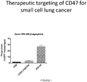
- CD47 is a valuable target for anticancer therapy due to its function as an inhibitor of macrophage phagocytosis as well as its broad expression on a variety of human neoplasms.
- SIRP ⁇ signal-regulatory protein ⁇
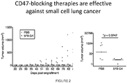
- CD47 is able to transduce inhibitory signals that prevent phagocytosis. Blocking the interaction between CD47 and SIRP ⁇ with antibodies not only stimulates macrophages to engulf cancer cells in vitro but also exerts robust anticancer effects in vivo.
- Other CD47 blocking agents include "next-generation" CD47 antagonists that bind and block human CD47 with extraordinarily high affinity.
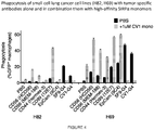
- high-affinity SIRP ⁇ variants can reduce the threshold for macrophage activation and promote phagocytic response driven by tumor-specific antibodies.
- the degree to which the anticancer activity of a given therapeutic antibody is enhanced by CD47 blockade likely depends on multiple factors, including the levels of antigen expression on the surface of malignant cells, the isotype of its heavy chain, and the orientation assumed by the antibody upon antigen binding, which affects its ability to engage Fc receptors on immune effectors.
- High-affinity SIRP ⁇ monomers represent therefore a rapid, safe and effective alternative to several other approaches, including drug/toxin conjugation strategies, that have been pursued in this direction.
- the invention provides a targeted agent that selectively blocks CD47 binding to SIRP ⁇ for use in the treatment of small cell lung cancer in a patient, by administering:
- the invention further provides an antibody that specifically binds to one or more cell surface antigens on lung cancer cells, for use in the treatment of small cell lung cancer in a patient, by administering:
- the lung cancer is small cell lung cancer.
- the therapy is targeted at one or more cell-surface antigens, including CD24, CD166, CD56, CD326, CD298, CD29, CD63, CD9, CD164, CD99, CD46, CD59, CD57, CD165, EpCAM, etc.
- the targeted therapy comprises administering to an individual suffering from lung cancer a therapeutic dose of an antibody that specifically binds to a cell surface marker selected from CD24, CD166, CD56, CD326, CD298, CD29, CD63, CD9, CD164, CD99, CD46, CD59, CD57, CD165 and EpCAM.
- the targeted therapy is combined with a CD47 blocking agent.
- Cancer cells evade macrophage surveillance by upregulation of CD47 expression.
- Administration of agents that mask the CD47 protein e.g. antibodies that bind to CD47 or SIRP ⁇ and prevent interaction between CD47 and SIRP ⁇ , are administered to a patient, which increases the clearance of cancer cells via phagocytosis.
- the agent that blocks CD47 is combined with monoclonal antibodies directed against one or more lung cancer cell markers, which compositions can be synergistic in enhancing phagocytosis and elimination of cancer cells as compared to the use of single agents.
- anti-CD47 and anti-CD56 include anti-CD47 and anti-CD56; anti-CD47 and anti-CD44, anti-CD47 and anti-CD99, anti-CD47 and anti-EpCam.
- the anti-CD47 reagent is a high affinity SIRP ⁇ polypeptide, which can be provided in the form of a monomer or a multimer, e.g. as a fusion protein with an IgG Fc polypeptide.
- the therapy provides for a multispecific antibody that targets CD47 and a second cancer cell marker, including multispecific antibodies that target CD47 and CD56; CD47 and CD44, CD47 and EpCam, etc.
- Compositions of such multispecific antibodies are also provided, where the multispecific antibody is desirably human or humanized; and may be modified to extend the blood half-life, e.g. by pegylation, etc.
- a therapeutic agent e.g. an antibody
- a marker of lung cancer e.g. targeted to one or more cell-surface antigens, including CD24, CD166, CD56, CD326, CD298, CD29, CD63, CD9, CD164, CD99, CD46, CD59, CD57, CD165, EpCAM, etc.
- a combination, e.g. a synergistic combination, of agents is disclosed, wherein one agent is an anti-CD47 blocking agent, and the second agent is targeted to a lung cancer marker, e.g. CD24, CD166, CD56, CD326, CD298, CD29, CD63, CD9, CD164, CD99, CD46, CD59, CD57, CD165, EpCAM, etc.
- Synergistic combination may provide for a therapeutic effect that is comparable to the effectiveness of a monotherapy, i.e. the individual components of the combination, while reducing adverse side effects, e.g. damage to non-targeted tissues, immune status, and other clinical indicia.
- synergistic combinations may provide for an improved effectiveness when compared to the effectiveness of a monotherapy, i.e. the individual components of the combination, which effect may be measured by total tumor cell number; length of time to relapse; and other indicia of patient health.
- Synergistic combinations disclosed herein combine an agent that is targeted to inhibit or block CD47 function; and an agent that is targeted to inhibit or block a second lung cancer cell marker, usually a cell surface marker.
- the combination may be provided with a combination of agents, e.g. two distinct proteins, each of which is specific for a different marker; or may be provided as a multispecific agent, e.g. antibody, that combines specificity for two or more different markers.
- Combination therapy refers to those situations in which a subject is simultaneously exposed to two or more therapeutic regimens (e.g., two or more therapeutic agents).
- two or more agents may be administered simultaneously; in some embodiments, such agents may be administered sequentially; in some embodiments, such agents are administered in overlapping dosing regimens.
- Dosage Form refers to a physically discrete unit of an active agent (e.g., a therapeutic or diagnostic agent) for administration to a subject.
- Each unit contains a predetermined quantity of active agent.
- such quantity is a unit dosage amount (or a whole fraction thereof) appropriate for administration in accordance with a dosing regimen that has been determined to correlate with a desired or beneficial outcome when administered to a relevant population (i.e., with a therapeutic dosing regimen).
- a dosage amount or a whole fraction thereof
- the total amount of a therapeutic composition or agent administered to a particular subject is determined by one or more attending physicians and may involve administration of multiple dosage forms.
- Dosing regimen refers to a set of unit doses (typically more than one) that are administered individually to a subject, typically separated by periods of time.
- a given therapeutic agent has a recommended dosing regimen, which may involve one or more doses.
- a dosing regimen comprises a plurality of doses each of which are separated from one another by a time period of the same length; in some embodiments, a dosing regimen comprises a plurality of doses and at least two different time periods separating individual doses. In some embodiments, all doses within a dosing regimen are of the same unit dose amount.
- a dosing regimen comprises a first dose in a first dose amount, followed by one or more additional doses in a second dose amount different from the first dose amount.
- a dosing regimen comprises a first dose in a first dose amount, followed by one or more additional doses in a second dose amount same as the first dose amount.
- a dosing regimen is correlated with a desired or beneficial outcome when administered across a relevant population (i.e., is a therapeutic dosing regimen).
- CD47 polypeptides The three transcript variants of human CD 47 (variant 1, NM 001777; variant 2, NM 198793; and variant 3, NM 001025079) encode three isoforms of CD47 polypeptide.
- CD47 isoform 1 (NP 001768), the longest of the three isoforms, is 323 amino acids long.
- CD47 isoform 2 (NP 942088) is 305 amino acid long.
- CD47 isoform 3 is 312 amino acids long.
- the three isoforms are identical in sequence in the first 303 amino acids.
- Amino acids 1-8 comprise the signal sequence
- amino acids 9-142 comprise the CD47 immunoglobulin like domain, which is the soluble fragment
- amino acids 143-300 is the transmembrane domain.
- a “functional derivative” of a native sequence polypeptide is a compound having a qualitative biological property in common with a native sequence polypeptide.
- “Functional derivatives” include, but are not limited to, fragments of a native sequence and derivatives of a native sequence polypeptide and its fragments, provided that they have a biological activity in common with a corresponding native sequence polypeptide.
- the term “derivative” encompasses both amino acid sequence variants of polypeptide and covalent modifications thereof. Derivatives and fusion of soluble CD47 find use as CD47 mimetic molecules.
- the first 142 amino acids of CD47 polypeptide comprise the extracellular region of CD47 (SEQ ID NO: 1).
- the three isoforms have identical amino acid sequence in the extracellular region, and thus any of the isoforms are can be used to generate soluble CD47.
- “Soluble CD47” is a CD47 protein that lacks the transmembrane domain. Soluble CD47 is secreted out of the cell expressing it instead of being localized at the cell surface.
- In vitro assays for CD47 biological activity include, e.g. inhibition of phagocytosis of porcine cells by human macrophages, binding to SIRP ⁇ receptor, SIRP ⁇ tyrosine phosphorylation, etc.
- An exemplary assay for CD47 biological activity contacts a human macrophage composition in the presence of a candidate agent. The cells are incubated with the candidate agent for about 30 minutes and lysed. The cell lysate is mixed with anti-human SIRP ⁇ antibodies to immunoprecipitate SIRP ⁇ . Precipitated proteins are resolved by SDS PAGE, then transferred to nitrocellulose and probed with antibodies specific for phosphotyrosine.
- a candidate agent useful as a CD47 mimetic increases SIRP ⁇ tyrosine phosphorylation by at least 10%, or up to 20%, or 50%, or 70% or 80% or up to about 90% compared to the level of phosphorylation observed in the absence of candidate agent.
- Another exemplary assay for CD47 biological activity measures phagocytosis of hematopoietic cells by human macrophages.
- a candidate agent useful as a CD47 mimetic results in the down regulation of phagocytosis by at least about 10%, at least about 20%, at least about 50%, at least about 70%, at least about 80%, or up to about 90% compared to level of phagocytosis observed in absence of candidate agent.
- manipulating phagocytosis is meant an up-regulation or a down-regulation in phagocytosis by at least about 10%, or up to 20%, or 50%, or 70% or 80% or up to about 90% compared to level of phagocytosis observed in absence of intervention.
- manipulating phagocytosis means a down-regulation in phagocytosis by at least about 10%, or up to 20%, or 50%, or 70% or 80% or up to about 90% compared to level of phagocytosis observed in absence of intervention.
- Anti-CD47 agent refers to any agent that reduces the binding of CD47 (e.g., on a target cell) to SIRP ⁇ (e.g., on a phagocytic cell).
- suitable anti-CD47 reagents include SIRP ⁇ reagents, including without limitation high affinity SIRP ⁇ polypeptides, anti-SIRPa antibodies, soluble CD47 polypeptides, and anti-CD47 antibodies or antibody fragments.
- a suitable anti-CD47 agent e.g. an anti-CD47 antibody, a SIRP ⁇ reagent, etc.
- a suitable anti-CD47 agent e.g., an anti-SIRP ⁇ antibody, a soluble CD47 polypeptide, etc.
- a suitable anti-CD47 agent that binds SIRP ⁇ does not activate SIRP ⁇ (e.g., in the SIRP ⁇ -expressing phagocytic cell).
- a suitable anti-CD47 agent can be assessed by assaying the agent (further described below).
- target cells are incubated in the presence or absence of the candidate agent.
- An agent for use in the invention will up-regulate phagocytosis by at least 10% (e.g., at least 20%, at least 30%, at least 40%, at least 50%, at least 60%, at least 70%, at least 80%, at least 90%, at least 100%, at least 120%, at least 140%, at least 160%, at least 180%, or at least 200%) compared to phagocytosis in the absence of the agent.
- an in vitro assay for levels of tyrosine phosphorylation of SIRP ⁇ will show a decrease in phosphorylation by at least 5% (e.g., at least 10%, at least 15%, at least 20%, at least 30%, at least 40%, at least 50%, at least 60%, at least 70%, at least 80%, at least 90%, or 100%) compared to phosphorylation observed in absence of the candidate agent.
- the anti-CD47 agent does not activate CD47 upon binding.
- CD47 When CD47 is activated, a process akin to apoptosis (i.e., programmed cell death) may occur ( Manna and Frazier, Cancer Research, 64, 1026-1036, Feb. 1 2004 ).
- the anti-CD47 agent does not directly induce cell death of a CD47-expressing cell by apoptosis.
- SIRP ⁇ reagent comprises the portion of SIRP ⁇ that is sufficient to bind CD47 at a recognizable affinity, which normally lies between the signal sequence and the transmembrane domain, or a fragment thereof that retains the binding activity.
- a suitable SIRP ⁇ reagent reduces (e.g., blocks, prevents, etc.) the interaction between the native proteins SIRP ⁇ and CD47.
- the SIRP ⁇ reagent will usually comprise at least the d1 domain of SIRP ⁇ .
- a SIRP ⁇ reagent is a fusion protein, e.g., fused in frame with a second polypeptide.
- the second polypeptide is capable of increasing the size of the fusion protein, e.g., so that the fusion protein will not be cleared from the circulation rapidly.
- the second polypeptide is part or whole of an immunoglobulin Fc region. The Fc region aids in phagocytosis by providing an "eat me” signal, which enhances the block of the "don't eat me” signal provided by the high affinity SIRP ⁇ reagent.
- the second polypeptide is any suitable polypeptide that is substantially similar to Fc, e.g., providing increased size, multimerization domains, and/or additional binding or interaction with Ig molecules.
- a subject anti-CD47 agent is a "high affinity SIRP ⁇ reagent", which includes SIRP ⁇ -derived polypeptides and analogs thereof.
- High affinity SIRP ⁇ reagents are described in international patent publication WO 2013/109752 .
- High affinity SIRP ⁇ reagents are variants of the native SIRP ⁇ protein.
- a high affinity SIRP ⁇ reagent is soluble, where the polypeptide lacks the SIRP ⁇ transmembrane domain and comprises at least one amino acid change relative to the wild-type SIRP ⁇ sequence, and wherein the amino acid change increases the affinity of the SIRP ⁇ polypeptide binding to CD47, for example by decreasing the off-rate by at least 10-fold, at least 20-fold, at least 50-fold, at least 100-fold, at least 500-fold, or more.
- a high affinity SIRP ⁇ reagent comprises the portion of SIRP ⁇ that is sufficient to bind CD47 at a recognizable affinity, e.g., high affinity, which normally lies between the signal sequence and the transmembrane domain, or a fragment thereof that retains the binding activity.
- the high affinity SIRP ⁇ reagent will usually comprise at least the d1 domain of SIRP ⁇ with modified amino acid residues to increase affinity.
- a SIRP ⁇ reagent can be used as a "monomer", in which the binding domain of SIRP ⁇ is used, but where the binding domain is provided as a soluble monomeric protein.
- a SIRP ⁇ variant for use in the present invention is a fusion protein, e.g., fused in frame with a second polypeptide, particularly where the second polypeptide provides for multimerization.
- the second polypeptide is part or whole of an immunoglobulin Fc region. The Fc region aids in phagocytosis by providing an "eat me" signal, which enhances the block of the "don't eat me” signal provided by the high affinity SIRP ⁇ reagent.
- the second polypeptide is any suitable polypeptide that is substantially similar to Fc, e.g., providing increased size, multimerization domains, and/or additional binding or interaction with Ig molecules.
- high affinity SIRP ⁇ reagents comprise a d1 domain of human SIRP ⁇ , with at least one amino acid change relative to the wild-type sequence within the d1 domain.
- a high affinity SIRP ⁇ reagent optionally comprises additional amino acid sequences, for example antibody Fc sequences; portions of the wild-type human SIRP ⁇ protein other than the d1 domain, including without limitation residues 150 to 374 of the native protein orfragments thereof, usually fragments contiguous with the d1 domain; and the like.
- High affinity SIRP ⁇ reagents may be monomeric or multimeric, i.e. dimer, trimer, tetramer, etc.
- a subject anti-CD47 agent is an antibody that specifically binds CD47 (i.e., an anti-CD47 antibody) and reduces the interaction between CD47 on one cell (e.g., an infected cell) and SIRP ⁇ on another cell (e.g., a phagocytic cell).
- a suitable anti-CD47 antibody does not activate CD47 upon binding, for example an antibody that does not induce apoptosis upon binding.
- suitable antibodies include clones B6H12, 5F9, 8B6, and C3 (for example as described in International Patent Publication WO 2011/143624 ).
- Suitable anti-CD47 antibodies include fully human, humanized or chimeric versions of such antibodies.
- Humanized antibodies e.g., hu5F9-G4
- caninized, felinized, etc. antibodies are especially useful for applications in dogs, cats, and other species respectively.
- Antibodies of interest include humanized antibodies, or caninized, felinized, equinized, bovinized, porcinized, etc., antibodies, and variants thereof.
- a subject anti-CD47 agent is an antibody that specifically binds SIRP ⁇ (i.e., an anti-SIRPa antibody) and reduces the interaction between CD47 on one cell (e.g., an infected cell) and SIRP ⁇ on another cell (e.g., a phagocytic cell).
- Suitable anti-SIRPa antibodies can bind SIRP ⁇ without activating or stimulating signaling through SIRP ⁇ because activation of SIRP ⁇ would inhibit phagocytosis. Instead, suitable anti-SIRPa antibodies facilitate the preferential phagocytosis of inflicted cells over normal cells.
- a suitable anti-SIRPa antibody specifically binds SIRP ⁇ (without activating/stimulating enough of a signaling response to inhibit phagocytosis) and blocks an interaction between SIRP ⁇ and CD47.
- Suitable anti-SIRPa antibodies include fully human, humanized or chimeric versions of such antibodies. Humanized antibodies are especially useful for in vivo applications in humans due to their low antigenicity. Similarly caninized, felinized, etc. antibodies are especially useful for applications in dogs, cats, and other species respectively.
- Antibodies of interest include humanized antibodies, or caninized, felinized, equinized, bovinized, porcinized, etc., antibodies, and variants thereof.
- antibody or “antibody moiety” is intended to include any polypeptide chain-containing molecular structure with a specific shape that fits to and recognizes an epitope, where one or more non-covalent binding interactions stabilize the complex between the molecular structure and the epitope.
- Antibodies utilized in the present invention may be polyclonal antibodies, although monoclonal antibodies are preferred because they may be reproduced by cell culture or recombinantly, and can be modified to reduce their antigenicity.
- Polyclonal antibodies can be raised by a standard protocol by injecting a production animal with an antigenic composition. See, e.g., Harlow and Lane, Antibodies: A Laboratory Manual, Cold Spring Harbor Laboratory, 1988 .
- antibodies may be raised by immunizing the production animal with the protein and a suitable adjuvant (e.g., Freund's, Freund's complete, oil-in-water emulsions, etc.)
- a suitable adjuvant e.g., Freund's, Freund's complete, oil-in-water emulsions, etc.
- conjugate proteins that are commercially available for such use include bovine serum albumin (BSA) and keyhole limpet hemocyanin (KLH).
- BSA bovine serum albumin
- KLH keyhole limpet hemocyanin
- peptides derived from the full sequence may be utilized.
- a superior immune response may be elicited if the polypeptide is joined to a carrier protein, such as ovalbumin, BSA or KLH.
- hybridomas may be formed by isolating the stimulated immune cells, such as those from the spleen of the inoculated animal.
- immortalized cells such as myeloma cells or transformed cells, which are capable of replicating indefinitely in cell culture, thereby producing an immortal, immunoglobulin-secreting cell line.
- immortalized cells such as myeloma cells or transformed cells, which are capable of replicating indefinitely in cell culture, thereby producing an immortal, immunoglobulin-secreting cell line.
- the antibodies or antigen binding fragments may be produced by genetic engineering. Humanized, chimeric, or xenogeneic human antibodies, which produce less of an immune response when administered to humans, are preferred for use in the present invention.
- immunoglobulin fragments comprising the epitope binding site (e.g., Fab', F(ab') 2 , or other fragments) are useful as antibody moieties in the present invention.
- Such antibody fragments may be generated from whole immunoglobulins by ricin, pepsin, papain, or other protease cleavage.
- "Fragment,” or minimal immunoglobulins may be designed utilizing recombinant immunoglobulin techniques.
- Fv immunoglobulins for use in the present invention may be produced by linking a variable light chain region to a variable heavy chain region via a peptide linker (e.g., poly-glycine or another sequence which does not form an alpha helix or beta sheet motif).
- a peptide linker e.g., poly-glycine or another sequence which does not form an alpha helix or beta sheet motif.
- Antibodies include free antibodies and antigen binding fragments derived therefrom, and conjugates, e.g. pegylated antibodies, drug, radioisotope, or toxin conjugates, and the like. Monoclonal antibodies directed against a specific epitope, or combination of epitopes, will allow for the targeting and/or depletion of cellular populations expressing the marker.
- Various techniques can be utilized using monoclonal antibodies to screen for cellular populations expressing the marker(s), and include magnetic separation using antibody-coated magnetic beads, "panning" with antibody attached to a solid matrix (i.e., plate), and flow cytometry (See, e.g., U.S. Pat. No. 5,985,660 ; and Morrison et al. Cell, 96:737-49 (1999 )). These techniques allow for the screening of particular populations of cells; in immunohistochemistry of biopsy samples; in detecting the presence of markers shed by cancer cells into the blood and other biologic fluids, and the like.
- Humanized versions of such antibodies are also contemplated for use in this invention. Humanized antibodies are especially useful for in vivo applications in humans due to their low antigenicity.
- multispecific or bispecific antibody refers to a synthetic or recombinant antibody that recognizes more than one protein.
- Bispecific antibodies directed against a combination of epitopes will allow for the targeting and/or depletion of cellular populations expressing the combination of epitopes.
- Exemplary bi-specific antibodies include those targeting a combination of CD47 and an SCLC cancer cell marker. Generation of bi-specific antibodies are described in the literature. Higher order specificities, e.g. trispecific antibodies, are described by Holliger and Hudson (2005) Nature Biotechnology 23:1126-1136 .
- the efficacy of a CD47 inhibitor can be assessed by assaying CD47 activity.
- the above-mentioned assays or modified versions thereof are used.
- SCLC are incubated with bone marrow derived macrophages, in the presence or absence of the candidate agent.
- An inhibitor of the cell surface CD47 will up-regulate phagocytosis by at least about 10%, or up to 20%, or 50%, or 70% or 80% or up to about 90% compared to the phagocytosis in absence of the candidate agent.
- an in vitro assay for levels of tyrosine phosphorylation of SIRP ⁇ will show a decrease in phosphorylation by at least about 10%, or up to 20%, or 50%, or 70% or 80% or up to about 90% compared to phosphorylation observed in absence of the candidate agent.
- the agent, or a pharmaceutical composition comprising the agent is provided in an amount effective to detectably inhibit the binding of CD47 to SIRP ⁇ receptor present on the surface of phagocytic cells.
- the effective amount is determined via empirical testing routine in the art. The effective amount may vary depending on the number of cells being transplanted, site of transplantation and factors specific to the transplant recipient.
- phagocytic cells and “phagocytes” are used interchangeably herein to refer to a cell that is capable of phagocytosis.
- phagocytes There are four main categories of phagocytes: macrophages, mononuclear cells (histiocytes and monocytes); polymorphonuclear leukocytes (neutrophils) and dendritic cells.
- biological sample encompasses a variety of sample types obtained from an organism and can be used in a diagnostic or monitoring assay.
- the term encompasses blood and other liquid samples of biological origin, solid tissue samples, such as a biopsy specimen or tissue cultures or cells derived therefrom and the progeny thereof.
- the term encompasses samples that have been manipulated in any way after their procurement, such as by treatment with reagents, solubilization, or enrichment for certain components.
- the term encompasses a clinical sample, and also includes cells in cell culture, cell supernatants, cell lysates, serum, plasma, biological fluids, and tissue samples.
- treatment means treating, “treating”, “treat” and the like to generally refer to obtaining a desired pharmacologic and/or physiologic effect.
- the effect may be prophylactic in terms of completely or partially preventing a disease or symptom thereof and/or may be therapeutic in terms of a partial or complete stabilization or cure for a disease and/or adverse effect attributable to the disease.
- Treatment covers any treatment of a disease in a mammal, e.g.
- mice, rat, rabbit, pig, primate including humans and other apes, particularly a human, and includes: (a) preventing the disease or symptom from occurring in a subject which may be predisposed to the disease or symptom but has not yet been diagnosed as having it; (b) inhibiting the disease symptom, i.e., arresting its development; or (c) relieving the disease symptom, i.e., causing regression of the disease or symptom.
- a "host cell”, as used herein, refers to a microorganism or a eukaryotic cell or cell line cultured as a unicellular entity which can be, or has been, used as a recipient for a recombinant vector or other transfer polynucleotides, and include the progeny of the original cell which has been transfected. It is understood that the progeny of a single cell may not necessarily be completely identical in morphology or in genomic or total DNA complement as the original parent, due to natural, accidental, or deliberate mutation.
- cancer neoplasm
- tumor tumor
- tumor tumor
- tumor tumor
- tumor tumor
- tumor tumor
- cells of interest for detection or treatment in the present application include precancerous (e.g., benign), malignant, pre-metastatic, metastatic, and non-metastatic cells. Detection of cancerous cells is of particular interest.
- normal as used in the context of "normal cell,” is meant to refer to a cell of an untransformed phenotype or exhibiting a morphology of a non-transformed cell of the tissue type being examined.
- Cancerous phenotype generally refers to any of a variety of biological phenomena that are characteristic of a cancerous cell, which phenomena can vary with the type of cancer.
- the cancerous phenotype is generally identified by abnormalities in, for example, cell growth or proliferation (e.g., uncontrolled growth or proliferation), regulation of the cell cycle, cell mobility, cell-cell interaction, or metastasis, etc.
- “Therapeutic target” refers to a gene or gene product that, upon modulation of its activity (e.g., by modulation of expression, biological activity, and the like), can provide for modulation of the cancerous phenotype.
- modulation is meant to refer to an increase or a decrease in the indicated phenomenon (e.g., modulation of a biological activity refers to an increase in a biological activity or a decrease in a biological activity).
- Lung carcinoma is the leading cause of cancer-related death worldwide. About 85% of cases are related to cigarette smoking. Symptoms can include cough, chest discomfort or pain, weight loss, and, less commonly, hemoptysis; however, many patients present with metastatic disease without any clinical symptoms. The diagnosis is typically made by chest x-ray or CT and confirmed by biopsy. Depending on the stage of the disease, treatment includes surgery, chemotherapy, radiation therapy, or a combination. For the past several decades, the prognosis for a lung cancer patient has been poor, particularly for patients with stage IV (metastatic) disease.
- stage IV metastatic
- Respiratory epithelial cells require prolonged exposure to cancer-promoting agents and accumulation of multiple genetic mutations before becoming neoplastic (an effect called field carcinogenesis).
- secondary or additional mutations in genes that stimulate cell growth K-ras, MYC
- EGFR growth factor receptor signaling
- p53 APC
- Other mutations that may be responsible include the EML-4-ALK translocation and mutations in ROS-1, BRAF, and PI3KCA.
- Driver mutations can cause or contribute to lung cancer among smokers, these mutations are particularly likely to be a cause of lung cancer among nonsmokers.
- Chest x-ray is often the initial imaging test. It may show clearly defined abnormalities, such as a single mass or multifocal masses or a solitary pulmonary nodule, an enlarged hilum, widened mediastinum, tracheobronchial narrowing, atelectasis, non-resolving parenchymal infiltrates, cavitary lesions, or unexplained pleural thickening or effusion.
- CT shows many characteristic anatomic patterns and appearances that may strongly suggest the diagnosis.
- CT also can guide core needle biopsy of accessible lesions and is useful for staging. If a lesion found on a plain x-ray is highly likely to be lung cancer, PET-CT may be done. This study combines anatomic imaging from CT with functional imaging from PET. The PET images can help differentiate inflammatory and malignant processes.
- SCLC has 2 stages: limited and extensive.
- Limited-stage SCLC disease is cancer confined to one hemithorax (including ipsilateral lymph nodes) that can be encompassed within one tolerable radiation therapy port, unless there is a pleural or pericardial effusion.
- Extensive-stage disease is cancer outside a single hemithorax or the presence of malignant cells detected in pleural or pericardial effusions.
- Less than one third of patients with SCLC will present with limited-stage disease; the remainder of patients often have extensive distant metastases.
- the overall prognosis for SCLC is poor.
- the median survival time for limited-stage SCLC is 20 mo, with a 5-yr survival rate of 20%. Patients with extensive-stage SCLC do especially poorly, with a 5-yr survival rate of ⁇ 1%.
- NSCLC has 4 stages, I through IV (using the TNM system).
- TNM staging is based on tumor size, tumor and lymph node location, and the presence or absence of distant metastases.
- the 5-yr survival rate of patients with NSCLC varies by stage, from 60 to 70% for patients with stage I disease to ⁇ 1% for patients with stage IV disease.
- SCLC of any stage is typically initially responsive to treatment, but responses are usually short-lived.
- Chemotherapy with or without radiation therapy, is given depending on the stage of disease. In many patients, chemotherapy prolongs survival and improves quality of life enough to warrant its use.
- Surgery generally plays no role in treatment of SCLC, although it may be curative in the rare patient who has a small focal tumor without spread (such as a solitary pulmonary nodule) who underwent surgical resection before the tumor was identified as SCLC.
- Chemotherapy regimens of etoposide and a platinum compound are commonly used, as are other drugs, such as irinotecan, topotecan, vinca alkaloids (vinblastine, vincristine, vinorelbine), alkylating agents (cyclophosphamide, ifosfamide), doxorubicin, taxanes (docetaxel, paclitaxel), and gemcitabine.
- drugs such as irinotecan, topotecan, vinca alkaloids (vinblastine, vincristine, vinorelbine), alkylating agents (cyclophosphamide, ifosfamide), doxorubicin, taxanes (docetaxel, paclitaxel), and gemcitabine.
- irinotecan vinca alkaloids
- vincristine vincristine
- vinorelbine alkylating agents
- doxorubicin doxorubicin
- taxanes docetax
- Treatment for NSCLC typically involves assessment of eligibility for surgery followed by choice of surgery, chemotherapy, radiation therapy, or a combination of modalities as appropriate, depending on tumor type and stage.
- a targeted therapeutic agent directed to a lung cancer cell surface marker including without limitation CD24, CD166, CD56, CD326, CD298, CD29, CD63, CD9, CD164, CD99, CD46, CD59, CD57, CD 165 , EpCAM, etc.
- the marker may be one of CD56, CD44, CD99 and EpCam.
- the targeted therapeutic agent may be combined with a CD47 blocking agent, e.g. soluble SIRP ⁇ monomer or multimer, an anti-CD47 antibody, small molecule, etc.
- the cancer is SCLC.
- Reducing growth of cancer cells includes, but is not limited to, reducing proliferation of cancer cells, and reducing the incidence of a non-cancerous cell becoming a cancerous cell. Whether a reduction in cancer cell growth has been achieved can be readily determined using any known assay, including, but not limited to, [ 3 H]-thymidine incorporation; counting cell number over a period of time; detecting and/or measuring a marker associated with SCLC, etc.
- Whether a substance, or a specific amount of the substance, is effective in treating cancer can be assessed using any of a variety of known diagnostic assays for cancer, including, but not limited to biopsy, contrast radiographic studies, CAT scan, and detection of a tumor marker associated with cancer in the blood of the individual.
- the substance can be administered systemically or locally, usually systemically.
- An agent e.g. a chemotherapeutic drug that reduces cancer cell growth
- a method of delivering a drug to a cancer cell comprising administering a drug-antibody complex to a subject, wherein the antibody is specific for a cancer-associated polypeptide, and the drug is one that reduces cancer cell growth, a variety of which are known in the art.
- Targeting can be accomplished by coupling (e.g., linking, directly or via a linker molecule, either covalently or non-covalently, so as to form a drug-antibody complex) a drug to an antibody specific for a cancer-associated polypeptide.
- Methods of coupling a drug to an antibody are well known in the art and need not be elaborated upon herein.
- a bi-specific antibody may be used.
- a bi-specific antibody in which one antigen binding domain is directed against CD47 and the other antigen binding domain is directed against a cancer cell marker, such as CD24, CD166, CD56, CD326, CD298, CD29, CD63, CD9, CD164, CD99, CD46, CD59, CD57, CD165, EpCAM, etc. may be used.
- antibody or “antibody moiety” is intended to include any polypeptide chain-containing molecular structure that has a specific shape which fits to and recognizes an epitope, where one or more non-covalent binding interactions stabilize the complex between the molecular structure and the epitope.
- hybridomas may be formed by isolating the stimulated immune cells, such as those from the spleen of the inoculated animal. These cells are then fused to immortalized cells, such as myeloma cells or transformed cells, which are capable of replicating indefinitely in cell culture, thereby producing an immortal, immunoglobulin-secreting cell line.
- the immortal cell line utilized is preferably selected to be deficient in enzymes necessary for the utilization of certain nutrients.
- Many such cell lines (such as myelomas) are known to those skilled in the art, and include, for example: thymidine kinase (TK) or hypoxanthine-guanine phosphoriboxyl transferase (HGPRT). These deficiencies allow selection for fused cells according to their ability to grow on, for example, hypoxanthine aminopterinthymidine medium (HAT).
- TK thymidine kinase
- HGPRT hypoxanthine-guanine phosphoriboxyl transferase
- Antibodies which have a reduced propensity to induce a violent or detrimental immune response in humans (such as anaphylactic shock), and which also exhibit a reduced propensity for priming an immune response which would prevent repeated dosage with the antibody therapeutic or imaging agent (e.g., the human-anti-murine-antibody "HAMA" response), are preferred for use in the invention. These antibodies are preferred for all administrative routes. Thus, humanized, chimeric, or xenogenic human antibodies, which produce less of an immune response when administered to humans, are preferred for use in the present invention.
- Chimeric antibodies may be made by recombinant means by combining the murine variable light and heavy chain regions (VK and VH), obtained from a murine (or other animal-derived) hybridoma clone, with the human constant light and heavy chain regions, in order to produce an antibody with predominantly human domains.
- VK and VH murine variable light and heavy chain regions
- the production of such chimeric antibodies is well known in the art, and may be achieved by standard means (as described, e.g., in U.S. Pat. No. 5,624,659 ).
- Humanized antibodies are engineered to contain even more human-like immunoglobulin domains, and incorporate only the complementarity-determining regions of the animal-derived antibody.
- polyclonal or monoclonal antibodies may be produced from animals which have been genetically altered to produce human immunoglobulins.
- the transgenic animal may be produced by initially producing a "knock-out" animal which does not produce the animal's natural antibodies, and stably transforming the animal with a human antibody locus (e.g., by the use of a human artificial chromosome). Only human antibodies are then made by the animal. Techniques for generating such animals, and deriving antibodies therefrom, are described in U.S. Pat. Nos. 6,162,963 and 6,150,584 . Such fully human xenogenic antibodies are a preferred antibody for use in the compositions of the present invention.
- single chain antibodies can be produced from phage libraries containing human variable regions. See U.S. Pat. No. 6,174,708 .
- immunoglobulin fragments comprising the epitope binding site (e.g., Fab', F(ab') 2 , or other fragments) are useful as antibody moieties in the present invention.
- Such antibody fragments may be generated from whole immunoglobulins by ficin, pepsin, papain, or other protease cleavage. "Fragment,” or minimal immunoglobulins may be designed utilizing recombinant immunoglobulin techniques.
- Fv immunoglobulins for use in the present invention may be produced by linking a variable light chain region to a variable heavy chain region via a peptide linker (e.g., poly-glycine or another sequence which does not form an alpha helix or beta sheet motif).
- a peptide linker e.g., poly-glycine or another sequence which does not form an alpha helix or beta sheet motif.
- Fv fragments are heterodimers of the variable heavy chain domain (V H ) and the variable light chain domain (V L ).
- the heterodimers of heavy and light chain domains that occur in whole IgG, for example, are connected by a disulfide bond.
- Recombinant Fvs in which V H and V L are connected by a peptide linker are typically stable, see, for example, Huston et al., Proc. Natl. Acad, Sci. USA 85:5879 5883 (1988 ) and Bird et al., Science 242:423 426 (1988 ).
- These are single chain Fvs which have been found to retain specificity and affinity and have been shown to be useful for imaging tumors and to make recombinant immunotoxins for tumor therapy.
- Derivatized polypeptides with added chemical linkers detectable moieties such as fluorescent dyes, enzymes, substrates, chemiluminescent moieties, specific binding moieties such as streptavidin, avidin, or biotin, or drug conjugates may be utilized in the compositions of the present invention.
- polypeptide reagents are coupled or conjugated to one or more therapeutic, cytotoxic, or imaging moieties.
- cytotoxic moiety simply means a moiety which inhibits cell growth or promotes cell death when proximate to or absorbed by the cell.
- Suitable cytotoxic moieties in this regard include radioactive isotopes (radionuclides), chemotoxic agents such as differentiation inducers and small chemotoxic drugs, toxin proteins, and derivatives thereof.
- Agents may be conjugated to a polypeptide reagent by any suitable technique, with appropriate consideration of the need for pharmokinetic stability and reduced overall toxicity to the patient.
- a therapeutic agent may be coupled to a suitable moiety either directly or indirectly (e.g. via a linker group).
- a direct reaction is possible when each possesses a functional group capable of reacting with the other.
- a nucleophilic group such as an amino or sulfhydryl group
- a carbonyl-containing group such as an anhydride or an acid halide, or with an alkyl group containing a good leaving group (e.g., a halide).
- a suitable chemical linker group may be used.
- a linker group can function as a spacer to distance a polypeptide reagent from an agent in order to avoid interference with binding capabilities.
- a linker group can also serve to increase the chemical reactivity of a substituent on a moiety or a polypeptide reagent, and thus increase the coupling efficiency.
- An increase in chemical reactivity may also facilitate the use of moieties, or functional groups on moieties, which otherwise would not be possible.
- Suitable linkage chemistries include maleimidyl linkers and alkyl halide linkers (which react with a sulfhydryl on the antibody moiety) and succinimidyl linkers (which react with a primary amine on the antibody moiety).
- Several primary amine and sulfhydryl groups are present on immunoglobulins, and additional groups may be designed into recombinant immunoglobulin molecules. It will be evident to those skilled in the art that a variety of bifunctional or polyfunctional reagents, both homo- and hetero-functional (such as those described in the catalog of the Pierce Chemical Co., Rockford, III.), may be employed as a linker group.
- Coupling may be effected, for example, through amino groups, carboxyl groups, sulfhydryl groups or oxidized carbohydrate residues.
- cytotoxic moieties may be coupled to a polypeptide reagent through a an oxidized carbohydrate group at a glycosylation site, as described in U.S. Pat. Nos. 5,057,313 and 5,156,840 .
- Yet another alternative method of coupling a polypeptide reagent to a cytotoxic or therapeutic moiety is by the use of a non-covalent binding pair, such as streptavidin/biotin, or avidin/biotin.
- a non-covalent binding pair such as streptavidin/biotin, or avidin/biotin.
- one member of the pair is covalently coupled to the anti-CD47, CV1, etc. moiety and the other member of the binding pair is covalently coupled to the therapeutic, cytotoxic, or imaging moiety.
- a cytotoxic moiety is more potent when free from the binding portion of a polypeptide reagent
- linker groups A number of different cleavable linker groups have been described. The mechanisms for the intracellular release of a cytotoxic moiety agent from these linker groups include cleavage by reduction of a disulfide bond (e.g., U.S. Pat. No. 4,489,710 ), by irradiation of a photolabile bond (e.g., U.S. Pat. No.
- conjugates with more than one moiety may be prepared in a variety of ways. For example, more than one moiety may be coupled directly to a polypeptide molecule, or linkers which provide multiple sites for attachment (e.g., dendrimers) can be used. Alternatively, a carrier with the capacity to hold more than one cytotoxic or imaging moiety can be used.
- a carrier may bear the agents in a variety of ways, including covalent bonding either directly or via a linker group, and non-covalent associations.
- Suitable covalent-bond carriers include proteins such as albumins (e.g., U.S. Pat. No. 4,507,234 ), peptides, and polysaccharides such as aminodextran (e.g., U.S. Pat. No. 4,699,784 ), each of which have multiple sites for the attachment of moieties.
- a carrier may also bear an agent by non-covalent associations, such as non-covalent bonding or by encapsulation, such as within a liposome vesicle (e.g., U.S. Pat. Nos.
- Encapsulation carriers are especially useful for imaging moiety conjugation to antibody moieties for use in the invention, as a sufficient amount of the imaging moiety (dye, magnetic resonance contrast reagent, etc.) for detection may be more easily associated with the antibody moiety.
- encapsulation carriers are also useful in chemotoxic therapeutic embodiments, as they can allow the therapeutic compositions to gradually release a chemotoxic moiety over time while concentrating it in the vicinity of the tumor cells.
- radionuclides for use as cytotoxic moieties are radionuclides which are suitable for pharmacological administration.
- radionuclides include 123 I, 125 I, 131 I, 90 Y, 211 At, 67 Cu, 186 Re, 188 Re, 212 Pb, and 212 Bi.
- Iodine and astatine isotopes are more preferred radionuclides for use in the therapeutic compositions of the present invention, as a large body of literature has been accumulated regarding their use.
- Preferred chemotoxic agents include small-molecule drugs such as carboplatin, cisplatin, vincristine, taxanes such as paclitaxel and doceltaxel, hydroxyurea, gemcitabine, vinorelbine, irinotecan, tirapazamine, matrilysin, methotrexate, pyrimidine and purine analogs, and other suitable small toxins known in the art.
- Preferred chemotoxin differentiation inducers include phorbol esters and butyric acid. Chemotoxic moieties may be directly conjugated to the antibody moiety via a chemical linker, or may be encapsulated in a carrier, which is in turn coupled to the antibody.
- Preferred toxin proteins for use as cytotoxic moieties include ricins A and B, abrin, diphtheria toxin, bryodin 1 and 2, momordin, trichokirin, cholera toxin, gelonin, Pseudomonas exotoxin, Shigella toxin, pokeweed antiviral protein, and other toxin proteins known in the medicinal biochemistry arts.
- ricins A and B abrin, diphtheria toxin, bryodin 1 and 2, momordin, trichokirin, cholera toxin, gelonin, Pseudomonas exotoxin, Shigella toxin, pokeweed antiviral protein, and other toxin proteins known in the medicinal biochemistry arts.
- these toxin agents may elicit undesirable immune responses in the patient, especially if injected intravascularly, it is preferred that they be encapsulated in a carrier for coupling to the antibody.
- a targeted therapeutic agent, or combination of targeted therapeutic agents may be administered separately or together; and will generally be administered within the same general time frame, e.g. within a week, within 3-4 days, within 1 day or simultaneously with each other.
- the agent or agents are mixed, prior to administration, with a non-toxic, pharmaceutically acceptable carrier substance.
- a non-toxic, pharmaceutically acceptable carrier substance usually, this will be an aqueous solution, such as normal saline or phosphate-buffered saline (PBS), Ringer's solution, lactate-Ringer's solution, or any isotonic physiologically acceptable solution for administration by the chosen means.
- PBS normal saline or phosphate-buffered saline
- Ringer's solution such as normal saline or phosphate-buffered saline (PBS), Ringer's solution, lactate-Ringer's solution, or any isotonic physiologically acceptable solution for administration by the chosen means.
- PBS normal saline or phosphate-buffered saline
- Ringer's solution such as normal saline or phosphate-buffered saline (PBS), Ringer's solution, lactate-Ringer's solution, or
- the agents may be stabilized against aggregation and polymerization with amino acids and non-ionic detergents, polysorbate, and polyethylene glycol.
- additional stabilizers may include various physiologically-acceptable carbohydrates and salts.
- polyvinylpyrrolidone may be added in addition to the amino acid.
- Suitable therapeutic immunoglobulin solutions which are stabilized for storage and administration to humans are described in U.S. Pat. No. 5,945,098 .
- Other agents such as human serum albumin (HSA), may be added to the therapeutic or imaging composition to stabilize the antibody conjugates.
- HSA human serum albumin
- compositions for use in the invention may be administered using any medically appropriate procedure, e.g., intravascular (intravenous, intraarterial, intracapillary) administration, injection into the tumor, etc.
- Intravascular injection may be by intravenous or intraarterial injection.
- the effective amount of the therapeutic compositions to be given to a particular patient will depend on a variety of factors, several of which will be different from patient to patient.
- a competent clinician will be able to determine an effective amount of a therapeutic composition to administer to a patient to retard the growth and promote the death of tumor cells. Dosage of the agents will depend on the treatment of the tumor, route of administration, the nature of the therapeutics, sensitivity of the tumor to the therapeutics, etc.
- a clinician can determine the maximum safe dose for an individual, depending on the route of administration. For instance, an intravenously administered dose may be more than an locally administered dose, given the greater body of fluid into which the therapeutic composition is being administered. Similarly, compositions which are rapidly cleared from the body may be administered at higher doses, or in repeated doses, in order to maintain a therapeutic concentration. Utilizing ordinary skill, the competent clinician will be able to optimize the dosage of a particular therapeutic or imaging composition in the course of routine clinical trials.
- an effective dosage will be 0.001 to 100 milligrams of antibody per kilogram subject body weight.
- the ratio of anti-CD47 to the second agent may range from 1:100; 1:50; 1:10; 1:5; 1:2; 1:1; 2:1; 5:1; 10:1; 50:1; 100:1.
- the agents can be administered to the subject in a series of more than one administration.
- regular periodic administration e.g., every 2-3 days
- antibody moieties which do not provoke HAMA or other immune responses are preferred.
- High-affinity SIRP ⁇ variants enhance macrophage destruction of small cell lung cancer.
- CD47 allows cancer cells to evade the immune system by signaling through SIRP ⁇ , an inhibitory receptor on macrophages.
- SIRP ⁇ an inhibitory receptor on macrophages.
- next-generation CD47 antagonists by engineering the N-terminal immunoglobulin domain of SIRP ⁇ .
- These "high-affinity SIRP ⁇ variants” have an affinity for human CD47 (K D ) as low as 11.1 pM, approximately 50,000-fold improved over wild-type SIRP ⁇ . When combined with tumor-specific antibodies, the high-affinity SIRP ⁇ variants act as immunotherapeutic adjuvants to maximize macrophage destruction of cancer cells.
- SCLC small cell lung cancer
- CD47-blocking therapies stimulate macrophage destruction of small cell lung cancer.
- SCLC Small cell lung cancer
- CD47 a cell-surface molecule that allows cancer cells to evade the immune system.
- CD47 promotes immune evasion by signaling through SIRPa, an inhibitory receptor on macrophages.
- CD47-blocking therapies could be applied to the treatment of SCLC.
- CD47-blocking therapies were able to induce macrophage phagocytosis of SCLC samples in vitro.
- CD47-blocking therapies also inhibited tumor growth and significantly prolonged survival of mice bearing SCLC tumors.
- SCLC Small cell lung cancer
- CD47 a cell-surface molecule, as a "marker of self" that prevents cells of the innate immune system from attacking hematologic malignancies and certain types of solid tumors.
- SIRPa inhibitory signals
- the CD47-SIRP ⁇ interaction represents a myeloid-specific immune checkpoint.
- CD47-SIRP ⁇ axis A number of reagents have been generated to disrupt signaling by the CD47-SIRP ⁇ axis, including anti-CD47 antibodies and engineered variants of its receptor, SIRP ⁇ . Recent studies have shown that blockade of CD47 lowers the threshold for macrophage phagocytosis of cancer. We hypothesized that SCLC cells also express CD47 and that CD47-blocking therapies could be used to stimulate macrophage phagocytosis of SCLC cells and inhibit growth of SCLC tumors in vivo.
- CD47-blocking therapies have been shown to enhance the response of macrophages to monoclonal antibodies.
- Monoclonal antibodies such as rituximab for lymphoma or trastuzumab for Her2 + breast cancer-have demonstrated immense success for the treatment of cancer.
- No monoclonal antibodies are clinically approved for the treatment of SCLC, thus, we aimed to identify new SCLC surface antigens that could be targeted with monoclonal antibodies.
- treatment with monoclonal antibodies can produce robust anti-tumor effects, they often fail to elicit cures when used as single agents, highlighting the need to improve the efficacy of these approaches. Therefore, we aimed to combine CD47-blocking therapies with other antibodies to achieve maximal anti-tumor responses against SCLC.
- CD47 is Expressed on the Surface of SCLC.
- CD47-blocking therapies could be applied to SCLC.
- CD47-blocking Antibodies Induce Phagocytosis of SCLC by Human Macrophages.
- To validate CD47 as a genuine therapeutic target on SCLC we performed in vitro phagocytosis assays using human macrophages and SCLC samples. Macrophages were co-cultured with SCLC cells in the presence of a vehicle control or anti-CD47 antibodies.
- NCI-H196 One cell line, NCI-H196, appeared to be resistant to phagocytosis, suggesting additional mechanisms modify the susceptibility of this cell line to macrophage attack.
- the patient-derived xenograft H29 was also subjected to phagocytosis assays with human macrophages. Treatment of this sample with anti-CD47 antibodies also resulted in a significant increase in phagocytosis ( Figure 5G ).
- CD47-blocking Antibodies Inhibit Growth of SCLC Tumors in vivo.
- NCI-H82 cells into the lower left flanks of NSG mice, which lack functional T cells, B cells, and NK cells but retain functional macrophages.
- Serum MCP-3 is a Biomarker of Response to CD47-blocking Therapies.
- To identify potential biomarkers of a response to CD47-blocking therapies we again engrafted mice with NCI-H82 cells. We allowed tumors to grow to approximately 1.5 cm in diameter and then we treated the mice with a single dose of vehicle control or anti-CD47 antibody clone Hu5F9-G4.
- MCP-3 may serve as a biomarker of response to CD47-blocking therapies in patients. Secretion of MCP-3 may be a positive feedback mechanism that recruits more macrophages to the tumor and could in part explain the robust effects of CD47-blocking therapies in vivo.
- CD47 was the most intensely staining surface antigen ( Figure 8B ).
- Another highly expressed antigen across all samples was CD56 (NCAM), a known marker of neuroendocrine tumors and a therapeutic target currently under evaluation for SCLC, thus validating our approach.
- NCAM CD56
- a number of other highly expressed surface antigens were also identified that could potentially be targeted by monoclonal antibody therapies, including CD24, CD29, and CD99 ( Figure 8B ).
- other immune checkpoint ligands such as CD80, CD86, PD-L1, or PD-L2 were not appreciably expressed on the surface of the SCLC samples.
- phagocytosis with Hu5F9-G4 was entirely blocked when combined with CV1, since CV1 competes for the same binding surface and binds with extremely high affinity.
- the anti-CD29 antibody was not able to induce phagocytosis even in combination with CV1, an important demonstration that additional factors such as surface binding geometry or the ability to engage Fc receptors may modify the response of macrophages to therapeutic antibodies.
- lorvotuzumab is under evaluation as a therapeutic agent for SCLC, we investigated its ability to induce phagocytosis over a varying range of concentrations. Treatment with lorvotuzumab alone produced a dose-response relationship for inducing macrophage phagocytosis. Importantly, we found that over each lorvotuzumab concentration tested, the addition of CV1 produced a greater degree of phagocytosis ( Figure 9C ). These findings demonstrate that CV1 could increase both the maximal efficacy and the potency of lorvotuzumab, as previously observed when CV1 was combined with rituximab, trastuzumab, and cetuximab.
- CD47-blockade was able to enhance the efficacy of lorvotuzumab, an antibody proceeding through clinical trials for SCLC as an antibody-drug conjugate (ADC) with the cytotoxic agent mertansine.
- ADC antibody-drug conjugate
- Combining therapeutic antibodies with CD47-blocking therapies represents an alternative method to enhance the efficacy of therapeutic antibodies.
- CV1 One benefit of CV1 over ADCs is that it can be combined with any antibody without further engineering. ADCs often rely on internalization to deliver their cytotoxic payload, and this dependency can limit efficacy and increase side effects. Since CD47 blockade stimulates macrophages to identify cells for removal, there may be an added layer of specificity conferred by cell-cell interactions than that achieved by ADCs. Nonetheless, it is likely that even lorvotuzumab-mertansine could benefit from combination with CV1 if the ability to engage Fc receptors is preserved.
- NCI-H82, NCI-524, NCI-H69, and NCI-1688 were obtained from ATCC. Cells were cultured in RPMI-1640 supplemented with 10% fetal bovine serum (Hyclone), 1x Glutamax (Invitrogen), and 100 U/mL penicillin and 100 ug/mL streptomycin (Invitrogen). Cell lines were grown in suspension (NCI-H82, NCI-524, NCI-H69) and dissociated by gentle pipetting or brief incubation with 1x TrypLE (Invitrogen). NCI-1688 cells were grown in adherent monolayers and or removed by brief incubation with 1x TrypLE. Cell lines were cultured in humidified incubators at 37°C with 5% carbon dioxide.
- Human macrophage differentiation Leukocyte reduction system chambers were obtained from anonymous blood donors at the Stanford Blood Center. Monocytes were purified on an AutoMACS (Miltenyi) using CD14+ microbeads or CD14+ whole blood microbeads (Miltenyi) according to the manufacturer's instructions. Purified CD14+ monocytes were plated on 15 cm tissue culture dishes at a density of 10 million monocytes per plate. Monocytes were differentiated to macrophages by culture in IMDM supplemented with 10% Human AB serum (Invitrogen), 1x GlutaMax (Invitrogen), and 100 U/mL penicillin and 100 ug/mL streptomycin for approximately 7-10 days.
- IMDM Human AB serum
- 1x GlutaMax Invitrogen
- In vitro phagocytosis assays were performed as previously described. Briefly, SCLC cancer cells were removed from plates and washed with serum-free IMDM. GFP-luciferase+ cells or cells labeled with calcein AM (Invitrogen) were used as target cells. Macrophages were washed twice with HBSS, then incubated with 1x TrypLE for approximately 20 minutes in humidified incubators at 37°C. Macrophages were removed from plates using cell lifters (Corning), then washed twice with serum-free IMDM. Phagocytosis reactions were carried out using 50,000 macrophages and 100,000 tumor cells.
- Phagocytosis was evaluated as the percentage of calcein-AM + macrophages using FlowJo v9.4.10 (Tree Star) and was normalized to the maximal response by each independent donor where indicated. Statistical significance was determined and data were fit to sigmoidal dose-response curves using Prism 5 (Graphpad).
- Additional reagents used in phagocytosis include the high-affinity SIRP ⁇ variant CV1 monomer, which was produced as previously described and used at a concentration of 1 ⁇ M for blocking.
- Antibodies to identified SCLC antigens were used in phagocytosis assays at a concentration of 10 ⁇ g/mL, including anti-CD56 (NCAM) clone HCD56 (BioLegend), anti-CD56 (NCAM) clone MEM-188 (BioLegend), anti-CD24 clone ML5 (Biolegend), anti-CD29 clone TS2/16 (BioLegend), anti-CD99 clone 12E7 (Abcam).
- lorvotuzumab was made recombinantly using the heavy and light chain variable region sequences available in the KEGG database (Drug: D09927). Lorvotuzumab variable regions were cloned into pFUSE-CHIg-hG1 and pFUSE2-CLIg-hK (Invivogen) for expression. Lorvotuzumab was produced recombinantly by transient transfection of 293F cells (Invitrogen) using 293fectin (Invitrogen), followed by purification over a HiTrap Protein A column (GE Healthcare). Purified antibody was eluted with 100 mM citrate buffer (pH 3.0) and neutralized with 1/10 th volume of Tris buffer (pH 8.0). Antibody was desalted using a PD-10 column (GE Healthcare).
- Sorting of macrophage populations after phagocytosis 2.5 million human macrophages were combined with 5 million GFP + NCI-H82 cells and 10 ⁇ g/mL anti-CD47 antibody (clone Hu5F9-G4) in serum-free medium and incubated for two hours. Macrophages were identified by staining with anti-CD45, and macrophages populations were sorted on a FACSAria II cell sorter (BD Biosciences). Cells from sorted populations were centrifuged onto microscope slides then stained with Modified Wright-Giemsa stain (Sigma-Aldrich) according to the manufacturer's instructions and imaged on a DM5500 B upright light microscope (Leica).
- mice Nod.Cg-Prkdc scid IL2rg tm1Wjl /SzJ (NSG) mice were used for all in vivo experiments. Mice were engrafted with tumors at approximately 6-10 weeks of age, and experiments were performed with age and sex-matched cohorts. Mice were maintained in a barrier facility under the care of the Stanford Veterinary Services Center and handled according to protocols approved by the Stanford University Administrative Panel on Laboratory Animal Care.
- NCI-H82 cells were subcutaneously engrafted into the flanks of NSG mice. Tumors were allowed to grow for 8 days, then mice were randomized into treatment groups with PBS or 250 ⁇ g anti-CD47 antibody (clone Hu5F9-G4). Treatment was administered every other day by intraperitoneal injection. Tumor growth was monitored by tumor dimension measurements that were used to calculate tumor volumes according to the ellipsoid formula ( ⁇ /6 ⁇ length ⁇ width 2 ).
- mice 3 ⁇ 10 6 GFP-luciferase + H29 cells were subcutaneously engrafted with 25% Matrigel (BD Biosciences) into the flanks of NSG mice. Tumors were allowed to grow for 15 days, then mice were randomized into treatment with into treatment groups with PBS or 250 ⁇ g anti-CD47 antibody (clone Hu5F9-G4). Treatment was administered every other day by intraperitoneal injection. Tumor growth was monitored by bioluminescence imaging and tumor volume measurements as described above. Statistical significance of tumor growth was determined by Mann-Whitney test. Survival was analyzed by Mantel-Cox test. Pilot in vivo experiments with H82 cells and H29 cells were performed with smaller cohorts of mice with similar results.
- Matrigel BD Biosciences
- Bioluminescence imaging Mice bearing GFP-luciferase+ tumors were imaged as previously described. Briefly, anesthetized mice were injected with 200 ⁇ L D-luciferin (firefly) potassium salt (Biosynth) reconstituted at 16.67 mg/mL in sterile PBS. Bioluminescence imaging was performed using an IVIS Spectrum (Caliper Life Sciences) over 20 minutes to record maximal radiance. Peak total flux values were assessed from the anatomical region of interest using Living Image 4.0 (Caliper Life Sciences) and were used for analysis.
- IVIS Spectrum Caliper Life Sciences
- CD47 is an adverse prognostic factor and therapeutic antibody target on human acute myeloid leukemia stem cells. Cell. 2009;138:286-99 . Willingham SB, Volkmer JP, Gentles AJ, Sahoo D, Dalerba P, Mitra SS, et al.
- SIRPa signal regulatory protein alpha
- IDEC-C2B8 (Rituximab) anti-CD20 monoclonal antibody therapy in patients with relapsed low-grade non-Hodgkin's lymphoma. Blood. 1997;90:2188-95 .
- Vogel CL Cobleigh MA, Tripathy D, Gutheil JC, Harris LN, Fehrenbacher L, et al.
Description
- Targeted therapies, such as antibodies and specific ligands have proven effective at fighting cancer, especially in cases where conventional therapy fails. Even more encouraging is that antibodies for cancer generally operate in a distinct mechanism from traditional chemotherapy or radiotherapy, so they can often be combined with traditional therapies to generate an additive or synergistic effect.
- Antibodies can achieve their therapeutic effect through various mechanisms. They can have direct effects in producing apoptosis or programmed cell death. They can block growth factor receptors, effectively arresting proliferation of tumor cells. In cells that express monoclonal antibodies, they can bring about anti-idiotype antibody formation. Indirect effects include recruiting cells that have cytotoxicity, such as monocytes and macrophages. This type of antibody-mediated cell kill is called antibody-dependent cell mediated cytotoxicity (ADCC). Monoclonal antibodies also bind complement, leading to direct cell toxicity, known as complement dependent cytotoxicity (CDC).
- CD47 is a valuable target for anticancer therapy due to its function as an inhibitor of macrophage phagocytosis as well as its broad expression on a variety of human neoplasms. By binding to signal-regulatory protein α (SIRPα), a receptor expressed on the surface of macrophages, CD47 is able to transduce inhibitory signals that prevent phagocytosis. Blocking the interaction between CD47 and SIRPα with antibodies not only stimulates macrophages to engulf cancer cells in vitro but also exerts robust anticancer effects in vivo. Other CD47 blocking agents include "next-generation" CD47 antagonists that bind and block human CD47 with extraordinarily high affinity.
- By disabling the inhibitory signals transduced by SIRPα, high-affinity SIRPα variants can reduce the threshold for macrophage activation and promote phagocytic response driven by tumor-specific antibodies. The degree to which the anticancer activity of a given therapeutic antibody is enhanced by CD47 blockade likely depends on multiple factors, including the levels of antigen expression on the surface of malignant cells, the isotype of its heavy chain, and the orientation assumed by the antibody upon antigen binding, which affects its ability to engage Fc receptors on immune effectors. High-affinity SIRPα monomers represent therefore a rapid, safe and effective alternative to several other approaches, including drug/toxin conjugation strategies, that have been pursued in this direction.
- Edris et al. (2012) PNAS USA 109(17):6656-661 reports that antibody therapy targeting the CD47 protein is effective in a model of aggressive metastatic leiomyosarcoma. Wang et al. (2013) Molecular Therapy 21(10):1919-1929 reports that intravenous delivery of siRNA targeting CD47 effectively inhibits melanoma tumor growth. Maxhimer et al. (2009) Science Translational Medicine 1(3):3ra7 describes radioprotection in normal tissue and delayed tumor growth by blockade of CD47 signaling.
EP2111869 describes compositions and methods to enhance the immune system. - Identification of effective targets and combinations of targeted therapies remain of high interest. The present invention addresses this need.
- The invention provides a targeted agent that selectively blocks CD47 binding to SIRPα for use in the treatment of small cell lung cancer in a patient, by administering:
- (i) the targeted agent that selectively blocks CD47 binding to SIRPα in combination with
- (ii) an antibody that specifically binds to one or more cell surface antigens on lung cancer cells,
wherein the agent that selectively blocks CD47 binding to SIRPα is:- (a) an antibody that specifically binds to CD47,
- (b) an antibody that specifically binds to SIRPα, or
- (c) a soluble SIRPα polypeptide,
- The invention further provides an antibody that specifically binds to one or more cell surface antigens on lung cancer cells, for use in the treatment of small cell lung cancer in a patient, by administering:
- (i) the antibody that specifically binds to one or more cell surface antigens on lung cancer cells in combination with
- (ii) a targeted agent that selectively blocks CD47 binding to SIRPα,
wherein the agent that selectively blocks CD47 binding to SIRPα is:- (a) an antibody that specifically binds to CD47,
- (b) an antibody that specifically binds to SIRPα, or
- (c) a soluble SIRPα polypeptide,
- Herein disclosed are methods and compositions for the treatment of lung cancer with a targeted therapy. The lung cancer is small cell lung cancer. The therapy is targeted at one or more cell-surface antigens, including CD24, CD166, CD56, CD326, CD298, CD29, CD63, CD9, CD164, CD99, CD46, CD59, CD57, CD165, EpCAM, etc. The targeted therapy comprises administering to an individual suffering from lung cancer a therapeutic dose of an antibody that specifically binds to a cell surface marker selected from CD24, CD166, CD56, CD326, CD298, CD29, CD63, CD9, CD164, CD99, CD46, CD59, CD57, CD165 and EpCAM.
- The targeted therapy is combined with a CD47 blocking agent. Cancer cells evade macrophage surveillance by upregulation of CD47 expression. Administration of agents that mask the CD47 protein, e.g. antibodies that bind to CD47 or SIRPα and prevent interaction between CD47 and SIRPα, are administered to a patient, which increases the clearance of cancer cells via phagocytosis. The agent that blocks CD47 is combined with monoclonal antibodies directed against one or more lung cancer cell markers, which compositions can be synergistic in enhancing phagocytosis and elimination of cancer cells as compared to the use of single agents.
- Specific reagent combinations of interest for therapy include anti-CD47 and anti-CD56; anti-CD47 and anti-CD44, anti-CD47 and anti-CD99, anti-CD47 and anti-EpCam. In some such embodiments the anti-CD47 reagent is a high affinity SIRPα polypeptide, which can be provided in the form of a monomer or a multimer, e.g. as a fusion protein with an IgG Fc polypeptide.
- In other embodiments, the therapy provides for a multispecific antibody that targets CD47 and a second cancer cell marker, including multispecific antibodies that target CD47 and CD56; CD47 and CD44, CD47 and EpCam, etc. Compositions of such multispecific antibodies are also provided, where the multispecific antibody is desirably human or humanized; and may be modified to extend the blood half-life, e.g. by pegylation, etc.
-
-
Figure 1 : CD47-blocking therapies stimulate macrophage phagocytosis of small cell lung cancer. Human monocytes from anonymous blood donors were purified by magnetic activated cell sorting (MACS) using CD14+ selection. Monocytes were cultured in the presence of 10% AB human serum for one week, at which point the exhibited morphological changes characteristic of differentiation to macrophages. Macrophages were co-cultured with primary human small cell lung cancer cells (SCLC sample "H29") labeled with a green fluorescent dye. Cells were treated with either a vehicle control (phosphate buffered saling, PBS), anti-CD56 antibody (clone MEM-188), or humanized anti-CD47 antibody (clone 5F9-G4). Phagocytosis was evaluated by high-throughput flow cytometry as the percentage of macrophages that had engulfed green fluorescent SCLC cells. Treatment with anti-CD47 antibody was able to induce elevated levels of phagocytosis as a single agent. -
Figure 2 : CD47-blockade produces a therapeutic response against small cell lung cancer cells in vivo using mouse xenotransplantation models. Primary small cell lung cancer cells (sample H29) were engrafted into immunodeficient NSG mice. After approximately three weeks of growth, mice were randomized into two treatment cohorts. The first cohort was treated with a vehicle control (phosphate buffered saline, PBS; red), and the second cohort was treated with daily injections of 250 µg anti-CD47 antibody (clone 5F9-G4, blue). Tumor growth was monitored over time. Each point represents a tumor growing in an individual mouse. Black bars represent median tumor volume. Left Tumor volume measurements over entire time course of study. Right Tumor volume measurements on day 89 of study. Note logarithmic scale. -
Figure 3 : Novel therapeutic targets highly expressed on the surface of small cell lung cancer cells. Primary human small cell lung cancer cells (sample H29) were subjected to comprehensive flow cytometric immunophenotyping using a LEGENDScreen assay (Biolegend). Surface antigens were ranked based on their geometric mean fluorescence intensity (Geo. MFI). These antigens are therapeutic targets for antibodies in combination with CD47-blocking agents. -
Figure 4 : The ability of SCLC-targeting antibodies to induce macrophage phagocytosis can be enhanced by combination with CD47-blocking therapies. Two human small cell lung cancer cell lines (H82 and H69) were labeled with a green fluorescent dye, and then were co-cultured with primary NSG mouse macrophages in the presence of the indicated antibodies either in combination with vehicle control (phosphate buffered saline, PBS; gray) or with high-affinity SIRPalpha variant CV1 monomer (black). Phagocytosis was evaluated by high-throughput flow cytometry as the percentage of macrophages that had engulfed green fluorescent SCLC cells. Anti-CD47 reagents 5F9-G4 and CV1-G4 were not tested in combination with CV1 monomer due to direct competition. -
Figure 5 . CD47-blockade induces macrophage phagocytosis of SCLC cells in vitro. A Expression of CD47 on the surface of a panel of human SCLC cell lines as evaluated by flow cytometry. Black dotted line represents unstained NCI-H82 cells. B Expression of CD47 on the surface of the primary human SCLC sample H29. C Diagram depicting in vitro phagocytosis assays using human macrophages and fluorescent tumor cells. D Representative flow cytometry plots of phagocytosis assays performed with human macrophages and calcein AM-labeled SCLC cells. E Representative images of cell populations after fluorescence activated cell sorting. The sorted double-positive population contained macrophages with engulfed tumor cells. Scale bar represents 20 µm. F Summary of phagocytosis assays using human macrophages and calcein AM-labeled SCLC cells as analyzed by flow cytometry. SCLC cells were treated with vehicle control (PBS) or anti-CD47 antibodies (clone Hu5F9-G4). The percentage of calcein AM+ macrophages was normalized to the maximal response by each macrophage donor. G Phagocytosis of primary H29 SCLC cells by human macrophages after treatment with vehicle control (PBS) or anti-CD47 antibodies (clone Hu5F9-G4). F-G Phagocytosis assays were performed with macrophages derived from four independent blood donors. Data represent mean ± SD. ns, not significant; **P < 0.01; ****P < 0.0001 for the indicated comparisons by two-way analysis of variance with Sidak correction (F) or two-tailed t test (G). -
Figure 6 . CD47-blocking antibodies inhibit growth of human SCLC tumors in vivo. A Growth of NCI-H82 cells in the subcutaneously tissue of NSG mice. Mice were randomized into groups treated with vehicle control (PBS) or anti-CD47 antibodies (clone Hu5F9-G4). Growth was evaluated by tumor volume measurements. Seven to eight mice were treated per cohort, and each point represents tumor volume of independent animals. B Growth of GFP-luciferase+ patient-derived xenograft H29 tumors in the subcutaneous tissue of NSG mice as evaluated by bioluminescence imaging. Mice were randomized into groups treated with vehicle control (PBS) or anti-CD47 antibodies (clone Hu5F9-G4). C Representative bioluminescence images of H29 tumors onday 85 post-engraftment. D Growth of H29 tumors as evaluated by tumor volume measurements. E Survival of mice bearing patient-derived xenograft H29 tumors that were treated with the indicated therapies. P = 0.0004 by Mantel-Cox test. A-E Black arrows indicate the start of treatment. Points indicate measurements from independent animals, bars indicate median values. Cohorts consisted of a minimum of 7-8 mice. Measurements at each time point are staggered for clarity. ns, not significant; *P < 0.05; **P < 0.01; ***P < 0.001 for the indicated comparisons by Mann-Whitney test. -
Figure 7 . MCP-3 is a serum biomarker that predicts response to CD47-blocking therapies. A Untreated NSG mice (No tumor) or NSG mice bearing subcutaneous NCI-H82 cells were injected with a single dose of anti-CD47 antibodies (clone Hu5F9-G4). Serum samples were collected pre-treatment or 24 hours post-treatment. MCP-3 levels were measured by Luminex multiplex array. B MCP-3 levels in mice bearing patient-derived xenograft H29 tumors were evaluated as in A. Points represent measurements from individual mice, bars represent mean ± SD. Five mice were evaluated per condition. ns = not significant; ****P < 0.0001 by two-way analysis of variance with Sidak correction. -
Figure 8 . Comprehensive FACS-based antibody screening identifies new and established therapeutic targets on SCLC. Antigen expression on the surface of four SCLC cell lines and primary patient sample H29 was assessed using LEGENDScreen Human Cell Screening Kits (BioLegend), a collection of 332 antibodies targeting cell surface antigens. Antibody binding was detected by fluorescence-activated cell sorting (FACS) analysis. A Histogram depicting geometric mean fluorescence intensity (MFI) of all antibodies screened for SCLC surface binding. Data represent median values for each antibody across all five SCLC samples. Data were fit to Gaussian distribution (black curve), and negative antigens (gray) were defined by median MFI less than two standard deviations above the mean. Low antigens (red) defined as MFI less than one order of magnitude above the negative threshold. High antigens (blue) defined as one order of magnitude greater than negative threshold. B Ranked list of the 39 antigens identified as 'high' based on median MFI across all five SCLC samples. -
Figure 9 . High-affinity SIRPα variants enhance macrophage phagocytosis of SCLC in response to tumor-binding antibodies. Phagocytosis of NCI-H82 cells (A) and NCI-H524 cells (B) in response to tumor-binding antibodies alone (red) or in combination with the high-affinity SIRPα variant CV1 monomer (blue). Points represent measurements from individual donors, bars represent median values. Three clones of anti-CD56 (NCAM) antibodies were tested, as well as antibodies to CD24, CD29, CD99, and CD47 (clone Hu5F9-G4). C Phagocytosis of NCI-H82 SCLC cells in response to varying concentrations of the anti-CD56 antibody lorvotuzumab alone (red) or in combination with the high-affinity SIRPα variant CV1 monomer (blue). Data represent mean ± SD. A-C Phagocytosis assays were performed with human macrophages derived from a minimum of four independent blood donors. Measurements were normalized to the maximal response by each macrophage donor. ns, not significant; *P < 0.05; **P < 0.01; ***P < 0.001; ****P < 0.0001 for the indicated comparisons by two-way analysis of variance with Sidak correction. - Herein disclosed are methods and compositions for the treatment of lung cancer with a therapeutic agent, e.g. an antibody, targeted to a marker of lung cancer, e.g. targeted to one or more cell-surface antigens, including CD24, CD166, CD56, CD326, CD298, CD29, CD63, CD9, CD164, CD99, CD46, CD59, CD57, CD165, EpCAM, etc. A combination, e.g. a synergistic combination, of agents is disclosed, wherein one agent is an anti-CD47 blocking agent, and the second agent is targeted to a lung cancer marker, e.g. CD24, CD166, CD56, CD326, CD298, CD29, CD63, CD9, CD164, CD99, CD46, CD59, CD57, CD165, EpCAM, etc.
- Where a range of values is provided, it is understood that each intervening value, to the tenth of the unit of the lower limit unless the context clearly dictates otherwise, between the upper and lower limit of that range and any other stated or intervening value in that stated range, is encompassed within the invention. The upper and lower limits of these smaller ranges may independently be included in the smaller ranges and are also encompassed within the invention, subject to any specifically excluded limit in the stated range. Where the stated range includes one or both of the limits, ranges excluding either or both of those included limits are also included in the invention.
- Methods recited herein may be carried out in any order of the recited events which is logically possible, as well as the recited order of events.
- Unless defined otherwise, all technical and scientific terms used herein have the same meaning as commonly understood by one of ordinary skill in the art to which this invention belongs.
- It must be noted that as used herein and in the appended claims, the singular forms "a", "an", and "the" include plural referents unless the context clearly dictates otherwise. It is further noted that the claims may be drafted to exclude any optional element. As such, this statement is intended to serve as antecedent basis for use of such exclusive terminology as "solely," "only" and the like in connection with the recitation of claim elements, or use of a "negative" limitation.
- The publications discussed herein are provided solely for their disclosure prior to the filing date of the present application. Nothing herein is to be construed as an admission that the present invention is not entitled to antedate such publication by virtue of prior invention. Further, the dates of publication provided may be different from the actual publication dates which may need to be independently confirmed.
- Synergistic combination. Synergistic combinations may provide for a therapeutic effect that is comparable to the effectiveness of a monotherapy, i.e. the individual components of the combination, while reducing adverse side effects, e.g. damage to non-targeted tissues, immune status, and other clinical indicia. Alternatively synergistic combinations may provide for an improved effectiveness when compared to the effectiveness of a monotherapy, i.e. the individual components of the combination, which effect may be measured by total tumor cell number; length of time to relapse; and other indicia of patient health.
- Synergistic combinations disclosed herein combine an agent that is targeted to inhibit or block CD47 function; and an agent that is targeted to inhibit or block a second lung cancer cell marker, usually a cell surface marker. The combination may be provided with a combination of agents, e.g. two distinct proteins, each of which is specific for a different marker; or may be provided as a multispecific agent, e.g. antibody, that combines specificity for two or more different markers.
- Combination Therapy: As used herein, the term "combination therapy" refers to those situations in which a subject is simultaneously exposed to two or more therapeutic regimens (e.g., two or more therapeutic agents). In some embodiments, two or more agents may be administered simultaneously; in some embodiments, such agents may be administered sequentially; in some embodiments, such agents are administered in overlapping dosing regimens.
- Dosage Form: As used herein, the term "dosage form" refers to a physically discrete unit of an active agent (e.g., a therapeutic or diagnostic agent) for administration to a subject. Each unit contains a predetermined quantity of active agent. In some embodiments, such quantity is a unit dosage amount (or a whole fraction thereof) appropriate for administration in accordance with a dosing regimen that has been determined to correlate with a desired or beneficial outcome when administered to a relevant population (i.e., with a therapeutic dosing regimen). Those of ordinary skill in the art appreciate that the total amount of a therapeutic composition or agent administered to a particular subject is determined by one or more attending physicians and may involve administration of multiple dosage forms.
- Dosing Regimen: As used herein, the term "dosing regimen" refers to a set of unit doses (typically more than one) that are administered individually to a subject, typically separated by periods of time. In some embodiments, a given therapeutic agent has a recommended dosing regimen, which may involve one or more doses. In some embodiments, a dosing regimen comprises a plurality of doses each of which are separated from one another by a time period of the same length; in some embodiments, a dosing regimen comprises a plurality of doses and at least two different time periods separating individual doses. In some embodiments, all doses within a dosing regimen are of the same unit dose amount. In some embodiments, different doses within a dosing regimen are of different amounts. In some embodiments, a dosing regimen comprises a first dose in a first dose amount, followed by one or more additional doses in a second dose amount different from the first dose amount. In some embodiments, a dosing regimen comprises a first dose in a first dose amount, followed by one or more additional doses in a second dose amount same as the first dose amount In some embodiments, a dosing regimen is correlated with a desired or beneficial outcome when administered across a relevant population (i.e., is a therapeutic dosing regimen).
- CD47 polypeptides. The three transcript variants of human CD 47 (
variant 1, NM 001777;variant 2, NM 198793; andvariant 3, NM 001025079) encode three isoforms of CD47 polypeptide. CD47 isoform 1 (NP 001768), the longest of the three isoforms, is 323 amino acids long. CD47 isoform 2 (NP 942088) is 305 amino acid long.CD47 isoform 3 is 312 amino acids long. The three isoforms are identical in sequence in the first 303 amino acids. Amino acids 1-8 comprise the signal sequence, amino acids 9-142 comprise the CD47 immunoglobulin like domain, which is the soluble fragment, and amino acids 143-300 is the transmembrane domain. - A "functional derivative" of a native sequence polypeptide is a compound having a qualitative biological property in common with a native sequence polypeptide. "Functional derivatives" include, but are not limited to, fragments of a native sequence and derivatives of a native sequence polypeptide and its fragments, provided that they have a biological activity in common with a corresponding native sequence polypeptide. The term "derivative" encompasses both amino acid sequence variants of polypeptide and covalent modifications thereof. Derivatives and fusion of soluble CD47 find use as CD47 mimetic molecules.
- The first 142 amino acids of CD47 polypeptide comprise the extracellular region of CD47 (SEQ ID NO: 1). The three isoforms have identical amino acid sequence in the extracellular region, and thus any of the isoforms are can be used to generate soluble CD47. "Soluble CD47" is a CD47 protein that lacks the transmembrane domain. Soluble CD47 is secreted out of the cell expressing it instead of being localized at the cell surface.
- In vitro assays for CD47 biological activity include, e.g. inhibition of phagocytosis of porcine cells by human macrophages, binding to SIRP α receptor, SIRP α tyrosine phosphorylation, etc. An exemplary assay for CD47 biological activity contacts a human macrophage composition in the presence of a candidate agent. The cells are incubated with the candidate agent for about 30 minutes and lysed. The cell lysate is mixed with anti-human SIRP α antibodies to immunoprecipitate SIRP α. Precipitated proteins are resolved by SDS PAGE, then transferred to nitrocellulose and probed with antibodies specific for phosphotyrosine. A candidate agent useful as a CD47 mimetic increases SIRP α tyrosine phosphorylation by at least 10%, or up to 20%, or 50%, or 70% or 80% or up to about 90% compared to the level of phosphorylation observed in the absence of candidate agent. Another exemplary assay for CD47 biological activity measures phagocytosis of hematopoietic cells by human macrophages. A candidate agent useful as a CD47 mimetic results in the down regulation of phagocytosis by at least about 10%, at least about 20%, at least about 50%, at least about 70%, at least about 80%, or up to about 90% compared to level of phagocytosis observed in absence of candidate agent.
- By "manipulating phagocytosis" is meant an up-regulation or a down-regulation in phagocytosis by at least about 10%, or up to 20%, or 50%, or 70% or 80% or up to about 90% compared to level of phagocytosis observed in absence of intervention. Thus in the context of decreasing phagocytosis of circulating hematopoietic cells, particularly in a transplantation context, manipulating phagocytosis means a down-regulation in phagocytosis by at least about 10%, or up to 20%, or 50%, or 70% or 80% or up to about 90% compared to level of phagocytosis observed in absence of intervention.
- Anti-CD47 agent. As used herein, the term "anti-CD47 agent" refers to any agent that reduces the binding of CD47 (e.g., on a target cell) to SIRPα (e.g., on a phagocytic cell). Non-limiting examples of suitable anti-CD47 reagents include SIRPα reagents, including without limitation high affinity SIRPα polypeptides, anti-SIRPa antibodies, soluble CD47 polypeptides, and anti-CD47 antibodies or antibody fragments. A suitable anti-CD47 agent (e.g. an anti-CD47 antibody, a SIRPα reagent, etc.) may specifically bind CD47 to reduce the binding of CD47 to SIRPα. A suitable anti-CD47 agent (e.g., an anti-SIRPα antibody, a soluble CD47 polypeptide, etc.) may specifically bind SIRPα to reduce the binding of CD47 to SIRPα. A suitable anti-CD47 agent that binds SIRPα does not activate SIRPα (e.g., in the SIRPα-expressing phagocytic cell).
- The efficacy of a suitable anti-CD47 agent can be assessed by assaying the agent (further described below). In an exemplary assay, target cells are incubated in the presence or absence of the candidate agent. An agent for use in the invention will up-regulate phagocytosis by at least 10% (e.g., at least 20%, at least 30%, at least 40%, at least 50%, at least 60%, at least 70%, at least 80%, at least 90%, at least 100%, at least 120%, at least 140%, at least 160%, at least 180%, or at least 200%) compared to phagocytosis in the absence of the agent. Similarly, an in vitro assay for levels of tyrosine phosphorylation of SIRPα will show a decrease in phosphorylation by at least 5% (e.g., at least 10%, at least 15%, at least 20%, at least 30%, at least 40%, at least 50%, at least 60%, at least 70%, at least 80%, at least 90%, or 100%) compared to phosphorylation observed in absence of the candidate agent.
- In some embodiments, the anti-CD47 agent does not activate CD47 upon binding. When CD47 is activated, a process akin to apoptosis (i.e., programmed cell death) may occur (Manna and Frazier, Cancer Research, 64, 1026-1036, Feb. 1 2004). Thus, in some embodiments, the anti-CD47 agent does not directly induce cell death of a CD47-expressing cell by apoptosis.
- SIRPα reagent. A SIRPα reagent comprises the portion of SIRPα that is sufficient to bind CD47 at a recognizable affinity, which normally lies between the signal sequence and the transmembrane domain, or a fragment thereof that retains the binding activity. A suitable SIRPα reagent reduces (e.g., blocks, prevents, etc.) the interaction between the native proteins SIRPα and CD47. The SIRPα reagent will usually comprise at least the d1 domain of SIRPα. In some embodiments, a SIRPα reagent is a fusion protein, e.g., fused in frame with a second polypeptide. In some embodiments, the second polypeptide is capable of increasing the size of the fusion protein, e.g., so that the fusion protein will not be cleared from the circulation rapidly. In some embodiments, the second polypeptide is part or whole of an immunoglobulin Fc region. The Fc region aids in phagocytosis by providing an "eat me" signal, which enhances the block of the "don't eat me" signal provided by the high affinity SIRPα reagent. In other embodiments, the second polypeptide is any suitable polypeptide that is substantially similar to Fc, e.g., providing increased size, multimerization domains, and/or additional binding or interaction with Ig molecules.
- In some embodiments, a subject anti-CD47 agent is a "high affinity SIRPα reagent", which includes SIRPα-derived polypeptides and analogs thereof. High affinity SIRPα reagents are described in international patent publication
WO 2013/109752 . High affinity SIRPα reagents are variants of the native SIRPα protein. In some embodiments, a high affinity SIRPα reagent is soluble, where the polypeptide lacks the SIRPα transmembrane domain and comprises at least one amino acid change relative to the wild-type SIRPα sequence, and wherein the amino acid change increases the affinity of the SIRPα polypeptide binding to CD47, for example by decreasing the off-rate by at least 10-fold, at least 20-fold, at least 50-fold, at least 100-fold, at least 500-fold, or more. - A high affinity SIRPα reagent comprises the portion of SIRPα that is sufficient to bind CD47 at a recognizable affinity, e.g., high affinity, which normally lies between the signal sequence and the transmembrane domain, or a fragment thereof that retains the binding activity. The high affinity SIRPα reagent will usually comprise at least the d1 domain of SIRPα with modified amino acid residues to increase affinity.
- A SIRPα reagent can be used as a "monomer", in which the binding domain of SIRPα is used, but where the binding domain is provided as a soluble monomeric protein. In other embodiments, a SIRPα variant for use in the present invention is a fusion protein, e.g., fused in frame with a second polypeptide, particularly where the second polypeptide provides for multimerization. In some embodiments, the second polypeptide is part or whole of an immunoglobulin Fc region. The Fc region aids in phagocytosis by providing an "eat me" signal, which enhances the block of the "don't eat me" signal provided by the high affinity SIRPα reagent. In other embodiments, the second polypeptide is any suitable polypeptide that is substantially similar to Fc, e.g., providing increased size, multimerization domains, and/or additional binding or interaction with Ig molecules.
- The amino acid changes that provide for increased affinity are localized in the d1 domain, and thus high affinity SIRPα reagents comprise a d1 domain of human SIRPα, with at least one amino acid change relative to the wild-type sequence within the d1 domain. Such a high affinity SIRPα reagent optionally comprises additional amino acid sequences, for example antibody Fc sequences; portions of the wild-type human SIRPα protein other than the d1 domain, including without
limitation residues 150 to 374 of the native protein orfragments thereof, usually fragments contiguous with the d1 domain; and the like. High affinity SIRPα reagents may be monomeric or multimeric, i.e. dimer, trimer, tetramer, etc. - Anti-CD47 antibodies. In some embodiments, a subject anti-CD47 agent is an antibody that specifically binds CD47 (i.e., an anti-CD47 antibody) and reduces the interaction between CD47 on one cell (e.g., an infected cell) and SIRPα on another cell (e.g., a phagocytic cell). In some embodiments, a suitable anti-CD47 antibody does not activate CD47 upon binding, for example an antibody that does not induce apoptosis upon binding. Non-limiting examples of suitable antibodies include clones B6H12, 5F9, 8B6, and C3 (for example as described in International Patent Publication
WO 2011/143624 ). Suitable anti-CD47 antibodies include fully human, humanized or chimeric versions of such antibodies. Humanized antibodies (e.g., hu5F9-G4) are especially useful for in vivo applications in humans due to their low antigenicity. Similarly caninized, felinized, etc. antibodies are especially useful for applications in dogs, cats, and other species respectively. Antibodies of interest include humanized antibodies, or caninized, felinized, equinized, bovinized, porcinized, etc., antibodies, and variants thereof. - Anti-SIRPa antibodies. In some embodiments, a subject anti-CD47 agent is an antibody that specifically binds SIRPα (i.e., an anti-SIRPa antibody) and reduces the interaction between CD47 on one cell (e.g., an infected cell) and SIRPα on another cell (e.g., a phagocytic cell). Suitable anti-SIRPa antibodies can bind SIRPα without activating or stimulating signaling through SIRPα because activation of SIRPα would inhibit phagocytosis. Instead, suitable anti-SIRPa antibodies facilitate the preferential phagocytosis of inflicted cells over normal cells. Those cells that express higher levels of CD47 (e.g., infected cells) relative to other cells (non-infected cells) will be preferentially phagocytosed. Thus, a suitable anti-SIRPa antibody specifically binds SIRPα (without activating/stimulating enough of a signaling response to inhibit phagocytosis) and blocks an interaction between SIRPα and CD47. Suitable anti-SIRPa antibodies include fully human, humanized or chimeric versions of such antibodies. Humanized antibodies are especially useful for in vivo applications in humans due to their low antigenicity. Similarly caninized, felinized, etc. antibodies are especially useful for applications in dogs, cats, and other species respectively. Antibodies of interest include humanized antibodies, or caninized, felinized, equinized, bovinized, porcinized, etc., antibodies, and variants thereof.
- The term "antibody" or "antibody moiety" is intended to include any polypeptide chain-containing molecular structure with a specific shape that fits to and recognizes an epitope, where one or more non-covalent binding interactions stabilize the complex between the molecular structure and the epitope. Antibodies utilized in the present invention may be polyclonal antibodies, although monoclonal antibodies are preferred because they may be reproduced by cell culture or recombinantly, and can be modified to reduce their antigenicity.
- Polyclonal antibodies can be raised by a standard protocol by injecting a production animal with an antigenic composition. See, e.g., Harlow and Lane, Antibodies: A Laboratory Manual, Cold Spring Harbor Laboratory, 1988. When utilizing an entire protein, or a larger section of the protein, antibodies may be raised by immunizing the production animal with the protein and a suitable adjuvant (e.g., Freund's, Freund's complete, oil-in-water emulsions, etc.) When a smaller peptide is utilized, it is advantageous to conjugate the peptide with a larger molecule to make an immunostimulatory conjugate. Commonly utilized conjugate proteins that are commercially available for such use include bovine serum albumin (BSA) and keyhole limpet hemocyanin (KLH). In order to raise antibodies to particular epitopes, peptides derived from the full sequence may be utilized. Alternatively, in order to generate antibodies to relatively short peptide portions of the protein target, a superior immune response may be elicited if the polypeptide is joined to a carrier protein, such as ovalbumin, BSA or KLH. Alternatively, for monoclonal antibodies, hybridomas may be formed by isolating the stimulated immune cells, such as those from the spleen of the inoculated animal. These cells are then fused to immortalized cells, such as myeloma cells or transformed cells, which are capable of replicating indefinitely in cell culture, thereby producing an immortal, immunoglobulin-secreting cell line. In addition, the antibodies or antigen binding fragments may be produced by genetic engineering. Humanized, chimeric, or xenogeneic human antibodies, which produce less of an immune response when administered to humans, are preferred for use in the present invention.
- In addition to entire immunoglobulins (or their recombinant counterparts), immunoglobulin fragments comprising the epitope binding site (e.g., Fab', F(ab')2, or other fragments) are useful as antibody moieties in the present invention. Such antibody fragments may be generated from whole immunoglobulins by ricin, pepsin, papain, or other protease cleavage. "Fragment," or minimal immunoglobulins may be designed utilizing recombinant immunoglobulin techniques. For instance "Fv" immunoglobulins for use in the present invention may be produced by linking a variable light chain region to a variable heavy chain region via a peptide linker (e.g., poly-glycine or another sequence which does not form an alpha helix or beta sheet motif).
- Antibodies include free antibodies and antigen binding fragments derived therefrom, and conjugates, e.g. pegylated antibodies, drug, radioisotope, or toxin conjugates, and the like. Monoclonal antibodies directed against a specific epitope, or combination of epitopes, will allow for the targeting and/or depletion of cellular populations expressing the marker. Various techniques can be utilized using monoclonal antibodies to screen for cellular populations expressing the marker(s), and include magnetic separation using antibody-coated magnetic beads, "panning" with antibody attached to a solid matrix (i.e., plate), and flow cytometry (See, e.g.,
U.S. Pat. No. 5,985,660 ; and Morrison et al. Cell, 96:737-49 (1999)). These techniques allow for the screening of particular populations of cells; in immunohistochemistry of biopsy samples; in detecting the presence of markers shed by cancer cells into the blood and other biologic fluids, and the like. - Humanized versions of such antibodies are also contemplated for use in this invention. Humanized antibodies are especially useful for in vivo applications in humans due to their low antigenicity.
- The phrase "multispecific or bispecific antibody" refers to a synthetic or recombinant antibody that recognizes more than one protein. Bispecific antibodies directed against a combination of epitopes, will allow for the targeting and/or depletion of cellular populations expressing the combination of epitopes. Exemplary bi-specific antibodies include those targeting a combination of CD47 and an SCLC cancer cell marker. Generation of bi-specific antibodies are described in the literature. Higher order specificities, e.g. trispecific antibodies, are described by Holliger and Hudson (2005) Nature Biotechnology 23:1126-1136.
- The efficacy of a CD47 inhibitor can be assessed by assaying CD47 activity. The above-mentioned assays or modified versions thereof are used. In an exemplary assay, SCLC are incubated with bone marrow derived macrophages, in the presence or absence of the candidate agent. An inhibitor of the cell surface CD47 will up-regulate phagocytosis by at least about 10%, or up to 20%, or 50%, or 70% or 80% or up to about 90% compared to the phagocytosis in absence of the candidate agent. Similarly, an in vitro assay for levels of tyrosine phosphorylation of SIRPα will show a decrease in phosphorylation by at least about 10%, or up to 20%, or 50%, or 70% or 80% or up to about 90% compared to phosphorylation observed in absence of the candidate agent.
- In one embodiment of the invention, the agent, or a pharmaceutical composition comprising the agent, is provided in an amount effective to detectably inhibit the binding of CD47 to SIRPα receptor present on the surface of phagocytic cells. The effective amount is determined via empirical testing routine in the art. The effective amount may vary depending on the number of cells being transplanted, site of transplantation and factors specific to the transplant recipient.
- The terms "phagocytic cells" and "phagocytes" are used interchangeably herein to refer to a cell that is capable of phagocytosis. There are four main categories of phagocytes: macrophages, mononuclear cells (histiocytes and monocytes); polymorphonuclear leukocytes (neutrophils) and dendritic cells.
- The term "biological sample" encompasses a variety of sample types obtained from an organism and can be used in a diagnostic or monitoring assay. The term encompasses blood and other liquid samples of biological origin, solid tissue samples, such as a biopsy specimen or tissue cultures or cells derived therefrom and the progeny thereof. The term encompasses samples that have been manipulated in any way after their procurement, such as by treatment with reagents, solubilization, or enrichment for certain components. The term encompasses a clinical sample, and also includes cells in cell culture, cell supernatants, cell lysates, serum, plasma, biological fluids, and tissue samples.
- The terms "treatment", "treating", "treat" and the like are used herein to generally refer to obtaining a desired pharmacologic and/or physiologic effect. The effect may be prophylactic in terms of completely or partially preventing a disease or symptom thereof and/or may be therapeutic in terms of a partial or complete stabilization or cure for a disease and/or adverse effect attributable to the disease. "Treatment" as used herein covers any treatment of a disease in a mammal, e.g. mouse, rat, rabbit, pig, primate, including humans and other apes, particularly a human, and includes: (a) preventing the disease or symptom from occurring in a subject which may be predisposed to the disease or symptom but has not yet been diagnosed as having it; (b) inhibiting the disease symptom, i.e., arresting its development; or (c) relieving the disease symptom, i.e., causing regression of the disease or symptom.
- The terms "recipient", "individual", "subject", "host", and "patient", used interchangeably herein and refer to any mammalian subject for whom diagnosis, treatment, or therapy is desired, particularly humans.
- A "host cell", as used herein, refers to a microorganism or a eukaryotic cell or cell line cultured as a unicellular entity which can be, or has been, used as a recipient for a recombinant vector or other transfer polynucleotides, and include the progeny of the original cell which has been transfected. It is understood that the progeny of a single cell may not necessarily be completely identical in morphology or in genomic or total DNA complement as the original parent, due to natural, accidental, or deliberate mutation.
- The terms "cancer", "neoplasm", "tumor", and "carcinoma", are used interchangeably herein to refer to cells which exhibit relatively autonomous growth, so that they exhibit an aberrant growth phenotype characterized by a significant loss of control of cell proliferation. In general, cells of interest for detection or treatment in the present application include precancerous (e.g., benign), malignant, pre-metastatic, metastatic, and non-metastatic cells. Detection of cancerous cells is of particular interest. The term "normal" as used in the context of "normal cell," is meant to refer to a cell of an untransformed phenotype or exhibiting a morphology of a non-transformed cell of the tissue type being examined. "Cancerous phenotype" generally refers to any of a variety of biological phenomena that are characteristic of a cancerous cell, which phenomena can vary with the type of cancer. The cancerous phenotype is generally identified by abnormalities in, for example, cell growth or proliferation (e.g., uncontrolled growth or proliferation), regulation of the cell cycle, cell mobility, cell-cell interaction, or metastasis, etc.
- "Therapeutic target" refers to a gene or gene product that, upon modulation of its activity (e.g., by modulation of expression, biological activity, and the like), can provide for modulation of the cancerous phenotype. As used throughout, "modulation" is meant to refer to an increase or a decrease in the indicated phenomenon (e.g., modulation of a biological activity refers to an increase in a biological activity or a decrease in a biological activity).
- Lung carcinoma is the leading cause of cancer-related death worldwide. About 85% of cases are related to cigarette smoking. Symptoms can include cough, chest discomfort or pain, weight loss, and, less commonly, hemoptysis; however, many patients present with metastatic disease without any clinical symptoms. The diagnosis is typically made by chest x-ray or CT and confirmed by biopsy. Depending on the stage of the disease, treatment includes surgery, chemotherapy, radiation therapy, or a combination. For the past several decades, the prognosis for a lung cancer patient has been poor, particularly for patients with stage IV (metastatic) disease.
- Respiratory epithelial cells require prolonged exposure to cancer-promoting agents and accumulation of multiple genetic mutations before becoming neoplastic (an effect called field carcinogenesis). In some patients with lung cancer, secondary or additional mutations in genes that stimulate cell growth (K-ras, MYC), cause abnormalities in growth factor receptor signaling (EGFR, HER2/neu), and inhibit apoptosis contribute to proliferation of abnormal cells. In addition, mutations that inhibit tumor-suppressor genes (p53, APC) can lead to cancer. Other mutations that may be responsible include the EML-4-ALK translocation and mutations in ROS-1, BRAF, and PI3KCA. Genes such as these that are primarily responsible for lung cancer are called driver mutations. Although driver mutations can cause or contribute to lung cancer among smokers, these mutations are particularly likely to be a cause of lung cancer among nonsmokers.
- Chest x-ray is often the initial imaging test. It may show clearly defined abnormalities, such as a single mass or multifocal masses or a solitary pulmonary nodule, an enlarged hilum, widened mediastinum, tracheobronchial narrowing, atelectasis, non-resolving parenchymal infiltrates, cavitary lesions, or unexplained pleural thickening or effusion. These findings are suggestive but not diagnostic of lung cancer and require follow-up with CT scans or combined PET-CT scans and cytopathologic confirmation.
- CT shows many characteristic anatomic patterns and appearances that may strongly suggest the diagnosis. CT also can guide core needle biopsy of accessible lesions and is useful for staging. If a lesion found on a plain x-ray is highly likely to be lung cancer, PET-CT may be done. This study combines anatomic imaging from CT with functional imaging from PET. The PET images can help differentiate inflammatory and malignant processes.
- SCLC has 2 stages: limited and extensive. Limited-stage SCLC disease is cancer confined to one hemithorax (including ipsilateral lymph nodes) that can be encompassed within one tolerable radiation therapy port, unless there is a pleural or pericardial effusion. Extensive-stage disease is cancer outside a single hemithorax or the presence of malignant cells detected in pleural or pericardial effusions. Less than one third of patients with SCLC will present with limited-stage disease; the remainder of patients often have extensive distant metastases. The overall prognosis for SCLC is poor. The median survival time for limited-stage SCLC is 20 mo, with a 5-yr survival rate of 20%. Patients with extensive-stage SCLC do especially poorly, with a 5-yr survival rate of < 1%.
- NSCLC has 4 stages, I through IV (using the TNM system). TNM staging is based on tumor size, tumor and lymph node location, and the presence or absence of distant metastases. The 5-yr survival rate of patients with NSCLC varies by stage, from 60 to 70% for patients with stage I disease to < 1% for patients with stage IV disease.
- Conventional treatment varies by cell type and by stage of disease. Many patient factors not related to the tumor affect treatment choice. Poor cardiopulmonary reserve, undernutrition, frailty or poor physical performance status, comorbidities, including cytopenias, and psychiatric or cognitive illness all may lead to a decision for palliative over curative treatment or for no treatment at all, even though a cure with aggressive therapy might technically be possible.
- SCLC of any stage is typically initially responsive to treatment, but responses are usually short-lived. Chemotherapy, with or without radiation therapy, is given depending on the stage of disease. In many patients, chemotherapy prolongs survival and improves quality of life enough to warrant its use. Surgery generally plays no role in treatment of SCLC, although it may be curative in the rare patient who has a small focal tumor without spread (such as a solitary pulmonary nodule) who underwent surgical resection before the tumor was identified as SCLC. Chemotherapy regimens of etoposide and a platinum compound (either cisplatin or carboplatin) are commonly used, as are other drugs, such as irinotecan, topotecan, vinca alkaloids (vinblastine, vincristine, vinorelbine), alkylating agents (cyclophosphamide, ifosfamide), doxorubicin, taxanes (docetaxel, paclitaxel), and gemcitabine. When disease is confined to a hemithorax, radiation therapy further improves clinical outcomes; such response to radiation therapy was the basis for the definition of limited-stage disease. The use of cranial radiation to prevent brain metastases is also advocated in certain cases; micrometastases are common in SCLC, and chemotherapy has less ability to cross the blood-brain barrier.
- In extensive-stage disease, treatment is based on chemotherapy rather than radiation therapy, although radiation therapy is often used as palliative treatment for metastases to bone or brain. In patients with an excellent response to chemotherapy, prophylactic brain irradiation is sometimes used as in limited-stage SCLC to prevent growth of SCLC in the brain.
- Treatment for NSCLC typically involves assessment of eligibility for surgery followed by choice of surgery, chemotherapy, radiation therapy, or a combination of modalities as appropriate, depending on tumor type and stage.
- Herein disclosed are methods for reducing growth of lung cancer cells through the introduction of an effective dose of a targeted therapeutic agent directed to a lung cancer cell surface marker, including without limitation CD24, CD166, CD56, CD326, CD298, CD29, CD63, CD9, CD164, CD99, CD46, CD59, CD57, CD165, EpCAM, etc. The marker may be one of CD56, CD44, CD99 and EpCam. The targeted therapeutic agent may be combined with a CD47 blocking agent, e.g. soluble SIRPα monomer or multimer, an anti-CD47 antibody, small molecule, etc. According to the invention the cancer is SCLC. By blocking the activity of CD47, the downregulation of phagocytosis that is found with certain tumor cells is prevented.
- "Reducing growth of cancer cells" includes, but is not limited to, reducing proliferation of cancer cells, and reducing the incidence of a non-cancerous cell becoming a cancerous cell. Whether a reduction in cancer cell growth has been achieved can be readily determined using any known assay, including, but not limited to, [3H]-thymidine incorporation; counting cell number over a period of time; detecting and/or measuring a marker associated with SCLC, etc.
- Whether a substance, or a specific amount of the substance, is effective in treating cancer can be assessed using any of a variety of known diagnostic assays for cancer, including, but not limited to biopsy, contrast radiographic studies, CAT scan, and detection of a tumor marker associated with cancer in the blood of the individual. The substance can be administered systemically or locally, usually systemically.
- An agent, e.g. a chemotherapeutic drug that reduces cancer cell growth, can be targeted to a cancer cell by conjugation to a CD47 specific antibody. Thus, herein disclosed is a method of delivering a drug to a cancer cell, comprising administering a drug-antibody complex to a subject, wherein the antibody is specific for a cancer-associated polypeptide, and the drug is one that reduces cancer cell growth, a variety of which are known in the art. Targeting can be accomplished by coupling (e.g., linking, directly or via a linker molecule, either covalently or non-covalently, so as to form a drug-antibody complex) a drug to an antibody specific for a cancer-associated polypeptide. Methods of coupling a drug to an antibody are well known in the art and need not be elaborated upon herein.
- A bi-specific antibody may be used. For example a bi-specific antibody in which one antigen binding domain is directed against CD47 and the other antigen binding domain is directed against a cancer cell marker, such as CD24, CD166, CD56, CD326, CD298, CD29, CD63, CD9, CD164, CD99, CD46, CD59, CD57, CD165, EpCAM, etc. may be used.
- Generally, as the term is utilized in the specification, "antibody" or "antibody moiety" is intended to include any polypeptide chain-containing molecular structure that has a specific shape which fits to and recognizes an epitope, where one or more non-covalent binding interactions stabilize the complex between the molecular structure and the epitope. For monoclonal antibodies, hybridomas may be formed by isolating the stimulated immune cells, such as those from the spleen of the inoculated animal. These cells are then fused to immortalized cells, such as myeloma cells or transformed cells, which are capable of replicating indefinitely in cell culture, thereby producing an immortal, immunoglobulin-secreting cell line. The immortal cell line utilized is preferably selected to be deficient in enzymes necessary for the utilization of certain nutrients. Many such cell lines (such as myelomas) are known to those skilled in the art, and include, for example: thymidine kinase (TK) or hypoxanthine-guanine phosphoriboxyl transferase (HGPRT). These deficiencies allow selection for fused cells according to their ability to grow on, for example, hypoxanthine aminopterinthymidine medium (HAT).
- Antibodies which have a reduced propensity to induce a violent or detrimental immune response in humans (such as anaphylactic shock), and which also exhibit a reduced propensity for priming an immune response which would prevent repeated dosage with the antibody therapeutic or imaging agent (e.g., the human-anti-murine-antibody "HAMA" response), are preferred for use in the invention. These antibodies are preferred for all administrative routes. Thus, humanized, chimeric, or xenogenic human antibodies, which produce less of an immune response when administered to humans, are preferred for use in the present invention.
- Chimeric antibodies may be made by recombinant means by combining the murine variable light and heavy chain regions (VK and VH), obtained from a murine (or other animal-derived) hybridoma clone, with the human constant light and heavy chain regions, in order to produce an antibody with predominantly human domains. The production of such chimeric antibodies is well known in the art, and may be achieved by standard means (as described, e.g., in
U.S. Pat. No. 5,624,659 ). Humanized antibodies are engineered to contain even more human-like immunoglobulin domains, and incorporate only the complementarity-determining regions of the animal-derived antibody. This is accomplished by carefully examining the sequence of the hyper-variable loops of the variable regions of the monoclonal antibody, and fitting them to the structure of the human antibody chains. Although facially complex, the process is straightforward in practice. See, e.g.,U.S. Pat. No. 6,187,287 . - Alternatively, polyclonal or monoclonal antibodies may be produced from animals which have been genetically altered to produce human immunoglobulins. The transgenic animal may be produced by initially producing a "knock-out" animal which does not produce the animal's natural antibodies, and stably transforming the animal with a human antibody locus (e.g., by the use of a human artificial chromosome). Only human antibodies are then made by the animal. Techniques for generating such animals, and deriving antibodies therefrom, are described in
U.S. Pat. Nos. 6,162,963 and6,150,584 . Such fully human xenogenic antibodies are a preferred antibody for use in the compositions of the present invention. Alternatively, single chain antibodies can be produced from phage libraries containing human variable regions. SeeU.S. Pat. No. 6,174,708 . - In addition to entire immunoglobulins (or their recombinant counterparts), immunoglobulin fragments comprising the epitope binding site (e.g., Fab', F(ab')2, or other fragments) are useful as antibody moieties in the present invention. Such antibody fragments may be generated from whole immunoglobulins by ficin, pepsin, papain, or other protease cleavage. "Fragment," or minimal immunoglobulins may be designed utilizing recombinant immunoglobulin techniques. For instance "Fv" immunoglobulins for use in the present invention may be produced by linking a variable light chain region to a variable heavy chain region via a peptide linker (e.g., poly-glycine or another sequence which does not form an alpha helix or beta sheet motif).
- Fv fragments are heterodimers of the variable heavy chain domain (VH) and the variable light chain domain (VL). The heterodimers of heavy and light chain domains that occur in whole IgG, for example, are connected by a disulfide bond. Recombinant Fvs in which VH and VL are connected by a peptide linker are typically stable, see, for example, Huston et al., Proc. Natl. Acad, Sci. USA 85:5879 5883 (1988) and Bird et al., Science 242:423 426 (1988). These are single chain Fvs which have been found to retain specificity and affinity and have been shown to be useful for imaging tumors and to make recombinant immunotoxins for tumor therapy. However, researchers have found that some of the single chain Fvs have a reduced affinity for antigen and the peptide linker can interfere with binding. Improved Fv's have been also been made which comprise stabilizing disulfide bonds between the VH and VL regions, as described in
U.S. Pat. No. 6,147,203 . Any of these minimal antibodies may be utilized in the present invention, and those which are humanized to avoid HAMA reactions are preferred for use in embodiments of the invention. - Derivatized polypeptides with added chemical linkers, detectable moieties such as fluorescent dyes, enzymes, substrates, chemiluminescent moieties, specific binding moieties such as streptavidin, avidin, or biotin, or drug conjugates may be utilized in the compositions of the present invention.
- In some embodiments, polypeptide reagents are coupled or conjugated to one or more therapeutic, cytotoxic, or imaging moieties. As used herein, "cytotoxic moiety" (C) simply means a moiety which inhibits cell growth or promotes cell death when proximate to or absorbed by the cell. Suitable cytotoxic moieties in this regard include radioactive isotopes (radionuclides), chemotoxic agents such as differentiation inducers and small chemotoxic drugs, toxin proteins, and derivatives thereof. Agents may be conjugated to a polypeptide reagent by any suitable technique, with appropriate consideration of the need for pharmokinetic stability and reduced overall toxicity to the patient. A therapeutic agent may be coupled to a suitable moiety either directly or indirectly (e.g. via a linker group). A direct reaction is possible when each possesses a functional group capable of reacting with the other. For example, a nucleophilic group, such as an amino or sulfhydryl group, may be capable of reacting with a carbonyl-containing group, such as an anhydride or an acid halide, or with an alkyl group containing a good leaving group (e.g., a halide). Alternatively, a suitable chemical linker group may be used. A linker group can function as a spacer to distance a polypeptide reagent from an agent in order to avoid interference with binding capabilities. A linker group can also serve to increase the chemical reactivity of a substituent on a moiety or a polypeptide reagent, and thus increase the coupling efficiency. An increase in chemical reactivity may also facilitate the use of moieties, or functional groups on moieties, which otherwise would not be possible.
- Suitable linkage chemistries include maleimidyl linkers and alkyl halide linkers (which react with a sulfhydryl on the antibody moiety) and succinimidyl linkers (which react with a primary amine on the antibody moiety). Several primary amine and sulfhydryl groups are present on immunoglobulins, and additional groups may be designed into recombinant immunoglobulin molecules. It will be evident to those skilled in the art that a variety of bifunctional or polyfunctional reagents, both homo- and hetero-functional (such as those described in the catalog of the Pierce Chemical Co., Rockford, III.), may be employed as a linker group. Coupling may be effected, for example, through amino groups, carboxyl groups, sulfhydryl groups or oxidized carbohydrate residues. There are numerous references describing such methodology, e.g.,
U.S. Pat. No. 4,671,958 . As an alternative coupling method, cytotoxic moieties may be coupled to a polypeptide reagent through a an oxidized carbohydrate group at a glycosylation site, as described inU.S. Pat. Nos. 5,057,313 and5,156,840 . Yet another alternative method of coupling a polypeptide reagent to a cytotoxic or therapeutic moiety is by the use of a non-covalent binding pair, such as streptavidin/biotin, or avidin/biotin. In these embodiments, one member of the pair is covalently coupled to the anti-CD47, CV1, etc. moiety and the other member of the binding pair is covalently coupled to the therapeutic, cytotoxic, or imaging moiety. - Where a cytotoxic moiety is more potent when free from the binding portion of a polypeptide reagent, it may be desirable to use a linker group which is cleavable during or upon internalization into a cell, or which is gradually cleavable over time in the extracellular environment. A number of different cleavable linker groups have been described. The mechanisms for the intracellular release of a cytotoxic moiety agent from these linker groups include cleavage by reduction of a disulfide bond (e.g.,
U.S. Pat. No. 4,489,710 ), by irradiation of a photolabile bond (e.g.,U.S. Pat. No. 4,625,014 ), by hydrolysis of derivatized amino acid side chains (e.g.,U.S. Pat. No. 4,638,045 ), by serum complement-mediated hydrolysis (e.g.,U.S. Pat. No. 4,671,958 ), and acid-catalyzed hydrolysis (e.g.,U.S. Pat. No. 4,569,789 ). - It may be desirable to couple more than one moiety to a polypeptide reagent. By poly-derivatizing the reagent, several strategies may be simultaneously implemented, e.g. a therapeutic antibody may be labeled for tracking by a visualization technique. Regardless of the particular embodiment, conjugates with more than one moiety may be prepared in a variety of ways. For example, more than one moiety may be coupled directly to a polypeptide molecule, or linkers which provide multiple sites for attachment (e.g., dendrimers) can be used. Alternatively, a carrier with the capacity to hold more than one cytotoxic or imaging moiety can be used.
- A carrier may bear the agents in a variety of ways, including covalent bonding either directly or via a linker group, and non-covalent associations. Suitable covalent-bond carriers include proteins such as albumins (e.g.,
U.S. Pat. No. 4,507,234 ), peptides, and polysaccharides such as aminodextran (e.g.,U.S. Pat. No. 4,699,784 ), each of which have multiple sites for the attachment of moieties. A carrier may also bear an agent by non-covalent associations, such as non-covalent bonding or by encapsulation, such as within a liposome vesicle (e.g.,U.S. Pat. Nos. 4,429,008 and4,873,088 ). Encapsulation carriers are especially useful for imaging moiety conjugation to antibody moieties for use in the invention, as a sufficient amount of the imaging moiety (dye, magnetic resonance contrast reagent, etc.) for detection may be more easily associated with the antibody moiety. In addition, encapsulation carriers are also useful in chemotoxic therapeutic embodiments, as they can allow the therapeutic compositions to gradually release a chemotoxic moiety over time while concentrating it in the vicinity of the tumor cells. - Preferred radionuclides for use as cytotoxic moieties are radionuclides which are suitable for pharmacological administration. Such radionuclides include 123I, 125I, 131I, 90Y, 211At, 67Cu, 186Re, 188Re, 212Pb, and 212Bi. Iodine and astatine isotopes are more preferred radionuclides for use in the therapeutic compositions of the present invention, as a large body of literature has been accumulated regarding their use.
- Preferred chemotoxic agents include small-molecule drugs such as carboplatin, cisplatin, vincristine, taxanes such as paclitaxel and doceltaxel, hydroxyurea, gemcitabine, vinorelbine, irinotecan, tirapazamine, matrilysin, methotrexate, pyrimidine and purine analogs, and other suitable small toxins known in the art. Preferred chemotoxin differentiation inducers include phorbol esters and butyric acid. Chemotoxic moieties may be directly conjugated to the antibody moiety via a chemical linker, or may be encapsulated in a carrier, which is in turn coupled to the antibody. Preferred toxin proteins for use as cytotoxic moieties include ricins A and B, abrin, diphtheria toxin,
bryodin - For administration, a targeted therapeutic agent, or combination of targeted therapeutic agents may be administered separately or together; and will generally be administered within the same general time frame, e.g. within a week, within 3-4 days, within 1 day or simultaneously with each other.
- The agent or agents are mixed, prior to administration, with a non-toxic, pharmaceutically acceptable carrier substance. Usually, this will be an aqueous solution, such as normal saline or phosphate-buffered saline (PBS), Ringer's solution, lactate-Ringer's solution, or any isotonic physiologically acceptable solution for administration by the chosen means. Preferably, the solution is sterile and pyrogen-free, and is manufactured and packaged under current Good Manufacturing Processes (GMPs), as approved by the FDA. The clinician of ordinary skill is familiar with appropriate ranges for pH, tonicity, and additives or preservatives when formulating pharmaceutical compositions for administration by intravascular injection, direct injection into the lymph nodes, intraperitoneal, or by other routes. In addition to additives for adjusting pH or tonicity, the agents may be stabilized against aggregation and polymerization with amino acids and non-ionic detergents, polysorbate, and polyethylene glycol. Optionally, additional stabilizers may include various physiologically-acceptable carbohydrates and salts. Also, polyvinylpyrrolidone may be added in addition to the amino acid. Suitable therapeutic immunoglobulin solutions which are stabilized for storage and administration to humans are described in
U.S. Pat. No. 5,945,098 . Other agents, such as human serum albumin (HSA), may be added to the therapeutic or imaging composition to stabilize the antibody conjugates. - The compositions for use in the invention may be administered using any medically appropriate procedure, e.g., intravascular (intravenous, intraarterial, intracapillary) administration, injection into the tumor, etc. Intravascular injection may be by intravenous or intraarterial injection. The effective amount of the therapeutic compositions to be given to a particular patient will depend on a variety of factors, several of which will be different from patient to patient. A competent clinician will be able to determine an effective amount of a therapeutic composition to administer to a patient to retard the growth and promote the death of tumor cells. Dosage of the agents will depend on the treatment of the tumor, route of administration, the nature of the therapeutics, sensitivity of the tumor to the therapeutics, etc. Utilizing LD50 animal data, and other information available for the conjugated cytotoxic or imaging moiety, a clinician can determine the maximum safe dose for an individual, depending on the route of administration. For instance, an intravenously administered dose may be more than an locally administered dose, given the greater body of fluid into which the therapeutic composition is being administered. Similarly, compositions which are rapidly cleared from the body may be administered at higher doses, or in repeated doses, in order to maintain a therapeutic concentration. Utilizing ordinary skill, the competent clinician will be able to optimize the dosage of a particular therapeutic or imaging composition in the course of routine clinical trials.
- Typically an effective dosage will be 0.001 to 100 milligrams of antibody per kilogram subject body weight. The ratio of anti-CD47 to the second agent may range from 1:100; 1:50; 1:10; 1:5; 1:2; 1:1; 2:1; 5:1; 10:1; 50:1; 100:1. The agents can be administered to the subject in a series of more than one administration. For therapeutic compositions, regular periodic administration (e.g., every 2-3 days) will sometimes be required, or may be desirable to reduce toxicity. For therapeutic compositions which will be utilized in repeated-dose regimens, antibody moieties which do not provoke HAMA or other immune responses are preferred.
- High-affinity SIRPα variants enhance macrophage destruction of small cell lung cancer.
- CD47 allows cancer cells to evade the immune system by signaling through SIRPα, an inhibitory receptor on macrophages. We recently developed next-generation CD47 antagonists by engineering the N-terminal immunoglobulin domain of SIRPα. These "high-affinity SIRPα variants" have an affinity for human CD47 (KD) as low as 11.1 pM, approximately 50,000-fold improved over wild-type SIRPα. When combined with tumor-specific antibodies, the high-affinity SIRPα variants act as immunotherapeutic adjuvants to maximize macrophage destruction of cancer cells.
- We have now applied these reagents to small cell lung cancer (SCLC), a cancer with poor prognosis for which no clinically approved antibodies exist. We found SCLC cell lines and primary samples expressed high levels of CD47 on their surface. Using human macrophages, we found that CD47-blocking therapies were able to induce macrophage phagocytosis of SCLC cells. Treatment of mice bearing primary human SCLC tumors with CD47-blocking antibodies was able to inhibit tumor growth and significantly prolong survival. To identify novel SCLC antigens that can be targeted in combination with high-affinity SIRPα variants, SCLC samples were screened by flow cytometry using comprehensive antibody arrays.
- We validated tumor-specific antigens on the surface of SCLC cells, and identified antibodies to these antigens that could stimulate phagocytosis in vitro. When combined with high-affinity SIRPα monomers, the ability of these antibodies to stimulate phagocytosis was dramatically enhanced.
- CD47-blocking therapies stimulate macrophage destruction of small cell lung cancer.
- Small cell lung cancer (SCLC) is a highly aggressive subtype of lung cancer with dismal prognosis. There are no clinically approved antibodies, targeted therapies, or immunotherapies for the disease. We found that SCLC samples expressed high levels of CD47, a cell-surface molecule that allows cancer cells to evade the immune system. In particular, CD47 promotes immune evasion by signaling through SIRPa, an inhibitory receptor on macrophages. We hypothesized that CD47-blocking therapies could be applied to the treatment of SCLC. We found that CD47-blocking therapies were able to induce macrophage phagocytosis of SCLC samples in vitro. CD47-blocking therapies also inhibited tumor growth and significantly prolonged survival of mice bearing SCLC tumors. Furthermore, using comprehensive antibody arrays, we identified several new and established therapeutic targets on the surface of SCLC cells. Antibodies to these targets could elicit macrophage phagocytosis and were enhanced when combined with CD47-blocking therapies. These findings suggest that therapies that disrupt the CD47-SIRPa axis could benefit patients with SCLC, particularly when combined with tumor-specific antibodies.
- Small cell lung cancer (SCLC), which derives from neuroendocrine cells of the lung, is one of the most lethal subtypes of cancer in humans. Each year, more than 25,000 patients are diagnosed with SCLC in the United States alone, and patients typically live only 6-12 months after diagnosis. The 5-year survival rate has remained dismal, hovering around 5% since the 1970s. Except for the combination of radiation and chemotherapy, there have been no new therapeutic approaches implemented in the past 30 years. Despite a plethora of clinical trials, no targeted therapies have been approved for SCLC. SCLC is strongly linked to heavy cigarette smoking, and increased smoking rates in developing countries will continue to increase the worldwide prevalence of SCLC in the future. For these reasons, there is a need to identify novel therapeutic targets and generate new treatments for patients with SCLC.
- One of the most promising advances in the field of oncology is immunotherapy, which aims to stimulate a patient's own immune system to attack and eliminate cancer. As tumors develop, they acquire mechanisms to avoid destruction by the immune system. By understanding these mechanisms, we can develop new strategies to coax the immune system to recognize cancer as foreign. Previous studies have identified CD47, a cell-surface molecule, as a "marker of self" that prevents cells of the innate immune system from attacking hematologic malignancies and certain types of solid tumors. CD47 acts by sending inhibitory signals through SIRPa, a receptor expressed on the surface of macrophages and other myeloid cells. In this sense, the CD47-SIRPα interaction represents a myeloid-specific immune checkpoint. A number of reagents have been generated to disrupt signaling by the CD47-SIRPα axis, including anti-CD47 antibodies and engineered variants of its receptor, SIRPα. Recent studies have shown that blockade of CD47 lowers the threshold for macrophage phagocytosis of cancer. We hypothesized that SCLC cells also express CD47 and that CD47-blocking therapies could be used to stimulate macrophage phagocytosis of SCLC cells and inhibit growth of SCLC tumors in vivo.
- Furthermore, CD47-blocking therapies have been shown to enhance the response of macrophages to monoclonal antibodies. Monoclonal antibodies-such as rituximab for lymphoma or trastuzumab for Her2+ breast cancer-have demonstrated immense success for the treatment of cancer. No monoclonal antibodies are clinically approved for the treatment of SCLC, thus, we aimed to identify new SCLC surface antigens that could be targeted with monoclonal antibodies. While treatment with monoclonal antibodies can produce robust anti-tumor effects, they often fail to elicit cures when used as single agents, highlighting the need to improve the efficacy of these approaches. Therefore, we aimed to combine CD47-blocking therapies with other antibodies to achieve maximal anti-tumor responses against SCLC.
- As a first step in our approach, we investigated whether CD47 was expressed on the surface of SCLC samples. Next, we examined whether CD47-blocking therapies could stimulate macrophage phagocytosis of SCLC in vitro. Mouse models of human cancer were used to evaluate the response of SCLC samples to CD47-blocking therapies in vivo. To identify new therapeutic targets on the surface of SCLC samples, we performed high-throughput flow cytometry using comprehensive antibody arrays. Last, we aimed to demonstrate that antibodies towards the identified antigens could be combined with CD47-blocking therapies to further increase phagocytosis. The overall objectives of this study were to validate CD47-blocking therapies for SCLC and identify additional antibodies that could be used to target SCLC. In this manner, we aim to identify new immunotherapeutic combinations that could be used for the benefit of patients with SCLC.
- CD47 is Expressed on the Surface of SCLC. To evaluate whether CD47-blocking therapies could be applied to SCLC, we first examined expression of CD47 on the surface of SCLC cells. We obtained six SCLC cell lines and subjected them to flow cytometry to evaluate CD47 expression on the cell surface. All six cell lines exhibited high CD47 expression (
Figure 5A ). We also evaluated CD47 surface expression on a SCLC patient-derived xenograft obtained from a primary SCLC patient sample. Similar to the cell lines, the H29 patient sample also expressed high levels of CD47 on its surface (Figure 5B ). These findings suggested that CD47 is an immunotherapeutic target on SCLC. - CD47-blocking Antibodies Induce Phagocytosis of SCLC by Human Macrophages. To validate CD47 as a genuine therapeutic target on SCLC, we performed in vitro phagocytosis assays using human macrophages and SCLC samples. Macrophages were co-cultured with SCLC cells in the presence of a vehicle control or anti-CD47 antibodies. We tested anti-CD47 antibody clone Hu5F9-G4, a humanized anti-CD47 antibody that blocks the interaction between CD47 and SIRPa and is under investigation in a Phase I clinical trial for solid tumors (ClinicalTrials.gov identifier: NCT02216409). High-throughput flow cytometry was used to measure phagocytosis, which was evaluated by the percentage of macrophages engulfing calcein AM-labeled SCLC cells (
Figure 5C and D ). Fluorescence-activated cell sorting was used to confirm the double positive population contained macrophages with engulfed tumor cells (Figure 5E ). Four SCLC samples were subjected to evaluation in phagocytosis assays. Three cell lines (NCI-H524, NCI-1688, and NCI-H82) exhibited significant increases in phagocytosis when treated with the CD47-blocking antibody (Figure 5F ). One cell line, NCI-H196, appeared to be resistant to phagocytosis, suggesting additional mechanisms modify the susceptibility of this cell line to macrophage attack. The patient-derived xenograft H29 was also subjected to phagocytosis assays with human macrophages. Treatment of this sample with anti-CD47 antibodies also resulted in a significant increase in phagocytosis (Figure 5G ). - CD47-blocking Antibodies Inhibit Growth of SCLC Tumors in vivo. To evaluate the potential of CD47-blocking agents when administered as therapies for human SCLC, we established xenograft models of human SCLC. We engrafted NCI-H82 cells into the lower left flanks of NSG mice, which lack functional T cells, B cells, and NK cells but retain functional macrophages. Approximately one week after engraftment, mice were randomized into treatment with vehicle control or 250 µg anti-CD47 antibody clone Hu5F9-G4 administered every other day. Tumor volume measurements were used to evaluate mice for a response to therapy. After two weeks of treatment, a significant difference in median tumor volume was observed that persisted through the remainder of the experiment (
Figure 6A ). After approximately one month of treatment, the median tumor volume for the vehicle control cohort was 837.8 mm3 versus 160.2 mm3 for the cohort treated with the anti-CD47 antibody (P = 0.0281). Therefore, the CD47-blocking antibody was able to produce a significant inhibition of tumor growth. - We created a GFP-luciferase+ NCI-H82 cell line to monitor growth and dissemination in vivo. As an orthotopic model of human SCLC, we engrafted GFP-luciferase+ NCI-H82 cells into the left intrathoracic space. Four days after injections, engraftment was confirmed by bioluminescence imaging. We then randomized mice into two cohorts treated with either vehicle control or 250 µg anti-CD47 antibody clone Hu5F9-G4 administered every other day. We monitored tumor growth over time by bioluminescence imaging. Again, the CD47-blocking antibody produced a significant inhibition of tumor growth. Additionally, we observed a significant benefit in survival for the cohort treated with the CD47-blocking antibody. Post-mortem analysis revealed tumors formed within the thoracic cavity or in the parathoracic region. Mice in the vehicle control group also exhibited substantial metastases to the liver, which were not observed in the cohort treated with the anti-CD47 antibody.
- Since cell lines typically represent clonal populations of cells, we next tested the in vivo efficacy of CD47-blocking antibodies on a patient-derived xenograft, which more closely models treatment in patients since it maintains the heterogeneity of cancer cell populations within a tumor. Primary SCLC sample H29 was transduced to express GFP-luciferase to allow for dynamic measurements of tumor growth in vivo. Tumors were then engrafted into the lower left flanks of mice and allowed to establish for approximately 2 weeks. Mice were then randomized into two treatment cohorts with vehicle control or 250 µg anti-CD47 antibody clone Hu5F9-G4 administered every other day. We found the anti-CD47 antibody significantly inhibited tumor growth, as assessed by tumor volume measurements and bioluminescence imaging (
Figure 6B-D ). Treatment with the CD47-blocking therapy also produced significant benefits in survival. Byday 125 post-engraftment, all mice in the control group had died whereas the majority of mice in the anti-CD47 antibody group had only small tumors that failed to progress even after 225 days post-engraftment (Figure 6E ). These models demonstrate that CD47-blocking therapies could be effective for patients with SCLC. - Serum MCP-3 is a Biomarker of Response to CD47-blocking Therapies. To identify potential biomarkers of a response to CD47-blocking therapies, we again engrafted mice with NCI-H82 cells. We allowed tumors to grow to approximately 1.5 cm in diameter and then we treated the mice with a single dose of vehicle control or anti-CD47 antibody clone Hu5F9-G4. We collected serum samples immediately before treatment and 24 hours post-treatment. We subjected the serum samples to multiplex analysis of 38 cytokines. From this analysis, we found that macrophage chemotactic protein 3 (MCP-3) was systemically increased following treatment with anti-CD47 antibody clone Hu5F9-G4 (
Figure 7A ). No significant increase in MCP-3 was observed in mice without tumors that were treated with anti-CD47 antibody clone Hu5F9-G4 (Figure 7A ). We also performed a similar experiment using the patient-derived xenograft H29. Again, mice bearing tumors were subjected to a single dose of anti-CD47 antibody clone Hu5F9-G4. Serum cytokine analysis again revealed that MCP-3 was significantly increased following treatment with the CD47-blocking antibody (Figure 7B ). Therefore, MCP-3 may serve as a biomarker of response to CD47-blocking therapies in patients. Secretion of MCP-3 may be a positive feedback mechanism that recruits more macrophages to the tumor and could in part explain the robust effects of CD47-blocking therapies in vivo. - Comprehensive Antibody Arrays Identify Therapeutic Targets on SCLC. Monoclonal antibodies have proven to be some of the most effective treatments for cancer. However, there are few known antibody targets on the surface of SCLC. For this reason, we aimed to characterize the surface antigen profile of SCLC cells using comprehensive antibody arrays. We subjected four SCLC cell lines and the primary SCLC sample H29 to analysis using the BioLegend LEGENDScreen array, a comprehensive collection of 332 antibodies to human cell surface antigens. ***Discussion of histogram to define negative, low, and high antigens (
Figure 8A ). We identified 39 antigens that were highly expressed on the surface of the SCLC samples, making them possible targets of therapeutic antibodies. When we ranked these antigens by their median staining intensity, we found that CD47 was the most intensely staining surface antigen (Figure 8B ). Another highly expressed antigen across all samples was CD56 (NCAM), a known marker of neuroendocrine tumors and a therapeutic target currently under evaluation for SCLC, thus validating our approach. A number of other highly expressed surface antigens were also identified that could potentially be targeted by monoclonal antibody therapies, including CD24, CD29, and CD99 (Figure 8B ). Interestingly, other immune checkpoint ligands such as CD80, CD86, PD-L1, or PD-L2 were not appreciably expressed on the surface of the SCLC samples. - Combining Antibodies with CD47-blockade Enhances Phagocytosis of SCLC. To evaluate the therapeutic potential of the antigens identified by the LEGENDScreen arrays, we next evaluated their ability to be targeted by antibodies and induce phagocytosis in vitro. We obtained antibodies to a number of highly expressed surface antigens, including CD56 (clones HCD56 and MEM-188), CD24, CD29, and CD99. Additionally, we obtained the sequence for lorvotuzumab, an anti-CD56 antibody being evaluated in clinical trials as an antibody-drug conjugate, and we produced it recombinantly as a naked antibody. We tested these antibodies alone and in combination with the high-affinity CD47 antagonist CV1, which blocks CD47 but does not contribute an additional Fc stimulus (
Figure 9A and B ). We tested the ability of these antibodies to induce phagocytosis by human macrophages of two different SCLC cell lines, NCI-H82 (Figure 9A ) and NCI-H524 (Figure 9B ). Of the three anti-CD56 antibodies tested, we found that lorvotuzumab was able to produce the greatest increase in phagocytosis, and this effect was significantly enhanced by combination with CV1. Antibodies to CD24 or CD99 were also able to induce phagocytosis that was comparable or exceeded that of treatment with anti-CD47 clone Hu5F9-G4. As expected, phagocytosis with Hu5F9-G4 was entirely blocked when combined with CV1, since CV1 competes for the same binding surface and binds with extremely high affinity. Interestingly, the anti-CD29 antibody was not able to induce phagocytosis even in combination with CV1, an important demonstration that additional factors such as surface binding geometry or the ability to engage Fc receptors may modify the response of macrophages to therapeutic antibodies. - Since lorvotuzumab is under evaluation as a therapeutic agent for SCLC, we investigated its ability to induce phagocytosis over a varying range of concentrations. Treatment with lorvotuzumab alone produced a dose-response relationship for inducing macrophage phagocytosis. Importantly, we found that over each lorvotuzumab concentration tested, the addition of CV1 produced a greater degree of phagocytosis (
Figure 9C ). These findings demonstrate that CV1 could increase both the maximal efficacy and the potency of lorvotuzumab, as previously observed when CV1 was combined with rituximab, trastuzumab, and cetuximab. - Due to its poor prognosis and dearth of effective treatment options, there is an imminent need to identify novel treatments for SCLC. Immunotherapies are emerging as some of the most promising new therapies for cancer, and here we show that CD47, the myeloid-specific immune checkpoint, is a genuine immunotherapeutic target for SCLC. CD47 was highly expressed on the surface of all SCLC samples tested, and we found blocking CD47 enabled macrophage phagocytosis of SCLC samples in vitro. Using multiple xenograft models, the CD47-blocking antibody Hu5F9-G4 was able to inhibit tumor growth and prolong survival of mice bearing SCLC tumors. Importantly, we observed anti-tumor efficacy in a patient-derived xenograft model of SCLC, which maintains the complexity of the tumor-initiating cell population and thus serves as a more accurate model for treatment in humans. Additionally we identified MCP-3 as a serum biomarker that correlates with response to CD47-blocking therapies. Since the anti-CD47 antibody Hu5F9-G4 is under investigation in a Phase I clinical trial for human solid malignancies (ClinicalTrials.gov identifier: NCT02216409), our findings provide scientific justification for further evaluation of anti-CD47 antibodies in subsets of patients with SCLC.
- Furthermore, using comprehensive antibody arrays, we identified several antigens on the surface of SCLC samples that could be targeted with monoclonal antibodies therapies. Using the high-affinity SIRPα variant CV1, a next-generation CD47 antagonist, we found that CD47-blockade augmented the efficacy of anti-tumor antibodies for SCLC, as has been demonstrated for other cancers. The combination of high-affinity SIRPα variants with independent tumor-binding antibodies provided an optimal strategy for targeting CD47 in SCLC. Blockade of CD47 on the surface of SCLC was not sufficient to induce macrophage phagocytosis, but instead it augmented macrophage phagocytosis when SCLC-binding antibodies are present. Antibodies to CD56, CD24, and CD99 proved to be effective at inducing phagocytosis of SCLC, particularly when combined with CV1.
- Additionally, we found that CD47-blockade was able to enhance the efficacy of lorvotuzumab, an antibody proceeding through clinical trials for SCLC as an antibody-drug conjugate (ADC) with the cytotoxic agent mertansine. Combining therapeutic antibodies with CD47-blocking therapies represents an alternative method to enhance the efficacy of therapeutic antibodies. One benefit of CV1 over ADCs is that it can be combined with any antibody without further engineering. ADCs often rely on internalization to deliver their cytotoxic payload, and this dependency can limit efficacy and increase side effects. Since CD47 blockade stimulates macrophages to identify cells for removal, there may be an added layer of specificity conferred by cell-cell interactions than that achieved by ADCs. Nonetheless, it is likely that even lorvotuzumab-mertansine could benefit from combination with CV1 if the ability to engage Fc receptors is preserved.
- Our approach to identifying novel SCLC surface antigens can be applied to other types of cancer, and in the future could be used to assemble oligoclonal cocktails of antibodies that could be used to simulate the natural humoral immune response against foreign pathogens or cells. These cocktails could be combined with CD47-blocking therapies and other immunotherapies to mount an effective immune response against SCLC cells. These studies show that SCLC is responsive to CD47-blocking therapies.
- Cell lines and culture: NCI-H82, NCI-524, NCI-H69, and NCI-1688 were obtained from ATCC. Cells were cultured in RPMI-1640 supplemented with 10% fetal bovine serum (Hyclone), 1x Glutamax (Invitrogen), and 100 U/mL penicillin and 100 ug/mL streptomycin (Invitrogen). Cell lines were grown in suspension (NCI-H82, NCI-524, NCI-H69) and dissociated by gentle pipetting or brief incubation with 1x TrypLE (Invitrogen). NCI-1688 cells were grown in adherent monolayers and or removed by brief incubation with 1x TrypLE. Cell lines were cultured in humidified incubators at 37°C with 5% carbon dioxide.
- Human macrophage differentiation: Leukocyte reduction system chambers were obtained from anonymous blood donors at the Stanford Blood Center. Monocytes were purified on an AutoMACS (Miltenyi) using CD14+ microbeads or CD14+ whole blood microbeads (Miltenyi) according to the manufacturer's instructions. Purified CD14+ monocytes were plated on 15 cm tissue culture dishes at a density of 10 million monocytes per plate. Monocytes were differentiated to macrophages by culture in IMDM supplemented with 10% Human AB serum (Invitrogen), 1x GlutaMax (Invitrogen), and 100 U/mL penicillin and 100 ug/mL streptomycin for approximately 7-10 days.
- In vitro phagocytosis assays: In vitro phagocytosis assays were performed as previously described. Briefly, SCLC cancer cells were removed from plates and washed with serum-free IMDM. GFP-luciferase+ cells or cells labeled with calcein AM (Invitrogen) were used as target cells. Macrophages were washed twice with HBSS, then incubated with 1x TrypLE for approximately 20 minutes in humidified incubators at 37°C. Macrophages were removed from plates using cell lifters (Corning), then washed twice with serum-free IMDM. Phagocytosis reactions were carried out using 50,000 macrophages and 100,000 tumor cells. Cells were co-cultured for two hours at 37°C in the presence of antibody therapies. After co-culture, cells were washed with autoMACS Running Buffer (Miltenyi) and prepared for analysis by flow cytometry. Macrophages were stained using fluorophore-conjugated antibodies to CD45 (BioLegend) in the presence of 100 µg/mL mouse IgG (Lampire). Dead cells were excluded from the analysis by staining with DAPI (Sigma). Samples were analyzed by flow cytometry using a LSRFortessa (BD Biosciences) equipped with a high-throughput sampler. Phagocytosis was evaluated as the percentage of calcein-AM+ macrophages using FlowJo v9.4.10 (Tree Star) and was normalized to the maximal response by each independent donor where indicated. Statistical significance was determined and data were fit to sigmoidal dose-response curves using Prism 5 (Graphpad).
- Additional reagents used in phagocytosis include the high-affinity SIRPα variant CV1 monomer, which was produced as previously described and used at a concentration of 1 µM for blocking. Antibodies to identified SCLC antigens were used in phagocytosis assays at a concentration of 10 µg/mL, including anti-CD56 (NCAM) clone HCD56 (BioLegend), anti-CD56 (NCAM) clone MEM-188 (BioLegend), anti-CD24 clone ML5 (Biolegend), anti-CD29 clone TS2/16 (BioLegend), anti-CD99 clone 12E7 (Abcam). Additionally, lorvotuzumab was made recombinantly using the heavy and light chain variable region sequences available in the KEGG database (Drug: D09927). Lorvotuzumab variable regions were cloned into pFUSE-CHIg-hG1 and pFUSE2-CLIg-hK (Invivogen) for expression. Lorvotuzumab was produced recombinantly by transient transfection of 293F cells (Invitrogen) using 293fectin (Invitrogen), followed by purification over a HiTrap Protein A column (GE Healthcare). Purified antibody was eluted with 100 mM citrate buffer (pH 3.0) and neutralized with 1/10th volume of Tris buffer (pH 8.0). Antibody was desalted using a PD-10 column (GE Healthcare).
- Sorting of macrophage populations after phagocytosis: 2.5 million human macrophages were combined with 5 million GFP+ NCI-H82 cells and 10 µg/mL anti-CD47 antibody (clone Hu5F9-G4) in serum-free medium and incubated for two hours. Macrophages were identified by staining with anti-CD45, and macrophages populations were sorted on a FACSAria II cell sorter (BD Biosciences). Cells from sorted populations were centrifuged onto microscope slides then stained with Modified Wright-Giemsa stain (Sigma-Aldrich) according to the manufacturer's instructions and imaged on a DM5500 B upright light microscope (Leica).
- Mice: Nod.Cg-Prkdcscid IL2rgtm1Wjl/SzJ (NSG) mice were used for all in vivo experiments. Mice were engrafted with tumors at approximately 6-10 weeks of age, and experiments were performed with age and sex-matched cohorts. Mice were maintained in a barrier facility under the care of the Stanford Veterinary Services Center and handled according to protocols approved by the Stanford University Administrative Panel on Laboratory Animal Care.
- In vivo SCLC treatment models: 1.25×10⁶ NCI-H82 cells were subcutaneously engrafted into the flanks of NSG mice. Tumors were allowed to grow for 8 days, then mice were randomized into treatment groups with PBS or 250 µg anti-CD47 antibody (clone Hu5F9-G4). Treatment was administered every other day by intraperitoneal injection. Tumor growth was monitored by tumor dimension measurements that were used to calculate tumor volumes according to the ellipsoid formula (π/6×length×width2). For a patient-derived xenograft model of SCLC, 3×10⁶ GFP-luciferase+ H29 cells were subcutaneously engrafted with 25% Matrigel (BD Biosciences) into the flanks of NSG mice. Tumors were allowed to grow for 15 days, then mice were randomized into treatment with into treatment groups with PBS or 250 µg anti-CD47 antibody (clone Hu5F9-G4). Treatment was administered every other day by intraperitoneal injection. Tumor growth was monitored by bioluminescence imaging and tumor volume measurements as described above. Statistical significance of tumor growth was determined by Mann-Whitney test. Survival was analyzed by Mantel-Cox test. Pilot in vivo experiments with H82 cells and H29 cells were performed with smaller cohorts of mice with similar results.
- GFP-fluorescence from tumor nodules was visualized on an M205 FA fluorescent dissecting microscope (Leica) fitted with a
DFC 500 camera (Leica). - Bioluminescence imaging: Mice bearing GFP-luciferase+ tumors were imaged as previously described. Briefly, anesthetized mice were injected with 200 µL D-luciferin (firefly) potassium salt (Biosynth) reconstituted at 16.67 mg/mL in sterile PBS. Bioluminescence imaging was performed using an IVIS Spectrum (Caliper Life Sciences) over 20 minutes to record maximal radiance. Peak total flux values were assessed from the anatomical region of interest using Living Image 4.0 (Caliper Life Sciences) and were used for analysis.
- Comprehensive FACS-based antibody screening: Antigens on the surface of SCLC samples were analyzed using LEGENDScreen Human Cell Screening Kits (BioLegend), according to the manufacturer's protocol with the following modifications. Briefly, lyophilized antibodies were reconstituted in molecular biology grade water and added to cell samples at a 1:8 dilution. Approximately 20-40×106 total cells were used for the analysis per SCLC sample. NCI-H82 was labeled with calcein-AM and analyzed simultaneously with NCI-H524. NCI-H69 was labeled with calcein-AM and analyzed simultaneously with NCI-H1688. The primary patient sample H69 was analyzed independently. It was freshly dissociated from a low-passage xenograft and mouse lineage cells were excluded from the analysis by staining with Pacific Blue anti-mouse H-2kd (BioLegend). Samples were incubated with antibodies for 30 minutes on ice protected from light. For all samples, dead cells were excluded from the analysis by staining with DAPI.
Jaiswal S, Jamieson CH, Pang WW, Park CY, Chao MP, Majeti R, et al. CD47 is upregulated on circulating hematopoietic stem cells and leukemia cells to avoid phagocytosis. Cell. 2009;138:271-85.
Majeti R, Chao MP, Alizadeh AA, Pang WW, Jaiswal S, Gibbs KD, Jr., et al. CD47 is an adverse prognostic factor and therapeutic antibody target on human acute myeloid leukemia stem cells. Cell. 2009;138:286-99.
Willingham SB, Volkmer JP, Gentles AJ, Sahoo D, Dalerba P, Mitra SS, et al. The CD47-signal regulatory protein alpha (SIRPa) interaction is a therapeutic target for human solid tumors. Proceedings of the National Academy of Sciences of the United States of America. 2012;109:6662-7.
Weiskopf K, Ring AM, Ho CC, Volkmer JP, Levin AM, Volkmer AK, et al. Engineered SIRPalpha Variants as Immunotherapeutic Adjuvants to Anticancer Antibodies. Science. 2013.
Chao MP, Alizadeh AA, Tang C, Myklebust JH, Varghese B, Gill S, et al. Anti-CD47 antibody synergizes with rituximab to promote phagocytosis and eradicate non-Hodgkin lymphoma. Cell.142:699-713.
Maloney DG, Grillo-Lopez AJ, White CA, Bodkin D, Schilder RJ, Neidhart JA, et al. IDEC-C2B8 (Rituximab) anti-CD20 monoclonal antibody therapy in patients with relapsed low-grade non-Hodgkin's lymphoma. Blood. 1997;90:2188-95.
Vogel CL, Cobleigh MA, Tripathy D, Gutheil JC, Harris LN, Fehrenbacher L, et al. Efficacy and safety of trastuzumab as a single agent in first-line treatment of HER2-overexpressing metastatic breast cancer. J Clin Oncol. 2002;20:719-26.
Van Cutsem E, Kohne CH, Hitre E, Zaluski J, Chang Chien CR, Makhson A, et al. Cetuximab and chemotherapy as initial treatment for metastatic colorectal cancer. N Engl J Med. 2009;360:1408-17.
. Willingham SB, Volkmer JP, Gentles AJ, Sahoo D, Dalerba P, Mitra SS, et al. The CD47-signal regulatory protein alpha (SIRPa) interaction is a therapeutic target for human solid tumors. Proc Natl Acad Sci U S A.109:6662-7.
Shultz LD, Lyons BL, Burzenski LM, Gott B, Chen X, Chaleff S, et al. Human lymphoid and myeloid cell development in NOD/LtSz-scid IL2R gamma null mice engrafted with mobilized human hemopoietic stem cells. Journal of immunology. 2005;174:6477-89.
Claims (9)
- A targeted agent that selectively blocks CD47 binding to SIRPα, for use in the treatment of small cell lung cancer in a patient, by administering:(i) the targeted agent that selectively blocks CD47 binding to SIRPα in combination with(ii) an antibody that specifically binds to one or more cell surface antigens on lung cancer cells, wherein the agent that selectively blocks CD47 binding to SIRPα is:(a) an antibody that specifically binds to CD47,(b) an antibody that specifically binds to SIRPα, or(c) a soluble SIRPα polypeptide,and wherein the one or more cell-surface antigens on lung cancer cells are selected from EpCAM, CD24, CD166, CD56, CD326, CD298, CD29, CD63, CD9, CD164, CD99, CD46, CD59, CD57 and CD165.
- An antibody that specifically binds to one or more cell surface antigens on lung cancer cells, for use in the treatment of small cell lung cancer in a patient, by administering:(i) the antibody that specifically binds to one or more cell surface antigens on lung cancer cells in combination with(ii) a targeted agent that selectively blocks CD47 binding to SIRPα,
wherein the agent that selectively blocks CD47 binding to SIRPα is:(a) an antibody that specifically binds to CD47,(b) an antibody that specifically binds to SIRPα, or(c) a soluble SIRPα polypeptide,and wherein the one or more cell-surface antigens on lung cancer cells are selected from EpCAM, CD24, CD166, CD56, CD326, CD298, CD29, CD63, CD9, CD164, CD99, CD46, CD59, CD57 and CD165. - The targeted agent for use of claim 1 or the antibody for use of claim 2, wherein the agent that selectively blocks CD47 binding to SIRPα is an antibody that specifically binds to:(i) CD47, or(ii) SIRPα.
- The targeted agent for use of claim 1 or the antibody for use of claim 2, wherein the agent that selectively blocks CD47 binding to SIRPα is a soluble SIRPα polypeptide.
- The targeted agent for use of any one of claims 1, 3 and 4, or the antibody for use of any one of claims 2-4, wherein the treatment comprises administering the combination of agents simultaneously.
- The targeted agent for use of any one of claims 1, 3 and 4, or the antibody for use of any one of claims 2-4, wherein the treatment comprises administering the combination of agents sequentially.
- The targeted agent for use of any one of claims 1, 3 and 4, or the antibody for use of any one of claims 2-4, wherein the treatment comprises administering the combination of agents in overlapping dosing regimens
- The targeted agent for use of any one of claims 1 and 3-7, or the antibody for use of any one of claims 2-7, wherein the patient is a human.
- The targeted agent for use or the antibody for use of claim 8, wherein the one or more cell-surface antigens on lung cancer cells are selected from EpCam, CD56, CD99 and CD44.
Priority Applications (1)
| Application Number | Priority Date | Filing Date | Title |
|---|---|---|---|
| EP19163809.7A EP3553087A1 (en) | 2014-01-08 | 2015-01-08 | Targeted therapy for lung cancer |
Applications Claiming Priority (2)
| Application Number | Priority Date | Filing Date | Title |
|---|---|---|---|
| US201461925143P | 2014-01-08 | 2014-01-08 | |
| PCT/US2015/010650 WO2015105995A2 (en) | 2014-01-08 | 2015-01-08 | Targeted therapy for small cell lung cancer |
Related Child Applications (1)
| Application Number | Title | Priority Date | Filing Date |
|---|---|---|---|
| EP19163809.7A Division EP3553087A1 (en) | 2014-01-08 | 2015-01-08 | Targeted therapy for lung cancer |
Publications (3)
| Publication Number | Publication Date |
|---|---|
| EP3092006A2 EP3092006A2 (en) | 2016-11-16 |
| EP3092006A4 EP3092006A4 (en) | 2017-09-20 |
| EP3092006B1 true EP3092006B1 (en) | 2019-03-20 |
Family
ID=53524469
Family Applications (2)
| Application Number | Title | Priority Date | Filing Date |
|---|---|---|---|
| EP19163809.7A Pending EP3553087A1 (en) | 2014-01-08 | 2015-01-08 | Targeted therapy for lung cancer |
| EP15735468.9A Active EP3092006B1 (en) | 2014-01-08 | 2015-01-08 | Targeted therapy for small cell lung cancer |
Family Applications Before (1)
| Application Number | Title | Priority Date | Filing Date |
|---|---|---|---|
| EP19163809.7A Pending EP3553087A1 (en) | 2014-01-08 | 2015-01-08 | Targeted therapy for lung cancer |
Country Status (9)
| Country | Link |
|---|---|
| US (2) | US11046763B2 (en) |
| EP (2) | EP3553087A1 (en) |
| JP (3) | JP6560682B2 (en) |
| CN (1) | CN106456748A (en) |
| AU (2) | AU2015204766B2 (en) |
| CA (1) | CA2935774C (en) |
| ES (1) | ES2728668T3 (en) |
| SG (1) | SG11201605585SA (en) |
| WO (1) | WO2015105995A2 (en) |
Families Citing this family (28)
| Publication number | Priority date | Publication date | Assignee | Title |
|---|---|---|---|---|
| PT3656443T (en) | 2015-01-21 | 2021-12-27 | Univ Leland Stanford Junior | Use of trl agonist and anti-cd47 agent to enhance phagocytosis of cancer cells |
| CN108064283B (en) | 2015-02-24 | 2024-01-09 | 加利福尼亚大学董事会 | Binding triggered transcription switches and methods of use thereof |
| PL3341015T3 (en) | 2015-08-26 | 2021-12-13 | The Board Of Trustees Of The Leland Stanford Junior University | Enhanced depletion of targeted cells with cd47 blockade and an immune costimulatory agonist |
| JP7077226B2 (en) * | 2015-12-11 | 2022-05-30 | ザ ボード オブ トラスティーズ オブ ザ レランド スタンフォード ジュニア ユニバーシティー | Cancer treatment with double targeting of CD47 and EGFR |
| CN107149682B (en) * | 2016-03-04 | 2022-01-04 | 复旦大学 | CD 47-targeted immune checkpoint inhibitor pharmaceutical composition and preparation method thereof |
| US11472878B2 (en) * | 2016-04-15 | 2022-10-18 | The Board Of Trustees Of The Leland Stanford Junior University | Methods for determining and achieving therapeutically effective doses of anti-CD47 agents in treatment of cancer |
| US11400116B2 (en) | 2016-05-06 | 2022-08-02 | The Regents Of The University Of California | Systems and methods for targeting cancer cells |
| CN109803642B (en) * | 2016-07-15 | 2022-10-14 | 韩国科学技术研究院 | Novel nano cage and application thereof |
| WO2018014068A1 (en) * | 2016-07-19 | 2018-01-25 | Teva Pharmaceuticals Australia Pty Ltd | Attenuated type i ifn cd47 combination therapy |
| EP3487522A4 (en) * | 2016-07-19 | 2020-04-01 | Teva Pharmaceuticals Australia Pty Ltd | Anti-cd47 combination therapy |
| KR20190051956A (en) | 2016-08-23 | 2019-05-15 | 더 리젠츠 오브 더 유니버시티 오브 캘리포니아 | Protein dissociation cleavable chimeric polypeptides and methods for their use |
| JOP20190009A1 (en) | 2016-09-21 | 2019-01-27 | Alx Oncology Inc | Antibodies against signal-regulatory protein alpha and methods of use |
| EP3532497A4 (en) | 2016-10-26 | 2020-05-13 | The Board of Trustees of the Leland Stanford Junior University | Modified immunoglobulin hinge regions to reduce hemagglutination |
| EP3592387A4 (en) * | 2017-03-09 | 2021-03-31 | The Board of Trustees of the Leland Stanford Junior University | Treatment of pediatric brain tumors with targeting of cd47 pathway |
| MX2020001309A (en) | 2017-08-02 | 2020-08-20 | Phanes Therapeutics Inc | Anti-cd47 antibodies and uses thereof. |
| CN109422811A (en) * | 2017-08-29 | 2019-03-05 | 信达生物制药(苏州)有限公司 | Anti-cd 47 antibody and application thereof |
| CN107488231B (en) * | 2017-09-15 | 2020-10-30 | 四川大学 | anti-CD 56 antibodies and uses thereof |
| SG11202004864TA (en) | 2017-12-01 | 2020-06-29 | Seattle Genetics Inc | Cd47 antibodies and uses thereof for treating cancer |
| KR20200119834A (en) * | 2018-02-12 | 2020-10-20 | 포티 세븐, 인코포레이티드 | Anti-cancer regimen using anti-CD47 and anti-CD20 antibodies |
| AU2019240111A1 (en) | 2018-03-21 | 2020-09-17 | ALX Oncology Inc. | Antibodies against signal-regulatory protein alpha and methods of use |
| CN110305212A (en) | 2018-03-27 | 2019-10-08 | 信达生物制药(苏州)有限公司 | Anti-cd 47 antibody and application thereof |
| WO2019234576A1 (en) | 2018-06-03 | 2019-12-12 | Lamkap Bio Beta Ltd. | Bispecific antibodies against ceacam5 and cd47 |
| WO2019241403A1 (en) * | 2018-06-13 | 2019-12-19 | The Board Of Trustees Of The Leland Stanford Junior University | Compositions and methods for inducing phagocytosis |
| US11591390B2 (en) | 2018-09-27 | 2023-02-28 | Celgene Corporation | SIRP-α binding proteins and methods of use thereof |
| CN113631576A (en) * | 2019-03-26 | 2021-11-09 | 四十七公司 | Multispecific agents for the treatment of cancer |
| BR112022004995A2 (en) | 2019-09-18 | 2022-06-21 | Lamkap Bio Alpha AG | Bispecific antibodies against ceacam5 and cd3 |
| CN116419747A (en) | 2020-08-07 | 2023-07-11 | 福蒂斯治疗公司 | CD46 targeting immunoconjugates and methods of use thereof |
| WO2023165602A1 (en) * | 2022-03-04 | 2023-09-07 | Zai Lab (Us) Llc | Combinational use of anti-cd47 antibody |
Family Cites Families (39)
| Publication number | Priority date | Publication date | Assignee | Title |
|---|---|---|---|---|
| US4489710A (en) | 1981-06-23 | 1984-12-25 | Xoma Corporation | Composition and method for transplantation therapy |
| US4429008B1 (en) | 1981-12-10 | 1995-05-16 | Univ California | Thiol reactive liposomes |
| US5156840A (en) | 1982-03-09 | 1992-10-20 | Cytogen Corporation | Amine-containing porphyrin derivatives |
| US4671958A (en) | 1982-03-09 | 1987-06-09 | Cytogen Corporation | Antibody conjugates for the delivery of compounds to target sites |
| JPS59116229A (en) | 1982-12-24 | 1984-07-05 | Teijin Ltd | Cell toxicity complex and its preparation |
| US4873088A (en) | 1983-09-06 | 1989-10-10 | Liposome Technology, Inc. | Liposome drug delivery method and composition |
| US4625014A (en) | 1984-07-10 | 1986-11-25 | Dana-Farber Cancer Institute, Inc. | Cell-delivery agent |
| US4542225A (en) | 1984-08-29 | 1985-09-17 | Dana-Farber Cancer Institute, Inc. | Acid-cleavable compound |
| US4638045A (en) | 1985-02-19 | 1987-01-20 | Massachusetts Institute Of Technology | Non-peptide polyamino acid bioerodible polymers |
| US5057313A (en) | 1986-02-25 | 1991-10-15 | The Center For Molecular Medicine And Immunology | Diagnostic and therapeutic antibody conjugates |
| US4699784A (en) | 1986-02-25 | 1987-10-13 | Center For Molecular Medicine & Immunology | Tumoricidal methotrexate-antibody conjugate |
| US6150584A (en) | 1990-01-12 | 2000-11-21 | Abgenix, Inc. | Human antibodies derived from immunized xenomice |
| US6673986B1 (en) | 1990-01-12 | 2004-01-06 | Abgenix, Inc. | Generation of xenogeneic antibodies |
| US5945098A (en) | 1990-02-01 | 1999-08-31 | Baxter International Inc. | Stable intravenously-administrable immune globulin preparation |
| WO1994021293A1 (en) | 1993-03-19 | 1994-09-29 | Duke University | Method of treatment of tumors with an antibody binding to tenascin |
| US5747654A (en) | 1993-06-14 | 1998-05-05 | The United States Of America As Represented By The Department Of Health And Human Services | Recombinant disulfide-stabilized polypeptide fragments having binding specificity |
| UA40577C2 (en) | 1993-08-02 | 2001-08-15 | Мерк Патент Гмбх | Bispecific antigen molecule for lysis of tumor cells, method for preparing of bispecific antigen molecule, monoclonal antibody (variants), pharmaceutical preparation, pharmaceutical kit for lysis of tumor cells (variants), method of lysis of tumor cells |
| US5985660A (en) | 1994-06-15 | 1999-11-16 | Systemix, Inc. | Method of identifying biological response modifiers involved in dendritic and/or lymphoid progenitor cell proliferation and/or differentiation |
| WO1996004925A1 (en) | 1994-08-12 | 1996-02-22 | Immunomedics, Inc. | Immunoconjugates and humanized antibodies specific for b-cell lymphoma and leukemia cells |
| CN1173878A (en) | 1995-10-16 | 1998-02-18 | 尤尼利弗公司 | Bifunctional or bivalent antibody fragment analogue |
| FR2741892B1 (en) | 1995-12-04 | 1998-02-13 | Pasteur Merieux Serums Vacc | METHOD FOR PREPARING A MULTI-COMBINED BANK OF ANTIBODY GENE EXPRESSION VECTORS, BANK AND COLICLONAL ANTIBODY EXPRESSION SYSTEMS |
| US20040213792A1 (en) | 2003-04-24 | 2004-10-28 | Clemmons David R. | Method for inhibiting cellular activation by insulin-like growth factor-1 |
| JP2007008895A (en) | 2005-07-04 | 2007-01-18 | Chugai Pharmaceut Co Ltd | Combined use of anti-cd47 antibody and integrin ligand |
| US8377448B2 (en) | 2006-05-15 | 2013-02-19 | The Board Of Trustees Of The Leland Standford Junior University | CD47 related compositions and methods for treating immunological diseases and disorders |
| KR20080002341A (en) | 2006-06-30 | 2008-01-04 | 재단법인서울대학교산학협력재단 | Method to inhibit cancer targeting cd24 |
| WO2008059491A1 (en) * | 2006-11-13 | 2008-05-22 | The Medical Research, Infrastructure, And Health Services Fund Of The Tel Aviv Medical Center | Methods of treating cancer using sirna molecules directed against cd24 |
| PT3056514T (en) * | 2008-01-15 | 2019-07-19 | Univ Leland Stanford Junior | Methods for manipulating phagocytosis mediated by cd47 |
| CA3221018A1 (en) | 2008-01-15 | 2009-07-23 | Ravindra Majeti | Markers of acute myeloid leukemia stem cells |
| EP2111869A1 (en) | 2008-04-23 | 2009-10-28 | Stichting Sanquin Bloedvoorziening | Compositions and methods to enhance the immune system |
| WO2010008726A1 (en) | 2008-06-16 | 2010-01-21 | Immunogen Inc. | Novel synergistic effects |
| KR20110112299A (en) | 2008-12-19 | 2011-10-12 | 노파르티스 아게 | Soluble polypeptides for use in treating autoimmune and inflammatory disorders |
| SG168430A1 (en) | 2009-07-22 | 2011-02-28 | Agency Science Tech & Res | Molecular signature of human lung cancer initiating cells |
| CN102573916B (en) * | 2009-05-06 | 2017-05-17 | 生物测试股份公司 | Uses of immunoconjugates targeting cd138 |
| EP2477648B1 (en) * | 2009-09-15 | 2022-07-20 | The Board of Trustees of the Leland Stanford Junior University | Synergistic anti-cd47 therapy for hematologic cancers |
| RU2012120691A (en) * | 2009-10-21 | 2013-11-27 | Иммьюноджен, Инк. | NEW DOSAGE MODE AND METHOD OF TREATMENT |
| TW201125583A (en) | 2009-12-23 | 2011-08-01 | Bioalliance Cv | Anti-EpCAM antibodies that induce apoptosis of cancer cells and methods using same |
| EP3789038B1 (en) | 2010-05-14 | 2022-10-05 | The Board of Trustees of the Leland Stanford Junior University | Humanized and chimeric monoclonal antibodies to cd47 |
| EP2617730B1 (en) | 2010-09-17 | 2018-03-07 | National Institute of Advanced Industrial Science And Technology | Lung cancer differential marker |
| ME03512B (en) * | 2012-02-06 | 2020-04-20 | Inhibrx Inc | Cd47 antibodies and methods of use thereof |
-
2015
- 2015-01-08 WO PCT/US2015/010650 patent/WO2015105995A2/en active Application Filing
- 2015-01-08 EP EP19163809.7A patent/EP3553087A1/en active Pending
- 2015-01-08 ES ES15735468T patent/ES2728668T3/en active Active
- 2015-01-08 CA CA2935774A patent/CA2935774C/en active Active
- 2015-01-08 US US15/107,852 patent/US11046763B2/en active Active
- 2015-01-08 EP EP15735468.9A patent/EP3092006B1/en active Active
- 2015-01-08 SG SG11201605585SA patent/SG11201605585SA/en unknown
- 2015-01-08 JP JP2016543230A patent/JP6560682B2/en active Active
- 2015-01-08 AU AU2015204766A patent/AU2015204766B2/en active Active
- 2015-01-08 CN CN201580012596.XA patent/CN106456748A/en active Pending
-
2019
- 2019-07-19 JP JP2019133213A patent/JP7316862B2/en active Active
-
2020
- 2020-11-05 AU AU2020264334A patent/AU2020264334A1/en active Pending
-
2021
- 2021-05-21 US US17/327,363 patent/US20210277115A1/en active Pending
- 2021-10-01 JP JP2021162482A patent/JP2021193150A/en active Pending
Non-Patent Citations (1)
| Title |
|---|
| None * |
Also Published As
| Publication number | Publication date |
|---|---|
| ES2728668T3 (en) | 2019-10-28 |
| AU2015204766A1 (en) | 2016-07-14 |
| CA2935774C (en) | 2023-06-27 |
| AU2015204766B2 (en) | 2020-08-13 |
| JP2021193150A (en) | 2021-12-23 |
| JP2017502048A (en) | 2017-01-19 |
| US20160333093A1 (en) | 2016-11-17 |
| CA2935774A1 (en) | 2015-07-16 |
| AU2020264334A1 (en) | 2020-11-26 |
| EP3092006A4 (en) | 2017-09-20 |
| WO2015105995A3 (en) | 2015-11-12 |
| US11046763B2 (en) | 2021-06-29 |
| JP7316862B2 (en) | 2023-07-28 |
| SG11201605585SA (en) | 2016-08-30 |
| WO2015105995A2 (en) | 2015-07-16 |
| JP6560682B2 (en) | 2019-08-14 |
| EP3553087A1 (en) | 2019-10-16 |
| JP2019196383A (en) | 2019-11-14 |
| CN106456748A (en) | 2017-02-22 |
| US20210277115A1 (en) | 2021-09-09 |
| EP3092006A2 (en) | 2016-11-16 |
Similar Documents
| Publication | Publication Date | Title |
|---|---|---|
| US20210277115A1 (en) | Targeted therapy for small cell lung cancer | |
| US20210214437A1 (en) | Synergistic anti-cd47 therapy for hematologic cancers | |
| US11718670B2 (en) | Methods for determining and achieving therapeutically effective doses of anti-CD47 agents in treatment of cancer | |
| JP2017535246A (en) | Novel anti-MFI2 antibody and method of use | |
| JP2019506136A (en) | Novel anti-EMR2 antibody and method of use | |
| TW201725217A (en) | Novel anti-TNFSF9 antibodies and methods of use | |
| JP2019511199A (en) | Novel anti-UPK1B antibody and method of use | |
| TW201739768A (en) | Novel anti-TNFRSF21 antibodies and methods of use |
Legal Events
| Date | Code | Title | Description |
|---|---|---|---|
| PUAI | Public reference made under article 153(3) epc to a published international application that has entered the european phase |
Free format text: ORIGINAL CODE: 0009012 |
|
| 17P | Request for examination filed |
Effective date: 20160727 |
|
| AK | Designated contracting states |
Kind code of ref document: A2 Designated state(s): AL AT BE BG CH CY CZ DE DK EE ES FI FR GB GR HR HU IE IS IT LI LT LU LV MC MK MT NL NO PL PT RO RS SE SI SK SM TR |
|
| AX | Request for extension of the european patent |
Extension state: BA ME |
|
| DAX | Request for extension of the european patent (deleted) | ||
| RIC1 | Information provided on ipc code assigned before grant |
Ipc: C07K 16/28 20060101AFI20170425BHEP Ipc: A61P 35/00 20060101ALI20170425BHEP Ipc: A61K 38/17 20060101ALI20170425BHEP Ipc: A61K 39/395 20060101ALI20170425BHEP |
|
| RIC1 | Information provided on ipc code assigned before grant |
Ipc: A61K 39/395 20060101ALI20170508BHEP Ipc: A61K 38/17 20060101ALI20170508BHEP Ipc: A61P 35/00 20060101ALI20170508BHEP Ipc: C07K 16/28 20060101AFI20170508BHEP |
|
| REG | Reference to a national code |
Ref country code: DE Ref legal event code: R079 Ref document number: 602015026756 Country of ref document: DE Free format text: PREVIOUS MAIN CLASS: A61K0039395000 Ipc: C07K0016280000 |
|
| A4 | Supplementary search report drawn up and despatched |
Effective date: 20170821 |
|
| RIC1 | Information provided on ipc code assigned before grant |
Ipc: A61K 38/17 20060101ALI20170814BHEP Ipc: A61K 39/395 20060101ALI20170814BHEP Ipc: C07K 16/28 20060101AFI20170814BHEP Ipc: A61P 35/00 20060101ALI20170814BHEP |
|
| RAP1 | Party data changed (applicant data changed or rights of an application transferred) |
Owner name: THE BOARD OF TRUSTEES OF THE LELAND STANFORD JUNIO |
|
| STAA | Information on the status of an ep patent application or granted ep patent |
Free format text: STATUS: EXAMINATION IS IN PROGRESS |
|
| 17Q | First examination report despatched |
Effective date: 20180612 |
|
| GRAP | Despatch of communication of intention to grant a patent |
Free format text: ORIGINAL CODE: EPIDOSNIGR1 |
|
| STAA | Information on the status of an ep patent application or granted ep patent |
Free format text: STATUS: GRANT OF PATENT IS INTENDED |
|
| INTG | Intention to grant announced |
Effective date: 20181004 |
|
| GRAS | Grant fee paid |
Free format text: ORIGINAL CODE: EPIDOSNIGR3 |
|
| GRAA | (expected) grant |
Free format text: ORIGINAL CODE: 0009210 |
|
| STAA | Information on the status of an ep patent application or granted ep patent |
Free format text: STATUS: THE PATENT HAS BEEN GRANTED |
|
| AK | Designated contracting states |
Kind code of ref document: B1 Designated state(s): AL AT BE BG CH CY CZ DE DK EE ES FI FR GB GR HR HU IE IS IT LI LT LU LV MC MK MT NL NO PL PT RO RS SE SI SK SM TR |
|
| REG | Reference to a national code |
Ref country code: GB Ref legal event code: FG4D |
|
| REG | Reference to a national code |
Ref country code: CH Ref legal event code: EP |
|
| REG | Reference to a national code |
Ref country code: DE Ref legal event code: R096 Ref document number: 602015026756 Country of ref document: DE |
|
| REG | Reference to a national code |
Ref country code: AT Ref legal event code: REF Ref document number: 1110423 Country of ref document: AT Kind code of ref document: T Effective date: 20190415 |
|
| REG | Reference to a national code |
Ref country code: IE Ref legal event code: FG4D |
|
| REG | Reference to a national code |
Ref country code: NL Ref legal event code: FP |
|
| PG25 | Lapsed in a contracting state [announced via postgrant information from national office to epo] |
Ref country code: SE Free format text: LAPSE BECAUSE OF FAILURE TO SUBMIT A TRANSLATION OF THE DESCRIPTION OR TO PAY THE FEE WITHIN THE PRESCRIBED TIME-LIMIT Effective date: 20190320 Ref country code: FI Free format text: LAPSE BECAUSE OF FAILURE TO SUBMIT A TRANSLATION OF THE DESCRIPTION OR TO PAY THE FEE WITHIN THE PRESCRIBED TIME-LIMIT Effective date: 20190320 Ref country code: NO Free format text: LAPSE BECAUSE OF FAILURE TO SUBMIT A TRANSLATION OF THE DESCRIPTION OR TO PAY THE FEE WITHIN THE PRESCRIBED TIME-LIMIT Effective date: 20190620 Ref country code: LT Free format text: LAPSE BECAUSE OF FAILURE TO SUBMIT A TRANSLATION OF THE DESCRIPTION OR TO PAY THE FEE WITHIN THE PRESCRIBED TIME-LIMIT Effective date: 20190320 |
|
| REG | Reference to a national code |
Ref country code: LT Ref legal event code: MG4D |
|
| PG25 | Lapsed in a contracting state [announced via postgrant information from national office to epo] |
Ref country code: GR Free format text: LAPSE BECAUSE OF FAILURE TO SUBMIT A TRANSLATION OF THE DESCRIPTION OR TO PAY THE FEE WITHIN THE PRESCRIBED TIME-LIMIT Effective date: 20190621 Ref country code: BG Free format text: LAPSE BECAUSE OF FAILURE TO SUBMIT A TRANSLATION OF THE DESCRIPTION OR TO PAY THE FEE WITHIN THE PRESCRIBED TIME-LIMIT Effective date: 20190620 Ref country code: HR Free format text: LAPSE BECAUSE OF FAILURE TO SUBMIT A TRANSLATION OF THE DESCRIPTION OR TO PAY THE FEE WITHIN THE PRESCRIBED TIME-LIMIT Effective date: 20190320 Ref country code: RS Free format text: LAPSE BECAUSE OF FAILURE TO SUBMIT A TRANSLATION OF THE DESCRIPTION OR TO PAY THE FEE WITHIN THE PRESCRIBED TIME-LIMIT Effective date: 20190320 Ref country code: LV Free format text: LAPSE BECAUSE OF FAILURE TO SUBMIT A TRANSLATION OF THE DESCRIPTION OR TO PAY THE FEE WITHIN THE PRESCRIBED TIME-LIMIT Effective date: 20190320 |
|
| REG | Reference to a national code |
Ref country code: AT Ref legal event code: MK05 Ref document number: 1110423 Country of ref document: AT Kind code of ref document: T Effective date: 20190320 |
|
| RAP2 | Party data changed (patent owner data changed or rights of a patent transferred) |
Owner name: THE BOARD OF TRUSTEES OF THE LELAND STANFORD JUNIO |
|
| REG | Reference to a national code |
Ref country code: ES Ref legal event code: FG2A Ref document number: 2728668 Country of ref document: ES Kind code of ref document: T3 Effective date: 20191028 |
|
| PG25 | Lapsed in a contracting state [announced via postgrant information from national office to epo] |
Ref country code: SK Free format text: LAPSE BECAUSE OF FAILURE TO SUBMIT A TRANSLATION OF THE DESCRIPTION OR TO PAY THE FEE WITHIN THE PRESCRIBED TIME-LIMIT Effective date: 20190320 Ref country code: RO Free format text: LAPSE BECAUSE OF FAILURE TO SUBMIT A TRANSLATION OF THE DESCRIPTION OR TO PAY THE FEE WITHIN THE PRESCRIBED TIME-LIMIT Effective date: 20190320 Ref country code: CZ Free format text: LAPSE BECAUSE OF FAILURE TO SUBMIT A TRANSLATION OF THE DESCRIPTION OR TO PAY THE FEE WITHIN THE PRESCRIBED TIME-LIMIT Effective date: 20190320 Ref country code: EE Free format text: LAPSE BECAUSE OF FAILURE TO SUBMIT A TRANSLATION OF THE DESCRIPTION OR TO PAY THE FEE WITHIN THE PRESCRIBED TIME-LIMIT Effective date: 20190320 Ref country code: PT Free format text: LAPSE BECAUSE OF FAILURE TO SUBMIT A TRANSLATION OF THE DESCRIPTION OR TO PAY THE FEE WITHIN THE PRESCRIBED TIME-LIMIT Effective date: 20190720 Ref country code: AL Free format text: LAPSE BECAUSE OF FAILURE TO SUBMIT A TRANSLATION OF THE DESCRIPTION OR TO PAY THE FEE WITHIN THE PRESCRIBED TIME-LIMIT Effective date: 20190320 |
|
| PG25 | Lapsed in a contracting state [announced via postgrant information from national office to epo] |
Ref country code: SM Free format text: LAPSE BECAUSE OF FAILURE TO SUBMIT A TRANSLATION OF THE DESCRIPTION OR TO PAY THE FEE WITHIN THE PRESCRIBED TIME-LIMIT Effective date: 20190320 Ref country code: PL Free format text: LAPSE BECAUSE OF FAILURE TO SUBMIT A TRANSLATION OF THE DESCRIPTION OR TO PAY THE FEE WITHIN THE PRESCRIBED TIME-LIMIT Effective date: 20190320 |
|
| PG25 | Lapsed in a contracting state [announced via postgrant information from national office to epo] |
Ref country code: AT Free format text: LAPSE BECAUSE OF FAILURE TO SUBMIT A TRANSLATION OF THE DESCRIPTION OR TO PAY THE FEE WITHIN THE PRESCRIBED TIME-LIMIT Effective date: 20190320 Ref country code: IS Free format text: LAPSE BECAUSE OF FAILURE TO SUBMIT A TRANSLATION OF THE DESCRIPTION OR TO PAY THE FEE WITHIN THE PRESCRIBED TIME-LIMIT Effective date: 20190720 |
|
| REG | Reference to a national code |
Ref country code: DE Ref legal event code: R097 Ref document number: 602015026756 Country of ref document: DE |
|
| PLBE | No opposition filed within time limit |
Free format text: ORIGINAL CODE: 0009261 |
|
| STAA | Information on the status of an ep patent application or granted ep patent |
Free format text: STATUS: NO OPPOSITION FILED WITHIN TIME LIMIT |
|
| PG25 | Lapsed in a contracting state [announced via postgrant information from national office to epo] |
Ref country code: DK Free format text: LAPSE BECAUSE OF FAILURE TO SUBMIT A TRANSLATION OF THE DESCRIPTION OR TO PAY THE FEE WITHIN THE PRESCRIBED TIME-LIMIT Effective date: 20190320 |
|
| 26N | No opposition filed |
Effective date: 20200102 |
|
| PG25 | Lapsed in a contracting state [announced via postgrant information from national office to epo] |
Ref country code: SI Free format text: LAPSE BECAUSE OF FAILURE TO SUBMIT A TRANSLATION OF THE DESCRIPTION OR TO PAY THE FEE WITHIN THE PRESCRIBED TIME-LIMIT Effective date: 20190320 |
|
| PG25 | Lapsed in a contracting state [announced via postgrant information from national office to epo] |
Ref country code: TR Free format text: LAPSE BECAUSE OF FAILURE TO SUBMIT A TRANSLATION OF THE DESCRIPTION OR TO PAY THE FEE WITHIN THE PRESCRIBED TIME-LIMIT Effective date: 20190320 |
|
| PG25 | Lapsed in a contracting state [announced via postgrant information from national office to epo] |
Ref country code: MC Free format text: LAPSE BECAUSE OF FAILURE TO SUBMIT A TRANSLATION OF THE DESCRIPTION OR TO PAY THE FEE WITHIN THE PRESCRIBED TIME-LIMIT Effective date: 20190320 |
|
| REG | Reference to a national code |
Ref country code: CH Ref legal event code: PL |
|
| REG | Reference to a national code |
Ref country code: BE Ref legal event code: MM Effective date: 20200131 |
|
| PG25 | Lapsed in a contracting state [announced via postgrant information from national office to epo] |
Ref country code: LU Free format text: LAPSE BECAUSE OF NON-PAYMENT OF DUE FEES Effective date: 20200108 |
|
| PG25 | Lapsed in a contracting state [announced via postgrant information from national office to epo] |
Ref country code: CH Free format text: LAPSE BECAUSE OF NON-PAYMENT OF DUE FEES Effective date: 20200131 Ref country code: BE Free format text: LAPSE BECAUSE OF NON-PAYMENT OF DUE FEES Effective date: 20200131 Ref country code: LI Free format text: LAPSE BECAUSE OF NON-PAYMENT OF DUE FEES Effective date: 20200131 |
|
| PG25 | Lapsed in a contracting state [announced via postgrant information from national office to epo] |
Ref country code: IE Free format text: LAPSE BECAUSE OF NON-PAYMENT OF DUE FEES Effective date: 20200108 |
|
| PG25 | Lapsed in a contracting state [announced via postgrant information from national office to epo] |
Ref country code: MT Free format text: LAPSE BECAUSE OF FAILURE TO SUBMIT A TRANSLATION OF THE DESCRIPTION OR TO PAY THE FEE WITHIN THE PRESCRIBED TIME-LIMIT Effective date: 20190320 Ref country code: CY Free format text: LAPSE BECAUSE OF FAILURE TO SUBMIT A TRANSLATION OF THE DESCRIPTION OR TO PAY THE FEE WITHIN THE PRESCRIBED TIME-LIMIT Effective date: 20190320 |
|
| PG25 | Lapsed in a contracting state [announced via postgrant information from national office to epo] |
Ref country code: MK Free format text: LAPSE BECAUSE OF FAILURE TO SUBMIT A TRANSLATION OF THE DESCRIPTION OR TO PAY THE FEE WITHIN THE PRESCRIBED TIME-LIMIT Effective date: 20190320 |
|
| PGFP | Annual fee paid to national office [announced via postgrant information from national office to epo] |
Ref country code: ES Payment date: 20230216 Year of fee payment: 9 |
|
| PGFP | Annual fee paid to national office [announced via postgrant information from national office to epo] |
Ref country code: IT Payment date: 20221229 Year of fee payment: 9 Ref country code: DE Payment date: 20221213 Year of fee payment: 9 |
|
| PGFP | Annual fee paid to national office [announced via postgrant information from national office to epo] |
Ref country code: GB Payment date: 20231221 Year of fee payment: 10 |
|
| PGFP | Annual fee paid to national office [announced via postgrant information from national office to epo] |
Ref country code: NL Payment date: 20231226 Year of fee payment: 10 Ref country code: FR Payment date: 20231222 Year of fee payment: 10 |