This invention was made with government support under grant numbers R37-CA11898 from the National Institutes of Health, NS20023 from the National Institutes of Health, CA56115 from the National Institutes of Health, and CA42324 from the National Institutes of Health. The government has certain rights to this invention.
This is a continuation of application Ser. No. 08/033,827 filed on Mar. 19, 1993, now abandoned.
FIELD OF THE INVENTION
The present invention relates to the treatment of cancer, and particularly relates to the treatment of cystic brain tumors and cystic brain tumor resection cavities with anti-tenascin antibodies such as 81C6.
BACKGROUND OF THE INVENTION
Despite years of intensive investigation, the prognosis for most patients with primary anaplastic central nervous system (CNS) tumors remains poor. Median survival for adults with the most common form of CNS tumor, the glioblastoma multiforme, is 8-12 months. The outlook is somewhat better for less common tumors such as anaplastic astrocytoma and medulloblastoma, but most primary anaplastic CNS tumors are highly resistant to currently available therapy.
Only radiotherapy has been shown to prolong survival in patients with anaplastic gliomas. Following conventional therapy with surgery and external beam radiotherapy, malignant gliomas tend to recur at or near the original tumor site. Temporarily implanted radioactive iodine sources (interstitial brachytherapy) have recently been used to deliver high dose focal radiotherapy to locally recurrent malignant gliomas.
The possibility of utilizing therapeutic antibodies in the treatment of cancer is beginning to be investigated. R. Moseley et al., Br. J. Cancer 62, 637 (1990) describe the intrathecal administration of 131 I radiolabelled monoclonal antibody for the treatment of neoplastic meningitis. The treatment of a patient afflicted with melanoma with Mel-14 is described (see also L. Lashford, Cancer 61, 857 (1988)). Nevertheless, satisfactory treatments are not yet available, and there is a continued need for new treatments for these diseases.
SUMMARY OF THE INVENTION
In accordance with the foregoing, a first aspect of the present invention is a method of treating a cystic brain tumor. The method comprises administering to a human subject afflicted with a cystic brain tumor (e.g., one which expresses tenascin) an antibody that binds to tenascin in a therapeutically effective amount. The administering step is carried out by depositing the antibody in the cyst cavity of the cystic brain tumor.
Also disclosed herein is a method of treating a solid tumor which comprises, first, removing a solid tumor (e.g., one which expresses tenascin) from a solid tissue organ (e.g., the brain) of an afflicted human subject; then forming an enclosed resection cavity in the organ of the subject at the location from which the solid tumor was removed; and then administering to the subject an antineoplastic agent such as an antibody (e.g., an antibody that binds to tenascin) which is selectively toxic to the cells of the solid tumor in a therapeutically effective amount. The administering step is carried out by depositing the antineoplastic agent in the resection cavity.
Particularly preferred for carrying out the foregoing is the monoclonal antibody 81C6 and antibodies which bind to the epitope bound by monoclonal antibody 81C6.
A further aspect of the present invention is the use of an antibody that binds to tenascin as described herein for the preparation of a medicament for carrying out the treatments described herein.
The foregoing and other objects and aspects of the present invention are explained in detail in the drawings herein and the specification set forth below.
BRIEF DESCRIPTION OF THE DRAWINGS
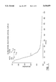
FIG. 1 shows the survival of three human patients treated by the method of the present invention, as compared to the survival curve of a pooled sample of 383 patients treated by previous methods.
FIG. 2 shows the nucleotide sequence of 81C6 heavy (FIG. 2A) (SEQ ID NO: 1) and light (FIG. 2B) (SEQ ID NO: 3) chain variable region genes. The nucleotide sequence is numbered in the righthand margin. Nucleotides with asterisks indicate the conserved octanucleotides. The deduced amino acid sequence is above the nucleotide sequence (SEQ ID NO: 3 and SEQ ID NO: 4). Superscript numbers above the amino acid sequence delineate the leader sequence (-20, -4) and the beginning of the actual immunoglobulin sequence (+1). Underlined amino acids indicate where sequence data were obtained by N-terminal amino acid sequencing.
DETAILED DESCRIPTION OF THE INVENTION
Amino acid sequences disclosed herein are presented in the amino to carboxy direction, from left to right. The amino and carboxy groups are not presented in the sequence.
Nucleotide sequences are presented herein by single strand only, in the 5' to 3' direction, from left to right.
A. Antibodies
The term "antibodies" as used herein refers to all types of immunoglobulins, including IgG, IgM, IgA, IgD, and IgE. The term "immunoglobulin" includes the subtypes of these immunoglobulins, such as IgG1, IgG2, IgG3, IgG4, etc. Of these immunoglobulins, IgM and IgG are preferred, and IgG is particularly preferred. The antibodies may be of any species of origin, including (for example) mouse, rat, rabbit, horse, or human, or may be chimeric antibodies. See, e.g., M. Walker et al., Molec. Immunol. 26, 403-11 (1989). Such monoclonal antibodies are produced in accordance with known techniques. The term "antibody" as used herein includes antibody fragments which retain the capability of binding to a target antigen, for example, Fab, F(ab')2, and Fv fragments, and the corresponding fragments obtained from antibodies other than IgG. Such fragments are also produced by known techniques.
The monoclonal antibodies may be recombinant monoclonal antibodies produced according to the methods disclosed in Reading U.S. Pat. No. 4,474,893, or Cabilly et al., U.S. Pat. No. 4,816,567. The antibodies may also be chemically constructed by specific antibodies made according to the method disclosed in Segel et al., U.S. Pat. No. 4,676,980 (Applicants specifically intend that the disclosure of all U.S. patent references cited herein be incorporated herein by reference).
Monoclonal antibodies may be chimeric antibodies produced in accordance with known techniques. For example, chimeric monoclonal antibodies may be complementarity determining region-grafted antibodies (or "CDR-grafted antibodies") produced in accordance with known techniques.
Monoclonal Fab fragments may be produced in Escherichia coli by recombinant techniques known to those skilled in the art. See, e.g., W. Huse, Science 246, 1275-81 (1989).
As noted above, antibodies employed in carrying out the present invention are those which bind to tenascin. Particularly preferred are monoclonal antibody 81C6 and antibodies that bind to the epitope bound by monoclonal antibody 81C6 (i.e., antibodies that cross-react with, or block the binding of, monoclonal antibody 81C6). The monoclonal antibody 81C6 is a murine IgG2b monoclonal antibody raised from a hybridoma fusion following immunization of BALB/c mice with the glial fibrillary acidic protein (GFAP)-expressing permanent humanglioma line U-251 MG, as known and described in M. Bourdon et al., Cancer Res. 43, 2796 (1983).
B. Therapeutic Antibodies
Monoclonal antibodies used for therapy (i.e., in a method of combatting cancer) may be monoclonal antibodies per se or monoclonal antibodies coupled to a therapeutic agent. Such antibodies are sometimes referred to herein as therapeutic monoclonal antibodies. Any therapeutic agent conventionally coupled to a monoclonal antibody may be employed, including (but not limited to) radioisotopes, cytotoxic agents, and chemotherapeutic agents. See generally Monoclonal Antibodies and Cancer Therapy (R. Reisfeld and S. Sell Eds. 1985)(Alan R. Liss Inc. N.Y.). Therapeutic agents may be coupled to the antibody by direct means or indirect means (e.g., via a chelator) by any suitable technique, such as the Iodogen method or with N-succinimidyl-3-(tri-n-butylstanyl)benzoate (the "ATE method"), as will be apparent to those skilled in the art. See, e.g., M. Zalutsky and A. Narula, Appl. Radiat. Isot. 38, 1051 (1987).
Examples of radioisotopes which may be coupled to a therapeutic monoclonal antibody include, but are not limited to, 131 I, 90 Y, 211 At, 212 Bi, 67 Cu, 186 Re, 188 Re, and 212 Pb. Examples of chemotherapeutic agents which may be coupled to a therapeutic monoclonal antibody include, but are not limited to, methotrexate. Examples of cytotoxic agents which may be coupled to a therapeutic monoclonal antibody include, but are not limited to, ricin (or more particularly the ricin A chain).
It will be appreciated that monoclonal antibodies per se which are used as therapeutic monoclonal antibodies incorporate those portions of the constant region of an antibody necessary to evoke a therapeutically useful immunological response in the subject being treated.
Therapeutic monoclonal antibodies may be provided in lyophylized form in a sterile aseptic container or may be provided in a pharmaceutical formulation in combination with a pharmaceutically acceptable carrier, such as sterile pyrogen-free water or sterile pyrogen-free physiological saline solution.
C. Subjects
The method disclosed herein may be employed with subjects suspected of having solid or cystic tumors residing in the central nervous system, particularly the brain (e.g., in the cerebellum, or more preferably in the cerebral cortex, including the frontal, parietal, occipital and temporal lobes). In addition, the method disclosed herein may be employed with solid tumors residing in other solid tissue organs, such as liver, kidney, spleen, brain, breast, muscle, and prostate.
The tumor may be any tumor, primary or secondary, that expresses tenascin, including (but not limited to) astrocytic tumors, melanomas, breast carcinomas, and Wilm's tumor.
The term "astrocytic tumors" as used herein is used in accordance with the World Health Organization Classification Scheme, and includes astrocytomas, anaplastic astrocytomas, and glioblastoma multiforme. See also D. Russell and L. Rubinstein, Pathology of Tumors of the Nervous System, pp. 83-289 (1989) (Williams and Wilkins).
Some tumors which may be treated by the method of the present invention are cystic tumors: that is, tumors which grow around a fluid-filled cavity, or cyst. Examples of such cystic tumors include (but are not limited to) cystic astrocytic tumors such as cystic astrocytomas, cystic anaplastic astrocytomas, and cystic glioblastoma multiforme.
For administration, the antibody will generally be mixed, prior to administration, with a non-toxic, pharmaceutically acceptable carrier substance (e.g. normal saline or phosphate-buffered saline), and will be administered using any medically appropriate procedure, e.g., intravenous or intra-arterial administration, injection into the cerebrospinal fluid). In certain cases, intradermal, intracavity, intrathecal or direct administration to the tumor or to an artery supplying the tumor is advantageous. In addition, either intrathecal administration or injection into the carotid artery are advantageous for therapy of tumors located in the brain.
Dosage of the antibody will depend, among other things, on the tumor being treated, the route of administration, the nature of the therapeutic agent employed, and the sensitivity of the tumor to the particular therapeutic agent. For example, the dosage will typically be about 1 to 10 micrograms per Kilogram subject body weight. In another example, where the therapeutic agent is 131 I, the dosage to the patient will typically be from 10 mCi to 100, 300 or even 500 mCi. Stated otherwise, where the therapeutic agent is 131 I, the dosage to the patient will typically be from 5,000 Rads to 100,000 Rads (preferably at least 13,000 Rads, or even at least 50,000 Rads). Doses for other radionuclides are typically selected so that the tumoricidal dose will be equivalent to the foregoing range for 131 I. The antibody can be administered to the subject in a series of more than one administration, and regular periodic administration will sometimes be required.
The antibody may be administered by depositing it into the inner cavity of a cystic tumor (i.e., a fluid-filled cavity around which the tumor grows) by any suitable technique, such as by direct injection (aided by stereotaxic positioning of an injection syringe, if necessary) or by placing the tip of an Ommaya reservoir into the cavity and administering the antibody through the Ommaya reservoir. The Ommaya reservoir apparatus is known. See, e.g., F. Balis and D. Poplack, Am J. Pediatr. Hematol. Oncol. 11, 74, 76 FIG. 1 (1989). Where the tumor is a solid tumor, the antibody may be administered by first creating a resection cavity in the location of the tumor in the manner described below, and then depositing the antibody in the resection cavity in like manner as with cystic tumors.
D. Surgical Creation of an Intracranial Cystic Resection Cavity
Virtually all cortical solitary metastases, including those appearing in the four cerebral lobes (frontal, parietal, temporal and occipital) and in the cerebellum, are amenable to creation of the cystic resection cavities by surgery, particularly those in the cerebral lobes.
The procedure differs from an ordinary craniotomy and tumor resection in only a few minor respects. First, the smallest possible cortical incision is made and the tumor is removed to the greatest extent possible by resection of tissue within the small cortical incision and in the depths of the cortex. A so-called gross total tumor resection is attempted, with the only thing prohibiting gross total resection being the potential impingement upon neurologically active areas such as speech or motor areas that would leave permanent neurologic damage if surgically approached. Following gross total resection of the tumor in a standard neurosurgical fashion with cauterization, suction, and forceps removal, the cavity is then preferably rinsed with saline until all bleeding is stopped by cauterization. Next, the pia-arachnoid membrane, which is the surface membrane lining the brain around the cortical incision, is preferably cauterized to enhance the formation of fibroblastic reaction and scarring in the pia-arachnoid area and any astroglial scarring in the areas of normal brain. The result is the formation of an enclosed, fluid-filled cavity within the brain tissue at the location from which the tumor was removed (i.e., the cavity is surrounded on all sides by the organ tissue). The enclosed nature of the resection cavity enhances retention and localization of the therapeutic agent to be administered at the desired site. If desired for administering the therapeutic agent, an Ommaya reservoir may then be placed into the cavity with the tip of the catheter as deep as possible in the tumor bed, and the reservoir secured to the bone in accordance with standard techniques. A standard water-tight dural closure may then be carried out with sutures, as in any other craniotomy.
Resection cavities are formed in other solid tissue organs, as described above, by modification of the foregoing techniques which will be apparent to those skilled in the art.
The present invention is explained in greater detail in the following non-limiting Examples.
COMPARATIVE EXAMPLE A
Survival of Glioblastoma Multiforme Patients
The survival of 383 human glioblastoma multiforme patients treated by the best available techniques is given in FIG. 1 as line 1. The proportion of the total population surviving after diagnosis (diagnosis occuring on year 0.0) is indicated. Note that the line drops precipitously, indicating the relentless and aggressive nature of this disease.
EXAMPLE 1
Drug Formulation
Drug is formulated as 2 ml of a sterile, pyrogen-free solution that contains 10 mg of monoclonal antibody 81C6 (20-60 mCi iodine-131), 0.7 to 0.9% sodium chloride, 0-0.6% sodium phosphate, 0.5% albutein, and water. Antibody is conjugated to iodine-131 by the Iodogen method in accordance with known techniques (see, e.g., R. Moseley et al., Br. J. Cancer 62, 637 (1990)) and the drug formulation is prepared within 24 hours of administration to the patient.
EXAMPLE 2
Intracystic Administration of 81C6 to a Recurrent Cystic Glioblastoma Patient
A 45 year old adult male with a recurrent cystic glioblastoma was administered 15.2 mCi of 131 I conjugated to 10 mg of monoclonal antibody 81C6 by the Iodogen method and formulated as described above through an Ommaya reservoir placed into the recurrent tumor cyst. Clinical and radiographic examination after treatment indicated a complete response.
The patient received a second treatment of the administration of 20.0 mCi of 131 I conjugated to 7.9 mg of monoclonal antibody 81C6 by the ATE method and formulated as described above through the Ommaya reservoir approximately 15 months after the first treatment.
As of 18 months from the first treatment, the patient remained alive. This patient is indicated as line 2 in FIG. 1.
EXAMPLE 3
Intracystic Administration of 81C6 to a Recurrent Cystic Astrocytoma Patient
A 29 year old adult female with a recurrent cystic astrocytoma was administered 21.7 mCi of 131 I conjugated to 10 mg of monoclonal antibody 81C6 by the Iodogen method through an Ommaya reservoir placed into the recurrent tumor cyst. Clinical and radiographic examination after treatment indicated a partial response. The patient was still alive 11 months after treatment. This patient is indicated as line 3 in FIG. 1.
EXAMPLE 4
Intracystic Administration of 81C6 to a Recurrent Cystic Glioblastoma Patient
A 14 year old female with a recurrent cystic glioblastoma was administered 20.07 mCi of 131 I conjugated to 10 mg of monoclonal antibody 81C6 by the Iodogen method through an Ommaya reservoir placed into the recurrent tumor cyst. Clinical and radiographic examination after treatment indicated progressive disease. The patient survived 4 months beyond treatment. This patient is indicated as line 4 in FIG. 1.
EXAMPLE 5
Surgical Creation of an Intracranial Cystic Resection Cavity in a Human Glioblastoma Patient
A cystic resection cavity was surgically created in a 53 year old female patient afflicted with an intracranial glioblastoma. The procedure was carried out in essentially the same manner as an ordinary craniotomy and tumor resection, but differed in a few respects. First, the smallest possible cortical incision was made and the tumor was removed to the greatest extent possible by resection of tissue within the small cortical incision and in the depths of the cortex. A so-called gross total tumor resection was attempted, with the only thing prohibiting gross total resection being the potential impingement upon neurologically active areas such as speech or motor areas that would leave permanent neurologic damage if surgically approached. Following gross total resection of the tumor in a standard neurosurgical fashion with cauterization, suction, and forceps removal, the cavity was then rinsed with saline until all bleeding was stopped by cauterization and the pia-arachnoid membrane, which is the surface membrane lining the brain around the cortical incision, was cauterized to enhance the formation of fibroblastic reaction and scarring in the pia-arachnoid area and any astroglial scarring in the areas of normal brain. An Ommaya reservoir was then placed into the cavity with the tip of the catheter as deep as possible in the tumor bed, and the reservoir secured to the bone in accordance with standard techniques. A standard water-tight dural closure was then carried out with sutures.
EXAMPLE 6
Administration of 81C6 to an Intracranial Cystic Resection Cavity in a Human Glioblastoma Patient
The patient described in the preceding example was administered 15.0 mCi of 131 I conjugated to 10 mg of monoclonal antibody 81C6 by the Iodogen method through an Ommaya reservoir placed into the cystic resection cavity created as described above. Three months after administration, the patient was still alive.
Technetium albumin injections of the cystic resection cavity, followed by sequential radionuclide scans of the brain, showed up to approximately 90% retention of the injected radionuclide albumin conjugate for 72 hours after injection and significant retention of the therapeutic dose of radiolabeled antibody to give a radiation dose calculated to range between 20,000 and 60,000 rads to the walls of the cyst.
EXAMPLE 7
Cloning and Expression of a Mouse/human Chimeric Antibody Cross-reactive with 81C6
This example describes the molecular cloning and characterization of variable region genes for 81C6 antibody. The mouse/human chimeric genes were transfected into SP2/0 cells, and stable integration and expression was obtained. The production of chimeric 81C6 antibody was two to six times higher than that of the parent mouse 81C6 antibody. These data clearly demonstrate that a high level of chimeric antibody production depends upon multiple copies of gene integration and strong expression of both light-chain (SEQUENCE ID NO. 3) and heavy-chain (SEQ ID NO: 1) chimetic genes in the same transfectoma.
I. MATERIALS AND METHODS
Cells and Cell Culture
Native 81C6 (n81C6) antibody has been generated and characterized previously (M. Bourdon et al., Cancer Res. 43, 2796, (1983)) . The SP2/0 cell line was obtained from the American Type Cell Collection (Rockville, Md.). Cells were grown in 1 × zinc option medium (GIBCO, Grand Island, N.Y.) containing 10% fetal bovine serum (GIBCO).
Purification and Analysis of RNA and DNA
Total RNA and genomic DNA were isolated from cells using the guanidine isothiocyanate cushion method of ultracentrifugation (J. Chirgwin et al., Biochemistry 18, 5294 (1979)) as detailed in S. Batra et al., Cel. Growth Differ. 2, 385 (1991) and S. Batra et al., J. Biol. Chem. 266, 6830 (1991). Genomic DNA was further purified by sodium dodecyl sulfate-protenase-K digestion and phenol:chloroform (1:1, v/v) extraction. The RNA pellet was resuspended in 0.3M sodium acetate and precipitated with ethanol. Plasmid DNA was isolated by alkaline lysis and PZ523 columns (5 prime→3 prime, Boulder, Colo.). DNA was analyzed using standard Southern blotting. After digestion with various restriction endonucleases, DNA fragments were fractionated through 0.8% agarose gel by electrophoresis and transferred to a nitrocellulose or nylon transfer membrane (Scheicher & Schuell, Keene, N.H.). The membrane was then hybridized with [32 P]dCTP (ICN Biochemicals, Irvine, Calif.)--labeled DNA probes to identify the immunoglobulin gene. The heavy and light chains were detected with pJH-11 and pJK probes, respectively. The pJH-11 probe is a 1.9-kilobase (kb) BamHI-EcoRI fragment of a murine heavy-chain gene that includes the J3 and J4 segments and the enhancer region, and the pJK probe is a 2.7-kb HindIII DNA fragment of a murine K-light-chain gene containing J1-5 segments.
Total RNA from control and transfected cells was fractionated by electrophoresis on a formaldehyde-containing agarose gel and then transferred to nylon membrane under conditions recommended by the supplier (Scheicher & Schuell). The heavy-chain transcription was detected using an 81C6 heavy-chain-specific 6-kb EcoRI fragment as the probe. The light-chain transcript was defined using an 81C6 light-chain-specific 11-kb HindIII fragment as the probe.
Antibody Purification and Amino Acid Sequence Analysis
Both native 81C6 (n81C6) and chimeric 81C6 (ch81C6) were purified from cultured supernatants or athymic mouse ascites using protein A-Sepharose (Pharmacia, Piscataway, N.J.) chromatography. The amino acid sequence of each chain of both the antibodies (normal and chimeric) was analyzed by automated Edman degradation in an Applied Biosystems (Foster City, Calif.) Model 477A sequencer. On-line phenylthiohydantoin analysis was employed in conjunction with an Applied Biosystems Model 120A high-performance liquid chromatography apparatus (J. Enghild et al., J. Biol. Chem. 266, 747 (1991).
Construction and Screening of Genomic Libraries
81C6 Heavy-chain and light-chain libraries were constructed from size-selected DNA restriction fragments that had been identified as containing putative rearranged variable region genes by Southern blot analysis. For the heavy-chain libraries, genomic DNA was digested with EcoRI and fractionated through agarose gel. The 3- and 6-kb fragments were isolated from agarose gel using the Genclean II kit (Bio 101, Inc., La Jolla, Calif.) and then ligated separately into the EcoRI site of the λ-ZAP II vector (Stratagene, La Jolla, Calif.). For the light-chain library, 81C6 DNA was digested with HindIII, and the 11-kb fragment was cloned into the HindIII site of the λ-DASH vector (Stratagene). The packaging reaction was performed in vitro using Gigapack gold packaging extracts according to the manufacturer's instructions (Stratagene). The libraries were screened with pJH-11 for heavy chains and pJK for light chains. The positive clones were rescued or subcloned and further characterized by DNA sequencing using -oligonucleotide primers that are specific for the .sub.γ -heavy-chain and .sub.κ -light-chain J-regions and Sequenase Version 2.0 (United States Biochemical, Cleveland, Ohio).
Double-strand sequencing was employed for sequence analysis, and both sense and antisense strands of DNA were sequenced. Heavy chain-specific primers were synthesized by Biosynthesis (Denton, Tex.) for each heavy-chain J-region as follows: J1 (5'-CCCGTTTCAGAATGGAATGTGCAG-3') (SEQ ID NO. 5), J2 (5'-CTAAGCTGAATAGAAGAGAGAGG-3') (SEQ ID NO. 6), J3 (5"-TGGGAGAAGTTAGGACT-3'), (SEQ ID NO. 7), and J4 (5'-ATAAAGACCTGGAGAGGCC-3'), (SEQ ID NO. 8), .sub.γ -Chain J-region primers were made by Biosynthesis as described by Hoogenboom, et al., 1990. To complete the 5' terminal sequence and to read the opposite strand, internal 81C8-specific primers were also used. The amino acid sequence was translated from the nucleotide sequence and compared with the actual sequence.
Construction of Chimeric Vectors
The functional, rearranged 81C6 heavy-chain variable region gene was inserted into expression vector pSV2ΔHgpt-HuG2, which contains a genomic fragment encoding the human IgG2-constant region and the Ecogpt gene providing resistance to mycophenolic acid. The 81C6 light-chain variable region gene was ligated into expression vector pSV184ΔHneo-HuK, which includes a DNA fragment encoding the human K-constant region and the neogene giving resistance to G418 (V. Oi and S. Morrison, Biotechniques 4, 214 (1986).
Transfection
The chimeric heavy-chain and light-chain constructs were separately introduced or cotransfected into SP2/0 cells using a lipofectin reagent (GIBCO/BRL). The transfected cells were incubated at 37° C. in a 5% CO2 atmosphere in 1 × zinc option medium for 24 hours and then in medium containing 10% fetal bovine serum. After 48-h incubation, the cells were transferred to a 96-well microtiter plate and grown in selection medium containing G418 and/or mycophenolic acid. The supernatants of drug-resistant cells were screened for anti-tenascin activity by enzyme-linked immunosorbent assay.
Immunoassay for Anti-tenascin Activity
Each microtiter well (Dynatech Laboratories, Inc, Chantilly, Va.) was coated with 50 μl of 2 mg/μl human intact tenascin or recombinant tenascin fragment in 0.1M sodium carbonate buffer (pH 9.6) overnight at 4° C., washed with 0.025% gelatin, 0.1% BRIJ 35 (Sigma, St. Louis, Mont.) in 115 mM sodium phosphate buffer (pH 7.4) 3 times, and blocked with the washing buffer for 30 min. After the washing buffer was removed, 50 μl of culture supernatant or purified antibody was added to each well and the contents were incubated for 1 h at room temperature, washed, and incubated with 50 μl of biotinylated goat anti-mouse (for mouse antibody) or goat anti-human (for chimeric antibody) IgG (BRL) for 1 h at room temperature. The plates were washed and incubated with 50 μl of streptavidin-alkaline phosphatase (BRL) for 1 h at room temperature, rewashed, and then incubated with 100 μl/per well of phosphatase substrate (Sigma) (4 mg/ml in 10%) diethanolamine, 0.5 mM MgCl2). The reaction was stopped with 10mM L-cysteine, and absorbance was read at 405 nm.
Radioiodination
Purified n81C6 and ch81C6 (IgG2) were labeled with 125 I (Amersham, Arlington Heights, Ill.) using the Iodo-Gen (Pierce, Rockford, Ill.) in accordance with known techniques (P. Fraker and J. Speck, Biochem. Biophys. Res. Commun. 80, 849 (1978)). The labeled antibodies were separated from free radioiodine by passage through a Sephadex G-25 column. Iodinated antibody preparations were greater than 97% in trichloroacetic acid precipitation analysis. The size and homogeneity of radiolabeled antibody were monitored by size exclusion high-performance liquid chromatography in conjunction with a Model 170 flow-through gamma detector (Beckman, Irvine, Calif.).
Immunoreactivity and Affinity Determination
For the immunoreactivity assay, 50 ng of labeled antibody was incubated overnight in duplicate with different amounts of D-54 MG human glioma xenograft (antigen positive) and normal rat liver (antigen negative) homogenate in 1 ml of 115 mM phosphate buffer containing 1% bovine serum albumin. The specific binding percentage was calculated by subtracting the percentage bound to the rat liver homogenate from that bound to the D-54 tumor homogenate.
A modified Scatchard analysis was used to measure the binding affinity of labeled ch81C6. Serially diluted 125I-labeled antibody (200 μl, 1:2 dilution starting from 1600 ng/ml) was incubated in 48-well plates overnight at 37° C. with the antigen-positive human glioma cell line U-251 MG-C3 fixed in glutaraldehyde and the antigen-negative human osteogenic sarcoma cell line 2T. The cells were washed and solubilized with 2N NaOH, and the radioactivity was measured with a gamma counter. The data was analyzed in accordance with known techniques (Y. S. Lee et al., Cancer Res. 48, 559 (1988)).
Cross-competition Assay
Serially diluted (1:2 dilution starting from 100 μg/well in 50 μl) and unlabeled antibodies, experimental or control, were added to glutaraldehyde-fixed U-251 MG-C3 and 2T cells in 96-well plates. After the cells were incubated at 37° C. for 2 h, 50 μl of 125 I n81C6 or 125I ch81C6 (IgG2) (200,000 cpm) were added to each well, and the plates were incubated at 37° C. for 2 h. The cells were washed and solubilized with 2N NaOH, and the bound reactivity was counted with a gamma counter. The percentage binding was determined by the formula: ##EQU1## Each value was corrected for counter background and cpm bound in antigen negative cell.
II. RESULTS
Identification and Cloning of Rearranged 81C6 Variable Region Genes
Genomic DNA from a 81C6 hybridoma; its fusion partner, myeloma P3X63/Ag8.653; and BALB/c mouse liver representative of the mouse germ line were digested with EcoRI, HindIII and BamHI and subjected to the Southern blot analysis to identify the putative rearranged 81C6 variable region genes. The hybridization pattern using the 32 P-labeled heavy-chain probe pJH-11 revealed that the 3- and 6-kb EcoRI bands, a 1.2-kb HindIII band, and a 11-kb BamH1 band were unique to the 81C6 hybridoma. These unique bands were considered as the putative rearranged heavy-chain variable region genes. Hybridization using the 32 P-labeled light-chain probe pJK revealed an 11-kb HindIII band and a 6-kb BamHI band which were found in the 81C6 hybridoma only, and which were most likely the putative rearranged light-chain variable region genes.
In order to clone the heavy-chain gene, two genomic libraries were constructed for 3- and 6-kb EcoRI DNA fragments and screened with the mouse heavy-chain probe pJH-11. Positive clones were subjected to nucleotide sequencing. The sequence analysis found that the clone isolated from a 3-kb fragment library was an aberrantly rearranged gene. The nucleotide sequence of a positive clone from a 6-kb fragment library is shown in FIG. 2A. It had all the features of an intact variable region, including a functional leader sequence, in-frame V-D and D-J junctions, and cysteines 22 and 92, which are necessary for an intrachain disulfide bond. The conserved octanucleotide, ATGCAAAT, was found at nucleotide 144, upstream from the 3' end of the first exon. The 81C6 heavy-chain variable region segment was found to rearrange to the J3 segment. The N-terminal amino acid sequence matched the deduced amino acid sequence.
The putative K-chain variable region of the 11-kb HindIII DNA fragment was isolated from the genomic library prepared in the λ-DASH vector using the light-chain probe pJK. FIG. 2B shows the nucleotide sequence for the 81C6 K-chain, which is rearranged to the J1 segment. The conserved octanucleotide, ATTTGCAT, was found at nucleotide 147, upstream from the 3' end of the first exon. A large 358-base pair intervening sequence was found in the middle of the leader peptide. The deduced amino acid sequence matched the partial sequence obtained using N-terminal sequencing of the purified 81C6 antibody.
Construction and Transfection of Chimeric Genes
The 6-kb EcoRI fragment of the cloned 81C6 heavy-chain variable region gene was ligated at the unique EcoRI site in the intron before the first exon in the human IgG2 constant region of the expression vector pSV2ΔHgpt-HuG2. Similarly, the 11-kb HindIII fragment of the cloned 81C6 light-chain variable region gene was inserted into a unique HindIII site in expression vector pSV184ΔHneo-HuK, which contained the human K-constant region gene. The chimeric heavy-chain and light-chain constructs were either separately introduced into SP2/0 cells for confirming the orientation of 81C6 variable region genes in the expression vector, or cotransfected into the SP2/0 cells for expressing chimeric antibody. The transfected cells were grown in medium containing G418 and/or mycophenolic acid. Supernatants of cells resistant to both drugs were screened for the presence of antibody activity using human intact tenascin-coated plates and enzyme-linked immunosorbent assay. Thirteen positive transfectomas were obtained from three transfection experiments. Five of them were subcloned twice, expanded in culture, and injected into athymic mice for producing antibody.
Expression of ch81C6
Genomic DNA was isolated from the transfected and control cells and analyzed by Southern blotting to determine the integration of chimeric constructs into the SP2/0 cell genome (data not shown). Hybridization bands at 3, 6, and 6.5 kb were found in n81C6. All untransfected and transfected SP2/0 cells showed both a strong 6-kb and a weak 3.4-kb band, which were considered to be endogenous immunoglobulin genes from the SP2/0 genome. The hybridization signal in SP2/0 cells transfected with the heavy-chain expression vector was identical to that of untransfected SP2/0 cells. Additional hybridization bands of different sizes were found in cells transfected with the heavy-chain construct alone or in transfectomas that received both heavy- and light-chain chimeric constructs and secreted chimeric antibody. An exception was transfectomas H3D4A10 and F12D9A2, which showed additional hybridization bands only after digestion with HindIII or BamHI. The strongest integration signal, with additional, intense bands, was found in transfectoma F9C11A5.
The hybridization of the light-chain probe to the HindIII-digested DNA blot showed hybridization bands at 11 and 2.5 kb in n81C6 as expected (data not shown). Hybridization bands at 6.5 and 2.5 kb were shown in all untransfected and transfected SP2/0 cells; these two bands were considered to be endogenous immunoglobulin genes from the SP2/0 genome. The hybridization pattern of transfected cells with the vector alone was the same as the pattern of untransfected SP2/0 cells. Additional hybridization bands of different sizes were also detected in all transfected cells either with the light-chain chimeric construct alone or with both light- and heavy-chain chimeric constructs. These additional bands most likely resulted from the integration of chimeric DNA constructs into the genome. Lanes 4 and 5 show transfected cells with the light-chain chimeric construct alone. The DNA integration signals varied with different transfectomas. The strongest signal here, as with the heavy-chain probe, was also found in transfectoma F9C11A5.
Total RNA, purified from the transfected and control cell lines, was separated by agarose/formaldehyde gel electrophoresis, Northern blotted, and probed with a 6-kb heavy-chain variable segment and an 11-kb HindIII DNA fragment of 81C6 for heavy-chain and light-chain gene expression, respectively. The data showed the expression of heavy-chain mRNA in the untransfected and transfected cells. The same blot was probed with β-actin cDNA as a control for mRNA quantity and quality (P. Gunning et al., Mol. Cel. Biol. 3, 787 (1983)). The heavy-chain mRNA was detected in n81C6, transfected SP2/0 cells with the expression vector containing the 81C6 heavy chain in sense orientation, and in all transfectomas secreting chimeric 81C6. But no signal was found in untransfected SP2/0 cells, transfected cells with expression vector alone, or transfected cells with the expression vector carrying the 81C6 heavy chain in antisense orientation. Light-chain mRNA was detected in n81C6, transfected SP2/0 cells with the expression vector containing the 81C6 light-chain variable region gene (SEQ ID NO. 3) in sense orientation, and all transfectomas secreting chimeric antibody. There was no hybridization signal found in untransfected SP2/0, transfected cells with the light-chain expression vector alone, and in cells with the expression vector containing the 81C6 light chain in antisense orientation. Strong expression signals of both heavy- and light-chain mRNA were detected in the transfectoma F9C11A5.
Chimeric 81C6 antibody was purified from cultured supernatants and ascites by protein A-Sepharose chromatography. The production levels of five transfectomas in supernatant and in ascites are presented in Table 1. High antibody production was obtained from the supernatants and ascites of transfectoma F9C11A5, which had multiple copies of integration of heavy- and light-chain chimeric constructs and high levels of heavy- and light-chain mRNA expression. The antibody concentration in transfectoma F9C11A5 ascites was two to six times higher than that of n81C6, which was 2.5 mg/ml ascites in previous purification. Production levels of the four other transfectomas were similar, with the exception of H3D4A10 ascites, which was unusually low.
              TABLE 1
______________________________________
Antibody production by ch81C6 transfectomas and n81C6.
                 Antibody in
                           Antibody in
                 supernatant
                           ascites
Cells            (ug/ml)   (mg/ml)
______________________________________
ch81C6 transfectomas
F9C11A5.sup.a    31.4      6.5
G4D10A12.5       3.7       2.5
B4D9B1           4.4       1.9
H3D4A10          4.7       0.09
F12D9A2          3.4       1.5
n81C6            8.5       2.5
______________________________________
 .sup.a F9C11A5 transfectoma was further used for production of ch81C6
 antibodies, which showed a varied yield of 65 to 13.9 mg/ml of ascites.
Characterization of ch81C6
The ch81C6 from high production transfectoma F9C11A5 was purified and further characterized. It was labeled with 125 I using the Iodogen method, and the percentage of immunoreactivity was determined by absorption with D-54 MG xenograft homogenate. The affinity constant was defined by Scatchard analysis using binding assay on a tenascin-expressing cell line U-251 MGC-3 as described above. Immunoreactivity of 58% was obtained for 125 I ch81C6, which was comparable to a range of 35-70% for 125 I n81C6 in previous experiments (Y. S. Lee et al., Cancer Res. 48, 559 (1988); Y. S. Lee et al., Cancer Res. 48, 2904 (1988)). The binding affinity of 125 I ch81C6 was 1.67×10⁸ M-1, which was also in the range of 3.3×10⁷ to 3.0×10⁸ M1 for radiolabeled n81C6 in previous reports (Lee et al., supra).
Cross-competition assays were performed on U-251 MG-C3 cells using 125 I ch81C6 and 125 I n81C6 to determine their binding properties. Less than 10% 125 I ch81C6 binding activity was detected after incubation with unlabeled n81C6 or unlabeled ch81C6. The negative control chimeric IgG2, TPS3.2, did not show significant inhibition. Similar reciprocal inhibition of binding was observed for 125 I n81C6 with unlabeled ch81C6 or unlabeled n81C6, but not with negative control murine IgG2b 45.6.
We also tested the immunoreactivity of 81C6 with four recombinant tenascin fragments containing fibronectin III domains 3-5, 6-12, 13-15, and epidermal growth factor-like domains, respectively. Both native and chimeric 81C6 reacted with tenascin fragments containing fibronectin III domains 6-12 as expected, but not with other fragments tested.
The foregoing examples are illustrative of the present invention, and are not to be construed as limiting thereof. The invention is defined by the following claims, with equivalents of the claims to be included therein.
__________________________________________________________________________
SEQUENCE LISTING
(1) GENERAL INFORMATION:
(iii) NUMBER OF SEQUENCES: 8
(2) INFORMATION FOR SEQ ID NO:1:
(i) SEQUENCE CHARACTERISTICS:
(A) LENGTH: 660 base pairs
(B) TYPE: nucleic acid
(C) STRANDEDNESS: single
(D) TOPOLOGY: linear
(ii) MOLECULE TYPE: DNA (genomic)
(ix) FEATURE:
(A) NAME/KEY: CDS
(B) LOCATION: join(149..193, 277..642)
(xi) SEQUENCE DESCRIPTION: SEQ ID NO:1:
CTGGACTAGGTTCTTATGTAAGAAGTCCCCTGCTCATCATTATGCAAATTACCTGAGTCT60
ATGGTGATTAAAACAGGGATGTCCACACCCTTAAATCAACCGACGATCAGTGTCCTCTCC120
AAAGTCCCTGAACACACTGACTCTAACCATGGAATGGAGTTGGATATTTCTC172
MetGluTrpSerTrpIlePheLeu
15
TTTCTCCTGTCAGGAACTGCAGGTAAGGGGCTCACCAGTTCAAAATCTGAA223
PheLeuLeuSerGlyThrAla
1015
GTGGAGACAGGACCTGAGGTGACAATGACATCTACTCTGACATTCTCTCCTCAGGT279
Gly
GTCCACTCTGAGGTCCAGCTGCAGCAGTCTGGACCTGAGCTGGTAAAG327
ValHisSerGluValGlnLeuGlnGlnSerGlyProGluLeuValLys
202530
CCTGGGGCTTCAGTGAAGATGTCCTGCAAGGCTTCTGGATACACATTC375
ProGlyAlaSerValLysMetSerCysLysAlaSerGlyTyrThrPhe
354045
ACTAGCTATGTTGTGCACTGGGTGAAGCAGAACCCTGGGCAGGGCCTT423
ThrSerTyrValValHisTrpValLysGlnAsnProGlyGlnGlyLeu
505560
GAGTGGATTGGATATATTAATCCTTTCAATGATGGTACTAAGTACAAT471
GluTrpIleGlyTyrIleAsnProPheAsnAspGlyThrLysTyrAsn
65707580
GAGAACTTCAAAGGCAAGGCCACACTGACTTCAGACAGATCCTCCAGC519
GluAsnPheLysGlyLysAlaThrLeuThrSerAspArgSerSerSer
859095
ACAGCCTACATGGAGCTCAGCAGCCTGACCTCTGAGGAATCTGCGGTC567
ThrAlaTyrMetGluLeuSerSerLeuThrSerGluGluSerAlaVal
100105110
TATTTCTGTGCAAGAGACATGGGTCGCGAAGGCTTTGCTTACTGGGGC615
TyrPheCysAlaArgAspMetGlyArgGluGlyPheAlaTyrTrpGly
115120125
CAAGGGACTCTGGTCACTGTCTCTGCAGGTGAGTCCTAACTTCTC660
GlnGlyThrLeuValThrValSerAla
130135
(2) INFORMATION FOR SEQ ID NO:2:
(i) SEQUENCE CHARACTERISTICS:
(A) LENGTH: 137 amino acids
(B) TYPE: amino acid
(D) TOPOLOGY: linear
(ii) MOLECULE TYPE: protein
(xi) SEQUENCE DESCRIPTION: SEQ ID NO:2:
MetGluTrpSerTrpIlePheLeuPheLeuLeuSerGlyThrAlaGly
151015
ValHisSerGluValGlnLeuGlnGlnSerGlyProGluLeuValLys
202530
ProGlyAlaSerValLysMetSerCysLysAlaSerGlyTyrThrPhe
354045
ThrSerTyrValValHisTrpValLysGlnAsnProGlyGlnGlyLeu
505560
GluTrpIleGlyTyrIleAsnProPheAsnAspGlyThrLysTyrAsn
65707580
GluAsnPheLysGlyLysAlaThrLeuThrSerAspArgSerSerSer
859095
ThrAlaTyrMetGluLeuSerSerLeuThrSerGluGluSerAlaVal
100105110
TyrPheCysAlaArgAspMetGlyArgGluGlyPheAlaTyrTrpGly
115120125
GlnGlyThrLeuValThrValSerAla
130135
(2) INFORMATION FOR SEQ ID NO:3:
(i) SEQUENCE CHARACTERISTICS:
(A) LENGTH: 882 base pairs
(B) TYPE: nucleic acid
(C) STRANDEDNESS: single
(D) TOPOLOGY: linear
(ii) MOLECULE TYPE: cDNA
(ix) FEATURE:
(A) NAME/KEY: CDS
(B) LOCATION: join(121..168, 527..874)
(xi) SEQUENCE DESCRIPTION: SEQ ID NO:3:
TATCAACCAGGAGATTTGCATATTGCTCCCTAGGGAGGACCTCTTCTTGCAGGTGCAGGG60
TAAAAGCTCACTCCTCTTTCTGTCTTGATTACTTTTGACTCACCATATCAAGTTCGCAGA120
ATGAGGTTCTCTGCTCAGCTTCTGGGGCTGCTTGTGCTCTGGATCCCT168
MetArgPheSerAlaGlnLeuLeuGlyLeuLeuValLeuTrpIlePro
151015
GGTAAGGAGACAAAGATGAAGAAGGAGAATTTTGAGGGAGGGGGATTTCTGAGACATGAT228
GATAAATATGTATGTTCTGTACATGTCTGAGATATACAGTTCTGTTCTCCAGTAAAGGAC288
TTGTGAGGTTCAAAGTGTGAAGAGATTAAGGTCTGTTTTTCTGTGACAACTCTGACAGTT348
CCAAAGCCAAAAGTCAAATGAAAGAGACTCTCTGTGCTTCCTCTACATGCATATTTTATG408
TGGAGCACTTCTAGAGTATGAGTGAAAGACATGAACAAAATAAGTAGAAAAAAATAAAGG468
AAAGAAATTCACTTCATCGTATCATTTTTTTACATAACCAATTAATTCTCTTATTGCA526
GGATCCACTGCAGATATTGTGATGACGCAGGCTGCATTCTCCAATCCA574
GlySerThrAlaAspIleValMetThrGlnAlaAlaPheSerAsnPro
202530
GTCACTCTTGGAACATCAGCTTCCATCTCCTGCAGGTCTAGTAAGAGT622
ValThrLeuGlyThrSerAlaSerIleSerCysArgSerSerLysSer
354045
CTCCTACATAGTAATGGCATCACTTATTTGTATTGGTATCTGCAGAAG670
LeuLeuHisSerAsnGlyIleThrTyrLeuTyrTrpTyrLeuGlnLys
505560
CCAGGCCAGTCTCCTCAGCTCCTGATTTATCAGATGTCCAACCTTGCC718
ProGlyGlnSerProGlnLeuLeuIleTyrGlnMetSerAsnLeuAla
65707580
TCAGGAGTCCCAGACAGGTTCAGTAGCAGTGGGTCAGGAACTGATTTC766
SerGlyValProAspArgPheSerSerSerGlySerGlyThrAspPhe
859095
ACACTGAGAATCAGCAGAGTGGAGGCTGAGGATGTGGGTGTTTATTAC814
ThrLeuArgIleSerArgValGluAlaGluAspValGlyValTyrTyr
100105110
TGTGCTCAAAATCTAGAACTTCCTCGGACGTTCGGTGGAGGCACCAAG862
CysAlaGlnAsnLeuGluLeuProArgThrPheGlyGlyGlyThrLys
115120125
CTGGAAATCAAACGTAAGTA882
LeuGluIleLys
130
(2) INFORMATION FOR SEQ ID NO:4:
(i) SEQUENCE CHARACTERISTICS:
(A) LENGTH: 132 amino acids
(B) TYPE: amino acid
(D) TOPOLOGY: linear
(ii) MOLECULE TYPE: protein
(xi) SEQUENCE DESCRIPTION: SEQ ID NO:4:
MetArgPheSerAlaGlnLeuLeuGlyLeuLeuValLeuTrpIlePro
151015
GlySerThrAlaAspIleValMetThrGlnAlaAlaPheSerAsnPro
202530
ValThrLeuGlyThrSerAlaSerIleSerCysArgSerSerLysSer
354045
LeuLeuHisSerAsnGlyIleThrTyrLeuTyrTrpTyrLeuGlnLys
505560
ProGlyGlnSerProGlnLeuLeuIleTyrGlnMetSerAsnLeuAla
65707580
SerGlyValProAspArgPheSerSerSerGlySerGlyThrAspPhe
859095
ThrLeuArgIleSerArgValGluAlaGluAspValGlyValTyrTyr
100105110
CysAlaGlnAsnLeuGluLeuProArgThrPheGlyGlyGlyThrLys
115120125
LeuGluIleLys
130
(2) INFORMATION FOR SEQ ID NO:5:
(i) SEQUENCE CHARACTERISTICS:
(A) LENGTH: 24 base pairs
(B) TYPE: nucleic acid
(C) STRANDEDNESS: single
(D) TOPOLOGY: linear
(ii) MOLECULE TYPE: DNA (genomic)
(xi) SEQUENCE DESCRIPTION: SEQ ID NO:5:
CCCGTTTCAGAATGGAATGTGCAG24
(2) INFORMATION FOR SEQ ID NO:6:
(i) SEQUENCE CHARACTERISTICS:
(A) LENGTH: 23 base pairs
(B) TYPE: nucleic acid
(C) STRANDEDNESS: single
(D) TOPOLOGY: linear
(ii) MOLECULE TYPE: DNA (genomic)
(xi) SEQUENCE DESCRIPTION: SEQ ID NO:6:
CTAAGCTGAATAGAAGAGAGAGG23
(2) INFORMATION FOR SEQ ID NO:7:
(i) SEQUENCE CHARACTERISTICS:
(A) LENGTH: 17 base pairs
(B) TYPE: nucleic acid
(C) STRANDEDNESS: single
(D) TOPOLOGY: linear
(ii) MOLECULE TYPE: DNA (genomic)
(xi) SEQUENCE DESCRIPTION: SEQ ID NO:7:
TGGGAGAAGTTAGGACT17
(2) INFORMATION FOR SEQ ID NO:8:
(i) SEQUENCE CHARACTERISTICS:
(A) LENGTH: 19 base pairs
(B) TYPE: nucleic acid
(C) STRANDEDNESS: single
(D) TOPOLOGY: linear
(ii) MOLECULE TYPE: DNA (genomic)
(xi) SEQUENCE DESCRIPTION: SEQ ID NO:8:
ATAAAGACCTGGAGAGGCC19
__________________________________________________________________________