EP3066122B1 - Anticorps monoclonaux anti-pré-haptoglobine-2 et leurs utilisations - Google Patents
Anticorps monoclonaux anti-pré-haptoglobine-2 et leurs utilisations Download PDFInfo
- Publication number
- EP3066122B1 EP3066122B1 EP14860426.7A EP14860426A EP3066122B1 EP 3066122 B1 EP3066122 B1 EP 3066122B1 EP 14860426 A EP14860426 A EP 14860426A EP 3066122 B1 EP3066122 B1 EP 3066122B1
- Authority
- EP
- European Patent Office
- Prior art keywords
- haptoglobin
- antibody
- monoclonal antibody
- human
- seq
- Prior art date
- Legal status (The legal status is an assumption and is not a legal conclusion. Google has not performed a legal analysis and makes no representation as to the accuracy of the status listed.)
- Active
Links
- 108010027843 zonulin Proteins 0.000 title claims description 183
- 241000282414 Homo sapiens Species 0.000 claims description 86
- 238000000034 method Methods 0.000 claims description 37
- 239000007787 solid Substances 0.000 claims description 30
- 238000009739 binding Methods 0.000 claims description 26
- 230000027455 binding Effects 0.000 claims description 25
- YBJHBAHKTGYVGT-ZKWXMUAHSA-N (+)-Biotin Chemical compound N1C(=O)N[C@@H]2[C@H](CCCCC(=O)O)SC[C@@H]21 YBJHBAHKTGYVGT-ZKWXMUAHSA-N 0.000 claims description 14
- 238000002198 surface plasmon resonance spectroscopy Methods 0.000 claims description 9
- 229960002685 biotin Drugs 0.000 claims description 8
- 239000011616 biotin Substances 0.000 claims description 8
- 235000020958 biotin Nutrition 0.000 claims description 7
- 108090001008 Avidin Proteins 0.000 claims description 2
- 238000003556 assay Methods 0.000 description 43
- 108090000765 processed proteins & peptides Proteins 0.000 description 33
- 238000001514 detection method Methods 0.000 description 31
- 108090000623 proteins and genes Proteins 0.000 description 31
- 239000000523 sample Substances 0.000 description 29
- 102000004169 proteins and genes Human genes 0.000 description 26
- 108050005077 Haptoglobin Proteins 0.000 description 22
- 238000002965 ELISA Methods 0.000 description 21
- 102000014702 Haptoglobin Human genes 0.000 description 21
- 239000000427 antigen Substances 0.000 description 20
- 102000036639 antigens Human genes 0.000 description 20
- 108091007433 antigens Proteins 0.000 description 20
- 102100025255 Haptoglobin Human genes 0.000 description 18
- 210000004408 hybridoma Anatomy 0.000 description 18
- 230000002829 reductive effect Effects 0.000 description 17
- 239000012634 fragment Substances 0.000 description 15
- 238000003018 immunoassay Methods 0.000 description 15
- 210000004027 cell Anatomy 0.000 description 14
- 108010001336 Horseradish Peroxidase Proteins 0.000 description 13
- 239000011859 microparticle Substances 0.000 description 10
- 102000004196 processed proteins & peptides Human genes 0.000 description 10
- 238000003118 sandwich ELISA Methods 0.000 description 10
- 102000004190 Enzymes Human genes 0.000 description 9
- 108090000790 Enzymes Proteins 0.000 description 9
- 241001465754 Metazoa Species 0.000 description 9
- 229940088598 enzyme Drugs 0.000 description 9
- 241000699666 Mus <mouse, genus> Species 0.000 description 8
- 150000001413 amino acids Chemical group 0.000 description 8
- 210000002966 serum Anatomy 0.000 description 8
- 210000004698 lymphocyte Anatomy 0.000 description 7
- 239000000203 mixture Substances 0.000 description 7
- 230000009257 reactivity Effects 0.000 description 7
- DGVVWUTYPXICAM-UHFFFAOYSA-N β‐Mercaptoethanol Chemical compound OCCS DGVVWUTYPXICAM-UHFFFAOYSA-N 0.000 description 7
- 241000283707 Capra Species 0.000 description 6
- 208000015943 Coeliac disease Diseases 0.000 description 6
- 108010021625 Immunoglobulin Fragments Proteins 0.000 description 6
- 239000002671 adjuvant Substances 0.000 description 6
- 239000003153 chemical reaction reagent Substances 0.000 description 6
- 239000000539 dimer Substances 0.000 description 6
- 239000000178 monomer Substances 0.000 description 6
- 238000001262 western blot Methods 0.000 description 6
- 108060003951 Immunoglobulin Proteins 0.000 description 5
- 102000008394 Immunoglobulin Fragments Human genes 0.000 description 5
- 239000003085 diluting agent Substances 0.000 description 5
- 238000010790 dilution Methods 0.000 description 5
- 239000012895 dilution Substances 0.000 description 5
- 239000000499 gel Substances 0.000 description 5
- 102000018358 immunoglobulin Human genes 0.000 description 5
- 239000003550 marker Substances 0.000 description 5
- 230000035945 sensitivity Effects 0.000 description 5
- 238000012360 testing method Methods 0.000 description 5
- 102000002260 Alkaline Phosphatase Human genes 0.000 description 4
- 108020004774 Alkaline Phosphatase Proteins 0.000 description 4
- 108700005091 Immunoglobulin Genes Proteins 0.000 description 4
- 241000699670 Mus sp. Species 0.000 description 4
- 206010035226 Plasma cell myeloma Diseases 0.000 description 4
- 239000003593 chromogenic compound Substances 0.000 description 4
- 230000009260 cross reactivity Effects 0.000 description 4
- 150000004676 glycans Chemical class 0.000 description 4
- 230000001900 immune effect Effects 0.000 description 4
- 229940124452 immunizing agent Drugs 0.000 description 4
- 238000000338 in vitro Methods 0.000 description 4
- 125000005647 linker group Chemical group 0.000 description 4
- 239000000463 material Substances 0.000 description 4
- 201000000050 myeloid neoplasm Diseases 0.000 description 4
- 239000002245 particle Substances 0.000 description 4
- 229920000642 polymer Polymers 0.000 description 4
- 229920001282 polysaccharide Polymers 0.000 description 4
- 239000005017 polysaccharide Substances 0.000 description 4
- -1 preHP1 Proteins 0.000 description 4
- 239000000047 product Substances 0.000 description 4
- 238000003127 radioimmunoassay Methods 0.000 description 4
- 238000012216 screening Methods 0.000 description 4
- 239000000243 solution Substances 0.000 description 4
- 239000000126 substance Substances 0.000 description 4
- 239000000758 substrate Substances 0.000 description 4
- 239000006228 supernatant Substances 0.000 description 4
- 108010076504 Protein Sorting Signals Proteins 0.000 description 3
- 108010090804 Streptavidin Proteins 0.000 description 3
- QAOWNCQODCNURD-UHFFFAOYSA-N Sulfuric acid Chemical compound OS(O)(=O)=O QAOWNCQODCNURD-UHFFFAOYSA-N 0.000 description 3
- 210000000628 antibody-producing cell Anatomy 0.000 description 3
- 239000011324 bead Substances 0.000 description 3
- 239000000872 buffer Substances 0.000 description 3
- 210000004899 c-terminal region Anatomy 0.000 description 3
- 239000003638 chemical reducing agent Substances 0.000 description 3
- 239000003795 chemical substances by application Substances 0.000 description 3
- 239000003398 denaturant Substances 0.000 description 3
- 238000011161 development Methods 0.000 description 3
- 238000005516 engineering process Methods 0.000 description 3
- 125000000524 functional group Chemical group 0.000 description 3
- 230000004927 fusion Effects 0.000 description 3
- 230000000984 immunochemical effect Effects 0.000 description 3
- 230000002998 immunogenetic effect Effects 0.000 description 3
- 238000013507 mapping Methods 0.000 description 3
- 230000003287 optical effect Effects 0.000 description 3
- 230000002285 radioactive effect Effects 0.000 description 3
- 239000002904 solvent Substances 0.000 description 3
- 239000003053 toxin Substances 0.000 description 3
- 231100000765 toxin Toxicity 0.000 description 3
- 239000013598 vector Substances 0.000 description 3
- 238000005406 washing Methods 0.000 description 3
- LMDZBCPBFSXMTL-UHFFFAOYSA-N 1-Ethyl-3-(3-dimethylaminopropyl)carbodiimide Substances CCN=C=NCCCN(C)C LMDZBCPBFSXMTL-UHFFFAOYSA-N 0.000 description 2
- STGOXLCCKKFIGD-UHFFFAOYSA-N 1-hydroxy-2,5-dioxopyrrolidine-3,3-disulfonic acid Chemical compound ON1C(=O)CC(S(O)(=O)=O)(S(O)(=O)=O)C1=O STGOXLCCKKFIGD-UHFFFAOYSA-N 0.000 description 2
- KUWPCJHYPSUOFW-YBXAARCKSA-N 2-nitrophenyl beta-D-galactoside Chemical compound O[C@@H]1[C@@H](O)[C@@H](O)[C@@H](CO)O[C@H]1OC1=CC=CC=C1[N+]([O-])=O KUWPCJHYPSUOFW-YBXAARCKSA-N 0.000 description 2
- YRNWIFYIFSBPAU-UHFFFAOYSA-N 4-[4-(dimethylamino)phenyl]-n,n-dimethylaniline Chemical compound C1=CC(N(C)C)=CC=C1C1=CC=C(N(C)C)C=C1 YRNWIFYIFSBPAU-UHFFFAOYSA-N 0.000 description 2
- IJGRMHOSHXDMSA-UHFFFAOYSA-N Atomic nitrogen Chemical compound N#N IJGRMHOSHXDMSA-UHFFFAOYSA-N 0.000 description 2
- 208000023275 Autoimmune disease Diseases 0.000 description 2
- 108010047041 Complementarity Determining Regions Proteins 0.000 description 2
- 108020004635 Complementary DNA Proteins 0.000 description 2
- BWGNESOTFCXPMA-UHFFFAOYSA-N Dihydrogen disulfide Chemical compound SS BWGNESOTFCXPMA-UHFFFAOYSA-N 0.000 description 2
- 101800000803 Haptoglobin alpha chain Proteins 0.000 description 2
- 102400000142 Haptoglobin alpha chain Human genes 0.000 description 2
- 102000001554 Hemoglobins Human genes 0.000 description 2
- 108010054147 Hemoglobins Proteins 0.000 description 2
- 241000238631 Hexapoda Species 0.000 description 2
- 108010093488 His-His-His-His-His-His Proteins 0.000 description 2
- 241000282412 Homo Species 0.000 description 2
- MHAJPDPJQMAIIY-UHFFFAOYSA-N Hydrogen peroxide Chemical compound OO MHAJPDPJQMAIIY-UHFFFAOYSA-N 0.000 description 2
- 206010020751 Hypersensitivity Diseases 0.000 description 2
- XEEYBQQBJWHFJM-UHFFFAOYSA-N Iron Chemical compound [Fe] XEEYBQQBJWHFJM-UHFFFAOYSA-N 0.000 description 2
- 241000283973 Oryctolagus cuniculus Species 0.000 description 2
- 229920001213 Polysorbate 20 Polymers 0.000 description 2
- 239000004793 Polystyrene Substances 0.000 description 2
- 108020004511 Recombinant DNA Proteins 0.000 description 2
- 108010008281 Recombinant Fusion Proteins Proteins 0.000 description 2
- 102000007056 Recombinant Fusion Proteins Human genes 0.000 description 2
- FAPWRFPIFSIZLT-UHFFFAOYSA-M Sodium chloride Chemical compound [Na+].[Cl-] FAPWRFPIFSIZLT-UHFFFAOYSA-M 0.000 description 2
- 108010046334 Urease Proteins 0.000 description 2
- 230000007815 allergy Effects 0.000 description 2
- 230000001363 autoimmune Effects 0.000 description 2
- 102000005936 beta-Galactosidase Human genes 0.000 description 2
- 108010005774 beta-Galactosidase Proteins 0.000 description 2
- 230000000903 blocking effect Effects 0.000 description 2
- 125000003178 carboxy group Chemical group [H]OC(*)=O 0.000 description 2
- 125000002843 carboxylic acid group Chemical group 0.000 description 2
- 238000010367 cloning Methods 0.000 description 2
- 230000002860 competitive effect Effects 0.000 description 2
- 150000001875 compounds Chemical class 0.000 description 2
- 238000003094 enzyme-multiplied immunoassay technique Methods 0.000 description 2
- 238000002474 experimental method Methods 0.000 description 2
- GNBHRKFJIUUOQI-UHFFFAOYSA-N fluorescein Chemical compound O1C(=O)C2=CC=CC=C2C21C1=CC=C(O)C=C1OC1=CC(O)=CC=C21 GNBHRKFJIUUOQI-UHFFFAOYSA-N 0.000 description 2
- 239000007850 fluorescent dye Substances 0.000 description 2
- 230000036541 health Effects 0.000 description 2
- 238000010166 immunofluorescence Methods 0.000 description 2
- 230000002055 immunohistochemical effect Effects 0.000 description 2
- 238000011534 incubation Methods 0.000 description 2
- 208000027866 inflammatory disease Diseases 0.000 description 2
- 239000007924 injection Substances 0.000 description 2
- 238000002347 injection Methods 0.000 description 2
- 230000003993 interaction Effects 0.000 description 2
- XMBWDFGMSWQBCA-YPZZEJLDSA-N iodane Chemical compound [125IH] XMBWDFGMSWQBCA-YPZZEJLDSA-N 0.000 description 2
- 229940044173 iodine-125 Drugs 0.000 description 2
- 239000012528 membrane Substances 0.000 description 2
- 238000000386 microscopy Methods 0.000 description 2
- 238000012544 monitoring process Methods 0.000 description 2
- 108020004707 nucleic acids Proteins 0.000 description 2
- 102000039446 nucleic acids Human genes 0.000 description 2
- 150000007523 nucleic acids Chemical class 0.000 description 2
- 239000004033 plastic Substances 0.000 description 2
- 229920003023 plastic Polymers 0.000 description 2
- 229920000098 polyolefin Polymers 0.000 description 2
- 239000000256 polyoxyethylene sorbitan monolaurate Substances 0.000 description 2
- 235000010486 polyoxyethylene sorbitan monolaurate Nutrition 0.000 description 2
- 229920001184 polypeptide Polymers 0.000 description 2
- 229920002223 polystyrene Polymers 0.000 description 2
- 239000013641 positive control Substances 0.000 description 2
- 239000002243 precursor Substances 0.000 description 2
- 230000005855 radiation Effects 0.000 description 2
- PYWVYCXTNDRMGF-UHFFFAOYSA-N rhodamine B Chemical compound [Cl-].C=12C=CC(=[N+](CC)CC)C=C2OC2=CC(N(CC)CC)=CC=C2C=1C1=CC=CC=C1C(O)=O PYWVYCXTNDRMGF-UHFFFAOYSA-N 0.000 description 2
- 239000007790 solid phase Substances 0.000 description 2
- 241000894007 species Species 0.000 description 2
- 238000011895 specific detection Methods 0.000 description 2
- 210000004988 splenocyte Anatomy 0.000 description 2
- MPLHNVLQVRSVEE-UHFFFAOYSA-N texas red Chemical compound [O-]S(=O)(=O)C1=CC(S(Cl)(=O)=O)=CC=C1C(C1=CC=2CCCN3CCCC(C=23)=C1O1)=C2C1=C(CCC1)C3=[N+]1CCCC3=C2 MPLHNVLQVRSVEE-UHFFFAOYSA-N 0.000 description 2
- 230000000007 visual effect Effects 0.000 description 2
- NFGXHKASABOEEW-UHFFFAOYSA-N 1-methylethyl 11-methoxy-3,7,11-trimethyl-2,4-dodecadienoate Chemical compound COC(C)(C)CCCC(C)CC=CC(C)=CC(=O)OC(C)C NFGXHKASABOEEW-UHFFFAOYSA-N 0.000 description 1
- SMZOUWXMTYCWNB-UHFFFAOYSA-N 2-(2-methoxy-5-methylphenyl)ethanamine Chemical compound COC1=CC=C(C)C=C1CCN SMZOUWXMTYCWNB-UHFFFAOYSA-N 0.000 description 1
- NIXOWILDQLNWCW-UHFFFAOYSA-N 2-Propenoic acid Natural products OC(=O)C=C NIXOWILDQLNWCW-UHFFFAOYSA-N 0.000 description 1
- PMUNIMVZCACZBB-UHFFFAOYSA-N 2-hydroxyethylazanium;chloride Chemical compound Cl.NCCO PMUNIMVZCACZBB-UHFFFAOYSA-N 0.000 description 1
- UAIUNKRWKOVEES-UHFFFAOYSA-N 3,3',5,5'-tetramethylbenzidine Chemical compound CC1=C(N)C(C)=CC(C=2C=C(C)C(N)=C(C)C=2)=C1 UAIUNKRWKOVEES-UHFFFAOYSA-N 0.000 description 1
- FWBHETKCLVMNFS-UHFFFAOYSA-N 4',6-Diamino-2-phenylindol Chemical compound C1=CC(C(=N)N)=CC=C1C1=CC2=CC=C(C(N)=N)C=C2N1 FWBHETKCLVMNFS-UHFFFAOYSA-N 0.000 description 1
- XZKIHKMTEMTJQX-UHFFFAOYSA-N 4-Nitrophenyl Phosphate Chemical compound OP(O)(=O)OC1=CC=C([N+]([O-])=O)C=C1 XZKIHKMTEMTJQX-UHFFFAOYSA-N 0.000 description 1
- 241000894006 Bacteria Species 0.000 description 1
- 108091003079 Bovine Serum Albumin Proteins 0.000 description 1
- 241000557626 Corvus corax Species 0.000 description 1
- 241000699800 Cricetinae Species 0.000 description 1
- 108020004414 DNA Proteins 0.000 description 1
- SHIBSTMRCDJXLN-UHFFFAOYSA-N Digoxigenin Natural products C1CC(C2C(C3(C)CCC(O)CC3CC2)CC2O)(O)C2(C)C1C1=CC(=O)OC1 SHIBSTMRCDJXLN-UHFFFAOYSA-N 0.000 description 1
- 238000012286 ELISA Assay Methods 0.000 description 1
- 238000008157 ELISA kit Methods 0.000 description 1
- 101800001341 Haptoglobin beta chain Proteins 0.000 description 1
- 102400000143 Haptoglobin beta chain Human genes 0.000 description 1
- 108091006054 His-tagged proteins Proteins 0.000 description 1
- 102000003839 Human Proteins Human genes 0.000 description 1
- 108090000144 Human Proteins Proteins 0.000 description 1
- 108010067060 Immunoglobulin Variable Region Proteins 0.000 description 1
- CERQOIWHTDAKMF-UHFFFAOYSA-N Methacrylic acid Chemical compound CC(=C)C(O)=O CERQOIWHTDAKMF-UHFFFAOYSA-N 0.000 description 1
- 241001529936 Murinae Species 0.000 description 1
- HRNLUBSXIHFDHP-UHFFFAOYSA-N N-(2-aminophenyl)-4-[[[4-(3-pyridinyl)-2-pyrimidinyl]amino]methyl]benzamide Chemical compound NC1=CC=CC=C1NC(=O)C(C=C1)=CC=C1CNC1=NC=CC(C=2C=NC=CC=2)=N1 HRNLUBSXIHFDHP-UHFFFAOYSA-N 0.000 description 1
- 239000000020 Nitrocellulose Substances 0.000 description 1
- 239000004677 Nylon Substances 0.000 description 1
- 108091034117 Oligonucleotide Proteins 0.000 description 1
- 102000057297 Pepsin A Human genes 0.000 description 1
- 108090000284 Pepsin A Proteins 0.000 description 1
- 102000004160 Phosphoric Monoester Hydrolases Human genes 0.000 description 1
- 108090000608 Phosphoric Monoester Hydrolases Proteins 0.000 description 1
- 241000276498 Pollachius virens Species 0.000 description 1
- 239000004952 Polyamide Substances 0.000 description 1
- 229920000805 Polyaspartic acid Polymers 0.000 description 1
- 229920002535 Polyethylene Glycol 1500 Polymers 0.000 description 1
- 108010020346 Polyglutamic Acid Proteins 0.000 description 1
- 108010039918 Polylysine Proteins 0.000 description 1
- 239000004743 Polypropylene Substances 0.000 description 1
- 102000001708 Protein Isoforms Human genes 0.000 description 1
- 108010029485 Protein Isoforms Proteins 0.000 description 1
- 241000700159 Rattus Species 0.000 description 1
- 229920002684 Sepharose Polymers 0.000 description 1
- 238000000692 Student's t-test Methods 0.000 description 1
- 206010067584 Type 1 diabetes mellitus Diseases 0.000 description 1
- 241000607626 Vibrio cholerae Species 0.000 description 1
- HCHKCACWOHOZIP-UHFFFAOYSA-N Zinc Chemical compound [Zn] HCHKCACWOHOZIP-UHFFFAOYSA-N 0.000 description 1
- UZQJVUCHXGYFLQ-AYDHOLPZSA-N [(2s,3r,4s,5r,6r)-4-[(2s,3r,4s,5r,6r)-4-[(2r,3r,4s,5r,6r)-4-[(2s,3r,4s,5r,6r)-3,5-dihydroxy-6-(hydroxymethyl)-4-[(2s,3r,4s,5s,6r)-3,4,5-trihydroxy-6-(hydroxymethyl)oxan-2-yl]oxyoxan-2-yl]oxy-3,5-dihydroxy-6-(hydroxymethyl)oxan-2-yl]oxy-3,5-dihydroxy-6-(hy Chemical compound O([C@H]1[C@H](O)[C@@H](CO)O[C@H]([C@@H]1O)O[C@H]1[C@H](O)[C@@H](CO)O[C@H]([C@@H]1O)O[C@H]1CC[C@]2(C)[C@H]3CC=C4[C@@]([C@@]3(CC[C@H]2[C@@]1(C=O)C)C)(C)CC(O)[C@]1(CCC(CC14)(C)C)C(=O)O[C@H]1[C@@H]([C@@H](O[C@H]2[C@@H]([C@@H](O[C@H]3[C@@H]([C@@H](O[C@H]4[C@@H]([C@@H](O[C@H]5[C@@H]([C@@H](O)[C@H](O)[C@@H](CO)O5)O)[C@H](O)[C@@H](CO)O4)O)[C@H](O)[C@@H](CO)O3)O)[C@H](O)[C@@H](CO)O2)O)[C@H](O)[C@@H](CO)O1)O)[C@@H]1O[C@H](CO)[C@@H](O)[C@H](O)[C@H]1O UZQJVUCHXGYFLQ-AYDHOLPZSA-N 0.000 description 1
- 238000002835 absorbance Methods 0.000 description 1
- 239000008351 acetate buffer Substances 0.000 description 1
- 239000000654 additive Substances 0.000 description 1
- 230000000996 additive effect Effects 0.000 description 1
- 238000001042 affinity chromatography Methods 0.000 description 1
- 125000003275 alpha amino acid group Chemical group 0.000 description 1
- 229940037003 alum Drugs 0.000 description 1
- WNROFYMDJYEPJX-UHFFFAOYSA-K aluminium hydroxide Chemical compound [OH-].[OH-].[OH-].[Al+3] WNROFYMDJYEPJX-UHFFFAOYSA-K 0.000 description 1
- ILRRQNADMUWWFW-UHFFFAOYSA-K aluminium phosphate Chemical compound O1[Al]2OP1(=O)O2 ILRRQNADMUWWFW-UHFFFAOYSA-K 0.000 description 1
- 125000003277 amino group Chemical group 0.000 description 1
- QGZKDVFQNNGYKY-UHFFFAOYSA-O ammonium group Chemical group [NH4+] QGZKDVFQNNGYKY-UHFFFAOYSA-O 0.000 description 1
- 238000004458 analytical method Methods 0.000 description 1
- 239000005557 antagonist Substances 0.000 description 1
- 230000002391 anti-complement effect Effects 0.000 description 1
- 108010008730 anticomplement Proteins 0.000 description 1
- 230000000890 antigenic effect Effects 0.000 description 1
- 238000013459 approach Methods 0.000 description 1
- 239000011230 binding agent Substances 0.000 description 1
- 102000023732 binding proteins Human genes 0.000 description 1
- 108091008324 binding proteins Proteins 0.000 description 1
- 230000004071 biological effect Effects 0.000 description 1
- 239000012472 biological sample Substances 0.000 description 1
- 210000004369 blood Anatomy 0.000 description 1
- 239000008280 blood Substances 0.000 description 1
- 159000000007 calcium salts Chemical class 0.000 description 1
- 238000005251 capillar electrophoresis Methods 0.000 description 1
- 239000000969 carrier Substances 0.000 description 1
- 230000032823 cell division Effects 0.000 description 1
- 239000006285 cell suspension Substances 0.000 description 1
- 230000001413 cellular effect Effects 0.000 description 1
- 229920002678 cellulose Polymers 0.000 description 1
- 235000010980 cellulose Nutrition 0.000 description 1
- 238000012512 characterization method Methods 0.000 description 1
- 238000006243 chemical reaction Methods 0.000 description 1
- 239000012504 chromatography matrix Substances 0.000 description 1
- 239000011248 coating agent Substances 0.000 description 1
- 238000000576 coating method Methods 0.000 description 1
- 238000002967 competitive immunoassay Methods 0.000 description 1
- 238000012790 confirmation Methods 0.000 description 1
- 230000001268 conjugating effect Effects 0.000 description 1
- 239000000470 constituent Substances 0.000 description 1
- 238000004132 cross linking Methods 0.000 description 1
- 230000037029 cross reaction Effects 0.000 description 1
- 239000012228 culture supernatant Substances 0.000 description 1
- 230000003247 decreasing effect Effects 0.000 description 1
- 238000000326 densiometry Methods 0.000 description 1
- 230000029087 digestion Effects 0.000 description 1
- QONQRTHLHBTMGP-UHFFFAOYSA-N digitoxigenin Natural products CC12CCC(C3(CCC(O)CC3CC3)C)C3C11OC1CC2C1=CC(=O)OC1 QONQRTHLHBTMGP-UHFFFAOYSA-N 0.000 description 1
- SHIBSTMRCDJXLN-KCZCNTNESA-N digoxigenin Chemical compound C1([C@@H]2[C@@]3([C@@](CC2)(O)[C@H]2[C@@H]([C@@]4(C)CC[C@H](O)C[C@H]4CC2)C[C@H]3O)C)=CC(=O)OC1 SHIBSTMRCDJXLN-KCZCNTNESA-N 0.000 description 1
- 201000010099 disease Diseases 0.000 description 1
- 208000037265 diseases, disorders, signs and symptoms Diseases 0.000 description 1
- BFMYDTVEBKDAKJ-UHFFFAOYSA-L disodium;(2',7'-dibromo-3',6'-dioxido-3-oxospiro[2-benzofuran-1,9'-xanthene]-4'-yl)mercury;hydrate Chemical compound O.[Na+].[Na+].O1C(=O)C2=CC=CC=C2C21C1=CC(Br)=C([O-])C([Hg])=C1OC1=C2C=C(Br)C([O-])=C1 BFMYDTVEBKDAKJ-UHFFFAOYSA-L 0.000 description 1
- 238000009826 distribution Methods 0.000 description 1
- VHJLVAABSRFDPM-QWWZWVQMSA-N dithiothreitol Chemical compound SC[C@@H](O)[C@H](O)CS VHJLVAABSRFDPM-QWWZWVQMSA-N 0.000 description 1
- 229940079593 drug Drugs 0.000 description 1
- 239000003814 drug Substances 0.000 description 1
- 239000000975 dye Substances 0.000 description 1
- 239000012636 effector Substances 0.000 description 1
- 238000001962 electrophoresis Methods 0.000 description 1
- 239000000839 emulsion Substances 0.000 description 1
- 239000012091 fetal bovine serum Substances 0.000 description 1
- MHMNJMPURVTYEJ-UHFFFAOYSA-N fluorescein-5-isothiocyanate Chemical compound O1C(=O)C2=CC(N=C=S)=CC=C2C21C1=CC=C(O)C=C1OC1=CC(O)=CC=C21 MHMNJMPURVTYEJ-UHFFFAOYSA-N 0.000 description 1
- 238000002875 fluorescence polarization Methods 0.000 description 1
- 239000011521 glass Substances 0.000 description 1
- PCHJSUWPFVWCPO-UHFFFAOYSA-N gold Chemical compound [Au] PCHJSUWPFVWCPO-UHFFFAOYSA-N 0.000 description 1
- 239000003102 growth factor Substances 0.000 description 1
- 239000005556 hormone Substances 0.000 description 1
- 229940088597 hormone Drugs 0.000 description 1
- 230000002209 hydrophobic effect Effects 0.000 description 1
- 125000002887 hydroxy group Chemical group [H]O* 0.000 description 1
- 230000002163 immunogen Effects 0.000 description 1
- 230000016784 immunoglobulin production Effects 0.000 description 1
- 229940124541 immunological agent Drugs 0.000 description 1
- 229910052738 indium Inorganic materials 0.000 description 1
- 239000007928 intraperitoneal injection Substances 0.000 description 1
- 229910052742 iron Inorganic materials 0.000 description 1
- IQPQWNKOIGAROB-UHFFFAOYSA-N isocyanate group Chemical group [N-]=C=O IQPQWNKOIGAROB-UHFFFAOYSA-N 0.000 description 1
- 238000001499 laser induced fluorescence spectroscopy Methods 0.000 description 1
- 239000004816 latex Substances 0.000 description 1
- 229920000126 latex Polymers 0.000 description 1
- GZQKNULLWNGMCW-PWQABINMSA-N lipid A (E. coli) Chemical compound O1[C@H](CO)[C@@H](OP(O)(O)=O)[C@H](OC(=O)C[C@@H](CCCCCCCCCCC)OC(=O)CCCCCCCCCCCCC)[C@@H](NC(=O)C[C@@H](CCCCCCCCCCC)OC(=O)CCCCCCCCCCC)[C@@H]1OC[C@@H]1[C@@H](O)[C@H](OC(=O)C[C@H](O)CCCCCCCCCCC)[C@@H](NC(=O)C[C@H](O)CCCCCCCCCCC)[C@@H](OP(O)(O)=O)O1 GZQKNULLWNGMCW-PWQABINMSA-N 0.000 description 1
- 239000007788 liquid Substances 0.000 description 1
- 210000001165 lymph node Anatomy 0.000 description 1
- 210000004962 mammalian cell Anatomy 0.000 description 1
- 238000004519 manufacturing process Methods 0.000 description 1
- 239000012577 media supplement Substances 0.000 description 1
- 229910052751 metal Inorganic materials 0.000 description 1
- 239000002184 metal Substances 0.000 description 1
- 150000002739 metals Chemical class 0.000 description 1
- 238000002493 microarray Methods 0.000 description 1
- 239000004005 microsphere Substances 0.000 description 1
- 239000002480 mineral oil Substances 0.000 description 1
- 235000010446 mineral oil Nutrition 0.000 description 1
- 238000012986 modification Methods 0.000 description 1
- 230000004048 modification Effects 0.000 description 1
- 238000000329 molecular dynamics simulation Methods 0.000 description 1
- 229940035032 monophosphoryl lipid a Drugs 0.000 description 1
- 238000007837 multiplex assay Methods 0.000 description 1
- 239000002105 nanoparticle Substances 0.000 description 1
- 229920001220 nitrocellulos Polymers 0.000 description 1
- 229910052757 nitrogen Inorganic materials 0.000 description 1
- 230000036963 noncompetitive effect Effects 0.000 description 1
- 230000009871 nonspecific binding Effects 0.000 description 1
- 229920001778 nylon Polymers 0.000 description 1
- 239000003921 oil Substances 0.000 description 1
- 239000000123 paper Substances 0.000 description 1
- 230000036961 partial effect Effects 0.000 description 1
- 239000013610 patient sample Substances 0.000 description 1
- 229940111202 pepsin Drugs 0.000 description 1
- 238000002823 phage display Methods 0.000 description 1
- 239000000546 pharmaceutical excipient Substances 0.000 description 1
- 238000003322 phosphorimaging Methods 0.000 description 1
- INAAIJLSXJJHOZ-UHFFFAOYSA-N pibenzimol Chemical compound C1CN(C)CCN1C1=CC=C(N=C(N2)C=3C=C4NC(=NC4=CC=3)C=3C=CC(O)=CC=3)C2=C1 INAAIJLSXJJHOZ-UHFFFAOYSA-N 0.000 description 1
- 229920000724 poly(L-arginine) polymer Polymers 0.000 description 1
- 229920000233 poly(alkylene oxides) Polymers 0.000 description 1
- 229920002627 poly(phosphazenes) Polymers 0.000 description 1
- 238000002264 polyacrylamide gel electrophoresis Methods 0.000 description 1
- 229920002647 polyamide Polymers 0.000 description 1
- 108010011110 polyarginine Proteins 0.000 description 1
- 108010064470 polyaspartate Proteins 0.000 description 1
- 229920000728 polyester Polymers 0.000 description 1
- 229920000570 polyether Polymers 0.000 description 1
- 229920002643 polyglutamic acid Polymers 0.000 description 1
- 229920001195 polyisoprene Polymers 0.000 description 1
- 229920000656 polylysine Polymers 0.000 description 1
- 229920001155 polypropylene Polymers 0.000 description 1
- 229920000136 polysorbate Polymers 0.000 description 1
- 229920002635 polyurethane Polymers 0.000 description 1
- 239000004814 polyurethane Substances 0.000 description 1
- 230000008569 process Effects 0.000 description 1
- 238000004393 prognosis Methods 0.000 description 1
- 238000000455 protein structure prediction Methods 0.000 description 1
- 238000001243 protein synthesis Methods 0.000 description 1
- 238000000746 purification Methods 0.000 description 1
- 238000004445 quantitative analysis Methods 0.000 description 1
- 230000008707 rearrangement Effects 0.000 description 1
- 238000010188 recombinant method Methods 0.000 description 1
- 238000011084 recovery Methods 0.000 description 1
- 238000011160 research Methods 0.000 description 1
- 230000004044 response Effects 0.000 description 1
- 230000000717 retained effect Effects 0.000 description 1
- HOZOZZFCZRXYEK-HNHWXVNLSA-M scopolamine butylbromide Chemical compound [Br-].C1([C@@H](CO)C(=O)OC2C[C@@H]3[N+]([C@H](C2)[C@@H]2[C@H]3O2)(C)CCCC)=CC=CC=C1 HOZOZZFCZRXYEK-HNHWXVNLSA-M 0.000 description 1
- 238000007423 screening assay Methods 0.000 description 1
- 239000006152 selective media Substances 0.000 description 1
- 239000011780 sodium chloride Substances 0.000 description 1
- 238000002415 sodium dodecyl sulfate polyacrylamide gel electrophoresis Methods 0.000 description 1
- 239000001488 sodium phosphate Substances 0.000 description 1
- 229910000162 sodium phosphate Inorganic materials 0.000 description 1
- 230000009870 specific binding Effects 0.000 description 1
- 230000003393 splenic effect Effects 0.000 description 1
- 239000013589 supplement Substances 0.000 description 1
- 239000000375 suspending agent Substances 0.000 description 1
- 238000012353 t test Methods 0.000 description 1
- 210000001578 tight junction Anatomy 0.000 description 1
- 210000001519 tissue Anatomy 0.000 description 1
- 238000004448 titration Methods 0.000 description 1
- 230000009261 transgenic effect Effects 0.000 description 1
- 230000014616 translation Effects 0.000 description 1
- RYFMWSXOAZQYPI-UHFFFAOYSA-K trisodium phosphate Chemical compound [Na+].[Na+].[Na+].[O-]P([O-])([O-])=O RYFMWSXOAZQYPI-UHFFFAOYSA-K 0.000 description 1
- 210000004881 tumor cell Anatomy 0.000 description 1
- 125000001493 tyrosinyl group Chemical class [H]OC1=C([H])C([H])=C(C([H])=C1[H])C([H])([H])C([H])(N([H])[H])C(*)=O 0.000 description 1
- 238000010200 validation analysis Methods 0.000 description 1
- XLYOFNOQVPJJNP-UHFFFAOYSA-N water Substances O XLYOFNOQVPJJNP-UHFFFAOYSA-N 0.000 description 1
- 239000011701 zinc Substances 0.000 description 1
- 229910052725 zinc Inorganic materials 0.000 description 1
Images


Classifications
-
- G—PHYSICS
- G01—MEASURING; TESTING
- G01N—INVESTIGATING OR ANALYSING MATERIALS BY DETERMINING THEIR CHEMICAL OR PHYSICAL PROPERTIES
- G01N33/00—Investigating or analysing materials by specific methods not covered by groups G01N1/00 - G01N31/00
- G01N33/48—Biological material, e.g. blood, urine; Haemocytometers
- G01N33/50—Chemical analysis of biological material, e.g. blood, urine; Testing involving biospecific ligand binding methods; Immunological testing
- G01N33/53—Immunoassay; Biospecific binding assay; Materials therefor
- G01N33/577—Immunoassay; Biospecific binding assay; Materials therefor involving monoclonal antibodies binding reaction mechanisms characterised by the use of monoclonal antibodies; monoclonal antibodies per se are classified with their corresponding antigens
-
- C—CHEMISTRY; METALLURGY
- C07—ORGANIC CHEMISTRY
- C07K—PEPTIDES
- C07K16/00—Immunoglobulins [IGs], e.g. monoclonal or polyclonal antibodies
- C07K16/18—Immunoglobulins [IGs], e.g. monoclonal or polyclonal antibodies against material from animals or humans
-
- C—CHEMISTRY; METALLURGY
- C07—ORGANIC CHEMISTRY
- C07B—GENERAL METHODS OF ORGANIC CHEMISTRY; APPARATUS THEREFOR
- C07B2200/00—Indexing scheme relating to specific properties of organic compounds
- C07B2200/11—Compounds covalently bound to a solid support
-
- C—CHEMISTRY; METALLURGY
- C07—ORGANIC CHEMISTRY
- C07K—PEPTIDES
- C07K2317/00—Immunoglobulins specific features
- C07K2317/30—Immunoglobulins specific features characterized by aspects of specificity or valency
- C07K2317/33—Crossreactivity, e.g. for species or epitope, or lack of said crossreactivity
-
- C—CHEMISTRY; METALLURGY
- C07—ORGANIC CHEMISTRY
- C07K—PEPTIDES
- C07K2317/00—Immunoglobulins specific features
- C07K2317/30—Immunoglobulins specific features characterized by aspects of specificity or valency
- C07K2317/34—Identification of a linear epitope shorter than 20 amino acid residues or of a conformational epitope defined by amino acid residues
-
- C—CHEMISTRY; METALLURGY
- C07—ORGANIC CHEMISTRY
- C07K—PEPTIDES
- C07K2317/00—Immunoglobulins specific features
- C07K2317/90—Immunoglobulins specific features characterized by (pharmaco)kinetic aspects or by stability of the immunoglobulin
- C07K2317/92—Affinity (KD), association rate (Ka), dissociation rate (Kd) or EC50 value
-
- G—PHYSICS
- G01—MEASURING; TESTING
- G01N—INVESTIGATING OR ANALYSING MATERIALS BY DETERMINING THEIR CHEMICAL OR PHYSICAL PROPERTIES
- G01N2333/00—Assays involving biological materials from specific organisms or of a specific nature
- G01N2333/435—Assays involving biological materials from specific organisms or of a specific nature from animals; from humans
- G01N2333/46—Assays involving biological materials from specific organisms or of a specific nature from animals; from humans from vertebrates
- G01N2333/47—Assays involving proteins of known structure or function as defined in the subgroups
Definitions
- Haptoglobins bind to free hemoglobin in plasma (see, e.g., Anderson et al., Nature 489(7416):456-9 (2012 )).
- Haptoglobin is made up of two ⁇ - and two ⁇ -chains, connected by disulfide bridges. The chains originate from a common precursor protein (“pre-Haptoglobin”), which is proteolytically cleaved during protein synthesis.
- Hp The Haptoglobin gene ( Hp ) exists in two allelic forms in the human population, called Hp1 and Hp2, the latter one having arisen due to the partial duplication of Hp1 gene. Three genotypes of Hp, therefore, are found in humans: Hp1-1, Hp2-1, and Hp2-2. Different Hp genotypes have been shown to bind hemoglobin with different affinities, with Hp2-2 being the weakest binder.
- Haptoglobin proteins that can be detected (Haptoglobin-1 and Haptoglobin-2) with precursors of each (pre-Haptoglobin-1 and pre-Haptoglobin-2) also generated (s ee, e.g., Polticelli, F., et al., FEBS J. 275(22):5648-56 (2008 ) and WO 2013/090259 ).
- pre-Haptoglobin-2 has some biological activity similar to a toxin made by Vibrio cholera, zonula occludens toxin (Zot) ( see, US Patent Publication 2012/0107329 ).
- Zot zonula occludens toxin
- a human protein similar to Zot in serum, EDTA-plasma and stool is possible with a Zonulin ELISA kit of Immundiagnostik AG (http://www.immundiagnostik.com/pdf/Zonulin.pdf).
- peptide antagonists of Zonulin and its use are disclosed in US 2011/0183923 .
- Monoclonal antibodies e.g., isolated antibodies that bind pre-Haptoglobin-2 are provided.
- the antibodies bind human pre-Haptoglobin-2 but do not bind human pre-Haptoglobin-1, human Haptoglobin-1, or human Haptoglobin-2 as determined by surface plasmon reasonance, wherein the monoclonal antibody specifically binds GYVEHSVRY (SEQ ID NO:1).
- the monoclonal antibody may bind KPPEIAHGYVEHSVRYQCKNYYK (SEQ ID NO:2).
- the monoclonal antibody may bind KPPEIAHGYVEHSVR (SEQ ID NO:3), PPEIAHGYVEHSVRY (SEQ ID NO:4), PEIAHGYVEHSVRYQ (SEQ ID NO:5), EIAHGYVEHSVRYQC (SEQ ID NO:6), IAHGYVEHSVRYQCK (SEQ ID NO:7), AHGYVEHSVRYQCKN (SEQ ID NO:8), HGYVEHSVRYQCKNY (SEQ ID NO:9), GYVEHSVRYQCKNYY (SEQ ID NO:10), and/or YVEHSVRYQCKNYYK (SEQ ID NO:11).
- the monoclonal antibody may bind one or more peptide listed above but may not bind PKPPEIAHGYVEHSV (SEQ ID NO:12) or VEHSVRYQCKNYYKL (SEQ ID NO:13).
- the monoclonal antibody binds pre-Haptoglobin-2 with a KD of less than 25 or 10 nM.
- the monoclonal antibody is linked to a detectable label.
- the detectable label is biotin.
- the monoclonal antibody is linked to a solid support.
- the monoclonal antibody may be a chimeric antibody.
- the monoclonal antibodies bind non-reduced human pre-Haptoglobin-2 but do not significantly bind reduced human pre-Haptoglobin-2. In some embodiments, the monoclonal antibody binds pre-Haptoglobin-2 with a KD of less than 25 or 10 nM. In some embodiments, the monoclonal antibody is linked to a detectable label. In some embodiments, the detectable label is biotin. In some embodiments, the monoclonal antibody is linked to a solid support.
- kits comprising one or more monoclonal antibody or antibody pair as described herein.
- the kit comprises a monoclonal antibody as described above, wherein the monoclonal antibody is linked to a solid support.
- the kit further comprises a second antibody that binds human pre-Haptoglobin-2, wherein the monoclonal antibody linked to the solid support and the second antibody can simultaneously bind to human pre-Haptoglobin-2.
- the second monoclonal antibody is linked to a detectable label.
- the detectable label is biotin.
- the kit further comprises avidin linked to a second detectable label.
- the kit further comprises a detectably-labeled secondary antibody that binds the second monoclonal antibody.
- Also provided are methods of detecting human pre-Haptoglobin-2 in a sample the method comprising: contacting the sample with a monoclonal antibody as described above under conditions such that the antibody binds to human pre-Haptoglobin-2, if present in the sample; and detecting the presence, absence, or quantity of binding of the antibody to human pre-Haptoglobin-2 from the sample.
- the monoclonal antibody is linked to a solid support.
- the method further comprises contacting the human pre-Haptoglobin-2 bound to the antibody linked to the solid support with a second monoclonal antibody that binds human pre-Haptoglobin-2, and wherein the detecting comprises detecting the presence, absence, or quantity of the second monoclonal antibody.
- the antibody is detectably-labeled and the detecting comprises detecting the detectably-labeled antibody with a microscope.
- a sterile composition suitable for administration to an animal comprising a non-full length fragment of pre-Haptoglobin-2 comprising or consisting of GYVEHSVRY (SEQ ID NO:1).
- the fragment may comprise or consist of KPPEIAHGYVEHSVRYQCKNYYK (SEQ ID NO:2).
- the fragment may comprise or consist of PPEIAHGYVEHSVRY (SEQ ID NO:4), PEIAHGYVEHSVRYQ (SEQ ID NO:5), EIAHGYVEHSVRYQC (SEQ ID NO:6), IAHGYVEHSVRYQCK (SEQ ID NO:7), AHGYVEHSVRYQCKN (SEQ ID NO:8), HGYVEHSVRYQCKNY (SEQ ID NO:9), GYVEHSVRYQCKNYY (SEQ ID NO:10), or YVEHSVRYQCKNYYK (SEQ ID NO:11).
- PPEIAHGYVEHSVRY SEQ ID NO:4
- PEIAHGYVEHSVRYQ SEQ ID NO:5
- EIAHGYVEHSVRYQC SEQ ID NO:6
- IAHGYVEHSVRYQCK SEQ ID NO:7
- AHGYVEHSVRYQCKN SEQ ID NO:8
- the fragment of pre-Haptoglobin-2 may comprises GYVEHSVRY (SEQ ID NO:1) and no more than 20, 15, 10, or 5 adjacent amino acids from pre-Haptoglobin-2.
- the fragment of pre-Haptoglobin-2 may comprise at least one heterologous amino acid, i.e., at least one amino acid, optionally at an end of the fragment, that does not occur at that position in the native pre-Haptoglobin-2 protein.
- the sterile composition may further comprise one or more carrier, excipient or adjuvant.
- Adjuvants include, for example, aluminum hydroxide, lipid A, killed bacteria, polysaccharide, mineral oil, Freund's incomplete adjuvant, Freund's complete adjuvant, aluminum phosphate, iron, zinc, a calcium salt, acylated tyrosine, an acylated sugar, a CpG oligonucleotide, a cationically derivatized polysaccharide, an anionically derivatized polysaccharide, a polyphosphazine, a biodegradable microsphere, a monophosphoryl lipid A, MF59, oil in water emulsions AS03 and AS04, ISCOM, and quil A.
- Also disclosed herein is a method of raising antibodies that bind pre-Haptoglobin-2 (and optionally have the other binding properties of the antibodies described herein), the method comprising administering the sterile composition as described above to an animal, and selecting antibodies that bind to pre-Haptoglobin-2.
- Figure 1 provides a table of data summarizing results of binding data for two different antibody pairs (13D11+1 1G3-bt and 9G7+16H4-bt) using a sandwich ELISA as described in the Example.
- Figure 2 provides data showing the ability of the antibody pairs to detect pre-Haptoglobin-2 in the presence of different Haptoglobin forms at known physiological levels.
- Figure 3 provides K D (K a /K d ) data for various antibodies as determined by surface plasmon resonance (SPR).
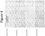
- Figure 4 provides epitope mapping data, specifically a dot blot showing peptide binding for antibodies.
- the peptides represent overlapping peptides covering the entire sequences of pre-Haptoglobin-2.
- Figure 5 shows that under native (no SDS), non-reducing conditions, all antibodies were capable of recognizing pre-Haptoglobin-1 and pre-Haptoglobin 2, but not mature (processed) haptoglobins.
- Figure 6 shows specificity and sensitivity data for antibody pair 13D11+11G3-bt using a sandwich ELISA on human serum.
- Figure 7 shows data from the Alpco Zonulin ELISA and the antibody pair 13D11+1 1G3-bt on normal and celiac patient samples.
- an “antibody” refers to a protein functionally defined as a binding protein and structurally defined as comprising an amino acid sequence that is recognized by one of skill as being derived from the framework region of an immunoglobulin-encoding gene of an animal that produces antibodies.
- An antibody can consist of one or more polypeptides substantially encoded by immunoglobulin genes or fragments of immunoglobulin genes.
- the recognized immunoglobulin genes include the kappa, lambda, alpha, gamma, delta, epsilon and mu constant region genes, as well as myriad immunoglobulin variable region genes.
- Light chains are classified as either kappa or lambda.
- Heavy chains are classified as gamma, mu, alpha, delta, or epsilon, which in turn define the immunoglobulin classes, IgG, IgM, IgA, IgD and IgE, respectively.
- a typical immunoglobulin (antibody) structural unit is known to comprise a tetramer.
- Each tetramer is composed of two identical pairs of polypeptide chains, each pair having one "light” (about 25 kD) and one "heavy” chain (about 50 kD).
- the N-terminus of each chain defines a variable region of about 100 to 110 or more amino acids primarily responsible for antigen recognition.
- the terms variable light chain (V L ) and variable heavy chain (V H ) refer to these light and heavy chains, respectively.
- antibody as used herein includes antibody fragments that retain binding specificity. For example, there are a number of well characterized antibody fragments.
- pepsin digests an antibody C-terminal to the disulfide linkages in the hinge region to produce F(ab)'2, a dimer of Fab which itself is a light chain joined to VH-CH1 (Fd) by a disulfide bond.
- the F(ab)'2 may be reduced under mild conditions to break the disulfide linkage in the hinge region thereby converting the (Fab')2 dimer into an Fab' monomer.
- the Fab' monomer is essentially a Fab with all or part of the hinge region (see, Fundamental Immunology, W.E.
- antibody also includes antibody fragments produced either by the modification of whole antibodies or synthesized using recombinant DNA methodologies.
- Antibodies include dimers such as V H -V L dimers, V H dimers, or V L dimers, including single chain antibodies.
- the antibody can be another fragment, such as a disulfide-stabilized Fv (dsFv).
- Antibodies may include those that have been displayed on phage or generated by recombinant technology using vectors where the chains are secreted as soluble proteins, e.g., scFv, Fv, Fab, (Fab')2 or generated by recombinant technology using vectors where the chains are secreted as soluble proteins.
- a “monoclonal antibody” refers to an antibody generated from a clonal antibody-producing cell such as a hybridoma, lymphocyte, or a recombinant antibody-producing cell.
- “Clonal” means the cells have been cultured in a pure culture in the absence of other cells.
- Monoclonal antibodies can be prepared using hybridoma methods, such as those described by Kohler & Milstein, Nature 256:495 (1975 ). In a hybridoma method, a mouse, rat, rabbit, or other appropriate host animal, is typically immunized with an immunizing agent to elicit lymphocytes that produce or are capable of producing antibodies that will specifically bind to the immunizing agent. Alternatively, the lymphocytes may be immunized in vitro. The resulting lymphocytes are fused with myeloma or other tumor cells to generate hybridoma cells capable of repeated cell divisions. Clonal hybridomas can then be selected, e.g., via dilution or single-
- Epitope or “antigenic determinant” refers to a site on an antigen to which an antibody binds.
- Epitopes can be formed both from contiguous amino acids or noncontiguous amino acids juxtaposed by tertiary folding of a protein. Epitopes formed from contiguous amino acids are typically retained on exposure to denaturing solvents whereas epitopes formed by tertiary folding are typically lost on treatment with denaturing solvents.
- An epitope typically includes at least 3, and more usually, at least 5 or 8-10 amino acids in a unique spatial conformation. Methods of epitope mapping are well known in the art (see, e.g., Epitope Mapping Protocols in Methods in Molecular Biology, Vol. 66, Glenn E. Morris, Ed (1996 )).
- an antibody pair that specifically detects pre-Haptoglobin-2 typically binds to pre-Haptoglobin-2 with a reactivity that is at least 5 or 10-fold better than the reactivity of the same antibody pair for Haptoglobin-2, pre-Haptoglobin-1, or Haptoglobin-1.
- a pair of antibodies does not "specifically bind" to a non-target protein if the pair of antibodies bind to pre-Haptoglobin-2 with a reactivity at least 10-fold greater than the reactivity for the non-target protein.
- antibody pairs 13D11+11G3 or 9G7+16H4 weakly bind to Haptoglobin-2 and pre-Haptoglobin-1, respectively, these pairs bind to pre-Haptoglobin-2 with an reactivity at least 10-fold higher (see Figure 1 ), and thus the antibody pairs are not considered to "significantly bind" to Haptoglobin-2 or pre-Hapotoglobin-1.
- a “label” or a “detectable moiety” is a heterologous composition detectable by spectroscopic, photochemical, biochemical, immunochemical, chemical, or other physical means.
- useful labels include fluorescent dyes, electron-dense reagents, enzymes ( e.g., as commonly used in an ELISA), radioactive labels, biotin, digoxigenin, or haptens and proteins or other entities which can be made detectable.
- the labels may be incorporated into the antibodies at any position.
- the labels need not be directly conjugated to the antibody, but can be present on a secondary detection agents, such as a secondary antibody that binds the anti- pre-Haptoglobin-2 antibody. Any method known in the art for conjugating the antibody to the label may be employed, e.g., using methods described in Hermanson, Bioconjugate Techniques 1996, Academic Press, Inc., San Diego .
- An antibody light or heavy chain variable region consists of a "framework" region interrupted by three hypervariable regions, also called complementarity-determining regions or CDRs.
- the extent of the framework region and CDRs have been defined (see, " Sequences of Proteins of Immunological Interest,” E. Kabat, et al., U.S. Department of Health and Human Services, (1987 )).
- the sequences of the framework regions of different light or heavy chains are relatively conserved within a species.
- the framework region of an antibody that is the combined framework regions of the constituent light and heavy chains, serves to position and align the CDRs in three dimensional space.
- the CDRs are primarily responsible for binding to an epitope of an antigen.
- the CDRs are typically referred to as CDR1, CDR2, and CDR3, numbered sequentially starting from the N-terminus.
- a “chimeric antibody” is an antibody molecule in which (a) the constant region, or a portion thereof, is altered, replaced or exchanged so that the antigen binding site (variable region) is linked to a constant region of a different or altered class, effector function and/or species, or an entirely different molecule which confers new properties to the chimeric antibody, e.g., an enzyme, toxin, hormone, growth factor or drug; or (b) the variable region, or a portion thereof, is altered, replaced or exchanged with a variable region having a different or altered antigen specificity.
- CDR complementarity-determining region
- CDR also generally known as hypervariable regions or hypervariable loops
- CDR refers to the art-recognized term as exemplified by Chothia and Lesk (1987) J. Mol. Biol. 196: 901 ; Chothia et al. (1989) Nature 342: 877 ; Kabat et al., Sequences of Proteins of Immunological Interest (National Institutes of Health, Bethesda, Md.) (1987 ); and Tramontano et al. (1990) J. Mol. Biol. 215: 175 .
- “Framework region of the variable region” or "FR” refers to the region of the V domain that flank the CDRs.
- the positions of the CDRs and framework regions can be determined using various well known definitions in the art as described in, e.g., Kabat, supra, Chothia, supra, international ImMunoGeneTics database (IMGT), and AbM (see, e.g., Chothia & Lesk, 1987, J. Mol. Biol. 196, 901-917 ; Chothia, et al., 1989, Nature 342, 877-883 ; Chothia, et al., 1992, J. Mol. Biol. 227, 799-817 ; Al-Lazikani et al., J.Mol.Biol 1997, 273(4 )).
- IMGT ImMunoGeneTics database
- CDRs are also described in the following: Ruiz et al., IMGT, the international ImMunoGeneTics database. Nucleic Acids Res., 28, 219-221 (2000 ); and Lefranc,M.-P. IMGT, the international ImMunoGeneTics database. Nucleic Acids Res. Jan 1;29(1):207-9 (2001 ); MacCallum et al, J. Mol. Biol., 262 (5), 732-745 (1996 ); Martin et al, Proc. Natl Acad. Sci.
- Monoclonal antibodies that have high affinity for human pre-Haptoglobin-2. Moreover, some antibody pairs have high reactivity for human pre-Haptoglobin-2, i.e., the antibody pairs detect human pre-Haptoglobin-2 but do not detect, or do not significantly detect, human pre-Haptoglobin-1, human Haptoglobin-1, or human Haptoglobin-2. Tripathi, et al. published findings suggesting that zonulin is pre-haptoglobin 2 (PNAS 106(39): 16799-804, 2009 ).
- the inventors have surprisingly found that these antibody pairs specifically detect pre-Haptoglobin-2 with much better specificity than commercially-available polyclonal antibodies used in an ELISA sold as specific for Zonulin.
- the herein disclosed pairs of antibodies that allow for specific detection of pre-Haptoglobin-2, even in the presence of human pre-Haptoglobin-1, human Haptoglobin-1, or human Haptoglobin-2.
- they bind at least one of human pre-Haptoglobin-1, human Haptoglobin-1, or human Haptoglobin-2 in addition to human pre-Haptoglobin-2.
- specific pairs of antibodies were tested in a sandwich immunoassay, the pairs specifically detect human pre-Haptoglobin-2 and do not significantly detect human pre-Haptoglobin-1, human Haptoglobin-1, or human Haptoglobin-2.
- monoclonal antibodies that bind to human pre-Haptoglobin-2 but do not bind human pre-Haptoglobin-1, human Haptoglobin-1, or human Haptoglobin-2 (e.g., as determined by surface plasmon resonance). These monoclonal antibodies bind to a number of overlapping peptides comprising GYVEHSVRY (SEQ ID NO:1). In some embodiments, the monoclonal antibodies having the above criteria have a K D for human pre-Haptoglobin-2 of less than 25 or 10 nM, e.g., 25-1 or 10-1 nM (e.g., as determined by surface plasmon resonance). As known in the art, a lower K D value indicates stronger binding (higher affinity) to the target.
- these monoclonal antibodies bind to native (lacking a denaturant), non-reduced (lacking a reducing agent (e.g. ⁇ -mercaptoethanol (BME) or dithiothreitol)), human pre-Haptoglobin-2, but do not significantly bind reduced, denatured human pre-Haptoglobin-2.
- a sample comprising pre-Haptoglobin-2 run on a gel with a denaturant (e.g.,SDS) and a reducing agent (e.g., BME) and blotted on a western blot (e.g., a typical western blotting procedure) represents a reduced, denatured pre-Haptoglobin-2.
- a sample comprising pre-Haptoglobin-2 run on a gel lacking a denaturant (e.g., lacking SDS) and lacking a reducing agent (e.g., lacking BME) and blotted on a western blot represents a native, non-reduced pre-Haptoglobin-2.
- reducing conditions are thought to disrupt secondary structure of proteins and thus it is believed that the monoclonal antibodies that bind non-reduced human pre-Haptoglobin-2, but do not significantly bind reduced human pre-Haptoglobin-2, bind to three-dimensional (non-linear) epitopes on human pre-Haptoglobin-2.
- the monoclonal antibodies having the criteria described in this paragraph have a K D for human pre-Haptoglobin-2 of less than 25or 10 nM, e.g., 25-0.1 or 10-0.1 nM.
- Antibodies described herein can be generated as desired.
- Monoclonal antibodies may be prepared using hybridoma methods.
- a mouse, hamster, or other appropriate host animal may be immunized with an immunizing agent to elicit lymphocytes that produce or are capable of producing antibodies that will specifically bind to the immunizing agent.
- the lymphocytes may be immunized in vitro.
- An appropriate immunogen can be, for example, a recombinant full-length pre-Haptoglobin-2 (e.g. affinity-tagged and expressed by recombinant insect cells).
- a variety of methods are known and can be used for generating hybridomas or other antibody-producing cells. See, e.g., Harlow, ANTIBODIES, Cold Spring Harbor Press, N.Y.
- hybridomas producing monoclonal antibodies to an antigen splenocytes and lymph node cells from immunized animals can be isolated and fused to an appropriate immortalized cell line, such as a mouse myeloma cell line. The resulting hybridomas can then be screened for the production of antigen-specific antibodies. For example, single cell suspensions of splenic lymphocytes from immunized mice can be fused to SP2/0-Ag14 myeloma cells (ATCC, CRL 1581) with 1 ml PEG1500 (Roche).
- Cells can be plated at approximately 3x10 4 per well in flat bottom microtiter plate, followed by a two-week incubation in selective medium containing besides usual reagents 20 % Hyclone Fetal Bovine Serum, 10 % Hybridoma Cloning Supplement (PAA), 1 % OPI Media Supplement (Sigma) and 2 % Azaserine-Hypoxanthine (Sigma).
- a plurality of hybridoma culture supernatants (or other antibody solutions) can be screened in parallel for desired binding properties.
- Human monoclonal antibodies can be produced using various techniques known in the art, including phage display libraries. Similarly, human antibodies can be made by introducing of human immunoglobulin loci into transgenic animals, e.g., mice in which the endogenous immunoglobulin genes have been partially or completely inactivated. Upon challenge, human antibody production is observed, which closely resembles that seen in humans in all respects, including gene rearrangement, assembly, and antibody repertoire. This approach is described, e.g., in U.S. Patents.
- recombinant (or purified) preHP2, preHP1, HP1, and HP2 can be coated to plates and the hybridoma supernatant can then be applied to the plates. Detection of binding can be achieved using an anti-mouse antibodies linked to horseradish peroxidase (HRP) (a direct ELISA format).
- HRP horseradish peroxidase
- the plates can be coated with the hybridoma supernatant via an anti-mouse antibody, protein can be applied, and binding then detected using a pan-haptoglobin antibody followed by contact with secondary antibodies linked to HRP (a sandwich ELISA format).
- any of the antibodies described herein can be isolated from other biological components, e.g., can be in a solution separate from cells or cellular components.
- the antibodies may represent at least 90, 95, or 99 % of all protein in the solution.
- one or more antibody described herein can be linked to a solid support.
- Any type of solid support can be used as desired.
- the solid support can be the wall or floor of an assay vessel, or a dipstick or other implement to be inserted into an assay vessel, or particles placed inside or suspended in an assay vessel.
- Particles, and especially beads are particularly useful in many embodiments, including beads that are microscopic in size (i.e., microparticles) and formed of a polymeric material.
- Polymers useful as microparticles are those that are chemically inert relative to the components of the biological sample and to the assay reagents other than the binding members that are immobilized on the microparticle surface.
- Exemplary microparticle materials are those with minimal autofluorescence, and that are solid and insoluble in the sample and in any buffers, solvents, carriers, diluents, or suspending agents used in the assay, in addition to allowing immobilization of the assay reagent.
- suitable polymers are polystyrenes, polyesters, polyethers, polyolefins, polyalkylene oxides, polyamides, polyurethanes, polysaccharides, celluloses, and polyisoprenes.
- Crosslinking is useful in many polymers for imparting structural integrity and rigidity to the microparticle.
- the size range of the microparticles can vary. The microparticles may range in diameter from 0.3 ⁇ m to 100 ⁇ m, and in other cases, from 0.5 ⁇ m to ⁇ m 40 ⁇ m, and in still other cases, from 2 ⁇ m to 10 ⁇ m.
- Attachment of the antibodies to the surfaces of the solid support can be achieved, for example, by electrostatic attraction, specific affinity interaction, hydrophobic interaction, or covalent bonding.
- Functional groups for covalent bonding can be incorporated into the polymer structure by conventional means, such as the use of monomers that contain the functional groups, either as the sole monomer or as a co-monomer. Examples of suitable functional groups are amine groups (-NH 2 ), ammonium groups (-NH 3 + or -NR 3 + ), hydroxyl groups (-OH), carboxylic acid groups (-COOH), and isocyanate groups (-NCO).
- Useful monomers for introducing carboxylic acid groups into polyolefins, for example, are acrylic acid and methacrylic acid.
- Linking groups can also be used for increasing the density of the antibodies on the solid phase surface and for decreasing steric hindrance to increase the range and sensitivity of the assay.
- suitable useful linking groups are polylysine, polyaspartic acid, polyglutamic acid and polyarginine.
- one or more antibody described herein can be linked to a label.
- the particular label or detectable group used is not critical, as long as it does not significantly interfere with the specific binding of the antibody used and allows for detection of the antibody.
- the label can be directly attached to the antibody or to another agent used in the detection assay, such as a secondary antibody.
- the detectable group can be any material having a detectable physical or chemical property.
- detectable labels have been well-developed in the field of immunoassays and, in general, any label useful in such methods can be applied to the antibodies described here.
- a label is any composition directly or indirectly detectable by spectroscopic, photochemical, biochemical, immunochemical, electrical, optical or chemical means.
- Useful labels include fluorescent compounds (e.g., fluorescein isothiocyanate, Texas red and rhodamine, fluorescein), radiolabels, enzymes (e.g., horse radish peroxidase, alkaline phosphatase and others commonly used in an ELISA), streptavidin/biotin, and colorimetric labels such as colloidal gold or colored glass or plastic beads (e.g., polystyrene, polypropylene or latex). Chemiluminescent compounds may also be used.
- fluorescent compounds e.g., fluorescein isothiocyanate, Texas red and rhodamine, fluorescein
- radiolabels e.g., enzymes (e.g., horse radish peroxidase, alkaline phosphatase and others commonly used in an ELISA), streptavidin/biotin, and colorimetric labels such as colloidal gold or colored glass or plastic beads (e.g., polyst
- One or more anti-pre-Haptiglobin-2 antibody disclosed herein may have the CDRs (i.e., CDRs 1, 2, and 3 from the heavy chain variable region and CDRs1, 2, and 3 from the light chain variable region) from one of: 13D11, 11G3, 9G7, and 16H4.
- CDRs i.e., CDRs 1, 2, and 3 from the heavy chain variable region and CDRs1, 2, and 3 from the light chain variable region
- 13D11, 11G3, 9G7, and 16H4 Once a CDR is identified from 13D11, 11G3, 9G7, or 16H4, the CDRs, or the entire variable regions, can be cloned back into an immunoglobulin scaffold. Examples of this technique include the CDR grafting antibody technique. See, e.g., P.T. Jones et al., Nature, 321:522, 1986 .
- the region comprising the selected CDRs and the immunoglobulin scaffolds in which the CDRs are inserted are from the same animal.
- the CDRs and scaffold may also be from different animals.
- cDNAs encoding the variable regions thus constructed may be expressed as desired.
- Cells, e.g. mammalian cells, comprising vectors for expressing the cDNAs can also be provided.
- the CDRs are used to make a single chain antibody (scFv).
- scFv single chain antibody
- the VH- and VL-encoding DNA fragments are operatively linked to another fragment encoding a flexible linker such that the VH and VL sequences can be expressed as a contiguous single-chain protein, with the VL and VH regions joined by the flexible linker (see e.g., Bird et al. (1988) Science 242:423-426 ; Huston et al. (1988) Proc. Natl. Acad. Sci. USA 85:5879-5883 ; McCafferty et al., Nature (1990) 348:552-554 ).
- immunoassay techniques including competitive and non-competitive immunoassays, employing one or more (e.g., two) antibodies having the binding properties as described herein, can be used to detect the presence, absence, or level of pre-Haptoglobin-2 in a sample.
- immunoassay formats are described in, e.g., Self and Cook, Curr. Opin. Biotechnol., 7:60-65 (1996 )).
- immunoassay encompasses techniques including enzyme immunoassays (EIA) such as enzyme multiplied immunoassay technique (EMIT), enzyme-linked immunosorbent assay (ELISA), antigen capture ELISA, sandwich ELISA, IgM antibody capture ELISA (MAC ELISA), and microparticle enzyme immunoassay (MEIA); capillary electrophoresis immunoassays (CEIA); radioimmunoassays (RIA); immunoradiometric assays (IRMA); fluorescence polarization immunoassays (FPIA); and chemiluminescence assays (CL). If desired, such immunoassays can be automated.
- EIA enzyme multiplied immunoassay technique
- ELISA enzyme-linked immunosorbent assay
- MAC ELISA enzyme-linked immunosorbent assay
- MEIA microparticle enzyme immunoassay
- CEIA capillary electrophoresis immunoassays
- RIA radioimmunoas
- Immunoassays can also be used in conjunction with laser induced fluorescence (see, e.g., Schmalzing and Nashabeh, Electrophoresis, 18:2184-2193 (1997 ); Bao, J. Chromatogr. B. Biomed. Sci., 699:463-480 (1997 ).
- Antigen capture ELISA can be useful for detecting the presence or level of pre-Haptoglobin-2 in a sample.
- an antibody directed to pre-Haptoglobin-2 is linked to a solid phase and sample is added such that pre-Haptoglobin-2, if present in a sample, is bound by the antibody. After unbound proteins are removed by washing, the amount of bound marker can be quantitated using, e.g., a radioimmunoassay (see, e.g., Harlow and Lane, Antibodies: A Laboratory Manual, Cold Spring Harbor Laboratory, New York, 1988 )).
- sandwich ELISA immunoassays are also suitable for use in detecting pre-Haptoglobin-2.
- a first (capture) antibody is bound to a solid support, and pre-Haptoglobin-2, if present in the sample, is allowed to bind to the first antibody.
- other components of the sample are removed (e.g., washed away) before a second (detection) antibody is contacted to the antigen bound to the capture antibody.
- the amount of the marker is quantitated by measuring the amount of a second (capture) antibody that binds pre-Haptoglobin-2.
- the antibodies can be immobilized onto a variety of solid supports, such as magnetic or chromatographic matrix particles, the surface of an assay plate (e.g., microtiter wells) and pieces of a solid substrate material or membrane (e.g., plastic, nylon, paper) as described herein.
- An assay strip may be prepared by coating the antibody or a plurality of antibodies in an array on a solid support. This strip can then be dipped into the test sample and processed quickly through washes and detection steps to generate a measurable signal, such as a colored spot.
- the capture antibody binds to GYVEHSVRY (SEQ ID NO:1), or a fragment of pre-Haptoglobin-2 comprising GYVEHSVRY (SEQ ID NO:1).
- the monoclonal antibody may bind KPPEIAHGYVEHSVRYQCKNYYK (SEQ ID NO:2).
- the monoclonal antibody may bind KPPEIAHGYVEHSVR (SEQ ID NO:3), PPEIAHGYVEHSVRY (SEQ ID NO:4), PEIAHGYVEHSVRYQ (SEQ ID NO:5), EIAHGYVEHSVRYQC (SEQ ID NO:6), IAHGYVEHSVRYQCK (SEQ ID NO:7), AHGYVEHSVRYQCKN (SEQ ID NO:8), HGYVEHSVRYQCKNY (SEQ ID NO:9), GYVEHSVRYQCKNYY (SEQ ID NO:10), and/or YVEHSVRYQCKNYYK (SEQ ID NO:11).
- the monoclonal antibody may bind one or more peptide listed above but may not bind PKPPEIAHGYVEHSV (SEQ ID NO:12) or
- the capture antibody may bind non-reduced pre-Haptoglobin-2 but may not bind to reduced pre-Haptoglobin-2.
- the detection antibody binds to GYVEHSVRY (SEQ ID NO:1), or a fragment of pre-Haptoglobin-2 comprising GYVEHSVRY (SEQ ID NO:1).
- the monoclonal antibody may bind KPPEIAHGYVEHSVRYQCKNYYK (SEQ ID NO:2).
- the monoclonal antibody may bind KPPEIAHGYVEHSVR (SEQ ID NO:3), PPEIAHGYVEHSVRY (SEQ ID NO:4), PEIAHGYVEHSVRYQ (SEQ ID NO:5), EIAHGYVEHSVRYQC (SEQ ID NO:6), IAHGYVEHSVRYQCK (SEQ ID NO:7), AHGYVEHSVRYQCKN (SEQ ID NO:8), HGYVEHSVRYQCKNY (SEQ ID NO:9), GYVEHSVRYQCKNYY (SEQ ID NO:10), and/or YVEHSVRYQCKNYYK (SEQ ID NO:11).
- the monoclonal antibody may bind one or more peptide listed above but may not bind PKPPEIAHGYVEHSV (SEQ ID NO:12) or VEHSVRYQCKNYYKL (SEQ ID NO:13).
- the detection antibody binds non-reduced pre-Haptoglobin-2 but does not bind to reduced pre-Haptoglobin-2.
- One or both antibodies of the pair may have affinity for Haptoglobin-2 or pre-Haptoglobin-1 as well as for pre- Haptoglobin -2.
- the antibody pair when paired in a sandwich assay, specifically detects pre-Haptoglobin-2 and does not significantly detect Haptoglobin-2 or pre-Haptoglobin-1.
- the antibody pair may detect pre-Hapoglobin-2 with better sensitivity and specificity than a sandwich assay based on polyclonal antibodies directed to Zonulin.
- a radioimmunoassay using, for example, an iodine-125 ( 125 I) labeled secondary antibody is also suitable for detecting the presence or level of one or more markers in a sample.
- a secondary antibody labeled with a chemiluminescent marker can also be suitable.
- a chemiluminescence assay using a chemiluminescent secondary antibody is suitable for sensitive, non-radioactive detection of marker levels.
- Such secondary antibodies can be obtained commercially from various sources, e.g., Amersham Lifesciences, Inc. (Arlington Heights, 111.).
- Direct labels include fluorescent or luminescent tags, metals, dyes and radionuclides, attached to the antibody.
- An antibody labeled with iodine-125 ( 125 I) can be used for determining the level of pre-Haptoglobin-2 in a sample.
- a chemiluminescence assay using a chemiluminescent antibody specific for pre-Haptoglobin-2 is suitable for sensitive, non-radioactive detection of pre-Haptoglobin-2 levels.
- An antibody labeled with fluorochrome is also suitable for determining the levels of pre-Haptoglobin-2 in a sample.
- fluorochromes examples include DAPI, fluorescein, Hoechst 33258, R-phycocyanin, B-phycoerythrin, R-phycoerythrin, rhodamine, Texas red, and lissamine. Secondary antibodies linked to fluorochromes can be obtained commercially, e.g., goat F(ab') 2 anti-human IgG-FITC is available from Tago Immunologicals (Burlingame, Calif.).
- Indirect labels include various enzymes well-known in the art, such as horseradish peroxidase (HRP), alkaline phosphatase (AP), ⁇ -galactosidase and urease.
- HRP horseradish peroxidase
- AP alkaline phosphatase
- ⁇ -galactosidase ⁇ -galactosidase
- urease a horseradish-peroxidase detection system can be used, for example, with the chromogenic substrate tetramethylbenzidine (TMB), which yields a soluble product in the presence of hydrogen peroxide that is detectable at 450 nm.
- TMB tetramethylbenzidine
- An alkaline phosphatase detection system can be used with the chromogenic substrate p-nitrophenyl phosphate, for example, which yields a soluble product readily detectable at 405 nm.
- a ⁇ galactosidase detection system can be used with the chromogenic substrate o-nitrophenyl- ⁇ -D-galactopyranoside (ONPG), which yields a soluble product detectable at 410 nm.
- a urease detection system can be used with a substrate such as urea-bromocresol purple (Sigma Immunochemicals; St. Louis, Mo.).
- a useful secondary antibody linked to an enzyme can be obtained from a number of commercial sources, e.g., goat F(ab') 2 anti-human IgG-alkaline phosphatase can be purchased from Jackson ImmunoResearch (West Grove, Pa.).
- a signal from the direct or indirect label can be analyzed, for example, using a spectrophotometer to detect color from a chromogenic substrate; a radiation counter to detect radiation such as a gamma counter for detection of 125 I; or a fluorometer to detect fluorescence in the presence of light of a certain wavelength.
- a quantitative analysis of the amount of marker levels can be made using a spectrophotometer such as an EMAX Microplate Reader (Molecular Devices; Menlo Park, Calif.) in accordance with the manufacturer's instructions.
- the assays described herein can be automated or performed robotically, and the signal from multiple samples can be detected simultaneously.
- Quantitative Western blotting can also be used to detect or determine the presence or level of pre-Haptoglobin-2 in a sample.
- Western blots can be quantitated by methods such as scanning densitometry or phosphorimaging. As an, protein samples are electrophoresed on 10 % SDS-PAGE Laemmli gels. Murine monoclonal antibodies are reacted with the blot, and antibody binding can be confirmed to be linear using a preliminary slot blot experiment.
- Goat anti-mouse horseradish peroxidase-coupled antibodies are used as the secondary antibody, and signal detection performed using chemiluminescence, for example, with the Renaissance chemiluminescence kit (New England Nuclear; Boston, Mass.) according to the manufacturer's instructions. Autoradiographs of the blots are analyzed using a scanning densitometer (Molecular Dynamics; Sunnyvale, Calif.) and normalized to a positive control. Values are reported, for example, as a ratio between the actual value to the positive control (densitometric index). Such methods are well known in the art as described, for example, in Parra et al., J. Vasc. Surg., 28:669-675 (1998 ).
- immunohistochemical assay encompasses techniques that utilize the visual detection of fluorescent dyes or enzymes coupled (i.e., conjugated) to antibodies that react with pre-Haptoglobin-2 using fluorescent microscopy or light microscopy (e.g., in a tissue slice) and includes direct fluorescent antibody assay, indirect fluorescent antibody (IFA) assay, anticomplement immunofluorescence, avidin-biotin immunofluorescence, and immunoperoxidase assays.
- An IFA assay for example, is useful for determining whether a sample is positive for pre-Haptoglobin-2 or the level of pre-Haptoglobin-2 in a sample.
- concentration of pre-Haptoglobin-2 in a sample can be quantitated, e.g., through endpoint titration or through measuring the visual intensity of fluorescence compared to a known reference standard.
- Pre-Haptoglobin-2 may be detected as part of a multiplex assay.
- the analysis of a plurality of markers may be carried out separately or simultaneously with one test sample.
- suitable apparatuses include clinical laboratory analyzers such as the ElecSys (Roche), the AxSym (Abbott), the Access (Beckman), the ADVIATM, the CENTAURTM (Bayer), and the NICHOLS ADVANTAGETM (Nichols Institute) immunoassay systems.
- Exemplary apparatuses or protein chips perform simultaneous assays of a plurality of markers on a single surface.
- Exemplary physical formats comprise surfaces having a plurality of discrete, addressable locations for the detection of a plurality of different markers.
- each discrete surface location may comprise antibodies to immobilize one or more markers for detection at each location.
- Surfaces may alternatively comprise one or more discrete particles (e.g., microparticles or nanoparticles) immobilized at discrete locations of a surface, where the microparticles comprise antibodies to immobilize one or more markers for detection.
- Detection of pre-Haptoglobin-2 can be used for providing a diagnosing or prognosis, monitoring disease and/or monitoring treatment of autoimmune and inflammatory disease or allergies.
- Expression of pre-Haptoglobin-2 is associated with autoimmune and inflammatory diseases as well as allergies.
- expression of pre-Haptoglobin-2 is associated with celiac disease and Type-1 diabetes. See, e.g., US Patent Publication 2012/0107329 .
- kits for performing an immunoassay employing one or more (e.g., two) pre-Haptoglobin-2 antibody as described herein.
- the kit comprises a least one pre-Haptoglobin-2 antibody as described herein linked to a solid support.
- the kit comprises at least one pre-Haptoglobin-2 antibody as described herein linked to a label.
- the kit comprises a least one pre-Haptoglobin-2 antibody as described herein linked to a solid support and at least one pre-Haptoglobin-2 antibody as described herein linked to a label.
- the antibody linked to the solid support binds to GYVEHSVRY (SEQ ID NO:1), or a fragment of pre-Haptoglobin-2 comprising GYVEHSVRY (SEQ ID NO:1).
- the antibody linked to the solid support may bind KPPEIAHGYVEHSVRYQCKNYYK (SEQ ID NO:2).
- the antibody linked to the solid support may bind KPPEIAHGYVEHSVR (SEQ ID NO:3), PPEIAHGYVEHSVRY (SEQ ID NO:4), PEIAHGYVEHSVRYQ (SEQ ID NO:5), EIAHGYVEHSVRYQC (SEQ ID NO:6), IAHGYVEHSVRYQCK (SEQ ID NO:7), AHGYVEHSVRYQCKN (SEQ ID NO:8), HGYVEHSVRYQCKNY (SEQ ID NO:9), GYVEHSVRYQCKNYY (SEQ ID NO:10), and/or YVEHSVRYQCKNYYK (SEQ ID NO:11).
- the antibody linked to the solid support may bind one or more peptide listed above but may not bind PKPPEIAHGYVEHSV (SEQ ID NO:12) or VEHSVRYQCKNYYKL (SEQ ID NO:13).
- At least one pre-Haptoglobin-2 antibody in the kit may have a K D for human pre-Haptoglobin-2 of less than 25, 20, 15, 10, 5, or 1 nM, e.g., 25-0.1 or 10-0.1 nM.
- the kit comprises at least one antibody (e.g., linked to a solid support or linked to a label) that binds to non-reduced human pre-Haptoglobin-2, but do not significantly bind reduced human pre-Haptoglobin-2.
- antibodies do not bind to human pre-Haptoglobin-1 (e.g., as determined by surface plasmon resonance).
- This example describes generation and characterization of antibodies that bind to pre-Haptoglobin-2 and development of the antibodies to specific detection of pre-Haptoglobin-2.
- preHP2 Full-length recombinant pre-Haptoglobin-2 (preHP2) was generated and purified from cells. 10 ⁇ g of recombinant human pre-HP2 (GenBank accession no. NP_005134) expressed in insect cells with a C-terminal hexahistidine (SEQ ID NO:14) tag were emulsified in equal ratio with Sigma Adjuvant System or Alum adjuvant (Thermo Scientific Pierce, Rockford, IL). Four 8-week-old CD1 mice (Charles River) were immunized by intraperitoneal injections. Two booster injections were administrated at the same doses at 2-week intervals.
- mice were bled 10 d after each boost and serology was controlled by indirect ELISA to select the best responding mouse.
- a pre-fusion boost was administered 4 d before fusion.
- Sp2/0Ag14 myeloma cell line (ATCC CRL 1581) was fused with splenocytes from selected immunized mouse according to standard protocols and fusion product was plated in culture microplates for 15 d before primary screening.
- clones' selection either indirect or sandwich ELISA were used all along the process: from primary screening, confirmation to sub-cloning screenings.
- Antigens used were recombinant pre-HP2 as specific antigen and recombinant pre-HP1, purified mature haptoglobin 1 and 2 (Sigma Aldrich) and a non-specific His-tagged protein as control antigens.
- microplates were first coated with a goat anti-mouse IgG antibody (Jackson Immuno Research, West Grove, PA) before adding hybridoma supernatants; antigen binding was detected with a rabbit anti-pan haptoglobin polyclonal antibody (internal source) followed by the addition of a horseradish peroxidase (HRP) conjugated goat anti-rabbit IgG antibody (Biosource, Grand Island, NY).
- HRP-conjugated goat anti-mouse IgG antibody Sigma.
- TMB tetramethylbenzidine
- OD optical density
- antibody-producing hybridomas were twice sub-cloned and then frozen in liquid nitrogen. Monoclonal antibodies were produced in vitro by collecting concentrated supernatants. Purifications were done by affinity chromatography on Protein A Sepharose (GE Healthcare, Piscataway, NJ). The mAbs were isotyped with a mouse isotyping test kit (Roche, Indianapolis, IN) according to the manufacturer's recommendations.
- pre-Haptoglobin-2 pre-Haptoglobin-2
- pre-HPl pre-Haptoglobin-1
- HP2-2 Haptoglobin-2
- HP1-1 Haptoglobin-1
- Standard sandwich ELISA assay conditions were used as described above except the detection antibody was biotinylated and binding was detected with streptavidin HRP.
- capture antibody was coated onto a 96 well plate (Maxisorp, Nunc), antigen (preHP2) was then added, followed by biotinylated detection antibody and streptavidin HRP, with washing with PBS+0.1 % Tween between each step. Color was developed with 3, 3', 5,5 '-tetramethylbenzidine (TMB). Clones were used as both capture and detection.
- Concentrations on the left of Figure 1 indicate the concentrations at which the protein was tested.
- the mature Haptoglobin proteins were tested at 1000-fold higher concentrations than the pre-Haptoglobin proteins. The reason for this is that mature haptoglobins are known to normally be present in human serum around 1-1.5mg/ml.
- the assay was intended to be run at a 1:10 dilution of human serum, so 100 ug/ml would be the final concentration of mature haptoglobin in the well to test for cross-reaction with the antibodies.
- a reference range of normal preHP 1 and preHP2 in human serum was not known, so 0.1 ug/ml was chosen because that level falls in the higher range of the standard curve.
- 13D11+1 1G3-bt had minor cross-reactivity in this sandwich assay with HP2-2, whereas 9G7+16H4-bt had minor cross-reactivity with pre-HP1.
- the cross-reactivity is considered insignificant binding in that the signal for pre-HP2 was at least 10-fold higher than signal for pre-HP1 and at least 10,000-fold higher than HP2-2 (keeping in mind HP2-2 was loaded at 1,000-fold higher concentrations than pre-HP2).
- the four antibodies from the test pairs i.e., 13D11, 11G3, 9G7, and 16H4 were tested by surface plasmon resonance (SPR) for their affinity for pre-Haptoglobin-2.
- SPR surface plasmon resonance
- 13D11 displayed only binding affinity for pre-Haptoglobin-2.
- the remaining three antibodies (12G3, 9G7, and 16H4) had affinity for pre-Haptoglobin-2 as well as pre-Haptoglobin-1.
- antibody 9G7 displayed a stronger affinity (lower K D ) for pre-Haptoglobin-1 than pre-Haptoglobin-2 even though when paired with 16H4, the combination in a sandwich assay specifically detected pre-Haptoglobin-2 with only insignificant pre-Haptoglobin-1 background binding.
- Immobilization of the monoclonal antibodies was performed in the vertical orientation of the ProteOn XPR36 system (Bio-Rad) using a flow rate of 30 ⁇ l/min at 25 °C on a GLC chip.
- Four channels were activated for 3 min (90 ⁇ l) using a mixture of 20 mM 1-ethyl-3-(3-dimethylaminopropyl) carbodiimide hydrochloride carbodiimide(EDC) and 5 mM sulfo- N-hydroxysulfosuccinimide (sulfo-NHS). This was followed by an immediate injection of 50 ⁇ l of 10 ug/ml monoclonal antibody in 10 mM acetate buffer (pH 4.5).
- the human pre-Haptoglobin-2 amino acid sequence is provided below (SEQ ID NO:15): Italics: Signal sequence Bold: ⁇ -chain repeat sequence Underline: 13D11 minimal epitopes
- Antibody from hybridoma 13D11 bound the following peptides: 35,94 KPPEIAHGYVEHSVR (SEQ ID NO:3) 36,95 PPEIAHGYVEHSVRY (SEQ ID NO:4) 37,96 PEIAHGYVEHSVRYQ (SEQ ID NO:5) 38,97 EIAHGYVEHSVRYQC (SEQ ID NO:6) 39,98 IAHGYVEHSVRYQCK (SEQ ID NO:7) 40,99 AHGYVEHSVRYQCKN (SEQ ID NO:8) 41,100 HGYVEHSVRYQCKNY (SEQ ID NO:9) 42,101 GYVEHSVRYQCKNYY (SEQ ID NO:10) 43,102 YVEHSVRYQCKNYYK (SEQ ID NO:1 1). Antibody from hybridoma 13D11 did not bind the following peptides, which are adjacent to those listed above:
- non-denaturing (native) western blots were prepared from polyacrylamide gel electrophoresis (PAGE) and without ("non-reducing") ⁇ -mercaptoethanol. Those gels run without SDS and ⁇ -mercaptoethanol were considered non-reducing and non-denaturing, leaving the proteins in state believed to retain at least some native three-dimensional and secondary structure. Control gels run without SDS but with samples loaded with ⁇ -mercaptoethanol ("reducing") were run and blotted in parallel.
- Results for detection of pre-Haptoglobin-2 by antibody pair 13D11+1 1G3-bt was compared to binding by a commercial polyclonal-antibody-based Zonulin assay (Alpco Zonulin ELISA, Alpco, Salem, New Hampshire) performed according to manufacturer's directions.
- the Alpco Zonulin assay is a competitive ELISA, which uses a polyclonal anti-zonulin antibody to capture native zonulin in the presence of a biotinylated competitor, presumably recombinant zonulin protein (composition of the competitor is not disclosed by the manufacturer).
- High signal from the competitor means that native zonulin is not present at substantial amounts in the sample, while low signal in the assay means that high levels of native zonulin is present (inverse correlation of signal to amount of target in the sample being assayed).
- Pre-HP2 capture antibody (clone 13D11) was diluted to 2 ⁇ g/mL in PBS (10 mM Sodium Phosphate, 150 mM NaCl, pH 7.8) and added to a 96-well plates (100 ⁇ l/well; Maxisorp, Nunc, Roskilde, Denmark). After an overnight incubation at 4 °C, antibody solution was removed and wells were blocked with blocking buffer (PBS + 3 % BSA) for one hour at room temperature (RT).
- PBS mM Sodium Phosphate, 150 mM NaCl, pH 7.8
- Serum samples and calibrators diluted in sample diluent (PBS + 0.1 % Tween-20 + 0.1 %BSA + 0.3 ⁇ g/ml mouse IgG (Meridian Life Science, Memphis, TN)) were then added to the plate (100 ⁇ l/well) after removal of the blocking buffer and incubated for two hours at RT. The plate was then washed three times with PBS + 0.1 % Tween-20 (PBST) using an automated plate washer (BioPlex Pro II Wash Station, Bio-Rad, Hercules, CA).
- biotinylated detection antibody (clone 11G3) diluted to 2 ug/ml in conjugate diluent (sample diluent without mouse IgG) was added (100 ⁇ l/well) and incubated for one hour at RT.
- the plate was again washed three times in PBST, and streptavidin-HRP (Thermo Fisher) diluted 1:10,000 in conjugate diluent was added (100 ⁇ l/well) and incubated for one hour at RT.
- Ultra TMB ELISA substrate (Thermo Fisher) was added to the plate (100 ⁇ l/well) and color was allowed to develop for 15 min at RT.
- preHP2 was spiked into serum at 2 ug/ml and then this sample was diluted according to the manufacturer's directions.
- preHP1 (2 ⁇ g/ml), HP1-1(1 mg/ml) and HP2-2(1 mg/ml) were also spiked and assayed. No difference was seen between preHP2 signal and the other haptoglobins indicating that the Alpco assay is not specifically detecting preHP2.
- the table at the bottom of Figure 7 summarizes the results of the assay.
Landscapes
- Health & Medical Sciences (AREA)
- Chemical & Material Sciences (AREA)
- Immunology (AREA)
- Life Sciences & Earth Sciences (AREA)
- Molecular Biology (AREA)
- Organic Chemistry (AREA)
- Engineering & Computer Science (AREA)
- Biochemistry (AREA)
- Medicinal Chemistry (AREA)
- General Health & Medical Sciences (AREA)
- Hematology (AREA)
- Biomedical Technology (AREA)
- Urology & Nephrology (AREA)
- Genetics & Genomics (AREA)
- Proteomics, Peptides & Aminoacids (AREA)
- Biophysics (AREA)
- Chemical Kinetics & Catalysis (AREA)
- Cell Biology (AREA)
- Microbiology (AREA)
- Biotechnology (AREA)
- Food Science & Technology (AREA)
- Physics & Mathematics (AREA)
- Analytical Chemistry (AREA)
- General Physics & Mathematics (AREA)
- Pathology (AREA)
- Peptides Or Proteins (AREA)
- Preparation Of Compounds By Using Micro-Organisms (AREA)
Claims (15)
- Anticorps monoclonal isolé qui se lie à la pré-Haptoglobine-2 humaine mais ne se lie ni à la pré-Haptoglobine-1 humaine, ni à l'Haptoglobine-l humaine ni à l'Haptoglobine-2 humaine, tel que déterminé par résonance plasmonique de surface, ledit anticorps monoclonal se liant spécifiquement à GYVEHSVRY (SEQ ID NO : 1).
- Anticorps monoclonal isolé selon la revendication 1, ledit anticorps monoclonal se liant à la pré-Haptoglobine-2 avec un KD inférieur à 25 ou 10 nM.
- Anticorps monoclonal isolé selon la revendication 1, ledit anticorps monoclonal étant lié à un marqueur détectable.
- Anticorps monoclonal isolé selon la revendication 3, où le marqueur détectable est la biotine.
- Anticorps monoclonal isolé selon la revendication 1, ledit anticorps monoclonal étant lié à un support solide.
- Trousse contenant l'anticorps monoclonal selon la revendication 1, dans laquelle l'anticorps monoclonal est lié à un support solide.
- Trousse selon la revendication 6, contenant en outre un deuxième anticorps qui se lie à la pré-Haptoglobine-2 humaine, dans laquelle l'anticorps monoclonal lié au support solide et le deuxième anticorps peuvent se lier simultanément à la pré-Haptoglobine-2 humaine.
- Trousse selon la revendication 7, dans laquelle le deuxième anticorps monoclonal est lié à un marqueur détectable.
- Trousse selon la revendication 8, dans laquelle le marqueur détectable est la biotine.
- Trousse selon la revendication 7, contenant en outre de l'avidine liée à un deuxième marqueur détectable.
- Trousse selon la revendication 7, contenant en outre un anticorps secondaire marqué de façon détectable, qui se lie au deuxième anticorps monoclonal.
- Procédé de détection de la pré-Haptoglobine-2 humaine dans un échantillon, ledit procédé comprenant :la mise en contact de l'échantillon avec un anticorps monoclonal selon la revendication 1 dans des conditions telles que l'anticorps se lie à la pré-Haptoglobine-2 humaine si elle est présente dans l'échantillon ; etla détection de la présence, de l'absence ou de la quantité de liaison de l'anticorps à la pré-Haptoglobine-2 humaine de l'échantillon.
- Procédé selon la revendication 12, dans lequel l'anticorps monoclonal est lié à un support solide.
- Procédé selon la revendication 13, dans lequel après la mise en contact, les composants non liés de l'échantillon sont éliminés de l'anticorps lié au support solide par lavage tandis que, si elle est présente, la pré-Haptoglobine-2 humaine reste liée à l'anticorps, et le procédé comprend en outre la mise en contact de la pré-Haptoglobine-2 humaine liée à l'anticorps, qui est lui-même lié au support solide, avec un deuxième anticorps monoclonal qui se lie à la pré-Haptoglobine-2 humaine, et dans lequel la détection comprend la détection de la présence, de l'absence ou de la quantité du deuxième anticorps monoclonal.
- Procédé selon la revendication 12, dans lequel l'anticorps est marqué de façon détectable et la détection comprend la détection au microscope de l'anticorps marqué de façon détectable.
Priority Applications (1)
| Application Number | Priority Date | Filing Date | Title |
|---|---|---|---|
| EP21164773.0A EP3896082A1 (fr) | 2013-11-05 | 2014-11-05 | Anticorps monoclonaux pré-haptoglobine 2 et leurs utilisations |
Applications Claiming Priority (2)
| Application Number | Priority Date | Filing Date | Title |
|---|---|---|---|
| US201361900001P | 2013-11-05 | 2013-11-05 | |
| PCT/US2014/064055 WO2015069718A2 (fr) | 2013-11-05 | 2014-11-05 | Anticorps monoclonaux anti-pré-haptoglobine-2 et leurs utilisations |
Related Child Applications (1)
| Application Number | Title | Priority Date | Filing Date |
|---|---|---|---|
| EP21164773.0A Division EP3896082A1 (fr) | 2013-11-05 | 2014-11-05 | Anticorps monoclonaux pré-haptoglobine 2 et leurs utilisations |
Publications (3)
| Publication Number | Publication Date |
|---|---|
| EP3066122A2 EP3066122A2 (fr) | 2016-09-14 |
| EP3066122A4 EP3066122A4 (fr) | 2017-07-19 |
| EP3066122B1 true EP3066122B1 (fr) | 2021-04-14 |
Family
ID=53042310
Family Applications (2)
| Application Number | Title | Priority Date | Filing Date |
|---|---|---|---|
| EP14860426.7A Active EP3066122B1 (fr) | 2013-11-05 | 2014-11-05 | Anticorps monoclonaux anti-pré-haptoglobine-2 et leurs utilisations |
| EP21164773.0A Pending EP3896082A1 (fr) | 2013-11-05 | 2014-11-05 | Anticorps monoclonaux pré-haptoglobine 2 et leurs utilisations |
Family Applications After (1)
| Application Number | Title | Priority Date | Filing Date |
|---|---|---|---|
| EP21164773.0A Pending EP3896082A1 (fr) | 2013-11-05 | 2014-11-05 | Anticorps monoclonaux pré-haptoglobine 2 et leurs utilisations |
Country Status (3)
| Country | Link |
|---|---|
| US (1) | US20160266124A1 (fr) |
| EP (2) | EP3066122B1 (fr) |
| WO (1) | WO2015069718A2 (fr) |
Families Citing this family (1)
| Publication number | Priority date | Publication date | Assignee | Title |
|---|---|---|---|---|
| JPWO2020241617A1 (fr) * | 2019-05-29 | 2020-12-03 |
Citations (1)
| Publication number | Priority date | Publication date | Assignee | Title |
|---|---|---|---|---|
| KR100737326B1 (ko) * | 2006-01-09 | 2007-07-09 | 김영미 | 햅토글로빈 알파1 또는 알파2 체인 특이적 항체 및 이를이용한 표현형질 결정 방법 |
Family Cites Families (9)
| Publication number | Priority date | Publication date | Assignee | Title |
|---|---|---|---|---|
| GB8823869D0 (en) | 1988-10-12 | 1988-11-16 | Medical Res Council | Production of antibodies |
| US5661016A (en) | 1990-08-29 | 1997-08-26 | Genpharm International Inc. | Transgenic non-human animals capable of producing heterologous antibodies of various isotypes |
| US5633425A (en) | 1990-08-29 | 1997-05-27 | Genpharm International, Inc. | Transgenic non-human animals capable of producing heterologous antibodies |
| US5625126A (en) | 1990-08-29 | 1997-04-29 | Genpharm International, Inc. | Transgenic non-human animals for producing heterologous antibodies |
| US5545806A (en) | 1990-08-29 | 1996-08-13 | Genpharm International, Inc. | Ransgenic non-human animals for producing heterologous antibodies |
| EP0814159B1 (fr) | 1990-08-29 | 2005-07-27 | GenPharm International, Inc. | Souris transgéniques capables de produire des anticorps hétérologues |
| US5885527A (en) | 1992-05-21 | 1999-03-23 | Biosite Diagnostics, Inc. | Diagnostic devices and apparatus for the controlled movement of reagents without membrances |
| US6458925B1 (en) * | 1998-08-03 | 2002-10-01 | University Of Maryland, Baltimore | Peptide antagonists of zonulin and methods for use of the same |
| US20120107329A1 (en) | 2009-06-10 | 2012-05-03 | University Of Maryland, Baltimore | EGFR and PAR2 Regulation of Intestinal Permeability |
-
2014
- 2014-11-05 EP EP14860426.7A patent/EP3066122B1/fr active Active
- 2014-11-05 EP EP21164773.0A patent/EP3896082A1/fr active Pending
- 2014-11-05 US US15/034,527 patent/US20160266124A1/en not_active Abandoned
- 2014-11-05 WO PCT/US2014/064055 patent/WO2015069718A2/fr active Application Filing
Patent Citations (1)
| Publication number | Priority date | Publication date | Assignee | Title |
|---|---|---|---|---|
| KR100737326B1 (ko) * | 2006-01-09 | 2007-07-09 | 김영미 | 햅토글로빈 알파1 또는 알파2 체인 특이적 항체 및 이를이용한 표현형질 결정 방법 |
Non-Patent Citations (1)
| Title |
|---|
| DATABASE Geneseq [online] 8 January 2009 (2009-01-08), "Human haptoglobin alpha1 antigen peptide sequence, SEQ ID:1.", retrieved from EBI accession no. GSP:ATT10638 Database accession no. ATT10638 * |
Also Published As
| Publication number | Publication date |
|---|---|
| WO2015069718A2 (fr) | 2015-05-14 |
| US20160266124A1 (en) | 2016-09-15 |
| EP3896082A1 (fr) | 2021-10-20 |
| EP3066122A2 (fr) | 2016-09-14 |
| EP3066122A4 (fr) | 2017-07-19 |
| WO2015069718A3 (fr) | 2015-08-13 |
Similar Documents
| Publication | Publication Date | Title |
|---|---|---|
| KR101593641B1 (ko) | 중동호흡기증후군 코로나바이러스 뉴클레오캡시드를 인식하는 항체 및 그의 용도 | |
| CN110240653B (zh) | 抗gp73单克隆抗体和获得抗gp73单克隆抗体的方法 | |
| CA2406240C (fr) | Anticorps monoclonaux diriges contre l'antigene capsidique du vhc | |
| CA2936805A1 (fr) | Antigene de surface cellulaire du cancer de la prostate destine au diagnostic | |
| US9116152B2 (en) | Monoclonal antibodies which specifically recognize human liver-carboxylesterase 1, hybridoma cell lines which produce monoclonal antibodies, and uses thereof | |
| EP3066122B1 (fr) | Anticorps monoclonaux anti-pré-haptoglobine-2 et leurs utilisations | |
| CN111269314A (zh) | 一种抗ck18单克隆抗体及其应用 | |
| JP5683466B2 (ja) | 抗psk抗体 | |
| EP2484694B1 (fr) | Anticorps monoclonal contre le polypeptide hig1 humain | |
| TWI741216B (zh) | 專一性抑制或減緩ptx3與ptx3受體結合之單株抗體或其抗原結合片段及其用途 | |
| KR20210116115A (ko) | 돼지 유행성 설사병 바이러스와 결합하는 항체 및 이의 용도 | |
| US11313864B2 (en) | Method for determining anti-drug antibodies in a minipig sample | |
| CN105683755B (zh) | 陷阱分子和检测分子的用途及其相关试剂盒和组合物 | |
| KR20190138442A (ko) | 전염성 췌장 괴저 바이러스 검출용 항체 및 그의 용도 | |
| US9587033B2 (en) | Therapeutic and diagnostic applications targeting TNK-1 | |
| JP5585587B2 (ja) | 5.9kDaペプチドの免疫学的測定方法 | |
| JP2011160696A (ja) | 修飾ヒトigf−1/eペプチドに対する抗体 | |
| JP2009142269A (ja) | ヒトIgGのFcを含有するタンパク質の測定試薬 | |
| JP2012183000A (ja) | 酵素合成グリコーゲンに対する抗体 |
Legal Events
| Date | Code | Title | Description |
|---|---|---|---|
| PUAI | Public reference made under article 153(3) epc to a published international application that has entered the european phase |
Free format text: ORIGINAL CODE: 0009012 |
|
| 17P | Request for examination filed |
Effective date: 20160428 |
|
| AK | Designated contracting states |
Kind code of ref document: A2 Designated state(s): AL AT BE BG CH CY CZ DE DK EE ES FI FR GB GR HR HU IE IS IT LI LT LU LV MC MK MT NL NO PL PT RO RS SE SI SK SM TR |
|
| AX | Request for extension of the european patent |
Extension state: BA ME |
|
| RIN1 | Information on inventor provided before grant (corrected) |
Inventor name: FLANAGAN, JOHN Inventor name: DU PATY, EMILIE Inventor name: WALKER, ROGER Inventor name: LAUNE, DANIEL Inventor name: GALEA, PASCALE Inventor name: RIEUNIER, FRANCOIS |
|
| DAX | Request for extension of the european patent (deleted) | ||
| A4 | Supplementary search report drawn up and despatched |
Effective date: 20170619 |
|
| RIC1 | Information provided on ipc code assigned before grant |
Ipc: C07K 17/00 20060101ALI20170612BHEP Ipc: A61K 39/00 20060101ALI20170612BHEP Ipc: A61K 39/395 20060101ALI20170612BHEP Ipc: G01N 33/577 20060101ALI20170612BHEP Ipc: C07K 17/14 20060101ALI20170612BHEP Ipc: C07K 16/18 20060101ALI20170612BHEP Ipc: C12P 21/08 20060101ALI20170612BHEP Ipc: C07K 16/00 20060101AFI20170612BHEP |
|
| STAA | Information on the status of an ep patent application or granted ep patent |
Free format text: STATUS: EXAMINATION IS IN PROGRESS |
|
| 17Q | First examination report despatched |
Effective date: 20180515 |
|
| GRAP | Despatch of communication of intention to grant a patent |
Free format text: ORIGINAL CODE: EPIDOSNIGR1 |
|
| STAA | Information on the status of an ep patent application or granted ep patent |
Free format text: STATUS: GRANT OF PATENT IS INTENDED |
|
| INTG | Intention to grant announced |
Effective date: 20201030 |
|
| RIN1 | Information on inventor provided before grant (corrected) |
Inventor name: WALKER, ROGER Inventor name: RIEUNIER, FRANCOIS Inventor name: LAUNE, DANIEL Inventor name: GALEA, PASCALE Inventor name: DU PATY, EMILIE Inventor name: FLANAGAN, JOHN |
|
| GRAS | Grant fee paid |
Free format text: ORIGINAL CODE: EPIDOSNIGR3 |
|
| GRAA | (expected) grant |
Free format text: ORIGINAL CODE: 0009210 |
|
| STAA | Information on the status of an ep patent application or granted ep patent |
Free format text: STATUS: THE PATENT HAS BEEN GRANTED |
|
| AK | Designated contracting states |
Kind code of ref document: B1 Designated state(s): AL AT BE BG CH CY CZ DE DK EE ES FI FR GB GR HR HU IE IS IT LI LT LU LV MC MK MT NL NO PL PT RO RS SE SI SK SM TR |
|
| RAP3 | Party data changed (applicant data changed or rights of an application transferred) |
Owner name: BIO-RAD LABORATORIES, INC. |
|
| REG | Reference to a national code |
Ref country code: GB Ref legal event code: FG4D |
|
| REG | Reference to a national code |
Ref country code: CH Ref legal event code: EP |
|
| REG | Reference to a national code |
Ref country code: DE Ref legal event code: R096 Ref document number: 602014076656 Country of ref document: DE |
|
| REG | Reference to a national code |
Ref country code: CH Ref legal event code: NV Representative=s name: BOHEST AG, CH |
|
| REG | Reference to a national code |
Ref country code: IE Ref legal event code: FG4D |
|
| REG | Reference to a national code |
Ref country code: AT Ref legal event code: REF Ref document number: 1382310 Country of ref document: AT Kind code of ref document: T Effective date: 20210515 |
|
| REG | Reference to a national code |
Ref country code: LT Ref legal event code: MG9D |
|
| REG | Reference to a national code |
Ref country code: AT Ref legal event code: MK05 Ref document number: 1382310 Country of ref document: AT Kind code of ref document: T Effective date: 20210414 |
|
| REG | Reference to a national code |
Ref country code: NL Ref legal event code: MP Effective date: 20210414 |
|
| PG25 | Lapsed in a contracting state [announced via postgrant information from national office to epo] |
Ref country code: LT Free format text: LAPSE BECAUSE OF FAILURE TO SUBMIT A TRANSLATION OF THE DESCRIPTION OR TO PAY THE FEE WITHIN THE PRESCRIBED TIME-LIMIT Effective date: 20210414 Ref country code: NL Free format text: LAPSE BECAUSE OF FAILURE TO SUBMIT A TRANSLATION OF THE DESCRIPTION OR TO PAY THE FEE WITHIN THE PRESCRIBED TIME-LIMIT Effective date: 20210414 Ref country code: FI Free format text: LAPSE BECAUSE OF FAILURE TO SUBMIT A TRANSLATION OF THE DESCRIPTION OR TO PAY THE FEE WITHIN THE PRESCRIBED TIME-LIMIT Effective date: 20210414 Ref country code: AT Free format text: LAPSE BECAUSE OF FAILURE TO SUBMIT A TRANSLATION OF THE DESCRIPTION OR TO PAY THE FEE WITHIN THE PRESCRIBED TIME-LIMIT Effective date: 20210414 Ref country code: BG Free format text: LAPSE BECAUSE OF FAILURE TO SUBMIT A TRANSLATION OF THE DESCRIPTION OR TO PAY THE FEE WITHIN THE PRESCRIBED TIME-LIMIT Effective date: 20210714 Ref country code: HR Free format text: LAPSE BECAUSE OF FAILURE TO SUBMIT A TRANSLATION OF THE DESCRIPTION OR TO PAY THE FEE WITHIN THE PRESCRIBED TIME-LIMIT Effective date: 20210414 |
|
| PG25 | Lapsed in a contracting state [announced via postgrant information from national office to epo] |
Ref country code: LV Free format text: LAPSE BECAUSE OF FAILURE TO SUBMIT A TRANSLATION OF THE DESCRIPTION OR TO PAY THE FEE WITHIN THE PRESCRIBED TIME-LIMIT Effective date: 20210414 Ref country code: IS Free format text: LAPSE BECAUSE OF FAILURE TO SUBMIT A TRANSLATION OF THE DESCRIPTION OR TO PAY THE FEE WITHIN THE PRESCRIBED TIME-LIMIT Effective date: 20210814 Ref country code: GR Free format text: LAPSE BECAUSE OF FAILURE TO SUBMIT A TRANSLATION OF THE DESCRIPTION OR TO PAY THE FEE WITHIN THE PRESCRIBED TIME-LIMIT Effective date: 20210715 Ref country code: PT Free format text: LAPSE BECAUSE OF FAILURE TO SUBMIT A TRANSLATION OF THE DESCRIPTION OR TO PAY THE FEE WITHIN THE PRESCRIBED TIME-LIMIT Effective date: 20210816 Ref country code: PL Free format text: LAPSE BECAUSE OF FAILURE TO SUBMIT A TRANSLATION OF THE DESCRIPTION OR TO PAY THE FEE WITHIN THE PRESCRIBED TIME-LIMIT Effective date: 20210414 Ref country code: NO Free format text: LAPSE BECAUSE OF FAILURE TO SUBMIT A TRANSLATION OF THE DESCRIPTION OR TO PAY THE FEE WITHIN THE PRESCRIBED TIME-LIMIT Effective date: 20210714 Ref country code: ES Free format text: LAPSE BECAUSE OF FAILURE TO SUBMIT A TRANSLATION OF THE DESCRIPTION OR TO PAY THE FEE WITHIN THE PRESCRIBED TIME-LIMIT Effective date: 20210414 Ref country code: SE Free format text: LAPSE BECAUSE OF FAILURE TO SUBMIT A TRANSLATION OF THE DESCRIPTION OR TO PAY THE FEE WITHIN THE PRESCRIBED TIME-LIMIT Effective date: 20210414 Ref country code: RS Free format text: LAPSE BECAUSE OF FAILURE TO SUBMIT A TRANSLATION OF THE DESCRIPTION OR TO PAY THE FEE WITHIN THE PRESCRIBED TIME-LIMIT Effective date: 20210414 |
|
| REG | Reference to a national code |
Ref country code: DE Ref legal event code: R097 Ref document number: 602014076656 Country of ref document: DE |
|
| PG25 | Lapsed in a contracting state [announced via postgrant information from national office to epo] |
Ref country code: SK Free format text: LAPSE BECAUSE OF FAILURE TO SUBMIT A TRANSLATION OF THE DESCRIPTION OR TO PAY THE FEE WITHIN THE PRESCRIBED TIME-LIMIT Effective date: 20210414 Ref country code: SM Free format text: LAPSE BECAUSE OF FAILURE TO SUBMIT A TRANSLATION OF THE DESCRIPTION OR TO PAY THE FEE WITHIN THE PRESCRIBED TIME-LIMIT Effective date: 20210414 Ref country code: CZ Free format text: LAPSE BECAUSE OF FAILURE TO SUBMIT A TRANSLATION OF THE DESCRIPTION OR TO PAY THE FEE WITHIN THE PRESCRIBED TIME-LIMIT Effective date: 20210414 Ref country code: EE Free format text: LAPSE BECAUSE OF FAILURE TO SUBMIT A TRANSLATION OF THE DESCRIPTION OR TO PAY THE FEE WITHIN THE PRESCRIBED TIME-LIMIT Effective date: 20210414 Ref country code: DK Free format text: LAPSE BECAUSE OF FAILURE TO SUBMIT A TRANSLATION OF THE DESCRIPTION OR TO PAY THE FEE WITHIN THE PRESCRIBED TIME-LIMIT Effective date: 20210414 Ref country code: RO Free format text: LAPSE BECAUSE OF FAILURE TO SUBMIT A TRANSLATION OF THE DESCRIPTION OR TO PAY THE FEE WITHIN THE PRESCRIBED TIME-LIMIT Effective date: 20210414 |
|
| PLBE | No opposition filed within time limit |
Free format text: ORIGINAL CODE: 0009261 |
|
| STAA | Information on the status of an ep patent application or granted ep patent |
Free format text: STATUS: NO OPPOSITION FILED WITHIN TIME LIMIT |
|
| 26N | No opposition filed |
Effective date: 20220117 |
|
| PG25 | Lapsed in a contracting state [announced via postgrant information from national office to epo] |
Ref country code: IS Free format text: LAPSE BECAUSE OF FAILURE TO SUBMIT A TRANSLATION OF THE DESCRIPTION OR TO PAY THE FEE WITHIN THE PRESCRIBED TIME-LIMIT Effective date: 20210814 Ref country code: AL Free format text: LAPSE BECAUSE OF FAILURE TO SUBMIT A TRANSLATION OF THE DESCRIPTION OR TO PAY THE FEE WITHIN THE PRESCRIBED TIME-LIMIT Effective date: 20210414 |
|
| PG25 | Lapsed in a contracting state [announced via postgrant information from national office to epo] |
Ref country code: MC Free format text: LAPSE BECAUSE OF FAILURE TO SUBMIT A TRANSLATION OF THE DESCRIPTION OR TO PAY THE FEE WITHIN THE PRESCRIBED TIME-LIMIT Effective date: 20210414 |
|
| PG25 | Lapsed in a contracting state [announced via postgrant information from national office to epo] |
Ref country code: LU Free format text: LAPSE BECAUSE OF NON-PAYMENT OF DUE FEES Effective date: 20211105 Ref country code: BE Free format text: LAPSE BECAUSE OF NON-PAYMENT OF DUE FEES Effective date: 20211130 |
|
| REG | Reference to a national code |
Ref country code: BE Ref legal event code: MM Effective date: 20211130 |
|
| PG25 | Lapsed in a contracting state [announced via postgrant information from national office to epo] |
Ref country code: IE Free format text: LAPSE BECAUSE OF NON-PAYMENT OF DUE FEES Effective date: 20211105 |
|
| PGFP | Annual fee paid to national office [announced via postgrant information from national office to epo] |
Ref country code: IT Payment date: 20221122 Year of fee payment: 9 Ref country code: GB Payment date: 20221122 Year of fee payment: 9 Ref country code: FR Payment date: 20221122 Year of fee payment: 9 Ref country code: DE Payment date: 20221128 Year of fee payment: 9 |
|
| PGFP | Annual fee paid to national office [announced via postgrant information from national office to epo] |
Ref country code: CH Payment date: 20221123 Year of fee payment: 9 |
|
| PG25 | Lapsed in a contracting state [announced via postgrant information from national office to epo] |
Ref country code: HU Free format text: LAPSE BECAUSE OF FAILURE TO SUBMIT A TRANSLATION OF THE DESCRIPTION OR TO PAY THE FEE WITHIN THE PRESCRIBED TIME-LIMIT; INVALID AB INITIO Effective date: 20141105 |
|
| P01 | Opt-out of the competence of the unified patent court (upc) registered |
Effective date: 20230510 |
|
| PG25 | Lapsed in a contracting state [announced via postgrant information from national office to epo] |
Ref country code: CY Free format text: LAPSE BECAUSE OF FAILURE TO SUBMIT A TRANSLATION OF THE DESCRIPTION OR TO PAY THE FEE WITHIN THE PRESCRIBED TIME-LIMIT Effective date: 20210414 |
|
| PG25 | Lapsed in a contracting state [announced via postgrant information from national office to epo] |
Ref country code: MK Free format text: LAPSE BECAUSE OF FAILURE TO SUBMIT A TRANSLATION OF THE DESCRIPTION OR TO PAY THE FEE WITHIN THE PRESCRIBED TIME-LIMIT Effective date: 20210414 |
|
| REG | Reference to a national code |
Ref country code: DE Ref legal event code: R119 Ref document number: 602014076656 Country of ref document: DE |
|
| REG | Reference to a national code |
Ref country code: CH Ref legal event code: PL |
|
| PG25 | Lapsed in a contracting state [announced via postgrant information from national office to epo] |
Ref country code: CH Free format text: LAPSE BECAUSE OF NON-PAYMENT OF DUE FEES Effective date: 20231130 |
|
| GBPC | Gb: european patent ceased through non-payment of renewal fee |
Effective date: 20231105 |
|
| PG25 | Lapsed in a contracting state [announced via postgrant information from national office to epo] |
Ref country code: CH Free format text: LAPSE BECAUSE OF NON-PAYMENT OF DUE FEES Effective date: 20231130 |