EP3065638B1 - Verfahren, vorrichtung und system zur messung einer atemanstrengung - Google Patents
Verfahren, vorrichtung und system zur messung einer atemanstrengung Download PDFInfo
- Publication number
- EP3065638B1 EP3065638B1 EP14830558.4A EP14830558A EP3065638B1 EP 3065638 B1 EP3065638 B1 EP 3065638B1 EP 14830558 A EP14830558 A EP 14830558A EP 3065638 B1 EP3065638 B1 EP 3065638B1
- Authority
- EP
- European Patent Office
- Prior art keywords
- signal
- effort
- component
- abdomen
- volume
- Prior art date
- Legal status (The legal status is an assumption and is not a legal conclusion. Google has not performed a legal analysis and makes no representation as to the accuracy of the status listed.)
- Active
Links
- 230000000241 respiratory effect Effects 0.000 title claims description 74
- 238000000034 method Methods 0.000 title claims description 66
- 210000001015 abdomen Anatomy 0.000 claims description 58
- 210000000038 chest Anatomy 0.000 claims description 53
- 230000029058 respiratory gaseous exchange Effects 0.000 claims description 42
- 230000000414 obstructive effect Effects 0.000 claims description 10
- 238000005259 measurement Methods 0.000 claims description 6
- 230000009466 transformation Effects 0.000 claims description 6
- 238000005303 weighing Methods 0.000 claims description 4
- 210000000115 thoracic cavity Anatomy 0.000 claims 19
- 230000003187 abdominal effect Effects 0.000 claims 10
- 230000033001 locomotion Effects 0.000 description 21
- 230000008859 change Effects 0.000 description 10
- 230000007958 sleep Effects 0.000 description 8
- 238000009423 ventilation Methods 0.000 description 7
- 239000004020 conductor Substances 0.000 description 5
- 230000000694 effects Effects 0.000 description 4
- 230000006870 function Effects 0.000 description 4
- 238000005457 optimization Methods 0.000 description 4
- 238000004458 analytical method Methods 0.000 description 3
- 238000005516 engineering process Methods 0.000 description 3
- 206010067775 Upper airway obstruction Diseases 0.000 description 2
- 208000008784 apnea Diseases 0.000 description 2
- QVGXLLKOCUKJST-UHFFFAOYSA-N atomic oxygen Chemical compound [O] QVGXLLKOCUKJST-UHFFFAOYSA-N 0.000 description 2
- 238000004364 calculation method Methods 0.000 description 2
- 239000003990 capacitor Substances 0.000 description 2
- 230000001186 cumulative effect Effects 0.000 description 2
- 230000001939 inductive effect Effects 0.000 description 2
- 238000012986 modification Methods 0.000 description 2
- 230000004048 modification Effects 0.000 description 2
- 230000010355 oscillation Effects 0.000 description 2
- 229910052760 oxygen Inorganic materials 0.000 description 2
- 239000001301 oxygen Substances 0.000 description 2
- 230000035479 physiological effects, processes and functions Effects 0.000 description 2
- 238000012545 processing Methods 0.000 description 2
- 230000009467 reduction Effects 0.000 description 2
- 208000000884 Airway Obstruction Diseases 0.000 description 1
- 238000012935 Averaging Methods 0.000 description 1
- 208000003417 Central Sleep Apnea Diseases 0.000 description 1
- 230000002159 abnormal effect Effects 0.000 description 1
- 230000005540 biological transmission Effects 0.000 description 1
- 230000035565 breathing frequency Effects 0.000 description 1
- 230000001419 dependent effect Effects 0.000 description 1
- 239000003814 drug Substances 0.000 description 1
- 210000000876 intercostal muscle Anatomy 0.000 description 1
- 230000005055 memory storage Effects 0.000 description 1
- 230000004060 metabolic process Effects 0.000 description 1
- 238000012544 monitoring process Methods 0.000 description 1
- 230000003387 muscular Effects 0.000 description 1
- 230000000803 paradoxical effect Effects 0.000 description 1
- 238000009613 pulmonary function test Methods 0.000 description 1
- 230000036385 rapid eye movement (rem) sleep Effects 0.000 description 1
- 230000001105 regulatory effect Effects 0.000 description 1
- 230000033764 rhythmic process Effects 0.000 description 1
- 230000035945 sensitivity Effects 0.000 description 1
- 230000008054 signal transmission Effects 0.000 description 1
- 230000006641 stabilisation Effects 0.000 description 1
- 238000011105 stabilization Methods 0.000 description 1
- 230000001131 transforming effect Effects 0.000 description 1
Images


Classifications
-
- A—HUMAN NECESSITIES
- A61—MEDICAL OR VETERINARY SCIENCE; HYGIENE
- A61B—DIAGNOSIS; SURGERY; IDENTIFICATION
- A61B5/00—Measuring for diagnostic purposes; Identification of persons
- A61B5/103—Detecting, measuring or recording devices for testing the shape, pattern, colour, size or movement of the body or parts thereof, for diagnostic purposes
- A61B5/11—Measuring movement of the entire body or parts thereof, e.g. head or hand tremor, mobility of a limb
- A61B5/113—Measuring movement of the entire body or parts thereof, e.g. head or hand tremor, mobility of a limb occurring during breathing
- A61B5/1135—Measuring movement of the entire body or parts thereof, e.g. head or hand tremor, mobility of a limb occurring during breathing by monitoring thoracic expansion
-
- A—HUMAN NECESSITIES
- A61—MEDICAL OR VETERINARY SCIENCE; HYGIENE
- A61B—DIAGNOSIS; SURGERY; IDENTIFICATION
- A61B5/00—Measuring for diagnostic purposes; Identification of persons
- A61B5/08—Detecting, measuring or recording devices for evaluating the respiratory organs
- A61B5/0806—Detecting, measuring or recording devices for evaluating the respiratory organs by whole-body plethysmography
-
- A—HUMAN NECESSITIES
- A61—MEDICAL OR VETERINARY SCIENCE; HYGIENE
- A61B—DIAGNOSIS; SURGERY; IDENTIFICATION
- A61B5/00—Measuring for diagnostic purposes; Identification of persons
- A61B5/02—Detecting, measuring or recording pulse, heart rate, blood pressure or blood flow; Combined pulse/heart-rate/blood pressure determination; Evaluating a cardiovascular condition not otherwise provided for, e.g. using combinations of techniques provided for in this group with electrocardiography or electroauscultation; Heart catheters for measuring blood pressure
- A61B5/0205—Simultaneously evaluating both cardiovascular conditions and different types of body conditions, e.g. heart and respiratory condition
-
- A—HUMAN NECESSITIES
- A61—MEDICAL OR VETERINARY SCIENCE; HYGIENE
- A61B—DIAGNOSIS; SURGERY; IDENTIFICATION
- A61B5/00—Measuring for diagnostic purposes; Identification of persons
- A61B5/72—Signal processing specially adapted for physiological signals or for diagnostic purposes
- A61B5/7271—Specific aspects of physiological measurement analysis
- A61B5/7278—Artificial waveform generation or derivation, e.g. synthesising signals from measured signals
-
- A—HUMAN NECESSITIES
- A61—MEDICAL OR VETERINARY SCIENCE; HYGIENE
- A61B—DIAGNOSIS; SURGERY; IDENTIFICATION
- A61B2560/00—Constructional details of operational features of apparatus; Accessories for medical measuring apparatus
- A61B2560/02—Operational features
- A61B2560/0223—Operational features of calibration, e.g. protocols for calibrating sensors
-
- A—HUMAN NECESSITIES
- A61—MEDICAL OR VETERINARY SCIENCE; HYGIENE
- A61B—DIAGNOSIS; SURGERY; IDENTIFICATION
- A61B5/00—Measuring for diagnostic purposes; Identification of persons
- A61B5/02—Detecting, measuring or recording pulse, heart rate, blood pressure or blood flow; Combined pulse/heart-rate/blood pressure determination; Evaluating a cardiovascular condition not otherwise provided for, e.g. using combinations of techniques provided for in this group with electrocardiography or electroauscultation; Heart catheters for measuring blood pressure
- A61B5/024—Detecting, measuring or recording pulse rate or heart rate
-
- A—HUMAN NECESSITIES
- A61—MEDICAL OR VETERINARY SCIENCE; HYGIENE
- A61B—DIAGNOSIS; SURGERY; IDENTIFICATION
- A61B5/00—Measuring for diagnostic purposes; Identification of persons
- A61B5/08—Detecting, measuring or recording devices for evaluating the respiratory organs
- A61B5/0809—Detecting, measuring or recording devices for evaluating the respiratory organs by impedance pneumography
-
- A—HUMAN NECESSITIES
- A61—MEDICAL OR VETERINARY SCIENCE; HYGIENE
- A61B—DIAGNOSIS; SURGERY; IDENTIFICATION
- A61B5/00—Measuring for diagnostic purposes; Identification of persons
- A61B5/08—Detecting, measuring or recording devices for evaluating the respiratory organs
- A61B5/087—Measuring breath flow
-
- A—HUMAN NECESSITIES
- A61—MEDICAL OR VETERINARY SCIENCE; HYGIENE
- A61B—DIAGNOSIS; SURGERY; IDENTIFICATION
- A61B5/00—Measuring for diagnostic purposes; Identification of persons
- A61B5/08—Detecting, measuring or recording devices for evaluating the respiratory organs
- A61B5/091—Measuring volume of inspired or expired gases, e.g. to determine lung capacity
Definitions
- the present disclosure relates to a method and system for measuring respiratory effort of a subject, and introduces methods for calculating calibration factors for calibrating signals representative of the respiratory effort of a subject.
- Non-invasive methods are useful and popular to measure breathing movements and respiratory effort.
- Respiratory Inductive Plethysmography is one such method, which includes the use of respiratory bands to measure respiratory effort related areal changes.
- RIP technology includes a measurement of an inductance of a conductive belt or belts that encircles a respiratory region of a subject. The use of RIP for measuring respiratory effort is illustrated in KEVIN P COHEN et al. Comparison of Impedance and Inductance Ventilation Sensors on Adults During Breathing, Motion, and Simulated Airway Obstruction, IEEE TRANSACTIONS ON BIOMEDICAL ENGINEERING, IEEE SERVICE CENTER, PISCATAWAY, NJ, USA, vol. 44, no. 7, 1 July 1997 (1997-07-01), ISSN: 0018-9294 .
- the signal amplitude received from the respiratory effort belts depends on both the shape of the subject and the placement of the belts.
- To create a respiration volume signal by summing the signal of the respiratory effort belts one must use correct weighting constants for the measured belt signals to transform each signal correctly into a volume signal before summing them together.
- the signals of the respiratory effort belts must be measured simultaneously with a quantitative reference measure.
- Known methods therefore require quantitative equipment for respiratory volume measure, such as a spirometer, body-box or similar ways to measure respiratory volume accurately during the calibration.
- Statistical measures of RIP during normal breathing to evaluate weighting constants may be used for respiratory analysis and sleep diagnostics.
- the calculation of a calibration factor will change if the belts move or the subject changes position.
- recalibration is needed after such movements and changes, which requires a few minutes of normal, non-obstructive breathing. This can be difficult with a sleeping subject, especially with subject's suffering from sleep disordered breathing.
- a method for calculating and calibrating the respiratory signals in a more continuous fashion without the need for quantitative equipment would be advantageous.
- the present disclosure concerns a method and system for measuring respiratory effort of a subject as defined in the claims.
- a method according to the present invention is defined in appended independent claim 1.
- a system according to the present invention is defined in independent claim 14.
- Non-invasive methods to measure breathing movements and respiratory effort may include the use of respiratory effort bands or belts placed around the respiratory region of a subject.
- the sensor belt may be capable of measuring either changes in the band stretching or the area of the body encircled by the belt when placed around a subject's body.
- a first belt may be placed around the thorax and second belt may be placed around the abdomen to capture respiratory movements caused by both the diaphragm and the intercostal-muscles.
- the resulting signal is a qualitative measure of the respiratory movement.
- This type of measurement is used for example for measurement of sleep disordered breathing and may distinguish between reduced respiration caused by obstruction in the upper airway (obstructive apnea), where there can be considerable respiratory movement measured, or if it is caused by reduced effort (central apnea), where reduction in flow and reduction in the belt movement occur at the same time.
- areal sensitive respiratory effort belts provide detailed information on the actual form, shape and amplitude of the respiration taking place. If the areal changes of both the thorax and abdomen are known, by using a certain calibration technology, the continuous respiratory volume can be measured from those signals and therefore the respiratory flow can be derived.
- Respiratory Inductive Plethysmography is a method to measure respiratory related areal changes.
- belts 31, 32 may contain a conductor 34, 35 that when put on a subject 33, form a conductive loop that creates an inductance that is directly proportional to the absolute cross sectional area of the body part that is encircled by the loop.
- Conductors 34, 35 may be connected to signal processor 38 by leads 36, 37.
- Processor 38 may include a memory storage.
- conductors may be connected to a transmission unit that transmits respiratory signals, for example raw unprocessed respiratory signals, or semi-processed signals, from conductors to processing unit. Respiratory signals or respiratory signal data may be transmitted to the processor by hardwire, wireless, or by other means of signal transmission.
- Resonance circuitry may be used for measuring the inductance and inductance change of the belt.
- an inductance L and capacitance C can be connected together in parallel.
- the oscillation can however be maintained at a frequency close to the resonance frequency.
- the inductance L can be calculated by measuring the frequency f and thereby an estimation of the cross-sectional area can be derived.
- the signal amplitude received from the respiratory effort belts depends on both the shape of the subject and the placement of the belts.
- the thorax respiration signal may be approximately the same for the whole thorax region but the areal change may be differently proportional to the thorax respiration signal, depending on where on the thorax the belt is placed and how the subject is shaped.
- the abdomen respiration signal may be driven by the diaphragm alone and therefore may be the same all over the abdomen region, but depending on where the belt is located and the shape of the abdomen, the areal change may be differently proportional to the abdomen respiration signal.
- V R k v k t ⁇ T + 1 ⁇ k t ⁇ A
- k t is the weight of the thorax signal towards the abdomen signal
- k v is the gain required to change the weighted belt sum to the actual V R measured in liters or other volume unit.
- the signals T and A must be measured simultaneously with a quantitative reference measure of V R .
- the constants, k v and k t can be derived using methods such as least square fitting. This method does therefore require quantitative equipment for respiratory volume measure, such as a spirometer, body-box or similar ways to measure V R accurately during the calibration.
- V R ⁇ ⁇ ⁇ ⁇ ⁇ ⁇ ⁇ ⁇ ⁇ ⁇ ⁇ ⁇ ⁇ ⁇ ⁇ ⁇ ⁇ ⁇ ⁇ ⁇ ⁇ ⁇ ⁇ ⁇ ⁇ ⁇ ⁇ ⁇ ⁇ ⁇ ⁇ ⁇ ⁇ ⁇ ⁇ ⁇ ⁇ ⁇ ⁇ ⁇ ⁇ ⁇ ⁇ ⁇ ⁇ ⁇ ⁇ ⁇ ⁇ ⁇ ⁇ ⁇ ⁇ ⁇ ⁇
- a method may use statistical measures of RIP during normal breathing to evaluate k A , using an algorithm referred to as Qualitative Diagnostic Calibration (QDC).
- QDC Qualitative Diagnostic Calibration
- the QDC algorithm allows a qualitative calibration of the RIP signals during normal breathing to estimate the k A without the use of a reference volume signal.
- the method is based on the findings that during normal breathing (non-obstructive), the variance in the amplitude of the thorax and abdomen RIP signals, when correctly calibrated should be the same, given that the tidal volume of the breaths are approximately the same.
- a number of breaths e.g.
- This method may be useful for respiratory analysis and sleep diagnostics.
- the drawback is however that the k A will change if the belts move or the subject changes position. To maintain accuracy, recalibration is needed after such movements and changes, and that requires a few minutes of normal, non-obstructive breathing. This can be difficult with a sleeping subject, especially with subject's suffering from sleep disordered breathing.
- a calibration factor for the respiratory signals would be obtainable in a more continuous fashion.
- V S 2 k T T + A
- k VT is the contribution of the thorax movement, in the range from 0 to 1 to the volume sum V S , whereas the remaining contribution of (1 - k VT ) must come from the abdomen movement.
- the product of k VT ⁇ V S is therefore the flow-contributing component of T while (1 - k VT ) ⁇ V S is the flow-contributing component of A.
- the residual movement of the thorax, T may be termed herein as a paradox component P and is the exact opposite of a paradox movement in k A A. Therefore by summing the two, the P and -P cancel the effect of each other, as this movement is not contributing any respiratory volume.
- the model may therefore successfully describe respiratory movements of thorax and abdomen for differing levels of obstruction, given that the coefficients, k A and k VT are known. Further, a calibration factor for calibrating the thorax effort signal T and the abdomen effort signal A may be obtained based on the optimized weight factor k A . The weight factor coefficients k A and k VT may then be stored, and the calibrated thorax effort signal T and the abdomen effort signal A and the volume sum V S may be stored and displayed on a display device.
- V S is the sum of the volume-contributing components of the thorax and abdomen signals
- the paradox components present in T and k A A disappear in the sum.
- the amplitude of the summed signal V S is therefore less than the sum of the amplitudes of T and k A A , and the same is true for the power of the summed signal V S as compared to the sum of the powers of T and k A A.
- V S ( T + k A A )
- V S ⁇ T + k A A the following applies: V S ⁇ T + k A A and therefore its power is as follows: P ⁇ V S ⁇ ⁇ P ⁇ T ⁇ + P ⁇ k A A ⁇ .
- a useful k A value can be obtained which is a value that maximizes the power loss and/or amplitude loss, compared with the sum of the power or amplitudes respectively, of T and A over a period of time. This is done in certain useful embodiments of the disclosure and readily achieved with iterative calculations.
- Obstructive changes in the airway during a single breath are by nature quicker events than the respiration itself and do therefore contain higher frequency components compared with the flow signal.
- the power of P is therefore more in the higher frequencies compared with V S and the relative power of the fundamental frequency is therefore higher in V S than in both T and k A A.
- a useful k A value is obtained, by finding a k A value such that the proportional power of lower frequencies in the respiratory signal is maximized as compared with higher frequencies in the respiratory signal, over a period of time.
- lower frequencies could be, in one embodiment, frequencies lower than double the average frequency being measured (which is generally the fundamental frequency of the breathing signal, readily determined, e.g. by FT transforming of the signal).
- Higher frequencies would accordingly be those frequencies that are higher than the cutoff between lower and higher frequencies, or in some embodiments those frequencies that are higher than the fundamental frequency of the breathing signal.
- the present disclosure further provides for, in some embodiments, ways to use the magnitude of the loss of amplitude or power, the magnitude of loss of the higher frequency components or a combination of these to determine a k A value that is considered optimal over a certain period of time.
- the logic is that as P contains higher frequency components than V S , maximizing the relative power of the fundamental frequency is the same as minimizing the effects of P in the sum.
- the MFF method is applied in useful embodiments.
- the fundamental frequency is generally the undisturbed breathing rhythm, the period referred to can be relatively short, such as but not limited to breath by breath (one breathing cycle), or a predetermined period such as 1 min or a few minutes.
- Different time-frequency analysis methods can be used separately or together for maximizing of the method efficiency, including but not limited to, Wavelet transform, Fourier transformation, statistical modeling, etc.
- MSA Minimum signal amplitude optimization
- the logic is that P contributes to the amplitude of the measured signal but not to the amplitude of the respiration.
- the period in the MSA method can in some embodiments be the period of a single breath (suitably determined as described above), or a few breaths, or longer, such as in the range from about 10 sec. to about 10 minutes, such as e.g. about 0.5 minute, or about 1 min period, about 5 minutes or about 10min period. In other embodiments longer periods are used, such as 0.5 hour, 1 hour, or a period of a few hours (e.g. 2, 3, 4 or 5 hours), where a suitable period can be selected depending on the application.
- MOA Minimum Obstruction Amplitude
- the MOA method is useful for periods of time in embodiments where there are quick changes in the signal amplitudes between breaths and periods of obstruction.
- the obstructive periods provide the opportunity to perform conventional isovolume calibration, by selecting the k A that minimizes in the best way the V S during obstruction. This can be done for a period of the signal (e.g. 0.5 minute, or 1 min or a few minutes such as in the range 1-5 minutes), by dividing the period into a number of n shorter time frames of few seconds each (such as e.g. in the range of 3-10 seconds, or in the range of 3-5 seconds; should fit within an apnea).
- the S l min ⁇ i ⁇ is found and the relative k A ⁇ i ⁇ is stored.
- the value of k A for the whole period (longer period) is then selected by a weighted average over the period by giving the timeframes that resulted in the lowest S l min values the maximum weight. More weight can be given to the timeframes that performed in the best way by using a non-linear weight transformation.
- combinations of two or more of the above methods are applied to obtain a suitable optimal k A value.
- one or more method as described above is applied to derive an intermediate k A value for a shorter time span, such as e.g. in the range of 5-60 seconds, such as 5-30 seconds, or in the range 5-20 seconds, or in the range 10-30 seconds, and weighing the performance for each timespan and choosing a k A for the longer timespan (e.g. in the range 1-15 minutes, or in the range 1-10 minutes, such as 5-10 minutes, or longer time spans such as in the range 10-60 minutes, or in some embodiments even longer periods, such as in the range 1-10 hours, e.g. in the range 1-5 hours or in the range 5-10 hours), based on selecting the method providing the most determinant result.
- a shorter time span such as e.g. in the range of 5-60 seconds, such as 5-30 seconds, or in the range 5-20 seconds, or in the range 10-30 seconds
- a k A for the longer timespan e.g. in the range 1-15 minutes, or in the range 1-10 minutes, such as 5-10 minutes
- the performance of the intermediate periods and intermediate values is evaluated by weighing the performance for each intermediate time span (e.g. averaging or otherwise statistically comparing), then the different methods can be compared by comparing which method(s) gives a most consistent value with minimal fluctuations while maintaining the minimal paradoxical components.
- the method reliability is in some embodiments enhanced further by selecting a set of periods and basing the selection of k Aj on an average, weighted sum or other performance criteria from the periods in the set. In this way, an accurate k Aj value can be selected for periods that have low signal and frequency magnitude losses, based on a more reliable estimation of a neighboring period.
- the k Aj value used for the whole time span can be the weighted sum of the k A values for each intermediate period where the weight of the periods that have the maximum amplitude/power loss and/or frequency loss is higher than for periods that show lower losses.
- time spans e.g., minute time spans
- the method should preferably use the k A that fits best for each condition.
- the measure of how well a method performs can be based on the quantity of the amplitude, signal or frequency loss, where the method providing the highest loss is generally considered the optimal one.
- a good criteria for selecting the k A also preferably results in less switching between methods and optimally that switching from one method to the other occurs when the two methods predict nearly the same value of k A . This way a continuous trend is achieved where sudden shifts in the resulting signal are avoided, caused by switching methods.
- a good result can be achieved by processing a series of periods instead of one by one and then shoosing the methods for each period that maximizes the continuity of the k A between periods, minimizes the number of switches between methods or a combination of both.
- a number of interesting and useful signals can be derived with the embodiments of this disclosure, from the calibrated thorax and abdomen signals.
- the level of the paradox signal P has more than one solution depending on the k VT .
- a method must be created to seek for the correct k VT .
- the physiological characteristics of P is that the paradox is driven by the pressure difference in the Thorax and in the surrounding atmosphere, caused by the inhalation of air over a partial obstruction in the upper airway.
- the paradox status generally has a balance to the lowest energy level capable of creating that status.
- This characteristic can be used to determine a useful and correct value P by choosing the k VT that results in the lowest power function/amplitude of P. Accordingly, in a further embodiment, this disclosure provides a method for determining a useful value of the paradox component P.
- the paradox signal P is of special interest as it is directly derived from the thorax internal pressure and is a strong indication of the respiratory effort taking place for each breath.
- This parameter is a candidate for being used as a substitute for a very invasive method currently being used in sleep medicine.
- This method is direct measure of the esophageal pressure that is currently performed by threading a catheter through the nose and into the esophageal to monitor the respiratory pressure below the upper airway obstruction.
- the other signal is the thorax contribution that is also of interest as the physiology suggests that the ratio of thorax contribution vs. abdomen contribution changes with the level of sleep, the respiratory muscular activity being different during REM sleep compared with the S1, S2 and S3. Sudden changes in the contribution ratio are therefore strongly related with REM onset and offset.
- the value S l min defined in (10) describes the efficiency of the respiration, that is, what portion of the respiratory movement did result in respiratory flow and what portion did not. This index is of great interest as it predicts in a continuous manner the quality of the breathing, being 100% during no obstruction to become 0% for total obstruction or the other way around, the power loss being 0% for no obstruction and 100% for full obstruction.
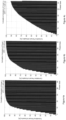
- FIGS. 5a, 5b, and 5c show cumulative and relative histograms of power loss in three subjects with different breathing. As can be seen the histograms are quite different, illustrating difference between subjects with healthy breathing and subjects with disordered breathing. Using 20% power loss as a threshold, only 5% of the breaths of Subject 1 are above that threshold, 20% of the breaths of Subject 2 and half of the breaths of Subject 3. The power loss index is therefore a candidate to be used as a quantitative measure of the level of partial obstruction.
- V R is an absolute volume signal measured in liters
- V S is a signal that is directly proportional to V R but is not the absolute volume signal.
- the present disclosure provides a further method that makes use of the physiological characteristics that the human body regulates the intake of oxygen to match the need of the cells at all time, not building up or dropping oxygen levels during normal breathing.
- the indication of increased metabolism is higher minute ventilation (minute ventilation referring to the total volume inspired or expired per minute) and heart rate and during aerobic breathing the ratio between minute ventilation and heart rate is close to linear.
- the method monitors the relative minute ventilation from the V S signal and compares it to a measured heart rate.
- the characteristic linearity between the minute ventilation and heart rate is captured during periods where the k A is not changing significantly but where there is a variation in the minute ventilation and heart rate.
- the captured value is then used to correct the V S when there is a change in the k A values. This way the V R V S ratio can be kept nearly constant throughout the recording, allowing volume calibration to take place at one or more points during the recording and delivering reliable measure for all periods.
Claims (14)
- Verfahren zum Messen einer Atemanstrengung eines Subjekts, wobei das Verfahren umfasst:Erhalten eines Thorax-Anstrengungssignals (T), wobei das Thorax-Anstrengungssignal (T) ein Indikator einer thorakalen Komponente der Atemanstrengung ist,wobei das Thorax-Anstrengungssignal (T) eine volumenbeitragende thorakale Komponente (VST) und eine thorakale Paradoxkomponente (PT) beinhaltet, die thorakale Paradoxkomponente (PT) eine nicht volumenbeitragende thorakale Komponente der Atemanstrengung darstellt und die volumenbeitragende thorakale Komponente (VST) eine volumenbeitragende Komponente der Atemanstrengung darstellt, undErhalten eines Abdomen-Anstrengungssignals (A), wobei das Abdomen-Anstrengungssignal (A) ein Indikator einer abdominalen Komponente der Atemanstrengung ist,wobei das Abdomen-Anstrengungssignal (A) eine volumenbeitragende abdominale Komponente (VSA) und eine abdominale Paradoxkomponente (PA) beinhaltet, die abdominale Paradoxkomponente (PA) eine nicht volumenbeitragende abdominale Komponente der Atemanstrengung darstellt und die volumenbeitragende abdominale Komponente (VSA) eine volumenbeitragende Komponente der Atemanstrengung darstellt, wobei die abdominale Paradoxkomponente (PA) negativ proportional zu der thorakalen Paradoxkomponente (PT) ist,wobei das Verfahren ferner die folgenden Schritte umfasst, die von einem Prozessor durchgeführt werden:Durchführen einer Kalibrierung des Thorax-Anstrengungssignals (T) und des Abdomen-Anstrengungssignals (A), was unterschiedliche Niveaus einer obstruktiven Atmung während der Kalibrierung ermöglicht, basierend auf dem erhaltenen Thorax-Anstrengungssignal (T) und dem Abdomen-Anstrengungssignal (A) durchGewichten des Thorax-Anstrengungssignals (T) mit einem ersten Gewichtungsfaktor (kT) und Gewichten des Abdomen-Anstrengungssignals (A) mit einem zweiten Gewichtungsfaktor (kA), um ein volumenproportionales Signal (VSw) zu erhalten, bei dem es sich um die gewichtete Summe des Thorax- und des Abdomen-Anstrengungssignals handelt, wobei das volumenproportionale Signal (VSw) proportional zu dem tatsächlichen Atemvolumen der Atemanstrengung ist,gekennzeichnet durchBerechnen eines Verhältnisses zwischen einer Amplitude oder Leistung, oder einer Transformationsfunktion der Amplitude oder Leistung des volumenproportionalen Signals (VSw) und einer Summe der Amplituden, Leistung oder einer Transformationsfunktion des Thorax-Anstrengungssignals (T), gewichtet mit dem ersten Gewichtungsfaktor (kT), und des Abdomen-Anstrengungssignals (A), gewichtet mit dem zweiten Gewichtungsfaktor (kA), undAuswählen des ersten und des zweiten Gewichtungsfaktors (kT und kA), die das berechnete Verhältnis minimieren.
- Verfahren gemäß Anspruch 1, wobei die Kalibrierung des Thorax-Anstrengungssignals (T) und des Abdomen-Anstrengungssignals (A) nur auf dem erhaltenen Thorax-Anstrengungssignal (T) und dem Abdomen-Anstrengungssignal (A) basiert, ohne eine quantitative Referenzmessung zusätzlich zu dem Thorax-Anstrengungssignal (T) und dem Abdomen-Anstrengungssignal (A).
- Verfahren gemäß Anspruch 1, umfassend das Bestimmen eines Leistungsverlustes basierend auf dem optimierten ersten und zweiten Gewichtungsfaktor (kA und kT).
- Verfahren gemäß Anspruch 3, wobei das Bestimmen des Leistungsverlustes ein Maximieren des Leistungsverlustes eines resultierenden Signals verglichen mit einer Summe der Leistung des Thorax-Anstrengungssignals (T), gewichtet mit dem optimierten ersten Gewichtungsfaktor (kT), und des Abdomen-Anstrengungssignals (A), gewichtet mit dem optimierten zweiten Gewichtungsfaktor (kA), über einen Zeitraum umfasst.
- Verfahren gemäß Anspruch 1, umfassend das Berechnen eines atemflussproportionalen Signals (FS) durch Berechnen einer ersten Ableitung des volumenproportionalen Signals (VSw).
- Verfahren gemäß Anspruch 1, wobei der erste und der zweite Gewichtungsfaktor (kT und kA) optimiert wird, indem Werte für den ersten und den zweiten Gewichtungsfaktor (kT und kA) bestimmt werden, welche die Leistung niedrigerer Frequenzen in dem volumenproportionalen Signal (VSw) im Vergleich zu einer proportionalen Leistung höherer Frequenzen in dem volumenproportionalen Signal (VSw) über einen Zeitraum maximieren.
- Verfahren gemäß Anspruch 1, umfassend das Maximieren eines Amplitudenverlustes eines resultierenden Signals im Vergleich zu einer Summe der Amplitude des Thorax-Anstrengungssignals (T) und des Abdomen-Anstrengungssignals (A) über einen Zeitraum.
- Verfahren gemäß Anspruch 1, umfassend:Erhalten des ersten und des zweiten Kalibrierungsfaktors (kT und kA), indem die Kalibrierung des Thorax-Anstrengungssignals (T) und des Abdomen-Anstrengungssignals (A) über mehrere Zeitspannen durchgeführt wird, beinhaltend eine erste Zeitspanne innerhalb einer zweiten Zeitspanne, wobei die erste Zeitspanne kürzer als die zweite Zeitspanne ist, undAuswählen eines ersten und eines zweiten Kalibrierungsfaktors (kT und kA) für die zweite Zeitspanne basierend auf den erhaltenen Kalibrierungsfaktoren der mehreren Zeitspannen, die ein am stärksten determinierendes Ergebnis liefern.
- Verfahren gemäß Anspruch 1, umfassend das Auswählen eines Wertes für den ersten und den zweiten Gewichtungsfaktor (kT und kA) durch Bestimmen von Zwischenwerten für die Gewichtungsfaktoren für einen Satz von mehreren aufeinanderfolgenden Zwischenzeitspannen und Berechnen des Wertes für den ersten und den zweiten Gewichtungsfaktor (kA und kT) durch Gewichten von Werten in dem Satz von Zeitspannen, um die Kontinuität gewichteter Werte über den Satz zu maximieren.
- Verfahren gemäß Anspruch 1, umfassend das Ableiten der thorakalen Paradoxkomponente (PT) des Thorax-Anstrengungssignals (T) oder der abdominalen Paradoxkomponente (PA) des Abdomen-Anstrengungssignals (A), oder eine Ableitung der thorakalen Paradoxkomponente (PT) des Thorax-Anstrengungssignals (T) oder der abdominalen Paradoxkomponente (PA) des Abdomen-Anstrengungssignals (A), durch Bestimmen von Werten für den ersten und den zweiten Gewichtungsfaktor (kT und kA), die eine Amplitude oder Leistung des volumenproportionalen Signals (VSw) verglichen mit der Summe der Leistungen des Thorax-Anstrengungssignals, gewichtet mit dem ersten Gewichtungsfaktor (kT), und des Abdomen-Anstrengungssignals (A), gewichtet mit dem zweiten Gewichtungsfaktor (kA), über einen Zeitraum minimieren.
- Verfahren gemäß Anspruch 10, umfassend das Evaluieren eines Flusswiderstands anhand der thorakalen Paradoxkomponente (PT) oder der abdominalen Paradoxkomponente (PA) und einer ersten Ableitung des volumenproportionalen Signals (Vs) im Hinblick auf die Zeit.
- Verfahren gemäß Anspruch 1, wobeidas Thorax-Anstrengungssignal (T) durch Bereitstellen einer ersten Sensorvorrichtung erhalten wird, die dafür ausgelegt ist, die thorakale Komponente der Atemanstrengung des Subjekts zu messen, unddas Abdomen-Anstrengungssignal (A) durch Bereitstellen einer zweiten Sensorvorrichtung erhalten wird, die dafür ausgelegt ist, die abdominale Komponente der Atemanstrengung des Subjekts zu messen.
- Verfahren gemäß Anspruch 1, umfassend das Bestimmen einer Atemvolumenkalibrierung basierend auf der gemessenen Herzfrequenz des Subjekts.
- Atemanstrengung-Messsystem, umfassend eine erste Sensorvorrichtung, die dafür ausgelegt ist, ein Thorax-Anstrengungssignal (T) zu erhalten, wobei das Thorax-Anstrengungssignal (T) ein Indikator einer thorakalen Komponente der Atemanstrengung ist,wobei das Thorax-Anstrengungssignal (T) eine volumenbeitragende thorakale Komponente (VST) und eine thorakale Paradoxkomponente (PT) aufweist, wobei die thorakale Paradoxkomponente (PT) eine nicht volumenbeitragende thorakale Komponente der Atemanstrengung darstellt und die volumenbeitragende thorakale Komponente (VST) eine volumenbeitragende Komponente der Atemanstrengung darstellt,eine zweite Sensorvorrichtung, die dafür ausgelegt ist, ein Abdomen-Anstrengungssignal (A) zu erhalten, wobei das Abdomen-Anstrengungssignal (A) ein Indikator einer abdominalen Komponente der Atemanstrengung ist,wobei das Abdomen-Anstrengungssignal (A) eine volumenbeitragende abdominale Komponente (VSA) und eine abdominale Paradoxkomponente (PA) beinhaltet, die abdominale Paradoxkomponente (PA) eine nicht volumenbeitragende abdominale Komponente der Atemanstrengung darstellt und die volumenbeitragende abdominale Komponente (VSA) eine volumenbeitragende Komponente der Atemanstrengung darstellt, wobei die abdominale Paradoxkomponente (PA) negativ proportional zu der thorakalen Paradoxkomponente (PT) ist, undeinen Prozessor, der dafür ausgelegt ist, das Thorax-Anstrengungssignal (T) und das Abdomen-Anstrengungssignal (A) zu empfangen; wobei der Prozessor dafür ausgelegt ist, eine Kalibrierung des Thorax-Anstrengungssignals (T) und des Abdomen-Anstrengungssignals (A) durchzuführen, was unterschiedliche Niveaus einer obstruktiven Atmung während der Kalibrierung ermöglicht, basierend auf dem erhaltenen Thorax-Anstrengungssignal (T) und dem Abdomen-Anstrengungssignal (A) durchGewichten des Thorax-Anstrengungssignals (T) mit einem ersten Gewichtungsfaktor (kT) und Gewichten des Abdomen-Anstrengungssignals (A) mit einem zweiten Gewichtungsfaktor (kA), um ein volumenproportionales Signal (VSw) zu erhalten, bei dem es sich um die gewichtete Summe des Thorax- und des Abdomen-Anstrengungssignals handelt, wobei das volumenproportionale Signal (VSw) proportional zu dem tatsächlichen Atemvolumen der Atemanstrengung ist, und dadurch gekennzeichnet, dass der Prozessor dafür ausgelegt ist, ein Verhältnis zwischen einer Amplitude oder Leistung oder einer Transformationsfunktion der Amplitude oder Leistung des volumenproportionalen Signals (VSw) und einer Summe der Amplituden, Leistung oder einer Transformationsfunktion des Thorax-Anstrengungssignals (T), gewichtet mit dem ersten Gewichtungsfaktor (kT), und des Abdomen-Anstrengungssignals (A), gewichtet mit dem zweiten Gewichtungsfaktor (kA), zu berechnen, undden ersten und den zweiten Gewichtungsfaktor (kT und kA) auszuwählen, die das berechnete Verhältnis minimieren.
Applications Claiming Priority (2)
| Application Number | Priority Date | Filing Date | Title |
|---|---|---|---|
| IS050066 | 2013-11-06 | ||
| PCT/IB2014/002760 WO2015068041A1 (en) | 2013-11-06 | 2014-11-06 | Method, apparatus, and system for measuring respiratory effort |
Publications (3)
| Publication Number | Publication Date |
|---|---|
| EP3065638A1 EP3065638A1 (de) | 2016-09-14 |
| EP3065638B1 true EP3065638B1 (de) | 2024-01-10 |
| EP3065638C0 EP3065638C0 (de) | 2024-01-10 |
Family
ID=52396737
Family Applications (1)
| Application Number | Title | Priority Date | Filing Date |
|---|---|---|---|
| EP14830558.4A Active EP3065638B1 (de) | 2013-11-06 | 2014-11-06 | Verfahren, vorrichtung und system zur messung einer atemanstrengung |
Country Status (3)
| Country | Link |
|---|---|
| US (2) | US10588550B2 (de) |
| EP (1) | EP3065638B1 (de) |
| WO (1) | WO2015068041A1 (de) |
Families Citing this family (11)
| Publication number | Priority date | Publication date | Assignee | Title |
|---|---|---|---|---|
| WO2010131267A1 (en) * | 2009-05-15 | 2010-11-18 | Nox Medical | System and methods using flexible capacitive electrodes for measuring biosignals |
| EP2584962B1 (de) | 2010-06-25 | 2014-09-10 | Nox Medical ehf. | Biometrischer bandverbinder |
| EP3065638B1 (de) * | 2013-11-06 | 2024-01-10 | Nox Medical ehf. | Verfahren, vorrichtung und system zur messung einer atemanstrengung |
| WO2018033889A1 (en) * | 2016-08-19 | 2018-02-22 | Nox Medical | Method, apparatus, and system for measuring respiratory effort of a subject |
| EP3537961A1 (de) | 2016-11-10 | 2019-09-18 | The Research Foundation for The State University of New York | System, verfahren und biomarker für verstopfungen der atemwege |
| US11896386B2 (en) | 2017-06-02 | 2024-02-13 | Nox Medical Ehf | Coherence-based method, apparatus, and system for identifying corresponding signals of a physiological study |
| US11602282B2 (en) | 2017-09-08 | 2023-03-14 | Nox Medical Ehf | System and method for non-invasively determining an internal component of respiratory effort |
| US11035825B2 (en) * | 2017-11-15 | 2021-06-15 | Infineon Technologies Ag | Sensing systems and methods for the estimation of analyte concentration |
| WO2020255128A1 (en) * | 2019-06-17 | 2020-12-24 | Resmetrix Medical Ltd. | System for monitoring respiratory conditions and method thereof |
| KR102379321B1 (ko) * | 2019-11-08 | 2022-03-28 | 주식회사 고니메드 | 스트레인 게이지를 이용한 개인 상관계수 기반의 맞춤형 호흡량 측정 시스템 |
| KR102379320B1 (ko) * | 2019-11-08 | 2022-03-28 | 주식회사 고니메드 | 스트레인 게이지를 이용한 개인 상관계수 기반의 맞춤형 호흡량 측정 장치 및 방법 |
Family Cites Families (79)
| Publication number | Priority date | Publication date | Assignee | Title |
|---|---|---|---|---|
| US1193050A (en) | 1916-08-01 | Adjusting-buckle | ||
| US937130A (en) | 1908-04-15 | 1909-10-19 | Paul F Williams | Ground-pipe cap. |
| US1001054A (en) | 1911-03-15 | 1911-08-22 | Milton H Lawrence | Ground-wire fastener. |
| US1115459A (en) | 1912-03-04 | 1914-10-27 | Alkoury A Abizaid | Buckle. |
| US2305277A (en) | 1942-07-03 | 1942-12-15 | Sloane Nathan | Plastic snap fastener |
| US2649573A (en) | 1948-12-21 | 1953-08-18 | Harold D Goldberg | Area measuring device |
| US2667159A (en) | 1948-12-21 | 1954-01-26 | Harold D Goldberg | Plethysmograph |
| US3092759A (en) | 1958-02-28 | 1963-06-04 | Siemens And Halske Ag Berlin A | Wired circuit plate with electrical components |
| US3347223A (en) | 1964-01-08 | 1967-10-17 | Universal Match Corp | Pneumograph |
| US3560845A (en) | 1965-05-03 | 1971-02-02 | Harold D Goldberg | Measuring devices |
| US3500823A (en) | 1967-11-20 | 1970-03-17 | Us Air Force | Electrocardiographic and bioelectric capacitive electrode |
| BE757594A (fr) | 1969-10-22 | 1971-04-16 | Textron Inc | Fermeture a declic en matiere plastique moulee, et bande d'elements pour cette fermeture |
| GB1596298A (en) | 1977-04-07 | 1981-08-26 | Morgan Ltd P K | Method of and apparatus for detecting or measuring changes in the cross-sectional area of a non-magnetic object |
| US4373534A (en) * | 1981-04-14 | 1983-02-15 | Respitrace Corporation | Method and apparatus for calibrating respiration monitoring system |
| JPS58154618U (ja) | 1982-04-13 | 1983-10-15 | 武田 精 | スナツプフアスナ−の雌型嵌合体 |
| US4671591A (en) | 1985-07-15 | 1987-06-09 | Physio-Control Corporation | Electrical connector |
| US4834109A (en) | 1986-01-21 | 1989-05-30 | Respitrace Corporation | Single position non-invasive calibration technique |
| US4777962A (en) | 1986-05-09 | 1988-10-18 | Respitrace Corporation | Method and apparatus for distinguishing central obstructive and mixed apneas by external monitoring devices which measure rib cage and abdominal compartmental excursions during respiration |
| US5301678A (en) | 1986-11-19 | 1994-04-12 | Non-Invasive Monitoring System, Inc. | Stretchable band - type transducer particularly suited for use with respiration monitoring apparatus |
| US4807640A (en) | 1986-11-19 | 1989-02-28 | Respitrace Corporation | Stretchable band-type transducer particularly suited for respiration monitoring apparatus |
| US4817625A (en) | 1987-04-24 | 1989-04-04 | Laughton Miles | Self-inductance sensor |
| US4832608A (en) | 1987-05-22 | 1989-05-23 | Cherne Medical, Inc. | Electrode belt adapter |
| DE3719474C2 (de) | 1987-06-11 | 1994-10-13 | Nicolay Gmbh | Kontaktorgan zum Herstellen einer elektrisch leitenden Verbindung |
| US5326272A (en) | 1990-01-30 | 1994-07-05 | Medtronic, Inc. | Low profile electrode connector |
| US5331968A (en) | 1990-10-19 | 1994-07-26 | Gerald Williams | Inductive plethysmographic transducers and electronic circuitry therefor |
| US5353793A (en) | 1991-11-25 | 1994-10-11 | Oishi-Kogyo Company | Sensor apparatus |
| JP3418538B2 (ja) | 1997-12-24 | 2003-06-23 | ワイケイケイ株式会社 | 合成樹脂製バックルのベルト取付構造 |
| FI107776B (fi) | 1998-06-22 | 2001-10-15 | Polar Electro Oy | Häiriösuoja |
| US7171265B2 (en) | 1998-07-31 | 2007-01-30 | Harbinger Medical, Inc. | Apparatus and method for detecting lead adequacy and quality |
| US6723055B2 (en) * | 1999-04-23 | 2004-04-20 | Trustees Of Tufts College | System for measuring respiratory function |
| US6413225B1 (en) | 1999-06-18 | 2002-07-02 | Vivometrics, Inc. | Quantitative calibration of breathing monitors with transducers placed on both rib cage and abdomen |
| US6807438B1 (en) | 1999-08-26 | 2004-10-19 | Riccardo Brun Del Re | Electric field sensor |
| DE19941500A1 (de) | 1999-08-31 | 2001-03-22 | Taller Gmbh | Automobilsitz-Stecker |
| CA2405848C (en) | 2000-04-17 | 2010-11-09 | Vivometrics, Inc. | Systems and methods for ambulatory monitoring of physiological signs |
| FR2811214B1 (fr) | 2000-07-05 | 2003-01-31 | R B I | Procede et dispositif de determination de parametres physiologiques cardio-respiratoires |
| US20020032388A1 (en) | 2000-09-13 | 2002-03-14 | Helgi Kristbjarnarson | Disposable sensor for measuring respiration and method of forming the same |
| US6461307B1 (en) | 2000-09-13 | 2002-10-08 | Flaga Hf | Disposable sensor for measuring respiration |
| US6341504B1 (en) | 2001-01-31 | 2002-01-29 | Vivometrics, Inc. | Composite elastic and wire fabric for physiological monitoring apparel |
| US6993378B2 (en) | 2001-06-25 | 2006-01-31 | Science Applications International Corporation | Identification by analysis of physiometric variation |
| US8790272B2 (en) | 2002-03-26 | 2014-07-29 | Adidas Ag | Method and system for extracting cardiac parameters from plethysmographic signals |
| US6783498B2 (en) | 2002-03-26 | 2004-08-31 | Vivometrics, Inc. | Method and system for extracting cardiac parameters from plethysmographic signals |
| US8137270B2 (en) | 2003-11-18 | 2012-03-20 | Adidas Ag | Method and system for processing data from ambulatory physiological monitoring |
| US7727161B2 (en) | 2003-04-10 | 2010-06-01 | Vivometrics, Inc. | Systems and methods for monitoring cough |
| CA2809764C (en) * | 2003-04-10 | 2016-10-25 | Vivometrics, Inc. | Systems and methods for respiratory event detection |
| AU2004277381B2 (en) | 2003-08-22 | 2008-04-24 | Foster-Miller, Inc. | Physiological monitoring garment |
| CA2574759A1 (en) | 2004-06-18 | 2006-01-26 | Vivometrics, Inc. | Systems and methods for real-time physiological monitoring |
| CN101022812A (zh) | 2004-08-23 | 2007-08-22 | 特瓦制药工业有限公司 | 固体和晶体伊班膦酸钠及其制备方法 |
| EP1791467B1 (de) | 2004-09-21 | 2013-03-27 | Adidas AG | Verbesserte sensoren für induktive plethysmographische überwachungsanwendungen und damit ausgestattete kleidung |
| DE102004063249A1 (de) | 2004-12-23 | 2006-07-13 | Fraunhofer-Gesellschaft zur Förderung der angewandten Forschung e.V. | Sensorsystem und Verfahren zur kapazitiven Messung elektromagnetischer Signale biologischen Ursprungs |
| US20060258948A1 (en) | 2005-03-01 | 2006-11-16 | Linville David J | Reusable inductive transducer for measuring respiration |
| US7762953B2 (en) | 2005-04-20 | 2010-07-27 | Adidas Ag | Systems and methods for non-invasive physiological monitoring of non-human animals |
| CA2606699C (en) | 2005-05-20 | 2017-04-18 | Vivometrics, Inc. | Methods and systems for determining dynamic hyperinflation |
| US20060282001A1 (en) | 2005-06-09 | 2006-12-14 | Michel Noel | Physiologic sensor apparatus |
| US8033996B2 (en) | 2005-07-26 | 2011-10-11 | Adidas Ag | Computer interfaces including physiologically guided avatars |
| US7364440B2 (en) | 2006-01-17 | 2008-04-29 | Lifesync Corporation | Multi-lead keyhole connector |
| US8762733B2 (en) | 2006-01-30 | 2014-06-24 | Adidas Ag | System and method for identity confirmation using physiologic biometrics to determine a physiologic fingerprint |
| US8177724B2 (en) | 2006-06-08 | 2012-05-15 | Adidas Ag | System and method for snore detection and confirmation |
| US7593767B1 (en) * | 2006-06-15 | 2009-09-22 | Cleveland Medical Devices Inc | Ambulatory sleepiness and apnea propensity evaluation system |
| US8475387B2 (en) | 2006-06-20 | 2013-07-02 | Adidas Ag | Automatic and ambulatory monitoring of congestive heart failure patients |
| TWI303984B (en) | 2007-01-30 | 2008-12-11 | Taiwan Textile Res Inst | Textile structure for detecting human vital signs and signal detector applying the same |
| GB2446875A (en) | 2007-02-23 | 2008-08-27 | Intense Ltd | Production of fine geometric openings in thin film materials |
| TWI317630B (en) | 2007-03-12 | 2009-12-01 | Taiwan Textile Res Inst | Respiration monitoring system |
| KR100895297B1 (ko) | 2007-04-30 | 2009-05-07 | 한국전자통신연구원 | 다양한 생체 신호의 동시 측정을 위한 다채널 전극센서장치 |
| EP2265177B1 (de) | 2008-04-11 | 2012-03-14 | Dymedix Corporation | Respiratorischer ausgang eines piezoelektrischen foliensensors mit mehrfacher polarität |
| JP2011523566A (ja) | 2008-05-02 | 2011-08-18 | ダイメディックス コーポレイション | 中枢神経系を刺激するためのアジテーター |
| US8679012B1 (en) | 2008-08-13 | 2014-03-25 | Cleveland Medical Devices Inc. | Medical device and method with improved biometric verification |
| US8790273B2 (en) | 2008-09-05 | 2014-07-29 | Adidas | Noninvasive method and system for measuring pulmonary ventilation |
| US8251736B2 (en) | 2008-09-23 | 2012-08-28 | Tyco Electronics Corporation | Connector assembly for connecting an electrical lead to an electrode |
| US7819710B2 (en) | 2008-09-23 | 2010-10-26 | Tyco Electronics Corporation | Termination cap for terminating an electrical lead directly to a stud of an electrode and an electrode lead assembly containing such termination cap |
| US8025539B2 (en) | 2009-05-15 | 2011-09-27 | Nox Medical Ehf. | Connector for biometric belt |
| WO2010131267A1 (en) | 2009-05-15 | 2010-11-18 | Nox Medical | System and methods using flexible capacitive electrodes for measuring biosignals |
| KR20120081588A (ko) | 2009-09-11 | 2012-07-19 | 컴퓨메딕스 메디컬 이노베이션 피티와이 엘티디 | 호흡 유도 혈류량 측정 검사 밴드 |
| EP2339696A1 (de) | 2009-12-22 | 2011-06-29 | General Electric Company | Elektrische Verbinderbaugruppe |
| US7914350B1 (en) | 2010-04-13 | 2011-03-29 | Cadwell Labs | Apparatus, system, and method for creating an electrical connection to a tool |
| EP2584962B1 (de) | 2010-06-25 | 2014-09-10 | Nox Medical ehf. | Biometrischer bandverbinder |
| WO2013096954A1 (en) | 2011-12-23 | 2013-06-27 | The Trustees Of Dartmouth College | Wearable computing device for secure control of physiological sensors and medical devices, with secure storage of medical records, and bioimpedance biometric |
| WO2015001188A1 (en) | 2013-07-02 | 2015-01-08 | Tampereen Teknillinen Yliopisto | Method for respiratory measurement |
| EP3065638B1 (de) * | 2013-11-06 | 2024-01-10 | Nox Medical ehf. | Verfahren, vorrichtung und system zur messung einer atemanstrengung |
| WO2018033889A1 (en) | 2016-08-19 | 2018-02-22 | Nox Medical | Method, apparatus, and system for measuring respiratory effort of a subject |
-
2014
- 2014-11-06 EP EP14830558.4A patent/EP3065638B1/de active Active
- 2014-11-06 US US14/535,093 patent/US10588550B2/en active Active
- 2014-11-06 WO PCT/IB2014/002760 patent/WO2015068041A1/en active Application Filing
-
2020
- 2020-03-13 US US16/818,533 patent/US20200214600A1/en active Pending
Also Published As
| Publication number | Publication date |
|---|---|
| US20200214600A1 (en) | 2020-07-09 |
| US20150126879A1 (en) | 2015-05-07 |
| WO2015068041A1 (en) | 2015-05-14 |
| EP3065638A1 (de) | 2016-09-14 |
| US10588550B2 (en) | 2020-03-17 |
| EP3065638C0 (de) | 2024-01-10 |
Similar Documents
| Publication | Publication Date | Title |
|---|---|---|
| US20200214600A1 (en) | Method, apparatus, and system for measuring respiratory effort | |
| US20210106258A1 (en) | Method, apparatus, and system for determining a value of one or more parameters of a respiratory effort of a subject | |
| Criée et al. | Body plethysmography–its principles and clinical use | |
| Vogt et al. | Pulmonary function testing in children and infants | |
| Lee et al. | Time-varying autoregressive model-based multiple modes particle filtering algorithm for respiratory rate extraction from pulse oximeter | |
| EP2819571B1 (de) | Verarbeitung eines einen physiologischen rhythmus darstellenden signals | |
| WO2009124297A1 (en) | Non-contact physiologic motion sensors and methods for use | |
| US11058323B2 (en) | Device and method for assessing respiratory data in a monitored subject | |
| JP6963293B2 (ja) | 心拍・呼吸計測システム及び心拍・呼吸計測方法 | |
| Hudgel et al. | Accuracy of tidal volume, lung volume, and flow measurements by inductance vest in COPD patients | |
| Harju et al. | Determination of saturation, heart rate, and respiratory rate at forearm using a Nellcor™ forehead SpO 2-saturation sensor | |
| KR20140087902A (ko) | 수면 시 측정된 연속 혈압 신호를 통하여 수면호흡장애를 검출하는 장치 및 방법 | |
| Grossman et al. | Reliability of respiratory tidal volume estimation by means of ambulatory inductive plethysmography | |
| Lo et al. | Assessment of an alternative calibration technique to record breathing pattern and its variability with respiratory inductive plethysmography | |
| Mannée et al. | Telemonitoring techniques for lung volume measurement: accuracy, artifacts and effort | |
| US20220192536A1 (en) | System for monitoring respiratory conditions and method thereof | |
| Liu et al. | Non-invasive respiration and ventilation prediction using a single abdominal sensor belt | |
| Materko et al. | influence of the respiratory signal in heart rate variability analysis in the respiratory pattern in healthy elderly and with COPD | |
| CN104755016B (zh) | 用于呼吸率测量的系统和方法 | |
| CN115886750A (zh) | 一种心息一致性评估方法、存储介质及心息指标监测设备 | |
| WO2017079567A1 (en) | Implantable devices and methods for monitoring copd in patients | |
| US20230200677A1 (en) | Methods and systems of calibrating respiratory measurements to determine flow, ventilation and/or endotypes | |
| CN116528757A (zh) | 从噪声短持续时间胸部阻抗测量中提取呼吸参数的技术 | |
| US20190216403A1 (en) | Method for predicting acute airways obstruction of a patient with chronic respiratory disease | |
| Lay-Ekuakille et al. | Ergospirometric signal processing for human breath and force measurements |
Legal Events
| Date | Code | Title | Description |
|---|---|---|---|
| PUAI | Public reference made under article 153(3) epc to a published international application that has entered the european phase |
Free format text: ORIGINAL CODE: 0009012 |
|
| 17P | Request for examination filed |
Effective date: 20160525 |
|
| AK | Designated contracting states |
Kind code of ref document: A1 Designated state(s): AL AT BE BG CH CY CZ DE DK EE ES FI FR GB GR HR HU IE IS IT LI LT LU LV MC MK MT NL NO PL PT RO RS SE SI SK SM TR |
|
| AX | Request for extension of the european patent |
Extension state: BA ME |
|
| RIN1 | Information on inventor provided before grant (corrected) |
Inventor name: HALLGRIMSSON, HARALDUR TOMAS Inventor name: SIGURDARSON, GUDJON TEITUR Inventor name: HOSKULDSSON, SVEINBJORN |
|
| DAX | Request for extension of the european patent (deleted) | ||
| STAA | Information on the status of an ep patent application or granted ep patent |
Free format text: STATUS: EXAMINATION IS IN PROGRESS |
|
| 17Q | First examination report despatched |
Effective date: 20170831 |
|
| STAA | Information on the status of an ep patent application or granted ep patent |
Free format text: STATUS: EXAMINATION IS IN PROGRESS |
|
| P01 | Opt-out of the competence of the unified patent court (upc) registered |
Effective date: 20230529 |
|
| GRAP | Despatch of communication of intention to grant a patent |
Free format text: ORIGINAL CODE: EPIDOSNIGR1 |
|
| STAA | Information on the status of an ep patent application or granted ep patent |
Free format text: STATUS: GRANT OF PATENT IS INTENDED |
|
| INTG | Intention to grant announced |
Effective date: 20230731 |
|
| GRAS | Grant fee paid |
Free format text: ORIGINAL CODE: EPIDOSNIGR3 |
|
| GRAA | (expected) grant |
Free format text: ORIGINAL CODE: 0009210 |
|
| STAA | Information on the status of an ep patent application or granted ep patent |
Free format text: STATUS: THE PATENT HAS BEEN GRANTED |
|
| AK | Designated contracting states |
Kind code of ref document: B1 Designated state(s): AL AT BE BG CH CY CZ DE DK EE ES FI FR GB GR HR HU IE IS IT LI LT LU LV MC MK MT NL NO PL PT RO RS SE SI SK SM TR |
|
| REG | Reference to a national code |
Ref country code: GB Ref legal event code: FG4D |
|
| REG | Reference to a national code |
Ref country code: CH Ref legal event code: EP |
|
| REG | Reference to a national code |
Ref country code: DE Ref legal event code: R096 Ref document number: 602014089324 Country of ref document: DE |
|
| REG | Reference to a national code |
Ref country code: IE Ref legal event code: FG4D |
|
| U01 | Request for unitary effect filed |
Effective date: 20240212 |
|
| U07 | Unitary effect registered |
Designated state(s): AT BE BG DE DK EE FI FR IT LT LU LV MT NL PT SE SI Effective date: 20240220 |
|
| P04 | Withdrawal of opt-out of the competence of the unified patent court (upc) registered |
Effective date: 20240216 |