EP2999397B1 - Agencement pour faciliter une cicatrisation et pansement - Google Patents
Agencement pour faciliter une cicatrisation et pansement Download PDFInfo
- Publication number
- EP2999397B1 EP2999397B1 EP14731676.4A EP14731676A EP2999397B1 EP 2999397 B1 EP2999397 B1 EP 2999397B1 EP 14731676 A EP14731676 A EP 14731676A EP 2999397 B1 EP2999397 B1 EP 2999397B1
- Authority
- EP
- European Patent Office
- Prior art keywords
- wound
- electrodes
- electrode
- wound dressing
- arrangement according
- Prior art date
- Legal status (The legal status is an assumption and is not a legal conclusion. Google has not performed a legal analysis and makes no representation as to the accuracy of the status listed.)
- Active
Links
Images
Classifications
-
- A—HUMAN NECESSITIES
- A61—MEDICAL OR VETERINARY SCIENCE; HYGIENE
- A61N—ELECTROTHERAPY; MAGNETOTHERAPY; RADIATION THERAPY; ULTRASOUND THERAPY
- A61N1/00—Electrotherapy; Circuits therefor
- A61N1/18—Applying electric currents by contact electrodes
- A61N1/20—Applying electric currents by contact electrodes continuous direct currents
- A61N1/205—Applying electric currents by contact electrodes continuous direct currents for promoting a biological process
-
- A—HUMAN NECESSITIES
- A61—MEDICAL OR VETERINARY SCIENCE; HYGIENE
- A61B—DIAGNOSIS; SURGERY; IDENTIFICATION
- A61B5/00—Measuring for diagnostic purposes; Identification of persons
- A61B5/05—Detecting, measuring or recording for diagnosis by means of electric currents or magnetic fields; Measuring using microwaves or radio waves
- A61B5/053—Measuring electrical impedance or conductance of a portion of the body
- A61B5/0531—Measuring skin impedance
-
- A—HUMAN NECESSITIES
- A61—MEDICAL OR VETERINARY SCIENCE; HYGIENE
- A61B—DIAGNOSIS; SURGERY; IDENTIFICATION
- A61B5/00—Measuring for diagnostic purposes; Identification of persons
- A61B5/44—Detecting, measuring or recording for evaluating the integumentary system, e.g. skin, hair or nails
- A61B5/441—Skin evaluation, e.g. for skin disorder diagnosis
- A61B5/445—Evaluating skin irritation or skin trauma, e.g. rash, eczema, wound, bed sore
-
- A—HUMAN NECESSITIES
- A61—MEDICAL OR VETERINARY SCIENCE; HYGIENE
- A61B—DIAGNOSIS; SURGERY; IDENTIFICATION
- A61B5/00—Measuring for diagnostic purposes; Identification of persons
- A61B5/48—Other medical applications
- A61B5/4848—Monitoring or testing the effects of treatment, e.g. of medication
-
- A—HUMAN NECESSITIES
- A61—MEDICAL OR VETERINARY SCIENCE; HYGIENE
- A61F—FILTERS IMPLANTABLE INTO BLOOD VESSELS; PROSTHESES; DEVICES PROVIDING PATENCY TO, OR PREVENTING COLLAPSING OF, TUBULAR STRUCTURES OF THE BODY, e.g. STENTS; ORTHOPAEDIC, NURSING OR CONTRACEPTIVE DEVICES; FOMENTATION; TREATMENT OR PROTECTION OF EYES OR EARS; BANDAGES, DRESSINGS OR ABSORBENT PADS; FIRST-AID KITS
- A61F13/00—Bandages or dressings; Absorbent pads
- A61F13/02—Adhesive bandages or dressings
- A61F13/0246—Adhesive bandages or dressings characterised by the skin-adhering layer
- A61F13/0253—Adhesive bandages or dressings characterised by the skin-adhering layer characterized by the adhesive material
-
- A—HUMAN NECESSITIES
- A61—MEDICAL OR VETERINARY SCIENCE; HYGIENE
- A61N—ELECTROTHERAPY; MAGNETOTHERAPY; RADIATION THERAPY; ULTRASOUND THERAPY
- A61N1/00—Electrotherapy; Circuits therefor
- A61N1/02—Details
- A61N1/04—Electrodes
- A61N1/0404—Electrodes for external use
- A61N1/0408—Use-related aspects
- A61N1/0468—Specially adapted for promoting wound healing
-
- A—HUMAN NECESSITIES
- A61—MEDICAL OR VETERINARY SCIENCE; HYGIENE
- A61N—ELECTROTHERAPY; MAGNETOTHERAPY; RADIATION THERAPY; ULTRASOUND THERAPY
- A61N1/00—Electrotherapy; Circuits therefor
- A61N1/02—Details
- A61N1/04—Electrodes
- A61N1/0404—Electrodes for external use
- A61N1/0472—Structure-related aspects
- A61N1/0476—Array electrodes (including any electrode arrangement with more than one electrode for at least one of the polarities)
-
- A—HUMAN NECESSITIES
- A61—MEDICAL OR VETERINARY SCIENCE; HYGIENE
- A61N—ELECTROTHERAPY; MAGNETOTHERAPY; RADIATION THERAPY; ULTRASOUND THERAPY
- A61N1/00—Electrotherapy; Circuits therefor
- A61N1/02—Details
- A61N1/04—Electrodes
- A61N1/0404—Electrodes for external use
- A61N1/0472—Structure-related aspects
- A61N1/0484—Garment electrodes worn by the patient
-
- A—HUMAN NECESSITIES
- A61—MEDICAL OR VETERINARY SCIENCE; HYGIENE
- A61N—ELECTROTHERAPY; MAGNETOTHERAPY; RADIATION THERAPY; ULTRASOUND THERAPY
- A61N1/00—Electrotherapy; Circuits therefor
- A61N1/02—Details
- A61N1/04—Electrodes
- A61N1/0404—Electrodes for external use
- A61N1/0472—Structure-related aspects
- A61N1/0492—Patch electrodes
- A61N1/0496—Patch electrodes characterised by using specific chemical compositions, e.g. hydrogel compositions, adhesives
Definitions
- the present description relates to the field of electrotherapy and measuring by means of electric currents for diagnostic purposes, and more particularly to an electrode arrangement for facilitating wound healing, a method for measuring wound healing and a wound dressing having an electrode arrangement.
- the present invention is set out in the appended claims.
- Wounds such as e.g. chronic wounds and ulcers affect nearly 1 % of population and up to 10 % of institutionalized patients. By the year 2030, 366 million people worldwide are estimated to suffer from diabetes further increasing the prevalence of chronic wounds and ulcers.
- Edema prevents appropriate transport of oxygen and nutrients, which is essential for proper wound healing to occur. Edema also adds the mechanical stress in the wound site and disturbs waste removal from the wound area. A commonly used method to ease edema is compression therapy. Compression stockings are used for improving healing of chronic wounds of vascular etiology.
- TEP transepithelial potential
- a therapeutic approach which utilizes electrical stimulation of the wound via application of direct current should be beneficial.
- direct current is applied to the surface of the wound in order to stimulate the healing of the wound.
- the electrical stimulation of the wound has been found to affect the biological healing of the wound in the inflammation phase of the wound, in the proliferation phase of the wound and in the epithelisation phase of the wound.
- the electrical stimulation of the wound initiates the wound healing process, increases the blood circulation, promotes phagocytosis, improves tissue oxygen intake, reduces edema, stimulates fibroblasts and epithelial cells, stimulates DNA synthesis, calms the infection and dissolves the necrotic tissue.
- the electrical stimulation of the wound stimulates fibroblasts and epithelial cells, stimulates DNA synthesis and protein synthesis, adenosine triphosphate (ATP) formation, enhances membrane transport and stimulates the diminishing of the wound.
- ATP adenosine triphosphate
- the electrical stimulation of the wound stimulates the reformation and the migration of the epithelial cells and leads to softer and thinner skin, and improved scarring. Higher quality scarring is an important factor in decreasing the high recurring tendency of a chronic wound.
- Vascularization plays an important role in soft tissue healing, and hence enhancing angiogenesis to ensure sufficient blood flow in the newly formed epithelial layer will support the healing process.
- Vascular endothelial growth factor (VEGF) has been successfully used in preclinical ischaemic tissue models to enhance and promote the development of collateral blood vessels.
- dissolution of certain bioactive glass compositions have been shown to stimulate release of angiogenetic growth factors resulting in an increase in tubule branching and formation of complex networks of interconnected tubules. Soluble products of these bioactive glasses induce endothelial cell proliferation and up-regulation of VEGF production, which indicate that these glasses possess a proangiogenic potential.
- Significantly enhanced mitogenic stimulation of endothelial cells with an additive effect with VEGF release has also been observed in the presence of a BAG coating.
- Chronic wounds are a cause for disability, pain, emotional and social problems for the patients. Chronic wounds are associated with prolonged hospitalizations and considerable morbidity. These wounds, also known as ulcers, represent a major burden for the healthcare system affecting a large population of patients. Chronic wounds persist for months or even years representing medical, social, and economic problems for individuals and the society.
- prior art wound monitoring sensors available, e.g. array sensors which take the form of patterns on insulating material.
- these prior art wound monitoring sensors may typically use materials that interfere with or irritate the wound, occlude the wound and can cause skin maceration.
- some of the prior art wound monitoring sensors adhere to the wound, which can result in wound damage when they are removed.
- some of the prior art wound monitoring sensors also interfere with the healing of the wound by interfering with moisture control, whilst some only have a limited lifetime in a wound environment.
- bioimpedance measurement utilizes low level AC excitation current, it does not possess any risks or inconvenience for the patient.
- the idea of utilizing bioimpedance monitoring of a chronic wound is based on the pathophysiology of the wound. Often the integrity of the skin is lost in chronic wounds, and from an electrical point of view, the loss of high impedance stratum corneum leads into steep decrease in measured impedance. As the healing of the wound proceeds, the wound base lifts up and the wound starts to close up from the peripherals. Finally, the skin integrity is obtained. The gradual gain in the skin integrity is observed as increasing impedance particularly at lower frequencies.
- a chronic wound is trapped in an on-going inflammation phase of the wound healing process.
- Prolonged inflammation of a chronic wound is characterized by accumulation of highly conductive fluid into the wound and the surrounding area.
- the fluids may accumulate into the intracellular space as a result of ischemia. If the blood flow to the tissues is interrupted, cell metabolism continues but in an anaerobic way.
- a prolonged ischemia inevitably results in decline of metabolism. This results in the decreased activity of ion pumps, which leads changes in the ion distribution in extracellular fluid and intracellular fluid.
- the result is cellular edema because of inflow of water and sodium into the cell.
- the decline of extracellular fluid volume reduces the width of the electrical path of the low frequency current and increases the extracellular resistance.
- Severe ischemia finally results in cell necrosis.
- the cellular integrity is lost in necrosis and intracellular fluid leaks into the extracellular space. The necrosis is observed as a decrease in extracellular resistance.
- the fluids in a chronic wound may also accumulate into extracellular space.
- the increased volume of extracellular fluid can be observed as a decrease in the extracellular resistance.
- the swelling is due to increased vasodilatation and increased permeability of the capillaries.
- the fluid accumulates into the extracellular space.
- Another possible cause for fluid accumulation is peripheral edema. Peripheral edema results from increased capillary permeability or impaired return of fluid by lymphatic system from the interstitial space to vascular compartment. Lymphedema is a result of impaired function of lymphatic system and the fluid tends to accumulate into the extracellular space.
- European patent specification EP 1569553B1 presents a prior art wound mapping system presenting an array of rectangular electrodes that may be used to stimulate wound tissue electrically or measure impedance of wound tissue.
- the measurement electrodes are isolated from each other by a nonconducting hydrogel layer.
- the conducting parts of the stimulating electrodes are in direct contact with wound tissue via a hydrogel patch on the exposed conducting electrode.
- the conducting parts of the stimulating electrodes are designed to be electrically connected to the tissue but not to measure moisture above the wound or at a localised site between the electrodes. However, this allows the electrodes to dry into healing tissue and stick to the healed cell layer. Removal of the device with the wound dressing would remove the healed skin.
- WO2009/092616 discloses a electrical wound healing system.
- An object of the present invention is thus to provide an electrode arrangement for facilitating wound healing and a wound dressing having an electrode arrangement so as to overcome the above mentioned problems and to alleviate the above mentioned disadvantages.
- the present invention is set out in the appended claims.
- the embodiments, examples or aspects according to the present description which do not fall within the scope of said claims are provided for illustrative purposes only and do not form part of the present invention. Any methods disclosed hereinafter do not form part of the scope of the invention and are disclosed for illustrative purposes only.
- an arrangement for facilitating wound healing as defined in claim 1 which arrangement comprises at least two impedance reference electrodes, a frame like counter-electrode and stimulation electrodes in a form of an array; and a bioadhesive affinity layer surrounding the stimulation electrodes; said arrangement being suited for applying on top of the wound so that the stimulation electrode array is on the wound area, and that the at least two impedance reference electrodes and the frame like counter-electrode are suited for placing in contact with the healthy skin surrounding the wound area; which electrodes are suited for applying LIDC type electrical stimulation current to the wound area and for bioimpedance measurement.
- the frame like counter-electrode is anode electrode and the stimulation electrodes are cathode electrodes, or vice versa.
- the electrodes are suited for bioimpedance measurement with measurement frequencies in the range of 10 Hz - 200 000 Hz, preferably in the range of 1 000 Hz - 50 000 Hz.
- the polarity of the frame like counter-electrode and the stimulation electrodes is switchable.
- the electrodes are multiplexed.
- the present description also pertains to a method for measuring wound healing, which method comprises
- the frame like counter-electrode is used as an anode electrode and the stimulation electrodes are used as cathode electrodes, or vice versa.
- the electrodes are suited for bioimpedance measurement with measurement frequencies in the range of 10 Hz - 200 000 Hz, preferably in the range of 1 000 Hz - 50 000 Hz.
- the arrangement is a wound dressing.
- the frame like counter-electrode is anode electrode and the stimulation electrodes are cathode electrodes, or vice versa.
- the electrodes are suited for bioimpedance measurement with measurement frequencies in the range of 10 Hz - 200 000 Hz, preferably in the range of 1 000 Hz - 50 000 Hz.
- the polarity of the frame like counter-electrode and the stimulation electrodes is switchable.
- the wound dressing has a button battery, a printed battery structure or an electrochemical cell used as a power source for the electrodes.
- the wound dressing has a tether with the electrical connections of the electrodes. More preferably, the tether has a tether connector connectable to an electrode routing plug of an outside measurement terminal device and/or to an external power source.
- the wound dressing is produced by reel-to-reel print manufacturing, by sheet print manufacturing, by rotary screen print manufacturing or by any other mass production print manufacturing.
- the electrodes and the conductor pattern are printed to a paper substrate, to a polymer substrate or to a composite substrate functioning as a body of the wound dressing.
- the electrodes and the conductor pattern are etched on top of a suitable layer of plastic laminate, metal laminate or a composite laminate and the etched laminate layer is attached to a paper substrate, polymer substrate or composite substrate functioning as a body of the wound dressing.
- the bioadhesive affinity layer is manufactured of a peptide modified polysaccharide bioadhesive comprising a peptide component and a polysaccharide component.
- the peptide component is an integrin binding peptide, such as Arg-Gly-Asp (RGD), Gly-Arg-Gly-Asp-Ser (GRGDS), or cyclic RGD.
- the polysaccharide component is galactoglucomannan, xyloglucan or galactomannan.
- the polysaccharide component is spruce galactoglucomannan.
- the polysaccharide component comprises galactose side units.
- surface modification is applied to the wound dressing.
- the wound dressing comprises a bioactive layer.
- the bioactive layer contains a biopolymer based bioactive glass granules or spheres containing screen printable paste.
- said screen printable paste contains polylactic acid (PLA) as the polymer component and 20-100 ⁇ m granules of an antimicrobial angiogenesis-promoting bioactive glass, e.g. BAG-S53P4.
- PVA polylactic acid
- antibacterial silver is applied to the wound dressing.
- the wound dressing is used for facilitating wound healing.
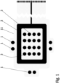
- FIG. 1 shows a bottom side view of an electrode arrangement of one embodiment of a wound dressing according to the present invention.
- the wound dressing according to the present invention comprises a printed substrate 11 having impedance reference electrodes 1, a frame like counter electrode 2 and stimulation electrodes 3 in a form of an array printed on the printed substrate 11.
- Highly conductive screen printable inks may be used as the material for the electrodes 1-3.
- the electrodes 1-3 and the conductor pattern may be printed directly to a paper substrate 11, polymer substrate 11 or composite substrate 11 functioning as the wound dressing laminate.
- Another alternative is to etch the electrodes 1-3 and the conductor pattern on top of a suitable layer of plastic laminate 11, metal laminate 11 or a composite laminate 11 and attach this etched laminate layer 11 to a paper substrate, polymer substrate or composite substrate functioning as the wound dressing laminate.
- this part may be provided in any size and shape.
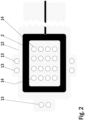
- Figure 2 shows a bottom side view of a lower laminate part of an electrode arrangement of one embodiment of a wound dressing according to the present invention.
- the lower laminate part 12 of an electrode arrangement of a wound dressing according to the present invention contains a printed or etched frame like counter electrode 2 and a number of perforations 13, 14 to allow for wound contact with the stimulation electrode array and impedance reference electrodes.
- Highly conductive screen printable inks may be used as the material for the frame like electrode 2.
- the electrode 2 and the conductor pattern may be printed directly to a paper substrate 12, polymer substrate 12 or composite substrate 12 functioning as the lower part of the wound dressing laminate.
- Another alternative is to etch the electrode 2 and the conductor pattern on top of a suitable layer of plastic laminate 12, metal laminate 12 or a composite laminate 12 and attach this etched laminate layer 12 to a paper substrate, polymer substrate or composite substrate functioning as the lower part of the wound dressing laminate.
- this part may be provided in any size and shape.
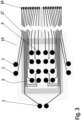
- Figure 3 shows a bottom side view of an upper laminate part of an electrode arrangement of one embodiment of a wound dressing according to the present invention.
- the upper laminate part 15 of an electrode arrangement of a wound dressing according to the present invention contains a printed or etched substrate 15 having impedance reference electrodes 1 and stimulation electrodes 3 in a form of an array printed or etched on the substrate 15.
- the upper laminate part 15 shown in the Figure 3 also contains printed connectors 16 and wiring layout 17. Highly conductive screen printable inks may be used as the material for the reference electrodes 1 and the stimulation electrodes 3.
- the electrodes and the conductor pattern may be printed directly to a paper substrate 15, polymer substrate 15 or composite substrate 15 functioning as a body of the wound dressing.
- the stimulation electrode array 3 may comprise any desired number of electrodes in any desired configuration.
- a typical non-limiting electrode array comprises 10 to 200 electrodes.
- the number of arrayed electrodes depends, at least party, from the size and the shape of the wound dressing.
- the wound dressing may be provided in any sizes and shapes.
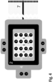
- FIG. 4 shows a bottom side view of one embodiment of a wound dressing according to the present invention.
- the wound dressing according to the present invention contains impedance reference electrodes 1, a frame-like counter electrode 2, a stimulation electrode array 3, a hydrogel adhesive layer 4, a bioadhesive affinity layer 5, a bioactive layer 6 and a tether with a tether connector 7 connectable to an electrode routing plug of an outside measurement terminal device.
- Impedance reference electrodes 1 provide the reference value for the wound impedance measurement.
- the frame-like electrode 2 acts as the counter electrode and the stimulation electrode array 3 provides the stimulation current during LIDC stimulation. When mapping the wound various combinations of electrode pairs of the stimulation electrode array 3 can be measured.
- the hydrogel adhesive layer 4 is coated with a suitable non-conductive hydrogel acting as an adhesive.
- the bioadhesive affinity layer 5 is coated with a peptide modified polysaccharide bioadhesive to be used in the bioadhesive affinity layer 5 and comprises a peptide component and a polysaccharide component.
- the peptide component is an integrin binding peptide, such as Arg-Gly-Asp (RGD), Gly-Arg-Gly-Asp-Ser (GRGDS), or cyclic RGD.
- the peptide component provides bioadhesive and hemostatic properties to the present wound dressing, at least partly, owing to its ability to enhance endothelial cell adhesion and proliferation.
- a number of different polysaccharides having high affinity for cellulosic surfaces may be used as the polysaccharide component in the peptide modified polysaccharide bioadhesive to be used in the bioadhesive affinity layer 5.
- the polysaccharide component comprises galactose side units.
- suitable polysaccharide species is galactoglucomannan, the major hemicellulose type or heteropolysaccharide in softwoods.
- the polysaccharide component is spruce galactoglucomannan.
- suitable polysaccharide species include xyloglucan and galactomannan.
- the peptide modified polysaccharide bioadhesive may be produced by activation of the galactose side units in the selected polysaccharide through chemo-enzymatic processes in water medium. Next, the peptides are anchored to these activated sites by peptide coupling while maintaining the integrity of the polysaccharide main chain and hence the affinity towards a cellulosic surface of the wound dressing.
- Alternative materials for the bioadhesive affinity layer 5 include chitosan and derivatives thereof.
- the bioactive layer 6 in figure 4 contains a biopolymer based bioactive glass granules or spheres containing paste.
- This screen printable paste may contain polylactic acid (PLA) as the polymer component and 20-100 ⁇ m granules of an antimicrobial angiogenesis-promoting bioactive glass, e.g. BAGS53P4.
- the electrode routing plug may be integrated directly to the measurement device, or a communication tether may be used in between.
- the length of the tether of the wound dressing according to the present invention may be very short (in the range of centimetres) or very long (in the range of metres) or something in between.
- a button battery or a printed battery structure realized either as a hybrid (zinc/air or aluminium/air) or as fully chemical (zinc/silver oxide) battery may be used.
- an electrochemical cell e.g. using enzyme catalyst may be used as a power source for the wound dressing according to the present invention.
- One such electrochemical cell using enzyme catalyst has been described in an International patent application WO 2007/147942 .
- the tether connector is providing contact to the power supply containing connector during stimulation and to the bioimpedance measuring device during evaluation of degree of wound healing.
- the principal idea of application of the wound dressing according to the present invention is that the wound dressing is applied on top of the wound so that the stimulation electrode array 3 is on the wound area and the frame like counter-electrode 2 is on and in contact with the intact skin surrounding the wound area.
- the frame like counter-electrode 2 may e.g. be square formed.
- the stimulation electrode array 3 may e.g. be a simulation software optimized stimulation electrode array 3.
- the injured tissue is normally characterized by a higher potential compared with the surrounding intact skin and in the wound edge cells are in electric field.
- Electrical stimulation according to the present invention restarts or accelerates wound healing process by imitating the natural electrical current and to increase this lateral current, positive polarity should be placed on the wound and negative on the intact skin area.
- the polarity of the wound may also be reversible.
- the current density should be sufficiently high in the wound and the electrode layout should be selected such that the current reaches the deeper skin layers.
- electrical stimulation is applied to the wound there is regenerated epithelium and granulation tissue being formed under the wound stimulation points this will increase the local contact resistance between the stimulation electrode and the wound. This causes the stimulation current to seek to wound stimulation points where the healing is slower this resulting to a more even stimulation effect.
- the present wound dressing may be termed as self-adjustable.
- point-like stimulation electrodes of the stimulation electrode array 3 on the wound surface provide a better skin contact to the wound when compared to larger structures due to more flexible surface of the wound dressing.
- the electrode placement according to the present invention also provides better current density feature and additionally gives possibility to self-regulatory adjustment of the wound stimulation current as the impedance increases at the edges of the wound as the healing proceeds and the stimulation current naturally seeks lower impedance pathway.
- This electrode placement also offers a possibility to polarity reversal.
- the polarity of the frame like counter-electrode 2 and the stimulation electrodes 3 is switchable during treatment to enhance the diffusion of various wound healing related components and decrease the formation of concentration gradients.
- the electrodes 2, 3 can also be multiplexed so as to allow for measurement of bioimpedance in a two electrode and four electrode configurations.
- the wound dressing structure according to the present invention is flexible and thin and the wound dressing surface area is scalable.
- the proposed wound dressing is self-sustaining and does not involve leads during stimulation functionality, thereby providing overall convenience and ease of use for the patient.
- the wound dressing structure according to the present invention may be manufactured by using reel-to-reel print manufacturing. At least the electrodes and potentially an integrated power source in the wound dressing may be produced by reel-to-reel print manufacturing. Also a surface modification may be applied to the wound dressing in order to enhance the wound contact for example by drop casting, by curtain spraying or by administration of skin adhesive using spraying techniques.
- antibacterial silver may be used in the wound dressing surface facing the wound; this improving the antimicrobial properties against wound infection.
- bioactive glass may be used in the wound dressing surface facing the wound as bioactive glass possesses good antimicrobial properties. The use of bioactive glass may provide additional protection against wound infection or reduce the on-going infection.
- bioactive glasses enhance angiogenesis, or blood vessel growth, a process that is critical in wound healing. Suitable bioactive glasses are readily available and easily chosen by a person skilled in the art.
- thermometer or thermocouple can be included in the present wound dressing to allow early detection and monitoring of a possible wound infection.
- the wound dressing according to the present invention incorporates galvanic wound stimulation functions with a wound healing monitoring possibility by using bioimpedance method.
- the measurement of bioimpedance is non-intrusive and do not require removal of the wound dressing, therefore it may be used in less controlled environment such as in home care.
- the bioimpedance monitoring of wound healing is based on the impedance measurement of wound tissue in reference to intact skin.
- Three most common electrode systems include 2-, 3- and 4-electrode systems.
- the wound healing monitoring is performed by utilizing 2-electrode bioimpedance measurement configuration.
- the same electrodes are used for both the excitation current feeding and voltage measurement.
- the output of 2-electrode bioimpedance measurement consists of the electrode impedance of both electrodes, the skin impedance under both electrodes and the tissue impedance between the two measurement electrodes.
- the outer layers of skin provide very high impedance compared to underlying tissues.
- the 2-electrode bioimpedance measurement method outputs so called true impedance since negative sensitivity areas do not exist in this configuration. This makes the analysis of the measurement results less prone to misinterpretations.
- Skin impedance can also be measured using the 3-electrode bioimpedance measurement configuration; however this includes certain obvious disadvantages.
- the 3-electrode bioimpedance measurement includes areas of negative sensitivity, which may compromise correct interpretation of the output. Placing of the third electrode is important for obtaining reliable and comparable results; this may also prove to be difficult and impractical in clinical use.
- the wound dressing according to the present invention may generate and repair the bio-mimicking potential difference between the wound area and the surrounding intact skin.
- the wound dressing according to the present invention may deliver a micro-amperage DC stimulus current to the wound tissue.
- the wound dressing according to the present invention may be used alongside with the conventional wound care practices.
- the stimulus current may be limited by either separately printed resistors, internal resistance of the battery or possibly only by the skin/tissue impedance, so that the treatment current is self-regulated.
- the stimulus current may be fed to the wound surface through multiple antibacterial silver pathways; this improving the electrode-skin contact.
- a wound healing process usually starts from the edges of the wound and the wound base lifts up.
- the skin integrity is gradually regained and the amount of exudates is reduced in the peripheral wound area. Consequently, the impedance increases, and more current flows to the open and moist centre of the wound which provides a lower impedance pathway.
- the stimulation current penetrates the skin surface and enters to the underlying wound tissue, thus improving the healing impact.
- the bioimpedance measurements may be carried out using at least one frequency depending on the width and the depth of the wound. Measurement frequencies in the range of 10 Hz - 200 000 Hz and preferably in the range of 1 000 Hz - 50 000 Hz may be used.
- the bioimpedance measurement of the wound dressing according to the present invention may be based on a stand-alone -device and connected to the wound dressing 12 only for the time of measurement.
- a communication tether is to be used between the outside measurement terminal device and the wound dressing.
- the tether provides galvanic connection between the outside measurement terminal device and the impedance measurement electrodes on the wound dressing.
- the patch may contain intelligent electronics, and a wireless communication method, such as e.g. infrared communication method or RF communication method may provide the connection to the measurement device instead.
- the impedance measurement device may be a handheld device.
- the impedance measurement device may have a flat connector probe that is slid into a fold on the patch.
- the impedance measurement device may have a measurement clip that clamps around a contact extrusion on the patch.
- the impedance measurement device may be pressed against contacts on the patch.
- the outside measurement terminal device may contain means to display the result immediately to the operator, store the measured data and to upload the data to the operator's personal computer. As the device may be used in clinical trials, precautions will be taken to make the electrical and physical interfaces of the device safe.
- the present wound dressing may be used to treat any type of wounds, in particular chronic wounds.
- treatment refers not only to a complete healing of a wound, but also to alleviation and amelioration of symptoms related to incomplete or improper wound healing, including, but not limited to, pain, swelling or edema, burning, itching, rash, redness, discoloration and dry, scaly skin.
- ulcers are wounds or open sores that will not heal or keep returning. Ulcers may develop anywhere on a human body, foot and leg ulcers being the most typical ulcer types. Non-limiting examples of ulcers to be treated in accordance with the present invention include pressure ulcers, or bedsores, venous ulcers, neuropathic (diabetic) ulcers, and arterial (ischemic) ulcers. Typically in foot and leg ulcers, the present wound dressing is to be worn under compression stockings.
- burn wounds including first to third degree burns
- the wound dressing according to the present invention may be treated with the wound dressing according to the present invention.
- the present wound dressing may in some extreme embodiments be formulated as a bed sheet to cover large-surface wounds, such as large-surface burn wounds.
- the wound dressing according to the present invention and the electrode arrangement for facilitating wound healing according to the present invention provide clear advantages and improvements in the area of the treatment of chronic wounds.
- the wound dressing according to the present invention and the electrode arrangement for facilitating wound healing according to the present invention provide a continuous, non-invasive and objective solution for monitoring chronic wound healing without disturbing the delicate healing process.
- a further important advantage of the present wound dressing and the electrode arrangement for facilitating wound healing is easy hygienic disposal with hospital or household waste. This is made possible by using only combustible and/or biodegradable materials in the wound dressing and the electrode arrangement.
Landscapes
- Health & Medical Sciences (AREA)
- Life Sciences & Earth Sciences (AREA)
- Engineering & Computer Science (AREA)
- Biomedical Technology (AREA)
- Animal Behavior & Ethology (AREA)
- General Health & Medical Sciences (AREA)
- Public Health (AREA)
- Veterinary Medicine (AREA)
- Nuclear Medicine, Radiotherapy & Molecular Imaging (AREA)
- Radiology & Medical Imaging (AREA)
- Molecular Biology (AREA)
- Heart & Thoracic Surgery (AREA)
- Biophysics (AREA)
- Physics & Mathematics (AREA)
- Pathology (AREA)
- Medical Informatics (AREA)
- Surgery (AREA)
- Dermatology (AREA)
- Chemical Kinetics & Catalysis (AREA)
- General Chemical & Material Sciences (AREA)
- Chemical & Material Sciences (AREA)
- Vascular Medicine (AREA)
- Electrotherapy Devices (AREA)
- Materials For Medical Uses (AREA)
Claims (15)
- Agencement pour faciliter la cicatrisation des plaies, l'agencement comprenant :- au moins deux électrodes de référence d'impédance (1),- une contre-électrode rectangulaire de type cadre continu (2) et- des électrodes de stimulation (3) sous la forme d'un réseau ; et- une couche d'affinité bioadhésive (5) entourant les électrodes de stimulation (3) ;- ledit agencement étant adapté pour être appliqué au-dessus de la plaie de sorte que le réseau d'électrodes de stimulation (3) se trouve sur la zone de la plaie, et que les au moins deux électrodes de référence d'impédance (1) et la contre-électrode rectangulaire de type cadre continu (2) sont adaptées pour être mises en contact avec la peau saine entourant la zone de la plaie ; dans lequel les électrodes (2), (3) sont adaptées à l'application d'un courant de stimulation électrique de type courant continu de faible intensité, LIDC, et à la mesure de la bioimpédance,dans lequel la contre-électrode rectangulaire de type cadre continu (2) encadre le réseau d'électrodes de stimulation (3).
- Agencement selon la revendication 1, dans lequel la contre-électrode de type cadre (2) est de forme carrée.
- Agencement selon la revendication 1 ou 2, dans lequel la contre-électrode de type cadre (2) est une électrode d'anode (2) et les électrodes de stimulation (3) sont des électrodes de cathode (3), ou vice versa et dans lequel les électrodes (2), (3) sont adaptées à la mesure de la bioimpédance avec des fréquences de mesure dans la plage de 10 Hz à 200 000 Hz, de préférence dans la plage de 1 000 Hz à 50 000 Hz.
- Agencement selon l'une quelconque des revendications 1 à 3, dans lequel la polarité de la contre-électrode de type cadre (2) et des électrodes de stimulation (3) est commutable.
- Agencement selon l'une quelconque des revendications 1 à 4, dans lequel les électrodes (2), (3) sont multiplexées.
- Agencement selon l'une quelconque des revendications 1 à 5, dans lequel l'agencement est un pansement.
- Agencement selon la revendication 6,
dans lequel le pansement présente une pile bouton, une structure de batterie imprimée ou une cellule électrochimique utilisée comme source d'alimentation pour les électrodes (2), (3). - Agencement selon la revendication 6 ou 7,
dans lequel le pansement présente une attache (7) avec les connexions électriques des électrodes (2), (3), l'attache (7) présentant de préférence un connecteur d'attache pouvant être connecté à une fiche de routage d'électrode d'un dispositif terminal de mesure extérieur et/ou à une source d'alimentation externe. - Agencement selon l'une quelconque des revendications 6 à 8,
dans lequel le pansement est produit par fabrication d'impression bobine à bobine, par fabrication d'impression de feuille, par fabrication de sérigraphie rotative ou par toute autre fabrication d'impression de production de masse. - Agencement selon l'une quelconque des revendications 6 à 9,
dans lequel les électrodes (2), (3) et le motif conducteur sont imprimés sur un substrat en papier, sur un substrat polymère ou sur un substrat composite fonctionnant comme un corps du pansement, les électrodes (2), (3) et le motif conducteur sont gravés au-dessus d'une couche appropriée de stratifié plastique, de stratifié métallique ou de stratifié composite et la couche stratifiée gravée est fixée à un substrat en papier, un substrat polymère ou un substrat composite fonctionnant comme un corps du pansement. - Agencement selon l'une quelconque des revendications 6 à 10,
dans lequel la couche d'affinité bioadhésive (5) est fabriquée à partir d'un bioadhésif de polysaccharide modifié par un peptide comprenant un composant peptidique et un composant polysaccharide, le composant peptidique est un peptide de liaison à l'intégrine, tel que Arg-Gly-Asp (RGD), Gly-Arg-Gly- Asp-Ser (GRGDS) ou RGD cyclique et le composant polysaccharide étant de préférence du galactoglucomannane, du xyloglucane ou du galactomannane, de préférence le composant polysaccharide est du galactoglucomannane d'épicéa, ou le composant polysaccharide comprend des unités latérales de galactose. - Agencement selon l'une quelconque des revendications 6 à 11,
dans lequel une modification de surface est appliquée au pansement. - Agencement selon l'une quelconque des revendications 6 à 12,
dans lequel le pansement comprend une couche bioactive (6), la couche bioactive contenant de préférence des granules ou des sphères de verre bioactif à base de biopolymère contenant une pâte sérigraphiable, ladite pâte sérigraphiable contenant de l'acide polylactique (PLA) en tant que composant polymère et 20 à 100 pm de granules d'un verre bioactif favorisant l'angiogenèse antimicrobienne, par exemple, BAG-S53P4. - Agencement selon l'une quelconque des revendications 6 à 13,
dans lequel de l'argent antibactérien est appliqué sur le pansement. - Agencement selon l'une quelconque des revendications 6 à 14, configuré pour faciliter la cicatrisation.
Applications Claiming Priority (2)
| Application Number | Priority Date | Filing Date | Title |
|---|---|---|---|
| FI20135557 | 2013-05-23 | ||
| PCT/FI2014/050388 WO2014188070A1 (fr) | 2013-05-23 | 2014-05-21 | Agencement pour faciliter une cicatrisation, procede pour mesurer une cicatrisation, et pansement |
Publications (2)
| Publication Number | Publication Date |
|---|---|
| EP2999397A1 EP2999397A1 (fr) | 2016-03-30 |
| EP2999397B1 true EP2999397B1 (fr) | 2023-06-14 |
Family
ID=50979799
Family Applications (1)
| Application Number | Title | Priority Date | Filing Date |
|---|---|---|---|
| EP14731676.4A Active EP2999397B1 (fr) | 2013-05-23 | 2014-05-21 | Agencement pour faciliter une cicatrisation et pansement |
Country Status (3)
| Country | Link |
|---|---|
| US (1) | US10166387B2 (fr) |
| EP (1) | EP2999397B1 (fr) |
| WO (1) | WO2014188070A1 (fr) |
Families Citing this family (71)
| Publication number | Priority date | Publication date | Assignee | Title |
|---|---|---|---|---|
| WO2009114624A2 (fr) | 2008-03-12 | 2009-09-17 | Bluesky Medical Group Inc. | Pansement à pression négative et son procédé d’utilisation |
| WO2011143071A2 (fr) | 2010-05-08 | 2011-11-17 | The Regents Of The University Of California | Appareil de détection de scanner sem, système et méthodologie pour la détection précoce des ulcères |
| US10201703B2 (en) * | 2012-02-02 | 2019-02-12 | The United States Of America, As Represented By The Department Of Veterans Affairs | Integrated surface stimulation device for wound therapy and infection control |
| US20190212311A1 (en) | 2013-01-11 | 2019-07-11 | Smith & Nephew Plc | Ph and moisture indicator devices and formulations |
| GB201317746D0 (en) | 2013-10-08 | 2013-11-20 | Smith & Nephew | PH indicator |
| US10182740B2 (en) | 2015-04-24 | 2019-01-22 | Bruin Biometrics, Llc | Apparatus and methods for determining damaged tissue using sub-epidermal moisture measurements |
| KR101649821B1 (ko) * | 2015-04-28 | 2016-08-19 | 조정호 | 피부 부착용 전도성 패드 |
| AU2016202751B2 (en) * | 2015-04-30 | 2019-11-07 | Richard Malter | Iontophoresis device and method of treatment |
| EP3322451B1 (fr) * | 2015-07-14 | 2021-03-24 | Washington State University | Réduction ou prévention électrochimique d'infections |
| EP3117807A1 (fr) * | 2015-07-16 | 2017-01-18 | Carag AG | Pansement pour le traitement de plaies multifonctionnel |
| EP3423123B1 (fr) | 2016-03-04 | 2024-11-13 | Smith & Nephew plc | Appareil de traitement de plaies par pression négative pour des plaies post-chirurgie du sein |
| CN109069712A (zh) | 2016-05-13 | 2018-12-21 | 史密夫及内修公开有限公司 | 启用传感器的伤口监测和治疗装置 |
| EP3528889A4 (fr) | 2016-10-21 | 2020-06-03 | Ohio State Innovation Foundation | Pansement de soin des plaies antimicrobien |
| MX2019004923A (es) | 2017-02-03 | 2019-06-20 | Bruin Biometrics Llc | Medicion de la susceptibilidad a ulceras del pie diabetico. |
| LT3515296T (lt) * | 2017-02-03 | 2024-02-12 | Bbi Medical Innovations, Llc | Audinio gyvybingumo matavimas |
| KR102402347B1 (ko) | 2017-02-03 | 2022-05-30 | 브루인 바이오메트릭스, 엘엘씨 | 부종의 측정 |
| CA3054467A1 (fr) | 2017-02-28 | 2018-09-07 | T.J.Smith And Nephew,Limited | Systeme de traitement de plaie par pression negative a pansements multiples |
| CA3055201A1 (fr) | 2017-03-03 | 2018-09-07 | Ohio State Innovation Foundation | Generation d'energie a partir de l'electrochimie des tissus |
| WO2018162728A2 (fr) | 2017-03-09 | 2018-09-13 | Smith & Nephew Plc | Dispositif, appareil et procédé de détermination de pression de perfusion cutanée |
| EP3592230A1 (fr) | 2017-03-09 | 2020-01-15 | Smith & Nephew PLC | Appareil et procédé d'imagerie du sang dans une région cible de tissu |
| EP3592212B1 (fr) | 2017-03-09 | 2024-08-07 | Smith & Nephew plc | Pansement |
| SG11201909449TA (en) | 2017-04-11 | 2019-11-28 | Smith & Nephew | Component positioning and stress relief for sensor enabled wound dressings |
| WO2018210692A1 (fr) | 2017-05-15 | 2018-11-22 | Smith & Nephew Plc | Dispositif et procédé d'analyse de plaies |
| AU2018269113A1 (en) | 2017-05-15 | 2019-11-21 | Smith & Nephew Plc | Negative pressure wound therapy system using eulerian video magnification |
| EP3409190A1 (fr) | 2017-05-31 | 2018-12-05 | CutoSense Oy | Mesure de la cicatrisation des plaies |
| CN107281638B (zh) * | 2017-06-14 | 2021-04-30 | 深圳湾新科技有限公司 | 利用穴位贴进行针灸的控制方法 |
| CA3066073A1 (fr) | 2017-06-23 | 2018-12-27 | Smith & Nephew Plc | Positionnement de capteurs pour la surveillance ou le traitement de plaie active(e) par capteurs |
| GB201804502D0 (en) | 2018-03-21 | 2018-05-02 | Smith & Nephew | Biocompatible encapsulation and component stress relief for sensor enabled negative pressure wound therapy dressings |
| GB201809007D0 (en) | 2018-06-01 | 2018-07-18 | Smith & Nephew | Restriction of sensor-monitored region for sensor-enabled wound dressings |
| EP3664859A2 (fr) | 2017-08-10 | 2020-06-17 | Smith & Nephew plc | Positionnement de capteurs pour la surveillance ou le traitement des plaies activé(e) par capteurs |
| GB201804971D0 (en) | 2018-03-28 | 2018-05-09 | Smith & Nephew | Electrostatic discharge protection for sensors in wound therapy |
| GB201718870D0 (en) | 2017-11-15 | 2017-12-27 | Smith & Nephew Inc | Sensor enabled wound therapy dressings and systems |
| WO2019048624A1 (fr) | 2017-09-10 | 2019-03-14 | Smith & Nephew Plc | Systèmes et procédés d'inspection d'encapsulation et de composants dans des pansements équipés de capteurs |
| US11458309B2 (en) | 2017-09-19 | 2022-10-04 | United States Government As Represented By The Department Of Veterans Affairs | Flexible implantable tissue stimulator and methods of making and using same |
| GB201718859D0 (en) | 2017-11-15 | 2017-12-27 | Smith & Nephew | Sensor positioning for sensor enabled wound therapy dressings and systems |
| WO2019063481A1 (fr) | 2017-09-27 | 2019-04-04 | Smith & Nephew Plc | Détection de ph pour appareils de surveillance et de thérapie de plaie à pression négative |
| WO2019072531A1 (fr) | 2017-09-28 | 2019-04-18 | Smith & Nephew Plc | Neurostimulation et surveillance à l'aide d'un appareil de surveillance et de traitement de plaie activé par un capteur |
| US11559438B2 (en) | 2017-11-15 | 2023-01-24 | Smith & Nephew Plc | Integrated sensor enabled wound monitoring and/or therapy dressings and systems |
| AU2018368709C1 (en) | 2017-11-16 | 2025-07-17 | Bruin Biometrics, Llc | Providing a continuity of care across multiple care settings |
| US11013910B2 (en) | 2018-02-06 | 2021-05-25 | Adlore, Inc. | Devices, methods, and systems for the treatment and/or monitoring of damaged tissue |
| US12403299B2 (en) | 2018-02-06 | 2025-09-02 | Adlore, Inc. | Apparatuses, systems, and methods for the treatment of damaged tissue |
| US11338128B2 (en) | 2019-08-28 | 2022-05-24 | Adlore, Inc. | Apparatuses, systems, and methods for the treatment of damaged tissue |
| PT3749181T (pt) | 2018-02-09 | 2024-05-15 | Bruin Biometrics Llc | Deteção de lesões nos tecidos |
| US11109787B2 (en) * | 2018-05-21 | 2021-09-07 | Vine Medical LLC | Multi-tip probe for obtaining bioelectrical measurements |
| US12127848B2 (en) * | 2018-06-15 | 2024-10-29 | Coloplast A/S | Wound dressing system, monitor device and related methods |
| WO2019238196A1 (fr) * | 2018-06-15 | 2019-12-19 | Coloplast A/S | Pansement et procédé de fabrication d'un pansement |
| US12097040B2 (en) | 2018-06-15 | 2024-09-24 | Coloplast A/S | Wound dressing system and method with data collection based on environmental factor of geographic location |
| WO2019238181A1 (fr) | 2018-06-15 | 2019-12-19 | Coloplast A/S | Pansement de plaie à détection d'humidité |
| GB201814011D0 (en) | 2018-08-29 | 2018-10-10 | Smith & Nephew | Componet positioning and encapsulation for sensor enabled wound dressings |
| GB201814158D0 (en) | 2018-08-31 | 2018-10-17 | Smith & Nephew | Blockage and leak detection in multiple dressing reduced pressure wound therapy systems |
| GB2592508B (en) | 2018-09-12 | 2022-08-31 | Smith & Nephew | Device, apparatus and method of determining skin perfusion pressure |
| EP3856104B1 (fr) | 2018-09-28 | 2025-08-06 | T.J.Smith And Nephew, Limited | Fibres optiques permettant la détection optique à travers des pansements de plaie |
| CA3115263A1 (fr) | 2018-10-11 | 2020-04-16 | Bruin Biometrics, Llc | Dispositif a element jetable |
| GB201816838D0 (en) | 2018-10-16 | 2018-11-28 | Smith & Nephew | Systems and method for applying biocompatible encapsulation to sensor enabled wound monitoring and therapy dressings |
| GB201820927D0 (en) | 2018-12-21 | 2019-02-06 | Smith & Nephew | Wound therapy systems and methods with supercapacitors |
| GB201901242D0 (en) | 2019-01-30 | 2019-03-20 | Smith & Nephew | Optical sensing systems and methods for sensing enabled wound dressings and systems |
| WO2020157103A1 (fr) | 2019-01-30 | 2020-08-06 | Smith & Nephew Plc | Pansements et systèmes intégrés à des capteurs |
| US12453513B2 (en) | 2019-03-14 | 2025-10-28 | Coloplast A/S | Data collection schemes for a wound dressing and related methods |
| US11540950B2 (en) | 2019-03-14 | 2023-01-03 | Coloplast A/S | Moisture sensing wound dressing |
| JP7529681B2 (ja) | 2019-03-18 | 2024-08-06 | スミス アンド ネフュー ピーエルシー | センサ統合基板用設計ルール |
| EP3941346A1 (fr) * | 2019-03-19 | 2022-01-26 | Smith & Nephew plc | Systèmes et procédés pour la mesure de l'impédance d'un tissu |
| CN110665120B (zh) * | 2019-09-12 | 2024-06-04 | 浙江大学 | 远程监控和药物释放的脉冲电刺激柔性电子壳聚糖敷贴 |
| GB201914443D0 (en) | 2019-10-07 | 2019-11-20 | Smith & Nephew | Sensor enabled negative pressure wound monitoring apparatus with different impedances inks |
| GB201918856D0 (en) | 2019-12-19 | 2020-02-05 | Smith & Nephew | Sensor integrated dressings and systems |
| GB202003203D0 (en) | 2020-03-05 | 2020-04-22 | Smith & Nephew | Sensor integrated dressings and systems |
| GB2609367B (en) | 2020-04-21 | 2024-09-25 | Smith & Nephew | Wound treatment management using augmented reality overlay |
| US20230241381A1 (en) * | 2020-05-07 | 2023-08-03 | Vanquish Innovation Aps | Device for wound care |
| GB202007391D0 (en) | 2020-05-19 | 2020-07-01 | Smith & Nephew | Patient protection from unsafe electric current in sensor integrated dressings and systems |
| CN112843465B (zh) * | 2020-12-31 | 2023-09-19 | 北京理工大学 | 便携式电愈贴及其制备方法 |
| US11642075B2 (en) | 2021-02-03 | 2023-05-09 | Bruin Biometrics, Llc | Methods of treating deep and early-stage pressure induced tissue damage |
| KR102658992B1 (ko) * | 2023-08-24 | 2024-04-19 | 주식회사 에너지마이닝 | 자가치료가 가능한 창상 및 상처치료용 스마트 밴드 및 이를 포함하는 스마트 밴드 제어 시스템 |
Family Cites Families (8)
| Publication number | Priority date | Publication date | Assignee | Title |
|---|---|---|---|---|
| GB0228375D0 (en) * | 2002-12-05 | 2003-01-08 | Innovation And Entpr Off Of | Wound mapping |
| CA2596529C (fr) | 2005-01-27 | 2014-08-19 | Corium International, Inc. | Formulations et utilisations d'adhesifs hydrophiles biocompatibles |
| FI119489B (fi) | 2006-06-19 | 2008-11-28 | Teknillinen Korkeakoulu | Entsymaattisesti katalysoitu sähkökemiallinen hybridikenno |
| US20080027509A1 (en) | 2006-07-28 | 2008-01-31 | Biofisica Llc | Apparatus and methods for facilitating wound healing and treating skin |
| US20080103550A1 (en) | 2006-10-30 | 2008-05-01 | Stuart Wenzel | Multiple electrode wound healing patch |
| GB0801264D0 (en) | 2008-01-24 | 2008-02-27 | Univ Ulster | Electrically enhances wound healing system and method |
| WO2009144615A1 (fr) * | 2008-05-26 | 2009-12-03 | Koninklijke Philips Electronics N.V. | Contrôle d'humidité d’un timbre à multiples électrodes pour surveillance et stimulation électrique de cicatrisation d'une plaie |
| GB0912009D0 (en) | 2009-07-10 | 2009-08-19 | Univ Strathclyde | Sensor |
-
2014
- 2014-05-21 WO PCT/FI2014/050388 patent/WO2014188070A1/fr not_active Ceased
- 2014-05-21 US US14/893,084 patent/US10166387B2/en active Active
- 2014-05-21 EP EP14731676.4A patent/EP2999397B1/fr active Active
Also Published As
| Publication number | Publication date |
|---|---|
| WO2014188070A1 (fr) | 2014-11-27 |
| US20160101282A1 (en) | 2016-04-14 |
| US10166387B2 (en) | 2019-01-01 |
| EP2999397A1 (fr) | 2016-03-30 |
Similar Documents
| Publication | Publication Date | Title |
|---|---|---|
| EP2999397B1 (fr) | Agencement pour faciliter une cicatrisation et pansement | |
| US10206604B2 (en) | Arrangement for facilitating wound healing, a method for measuring wound healing and a wound dressing | |
| Abe et al. | Electrical aspects of skin as a pathway to engineering skin devices | |
| EP2451349B1 (fr) | Pansement de lésion avec un capteur d'impédance | |
| JP4564360B2 (ja) | 組織マッピングシステムと方法 | |
| JP4565839B2 (ja) | 血管注入のための組織モニタリング装置 | |
| US20100174343A1 (en) | Apparatus and methods for facilitating wound healing and treating skin | |
| Yang et al. | Soft, wireless electronic dressing system for wound analysis and biophysical therapy | |
| Kekonen et al. | Bioimpedance measurement based evaluation of wound healing | |
| Gnanasambanthan et al. | Development of a flexible and wearable microelectrode array patch using a screen-printed masking technique for accelerated wound healing | |
| CN209500539U (zh) | 用于预防静脉血栓栓塞症的智能电刺激止疼仪 | |
| Liu et al. | Wearable, battery-free, and wireless microneedle-based bioelectronics for robustly-integrated chronic wound management and therapeutic diagnosis | |
| JP2019107040A (ja) | 生体の対象部位の水分量を評価する方法 | |
| Euler et al. | Influence of the electrolyte concentration and amount on the performance of textile electrodes in electrostimulation: a systematic study | |
| CN101274118B (zh) | 夹合自粘贴载药膜的理疗电极片及其制备方法 | |
| JP2023525077A (ja) | 傷の電気治療のための装置及び方法 | |
| Zameer et al. | Functional Hydrogels for Wearable Electronics | |
| Ragnaboina et al. | Recent Advancements in Smart Bandages for Wound Healing | |
| CN207181358U (zh) | 一种多工作电极葡萄糖传感器 | |
| CN103007334A (zh) | 一种液体创口修复膜及其制备方法 | |
| CN100588438C (zh) | 夹合自粘贴载药膜的理疗电极片及其制备方法 | |
| JP7793541B2 (ja) | 傷を治療するための装置 | |
| CN216676743U (zh) | 一种自吸式超声电导贴片 | |
| Greig | E-textile based electrical stimulation for wound healing | |
| Yun et al. | Microneedle Electrodes Preserve Long-Term EMG Stability against Stratum Corneum Remodeling |
Legal Events
| Date | Code | Title | Description |
|---|---|---|---|
| PUAI | Public reference made under article 153(3) epc to a published international application that has entered the european phase |
Free format text: ORIGINAL CODE: 0009012 |
|
| 17P | Request for examination filed |
Effective date: 20151223 |
|
| AK | Designated contracting states |
Kind code of ref document: A1 Designated state(s): AL AT BE BG CH CY CZ DE DK EE ES FI FR GB GR HR HU IE IS IT LI LT LU LV MC MK MT NL NO PL PT RO RS SE SI SK SM TR |
|
| AX | Request for extension of the european patent |
Extension state: BA ME |
|
| DAX | Request for extension of the european patent (deleted) | ||
| STAA | Information on the status of an ep patent application or granted ep patent |
Free format text: STATUS: EXAMINATION IS IN PROGRESS |
|
| 17Q | First examination report despatched |
Effective date: 20170324 |
|
| RAP1 | Party data changed (applicant data changed or rights of an application transferred) |
Owner name: CUTOSENSE OY |
|
| GRAP | Despatch of communication of intention to grant a patent |
Free format text: ORIGINAL CODE: EPIDOSNIGR1 |
|
| STAA | Information on the status of an ep patent application or granted ep patent |
Free format text: STATUS: GRANT OF PATENT IS INTENDED |
|
| INTG | Intention to grant announced |
Effective date: 20221221 |
|
| RIN1 | Information on inventor provided before grant (corrected) |
Inventor name: HYTTINEN, JARI Inventor name: VIIK, JARI Inventor name: YLAENEN, HEIMO O. Inventor name: KEKONEN, ATTE Inventor name: KOEPPAE, SIMO Inventor name: WILLFOER, STEFAN Inventor name: LEPPAENEN, ANN-SOFIE Inventor name: XU, CHUNLIN Inventor name: JOHANSSON, MAX Inventor name: ERIKSSON, JAN-ERIK Inventor name: BERGELIN, MIKAEL |
|
| GRAS | Grant fee paid |
Free format text: ORIGINAL CODE: EPIDOSNIGR3 |
|
| GRAA | (expected) grant |
Free format text: ORIGINAL CODE: 0009210 |
|
| STAA | Information on the status of an ep patent application or granted ep patent |
Free format text: STATUS: THE PATENT HAS BEEN GRANTED |
|
| AK | Designated contracting states |
Kind code of ref document: B1 Designated state(s): AL AT BE BG CH CY CZ DE DK EE ES FI FR GB GR HR HU IE IS IT LI LT LU LV MC MK MT NL NO PL PT RO RS SE SI SK SM TR |
|
| REG | Reference to a national code |
Ref country code: CH Ref legal event code: EP |
|
| REG | Reference to a national code |
Ref country code: DE Ref legal event code: R096 Ref document number: 602014087364 Country of ref document: DE |
|
| REG | Reference to a national code |
Ref country code: AT Ref legal event code: REF Ref document number: 1578612 Country of ref document: AT Kind code of ref document: T Effective date: 20230715 |
|
| REG | Reference to a national code |
Ref country code: LT Ref legal event code: MG9D |
|
| REG | Reference to a national code |
Ref country code: NL Ref legal event code: MP Effective date: 20230614 |
|
| PG25 | Lapsed in a contracting state [announced via postgrant information from national office to epo] |
Ref country code: SE Free format text: LAPSE BECAUSE OF FAILURE TO SUBMIT A TRANSLATION OF THE DESCRIPTION OR TO PAY THE FEE WITHIN THE PRESCRIBED TIME-LIMIT Effective date: 20230614 Ref country code: NO Free format text: LAPSE BECAUSE OF FAILURE TO SUBMIT A TRANSLATION OF THE DESCRIPTION OR TO PAY THE FEE WITHIN THE PRESCRIBED TIME-LIMIT Effective date: 20230914 Ref country code: ES Free format text: LAPSE BECAUSE OF FAILURE TO SUBMIT A TRANSLATION OF THE DESCRIPTION OR TO PAY THE FEE WITHIN THE PRESCRIBED TIME-LIMIT Effective date: 20230614 |
|
| REG | Reference to a national code |
Ref country code: AT Ref legal event code: MK05 Ref document number: 1578612 Country of ref document: AT Kind code of ref document: T Effective date: 20230614 |
|
| PG25 | Lapsed in a contracting state [announced via postgrant information from national office to epo] |
Ref country code: RS Free format text: LAPSE BECAUSE OF FAILURE TO SUBMIT A TRANSLATION OF THE DESCRIPTION OR TO PAY THE FEE WITHIN THE PRESCRIBED TIME-LIMIT Effective date: 20230614 Ref country code: NL Free format text: LAPSE BECAUSE OF FAILURE TO SUBMIT A TRANSLATION OF THE DESCRIPTION OR TO PAY THE FEE WITHIN THE PRESCRIBED TIME-LIMIT Effective date: 20230614 Ref country code: LV Free format text: LAPSE BECAUSE OF FAILURE TO SUBMIT A TRANSLATION OF THE DESCRIPTION OR TO PAY THE FEE WITHIN THE PRESCRIBED TIME-LIMIT Effective date: 20230614 Ref country code: LT Free format text: LAPSE BECAUSE OF FAILURE TO SUBMIT A TRANSLATION OF THE DESCRIPTION OR TO PAY THE FEE WITHIN THE PRESCRIBED TIME-LIMIT Effective date: 20230614 Ref country code: HR Free format text: LAPSE BECAUSE OF FAILURE TO SUBMIT A TRANSLATION OF THE DESCRIPTION OR TO PAY THE FEE WITHIN THE PRESCRIBED TIME-LIMIT Effective date: 20230614 Ref country code: GR Free format text: LAPSE BECAUSE OF FAILURE TO SUBMIT A TRANSLATION OF THE DESCRIPTION OR TO PAY THE FEE WITHIN THE PRESCRIBED TIME-LIMIT Effective date: 20230915 |
|
| PG25 | Lapsed in a contracting state [announced via postgrant information from national office to epo] |
Ref country code: FI Free format text: LAPSE BECAUSE OF FAILURE TO SUBMIT A TRANSLATION OF THE DESCRIPTION OR TO PAY THE FEE WITHIN THE PRESCRIBED TIME-LIMIT Effective date: 20230614 |
|
| PG25 | Lapsed in a contracting state [announced via postgrant information from national office to epo] |
Ref country code: SK Free format text: LAPSE BECAUSE OF FAILURE TO SUBMIT A TRANSLATION OF THE DESCRIPTION OR TO PAY THE FEE WITHIN THE PRESCRIBED TIME-LIMIT Effective date: 20230614 |
|
| PG25 | Lapsed in a contracting state [announced via postgrant information from national office to epo] |
Ref country code: IS Free format text: LAPSE BECAUSE OF FAILURE TO SUBMIT A TRANSLATION OF THE DESCRIPTION OR TO PAY THE FEE WITHIN THE PRESCRIBED TIME-LIMIT Effective date: 20231014 |
|
| PG25 | Lapsed in a contracting state [announced via postgrant information from national office to epo] |
Ref country code: SM Free format text: LAPSE BECAUSE OF FAILURE TO SUBMIT A TRANSLATION OF THE DESCRIPTION OR TO PAY THE FEE WITHIN THE PRESCRIBED TIME-LIMIT Effective date: 20230614 Ref country code: SK Free format text: LAPSE BECAUSE OF FAILURE TO SUBMIT A TRANSLATION OF THE DESCRIPTION OR TO PAY THE FEE WITHIN THE PRESCRIBED TIME-LIMIT Effective date: 20230614 Ref country code: RO Free format text: LAPSE BECAUSE OF FAILURE TO SUBMIT A TRANSLATION OF THE DESCRIPTION OR TO PAY THE FEE WITHIN THE PRESCRIBED TIME-LIMIT Effective date: 20230614 Ref country code: PT Free format text: LAPSE BECAUSE OF FAILURE TO SUBMIT A TRANSLATION OF THE DESCRIPTION OR TO PAY THE FEE WITHIN THE PRESCRIBED TIME-LIMIT Effective date: 20231016 Ref country code: IS Free format text: LAPSE BECAUSE OF FAILURE TO SUBMIT A TRANSLATION OF THE DESCRIPTION OR TO PAY THE FEE WITHIN THE PRESCRIBED TIME-LIMIT Effective date: 20231014 Ref country code: EE Free format text: LAPSE BECAUSE OF FAILURE TO SUBMIT A TRANSLATION OF THE DESCRIPTION OR TO PAY THE FEE WITHIN THE PRESCRIBED TIME-LIMIT Effective date: 20230614 Ref country code: CZ Free format text: LAPSE BECAUSE OF FAILURE TO SUBMIT A TRANSLATION OF THE DESCRIPTION OR TO PAY THE FEE WITHIN THE PRESCRIBED TIME-LIMIT Effective date: 20230614 Ref country code: AT Free format text: LAPSE BECAUSE OF FAILURE TO SUBMIT A TRANSLATION OF THE DESCRIPTION OR TO PAY THE FEE WITHIN THE PRESCRIBED TIME-LIMIT Effective date: 20230614 |
|
| PG25 | Lapsed in a contracting state [announced via postgrant information from national office to epo] |
Ref country code: PL Free format text: LAPSE BECAUSE OF FAILURE TO SUBMIT A TRANSLATION OF THE DESCRIPTION OR TO PAY THE FEE WITHIN THE PRESCRIBED TIME-LIMIT Effective date: 20230614 |
|
| REG | Reference to a national code |
Ref country code: DE Ref legal event code: R097 Ref document number: 602014087364 Country of ref document: DE |
|
| PLBE | No opposition filed within time limit |
Free format text: ORIGINAL CODE: 0009261 |
|
| STAA | Information on the status of an ep patent application or granted ep patent |
Free format text: STATUS: NO OPPOSITION FILED WITHIN TIME LIMIT |
|
| PG25 | Lapsed in a contracting state [announced via postgrant information from national office to epo] |
Ref country code: DK Free format text: LAPSE BECAUSE OF FAILURE TO SUBMIT A TRANSLATION OF THE DESCRIPTION OR TO PAY THE FEE WITHIN THE PRESCRIBED TIME-LIMIT Effective date: 20230614 |
|
| PG25 | Lapsed in a contracting state [announced via postgrant information from national office to epo] |
Ref country code: SI Free format text: LAPSE BECAUSE OF FAILURE TO SUBMIT A TRANSLATION OF THE DESCRIPTION OR TO PAY THE FEE WITHIN THE PRESCRIBED TIME-LIMIT Effective date: 20230614 |
|
| 26N | No opposition filed |
Effective date: 20240315 |
|
| PG25 | Lapsed in a contracting state [announced via postgrant information from national office to epo] |
Ref country code: SI Free format text: LAPSE BECAUSE OF FAILURE TO SUBMIT A TRANSLATION OF THE DESCRIPTION OR TO PAY THE FEE WITHIN THE PRESCRIBED TIME-LIMIT Effective date: 20230614 Ref country code: IT Free format text: LAPSE BECAUSE OF FAILURE TO SUBMIT A TRANSLATION OF THE DESCRIPTION OR TO PAY THE FEE WITHIN THE PRESCRIBED TIME-LIMIT Effective date: 20230614 |
|
| PG25 | Lapsed in a contracting state [announced via postgrant information from national office to epo] |
Ref country code: BG Free format text: LAPSE BECAUSE OF FAILURE TO SUBMIT A TRANSLATION OF THE DESCRIPTION OR TO PAY THE FEE WITHIN THE PRESCRIBED TIME-LIMIT Effective date: 20230614 |
|
| PG25 | Lapsed in a contracting state [announced via postgrant information from national office to epo] |
Ref country code: BG Free format text: LAPSE BECAUSE OF FAILURE TO SUBMIT A TRANSLATION OF THE DESCRIPTION OR TO PAY THE FEE WITHIN THE PRESCRIBED TIME-LIMIT Effective date: 20230614 |
|
| REG | Reference to a national code |
Ref country code: CH Ref legal event code: PL |
|
| PG25 | Lapsed in a contracting state [announced via postgrant information from national office to epo] |
Ref country code: MC Free format text: LAPSE BECAUSE OF FAILURE TO SUBMIT A TRANSLATION OF THE DESCRIPTION OR TO PAY THE FEE WITHIN THE PRESCRIBED TIME-LIMIT Effective date: 20230614 |
|
| PG25 | Lapsed in a contracting state [announced via postgrant information from national office to epo] |
Ref country code: LU Free format text: LAPSE BECAUSE OF NON-PAYMENT OF DUE FEES Effective date: 20240521 |
|
| PG25 | Lapsed in a contracting state [announced via postgrant information from national office to epo] |
Ref country code: MC Free format text: LAPSE BECAUSE OF FAILURE TO SUBMIT A TRANSLATION OF THE DESCRIPTION OR TO PAY THE FEE WITHIN THE PRESCRIBED TIME-LIMIT Effective date: 20230614 Ref country code: LU Free format text: LAPSE BECAUSE OF NON-PAYMENT OF DUE FEES Effective date: 20240521 Ref country code: CH Free format text: LAPSE BECAUSE OF NON-PAYMENT OF DUE FEES Effective date: 20240531 |
|
| REG | Reference to a national code |
Ref country code: BE Ref legal event code: MM Effective date: 20240531 |
|
| PG25 | Lapsed in a contracting state [announced via postgrant information from national office to epo] |
Ref country code: IE Free format text: LAPSE BECAUSE OF NON-PAYMENT OF DUE FEES Effective date: 20240521 |
|
| PG25 | Lapsed in a contracting state [announced via postgrant information from national office to epo] |
Ref country code: BE Free format text: LAPSE BECAUSE OF NON-PAYMENT OF DUE FEES Effective date: 20240531 |
|
| PGFP | Annual fee paid to national office [announced via postgrant information from national office to epo] |
Ref country code: DE Payment date: 20250521 Year of fee payment: 12 |
|
| PGFP | Annual fee paid to national office [announced via postgrant information from national office to epo] |
Ref country code: GB Payment date: 20250527 Year of fee payment: 12 |
|
| PGFP | Annual fee paid to national office [announced via postgrant information from national office to epo] |
Ref country code: FR Payment date: 20250528 Year of fee payment: 12 |
|
| PG25 | Lapsed in a contracting state [announced via postgrant information from national office to epo] |
Ref country code: CY Free format text: LAPSE BECAUSE OF FAILURE TO SUBMIT A TRANSLATION OF THE DESCRIPTION OR TO PAY THE FEE WITHIN THE PRESCRIBED TIME-LIMIT; INVALID AB INITIO Effective date: 20140521 |
|
| PG25 | Lapsed in a contracting state [announced via postgrant information from national office to epo] |
Ref country code: HU Free format text: LAPSE BECAUSE OF FAILURE TO SUBMIT A TRANSLATION OF THE DESCRIPTION OR TO PAY THE FEE WITHIN THE PRESCRIBED TIME-LIMIT; INVALID AB INITIO Effective date: 20140521 |