EP2994559B1 - Dosage par pcr immunomagnétique numérique - Google Patents
Dosage par pcr immunomagnétique numérique Download PDFInfo
- Publication number
- EP2994559B1 EP2994559B1 EP14794346.8A EP14794346A EP2994559B1 EP 2994559 B1 EP2994559 B1 EP 2994559B1 EP 14794346 A EP14794346 A EP 14794346A EP 2994559 B1 EP2994559 B1 EP 2994559B1
- Authority
- EP
- European Patent Office
- Prior art keywords
- label
- antigen
- affinity agent
- sample
- affinity
- Prior art date
- Legal status (The legal status is an assumption and is not a legal conclusion. Google has not performed a legal analysis and makes no representation as to the accuracy of the status listed.)
- Active
Links
- 230000005291 magnetic effect Effects 0.000 title claims description 57
- 238000003556 assay Methods 0.000 title description 3
- 239000003795 chemical substances by application Substances 0.000 claims description 189
- 238000005192 partition Methods 0.000 claims description 159
- 239000000427 antigen Substances 0.000 claims description 150
- 108091007433 antigens Proteins 0.000 claims description 148
- 102000036639 antigens Human genes 0.000 claims description 148
- 239000000523 sample Substances 0.000 claims description 142
- 238000000034 method Methods 0.000 claims description 76
- 150000007523 nucleic acids Chemical class 0.000 claims description 73
- 108020004707 nucleic acids Proteins 0.000 claims description 72
- 102000039446 nucleic acids Human genes 0.000 claims description 72
- 239000011324 bead Substances 0.000 claims description 53
- 239000012491 analyte Substances 0.000 claims description 39
- 238000001514 detection method Methods 0.000 claims description 33
- 102000004169 proteins and genes Human genes 0.000 claims description 32
- 108090000623 proteins and genes Proteins 0.000 claims description 31
- 230000003321 amplification Effects 0.000 claims description 29
- 238000003199 nucleic acid amplification method Methods 0.000 claims description 29
- 102000004190 Enzymes Human genes 0.000 claims description 23
- 108090000790 Enzymes Proteins 0.000 claims description 23
- 108010014303 DNA-directed DNA polymerase Proteins 0.000 claims description 21
- 102000016928 DNA-directed DNA polymerase Human genes 0.000 claims description 21
- 238000000638 solvent extraction Methods 0.000 claims description 21
- 239000012530 fluid Substances 0.000 claims description 14
- 239000003153 chemical reaction reagent Substances 0.000 claims description 13
- 108091023037 Aptamer Proteins 0.000 claims description 12
- 150000004676 glycans Chemical class 0.000 claims description 12
- 229920001282 polysaccharide Polymers 0.000 claims description 12
- 239000005017 polysaccharide Substances 0.000 claims description 12
- 239000002775 capsule Substances 0.000 claims description 11
- 108020004774 Alkaline Phosphatase Proteins 0.000 claims description 5
- 102000002260 Alkaline Phosphatase Human genes 0.000 claims description 5
- 210000004027 cell Anatomy 0.000 claims description 5
- 238000009830 intercalation Methods 0.000 claims description 5
- 230000003612 virological effect Effects 0.000 claims description 5
- 108010005774 beta-Galactosidase Proteins 0.000 claims description 4
- 210000002421 cell wall Anatomy 0.000 claims description 4
- 230000002163 immunogen Effects 0.000 claims description 4
- 150000002632 lipids Chemical class 0.000 claims description 4
- 244000005700 microbiome Species 0.000 claims description 4
- 239000003053 toxin Substances 0.000 claims description 4
- 231100000765 toxin Toxicity 0.000 claims description 4
- 108700012359 toxins Proteins 0.000 claims description 4
- CHADEQDQBURGHL-UHFFFAOYSA-N (6'-acetyloxy-3-oxospiro[2-benzofuran-1,9'-xanthene]-3'-yl) acetate Chemical compound O1C(=O)C2=CC=CC=C2C21C1=CC=C(OC(C)=O)C=C1OC1=CC(OC(=O)C)=CC=C21 CHADEQDQBURGHL-UHFFFAOYSA-N 0.000 claims description 3
- 108090000565 Capsid Proteins Proteins 0.000 claims description 3
- 108090000371 Esterases Proteins 0.000 claims description 3
- 241001076388 Fimbria Species 0.000 claims description 3
- 108010001336 Horseradish Peroxidase Proteins 0.000 claims description 3
- 108060001084 Luciferase Proteins 0.000 claims description 3
- 239000005089 Luciferase Substances 0.000 claims description 3
- 210000003495 flagella Anatomy 0.000 claims description 3
- 102000005936 beta-Galactosidase Human genes 0.000 claims 1
- 239000003921 oil Substances 0.000 description 20
- 235000019198 oils Nutrition 0.000 description 20
- 229940088598 enzyme Drugs 0.000 description 19
- 239000007850 fluorescent dye Substances 0.000 description 17
- 239000000975 dye Substances 0.000 description 15
- 238000007885 magnetic separation Methods 0.000 description 13
- 238000003752 polymerase chain reaction Methods 0.000 description 13
- 238000006243 chemical reaction Methods 0.000 description 11
- 238000011002 quantification Methods 0.000 description 11
- 239000000203 mixture Substances 0.000 description 9
- 239000000243 solution Substances 0.000 description 9
- 238000005406 washing Methods 0.000 description 9
- 238000011534 incubation Methods 0.000 description 8
- 239000004971 Cross linker Substances 0.000 description 7
- 230000027455 binding Effects 0.000 description 7
- 230000000295 complement effect Effects 0.000 description 7
- 239000003094 microcapsule Substances 0.000 description 7
- 230000003287 optical effect Effects 0.000 description 7
- 108090000765 processed proteins & peptides Proteins 0.000 description 7
- YBJHBAHKTGYVGT-ZKWXMUAHSA-N (+)-Biotin Chemical compound N1C(=O)N[C@@H]2[C@H](CCCCC(=O)O)SC[C@@H]21 YBJHBAHKTGYVGT-ZKWXMUAHSA-N 0.000 description 6
- 102000008394 Immunoglobulin Fragments Human genes 0.000 description 6
- 108010021625 Immunoglobulin Fragments Proteins 0.000 description 6
- 108010006785 Taq Polymerase Proteins 0.000 description 6
- -1 antibodies Proteins 0.000 description 6
- 230000015572 biosynthetic process Effects 0.000 description 6
- 239000012472 biological sample Substances 0.000 description 5
- 238000010252 digital analysis Methods 0.000 description 5
- 238000011304 droplet digital PCR Methods 0.000 description 5
- 238000005755 formation reaction Methods 0.000 description 5
- 229910052739 hydrogen Inorganic materials 0.000 description 5
- 239000001257 hydrogen Substances 0.000 description 5
- 230000008569 process Effects 0.000 description 5
- 239000000758 substrate Substances 0.000 description 5
- LMDZBCPBFSXMTL-UHFFFAOYSA-N 1-ethyl-3-(3-dimethylaminopropyl)carbodiimide Chemical compound CCN=C=NCCCN(C)C LMDZBCPBFSXMTL-UHFFFAOYSA-N 0.000 description 4
- 239000000839 emulsion Substances 0.000 description 4
- 238000005516 engineering process Methods 0.000 description 4
- 238000010438 heat treatment Methods 0.000 description 4
- 238000002372 labelling Methods 0.000 description 4
- 125000004573 morpholin-4-yl group Chemical group N1(CCOCC1)* 0.000 description 4
- 102000004196 processed proteins & peptides Human genes 0.000 description 4
- 239000007787 solid Substances 0.000 description 4
- ASNTZYQMIUCEBV-UHFFFAOYSA-N 2,5-dioxo-1-[6-[3-(pyridin-2-yldisulfanyl)propanoylamino]hexanoyloxy]pyrrolidine-3-sulfonic acid Chemical compound O=C1C(S(=O)(=O)O)CC(=O)N1OC(=O)CCCCCNC(=O)CCSSC1=CC=CC=N1 ASNTZYQMIUCEBV-UHFFFAOYSA-N 0.000 description 3
- JJUBFBTUBACDHW-UHFFFAOYSA-N 3,3,4,4,5,5,6,6,7,7,8,8,9,9,10,10,10-heptadecafluoro-1-decanol Chemical compound OCCC(F)(F)C(F)(F)C(F)(F)C(F)(F)C(F)(F)C(F)(F)C(F)(F)C(F)(F)F JJUBFBTUBACDHW-UHFFFAOYSA-N 0.000 description 3
- 102100026189 Beta-galactosidase Human genes 0.000 description 3
- 108020004414 DNA Proteins 0.000 description 3
- 108091034117 Oligonucleotide Proteins 0.000 description 3
- 102100024554 Tetranectin Human genes 0.000 description 3
- 150000001345 alkine derivatives Chemical class 0.000 description 3
- 239000007864 aqueous solution Substances 0.000 description 3
- 150000001540 azides Chemical class 0.000 description 3
- 229960002685 biotin Drugs 0.000 description 3
- 235000020958 biotin Nutrition 0.000 description 3
- 239000011616 biotin Substances 0.000 description 3
- 229910052799 carbon Inorganic materials 0.000 description 3
- 239000003593 chromogenic compound Substances 0.000 description 3
- 238000004581 coalescence Methods 0.000 description 3
- 230000003993 interaction Effects 0.000 description 3
- 229920001184 polypeptide Polymers 0.000 description 3
- 230000002285 radioactive effect Effects 0.000 description 3
- 238000003860 storage Methods 0.000 description 3
- 239000000126 substance Substances 0.000 description 3
- 210000001519 tissue Anatomy 0.000 description 3
- XUDGDVPXDYGCTG-UHFFFAOYSA-N (2,5-dioxopyrrolidin-1-yl) 2-[2-(2,5-dioxopyrrolidin-1-yl)oxycarbonyloxyethylsulfonyl]ethyl carbonate Chemical compound O=C1CCC(=O)N1OC(=O)OCCS(=O)(=O)CCOC(=O)ON1C(=O)CCC1=O XUDGDVPXDYGCTG-UHFFFAOYSA-N 0.000 description 2
- 108091032973 (ribonucleotides)n+m Proteins 0.000 description 2
- VILFTWLXLYIEMV-UHFFFAOYSA-N 1,5-difluoro-2,4-dinitrobenzene Chemical compound [O-][N+](=O)C1=CC([N+]([O-])=O)=C(F)C=C1F VILFTWLXLYIEMV-UHFFFAOYSA-N 0.000 description 2
- FPKVOQKZMBDBKP-UHFFFAOYSA-N 1-[4-[(2,5-dioxopyrrol-1-yl)methyl]cyclohexanecarbonyl]oxy-2,5-dioxopyrrolidine-3-sulfonic acid Chemical compound O=C1C(S(=O)(=O)O)CC(=O)N1OC(=O)C1CCC(CN2C(C=CC2=O)=O)CC1 FPKVOQKZMBDBKP-UHFFFAOYSA-N 0.000 description 2
- KUWPCJHYPSUOFW-YBXAARCKSA-N 2-nitrophenyl beta-D-galactoside Chemical compound O[C@@H]1[C@@H](O)[C@@H](O)[C@@H](CO)O[C@H]1OC1=CC=CC=C1[N+]([O-])=O KUWPCJHYPSUOFW-YBXAARCKSA-N 0.000 description 2
- QLHLYJHNOCILIT-UHFFFAOYSA-N 4-o-(2,5-dioxopyrrolidin-1-yl) 1-o-[2-[4-(2,5-dioxopyrrolidin-1-yl)oxy-4-oxobutanoyl]oxyethyl] butanedioate Chemical compound O=C1CCC(=O)N1OC(=O)CCC(=O)OCCOC(=O)CCC(=O)ON1C(=O)CCC1=O QLHLYJHNOCILIT-UHFFFAOYSA-N 0.000 description 2
- ILDXDBSWYJDHAL-UHFFFAOYSA-N 6-o-(2,5-dioxopyrrolidin-1-yl) 1-o-methyl hexanedioate Chemical compound COC(=O)CCCCC(=O)ON1C(=O)CCC1=O ILDXDBSWYJDHAL-UHFFFAOYSA-N 0.000 description 2
- 102000053602 DNA Human genes 0.000 description 2
- 238000002965 ELISA Methods 0.000 description 2
- MHAJPDPJQMAIIY-UHFFFAOYSA-N Hydrogen peroxide Chemical compound OO MHAJPDPJQMAIIY-UHFFFAOYSA-N 0.000 description 2
- 108060003951 Immunoglobulin Proteins 0.000 description 2
- 241001465754 Metazoa Species 0.000 description 2
- 101710163270 Nuclease Proteins 0.000 description 2
- 238000012408 PCR amplification Methods 0.000 description 2
- 108010046334 Urease Proteins 0.000 description 2
- 125000000129 anionic group Chemical group 0.000 description 2
- 239000012062 aqueous buffer Substances 0.000 description 2
- 239000002199 base oil Substances 0.000 description 2
- 230000005540 biological transmission Effects 0.000 description 2
- NXVYSVARUKNFNF-NXEZZACHSA-N bis(2,5-dioxopyrrolidin-1-yl) (2r,3r)-2,3-dihydroxybutanedioate Chemical compound O=C([C@H](O)[C@@H](O)C(=O)ON1C(CCC1=O)=O)ON1C(=O)CCC1=O NXVYSVARUKNFNF-NXEZZACHSA-N 0.000 description 2
- LNQHREYHFRFJAU-UHFFFAOYSA-N bis(2,5-dioxopyrrolidin-1-yl) pentanedioate Chemical compound O=C1CCC(=O)N1OC(=O)CCCC(=O)ON1C(=O)CCC1=O LNQHREYHFRFJAU-UHFFFAOYSA-N 0.000 description 2
- VYLDEYYOISNGST-UHFFFAOYSA-N bissulfosuccinimidyl suberate Chemical group O=C1C(S(=O)(=O)O)CC(=O)N1OC(=O)CCCCCCC(=O)ON1C(=O)C(S(O)(=O)=O)CC1=O VYLDEYYOISNGST-UHFFFAOYSA-N 0.000 description 2
- 239000002981 blocking agent Substances 0.000 description 2
- 210000004369 blood Anatomy 0.000 description 2
- 239000008280 blood Substances 0.000 description 2
- 239000000872 buffer Substances 0.000 description 2
- 230000008878 coupling Effects 0.000 description 2
- 238000010168 coupling process Methods 0.000 description 2
- 238000005859 coupling reaction Methods 0.000 description 2
- ZWIBGKZDAWNIFC-UHFFFAOYSA-N disuccinimidyl suberate Chemical compound O=C1CCC(=O)N1OC(=O)CCCCCCC(=O)ON1C(=O)CCC1=O ZWIBGKZDAWNIFC-UHFFFAOYSA-N 0.000 description 2
- 230000000694 effects Effects 0.000 description 2
- 238000001914 filtration Methods 0.000 description 2
- 238000005189 flocculation Methods 0.000 description 2
- 230000016615 flocculation Effects 0.000 description 2
- 230000000984 immunochemical effect Effects 0.000 description 2
- 102000018358 immunoglobulin Human genes 0.000 description 2
- 238000007834 ligase chain reaction Methods 0.000 description 2
- 239000002480 mineral oil Substances 0.000 description 2
- 235000010446 mineral oil Nutrition 0.000 description 2
- 239000003068 molecular probe Substances 0.000 description 2
- 229920000642 polymer Polymers 0.000 description 2
- 239000002096 quantum dot Substances 0.000 description 2
- 238000010791 quenching Methods 0.000 description 2
- 230000000171 quenching effect Effects 0.000 description 2
- 238000003753 real-time PCR Methods 0.000 description 2
- 238000011160 research Methods 0.000 description 2
- 238000012552 review Methods 0.000 description 2
- 238000013515 script Methods 0.000 description 2
- 230000035945 sensitivity Effects 0.000 description 2
- 150000003384 small molecules Chemical class 0.000 description 2
- XLYOFNOQVPJJNP-UHFFFAOYSA-N water Substances O XLYOFNOQVPJJNP-UHFFFAOYSA-N 0.000 description 2
- JWDFQMWEFLOOED-UHFFFAOYSA-N (2,5-dioxopyrrolidin-1-yl) 3-(pyridin-2-yldisulfanyl)propanoate Chemical compound O=C1CCC(=O)N1OC(=O)CCSSC1=CC=CC=N1 JWDFQMWEFLOOED-UHFFFAOYSA-N 0.000 description 1
- VOTJUWBJENROFB-UHFFFAOYSA-N 1-[3-[[3-(2,5-dioxo-3-sulfopyrrolidin-1-yl)oxy-3-oxopropyl]disulfanyl]propanoyloxy]-2,5-dioxopyrrolidine-3-sulfonic acid Chemical compound O=C1C(S(=O)(=O)O)CC(=O)N1OC(=O)CCSSCCC(=O)ON1C(=O)C(S(O)(=O)=O)CC1=O VOTJUWBJENROFB-UHFFFAOYSA-N 0.000 description 1
- AIQCTYVNRWYDIF-UHFFFAOYSA-N 1-phenyl-9h-xanthene Chemical class C=12CC3=CC=CC=C3OC2=CC=CC=1C1=CC=CC=C1 AIQCTYVNRWYDIF-UHFFFAOYSA-N 0.000 description 1
- HIYWOHBEPVGIQN-UHFFFAOYSA-N 1h-benzo[g]indole Chemical class C1=CC=CC2=C(NC=C3)C3=CC=C21 HIYWOHBEPVGIQN-UHFFFAOYSA-N 0.000 description 1
- PJDOLCGOTSNFJM-UHFFFAOYSA-N 2,2,3,3,4,4,5,5,6,6,7,7,8,8,8-pentadecafluorooctan-1-ol Chemical compound OCC(F)(F)C(F)(F)C(F)(F)C(F)(F)C(F)(F)C(F)(F)C(F)(F)F PJDOLCGOTSNFJM-UHFFFAOYSA-N 0.000 description 1
- GVJXGCIPWAVXJP-UHFFFAOYSA-N 2,5-dioxo-1-oxoniopyrrolidine-3-sulfonate Chemical compound ON1C(=O)CC(S(O)(=O)=O)C1=O GVJXGCIPWAVXJP-UHFFFAOYSA-N 0.000 description 1
- SYEKJCKNTHYWOJ-UHFFFAOYSA-N 2-(2,5-dioxopyrrolidin-1-yl)-2-sulfobutanedioic acid;ethane-1,2-diol Chemical compound OCCO.OC(=O)CC(S(O)(=O)=O)(C(O)=O)N1C(=O)CCC1=O.OC(=O)CC(S(O)(=O)=O)(C(O)=O)N1C(=O)CCC1=O SYEKJCKNTHYWOJ-UHFFFAOYSA-N 0.000 description 1
- FPNZBYLXNYPRLR-UHFFFAOYSA-N 2-(4-carbamimidoylphenyl)-1h-indole-6-carboximidamide;hydron;dichloride Chemical compound Cl.Cl.C1=CC(C(=N)N)=CC=C1C1=CC2=CC=C(C(N)=N)C=C2N1 FPNZBYLXNYPRLR-UHFFFAOYSA-N 0.000 description 1
- MHIITNFQDPFSES-UHFFFAOYSA-N 25,26,27,28-tetrazahexacyclo[16.6.1.13,6.18,11.113,16.019,24]octacosa-1(25),2,4,6,8(27),9,11,13,15,17,19,21,23-tridecaene Chemical class N1C(C=C2C3=CC=CC=C3C(C=C3NC(=C4)C=C3)=N2)=CC=C1C=C1C=CC4=N1 MHIITNFQDPFSES-UHFFFAOYSA-N 0.000 description 1
- OALHHIHQOFIMEF-UHFFFAOYSA-N 3',6'-dihydroxy-2',4',5',7'-tetraiodo-3h-spiro[2-benzofuran-1,9'-xanthene]-3-one Chemical compound O1C(=O)C2=CC=CC=C2C21C1=CC(I)=C(O)C(I)=C1OC1=C(I)C(O)=C(I)C=C21 OALHHIHQOFIMEF-UHFFFAOYSA-N 0.000 description 1
- FPQQSJJWHUJYPU-UHFFFAOYSA-N 3-(dimethylamino)propyliminomethylidene-ethylazanium;chloride Chemical compound Cl.CCN=C=NCCCN(C)C FPQQSJJWHUJYPU-UHFFFAOYSA-N 0.000 description 1
- QQHITEBEBQNARV-UHFFFAOYSA-N 3-[[2-carboxy-2-(2,5-dioxopyrrolidin-1-yl)-2-sulfoethyl]disulfanyl]-2-(2,5-dioxopyrrolidin-1-yl)-2-sulfopropanoic acid Chemical compound O=C1CCC(=O)N1C(S(O)(=O)=O)(C(=O)O)CSSCC(S(O)(=O)=O)(C(O)=O)N1C(=O)CCC1=O QQHITEBEBQNARV-UHFFFAOYSA-N 0.000 description 1
- FWBHETKCLVMNFS-UHFFFAOYSA-N 4',6-Diamino-2-phenylindol Chemical compound C1=CC(C(=N)N)=CC=C1C1=CC2=CC=C(C(N)=N)C=C2N1 FWBHETKCLVMNFS-UHFFFAOYSA-N 0.000 description 1
- XZKIHKMTEMTJQX-UHFFFAOYSA-N 4-Nitrophenyl Phosphate Chemical compound OP(O)(=O)OC1=CC=C([N+]([O-])=O)C=C1 XZKIHKMTEMTJQX-UHFFFAOYSA-N 0.000 description 1
- YRNWIFYIFSBPAU-UHFFFAOYSA-N 4-[4-(dimethylamino)phenyl]-n,n-dimethylaniline Chemical compound C1=CC(N(C)C)=CC=C1C1=CC=C(N(C)C)C=C1 YRNWIFYIFSBPAU-UHFFFAOYSA-N 0.000 description 1
- NJYVEMPWNAYQQN-UHFFFAOYSA-N 5-carboxyfluorescein Chemical compound C12=CC=C(O)C=C2OC2=CC(O)=CC=C2C21OC(=O)C1=CC(C(=O)O)=CC=C21 NJYVEMPWNAYQQN-UHFFFAOYSA-N 0.000 description 1
- BZTDTCNHAFUJOG-UHFFFAOYSA-N 6-carboxyfluorescein Chemical compound C12=CC=C(O)C=C2OC2=CC(O)=CC=C2C11OC(=O)C2=CC=C(C(=O)O)C=C21 BZTDTCNHAFUJOG-UHFFFAOYSA-N 0.000 description 1
- 241000251468 Actinopterygii Species 0.000 description 1
- 108010011170 Ala-Trp-Arg-His-Pro-Gln-Phe-Gly-Gly Proteins 0.000 description 1
- 239000012099 Alexa Fluor family Substances 0.000 description 1
- 102000008102 Ankyrins Human genes 0.000 description 1
- 108010049777 Ankyrins Proteins 0.000 description 1
- 235000011330 Armoracia rusticana Nutrition 0.000 description 1
- 240000003291 Armoracia rusticana Species 0.000 description 1
- 102000015081 Blood Coagulation Factors Human genes 0.000 description 1
- 108010039209 Blood Coagulation Factors Proteins 0.000 description 1
- OKTJSMMVPCPJKN-UHFFFAOYSA-N Carbon Chemical compound [C] OKTJSMMVPCPJKN-UHFFFAOYSA-N 0.000 description 1
- SHIBSTMRCDJXLN-UHFFFAOYSA-N Digoxigenin Natural products C1CC(C2C(C3(C)CCC(O)CC3CC2)CC2O)(O)C2(C)C1C1=CC(=O)OC1 SHIBSTMRCDJXLN-UHFFFAOYSA-N 0.000 description 1
- 241000196324 Embryophyta Species 0.000 description 1
- 241000282326 Felis catus Species 0.000 description 1
- LLQPHQFNMLZJMP-UHFFFAOYSA-N Fentrazamide Chemical compound N1=NN(C=2C(=CC=CC=2)Cl)C(=O)N1C(=O)N(CC)C1CCCCC1 LLQPHQFNMLZJMP-UHFFFAOYSA-N 0.000 description 1
- 102000016359 Fibronectins Human genes 0.000 description 1
- 108010067306 Fibronectins Proteins 0.000 description 1
- PXGOKWXKJXAPGV-UHFFFAOYSA-N Fluorine Chemical compound FF PXGOKWXKJXAPGV-UHFFFAOYSA-N 0.000 description 1
- 241000233866 Fungi Species 0.000 description 1
- 241000287828 Gallus gallus Species 0.000 description 1
- 108010015776 Glucose oxidase Proteins 0.000 description 1
- 239000004366 Glucose oxidase Substances 0.000 description 1
- HVLSXIKZNLPZJJ-TXZCQADKSA-N HA peptide Chemical compound C([C@@H](C(=O)N[C@@H](CC(O)=O)C(=O)N[C@@H](C(C)C)C(=O)N1[C@@H](CCC1)C(=O)N[C@@H](CC(O)=O)C(=O)N[C@@H](CC=1C=CC(O)=CC=1)C(=O)N[C@@H](C)C(O)=O)NC(=O)[C@H]1N(CCC1)C(=O)[C@@H](N)CC=1C=CC(O)=CC=1)C1=CC=C(O)C=C1 HVLSXIKZNLPZJJ-TXZCQADKSA-N 0.000 description 1
- UFHFLCQGNIYNRP-UHFFFAOYSA-N Hydrogen Chemical compound [H][H] UFHFLCQGNIYNRP-UHFFFAOYSA-N 0.000 description 1
- AVXURJPOCDRRFD-UHFFFAOYSA-N Hydroxylamine Chemical compound ON AVXURJPOCDRRFD-UHFFFAOYSA-N 0.000 description 1
- 108050006654 Lipocalin Proteins 0.000 description 1
- 102000019298 Lipocalin Human genes 0.000 description 1
- 241000124008 Mammalia Species 0.000 description 1
- HRNLUBSXIHFDHP-UHFFFAOYSA-N N-(2-aminophenyl)-4-[[[4-(3-pyridinyl)-2-pyrimidinyl]amino]methyl]benzamide Chemical compound NC1=CC=CC=C1NC(=O)C(C=C1)=CC=C1CNC1=NC=CC(C=2C=NC=CC=2)=N1 HRNLUBSXIHFDHP-UHFFFAOYSA-N 0.000 description 1
- 108091028043 Nucleic acid sequence Proteins 0.000 description 1
- 108091005461 Nucleic proteins Proteins 0.000 description 1
- 108020005187 Oligonucleotide Probes Proteins 0.000 description 1
- 241001494479 Pecora Species 0.000 description 1
- 102000003992 Peroxidases Human genes 0.000 description 1
- 108010089430 Phosphoproteins Proteins 0.000 description 1
- 102000007982 Phosphoproteins Human genes 0.000 description 1
- 241000276498 Pollachius virens Species 0.000 description 1
- 239000004721 Polyphenylene oxide Substances 0.000 description 1
- 206010036790 Productive cough Diseases 0.000 description 1
- XBDQKXXYIPTUBI-UHFFFAOYSA-M Propionate Chemical compound CCC([O-])=O XBDQKXXYIPTUBI-UHFFFAOYSA-M 0.000 description 1
- 108020004511 Recombinant DNA Proteins 0.000 description 1
- 108020004682 Single-Stranded DNA Proteins 0.000 description 1
- 108010090804 Streptavidin Proteins 0.000 description 1
- 102000002933 Thioredoxin Human genes 0.000 description 1
- GRRMZXFOOGQMFA-UHFFFAOYSA-J YoYo-1 Chemical compound [I-].[I-].[I-].[I-].C12=CC=CC=C2C(C=C2N(C3=CC=CC=C3O2)C)=CC=[N+]1CCC[N+](C)(C)CCC[N+](C)(C)CCC[N+](C1=CC=CC=C11)=CC=C1C=C1N(C)C2=CC=CC=C2O1 GRRMZXFOOGQMFA-UHFFFAOYSA-J 0.000 description 1
- 238000002835 absorbance Methods 0.000 description 1
- 239000002253 acid Substances 0.000 description 1
- 150000007513 acids Chemical class 0.000 description 1
- DPKHZNPWBDQZCN-UHFFFAOYSA-N acridine orange free base Chemical compound C1=CC(N(C)C)=CC2=NC3=CC(N(C)C)=CC=C3C=C21 DPKHZNPWBDQZCN-UHFFFAOYSA-N 0.000 description 1
- 150000001251 acridines Chemical class 0.000 description 1
- FZEYVTFCMJSGMP-UHFFFAOYSA-N acridone Chemical class C1=CC=C2C(=O)C3=CC=CC=C3NC2=C1 FZEYVTFCMJSGMP-UHFFFAOYSA-N 0.000 description 1
- 239000000654 additive Substances 0.000 description 1
- 230000000996 additive effect Effects 0.000 description 1
- QGZKDVFQNNGYKY-UHFFFAOYSA-O ammonium group Chemical group [NH4+] QGZKDVFQNNGYKY-UHFFFAOYSA-O 0.000 description 1
- 150000003863 ammonium salts Chemical class 0.000 description 1
- 150000004056 anthraquinones Chemical class 0.000 description 1
- 229940111121 antirheumatic drug quinolines Drugs 0.000 description 1
- 150000001545 azulenes Chemical class 0.000 description 1
- 230000001580 bacterial effect Effects 0.000 description 1
- DZBUGLKDJFMEHC-UHFFFAOYSA-N benzoquinolinylidene Natural products C1=CC=CC2=CC3=CC=CC=C3N=C21 DZBUGLKDJFMEHC-UHFFFAOYSA-N 0.000 description 1
- 238000002306 biochemical method Methods 0.000 description 1
- 239000013060 biological fluid Substances 0.000 description 1
- 229920001222 biopolymer Polymers 0.000 description 1
- 239000010836 blood and blood product Substances 0.000 description 1
- 239000003114 blood coagulation factor Substances 0.000 description 1
- 210000001772 blood platelet Anatomy 0.000 description 1
- 229940125691 blood product Drugs 0.000 description 1
- 210000001124 body fluid Anatomy 0.000 description 1
- 210000000988 bone and bone Anatomy 0.000 description 1
- 210000004556 brain Anatomy 0.000 description 1
- 239000008366 buffered solution Substances 0.000 description 1
- 238000004422 calculation algorithm Methods 0.000 description 1
- 210000000845 cartilage Anatomy 0.000 description 1
- 230000008859 change Effects 0.000 description 1
- 239000002738 chelating agent Substances 0.000 description 1
- 239000005081 chemiluminescent agent Substances 0.000 description 1
- 150000004035 chlorins Chemical class 0.000 description 1
- ZYVSOIYQKUDENJ-WKSBCEQHSA-N chromomycin A3 Chemical compound O([C@@H]1C[C@@H](O[C@H](C)[C@@H]1OC(C)=O)OC=1C=C2C=C3C[C@H]([C@@H](C(=O)C3=C(O)C2=C(O)C=1C)O[C@@H]1O[C@H](C)[C@@H](O)[C@H](O[C@@H]2O[C@H](C)[C@@H](O)[C@H](O[C@@H]3O[C@@H](C)[C@H](OC(C)=O)[C@@](C)(O)C3)C2)C1)[C@H](OC)C(=O)[C@@H](O)[C@@H](C)O)[C@@H]1C[C@@H](O)[C@@H](OC)[C@@H](C)O1 ZYVSOIYQKUDENJ-WKSBCEQHSA-N 0.000 description 1
- 238000004891 communication Methods 0.000 description 1
- 230000001268 conjugating effect Effects 0.000 description 1
- 230000002596 correlated effect Effects 0.000 description 1
- 230000000875 corresponding effect Effects 0.000 description 1
- 150000004038 corrins Chemical class 0.000 description 1
- 210000004748 cultured cell Anatomy 0.000 description 1
- 238000006352 cycloaddition reaction Methods 0.000 description 1
- 230000007423 decrease Effects 0.000 description 1
- 230000003247 decreasing effect Effects 0.000 description 1
- 238000013461 design Methods 0.000 description 1
- 238000007847 digital PCR Methods 0.000 description 1
- QONQRTHLHBTMGP-UHFFFAOYSA-N digitoxigenin Natural products CC12CCC(C3(CCC(O)CC3CC3)C)C3C11OC1CC2C1=CC(=O)OC1 QONQRTHLHBTMGP-UHFFFAOYSA-N 0.000 description 1
- SHIBSTMRCDJXLN-KCZCNTNESA-N digoxigenin Chemical compound C1([C@@H]2[C@@]3([C@@](CC2)(O)[C@H]2[C@@H]([C@@]4(C)CC[C@H](O)C[C@H]4CC2)C[C@H]3O)C)=CC(=O)OC1 SHIBSTMRCDJXLN-KCZCNTNESA-N 0.000 description 1
- 238000006073 displacement reaction Methods 0.000 description 1
- 239000003814 drug Substances 0.000 description 1
- 229940079593 drug Drugs 0.000 description 1
- 238000005538 encapsulation Methods 0.000 description 1
- 230000002255 enzymatic effect Effects 0.000 description 1
- 210000003743 erythrocyte Anatomy 0.000 description 1
- ZMMJGEGLRURXTF-UHFFFAOYSA-N ethidium bromide Chemical compound [Br-].C12=CC(N)=CC=C2C2=CC=C(N)C=C2[N+](CC)=C1C1=CC=CC=C1 ZMMJGEGLRURXTF-UHFFFAOYSA-N 0.000 description 1
- 229960005542 ethidium bromide Drugs 0.000 description 1
- IYBKWXQWKPSYDT-UHFFFAOYSA-L ethylene glycol disuccinate bis(sulfo-N-succinimidyl) ester sodium salt Chemical compound [Na+].[Na+].O=C1C(S(=O)(=O)[O-])CC(=O)N1OC(=O)CCC(=O)OCCOC(=O)CCC(=O)ON1C(=O)C(S([O-])(=O)=O)CC1=O IYBKWXQWKPSYDT-UHFFFAOYSA-L 0.000 description 1
- 238000001704 evaporation Methods 0.000 description 1
- 230000008020 evaporation Effects 0.000 description 1
- 230000000763 evoking effect Effects 0.000 description 1
- 230000005284 excitation Effects 0.000 description 1
- 230000005281 excited state Effects 0.000 description 1
- 238000010195 expression analysis Methods 0.000 description 1
- 210000001508 eye Anatomy 0.000 description 1
- 239000003302 ferromagnetic material Substances 0.000 description 1
- MHMNJMPURVTYEJ-UHFFFAOYSA-N fluorescein-5-isothiocyanate Chemical compound O1C(=O)C2=CC(N=C=S)=CC=C2C21C1=CC=C(O)C=C1OC1=CC(O)=CC=C21 MHMNJMPURVTYEJ-UHFFFAOYSA-N 0.000 description 1
- 238000001917 fluorescence detection Methods 0.000 description 1
- 108091006047 fluorescent proteins Proteins 0.000 description 1
- 102000034287 fluorescent proteins Human genes 0.000 description 1
- 239000011737 fluorine Substances 0.000 description 1
- 229910052731 fluorine Inorganic materials 0.000 description 1
- NBVXSUQYWXRMNV-UHFFFAOYSA-N fluoromethane Chemical compound FC NBVXSUQYWXRMNV-UHFFFAOYSA-N 0.000 description 1
- 239000012634 fragment Substances 0.000 description 1
- 230000006870 function Effects 0.000 description 1
- 229940116332 glucose oxidase Drugs 0.000 description 1
- 235000019420 glucose oxidase Nutrition 0.000 description 1
- 239000003630 growth substance Substances 0.000 description 1
- 210000002216 heart Anatomy 0.000 description 1
- 238000007849 hot-start PCR Methods 0.000 description 1
- 150000002431 hydrogen Chemical class 0.000 description 1
- 238000003384 imaging method Methods 0.000 description 1
- 238000011065 in-situ storage Methods 0.000 description 1
- 150000002475 indoles Chemical class 0.000 description 1
- 150000002484 inorganic compounds Chemical class 0.000 description 1
- 229910010272 inorganic material Inorganic materials 0.000 description 1
- 210000003734 kidney Anatomy 0.000 description 1
- 239000003446 ligand Substances 0.000 description 1
- 210000004185 liver Anatomy 0.000 description 1
- 230000007774 longterm Effects 0.000 description 1
- 239000000891 luminescent agent Substances 0.000 description 1
- 210000004072 lung Anatomy 0.000 description 1
- 229920002521 macromolecule Polymers 0.000 description 1
- 238000004519 manufacturing process Methods 0.000 description 1
- 239000000463 material Substances 0.000 description 1
- 230000001404 mediated effect Effects 0.000 description 1
- 108020004999 messenger RNA Proteins 0.000 description 1
- 125000001434 methanylylidene group Chemical group [H]C#[*] 0.000 description 1
- 108091070501 miRNA Proteins 0.000 description 1
- 239000002679 microRNA Substances 0.000 description 1
- 238000002156 mixing Methods 0.000 description 1
- 238000012986 modification Methods 0.000 description 1
- 230000004048 modification Effects 0.000 description 1
- 238000010369 molecular cloning Methods 0.000 description 1
- 238000012544 monitoring process Methods 0.000 description 1
- 230000035772 mutation Effects 0.000 description 1
- 238000007857 nested PCR Methods 0.000 description 1
- 230000006855 networking Effects 0.000 description 1
- 239000002773 nucleotide Substances 0.000 description 1
- 125000003729 nucleotide group Chemical group 0.000 description 1
- 239000002751 oligonucleotide probe Substances 0.000 description 1
- 238000011275 oncology therapy Methods 0.000 description 1
- 150000002894 organic compounds Chemical class 0.000 description 1
- 230000005298 paramagnetic effect Effects 0.000 description 1
- 239000002245 particle Substances 0.000 description 1
- 108040007629 peroxidase activity proteins Proteins 0.000 description 1
- 238000002823 phage display Methods 0.000 description 1
- 150000005053 phenanthridines Chemical class 0.000 description 1
- 150000005036 phenoselenazines Chemical class 0.000 description 1
- 125000001484 phenothiazinyl group Chemical class C1(=CC=CC=2SC3=CC=CC=C3NC12)* 0.000 description 1
- 125000001644 phenoxazinyl group Chemical class C1(=CC=CC=2OC3=CC=CC=C3NC12)* 0.000 description 1
- 239000005080 phosphorescent agent Substances 0.000 description 1
- INAAIJLSXJJHOZ-UHFFFAOYSA-N pibenzimol Chemical compound C1CN(C)CCN1C1=CC=C(N=C(N2)C=3C=C4NC(=NC4=CC=3)C=3C=CC(O)=CC=3)C2=C1 INAAIJLSXJJHOZ-UHFFFAOYSA-N 0.000 description 1
- 210000002381 plasma Anatomy 0.000 description 1
- 229920000570 polyether Polymers 0.000 description 1
- 150000004032 porphyrins Chemical class 0.000 description 1
- 238000002360 preparation method Methods 0.000 description 1
- 239000013615 primer Substances 0.000 description 1
- 239000002987 primer (paints) Substances 0.000 description 1
- 238000012545 processing Methods 0.000 description 1
- XJMOSONTPMZWPB-UHFFFAOYSA-M propidium iodide Chemical compound [I-].[I-].C12=CC(N)=CC=C2C2=CC=C(N)C=C2[N+](CCC[N+](C)(CC)CC)=C1C1=CC=CC=C1 XJMOSONTPMZWPB-UHFFFAOYSA-M 0.000 description 1
- 150000003216 pyrazines Chemical class 0.000 description 1
- 150000008318 pyrimidones Chemical class 0.000 description 1
- 238000012797 qualification Methods 0.000 description 1
- 150000003248 quinolines Chemical class 0.000 description 1
- PYWVYCXTNDRMGF-UHFFFAOYSA-N rhodamine B Chemical compound [Cl-].C=12C=CC(=[N+](CC)CC)C=C2OC2=CC(N(CC)CC)=CC=C2C=1C1=CC=CC=C1C(O)=O PYWVYCXTNDRMGF-UHFFFAOYSA-N 0.000 description 1
- 238000005096 rolling process Methods 0.000 description 1
- 210000003296 saliva Anatomy 0.000 description 1
- 238000000926 separation method Methods 0.000 description 1
- 210000002966 serum Anatomy 0.000 description 1
- 229910052710 silicon Inorganic materials 0.000 description 1
- 239000010703 silicon Substances 0.000 description 1
- 229920002545 silicone oil Polymers 0.000 description 1
- 238000007860 single-cell PCR Methods 0.000 description 1
- 210000002027 skeletal muscle Anatomy 0.000 description 1
- 230000009870 specific binding Effects 0.000 description 1
- 230000003595 spectral effect Effects 0.000 description 1
- 210000003802 sputum Anatomy 0.000 description 1
- 208000024794 sputum Diseases 0.000 description 1
- 238000007619 statistical method Methods 0.000 description 1
- 210000000130 stem cell Anatomy 0.000 description 1
- 108010018381 streptavidin-binding peptide Proteins 0.000 description 1
- 239000004094 surface-active agent Substances 0.000 description 1
- 239000000725 suspension Substances 0.000 description 1
- 238000003786 synthesis reaction Methods 0.000 description 1
- 230000009897 systematic effect Effects 0.000 description 1
- 150000003518 tetracenes Chemical class 0.000 description 1
- ABZLKHKQJHEPAX-UHFFFAOYSA-N tetramethylrhodamine Chemical compound C=12C=CC(N(C)C)=CC2=[O+]C2=CC(N(C)C)=CC=C2C=1C1=CC=CC=C1C([O-])=O ABZLKHKQJHEPAX-UHFFFAOYSA-N 0.000 description 1
- 108060008226 thioredoxin Proteins 0.000 description 1
- 229940094937 thioredoxin Drugs 0.000 description 1
- 210000001685 thyroid gland Anatomy 0.000 description 1
- 238000007056 transamidation reaction Methods 0.000 description 1
- 238000013518 transcription Methods 0.000 description 1
- 230000035897 transcription Effects 0.000 description 1
- 238000012546 transfer Methods 0.000 description 1
- 239000006163 transport media Substances 0.000 description 1
- AAAQKTZKLRYKHR-UHFFFAOYSA-N triphenylmethane Chemical compound C1=CC=CC=C1C(C=1C=CC=CC=1)C1=CC=CC=C1 AAAQKTZKLRYKHR-UHFFFAOYSA-N 0.000 description 1
- 238000011144 upstream manufacturing Methods 0.000 description 1
- 210000002700 urine Anatomy 0.000 description 1
- 229960005486 vaccine Drugs 0.000 description 1
- 235000015112 vegetable and seed oil Nutrition 0.000 description 1
- 239000008158 vegetable oil Substances 0.000 description 1
- 239000011782 vitamin Substances 0.000 description 1
- 229940088594 vitamin Drugs 0.000 description 1
- 229930003231 vitamin Natural products 0.000 description 1
- 235000013343 vitamin Nutrition 0.000 description 1
- 239000011534 wash buffer Substances 0.000 description 1
Images


Classifications
-
- C—CHEMISTRY; METALLURGY
- C12—BIOCHEMISTRY; BEER; SPIRITS; WINE; VINEGAR; MICROBIOLOGY; ENZYMOLOGY; MUTATION OR GENETIC ENGINEERING
- C12Q—MEASURING OR TESTING PROCESSES INVOLVING ENZYMES, NUCLEIC ACIDS OR MICROORGANISMS; COMPOSITIONS OR TEST PAPERS THEREFOR; PROCESSES OF PREPARING SUCH COMPOSITIONS; CONDITION-RESPONSIVE CONTROL IN MICROBIOLOGICAL OR ENZYMOLOGICAL PROCESSES
- C12Q1/00—Measuring or testing processes involving enzymes, nucleic acids or microorganisms; Compositions therefor; Processes of preparing such compositions
- C12Q1/68—Measuring or testing processes involving enzymes, nucleic acids or microorganisms; Compositions therefor; Processes of preparing such compositions involving nucleic acids
- C12Q1/6804—Nucleic acid analysis using immunogens
Definitions
- the quantification of antigens in a sample can provide useful information for a number of clinical applications.
- One method for detecting and quantifying antigens is by enzyme-linked immunosorbent assay (ELISA).
- ELISA enzyme-linked immunosorbent assay
- methods of detecting an antigen in a sample comprises:
- the label that is partitioned is in the antigen-affinity agent-label complex. In some embodiments, the label is cleaved from the antigen-affinity agent-label complex prior to the partitioning step (a).
- the method further comprises resuspending at least the label from the separated sample in a solution.
- methods of detecting an antibody analyte in a sample, wherein the antibody analyte specifically binds to a particular antigen are provided.
- the method comprises:
- each of the first, second, and/or third affinity agents is selected from the group consisting of antibodies, aptamers, and non-antibody protein scaffolds. In some embodiments, each of the first, second, and/or third affinity agents is an antibody.
- the label is a nucleic acid label.
- the nucleic acid label comprises a detectable tag.
- the detectable tag is a fluorophore.
- the detectable tag is an intercalating dye.
- the nucleic acid label is amplified prior to the detecting step. In some embodiments, the nucleic acid label is amplified in the presence of a probe comprising a fluorophore and a quencher, wherein amplification of the nucleic acid label generates a fluorescent signal from the probe. In some embodiments, the solution comprises reagents for nucleic acid amplification. In some embodiments, the nucleic acid label is not amplified before the detection step.
- the label is an enzyme, and the detecting comprises detecting a product generated by the enzyme.
- the enzyme is selected from the group consisting of horseradish peroxidase, ⁇ -galactosidase, luciferase, alkaline phosphatase, an esterase that hydrolyzes fluorescein diacetate, and a polymerase.
- the enzyme is a DNA polymerase, and the detecting comprises detecting a nucleic acid amplification product generated by the DNA polymerase.
- the label is a fluorophore.
- the detecting comprises detecting the signal generated by the fluorophore label.
- the label is covalently bound to the first, second, and/or third affinity agents at least prior to forming the antigen-affinity agent-label complex or analyte-antigen-affinity agent-label complex (e.g., when the first, second, and/or third affinity agents are contacted to the sample).
- the antigen-affinity agent-label complex is separated from uncomplexed components in the sample comprising the antigen-affinity agent-label complex, or the analyte-antigen-affinity agent-label complex is separated from uncomplexed components in the sample comprising the analyte-antigen-affinity agent-label complex, using a magnet that attracts the magnetic bead linked to an affinity agent in the antigen-affinity agent-label complex (e.g ., a magnetic bead linked to an affinity agent in an antigen-affinity agent-label complex or an analyte-antigen-affinity agent-label complex).
- a magnet that attracts the magnetic bead linked to an affinity agent in the antigen-affinity agent-label complex e.g ., a magnetic bead linked to an affinity agent in an antigen-affinity agent-label complex or an analyte-antigen-affinity agent-label complex.
- the label from the separated sample e.g., a label in an antigen-affinity agent-label complex or analyte-antigen-affinity agent-label complex, or label that has been cleaved from an antigen-affinity agent-label complex or analyte-antigen-affinity agent-label complex
- the label from the separated sample is partitioned into a sufficient number of partitions such that, on average, at least one partition lacks a copy of the label.
- the partitions are droplets. In some embodiments, the droplets are surrounded by an immiscible carrier fluid.
- the first affinity agent and the second affinity agent specifically bind to the antigen with about equal affinities. In some embodiments, the first affinity agent and the second affinity agent specifically bind to the antigen with different affinities.
- the antigen is a protein, an immunogen, a polysaccharide, a toxin, a cell wall, a cell capsule, a viral capsule, a viral coat, a flagellum, a fimbria or pilus, a microorganism, a nucleic acid complexed to a protein or a polysaccharide, or a lipid complexed to a protein or a polysaccharide.
- the method comprises detecting the label, thereby qualifying the presence of antigen or antibody analyte.
- the method further comprises quantifying the number of partitions comprising the label, thereby quantifying the amount of antigen or antibody analyte.
- the method further comprises detecting two or more distinct antigens or two or more distinct antibody analytes in the sample.
- antigen refers to a substance that is capable of evoking the production of an antibody.
- antigens include, but are not limited to, a protein, an immunogen, a polysaccharide, a toxin, a cell wall, a cell capsule, a viral capsule, a viral coat, a flagellum, a fimbria or pilus, a microorganism, a nucleic acid complexed to a protein or a polysaccharide, or a lipid complexed to a protein or a polysaccharide.
- affinity agent refers to a molecule that specifically binds to an antigen.
- exemplary affinity agents include, but are not limited to, an antibody, an antibody fragment, a non-antibody protein scaffold, an antibody mimetic, or an aptamer.
- the affinity agent is an antibody.
- antibody analyte refers to an antibody or antibody fragment that specifically binds to a particular antigen.
- binding typically indicates that the affinity agent (e.g., an antibody) or antibody analyte, respectively, binds a majority of the antigen in a pure population, assuming an appropriate molar ratio of affinity agent or antibody analyte to antigen.
- an affinity agent that binds a given antigen typically binds to at least 2 / 3 of the antigen molecules in a solution (e.g ., 75%, 80%, 85%, 90%, 91%, 92%, 93%, 94%, 95%, 96%, 97%, 98%, 99%, or 100%).
- a solution e.g ., 75%, 80%, 85%, 90%, 91%, 92%, 93%, 94%, 95%, 96%, 97%, 98%, 99%, or 100%.
- an affinity agent e.g., an antibody
- antibody analyte binds to an antigen with at least 2-fold greater affinity than to non-antigen molecules, e.g ., at least 4-fold, 5-fold, 6-fold, 7-fold, 8-fold, 9-fold, 10-fold, 20-fold, 25-fold, 50-fold, or 100-fold greater affinity.
- an affinity agent that specifically binds a particular antigen will typically bind the antigen with at least a 2-fold greater affinity than to a non-antigen molecule.
- label and “detectable label” interchangeably refer to a composition detectable by spectroscopic, photochemical, biochemical, immunochemical, chemical, or other physical means.
- useful labels include fluorescent dyes, luminescent agents, radioisotopes (e.g ., 32 P, 3 H), electron-dense reagents, enzymes, biotin, digoxigenin, or haptens and proteins, nucleic acids, or other entities which may be made detectable, e.g , by incorporating a radiolabel into an oligonucleotide, peptide, or antibody specifically reactive with a target molecule. Any method known in the art for conjugating an antibody to the label may be employed, e.g., using methods described in Hermanson, Bioconjugate Techniques 1996, Academic Press, Inc., San Diego .
- linked for example as used with reference to an affinity agent linked to a magnetic bead, refers to being bound, either covalently, through a linker or a chemical bond, or noncovalently, through ionic, van der Waals, electrostatic, or hydrogen bonds.
- Partitioning refers to separating a sample into a plurality of portions, or “partitions.”
- Partitions can be solid or fluid.
- a partition is a solid partition, e.g ., a microchannel.
- a partition is a fluid partition, e.g., a droplet.
- a fluid partition e.g., a droplet
- a fluid partition is a mixture of immiscible fluids (e.g., water and oil).
- a fluid partition e.g., a droplet
- the present invention provides methods for detecting one or more antigens or antibody analytes in a sample.
- Samples are contacted with labeled affinity agents (such as antibodies) that specifically bind to an antigen that may be present in the sample.
- labeled affinity agents such as antibodies
- the samples are then partitioned into a number of partitions and analyzed for the presence of label (e.g., in a complex with antigen and affinity agent), e.g., using digital analysis.
- a label to be detected is amplified prior to detection, such as a nucleic acid label that can be amplified, e.g ., using droplet digital PCR.
- the methods described herein allow for improved sensitivity of detecting an antigen or antibody analyte in a sample and/or precise quantification of antigen or antibody analyte in a sample.
- methods of detecting an antigen of interest in a sample using an affinity agent that specifically binds to the antigen comprise:
- the label that is partitioned is in the antigen-affinity agent-label complex. In some embodiments, the label is cleaved from the antigen-affinity agent-label complex prior to the partitioning step.
- the sample is a biological sample.
- Biological samples can be obtained from any biological organism, e.g ., an animal, plant, fungus, bacterial, or any other organism.
- the biological sample is from an animal, e.g ., a mammal (e.g., a human or a non-human primate, a cow, horse, pig, sheep, cat, dog, mouse, or rat), a bird ( e.g ., chicken), or a fish.
- a mammal e.g., a human or a non-human primate, a cow, horse, pig, sheep, cat, dog, mouse, or rat
- a bird e.g ., chicken
- a biological sample can be any tissue or bodily fluid obtained from the biological organism, e.g., blood, a blood fraction, or a blood product (e.g., serum, plasma, platelets, red blood cells, and the like), sputum or saliva, tissue (e.g ., kidney, lung, liver, heart, brain, nervous tissue, thyroid, eye, skeletal muscle, cartilage, or bone tissue); cultured cells, e.g ., primary cultures, explants, and transformed cells, stem cells, stool, urine.
- tissue e.g., kidney, lung, liver, heart, brain, nervous tissue, thyroid, eye, skeletal muscle, cartilage, or bone tissue
- cultured cells e.g ., primary cultures, explants, and transformed cells, stem cells, stool, urine.
- the one or more antigens to be detected are peptides, proteins (e.g ., antibodies, enzymes, growth regulators, clotting factors, or phosphoproteins), immunogens, polysaccharides, toxins, cell walls, cell capsules, viral capsules, viral coats, flagellae, fimbriae or pili, microorganisms, nucleic acids complexed to protein or polysaccharide, or lipids complexed to protein or polysaccharide.
- the antigen is a protein.
- two, three, four, five, or more different antigens are to be detected.
- the two or more different antigens are the same type of antigen (e.g ., two or more proteins present in a complex).
- the two or more different antigens are different types of antigens.
- affinity agent suitable for use according to the methods described herein is any molecule that specifically binds to an antigen of interest.
- affinity agents include an antibody, an antibody fragment, a non-antibody protein scaffold, an antibody mimetic, or an aptamer.
- the affinity agent is an antibody.
- antibody refers to a polypeptide of the immunoglobulin family or a polypeptide comprising fragments of an immunoglobulin that is capable of noncovalently, reversibly, and in a specific manner binding a corresponding antigen.
- the term antibody also includes antibody fragments either produced by the modification of whole antibodies, or those synthesized de novo using recombinant DNA methodologies ( e.g ., single chain Fv) or those identified using phage display libraries ( see, e.g., McCafferty et al., Nature 348:552-554 (1990 )).
- the affinity agent is an antibody mimetic.
- An "antibody mimetic,” as used herein, refers to a molecule (e.g., a peptide, protein, nucleic acid, or small molecule) that exhibits antigen binding similar to an antibody but which is not structurally related to the antibody.
- the antibody mimetic is an artificial molecule.
- the affinity agent is a non-antibody protein scaffold.
- a "non-antibody protein scaffold” refers to a non-immunogenic polypeptide that is capable of binding to an antigen with high specificity.
- the protein scaffold has a structure derived from protein A, a lipocalin, a fibronectin domain, an ankyrin consensus repeat domain, or thioredoxin.
- the affinity agent is an aptamer.
- An "aptamer,” as used herein, refers to a DNA or RNA molecule that has a specific binding affinity for an antigen, such as a protein or nucleic acid.
- aptamers are selected from random pools based on their ability to bind other molecules with high affinity specificity based on non-Watson and Crick interactions with a target molecule ( e.g., an antigen of interest) ( see, e.g., Cox and Ellington, Bioorg. Med. Chem. 9:2525-2531 (2001 ); Lee et al., Nuc. Acids Res. 32:D95-D100 (2004 )).
- aptamers can be selected using a selection process known as Systematic Evolution of Ligands by Exponential Enrichment (SELEX). See, e.g., Gold et al., US 5,270,163 . Aptamers can be selected which bind, for example, nucleic acids, proteins, small organic compounds, vitamins, or inorganic compounds.
- SELEX Systematic Evolution of Ligands by Exponential Enrichment
- complexed antigen-affinity agent-label in the sample is separated from uncomplexed components (e.g., uncomplexed affinity agent(s) and uncomplexed antigen, if present, in the sample) based on the presence or absence of a magnetic bead that is linked to an affinity agent.
- a magnetic bead suitable for linking to an affinity agent as described herein can comprise any paramagnetic, superparamagnetic, or ferromagnetic material and may comprise any shape.
- a magnetic bead can be shaped substantially like a sphere, cube, tetrahedron, octahedron, dodecahedron, icosahedron, etc.
- combinations of two or more shapes of magnetic beads can be used for linking to an affinity agent.
- the magnetic beads are substantially uniform in size, shape, and/or surface area.
- Magnetic beads can be linked to affinity agents in a variety of ways, such as by the formation of covalent bonds, ionic bonds, hydrogen bonding, or by Van der Waals interactions.
- the magnetic bead is linked to an affinity agent via a covalent bond. Covalent bonds can be formed by a variety of reactions, such as transamidation.
- the magnetic bead is linked to an affinity agent via a cross-linker.
- the cross-linker is a member selected from the group consisting of a heterobifunctional crosslinker and a homobifunctional crosslinker. In some embodiments, the cross-linker is a homobifunctional crosslinker.
- the cross-linker is bis(sulfosuccinimidyl)suberate (BS3), ethylene glycol bis[succinimidylsuccinate] (EGS), ethylene glycol bis[sulfosuccinimidylsuccinate] (sulfo-EGS), bis[2-(succinimidooxycarbonyloxy)ethyl]sulfone (BSOCOES), dithiobis(succinimidyl)propionate (DSP), 3,3'-dithiobis(sulfosuccinimidylpropionate) (DTSSP), disuccinimidyl suberate (DSS), disuccinimidyl glutarate (DSG), methyl N-succinimidyl adipate (MSA), disuccinimidyl tartarate (DST), 1,5-difluoro-2,4-dinitrobenzene (DFDNB), 1-ethyl-3-[
- Magnetic beads and the technology for coupling magnetic beads to an affinity agent, are known in the art. Magnetic beads are commercially available, e.g ., DynabeadsTM magnetic beads (Life Technologies, Grand Island, NY); PureProteomeTM magnetic beads (EMD Millipore, Billerica, MA); and xMAP® Antibody Coupling Kit (Luminex Corporation, Austin, TX).
- DynabeadsTM magnetic beads Life Technologies, Grand Island, NY
- PureProteomeTM magnetic beads EMD Millipore, Billerica, MA
- xMAP® Antibody Coupling Kit Luminex Corporation, Austin, TX.
- a magnetic bead is linked to an affinity agent that forms part of an antigen-affinity agent-label complex, and the antigen-affinity agent-label complex is separated from uncomplexed components (i.e., components that are not bound in the antigen-affinity agent-label complex) using a magnet that attracts the magnetic bead linked to the affinity agent in the complex.
- the magnetic separation is performed using a magnetic separation apparatus that comprises a magnet.
- the magnetic separation apparatus is a magnetic rack. Magnetic racks are commercially available, e.g ., from Invitrogen.
- complexed antigen-affinity agent-label in the sample is separated from uncomplexed components (e.g ., uncomplexed affinity agent(s) and uncomplexed antigen, if present, in the sample) by buoyancy or filtration.
- uncomplexed components e.g ., uncomplexed affinity agent(s) and uncomplexed antigen, if present, in the sample
- a magnetic bead is linked to an affinity agent that forms part of an antigen-affinity agent-label complex, and the antigen-affinity agent-label complex is separated from uncomplexed components based on decreased buoyancy of magnetic beads in the antigen-affinity agent-label complex as compared to uncomplexed components, or by filtering for magnetic beads.
- the methods further comprise, after the step of separating antigen-affinity agent-label complex from uncomplexed components, and before the step of partitioning the separated sample, resuspending at least the label (e.g., label in the antigen-affinity agent-label complex or label cleaved from the antigen-affinity agent-label complex) in a solution.
- a suitable solution for resuspending the antigen-affinity agent-label complex can be determined by a person skilled in the art.
- the solution is an aqueous solution, e.g ., an aqueous buffer or a PCR master mix such as Droplet PCR Supermix (Bio-Rad Laboratories, Inc., Hercules, CA).
- the methods further comprise, after the step of separating antigen-affinity agent-label complex from uncomplexed components, and before the step of partitioning the separated sample, washing at least the label (e.g., label in the antigen-affinity agent-label complex or label cleaved from the antigen-affinity agent-label complex) from uncomplexed components.
- the washing step comprises washing at least the label from free affinity agent(s) and free antigen, if present.
- wash conditions, wash buffers, etc. will vary based upon conditions such as antigen, affinity agent, etc. and can be determined by a person skilled in the art. A wash process can be repeated for additional washes as necessary.
- no intervening wash step is performed after separating the antigen-affinity agent-label complex from uncomplexed components in the sample and before partitioning at least the label from the separated sample.
- an affinity agent as described herein is linked to a detectable label.
- the label can be linked directly to the affinity agent ( e.g., by a covalent bond) or the attachment can be indirect ( e.g ., using a chelator or linker molecule).
- label and detectable label are used synonymously herein.
- detectable labels examples include biotin/streptavidin labels, nucleic acid (e.g., oligonucleotide) labels, chemically reactive labels, fluorescent labels, enzyme labels, radioactive labels, quantum dots, polymer dots, mass labels, and combinations thereof.
- the label can include an optical agent such as a fluorescent agent, phosphorescent agent, chemiluminescent agent, etc. Numerous agents (e.g ., dyes, probes, or indicators) are known in the art and can be used in the present invention. ( See, e.g., Invitrogen, The Handbook-A Guide to Fluorescent Probes and Labeling Technologies, Tenth Edition (2005 )).
- Fluorescent agents can include a variety of organic and/or inorganic small molecules or a variety of fluorescent proteins and derivatives thereof.
- fluorescent agents can include cyanines, phthalocyanines, porphyrins, indocyanines, rhodamines, phenoxazines, phenylxanthenes, phenothiazines, phenoselenazines, fluoresceins (e.g., FITC, 5-carboxyfluorescein, and 6-carboxyfluorescein), benzoporphyrins, squaraines, dipyrrolo pyrimidones, tetracenes, quinolines, pyrazines, corrins, croconiums, acridones, phenanthridines, rhodamines (e.g.
- a fluorescent agent is an Alexa Fluor dye.
- a fluorescent agent is a polymer dot or a quantum dot.
- Fluorescent dyes and fluorescent label reagents include those which are commercially available, e.g., from Invitrogen/Molecular Probes (Eugene, OR) and Pierce Biotechnology, Inc. (Rockford, IL).
- the optical agent is an intercalating dye.
- Intercalating dyes include SYBR Green and Pico Green (from Molecular Probes, Inc., Eugene, OR), ethidium bromide, propidium iodide, chromomycin, acridine orange, Hoechst 33258, TOTO-I, YOYO- 1, and DAPI (4',6-diamidino-2-phenylindole hydrochloride).
- the label is a radioisotope.
- Radioisotopes include radionuclides that emit gamma rays, positrons, beta and alpha particles, and X-rays. Suitable radionuclides include 225 Ac, 72 As, 211 At, 11 B, 128 Ba, 212 Bi, 75 Br, 77 Br, 14 C, 109 Cd, 62 Cu, 64 Cu, 67 Cu, 18 F, 67 Ga, 68 Ga, 3 H, 166 Ho, 123 I, 124 I, 125 I, 130 I, 131 I, 111 In, 177 Lu, 13 N, 15 O, 32 P, 33 P, 212 Pb, 103 Pd, 186 Re, 188 Re, 47 Sc, 153 Sm , 89 Sr, 99m Tc, 88 Y and 90 Y.
- the label is an affinity tag.
- suitable affinity tags include biotin, peptide tags (e.g ., FLAG-tag, HA-tag, His-tag, Myc-tag, S-tag, SBP-tag, Strep-tag), and protein tags (e.g ., GST-tag, MBP-tag, GFP-tag).
- the label is a "click" chemistry moiety.
- Click chemistry uses simple, robust reactions, such as the copper-catalyzed cycloaddition of azides and alkynes, to create intermolecular linkages.
- a click chemistry moiety e.g ., an azide or alkyne moiety
- another detectably labeled e.g., a fluorescently labeled, biotinylated, or radiolabeled alkyne or azide moiety.
- the label is a nucleic acid label.
- suitable nucleic acid labels include oligonucleotide sequences, single-stranded DNA, double-stranded DNA, RNA ( e.g ., mRNA or miRNA), or DNA-RNA hybrids.
- the nucleic acid label is about 10, 15, 20, 25, 30, 35, 40, 45, 50, 60, 70, 80, 90, 100, 150, 200, 250, 300, 350, 400, 450, 500, 600, 700, 800, 900, or 1000 nucleotides in length.
- the nucleic acid label comprises a detectable tag (e.g., a "tagged nucleic acid").
- the detectable tag is an optical agent (e.g., a fluorophore or intercalating dye) as described herein.
- the detectable tag is a sequence that is complementary or substantially complementary to a detectable probe, e.g., a TaqMan® probe (Life Technologies, Grand Island, NY) having a fluorophore and a quencher that anneals to the nucleic acid region, thereby enabling detection of the nucleic acid label.
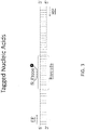
- the nucleic acid label is "barcoded" specifically for a particular affinity agent ( i.e., has a specific recognition signature for a specific affinity agent). See Figure 3 .
- the label is a nucleic acid label.
- the nucleic acid label is covalently bound to an affinity agent (e.g., antibody) at a chemically suitable location.
- the label is an enzyme
- the presence of the label e.g., in an antigen-affinity agent-label complex
- suitable enzymes include a polymerase (e.g ., DNA polymerase), urease, alkaline phosphatase, (horseradish) hydrogen peroxidase (HRP), glucose oxidase, ⁇ -galactosidase, luciferase, alkaline phosphatase, and an esterase that hydrolyzes fluorescein diacetate.
- a horseradish-peroxidase detection system can be used with the chromogenic substrate tetramethylbenzidine (TMB), which yields a soluble product in the presence of hydrogen peroxide that is detectable at 450 nm.
- An alkaline phosphatase detection system can be used with the chromogenic substrate p-nitrophenyl phosphate, which yields a soluble product readily detectable at 405 nm.
- a ⁇ -galactosidase detection system can be used with the chromogenic substrate o-nitrophenyl- ⁇ -D-galactopyranoside (ONPG), which yields a soluble product detectable at 410 nm.
- a urease detection system can be used with a substrate such as urea-bromocresol purple (Sigma Immunochemicals; St. Louis, MO).
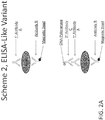
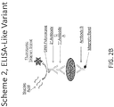
- the label is a DNA polymerase
- the detecting comprises detecting a nucleic acid amplification product that is generated by the DNA polymerase. See Figures 2A-B .
- the DNA polymerase is a thermostable DNA polymerase, e.g ., Taq polymerase.
- the DNA polymerase is a chemical- or antibody-mediated hot start Taq polymerase.
- Taq polymerases and hot start Taq polymerases are readily commercially available (e.g ., FastStart Taq DNA polymerase, available from Roche Applied Science, or iTaqTM DNA polymerase, available from Bio-Rad Laboratories, Inc.).
- FastStart Taq DNA polymerase available from Roche Applied Science
- iTaqTM DNA polymerase available from Bio-Rad Laboratories, Inc.
- One of skill will recognize that other DNA polymerases, such as other strains and/or mutations of Taq, can be used for detecting a nucle
- a detectable label is attached to an affinity agent via a covalent bond, ionic bond, hydrogen bonding, or by Van der Waals interactions.
- a detectable label e.g., a label as described herein
- exemplary detection methods include radioactive detection, optical absorbance detection (e.g ., fluorescence or chemiluminescence), or mass spectral detection.
- a fluorescent label can be detected using a detector device equipped with a module to generate excitation light that can be absorbed by a fluorophore, as well as a module to detect light emitted by the fluorophore.
- detectable labels in partitioned samples can be detected in bulk.
- partitioned samples e.g ., droplets
- the signal(s) e.g ., fluorescent signal(s)
- the signal(s) may be detected using a plate reader.
- the detector further comprises handling capabilities for the partitioned samples (e.g ., droplets), with individual partitioned samples entering the detector, undergoing detection, and then exiting the detector.
- partitioned samples e.g ., droplets
- partitioned samples may be detected serially while the partitioned samples are flowing.
- partitioned samples e.g ., droplets
- detectable labels in partitioned samples can be detected serially without flowing the partitioned samples ( e.g ., using a chamber slide).
- a general purpose computer system (referred to herein as a "host computer") can be used to store and process the data.
- a computer-executable logic can be employed to perform such functions as subtraction of background signal, assignment of target and/or reference sequences, and qualification and/or quantification of the data.
- a host computer can be useful for displaying, storing, retrieving, or calculating diagnostic results from the molecular profiling; storing, retrieving, or calculating raw data from expression analysis; or displaying, storing, retrieving, or calculating any sample or patient information useful in the methods of the present invention.
- the host computer may be configured with many different hardware components and can be made in many dimensions and styles (e.g., desktop PC, laptop, tablet PC, handheld computer, server, workstation, mainframe). Standard components, such as monitors, keyboards, disk drives, CD and/or DVD drives, and the like, may be included.
- the connections may be provided via any suitable transport media (e.g., wired, optical, and/or wireless media) and any suitable communication protocol (e.g., TCP/IP); the host computer may include suitable networking hardware (e.g., modem, Ethernet card, WiFi card).
- the host computer may implement any of a variety of operating systems, including UNIX, Linux, Microsoft Windows, MacOS, or any other operating system.
- Computer code for implementing aspects of the present invention may be written in a variety of languages, including PERL, C, C++, Java, JavaScript, VBScript, AWK, or any other scripting or programming language that can be executed on the host computer or that can be compiled to execute on the host computer. Code may also be written or distributed in low level languages such as assembler languages or machine languages.
- the host computer system advantageously provides an interface via which the user controls operation of the tools.
- software tools are implemented as scripts (e.g., using PERL), execution of which can be initiated by a user from a standard command line interface of an operating system such as Linux or UNIX.
- commands can be adapted to the operating system as appropriate.
- a graphical user interface may be provided, allowing the user to control operations using a pointing device.
- the present invention is not limited to any particular user interface.
- Scripts or programs incorporating various features of the present invention may be encoded on various computer readable media for storage and/or transmission.
- suitable media include magnetic disk or tape, optical storage media such as compact disk (CD) or DVD (digital versatile disk), flash memory, and carrier signals adapted for transmission via wired, optical, and/or wireless networks conforming to a variety of protocols, including the Internet.
- the separated sample comprising at least the label to be detected is partitioned into a plurality of partitions.
- Partitions can include any of a number of types of partitions, including solid partitions ( e.g., wells or tubes) and fluid partitions ( e.g., aqueous droplets within an oil phase).
- the partitions are droplets.
- the partitions are microchannels.
- a droplet comprises an emulsion composition, i.e ., a mixture of immiscible fluids (e.g ., water and oil).
- a droplet is an aqueous droplet that is surrounded by an immiscible carrier fluid (e.g., oil).
- a droplet is an oil droplet that is surrounded by an immiscible carrier fluid (e.g., an aqueous solution).
- the droplets described herein are relatively stable and have minimal coalescence between two or more droplets.
- emulsions can also have limited flocculation, a process by which the dispersed phase comes out of suspension in flakes.
- the droplet is formed by flowing an oil phase through an aqueous solution comprising the label(s) to be detected.
- the aqueous sample comprising the label(s) to be detected comprises a buffered solution and reagents for detecting the label(s).
- the oil for the oil phase may be synthetic or naturally occurring.
- the oil comprises carbon and/or silicon.
- the oil comprises hydrogen and/or fluorine.
- Exemplary oils include silicone oil, mineral oil, fluorocarbon oil, vegetable oil, or a combination thereof.
- the oil phase may comprise a fluorinated base oil which may additionally be stabilized by combination with a fluorinated surfactant such as a perfluorinated polyether.
- the base oil comprises one or more of a HFE 7500, FC-40, FC-43, FC-70, or another common fluorinated oil.
- the oil phase comprises an anionic fluorosurfactant.
- the anionic fluorosurfactant is Ammonium Krytox (Krytox-AS), the ammonium salt of Krytox FSH, or a morpholino derivative of Krytox FSH.
- Krytox-AS may be present at a concentration of about 0.1%, 0.2%, 0.3%, 0.4%, 0.5%, 0.6%, 0.7%, 0.8%, 0.9%, 1.0%, 2.0%, 3.0%, or 4.0% (w/w). In some embodiments, the concentration of Krytox-AS is about 1.8%. In some embodiments, the concentration of Krytox-AS is about 1.62%. Morpholino derivative of Krytox FSH may be present at a concentration of about 0.1%, 0.2%, 0.3%, 0.4%, 0.5%, 0.6%, 0.7%, 0.8%, 0.9%, 1.0%, 2.0%, 3.0%, or 4.0% (w/w). In some embodiments, the concentration of morpholino derivative of Krytox FSH is about 1.8%. In some embodiments, the concentration of morpholino derivative of Krytox FSH is about 1.62%.
- the oil phase further comprises an additive for tuning the oil properties, such as vapor pressure, viscosity, or surface tension.
- an additive for tuning the oil properties such as vapor pressure, viscosity, or surface tension.
- Non-limiting examples include perfluorooctanol and 1H,1H,2H,2H-Perfluorodecanol.
- 1H,1H,2H,2H-Perfluorodecanol is added to a concentration of about 0.05%, 0.06%, 0.07%, 0.08%, 0.09%, 0.1%, 0.2%, 0.3%, 0.4%, 0.5%, 0.6%, 0.7%, 0.8%, 0.9%, 1.0%, 1.25%, 1.50%, 1.75%, 2.0%, 2.25%, 2.5%, 2.75%, or 3.0% (w/w).
- 1H,1H,2H,2H-Perfluorodecanol is added to a concentration of about 0.18% (w/w).
- the emulsion is formulated to produce highly monodisperse droplets having a liquid-like interfacial film that can be converted by heating into microcapsules having a solid-like interfacial film; such microcapsules may behave as bioreactors able to retain their contents through an incubation period.
- the conversion to microcapsule form may occur upon heating. For example, such conversion may occur at a temperature of greater than about 40, 50, 60, 70, 80, 90, or 95 °C.
- a fluid or mineral oil overlay may be used to prevent evaporation. Excess continuous phase oil may or may not be removed prior to heating.
- the biocompatible capsules may be resistant to coalescence and/or flocculation across a wide range of thermal and mechanical processing.
- the microcapsules may be stored at about -70, -20, 0, 3, 4, 5, 6, 7, 8, 9, 10, 15, 20, 25, 30, 35, or 40 °C.
- these capsules are useful in biomedical applications, such as stable, digitized encapsulation of macromolecules, particularly aqueous biological fluids comprising a mix of target molecules such as nucleic acids, proteins, or both together; drug and vaccine delivery; biomolecular libraries; clinical imaging applications; and others.
- the microcapsule partitions may contain one or more affinity agents as described herein and may resist coalescence, particularly at high temperatures. Accordingly, the capsules can be incubated at a very high density (e.g ., number of partitions per unit volume). In some embodiments, greater than 100,000, 500,000, 1,000,000, 1,500,000, 2,000,000, 2,500,000, 5,000,000, or 10,000,000 partitions may be incubated per ml. In some embodiments, the sample-probe incubations occur in a single well, e.g., a well of a microtiter plate, without inter-mixing between partitions. The microcapsules may also contain other components necessary for the incubation.
- the sample is partitioned into at least 500 partitions (e.g ., droplets), at least 1000 partitions, at least 2000 partitions, at least 3000 partitions, at least 4000 partitions, at least 5000 partitions, at least 6000 partitions, at least 7000 partitions, at least 8000 partitions, at least 10,000 partitions, at least 15,000 partitions, at least 20,000 partitions, at least 30,000 partitions, at least 40,000 partitions, at least 50,000 partitions, at least 60,000 partitions, at least 70,000 partitions, at least 80,000 partitions, at least 90,000 partitions, at least 100,000 partitions, at least 200,000 partitions, at least 300,000 partitions, at least 400,000 partitions, at least 500,000 partitions, at least 600,000 partitions, at least 700,000 partitions, at least 800,000 partitions, at least 900,000 partitions, at least 1,000,000 partitions, at least 2,000,000 partitions, at least 3,000,000 partitions, at least 4,000,000 partitions, at least 5,000,000 partitions,
- the sample is partitioned into a sufficient number of partitions such that at least a majority of partitions have no more than 1, 2, 3, 4, 5, 6, 7, 8, 9, 10, 15, 20, 30, 40, 50, 60, 70, 80, 90, 100, 200, 300, 400, or 500 copies of a label.
- a majority of the partitions have no more than 1, 2, 3, 4, 5, 6, 7, 8, 9, 10, 15, 20, 30, 40, 50, 60, 70, 80, 90, 100, 200, 300, 400, or 500 copies of the one or more labels to be detected.
- on average no more than 1, 2, 3, 4, 5, 6, 7, 8, 9, 10, 15, 20, 30, 40, 50, 60, 70, 80, 90, 100, 200, 300, 400, or 500 copies of the one or more labels are present per partition.
- the sample is partitioned into a sufficient number of partitions such that, on average, at least one partition lacks a copy of the label. In some embodiments, the sample is partitioned into a sufficient number of partitions such that, on average, at least 5, 10, 15, 20, 25, 30, 35, 40, 50, 60, 70, 80, 90, 100, 125, 150, 175, 200, 250, 300, 350, 400, 450, or 500 partitions lack a copy of the label.
- the sample is partitioned into a sufficient number of partitions such that, on average, at least 5, 10, 15, 20, 25, 30, 35, 40, 50, 60, 70, 80, 90, 100, 125, 150, 175, 200, 250, 300, 350, 400, 450, or 500 partitions lack a copy of the label and such that, on average, at least 5, 10, 15, 20, 25, 30, 35, 40, 50, 60, 70, 80, 90, 100, 125, 150, 175, 200, 250, 300, 350, 400, 450, or 500 partitions have at least one copy of the label.
- the droplets that are generated are substantially uniform in shape and/or size.
- the droplets are substantially uniform in average diameter.
- the droplets that are generated have an average diameter of about 0.001 ⁇ m, about 0.005 ⁇ m, about 0.01 ⁇ m, about 0.05 ⁇ m, about 0.1 ⁇ m, about 0.5 ⁇ m, about 1 ⁇ m, about 5 ⁇ m, about 10 ⁇ m, about 20 ⁇ m, about 30 ⁇ m, about 40 ⁇ m, about 50 ⁇ m, about 60 ⁇ m, about 70 ⁇ m, about 80 ⁇ m, about 90 ⁇ m, about 100 ⁇ m, about 150 ⁇ m, about 200 ⁇ m, about 300 ⁇ m, about 400 ⁇ m, about 500 ⁇ m, about 600 ⁇ m, about 700 ⁇ m, about 800 ⁇ m, about 900 ⁇ m, or about 1000 ⁇ m.
- the droplets that are generated have an average diameter of less than about 1000 ⁇ m, less than about 900 ⁇ m, less than about 800 ⁇ m, less than about 700 ⁇ m, less than about 600 ⁇ m, less than about 500 ⁇ m, less than about 400 ⁇ m, less than about 300 ⁇ m, less than about 200 ⁇ m, less than about 100 ⁇ m, less than about 50 ⁇ m, or less than about 25 ⁇ m.
- the droplets that are generated are non-uniform in shape and/or size.
- the droplets that are generated are substantially uniform in volume.
- the droplets that are generated have an average volume of about 0.001 nl, about 0.005 nl, about 0.01 nl, about 0.02 nl, about 0.03 nl, about 0.04 nl, about 0.05 nl, about 0.06 nl, about 0.07 nl, about 0.08 nl, about 0.09 nl, about 0.1 nl, about 0.2 nl, about 0.3 nl, about 0.4 nl, about 0.5 nl, about 0.6 nl, about 0.7 nl, about 0.8 nl, about 0.9 nl, about 1 nl, about 1.5 nl, about 2 nl, about 2.5 nl, about 3 nl, about 3.5 nl, about 4 nl, about 4.5 nl, about 5 nl, about 5.5 nl, about 6 nl, about 6.5 nl, about
- a digital readout assay can be used to detect and quantify one or more antigens in a sample by partitioning at least the labels from the separated sample (e.g., labels in antigen-affinity agent-label complexes or labels cleaved from antigen-affinity agent-label complexes) and identifying the partitions containing the label.
- the process of digital analysis involves determining for each partition of a sample whether the partition is positive or negative for the presence of the label or labels to be detected. If the antigen-affinity agent-label complex comprises antigen and label that are at an unknown ratio, then the presence of label corresponds to a positive detection of the antigen.
- the antigen-affinity agent-label complex comprises antigen and label at a known ratio (i.e., 1:1 ratio)
- the amount of label that is detected for a sample can be correlated to the amount of antigen present in the sample.
- the partitions are examined for the presence or absence of a detectable signal in each partition. A partition is "positive" for the presence of the antigen if a signal is detected in the partition.
- the signal that is detected in the partition is generated by a label linked to an affinity agent in an antigen-affinity agent-label complex (e.g ., a fluorescent, chemiluminescent, radioactive, or enzymatic label linked to the affinity agent).
- a label linked to an affinity agent in an antigen-affinity agent-label complex e.g ., a fluorescent, chemiluminescent, radioactive, or enzymatic label linked to the affinity agent.
- a partition is "negative" for the presence of the antigen if no signal detected in the partition.
- a detector that is capable of detecting a signal or multiple signals is used to analyze each partition for the presence or absence of the antigen.
- a detector that is capable of detecting a signal or multiple signals is used to analyze each partition for the presence or absence of the antigen.
- a two-color reader fluorescence detector
- the fraction of positive-counted partitions can enable the determination of absolute concentrations for the antigen or antigens to be measured.
- the data for the partitions is analyzed using an algorithm based on Poisson statistics to quantify the amount of antigen in the sample.
- Statistical methods for quantifying the concentration or amount of an antigen or antigens is described, for example, in WO 2010/036352 .
- the methods described herein comprise an amplification step.
- a nucleic acid label attached to an affinity agent as part of an antigen-affinity agent-label complex is amplified prior to the step of detecting a detectable signal from the label.
- the nucleic acid label is amplified in the presence of primers and a probe that specifically bind the nucleic acid label, and the probe generates a detectable (e.g ., fluorescent) signal when the nucleic acid is amplified, thereby indicating the presence of an antigen-affinity agent-label complex.
- the amplification step comprises contacting the sample comprising the labeled affinity agent with a nucleic acid and detecting a nucleic acid amplification product generated by the label (e.g., the DNA polymerase).
- the nucleic acid to be amplified comprises a sequence that is complementary or substantially complementary to a probe that generates a detectable (e.g ., fluorescent) signal when the nucleic acid is amplified, thereby indicating the presence of an antigen-affinity agent-label complex.
- a system such as the TaqMan® system can be used.
- This system utilizes a short oligonucleotide probe (e.g ., about 20-25 bases in length) that is labeled with two different fluorescent dyes.
- the 5' terminus of the probe is attached to a reporter dye, or "fluorescer,” and the 3' terminus is attached to a quenching moiety, or "quencher.”
- the dyes are attached at other locations on the probe.
- the probe can be designed to have at least substantial sequence complementarity with the probe-binding site on the nucleic acid to be amplified. Upstream and downstream PCR primers that bind to regions that flank the probe binding site are utilized for amplifying the nucleic acid.
- the fluorogenic probe When the fluorogenic probe is intact, energy transfer between the fluorescer and quencher moiety occurs and quenches emission from the fluorescer.
- the probe is cleaved, e.g., by the 5' nuclease activity of a nucleic acid polymerase such as Taq polymerase, or by a separately provided nuclease activity that cleaves bound probe, thereby separating the fluorescer and quencher moieties. This results in an increase of reporter emission intensity that can be measured by an appropriate detector. Detection and/or quantification of the antigen is determined by mathematical conversion of fluorescent signal to number of antigens according to methods known in the art.
- structured probes can be utilized to detect a nucleic acid amplification product.
- molecular beacons a change in conformation of a probe as it hybridizes to a complementary region of the amplified product results in the formation of a detectable signal.
- the probe includes additional sections, generally one section at the 5' end and another section at the 3' end, that are complementary to each other.
- One end section is typically attached to a reporter dye and the other end section is usually attached to a quencher dye.
- the two end sections can hybridize with each other to form a stem-loop structure.
- the reporter dye and quencher are in sufficiently close proximity that fluorescence from the reporter dye is effectively quenched by the quencher.
- the hybridized probe in contrast, has a linearized conformation which decreases the extent of quenching.
- Probes of this type and methods of their use is described further, for example, by Piatek, A. S., et al., Nat. Biotechnol. 16:359-63 (1998 ); Tyagi, S. and Kramer, F. R., Nature Biotechnology 14:303-308 (1996 ); and Tyagi, S. et al., Nat. Biotechnol. 16:49-53 (1998 ).
- the method comprises incubating the sample comprising the labeled affinity agent with the nucleic acid under conditions suitable for amplifying the nucleic acid.
- the sample can be incubated with amplification reagents (dNTPs, primers, probes, blocking agents, biopreservatives, etc.).
- dNTPs amplification reagents
- Suitable conditions for amplifying the nucleic acid will depend, e.g., on the nature of the DNA polymerase and the nucleic acid to be amplified, and can be readily determined by a person of skill in the art.
- the label from the separated sample (e.g ., label in an antigen-affinity agent-label complex or label cleaved from antigen-affinity agent-label complex) is resuspended in a solution (e.g., an aqueous buffer) comprising reagents for amplification (e.g., dNTPs, primers, TaqMan® probe, etc.).
- a solution e.g., an aqueous buffer
- reagents for amplification e.g., dNTPs, primers, TaqMan® probe, etc.
- the solution is partitioned into a plurality of partitions before the amplification step.
- amplification is performed before the partitioning step.
- PCR polymerase chain reaction
- quantitative PCR real time PCR
- hot start PCR single cell PCR
- nested PCR in situ colony PCR
- digital PCR droplet digitalTM PCR
- emulsion PCR ligase chain reaction
- LCR droplet digitalTM PCR
- NASBA transcription based amplification system
- SDA strand displacement amplification
- RCA rolling circle amplification
- HRCA hyper-branched RCA
- the amplification is performed using droplet digitalTM PCR (ddPCR).
- ddPCR droplet digitalTM PCR
- Devices for performing ddPCR are known in the art.
- ddPCR can be performed with the QX100TM Droplet DigitalTM PCR System (Bio-Rad Laboratories, Inc., Hercules, CA). Methods and devices for performing ddPCR are also described, for example, in WO 2010/036352 and WO 2012/129187 .
- methods of detecting an antibody analyte of interest in a sample are provided, wherein the antibody analyte specifically binds to a particular antigen.
- the method comprises:
- the label that is partitioned is in the analyte-antigen-affinity agent-label complex. In some embodiments, the label is cleaved from the analyte-antigen-affinity agent-label complex prior to the partitioning step.
- the methods described herein can be used to detect one or more antibody analytes in any type of sample (e.g ., biological sample). In some embodiments, two, three, four, five, or more different antibody analytes are to be detected. In some embodiments, wherein two or more different antibody analytes are to be detected, the two or more different antibody analytes specifically bind to the same antigen. In some embodiments, wherein two or more different antigens are to be detected, the two or more different antibody analytes specifically bind to different antigens.
- the reagents e.g ., affinity agents, detectable labels, and magnetic beads
- conditions e.g ., detection, partitioning, and digital analysis
- the methods can be any of the reagents and/or conditions described herein, e.g., in Section II above.
- partition libraries comprising a plurality of partitions for carrying out the methods as described herein.
- the partition library may comprise a plurality of partitions for detecting the presence of an antigen in a sample.
- the partition library may comprise two or more partitions, wherein at least some partitions of the library (e.g., at least 5%, 10%, 15%, 20%, 25%, 30%, 35%, 40%, 45%, 50%, 55%, 60%, 65%, 70%, 75%, 80%, 85%, 90%, or 95% of the partitions in the library) comprise one, two, three, four, five or more different affinity agents.
- the partitions may comprise a first affinity agent and a second affinity agent; wherein the first affinity agent comprises a label; wherein the second affinity agent is linked to a magnetic bead; and wherein each of the first and second affinity agents specifically binds to the antigen, if present.
- the partitions may comprise a first affinity agent, a second affinity agent, and a third affinity agent, wherein each of the first and second affinity agents specifically binds to an antigen, if present, wherein the second affinity agent is linked to a magnetic bead, and wherein the third affinity agent comprises a label and specifically binds to the first affinity agent.
- the partition library may comprise a plurality of partitions for detecting the presence of an antibody analyte in a sample, wherein the antibody analyte specifically binds to a particular antigen.
- the partitions may comprise the antigen, a first affinity agent, and a second affinity agent; wherein the first affinity agent is linked to a magnetic bead and specifically binds to the antigen; and wherein the second affinity agent comprises a label and specifically binds to the antibody analyte, if present.
- the partition library may comprise at least 500, 1000, 2000, 3000, 4000, 5000, 6000, 7000, 8000, 10,000, 15,000, 20,000, 30,000, 40,000, 50,000, 60,000, 70,000, 80,000, 90,000, 100,000, 200,000, 300,000, 400,000, 500,000, 600,000, 700,000, 800,000, 900,000, 1,000,000, 2,000,000, 3,000,000, 4,000,000, 5,000,000, 10,000,000, 20,000,000, 30,000,000, 40,000,000, 50,000,000, 60,000,000, 70,000,000, 80,000,000, 90,000,000, 100,000,000, 150,000,000, or 200,000,000 partitions.
- the affinity agent for the partitions of the partition library can be any affinity agent described herein.
- the affinity agent(s) may be an antibody.
- the affinity agent(s) may be an antibody fragment.
- the affinity agent(s) may be a non-antibody protein scaffold.
- the affinity agent(s) may be an antibody mimetic.
- the affinity agent(s) may be an aptamer.
- an affinity agent may be labeled as described herein, e.g ., with a nucleic acid label, an enzyme label, or a fluorophore label.
- At least some partitions of the partition library may comprise one or more labels. Further, at least some partitions may comprise two, three, four, five, or more different labels. Further, the sample may be partitioned into a sufficient number of partitions such that, on average, at least 1, 5, 10, 15, 20, 25, 30, 35, 40, 50, 60, 70, 80, 90, 100, 125, 150, 175, 200, 250, 300, 350, 400, 450, or 500 partitions lack a copy of the label.
- the sample may be partitioned into a sufficient number of partitions such that, on average, at least 1, 5, 10, 15, 20, 25, 30, 35, 40, 50, 60, 70, 80, 90, 100, 125, 150, 175, 200, 250, 300, 350, 400, 450, or 500 partitions lack a copy of the label and such that, on average, at least 1, 5, 10, 15, 20, 25, 30, 35, 40, 50, 60, 70, 80, 90, 100, 125, 150, 175, 200, 250, 300, 350, 400, 450, or 500 partitions have at least one copy of the label.
- the partition library may comprise a plurality of partitions that are solid partitions ( e.g ., wells or tubes). Further, the partitions may be microchannels. Further, the partition library may comprise a plurality of partitions that are fluid partitions ( e.g ., aqueous droplets within an oil phase). Further, the partitions may be droplets. Further, the partitions may be microcapsules. Examples of suitable partitions and methods of generating partitions are described above.
- the partition library may comprise partitions that are substantially uniform in shape and/or size.
- the partitions e.g ., droplets
- the partitions e.g ., droplets
- the partitions have an average diameter of about 0.001, 0.005, 0.01, 0.05, 0.1, 0.5, 1, 5, 10, 20, 30, 40, 50, 60, 70, 80, 90, 100, 150, 200, 300, 400, 500, 600, 700, 800, 900, or 1000 ⁇ m.
- the partitions (e.g ., droplets) may have an average diameter of less than about 1000, 900, 800, 700, 600, 500, 400, 300, 200, 100, 50, or 25 ⁇ m.
- the partition library may comprise partitions ( e.g ., droplets) that are non-uniform in shape and/or size.
- the partitions may be substantially uniform in volume.
- the partitions e.g ., droplets
- the partitions may have an average volume of about 0.001, 0.005, 0.01, 0.02, 0.03, 0.04, 0.05, 0.06, 0.07, 0.08, 0.09, 0.1, 0.2, 0.3, 0.4, 0.5, 0.6, 0.7, 0.8, 0.9, 1, 1.5, 2, 2.5, 3, 3.5, 4, 4.5, 5, 5.5, 6, 6.5, 7, 7.5, 8, 8.5, 9, 9.5, 10, 11, 12, 13, 14, 15, 16, 17, 18, 19, 20, 25, 30, 35, 40, 45, or 50 nl.
- the partitions are stable and are capable of long-term storage. Further, the partitions can be stored at about -70, -20, 0, 3, 4, 5, 6, 7, 8, 9, 10, 15, 20, 25, 30, 35, or 40 °C for an extended period of time (e.g., for at least 30 d, at least 60 d, at least 90 d, or longer).
- kits for detecting an antigen or for detecting an antibody analyte that specifically binds to an antigen may comprise one or more affinity agents as described herein, e.g., one or more antibodies, antibody fragments, non-antibody protein scaffolds, antibody mimetics, and/or aptamers as described herein.
- a kit may further comprise one or more reagents for forming partitions, detection reagents, and/or reagents for nucleic acid amplification (e.g ., buffers, enzymes, dNTPs, primers, TaqMan® probes, blocking agents, biopreservatives, and the like).
- a kit for detecting an antibody analyte may further comprise an antigen to which the antibody analyte specifically binds.
- a kit may further comprise instructions for carrying out the methods described herein.
- This example demonstrates detecting an antigen of interest using two primary affinity agents (e.g ., antibodies), each of which specifically binds to the antigen.
- a sample is contacted with primary antibody A and primary antibody B.
- Antibody B has a magnetic bead covalently attached.
- the sample is contacted with a secondary antibody C that is labeled with a tagged nucleic acid ("TNA").
- TNA tagged nucleic acid
- the antigen-primary antibody-secondary antibody-TNA complex is purified from free primary antibody A and secondary antibody C by magnetic separation with washing, then the purified complex is resuspended and captured in droplets.
- Droplet digital PCR is performed to detect the presence of the TNA label and/or quantify the amount of TNA label in each droplet. Detection and/or quantification of the antigen is then determined by mathematical conversion of the signal from the label to the number of antigens. See Figure 1 .
- This example demonstrates detecting an antigen using two primary affinity agents (e.g ., antibodies), each of which specifically binds to the antigen.
- an enzyme-linked secondary antibody is added that converts a substrate into a detectable form for detecting the presence of the antigen.
- a sample is contacted with primary antibody A and primary antibody B.
- Antibody B has a magnetic bead covalently attached.
- the sample is contacted with a secondary antibody C that binds specifically to antibody A.
- the secondary antibody C is coupled to a DNA polymerase such as Taq, Fast Start Taq, or some other polymerase (e.g ., a mutagenized or variant DNA polymerase with desired properties for the application) or to another enzyme ( Figure 2A ).
- a DNA polymerase such as Taq, Fast Start Taq, or some other polymerase (e.g ., a mutagenized or variant DNA polymerase with desired properties for the application) or to another enzyme ( Figure 2A ).
- the antigen-primary antibody-secondary antibody-DNA polymerase complex is purified from free primary and secondary antibodies by magnetic separation with washing, then the complex is resuspended in a master mix or appropriate buffer suitable for digital droplet formation and PCR amplification or other means of fluorogenic or chromogenic detection (e.g ., including nucleic acid(s) suitable for amplification, appropriate forward and reverse primers, and TaqMan® probe; or an appropriate substrate for the enzyme coupled to the secondary antibody C and reagents for fluorogenic or chromogenic detection).
- fluorogenic or chromogenic detection e.g ., including nucleic acid(s) suitable for amplification, appropriate forward and reverse primers, and TaqMan® probe; or an appropriate substrate for the enzyme coupled to the secondary antibody C and reagents for fluorogenic or chromogenic detection.
- the resuspended complex is captured in droplets, then the nucleic acid is amplified and the fluorogenic signal that is generated from the TaqMan® probe is detected and/or quantified, or the fluorogenic or chromogenic signal generated by the enzyme is detected and/or quantified ( Figure 2B ). Detection and/or quantification of the antigen is then determined by mathematical conversion of the signal from the TaqMan® probe to the number of antigens.
- This example demonstrates detecting an antigen of interest using two primary affinity agents (e.g ., antibodies), each of which specifically binds to the antigen.
- a sample is contacted with primary antibody A and primary antibody B.
- Antibody B has a magnetic bead covalently attached.
- the sample is contacted with a secondary antibody C that is labeled with a fluorophore.
- the antigen-primary antibody-secondary antibody-fluorophore complex is purified from free primary antibody A and secondary antibody C by magnetic separation with washing, then the purified complex is resuspended and captured in droplets.
- the fluorescent signal generated by the fluorophore label is detected according to any method described herein. Detection and/or quantification of the antigen is then determined by mathematical conversion of the signal from the label to the number of antigens. See Figure 4 .
- This example demonstrates detecting an antibody analyte of interest that binds to a particular antigen.
- a sample comprising an antibody analyte of interest is contacted with an antigen to which the antibody analyte specifically binds and an affinity agent (e.g., a primary antibody B) that also specifically binds to the antigen.
- the affinity agent has a magnetic bead covalently attached.
- the sample is contacted with a labeled affinity agent (e.g., a secondary antibody C).
- the label can be any label described herein, for example but not limited to a tagged nucleic acid (TNA), a DNA polymerase or other enzyme capable of converting a substrate to a detectable signal, or a fluorescent label.
- TNA tagged nucleic acid
- the analyte-antigen-antibody-label complex is purified from free antigen and affinity agent by magnetic separation with washing, then the purified complex is resuspended and captured in droplets.
- the label is then detected as described herein. For example, if the label is a TNA label, droplet digital PCR is performed to detect the presence of the TNA label and/or quantify the amount of TNA label in each droplet. If the label is a fluorophore, the fluorescent signal generated by the fluorophore is detected. Detection and/or quantification of the antibody analyte is then determined by mathematical conversion of the signal from the label to the number of analytes. See Figure 5 .
- This example demonstrates detecting an antigen of interest using two primary affinity agents (e.g ., antibodies), each of which specifically binds to the antigen.
- a sample is contacted with primary antibody A and primary antibody B.
- Antibody A is labeled, e.g ., with a tagged nucleic acid or a fluorescent label, e.g. , fluorophore.
- Antibody B has a magnetic bead covalently attached.
- the antigen-primary antibody-secondary antibody-fluorophore complex is purified from free primary antibody A by magnetic separation with washing, then the purified complex is resuspended and captured in droplets.
- a fluorescent signal generated by the fluorophore label can be detected according to any detection method described herein, or droplet digital PCR is performed to detect the presence of a TNA label and/or quantify the amount of TNA label in each droplet). Detection and/or quantification of the antigen is then determined by mathematical conversion of the amount of/signal from the label to the number of antigens. See Figure 6 .
Landscapes
- Chemical & Material Sciences (AREA)
- Organic Chemistry (AREA)
- Health & Medical Sciences (AREA)
- Life Sciences & Earth Sciences (AREA)
- Analytical Chemistry (AREA)
- Immunology (AREA)
- Zoology (AREA)
- Wood Science & Technology (AREA)
- Proteomics, Peptides & Aminoacids (AREA)
- Engineering & Computer Science (AREA)
- Physics & Mathematics (AREA)
- Microbiology (AREA)
- Molecular Biology (AREA)
- Biotechnology (AREA)
- Biophysics (AREA)
- Pathology (AREA)
- Biochemistry (AREA)
- Bioinformatics & Cheminformatics (AREA)
- General Engineering & Computer Science (AREA)
- General Health & Medical Sciences (AREA)
- Genetics & Genomics (AREA)
- Measuring Or Testing Involving Enzymes Or Micro-Organisms (AREA)
Claims (13)
- Procédé de détection d'un antigène dans un échantillon, ledit procédé comprenant :(a) la mise en contact de l'échantillon avec un premier agent d'affinité et un deuxième agent d'affinité, dans laquelle le deuxième agent d'affinité est lié à une bille magnétique et dans laquelle le premier et le deuxième agent d'affinité se lient spécifiquement à l'antigène, si celui-ci est présent, et la mise en contact de l'échantillon qui a été en contact avec le premier et le deuxième agent d'affinité avec un troisième agent d'affinité, dans laquelle le troisième agent d'affinité comprend un marqueur et dans laquelle le troisième agent d'affinité se lie spécifiquement au premier agent d'affinité, formant ainsi un complexe antigène-agent d'affinité-marqueur ; ou(a') la mise en contact de l'échantillon avec un premier agent d'affinité et un deuxième agent d'affinité, dans laquelle le premier agent d'affinité comprend un marqueur, dans laquelle le deuxième agent d'affinité est lié à une bille magnétique, et dans laquelle le premier et le deuxième agent d'affinité se lient spécifiquement à l'antigène, si celui-ci est présent, formant ainsi un complexe antigène-agent d'affinité-marqueur ; ou(a") la mise en contact de l'échantillon avec l'antigène et un premier agent d'affinité, dans laquelle le premier agent d'affinité est lié à une bille magnétique et se lie spécifiquement à l'antigène, formant ainsi un complexe analyte-antigène-agent d'affinité, et la mise en contact de l'échantillon comprenant le complexe analyte-antigène-agent d'affinité avec un deuxième agent d'affinité, dans laquelle le deuxième agent d'affinité comprend un marqueur et se lie spécifiquement à l'analyte d'anticorps, formant ainsi un complexe analyte-antigène-agent d'affinité-marqueur ;(b) la séparation du complexe antigène-agent d'affinité-marqueur des composants non complexés dans l'échantillon, basée sur la présence ou l'absence de la bille magnétique, générant ainsi un échantillon séparé comprenant le complexe antigène-agent d'affinité-marqueur ;(c) le partitionnement d'au moins le marqueur à partir de l'échantillon séparé en une pluralité de partitions ; et(d) la détection de la présence du marqueur dans au moins une partition ; détectant ainsi la présence de l'antigène.
- Procédé selon la revendication 1, dans lequel(i) le marqueur qui est partitionné se trouve dans le complexe antigène-agent d'affinité-marqueur ; ou(ii) le marqueur est clivé du complexe antigène-agent d'affinité-marqueur avant l'étape de partitionnement (c) ; ou(iii) après l'étape de séparation (b) et avant l'étape de partitionnement (c), le procédé comprend en outre la remise en suspension d'au moins le marqueur issu de l'échantillon séparé dans une solution.
- Procédé selon la revendication 1, dans lequel(i) dans l'étape (a), chacun des premier, deuxième et troisième agents d'affinité est choisi dans le groupe constitué par les anticorps, les aptamères et les squelettes protéiques non anticorps, de préférence, chacun des premier, deuxième et troisième agents d'affinité est un anticorps ; ou(ii) dans les étapes (a') et (a"), chacun des premier et deuxième agents d'affinité est choisi dans le groupe constitué par les anticorps, les aptamères et les squelettes protéiques non anticorps, de préférence, chacun des premier et deuxième agents d'affinité est un anticorps.
- Procédé selon la revendication 1, dans lequel le marqueur est un marqueur d'acide nucléique, de préférence, le marqueur d'acide nucléique comprend une étiquette détectable.
- Procédé selon la revendication 4, dans lequel(i) le marqueur d'acide nucléique est amplifié avant l'étape de détection (d), de préférence, le marqueur d'acide nucléique est amplifié en présence d'une sonde comprenant un fluorophore et un extincteur, dans lequel l'amplification du marqueur d'acide nucléique génère un signal fluorescent provenant de la sonde, ou la solution comprend des réactifs pour l'amplification d'acide nucléique ; ou(ii) le marqueur d'acide nucléique n'est pas amplifié avant l'étape de détection.
- Procédé selon la revendication 4, dans lequel(i) l'étiquette détectable est un fluorophore ; ou(ii) l'étiquette détectable est un colorant intercalant.
- Procédé selon la revendication 1, dans lequel(i) le marqueur est une enzyme, et la détection comprend la détection d'un produit généré par l'enzyme, de préférence, l'enzyme est choisie dans le groupe constitué par la peroxydase de raifort, la β-galactosidase, la luciférase, la phosphatase alcaline, une estérase qui hydrolyse le diacétate de fluorescéine, et une polymérase, le plus préférablement, l'enzyme est une ADN-polymérase, et la détection comprend la détection d'un produit d'amplification d'acide nucléique généré par l'ADN-polymérase ; ou(ii) le marqueur est un fluorophore.
- Procédé selon la revendication 1, dans lequel(i) dans l'étape (a), le troisième agent d'affinité qui est mis en contact avec l'échantillon qui a été en contact avec le premier et le deuxième agent d'affinité est lié de manière covalente au marqueur ; ou(ii) dans l'étape (a'), le marqueur est lié de manière covalente au premier agent d'affinité lorsque le premier agent d'affinité est mis en contact avec l'échantillon ; ou(iii) le deuxième agent d'affinité qui est mis en contact avec l'échantillon qui a été en contact avec le complexe analyte-antigène-agent d'affinité est lié de manière covalente au marqueur.
- Procédé selon la revendication 1, dans lequel(i) le complexe antigène-agent d'affinité-marqueur est séparé des composants non complexés dans l'échantillon en utilisant un aimant qui attire la bille magnétique liée au deuxième agent d'affinité dans le complexe antigène-agent d'affinité-marqueur ; ou(ii) les partitions sont des gouttelettes, de préférence, les gouttelettes sont entourées d'un fluide porteur non miscible.
- Procédé selon la revendication 1, dans lequel, dans les étapes (a) et (a'),(i) le premier agent d'affinité et le deuxième agent d'affinité se lient spécifiquement à l'antigène avec des affinités à peu près égales ; ou(ii) le premier agent d'affinité et le deuxième agent d'affinité se lient spécifiquement à l'antigène avec des affinités différentes.
- Procédé selon la revendication 1, dans lequel l'antigène est une protéine, un immunogène, un polysaccharide, une toxine, une paroi cellulaire, une capsule de cellule, une capsule virale, une enveloppe virale, un flagelle, une fimbria ou pilus, un microorganisme, un acide nucléique complexé à une protéine ou un polysaccharide, ou un lipide complexé à une protéine ou un polysaccharide.
- Procédé selon la revendication 1,(i) ledit procédé comprenant en outre la quantification du nombre de partitions comprenant le marqueur ; ou(ii) ledit procédé comprenant la détection de deux antigènes distincts ou plus dans l'échantillon.
- Procédé selon la revendication 1 qui est un procédé de détection dans un échantillon d'un analyte d'anticorps qui se lie spécifiquement à un antigène, ledit procédé comprenant :(a") la mise en contact de l'échantillon avec l'antigène et un premier agent d'affinité, dans laquelle le premier agent d'affinité est lié à une bille magnétique et se lie spécifiquement à l'antigène, formant ainsi un complexe analyte-antigène-agent d'affinité, et la mise en contact de l'échantillon comprenant le complexe analyte-antigène-agent d'affinité avec un deuxième agent d'affinité, dans laquelle le deuxième agent d'affinité comprend un marqueur et se lie spécifiquement à l'analyte d'anticorps, formant ainsi un complexe analyte-antigène-agent d'affinité-marqueur ;(b) la séparation du complexe analyte-antigène-agent d'affinité-marqueur des composants non complexés dans l'échantillon, basée sur la présence ou l'absence de la bille magnétique, générant ainsi un échantillon séparé comprenant le complexe analyte-antigène-agent d'affinité-marqueur ;(c) le partitionnement d'au moins le marqueur à partir de l'échantillon séparé en une pluralité de partitions ; et(d) la détection de la présence du marqueur dans au moins une partition ; détectant ainsi la présence de l'analyte d'anticorps.
Applications Claiming Priority (3)
| Application Number | Priority Date | Filing Date | Title |
|---|---|---|---|
| US201361821530P | 2013-05-09 | 2013-05-09 | |
| US201361837533P | 2013-06-20 | 2013-06-20 | |
| PCT/US2014/037181 WO2014182835A1 (fr) | 2013-05-09 | 2014-05-07 | Dosage par pcr immunomagnétique numérique |
Publications (3)
| Publication Number | Publication Date |
|---|---|
| EP2994559A1 EP2994559A1 (fr) | 2016-03-16 |
| EP2994559A4 EP2994559A4 (fr) | 2017-02-08 |
| EP2994559B1 true EP2994559B1 (fr) | 2020-07-08 |
Family
ID=51865219
Family Applications (1)
| Application Number | Title | Priority Date | Filing Date |
|---|---|---|---|
| EP14794346.8A Active EP2994559B1 (fr) | 2013-05-09 | 2014-05-07 | Dosage par pcr immunomagnétique numérique |
Country Status (4)
| Country | Link |
|---|---|
| US (1) | US9896717B2 (fr) |
| EP (1) | EP2994559B1 (fr) |
| CN (1) | CN105431575B (fr) |
| WO (1) | WO2014182835A1 (fr) |
Families Citing this family (50)
| Publication number | Priority date | Publication date | Assignee | Title |
|---|---|---|---|---|
| US9617589B2 (en) | 2012-05-25 | 2017-04-11 | The University Of North Carolina At Chapel Hill | Microfluidic devices, solid supports for reagents and related methods |
| US20150376609A1 (en) | 2014-06-26 | 2015-12-31 | 10X Genomics, Inc. | Methods of Analyzing Nucleic Acids from Individual Cells or Cell Populations |
| US10400280B2 (en) | 2012-08-14 | 2019-09-03 | 10X Genomics, Inc. | Methods and systems for processing polynucleotides |
| US20140155295A1 (en) | 2012-08-14 | 2014-06-05 | 10X Technologies, Inc. | Capsule array devices and methods of use |
| US10752949B2 (en) | 2012-08-14 | 2020-08-25 | 10X Genomics, Inc. | Methods and systems for processing polynucleotides |
| US9951386B2 (en) | 2014-06-26 | 2018-04-24 | 10X Genomics, Inc. | Methods and systems for processing polynucleotides |
| US11591637B2 (en) | 2012-08-14 | 2023-02-28 | 10X Genomics, Inc. | Compositions and methods for sample processing |
| US10323279B2 (en) | 2012-08-14 | 2019-06-18 | 10X Genomics, Inc. | Methods and systems for processing polynucleotides |
| US9701998B2 (en) | 2012-12-14 | 2017-07-11 | 10X Genomics, Inc. | Methods and systems for processing polynucleotides |
| US10273541B2 (en) | 2012-08-14 | 2019-04-30 | 10X Genomics, Inc. | Methods and systems for processing polynucleotides |
| EP2931919B1 (fr) | 2012-12-14 | 2019-02-20 | 10X Genomics, Inc. | Procédés et systèmes pour le traitement de polynucléotides |
| US10533221B2 (en) | 2012-12-14 | 2020-01-14 | 10X Genomics, Inc. | Methods and systems for processing polynucleotides |
| CN108753766A (zh) | 2013-02-08 | 2018-11-06 | 10X基因组学有限公司 | 多核苷酸条形码生成 |
| CA2943624A1 (fr) | 2014-04-10 | 2015-10-15 | 10X Genomics, Inc. | Dispositifs fluidiques, systemes et procedes permettant d'encapsuler et de separer des reactifs, et leurs applications |
| EP3212807B1 (fr) | 2014-10-29 | 2020-09-02 | 10X Genomics, Inc. | Procédés et compositions de séquençage ciblé d'acides nucléiques |
| US9975122B2 (en) | 2014-11-05 | 2018-05-22 | 10X Genomics, Inc. | Instrument systems for integrated sample processing |
| SG11201705615UA (en) | 2015-01-12 | 2017-08-30 | 10X Genomics Inc | Processes and systems for preparing nucleic acid sequencing libraries and libraries prepared using same |
| AU2016222719B2 (en) | 2015-02-24 | 2022-03-31 | 10X Genomics, Inc. | Methods for targeted nucleic acid sequence coverage |
| US10697000B2 (en) | 2015-02-24 | 2020-06-30 | 10X Genomics, Inc. | Partition processing methods and systems |
| WO2017034925A1 (fr) * | 2015-08-25 | 2017-03-02 | Bio-Rad Laboratories, Inc. | Dosage immunologique numérique |
| US10739336B2 (en) * | 2015-10-01 | 2020-08-11 | Hong Kong Baptist University | Magnetic platform for direct and multiplex immune-based detection of trace amount of protein biomarkers |
| US11371094B2 (en) | 2015-11-19 | 2022-06-28 | 10X Genomics, Inc. | Systems and methods for nucleic acid processing using degenerate nucleotides |
| CN115369161A (zh) | 2015-12-04 | 2022-11-22 | 10X 基因组学有限公司 | 用于核酸分析的方法和组合物 |
| WO2017197338A1 (fr) | 2016-05-13 | 2017-11-16 | 10X Genomics, Inc. | Systèmes microfluidiques et procédés d'utilisation |
| US10550429B2 (en) | 2016-12-22 | 2020-02-04 | 10X Genomics, Inc. | Methods and systems for processing polynucleotides |
| US10815525B2 (en) | 2016-12-22 | 2020-10-27 | 10X Genomics, Inc. | Methods and systems for processing polynucleotides |
| WO2018140966A1 (fr) | 2017-01-30 | 2018-08-02 | 10X Genomics, Inc. | Procédés et systèmes de codage à barres de cellules individuelles sur la base de gouttelettes |
| US10995333B2 (en) | 2017-02-06 | 2021-05-04 | 10X Genomics, Inc. | Systems and methods for nucleic acid preparation |
| US20180284108A1 (en) * | 2017-04-01 | 2018-10-04 | Michael Joseph Pugia | Method for complete and fragmented markers |
| EP4230746A3 (fr) | 2017-05-26 | 2023-11-01 | 10X Genomics, Inc. | Analyse de cellule unique de chromatine accessible par transposase |
| US10844372B2 (en) | 2017-05-26 | 2020-11-24 | 10X Genomics, Inc. | Single cell analysis of transposase accessible chromatin |
| EP4209784A1 (fr) * | 2017-09-27 | 2023-07-12 | Bio-Rad Laboratories, Inc. | Essai de liaison par affinité numérique |
| WO2019079125A2 (fr) * | 2017-10-19 | 2019-04-25 | Bio-Rad Laboratories, Inc. | Tests d'amplification numérique avec des changements non conventionnels et/ou inverses de photoluminescence |
| CN111051523B (zh) | 2017-11-15 | 2024-03-19 | 10X基因组学有限公司 | 功能化凝胶珠 |
| US10829815B2 (en) | 2017-11-17 | 2020-11-10 | 10X Genomics, Inc. | Methods and systems for associating physical and genetic properties of biological particles |
| EP3752832A1 (fr) | 2018-02-12 | 2020-12-23 | 10X Genomics, Inc. | Procédés de caractérisation d'analytes multiples à partir de cellules individuelles ou de populations cellulaires |
| SG11202009889VA (en) | 2018-04-06 | 2020-11-27 | 10X Genomics Inc | Systems and methods for quality control in single cell processing |
| WO2019217758A1 (fr) | 2018-05-10 | 2019-11-14 | 10X Genomics, Inc. | Procédés et systèmes de génération de banque moléculaire |
| WO2019226970A1 (fr) | 2018-05-24 | 2019-11-28 | Dana-Farber Cancer Institute, Inc. | Test de mutation d'adn amélioré par dénaturation pour échantillons biologiques limités |
| US20200032335A1 (en) | 2018-07-27 | 2020-01-30 | 10X Genomics, Inc. | Systems and methods for metabolome analysis |
| US20210214776A1 (en) * | 2018-09-26 | 2021-07-15 | The University Of North Carolina At Chapel Hill | Compounds, compositions, and methods for improving assays |
| US11459607B1 (en) | 2018-12-10 | 2022-10-04 | 10X Genomics, Inc. | Systems and methods for processing-nucleic acid molecules from a single cell using sequential co-partitioning and composite barcodes |
| US11845983B1 (en) | 2019-01-09 | 2023-12-19 | 10X Genomics, Inc. | Methods and systems for multiplexing of droplet based assays |
| US11851683B1 (en) | 2019-02-12 | 2023-12-26 | 10X Genomics, Inc. | Methods and systems for selective analysis of cellular samples |
| US11857981B2 (en) | 2019-12-23 | 2024-01-02 | 10X Genomics, Inc. | Magnetic separator for an automated single cell sequencing system |
| US11686730B2 (en) | 2020-04-30 | 2023-06-27 | Quanterix Corporation | Quantitative antibody test |
| US11701668B1 (en) | 2020-05-08 | 2023-07-18 | 10X Genomics, Inc. | Methods and devices for magnetic separation |
| US11946038B1 (en) | 2020-05-29 | 2024-04-02 | 10X Genomics, Inc. | Methods and systems including flow and magnetic modules |
| CA3177420A1 (fr) * | 2020-12-15 | 2022-06-23 | Quantum-Si Incorporated | Procedes de biocapteurs ultrasensibles |
| CN113684246B (zh) * | 2021-08-06 | 2024-05-17 | 南方科技大学 | 高灵敏度、无需扩增的核酸检测方法及其应用 |
Family Cites Families (23)
| Publication number | Priority date | Publication date | Assignee | Title |
|---|---|---|---|---|
| US5270163A (en) | 1990-06-11 | 1993-12-14 | University Research Corporation | Methods for identifying nucleic acid ligands |
| US5210015A (en) | 1990-08-06 | 1993-05-11 | Hoffman-La Roche Inc. | Homogeneous assay system using the nuclease activity of a nucleic acid polymerase |
| US5538848A (en) | 1994-11-16 | 1996-07-23 | Applied Biosystems Division, Perkin-Elmer Corp. | Method for detecting nucleic acid amplification using self-quenching fluorescence probe |
| US5863736A (en) | 1997-05-23 | 1999-01-26 | Becton, Dickinson And Company | Method, apparatus and computer program products for determining quantities of nucleic acid sequences in samples |
| AU2003303954A1 (en) * | 2002-10-25 | 2004-10-11 | Emory University | Multifunctional magnetic nanoparticle probes for intracellular molecular imaging and monitoring |
| TWI375796B (en) | 2003-04-18 | 2012-11-01 | Becton Dickinson Co | Immuno-amplification |
| US20050233332A1 (en) | 2004-04-14 | 2005-10-20 | Collis Matthew P | Multiple fluorophore detector system |
| US8831887B2 (en) | 2005-10-12 | 2014-09-09 | The Research Foundation For The State University Of New York | Absolute PCR quantification |
| US8492168B2 (en) * | 2006-04-18 | 2013-07-23 | Advanced Liquid Logic Inc. | Droplet-based affinity assays |
| US9476856B2 (en) * | 2006-04-13 | 2016-10-25 | Advanced Liquid Logic, Inc. | Droplet-based affinity assays |
| ES2356777T3 (es) * | 2006-04-18 | 2011-04-13 | Advanced Liquid Logic, Inc. | Bioquímica basada en gotas. |
| US7740282B2 (en) | 2006-05-05 | 2010-06-22 | Leviton Manufacturing Co., Inc. | Port identification system and method |
| EP2481815B1 (fr) * | 2006-05-11 | 2016-01-27 | Raindance Technologies, Inc. | Dispositifs microfluidiques |
| CA3210271A1 (fr) | 2008-09-23 | 2010-04-01 | Bio-Rad Laboratories, Inc. | Systeme de dosage base sur des gouttelettes |
| US9156010B2 (en) | 2008-09-23 | 2015-10-13 | Bio-Rad Laboratories, Inc. | Droplet-based assay system |
| US8450063B2 (en) | 2009-01-28 | 2013-05-28 | Fluidigm Corporation | Determination of copy number differences by amplification |
| US20120045748A1 (en) * | 2010-06-30 | 2012-02-23 | Willson Richard C | Particulate labels |
| WO2012045012A2 (fr) * | 2010-09-30 | 2012-04-05 | Raindance Technologies, Inc. | Dosages sandwich dans des gouttelettes |
| WO2012129187A1 (fr) | 2011-03-18 | 2012-09-27 | Bio-Rad Laboratories, Inc. | Essais numériques multiplexés avec utilisation combinée de signaux |
| WO2013019751A1 (fr) | 2011-07-29 | 2013-02-07 | Bio-Rad Laboratories, Inc., | Caractérisation de banque par essai numérique |
| CN102382888B (zh) * | 2011-11-08 | 2014-03-12 | 宁波大学 | 腐败希瓦氏菌的免疫磁珠-fq-pcr检测方法 |
| US9970052B2 (en) | 2012-08-23 | 2018-05-15 | Bio-Rad Laboratories, Inc. | Digital assays with a generic reporter |
| EP2954102B1 (fr) * | 2013-02-08 | 2018-12-19 | Bio-Rad Laboratories, Inc. | Dosage par division et basé sur l'affinité permettant la détection de molécules cibles |
-
2014
- 2014-05-07 WO PCT/US2014/037181 patent/WO2014182835A1/fr active Application Filing
- 2014-05-07 US US14/272,370 patent/US9896717B2/en active Active
- 2014-05-07 EP EP14794346.8A patent/EP2994559B1/fr active Active
- 2014-05-07 CN CN201480026340.XA patent/CN105431575B/zh active Active
Non-Patent Citations (1)
| Title |
|---|
| None * |
Also Published As
| Publication number | Publication date |
|---|---|
| EP2994559A4 (fr) | 2017-02-08 |
| CN105431575B (zh) | 2017-08-29 |
| EP2994559A1 (fr) | 2016-03-16 |
| CN105431575A (zh) | 2016-03-23 |
| US9896717B2 (en) | 2018-02-20 |
| WO2014182835A1 (fr) | 2014-11-13 |
| US20140336068A1 (en) | 2014-11-13 |
Similar Documents
| Publication | Publication Date | Title |
|---|---|---|
| EP2994559B1 (fr) | Dosage par pcr immunomagnétique numérique | |
| EP2954102B1 (fr) | Dosage par division et basé sur l'affinité permettant la détection de molécules cibles | |
| Soda et al. | Advanced liquid biopsy technologies for circulating biomarker detection | |
| CN105452486B (zh) | 用于差分检测的嵌入染料 | |
| EP2971138B1 (fr) | Analyses numériques à cibles associées | |
| AU2019200289B2 (en) | Transposition into native chromatin for personal epigenomics | |
| KR101041106B1 (ko) | 핵산과 단백질의 신규한 실시간 탐지방법 | |
| CN111836903A (zh) | 基于crispr效应系统的多重诊断 | |
| JP2020501546A (ja) | Crisprエフェクターシステムベースの診断法 | |
| CN111187804A (zh) | 一种基于CRISPR/Cas12a的肺炎支原体核酸快速检测试剂盒及其检测方法 | |
| CN105821138A (zh) | 一种基于连接反应构建双茎环结构dna模板检测核酸的方法 | |
| Wang et al. | CRISPR-Cas13a cascade-based viral RNA assay for detecting SARS-CoV-2 and its mutations in clinical samples | |
| US11618923B2 (en) | Methods of determining multiple interactions between nucleic acids in a cell | |
| EP3688185B1 (fr) | Dosage de liaison d'affinité numérique | |
| CN114761111A (zh) | 用于同时检测单细胞中的拷贝数变异和单核苷酸变异的方法、系统和装置 | |
| US20160273027A1 (en) | Methods for detecting nucleic acids proximity | |
| Young et al. | A rapid, confirmatory test for body fluid identification | |
| US20240182988A1 (en) | Nucleic acid detection using type iii crispr complex | |
| CN112903641A (zh) | 一种检测组蛋白修饰酶的生物传感器及其检测方法和应用 | |
| US20240102113A1 (en) | Silica-based chromatographic processes for isolating nucleic acid-protein complexes and detecting target nucleic acids | |
| CN113366114B (zh) | 一种可检测多种dna糖基化酶活性的方法 | |
| US20230416809A1 (en) | Spatial detection of biomolecule interactions | |
| CN106676101A (zh) | 一种用于乳腺癌诊断的锁核酸修饰分子信标及其制备方法 |
Legal Events
| Date | Code | Title | Description |
|---|---|---|---|
| PUAI | Public reference made under article 153(3) epc to a published international application that has entered the european phase |
Free format text: ORIGINAL CODE: 0009012 |
|
| 17P | Request for examination filed |
Effective date: 20151014 |
|
| AK | Designated contracting states |
Kind code of ref document: A1 Designated state(s): AL AT BE BG CH CY CZ DE DK EE ES FI FR GB GR HR HU IE IS IT LI LT LU LV MC MK MT NL NO PL PT RO RS SE SI SK SM TR |
|
| AX | Request for extension of the european patent |
Extension state: BA ME |
|
| DAX | Request for extension of the european patent (deleted) | ||
| A4 | Supplementary search report drawn up and despatched |
Effective date: 20170110 |
|
| RIC1 | Information provided on ipc code assigned before grant |
Ipc: C12Q 1/68 20060101AFI20170103BHEP Ipc: C40B 30/04 20060101ALI20170103BHEP |
|
| STAA | Information on the status of an ep patent application or granted ep patent |
Free format text: STATUS: EXAMINATION IS IN PROGRESS |
|
| 17Q | First examination report despatched |
Effective date: 20190122 |
|
| REG | Reference to a national code |
Ref country code: DE Ref legal event code: R079 Ref document number: 602014067530 Country of ref document: DE Free format text: PREVIOUS MAIN CLASS: C40B0030040000 Ipc: C12Q0001680400 |
|
| GRAP | Despatch of communication of intention to grant a patent |
Free format text: ORIGINAL CODE: EPIDOSNIGR1 |
|
| STAA | Information on the status of an ep patent application or granted ep patent |
Free format text: STATUS: GRANT OF PATENT IS INTENDED |
|
| RIC1 | Information provided on ipc code assigned before grant |
Ipc: C12Q 1/6804 20180101AFI20190625BHEP |
|
| INTG | Intention to grant announced |
Effective date: 20190723 |
|
| RIN1 | Information on inventor provided before grant (corrected) |
Inventor name: HODGES, SHAWN Inventor name: HEREDIA, NICK Inventor name: PETERSEN, JONATHAN |
|
| GRAJ | Information related to disapproval of communication of intention to grant by the applicant or resumption of examination proceedings by the epo deleted |
Free format text: ORIGINAL CODE: EPIDOSDIGR1 |
|
| STAA | Information on the status of an ep patent application or granted ep patent |
Free format text: STATUS: EXAMINATION IS IN PROGRESS |
|
| GRAP | Despatch of communication of intention to grant a patent |
Free format text: ORIGINAL CODE: EPIDOSNIGR1 |
|
| INTC | Intention to grant announced (deleted) | ||
| STAA | Information on the status of an ep patent application or granted ep patent |
Free format text: STATUS: GRANT OF PATENT IS INTENDED |
|
| INTG | Intention to grant announced |
Effective date: 20200109 |
|
| GRAS | Grant fee paid |
Free format text: ORIGINAL CODE: EPIDOSNIGR3 |
|
| GRAA | (expected) grant |
Free format text: ORIGINAL CODE: 0009210 |
|
| STAA | Information on the status of an ep patent application or granted ep patent |
Free format text: STATUS: THE PATENT HAS BEEN GRANTED |
|
| AK | Designated contracting states |
Kind code of ref document: B1 Designated state(s): AL AT BE BG CH CY CZ DE DK EE ES FI FR GB GR HR HU IE IS IT LI LT LU LV MC MK MT NL NO PL PT RO RS SE SI SK SM TR |
|
| REG | Reference to a national code |
Ref country code: CH Ref legal event code: EP Ref country code: AT Ref legal event code: REF Ref document number: 1288497 Country of ref document: AT Kind code of ref document: T Effective date: 20200715 |
|
| REG | Reference to a national code |
Ref country code: DE Ref legal event code: R096 Ref document number: 602014067530 Country of ref document: DE |
|
| REG | Reference to a national code |
Ref country code: IE Ref legal event code: FG4D |
|
| REG | Reference to a national code |
Ref country code: LT Ref legal event code: MG4D |
|
| REG | Reference to a national code |
Ref country code: AT Ref legal event code: MK05 Ref document number: 1288497 Country of ref document: AT Kind code of ref document: T Effective date: 20200708 |
|
| REG | Reference to a national code |
Ref country code: NL Ref legal event code: MP Effective date: 20200708 |
|
| PG25 | Lapsed in a contracting state [announced via postgrant information from national office to epo] |
Ref country code: FI Free format text: LAPSE BECAUSE OF FAILURE TO SUBMIT A TRANSLATION OF THE DESCRIPTION OR TO PAY THE FEE WITHIN THE PRESCRIBED TIME-LIMIT Effective date: 20200708 Ref country code: SE Free format text: LAPSE BECAUSE OF FAILURE TO SUBMIT A TRANSLATION OF THE DESCRIPTION OR TO PAY THE FEE WITHIN THE PRESCRIBED TIME-LIMIT Effective date: 20200708 Ref country code: BG Free format text: LAPSE BECAUSE OF FAILURE TO SUBMIT A TRANSLATION OF THE DESCRIPTION OR TO PAY THE FEE WITHIN THE PRESCRIBED TIME-LIMIT Effective date: 20201008 Ref country code: GR Free format text: LAPSE BECAUSE OF FAILURE TO SUBMIT A TRANSLATION OF THE DESCRIPTION OR TO PAY THE FEE WITHIN THE PRESCRIBED TIME-LIMIT Effective date: 20201009 Ref country code: AT Free format text: LAPSE BECAUSE OF FAILURE TO SUBMIT A TRANSLATION OF THE DESCRIPTION OR TO PAY THE FEE WITHIN THE PRESCRIBED TIME-LIMIT Effective date: 20200708 Ref country code: ES Free format text: LAPSE BECAUSE OF FAILURE TO SUBMIT A TRANSLATION OF THE DESCRIPTION OR TO PAY THE FEE WITHIN THE PRESCRIBED TIME-LIMIT Effective date: 20200708 Ref country code: HR Free format text: LAPSE BECAUSE OF FAILURE TO SUBMIT A TRANSLATION OF THE DESCRIPTION OR TO PAY THE FEE WITHIN THE PRESCRIBED TIME-LIMIT Effective date: 20200708 Ref country code: NO Free format text: LAPSE BECAUSE OF FAILURE TO SUBMIT A TRANSLATION OF THE DESCRIPTION OR TO PAY THE FEE WITHIN THE PRESCRIBED TIME-LIMIT Effective date: 20201008 Ref country code: LT Free format text: LAPSE BECAUSE OF FAILURE TO SUBMIT A TRANSLATION OF THE DESCRIPTION OR TO PAY THE FEE WITHIN THE PRESCRIBED TIME-LIMIT Effective date: 20200708 Ref country code: PT Free format text: LAPSE BECAUSE OF FAILURE TO SUBMIT A TRANSLATION OF THE DESCRIPTION OR TO PAY THE FEE WITHIN THE PRESCRIBED TIME-LIMIT Effective date: 20201109 |
|
| PG25 | Lapsed in a contracting state [announced via postgrant information from national office to epo] |
Ref country code: PL Free format text: LAPSE BECAUSE OF FAILURE TO SUBMIT A TRANSLATION OF THE DESCRIPTION OR TO PAY THE FEE WITHIN THE PRESCRIBED TIME-LIMIT Effective date: 20200708 Ref country code: LV Free format text: LAPSE BECAUSE OF FAILURE TO SUBMIT A TRANSLATION OF THE DESCRIPTION OR TO PAY THE FEE WITHIN THE PRESCRIBED TIME-LIMIT Effective date: 20200708 Ref country code: RS Free format text: LAPSE BECAUSE OF FAILURE TO SUBMIT A TRANSLATION OF THE DESCRIPTION OR TO PAY THE FEE WITHIN THE PRESCRIBED TIME-LIMIT Effective date: 20200708 Ref country code: IS Free format text: LAPSE BECAUSE OF FAILURE TO SUBMIT A TRANSLATION OF THE DESCRIPTION OR TO PAY THE FEE WITHIN THE PRESCRIBED TIME-LIMIT Effective date: 20201108 |
|
| PG25 | Lapsed in a contracting state [announced via postgrant information from national office to epo] |
Ref country code: NL Free format text: LAPSE BECAUSE OF FAILURE TO SUBMIT A TRANSLATION OF THE DESCRIPTION OR TO PAY THE FEE WITHIN THE PRESCRIBED TIME-LIMIT Effective date: 20200708 |
|
| REG | Reference to a national code |
Ref country code: DE Ref legal event code: R097 Ref document number: 602014067530 Country of ref document: DE |
|
| RAP4 | Party data changed (patent owner data changed or rights of a patent transferred) |
Owner name: BIO-RAD LABORATORIES, INC. |
|
| PG25 | Lapsed in a contracting state [announced via postgrant information from national office to epo] |
Ref country code: IT Free format text: LAPSE BECAUSE OF FAILURE TO SUBMIT A TRANSLATION OF THE DESCRIPTION OR TO PAY THE FEE WITHIN THE PRESCRIBED TIME-LIMIT Effective date: 20200708 Ref country code: DK Free format text: LAPSE BECAUSE OF FAILURE TO SUBMIT A TRANSLATION OF THE DESCRIPTION OR TO PAY THE FEE WITHIN THE PRESCRIBED TIME-LIMIT Effective date: 20200708 Ref country code: CZ Free format text: LAPSE BECAUSE OF FAILURE TO SUBMIT A TRANSLATION OF THE DESCRIPTION OR TO PAY THE FEE WITHIN THE PRESCRIBED TIME-LIMIT Effective date: 20200708 Ref country code: EE Free format text: LAPSE BECAUSE OF FAILURE TO SUBMIT A TRANSLATION OF THE DESCRIPTION OR TO PAY THE FEE WITHIN THE PRESCRIBED TIME-LIMIT Effective date: 20200708 Ref country code: SM Free format text: LAPSE BECAUSE OF FAILURE TO SUBMIT A TRANSLATION OF THE DESCRIPTION OR TO PAY THE FEE WITHIN THE PRESCRIBED TIME-LIMIT Effective date: 20200708 Ref country code: RO Free format text: LAPSE BECAUSE OF FAILURE TO SUBMIT A TRANSLATION OF THE DESCRIPTION OR TO PAY THE FEE WITHIN THE PRESCRIBED TIME-LIMIT Effective date: 20200708 |
|
| PLBE | No opposition filed within time limit |
Free format text: ORIGINAL CODE: 0009261 |
|
| STAA | Information on the status of an ep patent application or granted ep patent |
Free format text: STATUS: NO OPPOSITION FILED WITHIN TIME LIMIT |
|
| PG25 | Lapsed in a contracting state [announced via postgrant information from national office to epo] |
Ref country code: AL Free format text: LAPSE BECAUSE OF FAILURE TO SUBMIT A TRANSLATION OF THE DESCRIPTION OR TO PAY THE FEE WITHIN THE PRESCRIBED TIME-LIMIT Effective date: 20200708 |
|
| 26N | No opposition filed |
Effective date: 20210409 |
|
| PG25 | Lapsed in a contracting state [announced via postgrant information from national office to epo] |
Ref country code: SK Free format text: LAPSE BECAUSE OF FAILURE TO SUBMIT A TRANSLATION OF THE DESCRIPTION OR TO PAY THE FEE WITHIN THE PRESCRIBED TIME-LIMIT Effective date: 20200708 |
|
| PG25 | Lapsed in a contracting state [announced via postgrant information from national office to epo] |
Ref country code: SI Free format text: LAPSE BECAUSE OF FAILURE TO SUBMIT A TRANSLATION OF THE DESCRIPTION OR TO PAY THE FEE WITHIN THE PRESCRIBED TIME-LIMIT Effective date: 20200708 |
|
| REG | Reference to a national code |
Ref country code: CH Ref legal event code: PL |
|
| PG25 | Lapsed in a contracting state [announced via postgrant information from national office to epo] |
Ref country code: CH Free format text: LAPSE BECAUSE OF NON-PAYMENT OF DUE FEES Effective date: 20210531 Ref country code: LI Free format text: LAPSE BECAUSE OF NON-PAYMENT OF DUE FEES Effective date: 20210531 Ref country code: LU Free format text: LAPSE BECAUSE OF NON-PAYMENT OF DUE FEES Effective date: 20210507 Ref country code: MC Free format text: LAPSE BECAUSE OF FAILURE TO SUBMIT A TRANSLATION OF THE DESCRIPTION OR TO PAY THE FEE WITHIN THE PRESCRIBED TIME-LIMIT Effective date: 20200708 |
|
| REG | Reference to a national code |
Ref country code: BE Ref legal event code: MM Effective date: 20210531 |
|
| PG25 | Lapsed in a contracting state [announced via postgrant information from national office to epo] |
Ref country code: IE Free format text: LAPSE BECAUSE OF NON-PAYMENT OF DUE FEES Effective date: 20210507 |
|
| PG25 | Lapsed in a contracting state [announced via postgrant information from national office to epo] |
Ref country code: BE Free format text: LAPSE BECAUSE OF NON-PAYMENT OF DUE FEES Effective date: 20210531 |
|
| PG25 | Lapsed in a contracting state [announced via postgrant information from national office to epo] |
Ref country code: HU Free format text: LAPSE BECAUSE OF FAILURE TO SUBMIT A TRANSLATION OF THE DESCRIPTION OR TO PAY THE FEE WITHIN THE PRESCRIBED TIME-LIMIT; INVALID AB INITIO Effective date: 20140507 |
|
| PG25 | Lapsed in a contracting state [announced via postgrant information from national office to epo] |
Ref country code: CY Free format text: LAPSE BECAUSE OF FAILURE TO SUBMIT A TRANSLATION OF THE DESCRIPTION OR TO PAY THE FEE WITHIN THE PRESCRIBED TIME-LIMIT Effective date: 20200708 |
|
| PG25 | Lapsed in a contracting state [announced via postgrant information from national office to epo] |
Ref country code: MK Free format text: LAPSE BECAUSE OF FAILURE TO SUBMIT A TRANSLATION OF THE DESCRIPTION OR TO PAY THE FEE WITHIN THE PRESCRIBED TIME-LIMIT Effective date: 20200708 |
|
| PGFP | Annual fee paid to national office [announced via postgrant information from national office to epo] |
Ref country code: GB Payment date: 20240521 Year of fee payment: 11 |
|
| PGFP | Annual fee paid to national office [announced via postgrant information from national office to epo] |
Ref country code: DE Payment date: 20240529 Year of fee payment: 11 |
|
| PGFP | Annual fee paid to national office [announced via postgrant information from national office to epo] |
Ref country code: FR Payment date: 20240527 Year of fee payment: 11 |
|
| PG25 | Lapsed in a contracting state [announced via postgrant information from national office to epo] |
Ref country code: MT Free format text: LAPSE BECAUSE OF FAILURE TO SUBMIT A TRANSLATION OF THE DESCRIPTION OR TO PAY THE FEE WITHIN THE PRESCRIBED TIME-LIMIT Effective date: 20200708 |