EP2989122B1 - Genetic reprogramming of bacterial biofilms - Google Patents
Genetic reprogramming of bacterial biofilms Download PDFInfo
- Publication number
- EP2989122B1 EP2989122B1 EP14787950.6A EP14787950A EP2989122B1 EP 2989122 B1 EP2989122 B1 EP 2989122B1 EP 14787950 A EP14787950 A EP 14787950A EP 2989122 B1 EP2989122 B1 EP 2989122B1
- Authority
- EP
- European Patent Office
- Prior art keywords
- csga
- curli
- biofilm
- polypeptide
- biofilms
- Prior art date
- Legal status (The legal status is an assumption and is not a legal conclusion. Google has not performed a legal analysis and makes no representation as to the accuracy of the status listed.)
- Not-in-force
Links
- 230000001580 bacterial effect Effects 0.000 title claims description 37
- 230000002068 genetic effect Effects 0.000 title description 13
- 230000008672 reprogramming Effects 0.000 title description 2
- 108090000765 processed proteins & peptides Proteins 0.000 claims description 338
- 102000004196 processed proteins & peptides Human genes 0.000 claims description 235
- 229920001184 polypeptide Polymers 0.000 claims description 188
- 150000001413 amino acids Chemical group 0.000 claims description 78
- 102000004190 Enzymes Human genes 0.000 claims description 76
- 108090000790 Enzymes Proteins 0.000 claims description 76
- 238000000034 method Methods 0.000 claims description 55
- 230000021615 conjugation Effects 0.000 claims description 51
- 239000000203 mixture Substances 0.000 claims description 46
- 230000015572 biosynthetic process Effects 0.000 claims description 44
- 238000003786 synthesis reaction Methods 0.000 claims description 23
- 238000000576 coating method Methods 0.000 claims description 18
- 238000004519 manufacturing process Methods 0.000 claims description 17
- XLYOFNOQVPJJNP-UHFFFAOYSA-N water Substances O XLYOFNOQVPJJNP-UHFFFAOYSA-N 0.000 claims description 15
- DHMQDGOQFOQNFH-UHFFFAOYSA-N Glycine Chemical compound NCC(O)=O DHMQDGOQFOQNFH-UHFFFAOYSA-N 0.000 claims description 14
- 239000011248 coating agent Substances 0.000 claims description 13
- 239000002105 nanoparticle Substances 0.000 claims description 13
- 238000000338 in vitro Methods 0.000 claims description 11
- 239000004471 Glycine Substances 0.000 claims description 7
- YBJHBAHKTGYVGT-ZKWXMUAHSA-N (+)-Biotin Chemical compound N1C(=O)N[C@@H]2[C@H](CCCCC(=O)O)SC[C@@H]21 YBJHBAHKTGYVGT-ZKWXMUAHSA-N 0.000 claims description 6
- 101710201279 Biotin carboxyl carrier protein Proteins 0.000 claims description 6
- 239000012620 biological material Substances 0.000 claims description 6
- 239000003814 drug Substances 0.000 claims description 6
- 238000001914 filtration Methods 0.000 claims description 6
- 239000002070 nanowire Substances 0.000 claims description 5
- 125000003607 serino group Chemical group [H]N([H])[C@]([H])(C(=O)[*])C(O[H])([H])[H] 0.000 claims description 5
- 108010090804 Streptavidin Proteins 0.000 claims description 4
- 229960002685 biotin Drugs 0.000 claims description 4
- 239000011616 biotin Substances 0.000 claims description 4
- 238000012258 culturing Methods 0.000 claims description 4
- 238000002955 isolation Methods 0.000 claims description 4
- 230000001225 therapeutic effect Effects 0.000 claims description 4
- RRBZUCWNYQUCTR-UHFFFAOYSA-N 2-(aminoazaniumyl)acetate Chemical compound NNCC(O)=O RRBZUCWNYQUCTR-UHFFFAOYSA-N 0.000 claims description 3
- 239000011149 active material Substances 0.000 claims description 3
- 239000007864 aqueous solution Substances 0.000 claims description 3
- 235000020958 biotin Nutrition 0.000 claims description 3
- 238000012824 chemical production Methods 0.000 claims description 3
- 230000002787 reinforcement Effects 0.000 claims description 3
- 229940124597 therapeutic agent Drugs 0.000 claims description 3
- 210000004027 cell Anatomy 0.000 description 241
- 108090000623 proteins and genes Proteins 0.000 description 183
- 102000004169 proteins and genes Human genes 0.000 description 163
- 235000018102 proteins Nutrition 0.000 description 145
- 230000000694 effects Effects 0.000 description 102
- 239000000835 fiber Substances 0.000 description 82
- 229940024606 amino acid Drugs 0.000 description 79
- 230000004927 fusion Effects 0.000 description 77
- 235000001014 amino acid Nutrition 0.000 description 72
- 229940088598 enzyme Drugs 0.000 description 72
- 238000005516 engineering process Methods 0.000 description 58
- 230000027455 binding Effects 0.000 description 55
- 239000002121 nanofiber Substances 0.000 description 55
- 239000000463 material Substances 0.000 description 54
- 241000588724 Escherichia coli Species 0.000 description 53
- IQFVPQOLBLOTPF-HKXUKFGYSA-L congo red Chemical compound [Na+].[Na+].C1=CC=CC2=C(N)C(/N=N/C3=CC=C(C=C3)C3=CC=C(C=C3)/N=N/C3=C(C4=CC=CC=C4C(=C3)S([O-])(=O)=O)N)=CC(S([O-])(=O)=O)=C21 IQFVPQOLBLOTPF-HKXUKFGYSA-L 0.000 description 52
- 230000006870 function Effects 0.000 description 46
- 241000894006 Bacteria Species 0.000 description 44
- 150000007523 nucleic acids Chemical class 0.000 description 44
- 102000039446 nucleic acids Human genes 0.000 description 38
- 108020004707 nucleic acids Proteins 0.000 description 38
- 230000014509 gene expression Effects 0.000 description 37
- 239000000758 substrate Substances 0.000 description 35
- 230000001939 inductive effect Effects 0.000 description 27
- 101150112550 csgA gene Proteins 0.000 description 26
- 239000000243 solution Substances 0.000 description 25
- 239000013612 plasmid Substances 0.000 description 24
- 239000002904 solvent Substances 0.000 description 23
- BPHPUYQFMNQIOC-NXRLNHOXSA-N isopropyl beta-D-thiogalactopyranoside Chemical compound CC(C)S[C@@H]1O[C@H](CO)[C@H](O)[C@H](O)[C@H]1O BPHPUYQFMNQIOC-NXRLNHOXSA-N 0.000 description 22
- IAZDPXIOMUYVGZ-UHFFFAOYSA-N Dimethylsulphoxide Chemical compound CS(C)=O IAZDPXIOMUYVGZ-UHFFFAOYSA-N 0.000 description 21
- FAPWRFPIFSIZLT-UHFFFAOYSA-M Sodium chloride Chemical compound [Na+].[Cl-] FAPWRFPIFSIZLT-UHFFFAOYSA-M 0.000 description 21
- 108020001507 fusion proteins Proteins 0.000 description 20
- 102000037865 fusion proteins Human genes 0.000 description 20
- 239000011159 matrix material Substances 0.000 description 20
- 238000000746 purification Methods 0.000 description 20
- 239000013598 vector Substances 0.000 description 19
- 102000004139 alpha-Amylases Human genes 0.000 description 18
- 108090000637 alpha-Amylases Proteins 0.000 description 18
- 229940024171 alpha-amylase Drugs 0.000 description 18
- 210000002744 extracellular matrix Anatomy 0.000 description 18
- 239000003960 organic solvent Substances 0.000 description 18
- 230000028327 secretion Effects 0.000 description 18
- 238000010186 staining Methods 0.000 description 18
- 238000003556 assay Methods 0.000 description 17
- 238000006243 chemical reaction Methods 0.000 description 17
- 239000013580 millipore water Substances 0.000 description 17
- 108020004414 DNA Proteins 0.000 description 16
- 229960000723 ampicillin Drugs 0.000 description 16
- AVKUERGKIZMTKX-NJBDSQKTSA-N ampicillin Chemical compound C1([C@@H](N)C(=O)N[C@H]2[C@H]3SC([C@@H](N3C2=O)C(O)=O)(C)C)=CC=CC=C1 AVKUERGKIZMTKX-NJBDSQKTSA-N 0.000 description 16
- 239000000872 buffer Substances 0.000 description 16
- 230000003197 catalytic effect Effects 0.000 description 16
- 229910052751 metal Inorganic materials 0.000 description 16
- 239000002184 metal Substances 0.000 description 16
- 238000005406 washing Methods 0.000 description 16
- 230000000813 microbial effect Effects 0.000 description 15
- 238000006467 substitution reaction Methods 0.000 description 15
- 238000012360 testing method Methods 0.000 description 15
- 238000003780 insertion Methods 0.000 description 14
- 230000037431 insertion Effects 0.000 description 14
- 102000010834 Extracellular Matrix Proteins Human genes 0.000 description 13
- 108010037362 Extracellular Matrix Proteins Proteins 0.000 description 13
- 108091028043 Nucleic acid sequence Proteins 0.000 description 13
- 238000004458 analytical method Methods 0.000 description 13
- 230000032770 biofilm formation Effects 0.000 description 13
- 238000011534 incubation Methods 0.000 description 13
- LFQSCWFLJHTTHZ-UHFFFAOYSA-N Ethanol Chemical compound CCO LFQSCWFLJHTTHZ-UHFFFAOYSA-N 0.000 description 12
- 230000008901 benefit Effects 0.000 description 12
- 238000002474 experimental method Methods 0.000 description 12
- 239000000523 sample Substances 0.000 description 12
- SQGYOTSLMSWVJD-UHFFFAOYSA-N silver(1+) nitrate Chemical compound [Ag+].[O-]N(=O)=O SQGYOTSLMSWVJD-UHFFFAOYSA-N 0.000 description 12
- JADVWWSKYZXRGX-UHFFFAOYSA-M thioflavine T Chemical compound [Cl-].C1=CC(N(C)C)=CC=C1C1=[N+](C)C2=CC=C(C)C=C2S1 JADVWWSKYZXRGX-UHFFFAOYSA-M 0.000 description 12
- 108010088751 Albumins Proteins 0.000 description 11
- 102000009027 Albumins Human genes 0.000 description 11
- 230000009286 beneficial effect Effects 0.000 description 11
- 238000013461 design Methods 0.000 description 11
- PCHJSUWPFVWCPO-UHFFFAOYSA-N gold Chemical compound [Au] PCHJSUWPFVWCPO-UHFFFAOYSA-N 0.000 description 11
- 229910052737 gold Inorganic materials 0.000 description 11
- 239000010931 gold Substances 0.000 description 11
- 239000012528 membrane Substances 0.000 description 11
- 230000002503 metabolic effect Effects 0.000 description 11
- 239000000178 monomer Substances 0.000 description 11
- 229920000642 polymer Polymers 0.000 description 11
- 238000012545 processing Methods 0.000 description 11
- 239000000047 product Substances 0.000 description 11
- 108020001580 protein domains Proteins 0.000 description 11
- 238000001338 self-assembly Methods 0.000 description 11
- 239000011780 sodium chloride Substances 0.000 description 11
- 229910000831 Steel Inorganic materials 0.000 description 10
- 239000003054 catalyst Substances 0.000 description 10
- 238000012217 deletion Methods 0.000 description 10
- 230000037430 deletion Effects 0.000 description 10
- 230000007613 environmental effect Effects 0.000 description 10
- 230000001965 increasing effect Effects 0.000 description 10
- 238000011160 research Methods 0.000 description 10
- 239000010959 steel Substances 0.000 description 10
- 239000000126 substance Substances 0.000 description 10
- 239000006228 supernatant Substances 0.000 description 10
- PEDCQBHIVMGVHV-UHFFFAOYSA-N Glycerine Chemical compound OCC(O)CO PEDCQBHIVMGVHV-UHFFFAOYSA-N 0.000 description 9
- 238000003917 TEM image Methods 0.000 description 9
- 210000004899 c-terminal region Anatomy 0.000 description 9
- 238000006555 catalytic reaction Methods 0.000 description 9
- 239000003795 chemical substances by application Substances 0.000 description 9
- 238000003384 imaging method Methods 0.000 description 9
- 229910001220 stainless steel Inorganic materials 0.000 description 9
- 239000011534 wash buffer Substances 0.000 description 9
- 102100029727 Enteropeptidase Human genes 0.000 description 8
- 108010013369 Enteropeptidase Proteins 0.000 description 8
- 239000007983 Tris buffer Substances 0.000 description 8
- XSQUKJJJFZCRTK-UHFFFAOYSA-N Urea Chemical compound NC(N)=O XSQUKJJJFZCRTK-UHFFFAOYSA-N 0.000 description 8
- 125000003275 alpha amino acid group Chemical group 0.000 description 8
- 238000013459 approach Methods 0.000 description 8
- 238000003776 cleavage reaction Methods 0.000 description 8
- 238000010367 cloning Methods 0.000 description 8
- 150000001875 compounds Chemical class 0.000 description 8
- 239000011521 glass Substances 0.000 description 8
- 230000012010 growth Effects 0.000 description 8
- 239000000411 inducer Substances 0.000 description 8
- 230000006698 induction Effects 0.000 description 8
- 230000008569 process Effects 0.000 description 8
- 230000007017 scission Effects 0.000 description 8
- 239000010935 stainless steel Substances 0.000 description 8
- LENZDBCJOHFCAS-UHFFFAOYSA-N tris Chemical compound OCC(N)(CO)CO LENZDBCJOHFCAS-UHFFFAOYSA-N 0.000 description 8
- 238000002835 absorbance Methods 0.000 description 7
- 230000000903 blocking effect Effects 0.000 description 7
- 102220364092 c.229G>C Human genes 0.000 description 7
- 239000013592 cell lysate Substances 0.000 description 7
- 238000011161 development Methods 0.000 description 7
- 230000018109 developmental process Effects 0.000 description 7
- 230000002255 enzymatic effect Effects 0.000 description 7
- 239000012634 fragment Substances 0.000 description 7
- 239000005090 green fluorescent protein Substances 0.000 description 7
- 239000002086 nanomaterial Substances 0.000 description 7
- 241000894007 species Species 0.000 description 7
- 238000011282 treatment Methods 0.000 description 7
- SFIHWLKHBCDNCE-UHFFFAOYSA-N uranyl formate Chemical compound OC=O.OC=O.O=[U]=O SFIHWLKHBCDNCE-UHFFFAOYSA-N 0.000 description 7
- CSCPPACGZOOCGX-UHFFFAOYSA-N Acetone Chemical compound CC(C)=O CSCPPACGZOOCGX-UHFFFAOYSA-N 0.000 description 6
- 241000589516 Pseudomonas Species 0.000 description 6
- 108010092505 SpyTag peptide Proteins 0.000 description 6
- 239000002253 acid Substances 0.000 description 6
- 230000003941 amyloidogenesis Effects 0.000 description 6
- 230000001413 cellular effect Effects 0.000 description 6
- 239000013604 expression vector Substances 0.000 description 6
- 150000004676 glycans Chemical class 0.000 description 6
- 229960000789 guanidine hydrochloride Drugs 0.000 description 6
- PJJJBBJSCAKJQF-UHFFFAOYSA-N guanidinium chloride Chemical compound [Cl-].NC(N)=[NH2+] PJJJBBJSCAKJQF-UHFFFAOYSA-N 0.000 description 6
- FFUAGWLWBBFQJT-UHFFFAOYSA-N hexamethyldisilazane Chemical compound C[Si](C)(C)N[Si](C)(C)C FFUAGWLWBBFQJT-UHFFFAOYSA-N 0.000 description 6
- 239000007788 liquid Substances 0.000 description 6
- 150000002739 metals Chemical class 0.000 description 6
- BDAGIHXWWSANSR-UHFFFAOYSA-N methanoic acid Natural products OC=O BDAGIHXWWSANSR-UHFFFAOYSA-N 0.000 description 6
- 229920001282 polysaccharide Polymers 0.000 description 6
- 239000005017 polysaccharide Substances 0.000 description 6
- 238000013518 transcription Methods 0.000 description 6
- 230000035897 transcription Effects 0.000 description 6
- 229920001817 Agar Polymers 0.000 description 5
- 239000004382 Amylase Substances 0.000 description 5
- 102000009091 Amyloidogenic Proteins Human genes 0.000 description 5
- 108010048112 Amyloidogenic Proteins Proteins 0.000 description 5
- OKTJSMMVPCPJKN-UHFFFAOYSA-N Carbon Chemical compound [C] OKTJSMMVPCPJKN-UHFFFAOYSA-N 0.000 description 5
- SXRSQZLOMIGNAQ-UHFFFAOYSA-N Glutaraldehyde Chemical compound O=CCCCC=O SXRSQZLOMIGNAQ-UHFFFAOYSA-N 0.000 description 5
- 229930040373 Paraformaldehyde Natural products 0.000 description 5
- 150000007513 acids Chemical class 0.000 description 5
- 238000007792 addition Methods 0.000 description 5
- 239000008272 agar Substances 0.000 description 5
- 230000003942 amyloidogenic effect Effects 0.000 description 5
- 230000003190 augmentative effect Effects 0.000 description 5
- 229940041514 candida albicans extract Drugs 0.000 description 5
- 229910052799 carbon Inorganic materials 0.000 description 5
- 230000008859 change Effects 0.000 description 5
- 238000001493 electron microscopy Methods 0.000 description 5
- 238000000445 field-emission scanning electron microscopy Methods 0.000 description 5
- 238000000799 fluorescence microscopy Methods 0.000 description 5
- 108091006047 fluorescent proteins Proteins 0.000 description 5
- 102000034287 fluorescent proteins Human genes 0.000 description 5
- 239000000499 gel Substances 0.000 description 5
- 238000010353 genetic engineering Methods 0.000 description 5
- 230000002401 inhibitory effect Effects 0.000 description 5
- 230000010354 integration Effects 0.000 description 5
- 239000006166 lysate Substances 0.000 description 5
- 239000013642 negative control Substances 0.000 description 5
- 229920002866 paraformaldehyde Polymers 0.000 description 5
- 239000002245 particle Substances 0.000 description 5
- 230000037361 pathway Effects 0.000 description 5
- 239000008188 pellet Substances 0.000 description 5
- 239000012071 phase Substances 0.000 description 5
- 230000001681 protective effect Effects 0.000 description 5
- 230000009870 specific binding Effects 0.000 description 5
- 239000012138 yeast extract Substances 0.000 description 5
- 102000013142 Amylases Human genes 0.000 description 4
- 108010065511 Amylases Proteins 0.000 description 4
- 102100027603 Fetal and adult testis-expressed transcript protein Human genes 0.000 description 4
- 101000937113 Homo sapiens Fetal and adult testis-expressed transcript protein Proteins 0.000 description 4
- 241000588747 Klebsiella pneumoniae Species 0.000 description 4
- BAVYZALUXZFZLV-UHFFFAOYSA-N Methylamine Chemical compound NC BAVYZALUXZFZLV-UHFFFAOYSA-N 0.000 description 4
- BQCADISMDOOEFD-UHFFFAOYSA-N Silver Chemical compound [Ag] BQCADISMDOOEFD-UHFFFAOYSA-N 0.000 description 4
- 101001042143 Typhula ishikariensis Ice-binding protein K1-A Proteins 0.000 description 4
- 235000019418 amylase Nutrition 0.000 description 4
- 206010002022 amyloidosis Diseases 0.000 description 4
- 239000011942 biocatalyst Substances 0.000 description 4
- 230000002210 biocatalytic effect Effects 0.000 description 4
- 230000036983 biotransformation Effects 0.000 description 4
- 238000009835 boiling Methods 0.000 description 4
- 238000006664 bond formation reaction Methods 0.000 description 4
- 239000004202 carbamide Substances 0.000 description 4
- 239000001913 cellulose Substances 0.000 description 4
- 229920002678 cellulose Polymers 0.000 description 4
- 238000012512 characterization method Methods 0.000 description 4
- UQLDLKMNUJERMK-UHFFFAOYSA-L di(octadecanoyloxy)lead Chemical compound [Pb+2].CCCCCCCCCCCCCCCCCC([O-])=O.CCCCCCCCCCCCCCCCCC([O-])=O UQLDLKMNUJERMK-UHFFFAOYSA-L 0.000 description 4
- 238000000349 field-emission scanning electron micrograph Methods 0.000 description 4
- 238000007306 functionalization reaction Methods 0.000 description 4
- 230000003993 interaction Effects 0.000 description 4
- 230000000670 limiting effect Effects 0.000 description 4
- 230000004807 localization Effects 0.000 description 4
- 239000008204 material by function Substances 0.000 description 4
- 230000035772 mutation Effects 0.000 description 4
- 238000010899 nucleation Methods 0.000 description 4
- 230000002688 persistence Effects 0.000 description 4
- 239000003208 petroleum Substances 0.000 description 4
- 238000000159 protein binding assay Methods 0.000 description 4
- 230000005855 radiation Effects 0.000 description 4
- 239000011347 resin Substances 0.000 description 4
- 229920005989 resin Polymers 0.000 description 4
- 230000000717 retained effect Effects 0.000 description 4
- 238000004626 scanning electron microscopy Methods 0.000 description 4
- 229910052709 silver Inorganic materials 0.000 description 4
- 239000004332 silver Substances 0.000 description 4
- 238000012546 transfer Methods 0.000 description 4
- 230000032258 transport Effects 0.000 description 4
- 229960005486 vaccine Drugs 0.000 description 4
- 239000013603 viral vector Substances 0.000 description 4
- 238000004065 wastewater treatment Methods 0.000 description 4
- 101710097814 13 kDa protein Proteins 0.000 description 3
- YMHOBZXQZVXHBM-UHFFFAOYSA-N 2,5-dimethoxy-4-bromophenethylamine Chemical compound COC1=CC(CCN)=C(OC)C=C1Br YMHOBZXQZVXHBM-UHFFFAOYSA-N 0.000 description 3
- AKXKFZDCRYJKTF-UHFFFAOYSA-N 3-Hydroxypropionaldehyde Chemical compound OCCC=O AKXKFZDCRYJKTF-UHFFFAOYSA-N 0.000 description 3
- 239000010964 304L stainless steel Substances 0.000 description 3
- FWBHETKCLVMNFS-UHFFFAOYSA-N 4',6-Diamino-2-phenylindol Chemical compound C1=CC(C(=N)N)=CC=C1C1=CC2=CC=C(C(N)=N)C=C2N1 FWBHETKCLVMNFS-UHFFFAOYSA-N 0.000 description 3
- OSWFIVFLDKOXQC-UHFFFAOYSA-N 4-(3-methoxyphenyl)aniline Chemical compound COC1=CC=CC(C=2C=CC(N)=CC=2)=C1 OSWFIVFLDKOXQC-UHFFFAOYSA-N 0.000 description 3
- WEVYAHXRMPXWCK-UHFFFAOYSA-N Acetonitrile Chemical compound CC#N WEVYAHXRMPXWCK-UHFFFAOYSA-N 0.000 description 3
- 102000001049 Amyloid Human genes 0.000 description 3
- 108010094108 Amyloid Proteins 0.000 description 3
- 101000653197 Beet necrotic yellow vein virus (isolate Japan/S) Movement protein TGB3 Proteins 0.000 description 3
- 102000053602 DNA Human genes 0.000 description 3
- 238000001712 DNA sequencing Methods 0.000 description 3
- 241001646716 Escherichia coli K-12 Species 0.000 description 3
- 108010025885 Glycerol dehydratase Proteins 0.000 description 3
- 108010093096 Immobilized Enzymes Proteins 0.000 description 3
- 108010058683 Immobilized Proteins Proteins 0.000 description 3
- 101710164418 Movement protein TGB2 Proteins 0.000 description 3
- 241001529936 Murinae Species 0.000 description 3
- 101710116435 Outer membrane protein Proteins 0.000 description 3
- 229920001213 Polysorbate 20 Polymers 0.000 description 3
- FOIXSVOLVBLSDH-UHFFFAOYSA-N Silver ion Chemical compound [Ag+] FOIXSVOLVBLSDH-UHFFFAOYSA-N 0.000 description 3
- HEMHJVSKTPXQMS-UHFFFAOYSA-M Sodium hydroxide Chemical compound [OH-].[Na+] HEMHJVSKTPXQMS-UHFFFAOYSA-M 0.000 description 3
- 239000004098 Tetracycline Substances 0.000 description 3
- 229920004890 Triton X-100 Polymers 0.000 description 3
- 241000545067 Venus Species 0.000 description 3
- 238000001261 affinity purification Methods 0.000 description 3
- 239000012062 aqueous buffer Substances 0.000 description 3
- -1 at least 50% Chemical compound 0.000 description 3
- 230000010065 bacterial adhesion Effects 0.000 description 3
- 230000004888 barrier function Effects 0.000 description 3
- 230000015556 catabolic process Effects 0.000 description 3
- 239000003283 colorimetric indicator Substances 0.000 description 3
- 238000004624 confocal microscopy Methods 0.000 description 3
- 238000010276 construction Methods 0.000 description 3
- 230000007423 decrease Effects 0.000 description 3
- 238000004925 denaturation Methods 0.000 description 3
- 230000036425 denaturation Effects 0.000 description 3
- 238000010586 diagram Methods 0.000 description 3
- 238000009792 diffusion process Methods 0.000 description 3
- 238000010790 dilution Methods 0.000 description 3
- 239000012895 dilution Substances 0.000 description 3
- 229940042399 direct acting antivirals protease inhibitors Drugs 0.000 description 3
- 230000002708 enhancing effect Effects 0.000 description 3
- 238000002073 fluorescence micrograph Methods 0.000 description 3
- 235000019253 formic acid Nutrition 0.000 description 3
- 239000000446 fuel Substances 0.000 description 3
- 230000036541 health Effects 0.000 description 3
- 239000005556 hormone Substances 0.000 description 3
- 229940088597 hormone Drugs 0.000 description 3
- 238000003126 immunogold labeling Methods 0.000 description 3
- 230000002779 inactivation Effects 0.000 description 3
- 239000011147 inorganic material Substances 0.000 description 3
- 150000002632 lipids Chemical class 0.000 description 3
- 230000014759 maintenance of location Effects 0.000 description 3
- 238000007734 materials engineering Methods 0.000 description 3
- 238000005259 measurement Methods 0.000 description 3
- 229910021645 metal ion Inorganic materials 0.000 description 3
- 244000005700 microbiome Species 0.000 description 3
- 235000015097 nutrients Nutrition 0.000 description 3
- 230000003287 optical effect Effects 0.000 description 3
- 230000007918 pathogenicity Effects 0.000 description 3
- 239000000137 peptide hydrolase inhibitor Substances 0.000 description 3
- 210000001322 periplasm Anatomy 0.000 description 3
- 239000008363 phosphate buffer Substances 0.000 description 3
- 239000000256 polyoxyethylene sorbitan monolaurate Substances 0.000 description 3
- 235000010486 polyoxyethylene sorbitan monolaurate Nutrition 0.000 description 3
- 229920000136 polysorbate Polymers 0.000 description 3
- 239000011148 porous material Substances 0.000 description 3
- 238000002360 preparation method Methods 0.000 description 3
- 230000001737 promoting effect Effects 0.000 description 3
- 238000011084 recovery Methods 0.000 description 3
- 238000001878 scanning electron micrograph Methods 0.000 description 3
- 150000003384 small molecules Chemical class 0.000 description 3
- RWVGQQGBQSJDQV-UHFFFAOYSA-M sodium;3-[[4-[(e)-[4-(4-ethoxyanilino)phenyl]-[4-[ethyl-[(3-sulfonatophenyl)methyl]azaniumylidene]-2-methylcyclohexa-2,5-dien-1-ylidene]methyl]-n-ethyl-3-methylanilino]methyl]benzenesulfonate Chemical compound [Na+].C1=CC(OCC)=CC=C1NC1=CC=C(C(=C2C(=CC(C=C2)=[N+](CC)CC=2C=C(C=CC=2)S([O-])(=O)=O)C)C=2C(=CC(=CC=2)N(CC)CC=2C=C(C=CC=2)S([O-])(=O)=O)C)C=C1 RWVGQQGBQSJDQV-UHFFFAOYSA-M 0.000 description 3
- 238000004544 sputter deposition Methods 0.000 description 3
- 235000000346 sugar Nutrition 0.000 description 3
- 150000008163 sugars Chemical class 0.000 description 3
- 229920001059 synthetic polymer Polymers 0.000 description 3
- 229930101283 tetracycline Natural products 0.000 description 3
- 229960002180 tetracycline Drugs 0.000 description 3
- 235000019364 tetracycline Nutrition 0.000 description 3
- 150000003522 tetracyclines Chemical class 0.000 description 3
- 210000001519 tissue Anatomy 0.000 description 3
- 231100000331 toxic Toxicity 0.000 description 3
- 230000002588 toxic effect Effects 0.000 description 3
- 108091006107 transcriptional repressors Proteins 0.000 description 3
- 230000009466 transformation Effects 0.000 description 3
- 230000003612 virological effect Effects 0.000 description 3
- 108091032973 (ribonucleotides)n+m Proteins 0.000 description 2
- YXGBAQKCCMQLGH-UHFFFAOYSA-N 2-[6-[6-[6-[4,5-dihydroxy-2-(hydroxymethyl)-6-(4-nitrophenoxy)oxan-3-yl]oxy-4,5-dihydroxy-2-(hydroxymethyl)oxan-3-yl]oxy-4,5-dihydroxy-2-(hydroxymethyl)oxan-3-yl]oxy-4,5-dihydroxy-2-(hydroxymethyl)oxan-3-yl]oxy-6-(hydroxymethyl)oxane-3,4,5-triol Chemical compound OC1C(O)C(O)C(CO)OC1OC1C(CO)OC(OC2C(OC(OC3C(OC(OC4C(OC(OC=5C=CC(=CC=5)[N+]([O-])=O)C(O)C4O)CO)C(O)C3O)CO)C(O)C2O)CO)C(O)C1O YXGBAQKCCMQLGH-UHFFFAOYSA-N 0.000 description 2
- BTJIUGUIPKRLHP-UHFFFAOYSA-N 4-nitrophenol Chemical compound OC1=CC=C([N+]([O-])=O)C=C1 BTJIUGUIPKRLHP-UHFFFAOYSA-N 0.000 description 2
- 229910000851 Alloy steel Inorganic materials 0.000 description 2
- IJGRMHOSHXDMSA-UHFFFAOYSA-N Atomic nitrogen Chemical compound N#N IJGRMHOSHXDMSA-UHFFFAOYSA-N 0.000 description 2
- 208000034309 Bacterial disease carrier Diseases 0.000 description 2
- 101800001415 Bri23 peptide Proteins 0.000 description 2
- 101800000655 C-terminal peptide Proteins 0.000 description 2
- 102400000107 C-terminal peptide Human genes 0.000 description 2
- 102100033849 CCHC-type zinc finger nucleic acid binding protein Human genes 0.000 description 2
- 101710116319 CCHC-type zinc finger nucleic acid binding protein Proteins 0.000 description 2
- 102000014914 Carrier Proteins Human genes 0.000 description 2
- 101710088194 Dehydrogenase Proteins 0.000 description 2
- 102000016911 Deoxyribonucleases Human genes 0.000 description 2
- 108010053770 Deoxyribonucleases Proteins 0.000 description 2
- WHUUTDBJXJRKMK-UHFFFAOYSA-N Glutamic acid Natural products OC(=O)C(N)CCC(O)=O WHUUTDBJXJRKMK-UHFFFAOYSA-N 0.000 description 2
- 108010043121 Green Fluorescent Proteins Proteins 0.000 description 2
- 102000004144 Green Fluorescent Proteins Human genes 0.000 description 2
- 102000008394 Immunoglobulin Fragments Human genes 0.000 description 2
- 108010021625 Immunoglobulin Fragments Proteins 0.000 description 2
- DCXYFEDJOCDNAF-REOHCLBHSA-N L-asparagine Chemical compound OC(=O)[C@@H](N)CC(N)=O DCXYFEDJOCDNAF-REOHCLBHSA-N 0.000 description 2
- CKLJMWTZIZZHCS-REOHCLBHSA-N L-aspartic acid Chemical compound OC(=O)[C@@H](N)CC(O)=O CKLJMWTZIZZHCS-REOHCLBHSA-N 0.000 description 2
- ROHFNLRQFUQHCH-YFKPBYRVSA-N L-leucine Chemical compound CC(C)C[C@H](N)C(O)=O ROHFNLRQFUQHCH-YFKPBYRVSA-N 0.000 description 2
- FFEARJCKVFRZRR-BYPYZUCNSA-N L-methionine Chemical compound CSCC[C@H](N)C(O)=O FFEARJCKVFRZRR-BYPYZUCNSA-N 0.000 description 2
- QIVBCDIJIAJPQS-VIFPVBQESA-N L-tryptophane Chemical compound C1=CC=C2C(C[C@H](N)C(O)=O)=CNC2=C1 QIVBCDIJIAJPQS-VIFPVBQESA-N 0.000 description 2
- 108010054278 Lac Repressors Proteins 0.000 description 2
- KDXKERNSBIXSRK-UHFFFAOYSA-N Lysine Natural products NCCCCC(N)C(O)=O KDXKERNSBIXSRK-UHFFFAOYSA-N 0.000 description 2
- 102000016943 Muramidase Human genes 0.000 description 2
- 108010014251 Muramidase Proteins 0.000 description 2
- ZMXDDKWLCZADIW-UHFFFAOYSA-N N,N-Dimethylformamide Chemical compound CN(C)C=O ZMXDDKWLCZADIW-UHFFFAOYSA-N 0.000 description 2
- 108010062010 N-Acetylmuramoyl-L-alanine Amidase Proteins 0.000 description 2
- PXHVJJICTQNCMI-UHFFFAOYSA-N Nickel Chemical compound [Ni] PXHVJJICTQNCMI-UHFFFAOYSA-N 0.000 description 2
- 108010008281 Recombinant Fusion Proteins Proteins 0.000 description 2
- 102000007056 Recombinant Fusion Proteins Human genes 0.000 description 2
- MTCFGRXMJLQNBG-UHFFFAOYSA-N Serine Natural products OCC(N)C(O)=O MTCFGRXMJLQNBG-UHFFFAOYSA-N 0.000 description 2
- 101710172711 Structural protein Proteins 0.000 description 2
- QIVBCDIJIAJPQS-UHFFFAOYSA-N Tryptophan Natural products C1=CC=C2C(CC(N)C(O)=O)=CNC2=C1 QIVBCDIJIAJPQS-UHFFFAOYSA-N 0.000 description 2
- 230000006978 adaptation Effects 0.000 description 2
- 230000004075 alteration Effects 0.000 description 2
- 125000000539 amino acid group Chemical class 0.000 description 2
- 230000007921 bacterial pathogenicity Effects 0.000 description 2
- 230000008956 bacterial persistence Effects 0.000 description 2
- 230000006399 behavior Effects 0.000 description 2
- 108091008324 binding proteins Proteins 0.000 description 2
- 230000004071 biological effect Effects 0.000 description 2
- 229920001222 biopolymer Polymers 0.000 description 2
- 239000006227 byproduct Substances 0.000 description 2
- FPPNZSSZRUTDAP-UWFZAAFLSA-N carbenicillin Chemical compound N([C@H]1[C@H]2SC([C@@H](N2C1=O)C(O)=O)(C)C)C(=O)C(C(O)=O)C1=CC=CC=C1 FPPNZSSZRUTDAP-UWFZAAFLSA-N 0.000 description 2
- 229960003669 carbenicillin Drugs 0.000 description 2
- 210000000170 cell membrane Anatomy 0.000 description 2
- 230000003833 cell viability Effects 0.000 description 2
- 238000005119 centrifugation Methods 0.000 description 2
- 239000003153 chemical reaction reagent Substances 0.000 description 2
- 238000003501 co-culture Methods 0.000 description 2
- 230000008045 co-localization Effects 0.000 description 2
- 230000007797 corrosion Effects 0.000 description 2
- 238000005260 corrosion Methods 0.000 description 2
- 235000018417 cysteine Nutrition 0.000 description 2
- XUJNEKJLAYXESH-UHFFFAOYSA-N cysteine Natural products SCC(N)C(O)=O XUJNEKJLAYXESH-UHFFFAOYSA-N 0.000 description 2
- DIOQZVSQGTUSAI-UHFFFAOYSA-N decane Chemical compound CCCCCCCCCC DIOQZVSQGTUSAI-UHFFFAOYSA-N 0.000 description 2
- 229940124447 delivery agent Drugs 0.000 description 2
- 230000001687 destabilization Effects 0.000 description 2
- 239000003599 detergent Substances 0.000 description 2
- 239000012470 diluted sample Substances 0.000 description 2
- 239000000539 dimer Substances 0.000 description 2
- 238000010494 dissociation reaction Methods 0.000 description 2
- 230000005593 dissociations Effects 0.000 description 2
- 229940079593 drug Drugs 0.000 description 2
- 238000001035 drying Methods 0.000 description 2
- 230000000369 enteropathogenic effect Effects 0.000 description 2
- 231100000317 environmental toxin Toxicity 0.000 description 2
- 210000001723 extracellular space Anatomy 0.000 description 2
- 238000000855 fermentation Methods 0.000 description 2
- 230000004151 fermentation Effects 0.000 description 2
- 238000007380 fibre production Methods 0.000 description 2
- 238000002523 gelfiltration Methods 0.000 description 2
- 229910021389 graphene Inorganic materials 0.000 description 2
- 230000007062 hydrolysis Effects 0.000 description 2
- 238000006460 hydrolysis reaction Methods 0.000 description 2
- 230000002209 hydrophobic effect Effects 0.000 description 2
- 230000003100 immobilizing effect Effects 0.000 description 2
- 238000010348 incorporation Methods 0.000 description 2
- 208000015181 infectious disease Diseases 0.000 description 2
- 229910010272 inorganic material Inorganic materials 0.000 description 2
- 229910052500 inorganic mineral Inorganic materials 0.000 description 2
- 239000000543 intermediate Substances 0.000 description 2
- 150000002500 ions Chemical class 0.000 description 2
- 229930027917 kanamycin Natural products 0.000 description 2
- 229960000318 kanamycin Drugs 0.000 description 2
- SBUJHOSQTJFQJX-NOAMYHISSA-N kanamycin Chemical compound O[C@@H]1[C@@H](O)[C@H](O)[C@@H](CN)O[C@@H]1O[C@H]1[C@H](O)[C@@H](O[C@@H]2[C@@H]([C@@H](N)[C@H](O)[C@@H](CO)O2)O)[C@H](N)C[C@@H]1N SBUJHOSQTJFQJX-NOAMYHISSA-N 0.000 description 2
- 229930182823 kanamycin A Natural products 0.000 description 2
- 238000003367 kinetic assay Methods 0.000 description 2
- 239000004325 lysozyme Substances 0.000 description 2
- 229960000274 lysozyme Drugs 0.000 description 2
- 235000010335 lysozyme Nutrition 0.000 description 2
- 238000012423 maintenance Methods 0.000 description 2
- 239000012092 media component Substances 0.000 description 2
- 229930182817 methionine Natural products 0.000 description 2
- 239000011707 mineral Substances 0.000 description 2
- 230000004048 modification Effects 0.000 description 2
- 238000012986 modification Methods 0.000 description 2
- 238000010369 molecular cloning Methods 0.000 description 2
- 230000006911 nucleation Effects 0.000 description 2
- 239000002773 nucleotide Substances 0.000 description 2
- 125000003729 nucleotide group Chemical group 0.000 description 2
- 238000005192 partition Methods 0.000 description 2
- BASFCYQUMIYNBI-UHFFFAOYSA-N platinum Chemical compound [Pt] BASFCYQUMIYNBI-UHFFFAOYSA-N 0.000 description 2
- 108091033319 polynucleotide Proteins 0.000 description 2
- 102000040430 polynucleotide Human genes 0.000 description 2
- 239000002157 polynucleotide Substances 0.000 description 2
- 239000004810 polytetrafluoroethylene Substances 0.000 description 2
- 229920001343 polytetrafluoroethylene Polymers 0.000 description 2
- 238000012805 post-processing Methods 0.000 description 2
- 230000004481 post-translational protein modification Effects 0.000 description 2
- 239000000843 powder Substances 0.000 description 2
- 238000001556 precipitation Methods 0.000 description 2
- 230000002265 prevention Effects 0.000 description 2
- ULWHHBHJGPPBCO-UHFFFAOYSA-N propane-1,1-diol Chemical compound CCC(O)O ULWHHBHJGPPBCO-UHFFFAOYSA-N 0.000 description 2
- 238000000751 protein extraction Methods 0.000 description 2
- 239000002096 quantum dot Substances 0.000 description 2
- 230000004044 response Effects 0.000 description 2
- 230000003248 secreting effect Effects 0.000 description 2
- 238000002415 sodium dodecyl sulfate polyacrylamide gel electrophoresis Methods 0.000 description 2
- 239000007858 starting material Substances 0.000 description 2
- 150000003431 steroids Chemical class 0.000 description 2
- 230000035882 stress Effects 0.000 description 2
- 238000001356 surgical procedure Methods 0.000 description 2
- 230000002123 temporal effect Effects 0.000 description 2
- 108700020534 tetracycline resistance-encoding transposon repressor Proteins 0.000 description 2
- 239000010409 thin film Substances 0.000 description 2
- 230000002103 transcriptional effect Effects 0.000 description 2
- 238000003828 vacuum filtration Methods 0.000 description 2
- 238000003260 vortexing Methods 0.000 description 2
- 239000002351 wastewater Substances 0.000 description 2
- 229910052984 zinc sulfide Inorganic materials 0.000 description 2
- 108010011958 1,3-propanediol dehydrogenase Proteins 0.000 description 1
- RYHBNJHYFVUHQT-UHFFFAOYSA-N 1,4-Dioxane Chemical compound C1COCCO1 RYHBNJHYFVUHQT-UHFFFAOYSA-N 0.000 description 1
- ZSDGHWLLLGYAJV-AHEHSYJASA-N 2-[(E)-[(E)-3-[1-(2-nitrophenyl)pyrrol-2-yl]prop-2-enylidene]amino]guanidine Chemical compound NC(N)=N\N=C\C=C\C1=CC=CN1C1=CC=CC=C1[N+]([O-])=O ZSDGHWLLLGYAJV-AHEHSYJASA-N 0.000 description 1
- FWMNVWWHGCHHJJ-SKKKGAJSSA-N 4-amino-1-[(2r)-6-amino-2-[[(2r)-2-[[(2r)-2-[[(2r)-2-amino-3-phenylpropanoyl]amino]-3-phenylpropanoyl]amino]-4-methylpentanoyl]amino]hexanoyl]piperidine-4-carboxylic acid Chemical compound C([C@H](C(=O)N[C@H](CC(C)C)C(=O)N[C@H](CCCCN)C(=O)N1CCC(N)(CC1)C(O)=O)NC(=O)[C@H](N)CC=1C=CC=CC=1)C1=CC=CC=C1 FWMNVWWHGCHHJJ-SKKKGAJSSA-N 0.000 description 1
- 241001479434 Agfa Species 0.000 description 1
- 102000002260 Alkaline Phosphatase Human genes 0.000 description 1
- 108020004774 Alkaline Phosphatase Proteins 0.000 description 1
- 108700028369 Alleles Proteins 0.000 description 1
- 239000004475 Arginine Substances 0.000 description 1
- DCXYFEDJOCDNAF-UHFFFAOYSA-N Asparagine Natural products OC(=O)C(N)CC(N)=O DCXYFEDJOCDNAF-UHFFFAOYSA-N 0.000 description 1
- 241000193830 Bacillus <bacterium> Species 0.000 description 1
- 241000194108 Bacillus licheniformis Species 0.000 description 1
- 241000498991 Bacillus licheniformis DSM 13 = ATCC 14580 Species 0.000 description 1
- 108020000946 Bacterial DNA Proteins 0.000 description 1
- 239000002028 Biomass Substances 0.000 description 1
- 238000009010 Bradford assay Methods 0.000 description 1
- 229920002101 Chitin Polymers 0.000 description 1
- 102000004127 Cytokines Human genes 0.000 description 1
- 108090000695 Cytokines Proteins 0.000 description 1
- SHZGCJCMOBCMKK-UHFFFAOYSA-N D-mannomethylose Natural products CC1OC(O)C(O)C(O)C1O SHZGCJCMOBCMKK-UHFFFAOYSA-N 0.000 description 1
- 238000000116 DAPI staining Methods 0.000 description 1
- 241000672609 Escherichia coli BL21 Species 0.000 description 1
- 229920002444 Exopolysaccharide Polymers 0.000 description 1
- WQZGKKKJIJFFOK-GASJEMHNSA-N Glucose Natural products OC[C@H]1OC(O)[C@H](O)[C@@H](O)[C@@H]1O WQZGKKKJIJFFOK-GASJEMHNSA-N 0.000 description 1
- 201000008225 Klebsiella pneumonia Diseases 0.000 description 1
- QNAYBMKLOCPYGJ-REOHCLBHSA-N L-alanine Chemical compound C[C@H](N)C(O)=O QNAYBMKLOCPYGJ-REOHCLBHSA-N 0.000 description 1
- 125000000998 L-alanino group Chemical group [H]N([*])[C@](C([H])([H])[H])([H])C(=O)O[H] 0.000 description 1
- AGPKZVBTJJNPAG-WHFBIAKZSA-N L-isoleucine Chemical compound CC[C@H](C)[C@H](N)C(O)=O AGPKZVBTJJNPAG-WHFBIAKZSA-N 0.000 description 1
- COLNVLDHVKWLRT-QMMMGPOBSA-N L-phenylalanine Chemical compound OC(=O)[C@@H](N)CC1=CC=CC=C1 COLNVLDHVKWLRT-QMMMGPOBSA-N 0.000 description 1
- SHZGCJCMOBCMKK-JFNONXLTSA-N L-rhamnopyranose Chemical compound C[C@@H]1OC(O)[C@H](O)[C@H](O)[C@H]1O SHZGCJCMOBCMKK-JFNONXLTSA-N 0.000 description 1
- PNNNRSAQSRJVSB-UHFFFAOYSA-N L-rhamnose Natural products CC(O)C(O)C(O)C(O)C=O PNNNRSAQSRJVSB-UHFFFAOYSA-N 0.000 description 1
- OUYCCCASQSFEME-QMMMGPOBSA-N L-tyrosine Chemical compound OC(=O)[C@@H](N)CC1=CC=C(O)C=C1 OUYCCCASQSFEME-QMMMGPOBSA-N 0.000 description 1
- KZSNJWFQEVHDMF-BYPYZUCNSA-N L-valine Chemical compound CC(C)[C@H](N)C(O)=O KZSNJWFQEVHDMF-BYPYZUCNSA-N 0.000 description 1
- GUBGYTABKSRVRQ-QKKXKWKRSA-N Lactose Natural products OC[C@H]1O[C@@H](O[C@H]2[C@H](O)[C@@H](O)C(O)O[C@@H]2CO)[C@H](O)[C@@H](O)[C@H]1O GUBGYTABKSRVRQ-QKKXKWKRSA-N 0.000 description 1
- ROHFNLRQFUQHCH-UHFFFAOYSA-N Leucine Natural products CC(C)CC(N)C(O)=O ROHFNLRQFUQHCH-UHFFFAOYSA-N 0.000 description 1
- 239000004472 Lysine Substances 0.000 description 1
- 238000000719 MTS assay Methods 0.000 description 1
- 231100000070 MTS assay Toxicity 0.000 description 1
- 101710175625 Maltose/maltodextrin-binding periplasmic protein Proteins 0.000 description 1
- 102100025169 Max-binding protein MNT Human genes 0.000 description 1
- 102000003939 Membrane transport proteins Human genes 0.000 description 1
- 108090000301 Membrane transport proteins Proteins 0.000 description 1
- OVRNDRQMDRJTHS-FMDGEEDCSA-N N-acetyl-beta-D-glucosamine Chemical compound CC(=O)N[C@H]1[C@H](O)O[C@H](CO)[C@@H](O)[C@@H]1O OVRNDRQMDRJTHS-FMDGEEDCSA-N 0.000 description 1
- 108090000854 Oxidoreductases Proteins 0.000 description 1
- 102000004316 Oxidoreductases Human genes 0.000 description 1
- 206010035717 Pneumonia klebsiella Diseases 0.000 description 1
- 108020004511 Recombinant DNA Proteins 0.000 description 1
- 102000009661 Repressor Proteins Human genes 0.000 description 1
- 241000607142 Salmonella Species 0.000 description 1
- 241000193996 Streptococcus pyogenes Species 0.000 description 1
- 101710137500 T7 RNA polymerase Proteins 0.000 description 1
- 239000006180 TBST buffer Substances 0.000 description 1
- AYFVYJQAPQTCCC-UHFFFAOYSA-N Threonine Natural products CC(O)C(N)C(O)=O AYFVYJQAPQTCCC-UHFFFAOYSA-N 0.000 description 1
- 239000004473 Threonine Substances 0.000 description 1
- KZSNJWFQEVHDMF-UHFFFAOYSA-N Valine Natural products CC(C)C(N)C(O)=O KZSNJWFQEVHDMF-UHFFFAOYSA-N 0.000 description 1
- 108700005077 Viral Genes Proteins 0.000 description 1
- 241000700605 Viruses Species 0.000 description 1
- 230000001594 aberrant effect Effects 0.000 description 1
- 238000010521 absorption reaction Methods 0.000 description 1
- 230000009471 action Effects 0.000 description 1
- 230000003044 adaptive effect Effects 0.000 description 1
- 101150063416 add gene Proteins 0.000 description 1
- 239000000853 adhesive Substances 0.000 description 1
- 230000001070 adhesive effect Effects 0.000 description 1
- 230000002411 adverse Effects 0.000 description 1
- 238000007605 air drying Methods 0.000 description 1
- 235000004279 alanine Nutrition 0.000 description 1
- 125000001931 aliphatic group Chemical group 0.000 description 1
- WQZGKKKJIJFFOK-PHYPRBDBSA-N alpha-D-galactose Chemical compound OC[C@H]1O[C@H](O)[C@H](O)[C@@H](O)[C@H]1O WQZGKKKJIJFFOK-PHYPRBDBSA-N 0.000 description 1
- 239000002647 aminoglycoside antibiotic agent Substances 0.000 description 1
- 238000004873 anchoring Methods 0.000 description 1
- 239000003242 anti bacterial agent Substances 0.000 description 1
- 229940088710 antibiotic agent Drugs 0.000 description 1
- 230000001640 apoptogenic effect Effects 0.000 description 1
- 239000008346 aqueous phase Substances 0.000 description 1
- 239000003125 aqueous solvent Substances 0.000 description 1
- PYMYPHUHKUWMLA-WDCZJNDASA-N arabinose Chemical compound OC[C@@H](O)[C@@H](O)[C@H](O)C=O PYMYPHUHKUWMLA-WDCZJNDASA-N 0.000 description 1
- PYMYPHUHKUWMLA-UHFFFAOYSA-N arabinose Natural products OCC(O)C(O)C(O)C=O PYMYPHUHKUWMLA-UHFFFAOYSA-N 0.000 description 1
- ODKSFYDXXFIFQN-UHFFFAOYSA-N arginine Natural products OC(=O)C(N)CCCNC(N)=N ODKSFYDXXFIFQN-UHFFFAOYSA-N 0.000 description 1
- 235000009582 asparagine Nutrition 0.000 description 1
- 229960001230 asparagine Drugs 0.000 description 1
- 235000003704 aspartic acid Nutrition 0.000 description 1
- 239000012131 assay buffer Substances 0.000 description 1
- 239000011324 bead Substances 0.000 description 1
- 239000003782 beta lactam antibiotic agent Substances 0.000 description 1
- SRBFZHDQGSBBOR-UHFFFAOYSA-N beta-D-Pyranose-Lyxose Natural products OC1COC(O)C(O)C1O SRBFZHDQGSBBOR-UHFFFAOYSA-N 0.000 description 1
- OQFSQFPPLPISGP-UHFFFAOYSA-N beta-carboxyaspartic acid Natural products OC(=O)C(N)C(C(O)=O)C(O)=O OQFSQFPPLPISGP-UHFFFAOYSA-N 0.000 description 1
- 230000001588 bifunctional effect Effects 0.000 description 1
- 230000031018 biological processes and functions Effects 0.000 description 1
- 230000033228 biological regulation Effects 0.000 description 1
- 230000001851 biosynthetic effect Effects 0.000 description 1
- 230000002051 biphasic effect Effects 0.000 description 1
- 230000003139 buffering effect Effects 0.000 description 1
- 239000007978 cacodylate buffer Substances 0.000 description 1
- 150000001720 carbohydrates Chemical class 0.000 description 1
- 239000002041 carbon nanotube Substances 0.000 description 1
- 229910021393 carbon nanotube Inorganic materials 0.000 description 1
- 125000003178 carboxy group Chemical group [H]OC(*)=O 0.000 description 1
- 239000000969 carrier Substances 0.000 description 1
- 150000001768 cations Chemical class 0.000 description 1
- 238000004113 cell culture Methods 0.000 description 1
- 230000022131 cell cycle Effects 0.000 description 1
- 230000030833 cell death Effects 0.000 description 1
- 238000001516 cell proliferation assay Methods 0.000 description 1
- 230000015861 cell surface binding Effects 0.000 description 1
- 230000006364 cellular survival Effects 0.000 description 1
- 230000007541 cellular toxicity Effects 0.000 description 1
- 238000002144 chemical decomposition reaction Methods 0.000 description 1
- 150000005829 chemical entities Chemical class 0.000 description 1
- 239000007795 chemical reaction product Substances 0.000 description 1
- 239000003638 chemical reducing agent Substances 0.000 description 1
- 231100000481 chemical toxicant Toxicity 0.000 description 1
- 230000000112 colonic effect Effects 0.000 description 1
- 239000002299 complementary DNA Substances 0.000 description 1
- 230000006835 compression Effects 0.000 description 1
- 238000007906 compression Methods 0.000 description 1
- 238000004590 computer program Methods 0.000 description 1
- 239000000356 contaminant Substances 0.000 description 1
- 238000005536 corrosion prevention Methods 0.000 description 1
- 238000004132 cross linking Methods 0.000 description 1
- 239000013078 crystal Substances 0.000 description 1
- 125000000151 cysteine group Chemical group N[C@@H](CS)C(=O)* 0.000 description 1
- 230000009089 cytolysis Effects 0.000 description 1
- 238000006731 degradation reaction Methods 0.000 description 1
- 230000000593 degrading effect Effects 0.000 description 1
- 230000018044 dehydration Effects 0.000 description 1
- 238000006297 dehydration reaction Methods 0.000 description 1
- 230000002939 deleterious effect Effects 0.000 description 1
- 230000001419 dependent effect Effects 0.000 description 1
- 230000000368 destabilizing effect Effects 0.000 description 1
- 201000010099 disease Diseases 0.000 description 1
- 208000037265 diseases, disorders, signs and symptoms Diseases 0.000 description 1
- 238000013455 disruptive technology Methods 0.000 description 1
- 239000012154 double-distilled water Substances 0.000 description 1
- 238000009510 drug design Methods 0.000 description 1
- 238000004146 energy storage Methods 0.000 description 1
- 230000029578 entry into host Effects 0.000 description 1
- 239000003344 environmental pollutant Substances 0.000 description 1
- 230000006353 environmental stress Effects 0.000 description 1
- 238000006911 enzymatic reaction Methods 0.000 description 1
- 230000008029 eradication Effects 0.000 description 1
- 238000001704 evaporation Methods 0.000 description 1
- 230000008020 evaporation Effects 0.000 description 1
- 239000010408 film Substances 0.000 description 1
- 210000003495 flagella Anatomy 0.000 description 1
- 239000012530 fluid Substances 0.000 description 1
- 235000013305 food Nutrition 0.000 description 1
- 230000005714 functional activity Effects 0.000 description 1
- 229930182830 galactose Natural products 0.000 description 1
- 238000002290 gas chromatography-mass spectrometry Methods 0.000 description 1
- 239000008103 glucose Substances 0.000 description 1
- 235000013922 glutamic acid Nutrition 0.000 description 1
- 239000004220 glutamic acid Substances 0.000 description 1
- ZDXPYRJPNDTMRX-UHFFFAOYSA-N glutamine Natural products OC(=O)C(N)CCC(N)=O ZDXPYRJPNDTMRX-UHFFFAOYSA-N 0.000 description 1
- 239000003673 groundwater Substances 0.000 description 1
- 229910001385 heavy metal Inorganic materials 0.000 description 1
- 108010037896 heparin-binding hemagglutinin Proteins 0.000 description 1
- 238000000589 high-performance liquid chromatography-mass spectrometry Methods 0.000 description 1
- 230000006801 homologous recombination Effects 0.000 description 1
- 238000002744 homologous recombination Methods 0.000 description 1
- 239000003688 hormone derivative Substances 0.000 description 1
- 229910052588 hydroxylapatite Inorganic materials 0.000 description 1
- 238000003018 immunoassay Methods 0.000 description 1
- 238000012744 immunostaining Methods 0.000 description 1
- 238000001727 in vivo Methods 0.000 description 1
- 239000004615 ingredient Substances 0.000 description 1
- 230000000977 initiatory effect Effects 0.000 description 1
- 238000011835 investigation Methods 0.000 description 1
- WBJZTOZJJYAKHQ-UHFFFAOYSA-K iron(3+) phosphate Chemical compound [Fe+3].[O-]P([O-])([O-])=O WBJZTOZJJYAKHQ-UHFFFAOYSA-K 0.000 description 1
- SZVJSHCCFOBDDC-UHFFFAOYSA-N iron(II,III) oxide Inorganic materials O=[Fe]O[Fe]O[Fe]=O SZVJSHCCFOBDDC-UHFFFAOYSA-N 0.000 description 1
- 229910000399 iron(III) phosphate Inorganic materials 0.000 description 1
- 230000002427 irreversible effect Effects 0.000 description 1
- 229960000310 isoleucine Drugs 0.000 description 1
- AGPKZVBTJJNPAG-UHFFFAOYSA-N isoleucine Natural products CCC(C)C(N)C(O)=O AGPKZVBTJJNPAG-UHFFFAOYSA-N 0.000 description 1
- 239000008101 lactose Substances 0.000 description 1
- 239000003446 ligand Substances 0.000 description 1
- 238000012417 linear regression Methods 0.000 description 1
- 238000004895 liquid chromatography mass spectrometry Methods 0.000 description 1
- 238000004949 mass spectrometry Methods 0.000 description 1
- 230000035800 maturation Effects 0.000 description 1
- 230000007246 mechanism Effects 0.000 description 1
- 230000001404 mediated effect Effects 0.000 description 1
- 239000002609 medium Substances 0.000 description 1
- 108020004999 messenger RNA Proteins 0.000 description 1
- 230000003641 microbiacidal effect Effects 0.000 description 1
- 229940124561 microbicide Drugs 0.000 description 1
- 238000005065 mining Methods 0.000 description 1
- 238000002156 mixing Methods 0.000 description 1
- 239000002062 molecular scaffold Substances 0.000 description 1
- 229950006780 n-acetylglucosamine Drugs 0.000 description 1
- 239000002071 nanotube Substances 0.000 description 1
- 229910052759 nickel Inorganic materials 0.000 description 1
- 229910052757 nitrogen Inorganic materials 0.000 description 1
- 238000010606 normalization Methods 0.000 description 1
- 238000006384 oligomerization reaction Methods 0.000 description 1
- 238000005457 optimization Methods 0.000 description 1
- 230000005693 optoelectronics Effects 0.000 description 1
- 150000002894 organic compounds Chemical class 0.000 description 1
- 239000011368 organic material Substances 0.000 description 1
- 239000012074 organic phase Substances 0.000 description 1
- 239000012285 osmium tetroxide Substances 0.000 description 1
- 229910000489 osmium tetroxide Inorganic materials 0.000 description 1
- 230000003204 osmotic effect Effects 0.000 description 1
- 230000001590 oxidative effect Effects 0.000 description 1
- XYJRXVWERLGGKC-UHFFFAOYSA-D pentacalcium;hydroxide;triphosphate Chemical compound [OH-].[Ca+2].[Ca+2].[Ca+2].[Ca+2].[Ca+2].[O-]P([O-])([O-])=O.[O-]P([O-])([O-])=O.[O-]P([O-])([O-])=O XYJRXVWERLGGKC-UHFFFAOYSA-D 0.000 description 1
- COLNVLDHVKWLRT-UHFFFAOYSA-N phenylalanine Natural products OC(=O)C(N)CC1=CC=CC=C1 COLNVLDHVKWLRT-UHFFFAOYSA-N 0.000 description 1
- 230000004962 physiological condition Effects 0.000 description 1
- 230000006461 physiological response Effects 0.000 description 1
- 239000013600 plasmid vector Substances 0.000 description 1
- 229910052697 platinum Inorganic materials 0.000 description 1
- 231100000719 pollutant Toxicity 0.000 description 1
- 239000010970 precious metal Substances 0.000 description 1
- 239000002244 precipitate Substances 0.000 description 1
- 239000006041 probiotic Substances 0.000 description 1
- 230000000529 probiotic effect Effects 0.000 description 1
- 235000018291 probiotics Nutrition 0.000 description 1
- 230000002035 prolonged effect Effects 0.000 description 1
- 239000011253 protective coating Substances 0.000 description 1
- 230000009993 protective function Effects 0.000 description 1
- 235000004252 protein component Nutrition 0.000 description 1
- 238000000164 protein isolation Methods 0.000 description 1
- 230000012743 protein tagging Effects 0.000 description 1
- 230000003161 proteinsynthetic effect Effects 0.000 description 1
- 230000005588 protonation Effects 0.000 description 1
- 239000010453 quartz Substances 0.000 description 1
- 230000018612 quorum sensing Effects 0.000 description 1
- ZAHRKKWIAAJSAO-UHFFFAOYSA-N rapamycin Natural products COCC(O)C(=C/C(C)C(=O)CC(OC(=O)C1CCCCN1C(=O)C(=O)C2(O)OC(CC(OC)C(=CC=CC=CC(C)CC(C)C(=O)C)C)CCC2C)C(C)CC3CCC(O)C(C3)OC)C ZAHRKKWIAAJSAO-UHFFFAOYSA-N 0.000 description 1
- 239000011541 reaction mixture Substances 0.000 description 1
- 230000035484 reaction time Effects 0.000 description 1
- 230000006798 recombination Effects 0.000 description 1
- 238000005215 recombination Methods 0.000 description 1
- 238000004064 recycling Methods 0.000 description 1
- 230000002829 reductive effect Effects 0.000 description 1
- 230000001172 regenerating effect Effects 0.000 description 1
- 230000022532 regulation of transcription, DNA-dependent Effects 0.000 description 1
- 230000010076 replication Effects 0.000 description 1
- 230000004043 responsiveness Effects 0.000 description 1
- 229910052703 rhodium Inorganic materials 0.000 description 1
- 239000010948 rhodium Substances 0.000 description 1
- MHOVAHRLVXNVSD-UHFFFAOYSA-N rhodium atom Chemical compound [Rh] MHOVAHRLVXNVSD-UHFFFAOYSA-N 0.000 description 1
- 229920002477 rna polymer Polymers 0.000 description 1
- 150000003839 salts Chemical class 0.000 description 1
- 229920006395 saturated elastomer Polymers 0.000 description 1
- 238000012216 screening Methods 0.000 description 1
- 239000004065 semiconductor Substances 0.000 description 1
- 238000000926 separation method Methods 0.000 description 1
- 238000010008 shearing Methods 0.000 description 1
- 238000007086 side reaction Methods 0.000 description 1
- VYPSYNLAJGMNEJ-UHFFFAOYSA-N silicon dioxide Inorganic materials O=[Si]=O VYPSYNLAJGMNEJ-UHFFFAOYSA-N 0.000 description 1
- 229960002930 sirolimus Drugs 0.000 description 1
- QFJCIRLUMZQUOT-HPLJOQBZSA-N sirolimus Chemical compound C1C[C@@H](O)[C@H](OC)C[C@@H]1C[C@@H](C)[C@H]1OC(=O)[C@@H]2CCCCN2C(=O)C(=O)[C@](O)(O2)[C@H](C)CC[C@H]2C[C@H](OC)/C(C)=C/C=C/C=C/[C@@H](C)C[C@@H](C)C(=O)[C@H](OC)[C@H](O)/C(C)=C/[C@@H](C)C(=O)C1 QFJCIRLUMZQUOT-HPLJOQBZSA-N 0.000 description 1
- IHQKEDIOMGYHEB-UHFFFAOYSA-M sodium dimethylarsinate Chemical compound [Na+].C[As](C)([O-])=O IHQKEDIOMGYHEB-UHFFFAOYSA-M 0.000 description 1
- 244000000000 soil microbiome Species 0.000 description 1
- 239000007787 solid Substances 0.000 description 1
- 238000000638 solvent extraction Methods 0.000 description 1
- 125000006850 spacer group Chemical group 0.000 description 1
- 230000006641 stabilisation Effects 0.000 description 1
- 238000011105 stabilization Methods 0.000 description 1
- 238000010561 standard procedure Methods 0.000 description 1
- 230000000707 stereoselective effect Effects 0.000 description 1
- 230000003637 steroidlike Effects 0.000 description 1
- 238000003860 storage Methods 0.000 description 1
- 238000000672 surface-enhanced laser desorption--ionisation Methods 0.000 description 1
- 101150024821 tetO gene Proteins 0.000 description 1
- 238000002366 time-of-flight method Methods 0.000 description 1
- 239000002407 tissue scaffold Substances 0.000 description 1
- 239000003440 toxic substance Substances 0.000 description 1
- 239000003053 toxin Substances 0.000 description 1
- 231100000765 toxin Toxicity 0.000 description 1
- 108700012359 toxins Proteins 0.000 description 1
- 108091006106 transcriptional activators Proteins 0.000 description 1
- 238000000844 transformation Methods 0.000 description 1
- 230000009261 transgenic effect Effects 0.000 description 1
- 238000013519 translation Methods 0.000 description 1
- 108091005703 transmembrane proteins Proteins 0.000 description 1
- 102000035160 transmembrane proteins Human genes 0.000 description 1
- 238000004627 transmission electron microscopy Methods 0.000 description 1
- OUYCCCASQSFEME-UHFFFAOYSA-N tyrosine Natural products OC(=O)C(N)CC1=CC=C(O)C=C1 OUYCCCASQSFEME-UHFFFAOYSA-N 0.000 description 1
- 239000004474 valine Substances 0.000 description 1
- 230000035899 viability Effects 0.000 description 1
- 239000002132 β-lactam antibiotic Substances 0.000 description 1
- 150000003952 β-lactams Chemical class 0.000 description 1
Images


Classifications
-
- C—CHEMISTRY; METALLURGY
- C07—ORGANIC CHEMISTRY
- C07K—PEPTIDES
- C07K14/00—Peptides having more than 20 amino acids; Gastrins; Somatostatins; Melanotropins; Derivatives thereof
- C07K14/195—Peptides having more than 20 amino acids; Gastrins; Somatostatins; Melanotropins; Derivatives thereof from bacteria
- C07K14/24—Peptides having more than 20 amino acids; Gastrins; Somatostatins; Melanotropins; Derivatives thereof from bacteria from Enterobacteriaceae (F), e.g. Citrobacter, Serratia, Proteus, Providencia, Morganella, Yersinia
- C07K14/245—Escherichia (G)
-
- A—HUMAN NECESSITIES
- A61—MEDICAL OR VETERINARY SCIENCE; HYGIENE
- A61P—SPECIFIC THERAPEUTIC ACTIVITY OF CHEMICAL COMPOUNDS OR MEDICINAL PREPARATIONS
- A61P31/00—Antiinfectives, i.e. antibiotics, antiseptics, chemotherapeutics
- A61P31/04—Antibacterial agents
-
- A—HUMAN NECESSITIES
- A61—MEDICAL OR VETERINARY SCIENCE; HYGIENE
- A61P—SPECIFIC THERAPEUTIC ACTIVITY OF CHEMICAL COMPOUNDS OR MEDICINAL PREPARATIONS
- A61P43/00—Drugs for specific purposes, not provided for in groups A61P1/00-A61P41/00
-
- C—CHEMISTRY; METALLURGY
- C07—ORGANIC CHEMISTRY
- C07K—PEPTIDES
- C07K14/00—Peptides having more than 20 amino acids; Gastrins; Somatostatins; Melanotropins; Derivatives thereof
- C07K14/195—Peptides having more than 20 amino acids; Gastrins; Somatostatins; Melanotropins; Derivatives thereof from bacteria
- C07K14/21—Peptides having more than 20 amino acids; Gastrins; Somatostatins; Melanotropins; Derivatives thereof from bacteria from Pseudomonadaceae (F)
-
- C—CHEMISTRY; METALLURGY
- C07—ORGANIC CHEMISTRY
- C07K—PEPTIDES
- C07K2319/00—Fusion polypeptide
- C07K2319/20—Fusion polypeptide containing a tag with affinity for a non-protein ligand
-
- C—CHEMISTRY; METALLURGY
- C07—ORGANIC CHEMISTRY
- C07K—PEPTIDES
- C07K2319/00—Fusion polypeptide
- C07K2319/70—Fusion polypeptide containing domain for protein-protein interaction
- C07K2319/735—Fusion polypeptide containing domain for protein-protein interaction containing a domain for self-assembly, e.g. a viral coat protein (includes phage display)
Definitions
- the technology described herein relates to engineered polypeptides, bacteria comprising such polypeptides, and biofilms comprising said bacterial cells.
- WO 2012/166906 discloses an E. coli cell expressing a CsgA protein to which there is attached at the C-terminus a heterologous protein.
- the heterologous protein may be an inorganic binding peptide, a maltose binding protein or an antibody or antibody fragment and may be attached to the CsgA protein via a linker.
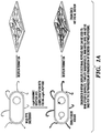
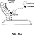
- Described herein is a technology which permits the programming of an E. coli biofilm's functional properties by genetically appending functional peptide domains to the CsgA protein.
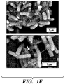
- the amyloid nanofiber network displays the peptide in very high density on its surface.
- the biofilm's function is then augmented according to the sequence of the displayed peptides. It is demonstrated herein that functional peptide domains of various lengths and secondary structures can be appended to CsgA without precluding the formation of curli fibers. Lastly, it is demonstrated that the peptide domains maintain their function in the context of the biofilm after secretion and assembly.
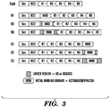
- composition comprising a first engineered polypeptide and a second engineered polypeptide, wherein the first engineered polypeptide comprises a CsgA polypeptide and a conjugation domain fused to the CsgA polypeptide by a linker sequence; wherein the amino acid sequence of the CsgA polypeptide is at least 80% homologous to the amino acid sequence set forth in SEQ ID NO: 1; wherein the conjugation domain is selected from the group consisting of SpyTag; biotin acceptor peptide (BAP); biotin carboxyl carrier protein (BCCP); and a peptide comprising a LPXTG motif; wherein the linker sequence is located N-terminal to the conjugation domain; and wherein the linker sequence comprises at least 6 amino acids; and wherein the second engineered polypeptide comprises a functionalizing polypeptide and a partner conjugation domain fused to the functionalizing polypeptide; wherein the partner conjugation domain is selected from the group consisting of SpyCat
- described herein is a bacterial cell comprising the above composition.
- described herein is a biofilm comprising the above composition or the bacterial cell comprising the above composition.
- described herein is a method of producing a biofilm comprising culturing the bacterial cells described herein under conditions suitable for the production of a biofilm.
- compositions as defined above comprising filaments comprising the engineered polypeptide.
- the composition further comprises the bacterial cell comprising the above composition as described herein.
- described herein is the use of the bacterial cell, the above composition, or the biofilm as described herein, in an in vitro method selected from the group consisting of biocatalysis; industrial biocatalysis; immobilized biocatalysis; chemical production; filtration; isolation of molecules from an aqueous solution; water filtration; bioremediation; nanoparticle synthesis; nanowire synthesis; display of optically active materials; biosensors; surface coating; therapeutic biomaterial; biological scaffold; structural reinforcement of an object; and as a delivery system for therapeutic agents.
- Embodiments of the technology described herein relate to the inventors' discovery of how to functionalize bacterial biofilms. More specifically, the inventors have discovered CsgA, the major component of E. coli biofilms, can be engineered to comprise polypeptides having a function or activity which provide the biofilm comprising the engineered CsgA with a new property or activity.
- CsgA polypeptide comprising a CsgA polypeptide with a C-terminal display tag flanking the CsgA polypeptide.
- the display tag comprises an activity polypeptide and a linker sequence, wherein the linker sequence is located N-terminal to the display polypeptide and wherein the linker sequence comprises at least 6 amino acids.
- CsgA refers to the major structural subunit of curli. The sequences of CsgA and its homologs are known in a number of species, e.g. the sequence of E.
- CsgA refers to E. coli CsgA.
- CsgA refers to a polypeptide having at least 80% homology to SEQ ID NO: 1 (e.g. 80% or greater homology, 90% or greater homology, or 95% or greater homology), e.g. naturally occurring mutations or variants of CsgA, homologs of CsgA, or engineered mutatations or variants of CsgA.
- an "engineered CsgA polypeptide” refers to a polypeptide comprising a C-terminal display tag flanking the CsgA polypeptide, e.g. a polypeptide display tag located on the c-terminus of a CsgA polypeptide.
- the display tag is located on the C-terminus of a CsgA polypeptide of SEQ ID NO: 1.
- the display tag flanks the CsgA polypeptide, i.e., the entirety of the CsgA polypeptide is located on the N-terminus of the display tag, e.g. the display tag does not interrupt the sequence of the CsgA polypeptide.
- a "display tag” is a polypeptide engineered to be located at the C-terminus of a polypeptide comprising a CsgA polypeptide.
- a display tag comprises no more than 100 amino acids.
- a display tag comprises no more than 50 amino acids.
- a display tag comprises no more than 40 amino acids.
- a display tag comprises no more than 30 amino acids.
- a display tag as described herein comprises, from N-terminus to C-terminus, a linker sequence and an activity polypeptide.
- a linker sequence is a polypeptide sequence of at least 6 amino acids. In some embodiments, the linker sequence comprises from about 6 amino acids to about 50 amino acids. In some embodiments, the linker sequence comprises from about 6 amino acids to about 100 amino acids. In some embodiments, the linker sequence comprises from about 30 amino acids to about 100 amino acids. In some embodiments, the linker sequence comprises from about 40 amino acids to about 100 amino acids. In some embodiments, the linker sequence comprises from about 50 amino acids to about 100 amino acids. In some embodiments, the linker sequence comprises from about 6 amino acids to about 30 amino acids.
- the linker sequence comprises from about 20 amino acids to about 50 amino acids. In some embodiments, the linker sequence comprises from about 30 amino acids to about 50 amino acids. In some embodiments, the linker sequence comprises from about 40 amino acids to about 50 amino acids. In some embodiments, the linker sequence comprises from about 6 amino acids to about 20 amino acids. In some embodiments, the linker sequence comprises from about 6 to about 10 amino acids. In some embodiments, the linker sequence comprises a flexible polypeptide, e.g a polypeptide not having a rigid secondary and/or tertiary structure. In some emboidments, the linker sequence comprises glycine and serine residues.
- At least 50% of the amino acids comprised by the linker sequence are glycine or serine residues, e.g. at least 50%, at least 60%, at least 70%, at least 80%, at least 90%, at least 95%, or more are glycine or serine residues.
- the linker sequence consists of glycine and serine residues.
- an “activity polypeptide” refers to a polypeptide having an activity or function, such that when it is present in a biofilm, it confers upon the biofilm a property, function, or activity which it did not have in the absence of the activity of the polypeptide.
- an activity polypeptide can be, e.g. an enzyme, a polypeptide that binds another molecule, a binding domain, a peptide that is bound by another molecule (e.g. a ligand or epitope), or the like.
- polypeptides for use as activity polypeptides include, but are not limited to Metal binding domain (MBD); SpyTag; graphene binding (GBP); carbon nanotube binding (CBP); gold binding (A3); CT43; FLAG; Z8; E14; QBP1; CLP12; and AFP8.
- MBD Metal binding domain
- GBP graphene binding
- CBP carbon nanotube binding
- Au3 gold binding
- CT43 CT43
- FLAG FLAG
- Z8 E14
- QBP1 QBP1
- CLP12 CLP12
- AFP8 AFP8.
- the activity polypeptide when present as part of an engineered CsgA polypeptide, is functional.
- a polypeptide is said to be “functional” or expressed as a “functional” polypeptide if the polypeptide retains at least about 50% of the activity (e.g. enzymatic activity or binding activity) that it has as an isolated polypeptide.
- activity e.g. enzymatic activity or binding activity
- One of skill in the art can readily detect increases in reaction products and/or detect decreases in reaction substrates, e.g.
- a functional activity polypeptide can retain at least 50% of the activity of the isolated polypeptide, e.g. 50% or more of the activity, 60% or more of the activity, 75% or more of the activity, or 90% or more of the activity of the isolated polypeptide.
- the activity polypeptide can be a conjugation domain.
- Such embodiments can permit immobilization of target proteins in the biofilm, e.g., when the target protein is too large to be expressed as a fusion with CsgA.
- the conjugation domain present on the engineered CsgA polypeptide can specifically bind to a partner conjugation domain present as part of the target protein, thereby incorporating the target protein into the biofilm.
- conjugation domain refers to a polypeptide that can specifically bind to and/or be specifically bound by a partner conjugation domain, e.g. under conditions suitable for growth of a biofilm.
- a conjugation domain can be, e.g., about 100 amino acids or less in size, about 75 amino acids or less in size, about 50 amino acids or less in size, about 40 amino acids or less in size or smaller.
- a partner conjugation domain can be about the same size as the conjugation domain or larger, e.g., a partner conjugation domain can be about 4000 amino acids or less in size, about 3000 amino acids or less in size, about 2000 amino acids or less in size, about 1000 amino acids or less in size, about 500 amino acids or less in size, about 200 amino acids or less in size, about 100 amino acids or less in size, about 75 amino acids or less in size, about 50 amino acids or less in size, about 40 amino acids or less in size, or smaller.
- the binding of the conjugation domain and partner conjugation domain is covalent.
- conjugation domains are known in the art and include, but are not limited to, SpyTag; biotin acceptor peptide (BAP); biotin carboxyl carrier protein (BCCP); and a peptide comprising a LPXTG motif.
- partner conjugation domains are known in the art and include but are not limited to, respectively, SpyCatcher, streptavidin; streptavidin; and peptides comprising aminoglycine. Further discussion of conjugation systems comprising a conjugation domain and a partner conjugation domain can be found, e.g., in Mao et al.
- an engineered CsgA polypeptide comprising conjugation domain has the sequence of SEQ ID NO: 3 or is encoded by a polynucleotide having the sequence of SEQ ID NO: 2.
- the target polypeptide comprising the partner conjugation domain can further comprise a "functionalizing polypeptide.”
- a “functionalizing polypeptide” refers to a polypeptide having an activity or function, such that when it is present in a biofilm, it confers upon the biofilm a property, function, or activity which it did not have in the absence of the polypeptide.
- a functionalizing polypeptide can be of any size and is not part of the engineered CsgA polypeptide.
- Exemplary functionalizing polypeptide can include, e.g. an enzyme, a polypeptide that binds another molecule, an antibody or the like.
- a polypeptide comprising a functionalizing polypeptide and a conjugation domain can further comprise an extracellular localization tag, e.g. a sequence which will cause a cell expressing the polypeptide to secrete the polypeptide.
- a functionalized engineered CsgA polypeptide or functionalized biofilm can be provided by contacting an engineered CsgA polypeptide comprising a conjugation domain (or a cell and/or biofilm comprising that polypeptide) with a polypeptide comprising the partner conjugation domain.
- the engineered CsgA polypeptide and the polypeptide comprising the partner conjugation domain are maintained in contact for a period of time, i.e. the "binding step.”
- the binding step is followed by a washing step, e.g. to remove excess unbound polypeptide.
- an engineered CsgA polypeptide comprising a conjugation domain is bound to (or binds) the partner conjugation domain in the presence of albumin (i.e. the "binding step").
- the albumin is BSA.
- the albumin is present at about 0.1% to about 10%.
- the albumin is present at about 0.5% to about 5%.
- the albumin is present at about 1% to about 2%.
- the binding step is allowed to proceed for at least about 2 hours, e.g. about 2 hours or more, about 6 hours or more, about 12 hours or more, or about 24 hours or more. In some embodiments, the binding step is allowed to proceed in the presence of albumin.
- the washing step proceeds for about 10 minutes to about 6 hours. In some embodiments, the washing step proceeds for about 30 minutes to about 3 hours. In some embodiments, the washing step proceeds for about 90 minutes.
- the polypeptides are agitated (e.g. shaken) during the washing step.
- the washing step comprises washing the polypeptides in a solution of albumin.
- the albumin is BSA. In some embodiments, the albumin is present at about 0.01% to about 3%. In some embodiments, the albumin is present at about 0.1% to about 1%. In some embodiments, the albumin is present at about 0.3%. In some embodiments, the washing step comprises 2 or more successive washes. In some embodiments, the washing step comprises 3 successive washes.
- specific binding refers to a chemical interaction between two molecules, compounds, cells and/or particles wherein the first entity binds to the second, target entity with greater specificity and affinity than it binds to a third entity which is a non-target.
- specific binding can refer to an affinity of the first entity for the second target entity which is at least 10 times, at least 50 times, at least 100 times, at least 500 times, at least 1000 times or greater than the affinity for the third nontarget entity.
- a reagent specific for a given target is one that exhibits specific binding for that target under the conditions of the assay being utilized.
- a vector comprising a nucleic acid sequence encoding an engineered CsgA polypeptide as described herein.
- the term "vector”, as used herein, refers to a nucleic acid construct designed for delivery to a host cell or transfer between different host cells.
- a vector can be viral or non-viral.
- Many vectors useful for transferring genes into target cells are available, e.g. the vectors may be episomal, e.g., plasmids, virus derived vectors or may be integrated into the target cell genome, through homologous recombination or random integration.
- a vector can be an expression vector.
- expression vector refers to a vector that has the ability to incorporate and express heterologous nucleic acid fragments in a cell.
- An expression vector may comprise additional elements, for example, the expression vector may have two replication systems, thus allowing it to be maintained in two organisms.
- the nucleic acid incorporated into the vector can be operatively linked to an expression control sequence when the expression control sequence controls and regulates the transcription and translation of that polynucleotide sequence.
- a nucleic acid encoding an engineered CsgA polypeptide can be present within a portion of a plasmid.
- Plasmid vectors can include, but are not limited to, pBR322, pBR325, pACYC177, pACYC184, pUC8, pUC9, pUC18, pUC19, pLG339, pR290, pKC37, pKC101, SV 40, pBluescript II SK +/- or KS +/- (see " Stratagene Cloning Systems” Catalog (1993) from Stratagene, La Jolla, Calif ), pQE, pIH821, pGEX, pET series (see Studier et. al., "Use of T7 RNA Polymerase to Direct Expression of Cloned Genes," Gene Expression Technology, vol. 185 (1990 ).
- viral vector refers to a nucleic acid vector construct that includes at least one element of viral origin and has the capacity to be packaged into a viral vector particle.
- the viral vector can contain a transgenic gene in place of non-essential viral genes.
- the vector and/or particle may be utilized for the purpose of transferring any nucleic acids into cells either in vitro or in vivo.
- Numerous viral vectors are known in the art and can be used as carriers of a nucleic acid into a cell, e.g. lambda vector system gt11, gt WES.tB, Charon 4.
- the nucleic acid encoding an engineered CsgA polypeptide can be constitutively expressed. In some embodiments, the nucleic acid encoding an engineered CsgA polypeptide can be operably linked to a constitutive promoter. In some embodiments, the nucleic acid encoding an engineered CsgA polypeptide can be inducibly expressed. In some embodiments, the nucleic acid encoding an engineered CsgA polypeptide can be operably linked to an inducible promoter. In some embodiments, the nucleic acid encoding an engineered CsgA polypeptide can be operably linked to a native CsgA promoter.
- an “inducible promoter” is one that is characterized by initiating or enhancing transcriptional activity when in the presence of, influenced by, or contacted by an inducer or inducing agent than when not in the presence of, under the influence of, or in contact with the inducer or inducing agent.
- An “inducer” or “inducing agent” may be endogenous, or a normally exogenous compound or protein that is administered in such a way as to be active in inducing transcriptional activity from the inducible promoter.
- the inducer or inducing agent e.g., a chemical, a compound or a protein
- can itself be the result of transcription or expression of a nucleic acid sequence e.g., an inducer can be a transcriptional repressor protein
- an inducer can be a transcriptional repressor protein
- inducible promoters include but are not limited to, the lac operon promoter, a nitrogen-sensitive promoter, an IPTG-inducible promoter, a salt-inducible promoter, and tetracycline, steroid-responsive promoters, rapamycin responsive promoters and the like.
- Inducible promoters for use in prokaryotic systems are well known in the art, see, e.g. the beta.-lactamase and lactose promoter systems ( Chang et al., Nature, 275: 615 (1978 ); Goeddel et al., Nature, 281: 544 (1979 )), the arabinose promoter system, including the araBAD promoter ( Guzman et al., J . Bacteriol., 174: 7716-7728 (1992 ); Guzman et al., J. Bacteriol., 177: 4121-4130 (1995 ); Siegele and Hu, Proc. Natl. Acad. Sci.
- An inducible promoter useful in the methods and systems as disclosed herein can be induced by one or more physiological conditions, such as changes in pH, temperature, radiation, osmotic pressure, saline gradients, cell surface binding, and the concentration of one or more extrinsic or intrinsic inducing agents.
- the extrinsic inducer or inducing agent may comprise amino acids and amino acid analogs, saccharides and polysaccharides, nucleic acids, protein transcriptional activators and repressors, cytokines, toxins, petroleum-based compounds, metal containing compounds, salts, ions, enzyme substrate analogs, hormones, and combinations thereof.
- the inducible promoter is activated or repressed in response to a change of an environmental condition, such as the change in concentration of a chemical, metal, temperature, radiation, nutrient or change in pH.
- an inducible promoter useful in the methods and systems as disclosed herein can be a phage inducible promoter, nutrient inducible promoter, temperature inducible promoter, radiation inducible promoter, metal inducible promoter, hormone inducible promoter, steroid inducible promoter, and/or hybrids and combinations thereof.
- Appropriate environmental inducers can include, but are not limited to, exposure to heat (i.e., thermal pulses or constant heat exposure), various steroidal compounds, divalent cations (including Cu2+ and Zn2+), galactose, tetracycline, IPTG (isopropyl- ⁇ -D thiogalactoside), as well as other naturally occurring and synthetic inducing agents and gratuitous inducers.
- Inducible promoters useful in the methods and systems as disclosed herein also include those that are repressed by "transcriptional repressors” that are subject to inactivation by the action of environmental, external agents, or the product of another gene. Such inducible promoters may also be termed “repressible promoters” where it is required to distinguish between other types of promoters in a given module or component of the biological switch converters described herein. Preferred repressors for use in the present invention are sensitive to inactivation by physiologically benign agent.
- a lac repressor protein is used to control the expression of a promoter sequence that has been engineered to contain a lacO operator sequence
- treatment of the host cell with IPTG will cause the dissociation of the lac repressor from the engineered promoter containing a lacO operator sequence and allow transcription to occur.
- a tet repressor is used to control the expression of a promoter sequence that has been engineered to contain a tetO operator sequence
- treatment of the host cell with tetracycline will cause the dissociation of the tet repressor from the engineered promoter and allow transcription of the sequence downstream of the engineered promoter to occur.
- an engineered microbial cell comprising an engineered CsgA polypeptide and/or comprising a vector or nucleic acid encoding such a polypeptide.
- described herein is a population of cells comprising two cell types, the first cell type encoding and/or comprising an engineered CsgA polypeptide comprising an activity polypeptide comprising a conjugation domain and the second cell type encoding and/or comprising a second engineered polypeptide comprising a partner conjugation domain and a functionalizing polypeptide.
- a single cell can comprise a CsgA polypeptide with a conjugation domain and also comprise the polypeptide which will bind to and/or be bound by that CsgA polypeptide or that a first cell can comprise a CsgA polypeptide with a conjugation domain and a second cell can comprise the polypeptide which will bind to and/or be bound by that CsgA polypeptide.
- an engineered CsgA polypeptide with a conjugation domain can be contacted with a second polypeptide comprising a partner conjugation domain and a functionalizing polypeptide, e.g. the second polypeptide can be produced (e.g.
- a bacterial cell of the methods and compositions described herein can be any of any species.
- the bacterial cells are of a species and/or strain which is amenable to culture and genetic manipulation.
- the bacterial cell can be a gram-positive bacterial cell.
- the bacterial cell can be a gram-negative bacterial cell.
- the parental strain of the bacterial cell of the technology described herein can be a strain optimized for protein expression.
- Non-limiting examples of bacterial species and strains suitable for use in the present technologies include Escherichia coli, E. coli BL21, E. coli Tuner, E. coli Rosetta, E. coli JM101, and derivatives of any of the foregoing.
- Bacterial strains for protein expression are commercially available, e.g. EXPRESSTM Competent E. coli (Cat. No. C2523; New England Biosciences; Ipswich, MA).
- the cell is an E. coli cell.
- the nucleic acid encoding an engineered CsgA polypeptide is comprised by a cell expressing wild-type CsgA. In some embodiments, the nucleic acid encoding an engineered CsgA polypeptide is comprised by a cell with a mutation and/or deletion of the wild-type CsgA gene, e.g. such that the cell does not express wild-type CsgA. In some embodiments, the nucleic acid encoding an engineered CsgA polypeptide is introduced into a cell by homolgous recombination, e.g. such that the nucleic acid encoding an engineered CsgA polypeptide replaces the wild-type CsgA gene in the cell.
- a biofilm comprising an engineered microbial cell comprising one or more engineered CsgA polypeptide and/or comprising a vector or nucleic acid encoding such a polypeptides.
- a biofilm refers to a mass of microorganisms which can adhere or is adhering to a surface.
- a biofilm comprises a matrix of extracellular polymeric substances, including, but not limited to extracellular DNA, proteins, glyopeptides, and polysaccharides.
- the nature of a biofilm such as its structure and composition, can depend on the particular species of bacteria present in the biofilm. Bacteria present in a biofilm are commonly genetically or phenotypically different than corresponding bacteria not in a biofilm, such as isolated bacteria or bacteria in a colony.
- the technology described herein relates to a biofilm that is produced by culturing an engineered microbial cell comprising an engineered CsgA polypeptide (and/or comprising a vector or nucleic acid encoding such a polypeptide) under conditions suitable for the production of a biofilm.
- Conditions suitable for the production of a biofilm can include, but are not limited to, conditions under which the microbial cell is capable of logarithmic growth and/or polypeptide synthesis. Conditions may vary depending upon the species and strain of microbial cell selected. Conditions for the culture of microbal cells are well known in the art. Biofilm production can also be induced and/or enhanced by methods well known in the art, e.g.
- conditions suitable for the production of a biofilm can also include conditions which increase the expression and secretion of CsgA, e.g. by exogenously expressing CsgD.
- the biofilm can comprise the cell which produced the biofilm.
- CsgA units When expressed by a cell capable of forming curli, e.g. a cell expressing CsgA, CsgB, CsgC, CsgD, CsgE, CsgF, and CsgG or some subset thereof, CsgA units will be assembled to form curli filaments, e.g. polymeric chains of CsgA.
- filaments of the polypeptide can be present in the composition.
- the filaments can be part of a proteinaceous network, e.g. multiple filaments which can be, e.g. interwoven, overlapping, and/or in contact with each other.
- the proteinaceous network can comprise additional biofilm components, e.g. materials typically found in an E.
- biofilm components can include biofilm proteins (e.g. FimA, FimH, Ag43, AidA, and/or TibA) and/or non-proteinaceous biofilm components (e.g. cellulose, PGA and/or colonic acid).
- the composition can further comprise an engineered microbial cell comprising an engineered CsgA polypeptide and/or comprising a vector or nucleic acid encoding such a polypeptide.
- a cell, composition, or biofilm comprising an engineered CsgA polypeptide (and/or comprising a vector or nucleic acid encoding such a polypeptide) to display a polypeptide, e.g. within the biofilm, within the composition, and/or on the cell surface.
- display refers to expressing the polypeptide (e.g. as an activity polypeptide) in such a manner that it can come in contact with the extracellular environment.
- a displayed polypeptide can be capable of binding with a binding partner, catalyzing an enzymatic reaction, and/or performing any other activity which it would perform as an isolated polypeptide.
- a polypeptide displayed within a biofilm will retain more activity than a soluble version of that polypeptide. It is contemplated herein that a polypeptide displayed within a biofilm (e.g. an activity polypeptide and/or functionalizing polypeptide) will retain more activity than a soluble version of that polypeptide when exposed to activity degrading conditions such as, e.g., high or low pH, organic solvents, dessication, high or low temperature, radiation, etc.
- a cell, composition, or biofilm comprising an engineered CsgA polypeptide (and/or comprising a vector or nucleic acid encoding such a polypeptide), in an application selected from the group consisting of biocatalysis; industrial biocatalysis; immobilized biocatalysis; chemical production; filtration; isolation of molecules from an aqueous solution; water filtration; bioremediation; nanoparticle synthesis; nanowire synthesis; display of optically active materials; biosensors; surface coating; therapeutic biomaterial; biological scaffold; structural reinforcement of an object; and as a delivery system for therapeutic agents.
- an application selected from the group consisting of biocatalysis; industrial biocatalysis; immobilized biocatalysis; chemical production; filtration; isolation of molecules from an aqueous solution; water filtration; bioremediation; nanoparticle synthesis; nanowire synthesis; display of optically active materials; biosensors; surface coating; therapeutic biomaterial; biological scaffold; structural reinforcement of an object; and as a delivery system for therapeutic agents.
- a cell, composition and/or biofilm can comprise multiple different engineered CsgA polypeptides, each of which comprises a different activity polypeptide, e.g. an engineered CsgA polypeptide comprising an enzymatic activity polypeptide and an engineered CsgA polypeptide comprising a binding domain activity polypeptide.
- a cell, composition, and/or biofilm can comprise 1 or more engineered CsgA polypeptides, e.g. 1, 2, 3, 4, 5, 6, or more engineered CsgA polypeptides.
- Biofilms are attractive as catalysts for a variety of biochemical transformations due to the their ability to withstand harsh conditions, their propensity for surface attachment and their scalability.
- biotransformations are catalyzed either by enzymes inside living cells or by enzymes displayed on the cell surface. Both approaches have inherent limitations related to mass transfer and solubility of substrates, in the case of whole cells, or related to the limited surface area available on the cell membrane, in the case of surface display. Described herein is the development of technique called Biofilm Integrated Nanofiber Display (BIND) that enables the rational design of the biofilm extracellular matrix so that it can function as a substrate for site-specific covalent surface immobilization of enzymes.
- BIND Biofilm Integrated Nanofiber Display
- the BIND platform provides a way to create living surface coating materials that can be programmed to exhibit a range of protective functions for surfaces on which the engineered biofilms are immobilized: 1) strong adhesion to the surface, 2) the ability to secrete soluble entities into the local environment that would prevent degradation or fouling, such as microbicides, reductants, etc., 3) the ability to secrete biopolymeric material to fill in cracks forming in the underlying substrate, or 4) the ability to template the growth of a mineral or other ordered material from a exogenously supplied building blocks.
- Enzymatically-derived 13PDO can potentially be produced at a much lower cost than whole-cell fermentation processes, which require large amounts of media for every cycle and often produce metabolic side products, resulting in a complex mixture that must be processed to isolate the desired product.
- the BIND platform technology will reduce the cost barriers typically associated with immobilized enzyme biocatalysis, by eliminating the multi-step purification and immobilization with a single culturing step, and replacing costly synthetic scaffolds with an ultra-stable self-produced scaffold.
- the metabolic production of 13PDO by microorganisms from glycerol proceeds from 2 enzymatic step.
- the first step is the enzymatic dehydration of glycerol by Glycerol dehydratase (Gdh) to produce 3-hydroxypropionaldehyde (3HPA).
- Gdh Glycerol dehydratase
- 3HPA 3-hydroxypropionaldehyde
- Pdh 1,3-propanediol dehydrogenase
- Both Gdh and Pdh have been studied extensively, with the best studied from Klebsiella pneumonia, a 1,3-PDO producing microbe.
- the DuPont process uses K. pneumonia genes cloned into E. coli for their industrial-scale fermentation process.
- a functionalizing polypeptide can comprise a glycerol dehydratase. In some embodiments, a functionalizing polypeptide can comprise a K . pneumonia glycerol dehydratase. In some embodiments, a functionalizing polypeptide can comprise a propanediol dehydrogenase. In some embodiments, a functionalizing polypeptide can comprise a K . pneumonia propanediol dehydrogenase.
- Biorecovery of Valuable Metals or Removal of Metal Pollutants using BIND Metal removal and recovery are highly relevant for a number of industries, including mining, recycling, and water treatment.
- the BIND platform provides a method to display engineered binding proteins with high affinity and selectivity for specific metal ions and particles.
- the BIND platform represents a significant advancement over other biosorption techniques because it is able to display full-length proteins in high density and in a scalable manner.
- the use of recombinant proteins for these applications would be cost-prohibitive in most cases because of the production and purification protocols necessary to generate them.
- Contemplated herein are biofilm-based materials that can serve as a separations medium for metal ions and particles.
- biofilm-based materials that are suitable for use inside the body.
- the BIND platform provides a means to program the adhesion of the biofilm-based material to biological tissues. Such materials would be able to control the residence time and localization of the biofilms inside the body. These materials would have the capability of establishing themselves at a specified location, altering the properties of the biological tissue through direct interaction of the curli nanofibers with the tissue, and secreting soluble biomolecules to alter local biological processes.
- the terms “increased”, “increase”, “enhance”, or “activate” are all used herein to mean an increase by a statically significant amount.
- the terms “increased”, “increase”, “enhance”, or “activate” can mean an increase of at least 10% as compared to a reference level, for example an increase of at least about 20%, or at least about 30%, or at least about 40%, or at least about 50%, or at least about 60%, or at least about 70%, or at least about 80%, or at least about 90% or up to and including a 100% increase or any increase between 10-100% as compared to a reference level, or at least about a 2-fold, or at least about a 3-fold, or at least about a 4-fold, or at least about a 5-fold or at least about a 10-fold increase, or any increase between 2-fold and 10-fold or greater as compared to a reference level.
- protein and “polypeptide” are used interchangeably herein to designate a series of amino acid residues, connected to each other by peptide bonds between the alpha-amino and carboxy groups of adjacent residues.
- protein and “polypeptide” refer to a polymer of amino acids, including modified amino acids (e.g., phosphorylated, glycated, glycosylated, etc.) and amino acid analogs, regardless of its size or function.
- modified amino acids e.g., phosphorylated, glycated, glycosylated, etc.
- amino acid analogs regardless of its size or function.
- polypeptide proteins and “polypeptide” are used interchangeably herein when referring to a gene product and fragments thereof.
- exemplary polypeptides or proteins include gene products, naturally occurring proteins, homologs, orthologs, paralogs, fragments and other equivalents, variants, fragments, and analogs of the foregoing.
- a “variant,” as referred to herein, is a polypeptide substantially homologous to a native or reference polypeptide, but which has an amino acid sequence different from that of the native or reference polypeptide because of one or a plurality of deletions, insertions or substitutions.
- Polypeptide-encoding DNA sequences encompass sequences that comprise one or more additions, deletions, or substitutions of nucleotides when compared to a native or reference DNA sequence, but that encode a variant protein or fragment thereof that retains the relevant biological activity relative to the reference protein.
- amino acid sequences one of skill will recognize that individual substitutions, deletions or additions to a nucleic acid, peptide, polypeptide, or protein sequence which alters a single amino acid or a small percentage, (i.e. 5% or fewer, e.g. 4% or fewer, or 3% or fewer, or 1% or fewer) of amino acids in the encoded sequence is a "conservatively modified variant" where the alteration results in the substitution of an amino acid with a chemically similar amino acid. It is contemplated that some changes can potentially improve the relevant activity, such that a variant, whether conservative or not, has more than 100% of the activity of a wildtype or native polypeptide, e.g. 110%, 125%, 150%, 175%, 200%, 500%, 1000% or more.
- One method of identifying amino acid residues which can be substituted is to align, for example, CsgA from E. coli to a CsgA polypeptide from other species. Alignment can provide guidance regarding not only residues likely to be necessary for function but also, conversely, those residues likely to tolerate change. Where, for example, an alignment shows two identical or similar amino acids at corresponding positions, it is more likely that that site is important functionally. Where, conversely, alignment shows residues in corresponding positions to differ significantly in size, charge, hydrophobicity, etc., it is more likely that that site can tolerate variation in a functional polypeptide. Such alignments are readily created by one of ordinary skill in the art, e.g.
- homologs of any given polypeptide or nucleic acid sequence can be found using BLAST programs, e.g. by searching freely available databases of sequence for homologous sequences, or by querying those databases for annotations indicating a homolog (e.g. search strings that comprise a gene name or describe the activity of a gene). Such databases can be found, e.g. on the world wide web at http://ncbi.nlm.nih.gov/.
- the variant amino acid or DNA sequence can be at least 90%, at least 95%, at least 96%, at least 97%, at least 98%, at least 99%, or more, identical to a native or reference sequence.
- the degree of homology (percent identity) between a native and a mutant sequence can be determined, for example, by comparing the two sequences using freely available computer programs commonly employed for this purpose on the world wide web.
- the variant amino acid or DNA sequence can be at least 90%, at least 91%, at least 92%, at least 93%, at least 94%, at least 95%, at least 96%, at least 97%, at least 98%, at least 99%, or more, similar to the sequence from which it is derived (referred to herein as an "original" sequence).
- the degree of similarity (percent similarity) between an original and a mutant sequence can be determined, for example, by using a similarity matrix.
- Similarity matrices are well known in the art and a number of tools for comparing two sequences using similarity matrices are freely available online, e.g. BLASTp (available on the world wide web at http://blast.ncbi.nlm.nih.gov), with default parameters set.
- the variant is a conservative substitution variant.
- Variants can be obtained by mutations of native nucleotide sequences, for example.
- a "variant,” as referred to herein, is a polypeptide substantially homologous to a native or reference polypeptide, but which has an amino acid sequence different from that of the native or reference polypeptide because of one or a plurality of deletions, insertions or substitutions.
- Polypeptide-encoding DNA sequences encompass sequences that comprise one or more additions, deletions, or substitutions of nucleotides when compared to a native or reference DNA sequence, but that encode a variant protein or fragment thereof that retains the relevant biological activity relative to the reference protei n .
- amino acid sequences one of skill will recognize that individual substitutions, deletions or additions to a nucleic acid, peptide, polypeptide, or protein sequence which alters a single amino acid or a small percentage, (i.e. 5% or fewer, e.g. 4% or fewer, or 3% or fewer, or 1% or fewer) of amino acids in the encoded sequence is a "conservatively modified variant" where the alteration results in the substitution of an amino acid with a chemically similar amino acid. It is contemplated that some changes can potentially improve the relevant activity, such that a variant, whether conservative or note, has more than 100% of the activity of the wildtype enzyme , e.g. 110%, 125%, 150%, 175%, 200%, 500%, 1000% or more.
- the degree of similarity (percent similarity) between an original and a mutant sequence can be determined, for example, by using a similarity matrix.
- Similarity matrices are well known in the art and a number of tools for comparing two sequences using similarity matrices are freely available online, e.g. BLASTp (available on the world wide web at http://blast.ncbi.nlm.nih.gov), with default parameters set.
- a given amino acid can be replaced by a residue having similar physiochemical characteristics, e.g., substituting one aliphatic residue for another (such as Ile, Val, Leu, or Ala for one another), or substitution of one polar residue for another (such as between Lys and Arg; Glu and Asp; or Gln and Asn).
- Other such conservative substitutions e.g., substitutions of entire regions having similar hydrophobicity characteristics, are well known.
- Polypeptides comprising conservative amino acid substitutions can be tested in any one of the assays described herein to confirm that a desired apoptotic activity of a native or reference polypeptide is retained.
- Conservative substitution tables providing functionally similar amino acids are well known in the art.
- conservatively modified variants are in addition to and do not exclude polymorphic variants, interspecies homologs, and alleles consistent with the disclosure.
- conservative substitutions for one another include: 1) Alanine (A), Glycine (G); 2) Aspartic acid (D), Glutamic acid (E); 3) Asparagine (N), Glutamine (Q); 4) Arginine (R), Lysine (K); 5) Isoleucine (I), Leucine (L), Methionine (M), Valine (V); 6) Phenylalanine (F), Tyrosine (Y), Tryptophan (W); 7) Serine (S), Threonine (T); and 8) Cysteine (C), Methionine (M) (see, e.g., Creighton, Proteins (1984 )).
- cysteine residues not involved in maintaining the proper conformation of the polypeptide also can be substituted, generally with serine, to improve the oxidative stability of the molecule and prevent aberrant crosslinking.
- cysteine bond(s) can be added to the polypeptide to improve its stability or facilitate oligomerization.
- nucleic acid or “nucleic acid sequence” refers to any molecule, preferably a polymeric molecule, incorporating units of ribonucleic acid, deoxyribonucleic acid or an analog thereof.
- the nucleic acid can be either single-stranded or double-stranded.
- a single-stranded nucleic acid can be one nucleic acid strand of a denatured double- stranded DNA. Alternatively, it can be a single-stranded nucleic acid not derived from any double-stranded DNA.
- the nucleic acid can be DNA.
- nucleic acid can be RNA.
- Suitable nucleic acid molecules are DNA, including genomic DNA or cDNA. Other suitable nucleic acid molecules are RNA, including mRNA.
- statically significant or “significantly” refers to statistical significance and generally means a two standard deviation (2SD) or greater difference.
- compositions, methods, and respective component(s) thereof are used in reference to compositions, methods, and respective component(s) thereof, that are essential to the method or composition, yet open to the inclusion of unspecified elements, whether essential or not.
- compositions, methods, and respective components thereof as described herein, which are exclusive of any element not recited in that description of the embodiment.
- the term "consisting essentially of' refers to those elements required for a given embodiment. The term permits the presence of elements that do not materially affect the basic and novel or functional characteristic(s) of that embodiment.
- Described herein is a method to genetically modify the major proteinaceous component of bacterial biofilms to display functional peptides, thus reprogramming biofilms for a wide variety of beneficial applications including but not limited to industrial biocatalysis, bioremediation, bioenergy, materials templating, and biosensing.
- This technology is based on the curli system of E. coli, which consists of cell-surface anchored amyloid fibrils that are made from the self-assembly of a secreted protein ( Figs. 1A-1F ).
- the technology described herein demonstrates that functional peptide domains can be appended to the secreted protein so that, once it is assembled, the amyloid fibril network exhibits augmented functionality ( e . g ., binding to various biological and chemical entities).
- this extracellular matrix-peptide display technology can be adapted for the immobilization of any protein.
- Fusion domains can be displayed on Curli nanofibers.
- biofilms as an extracellular matrix material to colonize various surfaces and protect themselves from environmental stresses.
- One of the major structural components of these biofilms is nanoscale fibers composed of proteins.
- the bacteria secrete these proteins into the extracellular milieu, where the protein monomers spontaneously self-assemble into a polymeric chain which is anchored on the cell surface.
- These protein nanofibers are known to exhibit amyloidogenic structural characteristics and are extremely robust. They have been shown to impart a number of evolutionary advantages, including mediating adhesion to surfaces, invasion of host cells, and sequestering toxic metals. 1-3
- Most current biofilm research is focused on inhibiting or dispersing biofilms deleterious to human health.
- biofilms as an adhesion and persistence mechanism on almost any surface poses a great risk for infection in the biomedical and food industries 4 .
- Described herein is a platform technology which utilizes biofilms for beneficial applications. It is demonstrated herein that functional peptides can be engineered into biofilms by the successful expression and secretion of genetic fusions of fimbriae-forming nanofibers such as curli.
- Key aspects of the technology described herein can include, but are not limited to: the ability to genetically program various physical, chemical, and biochemical functionalities into the protein structural component of a biofilm; a biofilm composed of curli nanofibers or any such similar extracellular self-assembling protein-based amyloid fibers; an engineered unit of the biofilm nanofiber composed of a self-assembling protein domain containing one or more curli unit or self-assembling domains, a spacer domain of variable length, and a peptide "activity" domain of arbitrary length on the C-terminus of the protein; the N-terminus of the protein can optionally additionally comprise various domains that allow periplasmic localization and/or protein secretion into the extracellular space; the peptide activity domain can be any peptide that allows for substrate adhesion, the binding to any biomolecule or chemical, has self-contained catalytic activity, is involved in catalytic activity in coordination with an externally localized protein, can template inorganic structures, can induce physiological responses in cells, can bind to ions in
- proteins can be immobilized on the engineered curli biofilm by expressing on the curli nanofibers a peptide tag that specifically interacts with a protein domain to form a covalent or non-covalent complex. Any target protein fused to this interacting protein domain can thus be displayed on the biofilms by exposing the engineered curli-peptide tag biofilms to the fusion protein.
- This fusion protein can be expressed in cis or trans and used in various states of purity.
- the curli system of Escherichia coli is composed of small protein monomers, CsgA, that are secreted by the cell and self-assemble extracellularly into highly robust amyloid nanofibers that are anchored to the cell surface by an outer-membrane bound homologous protein, CsgB. 5
- the resulting curli nanofibers have a diameter of ⁇ 7 nm, form a tangled curly mass, and are resistant to boiling in detergent. Incubation of curli fibers in ⁇ 90% formic acid is required to dissociate the amyloid nanofiber into its monomers.
- Congo Red (CR) staining of bacterial colonies on low-salt media is a standard colorimetric indicator for amyloid fibril formation, in which a red coloration of the bacteria indicates successful Curli fiber formation.
- the results of the insertion panel show that only the C3 fusion, which has the longest linker between the CsgA C-terminus and the MBD, is able to form an appreciable amount of amyloid fibers.
- a library of different functional peptides and protein domains ranging in size from 7 to 59 residues was selected and cloned into the C-terminal region, while retaining the flexible linker ( Fig. 1C ).
- Fig. 1D of the curli fusion constructs streaked onto CR plates indicates that various small functional peptides are tolerated by the curli machinery and form curli nanofibers as evidenced by CR staining, but the 59-amino acid Mms6 domain does not exhibit amyloid-positive CR staining.
- Curli-displayed fusion peptide domains are functional and can be used as a scaffold for the presentation of proteins.
- a reporter fusion protein of a fluorescent protein (Venus) to the SC domain was designed, expressed, and purified.
- Venus a fluorescent protein
- this purified Venus-SC protein was added to the wild-type curli biofilm displaying no peptide, no localization of fluorescence to the biofilm was observed ( Fig. 2B , Row 1).
- bioengineered curli biofilm displaying the ST peptide was exposed to the Venus-SC protein, significant fluorescence localized to the biofilm was observed ( Fig. 2B , Row 2).
- a Venus-SC(E77Q) mutant that is unable to catalyze the covalent bond to the ST peptide does not display strong localized fluorescence ( Fig. 2B , Row 3).
- the curli-SpyTag system can capture SpyCatcher-fusion proteins from cell lysate without extensive purification.
- Venus-SC and Venus-SC(E77Q) were expressed, cells lysed to release the proteins, the cell debris pelleted and the clarified cellular lysate added directly to the biofilms.
- the Venus-SC is not captured by biofilms expressing curli without ST ( Fig. 2C (i)), but is captured by curli-ST expressing biofilms ( Fig.
- the protein immobilization onto biofilm using the curli peptide display technology may use any of the various technologies consisting of an interacting peptide tag and protein domain, including but not limited to: the SpyTag-SpyCatcher system, 7 the BCCP-Biotin Ligase-Streptavidin system, 9 or Sortase-mediated Ligation. 10
- the technology described herein is broadly applicable for the biofilm-immobilization of enzymatic, optically active, electronically active, biotemplating, structural, stimulus-responsive, and binding proteins or any combination thereof.
- the curli system described herein is a biologically produced peptide-functionalized surface coating capable of being programmed to specifically immobilize another chemical or biological entity or to exhibit specific binding properties.
- the displayed peptide may possess intrinsic properties such as binding to other exogenously added functional components, such as inorganic nanoparticles (especially those with interesting opto-electronic properties or magneto-responsiveness), carbon-based nanostructures (i.e., graphene or nanotubes, which may confer conductivity), or environmental toxins (i.e., hormones or toxic metals).
- the engineered biofilms can also be used to display peptides that template the formation of inorganic or organic materials.
- biofilm Functionalizing the biofilm with peptides that specifically bind to different materials allows the surface coating of these materials in a genetically programmable manner.
- applications whereby the living biofilm is used to immobilize and present any arbitrary protein as might be useful for applications in biocatalysis, biotemplating, or biosensing are specifically contemplated.
- the synthesis and assembly of the material described herein is accomplished entirely by the bacterial cell, which acts as a factory for the production of programmed nanomaterials.
- Biofilms are used on large scales in technologies for bioremediation and waste water treatment. 11-13 The use of biofilms for still further applications, such as microbial fuel cells, 14-19 biocatalysis, 8 , 20-24 and corrosion prevention. 25-27 has been investigated. However, these applications rely on the intrinsic capabilities of naturally occurring microbes. For example, biofilms used in the context of removing heavy metals from water samples employ soil bacteria that are known to have the capacity to sequester metal ions; microbes used in fuel cells are most often those that are known to naturally produce electroconductive extracellular components.
- One substantial drawback to these existing methods is that it requires a cell that is capable of internalizing the substrate to be bound or biocatalyzed. This severely limits the efficiency of the process by adding a kinetic diffusion barrier.
- the technology describes herein provides a bacterial biofilm component (curli) whose capabilities have been enhanced or augmented with completely new ones based on a rational genetic engineering approach.
- each cell acts as a factory to generate engineered nanofibers, and therefore greatly increases the functional surface area of the scaffold available for the display of the peptides.
- the curli fibers are extremely stable and can exist even after the removal of the cells, whereas prior cell-surface display systems are vulnerable to harsh conditions which may cause lysis of the cells.
- the technology described herein provides a programmable functionalized biofilm, including the immobilization of proteins, which is an advance over the prior art and greatly expands the potential applications of this platform technology. Further, the present technology is preferable to fimbrial display in that curli nanofibers form through the self-assembly of a single monomeric protein, CsgA, whereas the fimbriae systems.
- the fact that the curli forms the major protein component of E. coli biofilms and is a simple genetic system composed of a single protein monomer provides distinct advantages to the present described engineered curli platform.
- the technology described herein is contemplated to have a number of applications, including but not limited to, biocatalysis, metal recovery, and electrobiological applications.
- the engineered biofilms can be useful for any applications where surface coatings with programmable functions are required.
- the curli-based system can be useful as a screening tool to rapidly identify or even evolve self-assembling proteins with desired behaviors.
- the extracellular nature of the curli biofilm, its intrinsic high stability, and vast potential for a high-surface area material will make this technology highly valuable in various biocatalysis, bioremediation, and biomedicine applications.
- Biofilm-Integrated Nanofiber Display BIND
- the display of functional peptides is genetically engineered on the curli system of E. coli, the major proteinaceous component of the biofilm matrix consisting of thin amyloidogenic nanofibers.
- Bacteria in such a system are contemplated as serving as a living foundry for the production, assembly, and post-processing of customized advanced biomaterials and programmable surface coatings.
- a fusion site in the structural curli gene csgA is identified that allows for the display of peptides while retaining the ability of secreted CsgA chimeras to self-assemble into curli fiber networks.
- a library of functional peptides with a range of sizes and secondary structures were recombinantly engineered into the curli biofilms, demonstrating the modularity and flexibility of the curli display system.
- the kinetics and stability of the BIND nanofibers are established and it is demonstrated that the peptides are fully displayed, as designed.
- the retention of peptide functionality in BIND biofilms was demonstrated in three broad applications: engineered adhesion, peptide catalysis, and protein immobilization.
- biofilm formation is essential for bacterial adhesion and colonization of both natural and man-made surfaces. Characterization of biofilms in the mid-20 th century revealed their key role in microbial persistence and pathogenicity, which has recently led to an abundance of research on biofilm prevention and dispersal. 6,7 However, these highly evolved extracellular matrices hold untapped potential as a beneficial nanobiotechnology engineering platform.
- the presently described approach to controlling the molecular composition of biofilm-based materials relies on the curli system - a proteinaceous component of some E. coli biofilms.
- the curli system is composed of a small 13-kDa-protein monomer, CsgA, that is secreted by the cell and self-assemble into highly robust amyloid nanofibers that are anchored to the cell surface by a homologous outer-membrane protein, CsgB 14-16 .
- the resulting curli nanofibers have a diameter of ⁇ 7 nm and form a tangled curly mass that encapsulates the cells. 17 Curli biosynthesis is promoted by an operon that contains seven genes ( csgA - G ).
- CsgA is the main structural component, 19 while the other proteins are involved in the nucleation of amyloid fibers (CsgB), 20 or the processing (CsgE, F),21, 22 secretion (CsgC, G)23, 24 , and control of transcription (CsgD) 17 of CsgA.
- the curli system was selected for this technology because it exhibits several features that make it amenable to the type of materials engineering platform which is contemplated herein.
- the amyloid fibers formed by CsgA are extremely robust, being able to withstand boiling in SDS 25 , increasing their potential utility in harsh environments.
- the extracellular fiber network is composed primarily of a single protein, its structural features can be easily controlled by manipulating a single gene.
- analogous extracellular amyloid systems exist in other organisms, notably Salmonella 26, 27 and Pseudomonas , 28 the curli system is by far the best studied, and the fact that it occurs natively in E. coli and consists of a single structural protein makes it highly genetically tractable.
- some of these other fimbriae systems have been investigated as potential vaccine delivery agents, 29 there has been no research into their use in other biofilm-based biotechnological applications.
- biofilm-integrated nanofiber display BIND
- BIND biofilm-integrated nanofiber display
- the amyloid nanofiber network displays the peptide in very high density on its surface.
- the biofilm's function is then augmented according to the sequence of the displayed peptides. It is demonstrated herein that functional peptide domains of various lengths and secondary structures can be appended to CsgA without precluding the formation of curli fibers.
- the system has to be genetically tractable and modular, allowing for the facile integration of any functional peptide domain into the biofilm. This precludes the use of the polysaccharide biopolymers that form the bulk of biofilm mass, 30, 31 as their synthesis relies on multi-enzymatic pathways that are difficult to engineer.
- MBD metal binding domain
- a library of peptide domain fusions ranging in size from 7 to 59 amino acids while maintaining the 6 amino acid flexible linker from C3 was created (Table 1).
- the library also represents different secondary structures, as most of the peptides are not designed to exhibit any defined conformation, while MBD and Mms635 contain intramolecular disulfide bonds, which should lock them into a more rigid conformation.
- the library members were designed to span a range of functions that might be potentially useful in future applications of the BIND system to various technologies, including capturing proteins 36 and binding to inorganic nanoparticles 37-39 and surfaces.
- TEM imaging of the modified curli biofilms suggests that the CsgA-peptide fusions assemble into nano-scale fibers similar to those observed for wt-CsgA (data not shown).
- the fibers display a characteristic tangled curly morphology and appear to be closely associated with the cell surface.
- the TEM images were intentionally obtained with diluted samples so that the nanostructure of the fibers could be easily discerned.
- the fibers in these images that appear to be fully extended are likely an artifact of the drying process during sample preparation and do not represent the native fiber morphology.
- SEM imaging shows that the modified curli biofilms can be very dense and several cell layers thick (data not shown) while maintaining a highly interconnected network of fibers between cells.
- CsgA-peptide fusions were appended to a His-tag followed by an enterokinase cleavage sequence.
- the purification sequences were inserted in place of the Sec tag such that the proteins would not be exported to the periplasm and after affinity purification from cell lysates, the enterokinase cleavage yielded proteins that were identical to those secreted by the corresponding LSR10 transformants after processing of the native Sec tag 40 .
- the purified proteins can monitored for their assembly kinetics using an established thioflavin T (ThT) assay.
- the BIND system is capable of introducing a variety of novel functions to curli-based biofilms. Therefore, in addition to confirming secretion and assembly of CsgA-peptide chimeras, it was also sought to demonstrate that the fused peptide domains maintain their cognate functions in the context of the fully formed biofilms. Accordingly, two peptides were selected from Table 1 (MBD and SpyTag) and their ability to augment biofilm performance was tested. MBD was chosen because its affinity for steel should enhance the adhesion of curli-based biofilms to stainless steel surfaces. To test this hypothesis, LSR10 cells expressing the CsgA-MBD mutant were grown in culture and, after induction, spotted onto stainless steel 304L coupons and allowed to dry in air.
- the SpyTag-SpyCatcher system is a recently developed strategy for protein capture that uses a CnaB2 protein that has been split into a 13 amino acid peptide (SpyTag) and a 15-kDa protein (SpyCatcher). 36 When brought together, the two fragments catalyze the formation of an intermolecular isopeptide bond. It was sought to use this strategy to circumvent the apparent size limitations of the curli export machinery by enabling covalent bond formation between the curli network and larger proteins using completely genetically encodable components.
- biofilms displaying containing the CsgA-SpyTag chimera were formed on a surface-modified glass substrate using PHL628 cells, an E. coli strain that has been engineered to overproduce CsgA.
- a SpyCatcher-Venus fusion protein was used to probe for the presence and functionality of the SpyTag domain.
- Venus is a green fluorescent protein variant with enhanced optical properties.
- the glass-immobilized biofilms were treated with a nucleic acid stain (SYTO 61, Invitrogen) to test for the presence of cells.
- curli fibers can be engineered to display a variety of peptides with useful features, such as binding to or biotemplating the synthesis of inorganic materials, enhancing biofilm adhesion to particular surfaces, or providing a scaffold-like surface coating for the immobilization of other biomolecules.
- the biofilm itself is a "green” (i.e., environmentally friendly) material in that it is made biosynthetically and requires no petroleum-derived raw building blocks. Additionally, the biofilm has the capacity to be a self-generating and self-repairing renewable material.
- the curli system of E. coli plays a central role in host-cell adhesion of enteropathogenic strains and is critical for the formation of biofilms 25 .
- Curli has been extensively studied as a model system for functional bacterial amyloids 18 as well as for the development of biofilm-inhibiting compounds 43 .
- the curli system is demonstrated herein to be ideal as a programmable biofilm platform as it is predominantly composed of a single genetically programmable unit, the self-assembling CsgA proteins.
- csgA gene was isolated from E . coli K-12 genomic DNA and cloned into pBbEla, a ColE1 plasmid under control of the Trc promoter 44 .
- Expression vectors were constructed using pET30a plasmids (EMD), with the native N22 region of the CsgA protein cloned immediately after the enterokinase cleavage site. Peptide insert regions were either fully synthesized (Integrated DNA Technologies) or PCR-generated by overlap extension. All cloning was performed by using isothermal Gibson Assembly as described 45 and verified by DNA sequencing.
- LSR10 cells or PHL628 cells were transformed with pBbE1a plasmids encoding for CsgA or CsgA-peptide fusions.
- As a negative control cells were transformed with empty pBbE1a plasmid.
- the cells were then streaked or spotted onto YESCA-CR plates, containing 10 g/L of casamino acids, 1 g/L of yeast extract, and 20 g/L of agar. All media components were from Fisher.
- the plates were supplemented with 100 mg/mL of ampicillin, 0.5 mM of IPTG, 25 mg/mL of Congo Red and 5 mg/mL of Brilliant Blue G250.
- the plates were then incubated for 48 hours at 25°C and then imaged to determine the extent of Congo Red binding.
- the transformants were grown in YESCA liquid media supplemented with 100 mg/mL of ampicillin and 0.2 mM of IPTG for 48 hours at 25°C before spotting 20 mL onto YESCA-CR plates. This same YESCA liquid induction procedure was used to prepare samples for CsgA purification, and electron microscopy.
- Congo Red binding was adapted from previously published methods. Briefly, transformant cultures grown on YESCA plates for 48 hours at 25°C were scraped and resuspended gently in PBS. The cell resuspention was adjusted to an OD600 of 3. To 1 mL of this, a Congo Red solution was added to a final concentration of 0.001% and allowed to incubate at 4°C for 1 hour. The cells were then pelleted and the 490 nm absorbance of 200 ⁇ L of the supernatant was measured in a BioTek H1 microplate reader. The amount of Congo Red binding was determined as the subtractive amount of this measurement against a PBS + Congo Red control. All samples were performed in triplicate.
- Rosetta cell transformants were grown in LB until mid-log phase and induced with 0.2 mM IPTG for 3 hours. The cells were pelleted and then frozen at -20°C for subsequent purification. The pellets were thawed and lysed in BugBuster Protein Extraction Reagent (EMD), 1 mg/mL Lysozyme, 50 ⁇ g/mL DNase, and protease inhibitors (Roche). After 30 minutes, the lysate was diluted into a solution of 8 guanidine hydrochloride, 250 mM NaCl, and 50mM Tris at a pH of 7.5 and incubated for 16 hours to dissolve aggregates.
- EMD BugBuster Protein Extraction Reagent
- Lysozyme 1 mg/mL Lysozyme
- 50 ⁇ g/mL DNase 50 ⁇ g/mL DNase
- protease inhibitors protease inhibitors
- any insoluble mass was pelleted by centrifugation at 18,000 rpm for 30 minutes, the clarified lysate was filtered through a 0.22 micron filter, and then incubated with Ni-NTA resin (QIAGEN) for 2 hours. The protein-bound resin was then washed with 8 guanidine hydrochloride, 250 mM NaCl, 0.1% Triton-XlOO, 1mM DTT, and 50mM Tris (pH of 7.5) and eluted with the same buffer supplemented with 200 mM imidiazole.
- the eluate was dialyzed into EK cleavage buffer (1M Urea, 20 mM methylamine, 50mM Tris, pH 7.5) and then incubated with 3 ⁇ g of enterokinase (Roche) for 24 hours.
- EK cleavage buffer (1M Urea, 20 mM methylamine, 50mM Tris, pH 7.5)
- enterokinase (Roche) for 24 hours.
- the cleaved CsgA protein was then lyophilized, treated with 100 ⁇ L of HFIP to dissolve any curli fibers, and stored as a dried powder.
- the cleaved, HFIP-treated protein was resolubilized into 8M guanidine hydrochloride, 250 mM NaCl, 0.1% Triton-X100, and 50mM Tris at a pH of 7.5.
- This solution was FPLC purified on a Sephadex-G75 gel-filtration column to remove dimers and oligomers. The fraction containing the monomeric CsgA fusions were then desalted and the concentration determined by UV absorbance.
- the ThT assay was immediately performed with 30 ⁇ M of the CsgA fusion or wild-type protein with 40 ⁇ M ThT; the fluorescence was measured in a SpectramaxM2 plate reader at 438ex/495em.
- Curliated wildtype or BIND cell samples were either directly taken from induced YESCA cultures or scraped from YESCA-CR plates and resuspended in millipore H2O.
- 5 mL of the sample was spotted onto formvar-carbon grids (Electron Microscopy Sciences), washed twice with millipore H2O, and stained for 15 seconds with 1% uranyl formate before analysis on a JEOL 1200 TEM.
- SEM analysis samples were applied to Nucleopore filters under vacuum, washed with Millipore H2O and fixed with 2% glutaraldehyde + 2% paraformaldehyde overnight at 4°C.
- the samples were then washed in Millipore H2O, dehydrated with an increasing ethanol step gradient, and dried using an hexamethyldisilazane step gradient before gold sputtering and analysis on a Zeiss Supra55VP FE-SEM.
- the cells were first adhered to the TEM grid as described above. Then, the grids were washed 3x in blocking buffer (PBS + 1 % BSA), floated on a drop containing a 1:1000 dilution of primary anti-FLAG murine antibody in PBS for XX minutes, washed in blocking buffer again, and then floated on a drop of 1:1000 diluted anti-mouse 15 nm gold-conjugated antibody. After a final 3x wash in PBS and then millipore H2O, the grids were stained with 1% uranyl formate for 15 seconds and imaged on a JEOL 1200 TEM.
- blocking buffer PBS + 1 % BSA
- Rosetta cells containing pDEST14-SpyCatcher-Venus were grown up in 5 mL overnight cultures in LB at 37C with 100 mg/L ampicillin. 500 mL cultures supplemented with ampicillin were inoculated with the overnight culture and grown up for 6h at 37 C until an OD of 0.6. SpyCatcher-Venus expression was induced with 0.5 mM IPTG and allowed to express overnight at 18C. Cells were harvested and lysed and SpyCatcher-Venus was purified on a Ni-NTA column. Protein was collected, buffer exchanged into 50 mM phosphate buffer 50 mM NaCl pH 7, concentrated and stored at -80C until further use.
- Fluorescent images were taken in epifluorescence mode on a Leica TIRF DM16000B instrument. Glass cover slips (No: 1.5) were plasma activated for 30 s each. Slides were immersed in 0.01 w/v% PLL solution for 2 h and then were placed in 60C incubator for 2 h. PHL628 WT and CsgA-SpyTag(ST) cells were grown up in 20 mL cultures for 6h at 37C in YESCA broth containing 100 mg/L ampicillin until an OD of 0.6. Coverslips were dropped into the cultures and curli expression and biofilm formation were induced with 0.5 mM IPTG and 3% DMSO. Cultures were shaken at 25C and 150 rpm for 48h.
- Biofilm-Integrated Nanofiber Display BIND
- the display of functional peptides is genetically engineered on the curli system of E. coli, the major proteinaceous component of the biofilm matrix consisting of thin amyloidogenic nanofibers. It is contemplated herein that bacteria in such a system can serve as a living foundry for the production, assembly, and post-processing of customized advanced biomaterials and programmable surface coatings.
- Identified herein is a fusion site in the structural curli gene csgA that allows for the display of peptides while retaining the ability of secreted CsgA chimeras to self-assemble into curli fiber networks.
- a library of functional peptides with a range of sizes and secondary structures were recombinantly engineered into the curli biofilms, demonstrating the modularity and flexibility of the curli display system.
- the kinetics and stability of the BIND nanofibers are established and it is demonstrated that the peptides are fully displayed, as designed.
- the retention of peptide functionality in BIND biofilms was demonstrated in three broad applications: engineered adhesion, peptide catalysis, and protein immobilization.
- Biofilm formation is essential for bacterial adhesion and colonization of both natural and man-made surfaces. Characterization of biofilms in the mid-20th century revealed their key role in microbial persistence and pathogenicity, which has recently led to an abundance of research on biofilm prevention and dispersal.6, 7 However, these highly evolved extracellular matrices hold untapped potential as a beneficial nanobiotechnology engineering platform. There is a significant body of work that investigates the use of biofilms for beneficial purposes such as wastewater treatment8-10 and biotransformations11-13, but these efforts focus on the use of naturally occurring organisms that happen to have evolved various desired qualities. Efforts to rationally engineer the structure of biofilms at the molecular level have, to our knowledge, been completely absent. To date, there exists no robust and broad technology for the facile engineering of biofilm components.
- the approach described herein to controlling the molecular composition of biofilm-based materials relies on the curli system - a proteinaceous component of some E. coli biofilms.
- the curli system is composed of a small 13-kDa-protein monomer, CsgA, that is secreted by the cell and self-assemble into highly robust amyloid nanofibers that are anchored to the cell surface by a homologous outer-membrane protein, CsgB14-16.
- the resulting curli nanofibers have a diameter of ⁇ 7 nm and form a tangled curly mass that encapsulates the cells.
- CsgA-G Curli biosynthesis is promoted by an operon that contains seven genes (csgA-G).18 Of these, CsgA is the main structural component,19 while the other proteins are involved in the nucleation of amyloid fibers (CsgB),20 or the processing (CsgE, F),21, 22 secretion (CsgC, G)23, 24, and control of transcription (CsgD)17 of CsgA.
- the curli system was utilized because it exhibits several features that make it amenable to the type of materials engineering platform that are contemplated herein. First, the amyloid fibers formed by CsgA are extremely robust, being able to withstand boiling in SDS25, increasing their potential utility in harsh environments.
- the extracellular fiber network is composed primarily of a single protein, its structural features can be easily controlled by manipulating a single gene.
- analogous extracellular amyloid systems exist in other organisms, notably Salmonella 26, 27 and Pseudomonas ,28 the curli system is by far the best studied, and the fact that it occurs natively in E . coli and consists of a single structural protein makes it highly genetically tractable.
- some of these other fimbriae systems have been investigated as cell-surface display technologies for potential vaccine delivery agents,29 there has been no research into their use in other biofilm-based biotechnological applications.
- biofilm-integrated nanofiber display BIND
- BIND biofilm-integrated nanofiber display
- the amyloid nanofiber network displays the peptide in very high density on its surface.
- the biofilm's function is then augmented according to the sequence of the displayed peptides. It is demonstrated herein that functional peptide domains of various lengths and secondary structures can be appended to CsgA without precluding the formation of curli fibers.
- C-terminal Peptide Fusions to CsgA are Able to Form Curli Nanofibers. Described herein is the creation of genetic fusions to the CsgA protein that maintain its ability to form curli fibers. This was accomplished by creating a panel of mutants (schematically shown in Fig. 3 ) consisting of CsgA fused at the N- or C- terminus to a metal binding domain (MBD), a peptide domain from the Pseudomonas spp. known to bind strongly to stainless steel surfaces.33 Three variants were prepared for each terminus: MBD is linked to CsgA either directly, or with a short (GS) or long (GSGGSG) flanking linker.
- MBD metal binding domain
- the csgA variants were cloned into plasmids and transformed into a strain of E. coli (LSR10) missing the wild-type csgA gene, but containing the remaining curli processing machinery.34 Therefore, upon induction, amyloid formation could be attributed solely to the heterologously engineered CsgA fusion mutants.
- Congo Red (CR) staining of bacterial colonies on low-salt media which is a standard colorimetric indicator for amyloid fibril formation, was used.
- the library members were designed to span a range of functions that might be potentially useful in future applications of the BIND system to various technologies, including capturing proteins36 and binding to inorganic nanoparticles37-39 and surfaces.33
- the library members were cloned into LSR10 cells and probed for the formation of curli-based amyloid networks by CR staining. Positive CR staining ( Fig. 2A ) and quantitative analyses ( Fig. 4 ) for most of the library members suggests that small peptide fusions were tolerated by the curli export machinery and successfully assembled into extracellular amyloid networks. The only mutant for which there was no positive staining was the 59-amino acid Mms6 domain.
- TEM imaging of the modified curli biofilms indicates that the CsgA-peptide fusions assemble into nano-scale fibers similar to those observed for wt-CsgA (data not shown).
- the fibers display a characteristic tangled curly morphology and appear to be closely associated with the cell surface.
- the TEM images were intentionally obtained with diluted samples so that the nanostructure of the fibers could be easily discerned.
- the fibers in these images that appear to be fully extended are likely an artifact of the drying process during sample preparation and do not represent the native fiber morphology.
- SEM imaging shows that the modified curli biofilms can be very dense and several cell layers thick (data not shown) while maintaining a highly interconnected network of fibers between cells.
- the BIND system described herein can introduce a variety of novel functions to curli-based biofilms. Therefore, in addition to confirming secretion and assembly of CsgA-peptide chimeras, it was also sought to demonstrate that the fused peptide domains maintain their cognate functions in the context of the fully formed biofilms. Accordingly, two peptides from Table 1 were selected (MBD and SpyTag) and their ability to augment biofilm performance tested. MBD was chosen because its affinity for steel should enhance the adhesion of curli-based biofilms to stainless steel surfaces.
- LSR10 cells expressing the CsgA-MBD mutant were grown in culture and, after induction, spotted onto stainless steel 304L coupons and allowed to dry in air. The same procedure was followed with cells expressing wt-CsgA and no CsgA as negative controls. Each coupon included an array of three spots - one from each culture. The coupons were then subjected to vigorous washing by submerging them in aqueous buffer and vortexing ( Fig. 5A ). Biofilms composed of the CsgA-MBD fusion clearly withstood the washing procedure, while those expressing wt-CsgA or no CsgA were easily washed off the surface ( Figs. 5B-5D ).
- the SpyTag-SpyCatcher system is a recently developed strategy for protein capture that uses a CnaB2 protein that has been split into a 13 amino acid peptide (SpyTag) and a 15-kDa protein (SpyCatcher).36 When brought together, the two fragments catalyze the formation of an intermolecular isopeptide bond. This strategy was used to circumvent the apparent size limitations of the curli export machinery by enabling covalent bond formation between the curli network and larger proteins using completely genetically encodable components.
- biofilms displaying containing the CsgA-SpyTag chimera were formed on a surface-modified glass substrate using PHL628 cells, an E. coli strain that has been engineered to over-produce CsgA.
- a SpyCatcher-Venus fusion protein was used to probe for the presence and functionality of the SpyTag domain.
- Venus is a green fluorescent protein variant with enhanced optical properties.
- the glass-immobilized biofilms were treated with a nucleic acid stain (SYTO 61, Invitrogen) to test for the presence of cells.
- curli fibers can be engineered to display a variety of peptides with useful features, such as binding to or biotemplating the synthesis of inorganic materials, enhancing biofilm adhesion to particular surfaces, or providing a scaffold-like surface coating for the immobilization of other biomolecules.
- the biofilm itself is a "green” (i.e., environmentally friendly) material in that it is made biosynthetically and requires no petroleum-derived raw building blocks. Additionally, the biofilm has the capacity to be a self-generating and self-repairing renewable material.
- the curli system of E. coli plays a central role in host-cell adhesion of enteropathogenic strains and is critical for the formation of biofilms25. Curli has been extensively studied as a model system for functional bacterial amyloids 18 as well as for the development of biofilm-inhibiting compounds43. It is demonstrated herein that the curli system can be a programmable biofilm platform as it is predominantly composed of a single genetically programmable unit, the self-assembling CsgA protein.
- LSR10 cells or PHL628 cells were transformed with pBbEla plasmids encoding for CsgA or CsgA-peptide fusions.
- pBbE1a plasmid As a negative control, cells were transformed with empty pBbE1a plasmid. The cells were then streaked or spotted onto YESCA-CR plates, containing 10 g/L of casamino acids, 1 g/L of yeast extract, and 20 g/L of agar. All media components were from Fisher. The plates were supplemented with 100 mg/mL of ampicillin, 0.5 mM of IPTG, 25 mg/mL of Congo Red and 5 mg/mL of Brilliant Blue G250.
- the plates were then incubated for 48 hours at 25°C and then imaged to determine the extent of Congo Red binding.
- the transformants were grown in YESCA liquid media supplemented with 100 mg/mL of ampicillin and 0.2 mM of IPTG for 48 hours at 25°C before spotting 20 mL onto YESCA-CR plates. This same YESCA liquid induction procedure was used to prepare samples for CsgA purification, and electron microscopy.
- any insoluble mass was pelleted by centrifugation at 18,000 rpm for 30 minutes, the clarified lysate was filtered through a 0.22 micron filter, and then incubated with Ni-NTA resin (QIAGEN) for 2 hours. The protein-bound resin was then washed with 8 guanidine hydrochloride, 250 mM NaCl, 0.1% Triton-X100, 1mM DTT, and 50mM Tris (pH of 7.5) and eluted with the same buffer supplemented with 200 mM imidiazole.
- the eluate was dialyzed into EK cleavage buffer (1M Urea, 20 mM methylamine, 50mM Tris, pH 7.5) and then incubated with 3 ⁇ g of enterokinase (Roche) for 24hours.
- EK cleavage buffer (1M Urea, 20 mM methylamine, 50mM Tris, pH 7.5)
- enterokinase (Roche) for 24hours.
- the cleaved CsgA protein was then lyophilized, treated with 100 ⁇ L of HFIP to dissolve any curli fibers, and stored as a dried powder.
- ThT Kinetic Assay Immediately before the ThT assay, the cleaved, HFIP-treated protein was resolubilized into 8M guanidine hydrochloride, 250 mM NaCl, 0.1% Triton-X100, and 50mM Tris at a pH of 7.5. This solution was FPLC purified on a Sephadex-G75 gel-filtration column to remove dimers and oligomers. The fraction containing the monomeric CsgA fusions were then desalted and the concentration determined by UV absorbance. The ThT assay was immediately performed with 30 ⁇ M of the CsgA fusion or wild-type protein with 40 ⁇ M ThT; the fluorescence was measured in a SpectramaxM2 plate reader at 438ex/495em.
- the samples were then washed in Millipore H2O, dehydrated with an increasing ethanol step gradient, and dried using an hexamethyldisilazane step gradient before gold sputtering and analysis on a Zeiss Supra55VP FE-SEM.
- SpyCatcher-Venus construction and expression Rosetta cells containing pDEST14-SpyCatcher-Venus were grown up in 5 mL overnight cultures in LB at 37C with 100 mg/L ampicillin. 500 mL cultures supplemented with ampicillin were inoculated with the overnight culture and grown up for 6h at 37 C until an OD of 0.6. SpyCatcher-Venus expression was induced with 0.5 mM IPTG and allowed to express overnight at 18C. Cells were harvested and lysed and SpyCatcher-Venus was purified on a Ni-NTA column. Protein was collected, buffer exchanged into 50 mM phosphate buffer 50 mM NaCl pH 7, concentrated and stored at -80C until further use.
- Fluorescent Biofilm Imaging Fluorescent images were taken in epifluorescence mode on a Leica TIRF DM16000BTM instrument. Glass cover slips (No: 1.5) were plasma activated for 30 s each. Slides were immersed in 0.01 w/v% PLL solution for 2 h and then were placed in 60C incubator for 2 h. PHL628 WT and CsgA-SpyTag(ST) cells were grown up in 20 mL cultures for 6h at 37C in YESCA broth containing 100 mg/L ampicillin until an OD of 0.6. Coverslips were dropped into the cultures and curli expression and biofilm formation were induced with 0.5 mM IPTG and 3% DMSO.
- biofilms for catalysis are desirable due to the their ability to withstand harsh conditions, their natural attachment to surfaces and their scalability.
- Whole cell catalysis using biofilms has limitations, however, due to the need for mass transport of substrates across the cell membrane.
- Enzyme display on the surface of bacteria has had limited success because of a need to co-express the enzyme with a trans-membrane or surface-displayed protein and the limited surface real estate of bacteria.
- BIND Biofilm Integrated Nanofiber Display, a platform as described herein was developed for covalently and site-specifically attaching orthogonal enzymes to the extracellular matrix of E. coli biofilms.
- ⁇ -Amylase fused to an attachment domain, SpyCatcher was immobilized onto E.
- biofilms displaying curli fibers with a capture domain, SpyTag. It is demonstrated herein that biofilms protected the enzyme from harsh pH conditions compared to free enzyme. Furthermore, the biofilms protected the immobilized ⁇ -Amylase from denaturation in immiscible organic solvents. Described herein is a new method of using the extracellular polymeric matrix of E. coli for creating versatile and controllable biocatalytic surfaces.
- Biocatalysis provides an environmentally friendly alternative 1 to chemical synthesis with its ability to perform complex chemical transformations in a scalable manner 2 .
- the main types of biocatalysts are whole-cell engineered microbes 3 , cell lysates or purified enzymes 4
- Nature has evolved enzymes to be able to perform catalysis that creates stereoselective intermediates of large, complex molecules, a feat that the synthetic chemistry field is still trying to replicate.
- multi-enzyme pathways allow the transformation of simple input molecules, such as glucose, into high value products 5 .
- Biphasic systems have been developed to address the problem of substrate insolubility in aqueous solvents, however these systems face issues with cell toxicity, biocatalyst inactivation over prolonged exposure to organic solvents and denaturation of the catalyst from shearing upon vigorous mixing 11 .
- Biofilms are matrix-enclosed bacteria adhered to each other and/or to surfaces or interfaces 12 . They have many advantages over planktonic cells, like the ones traditionally used for whole-cell catalysis, because the bacteria in a biofilm secrete an extracellular polymeric matrix that provides protection against toxic chemicals 13 , metals 14 and physical stress 15 . At the same time, they retain the advantages of whole cell catalysts including stabilization of the required enzyme(s) in a natural, biological environment 8 , renewability and the ability to tune activity through engineering.
- BIND Biofilm-Integrated Nanofiber Display
- the Remaut group's expression system has limitations for proteins with more innate structure - proteins containing disulfide bonds that create loops greater than the internal diameter of CsgG ( ⁇ 2.5 nm), for example 19 .
- the work described herein provides a more generalizable method for displaying proteins on curli fibers.
- the SpyCatcher-SpyTag system was employed.
- SpyCatcher catalyzes the formation of an isopeptide bond with the 14-amino acid SpyTag 20 .
- CsgA-SpyTag assembles into near-native curli fibers with the SpyTag accessible for conjugation to SpyCatcher 18 .
- the SpyCatcher-SpyTag system can be used to immobilize a large enzyme, ⁇ -Amylase, onto the curli fibers of E . coli biofilms ( Figs. 8A-8D ).
- This reaction is robust, with the ability to form site-specific attachment between the two components, even in a complex mixture.
- the activity of the enzyme on the biofilm was characterized using a filter-plate assay and it was shown that ⁇ -Amylase activity is retained under a range of pH and organic solvent incubation conditions, even when metabolic activity of the cells is lost.
- E. coli csgA and csgA-SpyTag genes were cloned into pBbEla, a ColEl plasmid under control of the Trc promoter, as previously described.
- CsgA was expressed in YESCA media, containing 10 g/L of casamino acids (Fisher, BP1424), 1 g/L of yeast extract (Fisher, BP1422).
- ⁇ -Amylase was isolated from Bacillus licheniformis ATCC 14580.
- SpyCatcher gene in pDEST14 was acquired from AddGene (35044). Amylase was inserted at the N-terminus of SpyCatcher and the construct transferred to a pET28b vector, expressed in RosettaTM cells grown in Terrific Broth (Sigma T0918). The csgA deletion mutant was PHL628- ⁇ csgA ( MG1655 malA-Kan ompR234 ⁇ csgA ). Cells were lysed using a Misonix Probe Sonicator 4000TM. Milipore PCF and hydrophilic PTFE filter plates (PCFMSSLBPC10, MSRLN0410) and the Milipore MultiScreenTM vacuum manifold setup was used for filter plate assays.
- PCFMSSLBPC10, MSRLN0410 Milipore MultiScreenTM vacuum manifold setup was used for filter plate assays.
- pNPMP 4-nitrophenyl-a-D-maltopentaoside
- pNPMP 4-nitrophenyl-a-D-maltopentaoside
- ⁇ -Amylase from Bacillus licheniformis (Sigma, A3403) as a standard.
- PHL628 cells were transformed with pBbEla plasmids encoding for CsgA or CsgA-ST fusions prior to each experiment. The cells were then streaked onto YESCA plates, containing 10 g/L of casamino acids, 1 g/L of yeast extract, and 15 g/L of agar. The plates were supplemented with 100 ⁇ g/mL of ampicillin. PHL628 cells were grown up in YESCA with Ampicillin until an OD of 0.4-0.6 at 30°C. Curli expression was induced with 0.3 mM IPTG. Cultures were shaken for 18h24h at 25°C and 150 rpm.
- Amylase-SpyCatcher expression RosettaTM cells containing pET28b Amylase-Spycatcher were grown up in 5 mL overnight cultures in LB at 30°C with 100 mg/L kanamycin. 1L of terrific broth was supplemented with kanamycin to 100 mg/L, inoculated with the overnight culture and grown up for 5h at 30°C until an OD of 0.4. AmylaseSC expression was induced with 0.5 mM IPTG and allowed to express overnight at 20°C. Cells were harvested and lysed in TBST and AmylaseSC was purified on a Ni-NTA column. Protein was collected, buffer exchanged into PBS and used within a day for experiments.
- pNPMP p-nitrophenyl-a-D-maltopentaoside
- AmylaseSC and ⁇ -Amylase were measured using a Bradford assay and diluted to 1.35 mM. 85 uL of 0.065-2 mM pNPMP in ddH2O was added to 50 uL of protein in PBS. Activity was measured at 405 nm ON in BioTek H1 microplate reader.
- CsgA and CsgA-ST expressing PHL628 cells were cultured for 18h at 25°C at 150 rpm as described above. Curli content was measured using the quantitative congo red binding assay. 50-100 uL of cells (normalized to CR absorption) were transferred onto Milipore PCF of PTFE membrane filter plates blocked with 2-4% BSA for 1.5h. The media was filtered through using a vacuum manifold. Cells were washed 2x 200 uL PBS. Cells were incubated ON with 50 uL of AmylaseSC in PBS containing 1-2% BSA overnight. Liquid was removed using vacuum filtration and the biofilms were washed quickly 3x 150 uL 0.3% BSA in PBS and 3 more times over 90 min shaking.
- biofilms were incubated with 100-150 uL of solvent for 1h. Solvent was removed and cells washed 2x 150 uL 0.3% BSA in PBS. 50 uL of PBS and 50 uL of 2.5 mM pNPMP were added to the biofilm. Plates were placed on a desktop shaker at room temperature for 1.5h. At the end of the experiment, the supernatant was vacuum filtered into a new 96-well plate and pNP release measured at 405 nm. Reference for relative activity is the pH 7 PBS condition.
- Biofilms were prepared as described above. Subsequent to exposure of biofilms to pH, miscible and immiscible organic solvents, biofilms were washed with PBS, incubated with Assay buffer for 1h, filtered through and results read optically at 490 nm. Reference for relative metabolic activity is the pH 7 PBS condition.
- Confocal Microscopy Confocal microscopy was performed on assayed cells and fixed cells from PCF plate samples. Cells were incubated with DAPI for 20 min and washed 4x over 2h with 0.2% BSA in PBS. Membranes from PCF plates were cut out and membranes were placed between two cover slips to image with a Leica SP5 X MP Inverted Confocal Microscope with 63x glycerol lens.
- AmylaseSC stability Only a few methods are currently used that allow site-specific, covalent attachment of proteins to polymers or surfaces. These generally involve post-translational modification, or introduction of orthogonal chemical units through unnatural amino acids 21 .
- the SpyCatcher-SpyTag technology described herein is unique in that it introduces a covalent post-translational modification using completely genetically engineerable components with biologically compatible reaction conditions.
- SpyTag-SpyCatcher technology was selected over fusing enzymes directly to CsgA monomers because the CsgA secretion machinery (namely the transported CsgG) has been shown to have low tolerance for large, structured proteins 19 .
- ⁇ -Amylase was used as the proof of concept demonstration for catalytic biofilms because of its wide use, industrial applicability and the commercial availability of a water-soluble colorimetric substrate. While multiple proteins (127 domains, MBP, GFP) have been successfully attached to SpyCatcher 20 , being able to create a functional enzyme-SpyCatcher fusion has not been shown before. When fusing two proteins together, there is a concern that the activity of the enzyme will diminish due to destabilization by the fused domain or blocking of the active site. When designing our Amylase-SpyCatcher construct, a 13 amino acid linker was included between the two proteins to try to mitigate such destabilization effects.
- AmylaseSC activity was examinedover a span of 64 days and at temperature ranges up to 80°C. As seen from the activity data gathered at 64 days, even at 37°C, ⁇ -Amylase only looses 13% of its activity, and when attached to SpyCatcher, that activity loss is still relatively small at 30%. Looking over a wider temperature range, AmylaseSC lost activity faster than ⁇ -Amylase only above 60°C ( Fig. 14B ). Since the normal operating range of most cell-based applications are below 60°C, it can be concluded that at relevant temperatures, the two enzymes perform the same.
- AmylaseSC attachment to SpyTag expressing curli fibers in vitro In order for AmylaseSC to be able to attach to curli fibers, the SpyTag peptide needs to be accessible. In a biofilm, the peptides may be blocked due to interaction with a surface, other bacteria, other proteins or nearby SpyTag peptides and curli. Furthermore, attachment of the enzyme from a complex mixture is a desirable quality for an immobilization platform because large-scale purification of enzymes is both expensive and time-consuming 22 .
- AmylaseSC can attach to CsgA-ST in a relevant complex mixture, while the curli fibers are assembled, AmylaseSC was incubated with crudely purified curli fibers and bound protein visualized using a denaturing SDS-Page gel. Due to the harsh sample preparation conditions, which involved using 8M urea and formic acid, all non-specificly bound proteins should be broken up and only covalently bound entities should run as one band. As can be seen in lanes 1 and 2 on the gel ( Fig.
- AmylaseSC immobilization onto biofilms A 96-well filter plate setup was used to test the catalytic potential of the functional biofilms. Cells were grown in culture and allowed to express curli for 18h before being transferred into 96-well filter plates. The biofilms were then functionalized with AmylaseSC over 24h and reacted with pNPMP. After the desired reaction time, the solutions were filtered into another 96-well plate and analyzed for the hydrolysis of pNP.
- a seeding density of cells was chosen such that the biofilm would be composed of a mono or bilayer of cells on the filters. This cell density was desirable because the MG1655 ⁇ csgA ompR234 cells used in these experiment express both curli and cellulose in their extracellular matrix, resulting in a large quantity of extracellular material that blocks the filters when present in higher density.
- Confocal fluorescence images of the biofilms stained with DAPI are shown in Figs. 10A-10B . As can be seen, the cells are surrounded by a thick matte of extracellular material. Due to the compression of the fibers from the vacuum filtration, it is difficult to distinguish between curli and cellulose on the images, however it is evident that the extracellular matrix forms a large, accessible surface area. Although this matrix looks solid on the SEM images (data not shown), it is most likely porous in reality because the DAPI staining was done on the same samples prior to SEM imaging, indicating that small molecules can get through.
- the relationship between the amount of cells per well and the activity of the biofilm provides information about the relationship between cell count and the amount of curli available for immobilizing AmylaseSC.
- biofilms become thicker, there is a decrease in the extent to which molecules, such as nutrients, antibiotics, etc. can reach the bottom layers of cells 12,23 .
- This provides biofilm with an important evolutionary advantage over planktonic bacteria, however in the case of the present catalytic system, there is an inevitable tradeoff between biofilm thickness and the ability of AmylaseSC to reach possible conjugation sites and for the amylase substrate to reach catalytic sites.
- Fig. 11B shows that the activity of the biofilm linearly increases between 8x10 6 CFU to 7x10 7 CFU/well (upper limit due to filter clogging).
- AmylaseSC activity on biofilms under various pH The change in ionization of charged residues on an enzyme can disrupt the enzyme's activity 24 by partially unfolding the protein and/or destabilizing the active site. It was hypothesized that an enzyme immobilized onto curli fibers would be less susceptible to activity loss due to extreme pH because of a buffering effect created by ionizable groups on the cells, curli fibers and other nearby proteins.
- Fig. 12B shows that biofilms have metabolic activity at all pHs except pH 2 and 12.
- the higher activity at pH 3-6 than pH 7 (normalization pH) is most likely a result of stress on the cells from lower pH, which increases their metabolic activity as they try to mitigate sub-optimal environmental conditions.
- a higher pH tolerance for the whole cells is reasonable because the enzymes inside cells would be better protected from denaturation than enzymes on the extracellular matrix.
- AmylaseSC activity on biofilms in organic solvents may need to withstand conditions that are not normally beneficial for bacterial growth or enzyme stability.
- One major reason for this is that many small molecule enzyme substrates cannot be dissolved in water.
- the partitioning coefficient a measure of hydrophobicity, correlates in an s-curve manner with bioactivity 31 .
- water-miscible solvents have a logP ⁇ 0, logP of 0 ⁇ 2 corresponds to polar organic compounds and logP>2 to mostly nonpolar compounds.
- a logP of 2 is necessary for any activity.
- the results plotted against the partition coefficient show that enzyme activity is preserved when biofilms are incubated in solvents with logP>0 - 100% activity is retained for solvents with logP>2 and, in contrast to whole cell catalysts, 70-90% activity is retained in solvents with logP 0.6-0.8. This is a unique feature of the present system and points to the potential of a broader use for the biofilm platform in organic synthesis.
- the novelty of the BIND system lies in the ability to create a functionalizable polymer surface that is a scalable alternative to environmentally unfriendly synthetic polymers. Unlike most such polymers, CsgA-ST expressing curli fibers displays functional handles that can be easily modified by any enzyme expressed as a fusion protein with SpyCatcher. Like enzymes displayed on synthetic polymers, displaying enzymes using BIND allows biocatalysis on a living surface without the mass transfer limitations in uniting the substrate and the enzyme in a whole cell catalyst.
- the BIND platform at its core is an immobilization strategy and this paper further demonstrates that BIND can be used to display enzymes that are then protected from harsh environmental conditions. Vast amount of research effort is placed, and will be placed in the future, in optimizing enzymes for the catalysis of synthetic intermediates for applications such as pharmaceutical synthesis and breakdown.
- the BIND system eliminates the need to reengineer bacteria to incorporate these new enzymes in whole cell or biofilm catalysis by allowing the researcher to simply switch out the enzyme attached to curli.
- Another advantage of the BIND system is that the functional state of the cells is irrelevant to the catalysis. In whole cell biofilm catalysis, only a fraction of cells are performing the actual catalytic function 22,32 , while the rest of the cells may be growing, inactive or dead. Furthermore, the metabolic activity of cells shifts during the biofilm maturation process, so the activity is not constant during the lifetime of the experiment 33 . Since the enzymes are displayed on the external polymer network in BIND, their ability to catalyze a reaction is independent of the stage of the cell cycle.
- the methods and compositions described herein permit displaying multiple enzymes on curli, either by using SpyCatcher-SpyTag immobilization only or by using a combination of immobilization domains. This would produce biofilms that can synthesize or break down molecules through multiple enzymatic steps. As shown previously, clustering of sequential enzymatic steps on the nanoscale has advantages in terms of the efficiency of the catalytic process 34 .
- BIND Biofilm-Integrated Nanofiber Display
- CsgA( 1 ) amyloidogenic protein
- CsgA( 1 ) amyloidogenic protein
- CsgA fusion proteins are successfully secreted by the cellular export machinery, self-assemble into amyloid nanofiber networks, and display the peptide of interest in high density.
- the displayed peptide domains confer various non-natural functions to the biofilms, including adhesion to specific surfaces, nanoparticle templating, protein immobilization, or a combination thereof.
- BIND is a novel strategy for the broad functionalization of biofilms and demonstrates the utility of biofilms as a designable biomaterial.
- Bacterial biofilms are embarking on a similar trajectory vis-à-vis functional advanced materials.
- the majority of bacteria in the natural world exist as biofilms: organized communities of cells ensconced in a network of extracellular polysaccharides, proteins, and other biomolecular components( 2 ). This extracellular matrix protects bacteria from environmental rigors and mediates substrate adhesion, thus promoting microbial persistence and pathogenicity.
- biofilm research has focused their eradication due to the negative roles biofilms play in clinical infection.
- biofilms Described herein is the domestication of biofilms as a platform for a programmable and modular self-assembling nanomaterial, with the bacterium serving as a living foundry for the synthesis of its raw building blocks, its assembly into higher order structures, and its maintenance over time.
- beneficial purposes such as energy generation( 3 ), wastewater treatment( 4 ) and biotransformations( 5 )
- their widespread use is hindered by a lack of methods to rationally engineer biofilm formation, morphology, adhesion, and function.
- Any engineering of biofilms to date has focused on altering the cellular populations rather than the biofilm material itself.
- methods exist to display peptides and proteins on various extracellular scaffolds( 6 ) efforts to rationally engineer the molecular structure and properties of the biofilm matrix have, to our knowledge, been completely absent.
- Curli are highly robust functional amyloid nanofibers with a diameter of ⁇ 7 nm that form a tangled network encapsulating the cells. Curli are formed from the extracellular self-assembly CsgA, a small secreted 13 kDa protein. A homologous outer-membrane protein, CsgB, nucleates CsgA assembly and also anchors the nanofibers to the bacterial surface.
- the curli biosynthetic operon contains seven genes ( csgA - G )( 1 ), whose products mediate the processing (CsgE, F), secretion (CsgC, G), and transcriptional regulation (CsgD) of CsgA and CsgB.
- the curli system exhibits numerous features that make it an ideal platform for the type of materials engineering by way of synthetic biology described herein.
- the curli nanofiber is composed primarily of one protein, it presents a tractable entry point towards creating a large diversity of biofilm extracellular matrices with conventional genetic engineering methods.
- it would be more difficult to engineer the exopolysaccharide component of biofilms as polysaccharide synthesis is often tied to multi-step pathways with a limited tolerance for chemically diverse monomers compared to the protein synthetic machinery.
- the functional amyloid fibers formed by CsgA are extremely robust, being able to withstand boiling in detergents( 7 ) and extended incubation in solvents, increasing their potential utility in harsh environments.
- amyloid nanofibers have been shown to have a strength comparable to steel and a mechanical stiffness comparable to silk( 8 ), suggesting that biofilms with high amyloid content may be able to withstand mechanically demanding environments.
- functional amyloid fibrils are abundant in many naturally occurring bacterial biofilms and can constitute up to 10-40% of the total biovolume( 9 ), suggesting that curli can be artificially engineered to comprise a significant portion of the biofilm.
- analogous extracellular functional amyloids are produced by many bacteria, the curli system is the best studied and is native to the canonical model bacteria, making it an attractive starting platform for the development of engineered materials.
- the BIND system enables the precise genetic programming of the E . coli biofilm matrix by fusing functional peptide domains to the CsgA protein ( Fig. 16A ). It is demonstrated herein that the chimeric CsgA variants are secreted by the native cellular export machinery and assemble into networks of curli fibers that resemble the wild-type system. It is also demonstrated that this technique is compatible with a wide range of peptide domains of various lengths and secondary structures. Lastly, it is demonstrated that the peptide domains maintain their function after secretion and assembly and confer artificial functions to the biofilm as a whole. In three proof-of-concept experiments, the ability to program different functions into the biofilm is highlighted: specific adhesion to an abiotic surface, nanoparticle templating, and site-specific covalent immobilization of an arbitrary functionalized recombinant protein.
- a library ( Fig. 3 ) was generated consisting of N- and C-terminal fusions to a peptide domain known to bind strongly to stainless steel surfaces( 12 ). Terminal fusions were chosen to allow for the integration of both linear and circularly constrained peptides. Three variants were prepared for each terminus with varying flexible linker lengths. The csgA variants were expressed in a csgA deletion strain of E. coli (LSR10) that retains the remaining curli processing machinery under native regulation( 13 ). This strain does not produce flagella, cellulose, or LPS Opolysaccharides( 14-16 ).
- LSR10 E. coli
- any extracellular fibers could be attributed solely to the selfassembly of heterologously engineered CsgA fusion mutants.
- An amyloid-staining colorimetric dye, Congo Red (CR) was used to determine the extent of curli production for the various mutants. Based on this assay, only the C3 fusion site with the longest C-terminal linker between CsgA and MBD was able to form an appreciable amount of amyloid fibers ( Fig. 1B ). It is possible that the N-terminal fusions inhibited cellular export due to their proximity to the CsgGspecific export recognition sequence. Scanning (SEM, Fig.16M ) and transmission electron microscopy (TEM, Fig. Fig. 20M ) characterization of the C3 mutant curli fibers confirmed that they exhibited morphology similar to the wild-type CsgA fibers.
- a library of 12 peptide domain fusions was created to test the effect of peptide length and structure on secretion and assembly.
- the library members ranged in length from 7 to 59 amino acids, encoded a wide variety of functions( 17-28 ), and were fused to CsgA C-terminus using the six-amino acid flexible linker (Table 1, Fig. 17 ).
- the library members were cloned into LSR10 cells and probed for the formation of curli by CR staining. Quantitative differences in curli production between library members were monitored by measuring the staining intensity of transformants spotted on CR plates ( Fig. 16B ). SEM ( Fig. 16C-16P ) and TEM ( Figs.
- the CsgA-peptide fusions assemble into nanoscale fibers similar in morphology to wt-CsgA ( Fig. 16C-16O ).
- the fibers display a characteristic tangled morphology and appear to be closely associated with the cell surface in meshlike networks, suggesting that peptides of arbitrary sequence and function could be displayed on the surface of curli fibers.
- Some of the BIND variants, such as the FLAG-BIND exhibited the ability to form extensive thin, fabric-like 2D meshes unseen in normal curli biofilms, suggesting that the platform can also be manipulated to alter the macromolecular architecture of biofilms ( Fig. 22 ).
- the true value of the BIND system is in its ability to perform as an active surface coating whose function can be genetically programmed in a modular fashion.
- three peptides were selected from Table 1 (MBD, A3 and SpyTag) and their ability to introduce new functions to curli-producing biofilms tested, specifically, the ability to enhance adhesion to abiotic surfaces, to biotemplate the growth of inorganic nanoparticles, and to covalently immobilize full-length proteins.
- PHL628 a csgA deletion strain which overproduces the curli processing machinery and is also able to produce CR-positive biofilms was used ( Fig. 4 ) (30).
- This result demonstrates that BIND programming using MBD is sufficient to impart adhesive function to biofilms.
- the modularity of the BIND platform lends itself to a plug-and-play approach to the design of biofilm adhesion for applications in bioremediation or chemical synthesis, where non-specific biofilm growth is viewed as a disadvantage. This capability will be particularly useful in applications where patterned surfaces are used to spatially control biofilm formation, or where it is desired to localize biofilm growth to specific materials, as is often the case in industrial bioreactors.
- Peptide binding to surfaces can also be used to promote materials templating, which is demonstrated herein using BIND composed of a CsgA-A3 fusion.
- the A3 peptide was previously developed by phage display to bind silver and has been shown to control the templating of silver nanoparticles.
- the A3-BIND biofilms demonstrate an enhanced ability to bind to growing silver nanoparticles from a solution of AgNO3 in contrast to the wildtype biofilm ( Figs. 18D-18E ).
- the utility of the BIND system would be greatly expanded if it could be used to display full proteins of arbitrary length and dimensions to program the biofilm with artificial catalytic, electron transport, or sensing capabilities.
- Fig. 19A A completely genetically encodable strategy was employed ( FIG. 19A ) to covalently immobilize proteins onto the BIND network, using a split-adhesin system in which a 13-amino acid peptide (SpyTag) forms an isopeptide bond with a 15-kDa protein (SpyCatcher) ( 31 ). Accordingly, biofilms displaying the CsgA-SpyTag chimera were grown on a glass substrate using PHL628 cells and formed a characteristic curli network when either wt-CsgA or CsgA-SpyTag were expressed ( Figs. 19B-19G ).
- a SpyCatcher-Venus fusion protein was used to probe for the presence and functionality of the SpyTag domain.
- Treatment with either SpyCatcher-Venus or a non-functional mutant (SpyCatcherEQ-Venus) revealed that only biofilms expressing CsgA-SpyTag were able to bind SpyCatcher-Venus ( Fig. 6F ).
- Described herein is an elegant strategy using the BIND platform, whereby the fabrication of the nanofiber scaffold, biosynthesis of the enzyme, and immobilization reaction could all be accomplished by a single engineered bacterial strain without any purification steps.
- a key aspect of BIND is that by virtue of the random extracellular self-assembly of curli fibers, the expression of different CsgA fusions will result in a multifunctional biofilm surface.
- a BIND structure can be programmed for any combination of adhesion, display, molecular templating, or protein immobilization. This characteristic is demonstrated herien by coculturing CsgA-FLAG and CsgA-SpyTag to produce a bifunctional BIND biofilm that can display the FLAG tag as well as immobilize GFP through the SpyTag-SpyCatcher system (data not shown).
- biofilm-based materials can be used in a wide range of environments that may or may not be conducive to cellular survival.
- the encapsulated cells of the biofilm may be induced to self-regenerate or heal the material over time, or alter the material in response to environmental cues.
- the robust engineered matrix may function on its own without the need for maintenance.
- BIND may be useful as a scalable and "green" approach to fabricating customized interfacial materials across a wide range of size scales and environments.
- LSR10 cells or PHL628 cells were transformed with pBbEla plasmids encoding for CsgA or CsgA-peptide fusions.
- pBbEla plasmids encoding for CsgA or CsgA-peptide fusions.
- As a negative control cells were transformed with empty pBbEla plasmid.
- the cells were then streaked or spotted onto YESCA-CR plates, containing 10 g/L of casamino acids, 1 g/L of yeast extract, and 20 g/L of agar. These plates were supplemented with 100 ⁇ g/mL of ampicillin, 0.5 mM of IPTG, 25 ⁇ g/mL of Congo Red and 5 ⁇ g/mL of Brilliant Blue G250.
- the plates were then incubated for 48 hours at 25°C and then imaged to determine the extent of Congo Red binding.
- the transformants were grown in YESCA liquid media supplemented with 100 ⁇ g/mL of ampicillin and 0.2 mM of IPTG for 48 hours at 25°C before spotting 20 ⁇ L onto YESCA-CR plates.
- samples were applied to Nucleopore filters under vacuum, washed with millipore H2O and fixed with 2% glutaraldehyde + 2% paraformaldehyde overnight at 4°C, followed by fixation in 1% osmium tetroxide. The samples were then washed in Millipore H2O, dehydrated with an increasing ethanol step gradient, followed by a hexamethyldisilazane step gradient before gold sputtering and analysis on a Zeiss Supra55VPTM FE-SEM.
- the spotted coupon was placed in a sterile petri dish and placed in 4C to allow attachment and minimize evaporation. After 48 hours, the coupons were rinsed briefly with PBS, placed in a tube filled with PBS, and vortexed 3x for 30 seconds at a vortex setting of 5. The coupons were then fixed and SEM imaged according to the protocols described above.
- PHL628 csgA cells were transformed with wild-type CsgA or CsgA-A3 expressing plasmids and induced with 0.2 mM IPTG in YESCA broth containing 100 ⁇ g/mL carbenicillin for 48 hours. The cells and curli were isolated by pelleting and then resuspended in PBS+CM.
- Nickleformvar/carbon TEM grids were floated on drops of these resuspended samples, washed twice with PBS+CM, thrice with mpH2O, and then incubated on a drop containing 147 mM AgNO3 for 4 hours. The grids were then washed thrice with mpH2O and negatively stained and analyzed by TEM as described above.
- Biofilm Fluorescence Microscopy Imaging PHL628 csgA cells transformed with control, wild-type CsgA, and CsgA-SpyTag expressing plasmids were grown up in 20 mL YESCA broth containing 100 ⁇ g/mL ampicillin at 30°C until an OD of 0.6. Plasma-activated and PLL-functionalized coverslips were placed into the cultures and curli expression and biofilm formation were induced by adding 0.5 mM IPTG and 3% DMSO. Cultures were grown at 25°C and 150 rpm for 48 hours. Slides were removed from the cultures and washed 3x 20 min in wash buffer (1x PBS+0.5% Tween 20), shaking at 150 rpm.
- SpyCatcher-Venus Construction and Expression RosettaTM cell transformants containing pDEST14-SpyCatcher-Venus were used to inoculate 500 mL cultures supplemented with 100 ⁇ g/mL ampicillin and grown for 6 hours at 37°C until an OD of 0.6.
- SpyCatcher-Venus expression was induced with 0.5 mM IPTG and allowed to express overnight at 18°C.
- Cells were harvested and lysed and SpyCatcher-Venus was purified on a Ni-NTA column. Protein was collected, buffer exchanged into 50 mM phosphate buffer / 50 mM NaCl, pH 7, concentrated and stored at - 80°C until further use. The E77Q mutant was purified in a similar manner.
Landscapes
- Chemical & Material Sciences (AREA)
- Health & Medical Sciences (AREA)
- Organic Chemistry (AREA)
- General Health & Medical Sciences (AREA)
- Medicinal Chemistry (AREA)
- Life Sciences & Earth Sciences (AREA)
- Proteomics, Peptides & Aminoacids (AREA)
- Molecular Biology (AREA)
- Genetics & Genomics (AREA)
- Biophysics (AREA)
- Biochemistry (AREA)
- Gastroenterology & Hepatology (AREA)
- Nuclear Medicine, Radiotherapy & Molecular Imaging (AREA)
- General Chemical & Material Sciences (AREA)
- Veterinary Medicine (AREA)
- Public Health (AREA)
- Chemical Kinetics & Catalysis (AREA)
- Animal Behavior & Ethology (AREA)
- Pharmacology & Pharmacy (AREA)
- Oncology (AREA)
- Bioinformatics & Cheminformatics (AREA)
- Engineering & Computer Science (AREA)
- Communicable Diseases (AREA)
- Peptides Or Proteins (AREA)
- Micro-Organisms Or Cultivation Processes Thereof (AREA)
- Medicines That Contain Protein Lipid Enzymes And Other Medicines (AREA)
- Medicines Containing Material From Animals Or Micro-Organisms (AREA)
- Immobilizing And Processing Of Enzymes And Microorganisms (AREA)
- Medicinal Preparation (AREA)
- Water Treatment By Sorption (AREA)
- Solid-Sorbent Or Filter-Aiding Compositions (AREA)
Applications Claiming Priority (2)
| Application Number | Priority Date | Filing Date | Title |
|---|---|---|---|
| US201361814908P | 2013-04-23 | 2013-04-23 | |
| PCT/US2014/035095 WO2014176311A1 (en) | 2013-04-23 | 2014-04-23 | Genetic reprogramming of bacterial biofilms |
Publications (3)
| Publication Number | Publication Date |
|---|---|
| EP2989122A1 EP2989122A1 (en) | 2016-03-02 |
| EP2989122A4 EP2989122A4 (en) | 2016-10-26 |
| EP2989122B1 true EP2989122B1 (en) | 2019-04-03 |
Family
ID=51792357
Family Applications (1)
| Application Number | Title | Priority Date | Filing Date |
|---|---|---|---|
| EP14787950.6A Not-in-force EP2989122B1 (en) | 2013-04-23 | 2014-04-23 | Genetic reprogramming of bacterial biofilms |
Country Status (4)
| Country | Link |
|---|---|
| US (3) | US9815871B2 (enExample) |
| EP (1) | EP2989122B1 (enExample) |
| JP (2) | JP6618892B2 (enExample) |
| WO (1) | WO2014176311A1 (enExample) |
Families Citing this family (20)
| Publication number | Priority date | Publication date | Assignee | Title |
|---|---|---|---|---|
| JP6618892B2 (ja) | 2013-04-23 | 2019-12-11 | プレジデント アンド フェローズ オブ ハーバード カレッジ | 細菌バイオフィルムの遺伝的リプログラミング |
| WO2015097289A1 (en) * | 2013-12-24 | 2015-07-02 | Vib Vzw | Secretion and functional display of chimeric polypeptides |
| EP3280397A4 (en) * | 2015-04-06 | 2018-08-22 | President and Fellows of Harvard College | Biosynthetic amyloid-based materials displaying functional protein sequences |
| WO2017087827A1 (en) * | 2015-11-19 | 2017-05-26 | President And Fellows Of Harvard College | Method of making recombinant silk andsilk-amyloid hybrid proteins using bacteria |
| CA3005351A1 (en) | 2015-11-19 | 2017-05-26 | President And Fellows Of Harvard College | Electrically conductive protein nanofibers and biofilms |
| US12351805B2 (en) | 2015-11-19 | 2025-07-08 | Northeastern University | Method of making recombinant silk and silk-amyloid hybrid proteins using bacteria |
| US10995114B2 (en) | 2016-05-16 | 2021-05-04 | President And Fellows Of Harvard College | Scalable production of genetically engineered nanofibrous macroscopic materials via filtration |
| WO2017201428A1 (en) * | 2016-05-19 | 2017-11-23 | President And Fellows Of Harvard College | Methods of making gels and film using curli nanofibers |
| WO2018005493A2 (en) * | 2016-06-27 | 2018-01-04 | President And Fellows Of Harvard College | Regeneratable curli nanofiber-based catalysts for multistep biotransformations |
| US10738338B2 (en) | 2016-10-18 | 2020-08-11 | The Research Foundation for the State University | Method and composition for biocatalytic protein-oligonucleotide conjugation and protein-oligonucleotide conjugate |
| CN107446052A (zh) * | 2017-06-09 | 2017-12-08 | 上海科技大学 | 多功能复合超分子纳米纤维自组装体系及其应用 |
| US11723985B2 (en) | 2017-09-08 | 2023-08-15 | Cytoseek Limited | Protein delivery to membranes |
| GB201819850D0 (en) | 2018-12-05 | 2019-01-23 | Univ Oxford Innovation Ltd | Polypeptide and its use in affinity purification |
| GB201902992D0 (en) | 2019-03-06 | 2019-04-17 | Cytoseek Ltd | Product and method |
| CN112646800B (zh) * | 2019-10-11 | 2025-10-17 | 北京科技大学 | 制备固定化酶或固定化蛋白质的方法以及固定化酶或固定化蛋白质 |
| CN112625991B (zh) * | 2020-12-30 | 2023-05-05 | 江苏大学 | 一种具备石墨烯亲和能力的希瓦氏菌及其构建方法与应用 |
| CN112934180B (zh) * | 2021-01-26 | 2022-07-29 | 中南大学 | 甘氨酸衍生物-氧化石墨烯复合材料及其制备方法与应用 |
| EP4525615A2 (en) | 2022-05-14 | 2025-03-26 | Novozymes A/S | Compositions and methods for preventing, treating, supressing and/or eliminating phytopathogenic infestations and infections |
| CN114934044B (zh) * | 2022-05-27 | 2023-11-10 | 国网电力科学研究院武汉南瑞有限责任公司 | 一种重组大肠杆菌在养护铅酸蓄电池中的应用 |
| KR102855274B1 (ko) * | 2022-12-06 | 2025-09-08 | 충남대학교산학협력단 | strep-tag 결합물질에 대한 결합능을 가지는 융합단백질을 암호화하는 유전자 구조물 및 이를 함유하는 발현벡터, 재조합 균주 및 항암 조성물 |
Family Cites Families (34)
| Publication number | Priority date | Publication date | Assignee | Title |
|---|---|---|---|---|
| US5762620A (en) | 1992-04-02 | 1998-06-09 | Ndm Acquisition Corp. | Wound dressing containing a partially dehydrated hydrogel |
| US6864365B1 (en) * | 1999-04-05 | 2005-03-08 | Innovation And Development Corporation, University Of Victoria | Bacterial fimbrial system for presentation of heterologous peptide sequences |
| DE60229867D1 (de) | 2001-07-02 | 2008-12-24 | Merrion Res Iii Ltd | Zielgerichtete ligande gegen peyer's flecke- und/oder m-zellen |
| CN101044154A (zh) | 2002-02-14 | 2007-09-26 | 威廉·J·鲁特 | 治疗宿主中切割的嵌合分子 |
| EP1626799B1 (en) | 2003-04-30 | 2010-04-14 | Drexel University | Thermogelling polymer blends for biomaterial applications |
| WO2006096515A2 (en) | 2005-03-04 | 2006-09-14 | Biorexis Pharmaceutical Corporation | Modified transferrin fusion proteins |
| AU2008204442B2 (en) | 2007-01-12 | 2013-03-14 | Intrexon Actobiotics Nv | Lactococcus promoters and uses thereof |
| JP2009073766A (ja) | 2007-09-21 | 2009-04-09 | Otsuka Pharmaceut Co Ltd | 医薬 |
| WO2010111516A2 (en) * | 2009-03-25 | 2010-09-30 | Whitehead Institute For Biomedical Research | Compositions and methods for inhibiting biofilms |
| WO2010129033A2 (en) * | 2009-04-29 | 2010-11-11 | Calmune Corporation | Modified antibodies for passive immunotherapy |
| US20110108199A1 (en) | 2009-11-10 | 2011-05-12 | Tyco Healthcare Group Lp | Hemostatic Tapes and Dispensers Therefor |
| JP2013519699A (ja) | 2010-02-16 | 2013-05-30 | ノヴォ ノルディスク アー/エス | 因子viii融合タンパク質 |
| EP2552458A4 (en) | 2010-03-31 | 2014-08-27 | Univ Wayne State | INJECTABLE DENDRIMER HYDROGEL NANOPARTICLES |
| KR20110111594A (ko) | 2010-04-05 | 2011-10-12 | 서울대학교산학협력단 | 알파-시뉴클레인 유래 곱슬 아밀로이드 섬유의 제조방법, 이를 이용한 하이드로젤의 제조 방법 및 그 이용 방법 |
| US8741417B2 (en) | 2010-04-16 | 2014-06-03 | Korea University Research And Business Foundation | Films having switchable reflectivity |
| WO2012048289A1 (en) | 2010-10-08 | 2012-04-12 | Board Of Regents, The University Of Texas System | Anti-adhesive barrier membrane using alginate and hyaluronic acid for biomedical applications |
| BR112013008957A2 (pt) | 2010-10-15 | 2019-09-24 | Univ Cornell | composições e métodos para tratamento de doenças endócrinas, gastrointestinais ou autoimunes |
| KR20120060442A (ko) | 2010-12-02 | 2012-06-12 | 가톨릭대학교 산학협력단 | 위암 진단용 마커로서 tff2의 용도 |
| US9140698B2 (en) | 2011-04-06 | 2015-09-22 | The Board Of Regents Of The University Of Texas System | Modulating bacterial MAM polypeptides in pathogenic disease |
| WO2012166906A1 (en) | 2011-05-31 | 2012-12-06 | Massachusetts Institute Of Technology | Cell-directed synthesis of multifunctional nanopatterns and nanomaterials |
| JP6006309B2 (ja) | 2011-07-08 | 2016-10-12 | アミリン・ファーマシューティカルズ, リミテッド・ライアビリティ・カンパニーAmylin Pharmaceuticals, Llc | 作用持続期間が増大し、免疫原性が減少した操作されたポリペプチド |
| CA2880791A1 (en) | 2011-08-03 | 2013-02-07 | Children's Medical Center Corporation | A broadly neutralizing human antibody that recognizes the receptor-binding pocket of influenza hemagglutinin |
| WO2013188529A1 (en) | 2012-06-15 | 2013-12-19 | Temple University Of The Commonwealth System Of Higher Education | Use of isolated bacterial amyloids for treatment of inflammatory disorders or diseases of the epithelium |
| IN2015DN01299A (enExample) | 2012-07-23 | 2015-07-03 | Zymeworks Inc | |
| US20150291991A1 (en) | 2012-11-16 | 2015-10-15 | President And Fellows Of Harvard College | Methods and compositions relating to amyloidogenic polypeptides |
| KR102211837B1 (ko) | 2013-01-14 | 2021-02-03 | 젠코어 인코포레이티드 | 신규한 이형이량체 단백질 |
| JP2014156428A (ja) | 2013-02-15 | 2014-08-28 | Univ Of Tokyo | 抗体結合タンパク質 |
| CN105518143B (zh) | 2013-03-15 | 2020-02-21 | 北京凯得尔森生物技术有限公司 | 融合蛋白分子和它的使用方法 |
| JP6618892B2 (ja) | 2013-04-23 | 2019-12-11 | プレジデント アンド フェローズ オブ ハーバード カレッジ | 細菌バイオフィルムの遺伝的リプログラミング |
| WO2015080671A1 (en) | 2013-11-30 | 2015-06-04 | Agency For Science, Technology And Research | Self-assembling peptides, peptidomimetics and peptidic conjugates as building blocks for biofabrication and printing |
| WO2015097289A1 (en) * | 2013-12-24 | 2015-07-02 | Vib Vzw | Secretion and functional display of chimeric polypeptides |
| US20150342893A1 (en) | 2014-05-29 | 2015-12-03 | Sigmoid Pharma Limited | Celecoxib formulations useful for treating colorectal cancer |
| EP3280397A4 (en) | 2015-04-06 | 2018-08-22 | President and Fellows of Harvard College | Biosynthetic amyloid-based materials displaying functional protein sequences |
| WO2017201428A1 (en) | 2016-05-19 | 2017-11-23 | President And Fellows Of Harvard College | Methods of making gels and film using curli nanofibers |
-
2014
- 2014-04-23 JP JP2016510750A patent/JP6618892B2/ja active Active
- 2014-04-23 WO PCT/US2014/035095 patent/WO2014176311A1/en not_active Ceased
- 2014-04-23 US US14/786,304 patent/US9815871B2/en active Active
- 2014-04-23 EP EP14787950.6A patent/EP2989122B1/en not_active Not-in-force
-
2017
- 2017-11-09 US US15/808,237 patent/US10550160B2/en active Active
-
2018
- 2018-09-05 JP JP2018166085A patent/JP2019022491A/ja active Pending
-
2019
- 2019-12-13 US US16/713,368 patent/US11352397B2/en active Active
Non-Patent Citations (1)
| Title |
|---|
| None * |
Also Published As
| Publication number | Publication date |
|---|---|
| US20160185828A1 (en) | 2016-06-30 |
| US20200255480A1 (en) | 2020-08-13 |
| JP2019022491A (ja) | 2019-02-14 |
| US10550160B2 (en) | 2020-02-04 |
| US20180186840A1 (en) | 2018-07-05 |
| WO2014176311A1 (en) | 2014-10-30 |
| JP6618892B2 (ja) | 2019-12-11 |
| EP2989122A1 (en) | 2016-03-02 |
| US11352397B2 (en) | 2022-06-07 |
| US9815871B2 (en) | 2017-11-14 |
| EP2989122A4 (en) | 2016-10-26 |
| JP2016526013A (ja) | 2016-09-01 |
Similar Documents
| Publication | Publication Date | Title |
|---|---|---|
| US11352397B2 (en) | Genetic reprogramming of bacterial biofilms | |
| Martínez et al. | Cell wall homeostasis in lactic acid bacteria: threats and defences | |
| Nguyen et al. | Programmable biofilm-based materials from engineered curli nanofibres | |
| Pane et al. | Rational design of a carrier protein for the production of recombinant toxic peptides in Escherichia coli | |
| Wang et al. | A type VI secretion system delivers a cell wall amidase to target bacterial competitors | |
| Hall Sedlak et al. | Engineered Escherichia coli silver-binding periplasmic protein that promotes silver tolerance | |
| Wu et al. | Flexible peptide linkers enhance the antimicrobial activity of surface-immobilized bacteriolytic enzymes | |
| US11286491B2 (en) | Biosynthetic amyloid-based materials displaying functional protein sequences | |
| Chen et al. | Enterococcus faecalis PcfC, a spatially localized substrate receptor for type IV secretion of the pCF10 transfer intermediate | |
| Scheurwater et al. | The C-terminal domain of Escherichia coli YfhD functions as a lytic transglycosylase | |
| Wong et al. | Design of modular polyhydroxyalkanoate scaffolds for protein immobilization by directed ligation | |
| US20160222372A1 (en) | Enzyme/Protein Packaged Bacterial Vesicles for Therapeutic Delivery | |
| Wang et al. | Rapid and efficient production of cecropin A antibacterial peptide in Escherichia coli by fusion with a self-aggregating protein | |
| KR20230112606A (ko) | 신규 박테리아 단백질 섬유 | |
| Zwarycz et al. | Predatory strategies of Myxococcus xanthus: prey susceptibility to OMVs and moonlighting enzymes | |
| Legaree et al. | Function of penicillin-binding protein 2 in viability and morphology of Pseudomonas aeruginosa | |
| US10995114B2 (en) | Scalable production of genetically engineered nanofibrous macroscopic materials via filtration | |
| Furst et al. | New techniques for the generation and analysis of tailored microbial systems on surfaces | |
| Chérier et al. | Pectocin M1 (PcaM1) inhibits Escherichia coli cell growth and peptidoglycan biosynthesis through periplasmic expression | |
| Ghalsasi et al. | Engineering Escherichia coli to disrupt poly-N-acetylglucosamine containing bacterial biofilms | |
| CN115417507A (zh) | 一种高效降解兽药抗生素残留的漆酶及其应用 | |
| Klein et al. | Xylan-degrading catalytic flagellar nanorods | |
| EP3377619B1 (en) | Improved means for water purification | |
| US11124555B2 (en) | Fusion polypeptides comprising one or more inclusion body tags, methods and uses | |
| Amro et al. | TraC of the conjugative plasmid pKM101 is secreted as a monomer in outer membrane vesicles and stimulates conjugation |
Legal Events
| Date | Code | Title | Description |
|---|---|---|---|
| PUAI | Public reference made under article 153(3) epc to a published international application that has entered the european phase |
Free format text: ORIGINAL CODE: 0009012 |
|
| 17P | Request for examination filed |
Effective date: 20151120 |
|
| AK | Designated contracting states |
Kind code of ref document: A1 Designated state(s): AL AT BE BG CH CY CZ DE DK EE ES FI FR GB GR HR HU IE IS IT LI LT LU LV MC MK MT NL NO PL PT RO RS SE SI SK SM TR |
|
| AX | Request for extension of the european patent |
Extension state: BA ME |
|
| DAX | Request for extension of the european patent (deleted) | ||
| A4 | Supplementary search report drawn up and despatched |
Effective date: 20160926 |
|
| RIC1 | Information provided on ipc code assigned before grant |
Ipc: C07K 14/245 20060101AFI20160920BHEP |
|
| STAA | Information on the status of an ep patent application or granted ep patent |
Free format text: STATUS: EXAMINATION IS IN PROGRESS |
|
| 17Q | First examination report despatched |
Effective date: 20170626 |
|
| GRAP | Despatch of communication of intention to grant a patent |
Free format text: ORIGINAL CODE: EPIDOSNIGR1 |
|
| STAA | Information on the status of an ep patent application or granted ep patent |
Free format text: STATUS: GRANT OF PATENT IS INTENDED |
|
| INTG | Intention to grant announced |
Effective date: 20181017 |
|
| GRAS | Grant fee paid |
Free format text: ORIGINAL CODE: EPIDOSNIGR3 |
|
| GRAA | (expected) grant |
Free format text: ORIGINAL CODE: 0009210 |
|
| STAA | Information on the status of an ep patent application or granted ep patent |
Free format text: STATUS: THE PATENT HAS BEEN GRANTED |
|
| AK | Designated contracting states |
Kind code of ref document: B1 Designated state(s): AL AT BE BG CH CY CZ DE DK EE ES FI FR GB GR HR HU IE IS IT LI LT LU LV MC MK MT NL NO PL PT RO RS SE SI SK SM TR |
|
| REG | Reference to a national code |
Ref country code: GB Ref legal event code: FG4D |
|
| REG | Reference to a national code |
Ref country code: CH Ref legal event code: EP Ref country code: AT Ref legal event code: REF Ref document number: 1115602 Country of ref document: AT Kind code of ref document: T Effective date: 20190415 |
|
| REG | Reference to a national code |
Ref country code: DE Ref legal event code: R096 Ref document number: 602014044074 Country of ref document: DE |
|
| REG | Reference to a national code |
Ref country code: IE Ref legal event code: FG4D |
|
| REG | Reference to a national code |
Ref country code: NL Ref legal event code: MP Effective date: 20190403 |
|
| REG | Reference to a national code |
Ref country code: LT Ref legal event code: MG4D |
|
| REG | Reference to a national code |
Ref country code: AT Ref legal event code: MK05 Ref document number: 1115602 Country of ref document: AT Kind code of ref document: T Effective date: 20190403 |
|
| PG25 | Lapsed in a contracting state [announced via postgrant information from national office to epo] |
Ref country code: NL Free format text: LAPSE BECAUSE OF FAILURE TO SUBMIT A TRANSLATION OF THE DESCRIPTION OR TO PAY THE FEE WITHIN THE PRESCRIBED TIME-LIMIT Effective date: 20190403 |
|
| PG25 | Lapsed in a contracting state [announced via postgrant information from national office to epo] |
Ref country code: AL Free format text: LAPSE BECAUSE OF FAILURE TO SUBMIT A TRANSLATION OF THE DESCRIPTION OR TO PAY THE FEE WITHIN THE PRESCRIBED TIME-LIMIT Effective date: 20190403 Ref country code: ES Free format text: LAPSE BECAUSE OF FAILURE TO SUBMIT A TRANSLATION OF THE DESCRIPTION OR TO PAY THE FEE WITHIN THE PRESCRIBED TIME-LIMIT Effective date: 20190403 Ref country code: PT Free format text: LAPSE BECAUSE OF FAILURE TO SUBMIT A TRANSLATION OF THE DESCRIPTION OR TO PAY THE FEE WITHIN THE PRESCRIBED TIME-LIMIT Effective date: 20190803 Ref country code: SE Free format text: LAPSE BECAUSE OF FAILURE TO SUBMIT A TRANSLATION OF THE DESCRIPTION OR TO PAY THE FEE WITHIN THE PRESCRIBED TIME-LIMIT Effective date: 20190403 Ref country code: FI Free format text: LAPSE BECAUSE OF FAILURE TO SUBMIT A TRANSLATION OF THE DESCRIPTION OR TO PAY THE FEE WITHIN THE PRESCRIBED TIME-LIMIT Effective date: 20190403 Ref country code: NO Free format text: LAPSE BECAUSE OF FAILURE TO SUBMIT A TRANSLATION OF THE DESCRIPTION OR TO PAY THE FEE WITHIN THE PRESCRIBED TIME-LIMIT Effective date: 20190703 Ref country code: HR Free format text: LAPSE BECAUSE OF FAILURE TO SUBMIT A TRANSLATION OF THE DESCRIPTION OR TO PAY THE FEE WITHIN THE PRESCRIBED TIME-LIMIT Effective date: 20190403 Ref country code: LT Free format text: LAPSE BECAUSE OF FAILURE TO SUBMIT A TRANSLATION OF THE DESCRIPTION OR TO PAY THE FEE WITHIN THE PRESCRIBED TIME-LIMIT Effective date: 20190403 Ref country code: CZ Free format text: LAPSE BECAUSE OF FAILURE TO SUBMIT A TRANSLATION OF THE DESCRIPTION OR TO PAY THE FEE WITHIN THE PRESCRIBED TIME-LIMIT Effective date: 20190403 |
|
| PG25 | Lapsed in a contracting state [announced via postgrant information from national office to epo] |
Ref country code: RS Free format text: LAPSE BECAUSE OF FAILURE TO SUBMIT A TRANSLATION OF THE DESCRIPTION OR TO PAY THE FEE WITHIN THE PRESCRIBED TIME-LIMIT Effective date: 20190403 Ref country code: PL Free format text: LAPSE BECAUSE OF FAILURE TO SUBMIT A TRANSLATION OF THE DESCRIPTION OR TO PAY THE FEE WITHIN THE PRESCRIBED TIME-LIMIT Effective date: 20190403 Ref country code: BG Free format text: LAPSE BECAUSE OF FAILURE TO SUBMIT A TRANSLATION OF THE DESCRIPTION OR TO PAY THE FEE WITHIN THE PRESCRIBED TIME-LIMIT Effective date: 20190703 Ref country code: GR Free format text: LAPSE BECAUSE OF FAILURE TO SUBMIT A TRANSLATION OF THE DESCRIPTION OR TO PAY THE FEE WITHIN THE PRESCRIBED TIME-LIMIT Effective date: 20190704 Ref country code: LV Free format text: LAPSE BECAUSE OF FAILURE TO SUBMIT A TRANSLATION OF THE DESCRIPTION OR TO PAY THE FEE WITHIN THE PRESCRIBED TIME-LIMIT Effective date: 20190403 |
|
| REG | Reference to a national code |
Ref country code: CH Ref legal event code: PL |
|
| REG | Reference to a national code |
Ref country code: BE Ref legal event code: MM Effective date: 20190430 |
|
| PG25 | Lapsed in a contracting state [announced via postgrant information from national office to epo] |
Ref country code: IS Free format text: LAPSE BECAUSE OF FAILURE TO SUBMIT A TRANSLATION OF THE DESCRIPTION OR TO PAY THE FEE WITHIN THE PRESCRIBED TIME-LIMIT Effective date: 20190803 Ref country code: AT Free format text: LAPSE BECAUSE OF FAILURE TO SUBMIT A TRANSLATION OF THE DESCRIPTION OR TO PAY THE FEE WITHIN THE PRESCRIBED TIME-LIMIT Effective date: 20190403 Ref country code: LU Free format text: LAPSE BECAUSE OF NON-PAYMENT OF DUE FEES Effective date: 20190423 |
|
| REG | Reference to a national code |
Ref country code: DE Ref legal event code: R097 Ref document number: 602014044074 Country of ref document: DE |
|
| REG | Reference to a national code |
Ref country code: DE Ref legal event code: R082 Ref document number: 602014044074 Country of ref document: DE Representative=s name: 2K PATENT- UND RECHTSANWAELTE PARTNERSCHAFT MB, DE |
|
| PG25 | Lapsed in a contracting state [announced via postgrant information from national office to epo] |
Ref country code: CH Free format text: LAPSE BECAUSE OF NON-PAYMENT OF DUE FEES Effective date: 20190430 Ref country code: MC Free format text: LAPSE BECAUSE OF FAILURE TO SUBMIT A TRANSLATION OF THE DESCRIPTION OR TO PAY THE FEE WITHIN THE PRESCRIBED TIME-LIMIT Effective date: 20190403 Ref country code: EE Free format text: LAPSE BECAUSE OF FAILURE TO SUBMIT A TRANSLATION OF THE DESCRIPTION OR TO PAY THE FEE WITHIN THE PRESCRIBED TIME-LIMIT Effective date: 20190403 Ref country code: DK Free format text: LAPSE BECAUSE OF FAILURE TO SUBMIT A TRANSLATION OF THE DESCRIPTION OR TO PAY THE FEE WITHIN THE PRESCRIBED TIME-LIMIT Effective date: 20190403 Ref country code: SK Free format text: LAPSE BECAUSE OF FAILURE TO SUBMIT A TRANSLATION OF THE DESCRIPTION OR TO PAY THE FEE WITHIN THE PRESCRIBED TIME-LIMIT Effective date: 20190403 Ref country code: RO Free format text: LAPSE BECAUSE OF FAILURE TO SUBMIT A TRANSLATION OF THE DESCRIPTION OR TO PAY THE FEE WITHIN THE PRESCRIBED TIME-LIMIT Effective date: 20190403 Ref country code: LI Free format text: LAPSE BECAUSE OF NON-PAYMENT OF DUE FEES Effective date: 20190430 |
|
| PLBE | No opposition filed within time limit |
Free format text: ORIGINAL CODE: 0009261 |
|
| STAA | Information on the status of an ep patent application or granted ep patent |
Free format text: STATUS: NO OPPOSITION FILED WITHIN TIME LIMIT |
|
| PG25 | Lapsed in a contracting state [announced via postgrant information from national office to epo] |
Ref country code: BE Free format text: LAPSE BECAUSE OF NON-PAYMENT OF DUE FEES Effective date: 20190430 Ref country code: SM Free format text: LAPSE BECAUSE OF FAILURE TO SUBMIT A TRANSLATION OF THE DESCRIPTION OR TO PAY THE FEE WITHIN THE PRESCRIBED TIME-LIMIT Effective date: 20190403 Ref country code: IT Free format text: LAPSE BECAUSE OF FAILURE TO SUBMIT A TRANSLATION OF THE DESCRIPTION OR TO PAY THE FEE WITHIN THE PRESCRIBED TIME-LIMIT Effective date: 20190403 |
|
| 26N | No opposition filed |
Effective date: 20200106 |
|
| PG25 | Lapsed in a contracting state [announced via postgrant information from national office to epo] |
Ref country code: TR Free format text: LAPSE BECAUSE OF FAILURE TO SUBMIT A TRANSLATION OF THE DESCRIPTION OR TO PAY THE FEE WITHIN THE PRESCRIBED TIME-LIMIT Effective date: 20190403 |
|
| PG25 | Lapsed in a contracting state [announced via postgrant information from national office to epo] |
Ref country code: IE Free format text: LAPSE BECAUSE OF NON-PAYMENT OF DUE FEES Effective date: 20190423 |
|
| PG25 | Lapsed in a contracting state [announced via postgrant information from national office to epo] |
Ref country code: SI Free format text: LAPSE BECAUSE OF FAILURE TO SUBMIT A TRANSLATION OF THE DESCRIPTION OR TO PAY THE FEE WITHIN THE PRESCRIBED TIME-LIMIT Effective date: 20190403 |
|
| PG25 | Lapsed in a contracting state [announced via postgrant information from national office to epo] |
Ref country code: CY Free format text: LAPSE BECAUSE OF FAILURE TO SUBMIT A TRANSLATION OF THE DESCRIPTION OR TO PAY THE FEE WITHIN THE PRESCRIBED TIME-LIMIT Effective date: 20190403 |
|
| PG25 | Lapsed in a contracting state [announced via postgrant information from national office to epo] |
Ref country code: MT Free format text: LAPSE BECAUSE OF FAILURE TO SUBMIT A TRANSLATION OF THE DESCRIPTION OR TO PAY THE FEE WITHIN THE PRESCRIBED TIME-LIMIT Effective date: 20190403 Ref country code: HU Free format text: LAPSE BECAUSE OF FAILURE TO SUBMIT A TRANSLATION OF THE DESCRIPTION OR TO PAY THE FEE WITHIN THE PRESCRIBED TIME-LIMIT; INVALID AB INITIO Effective date: 20140423 |
|
| PGFP | Annual fee paid to national office [announced via postgrant information from national office to epo] |
Ref country code: FR Payment date: 20210426 Year of fee payment: 8 Ref country code: DE Payment date: 20210428 Year of fee payment: 8 |
|
| PGFP | Annual fee paid to national office [announced via postgrant information from national office to epo] |
Ref country code: GB Payment date: 20210427 Year of fee payment: 8 |
|
| PG25 | Lapsed in a contracting state [announced via postgrant information from national office to epo] |
Ref country code: MK Free format text: LAPSE BECAUSE OF FAILURE TO SUBMIT A TRANSLATION OF THE DESCRIPTION OR TO PAY THE FEE WITHIN THE PRESCRIBED TIME-LIMIT Effective date: 20190403 |
|
| REG | Reference to a national code |
Ref country code: DE Ref legal event code: R119 Ref document number: 602014044074 Country of ref document: DE |
|
| GBPC | Gb: european patent ceased through non-payment of renewal fee |
Effective date: 20220423 |
|
| PG25 | Lapsed in a contracting state [announced via postgrant information from national office to epo] |
Ref country code: GB Free format text: LAPSE BECAUSE OF NON-PAYMENT OF DUE FEES Effective date: 20220423 Ref country code: FR Free format text: LAPSE BECAUSE OF NON-PAYMENT OF DUE FEES Effective date: 20220430 Ref country code: DE Free format text: LAPSE BECAUSE OF NON-PAYMENT OF DUE FEES Effective date: 20221103 |