EP2967836B1 - Cryopreservation of viable human skin substitutes - Google Patents
Cryopreservation of viable human skin substitutes Download PDFInfo
- Publication number
- EP2967836B1 EP2967836B1 EP14775892.4A EP14775892A EP2967836B1 EP 2967836 B1 EP2967836 B1 EP 2967836B1 EP 14775892 A EP14775892 A EP 14775892A EP 2967836 B1 EP2967836 B1 EP 2967836B1
- Authority
- EP
- European Patent Office
- Prior art keywords
- skin equivalent
- cryopreserved
- tissues
- cryoprotectant
- equivalent
- Prior art date
- Legal status (The legal status is an assumption and is not a legal conclusion. Google has not performed a legal analysis and makes no representation as to the accuracy of the status listed.)
- Active
Links
Images


Classifications
-
- A—HUMAN NECESSITIES
- A01—AGRICULTURE; FORESTRY; ANIMAL HUSBANDRY; HUNTING; TRAPPING; FISHING
- A01N—PRESERVATION OF BODIES OF HUMANS OR ANIMALS OR PLANTS OR PARTS THEREOF; BIOCIDES, e.g. AS DISINFECTANTS, AS PESTICIDES OR AS HERBICIDES; PEST REPELLANTS OR ATTRACTANTS; PLANT GROWTH REGULATORS
- A01N1/00—Preservation of bodies of humans or animals, or parts thereof
- A01N1/10—Preservation of living parts
- A01N1/12—Chemical aspects of preservation
- A01N1/122—Preservation or perfusion media
- A01N1/125—Freeze protecting agents, e.g. cryoprotectants or osmolarity regulators
-
- A—HUMAN NECESSITIES
- A61—MEDICAL OR VETERINARY SCIENCE; HYGIENE
- A61P—SPECIFIC THERAPEUTIC ACTIVITY OF CHEMICAL COMPOUNDS OR MEDICINAL PREPARATIONS
- A61P17/00—Drugs for dermatological disorders
- A61P17/02—Drugs for dermatological disorders for treating wounds, ulcers, burns, scars, keloids, or the like
Definitions
- the present invention relates generally to systems and methods for cryopreservation of viable human skin substitutes.
- APLIGRAF requires about four weeks to manufacture, is usable for only 15 days and must be maintained between 20 and 23°C until used.
- EPICEL is transported by a nurse from Genzyme Biosurgery's production facility in Cambridge, MA to the point of use in a portable incubator and is used immediately upon arrival. Such constraints represent significant challenges to developing convenient and cost-effective products.
- the invention is directed to a method of cryopreserving an organotypically cultured skin equivalent to maintain viable tissue comprising: a) treating an organotypically cultured skin equivalent in a cryoprotectant solution in a single step, wherein said organotypically cultured skin equivalent comprises stratified squamous epithelia on a dermal layer comprising fibroblast, and wherein said cryoprotectant is provided in a solution comprising 21% to 70% of said solution by volume and said cryoprotectant is glycerol; b) separating the treated organotypically cultured skin equivalent from excess cryoprotectant solution, and packaging said treated organotypically cultured skin equivalent in the absence of additional cryoprotectant to provide a packaged skin equivalent, wherein said packaging preferably further comprises enclosing said cryopreserved skin equivalent in a sterile bag and enclosing said sterile bag in a second bag; and c) freezing said packaged organotypically cultured skin equivalent to
- the present invention relates generally to systems and methods for cryopreservation of viable human skin substitutes.
- the present description provides methods of cryopreserving an organotypically cultured skin equivalent to maintain viable tissue comprising: treating an organotypically cultured skin equivalent in a cryoprotectant solution in a single step; packaging the organotypically cultured skin equivalent to provide a packaged skin equivalent; and freezing the organotypically cultured skin equivalent to provide a packaged cryopreserved skin equivalent.
- the cryoprotectant may be provided in a solution comprising about 20% or 21% to about 70% of the solution by volume, and more preferably about 20% or 21% to about 45% of the solution by volume or 37.5% to 62.5% of the solution by volume, or most preferably from about 25% to 40% of the solution by volume or 42.5% to 57.5% of the solution by volume, depending on the temperature.
- the treatment with cryoprotectant may be conducted at from about 2 °C to 8 °C, while in other examples, the treatment step is conducted at room temperature, for example from about 15 °C to 30 °C.
- the cryoprotectant is glycerol.
- the freezing may further comprise freezing the organotypically cultured skin equivalent in the absence of substantial excess cryoprotectant.
- the freezing may further comprise freezing at about -80 °C.
- the freezing may further comprise direct exposure to temperatures ranging from about -50 °C to -100 °C.
- the packaging may further comprise enclosing the cryopreserved skin equivalent in a sterile bag and enclosing the sterile bag in a second bag.
- the organotypically cultured skin equivalents may comprise NIKS cells.
- the NIKS cells may comprise an exogenous nucleic acid sequence encoding an exogenous polypeptide.
- the skin equivalent may retain viability after thawing.
- the skin equivalent may have an A 550 of at least 50% of a reference skin equivalent as determined by an MTT assay.
- the methods may further comprise thawing said cryopreserved skin equivalent and applying said thawed skin equivalent to a patient in need thereof, wherein said thawed skin equivalent is not rinsed prior to said application to said patient.
- the present description provides methods of thawing a cryopreserved skin equivalent prior to application to a subject, comprising: warming the cryopreserved skin equivalent; and contacting the cryopreserved skin equivalent with a diffusion mediator comprising a tissue compatible solution to allow removal of the cryoprotectant solution by diffusion.
- the warming may comprise exposure to room temperature at the site of use.
- the diffusion mediator may be selected from the group consisting of an absorbent medium, a membrane, and a dialysis bag.
- the absorbent medium may be selected from the group consisting of Telfa pads, foam pads, gauze pads, and cellulosic pads containing the tissue compatible medium.
- the tissue compatible solution may be a buffered solution.
- the present description provides methods of treating a subject comprising providing a packaged cryopreserved skin equivalent produced as described above; aseptically transferring the cryopreserved skin equivalent from the package; warming the cryopreserved skin equivalent; contacting the cryopreserved skin equivalent with an absorbent medium comprising a tissue compatible solution to allow removal of the cryoprotectant solution by diffusion; and applying the cryopreserved skin equivalent to the subject.
- the present description provides a cryopreserved skin equivalent equilibrated with a cryoprotectant, the skin equivalent being substantially free of excess cryoprotectant on the exterior surface of the skin equivalent.
- the present description provides a system comprising the foregoing skin equivalent disposed on an absorbent medium.
- the present description provides methods comprising providing the packaged cryopreserved skin equivalent as described above; and applying the skin equivalent to a wound under conditions such that the skin equivalent contacts the wound.
- the present description provides a kit comprising a cryopreserved skin substitute, an absorbent medium, and a tissue compatible solution.
- the cryopreserved skin substitute may be packaged in a sealable enclosure.
- the cryopreserved skin substitute may be provided in a culture vessel packaged in the bag.
- the present description provides a method comprising: providing a culture dish comprising a cell culture substrate movable between defined upper and lower positions in the culture dish, forming a skin equivalent on the cell culture substrate, wherein the cell culture substrate is at the upper position, lowering the cell culture substrate to the lower position for further processing.
- the further processing may comprise treating the skin equivalent with a cryoprotectant solution.
- the further processing may comprise freezing the skin equivalent in the culture dish.
- the present description provides a method of producing a cryopreserved skin equivalent comprising: providing a culture dish comprising an insert movable between upper and lower positions in the culture dish, the insert having a bottom planar surface formed from a porous membrane, forming a dermal equivalent comprising fibroblast cells on the porous membrane in the insert, wherein the insert is placed the upper position the culture dish, culturing the fibroblast cells to form a dermal equivalent, applying keratinocyte cells to the dermal equivalent, culturing the keratinocytes in a culture medium under conditions such that the keratinocytes form a skin equivalent comprising stratified epithelium, removing the culture medium, lowering the insert to the lower position, treating the skin equivalent with a cryoprotectant solution, and freezing the skin equivalent in the culture dish.
- the present disclosure relates to methods of treating a patient in need thereof with a cryopreserved skin equivalent made by the foregoing methods comprising thawing said cryopreserved skin equivalent and applying said thawed skin equivalent to said patient in need thereof, wherein said thawed skin equivalent is not rinsed prior to said application to said patient.
- a cryopreserved skin equivalent made by the foregoing methods comprising thawing said cryopreserved skin equivalent and applying said thawed skin equivalent to said patient in need thereof, wherein said thawed skin equivalent is not rinsed prior to said application to said patient.
- skin equivalent As used herein, the terms “skin equivalent”, “human skin equivalent”, “human skin substitute”, and “organotypic cultures” are used interchangeably to refer to an in vitro derived culture of keratinocytes that has stratified into squamous epithelia. Typically, the skin equivalents are produced by organotypic culture and include a dermal layer in addition to a keratinocyte layer.
- sterile refers to a skin equivalent that is essentially or completely free of detectable microbial or fungal contamination.
- NIKS cells refers to cells having the characteristics of the cells deposited as cell line ATCC CRL-1219. NIKS stands for near-diploid immortalized keratinocytes.
- a "viable” when used in reference to a skin equivalent refers to the viability of cells in the skin equivalent following cryopreservation.
- a "viable" skin has an A 550 of at least 50%, 60%, 70%, 80% or 90% of a control non-cryopreserved tissue as measured by an MTT assay or at least 50%, 60%, 70%, 80% or 90% of the readout value of a similar viability assay.
- the invention is directed to a method of cryopreserving an organotypically cultured skin equivalent to maintain viable tissue comprising: a) treating an organotypically cultured skin equivalent in a cryoprotectant solution in a single step, wherein said organotypically cultured skin equivalent comprises stratified squamous epithelia on a dermal layer comprising fibroblast, and wherein said cryoprotectant is provided in a solution comprising 21% to 70% of said solution by volume and said cryoprotectant is glycerol; b) separating the treated organotypically cultured skin equivalent from excess cryoprotectant solution, and packaging said treated organotypically cultured skin equivalent in the absence of additional cryoprotectant to provide a packaged skin equivalent, wherein said packaging preferably further comprises enclosing said cryopreserved skin equivalent in a sterile bag and enclosing said sterile bag in a second bag; and c) freezing said packaged organotypically cultured skin equivalent to
- the present invention relates generally to systems and methods for cryopreservation of human skin substitutes.
- the present invention relates to methods for cryopreserving viable human skin equivalents so that they can be stored for prolonged periods at the site of use, such as a hospital, operating room or burn unit.
- the methods disclosed herein allow for novel increases in efficiency of the preservation process and utilization of preserved skin equivalents, including single-step equilibration in a cryoprotectant, packaging of the skin equivalent in a sterile package prior to cryopreservation, and the ability to use the cryopreserved tissues for direct application to a patient (e.g., in a grafting procedure) without rinsing.
- a ready-to-use cryopreserved skin equivalents for use in treatment of a patient is provided, preferably for use in grafting procedures.
- the cryopreserved skin equivalent is designed for long term storage at the site of use.
- cryopreserved equivalents may be engineered to deliver the broad spectrum human host defense peptides such as ⁇ -defensin-3 (hBD-3) or cathelicidin (hCAP18/LL-37), or pro-angiogenic factors, to the wound bed.
- hBD-3 ⁇ -defensin-3
- hCAP18/LL-37 cathelicidin
- pro-angiogenic factors pro-angiogenic factors
- cadaver skin has been harvested and cryopreserved by treatment with from 10% to 20% glycerol as a cryopreservative. See e.g., Kagan et al., Clin Lab Med 25 (2005) 587-605 . Surprisingly, it has been found that increased glycerol concentrations are needed to cryopreserve human skin equivalents.
- the present description provides a cryopreserved skin equivalent.
- the skin equivalent has been engineered to express and provide exogenous antimicrobial polypeptides or pro-angiogenic factors.
- antimicrobial polypeptide may be human ⁇ -defensin-1, human ⁇ -defensin-2, human ⁇ -defensin-3, or cathelicidin (hCAP-18/LL37) or variant.
- Nucleic acid constructs or vectors encoding the antimicrobial polypeptide or pro-angiogenic factor may be introduced into the keratinocytes (e.g., NIKS cells) and the transfected keratinocytes are used to make the skin equivalent by organotypic culture techniques.
- keratinocytes e.g., NIKS cells
- Examples for the production of skin equivalents expressing exogenous polypeptides, as well as additional wild-type and variant antimicrobial polypeptides can be found in U.S. Pat. Nos. 7,674,291 ; 7,807,148 ; 7,915,042 ; 7,988,959 ; and 8,092,531 .
- the cryopreserved skin equivalents may be applied to wounds after thawing and left in place.
- the cryopreserved skin equivalents may be applied temporarily to wounds.
- the cryopreserved skin equivalents may be removed and replaced with additional cryopreserved human skin equivalents.
- Sources of cells include keratinocytes and dermal fibroblasts biopsied from humans and cavaderic donors ( Auger et al., In Vitro Cell. Dev. Biol. - Animal 36:96-103 ; U.S. Pat. Nos. 5,968,546 and 5,693,332 ), neonatal foreskins ( Asbill et al., Pharm.
- NIKS cells can be utilized.
- the discovery of the novel NIKS human keratinocyte cell line provides an opportunity to genetically engineer human keratinocytes with non-viral vectors.
- a unique advantage of the NIKS cells is that they are a consistent source of genetically-uniform, pathogen-free human keratinocytes. For this reason, they are useful for the application of genetic engineering and genomic gene expression approaches to provide human skin equivalents with enhanced properties over currently available skin equivalents.
- NIKS cells identified and characterized at the University of Wisconsin, are nontumorigenic, karyotypically stable, and exhibit normal growth and differentiation both in monolayer and organotypic culture.
- NIKS cells form fully stratified skin equivalents in culture. These cultures are indistinguishable by all criteria tested thus far from organotypic cultures formed from primary human keratinocytes.
- NIKS cells exhibit an extended lifespan in monolayer culture. This provides an opportunity to genetically manipulate the cells and isolate new clones of cells with new useful properties ( Allen-Hoffmann et al., J. Invest. Dermatol., 114(3): 444-455 (2000 )).
- the NIKS cells arose from the BC-1- Ep strain of human neonatal foreskin keratinocytes isolated from an apparently normal male infant.
- the BC-1- Ep cells exhibited no morphological or growth characteristics that were atypical for cultured normal human keratinocytes.
- Cultivated BC-1- Ep cells exhibited stratification as well as features of programmed cell death.
- the BC-1- Ep cells were serially cultivated to senescence in standard keratinocyte growth medium at a density of 3 x 10 5 cells per 100-mm dish and passaged at weekly intervals (approximately a 1:25 split).
- the keratinocytes that emerged from the original senescencing population are now termed NIKS.
- the NIKS cell line has been screened for the presence of proviral DNA sequences for HIV-1, HIV-2, EBV, CMV, HTLV-1, HTLV-2, HBV, HCV, B-19 parvovirus, HPV-16, SV40, HHV-6, HHV-7, HPV-18 and HPV-31 using either PCR or Southern analysis. None of these viruses were detected.
- Chromosomal analysis was performed on the parental BC-1- Ep cells at passage 3 and NIKS cells at passages 31 and 54.
- the parental BC-1- Ep cells have a normal chromosomal complement of 46, XY.
- all NIKS cells contained 47 chromosomes with an extra isochromosome of the long arm of chromosome 8. No other gross chromosomal abnormalities or marker chromosomes were detected.
- the karyotype of the NIKS cells has been shown to be stable to at least passage 54.
- the DNA fingerprints for the NIKS cell line and the BC-1- Ep keratinocytes are identical at all twelve loci analyzed demonstrating that the NIKS cells arose from the parental BC-1- Ep population.
- the odds of the NIKS cell line having the parental BC-1- Ep DNA fingerprint by random chance is 4 x 10 -16 .
- the DNA fingerprints from three different sources of human keratinocytes, ED-1-Ep, SCC4 and SCC13y are different from the BC-1-Ep pattern. This data also shows that keratinocytes isolated from other humans, ED-1-Ep, SCC4, and SCC13y, are unrelated to the BC-1-Ep cells or each other.
- the NIKS DNA fingerprint data provides an unequivocal way to identify the NIKS cell line.
- Loss of p53 function is associated with an enhanced proliferative potential and increased frequency of immortality in cultured cells.
- the sequence of p53 in the NIKS cells is identical to published p53 sequences (GenBank accession number: M14695). In humans, p53 exists in two predominant polymorphic forms distinguished by the amino acid at codon 72. Both alleles of p53 in the NIKS cells are wild-type and have the sequence CGC at codon 72, which codes for an arginine. The other common form of p53 has a proline at this position. The entire sequence of p53 in the NIKS cells is identical to the BC-1- Ep progenitor cells. Rb was also found to be wild-type in NIKS cells.
- Anchorage-independent growth is highly correlated to tumorigenicity in vivo. For this reason, the anchorage-independent growth characteristics of NIKS cells in agar or methylcellulose-containing medium were investigated. NIKS cells remained as single cells after 4 weeks in either agar- or methylcellulose-containing medium. The assays were continued for a total of 8 weeks to detect slow growing variants of the NIKS cells. None were observed.
- the organotypically cultured skin equivalents of the present invention comprise a dermal equivalent formed from collagen or a similar material and fibroblasts.
- the keratinocytes for example NIKS cells or a combination of NIKS cells and cells from a patient are seeded onto the dermal equivalent and form an epidermal layer characterized by squamous differentiation following the organotypic culture process.
- cornified envelopes were monitored as a marker of squamous differentiation.
- early stages of cornified envelope assembly result in the formation of an immature structure composed of involucrin, cystatin-a and other proteins, which represent the innermost third of the mature cornified envelope.
- Less than 2% of the keratinocytes from the adherent BC-1-Ep cells or the NIKS cell line produce cornified envelopes. This finding is consistent with previous studies demonstrating that actively growing, subconfluent keratinocytes produce less than 5% cornified envelopes.
- the NIKS cell line was removed from adherent culture and suspended for 24 hours in medium made semi-solid with methylcellulose.
- Many aspects of terminal differentiation, including differential expression of keratins and cornified envelope formation can be triggered in vitro by loss of keratinocyte cell-cell and cell-substratum adhesion.
- the NIKS keratinocytes produced as many as and usually more cornified envelopes than the parental keratinocytes.
- the cells were cultivated in organotypic culture. Keratinocyte cultures grown on plastic substrata and submerged in medium replicate but exhibit limited differentiation. Specifically, human keratinocytes become confluent and undergo limited stratification producing a sheet consisting of 3 or more layers of keratinocytes. By light and electron microscopy there are striking differences between the architecture of the multilayered sheets formed in submerged culture and intact human skin. In contrast, organotypic culturing techniques allow for keratinocyte growth and differentiation under in vivo -like conditions. Specifically, the cells adhere to a physiological substratum consisting of dermal fibroblasts embedded within a fibrillar collagen base.
- the organotypic culture is maintained at the air-medium interface. In this way, cells in the upper sheets are air-exposed while the proliferating basal cells remain closest to the gradient of nutrients provided by diffusion through the collagen gel. Under these conditions, correct tissue architecture is formed.
- Several characteristics of a normal differentiating epidermis are evident. In both the parental cells and the NIKS cell line a single layer of cuboidal basal cells rests at the junction of the epidermis and the dermal equivalent. The rounded morphology and high nuclear to cytoplasmic ratio is indicative of an actively dividing population of keratinocytes. In normal human epidermis, as the basal cells divide they give rise to daughter cells that migrate upwards into the differentiating layers of the tissue.
- the daughter cells increase in size and become flattened and squamous. Eventually these cells enucleate and form cornified, keratinized structures. This normal differentiation process is evident in the upper layers of both the parental cells and the NIKS cells. The appearance of flattened squamous cells is evident in the upper epidermal layers and demonstrates that stratification has occurred in the organotypic cultures. In the uppermost part of the organotypic cultures the enucleated squames peel off the top of the culture. To date, no histological differences in differentiation at the light microscope level between the parental keratinocytes and the NIKS keratinocyte cell line grown in organotypic culture have been observed.
- Hemidesmosomes are specialized structures that increase adhesion of the keratinocytes to the basal lamina and help maintain the integrity and strength of the tissue. The presence of these structures was especially evident in areas where the parental cells or the NIKS cells had attached directly to the porous support. These findings are consistent with earlier ultrastructural findings using human foreskin keratinocytes cultured on a fibroblast-containing porous support. Analysis at both the light and electron microscopic levels demonstrate that the NIKS cell line in organotypic culture can stratify, differentiate, and form structures such as desmosomes, basal lamina, and hemidesmosomes found in normal human epidermis.
- the present invention provides cryopreserved viable skin equivalents as defined in the claims.
- the cryopreserved skin equivalents are preferable storable at approximately -50 °C, -60 °C, -70 °C, -80 °C or colder for an extended period of time such as greater than 1, 2, 3, 4, 5 or 6 months and up to 12 or 24 months without a substantial loss of viability.
- cryopreservation process may comprise treating an organotypically cultured skin equivalent in a cryoprotectant solution.
- the cryoprotectant may be glycerol.
- the cryoprotectant may be provided in different concentrations in the cryoprotectant solution.
- the cryoprotectant may be provided in a solution comprising about 20% or 21% to about 70% of the solution by volume, and more preferably about 20% or 21% to about 45% of the solution by volume or 37.5% to 62.5% of the solution by volume, or most preferably from about 25% to 40% of the solution by volume or 42.5% to 57.5% of the solution by volume, depending on the temperature.
- the cryoprotectant solution may preferably comprise about 32.5% v/v or about 50% v/v cryoprotectant (e.g., glycerol).
- the cryoprotectant may be provided in a base medium solution.
- Suitable base medium solutions include, but are not limited to, DMEM, Ham's F-10, Ham's F-12, DMEM/F-12, Medium 199, MEM and RPMI.
- the base medium may form the remainder of the solution volume.
- the cryoprotectant solution may be buffered.
- Suitable buffers include, but are not limited to, HEPES, Tris, MOPS, and Trizma buffers. Buffering agents may be included at an amount to provide a buffered system in the range of pH 7.0 to 7.4.
- the cryoprotectant solution may be buffered with from about 5 mM to 15 mM HEPES, most preferably about 10 mM HEPES to a pH of about 7.0 to 7.4.
- Treatment with the cryoprotectant solution may be conducted in a single step.
- single step it is meant that the cryoprotectant solution is not exchanged during the equilibration procedure as is common in the art.
- the treatment step may be performed using a cryoprotectant solution with a defined concentration of cryoprotectant as opposed to a stepwise equilibration procedure where several media changes with increasing concentrations of cryoprotectant at each step.
- the treatment step may be conducted at a reduced temperature.
- the treatment step may be conducted at from about 2°C to 8°C, while in other examples, the treatment step may be conducted at room temperature, for example from about 15°C to 30°C.
- the skin equivalent may be incubated in the cryoprotectant solution for about 10 to 60 minutes, preferably from about 20 to 30 minutes.
- the skin equivalent may be frozen following treatment with the cryoprotectant solution. Excess cryoprotectant solution may be removed from the skin equivalent prior to freezing, for example by aspirating the solution or moving the treated skin equivalent to a fresh vessel. Accordingly, the treated skin equivalent may be frozen by exposure to temperatures ranging from about -50 °C to -100 °C, and most preferably at about -80 °C. Treated skin equivalent may be simply placed in a bag or other vessel such as a culture dish and placed in a freezing unit such as a low temperature (e.g., -80 °C freezer) freezing unit.
- a low temperature e.g., -80 °C freezer
- culture vessel refers to any vessel of the type commonly used to culture cells or tissues and include circular, rectangular, and square dishes formed from a suitable material such as tissue culture plastic, polystyrene, polymers, plastics, glass, etc.
- the cryopreserved skin equivalent may be packaged for long term storage.
- the skin equivalent may be placed in a bag or culture vessel as described above.
- the bag or culture vessel may be sealed, preferably heat sealed in a sterile bag (e.g., a plastic or polymer bag) to provide a primary package.
- the primary package is then sealed inside a secondary bag, for example a secondary plastic, foil, or Mylar bag.
- the cryopreserved tissues of the present description may preferably be stored at low temperature, from about -50 °C about -100 °C, preferably about -80 °C.
- the skin equivalents may be preferably stored from about 1, 2, 3, 4, 5 or 6 months and up to 12 or 24 months without a substantial loss of viability.
- the present description provides a method of thawing a cryopreserved skin equivalent prior to application to a subject, comprising warming said cryopreserved skin equivalent and contacting said cryopreserved skin equivalent with an absorbent medium comprising a tissue compatible solution to allow removal of said cryoprotectant solution by diffusion.
- the cryopreserved skin equivalent in a suitable bag or culture vessel may be simply placed on a bench or table top and allowed to thaw. Thawing under controlled conditions as is common in the art is not necessary.
- Cryopreserved skin equivalent may be placed on an absorbent medium to remove thawed cryoprotectant solution from the skin equivalent. There is no limitation to the use a particular absorbent medium.
- Suitable absorbent media include, but are not limited to, Telfa pads, cellulosic pads (e.g., Whatman 1003-090 filter pads and Pall 70010 filter pads), gauze pads, and foam pads (e.g., Covidien 55544 hydrophilic foam pad).
- the absorbent medium may be a Telfa pad.
- the absorbent medium may comprise a tissue-compatible solution.
- the tissue compatible solution may be a buffered solution.
- Suitable tissue compatible solutions include, but are not limited to, DMEM, Ham's F-10, Ham's F-12, DMEM/F-12, Medium 199, MEM and RPMI.
- Suitable buffers include, but are not limited to, HEPES, Tris, MOPS, and Trizma buffers. Buffering agents may be included at an amount to provide a buffered system in the range of pH 7.0 to 7.4.
- kits comprising a cryopreserved skin substitute, preferably provided in a package as described above.
- the kits may further comprise an absorbent medium, and a tissue compatible solution.
- the present description provides a process for forming an organotypically cultured skin equivalent and freezing the skin equivalent in the same culture vessel.
- the culture vessel may comprise an insert movable between upper and lower positions in the culture dish.
- the insert preferably comprises a bottom surface which is a porous membrane.
- the vessel is filled with the appropriate culture medium and a dermal equivalent is formed on the porous membrane.
- the dermal equivalent is then seeded with keratinocytes (e.g., NIKS cells) in the presence of the appropriate culture medium.
- keratinocytes e.g., NIKS cells
- an air interface is created by lowering the level of culture medium in the vessel and the culture is continued until the stratified skin equivalent is formed.
- the culture medium is then removed from the vessel and the insert is lowered to the lower position.
- the cryoprotectant solution is added for treatment and then removed, and the vessel is then transferred to the freezing unit.
- cryopreserved skin equivalents of the present invention may be used therapeutically.
- the cryopreserved skin substitute may be used in wound closure and burn treatment applications.
- the use of autografts and allografts for the treatment of burns and wound closure is described in Myers et al., A. J. Surg. 170(1):75-83 (1995 ) and U.S. Pat. Nos. 5,693,332 ; 5,658,331 ; and 6,039,760 .
- the skin equivalents may be used in conjunction with dermal replacements such as DERMAGRAFT or INTEGRA. Accordingly, the present description provides methods for wound closure, including ulcers or wounds caused by burns, comprising providing a skin equivalent and a patient suffering from a wound and treating the patient with the skin equivalent under conditions such that the wound is closed.
- the skin equivalents may be utilized to treat chronic skin wounds.
- Chronic skin wounds e.g ., venous ulcers, diabetic ulcers, pressure ulcers
- the healing of such a wound often takes well over a year of treatment.
- Treatment options currently include dressings and debridement (use of chemicals or surgery to clear away necrotic tissue), and/or antibiotics in the case of infection. These treatment options take extended periods of time and high levels of patient compliance.
- a therapy that can increase a practitioner's success in healing chronic wounds and accelerate the rate of wound healing would meet an unmet need in the field.
- the present description contemplates treatment of skin wounds with cryopreserved skin equivalents. Skin equivalents may be topically applied to wounds.
- Cryopreserved skin equivalents may be used for application to partial thickness wounds. Cryopreserved skin equivalents may be used to treat full thickness wounds. Cryopreserved skin equivalents may be used to treat numerous types of internal wounds, including, but not limited to, internal wounds of the mucous membranes that line the gastrointestinal tract, ulcerative colitis, and inflammation of mucous membranes that may be caused by cancer therapies. Skin equivalents expressing host defense peptides or pro-angiogenic factors may be used as a temporary or permanent wound dressing.
- the cells may be engineered to provide additional therapeutic agents to a subject.
- therapeutic agents may be delivered to the subject, including, but not limited to, enzymes, peptides, peptide hormones, other proteins, ribosomal RNA, ribozymes, small interfering RNA (siRNA) micro RNA (miRNA), and antisense RNA.
- the agents may be host defense peptides such as human beta-defensin 1, 2, or 3 or cathelicidin or other proteins such as VEGF and HIF-1 ⁇ , see, e.g., U.S. Pat. Nos.
- therapeutic agents may be delivered for a variety of purposes, including but not limited to the purpose of correcting genetic defects.
- the therapeutic agent may be delivered for the purpose of detoxifying a patient with an inherited inborn error of metabolism (e.g ., aminoacidopathesis) in which the skin equivalent serves as wild-type tissue. It is contemplated that delivery of the therapeutic agent corrects the defect.
- the cells may be transfected with a DNA construct encoding a therapeutic agent (e.g ., insulin, clotting factor IX, erythropoietin, etc.) and skin equivalents prepared from transfected cells are administered to the subject.
- a therapeutic agent e.g ., insulin, clotting factor IX, erythropoietin, etc.
- the therapeutic agent is then delivered to the patient's bloodstream or other tissues from the graft.
- the nucleic acid encoding the therapeutic agent may be operably linked to a suitable promoter.
- a suitable promoter There is no limitation to the use of any particular promoter. Indeed, the use of a variety of promoters is contemplated, including, but not limited to, inducible, constitutive, tissue-specific, and keratinocyte-specific promoters.
- the nucleic acid encoding the therapeutic agent may be introduced directly into the keratinocytes (i.e., by electroporation, calcium phosphate co-precipitation, or liposome transfection).
- the nucleic acid encoding the therapeutic agent may be provided as a vector and the vector is introduced into the keratinocytes by methods known in the art.
- the vector may be an episomal vector such as a replicating plasmid.
- the vector may integrate into the genome of the keratinocytes. Examples of integrating vectors include, but are not limited to, retroviral vectors, adeno-associated virus vectors, non-replicating plasmid vectors and transposon vectors.
- kb kilobase
- bp base pair
- PCR polymerase chain reaction
- BSA bovine serum albumin
- CFU colony forming units
- kGy kiloGray
- PVDF polyvinylidine fluoride
- BCA bicinchoninic acid
- SDS-PAGE sodium dodecyl sulfate polyacrylamide gel electrophoresis
- StrataGraft® skin tissue is a living, full-thickness, allogeneic human skin substitute that reproduces many of the structural and biological properties of normal human skin.
- StrataGraft® skin tissue contains both a viable, fully-stratified epidermal layer derived from NIKS® cells, which are a consistent and well-characterized source of pathogen-free human keratinocyte progenitors, and a dermal layer containing normal human dermal fibroblasts (NHDF) embedded in a collagen-rich matrix.
- NIKS® cells which are a consistent and well-characterized source of pathogen-free human keratinocyte progenitors
- NHDF normal human dermal fibroblasts
- StrataGraft® skin tissue possesses excellent tensile strength and handling characteristics that enable it to be meshed, stapled, and sutured similarly to human skin grafts.
- StrataGraft® also exhibits barrier function comparable to that of intact human skin and is capable of delivering bioactive molecules for wound bed conditioning and
- NIKS keratinocytes are expanded in monolayer cell culture.
- NHDF are expanded in monolayer culture and combined with purified type I collagen and culture medium and allowed to gel to form the cellularized dermal equivalent (DE).
- NHDF are seeded into Transwell inserts and allowed to proliferate and secrete and assemble extracellular matrix molecules into a simplified dermal equivalent.
- Stage II NIKS keratinocytes are seeded onto the surface of the DE and cultured under submerged conditions for two days to promote complete epithelialization of the DE surface.
- the tissue is then lifted to the air-liquid interface in Stage III, where it is maintained for 18 days in a controlled, low humidity environment to promote tissue maturation.
- the skin equivalents are generally prepared as described in U.S. Pat. Nos. 7,674,291 ; 7,807,148 ; 7,915,042 ; 7,988,959 ; and 8,092,531 .
- This example describes improved cryopreservation methods for human skin equivalents.
- the production process is unchanged from the current method described above. All tissues in the lot are fed with fresh medium and incubated overnight prior to cryopreservation. Prior to cryopreservation, media samples from all tissues in each lot are tested for sterility. The remaining tissues in each lot are cryopreserved as follows.
- Tissues are placed into quarantine storage at -70 to -90°C pending results of lot release testing.
- a representative tissue from each lot of cryopreserved StrataGraft® skin tissue is tested using a panel of Quality Control SOP that have historically been used for lot release testing of StrataGraft® tissue.
- Stratatech has established and qualified a panel of lot release assays that are used to characterize StrataGraft® skin tissue. A subset of these lot release assays has been used to monitor and evaluate the impact that changes to the storage conditions may have on key biological and structural characteristics of StrataGraft ®skin tissue (e.g ., barrier function, viability, and histological appearance). Although transient minor changes in the histological appearance of StrataGraft® tissue are generally observed following cryopreservation, the histological architecture normalizes after being reintroduced into organotypic culture for several days, indicating that the viable cells in the basal layer of StrataGraft® are able to proliferate and reproduce the epidermal layer after cryopreservation.
- cryoprotectant concentration incubation time, incubation temperature, freezing rate, and storage conditions has enabled Stratatech to identify a cryopreservation process that enables long-term storage of StrataGraft® skin tissue with consistent and defined quality and that meets the specifications that define StrataGraft® skin tissue.
- cryopreserved tissue As anticipated, many of these individual parameters interact to influence the properties of cryopreserved tissue. For example, it is not possible to optimize cryoprotectant concentration without also taking into account the cryoprotectant incubation time and temperature. Likewise, post-thaw incubation temperature influences the allowable range of post-thaw incubation times.
- a range of acceptable values for each of the individual parameters was identified and used to define the final combination of cryoprotectant formulation, pre-freeze incubation time, pre-freeze incubation temperature, freeze rate, and storage condition.
- the operating parameters of the cryopreservation process as developed for cryopreservation and storage of StrataGraft® skin tissue are listed above.
- Glycerol (glycerin) was identified as the most desirable cryoprotectant for StrataGraft ®tissue.
- the glycerol used in the cryoprotectant formulation is synthetic, USP-grade material that undergoes additional testing for endotoxin prior to release for use.
- the cryoprotectant solution contains Dulbecco's Modified Eagle Medium (DMEM) and 10 mM HEPES to maintain pH of 7.0 to 7.4 at ambient atmospheric conditions.
- DMEM was chosen as the base for the cryopreservation solution because it is already a component of the culture medium used to prepare StrataGraft® skin tissue.
- HEPES is a well-characterized buffering agent that maintains the pH of the cryopreservation solution outside of a CO 2 environment.
- Glycerol concentrations tested ranged from 16.25% to 65%. In some cases (which are not according to the present invention), the concentration of glycerol was gradually increased in two or three steps by incubation in a series of solutions with increasing glycerol concentration.
- tissue cryopreserved with this three-step method revealed that, after thawing and dilution of the cryoprotectant, the tissues exhibited viability (assessed by the ability of viable cells in the tissue to convert MTT to its formazan product), histological architecture, and barrier function comparable to tissues that were not cryopreserved.
- the tissues maintained high levels of viability following re-introduction into organotypic culture for up to nine days after thawing, demonstrating that the metabolic activity detected shortly after thawing was not just residual enzymatic activity.
- cryopreservation of human cells has been accomplished at a more moderate rate of approximately -1°C/min without the use of controlled-rate freezers. This is routinely accomplished by placing vials of cells in an ultra-cold freezer in an insulated box or container designed to moderate the cooling rate to approximately -1°C/min.
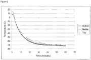
- Temperature monitoring studies were performed to track the temperature of tissues during the cryopreservation process. Following packaging in an inner sterile bag and an outer Mylar bag, StrataGraft® tissues are transferred to a pre-cooled rack inside of an ultra-cold freezer. Each tissue is placed in a separate slot in the freezer rack, with ample room above and below the tissue to allow unrestricted airflow during the freezing process. Using temperature monitoring probes positioned within culture dishes packaged as described above and loaded into freezer racks in this configuration, the temperature rapidly decreases to approximately -50°C within the first 15 minutes, further cools to approximately -65°C by 30 minutes and reaches a final temperature of approximately -80°C after three hours. There is no significant difference in the temperature profiles between tissues placed in the top, middle, and bottom positions of the freezer rack. See Figure 2 .
- tissues were frozen in contact with a layer of cryoprotectant solution after incubation with cryoprotectant. Although tissues cryopreserved in this manner exhibited good post-thaw properties, rapid thawing of tissues frozen in contact with this layer of cryoprotectant required incubation for several minutes in a 35-39°C water bath, which would to be difficult to implement and standardize in a surgical suite. It was subsequently determined that contact with the cryoprotectant solution was not required after tissues had been treated with cryoprotectant. This enabled development of the final product configuration in which tissues are transferred to an empty sterile 100 mm culture dish after treatment with cryoprotectant, where they are frozen in contact with the bottom of the empty dish rather than being frozen in contact with a layer of cryoprotectant solution.
- the 100 mm culture dishes containing cryoprotectant-treated tissues are aseptically packaged and heat-sealed inside of a sterile polyethylene sample bag.
- the inner bag is then heat-sealed inside a puncture-resistant, food grade, metalized polyester/polyethylene bag, which protects the packaged tissues from light, moisture, and provides a barrier to CO 2 vapor during shipment on dry ice.
- the stability and comparability studies described below utilized tissues packaged and cryopreserved in this configuration.
- Cryopreservation of viable skin equivalents enables burn centers to have ready access to this cell-based regenerative medicine therapeutic for burns and other indications that require rapid intervention.
- major burn centers would be able to maintain an inventory of the product for use without the need to schedule a delivery on a case by case basis.
- cryopreserved tissues were stored at -196°C in a vapor-phase nitrogen freezer. Since burn centers do not typically have liquid nitrogen storage capabilities, cryopreservation procedures were developed that permit storage of tissue for at least six months in ultra-cold freezers (-60°C to -90°C), which are readily available in blood and tissue banks at most hospitals and trauma centers.
- Cryopreserved StrataGraft® skin tissue will be shipped to clinical sites on dry ice for next morning delivery via commercial courier such as FedEx or UPS.
- the shipping container (Freezetherm FT29, Laminar Medica) is a validated dry ice shipping box that holds sufficient dry ice to maintain the cryopreserved tissues at ⁇ -75°C for at least 72 hours at ambient temperatures of up to 35°C to account for possible delays in delivery.
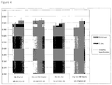
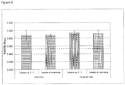
- Experimental data indicates that storage of cryopreserved tissues in the dry ice shipping container for >48 hours does not have any detectable adverse effect on tissue viability or histological architecture. See Figure 4 .
- cryopreserved StrataGraft tissues will be stored in an ultra-cold freezer (e.g., -60°C to -90°C) until use.
- cryopreserved StrataGraft tissue Prior to clinical use, cryopreserved StrataGraft tissue is thawed and incubated briefly on pads saturated with culture medium to remove residual cryoprotectant. Due to the geometry of the tissue, the thaw phase is rapid. After tissues are thawed, Transwell inserts containing the tissue are aseptically transferred to the sterile field and placed in sterile dishes containing absorbent pads saturated with culture medium. As described below, the timing of the post-thaw incubation phase is flexible enough to accommodate delays that could be reasonably anticipated during clinical use.
- tissues are cryopreserved in a culture dish without a layer of cryoprotectant solution, which allows the tissues to be thawed rapidly at ambient temperature simply by placing the package onto a bench or table. Precise control over the thaw temperature and time is not required, as experimental data shows that tissues thawed for varying times at temperatures ranging from 22°C to 40°C exhibit similar post-thaw properties.
- Buffered post-thaw incubation solutions work better than unbuffered solutions. Tissues incubated in simple unbuffered salt solutions (lactated Ringer's or normal saline) do not survive as well as tissues incubated in culture media-based solutions. Stratatech's SM01 culture media (StrataLife series) or commercially available DMEM/F12 media buffered with HEPES are preferred. See Figure 6 .
- Tissues can be left on the media saturated pad for times ranging from 15 min to 4 hr at 20-25°C or up to 2 hr at 40°C with no significant effect on tissue viability.
- This example describes improved cryopreservation methods for human skin equivalents utilizing a pre-freeze treatment step with cryopreservation solutions containing 32.5% or 50% glycerol at room temperature.
- the production process is unchanged from the current method described previously.
- the tissues are treated and cryopreserved as follows.
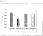
- Cryopreserved tissues were thawed at room temperature for 10 minutes, transferred to a hold chamber containing Telfa pads saturated with 40 ml of HEPES-buffered culture medium that had been warmed to room temperature, and held at RT for 15 to 20 minutes. Tissues were transferred to a culture dish containing 90 ml of SM01 medium and returned to culture overnight. Tissues were analyzed for viability after overnight re-culture. Tissues pre-treated with 32.5% glycerol at room temperature for 15 to 45 minutes had acceptable post-thaw viability. Tissues treated with 50% glycerol at room temperature for 15 minutes also had acceptable viability; however, tissues treated with 50% glycerol at room temperature for 45 minutes had unacceptable viability. See Figure 10 .
- MTT assays are preferably conducted as follows. Samples are excised from the skin tissue using an 8 mm biopsy punch. The samples are transferred to 0.3 ml MTT Assay Medium (1 mg/ml MTT reagent in Ham's F-12) in a 24-well plate that has been pre-warmed to 37 °C / 5% CO 2 . The samples are incubated for 85-95 minutes at 37 °C / 5% CO 2 . The samples are blotted and transferred to 2 ml isopropanol. The samples are thoroughly mixed to completely extract the purple formazan product. 200 ⁇ l in triplicate of each extract is aliquoted into a 96-well plate, using isopropanol as a blank. The absorbance (550 nm) is measured in a spectrophotometer. tissues to ensure uniform and rapid cooling. Leave tissues undisturbed overnight during the freezing process.
- Cryopreserved tissues were thawed at room temperature for 10 minutes, transferred to a hold chamber containing Telfa pads saturated with 40 ml of HEPES-buffered culture medium that had been warmed to room temperature, and held at RT for 15 to 20 minutes. Tissues were transferred to a culture dish containing 90 ml of SM01 medium and returned to culture overnight. Tissues were analyzed for viability after overnight re-culture. Tissues pre-treated with 32.5% glycerol at room temperature for 15 to 45 minutes had acceptable post-thaw viability. Tissues treated with 50% glycerol at room temperature for 15 minutes also had acceptable viability; however, tissues treated with 50% glycerol at room temperature for 45 minutes had unacceptable viability. See Figure 10 .
- MTT assays are preferably conducted as follows. Samples are excised from the skin tissue using an 8 mm biopsy punch. The samples are transferred to 0.3 ml MTT Assay Medium (1 mg/ml MTT reagent in Ham's F-12) in a 24-well plate that has been pre-warmed to 37 °C / 5% CO 2 . The samples are incubated for 85-95 minutes at 37 °C / 5% CO 2 . The samples are blotted and transferred to 2 ml isopropanol. The samples are thoroughly mixed to completely extract the purple formazan product. 200 ⁇ l in triplicate of each extract is aliquoted into a 96-well plate, using isopropanol as a blank. The absorbance (550 nm) is measured in a spectrophotometer.
Landscapes
- Life Sciences & Earth Sciences (AREA)
- Health & Medical Sciences (AREA)
- Engineering & Computer Science (AREA)
- General Health & Medical Sciences (AREA)
- Dentistry (AREA)
- Wood Science & Technology (AREA)
- Zoology (AREA)
- Environmental Sciences (AREA)
- General Chemical & Material Sciences (AREA)
- Organic Chemistry (AREA)
- Chemical & Material Sciences (AREA)
- Chemical Kinetics & Catalysis (AREA)
- Bioinformatics & Cheminformatics (AREA)
- Medicinal Chemistry (AREA)
- Nuclear Medicine, Radiotherapy & Molecular Imaging (AREA)
- Dermatology (AREA)
- Pharmacology & Pharmacy (AREA)
- Animal Behavior & Ethology (AREA)
- Public Health (AREA)
- Veterinary Medicine (AREA)
- Micro-Organisms Or Cultivation Processes Thereof (AREA)
- Medicines Containing Material From Animals Or Micro-Organisms (AREA)
- Materials For Medical Uses (AREA)
- Medicinal Preparation (AREA)
Priority Applications (3)
| Application Number | Priority Date | Filing Date | Title |
|---|---|---|---|
| EP21177142.3A EP3900534A1 (en) | 2013-03-13 | 2014-03-13 | Method for warming cryoconserved human skin substitutes |
| EP18197432.0A EP3470018B1 (en) | 2013-03-13 | 2014-03-13 | Cryopreservation of viable human skin substitutes |
| DK18197432.0T DK3470018T3 (da) | 2013-03-13 | 2014-03-13 | Kryokonservering af levedygtige menneskehudssubstitutter |
Applications Claiming Priority (2)
| Application Number | Priority Date | Filing Date | Title |
|---|---|---|---|
| US201361779661P | 2013-03-13 | 2013-03-13 | |
| PCT/US2014/025599 WO2014160000A1 (en) | 2013-03-13 | 2014-03-13 | Cryopreservation of viable human skin substitutes |
Related Child Applications (2)
| Application Number | Title | Priority Date | Filing Date |
|---|---|---|---|
| EP21177142.3A Division EP3900534A1 (en) | 2013-03-13 | 2014-03-13 | Method for warming cryoconserved human skin substitutes |
| EP18197432.0A Division EP3470018B1 (en) | 2013-03-13 | 2014-03-13 | Cryopreservation of viable human skin substitutes |
Publications (3)
| Publication Number | Publication Date |
|---|---|
| EP2967836A1 EP2967836A1 (en) | 2016-01-20 |
| EP2967836A4 EP2967836A4 (en) | 2016-11-23 |
| EP2967836B1 true EP2967836B1 (en) | 2018-10-10 |
Family
ID=51527939
Family Applications (3)
| Application Number | Title | Priority Date | Filing Date |
|---|---|---|---|
| EP14775892.4A Active EP2967836B1 (en) | 2013-03-13 | 2014-03-13 | Cryopreservation of viable human skin substitutes |
| EP21177142.3A Withdrawn EP3900534A1 (en) | 2013-03-13 | 2014-03-13 | Method for warming cryoconserved human skin substitutes |
| EP18197432.0A Active EP3470018B1 (en) | 2013-03-13 | 2014-03-13 | Cryopreservation of viable human skin substitutes |
Family Applications After (2)
| Application Number | Title | Priority Date | Filing Date |
|---|---|---|---|
| EP21177142.3A Withdrawn EP3900534A1 (en) | 2013-03-13 | 2014-03-13 | Method for warming cryoconserved human skin substitutes |
| EP18197432.0A Active EP3470018B1 (en) | 2013-03-13 | 2014-03-13 | Cryopreservation of viable human skin substitutes |
Country Status (10)
| Country | Link |
|---|---|
| US (3) | US10091983B2 (enExample) |
| EP (3) | EP2967836B1 (enExample) |
| JP (2) | JP6580028B2 (enExample) |
| AU (4) | AU2014244264B2 (enExample) |
| CA (2) | CA3071928A1 (enExample) |
| DK (2) | DK2967836T3 (enExample) |
| ES (2) | ES2893844T3 (enExample) |
| IL (1) | IL241448B (enExample) |
| PT (2) | PT2967836T (enExample) |
| WO (1) | WO2014160000A1 (enExample) |
Families Citing this family (15)
| Publication number | Priority date | Publication date | Assignee | Title |
|---|---|---|---|---|
| CA3071928A1 (en) * | 2013-03-13 | 2014-10-02 | Stratatech Corporation | Cryopreservation of viable human skin substitutes |
| AU2015287760B2 (en) * | 2014-07-09 | 2019-06-20 | Genentech, Inc. | pH adjustment to improve thaw recovery of cell banks |
| US9777064B2 (en) | 2015-03-17 | 2017-10-03 | Chimera Bioengineering, Inc. | Smart CAR devices, DE CAR polypeptides, side CARs and uses thereof |
| US11052111B2 (en) * | 2015-12-08 | 2021-07-06 | Chimera Bioengineering, Inc. | Smart CAR devices and DE CAR polypeptides for treating disease and methods for enhancing immune responses |
| EP3458077A4 (en) | 2016-05-17 | 2020-04-01 | Chimera Bioengineering Inc. | METHODS OF MANUFACTURING NEW AREAS OF ANTIGEN BINDING |
| CN112481217A (zh) | 2016-09-01 | 2021-03-12 | 嵌合体生物工程公司 | Gold优化的car t-细胞 |
| CN111989108B (zh) | 2018-02-13 | 2024-07-16 | 嵌合体生物工程公司 | 利用rna去稳定元件协调基因表达 |
| JP7669266B2 (ja) | 2018-08-31 | 2025-04-28 | ウィスコンシン アラムニ リサーチ ファンデーション | 動脈内皮細胞を播種した血管移植片の生成 |
| WO2020102854A1 (en) * | 2018-11-22 | 2020-05-28 | Cryogenics Holdings Pty Ltd. | Method and apparatus for freezing of biological products |
| JP2022546282A (ja) | 2019-08-18 | 2022-11-04 | キメラ・バイオエンジニアリング,インコーポレーテッド | Gold制御導入遺伝子による併用療法 |
| EP4034038A4 (en) * | 2019-09-27 | 2023-11-01 | Stratatech Corporation | METHODS OF TREATING ACUTE WOUNDS AND IMPROVING RESULTS |
| CN114836291B (zh) * | 2021-02-01 | 2025-11-21 | 深圳拜尔洛克生物技术有限公司 | 生物材料解冻复苏的系统及其方法 |
| US20240358766A1 (en) * | 2021-05-28 | 2024-10-31 | Stratatech Corporation | Allogeneic cultured keratinocyte products |
| AU2022282461A1 (en) * | 2021-05-28 | 2023-11-16 | Stratatech Corporation | Tissue container systems |
| WO2023056194A2 (en) | 2021-09-29 | 2023-04-06 | Chimera Bioengineering, Inc. | Compositions and methods for anti-tnmuc1 gold car t-cells |
Family Cites Families (30)
| Publication number | Priority date | Publication date | Assignee | Title |
|---|---|---|---|---|
| US4485096A (en) | 1982-02-26 | 1984-11-27 | Massachusetts Institute Of Technology | Tissue-equivalent and method for preparation thereof |
| IL95429A (en) | 1989-09-15 | 1997-09-30 | Organogenesis | Living tissue equivalents comprising hydrated collagen lattice and a collagen gel and their production |
| RU2135191C1 (ru) | 1990-04-24 | 1999-08-27 | Ортек Интернэшнл, Инк. | Композиционный эквивалент живой кожи, способ его получения, тест-набор |
| IT1248934B (it) | 1990-06-01 | 1995-02-11 | Fidia Spa | Membrane forate biocompatibili,processi per la loro preparazione,loro impiego come supporto per la crescita in vitro di cellule epiteliali, pelli artificiali cosi' ottenute e loro impiego nei trapianti di pelle |
| US5145770A (en) * | 1990-06-04 | 1992-09-08 | Biosurface Technology, Inc. | Cryopreservation of cultured epithelial sheets |
| US5891617A (en) | 1993-09-15 | 1999-04-06 | Organogenesis Inc. | Cryopreservation of harvested skin and cultured skin or cornea equivalents by slow freezing |
| US5518878A (en) * | 1993-09-15 | 1996-05-21 | Organogenesis Inc. | Cryopreservation of cultured skin or cornea equivalents with agitation |
| JPH10509610A (ja) * | 1994-11-09 | 1998-09-22 | クリ・ハークチ,ワリド | 創傷修復用包帯およびそれらの保存法 |
| IT1281870B1 (it) * | 1995-04-27 | 1998-03-03 | Fidia Advanced Biopolymers Srl | Pelle artificiale umana costituita da materiali biocompatibili a base di derivati dell'acido ialuronico |
| US5693332C1 (en) | 1995-08-11 | 2001-01-09 | Univ California | Human keratinocytes supported on a hydrophilic membrane and methods of using same to effect wound closure |
| US5689961A (en) * | 1996-01-30 | 1997-11-25 | Organogenesis Inc. | Ice seeding apparatus for cryopreservation systems |
| US5968546A (en) | 1997-05-16 | 1999-10-19 | Baur; Marcus | Keratinocyte culture from precursor cells |
| US5989837A (en) | 1998-07-13 | 1999-11-23 | Wisconsin Alumni Research Foundation | Immortalized human keratinocyte cell line |
| AU2002246747A1 (en) * | 2000-12-27 | 2002-08-06 | Ortec International, Inc. | Processes for making cryopreserved composite living constructs and products resulting therefrom |
| US6974697B2 (en) | 2001-03-02 | 2005-12-13 | Stratech Corporation | Skin substitutes with improved barrier function |
| EP1404809B1 (en) | 2001-03-02 | 2013-12-11 | Stratatech Corporation | Improved skin substitutes and uses thereof |
| EP1549738B1 (en) | 2002-04-30 | 2010-04-21 | Stratatech Corporation | Keratinocytes expressing exogenous angiogenic growth factors |
| EP1546392A4 (en) | 2002-08-02 | 2006-05-24 | Stratatech Corp | DETECTION OF SPECIFIC DNA FROM SPECIES |
| EP1633858A4 (en) * | 2003-05-30 | 2006-11-02 | Stratatech Corp | STORAGE OF ORGANS AT ROOM TEMPERATURE |
| CA2534349C (en) | 2003-08-01 | 2012-04-10 | Stratatech Corporation | Human skin equivalents expressing exogenous polypeptides |
| WO2005041880A2 (en) * | 2003-10-29 | 2005-05-12 | Science & Technology Corporation @ Unm | RhPV AS A MODEL FOR HPV-INDUCED CANCERS |
| JP4993752B2 (ja) | 2005-03-01 | 2012-08-08 | ストラタテック コーポレーション | 外因性ポリペプチドを発現するヒト皮膚同等物 |
| JP2008532550A (ja) | 2005-03-17 | 2008-08-21 | ストラタテック コーポレーション | 純度が改良された代用皮膚 |
| KR100803384B1 (ko) * | 2007-01-15 | 2008-02-13 | 한스바이오메드 주식회사 | 피부조직의 장기 보관 방법 |
| WO2009065005A1 (en) * | 2007-11-14 | 2009-05-22 | Stratatech Corporation | Cold storage of organotypically cultured skin equivalents for clinical applications |
| PT2352472T (pt) * | 2008-11-04 | 2017-04-11 | Stratatech Corp | Equivalentes de pele secos e irradiados prontos a utilizar |
| WO2010135655A2 (en) | 2009-05-21 | 2010-11-25 | Stratatech Corporation | Human skin substitutes expressing il-12 |
| WO2013062994A1 (en) * | 2011-10-25 | 2013-05-02 | Biomimetic Therapeutics, Inc. | Compositions and methods for treating full thickness burn injuries |
| WO2013062995A1 (en) * | 2011-10-25 | 2013-05-02 | Biomimetic Therapeutics, Inc. | Compositions and methods for treating deep partial and full thickness wounds and injuries |
| CA3071928A1 (en) * | 2013-03-13 | 2014-10-02 | Stratatech Corporation | Cryopreservation of viable human skin substitutes |
-
2014
- 2014-03-13 CA CA3071928A patent/CA3071928A1/en active Pending
- 2014-03-13 AU AU2014244264A patent/AU2014244264B2/en not_active Ceased
- 2014-03-13 JP JP2016501891A patent/JP6580028B2/ja active Active
- 2014-03-13 CA CA2905656A patent/CA2905656C/en active Active
- 2014-03-13 ES ES18197432T patent/ES2893844T3/es active Active
- 2014-03-13 DK DK14775892.4T patent/DK2967836T3/en active
- 2014-03-13 DK DK18197432.0T patent/DK3470018T3/da active
- 2014-03-13 EP EP14775892.4A patent/EP2967836B1/en active Active
- 2014-03-13 PT PT14775892T patent/PT2967836T/pt unknown
- 2014-03-13 PT PT18197432T patent/PT3470018T/pt unknown
- 2014-03-13 EP EP21177142.3A patent/EP3900534A1/en not_active Withdrawn
- 2014-03-13 EP EP18197432.0A patent/EP3470018B1/en active Active
- 2014-03-13 WO PCT/US2014/025599 patent/WO2014160000A1/en not_active Ceased
- 2014-03-13 ES ES14775892T patent/ES2705043T3/es active Active
- 2014-03-13 US US14/208,439 patent/US10091983B2/en active Active
-
2015
- 2015-09-10 IL IL241448A patent/IL241448B/en active IP Right Grant
-
2017
- 2017-07-17 AU AU2017206148A patent/AU2017206148B2/en not_active Ceased
-
2018
- 2018-09-05 US US16/122,598 patent/US11297829B2/en active Active
-
2019
- 2019-08-27 JP JP2019154515A patent/JP7220640B2/ja active Active
- 2019-09-17 AU AU2019232795A patent/AU2019232795B2/en not_active Ceased
-
2021
- 2021-11-05 AU AU2021261937A patent/AU2021261937B2/en not_active Ceased
-
2022
- 2022-03-14 US US17/694,245 patent/US20220192181A1/en not_active Abandoned
Also Published As
Similar Documents
| Publication | Publication Date | Title |
|---|---|---|
| AU2021261937B2 (en) | Cryopreservation of viable human skin substitutes | |
| US20220330542A1 (en) | Tissue container systems | |
| US8992997B2 (en) | Dried and irradiated skin equivalents for ready use | |
| HK40059784A (en) | Method for warming cryoconserved human skin substitutes | |
| US20220287298A1 (en) | Tissue container systems | |
| US20240245050A1 (en) | Tissue container systems | |
| NZ796375A (en) | Tissue container systems |
Legal Events
| Date | Code | Title | Description |
|---|---|---|---|
| PUAI | Public reference made under article 153(3) epc to a published international application that has entered the european phase |
Free format text: ORIGINAL CODE: 0009012 |
|
| 17P | Request for examination filed |
Effective date: 20150916 |
|
| AK | Designated contracting states |
Kind code of ref document: A1 Designated state(s): AL AT BE BG CH CY CZ DE DK EE ES FI FR GB GR HR HU IE IS IT LI LT LU LV MC MK MT NL NO PL PT RO RS SE SI SK SM TR |
|
| AX | Request for extension of the european patent |
Extension state: BA ME |
|
| DAX | Request for extension of the european patent (deleted) | ||
| RIC1 | Information provided on ipc code assigned before grant |
Ipc: A01N 1/02 20060101ALI20160718BHEP Ipc: A61F 2/00 20060101ALI20160718BHEP Ipc: A61F 2/10 20060101AFI20160718BHEP |
|
| A4 | Supplementary search report drawn up and despatched |
Effective date: 20161026 |
|
| RIC1 | Information provided on ipc code assigned before grant |
Ipc: A61F 2/10 20060101AFI20161020BHEP Ipc: A61F 2/00 20060101ALI20161020BHEP Ipc: A01N 1/02 20060101ALI20161020BHEP |
|
| STAA | Information on the status of an ep patent application or granted ep patent |
Free format text: STATUS: EXAMINATION IS IN PROGRESS |
|
| 17Q | First examination report despatched |
Effective date: 20170907 |
|
| GRAP | Despatch of communication of intention to grant a patent |
Free format text: ORIGINAL CODE: EPIDOSNIGR1 |
|
| STAA | Information on the status of an ep patent application or granted ep patent |
Free format text: STATUS: GRANT OF PATENT IS INTENDED |
|
| INTG | Intention to grant announced |
Effective date: 20180503 |
|
| GRAS | Grant fee paid |
Free format text: ORIGINAL CODE: EPIDOSNIGR3 |
|
| GRAA | (expected) grant |
Free format text: ORIGINAL CODE: 0009210 |
|
| STAA | Information on the status of an ep patent application or granted ep patent |
Free format text: STATUS: THE PATENT HAS BEEN GRANTED |
|
| AK | Designated contracting states |
Kind code of ref document: B1 Designated state(s): AL AT BE BG CH CY CZ DE DK EE ES FI FR GB GR HR HU IE IS IT LI LT LU LV MC MK MT NL NO PL PT RO RS SE SI SK SM TR |
|
| REG | Reference to a national code |
Ref country code: GB Ref legal event code: FG4D |
|
| REG | Reference to a national code |
Ref country code: CH Ref legal event code: EP Ref country code: AT Ref legal event code: REF Ref document number: 1050368 Country of ref document: AT Kind code of ref document: T Effective date: 20181015 |
|
| REG | Reference to a national code |
Ref country code: IE Ref legal event code: FG4D |
|
| REG | Reference to a national code |
Ref country code: DE Ref legal event code: R096 Ref document number: 602014033888 Country of ref document: DE |
|
| REG | Reference to a national code |
Ref country code: CH Ref legal event code: NV Representative=s name: TROESCH SCHEIDEGGER WERNER AG, CH |
|
| REG | Reference to a national code |
Ref country code: PT Ref legal event code: SC4A Ref document number: 2967836 Country of ref document: PT Date of ref document: 20190121 Kind code of ref document: T Free format text: AVAILABILITY OF NATIONAL TRANSLATION Effective date: 20190109 |
|
| REG | Reference to a national code |
Ref country code: DK Ref legal event code: T3 Effective date: 20190123 |
|
| REG | Reference to a national code |
Ref country code: NL Ref legal event code: FP |
|
| REG | Reference to a national code |
Ref country code: SE Ref legal event code: TRGR |
|
| REG | Reference to a national code |
Ref country code: LT Ref legal event code: MG4D Ref country code: NO Ref legal event code: T2 Effective date: 20181010 |
|
| REG | Reference to a national code |
Ref country code: ES Ref legal event code: FG2A Ref document number: 2705043 Country of ref document: ES Kind code of ref document: T3 Effective date: 20190321 |
|
| REG | Reference to a national code |
Ref country code: GR Ref legal event code: EP Ref document number: 20190400071 Country of ref document: GR Effective date: 20190422 |
|
| PG25 | Lapsed in a contracting state [announced via postgrant information from national office to epo] |
Ref country code: LT Free format text: LAPSE BECAUSE OF FAILURE TO SUBMIT A TRANSLATION OF THE DESCRIPTION OR TO PAY THE FEE WITHIN THE PRESCRIBED TIME-LIMIT Effective date: 20181010 Ref country code: BG Free format text: LAPSE BECAUSE OF FAILURE TO SUBMIT A TRANSLATION OF THE DESCRIPTION OR TO PAY THE FEE WITHIN THE PRESCRIBED TIME-LIMIT Effective date: 20190110 Ref country code: PL Free format text: LAPSE BECAUSE OF FAILURE TO SUBMIT A TRANSLATION OF THE DESCRIPTION OR TO PAY THE FEE WITHIN THE PRESCRIBED TIME-LIMIT Effective date: 20181010 Ref country code: HR Free format text: LAPSE BECAUSE OF FAILURE TO SUBMIT A TRANSLATION OF THE DESCRIPTION OR TO PAY THE FEE WITHIN THE PRESCRIBED TIME-LIMIT Effective date: 20181010 Ref country code: LV Free format text: LAPSE BECAUSE OF FAILURE TO SUBMIT A TRANSLATION OF THE DESCRIPTION OR TO PAY THE FEE WITHIN THE PRESCRIBED TIME-LIMIT Effective date: 20181010 |
|
| PG25 | Lapsed in a contracting state [announced via postgrant information from national office to epo] |
Ref country code: RS Free format text: LAPSE BECAUSE OF FAILURE TO SUBMIT A TRANSLATION OF THE DESCRIPTION OR TO PAY THE FEE WITHIN THE PRESCRIBED TIME-LIMIT Effective date: 20181010 Ref country code: AL Free format text: LAPSE BECAUSE OF FAILURE TO SUBMIT A TRANSLATION OF THE DESCRIPTION OR TO PAY THE FEE WITHIN THE PRESCRIBED TIME-LIMIT Effective date: 20181010 |
|
| REG | Reference to a national code |
Ref country code: DE Ref legal event code: R097 Ref document number: 602014033888 Country of ref document: DE |
|
| PG25 | Lapsed in a contracting state [announced via postgrant information from national office to epo] |
Ref country code: CZ Free format text: LAPSE BECAUSE OF FAILURE TO SUBMIT A TRANSLATION OF THE DESCRIPTION OR TO PAY THE FEE WITHIN THE PRESCRIBED TIME-LIMIT Effective date: 20181010 |
|
| REG | Reference to a national code |
Ref country code: AT Ref legal event code: UEP Ref document number: 1050368 Country of ref document: AT Kind code of ref document: T Effective date: 20181010 |
|
| PLBE | No opposition filed within time limit |
Free format text: ORIGINAL CODE: 0009261 |
|
| STAA | Information on the status of an ep patent application or granted ep patent |
Free format text: STATUS: NO OPPOSITION FILED WITHIN TIME LIMIT |
|
| PG25 | Lapsed in a contracting state [announced via postgrant information from national office to epo] |
Ref country code: RO Free format text: LAPSE BECAUSE OF FAILURE TO SUBMIT A TRANSLATION OF THE DESCRIPTION OR TO PAY THE FEE WITHIN THE PRESCRIBED TIME-LIMIT Effective date: 20181010 Ref country code: EE Free format text: LAPSE BECAUSE OF FAILURE TO SUBMIT A TRANSLATION OF THE DESCRIPTION OR TO PAY THE FEE WITHIN THE PRESCRIBED TIME-LIMIT Effective date: 20181010 Ref country code: SM Free format text: LAPSE BECAUSE OF FAILURE TO SUBMIT A TRANSLATION OF THE DESCRIPTION OR TO PAY THE FEE WITHIN THE PRESCRIBED TIME-LIMIT Effective date: 20181010 Ref country code: SK Free format text: LAPSE BECAUSE OF FAILURE TO SUBMIT A TRANSLATION OF THE DESCRIPTION OR TO PAY THE FEE WITHIN THE PRESCRIBED TIME-LIMIT Effective date: 20181010 |
|
| 26N | No opposition filed |
Effective date: 20190711 |
|
| PG25 | Lapsed in a contracting state [announced via postgrant information from national office to epo] |
Ref country code: SI Free format text: LAPSE BECAUSE OF FAILURE TO SUBMIT A TRANSLATION OF THE DESCRIPTION OR TO PAY THE FEE WITHIN THE PRESCRIBED TIME-LIMIT Effective date: 20181010 Ref country code: MC Free format text: LAPSE BECAUSE OF FAILURE TO SUBMIT A TRANSLATION OF THE DESCRIPTION OR TO PAY THE FEE WITHIN THE PRESCRIBED TIME-LIMIT Effective date: 20181010 |
|
| PG25 | Lapsed in a contracting state [announced via postgrant information from national office to epo] |
Ref country code: LU Free format text: LAPSE BECAUSE OF NON-PAYMENT OF DUE FEES Effective date: 20190313 |
|
| PG25 | Lapsed in a contracting state [announced via postgrant information from national office to epo] |
Ref country code: TR Free format text: LAPSE BECAUSE OF FAILURE TO SUBMIT A TRANSLATION OF THE DESCRIPTION OR TO PAY THE FEE WITHIN THE PRESCRIBED TIME-LIMIT Effective date: 20181010 |
|
| PG25 | Lapsed in a contracting state [announced via postgrant information from national office to epo] |
Ref country code: MT Free format text: LAPSE BECAUSE OF NON-PAYMENT OF DUE FEES Effective date: 20190313 |
|
| PG25 | Lapsed in a contracting state [announced via postgrant information from national office to epo] |
Ref country code: CY Free format text: LAPSE BECAUSE OF FAILURE TO SUBMIT A TRANSLATION OF THE DESCRIPTION OR TO PAY THE FEE WITHIN THE PRESCRIBED TIME-LIMIT Effective date: 20181010 |
|
| PG25 | Lapsed in a contracting state [announced via postgrant information from national office to epo] |
Ref country code: HU Free format text: LAPSE BECAUSE OF FAILURE TO SUBMIT A TRANSLATION OF THE DESCRIPTION OR TO PAY THE FEE WITHIN THE PRESCRIBED TIME-LIMIT; INVALID AB INITIO Effective date: 20140313 |
|
| PG25 | Lapsed in a contracting state [announced via postgrant information from national office to epo] |
Ref country code: MK Free format text: LAPSE BECAUSE OF FAILURE TO SUBMIT A TRANSLATION OF THE DESCRIPTION OR TO PAY THE FEE WITHIN THE PRESCRIBED TIME-LIMIT Effective date: 20181010 |
|
| P01 | Opt-out of the competence of the unified patent court (upc) registered |
Effective date: 20230601 |
|
| PGFP | Annual fee paid to national office [announced via postgrant information from national office to epo] |
Ref country code: GR Payment date: 20240326 Year of fee payment: 11 |
|
| PGFP | Annual fee paid to national office [announced via postgrant information from national office to epo] |
Ref country code: IS Payment date: 20240228 Year of fee payment: 11 |
|
| PGFP | Annual fee paid to national office [announced via postgrant information from national office to epo] |
Ref country code: IE Payment date: 20240327 Year of fee payment: 11 Ref country code: NL Payment date: 20240326 Year of fee payment: 11 |
|
| PGFP | Annual fee paid to national office [announced via postgrant information from national office to epo] |
Ref country code: AT Payment date: 20240221 Year of fee payment: 11 |
|
| PGFP | Annual fee paid to national office [announced via postgrant information from national office to epo] |
Ref country code: FI Payment date: 20240325 Year of fee payment: 11 Ref country code: DE Payment date: 20240327 Year of fee payment: 11 Ref country code: GB Payment date: 20240327 Year of fee payment: 11 Ref country code: PT Payment date: 20240227 Year of fee payment: 11 |
|
| PGFP | Annual fee paid to national office [announced via postgrant information from national office to epo] |
Ref country code: SE Payment date: 20240327 Year of fee payment: 11 Ref country code: NO Payment date: 20240228 Year of fee payment: 11 Ref country code: IT Payment date: 20240321 Year of fee payment: 11 Ref country code: FR Payment date: 20240325 Year of fee payment: 11 Ref country code: DK Payment date: 20240325 Year of fee payment: 11 Ref country code: BE Payment date: 20240327 Year of fee payment: 11 |
|
| PGFP | Annual fee paid to national office [announced via postgrant information from national office to epo] |
Ref country code: CH Payment date: 20240402 Year of fee payment: 11 |
|
| PGFP | Annual fee paid to national office [announced via postgrant information from national office to epo] |
Ref country code: ES Payment date: 20240401 Year of fee payment: 11 |
|
| REG | Reference to a national code |
Ref country code: DE Ref legal event code: R119 Ref document number: 602014033888 Country of ref document: DE |
|
| PG25 | Lapsed in a contracting state [announced via postgrant information from national office to epo] |
Ref country code: PT Free format text: LAPSE BECAUSE OF NON-PAYMENT OF DUE FEES Effective date: 20250915 |
|
| PG25 | Lapsed in a contracting state [announced via postgrant information from national office to epo] |
Ref country code: FI Free format text: LAPSE BECAUSE OF NON-PAYMENT OF DUE FEES Effective date: 20250313 |
|
| REG | Reference to a national code |
Ref country code: CH Ref legal event code: H13 Free format text: ST27 STATUS EVENT CODE: U-0-0-H10-H13 (AS PROVIDED BY THE NATIONAL OFFICE) Effective date: 20251023 |
|
| REG | Reference to a national code |
Ref country code: DK Ref legal event code: EBP Effective date: 20250331 |
|
| REG | Reference to a national code |
Ref country code: SE Ref legal event code: EUG |
|
| REG | Reference to a national code |
Ref country code: NL Ref legal event code: MM Effective date: 20250401 |
|
| REG | Reference to a national code |
Ref country code: AT Ref legal event code: MM01 Ref document number: 1050368 Country of ref document: AT Kind code of ref document: T Effective date: 20250313 |
|
| GBPC | Gb: european patent ceased through non-payment of renewal fee |
Effective date: 20250313 |
|
| REG | Reference to a national code |
Ref country code: BE Ref legal event code: MM Effective date: 20250331 |
|
| PG25 | Lapsed in a contracting state [announced via postgrant information from national office to epo] |
Ref country code: NL Free format text: LAPSE BECAUSE OF NON-PAYMENT OF DUE FEES Effective date: 20250401 |
|
| PG25 | Lapsed in a contracting state [announced via postgrant information from national office to epo] |
Ref country code: DE Free format text: LAPSE BECAUSE OF NON-PAYMENT OF DUE FEES Effective date: 20251001 |
|
| PG25 | Lapsed in a contracting state [announced via postgrant information from national office to epo] |
Ref country code: GB Free format text: LAPSE BECAUSE OF NON-PAYMENT OF DUE FEES Effective date: 20250313 |
|
| PG25 | Lapsed in a contracting state [announced via postgrant information from national office to epo] |
Ref country code: NO Free format text: LAPSE BECAUSE OF NON-PAYMENT OF DUE FEES Effective date: 20250331 |
|
| PG25 | Lapsed in a contracting state [announced via postgrant information from national office to epo] |
Ref country code: AT Free format text: LAPSE BECAUSE OF NON-PAYMENT OF DUE FEES Effective date: 20250313 |
|
| PG25 | Lapsed in a contracting state [announced via postgrant information from national office to epo] |
Ref country code: FR Free format text: LAPSE BECAUSE OF NON-PAYMENT OF DUE FEES Effective date: 20250331 Ref country code: IT Free format text: LAPSE BECAUSE OF NON-PAYMENT OF DUE FEES Effective date: 20250313 |
|
| PG25 | Lapsed in a contracting state [announced via postgrant information from national office to epo] |
Ref country code: GR Free format text: LAPSE BECAUSE OF NON-PAYMENT OF DUE FEES Effective date: 20251003 Ref country code: BE Free format text: LAPSE BECAUSE OF NON-PAYMENT OF DUE FEES Effective date: 20250331 |
|
| PG25 | Lapsed in a contracting state [announced via postgrant information from national office to epo] |
Ref country code: CH Free format text: LAPSE BECAUSE OF NON-PAYMENT OF DUE FEES Effective date: 20250331 |
|
| PG25 | Lapsed in a contracting state [announced via postgrant information from national office to epo] |
Ref country code: SE Free format text: LAPSE BECAUSE OF NON-PAYMENT OF DUE FEES Effective date: 20250314 Ref country code: IE Free format text: LAPSE BECAUSE OF NON-PAYMENT OF DUE FEES Effective date: 20250313 |
|
| PG25 | Lapsed in a contracting state [announced via postgrant information from national office to epo] |
Ref country code: DK Free format text: LAPSE BECAUSE OF NON-PAYMENT OF DUE FEES Effective date: 20250331 |