EP3470018B1 - Cryopreservation of viable human skin substitutes - Google Patents
Cryopreservation of viable human skin substitutes Download PDFInfo
- Publication number
- EP3470018B1 EP3470018B1 EP18197432.0A EP18197432A EP3470018B1 EP 3470018 B1 EP3470018 B1 EP 3470018B1 EP 18197432 A EP18197432 A EP 18197432A EP 3470018 B1 EP3470018 B1 EP 3470018B1
- Authority
- EP
- European Patent Office
- Prior art keywords
- skin equivalent
- cryopreserved
- solution
- cryoprotectant
- tissues
- Prior art date
- Legal status (The legal status is an assumption and is not a legal conclusion. Google has not performed a legal analysis and makes no representation as to the accuracy of the status listed.)
- Active
Links
- 238000005138 cryopreservation Methods 0.000 title description 40
- 210000001519 tissue Anatomy 0.000 claims description 225
- 210000003491 skin Anatomy 0.000 claims description 211
- PEDCQBHIVMGVHV-UHFFFAOYSA-N glycerol group Chemical group OCC(O)CO PEDCQBHIVMGVHV-UHFFFAOYSA-N 0.000 claims description 143
- 239000002577 cryoprotective agent Substances 0.000 claims description 110
- 238000000034 method Methods 0.000 claims description 86
- 210000002510 keratinocyte Anatomy 0.000 claims description 68
- 230000035899 viability Effects 0.000 claims description 44
- 239000002609 medium Substances 0.000 claims description 33
- 238000011282 treatment Methods 0.000 claims description 33
- 238000007710 freezing Methods 0.000 claims description 32
- 230000008014 freezing Effects 0.000 claims description 32
- 208000027418 Wounds and injury Diseases 0.000 claims description 29
- 206010052428 Wound Diseases 0.000 claims description 28
- 230000002500 effect on skin Effects 0.000 claims description 24
- 239000002250 absorbent Substances 0.000 claims description 21
- 230000002745 absorbent Effects 0.000 claims description 21
- 239000001963 growth medium Substances 0.000 claims description 20
- 238000004806 packaging method and process Methods 0.000 claims description 16
- 238000009792 diffusion process Methods 0.000 claims description 15
- 102000004196 processed proteins & peptides Human genes 0.000 claims description 15
- 108090000765 processed proteins & peptides Proteins 0.000 claims description 15
- 238000010257 thawing Methods 0.000 claims description 15
- 238000000134 MTT assay Methods 0.000 claims description 14
- 231100000002 MTT assay Toxicity 0.000 claims description 14
- 210000002950 fibroblast Anatomy 0.000 claims description 14
- 229920001184 polypeptide Polymers 0.000 claims description 14
- 230000000845 anti-microbial effect Effects 0.000 claims description 12
- 230000001023 pro-angiogenic effect Effects 0.000 claims description 9
- 238000004113 cell culture Methods 0.000 claims description 8
- 210000004379 membrane Anatomy 0.000 claims description 8
- 239000012528 membrane Substances 0.000 claims description 8
- 239000000758 substrate Substances 0.000 claims description 8
- 238000010792 warming Methods 0.000 claims description 6
- 238000012258 culturing Methods 0.000 claims description 5
- 239000006260 foam Substances 0.000 claims description 4
- 238000000502 dialysis Methods 0.000 claims description 2
- 210000005127 stratified epithelium Anatomy 0.000 claims description 2
- 210000004027 cell Anatomy 0.000 description 111
- 239000000243 solution Substances 0.000 description 98
- 235000011187 glycerol Nutrition 0.000 description 47
- 238000011534 incubation Methods 0.000 description 36
- 238000003860 storage Methods 0.000 description 31
- 230000008569 process Effects 0.000 description 28
- 239000010410 layer Substances 0.000 description 18
- 238000004458 analytical method Methods 0.000 description 17
- 239000003814 drug Substances 0.000 description 15
- 239000000047 product Substances 0.000 description 15
- 238000004519 manufacturing process Methods 0.000 description 14
- CURLTUGMZLYLDI-UHFFFAOYSA-N Carbon dioxide Chemical compound O=C=O CURLTUGMZLYLDI-UHFFFAOYSA-N 0.000 description 13
- JKMHFZQWWAIEOD-UHFFFAOYSA-N 2-[4-(2-hydroxyethyl)piperazin-1-yl]ethanesulfonic acid Chemical compound OCC[NH+]1CCN(CCS([O-])(=O)=O)CC1 JKMHFZQWWAIEOD-UHFFFAOYSA-N 0.000 description 11
- 239000006144 Dulbecco’s modified Eagle's medium Substances 0.000 description 11
- 239000007995 HEPES buffer Substances 0.000 description 11
- 229940124597 therapeutic agent Drugs 0.000 description 11
- IJGRMHOSHXDMSA-UHFFFAOYSA-N Atomic nitrogen Chemical compound N#N IJGRMHOSHXDMSA-UHFFFAOYSA-N 0.000 description 10
- 230000004069 differentiation Effects 0.000 description 10
- 210000002615 epidermis Anatomy 0.000 description 10
- 230000012010 growth Effects 0.000 description 10
- 238000012360 testing method Methods 0.000 description 10
- 238000012546 transfer Methods 0.000 description 10
- 239000013598 vector Substances 0.000 description 10
- 235000011089 carbon dioxide Nutrition 0.000 description 9
- 239000012467 final product Substances 0.000 description 8
- 102100025064 Cellular tumor antigen p53 Human genes 0.000 description 7
- 238000011161 development Methods 0.000 description 7
- 230000018109 developmental process Effects 0.000 description 7
- 230000001965 increasing effect Effects 0.000 description 7
- 230000015572 biosynthetic process Effects 0.000 description 6
- POIUWJQBRNEFGX-XAMSXPGMSA-N cathelicidin Chemical compound C([C@@H](C(=O)N[C@@H](CCCNC(N)=N)C(=O)N[C@@H](CCCCN)C(=O)N[C@@H](CO)C(=O)N[C@@H](CCCCN)C(=O)N[C@@H](CCC(O)=O)C(=O)N[C@@H](CCCCN)C(=O)N[C@@H]([C@@H](C)CC)C(=O)NCC(=O)N[C@@H](CCCCN)C(=O)N[C@@H](CCC(O)=O)C(=O)N[C@@H](CC=1C=CC=CC=1)C(=O)N[C@@H](CCCCN)C(=O)N[C@@H](CCCNC(N)=N)C(=O)N[C@@H]([C@@H](C)CC)C(=O)N[C@@H](C(C)C)C(=O)N[C@@H](CCC(N)=O)C(=O)N[C@@H](CCCNC(N)=N)C(=O)N[C@@H]([C@@H](C)CC)C(=O)N[C@@H](CCCCN)C(=O)N[C@@H](CC(O)=O)C(=O)N[C@@H](CC=1C=CC=CC=1)C(=O)N[C@@H](CC(C)C)C(=O)N[C@@H](CCCNC(N)=N)C(=O)N[C@@H](CC(N)=O)C(=O)N[C@@H](CC(C)C)C(=O)N[C@@H](C(C)C)C(=O)N1[C@@H](CCC1)C(=O)N[C@@H](CCCNC(N)=N)C(=O)N[C@@H]([C@@H](C)O)C(=O)N[C@@H](CCC(O)=O)C(=O)N[C@@H](CO)C(O)=O)NC(=O)[C@H](CC=1C=CC=CC=1)NC(=O)[C@H](CC(O)=O)NC(=O)CNC(=O)[C@H](CC(C)C)NC(=O)[C@@H](N)CC(C)C)C1=CC=CC=C1 POIUWJQBRNEFGX-XAMSXPGMSA-N 0.000 description 6
- 238000011067 equilibration Methods 0.000 description 6
- 230000007774 longterm Effects 0.000 description 6
- 229920002799 BoPET Polymers 0.000 description 5
- 108020004414 DNA Proteins 0.000 description 5
- 241001465754 Metazoa Species 0.000 description 5
- 239000005041 Mylar™ Substances 0.000 description 5
- 239000000203 mixture Substances 0.000 description 5
- 229910052757 nitrogen Inorganic materials 0.000 description 5
- 150000007523 nucleic acids Chemical group 0.000 description 5
- 239000004033 plastic Substances 0.000 description 5
- 229920003023 plastic Polymers 0.000 description 5
- 229920006395 saturated elastomer Polymers 0.000 description 5
- 238000013517 stratification Methods 0.000 description 5
- 101000912247 Homo sapiens Beta-defensin 103 Proteins 0.000 description 4
- 101001093139 Homo sapiens MAU2 chromatid cohesion factor homolog Proteins 0.000 description 4
- KFZMGEQAYNKOFK-UHFFFAOYSA-N Isopropanol Chemical compound CC(C)O KFZMGEQAYNKOFK-UHFFFAOYSA-N 0.000 description 4
- 102100036309 MAU2 chromatid cohesion factor homolog Human genes 0.000 description 4
- 206010072170 Skin wound Diseases 0.000 description 4
- 239000002870 angiogenesis inducing agent Substances 0.000 description 4
- 230000004888 barrier function Effects 0.000 description 4
- 239000000872 buffer Substances 0.000 description 4
- 102000014509 cathelicidin Human genes 0.000 description 4
- 108060001132 cathelicidin Proteins 0.000 description 4
- 239000001913 cellulose Substances 0.000 description 4
- 229920002678 cellulose Polymers 0.000 description 4
- 238000009472 formulation Methods 0.000 description 4
- 238000000338 in vitro Methods 0.000 description 4
- 239000007788 liquid Substances 0.000 description 4
- 108020004707 nucleic acids Proteins 0.000 description 4
- 102000039446 nucleic acids Human genes 0.000 description 4
- 229910001220 stainless steel Inorganic materials 0.000 description 4
- 239000010935 stainless steel Substances 0.000 description 4
- LENZDBCJOHFCAS-UHFFFAOYSA-N tris Chemical compound OCC(N)(CO)CO LENZDBCJOHFCAS-UHFFFAOYSA-N 0.000 description 4
- 239000012808 vapor phase Substances 0.000 description 4
- 102000014133 Antimicrobial Cationic Peptides Human genes 0.000 description 3
- 108010050820 Antimicrobial Cationic Peptides Proteins 0.000 description 3
- 108010035532 Collagen Proteins 0.000 description 3
- 102000008186 Collagen Human genes 0.000 description 3
- 241000282412 Homo Species 0.000 description 3
- 101000952040 Homo sapiens Beta-defensin 1 Proteins 0.000 description 3
- 101000884714 Homo sapiens Beta-defensin 4A Proteins 0.000 description 3
- 206010028980 Neoplasm Diseases 0.000 description 3
- 238000003556 assay Methods 0.000 description 3
- 210000000270 basal cell Anatomy 0.000 description 3
- 210000002469 basement membrane Anatomy 0.000 description 3
- 239000006172 buffering agent Substances 0.000 description 3
- 210000000349 chromosome Anatomy 0.000 description 3
- 230000001684 chronic effect Effects 0.000 description 3
- 229920001436 collagen Polymers 0.000 description 3
- 238000001816 cooling Methods 0.000 description 3
- 230000007423 decrease Effects 0.000 description 3
- 210000003953 foreskin Anatomy 0.000 description 3
- 210000000301 hemidesmosome Anatomy 0.000 description 3
- 102000046975 human DEFB1 Human genes 0.000 description 3
- 102000055779 human DEFB103A Human genes 0.000 description 3
- 102000049262 human DEFB4A Human genes 0.000 description 3
- 230000001976 improved effect Effects 0.000 description 3
- 239000000463 material Substances 0.000 description 3
- 229920000609 methyl cellulose Polymers 0.000 description 3
- 239000001923 methylcellulose Substances 0.000 description 3
- 238000004264 monolayer culture Methods 0.000 description 3
- 235000015097 nutrients Nutrition 0.000 description 3
- 239000012071 phase Substances 0.000 description 3
- 238000003752 polymerase chain reaction Methods 0.000 description 3
- 238000012545 processing Methods 0.000 description 3
- 108090000623 proteins and genes Proteins 0.000 description 3
- 102000004169 proteins and genes Human genes 0.000 description 3
- 230000002829 reductive effect Effects 0.000 description 3
- 230000001172 regenerating effect Effects 0.000 description 3
- 230000003362 replicative effect Effects 0.000 description 3
- 239000000523 sample Substances 0.000 description 3
- 238000013341 scale-up Methods 0.000 description 3
- 230000001225 therapeutic effect Effects 0.000 description 3
- DVLFYONBTKHTER-UHFFFAOYSA-N 3-(N-morpholino)propanesulfonic acid Chemical compound OS(=O)(=O)CCCN1CCOCC1 DVLFYONBTKHTER-UHFFFAOYSA-N 0.000 description 2
- 108010001781 Apligraf Proteins 0.000 description 2
- 102100026887 Beta-defensin 103 Human genes 0.000 description 2
- 108091003079 Bovine Serum Albumin Proteins 0.000 description 2
- 102100038608 Cathelicidin antimicrobial peptide Human genes 0.000 description 2
- 108020004705 Codon Proteins 0.000 description 2
- 101000741320 Homo sapiens Cathelicidin antimicrobial peptide Proteins 0.000 description 2
- 239000007993 MOPS buffer Substances 0.000 description 2
- 108091028043 Nucleic acid sequence Proteins 0.000 description 2
- 239000004698 Polyethylene Substances 0.000 description 2
- 239000006146 Roswell Park Memorial Institute medium Substances 0.000 description 2
- 108020004459 Small interfering RNA Proteins 0.000 description 2
- 239000007983 Tris buffer Substances 0.000 description 2
- 230000002411 adverse Effects 0.000 description 2
- 230000003466 anti-cipated effect Effects 0.000 description 2
- 238000013459 approach Methods 0.000 description 2
- 238000011717 athymic nude mouse Methods 0.000 description 2
- AFYNADDZULBEJA-UHFFFAOYSA-N bicinchoninic acid Chemical compound C1=CC=CC2=NC(C=3C=C(C4=CC=CC=C4N=3)C(=O)O)=CC(C(O)=O)=C21 AFYNADDZULBEJA-UHFFFAOYSA-N 0.000 description 2
- 229940098773 bovine serum albumin Drugs 0.000 description 2
- 239000008366 buffered solution Substances 0.000 description 2
- 230000002759 chromosomal effect Effects 0.000 description 2
- 230000006378 damage Effects 0.000 description 2
- 230000001934 delay Effects 0.000 description 2
- 210000001047 desmosome Anatomy 0.000 description 2
- 230000001700 effect on tissue Effects 0.000 description 2
- 238000001493 electron microscopy Methods 0.000 description 2
- 238000011156 evaluation Methods 0.000 description 2
- 230000001747 exhibiting effect Effects 0.000 description 2
- 239000000284 extract Substances 0.000 description 2
- 239000000499 gel Substances 0.000 description 2
- 230000014509 gene expression Effects 0.000 description 2
- 230000035876 healing Effects 0.000 description 2
- 230000036512 infertility Effects 0.000 description 2
- 238000002347 injection Methods 0.000 description 2
- 239000007924 injection Substances 0.000 description 2
- 208000014674 injury Diseases 0.000 description 2
- NOESYZHRGYRDHS-UHFFFAOYSA-N insulin Chemical compound N1C(=O)C(NC(=O)C(CCC(N)=O)NC(=O)C(CCC(O)=O)NC(=O)C(C(C)C)NC(=O)C(NC(=O)CN)C(C)CC)CSSCC(C(NC(CO)C(=O)NC(CC(C)C)C(=O)NC(CC=2C=CC(O)=CC=2)C(=O)NC(CCC(N)=O)C(=O)NC(CC(C)C)C(=O)NC(CCC(O)=O)C(=O)NC(CC(N)=O)C(=O)NC(CC=2C=CC(O)=CC=2)C(=O)NC(CSSCC(NC(=O)C(C(C)C)NC(=O)C(CC(C)C)NC(=O)C(CC=2C=CC(O)=CC=2)NC(=O)C(CC(C)C)NC(=O)C(C)NC(=O)C(CCC(O)=O)NC(=O)C(C(C)C)NC(=O)C(CC(C)C)NC(=O)C(CC=2NC=NC=2)NC(=O)C(CO)NC(=O)CNC2=O)C(=O)NCC(=O)NC(CCC(O)=O)C(=O)NC(CCCNC(N)=N)C(=O)NCC(=O)NC(CC=3C=CC=CC=3)C(=O)NC(CC=3C=CC=CC=3)C(=O)NC(CC=3C=CC(O)=CC=3)C(=O)NC(C(C)O)C(=O)N3C(CCC3)C(=O)NC(CCCCN)C(=O)NC(C)C(O)=O)C(=O)NC(CC(N)=O)C(O)=O)=O)NC(=O)C(C(C)CC)NC(=O)C(CO)NC(=O)C(C(C)O)NC(=O)C1CSSCC2NC(=O)C(CC(C)C)NC(=O)C(NC(=O)C(CCC(N)=O)NC(=O)C(CC(N)=O)NC(=O)C(NC(=O)C(N)CC=1C=CC=CC=1)C(C)C)CC1=CN=CN1 NOESYZHRGYRDHS-UHFFFAOYSA-N 0.000 description 2
- 239000003550 marker Substances 0.000 description 2
- 108091070501 miRNA Proteins 0.000 description 2
- 239000002679 microRNA Substances 0.000 description 2
- 238000012544 monitoring process Methods 0.000 description 2
- 210000004400 mucous membrane Anatomy 0.000 description 2
- VMGAPWLDMVPYIA-HIDZBRGKSA-N n'-amino-n-iminomethanimidamide Chemical compound N\N=C\N=N VMGAPWLDMVPYIA-HIDZBRGKSA-N 0.000 description 2
- 210000000056 organ Anatomy 0.000 description 2
- 229920000573 polyethylene Polymers 0.000 description 2
- 229920000642 polymer Polymers 0.000 description 2
- 230000002062 proliferating effect Effects 0.000 description 2
- 230000009467 reduction Effects 0.000 description 2
- 238000012429 release testing Methods 0.000 description 2
- BOLDJAUMGUJJKM-LSDHHAIUSA-N renifolin D Natural products CC(=C)[C@@H]1Cc2c(O)c(O)ccc2[C@H]1CC(=O)c3ccc(O)cc3O BOLDJAUMGUJJKM-LSDHHAIUSA-N 0.000 description 2
- 230000009758 senescence Effects 0.000 description 2
- 239000002356 single layer Substances 0.000 description 2
- 239000004055 small Interfering RNA Substances 0.000 description 2
- 238000002415 sodium dodecyl sulfate polyacrylamide gel electrophoresis Methods 0.000 description 2
- 238000002560 therapeutic procedure Methods 0.000 description 2
- XLYOFNOQVPJJNP-UHFFFAOYSA-N water Substances O XLYOFNOQVPJJNP-UHFFFAOYSA-N 0.000 description 2
- 230000029663 wound healing Effects 0.000 description 2
- 108091032973 (ribonucleotides)n+m Proteins 0.000 description 1
- 229920001817 Agar Polymers 0.000 description 1
- 108700028369 Alleles Proteins 0.000 description 1
- 108020005544 Antisense RNA Proteins 0.000 description 1
- 239000004475 Arginine Substances 0.000 description 1
- 101710125296 Beta-defensin 3 Proteins 0.000 description 1
- 108010039209 Blood Coagulation Factors Proteins 0.000 description 1
- 102000015081 Blood Coagulation Factors Human genes 0.000 description 1
- 108090000994 Catalytic RNA Proteins 0.000 description 1
- 102000053642 Catalytic RNA Human genes 0.000 description 1
- 206010008805 Chromosomal abnormalities Diseases 0.000 description 1
- 208000031404 Chromosome Aberrations Diseases 0.000 description 1
- 206010009900 Colitis ulcerative Diseases 0.000 description 1
- 102000012422 Collagen Type I Human genes 0.000 description 1
- 108010022452 Collagen Type I Proteins 0.000 description 1
- 241000702421 Dependoparvovirus Species 0.000 description 1
- 206010056340 Diabetic ulcer Diseases 0.000 description 1
- 108090000790 Enzymes Proteins 0.000 description 1
- 102000004190 Enzymes Human genes 0.000 description 1
- 102000003951 Erythropoietin Human genes 0.000 description 1
- 108090000394 Erythropoietin Proteins 0.000 description 1
- 102000010834 Extracellular Matrix Proteins Human genes 0.000 description 1
- 108010037362 Extracellular Matrix Proteins Proteins 0.000 description 1
- KRHYYFGTRYWZRS-UHFFFAOYSA-M Fluoride anion Chemical compound [F-] KRHYYFGTRYWZRS-UHFFFAOYSA-M 0.000 description 1
- 208000031886 HIV Infections Diseases 0.000 description 1
- 101001046870 Homo sapiens Hypoxia-inducible factor 1-alpha Proteins 0.000 description 1
- 241000714260 Human T-lymphotropic virus 1 Species 0.000 description 1
- 241000714259 Human T-lymphotropic virus 2 Species 0.000 description 1
- 241000701041 Human betaherpesvirus 7 Species 0.000 description 1
- 241000701027 Human herpesvirus 6 Species 0.000 description 1
- 241000713772 Human immunodeficiency virus 1 Species 0.000 description 1
- 241000713340 Human immunodeficiency virus 2 Species 0.000 description 1
- 241000341655 Human papillomavirus type 16 Species 0.000 description 1
- 102100022875 Hypoxia-inducible factor 1-alpha Human genes 0.000 description 1
- 206010062018 Inborn error of metabolism Diseases 0.000 description 1
- 206010061218 Inflammation Diseases 0.000 description 1
- 108090001061 Insulin Proteins 0.000 description 1
- 102100023915 Insulin Human genes 0.000 description 1
- 102000011782 Keratins Human genes 0.000 description 1
- 108010076876 Keratins Proteins 0.000 description 1
- 241000829100 Macaca mulatta polyomavirus 1 Species 0.000 description 1
- 241000699670 Mus sp. Species 0.000 description 1
- 239000002033 PVDF binder Substances 0.000 description 1
- 239000004793 Polystyrene Substances 0.000 description 1
- 208000004210 Pressure Ulcer Diseases 0.000 description 1
- ONIBWKKTOPOVIA-UHFFFAOYSA-N Proline Natural products OC(=O)C1CCCN1 ONIBWKKTOPOVIA-UHFFFAOYSA-N 0.000 description 1
- 241000125945 Protoparvovirus Species 0.000 description 1
- 102000002278 Ribosomal Proteins Human genes 0.000 description 1
- 108010000605 Ribosomal Proteins Proteins 0.000 description 1
- FAPWRFPIFSIZLT-UHFFFAOYSA-M Sodium chloride Chemical compound [Na+].[Cl-] FAPWRFPIFSIZLT-UHFFFAOYSA-M 0.000 description 1
- 208000025865 Ulcer Diseases 0.000 description 1
- 201000006704 Ulcerative Colitis Diseases 0.000 description 1
- 208000000558 Varicose Ulcer Diseases 0.000 description 1
- 108010019530 Vascular Endothelial Growth Factors Proteins 0.000 description 1
- 102000005789 Vascular Endothelial Growth Factors Human genes 0.000 description 1
- 241000700605 Viruses Species 0.000 description 1
- 238000002835 absorbance Methods 0.000 description 1
- 238000004115 adherent culture Methods 0.000 description 1
- 230000001464 adherent effect Effects 0.000 description 1
- 239000008272 agar Substances 0.000 description 1
- 230000000735 allogeneic effect Effects 0.000 description 1
- 150000001413 amino acids Chemical class 0.000 description 1
- 239000003242 anti bacterial agent Substances 0.000 description 1
- 230000002924 anti-infective effect Effects 0.000 description 1
- 229940088710 antibiotic agent Drugs 0.000 description 1
- 229960005475 antiinfective agent Drugs 0.000 description 1
- 238000003782 apoptosis assay Methods 0.000 description 1
- ODKSFYDXXFIFQN-UHFFFAOYSA-N arginine Natural products OC(=O)C(N)CCCNC(N)=N ODKSFYDXXFIFQN-UHFFFAOYSA-N 0.000 description 1
- 239000012911 assay medium Substances 0.000 description 1
- 230000008901 benefit Effects 0.000 description 1
- 230000000975 bioactive effect Effects 0.000 description 1
- 230000004071 biological effect Effects 0.000 description 1
- 229960000074 biopharmaceutical Drugs 0.000 description 1
- 238000001574 biopsy Methods 0.000 description 1
- 210000004369 blood Anatomy 0.000 description 1
- 239000008280 blood Substances 0.000 description 1
- 239000003114 blood coagulation factor Substances 0.000 description 1
- GEHJBWKLJVFKPS-UHFFFAOYSA-N bromochloroacetic acid Chemical compound OC(=O)C(Cl)Br GEHJBWKLJVFKPS-UHFFFAOYSA-N 0.000 description 1
- 239000001506 calcium phosphate Substances 0.000 description 1
- 229910000389 calcium phosphate Inorganic materials 0.000 description 1
- 235000011010 calcium phosphates Nutrition 0.000 description 1
- BPKIGYQJPYCAOW-FFJTTWKXSA-I calcium;potassium;disodium;(2s)-2-hydroxypropanoate;dichloride;dihydroxide;hydrate Chemical compound O.[OH-].[OH-].[Na+].[Na+].[Cl-].[Cl-].[K+].[Ca+2].C[C@H](O)C([O-])=O BPKIGYQJPYCAOW-FFJTTWKXSA-I 0.000 description 1
- 230000011712 cell development Effects 0.000 description 1
- 230000010261 cell growth Effects 0.000 description 1
- 230000003833 cell viability Effects 0.000 description 1
- 230000017455 cell-cell adhesion Effects 0.000 description 1
- 239000003153 chemical reaction reagent Substances 0.000 description 1
- 239000003795 chemical substances by application Substances 0.000 description 1
- 238000000975 co-precipitation Methods 0.000 description 1
- 239000000512 collagen gel Substances 0.000 description 1
- 239000000515 collagen sponge Substances 0.000 description 1
- 230000001332 colony forming effect Effects 0.000 description 1
- 230000002860 competitive effect Effects 0.000 description 1
- 230000000295 complement effect Effects 0.000 description 1
- 239000003184 complementary RNA Substances 0.000 description 1
- 239000002131 composite material Substances 0.000 description 1
- 150000001875 compounds Chemical class 0.000 description 1
- 230000003750 conditioning effect Effects 0.000 description 1
- 238000011109 contamination Methods 0.000 description 1
- 230000001276 controlling effect Effects 0.000 description 1
- 230000002596 correlated effect Effects 0.000 description 1
- 230000002338 cryopreservative effect Effects 0.000 description 1
- 238000002425 crystallisation Methods 0.000 description 1
- 230000008025 crystallization Effects 0.000 description 1
- 210000004748 cultured cell Anatomy 0.000 description 1
- 230000001086 cytosolic effect Effects 0.000 description 1
- 238000001804 debridement Methods 0.000 description 1
- 230000003247 decreasing effect Effects 0.000 description 1
- 230000007547 defect Effects 0.000 description 1
- 230000002950 deficient Effects 0.000 description 1
- 238000010790 dilution Methods 0.000 description 1
- 239000012895 dilution Substances 0.000 description 1
- 201000010099 disease Diseases 0.000 description 1
- 208000037265 diseases, disorders, signs and symptoms Diseases 0.000 description 1
- 229940079593 drug Drugs 0.000 description 1
- 230000004064 dysfunction Effects 0.000 description 1
- 230000000694 effects Effects 0.000 description 1
- 238000004520 electroporation Methods 0.000 description 1
- 239000002158 endotoxin Substances 0.000 description 1
- 230000002255 enzymatic effect Effects 0.000 description 1
- 229940105423 erythropoietin Drugs 0.000 description 1
- 238000002474 experimental method Methods 0.000 description 1
- 210000002744 extracellular matrix Anatomy 0.000 description 1
- 229960004222 factor ix Drugs 0.000 description 1
- 108060002894 fibrillar collagen Proteins 0.000 description 1
- 102000013373 fibrillar collagen Human genes 0.000 description 1
- 239000011888 foil Substances 0.000 description 1
- 239000012737 fresh medium Substances 0.000 description 1
- 230000002538 fungal effect Effects 0.000 description 1
- 210000001035 gastrointestinal tract Anatomy 0.000 description 1
- 230000009395 genetic defect Effects 0.000 description 1
- 238000010353 genetic engineering Methods 0.000 description 1
- 239000011521 glass Substances 0.000 description 1
- 150000002314 glycerols Chemical class 0.000 description 1
- 230000036541 health Effects 0.000 description 1
- 210000005260 human cell Anatomy 0.000 description 1
- 238000001727 in vivo Methods 0.000 description 1
- 208000016245 inborn errors of metabolism Diseases 0.000 description 1
- 230000001939 inductive effect Effects 0.000 description 1
- 208000015181 infectious disease Diseases 0.000 description 1
- 230000004054 inflammatory process Effects 0.000 description 1
- 230000000977 initiatory effect Effects 0.000 description 1
- 229940125396 insulin Drugs 0.000 description 1
- 102000007236 involucrin Human genes 0.000 description 1
- 108010033564 involucrin Proteins 0.000 description 1
- 210000000661 isochromosome Anatomy 0.000 description 1
- 238000001638 lipofection Methods 0.000 description 1
- 238000012423 maintenance Methods 0.000 description 1
- 239000011159 matrix material Substances 0.000 description 1
- 230000035800 maturation Effects 0.000 description 1
- 230000002503 metabolic effect Effects 0.000 description 1
- 229910052751 metal Inorganic materials 0.000 description 1
- 239000002184 metal Substances 0.000 description 1
- 239000011140 metalized polyester Substances 0.000 description 1
- 230000000813 microbial effect Effects 0.000 description 1
- NJTGANWAUPEOAX-UHFFFAOYSA-N molport-023-220-454 Chemical compound OCC(O)CO.OCC(O)CO NJTGANWAUPEOAX-UHFFFAOYSA-N 0.000 description 1
- 230000000877 morphologic effect Effects 0.000 description 1
- 230000001338 necrotic effect Effects 0.000 description 1
- 238000009581 negative-pressure wound therapy Methods 0.000 description 1
- 231100001221 nontumorigenic Toxicity 0.000 description 1
- 238000011275 oncology therapy Methods 0.000 description 1
- 230000003204 osmotic effect Effects 0.000 description 1
- 230000036961 partial effect Effects 0.000 description 1
- 239000000813 peptide hormone Substances 0.000 description 1
- 230000000704 physical effect Effects 0.000 description 1
- 239000013612 plasmid Substances 0.000 description 1
- 239000013600 plasmid vector Substances 0.000 description 1
- -1 polyethylene Polymers 0.000 description 1
- 229920002223 polystyrene Polymers 0.000 description 1
- 229920002981 polyvinylidene fluoride Polymers 0.000 description 1
- 239000013641 positive control Substances 0.000 description 1
- OXCMYAYHXIHQOA-UHFFFAOYSA-N potassium;[2-butyl-5-chloro-3-[[4-[2-(1,2,4-triaza-3-azanidacyclopenta-1,4-dien-5-yl)phenyl]phenyl]methyl]imidazol-4-yl]methanol Chemical compound [K+].CCCCC1=NC(Cl)=C(CO)N1CC1=CC=C(C=2C(=CC=CC=2)C2=N[N-]N=N2)C=C1 OXCMYAYHXIHQOA-UHFFFAOYSA-N 0.000 description 1
- 238000002360 preparation method Methods 0.000 description 1
- 238000004321 preservation Methods 0.000 description 1
- 230000001566 pro-viral effect Effects 0.000 description 1
- 238000011165 process development Methods 0.000 description 1
- 230000005522 programmed cell death Effects 0.000 description 1
- 230000002035 prolonged effect Effects 0.000 description 1
- 238000003908 quality control method Methods 0.000 description 1
- 238000011160 research Methods 0.000 description 1
- 230000000717 retained effect Effects 0.000 description 1
- 230000001177 retroviral effect Effects 0.000 description 1
- 108091092562 ribozyme Proteins 0.000 description 1
- 239000012266 salt solution Substances 0.000 description 1
- 239000007787 solid Substances 0.000 description 1
- 206010041823 squamous cell carcinoma Diseases 0.000 description 1
- 210000000130 stem cell Anatomy 0.000 description 1
- 239000000126 substance Substances 0.000 description 1
- 238000001356 surgical procedure Methods 0.000 description 1
- 230000009897 systematic effect Effects 0.000 description 1
- 230000008685 targeting Effects 0.000 description 1
- 230000000451 tissue damage Effects 0.000 description 1
- 231100000827 tissue damage Toxicity 0.000 description 1
- 230000017423 tissue regeneration Effects 0.000 description 1
- 230000001052 transient effect Effects 0.000 description 1
- 230000008733 trauma Effects 0.000 description 1
- QORWJWZARLRLPR-UHFFFAOYSA-H tricalcium bis(phosphate) Chemical compound [Ca+2].[Ca+2].[Ca+2].[O-]P([O-])([O-])=O.[O-]P([O-])([O-])=O QORWJWZARLRLPR-UHFFFAOYSA-H 0.000 description 1
- 230000001960 triggered effect Effects 0.000 description 1
- 231100000588 tumorigenic Toxicity 0.000 description 1
- 230000000381 tumorigenic effect Effects 0.000 description 1
- 231100000397 ulcer Toxicity 0.000 description 1
- 231100000747 viability assay Toxicity 0.000 description 1
- 238000003026 viability measurement method Methods 0.000 description 1
- 239000013603 viral vector Substances 0.000 description 1
- 230000003442 weekly effect Effects 0.000 description 1
Images


Classifications
-
- A—HUMAN NECESSITIES
- A01—AGRICULTURE; FORESTRY; ANIMAL HUSBANDRY; HUNTING; TRAPPING; FISHING
- A01N—PRESERVATION OF BODIES OF HUMANS OR ANIMALS OR PLANTS OR PARTS THEREOF; BIOCIDES, e.g. AS DISINFECTANTS, AS PESTICIDES OR AS HERBICIDES; PEST REPELLANTS OR ATTRACTANTS; PLANT GROWTH REGULATORS
- A01N1/00—Preservation of bodies of humans or animals, or parts thereof
- A01N1/02—Preservation of living parts
- A01N1/0205—Chemical aspects
- A01N1/021—Preservation or perfusion media, liquids, solids or gases used in the preservation of cells, tissue, organs or bodily fluids
- A01N1/0221—Freeze-process protecting agents, i.e. substances protecting cells from effects of the physical process, e.g. cryoprotectants, osmolarity regulators like oncotic agents
-
- A—HUMAN NECESSITIES
- A61—MEDICAL OR VETERINARY SCIENCE; HYGIENE
- A61P—SPECIFIC THERAPEUTIC ACTIVITY OF CHEMICAL COMPOUNDS OR MEDICINAL PREPARATIONS
- A61P17/00—Drugs for dermatological disorders
- A61P17/02—Drugs for dermatological disorders for treating wounds, ulcers, burns, scars, keloids, or the like
Definitions
- the present invention relates generally to systems and methods for cryopreservation of viable human skin substitutes.
- APLIGRAF requires about four weeks to manufacture, is usable for only 15 days and must be maintained between 20 and 23°C until used.
- EPICEL is transported by a nurse from Genzyme Biosurgery's production facility in Cambridge, MA to the point of use in a portable incubator and is used immediately upon arrival. Such constraints represent significant challenges to developing convenient and cost-effective products.
- WO 02/058588 A2 Methods of cryopreservation of a composite living skin equivalent which comprises an epidermal layer of cultured keratinocyte cells, a layer of high purity, non-porous collagen, and a dermal layer of cultured fibroblast cells in a porous, cross-linked collagen sponge is known from WO 02/058588 A2 .
- WO 02/058588 A2 does not disclose organotypically cultured skin equivalents comprising stratified squamous epithelia on a dermal layer comprising fibroblasts, as required by the present invention as defined in the appended claims.
- WO 2005/012492 A2 discloses a method for providing a skin equivalent expressing an exogenous antimicrobial polypeptide, wherein such antimicrobial polypeptide is human beta-defensin 1, 2 or 3 or human cathelicidin.
- WO 2005/012492 A2 does not disclose a method of cryopreserving the skin equivalents as defined in the appended claims.
- a method of cryopreserving an organotypically cultured skin equivalent to maintain viable tissue is disclosed in WO 96/24018 A1 , which has been categorized as the closest prior art in the European examination proceedings.
- the invention as defined in the appended claims differs from the method of WO 96/24018 A1 in that the known method involves an equilibration process followed by controlled freezing steps with external initiation of extracellular ice crystallization and storage of the skin equivalent with excess medium under liquid nitrogen vapor phase conditions.
- a further difference lies in the fact that the skin equivalent employed in claim 1 of the enclosed claim set has been engineered to express and provide exogenous antimicrobial polypeptides or proangiogenic factors.
- Said differences provide for a simplified method of cryopreserving an organotypically cultured skin equivalent that has been engineered to express and provide exogenous antimicrobial polypeptides or proangiogenic factors and has been found to be inventive over said prior art WO 96/24018 A1 , WO 02/058588 A2 and 2005/012492 A2 .
- the invention is directed to a method of cryopreserving an organotypically cultured skin equivalent to maintain viable tissue comprising: a) treating an organotypically cultured skin equivalent in a cryoprotectant solution in a single step, wherein said organotypically cultured skin equivalent comprises stratified squamous epithelia on a dermal layer comprising fibroblast, wherein the skin equivalent has been engineered to express and provide exogenous antimicrobial polypeptides or proangiogenic factors, and wherein said cryoprotectant is provided in a solution comprising 21% to 70% of the solution by volume, preferably 21% to 45% of the solution by volume or 37.5% to 62.5% of the solution by volume, more preferably 25% to 40% of the solution by volume or 42.5% to 57.5% of said solution by volume, depending on the temperature, and said cryoprotectant is glycerol; b) separating the treated organotypically cultured skin equivalent from excess cryoprotectant solution, and packaging
- the invention is directed to a packaged cryopreserved skin equivalent produced according to said method for use in the treatment of a patient, wherein prior to application to said patient said cryopreserved skin equivalent is a) thawed and not rinsed; or b) warmed, aseptically transferred from said package and contacted with an absorbent medium comprising a tissue compatible solution to allow removal of said cryoprotectant solution by diffusion.
- an absorbent medium comprising a tissue compatible solution to allow removal of said cryoprotectant solution by diffusion.
- the present description provides methods of cryopreserving an organotypically cultured skin equivalent to maintain viable tissue comprising: treating an organotypically cultured skin equivalent in a cryoprotectant solution in a single step; packaging the organotypically cultured skin equivalent to provide a packaged skin equivalent; and freezing the organotypically cultured skin equivalent to provide a packaged cryopreserved skin equivalent.
- the cryoprotectant may be provided in a solution comprising about 20% or 21% to about 70% of the solution by volume, and more preferably about 20% or 21% to about 45% of the solution by volume or 37.5% to 62.5% of the solution by volume, or most preferably from about 25% to 40% of the solution by volume or 42.5% to 57.5% of the solution by volume, depending on the temperature.
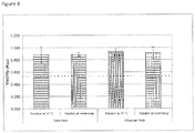
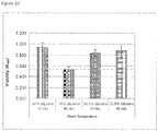
- the treatment with cryoprotectant may be conducted at from about 2°C to 8°C, while in other examples, the treatment step is conducted at room temperature, for example from about 15°C to 30°C.
- the cryoprotectant is glycerol.
- the freezing may further comprise freezing the organotypically cultured skin equivalent in the absence of substantial excess cryoprotectant.
- the freezing may further comprise freezing at about -80°C.
- the freezing may further comprise direct exposure to temperatures ranging from about -50°C to -100°C.
- the packaging may further comprise enclosing the cryopreserved skin equivalent in a sterile bag and enclosing the sterile bag in a second bag.
- the organotypically cultured skin equivalents may comprise NIKS cells.
- the NIKS cells may comprise an exogenous nucleic acid sequence encoding an exogenous polypeptide.
- the skin equivalent may retain viability after thawing.
- the skin equivalent may have an A 550 of at least 50% of a reference skin equivalent as determined by an MTT assay.
- the methods may further comprise thawing said cryopreserved skin equivalent and applying said thawed skin equivalent to a patient in need thereof, wherein said thawed skin equivalent is not rinsed prior to said application to said patient.
- the present description provides methods of thawing a cryopreserved skin equivalent prior to application to a subject, comprising: warming the cryopreserved skin equivalent; and contacting the cryopreserved skin equivalent with a diffusion mediator comprising a tissue compatible solution to allow removal of the cryoprotectant solution by diffusion.
- the warming may comprise exposure to room temperature at the site of use.
- the diffusion mediator may be selected from the group consisting of an absorbent medium, a membrane, and a dialysis bag.
- the absorbent medium may be selected from the group consisting of Telfa pads, foam pads, gauze pads, and cellulosic pads containing the tissue compatible medium.
- the tissue compatible solution may be a buffered solution.
- the present description provides methods of treating a subject comprising providing a packaged cryopreserved skin equivalent produced as described above; aseptically transferring the cryopreserved skin equivalent from the package; warming the cryopreserved skin equivalent; contacting the cryopreserved skin equivalent with an absorbent medium comprising a tissue compatible solution to allow removal of the cryoprotectant solution by diffusion; and applying the cryopreserved skin equivalent to the subject.
- the present description provides a cryopreserved skin equivalent equilibrated with a cryoprotectant, the skin equivalent being substantially free of excess cryoprotectant on the exterior surface of the skin equivalent.
- the present description provides a system comprising the foregoing skin equivalent disposed on an absorbent medium.
- the present description provides methods comprising providing the packaged cryopreserved skin equivalent as described above; and applying the skin equivalent to a wound under conditions such that the skin equivalent contacts the wound.
- the present description provides a kit comprising a cryopreserved skin substitute, an absorbent medium, and a tissue compatible solution.
- the cryopreserved skin substitute may be packaged in a sealable enclosure.
- the cryopreserved skin substitute may be provided in a culture vessel packaged in the bag.
- the present description provides a method comprising: providing a culture dish comprising a cell culture substrate movable between defined upper and lower positions in the culture dish, forming a skin equivalent on the cell culture substrate, wherein the cell culture substrate is at the upper position, lowering the cell culture substrate to the lower position for further processing.
- the further processing may comprise treating the skin equivalent with a cryoprotectant solution.
- the further processing may comprise freezing the skin equivalent in the culture dish.
- the present description provides a method of producing a cryopreserved skin equivalent comprising: providing a culture dish comprising an insert movable between upper and lower positions in the culture dish, the insert having a bottom planar surface formed from a porous membrane, forming a dermal equivalent comprising fibroblast cells on the porous membrane in the insert, wherein the insert is placed the upper position the culture dish, culturing the fibroblast cells to form a dermal equivalent, applying keratinocyte cells to the dermal equivalent, culturing the keratinocytes in a culture medium under conditions such that the keratinocytes form a skin equivalent comprising stratified epithelium, removing the culture medium, lowering the insert to the lower position, treating the skin equivalent with a cryoprotectant solution, and freezing the skin equivalent in the culture dish.
- the present disclosure relates to methods of treating a patient in need thereof with a cryopreserved skin equivalent made by the foregoing methods comprising thawing said cryopreserved skin equivalent and applying said thawed skin equivalent to said patient in need thereof, wherein said thawed skin equivalent is not rinsed prior to said application to said patient.
- a cryopreserved skin equivalent made by the foregoing methods comprising thawing said cryopreserved skin equivalent and applying said thawed skin equivalent to said patient in need thereof, wherein said thawed skin equivalent is not rinsed prior to said application to said patient.
- skin equivalent As used herein, the terms “skin equivalent”, “human skin equivalent”, “human skin substitute”, and “organotypic cultures” are used interchangeably to refer to an in vitro derived culture of keratinocytes that has stratified into squamous epithelia. Typically, the skin equivalents are produced by organotypic culture and include a dermal layer in addition to a keratinocyte layer.
- sterile refers to a skin equivalent that is essentially or completely free of detectable microbial or fungal contamination.
- NIKS cells refers to cells having the characteristics of the cells deposited as cell line ATCC CRL-1219. NIKS stands for near-diploid immortalized keratinocytes.
- a "viable” when used in reference to a skin equivalent refers to the viability of cells in the skin equivalent following cryopreservation.
- a "viable" skin has an A 550 of at least 50%, 60%, 70%, 80% or 90% of a control non-cryopreserved tissue as measured by an MTT assay or at least 50%, 60%, 70%, 80% or 90% of the readout value of a similar viability assay.
- the invention is directed to a method of cryopreserving an organotypically cultured skin equivalent to maintain viable tissue comprising: a) treating an organotypically cultured skin equivalent in a cryoprotectant solution in a single step, wherein said organotypically cultured skin equivalent comprises stratified squamous epithelia on a dermal layer comprising fibroblast, wherein the skin equivalent has been engineered to express and provide exogenous antimicrobial polypeptides or proangiogenic factors, and wherein said cryoprotectant is provided in a solution comprising 21% to 70% of the solution by volume, preferably 21% to 45% of the solution by volume or 37.5% to 62.5% of the solution by volume, more preferably 25% to 40% of the solution by volume or 42.5% to 57.5% of said solution by volume, depending on the temperature, and said cryoprotectant is glycerol; b) separating the treated organotypically cultured skin equivalent from excess cryoprotectant solution, and packaging
- the invention is directed to a packaged cryopreserved skin equivalent produced according to said method for use in the treatment of a patient, wherein prior to application to said patient said cryopreserved skin equivalent is a) thawed and not rinsed; or b) warmed, aseptically transferred from said package and contacted with an absorbent medium comprising a tissue compatible solution to allow removal of said cryoprotectant solution by diffusion.
- the present invention relates generally to systems and methods for cryopreservation of human skin substitutes.
- the present invention relates to methods for cryopreserving viable human skin equivalents so that they can be stored for prolonged periods at the site of use, such as a hospital, operating room or burn unit.
- the methods disclosed herein allow for novel increases in efficiency of the preservation process and utilization of preserved skin equivalents, including single-step equilibration in a cryoprotectant, packaging of the skin equivalent in a sterile package prior to cryopreservation, and the ability to use the cryopreserved tissues for direct application to a patient (e.g., in a grafting procedure) without rinsing.
- a ready-to-use cryopreserved skin equivalents for use in treatment of a patient is provided, preferably for use in grafting procedures.
- the cryopreserved skin equivalent is designed for long term storage at the site of use.
- the cryopreserved equivalents may be engineered to deliver the broad spectrum human host defense peptides such as ⁇ -defensin-3 (hBD-3) or cathelicidin (hCAP18/LL-37), or pro-angiogenic factors, to the wound bed.
- cadaver skin has been harvested and cryopreserved by treatment with from 10% to 20% glycerol as a cryopreservative. See e.g., Kagan et al., Clin Lab Med 25 (2005) 587-605 . Surprisingly, it has been found that increased glycerol concentrations are needed to cryopreserve human skin equivalents.
- the present description provides a cryopreserved skin equivalent.
- the skin equivalent has been engineered to express and provide exogenous antimicrobial polypeptides or pro-angiogenic factors.
- antimicrobial polypeptide may be human ⁇ -defensin-1, human ⁇ -defensin-2, human ⁇ -defensin-3, or cathelicidin (hCAP-18/LL37) or variant.
- Nucleic acid constructs or vectors encoding the antimicrobial polypeptide or pro-angiogenic factor may be introduced into the keratinocytes (e.g., NIKS cells) and the transfected keratinocytes are used to make the skin equivalent by organotypic culture techniques.
- keratinocytes e.g., NIKS cells
- Examples for the production of skin equivalents expressing exogenous polypeptides, as well as additional wild-type and variant antimicrobial polypeptides can be found in U.S. Pat. Nos. 7,674,291 ; 7,807,148 ; 7,915,042 ; 7,988,959 ; and 8,092,531 .
- the cryopreserved skin equivalents may be applied to wounds after thawing and left in place.
- the cryopreserved skin equivalents may be applied temporarily to wounds.
- the cryopreserved skin equivalents may be removed and replaced with additional cryopreserved human skin equivalents.
- Sources of cells include keratinocytes and dermal fibroblasts biopsied from humans and cavaderic donors ( Auger et al., In Vitro Cell. Dev. Biol. - Animal 36:96-103 ; U.S. Pat. Nos. 5,968,546 and 5,693,332 ), neonatal foreskins ( Asbill et al., Pharm.
- NIKS cells can be utilized.
- the discovery of the novel NIKS human keratinocyte cell line provides an opportunity to genetically engineer human keratinocytes with non-viral vectors.
- a unique advantage of the NIKS cells is that they are a consistent source of genetically-uniform, pathogen-free human keratinocytes. For this reason, they are useful for the application of genetic engineering and genomic gene expression approaches to provide human skin equivalents with enhanced properties over currently available skin equivalents.
- NIKS cells identified and characterized at the University of Wisconsin, are nontumorigenic, karyotypically stable, and exhibit normal growth and differentiation both in monolayer and organotypic culture.
- NIKS cells form fully stratified skin equivalents in culture. These cultures are indistinguishable by all criteria tested thus far from organotypic cultures formed from primary human keratinocytes.
- NIKS cells exhibit an extended lifespan in monolayer culture. This provides an opportunity to genetically manipulate the cells and isolate new clones of cells with new useful properties ( Allen-Hoffmann et al., J. Invest. Dermatol., 114(3): 444-455 (2000 )).
- the NIKS cells arose from the BC-1-Ep strain of human neonatal foreskin keratinocytes isolated from an apparently normal male infant. In early passages, the BC-1-Ep cells exhibited no morphological or growth characteristics that were atypical for cultured normal human keratinocytes. Cultivated BC-1-Ep cells exhibited stratification as well as features of programmed cell death. To determine replicative lifespan, the BC-1-Ep cells were serially cultivated to senescence in standard keratinocyte growth medium at a density of 3 x 10 5 cells per 100-mm dish and passaged at weekly intervals (approximately a 1:25 split).
- the keratinocytes that emerged from the original senescencing population are now termed NIKS.
- the NIKS cell line has been screened for the presence of proviral DNA sequences for HIV-1, HIV-2, EBV, CMV, HTLV-1, HTLV-2, HBV, HCV, B-19 parvovirus, HPV-16, SV40, HHV-6, HHV-7, HPV-18 and HPV-31 using either PCR or Southern analysis. None of these viruses were detected.
- NIKS cells Chromosomal analysis was performed on the parental BC-1-Ep cells at passage 3 and NIKS cells at passages 31 and 54.
- the parental BC-1-Ep cells have a normal chromosomal complement of 46, XY.
- all NIKS cells contained 47 chromosomes with an extra isochromosome of the long arm of chromosome 8. No other gross chromosomal abnormalities or marker chromosomes were detected.
- the karyotype of the NIKS cells has been shown to be stable to at least passage 54.
- the DNA fingerprints for the NIKS cell line and the BC-1-Ep keratinocytes are identical at all twelve loci analyzed demonstrating that the NIKS cells arose from the parental BC-1-Ep population.
- the odds of the NIKS cell line having the parental BC-1-Ep DNA fingerprint by random chance is 4 x 10 -16 .
- the DNA fingerprints from three different sources of human keratinocytes, ED-1-Ep, SCC4 and SCC13y are different from the BC-1-Ep pattern. This data also shows that keratinocytes isolated from other humans, ED-1-Ep, SCC4, and SCC13y, are unrelated to the BC-1-Ep cells or each other.
- the NIKS DNA fingerprint data provides an unequivocal way to identify the NIKS cell line.
- Loss of p53 function is associated with an enhanced proliferative potential and increased frequency of immortality in cultured cells.
- the sequence of p53 in the NIKS cells is identical to published p53 sequences (GenBank accession number: M14695). In humans, p53 exists in two predominant polymorphic forms distinguished by the amino acid at codon 72. Both alleles of p53 in the NIKS cells are wild-type and have the sequence CGC at codon 72, which codes for an arginine. The other common form of p53 has a proline at this position. The entire sequence of p53 in the NIKS cells is identical to the BC-1-Ep progenitor cells. Rb was also found to be wild-type in NIKS cells.
- Anchorage-independent growth is highly correlated to tumorigenicity in vivo. For this reason, the anchorage-independent growth characteristics of NIKS cells in agar or methylcellulose-containing medium were investigated. NIKS cells remained as single cells after 4 weeks in either agar- or methylcellulose-containing medium. The assays were continued for a total of 8 weeks to detect slow growing variants of the NIKS cells. None were observed.
- the organotypically cultured skin equivalents of the present invention comprise a dermal equivalent formed from collagen or a similar material and fibroblasts.
- the keratinocytes for example NIKS cells or a combination of NIKS cells and cells from a patient are seeded onto the dermal equivalent and form an epidermal layer characterized by squamous differentiation following the organotypic culture process.
- cornified envelopes were monitored as a marker of squamous differentiation.
- early stages of cornified envelope assembly result in the formation of an immature structure composed of involucrin, cystatin-a and other proteins, which represent the innermost third of the mature cornified envelope.
- Less than 2% of the keratinocytes from the adherent BC-1-Ep cells or the NIKS cell line produce cornified envelopes. This finding is consistent with previous studies demonstrating that actively growing, subconfluent keratinocytes produce less than 5% cornified envelopes.
- the NIKS cell line was removed from adherent culture and suspended for 24 hours in medium made semi-solid with methylcellulose.
- Many aspects of terminal differentiation, including differential expression of keratins and cornified envelope formation can be triggered in vitro by loss of keratinocyte cell-cell and cell-substratum adhesion.
- the NIKS keratinocytes produced as many as and usually more cornified envelopes than the parental keratinocytes.
- the cells were cultivated in organotypic culture. Keratinocyte cultures grown on plastic substrata and submerged in medium replicate but exhibit limited differentiation. Specifically, human keratinocytes become confluent and undergo limited stratification producing a sheet consisting of 3 or more layers of keratinocytes. By light and electron microscopy there are striking differences between the architecture of the multilayered sheets formed in submerged culture and intact human skin. In contrast, organotypic culturing techniques allow for keratinocyte growth and differentiation under in vivo -like conditions. Specifically, the cells adhere to a physiological substratum consisting of dermal fibroblasts embedded within a fibrillar collagen base.
- the organotypic culture is maintained at the air-medium interface. In this way, cells in the upper sheets are air-exposed while the proliferating basal cells remain closest to the gradient of nutrients provided by diffusion through the collagen gel. Under these conditions, correct tissue architecture is formed.
- Several characteristics of a normal differentiating epidermis are evident. In both the parental cells and the NIKS cell line a single layer of cuboidal basal cells rests at the junction of the epidermis and the dermal equivalent. The rounded morphology and high nuclear to cytoplasmic ratio is indicative of an actively dividing population of keratinocytes. In normal human epidermis, as the basal cells divide they give rise to daughter cells that migrate upwards into the differentiating layers of the tissue.
- the daughter cells increase in size and become flattened and squamous. Eventually these cells enucleate and form cornified, keratinized structures. This normal differentiation process is evident in the upper layers of both the parental cells and the NIKS cells. The appearance of flattened squamous cells is evident in the upper epidermal layers and demonstrates that stratification has occurred in the organotypic cultures. In the uppermost part of the organotypic cultures the enucleated squames peel off the top of the culture. To date, no histological differences in differentiation at the light microscope level between the parental keratinocytes and the NIKS keratinocyte cell line grown in organotypic culture have been observed.
- Hemidesmosomes are specialized structures that increase adhesion of the keratinocytes to the basal lamina and help maintain the integrity and strength of the tissue. The presence of these structures was especially evident in areas where the parental cells or the NIKS cells had attached directly to the porous support. These findings are consistent with earlier ultrastructural findings using human foreskin keratinocytes cultured on a fibroblast-containing porous support. Analysis at both the light and electron microscopic levels demonstrate that the NIKS cell line in organotypic culture can stratify, differentiate, and form structures such as desmosomes, basal lamina, and hemidesmosomes found in normal human epidermis.
- the present invention provides cryopreserved viable skin equivalents as defined in the claims.
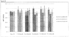
- the cryopreserved skin equivalents are preferable storable at approximately -50°C, - 60°C, -70°C, -80°C or colder for an extended period of time such as greater than 1, 2, 3, 4, 5 or 6 months and up to 12 or 24 months without a substantial loss of viability.
- cryopreservation process may comprise treating an organotypically cultured skin equivalent in a cryoprotectant solution.
- the cryoprotectant may be glycerol.
- the cryoprotectant may be provided in different concentrations in the cryoprotectant solution.
- the cryoprotectant may be provided in a solution comprising about 20% or 21% to about 70% of the solution by volume, and more preferably about 20% or 21% to about 45% of the solution by volume or 37.5% to 62.5% of the solution by volume, or most preferably from about 25% to 40% of the solution by volume or 42.5% to 57.5% of the solution by volume, depending on the temperature.
- the cryoprotectant solution may preferably comprise about 32.5% v/v or about 50% v/v cryoprotectant (e.g., glycerol).
- the cryoprotectant may be provided in a base medium solution.
- Suitable base medium solutions include, but are not limited to, DMEM, Ham's F-10, Ham's F-12, DMEM/F-12, Medium 199, MEM and RPMI.
- the base medium may form the remainder of the solution volume.
- the cryoprotectant solution may be buffered.
- Suitable buffers include, but are not limited to, HEPES, Tris, MOPS, and Trizma buffers. Buffering agents may be included at an amount to provide a buffered system in the range of pH 7.0 to 7.4.
- the cryoprotectant solution may be buffered with from about 5 mM to 15 mM HEPES, most preferably about 10 mM HEPES to a pH of about 7.0 to 7.4.
- Treatment with the cryoprotectant solution may be conducted in a single step.
- single step it is meant that the cryoprotectant solution is not exchanged during the equilibration procedure as is common in the art.
- the treatment step may be performed using a cryoprotectant solution with a defined concentration of cryoprotectant as opposed to a stepwise equilibration procedure where several media changes with increasing concentrations of cryoprotectant at each step.
- the treatment step may be conducted at a reduced temperature.
- the treatment step may be conducted at from about 2°C to 8°C, while in other examples, the treatment step may be conducted at room temperature, for example from about 15°C to 30°C.
- the skin equivalent may be incubated in the cryoprotectant solution for about 10 to 60 minutes, preferably from about 20 to 30 minutes.
- the skin equivalent may be frozen following treatment with the cryoprotectant solution. Excess cryoprotectant solution may be removed from the skin equivalent prior to freezing, for example by aspirating the solution or moving the treated skin equivalent to a fresh vessel. Accordingly, the treated skin equivalent may be frozen by exposure to temperatures ranging from about -50°C to -100°C, and most preferably at about -80°C. Treated skin equivalent may be simply placed in a bag or other vessel such as a culture dish and placed in a freezing unit such as a low temperature (e.g., -80°C freezer) freezing unit.
- a low temperature e.g., -80°C freezer
- culture vessel refers to any vessel of the type commonly used to culture cells or tissues and include circular, rectangular, and square dishes formed from a suitable material such as tissue culture plastic, polystyrene, polymers, plastics, glass, etc.
- the cryopreserved skin equivalent may be packaged for long term storage.
- the skin equivalent may be placed in a bag or culture vessel as described above.
- the bag or culture vessel may be sealed, preferably heat sealed in a sterile bag (e.g., a plastic or polymer bag) to provide a primary package.
- the primary package is then sealed inside a secondary bag, for example a secondary plastic, foil, or Mylar bag.
- the cryopreserved tissues of the present description may preferably be stored at low temperature, from about -50°C to about -100°C, preferably about -80°C.
- the skin equivalents may be preferably stored from about 1, 2, 3, 4, 5 or 6 months and up to 12 or 24 months without a substantial loss of viability.
- the present description provides a method of thawing a cryopreserved skin equivalent prior to application to a subject, comprising warming said cryopreserved skin equivalent and contacting said cryopreserved skin equivalent with an absorbent medium comprising a tissue compatible solution to allow removal of said cryoprotectant solution by diffusion.
- the cryopreserved skin equivalent in a suitable bag or culture vessel may be simply placed on a bench or table top and allowed to thaw. Thawing under controlled conditions as is common in the art is not necessary.
- Cryopreserved skin equivalent may be placed on an absorbent medium to remove thawed cryoprotectant solution from the skin equivalent. There is no limitation to the use a particular absorbent medium.
- Suitable absorbent media include, but are not limited to, Telfa pads, cellulosic pads (e.g., Whatman 1003-090 filter pads and Pall 70010 filter pads), gauze pads, and foam pads (e.g., Covidien 55544 hydrophilic foam pad).
- the absorbent medium may be a Telfa pad.
- the absorbent medium may comprise a tissue-compatible solution.
- the tissue compatible solution may be a buffered solution.
- Suitable tissue compatible solutions include, but are not limited to, DMEM, Ham's F-10, Ham's F-12, DMEM/F-12, Medium 199, MEM and RPMI.
- Suitable buffers include, but are not limited to, HEPES, Tris, MOPS, and Trizma buffers. Buffering agents may be included at an amount to provide a buffered system in the range of pH 7.0 to 7.4.
- kits comprising a cryopreserved skin substitute, preferably provided in a package as described above.
- the kits may further comprise an absorbent medium, and a tissue compatible solution.
- the present description provides a process for forming an organotypically cultured skin equivalent and freezing the skin equivalent in the same culture vessel.
- the culture vessel may comprise an insert movable between upper and lower positions in the culture dish.
- the insert preferably comprises a bottom surface which is a porous membrane.
- the vessel is filled with the appropriate culture medium and a dermal equivalent is formed on the porous membrane.
- the dermal equivalent is then seeded with keratinocytes (e.g., NIKS cells) in the presence of the appropriate culture medium.
- keratinocytes e.g., NIKS cells
- an air interface is created by lowering the level of culture medium in the vessel and the culture is continued until the stratified skin equivalent is formed.
- the culture medium is then removed from the vessel and the insert is lowered to the lower position.
- the cryoprotectant solution is added for treatment and then removed, and the vessel is then transferred to the freezing unit.
- cryopreserved skin equivalents of the present invention may be used therapeutically.
- the cryopreserved skin substitute may be used in wound closure and burn treatment applications.
- the use of autografts and allografts for the treatment of burns and wound closure is described in Myers et al., A. J. Surg. 170(1):75-83 (1995 ) and U.S. Pat. Nos. 5,693,332 ; 5,658,331 ; and 6,039,760 .
- the skin equivalents may be used in conjunction with dermal replacements such as DERMAGRAFT or INTEGRA. Accordingly, the present description provides methods for wound closure, including ulcers or wounds caused by burns, comprising providing a skin equivalent and a patient suffering from a wound and treating the patient with the skin equivalent under conditions such that the wound is closed.
- the skin equivalents may be utilized to treat chronic skin wounds.
- Chronic skin wounds e.g ., venous ulcers, diabetic ulcers, pressure ulcers
- the healing of such a wound often takes well over a year of treatment.
- Treatment options currently include dressings and debridement (use of chemicals or surgery to clear away necrotic tissue), and/or antibiotics in the case of infection. These treatment options take extended periods of time and high levels of patient compliance.
- a therapy that can increase a practitioner's success in healing chronic wounds and accelerate the rate of wound healing would meet an unmet need in the field.
- the present description contemplates treatment of skin wounds with cryopreserved skin equivalents. Skin equivalents may be topically applied to wounds.
- Cryopreserved skin equivalents may be used for application to partial thickness wounds. Cryopreserved skin equivalents may be used to treat full thickness wounds. Cryopreserved skin equivalents may be used to treat numerous types of internal wounds, including, but not limited to, internal wounds of the mucous membranes that line the gastrointestinal tract, ulcerative colitis, and inflammation of mucous membranes that may be caused by cancer therapies. Skin equivalents expressing host defense peptides or pro-angiogenic factors may be used as a temporary or permanent wound dressing.
- the cells may be engineered to provide additional therapeutic agents to a subject.
- therapeutic agents may be delivered to the subject, including, but not limited to, enzymes, peptides, peptide hormones, other proteins, ribosomal RNA, ribozymes, small interfering RNA (siRNA) micro RNA (miRNA), and antisense RNA.
- the agents may be host defense peptides such as human beta-defensin 1, 2, or 3 or cathelicidin or other proteins such as VEGF and HIF-1 ⁇ , see, e.g., U.S. Pat. Nos.
- therapeutic agents may be delivered for a variety of purposes, including but not limited to the purpose of correcting genetic defects.
- the therapeutic agent may be delivered for the purpose of detoxifying a patient with an inherited inborn error of metabolism (e.g., aminoacidopathesis) in which the skin equivalent serves as wild-type tissue. It is contemplated that delivery of the therapeutic agent corrects the defect.
- the cells may be transfected with a DNA construct encoding a therapeutic agent (e.g., insulin, clotting factor IX, erythropoietin, etc.) and skin equivalents prepared from transfected cells are administered to the subject.
- a therapeutic agent e.g., insulin, clotting factor IX, erythropoietin, etc.
- the therapeutic agent is then delivered to the patient's bloodstream or other tissues from the graft.
- the nucleic acid encoding the therapeutic agent may be operably linked to a suitable promoter.
- a suitable promoter There is no limitation to the use of any particular promoter. Indeed, the use of a variety of promoters is contemplated, including, but not limited to, inducible, constitutive, tissue-specific, and keratinocyte-specific promoters.
- the nucleic acid encoding the therapeutic agent may be introduced directly into the keratinocytes (i.e., by electroporation, calcium phosphate co-precipitation, or liposome transfection).
- the nucleic acid encoding the therapeutic agent may be provided as a vector and the vector is introduced into the keratinocytes by methods known in the art.
- the vector may be an episomal vector such as a replicating plasmid.
- the vector may integrate into the genome of the keratinocytes. Examples of integrating vectors include, but are not limited to, retroviral vectors, adeno-associated virus vectors, non-replicating plasmid vectors and transposon vectors.
- kb kilobase
- bp base pair
- PCR polymerase chain reaction
- BSA bovine serum albumin
- CFU colony forming units
- kGy kiloGray
- PVDF polyvinylidine fluoride
- BCA bicinchoninic acid
- SDS-PAGE sodium dodecyl sulfate polyacrylamide gel electrophoresis
- StrataGraft® skin tissue is a living, full-thickness, allogeneic human skin substitute that reproduces many of the structural and biological properties of normal human skin.
- StrataGraft® skin tissue contains both a viable, fully-stratified epidermal layer derived from NIKS® cells, which are a consistent and well-characterized source of pathogen-free human keratinocyte progenitors, and a dermal layer containing normal human dermal fibroblasts (NHDF) embedded in a collagen-rich matrix.
- NIKS® cells which are a consistent and well-characterized source of pathogen-free human keratinocyte progenitors
- NHDF normal human dermal fibroblasts
- StrataGraft® skin tissue possesses excellent tensile strength and handling characteristics that enable it to be meshed, stapled, and sutured similarly to human skin grafts.
- StrataGraft® also exhibits barrier function comparable to that of intact human skin and is capable of delivering bioactive molecules for wound bed conditioning and
- NIKS keratinocytes are expanded in monolayer cell culture.
- NHDF are expanded in monolayer culture and combined with purified type I collagen and culture medium and allowed to gel to form the cellularized dermal equivalent (DE).
- NHDF are seeded into Transwell inserts and allowed to proliferate and secrete and assemble extracellular matrix molecules into a simplified dermal equivalent.
- Stage II NIKS keratinocytes are seeded onto the surface of the DE and cultured under submerged conditions for two days to promote complete epithelialization of the DE surface.
- the tissue is then lifted to the air-liquid interface in Stage III, where it is maintained for 18 days in a controlled, low humidity environment to promote tissue maturation.
- the skin equivalents are generally prepared as described in U.S. Pat. Nos. 7,674,291 ; 7,807,148 ; 7,915,042 ; 7,988,959 ; and 8,092,531 .
- This example describes improved cryopreservation methods for human skin equivalents.
- the production process is unchanged from the current method described above. All tissues in the lot are fed with fresh medium and incubated overnight prior to cryopreservation. Prior to cryopreservation, media samples from all tissues in each lot are tested for sterility. The remaining tissues in each lot are cryopreserved as follows.
- Step 1- Pre-cool 100 mm culture dishes containing 20 ml of cryoprotectant solution to 2-8°C on a stainless steel cold treatment surface inside biosafety cabinet. Temperature of cold treatment surface is maintained at 2-8°C for several hours by contact with frozen gel packs submerged in water.
- Step 2- Transfer Transwells containing StrataGraft® tissues into individual dishes containing pre-cooled cryoprotectant solution. Incubate tissues 20-30 minutes in cryoprotectant on the cold treatment surface.
- Step 3- Transfer treated StrataGraft® tissues to new sterile 100 mm culture dishes containing final product label so that the tissue rests on the bottom of the culture dish and return tissues back to the cold treatment surface. Excess cryoprotectant is allowed to drain from the skin equivalent to provide a treated skin equivalent that is substantially free of excess cryoprotectant on the exterior surfaces of the skin equivalent.
- Step 4- Heat-seal 100 mm culture dishes in clear, sterile bags. Place primary package into secondary Mylar bag, heat-seal, and transfer packaged tissues to cold storage container until all tissues are packaged.
- Step 5- Remove cold storage container with packaged StrataGraft® tissues from cleanroom and transfer tissues to an ultralow freezer (-75°C to -80°C). Place tissues in a pre-cooled rack in the freezer that allows unrestricted airflow to the top and bottom of the packaged tissues to ensure uniform and rapid cooling. Leave tissues undisturbed overnight during the freezing process.
- Tissues are placed into quarantine storage at -70 to -90°C pending results of lot release testing.
- a representative tissue from each lot of cryopreserved StrataGraft® skin tissue is tested using a panel of Quality Control SOP that have historically been used for lot release testing of StrataGraft® tissue.
- Stratatech has established and qualified a panel of lot release assays that are used to characterize StrataGraft® skin tissue. A subset of these lot release assays has been used to monitor and evaluate the impact that changes to the storage conditions may have on key biological and structural characteristics of StrataGraft ®skin tissue (e.g., barrier function, viability, and histological appearance). Although transient minor changes in the histological appearance of StrataGraft® tissue are generally observed following cryopreservation, the histological architecture normalizes after being reintroduced into organotypic culture for several days, indicating that the viable cells in the basal layer of StrataGraft® are able to proliferate and reproduce the epidermal layer after cryopreservation.
- barrier function e.g., barrier function, viability, and histological appearance
- cryoprotectant concentration incubation time, incubation temperature, freezing rate, and storage conditions has enabled Stratatech to identify a cryopreservation process that enables long-term storage of StrataGraft® skin tissue with consistent and defined quality and that meets the specifications that define StrataGraft® skin tissue.
- cryopreserved tissue As anticipated, many of these individual parameters interact to influence the properties of cryopreserved tissue. For example, it is not possible to optimize cryoprotectant concentration without also taking into account the cryoprotectant incubation time and temperature. Likewise, post-thaw incubation temperature influences the allowable range of post-thaw incubation times.
- a range of acceptable values for each of the individual parameters was identified and used to define the final combination of cryoprotectant formulation, pre-freeze incubation time, pre-freeze incubation temperature, freeze rate, and storage condition.
- the operating parameters of the cryopreservation process as developed for cryopreservation and storage of StrataGraft® skin tissue are listed above.
- Glycerol (glycerin) was identified as the most desirable cryoprotectant for StrataGraft ® tissue.
- the glycerol used in the cryoprotectant formulation is synthetic, USP-grade material that undergoes additional testing for endotoxin prior to release for use.
- the cryoprotectant solution contains Dulbecco's Modified Eagle Medium (DMEM) and 10 mM HEPES to maintain pH of 7.0 to 7.4 at ambient atmospheric conditions.
- DMEM was chosen as the base for the cryopreservation solution because it is already a component of the culture medium used to prepare StrataGraft® skin tissue.
- HEPES is a well-characterized buffering agent that maintains the pH of the cryopreservation solution outside of a CO 2 environment.
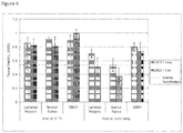
- Glycerol concentrations tested ranged from 16.25% to 65%. In some cases (which are not according to the invention), the concentration of glycerol was gradually increased in two or three steps by incubation in a series of solutions with increasing glycerol concentration.
- tissue cryopreserved with this three-step method revealed that, after thawing and dilution of the cryoprotectant, the tissues exhibited viability (assessed by the ability of viable cells in the tissue to convert MTT to its formazan product), histological architecture, and barrier function comparable to tissues that were not cryopreserved.
- the tissues maintained high levels of viability following re-introduction into organotypic culture for up to nine days after thawing, demonstrating that the metabolic activity detected shortly after thawing was not just residual enzymatic activity.
- cryopreservation of human cells has been accomplished at a more moderate rate of approximately -1°C/min without the use of controlled-rate freezers. This is routinely accomplished by placing vials of cells in an ultra-cold freezer in an insulated box or container designed to moderate the cooling rate to approximately -1°C/min.
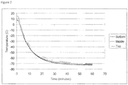
- Temperature monitoring studies were performed to track the temperature of tissues during the cryopreservation process. Following packaging in an inner sterile bag and an outer Mylar bag, StrataGraft® tissues are transferred to a pre-cooled rack inside of an ultra-cold freezer. Each tissue is placed in a separate slot in the freezer rack, with ample room above and below the tissue to allow unrestricted airflow during the freezing process. Using temperature monitoring probes positioned within culture dishes packaged as described above and loaded into freezer racks in this configuration, the temperature rapidly decreases to approximately -50°C within the first 15 minutes, further cools to approximately -65°C by 30 minutes and reaches a final temperature of approximately -80°C after three hours. There is no significant difference in the temperature profiles between tissues placed in the top, middle, and bottom positions of the freezer rack. See Figure 2 .
- tissues were frozen in contact with a layer of cryoprotectant solution after incubation with cryoprotectant. Although tissues cryopreserved in this manner exhibited good post-thaw properties, rapid thawing of tissues frozen in contact with this layer of cryoprotectant required incubation for several minutes in a 35-39°C water bath, which would to be difficult to implement and standardize in a surgical suite. It was subsequently determined that contact with the cryoprotectant solution was not required after tissues had been treated with cryoprotectant. This enabled development of the final product configuration in which tissues are transferred to an empty sterile 100 mm culture dish after treatment with cryoprotectant, where they are frozen in contact with the bottom of the empty dish rather than being frozen in contact with a layer of cryoprotectant solution.
- the 100 mm culture dishes containing cryoprotectant-treated tissues are aseptically packaged and heat-sealed inside of a sterile polyethylene sample bag.
- the inner bag is then heat-sealed inside a puncture-resistant, food grade, metalized polyester/polyethylene bag, which protects the packaged tissues from light, moisture, and provides a barrier to CO 2 vapor during shipment on dry ice.
- the stability and comparability studies described below utilized tissues packaged and cryopreserved in this configuration.
- Cryopreservation of viable skin equivalents enables burn centers to have ready access to this cell-based regenerative medicine therapeutic for burns and other indications that require rapid intervention.
- major burn centers would be able to maintain an inventory of the product for use without the need to schedule a delivery on a case by case basis.
- cryopreserved tissues were stored at -196°C in a vapor-phase nitrogen freezer. Since burn centers do not typically have liquid nitrogen storage capabilities, cryopreservation procedures were developed that permit storage of tissue for at least six months in ultra-cold freezers (-60°C to -90°C), which are readily available in blood and tissue banks at most hospitals and trauma centers.
- Cryopreserved StrataGraft® skin tissue will be shipped to clinical sites on dry ice for next morning delivery via commercial courier such as FedEx or UPS.
- the shipping container (Freezetherm FT29, Laminar Medica) is a validated dry ice shipping box that holds sufficient dry ice to maintain the cryopreserved tissues at ⁇ -75°C for at least 72 hours at ambient temperatures of up to 35°C to account for possible delays in delivery.
- Experimental data indicates that storage of cryopreserved tissues in the dry ice shipping container for >48 hours does not have any detectable adverse effect on tissue viability or histological architecture. See Figure 4 .
- cryopreserved StrataGraft tissues will be stored in an ultra-cold freezer (e.g., -60°C to -90°C) until use.
- cryopreserved StrataGraft tissue Prior to clinical use, cryopreserved StrataGraft tissue is thawed and incubated briefly on pads saturated with culture medium to remove residual cryoprotectant. Due to the geometry of the tissue, the thaw phase is rapid. After tissues are thawed, Transwell inserts containing the tissue are aseptically transferred to the sterile field and placed in sterile dishes containing absorbent pads saturated with culture medium. As described below, the timing of the post-thaw incubation phase is flexible enough to accommodate delays that could be reasonably anticipated during clinical use.
- tissues are cryopreserved in a culture dish without a layer of cryoprotectant solution, which allows the tissues to be thawed rapidly at ambient temperature simply by placing the package onto a bench or table. Precise control over the thaw temperature and time is not required, as experimental data shows that tissues thawed for varying times at temperatures ranging from 22°C to 40°C exhibit similar post-thaw properties.
- Buffered post-thaw incubation solutions work better than unbuffered solutions. Tissues incubated in simple unbuffered salt solutions (lactated Ringer's or normal saline) do not survive as well as tissues incubated in culture media-based solutions. Stratatech's SM01 culture media (StrataLife series) or commercially available DMEM/F12 media buffered with HEPES are preferred. See Figure 6 .
- Tissues can be left on the media saturated pad for times ranging from 15 min to 4 hr at 20-25°C or up to 2 hr at 40°C with no significant effect on tissue viability.
- This example describes improved cryopreservation methods for human skin equivalents utilizing a pre-freeze treatment step with cryopreservation solutions containing 32.5% or 50% glycerol at room temperature.
- the production process is unchanged from the current method described previously.
- the tissues are treated and cryopreserved as follows.
- Step 1- Dispense 20 ml of cryoprotectant solution to 100 mm culture dishes.
- Step 2- Transfer Transwells containing StrataGraft® tissues into individual dishes containing cryoprotectant solution. Incubate tissues 15-45 minutes in cryoprotectant.
- Step 3- Transfer treated StrataGraft® tissues to new sterile 100 mm culture dishes containing final product label so that the tissue rests on the bottom of the culture dish. Excess cryoprotectant is allowed to drain from the skin equivalent to provide a treated skin equivalent that is substantially free of excess cryoprotectant on the exterior surfaces of the skin equivalent.
- Step 4- Heat-seal 100 mm culture dishes in clear, sterile bags. Place primary package into secondary Mylar bag and heat-seal. Step 5- Remove the packaged StrataGraft® tissues from cleanroom and transfer tissues to an ultralow freezer (-75°C to -80°C). Place tissues in a pre-cooled rack in the freezer that allows unrestricted airflow to the top and bottom of the packaged tissues to ensure uniform and rapid cooling. Leave tissues undisturbed overnight during the freezing process.
- Cryopreserved tissues were thawed at room temperature for 10 minutes, transferred to a hold chamber containing Telfa pads saturated with 40 ml of HEPES-buffered culture medium that had been warmed to room temperature, and held at RT for 15 to 20 minutes. Tissues were transferred to a culture dish containing 90 ml of SM01 medium and returned to culture overnight. Tissues were analyzed for viability after overnight re-culture. Tissues pre-treated with 32.5% glycerol at room temperature for 15 to 45 minutes had acceptable post-thaw viability. Tissues treated with 50% glycerol at room temperature for 15 minutes also had acceptable viability; however, tissues treated with 50% glycerol at room temperature for 45 minutes had unacceptable viability. See Figure 10 .
- MTT assays are preferably conducted as follows. Samples are excised from the skin tissue using an 8 mm biopsy punch. The samples are transferred to 0.3 ml MTT Assay Medium (1 mg/ml MTT reagent in Ham's F-12) in a 24-well plate that has been pre-warmed to 37 °C / 5% CO 2 . The samples are incubated for 85-95 minutes at 37 °C / 5% CO 2 . The samples are blotted and transferred to 2 ml isopropanol. The samples are thoroughly mixed to completely extract the purple formazan product. 200 ⁇ l in triplicate of each extract is aliquoted into a 96-well plate, using isopropanol as a blank. The absorbance (550 nm) is measured in a spectrophotometer.
Description
- The present invention relates generally to systems and methods for cryopreservation of viable human skin substitutes.
- The emerging field of tissue engineering (TE) is poised to make enormous progress in the treatment of organ disease and dysfunction in the coming decade. In 2001, there were 23 cell-based therapeutics approved for market in the United States (U.S.) and Europe, of which nine were skin substitutes or grafts, and 100 more products were in development. (De Bree, Genomics-based Drug Data Report and Regenerative Therapy (1) 2:77-96 (2001)). A decade later, engineered tissues have emerged as a discrete industry sector within the wound care industry and represent one of regenerative medicine's most promising cell based therapeutic platforms. The global wound care market was estimated to be $16.8 billion in 2012 and has been growing at a rate of approximately 6% annually (Kalorama Information, April 2012). The bulk of this market is comprised of traditional sectors that are mature and highly competitive, and include products targeting basic and advanced wound care, wound closure, and anti-infectives. This has led competitors to increasingly focus their attention on developing highly differentiated products in the more innovative active wound care sector, a sector that represents approximately 15% of the overall market. While sales of negative pressure wound therapy still predominate, 2010 sales of engineered tissues and other products within the U.S. biologics market grew to $448 million, and are projected to increase to $1.058 billion by 2015, a compound annual growth rate of 18.8% (BioMedGPS-SmarTRAK market analysis, 2012).
- Although a multitude of revolutionary and economically important applications for engineered tissues and organs exist in the human health arena, the full economic potential of the industry is far from realized. At present, only two tissue engineering companies worldwide have been able to commercialize cell based, skin substitute products focused on cutaneous wound healing and achieve annual sales in excess of $100 million.
- A major impediment to the acceptance of engineered tissues by medical practitioners, healthcare providers, and second party payers is the lack of a means to effectively and efficiently preserve and store engineered tissues. The nature of living cells and tissue products makes development of long-term storage challenging. Current engineered tissues must often be stored and shipped under carefully controlled conditions to maintain viability and function. Typically, engineered tissue products take weeks or months to produce but must be used within hours or days after manufacture. As a result, TE companies must continually operate with their production facilities at top capacity and absorb the costs of unsold product which must be discarded. These inventory losses, on top of already costly manufacturing process, have forced prices to impractical levels. As one specific example, APLIGRAF requires about four weeks to manufacture, is usable for only 15 days and must be maintained between 20 and 23°C until used. As another example, EPICEL is transported by a nurse from Genzyme Biosurgery's production facility in Cambridge, MA to the point of use in a portable incubator and is used immediately upon arrival. Such constraints represent significant challenges to developing convenient and cost-effective products.
- Cryopreservation has been explored as a solution to the storage problem, but it is known to induce tissue damage through ice formation, chilling injury, and osmotic imbalance. Besides APLIGRAF, the only other approved living skin equivalent, ORCEL, has been evaluated as a frozen product but had the drawback that it must be maintained at temperatures below -100°C prior to use. This requires specialized product delivery and storage conditions, including the use of dangerous goods during transport, and use of liquid nitrogen for storage, which is expensive, dangerous, and not readily available in rural clinics and field hospitals.
- Accordingly, what is needed in the art are improved methods of cryopreserving viable engineered tissues and cells for storage under conditions that are routinely available at the point of use.
- Methods of cryopreservation of a composite living skin equivalent which comprises an epidermal layer of cultured keratinocyte cells, a layer of high purity, non-porous collagen, and a dermal layer of cultured fibroblast cells in a porous, cross-linked collagen sponge is known from
WO 02/058588 A2 WO 02/058588 A2 WO 2005/012492 A2 discloses a method for providing a skin equivalent expressing an exogenous antimicrobial polypeptide, wherein such antimicrobial polypeptide is human beta-defensin WO 2005/012492 A2 does not disclose a method of cryopreserving the skin equivalents as defined in the appended claims. A method of cryopreserving an organotypically cultured skin equivalent to maintain viable tissue is disclosed inWO 96/24018 A1 WO 96/24018 A1 claim 1 of the enclosed claim set has been engineered to express and provide exogenous antimicrobial polypeptides or proangiogenic factors. Said differences provide for a simplified method of cryopreserving an organotypically cultured skin equivalent that has been engineered to express and provide exogenous antimicrobial polypeptides or proangiogenic factors and has been found to be inventive over said prior artWO 96/24018 A1 WO 02/058588 A2 2005/012492 A2 . - The invention is set out in the appended claims. Namely, the invention is directed to a method of cryopreserving an organotypically cultured skin equivalent to maintain viable tissue comprising: a) treating an organotypically cultured skin equivalent in a cryoprotectant solution in a single step, wherein said organotypically cultured skin equivalent comprises stratified squamous epithelia on a dermal layer comprising fibroblast, wherein the skin equivalent has been engineered to express and provide exogenous antimicrobial polypeptides or proangiogenic factors, and wherein said cryoprotectant is provided in a solution comprising 21% to 70% of the solution by volume, preferably 21% to 45% of the solution by volume or 37.5% to 62.5% of the solution by volume, more preferably 25% to 40% of the solution by volume or 42.5% to 57.5% of said solution by volume, depending on the temperature, and said cryoprotectant is glycerol; b) separating the treated organotypically cultured skin equivalent from excess cryoprotectant solution, and packaging said treated organotypically cultured skin equivalent in the absence of additional cryoprotectant to provide a packaged skin equivalent, wherein said packaging preferably further comprises enclosing said cryopreserved skin equivalent in a sterile bag and enclosing said sterile bag in a second bag; and c) freezing said packaged organotypically cultured skin equivalent to provide a cryopreserved skin equivalent. Furthermore, the invention is directed to a packaged cryopreserved skin equivalent produced according to said method for use in the treatment of a patient, wherein prior to application to said patient said cryopreserved skin equivalent is a) thawed and not rinsed; or b) warmed, aseptically transferred from said package and contacted with an absorbent medium comprising a tissue compatible solution to allow removal of said cryoprotectant solution by diffusion. The embodiments of the description which do not fall within the scope of said claims are provided for illustrative purposes only and do not form part of the present invention. The embodiments of the description which do not fall within the scope of the claims are provided for illustrative purposes only and do not form part of the present invention. The present invention relates generally to systems and methods for cryopreservation of viable human skin substitutes.
- The present description provides methods of cryopreserving an organotypically cultured skin equivalent to maintain viable tissue comprising: treating an organotypically cultured skin equivalent in a cryoprotectant solution in a single step; packaging the organotypically cultured skin equivalent to provide a packaged skin equivalent; and freezing the organotypically cultured skin equivalent to provide a packaged cryopreserved skin equivalent. The cryoprotectant may be provided in a solution comprising about 20% or 21% to about 70% of the solution by volume, and more preferably about 20% or 21% to about 45% of the solution by volume or 37.5% to 62.5% of the solution by volume, or most preferably from about 25% to 40% of the solution by volume or 42.5% to 57.5% of the solution by volume, depending on the temperature. The treatment with cryoprotectant may be conducted at from about 2°C to 8°C, while in other examples, the treatment step is conducted at room temperature, for example from about 15°C to 30°C. The cryoprotectant is glycerol. The freezing may further comprise freezing the organotypically cultured skin equivalent in the absence of substantial excess cryoprotectant. The freezing may further comprise freezing at about -80°C. The freezing may further comprise direct exposure to temperatures ranging from about -50°C to -100°C. The packaging may further comprise enclosing the cryopreserved skin equivalent in a sterile bag and enclosing the sterile bag in a second bag. The organotypically cultured skin equivalents may comprise NIKS cells. The NIKS cells may comprise an exogenous nucleic acid sequence encoding an exogenous polypeptide. The skin equivalent may retain viability after thawing. The skin equivalent may have an A550 of at least 50% of a reference skin equivalent as determined by an MTT assay.
- The methods may further comprise thawing said cryopreserved skin equivalent and applying said thawed skin equivalent to a patient in need thereof, wherein said thawed skin equivalent is not rinsed prior to said application to said patient. The present description provides methods of thawing a cryopreserved skin equivalent prior to application to a subject, comprising: warming the cryopreserved skin equivalent; and contacting the cryopreserved skin equivalent with a diffusion mediator comprising a tissue compatible solution to allow removal of the cryoprotectant solution by diffusion. The warming may comprise exposure to room temperature at the site of use. The diffusion mediator may be selected from the group consisting of an absorbent medium, a membrane, and a dialysis bag. The absorbent medium may be selected from the group consisting of Telfa pads, foam pads, gauze pads, and cellulosic pads containing the tissue compatible medium. The tissue compatible solution may be a buffered solution.
- The present description provides methods of treating a subject comprising providing a packaged cryopreserved skin equivalent produced as described above; aseptically transferring the cryopreserved skin equivalent from the package; warming the cryopreserved skin equivalent; contacting the cryopreserved skin equivalent with an absorbent medium comprising a tissue compatible solution to allow removal of the cryoprotectant solution by diffusion; and applying the cryopreserved skin equivalent to the subject.
- The present description provides a cryopreserved skin equivalent equilibrated with a cryoprotectant, the skin equivalent being substantially free of excess cryoprotectant on the exterior surface of the skin equivalent. The present description provides a system comprising the foregoing skin equivalent disposed on an absorbent medium.
- The present description provides methods comprising providing the packaged cryopreserved skin equivalent as described above; and applying the skin equivalent to a wound under conditions such that the skin equivalent contacts the wound.
- The present description provides a kit comprising a cryopreserved skin substitute, an absorbent medium, and a tissue compatible solution. The cryopreserved skin substitute may be packaged in a sealable enclosure. The cryopreserved skin substitute may be provided in a culture vessel packaged in the bag.
- The present description provides a method comprising: providing a culture dish comprising a cell culture substrate movable between defined upper and lower positions in the culture dish, forming a skin equivalent on the cell culture substrate, wherein the cell culture substrate is at the upper position, lowering the cell culture substrate to the lower position for further processing. The further processing may comprise treating the skin equivalent with a cryoprotectant solution. The further processing may comprise freezing the skin equivalent in the culture dish.
- The present description provides a method of producing a cryopreserved skin equivalent comprising: providing a culture dish comprising an insert movable between upper and lower positions in the culture dish, the insert having a bottom planar surface formed from a porous membrane, forming a dermal equivalent comprising fibroblast cells on the porous membrane in the insert, wherein the insert is placed the upper position the culture dish, culturing the fibroblast cells to form a dermal equivalent, applying keratinocyte cells to the dermal equivalent, culturing the keratinocytes in a culture medium under conditions such that the keratinocytes form a skin equivalent comprising stratified epithelium, removing the culture medium, lowering the insert to the lower position, treating the skin equivalent with a cryoprotectant solution, and freezing the skin equivalent in the culture dish.
- The present disclosure relates to methods of treating a patient in need thereof with a cryopreserved skin equivalent made by the foregoing methods comprising thawing said cryopreserved skin equivalent and applying said thawed skin equivalent to said patient in need thereof, wherein said thawed skin equivalent is not rinsed prior to said application to said patient. For the sake of clarity, this aspect of the present disclosure is not part of the invention as defined in the appended claims.
-
-
FIG. 1 is a graph of viability testing of thawed tissues using the MTT assay. The tissues were cryopreserved using a controlled-rate freezer (CRF) or by passive freezing in an ultra-cold freezer (set to -80 °C) and stored for up to 3 months, then thawed and analyzed at 1 hour and 1 day. Viability was measured by MTT analysis of three 0.5 cm2 punches from each 44 cm2 tissue at each time point (mean +/- 1 st.dev.). -
FIG. 2 is a temperature profile from a simulated passive tissue freeze. Temperature probes were affixed to the bottom of three 100 mm x 20 mm dishes with a Transwell insert on top. The lid was placed on the dish and then they were packaged within an inner Whirl-pak pouch and an outer Mylar pouch and placed in an ultra-cold freezer set to -80 °C. The packaged tissues were placed in a metal rack at a bottom, middle, and top position. -
FIG. 3 is a graph of viability testing of thawed tissues using the MTT assay. The tissues were cryopreserved using a CRF and then stored in ultra-cold freezers set to -80 °C or -50 °C for 1 to 6 months, then thawed and analyzed at 1 hour and 1 day. Viability was measured by MTT analysis of three 0.5 cm2 punches from each 44 cm2 tissue at each time point (mean +/- 1 st.dev.). -
FIG. 4 is a graph of viability testing of thawed tissues using the MTT assay. Tissues from two independent lots were cryopreserved and then stored in an ultra-cold freezer set to - 80 °C for greater than 1 month. Two tissues from each lot were placed into a container of dry ice for 48 hours, with two control tissues from each lot remaining in the ultra-cold freezer. At the end of the dry ice exposure, all tissues were thawed and analyzed at 15 minutes and 1 day. Viability was measured by MTT analysis of four 0.5 cm2 punches from each 44 cm2 tissue at each time point (mean +/- 1 st.dev.). -
FIG. 5 is a graph of viability testing of thawed tissues using the MTT assay. The tissues were cryopreserved using a CRF following a stepwise temperature reduction (room temperature to 2-8 °C to -20 °C) during which the tissues were exposed to a graded series of glycerol concentrations soaked into sterile cellulose pads. All tissues were initially exposed to 16.3% glycerol at room temperature. Next, all tissues were switched to 2-8 °C, with two tissues transferred to 32.5% glycerol. Lastly, all tissues were switched to -20 °C, with one tissue transferred to 65% glycerol. Tissues were stored in vapor-phase LN2 for 6 days. Viability was measured by MTT analysis of three 0.5 cm2 punches from each 44 cm2 tissue at 1 hour and 1 day post-thaw (mean +/- 1 st.dev.). -
FIG. 6 is a graph of viability testing of thawed tissues using the MTT assay. The tissues were cryopreserved in 65% glycerol by passive freezing in an ultra-cold freezer set to -80 °C and then stored at -80 °C for 6 weeks. Tissues were thawed and placed in a hold chamber, consisting of two cellulose filter pads on a raised stainless steel lifter, containing 100 ml of the specified solution and held for 1 hour at the specified temperature. Tissues were analyzed after 1 hour and 1 day. Viability was measured by MTT analysis of three 0.5 cm2 punches from each 44 cm2 tissue at each time point (mean +/- 1 st.dev.). -
FIG. 7 is a graph of viability testing of thawed tissues using the MTT assay. The tissues were cryopreserved in 65% glycerol and then stored in vapor-phase LN2 for 2 weeks. One tissue was thawed directly into a growth chamber without a cellulose pad, while the other was placed in a hold chamber, consisting of 2 cellulose filter pads on a raised stainless steel lifter, containing 100 ml of the growth media. The thawed tissues were held for 1 hour at 37 °C. Tissues were analyzed after 1 hour and 1 day. Viability was measured by MTT analysis of three 0.5 cm2 punches from each 44 cm2 tissue at each time point (mean +/- 1 st.dev.). -
FIG. 8 is a graph of viability testing of thawed tissues using the MTT assay. Tissues from one lot were cryopreserved and then stored in an ultra-cold freezer set to -80 °C for 1 week. The tissues were thawed for 10 minutes, placed in hold chambers containing either Telfa pads or Whatman pads soaked with 40 ml of a buffered nutrient solution warmed to either 37 °C or room temperature (n=2 tissues per thaw condition). The tissues were placed in the hold chambers for 15-20 minutes, and then re-cultured in a growth chamber for 1 day. The tissues were analyzed at 1 day post-thaw. Viability was measured by MTT analysis of four 0.5 cm2 punches from each 44 cm2 tissue (mean +/- 1 st.dev.). The viability specification is indicated by the red dashed line. -
FIG. 9 is a graph of viability testing of thawed tissues using the MTT assay. Tissues from three independent lots were treated with 50% glycerol at 2 to 8 °C and cryopreserved as described in Example 2. Cryopreserved tissues were stored in an ultra-cold freezer at approximately -80 °C for up to 12 months. After 0, 2, 3, 5, and 12 months of storage, two tissues from each lot were thawed at room temperature and held on media saturated Telfa pads for 15 minutes. Tissues were then returned to culture overnight in a culture dish with a raised stainless steel lifter, containing 90 ml of growth media. Viability was measured by MTT analysis of four 0.5 cm2 punches from each 44 cm2 tissue at each time point (mean +/- 1 st.dev.). -
FIG. 10 is a graph of viability testing of thawed tissues using the MTT assay. Tissues from one lot were treated with the specified glycerol concentration (32.5% or 50%) at the listed conditions prior to the freeze and then stored in an ultra-cold freezer set to -80 °C for 9 days. The tissues were thawed for 10 minutes, placed on a Telfa stack soaked with 40 ml of a buffered nutrient solution for 15-20 minutes, and then re-cultured in a growth chamber for 1 day. The tissues were analyzed at 1 day post-thaw. Viability was measured by MTT analysis of four 0.5 cm2 punches from each 44 cm2 tissue (mean +/- 1 st.dev.). The viability specification is indicated by the red dashed line. - As used herein, the terms "skin equivalent", "human skin equivalent", "human skin substitute", and "organotypic cultures" are used interchangeably to refer to an in vitro derived culture of keratinocytes that has stratified into squamous epithelia. Typically, the skin equivalents are produced by organotypic culture and include a dermal layer in addition to a keratinocyte layer.
- As used herein, the term "sterile" refers to a skin equivalent that is essentially or completely free of detectable microbial or fungal contamination.
- As used herein, the term "NIKS cells" refers to cells having the characteristics of the cells deposited as cell line ATCC CRL-1219. NIKS stands for near-diploid immortalized keratinocytes.
- As used herein, the term "viable" when used in reference to a skin equivalent refers to the viability of cells in the skin equivalent following cryopreservation. In preferred embodiments, a "viable" skin has an A550 of at least 50%, 60%, 70%, 80% or 90% of a control non-cryopreserved tissue as measured by an MTT assay or at least 50%, 60%, 70%, 80% or 90% of the readout value of a similar viability assay.
- The invention is set out in the appended claims. Namely, the invention is directed to a method of cryopreserving an organotypically cultured skin equivalent to maintain viable tissue comprising: a) treating an organotypically cultured skin equivalent in a cryoprotectant solution in a single step, wherein said organotypically cultured skin equivalent comprises stratified squamous epithelia on a dermal layer comprising fibroblast, wherein the skin equivalent has been engineered to express and provide exogenous antimicrobial polypeptides or proangiogenic factors, and wherein said cryoprotectant is provided in a solution comprising 21% to 70% of the solution by volume, preferably 21% to 45% of the solution by volume or 37.5% to 62.5% of the solution by volume, more preferably 25% to 40% of the solution by volume or 42.5% to 57.5% of said solution by volume, depending on the temperature, and said cryoprotectant is glycerol; b) separating the treated organotypically cultured skin equivalent from excess cryoprotectant solution, and packaging said treated organotypically cultured skin equivalent in the absence of additional cryoprotectant to provide a packaged skin equivalent, wherein said packaging preferably further comprises enclosing said cryopreserved skin equivalent in a sterile bag and enclosing said sterile bag in a second bag; and c) freezing said packaged organotypically cultured skin equivalent to provide a cryopreserved skin equivalent. Furthermore, the invention is directed to a packaged cryopreserved skin equivalent produced according to said method for use in the treatment of a patient, wherein prior to application to said patient said cryopreserved skin equivalent is a) thawed and not rinsed; or b) warmed, aseptically transferred from said package and contacted with an absorbent medium comprising a tissue compatible solution to allow removal of said cryoprotectant solution by diffusion. The embodiments of the description which do not fall within the scope of said claims are provided for illustrative purposes only and do not form part of the present invention. The embodiments of the description which do not fall within the scope of the claims are provided for illustrative purposes only and do not form part of the present invention.
- The present invention relates generally to systems and methods for cryopreservation of human skin substitutes. In particular, the present invention relates to methods for cryopreserving viable human skin equivalents so that they can be stored for prolonged periods at the site of use, such as a hospital, operating room or burn unit. The methods disclosed herein allow for novel increases in efficiency of the preservation process and utilization of preserved skin equivalents, including single-step equilibration in a cryoprotectant, packaging of the skin equivalent in a sterile package prior to cryopreservation, and the ability to use the cryopreserved tissues for direct application to a patient (e.g., in a grafting procedure) without rinsing. A ready-to-use cryopreserved skin equivalents for use in treatment of a patient is provided, preferably for use in grafting procedures. The cryopreserved skin equivalent is designed for long term storage at the site of use. The cryopreserved equivalents may be engineered to deliver the broad spectrum human host defense peptides such as β-defensin-3 (hBD-3) or cathelicidin (hCAP18/LL-37), or pro-angiogenic factors, to the wound bed.
- Previously, cadaver skin has been harvested and cryopreserved by treatment with from 10% to 20% glycerol as a cryopreservative. See e.g., Kagan et al., Clin Lab Med 25 (2005) 587-605. Surprisingly, it has been found that increased glycerol concentrations are needed to cryopreserve human skin equivalents.
- Accordingly, in some examples, the present description provides a cryopreserved skin equivalent. In some examples, the skin equivalent has been engineered to express and provide exogenous antimicrobial polypeptides or pro-angiogenic factors. There is no limitation to the use of any particular antimicrobial polypeptide. The antimicrobial polypeptide may be human β-defensin-1, human β-defensin-2, human β-defensin-3, or cathelicidin (hCAP-18/LL37) or variant. Nucleic acid constructs or vectors encoding the antimicrobial polypeptide or pro-angiogenic factor may be introduced into the keratinocytes (e.g., NIKS cells) and the transfected keratinocytes are used to make the skin equivalent by organotypic culture techniques. Examples for the production of skin equivalents expressing exogenous polypeptides, as well as additional wild-type and variant antimicrobial polypeptides can be found in
U.S. Pat. Nos. 7,674,291 ;7,807,148 ;7,915,042 ;7,988,959 ; and8,092,531 . - The cryopreserved skin equivalents may be applied to wounds after thawing and left in place. The cryopreserved skin equivalents may be applied temporarily to wounds. The cryopreserved skin equivalents may be removed and replaced with additional cryopreserved human skin equivalents.
- There is not limitation to the use of any particular source of cells that are capable of differentiating into squamous epithelia. Indeed, the use of a variety of cell lines and sources that can differentiate into squamous epithelia, including both primary and immortalized keratinocytes is contemplated. Sources of cells include keratinocytes and dermal fibroblasts biopsied from humans and cavaderic donors (Auger et al., In Vitro Cell. Dev. Biol. - Animal 36:96-103;
U.S. Pat. Nos. 5,968,546 and5,693,332 ), neonatal foreskins (Asbill et al., Pharm. Research 17(9): 1092-97 (2000); Meana et al., Burns 24:621-30 (1998);U.S. Pat. Nos. 4,485,096 ;6,039,760 ; and5,536,656 ), and immortalized keratinocytes cell lines such as NM1 cells (Baden, In Vitro Cell. Dev. Biol. 23(3):205-213 (1987)), HaCaT cells (Boucamp et al., J. cell. Boil. 106:761-771 (1988)); and NIKS cells (Cell line BC-1-Ep/SL;U.S. Pat. No. 5,989,837 ; ATCC CRL-12191). Each of the mentioned cell lines can be cultured or genetically modified in order to produce a cell line capable of expressing or co-expressing the desired protein(s). NIKS cells may be utilized. The discovery of the novel NIKS human keratinocyte cell line provides an opportunity to genetically engineer human keratinocytes with non-viral vectors. A unique advantage of the NIKS cells is that they are a consistent source of genetically-uniform, pathogen-free human keratinocytes. For this reason, they are useful for the application of genetic engineering and genomic gene expression approaches to provide human skin equivalents with enhanced properties over currently available skin equivalents. NIKS cells, identified and characterized at the University of Wisconsin, are nontumorigenic, karyotypically stable, and exhibit normal growth and differentiation both in monolayer and organotypic culture. NIKS cells form fully stratified skin equivalents in culture. These cultures are indistinguishable by all criteria tested thus far from organotypic cultures formed from primary human keratinocytes. Unlike primary cells however, NIKS cells exhibit an extended lifespan in monolayer culture. This provides an opportunity to genetically manipulate the cells and isolate new clones of cells with new useful properties (Allen-Hoffmann et al., J. Invest. Dermatol., 114(3): 444-455 (2000)). - The NIKS cells arose from the BC-1-Ep strain of human neonatal foreskin keratinocytes isolated from an apparently normal male infant. In early passages, the BC-1-Ep cells exhibited no morphological or growth characteristics that were atypical for cultured normal human keratinocytes. Cultivated BC-1-Ep cells exhibited stratification as well as features of programmed cell death. To determine replicative lifespan, the BC-1-Ep cells were serially cultivated to senescence in standard keratinocyte growth medium at a density of 3 x 10⁵ cells per 100-mm dish and passaged at weekly intervals (approximately a 1:25 split). By
passage 15, most keratinocytes in the population appeared senescent as judged by the presence of numerous abortive colonies which exhibited large, flat cells. However, at passage 16, keratinocytes exhibiting a small cell size were evident. By passage 17, only the small-sized keratinocytes were present in the culture and no large, senescent keratinocytes were evident. The resulting population of small keratinocytes that survived this putative crisis period appeared morphologically uniform and produced colonies of keratinocytes exhibiting typical keratinocyte characteristics including cell-cell adhesion and apparent squame production. The keratinocytes that survived senescence were serially cultivated at a density of 3 x 10⁵ cells per 100-mm dish. Typically the cultures reached a cell density of approximately 8 x 10⁶ cells within 7 days. This stable rate of cell growth was maintained through at least 59 passages, demonstrating that the cells had achieved immortality. The keratinocytes that emerged from the original senescencing population are now termed NIKS. The NIKS cell line has been screened for the presence of proviral DNA sequences for HIV-1, HIV-2, EBV, CMV, HTLV-1, HTLV-2, HBV, HCV, B-19 parvovirus, HPV-16, SV40, HHV-6, HHV-7, HPV-18 and HPV-31 using either PCR or Southern analysis. None of these viruses were detected. - Chromosomal analysis was performed on the parental BC-1-Ep cells at
passage 3 and NIKS cells at passages 31 and 54. The parental BC-1-Ep cells have a normal chromosomal complement of 46, XY. At passage 31, all NIKS cells contained 47 chromosomes with an extra isochromosome of the long arm of chromosome 8. No other gross chromosomal abnormalities or marker chromosomes were detected. The karyotype of the NIKS cells has been shown to be stable to at least passage 54. - The DNA fingerprints for the NIKS cell line and the BC-1-Ep keratinocytes are identical at all twelve loci analyzed demonstrating that the NIKS cells arose from the parental BC-1-Ep population. The odds of the NIKS cell line having the parental BC-1-Ep DNA fingerprint by random chance is 4 x 10-16. The DNA fingerprints from three different sources of human keratinocytes, ED-1-Ep, SCC4 and SCC13y are different from the BC-1-Ep pattern. This data also shows that keratinocytes isolated from other humans, ED-1-Ep, SCC4, and SCC13y, are unrelated to the BC-1-Ep cells or each other. The NIKS DNA fingerprint data provides an unequivocal way to identify the NIKS cell line.
- Loss of p53 function is associated with an enhanced proliferative potential and increased frequency of immortality in cultured cells. The sequence of p53 in the NIKS cells is identical to published p53 sequences (GenBank accession number: M14695). In humans, p53 exists in two predominant polymorphic forms distinguished by the amino acid at codon 72. Both alleles of p53 in the NIKS cells are wild-type and have the sequence CGC at codon 72, which codes for an arginine. The other common form of p53 has a proline at this position. The entire sequence of p53 in the NIKS cells is identical to the BC-1-Ep progenitor cells. Rb was also found to be wild-type in NIKS cells.
- Anchorage-independent growth is highly correlated to tumorigenicity in vivo. For this reason, the anchorage-independent growth characteristics of NIKS cells in agar or methylcellulose-containing medium were investigated. NIKS cells remained as single cells after 4 weeks in either agar- or methylcellulose-containing medium. The assays were continued for a total of 8 weeks to detect slow growing variants of the NIKS cells. None were observed.
- To determine the tumorigenicity of the parental BC-1-Ep keratinocytes and the immortal NIKS keratinocyte cell line, cells were injected into the flanks of athymic nude mice. The human squamous cell carcinoma cell line, SCC4, was used as a positive control for tumor production in these animals. The injection of samples was designed such that animals received SCC4 cells in one flank and either the parental BC-1-Ep keratinocytes or the NIKS cells in the opposite flank. This injection strategy eliminated animal to animal variation in tumor production and confirmed that the mice would support vigorous growth of tumorigenic cells. Neither the parental BC-1-Ep keratinocytes (passage 6) nor the NIKS keratinocytes (passage 35) produced tumors in athymic nude mice.
- NIKS cells were analyzed for the ability to undergo differentiation in both submerged culture and organotypic culture. Techniques for organotypic culture are described in detail in the examples. In particularly preferred embodiments, the organotypically cultured skin equivalents of the present invention comprise a dermal equivalent formed from collagen or a similar material and fibroblasts. The keratinocytes, for example NIKS cells or a combination of NIKS cells and cells from a patient are seeded onto the dermal equivalent and form an epidermal layer characterized by squamous differentiation following the organotypic culture process.
- For cells in submerged culture, the formation cornified envelopes was monitored as a marker of squamous differentiation. In cultured human keratinocytes, early stages of cornified envelope assembly result in the formation of an immature structure composed of involucrin, cystatin-a and other proteins, which represent the innermost third of the mature cornified envelope. Less than 2% of the keratinocytes from the adherent BC-1-Ep cells or the NIKS cell line produce cornified envelopes. This finding is consistent with previous studies demonstrating that actively growing, subconfluent keratinocytes produce less than 5% cornified envelopes. To determine whether the NIKS cell line is capable of producing cornified envelopes when induced to differentiate, the cells were removed from adherent culture and suspended for 24 hours in medium made semi-solid with methylcellulose. Many aspects of terminal differentiation, including differential expression of keratins and cornified envelope formation can be triggered in vitro by loss of keratinocyte cell-cell and cell-substratum adhesion. The NIKS keratinocytes produced as many as and usually more cornified envelopes than the parental keratinocytes. These findings demonstrate that the NIKS keratinocytes are not defective in their ability to initiate the formation of this cell type-specific differentiation structure.
- To confirm that the NIKS keratinocytes can undergo squamous differentiation, the cells were cultivated in organotypic culture. Keratinocyte cultures grown on plastic substrata and submerged in medium replicate but exhibit limited differentiation. Specifically, human keratinocytes become confluent and undergo limited stratification producing a sheet consisting of 3 or more layers of keratinocytes. By light and electron microscopy there are striking differences between the architecture of the multilayered sheets formed in submerged culture and intact human skin. In contrast, organotypic culturing techniques allow for keratinocyte growth and differentiation under in vivo-like conditions. Specifically, the cells adhere to a physiological substratum consisting of dermal fibroblasts embedded within a fibrillar collagen base. The organotypic culture is maintained at the air-medium interface. In this way, cells in the upper sheets are air-exposed while the proliferating basal cells remain closest to the gradient of nutrients provided by diffusion through the collagen gel. Under these conditions, correct tissue architecture is formed. Several characteristics of a normal differentiating epidermis are evident. In both the parental cells and the NIKS cell line a single layer of cuboidal basal cells rests at the junction of the epidermis and the dermal equivalent. The rounded morphology and high nuclear to cytoplasmic ratio is indicative of an actively dividing population of keratinocytes. In normal human epidermis, as the basal cells divide they give rise to daughter cells that migrate upwards into the differentiating layers of the tissue. The daughter cells increase in size and become flattened and squamous. Eventually these cells enucleate and form cornified, keratinized structures. This normal differentiation process is evident in the upper layers of both the parental cells and the NIKS cells. The appearance of flattened squamous cells is evident in the upper epidermal layers and demonstrates that stratification has occurred in the organotypic cultures. In the uppermost part of the organotypic cultures the enucleated squames peel off the top of the culture. To date, no histological differences in differentiation at the light microscope level between the parental keratinocytes and the NIKS keratinocyte cell line grown in organotypic culture have been observed.
- To observe more detailed characteristics of the parental (passage 5) and NIKS (passage 38) organotypic cultures and to confirm the histological observations, samples were analyzed using electron microscopy. Parental cells and the immortalized NIKS human keratinocyte cell line were harvested after 15 days in organotypic culture and sectioned perpendicular to the basal layer to show the extent of stratification. Both the parental cells and the NIKS cell line undergo extensive stratification in organotypic culture and form structures that are characteristic of normal human epidermis. Abundant desmosomes are formed in organotypic cultures of parental cells and the NIKS cell line. The formation of a basal lamina and associated hemidesmosomes in the basal keratinocyte layers of both the parental cells and the cell line was also noted.
- Hemidesmosomes are specialized structures that increase adhesion of the keratinocytes to the basal lamina and help maintain the integrity and strength of the tissue. The presence of these structures was especially evident in areas where the parental cells or the NIKS cells had attached directly to the porous support. These findings are consistent with earlier ultrastructural findings using human foreskin keratinocytes cultured on a fibroblast-containing porous support. Analysis at both the light and electron microscopic levels demonstrate that the NIKS cell line in organotypic culture can stratify, differentiate, and form structures such as desmosomes, basal lamina, and hemidesmosomes found in normal human epidermis.
- The present invention provides cryopreserved viable skin equivalents as defined in the claims. The cryopreserved skin equivalents are preferable storable at approximately -50°C, - 60°C, -70°C, -80°C or colder for an extended period of time such as greater than 1, 2, 3, 4, 5 or 6 months and up to 12 or 24 months without a substantial loss of viability.
- Preferably, all steps of the cryopreservation process prior to product packaging are performed aseptically inside a
Class 100 biosafety cabinet in a Class 10,000 cleanroom. The cryopreservation process may comprise treating an organotypically cultured skin equivalent in a cryoprotectant solution. Certain examples of the present description are not limited to the use of any particular cryoprotectant. The cryoprotectant may be glycerol. The cryoprotectant may be provided in different concentrations in the cryoprotectant solution. The cryoprotectant may be provided in a solution comprising about 20% or 21% to about 70% of the solution by volume, and more preferably about 20% or 21% to about 45% of the solution by volume or 37.5% to 62.5% of the solution by volume, or most preferably from about 25% to 40% of the solution by volume or 42.5% to 57.5% of the solution by volume, depending on the temperature. The cryoprotectant solution may preferably comprise about 32.5% v/v or about 50% v/v cryoprotectant (e.g., glycerol). The cryoprotectant may be provided in a base medium solution. Suitable base medium solutions include, but are not limited to, DMEM, Ham's F-10, Ham's F-12, DMEM/F-12, Medium 199, MEM and RPMI. The base medium may form the remainder of the solution volume. The cryoprotectant solution may be buffered. Suitable buffers include, but are not limited to, HEPES, Tris, MOPS, and Trizma buffers. Buffering agents may be included at an amount to provide a buffered system in the range of pH 7.0 to 7.4. The cryoprotectant solution may be buffered with from about 5 mM to 15 mM HEPES, most preferably about 10 mM HEPES to a pH of about 7.0 to 7.4. - Treatment with the cryoprotectant solution may be conducted in a single step. By "single step" it is meant that the cryoprotectant solution is not exchanged during the equilibration procedure as is common in the art. For example, the treatment step may be performed using a cryoprotectant solution with a defined concentration of cryoprotectant as opposed to a stepwise equilibration procedure where several media changes with increasing concentrations of cryoprotectant at each step. The treatment step may be conducted at a reduced temperature. The treatment step may be conducted at from about 2°C to 8°C, while in other examples, the treatment step may be conducted at room temperature, for example from about 15°C to 30°C. The skin equivalent may be incubated in the cryoprotectant solution for about 10 to 60 minutes, preferably from about 20 to 30 minutes.
- The skin equivalent may be frozen following treatment with the cryoprotectant solution. Excess cryoprotectant solution may be removed from the skin equivalent prior to freezing, for example by aspirating the solution or moving the treated skin equivalent to a fresh vessel. Accordingly, the treated skin equivalent may be frozen by exposure to temperatures ranging from about -50°C to -100°C, and most preferably at about -80°C. Treated skin equivalent may be simply placed in a bag or other vessel such as a culture dish and placed in a freezing unit such as a low temperature (e.g., -80°C freezer) freezing unit. In contrast, it is common in the art to control the rate of freezing either by controlling the temperature in the freezing unit or by placing the tissue to be frozen in a container that allows control of the rate of decrease in temperature. The treated skin equivalent may be placed in a sterile culture vessel for freezing. The term "culture vessel" refers to any vessel of the type commonly used to culture cells or tissues and include circular, rectangular, and square dishes formed from a suitable material such as tissue culture plastic, polystyrene, polymers, plastics, glass, etc.
- The cryopreserved skin equivalent may be packaged for long term storage. The skin equivalent may be placed in a bag or culture vessel as described above. The bag or culture vessel may be sealed, preferably heat sealed in a sterile bag (e.g., a plastic or polymer bag) to provide a primary package. The primary package is then sealed inside a secondary bag, for example a secondary plastic, foil, or Mylar bag. The cryopreserved tissues of the present description may preferably be stored at low temperature, from about -50°C to about -100°C, preferably about -80°C. The skin equivalents may be preferably stored from about 1, 2, 3, 4, 5 or 6 months and up to 12 or 24 months without a substantial loss of viability.
- The present description provides a method of thawing a cryopreserved skin equivalent prior to application to a subject, comprising warming said cryopreserved skin equivalent and contacting said cryopreserved skin equivalent with an absorbent medium comprising a tissue compatible solution to allow removal of said cryoprotectant solution by diffusion. The cryopreserved skin equivalent in a suitable bag or culture vessel may be simply placed on a bench or table top and allowed to thaw. Thawing under controlled conditions as is common in the art is not necessary. Cryopreserved skin equivalent may be placed on an absorbent medium to remove thawed cryoprotectant solution from the skin equivalent. There is no limitation to the use a particular absorbent medium. Suitable absorbent media include, but are not limited to, Telfa pads, cellulosic pads (e.g., Whatman 1003-090 filter pads and Pall 70010 filter pads), gauze pads, and foam pads (e.g., Covidien 55544 hydrophilic foam pad). The absorbent medium may be a Telfa pad. The absorbent medium may comprise a tissue-compatible solution. The tissue compatible solution may be a buffered solution. Suitable tissue compatible solutions include, but are not limited to, DMEM, Ham's F-10, Ham's F-12, DMEM/F-12, Medium 199, MEM and RPMI. Suitable buffers include, but are not limited to, HEPES, Tris, MOPS, and Trizma buffers. Buffering agents may be included at an amount to provide a buffered system in the range of pH 7.0 to 7.4.
- The present description provides kits comprising a cryopreserved skin substitute, preferably provided in a package as described above. The kits may further comprise an absorbent medium, and a tissue compatible solution.
- The present description provides a process for forming an organotypically cultured skin equivalent and freezing the skin equivalent in the same culture vessel. The culture vessel may comprise an insert movable between upper and lower positions in the culture dish. The insert preferably comprises a bottom surface which is a porous membrane. In use, the vessel is filled with the appropriate culture medium and a dermal equivalent is formed on the porous membrane. The dermal equivalent is then seeded with keratinocytes (e.g., NIKS cells) in the presence of the appropriate culture medium. At the appropriate time, an air interface is created by lowering the level of culture medium in the vessel and the culture is continued until the stratified skin equivalent is formed. The culture medium is then removed from the vessel and the insert is lowered to the lower position. The cryoprotectant solution is added for treatment and then removed, and the vessel is then transferred to the freezing unit.
- It is contemplated that the cryopreserved skin equivalents of the present invention as defined in the claims may be used therapeutically. The cryopreserved skin substitute may be used in wound closure and burn treatment applications. The use of autografts and allografts for the treatment of burns and wound closure is described in Myers et al., A. J. Surg. 170(1):75-83 (1995) and
U.S. Pat. Nos. 5,693,332 ;5,658,331 ; and6,039,760 . The skin equivalents may be used in conjunction with dermal replacements such as DERMAGRAFT or INTEGRA. Accordingly, the present description provides methods for wound closure, including ulcers or wounds caused by burns, comprising providing a skin equivalent and a patient suffering from a wound and treating the patient with the skin equivalent under conditions such that the wound is closed. - The skin equivalents may be utilized to treat chronic skin wounds. Chronic skin wounds (e.g., venous ulcers, diabetic ulcers, pressure ulcers) are a serious problem. The healing of such a wound often takes well over a year of treatment. Treatment options currently include dressings and debridement (use of chemicals or surgery to clear away necrotic tissue), and/or antibiotics in the case of infection. These treatment options take extended periods of time and high levels of patient compliance. As such, a therapy that can increase a practitioner's success in healing chronic wounds and accelerate the rate of wound healing would meet an unmet need in the field. Accordingly, the present description contemplates treatment of skin wounds with cryopreserved skin equivalents. Skin equivalents may be topically applied to wounds. Cryopreserved skin equivalents may be used for application to partial thickness wounds. Cryopreserved skin equivalents may be used to treat full thickness wounds. Cryopreserved skin equivalents may be used to treat numerous types of internal wounds, including, but not limited to, internal wounds of the mucous membranes that line the gastrointestinal tract, ulcerative colitis, and inflammation of mucous membranes that may be caused by cancer therapies. Skin equivalents expressing host defense peptides or pro-angiogenic factors may be used as a temporary or permanent wound dressing.
- The cells may be engineered to provide additional therapeutic agents to a subject. There is no limitation to the delivery of any particular therapeutic agent. Indeed, it is contemplated that a variety of therapeutic agents may be delivered to the subject, including, but not limited to, enzymes, peptides, peptide hormones, other proteins, ribosomal RNA, ribozymes, small interfering RNA (siRNA) micro RNA (miRNA), and antisense RNA. The agents may be host defense peptides such as human beta-
defensin U.S. Pat. Nos. 7,674,291 ;7,807,148 ;7,915,042 ;7,988,959 ; and8,092,531 . These therapeutic agents may be delivered for a variety of purposes, including but not limited to the purpose of correcting genetic defects. The therapeutic agent may be delivered for the purpose of detoxifying a patient with an inherited inborn error of metabolism (e.g., aminoacidopathesis) in which the skin equivalent serves as wild-type tissue. It is contemplated that delivery of the therapeutic agent corrects the defect. The cells may be transfected with a DNA construct encoding a therapeutic agent (e.g., insulin, clotting factor IX, erythropoietin, etc.) and skin equivalents prepared from transfected cells are administered to the subject. The therapeutic agent is then delivered to the patient's bloodstream or other tissues from the graft. The nucleic acid encoding the therapeutic agent may be operably linked to a suitable promoter. There is no limitation to the use of any particular promoter. Indeed, the use of a variety of promoters is contemplated, including, but not limited to, inducible, constitutive, tissue-specific, and keratinocyte-specific promoters. The nucleic acid encoding the therapeutic agent may be introduced directly into the keratinocytes (i.e., by electroporation, calcium phosphate co-precipitation, or liposome transfection). The nucleic acid encoding the therapeutic agent may be provided as a vector and the vector is introduced into the keratinocytes by methods known in the art. The vector may be an episomal vector such as a replicating plasmid. The vector may integrate into the genome of the keratinocytes. Examples of integrating vectors include, but are not limited to, retroviral vectors, adeno-associated virus vectors, non-replicating plasmid vectors and transposon vectors. - The following examples are provided.
- In the experimental disclosure which follows, the following abbreviations apply: eq (equivalents); M (Molar); mM (millimolar); µM (micromolar); N (Normal); mol (moles); mmol (millimoles); µmol (micromoles); nmol (nanomoles); g (grams); mg (milligrams); µg (micrograms); ng (nanograms); 1 or L (liters); ml or mL (milliliters); µl or µL (microliters); cm (centimeters); mm (millimeters); µm (micrometers); nm (nanometers); °C (degrees Centigrade); U (units), mU (milliunits); min. (minutes); sec. (seconds); % (percent); kb (kilobase); bp (base pair); PCR (polymerase chain reaction); BSA (bovine serum albumin); CFU (colony forming units); kGy (kiloGray); PVDF (polyvinylidine fluoride); BCA (bicinchoninic acid); SDS-PAGE (sodium dodecyl sulfate polyacrylamide gel electrophoresis).
- StrataGraft® skin tissue is a living, full-thickness, allogeneic human skin substitute that reproduces many of the structural and biological properties of normal human skin. StrataGraft® skin tissue contains both a viable, fully-stratified epidermal layer derived from NIKS® cells, which are a consistent and well-characterized source of pathogen-free human keratinocyte progenitors, and a dermal layer containing normal human dermal fibroblasts (NHDF) embedded in a collagen-rich matrix. StrataGraft® skin tissue possesses excellent tensile strength and handling characteristics that enable it to be meshed, stapled, and sutured similarly to human skin grafts. StrataGraft® also exhibits barrier function comparable to that of intact human skin and is capable of delivering bioactive molecules for wound bed conditioning and tissue regeneration. The physical and biological characteristics of StrataGraft skin tissue make it ideal for the treatment of a variety of skin wounds.
- The manufacturing process for StrataGraft® skin tissue encompasses three sequential cell and tissue culture processes. In Stage I of the manufacturing process, NIKS keratinocytes are expanded in monolayer cell culture. Concurrent with the NIKS keratinocyte culture in Stage I, NHDF are expanded in monolayer culture and combined with purified type I collagen and culture medium and allowed to gel to form the cellularized dermal equivalent (DE). Alternatively, NHDF are seeded into Transwell inserts and allowed to proliferate and secrete and assemble extracellular matrix molecules into a simplified dermal equivalent. In Stage II, NIKS keratinocytes are seeded onto the surface of the DE and cultured under submerged conditions for two days to promote complete epithelialization of the DE surface. The tissue is then lifted to the air-liquid interface in Stage III, where it is maintained for 18 days in a controlled, low humidity environment to promote tissue maturation. The skin equivalents are generally prepared as described in
U.S. Pat. Nos. 7,674,291 ;7,807,148 ;7,915,042 ;7,988,959 ; and8,092,531 . - This example describes improved cryopreservation methods for human skin equivalents. The production process is unchanged from the current method described above. All tissues in the lot are fed with fresh medium and incubated overnight prior to cryopreservation. Prior to cryopreservation, media samples from all tissues in each lot are tested for sterility. The remaining tissues in each lot are cryopreserved as follows.
Parameter Operating Range Cryoprotectant formulation 50% (v/v) glycerol DMEM (IX) 10 mM HEPES (pH 7.0 to 7.4) Pre-freeze cryoprotectant incubation temperature 2-8 °C Pre-freeze cryoprotectant incubation time 20-30 minutes Freeze method Direct transfer to -80 °C freezer Storage temperature -70 to -90 °C Shipping conditions Overnight delivery on dry ice - All steps of the cryopreservation process prior to the final product packaging step are performed aseptically inside a
Class 100 biosafety cabinet in a Class 10,000 cleanroom.Step 1- Pre-cool 100 mm culture dishes containing 20 ml of cryoprotectant solution to 2-8°C on a stainless steel cold treatment surface inside biosafety cabinet. Temperature of cold treatment surface is maintained at 2-8°C for several hours by contact with frozen gel packs submerged in water.Step 2- Transfer Transwells containing StrataGraft® tissues into individual dishes containing pre-cooled cryoprotectant solution. Incubate tissues 20-30 minutes in cryoprotectant on the cold treatment surface. Step 3- Transfer treated StrataGraft® tissues to new sterile 100 mm culture dishes containing final product label so that the tissue rests on the bottom of the culture dish and return tissues back to the cold treatment surface. Excess cryoprotectant is allowed to drain from the skin equivalent to provide a treated skin equivalent that is substantially free of excess cryoprotectant on the exterior surfaces of the skin equivalent. Step 4- Heat- seal 100 mm culture dishes in clear, sterile bags. Place primary package into secondary Mylar bag, heat-seal, and transfer packaged tissues to cold storage container until all tissues are packaged.Step 5- Remove cold storage container with packaged StrataGraft® tissues from cleanroom and transfer tissues to an ultralow freezer (-75°C to -80°C). Place tissues in a pre-cooled rack in the freezer that allows unrestricted airflow to the top and bottom of the packaged tissues to ensure uniform and rapid cooling. Leave tissues undisturbed overnight during the freezing process. - Tissues are placed into quarantine storage at -70 to -90°C pending results of lot release testing. A representative tissue from each lot of cryopreserved StrataGraft® skin tissue is tested using a panel of Quality Control SOP that have historically been used for lot release testing of StrataGraft® tissue.
- Stratatech has established and qualified a panel of lot release assays that are used to characterize StrataGraft® skin tissue. A subset of these lot release assays has been used to monitor and evaluate the impact that changes to the storage conditions may have on key biological and structural characteristics of StrataGraft ®skin tissue (e.g., barrier function, viability, and histological appearance). Although transient minor changes in the histological appearance of StrataGraft® tissue are generally observed following cryopreservation, the histological architecture normalizes after being reintroduced into organotypic culture for several days, indicating that the viable cells in the basal layer of StrataGraft® are able to proliferate and reproduce the epidermal layer after cryopreservation. The systematic evaluation of cryoprotectant concentration, incubation time, incubation temperature, freezing rate, and storage conditions has enabled Stratatech to identify a cryopreservation process that enables long-term storage of StrataGraft® skin tissue with consistent and defined quality and that meets the specifications that define StrataGraft® skin tissue.
- The following parameters were systematically evaluated during development of the StrataGraft cryopreservation process.
- Cryoprotectant composition
- Pre-freeze cryoprotectant incubation temperature
- Pre-freeze cryoprotectant incubation time
- Number of steps required during cryoprotectant incubation
- Freezing rate
- Final product packaging
- Storage temperature
- Shipping conditions
- Thaw temperature and time
- Post-thaw cryoprotectant diffusion mediator
- Post-thaw incubation solution
- Post-thaw incubation temperature
- Post-thaw incubation time
- As anticipated, many of these individual parameters interact to influence the properties of cryopreserved tissue. For example, it is not possible to optimize cryoprotectant concentration without also taking into account the cryoprotectant incubation time and temperature. Likewise, post-thaw incubation temperature influences the allowable range of post-thaw incubation times. During development of the cryopreservation process, a range of acceptable values for each of the individual parameters was identified and used to define the final combination of cryoprotectant formulation, pre-freeze incubation time, pre-freeze incubation temperature, freeze rate, and storage condition. The operating parameters of the cryopreservation process as developed for cryopreservation and storage of StrataGraft® skin tissue are listed above.
- Glycerol (glycerin) was identified as the most desirable cryoprotectant for StrataGraft ® tissue. The glycerol used in the cryoprotectant formulation is synthetic, USP-grade material that undergoes additional testing for endotoxin prior to release for use. In addition to glycerol, the cryoprotectant solution contains Dulbecco's Modified Eagle Medium (DMEM) and 10 mM HEPES to maintain pH of 7.0 to 7.4 at ambient atmospheric conditions. DMEM was chosen as the base for the cryopreservation solution because it is already a component of the culture medium used to prepare StrataGraft® skin tissue. HEPES is a well-characterized buffering agent that maintains the pH of the cryopreservation solution outside of a CO2 environment.
- A series of studies were performed to determine an appropriate glycerol concentration for use in the cryoprotectant solution. Glycerol concentrations tested ranged from 16.25% to 65%. In some cases (which are not according to the invention), the concentration of glycerol was gradually increased in two or three steps by incubation in a series of solutions with increasing glycerol concentration. Initial studies to demonstrate the feasibility of cryopreserving StrataGraft® tissue used a three-step process in which the glycerol concentration was sequentially increased to 16.25%, then 32.5%, and finally to 65% by incubation for 15-20 minutes in each glycerol solution, while gradually reducing the temperature at each incubation step (16.25% glycerol incubation at room temperature, 32.5% at 2-8°C, and 65% at -20°C). Finally, tissues were frozen to -140°C at -15 °C/min in a controlled-rate freezer. Cryopreserved tissues were transferred to an ultra-cold freezer (-80°C), where they were maintained until thawed for analysis. While this three-step process could be used to preserve the viability and histological architecture of StrataGraft® tissues during cryopreserved storage, the complexity of having multiple steps with different solutions performed at different temperatures introduces the opportunity for error and is not amenable to process scale-up that would be required for commercialization.
- Analysis of tissues cryopreserved with this three-step method revealed that, after thawing and dilution of the cryoprotectant, the tissues exhibited viability (assessed by the ability of viable cells in the tissue to convert MTT to its formazan product), histological architecture, and barrier function comparable to tissues that were not cryopreserved. The tissues maintained high levels of viability following re-introduction into organotypic culture for up to nine days after thawing, demonstrating that the metabolic activity detected shortly after thawing was not just residual enzymatic activity.
- During these initial studies, it was found that the concentration of glycerol in cryopreserved tissues could be reduced after thawing either by incubating the tissues in a series of solutions with decreasing glycerol concentration, or by placing the tissues in a media reservoir with a filter pad just below the tissue to moderate the diffusion of glycerol. Based on these results, subsequent studies primarily used the single-step approach of incubating thawed tissues in culture medium with a pad to moderate glycerol diffusion. See
Figure 7 . - After demonstrating the feasibility of cryopreserving StrataGraft® skin tissue using the three-step method, we used this method as a benchmark against which to compare simplifications to the cryopreservation method. These studies examined reducing the number of steps required to reach the same final glycerol concentration (65%) or evaluating final glycerol concentrations that were lower than that used in the initial studies. Based on these studies, it was determined that glycerol concentrations as low as 32.5% could be used to reproducibly maintain the viability and histological architecture of StrataGraft® tissue during cryopreserved storage. In contrast, a final glycerol concentration of 16.25% in the cryoprotectant solution did not support maintenance of viability in frozen tissues. See
Figure 5 . By evaluating a range of glycerol concentrations, it was determined that a cryoprotectant solution containing 50% glycerol reproducibly supported cryopreservation of StrataGraft® skin tissue and provided a margin of error above somewhat lower glycerol concentrations (e.g., 32.5%) that also supported efficient cryopreservation. - In addition to the concentration of glycerol in the cryoprotectant solution, three other factors affecting treatment of StrataGraft® tissue with cryoprotectant prior to cryopreservation are: 1) the pre-freeze cryoprotectant incubation time, 2) the temperature at which the tissues are treated with cryoprotectant, and 3) the number of steps required to reach the final glycerol concentration. As described above, initial feasibility studies with the three-step process involved sequentially incubating the tissues in solutions with increasing glycerol concentrations at successively lower temperatures for 15-20 minutes at each step. As stated above, a simpler cryopreservation process is preferred to avoid the complexity of having multiple steps with different solutions performed at different temperatures to reduce the opportunity for error and facilitate process scale-up. Toward this goal, the need for stepwise increase in cryoprotectant concentration and stepwise reduction in temperature during the pre-freeze cryoprotectant equilibration phase was evaluated.
- In a series of studies performed in conjunction with evaluation of different cryoprotectant concentrations, it was determined that StrataGraft® tissues treated with cryoprotectant solutions containing 32.5, 50, or 65% glycerol in a single step at 2-8°C for as little as 15 minutes and as long as 60 minutes were all able to withstand cryopreservation with minimal loss of viability or epidermal architecture. Although no decline in tissue performance was observed with cryoprotectant treatment times up to 60 minutes, relatively short glycerol treatment times (20 to 30 minutes) were chosen in order to minimize any potential adverse effects of prolonged exposure to cryoprotectant prior to freezing.
- As described above, the initial feasibility studies utilized a controlled-rate freezer to freeze tissues at a rate of -15°C/min after equilibration with cryoprotectant. However, the use of a controlled-rate freezer would impose significant additional costs and is not amenable to process scale-up. Historically, cryopreservation of human cells has been accomplished at a more moderate rate of approximately -1°C/min without the use of controlled-rate freezers. This is routinely accomplished by placing vials of cells in an ultra-cold freezer in an insulated box or container designed to moderate the cooling rate to approximately -1°C/min.
- Process development studies were designed to determine whether StrataGraft® tissues could be cryopreserved without using a controlled-rate freezer. These studies demonstrated that tissues frozen by direct transfer to an ultra-cold freezer (approximately -80°C) after treatment with cryoprotectant performed as well as tissues frozen in a controlled-rate freezer. See
Figure 1 . - Temperature monitoring studies were performed to track the temperature of tissues during the cryopreservation process. Following packaging in an inner sterile bag and an outer Mylar bag, StrataGraft® tissues are transferred to a pre-cooled rack inside of an ultra-cold freezer. Each tissue is placed in a separate slot in the freezer rack, with ample room above and below the tissue to allow unrestricted airflow during the freezing process. Using temperature monitoring probes positioned within culture dishes packaged as described above and loaded into freezer racks in this configuration, the temperature rapidly decreases to approximately -50°C within the first 15 minutes, further cools to approximately -65°C by 30 minutes and reaches a final temperature of approximately -80°C after three hours. There is no significant difference in the temperature profiles between tissues placed in the top, middle, and bottom positions of the freezer rack. See
Figure 2 . - In initial studies, tissues were frozen in contact with a layer of cryoprotectant solution after incubation with cryoprotectant. Although tissues cryopreserved in this manner exhibited good post-thaw properties, rapid thawing of tissues frozen in contact with this layer of cryoprotectant required incubation for several minutes in a 35-39°C water bath, which would to be difficult to implement and standardize in a surgical suite. It was subsequently determined that contact with the cryoprotectant solution was not required after tissues had been treated with cryoprotectant. This enabled development of the final product configuration in which tissues are transferred to an empty sterile 100 mm culture dish after treatment with cryoprotectant, where they are frozen in contact with the bottom of the empty dish rather than being frozen in contact with a layer of cryoprotectant solution.
- To maintain the sterility of cryopreserved tissues, the 100 mm culture dishes containing cryoprotectant-treated tissues are aseptically packaged and heat-sealed inside of a sterile polyethylene sample bag. The inner bag is then heat-sealed inside a puncture-resistant, food grade, metalized polyester/polyethylene bag, which protects the packaged tissues from light, moisture, and provides a barrier to CO2 vapor during shipment on dry ice. The stability and comparability studies described below utilized tissues packaged and cryopreserved in this configuration.
- Cryopreservation of viable skin equivalents enables burn centers to have ready access to this cell-based regenerative medicine therapeutic for burns and other indications that require rapid intervention. Optimally, major burn centers would be able to maintain an inventory of the product for use without the need to schedule a delivery on a case by case basis. In early feasibility studies, cryopreserved tissues were stored at -196°C in a vapor-phase nitrogen freezer. Since burn centers do not typically have liquid nitrogen storage capabilities, cryopreservation procedures were developed that permit storage of tissue for at least six months in ultra-cold freezers (-60°C to -90°C), which are readily available in blood and tissue banks at most hospitals and trauma centers. Results of these experiments, demonstrated that while tissues stored at -50°C exhibited significant losses of viability over the course of several weeks, tissues stored at -80°C retained levels of viability comparable to tissues that had been stored in nitrogen vapor. See
Figure 3 . These results were obtained with several independent lots of cryopreserved tissue, confirming the reproducibility of this finding. As described in the Stability of Cryopreserved Tissues section below, analysis of tissues stored at -80°C for six months revealed no significant loss of viability or changes to epidermal architecture during storage. - Cryopreserved StrataGraft® skin tissue will be shipped to clinical sites on dry ice for next morning delivery via commercial courier such as FedEx or UPS. The shipping container (Freezetherm FT29, Laminar Medica) is a validated dry ice shipping box that holds sufficient dry ice to maintain the cryopreserved tissues at <-75°C for at least 72 hours at ambient temperatures of up to 35°C to account for possible delays in delivery. Experimental data indicates that storage of cryopreserved tissues in the dry ice shipping container for >48 hours does not have any detectable adverse effect on tissue viability or histological architecture. See
Figure 4 . Following receipt at the clinical site, cryopreserved StrataGraft tissues will be stored in an ultra-cold freezer (e.g., -60°C to -90°C) until use. - Prior to clinical use, cryopreserved StrataGraft tissue is thawed and incubated briefly on pads saturated with culture medium to remove residual cryoprotectant. Due to the geometry of the tissue, the thaw phase is rapid. After tissues are thawed, Transwell inserts containing the tissue are aseptically transferred to the sterile field and placed in sterile dishes containing absorbent pads saturated with culture medium. As described below, the timing of the post-thaw incubation phase is flexible enough to accommodate delays that could be reasonably anticipated during clinical use.
- As described in the Final product packaging section above, tissues are cryopreserved in a culture dish without a layer of cryoprotectant solution, which allows the tissues to be thawed rapidly at ambient temperature simply by placing the package onto a bench or table. Precise control over the thaw temperature and time is not required, as experimental data shows that tissues thawed for varying times at temperatures ranging from 22°C to 40°C exhibit similar post-thaw properties.
- Buffered post-thaw incubation solutions work better than unbuffered solutions. Tissues incubated in simple unbuffered salt solutions (lactated Ringer's or normal saline) do not survive as well as tissues incubated in culture media-based solutions. Stratatech's SM01 culture media (StrataLife series) or commercially available DMEM/F12 media buffered with HEPES are preferred. See
Figure 6 . - Initial development studies found that buffered culture media were able to support tissue viability following post-thaw incubation at both 37°C and room temperature. However, warm post-thaw incubation temperatures (37°C) work better than cooler temps (20-25°C) for sub-optimal incubation solutions. See
Figure 6 . Starting the post-thaw incubation on a pad containing 37°C media that slowly cools to room temp over the course of 15-30 minutes also works well. Higher temp seems to be most important in the first few minutes after thaw. - Later development studies demonstrated that buffered media solutions pre-warmed to only room temperature were comparable to media warmed to 37°C in their ability to support the properties of thawed tissues. This was true for tissues held on either Telfa pads or Whatman pads. See
Figure 8 - Tissues can be left on the media saturated pad for times ranging from 15 min to 4 hr at 20-25°C or up to 2 hr at 40°C with no significant effect on tissue viability.
- Although many of the studies described above analyzed tissues that had been stored in a frozen state for only a few days or weeks, it is widely accepted that the majority of damage to cryopreserved cells and tissues occurs during the freezing and thawing stages, with relatively little loss of cellular viability taking place during long-term storage at reduced temperatures. Long-term storage results show that cryopreserved StrataGraft® skin tissue maintains high levels of viability and histological architecture after at least 12 months of storage at ultracold temperatures. Analysis of tissues produced and cryopreserved using the cryopreservation process described above indicates that tissues cryopreserved with this process maintain key biological, structural, and physical properties during storage for at least 12 months at ultracold temperatures. See
Figure 9 . - This example describes improved cryopreservation methods for human skin equivalents utilizing a pre-freeze treatment step with cryopreservation solutions containing 32.5% or 50% glycerol at room temperature. The production process is unchanged from the current method described previously. At the end of the production process, the tissues are treated and cryopreserved as follows.
Parameter Operating Range Cryoprotectant formulation 32.5% (v/v) glycerol DMEM (IX) 10 mM HEPES (pH 7.0 to 7.4) or 50% (v/v) glycerol DMEM (IX) 10 mM HEPES (pH 7.0 to 7.4) Pre-freeze cryoprotectant incubation temperature Room temperature Pre-freeze cryoprotectant incubation time 15-45 minutes Freeze method Direct transfer to -80 °C freezer Storage temperature -70 to -90 °C Shipping conditions Overnight delivery on dry ice - All steps of the cryopreservation process prior to the final product packaging step are performed aseptically inside a
Class 100 biosafety cabinet in a Class 10,000 cleanroom.Step 1- Dispense 20 ml of cryoprotectant solution to 100 mm culture dishes. Step 2- Transfer Transwells containing StrataGraft® tissues into individual dishes containing cryoprotectant solution. Incubate tissues 15-45 minutes in cryoprotectant. Step 3- Transfer treated StrataGraft® tissues to new sterile 100 mm culture dishes containing final product label so that the tissue rests on the bottom of the culture dish. Excess cryoprotectant is allowed to drain from the skin equivalent to provide a treated skin equivalent that is substantially free of excess cryoprotectant on the exterior surfaces of the skin equivalent. Step 4- Heat- seal 100 mm culture dishes in clear, sterile bags. Place primary package into secondary Mylar bag and heat-seal.Step 5- Remove the packaged StrataGraft® tissues from cleanroom and transfer tissues to an ultralow freezer (-75°C to -80°C). Place tissues in a pre-cooled rack in the freezer that allows unrestricted airflow to the top and bottom of the packaged tissues to ensure uniform and rapid cooling. Leave tissues undisturbed overnight during the freezing process. - Cryopreserved tissues were thawed at room temperature for 10 minutes, transferred to a hold chamber containing Telfa pads saturated with 40 ml of HEPES-buffered culture medium that had been warmed to room temperature, and held at RT for 15 to 20 minutes. Tissues were transferred to a culture dish containing 90 ml of SM01 medium and returned to culture overnight. Tissues were analyzed for viability after overnight re-culture. Tissues pre-treated with 32.5% glycerol at room temperature for 15 to 45 minutes had acceptable post-thaw viability. Tissues treated with 50% glycerol at room temperature for 15 minutes also had acceptable viability; however, tissues treated with 50% glycerol at room temperature for 45 minutes had unacceptable viability. See
Figure 10 . - MTT assays are preferably conducted as follows. Samples are excised from the skin tissue using an 8 mm biopsy punch. The samples are transferred to 0.3 ml MTT Assay Medium (1 mg/ml MTT reagent in Ham's F-12) in a 24-well plate that has been pre-warmed to 37 °C / 5% CO2. The samples are incubated for 85-95 minutes at 37 °C / 5% CO2. The samples are blotted and transferred to 2 ml isopropanol. The samples are thoroughly mixed to completely extract the purple formazan product. 200 µl in triplicate of each extract is aliquoted into a 96-well plate, using isopropanol as a blank. The absorbance (550 nm) is measured in a spectrophotometer.
Claims (15)
- A method of cryopreserving an organotypically cultured skin equivalent to maintain viable tissue comprising:a) treating an organotypically cultured skin equivalent in a cryoprotectant solution in a single step, wherein said organotypically cultured skin equivalent comprises stratified squamous epithelia on a dermal layer comprising fibroblast, wherein the skin equivalent has been engineered to express and provide exogenous antimicrobial polypeptides or proangiogenic factors, and wherein said cryoprotectant is provided in a solution comprising 21% to 70% of the solution by volume, preferably 21% to 45% of the solution by volume or 37.5% to 62.5% of the solution by volume, more preferably 25% to 40% of the solution by volume or 42.5% to 57.5% of said solution by volume, depending on the temperature, and said cryoprotectant is glycerol;b) separating the treated organotypically cultured skin equivalent from excess cryoprotectant solution, and packaging said treated organotypically cultured skin equivalent in the absence of additional cryoprotectant to provide a packaged skin equivalent, wherein said packaging preferably further comprises enclosing said cryopreserved skin equivalent in a sterile bag and enclosing said sterile bag in a second bag; andc) freezing said packaged organotypically cultured skin equivalent to provide a cryopreserved skin equivalent.
- The method of claim 1, wherein said treating an organotypically cultured skin equivalent in a cryoprotectant solution in a single step is at a temperature of 2°C to 8°C or 15°C to 30°C.
- The method of Claim 1, wherein said freezing further comprises freezing at -80°C or direct exposure to temperatures ranging from -50°C to -100°C.
- The method of Claim 1, wherein said skin equivalent retains viability after thawing, wherein said cryopreserved skin equivalent preferably has an A550 of at least 50% of a reference skin equivalent as determined by an MTT assay.
- A method of thawing a cryopreserved skin equivalent produced by the method of claim 1 prior to application to a patient, comprising:a) warming said cryopreserved skin equivalent, wherein said warming preferably comprises exposure to room temperature at the site of use; andb) contacting said cryopreserved skin equivalent with a diffusion mediator selected from the group consisting of an absorbent medium, a membrane and a dialysis bag, and comprising a tissue compatible solution, preferably a buffered tissue compatible solution, to allow removal of said cryoprotectant solution by diffusion.
- The method of Claim 5, wherein said diffusion mediator is an absorbent medium, wherein said absorbent medium is preferably selected from the group consisting of Telfa pads, foam pads, gauze pads, and cellulosic pads containing said tissue compatible medium.
- A cryopreserved skin equivalent produced by the method of claim 1.
- A system comprising the skin equivalent of Claim 7 placed on an absorbent medium.
- Packaged cryopreserved skin equivalent of Claim 7 for use in the treatment of wounds, wherein said skin equivalent is for application to a wound under conditions such that said skin equivalent contacts said wound.
- A kit comprising a cryopreserved skin equivalent of Claim 7, an absorbent medium, and a tissue compatible solution.
- The kit of Claim 10, wherein said cryopreserved skin equivalent is packaged in a sealable enclosure or said cryopreserved skin equivalent is provided in a culture vessel packaged in a bag.
- The method of claim 1 further comprising forming the organotypically cultured skin equivalent before step (a) by:providing a culture dish comprising a cell culture substrate movable between defined upper and lower positions in said culture dish, forming a skin equivalent on said cell culture substrate, wherein said cell culture substrate is at said upper position, and thenlowering said cell culture substrate to said lower position to perform step (a) of claim 1.
- The method of claim 1 further comprising forming the organotypically cultured skin equivalent before step (a) by:providing a culture dish comprising an insert movable between upper and lower positions in said culture dish, said insert having a bottom planar surface formed from a porous membrane,forming a dermal equivalent comprising fibroblast cells on said porous membrane in said insert, wherein said insert is placed in said upper position said in culture dish,culturing said fibroblast cells to form a dermal equivalent,applying keratinocytes to said dermal equivalent,culturing said keratinocytes in a culture medium under conditions such that said keratinocytes form a skin equivalent comprising stratified epithelium,removing said culture medium,lowering said insert to said lower position to perform step (a) of claim 1.
- Packaged cryopreserved skin equivalent produced according to Claim 1 for use in the treatment of a patient, wherein prior to application to said patient said cryopreserved skin equivalent isa) thawed and not rinsed; orb) warmed, aseptically transferred from said package and contacted with an absorbent medium comprising a tissue compatible solution to allow removal of said cryoprotectant solution by diffusion.
- The method of Claim 1, wherein in step (a) occurs at a temperature of 2°C to 8°C for 15 minutes to 60 minutes the skin equivalent is incubated in the cryoprotectant solution for 10 to 60 minutes, preferably from 20 to 30 minutes.
Priority Applications (1)
| Application Number | Priority Date | Filing Date | Title |
|---|---|---|---|
| EP21177142.3A EP3900534A1 (en) | 2013-03-13 | 2014-03-13 | Method for warming cryoconserved human skin substitutes |
Applications Claiming Priority (3)
| Application Number | Priority Date | Filing Date | Title |
|---|---|---|---|
| US201361779661P | 2013-03-13 | 2013-03-13 | |
| PCT/US2014/025599 WO2014160000A1 (en) | 2013-03-13 | 2014-03-13 | Cryopreservation of viable human skin substitutes |
| EP14775892.4A EP2967836B1 (en) | 2013-03-13 | 2014-03-13 | Cryopreservation of viable human skin substitutes |
Related Parent Applications (1)
| Application Number | Title | Priority Date | Filing Date |
|---|---|---|---|
| EP14775892.4A Division EP2967836B1 (en) | 2013-03-13 | 2014-03-13 | Cryopreservation of viable human skin substitutes |
Related Child Applications (2)
| Application Number | Title | Priority Date | Filing Date |
|---|---|---|---|
| EP21177142.3A Division EP3900534A1 (en) | 2013-03-13 | 2014-03-13 | Method for warming cryoconserved human skin substitutes |
| EP21177142.3A Division-Into EP3900534A1 (en) | 2013-03-13 | 2014-03-13 | Method for warming cryoconserved human skin substitutes |
Publications (2)
| Publication Number | Publication Date |
|---|---|
| EP3470018A1 EP3470018A1 (en) | 2019-04-17 |
| EP3470018B1 true EP3470018B1 (en) | 2021-07-28 |
Family
ID=51527939
Family Applications (3)
| Application Number | Title | Priority Date | Filing Date |
|---|---|---|---|
| EP21177142.3A Pending EP3900534A1 (en) | 2013-03-13 | 2014-03-13 | Method for warming cryoconserved human skin substitutes |
| EP18197432.0A Active EP3470018B1 (en) | 2013-03-13 | 2014-03-13 | Cryopreservation of viable human skin substitutes |
| EP14775892.4A Active EP2967836B1 (en) | 2013-03-13 | 2014-03-13 | Cryopreservation of viable human skin substitutes |
Family Applications Before (1)
| Application Number | Title | Priority Date | Filing Date |
|---|---|---|---|
| EP21177142.3A Pending EP3900534A1 (en) | 2013-03-13 | 2014-03-13 | Method for warming cryoconserved human skin substitutes |
Family Applications After (1)
| Application Number | Title | Priority Date | Filing Date |
|---|---|---|---|
| EP14775892.4A Active EP2967836B1 (en) | 2013-03-13 | 2014-03-13 | Cryopreservation of viable human skin substitutes |
Country Status (10)
| Country | Link |
|---|---|
| US (3) | US10091983B2 (en) |
| EP (3) | EP3900534A1 (en) |
| JP (2) | JP6580028B2 (en) |
| AU (4) | AU2014244264B2 (en) |
| CA (2) | CA3071928A1 (en) |
| DK (2) | DK2967836T3 (en) |
| ES (2) | ES2893844T3 (en) |
| IL (1) | IL241448B (en) |
| PT (2) | PT2967836T (en) |
| WO (1) | WO2014160000A1 (en) |
Families Citing this family (10)
| Publication number | Priority date | Publication date | Assignee | Title |
|---|---|---|---|---|
| CA3071928A1 (en) * | 2013-03-13 | 2014-10-02 | Stratatech Corporation | Cryopreservation of viable human skin substitutes |
| SG11201700097WA (en) | 2014-07-09 | 2017-02-27 | Genentech Inc | Ph adjustment to improve thaw recovery of cell banks |
| WO2016149254A1 (en) | 2015-03-17 | 2016-09-22 | Chimera Bioengineering, Inc. | Smart car devices, de car polypeptides, side cars and uses thereof |
| US11052111B2 (en) | 2015-12-08 | 2021-07-06 | Chimera Bioengineering, Inc. | Smart CAR devices and DE CAR polypeptides for treating disease and methods for enhancing immune responses |
| US10222369B2 (en) | 2016-05-17 | 2019-03-05 | Chimera Bioengineering, Inc. | Methods for making novel antigen binding domains |
| WO2018045177A1 (en) | 2016-09-01 | 2018-03-08 | Chimera Bioengineering, Inc. | Gold optimized car t-cells |
| US11110125B2 (en) | 2018-02-13 | 2021-09-07 | Chimera Bioengineering, Inc. | Coordinating gene expression using RNA destabilizing elements |
| CN112823030A (en) | 2018-08-31 | 2021-05-18 | 威斯康星校友研究基金会 | Generation of vascular grafts for arterial endothelial cell implantation |
| CN114786686A (en) | 2019-08-18 | 2022-07-22 | 嵌合体生物工程公司 | Combination therapy of GOLD-controlled transgenes |
| CN114836291A (en) * | 2021-02-01 | 2022-08-02 | 深圳拜尔洛克生物技术有限公司 | System and method for thawing and recovering biological material |
Family Cites Families (29)
| Publication number | Priority date | Publication date | Assignee | Title |
|---|---|---|---|---|
| US4485096A (en) | 1982-02-26 | 1984-11-27 | Massachusetts Institute Of Technology | Tissue-equivalent and method for preparation thereof |
| IL95429A (en) | 1989-09-15 | 1997-09-30 | Organogenesis | Living tissue equivalents comprising hydrated collagen lattice and a collagen gel and their production |
| HUT63319A (en) | 1990-04-24 | 1993-08-30 | Mark Eisenberg | Method for producing composition equivalent with living skin |
| IT1248934B (en) | 1990-06-01 | 1995-02-11 | Fidia Spa | BIOCOMPATIBLE PERFORATED MEMBRANES, PROCESSES FOR THEIR PREPARATION, THEIR USE AS A SUPPORT FOR THE IN VITRO GROWTH OF EPITHELIAL CELLS, ARTIFICIAL LEATHER THUS OBTAINED AND THEIR USE IN LEATHER TRANSPLANTS |
| US5145770A (en) * | 1990-06-04 | 1992-09-08 | Biosurface Technology, Inc. | Cryopreservation of cultured epithelial sheets |
| US5891617A (en) * | 1993-09-15 | 1999-04-06 | Organogenesis Inc. | Cryopreservation of harvested skin and cultured skin or cornea equivalents by slow freezing |
| US5518878A (en) * | 1993-09-15 | 1996-05-21 | Organogenesis Inc. | Cryopreservation of cultured skin or cornea equivalents with agitation |
| ATE206583T1 (en) * | 1994-11-09 | 2001-10-15 | Celadon Science Llc | WOUND DRESSING AND METHOD FOR PRESERVING THE SAME |
| IT1281870B1 (en) * | 1995-04-27 | 1998-03-03 | Fidia Advanced Biopolymers Srl | HUMAN ARTIFICIAL SKIN MADE UP OF BIOCOMPATIBLE MATERIALS BASED ON HYALURONIC ACID DERIVATIVES |
| US5693332C1 (en) | 1995-08-11 | 2001-01-09 | Univ California | Human keratinocytes supported on a hydrophilic membrane and methods of using same to effect wound closure |
| US5689961A (en) * | 1996-01-30 | 1997-11-25 | Organogenesis Inc. | Ice seeding apparatus for cryopreservation systems |
| US5968546A (en) | 1997-05-16 | 1999-10-19 | Baur; Marcus | Keratinocyte culture from precursor cells |
| US5989837A (en) | 1998-07-13 | 1999-11-23 | Wisconsin Alumni Research Foundation | Immortalized human keratinocyte cell line |
| JP2004520828A (en) * | 2000-12-27 | 2004-07-15 | オーテック・インターナショナル,インコーポレイテッド | Method for producing cryopreserved composite biological construct and product obtained from the method |
| US6974697B2 (en) | 2001-03-02 | 2005-12-13 | Stratech Corporation | Skin substitutes with improved barrier function |
| US7501238B2 (en) | 2001-03-02 | 2009-03-10 | Allen Comer | Skin substitutes for irritancy testing |
| US7498167B2 (en) | 2002-04-30 | 2009-03-03 | Allen Comer | Keratinocytes expressing exogenous angiogenic growth factors |
| US7462448B2 (en) | 2002-08-02 | 2008-12-09 | Stratatech Corporation | Species specific DNA detection |
| JP4734245B2 (en) | 2003-08-01 | 2011-07-27 | ストラタテック コーポレーション | Human skin equivalent expressing exogenous polypeptide |
| US7285386B2 (en) * | 2003-10-29 | 2007-10-23 | University Of New Mexico, And Educational Institution Of The State Of New Mexico | RhPV as a model for HPV-induced cancers |
| EP1858333B1 (en) | 2005-03-01 | 2015-12-23 | Stratatech Corporation | Human skin equivalents expressing exogenous polypeptides |
| US9295543B2 (en) | 2005-03-17 | 2016-03-29 | Stratatech Corporation | Skin substitutes with improved purity |
| KR100803384B1 (en) * | 2007-01-15 | 2008-02-13 | 한스바이오메드 주식회사 | Long-term preservation method for skin tissue |
| PT2215212T (en) * | 2007-11-14 | 2017-12-01 | Stratatech Corp | Cold storage of organotypically cultured skin equivalents for clinical applications |
| US8580314B2 (en) | 2008-11-04 | 2013-11-12 | Stratatech Corporation | Dried and irradiated skin equivalents for ready use |
| WO2010135655A2 (en) | 2009-05-21 | 2010-11-25 | Stratatech Corporation | Human skin substitutes expressing il-12 |
| US20130108670A1 (en) | 2011-10-25 | 2013-05-02 | Biomimetic Therapeutics, Inc. | Compositions and methods for treating full thickness burn injuries |
| WO2013062995A1 (en) * | 2011-10-25 | 2013-05-02 | Biomimetic Therapeutics, Inc. | Compositions and methods for treating deep partial and full thickness wounds and injuries |
| CA3071928A1 (en) * | 2013-03-13 | 2014-10-02 | Stratatech Corporation | Cryopreservation of viable human skin substitutes |
-
2014
- 2014-03-13 CA CA3071928A patent/CA3071928A1/en active Pending
- 2014-03-13 PT PT14775892T patent/PT2967836T/en unknown
- 2014-03-13 US US14/208,439 patent/US10091983B2/en active Active
- 2014-03-13 PT PT18197432T patent/PT3470018T/en unknown
- 2014-03-13 ES ES18197432T patent/ES2893844T3/en active Active
- 2014-03-13 DK DK14775892.4T patent/DK2967836T3/en active
- 2014-03-13 ES ES14775892T patent/ES2705043T3/en active Active
- 2014-03-13 JP JP2016501891A patent/JP6580028B2/en active Active
- 2014-03-13 AU AU2014244264A patent/AU2014244264B2/en active Active
- 2014-03-13 EP EP21177142.3A patent/EP3900534A1/en active Pending
- 2014-03-13 DK DK18197432.0T patent/DK3470018T3/en active
- 2014-03-13 EP EP18197432.0A patent/EP3470018B1/en active Active
- 2014-03-13 CA CA2905656A patent/CA2905656C/en active Active
- 2014-03-13 EP EP14775892.4A patent/EP2967836B1/en active Active
- 2014-03-13 WO PCT/US2014/025599 patent/WO2014160000A1/en active Application Filing
-
2015
- 2015-09-10 IL IL241448A patent/IL241448B/en active IP Right Grant
-
2017
- 2017-07-17 AU AU2017206148A patent/AU2017206148B2/en active Active
-
2018
- 2018-09-05 US US16/122,598 patent/US11297829B2/en active Active
-
2019
- 2019-08-27 JP JP2019154515A patent/JP7220640B2/en active Active
- 2019-09-17 AU AU2019232795A patent/AU2019232795B2/en active Active
-
2021
- 2021-11-05 AU AU2021261937A patent/AU2021261937B2/en active Active
-
2022
- 2022-03-14 US US17/694,245 patent/US20220192181A1/en active Pending
Non-Patent Citations (1)
| Title |
|---|
| None * |
Also Published As
Similar Documents
| Publication | Publication Date | Title |
|---|---|---|
| AU2021261937B2 (en) | Cryopreservation of viable human skin substitutes | |
| US20220330542A1 (en) | Tissue container systems | |
| US8992997B2 (en) | Dried and irradiated skin equivalents for ready use | |
| US20220287298A1 (en) | Tissue container systems | |
| WO2022251669A1 (en) | Tissue container systems | |
| NZ796375A (en) | Tissue container systems |
Legal Events
| Date | Code | Title | Description |
|---|---|---|---|
| PUAI | Public reference made under article 153(3) epc to a published international application that has entered the european phase |
Free format text: ORIGINAL CODE: 0009012 |
|
| STAA | Information on the status of an ep patent application or granted ep patent |
Free format text: STATUS: THE APPLICATION HAS BEEN PUBLISHED |
|
| AC | Divisional application: reference to earlier application |
Ref document number: 2967836 Country of ref document: EP Kind code of ref document: P |
|
| AK | Designated contracting states |
Kind code of ref document: A1 Designated state(s): AL AT BE BG CH CY CZ DE DK EE ES FI FR GB GR HR HU IE IS IT LI LT LU LV MC MK MT NL NO PL PT RO RS SE SI SK SM TR |
|
| STAA | Information on the status of an ep patent application or granted ep patent |
Free format text: STATUS: REQUEST FOR EXAMINATION WAS MADE |
|
| 17P | Request for examination filed |
Effective date: 20190920 |
|
| RBV | Designated contracting states (corrected) |
Designated state(s): AL AT BE BG CH CY CZ DE DK EE ES FI FR GB GR HR HU IE IS IT LI LT LU LV MC MK MT NL NO PL PT RO RS SE SI SK SM TR |
|
| STAA | Information on the status of an ep patent application or granted ep patent |
Free format text: STATUS: EXAMINATION IS IN PROGRESS |
|
| 17Q | First examination report despatched |
Effective date: 20200203 |
|
| STAA | Information on the status of an ep patent application or granted ep patent |
Free format text: STATUS: EXAMINATION IS IN PROGRESS |
|
| GRAP | Despatch of communication of intention to grant a patent |
Free format text: ORIGINAL CODE: EPIDOSNIGR1 |
|
| STAA | Information on the status of an ep patent application or granted ep patent |
Free format text: STATUS: GRANT OF PATENT IS INTENDED |
|
| INTG | Intention to grant announced |
Effective date: 20210316 |
|
| GRAS | Grant fee paid |
Free format text: ORIGINAL CODE: EPIDOSNIGR3 |
|
| GRAA | (expected) grant |
Free format text: ORIGINAL CODE: 0009210 |
|
| STAA | Information on the status of an ep patent application or granted ep patent |
Free format text: STATUS: THE PATENT HAS BEEN GRANTED |
|
| AC | Divisional application: reference to earlier application |
Ref document number: 2967836 Country of ref document: EP Kind code of ref document: P |
|
| AK | Designated contracting states |
Kind code of ref document: B1 Designated state(s): AL AT BE BG CH CY CZ DE DK EE ES FI FR GB GR HR HU IE IS IT LI LT LU LV MC MK MT NL NO PL PT RO RS SE SI SK SM TR |
|
| REG | Reference to a national code |
Ref country code: GB Ref legal event code: FG4D |
|
| REG | Reference to a national code |
Ref country code: CH Ref legal event code: EP |
|
| REG | Reference to a national code |
Ref country code: DE Ref legal event code: R096 Ref document number: 602014079154 Country of ref document: DE |
|
| REG | Reference to a national code |
Ref country code: AT Ref legal event code: REF Ref document number: 1414021 Country of ref document: AT Kind code of ref document: T Effective date: 20210815 |
|
| REG | Reference to a national code |
Ref country code: IE Ref legal event code: FG4D |
|
| REG | Reference to a national code |
Ref country code: FI Ref legal event code: FGE |
|
| REG | Reference to a national code |
Ref country code: NL Ref legal event code: FP |
|
| REG | Reference to a national code |
Ref country code: PT Ref legal event code: SC4A Ref document number: 3470018 Country of ref document: PT Date of ref document: 20211004 Kind code of ref document: T Free format text: AVAILABILITY OF NATIONAL TRANSLATION Effective date: 20210929 Ref country code: DK Ref legal event code: T3 Effective date: 20210928 |
|
| REG | Reference to a national code |
Ref country code: SE Ref legal event code: TRGR |
|
| REG | Reference to a national code |
Ref country code: LT Ref legal event code: MG9D Ref country code: GR Ref legal event code: EP Ref document number: 20210402483 Country of ref document: GR Effective date: 20211013 |
|
| REG | Reference to a national code |
Ref country code: NO Ref legal event code: T2 Effective date: 20210728 |
|
| PG25 | Lapsed in a contracting state [announced via postgrant information from national office to epo] |
Ref country code: LT Free format text: LAPSE BECAUSE OF FAILURE TO SUBMIT A TRANSLATION OF THE DESCRIPTION OR TO PAY THE FEE WITHIN THE PRESCRIBED TIME-LIMIT Effective date: 20210728 Ref country code: BG Free format text: LAPSE BECAUSE OF FAILURE TO SUBMIT A TRANSLATION OF THE DESCRIPTION OR TO PAY THE FEE WITHIN THE PRESCRIBED TIME-LIMIT Effective date: 20211028 Ref country code: RS Free format text: LAPSE BECAUSE OF FAILURE TO SUBMIT A TRANSLATION OF THE DESCRIPTION OR TO PAY THE FEE WITHIN THE PRESCRIBED TIME-LIMIT Effective date: 20210728 Ref country code: HR Free format text: LAPSE BECAUSE OF FAILURE TO SUBMIT A TRANSLATION OF THE DESCRIPTION OR TO PAY THE FEE WITHIN THE PRESCRIBED TIME-LIMIT Effective date: 20210728 |
|
| REG | Reference to a national code |
Ref country code: ES Ref legal event code: FG2A Ref document number: 2893844 Country of ref document: ES Kind code of ref document: T3 Effective date: 20220210 |
|
| PG25 | Lapsed in a contracting state [announced via postgrant information from national office to epo] |
Ref country code: PL Free format text: LAPSE BECAUSE OF FAILURE TO SUBMIT A TRANSLATION OF THE DESCRIPTION OR TO PAY THE FEE WITHIN THE PRESCRIBED TIME-LIMIT Effective date: 20210728 Ref country code: LV Free format text: LAPSE BECAUSE OF FAILURE TO SUBMIT A TRANSLATION OF THE DESCRIPTION OR TO PAY THE FEE WITHIN THE PRESCRIBED TIME-LIMIT Effective date: 20210728 |
|
| REG | Reference to a national code |
Ref country code: DE Ref legal event code: R097 Ref document number: 602014079154 Country of ref document: DE |
|
| PG25 | Lapsed in a contracting state [announced via postgrant information from national office to epo] |
Ref country code: SM Free format text: LAPSE BECAUSE OF FAILURE TO SUBMIT A TRANSLATION OF THE DESCRIPTION OR TO PAY THE FEE WITHIN THE PRESCRIBED TIME-LIMIT Effective date: 20210728 Ref country code: SK Free format text: LAPSE BECAUSE OF FAILURE TO SUBMIT A TRANSLATION OF THE DESCRIPTION OR TO PAY THE FEE WITHIN THE PRESCRIBED TIME-LIMIT Effective date: 20210728 Ref country code: RO Free format text: LAPSE BECAUSE OF FAILURE TO SUBMIT A TRANSLATION OF THE DESCRIPTION OR TO PAY THE FEE WITHIN THE PRESCRIBED TIME-LIMIT Effective date: 20210728 Ref country code: EE Free format text: LAPSE BECAUSE OF FAILURE TO SUBMIT A TRANSLATION OF THE DESCRIPTION OR TO PAY THE FEE WITHIN THE PRESCRIBED TIME-LIMIT Effective date: 20210728 Ref country code: CZ Free format text: LAPSE BECAUSE OF FAILURE TO SUBMIT A TRANSLATION OF THE DESCRIPTION OR TO PAY THE FEE WITHIN THE PRESCRIBED TIME-LIMIT Effective date: 20210728 Ref country code: AL Free format text: LAPSE BECAUSE OF FAILURE TO SUBMIT A TRANSLATION OF THE DESCRIPTION OR TO PAY THE FEE WITHIN THE PRESCRIBED TIME-LIMIT Effective date: 20210728 |
|
| PLBE | No opposition filed within time limit |
Free format text: ORIGINAL CODE: 0009261 |
|
| STAA | Information on the status of an ep patent application or granted ep patent |
Free format text: STATUS: NO OPPOSITION FILED WITHIN TIME LIMIT |
|
| 26N | No opposition filed |
Effective date: 20220429 |
|
| PG25 | Lapsed in a contracting state [announced via postgrant information from national office to epo] |
Ref country code: MC Free format text: LAPSE BECAUSE OF FAILURE TO SUBMIT A TRANSLATION OF THE DESCRIPTION OR TO PAY THE FEE WITHIN THE PRESCRIBED TIME-LIMIT Effective date: 20210728 |
|
| PG25 | Lapsed in a contracting state [announced via postgrant information from national office to epo] |
Ref country code: LU Free format text: LAPSE BECAUSE OF NON-PAYMENT OF DUE FEES Effective date: 20220313 |
|
| PGFP | Annual fee paid to national office [announced via postgrant information from national office to epo] |
Ref country code: NO Payment date: 20230329 Year of fee payment: 10 Ref country code: IE Payment date: 20230327 Year of fee payment: 10 Ref country code: FR Payment date: 20230327 Year of fee payment: 10 Ref country code: FI Payment date: 20230327 Year of fee payment: 10 Ref country code: DK Payment date: 20230329 Year of fee payment: 10 Ref country code: AT Payment date: 20230221 Year of fee payment: 10 |
|
| PGFP | Annual fee paid to national office [announced via postgrant information from national office to epo] |
Ref country code: SE Payment date: 20230315 Year of fee payment: 10 Ref country code: IT Payment date: 20230321 Year of fee payment: 10 Ref country code: BE Payment date: 20230327 Year of fee payment: 10 |
|
| P01 | Opt-out of the competence of the unified patent court (upc) registered |
Effective date: 20230601 |
|
| PGFP | Annual fee paid to national office [announced via postgrant information from national office to epo] |
Ref country code: ES Payment date: 20230403 Year of fee payment: 10 Ref country code: CH Payment date: 20230402 Year of fee payment: 10 |
|
| PG25 | Lapsed in a contracting state [announced via postgrant information from national office to epo] |
Ref country code: HU Free format text: LAPSE BECAUSE OF FAILURE TO SUBMIT A TRANSLATION OF THE DESCRIPTION OR TO PAY THE FEE WITHIN THE PRESCRIBED TIME-LIMIT; INVALID AB INITIO Effective date: 20140313 |
|
| PGFP | Annual fee paid to national office [announced via postgrant information from national office to epo] |
Ref country code: GR Payment date: 20240327 Year of fee payment: 11 |
|
| PGFP | Annual fee paid to national office [announced via postgrant information from national office to epo] |
Ref country code: IS Payment date: 20240228 Year of fee payment: 11 |
|
| PGFP | Annual fee paid to national office [announced via postgrant information from national office to epo] |
Ref country code: IE Payment date: 20240327 Year of fee payment: 11 Ref country code: NL Payment date: 20240326 Year of fee payment: 11 |
|
| PGFP | Annual fee paid to national office [announced via postgrant information from national office to epo] |
Ref country code: AT Payment date: 20240221 Year of fee payment: 11 |
|
| PG25 | Lapsed in a contracting state [announced via postgrant information from national office to epo] |
Ref country code: MK Free format text: LAPSE BECAUSE OF FAILURE TO SUBMIT A TRANSLATION OF THE DESCRIPTION OR TO PAY THE FEE WITHIN THE PRESCRIBED TIME-LIMIT Effective date: 20210728 Ref country code: CY Free format text: LAPSE BECAUSE OF FAILURE TO SUBMIT A TRANSLATION OF THE DESCRIPTION OR TO PAY THE FEE WITHIN THE PRESCRIBED TIME-LIMIT Effective date: 20210728 |
|
| PGFP | Annual fee paid to national office [announced via postgrant information from national office to epo] |
Ref country code: FI Payment date: 20240325 Year of fee payment: 11 Ref country code: DE Payment date: 20240327 Year of fee payment: 11 Ref country code: GB Payment date: 20240327 Year of fee payment: 11 Ref country code: PT Payment date: 20240227 Year of fee payment: 11 |