EP2931922B1 - Personalisierte biomarker für krebs - Google Patents
Personalisierte biomarker für krebs Download PDFInfo
- Publication number
- EP2931922B1 EP2931922B1 EP13863107.2A EP13863107A EP2931922B1 EP 2931922 B1 EP2931922 B1 EP 2931922B1 EP 13863107 A EP13863107 A EP 13863107A EP 2931922 B1 EP2931922 B1 EP 2931922B1
- Authority
- EP
- European Patent Office
- Prior art keywords
- therapy
- dna
- alu
- patient
- tumor
- Prior art date
- Legal status (The legal status is an assumption and is not a legal conclusion. Google has not performed a legal analysis and makes no representation as to the accuracy of the status listed.)
- Active
Links
- 206010028980 Neoplasm Diseases 0.000 title claims description 88
- 239000000090 biomarker Substances 0.000 title claims description 77
- 201000011510 cancer Diseases 0.000 title description 9
- 108020004414 DNA Proteins 0.000 claims description 124
- 239000000523 sample Substances 0.000 claims description 121
- 238000003780 insertion Methods 0.000 claims description 119
- 230000037431 insertion Effects 0.000 claims description 114
- 150000007523 nucleic acids Chemical class 0.000 claims description 83
- 102000039446 nucleic acids Human genes 0.000 claims description 81
- 108020004707 nucleic acids Proteins 0.000 claims description 81
- 238000000034 method Methods 0.000 claims description 73
- 230000003252 repetitive effect Effects 0.000 claims description 56
- 238000002560 therapeutic procedure Methods 0.000 claims description 49
- 239000012634 fragment Substances 0.000 claims description 46
- 230000003321 amplification Effects 0.000 claims description 42
- 238000003199 nucleic acid amplification method Methods 0.000 claims description 41
- 210000003819 peripheral blood mononuclear cell Anatomy 0.000 claims description 38
- 238000006243 chemical reaction Methods 0.000 claims description 35
- 241000282414 Homo sapiens Species 0.000 claims description 34
- 238000012163 sequencing technique Methods 0.000 claims description 34
- 238000001356 surgical procedure Methods 0.000 claims description 34
- 238000009258 post-therapy Methods 0.000 claims description 23
- 108091023043 Alu Element Proteins 0.000 claims description 13
- 210000004027 cell Anatomy 0.000 claims description 11
- 108091092240 circulating cell-free DNA Proteins 0.000 claims description 9
- 230000004044 response Effects 0.000 claims description 7
- 108020005187 Oligonucleotide Probes Proteins 0.000 claims description 5
- 239000002751 oligonucleotide probe Substances 0.000 claims description 5
- 206010006187 Breast cancer Diseases 0.000 claims description 4
- 208000026310 Breast neoplasm Diseases 0.000 claims description 4
- 230000037452 priming Effects 0.000 claims description 4
- 238000012544 monitoring process Methods 0.000 claims description 3
- 239000013615 primer Substances 0.000 description 88
- 108091034117 Oligonucleotide Proteins 0.000 description 23
- 210000002966 serum Anatomy 0.000 description 19
- 101000690736 Triticum aestivum Agglutinin isolectin 1 Proteins 0.000 description 17
- 238000009396 hybridization Methods 0.000 description 17
- 101100495925 Schizosaccharomyces pombe (strain 972 / ATCC 24843) chr3 gene Proteins 0.000 description 15
- 239000002773 nucleotide Substances 0.000 description 13
- 125000003729 nucleotide group Chemical group 0.000 description 13
- 238000004458 analytical method Methods 0.000 description 12
- 238000001514 detection method Methods 0.000 description 12
- 238000001914 filtration Methods 0.000 description 12
- 108091035707 Consensus sequence Proteins 0.000 description 10
- 238000013507 mapping Methods 0.000 description 10
- 239000007787 solid Substances 0.000 description 10
- JLCPHMBAVCMARE-UHFFFAOYSA-N [3-[[3-[[3-[[3-[[3-[[3-[[3-[[3-[[3-[[3-[[3-[[5-(2-amino-6-oxo-1H-purin-9-yl)-3-[[3-[[3-[[3-[[3-[[3-[[5-(2-amino-6-oxo-1H-purin-9-yl)-3-[[5-(2-amino-6-oxo-1H-purin-9-yl)-3-hydroxyoxolan-2-yl]methoxy-hydroxyphosphoryl]oxyoxolan-2-yl]methoxy-hydroxyphosphoryl]oxy-5-(5-methyl-2,4-dioxopyrimidin-1-yl)oxolan-2-yl]methoxy-hydroxyphosphoryl]oxy-5-(6-aminopurin-9-yl)oxolan-2-yl]methoxy-hydroxyphosphoryl]oxy-5-(6-aminopurin-9-yl)oxolan-2-yl]methoxy-hydroxyphosphoryl]oxy-5-(6-aminopurin-9-yl)oxolan-2-yl]methoxy-hydroxyphosphoryl]oxy-5-(6-aminopurin-9-yl)oxolan-2-yl]methoxy-hydroxyphosphoryl]oxyoxolan-2-yl]methoxy-hydroxyphosphoryl]oxy-5-(5-methyl-2,4-dioxopyrimidin-1-yl)oxolan-2-yl]methoxy-hydroxyphosphoryl]oxy-5-(4-amino-2-oxopyrimidin-1-yl)oxolan-2-yl]methoxy-hydroxyphosphoryl]oxy-5-(5-methyl-2,4-dioxopyrimidin-1-yl)oxolan-2-yl]methoxy-hydroxyphosphoryl]oxy-5-(5-methyl-2,4-dioxopyrimidin-1-yl)oxolan-2-yl]methoxy-hydroxyphosphoryl]oxy-5-(6-aminopurin-9-yl)oxolan-2-yl]methoxy-hydroxyphosphoryl]oxy-5-(6-aminopurin-9-yl)oxolan-2-yl]methoxy-hydroxyphosphoryl]oxy-5-(4-amino-2-oxopyrimidin-1-yl)oxolan-2-yl]methoxy-hydroxyphosphoryl]oxy-5-(4-amino-2-oxopyrimidin-1-yl)oxolan-2-yl]methoxy-hydroxyphosphoryl]oxy-5-(4-amino-2-oxopyrimidin-1-yl)oxolan-2-yl]methoxy-hydroxyphosphoryl]oxy-5-(6-aminopurin-9-yl)oxolan-2-yl]methoxy-hydroxyphosphoryl]oxy-5-(4-amino-2-oxopyrimidin-1-yl)oxolan-2-yl]methyl [5-(6-aminopurin-9-yl)-2-(hydroxymethyl)oxolan-3-yl] hydrogen phosphate Polymers Cc1cn(C2CC(OP(O)(=O)OCC3OC(CC3OP(O)(=O)OCC3OC(CC3O)n3cnc4c3nc(N)[nH]c4=O)n3cnc4c3nc(N)[nH]c4=O)C(COP(O)(=O)OC3CC(OC3COP(O)(=O)OC3CC(OC3COP(O)(=O)OC3CC(OC3COP(O)(=O)OC3CC(OC3COP(O)(=O)OC3CC(OC3COP(O)(=O)OC3CC(OC3COP(O)(=O)OC3CC(OC3COP(O)(=O)OC3CC(OC3COP(O)(=O)OC3CC(OC3COP(O)(=O)OC3CC(OC3COP(O)(=O)OC3CC(OC3COP(O)(=O)OC3CC(OC3COP(O)(=O)OC3CC(OC3COP(O)(=O)OC3CC(OC3COP(O)(=O)OC3CC(OC3COP(O)(=O)OC3CC(OC3COP(O)(=O)OC3CC(OC3CO)n3cnc4c(N)ncnc34)n3ccc(N)nc3=O)n3cnc4c(N)ncnc34)n3ccc(N)nc3=O)n3ccc(N)nc3=O)n3ccc(N)nc3=O)n3cnc4c(N)ncnc34)n3cnc4c(N)ncnc34)n3cc(C)c(=O)[nH]c3=O)n3cc(C)c(=O)[nH]c3=O)n3ccc(N)nc3=O)n3cc(C)c(=O)[nH]c3=O)n3cnc4c3nc(N)[nH]c4=O)n3cnc4c(N)ncnc34)n3cnc4c(N)ncnc34)n3cnc4c(N)ncnc34)n3cnc4c(N)ncnc34)O2)c(=O)[nH]c1=O JLCPHMBAVCMARE-UHFFFAOYSA-N 0.000 description 9
- 210000004369 blood Anatomy 0.000 description 9
- 239000008280 blood Substances 0.000 description 9
- 108020004437 Endogenous Retroviruses Proteins 0.000 description 7
- 238000013459 approach Methods 0.000 description 7
- 239000011324 bead Substances 0.000 description 7
- 238000005516 engineering process Methods 0.000 description 7
- 210000002381 plasma Anatomy 0.000 description 7
- 238000004590 computer program Methods 0.000 description 6
- 230000006870 function Effects 0.000 description 6
- 238000002360 preparation method Methods 0.000 description 6
- 238000003786 synthesis reaction Methods 0.000 description 6
- 102000053602 DNA Human genes 0.000 description 5
- 108091081062 Repeated sequence (DNA) Proteins 0.000 description 5
- 238000004422 calculation algorithm Methods 0.000 description 5
- 210000000349 chromosome Anatomy 0.000 description 5
- 108091033319 polynucleotide Proteins 0.000 description 5
- 102000040430 polynucleotide Human genes 0.000 description 5
- 239000002157 polynucleotide Substances 0.000 description 5
- 239000002987 primer (paints) Substances 0.000 description 5
- 230000009467 reduction Effects 0.000 description 5
- 230000002441 reversible effect Effects 0.000 description 5
- YBJHBAHKTGYVGT-ZKWXMUAHSA-N (+)-Biotin Chemical compound N1C(=O)N[C@@H]2[C@H](CCCCC(=O)O)SC[C@@H]21 YBJHBAHKTGYVGT-ZKWXMUAHSA-N 0.000 description 4
- ISAKRJDGNUQOIC-UHFFFAOYSA-N Uracil Chemical compound O=C1C=CNC(=O)N1 ISAKRJDGNUQOIC-UHFFFAOYSA-N 0.000 description 4
- 230000015572 biosynthetic process Effects 0.000 description 4
- -1 e.g. Proteins 0.000 description 4
- 230000002068 genetic effect Effects 0.000 description 4
- 238000011160 research Methods 0.000 description 4
- 238000002864 sequence alignment Methods 0.000 description 4
- RYYWUUFWQRZTIU-UHFFFAOYSA-K thiophosphate Chemical compound [O-]P([O-])([O-])=S RYYWUUFWQRZTIU-UHFFFAOYSA-K 0.000 description 4
- RWQNBRDOKXIBIV-UHFFFAOYSA-N thymine Chemical compound CC1=CNC(=O)NC1=O RWQNBRDOKXIBIV-UHFFFAOYSA-N 0.000 description 4
- 210000001519 tissue Anatomy 0.000 description 4
- 238000011282 treatment Methods 0.000 description 4
- 210000004881 tumor cell Anatomy 0.000 description 4
- 208000001333 Colorectal Neoplasms Diseases 0.000 description 3
- 238000001712 DNA sequencing Methods 0.000 description 3
- 108010014303 DNA-directed DNA polymerase Proteins 0.000 description 3
- 102000016928 DNA-directed DNA polymerase Human genes 0.000 description 3
- 108091028043 Nucleic acid sequence Proteins 0.000 description 3
- 238000003556 assay Methods 0.000 description 3
- 230000027455 binding Effects 0.000 description 3
- 238000002512 chemotherapy Methods 0.000 description 3
- 230000002759 chromosomal effect Effects 0.000 description 3
- 238000004925 denaturation Methods 0.000 description 3
- 230000036425 denaturation Effects 0.000 description 3
- RAXXELZNTBOGNW-UHFFFAOYSA-N imidazole Natural products C1=CNC=N1 RAXXELZNTBOGNW-UHFFFAOYSA-N 0.000 description 3
- 210000004698 lymphocyte Anatomy 0.000 description 3
- 239000012071 phase Substances 0.000 description 3
- 230000005855 radiation Effects 0.000 description 3
- 230000001225 therapeutic effect Effects 0.000 description 3
- RFLVMTUMFYRZCB-UHFFFAOYSA-N 1-methylguanine Chemical compound O=C1N(C)C(N)=NC2=C1N=CN2 RFLVMTUMFYRZCB-UHFFFAOYSA-N 0.000 description 2
- FZWGECJQACGGTI-UHFFFAOYSA-N 2-amino-7-methyl-1,7-dihydro-6H-purin-6-one Chemical compound NC1=NC(O)=C2N(C)C=NC2=N1 FZWGECJQACGGTI-UHFFFAOYSA-N 0.000 description 2
- OIVLITBTBDPEFK-UHFFFAOYSA-N 5,6-dihydrouracil Chemical compound O=C1CCNC(=O)N1 OIVLITBTBDPEFK-UHFFFAOYSA-N 0.000 description 2
- CKOMXBHMKXXTNW-UHFFFAOYSA-N 6-methyladenine Chemical compound CNC1=NC=NC2=C1N=CN2 CKOMXBHMKXXTNW-UHFFFAOYSA-N 0.000 description 2
- LRFVTYWOQMYALW-UHFFFAOYSA-N 9H-xanthine Chemical class O=C1NC(=O)NC2=C1NC=N2 LRFVTYWOQMYALW-UHFFFAOYSA-N 0.000 description 2
- 206010009944 Colon cancer Diseases 0.000 description 2
- 102000004594 DNA Polymerase I Human genes 0.000 description 2
- 108010017826 DNA Polymerase I Proteins 0.000 description 2
- 230000006820 DNA synthesis Effects 0.000 description 2
- 108060002716 Exonuclease Proteins 0.000 description 2
- HYVABZIGRDEKCD-UHFFFAOYSA-N N(6)-dimethylallyladenine Chemical compound CC(C)=CCNC1=NC=NC2=C1N=CN2 HYVABZIGRDEKCD-UHFFFAOYSA-N 0.000 description 2
- CZPWVGJYEJSRLH-UHFFFAOYSA-N Pyrimidine Chemical compound C1=CN=CN=C1 CZPWVGJYEJSRLH-UHFFFAOYSA-N 0.000 description 2
- 108020004682 Single-Stranded DNA Proteins 0.000 description 2
- 239000011543 agarose gel Substances 0.000 description 2
- PYMYPHUHKUWMLA-LMVFSUKVSA-N aldehydo-D-ribose Chemical compound OC[C@@H](O)[C@@H](O)[C@@H](O)C=O PYMYPHUHKUWMLA-LMVFSUKVSA-N 0.000 description 2
- PYMYPHUHKUWMLA-UHFFFAOYSA-N arabinose Natural products OCC(O)C(O)C(O)C=O PYMYPHUHKUWMLA-UHFFFAOYSA-N 0.000 description 2
- SRBFZHDQGSBBOR-UHFFFAOYSA-N beta-D-Pyranose-Lyxose Natural products OC1COC(O)C(O)C1O SRBFZHDQGSBBOR-UHFFFAOYSA-N 0.000 description 2
- 229960002685 biotin Drugs 0.000 description 2
- 235000020958 biotin Nutrition 0.000 description 2
- 239000011616 biotin Substances 0.000 description 2
- 239000003153 chemical reaction reagent Substances 0.000 description 2
- 230000000295 complement effect Effects 0.000 description 2
- 230000001351 cycling effect Effects 0.000 description 2
- OPTASPLRGRRNAP-UHFFFAOYSA-N cytosine Chemical compound NC=1C=CNC(=O)N=1 OPTASPLRGRRNAP-UHFFFAOYSA-N 0.000 description 2
- 238000010586 diagram Methods 0.000 description 2
- 239000010432 diamond Substances 0.000 description 2
- 238000006073 displacement reaction Methods 0.000 description 2
- 238000011156 evaluation Methods 0.000 description 2
- 102000013165 exonuclease Human genes 0.000 description 2
- 239000000499 gel Substances 0.000 description 2
- 238000013412 genome amplification Methods 0.000 description 2
- 210000004602 germ cell Anatomy 0.000 description 2
- UYTPUPDQBNUYGX-UHFFFAOYSA-N guanine Chemical compound O=C1NC(N)=NC2=C1N=CN2 UYTPUPDQBNUYGX-UHFFFAOYSA-N 0.000 description 2
- FDGQSTZJBFJUBT-UHFFFAOYSA-N hypoxanthine Chemical class O=C1NC=NC2=C1NC=N2 FDGQSTZJBFJUBT-UHFFFAOYSA-N 0.000 description 2
- 238000011534 incubation Methods 0.000 description 2
- YACKEPLHDIMKIO-UHFFFAOYSA-N methylphosphonic acid Chemical compound CP(O)(O)=O YACKEPLHDIMKIO-UHFFFAOYSA-N 0.000 description 2
- 238000011275 oncology therapy Methods 0.000 description 2
- 239000013610 patient sample Substances 0.000 description 2
- 150000004713 phosphodiesters Chemical class 0.000 description 2
- PTMHPRAIXMAOOB-UHFFFAOYSA-L phosphoramidate Chemical compound NP([O-])([O-])=O PTMHPRAIXMAOOB-UHFFFAOYSA-L 0.000 description 2
- 238000003752 polymerase chain reaction Methods 0.000 description 2
- 102000054765 polymorphisms of proteins Human genes 0.000 description 2
- 239000011541 reaction mixture Substances 0.000 description 2
- 230000000392 somatic effect Effects 0.000 description 2
- 241000894007 species Species 0.000 description 2
- 235000000346 sugar Nutrition 0.000 description 2
- 238000012360 testing method Methods 0.000 description 2
- 238000013518 transcription Methods 0.000 description 2
- 230000035897 transcription Effects 0.000 description 2
- 239000000107 tumor biomarker Substances 0.000 description 2
- 238000011144 upstream manufacturing Methods 0.000 description 2
- 229940035893 uracil Drugs 0.000 description 2
- 238000005406 washing Methods 0.000 description 2
- CADQNXRGRFJSQY-UOWFLXDJSA-N (2r,3r,4r)-2-fluoro-2,3,4,5-tetrahydroxypentanal Chemical compound OC[C@@H](O)[C@@H](O)[C@@](O)(F)C=O CADQNXRGRFJSQY-UOWFLXDJSA-N 0.000 description 1
- WJNGQIYEQLPJMN-IOSLPCCCSA-N 1-methylinosine Chemical compound C1=NC=2C(=O)N(C)C=NC=2N1[C@@H]1O[C@H](CO)[C@@H](O)[C@H]1O WJNGQIYEQLPJMN-IOSLPCCCSA-N 0.000 description 1
- RHCSKNNOAZULRK-APZFVMQVSA-N 2,2-dideuterio-2-(3,4,5-trimethoxyphenyl)ethanamine Chemical compound NCC([2H])([2H])C1=CC(OC)=C(OC)C(OC)=C1 RHCSKNNOAZULRK-APZFVMQVSA-N 0.000 description 1
- HLYBTPMYFWWNJN-UHFFFAOYSA-N 2-(2,4-dioxo-1h-pyrimidin-5-yl)-2-hydroxyacetic acid Chemical compound OC(=O)C(O)C1=CNC(=O)NC1=O HLYBTPMYFWWNJN-UHFFFAOYSA-N 0.000 description 1
- SGAKLDIYNFXTCK-UHFFFAOYSA-N 2-[(2,4-dioxo-1h-pyrimidin-5-yl)methylamino]acetic acid Chemical compound OC(=O)CNCC1=CNC(=O)NC1=O SGAKLDIYNFXTCK-UHFFFAOYSA-N 0.000 description 1
- YSAJFXWTVFGPAX-UHFFFAOYSA-N 2-[(2,4-dioxo-1h-pyrimidin-5-yl)oxy]acetic acid Chemical compound OC(=O)COC1=CNC(=O)NC1=O YSAJFXWTVFGPAX-UHFFFAOYSA-N 0.000 description 1
- KQKXKPADJJEYHY-UHFFFAOYSA-N 2-benzyl-n-tert-butyl-7h-purin-6-amine Chemical compound N=1C=2N=CNC=2C(NC(C)(C)C)=NC=1CC1=CC=CC=C1 KQKXKPADJJEYHY-UHFFFAOYSA-N 0.000 description 1
- ASJSAQIRZKANQN-CRCLSJGQSA-N 2-deoxy-D-ribose Chemical compound OC[C@@H](O)[C@@H](O)CC=O ASJSAQIRZKANQN-CRCLSJGQSA-N 0.000 description 1
- XMSMHKMPBNTBOD-UHFFFAOYSA-N 2-dimethylamino-6-hydroxypurine Chemical compound N1C(N(C)C)=NC(=O)C2=C1N=CN2 XMSMHKMPBNTBOD-UHFFFAOYSA-N 0.000 description 1
- SMADWRYCYBUIKH-UHFFFAOYSA-N 2-methyl-7h-purin-6-amine Chemical compound CC1=NC(N)=C2NC=NC2=N1 SMADWRYCYBUIKH-UHFFFAOYSA-N 0.000 description 1
- DVGKRPYUFRZAQW-UHFFFAOYSA-N 3 prime Natural products CC(=O)NC1OC(CC(O)C1C(O)C(O)CO)(OC2C(O)C(CO)OC(OC3C(O)C(O)C(O)OC3CO)C2O)C(=O)O DVGKRPYUFRZAQW-UHFFFAOYSA-N 0.000 description 1
- KOLPWZCZXAMXKS-UHFFFAOYSA-N 3-methylcytosine Chemical compound CN1C(N)=CC=NC1=O KOLPWZCZXAMXKS-UHFFFAOYSA-N 0.000 description 1
- GJAKJCICANKRFD-UHFFFAOYSA-N 4-acetyl-4-amino-1,3-dihydropyrimidin-2-one Chemical class CC(=O)C1(N)NC(=O)NC=C1 GJAKJCICANKRFD-UHFFFAOYSA-N 0.000 description 1
- OVONXEQGWXGFJD-UHFFFAOYSA-N 4-sulfanylidene-1h-pyrimidin-2-one Chemical compound SC=1C=CNC(=O)N=1 OVONXEQGWXGFJD-UHFFFAOYSA-N 0.000 description 1
- MQJSSLBGAQJNER-UHFFFAOYSA-N 5-(methylaminomethyl)-1h-pyrimidine-2,4-dione Chemical compound CNCC1=CNC(=O)NC1=O MQJSSLBGAQJNER-UHFFFAOYSA-N 0.000 description 1
- WPYRHVXCOQLYLY-UHFFFAOYSA-N 5-[(methoxyamino)methyl]-2-sulfanylidene-1h-pyrimidin-4-one Chemical compound CONCC1=CNC(=S)NC1=O WPYRHVXCOQLYLY-UHFFFAOYSA-N 0.000 description 1
- LQLQRFGHAALLLE-UHFFFAOYSA-N 5-bromouracil Chemical class BrC1=CNC(=O)NC1=O LQLQRFGHAALLLE-UHFFFAOYSA-N 0.000 description 1
- VKLFQTYNHLDMDP-PNHWDRBUSA-N 5-carboxymethylaminomethyl-2-thiouridine Chemical compound O[C@@H]1[C@H](O)[C@@H](CO)O[C@H]1N1C(=S)NC(=O)C(CNCC(O)=O)=C1 VKLFQTYNHLDMDP-PNHWDRBUSA-N 0.000 description 1
- ZFTBZKVVGZNMJR-UHFFFAOYSA-N 5-chlorouracil Chemical class ClC1=CNC(=O)NC1=O ZFTBZKVVGZNMJR-UHFFFAOYSA-N 0.000 description 1
- KSNXJLQDQOIRIP-UHFFFAOYSA-N 5-iodouracil Chemical class IC1=CNC(=O)NC1=O KSNXJLQDQOIRIP-UHFFFAOYSA-N 0.000 description 1
- KELXHQACBIUYSE-UHFFFAOYSA-N 5-methoxy-1h-pyrimidine-2,4-dione Chemical compound COC1=CNC(=O)NC1=O KELXHQACBIUYSE-UHFFFAOYSA-N 0.000 description 1
- ZLAQATDNGLKIEV-UHFFFAOYSA-N 5-methyl-2-sulfanylidene-1h-pyrimidin-4-one Chemical compound CC1=CNC(=S)NC1=O ZLAQATDNGLKIEV-UHFFFAOYSA-N 0.000 description 1
- LRSASMSXMSNRBT-UHFFFAOYSA-N 5-methylcytosine Chemical compound CC1=CNC(=O)N=C1N LRSASMSXMSNRBT-UHFFFAOYSA-N 0.000 description 1
- LMEHJKJEPRYEEB-UHFFFAOYSA-N 5-prop-1-ynylpyrimidine Chemical compound CC#CC1=CN=CN=C1 LMEHJKJEPRYEEB-UHFFFAOYSA-N 0.000 description 1
- DCPSTSVLRXOYGS-UHFFFAOYSA-N 6-amino-1h-pyrimidine-2-thione Chemical compound NC1=CC=NC(S)=N1 DCPSTSVLRXOYGS-UHFFFAOYSA-N 0.000 description 1
- MSSXOMSJDRHRMC-UHFFFAOYSA-N 9H-purine-2,6-diamine Chemical compound NC1=NC(N)=C2NC=NC2=N1 MSSXOMSJDRHRMC-UHFFFAOYSA-N 0.000 description 1
- DLFVBJFMPXGRIB-UHFFFAOYSA-N Acetamide Chemical compound CC(N)=O DLFVBJFMPXGRIB-UHFFFAOYSA-N 0.000 description 1
- GFFGJBXGBJISGV-UHFFFAOYSA-N Adenine Chemical compound NC1=NC=NC2=C1N=CN2 GFFGJBXGBJISGV-UHFFFAOYSA-N 0.000 description 1
- 229930024421 Adenine Natural products 0.000 description 1
- 241000972773 Aulopiformes Species 0.000 description 1
- 206010005003 Bladder cancer Diseases 0.000 description 1
- KXDHJXZQYSOELW-UHFFFAOYSA-M Carbamate Chemical compound NC([O-])=O KXDHJXZQYSOELW-UHFFFAOYSA-M 0.000 description 1
- BVKZGUZCCUSVTD-UHFFFAOYSA-L Carbonate Chemical compound [O-]C([O-])=O BVKZGUZCCUSVTD-UHFFFAOYSA-L 0.000 description 1
- 206010008342 Cervix carcinoma Diseases 0.000 description 1
- ZAQJHHRNXZUBTE-WUJLRWPWSA-N D-xylulose Chemical compound OC[C@@H](O)[C@H](O)C(=O)CO ZAQJHHRNXZUBTE-WUJLRWPWSA-N 0.000 description 1
- 102000012410 DNA Ligases Human genes 0.000 description 1
- 108010061982 DNA Ligases Proteins 0.000 description 1
- 230000004544 DNA amplification Effects 0.000 description 1
- 239000003155 DNA primer Substances 0.000 description 1
- 230000004568 DNA-binding Effects 0.000 description 1
- GHASVSINZRGABV-UHFFFAOYSA-N Fluorouracil Chemical class FC1=CNC(=O)NC1=O GHASVSINZRGABV-UHFFFAOYSA-N 0.000 description 1
- 241000282412 Homo Species 0.000 description 1
- UGQMRVRMYYASKQ-UHFFFAOYSA-N Hypoxanthine nucleoside Chemical class OC1C(O)C(CO)OC1N1C(NC=NC2=O)=C2N=C1 UGQMRVRMYYASKQ-UHFFFAOYSA-N 0.000 description 1
- UGQMRVRMYYASKQ-KQYNXXCUSA-N Inosine Chemical compound O[C@@H]1[C@H](O)[C@@H](CO)O[C@H]1N1C2=NC=NC(O)=C2N=C1 UGQMRVRMYYASKQ-KQYNXXCUSA-N 0.000 description 1
- 229930010555 Inosine Natural products 0.000 description 1
- 102100034343 Integrase Human genes 0.000 description 1
- 208000008839 Kidney Neoplasms Diseases 0.000 description 1
- 102000003960 Ligases Human genes 0.000 description 1
- 108090000364 Ligases Proteins 0.000 description 1
- 206010058467 Lung neoplasm malignant Diseases 0.000 description 1
- 206010025323 Lymphomas Diseases 0.000 description 1
- 108091092878 Microsatellite Proteins 0.000 description 1
- 108091092919 Minisatellite Proteins 0.000 description 1
- SGSSKEDGVONRGC-UHFFFAOYSA-N N(2)-methylguanine Chemical compound O=C1NC(NC)=NC2=C1N=CN2 SGSSKEDGVONRGC-UHFFFAOYSA-N 0.000 description 1
- 229930182474 N-glycoside Natural products 0.000 description 1
- 108091093105 Nuclear DNA Proteins 0.000 description 1
- TTZMPOZCBFTTPR-UHFFFAOYSA-N O=P1OCO1 Chemical compound O=P1OCO1 TTZMPOZCBFTTPR-UHFFFAOYSA-N 0.000 description 1
- 206010033128 Ovarian cancer Diseases 0.000 description 1
- 206010061535 Ovarian neoplasm Diseases 0.000 description 1
- 238000012408 PCR amplification Methods 0.000 description 1
- 206010061902 Pancreatic neoplasm Diseases 0.000 description 1
- 108091000080 Phosphotransferase Proteins 0.000 description 1
- 206010060862 Prostate cancer Diseases 0.000 description 1
- 208000000236 Prostatic Neoplasms Diseases 0.000 description 1
- KDCGOANMDULRCW-UHFFFAOYSA-N Purine Natural products N1=CNC2=NC=NC2=C1 KDCGOANMDULRCW-UHFFFAOYSA-N 0.000 description 1
- 108010066717 Q beta Replicase Proteins 0.000 description 1
- 108010092799 RNA-directed DNA polymerase Proteins 0.000 description 1
- 206010038389 Renal cancer Diseases 0.000 description 1
- 206010039491 Sarcoma Diseases 0.000 description 1
- 108020004487 Satellite DNA Proteins 0.000 description 1
- 241000239226 Scorpiones Species 0.000 description 1
- 208000005718 Stomach Neoplasms Diseases 0.000 description 1
- RTAQQCXQSZGOHL-UHFFFAOYSA-N Titanium Chemical compound [Ti] RTAQQCXQSZGOHL-UHFFFAOYSA-N 0.000 description 1
- 208000007097 Urinary Bladder Neoplasms Diseases 0.000 description 1
- 208000006105 Uterine Cervical Neoplasms Diseases 0.000 description 1
- 229960000643 adenine Drugs 0.000 description 1
- 230000000692 anti-sense effect Effects 0.000 description 1
- PYMYPHUHKUWMLA-WDCZJNDASA-N arabinose Chemical compound OC[C@@H](O)[C@@H](O)[C@H](O)C=O PYMYPHUHKUWMLA-WDCZJNDASA-N 0.000 description 1
- 238000002820 assay format Methods 0.000 description 1
- 230000009286 beneficial effect Effects 0.000 description 1
- 230000000903 blocking effect Effects 0.000 description 1
- 230000015556 catabolic process Effects 0.000 description 1
- 201000010881 cervical cancer Diseases 0.000 description 1
- 239000003795 chemical substances by application Substances 0.000 description 1
- 238000003776 cleavage reaction Methods 0.000 description 1
- 238000010276 construction Methods 0.000 description 1
- 229940104302 cytosine Drugs 0.000 description 1
- 238000006731 degradation reaction Methods 0.000 description 1
- 238000013461 design Methods 0.000 description 1
- 230000029087 digestion Effects 0.000 description 1
- 239000000539 dimer Substances 0.000 description 1
- NAGJZTKCGNOGPW-UHFFFAOYSA-K dioxido-sulfanylidene-sulfido-$l^{5}-phosphane Chemical compound [O-]P([O-])([S-])=S NAGJZTKCGNOGPW-UHFFFAOYSA-K 0.000 description 1
- KPUWHANPEXNPJT-UHFFFAOYSA-N disiloxane Chemical class [SiH3]O[SiH3] KPUWHANPEXNPJT-UHFFFAOYSA-N 0.000 description 1
- 238000009826 distribution Methods 0.000 description 1
- 239000000975 dye Substances 0.000 description 1
- 239000000839 emulsion Substances 0.000 description 1
- 238000000605 extraction Methods 0.000 description 1
- 238000002866 fluorescence resonance energy transfer Methods 0.000 description 1
- 239000007850 fluorescent dye Substances 0.000 description 1
- 229960002949 fluorouracil Drugs 0.000 description 1
- 238000013467 fragmentation Methods 0.000 description 1
- 238000006062 fragmentation reaction Methods 0.000 description 1
- 230000004927 fusion Effects 0.000 description 1
- 206010017758 gastric cancer Diseases 0.000 description 1
- 208000014829 head and neck neoplasm Diseases 0.000 description 1
- 150000002402 hexoses Chemical class 0.000 description 1
- 150000002460 imidazoles Chemical class 0.000 description 1
- 230000003100 immobilizing effect Effects 0.000 description 1
- 238000012296 in situ hybridization assay Methods 0.000 description 1
- 238000010348 incorporation Methods 0.000 description 1
- 230000000977 initiatory effect Effects 0.000 description 1
- 229960003786 inosine Drugs 0.000 description 1
- 201000010982 kidney cancer Diseases 0.000 description 1
- 238000012177 large-scale sequencing Methods 0.000 description 1
- 208000032839 leukemia Diseases 0.000 description 1
- 210000000265 leukocyte Anatomy 0.000 description 1
- 238000007834 ligase chain reaction Methods 0.000 description 1
- 239000007788 liquid Substances 0.000 description 1
- 201000007270 liver cancer Diseases 0.000 description 1
- 208000014018 liver neoplasm Diseases 0.000 description 1
- 201000005202 lung cancer Diseases 0.000 description 1
- 208000020816 lung neoplasm Diseases 0.000 description 1
- 230000005381 magnetic domain Effects 0.000 description 1
- 208000015486 malignant pancreatic neoplasm Diseases 0.000 description 1
- 208000026037 malignant tumor of neck Diseases 0.000 description 1
- 238000004519 manufacturing process Methods 0.000 description 1
- 239000003550 marker Substances 0.000 description 1
- 239000000463 material Substances 0.000 description 1
- 238000002844 melting Methods 0.000 description 1
- 230000008018 melting Effects 0.000 description 1
- 239000000203 mixture Substances 0.000 description 1
- 238000010369 molecular cloning Methods 0.000 description 1
- 238000002887 multiple sequence alignment Methods 0.000 description 1
- XJVXMWNLQRTRGH-UHFFFAOYSA-N n-(3-methylbut-3-enyl)-2-methylsulfanyl-7h-purin-6-amine Chemical compound CSC1=NC(NCCC(C)=C)=C2NC=NC2=N1 XJVXMWNLQRTRGH-UHFFFAOYSA-N 0.000 description 1
- 238000003499 nucleic acid array Methods 0.000 description 1
- 238000007899 nucleic acid hybridization Methods 0.000 description 1
- 239000002777 nucleoside Substances 0.000 description 1
- 229940124276 oligodeoxyribonucleotide Drugs 0.000 description 1
- 238000002966 oligonucleotide array Methods 0.000 description 1
- 239000013307 optical fiber Substances 0.000 description 1
- 201000002528 pancreatic cancer Diseases 0.000 description 1
- 208000008443 pancreatic carcinoma Diseases 0.000 description 1
- 210000005259 peripheral blood Anatomy 0.000 description 1
- 239000011886 peripheral blood Substances 0.000 description 1
- 125000002467 phosphate group Chemical group [H]OP(=O)(O[H])O[*] 0.000 description 1
- OJMIONKXNSYLSR-UHFFFAOYSA-N phosphorous acid Chemical class OP(O)O OJMIONKXNSYLSR-UHFFFAOYSA-N 0.000 description 1
- 102000020233 phosphotransferase Human genes 0.000 description 1
- 229920000642 polymer Polymers 0.000 description 1
- 238000006116 polymerization reaction Methods 0.000 description 1
- 230000008569 process Effects 0.000 description 1
- 238000012545 processing Methods 0.000 description 1
- 238000004393 prognosis Methods 0.000 description 1
- IGFXRKMLLMBKSA-UHFFFAOYSA-N purine Chemical compound N1=C[N]C2=NC=NC2=C1 IGFXRKMLLMBKSA-UHFFFAOYSA-N 0.000 description 1
- 150000003212 purines Chemical class 0.000 description 1
- 238000011002 quantification Methods 0.000 description 1
- 238000004445 quantitative analysis Methods 0.000 description 1
- 238000003753 real-time PCR Methods 0.000 description 1
- 230000010076 replication Effects 0.000 description 1
- 108091008146 restriction endonucleases Proteins 0.000 description 1
- 238000012552 review Methods 0.000 description 1
- 235000019515 salmon Nutrition 0.000 description 1
- 230000007017 scission Effects 0.000 description 1
- 238000010187 selection method Methods 0.000 description 1
- 239000004065 semiconductor Substances 0.000 description 1
- 230000008054 signal transmission Effects 0.000 description 1
- 239000007790 solid phase Substances 0.000 description 1
- 210000001082 somatic cell Anatomy 0.000 description 1
- 206010041823 squamous cell carcinoma Diseases 0.000 description 1
- 208000017572 squamous cell neoplasm Diseases 0.000 description 1
- 201000011549 stomach cancer Diseases 0.000 description 1
- 238000003860 storage Methods 0.000 description 1
- 150000008163 sugars Chemical class 0.000 description 1
- 150000003457 sulfones Chemical class 0.000 description 1
- 230000000153 supplemental effect Effects 0.000 description 1
- 238000011285 therapeutic regimen Methods 0.000 description 1
- 150000003568 thioethers Chemical class 0.000 description 1
- ZEMGGZBWXRYJHK-UHFFFAOYSA-N thiouracil Chemical compound O=C1C=CNC(=S)N1 ZEMGGZBWXRYJHK-UHFFFAOYSA-N 0.000 description 1
- 229940113082 thymine Drugs 0.000 description 1
- 229910052719 titanium Inorganic materials 0.000 description 1
- 239000010936 titanium Substances 0.000 description 1
- 230000007704 transition Effects 0.000 description 1
- 230000017105 transposition Effects 0.000 description 1
- 239000001226 triphosphate Substances 0.000 description 1
- 235000011178 triphosphate Nutrition 0.000 description 1
- 238000002604 ultrasonography Methods 0.000 description 1
- 201000005112 urinary bladder cancer Diseases 0.000 description 1
- 238000010200 validation analysis Methods 0.000 description 1
- 230000000007 visual effect Effects 0.000 description 1
- WCNMEQDMUYVWMJ-JPZHCBQBSA-N wybutoxosine Chemical compound C1=NC=2C(=O)N3C(CC([C@H](NC(=O)OC)C(=O)OC)OO)=C(C)N=C3N(C)C=2N1[C@@H]1O[C@H](CO)[C@@H](O)[C@H]1O WCNMEQDMUYVWMJ-JPZHCBQBSA-N 0.000 description 1
- 229940075420 xanthine Drugs 0.000 description 1
Images


Classifications
-
- C—CHEMISTRY; METALLURGY
- C12—BIOCHEMISTRY; BEER; SPIRITS; WINE; VINEGAR; MICROBIOLOGY; ENZYMOLOGY; MUTATION OR GENETIC ENGINEERING
- C12Q—MEASURING OR TESTING PROCESSES INVOLVING ENZYMES, NUCLEIC ACIDS OR MICROORGANISMS; COMPOSITIONS OR TEST PAPERS THEREFOR; PROCESSES OF PREPARING SUCH COMPOSITIONS; CONDITION-RESPONSIVE CONTROL IN MICROBIOLOGICAL OR ENZYMOLOGICAL PROCESSES
- C12Q1/00—Measuring or testing processes involving enzymes, nucleic acids or microorganisms; Compositions therefor; Processes of preparing such compositions
- C12Q1/68—Measuring or testing processes involving enzymes, nucleic acids or microorganisms; Compositions therefor; Processes of preparing such compositions involving nucleic acids
- C12Q1/6876—Nucleic acid products used in the analysis of nucleic acids, e.g. primers or probes
- C12Q1/6883—Nucleic acid products used in the analysis of nucleic acids, e.g. primers or probes for diseases caused by alterations of genetic material
- C12Q1/6886—Nucleic acid products used in the analysis of nucleic acids, e.g. primers or probes for diseases caused by alterations of genetic material for cancer
-
- C—CHEMISTRY; METALLURGY
- C12—BIOCHEMISTRY; BEER; SPIRITS; WINE; VINEGAR; MICROBIOLOGY; ENZYMOLOGY; MUTATION OR GENETIC ENGINEERING
- C12Q—MEASURING OR TESTING PROCESSES INVOLVING ENZYMES, NUCLEIC ACIDS OR MICROORGANISMS; COMPOSITIONS OR TEST PAPERS THEREFOR; PROCESSES OF PREPARING SUCH COMPOSITIONS; CONDITION-RESPONSIVE CONTROL IN MICROBIOLOGICAL OR ENZYMOLOGICAL PROCESSES
- C12Q1/00—Measuring or testing processes involving enzymes, nucleic acids or microorganisms; Compositions therefor; Processes of preparing such compositions
- C12Q1/68—Measuring or testing processes involving enzymes, nucleic acids or microorganisms; Compositions therefor; Processes of preparing such compositions involving nucleic acids
- C12Q1/6809—Methods for determination or identification of nucleic acids involving differential detection
-
- C—CHEMISTRY; METALLURGY
- C12—BIOCHEMISTRY; BEER; SPIRITS; WINE; VINEGAR; MICROBIOLOGY; ENZYMOLOGY; MUTATION OR GENETIC ENGINEERING
- C12Q—MEASURING OR TESTING PROCESSES INVOLVING ENZYMES, NUCLEIC ACIDS OR MICROORGANISMS; COMPOSITIONS OR TEST PAPERS THEREFOR; PROCESSES OF PREPARING SUCH COMPOSITIONS; CONDITION-RESPONSIVE CONTROL IN MICROBIOLOGICAL OR ENZYMOLOGICAL PROCESSES
- C12Q2600/00—Oligonucleotides characterized by their use
- C12Q2600/106—Pharmacogenomics, i.e. genetic variability in individual responses to drugs and drug metabolism
-
- C—CHEMISTRY; METALLURGY
- C12—BIOCHEMISTRY; BEER; SPIRITS; WINE; VINEGAR; MICROBIOLOGY; ENZYMOLOGY; MUTATION OR GENETIC ENGINEERING
- C12Q—MEASURING OR TESTING PROCESSES INVOLVING ENZYMES, NUCLEIC ACIDS OR MICROORGANISMS; COMPOSITIONS OR TEST PAPERS THEREFOR; PROCESSES OF PREPARING SUCH COMPOSITIONS; CONDITION-RESPONSIVE CONTROL IN MICROBIOLOGICAL OR ENZYMOLOGICAL PROCESSES
- C12Q2600/00—Oligonucleotides characterized by their use
- C12Q2600/112—Disease subtyping, staging or classification
-
- C—CHEMISTRY; METALLURGY
- C12—BIOCHEMISTRY; BEER; SPIRITS; WINE; VINEGAR; MICROBIOLOGY; ENZYMOLOGY; MUTATION OR GENETIC ENGINEERING
- C12Q—MEASURING OR TESTING PROCESSES INVOLVING ENZYMES, NUCLEIC ACIDS OR MICROORGANISMS; COMPOSITIONS OR TEST PAPERS THEREFOR; PROCESSES OF PREPARING SUCH COMPOSITIONS; CONDITION-RESPONSIVE CONTROL IN MICROBIOLOGICAL OR ENZYMOLOGICAL PROCESSES
- C12Q2600/00—Oligonucleotides characterized by their use
- C12Q2600/156—Polymorphic or mutational markers
-
- C—CHEMISTRY; METALLURGY
- C12—BIOCHEMISTRY; BEER; SPIRITS; WINE; VINEGAR; MICROBIOLOGY; ENZYMOLOGY; MUTATION OR GENETIC ENGINEERING
- C12Q—MEASURING OR TESTING PROCESSES INVOLVING ENZYMES, NUCLEIC ACIDS OR MICROORGANISMS; COMPOSITIONS OR TEST PAPERS THEREFOR; PROCESSES OF PREPARING SUCH COMPOSITIONS; CONDITION-RESPONSIVE CONTROL IN MICROBIOLOGICAL OR ENZYMOLOGICAL PROCESSES
- C12Q2600/00—Oligonucleotides characterized by their use
- C12Q2600/158—Expression markers
Definitions
- the invention is based, in part, on the discovery that insertions of repetitive elements occur in tumors of cancer patients and that such an insertion region can be used as an individualized biomarker the particular patient to monitor the response of the patient to a therapy.
- the invention provides methods of selecting a personalized biomarker for a patient having a tumor.
- the methods of the invention comprise (a) preparing a library of DNA fragments from nucleic acid samples comprising DNA obtained from the patient from each of the following sources: peripheral blood mononuclear cells (PBMCs), the tumor, and pre-therapy cell-free nucleic acids, which nucleic acids are obtained prior to a therapy; (b) sequencing the DNA fragments in the libraries to identify DNA fragments that comprise a repetitive element insertion region; and (c) comparing repetitive element insertion regions identified in the PBMC sample to the repetitive element insertion regions identified in the tumor sample and/or the cell free nucleic acids sample to identify repetitive element insertion regions that are present in the tumor, but not PBMCs, thereby identifying a personalized biomarker for the patient.
- the biomarker can be any repetitive element, such as an Alu sequence, a short interspersed nuclear element (SINE), a long interspersed nuclear element (LINE), or an endogenous retroviruse
- the DNA from one or more of the samples will be amplified (e.g., using PCR).
- the methods may further comprise the steps of adding adapter sequences comprising universal priming sites to each of the DNA fragments and amplifying the DNA fragments, prior to sequencing the fragments.
- the PCR amplification can be carried out using a primer specific to the repetitive element and a universal primer, in which case only fragments comprising the repetitive element will be amplified. Thus, allowing for the identification of fragments comprising repetitive element insertions.
- step of amplifying can be carried out using two universal primers, in which case all fragments are amplified.
- Fragments comprising repetitive element insertions can also be identified and/or enriched using oligonucleotide probes specific for the repetitive element.
- the probes can be used to physically remove the desired DNA fragments comprising the repetitive element from other DNA fragments in the library, thereby enriching for the desired DNA fragments.
- the enrichment step can include, for example, attaching hybridization complexes comprising biotinylated probes to a solid support (e.g., a magnetic bead).
- the methods can further comprise the step of obtaining a nucleic acid sample from the patient from cell-free nucleic acids post-therapy, which nucleic acids are obtained following the therapy.
- repetitive element regions identified the pre-therapy cell-free nucleic acid sample are compared to the post-therapy cell-free nucleic acid sample to identify repetitive element insertion regions that are not detected or greatly reduced in the post-therapy cell-free nucleic acid sample.
- the invention provides a method of selecting a personalized biomarker for a patient having a tumor, the method comprising: (a) performing at least one amplification reaction on DNA from nucleic acid samples comprising DNA obtained from the patient from each of the following sources: peripheral blood mononuclear cells (PBMCs), the tumor, pre-therapy cell-free nucleic acids, which nucleic acids are obtained prior to a therapy, and cell-free nucleic acids post-therapy, which nucleic acids are obtained following the therapy, wherein the amplification is performed using a primer to an Alu sequence and a universal primer; (b) sequencing the DNA obtained from the amplification reactions to determine regions that comprise an Alu insertion sequence relative to the corresponding regions in the human genome database; (c) comparing Alu insertion regions identified in step (b) from the PBMC sample to the Alu insertion regions identified in step (b) from the tumor sample to identify Alu insertion regions that are present in the tumor, but not PBMCs; (d) comparing the Alu insertion regions
- the invention provides a method of selecting a personalized biomarker for a patient having a tumor, the method comprising: (a) performing at least one amplification reaction on DNA from a nucleic acid sample comprising DNA obtained from circulating nucleic acids pre-therapy and a nucleic acid sample comprising DNA obtained from the circulating nucleic acids post-therapy, wherein the amplification is performed using a primer to an Alu sequence and a universal primer; (b) sequencing the DNA obtained from the amplification reactions to determine regions that comprise an Alu insertion sequence relative to a normal human genome and can be uniquely positioned to a defined region in a normal human genome; (c) quantifying the level of an Alu insertion region identified in step (b) from the pre-therapy circulating DNA sample in comparison to the level of the Alu insertion regions present in normal controls to identify Alu insertions regions that are present in circulating cell-free DNA at higher levels than normal controls; and (d) comparing the level of the Alu insertion regions identified in step (c) to
- the method further comprises performing an additional amplification reaction on DNA from each of the nucleic acid samples wherein the second amplification reactions comprising a second primer to an Alu sequence and a second universal primer.
- one of the amplification reactions comprises a primer pair comprising SEQ ID NO:5 and a universal primer; or a primer pair comprising SEQ ID NO:6 and a universal primer.
- one of the amplification reactions comprises a primer pair comprising SEQ ID NO:5 and a universal primer and the additional amplification reactions comprises a primer pair comprising SEQ ID NO:6 and a universal primer.
- Personalized biomarkers can be detected for a patient with any type of cancer.
- the patient has breast cancer, colorectal cancer, prostate cancer, lung cancer, liver cancer, pancreatic cancer, ovarian cancer, cervical cancer, gastric cancer, squamous cell cancer of the head and neck, bladder cancer, or renal cancer.
- the patient has a sarcoma.
- the patient has a lymphoma or leukemia.
- the patient has a solid tumor.
- the patient has a breast tumor.
- the invention provides a method of monitoring the response of a patient that has a tumor to a therapy, the method comprising performing a method of the invention to identify an individualized biomarker and evaluating the level of said biomarker in cell-free circulating nucleic acids obtained from the patient before and after the therapy.
- the therapy is surgery.
- the therapy is radiation.
- the therapy is chemotherapy.
- a “biomarker” refers to a region of DNA that comprises repetitive element (e.g ., Alu sequences, short interspersed nuclear elements (SINEs) and long interspersed nuclear elements (LINEs), or endogenous retroviruses (ERVs) .
- repetitive element e.g ., Alu sequences, short interspersed nuclear elements (SINEs) and long interspersed nuclear elements (LINEs), or endogenous retroviruses (ERVs) .
- SINEs short interspersed nuclear elements
- LINEs long interspersed nuclear elements
- ERPs endogenous retroviruses
- Such a biomarker in the context of this invention is particular to a specific patient and is not present in normal DNA, e.g., DNA from a peripheral blood sample from the patient, or is not present in the corresponding region of a normal human genome.
- An example of a reference normal human genome is the Homo sapiens (human) genome, hgl8/ build 36.1 genome version release March 2006.
- the term "unambiguously assigned” or "uniquely positioned to a defined region in a normal human genome” in the context of this invention refers to determining that a sequence read obtained from either circulating cell-free DNA or tumor or peripheral blood mononuclear cells DNA can be assigned to a particular region of the reference genome.
- a sequence is assigned to a particular region in the reference genome based on well-known algorithms for identity, such as the BLAST algorithm using high stringent parameters, such as e ⁇ 0.0001.
- identity such as the BLAST algorithm using high stringent parameters, such as e ⁇ 0.0001.
- such a sequence does not have a further equally fitting hit on the reference genome that is employed.
- the reference genome is typically a human genome build from the human genome database.
- circulating cell-free DNA means free DNA molecules of 25 nucleotides or longer that are not contained within any intact cells in human blood, and can be obtained from human serum or plasma.
- primer refers to an oligonucleotide that acts as a point of initiation of DNA synthesis under conditions in which synthesis of a primer extension product complementary to a nucleic acid strand is induced, i.e., in the presence of four different nucleoside triphosphates and an agent for polymerization (i.e., DNA polymerase or reverse transcriptase) in an appropriate buffer and at a suitable temperature.
- a primer is preferably a single-stranded oligodeoxyribonucleotide.
- the primer includes a "hybridizing region" exactly or substantially complementary to the target sequence, preferably about 15 to about 35 nucleotides in length.
- a primer oligonucleotide can either consist entirely of the hybridizing region or can contain additional features which allow for the detection, immobilization, or manipulation of the amplified product, but which do not alter the ability of the primer to serve as a starting reagent for DNA synthesis.
- a nucleic acid sequence tail can be included at the 5' end of the primer that hybridizes to a capture oligonucleotide.
- probe refers to an oligonucleotide that selectively hybridizes to a target nucleic acid under suitable conditions.
- a probe for detection of the biomarker sequences described herein can be any length, e.g., from 15-500 bp in length. Typically, in probe-based assays, hybridization probes that are less than 50 bp are preferred.
- target sequence or “target region” refers to a region of a nucleic acid that is to be analyzed and comprises the sequence of interest.
- nucleic acid refers to primers, probes, and oligomer fragments.
- the terms are not limited by length and are generic to linear polymers of polydeoxyribonucleotides (containing 2-deoxy-D-ribose), polyribonucleotides (containing D-ribose), and any other N-glycoside of a purine or pyrimidine base, or modified purine or pyrimidine bases. These terms include double- and single-stranded DNA, as well as double- and single-stranded RNA.
- Oligonucleotides for use in the invention may be used as primers and/or probes.
- a nucleic acid, polynucleotide or oligonucleotide can comprise phosphodiester linkages or modified linkages including, but not limited to phosphotriester, phosphoramidate, siloxane, carbonate, carboxymethylester, acetamidate, carbamate, thioether, bridged phosphoramidate, bridged methylene phosphonate, phosphorothioate, methylphosphonate, phosphorodithioate, bridged phosphorothioate or sulfone linkages, and combinations of such linkages.
- a nucleic acid, polynucleotide or oligonucleotide can comprise the five biologically occurring bases (adenine, guanine, thymine, cytosine and uracil) and/or bases other than the five biologically occurring bases. These bases may serve a number of purposes, e.g., to stabilize or destabilize hybridization; to promote or inhibit probe degradation; or as attachment points for detectable moieties or quencher moieties.
- bases may serve a number of purposes, e.g., to stabilize or destabilize hybridization; to promote or inhibit probe degradation; or as attachment points for detectable moieties or quencher moieties.
- a polynucleotide of the invention can contain one or more modified, non-standard, or derivatized base moieties, including, but not limited to, N6-methyl-adenine, N6-tert-butyl-benzyl-adenine, imidazole, substituted imidazoles, 5-fluorouracil, 5 bromouracil, 5-chlorouracil, 5-iodouracil, hypoxanthine, xanthine, 4-acetylcytosine, 5 (carboxyhydroxymethyl)uracil, 5 carboxymethylaminomethyl-2-thiouridine, 5 carboxymethylaminomethyluracil, dihydrouracil, beta-D-galactosylqueosine, inosine, N6 isopentenyladenine, 1-methylguanine, 1-methylinosine, 2,2-dimethylguanine, 2-methyladenine, 2-methylguanine, 3-methylcytosine, 5-methylcytosine, N6-methyla
- nucleic acid, polynucleotide or oligonucleotide can comprise one or more modified sugar moieties including, but not limited to, arabinose, 2-fluoroarabinose, xylulose, and a hexose.
- repetitive sequences or repetitive elements refer to highly repeated DNA elements present in a genome. These sequences are usually categorized in sequence families and are broadly classified as interspersed repetitive DNA (see, e.g ., Jelinek and Schmid, Ann. Rev. Biochem. 51:831-844, 1982 ; Hardman, Biochem J. 234:1-11, 1986 ; and Vogt, Hum. Genet. 84:301-306, 1990 ) or tandemly repeated DNA. Repetitive elements include satellite, minisatellite, and microsatellite DNA. In humans, interspersed repetitive DNA includes Alu sequences, short interspersed nuclear elements (SINE) and long interspersed nuclear elements (LINEs), and endogenous retroviruses (ERVs).
- SINE short interspersed nuclear elements
- LINEs long interspersed nuclear elements
- ERPs endogenous retroviruses
- Alu sequence or "Alu repeated sequence” refers to a sequence that is categorized as a member of a family of conserved, short interspersed elements of genomic DNA. There are about 1,000,000 copies of Alu-repetitive DNA sequences in the human genome (see, e.g., Rowald and Herrera (2000) Genetics 108:57-72 ). A typical Alu is a dimer, built of two similar sequence elements (left and right arms) that are separated by a short A-rich linker. Most Alu sequences have a long poly-A tail of about 20-100 bases. Alu sequences are named after the AluI restriction enzyme site within the consensus Alu sequence.
- Alu-repetitive sequences are described, for example, in Mighell et al., FEBS Lett. 417:1-5, 1997 and Batzer et al., J Mol Evol. 42:3-6, 1996 .
- Alu consensus sequences or refers to sequences derived from multiple sequence alignment of different Alu sequences obtained from the same species or the same cell, wherein the Alu consensus sequences comprises the sequence of nucleotides in common or most common between the represented clones.
- an "Alu primer” refers to an oligonucleotide that hybridizes to an Alu sequence or an Alu consensus sequence and that in conjunction with another primer, can amplify Alu-containing sequences in amplification reactions.
- SVA Sesine, VNTR, Alu
- SVA Sine, VNTR, Alu
- a typical SVA is flanked by target site duplications and comprise a variable number of hexameric repeats, a region with homology to antisense Alu sequence, a variable of tandem repeats a SINE-R region and a poly-A signal (see, e.g., Ostertag, et al., Am J Hum Genet 73(6): 1444-1451,2003 )
- a “universal” priming site is a site to which a universal primer will hybridize.
- “universal” refers to the use of a single primer or set of primers for a plurality of amplification reactions.
- the DNA is fragmented and adapted to contain a universal priming sequence to allow for sequencing and/or amplification of the circulating DNA using a single set of.
- the invention is based, at least in part, on the discovery that chromosomal regions that contain repetitive sequences can be identified in circulating cell-free DNA in patients having a tumor. Such sequences provide personalized biomarkers that can be used to monitor the response of a cancer patient to therapy.
- the invention provides a method of analyzing cell-free nucleic acids in a sample (blood, serum or plasma) from a cancer patient comprising determining personalized biomarkers for the patient.
- a sample blood, serum or plasma
- the invention provides methods of using such personalized biomarkers.
- a library of DNA fragments obtained from nucleic acid samples from the various patent sources is prepared. These samples typically include a tumor sample, a sample from normal tissue, e.g., peripheral blood mononuclear cells; and cell-free nucleic acid samples obtained from prior to treatment with a therapeutic regiment and after treatment with the therapeutic regimen. For cell-free nucleic acid samples, the samples are obtained from the blood, e.g ., serum or plasma. Nucleic acids can be isolated from serum or plasma using well known techniques, see, e.g ., the example sections. In the context of the current invention, the nucleic acid sequences that are analyzed are DNA sequences.
- the libraries are prepared from the DNA isolated from the samples using known techniques.
- the DNA may be fragmented to ensure an average size suitable for subsequent analysis (e.g ., about 200 bp).
- DNA fragments are modified to contain adapter sequences with (universal) primer binding sites to allow amplification of the library.
- Adapters and barcodes for use in molecular biology library preparation techniques are well known to those of skill in the art.
- a universal primer binds to such an adapter sequence so that universal amplification of the adapter-ligated target sequences.
- Universal primers and primer binding sites are well known in the art. These are often used in library construction to produce DNA fragments with known ends that can be used for further processing, e.g ., sequencing. Universal primers and adapters are commercially available. Examples are those employed in massive parallel sequencing using 454/Roche(e.g. Titanium), Lifetechnoligies (SOLiD or Personal Genome Machine), or Illumina (HiSeq) sequencing platforms.
- DNA fragments comprising repetitive elements can be identified using a number of techniques well known to those of skill in the art. For example, PCR may be performed on the libraries to identify fragments present in the library that comprise repetitive sequences. For example, amplification can be performed using a primer that hybridizes to the repetitive element (e.g ., an Alu sequence) as one of the amplification primers and a universal primer as the other primer.
- the desired DNA fragments can be identified using oligonucleotide probes specific to the target repetitive sequence.
- the probes are labeled (e.g ., with biotin) to allow for the subsequent enrichment of the desired fragments using a solid support (e.g ., magnetic beads).
- the enriched DNA fragments can be amplified using universal primers.
- primers and probes specific to any particular repetitive element is well within the skill of those of ordinary skill in the art.
- sequence of a number of repetitive elements are well known in the art (see e.g., repbase (version 18.10) - Genetic Information Research Institute ( Jurka et al., Cytogenet Genome Res 2005;110:462-7 )).
- Alu sequences are well known in the art. Individual Alu sequences in the human genome are on the average only 15 to 20% divergent from each other.
- PCR primers based on known Alu sequences e.g ., an Alu consensus sequence, can be selected based on known Alu sequences.
- Alu sequences are provided in Batzer et al, 1996, supra ; and Bennet et al., Genome Res. 18:1875-1883, 2008 .
- One reaction employs a reverse Alu primer and an appropriate forward universal primer' and the second reaction employs a forward Alu primer and an appropriate reverse universal primer.
- primers used for Alu-enrichment can be designed based on the multi-sequence alignment shown in Batzer.
- This multi-sequence alignment is an alignment of AluJ, AluSx, AluSq, AluSp, AluSc, AluY, AluYa5, AluYa8 and AluYb8 subfamily consensus sequences.
- a reverse primer can be selected based on conserved base pairs at the 5'-region of the aligned sequences, e.g ., residues 22-43 in the multi-sequence alignment.
- a forward primer can be selected based on conserved base pairs at the 3'-region of the aligned sequences, e.g ., the sequence between residues 259 and 283 of the same multi-sequence alignment. Both primers are designed to amplify all of the indicated subfamilies.
- PCR primers are based on Alu subfamilies that are active, i.e., capable of transposition.
- AluS or AluY subfamily consensus sequences can be used to identify conserved regions in the desired subfamilies.
- the primer sequence need not exactly match the Alu consensus sequence, so long as they can amplify Alu subfamilies of interest.
- Typical primer sequences are at least 20 nucleotides, preferably 25, 30, 35, 40, 45, or 50, or greater nucleotides, in length.
- an Alu primer for use in the invention comprises a nucleotide sequence of SEQ ID NO:5 or 6, or has at least 75% identity, typically at least 80%, 85%, 90%, 95%, or greater, identity to SEQ ID NO:5 or SEQ ID NO:6.
- sequence information for SINEs, LINEs , and ERVs is readily available.
- sequence information for SINEs, LINEs , and ERVs is readily available.
- repbase version 18.10 - Genetic Information Research Institute ( Jurka et al., Cytogenet Genome Res 2005; 110:462-7 )).
- the amplified products are then sequenced and the sequences evaluated to identify regions that have an repetitive element-insertion.
- the amplified products can be subjected to manipulations, e.g ., adding molecular barcodes, to facilitate sequencing procedures.
- Detection of individual biomarkers in accordance with the invention comprises sequencing DNA isolated from various patient samples, e.g ., cell-free nucleic acids present in the blood, DNA isolated from a patient tumor, etc.
- sequencing can be performed using known sequencing methodologies, e.g ., Illumina, Lifetechnologies, and Roche 454 sequencing systems.
- cell-free DNA, or DNA isolated from a patient tumor, or non-tumor tissues, e.g ., peripheral blood mononuclear cells, from a patient is sequenced using a large-scale sequencing method that provides the ability to obtain sequence information from many reads.
- sequencing platforms include those commercialized by Roche 454 Life Sciences (GS systems), Illumina (e.g., HiSeq, MiSeq) and Lifetechnologies (e.g., SOLiD systems).
- the Roche 454 Life Sciences sequencing platform involves using emulsion PCR and immobilizing DNA fragments onto bead. Incorporation of nucleotides during synthesis is detected by measuring light that is generated when a nucleotide is incorporated.
- the Illumina technology involves the attachment of randomly fragmented genomic DNA to a planar, optically transparent surface. Attached DNA fragments are extended and bridge amplified to create an ultra-high density sequencing flow cell with clusters containing copies of the same template. These templates are sequenced using a sequencing-by-synthesis technology that employs reversible terminators with removable fluorescent dyes.
- Methods that employ sequencing by hybridization may also be used. Such methods, e.g ., used in the Lifetechnologies SOLiD4+ technology uses a pool of all possible oligonucleotides of a fixed length, labeled according to the sequence. Oligonucleotides are annealed and ligated; the preferential ligation by DNA ligase for matching sequences results in a signal informative of the nucleotide at that position.
- the sequence can be determined using any other DNA sequencing method including, e.g ., methods that use semiconductor technology to detect nucleotides that are incorporated into an extended primer by measuring changes in current that occur when a nucleotide is incorporated (see, e.g., U.S. Patent Application Publication Nos. 20090127589 and 20100035252 ).
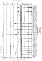
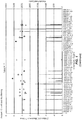
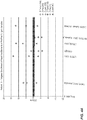
- repetitive element insertion regions For each DNA sample, the DNA is sequenced and evaluated for the presence of repetitive element insertion sequences. In identifying repetitive element insertion regions that can be used as personalized biomarkers, all sequences that map within a window of about 250 base pairs in the vicinity of repetitive element (e.g ., Alu, SVA elements, or LINEs) annotated in the reference genome are typically removed from consideration as a candidate insertion sequence using applications such as RepeatMasker. For the remaining sequences, insertion regions comprising repetitive sequences are identified.
- insertions regions can be defined using various criteria. In one embodiment, insertion regions are defined as those fragments that have sequences originating from the amplification reaction employing the reverse Alu primer that falls together within a 200 base pair window of a sequence originating from the Alu forward PCR primer.
- an Alu-insertion regions is an individualized biomarker when it has the following properties: it is present in a tumor sample from a patient, but not in normal samples, and it is present in a pre-therapy cell-free nucleic acid sample from the patient, but not a post-therapy cell-free nucleic acid sample from the patient.
- a direct comparison of various samples from the patient is performed.
- the following comparisons can be made to identify biomarkers.
- Repetitive element insertion regions identified in the DNA from a tumor sample from the patient is compared to the insertion regions identified in the DNA from normal controls.
- the normal sample may be a non-tumor sample from the patient and/or one or more samples from normal individuals (i.e., individuals not diagnosed with cancer).
- the normal sample is from non-tumor tissue from the patient, e.g., from peripheral blood mononuclear cells. Those repetitive element insertion regions that are present in a tumor sample, but not in normal DNA, are also compared to those present in pre-therapy cell-free DNA samples obtained from the patients.
- Repetitive element insertion regions that are present in both the tumor sample and pre-therapy samples are compared to the insertion regions contained in a post-therapy cell-free DNA sample obtained from the patient. Those insertion regions that are present in both the tumor sample and pre-therapy cell-free DNA sample are then compared to insertion regions that are detected in a post-therapy cell-free DNA sample. Those insertion regions that are not present in the post-therapy sample are biomarkers for that individual patient. One of skill in the art understands that these comparisons need not be performed in any particular order.
- the comparisons can be performed in a semi-quantitative or quantitative manner.
- a semi-quantitative approach the presence or absence of the insertion regions in a sample is simply identified as a positive or negative when determining a biomarker.
- more quantitative procedures can be employed to identify biomarkers.
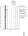
- the comparison between the insertion regions detected in the tumor sample to insertion regions detected in normal samples, from that patient and independent normal controls, can be quantitative.
- the methods of identifying a biomarker may comprise a comparison step where only those regions are considered as potential biomarkers where the normalized count in the PBMC differs at least 5-fold compared to the tumor DNA.
- factors other than "5-fold" e.g., 2 to 4-fold, or 6-fold or higher, can be used in such a quantitative analysis.
- the same type of quantitative comparison can be also applied to evaluating post-therapy insertion regions.
- Another aspect of a quantitative approach is to set limits for the comparisons between the tumor vs. normal repetitive element insertion regions and the tumor insertion regions to the pre-therapy insertion regions where a minimal analytical coverage per found region can be considered as criterion. For example, insertions to a defined genomic position that are only seen in a tumor sample, but not in a normal, non-cancerous control, are considered if such an insertion is found in sequencing in at least three instances. This number can be set on any number >0. Any combination of quantitative and qualitative means of filtering may be employed.
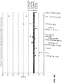
- the sequences from the pre-therapy and post-therapy samples are typically compared to those from a set of normal controls.
- the results obtained by a defined method can be compared to results obtained by the same methodological approach, but using a different sample or sets of samples.
- Such comparison samples can come from normal control individuals, e.g., plasma or serum sample from normal control individuals.
- Such a group of individuals without cancer are typically deemed a "reference group”.
- Samples obtained from such a reference group can be used as defined reference samples for transversal comparisons.
- normal somatic cells from an individual are available, such cells can be defined as a reference for an intra-individual comparison.
- Using a "timed" sample e.g ., pre therapy that can be compared to a post-therapy sample as a reference
- the region is sequenced to facilitate use of the marker as described below.
- a personalized biomarker identified in accordance with the invention may be used to evaluate response of that particular patient to a cancer therapy. Such an evaluation can be performed, e.g ., using an amplification reaction and/or nucleic acid hybridization to detect the levels of the personalized biomarker in the cell-free nucleic acids present in a blood sample from the patient.
- a blood sample is obtained from the patient before and/or after treatment with a cancer therapy. Serum or plasma from the blood sample is then analyzed for the presence of the personalized biomarker.
- Detection techniques for evaluating nucleic acids for the presence of a personalized biomarker involve procedures well known in the field of molecular genetics. In typical embodiments, detection of a personalized biomarker involves amplification of nucleic acids. Ample guidance for performing such techniques is provided in the art. Exemplary references include manuals such as PCR Technology: Principles and Applications for DNA Amplification (ed. H. A. Erlich, Freeman Press, NY, N.Y., 1992 ); PCR Protocols: A Guide to Methods and Applications (eds.
- Suitable amplification methods include ligase chain reaction (see, e.g ., Wu & Wallace, Genomics 4:560-569, 1988 ); strand displacement assay (see , e.g., Walker et al., Proc. Natl. Acad. Sci. USA 89:392-396, 1992 ; U.S. Pat. No. 5,455,166 ); and several transcription-based amplification systems, including the methods described in U.S. Pat. Nos. 5,437,990 ; 5,409,818 ; and 5,399,491 ; the transcription amplification system (TAS) ( Kwoh et al., Proc.
- TAS transcription amplification system
- oligonucleotide primers are employed that amplify the biomarker of interest.
- the presence of the biomarker can be determined, for example by detecting the length of the biomarker in the cell-free nucleic acid in comparison to a control.
- a probe may also be used to detect the presence of the biomarker.
- Oligonucleotides that are employed as primers and/or probes to detect biomarkers can be selected using methods well-known in the art.
- PCR primers may be designed using standard primer design computer software techniques known to individuals skilled in the art.
- the variables considered during PCR primer design may include primer length, GC pair content, melting temperature, and size of the target nucleic acid amplified by the primer pair.
- the biomarker is identified by hybridization under sequence-specific hybridization conditions with a probe that targets the biomarker region (e.g., targets some unambiguously assigned portion of, the target biomarker).
- Suitable hybridization formats are well known in the art, including but not limited to, solution phase, solid phase, oligonucleotide array formats, mixed phase, or in situ hybridization assays.
- solution phase hybridizations both the target nucleic acid and the probe or primers are free to interact in the reaction mixture.
- Techniques such as real-time PCR systems have also been developed that permit analysis, e.g., quantification, of amplified products during a PCR reaction.
- hybridization with a specific oligonucleotide probe occurs during the amplification program to identify the presence of a target nucleic acid.
- Hybridization of oligonucleotide probes ensure the highest specificity due to thermodynamically controlled two state transition.
- Examples for this assay formats are fluorescence resonance energy transfer hybridization probes, molecular beacons, molecular scorpions, and exonuclease hybridization probes ( e.g., reviewed in Bustin, J. Mol. Endocrin. 25:169-93,2000 ).
- the probe can be immobilized.
- the probe may comprise a label (e.g ., biotin) that allows the probe and target sequence to be captured on a solid support (e.g. , a magnetic bead).
- a label e.g ., biotin
- a solid support e.g. , a magnetic bead
- amplified target DNA is immobilized on a solid support and the target complex is incubated with the probe under suitable hybridization conditions, unhybridized probe is removed by washing under suitably stringent conditions, and the solid support is monitored for the presence of bound probe.
- the probes are immobilized on a solid support
- the target DNA is typically labeled, usually during amplification.
- the immobilized probe is incubated with the amplified target DNA under suitable hybridization conditions, unhybridized target DNA is removed by washing under suitably stringent conditions, and the solid support/probe is monitored for the presence of bound target DNA.
- multiple probes e.g ., that target different biomarkers for that patient, are immobilized on a solid support and the cell-free DNA from a patient is analyzed using the multiple probes simultaneously.
- nucleic acid arrays are described by WO 95/11995 .
- amplified nucleic acid corresponding to a target nucleic acid is performed using nucleic acid primers to the chromosomal region and is detected by monitoring the increase in the total amount of double-stranded DNA in the reaction mixture, is described, e.g., in U.S. Pat. No. 5,994,056 ; and European Patent Publication Nos. 487,218 and 512,334 .
- the detection of double-stranded target DNA relies on the increased fluorescence various DNA-binding dyes, e.g., SYBR Green, exhibit when bound to double-stranded DNA.
- Oligonucleotides can be prepared by any suitable method, usually chemical synthesis, and can also be purchased through commercial sources. Oligonucleotides can include modified phosphodiester linkages (e.g., phosphorothioate, methylphosphonates, phosphoamidate, or boranophosphate) or linkages other than a phosphorous acid derivative into an oligonucleotide may be used to prevent cleavage at a selected site. In addition, the use of 2'-amino modified sugars tends to favor displacement over digestion of the oligonucleotide when hybridized to a nucleic acid that is also the template for synthesis of a new nucleic acid strand.
- modified phosphodiester linkages e.g., phosphorothioate, methylphosphonates, phosphoamidate, or boranophosphate
- linkages other than a phosphorous acid derivative into an oligonucleotide may be used to prevent cleavage at a selected site.
- the level of the biomarker in the patient circulating CAN is determined relative to an index value, such as the amount of the biomarker that is identified in normal controls.
- kits useful for identifying one or more individualized biomarkers in the cell-free circulating nucleic acid from a patient provides at least one Alu primer and a universal primer and/or adapters to ligate a universal primer binding site to fragmented cell-free circulating DNA from the patient.
- Such reagents can be used to identify individual biomarkers.
- “detection” or “identification” or “identifying the presence” or “detecting the presence” of a personalized in a circulating cell-free nucleic acid sample from a patient refers to determining any level of the biomarker in the circulating nucleic acid sample from the patient.
- the information can be used to assist in evaluating the response of a patient to a therapy. For example, the presence of an individualized biomarker in circulating cell-free DNA in the patient following treatment with a particular therapeutic protocol may indicate that the patient has not completely responded to the protocol. Accordingly, the information may be used to assist in determining adjustments to the therapeutic protocol and/or to determine if an alternative therapy should be employed.
- the information obtained from the biomarker analysis may be stored in a computer readable form.
- a computer system typically comprises major subsystems such as a central processor, a system memory (typically RAM), an input/output (I/O) controller, an external device such as a display screen via a display adapter, serial ports, a keyboard, a fixed disk drive via a storage interface and a floppy disk drive operative to receive a floppy disc, and a CD-ROM (or DVD-ROM) device operative to receive a CD-ROM.
- Many other devices can be connected, such as a network interface connected via a serial port.
- the computer system may also be linked to a network, comprising a plurality of computing devices linked via a data link, such as an Ethernet cable (coax or 10BaseT), telephone line, ISDN line, wireless network, optical fiber, or other suitable signal transmission medium, whereby at least one network device (e.g., computer, disk array, etc.) comprises a pattern of magnetic domains (e.g., magnetic disk) and/or charge domains (e.g., an array of DRAM cells) composing a bit pattern encoding data acquired from an assay of the invention.
- a network device e.g., computer, disk array, etc.
- a pattern of magnetic domains e.g., magnetic disk
- charge domains e.g., an array of DRAM cells
- the computer system can comprise code for interpreting the results of a study to determine personalized biomarkers or to evaluating the presence of one or more of the personalized biomarkers identified in accordance with the invention to aid in prognosis.
- the biomarker analysis results are provided to a computer where a central processor executes a computer program for evaluating the one or more biomarkers.
- the invention may comprise the use of a computer system, such as that described above, which comprises: (1) a computer; (2) a stored bit pattern encoding the biomarker testing results obtained by the methods of the invention, which may be stored in the computer; (3) and, optionally, (4) a program for valuating a biomarker.
- a computer system such as that described above, which comprises: (1) a computer; (2) a stored bit pattern encoding the biomarker testing results obtained by the methods of the invention, which may be stored in the computer; (3) and, optionally, (4) a program for valuating a biomarker.
- the invention may comprise generating a report based on the detection of one or more personalized biomarkers for the patient.
- the present invention may comprise use of a system related to the above methods of the invention.
- the invention comprises use of a system for analyzing circulating cell-free DNA, comprising: (1) a sample analyzer for executing the method of analyzing circulating cell-free DNA in a patient's blood, serum or plasma as described in the various embodiments above; (2) a computer system for automatically receiving and analyzing data obtained in step (1) to provide a test value representing the status (presence or absence or amount, i.e., concentration or copy number) of a personalized biomarker for the patient.
- the computer-based analysis function can be implemented in any suitable language and/or browsers. For example, it may be implemented with C language and preferably using object-oriented high-level programming languages such as Visual Basic, SmallTalk, C++, and the like.
- the application can be written to suit environments such as the Microsoft WindowsTM environment including WindowsTM 98, WindowsTM 2000, WindowsTM NT, and the like.
- the application can also be written for the MacIntoshTM, SUNTM, UNIX or LINUX environment.
- the functional steps can also be implemented using a universal or platform-independent programming language.
- multi-platform programming languages include, but are not limited to, hypertext markup language (HTML), JAVATM, JavaScriptTM, Flash programming language, common gateway interface/structured query language (CGI/SQL), practical extraction report language (PERL), AppleScriptTM and other system script languages, programming language/structured query language (PL/SQL), and the like.
- JavaTM- or JavaScriptTM-enabled browsers such as HotJavaTM, MicrosoftTM ExplorerTM, or NetscapeTM can be used.
- active content web pages may include JavaTM applets or ActiveXTM controls or other active content technologies.
- the analysis function can also be embodied in computer program products and used in the systems described above or other computer- or internet-based systems. Accordingly, another aspect of the present disclosure relates to a computer program product comprising a computerusable medium having computer-readable program codes or instructions embodied thereon for enabling a processor to carry out the analysis and correlating functions as described above.
- These computer program instructions may be loaded onto a computer or other programmable apparatus to produce a machine, such that the instructions which execute on the computer or other programmable apparatus create means for implementing the functions or steps described above.
- These computer program instructions may also be stored in a computer-readable memory or medium that can direct a computer or other programmable apparatus to function in a particular manner, such that the instructions stored in the computer-readable memory or medium produce an article of manufacture including instruction means which implement the analysis.
- the computer program instructions may also be loaded onto a computer or other programmable apparatus to cause a series of operational steps to be performed on the computer or other programmable apparatus to produce a computer implemented process such that the instructions which execute on the computer or other programmable apparatus provide steps for implementing the functions or steps described above.
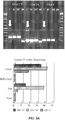
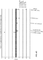
- This example describes the identification of individualized Alu biomarkers for breast cancer patients.
- Sequence libraries were prepared from tumor and peripheral blood mononuclear cells (PBMC) nuclear genomic DNA. Extracted genomic DNA (100 ng) was sheared by ultrasound to a size of about 200 bp. The fragments (50 ng) were end-repaired using 0.4 units/ ⁇ L Klenow Fragment (USB, Affymetrics) and 0.2 units/ ⁇ L Kinase (USB, Affymetrics) for 30 min at 37°C. The reaction was purified using AMPure beads (Angencourt). Single A-overhangs were attached to the end-repaired fragments by incubation with 0.4 units/ ⁇ L of Klenow Fragment exo-(NEB) for 30 min at 37°C. The reaction was purified using AMPure beads (Agencourt). Adapters were ligated to the A-tailed fragments. The adapters were two single-stranded DNA oligonucleotides:
- the P9 oligonucleotide has a 5-prime phosphate group.
- the P5 oligo has a phosphorothioate internucleotide linkage between the two 3-prime nucleotides, which is indicated by an asterisk. Both oligonucleotides were annealed to form a partially double stranded Y-adapter.
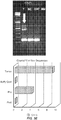
- the Y-adapters were ligated to the A-tailed fragment by incubation with 1.2 units/ ⁇ L ligase (USB, Affymetrics) for 30 min at roomtemperature. Adapter concentration in the reaction were 30x the molar amount of fragments ends. The reaction was purified using the SureClean Plus solution (Bioline).
- the ligated fragments were amplified in 12 cycles of PCR using the primers:
- Amplified library DNA (10 ng) was subjected to an Alu-enrichment PCR.
- the primers used in this PCR were:
- PCR-products For each sample and each Alu-enrichment PCR, 200 ng of PCR-products were subjected to 4 cycles of PCR using fusion primers that carry an individual 7 bp index sequence. This sequence serves as molecular barcode allowing the assignment of each read to the respective sample and PCR after sequencing.
- the 3' part of the index-primers anneals to the underlined sequence of primer Alu40.REV and Alu259.FOR.
- the 5' part of this primer adds the full Illumina sequencing adapter to the fragments.
- index-primer caagcagaagacggcatacgagat-7bpindex-gtgactggagttcagacgtgtgctcttccgatc. (SEQ ID NOs: 7 and 8)
- Library preparation for the cell-free DNA fragments extracted from the patients' serum was performed as described for tumor and PBMC DNA, but the fragmentation step was omitted.
- the starting amount of cell-free DNA was 10 ng.
- the enriched library fragments from patients and from different samples were pooled and sequenced on an Illumina HiSeq or Illumina HiScan system according to the manufacturers protocols. Sequencing was conducted as single-read sequencing for 51 cycles followed by 8 cycles of index sequencing.
- Sequence reads were aligned to the human genome (HG18, NCBI36, March 2006) using the CASAVA software v1.8 (Illumina) with default parametrization. Using the repeatmasked version of the human genome (HG18, NCBI36, March 2006), all sequence reads that map within a window of ⁇ 250 bp around an Alu or SVA element annotated in the reference genome were removed.
- Insertion regions were constructed for each sample and each specimen. Insertion regions were defined by the following criteria: At least one read originating from the Alu40.REV PCR and at least one read originating from the Alu259.FOR PCR fall together within a 200 bp window.
- the region "start” is defined by the starting position of the first read.
- the region “stop” is defined by the end position of the last read. Start position, stop position, and number of reads were recorded for each insertion region of the respective sample and specimen.
- a quantitative approach was designed, e.g., using a filter for the comparison. For example only those regions are kept where the normalized count in the PBMC differs by at least 5-fold compared to the tumor DNA. Other factors that can be used are 2- to 4-fold, or 6-fold or higher.
- the same quantitative comparison can be applied to the filtering described in filtering post-surgery insertion regions.
- Another aspect of a quantitative approach is to set limits for the comparisons between the tumor vs. normal Alu insertion regions and the tumor Alu insertions regions to the pre-therapy Alu insertions regions where a minimal read count per found region can be considered as criterion. Any combination of quantitative and qualitative means of filtering may be employed.
- Sequence reads generated from the serum DNA pre-surgery and post-surgery and 24 additional apparently healthy individuals were subjected to depth of coverage analysis. Data were either used as such or reads that were mapped in a distance not greater than 200 bp next to an annotated ALU were omitted.
- Mapped sequence reads were counted in windows of 250kbp across the genome. Read counts were normalized to counts per 10 million reads
- the upper and lower confidence limits were calculated by using a "leave one out” approach. Briefly. the Z-value was calculated for each bin with the remaining 23 normals as basis. The mean and standard deviation for each bin was then calculated using the 24 resulting data points (Z-values) for each window.
- Window read counts of the pre-surgery specimens were selected as potential diagnostic regions of copy-number imbalance if outside the upper or lower confidence bounds.
- the deviation of each window read count for each pre- and post-surgery specimen from the average of the healthy control window read counts was expressed as Z-value (basis: 24 normals).
- the genomic regions with significant copy-number imbalance in the pre-surgery serum sample were examined for significant imbalances in the post-surgery serum sample. For this, the difference between the windows Z-value of the post-surgery sample was subtracted from the windows Z-value of the pre-surgery sample.
- a highly positive or negative Z-value difference is indicative of an imbalanced region that is linked to the presence of the tumor, because it indicates a high deviation from the normal controls in the pre-surgery serum DNA, but a low deviation from the normal control in the post-surgery serum DNA.
- Figure 1 provides graphs showing the results for five 5 patients.
- the graphs were generated by a) with Alu filtering on (No Alus) and b) without. Normalized numbers of sequence reads obtained from cell-free circulation DNA that aligned to the human genomic database HG18 were summed into 250kbp window bins. The results were converted into Z-values as described. For each such bin, pre-surgery values ⁇ 3 were censored to zero. Circles represent the Z-value pre surgery, diamonds the difference between pre-surgery Z-value and post-surgery Z-values. The bars display the count sum that was used to calculate the Z-values for comparison.
- Primers spanning some of the putative Alu insertion sites listed in Table 1 were constructed from the human genomic reference genome (HG18, NCBI36, March 2006). Primers were tested on tumor DNA and PBMC DNA of the respective patient.
- the DNA template used for PCR was in some cases material that was subjected to a pre-amplification using either the Whole-Genome-Amplification 1 Kit (WGA1, Sigma-Aldrich) or the Whole- Genome-Amplification 4 Kit (WGA4, Sigma-Aldrich).
- WGA1, Sigma-Aldrich Whole- Genome-Amplification 4 Kit
- a list of primers and the respective template source is provided in Table 3. Agarose gel images of the PCR-amplified products are shown in Figure 3.
- Figure 3 shows the gel analysese for five representative patients, the results of the selection algorithm displayed in Figure 2 .
- each panel On the right hand side of each panel (A to E) the normalized counts of the specific insertion site is given, which is allocated to ⁇ 200bp in the HG18 database (as described in [0082]).
- gel images of PCRs that were designed to amplify the particular insertions are shown.
- Figure 4A-E shows Z-values for read of circulating DNA and tumor DNA mapped to known repetitive elements in the human genome, separated for the ALU40 and ALU 259 PCR.
- Figure 5 shows the results for two patients who exhibited differential reduction in cell-free DNA biomarkers. These differential Alu-insertion sites were selected from the quantitative selection procedure illustrated above.
- Patient 1 in Figure 5A showed the presence of a tumor specific length polymorphism site located in a unique region on chromosome 18 in the tumor and in the pre-surgical sample. These unique fragments were not detected in either the PBMC or in the post-surgical cell-free DNA sample. Additional length polymorphisms sites in unique regions from chromosomes 1 and 5 and two different unique regions from chromosome 6 were detected in the tumor and pre-surgical cell free DNA sample but not in the PBMC. However, these 4 unique biomarkers could still be detected in the post-surgical sample.
- This example describes the identification of individualized L1-LINEs biomarkers for colorectal cancer patients.
- the extracted DNA was ultrasonically sheared to an average size of 200 bp using a Covaris S220 focused ultrasonicator (Covaris) and AFA microTubes (Covaris).
- Fragmented DNA 100 ng was end-repaired, A-tailed and ligated to Illumina sequencing adapters using the NEBNext Ultra Library Preparation Kit (New England Biolabs) according to manufacturer's instructions.
- L1 fragment enrichment was conducted using biotinylated L1-specific oligonucleotides (Table 4) and 125 ng library DNA.
- Hybridization mixtures contained 4 pmol of each L1 probe, 5 ⁇ g sheared salmon sperm DNA, 1000 pmol blocking oligos against library specific adapters. After initial denaturation at 95° for 10 min the hybridization was carried out at 47°C for 72 h. Capture of the biotinylated probes together with the hybridized fragments was conducted using the TargetSeq Hybridization and Wash Kit (Lifetechnologies) according to the manufacturer's instructions. Captured sequences were recovered from the magnetic beads by heat denaturing. The enriched fragments were amplified for 7 cycles of PCR using primers specific for the ligated sequencing adapters.
- Paired-end sequencing was conducted on an Illumina HiSeq 2000 producing one 50 bp sequence for each end of the fragments.
- the unique mapping sequences were filtered by removing all sequences that mapped within a 200 bp window next to an L1 annotated in the reference genome, because those L1 elements are common to the population.
- the read-regions defined in a tumor-sample were compared to the read-region defined for the lymphocyte samples. All tumor read-regions residing within 50 bp upstream or downstream of a lymphocyte read-region were removed, because such reads identify L1 elements that are present in the germline genome and are not somatic de-novo insertions.
- regions containing at least two reads were selected as demarking a somatic de-novo L1 insertion in the tumor genome. Such regions are indicative of a tumor specific L1 insertion, because they (i) contain at least 2 reads of the "L1-uniqueHG19"-class that (ii) are not next to an annotated L1 (population L1) and (iii) are not next to an L1 found in the sequenced lymphocyte genomes (germline L1).
- the enrichment step showed a positive enrichment of 60% (from 44%- to 76%), of reads where at least one of the paired reads could be aligned to the L1-reference sequences.
- 1500 reads were identified in which one read aligned uniquely with a non L1 sequence in the human HG19 database and was not annotated to be in the vicinity of a L1 identified in the database.
- Table 5 shows the chromosomal regions discovered to harbor an Ll-HS reinsertion that is only present in the tumor, with position of the reinsertion (HG19) and the position in the Ll-HS.
Claims (15)
- Verfahren zum Auswählen eines personalisierten Biomarkers für einen Patienten, der einen Tumor hat, wobei das Verfahren umfasst:(a) Vorbereiten einer Bibliothek von DNA-Fragmenten aus Nukleinsäureproben, umfassend DNA, die von dem Patienten aus jeder der folgenden Quellen entnommen werden: mononukleären Zellen des peripheren Blutes (peripheral blood mononuclear cells, PBMCs), dem Tumor und zellfreien Prätherapie-Nukleinsäuren, wobei die Nukleinsäuren vor der Therapie entnommen werden;(b) Sequenzierung der DNA-Fragmente in den Bibliotheken, um DNA-Fragmente zu identifizieren, die eine Insertionsregion von repetitiven Elementen umfasst; und(c) Vergleichen von in der PMBC-Probe identifizierten Insertionsregionen von repetitiven Elementen mit in der Tumorprobe und/oder der zellfreien Nukleinsäureprobe identifizierten Insertionsregionen von repetitiven Elementen, die im Tumor, aber nicht in PBMCs vorhanden sind, wodurch ein personalisierter Biomarker für den Patienten identifiziert wird.
- Verfahren nach Anspruch 1, wobei das repetitive Element ein Alu-Element oder ein LINE-Element ist.
- Verfahren nach einem der vorhergehenden Ansprüche, wobei Schritt (c) ferner einen Vergleich von in der PBMC-Probe identifizierten Insertionsregionen von repetitiven Elementen mit den entsprechenden Regionen in der Humangenom-Datenbank umfasst, um Insertionsregionen in der PBMC-Probe zu identifizieren, die nicht in der Humangenom-Datenbank vorhanden sind.
- Verfahren nach einem der vorhergehenden Ansprüche, ferner umfassend die Schritte des Hinzufügens von Adaptersequenzen, die universelle Priming-Stellen zu jedem der DNA-Fragmente umfassen und die DNA-Fragmente vor Schritt (b) amplifizieren.
- Verfahren nach Anspruch 4, wobei der Schritt des Amplifizierens ausgeführt wird unter Verwendung:(i) eines für das repetitive Element spezifischen Primers und eines universellen Primers; oder(ii) von zwei universellen Primern.
- Verfahren nach einem der vorhergehenden Ansprüche, ferner umfassend den Schritt des Anreicherns der DNA-Fragmente in mindestens einer der Bibliotheken für DNA-Fragmente, die eine Insertionsregion von repetitiven Elementen umfassen, wobei wahlweise der Schritt des Anreicherns unter Verwendung von Oligonukleotid-Sonden ausgeführt wird, die spezifisch mit dem repetitiven Element hybridisieren.
- Verfahren nach einem der vorhergehenden Ansprüche, ferner umfassend den Schritt der Entnahme einer Nukleinsäureprobe von dem Patienten aus zellfreien Posttherapie-Nukleinsäuren, wobei die Nukleinsäuren nach der Therapie entnommen werden, wahlweise ferner umfassend den Schritt des Vergleichens der Regionen von repetitiven Elementen, die in der zellfreien Prätherapie-Nukleinsäureprobe identifiziert werden, mit der der zellfreien Posttherapie-Nukleinsäureprobe, um Insertionsregionen von repetitiven Elementen, die nicht in der zellfreien Posttherapie-Nukleinsäureprobe detektiert werden, zu identifizieren.
- Verfahren zum Auswählen eines personalisierten Biomarkers für einen Patienten, der einen Tumor hat, wobei das Verfahren umfasst:(a) Durchführen von mindestens einer Amplifikationsreaktion an DNA aus Nukleinsäureproben, umfassend DNA, die von dem Patienten aus jeder der folgenden Quellen entnommen werden: mononukleären Zellen des peripheren Blutes (peripheral blood mononuclear cells, PBMCs), dem Tumor, zellfreien Prätherapie-Nukleinsäuren, wobei die zellfreien Nukleinsäuren vor der Therapie entnommen werden, und zellfreien Posttherapie-Nukleinsäuren, wobei die Nukleinsäuren nach der Therapie entnommen werden, wobei die Amplifikation unter Verwendung eines Primers an einer Alu-Sequenz und einem universellen Primer durchgeführt wird.(b) Sequenzieren der DNA, die aus den Amplifikationsreaktionen erhalten wird, um Regionen, die eine Alu-Insertionssequenz umfassen, relativ zu den entsprechenden Regionen in der humanen Genom-Datenbank zu bestimmen;(c) Vergleichen von Alu-Insertionsregionen die in Schritt (b) aus der PBMC-Probe identifiziert werden, mit den Alu-Insertionsregionen, die in Schritt (b) aus der Tumorprobe identifiziert werden, um Alu-Insertionsregionen, die im Tumor, aber nicht in PBMCs vorhanden sind, zu identifizieren.(d) Vergleichen der Alu-Insertionsregionen, die in Schritt (c) identifiziert werden, mit den Alu-Insertionsregionen, die in der zellfreien Prätherapie-Nukleinsäureprobe von dem Patienten identifiziert werden, um Alu-Insertionsregionen, die in der Tumorprobe und in der zellfreien Prätherapie-Nukleinsäureprobe vorhanden sind, zu identifizieren; und(e) Vergleichen der Alu-Insertionsregionen, die in Schritt (d) identifiziert werden, mit den Alu-Insertionsregionen, die in der zellfreien Posttherapie-Nukleinsäureprobe von dem Patienten identifiziert werden, um Alu-Insertionsregionen, die nicht in der zellfreien Posttherapie-Nukleinsäureprobe detektiert werden, zu identifizieren, wodurch ein personalisierter Biomarker für den Patienten identifiziert wird.
- Verfahren zum Auswählen eines personalisierten Biomarkers für einen Patienten, der einen Tumor hat, wobei das Verfahren umfasst:(a) Durchführen von mindestens einer Amplifikationsreaktion an DNA aus einer Nukleinsäureprobe, umfassend DNA, die aus zirkulierenden Prätherapie-Nukleinsäuren entnommen wird, und einer Nukleinsäureprobe, umfassend DNA, die aus den zirkulierenden Posttherapie-Nukleinsäuren entnommen wird, wobei die Amplifikation unter Verwendung eines Primers an einer Alu-Sequenz und eines universellen Primers durchgeführt wird.(b) Sequenzieren der DNA, die aus den Amplifikationsreaktionen erhalten wird, um Regionen, die eine Alu-Insertionssequenz umfassen und einzigartig zu einer definierten Region in einem normalen humanen Genom positioniert sein können, relativ zu einem normalen humanem Genom, zu bestimmen;(c) Quantifizieren des Ausmaßes einer in Schritt (b) identifizierten Alu-Insertionsregion aus der zirkulierenden Prätherapie-DNA-Probe im Vergleich zum Ausmaß der Alu-Insertionsregionen, die in normalen Kontrollen vorhanden sind, um Alu-Insertionsregionen zu identifizieren, die in zirkulierender zellfreier DNA in größerem Ausmaß als in normalen Kontrollen vorhanden sind; und(d) Vergleichen des Ausmaßes der in Schritt (b) identifizierten Alu-Insertionsregionen mit dem Ausmaß der Alu-Insertionsregionen in der zellfreien Posttherapie-DNA-Probe, um Alu-Insertionsregionen zu identifizieren, die nicht in einem Ausmaß, das sich vom normalen unterscheidet, detektiert werden, wodurch ein personalisierter Biomarker für den Patienten identifiziert wird.
- Verfahren nach Anspruch 8 oder Anspruch 9, ferner umfassend das Durchführen einer zusätzlichen Amplifikationsreaktion an DNA aus jeder der Nukleinsäureproben, wobei die zweiten Amplifikationsreaktionen einen zweiten Primer an einer Alu-Sequenz und einen zweiten universellen Primer umfassen.
- Verfahren nach Anspruch 10, wobei eine der Amplifikationsreaktionen Folgendes umfasst:(i) ein Primer-Paar, umfassend SEQ ID Nr. 5 und einen universellen Primer; oder(ii) en Primer-Paar, umfassend SEQ ID Nr. 6 und einen universellen Primer.
- Verfahren nach Anspruch 10, wobei eine der Amplifikationsreaktionen ein Primer-Paar, umfassend SEQ ID Nr. 5, und einen universellen Primer umfasst, und die zusätzliche Amplifikationsreaktion ein Primer-Paar, umfassend SEQ ID Nr. 6, und einen universellen Primer umfasst.
- Verfahren nach einem der vorhergehenden Ansprüche, wobei die Therapie eine Operation ist.
- Verfahren nach einem der vorhergehenden Ansprüche, wobei der Patient einen Brusttumor hat.
- Verfahren zum Überwachen des Ansprechens eines Patienten, der einen Tumor hat, auf eine Therapie, wobei das Verfahren das Durchführen eines Verfahrens nach einem der vorhergehenden Ansprüche umfasst, um einen individualisierten Biomarker zu identifizieren, und das Bewerten des Spiegels des Biomarkers in zellfreien zirkulierenden Nukleinsäuren, die vom Patienten vor und nach der Therapie entnommen werden, umfasst.
Priority Applications (1)
| Application Number | Priority Date | Filing Date | Title |
|---|---|---|---|
| PL13863107T PL2931922T3 (pl) | 2012-12-14 | 2013-12-13 | Personalizowane biomarkery nowotworu |
Applications Claiming Priority (2)
| Application Number | Priority Date | Filing Date | Title |
|---|---|---|---|
| US201261737626P | 2012-12-14 | 2012-12-14 | |
| PCT/US2013/075015 WO2014093825A1 (en) | 2012-12-14 | 2013-12-13 | Personalized biomarkers for cancer |
Publications (3)
| Publication Number | Publication Date |
|---|---|
| EP2931922A1 EP2931922A1 (de) | 2015-10-21 |
| EP2931922A4 EP2931922A4 (de) | 2016-07-27 |
| EP2931922B1 true EP2931922B1 (de) | 2018-10-17 |
Family
ID=50934991
Family Applications (1)
| Application Number | Title | Priority Date | Filing Date |
|---|---|---|---|
| EP13863107.2A Active EP2931922B1 (de) | 2012-12-14 | 2013-12-13 | Personalisierte biomarker für krebs |
Country Status (5)
| Country | Link |
|---|---|
| US (1) | US9909186B2 (de) |
| EP (1) | EP2931922B1 (de) |
| ES (1) | ES2703764T3 (de) |
| PL (1) | PL2931922T3 (de) |
| WO (1) | WO2014093825A1 (de) |
Families Citing this family (14)
| Publication number | Priority date | Publication date | Assignee | Title |
|---|---|---|---|---|
| WO2014113204A1 (en) | 2013-01-17 | 2014-07-24 | Personalis, Inc. | Methods and systems for genetic analysis |
| EP3965111A1 (de) | 2013-08-30 | 2022-03-09 | Personalis, Inc. | Verfahren und system zur genomanalyse |
| GB2535066A (en) | 2013-10-03 | 2016-08-10 | Personalis Inc | Methods for analyzing genotypes |
| JP7441584B6 (ja) * | 2014-08-22 | 2024-03-15 | レゾリューション バイオサイエンス, インコーポレイテッド | 無細胞DNA(cfDNA)の定量的遺伝子解析のための方法 |
| WO2016070131A1 (en) | 2014-10-30 | 2016-05-06 | Personalis, Inc. | Methods for using mosaicism in nucleic acids sampled distal to their origin |
| US20180305757A1 (en) * | 2015-10-16 | 2018-10-25 | The Regents Of The University Of California | Repetitive Element (RE)-based Genome Analysis and Dynamic Genetics Surveillance Systems |
| AU2016353133B2 (en) | 2015-11-11 | 2022-12-08 | Resolution Bioscience, Inc. | High efficiency construction of dna libraries |
| US11299783B2 (en) | 2016-05-27 | 2022-04-12 | Personalis, Inc. | Methods and systems for genetic analysis |
| BR112019003704A2 (pt) | 2016-08-25 | 2019-05-28 | Resolution Bioscience Inc | métodos para a detecção de alterações na cópia genômica em amostras de dna |
| EP3541950A4 (de) * | 2016-11-16 | 2020-06-03 | Progenity, Inc. | Multimodaler test zum nachweis von nukleinsäureaberrationen |
| US10801064B2 (en) | 2018-05-31 | 2020-10-13 | Personalis, Inc. | Compositions, methods and systems for processing or analyzing multi-species nucleic acid samples |
| US11814750B2 (en) | 2018-05-31 | 2023-11-14 | Personalis, Inc. | Compositions, methods and systems for processing or analyzing multi-species nucleic acid samples |
| CA3113682A1 (en) * | 2019-02-15 | 2020-08-20 | Takara Bio Usa, Inc. | Methods of preparing and analyzing nucleic acid libraries |
| CN109825583B (zh) * | 2019-03-01 | 2021-08-17 | 清华大学 | 人重复元件dna甲基化作为肝癌早期诊断的标记物及其应用 |
Family Cites Families (16)
| Publication number | Priority date | Publication date | Assignee | Title |
|---|---|---|---|---|
| US5955589A (en) | 1991-12-24 | 1999-09-21 | Isis Pharmaceuticals Inc. | Gapped 2' modified oligonucleotides |
| YU187991A (sh) | 1990-12-11 | 1994-09-09 | Hoechst Aktiengesellschaft | 3-(2)-amino-ali tiol-modifikovani, s fluorescentnom bojom vezani nukleozidi, nukleotidi i oligonukleotidi, postupak za njihovo dobijanje i njihova upotreba |
| US5652099A (en) | 1992-02-12 | 1997-07-29 | Conrad; Michael J. | Probes comprising fluorescent nucleosides and uses thereof |
| US5446137B1 (en) | 1993-12-09 | 1998-10-06 | Behringwerke Ag | Oligonucleotides containing 4'-substituted nucleotides |
| DE4415370A1 (de) | 1994-05-02 | 1995-11-09 | Hoechst Ag | Modifizierte Oligonukleotide, deren Herstellung sowie deren Verwendung |
| DE4438918A1 (de) | 1994-11-04 | 1996-05-09 | Hoechst Ag | Modifizierte Oligonukleotide, deren Herstellung sowie deren Verwendung |
| US5773649A (en) | 1996-06-10 | 1998-06-30 | Centre De Recherche De L'hopital Sainte-Justine | DNA markers to detect cancer cells expressing a mutator phenotype and method of diagnosis of cancer cells |
| CA2268069A1 (en) * | 1996-10-04 | 1998-04-09 | Chronix Biomedical | Diagnostic detection of nucleic acids |
| DK0866071T3 (da) | 1997-03-20 | 2005-01-17 | Hoffmann La Roche | Modificerede primere |
| USH2191H1 (en) | 2000-10-24 | 2007-06-05 | Snp Consortium | Identification and mapping of single nucleotide polymorphisms in the human genome |
| WO2007111937A1 (en) * | 2006-03-23 | 2007-10-04 | Applera Corporation | Directed enrichment of genomic dna for high-throughput sequencing |
| US8986958B2 (en) * | 2009-03-30 | 2015-03-24 | Life Technologies Corporation | Methods for generating target specific probes for solution based capture |
| CN102409048B (zh) * | 2010-09-21 | 2013-10-23 | 深圳华大基因科技服务有限公司 | 一种基于高通量测序的dna标签文库构建方法 |
| EP2686451A4 (de) * | 2011-03-18 | 2014-12-24 | Baylor Res Inst | Line-1-hypomethylierung als biomarker für früh ausbrechenden kolorektalkrebs |
| GB201106254D0 (en) | 2011-04-13 | 2011-05-25 | Frisen Jonas | Method and product |
| JP2015534806A (ja) * | 2012-10-19 | 2015-12-07 | シーケンタ・インコーポレイテッド | 末梢血試料によるびまん性大細胞型b細胞性リンパ腫のモニタリング関連出願の相互参照本出願は、2012年10月19日出願の米国特許仮出願第61/716,270号の利益を主張するものであり、当該出願は参照により本明細書にその全体が組み込まれる。 |
-
2013
- 2013-12-13 WO PCT/US2013/075015 patent/WO2014093825A1/en active Application Filing
- 2013-12-13 US US14/652,098 patent/US9909186B2/en active Active
- 2013-12-13 ES ES13863107T patent/ES2703764T3/es active Active
- 2013-12-13 PL PL13863107T patent/PL2931922T3/pl unknown
- 2013-12-13 EP EP13863107.2A patent/EP2931922B1/de active Active
Non-Patent Citations (1)
| Title |
|---|
| None * |
Also Published As
| Publication number | Publication date |
|---|---|
| PL2931922T3 (pl) | 2019-04-30 |
| EP2931922A1 (de) | 2015-10-21 |
| US20160053328A1 (en) | 2016-02-25 |
| WO2014093825A1 (en) | 2014-06-19 |
| US9909186B2 (en) | 2018-03-06 |
| ES2703764T3 (es) | 2019-03-12 |
| EP2931922A4 (de) | 2016-07-27 |
Similar Documents
| Publication | Publication Date | Title |
|---|---|---|
| EP2931922B1 (de) | Personalisierte biomarker für krebs | |
| US11414706B2 (en) | Method of determining a numeric index of tumor-associated copy number changes of circulating nucleic acids associated with prostate cancer | |
| US20220228197A1 (en) | Method for determining copy number variations | |
| US11377695B2 (en) | Breast cancer associated circulating nucleic acid biomarkers | |
| CN107750277B (zh) | 使用无细胞dna片段大小来确定拷贝数变化 | |
| CA2852098C (en) | Colorectal cancer associated circulating nucleic acid biomarkers | |
| CA2840418C (en) | Method for determining the presence or absence of different aneuploidies in a sample | |
| US20230340590A1 (en) | Method for verifying bioassay samples | |
| EP3018213A1 (de) | Verfahren zur Bestimmung des Vorhandenseins eines biologischen Zustands durch Bestimmung sowohl der relativen als auch der totalen Menge an zwei verschiedenen Nukleinsäuren | |
| EP3775274B1 (de) | Verfahren zum nachweis von somatischen genetischen anomalien, kombination von fangsonden und nachweiskit | |
| AU2011365507A1 (en) | Normalizing chromosomes for the determination and verification of common and rare chromosomal aneuploidies | |
| CN112639983A (zh) | 微卫星不稳定性检测 | |
| WO2018093744A2 (en) | Non-unique barcodes in a genotyping assay | |
| AU2016262641A1 (en) | Detecting and classifying copy number variation | |
| WO2022084742A1 (en) | Simultaneous amplification of dna and rna from single cells | |
| NZ752319B2 (en) | Using cell-free dna fragment size to determine copy number variations |
Legal Events
| Date | Code | Title | Description |
|---|---|---|---|
| PUAI | Public reference made under article 153(3) epc to a published international application that has entered the european phase |
Free format text: ORIGINAL CODE: 0009012 |
|
| 17P | Request for examination filed |
Effective date: 20150710 |
|
| AK | Designated contracting states |
Kind code of ref document: A1 Designated state(s): AL AT BE BG CH CY CZ DE DK EE ES FI FR GB GR HR HU IE IS IT LI LT LU LV MC MK MT NL NO PL PT RO RS SE SI SK SM TR |
|
| AX | Request for extension of the european patent |
Extension state: BA ME |
|
| DAX | Request for extension of the european patent (deleted) | ||
| A4 | Supplementary search report drawn up and despatched |
Effective date: 20160629 |
|
| RIC1 | Information provided on ipc code assigned before grant |
Ipc: C12Q 1/68 20060101AFI20160623BHEP |
|
| STAA | Information on the status of an ep patent application or granted ep patent |
Free format text: STATUS: EXAMINATION IS IN PROGRESS |
|
| 17Q | First examination report despatched |
Effective date: 20170609 |
|
| REG | Reference to a national code |
Ref country code: DE Ref legal event code: R079 Ref document number: 602013045377 Country of ref document: DE Free format text: PREVIOUS MAIN CLASS: C12Q0001680000 Ipc: C12Q0001688600 |
|
| GRAP | Despatch of communication of intention to grant a patent |
Free format text: ORIGINAL CODE: EPIDOSNIGR1 |
|
| STAA | Information on the status of an ep patent application or granted ep patent |
Free format text: STATUS: GRANT OF PATENT IS INTENDED |
|
| RIC1 | Information provided on ipc code assigned before grant |
Ipc: C12Q 1/6809 20180101ALI20180411BHEP Ipc: C12Q 1/6886 20180101AFI20180411BHEP |
|
| INTG | Intention to grant announced |
Effective date: 20180507 |
|
| GRAS | Grant fee paid |
Free format text: ORIGINAL CODE: EPIDOSNIGR3 |
|
| GRAA | (expected) grant |
Free format text: ORIGINAL CODE: 0009210 |
|
| STAA | Information on the status of an ep patent application or granted ep patent |
Free format text: STATUS: THE PATENT HAS BEEN GRANTED |
|
| AK | Designated contracting states |
Kind code of ref document: B1 Designated state(s): AL AT BE BG CH CY CZ DE DK EE ES FI FR GB GR HR HU IE IS IT LI LT LU LV MC MK MT NL NO PL PT RO RS SE SI SK SM TR |
|
| REG | Reference to a national code |
Ref country code: GB Ref legal event code: FG4D |
|
| REG | Reference to a national code |
Ref country code: CH Ref legal event code: EP |
|
| REG | Reference to a national code |
Ref country code: IE Ref legal event code: FG4D |
|
| REG | Reference to a national code |
Ref country code: AT Ref legal event code: REF Ref document number: 1054097 Country of ref document: AT Kind code of ref document: T Effective date: 20181115 Ref country code: DE Ref legal event code: R096 Ref document number: 602013045377 Country of ref document: DE |
|
| REG | Reference to a national code |
Ref country code: CH Ref legal event code: NV Representative=s name: VENI GMBH, CH |
|
| REG | Reference to a national code |
Ref country code: RO Ref legal event code: EPE |
|
| REG | Reference to a national code |
Ref country code: NL Ref legal event code: FP |
|
| REG | Reference to a national code |
Ref country code: SE Ref legal event code: TRGR |
|
| REG | Reference to a national code |
Ref country code: LT Ref legal event code: MG4D Ref country code: ES Ref legal event code: FG2A Ref document number: 2703764 Country of ref document: ES Kind code of ref document: T3 Effective date: 20190312 |
|
| PG25 | Lapsed in a contracting state [announced via postgrant information from national office to epo] |
Ref country code: HR Free format text: LAPSE BECAUSE OF FAILURE TO SUBMIT A TRANSLATION OF THE DESCRIPTION OR TO PAY THE FEE WITHIN THE PRESCRIBED TIME-LIMIT Effective date: 20181017 Ref country code: LV Free format text: LAPSE BECAUSE OF FAILURE TO SUBMIT A TRANSLATION OF THE DESCRIPTION OR TO PAY THE FEE WITHIN THE PRESCRIBED TIME-LIMIT Effective date: 20181017 Ref country code: IS Free format text: LAPSE BECAUSE OF FAILURE TO SUBMIT A TRANSLATION OF THE DESCRIPTION OR TO PAY THE FEE WITHIN THE PRESCRIBED TIME-LIMIT Effective date: 20190217 Ref country code: NO Free format text: LAPSE BECAUSE OF FAILURE TO SUBMIT A TRANSLATION OF THE DESCRIPTION OR TO PAY THE FEE WITHIN THE PRESCRIBED TIME-LIMIT Effective date: 20190117 Ref country code: LT Free format text: LAPSE BECAUSE OF FAILURE TO SUBMIT A TRANSLATION OF THE DESCRIPTION OR TO PAY THE FEE WITHIN THE PRESCRIBED TIME-LIMIT Effective date: 20181017 Ref country code: FI Free format text: LAPSE BECAUSE OF FAILURE TO SUBMIT A TRANSLATION OF THE DESCRIPTION OR TO PAY THE FEE WITHIN THE PRESCRIBED TIME-LIMIT Effective date: 20181017 Ref country code: BG Free format text: LAPSE BECAUSE OF FAILURE TO SUBMIT A TRANSLATION OF THE DESCRIPTION OR TO PAY THE FEE WITHIN THE PRESCRIBED TIME-LIMIT Effective date: 20190117 |
|
| PG25 | Lapsed in a contracting state [announced via postgrant information from national office to epo] |
Ref country code: GR Free format text: LAPSE BECAUSE OF FAILURE TO SUBMIT A TRANSLATION OF THE DESCRIPTION OR TO PAY THE FEE WITHIN THE PRESCRIBED TIME-LIMIT Effective date: 20190118 Ref country code: PT Free format text: LAPSE BECAUSE OF FAILURE TO SUBMIT A TRANSLATION OF THE DESCRIPTION OR TO PAY THE FEE WITHIN THE PRESCRIBED TIME-LIMIT Effective date: 20190217 Ref country code: AL Free format text: LAPSE BECAUSE OF FAILURE TO SUBMIT A TRANSLATION OF THE DESCRIPTION OR TO PAY THE FEE WITHIN THE PRESCRIBED TIME-LIMIT Effective date: 20181017 Ref country code: RS Free format text: LAPSE BECAUSE OF FAILURE TO SUBMIT A TRANSLATION OF THE DESCRIPTION OR TO PAY THE FEE WITHIN THE PRESCRIBED TIME-LIMIT Effective date: 20181017 |
|
| REG | Reference to a national code |
Ref country code: DE Ref legal event code: R097 Ref document number: 602013045377 Country of ref document: DE |
|
| PG25 | Lapsed in a contracting state [announced via postgrant information from national office to epo] |
Ref country code: DK Free format text: LAPSE BECAUSE OF FAILURE TO SUBMIT A TRANSLATION OF THE DESCRIPTION OR TO PAY THE FEE WITHIN THE PRESCRIBED TIME-LIMIT Effective date: 20181017 Ref country code: CZ Free format text: LAPSE BECAUSE OF FAILURE TO SUBMIT A TRANSLATION OF THE DESCRIPTION OR TO PAY THE FEE WITHIN THE PRESCRIBED TIME-LIMIT Effective date: 20181017 |
|
| PLBE | No opposition filed within time limit |
Free format text: ORIGINAL CODE: 0009261 |
|
| STAA | Information on the status of an ep patent application or granted ep patent |
Free format text: STATUS: NO OPPOSITION FILED WITHIN TIME LIMIT |
|
| PG25 | Lapsed in a contracting state [announced via postgrant information from national office to epo] |
Ref country code: MC Free format text: LAPSE BECAUSE OF FAILURE TO SUBMIT A TRANSLATION OF THE DESCRIPTION OR TO PAY THE FEE WITHIN THE PRESCRIBED TIME-LIMIT Effective date: 20181017 Ref country code: EE Free format text: LAPSE BECAUSE OF FAILURE TO SUBMIT A TRANSLATION OF THE DESCRIPTION OR TO PAY THE FEE WITHIN THE PRESCRIBED TIME-LIMIT Effective date: 20181017 Ref country code: SM Free format text: LAPSE BECAUSE OF FAILURE TO SUBMIT A TRANSLATION OF THE DESCRIPTION OR TO PAY THE FEE WITHIN THE PRESCRIBED TIME-LIMIT Effective date: 20181017 Ref country code: LU Free format text: LAPSE BECAUSE OF NON-PAYMENT OF DUE FEES Effective date: 20181213 Ref country code: SK Free format text: LAPSE BECAUSE OF FAILURE TO SUBMIT A TRANSLATION OF THE DESCRIPTION OR TO PAY THE FEE WITHIN THE PRESCRIBED TIME-LIMIT Effective date: 20181017 |
|
| REG | Reference to a national code |
Ref country code: IE Ref legal event code: MM4A |
|
| 26N | No opposition filed |
Effective date: 20190718 |
|
| PG25 | Lapsed in a contracting state [announced via postgrant information from national office to epo] |
Ref country code: SI Free format text: LAPSE BECAUSE OF FAILURE TO SUBMIT A TRANSLATION OF THE DESCRIPTION OR TO PAY THE FEE WITHIN THE PRESCRIBED TIME-LIMIT Effective date: 20181017 Ref country code: IE Free format text: LAPSE BECAUSE OF NON-PAYMENT OF DUE FEES Effective date: 20181213 |
|
| PG25 | Lapsed in a contracting state [announced via postgrant information from national office to epo] |
Ref country code: MT Free format text: LAPSE BECAUSE OF NON-PAYMENT OF DUE FEES Effective date: 20181213 |
|
| PG25 | Lapsed in a contracting state [announced via postgrant information from national office to epo] |
Ref country code: TR Free format text: LAPSE BECAUSE OF FAILURE TO SUBMIT A TRANSLATION OF THE DESCRIPTION OR TO PAY THE FEE WITHIN THE PRESCRIBED TIME-LIMIT Effective date: 20181017 |
|
| PG25 | Lapsed in a contracting state [announced via postgrant information from national office to epo] |
Ref country code: CY Free format text: LAPSE BECAUSE OF FAILURE TO SUBMIT A TRANSLATION OF THE DESCRIPTION OR TO PAY THE FEE WITHIN THE PRESCRIBED TIME-LIMIT Effective date: 20181017 Ref country code: HU Free format text: LAPSE BECAUSE OF FAILURE TO SUBMIT A TRANSLATION OF THE DESCRIPTION OR TO PAY THE FEE WITHIN THE PRESCRIBED TIME-LIMIT; INVALID AB INITIO Effective date: 20131213 Ref country code: MK Free format text: LAPSE BECAUSE OF NON-PAYMENT OF DUE FEES Effective date: 20181017 |
|
| REG | Reference to a national code |
Ref country code: AT Ref legal event code: UEP Ref document number: 1054097 Country of ref document: AT Kind code of ref document: T Effective date: 20181017 |
|
| PGFP | Annual fee paid to national office [announced via postgrant information from national office to epo] |
Ref country code: ES Payment date: 20230110 Year of fee payment: 10 Ref country code: CH Payment date: 20230101 Year of fee payment: 10 |
|
| P01 | Opt-out of the competence of the unified patent court (upc) registered |
Effective date: 20230404 |
|
| PGFP | Annual fee paid to national office [announced via postgrant information from national office to epo] |
Ref country code: NL Payment date: 20231026 Year of fee payment: 11 |
|
| PGFP | Annual fee paid to national office [announced via postgrant information from national office to epo] |
Ref country code: GB Payment date: 20231019 Year of fee payment: 11 |
|
| PGFP | Annual fee paid to national office [announced via postgrant information from national office to epo] |
Ref country code: SE Payment date: 20231026 Year of fee payment: 11 Ref country code: RO Payment date: 20231031 Year of fee payment: 11 Ref country code: IT Payment date: 20231110 Year of fee payment: 11 Ref country code: FR Payment date: 20231024 Year of fee payment: 11 Ref country code: DE Payment date: 20231017 Year of fee payment: 11 Ref country code: AT Payment date: 20231127 Year of fee payment: 11 |
|
| PGFP | Annual fee paid to national office [announced via postgrant information from national office to epo] |
Ref country code: PL Payment date: 20231117 Year of fee payment: 11 Ref country code: BE Payment date: 20231121 Year of fee payment: 11 |
|
| PGFP | Annual fee paid to national office [announced via postgrant information from national office to epo] |
Ref country code: ES Payment date: 20240116 Year of fee payment: 11 |
|
| PGFP | Annual fee paid to national office [announced via postgrant information from national office to epo] |
Ref country code: CH Payment date: 20240102 Year of fee payment: 11 |