EP2802871B1 - Charakterisierung nicht-sichtbarer partikel mit hilfe eines partikelanalysators - Google Patents
Charakterisierung nicht-sichtbarer partikel mit hilfe eines partikelanalysators Download PDFInfo
- Publication number
- EP2802871B1 EP2802871B1 EP13703486.4A EP13703486A EP2802871B1 EP 2802871 B1 EP2802871 B1 EP 2802871B1 EP 13703486 A EP13703486 A EP 13703486A EP 2802871 B1 EP2802871 B1 EP 2802871B1
- Authority
- EP
- European Patent Office
- Prior art keywords
- particles
- factor
- proteinaceous
- fluorescent dye
- particle
- Prior art date
- Legal status (The legal status is an assumption and is not a legal conclusion. Google has not performed a legal analysis and makes no representation as to the accuracy of the status listed.)
- Active
Links
Images




Classifications
-
- G—PHYSICS
- G01—MEASURING; TESTING
- G01N—INVESTIGATING OR ANALYSING MATERIALS BY DETERMINING THEIR CHEMICAL OR PHYSICAL PROPERTIES
- G01N33/00—Investigating or analysing materials by specific methods not covered by groups G01N1/00 - G01N31/00
- G01N33/48—Biological material, e.g. blood, urine; Haemocytometers
- G01N33/50—Chemical analysis of biological material, e.g. blood, urine; Testing involving biospecific ligand binding methods; Immunological testing
- G01N33/68—Chemical analysis of biological material, e.g. blood, urine; Testing involving biospecific ligand binding methods; Immunological testing involving proteins, peptides or amino acids
- G01N33/6803—General methods of protein analysis not limited to specific proteins or families of proteins
- G01N33/6827—Total protein determination, e.g. albumin in urine
- G01N33/6839—Total protein determination, e.g. albumin in urine involving dyes, e.g. Coomassie blue, bromcresol green
-
- C—CHEMISTRY; METALLURGY
- C12—BIOCHEMISTRY; BEER; SPIRITS; WINE; VINEGAR; MICROBIOLOGY; ENZYMOLOGY; MUTATION OR GENETIC ENGINEERING
- C12Q—MEASURING OR TESTING PROCESSES INVOLVING ENZYMES, NUCLEIC ACIDS OR MICROORGANISMS; COMPOSITIONS OR TEST PAPERS THEREFOR; PROCESSES OF PREPARING SUCH COMPOSITIONS; CONDITION-RESPONSIVE CONTROL IN MICROBIOLOGICAL OR ENZYMOLOGICAL PROCESSES
- C12Q1/00—Measuring or testing processes involving enzymes, nucleic acids or microorganisms; Compositions therefor; Processes of preparing such compositions
- C12Q1/56—Measuring or testing processes involving enzymes, nucleic acids or microorganisms; Compositions therefor; Processes of preparing such compositions involving blood clotting factors, e.g. involving thrombin, thromboplastin, fibrinogen
-
- G—PHYSICS
- G01—MEASURING; TESTING
- G01N—INVESTIGATING OR ANALYSING MATERIALS BY DETERMINING THEIR CHEMICAL OR PHYSICAL PROPERTIES
- G01N15/00—Investigating characteristics of particles; Investigating permeability, pore-volume or surface-area of porous materials
- G01N15/10—Investigating individual particles
- G01N15/1012—Calibrating particle analysers; References therefor
-
- G—PHYSICS
- G01—MEASURING; TESTING
- G01N—INVESTIGATING OR ANALYSING MATERIALS BY DETERMINING THEIR CHEMICAL OR PHYSICAL PROPERTIES
- G01N15/00—Investigating characteristics of particles; Investigating permeability, pore-volume or surface-area of porous materials
- G01N15/10—Investigating individual particles
- G01N15/14—Optical investigation techniques, e.g. flow cytometry
- G01N15/1429—Signal processing
-
- G—PHYSICS
- G01—MEASURING; TESTING
- G01N—INVESTIGATING OR ANALYSING MATERIALS BY DETERMINING THEIR CHEMICAL OR PHYSICAL PROPERTIES
- G01N33/00—Investigating or analysing materials by specific methods not covered by groups G01N1/00 - G01N31/00
- G01N33/48—Biological material, e.g. blood, urine; Haemocytometers
- G01N33/483—Physical analysis of biological material
- G01N33/487—Physical analysis of biological material of liquid biological material
- G01N33/48707—Physical analysis of biological material of liquid biological material by electrical means
- G01N33/48735—Investigating suspensions of cells, e.g. measuring microbe concentration
-
- G—PHYSICS
- G01—MEASURING; TESTING
- G01N—INVESTIGATING OR ANALYSING MATERIALS BY DETERMINING THEIR CHEMICAL OR PHYSICAL PROPERTIES
- G01N33/00—Investigating or analysing materials by specific methods not covered by groups G01N1/00 - G01N31/00
- G01N33/48—Biological material, e.g. blood, urine; Haemocytometers
- G01N33/50—Chemical analysis of biological material, e.g. blood, urine; Testing involving biospecific ligand binding methods; Immunological testing
- G01N33/68—Chemical analysis of biological material, e.g. blood, urine; Testing involving biospecific ligand binding methods; Immunological testing involving proteins, peptides or amino acids
- G01N33/6803—General methods of protein analysis not limited to specific proteins or families of proteins
-
- G—PHYSICS
- G01—MEASURING; TESTING
- G01N—INVESTIGATING OR ANALYSING MATERIALS BY DETERMINING THEIR CHEMICAL OR PHYSICAL PROPERTIES
- G01N33/00—Investigating or analysing materials by specific methods not covered by groups G01N1/00 - G01N31/00
- G01N33/48—Biological material, e.g. blood, urine; Haemocytometers
- G01N33/50—Chemical analysis of biological material, e.g. blood, urine; Testing involving biospecific ligand binding methods; Immunological testing
- G01N33/86—Chemical analysis of biological material, e.g. blood, urine; Testing involving biospecific ligand binding methods; Immunological testing involving blood coagulating time or factors, or their receptors
-
- G—PHYSICS
- G01—MEASURING; TESTING
- G01N—INVESTIGATING OR ANALYSING MATERIALS BY DETERMINING THEIR CHEMICAL OR PHYSICAL PROPERTIES
- G01N15/00—Investigating characteristics of particles; Investigating permeability, pore-volume or surface-area of porous materials
- G01N15/10—Investigating individual particles
- G01N15/14—Optical investigation techniques, e.g. flow cytometry
- G01N2015/1493—Particle size
Definitions
- Therapeutic protein products provide unique and effective treatments for numerous human diseases and medical conditions. In many cases, these treatments are used chronically to slow disease progression, reduce morbidity and/or to replace essential proteins that are not produced endogenously in an individual. Factors that reduce or eliminate the effectiveness of a protein-based treatment can lead to patient suffering and even death.
- One means by which efficacy of therapeutic proteins can be compromised is by an immune response, resulting in antibody-mediated neutralization of the protein's activity or alterations in bioavailability. For example, in the case of treatment of hemophilia A, neutralizing antibodies to Factor VIII can cause life-threatening bleeding episodes, resulting in significant morbidity and necessitating treatment with a prolonged course of a tolerance-inducing therapy to reverse immunity.
- antibodies induced by a exogenous protein-based treatment can cross-react with and neutralize the patient's endogenous protein. If the endogenous protein serves a nonredundant biological function, such an immune response can have devastating results. For example, pure red cell aplasia can result from neutralizing antibodies to epoetin alpha. It has been presupposed that protein aggregates present in a therapeutic protein product may enhance immunogenicity by evoking an immune response. As such, protein aggregates have the potential to negatively impact clinical performance. Therefore, protein aggregates need to be characterized and as part of product characterization and quality assurance programs.
- Patent application US 2009/0311715 describes a method for determining the number of microvesicles per ⁇ L of plasma obtained from a mammal to determine whether or not the mammal has coronary or soft tissue calcification.
- Patent US 5,540,494 describes a method for the sizing of individual biological particles (such as liposomes) in a flow cytometer. The method allows the calculation of a single particle size from light scatter intensity to calculate the surface area and volume normalized fluorescence.
- Patent application WO 99/57566 describes a method for detecting amyloid fibrils utilizing flow cytometry.
- the presence of amyloid fibrils is detected by a multi-stage process in which each amyloidophilic dye is separately added and analyzed.
- Mach et al. (“The use of flow cytometry for the detection of subvisible particles in therapeutic protein formulations", Journal of Pharmaceutical Sciences, 2011, vol. 100, 1671-1678 ) describes the use of a flow cytometer equipped with forward and side-scattering as well as fluorescence detectors, to determine the number of subvisible particles in monoclonal antibody formulations.
- Proteins usually aggregate from partially unfolded molecules, which can be part of the native state ensemble of molecules. Even though product formulations are developed to maximize and maintain the fraction of protein molecules present in the native state, significant amounts of aggregates can form, especially over pharmaceutically relevant time scales and under stress conditions. For example, exposure to interfaces (e.g., air-liquid and solid-liquid), light, temperature fluctuations or minor impurities can induce aggregation. Such exposure can occur during processing steps, as well as in the final product container during storage, shipment and handling.
- protein aggregates can be generated from protein alone or from heterogeneous nucleation on foreign micro- and/or nanoparticles that are shed, for example, from filling pumps or product container/closures.
- levels and sizes of protein aggregates can be changed by many factors relevant to commercial production of therapeutic proteins. Such factors include a change in the type of filling pump during scale-up to commercial manufacturing, changes in formulation or container/closure, and even unintentional changes in the manufacturing process such as alterations in filling pump mechanical parameters or other unforeseen factors.
- Large protein aggregates can be quantified based on the mass percentage for each aggregate size and are usually classified as soluble or insoluble. There is mass balance between the amount of protein in aggregates and the loss of monomeric protein. However, smaller protein aggregates usually do not constitute a sufficient mass fraction of the protein population to be quantified based on aggregate mass or loss of monomeric protein. Typically, these smaller aggregates are quantified by counting the number of particles in given size ranges. As a first approximation, particles are divided into visible particles and subvisible particles. Although no strict size limit can be assigned for when particles become visible to the human eye, it is generally recognized that particles more than 100-200 ⁇ m in size are detected with relatively high probability under appropriate testing conditions. Subvisible particles are usually defined as particles that are too large for analysis by size exclusion chromatography (SEC), but too small to be visible to the unaided eye.
- SEC size exclusion chromatography
- NTA nanoparticle tracking analysis
- ELS electrophoretic light scattering
- ES-DMA electrospray differential mobility analysis
- GEMMA gas-phase electrophoretic mobility molecular analysis
- the present specification provides methods for simultaneous detection, counting and characterization of subvisible particles. Unlike other methods, the presently disclosed methods provide information about the nature (e.g., proteinaceous v. non-proteinaceous), composition, property (e.g., a structural, functional or chemical property), amount, and size of detected subvisible particles. This, in part, is accomplished by the use of a fluorescent dye that detects subvisible particles below 1 ⁇ m. This detection can allow for the identification of such subvisible particles from background noise as well as the distinction of proteinaceaous subvisible particles from non-proteinaceaous subvisible particles and the identification a particular composition or property presence in (or absence from) the proteinaceaous subvisible particles. Additionally, the disclosed methods allow for the characterization of individual particles while analyzing a bulk sample.
- a fluorescent dye that detects subvisible particles below 1 ⁇ m. This detection can allow for the identification of such subvisible particles from background noise as well as the distinction of proteinaceaous subvisible particles from non-proteinaceaous subvisible particles and the identification a particular composition or
- the present specification discloses a method of characterizing a population of subvisible particles in a pharmaceutical preparation using a particle analyzer.
- the method comprises the steps of: a) calibrating a particle analyzer using a size standard; b) interrogating the sample as the sample passes through a sensing zone of the particle analyzer, the sample comprising a fluorescent counting standard and a plurality of fluorescent dyes comprising a first fluorescent dye and a second fluorescent dye, wherein the pharmaceutical preparation comprises a proteinaceous material that is a pharmaceutical active ingredient, an adjuvant, or an excipient and wherein the interrogation includes light of one or more wavelengths that can be scattered by the size standard, scattered by or excite the fluorescent counting standard, and scattered by or excite the plurality of fluorescent dyes, and wherein the first fluorescent dye stains a proteinaceaous material in a manner that distinguishes the proteinaceaous material from a non-proteinaceaous material, and the second fluorescent dye stains a subset of the proteinaceaous material having a physical property
- an absolute number, size, and presence of a non-proteinaceaous material and an absolute number, size, and presence of non-proteinaceous subvisible material with and without a physical property are analyzed in order to characterize the population of subvisible particles in the sample.
- the data analysis step may further comprise determining absolute number of sized non-proteinaceous subvisible particles by substracting the absolute number of all sized subvisible particles by the absolute number of sized proteinaceous subvisible particles.
- an absolute number, size, and presence of a non-proteinaceaous material are analyzed in order to characterize the population of subvisible particles in the sample.
- the methods not covered by the claimed invention may include 1) in the interrogating the sample step that the at least one fluorescent dye includes a second fluorescent dye that stains a physical property of proteinaceaous material in a manner that distinguishes a proteinaceaous particle having the physical property from a proteinaceaous particle not having the physical property, and the interrogation includes one or more wavelengths that can excite the second fluorescent dye; 2) in the collection data step that the collecting data further includes collecting data from a third fluorescence parameter, the third fluorescence parameter representing presence of a proteinaceaous material having the physical property based upon fluorescence intensity emitted from the second fluorescent dye; and 3) in the data analysis step that the analyzing the data to define presence of the physical property to determine the fraction of counted, sized proteinaceous subvisible particles with or without the physical property, thereby characterizing the population of subvisible particles in the sample.
- an absolute number, size, and presence of non-proteinaceous subvisible material with and without a physical property are analyzed in order to characterize the
- Yet other aspects of the present specification not covered by the claimed invention disclose methods of characterizing a population of subvisible particles in a sample using a particle analyzer, the method comprising the steps of: a) calibrating a particle analyzer using a size standard; b) interrogating the sample as the sample passes through a sensing zone of the particle analyzer, the sample comprising at least one fluorescent dye, wherein the interrogation includes light of one or more wavelengths that can be scattered by the size standard and excite the at least one fluorescent dye, and wherein the at least one fluorescent dye includes a first fluorescent dye that stains proteinaceaous material in a manner that distinguishes a proteinaceaous particle from a non-proteinaceaous particle; c) collecting data from at least one light scatter parameter and at least one fluorescence parameter, wherein the data collected from the at least one light scatter parameter represents a particle size based upon the size standard calibration, and wherein the data collected from the at least one fluorescence parameter includes a first fluorescence parameter representing presence of a proteinaceaous
- the methods not covered by the claimed invention may include 1) in the interrogating the sample step that the sample further comprises a fluorescent counting standard, and the interrogation includes one or more wavelengths that can interrogate or excite the fluorescent counting standard; 2) in the collection data step that the at least one fluorescence parameter includes a second fluorescence parameter representing particle number based upon fluorescence intensity emitted from the fluorescent counting standard; and 3) in the data analysis step that analyzing the data to define the particle number to determine the absolute number of the sized proteinaceous subvisible particles, thereby characterizing the population of subvisible particles in the sample.
- an absolute number of a non-proteinaceaous material is analyzed in order to characterize the population of subvisible particles in the sample.
- the methods not covered by the claimed invention may further include 1) in the interrogating the sample step that the at least one fluorescent dye includes a second fluorescent dye that stains a physical property of proteinaceaous material in a manner that distinguishes a proteinaceaous material having the physical property from a proteinaceaous material not having the physical property, and the interrogation includes one or more wavelengths that can excite the second fluorescent dye; 2) in the collection data step that collecting data further includes collecting data from a third fluorescence parameter, the third fluorescence parameter representing presence of a proteinaceaous material having the physical property based upon fluorescence intensity emitted from the second fluorescent dye; and 3) in the data analysis step that analyzing the data to define presence of the physical property to determine the fraction of counted, sized proteinaceous subvisible particles with or without the physical property, thereby characterizing the population of subvisible particles in the sample.
- an absolute number, size, and presence of non-proteinaceous subvisible material with and without a physical property are analyzed in order to characterize the population
- the sample comprises a pharmaceutical preparation.
- the pharmaceutical preparation includes a proteinaceous material.
- the proteinaceous material may be chemically synthesized, recombinantly produced, or purified from a natural source.
- the proteinaceous material is an active pharmaceutical ingredient of the pharmaceutical preparation, an adjuvant, or an excipient used in the pharmaceutical preparation.
- An active pharmaceutical ingredient can refer to any substance or mixture of substances intended to be used in the manufacture of a drug product and that, when used in the production of a drug, becomes an active ingredient in the drug product. Such substances are intended to furnish pharmacological activity or other direct effect in the diagnosis, cure, mitigation, treatment or prevention of disease or to affect the structure and function of the body.
- An active pharmaceutical ingredient can include, without limitation, an antibody, an enzyme, a toxin, or any other polypeptide or peptidomemetic designed to have a beneficial effect when given to an individual.
- An excipient can include, without limitation, an albumin.
- An antibody refers to a molecule generated by an immune system that was made in response to a particular antigen that specifically binds to that antigen, and includes both naturally occurring antibodies and non-naturally occurring antibodies.
- An antibody can be a polyclonal antibody, a monoclonal antibody, a dimer, a multimer, a multispecific antibody, a humanized antibody, a chimeric antibody, bifunctional antibody, a cell-associated antibody like an Ig receptor, a linear antibody, a diabody, or a minibody, so long as the fragment exhibits the desired biological activity, and single chain derivatives of the same.
- An antibody can be a full-length immunoglobulin molecule comprising the V H and V L domains, as well as a light chain constant domain (C L ) and heavy chain constant domains, C H1 , C H2 and C H3 , or an immunologically active fragment of a full-length immunoglobulin molecule, such as, e.g., a Fab fragment, a F(ab') 2 fragment, a Fc fragment, a Fd fragment, a Fv fragment.
- a Fab fragment e.g., a Fab fragment, a F(ab') 2 fragment, a Fc fragment, a Fd fragment, a Fv fragment.
- An antibody can be derived from any vertebrate species (e.g., human, goat, horse, donkey, murine, rat, rabbit, or chicken), and can be of any type (e.g., IgG, IgE, IgM, IgD, and IgA), class (e.g., IgA, IgD, IgE, IgG, and IgM) or subclass (lgG1, IgG2, IgG3, IgG4, IgA1 and IgA2).
- Abciximab Adalimumab, Alemtuzumab, Basiliximab, Belimumab, Bevacizumab, Brentuximab Vedotin, Canakinumab, Certolizumab pegol, Cetuximab, Daclizumab, Denosumab, Efalizumab, Eculizumab, Gemtuzumab ozogamicin, Golimumab, Ibritumomab tiuxetan, Infliximab, Ipilimumab, Motavizumab, Muronomab, Natalizumab, Ofatumumab, Omalizumab, Palivizumab, Panitumumab, Ranibizumab, Raxibacumab, Rituximab, Tocilizumab, Tositumomab, Trastuzumab, and Ustekinumab.
- an active pharmaceutical ingredient includes a toxin, like a cytotoxin or a non-cytotoxin.
- a cytotoxin is a substance that has a toxic (or killing) effect on cells, such as, e.g., necrosis or apoptosis
- a non-cytotoxin is a substance that has a non-toxic (non-killing) effect on cells, such as, e.g., inhibition of endocytosis, exocytosis, cytokinesis, karyokinesis.
- Non-limiting examples of a cytotoxin include a venom such as, e.g., a snake venom and a conch venom, a plant toxin such as, e.g., ricin, and abrin, a bacterial toxin such as, e.g., diphtheria toxin, and Pseudomonas exotoxin, and a viral toxin such as, e.g., a Shiga toxin.
- Non-limiting examples of a non-cytotoxin include a Clostridial toxin like a C. botulinum toxin, a C. baratii toxin, a C. butyricum toxin, and a C. tetani.
- an active pharmaceutical ingredient includes a blood protein like a blood coagulation protein, including both its inactive and active forms.
- blood proteins include ADAMTS-13, ⁇ 1-antiplasmin, ⁇ 2-antiplasmin, antithrombin, antithrombin III, cancer procoagulant, erythropoietin, Factor II, Factor IIa, Factor V, Factor Va, Factor VI, Factor Via, Factor VII, Factor Vila, Factor VIII, Factor Villa, Factor IX, Factor IXa, Factor X, Factor Xa, Factor XI, Factor Xla, Factor XII, Factor Xlla, Factor XIII, Factor Xllla, fibronectin, fibrinogen (Factor I), heparin cofactor II, high-molecular-weight kininogen (HMWK), intramuscular immunoglobulin, intravenous immunoglobulin, plasmin, plasminogen, plasminogen, plasmin
- an active pharmaceutical ingredient includes an insulin.
- the sample comprises a fluorescent counting standard as disclosed herein, and a plurality of fluorescent dyes disclosed herein, comprising a first fluorescent dye and a second fluorescent dye.
- a sample may comprise a control sample.
- a control sample may be used to assess the presence of extraneous subvisible particles such as, e.g., dust particles, glass particles, leachable particles, and/or oil particles. The data collected from this control sample may then be subtracted from the test sample data.
- a subvisible particle is undisolved particlulate matter, other than gas bubbles, that cannot be observed by visual inspection. Although no strict size limit can be assigned for when particles become visible to the human eye, it is generally recognized that particles more than 100-200 ⁇ m in size are detected with relatively high probability under appropriate testing conditions. As such, the size of a subvisible particle may be less than or equal to 200 ⁇ m.
- Subvisible particles have different origins. They may be pure protein aggregates; glass particles; a leachable particle like Tungsten, silicon oil, metals; and also complexes of these leachable particles with proteins and protein aggregates. As such, subvisible particles may be proteinaceous, non-proteinaceous, or both.
- a method disclosed herein may characterize a subvisible particle having a size that is, e.g., less than or equal to 200 ⁇ m, less than or equal to 150 ⁇ m, less than or equal to 100 ⁇ m, less than or equal to 75 ⁇ m, less than or equal to 50 ⁇ m, less than or equal to 25 ⁇ m, less than or equal to 10 ⁇ m, less than or equal to 7.5 ⁇ m, less than or equal to 5 ⁇ m, less than or equal to 2.5 ⁇ m, less than or equal to 1 ⁇ m, less than or equal to 0.75 ⁇ m, less than or equal to 0.5 ⁇ m, or less than or equal to 0.1 ⁇ m.
- a method disclosed herein may characterize a subvisible particle having a size between, e.g., about 0.05 ⁇ m to about 200 ⁇ m, about 0.05 ⁇ m to about 150 ⁇ m, about 0.05 ⁇ m to about 100 ⁇ m, about 0.05 ⁇ m to about 75 ⁇ m, about 0.05 ⁇ m to about 50 ⁇ m, about 0.05 ⁇ m to about 20 ⁇ m, about 0.1 ⁇ m to about 200 ⁇ m, about 0.1 ⁇ m to about 150 ⁇ m, about 0.1 ⁇ m to about 100 ⁇ m, about 0.1 ⁇ m to about 75 ⁇ m, about 0.1 ⁇ m to about 50 ⁇ m, or about 0.1 ⁇ m to about 20 ⁇ m.
- Proteinaceous subvisible particles can be relatively large assemblies that contain thousands to millions of protein molecules.
- a method disclosed herein may characterize a proteinaceous subvisible particle having a size that is, e.g., less than or equal to 200 ⁇ m, less than or equal to 150 ⁇ m, less than or equal to 100 ⁇ m, less than or equal to 75 ⁇ m, less than or equal to 50 ⁇ m, less than or equal to 25 ⁇ m, less than or equal to 10 ⁇ m, less than or equal to 7.5 ⁇ m, less than or equal to 5 ⁇ m, less than or equal to 2.5 ⁇ m, less than or equal to 1 ⁇ m, less than or equal to 0.75 ⁇ m, less than or equal to 0.5 ⁇ m, or less than or equal to 0.1 ⁇ m.
- a method disclosed herein may characterize a proteinaceous subvisible particle having a size between, e.g., about 0.05 ⁇ m to about 200 ⁇ m, about 0.05 ⁇ m to about 150 ⁇ m, about 0.05 ⁇ m to about 100 ⁇ m, about 0.05 ⁇ m to about 75 ⁇ m, about 0.05 ⁇ m to about 50 ⁇ m, about 0.05 ⁇ m to about 20 ⁇ m, about 0.1 ⁇ m to about 200 ⁇ m, about 0.1 ⁇ m to about 150 ⁇ m, about 0.1 ⁇ m to about 100 ⁇ m, about 0.1 ⁇ m to about 75 ⁇ m, about 0.1 ⁇ m to about 50 ⁇ m, or about 0.1 ⁇ m to about 20 ⁇ m.
- Non-proteinaceous subvisible particles can be inorganic or organic matter and include, without limitation, glass particles, metal particles, and an oil.
- a method disclosed herein may characterize a non-proteinaceous subvisible particle having a size that is, e.g., less than or equal to 200 ⁇ m, less than or equal to 150 ⁇ m, less than or equal to 100 ⁇ m, less than or equal to 75 ⁇ m, less than or equal to 50 ⁇ m, less than or equal to 25 ⁇ m, less than or equal to 10 ⁇ m, less than or equal to 7.5 ⁇ m, less than or equal to 5 ⁇ m, less than or equal to 2.5 ⁇ m, less than or equal to 1 ⁇ m, less than or equal to 0.75 ⁇ m, less than or equal to 0.5 ⁇ m, or less than or equal to 0.1 ⁇ m.
- a method disclosed herein may characterize a non-proteinaceous subvisible particle having a size between, e.g., about 0.05 ⁇ m to about 200 ⁇ m, about 0.05 ⁇ m to about 150 ⁇ m, about 0.05 ⁇ m to about 100 ⁇ m, about 0.05 ⁇ m to about 75 ⁇ m, about 0.05 ⁇ m to about 50 ⁇ m, about 0.05 ⁇ m to about 20 ⁇ m, about 0.1 ⁇ m to about 200 ⁇ m, about 0.1 ⁇ m to about 150 ⁇ m, about 0.1 ⁇ m to about 100 ⁇ m, about 0.1 ⁇ m to about 75 ⁇ m, about 0.1 ⁇ m to about 50 ⁇ m, or about 0.1 ⁇ m to about 20 ⁇ m.
- a particle analyzer includes a flow cytometer.
- Flow cytometry can combine light scattering from particles and light emission from fluorochromic molecules to generate specific multiparameter data sets for particles in the range of about 0.02 ⁇ m to about 100 ⁇ m.
- One unique feature of flow cytometry is that it can measure light scattering and fluorescence emission properties of thousands of individual particles per second.
- the sample can be injected into a flow chamber where the particles are hydro-dynamically focused in a sheath of fluid that forces the particles into a single-file line in the center of the fluid stream. Each particle then passes through a sensing zone where an optimally focused light source interrogates the particle.
- Lasers are most often used as the light source in flow cytometry. As the light source interrogates the passing particle, the particle scatters light and, if having fluorescent properties, the particle can also be excited to a higher energy state where it releases energy as a photon of light with specific spectral properties.
- a standard flow cytometer can typically differentiate light scatter (LS) as Forward Light Scatter (FLS), Side Light Scatter (SLS) and Reverse Light Scatter (RLS) and detect up to four different fluorescent intensity emissions (FL1-FL4).
- Scattered and emitted light parameters from each particle are converted to electrical pulses by optical detectors. Collimated (parallel light waveforms) light is picked up by confocal lenses focused at the intersection point of the particle and the light source. Light is sent to different detectors by using optical filters.
- the most common type of detector used in flow cytometry is the photomultiplier tube (PMT).
- the electrical pulses originating from light detected by the PMTs are then processed by a series of linear and log amplifiers.
- Logarithmic amplification is most often used to measure fluorescence intensity. This type of amplification expands the scale for weak signals and compresses the scale for "strong" or specific fluorescence signals.
- ADC Analog to Digital Converter
- Typical scales include a one-parameter histogram and a two-parameter histogram.
- Flow cytometry data outputs are stored in the form of computer files.
- Data corresponding to one sample can be stored as a listmode file and/or histogram file.
- Listmode files contain a complete listing of all events corresponding to all the parameters collected, as specified by a user-defined acquisition protocol.
- the acquisition protocol serves as a template that defines which specific parameters (e.g., light scatter and/or fluorescence) are collected and how these collected parameters are displayed.
- an acquisition protocol serves to determine how the data is gated, and contains all the regions from which statistics can be generated.
- protocols contain other specific information that serves as direct interface between the computer workstation and the cytometer including those pertaining to high voltage settings for the PMT detectors, gains for amplification of linear parameters, sample flow rates, fluorescence compensation, and discrimination settings.
- Histogram files can be in the form of one-parameter or two-parameter files. Histogram files consist of a list of the events corresponding to the graphical display specified in your acquisition protocol.
- a one-parameter histogram is a graph of cell count on the y-axis and the measurement parameter on the x-axis.
- a two-parameter histogram is a graph representing two measurement parameters, on the x- and y-axes, and cell count height on a density gradient. This is similar to a topographical map. Particle counts are shown by dot density or by contour plot.
- a flow cytometer capable of cell sorting will include a tunable transducer which permits the breaking of the fluid sheath into individual droplets that encapsulate a single particle, electric charge delays for charging individual droplets, deflection plates for deflecting individually charged droplets into collection tubes, and software settings for defining sorting criteria, these include regions defining populations to be sorted.
- a size standard comprising a calibrated suspension of particles of one or more known sizes with each size having a mean diameter.
- a size standard is used to define size distribution ranges that will allow sizing of the subvisible particles present in a sample.
- Particles representing a particular size may be of any regular (geometric) or irregular shape, may be made from any material, and may be fluorescent or non-fluorescent.
- Mean particle diameters present in a size standard include, without limitation, 0.5 ⁇ m, 1.0 ⁇ m, 2.0 ⁇ m, 4.0 ⁇ m, 6 ⁇ m, 10 ⁇ m, 15 ⁇ m, 20 ⁇ m, 30 ⁇ m, 40 ⁇ m, 50 ⁇ m, 60 ⁇ m, 70 ⁇ m, 80 ⁇ m, 90 ⁇ m, 100 ⁇ m, 125 ⁇ m, 150 ⁇ m, 175 ⁇ m, 200 ⁇ m, or any combination thereof and in any range found between any of these values.
- Non-limiting examples of a size standard include Fluoresbright Size Calibration Kit I and II (Polysciences, Inc., Warrington, PA), Flow Cytometry Size Calibration Kit (Invitrogen, Corp., Carlsbad, CA), ChromoSphere-T Certified Size Standards (Thermo Fisher Scientific Inc., Waltham, MA), 3000 Series Nanosphere (Thermo Fisher Scientific Inc., Waltham, MA), 8000 Series Silica Particle Size Standards (Thermo Fisher Scientific Inc., Waltham, MA), and Size Calibration Standards Kit (Bangs Laboratories, Inc., Fishers, IN).
- a particle analyzer can be calibrated by running a sample comprising a size standard disclosed herein and collecting light scatter parameters.
- the voltage of the photomultiplier tube (PMT) may be adjusted in order to collect size information. For example, increasing the PMT voltage allows for detection of smaller sizes.
- the upper size limitation is typically dependant on the diameter of the tubing used in the particle analyzer, and thus may vary. In aspects of this embodiment, the upper size limit may be, e.g., 150 ⁇ m, 175 ⁇ m, 200 ⁇ m, 225 ⁇ m, 250 ⁇ m, 275 ⁇ m, or 300 ⁇ m.
- the light scatter data is collected based on particle size and these data are used to create a gating database in order to set gates of defined size distribution ranges.
- the size distribution range is for particles that are e.g., less than or equal to 150 ⁇ m, less than or equal to 175 ⁇ m, less than or equal to 200 ⁇ m, less than or equal to 225 ⁇ m, less than or equal to 250 ⁇ m, less than or equal to 275 ⁇ m, or less than or equal to 300 ⁇ m.
- the size distribution ranges are, e.g., less than 0.75 ⁇ m, 0.75-1 ⁇ m, 1-2 ⁇ m, 2-4 ⁇ m, 4-6 ⁇ m, 6-8 ⁇ m, 8-10 ⁇ m, 10-20 ⁇ m, 20-30 ⁇ m, 30-40 ⁇ m, 40-50 ⁇ m, 50-60 ⁇ m, 60-70 ⁇ m, 70-80 ⁇ m, 80-90 ⁇ m, 90-100 ⁇ m, 100-150 ⁇ m, or any combination thereof.
- the size distribution ranges are, e.g., 0.10-0.25 ⁇ m, 0.25-0.50 ⁇ m, 0.50-0.75 ⁇ m, 0.75-1 ⁇ m, 1-2 ⁇ m, 2-4 ⁇ m, 4-6 ⁇ m, 6-8 ⁇ m, 8-10 ⁇ m, 10-20 ⁇ m, 20-30 ⁇ m, 30-40 ⁇ m, 40-50 ⁇ m, 50-60 ⁇ m, 60-70 ⁇ m, 70-80 ⁇ m, 80-90 ⁇ m, 90-100 ⁇ m, 100-150 ⁇ m, or any combination thereof.
- the size distribution ranges are, e.g., less than 0.75 ⁇ m, 0.75-1 ⁇ m, 1-2 ⁇ m, 2-4.5 ⁇ m, 4.5-6 ⁇ m, 6-10 ⁇ m, more than 10 ⁇ m, or any combination thereof. In yet another aspect the size distribution ranges are, e.g., less than 0.10-0.25 ⁇ m, 0.25-0.50 ⁇ m, 0.50-0.75 ⁇ m, 0.75-1 ⁇ m, 1-2 ⁇ m, 2-4.5 ⁇ m, 4.5-6 ⁇ m, 6-10 ⁇ m, more than 10 ⁇ m, or any combination thereof.
- This gating database may be stored by the particle analyzer for subsequent analysis of a test sample.
- the present specification discloses, in part, interrogating a sample as the sample passes through a sensing zone of a particle analyzer. Interrogation occurs when a particle passes through an optimally focused light source causing the particle to scatter light and/or emit a photon or photons of light having specific spectral properties. Interrogation includes light of one or more wavelengths that can be scattered by a particle or be used to excite a particle having fluorescent properties.
- Which particular light wavelengths are used to interrogate a particle can be user-defined and based, in part, on the particular size desired to be detected, the particular excitation spectrum of a particle, the peak excitation wavelength of the particle, the particular emission spectrum of a particle, the peak emission wavelength of the particle, the degree of overlap between two or more particular excitation spectra, and the degree of overlap between two or more particular emission spectra.
- the interrogation includes one or more wavelengths that can be scattered by a size standard, scattered by or excite a fluorescent counting standard, and scattered by or excite a plurality of fluorescent dyes.
- a fluorescent dye refers to a compound that upon excitation absorbs light energy of a certain wavelength and emits light energy of a different wavelength.
- the method disclosed herein uses a first fluorescent dye and a second fluorescent dye.
- a method disclosed herein uses a first fluorescent dye, a second fluorescent dye, and a third fluorescent dye.
- a method disclosed herein uses a first fluorescent dye, a second fluorescent dye, a third fluorescent dye, and a fourth fluorescent dye.
- a method disclosed herein uses a first fluorescent dye and optionally a second fluorescent dye, a third fluorescent dye, a fourth fluorescent dye, a fifth fluorescent dye, a sixth fluorescent dye, a seventh fluorescent dye, an eighth fluorescent dye, a ninth fluorescent dye, and/or a tenth fluorescent dye.
- a method disclosed herein uses a first fluorescent dye and a second fluorescent dye, and optionally a third fluorescent dye, a fourth fluorescent dye, a fifth fluorescent dye, a sixth fluorescent dye, a seventh fluorescent dye, an eighth fluorescent dye, a ninth fluorescent dye, and/or a tenth fluorescent dye.
- a fluorescent dye disclosed herein encompass dyes which can emit in a variety of spectra, including ultraviolent, visible (including violet, blue, cyan, green, yellow, orange, and red), near infrared, medium infrared, and far infrared.
- a fluorescent dye includes without limitation, an acridine dye and its derivatives, an arylmethine dye and its derivatives, a coumarin dye and its derivatives, a cyanine dye and its derivatives, a naphthalene dye and its derivatives, an o-phthaldehyde dye and its derivatives, an oxadiazole dye and its derivatives, an oxazine dye and its derivatives, a phycoerythrin dye and its derivatives, a phycocyanin dye and its derivatives, a pyrene dye and its derivatives, a pyridyloxazole dye and its derivatives, a squarylium dye and its derivatives, a tetrapyrrole dye and its derivatives, and a xanthene dye and its derivatives. These and other dyes are known to a person of skill in the art.
- a fluorescent dye useful to practice the methods disclosed herein may exhibit an excitation spectrum in the ultraviolet, visible, near infrared, medium infrared, or far infrared spectrums. In an aspect of this embodiment, a fluorescent dye useful to practice the methods disclosed herein may exhibit an excitation spectrum within the range of about 300 nm to about 800 nm. In an aspect of this embodiment, a fluorescent dye useful to practice the methods disclosed herein may exhibit an excitation spectrum within the range of about 300 nm to about 635 nm.
- a fluorescent dye useful to practice the methods disclosed herein may exhibit an excitation spectrum in the range of about 300 nm to about 350 nm, about 350 nm to about 380 nm, about 380 nm to about 450 nm, about 420 nm to about 460 nm, about 450 nm to about 495 nm, about 460 nm to about 500 nm, about 495 nm to about 570 nm, about 500 nm to about 520 nm, about 520 nm to about 550 nm, about 550 nm to about 740 nm, about 570 nm to about 590 nm, about 590 nm to about 620 nm, about 620 nm to about 750 nm, or about 750 nm to about 800 nm.
- a fluorescent dye useful to practice the methods disclosed herein may exhibit an peak excitation wavelength in the range of about 300 nm to about 350 nm, about 350 nm to about 380 nm, about 380 nm to about 450 nm, about 420 nm to about 460 nm, about 450 nm to about 495 nm, about 460 nm to about 500 nm, about 495 nm to about 570 nm, about 500 nm to about 520 nm, about 520 nm to about 550 nm, about 550 nm to about 740 nm, about 570 nm to about 590 nm, about 590 nm to about 620 nm, about 620 nm to about 750 nm, or about 750 nm to about 800 nm.
- a fluorescent dye useful to practice the methods disclosed herein may exhibit an peak excitation wavelength of about 300 nm, about 310 nm, about 320 nm, about 330 nm, about 340 nm, about 350 nm, about 360 nm, about 370 nm, about 380 nm, about 390 nm, about 400 nm, about 410 nm, about 420 nm, about 430 nm, about 440 nm, about 450 nm, about 460 nm, about 470 nm, about 480 nm, about 490 nm, about 500 nm, about 510 nm, about 520 nm, about 530 nm, about 540 nm, about 550 nm, about 560 nm, about 570 nm, about 580 nm, about 590 nm, about 600 nm, about 610 nm, about 620 nm, or about
- a fluorescent dye useful to practice the methods disclosed herein may exhibit an emissions spectrum in the ultraviolet, visible, near infrared, medium infrared, or far infrared spectrums.
- a fluorescent dye useful to practice the methods disclosed herein may exhibit an emission spectrum within the range of about 300 nm to about 800 nm.
- a fluorescent dye useful to practice the methods disclosed herein may exhibit an emission spectrum within the range of about 385 nm to about 800 nm.
- a fluorescent dye useful to practice the methods disclosed herein may exhibit an emission spectrum in the range of about 300 nm to about 350 nm, about 350 nm to about 380 nm, about 380 nm to about 450 nm, about 420 nm to about 460 nm, about 450 nm to about 495 nm, about 460 nm to about 500 nm, about 495 nm to about 570 nm, about 500 nm to about 520 nm, about 520 nm to about 550 nm, about 550 nm to about 740 nm, about 570 nm to about 590 nm, about 590 nm to about 620 nm, about 620 nm to about 750 nm, or about 750 nm to about 800 nm.
- a fluorescent dye useful to practice the methods disclosed herein may exhibit an peak emission wavelength in the range of about 300 nm to about 350 nm, about 350 nm to about 380 nm, about 380 nm to about 450 nm, about 420 nm to about 460 nm, about 450 nm to about 495 nm, about 460 nm to about 500 nm, about 495 nm to about 570 nm, about 500 nm to about 520 nm, about 520 nm to about 550 nm, about 550 nm to about 740 nm, about 570 nm to about 590 nm, about 590 nm to about 620 nm, about 620 nm to about 750 nm, or about 750 nm to about 800 nm.
- a fluorescent dye useful to practice the methods disclosed herein may exhibit an peak emission wavelength of about 350 nm, about 360 nm, about 370 nm, about 380 nm, about 390 nm, about 400 nm, about 410 nm, about 420 nm, about 430 nm, about 440 nm, about 450 nm, about 460 nm, about 470 nm, about 480 nm, about 490 nm, about 500 nm, about 510 nm, about 520 nm, about 530 nm, about 540 nm, about 550 nm, about 560 nm, about 570 nm, about 580 nm, about 590 nm, about 600 nm, about 610 nm, about 620 nm, about 630 nm, about 640 nm, about 650 nm, about 660 nm, about 670 nm, about 6
- a fluorescent dye disclosed herein is selected based on its ability to stain proteinaceaous subvisible particles, or a composition or property present on proteinaceaous subvisible particles.
- stains when used in reference to a fluorescent dye refers to a fluorescent dye which will absorb light energy of a certain wavelength only when associated with material the user desires to be detected, will emit light energy of a certain wavelength only when associated with material the user desires to be detected, or will absorb light energy of a certain wavelength and emit light energy of a different wavelength only when associated with material the user desires to be detected.
- a fluorescent dye associates with material the user does not wish to be detected, then the dye either doesn't absorb light energy and/or doesn't emit light energy, or emits light energy in a manner that a particle analyzer can distinguish between the fluorescence emitted by the fluorescent dye associated with material the user desires to be detected and the fluorescence emitted by the fluorescent dye associated with the material the user does not wish to be detected.
- the plurality of fluorescent dyes includes a first fluorescent dye that stains proteinaceaous material in a manner that distinguishes the proteinaceaous particle from the non-proteinaceaous particle.
- stains when used in reference to a first fluorescent dye for proteinaceaous material refers to a first fluorescent dye which will absorb light energy of a certain wavelength only when associated with proteinaceous material, will emit light energy of a certain wavelength only when associated with proteinaceous material, or will absorb light energy of a certain wavelength and emit light energy of a different wavelength only when associated with proteinaceous material.
- a first fluorescent dye associates with non-proteinaceous material
- the dye either doesn't absorb light energy and/or doesn't emit light energy, or emits light energy in a manner that a particle analyzer can distinguish between the fluorescence emitted by the first fluorescent dye associated with proteinaceous material and the fluorescence emitted by the first fluorescent dye associated with non-proteinaceous material.
- the at least one fluorescent dye may include a first fluorescent dye that stains non-proteinaceaous material in a manner that distinguishes a non-proteinaceaous particle from a proteinaceaous particle.
- stains when used in reference to a first fluorescent dye for non-proteinaceaous material refers to a first fluorescent dye which will absorb light energy of a certain wavelength only when associated with non-proteinaceaous material, will emit light energy of a certain wavelength only when associated with non-proteinaceaous material, or will absorb light energy of a certain wavelength and emit light energy of a different wavelength only when associated with non-proteinaceaous material.
- a first fluorescent dye associates with proteinaceous material
- the dye either doesn't absorb light energy and/or doesn't emit light energy, or emits light energy in a manner that a particle analyzer can distinguish between the fluorescence emitted by the first fluorescent dye associated with non-proteinaceaous material and the fluorescence emitted by the first fluorescent dye associated with proteinaceous material.
- Detection and distinction of a fluorescent dye associated with material the user desires to be detected may be based on, e.g., changes in fluorescent intensity, changes in the excitation spectrum like peak excitation wavelength, or changes in the emissions spectrum like peak emission wavelength.
- detection and distinction of proteinaceous material based on first fluorescent dye staining may be based on, e.g., changes in fluorescent intensity, changes in the excitation spectrum like peak excitation wavelength, or changes in the emissions spectrum like peak emission wavelength.
- detection and distinction of non-proteinaceous material based on first fluorescent dye staining may be based on, e.g., changes in fluorescent intensity, changes in the excitation spectrum like peak excitation wavelength, or changes in the emissions spectrum like peak emission wavelength.
- Non-limiting examples of a first fluorescent dye that stains proteinaceaous material and may be used to distinguish proteinaceaous material from non-proteinaceaous material include styryl dye or merocyanine dye like SYPRO ® Ruby, SYPRO ® Tangerine, SYPRO ® Orange (Invitrogen Corp., Carlsbad, CA) and SYPRO ® Red (Invitrogen Corp., Carlsbad, CA); coumarin derivatives like 3-(2-benzothiazolyl-7-(diethylamino)-coumarin (BTDEC), 3-(2-benzimidazolyl)-7-(diethylamino)-coumarin (BIDEC), 3-(2-benzimidazolyl)-7-coumarin (BIC), 3-(2-benzimidazolyl)-7-(dipropylamino)-coumarin (BIDPC), 3-(2-benzoxazolyl)-7-(diethylamino)-coumarin (BODEC), 3-(6-methyl-2-benzo
- Fluorescent Dye Peak Excitation and Emission Wavelengths a Fluorescent Dye Peak Excitation ⁇ (nm) Peak Emission ⁇ (nm) SYPRO ® Ruby 450 610 SYPRO ® Orange 470 570 SYPRO ® Tangerine 490 640 SYPRO ® Red 550 630 BTDEC 474 511 BIDEC 457 497 BIC 456 497 BIDPC 456 497 Bis-ANS 395 500 BODEC 460 502 MBODEC 458 501 DIBPBC 554 580 DOBPBC 559 584 Dipyrrometheneboron difluoride compound 1 490 503 Dipyrrometheneboron difluoride compound 2 505 515 Dipyrrometheneboron difluoride compound 3 528 548 Dipyrrometheneboron difluoride compound 4 564 591 Dipyrrometheneboron difluoride compound 5 606 633 Dipyrrometheneboron difluoride compound 6 644 668 Dipyrrome
- Non-limiting examples, not covered by the claimed invention, of a first fluorescent dye that stains non-proteinaceaous material and may be used to distinguish non-proteinaceaous material from proteinaceaous material include lipophilic dyes.
- Lipophilic dyes useful in the methods disclosed herein include, but are not limited to, carbocyanine lipophilic dyes, lipophilic aminostyryl dyes, amphiphilic styryl dyes, dialkylaminostyryl dyes, amphiphilic probes, nonpolar probes, membrane probes, and lipophilic cation probes.
- Carbocyanine lipophilic dyes include, without limitation, an octadecyl(C 18 ) indocarbocyanine like Dil and DiD, a thiacarbocyanine like (DiS) and an oxacarbocyanine like DiO.
- Lipophilic aminostyryl dyes include, without limitation, 4-Di-10-ASP (D291); DiA, D3883 and FAST DiA (Molecular Probes, Inc., Eugene, OR).
- Amphiphilic probes include, without limitation, probes comprising derivatives of a rhodamine, a fluorescein, a coumarin, 1,6-Diphenyl-1,3,5-hexatriene (DPH) or DPH derivatives with a lipophilic "tail.”
- Non-limiting examples of such amphiphilic probes include octadecyl rhodamine B; fluoresceins, such as, e.g.
- 5-Dodecanoylaminofluorescein 5-hexadecanoyl-aminofluorescein, 5-octadecanoyl-aminofluorescein and the octadecyl ester of fluorescein; 4-heptadecyl-7-hydroxycoumarin; 1,6-Diphenyl-1,3,5-hexatriene (DPH), 1-(4-trimethylammoniumphenyl)-6-phenyl-1,3,5-hexatriene p-toluenesulfonate (TMA-DPH), N-((4-(6-phenyl-1,3,5-hexatrienyl)phenyl)propyl)trimethylammonium p-toluenesulfonate (TMAP-DPH), Dapoxyl sulfonic acid and 3-(4-(6-phenyl)-1,3,5-hexatrienyl)phenylpropionic acid (DPH),
- Nonpolar probes include, without limitation, probes comprising a BODIPY molecule with a lipophilic "tail.”
- Non-limiting examples of such nonpolar probes include BODIPY 493/503, BODIPY 505/515, BODIPY 665/676, BODIPY FL C5-ceramide and CellTrace BODIPY TR methyl ester, a phenoxazine dye nile red, a 1,3-Bis-(1-pyrene)propane or bimane azide molecule with a lipophilic "tail.”
- Membrane probes include, without limitation, dapoxyl derivatives, such as, e.g.
- Lipophilic cations include, without limitation, octadecyl rhodamine, and FM dyes such as N -(3-triethylammoniumpropyl)-4-(4-(dibutylamino)styryl)pyridinium dibromide (FM ® 1-43), N -(3-triethylammoniumpropyl)-4- (4-(dipentylamino)styryl)pyridinium dibromide (FM ® 1-84), N -(3-triethylammoniumpropyl)-4- (4-(diethylamino)styryl)pyridinium dibromide (FM ® 2-10) N -(3-triethylammoniumpropyl)-4-(6-(4-(diethylamino)phenyl)hexatrienyl) pyridinium dibromide (FM ® 4-64), N -(3-tri
- a fluorescent dye that distinguishes proteinaceaous particles from non-proteinaceaous particles enables the detection of subvisible particles below 1 ⁇ m from background noise.
- a fluorescent dye that stains proteinaceaous particles as disclosed herein enables the detection of subvisible particles having a size of, e.g., less than or equal to 1 ⁇ m, less than or equal to 900 nm, less than or equal to 800 nm, less than or equal to 750 nm, less than or equal to 700 nm, less than or equal to 600 nm, less than or equal to 500 nm, less than or equal to 400 nm, less than or equal to 300 nm, less than or equal to 250 nm, less than or equal to 200 nm, or less than or equal to 100 nm.
- a fluorescent dye that stains proteinaceaous particles as disclosed herein enables the detection of subvisible particles of between, e.g., about 100 nm to about 1 ⁇ m, about 250 nm to about 1 ⁇ m, about 500 nm to about 1 ⁇ m, about 750 nm to about 1 ⁇ m, about 100 nm to about 750 nm, about 250 nm to about 750 nm, or about 500 nm to about 750 nm.
- a fluorescent dye that stains non-proteinaceaous particles as disclosed herein enables the detection of subvisible particles having a size of, e.g., less than or equal to 1 ⁇ m, less than or equal to 900 nm, less than or equal to 800 nm, less than or equal to 750 nm, less than or equal to 700 nm, less than or equal to 600 nm, less than or equal to 500 nm, less than or equal to 400 nm, less than or equal to 300 nm, less than or equal to 250 nm, less than or equal to 200 nm, or less than or equal to 100 nm.
- a fluorescent dye that stains non-proteinaceaous particles as disclosed herein enables the detection of subvisible particles of between, e.g., about 100 nm to about 1 ⁇ m, about 250 nm to about 1 ⁇ m, about 500 nm to about 1 ⁇ m, about 750 nm to about 1 ⁇ m, about 100 nm to about 750 nm, about 250 nm to about 750 nm, or about 500 nm to about 750 nm.
- the plurality of fluorescent dyes includes a second fluorescent dye that stains a subset of the proteinaceous material having a physical property of the proteinaceaous material in a manner that distinguishes a proteinaceaous material having the physical property from a proteinaceaous material not having the physical property.
- stains when used in reference to a second fluorescent dye for a physical property of proteinaceaous material refers to a second fluorescent dye which will absorb light energy of a certain wavelength only when associated with proteinaceous material having a certain physical property, will emit light energy of a certain wavelength only when associated with proteinaceous material having a certain physical property, or will absorb light energy of a certain wavelength and emit light energy of a different wavelength only when associated with proteinaceous material having a certain physical property.
- a second fluorescent dye associates with non-proteinaceous material or proteinaceous material not having the physical property
- the dye either doesn't absorb light energy and/or doesn't emit light energy, or emits light energy in a manner that a particle analyzer can distinguish between the fluorescence emitted by the second fluorescent dye associated with proteinaceous material having a certain physical property and the fluorescence emitted by the second fluorescent dye associated with non-proteinaceous material or proteinaceous material not having the physical property.
- first fluorescent dye in conjunction with a second fluorescent dye will enable the detection and distinction of proteinaceous material having a certain physical property relative to non-proteinaceous material in situations where either the first or the second fluorescent dye associates with non-proteinaceous material in a manner that does not allow a particle analyzer to distinguish these two categories of material.
- a physical property of a proteinaceaous material is a feature that is present only on a subset of proteinaceaous particles present in a sample and includes, without limitation, a structural feature or chemical modification like a post-translational modification.
- a second fluorescent dye is one that may stain only a subset of proteinaceaous particles present in a sample.
- a structural feature of a proteinaceaous material include an ⁇ -helical structure, a ⁇ -sheet structure, hydrophobicity, and a peptide cleavage.
- Non-limiting examples of a chemical modification to a proteinaceaous material include an acylation, an adenylylation, an alkylation, an amidation, a glycation, a glycosylation, a glypiation, an isoprenylation, a myristoylation, a palimoylation, a pegylation, a phosphorylation, a polysialylation, a prenylation, a sulfation, and an ubiquination.
- Detection and distinction of proteinaceous material having a certain physical property based on second fluorescent dye staining may be based on, e.g., changes in fluorescent intensity, changes in the excitation spectrum like peak excitation wavelength, or changes in the emissions spectrum like peak emission wavelength.
- the at least one fluorescent dye may include a second fluorescent dye that stains a physical property of non-proteinaceaous material in a manner that distinguishes a non-proteinaceaous material having the physical property from a non-proteinaceaous material not having the physical property.
- stains when used in reference to a second fluorescent dye a physical property of non-proteinaceaous material refers to a second fluorescent dye which will absorb light energy of a certain wavelength only when associated with non-proteinaceaous material having a certain physical property, will emit light energy of a certain wavelength only when associated with non-proteinaceaous material having a certain physical property, or will absorb light energy of a certain wavelength and emit light energy of a different wavelength only when associated with non-proteinaceaous material having a certain physical property.
- a second fluorescent dye associates with proteinaceous material or non-proteinaceous material not having the physical property
- the dye either doesn't absorb light energy and/or doesn't emit light energy, or emits light energy in a manner that a particle analyzer can distinguish between the fluorescence emitted by the second fluorescent dye associated with non-proteinaceaous material having a certain physical property and the fluorescence emitted by the second fluorescent dye associated with proteinaceous material or non-proteinaceous material not having the physical property.
- first fluorescent dye in conjunction with a second fluorescent dye will enable the detection and distinction of non-proteinaceaous material having a certain physical property relative to proteinaceous material in situations where either the first or the second fluorescent dye associates with proteinaceous material in a manner that does not allow a particle analyzer to distinguish these two categories of material.
- detection and distinction of non-proteinaceous material having a certain physical property based on second fluorescent dye staining may be based on, e.g., changes in fluorescent intensity, changes in the excitation spectrum like peak excitation wavelength, or changes in the emissions spectrum like peak emission wavelength.
- Non-limiting examples of a second fluorescent dye that stains proteinaceaous material having a certain physical property and may be used to distinguish proteinaceaous particles having a certain physical property from proteinaceaous particles not having the certain physical property include 4-(3,6-dimethyl-1,3-benzothiazol-3-ium-2-yl)-N,N-dimethylaniline chloride (Thioflavin T) and its derivates including, e.g., sodium primuline (Thoflavin S) and 4-(1,3-benzothiazol-2-yl)-N-methylaniline (BTA-1); a thiophene and its derivatives based fluorescent oligomers and polymers, described in, e.g., Peter, et al, Am. J.
- Coumarin and its derivatives including, e.g., 3,3'-methanediylbis(4-hydroxy-2H-chromen-2-one) (Hydroxycoumarin), 11-oxo-2,3,6,7-tetrahydro-1H,5H,11H-pyrano[2,3-f]pyrido[3,2,1-ij]quinoline-10-carboxylic acid (Coumarin 343), and 9-methyl-2,3,6,7-tetrahydro-1H,5H,11H-pyrano[2,3-f]pyrido[3,2,1-ij]quinolin-11-one (Coumarin 102); 2,3,6,7-tetrahydro-1H,5H-benzo[ij]quinolizine and its derivates such as, e.g., (2E)-2-(2,3,6,7-tetrahydro-1H,5H-
- the sample comprises a fluorescent counting standard.
- a counting standard comprising a calibrated suspension of particles of known amount or concentration.
- a counting standard is used to define a number of particles in a sample and will allow for the determination of the number of subvisible particles present in a sample. Particles may be of any regular (geometric) or irregular shape, may be made from any material.
- a specific volume of the counting standard is added to a specific volume of sample, so that the ratio of sample volume to a counting standard volume is known.
- the volume of sample analyzed can be calculated from the number of counting standard particle events, and can be used with sample particle events to determine concentration. In general, at least 1,000 counting standard particle events should be acquired to assure a statistically significant determination of sample volume.
- a method disclosed herein may count, e.g., at least 1,000, at least 2,000, at least 3,000, at least 4,000, at least 5,000, at least 6,000, at least 7,000, at least 8,000, at least 9,000, or at least 10,000 counting standard particle events.
- a fluorescent counting standard includes particles that may fluorescent across a wide range of excitation and emission wavelengths.
- a fluorescent counting standard disclosed herein encompass particles which can emit in a variety of spectra, including ultraviolent, visible (including violet, blue, cyan, green, yellow, orange, and red), near infrared, medium infrared, and far infrared.
- Particles from a fluorescent counting standard useful to practice the methods disclosed herein may exhibit an excitation spectrum in the ultraviolet, visible, near infrared, medium infrared, or far infrared spectrums.
- particles from a fluorescent counting standard useful to practice the methods disclosed herein may exhibit an excitation spectrum within the range of about 300 nm to about 800 nm.
- particles from a fluorescent counting standard useful to practice the methods disclosed herein may exhibit an excitation spectrum within the range of about 300 nm to about 635 nm.
- particles from a fluorescent counting standard useful to practice the methods disclosed herein may exhibit an excitation spectrum in the range of about 300 nm to about 350 nm, about 350 nm to about 380 nm, about 380 nm to about 450 nm, about 420 nm to about 460 nm, about 450 nm to about 495 nm, about 460 nm to about 500 nm, about 495 nm to about 570 nm, about 500 nm to about 520 nm, about 520 nm to about 550 nm, about 550 nm to about 740 nm, about 570 nm to about 590 nm, about 590 nm to about 620 nm, about 620 nm to about 750 nm, or about 750 nm to about 800 nm.
- particles from a fluorescent counting standard useful to practice the methods disclosed herein may exhibit an peak excitation wavelength in the range of about 300 nm to about 350 nm, about 350 nm to about 380 nm, about 380 nm to about 450 nm, about 420 nm to about 460 nm, about 450 nm to about 495 nm, about 460 nm to about 500 nm, about 495 nm to about 570 nm, about 500 nm to about 520 nm, about 520 nm to about 550 nm, about 550 nm to about 740 nm, about 570 nm to about 590 nm, about 590 nm to about 620 nm, about 620 nm to about 750 nm, or about 750 nm to about 800 nm.
- particles from a fluorescent counting standard useful to practice the methods disclosed herein may exhibit an peak excitation wavelength of about 300 nm, about 310 nm, about 320 nm, about 330 nm, about 340 nm, about 350 nm, about 360 nm, about 370 nm, about 380 nm, about 390 nm, about 400 nm, about 410 nm, about 420 nm, about 430 nm, about 440 nm, about 450 nm, about 460 nm, about 470 nm, about 480 nm, about 490 nm, about 500 nm, about 510 nm, about 520 nm, about 530 nm, about 540 nm, about 550 nm, about 560 nm, about 570 nm, about 580 nm, about 590 nm, about 600 nm, about 610 nm, about 620 nm
- Particles from a fluorescent counting standard useful to practice the methods disclosed herein may exhibit an emissions spectrum in the ultraviolet, visible, near infrared, medium infrared, or far infrared spectrums.
- particles from a fluorescent counting standard useful to practice the methods disclosed herein may exhibit an emission spectrum within the range of about 300 nm to about 800 nm.
- particles from a fluorescent counting standard useful to practice the methods disclosed herein may exhibit an emission spectrum within the range of about 385 nm to about 800 nm.
- particles from a fluorescent counting standard useful to practice the methods disclosed herein may exhibit an emission spectrum in the range of about 300 nm to about 350 nm, about 350 nm to about 380 nm, about 380 nm to about 450 nm, about 420 nm to about 460 nm, about 450 nm to about 495 nm, about 460 nm to about 500 nm, about 495 nm to about 570 nm, about 500 nm to about 520 nm, about 520 nm to about 550 nm, about 550 nm to about 740 nm, about 570 nm to about 590 nm, about 590 nm to about 620 nm, about 620 nm to about 750 nm, or about 750 nm to about 800 nm.
- particles from a fluorescent counting standard useful to practice the methods disclosed herein may exhibit an peak emission wavelength in the range of about 300 nm to about 350 nm, about 350 nm to about 380 nm, about 380 nm to about 450 nm, about 420 nm to about 460 nm, about 450 nm to about 495 nm, about 460 nm to about 500 nm, about 495 nm to about 570 nm, about 500 nm to about 520 nm, about 520 nm to about 550 nm, about 550 nm to about 740 nm, about 570 nm to about 590 nm, about 590 nm to about 620 nm, about 620 nm to about 750 nm, or about 750 nm to about 800 nm.
- particles from a fluorescent counting standard useful to practice the methods disclosed herein may exhibit an peak emission wavelength of about 350 nm, about 360 nm, about 370 nm, about 380 nm, about 390 nm, about 400 nm, about 410 nm, about 420 nm, about 430 nm, about 440 nm, about 450 nm, about 460 nm, about 470 nm, about 480 nm, about 490 nm, about 500 nm, about 510 nm, about 520 nm, about 530 nm, about 540 nm, about 550 nm, about 560 nm, about 570 nm, about 580 nm, about 590 nm, about 600 nm, about 610 nm, about 620 nm, about 630 nm, about 640 nm, about 650 nm, about 660 nm, about 670 nm
- Non-limiting examples of a counting standard include COUNTBRIGHT TM Absolute Counting Beads (Invitrogen Corp., Carlsbad, CA), CYTO-CAL TM Count Control (Thermo Fisher Scientific Inc., Waltham, MA), FLOW CYTOMETRY ABSOLUTE COUNT STANDARD TM (Bangs Laboratories, Inc., Fishers, IN), PERFECT-COUNT TM Microspheres (Cytognos, S.L., Salmanca), and SPHERO TM AccuCount Particles (Spherotech, Inc., Lake Forest, IL)
- a "parameter” is synonymous with "independent variable” and refers to any of the simultaneous, independent measurements obtained from particles or cells being analyzed in a particle analyzer. The combination of two or more parameters, by some mathematical function, is defined as yielding another parameter.
- a LS parameter refers to a light scatter signal measured without specifying any particular angle or angular range.
- Light scatter parameters include FLS, Side Angle Light Scatter (SALS), and Reverse Angle Light Scatter (RALS).
- FLS refers to a light scatter signal measured in an angle range of between greater than 0° and less than 90° from the axis of the incident light in the forward direction.
- SALS also known as Orthogonal Light Scatter (OLS) or High Angle Light Scatter (HALS) refers to a light scatter signal measured at or about 90° from the incident light axis, in the forward direction.
- RALS refers to a light scatter signal measured in an angle range of about 160°-176° from the incident light axis, i.e., in a reverse direction relative to the forward direction of incident light.
- LALS Low Angle Light Scatter
- MALS Median Angle Light Scatter
- LMALS Low Median Angle Light Scatter
- UMALS Upper Median Angle Light Scatter
- LALS refers to a light scatter signal measured in an angle range of more than 0° and less than 9° from the axis of the incident light. In aspects of this embodiment, LALS is measured in an angle range of about 3° to about 7° from the axis of the incident light, in the forward direction, or about 5.1° from the axis of the incident light, in the forward direction.
- Median Angle Light Scatter refers to a light scatter signal measured in an angle range of between 9° and 70° from the incident light axis.
- Low Median Angle Light Scatter refers to a subset of MALS, where a light scatter signal is measured in an angle range of about 9° to about 20° from the incident light axis.
- Upper Median Angle Light Scatter refers to a subset of MALS, where a light scatter signal is measured in an angle range of about 20° to about 70° from the incident light axis, in the forward direction. In aspects of this embodiment, UMALS is measured in an angle range of about 20° to about 65° from the incident light axis, in the forward direction, or angle range of about 20° to about 42° from the incident light axis, in the forward direction.
- Data collected from at least one light scatter parameter defines a particle's size.
- the data collected from the at least one light scatter parameter may represent a particle size based upon particles present in size standard as disclosed herein.
- the data collected from the at least one light scatter parameter may represent a particle size based upon the particles present in a test sample.
- the data collected may represent particles having a size of, e.g., less than or equal to 0.1 ⁇ m, less than or equal to 0.5 ⁇ m, less than or equal to 0.75 ⁇ m, less than or equal to 1 ⁇ m, less than or equal to 2.5 ⁇ m, less than or equal to 5 ⁇ m, less than or equal to 7.5 ⁇ m, less than or equal to 10 ⁇ m, less than or equal to 25 ⁇ m, less than or equal to 50 ⁇ m, less than or equal to 75 ⁇ ms, less than or equal to 100 ⁇ m, less than or equal to 150 ⁇ m, less than or equal to 175 ⁇ m, less than or equal to 200 ⁇ m, less than or equal to 225 ⁇ m, less than or equal to 250 ⁇ m, less than or equal to 275 ⁇ m, or less than or equal to 300 ⁇ m.
- the data collected may represent particles having a size between, e.g., about 0.05 ⁇ m to about 200 ⁇ m, about 0.05 ⁇ m to about 150 ⁇ m, about 0.05 ⁇ m to about 100 ⁇ m, about 0.05 ⁇ m to about 75 ⁇ m, about 0.05 ⁇ m to about 50 ⁇ m, about 0.05 ⁇ m to about 20 ⁇ m, about 0.1 ⁇ m to about 200 ⁇ m, about 0.1 ⁇ m to about 150 ⁇ m, about 0.1 ⁇ m to about 100 ⁇ m, about 0.1 ⁇ m to about 75 ⁇ m, about 0.1 ⁇ m to about 50 ⁇ m, or about 0.1 ⁇ m to about 20 ⁇ m.
- the present specification discloses, in part, collecting data from at least one light loss parameter.
- the beam of incident light strikes a particle, the light is either scattered or absorbed, both of which remove energy from the incident light and the incident beam is attenuated. This attenuation is referred to as extinction.
- a Light Loss (LL) parameter refers to the amount of light energy absorbed (loss).
- Axial Light Loss (ALL), also known as forward extinction or AL2 refers to the decrease in light energy due to a particle passing through a beam of incident light and being detected by a photo-detector.
- the present specification discloses, in part, collecting data from at least one fluorescence parameter.
- Data from at least one fluorescence parameter is collected as a fluorescence intensity, or the amount of photons emitted from the fluorophoric particle after excitation.
- Data collected from a fluorescence parameter represents the presence of a proteinaceaous material based upon fluorescence intensity emitted from the first fluorescent dye disclosed herein, represents the presence of a proteinaceaous material having the physical property based upon fluorescence intensity emitted from the second fluorescent dye disclosed herein, and represents the particle number based upon fluorescence intensity emitted from the fluorescent counting standard disclosed herein.
- the data collected from the at least one fluorescence parameter may include a first fluorescence parameter representing presence of a non-proteinaceaous material based upon fluorescence intensity emitted from the first fluorescent dye, a second fluorescence parameter representing particle number based upon fluorescence intensity emitted from the fluorescent counting standard, and a third fluorescence parameter representing presence of a non-proteinaceaous material having a certain physical property based upon fluorescence intensity emitted from the second fluorescent dye.
- DC impedance and RF conductivity are electronic sensing parameters which refer to the Coulter Principle of aperture impedance cell sensing.
- DC impedance refers to the pulse peak information obtained from applying a direct or low-frequency current. The peak amplitude of the DC pulse is a function of particle volume and occurs when no current flows through the particle.
- RF conductivity refers to the pulse peak information derived from the measurement obtained from applying a high-frequency current. RF is a function of cell volume and internal conductivity and occurs when current flows through the particle.
- OP refers to the signal value or data obtained by the division of a RF conductivity signal by a DC impedance signal. Opacity is an electrical sensing parameter which is independent of size, but is a function of internal conductivity. In an embodiment, data collected from a particle is not an electrical sensing parameter. In an aspect of this embodiment, data collected from a particle is not a DC impedance signal. In another aspect of this embodiment, data collected from a particle is not a RF conductivity signal. In yet another aspect of this embodiment, data collected from a particle is not an OP signal.
- the present specification discloses, in part, analyzing the data to characterize the population of subvisible particles in the sample.
- Collected data are stored as computer files.
- data corresponding to a sample can be stored as a listmode file and/or histogram file.
- a listmode file contains a complete listing of all events corresponding to all the parameters collected, as specified by a user-defined acquisition protocol.
- Once data has been collected and written into a listmode file the data can be replayed using the same or different acquisition protocol or other program designed for analysis of flow cytometry data. For example, an acquisition protocol may be adjusted in order to define which region, gate, or parameter should be analyzed and displayed.
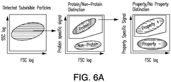
- collected data may be analyzed to define particle size and presence of proteinaceaous material in order to determine size and presence of proteinaceous subvisible particles.
- This is typically done by employing the gating database established by a size standard to set gates of defined size ranges which are used to estimate the size of each particle in the test sample and collect data regarding size distribution of all particles in the test sample.
- this data may be gated based on size and the resulting subset of sized particles analyzed for the presence of proteinaceaous material.
- data regarding the size and presence of proteinaceous subvisible particles in a sample can be determined.
- Such data may be graphically displayed as a one-parameter histogram.
- this data may be compared to total number of particles in order to define other useful characterizations including, e.g., the size and presence of non-proteinaceous subvisible particles and/or the presence of visible particles. For example, to qualitatively determine the presence of sized non-proteinaceous subvisible particles the presence value of all sized subvisible particles is subtracted by the presence value of sized proteinaceous subvisible particles. Similarly, data regarding the size and presence of non-proteinaceous subvisible particles in a sample can be determined as described above except that resulting subset of sized particles is analyzed for the presence of non-proteinaceaous material.
- the data representing size and presence of proteinaceous subvisible particles in a sample may be further analyzed to define particle number in order to determine the absolute number of the sized proteinaceous subvisible particles. This may be accomplished by gating the data representing particle size and the resulting subset of sized particles analyzed for particle number and the presence of proteinaceaous material. In this manner, data regarding the absolute number and size of proteinaceous subvisible particles in a sample can be determined. Such data may be graphically displayed as a two-parameter histogram. Once determined this data may be compared to total number of particles in order to define other useful characterizations including, e.g., the number, size and presence of non-proteinaceous subvisible particles and/or the number and presence of visible particles.
- the absolute number of all sized subvisible particles is subtracted by the absolute number of sized proteinaceous subvisible particles.
- data regarding absolute number of the sized non-proteinaceous subvisible particles in a sample can be determined as described above except that resulting subset of sized particles is analyzed for particle number and the presence of non-proteinaceaous material.
- the data representing absolute number, size and presence of proteinaceous subvisible particles in a sample is further analyzed to determine whether proteinaceous subvisible particles, or a subset thereof, have a certain physical property in order to determine the fraction of counted, sized proteinaceous subvisible particles with and without the physical property. This may be accomplished by gating the data representing particle size and the resulting subset of sized particles analyzed for particle number and the presence of proteinaceaous material with or without the physical property. In this manner, data regarding the absolute number and size of proteinaceous subvisible particles with and without a certain physical property in a sample can be determined. Such data may be graphically displayed as a two-parameter histogram.
- data regarding whether non-proteinaceous subvisible particles, or a subset thereof, have a certain physical property in order to determine the fraction of counted, sized non-proteinaceous subvisible particles with and without the physical property can be determined as described above except that the resulting subset of sized particles is analyzed for particle number and the presence of non-proteinaceaous material with or without the physical property.
- collected data may be analyzed to define particle size, particle number, and presence of proteinaceaous material in order to determine absolute number and size of proteinaceous subvisible particles.
- This is typically done by employing the gating database established by a size standard to set gates of defined size ranges which are used to estimate the size of each particle in the test sample and collect data regarding size distribution of all particles in the test sample. Once the size distribution of the particles is determined, this data may be gated based on size and the resulting subset of sized particles analyzed for particle number and the presence of proteinaceaous material. In this manner, data regarding the absolute number and size of proteinaceous subvisible particles in a sample can be determined. Such data may be graphically displayed as a two-parameter histogram.
- this data may be compared to the total number of particles in order to define other useful characterizations including, e.g., the number, size and presence of non-proteinaceous subvisible particles and/or the number and presence of visible particles.
- the absolute number of sized non-proteinaceous subvisible particles the absolute number of all sized subvisible particles is subtracted by the absolute number of sized proteinaceous subvisible particles.
- the data representing absolute number, size and presence of proteinaceous subvisible particles in a sample may be further analyzed to determine whether proteinaceous subvisible particles, or a subset thereof, have a certain physical property in order to determine the fraction of counted, sized proteinaceous subvisible particles with and without the physical property.
- data regarding the absolute number and size of non-proteinaceous subvisible particles in a sample can be determined as described above except that the resulting subset of sized particles is analyzed for particle number and the presence of non-proteinaceaous material.
- collected data may be analyzed to define particle size, particle number, presence of proteinaceaous material, and presence of a certain physical property in order to determine absolute number and size of proteinaceous subvisible particles with and without the physical property.
- This is typically done by employing the gating database established by a size standard to set gates of defined size ranges which are used to estimate the size of each particle in the test sample and collect data regarding size distribution of all particles in the test sample.
- this data may be gated based on size and the resulting subset of sized particles analyzed for particle number and the presence of proteinaceaous material with or without the physical property.
- data regarding the absolute number and size of proteinaceous subvisible particles with and without a certain physical property in a sample can be determined. Such data may be graphically displayed as a two-parameter histogram.
- data regarding the absolute number and size of non-proteinaceous subvisible particles with and without a certain physical property in a sample can be determined as described above except that the resulting subset of sized particles analyzed for particle number and the presence of non-proteinaceaous material with or without the physical property.
- the data may be analyzed to define the particle size, the particle number, presence of the proteinaceaous material, and presence of the physical property to determine absolute number and size of proteinaceous subvisible particles with and without the physical property, thereby characterizing the population of subvisible particles in the sample.
- the data may be analyzed to determine absolute number of sized non-proteinaceous subvisible particles by subtracting the absolute number of all sized subvisible particles by the absolute number of sized proteinaceous subvisible particles.
- the data may be analyzed to define the particle size and presence of the non-proteinaceaous material to determine size and presence of non-proteinaceaous subvisible particles, thereby characterizing the population of subvisible particles in the sample.
- the data may be analyzed to define the particle size, the particle number, and presence of the non-proteinaceaous material to determine absolute number and size of non-proteinaceaous subvisible particles, thereby characterizing the population of subvisible particles in the sample.
- the data may be analyzed to define the particle size, the particle number, presence of the non-proteinaceaous material, and presence of the physical property to determine absolute number and size of non-proteinaceaous subvisible particles with and without the physical property, thereby characterizing the population of subvisible particles in the sample.
- the data may be analyzed to determine absolute number of sized proteinaceous subvisible particles by subtracting the absolute number of all sized subvisible particles by the absolute number of sized non-proteinaceaous subvisible particles.
- a size standard stock solution was prepared from Fluoresbright Size Calibration Kit I and II (Polysciences, Inc., Warrington, PA). This stock solution included microspheres having mean diameters of 0.75 ⁇ m, 1.0 ⁇ m, 2.0 ⁇ m, 4.5 ⁇ m, 6.0 ⁇ m and 10 ⁇ m and was prepared according to the manufacturer's instructions.
- the prepared test sample was analyzed using a BD LSRFORTESSA TM particle analyzer (Becton Dickinson, San Jose, CA) equipped with a 405-nm violet laser, a 488-nm blue laser, a 561-nm yellow/green laser and a 635-nm red laser; and having two 488-nm light scattering detectors (low-angle forward scattering [FSC] and 90° side scattering [SSC]) and 16 fluorescence detectors.
- FSC low-angle forward scattering
- SSC 90° side scattering
- FIG. 1A Analysis of the data indicated that particles of about 0.75 ⁇ m, about 1.0 ⁇ m, about 2.0 ⁇ m, about 4.5 ⁇ m, about 6.0 ⁇ m, and about 10 ⁇ m were detected.
- FIG. 1A The results show that particles of less than 200 ⁇ m from a counting standard can be detected and sized.
- Analysis of the data also enabled the generation of particle size distribution ranges of less than about 0.75 ⁇ m, about 0.75 ⁇ m to about 1.0 ⁇ m, about 1.0 ⁇ m to about 2.0 ⁇ m, about 2.0 ⁇ m to about 4.5 ⁇ m, about 4.5 ⁇ m to about 6.0 ⁇ m, about 6.0 ⁇ m to about 10 ⁇ m, and about 10 ⁇ m to about 200 ⁇ m ( FIG. 1B ) and the placement of each detected particle into one of these size distribution ranges.
- FIG. 1B results showed that particles can be classified into a size distribution range comprising categories less than or equal to 200 ⁇ m.
- a size standard stock solution was prepared from Fluoresbright Size Calibration Kit I and II (Polysciences, Inc., Warrington, PA). This stock solution included microspheres having mean diameters of 0.75 ⁇ m, 1.0 ⁇ m, 2.0 ⁇ m, 4.5 ⁇ m, 6.0 ⁇ m and 10 ⁇ m and was prepared according to the manufacturer's instructions.
- the prepared test sample was analyzed using a BD FACSARIAIII TM particle analyzer (Becton Dickinson, San Jose, CA) equipped with a 405-nm violet laser, a 488-nm blue laser and a 633-nm red laser; and having two 488-nm light scattering detectors (low-angle forward scattering [FSC] and 90° side scattering [SSC]) and 11 fluorescence detectors.
- the forward and the side scatter were illustrated logarithmically.
- a threshold was set on the forward and side scatter in a manner that would include particles on the forward scatter vs. side scatter plot but still exclude electronic noise. All events were analyzed and recorded for 300 seconds running always at the same low sample flowrate (using a Diva Software Flowrate of 2).
- FIG. 2A Analysis of the data indicated that particles of about 0.75 ⁇ m, about 1.0 ⁇ m, about 2.0 ⁇ m, about 4.5 ⁇ m, about 6.0 ⁇ m, and about 10 ⁇ m were detected ( FIG. 2A ).
- the results show that particles of less than the diameter of the sample tubing (i.e., less than 200 ⁇ m) used in the flow cytometer can be detected and sized.
- the results show that particles of less than 200 ⁇ m from a counting standard can be detected and sized.
- COUNTBRIGHT TM Absolute Counting Beads (Invitrogen, Life Technologies Ltd., Paisley, Great Britain) were added to 450 ⁇ L of a particle sample including 50 ⁇ g/mL amyloid ⁇ peptide 1-42 aggregates.
- the prepared test sample was analyzed using a BD LSRFORTESSA TM particle analyzer (Becton Dickinson, San Jose, CA) as described in Preparation Example 1.
- the stored settings, thresholds, and gates determined by the size standard in Preparation Example 1 were used as a template for size distribution ranges. All events were analyzed, recorded, and evaluated as described in Preparation Example 1. Each sample was evaluated three times and a mean value and standard deviations of the three results were calculated.
- a control sample comprising formulation buffers was analyzed in parallel to record the background signal for the corresponding test sample. Background signals were subtracted from the sample signal in the final analyses.
- the first test sample included 25 ⁇ g/mL amyloid ⁇ peptide 1-42 aggregates (proteinaceous particles), the second test sample included polydisperse latex microspheres (non-proteinaceous particles) and the third test sample included a mixture of both amyloid ⁇ peptide 1-42 aggregates and polydisperse latex microspheres (Polybead PMMA clear 1-10 ⁇ m microparticles; Polysciences, Inc., Warrington, PA).
- To all three samples 50 ⁇ L (about 49,500 microspheres) of COUNTBRIGHT TM Absolute Counting Beads (Invitrogen, Life Technologies Ltd., Paisley, Great Britain) were added according to the manufacturer's instructions. To this mixture, 1x SYPRO TM Orange (SO) (Invitrogen, Life Technologies Ltd., Paisley, Great Britain) was added.
- the prepared test samples were analyzed using a BD LSRFORTESSA TM particle analyzer (Becton Dickinson, San Jose, CA) as described in Preparation Example 1.
- the stored settings, thresholds, and gates determined by the size standard in Preparation Example 1 were used as a template for size distribution ranges.
- Fluorescent intensity of 1x SYPRO TM Orange (SO) stained particles was detected using a blue laser with 530/30 bandpass filter. All events were analyzed, recorded, and evaluated as described in Preparation Example 3.
- a control sample comprising formulation buffers was analyzed in parallel to record the background signal for the corresponding test sample. Background signals were subtracted from the sample signal in the final analyses.
- a test sample of proteinaceous and non-proteinaceous particles was prepared as described in Preparation Example 3, except that Thioflavin T (ThT) was added to a concentration of 4 ⁇ M to these samples.
- Thioflavin T Thioflavin T
- the prepared test sample was analyzed using a BD LSRFORTESSA TM particle analyzer (Becton Dickinson, San Jose, CA) as described in Preparation Example 1.
- the stored settings, thresholds, and gates determined by the size standard in Preparation Example 1 were used as a template for size distribution ranges. Fluorescent intensity was detected using a violet laser with a 525/50 bandpass filter and a blue laser with 530/30 bandpass filter. All events were analyzed, recorded, and evaluated as described in Preparation Example 2.
- a control sample comprising formulation buffers was analyzed in parallel to record the background signal for the corresponding test sample. Background signals were subtracted from the sample signal in the final analyses.
- FIG. 5A Analysis of the data detected and discriminated proteinaceous particles from non-proteinaceous particles based on SYPRO TM Orange staining ( FIG. 5A ) and detected and discriminated proteinaceous particles having a ⁇ -sheet structure from proteinaceous particles lacking this structure ( FIG. 5B ). These results also allowed the identification of proteinaceous particles having a ⁇ -sheet structure in the sample ( FIG. 5C ). These results also allowed the determination of the absolute numbers of non-proteinaceous particles, proteinaceous lacking a ⁇ -sheet structure, and proteinaceous particles having a ⁇ -sheet structure ( FIG. 5D ). These results show that both qualitative and quantitative assessment of non-proteinaceous particles and proteinaceous with and without a certain physical property in a sample can be determined.
- the first test sample included 195 ⁇ L of a mixture of non-protein particles; the second test sample included 30 ⁇ L of 0.1 mg/mL glass beads of 3-10 ⁇ m as non-protein particles (Glass Beads 3-10 microns, Polysciences, Inc., Warrington, PA) and 165 ⁇ L of a mixture of A ⁇ peptide 1-42 subvisible particles and a protein subvisible particles containing low amounts of cross- ⁇ structures.
- Bis-ANS Sigma-Aldrich, St. Louis, MO
- DCVJ Sigma-Aldrich, St. Louis, MO
- the prepared test samples were analyzed using a BD FACSARIAIII TM particle analyzer (Becton Dickinson, San Jose, CA) as described in Preparation Example 2.
- the stored settings, thresholds, and gates determined by the size standard in Preparation Example 2 were used as a template for size distribution ranges. All events were analyzed, recorded, and evaluated as described in Preparation Example 2. Each sample was evaluated three times and a mean value and standard deviations of the three results were calculated.
- a control sample comprising formulation buffers was analyzed in parallel to record the background signal for the corresponding test sample. Background signals were subtracted from the sample signal in the final analyses.
- sample 1 containing only non-protein particles enabled the setting of gates to determine non-protein containing particles.
- This gate was stored and used for the analysis of sample 2 containing a mixture of non-protein particles, protein containing particles and amyloid like particles.
- Analysis of the data from sample 2 detected and discriminated proteinaceous particles from non-proteinaceous particles based on Bis-ANS staining and detected and discriminated proteinaceous particles having a ⁇ -sheet structure from proteinaceous particles lacking this structure using DCVJ.
- This gating strategy is shown in Appendix FIG. 6A .
- the results obtained from sample 2 show that proteinaceous subvisible particles can be detected and discriminated from proteinaceous and non-protein particles using Bis-ANS, and can be further characterized for containing cross- ⁇ structures using DCVJ ( FIG.
- the sample data was first plotted in Forward Scatter against Side Scatter to detect subvisible particles within the sample (Appendix FIG. 6B ).
- Bis-ANS was then used as the protein specific dye to discriminate between protein and non- protein containing particles (Appendix FIG. 6C ).
- Protein containing particles were further characterized using DCVJ which binds specific to cross- ⁇ structures and enables the identification of amyloid like protein particles (Appendix FIG. 6D ).
- Backgating analysis of protein and non-protein containing particles was also performed as shown in FIG. 6E .
- the term "about” means that the characteristic, item, quantity, parameter, property, or term so qualified encompasses a range of plus or minus ten percent above and below the value of the stated characteristic, item, quantity, parameter, property, or term.
- Each numerical indication should at least be construed in light of the number of reported significant digits and by applying ordinary rounding techniques. Notwithstanding that the numerical ranges and values setting forth the broad scope of the invention are approximations, the numerical ranges and values set forth in the specific examples are reported as precisely as possible. Any numerical range or value, however, inherently contains certain errors necessarily resulting from the standard deviation found in their respective testing measurements.
Landscapes
- Life Sciences & Earth Sciences (AREA)
- Health & Medical Sciences (AREA)
- Engineering & Computer Science (AREA)
- Chemical & Material Sciences (AREA)
- Molecular Biology (AREA)
- Immunology (AREA)
- Physics & Mathematics (AREA)
- Hematology (AREA)
- Biomedical Technology (AREA)
- Urology & Nephrology (AREA)
- General Health & Medical Sciences (AREA)
- Biochemistry (AREA)
- Analytical Chemistry (AREA)
- General Physics & Mathematics (AREA)
- Pathology (AREA)
- Biotechnology (AREA)
- Microbiology (AREA)
- Food Science & Technology (AREA)
- Medicinal Chemistry (AREA)
- Proteomics, Peptides & Aminoacids (AREA)
- Biophysics (AREA)
- Cell Biology (AREA)
- Bioinformatics & Cheminformatics (AREA)
- Bioinformatics & Computational Biology (AREA)
- Dispersion Chemistry (AREA)
- Organic Chemistry (AREA)
- Wood Science & Technology (AREA)
- Zoology (AREA)
- Signal Processing (AREA)
- Neurosurgery (AREA)
- General Engineering & Computer Science (AREA)
- Genetics & Genomics (AREA)
- Investigating, Analyzing Materials By Fluorescence Or Luminescence (AREA)
- Investigating Or Analysing Biological Materials (AREA)
- Measuring Or Testing Involving Enzymes Or Micro-Organisms (AREA)
- Analysing Materials By The Use Of Radiation (AREA)
Claims (14)
- Verfahren zur Charakterisierung einer Population aus nicht-sichtbaren Teilchen in einer pharmazeutischen Zubereitung, wobei das Verfahren die folgenden Schritte umfasst:a) ein Teilchenanalysator wird unter Verwendung eines Größenstandards kalibriert;b) während die Probe durch eine Detektionszone des Teilchenanalysators geführt wird wird die Probe analysiert, wobei die Probe einen Fluoreszenzzählstandard und mehrere Fluoreszenzfarbstoffe, die einen ersten Fluoreszenzfarbstoff und einen zweiten Fluoreszenzfarbstoff umfassen, umfasst,wobei die pharmazeutische Zubereitung ein Proteinhaltiges Material, das ein pharmazeutischer Wirkstoff, ein Adjuvans oder ein Hilfsstoff ist, umfasst,wobei die Analyse ein oder mehr Wellenlängen, welche den Größenstandard analysieren können, den Fluoreszenzzählstandard analysieren oder anregen können und die mehreren Fluoreszenzfarbstoffe analysieren oder anregen können, umfasst, undwobei der erste Fluoreszenzfarbstoff das Protein-haltige Material so färbt, dass das Protein-haltige Material von einem nicht-Protein-haltigen Material unterschieden werden kann, und der zweite Fluoreszenzfarbstoff eine Untereinheit des Protein-haltigen Materials mit einer physikalischen Eigenschaft des Protein-haltigen Materials so färbt, dass das Protein-haltige Material mit der physikalischen Eigenschaft von dem Protein-haltigen Material ohne die physikalische Eigenschaft unterschieden werden kann, und die Analyse ein oder mehr Wellenlängen, die den zweiten Fluoreszenzfarbstoff anregen können, einschließt;c) Daten von mindestens einem Lichtbrechungsparameter und mindestens einem Fluoreszenzparameter werden gesammelt,wobei die Daten, die von dem mindestens einen Lichtbrechungsparameter gesammelt werden, für eine Teilchengröße bezogen auf die Größenstandardkalibrierung stehen, undwobei die Daten, die von dem mindestens einen Fluoreszenzparameter gesammelt werden, einen ersten Fluoreszenzparameter, der die Anwesenheit des Protein-haltigen Materials bezogen auf die von dem ersten Fluoreszenzfarbstoff emittierte Fluoreszenzintensität anzeigt, einen zweiten Fluoreszenzparameter, der die Teilchenzahl bezogen auf die von dem Fluoreszenzzählstandard emittierte Fluoreszenzintensität anzeigt, und einen dritten Fluoreszenzparameter, der die Anwesenheit des Protein-haltigen Materials mit der physikalischen Eigenschaft bezogen auf die von dem zweiten Fluoreszenzfarbstoff emittierte Fluoreszenzintensität anzeigt, einschließen; undd) die Daten werden analysiert und die Teilchengröße, die Teilchenzahl, die Anwesenheit des Protein-haltigen Materials werden so definiert, dass eine absolute Zahl und Größe der Protein-haltigen nicht-sichtbaren Teilchen bestimmt wird, und die Anwesenheit der physikalischen Eigenschaft wird so definiert, dass ein Anteil von gezählten Protein-haltigen nicht-sichtbaren Teilchen einer Größe mit oder ohne physikalischer Eigenschaft bestimmt wird, und so wird die Population der nicht-sichtbaren Teilchen in der Probe charakterisiert,
wobei Bestimmung, Zählen und Charakterisierung der Population der nicht-sichtbaren Teilchen gleichzeitig ablaufen. - Verfahren nach Anspruch 1, bei dem der Datenanalyseschritt es ferner umfasst, dass eine absolute Zahl von nicht-Protein-haltigen nicht-sichtbaren Teilchen einer Größe durch Subtrahieren einer absoluten Zahl aller nicht-sichtbaren Teilchen einer Größe durch die absolute Zahl der Protein-haltigen nicht-sichtbaren Teilchen einer Größe bestimmt wird.
- Verfahren nach Anspruch 1 oder 2, bei dem der erste Fluoreszenzfarbstoff ein Styrolfarbstoff, eine Merocyaninfarbstoff, 3-(2-Benzothiazolyl-7-(diethylamino)-coumarin (BTDEC), 3-(2-Benzimidazolyl)- 7-(diethylamino)-coumarin (BIDEC), 3-(2-Benzimidazolyl)-7-coumarin (BIC), 3-(2-Benzimidazolyl)-7-(dipropylamino)-coumarin (BIDPC), 3-(2-Benzoxazolyl)-7-(diethylamino)-coumarin (BODEC), 3-(6-Methyl-2-benzoxazolyl)-7-(diethylamino)-coumarin (MBODEC), 3-(Diethylamino)-7-imino-7H-(1)benzopyrano (3',2':3,4)pyrido(1,2-a)benzimidazol-6-carbonitril (DIBPBC), 3-(Diethylamino)-7-oxo-7H-(1)benzopyrano(3',2':3,4) (DOBPBC), Dipyrromethenbordifluorid, ein photoluminescenter Metallkomplex, oder ein Hydroxychinolon ist.
- Verfahren nach mindestens einem der Ansprüche 1 bis 3, bei dem der Datenanalyseschritt es ferner umfasst, dass eine absolute Zahl von nicht-Protein-haltigen nicht-sichtbaren Teilchen einer Größe durch Subtrahieren der absoluten Zahl aller nicht-sichtbaren Teilchen einer Größe durch die absolute Zahl der Protein-haltigen nicht-sichtbaren Teilchen einer Größe bestimmt wird.
- Verfahren nach mindestens einem der Ansprüche 1 bis 4, bei dem der erste Fluoreszenzfarbstoff oder der Fluoreszenzzählstandard eine Peak-Fluoreszenzanregungswellenlänge von ungefähr 300 nm bis ungefähr 635 nm und eine Peak-Fluoreszenzemissionswellenlänge von ungefähr 350 nm bis ungefähr 800 nm aufweist.
- Verfahren nach mindestens einem der Ansprüche 1 bis 5, bei dem die physikalische Eigenschaft ein Strukturmerkmal wie beispielsweise eine α-Helixstruktur, eine β-Faltblattstruktur, oder eine Peptidspaltung oder chemische Modifikation wie beispielsweise eine Acylierung, eine Adenylylierung, eine Alkylierung, eine Amidierung, eine Glycylierung, eine Glycosylierung, eine Glypiation, eine Isoprenylierung, eine Myristoylierung, eine Palimoylierung, eine Pegylierung, eine Phosphorylierung, eine Prenylierung, eine Sulfatisierung, oder eine Ubiquinierung ist.
- Verfahren nach mindestens einem der Ansprüche 1 bis 6, bei dem der zweite Fluoreszenzfarbstoff eine Peak-Fluoreszenzanregungswellenlänge von ungefähr 350 nm bis ungefähr 635 nm und eine Peak-Fluoreszenzemissionswellenlänge von ungefähr 350 nm bis ungefähr 800 nm aufweist.
- Verfahren nach mindestens einem der Ansprüche 1 bis 7, bei dem der pharmazeutische Wirkstoff ADAMTS-13, al-Antiplasmin, α2-Antiplasmin, Antithrombin, Antithrombin III, Krebs-Procoagulant, Erythropoietin, Faktor II, Faktor IIa, Faktor V, Faktor Va, Faktor VI, Faktor VIa, Faktor VII, Faktor VIIa, Faktor VIII, Faktor VIIIA, Faktor IX, Faktor IXa, Faktor X, Faktor Xa, Faktor XI, Faktor Xla, Faktor XII, Faktor XIIa, Faktor XIII, Faktor XIIIa, Fibronectin, Fibrinogen (Faktor I), Heparin Cofaktor II, Kininogen mit hohem Molekulargewicht (HMWK), intramuskuläres Immunoglobulin, intravenöses Immunoglobulin, Plasmin, Plasminogen, Plasminogen Aktivator Inhibitor-1 (PAI1), Plasminogen Activator Inhibitor-2 (PAI2), Prekallikrein, Prostacyclin, Protein C, aktives Protein C(APC), Protein S, Protein Z, Protein Z-bezogener Protease Inhibitor, Thrombomodulin, Gewebefaktor (Faktor III), Gewebefaktor Weginhibitor (TFPI), Gewebe Plasminogen Aktivator (t-PA), Urokinase, Von Willebrand Faktor, Abciximab, Adalimumab, Alemtuzumab, Basiliximab, Belimumab, Bevacizumab, Brentuximab Vedotin, Canakinumab, Certolizumab pegol, Cetuximab, Daclizumab, Denosumab, Efalizumab, Eculizumab, Gemtuzumab ozogamicin, Golimumab, Ibritumomab tiuxetan, Infliximab, Ipilimumab, Motavizumab, Muronomab, Natalizumab, Ofatumumab, Omalizumab, Palivizumab, Panitumumab, Ranibizumab, Raxibacumab, Rituximab, Tocilizumab, Tositumomab, Trastuzumab, oder Ustekinumab ist.
- Verfahren nach mindestens einem der Ansprüche 1 bis 8, bei dem Daten eines elektrischen Detektionsparameters nicht durch den Teilchenanalysator gesammelt werden.
- Verfahren nach mindestens einem der Ansprüche 1 bis 9, bei dem der elektrische Detektionsparameter DC Impedanz, RF Leitfähigkeit, Opazität oder eine Kombination davon ist.
- Verfahren nach mindestens einem der Ansprüche 1 bis 10, bei dem der zweite Fluoreszenzfarbstoff 4-(3,6-Dimethyl-1,3-benzothiazol-3-ium-2-yl)-N,N-dimethylanilinchlorid (Thioflavin T), Natriumprimulin (Thoflavin S), 4-(1,3-Benzothiazol-2-yl)-N-methylanilin (BTA-1), ein Thiophenbasierter oligomerischer and polymerischer Farbstoff, Coumarin, 3,3'-Methandiylbis(4-hydroxy-2H-chromen-2-on) (Hydroxycoumarin), 11-Oxo-2,3,6,7-tetrahydro-1H,5H,11H-pyrano[2,3-f]pyrido[3,2,1-ij]chinolin-10-carbonsäure (Coumarin 343), 9-Methyl-2,3,6,7-tetrahydro-1H,5H,11H-pyrano[2,3-f]pyrido[3,2,1-ij]chinolin-11-on (Coumarin 102), 2,3,6,7-Tetrahydro-1H,5H-benzo[ij]chinolizin, (2E)-2-(2,3,6,7-Tetrahydro-1H,5H-pyrido[3,2,1-ij]chinolin-9-ylmethyliden)hydrazincarbothioamid, 9-[(E)-Phenyldiazenyl]-2,3,6,7-tetrahydro-1H,5H-pyrido[3,2,1-ij]chinolin, (2,3,6,7-Tetrahydro-1H,5H-pyrido[3,2,1-ij]chinolin-9-ylmethyliden) propandinitril, 9-(2,2-Dicyanovinyl)julolidin (DCVJ), (2E)-2-Cyano-3-(2,3,6,7-tetrahydro-1H,5H-pyrido[3,2,1-ij]chinolin-9-yl)prop-2-ensäure (CCVJ), 9-(Diethylamino)-5H-benzo[a]phenoxazin-5-on (Nilrot), 3,3'-([1,1'-Biphenyl]-4,4'-diyl)bis(4-aminonaphthalen-1-sulfonsäure) (Congorot), [(trans,trans)-1-Brom-2,5-bis-(3-hydroxycarbonyl-4-hydroxy)styrylbenzol] (BSB), 4-[(E)-2-[2-Brom-4-[(E)-2-(4-hydroxyphenyl)vinyl]phenyl]vinyl]phenol (K114), 8-(Phenylamino)naphthalen-1-sulfonsäure (ANS), Dikalium 4,4'-bis(phenylamino)-1,1'-binaphthalen-5,5'-disulfonat (Bis-ANS), Benzolsulfonsäure-Natriumsalz, 3-(4-Anilinphenylazo)benzolsulfonsäure-Natriumsalz (Orange IV), 1,6-Diphenyl-1,3,5-hexatrien (DPH), 1-(4-Trimethylammoniumphenyl)-6-phenyl-1,3,5-hexatrien p-tololsulfonat (TMA-DPH), oder 4',6-Diamidino-2-phenylindoldihydrochlorid (DAPI) ist.
- Verfahren nach mindestens einem der Ansprüche 1 bis 11, bei dem die Teilchengröße bezogen auf die Größenstandardkalibrierung Größen kleiner als oder gleich 10 µm, kleiner als oder gleich 20 µm, kleiner als oder gleich 50 µm, kleiner als oder gleich 75 µm, kleiner als oder gleich 100 µm, kleiner als oder gleich 200 µm oder kleiner als oder gleich 300 µm umfasst.
- Verfahren nach mindestens einem der Ansprüche 1 bis 12, bei dem die physikalische Eigenschaft ein Strukturmerkmal oder eine chemische Modifikation ist.
- Verfahren nach Anspruch 13, bei dem das Strukturmerkmal eine α-Helixstruktur, eine β-Faltblattstruktur oder eine Peptidspaltung ist.
Priority Applications (1)
| Application Number | Priority Date | Filing Date | Title |
|---|---|---|---|
| EP22172277.0A EP4063854A1 (de) | 2012-01-11 | 2013-01-11 | Charakterisierung von subvisiblen teilchen mit einem teilchenanalysator |
Applications Claiming Priority (3)
| Application Number | Priority Date | Filing Date | Title |
|---|---|---|---|
| US201261585425P | 2012-01-11 | 2012-01-11 | |
| US201261747720P | 2012-12-31 | 2012-12-31 | |
| PCT/US2013/021313 WO2013106772A2 (en) | 2012-01-11 | 2013-01-11 | Characterization of subvisible particles using a particle analyzer |
Related Child Applications (2)
| Application Number | Title | Priority Date | Filing Date |
|---|---|---|---|
| EP22172277.0A Division-Into EP4063854A1 (de) | 2012-01-11 | 2013-01-11 | Charakterisierung von subvisiblen teilchen mit einem teilchenanalysator |
| EP22172277.0A Division EP4063854A1 (de) | 2012-01-11 | 2013-01-11 | Charakterisierung von subvisiblen teilchen mit einem teilchenanalysator |
Publications (2)
| Publication Number | Publication Date |
|---|---|
| EP2802871A2 EP2802871A2 (de) | 2014-11-19 |
| EP2802871B1 true EP2802871B1 (de) | 2022-07-27 |
Family
ID=47682058
Family Applications (2)
| Application Number | Title | Priority Date | Filing Date |
|---|---|---|---|
| EP22172277.0A Withdrawn EP4063854A1 (de) | 2012-01-11 | 2013-01-11 | Charakterisierung von subvisiblen teilchen mit einem teilchenanalysator |
| EP13703486.4A Active EP2802871B1 (de) | 2012-01-11 | 2013-01-11 | Charakterisierung nicht-sichtbarer partikel mit hilfe eines partikelanalysators |
Family Applications Before (1)
| Application Number | Title | Priority Date | Filing Date |
|---|---|---|---|
| EP22172277.0A Withdrawn EP4063854A1 (de) | 2012-01-11 | 2013-01-11 | Charakterisierung von subvisiblen teilchen mit einem teilchenanalysator |
Country Status (6)
| Country | Link |
|---|---|
| US (2) | US10274500B2 (de) |
| EP (2) | EP4063854A1 (de) |
| JP (2) | JP6158835B2 (de) |
| AU (1) | AU2013204688B2 (de) |
| ES (1) | ES2928973T3 (de) |
| WO (1) | WO2013106772A2 (de) |
Families Citing this family (11)
| Publication number | Priority date | Publication date | Assignee | Title |
|---|---|---|---|---|
| ES2928973T3 (es) | 2012-01-11 | 2022-11-23 | Takeda Pharmaceuticals Co | Caracterización de partículas subvisibles usando un analizador de partículas |
| JP6415829B2 (ja) * | 2014-02-28 | 2018-10-31 | キヤノンメディカルシステムズ株式会社 | 検体に含まれるエキソソームの特性を判定する方法、診断方法および装置 |
| US10616219B2 (en) * | 2014-12-11 | 2020-04-07 | FlowJo, LLC | Single cell data management and analysis systems and methods |
| US12300357B2 (en) | 2016-12-14 | 2025-05-13 | FlowJo, LLC | Applied computer technology for management, synthesis, visualization, and exploration of parameters in large multi-parameter data sets |
| ES2980676T3 (es) | 2017-05-25 | 2024-10-02 | Flowjo Llc | Visualización, análisis comparativo y detección de diferencias automatizada para grandes conjuntos de datos multiparamétricos |
| US12320753B2 (en) * | 2018-03-20 | 2025-06-03 | Sanofi Pasteur Limited | Methods for determining adjuvanted protein concentration and percent adsorption using intrinsic fluorescence |
| WO2019246591A1 (en) * | 2018-06-21 | 2019-12-26 | Codiak Biosciences, Inc. | Methods of measuring extracellular vesicles and nanoparticles in complex matrices by light scattering |
| CN113543882A (zh) * | 2018-12-31 | 2021-10-22 | 本-古里安大学B.G.内盖夫技术和应用公司 | 捕获流测定装置和方法 |
| US11187707B2 (en) | 2019-01-30 | 2021-11-30 | Regeneron Pharmaceuticals, Inc. | Method of characterization of visible and/or sub-visible particles in biologics |
| JP7637937B2 (ja) * | 2020-04-28 | 2025-03-03 | 国立大学法人北海道大学 | 標的ポリペプチドの情報取得方法および試薬キット |
| JP2024529107A (ja) | 2021-08-13 | 2024-08-01 | ビオテスト・アクチエンゲゼルシャフト | フィブリノゲン組成物および調製方法 |
Citations (1)
| Publication number | Priority date | Publication date | Assignee | Title |
|---|---|---|---|---|
| WO2004015421A1 (en) * | 2002-08-07 | 2004-02-19 | Cyflo Oy | Method and device for identifying micro organisms |
Family Cites Families (21)
| Publication number | Priority date | Publication date | Assignee | Title |
|---|---|---|---|---|
| US4704891A (en) * | 1986-08-29 | 1987-11-10 | Becton, Dickinson And Company | Method and materials for calibrating flow cytometers and other analysis instruments |
| US4774339A (en) | 1987-08-10 | 1988-09-27 | Molecular Probes, Inc. | Chemically reactive dipyrrometheneboron difluoride dyes |
| US5540494A (en) * | 1994-06-03 | 1996-07-30 | Purvis, Jr.; Norman B. | Method and apparatus for determining absolute particle size, surface area and volume normalized fluorescence using forward angle light scatter intensity in flow cytometry |
| JP3875754B2 (ja) * | 1995-11-17 | 2007-01-31 | シスメックス株式会社 | フローサイトメータ用標準液 |
| WO1999057566A1 (en) * | 1998-05-01 | 1999-11-11 | The University Of Tennessee Research Corporation | Flow cytometric characterization of amyloid fibrils |
| US6870165B2 (en) * | 2001-10-19 | 2005-03-22 | Biocal Technology, Inc. | Multi-color multiplexed analysis in a bio-separation system |
| US7091049B2 (en) * | 2002-06-26 | 2006-08-15 | Kimberly-Clark Worldwide, Inc. | Enhanced diffraction-based biosensor devices |
| AU2003903417A0 (en) * | 2003-07-04 | 2003-07-17 | Genera Biosystems Pty Ltd | Multiplex detection |
| AU2004262301A1 (en) * | 2003-07-25 | 2005-02-10 | Platypus Technologies, Llc | Liquid crystal based analyte detection |
| WO2005033283A2 (en) * | 2003-09-30 | 2005-04-14 | Singulex, Inc. | Methods for enhancing the analysis of particle detection |
| US7295306B2 (en) * | 2004-04-22 | 2007-11-13 | Kowa Company, Ltd. | Microchip and fluorescent particle counter with microchip |
| JP4953710B2 (ja) * | 2005-07-12 | 2012-06-13 | シスメックス株式会社 | 尿中有形成分分析装置用標準物質 |
| JP4744264B2 (ja) * | 2005-10-19 | 2011-08-10 | シスメックス株式会社 | 尿中有形成分分析装置用標準物質。 |
| US7947506B2 (en) | 2006-06-02 | 2011-05-24 | Pierce Biotechnology, Inc. | Protein detection and quantitation using hydroxyquinolone dyes |
| US20100105635A1 (en) * | 2007-01-30 | 2010-04-29 | Tohoku University | Composition for prevention or treatment of disease associated with amyloidosis through inhibition of amyloid protein aggregation and promotion of amyloid protein degradation |
| JP4509163B2 (ja) * | 2007-10-26 | 2010-07-21 | ソニー株式会社 | 微小粒子の測定方法 |
| US9943490B2 (en) * | 2007-11-05 | 2018-04-17 | The Trustees Of Princeton University | Composite flash-precipitated nanoparticles |
| CA2705848C (en) | 2007-11-21 | 2016-02-02 | Bio-Rad Laboratories, Inc. | Photoluminescent metal complexes for protein staining |
| JP5395359B2 (ja) | 2008-02-20 | 2014-01-22 | 株式会社アリミノ | 脱染剤組成物 |
| US20090311715A1 (en) * | 2008-05-09 | 2009-12-17 | Mayo Foundation For Medical Education And Research | Identifying coronary or soft tissue calcification |
| ES2928973T3 (es) | 2012-01-11 | 2022-11-23 | Takeda Pharmaceuticals Co | Caracterización de partículas subvisibles usando un analizador de partículas |
-
2013
- 2013-01-11 ES ES13703486T patent/ES2928973T3/es active Active
- 2013-01-11 EP EP22172277.0A patent/EP4063854A1/de not_active Withdrawn
- 2013-01-11 JP JP2014552349A patent/JP6158835B2/ja active Active
- 2013-01-11 EP EP13703486.4A patent/EP2802871B1/de active Active
- 2013-01-11 US US13/739,812 patent/US10274500B2/en active Active
- 2013-01-11 WO PCT/US2013/021313 patent/WO2013106772A2/en not_active Ceased
- 2013-01-11 AU AU2013204688A patent/AU2013204688B2/en active Active
-
2017
- 2017-06-08 JP JP2017113334A patent/JP2017207495A/ja active Pending
-
2019
- 2019-03-11 US US16/298,520 patent/US11921117B2/en active Active
Patent Citations (1)
| Publication number | Priority date | Publication date | Assignee | Title |
|---|---|---|---|---|
| WO2004015421A1 (en) * | 2002-08-07 | 2004-02-19 | Cyflo Oy | Method and device for identifying micro organisms |
Non-Patent Citations (2)
| Title |
|---|
| MUTHUVEL JAYACHANDRAN ET AL: "Methodology for isolation, identification and characterization of microvesicles in peripheral blood", JOURNAL OF IMMUNOLOGICAL METHODS, ELSEVIER SCIENCE PUBLISHERS B.V.,AMSTERDAM, NL, vol. 375, no. 1, 19 October 2011 (2011-10-19), pages 207 - 214, XP028434311, ISSN: 0022-1759, [retrieved on 20111029], DOI: 10.1016/J.JIM.2011.10.012 * |
| SPARROW ROSEMARY L ET AL: "Microparticle content of plasma for transfusion is influenced by the whole blood hold conditions: Pre-analytical considerations for proteomic investigations", JOURNAL OF PROTEOMICS, vol. 76, 16 July 2012 (2012-07-16), pages 211 - 219, XP028959196, ISSN: 1874-3919, DOI: 10.1016/J.JPROT.2012.07.013 * |
Also Published As
| Publication number | Publication date |
|---|---|
| AU2013204688A1 (en) | 2013-07-25 |
| WO2013106772A3 (en) | 2014-01-09 |
| EP2802871A2 (de) | 2014-11-19 |
| US20130177933A1 (en) | 2013-07-11 |
| ES2928973T3 (es) | 2022-11-23 |
| JP2015503766A (ja) | 2015-02-02 |
| EP4063854A1 (de) | 2022-09-28 |
| US10274500B2 (en) | 2019-04-30 |
| US20190271706A1 (en) | 2019-09-05 |
| US11921117B2 (en) | 2024-03-05 |
| JP2017207495A (ja) | 2017-11-24 |
| JP6158835B2 (ja) | 2017-07-05 |
| WO2013106772A2 (en) | 2013-07-18 |
| AU2013204688B2 (en) | 2016-02-04 |
Similar Documents
| Publication | Publication Date | Title |
|---|---|---|
| US11921117B2 (en) | Characterization of subvisible particles using a particle analyzer | |
| US20230251194A1 (en) | Optical analyses of particles and vesicles | |
| US9683920B2 (en) | Use of focused light scattering techniques in biological applications | |
| US10774359B2 (en) | Cellular analysis of body fluids | |
| ES2902648T3 (es) | Método de análisis de glóbulos rojos nucleados y analizador hematológico automatizado | |
| US8163151B2 (en) | Assays for detection of Von Willebrand factor (vWF) multimers and for degradation of vWF by agents such as Adamts13 and related methods | |
| US9404933B2 (en) | Methods for determining patient response to anti-platelet aggregation therapy | |
| JP2012215593A (ja) | 完全自動化したモノクローナル抗体による広範な識別法 | |
| US20110254533A1 (en) | Coaxial Illumination Of Coulter Aperture In Full Function Hematology Analyzer | |
| Zhou et al. | Early apoptosis real-time detection by label-free SERS based on externalized phosphatidylserine | |
| US9354161B2 (en) | Sample analyzing method and sample analyzer | |
| Tschöpe et al. | Platelet analysis using flowcytometric procedures | |
| KR20080082650A (ko) | 2개 이상의 세포 군집을 식별하는 방법, 및 용도 | |
| HK40081566A (en) | Characterization of subvisible particles using a particle analyzer | |
| Mullaney et al. | Laser flow microphotometers for rapid analysis and sorting of individual mammalian cells | |
| KR20130064255A (ko) | 신규한 혈소판 활성 측정 방법 및 그를 이용한 장치 | |
| GIANIORIO et al. | Analysis of platelet-derived microparticles and their role in platelet activation and stimulation under static and dynamic conditions | |
| WO2021210189A1 (ja) | 病原体量子結晶固相化基板およびそれを用いる病原体の量子結晶固相化蛍光計数法 | |
| GHEORGHE et al. | FLOW CYTOMETRIC ANALYSIS AS A QUICK AND EFFICIENT M ETHO D FO RMO RPHO LO G IC ALC HA NG ES DETERM INA TIO N IN HEM A TO LO G IC AL DISEA SES. RBC s OFGA UC HER DISEA SE ASA MO DEL |
Legal Events
| Date | Code | Title | Description |
|---|---|---|---|
| PUAI | Public reference made under article 153(3) epc to a published international application that has entered the european phase |
Free format text: ORIGINAL CODE: 0009012 |
|
| 17P | Request for examination filed |
Effective date: 20140711 |
|
| AK | Designated contracting states |
Kind code of ref document: A2 Designated state(s): AL AT BE BG CH CY CZ DE DK EE ES FI FR GB GR HR HU IE IS IT LI LT LU LV MC MK MT NL NO PL PT RO RS SE SI SK SM TR |
|
| AX | Request for extension of the european patent |
Extension state: BA ME |
|
| RAP1 | Party data changed (applicant data changed or rights of an application transferred) |
Owner name: BAXALTA GMBH Owner name: BAXALTA INCORPORATED |
|
| PUAG | Search results despatched under rule 164(2) epc together with communication from examining division |
Free format text: ORIGINAL CODE: 0009017 |
|
| 17Q | First examination report despatched |
Effective date: 20161010 |
|
| B565 | Issuance of search results under rule 164(2) epc |
Effective date: 20161010 |
|
| STAA | Information on the status of an ep patent application or granted ep patent |
Free format text: STATUS: EXAMINATION IS IN PROGRESS |
|
| RAP1 | Party data changed (applicant data changed or rights of an application transferred) |
Owner name: BAXALTA GMBH Owner name: BAXALTA INCORPORATED |
|
| RAP1 | Party data changed (applicant data changed or rights of an application transferred) |
Owner name: BAXALTA GMBH Owner name: BAXALTA INCORPORATED |
|
| RAP1 | Party data changed (applicant data changed or rights of an application transferred) |
Owner name: TAKEDA PHARMACEUTICAL COMPANY LIMITED |
|
| GRAP | Despatch of communication of intention to grant a patent |
Free format text: ORIGINAL CODE: EPIDOSNIGR1 |
|
| STAA | Information on the status of an ep patent application or granted ep patent |
Free format text: STATUS: GRANT OF PATENT IS INTENDED |
|
| GRAJ | Information related to disapproval of communication of intention to grant by the applicant or resumption of examination proceedings by the epo deleted |
Free format text: ORIGINAL CODE: EPIDOSDIGR1 |
|
| GRAP | Despatch of communication of intention to grant a patent |
Free format text: ORIGINAL CODE: EPIDOSNIGR1 |
|
| INTG | Intention to grant announced |
Effective date: 20211014 |
|
| INTG | Intention to grant announced |
Effective date: 20211027 |
|
| GRAJ | Information related to disapproval of communication of intention to grant by the applicant or resumption of examination proceedings by the epo deleted |
Free format text: ORIGINAL CODE: EPIDOSDIGR1 |
|
| STAA | Information on the status of an ep patent application or granted ep patent |
Free format text: STATUS: EXAMINATION IS IN PROGRESS |
|
| INTC | Intention to grant announced (deleted) | ||
| GRAP | Despatch of communication of intention to grant a patent |
Free format text: ORIGINAL CODE: EPIDOSNIGR1 |
|
| STAA | Information on the status of an ep patent application or granted ep patent |
Free format text: STATUS: GRANT OF PATENT IS INTENDED |
|
| INTG | Intention to grant announced |
Effective date: 20220217 |
|
| GRAS | Grant fee paid |
Free format text: ORIGINAL CODE: EPIDOSNIGR3 |
|
| GRAA | (expected) grant |
Free format text: ORIGINAL CODE: 0009210 |
|
| STAA | Information on the status of an ep patent application or granted ep patent |
Free format text: STATUS: THE PATENT HAS BEEN GRANTED |
|
| AK | Designated contracting states |
Kind code of ref document: B1 Designated state(s): AL AT BE BG CH CY CZ DE DK EE ES FI FR GB GR HR HU IE IS IT LI LT LU LV MC MK MT NL NO PL PT RO RS SE SI SK SM TR |
|
| REG | Reference to a national code |
Ref country code: GB Ref legal event code: FG4D |
|
| REG | Reference to a national code |
Ref country code: CH Ref legal event code: EP |
|
| REG | Reference to a national code |
Ref country code: DE Ref legal event code: R096 Ref document number: 602013082146 Country of ref document: DE |
|
| REG | Reference to a national code |
Ref country code: AT Ref legal event code: REF Ref document number: 1507391 Country of ref document: AT Kind code of ref document: T Effective date: 20220815 |
|
| REG | Reference to a national code |
Ref country code: IE Ref legal event code: FG4D |
|
| REG | Reference to a national code |
Ref country code: LT Ref legal event code: MG9D |
|
| REG | Reference to a national code |
Ref country code: ES Ref legal event code: FG2A Ref document number: 2928973 Country of ref document: ES Kind code of ref document: T3 Effective date: 20221123 |
|
| REG | Reference to a national code |
Ref country code: NL Ref legal event code: MP Effective date: 20220727 |
|
| PG25 | Lapsed in a contracting state [announced via postgrant information from national office to epo] |
Ref country code: SE Free format text: LAPSE BECAUSE OF FAILURE TO SUBMIT A TRANSLATION OF THE DESCRIPTION OR TO PAY THE FEE WITHIN THE PRESCRIBED TIME-LIMIT Effective date: 20220727 Ref country code: RS Free format text: LAPSE BECAUSE OF FAILURE TO SUBMIT A TRANSLATION OF THE DESCRIPTION OR TO PAY THE FEE WITHIN THE PRESCRIBED TIME-LIMIT Effective date: 20220727 Ref country code: PT Free format text: LAPSE BECAUSE OF FAILURE TO SUBMIT A TRANSLATION OF THE DESCRIPTION OR TO PAY THE FEE WITHIN THE PRESCRIBED TIME-LIMIT Effective date: 20221128 Ref country code: NO Free format text: LAPSE BECAUSE OF FAILURE TO SUBMIT A TRANSLATION OF THE DESCRIPTION OR TO PAY THE FEE WITHIN THE PRESCRIBED TIME-LIMIT Effective date: 20221027 Ref country code: NL Free format text: LAPSE BECAUSE OF FAILURE TO SUBMIT A TRANSLATION OF THE DESCRIPTION OR TO PAY THE FEE WITHIN THE PRESCRIBED TIME-LIMIT Effective date: 20220727 Ref country code: LV Free format text: LAPSE BECAUSE OF FAILURE TO SUBMIT A TRANSLATION OF THE DESCRIPTION OR TO PAY THE FEE WITHIN THE PRESCRIBED TIME-LIMIT Effective date: 20220727 Ref country code: LT Free format text: LAPSE BECAUSE OF FAILURE TO SUBMIT A TRANSLATION OF THE DESCRIPTION OR TO PAY THE FEE WITHIN THE PRESCRIBED TIME-LIMIT Effective date: 20220727 Ref country code: FI Free format text: LAPSE BECAUSE OF FAILURE TO SUBMIT A TRANSLATION OF THE DESCRIPTION OR TO PAY THE FEE WITHIN THE PRESCRIBED TIME-LIMIT Effective date: 20220727 |
|
| PG25 | Lapsed in a contracting state [announced via postgrant information from national office to epo] |
Ref country code: PL Free format text: LAPSE BECAUSE OF FAILURE TO SUBMIT A TRANSLATION OF THE DESCRIPTION OR TO PAY THE FEE WITHIN THE PRESCRIBED TIME-LIMIT Effective date: 20220727 Ref country code: IS Free format text: LAPSE BECAUSE OF FAILURE TO SUBMIT A TRANSLATION OF THE DESCRIPTION OR TO PAY THE FEE WITHIN THE PRESCRIBED TIME-LIMIT Effective date: 20221127 Ref country code: HR Free format text: LAPSE BECAUSE OF FAILURE TO SUBMIT A TRANSLATION OF THE DESCRIPTION OR TO PAY THE FEE WITHIN THE PRESCRIBED TIME-LIMIT Effective date: 20220727 Ref country code: GR Free format text: LAPSE BECAUSE OF FAILURE TO SUBMIT A TRANSLATION OF THE DESCRIPTION OR TO PAY THE FEE WITHIN THE PRESCRIBED TIME-LIMIT Effective date: 20221028 |
|
| PG25 | Lapsed in a contracting state [announced via postgrant information from national office to epo] |
Ref country code: SM Free format text: LAPSE BECAUSE OF FAILURE TO SUBMIT A TRANSLATION OF THE DESCRIPTION OR TO PAY THE FEE WITHIN THE PRESCRIBED TIME-LIMIT Effective date: 20220727 Ref country code: RO Free format text: LAPSE BECAUSE OF FAILURE TO SUBMIT A TRANSLATION OF THE DESCRIPTION OR TO PAY THE FEE WITHIN THE PRESCRIBED TIME-LIMIT Effective date: 20220727 Ref country code: DK Free format text: LAPSE BECAUSE OF FAILURE TO SUBMIT A TRANSLATION OF THE DESCRIPTION OR TO PAY THE FEE WITHIN THE PRESCRIBED TIME-LIMIT Effective date: 20220727 Ref country code: CZ Free format text: LAPSE BECAUSE OF FAILURE TO SUBMIT A TRANSLATION OF THE DESCRIPTION OR TO PAY THE FEE WITHIN THE PRESCRIBED TIME-LIMIT Effective date: 20220727 |
|
| REG | Reference to a national code |
Ref country code: DE Ref legal event code: R097 Ref document number: 602013082146 Country of ref document: DE |
|
| PG25 | Lapsed in a contracting state [announced via postgrant information from national office to epo] |
Ref country code: SK Free format text: LAPSE BECAUSE OF FAILURE TO SUBMIT A TRANSLATION OF THE DESCRIPTION OR TO PAY THE FEE WITHIN THE PRESCRIBED TIME-LIMIT Effective date: 20220727 Ref country code: EE Free format text: LAPSE BECAUSE OF FAILURE TO SUBMIT A TRANSLATION OF THE DESCRIPTION OR TO PAY THE FEE WITHIN THE PRESCRIBED TIME-LIMIT Effective date: 20220727 |
|
| PLBE | No opposition filed within time limit |
Free format text: ORIGINAL CODE: 0009261 |
|
| STAA | Information on the status of an ep patent application or granted ep patent |
Free format text: STATUS: NO OPPOSITION FILED WITHIN TIME LIMIT |
|
| P01 | Opt-out of the competence of the unified patent court (upc) registered |
Effective date: 20230428 |
|
| PG25 | Lapsed in a contracting state [announced via postgrant information from national office to epo] |
Ref country code: AL Free format text: LAPSE BECAUSE OF FAILURE TO SUBMIT A TRANSLATION OF THE DESCRIPTION OR TO PAY THE FEE WITHIN THE PRESCRIBED TIME-LIMIT Effective date: 20220727 |
|
| 26N | No opposition filed |
Effective date: 20230502 |
|
| PG25 | Lapsed in a contracting state [announced via postgrant information from national office to epo] |
Ref country code: SI Free format text: LAPSE BECAUSE OF FAILURE TO SUBMIT A TRANSLATION OF THE DESCRIPTION OR TO PAY THE FEE WITHIN THE PRESCRIBED TIME-LIMIT Effective date: 20220727 |
|
| PG25 | Lapsed in a contracting state [announced via postgrant information from national office to epo] |
Ref country code: LU Free format text: LAPSE BECAUSE OF NON-PAYMENT OF DUE FEES Effective date: 20230111 |
|
| REG | Reference to a national code |
Ref country code: BE Ref legal event code: MM Effective date: 20230131 |
|
| PG25 | Lapsed in a contracting state [announced via postgrant information from national office to epo] |
Ref country code: BE Free format text: LAPSE BECAUSE OF NON-PAYMENT OF DUE FEES Effective date: 20230131 |
|
| PG25 | Lapsed in a contracting state [announced via postgrant information from national office to epo] |
Ref country code: IE Free format text: LAPSE BECAUSE OF NON-PAYMENT OF DUE FEES Effective date: 20230111 |
|
| PG25 | Lapsed in a contracting state [announced via postgrant information from national office to epo] |
Ref country code: MC Free format text: LAPSE BECAUSE OF FAILURE TO SUBMIT A TRANSLATION OF THE DESCRIPTION OR TO PAY THE FEE WITHIN THE PRESCRIBED TIME-LIMIT Effective date: 20220727 |
|
| PG25 | Lapsed in a contracting state [announced via postgrant information from national office to epo] |
Ref country code: MC Free format text: LAPSE BECAUSE OF FAILURE TO SUBMIT A TRANSLATION OF THE DESCRIPTION OR TO PAY THE FEE WITHIN THE PRESCRIBED TIME-LIMIT Effective date: 20220727 |
|
| PG25 | Lapsed in a contracting state [announced via postgrant information from national office to epo] |
Ref country code: BG Free format text: LAPSE BECAUSE OF FAILURE TO SUBMIT A TRANSLATION OF THE DESCRIPTION OR TO PAY THE FEE WITHIN THE PRESCRIBED TIME-LIMIT Effective date: 20220727 |
|
| PG25 | Lapsed in a contracting state [announced via postgrant information from national office to epo] |
Ref country code: BG Free format text: LAPSE BECAUSE OF FAILURE TO SUBMIT A TRANSLATION OF THE DESCRIPTION OR TO PAY THE FEE WITHIN THE PRESCRIBED TIME-LIMIT Effective date: 20220727 |
|
| PG25 | Lapsed in a contracting state [announced via postgrant information from national office to epo] |
Ref country code: CY Free format text: LAPSE BECAUSE OF FAILURE TO SUBMIT A TRANSLATION OF THE DESCRIPTION OR TO PAY THE FEE WITHIN THE PRESCRIBED TIME-LIMIT; INVALID AB INITIO Effective date: 20130111 |
|
| PG25 | Lapsed in a contracting state [announced via postgrant information from national office to epo] |
Ref country code: HU Free format text: LAPSE BECAUSE OF FAILURE TO SUBMIT A TRANSLATION OF THE DESCRIPTION OR TO PAY THE FEE WITHIN THE PRESCRIBED TIME-LIMIT; INVALID AB INITIO Effective date: 20130111 |
|
| PG25 | Lapsed in a contracting state [announced via postgrant information from national office to epo] |
Ref country code: TR Free format text: LAPSE BECAUSE OF FAILURE TO SUBMIT A TRANSLATION OF THE DESCRIPTION OR TO PAY THE FEE WITHIN THE PRESCRIBED TIME-LIMIT Effective date: 20220727 |
|
| PGFP | Annual fee paid to national office [announced via postgrant information from national office to epo] |
Ref country code: GB Payment date: 20251220 Year of fee payment: 14 |
|
| PGFP | Annual fee paid to national office [announced via postgrant information from national office to epo] |
Ref country code: FR Payment date: 20251217 Year of fee payment: 14 |
|
| REG | Reference to a national code |
Ref country code: CH Ref legal event code: U11 Free format text: ST27 STATUS EVENT CODE: U-0-0-U10-U11 (AS PROVIDED BY THE NATIONAL OFFICE) Effective date: 20260201 |
|
| PGFP | Annual fee paid to national office [announced via postgrant information from national office to epo] |
Ref country code: ES Payment date: 20260202 Year of fee payment: 14 |
|
| PGFP | Annual fee paid to national office [announced via postgrant information from national office to epo] |
Ref country code: DE Payment date: 20251217 Year of fee payment: 14 |
|
| PGFP | Annual fee paid to national office [announced via postgrant information from national office to epo] |
Ref country code: AT Payment date: 20251218 Year of fee payment: 14 |
|
| PGFP | Annual fee paid to national office [announced via postgrant information from national office to epo] |
Ref country code: IT Payment date: 20251219 Year of fee payment: 14 |
|
| PGFP | Annual fee paid to national office [announced via postgrant information from national office to epo] |
Ref country code: CH Payment date: 20260201 Year of fee payment: 14 |