EP2802350B1 - Administration régulée d'agonistes de tlr dans des dispositifs polymères structuraux - Google Patents
Administration régulée d'agonistes de tlr dans des dispositifs polymères structuraux Download PDFInfo
- Publication number
- EP2802350B1 EP2802350B1 EP13702535.9A EP13702535A EP2802350B1 EP 2802350 B1 EP2802350 B1 EP 2802350B1 EP 13702535 A EP13702535 A EP 13702535A EP 2802350 B1 EP2802350 B1 EP 2802350B1
- Authority
- EP
- European Patent Office
- Prior art keywords
- cpg
- csf
- cells
- odn
- tumor
- Prior art date
- Legal status (The legal status is an assumption and is not a legal conclusion. Google has not performed a legal analysis and makes no representation as to the accuracy of the status listed.)
- Active
Links
- 239000000556 agonist Substances 0.000 title claims description 94
- 210000004443 dendritic cell Anatomy 0.000 claims description 534
- 108010017213 Granulocyte-Macrophage Colony-Stimulating Factor Proteins 0.000 claims description 347
- 102100039620 Granulocyte-macrophage colony-stimulating factor Human genes 0.000 claims description 332
- 239000000203 mixture Substances 0.000 claims description 269
- 206010028980 Neoplasm Diseases 0.000 claims description 257
- 239000000427 antigen Substances 0.000 claims description 191
- 108091007433 antigens Proteins 0.000 claims description 187
- 102000036639 antigens Human genes 0.000 claims description 186
- 229940046168 CpG oligodeoxynucleotide Drugs 0.000 claims description 156
- 102000002689 Toll-like receptor Human genes 0.000 claims description 78
- 108020000411 Toll-like receptor Proteins 0.000 claims description 78
- 238000000034 method Methods 0.000 claims description 75
- 230000007115 recruitment Effects 0.000 claims description 74
- 230000004913 activation Effects 0.000 claims description 72
- 239000006166 lysate Substances 0.000 claims description 63
- 201000011510 cancer Diseases 0.000 claims description 58
- 108090000623 proteins and genes Proteins 0.000 claims description 47
- 102000004169 proteins and genes Human genes 0.000 claims description 38
- 229920002873 Polyethylenimine Polymers 0.000 claims description 36
- 102000008230 Toll-like receptor 3 Human genes 0.000 claims description 36
- 108010060885 Toll-like receptor 3 Proteins 0.000 claims description 36
- 108091034117 Oligonucleotide Proteins 0.000 claims description 35
- 210000005134 plasmacytoid dendritic cell Anatomy 0.000 claims description 35
- 238000004519 manufacturing process Methods 0.000 claims description 34
- 108010065805 Interleukin-12 Proteins 0.000 claims description 32
- 102000013462 Interleukin-12 Human genes 0.000 claims description 32
- 229940117681 interleukin-12 Drugs 0.000 claims description 31
- 229940115272 polyinosinic:polycytidylic acid Drugs 0.000 claims description 18
- 102100036848 C-C motif chemokine 20 Human genes 0.000 claims description 17
- 101000713099 Homo sapiens C-C motif chemokine 20 Proteins 0.000 claims description 17
- 244000052769 pathogen Species 0.000 claims description 16
- 102100020715 Fms-related tyrosine kinase 3 ligand protein Human genes 0.000 claims description 15
- 101710162577 Fms-related tyrosine kinase 3 ligand protein Proteins 0.000 claims description 15
- 229940044655 toll-like receptor 9 agonist Drugs 0.000 claims description 15
- 201000010099 disease Diseases 0.000 claims description 13
- 208000037265 diseases, disorders, signs and symptoms Diseases 0.000 claims description 13
- 230000001717 pathogenic effect Effects 0.000 claims description 13
- 230000005975 antitumor immune response Effects 0.000 claims description 12
- 101000669447 Homo sapiens Toll-like receptor 4 Proteins 0.000 claims description 11
- 102100039360 Toll-like receptor 4 Human genes 0.000 claims description 9
- 229940029575 guanosine Drugs 0.000 claims description 9
- 239000002253 acid Substances 0.000 claims description 7
- 230000009467 reduction Effects 0.000 claims description 6
- 108091036414 Polyinosinic:polycytidylic acid Proteins 0.000 claims description 5
- 210000003169 central nervous system Anatomy 0.000 claims description 5
- 206010009944 Colon cancer Diseases 0.000 claims description 3
- 208000024313 Testicular Neoplasms Diseases 0.000 claims description 3
- 206010057644 Testis cancer Diseases 0.000 claims description 3
- 208000029742 colonic neoplasm Diseases 0.000 claims description 3
- 206010073071 hepatocellular carcinoma Diseases 0.000 claims description 3
- 231100000844 hepatocellular carcinoma Toxicity 0.000 claims description 3
- 208000032839 leukemia Diseases 0.000 claims description 3
- 201000003120 testicular cancer Diseases 0.000 claims description 3
- 206010006187 Breast cancer Diseases 0.000 claims description 2
- 208000026310 Breast neoplasm Diseases 0.000 claims description 2
- 206010018338 Glioma Diseases 0.000 claims description 2
- 208000008839 Kidney Neoplasms Diseases 0.000 claims description 2
- 208000000172 Medulloblastoma Diseases 0.000 claims description 2
- 208000034578 Multiple myelomas Diseases 0.000 claims description 2
- 206010035226 Plasma cell myeloma Diseases 0.000 claims description 2
- 206010038389 Renal cancer Diseases 0.000 claims description 2
- 230000006023 anti-tumor response Effects 0.000 claims description 2
- 201000010353 central nervous system germ cell tumor Diseases 0.000 claims description 2
- 208000029824 high grade glioma Diseases 0.000 claims description 2
- 201000010982 kidney cancer Diseases 0.000 claims description 2
- 201000011614 malignant glioma Diseases 0.000 claims description 2
- 208000007766 Kaposi sarcoma Diseases 0.000 claims 1
- 206010033128 Ovarian cancer Diseases 0.000 claims 1
- 206010060862 Prostate cancer Diseases 0.000 claims 1
- 208000000236 Prostatic Neoplasms Diseases 0.000 claims 1
- 208000009956 adenocarcinoma Diseases 0.000 claims 1
- 206010041823 squamous cell carcinoma Diseases 0.000 claims 1
- 206010042863 synovial sarcoma Diseases 0.000 claims 1
- 210000004027 cell Anatomy 0.000 description 243
- 229960005486 vaccine Drugs 0.000 description 98
- 230000000975 bioactive effect Effects 0.000 description 92
- 229940046166 oligodeoxynucleotide Drugs 0.000 description 68
- 239000000463 material Substances 0.000 description 67
- 102100022297 Integrin alpha-X Human genes 0.000 description 61
- 241000699670 Mus sp. Species 0.000 description 59
- 208000015181 infectious disease Diseases 0.000 description 59
- 238000011065 in-situ storage Methods 0.000 description 57
- 210000001744 T-lymphocyte Anatomy 0.000 description 56
- 210000001519 tissue Anatomy 0.000 description 52
- 229920000642 polymer Polymers 0.000 description 49
- 230000028993 immune response Effects 0.000 description 48
- 102000004127 Cytokines Human genes 0.000 description 47
- 108090000695 Cytokines Proteins 0.000 description 47
- 210000001151 cytotoxic T lymphocyte Anatomy 0.000 description 47
- 230000000694 effects Effects 0.000 description 44
- 210000001165 lymph node Anatomy 0.000 description 44
- 101500027983 Rattus norvegicus Octadecaneuropeptide Proteins 0.000 description 43
- 201000001441 melanoma Diseases 0.000 description 43
- 238000001727 in vivo Methods 0.000 description 40
- 239000000126 substance Substances 0.000 description 40
- 238000002513 implantation Methods 0.000 description 38
- 235000018102 proteins Nutrition 0.000 description 37
- 229940035032 monophosphoryl lipid a Drugs 0.000 description 34
- 229940022399 cancer vaccine Drugs 0.000 description 33
- -1 TLR11 Proteins 0.000 description 32
- 238000009566 cancer vaccine Methods 0.000 description 32
- 230000004083 survival effect Effects 0.000 description 32
- 230000005012 migration Effects 0.000 description 31
- 238000013508 migration Methods 0.000 description 31
- 150000007523 nucleic acids Chemical class 0.000 description 30
- 108020004414 DNA Proteins 0.000 description 29
- 102000053602 DNA Human genes 0.000 description 29
- 241001465754 Metazoa Species 0.000 description 27
- 239000004005 microsphere Substances 0.000 description 27
- 108090000765 processed proteins & peptides Proteins 0.000 description 27
- 230000004044 response Effects 0.000 description 27
- 230000011664 signaling Effects 0.000 description 27
- 210000004881 tumor cell Anatomy 0.000 description 27
- 102100034256 Mucin-1 Human genes 0.000 description 26
- 102000008235 Toll-Like Receptor 9 Human genes 0.000 description 26
- 108010060818 Toll-Like Receptor 9 Proteins 0.000 description 26
- 230000006870 function Effects 0.000 description 26
- 239000011148 porous material Substances 0.000 description 26
- 210000002865 immune cell Anatomy 0.000 description 25
- 238000002255 vaccination Methods 0.000 description 25
- 238000011746 C57BL/6J (JAX™ mouse strain) Methods 0.000 description 24
- 238000004458 analytical method Methods 0.000 description 23
- 238000000338 in vitro Methods 0.000 description 23
- 239000011159 matrix material Substances 0.000 description 23
- 102100036301 C-C chemokine receptor type 7 Human genes 0.000 description 22
- 101000716065 Homo sapiens C-C chemokine receptor type 7 Proteins 0.000 description 22
- JLCPHMBAVCMARE-UHFFFAOYSA-N [3-[[3-[[3-[[3-[[3-[[3-[[3-[[3-[[3-[[3-[[3-[[5-(2-amino-6-oxo-1H-purin-9-yl)-3-[[3-[[3-[[3-[[3-[[3-[[5-(2-amino-6-oxo-1H-purin-9-yl)-3-[[5-(2-amino-6-oxo-1H-purin-9-yl)-3-hydroxyoxolan-2-yl]methoxy-hydroxyphosphoryl]oxyoxolan-2-yl]methoxy-hydroxyphosphoryl]oxy-5-(5-methyl-2,4-dioxopyrimidin-1-yl)oxolan-2-yl]methoxy-hydroxyphosphoryl]oxy-5-(6-aminopurin-9-yl)oxolan-2-yl]methoxy-hydroxyphosphoryl]oxy-5-(6-aminopurin-9-yl)oxolan-2-yl]methoxy-hydroxyphosphoryl]oxy-5-(6-aminopurin-9-yl)oxolan-2-yl]methoxy-hydroxyphosphoryl]oxy-5-(6-aminopurin-9-yl)oxolan-2-yl]methoxy-hydroxyphosphoryl]oxyoxolan-2-yl]methoxy-hydroxyphosphoryl]oxy-5-(5-methyl-2,4-dioxopyrimidin-1-yl)oxolan-2-yl]methoxy-hydroxyphosphoryl]oxy-5-(4-amino-2-oxopyrimidin-1-yl)oxolan-2-yl]methoxy-hydroxyphosphoryl]oxy-5-(5-methyl-2,4-dioxopyrimidin-1-yl)oxolan-2-yl]methoxy-hydroxyphosphoryl]oxy-5-(5-methyl-2,4-dioxopyrimidin-1-yl)oxolan-2-yl]methoxy-hydroxyphosphoryl]oxy-5-(6-aminopurin-9-yl)oxolan-2-yl]methoxy-hydroxyphosphoryl]oxy-5-(6-aminopurin-9-yl)oxolan-2-yl]methoxy-hydroxyphosphoryl]oxy-5-(4-amino-2-oxopyrimidin-1-yl)oxolan-2-yl]methoxy-hydroxyphosphoryl]oxy-5-(4-amino-2-oxopyrimidin-1-yl)oxolan-2-yl]methoxy-hydroxyphosphoryl]oxy-5-(4-amino-2-oxopyrimidin-1-yl)oxolan-2-yl]methoxy-hydroxyphosphoryl]oxy-5-(6-aminopurin-9-yl)oxolan-2-yl]methoxy-hydroxyphosphoryl]oxy-5-(4-amino-2-oxopyrimidin-1-yl)oxolan-2-yl]methyl [5-(6-aminopurin-9-yl)-2-(hydroxymethyl)oxolan-3-yl] hydrogen phosphate Polymers Cc1cn(C2CC(OP(O)(=O)OCC3OC(CC3OP(O)(=O)OCC3OC(CC3O)n3cnc4c3nc(N)[nH]c4=O)n3cnc4c3nc(N)[nH]c4=O)C(COP(O)(=O)OC3CC(OC3COP(O)(=O)OC3CC(OC3COP(O)(=O)OC3CC(OC3COP(O)(=O)OC3CC(OC3COP(O)(=O)OC3CC(OC3COP(O)(=O)OC3CC(OC3COP(O)(=O)OC3CC(OC3COP(O)(=O)OC3CC(OC3COP(O)(=O)OC3CC(OC3COP(O)(=O)OC3CC(OC3COP(O)(=O)OC3CC(OC3COP(O)(=O)OC3CC(OC3COP(O)(=O)OC3CC(OC3COP(O)(=O)OC3CC(OC3COP(O)(=O)OC3CC(OC3COP(O)(=O)OC3CC(OC3COP(O)(=O)OC3CC(OC3CO)n3cnc4c(N)ncnc34)n3ccc(N)nc3=O)n3cnc4c(N)ncnc34)n3ccc(N)nc3=O)n3ccc(N)nc3=O)n3ccc(N)nc3=O)n3cnc4c(N)ncnc34)n3cnc4c(N)ncnc34)n3cc(C)c(=O)[nH]c3=O)n3cc(C)c(=O)[nH]c3=O)n3ccc(N)nc3=O)n3cc(C)c(=O)[nH]c3=O)n3cnc4c3nc(N)[nH]c4=O)n3cnc4c(N)ncnc34)n3cnc4c(N)ncnc34)n3cnc4c(N)ncnc34)n3cnc4c(N)ncnc34)O2)c(=O)[nH]c1=O JLCPHMBAVCMARE-UHFFFAOYSA-N 0.000 description 22
- 150000001875 compounds Chemical class 0.000 description 22
- 102000043131 MHC class II family Human genes 0.000 description 21
- 108091054438 MHC class II family Proteins 0.000 description 21
- 229920000615 alginic acid Polymers 0.000 description 21
- 235000010443 alginic acid Nutrition 0.000 description 21
- 230000001965 increasing effect Effects 0.000 description 21
- 230000004224 protection Effects 0.000 description 21
- 210000000612 antigen-presenting cell Anatomy 0.000 description 20
- 230000014509 gene expression Effects 0.000 description 20
- 230000036039 immunity Effects 0.000 description 20
- 102000004196 processed proteins & peptides Human genes 0.000 description 20
- 239000003446 ligand Substances 0.000 description 19
- 230000001681 protective effect Effects 0.000 description 19
- 239000000243 solution Substances 0.000 description 19
- FHVDTGUDJYJELY-UHFFFAOYSA-N 6-{[2-carboxy-4,5-dihydroxy-6-(phosphanyloxy)oxan-3-yl]oxy}-4,5-dihydroxy-3-phosphanyloxane-2-carboxylic acid Chemical compound O1C(C(O)=O)C(P)C(O)C(O)C1OC1C(C(O)=O)OC(OP)C(O)C1O FHVDTGUDJYJELY-UHFFFAOYSA-N 0.000 description 18
- 102000014150 Interferons Human genes 0.000 description 17
- 108010050904 Interferons Proteins 0.000 description 17
- 238000011161 development Methods 0.000 description 17
- 230000018109 developmental process Effects 0.000 description 17
- 229940047124 interferons Drugs 0.000 description 17
- 102000039446 nucleic acids Human genes 0.000 description 17
- 108020004707 nucleic acids Proteins 0.000 description 17
- 230000003389 potentiating effect Effects 0.000 description 17
- 230000004936 stimulating effect Effects 0.000 description 17
- 230000000638 stimulation Effects 0.000 description 17
- 102100036842 C-C motif chemokine 19 Human genes 0.000 description 16
- 101000713106 Homo sapiens C-C motif chemokine 19 Proteins 0.000 description 16
- 101001133056 Homo sapiens Mucin-1 Proteins 0.000 description 16
- 230000002596 correlated effect Effects 0.000 description 16
- 230000002163 immunogen Effects 0.000 description 16
- 229920001184 polypeptide Polymers 0.000 description 16
- 230000001105 regulatory effect Effects 0.000 description 16
- 238000013459 approach Methods 0.000 description 15
- 238000001943 fluorescence-activated cell sorting Methods 0.000 description 15
- 238000005187 foaming Methods 0.000 description 15
- 230000001404 mediated effect Effects 0.000 description 15
- 230000008569 process Effects 0.000 description 15
- CZMRCDWAGMRECN-UGDNZRGBSA-N Sucrose Chemical compound O[C@H]1[C@H](O)[C@@H](CO)O[C@@]1(CO)O[C@@H]1[C@H](O)[C@@H](O)[C@H](O)[C@@H](CO)O1 CZMRCDWAGMRECN-UGDNZRGBSA-N 0.000 description 14
- 229930006000 Sucrose Natural products 0.000 description 14
- 229940072056 alginate Drugs 0.000 description 14
- 125000003275 alpha amino acid group Chemical group 0.000 description 14
- 230000030741 antigen processing and presentation Effects 0.000 description 14
- 230000008595 infiltration Effects 0.000 description 14
- 238000001764 infiltration Methods 0.000 description 14
- 239000002245 particle Substances 0.000 description 14
- 239000005720 sucrose Substances 0.000 description 14
- 241000282414 Homo sapiens Species 0.000 description 13
- 108010047761 Interferon-alpha Proteins 0.000 description 13
- 102000006992 Interferon-alpha Human genes 0.000 description 13
- 241000699666 Mus <mouse, genus> Species 0.000 description 13
- 108010004729 Phycoerythrin Proteins 0.000 description 13
- 230000005867 T cell response Effects 0.000 description 13
- 230000002757 inflammatory effect Effects 0.000 description 13
- 230000035800 maturation Effects 0.000 description 13
- OHDXDNUPVVYWOV-UHFFFAOYSA-N n-methyl-1-(2-naphthalen-1-ylsulfanylphenyl)methanamine Chemical compound CNCC1=CC=CC=C1SC1=CC=CC2=CC=CC=C12 OHDXDNUPVVYWOV-UHFFFAOYSA-N 0.000 description 13
- 239000002243 precursor Substances 0.000 description 13
- LKKMLIBUAXYLOY-UHFFFAOYSA-N 3-Amino-1-methyl-5H-pyrido[4,3-b]indole Chemical compound N1C2=CC=CC=C2C2=C1C=C(N)N=C2C LKKMLIBUAXYLOY-UHFFFAOYSA-N 0.000 description 12
- 102100037850 Interferon gamma Human genes 0.000 description 12
- 108010074328 Interferon-gamma Proteins 0.000 description 12
- 230000008901 benefit Effects 0.000 description 12
- 230000001413 cellular effect Effects 0.000 description 12
- 238000009472 formulation Methods 0.000 description 12
- 239000003102 growth factor Substances 0.000 description 12
- LMDZBCPBFSXMTL-UHFFFAOYSA-N 1-ethyl-3-(3-dimethylaminopropyl)carbodiimide Chemical compound CCN=C=NCCCN(C)C LMDZBCPBFSXMTL-UHFFFAOYSA-N 0.000 description 11
- 108091028043 Nucleic acid sequence Proteins 0.000 description 11
- 239000002955 immunomodulating agent Substances 0.000 description 11
- 229940121354 immunomodulator Drugs 0.000 description 11
- 230000019734 interleukin-12 production Effects 0.000 description 11
- 108091029430 CpG site Proteins 0.000 description 10
- 230000003213 activating effect Effects 0.000 description 10
- 230000015556 catabolic process Effects 0.000 description 10
- 238000009833 condensation Methods 0.000 description 10
- 239000000499 gel Substances 0.000 description 10
- 150000004676 glycans Chemical class 0.000 description 10
- 210000000987 immune system Anatomy 0.000 description 10
- 239000007943 implant Substances 0.000 description 10
- 210000001616 monocyte Anatomy 0.000 description 10
- 229920001282 polysaccharide Polymers 0.000 description 10
- 239000005017 polysaccharide Substances 0.000 description 10
- 108010088201 squamous cell carcinoma-related antigen Proteins 0.000 description 10
- 108010004889 Heat-Shock Proteins Proteins 0.000 description 9
- 102000002812 Heat-Shock Proteins Human genes 0.000 description 9
- 241000282412 Homo Species 0.000 description 9
- 230000005809 anti-tumor immunity Effects 0.000 description 9
- 230000005494 condensation Effects 0.000 description 9
- 238000006731 degradation reaction Methods 0.000 description 9
- 238000009792 diffusion process Methods 0.000 description 9
- 239000002158 endotoxin Substances 0.000 description 9
- 239000000017 hydrogel Substances 0.000 description 9
- 238000010348 incorporation Methods 0.000 description 9
- 238000002347 injection Methods 0.000 description 9
- 239000007924 injection Substances 0.000 description 9
- 229920006008 lipopolysaccharide Polymers 0.000 description 9
- 239000000047 product Substances 0.000 description 9
- 229920002477 rna polymer Polymers 0.000 description 9
- 230000009885 systemic effect Effects 0.000 description 9
- 230000001225 therapeutic effect Effects 0.000 description 9
- AEMRFAOFKBGASW-UHFFFAOYSA-N Glycolic acid Natural products OCC(O)=O AEMRFAOFKBGASW-UHFFFAOYSA-N 0.000 description 8
- 101000669460 Homo sapiens Toll-like receptor 5 Proteins 0.000 description 8
- 108010008707 Mucin-1 Proteins 0.000 description 8
- 102000004887 Transforming Growth Factor beta Human genes 0.000 description 8
- 108090001012 Transforming Growth Factor beta Proteins 0.000 description 8
- 108060008682 Tumor Necrosis Factor Proteins 0.000 description 8
- 102000000852 Tumor Necrosis Factor-alpha Human genes 0.000 description 8
- 230000014102 antigen processing and presentation of exogenous peptide antigen via MHC class I Effects 0.000 description 8
- 210000003719 b-lymphocyte Anatomy 0.000 description 8
- 239000006285 cell suspension Substances 0.000 description 8
- 229940030156 cell vaccine Drugs 0.000 description 8
- JVTAAEKCZFNVCJ-UHFFFAOYSA-N lactic acid Chemical compound CC(O)C(O)=O JVTAAEKCZFNVCJ-UHFFFAOYSA-N 0.000 description 8
- VQWNELVFHZRFIB-UHFFFAOYSA-N odn 1826 Chemical compound O=C1NC(=O)C(C)=CN1C(O1)CC(O)C1COP(O)(=O)OC1CC(N2C(NC(=O)C(C)=C2)=O)OC1COP(O)(=O)OC1CC(N2C3=C(C(NC(N)=N3)=O)N=C2)OC1COP(O)(=O)OC1CC(N2C(N=C(N)C=C2)=O)OC1COP(O)(=O)OC1CC(N2C3=NC=NC(N)=C3N=C2)OC1COP(O)(=O)OC1CC(N2C3=C(C(NC(N)=N3)=O)N=C2)OC1COP(O)(=O)OC1CC(N2C(NC(=O)C(C)=C2)=O)OC1COP(O)(=O)OC1CC(N2C(N=C(N)C=C2)=O)OC1COP(O)(=O)OC1CC(N2C(N=C(N)C=C2)=O)OC1COP(O)(=O)OC1CC(N2C(NC(=O)C(C)=C2)=O)OC1COP(O)(=O)OC(C(O1)COP(O)(=O)OC2C(OC(C2)N2C3=C(C(NC(N)=N3)=O)N=C2)COP(O)(=O)OC2C(OC(C2)N2C(N=C(N)C=C2)=O)COP(O)(=O)OC2C(OC(C2)N2C3=NC=NC(N)=C3N=C2)COP(O)(=O)OC2C(OC(C2)N2C3=C(C(NC(N)=N3)=O)N=C2)COP(O)(=O)OC2C(OC(C2)N2C(NC(=O)C(C)=C2)=O)COP(O)(=O)OC2C(OC(C2)N2C3=NC=NC(N)=C3N=C2)COP(O)(=O)OC2C(OC(C2)N2C(N=C(N)C=C2)=O)COP(O)(=O)OC2C(OC(C2)N2C(N=C(N)C=C2)=O)COP(O)(=O)OC2C(OC(C2)N2C(NC(=O)C(C)=C2)=O)COP(O)(O)=O)CC1N1C=C(C)C(=O)NC1=O VQWNELVFHZRFIB-UHFFFAOYSA-N 0.000 description 8
- 102000040430 polynucleotide Human genes 0.000 description 8
- 108091033319 polynucleotide Proteins 0.000 description 8
- 239000002157 polynucleotide Substances 0.000 description 8
- 238000010186 staining Methods 0.000 description 8
- 241000894006 Bacteria Species 0.000 description 7
- 102100031413 L-dopachrome tautomerase Human genes 0.000 description 7
- 101710093778 L-dopachrome tautomerase Proteins 0.000 description 7
- 241001529936 Murinae Species 0.000 description 7
- 230000005764 inhibitory process Effects 0.000 description 7
- 239000002773 nucleotide Substances 0.000 description 7
- 125000003729 nucleotide group Chemical group 0.000 description 7
- 239000008055 phosphate buffer solution Substances 0.000 description 7
- 229920001606 poly(lactic acid-co-glycolic acid) Polymers 0.000 description 7
- 238000002360 preparation method Methods 0.000 description 7
- 102000005962 receptors Human genes 0.000 description 7
- 108020003175 receptors Proteins 0.000 description 7
- 210000000952 spleen Anatomy 0.000 description 7
- 238000007920 subcutaneous administration Methods 0.000 description 7
- 238000002054 transplantation Methods 0.000 description 7
- 230000032258 transport Effects 0.000 description 7
- MZOFCQQQCNRIBI-VMXHOPILSA-N (3s)-4-[[(2s)-1-[[(2s)-1-[[(1s)-1-carboxy-2-hydroxyethyl]amino]-4-methyl-1-oxopentan-2-yl]amino]-5-(diaminomethylideneamino)-1-oxopentan-2-yl]amino]-3-[[2-[[(2s)-2,6-diaminohexanoyl]amino]acetyl]amino]-4-oxobutanoic acid Chemical compound OC[C@@H](C(O)=O)NC(=O)[C@H](CC(C)C)NC(=O)[C@H](CCCN=C(N)N)NC(=O)[C@H](CC(O)=O)NC(=O)CNC(=O)[C@@H](N)CCCCN MZOFCQQQCNRIBI-VMXHOPILSA-N 0.000 description 6
- IJGRMHOSHXDMSA-UHFFFAOYSA-N Atomic nitrogen Chemical compound N#N IJGRMHOSHXDMSA-UHFFFAOYSA-N 0.000 description 6
- 108020000946 Bacterial DNA Proteins 0.000 description 6
- 102000029816 Collagenase Human genes 0.000 description 6
- 108060005980 Collagenase Proteins 0.000 description 6
- 108010017080 Granulocyte Colony-Stimulating Factor Proteins 0.000 description 6
- 102000004269 Granulocyte Colony-Stimulating Factor Human genes 0.000 description 6
- WZUVPPKBWHMQCE-UHFFFAOYSA-N Haematoxylin Chemical compound C12=CC(O)=C(O)C=C2CC2(O)C1C1=CC=C(O)C(O)=C1OC2 WZUVPPKBWHMQCE-UHFFFAOYSA-N 0.000 description 6
- 101000831567 Homo sapiens Toll-like receptor 2 Proteins 0.000 description 6
- 101000669406 Homo sapiens Toll-like receptor 6 Proteins 0.000 description 6
- 102100036383 Serpin B3 Human genes 0.000 description 6
- 102100039357 Toll-like receptor 5 Human genes 0.000 description 6
- 230000000259 anti-tumor effect Effects 0.000 description 6
- 230000001580 bacterial effect Effects 0.000 description 6
- 239000012620 biological material Substances 0.000 description 6
- 230000033228 biological regulation Effects 0.000 description 6
- 230000012292 cell migration Effects 0.000 description 6
- 230000004700 cellular uptake Effects 0.000 description 6
- 230000008859 change Effects 0.000 description 6
- 229960002424 collagenase Drugs 0.000 description 6
- 230000001276 controlling effect Effects 0.000 description 6
- 229920001577 copolymer Polymers 0.000 description 6
- 230000000875 corresponding effect Effects 0.000 description 6
- 230000000139 costimulatory effect Effects 0.000 description 6
- 238000005859 coupling reaction Methods 0.000 description 6
- 238000004132 cross linking Methods 0.000 description 6
- 238000000684 flow cytometry Methods 0.000 description 6
- 239000007789 gas Substances 0.000 description 6
- 230000006698 induction Effects 0.000 description 6
- 230000001939 inductive effect Effects 0.000 description 6
- 230000002401 inhibitory effect Effects 0.000 description 6
- 210000002540 macrophage Anatomy 0.000 description 6
- 230000007246 mechanism Effects 0.000 description 6
- 238000003752 polymerase chain reaction Methods 0.000 description 6
- 230000037452 priming Effects 0.000 description 6
- 210000003491 skin Anatomy 0.000 description 6
- 230000002459 sustained effect Effects 0.000 description 6
- ZRKFYGHZFMAOKI-QMGMOQQFSA-N tgfbeta Chemical compound C([C@H](NC(=O)[C@H](C(C)C)NC(=O)CNC(=O)[C@H](CCC(O)=O)NC(=O)[C@H](CCCNC(N)=N)NC(=O)[C@H](CC(N)=O)NC(=O)[C@H](CC(C)C)NC(=O)[C@H]([C@@H](C)O)NC(=O)[C@H](CCC(O)=O)NC(=O)[C@H]([C@@H](C)O)NC(=O)[C@H](CC(C)C)NC(=O)CNC(=O)[C@H](C)NC(=O)[C@H](CO)NC(=O)[C@H](CCC(N)=O)NC(=O)[C@@H](NC(=O)[C@H](C)NC(=O)[C@H](C)NC(=O)[C@@H](NC(=O)[C@H](CC(C)C)NC(=O)[C@@H](N)CCSC)C(C)C)[C@@H](C)CC)C(=O)N[C@@H]([C@@H](C)O)C(=O)N[C@@H](C(C)C)C(=O)N[C@@H](CC=1C=CC=CC=1)C(=O)N[C@@H](C)C(=O)N1[C@@H](CCC1)C(=O)N[C@@H]([C@@H](C)O)C(=O)N[C@@H](CC(N)=O)C(=O)N[C@@H](CCC(O)=O)C(=O)N[C@@H](C)C(=O)N[C@@H](CC=1C=CC=CC=1)C(=O)N[C@@H](CCCNC(N)=N)C(=O)N[C@@H](C)C(=O)N[C@@H](CC(C)C)C(=O)N1[C@@H](CCC1)C(=O)N1[C@@H](CCC1)C(=O)N[C@@H](CCCNC(N)=N)C(=O)N[C@@H](CCC(O)=O)C(=O)N[C@@H](CCCNC(N)=N)C(=O)N[C@@H](CO)C(=O)N[C@@H](CCCNC(N)=N)C(=O)N[C@@H](CC(C)C)C(=O)N[C@@H](CC(C)C)C(O)=O)C1=CC=C(O)C=C1 ZRKFYGHZFMAOKI-QMGMOQQFSA-N 0.000 description 6
- 230000004614 tumor growth Effects 0.000 description 6
- RKDVKSZUMVYZHH-UHFFFAOYSA-N 1,4-dioxane-2,5-dione Chemical compound O=C1COC(=O)CO1 RKDVKSZUMVYZHH-UHFFFAOYSA-N 0.000 description 5
- 210000001266 CD8-positive T-lymphocyte Anatomy 0.000 description 5
- 102000008186 Collagen Human genes 0.000 description 5
- 108010035532 Collagen Proteins 0.000 description 5
- 101001036688 Homo sapiens Melanoma-associated antigen B1 Proteins 0.000 description 5
- 101000763537 Homo sapiens Toll-like receptor 10 Proteins 0.000 description 5
- 206010061218 Inflammation Diseases 0.000 description 5
- 102000000589 Interleukin-1 Human genes 0.000 description 5
- 108010002352 Interleukin-1 Proteins 0.000 description 5
- 108090000174 Interleukin-10 Proteins 0.000 description 5
- 108010002350 Interleukin-2 Proteins 0.000 description 5
- 102000000588 Interleukin-2 Human genes 0.000 description 5
- KDXKERNSBIXSRK-UHFFFAOYSA-N Lysine Natural products NCCCCC(N)C(O)=O KDXKERNSBIXSRK-UHFFFAOYSA-N 0.000 description 5
- 108700018351 Major Histocompatibility Complex Proteins 0.000 description 5
- 102100039477 Melanoma-associated antigen B1 Human genes 0.000 description 5
- 101100481579 Mus musculus Tlr11 gene Proteins 0.000 description 5
- 101100481580 Mus musculus Tlr12 gene Proteins 0.000 description 5
- 101100481581 Mus musculus Tlr13 gene Proteins 0.000 description 5
- 230000006044 T cell activation Effects 0.000 description 5
- 230000009471 action Effects 0.000 description 5
- 239000002671 adjuvant Substances 0.000 description 5
- 230000000890 antigenic effect Effects 0.000 description 5
- 238000001574 biopsy Methods 0.000 description 5
- 210000004369 blood Anatomy 0.000 description 5
- 239000008280 blood Substances 0.000 description 5
- 210000003068 cdc Anatomy 0.000 description 5
- 229920001436 collagen Polymers 0.000 description 5
- 201000010276 collecting duct carcinoma Diseases 0.000 description 5
- 238000010276 construction Methods 0.000 description 5
- 230000008878 coupling Effects 0.000 description 5
- 238000010168 coupling process Methods 0.000 description 5
- 230000001186 cumulative effect Effects 0.000 description 5
- 230000001472 cytotoxic effect Effects 0.000 description 5
- 230000000593 degrading effect Effects 0.000 description 5
- 210000001787 dendrite Anatomy 0.000 description 5
- 230000001419 dependent effect Effects 0.000 description 5
- 230000004069 differentiation Effects 0.000 description 5
- 231100000673 dose–response relationship Toxicity 0.000 description 5
- 239000012636 effector Substances 0.000 description 5
- 239000012634 fragment Substances 0.000 description 5
- 230000001506 immunosuppresive effect Effects 0.000 description 5
- 230000001976 improved effect Effects 0.000 description 5
- 230000004054 inflammatory process Effects 0.000 description 5
- 239000007788 liquid Substances 0.000 description 5
- 230000004807 localization Effects 0.000 description 5
- 230000007774 longterm Effects 0.000 description 5
- 230000003278 mimic effect Effects 0.000 description 5
- 102000007863 pattern recognition receptors Human genes 0.000 description 5
- 108010089193 pattern recognition receptors Proteins 0.000 description 5
- 239000013612 plasmid Substances 0.000 description 5
- 238000012545 processing Methods 0.000 description 5
- 230000001737 promoting effect Effects 0.000 description 5
- 230000000069 prophylactic effect Effects 0.000 description 5
- 210000003289 regulatory T cell Anatomy 0.000 description 5
- 210000004988 splenocyte Anatomy 0.000 description 5
- 210000000130 stem cell Anatomy 0.000 description 5
- 230000020382 suppression by virus of host antigen processing and presentation of peptide antigen via MHC class I Effects 0.000 description 5
- 238000011282 treatment Methods 0.000 description 5
- 101150079396 trpC2 gene Proteins 0.000 description 5
- 239000013598 vector Substances 0.000 description 5
- XLYOFNOQVPJJNP-UHFFFAOYSA-N water Substances O XLYOFNOQVPJJNP-UHFFFAOYSA-N 0.000 description 5
- 229920000936 Agarose Polymers 0.000 description 4
- 108010022366 Carcinoembryonic Antigen Proteins 0.000 description 4
- 102100025475 Carcinoembryonic antigen-related cell adhesion molecule 5 Human genes 0.000 description 4
- 108010012236 Chemokines Proteins 0.000 description 4
- 102000019034 Chemokines Human genes 0.000 description 4
- QOSSAOTZNIDXMA-UHFFFAOYSA-N Dicylcohexylcarbodiimide Chemical compound C1CCCCC1N=C=NC1CCCCC1 QOSSAOTZNIDXMA-UHFFFAOYSA-N 0.000 description 4
- 101001036686 Homo sapiens Melanoma-associated antigen B2 Proteins 0.000 description 4
- 101001024605 Homo sapiens Next to BRCA1 gene 1 protein Proteins 0.000 description 4
- 101000604116 Homo sapiens RNA-binding protein Nova-2 Proteins 0.000 description 4
- 101000669402 Homo sapiens Toll-like receptor 7 Proteins 0.000 description 4
- 101000800483 Homo sapiens Toll-like receptor 8 Proteins 0.000 description 4
- 102000015696 Interleukins Human genes 0.000 description 4
- 108010063738 Interleukins Proteins 0.000 description 4
- 108090001030 Lipoproteins Proteins 0.000 description 4
- 102000004895 Lipoproteins Human genes 0.000 description 4
- 108091054437 MHC class I family Proteins 0.000 description 4
- 102100039479 Melanoma-associated antigen B2 Human genes 0.000 description 4
- 206010061309 Neoplasm progression Diseases 0.000 description 4
- 102100037001 Next to BRCA1 gene 1 protein Human genes 0.000 description 4
- 102100038461 RNA-binding protein Nova-2 Human genes 0.000 description 4
- 102100030326 Serpin B4 Human genes 0.000 description 4
- FAPWRFPIFSIZLT-UHFFFAOYSA-M Sodium chloride Chemical compound [Na+].[Cl-] FAPWRFPIFSIZLT-UHFFFAOYSA-M 0.000 description 4
- 102100024333 Toll-like receptor 2 Human genes 0.000 description 4
- 102100039387 Toll-like receptor 6 Human genes 0.000 description 4
- 102000003425 Tyrosinase Human genes 0.000 description 4
- 108060008724 Tyrosinase Proteins 0.000 description 4
- 208000027418 Wounds and injury Diseases 0.000 description 4
- WQZGKKKJIJFFOK-DVKNGEFBSA-N alpha-D-glucose Chemical compound OC[C@H]1O[C@H](O)[C@H](O)[C@@H](O)[C@@H]1O WQZGKKKJIJFFOK-DVKNGEFBSA-N 0.000 description 4
- 230000006399 behavior Effects 0.000 description 4
- 230000030833 cell death Effects 0.000 description 4
- 230000003915 cell function Effects 0.000 description 4
- 239000003795 chemical substances by application Substances 0.000 description 4
- 238000010924 continuous production Methods 0.000 description 4
- 230000016396 cytokine production Effects 0.000 description 4
- 231100000433 cytotoxic Toxicity 0.000 description 4
- 230000006378 damage Effects 0.000 description 4
- 238000009826 distribution Methods 0.000 description 4
- 229940079593 drug Drugs 0.000 description 4
- 239000003814 drug Substances 0.000 description 4
- 239000000839 emulsion Substances 0.000 description 4
- 210000002744 extracellular matrix Anatomy 0.000 description 4
- 230000002584 immunomodulator Effects 0.000 description 4
- 230000003308 immunostimulating effect Effects 0.000 description 4
- 230000002458 infectious effect Effects 0.000 description 4
- 238000011081 inoculation Methods 0.000 description 4
- 210000000265 leukocyte Anatomy 0.000 description 4
- 230000000813 microbial effect Effects 0.000 description 4
- 230000007935 neutral effect Effects 0.000 description 4
- 230000035755 proliferation Effects 0.000 description 4
- 230000002829 reductive effect Effects 0.000 description 4
- 239000006228 supernatant Substances 0.000 description 4
- 238000013268 sustained release Methods 0.000 description 4
- 239000012730 sustained-release form Substances 0.000 description 4
- 230000002123 temporal effect Effects 0.000 description 4
- 238000012360 testing method Methods 0.000 description 4
- 230000005751 tumor progression Effects 0.000 description 4
- KIUKXJAPPMFGSW-DNGZLQJQSA-N (2S,3S,4S,5R,6R)-6-[(2S,3R,4R,5S,6R)-3-Acetamido-2-[(2S,3S,4R,5R,6R)-6-[(2R,3R,4R,5S,6R)-3-acetamido-2,5-dihydroxy-6-(hydroxymethyl)oxan-4-yl]oxy-2-carboxy-4,5-dihydroxyoxan-3-yl]oxy-5-hydroxy-6-(hydroxymethyl)oxan-4-yl]oxy-3,4,5-trihydroxyoxane-2-carboxylic acid Chemical compound CC(=O)N[C@H]1[C@H](O)O[C@H](CO)[C@@H](O)[C@@H]1O[C@H]1[C@H](O)[C@@H](O)[C@H](O[C@H]2[C@@H]([C@@H](O[C@H]3[C@@H]([C@@H](O)[C@H](O)[C@H](O3)C(O)=O)O)[C@H](O)[C@@H](CO)O2)NC(C)=O)[C@@H](C(O)=O)O1 KIUKXJAPPMFGSW-DNGZLQJQSA-N 0.000 description 3
- 108091032973 (ribonucleotides)n+m Proteins 0.000 description 3
- 102000040650 (ribonucleotides)n+m Human genes 0.000 description 3
- JJTUDXZGHPGLLC-IMJSIDKUSA-N 4511-42-6 Chemical compound C[C@@H]1OC(=O)[C@H](C)OC1=O JJTUDXZGHPGLLC-IMJSIDKUSA-N 0.000 description 3
- 208000023275 Autoimmune disease Diseases 0.000 description 3
- 208000035143 Bacterial infection Diseases 0.000 description 3
- 102000004190 Enzymes Human genes 0.000 description 3
- 108090000790 Enzymes Proteins 0.000 description 3
- 108090000386 Fibroblast Growth Factor 1 Proteins 0.000 description 3
- 108090000379 Fibroblast growth factor 2 Proteins 0.000 description 3
- 108010040721 Flagellin Proteins 0.000 description 3
- 229920001503 Glucan Polymers 0.000 description 3
- 102000003886 Glycoproteins Human genes 0.000 description 3
- 108090000288 Glycoproteins Proteins 0.000 description 3
- 101001036406 Homo sapiens Melanoma-associated antigen C1 Proteins 0.000 description 3
- 108010002386 Interleukin-3 Proteins 0.000 description 3
- 108090000978 Interleukin-4 Proteins 0.000 description 3
- 108010002616 Interleukin-5 Proteins 0.000 description 3
- 108090001005 Interleukin-6 Proteins 0.000 description 3
- 108090001007 Interleukin-8 Proteins 0.000 description 3
- 241000124008 Mammalia Species 0.000 description 3
- 102100039447 Melanoma-associated antigen C1 Human genes 0.000 description 3
- 108010025020 Nerve Growth Factor Proteins 0.000 description 3
- 108700020796 Oncogene Proteins 0.000 description 3
- 102000043276 Oncogene Human genes 0.000 description 3
- 229910019142 PO4 Inorganic materials 0.000 description 3
- 102000010780 Platelet-Derived Growth Factor Human genes 0.000 description 3
- 108010038512 Platelet-Derived Growth Factor Proteins 0.000 description 3
- 229920000954 Polyglycolide Polymers 0.000 description 3
- 238000000692 Student's t-test Methods 0.000 description 3
- 210000000447 Th1 cell Anatomy 0.000 description 3
- 102100027009 Toll-like receptor 10 Human genes 0.000 description 3
- 241000700605 Viruses Species 0.000 description 3
- 206010052428 Wound Diseases 0.000 description 3
- 150000007513 acids Chemical class 0.000 description 3
- 230000003044 adaptive effect Effects 0.000 description 3
- 125000003277 amino group Chemical group 0.000 description 3
- 125000000129 anionic group Chemical group 0.000 description 3
- 238000003556 assay Methods 0.000 description 3
- 230000005784 autoimmunity Effects 0.000 description 3
- 208000022362 bacterial infectious disease Diseases 0.000 description 3
- 125000002091 cationic group Chemical group 0.000 description 3
- 230000020411 cell activation Effects 0.000 description 3
- 238000002659 cell therapy Methods 0.000 description 3
- 239000003153 chemical reaction reagent Substances 0.000 description 3
- 230000035605 chemotaxis Effects 0.000 description 3
- 230000003247 decreasing effect Effects 0.000 description 3
- 230000002500 effect on skin Effects 0.000 description 3
- 230000002708 enhancing effect Effects 0.000 description 3
- 229940088598 enzyme Drugs 0.000 description 3
- YQGOJNYOYNNSMM-UHFFFAOYSA-N eosin Chemical compound [Na+].OC(=O)C1=CC=CC=C1C1=C2C=C(Br)C(=O)C(Br)=C2OC2=C(Br)C(O)=C(Br)C=C21 YQGOJNYOYNNSMM-UHFFFAOYSA-N 0.000 description 3
- 238000010562 histological examination Methods 0.000 description 3
- 229920002674 hyaluronan Polymers 0.000 description 3
- 229960003160 hyaluronic acid Drugs 0.000 description 3
- 238000006703 hydration reaction Methods 0.000 description 3
- 238000009169 immunotherapy Methods 0.000 description 3
- 230000015788 innate immune response Effects 0.000 description 3
- 229940047122 interleukins Drugs 0.000 description 3
- 230000003834 intracellular effect Effects 0.000 description 3
- JJTUDXZGHPGLLC-UHFFFAOYSA-N lactide Chemical compound CC1OC(=O)C(C)OC1=O JJTUDXZGHPGLLC-UHFFFAOYSA-N 0.000 description 3
- 238000002386 leaching Methods 0.000 description 3
- 230000005923 long-lasting effect Effects 0.000 description 3
- 239000003550 marker Substances 0.000 description 3
- 210000004379 membrane Anatomy 0.000 description 3
- 239000012528 membrane Substances 0.000 description 3
- 229910052751 metal Inorganic materials 0.000 description 3
- 239000002184 metal Substances 0.000 description 3
- 244000005700 microbiome Species 0.000 description 3
- 238000002156 mixing Methods 0.000 description 3
- 210000000822 natural killer cell Anatomy 0.000 description 3
- 229910052757 nitrogen Inorganic materials 0.000 description 3
- 229960005030 other vaccine in atc Drugs 0.000 description 3
- 230000037361 pathway Effects 0.000 description 3
- 125000002467 phosphate group Chemical group [H]OP(=O)(O[H])O[*] 0.000 description 3
- 229920001432 poly(L-lactide) Polymers 0.000 description 3
- 238000006116 polymerization reaction Methods 0.000 description 3
- 230000000770 proinflammatory effect Effects 0.000 description 3
- 238000011002 quantification Methods 0.000 description 3
- 230000008929 regeneration Effects 0.000 description 3
- 238000011069 regeneration method Methods 0.000 description 3
- 230000000717 retained effect Effects 0.000 description 3
- 230000002441 reversible effect Effects 0.000 description 3
- 239000000523 sample Substances 0.000 description 3
- 238000001338 self-assembly Methods 0.000 description 3
- 150000003384 small molecules Chemical class 0.000 description 3
- 238000010254 subcutaneous injection Methods 0.000 description 3
- 239000007929 subcutaneous injection Substances 0.000 description 3
- 239000000758 substrate Substances 0.000 description 3
- 229920001059 synthetic polymer Polymers 0.000 description 3
- 230000008685 targeting Effects 0.000 description 3
- 102000055501 telomere Human genes 0.000 description 3
- 108091035539 telomere Proteins 0.000 description 3
- 238000002560 therapeutic procedure Methods 0.000 description 3
- 230000005740 tumor formation Effects 0.000 description 3
- 230000003612 virological effect Effects 0.000 description 3
- 238000003260 vortexing Methods 0.000 description 3
- BMTZEAOGFDXDAD-UHFFFAOYSA-M 4-(4,6-dimethoxy-1,3,5-triazin-2-yl)-4-methylmorpholin-4-ium;chloride Chemical compound [Cl-].COC1=NC(OC)=NC([N+]2(C)CCOCC2)=N1 BMTZEAOGFDXDAD-UHFFFAOYSA-M 0.000 description 2
- 102100032814 ATP-dependent zinc metalloprotease YME1L1 Human genes 0.000 description 2
- 101150044980 Akap1 gene Proteins 0.000 description 2
- 102100034609 Ankyrin repeat domain-containing protein 17 Human genes 0.000 description 2
- 102100023995 Beta-nerve growth factor Human genes 0.000 description 2
- 102100028668 C-type lectin domain family 4 member C Human genes 0.000 description 2
- 238000011740 C57BL/6 mouse Methods 0.000 description 2
- 108010029697 CD40 Ligand Proteins 0.000 description 2
- 102100032937 CD40 ligand Human genes 0.000 description 2
- OYPRJOBELJOOCE-UHFFFAOYSA-N Calcium Chemical compound [Ca] OYPRJOBELJOOCE-UHFFFAOYSA-N 0.000 description 2
- BHPQYMZQTOCNFJ-UHFFFAOYSA-N Calcium cation Chemical compound [Ca+2] BHPQYMZQTOCNFJ-UHFFFAOYSA-N 0.000 description 2
- 101100507655 Canis lupus familiaris HSPA1 gene Proteins 0.000 description 2
- 102100035444 Centrosomal protein of 85 kDa-like Human genes 0.000 description 2
- 102000009410 Chemokine receptor Human genes 0.000 description 2
- 108050000299 Chemokine receptor Proteins 0.000 description 2
- HEDRZPFGACZZDS-UHFFFAOYSA-N Chloroform Chemical compound ClC(Cl)Cl HEDRZPFGACZZDS-UHFFFAOYSA-N 0.000 description 2
- 108010038447 Chromogranin A Proteins 0.000 description 2
- 102000010792 Chromogranin A Human genes 0.000 description 2
- 102100031186 Chromogranin-A Human genes 0.000 description 2
- 208000035473 Communicable disease Diseases 0.000 description 2
- 239000004971 Cross linker Substances 0.000 description 2
- AEMOLEFTQBMNLQ-BZINKQHNSA-N D-Guluronic Acid Chemical compound OC1O[C@H](C(O)=O)[C@H](O)[C@@H](O)[C@H]1O AEMOLEFTQBMNLQ-BZINKQHNSA-N 0.000 description 2
- 102100037957 Dixin Human genes 0.000 description 2
- 102000003951 Erythropoietin Human genes 0.000 description 2
- 108090000394 Erythropoietin Proteins 0.000 description 2
- 208000001382 Experimental Melanoma Diseases 0.000 description 2
- 108010037362 Extracellular Matrix Proteins Proteins 0.000 description 2
- 102000010834 Extracellular Matrix Proteins Human genes 0.000 description 2
- 108010073385 Fibrin Proteins 0.000 description 2
- 102000009123 Fibrin Human genes 0.000 description 2
- BWGVNKXGVNDBDI-UHFFFAOYSA-N Fibrin monomer Chemical compound CNC(=O)CNC(=O)CN BWGVNKXGVNDBDI-UHFFFAOYSA-N 0.000 description 2
- 102000003971 Fibroblast Growth Factor 1 Human genes 0.000 description 2
- 102000003974 Fibroblast growth factor 2 Human genes 0.000 description 2
- 241000233866 Fungi Species 0.000 description 2
- 102100028617 GRIP and coiled-coil domain-containing protein 2 Human genes 0.000 description 2
- IAJILQKETJEXLJ-UHFFFAOYSA-N Galacturonsaeure Natural products O=CC(O)C(O)C(O)C(O)C(O)=O IAJILQKETJEXLJ-UHFFFAOYSA-N 0.000 description 2
- 108010010803 Gelatin Proteins 0.000 description 2
- 108700039691 Genetic Promoter Regions Proteins 0.000 description 2
- 102100041003 Glutamate carboxypeptidase 2 Human genes 0.000 description 2
- WHUUTDBJXJRKMK-UHFFFAOYSA-N Glutamic acid Natural products OC(=O)C(N)CCC(O)=O WHUUTDBJXJRKMK-UHFFFAOYSA-N 0.000 description 2
- 102000004858 Growth differentiation factor-9 Human genes 0.000 description 2
- 108090001086 Growth differentiation factor-9 Proteins 0.000 description 2
- 102100039939 Growth/differentiation factor 8 Human genes 0.000 description 2
- NYHBQMYGNKIUIF-UUOKFMHZSA-N Guanosine Chemical compound C1=NC=2C(=O)NC(N)=NC=2N1[C@@H]1O[C@H](CO)[C@@H](O)[C@H]1O NYHBQMYGNKIUIF-UUOKFMHZSA-N 0.000 description 2
- 102000003745 Hepatocyte Growth Factor Human genes 0.000 description 2
- 108090000100 Hepatocyte Growth Factor Proteins 0.000 description 2
- 102000008949 Histocompatibility Antigens Class I Human genes 0.000 description 2
- 101000924481 Homo sapiens Ankyrin repeat domain-containing protein 17 Proteins 0.000 description 2
- 101000766907 Homo sapiens C-type lectin domain family 4 member C Proteins 0.000 description 2
- 101000737643 Homo sapiens Centrosomal protein of 85 kDa-like Proteins 0.000 description 2
- 101000993094 Homo sapiens Chromogranin-A Proteins 0.000 description 2
- 101000951250 Homo sapiens Dixin Proteins 0.000 description 2
- 101001058870 Homo sapiens GRIP and coiled-coil domain-containing protein 2 Proteins 0.000 description 2
- 101000892862 Homo sapiens Glutamate carboxypeptidase 2 Proteins 0.000 description 2
- 101001023379 Homo sapiens Lysosome-associated membrane glycoprotein 1 Proteins 0.000 description 2
- 101000578784 Homo sapiens Melanoma antigen recognized by T-cells 1 Proteins 0.000 description 2
- 101001005718 Homo sapiens Melanoma-associated antigen 2 Proteins 0.000 description 2
- 101001005720 Homo sapiens Melanoma-associated antigen 4 Proteins 0.000 description 2
- 101000623901 Homo sapiens Mucin-16 Proteins 0.000 description 2
- 101001094820 Homo sapiens Paraneoplastic antigen Ma2 Proteins 0.000 description 2
- 101000595876 Homo sapiens Protein TASOR Proteins 0.000 description 2
- 101000743264 Homo sapiens RNA-binding protein 6 Proteins 0.000 description 2
- 101000973629 Homo sapiens Ribosome quality control complex subunit NEMF Proteins 0.000 description 2
- 101000628514 Homo sapiens STAGA complex 65 subunit gamma Proteins 0.000 description 2
- 101000652133 Homo sapiens STE20-like serine/threonine-protein kinase Proteins 0.000 description 2
- 101000649929 Homo sapiens Serine/threonine-protein kinase VRK1 Proteins 0.000 description 2
- 101000914484 Homo sapiens T-lymphocyte activation antigen CD80 Proteins 0.000 description 2
- 101000847107 Homo sapiens Tetraspanin-8 Proteins 0.000 description 2
- 101000763579 Homo sapiens Toll-like receptor 1 Proteins 0.000 description 2
- 101000831496 Homo sapiens Toll-like receptor 3 Proteins 0.000 description 2
- 101000835790 Homo sapiens Tudor domain-containing protein 6 Proteins 0.000 description 2
- 241001502974 Human gammaherpesvirus 8 Species 0.000 description 2
- OAKJQQAXSVQMHS-UHFFFAOYSA-N Hydrazine Chemical compound NN OAKJQQAXSVQMHS-UHFFFAOYSA-N 0.000 description 2
- 108090001061 Insulin Proteins 0.000 description 2
- 108010066302 Keratin-19 Proteins 0.000 description 2
- 102000018317 Keratin-19 Human genes 0.000 description 2
- 102100035133 Lysosome-associated membrane glycoprotein 1 Human genes 0.000 description 2
- 102000043129 MHC class I family Human genes 0.000 description 2
- 108010046938 Macrophage Colony-Stimulating Factor Proteins 0.000 description 2
- 102000007651 Macrophage Colony-Stimulating Factor Human genes 0.000 description 2
- 102100028389 Melanoma antigen recognized by T-cells 1 Human genes 0.000 description 2
- 102000000440 Melanoma-associated antigen Human genes 0.000 description 2
- 108050008953 Melanoma-associated antigen Proteins 0.000 description 2
- 102100025081 Melanoma-associated antigen 2 Human genes 0.000 description 2
- 102100025077 Melanoma-associated antigen 4 Human genes 0.000 description 2
- 102100023123 Mucin-16 Human genes 0.000 description 2
- 241000187479 Mycobacterium tuberculosis Species 0.000 description 2
- 108010056852 Myostatin Proteins 0.000 description 2
- 108010057466 NF-kappa B Proteins 0.000 description 2
- 102000003945 NF-kappa B Human genes 0.000 description 2
- PXHVJJICTQNCMI-UHFFFAOYSA-N Nickel Chemical compound [Ni] PXHVJJICTQNCMI-UHFFFAOYSA-N 0.000 description 2
- IOVCWXUNBOPUCH-UHFFFAOYSA-N Nitrous acid Chemical compound ON=O IOVCWXUNBOPUCH-UHFFFAOYSA-N 0.000 description 2
- 108010038807 Oligopeptides Proteins 0.000 description 2
- 102000015636 Oligopeptides Human genes 0.000 description 2
- 102100035467 Paraneoplastic antigen Ma2 Human genes 0.000 description 2
- 108010030678 Phosphatidylethanolamine N-Methyltransferase Proteins 0.000 description 2
- 229920002732 Polyanhydride Polymers 0.000 description 2
- 239000002202 Polyethylene glycol Substances 0.000 description 2
- 101800000795 Proadrenomedullin N-20 terminal peptide Proteins 0.000 description 2
- 101710149284 Protein SSX2 Proteins 0.000 description 2
- 102100037686 Protein SSX2 Human genes 0.000 description 2
- 102100035191 Protein TASOR Human genes 0.000 description 2
- 102100038150 RNA-binding protein 6 Human genes 0.000 description 2
- 102100022213 Ribosome quality control complex subunit NEMF Human genes 0.000 description 2
- 102100026710 STAGA complex 65 subunit gamma Human genes 0.000 description 2
- 102100030571 STE20-like serine/threonine-protein kinase Human genes 0.000 description 2
- 206010070834 Sensitisation Diseases 0.000 description 2
- 102100028235 Serine/threonine-protein kinase VRK1 Human genes 0.000 description 2
- 230000024932 T cell mediated immunity Effects 0.000 description 2
- 102100027222 T-lymphocyte activation antigen CD80 Human genes 0.000 description 2
- 108010017842 Telomerase Proteins 0.000 description 2
- 102100032802 Tetraspanin-8 Human genes 0.000 description 2
- RYYWUUFWQRZTIU-UHFFFAOYSA-N Thiophosphoric acid Chemical group OP(O)(S)=O RYYWUUFWQRZTIU-UHFFFAOYSA-N 0.000 description 2
- 102000036693 Thrombopoietin Human genes 0.000 description 2
- 108010041111 Thrombopoietin Proteins 0.000 description 2
- 229940123384 Toll-like receptor (TLR) agonist Drugs 0.000 description 2
- 102100039390 Toll-like receptor 7 Human genes 0.000 description 2
- 102100033110 Toll-like receptor 8 Human genes 0.000 description 2
- 102100033579 Trophoblast glycoprotein Human genes 0.000 description 2
- 101710190034 Trophoblast glycoprotein Proteins 0.000 description 2
- 102100026366 Tudor domain-containing protein 6 Human genes 0.000 description 2
- 102000044209 Tumor Suppressor Genes Human genes 0.000 description 2
- 108700025716 Tumor Suppressor Genes Proteins 0.000 description 2
- 102000005789 Vascular Endothelial Growth Factors Human genes 0.000 description 2
- 108010019530 Vascular Endothelial Growth Factors Proteins 0.000 description 2
- 241000251539 Vertebrata <Metazoa> Species 0.000 description 2
- 208000036142 Viral infection Diseases 0.000 description 2
- 239000012190 activator Substances 0.000 description 2
- 230000033289 adaptive immune response Effects 0.000 description 2
- AEMOLEFTQBMNLQ-WAXACMCWSA-N alpha-D-glucuronic acid Chemical compound O[C@H]1O[C@H](C(O)=O)[C@@H](O)[C@H](O)[C@H]1O AEMOLEFTQBMNLQ-WAXACMCWSA-N 0.000 description 2
- 235000001014 amino acid Nutrition 0.000 description 2
- 229940024606 amino acid Drugs 0.000 description 2
- 125000000539 amino acid group Chemical group 0.000 description 2
- 150000001413 amino acids Chemical class 0.000 description 2
- 108010036226 antigen CYFRA21.1 Proteins 0.000 description 2
- 239000008346 aqueous phase Substances 0.000 description 2
- 230000008952 bacterial invasion Effects 0.000 description 2
- 239000012867 bioactive agent Substances 0.000 description 2
- 239000000560 biocompatible material Substances 0.000 description 2
- 230000015572 biosynthetic process Effects 0.000 description 2
- 210000004204 blood vessel Anatomy 0.000 description 2
- 210000001185 bone marrow Anatomy 0.000 description 2
- 239000011575 calcium Substances 0.000 description 2
- 229910052791 calcium Inorganic materials 0.000 description 2
- 239000001506 calcium phosphate Substances 0.000 description 2
- 229910000389 calcium phosphate Inorganic materials 0.000 description 2
- 235000011010 calcium phosphates Nutrition 0.000 description 2
- 125000003178 carboxy group Chemical group [H]OC(*)=O 0.000 description 2
- 150000001768 cations Chemical class 0.000 description 2
- 230000024245 cell differentiation Effects 0.000 description 2
- 230000010261 cell growth Effects 0.000 description 2
- 210000000170 cell membrane Anatomy 0.000 description 2
- 230000010002 chemokinesis Effects 0.000 description 2
- 229960005188 collagen Drugs 0.000 description 2
- 238000004440 column chromatography Methods 0.000 description 2
- 239000002299 complementary DNA Substances 0.000 description 2
- 238000007906 compression Methods 0.000 description 2
- 230000006835 compression Effects 0.000 description 2
- ATDGTVJJHBUTRL-UHFFFAOYSA-N cyanogen bromide Chemical compound BrC#N ATDGTVJJHBUTRL-UHFFFAOYSA-N 0.000 description 2
- 230000001086 cytosolic effect Effects 0.000 description 2
- 230000003111 delayed effect Effects 0.000 description 2
- 229940029030 dendritic cell vaccine Drugs 0.000 description 2
- 230000035614 depigmentation Effects 0.000 description 2
- 238000013461 design Methods 0.000 description 2
- 238000010586 diagram Methods 0.000 description 2
- 238000012377 drug delivery Methods 0.000 description 2
- 210000003162 effector t lymphocyte Anatomy 0.000 description 2
- 230000009881 electrostatic interaction Effects 0.000 description 2
- 238000005516 engineering process Methods 0.000 description 2
- 230000008029 eradication Effects 0.000 description 2
- 229940105423 erythropoietin Drugs 0.000 description 2
- 230000001747 exhibiting effect Effects 0.000 description 2
- 238000002474 experimental method Methods 0.000 description 2
- 239000000835 fiber Substances 0.000 description 2
- 229950003499 fibrin Drugs 0.000 description 2
- 210000002950 fibroblast Anatomy 0.000 description 2
- 238000001914 filtration Methods 0.000 description 2
- 238000005755 formation reaction Methods 0.000 description 2
- 150000002270 gangliosides Chemical class 0.000 description 2
- 229920000159 gelatin Polymers 0.000 description 2
- 239000008273 gelatin Substances 0.000 description 2
- 235000019322 gelatine Nutrition 0.000 description 2
- 235000011852 gelatine desserts Nutrition 0.000 description 2
- 238000001415 gene therapy Methods 0.000 description 2
- 235000013922 glutamic acid Nutrition 0.000 description 2
- 239000004220 glutamic acid Substances 0.000 description 2
- 229920000578 graft copolymer Polymers 0.000 description 2
- 210000003714 granulocyte Anatomy 0.000 description 2
- 230000012010 growth Effects 0.000 description 2
- 210000002443 helper t lymphocyte Anatomy 0.000 description 2
- 238000004128 high performance liquid chromatography Methods 0.000 description 2
- 102000056142 human TLR1 Human genes 0.000 description 2
- 102000050028 human TLR10 Human genes 0.000 description 2
- 102000045718 human TLR2 Human genes 0.000 description 2
- 102000045716 human TLR3 Human genes 0.000 description 2
- 102000045717 human TLR4 Human genes 0.000 description 2
- 102000045719 human TLR5 Human genes 0.000 description 2
- 102000045706 human TLR6 Human genes 0.000 description 2
- 102000045715 human TLR7 Human genes 0.000 description 2
- 102000045720 human TLR8 Human genes 0.000 description 2
- 230000036571 hydration Effects 0.000 description 2
- 238000007654 immersion Methods 0.000 description 2
- 230000003100 immobilizing effect Effects 0.000 description 2
- 230000005934 immune activation Effects 0.000 description 2
- 230000008073 immune recognition Effects 0.000 description 2
- 230000003832 immune regulation Effects 0.000 description 2
- 230000006872 improvement Effects 0.000 description 2
- 239000012678 infectious agent Substances 0.000 description 2
- NOESYZHRGYRDHS-UHFFFAOYSA-N insulin Chemical compound N1C(=O)C(NC(=O)C(CCC(N)=O)NC(=O)C(CCC(O)=O)NC(=O)C(C(C)C)NC(=O)C(NC(=O)CN)C(C)CC)CSSCC(C(NC(CO)C(=O)NC(CC(C)C)C(=O)NC(CC=2C=CC(O)=CC=2)C(=O)NC(CCC(N)=O)C(=O)NC(CC(C)C)C(=O)NC(CCC(O)=O)C(=O)NC(CC(N)=O)C(=O)NC(CC=2C=CC(O)=CC=2)C(=O)NC(CSSCC(NC(=O)C(C(C)C)NC(=O)C(CC(C)C)NC(=O)C(CC=2C=CC(O)=CC=2)NC(=O)C(CC(C)C)NC(=O)C(C)NC(=O)C(CCC(O)=O)NC(=O)C(C(C)C)NC(=O)C(CC(C)C)NC(=O)C(CC=2NC=NC=2)NC(=O)C(CO)NC(=O)CNC2=O)C(=O)NCC(=O)NC(CCC(O)=O)C(=O)NC(CCCNC(N)=N)C(=O)NCC(=O)NC(CC=3C=CC=CC=3)C(=O)NC(CC=3C=CC=CC=3)C(=O)NC(CC=3C=CC(O)=CC=3)C(=O)NC(C(C)O)C(=O)N3C(CCC3)C(=O)NC(CCCCN)C(=O)NC(C)C(O)=O)C(=O)NC(CC(N)=O)C(O)=O)=O)NC(=O)C(C(C)CC)NC(=O)C(CO)NC(=O)C(C(C)O)NC(=O)C1CSSCC2NC(=O)C(CC(C)C)NC(=O)C(NC(=O)C(CCC(N)=O)NC(=O)C(CC(N)=O)NC(=O)C(NC(=O)C(N)CC=1C=CC=CC=1)C(C)C)CC1=CN=CN1 NOESYZHRGYRDHS-UHFFFAOYSA-N 0.000 description 2
- 230000003993 interaction Effects 0.000 description 2
- 230000011488 interferon-alpha production Effects 0.000 description 2
- 150000002500 ions Chemical class 0.000 description 2
- 210000002510 keratinocyte Anatomy 0.000 description 2
- 210000001821 langerhans cell Anatomy 0.000 description 2
- 231100000518 lethal Toxicity 0.000 description 2
- 230000001665 lethal effect Effects 0.000 description 2
- 230000033001 locomotion Effects 0.000 description 2
- 210000003563 lymphoid tissue Anatomy 0.000 description 2
- 238000013227 male C57BL/6J mice Methods 0.000 description 2
- 210000002752 melanocyte Anatomy 0.000 description 2
- 150000002739 metals Chemical class 0.000 description 2
- 206010061289 metastatic neoplasm Diseases 0.000 description 2
- 230000011987 methylation Effects 0.000 description 2
- 238000007069 methylation reaction Methods 0.000 description 2
- 238000012737 microarray-based gene expression Methods 0.000 description 2
- 238000010232 migration assay Methods 0.000 description 2
- 230000004048 modification Effects 0.000 description 2
- 238000012986 modification Methods 0.000 description 2
- 238000010369 molecular cloning Methods 0.000 description 2
- 238000012243 multiplex automated genomic engineering Methods 0.000 description 2
- 229940053128 nerve growth factor Drugs 0.000 description 2
- 230000001537 neural effect Effects 0.000 description 2
- OGIAAULPRXAQEV-UHFFFAOYSA-N odn 2216 Chemical compound O=C1NC(=O)C(C)=CN1C1OC(COP(O)(=O)OC2C(OC(C2)N2C3=NC=NC(N)=C3N=C2)COP(O)(=O)OC2C(OC(C2)N2C3=C(C(NC(N)=N3)=O)N=C2)COP(O)(=O)OC2C(OC(C2)N2C(N=C(N)C=C2)=O)COP(O)(=O)OC2C(OC(C2)N2C3=NC=NC(N)=C3N=C2)COP(O)(=O)OC2C(OC(C2)N2C3=C(C(NC(N)=N3)=O)N=C2)COP(O)(=O)OC2C(OC(C2)N2C3=C(C(NC(N)=N3)=O)N=C2)COP(O)(=O)OC2C(OC(C2)N2C3=C(C(NC(N)=N3)=O)N=C2)COP(O)(=O)OC2C(OC(C2)N2C3=C(C(NC(N)=N3)=O)N=C2)COP(O)(=O)OC2C(OC(C2)N2C3=C(C(NC(N)=N3)=O)N=C2)COP(O)(O)=O)C(OP(O)(=O)OCC2C(CC(O2)N2C(N=C(N)C=C2)=O)OP(O)(=O)OCC2C(CC(O2)N2C3=C(C(NC(N)=N3)=O)N=C2)OP(O)(=O)OCC2C(CC(O2)N2C(NC(=O)C(C)=C2)=O)OP(O)(=O)OCC2C(CC(O2)N2C(N=C(N)C=C2)=O)OP(O)(=O)OCC2C(CC(O2)N2C3=C(C(NC(N)=N3)=O)N=C2)OP(O)(=O)OCC2C(CC(O2)N2C3=C(C(NC(N)=N3)=O)N=C2)OP(O)(=O)OCC2C(CC(O2)N2C3=C(C(NC(N)=N3)=O)N=C2)OP(O)(=O)OCC2C(CC(O2)N2C3=C(C(NC(N)=N3)=O)N=C2)OP(O)(=O)OCC2C(CC(O2)N2C3=C(C(NC(N)=N3)=O)N=C2)OP(O)(=O)OCC2C(CC(O2)N2C3=C(C(NC(N)=N3)=O)N=C2)O)C1 OGIAAULPRXAQEV-UHFFFAOYSA-N 0.000 description 2
- UIRLPEMNFBJPIT-UHFFFAOYSA-N odn 2395 Chemical compound O=C1NC(=O)C(C)=CN1C1OC(COP(O)(O)=O)C(OP(O)(=O)OCC2C(CC(O2)N2C(N=C(N)C=C2)=O)OP(O)(=O)OCC2C(CC(O2)N2C3=C(C(NC(N)=N3)=O)N=C2)OP(O)(=O)OCC2C(CC(O2)N2C(NC(=O)C(C)=C2)=O)OP(O)(=O)OCC2C(CC(O2)N2C(N=C(N)C=C2)=O)OP(O)(=O)OCC2C(CC(O2)N2C3=C(C(NC(N)=N3)=O)N=C2)OP(O)(=O)OCC2C(CC(O2)N2C(NC(=O)C(C)=C2)=O)OP(O)(=O)OCC2C(CC(O2)N2C(NC(=O)C(C)=C2)=O)OP(O)(=O)OCC2C(CC(O2)N2C(NC(=O)C(C)=C2)=O)OP(O)(=O)OCC2C(CC(O2)N2C(NC(=O)C(C)=C2)=O)OP(O)(=O)OCC2C(CC(O2)N2C(N=C(N)C=C2)=O)OP(O)(=O)OCC2C(CC(O2)N2C3=C(C(NC(N)=N3)=O)N=C2)OP(O)(=O)OCC2C(CC(O2)N2C3=C(C(NC(N)=N3)=O)N=C2)OP(O)(=O)OCC2C(CC(O2)N2C(N=C(N)C=C2)=O)OP(O)(=O)OCC2C(CC(O2)N2C3=C(C(NC(N)=N3)=O)N=C2)OP(O)(=O)OCC2C(CC(O2)N2C(N=C(N)C=C2)=O)OP(O)(=O)OCC2C(CC(O2)N2C3=C(C(NC(N)=N3)=O)N=C2)OP(O)(=O)OCC2C(CC(O2)N2C(N=C(N)C=C2)=O)OP(O)(=O)OCC2C(CC(O2)N2C3=C(C(NC(N)=N3)=O)N=C2)OP(O)(=O)OCC2C(CC(O2)N2C(N=C(N)C=C2)=O)OP(O)(=O)OCC2C(CC(O2)N2C(N=C(N)C=C2)=O)OP(O)(=O)OCC2C(CC(O2)N2C3=C(C(NC(N)=N3)=O)N=C2)O)C1 UIRLPEMNFBJPIT-UHFFFAOYSA-N 0.000 description 2
- KDWFDOFTPHDNJL-TUBOTVQJSA-N odn-2006 Chemical compound O=C1NC(=O)C(C)=CN1[C@@H]1O[C@H](COP(O)(=O)O[C@@H]2[C@H](O[C@H](C2)N2C(NC(=O)C(C)=C2)=O)COP(O)(=O)O[C@H]2[C@H]([C@@H](O[C@@H]2COP(O)(=S)O[C@H]2[C@H]([C@@H](O[C@@H]2COP(O)(=O)O[C@@H]2[C@H](O[C@H](C2)N2C(NC(=O)C(C)=C2)=O)COP(O)(=O)O[C@@H]2[C@H](O[C@H](C2)N2C(NC(=O)C(C)=C2)=O)COP(O)(=O)O[C@@H]2[C@H](O[C@H](C2)N2C(NC(=O)C(C)=C2)=O)COP(O)(=O)O[C@@H]2[C@H](O[C@H](C2)N2C(NC(=O)C(C)=C2)=O)COP(O)(=O)O[C@H]2[C@H]([C@@H](O[C@@H]2COP(O)(=O)O[C@@H]2[C@H](O[C@H](C2)N2C(NC(=O)C(C)=C2)=O)COP(O)(=O)O[C@H]2[C@H]([C@@H](O[C@@H]2COP(O)(=S)O[C@H]2[C@H]([C@@H](O[C@@H]2COP(O)(=O)OC[C@@H]2[C@H](C[C@@H](O2)N2C(NC(=O)C(C)=C2)=O)OP(O)(=O)OC[C@@H]2[C@H]([C@@H](O)[C@@H](O2)N2C3=C(C(NC(N)=N3)=O)N=C2)OP(O)(=O)OC[C@@H]2[C@H](C[C@@H](O2)N2C(NC(=O)C(C)=C2)=O)OP(O)(=O)OC[C@@H]2[C@H](C[C@@H](O2)N2C(NC(=O)C(C)=C2)=O)OP(O)(=O)OC[C@@H]2[C@H](C[C@@H](O2)N2C(NC(=O)C(C)=C2)=O)OP(O)(=O)OC[C@@H]2[C@H](C[C@@H](O2)N2C(NC(=O)C(C)=C2)=O)OP(O)(=O)OC[C@@H]2[C@H]([C@@H](O)[C@@H](O2)N2C3=C(C(NC(N)=N3)=O)N=C2)OP(S)(=O)OC[C@@H]2[C@H]([C@@H](O)[C@@H](O2)N2C(N=C(N)C=C2)=O)OP(O)(=O)OC[C@@H]2[C@H](C[C@@H](O2)N2C(NC(=O)C(C)=C2)=O)OP(O)(=O)OC[C@@H]2[C@H](C[C@@H](O2)N2C(NC(=O)C(C)=C2)=O)O)N2C3=C(C(NC(N)=N3)=O)N=C2)O)N2C(N=C(N)C=C2)=O)O)N2C3=C(C(NC(N)=N3)=O)N=C2)O)N2C3=C(C(NC(N)=N3)=O)N=C2)O)N2C(N=C(N)C=C2)=O)O)[C@@H](O)C1 KDWFDOFTPHDNJL-TUBOTVQJSA-N 0.000 description 2
- 210000000056 organ Anatomy 0.000 description 2
- PIRWNASAJNPKHT-SHZATDIYSA-N pamp Chemical compound C([C@@H](C(=O)N[C@@H](CCCNC(N)=N)C(=O)N[C@@H](CCCCN)C(=O)N[C@@H](CCCCN)C(=O)N[C@@H](CC=1C2=CC=CC=C2NC=1)C(=O)N[C@@H](CC(N)=O)C(=O)N[C@@H](CCCCN)C(=O)N[C@@H](CC=1C2=CC=CC=C2NC=1)C(=O)N[C@@H](C)C(=O)N[C@@H](CC(C)C)C(=O)N[C@@H](CO)C(=O)N[C@@H](CCCNC(N)=N)C(N)=O)NC(=O)[C@H](CCC(O)=O)NC(=O)[C@H](CO)NC(=O)[C@H](C)NC(=O)[C@@H](NC(=O)[C@H](CC(O)=O)NC(=O)[C@H](CC(C)C)NC(=O)[C@H](CCCNC(N)=N)NC(=O)[C@H](C)N)C(C)C)C1=CC=CC=C1 PIRWNASAJNPKHT-SHZATDIYSA-N 0.000 description 2
- 239000012188 paraffin wax Substances 0.000 description 2
- 108010042234 peptide SVYDFFVWL Proteins 0.000 description 2
- 210000003819 peripheral blood mononuclear cell Anatomy 0.000 description 2
- 230000035699 permeability Effects 0.000 description 2
- 239000012071 phase Substances 0.000 description 2
- 239000010452 phosphate Substances 0.000 description 2
- 230000000704 physical effect Effects 0.000 description 2
- 229920001983 poloxamer Polymers 0.000 description 2
- 229920000233 poly(alkylene oxides) Polymers 0.000 description 2
- 229920001308 poly(aminoacid) Polymers 0.000 description 2
- 229920000747 poly(lactic acid) Polymers 0.000 description 2
- 229920002627 poly(phosphazenes) Polymers 0.000 description 2
- 229920002401 polyacrylamide Polymers 0.000 description 2
- 229920000728 polyester Polymers 0.000 description 2
- 229920001223 polyethylene glycol Polymers 0.000 description 2
- 239000004633 polyglycolic acid Substances 0.000 description 2
- 239000004626 polylactic acid Substances 0.000 description 2
- 229920005862 polyol Polymers 0.000 description 2
- 150000003077 polyols Chemical class 0.000 description 2
- 229920002451 polyvinyl alcohol Polymers 0.000 description 2
- 239000003361 porogen Substances 0.000 description 2
- 239000013641 positive control Substances 0.000 description 2
- OXCMYAYHXIHQOA-UHFFFAOYSA-N potassium;[2-butyl-5-chloro-3-[[4-[2-(1,2,4-triaza-3-azanidacyclopenta-1,4-dien-5-yl)phenyl]phenyl]methyl]imidazol-4-yl]methanol Chemical compound [K+].CCCCC1=NC(Cl)=C(CO)N1CC1=CC=C(C=2C(=CC=CC=2)C2=N[N-]N=N2)C=C1 OXCMYAYHXIHQOA-UHFFFAOYSA-N 0.000 description 2
- 238000007639 printing Methods 0.000 description 2
- VYXXMAGSIYIYGD-NWAYQTQBSA-N propan-2-yl 2-[[[(2R)-1-(6-aminopurin-9-yl)propan-2-yl]oxymethyl-(pyrimidine-4-carbonylamino)phosphoryl]amino]-2-methylpropanoate Chemical compound CC(C)OC(=O)C(C)(C)NP(=O)(CO[C@H](C)Cn1cnc2c(N)ncnc12)NC(=O)c1ccncn1 VYXXMAGSIYIYGD-NWAYQTQBSA-N 0.000 description 2
- 102000033506 protein kinase C binding proteins Human genes 0.000 description 2
- 108091009576 protein kinase C binding proteins Proteins 0.000 description 2
- 230000005855 radiation Effects 0.000 description 2
- 239000002994 raw material Substances 0.000 description 2
- 230000008439 repair process Effects 0.000 description 2
- 230000008313 sensitization Effects 0.000 description 2
- 230000019491 signal transduction Effects 0.000 description 2
- 210000004927 skin cell Anatomy 0.000 description 2
- 239000011780 sodium chloride Substances 0.000 description 2
- 238000005507 spraying Methods 0.000 description 2
- 238000010561 standard procedure Methods 0.000 description 2
- 238000007619 statistical method Methods 0.000 description 2
- 125000003011 styrenyl group Chemical class [H]\C(*)=C(/[H])C1=C([H])C([H])=C([H])C([H])=C1[H] 0.000 description 2
- 210000001550 testis Anatomy 0.000 description 2
- ABZLKHKQJHEPAX-UHFFFAOYSA-N tetramethylrhodamine Chemical compound C=12C=CC(N(C)C)=CC2=[O+]C2=CC(N(C)C)=CC=C2C=1C1=CC=CC=C1C([O-])=O ABZLKHKQJHEPAX-UHFFFAOYSA-N 0.000 description 2
- FYSNRJHAOHDILO-UHFFFAOYSA-N thionyl chloride Chemical compound ClS(Cl)=O FYSNRJHAOHDILO-UHFFFAOYSA-N 0.000 description 2
- 238000001890 transfection Methods 0.000 description 2
- 230000009466 transformation Effects 0.000 description 2
- QORWJWZARLRLPR-UHFFFAOYSA-H tricalcium bis(phosphate) Chemical compound [Ca+2].[Ca+2].[Ca+2].[O-]P([O-])([O-])=O.[O-]P([O-])([O-])=O QORWJWZARLRLPR-UHFFFAOYSA-H 0.000 description 2
- 230000005909 tumor killing Effects 0.000 description 2
- 239000012646 vaccine adjuvant Substances 0.000 description 2
- 229940124931 vaccine adjuvant Drugs 0.000 description 2
- 210000005135 veiled cell Anatomy 0.000 description 2
- 230000009385 viral infection Effects 0.000 description 2
- 239000013603 viral vector Substances 0.000 description 2
- AEMOLEFTQBMNLQ-AZLKCVHYSA-N (2r,3s,4s,5s,6r)-3,4,5,6-tetrahydroxyoxane-2-carboxylic acid Chemical compound O[C@@H]1O[C@@H](C(O)=O)[C@@H](O)[C@H](O)[C@@H]1O AEMOLEFTQBMNLQ-AZLKCVHYSA-N 0.000 description 1
- AEMOLEFTQBMNLQ-SYJWYVCOSA-N (2s,3s,4s,5s,6r)-3,4,5,6-tetrahydroxyoxane-2-carboxylic acid Chemical compound O[C@@H]1O[C@H](C(O)=O)[C@@H](O)[C@H](O)[C@@H]1O AEMOLEFTQBMNLQ-SYJWYVCOSA-N 0.000 description 1
- PCUNWFPYQFPXJH-UHFFFAOYSA-N 1-hydroxy-2,5-dioxopyrrolidine-3-sulfonic acid 1-hydroxypyrrolidine-2,5-dione Chemical compound ON1C(=O)CCC1=O.ON1C(=O)CC(C1=O)S(O)(=O)=O PCUNWFPYQFPXJH-UHFFFAOYSA-N 0.000 description 1
- 102100024341 10 kDa heat shock protein, mitochondrial Human genes 0.000 description 1
- 101710122378 10 kDa heat shock protein, mitochondrial Proteins 0.000 description 1
- FEBUJFMRSBAMES-UHFFFAOYSA-N 2-[(2-{[3,5-dihydroxy-2-(hydroxymethyl)-6-phosphanyloxan-4-yl]oxy}-3,5-dihydroxy-6-({[3,4,5-trihydroxy-6-(hydroxymethyl)oxan-2-yl]oxy}methyl)oxan-4-yl)oxy]-3,5-dihydroxy-6-(hydroxymethyl)oxan-4-yl phosphinite Chemical compound OC1C(O)C(O)C(CO)OC1OCC1C(O)C(OC2C(C(OP)C(O)C(CO)O2)O)C(O)C(OC2C(C(CO)OC(P)C2O)O)O1 FEBUJFMRSBAMES-UHFFFAOYSA-N 0.000 description 1
- MWOOKDULMBMMPN-UHFFFAOYSA-N 3-(2-ethyl-1,2-oxazol-2-ium-5-yl)benzenesulfonate Chemical compound O1[N+](CC)=CC=C1C1=CC=CC(S([O-])(=O)=O)=C1 MWOOKDULMBMMPN-UHFFFAOYSA-N 0.000 description 1
- FPQQSJJWHUJYPU-UHFFFAOYSA-N 3-(dimethylamino)propyliminomethylidene-ethylazanium;chloride Chemical compound Cl.CCN=C=NCCCN(C)C FPQQSJJWHUJYPU-UHFFFAOYSA-N 0.000 description 1
- 238000010146 3D printing Methods 0.000 description 1
- FWMNVWWHGCHHJJ-SKKKGAJSSA-N 4-amino-1-[(2r)-6-amino-2-[[(2r)-2-[[(2r)-2-[[(2r)-2-amino-3-phenylpropanoyl]amino]-3-phenylpropanoyl]amino]-4-methylpentanoyl]amino]hexanoyl]piperidine-4-carboxylic acid Chemical compound C([C@H](C(=O)N[C@H](CC(C)C)C(=O)N[C@H](CCCCN)C(=O)N1CCC(N)(CC1)C(O)=O)NC(=O)[C@H](N)CC=1C=CC=CC=1)C1=CC=CC=C1 FWMNVWWHGCHHJJ-SKKKGAJSSA-N 0.000 description 1
- 101710166488 6 kDa early secretory antigenic target Proteins 0.000 description 1
- 102100038222 60 kDa heat shock protein, mitochondrial Human genes 0.000 description 1
- 101710154868 60 kDa heat shock protein, mitochondrial Proteins 0.000 description 1
- 208000030507 AIDS Diseases 0.000 description 1
- 102000002260 Alkaline Phosphatase Human genes 0.000 description 1
- 108020004774 Alkaline Phosphatase Proteins 0.000 description 1
- 208000037259 Amyloid Plaque Diseases 0.000 description 1
- 102000013455 Amyloid beta-Peptides Human genes 0.000 description 1
- 108010090849 Amyloid beta-Peptides Proteins 0.000 description 1
- 108010083359 Antigen Receptors Proteins 0.000 description 1
- 101000705294 Arabidopsis thaliana Oxygen-evolving enhancer protein 1-2, chloroplastic Proteins 0.000 description 1
- DCXYFEDJOCDNAF-UHFFFAOYSA-N Asparagine Natural products OC(=O)C(N)CC(N)=O DCXYFEDJOCDNAF-UHFFFAOYSA-N 0.000 description 1
- 231100000699 Bacterial toxin Toxicity 0.000 description 1
- 101710125298 Beta-defensin 2 Proteins 0.000 description 1
- 102100038326 Beta-defensin 4A Human genes 0.000 description 1
- 101710176951 Beta-defensin 4A Proteins 0.000 description 1
- 108010051118 Bone Marrow Stromal Antigen 2 Proteins 0.000 description 1
- 102100037086 Bone marrow stromal antigen 2 Human genes 0.000 description 1
- 241000283690 Bos taurus Species 0.000 description 1
- 102100036846 C-C motif chemokine 21 Human genes 0.000 description 1
- 102100036850 C-C motif chemokine 23 Human genes 0.000 description 1
- QOFRNSMLZCPQKL-KTIIIUPTSA-N CCN(CC)CC.CCCCCCCCCCCCCC(=O)O[C@H](CCCCCCCCCCC)CC(=O)NCCO[C@@H]1O[C@H](CO)[C@@H](OP(O)(O)=O)[C@H](OC(=O)C[C@@H](CCCCCCCCCCC)OC(=O)CCCCCCCCCCCCC)[C@H]1NC(=O)C[C@@H](CCCCCCCCCCC)OC(=O)CCCCCCCCCCCCC Chemical compound CCN(CC)CC.CCCCCCCCCCCCCC(=O)O[C@H](CCCCCCCCCCC)CC(=O)NCCO[C@@H]1O[C@H](CO)[C@@H](OP(O)(O)=O)[C@H](OC(=O)C[C@@H](CCCCCCCCCCC)OC(=O)CCCCCCCCCCCCC)[C@H]1NC(=O)C[C@@H](CCCCCCCCCCC)OC(=O)CCCCCCCCCCCCC QOFRNSMLZCPQKL-KTIIIUPTSA-N 0.000 description 1
- 101150013553 CD40 gene Proteins 0.000 description 1
- 108010084313 CD58 Antigens Proteins 0.000 description 1
- 210000001239 CD8-positive, alpha-beta cytotoxic T lymphocyte Anatomy 0.000 description 1
- 108010021064 CTLA-4 Antigen Proteins 0.000 description 1
- 102000008203 CTLA-4 Antigen Human genes 0.000 description 1
- 229940045513 CTLA4 antagonist Drugs 0.000 description 1
- 101100421901 Caenorhabditis elegans sos-1 gene Proteins 0.000 description 1
- 241000700199 Cavia porcellus Species 0.000 description 1
- 108010067225 Cell Adhesion Molecules Proteins 0.000 description 1
- 102000016289 Cell Adhesion Molecules Human genes 0.000 description 1
- 102000000844 Cell Surface Receptors Human genes 0.000 description 1
- 108010001857 Cell Surface Receptors Proteins 0.000 description 1
- 241000282693 Cercopithecidae Species 0.000 description 1
- 108010082161 Chemokine CCL19 Proteins 0.000 description 1
- 102000003805 Chemokine CCL19 Human genes 0.000 description 1
- 108010055166 Chemokine CCL5 Proteins 0.000 description 1
- 102000001327 Chemokine CCL5 Human genes 0.000 description 1
- 229920002101 Chitin Polymers 0.000 description 1
- 229920001661 Chitosan Polymers 0.000 description 1
- 102000012422 Collagen Type I Human genes 0.000 description 1
- 108010022452 Collagen Type I Proteins 0.000 description 1
- 102000007644 Colony-Stimulating Factors Human genes 0.000 description 1
- 108010071942 Colony-Stimulating Factors Proteins 0.000 description 1
- 108091029523 CpG island Proteins 0.000 description 1
- 241000699800 Cricetinae Species 0.000 description 1
- MIKUYHXYGGJMLM-GIMIYPNGSA-N Crotonoside Natural products C1=NC2=C(N)NC(=O)N=C2N1[C@H]1O[C@@H](CO)[C@H](O)[C@@H]1O MIKUYHXYGGJMLM-GIMIYPNGSA-N 0.000 description 1
- 229920002558 Curdlan Polymers 0.000 description 1
- 239000001879 Curdlan Substances 0.000 description 1
- 102100039498 Cytotoxic T-lymphocyte protein 4 Human genes 0.000 description 1
- NYHBQMYGNKIUIF-UHFFFAOYSA-N D-guanosine Natural products C1=2NC(N)=NC(=O)C=2N=CN1C1OC(CO)C(O)C1O NYHBQMYGNKIUIF-UHFFFAOYSA-N 0.000 description 1
- WQZGKKKJIJFFOK-QTVWNMPRSA-N D-mannopyranose Chemical compound OC[C@H]1OC(O)[C@@H](O)[C@@H](O)[C@@H]1O WQZGKKKJIJFFOK-QTVWNMPRSA-N 0.000 description 1
- AEMOLEFTQBMNLQ-VANFPWTGSA-N D-mannopyranuronic acid Chemical group OC1O[C@H](C(O)=O)[C@@H](O)[C@H](O)[C@@H]1O AEMOLEFTQBMNLQ-VANFPWTGSA-N 0.000 description 1
- 230000007067 DNA methylation Effects 0.000 description 1
- 229920002307 Dextran Polymers 0.000 description 1
- 206010061818 Disease progression Diseases 0.000 description 1
- 102100029721 DnaJ homolog subfamily B member 1 Human genes 0.000 description 1
- 101100339887 Drosophila melanogaster Hsp27 gene Proteins 0.000 description 1
- 238000002965 ELISA Methods 0.000 description 1
- 108700041152 Endoplasmic Reticulum Chaperone BiP Proteins 0.000 description 1
- 101000738180 Euglena gracilis Chaperonin CPN60, mitochondrial Proteins 0.000 description 1
- 241000206602 Eukaryota Species 0.000 description 1
- 206010015866 Extravasation Diseases 0.000 description 1
- 241000282326 Felis catus Species 0.000 description 1
- 108010080379 Fibrin Tissue Adhesive Proteins 0.000 description 1
- 108010049003 Fibrinogen Proteins 0.000 description 1
- 102000008946 Fibrinogen Human genes 0.000 description 1
- 102100031706 Fibroblast growth factor 1 Human genes 0.000 description 1
- 102100024785 Fibroblast growth factor 2 Human genes 0.000 description 1
- 108010067306 Fibronectins Proteins 0.000 description 1
- 102000016359 Fibronectins Human genes 0.000 description 1
- 238000012413 Fluorescence activated cell sorting analysis Methods 0.000 description 1
- 208000006442 Gastroschisis Diseases 0.000 description 1
- 229920002148 Gellan gum Polymers 0.000 description 1
- 102000004457 Granulocyte-Macrophage Colony-Stimulating Factor Human genes 0.000 description 1
- 102000006354 HLA-DR Antigens Human genes 0.000 description 1
- 108010058597 HLA-DR Antigens Proteins 0.000 description 1
- 108010042283 HSP40 Heat-Shock Proteins Proteins 0.000 description 1
- 108010027814 HSP72 Heat-Shock Proteins Proteins 0.000 description 1
- 101150007616 HSP90AB1 gene Proteins 0.000 description 1
- 101150112743 HSPA5 gene Proteins 0.000 description 1
- 101150096895 HSPB1 gene Proteins 0.000 description 1
- 101150051208 HSPH1 gene Proteins 0.000 description 1
- 102100034051 Heat shock protein HSP 90-alpha Human genes 0.000 description 1
- 102100039170 Heat shock protein beta-6 Human genes 0.000 description 1
- 101710100489 Heat shock protein beta-6 Proteins 0.000 description 1
- 229920002971 Heparan sulfate Polymers 0.000 description 1
- HTTJABKRGRZYRN-UHFFFAOYSA-N Heparin Chemical compound OC1C(NC(=O)C)C(O)OC(COS(O)(=O)=O)C1OC1C(OS(O)(=O)=O)C(O)C(OC2C(C(OS(O)(=O)=O)C(OC3C(C(O)C(O)C(O3)C(O)=O)OS(O)(=O)=O)C(CO)O2)NS(O)(=O)=O)C(C(O)=O)O1 HTTJABKRGRZYRN-UHFFFAOYSA-N 0.000 description 1
- 241001441571 Hiodontidae Species 0.000 description 1
- 101000834898 Homo sapiens Alpha-synuclein Proteins 0.000 description 1
- 101000713085 Homo sapiens C-C motif chemokine 21 Proteins 0.000 description 1
- 101000713081 Homo sapiens C-C motif chemokine 23 Proteins 0.000 description 1
- 101000889276 Homo sapiens Cytotoxic T-lymphocyte protein 4 Proteins 0.000 description 1
- 101000746373 Homo sapiens Granulocyte-macrophage colony-stimulating factor Proteins 0.000 description 1
- 101001016865 Homo sapiens Heat shock protein HSP 90-alpha Proteins 0.000 description 1
- 101000946889 Homo sapiens Monocyte differentiation antigen CD14 Proteins 0.000 description 1
- 101150065069 Hsp90b1 gene Proteins 0.000 description 1
- 102000003839 Human Proteins Human genes 0.000 description 1
- 108090000144 Human Proteins Proteins 0.000 description 1
- 101001057748 Human cytomegalovirus (strain AD169) Uncharacterized protein IRL7 Proteins 0.000 description 1
- 206010062016 Immunosuppression Diseases 0.000 description 1
- 238000012404 In vitro experiment Methods 0.000 description 1
- 102000004877 Insulin Human genes 0.000 description 1
- 102100034343 Integrase Human genes 0.000 description 1
- 102000002227 Interferon Type I Human genes 0.000 description 1
- 108010014726 Interferon Type I Proteins 0.000 description 1
- 102100026720 Interferon beta Human genes 0.000 description 1
- 108090000467 Interferon-beta Proteins 0.000 description 1
- 108090000172 Interleukin-15 Proteins 0.000 description 1
- 108050003558 Interleukin-17 Proteins 0.000 description 1
- 102000013691 Interleukin-17 Human genes 0.000 description 1
- 108090000171 Interleukin-18 Proteins 0.000 description 1
- 108010002586 Interleukin-7 Proteins 0.000 description 1
- DCXYFEDJOCDNAF-REOHCLBHSA-N L-asparagine Chemical compound OC(=O)[C@@H](N)CC(N)=O DCXYFEDJOCDNAF-REOHCLBHSA-N 0.000 description 1
- ROHFNLRQFUQHCH-YFKPBYRVSA-N L-leucine Chemical compound CC(C)C[C@H](N)C(O)=O ROHFNLRQFUQHCH-YFKPBYRVSA-N 0.000 description 1
- 102100039648 Lactadherin Human genes 0.000 description 1
- 101710191666 Lactadherin Proteins 0.000 description 1
- ROHFNLRQFUQHCH-UHFFFAOYSA-N Leucine Natural products CC(C)CC(N)C(O)=O ROHFNLRQFUQHCH-UHFFFAOYSA-N 0.000 description 1
- 206010058467 Lung neoplasm malignant Diseases 0.000 description 1
- 208000008771 Lymphadenopathy Diseases 0.000 description 1
- 102100035304 Lymphotactin Human genes 0.000 description 1
- 229920000057 Mannan Polymers 0.000 description 1
- 108010071463 Melanoma-Specific Antigens Proteins 0.000 description 1
- 102000007557 Melanoma-Specific Antigens Human genes 0.000 description 1
- 102000018697 Membrane Proteins Human genes 0.000 description 1
- 108010052285 Membrane Proteins Proteins 0.000 description 1
- 201000009906 Meningitis Diseases 0.000 description 1
- 102100035877 Monocyte differentiation antigen CD14 Human genes 0.000 description 1
- 108090000143 Mouse Proteins Proteins 0.000 description 1
- MSFSPUZXLOGKHJ-UHFFFAOYSA-N Muraminsaeure Natural products OC(=O)C(C)OC1C(N)C(O)OC(CO)C1O MSFSPUZXLOGKHJ-UHFFFAOYSA-N 0.000 description 1
- 101100451677 Mus musculus Hspa4 gene Proteins 0.000 description 1
- 241000282341 Mustela putorius furo Species 0.000 description 1
- 101001057048 Mycobacterium tuberculosis (strain ATCC 25618 / H37Rv) ESAT-6-like protein EsxB Proteins 0.000 description 1
- 102000007072 Nerve Growth Factors Human genes 0.000 description 1
- 208000012902 Nervous system disease Diseases 0.000 description 1
- 208000025966 Neurological disease Diseases 0.000 description 1
- 101100380548 Neurospora crassa (strain ATCC 24698 / 74-OR23-1A / CBS 708.71 / DSM 1257 / FGSC 987) apg-2 gene Proteins 0.000 description 1
- 108010061952 Orosomucoid Proteins 0.000 description 1
- 102000012404 Orosomucoid Human genes 0.000 description 1
- 108010013639 Peptidoglycan Proteins 0.000 description 1
- 241000009328 Perro Species 0.000 description 1
- 239000004698 Polyethylene Substances 0.000 description 1
- 229920000331 Polyhydroxybutyrate Polymers 0.000 description 1
- 241000288906 Primates Species 0.000 description 1
- 208000024777 Prion disease Diseases 0.000 description 1
- 102000011195 Profilin Human genes 0.000 description 1
- 108050001408 Profilin Proteins 0.000 description 1
- 108010067787 Proteoglycans Proteins 0.000 description 1
- 102000016611 Proteoglycans Human genes 0.000 description 1
- 229920001218 Pullulan Polymers 0.000 description 1
- 239000004373 Pullulan Substances 0.000 description 1
- 108010092799 RNA-directed DNA polymerase Proteins 0.000 description 1
- 241000700159 Rattus Species 0.000 description 1
- 102000007056 Recombinant Fusion Proteins Human genes 0.000 description 1
- 108010008281 Recombinant Fusion Proteins Proteins 0.000 description 1
- 229920002305 Schizophyllan Polymers 0.000 description 1
- 241000270295 Serpentes Species 0.000 description 1
- 108091081024 Start codon Proteins 0.000 description 1
- 108091008874 T cell receptors Proteins 0.000 description 1
- 102000016266 T-Cell Antigen Receptors Human genes 0.000 description 1
- 210000000662 T-lymphocyte subset Anatomy 0.000 description 1
- 229940124614 TLR 8 agonist Drugs 0.000 description 1
- 238000012338 Therapeutic targeting Methods 0.000 description 1
- 108010060825 Toll-Like Receptor 7 Proteins 0.000 description 1
- 102000008236 Toll-Like Receptor 7 Human genes 0.000 description 1
- 102100031988 Tumor necrosis factor ligand superfamily member 6 Human genes 0.000 description 1
- 108050002568 Tumor necrosis factor ligand superfamily member 6 Proteins 0.000 description 1
- 102100040245 Tumor necrosis factor receptor superfamily member 5 Human genes 0.000 description 1
- LRQOQMWIEDQCHM-XCJASTIHSA-N Urobiose Chemical compound O[C@H]1[C@H](O)[C@@H](O)[C@H](CO)O[C@@H]1O[C@@]1(O)[C@H](CO)O[C@H](O[C@@]2(O)[C@@H](O[C@H](O)[C@@H](O)[C@@H]2O)CO)[C@@H](O)[C@@H]1O LRQOQMWIEDQCHM-XCJASTIHSA-N 0.000 description 1
- 101710123661 Venom allergen 5 Proteins 0.000 description 1
- 108020005202 Viral DNA Proteins 0.000 description 1
- 108700005077 Viral Genes Proteins 0.000 description 1
- 108010067390 Viral Proteins Proteins 0.000 description 1
- 210000003815 abdominal wall Anatomy 0.000 description 1
- 238000002835 absorbance Methods 0.000 description 1
- 238000010521 absorption reaction Methods 0.000 description 1
- 230000004721 adaptive immunity Effects 0.000 description 1
- 102000035181 adaptor proteins Human genes 0.000 description 1
- 108091005764 adaptor proteins Proteins 0.000 description 1
- 239000000853 adhesive Substances 0.000 description 1
- 230000001070 adhesive effect Effects 0.000 description 1
- 230000002152 alkylating effect Effects 0.000 description 1
- 108010004469 allophycocyanin Proteins 0.000 description 1
- 230000004075 alteration Effects 0.000 description 1
- 101150078331 ama-1 gene Proteins 0.000 description 1
- 238000010976 amide bond formation reaction Methods 0.000 description 1
- 150000001412 amines Chemical class 0.000 description 1
- 239000005557 antagonist Substances 0.000 description 1
- 230000001093 anti-cancer Effects 0.000 description 1
- 230000003110 anti-inflammatory effect Effects 0.000 description 1
- 230000003127 anti-melanomic effect Effects 0.000 description 1
- 230000000840 anti-viral effect Effects 0.000 description 1
- 230000001640 apoptogenic effect Effects 0.000 description 1
- 239000012062 aqueous buffer Substances 0.000 description 1
- 210000001367 artery Anatomy 0.000 description 1
- 125000003118 aryl group Chemical group 0.000 description 1
- 235000009582 asparagine Nutrition 0.000 description 1
- 229960001230 asparagine Drugs 0.000 description 1
- 230000002238 attenuated effect Effects 0.000 description 1
- 239000005667 attractant Substances 0.000 description 1
- 230000001363 autoimmune Effects 0.000 description 1
- 244000052616 bacterial pathogen Species 0.000 description 1
- 239000000688 bacterial toxin Substances 0.000 description 1
- 238000010923 batch production Methods 0.000 description 1
- 239000011324 bead Substances 0.000 description 1
- 230000009286 beneficial effect Effects 0.000 description 1
- AEMOLEFTQBMNLQ-UHFFFAOYSA-N beta-D-galactopyranuronic acid Natural products OC1OC(C(O)=O)C(O)C(O)C1O AEMOLEFTQBMNLQ-UHFFFAOYSA-N 0.000 description 1
- WQZGKKKJIJFFOK-VFUOTHLCSA-N beta-D-glucose Chemical compound OC[C@H]1O[C@@H](O)[C@H](O)[C@@H](O)[C@@H]1O WQZGKKKJIJFFOK-VFUOTHLCSA-N 0.000 description 1
- 229920000249 biocompatible polymer Polymers 0.000 description 1
- 229920002988 biodegradable polymer Polymers 0.000 description 1
- 239000004621 biodegradable polymer Substances 0.000 description 1
- 230000004071 biological effect Effects 0.000 description 1
- 230000036760 body temperature Effects 0.000 description 1
- 229910001424 calcium ion Inorganic materials 0.000 description 1
- 230000036952 cancer formation Effects 0.000 description 1
- 238000002619 cancer immunotherapy Methods 0.000 description 1
- 150000001718 carbodiimides Chemical class 0.000 description 1
- 230000003197 catalytic effect Effects 0.000 description 1
- 238000006555 catalytic reaction Methods 0.000 description 1
- 229920006317 cationic polymer Polymers 0.000 description 1
- 210000004970 cd4 cell Anatomy 0.000 description 1
- 230000021164 cell adhesion Effects 0.000 description 1
- 210000005056 cell body Anatomy 0.000 description 1
- 239000013592 cell lysate Substances 0.000 description 1
- 239000002771 cell marker Substances 0.000 description 1
- 230000006041 cell recruitment Effects 0.000 description 1
- 230000009134 cell regulation Effects 0.000 description 1
- 238000009172 cell transfer therapy Methods 0.000 description 1
- 230000003833 cell viability Effects 0.000 description 1
- 230000007969 cellular immunity Effects 0.000 description 1
- 230000036755 cellular response Effects 0.000 description 1
- 230000004637 cellular stress Effects 0.000 description 1
- 239000001913 cellulose Substances 0.000 description 1
- 229920002678 cellulose Polymers 0.000 description 1
- 238000005119 centrifugation Methods 0.000 description 1
- 239000000919 ceramic Substances 0.000 description 1
- 239000012707 chemical precursor Substances 0.000 description 1
- 239000002975 chemoattractant Substances 0.000 description 1
- 230000031902 chemoattractant activity Effects 0.000 description 1
- 230000003399 chemotactic effect Effects 0.000 description 1
- 239000005482 chemotactic factor Substances 0.000 description 1
- 238000002512 chemotherapy Methods 0.000 description 1
- 210000001612 chondrocyte Anatomy 0.000 description 1
- 230000008045 co-localization Effects 0.000 description 1
- 239000011248 coating agent Substances 0.000 description 1
- 238000000576 coating method Methods 0.000 description 1
- 239000000512 collagen gel Substances 0.000 description 1
- 229940047120 colony stimulating factors Drugs 0.000 description 1
- 238000011220 combination immunotherapy Methods 0.000 description 1
- 238000010835 comparative analysis Methods 0.000 description 1
- 230000000295 complement effect Effects 0.000 description 1
- 238000000748 compression moulding Methods 0.000 description 1
- 239000004020 conductor Substances 0.000 description 1
- 238000013270 controlled release Methods 0.000 description 1
- 239000003431 cross linking reagent Substances 0.000 description 1
- 210000004748 cultured cell Anatomy 0.000 description 1
- 235000019316 curdlan Nutrition 0.000 description 1
- 229940078035 curdlan Drugs 0.000 description 1
- 208000035250 cutaneous malignant susceptibility to 1 melanoma Diseases 0.000 description 1
- MGNCLNQXLYJVJD-UHFFFAOYSA-N cyanuric chloride Chemical compound ClC1=NC(Cl)=NC(Cl)=N1 MGNCLNQXLYJVJD-UHFFFAOYSA-N 0.000 description 1
- 125000000151 cysteine group Chemical class N[C@@H](CS)C(=O)* 0.000 description 1
- 230000003436 cytoskeletal effect Effects 0.000 description 1
- 230000007423 decrease Effects 0.000 description 1
- 230000007547 defect Effects 0.000 description 1
- 230000008260 defense mechanism Effects 0.000 description 1
- 230000008021 deposition Effects 0.000 description 1
- 230000001066 destructive effect Effects 0.000 description 1
- 230000009699 differential effect Effects 0.000 description 1
- 230000029087 digestion Effects 0.000 description 1
- 125000005442 diisocyanate group Chemical group 0.000 description 1
- OGQYPPBGSLZBEG-UHFFFAOYSA-N dimethyl(dioctadecyl)azanium Chemical compound CCCCCCCCCCCCCCCCCC[N+](C)(C)CCCCCCCCCCCCCCCCCC OGQYPPBGSLZBEG-UHFFFAOYSA-N 0.000 description 1
- 230000003292 diminished effect Effects 0.000 description 1
- 230000006806 disease prevention Effects 0.000 description 1
- 230000005750 disease progression Effects 0.000 description 1
- 235000021186 dishes Nutrition 0.000 description 1
- 239000006185 dispersion Substances 0.000 description 1
- 238000004090 dissolution Methods 0.000 description 1
- 230000009977 dual effect Effects 0.000 description 1
- 230000008030 elimination Effects 0.000 description 1
- 238000003379 elimination reaction Methods 0.000 description 1
- 238000005538 encapsulation Methods 0.000 description 1
- 230000002121 endocytic effect Effects 0.000 description 1
- 230000008519 endogenous mechanism Effects 0.000 description 1
- 210000001163 endosome Anatomy 0.000 description 1
- 210000002889 endothelial cell Anatomy 0.000 description 1
- 210000003038 endothelium Anatomy 0.000 description 1
- 230000007613 environmental effect Effects 0.000 description 1
- 230000017214 establishment of T cell polarity Effects 0.000 description 1
- 230000029142 excretion Effects 0.000 description 1
- 230000036251 extravasation Effects 0.000 description 1
- 238000001125 extrusion Methods 0.000 description 1
- 230000008713 feedback mechanism Effects 0.000 description 1
- 239000012091 fetal bovine serum Substances 0.000 description 1
- 229940012952 fibrinogen Drugs 0.000 description 1
- 108700014844 flt3 ligand Proteins 0.000 description 1
- 239000012530 fluid Substances 0.000 description 1
- MHMNJMPURVTYEJ-UHFFFAOYSA-N fluorescein-5-isothiocyanate Chemical compound O1C(=O)C2=CC(N=C=S)=CC=C2C21C1=CC=C(O)C=C1OC1=CC(O)=CC=C21 MHMNJMPURVTYEJ-UHFFFAOYSA-N 0.000 description 1
- 239000006260 foam Substances 0.000 description 1
- 238000004108 freeze drying Methods 0.000 description 1
- 239000012737 fresh medium Substances 0.000 description 1
- 125000000524 functional group Chemical group 0.000 description 1
- 230000002538 fungal effect Effects 0.000 description 1
- 108020001507 fusion proteins Proteins 0.000 description 1
- 102000037865 fusion proteins Human genes 0.000 description 1
- 229940014259 gelatin Drugs 0.000 description 1
- 230000030279 gene silencing Effects 0.000 description 1
- 238000012226 gene silencing method Methods 0.000 description 1
- 238000012637 gene transfection Methods 0.000 description 1
- 238000010353 genetic engineering Methods 0.000 description 1
- 229960002442 glucosamine Drugs 0.000 description 1
- 230000013595 glycosylation Effects 0.000 description 1
- 238000006206 glycosylation reaction Methods 0.000 description 1
- 239000001963 growth medium Substances 0.000 description 1
- UYTPUPDQBNUYGX-UHFFFAOYSA-N guanine Chemical class O=C1NC(N)=NC2=C1N=CN2 UYTPUPDQBNUYGX-UHFFFAOYSA-N 0.000 description 1
- 125000005614 guluronate group Chemical group 0.000 description 1
- 230000003394 haemopoietic effect Effects 0.000 description 1
- 230000036541 health Effects 0.000 description 1
- 208000024200 hematopoietic and lymphoid system neoplasm Diseases 0.000 description 1
- 238000007490 hematoxylin and eosin (H&E) staining Methods 0.000 description 1
- 229920006158 high molecular weight polymer Polymers 0.000 description 1
- 210000003630 histaminocyte Anatomy 0.000 description 1
- 230000007236 host immunity Effects 0.000 description 1
- 102000046157 human CSF2 Human genes 0.000 description 1
- 210000005260 human cell Anatomy 0.000 description 1
- 239000001257 hydrogen Substances 0.000 description 1
- 229910052739 hydrogen Inorganic materials 0.000 description 1
- 125000004435 hydrogen atom Chemical class [H]* 0.000 description 1
- 230000002209 hydrophobic effect Effects 0.000 description 1
- 229910052588 hydroxylapatite Inorganic materials 0.000 description 1
- 238000003384 imaging method Methods 0.000 description 1
- 230000005931 immune cell recruitment Effects 0.000 description 1
- 230000007124 immune defense Effects 0.000 description 1
- 230000008076 immune mechanism Effects 0.000 description 1
- 230000007235 immunity generation Effects 0.000 description 1
- 230000005847 immunogenicity Effects 0.000 description 1
- 238000011532 immunohistochemical staining Methods 0.000 description 1
- 238000003364 immunohistochemistry Methods 0.000 description 1
- 230000002480 immunoprotective effect Effects 0.000 description 1
- 230000001771 impaired effect Effects 0.000 description 1
- 238000011355 in situ vaccination Methods 0.000 description 1
- 238000012750 in vivo screening Methods 0.000 description 1
- 238000011534 incubation Methods 0.000 description 1
- 230000036512 infertility Effects 0.000 description 1
- 230000004968 inflammatory condition Effects 0.000 description 1
- 230000028709 inflammatory response Effects 0.000 description 1
- 206010022000 influenza Diseases 0.000 description 1
- 230000004941 influx Effects 0.000 description 1
- 239000004615 ingredient Substances 0.000 description 1
- 238000001746 injection moulding Methods 0.000 description 1
- 208000014674 injury Diseases 0.000 description 1
- 210000005007 innate immune system Anatomy 0.000 description 1
- 238000003780 insertion Methods 0.000 description 1
- 230000037431 insertion Effects 0.000 description 1
- 229940125396 insulin Drugs 0.000 description 1
- 230000008611 intercellular interaction Effects 0.000 description 1
- 210000000936 intestine Anatomy 0.000 description 1
- 230000004068 intracellular signaling Effects 0.000 description 1
- 230000009545 invasion Effects 0.000 description 1
- 238000005342 ion exchange Methods 0.000 description 1
- 239000012948 isocyanate Substances 0.000 description 1
- 238000002955 isolation Methods 0.000 description 1
- 150000002540 isothiocyanates Chemical class 0.000 description 1
- 230000002147 killing effect Effects 0.000 description 1
- 238000011813 knockout mouse model Methods 0.000 description 1
- 239000004310 lactic acid Substances 0.000 description 1
- 235000014655 lactic acid Nutrition 0.000 description 1
- 231100000636 lethal dose Toxicity 0.000 description 1
- 231100000225 lethality Toxicity 0.000 description 1
- AIHDCSAXVMAMJH-GFBKWZILSA-N levan Chemical compound O[C@H]1[C@H](O)[C@@H](CO)O[C@@]1(CO)OC[C@@H]1[C@@H](O)[C@H](O)[C@](CO)(CO[C@@H]2[C@H]([C@H](O)[C@@](O)(CO)O2)O)O1 AIHDCSAXVMAMJH-GFBKWZILSA-N 0.000 description 1
- 150000002632 lipids Chemical class 0.000 description 1
- 239000007791 liquid phase Substances 0.000 description 1
- 238000011068 loading method Methods 0.000 description 1
- 210000004072 lung Anatomy 0.000 description 1
- 201000005202 lung cancer Diseases 0.000 description 1
- 208000020816 lung neoplasm Diseases 0.000 description 1
- 210000004324 lymphatic system Anatomy 0.000 description 1
- 210000004698 lymphocyte Anatomy 0.000 description 1
- 210000005210 lymphoid organ Anatomy 0.000 description 1
- 108010019677 lymphotactin Proteins 0.000 description 1
- 238000012423 maintenance Methods 0.000 description 1
- 230000014759 maintenance of location Effects 0.000 description 1
- 201000004792 malaria Diseases 0.000 description 1
- 230000003211 malignant effect Effects 0.000 description 1
- 210000004962 mammalian cell Anatomy 0.000 description 1
- 238000005259 measurement Methods 0.000 description 1
- 238000010128 melt processing Methods 0.000 description 1
- 229910001092 metal group alloy Inorganic materials 0.000 description 1
- 230000001394 metastastic effect Effects 0.000 description 1
- 244000000010 microbial pathogen Species 0.000 description 1
- 239000011859 microparticle Substances 0.000 description 1
- 230000001617 migratory effect Effects 0.000 description 1
- 239000003068 molecular probe Substances 0.000 description 1
- 238000012544 monitoring process Methods 0.000 description 1
- 239000000178 monomer Substances 0.000 description 1
- 238000010172 mouse model Methods 0.000 description 1
- 210000002894 multi-fate stem cell Anatomy 0.000 description 1
- 201000006417 multiple sclerosis Diseases 0.000 description 1
- 229940031348 multivalent vaccine Drugs 0.000 description 1
- 229950006780 n-acetylglucosamine Drugs 0.000 description 1
- 239000002105 nanoparticle Substances 0.000 description 1
- 230000001338 necrotic effect Effects 0.000 description 1
- 230000001613 neoplastic effect Effects 0.000 description 1
- 230000004112 neuroprotection Effects 0.000 description 1
- 229910052759 nickel Inorganic materials 0.000 description 1
- 231100000252 nontoxic Toxicity 0.000 description 1
- 230000003000 nontoxic effect Effects 0.000 description 1
- 210000001331 nose Anatomy 0.000 description 1
- 230000009210 ongoing activation Effects 0.000 description 1
- 210000003463 organelle Anatomy 0.000 description 1
- JVFKLOJKAANZIF-UHFFFAOYSA-N oxolane-2,5-dione;pentanedial Chemical compound O=CCCCC=O.O=C1CCC(=O)O1 JVFKLOJKAANZIF-UHFFFAOYSA-N 0.000 description 1
- 238000006213 oxygenation reaction Methods 0.000 description 1
- 230000000242 pagocytic effect Effects 0.000 description 1
- 239000003973 paint Substances 0.000 description 1
- 238000010422 painting Methods 0.000 description 1
- 244000045947 parasite Species 0.000 description 1
- 239000008188 pellet Substances 0.000 description 1
- 230000035515 penetration Effects 0.000 description 1
- XYJRXVWERLGGKC-UHFFFAOYSA-D pentacalcium;hydroxide;triphosphate Chemical compound [OH-].[Ca+2].[Ca+2].[Ca+2].[Ca+2].[Ca+2].[O-]P([O-])([O-])=O.[O-]P([O-])([O-])=O.[O-]P([O-])([O-])=O XYJRXVWERLGGKC-UHFFFAOYSA-D 0.000 description 1
- 238000010647 peptide synthesis reaction Methods 0.000 description 1
- 229940023041 peptide vaccine Drugs 0.000 description 1
- 230000002093 peripheral effect Effects 0.000 description 1
- 230000002688 persistence Effects 0.000 description 1
- 230000002085 persistent effect Effects 0.000 description 1
- 102000005309 phosducin Human genes 0.000 description 1
- 108010031256 phosducin Proteins 0.000 description 1
- NBIIXXVUZAFLBC-UHFFFAOYSA-K phosphate Chemical compound [O-]P([O-])([O-])=O NBIIXXVUZAFLBC-UHFFFAOYSA-K 0.000 description 1
- 150000004713 phosphodiesters Chemical class 0.000 description 1
- 229920003023 plastic Polymers 0.000 description 1
- 239000004033 plastic Substances 0.000 description 1
- 230000004983 pleiotropic effect Effects 0.000 description 1
- 239000005015 poly(hydroxybutyrate) Substances 0.000 description 1
- 238000002264 polyacrylamide gel electrophoresis Methods 0.000 description 1
- 229920002851 polycationic polymer Polymers 0.000 description 1
- 229920000867 polyelectrolyte Polymers 0.000 description 1
- 229920000573 polyethylene Polymers 0.000 description 1
- 229920002643 polyglutamic acid Polymers 0.000 description 1
- 229920000656 polylysine Polymers 0.000 description 1
- 238000012667 polymer degradation Methods 0.000 description 1
- 150000004804 polysaccharides Polymers 0.000 description 1
- 238000003825 pressing Methods 0.000 description 1
- 230000002035 prolonged effect Effects 0.000 description 1
- 230000001902 propagating effect Effects 0.000 description 1
- 238000011321 prophylaxis Methods 0.000 description 1
- 238000000751 protein extraction Methods 0.000 description 1
- 235000019423 pullulan Nutrition 0.000 description 1
- 238000009790 rate-determining step (RDS) Methods 0.000 description 1
- 229940044551 receptor antagonist Drugs 0.000 description 1
- 239000002464 receptor antagonist Substances 0.000 description 1
- 238000010188 recombinant method Methods 0.000 description 1
- 238000011084 recovery Methods 0.000 description 1
- 230000001846 repelling effect Effects 0.000 description 1
- 230000008672 reprogramming Effects 0.000 description 1
- 238000011160 research Methods 0.000 description 1
- 238000012552 review Methods 0.000 description 1
- 206010039073 rheumatoid arthritis Diseases 0.000 description 1
- 238000005070 sampling Methods 0.000 description 1
- 210000002163 scaffold cell Anatomy 0.000 description 1
- 238000001878 scanning electron micrograph Methods 0.000 description 1
- 230000003248 secreting effect Effects 0.000 description 1
- 230000028327 secretion Effects 0.000 description 1
- 238000000110 selective laser sintering Methods 0.000 description 1
- 238000009097 single-agent therapy Methods 0.000 description 1
- 210000000329 smooth muscle myocyte Anatomy 0.000 description 1
- 239000007787 solid Substances 0.000 description 1
- 239000002904 solvent Substances 0.000 description 1
- 238000000807 solvent casting Methods 0.000 description 1
- 241000894007 species Species 0.000 description 1
- 230000002269 spontaneous effect Effects 0.000 description 1
- 238000009168 stem cell therapy Methods 0.000 description 1
- 210000002784 stomach Anatomy 0.000 description 1
- 230000035882 stress Effects 0.000 description 1
- 125000001424 substituent group Chemical group 0.000 description 1
- 230000008093 supporting effect Effects 0.000 description 1
- 238000001356 surgical procedure Methods 0.000 description 1
- 230000002195 synergetic effect Effects 0.000 description 1
- 238000010189 synthetic method Methods 0.000 description 1
- 238000009121 systemic therapy Methods 0.000 description 1
- 210000003411 telomere Anatomy 0.000 description 1
- 229940021747 therapeutic vaccine Drugs 0.000 description 1
- 238000004809 thin layer chromatography Methods 0.000 description 1
- RYYWUUFWQRZTIU-UHFFFAOYSA-K thiophosphate Chemical compound [O-]P([O-])([O-])=S RYYWUUFWQRZTIU-UHFFFAOYSA-K 0.000 description 1
- 239000003970 toll like receptor agonist Substances 0.000 description 1
- 229940044616 toll-like receptor 7 agonist Drugs 0.000 description 1
- 231100000167 toxic agent Toxicity 0.000 description 1
- 239000003440 toxic substance Substances 0.000 description 1
- 230000001988 toxicity Effects 0.000 description 1
- 231100000419 toxicity Toxicity 0.000 description 1
- 239000003053 toxin Substances 0.000 description 1
- 231100000765 toxin Toxicity 0.000 description 1
- 108700012359 toxins Proteins 0.000 description 1
- 238000013518 transcription Methods 0.000 description 1
- 230000035897 transcription Effects 0.000 description 1
- 230000032895 transmembrane transport Effects 0.000 description 1
- 201000008827 tuberculosis Diseases 0.000 description 1
- 210000003171 tumor-infiltrating lymphocyte Anatomy 0.000 description 1
- VBEQCZHXXJYVRD-GACYYNSASA-N uroanthelone Chemical compound C([C@@H](C(=O)N[C@H](C(=O)N[C@@H](CS)C(=O)N[C@@H](CC(N)=O)C(=O)N[C@@H](CS)C(=O)N[C@H](C(=O)N[C@@H]([C@@H](C)CC)C(=O)NCC(=O)N[C@@H](CC=1C=CC(O)=CC=1)C(=O)N[C@@H](CO)C(=O)NCC(=O)N[C@@H](CC(O)=O)C(=O)N[C@@H](CCCNC(N)=N)C(=O)N[C@@H](CS)C(=O)N[C@@H](CCC(N)=O)C(=O)N[C@@H]([C@@H](C)O)C(=O)N[C@@H](CCCNC(N)=N)C(=O)N[C@@H](CC(O)=O)C(=O)N[C@@H](CC(C)C)C(=O)N[C@@H](CCCNC(N)=N)C(=O)N[C@@H](CC=1C2=CC=CC=C2NC=1)C(=O)N[C@@H](CC=1C2=CC=CC=C2NC=1)C(=O)N[C@@H](CCC(O)=O)C(=O)N[C@@H](CC(C)C)C(=O)N[C@@H](CCCNC(N)=N)C(O)=O)C(C)C)[C@@H](C)O)NC(=O)[C@H](CO)NC(=O)[C@H](CC(O)=O)NC(=O)[C@H](CC(C)C)NC(=O)[C@H](CO)NC(=O)[C@H](CCC(O)=O)NC(=O)[C@@H](NC(=O)[C@H](CC=1NC=NC=1)NC(=O)[C@H](CCSC)NC(=O)[C@H](CS)NC(=O)[C@@H](NC(=O)CNC(=O)CNC(=O)[C@H](CC(N)=O)NC(=O)[C@H](CC(C)C)NC(=O)[C@H](CS)NC(=O)[C@H](CC=1C=CC(O)=CC=1)NC(=O)CNC(=O)[C@H](CC(O)=O)NC(=O)[C@H](CC=1C=CC(O)=CC=1)NC(=O)[C@H](CO)NC(=O)[C@H](CO)NC(=O)[C@H]1N(CCC1)C(=O)[C@H](CS)NC(=O)CNC(=O)[C@H]1N(CCC1)C(=O)[C@H](CC=1C=CC(O)=CC=1)NC(=O)[C@H](CO)NC(=O)[C@@H](N)CC(N)=O)C(C)C)[C@@H](C)CC)C1=CC=C(O)C=C1 VBEQCZHXXJYVRD-GACYYNSASA-N 0.000 description 1
- 210000005167 vascular cell Anatomy 0.000 description 1
- 210000004509 vascular smooth muscle cell Anatomy 0.000 description 1
- 239000003981 vehicle Substances 0.000 description 1
- 230000007998 vessel formation Effects 0.000 description 1
- 244000052613 viral pathogen Species 0.000 description 1
- 239000011800 void material Substances 0.000 description 1
- 229920001285 xanthan gum Polymers 0.000 description 1
- 239000000230 xanthan gum Substances 0.000 description 1
- 235000010493 xanthan gum Nutrition 0.000 description 1
- 229940082509 xanthan gum Drugs 0.000 description 1
Images
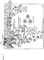

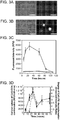
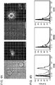

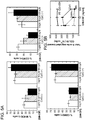
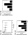


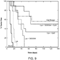
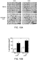

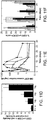

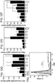


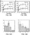
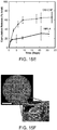
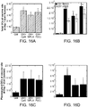
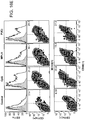
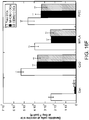

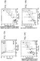
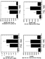


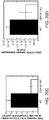
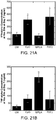
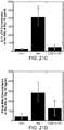
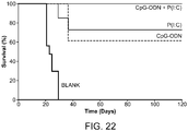
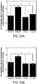


Classifications
-
- A—HUMAN NECESSITIES
- A61—MEDICAL OR VETERINARY SCIENCE; HYGIENE
- A61K—PREPARATIONS FOR MEDICAL, DENTAL OR TOILETRY PURPOSES
- A61K39/00—Medicinal preparations containing antigens or antibodies
- A61K39/0005—Vertebrate antigens
- A61K39/0011—Cancer antigens
-
- A—HUMAN NECESSITIES
- A61—MEDICAL OR VETERINARY SCIENCE; HYGIENE
- A61K—PREPARATIONS FOR MEDICAL, DENTAL OR TOILETRY PURPOSES
- A61K9/00—Medicinal preparations characterised by special physical form
- A61K9/14—Particulate form, e.g. powders, Processes for size reducing of pure drugs or the resulting products, Pure drug nanoparticles
- A61K9/16—Agglomerates; Granulates; Microbeadlets ; Microspheres; Pellets; Solid products obtained by spray drying, spray freeze drying, spray congealing,(multiple) emulsion solvent evaporation or extraction
- A61K9/1605—Excipients; Inactive ingredients
- A61K9/1629—Organic macromolecular compounds
- A61K9/1641—Organic macromolecular compounds obtained otherwise than by reactions only involving carbon-to-carbon unsaturated bonds, e.g. polyethylene glycol, poloxamers
- A61K9/1647—Polyesters, e.g. poly(lactide-co-glycolide)
-
- A—HUMAN NECESSITIES
- A61—MEDICAL OR VETERINARY SCIENCE; HYGIENE
- A61P—SPECIFIC THERAPEUTIC ACTIVITY OF CHEMICAL COMPOUNDS OR MEDICINAL PREPARATIONS
- A61P35/00—Antineoplastic agents
-
- A—HUMAN NECESSITIES
- A61—MEDICAL OR VETERINARY SCIENCE; HYGIENE
- A61K—PREPARATIONS FOR MEDICAL, DENTAL OR TOILETRY PURPOSES
- A61K39/00—Medicinal preparations containing antigens or antibodies
- A61K2039/54—Medicinal preparations containing antigens or antibodies characterised by the route of administration
-
- A—HUMAN NECESSITIES
- A61—MEDICAL OR VETERINARY SCIENCE; HYGIENE
- A61K—PREPARATIONS FOR MEDICAL, DENTAL OR TOILETRY PURPOSES
- A61K39/00—Medicinal preparations containing antigens or antibodies
- A61K2039/555—Medicinal preparations containing antigens or antibodies characterised by a specific combination antigen/adjuvant
- A61K2039/55511—Organic adjuvants
- A61K2039/55522—Cytokines; Lymphokines; Interferons
-
- A—HUMAN NECESSITIES
- A61—MEDICAL OR VETERINARY SCIENCE; HYGIENE
- A61K—PREPARATIONS FOR MEDICAL, DENTAL OR TOILETRY PURPOSES
- A61K39/00—Medicinal preparations containing antigens or antibodies
- A61K2039/555—Medicinal preparations containing antigens or antibodies characterised by a specific combination antigen/adjuvant
- A61K2039/55511—Organic adjuvants
- A61K2039/55555—Liposomes; Vesicles, e.g. nanoparticles; Spheres, e.g. nanospheres; Polymers
-
- A—HUMAN NECESSITIES
- A61—MEDICAL OR VETERINARY SCIENCE; HYGIENE
- A61K—PREPARATIONS FOR MEDICAL, DENTAL OR TOILETRY PURPOSES
- A61K39/00—Medicinal preparations containing antigens or antibodies
- A61K2039/555—Medicinal preparations containing antigens or antibodies characterised by a specific combination antigen/adjuvant
- A61K2039/55511—Organic adjuvants
- A61K2039/55561—CpG containing adjuvants; Oligonucleotide containing adjuvants
-
- Y—GENERAL TAGGING OF NEW TECHNOLOGICAL DEVELOPMENTS; GENERAL TAGGING OF CROSS-SECTIONAL TECHNOLOGIES SPANNING OVER SEVERAL SECTIONS OF THE IPC; TECHNICAL SUBJECTS COVERED BY FORMER USPC CROSS-REFERENCE ART COLLECTIONS [XRACs] AND DIGESTS
- Y02—TECHNOLOGIES OR APPLICATIONS FOR MITIGATION OR ADAPTATION AGAINST CLIMATE CHANGE
- Y02A—TECHNOLOGIES FOR ADAPTATION TO CLIMATE CHANGE
- Y02A50/00—TECHNOLOGIES FOR ADAPTATION TO CLIMATE CHANGE in human health protection, e.g. against extreme weather
- Y02A50/30—Against vector-borne diseases, e.g. mosquito-borne, fly-borne, tick-borne or waterborne diseases whose impact is exacerbated by climate change
Definitions
- the invention features a device and method for stimulating immune cells such as dendritic cells, in situ.
- presentation of Toll-like receptor (TLR) agonists in the context of the device is used for cancer vaccination.
- TLR Toll-like receptor
- Incorporation and presentation of the TLR agonists embedded in structural polymeric devices specifically stimulates CD8(+) dendritic cells (DCs) (corresponding to CD141+ DCs in humans) and plasmacytoid DCs, which subsets of DCs are critical for cancer vaccination.
- DCs dendritic cells
- plasmacytoid DCs which subsets of DCs are critical for cancer vaccination.
- the invention provides a device comprising a porous polymeric structure composition, a tumor antigen, and a toll-like receptor (TLR) agonist.
- the device comprises a polymeric structure composition, a tumor antigen, and a combination of toll-like receptor (TLR) agonists, wherein the TLR agonist is selected from the group consisting of TLR1, TLR2, TLR3, TLR4, TLR5, TLR6, TLR7, TLR8, TLR9, TLR10, TLR11, TLR12, and TLR13.
- the polymeric structure comprises poly (D,L-lactide-co-glycolide) (PLG).
- TLR agonists include pathogen associated molecular patterns (PAMPs), e.g., an infection-mimicking composition such as a bacterially-derived immunomodulator.
- PAMPs pathogen associated molecular patterns
- TLR agonists include nucleic acid or lipid compositions [e.g., monophosphoryl lipid A (MPLA)].
- MPLA monophosphoryl lipid A
- TLR agonists e.g., TLR1 agonists, TLR2 agonists, TLR3 agonists, TLR4 agonists, TLR5 agonists, TLR6 agonists, TLR7 agonists, TLR8 agonists, TLR9 agonists, TLR10 agonists, TLR11 agonists, TLR12 agonists, or TLR13 agonists.
- the TLR agonist comprises a TLR9 agonist such as a cytosine-guanosine oligonucleotide (CpG-ODN), a poly(ethylenimine) (PEI)-condensed oligonucleotide (ODN) such as PEI-CpG-ODN, or double stranded deoxyribonucleic acid (DNA).
- TLR9 agonists are useful to stimulate plasmacytoid DCs.
- the device comprises 5 ⁇ g, 10 ⁇ g 25 ⁇ g, 50 ⁇ g, 100 ⁇ g, 250 ⁇ g, or 500 ⁇ g of CpG-ODN.
- the TLR agonist comprises a TLR3 agonist such as polyinosine-polycytidylic acid (poly I:C), PEI-poly (I:C), polyadenylic-polyuridylic acid (poly (A:U)), PEI-poly (A:U), or double stranded ribonucleic acid (RNA).
- a TLR3 agonist such as polyinosine-polycytidylic acid (poly I:C), PEI-poly (I:C), polyadenylic-polyuridylic acid (poly (A:U)), PEI-poly (A:U), or double stranded ribonucleic acid (RNA).
- TLR3 agonists are useful to stimulate CD8+ DCs in mice and CD141+ DCs in humans.
- a plurality of TLR agonists e.g, a TLR3 agonist such as poly I:C and a TLR9 agonist such as CpG act in synergy to activate an anti-tumor immune response.
- the device comprises a TLR3 agonist such as poly (I:C) and the TLR9 agonist (CpG-ODN) or a PEI-CpG-ODN.
- the TLR agonist comprises the TLR3 agonist, poly (I:C) and the TLR9 agonist, CpG-ODN.
- the combination of poly (I:C) and CpG-ODN act synergistically as compared to the vaccines incorporating CpG-ODN or P(I:C) alone.
- the TLR agonist comprises a TLR4 agonist selected from the group consisting of lipopolysaccharide (LPS), monophosphoryl lipid A (MPLA), a heat shock protein, fibrinogen, heparin sulfate or a fragment threof, hyaluronic acid or a fragment thereof, nickel, an opoid, ⁇ 1-acid glycoprotein (AGP), RC-529, murine ⁇ -defensin 2, and complete Freund's adjuvant (CFA).
- the TLR agonist comprises a TLR5 agonist, wherein the TLR5 agonist is flagellin.
- Other suitable TLR agonists include TRL7 agonists selected from the group consisting of single-stranded RNA, guanosine anologs, imidazoqinolines, and loxorbine.
- the TLR agonist is present at a concentration effective to induce the local production of interleukin-12 (IL-12) by dendritic cells.
- IL-12 interleukin-12
- the invention also provides a device comprising a porous polymeric structure composition, a disease-associated antigen, and a toll-like receptor (TLR) agonist, wherein the TLR agonist preferentially binds to TLR3.
- the polymeric structure composition comprises poly-lactide-co-glycolide (PLG).
- PLG poly-lactide-co-glycolide
- the TLR3 agonist is present in an amount to preferentially stimulate CD8+ dendritic cells or CD141+ dendritic cells.
- the TLR agonist comprises a PEI- poly (I:C).
- the TLR agonist comprises a nucleic acid.
- the TLR agonist further comprises a TLR9 agonist.
- the TLR9 agonist comprises a cytosine-guanosine oligonucleotide (CpG-ODN) or a PEI-CpG-ODN.
- the device comprises a combination of TLR agonists, the combination comprising a TLR3 agonist and a TLR9 agonist.
- the TLR3 agonist comprises poly (I:C) and the TLR9 agonist comprises CpG-ODN.
- the device comprises a combination of TLR agonists, the combination comprising a TLR3 agonist and a TLR4 agonist.
- the TLR3 agonist comprises poly (I:C) and the TLR4 agonist comprises MPLA.
- the device further comprises a recruitment composition.
- exemplary recruitment compositions include granulocyte macrophage colony stimulating factor (GM-CSF), Flt3L, and CCL20.
- GM-CSF granulocyte macrophage colony stimulating factor
- Flt3L Flt3L
- CCL20 CCL20
- the recruitment composition comprises encapsulated GM-CSF.
- the disease-associated antigen comprises a tumor antigen.
- the tumor antigen comprises a tumor lysate, purified protein tumor antigen, or synthesized tumor antigen.
- the TLR agonist further comprises pathogen associated molecular patterns (PAMPs).
- PAMPs comprises a monophosphoryl lipid A (MPLA).
- MPLA monophosphoryl lipid A
- a device comprising a polymeric structure composition, a tumor antigen, and a combination of TLR agonists, wherein the TLR agonist is selected from the group consisting of TLR1, TLR2, TLR3, TLR4, TLR5, TLR6, TLR7, TLR8, TLR9, TLR10, TLR11, TLR12, and TLR13.
- a method for eliciting an anti-tumor immune response is carried out by contacting or implanting into a subject a device comprising a polymeric structure composition, a tumor antigen, and a TLR agonist, wherein the TLR agonist preferentially binds to TLR3.
- the TLR agonist comprises a TLR3 agonist.
- the TLR agonist comprises a TLR3 agonist and a TLR9 agonist.
- the anti-tumor immune response comprises activation of a CD8+ dendritic cell or a CD141+ dendritic cell.
- the anti-tumor immune response comprises activation of a plasmacytoid dendritic cell or a CD141+ dendritic cell.
- the anti-tumor immune response comprises a reduction in tumor burden.
- the TLR agonist is present at a concentration effective to induce production of interleukin-12 (IL-12) by dendritic cells.
- IL-12 interleukin-12
- the device further comprises granulocyte macrophage colony stimulating factor (GM-CSF).
- GM-CSF granulocyte macrophage colony stimulating factor
- the GM-CSF is encapsulated.
- Another optional recruitment composition is a cytokine.
- the device comprises 1 ⁇ g, 3 ⁇ g, 5 ⁇ g, 10 ⁇ g, 25 ⁇ g or 50 ⁇ g of GM-CSF.
- the device also contains a tumor antigen, e.g., in the form of a tumor lysate (cultured cells or patient-derived primary cells) or purified tumor antigen such as a synthesized/synthetic recombinant protein or biochemically-purified antigen from a tumor cell.
- a tumor antigen e.g., in the form of a tumor lysate (cultured cells or patient-derived primary cells) or purified tumor antigen such as a synthesized/synthetic recombinant protein or biochemically-purified antigen from a tumor cell.
- Also with in the invention is a method for eliciting an anti-tumor immune response by contacting a subject, e.g., implanting into a subject, a device comprising a porous polymeric structure composition, a tumor antigen, and a TLR agonist.
- a TLR agonist is selected from the group consisting of TLR3.
- the the method involves administering to a subject a device that contains a TLR3 agonist and/or a TLR9 agonist, which leads to elicitation of an anti-tumor immune response characterized by activation of plasmacytoid DCs and/or CD141+ DCs in the subject to which the vaccine was administered.
- the vaccine is useful for prophylaxis as well as therapy.
- the device is administered, e.g., topically applied or implanted, and is present over a period of time, e.g., in-dwelling, while constantly recruiting, educating, and dispersing or sending cells forth to lymph nodes or sites of disease or infection in the body. Improvements over existing devices include long term, ongoing activation of cells that enter the device and concomitant long term, ongoing egress of immunologically activated, e.g., antigen primed cells.
- the device includes a scaffold composition, a recruitment composition, and a deployment composition.
- the deployment composition that mediates prolonged and continuous egress of primed cells is an infection-mimicking composition such as a bacterially-derived immunomodulator.
- the bacterially-derived immunomodulator is a nucleic acid such as a cytosine-guanosine oligonucleotide (CpG-ODN).
- the methods are used to treat a wide variety of diseases and to develop vaccines against a wide variety of antigens.
- the present invention is used to develop a cancer vaccine.
- Another preferred embodiment of the present invention comprises an infection-mimicking microenvironment with means to activate the host immune system and subsequently induce an immune response.
- the use of a synthetic cytosine-guanosine oligodeoxynucleotide (CpG-ODN) sequence with exogenous granulocyte macrophage colony stimulating factor (GM-CSF) provides a method for precisely controlling dendritic cell migration and modulating antigen-specific immune responses.
- this synthetic cytosine-gyanosine oligonucleotide (CpG-ODN) sequence and/or poly (I:C) demonstrates significant improvements over earlier immune therapies.
- Table 1 FUNCTION EXEMPLARY DEVICE Attract a DC to Device Present an Immunogenic Factor Induce DC Migration from Device 1 Scaffold Composition Scaffold Composition Scaffold Composition Scaffold Composition 2 Bioactive Composition Bioactive Composition Bioactive Composition 3 Scaffold Composition Bioactive Composition Bioactive Composition 4 Scaffold Composition Scaffold Composition Bioactive Composition 5 Bioactive Composition Scaffold Composition Scaffold Composition 6 Bioactive Composition Bioactive Composition Scaffold Composition 7 Bioactive Composition Scaffold Composition Bioactive Composition 8 Scaffold Composition Bioactive Composition Scaffold Composition
- Devices perform three primary functions, e.g. attracting cells to the device, presenting an immunogenic factor, and inducing cell migration away from the device.
- Each of these primary functions are performed by the scaffold (bold font) and/or biological (standard font) composition(s).
- Table 1 provides exemplary combinations of either the scaffold or biological composition paired with at least one primary function in exemplary devices (1-8).
- the scaffold composition performs all three primary functions (device 1).
- the scaffold composition performs one primary function, e.g. attracts cells to the device (preferably, dendritic cells), whereas the biological composition performs two primary functions, e.g. presents an immunogenic factor and induces cells (preferably, dendritic cells) to migrate away from the device (device 3).
- Device 5 is the inverse combination of device 3.
- Exemplary secondary functions of the scaffold and/or biological compositions include, but are not limited to, targeting the device to a particular cell or tissue type, adhering/releasing the device to/from the surface of one or more cells or tissues, and modulating the stability/degradation of the device.
- the invention comprises a device comprising a scaffold composition and bioactive composition, the bioactive composition being incorporated into or conjugated onto the scaffold composition, wherein the scaffold composition attracts a dendritic cell, introduces a immunogenic factor into the dendritic cell thereby activating the dendritic cell, and induces the dendritic cell to migrate away from the scaffold composition.
- the bioactive composition incorporated into or coated onto the scaffold composition attracts a dendritic cell, introduces an immunogenic factor into the dendritic cell thereby activating the dendritic cell, and induces the dendritic cell to migrate away from the scaffold composition.
- the scaffold composition or bioactive composition separately attract a dendritic cell to the device, introduce an immunogenic factor into the dendritic cell, and induce the dendritic cell to migrate away from the device.
- DCs include conventional DCs as well as specific subsets of DCs.
- the TLR agonists e.g., TLR3 agonists, preferentially attract and stimulate CD141+ DCs in the human (CD8+ DCs in the mouse).
- the TLR9 agonisst e.g., CpG, preferentially attract and stimulate plasmacytoid DCs.
- the recruitment composition is GM-CSF, e.g., encapsulated GM-CSF.
- the device temporally controls local GM-CSF concentration, thereby controlling recruitment, residence, and subsequent dispersement/deployment of immune cells to lymph nodes or tissue sites distant from location of the device, e.g., sites of infection or tumor location.
- the concentration of GM-CSF determines whether if functions as a recruitment element or a deployment element.
- a method of programming dendritic cells in situ is carried out by introducing to a subject a device comprising scaffold composition and encapsulated recruitment composition.
- a pulse of recruitment composition is released from the device within 1-7 days of introduction of the device, leaving a residual amount of the recruitment composition in or on the device.
- the pulse is followed by slow release of the residual amount over several weeks.
- the local concentration of the recruitment composition and the temporal pattern of release mediates recruitment, retention, and subsequent release of dendritic cells from the device.
- the pulse comprises at least 50, 60, 75, 90 or 95 % of the amount of the recruitment composition associated with the device.
- An exemplary temporal release profile comprises a pulse characterized by release of at least 60% of the amount of the recruitment composition associated with the device in 1-5 days following the introduction of the device to a subject. Following the pulse, the residual amount is slowly released over an extended period of time (e.g., 1, 2, 3, 4, 5, 6, 7, 8, 9, 10, 12 days or 2, 3, 4, 5 or more weeks) following the pulse period.
- Other recruitment compositions include Flt3L and/or CCL20. The recruitment compounds are used individually or in combination.
- the method of making a scaffold is carried out by providing a scaffold composition, incorporating into or coating onto the scaffold composition a first bioactive composition comprising polypeptides with means for attracting or repelling a dendritic cell, and contacting the scaffold composition with a second bioactive composition, wherein the second bioactive composition is covalently or non-covalently associated with the scaffold composition wherein the second bioactive composition comprises a immunogenic factor.
- the linking and contacting steps are repeated to yield a plurality of layers, wherein the second bioactive composition comprises a combination of compounds with means to activate a dendritic cell.
- Methods comprise continuous in situ dendritic cell programming, comprising administering to a subject, a device comprising a scaffold composition and bioactive composition, the bioactive composition being incorporated into or conjugated onto the scaffold composition, wherein the scaffold composition attracts a dendritic cell, introduces a immunogenic factor into the dendritic cell thereby activating the dendritic cell, and induces the dendritic cell to migrate away from the scaffold composition.
- the devices recruit and stimulate a heterogeneous population of dendritic cells. Each subset is specialized and contributes significantly to the generation of an immune response. For example, the device mediates CpG-ODN presentation and enrichment of a subset of dendritic cells, plasmacytoid DC (pDC), or CD141+ DCs, which are particularly important in development of anti-tumor immunity.
- Methods comprise increasing vaccine efficacy, comprising administering to a subject, a device comprising a scaffold composition and bioactive composition, the bioactive composition being incorporated into or conjugated onto the scaffold composition, wherein the scaffold composition attracts a dendritic cell, introduces a immunogenic factor into the dendritic cell thereby activating the dendritic cell, and induces the dendritic cell to migrate away from the scaffold composition, thereby increasing the effectiveness of a vaccination procedure.
- Methods comprise vaccinating a subject against cancer, comprising administering to a subject, a device comprising a scaffold composition and bioactive composition, the bioactive composition being incorporated into or conjugated onto the scaffold composition, wherein the scaffold composition attracts a dendritic cell, introduces a immunogenic factor into the dendritic cell thereby activating the dendritic cell, and induces the dendritic cell to migrate away from the scaffold composition, thereby conferring upon a subject anti-tumor immunity, e.g., IL-12 production, and reduced tumor burden.
- the device is administered or implanted at or near the tumor site or site from which the tumor was excised or surgically removed.
- the device is implanted at a distance of 1, 3, 5, 10, 15, 20, 25, 40 mm from a tumor site or site of excision, e.g., the PLG vaccine device is administered 16-21 mm away from a tumor mass.
- Immunogenic factors include TLR ligands.
- the immunogenic factor used is a modified TLR-9 ligand sequence, PEI-CpG-ODN.
- the TLR ligand is a TLR3 agonist such as poly (I:C) or condensed PEI-poly (I:C).
- Scaffold compositions comprise a non-biodegradable material.
- exemplary non-biodegradable materials include, but are not limited to, metal, plastic polymer, or silk polymer.
- scaffold compositions are composed of a biocompatible material. This biocompatible material is non-toxic or non-immunogenic.
- Bioactive compositions are covalently or non-covalently linked to the scaffold composition.
- Bioactive compositions comprise an element, either covalently or non-covalently bonded to the surface of the scaffold composition, with means to attract a dendritic cell.
- bioactive compositions comprise an element, either covalently or non-covalently bonded to the surface of the scaffold composition, with means to introduce an immunogenic factor into a dendritic cell.
- bioactive compositions comprises an element, either covalently or non-covalently bonded to the surface of the scaffold composition, with means to induce a dendritic cell to migrate away from the scaffold composition.
- the element of the bioactive composition with means to manipulate a dendritic cell is a secreted or membrane-bound amino acid, peptide, polypeptide, protein, nucleotide, dinucleotide, oligonucleotide, polynucleotide, polymer, small molecule or compound.
- this element is granulocyte macrophage colony stimulating factor (GM-CSF), because this element attracts dendritic cells to the scaffold composition.
- GM-CSF granulocyte macrophage colony stimulating factor
- this element is a PEI-CpG-ODN sequence because this element has means to introduce CpG-ODN sequences into a dendritic cell thereby activating the cell.
- this element is a polynucleotide or polypeptide encoding for CCR7, a chemokine receptor that mediates dendritic cell migration towards lymph nodes and away from the scaffold composition.
- the CCR7 element is introduced into a dendritic cell simultaneously or sequentially with PEI-CpG-ODN sequences to enhance dendritic cell migration away from the scaffold composition.
- Scaffold compositions of the present invention contain an external surface. Scaffold compositions of the present invention alternatively, or in addition, contain an internal surface. External or internal surfaces of the scaffold compositions are solid or porous. Pore size is less than about 10 nm, in the range of about 100 nm-20 ⁇ m in diameter, or greater than about 20 ⁇ m. In preferred embodiments, the size of the pores allows the migration into and subsequent exit of cells such as DCs from the device. For example, the pores are nanoporous, microporous, or macroporous.
- the diameter of nanopores are less than about 10 nm; micropore are in the range of about 100 ⁇ m-20 ⁇ m in diameter; and, macropores are greater than about 20 ⁇ m (preferably greater than about 100 ⁇ m and even more preferably greater than about 400 ⁇ m).
- the scaffold is macroporous with open, interconnected pores of about 100-500 ⁇ m in diameter, e.g., 100-200, 200-400, or 400-500 ⁇ m.
- the size of the pores and the interconnected architecture allows the cells to enter, traverse within the volume of the device via the interconnected pores, and then leave the device via the pores to go to locations in the body outside of the device, e.g. to a tumor site, where an immune response is mounted against tumor cells.
- the activated DCs migrate away from the device and mount an immune response to solid tumors at discrete locations or throughout the body in the case of metastatic tumor cells or blood tumors such as leukemias.
- Scaffold compositions of the present invention comprise one or more compartments.
- Devices of the present invention are administered or implanted orally, systemically, sub- or trans-cunataneously, as an arterial stent, or surgically.
- the devices and methods of the invention provide a solution to several problems associated with protocols for continuous cell programming in situ.
- In situ cell programming systems that stimulate immune responses of the cells and induce their outward migration to populate infected or diseased bodily tissues enhance the success of recovery, e.g., the specific elimination of diseased tissue.
- Such a device that controls cell function and/or behavior e.g., locomotion, contains a scaffold composition and one or more bioactive compositions.
- the bioactive composition is incorporated into or coated onto the scaffold composition.
- the scaffold composition and/or bioactive composition temporally and spatially (directionally) controls dendritic cell attraction, programming, and migration.
- the devices mediate active recruitment, modification, and release of host cells from the material in vivo, thereby improving the function of cells that have contacted the scaffold.
- the device attracts or recruits cells already resident in the body to the scaffold material, and programs or reprograms the resident cells to a desired fate (e.g., immune activation).
- This device includes a scaffold composition which incorporates or is coated with a bioactive composition; the device regulates attraction, activation, and migration of dendritic cells.
- the device regulates attraction, activation, and/or migration of dendritic cells through the physical or chemical characteristics of the scaffold itself.
- the scaffold composition is differentially permeable, allowing cell migration only in certain physical areas of the scaffold.
- the permeability of the scaffold composition is regulated, for example, by selecting or engineering a material for greater or smaller pore size, density, polymer cross-linking, stiffness, toughness, ductility, or viscoelascticity.
- the scaffold composition contains physical channels or paths through which cells can move more easily towards a targeted area of egress of the device or of a compartment within the device.
- the scaffold composition is optionally organized into compartments or layers, each with a different permeability, so that the time required for a cell to move through the device is precisely and predictably controlled. Migration is also regulated by the degradation, de- or re-hydration, oxygenation, chemical or pH alteration, or ongoing self-assembly of the scaffold composition.
- Attraction, activation, and/or migration are regulated by a bioactive composition.
- the device controls and directs the activation and migration of cells through its structure. Chemical affinities are used to channel cells towards a specific area of egress. For example, cytokines are used to attract or retard the migration of cells.
- the device controls the timing of the migration. The density and mixture of these bioactive substances is controlled by initial doping levels or concentration gradient of the substance, by embedding the bioactive substances in scaffold material with a known leaching rate, by release as the scaffold material degrades, by diffusion from an area of concentration, by interaction of precursor chemicals diffusing into an area, or by production/excretion of compositions by resident support cells.
- the physical or chemical structure of the scaffold also regulates the diffusion of bioactive agents through the device.
- the bioactive composition includes one or more compounds that regulate cell function and/or behavior.
- the bioactive composition is covalently linked to the scaffold composition or non-covalently associated with the scaffold.
- the device optionally contains a second bioactive composition that comprises GM-CSF, a CpG-ODN or poly (I:C) sequence, a cancer antigen, and/or an immunomodulator.
- a second bioactive composition that comprises GM-CSF, a CpG-ODN or poly (I:C) sequence, a cancer antigen, and/or an immunomodulator.
- the second bioactive composition is covalently linked to the scaffold composition, keeping the composition relatively immobilized in or on the scaffold composition.
- the second bioactive composition is noncovalently associated with the scaffold.
- Noncovalent bonds are generally one to three orders of magnitude weaker than covalent bonds permitting diffusion of the factor out of the scaffold and into surrounding tissues.
- Noncovalent bonds include electrostatic, hydrogen, van der Waals, ⁇ aromatic, and hydrophobic.
- the scaffold composition is biocompatible.
- the composition is biodegradable/erodable or resistant to breakdown in the body.
- Relatively permanent (degradation resistant) scaffold compositions include metals and some polymers such as silk.
- the scaffold composition degrades at a predetermined rate based on a physical parameter selected from the group consisting of temperature, pH, hydration status, and porosity, the cross-link density, type, and chemistry or the susceptibility of main chain linkages to degradation or it degrades at a predetermined rate based on a ratio of chemical polymers.
- a high molecular weight polymer comprised of solely lactide degrades over a period of years, e.g., 1-2 years
- a low molecular weight polymer comprised of a 50:50 mixture of lactide and glycolide degrades in a matter of weeks, e.g., 1, 2, 3, 4, 6, 10 weeks.
- a calcium cross-linked gels composed of high molecular weight, high guluronic acid alginate degrade over several months (1, 2, 4, 6, 8, 10, 12 months) to years (1, 2, 5 years) in vivo, while a gel comprised of low molecular weight alginate, and/or alginate that has been partially oxidized, will degrade in a matter of weeks.
- Exemplary scaffold compositions include polylactic acid, polyglycolic acid, PLGA polymers, alginates and alginate derivatives, gelatin, collagen, fibrin, hyaluronic acid, laminin rich gels, agarose, natural and synthetic polysaccharides, polyamino acids, polypeptides, polyesters, polyanhydrides, polyphosphazines, poly(vinyl alcohols), poly(alkylene oxides), poly(allylamines)(PAM), poly(acrylates), modified styrene polymers, pluronic polyols, polyoxamers, poly(uronic acids), poly(vinylpyrrolidone) and copolymers or graft copolymers of any of the above.
- One preferred scaffold composition includes an RGD-modified alginate.
- the PLG matrix includes GM-CSF, danger signals, and a target antigen, e.g., a cancer antigen and serves as a residence for recruited DCs as they are programmed.
- the recruitment element, GM-CSF is encapsulated into the PLG scaffolds.
- PLG matrices that comprise the encapsulated GM-CSF provide a pulse of the dendritic cell recruitment composition and then a gradual slower rate of release.
- the pulse comprises at least 40, 50, 60, 75, 80% or more of the initial amount of bioactive composition with the remaining per cent being released gradually over then next days or weeks after administration to the site in or on the subject to be treated.
- release is approximately 60% of bioactive GM-CSF load within the first 5 days, followed by slow and sustained release of bioactive GM-CSF over the next 10 days.
- This release profile mediates a rate of diffusion of the factor through the surrounding tissue to effectively recruit resident DCs.
- Pores are nanoporous, microporous, or macroporous.
- the diameter of nanopores are less than about 10 nm; micropore are in the range of about 100 nm-20 ⁇ m in diameter; and, macropores are greater than about 20 ⁇ m (preferably greater than about 100 ⁇ m and even more preferably greater than about 400 ⁇ m).
- the scaffold is macroporous with aligned pores of about 400-500 ⁇ m in diameter.
- the device is manufactured in one stage in which one layer or compartment is made and infused or coated with one or more bioactive compositions.
- bioactive compositions comprise polypeptides or polynucleotides.
- the device is manufactured in two or more (3, 4, 5, 6, ....10 or more) stages in which one layer or compartment is made and infused or coated with one or more bioactive compositions followed by the construction of a second, third, fourth or more layers, which are in turn infused or coated with one or more bioactive compositions in sequence.
- Each layer or compartment is identical to the others or distinguished from one another by the number or mixture of bioactive compositions as well as distinct chemical, physical and biological properties.
- a method of making a scaffold is carried out by providing a scaffold composition and covalently linking or noncovalently associating the scaffold composition with a first bioactive composition.
- the first bioactive composition preferably contains granulocyte macrophage colony stimulating factor.
- the scaffold composition is also contacted with a second bioactive composition, preferably one or more cytosine-guanosine oligonucleotide (CpG-ODN) sequences.
- the second bioactive composition is associated with the scaffold composition to yield a doped scaffold, i.e., a scaffold composition that includes one or more bioactive substances.
- each of the contacting steps is characterized by a different amount of the second bioactive composition to yield a gradient of the second bioactive composition in the scaffold device.
- subsequent contacting steps involve a different bioactive composition, i.e., a third, fourth, fifth, sixth...., composition or mixture of compositions, that is distinguished from the prior compositions or mixtures of prior doping steps by the structure or chemical formula of the factor(s).
- the method optionally involves adhering individual niches, layers, or components to one another and/or insertion of semi-permeable, permeable, or nonpermeable membranes within or at one or more boundaries of the device to further control/regulate locomotion of cells or bioactive compositions.
- Therapeutic applications of the device include the instruction of immune cells.
- the method includes the steps of providing a device that includes scaffold composition with a bioactive composition incorporated therein or thereon and a mammalian cell bound to the scaffold and contacting a mammalian tissue with the device, e.g., by implanting or affixing the device into or onto a mammalian tissue.
- exemplary relative amounts of each component, recruiting composition (e.g., GM-CSF, Flt3L, or CCL20), danger signal (e.g., CpG-ODN), and antigen (e.g., purified tumor antigen or tumor cell lysate) are as follows: GM-CSF: 0.5 ⁇ g - 500 ⁇ g; CpG-ODN: 50 ⁇ g - 3,000 ⁇ g; and Tumor antigen/lysate: 100 ⁇ g - 10,000 ⁇ g.
- a method of modulating an activity of a cell is carried out by administering to a mammal a device containing a scaffold composition and a recruitment composition incorporated therein or thereon, and then contacting the cell with a deployment signal.
- the cells leave the device after encountering antigen (and other factors) and thus being activated to seek out tumor cells in the body to which an immune response is mounted.
- the activity of the cell at egress differs from that prior to entering the device.
- Cells are recruited into the device and remain resident in the device for a period of time, e.g., minutes; 0.2.
- the deployment signal is a composition such as protein, peptide, or nucleic acid or a state of activation of the cell.
- a composition such as protein, peptide, or nucleic acid or a state of activation of the cell.
- DCs become activated and migrate to lymph nodes, the spleen, and other anatomical locations, where they meet up with T cells to further propagate an antigen-specific immune response, e.g., anti-cancer response.
- an antigen-specific immune response e.g., anti-cancer response.
- cells migrating into the device only encounter the deployment signal once they have entered the device.
- the deployment signal is a nucleic acid molecule, e.g., a plasmid containing sequence encoding a protein that induces migration of the cell out of the device and into surrounding tissues.
- the deployment signal occurs when the cell encounters the plasmid in the device, the DNA becomes internalized in the cell (i.e., the cell is transfected), and the cell manufactures the gene product encoded by the DNA.
- the molecule that signals deployment is an element of the device and is released from the device in delayed manner (e.g., temporally or spatially) relative to exposure of the cell to the recruitment composition.
- the deployment signal is a reduction in or absence of the recruitment composition.
- a recruitment composition induces migration of cells into the device, and a reduction in the concentration or depletion, dissipation, or diffusion of the recruitment composition from the device results in egress of cells out of the device.
- immune cells such as T cells, B cells, or dendritic cells (DCs) of an individual are recruited into the device, primed and activated to mount an immune response against an antigen-specific target.
- an antigen corresponding to a target to which an immune response is desired is incorporated into or onto the scaffold structure.
- Cytokines such as granulocyte macrophage colony stimulating factor (GM-CSF) are also a component of the device to amplify immune activation and/or induce migration of the primed cells to lymph nodes.
- GM-CSF granulocyte macrophage colony stimulating factor
- Other cell specific recruitment compositions are described below.
- the device recruit cells in vivo, modifies these cells, and then promotes their migration to another site in the body.
- This approach is exemplified herein in the context of dendritic cells and cancer vaccine development but is also useful to other vaccines such as those against microbial pathogens as well as cell therapies in general.
- Cells educated using the devices described herein promote regeneration of a tissue or organ immediately adjacent to the material, or at some distant site. Alternatively, the cells are educated to promote destruction of a tissue (locally or at a distant site).
- the methods are also useful for disease prevention, e.g., to promote cell-based maintenance of tissue structure and function to stop or retard disease progression or age-related tissue changes.
- the education of cells within the device, "programming" and "reprogramming" permits modification of the function or activity of any cell in the body to become a multipotent stem cell again and exert therapeutic effects.
- Polynucleotides, polypeptides, or other agents are purified and/or isolated.
- an "isolated” or “purified” nucleic acid molecule, polynucleotide, polypeptide, or protein is substantially free of other cellular material, or culture medium when produced by recombinant techniques, or chemical precursors or other chemicals when chemically synthesized.
- Purified compounds are at least 60% by weight (dry weight) the compound of interest.
- the preparation is at least 75%, more preferably at least 90%, and most preferably at least 99%, by weight the compound of interest.
- a purified compound is one that is at least 90%, 91%, 92%, 93%, 94%, 95%, 98%, 99%, or 100% (w/w) of the desired compound by weight. Purity is measured by any appropriate standard method, for example, by column chromatography, thin layer chromatography, or high-performance liquid chromatography (HPLC) analysis.
- a purified or isolated polynucleotide ribonucleic acid (RNA) or deoxyribonucleic acid (DNA)
- RNA ribonucleic acid
- DNA deoxyribonucleic acid
- a purified or isolated polypeptide is free of the amino acids or sequences that flank it in its naturally-occurring state. Purified also defines a degree of sterility that is safe for administration to a human subject, e.g., lacking infectious or toxic agents.
- substantially pure is meant a nucleotide or polypeptide that has been separated from the components that naturally accompany it.
- the nucleotides and polypeptides are substantially pure when they are at least 60%, 70%, 80%, 90%, 95%, or even 99%, by weight, free from the proteins and naturally-occurring organic molecules with they are naturally associated.
- isolated nucleic acid is meant a nucleic acid that is free of the genes which flank it in the naturally-occurring genome of the organism from which the nucleic acid is derived.
- the term covers, for example: (a) a DNA which is part of a naturally occurring genomic DNA molecule, but is not flanked by both of the nucleic acid sequences that flank that part of the molecule in the genome of the organism in which it naturally occurs; (b) a nucleic acid incorporated into a vector or into the genomic DNA of a prokaryote or eukaryote in a manner, such that the resulting molecule is not identical to any naturally occurring vector or genomic DNA; (c) a separate molecule such as a cDNA, a genomic fragment, a fragment produced by polymerase chain reaction (PCR), or a restriction fragment; and (d) a recombinant nucleotide sequence that is part of a hybrid gene, i.e., a gene encoding a fusion protein
- Isolated nucleic acid molecules according to the present invention further include molecules produced synthetically, as well as any nucleic acids that have been altered chemically and/or that have modified backbones.
- the isolated nucleic acid is a purified cDNA or RNA polynucleotide.
- Isolated nucleic acid molecules also include messenger ribonucleic acid (mRNA) molecules.
- transitional term “comprising,” which is synonymous with “including,” “containing,” or “characterized by,” is inclusive or open-ended and does not exclude additional, unrecited elements or method steps.
- the transitional phrase “consisting of” excludes any element, step, or ingredient not specified in the claim.
- the transitional phrase “consisting essentially of” limits the scope of a claim to the specified materials or steps "and those that do not materially affect the basic and novel characteristic(s)" of the claimed invention.
- cancer vaccines Prior to the invention, cancer vaccines typically depended on cumbersome and expensive manipulation of cells in the laboratory, and subsequent cell transplantation resulted in poor lymph node homing and limited efficacy.
- the invention solves these problems by using materials that mimic key aspects of bacterial infection to directly control immune cell trafficking and activation in the body. Presentation of TLR agonists for cancer vaccination leads to improved activation of immune cells.
- the vaccines and methods comprise incorporation and presentation of TLR agonists embedded in structural polymeric devices.
- the data described herein demonstrate the critical role of CD8(+) Dendritic cells (DCs) and plasmacytoid DCs (as well as conventional DCs) for cancer vaccination, which are preferentially recruited and activated using the TLR-agonist containing structural polymeric device.
- the device is manufactured as a tiny bioengineered porous disc filled with tumor-specific antigens and TLR agonists.
- the disc is implanted into the body, e.g., inserted under the skin, where it activates the immune system to destroy tumor cells. While typical earlier methods involved growing cells outside the body, this approach reprograms cells that are already in the body.
- the device includes a recruitment component.
- the device optionally includes a recruitment molecule such as a cytokine.
- a recruitment molecule such as a cytokine.
- polymers were designed to first release a cytokine to recruit and house host dendritic cells (DCs), and subsequently present cancer antigens and danger signals to activate the resident DCs and dramatically enhance their homing to lymph nodes.
- Specific and protective anti-tumor immunity was generated with these materials, as 90% survival was achieved in animals that otherwise die from cancer within 25 days. These materials are useful in cancer and other vaccines to program and control the trafficking of a variety of cell types in the body.
- a polymer system was designed to not only serve as a drug delivery device, but also as a physical, antigen-presenting structure to which the DCs are recruited, and where DCs reside while they are activated using a material (poly[lactide-co-glycolide]) and bioactive molecules (GM-CSF and CpG-ODN). These bioactive molecules have excellent safety profiles.
- the material system serves as an effective cancer vaccine, eliminating the time, expense and regulatory burden inherent to existing cell therapies and reducing or eliminating the need for multiple, systemic injections and high total drug loading.
- the devices described herein utilize infection-mimicking materials to program DCs in situ.
- GM-CSF enhanced DC recruitment and proliferation in a dose dependent manner.
- high concentrations (>100ng/ml) of GM-CSF inhibited DC migration toward a lymph node derived chemoattractant (CCL19).
- Immunohistochemical staining revealed that the high concentrations of GM-CSF (500 ng/ml) also down-regulated DC expression of the CCL19 receptor CCR7 and MHCII.
- GM-CSF GM-CSF alone is to be used for both purposes, its local concentration is designed to decrease over time in order to release DCs that become trapped in the material.
- a danger signal e.g., CpG-ODN
- CpG-ODN CpG-ODN
- a macroporous poly-lactide-co-glycolide (PLG) matrix was designed to present GM-CSF, danger signals, and cancer antigens in a defined spatiotemporal manner in vivo, and serve as a residence for recruited DCs as they are programmed.
- GM-CSF was encapsulated (54% efficiency) into PLG scaffolds using a high pressure CO 2 foaming process. These matrices released approximately 60% of their bioactive GM-CSF load within the first 5 days, followed by slow and sustained release of bioactive GM-CSF over the next 10 days. This release profile allows diffusion of the factor through the surrounding tissue to effectively recruit resident DCs.
- in situ dendritic cell targeting systems are utilized to therapeutically manipulate the immune system with TLR agonists.
- macroporous polymeric scaffolds were designed that deliver three different classes of TLR agonists in vivo : CpG--ODN, MPLA, and P(I:C) in combination with GM-CSF, Flt3L, or CCL20 to augment DC recruitment and activation.
- the ability of in situ TLR presentation from macroporous matrices to modulate DC subset generation, and cancer vaccine efficacy in a B16-F10 melanoma model was also characterized.
- DCs dendritic cells
- T cells cytotoxic T lymphocytes
- PAMPs pathogen associated molecular patterns
- antigenic molecules Banchereau J, and Steinman RM., 1998 Nature, 392: 245-252 ; Mellman I. and Steinman R.M., 2001 Cell, 106: 255-258 ; Sansonetti P.J., 2006 Nat.
- PAMP recognition by pattern recognition receptors present intercellularly or at the DC surface signal the presence of infection and triggers signal transduction pathways ultimately resulting in DC activation ( Sansonetti P.J., 2006 Nat. Immunol., 7: 1237-1242 ; Meylan et al., 2006 Nature, 442: 39-44 ; Akira et al., 2006 Cell, 124: 783-801 ).
- PRRs pattern recognition receptors
- activated DCs are characterized by enhanced expression of MHC and co-stimulatory molecules and proinflammatory cytokines, which enables DCs to translate pathogenic signals to naive T cells and trigger adaptive immune responses
- Banchereau J, and Steinman RM. 1998 Nature, 392: 245-252 ; Mellman I. and Steinman R.M., 2001 Cell, 106: 255-258 ; Sansonetti P.J., 2006 Nat.
- DCs act as a network of distinct subsets that perform specialized functions to stimulate and polarize T cell responses in order to coordinate immune regulation ( Naik et al., 2007 Nat Immunol, 8: 1217-1226 ; O'Garral A. and Trinchieri G.
- CD8(+) DCs are especially adept at cross-presentation of exogenous antigen, IL-12 production and induction of cytotoxic T cell responses ( Schnorrer P, 2006 PNAS 28: 10729-34 ; Skokos D. and Nussenzweig M.C., J Exp Med, 204: 1525-1531 ; Den Haana et al., 2000 J Exp Med, 12: 1685-1696 ; Moser M. and Murphy K.M., 2000 Nat. Immunol., 1: 199-205 ; Hildner et al., 2008 Science, 322:1097-1100 ).
- the plasmacytoid DC (pDC) subset has the capacity to produce significant amounts of type-1 interferons (IFNs) in response to microbial nucleic acids, particularly during viral infection, to facilitate T cell activation, growth and survival for disease clearance ( Liu YJ, 2001 Cell, 106: 259-262 ; Jego et al., 2003 Immunity, 19: 225-234 ; Randolph et al., 2008 Annu. Rev. Immunol., 26: 293-316 ; Kanzler et al., 2007 Nat. Med., 13: 552-559 ).
- IFNs type-1 interferons
- pDC and CD8(+) DC subsets have been associated with priming t-helper 1 (Th1) effector cells for the control of infection and tumors.
- Th1 priming t-helper 1
- a balanced distribution of activated DC subsets is associated with the control of autoimmune disease and tumors, indicating that these cells may cooperate during the generation of protective immunity.
- cancer vaccines are designed to introduce antigen in combination with immunostimulatory signals to activate DCs either ex vivo prior to administration, or in situ ( Gilboa E., 2007 J Clin Invest., 117: 1195-1203 ; Banchereau J. and Steinman R.M., 2007 Nature, 49: 419-426 ; Kanzler et al., 2007 Nat. Med., 13: 552-559 ; Hansen et al., 2013 Vaccine, 31(4), 639-46 ; Schuler et al., 2003 Curr Opin Immunol, 15: 138-147 ; Curiel T.J, 2002 J Clin Invest, 109: 311-312 ).
- a range of stimuli are used to trigger DC maturation and differentiation including proinflammatory cytokines, PAMPs recognized by the toll-like receptor (TLR) family, and feedback signals from innate and adaptive immune cells.
- TLR toll-like receptor
- discrete combinations of these stimuli and DC subsets differentially control T cell activation and polarization, and these components are optimized and exploited to generate effective immune responses that eradicate tumors or infectious agents.
- components and DC subsets should be included in cancer vaccines, partly because current techniques limit the cell types that can be cultured or targeted ( Kanzler et al., 2007 Nat.
- Standard DC-based protocols widely used in the clinic utilize monocyte-derived conventional DCs that are unable to cross-present antigens, or efficiently produce IL-12 or type-1 IFNs that can prime CTL-mediated immune responses and tumor cell death ( Hansen et al., 2013 Vaccine, 31(4), 639-46 ; Schuler et al., 2003 Curr Opin Immunol, 15: 138-147 ; Curiel T.J, 2002 J Clin Invest, 109: 311-312 ).
- matrices were modified to present 3 different classes of TLR agonists, CpG-ODN, monophosphoryl lipid A (MPLA), and polyinosinic:polycytidylic acid (P(I:C)), all in combination with GM-CSF.
- TLR agonists CpG-ODN
- MPLA monophosphoryl lipid A
- P(I:C) polyinosinic:polycytidylic acid
- Dendritic Cell (DC) proliferation, migration and maturation are sensitive to inflammatory mediators, and granulocyte macrophage colony stimulating factor (GM-CSF) has been identified as a potent stimulator of immune responses, specifically against cancer antigens. GM-CSF also has the ability to recruit and program these antigen-presenting immune cells. Additionally, Cytosine-guanosine (CpG) oligonucleotide (CpG-ODN) sequences found in bacterial DNA are potent immunomodulators that stimulate DC activation, leading to specific T-cell responses. Creating an infection mimicking microenvironment by the presentation of exogenous GM-CSF and CpG-ODN provides an avenue to precisely control the number and timing of DC migration and modulate antigen specific immune responses.
- the vertebrate immune system employs various mechanisms for pathogen recognition making it adept at generating antigen-specific responses and clearing infection. Immunity is controlled by antigen presenting cells (APCs), especially dendritic cells (DCs), which capture antigens and are activated by stimuli, unique 'danger signals' of the invading pathogen, such as CpG dinucleotide sequences in bacterial DNA ( Banchereau J, and Steinman RM. Nature. 392, 245-252. (1998 ); Klinman DM. Nat. Rev. Immunol. 4, 249-58 (2004 ); each herein incorporated by reference).
- APCs antigen presenting cells
- DCs dendritic cells
- cancerous cells derived from self-tissues, are void of the danger signals required to signal DC maturation and instead promote an immunosuppressive microenvironment that allows cells to escape immunity.
- Key elements of infection are inflammatory cytokines and danger signals ( Figure 1 ).
- a polymeric material system is ideal to present these factors in the required spatiotemporal manner to provide an infection-mimicking microenvironment in situ that useful as a vaccine.
- These infection mimics provide the continuous programming of host DCs, providing for efficient DC activation and dispersement in situ.
- These infection-mimicking devices are used for numerous vaccine applications including melanoma cancer vaccines.
- inflammatory cytokines and danger signals stimulate specific DC responses that mediate immune recognition and pathogen clearance ( FIG 1 ).
- skin cells such as fibroblasts, keratinocytes and melanocytes are damaged resulting in the release of inflammatory cytokines, such as GM-CSF ( Hamilton J. Trends in Immunol. 23, 403-408.
- inflammatory cytokines such as GM-CSF
- GM-CSF GM-CSF
- fibroblasts fibroblasts
- keratinocytes and melanocytes are damaged resulting in the release of inflammatory cytokines, such as GM-CSF ( Hamilton J. Trends in Immunol. 23, 403-408.
- 2002 Hamilton J., and Anderson G.
- CpG-ODNs are potent "danger signals” that upregulate DC expression of CCR7, CD80/86 costimulatory molecules, and MHC-antigen complexes.
- TLR9 signaling induces DCs into promoting Thl-like, cytotoxic -Tcell responses, by cytokine production (e.g. type 1 IFN) and cross-presentation of antigen onto MHCI molecules.
- cytokine production e.g. type 1 IFN
- the presentation of these signals concurrently with tumor antigens provides the danger signal needed to promote immune responses that effectively fight cancerous cells.
- CPG-ODNs Different classes of CPG-ODNs promote different immune responses depending on the ODN's specific structure and sequence.
- the ODN utilized in the present invention CpG-ODN 1826, has been successfully tested in various mouse vaccination models, including melanoma.
- CpG-ODN 1826 has shown a beneficial effect alone or when used as adjuvant for peptide vaccines and whole cell vaccines.
- ODN 1826 has been shown to directly promote DC maturation and cytokine production. This particular CpG ODN sequence also indirectly activates Th1 cells and NK cells and, thus, enhances adaptive cellular immune responses.
- PEI polyethylenimine
- PEI has been utilized as a non-viral vector to enhance gene transfection and to fabricate PEI-DNA loaded PLG matrices that promoted long-term gene expression in host cells in situ ( Huang YC, Riddle F, Rice KG, and Mooney DJ. Hum Gene Ther. 5, 609-17. (2005 ), herein incorporated by reference). Therefore, CpG-ODNs were condensed with PEI molecules, and the size and charge of these PEI-CpG-ODN condensates, as dependent on the amine-phosphate charge ratio, was characterized.
- a PLG system was designed to not only serve as a drug delivery device, that releases inflammatory cytokines (eg. GM-CSF) but also as a physical structure to which the DCs are recruited and reside while they are activated by danger signals (eg. CpG-ODNs).
- cytokines eg. GM-CSF
- CpG-ODNs danger signals
- the ability to control DC recruitment to and DC residence within porous PLG matrices is achieved using temporal control over the delivery of GM-CSF in situ, which results in batches of programmed DCs being dispersed only when GM-CSF levels were designed to subside in situ.
- This system dispersed 6% of programmed DCs to the lymph nodes and induced protective anti-tumor immunity in 23% of mice when applied as a cancer vaccine.
- the cell programming and dispersement efficiency is improved using an overriding secondary signal (CpG-ODN) that continuously releases DCs from GM-CSF inhibition and promotes DC maturation and dispersement in the presence of high GM-CSF levels in situ.
- CpG-ODN overriding secondary signal
- PLG matrices were fabricated to locally present synthetic CpG-ODN with exogenous GM-CSF allowing for DCs recruited by GM-CSF to be stimulated by CpG-ODN in situ.
- Dendritic cells are immune cells within the mammalian immune system and are derived from hematopoietic bone marrow progenitor cells. More specifically, dendritic cells can be categorized into lymphoid (or plasmacytoid) dendritic cell (pDC) and myeloid dendritic cell (mDC) subdivisions having arisen from a lymphoid (or plasmacytoid) or myeloid precursor cell, respectively. From the progenitor cell, regardless of the progenitor cell type, an immature dendritic cell is born. Immature dendritic cells are characterized by high endocytic activity and low T-cell activation potential.
- immature dendritic cells constitutively sample their immediate surrounding environment for pathogens.
- pathogens include, but are not limited to, a virus or a bacteria. Sampling is accomplished by pattern recognition receptors (PRRs) such as the toll-like receptors (TLRs).
- PRRs pattern recognition receptors
- TLRs toll-like receptors
- Dendritic cells activate and mature once a pathogen is recognized by a pattern recognition receptor, such as a toll-like receptor.
- Mature dendritic cells not only phagocytose pathogens and break them down, but also, degrade their proteins, and present pieces of these proteins, also referred to as antigens, on their cell surfaces using MHC (Major Histocompatibility Complex) molecules (Classes I, II, and III). Mature dendritic cells also upregulate cell-surface receptors that serve as co-receptors for T-cell activation. Exemplary co-receptors include, but are not limited to, CD80, CD86, and CD40. Simultaneously, mature dendritic cells upregulate chemotactic receptors, such as CCR7, that allows the cell to migrate through the blood stream or the lymphatic system to the spleen or lymph node, respectively.
- MHC Major Histocompatibility Complex
- Dendritic cells are present in external tissues that are in contact with the external environment such as the skin (dendritic cells residing in skin are also referred to as Langerhans cells). Alternatively, dendritic cells are present in internal tissues that are in contact with the external environment such as linings of the nose, lungs, stomach, and intestines. Finally, immature dendritic cells reside in the blood stream. Once activated, dendritic cells from all off these tissues migrate to lymphoid tissues where they present antigens and interact with T cells and B cells to initiate an immune response.
- One signaling system of particular importance for the present invention involves the chemokine receptor CCR7 expressed on the surface of dendritic cells and the chemokine receptor ligand CCL19 secreted by lymph node structures to attract migrating mature dendritic cells toward high concentrations of immune cells.
- exemplary immune cells activated by contact with mature dendritic cells include, but are not limited to, helper T cells, killer T cells, and B cells.
- helper T cells include, but are not limited to, helper T cells, killer T cells, and B cells.
- dendritic cells are the most potent activators of all antigen-presenting cells.
- Dendritic cells earned their name from the characteristic cell shape comprising multiple dendrites extending from the cell body.
- the functional benefit of this cell shape is a significantly increased cell surface and contact area to the surroundings compared to the cell volume.
- Immature dendritic cells sometimes lack the characteristic dendrite formations and are referred to as veiled cells.
- Veiled cells possess large cytoplasmic veils rather than dendrites.
- Plasmacytoid dendritic cells are innate immune cells that circulate in the blood and are found in peripheral lymphoid organs. They constitute ⁇ 0.4% of peripheral blood mononuclear cells (PBMC). In humans these cells express the surface markers CD123, BDCA-2(CD303) and BDCA-4(CD304), but do not express high levels of CD11c or CD14, which distinguishes them from conventional dendritic cells or monocytes, respectively.
- Mouse pDC express CD11c, B220, BST-2 (mPDCA) and Siglec-H and are negative for CD11b.
- these cells express intracellular Toll-like receptors 7 and 9 which detect ssRNA and CpG DNA motifs, respectively.
- CD8- subset presents antigen using the class II pathway to CD4+ helper T cells.
- the CD8+ subset presents antigens using the class I pathway.
- the peptide/MHC class I molecules are presented to CD8+ T cells which go on to become cytotoxic T lymphocytes (CTL).
- CTL cytotoxic T lymphocytes
- the CD8 cell surface protein in the mouse corresponds to the CD141 cell surface protein in the human.
- CD8/CD141-positive cells express TLR3 and are preferentially activated by TLR3 agonists.
- TLRs Toll-like Receptors
- TLRs are a class of single transmembrane domain, non-catalytic, receptors that recognize structurally conserved molecules referred to as pathogen-associated molecular patterns (PAMPs). PAMPs are present on microbes and are distinguishable from host molecules. TLRs are present in all vertebrates. Thirteen TLRs (referred to as TLRsl-13, consecutively) have been identified in humans and mice. Humans comprise TLRs 1-10.
- TLRs and interleukin-1 (IL-1) receptors comprise a receptor superfamily the members of which all share a TIR domain (Toll-IL-1 receptor).
- TIR domains exist in three varieties with three distinct functions.
- TIR domains of subgroup 1 are present in receptors for interleukins produced by macrophages, monocytes, and dendritic cells.
- TIR domains of subgroup 2 are present in classical TLRs which bind directly or indirectly to molecules of microbial origin.
- TIR domains of subgroup 3 are present in cytosolic adaptor proteins that mediate signaling between proteins comprising TIR domains of subgroups 1 and 2.
- TLR ligands comprise molecules that are constantly associated with and highly specific for a threat to the host's survival such as a pathogen or cellular stress. TLR ligands are highly specific for pathogens and not the host. Exemplary pathogenic molecules include, but are not limited to, lipopolysaccharides (LPS), lipoproteins, lipoarabinomannan, flagellin, double-stranded RNA, and unmethylated CpG islands of DNA.
- LPS lipopolysaccharides
- lipoproteins lipoproteins
- lipoarabinomannan flagellin
- double-stranded RNA unmethylated CpG islands of DNA.
- the Toll-Like receptor 9 is activated by specific unmethylated CpG-containing sequences in bacterial DNA or synthetic oligonucleotides (ODNs) found in the endosomal compartment of dendritic cells.
- ODNs synthetic oligonucleotides
- Methylation status of the CpG site is a crucial distinction between bacterial and mammalian DNA, as well as between normal and cancerous tissue.
- Unmethylated ODNs including one or more CpG motifs mimic the effects of bacterial DNA.
- unmethylated ODNs including one or more CpG motifs occur within oncogenes present within malignant tumor cells.
- TLR-9 receptor recognizes one or more CpG-ODN sequences of the present invention.
- TLR-9 receptors encompassed by the present invention are described by the following sequences.
- Human TLR-9, isoform A is encoded by the following mRNA sequence (NCBI Accession No. NM_017442 and SEQ ID NO: 1; the start codon for all mRNA sequences presented herein is bolded and capitalized):
- Human TLR-9, isoform A is encoded by the following amino acid sequence (NCBI Accession No. NP_059138 and SEQ ID NO: 2):
- Human TLR3 is encoded by the following mRNA sequence (GenBank Accesion No. NM_003265.2 (GI:19718735), SEQ ID NO: 5):
- Human TLR3 is encoded by the following amino acid sequence (GenBank Accesion No. ABC86910.1 (GI:86161330), SEQ ID NO: 4):
- the nucleic acid sequence of human TLR1 is provided in GenBank Accession No. NM_003263.3 (GI:41350336).
- the amino acid sequence of human TLR1 is provided in GenBank Accession No. NP_003254.2 (GI:41350337).
- the nucleic acid sequence of human TLR2 is provided in GenBank Accession No. NM_003264.3 (GI:68160956).
- the amino acid sequence of human TLR2 is provided in GenBank Accession No. NP_003255.2 (GI:19718734).
- the nucleic acid sequence of human TLR4 is provided in GenBank Accession No. NM_138554.4 (GI:373432600).
- the amino acid sequence of human TLR4 is provided in GenBank Accession No. NP_612564.1 (GI: 19924149).
- the nucleic acid sequence of human TLR5 is provided in GenBank Accession No. NM_003268.5 (GI:281427130).
- the amino acid sequence of human TLR5 is provided in GenBank Accession No. NP_003259.2 (GI: 16751843).
- the nucleic acid sequence of human TLR6 is provided in GenBank Accession No. NM_006068.4 (GI:318067953).
- the amino acid sequence of human TLR6 is provided in GenBank Accession No. NP_006059.2 (GI:20143971).
- the nucleic acid sequence of human TLR7 is provided in GenBank Accession No. NM_016562.3 (GI:67944638).
- the amino acid sequence of human TLR7 is provided in GenBank Accession No. NP_057646.1 (GI:7706093).
- the nucleic acid sequence of human TLR8 is provided in GenBank Accession No. NM_138636.4 (GI:257196253).
- the amino acid sequence of human TLR8 is provided in GenBank Accession No. NP_619542.1 (GI:20302168).
- the nucleic acid sequence of human TLR10 is provided in GenBank Accession No. NM_030956.3 (GI:306140488).
- the amino acid sequence of human TLR10 is provided in GenBank Accession No. NP_112218.2 (GI:62865618).
- mouse TLR11 The nucleic acid sequence of mouse TLR11 is provided in GenBank Accession No. NM_205819.3 (GI:408684412). The amino acid sequence of mouse TLR11 is provided in GenBank Accession No. NP_991388.2 (GI:408684413).
- mouse TLR12 The nucleic acid sequence of mouse TLR12 is provided in GenBank Accession No. NM_205823.2 (GI:148539900). The amino acid sequence of mouse TLR12 is provided in GenBank Accession No. NP_991392.1 (GI:45430001).
- mice TLR13 The nucleic acid sequence of mouse TLR13 is provided in GenBank Accession No. NM_205820.1 (GI:45429998).
- the amino acid sequence of mouse TLR13 is provided in GenBank Accession No. NP_991389.1 (GI:45429999).
- TLR agonists both synthetic and natural ligands
- Table 2 A representative list of TLR agonists (both synthetic and natural ligands), along with their corresponding receptor is provided in Table 2 below.
- GM-CSF Granulocyte Macrophage Colony Stimulating Factor
- Granulocyte-macrophage colony-stimulating factor is a protein secreted by macrophages, T cells, mast cells, endothelial cells and fibroblasts.
- GM-CSF is a cytokine that functions as a white blood cell growth factor.
- GM-CSF stimulates stem cells to produce granulocytes and monocytes. Monocytes exit the blood stream, migrate into tissue, and subsequently mature into macrophages.
- Scaffold devices described herein comprise and release GM-CSF polypeptides to attract host DCs to the device.
- Contemplated GM-CSF polypeptides are isolated from endogenous sources or synthesized in vivo or in vitro. Endogenous GM-CSF polypeptides are isolated from healthy human tissue.
- Synthetic GM-CSF polypeptides are synthesized in vivo following transfection or transformation of template DNA into a host organism or cell, e.g. a mammal or cultured human cell line.
- GM-CSF polypeptides are synthesized in vitro by polymerase chain reaction (PCR) or other art-recognized methods Sambrook, J., Fritsch, E.F., and Maniatis, T., Molecular Cloning: A Laboratory Manual. Cold Spring Harbor Laboratory Press, NY, Vol. 1, 2, 3 (1989 ), herein incorporated by reference). [01] GM-CSF polypeptides are modified to increase protein stability in vivo. Alternatively, GM-CSF polypeptides are engineered to be more or less immunogenic.
- Endogenous mature human GM-CSF polypeptides are glycosylated, reportedly, at amino acid residues 23 (leucine), 27 (asparagine), and 39 (glutamic acid) (see US Patent No. 5,073,627 ).
- GM-CSF polypeptides of the present invention are modified at one or more of these amino acid residues with respect to glycosylation state.
- GM-CSF polypeptides are recombinant.
- GM-CSF polypeptides are humanized derivatives of mammalian GM-CSF polypeptides.
- Exemplary mammalian species from which GM-CSF polypeptides are derived include, but are not limited to, mouse, rat, hamster, guinea pig, ferret, cat, dog, monkey, or primate.
- GM-CSF is a recombinant human protein (PeproTech, Catalog # 300-03).
- GM-CSF is a recombinant murine (mouse) protein (PeproTech, Catalog #315-03) .
- GM-CSF is a humanized derivative of a recombinant mouse protein.
- Human Recombinant GM-CSF (PeproTech, Catalog # 300-03) is encoded by the following polypeptide sequence (SEQ ID NO: 3):
- Murine Recombinant GM-CSF (PeproTech, Catalog # 315-03) is encoded by the following polypeptide sequence (SEQ ID NO: 7):
- Human Endogenous GM-CSF is encoded by the following mRNA sequence (NCBI Accession No. NM_000758 and SEQ ID NO: 8):
- Human Endogenous GM-CSF is encoded by the following amino acid sequence (NCBI Accession No. NP_000749.2 and SEQ ID NO: 9): MWLQSLLLLGTVACSISAPARSPSPSTQPWEHVNAIQEARRLLNLSRDTAAEMNETV EVISEMFDLQEPTCLQTRLELYKQGLRGSLTKLKGPLTMMASHYKQHCPPTPETSCA TQIITFESFKENLKDFLLVIPFDCWEPVQE
- CpG Cytosine-Guanosine
- CpG-ODN Oligonucleotide
- CpG sites are regions of deoxyribonucleic acid (DNA) where a cysteine nucleotide occurs next to a guanine nucleotide in the linear sequence of bases along its length (the "p” represents the phosphate linkage between them and distinguishes them from a cytosine-guanine complementary base pairing).
- CpG sites play a pivotal role in DNA methylation, which is one of several endogenous mechanisms cells use to silence gene expression. Methylation of CpG sites within promoter elements can lead to gene silencing. In the case of cancer, it is known that tumor suppressor genes are often silences while oncogenes, or cancer-inducing genes, are expressed.
- CpG sites in the promoter regions of tumor suppressor genes (which prevent cancer formation) have been shown to be methylated while CpG sites in the promoter regions of oncogenes are hypomethylated or unmethylated in certain cancers.
- the TLR-9 receptor binds unmethylated CpG sites in DNA.
- the present invention comprises CpG dinucleotides and oligonucleotides.
- Contemplated CpG oligonucleotides are isolated from endogenous sources or synthesized in vivo or in vitro.
- Exemplary sources of endogenous CpG oligonucleotides include, but are not limited to, microorganisms, bacteria, fungi, protozoa, viruses, molds, or parasites.
- endogenous CpG oligonucleotides are isolated from mammalian benign or malignant neoplastic tumors.
- Synthetic CpG oligonucleotides are synthesized in vivo following transfection or transformation of template DNA into a host organism.
- Synthetic CpG oligonucleotides are synthesized in vitro by polymerase chain reaction (PCR) or other art-recognized methods ( Sambrook, J., Fritsch, E.F., and Maniatis, T., Molecular Cloning: A Laboratory Manual. Cold Spring Harbor Laboratory Press, NY, Vol. 1, 2, 3 (1989 ), herein incorporated by reference).
- PCR polymerase chain reaction
- CpG oligonucleotides are presented for cellular uptake by dendritic cells.
- naked CpG oligonucleotides are used. The term “naked” is used to describe an isolated endogenous or synthetic polynucleotide (or oligonucleotide) that is free of additional substituents.
- CpG oligonucleotides are bound to one or more compounds to increase the efficiency of cellular uptake.
- CpG oligonucleotides are bound to one or more compounds to increase the stability of the oligonucleotide within the scaffold and/or dendritic cell.
- CpG oligonucleotides are condensed prior to cellular uptake.
- CpG oligonucleotides are condensed using polyethylenimine (PEI), a cationic polymer that increases the efficiency of cellular uptake into dendritic cells.
- PEI polyethylenimine
- CpG oligonucleotides of the present invention can be divided into multiple classes.
- exemplary CpG-ODNs encompassed by compositions, methods and devices of the present invention are stimulatory, neutral, or suppressive.
- the term “stimulatory” used herein is meant to describe a class of CpG-ODN sequences that activate TLR9.
- the term “neutral” used herein is meant to describe a class of CpG-ODN sequences that do not activate TLR9.
- the term “suppressive” used herein is meant to describe a class of CpG-ODN sequences that inhibit TLR9.
- activate TLR9 describes a process by which TLR9 initiates intracellular signaling.
- Simulatory CpG-ODNs can further be divided into three types A, B and C, which differ in their immune-stimulatory activities.
- Type A stimulatory CpG ODNs are characterized by a phosphodiester central CpG-containing palindromic motif and a phosphorothioate 3' poly-G string. Following activation of TLR9, these CpG ODNs induce high IFN- ⁇ production from plasmacytoid dendritic cells (pDC).
- Type A CpG ODNs weakly stimulate TLR9-dependent NF- ⁇ B signaling.
- Type B stimulatory CpG ODNs contain a full phosphorothioate backbone with one or more CpG dinucleotides. Following TLR9 activation, these CpG-ODNs strongly activate B cells. In contrast to Type A Cpg-ODNs, Type B CpG-ODNS weakly stimulate IFN- ⁇ secretion.
- Type C stimulatory CpG ODNs comprise features of Types A and B.
- Type C CpG-ODNs contain a complete phosphorothioate backbone and a CpG containing palindromic motif. Similar to Type A CpG ODNs, Type C CpG ODNs induce strong IFN- ⁇ production from pDC. Simlar to Type B CpG ODNs, Type C CpG ODNs induce strong B cell stimulation.
- Exemplary stimulatory CpG ODNs comprise, but are not limited to, ODN 1585, ODN 1668, ODN 1826, ODN 2006, ODN 2006-G5, ODN 2216, ODN 2336, ODN 2395, ODN M362 (all InvivoGen).
- the present invention also encompasses any humanized version of the preceding CpG ODNs.
- compositions, methods, and devices of the present invention comprise ODN 1826 (the sequence of which from 5' to 3' is tccatgacgttcctgacgtt, wherein CpG elements are bolded, SEQ ID NO: 10).
- CpG ODNs that do not stimulate TLR9 are encompassed by the present invention. These ODNs comprise the same sequence as their stimulatory counterparts but contain GpC dinucleotides in place of CpG dinucleotides.
- Exemplary neutral, or control, CpG ODNs encompassed by the present invention comprise, but are not limited to, ODN 1585 control, ODN 1668 control, ODN 1826 control, ODN 2006 control, ODN 2216 control, ODN 2336 control, ODN 2395 control, ODN M362 control (all InvivoGen).
- the present invention also encompasses any humanized version of the preceding CpG ODNs.
- Suppressive CpG ODNs that inhibit TLR9 are encompassed by the present invention.
- Exemplary potent inhibitory sequences are (TTAGGG) 4 (ODN TTAGGG, InvivoGen), found in mammalian telomeres and ODN 2088 (InvivoGen), derived from a murine stimulatory CpG ODN by replacement of 3 bases.
- Suppressive ODNs disrupt the colocalization of CpG ODNs with TLR9 in endosomal vesicles without affecting cellular binding and uptake.
- Suppressive CpG ODNs encompassed by the present invention are used to fine-tune, attenuate, reverse, or oppose the action of a stimulatory CpG-ODN.
- compositions, methods, or devices of the present invention comprising suppressive CpG ODNs are used to treat autoimmune conditions or prevent immune responses following transplant procedures.
- Compositions, methods, and devices of the present invention comprise cancer antigens with means to vaccinate and/or provide protective immunity to a subject to whom such a device was administered.
- Cancer antigens are used alone or in combination with GM-CSF, CpG-ODN sequences, or immunomodulators.
- cancer antigens are used simultaneously or sequentially with GM-CSF, CpG-ODN sequences, or immunomodulators.
- Exemplary cancer antigens encompassed by the compositions, methods, and devices of the present invention include, but are not limited to, tumor lysates extracted from biopsies, irradiated tumor cells, MAGE series of antigens (MAGE-1 is an example), MART-1/melana, tyrosinase, ganglioside, gp100, GD-2, O-acetylated GD-3, GM-2, MUC-1, Sos1, Protein kinase C-binding protein, Reverse transcriptase protein, AKAP protein, VRK1, KIAA1735, T7-1, T11-3, T11-9, Homo Sapiens telomerase ferment (hTRT), Cytokeratin-19 (CYFRA21-1), SQUAMOUS CELL CARCINOMA ANTIGEN 1 (SCCA-1), (PROTEIN T4-A), SQUAMOUS CELL CARCINOMA ANTIGEN 2 (SCCA-2), Ovarian carcinoma antigen CA125 (1A
- compositions, methods, and devices of the present invention comprise immunomodulators including, but not limited to, TLR ligands, growth factors, and products of dying cells, e.g. heat shock proteins, with means to stimulate dendritic cell activation.
- Immunomodulators are used alone or in combination with GM-CSF, CpG-ODN sequences, or cancer antigens. Immunomodulators are used simultaneously or sequentially with GM-CSF, CpG-ODN sequences, or cancer antigens.
- TLR1 triacyl lipoproteins
- TLR2 lipoproteins, gram positive peptidoglycan, lipteichoic acids, fungi, and viral glycoproteins
- TLR3 double-stranded RNA, poly I:C
- TLR 4 lipopolysaccaride, viral glycoproteins
- TLR5 diacyl lipoproteins
- TLR6 small synthetic compounds, single-stranded RNA (TLR7 and TLR 8); unmethylated CpG DNA (TLR9); Profilin (TLR11).
- TRL ligands are host molecules like fibronectin and heat shock proteins (HSPs).
- HSPs heat shock proteins
- Host TLR ligands are also encompassed by the present invention.
- the role of TLRs in innate immunity and the signaling molecules used to activate and inhibit them are known in the art (for a review, see Holger K. Frank B., Hessel E., and Coffman RL. Therapeutic targeting of innate immunity with Toll-like receptor agonists and antagonists. Nature Medicine 13, 552-559 (2007 ), herein incorporated by reference).
- growth factors include, but are not limited to, transforming growth factor beta (TGF- ⁇ ), granulocyte-colony stimulating factor (G-CSF), granulocyte-macrophage colony stimulating factor (GM-CSF), nerve growth factor (NGF), neurotrophins, Platelet-derived growth factor (PDGF), erythropoietin (EPO), thrombopoietin (TPO), myostatin (GDF-8), growth differentiation factor-9 (GDF9), acidic fibroblast growth factor (aFGF or FGF-1), basic fibroblast growth factor (bFGF or FGF-2), epidermal growth factor (EGF), hepatocyte growth factor (HGF).
- TGF- ⁇ transforming growth factor beta
- G-CSF granulocyte-colony stimulating factor
- GM-CSF granulocyte-macrophage colony stimulating factor
- NGF nerve growth factor
- GDF-8 Platelet-derived growth factor
- EPO erythropoietin
- the present invention encompasses cytokines as well as growth factors for stimulating dendritic cell activation.
- cytokines include, but are not limited to, IL-1, IL-2, IL-3, IL-4, IL-5, IL-6, IL-8, IL-10, IL-12 1L-15, 1L-17, 1L-18, TNF- ⁇ , IFN-y, and IFN- ⁇ .
- exemplary cell death products include, but are not limited to, any intracellular feature of a cell such as organelles, vesicles, cytoskeletal elements, proteins, DNA, and RNA. Of particular interest are heat shock proteins expressed when a cell is under stress and which are released upon cell death.
- Exemplary heat shock proteins include, but are not limited to, Hsp10, Hsp20, Hsp27, Hsp33, Hsp40, Hsp60, Hsp70, Hsp71, Hsp72, Grp78, Hsx70, Hsp84, Hsp90, Grp94, Hsp100, Hsp104, Hsp110.
- the devices/scaffold described herein represent an infection-mimicking microenvironment.
- Each device constitutes a factory that attracts/accepts, educates/stimulates and sends forth to surrounding bodily tissues activated dendritic cells that are capable of stimulating/enhancing an immune response to a particular antigen.
- the scaffold devices are implanted or coated with pathogenic molecules to mimic and infectious microenvironment to further activate the dendritic cell response.
- CpG-ODN molecules were condensed with PEI to not only promote ODN uptake into DCs and localization to its TLR-9 receptor ( Fig 3 ), but also to electrostatically immobilize it in PLG matrices to be presented simultaneously with tumor antigens ( Fig 6 ).
- PEI-CpG-ODN condensates can decondense within DCs and stimulate TLR signaling that promoted DC activation and dispersement toward the lymph node derived chemokine, CCL19, in the presence of inhibitory levels of GM-CSF (500ng/ml).
- infection-mimics mediated a continuous process that shuttled DCs through an infectious-like microenvironment via recruitment with GM-CSF, followed by immediate activation of resident DCS via condensed CpG-ODN presentation, and subsequent release.
- Components of the scaffolds are organized in a variety of geometric shapes (e.g., discs, beads, pellets), niches, planar layers (e.g., thin sheets).
- discs of about 0.1-200 millimeters in diameter, e.g., 5, 10, 20, 40, 50 millimeters are implanted subcutaneously.
- the disc may have a thickness of 0.1 to 10 milimeters, e.g., 1, 2, 5 milimeters.
- the discs are readily compressed or lyophilized for administration to a patient.
- An exemplary disc for subcutaneous administration has the following dimensions: 8 milimeters in diameter and 1 milimeter in thickness.
- Multicomponent scaffolds are optionally constructed in concentric layers each of which is characterized by different physical qualities (% polymer, % crosslinking of polymer, chemical composition of scaffold, pore size, porosity, and pore architecture, stiffness, toughness, ductility, viscoelasticity, and or composition of bioactive substances such as growth factors, homing/migration factors, differentiation factors.
- Each niche has a specific effect on a cell population, e.g., promoting or inhibiting a specific cellular function, proliferation, differentiation, elaboration of secreted factors or enzymes, or migration.
- Cells incubated in the scaffold are educated and induced to migrate out of the scaffold to directly affect a target tissue, e.g., and injured tissue site.
- stromal vascular cells and smooth muscle cells are useful in sheetlike structures are used for repair of vessel-like structures such as blood vessels or layers of the body cavity.
- vessel-like structures such as blood vessels or layers of the body cavity.
- such structures are used to repair abdominal wall injuries or defects such as gastroschisis.
- sheetlike scaffolds seeded with dermal stem cells and/or keratinocytes are used in bandages or wound dressings for regeneration of dermal tissue.
- the device is placed or transplanted on or next to a target tissue, in a protected location in the body, next to blood vessels, or outside the body as in the case of an external wound dressing.
- Devices are introduced into or onto a bodily tissue using a variety of known methods and tools, e.g., spoon, tweezers or graspers, hypodermic needle, endoscopic manipulator, endo- or trans-vascular- catheter, stereotaxic needle, snake device, organ-surface-crawling robot (United States Patent Application 20050154376 ; Ota et al., 2006, Innovations 1:227-231 ), minimally invasive surgical devices, surgical implantation tools, and transdermal patches. Devices can also be assembled in place, for example by senquentially injecting or inserting matrix materials. Scaffold devices are optionally recharged with cells or with bioactive compounds, e.g., by sequential injection or spraying of substances such as growth factors or differentiation factors.
- substances such as growth factors or differentiation factors.
- a scaffold or scaffold device is the physical structure upon which or into which cells associate or attach
- a scaffold composition is the material from which the structure is made.
- scaffold compositions include biodegradable or permanent materials such as those listed below.
- the mechanical characteristics of the scaffold vary according to the application or tissue type for which regeneration is sought. It is biodegradable (e.g., collagen, alginates, polysaccharides, polyethylene glycol (PEG), poly(glycolide) (PGA), poly(L-lactide) (PLA), or poly(lactide-co-glycolide) (PLGA), poly lactic-coglycolic acid, or permanent (e.g., silk).
- biodegradable e.g., collagen, alginates, polysaccharides, polyethylene glycol (PEG), poly(glycolide) (PGA), poly(L-lactide) (PLA), or poly(lactide-co-glycolide) (PLGA), poly lactic-coglycolic acid, or permanent (
- the composition is degraded by physical or chemical action, e.g., level of hydration, heat or ion exchange or by cellular action, e.g., elaboration of enzyme, peptides, or other compounds by nearby or resident cells.
- the consistency varies from a soft/pliable (e.g., a gel) to glassy, rubbery, brittle, tough, elastic, stiff.
- the structures contain pores, which are nanoporous, microporous, or macroporous, and the pattern of the pores is optionally homogeneous, heterogenous, aligned, repeating, or random.
- Alginates are versatile polysaccharide based polymers that may be formulated for specific applications by controlling the molecular weight, rate of degradation and method of scaffold formation. Coupling reactions can be used to covalently attach bioactive epitopes, such as the cell adhesion sequence RGD to the polymer backbone.
- Alginate polymers are formed into a variety of scaffold types. Injectable hydrogels can be formed from low MW alginate solutions upon addition of a cross-linking agents, such as calcium ions, while macroporous scaffolds are formed by lyophilization of high MW alginate discs. Differences in scaffold formulation control the kinetics of scaffold degradation.
- Release rates of morphogens or other bioactive substances from alginate scaffolds is controlled by scaffold formulation to present morphogens in a spatially and temporally controlled manner. This controlled release not only eliminates systemic side effects and the need for multiple injections, but can be used to create a microenvironment that activates host cells at the implant site and transplanted cells seeded onto a scaffold.
- the scaffold comprises a biocompatible polymer matrix that is optionally biodegradable in whole or in part.
- a hydrogel is one example of a suitable polymer matrix material.
- materials which can form hydrogels include polylactic acid, polyglycolic acid, PLGA polymers, alginates and alginate derivatives, gelatin, collagen, agarose, natural and synthetic polysaccharides, polyamino acids such as polypeptides particularly poly(lysine), polyesters such as polyhydroxybutyrate and poly-epsilon.-caprolactone, polyanhydrides; polyphosphazines, poly(vinyl alcohols), poly(alkylene oxides) particularly poly(ethylene oxides), poly(allylamines)(PAM), poly(acrylates), modified styrene polymers such as poly(4-aminomethylstyrene), pluronic polyols, polyoxamers, poly(uronic acids), poly(vinylpyrrolidone) and copolymers of the above,
- the scaffolds are fabricated from a variety of synthetic polymers and naturally-occurring polymers such as, but not limited to, collagen, fibrin, hyaluronic acid, agarose, and laminin-rich gels.
- One preferred material for the hydrogel is alginate or modified alginate material.
- Alginate molecules are comprised of (1-4)-linked ⁇ -D-mannuronic acid (M units) and ⁇ L-guluronic acid (G units) monomers, which can vary in proportion and sequential distribution along the polymer chain.
- Alginate polysaccharides are polyelectrolyte systems which have a strong affinity for divalent cations (e.g. Ca +2 , Mg +2 , Ba +2 ) and form stable hydrogels when exposed to these molecules.
- calcium cross-linked alginate hydrogels are useful for dental applications, wound dressings chondrocyte transplantation and as a matrix for other cell types.
- An exemplary device utilizes an alginate or other polysaccharide of a relatively low molecular weight, preferably of size which, after dissolution, is at the renal threshold for clearance by humans, e.g., the alginate or polysaccharide is reduced to a molecular weight of 1000 to 80,000 daltons. Prefereably, the molecular mass is 1000 to 60,000 daltons, particularly preferably 1000 to 50,000 daltons. It is also useful to use an alginate material of high guluronate content since the guluronate units, as opposed to the mannuronate units, provide sites for ionic crosslinking through divalent cations to gel the polymer.
- U.S. Patent Number 6,642,363 incorporated herein by reference discloses methods for making and using polymers containing polysachharides such as alginates or modified alginates that are particularly useful for cell transplantation and tissue engineering applications.
- Useful polysaccharides other than alginates include agarose and microbial polysaccharides such as those listed in the table below.
- the scaffolds of the invention are porous or non-porous.
- the scaffolds are nanoporous having a diameter of less than about 10 nm; microporous wherein the diameter of the pores are preferably in the range of about 100 nm-20 ⁇ m; or macroporous wherein the diameter of the pores are greater than about 20 ⁇ m, more preferably greater than about 100 ⁇ m and even more preferably greater than about 400 ⁇ m.
- the scaffold is macroporous with aligned pores of about 400-500 ⁇ m in diameter.
- the preparation of polymer matrices having the desired pore sizes and pore alignments are described in the Examples. Other methods of preparing porous hydrogel products are known in the art. ( U.S. Pat. No. 6,511,650 incorporated herein by reference).
- the device includes one or more bioactive compositions.
- Bioactive compositions are purified naturally-occurring, synthetically produced, or recombinant compounds, e.g., polypeptides, nucleic acids, small molecules, or other agents.
- the compositions include GM-CSF, CpG-ODN, and tumor antigens or other antigens.
- the compositions described herein are purified. Purified compounds are at least 60% by weight (dry weight) the compound of interest. Preferably, the preparation is at least 75%, more preferably at least 90%, and most preferably at least 99%, by weight the compound of interest. Purity is measured by any appropriate standard method, for example, by column chromatography, polyacrylamide gel electrophoresis, or HPLC analysis.
- Coupling of the polypeptides to the polymer matrix is accomplished using synthetic methods known to one of ordinary skill in the art. Approaches to coupling of peptides to polymers are discussed in Hirano and Mooney, Advanced Materials, p.17-25 (2004 ). Other useful bonding chemistries include those discussed in Hermanson, Bioconjugate Techniques, p. 152-185 (1996 ), particularly by use of carbodiimide couplers, DCC and DIC (Woodward's Reagent K). Polypeptides contain a terminal amine group for such carbodiimide bonding. The amide bond formation is preferably catalyzed by 1-ethyl-3-(3-dimethylaminopropyl)carbodiimide (EDC), which is a water soluble enzyme commonly used in peptide synthesis.
- EDC 1-ethyl-3-(3-dimethylaminopropyl)carbodiimide
- bioactive compositions such as GM-CSF
- the release profile of bioactive compositions is controlled using a number of different techniques, e.g., encapsulation, nature of attachment/association with the scaffold, porosity of the scaffold, and particle size of the bioactive compositions.
- GM-CSF is encapsulated as one means by which to incorporate GM-CSF into the scaffolds.
- GM-CSF was first encapsulated into PLG microspheres, and then these GM-CSF loaded microspheres were then in a gas foaming process to develop macroporous PLG scaffolds.
- the incorporation of GM-CSF into the microspheres causes the GM-CSF to be more deeply embedded into the polymer, which causes the device to sustain the initial pulse of GM-CSF delivery over days 1-5.
- Other incorporation methods are optionally used to alter or fine tune the duration of the GM-CSF pulse as desired, which would in turn change the kinetics of DC recruitment.
- foaming PLG particles mixed with lyophilized GM-CSF results in GM-CSF that is associated more with the surface of the polymer scaffold, and the protein diffuses more quickly.
- Alternative methods for scaffold fabrication that modify release kinetics include modifying the physical structure of the scaffolds pores, thereby leading to different degradation times and release kinetics (change pore size or total porosity as a percentage of volume), e.g., as described in Riddle et al., Role of poly(lactide-co-glycolide) particle size on gas-foamed scaffolds. J Biomater Sci Polym Ed. 2004;15(12):1561-70 .
- Another way to alter release kinetics is to modify the composition, i.e., the raw materials from which the scaffold is made, thereby altering the release properties.
- different polymers e.g. alginate, PLA, PGA, or using PLGA are used.
- polymers with different ratios of glycolic and lactic acid leads to different release profiles.
- PLGs differing in composition (lactide to glycolide ratio) and molecular weight are used to prepare microspheres (5-50 ⁇ m) using known double emulsion (water/oil/water) process, followed by preparation of scaffolds using particulate PLG and PLG microspheres using gas foaming/particulate leaching techniques ( Ennett et al., Temporally regulated delivery of VEGF in vitro and in vivo. J Biomed Mater Res A. 2006 Oct;79(1 ).
- Another technique involves incorporating the protein into different compartments (e.g., encapsulating proteins PLG microspheres or simple mixing and lyophilizing with the polymer before foaming).
- a bioactive composition such as GM-CSF is incorporated within different layers/compartments of the device, thereby allowing multiple pulses of GM-CSF to be delivered. Each pulse charges (or recharges) the device with an influx of DCs.
- Scaffolds are fabricated using a variety of methods to create multiple pulses of GM-CSF (or other bioactive agents). For example, such devices are made by incorporating the protein into different compartments (e.g encapsulating proteins PLG microspheres or simple mixing and lyophilizing with the polymer before foaming) thereby creating 2 or more distinct release profiles (i.e. pulses) of the protein (e.g., as described in Richardson et al., Polymeric system for dual growth factor delivery. Nat Biotechnol. 2001 Nov; 19(11 )).
- the protein is encapsulated in fast degrading PLG microspheres (e.g. low MW, 50:50 ratio) and slow degrading PLG microspheres (high MW, 85:15 ratio). Then these microspheres are mixed together to be used later to fabricate the scaffolds. Therefore, the protein is encapsulated in both fast a degrading polymer and a slow degrading polymer, thereby resulting in at least 2 distinct releases kinetics and pulses of delivery.
- This method is utilized to create 3, 4, 5, or more different kinds of microspheres, the ratiometric characteristics of which differ, thereby leading to 3, 4, 5 or more pulses of release of the bioactive composition such as GM-CSF.
- Layered scaffolds are made by compression molding on different scaffold formulations with another. For example, the raw materials (sucrose + PLG1 + Protein) is compressed in a mold and a slightly varied formulation (sucrose + PLG2 + Protein) is also compressed in a mold. Then these two layers are compressed together and then foamed, resulting in a bilayered scaffold with distinct spatial control of the concentration of the protein, e.g., as described in Chen et al., Pharm Res. Spatio-temporal VEGF and PDGF delivery patterns blood vessel formation and maturation. 2007 Feb;24(2):258-64 ).
- the scaffold structure is constructed out of a number of different rigid, semi-rigid, flexible, gel, self-assembling, liquid crystalline, or fluid compositions such as peptide polymers, polysaccharides, synthetic polymers, hydrogel materials, ceramics (e.g., calcium phosphate or hydroxyapatite), proteins, glycoproteins, proteoglycans, metals and metal alloys.
- the compositions are assembled into cell scaffold structures using methods known in the art, e.g., injection molding, lyophillization of preformed structures, printing, self-assembly, phase inversion, solvent casting, melt processing, gas foaming, fiber forming/processing, particulate leaching or a combination thereof.
- the assembled devices are then implanted or administered to the body of an individual to be treated.
- the device is assembled in vivo in several ways.
- the scaffold is made from a gelling material, which is introduced into the body in its ungelled form where it gels in situ.
- Exemplary methods of delivering device components to a site at which assembly occurs include injection through a needle or other extrusion tool, spraying, painting, or methods of deposit at a tissue site, e.g., delivery using an application device inserted through a cannula.
- the ungelled or unformed scaffold material is mixed with bioactive substances and cells prior to introduction into the body or while it is introduced.
- the resultant in vivo / in situ assembled scaffold contains a mixture of these substances and cells.
- I n situ assembly of the scaffold occurs as a result of spontaneous association of polymers or from synergistically or chemically catalyzed polymerization.
- Synergistic or chemical catalysis is initiated by a number of endogenous factors or conditions at or near the assembly site, e.g., body temperature, ions or pH in the body, or by exogenous factors or conditions supplied by the operator to the assembly site, e.g., photons, heat, electrical, sound, or other radiation directed at the ungelled material after it has been introduced.
- the energy is directed at the scaffold material by a radiation beam or through a heat or light conductor, such as a wire or fiber optic cable or an ultrasonic transducer.
- a shear-thinning material such as an ampliphile, is used which re-cross links after the shear force exerted upon it, for example by its passage through a needle, has been relieved.
- Suitable hydrogels for both in vivo and ex vivo assembly of scaffold devices are well known in the art and described, e.g., in Lee et al., 2001, Chem. Rev. 7:1869-1879 .
- the peptide amphiphile approach to self-assembly assembly is described, e.g., in Hartgerink et al., 2002, Proc. Natl. Acad. Sci. U. S. A. 99:5133-5138 .
- a method for reversible gellation following shear thinning is exemplied in Lee et al., 2003, Adv. Mat. 15:1828-1832 .
- a multiple compartment device is assembled in vivo by applying sequential layers of similarly or differentially doped gel or other scaffold material to the target site.
- the device is formed by sequentially injecting the next, inner layer into the center of the previously injected material using a needle, forming concentric spheroids.
- Non-concentric compartments are formed by injecting material into different locations in a previously injected layer.
- a multi-headed injection device extrudes compartments in parallel and simultaneously.
- the layers are made of similar or different scaffolding compositions differentially doped with bioactive substances and different cell types. Alternatively, compartments self-organize based on their hydro-philic/phobic characteristics or on secondary interactions within each compartment.
- a device containing compartments with distinct chemical and/or physical properties is useful.
- a compartmentalized device is designed and fabricated using different compositions or concentrations of compositions for each compartment.
- the compartments are fabricated individually, and then adhered to each other (e.g., a "sandwich" with an inner compartment surrounded on one or all sides with the second compartment).
- This latter construction approach is accomplished using the intrinsic adhesiveness of each layer for the other, diffusion and interpenetration of polymer chains in each layer, polymerization or cross-linking of the second layer to the first, use of an adhesive (e.g., fibrin glue), or physical entrapment of one compartment in the other.
- compartments self-assemble and interface appropriately, either in vitro or in vivo, depending on the presence of appropriate precursors (e.g., temperature sensitive oligopeptides, ionic strength sensitive oligopeptides, block polymers, cross-linkers and polymer chains (or combinations thereof), and precursors containing cell adhesion molecules that allow cell-controlled assembly).
- appropriate precursors e.g., temperature sensitive oligopeptides, ionic strength sensitive oligopeptides, block polymers, cross-linkers and polymer chains (or combinations thereof)
- precursors e.g., temperature sensitive oligopeptides, ionic strength sensitive oligopeptides, block polymers, cross-linkers and polymer chains (or combinations thereof)
- precursors e.g., cell adhesion molecules that allow cell-controlled assembly.
- the compartmentalized device is formed using a printing technology.
- Successive layers of a scaffold precursor doped with bioactive substances is placed on a substrate then cross linked, for example by self-assembling chemistries.
- the cross linking is controlled by chemical-, photo- or heat-catalyzed polymerization, the thickness and pattern of each layer is controlled by a masque, allowing complex three dimensional patterns to be built up when un-cross-linked precursor material is washed away after each catalyzation.
- WT Brinkman et al. Photo-cross-linking of type 1 collagen gels in the presence of smooth muscle cells: mechanical properties, cell viability, and function. Biomacromolecules, 2003 Jul-Aug;4(4): 890-895 .; W.
- the release profiles of bioactive substances from scaffold devices is controlled by both factor diffusion and polymer degradation, the dose of the factor loaded in the system, and the composition of the polymer. Similarly, the range of action (tissue distribution) and duration of action, or spatiotemporal gradients of the released factors are regulated by these variables.
- the diffusion and degradation of the factors in the tissue of interest is optionally regulated by chemically modifying the factors (e.g., PEGylating growth factors). In both cases, the time frame of release determines the time over which effective cell delivery by the device is desired.
- the bioactive substances are added to the scaffold compositions using known methods including surface absorption, physical immobilization, e.g., using a phase change to entrap the substance in the scaffold material.
- a growth factor is mixed with the scaffold composition while it is in an aqueous or liquid phase, and after a change in environmental conditions (e.g., pH, temperature, ion concentration), the liquid gels or solidifies thereby entrapping the bioactive substance.
- covalent coupling e.g., using alkylating or acylating agents, is used to provide a stable, long term presentation of a bioactive substance on the scaffold in a defined conformation. Exemplary reagents for covalent coupling of such substances are provided in the table below.
- Bioactive substances suitable for use in the present invention include, but are not limited to: interferons, interleukins, chemokines, cytokines, colony stimulating factors, chemotactic factors, granulocyte/macrophage colony stimulating factor (GMCSF).
- GMCSF granulocyte/macrophage colony stimulating factor
- cytokines examples include, but are not limited to IL-1, IL-2, IL-3, IL-4, IL-5, IL-6, IL-7, IL-8, IL-10, IL-12, IL-15, IL-17, IL-18, granulocyte-macrophage colony stimulating factor (GM-CSF), granulocyte colony stimulating factor (G-CSF), interferon-y (y-IFN), IFN- ⁇ , tumor necrosis factor (TNF), TGF- ⁇ , FLT-3 ligand, and CD40 ligand.
- GM-CSF granulocyte-macrophage colony stimulating factor
- G-CSF granulocyte colony stimulating factor
- y-IFN interferon-y
- IFN- ⁇ IFN- ⁇
- TGF tumor necrosis factor
- FLT-3 ligand FLT-3 ligand
- CD40 ligand examples include, but are not limited to IL-1, IL-2, IL-3, IL-4,
- Scaffolds of the invention optionally comprise at least one non-viral gene therapy vector such that either the transplanted cells or host cells in the vicinity of the implant would take up and express gene that lead to local availability of the desired factor for a desirable time frame.
- non-viral vectors include, but are not limited to, cationic lipids, polymers, targeting proteins, and calcium phosphate.
- a 85:15, 120 kD copolymer of D,L-lactide and glycolide (PLG) was utilized in a gas-foaming process to form scaffolds with open, interconnected pores ( Cohen S., Yoshioka T., Lucarelli, M., Hwang L.H., and Langer R. Pharm. Res. 8,713-720 (1991 ); herein incorporated by reference).
- PLG microspheres encapsulating GM-CSF were made using standard double emulsion ( Harris, L.D., Kim, B.S., and Mooney, D.J. J. Biomed.Mater. Res. 42,396-402 (1998 ); herein incorporated by reference).
- biopsies of B16-F10 tumors that had grown subcutaneously in the backs of C57BL/6J mice (Jackson Laboratory, Bar Harbor Maine), were digested in collagenase (250 U/ml) (Worthington, Lakewood, NJ) and suspended at a concentration equivalent to 10 7 cells per ml after filtration through 40 ⁇ m cell strainers.
- the tumor cell suspension was subjected to 4 cycles of rapid freeze in liquid nitrogen and thaw (37°C) and then centrifuged at 400 rpm for 10 min.
- the supernatant (1ml) containing tumor lysates was collected and lyophilized with the PLG microspheres and the resulting mixture was used to make PLG scaffold-based cancer vaccines.
- PEI-CpG-ODN condensate solutions were vortexed with 60 ⁇ l of 50% (wt/vol) sucrose solution, lyophilized and mixed with dry sucrose to a final weight of 150 mg.
- the sucrose containing PEI-CpG-ODN condensate was then mixed with blank, GM-CSF and/or tumor lysate loaded PLG microspheres to make PLG cancer vaccines.
- Scaffold compositions of the present invention comprise GM-CSF and CpG-ODN sequences. A range of concentrations of each element are contemplated.
- the scaffold composition comprises PLG.
- GM-CSF per 40 mg polymeric scaffold composition, 0-100 ⁇ g of GM-CSF polypeptide is incorporated into or coated onto the scaffold composition.
- doses comprising 0-50 ⁇ g, 0-25 ⁇ g, 0-10 ⁇ g, 0-5 ⁇ g, and 0-3 ⁇ g of GM-CSF are incorporated into the scaffold composition.
- 0-3 ⁇ g of GM--CSF are incorporated into the scaffold composition.
- 0-1000 ⁇ g of PEI-CpG-ODN is incorporated into or coated onto the scaffold composition.
- doses comprising 0-500 ⁇ g, 0-250 ⁇ g, 0-100 ⁇ g, 0-50 ⁇ g, 0-25 ⁇ g, 0-10 ⁇ g, and 0-5 ⁇ g of PEI-CpG-ODN are incorporated into the scaffold composition.
- 0-50 ⁇ g of PEI-CpG-ODN are incorporated into the scaffold composition.
- PLG scaffolds were prepared with 50 ug of CpG-ODN and digested in 1 ml of chloroform (Sigma Aldrich, and washed with 2 mls of aqueous buffer. The aqueous phase was isolated and the amount of CpG-ODN incorporated was determined by absorbance readings (260/280 and 260/230 ratios calculated at 0.2 mm pathlength) using a Nanodrop instrument, ND1000 (Nanodrop technologies, Wilmington, DE). Similarly, to determine CpG-ODN release kinetics CpG-ODN loaded scaffolds were placed in 1 ml of Phosphate Buffer Solution (PBS) in an incubator (37°C). At various timepoints, the PBS release media was collected and replaced with fresh media. The total amount of CpG-ODN incorporated into PLG scaffolds and released into PBS over time was analyzed and recorded.
- PBS Phosphate Buffer Solution
- a DC line, JAWSII (ATCC, Manassas, VA) was used for in vitro experiments and was maintained in ⁇ -MEM (Invitrogen, Carlsbad, CA) supplemented with 20% FBS (Invitrogen, Carlsbad, CA) and 5ng/ml of GM-CSF.
- CpG-ODN CpG-rich oligonucleotides
- JAWSII cells were cultured with 5 ⁇ g/ml of CpG-ODN 1826, 5'-tcc atg acg ttc ctg acg tt-3', (SEQ ID NO: 10; Invivogen, San Diego, CA) for 24 hours, and in the presence of 0, 50 or 500 ng/ml GM-CSF for 12 hours.
- CpG ODN was condensed with PEI molecules by dropping ODN-1826 solutions into PEI solution, while vortexing the mixture ( Huang YC, Riddle F, Rice KG, and Mooney DJ. Hum Gene Ther. 5, 609-17. (2005 ); herein incorporated by reference).
- the charge ratio between PEI and CpG-ODN (NH3+:PO4-) was kept constant at 7 during condensation.
- DCs were also cultured with the stimulatory factors, TNF- ⁇ (10 ng/ml) (Peprotech, Rocky Hill, NJ) and LPS (10 ng/ml) (Sigma-Aldrich, St. Louis, MO).
- the DCs were then harvested and stained with primary antibodies (BD Pharmingen, San Diego, CA): PE-conjugated CD86 (B7, costimulatory molecule), FITC-conjugated CCR7, and FITC-conjugated MHCII.
- Primary antibodies BD Pharmingen, San Diego, CA
- PE-conjugated CD86 B7, costimulatory molecule
- FITC-conjugated CCR7 FITC-conjugated MHCII
- Cells were analyzed by FACS and gated according to positive FITC, and PE using isotype controls, and the percentage of cells staining positive for each surface antigen was recorded.
- Dispersement of DCs from PEI-CpG-ODN loaded PLG matrices toward CCL19 was assessed by incorporating 5, 50 and 500 ⁇ g of condensates into PLG scaffolds (13 mm diameter, 2mm thick that were quartered) seeded with 1x10 6 DCs and fixed onto transwells using bovine collagen (BD Biosciences, San Jose, CA).
- 500 ng/ml of GM-CSF was supplemented into the media of the top wells with scaffolds containing 25 ⁇ g of PEI-CpG-ODN.
- the cells that migrated into the bottom wells of the chamber were harvested and counted using a coulter counter.
- scaffolds were excised and the ingrown tissue was digested into single cell suspensions using a collagenase solution (Worthingtion, 250 U/ml) that was agitated at 37°C for 45 minutes.
- the cell suspensions were then poured through a 40 ⁇ m cell strainer to isolate cells from scaffold particles and the cells were pelleted and washed with cold PBS and counted using a Z2 coulter counter (Beckman Coulter).
- the resultant cell populations were then stained with primary antibodies (BD Pharmingen, San Diego, CA) conjugated to fluorescent markers to allow for analysis by flow cytometry.
- APC-conjugated CD11c (dendritic cell marker) and PE-conjugated CD86 (B7, costimulatory molecule) stains were conducted for DC recruitment analysis, and APC-conjugated CD11c, FITC-conjugated CCR7, and PE-conjugated MHCII stains were conducted for DC programming analysis.
- Cells were gated according to positive FITC, APC and PE using isotype controls, and the percentage of cells staining positive for each surface antigen was recorded.
- FITC fluoroscein isothiocyanate
- PLG scaffolds with melanoma tumor lysates and various dosages of GM-CSF and/or 10 ⁇ g PEI-CpG-ODN condensates were implanted subcutaneously into the lower left flank of C57BL/6J mice. Animals were challenged 14 days later with a subcutaneous injection of 10 5 B16-F10 melanoma cells (ATCC, Manassas, NJ) in the back of the neck. Animals were monitored for the onset of tumor growth (approximately 1mm 3 ) and sacrificed for humane reasons when tumors grew to 20 - 25 mm (longest diameter).
- tumors were biopsied at days 20-25 after injection and fixed in Z-fix (Anatech, Battle Creek, MI) and stained for hematoxylin and eosin.
- Z-fix Azatech, Battle Creek, MI
- immunoperoxidase staining was performed using the avidin-biotin-peroxidase Vectastain Elite ABC kit (Vector Laboratories).
- the primary antibodies used were GK 1.5 (CD4), and 53-6.72 (CD8) and staining was developed using DAB+ substrate chromogen (DAKO, Carpinteria, CA).
- PLG cancer vaccines were also compared to a common cell-based vaccine using B16-F10 melanoma cells that were genetically modified to express GM-CSF, and subsequently irradiated (3500rad) as described previously ( Dranoff G., et al. Proc. Natl. Acad. Sci. USA.
- the irradiated tumor cells (5x10 5 cells) were then injected subcutaneously into C57BL/6J mice that were challenged 14 days later with 10 5 B16-F10 melanoma cells.
- the biocompatible scaffolds are useful as delivery vehicles for cancer vaccines.
- the cancer vaccine stimulates an endogenous immune response against cancer cells.
- Currently produced vaccines predominantly activate the humoral immune system (i.e., the antibody dependent immune response).
- Other vaccines currently in development are focused on activating the cell-mediated immune system including cytotoxic T lymphocytes which are capable of killing tumor cells.
- Cancer vaccines generally enhance the presentation of cancer antigens to both antigen presenting cells (e.g., macrophages and dendritic cells) and/or to other immune cells such as T cells, B cells, and NK cells.
- cancer vaccines may take one of several forms, their purpose is to deliver cancer antigens and/or cancer associated antigens to antigen presenting cells (APC) in order to facilitate the endogenous processing of such antigens by APC and the ultimate presentation of antigen presentation on the cell surface in the context of MHC class I molecules.
- APC antigen presenting cells
- One form of cancer vaccine is a whole cell vaccine which is a preparation of cancer cells which have been removed from a subject, treated ex vivo and then reintroduced as whole cells in the subject. These treatments optionally involve cytokine exposure to activate the cells, genetic manipulation to overexpress cytokines from the cells, or priming with tumor specific antigens or cocktails of antigens, and expansion in culture.
- Dendritic cell vaccines activate antigen presenting cells directly, and their proliferation, activation and migration to lymph nodes is regulated by scaffold compositions to enhance their ability to elicit an immune response.
- Types of cancers to be treated include central nervous system (CNS) cancers, CNS Germ Cell tumor, lung cancer, Leukemia, Multiple Myeloma, Renal Cancer, Malignant Glioma, Medulloblastoma, and Melanoma.
- CNS central nervous system
- a scaffold device is implanted into a mammal.
- the device is tailored to activate immune cells and prime the cells with a specific antigen thereby enhancing immune defenses and destruction of undesired tissues and targeted microorganisms such as bacterial or viral pathogens.
- the device attracts appropriate immune cells, such as macrophages, T cells, B cells, NK cells, and dendritic cells, by containing and/or releasing signaling substances such as GM-CSF.
- signaling substances are incorporated in the scaffold composition in such a way as to control their release spatially and temporally using the same techniques used to integrate other bioactive compounds in the scaffold composition.
- the device programs the immune cells to attack or cause other aspects of the immune system to attack undesired tissues (e.g., cancer, adipose deposits, or virus-infected or otherwise diseased cells) or microorganisms.
- Immune cell activation is accomplished by exposing the resident immune cells to preparations of target-specific compositions, e.g,. ligands found on the surface of the undesired tissues or organisms, such as cancer cell surface markers, viral proteins, oligonucleatides, peptide sequences or other specific antigens.
- target-specific compositions e.g,. ligands found on the surface of the undesired tissues or organisms, such as cancer cell surface markers, viral proteins, oligonucleatides, peptide sequences or other specific antigens.
- useful cancer cell-specific antigens and other tissue or organism-specific proteins are listed in the table below.
- the device optionally contains multiple ligands or antigens in order to create a multivalent vaccine.
- the compositions are embedded in or coated on the surface of one or more compartments of the scaffold composition such that immune cells migrating through the device are exposed to the compositions in their traverse through the device. Antigens or other immune stimulatory molecules are exposed or become exposed to the cells as the scaffold composition degrades.
- the device may also contain vaccine adjuvants that program the immune cells to recognize ligands and enhance antigen presentation.
- Exemplary vaccine adjuvants include chemokines/cytokines, CpG rich oligonucleotides. or antibodies that are exposed concurrently with target cell-specific antigens or ligands.
- the device attracts immune cells to migrate into a scaffold where they are educated in an antigen-specific manner and activated.
- the programmed immune cells are then induced to egress towards lymph nodes in a number of ways.
- the recruitment composition and deployment signal/composition e.g., a lymph node migration inducing substance, is released in one or more bursts, programmed by the method of incorporation and/or release from the scaffold material, or controlled by the sequential degradation of scaffold compartments which contain the attractant. When a burst dissipates, the cells migrate away. Compartments containing repulsive substances are designed to degrade and release the repulsive substance in one or more bursts or steadily over time.
- Relative concentration of the repulsive substances cause the immune cells to migrate out of the device.
- cells which have been placed in or have migrated into the device are programmed to release repulsive substances or to change their own behavior.
- localized gene therapy is carried out by cell exposure to plasmid DNA attached to the scaffold.
- Useful repulsive substances include chemokines and cytokines.
- the device may cause immune cells to egress by degrading and releasing them.
- HIV associated antigens HIV associated antigens
- Neuro protection protect against neurological diseases (e.g., Alzheimer's, Parkinsons, Prion disease)
- GM-CSF cytokine granulocyte-macrophage colony-stimulating factor
- Implantable synthetic polymer matrices (antigen-loaded acellular biomaterial device) that spatially and temporally control the in vivo presentation of cytokines, tumor antigens, and danger signals were utilized herein.
- GM-CSF is released from these polylactide-co-glycolide (PLG) [a Food and Drug Administration (FDA)-approved biomaterial] matrices into the surrounding tissue to recruit DC precursors and DCs.
- PLG polylactide-co-glycolide
- FDA Food and Drug Administration
- CpG-rich oligonucleotides are immobilized on the matrices as danger signals, and antigen (tumor lysates) is released to matrix-resident DCs to program DC development and maturation.
- the biopsies of B16-F10 tumors that had grown subcutaneously in the backs of C57BL/6J mice were digested in collagenase (250 U/ml) (Worthington) and suspended at a concentration equivalent to 10 7 cells per milliliter after filtration through 40- ⁇ m cell strainers.
- the tumor cell suspension was subjected to four cycles of rapid freeze in liquid nitrogen and thaw (37°C) and then centrifuged at 400 rpm for 10 min.
- CpG-ODN 1826 5'-tccatgacgttcctgacgtt-3' (SEQ ID NO: 10) (Invivogen) was condensed with PEI (M n ⁇ 60,000) molecules by dropping ODN 1826 solutions into PEI solution while vortexing the mixture ( L. D. Harris, B. S. Kim, D. J.
- PEI-CpG-ODN condensate solutions were then vortexed with 60 ⁇ l of 50% (w/v) sucrose solution, lyophilized, and mixed with dry sucrose to a final weight of 150 mg.
- the sucrose containing PEI-CpG-ODN condensate was then mixed with blank, GM-CSF, and/or tumor lysate-loaded PLG microspheres to make PLG cancer vaccines.
- Blank PLG matrices and matrices containing 3000 ng of GM-CSF alone or in combination with either 1, 10, 50, or 100 ug of CpG-ODN (studies were also performed with tumor lysates copresented with either 3000 ng of GM-CSF or 100 ⁇ g of CpG-ODN alone or in combination) were implanted into subcutaneous pockets on the back of 7- to 9-week-old male C57BL/6J mice.
- scaffolds were excised and fixed in Z-fix solution (Anatech), embedded in paraffin, and stained with hematoxylin and eosin (H&E).
- scaffolds were excised at various time points and digested the ingrown tissue into single-cell suspensions with a collagenase solution (250 U/ml; Worthington) that was agitated at 37°C for 45 min.
- the cell suspensions were then poured through a 40- ⁇ m cell strainer to isolate cells from scaffold particles, and the cells were pelleted and washed with cold PBS and counted with a Z2 coulter counter (Beckman Coulter).
- Z2 coulter counter Beckman Coulter
- Allophycocyanin (APC)-conjugated CD11c (DC marker) and phycoerythrin (PE)-conjugated CD86 (B7, costimulatory molecule) stains were conducted for DC recruitment analysis, and APC-conjugated CD11c, fluorescein isothiocyanate (FITC)-conjugated CCR7, and PE-conjugated MHCII stains were conducted for DC programming analysis.
- APC-conjugated CD11c, fluorescein isothiocyanate (FITC)-conjugated CCR7, and PE-conjugated MHCII stains were conducted for DC programming analysis.
- cells were stained with APC-conjugated CD11c and PE-conjugated PDCA-1 (pDC marker), APC-conjugated CD11c and PE-conjugated CD8 (CD8 DCs), or APC-conjugated CD11c and FITC-conjugated CD11b (CD11b DCs).
- PE-Cy7-conjugated CD3 stains were performed in conjunction with APC-conjugated CD8a (CD8 T cells), FITC-conjugated CD4 (CD4 T cells), and PE-conjugated FoxP3 (Treg) and analyzed with flow cytometry.
- Cells were gated according to positive FITC, APC, and PE with isotype controls, and the percentage of cells staining positive for each surface antigen was recorded.
- PLG scaffolds with melanoma tumor lysates and various dosages of GM-CSF and/or various quantities of PEI-CpG-ODN condensates were implanted subcutaneously into the lower left flank of C57BL/6J mice.
- animals were challenged 14 days later with a subcutaneous injection of 10 5 B16-F10 melanoma cells [American Type Culture Collection (ATCC)] in the back of the neck. Animals were monitored for the onset of tumor growth ( ⁇ 1 mm 3 ) and killed for humane reasons when tumors grew to 20 to 25 mm (longest diameter).
- C57BL/6J mice were challenged with a subcutaneous injection of 5 ⁇ 10 5 B16-F10 melanoma cells (ATCC) in the back of the neck.
- a subcutaneous injection of 5 ⁇ 10 5 B16-F10 melanoma cells (ATCC) in the back of the neck At either day 9 or day 13 after tumor challenge, PLG vaccines loaded with 3000 ng of GM-CSF, 100 ⁇ g of CpG-ODN, and tumor lysates were implanted subcutaneously into the lower left flank of C57BL/6J mice. A subset of mice was vaccinated again at 10 days after the initial vaccination (days 19 and 23).
- IL-12p70 To determine in vivo IL-12p70, IFN- ⁇ , IFN- ⁇ , and TGF- ⁇ concentrations at the matrix implant site, the adjacent tissue was excised and digested with tissue protein extraction reagent (Pierce). After centrifugation, the concentrations of IL-12, IFN- ⁇ , IFN- ⁇ , and TGF- ⁇ in the supernatant were then analyzed with enzyme-linked immunosorbent assay (R&D Systems) according to the manufacturer's instructions.
- single-cell suspensions were prepared from the spleens of mice immunized with PLG vaccines (lysate + 3000 ng of GM-CSF + 100 ⁇ g of CpG) at various time points. These cells were initially stained with APC-H-2Kb-TRP2 pentamers (Proimmune) and subsequently stained with PE-conjugated monoclonal antibody to CD8 (BD Pharmingen) before being analyzed by flow cytometry.
- PLG vaccines lysate + 3000 ng of GM-CSF + 100 ⁇ g of CpG
- the data indicate that an implanted copolymer matrix (antigen-loaded acellular biomaterial device) that incorporates inflammatory cytokines, immune danger signal, and tumor antigens elicits an immune response network that eradicates established tumors in vivo.
- Macroporous PLG matrices were fabricated for GM-CSF release to recruit DCs and with an interconnected porous structure facilitates cell infiltration. Matrices were loaded with 0, 3000, and 7000 ng of GM-CSF and implanted into the subcutaneous pockets of C57BL/6J mice. Histological analysis at day 14 after implantation of PLG matrices loaded with 3000 ng of GM-CSF revealed enhanced cellular infiltration when compared to blank controls. Fluorescence-activated cell sorting (FACS) analysis for CD11c DCs showed that GM-CSF delivery recruited significantly more DCs (a factor of ⁇ 8 increase) than blank PLG matrices.
- FACS Fluorescence-activated cell sorting
- the matrix-resident DCs were almost exclusively CD11b + ( ⁇ 87%), in accordance with other studies of GM-CSF effects on DC recruitment in vivo ( N. Mach, S. Gillessen, S. B. Wilson, C. Sheehan, M. Mihm, G. Dranoff, Differences in dendritic cells stimulated in vivo by tumors engineered to secrete granulocyte-macrophage colonystimulating factor or Flt3-ligand. Cancer Res. 60, 3239-3246 (2000 ); E. Daro, B. Pulendran, K. Brasel, M. Teepe, D. Pettit, D. H. Lynch, D. Vremec, L. Robb, K. Shortman, H. J.
- GM-CSF delivery promoted greater cellular penetration into and association with the PLG material, as indicated by histological analysis and measurement of DC numbers, allowing for the subsequent programming of resident DC precursors and DCs.
- CpG-ODN The dose of CpG-ODN presented in combination with 3000 ng of GM-CSF was altered to regulate the numbers of resident pDCs, resulting in 190,000, 520,000, and 1,200,000 cells at doses of 0, 10, and 100 ⁇ g of CpG-ODN, respectively. Copresentation of CpG-ODN had little effect on the ability of GM-CSF to enhance CD11c + -CD11b + cDCs. High doses of CpG-ODN promoted the local production of IFN- ⁇ ( ⁇ 1010 pg/ml) and IFN- ⁇ ( ⁇ 600 pg/ml) independently of the presence of GM-CSF.
- Tumor lysate co-delivery with CpG-ODN and GM-CSF stimulates CD8+ generation and IL-12 production
- necrotic tumor cells may be particularly immunostimulatory, as they release a variety of endogenous mediators (for example, heat shock proteins and damaged nucleic acids) that trigger innate immune recognition (C. Fonseca, G. Dranoff, Capitalizing on the immunogenicity of dying tumor cells. Clin. Cancer Res. 14, 1603-1608 (2008 )).
- endogenous mediators for example, heat shock proteins and damaged nucleic acids
- freeze-thaw lysates of B16 melanomas were prepared, and antigen-presenting matrices were fabricated by encapsulating these lysates into the PLG material, resulting in localized and sustained antigen presentation to the infiltrating cell population ( O. A. Ali, N. Huebsch, L. Cao, G. Dranoff, D. J. Mooney, Infection-mimicking materials to program dendritic cells in situ. Nat. Mater. 8, 151-158 (2009 )).
- These antigen-presenting matrices unexpectedly stimulated CD8 + DC generation in situ ).
- CD8 + CD11c + cDCs are especially efficient at cross-presenting exogenous antigen on MHCII molecules ( J. D.
- PLG matrices co-delivering GM-CSF, CpG-ODN, and tumor lysates stimulate potent local and systemic CD8 + cytotoxic T cells
- PLG vaccines containing tumor lysates, GM-CSF, and CpG-ODN preferentially tuned and promoted CD8 + cytotoxic immune responses relative to other matrix formulations devoid of CpG. Further, the activation and persistence of systemic CTL responses was monitored by staining splenocytes with MHCII-tyrosinase-related protein 2 (TRP2) peptide pentamers to identify CTLs with specificity to TRP2, which is a major antigenic target of melanoma vaccines in mice and humans.
- TRP2 MHCII-tyrosinase-related protein 2
- TRP2-specific CTLs A significant expansion of TRP2-specific CTLs was observed in the spleens of vaccinated mice by day5, which continued and peaked between days 7 and 16 before falling at days 21 to 28, indicating that systemic anti-melanoma responses were being generated and sustained for extended periods.
- This system is capable of generating prophylactic immunity against poorly immunogenic B16-F10 melanoma ( O. A. Ali, N. Huebsch, L. Cao, G. Dranoff, D. J. Mooney, Infection-mimicking materials to program dendritic cells in situ. Nat. Mater. 8, 151-158 (2009 )).
- the relation of this antitumor efficacy to the specific DC networks invoked by various vaccine formulations was investigated.
- C57BL/6J mice were vaccinated with PLG-based matrices incorporating B16 tumor lysates, GM-CSF, and CpG-ODN in varying combinations and then challenged with live B16-F10 melanoma tumor cells at day 14 after vaccination.
- PLG vaccines with both B16- F10 tumor lysates and either 1, 10, 50, or 100 mg doses of CpG-ODN danger signaling allowed 10 to 30% of the vaccinated mice to survive, tumor-free, after an otherwise lethal cell challenge, whereas 100% of unvaccinated mice were killed by day 23 due to tumor burden.
- GM-CSF-mediated DC recruitment was combined with lysate and CpG-ODN delivery, the mice showed significant protection from tumor-induced lethality.
- CpG-ODN doses of 10, 50, and 100 ⁇ g resulted in 50%, 60%, and 90% survival rates, respectively.
- CD4 + T cell responses to antigen-presenting matrices with GM-CSF and CpG revealed peak activity at days 5 and 7, which decreased to negligible concentrations by day 12 after implantation.
- matrices containing GM-CSF and tumor lysate led to a significant enhancement of CD4 T cell infiltration at day 12, and these cells likely contribute to regulation of CTL responses.
- Incorporation of GM-CSF and tumor lysate into the vaccine matrix led to a factor of 10 increase in TGF ⁇ concentrations and a significant increase in IL-10 at the vaccine site; these are cytokines commonly associated with Treg activity and immunosuppression.
- GM-CSF cosignaling with tumor antigens resulted in a significant CD3 + FoxP3 + response at the vaccine site when compared to all other matrix formulations, resulting in an almost even ratio of CD8 + effectors and FoxP3 Treg cells.
- CpG-ODN presentation in concert with both tumor lysate and GM-CSF counteracted these immunosuppressive mechanisms, as TGF ⁇ and IL-10 concentrations and Treg activity were not enhanced over the control matrices, and CD8 CTLs outnumbered FoxP3 T cells by a factor of ⁇ 25 at day 12 after implantation.
- Example 1 PLG devices loaded with GM-CSF
- PLG matrices loaded with 3 ⁇ g of GM-CSF were implanted into the subcutaneous pockets of C57BL/6J mice.
- the macroporous PLG matrix presents GM-CSF, danger signals, and cancer antigens in a defined spatiotemporal manner in vivo, and serves as a residence for recruited DCs as they are programmed.
- These matrices released approximately 60% of their bioactive GM-CSF load within the first 5 days, followed by slow and sustained release of bioactive GM-CSF over the next 10 days ( Figure 11A ) to effectively recruit resident DCs.
- the matrices were made as follows. A 85:15, 120 kD copolymer of D,L-lactide and glycolide (PLG) (Alkermes, Cambridge, MA) was utilized in a gas-foaming process to form macroporous PLG matrices ( Harris, L.D., Kim, B.S., and Mooney, D.J. Open pore biodegradable matrices formed with gas foaming. J. Biomed.Mater. Res. 42,396-402 (1998 )). GM-CSF was encapsulated (54% efficiency) into PLG scaffolds using a high pressure CO 2 foaming process.
- PLG D,L-lactide and glycolide
- PLG microspheres encapsulating GM-CSF were made using standard double emulsion ( Cohen S., Yoshioka T., Lucarelli, M., Hwang L.H., and Langer R. Controlled delivery systems for proteins based on poly(lactic/glycolic acid) microspheres. Pharm. Res. 8,713-720 (1991 )).
- biopsies of B16-F10 tumors that had grown subcutaneously in the backs of C57BL/6J mice were digested in collagenase (250 U/ml) (Worthington, Lakewood, NJ), and subjected to 4 cycles of rapid freeze in liquid nitrogen and thaw (37°C) and then centrifuged at 400 rpm for 10 min.
- the supernatant containing tumor lysates was collected and lyophilized with the PLG microspheres and the resulting mixture was used to make PLG scaffold-based cancer vaccines.
- CpG-ODN 1826 5'-tcc atg acg ttc ctg acg tt-3', (SEQ ID NO: 10; Invivogen, San Diego, CA) was first condensed with poly(ethylenimine) (PEI, M w ⁇ 25,000 g mol-1, Sigma Aldrich) molecules by dropping ODN-1826 solutions into PEI solution, while vortexing the mixture.
- PEI poly(ethylenimine)
- the charge ratio between PEI and CpG-ODN (NH3+:PO4-) was kept constant at 7 during condensation.
- PEI-CpG-ODN condensate solutions were then vortexed with 60 ⁇ l of 50% (wt/vol) sucrose solution, lyophilized and mixed with dry sucrose to a final weight of 150 mg.
- the sucrose containing condensates was then mixed with blank, GM-CSF and/or tumor lysate loaded PLG microspheres to make PLG cancer vaccines.
- the dose of GM-CSF delivered from the PLG scaffolds was altered to provide distinct in vivo concentration profiles in the surrounding tissue, and regulate DC maturation and dispersion of resident DCs ( Figure 11E ).
- Implantation of scaffolds with no GM-CSF led to moderate local levels immediately after implantation that subsequently fell to low levels by day 1-2, and then peaked again at day 5, likely due to the inflammatory response to the surgery and implanted PLG.
- Delivery of GM-CSF from the PLG scaffolds led to a similar GM-CSF concentration profile over time, but at much higher local concentrations.
- the system attained an order of magnitude difference in the peak levels of GM-CSF in vivo, likely due to endogenous GM-CSF production by resident DCs and leukocytes.
- the secondary peak for GM-CSF was found at day 5 for the 3000 ng dose, and at day 7 for the 7000 ng dose ( Figure 11E ). Regardless of whether 3000 or 7000 ng doses of GM-CSF were utilized, the activation state of DCs peaked when GM-CSF levels began to subside (at days 10 and 28, respectively) and enter into the optimal concentration range for DC programming.
- FITC Fluorescein isocyanate
- a continuous process of in situ DC programming is achieved by presenting additional cues that released the DCs from GM-CSF inhibition once they reside in the matrices.
- the presentation of synthetic CpG-ODN with exogenous GM-CSF provides a mimic of bacterial infections, in which cells recruited by inflammatory cytokines are stimulated by local toll-like receptor activating "danger signals", such as CpG-ODN present in bacteria.
- CpG-ODN was immobilized to the PLG matrices by first condensing nucleotides with polyethylenimine (PEI) to form cationic nanoparticles.
- PEI polyethylenimine
- the CpG-ODN was largely retained in the matrices (>80% over 25 days) due to electrostatic interactions with the anionic PLG material.
- the CpG-ODN immobilization allows for host DCs, recruited by GM-CSF, to uptake these nucleotides locally as they reside in the matrices.
- this approach resulted in approximately 2.5 and 4.5 fold increases in the numbers of activated DCs (positive for MHCII and CCR7) in the scaffolds, respectively, over GM-CSF or CpG-ODN delivery alone.
- CpG-ODN presentation enhanced DC activation in the presence of inhibitory GM-CSF levels (>40 ng/ml) in situ, indicating a more continuous process of DC recruitment and activation.
- This infection-mimicking system reliably generated activated DCs'.
- the magnitude of the immune response with this infection-mimic was confirmed grossly, as the lymph nodes of these animals were markedly enlarged. Most importantly, a 6-fold increase in the number of DCs that were first recruited to the matrices and subsequently dispersed to the lymph nodes was achieved with this system.
- CpG-ODN delivery did, though, increase the number of plasmacytoid DCs at the site, which have been described to predominantly secrete Thelper(Th)-1 cytokines, especially type1 interferons and interleukin(IL)-12 that can promote CD8(+), cytotoxic T cell immunity in response to CpG-ODN presentation with antigen. Accordingly, CpG signaling not only upregulated the expression of activation markers on resident DCs, but also induced IFN- ⁇ and IL-12 production at the vaccine site, as expected from the increased presence of plasmacytoid DCs.
- TRP-2 tyrosinase-related protein-2 is a main antigenic target of the immune response elicited by melanoma vaccines in both mice (including B16 whole cell vaccines) and humans, and staining cells isolated from spleens with MHC class I/TRP2 peptide pentamers revealed a dramatic expansion of TRP2-specific CD8 T cells in vaccinated mice.
- mice These antigen-specific T cells are involved in the killing of tumor cells, and facilitated immune protection after vaccination. Additionally, 33% of surviving mice developed patches of skin and hair depigmentation starting at the sites of tumor inoculation (back of neck). Depigmentation, which likely involves T cell responses to melanocyte antigens, has been correlated to improved clinical responses in human melanoma patients, and, in these studies, was only observed in mice treated with infection mimics .
- the first approach utilized a pulse of GM-CSF alone to recruit DCs to the tumor-antigen presenting material.
- the DCs subsequently resided within the material and were trapped until GM-CSF levels fell and cells could become activated and disperse.
- the specific concentration and duration of GM-CSF are critical to its effects.
- a continuous process was subsequently developed to shuttle DCs through an infectious-like microenvironment via recruitment with GM-CSF, followed by activation of resident DCs via CpG-ODN presentation, and subsequent release.
- the system's quantitative control over DC trafficking and activation translated to a regulation over the efficacy of the cancer vaccine.
- the survival increased from 0 to 25 and finally 90%.
- T-cells mediated immune protection as a clear relation between the numbers of T cells in the tumors that did form and vaccine efficacy was found, and infection mimics induced the generation of melanoma-antigen specific T cells.
- the matrix structure was necessary to produce long-lasting immunity, as vaccines delivered in bolus form and sustained release without provision of a cell residence failed to produce significant protective immunity.
- the system is applicable to other situations in which one desires to promote a destructive immune response (e.g., eradicate infectious diseases) or to promote tolerance (e.g., subvert autoimmune disease).
- a destructive immune response e.g., eradicate infectious diseases
- tolerance e.g., subvert autoimmune disease
- polymers as a temporary residence for in situ cell programming is a powerful alternative to current cell therapies that depend on ex vivo cell manipulation (e.g., stem cell therapies).
- Synthetic CpG-ODN molecules were condensed with PEI, which resulted in positively charged, small PEI-CpG-ODN condensates that facilitates cellular internalization via promoting association with the cell membrane and enhancing transmembrane transport ( Fig 2 ).
- DCs stimulated with PEI-CpG-ODN in vitro exhibited enhanced levels of CD86, MHCII and CCR7 expression, in comparison to those stimulated with naked CpG-ODN, which correlated strongly with DC uptake of condensates ( Fig 4A and B ).
- PEI-CpG-ODN condensates also released DCs from GM-CSF inhibition, as significant DC activation was noted in cells exposed to both condensed oligonucleotides and high levels of GM-CSF ( Figure 5A ). Additionally, PEI-CpG-ODN stimulation also promoted DC migration away from high GM-CSF sources (500 ng/ml) toward CCL19 ( Figure 5B ).
- Example 4 Infection-mimics continuously program and disperse DCs in vivo
- Example 5 Infection-mimicking microenvironment confers potent anti-tumor immunity.
- Example 6 Tumor protection is regulated by CpG-ODN presentation and plasmacytoid DC (pDC) enrichment
- Hematopoetic precursor cells of both the myeloid and lymphoid lineage have the capacity to differentiate into two main categories of DCs, plasmacytoid DCs (pDCs) and conventional DCs (cDCs), each of which are equipped with specific defense mechanisms capable of propagating specific responses to invading pathogens. This plasticity likely allows for the recruitment and generation of the DC subset(s) most proficient at eliciting the desired immune response.
- cDCs include CD11c + CD11b + and CD11c + CD8 ⁇ + cells exhibiting classical DC morphology with the long, protruding dendrites that make them especially adept at antigen processing and antigen presentation to T cells.
- pDCs are round non-dendritic cell capable of producing large amounts of type-1 interferons in response to 'danger signals', such as unmethylated CpG dinucleotide sequences in bacterial or viral DNA.
- pDC derived type 1 interferons link innate and adaptive immunity to viral infection by triggering antigen cross presentation to CD8+ T cells and interleukin production (e.g. IL-12) by cDCs that facilitate the clonal expansion of cytotoxic T cells.
- Type 1 IFNs also act to directly induce naive T cell differentiation to T helper 1 cells.
- pDCs stimulated by inflammatory stimuli and microbial infection differentiate into a dendritic form capable of processing and presentating antigen to prime T cell responses.
- pDCs and cDCs cooperate to perform specialized functions that initiate distinct cellular and molecular events leading to protective immunity.
- the devices comprise an implantable, synthetic extra-cellular matrix (ECM) that controls the in situ recruitment and generation of a heterogenous DC network to produce protective immune responses to tumors.
- ECM extra-cellular matrix
- GM-CSF was incorporated into polylactide-co-glycolide (an FDA approved biomaterial) matrices to recruit DC precursors and DCs, as the cytokine is released from the material into the surrounding tissue.
- These macroporous matrices present immobilized tumor antigens and CpG-rich oligonucleotides as danger signals, capable of programming DC development and maturation as cells reside within the material.
- the distribution of the DC subsets generated at the vaccine site is regulated by modifying cancer-antigen presentation by the material and the dosages of danger signals, which significantly affected the magnitude of the protective immune response to tumors when tested in an art recognized B16-F10 tumor model.
- Matrices were made to release a pulse of GM-CSF to recruit DCs, and were loaded with 0, 3000, and 7000 ng of GM-CSF, and implanted into the subcutaneous pockets of C57BL/6J mice.
- a GM-CSF gradient formed in the surrounding tissue, which peaked at 12 hours post-implantation as the GM-CSF concentration reached 100 ⁇ g/ml and 30 ⁇ g/ml (>30 fold difference over no incorporated GM-CSF) at distances of 1-3mm and 3-5mm, respectively, from the implant site. Elevated GM-CSF levels were maintained for extended periods (approximately 10 days) while the factor was released from the PLG to the neighboring tissue.
- PLG matrices were then modified to immobilize TLR-activating, PEI-condensed CpG-ODN molecules and present them as danger signals to DC populations recruited by GM-CSF.
- Provision of condensed CpG-ODN signaling with GM-CSF dramatically enhanced cellular infiltration into PLG matrices, as revealed by histological analysis at Day 10 post-implantation.
- CpG-ODN presentation from PLG matrices regulated the local presence of specific DC subsets and the resulting production of protective cytokines.
- CpG-ODN leads to preferential recruitment and expansion of pDCs to the tumor site.
- the dose of CpG-ODN is controlled to regulate the numbers of resident pDCs, which increased from 190,000, to 520,000, to 1,100,000 cells at doses of 0, 10 and 100 ⁇ g of CpG-ODN, respectively.
- GM-CSF delivery alone significantly enhanced the numbers of CD11c(+)CD11b(+) cDCs recruited to the matrices, but co-presentation of CpG-ODN had little effect on either mDC populations or Cd11c(+)CD8(+) DCs.
- CpG-ODN High doses of CpG-ODN promoted the local production of IFN- ⁇ ( ⁇ 1010 pg/ml), IFN- ⁇ ( ⁇ 600 pg/ml) and, to a lesser degree, IL-12 (150 pg/ml) at the implant site, which correlated with the increased pDC numbers at this condition.
- Antigen-presenting matrices were fabricated by encapsulating B16-F10 melanoma tumor lysates into the PLG matrices. Controlled antigen presentation in combination with GM-CSF and CpG signaling enhanced the numbers of resident pDCs at Day 10 post-implantation by 2-fold over matrices without antigen, and by 10-fold over blank controls ( Fig 12A ).
- the in situ production of the T-cell growth factor, IL-12, at matrices presenting both antigen and CpG-ODN to cell populations recruited by GM-CSF was approximately 4-fold higher than blank matrices at least 2-fold higher all other matrix formulations ( Fig 3F ).
- a significant percentage (10.3%) of the total cells at the site of antigen presenting matrices were CD8(+) (cDC subset and cytotoxic T-cells) ( Figure 12G ), which was in correlation with both the number of CD11c(+)CD8(+) cDCs and the concentration of IL-12 ( Figures 12C , F,G).
- C57BL/6J mice were vaccinated using melanoma antigens (e.g., B16-F10 tumor lysates) presented from PLG-based vaccines that differentially regulated the generation and function of specific DC subsets in situ (varying GM-CSF and CPG-ODN combinations), and challenged with B16-F10 melanoma tumor cells at D14 post-vaccination.
- melanoma antigens e.g., B16-F10 tumor lysates
- PLG-based vaccines that differentially regulated the generation and function of specific DC subsets in situ (varying GM-CSF and CPG-ODN combinations)
- PLG vaccines presenting both B16-F10 tumor lysates and either 1, 10, 50 or 100 ⁇ g doses of CpG-ODN danger signaling led to approximately 10 -30% of the vaccinated mice surviving, tumor-free ( Figure 13A ), after an otherwise lethal dose while 100% of unvaccinated mice were euthanized by day 23 due to tumor burden.
- GM-CSF mediated DC recruitment combined with antigen and CpG-ODN presentation generated significant tumor protection.
- CpG-ODN doses of 10, 50, and 100 ⁇ g resulted in 50, 60 and 90% survival rates ( Figure 13B ).
- TRP-2 tyrosinase-related protein-2 is a main antigenic target of the immune response elicited by melanoma vaccines in both mice (including B16 whole cell vaccines) and humans, and staining splenocytes with MHC class I/TRP2 peptide pentamers revealed a significant expansion of TRP2-specific CD8 T cells in mice vaccinated with GM-CSF, antigen and 100 ⁇ g of CpG-ODN (0.55% splenocytes, 1.80x10 5 ⁇ 0.6x10 4 cells) in comparison to matrices presenting lower CpG doses, either 0 or 50 ⁇ g (0.2% and 0.3% splenocytes).
- CpG-ODN 0.55% splenocytes, 1.80x10 5 ⁇ 0.6x10 4 cells
- Figures 14A-B show survival of mice vaccinated with PLG vaccines versus controls in a therapeutic model.
- Mice were innoculated with 5x10 5 tumor cells and tumors were allowed to grow for 7 days in mice until palpable (1-3 mm 3 ).
- PLG vaccines containing GM-CSF, lysates and CpG-ODN were using for the vaccination.
- the macroporous, synthetic ECMs described herein provided control over the presentation of inflammatory and infectious signaling agents creating microenvironments capable of generating distinct DC networks in situ.
- the total cell number and heterogeneity of these DC networks correlated with the magnitude of immune responses to cancer antigens in B16 melanoma models.
- GM-CSF was released quickly from PLG-based ECMs to recruit and house host DC precursors and DCs in its macroporous structure.
- CpG-ODNs were then immobilized within the GM-CSF-secreting matrices to direct pDC development in situ, and, indeed, the CpG signaling not only enhanced CD11c(+)PDCA-1(+) pDC numbers at the implant site, but also enriched the site with pDCs in a dose dependent manner.
- tumor antigen was incorporated into PLG matrices, enhancement of activity and enrichment of CD11c+CD8+ cDCs at the vaccine site was observed.
- the provision of cancer antigens resulted in an enhancement of the total CD8+ cell population, indicating that Cd8+ DCs and Cd8+ T cells responded in situ to the antigen-presenting material and that the immune response had cytotoxic components.
- Cytokine analysis at the vaccine implant site indicated that DC subsets act in a cooperative fashion to generate an effective immune response.
- pDC numbers correlated strongly with the presence of type-1 IFNs, which aided the activation of and antigen cross-presentation by CD11c(+)CD11b(+) cDCs to enhance CTL priming by these cells.
- pDCs and CD8+ cDC numbers correlated with IL-12 production, which promotes antigen expression and cross-presentation by matrix resident DCs and the development and growth of CTLs.
- Example 7 Presentation of TLR agonists in structural polymeric devices
- mice and Batf3- / - knockout mice (6-8-week-old female; Jackson Laboratories) were used in the studies described in Example 7.
- PLG microspheres encapsulating GM-CSF were first made using standard double emulsion ( Cohen et al., 1991 Pharm. Res., 8: 713-720 ). The double emulsion process was also utilized to fabricate PLG microspheres containing MPLA (Avanti Polar Lipids, Alabaster, AL) as an adjuvant.
- MPLA Advanti Polar Lipids, Alabaster, AL
- PLG microspheres were then mixed with 150 mg of the porogen, sucrose (sieved to a particle size between 250 ⁇ m and 425 ⁇ m), and compression molded, thereby yielding a disc device with open, interconnected pores that are generally of the size range of the porogen.
- the resulting disc was allowed to equilibrate within a high-pressure CO 2 environment, and a rapid reduction in pressure causes the polymer particles to expand and fuse into an interconnected structure ( Harris et al., 1998 J. Biomed. Mater. Res., 42: 396-402 ).
- the sucrose was leached from the scaffolds by immersion in water yielding scaffolds that were 90% porous.
- biopsies of B16-F10 tumors that had grown subcutaneously in the backs of C57BL/6J mice were digested in collagenase (250 U/ml) (Worthington, Lakewood, NJ) and suspended at a concentration equivalent to 10 7 cells per ml after filtration through 40 ⁇ m cell strainers.
- the tumor cell suspension was subjected to 4 cycles of rapid freeze in liquid nitrogen and thaw (37°C) and then centrifuged at 400 rpm for 10 min.
- the supernatant (1ml) containing tumor lysates was collected, incubated with the PLG microspheres and lyophilized and the resulting mixture was used to make PLG scaffoldbased cancer vaccines.
- CpG-ODN 1826, 5'- tcc atg acg ttc ctg acg tt-3', (SEQ ID NO: 10; Invivogen, San Diego, CA) or P(I:C) (high molecular weight; Invivogen, Sand Diego, CA) was first condensed with poly(ethylenimine) (PEI, M n ⁇ 60,000, Sigma Aldrich) molecules by dropping ODN-1826 solutions into a PEI solution, while vortexing the mixture ( Huang, et al., 2003 J. Biomed. Mater. Res., A 67: 1384-1392 ).
- PEI poly(ethylenimine)
- the charge ratio between PEI and CpG-ODN (NH3+:PO4-) was kept constant at 7 during condensation.
- the charge ratio between PEI and P(I:C) (NH3+:PO4-) was kept constant at 3 during condensation.
- the condensate solutions were then vortexed with 60 ⁇ l of 50% (wt/vol) sucrose solution, lyophilized and mixed with dry sucrose to a final weight of 150 mg.
- the sucrose containing PEI-CpGODN condensate was then mixed with blank, GM-CSF and/or tumor lysate loaded PLG microspheres to make PLG cancer vaccines.
- GM-CSF loaded PLG matrices and matrices containing GM-CSF in combination with either 100 ⁇ g of CpG-ODN, MPLA, or P(I:C) were implanted into subcutaneous pockets on the back of 7-9 week old male C57BL/6J mice.
- scaffolds were excised at various time-points and the ingrown tissue was digested into single cell suspensions using a collagenase solution (Worthingtion, 250 U/ml) that was agitated at 37°C for 45 minutes.
- the cell suspensions were then poured through a 40 ⁇ m cell strainer to isolate cells from scaffold particles and the cells were pelleted and washed with cold PBS and counted using a Z2 coulter counter (Beckman Coulter).
- a Z2 coulter counter Beckman Coulter
- subsets of the total cell population isolated from PLG matrices were then stained with primary antibodies (BD Pharmingen, San Diego, CA) conjugated to fluorescent markers to allow for analysis by flow cytometry.
- APC-conjugated CD11c dendritic cell marker
- FITC-conjugated MHCII and PE-conjugated CD86 (B7, costimulatory molecule) stains were conducted for DC recruitment and activation analysis.
- cells were also stained with APC-conjugated CD11c and PE-conjugated PDCA-1 (plasmacytoid DC marker) or APC-conjugated CD11c and PE-conjugated CD8 (CD8 DCs).
- APC-conjugated CD11c and PE-conjugated PDCA-1 plasmacytoid DC marker
- CD8 DCs CD8 DCs
- PE-Cy7 conjugated CD3 stains were performed in conjunction with APC-conjugated CD8a (CD8 T cells), and PE-conjugated FoxP3 (Treg) and analyzed with flow cytometry.
- Cells were gated according to single positive FITC, APC and PE stainings and using isotype controls. The percentage of cells staining positive for each surface antigen was recorded.
- PLG scaffolds with melanoma tumor lysates and GM-CSF in combination with CpG-ODN, MPLA, or P(I:C) were implanted subcutaneously into the lower left flank of C57BL/6J mice.
- animals were challenged 14 days later with a subcutaneous injection of 10 5 B16-F10 melanoma cells (ATCC, Manassas, NJ) in the back of the neck. Animals were monitored for the onset of tumor growth (approximately 1mm 3 ) and sacrificed for humane reasons when tumors grew to 20 - 25 mm (longest diameter).
- mice were challenged with a subcutaneous injection of 5x10 5 B16-F10 melanoma cells (ATCC, Manassas, NJ) in the back of the neck.
- 5x10 5 B16-F10 melanoma cells ATCC, Manassas, NJ
- PLG vaccines loaded with 3000ng GM-CSF in combination with 100 ⁇ g of CpG-ODN, MPLA or P(I:C), and tumor lysates were implanted subcutaneously into the lower left flank of C57BL/6J mice.
- a subset of mice were vaccinated again 10 days after the initial vaccination (Days 19 and 23).
- IL-12p70 concentration at the matrix implant site was excised and digested with tissue protein extraction reagent. After centrifugation, the concentrations of IL-12, in the supernatant were then analyzed with ELISA (R&D systems), according to the manufacturers instructions.
- TRP-2-specific cytotoxic T lymphocytes single cell suspensions were prepared from the spleens of mice immunized with PLG vaccines [Antigen + 3000ng GM-CSF + 100 ⁇ g (CpG or MPLA or P(I:C)] at various timepoints. These cells were initially stained with PE-H-2Kb/TRP2 pentamers (Sigma Aldrich), and subsequently stained with FITC-anti-CD8 and PE-CY7 CD3 mAb (mAb (BD Pharmingen, San Diego) before being analyzed using flow cytommetry.
- PLG vaccines Antigen + 3000ng GM-CSF + 100 ⁇ g (CpG or MPLA or P(I:C)
- B16-F10 tumors were removed from mice, and digested in 1 mg/mL collagenase II (250 U/ml) (Worthington, Lakewood, NJ) and 0.1 mg/mL DNase for 1 hour at 37°C. Dissociated cells were filtered through a 40- ⁇ m filter, and directly stained with antibodies for phenotype characterization by fluorescence-activated cell sorting (FACS) analysis. APC-anti-CD8 and PE-Cy7-anti CD3 were used to identify T cells isolated from the B16F10 tumors. These TILs were also costained with FITC-anti-IFNy, and PE-anti-CD107a. All antibodies were obtained from eBioscience, San Diego, CA.
- Macroporous, poly-lactide-co-glycolide (PLG) matrices were designed to quickly release GM-CSF ( Ali et al., 2009 Nat Mater, 2: 151-8 ), approximately 60% of the protein was released by day 10 ( Figures 15A, 15B , and 15E ), to induce the recruitment of DCs or their precursors.
- GM-CSF loaded PLG scaffolds were also modified to present TLR-activating, CpG-ODN, MPLA and P(I:C) molecules, as danger signals.
- the in vitro release kinetics of GM-CSF were similar in all conditions ( Figures 15A, 15B , and 15E ).
- TLR agonists were more stably associated with scaffolds, as approximately 20-30% of incorporated CpG-ODN, P(I:C) and MPLA was released over the first 10 days in vitro, followed by slow and sustained release of danger signals over the next 14 days. Presentation of the TLR agonists was designed to provide a long-term, local signal to activate DCs. Importantly, the relatively high molecular weight and composition of the particular PLG chosen to fabricate scaffolds results in slow scaffold degradation, allowing for long-term analysis of the vaccine site and its regulation over DC activation and T cell immunity.
- matrices delivering GM-CSF in combination with danger signals were implanted subcutaneously into the backs of C57BL/6J mice.
- the magnitude of DC infiltration and activation into the matrices was determined by FACS analysis of cell populations isolated from the polymeric material after 7 days.
- Control matrices delivering GM-CSF alone contained 2.41 + 0.24 x 10 5 CD11c(+) ( Figures 16E and 16F ) cells, with relatively low expression levels of the activation markers, MHCII (2.57% of total CD1 1c(+) cells) and CD86 (4.58% of CD11c(+) cells).
- CD8(+) DCs were also present at the implant site at approximately 5-fold higher levels with MPLA and P(I:C) presentation and at a 9-fold higher number when utilizing CpG-ODN as a stimulant.
- the local delivery of TLR-activating agents promoted the local production of IL-12 (200-400 ng/ml) at the implant site ( Figure 16H ), with CpG and P(I:C) inducing the highest levels.
- the IL-12 concentration correlated with the increased numbers of activated DCs and DC subsets in these conditions ( Figure 16H ). Additionally, the concentrations of a panel of candidate inflammatory cytokines were assayed at the vaccine site.
- CpG-ODN, MPLA and P(I:C) presentation from PLG vaccines resulted in 90, 80 and 70% survival rates ( Figure 19B ).
- Regression analysis was subsequently performed to determine whether induction of long-term survival was related to pDC, CD8(+), and IL-12 levels at the vaccine site: previously published data using various doses of CpG-ODN were included in the analysis. Strikingly, animal survival rates were strongly correlated with the numbers of pDCs, CD8(+) DCs and with endogenous IL-12 generated by PLG vaccines ( Figures 17A-C ).
- TILs tumor-infiltrating leukocytes
- Vaccines featuring CpGODN, P(I:C) and MPLA signaling resulted in approximately 6.1, 3.1 and 1.4 fold increases in IFNy(+), CD107a(+) TILs in comparison to controls.
- CpG loaded vaccines resulted in significantly higher numbers of activated TILs in comparison to their P(I:C) and MPLA counterparts ( Figure 19D ).
- TRP2 tyrosinase-related protein
- Vaccination of wildtype mice promoted complete protection against tumor growth and long-term survival (100% surivival), as expected, but vaccinated Batf3-/- were not protected and tumor growth rates were similar to untreated, wild-type animals ( Figure 20A ). Moreover, vaccinated Batf3-/- failed to produce the local CTL responses observed in wild-type mice, and a 3-fold decrease in TRP2 specific cytotoxic T cells coincided with higher ratios of FoxP3(+) T regulatory (Tregs) cells at the vaccine site in this condition ( Figure 20B ). These results indicated that a lack of CD8(+) DCs resulted in limited cytotoxicity and allowed regulatory pathways mediated by Tregs to potentially extinguish the vaccine mediated, immune response.
- Wild type mice were also able to induce the production of the T cell growth factor, IL-12, at the vaccine site at 5-fold higher levels than found in vaccinated Batf3-/- mice ( Figure 20C ).
- the partial loss of IL-12 production in CD8(+) DC knock-out mice indicates that these cells are important producers or mediators of this Th1-polarizing cytokine.
- the vaccine sites of CD8(+) T cell knock out mice also showed decreased levels of IL-12 and IFN- ⁇ ( Figure 21C and D ).
- the IL-12-IFNy pathway is a positive feedback mechanism with each cytokine augmenting production of its counterpart.
- a PLG matrix was utilized that controls the presentation of tumor lysate, GM-CSF and TLR agonists to create a vaccine node that recruits and activates multiple DC subsets in situ.
- the contribution of DC subsets to vaccine efficacy was analyzed and it was demonstrated that effective tumor cell killing required the participation of CD8(+) DCs, with strong correlations to pDCs and IL-12.
- TLR agonists were required to activate DCs, in general, increasing their surface expression of MHCII and the costimulatory molecule, CD86 ( Figure 16 ) indicating an enhanced capacity to present antigen and activate T cell populations.
- appropriate TLR signaling enhanced the generation of CD8(+) and pDC subsets at the vaccine site and stimulated the production of IFNs and the potent T cell growth factor, IL-12.
- removal of TLR agonists from the system resulted in decreased numbers of Trp2 specific, cytotoxic CD8(+) T cells locally at the vaccine site and systemically in spleens and tumors and this coincided with reduced survival in vaccine studies.
- PLG vaccines presenting P(I:C) or CpG-ODN induced potent tumor rejection in therapeutic models of B16-F10 melanoma, causing complete tumor regression in over a third of vaccinated animals and eradicating tumors reaching 35mm 2 in size.
- Analysis of the tumor sites in vaccinated animals demonstrated intense and activated CD8(+), cytotoxic T cell infiltrates likely effecting tumor-cell killing.
- TLR agonists e.g., poly (I:C) and CpG or poly (I:C) and MPLA, lead to a synergistic anti-tumor effect.
- mice without the CD8(+) DC compartment generated tumors in prophylactic models that produced 90% tumor-free survival rates in wild-type mice.
- Cytokine analysis of the vaccine site of these animals revealed that local IL-12 levels, and CTL responses were markedly reduced indicating that CD8(+) DCs are an important source of IL-12, and Th-1 polarization.
- vaccination not only resulted in reduced cytotoxic, CD8(+) T cell activity (at tumor site and spleen) it also allowed the progression of Treg activity.
- CD8(+) DCs and IL-12 can cause Tregs inhibition or their conversion to IFNy-producing, effector T cells ( Wei et al., 2009 Immunity, 30: 155-167 ; Zhou et al., 2009 Nat Immunol., 10: 1000-1007 ; Oldenhove et al., 2009 Immunity, 31: 772-786 ), and these mechanisms are potentially critical to the efficacy of material-based cancer vaccines.
- T cell derived IFN ⁇ enhances DC expression of IL-12 and costimulatory molecules creating a feedback loop that amplifies CTL-mediated responses to infection.
- T cells that home back to the antigen-presenting vaccine site may be important vaccine components themselves as they may sustain and amplify CTL responses via IFNy-mediated DC activation and IL-12 production.
- CD8(+) DCs, pDCs and IL-12, or their equivalent functionally i.e., appropriate antigen presentation, and Th1 polarization
- the methods and devices described herein are also useful for other clinical indications, such as infectious and autoimmune disease.
- PAMPs pathogen associated molecular patterns
- Vaccine-dependent survival in an aggressive melanoma cancer model correlates strongly with the ability of the vaccine to specifically activate 2 subsets of dendritic cells - CD8(+) DCs and plasmacytoid DCs - regardless of the adjuvant utilized in the vaccine system ( Figures 17A-C ). This correlation has been confirmed utilizing 4 different vaccine adjuvants in the PLG vaccine. These vaccines induce potent tumor rejection in a therapeutic model of melanoma, by activating specific T cell responses that have been detected at the vaccine site and at tumors ( Figures 18A-D and 19A-D ). These findings demonstrate the PLG vaccine system's versatility in incorporating different types of agonists that stimulate different pathways in innate and adaptive immune responses ( Figures 15A-D ).
- DC subsets that are critical for anti-tumor immune responses were identified.
- Subsets of DCs include myeloid dendritic cell (mDC), plasmacytoid DCs, and CD8+ DCs.
- mDCs ar most similar to monocytes. mDC are made up of at least two subsets: (1) the more common mDC-1, which is a major stimulator of T cells; and (2) the extremely rare mDC-2, which may have a function in fighting wound infection.
- mDCs secrete IL-12 and are characterized by TLR 2 and TLR4.
- Plasmacytoid DCs look like plasma cells, but have certain characteristics similar to myeloid dendritic cells, can produce high amounts of interferon-alpha, and are characterized by TLR7 and TLR9.
- the TLR agonist, CpG binds to TLR9.
- CD8+ DCs in mice are equivalent to CD141+ dendritic cells.
- CD141+ DCs are found in human lymph nodes, bone marrow, tonsil, and blood. They are characterized by high expression of toll-like receptor 3 (TLR3), production of IL-12p70 and IFN- ⁇ , and superior capacity to induce T helper 1 cell responses, when compared with the more commonly studied CD1c+DC subset.
- Polyinosine-polycytidylic acid (poly I:C)-activated CD141+ DCs have a superior capacity to cross-present antigens to CD8+ cytotoxic T lymphocytes than poly I:C-activated CD1c+ DCs.
- CD141+ DC subset represents an important functionally distinct human DC subtype with characteristics similar to those of the mouse CD8 ⁇ + DC subset.
- CD141+ DCs play a role in the induction of cytotoxic T lymphocyte responses and their activation is important for vaccination against cancers, viruses, and other pathogens.
- CD141+ DCs and plasmacytoid DCs are critical for successful cancer vaccination (prophylactic and therapeutic).
- the results described herein indicate that p(I:C) in vaccine device stimulates CD141+ DCs in humans (CD8+ DCs in mice) and CpG stimulates plasmacytoid DCs.
- Devices with one or both of these TLR agonists lead to potent DC activation and the generation of significant prophylactic and therapeutic anti-tumor immune responses.
- a combination of different TLR agonists, e.g., a combination of p(I:C) and CpG, in a device leads to a synergistic effect in the activation of a DC immune response against tumors.
- CD141+ DCs and plasmacytoid DCs have not been specifically utilized or targeted in current clinical trials. The data obtained from these experiments was used to design cancer vaccine systems and provide a more informed translation of immune response data from mouse to humans.
- Figure 22 shows the results of a vaccine experiment where P(I:C) and CpG-ODN in combination were examined versus the CpG-ODN and P(I:C) alone.
- the combination of the TLR agonists P(I:C) and CpG-ODN outperformed the vaccines incorporating CpG-ODN or P(I:C) alone.
- PLG vaccines incorporating CpG-ODN and P(I:C) act synergistically to generate significant tumor inhibition, reduced tumor burden, and to generate improved anti-tumor immune responses.
- Example 8 Inflammatory cytokines presented from polymer matrices differentially generate and activate DCs in situ
- DCs dendritic cells
- PLG macroporous poly(lactide-co-glycolide) matrices that release the inflammatory cytokines GM-CSF, Flt3L and CCL20, in order to mimic infection-induced DC recruitment.
- the ability of these infection mimics to function as cancer vaccines was examined via induction of specific, anti-tumor T cell responses. As described in detail below, all vaccine systems tested were able to confer specific anti-tumor T cell responses and long-term survival in a therapeutic, B16-F10 melanoma model.
- GM-CSF and Flt3L vaccines resulted in similar survival rates, and outperformed CCL20 loaded scaffolds, even though they had differential effects on DC recruitment and generation.
- GM-CSF signaling was identified as the most potent chemotactic factor for conventional DCs and significantly enhanced surface expression of MHC(II) and CD86(+), which are utilized for priming T cell immunity.
- Flt3L vaccines led to greater numbers of plasmacytoid DCs (pDCs), correlating with increased levels of T cell priming cytokines that amplify T cell responses.
- mice (6-8-week-old female; Jackson Laboratories) were used in the experiments described in Example 8.
- DCs Primary cells isolation and culture
- BMDCs Primary bone-marrow-derived dendritic cells
- FBS heat-inactivated fetal bovine serum
- bone marrow leukocytes were seeded at 2 ⁇ 10 6 cells per 100-mm dish in 10 ml R10 medium containing 20 ng/ml granulocyte-macrophage colony-stimulating factor (GM-CSF) (Peprotech).
- 10 ml R10 medium containing 20 ng/mL granulocyte-macrophage colony-stimulating factor (GM-CSF) (Peprotech).
- GM-CSF granulocyte-macrophage colony-stimulating factor
- Transwell migration studies were performed by plating bone marrow derived dendritic cells in the top well of 6.5 mm transwell dishes (Costar, Cambridge, MA) with a pore size of 5 ⁇ m.
- the chemotactic effects of GM-CSF, FL (Flt3), and CCL20 on the migration of BMDCs was assessed by placing 500 ng/ml of recombinant murine GM-CSF, FL or CCL20 (Peprotech, Rocky Hill, NJ) in the bottom wells and 3x10 5 DCs in the top wells.
- the concentration of cytokine in each compartment was equal at 500 ng/ml and 3x10 5 DCs were added to the top wells.
- the number of cells that migrated from the top well to the bottom well through the porous membrane was counted at the end of 12 h to quantify migration.
- Cells that had migrated to the bottom well were collected by treatment with 0.25% trypsin-0.03% ethylenediaminetetraacetic acid (EDTA, Invitrogen) and counted with a Z2 coulter counter (Beckman Coulter, Inc).
- EDTA ethylenediaminetetraacetic acid
- PLG microspheres encapsulating GM-CSF, FLt3L, or CCL20 were first made using standard double emulsion, incorporating approximately 170 ng/mg of protein PLG microspheres. PLG microspheres were then mixed with 150 mg of the porogen, sucrose (sieved to a particle size between 250 ⁇ m and 425 ⁇ m), and compression molded.
- the resulting disc was allowed to equilibrate within a high-pressure CO 2 environment, and a rapid reduction in pressure causes the polymer particles to expand and fuse into an interconnected structure.
- the sucrose was leached from the scaffolds by immersion in water yielding scaffolds that were 90% porous.
- biopsies of B16-F10 tumors that had grown subcutaneously in the backs of C57BL/6J mice were digested in collagenase (250 U/ml) (Worthington, Lakewood, NJ) and suspended at a concentration equivalent to 10 7 cells per ml after filtration through 40 ⁇ m cell strainers.
- the tumor cell suspension was subjected to 4 cycles of rapid freeze in liquid nitrogen and thaw (37°C) and then centrifuged at 400 rpm for 10 min.
- the supernatant (1ml) containing tumor lysates was collected, incubated with the PLG microspheres and lyophilized and the resulting mixture was used to make PLG scaffold-based cancer vaccines.
- CpG-ODN 1826 5'-tcc atg acg ttc ctg acg tt-3', (Invivogen, San Diego, CA; SEQ ID NO: 10) was first condensed with poly(ethylenimine) (PEI, M n ⁇ 60,000, Sigma Aldrich) molecules by dropping ODN-1826 solutions into PEI solution, while vortexing the mixture.
- PEI poly(ethylenimine)
- the charge ratio between PEI and CpG-ODN (NH3+:PO4-) was kept constant at 7 during condensation.
- the condensate solutions were then vortexed with 60 ⁇ l of 50% (wt/vol) sucrose solution, lyophilized and mixed with dry sucrose to a final weight of 150 mg.
- the sucrose containing PEI-CpG-ODN condensate was then mixed with blank, GM-CSF and/or tumor lysate loaded PLG microspheres to make PLG cancer vaccines.
- GM-CSF loaded PLG matrices containing approximately 3 ⁇ g of GM-CSF, Flt3L, or CCL20 in combination with 100 ⁇ g of CpG-ODN were implanted into subcutaneous pockets on the back of 7-9 week old male C57BL/6J mice.
- scaffolds were excised and the ingrown tissue was digested into single cell suspensions using a collagenase solution (Worthingtion, 250 U/ml) that was agitated at 37°C for 45 minutes.
- the cell suspensions were then poured through a 40 ⁇ m cell strainer to isolate cells from scaffold particles and the cells were pelleted and washed with cold PBS and counted using a Z2 coulter counter (Beckman Coulter).
- a Z2 coulter counter Beckman Coulter
- subsets of the total cell population isolated from PLG matrices were then stained with primary antibodies (BD Pharmingen, San Diego, CA) conjugated to fluorescent markers to allow for analysis by flow cytometry.
- APC-conjugated CD11c dendritic cell marker
- FITC-conjugated MHCII and PE-conjugated CD86 (B7, costimulatory molecule) stains were conducted for DC recruitment and activation analysis.
- cells were also stained with APC-conjugated CD11c and PE-conjugated PDCA-1 (plasmacytoid DC marker). Cells were gated according to single positive FITC, APC and PE stainings using isotype controls. The percentage of cells staining positive for each surface antigen was recorded.
- Alexa®594 Goat anti-Hamster (5 mg/ml, Life Technologies, Grand Island, NY) secondary IgG was diluted in staining buffer and applied for 1h at RT. After three washes with PBS, samples were air-dried and mounted with ProLong Gold Antifade reagent with DAPI for nuclear staining (Life Technologies, Grand Island, NY). Confocal tile scans were obtained and processed with Zeiss LSM 710 laser-scanning microscope and bundled software (Zeiss, Thornwood, NY).
- PLG scaffolds containing B16-F10 melanoma tumor lysates 100 ⁇ g CpG-ODN in combination with 3 ⁇ g GM-CSF, Flt3L or CCL20 were implanted subcutaneously into the lower left flank of C57BL/6J mice to act as cancer vaccines.
- C57/BL6J mice were challenged with a subcutaneous injection of 5x10 5 B16-F10 melanoma cells (ATCC, Manassas, NJ) in the back of the neck, and 3 days after tumor challenge PLG vaccines were implanted subcutaneously into the lower left flank. Animals were monitored for the onset of tumor growth (approximately 1mm 3 ) and sacrificed for humane reasons when tumors grew to 20 - 25 mm (longest diameter).
- IL-12p70 and IFN- ⁇ concentration at the matrix implant site were excised and digested with tissue protein extraction reagent at Day 10 after implantation (Pierce). After centrifugation, the concentrations of IL-12 and IFN- ⁇ , in the supernatant were then analyzed with ELISA (R&D systems), according to the manufacturers instructions.
- ELISA ELISA
- Dendritic cells orchestrate immune responses to infection and tumors by priming and propagating specific, cytotoxic T lymphocyte (CTL) responses.
- Immature DCs residing in peripheral tissue detect foreign substances (i.e. antigens) unique to invading pathogens, and are activated by stimuli, such as pathogen associated molecular patterns (PAMPs) or products of dying cells (i.e. "danger signals"), originating during pathogen induced inflammatory responses.
- PAMPs pathogen associated molecular patterns
- danger signals products of dying cells
- Maturing DCs mature both process and present antigens on major histocompatibility complexes (MHC) receptors, and express the costimulatory molecules CD80 and CD86, both of which are required for effector T-cell stimulation.
- MHC major histocompatibility complexes
- CD80 and CD86 costimulatory molecules
- DCs originate from pluripotent stem cells in the bone marrow, enter the blood stream and localize into almost all organs. Based on the relative expression of a series of surface markers, different subsets of DCs or DC precursors can be identified in peripheral blood, including plasmacytoid DCs (pDCs) and conventional DCs (cDCs)2.
- pDCs are major type I interferon (IFN) producers, and specialize in activating adaptive immune responses to virus challenge via cytokine signaling.
- IFN interferon
- CD11c(+) cDCs such as epidermal DCs, are especially adept at antigen presentation and co-stimulation of T cells.
- DCs Upon microbial invasion and inflammation, DCs rapidly migrate into the draining lymph nodes and primary sites of infection at rates that vastly outnumber other APCs, such as macrophages.
- the production of most DC subsets, including (pDCs) is controlled in the steady state by the cytokine Fms-related tyrosine kinase 3 ligand ligand (FL).
- FL cytokine Fms-related tyrosine kinase 3 ligand ligand
- Other cytokines such as GM-CSF and CCL20, released by damaged or infected cells, actively recruit and localize cDCs to the sites of inflammation.
- these inflammatory cytokines have been shown to also enhance DC migration and proliferation and may regulate DC activation state.
- the quantity of DCs activated during infection or within tumors is correlated with the strength of the subsequent immune response and disease prognosis.
- DCs dendritic cells
- laboratory-based culture of DC precursors with inflammatory cytokines such as granulocyte macrophage-colony stimulating factor (GM-CSF) and FL (Flt3) has often been used.
- DCs modified in vitro to present tumor antigens are capable of eliciting antitumor effects in murine models upon transplantation.
- Initial clinical testing of ex vivo DC-based vaccines has revealed the induction of tumor regression in a subset of cancer patients, but little survival benefit. Protocols involving the ex vivo manipulation of DCs are limited by the quantities and types of DCs that can be produced, poor engraftment efficiency and LN homing, and loss of DC activation upon injection in the in vivo environment.
- infection-mimicking materials were developed to present inflammatory cytokines in combination with a danger signal to recruit and activate DCs in vivo.
- GM-CSF GM-CSF
- FL FL
- CCL20 CCL20
- nanoparticles containing cytosine-guanosine (CpG) rich oligonucleotide (CpG-ODN) sequences were immobilized onto scaffolds, as CpG-ODN are expressed in bacterial DNA, and are potent danger signals that can stimulate activation of matrix resident DCs.
- Macroporous, poly-lactide-co-glycolide (PLG) matrices were designed to provide long-term and sustained release of GM-CSF, FL, and CCL20 ( Figure 23A ) and to house DCs for activation.
- PLG scaffolds were 80-90% porous with an average pore size between 125-200 um to facilitate dendritic cell infiltration.
- the in vitro release kinetics for the three cytokines were similar, as the matrices quickly released protein with a burst over the first 5 days followed by sustained release over the next several weeks ( Figure 24A ).
- the scaffolds released approximately 43, 36, and 26% of the incorporated GMCSF, FL and CCL20, respectively, by day 4 followed by approximately 0.9% daily release of protein over the next 23 days ( Figure 24A ).
- PLG matrices delivering GM-CSF, FL and CCL20 were implanted subcutaneously into the backs of C57BL/6J mice and removed at day 7 after implantation.
- Immunohistochemical analysis revealed intense CD11c(+) DC infiltrates penetrating the porous network of all the scaffolds releasing cytokines, and GM-CSF mediated the most dense DC clustering ( Figure 24B ).
- the magnitude of DC infiltration and activation into the matrices was quantified by FACS analysis of cell populations isolated from the polymeric material.
- PLG scaffolds were modified to present nanoparticles containing TLR-activating, CpG-ODN, as an infection-mimicking danger signal in concert with delivery with inflammatory cytokines.
- This dramatically enhanced DC activation in situ over control conditions lacking cytokine signaling ( Figure 25A ).
- Analysis of the activation state of matrix-resident DCs revealed that GM-CSF induced recruitment in combination with CpG-ODN produced significant percentages of activated DCs, as MHCII(+) and CD86(+) DCs comprised approximately 54-66% of the total DCs recruited to scaffolds.
- MHCII(+) and CD86(+) DCs comprised approximately 54-66% of the total DCs recruited to scaffolds.
- Approximately 8-fold, 4-fold, and 4-fold increases in the total number of activated DCs were found with GM-CSF, at the implant site relative to control matrices devoid of cytokines ( Figure 25B ).
- FL presentation in combination with CpG-ODN enriched the PLG matrix with the highest average number of CD11c(+)PDCA-1(+) pDCs ( Fig 1C ), generating over 160,000 resident pDCs. Strikingly, approximately 22% of the total cells resident in these scaffolds consisted of this DC subset ( Figure 25A ). This indicates that FL is a strong mobilizing agent for pDCs. Scaffolds presenting GM-CSF and CCL20 also significantly enhanced pDC generation, leading to approximately 110,000 resident pDCs.
- the pDC subset has been associated with the induction of t-helper 1 (Th1) immunity via its capacity to induce IL-12 and type-1 interferons (IFNs), which are critical to propagating CTL responses to infections and tumors.
- Th1 t-helper 1
- IFNs type-1 interferons
- B16-F10 tumor lysates were incorporated into PLG matrices as tumor antigens.
- IL-12 and IFN- ⁇ secretion by activated DCs can prime CTL-mediated immune responses and tumor cell death.
- the presence of these Th-1 inducers at the scaffold implant site was quantified.
- PLG vaccines presenting FL in combination with CpG-ODN enhanced the local concentration of IL-12 and IFN- ⁇ by 8 and 13-fold, respectively, over control scaffolds ( Figure 26A and 26B ).
- GM-CSF release resulted in approximately 3 and 6-fold increases in the local concentration of IL-12 and IFN- ⁇ ( Figure 26A and 26B ).
- CCL20 release from PLG vaccines led to a 2-fold increase in IL-12 concentration, and no effect on IFN- ⁇ levels at the vaccine site ( Figure 26A and 26B ).
- Controlled mobilization and activation of DCs and DC precursors is of particular interest in the development of ex vivo DC based vaccines, and more generally the design of material systems that activate the immune system in vivo.
- polymers which mimic key aspects of microbial infection effectively recruit DCs for cancer vaccination.
- PLG scaffolds engineered to release GM-CSF, FL, and CCL20 led to significant numbers of resident DCs, and the co-presentation of danger signals led to DC maturation.
- pDCs are an important source of Th1 priming cytokines that amplify CTL responses, and increased pDCs numbers likely contributed to the enhanced local production of IL-12 and IFN ⁇ at the vaccine site.
- GM and FL are combined in material systems to exploit GMCSF mediated activation of cDCs and FL mediated generation of pDCs. This creates a superior DC network that is employed for cancer vaccination and immunotherapy.
- the polymer systems described herein not only serve as a antigen delivery devices to recruit and activate DCs, but also serve as a physical structure where DCs temporarily reside while they are activated.
- matrices are optionally fabricated from other more inflammatory polymers to boost immune responses and DC mobilization.
- Another important aspect of subsequent T cell priming by these cells is LN homing.
- the exit or dispersement of DCs after antigen exposure is optimized by incorporating different adjuvants into the material to activate migratory function.
- other matrix properties including degradation kinetics and porosity are altered to promote further control over DC trafficking.
Claims (15)
- Dispositif comprenant une composition de structure polymère poreuse, un antigène associé à une maladie, et un agoniste du récepteur de type Toll 3 (TLR3), dans lequel ledit agoniste du TLR3 comprend de l'acide polyinosine-polycytidylique condensé avec de la polyéthylèneimine (PEI-poly I:C).
- Dispositif selon la revendication 1, dans lequel ledit antigène associé à une maladie comprend un antigène tumoral, éventuellement dans lequel ledit antigène tumoral comprend un lysat tumoral, un antigène tumoral protéinique purifié, ou un antigène tumoral synthétisé.
- Dispositif selon la revendication 1, dans lequel ladite composition de structure polymère comprend du poly-lactide-co-glycolide (PLG).
- Dispositif selon la revendication 1, dans lequel ledit agoniste de TLR comprend en outre des motifs moléculaires associés aux pathogènes (PAMP).
- Dispositif selon la revendication 1, dans lequel ledit dispositif comprend en outre un agoniste du TLR9, éventuellement dans lequel ledit agoniste du TLR9 comprend un oligonucléotide de cytosine-guanosine (CpG-ODN) ou un PEI-CpG-ODN.
- Dispositif selon la revendication 1, dans lequel ledit dispositif comprend en outre une composition de recrutement, éventuellement dans lequel ladite composition de recrutement comprend du facteur de stimulation des colonies de granulocytes et de macrophages (GM-CSF, du Flt3L, ou du CCL20.
- Dispositif selon la revendication 1, dans lequel ledit dispositif comprend en outre un agoniste du TLR4, éventuellement dans lequel ledit agoniste du TLR4 comprend du MPLA.
- Dispositif selon la revendication 1, dans lequel ledit agoniste du TLR3 est présent dans une quantité pour stimuler de façon préférentielle les cellules dendritiques CD8+ ou les cellules dendritiques CD141+.
- Antigène tumoral et agoniste du TLR3, dans lequel ledit agoniste du TLR3 comprend du poly I:C condensé, fournis au sein d'un dispositif comprenant une composition de structure polymère poreuse, pour une utilisation dans un procédé pour le déclenchement d'une réponse antitumorale.
- Antigène et agoniste du TLR3 pour une utilisation selon la revendication 9, dans lequel ledit dispositif comprend en outre un agoniste du TLR9.
- Antigène et agoniste du TLR3 pour une utilisation selon la revendication 9, dans lequel ladite réponse immunitaire antitumorale comprend l'activation d'une cellule dendritique CD8+ ou d'une cellule dendritique CD141+.
- Antigène et agoniste du TLR3 pour une utilisation selon la revendication 9, dans lequel ladite réponse immunitaire antitumorale comprend l'activation d'une cellule dendritique plasmacytoïde ou d'une cellule dendritique CD141+.
- Antigène et agoniste du TLR3 pour une utilisation selon la revendication 9, dans lequel ladite réponse immunitaire antitumorale comprend une réduction de la charge tumorale.
- Antigène et agoniste du TLR3 pour une utilisation selon la revendication 9, dans lequel ledit agoniste du TLR est présent à une concentration efficace pour induire la production d'interleukine 12 (IL-12) par les cellules dendritiques.
- Antigène et agoniste du TLR3 pour une utilisation selon la revendication 9, dans lequel ladite réponse immunitaire antitumorale est efficace dans la prévention ou le traitement d'un cancer choisi dans le groupe constitué d'un cancer du système nerveux central (SNC), une tumeur des cellules germinales du SNC, une leucémie, un myélome multiple, un cancer rénal, un gliome malin, un cancer du sein, un carcinome à cellules squameuses, un carcinome ovarien, un cancer de la prostate, un sarcome de Kaposi, un cancer du côlon, un adénocarcinome, un cancer testiculaire, un carcinome hépatocellulaire, un sarcome synovial, et un médulloblastome.
Applications Claiming Priority (2)
| Application Number | Priority Date | Filing Date | Title |
|---|---|---|---|
| US201261586624P | 2012-01-13 | 2012-01-13 | |
| PCT/US2013/021478 WO2013106852A1 (fr) | 2012-01-13 | 2013-01-14 | Administration régulée d'agonistes de tlr dans des dispositifs polymères structuraux |
Publications (2)
| Publication Number | Publication Date |
|---|---|
| EP2802350A1 EP2802350A1 (fr) | 2014-11-19 |
| EP2802350B1 true EP2802350B1 (fr) | 2017-12-20 |
Family
ID=47633567
Family Applications (1)
| Application Number | Title | Priority Date | Filing Date |
|---|---|---|---|
| EP13702535.9A Active EP2802350B1 (fr) | 2012-01-13 | 2013-01-14 | Administration régulée d'agonistes de tlr dans des dispositifs polymères structuraux |
Country Status (7)
| Country | Link |
|---|---|
| EP (1) | EP2802350B1 (fr) |
| JP (2) | JP6293674B2 (fr) |
| CN (2) | CN109125718A (fr) |
| AU (1) | AU2013207687B2 (fr) |
| CA (1) | CA2861107A1 (fr) |
| HK (1) | HK1203849A1 (fr) |
| WO (1) | WO2013106852A1 (fr) |
Families Citing this family (36)
| Publication number | Priority date | Publication date | Assignee | Title |
|---|---|---|---|---|
| EP2347775B1 (fr) | 2005-12-13 | 2020-04-15 | President and Fellows of Harvard College | Echafaudages pour transplantation cellulaire |
| WO2009002401A2 (fr) | 2007-06-21 | 2008-12-31 | President And Fellows Of Harvard College | Échafaudages pour recueil ou élimination de cellules |
| JP5690143B2 (ja) | 2008-02-13 | 2015-03-25 | プレジデント・アンド・フェロウズ・オブ・ハーバード・カレッジ | 持続的細胞プログラミング装置 |
| WO2009146456A1 (fr) | 2008-05-30 | 2009-12-03 | President And Fellows Of Harvard College | Libération contrôlée de facteurs de croissance et de molécules de signalisation pour favoriser l’angiogenèse |
| US9297005B2 (en) | 2009-04-13 | 2016-03-29 | President And Fellows Of Harvard College | Harnessing cell dynamics to engineer materials |
| CA2768552A1 (fr) | 2009-07-31 | 2011-02-03 | President And Fellows Of Harvard College | Programmation de cellules a des fins de therapie tolerogenique |
| EP2542230A4 (fr) | 2010-03-05 | 2013-08-28 | Harvard College | Amélioration de prise de greffe de cellule-souche de muscle squelettique par double apport de vegf et d'igf-1 |
| WO2011163669A2 (fr) | 2010-06-25 | 2011-12-29 | President And Fellows Of Harvard College | Coadministration de facteurs stimulants et inhibiteurs afin de créer des zones spatialement restreintes et temporellement stables |
| CN107648668B (zh) | 2010-10-06 | 2021-06-18 | 哈佛学院董事会 | 用于基于材料的细胞疗法的可注射的成孔水凝胶 |
| US9603894B2 (en) | 2010-11-08 | 2017-03-28 | President And Fellows Of Harvard College | Materials presenting notch signaling molecules to control cell behavior |
| WO2012148684A1 (fr) | 2011-04-27 | 2012-11-01 | President And Fellows Of Harvard College | Hydrogels d'opale inverse n'endommageant pas les cellules pour encapsulation cellulaire, administration de médicament et de protéine, et encapsulation de nanoparticule fonctionnelle |
| ES2685327T3 (es) | 2011-04-28 | 2018-10-08 | President And Fellows Of Harvard College | Armazones tridimensionales macroscópicos preformados inyectables para administración mínimamente invasiva |
| US9675561B2 (en) | 2011-04-28 | 2017-06-13 | President And Fellows Of Harvard College | Injectable cryogel vaccine devices and methods of use thereof |
| WO2012167230A1 (fr) | 2011-06-03 | 2012-12-06 | President And Fellows Of Harvard College | Vaccin anticancéreux de génération d'antigène in situ |
| EP3662896B1 (fr) | 2012-04-16 | 2024-02-28 | President and Fellows of Harvard College | Compositions de silice mésoporeuse pour moduler les réponses immunitaires |
| EP2970985A1 (fr) * | 2013-03-14 | 2016-01-20 | Fred Hutchinson Cancer Research Center | Compositions et méthodes de modification de cellules à des fins thérapeutiques |
| EP3038619B1 (fr) | 2013-08-28 | 2023-10-04 | PCI Biotech AS | Composé et méthode de vaccination et d'immunisation |
| JP7348708B2 (ja) | 2014-04-30 | 2023-09-21 | プレジデント・アンド・フェロウズ・オブ・ハーバード・カレッジ | 組み合わせワクチン装置および癌細胞を殺滅する方法 |
| MA41044A (fr) | 2014-10-08 | 2017-08-15 | Novartis Ag | Compositions et procédés d'utilisation pour une réponse immunitaire accrue et traitement contre le cancer |
| MX2017004810A (es) | 2014-10-14 | 2017-10-16 | Novartis Ag | Moleculas de anticuerpo que se unen a pd-l1 y usos de las mismas. |
| US11786457B2 (en) | 2015-01-30 | 2023-10-17 | President And Fellows Of Harvard College | Peritumoral and intratumoral materials for cancer therapy |
| GB201503776D0 (en) | 2015-03-05 | 2015-04-22 | Pci Biotech As | Compound and method |
| EP3280464A4 (fr) * | 2015-04-10 | 2018-09-26 | President and Fellows of Harvard College | Dispositifs de piégeage de cellules immunitaires et leurs procédés de fabrication et d'utilisation |
| US20180207273A1 (en) | 2015-07-29 | 2018-07-26 | Novartis Ag | Combination therapies comprising antibody molecules to tim-3 |
| DK3317301T3 (da) | 2015-07-29 | 2021-06-28 | Immutep Sas | Kombinationsterapier omfattende antistofmolekyler mod lag-3 |
| EP3389712B1 (fr) | 2015-12-17 | 2024-04-10 | Novartis AG | Molécules d'anticorps anti-pd-1 et leurs utilisations |
| CN115487351A (zh) | 2016-02-06 | 2022-12-20 | 哈佛学院校长同事会 | 重塑造血巢以重建免疫 |
| US10188749B2 (en) | 2016-04-14 | 2019-01-29 | Fred Hutchinson Cancer Research Center | Compositions and methods to program therapeutic cells using targeted nucleic acid nanocarriers |
| MA45738A (fr) | 2016-07-13 | 2019-05-22 | Harvard College | Échafaudages mimétiques de cellules présentant l'antigène et procédés pour les préparer et les utiliser |
| EP3493842A4 (fr) * | 2016-08-02 | 2020-07-29 | President and Fellows of Harvard College | Biomatériaux pour moduler des réponses immunitaires |
| KR101946572B1 (ko) * | 2017-01-31 | 2019-02-11 | 주식회사 큐라티스 | 면역 관용성 플라즈마사이토이드 수지상 세포 및 그 제조방법 |
| WO2019023584A1 (fr) * | 2017-07-27 | 2019-01-31 | Cornell University | Fixation et rétention de vésicules extracellulaires |
| WO2019038587A1 (fr) * | 2017-08-23 | 2019-02-28 | Procella Therapeutics Ab | Utilisation de neuropiline-1 (nrp1) en tant que marqueur de surface cellulaire pour isoler des cellules progénitrices ventriculaires cardiaques humaines |
| CA3094072A1 (fr) * | 2018-03-15 | 2019-09-19 | Atossa Therapeutics, Inc. | Procedes in situ d'induction de reponse immunitaire |
| US11752175B2 (en) | 2018-09-21 | 2023-09-12 | Cornell University | Compositions and methods for glaucoma |
| CN115645523B (zh) * | 2022-12-22 | 2023-03-21 | 深圳大学总医院 | 聚合物脂质杂化纳米粒作为免疫佐剂的应用以及一种免疫制剂 |
Family Cites Families (12)
| Publication number | Priority date | Publication date | Assignee | Title |
|---|---|---|---|---|
| US5073627A (en) | 1989-08-22 | 1991-12-17 | Immunex Corporation | Fusion proteins comprising GM-CSF and IL-3 |
| ES2317657T3 (es) | 1996-09-19 | 2009-04-16 | The Regents Of The University Of Michigan | Polimeros que contienen polisacaridos tales como alginatos o alginatos modificados. |
| JP2002542326A (ja) | 1999-04-09 | 2002-12-10 | ザ・リージェンツ・オブ・ザ・ユニバーシティ・オブ・ミシガン | 多孔性ハイドロゲル製品の製造 |
| SE0301109D0 (sv) * | 2003-04-14 | 2003-04-14 | Mallen Huang | Nucleotide vaccine composition |
| EP1689289B1 (fr) | 2003-11-07 | 2016-12-21 | Carnegie Mellon University | Robot pour interventions peu invasives |
| CA2548992A1 (fr) * | 2003-12-11 | 2005-08-11 | Vaxdesign Corporation | Compositions d'immunotherapie, methodes de preparation et d'utilisation de ces dernieres |
| EP2347775B1 (fr) * | 2005-12-13 | 2020-04-15 | President and Fellows of Harvard College | Echafaudages pour transplantation cellulaire |
| JP5690143B2 (ja) * | 2008-02-13 | 2015-03-25 | プレジデント・アンド・フェロウズ・オブ・ハーバード・カレッジ | 持続的細胞プログラミング装置 |
| US8551749B2 (en) * | 2009-04-23 | 2013-10-08 | The Invention Science Fund I, Llc | Device including bone cage and method for treatment of disease in a subject |
| CN107029223A (zh) * | 2010-05-26 | 2017-08-11 | 西莱克塔生物科技公司 | 合成纳米载体联合疫苗 |
| GB201009273D0 (en) * | 2010-06-03 | 2010-07-21 | Glaxosmithkline Biolog Sa | Novel vaccine |
| AU2011279024A1 (en) * | 2010-07-16 | 2013-02-21 | The Johns Hopkins University | Methods and compositions for cancer immunotherapy |
-
2013
- 2013-01-14 CN CN201810822664.4A patent/CN109125718A/zh active Pending
- 2013-01-14 WO PCT/US2013/021478 patent/WO2013106852A1/fr active Application Filing
- 2013-01-14 CA CA2861107A patent/CA2861107A1/fr not_active Abandoned
- 2013-01-14 EP EP13702535.9A patent/EP2802350B1/fr active Active
- 2013-01-14 CN CN201380012442.1A patent/CN104411331B/zh active Active
- 2013-01-14 JP JP2014552371A patent/JP6293674B2/ja active Active
- 2013-01-14 AU AU2013207687A patent/AU2013207687B2/en active Active
-
2015
- 2015-05-18 HK HK15104698.0A patent/HK1203849A1/xx unknown
-
2018
- 2018-02-15 JP JP2018024971A patent/JP2018118969A/ja active Pending
Also Published As
| Publication number | Publication date |
|---|---|
| AU2013207687A1 (en) | 2014-08-14 |
| CA2861107A1 (fr) | 2013-07-18 |
| CN109125718A (zh) | 2019-01-04 |
| AU2013207687B2 (en) | 2017-11-30 |
| JP2015503626A (ja) | 2015-02-02 |
| EP2802350A1 (fr) | 2014-11-19 |
| JP6293674B2 (ja) | 2018-03-14 |
| CN104411331A (zh) | 2015-03-11 |
| CN104411331B (zh) | 2018-08-21 |
| JP2018118969A (ja) | 2018-08-02 |
| HK1203849A1 (en) | 2015-11-06 |
| WO2013106852A1 (fr) | 2013-07-18 |
Similar Documents
| Publication | Publication Date | Title |
|---|---|---|
| US10568949B2 (en) | Method of eliciting an anti-tumor immune response with controlled delivery of TLR agonists in porous polymerlc devices | |
| EP2802350B1 (fr) | Administration régulée d'agonistes de tlr dans des dispositifs polymères structuraux | |
| US9370558B2 (en) | Controlled delivery of TLR agonists in structural polymeric devices | |
| JP7348708B2 (ja) | 組み合わせワクチン装置および癌細胞を殺滅する方法 | |
| WO2011063336A2 (fr) | Site secondaire de stimulation d'antigène pour vaccination thérapeutique | |
| JP2007530430A (ja) | パターン認識受容体−リガンド:脂質複合体を使用するワクチン | |
| AU2017305345A1 (en) | Biomaterials for modulating immune responses | |
| WO2020061129A1 (fr) | Compositions et procédés de marquage et de modulation de cellules in vitro et in vivo | |
| Ali | Programming cells in situ |
Legal Events
| Date | Code | Title | Description |
|---|---|---|---|
| PUAI | Public reference made under article 153(3) epc to a published international application that has entered the european phase |
Free format text: ORIGINAL CODE: 0009012 |
|
| 17P | Request for examination filed |
Effective date: 20140812 |
|
| AK | Designated contracting states |
Kind code of ref document: A1 Designated state(s): AL AT BE BG CH CY CZ DE DK EE ES FI FR GB GR HR HU IE IS IT LI LT LU LV MC MK MT NL NO PL PT RO RS SE SI SK SM TR |
|
| DAX | Request for extension of the european patent (deleted) | ||
| REG | Reference to a national code |
Ref country code: HK Ref legal event code: DE Ref document number: 1203849 Country of ref document: HK |
|
| 17Q | First examination report despatched |
Effective date: 20160715 |
|
| GRAP | Despatch of communication of intention to grant a patent |
Free format text: ORIGINAL CODE: EPIDOSNIGR1 |
|
| INTG | Intention to grant announced |
Effective date: 20170623 |
|
| GRAJ | Information related to disapproval of communication of intention to grant by the applicant or resumption of examination proceedings by the epo deleted |
Free format text: ORIGINAL CODE: EPIDOSDIGR1 |
|
| GRAR | Information related to intention to grant a patent recorded |
Free format text: ORIGINAL CODE: EPIDOSNIGR71 |
|
| GRAS | Grant fee paid |
Free format text: ORIGINAL CODE: EPIDOSNIGR3 |
|
| GRAA | (expected) grant |
Free format text: ORIGINAL CODE: 0009210 |
|
| INTC | Intention to grant announced (deleted) | ||
| INTG | Intention to grant announced |
Effective date: 20171108 |
|
| AK | Designated contracting states |
Kind code of ref document: B1 Designated state(s): AL AT BE BG CH CY CZ DE DK EE ES FI FR GB GR HR HU IE IS IT LI LT LU LV MC MK MT NL NO PL PT RO RS SE SI SK SM TR |
|
| REG | Reference to a national code |
Ref country code: GB Ref legal event code: FG4D |
|
| REG | Reference to a national code |
Ref country code: CH Ref legal event code: EP |
|
| REG | Reference to a national code |
Ref country code: IE Ref legal event code: FG4D |
|
| REG | Reference to a national code |
Ref country code: AT Ref legal event code: REF Ref document number: 955765 Country of ref document: AT Kind code of ref document: T Effective date: 20180115 |
|
| REG | Reference to a national code |
Ref country code: DE Ref legal event code: R096 Ref document number: 602013031013 Country of ref document: DE |
|
| REG | Reference to a national code |
Ref country code: FR Ref legal event code: PLFP Year of fee payment: 6 |
|
| REG | Reference to a national code |
Ref country code: FR Ref legal event code: PLFP Year of fee payment: 7 |
|
| REG | Reference to a national code |
Ref country code: NL Ref legal event code: MP Effective date: 20171220 |
|
| PG25 | Lapsed in a contracting state [announced via postgrant information from national office to epo] |
Ref country code: SE Free format text: LAPSE BECAUSE OF FAILURE TO SUBMIT A TRANSLATION OF THE DESCRIPTION OR TO PAY THE FEE WITHIN THE PRESCRIBED TIME-LIMIT Effective date: 20171220 Ref country code: FI Free format text: LAPSE BECAUSE OF FAILURE TO SUBMIT A TRANSLATION OF THE DESCRIPTION OR TO PAY THE FEE WITHIN THE PRESCRIBED TIME-LIMIT Effective date: 20171220 Ref country code: NO Free format text: LAPSE BECAUSE OF FAILURE TO SUBMIT A TRANSLATION OF THE DESCRIPTION OR TO PAY THE FEE WITHIN THE PRESCRIBED TIME-LIMIT Effective date: 20180320 Ref country code: LT Free format text: LAPSE BECAUSE OF FAILURE TO SUBMIT A TRANSLATION OF THE DESCRIPTION OR TO PAY THE FEE WITHIN THE PRESCRIBED TIME-LIMIT Effective date: 20171220 |
|
| REG | Reference to a national code |
Ref country code: LT Ref legal event code: MG4D |
|
| REG | Reference to a national code |
Ref country code: AT Ref legal event code: MK05 Ref document number: 955765 Country of ref document: AT Kind code of ref document: T Effective date: 20171220 |
|
| PG25 | Lapsed in a contracting state [announced via postgrant information from national office to epo] |
Ref country code: HR Free format text: LAPSE BECAUSE OF FAILURE TO SUBMIT A TRANSLATION OF THE DESCRIPTION OR TO PAY THE FEE WITHIN THE PRESCRIBED TIME-LIMIT Effective date: 20171220 Ref country code: RS Free format text: LAPSE BECAUSE OF FAILURE TO SUBMIT A TRANSLATION OF THE DESCRIPTION OR TO PAY THE FEE WITHIN THE PRESCRIBED TIME-LIMIT Effective date: 20171220 Ref country code: GR Free format text: LAPSE BECAUSE OF FAILURE TO SUBMIT A TRANSLATION OF THE DESCRIPTION OR TO PAY THE FEE WITHIN THE PRESCRIBED TIME-LIMIT Effective date: 20180321 Ref country code: BG Free format text: LAPSE BECAUSE OF FAILURE TO SUBMIT A TRANSLATION OF THE DESCRIPTION OR TO PAY THE FEE WITHIN THE PRESCRIBED TIME-LIMIT Effective date: 20180320 Ref country code: LV Free format text: LAPSE BECAUSE OF FAILURE TO SUBMIT A TRANSLATION OF THE DESCRIPTION OR TO PAY THE FEE WITHIN THE PRESCRIBED TIME-LIMIT Effective date: 20171220 |
|
| PG25 | Lapsed in a contracting state [announced via postgrant information from national office to epo] |
Ref country code: NL Free format text: LAPSE BECAUSE OF FAILURE TO SUBMIT A TRANSLATION OF THE DESCRIPTION OR TO PAY THE FEE WITHIN THE PRESCRIBED TIME-LIMIT Effective date: 20171220 |
|
| PG25 | Lapsed in a contracting state [announced via postgrant information from national office to epo] |
Ref country code: CZ Free format text: LAPSE BECAUSE OF FAILURE TO SUBMIT A TRANSLATION OF THE DESCRIPTION OR TO PAY THE FEE WITHIN THE PRESCRIBED TIME-LIMIT Effective date: 20171220 Ref country code: CY Free format text: LAPSE BECAUSE OF FAILURE TO SUBMIT A TRANSLATION OF THE DESCRIPTION OR TO PAY THE FEE WITHIN THE PRESCRIBED TIME-LIMIT Effective date: 20171220 Ref country code: EE Free format text: LAPSE BECAUSE OF FAILURE TO SUBMIT A TRANSLATION OF THE DESCRIPTION OR TO PAY THE FEE WITHIN THE PRESCRIBED TIME-LIMIT Effective date: 20171220 Ref country code: SK Free format text: LAPSE BECAUSE OF FAILURE TO SUBMIT A TRANSLATION OF THE DESCRIPTION OR TO PAY THE FEE WITHIN THE PRESCRIBED TIME-LIMIT Effective date: 20171220 |
|
| PG25 | Lapsed in a contracting state [announced via postgrant information from national office to epo] |
Ref country code: IT Free format text: LAPSE BECAUSE OF FAILURE TO SUBMIT A TRANSLATION OF THE DESCRIPTION OR TO PAY THE FEE WITHIN THE PRESCRIBED TIME-LIMIT Effective date: 20171220 Ref country code: IS Free format text: LAPSE BECAUSE OF FAILURE TO SUBMIT A TRANSLATION OF THE DESCRIPTION OR TO PAY THE FEE WITHIN THE PRESCRIBED TIME-LIMIT Effective date: 20180420 Ref country code: RO Free format text: LAPSE BECAUSE OF FAILURE TO SUBMIT A TRANSLATION OF THE DESCRIPTION OR TO PAY THE FEE WITHIN THE PRESCRIBED TIME-LIMIT Effective date: 20171220 Ref country code: AT Free format text: LAPSE BECAUSE OF FAILURE TO SUBMIT A TRANSLATION OF THE DESCRIPTION OR TO PAY THE FEE WITHIN THE PRESCRIBED TIME-LIMIT Effective date: 20171220 Ref country code: SM Free format text: LAPSE BECAUSE OF FAILURE TO SUBMIT A TRANSLATION OF THE DESCRIPTION OR TO PAY THE FEE WITHIN THE PRESCRIBED TIME-LIMIT Effective date: 20171220 Ref country code: PL Free format text: LAPSE BECAUSE OF FAILURE TO SUBMIT A TRANSLATION OF THE DESCRIPTION OR TO PAY THE FEE WITHIN THE PRESCRIBED TIME-LIMIT Effective date: 20171220 |
|
| REG | Reference to a national code |
Ref country code: CH Ref legal event code: PL |
|
| REG | Reference to a national code |
Ref country code: DE Ref legal event code: R097 Ref document number: 602013031013 Country of ref document: DE |
|
| PG25 | Lapsed in a contracting state [announced via postgrant information from national office to epo] |
Ref country code: MC Free format text: LAPSE BECAUSE OF FAILURE TO SUBMIT A TRANSLATION OF THE DESCRIPTION OR TO PAY THE FEE WITHIN THE PRESCRIBED TIME-LIMIT Effective date: 20171220 |
|
| REG | Reference to a national code |
Ref country code: HK Ref legal event code: GR Ref document number: 1203849 Country of ref document: HK |
|
| PLBE | No opposition filed within time limit |
Free format text: ORIGINAL CODE: 0009261 |
|
| STAA | Information on the status of an ep patent application or granted ep patent |
Free format text: STATUS: NO OPPOSITION FILED WITHIN TIME LIMIT |
|
| PG25 | Lapsed in a contracting state [announced via postgrant information from national office to epo] |
Ref country code: LU Free format text: LAPSE BECAUSE OF NON-PAYMENT OF DUE FEES Effective date: 20180114 |
|
| REG | Reference to a national code |
Ref country code: IE Ref legal event code: MM4A |
|
| REG | Reference to a national code |
Ref country code: BE Ref legal event code: MM Effective date: 20180131 |
|
| 26N | No opposition filed |
Effective date: 20180921 |
|
| PG25 | Lapsed in a contracting state [announced via postgrant information from national office to epo] |
Ref country code: DK Free format text: LAPSE BECAUSE OF FAILURE TO SUBMIT A TRANSLATION OF THE DESCRIPTION OR TO PAY THE FEE WITHIN THE PRESCRIBED TIME-LIMIT Effective date: 20171220 Ref country code: BE Free format text: LAPSE BECAUSE OF NON-PAYMENT OF DUE FEES Effective date: 20180131 Ref country code: CH Free format text: LAPSE BECAUSE OF NON-PAYMENT OF DUE FEES Effective date: 20180131 Ref country code: LI Free format text: LAPSE BECAUSE OF NON-PAYMENT OF DUE FEES Effective date: 20180131 |
|
| PG25 | Lapsed in a contracting state [announced via postgrant information from national office to epo] |
Ref country code: IE Free format text: LAPSE BECAUSE OF NON-PAYMENT OF DUE FEES Effective date: 20180114 |
|
| PG25 | Lapsed in a contracting state [announced via postgrant information from national office to epo] |
Ref country code: SI Free format text: LAPSE BECAUSE OF FAILURE TO SUBMIT A TRANSLATION OF THE DESCRIPTION OR TO PAY THE FEE WITHIN THE PRESCRIBED TIME-LIMIT Effective date: 20171220 |
|
| PG25 | Lapsed in a contracting state [announced via postgrant information from national office to epo] |
Ref country code: ES Free format text: LAPSE BECAUSE OF FAILURE TO SUBMIT A TRANSLATION OF THE DESCRIPTION OR TO PAY THE FEE WITHIN THE PRESCRIBED TIME-LIMIT Effective date: 20171220 |
|
| REG | Reference to a national code |
Ref country code: DE Ref legal event code: R082 Ref document number: 602013031013 Country of ref document: DE Representative=s name: 2K PATENT- UND RECHTSANWAELTE PARTNERSCHAFT MB, DE |
|
| PG25 | Lapsed in a contracting state [announced via postgrant information from national office to epo] |
Ref country code: MT Free format text: LAPSE BECAUSE OF NON-PAYMENT OF DUE FEES Effective date: 20180114 |
|
| PG25 | Lapsed in a contracting state [announced via postgrant information from national office to epo] |
Ref country code: TR Free format text: LAPSE BECAUSE OF FAILURE TO SUBMIT A TRANSLATION OF THE DESCRIPTION OR TO PAY THE FEE WITHIN THE PRESCRIBED TIME-LIMIT Effective date: 20171220 |
|
| PG25 | Lapsed in a contracting state [announced via postgrant information from national office to epo] |
Ref country code: PT Free format text: LAPSE BECAUSE OF FAILURE TO SUBMIT A TRANSLATION OF THE DESCRIPTION OR TO PAY THE FEE WITHIN THE PRESCRIBED TIME-LIMIT Effective date: 20171220 Ref country code: HU Free format text: LAPSE BECAUSE OF FAILURE TO SUBMIT A TRANSLATION OF THE DESCRIPTION OR TO PAY THE FEE WITHIN THE PRESCRIBED TIME-LIMIT; INVALID AB INITIO Effective date: 20130114 |
|
| PG25 | Lapsed in a contracting state [announced via postgrant information from national office to epo] |
Ref country code: MK Free format text: LAPSE BECAUSE OF NON-PAYMENT OF DUE FEES Effective date: 20171220 |
|
| PG25 | Lapsed in a contracting state [announced via postgrant information from national office to epo] |
Ref country code: AL Free format text: LAPSE BECAUSE OF FAILURE TO SUBMIT A TRANSLATION OF THE DESCRIPTION OR TO PAY THE FEE WITHIN THE PRESCRIBED TIME-LIMIT Effective date: 20171220 |
|
| PGFP | Annual fee paid to national office [announced via postgrant information from national office to epo] |
Ref country code: FR Payment date: 20230125 Year of fee payment: 11 |
|
| PGFP | Annual fee paid to national office [announced via postgrant information from national office to epo] |
Ref country code: GB Payment date: 20230127 Year of fee payment: 11 Ref country code: DE Payment date: 20230127 Year of fee payment: 11 |
|
| P01 | Opt-out of the competence of the unified patent court (upc) registered |
Effective date: 20230511 |