EP2791324B1 - Cell population for use in treating neuropsychiatric disorders - Google Patents
Cell population for use in treating neuropsychiatric disorders Download PDFInfo
- Publication number
- EP2791324B1 EP2791324B1 EP12856927.4A EP12856927A EP2791324B1 EP 2791324 B1 EP2791324 B1 EP 2791324B1 EP 12856927 A EP12856927 A EP 12856927A EP 2791324 B1 EP2791324 B1 EP 2791324B1
- Authority
- EP
- European Patent Office
- Prior art keywords
- mice
- cells
- hippocampus
- cell
- nrp2
- Prior art date
- Legal status (The legal status is an assumption and is not a legal conclusion. Google has not performed a legal analysis and makes no representation as to the accuracy of the status listed.)
- Not-in-force
Links
- 210000004027 cell Anatomy 0.000 claims description 119
- 210000001320 hippocampus Anatomy 0.000 claims description 92
- 102000004899 14-3-3 Proteins Human genes 0.000 claims description 85
- 108700020469 14-3-3 Proteins 0.000 claims description 78
- 101150094625 14-3-3zeta gene Proteins 0.000 claims description 72
- 210000000130 stem cell Anatomy 0.000 claims description 60
- 101150106321 Nrp2 gene Proteins 0.000 claims description 51
- 210000000933 neural crest Anatomy 0.000 claims description 51
- 201000000980 schizophrenia Diseases 0.000 claims description 47
- 208000024891 symptom Diseases 0.000 claims description 35
- 241000124008 Mammalia Species 0.000 claims description 29
- 230000002950 deficient Effects 0.000 claims description 28
- 238000011282 treatment Methods 0.000 claims description 22
- 241000282414 Homo sapiens Species 0.000 claims description 15
- 102100022273 Disrupted in schizophrenia 1 protein Human genes 0.000 claims description 14
- 101000902072 Homo sapiens Disrupted in schizophrenia 1 protein Proteins 0.000 claims description 14
- 208000028017 Psychotic disease Diseases 0.000 claims description 10
- 208000020925 Bipolar disease Diseases 0.000 claims description 8
- 210000004268 dentin Anatomy 0.000 claims description 6
- 210000003780 hair follicle Anatomy 0.000 claims description 6
- 230000009467 reduction Effects 0.000 claims description 5
- 206010012218 Delirium Diseases 0.000 claims description 4
- 206010012289 Dementia Diseases 0.000 claims description 4
- 208000024791 Schizotypal Personality disease Diseases 0.000 claims description 4
- 208000028810 Shared psychotic disease Diseases 0.000 claims description 4
- 230000009918 complex formation Effects 0.000 claims description 4
- 208000024714 major depressive disease Diseases 0.000 claims description 4
- 208000017194 Affective disease Diseases 0.000 claims description 3
- 206010053164 Alcohol withdrawal syndrome Diseases 0.000 claims description 3
- 206010003805 Autism Diseases 0.000 claims description 3
- 208000020706 Autistic disease Diseases 0.000 claims description 3
- 208000019022 Mood disease Diseases 0.000 claims description 3
- 208000011963 Substance-induced psychotic disease Diseases 0.000 claims description 3
- 231100000393 Substance-induced psychotic disorder Toxicity 0.000 claims description 3
- 210000004504 adult stem cell Anatomy 0.000 claims description 3
- 208000029650 alcohol withdrawal Diseases 0.000 claims description 3
- 208000028683 bipolar I disease Diseases 0.000 claims description 3
- 201000002545 drug psychosis Diseases 0.000 claims description 3
- 241000699670 Mus sp. Species 0.000 description 114
- 238000000034 method Methods 0.000 description 37
- 241000699666 Mus <mouse, genus> Species 0.000 description 36
- 210000002569 neuron Anatomy 0.000 description 33
- 208000037265 diseases, disorders, signs and symptoms Diseases 0.000 description 31
- 108090000623 proteins and genes Proteins 0.000 description 30
- 210000004556 brain Anatomy 0.000 description 28
- 238000012360 testing method Methods 0.000 description 28
- 230000014509 gene expression Effects 0.000 description 24
- 230000000971 hippocampal effect Effects 0.000 description 21
- 210000001519 tissue Anatomy 0.000 description 21
- 230000000694 effects Effects 0.000 description 20
- 210000002763 pyramidal cell Anatomy 0.000 description 19
- 210000001947 dentate gyrus Anatomy 0.000 description 18
- 208000035475 disorder Diseases 0.000 description 18
- 239000010410 layer Substances 0.000 description 18
- 210000001176 projection neuron Anatomy 0.000 description 14
- 241001465754 Metazoa Species 0.000 description 13
- 108010029485 Protein Isoforms Proteins 0.000 description 13
- 102000001708 Protein Isoforms Human genes 0.000 description 13
- 201000010099 disease Diseases 0.000 description 13
- 230000007547 defect Effects 0.000 description 12
- 210000001353 entorhinal cortex Anatomy 0.000 description 12
- 102000004169 proteins and genes Human genes 0.000 description 12
- 230000015654 memory Effects 0.000 description 11
- 230000006977 prepulse inhibition Effects 0.000 description 11
- 230000008929 regeneration Effects 0.000 description 11
- 238000011069 regeneration method Methods 0.000 description 11
- 239000000835 fiber Substances 0.000 description 10
- 210000004295 hippocampal neuron Anatomy 0.000 description 10
- 210000001787 dendrite Anatomy 0.000 description 9
- 206010012239 Delusion Diseases 0.000 description 8
- 208000004547 Hallucinations Diseases 0.000 description 8
- 210000003050 axon Anatomy 0.000 description 8
- 230000006378 damage Effects 0.000 description 8
- 238000011161 development Methods 0.000 description 8
- 230000018109 developmental process Effects 0.000 description 8
- 239000003814 drug Substances 0.000 description 8
- 230000013016 learning Effects 0.000 description 8
- 230000001537 neural effect Effects 0.000 description 8
- 230000036278 prepulse Effects 0.000 description 8
- FWBHETKCLVMNFS-UHFFFAOYSA-N 4',6-Diamino-2-phenylindol Chemical compound C1=CC(C(=N)N)=CC=C1C1=CC2=CC=C(C(N)=N)C=C2N1 FWBHETKCLVMNFS-UHFFFAOYSA-N 0.000 description 7
- 230000001413 cellular effect Effects 0.000 description 7
- 230000017511 neuron migration Effects 0.000 description 7
- 239000013598 vector Substances 0.000 description 7
- XLYOFNOQVPJJNP-UHFFFAOYSA-N water Substances O XLYOFNOQVPJJNP-UHFFFAOYSA-N 0.000 description 7
- 101000723543 Homo sapiens 14-3-3 protein theta Proteins 0.000 description 6
- 101001092197 Homo sapiens RNA binding protein fox-1 homolog 3 Proteins 0.000 description 6
- 101150051337 NRP1 gene Proteins 0.000 description 6
- 241001282736 Oriens Species 0.000 description 6
- 102100035530 RNA binding protein fox-1 homolog 3 Human genes 0.000 description 6
- 230000009471 action Effects 0.000 description 6
- 230000003542 behavioural effect Effects 0.000 description 6
- 210000003710 cerebral cortex Anatomy 0.000 description 6
- 231100000868 delusion Toxicity 0.000 description 6
- 230000000670 limiting effect Effects 0.000 description 6
- 238000011321 prophylaxis Methods 0.000 description 6
- 239000000523 sample Substances 0.000 description 6
- 238000010186 staining Methods 0.000 description 6
- WOVKYSAHUYNSMH-RRKCRQDMSA-N 5-bromodeoxyuridine Chemical compound C1[C@H](O)[C@@H](CO)O[C@H]1N1C(=O)NC(=O)C(Br)=C1 WOVKYSAHUYNSMH-RRKCRQDMSA-N 0.000 description 5
- 101000979001 Homo sapiens Methionine aminopeptidase 2 Proteins 0.000 description 5
- 101000969087 Homo sapiens Microtubule-associated protein 2 Proteins 0.000 description 5
- 102100023174 Methionine aminopeptidase 2 Human genes 0.000 description 5
- 102100039560 Microtubule-associated protein RP/EB family member 1 Human genes 0.000 description 5
- 241000283973 Oryctolagus cuniculus Species 0.000 description 5
- 230000001594 aberrant effect Effects 0.000 description 5
- 230000000735 allogeneic effect Effects 0.000 description 5
- 238000004458 analytical method Methods 0.000 description 5
- 210000005013 brain tissue Anatomy 0.000 description 5
- 229940079593 drug Drugs 0.000 description 5
- 238000012744 immunostaining Methods 0.000 description 5
- 238000000338 in vitro Methods 0.000 description 5
- 230000001965 increasing effect Effects 0.000 description 5
- 210000001161 mammalian embryo Anatomy 0.000 description 5
- 239000003550 marker Substances 0.000 description 5
- 210000001982 neural crest cell Anatomy 0.000 description 5
- 210000001178 neural stem cell Anatomy 0.000 description 5
- 229920003023 plastic Polymers 0.000 description 5
- 239000004033 plastic Substances 0.000 description 5
- 239000002243 precursor Substances 0.000 description 5
- 230000002829 reductive effect Effects 0.000 description 5
- 210000002966 serum Anatomy 0.000 description 5
- 241000894007 species Species 0.000 description 5
- 230000001225 therapeutic effect Effects 0.000 description 5
- 230000002861 ventricular Effects 0.000 description 5
- 230000000007 visual effect Effects 0.000 description 5
- 238000001262 western blot Methods 0.000 description 5
- 206010052804 Drug tolerance Diseases 0.000 description 4
- LFQSCWFLJHTTHZ-UHFFFAOYSA-N Ethanol Chemical compound CCO LFQSCWFLJHTTHZ-UHFFFAOYSA-N 0.000 description 4
- 238000010826 Nissl staining Methods 0.000 description 4
- 230000003376 axonal effect Effects 0.000 description 4
- 230000006399 behavior Effects 0.000 description 4
- 230000001149 cognitive effect Effects 0.000 description 4
- 230000004069 differentiation Effects 0.000 description 4
- 210000001671 embryonic stem cell Anatomy 0.000 description 4
- 230000002996 emotional effect Effects 0.000 description 4
- 230000001747 exhibiting effect Effects 0.000 description 4
- 238000002474 experimental method Methods 0.000 description 4
- 210000004565 granule cell Anatomy 0.000 description 4
- 230000026781 habituation Effects 0.000 description 4
- 230000004807 localization Effects 0.000 description 4
- 239000006166 lysate Substances 0.000 description 4
- 230000035800 maturation Effects 0.000 description 4
- 210000004498 neuroglial cell Anatomy 0.000 description 4
- 210000002186 septum of brain Anatomy 0.000 description 4
- 230000024188 startle response Effects 0.000 description 4
- 230000000946 synaptic effect Effects 0.000 description 4
- 238000002054 transplantation Methods 0.000 description 4
- 230000031836 visual learning Effects 0.000 description 4
- 108700028369 Alleles Proteins 0.000 description 3
- 206010003497 Asphyxia Diseases 0.000 description 3
- 206010010356 Congenital anomaly Diseases 0.000 description 3
- PEDCQBHIVMGVHV-UHFFFAOYSA-N Glycerine Chemical compound OCC(O)CO PEDCQBHIVMGVHV-UHFFFAOYSA-N 0.000 description 3
- 206010019196 Head injury Diseases 0.000 description 3
- 241000282412 Homo Species 0.000 description 3
- 229920002684 Sepharose Polymers 0.000 description 3
- 206010041243 Social avoidant behaviour Diseases 0.000 description 3
- 230000002159 abnormal effect Effects 0.000 description 3
- 230000015572 biosynthetic process Effects 0.000 description 3
- 239000000872 buffer Substances 0.000 description 3
- 239000006285 cell suspension Substances 0.000 description 3
- 210000001638 cerebellum Anatomy 0.000 description 3
- UQLDLKMNUJERMK-UHFFFAOYSA-L di(octadecanoyloxy)lead Chemical compound [Pb+2].CCCCCCCCCCCCCCCCCC([O-])=O.CCCCCCCCCCCCCCCCCC([O-])=O UQLDLKMNUJERMK-UHFFFAOYSA-L 0.000 description 3
- 230000006870 function Effects 0.000 description 3
- 230000004927 fusion Effects 0.000 description 3
- 239000008187 granular material Substances 0.000 description 3
- 238000003119 immunoblot Methods 0.000 description 3
- 230000001506 immunosuppresive effect Effects 0.000 description 3
- 238000002347 injection Methods 0.000 description 3
- 239000007924 injection Substances 0.000 description 3
- 230000003993 interaction Effects 0.000 description 3
- 238000002955 isolation Methods 0.000 description 3
- 238000003475 lamination Methods 0.000 description 3
- 238000011068 loading method Methods 0.000 description 3
- 239000000463 material Substances 0.000 description 3
- 238000005259 measurement Methods 0.000 description 3
- 108020004999 messenger RNA Proteins 0.000 description 3
- 239000000203 mixture Substances 0.000 description 3
- 239000003068 molecular probe Substances 0.000 description 3
- 230000007472 neurodevelopment Effects 0.000 description 3
- 210000004940 nucleus Anatomy 0.000 description 3
- 230000037361 pathway Effects 0.000 description 3
- 210000000063 presynaptic terminal Anatomy 0.000 description 3
- 238000010243 pulse-chase analysis Methods 0.000 description 3
- 230000011664 signaling Effects 0.000 description 3
- 210000000439 stratum lucidum Anatomy 0.000 description 3
- 210000000225 synapse Anatomy 0.000 description 3
- 210000001103 thalamus Anatomy 0.000 description 3
- 238000012549 training Methods 0.000 description 3
- 108091032973 (ribonucleotides)n+m Proteins 0.000 description 2
- OPIFSICVWOWJMJ-AEOCFKNESA-N 5-bromo-4-chloro-3-indolyl beta-D-galactoside Chemical compound O[C@@H]1[C@@H](O)[C@@H](O)[C@@H](CO)O[C@H]1OC1=CNC2=CC=C(Br)C(Cl)=C12 OPIFSICVWOWJMJ-AEOCFKNESA-N 0.000 description 2
- 206010000117 Abnormal behaviour Diseases 0.000 description 2
- 208000019901 Anxiety disease Diseases 0.000 description 2
- 206010002942 Apathy Diseases 0.000 description 2
- 206010005885 Blunted affect Diseases 0.000 description 2
- BTBUEUYNUDRHOZ-UHFFFAOYSA-N Borate Chemical compound [O-]B([O-])[O-] BTBUEUYNUDRHOZ-UHFFFAOYSA-N 0.000 description 2
- 108020004635 Complementary DNA Proteins 0.000 description 2
- 241001076388 Fimbria Species 0.000 description 2
- 206010016759 Flat affect Diseases 0.000 description 2
- 239000012981 Hank's balanced salt solution Substances 0.000 description 2
- 101001064282 Homo sapiens Platelet-activating factor acetylhydrolase IB subunit beta Proteins 0.000 description 2
- 101150006989 NDEL1 gene Proteins 0.000 description 2
- 102000004213 Neuropilin-2 Human genes 0.000 description 2
- 108090000770 Neuropilin-2 Proteins 0.000 description 2
- 102100030655 Platelet-activating factor acetylhydrolase IB subunit beta Human genes 0.000 description 2
- 229920005372 Plexiglas® Polymers 0.000 description 2
- 241000288906 Primates Species 0.000 description 2
- 241000700159 Rattus Species 0.000 description 2
- FAPWRFPIFSIZLT-UHFFFAOYSA-M Sodium chloride Chemical compound [Na+].[Cl-] FAPWRFPIFSIZLT-UHFFFAOYSA-M 0.000 description 2
- 102000004874 Synaptophysin Human genes 0.000 description 2
- 108090001076 Synaptophysin Proteins 0.000 description 2
- 108700019146 Transgenes Proteins 0.000 description 2
- 102100029823 Tyrosine-protein kinase BTK Human genes 0.000 description 2
- 208000027418 Wounds and injury Diseases 0.000 description 2
- 230000005856 abnormality Effects 0.000 description 2
- VREFGVBLTWBCJP-UHFFFAOYSA-N alprazolam Chemical compound C12=CC(Cl)=CC=C2N2C(C)=NN=C2CN=C1C1=CC=CC=C1 VREFGVBLTWBCJP-UHFFFAOYSA-N 0.000 description 2
- 150000001413 amino acids Chemical class 0.000 description 2
- 230000036506 anxiety Effects 0.000 description 2
- 230000006400 anxiety behaviour Effects 0.000 description 2
- 230000001640 apoptogenic effect Effects 0.000 description 2
- 230000006907 apoptotic process Effects 0.000 description 2
- 239000011324 bead Substances 0.000 description 2
- 230000008901 benefit Effects 0.000 description 2
- 210000002459 blastocyst Anatomy 0.000 description 2
- 210000004369 blood Anatomy 0.000 description 2
- 239000008280 blood Substances 0.000 description 2
- 230000003925 brain function Effects 0.000 description 2
- 238000010804 cDNA synthesis Methods 0.000 description 2
- 210000005056 cell body Anatomy 0.000 description 2
- 238000004113 cell culture Methods 0.000 description 2
- 230000008859 change Effects 0.000 description 2
- 238000011260 co-administration Methods 0.000 description 2
- 238000000749 co-immunoprecipitation Methods 0.000 description 2
- 239000002299 complementary DNA Substances 0.000 description 2
- 230000006735 deficit Effects 0.000 description 2
- 230000007850 degeneration Effects 0.000 description 2
- 238000012217 deletion Methods 0.000 description 2
- 230000037430 deletion Effects 0.000 description 2
- 230000001419 dependent effect Effects 0.000 description 2
- 238000013461 design Methods 0.000 description 2
- 238000003745 diagnosis Methods 0.000 description 2
- 230000003292 diminished effect Effects 0.000 description 2
- VYFYYTLLBUKUHU-UHFFFAOYSA-N dopamine Chemical compound NCCC1=CC=C(O)C(O)=C1 VYFYYTLLBUKUHU-UHFFFAOYSA-N 0.000 description 2
- 239000000284 extract Substances 0.000 description 2
- 210000002950 fibroblast Anatomy 0.000 description 2
- 238000009472 formulation Methods 0.000 description 2
- 230000009395 genetic defect Effects 0.000 description 2
- 230000002068 genetic effect Effects 0.000 description 2
- 230000012010 growth Effects 0.000 description 2
- 230000027984 hippocampus development Effects 0.000 description 2
- 210000003016 hypothalamus Anatomy 0.000 description 2
- 238000003364 immunohistochemistry Methods 0.000 description 2
- 238000001114 immunoprecipitation Methods 0.000 description 2
- 239000003018 immunosuppressive agent Substances 0.000 description 2
- 208000014674 injury Diseases 0.000 description 2
- 238000003780 insertion Methods 0.000 description 2
- 230000037431 insertion Effects 0.000 description 2
- 230000009191 jumping Effects 0.000 description 2
- 238000009533 lab test Methods 0.000 description 2
- 210000003715 limbic system Anatomy 0.000 description 2
- 230000007774 longterm Effects 0.000 description 2
- 239000012139 lysis buffer Substances 0.000 description 2
- 230000014759 maintenance of location Effects 0.000 description 2
- 210000000691 mamillary body Anatomy 0.000 description 2
- 238000004519 manufacturing process Methods 0.000 description 2
- 230000007246 mechanism Effects 0.000 description 2
- 210000002752 melanocyte Anatomy 0.000 description 2
- 239000012528 membrane Substances 0.000 description 2
- 238000013508 migration Methods 0.000 description 2
- 230000001617 migratory effect Effects 0.000 description 2
- 210000000653 nervous system Anatomy 0.000 description 2
- 210000001020 neural plate Anatomy 0.000 description 2
- 210000000276 neural tube Anatomy 0.000 description 2
- 210000002241 neurite Anatomy 0.000 description 2
- 238000010606 normalization Methods 0.000 description 2
- 238000012346 open field test Methods 0.000 description 2
- 210000000056 organ Anatomy 0.000 description 2
- 230000008520 organization Effects 0.000 description 2
- 210000001769 parahippocampal gyrus Anatomy 0.000 description 2
- 210000001871 perforant pathway Anatomy 0.000 description 2
- 230000002093 peripheral effect Effects 0.000 description 2
- 210000002856 peripheral neuron Anatomy 0.000 description 2
- YBYRMVIVWMBXKQ-UHFFFAOYSA-N phenylmethanesulfonyl fluoride Chemical compound FS(=O)(=O)CC1=CC=CC=C1 YBYRMVIVWMBXKQ-UHFFFAOYSA-N 0.000 description 2
- 210000002442 prefrontal cortex Anatomy 0.000 description 2
- 230000011514 reflex Effects 0.000 description 2
- 230000001172 regenerating effect Effects 0.000 description 2
- 230000001105 regulatory effect Effects 0.000 description 2
- 230000004044 response Effects 0.000 description 2
- 230000000284 resting effect Effects 0.000 description 2
- QZAYGJVTTNCVMB-UHFFFAOYSA-N serotonin Chemical compound C1=C(O)C=C2C(CCN)=CNC2=C1 QZAYGJVTTNCVMB-UHFFFAOYSA-N 0.000 description 2
- 238000002415 sodium dodecyl sulfate polyacrylamide gel electrophoresis Methods 0.000 description 2
- 230000006886 spatial memory Effects 0.000 description 2
- 230000007596 spatial working memory Effects 0.000 description 2
- 230000002269 spontaneous effect Effects 0.000 description 2
- 238000013518 transcription Methods 0.000 description 2
- 230000035897 transcription Effects 0.000 description 2
- 230000009466 transformation Effects 0.000 description 2
- 230000007704 transition Effects 0.000 description 2
- 238000011144 upstream manufacturing Methods 0.000 description 2
- SFLSHLFXELFNJZ-QMMMGPOBSA-N (-)-norepinephrine Chemical compound NC[C@H](O)C1=CC=C(O)C(O)=C1 SFLSHLFXELFNJZ-QMMMGPOBSA-N 0.000 description 1
- QKNYBSVHEMOAJP-UHFFFAOYSA-N 2-amino-2-(hydroxymethyl)propane-1,3-diol;hydron;chloride Chemical compound Cl.OCC(N)(CO)CO QKNYBSVHEMOAJP-UHFFFAOYSA-N 0.000 description 1
- 102000007469 Actins Human genes 0.000 description 1
- 108010085238 Actins Proteins 0.000 description 1
- 108010029445 Agammaglobulinaemia Tyrosine Kinase Proteins 0.000 description 1
- 208000000044 Amnesia Diseases 0.000 description 1
- 208000031091 Amnestic disease Diseases 0.000 description 1
- 102100027153 Ankyrin repeat and sterile alpha motif domain-containing protein 1B Human genes 0.000 description 1
- 101710149294 Ankyrin repeat and sterile alpha motif domain-containing protein 1B Proteins 0.000 description 1
- 108010039627 Aprotinin Proteins 0.000 description 1
- 206010003694 Atrophy Diseases 0.000 description 1
- 206010063659 Aversion Diseases 0.000 description 1
- 238000000035 BCA protein assay Methods 0.000 description 1
- 101001057606 Bacillus subtilis (strain 168) Elongation factor Tu Proteins 0.000 description 1
- 208000035143 Bacterial infection Diseases 0.000 description 1
- 241000283690 Bos taurus Species 0.000 description 1
- 108091003079 Bovine Serum Albumin Proteins 0.000 description 1
- 102000016838 Calbindin 1 Human genes 0.000 description 1
- 108010028310 Calbindin 1 Proteins 0.000 description 1
- 239000004429 Calibre Substances 0.000 description 1
- 241000282421 Canidae Species 0.000 description 1
- 241000282472 Canis lupus familiaris Species 0.000 description 1
- 241000700198 Cavia Species 0.000 description 1
- 241000282994 Cervidae Species 0.000 description 1
- 108091026890 Coding region Proteins 0.000 description 1
- 102000013717 Cyclin-Dependent Kinase 5 Human genes 0.000 description 1
- 108010025454 Cyclin-Dependent Kinase 5 Proteins 0.000 description 1
- 229930105110 Cyclosporin A Natural products 0.000 description 1
- PMATZTZNYRCHOR-CGLBZJNRSA-N Cyclosporin A Chemical compound CC[C@@H]1NC(=O)[C@H]([C@H](O)[C@H](C)C\C=C\C)N(C)C(=O)[C@H](C(C)C)N(C)C(=O)[C@H](CC(C)C)N(C)C(=O)[C@H](CC(C)C)N(C)C(=O)[C@@H](C)NC(=O)[C@H](C)NC(=O)[C@H](CC(C)C)N(C)C(=O)[C@H](C(C)C)NC(=O)[C@H](CC(C)C)N(C)C(=O)CN(C)C1=O PMATZTZNYRCHOR-CGLBZJNRSA-N 0.000 description 1
- 108010036949 Cyclosporine Proteins 0.000 description 1
- 102000004127 Cytokines Human genes 0.000 description 1
- 108090000695 Cytokines Proteins 0.000 description 1
- 108020004414 DNA Proteins 0.000 description 1
- 241000283086 Equidae Species 0.000 description 1
- 241000283074 Equus asinus Species 0.000 description 1
- 108700024394 Exon Proteins 0.000 description 1
- 241000282326 Felis catus Species 0.000 description 1
- 102000002464 Galactosidases Human genes 0.000 description 1
- 108010093031 Galactosidases Proteins 0.000 description 1
- 102100031181 Glyceraldehyde-3-phosphate dehydrogenase Human genes 0.000 description 1
- 108010043121 Green Fluorescent Proteins Proteins 0.000 description 1
- 102000004144 Green Fluorescent Proteins Human genes 0.000 description 1
- 206010053759 Growth retardation Diseases 0.000 description 1
- 206010019070 Hallucination, auditory Diseases 0.000 description 1
- 206010019075 Hallucination, visual Diseases 0.000 description 1
- 241001559542 Hippocampus hippocampus Species 0.000 description 1
- 108010025815 Kanamycin Kinase Proteins 0.000 description 1
- KDXKERNSBIXSRK-YFKPBYRVSA-N L-lysine Chemical compound NCCCC[C@H](N)C(O)=O KDXKERNSBIXSRK-YFKPBYRVSA-N 0.000 description 1
- 108010085895 Laminin Proteins 0.000 description 1
- GDBQQVLCIARPGH-UHFFFAOYSA-N Leupeptin Natural products CC(C)CC(NC(C)=O)C(=O)NC(CC(C)C)C(=O)NC(C=O)CCCN=C(N)N GDBQQVLCIARPGH-UHFFFAOYSA-N 0.000 description 1
- 241000289619 Macropodidae Species 0.000 description 1
- VVQNEPGJFQJSBK-UHFFFAOYSA-N Methyl methacrylate Chemical compound COC(=O)C(C)=C VVQNEPGJFQJSBK-UHFFFAOYSA-N 0.000 description 1
- 240000008790 Musa x paradisiaca Species 0.000 description 1
- 235000018290 Musa x paradisiaca Nutrition 0.000 description 1
- 206010028980 Neoplasm Diseases 0.000 description 1
- 208000012902 Nervous system disease Diseases 0.000 description 1
- 208000025966 Neurological disease Diseases 0.000 description 1
- GRYLNZFGIOXLOG-UHFFFAOYSA-N Nitric acid Chemical compound O[N+]([O-])=O GRYLNZFGIOXLOG-UHFFFAOYSA-N 0.000 description 1
- 239000000020 Nitrocellulose Substances 0.000 description 1
- 238000012408 PCR amplification Methods 0.000 description 1
- 241001494479 Pecora Species 0.000 description 1
- 108091005804 Peptidases Proteins 0.000 description 1
- 102000004160 Phosphoric Monoester Hydrolases Human genes 0.000 description 1
- 108090000608 Phosphoric Monoester Hydrolases Proteins 0.000 description 1
- 244000082490 Proboscidea louisianica Species 0.000 description 1
- 235000015926 Proboscidea louisianica ssp. fragrans Nutrition 0.000 description 1
- 235000015925 Proboscidea louisianica subsp. louisianica Nutrition 0.000 description 1
- 239000004365 Protease Substances 0.000 description 1
- 238000011529 RT qPCR Methods 0.000 description 1
- 108700008625 Reporter Genes Proteins 0.000 description 1
- 102100037486 Reverse transcriptase/ribonuclease H Human genes 0.000 description 1
- 241000283984 Rodentia Species 0.000 description 1
- 241000282887 Suidae Species 0.000 description 1
- 238000012288 TUNEL assay Methods 0.000 description 1
- 108020005038 Terminator Codon Proteins 0.000 description 1
- 208000007536 Thrombosis Diseases 0.000 description 1
- 108091023040 Transcription factor Proteins 0.000 description 1
- 102000040945 Transcription factor Human genes 0.000 description 1
- 206010052779 Transplant rejections Diseases 0.000 description 1
- 229920004890 Triton X-100 Polymers 0.000 description 1
- 239000013504 Triton X-100 Substances 0.000 description 1
- 241000251539 Vertebrata <Metazoa> Species 0.000 description 1
- 208000036142 Viral infection Diseases 0.000 description 1
- 230000001154 acute effect Effects 0.000 description 1
- 230000001919 adrenal effect Effects 0.000 description 1
- 210000004100 adrenal gland Anatomy 0.000 description 1
- 230000006986 amnesia Effects 0.000 description 1
- 210000004727 amygdala Anatomy 0.000 description 1
- 238000000540 analysis of variance Methods 0.000 description 1
- 239000003242 anti bacterial agent Substances 0.000 description 1
- 229940088710 antibiotic agent Drugs 0.000 description 1
- 239000000164 antipsychotic agent Substances 0.000 description 1
- 229960004405 aprotinin Drugs 0.000 description 1
- 238000003556 assay Methods 0.000 description 1
- 230000037444 atrophy Effects 0.000 description 1
- 230000001363 autoimmune Effects 0.000 description 1
- 208000022362 bacterial infectious disease Diseases 0.000 description 1
- TZCXTZWJZNENPQ-UHFFFAOYSA-L barium sulfate Chemical compound [Ba+2].[O-]S([O-])(=O)=O TZCXTZWJZNENPQ-UHFFFAOYSA-L 0.000 description 1
- 230000006736 behavioral deficit Effects 0.000 description 1
- 102000005936 beta-Galactosidase Human genes 0.000 description 1
- 108010005774 beta-Galactosidase Proteins 0.000 description 1
- 230000037396 body weight Effects 0.000 description 1
- 210000000988 bone and bone Anatomy 0.000 description 1
- 208000029028 brain injury Diseases 0.000 description 1
- 210000000133 brain stem Anatomy 0.000 description 1
- 238000009395 breeding Methods 0.000 description 1
- 230000001488 breeding effect Effects 0.000 description 1
- 102000014823 calbindin Human genes 0.000 description 1
- 108060001061 calbindin Proteins 0.000 description 1
- 238000004364 calculation method Methods 0.000 description 1
- 201000011510 cancer Diseases 0.000 description 1
- 210000000845 cartilage Anatomy 0.000 description 1
- 230000022131 cell cycle Effects 0.000 description 1
- 230000030833 cell death Effects 0.000 description 1
- 230000024245 cell differentiation Effects 0.000 description 1
- 239000013592 cell lysate Substances 0.000 description 1
- 230000012292 cell migration Effects 0.000 description 1
- 210000003855 cell nucleus Anatomy 0.000 description 1
- 230000004663 cell proliferation Effects 0.000 description 1
- 239000002458 cell surface marker Substances 0.000 description 1
- 230000005754 cellular signaling Effects 0.000 description 1
- 210000003169 central nervous system Anatomy 0.000 description 1
- 239000003153 chemical reaction reagent Substances 0.000 description 1
- 239000003795 chemical substances by application Substances 0.000 description 1
- 230000001713 cholinergic effect Effects 0.000 description 1
- 229960001265 ciclosporin Drugs 0.000 description 1
- 230000004186 co-expression Effects 0.000 description 1
- 230000019771 cognition Effects 0.000 description 1
- 230000007278 cognition impairment Effects 0.000 description 1
- 208000010877 cognitive disease Diseases 0.000 description 1
- 238000004891 communication Methods 0.000 description 1
- 239000003636 conditioned culture medium Substances 0.000 description 1
- 238000007596 consolidation process Methods 0.000 description 1
- 230000001276 controlling effect Effects 0.000 description 1
- 230000001054 cortical effect Effects 0.000 description 1
- 230000009089 cytolysis Effects 0.000 description 1
- 230000001086 cytosolic effect Effects 0.000 description 1
- 230000007423 decrease Effects 0.000 description 1
- 230000003247 decreasing effect Effects 0.000 description 1
- 238000001514 detection method Methods 0.000 description 1
- 230000009547 development abnormality Effects 0.000 description 1
- 238000010586 diagram Methods 0.000 description 1
- 239000012895 dilution Substances 0.000 description 1
- 238000010790 dilution Methods 0.000 description 1
- 238000007598 dipping method Methods 0.000 description 1
- 229940042399 direct acting antivirals protease inhibitors Drugs 0.000 description 1
- 229960003638 dopamine Drugs 0.000 description 1
- 230000003828 downregulation Effects 0.000 description 1
- 230000004064 dysfunction Effects 0.000 description 1
- 210000003981 ectoderm Anatomy 0.000 description 1
- 239000012636 effector Substances 0.000 description 1
- 210000002257 embryonic structure Anatomy 0.000 description 1
- 238000005538 encapsulation Methods 0.000 description 1
- 238000005516 engineering process Methods 0.000 description 1
- 210000005216 enteric neuron Anatomy 0.000 description 1
- 230000007613 environmental effect Effects 0.000 description 1
- 230000007705 epithelial mesenchymal transition Effects 0.000 description 1
- 230000007717 exclusion Effects 0.000 description 1
- 230000021824 exploration behavior Effects 0.000 description 1
- 238000010195 expression analysis Methods 0.000 description 1
- 239000012091 fetal bovine serum Substances 0.000 description 1
- 238000001914 filtration Methods 0.000 description 1
- 210000001222 gaba-ergic neuron Anatomy 0.000 description 1
- 230000003371 gabaergic effect Effects 0.000 description 1
- 230000007045 gastrulation Effects 0.000 description 1
- 238000003205 genotyping method Methods 0.000 description 1
- 210000004602 germ cell Anatomy 0.000 description 1
- 108020004445 glyceraldehyde-3-phosphate dehydrogenase Proteins 0.000 description 1
- PCHJSUWPFVWCPO-UHFFFAOYSA-N gold Chemical compound [Au] PCHJSUWPFVWCPO-UHFFFAOYSA-N 0.000 description 1
- 239000010931 gold Substances 0.000 description 1
- 229910052737 gold Inorganic materials 0.000 description 1
- 239000001963 growth medium Substances 0.000 description 1
- 231100000001 growth retardation Toxicity 0.000 description 1
- 210000004326 gyrus cinguli Anatomy 0.000 description 1
- 230000008801 hippocampal function Effects 0.000 description 1
- 208000013403 hyperactivity Diseases 0.000 description 1
- 230000002267 hypothalamic effect Effects 0.000 description 1
- 230000001900 immune effect Effects 0.000 description 1
- 230000028993 immune response Effects 0.000 description 1
- 238000011532 immunohistochemical staining Methods 0.000 description 1
- 229960003444 immunosuppressant agent Drugs 0.000 description 1
- 230000001861 immunosuppressant effect Effects 0.000 description 1
- 229940124589 immunosuppressive drug Drugs 0.000 description 1
- 230000001771 impaired effect Effects 0.000 description 1
- 238000002513 implantation Methods 0.000 description 1
- 238000011065 in-situ storage Methods 0.000 description 1
- 238000010832 independent-sample T-test Methods 0.000 description 1
- 230000001939 inductive effect Effects 0.000 description 1
- 239000003112 inhibitor Substances 0.000 description 1
- 230000002401 inhibitory effect Effects 0.000 description 1
- ZPNFWUPYTFPOJU-LPYSRVMUSA-N iniprol Chemical compound C([C@H]1C(=O)NCC(=O)NCC(=O)N[C@H]2CSSC[C@H]3C(=O)N[C@@H](CCCCN)C(=O)N[C@@H](C)C(=O)N[C@@H](CCCNC(N)=N)C(=O)N[C@H](C(N[C@H](C(=O)N[C@@H](CCCNC(N)=N)C(=O)N[C@@H](CC=4C=CC(O)=CC=4)C(=O)N[C@@H](CC=4C=CC=CC=4)C(=O)N[C@@H](CC=4C=CC(O)=CC=4)C(=O)N[C@@H](CC(N)=O)C(=O)N[C@@H](C)C(=O)N[C@@H](CCCCN)C(=O)N[C@@H](C)C(=O)NCC(=O)N[C@@H](CC(C)C)C(=O)N[C@@H](CSSC[C@H](NC(=O)[C@H](CC(O)=O)NC(=O)[C@H](CCC(O)=O)NC(=O)[C@H](C)NC(=O)[C@H](CO)NC(=O)[C@H](CCCCN)NC(=O)[C@H](CC=4C=CC=CC=4)NC(=O)[C@H](CC(N)=O)NC(=O)[C@H](CC(N)=O)NC(=O)[C@H](CCCNC(N)=N)NC(=O)[C@H](CCCCN)NC(=O)[C@H](C)NC(=O)[C@H](CCCNC(N)=N)NC2=O)C(=O)N[C@@H](CCSC)C(=O)N[C@@H](CCCNC(N)=N)C(=O)N[C@@H]([C@@H](C)O)C(=O)N[C@@H](CSSC[C@H](NC(=O)[C@H](CC=2C=CC=CC=2)NC(=O)[C@H](CC(O)=O)NC(=O)[C@H]2N(CCC2)C(=O)[C@@H](N)CCCNC(N)=N)C(=O)N[C@@H](CC(C)C)C(=O)N[C@@H](CCC(O)=O)C(=O)N2[C@@H](CCC2)C(=O)N2[C@@H](CCC2)C(=O)N[C@@H](CC=2C=CC(O)=CC=2)C(=O)N[C@@H]([C@@H](C)O)C(=O)NCC(=O)N2[C@@H](CCC2)C(=O)N3)C(=O)NCC(=O)NCC(=O)N[C@@H](C)C(O)=O)C(=O)N[C@@H](CCC(N)=O)C(=O)N[C@H](C(=O)N[C@@H](CC=2C=CC=CC=2)C(=O)N[C@H](C(=O)N1)C(C)C)[C@@H](C)O)[C@@H](C)CC)=O)[C@@H](C)CC)C1=CC=C(O)C=C1 ZPNFWUPYTFPOJU-LPYSRVMUSA-N 0.000 description 1
- 238000007689 inspection Methods 0.000 description 1
- 230000003834 intracellular effect Effects 0.000 description 1
- 238000001990 intravenous administration Methods 0.000 description 1
- 238000011813 knockout mouse model Methods 0.000 description 1
- 238000002372 labelling Methods 0.000 description 1
- 231100000518 lethal Toxicity 0.000 description 1
- 230000001665 lethal effect Effects 0.000 description 1
- GDBQQVLCIARPGH-ULQDDVLXSA-N leupeptin Chemical compound CC(C)C[C@H](NC(C)=O)C(=O)N[C@@H](CC(C)C)C(=O)N[C@H](C=O)CCCN=C(N)N GDBQQVLCIARPGH-ULQDDVLXSA-N 0.000 description 1
- 108010052968 leupeptin Proteins 0.000 description 1
- 244000144972 livestock Species 0.000 description 1
- 230000003137 locomotive effect Effects 0.000 description 1
- 230000006742 locomotor activity Effects 0.000 description 1
- 210000000627 locus coeruleus Anatomy 0.000 description 1
- 230000007787 long-term memory Effects 0.000 description 1
- 230000036244 malformation Effects 0.000 description 1
- 108010082117 matrigel Proteins 0.000 description 1
- 239000002609 medium Substances 0.000 description 1
- 230000003340 mental effect Effects 0.000 description 1
- 238000002406 microsurgery Methods 0.000 description 1
- 230000005012 migration Effects 0.000 description 1
- 239000008267 milk Substances 0.000 description 1
- 210000004080 milk Anatomy 0.000 description 1
- 235000013336 milk Nutrition 0.000 description 1
- 230000004048 modification Effects 0.000 description 1
- 238000012986 modification Methods 0.000 description 1
- 239000002052 molecular layer Substances 0.000 description 1
- 230000003990 molecular pathway Effects 0.000 description 1
- 230000036651 mood Effects 0.000 description 1
- 210000002894 multi-fate stem cell Anatomy 0.000 description 1
- 230000035772 mutation Effects 0.000 description 1
- 230000004012 neuralation Effects 0.000 description 1
- 238000003522 neurite outgrowth assay Methods 0.000 description 1
- 230000000626 neurodegenerative effect Effects 0.000 description 1
- 230000001123 neurodevelopmental effect Effects 0.000 description 1
- 229910017604 nitric acid Inorganic materials 0.000 description 1
- 229920001220 nitrocellulos Polymers 0.000 description 1
- 108091027963 non-coding RNA Proteins 0.000 description 1
- 102000042567 non-coding RNA Human genes 0.000 description 1
- 229960002748 norepinephrine Drugs 0.000 description 1
- SFLSHLFXELFNJZ-UHFFFAOYSA-N norepinephrine Natural products NCC(O)C1=CC=C(O)C(O)=C1 SFLSHLFXELFNJZ-UHFFFAOYSA-N 0.000 description 1
- 229920002113 octoxynol Polymers 0.000 description 1
- 210000002475 olfactory pathway Anatomy 0.000 description 1
- 210000004248 oligodendroglia Anatomy 0.000 description 1
- 230000036961 partial effect Effects 0.000 description 1
- 239000000137 peptide hydrolase inhibitor Substances 0.000 description 1
- 230000000737 periodic effect Effects 0.000 description 1
- 210000001428 peripheral nervous system Anatomy 0.000 description 1
- 230000035790 physiological processes and functions Effects 0.000 description 1
- 210000001778 pluripotent stem cell Anatomy 0.000 description 1
- 239000004926 polymethyl methacrylate Substances 0.000 description 1
- 102000054765 polymorphisms of proteins Human genes 0.000 description 1
- 230000036544 posture Effects 0.000 description 1
- 239000000843 powder Substances 0.000 description 1
- 238000002360 preparation method Methods 0.000 description 1
- 230000002062 proliferating effect Effects 0.000 description 1
- 230000035755 proliferation Effects 0.000 description 1
- 230000001902 propagating effect Effects 0.000 description 1
- 208000020016 psychiatric disease Diseases 0.000 description 1
- 238000000746 purification Methods 0.000 description 1
- 210000001609 raphe nuclei Anatomy 0.000 description 1
- 238000013102 re-test Methods 0.000 description 1
- 238000003753 real-time PCR Methods 0.000 description 1
- 230000000384 rearing effect Effects 0.000 description 1
- 238000011084 recovery Methods 0.000 description 1
- 108010054624 red fluorescent protein Proteins 0.000 description 1
- 230000022983 regulation of cell cycle Effects 0.000 description 1
- 230000008439 repair process Effects 0.000 description 1
- 230000010076 replication Effects 0.000 description 1
- 238000011160 research Methods 0.000 description 1
- 230000001177 retroviral effect Effects 0.000 description 1
- 238000003757 reverse transcription PCR Methods 0.000 description 1
- 230000033764 rhythmic process Effects 0.000 description 1
- 239000012723 sample buffer Substances 0.000 description 1
- 230000001953 sensory effect Effects 0.000 description 1
- 238000000926 separation method Methods 0.000 description 1
- 210000002813 septal nuclei Anatomy 0.000 description 1
- 229940076279 serotonin Drugs 0.000 description 1
- 230000006403 short-term memory Effects 0.000 description 1
- 239000002356 single layer Substances 0.000 description 1
- 210000002460 smooth muscle Anatomy 0.000 description 1
- 230000011273 social behavior Effects 0.000 description 1
- 239000011780 sodium chloride Substances 0.000 description 1
- CMZUMMUJMWNLFH-UHFFFAOYSA-N sodium metavanadate Chemical compound [Na+].[O-][V](=O)=O CMZUMMUJMWNLFH-UHFFFAOYSA-N 0.000 description 1
- 230000003595 spectral effect Effects 0.000 description 1
- 238000007619 statistical method Methods 0.000 description 1
- 239000008223 sterile water Substances 0.000 description 1
- 239000000758 substrate Substances 0.000 description 1
- 239000006228 supernatant Substances 0.000 description 1
- 230000009885 systemic effect Effects 0.000 description 1
- 230000008685 targeting Effects 0.000 description 1
- 210000003478 temporal lobe Anatomy 0.000 description 1
- 238000012956 testing procedure Methods 0.000 description 1
- 238000002560 therapeutic procedure Methods 0.000 description 1
- 210000000515 tooth Anatomy 0.000 description 1
- 239000003053 toxin Substances 0.000 description 1
- 231100000765 toxin Toxicity 0.000 description 1
- 108700012359 toxins Proteins 0.000 description 1
- 238000011830 transgenic mouse model Methods 0.000 description 1
- 230000001052 transient effect Effects 0.000 description 1
- 238000013519 translation Methods 0.000 description 1
- 238000013042 tunel staining Methods 0.000 description 1
- 238000007492 two-way ANOVA Methods 0.000 description 1
- 241001430294 unidentified retrovirus Species 0.000 description 1
- 230000003827 upregulation Effects 0.000 description 1
- 210000002700 urine Anatomy 0.000 description 1
- 230000035899 viability Effects 0.000 description 1
- 230000003612 virological effect Effects 0.000 description 1
- 238000012800 visualization Methods 0.000 description 1
- 229910000166 zirconium phosphate Inorganic materials 0.000 description 1
Images


Classifications
-
- A—HUMAN NECESSITIES
- A61—MEDICAL OR VETERINARY SCIENCE; HYGIENE
- A61K—PREPARATIONS FOR MEDICAL, DENTAL OR TOILETRY PURPOSES
- A61K35/00—Medicinal preparations containing materials or reaction products thereof with undetermined constitution
- A61K35/12—Materials from mammals; Compositions comprising non-specified tissues or cells; Compositions comprising non-embryonic stem cells; Genetically modified cells
- A61K35/30—Nerves; Brain; Eyes; Corneal cells; Cerebrospinal fluid; Neuronal stem cells; Neuronal precursor cells; Glial cells; Oligodendrocytes; Schwann cells; Astroglia; Astrocytes; Choroid plexus; Spinal cord tissue
-
- A—HUMAN NECESSITIES
- A61—MEDICAL OR VETERINARY SCIENCE; HYGIENE
- A61K—PREPARATIONS FOR MEDICAL, DENTAL OR TOILETRY PURPOSES
- A61K35/00—Medicinal preparations containing materials or reaction products thereof with undetermined constitution
- A61K35/12—Materials from mammals; Compositions comprising non-specified tissues or cells; Compositions comprising non-embryonic stem cells; Genetically modified cells
- A61K35/36—Skin; Hair; Nails; Sebaceous glands; Cerumen; Epidermis; Epithelial cells; Keratinocytes; Langerhans cells; Ectodermal cells
-
- A—HUMAN NECESSITIES
- A61—MEDICAL OR VETERINARY SCIENCE; HYGIENE
- A61K—PREPARATIONS FOR MEDICAL, DENTAL OR TOILETRY PURPOSES
- A61K35/00—Medicinal preparations containing materials or reaction products thereof with undetermined constitution
- A61K35/12—Materials from mammals; Compositions comprising non-specified tissues or cells; Compositions comprising non-embryonic stem cells; Genetically modified cells
- A61K35/32—Bones; Osteocytes; Osteoblasts; Tendons; Tenocytes; Teeth; Odontoblasts; Cartilage; Chondrocytes; Synovial membrane
-
- A—HUMAN NECESSITIES
- A61—MEDICAL OR VETERINARY SCIENCE; HYGIENE
- A61P—SPECIFIC THERAPEUTIC ACTIVITY OF CHEMICAL COMPOUNDS OR MEDICINAL PREPARATIONS
- A61P25/00—Drugs for disorders of the nervous system
-
- A—HUMAN NECESSITIES
- A61—MEDICAL OR VETERINARY SCIENCE; HYGIENE
- A61P—SPECIFIC THERAPEUTIC ACTIVITY OF CHEMICAL COMPOUNDS OR MEDICINAL PREPARATIONS
- A61P25/00—Drugs for disorders of the nervous system
- A61P25/18—Antipsychotics, i.e. neuroleptics; Drugs for mania or schizophrenia
-
- A—HUMAN NECESSITIES
- A61—MEDICAL OR VETERINARY SCIENCE; HYGIENE
- A61P—SPECIFIC THERAPEUTIC ACTIVITY OF CHEMICAL COMPOUNDS OR MEDICINAL PREPARATIONS
- A61P25/00—Drugs for disorders of the nervous system
- A61P25/24—Antidepressants
-
- A—HUMAN NECESSITIES
- A61—MEDICAL OR VETERINARY SCIENCE; HYGIENE
- A61P—SPECIFIC THERAPEUTIC ACTIVITY OF CHEMICAL COMPOUNDS OR MEDICINAL PREPARATIONS
- A61P25/00—Drugs for disorders of the nervous system
- A61P25/28—Drugs for disorders of the nervous system for treating neurodegenerative disorders of the central nervous system, e.g. nootropic agents, cognition enhancers, drugs for treating Alzheimer's disease or other forms of dementia
-
- C—CHEMISTRY; METALLURGY
- C12—BIOCHEMISTRY; BEER; SPIRITS; WINE; VINEGAR; MICROBIOLOGY; ENZYMOLOGY; MUTATION OR GENETIC ENGINEERING
- C12N—MICROORGANISMS OR ENZYMES; COMPOSITIONS THEREOF; PROPAGATING, PRESERVING, OR MAINTAINING MICROORGANISMS; MUTATION OR GENETIC ENGINEERING; CULTURE MEDIA
- C12N5/00—Undifferentiated human, animal or plant cells, e.g. cell lines; Tissues; Cultivation or maintenance thereof; Culture media therefor
- C12N5/06—Animal cells or tissues; Human cells or tissues
- C12N5/0602—Vertebrate cells
- C12N5/0618—Cells of the nervous system
- C12N5/0623—Stem cells
Definitions
- the present invention relates generally to the regeneration of the hippocampus in a mammal and agents for use therein. More particularly, the present disclosure provides a method of regenerating the hippocampus in a mammal by administering a sub-population of neural crest stem cells. The method of the present disclosure is useful in the treatment of conditions characterised by a defective hippocampus, such as neuropsychiatric disorders.
- Schizophrenia is one of the most disabling and emotionally devastating illnesses known to man. Unfortunately, because it has been misunderstood for so long, it has received relatively little attention and its victims have been undeservingly stigmatized. Schizophrenia is in fact, a fairly common disorder. It affects both sexes equally and strikes about 1% of the population worldwide. Another 2-3% have schizotypal personality disorder, a milder form of the disease. Because of its prevalence and severity, schizophrenia has been studied extensively in an effort to develop better criteria for diagnosing the illness.
- Schizophrenia is characterized by a constellation of distinctive and predictable symptoms.
- the symptoms that are most commonly associated with the disease are called positive symptoms, that denote the presence of grossly abnormal behaviour.
- These include thought disorder (speech which is difficult to follow or jumping from one subject to another with no logical connection), delusions (false beliefs of opposition, guilt, grandeur or being under outside control) and hallucinations (visual or auditory).
- thought disorder speech which is difficult to follow or jumping from one subject to another with no logical connection
- delusions familialated asensical language
- renders the person with schizophrenia incapable of participating in conversation contributing to his alienation from his family, friends and society. Delusions are common among individuals with schizophrenia.
- paranoid delusion An affected person may believe that he is being conspired against (called “paranoid delusion”). "broadcasting” describes a type of delusion in which the individual with this illness believes that his thoughts can be heard by others. Hallucinations can be heard, seen or even felt. Most often they take the form of voices heard only by the afflicted person. Such voices may describe the person's actions, warn him of danger or tell him what to do. At times the individual may hear several voices carrying on a conversation. Less obvious than the above "positive symptoms” and “thought disorder” but equally serious are the deficit or negative symptoms that represent the absence of normal behaviour. These include flat or blunted affect (i.e. lack of emotional expression), apathy, social withdrawal and lack of insight.
- the onset of schizophrenia usually occurs during adolescence or early adulthood, although it has been known to develop in older people. Onset may be rapid with acute symptoms developing over several weeks, or it may be slow developing over months or even years. While schizophrenia can affect anyone at any point in life, it is somewhat more common in those persons who are genetically predisposed to the disease with the first psychotic episode generally occurring in late adolescence or early adulthood.
- the probability of developing schizophrenia as the offspring of two parents, neither of whom has the disease is 1 percent.
- the probability of developing schizophrenia as the offspring of one parent with the disease is approximately 13 percent.
- the probability of developing schizophrenia as the offspring of both parents with the disease is approximately 35 percent. This is indicative of the existence of a genetic link.
- diagnosis should ultimately be based on causes i.e., on whether an illness results from a genetic defect, a viral or bacterial infection, toxins or stress.
- causes i.e., on whether an illness results from a genetic defect, a viral or bacterial infection, toxins or stress.
- causes of most psychiatric illnesses are unknown and therefore these disorders are still grouped according to which of the four major mental faculties are affected:
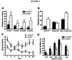
- the 14-3-3 proteins constitute a family of highly conserved regulatory molecules expressed abundantly throughout development and in adult tissue. These proteins comprise seven distinct isoforms ( ⁇ , ⁇ , ⁇ , ⁇ , ⁇ , ⁇ , ⁇ , ⁇ ), that bind a multitude of functionally diverse signalling molecules to control cell cycle regulation, proliferation, migration, differentiation and apoptosis ( Berg et al. Nat Rev Neurosci 2003; 4(9):752-762 ; Fu et al. Annu Rev Pharmacol Toxicol 2000; 40:617-647 ; Toyo-oka et al. Nat Genet 2003 Jul; 34(3): 274-285 ; Aitken A., Semin Cancer Biol 2006; 16(3):162-172 ; Rosner et al. Amino Acids 2006; 30(1):105-109 ).
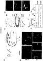
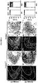
- the defect in 14-3-3 ⁇ and 14-3-3 ⁇ /DISCl complex functionality has been determined to lead.to developmental abnormalities of the hippocampus arising from aberrant neuronal migration. Still further, in terms of the development of the hippocampus it has been determined that the Nrp2 + neural crest stem cells, being a subpopulation of neural crest stem cells, specifically differentiate to neurons of the hippocampus and can effectively regenerate the hippocampus. This has therefore now facilitated the design of a therapeutic treatment for neuropsychiatric conditions, such as schizophrenia.
- the term "derived from” shall be taken to indicate that a particular integer or group of integers has originated from the species specified, but has not necessarily been obtained directly from the specified source. Further, as used herein the singular forms of "a”, “and” and “the” include plural referents unless the context clearly dictates otherwise.
- Nrp2 + neural crest stem cell population or variants thereof for use in the treatment of a neuropsychiatric condition in a mammal, as specified in the claims.
- One aspect of the present disclosure is directed to a method of treating a mammal with a condition characterised by a defective hippocampus, said method comprising administering to said mammal an effective number of Nrp2 + neural crest stem cells or mutants or variants thereof for a time and under conditions sufficient to effect regeneration of the hippocampus.
- a method of treating a human with a condition characterised by a defective hippocampus comprising administering to said mammal an effective number of Nrp2 + neural crest stem cells or mutants or variants thereof for a time and under conditions sufficient to effect regeneration of the hippocampus.
- a method of treating a mammal with a condition characterised by a defective hippocampus comprising administering to said mammal an effective number of adult Nrp2 + neural crest stem cells or mutants or variants thereof for a time and under conditions sufficient to effect regeneration of the hippocampus.
- Yet another aspect of the present disclosure is directed to the use of Nrp2 + neural crest stem cells or mutants or variants thereof in the manufacture of a medicament for the treatment of a condition in a mammal, which condition is characterised by a defective hippocampus, wherein said stem cells regenerate the hippocampus.
- a further aspect of the present disclosure isdirected to an isolated cellular population comprising Nrp2 + neural crest stem cells for use in the method of the disclosure.
- the present invention is predicated, in part, on the determination that a reduction in the functional level of protein 14-3-3 ⁇ , such as in the context of absolute levels of protein 14-3-3 ⁇ or levels of protein 14-3-3 ⁇ /DISC1 complex formation, is indicative of the onset or predisposition to the onset of a neuropsychiatric condition, such as schizophrenia or related condition.
- a reduction in the functional level of protein 14-3-3 ⁇ such as in the context of absolute levels of protein 14-3-3 ⁇ or levels of protein 14-3-3 ⁇ /DISC1 complex formation, is indicative of the onset or predisposition to the onset of a neuropsychiatric condition, such as schizophrenia or related condition.
- a reduction in the functional level of protein 14-3-3 ⁇ such as in the context of absolute levels of protein 14-3-3 ⁇ or levels of protein 14-3-3 ⁇ /DISC1 complex formation
- the further determination that this leads to the degeneration of the hippocampus has provided the basis for developing a therapeutic treatment for individuals exhibiting a defective hippocampus, such as schizophrenia patients.
- one aspect of the present disclosure is directed to a method of treating a mammal with a condition characterised by a defective hippocampus, said method comprising administering to said mammal an effective number of Nrp2 + neural crest stem cells or mutants or variants thereof for a time and under conditions sufficient to effect regeneration of the hippocampus.
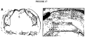
- hippocampus should be understood as a reference to the hippocampus region of the brain. Without limiting the present invention to any one theory or mode of action the hippocampus is a major component of the brains of humans and other mammals. It belongs to the limbic system and plays important roles in the consolidation of information from short-term memory to long-term memory and spatial navigation. Like the cerebral cortex, with which it is closely associated, it is a paired structure, with mirror-image halves in the left and right sides of the brain. In humans and other primates, the hippocampus is located inside the medial temporal lobe, beneath the cortical surface. It contains two main interlocking parts: Ammon's horn and the dentate gyrus.
- the hippocampus is an elaboration of the edge of the cerebral cortex ( Amaral and Lavenex (2006). "Ch 3. Hippocampal Neuroanatomy”. The Hippocampus Book. Oxford University Press .
- the hippocampus is anatomically connected to parts of the brain that are involved with emotional behaviour-the septum, the hypothalamic mammillary body, and the anterior nuclear complex in the thalamus.
- the hippocampus as a whole has the shape of a curved tube, which has been analogized variously to a seahorse, a ram's horn ( Cornu Ammonis, hence the subdivisions CA I through CA4), or a banana (Amaral and Lavenex, supra). It can be distinguished as a zone where the cortex narrows into a single layer of densely packed pyramidal neurons which curl into a tight U shape; one edge of the "U,” field CA4, is embedded into a backward facing strongly flexed V-shaped cortex, the dentate gyrus. It consists of ventral and dorsal portions, both of which share similar composition but are parts of different neural circuits ( Moser and Moser (1998) Hippocampus 8(6): 608-19 ). This general layout holds across the full range of mammalian species.
- the entorhinal cortex located in the parahippocampal gyrus, is considered to be part of the hippocampal region because of its anatomical connections.
- the EC is strongly and reciprocally connected with many other parts of the cerebral cortex.
- the medial septal nucleus, the anterior nuclear complex and nucleus reuniens of the thalamus and the supramammillary nucleus of the hypothalamus, as well as the raphe nuclei and locus coeruleus in the brainstem send axons to the EC.
- the main output pathway (perforant path) of EC axons comes from the large stellate pyramidal cells in layer II that "perforate” the subiculum and project densely to the granule cells in the dentate gyrus, apical dendrites of CA3 get a less dense projection, and the apical dendrites of CA1 get a sparse projection.
- the perforant path establishes the EC as the main "interface" between the hippocampus and other parts of the cerebral cortex.
- the dentate granule cell axons (called mossy fibers) pass on the information from the EC on thorny spines that exit from the proximal apical dendrite of CA3 pyramidal cells. Then, CA3 axons exit from the deep part of the cell body, and loop up into the region where the apical dendrites are located, then extend back into the deep layers of the entorhinal cortex-the Shaffer collaterals completing the reciprocal circuit; field CA1 also sends axons back to the EC, but these are more sparse than the CA3 projection.
- the flow of information from the EC is largely unidirectional, with signals propagating through a series of tightly packed cell layers, first to the dentate gyrus, then to the CA3 layer, then to the CA1 layer, then to the subiculum, then out of the hippocampus to the EC, mainly due to collateralization of the CA3 axons.
- Each of these layers also contains complex intrinsic circuitry and extensive longitudinal connections (Amaral and Lavenex 2006, supra).
- hippocampal function (Amaral and Lavenex 2006, .supra ) . Beyond the output to the EC, additional output pathways go to other cortical areas including the prefrontal cortex. A very important large output goes to the lateral septal area and to the mammillary body of the hypothalamus. The hippocampus receives modulatory input from the serotonin, norepinephrine, and dopamine systems, and from nucleus reuniens of the thalamus to field CA1. A very important projection comes from the medial septal area, which sends cholinergic and GABAergic fibers to all parts of the hippocampus.
- the inputs from the septal area play a key role in controlling the physiological state of the hippocampus: destruction of the septal area abolishes the hippocampal theta rhythm, and severely impairs certain types of memory( Winson (1978), Science 201(4351):160-63 ).
- the cortical region adjacent to the hippocampus is known collectively as the parahippocampal gyrus (or parahippocampus) ( Eichenbaum et al. (2007), Annu Rev Neurosci 30:123-52 ). It includes the EC and also the perirhinal cortex, which derives its name from the fact that it lies next to the rhinal sulcus.
- the perirhinal cortex plays an important role in visual recognition of complex objects, but there is also substantial evidence that it makes a contribution to memory which can be distinguished from the contribution of the hippocampus, and that complete amnesia occurs only when both the hippocampus and the parahippocampus are damaged ( Eichenbaum et al. (2007), Annu Rev Neurosci 30:123-52 ).
- a "defective" hippocampus should be understood as a reference to a hippocampus, all or part of the structure or function which is not normal. To this end, the defect may be congenital or it may be acquired. For example, anatomical malformation of the hippocampus may be present from birth. However, the hippocampus defects which are associated with the onset of many neuropsychiatric and neurodegenerative conditions are often acquired postnatally and are the result of injuries (e.g. head trauma or asphyxiation), exposure to environmental factors, drug use and the like. In other situations, a genetic defect is present congenitally but does not manifest until much later sometimes not until adulthood.
- injuries e.g. head trauma or asphyxiation
- the method of the present disclosure provides a means of regenerating hippocampus tissue, thereby at least in part restoring tissue which is structurally and functionally normal.
- regeneration is a reference to the generation of at least some normal hippocampus tissue within the hippocampus area of the brain. It is not intended to mean that the hippocampus is entirely replaced or that even all of the defective tissue is replaced. Rather, it is a reference to the fact that the method of the present disclosure increases the proportion of normal hippocampus tissue relative to the proportion which existed in the subject prior to the application of the method of the disclosure. Accordingly, the method of the present disclosure isnot limited to its application in the context of the complete normalisation of all the affected hippocampus tissue. Rather, it should also be understood to extend to the partial normalisation of all or only some of the defective tissue
- mammal as used herein includes humans, primates, livestock animals (e.g. horses, cattle, sheep, pigs, donkeys), laboratory test animals (e.g. mice, rats, guinea pigs), companion animals (e.g. dogs, cats) and captive wild animals (e.g. kangaroos, deer, foxes).
- livestock animals e.g. horses, cattle, sheep, pigs, donkeys
- laboratory test animals e.g. mice, rats, guinea pigs
- companion animals e.g. dogs, cats
- captive wild animals e.g. kangaroos, deer, foxes.
- the mammal is a human or a laboratory test animal. Even more preferably, the mammal is a human.
- a method of treating a human with a condition characterised by a defective hippocampus comprising administering to said mammal an effective number of Nrp2 + neural crest stem cells or mutants or variants thereof for a time and under conditions sufficient to effect regeneration of the hippocampus.
- the method of the present disclosure is predicated on the determination that the administration of Nrp2 + neural crest stem cells to the brain of a mammal with a defective hippocampus results in not just engraftment of the cells into the tissue, but also repair and restoration of hippocampus morphology and functioning.
- stem cell is meant that the cell is not fully differentiated but requires further differentiation to achieve maturation.
- Such cells are also sometimes referred to as “precursor” cells, “progenitor” cells, “multipotent” cells or “pluripotent” cells.
- neural crest cells are a transient, multipotent, migratory cell population unique to vertebrates that give rise to a diverse cell lineage including melanocytes, craniofacial cartilage and bone, smooth muscle, peripheral and enteric neurons and glia. After gastrulation, neural crest cells are specified at the border of the neural plate and the non-neural ectoderm. During neuralation, the borders of the neural plate, also known as the neural folds, converge at the dorsal midline to form the neural tube.
- neural crest cells from the roof plate of the neural tube undergo an epithelial to mesenchymal transition, delaminating from the neuroepithelium and migrating through the periphery where they differentiate into varied cell types.
- a gene regulatory network described as a set of interacting signals, transcription factors, and downstream effector genes that confer cell characteristics such as multipotency and migratory capabilities.
- neural crest stem cell should therefore be understood as a reference to any cell which exhibits one or more of the functional or phenotypic characteristics of a neural crest stem cell or which exhibits the potentiality to differentiate to any of the cell types which a neural crest stem cell can differentiate to.
- the subject neural crest stem cell may be one which exhibits multipotentiality, for example is a progenitor which can be induced to differentiate to give rise to any one or more multiple peripheral structures such as the cranial skeleton, dentine of the teeth, melanocytes, peripheral neurons, adrenal chromafin cells and specific cells within hair follicles, or it may be already committed to a subgroup of these lineages.
- the subject cell is nevertheless still a “stem cell” on the basis that it is not fully differentiated.
- stem cell should not be understood as a limitation on the maturity/immaturity of the subject cell relative to that which might be implied by the use of the terms “progenitor cell”, “multipotent cell”, “pluripotent cell” or other such term.
- Reference to a cell exhibiting a "functional" characteristic of a neural crest stem cell should be understood as a reference to a cell which is restricted to differentiating along any one or more of the neural crest cell derived lineages, such as those detailed above.
- Reference to a "phenotypic" characteristic should be understood as a reference to a cell surface or intracellular expression profile of one or more proteinaceous or non-proteinaceous molecules which is characteristic of a neural crest stem cell.
- Nrp2 neuroropilin 2
- neural crest stem cells can be derived either from an embryonic source or, more conveniently, from an adult source.
- adult neural crest stem cells can be easily and routinely isolated from the dentine of teeth and the bulge of the hair follicle and provide the same precursor cell source for the neurons and glia in the central nervous system. When engrafted, these cells differentiate into GABAergic neurons and oligodendrocytes. Accordingly, either an adult source or an embryonic source can be used in the context of the method of the present disclosure.
- the subject stem cells are adult stem cells.
- a method of treating a mammal with a condition characterised by a defective hippocampus comprising administering to said mammal an effective number of adult Nrp2 + neural crest stem cells or mutants or variants thereof for a time and under conditions sufficient to effect regeneration of the hippocampus.
- said adult Nrp2 + neural crest stem cells are isolated from the dentine or the hair follicle.
- the subject Nrp2 + neural crest stem cells population may be a single cell suspension or a cell aggregate, such as a tissue, which has been freshly isolated from an individual (such as an individual who may be the subject of treatment) or it may have been sourced from a non-fresh source, such as from a culture (for example, where cell numbers were expanded and/or the cells were cultured so as to render them receptive to differentiative signals) or a frozen stock of cells (for example, an established cell line), which had been isolated at some earlier time point either from an individual or from another source.
- the subject cells may have undergone some other form of treatment or manipulation, such as but not limited to enrichment or purification, modification of cell cycle status, molecular transformation or the formation of a cell line.
- the subject cell may be a primary cell or a secondary cell.
- a primary cell is one which has been freshly isolated from an individual.
- a secondary cell is one which, following its isolation, has undergone some form of in vitro manipulation such as the preparation of a cell line.
- mutant or variant of the subject cellular population should be understood as a reference to a cell which is derived from the cellular population but exhibits at least one difference at the phenotypic or functional level.
- the mutant or variant may have altered expression of its cell surface markers as a whole or some aspect of its functionality subsequently to initial isolation. Such changes can occur either spontaneously (as exemplified by the spontaneous upregulation or downregulation of cell surface markers which can occur subsequently to in vitro culture or spontaneous transformation) or as a result of a directed manipulation, such as would occur if a cell was deliberately transformed (for example, in order to effect the creation of a cell line) or transfected (for example to effect the expression of a particular gene or marker).
- Nrp2 + neural crest stem cell populations of the present invention may exhibit some variation in differentiative status within a single phenotypic profile. That is, within a single phenotypic profile, although the cells comprising that profile may substantially exhibit similar phenotypic and/or functional characteristics, there may nevertheless exhibit some differences. This may be apparent, for example, in terms of differences in the transcriptome profile or cell surface marker expression (other than the markers defined herein) of the cells which comprise the phenotypic profile in issue.
- the Nrp2 + neural crest stem cells may not represent a highly specific and discrete stage, but may be characterised by a number of discrete cellular subpopulations which reflect a transition or phase if one were to compare cells which have differentiated into this stage versus cells which are on the cusp of maturing out of this stage. Accordingly, the existence of cellular subpopulations within a single phenotypic profile of the present invention is encompassed.
- these cells may be derived from the inner cell mass of a blastocyst stage human embryo or an established cell line may be used (such as those developed by Thomson and Odorico, Trends Biotechnol., 18:53-57 (2002 ). namely. H1. H7, H9.1, H9.2, H13 or H14).
- cells from the inner cell mass are separated from the surrounding trophectoderm by microsurgery or by immunosurgery (which employs antibodies directed to the trophectoderm to break it down) and are plated in culture dishes containing growth medium supplemented with fetal bovine serum (alternatively.
- KnockOut Dulbecco's modified minimal essential medium containing basic FGF can be supplemented with Serum Replacer (Life Technologies) and used without serum), usually on feeder layers of mouse embryonic fibroblasts that have been mitotically inactivated to prevent replication.
- a feeder-free culture system may be employed, such as that reported by Chunhui Xu, Melissa Carpenter and colleagues using Matrigel or laminin as a substrate, basic FGF, and conditioned medium from cultures of mouse embryo fibroblasts ( Xu, et al., Nat Biotechnol. 2001 Oct; 19(10):971-4 ).
- the Nrp2 + neural crest stem cell population is then differentiated from this starting pluripotent stem cell population.
- hESC human embryonic stem cells
- the hESC do not form part of the claimed invention and are mentioned for reference purposes only.
- the present disclosure is predicated on administering a Nrp2 + neural crest stem cell population to a mammal in order to facilitate its localisation to the brain of the mammal.
- localisation is meant that at least some of the Nrp2 + neural crest stem cell population which is introduced to the patient targets the brain. It should be understood, however, that in terms of any treatment event, a proportion of the administered Nrp2 + neural crest stem cells may not target the brain, but may either be cleared or else lodge in non-brain tissues.
- the cells which are administered in the context of the present disclosure are preferably autologous cells which are isolated and transplanted back into the individual from which they were originally harvested (for example, dentine derived Nrp2 + neural crest stem cells),
- autologous cells which are isolated and transplanted back into the individual from which they were originally harvested
- the present disclosure nevertheless extends to the use of cells derived from any other suitable source where the subject cells exhibit the same major histocompatability profile as the individual who is the subject of treatment. Accordingly, such cells are effectively autologous in that they would not result in the histocompatability problems which are normally associated with the transplanting of cells exhibiting a foreign MHC profile.
- Such cells should be understood as falling within the definition of "autologous”.
- the subject cells are isolated from a genetically identical twin, or are differentiated from the stem cells of an embryo generated using gametes derived from the subject individual or cloned from the subject individual.
- the cells may also have been engineered to exhibit the desired major histocompatability profile. The use of such cells overcomes the difficulties which are inherently encountered in the context of tissue and organ transplants.
- allogeneic cells are those which are isolated from the same species as the subject being treated but which exhibit a different MHC profile. Although the use of such cells in the context of therapeutics may result in the onset of an allogeneic based immune response, this problem can nevertheless be minimised by use of cells which exhibit an MHC profile exhibiting similarity to that of the subject being treated, such as a cell population which has been isolated/generated from a relative such as a sibling, parent or child. The immunological tissue rejection which is often characteristic of the use of allogeneic cells may also be minimised via the use of immunosuppressant drugs.
- Nrp2 + neural stem cell lines are established.
- the present disclosure should also be understood to extend to xenogeneic transplantation. That is, the cells which are introduced into a patient are isolated from a species other than the species of the subject being treated.
- an "effective number” means that number of cells necessary to at least partly attain the desired effect, or to delay the onset of, inhibit the progression of, or halt altogether the onset or progression of the particular condition being treated. Such amounts will depend, of course, on the particular condition being treated, the severity of the condition and individual patient parameters including age, physical conditions, size, weight, physiological status, concurrent treatment, medical history and parameters related to the disorder in issue.
- One skilled in the art would be able to determine the number of Nrp2 + neural crest stem cells that would constitute an effective dose, and the optimal mode of administration thereof without undue experimentation, this latter issue being further discussed hereinafter. These factors are well known to those of ordinary skill in the art and can be addressed with no more than routine experimentation. It is preferred generally that a maximal cell number be used, that is, the highest safe number according to sound medical judgement. It will be understood by those of ordinary skill in the art, however, that a lower cell number may be administered for medical reasons, psychological reasons or for any.other reasons.
- Nrp2 + neural crest stem cells which are administered in accordance with the method of the disclosure may necessarily contribute to the treatment regime discussed herein. For example, some cells may localise to non brain tissues while others may become non-viable or non-functional.
- the purified population may nevertheless comprise some non-Nrp2 + neural crest stem cells where 100% purity is not obtained, The present disclosure is therefore achieved provided the relevant portion of the cells which are introduced to the patient constitute an "effective number" as defined above.
- the subject cells require introduction into the subject individual.
- the cells may be introduced by any suitable method.
- cell suspensions may be introduced by direct injection to a tissue or inside a blood clot whereby the cells are immobilised in the clot thereby facilitating transplantation.
- the cells may also be encapsulated prior to transplantation. Encapsulation is a technique which is useful for preventing the dissemination of cells which may continue to proliferate (i.e. exhibit characteristics of immortality).
- the cells may also be introduced by localised, intravenous or systemic routes.
- the cells may also be introduced by surgical implantation (grafting). This may be necessary, for example, where the cells exist in the form of a tissue graft or where the cells are encapsulated prior to transplanting. Without limiting the present disclosure to any one theory or mode of action, where cells are administered as an encapsulated cell suspension, the cells will coalesce into a mass.
- the cells which are administered to the patient can be administered as single or multiple doses by any suitable route.
- a single administration is utilised, particularly where surgical engraftment into the brain is the method used.
- Administration via injection can be directed to various regions of a tissue or organ, depending on the type of treatment required.
- other proteinaceous or non-proteinaceous molecules such as antibiotics or differentiation inducing cytokines may be coadministered either with the introduction of the Nrp2 + neural crest stem cells or during the differentiation and proliferation phase of the transplanted cells.
- coadministered is meant simultaneous administration in the same formulation or in different formulations via the same or different routes or sequential administration via the same or different routes.
- sequential administration is meant a time difference of from seconds, minutes, hours or days between the transplantation of these cells and the administration of the proteinaceous or non-proteinaceous molecules.
- Other examples of circumstances in which co-administration may be required include, but are not limited to:
- the patient may be necessary to maintain the patient on a course of medication to alleviate the symptoms of the condition until such time as the transplanted cells become integrated and fully functional (for example, the administration of anti-psychotic drugs to treat schizophrenia).
- it may be necessary to commence the long term use of medication to prevent re-occurrence of the damage.
- the ongoing use of immunosuppressive drugs may be required even when syngeneic cells have been used.
- the method of the present disclosure can either be performed in isolation to treat the condition in issue or it can be performed together with one or more additional techniques designed to facilitate or augment the subject treatment.
- additional techniques may take the form of the co-administration of other proteinaceous or non-proteinaceous molecules, as detailed hereinbefore.
- condition characterised by a defective hippocampus should be understood as a reference to any condition, a symptom or cause of which is hippocampus degeneration or damage.
- conditions include, but are not limited to, congenital anatomical abnormalities of the brain, acquired injury such as through head trauma or asphyxiation, atrophy and hypoplasia such as that seen in returning military officers after extended duress or conditions characterised by a reduction in the level of functional protein 14-3-3 ⁇ or protein 14-3-3 ⁇ /DISC1 complex formation, such as a neuropsychiatric condition.
- Neuropsychiatric condition should be understood as a reference to a condition characterised by neurologically based cognitive, emotional and behavioural disturbances.
- Examples of such conditions include, inter alia, a condition characterised by one or more symptoms of schizophrenia, schizophrenia, schizotypal personality disorder, psychosis, bipolar disorder, manic depression, affective disorder, or schizophreniform or schizoaffective disorders, psychotic depression autism, drug induced psychosis, delirium, alcohol withdrawal syndrome or dementia induced psychosis.
- said neuropsychiatric condition is a condition which is characterised by one or more symptoms of schizophrenia.
- a method of treating a mammal with a neuropsychiatric condition comprising administering to said mammal an effective number of Nrp2 + neural crest stem cells or mutants or variants thereof for a time and under conditions sufficient to effect regeneration of the hippocampus.
- said mammal is a human.
- said Nrp2 + neural crest stem cells are adult-derived stem cells.
- said neuropsychiatric condition is a condition characterised by one or more symptoms of schizophrenia.
- said condition is schizophrenia.
- symptoms characteristic of schizophrenia should be understood as a reference to any one or more symptoms which may occur in an individual suffering from schizophrenia. These symptoms may be evident throughout the disease course or they may be evident only transiently or periodically. For example, the hallucinations associated with schizophrenia usually occur in periodic episodes while the characteristic social withdrawal may exhibit an ongoing manifestation. It should also be understood that the subject symptoms may not necessarily be exhibited by all individuals suffering from schizophrenia. For example, some individuals may suffer from auditory hallucinations only while others may suffer only from visual hallucinations. However, for the purpose of the present invention, any such symptoms, irrespective of how many or few schizophrenia patients ever actually exhibit the given symptom, are encompassed by this definition.
- the symptoms that are most commonly associated with the disease are called positive symptoms (which denote the presence of grossly abnormal behaviour), thought disorder and negative symptoms.
- positive symptoms which denote the presence of grossly abnormal behaviour
- positive symptoms include speech which is difficult to follow or jumping from one subject to another with no logical connection, delusions (false beliefs of opposition, guilt, grandeur or being under outside control) and hallucinations (visual or auditory).
- delusions familial beliefs of opposition, guilt, grandeur or being under outside control
- hallucinations visual or auditory
- Thought disorder is the diminished ability to think clearly and logically. Often it is manifested by disconnected and nonsensical language that renders the person with schizophrenia incapable of participating in conversation, contributing to alienation from family, friends and society. Delusions are common among individuals with schizophrenia.
- paranoid delusion An affected person may believe that he or she is being conspired against (called “paranoid delusion”). "Broadcasting” describes a type of delusion in which the individual with this illness believes that their thoughts can be heard by others. Hallucinations can be heard, seen or even felt. Most often they take the form of voices heard only by the afflicted person. Such voices may describe the person's actions, warn of danger or tell him what to do. At times the individual may hear several voices carrying on a conversation. Less obvious than the "positive symptoms” but equally serious are the deficit or negative symptoms that represent the absence of normal behaviour. These include flat or blunted affect (i.e. lack of emotional expression); apathy, social withdrawal and lack of insight. Both the positive symptoms and the negative symptoms should be understood to fall within the definition of "symptoms characteristic of schizophrenia”.
- a condition characterised by one or more symptoms characteristic of schizophrenia should be understood as a reference to any neuropsychiatric condition which is characterised by the presence of one or more of these symptoms.
- said condition is schizophrenia.
- the subject undergoing treatment may be undergoing therapeutic or prophylactic treatment and may be any human or animal in need of therapeutic or prophylactic treatment.
- treatment and prophylaxis
- treatment and prophylaxis are to be considered in its broadest context.
- the term “treatment” does not necessarily imply that a mammal is treated until total recovery.
- prophylaxis does not necessarily mean that the subject will not eventually contract a disease condition.
- treatment and prophylaxis include amelioration of the symptoms of a particular condition or preventing or otherwise reducing the risk of developing a particular condition.
- the term “prophylaxis” may be considered as reducing the severity of the onset of a particular condition. “Treatment” may also reduce the severity of an existing condition.
- Yet another aspect of the present disclosure is directedto the use of Nrp2 + neural crest stem cells or mutants or variants thereof in the manufacture of a medicament for the treatment of a condition in a mammal, which condition is characterised by a defective hippocampus, wherein said stem cells regenerate the hippocampus.
- said mammal is a human.
- said Nrp2 + neural crest stem cells are adult stem cells and still more particularly dentine or hair follicle derived stem cells.
- said condition is a congenital anatomical abnormality of the brain, acquired brain injury such as through head trauma or asphyxiation or a condition characterised by a reduction in the level of functional protein 14-3-3 ⁇ or protein 14-3-3 ⁇ /DISC1 complex formation.
- said condition is a neuropsychiatric condition, more particularly a condition characterised by one or more symptoms of schizophrenia, schizophrenia, schizotypal personality disorder, psychosis, bipolar disorder, manic depression. affective disorder, or schizophreniform or schizoaffective disorders, psychotic depression, autism, drug induced psychosis, delirium, alcohol withdrawal syndrome or dementia induced psychosis.
- Yet another aspect of the present disclosure is directed to an isolated cellular population comprising Nrp2 + neural crest stem cells for use in the method of the disclosure.
- mice 14-3-3 ⁇ Gt(OST062)Lex and 14-3-3 ⁇ Gt(OST390)Lex mutant mice carrying gene trap constructs that contain the Geo reporter gene were derived from Lexicon Genetics ES cell lines OST062 and OST390, respectively.
- the gene trap vector in 14-3-3 ⁇ Gt(OST062)Lex mice inserted into the first intron of 14-3-3 ⁇
- the gene trap vector in 14-3-3 ⁇ Gt(OST390)Lex mice inserted into the second intron of 14-3-3 ⁇ .
- ES cell lines were amplified and injected into SV129 blastocysts.
- Resulting germ line transmitting males were either maintained in the SV129 background or backcrossed in to the C57/B16 and BA1, BC backgrounds over 6 generations.
- qRT-PCR and western blot from whole tissue samples was used to confirm complete KO of the gene in these mouse strains.
- 14-3-3C genotype was determined by PCR amplification of genomic tail DNA using the primers detailed in supplementary table 1.
- Mice were maintained as heterozygous breeding pairs that were phenotypically indistinguishable to WT littermates. Animal experiments were conducted in accordance with the guidelines of the Animal Ethics Committee of the Institute of Medical and Veterinary Sciences and the University of Sydney.
- Behavioural assays All procedures were carried out under normal light conditions between 8.00 am and 12.00 pm. Behavioural phenotyping was performed as previously described ( Coyle et al. Behav Brain Res 2009, 197(1): 210-218 ; Summers et al. Pediatr Res 2006; 59(1): 66-71 ; van den Buuse et al. Int J Neuropsychopharmacol 2009; 12(10): 1383-1393 ).
- One cohort of mice was used for the open field test at ages of 5-, 10-, 20- and 40-week time points.
- One cohort of mice was used at the age of 12 weeks for spatial working memory, then elevated plus maze and object recognition tasks.
- a separate cohort of mice was used at the age of 12 weeks for PPI.
- Locomotor function test Exploratory activity and anxiety level of mice were measured in an open field made from a box (50 cm x 27 cm) with the floor divided into 15 squares (9 cm x 10 cm). Each mouse was introduced in to the same position of the box facing the right top corner. The behaviour of the mouse was observed for 3 min and locomotor activity was scored as a measure of line crossings (i.e. when a mouse removed all four paws from one square into another). Number of rears up was scored when a mouse had both front paws off the floor. Urine and faecal material were removed between session and the box was cleaned thoroughly with 80% ethanol to remove any lingering scents.
- the object recognition task takes advantage of the natural affinity of mice for novelty; mice that recognise a previously seen (familiar) object will spend more time exploring novel objects ( Dere et al. Neurosci Biobehav Rev 2006; 30(8):1206-1224 ; Sik et al. Behav Brain Res 2003; 147(1-2):49-54 ).
- the apparatus consisted of a plastic arena (length; 50 cm, width; 35 cm, depth; 20 cm) filled with bedding. Two different sets of objects were used; a yellow -capped plastic jar (height, 6 cm; base diameter, 4.3 cm) and a red plastic bulb (length: 8 cm, width: 4 cm).
- mice spent equal amounts of time when presented with both of these objects, regardless of the position they were placed in the arena (data not shown).
- the same cohort of mice tested for spatial learning and memory were assessed for object recognition memory.
- Each mouse was given 5-min to explore the test box without any objects present to habituate them to the test arena.
- Mice underwent the testing session comprised of two trials. The duration of each trial was 3 min.
- the box contained two identical objects (a, samples) which were placed in the north-west (left) and northeast (right) corners of the box (5 cm away from the walls). A mouse was always placed in the apparatus facing the south wall. After the first exploration period, mice were placed back in their homecage.
- the mouse was placed in the apparatus for the second trial (choice phase), but now with a familiar one (a, sample) and a novel object (b).
- the objects were cleaned thoroughly with alcohol between sessions to remove any lingering scents.
- the time spent exploring each object during trial 1 and trial2 was recorded. Exploration was defined as either touching the object with the nose or being within 2 cm of it.
- the basic measures in the object recognition task were the times spent exploring an object during trial 1 and trial 2.
- e1 (a + a) and e2 (a + b) are measures of the total exploration time of both objects during trial 1 and trial 2, respectively.
- h1 is an index of habituation measured by the difference in total exploration time from trial 1 to trial 2(e1 - e2).
- d1 (b - a) and d2 (d1/e2) were considered as index measures of discrimination between the novel and the familiar objects.
- d2 is a relative measure of discrimination that corrects d1 for exploratory activity (e2).
- a discrimination index above zero describes animals exploring the novel object more than the familiar object. An animal with no preference for either object will have an index near zero.
- Mice with a total exploration time of less than 7 s during trials in the sample or choice phase were excluded from the analyses as the measurement of exploration time has been found to be non-reliable below this threshold(van den Buuse et al. supra; de Bruin et al, Pharmacol Biochem Behav 2006; 85(1):253-260 ).
- Elevated cross bar test The anxiety behaviour of mice based on their natural aversion of open and elevated areas was assessed using an elevated plus-maze as previously described ( Komada et al. J Vis Exp 2008; (22 ); Walf et al. Nat Protoc 2007; 2(2):322-328 ). Briefly, the apparatus was made in the shape of a cross from black plexiglass and consisted of two open arms (25 cm x 5 cm) and two closed arms (25 cm x 5 cm x 16 cm) that crossed in the middle perpendicular to each other. In the middle of the to arms there was a central platform (5 cm x 5 cm). The cross maze was raised 1 m from the ground.
- mice were introduced to the center of the apparatus facing the open arm opposite to the experimenter were and observed by video recording for 5 minutes. The number of entries into the open and closed arms and the time in exploring both types of arm were scored. Naturalistic behaviour of the mouse such as the number of head dipping, number of rearing and number of stretch attended postures were measured. After each trial all arms and the central area thoroughly cleaned with alcohol to remove any lingering scents.
- mice 12 week old mice were individually habituated to the maze environment by being placed into the pool without the escape platform and allowed to swim for 60 s. Learning trials were conducted over a 6-day training period in which mice were required to learn the position of the submerged escape platform from the other three (East, South, West) arms that did not contain an escape platform. Each mouse was given six daily trials (two blocks of three trials separated by a 30min rest interval), in which each of the three arms were chosen as a starting point in a randomized pattern (twice daily). For each trial, the mouse was placed in the distal end of an arm facing the wall and allowed 60 s to reach the escape platform where it remained for 10 s. Mice that did not climb onto the escape platform in the given time were placed on the platform for 10 s.
- mice were assessed on their long-term retention of the escape platform location which was placed in the same position as during the learning phase.
- Memory was tested 14 (M1 and 28 (M2) days after the final day of learning and consisted of a single day of 6 trials as described for the learning period. Data were recorded for each mouse for each trial on their escape latency (i.e. time (s) taken to swim to the platform), number of correct trials (i.e. if a mouse found the platform on the first arm entry) and number of incorrect entries/reentries (i.e. the number of times that a mouse went into an arm that did not contain the escape platform).
- escape latency i.e. time (s) taken to swim to the platform
- number of correct trials i.e. if a mouse found the platform on the first arm entry
- number of incorrect entries/reentries i.e. the number of times that a mouse went into an arm that did not contain the escape platform.
- mice were placed in clear Plexiglas cylinders which were closed on either side and acoustic stimuli were delivered over 70-dB background noise through a speaker in the ceiling of the box.
- Each testing session consisted of 104 trials with an average inter-trial interval between 25 s.
- the first and last eight trials consisted of single 40-ms 115 -dB pulse alone startle stimuli.
- the middle 88 trials consisted of pseudo-randomised delivery of 16 115-dB pulse-alone stimuli, eight trials during which no stimulus was delivered, and 64 prepulse trials. The total of 32 115-dB pulse alone trials was expressed as four blocks of eight and used to determine startle habituation.
- Prepulse trials consisted of a single 115-dB pulse preceded by a 30-ms or 100-ms inter-stimulus interval (ISI) with a 20-ms non-startling stimulus of 2, 4, 8 or 16 dB over the 70-dB baseline.
- ISI inter-stimulus interval
- Whole-body startle responses were converted into quantitative values by a piezo -electric accelerometer unit attached beneath the platform. Percentage prepulse inhibition (%PPI) was calculated as pulse-alone startle response - prepulse + pulse startle response / pulse-alone startle response X 100.
- BrdU-pulse-chase analysis and TUNEL labelling BrdU was injected at 100 ⁇ g / g of body weight of the pregnant mice at 14.5 dpc or 16.5 dpc and the pups were euthanized at postnatal-day-7. Final destination of the proliferating hippocampal neurons that were born at these time points were revealed by BrdU immunohistochemistry on frozen brain sections.
- Tissue were denatured with 2M HCI for 20 min at 37°C, neutralised in 0.1 M borate buffer (pH 8.5) for 10 min, blocked with 10% horse serum in PBST and probed with rat monoclonal anti-BrdU (1:250; Abcam) and mouse monoclonal anti-NeuN (1:500; Chemicon) antibodies overnight at 4°C.
- Cell apoptosis was determined by the TUNEL assay using the In Situ Cell Death Detection Kit (TMR Red; Roche Applied Science) according to the manufacturer's instructions followed by counterstained with DAPI (Molecular Probes).
- the precleared lysates were incubated for 2 h at 4C with 2 ug/ml of either anti-DISCI antibody (C-term) (Invitrogen) or anti-14-3-3 antibody (3F7 Abcam) absorbed to protein A-Sepharose (Amersham Biosciences).
- C-term Invitrogen
- anti-14-3-3 antibody 3F7 Abcam
- the sepharose beads were washed 3 times with lysis buffer before being boiled for 5 min in SDS-PAGE sample buffer.
- the immunoprecipitated proteins and lysates were separated by SDS-PAGE, and electrophorectically transferred to a nitrocellulose membrane and analysed by immunoblotting.
- P7 hippocampi neuron-glial cocultures were prepared as described ( Kaech et al. Nat Protoc 2006, 1(5):2406-2415 ).
- Nitric acid-treated coverslips (diameter 13 mm) were coated with 100 ⁇ g/ml poly-L-lysin/PLL (Sigma) in borate buffer for overnight at 37°C, and were then washed with sterile water for 3 x 1 h.
- Dentate gyri and CA samples were dissected and dissociated in Hank's balanced salt solution (HBSS) and neurons were plated at a density of I x 10 5 cells per culture dish (with 4 PLL-coated coverslips).
- HBSS Hank's balanced salt solution
- mice display behavioural and cognitive defects
- 14-3-3 proteins are abundantly expressed in the developing and adult brain ( Berg et al. Nat Rev Neurosci 2003; 4(9):752-762 ; Baxter et al. Neuroscience 2002; 109(1):5-14 ).
- 14-3-3 ⁇ in neurodevetopment and brain function generated two knockout mouse lines were generated from embryonic stem cell clones containing retroviral gene-trap insertions within intron 1 our 2, termed 14-3-3 and 14-3-3 ⁇ Gt(OST390)Lex , respectively ( Fig. 8 ; Lexicon Genetics).
- mice spent an equal time exploring each identical object (14-3-3 ⁇ 062+/+ , 50.82 ⁇ 1.2%; 14-3-3 ⁇ 062-/- 49.18 ⁇ 1.2%).
- 14-3-3 ⁇ 062-l- mice When presented with a familiar and new object, 14-3-3 ⁇ 062-l- mice exhibited signiticantly impaired novel object recognition compared to controls over the test period. Consistent with a lack of preference between the familiar and novel objects, 14-3-3 ⁇ 062-/- mice had a reduced discrimination index (time exploring novel object - time exploring familiar object / time exploring novel object + time exploring familiar object) indicating that they failed to retain new information (14-3-3 ⁇ 062+/+ , 0.1667 ⁇ 0.086s; 14-3-3 ⁇ 062-/- , -0.0569 ⁇ 0.047s; p ⁇ 0.05). Once again, there were no sex differences in either phase of testing ( p >0.5).
- 14-3-3 ⁇ 062-/- mutants also demonstrated hyperactivity in the object recognition test with longer exploratory times in both phases of the trial (Sample phase, 14-3-3 ⁇ 0 62+/+ , 27.33 ⁇ 2.7s; 14-3-3 ⁇ 062-/- , 38.62 ⁇ 4.1s; p ⁇ 0,05 ; test phase, 14-3-3 ⁇ 062+/+ ,24.58 ⁇ 3.1s; 14-3-3 ⁇ 062-/- , 50.77 ⁇ 4.7s; p ⁇ 0.0001).
- 14-3-3 ⁇ 062-/- mice When placed in such a test, 14-3-3 ⁇ 062-/- mice also demonstrated increased activity compared to wild type controls. 14-3-3 ⁇ 062-/- mice had 25.23 ⁇ 1.76 transitions between cross arms during a 5min test period while 14-3-3 ⁇ 062+/+ had 12.29 ⁇ 1.21 ( p ⁇ 0.0001). In addition, 14-3-3 ⁇ 062-/- mice spent significantly more time in the open arms ( Fig.
- Defects in sensorimotor gating are an endophenotype of neuropsychiatric disorders such as schizophrenia and related disorders. Appropriate signalling in the hippocampus and other brain regions are essential for this filtering mechanism.
- Hippocampal neurons derive from the neuroepithelium along the ventricular zone (NEv) and from a restricted area of neuroepithelium adjacent to the fimbria (NEf) ( Nakahira et al. J Comp Neurol 2005; 483(3):329-340 ) ( Fig. 2A ).
- NEv neuroepithelium along the ventricular zone
- NEf neuroepithelium adjacent to the fimbria
- Fig. 2A At 14.5 dpc 14-3-3 ⁇ immunostaining was detected in migrating hippocampal neurons within the intermediate zone, but not in their neuroepithelial precursors ( Fig. 2Bi ).
- 14-3-3 ⁇ was undetectable in other regions of brain, such as the cerebellum, after early post natal stages ( Fig. 13 ). Expression within CA and DG neurons was confirmed by western blot of protein extracted from microdissected adult hippocampi ( Fig. 2D ). This also confirmed complete removal of the protein from these brain regions of 14-3-3 ⁇ 062-/- mice. Finally, after 10 days in vitro (DIV), hippocampal MAP2 positive neuronal cultures also showed punctate immunocytostaining for 14-3-3 ⁇ within the cell body and axon / dendrites ( Fig. 2E ).
- 14-3-3 ⁇ is expressed in hippocampal neurons we next examined if CA and DG neurons were examined to determine if they are positioned correctly in adult and embryonic mutants.
- Nissl-staining of 14-3-3 ⁇ 062-/- mice revealed developmental defects first noticeable prior to hippocampal maturation (5/5 at P0, 4/4 at P7, 2/2 at P28 and 2/2 at P56; Fig. 3A and Fig. 14 ).
- pyramidal neurons were ectopically positioned in the stratum radiatum and stratum oriens in addition to their usual resting place of the stratum pyramidale. Within the CA3 subfield, pyramidal neurons split in to a bilaminar stratum instead of a single cell layer..
- Dentate granule neurons were also diffusely packed in the 14-3-3 ⁇ 062-/- mice compared with 14-3-3 ⁇ 062+/+ littermates. Consistent with Nissl staining, analysis of hippocampal organization in thyl -YFP mice also revealed a disrupted laminar organization ( Fig. 3B ).
- mice 14-3-3 ⁇ -deficient mice display hippocampal neuronal migration defects
- BrdU-retaining cells were counted from 10 ⁇ m sections using 5 mice of each genotype and the relative percentage in each layer was quantified. Both injection time points show that nearly all neurons born in the ventricular zone at 14.5 dpc or 16.5 dpc migrate in to the stratum pyramidalc of the CA in control mice ( Fig. 4 ). Strikingly, however, a significant percentage of BrdU-retaining cells were identified outside of the stratum pyramidale in 14-14-3-3 ⁇ 062-/- mice. Failure of neurons to migrate from their birthplace or to stop within their correct layer therefore gives rise to the duplicated stratum pyramidale in the 14-3-3 ⁇ 062-/- hippocampus.
- anti-synaptophysin was used to identify presynapses in both the IPMF and SPMF of the CA3 subfield in control animals.
- misrouted axons also formed aberrant synapses within the stratum pyramidale ( Fig. 6 ).
- Visualisation of synaptic boutons by golgi stain further revealed notable differences in synapse formation in CA3.
- large spine excrescences on the proximal region of the apical dendrites were followed by fine-calibre dendritic branches.
- Nrp2 and Nrp2 lineage tracing mice have been generated.
- Cre/RFP or Cre/GFP have been placed under the expression of the Nrp1 or Nrp2 promoters ( Figure 16 ).
- Cre/GFP Cre/GFP have been placed under the expression of the Nrp1 or Nrp2 promoters ( Figure 16 ).
- Studies with these mice show for the first time that neurons of the hippocampus are derived from Nrp2-expressing neural stem cells.
Landscapes
- Health & Medical Sciences (AREA)
- Life Sciences & Earth Sciences (AREA)
- Engineering & Computer Science (AREA)
- Biomedical Technology (AREA)
- Chemical & Material Sciences (AREA)
- General Health & Medical Sciences (AREA)
- Cell Biology (AREA)
- Bioinformatics & Cheminformatics (AREA)
- Zoology (AREA)
- Animal Behavior & Ethology (AREA)
- Biotechnology (AREA)
- Medicinal Chemistry (AREA)
- Developmental Biology & Embryology (AREA)
- Veterinary Medicine (AREA)
- Pharmacology & Pharmacy (AREA)
- Public Health (AREA)
- Neurology (AREA)
- Neurosurgery (AREA)
- Organic Chemistry (AREA)
- Epidemiology (AREA)
- Virology (AREA)
- Immunology (AREA)
- General Chemical & Material Sciences (AREA)
- Genetics & Genomics (AREA)
- Wood Science & Technology (AREA)
- Chemical Kinetics & Catalysis (AREA)
- Nuclear Medicine, Radiotherapy & Molecular Imaging (AREA)
- Psychiatry (AREA)
- Microbiology (AREA)
- Ophthalmology & Optometry (AREA)
- Biochemistry (AREA)
- General Engineering & Computer Science (AREA)
- Rheumatology (AREA)
- Orthopedic Medicine & Surgery (AREA)
- Dermatology (AREA)
- Pain & Pain Management (AREA)
- Hospice & Palliative Care (AREA)
- Medicines Containing Material From Animals Or Micro-Organisms (AREA)
- Medicines That Contain Protein Lipid Enzymes And Other Medicines (AREA)
Applications Claiming Priority (2)
| Application Number | Priority Date | Filing Date | Title |
|---|---|---|---|
| AU2011905180A AU2011905180A0 (en) | 2011-12-13 | Method of treatment | |
| PCT/AU2012/001530 WO2013086574A1 (en) | 2011-12-13 | 2012-12-13 | Method of treatment |
Publications (3)
| Publication Number | Publication Date |
|---|---|
| EP2791324A1 EP2791324A1 (en) | 2014-10-22 |
| EP2791324A4 EP2791324A4 (en) | 2015-04-29 |
| EP2791324B1 true EP2791324B1 (en) | 2017-05-10 |
Family
ID=48611719
Family Applications (1)
| Application Number | Title | Priority Date | Filing Date |
|---|---|---|---|
| EP12856927.4A Not-in-force EP2791324B1 (en) | 2011-12-13 | 2012-12-13 | Cell population for use in treating neuropsychiatric disorders |
Country Status (9)
| Country | Link |
|---|---|
| US (1) | US20140348793A1 (OSRAM) |
| EP (1) | EP2791324B1 (OSRAM) |
| JP (1) | JP2015500297A (OSRAM) |
| KR (1) | KR20140116103A (OSRAM) |
| CN (1) | CN104540938A (OSRAM) |
| AU (1) | AU2012350357A1 (OSRAM) |
| CA (1) | CA2859213A1 (OSRAM) |
| IN (1) | IN2014DN05769A (OSRAM) |
| WO (1) | WO2013086574A1 (OSRAM) |
Families Citing this family (1)
| Publication number | Priority date | Publication date | Assignee | Title |
|---|---|---|---|---|
| WO2022006404A1 (en) * | 2020-07-02 | 2022-01-06 | The Johns Hopkins University | Fmri-hippocampus acoustic battery (fhab) |
Family Cites Families (4)
| Publication number | Priority date | Publication date | Assignee | Title |
|---|---|---|---|---|
| US20030003572A1 (en) * | 1999-03-05 | 2003-01-02 | David J. Anderson | Isolation and enrichment of neural stem cells from uncultured tissue based on cell-surface marker expression |
| ITNA20060017A1 (it) * | 2006-02-20 | 2007-08-21 | Aquino Riccardo D | Tecnica di prelievo e selezione di un una popolazione di cellule staminali di tipo embrionale da tessuti follicolari peridentari di uomo adulto. |
| EP2531594B1 (en) * | 2010-02-03 | 2016-11-16 | Samsung Life Public Welfare Foundation | Method for proliferating stem cells by activating c-met/hgf signaling and notch signaling |
| US9920371B2 (en) * | 2011-09-22 | 2018-03-20 | Medvet Sciences Pty. Ltd. | Screening method |
-
2012
- 2012-12-13 KR KR1020147019564A patent/KR20140116103A/ko not_active Withdrawn
- 2012-12-13 CA CA2859213A patent/CA2859213A1/en not_active Abandoned
- 2012-12-13 CN CN201280069662.3A patent/CN104540938A/zh active Pending
- 2012-12-13 WO PCT/AU2012/001530 patent/WO2013086574A1/en not_active Ceased
- 2012-12-13 JP JP2014546242A patent/JP2015500297A/ja active Pending
- 2012-12-13 EP EP12856927.4A patent/EP2791324B1/en not_active Not-in-force
- 2012-12-13 US US14/365,569 patent/US20140348793A1/en not_active Abandoned
- 2012-12-13 IN IN5769DEN2014 patent/IN2014DN05769A/en unknown
- 2012-12-13 AU AU2012350357A patent/AU2012350357A1/en not_active Abandoned
Also Published As
| Publication number | Publication date |
|---|---|
| EP2791324A1 (en) | 2014-10-22 |
| KR20140116103A (ko) | 2014-10-01 |
| EP2791324A4 (en) | 2015-04-29 |
| IN2014DN05769A (OSRAM) | 2015-04-10 |
| US20140348793A1 (en) | 2014-11-27 |
| CA2859213A1 (en) | 2013-06-20 |
| WO2013086574A1 (en) | 2013-06-20 |
| CN104540938A (zh) | 2015-04-22 |
| JP2015500297A (ja) | 2015-01-05 |
| AU2012350357A1 (en) | 2014-07-24 |
Similar Documents
| Publication | Publication Date | Title |
|---|---|---|
| Byun et al. | Stress induces behavioral abnormalities by increasing expression of phagocytic receptor MERTK in astrocytes to promote synapse phagocytosis | |
| Whitaker-Azmitia | Serotonin and brain development: role in human developmental diseases | |
| Zhang et al. | Ezh2 regulates adult hippocampal neurogenesis and memory | |
| Scheinert et al. | Therapeutic effects of stress-programmed lymphocytes transferred to chronically stressed mice | |
| Pahle et al. | Selective inactivation of reelin in inhibitory interneurons leads to subtle changes in the dentate gyrus but leaves cortical layering and behavior unaffected | |
| Saraceno et al. | Consequences of excessive plasticity in the hippocampus induced by perinatal asphyxia | |
| Chen et al. | Human‐Derived Induced GABAergic Progenitor Cells Improve Cognitive Function in Mice and Inhibit Astrocyte Activation with Anti‐Inflammatory Exosomes | |
| EP2791324B1 (en) | Cell population for use in treating neuropsychiatric disorders | |
| JP2004520051A6 (ja) | 腸管神経系に由来する幹細胞および前駆細胞ならびにそれらの使用 | |
| Kantarci et al. | Schwann cells promote sensory neuron excitability during development | |
| Lin | Kv1. 1 regulation of adult hippocampal neurogenesis | |
| Lampada et al. | Stress and elevation of serotonin from the raphe nuclei induce Septal neuron production in adult mice | |
| Moghimyfiroozabad | C1QL1 & c-KIT molecular complexes: cell surface signaling pathways acting at neuronal synapses and beyond | |
| Real | Contribuction of Human iPscs to the Study of Neuron Development and Disease Modelling in Vivo | |
| Adair | An investigation into regional dependant microglial gene expression and function in the control of hippocampal neural stem and progenitor cell regulation | |
| Daneman | How is the blood-brain barrier built? The cellular and molecular interactions that regulate the formation of the blood-brain barrier | |
| Jone | Neuregulin 1 nuclear signaling is important for postnatal hippocampal neurogenesis and neuronal development | |
| Lithopoulos | Mesenchymal Stromal Cells to Treat Lung and Brain Injury in Neonatal Models of Chronic Lung Disease | |
| Riccetti | The Role and Regulation of Fibroblasts in Alveolarization | |
| Depret | Role of the planar polarity pathway in cognitive functions in the context of postnatal development and aging | |
| Aloni | Thesis for the degree | |
| Zhang et al. | Spatial learning and expression of neural cell adhesion molecule L1 in rats X-irradiated prenatally | |
| Franquin | Investigation of early ALS features in the cortex using mouse and human induced pluripotent stem cell models | |
| Franquin | Investigation of Early ALS Features Using Mouse in vivo and Human Induced Pluripotent Stem Cell in vitro Models | |
| Brioschi | Microglia promote synaptic loss and depressive-like behavior under chronic-stress exposure |
Legal Events
| Date | Code | Title | Description |
|---|---|---|---|
| PUAI | Public reference made under article 153(3) epc to a published international application that has entered the european phase |
Free format text: ORIGINAL CODE: 0009012 |
|
| 17P | Request for examination filed |
Effective date: 20140714 |
|
| AK | Designated contracting states |
Kind code of ref document: A1 Designated state(s): AL AT BE BG CH CY CZ DE DK EE ES FI FR GB GR HR HU IE IS IT LI LT LU LV MC MK MT NL NO PL PT RO RS SE SI SK SM TR |
|
| DAX | Request for extension of the european patent (deleted) | ||
| RA4 | Supplementary search report drawn up and despatched (corrected) |
Effective date: 20150327 |
|
| RIC1 | Information provided on ipc code assigned before grant |
Ipc: C12N 5/0797 20100101ALI20150323BHEP Ipc: C12N 5/0793 20100101AFI20150323BHEP Ipc: A61K 45/00 20060101ALI20150323BHEP Ipc: A61P 25/18 20060101ALI20150323BHEP Ipc: A61P 25/28 20060101ALI20150323BHEP |
|
| 17Q | First examination report despatched |
Effective date: 20160613 |
|
| GRAP | Despatch of communication of intention to grant a patent |
Free format text: ORIGINAL CODE: EPIDOSNIGR1 |
|
| STAA | Information on the status of an ep patent application or granted ep patent |
Free format text: STATUS: GRANT OF PATENT IS INTENDED |
|
| INTG | Intention to grant announced |
Effective date: 20161202 |
|
| GRAS | Grant fee paid |
Free format text: ORIGINAL CODE: EPIDOSNIGR3 |
|
| GRAA | (expected) grant |
Free format text: ORIGINAL CODE: 0009210 |
|
| STAA | Information on the status of an ep patent application or granted ep patent |
Free format text: STATUS: THE PATENT HAS BEEN GRANTED |
|
| AK | Designated contracting states |
Kind code of ref document: B1 Designated state(s): AL AT BE BG CH CY CZ DE DK EE ES FI FR GB GR HR HU IE IS IT LI LT LU LV MC MK MT NL NO PL PT RO RS SE SI SK SM TR |
|
| REG | Reference to a national code |
Ref country code: GB Ref legal event code: FG4D |
|
| RIN1 | Information on inventor provided before grant (corrected) |
Inventor name: LOPEZ, ANGEL Inventor name: SCHWARZ, QUENTEN |
|
| REG | Reference to a national code |
Ref country code: AT Ref legal event code: REF Ref document number: 892357 Country of ref document: AT Kind code of ref document: T Effective date: 20170515 Ref country code: CH Ref legal event code: EP |
|
| REG | Reference to a national code |
Ref country code: IE Ref legal event code: FG4D |
|
| REG | Reference to a national code |
Ref country code: DE Ref legal event code: R096 Ref document number: 602012032403 Country of ref document: DE |
|
| REG | Reference to a national code |
Ref country code: NL Ref legal event code: MP Effective date: 20170510 |
|
| REG | Reference to a national code |
Ref country code: LT Ref legal event code: MG4D |
|
| REG | Reference to a national code |
Ref country code: AT Ref legal event code: MK05 Ref document number: 892357 Country of ref document: AT Kind code of ref document: T Effective date: 20170510 |
|
| PG25 | Lapsed in a contracting state [announced via postgrant information from national office to epo] |
Ref country code: NO Free format text: LAPSE BECAUSE OF FAILURE TO SUBMIT A TRANSLATION OF THE DESCRIPTION OR TO PAY THE FEE WITHIN THE PRESCRIBED TIME-LIMIT Effective date: 20170810 Ref country code: ES Free format text: LAPSE BECAUSE OF FAILURE TO SUBMIT A TRANSLATION OF THE DESCRIPTION OR TO PAY THE FEE WITHIN THE PRESCRIBED TIME-LIMIT Effective date: 20170510 Ref country code: LT Free format text: LAPSE BECAUSE OF FAILURE TO SUBMIT A TRANSLATION OF THE DESCRIPTION OR TO PAY THE FEE WITHIN THE PRESCRIBED TIME-LIMIT Effective date: 20170510 Ref country code: GR Free format text: LAPSE BECAUSE OF FAILURE TO SUBMIT A TRANSLATION OF THE DESCRIPTION OR TO PAY THE FEE WITHIN THE PRESCRIBED TIME-LIMIT Effective date: 20170811 Ref country code: AT Free format text: LAPSE BECAUSE OF FAILURE TO SUBMIT A TRANSLATION OF THE DESCRIPTION OR TO PAY THE FEE WITHIN THE PRESCRIBED TIME-LIMIT Effective date: 20170510 Ref country code: HR Free format text: LAPSE BECAUSE OF FAILURE TO SUBMIT A TRANSLATION OF THE DESCRIPTION OR TO PAY THE FEE WITHIN THE PRESCRIBED TIME-LIMIT Effective date: 20170510 Ref country code: FI Free format text: LAPSE BECAUSE OF FAILURE TO SUBMIT A TRANSLATION OF THE DESCRIPTION OR TO PAY THE FEE WITHIN THE PRESCRIBED TIME-LIMIT Effective date: 20170510 |
|
| PG25 | Lapsed in a contracting state [announced via postgrant information from national office to epo] |
Ref country code: RS Free format text: LAPSE BECAUSE OF FAILURE TO SUBMIT A TRANSLATION OF THE DESCRIPTION OR TO PAY THE FEE WITHIN THE PRESCRIBED TIME-LIMIT Effective date: 20170510 Ref country code: SE Free format text: LAPSE BECAUSE OF FAILURE TO SUBMIT A TRANSLATION OF THE DESCRIPTION OR TO PAY THE FEE WITHIN THE PRESCRIBED TIME-LIMIT Effective date: 20170510 Ref country code: PL Free format text: LAPSE BECAUSE OF FAILURE TO SUBMIT A TRANSLATION OF THE DESCRIPTION OR TO PAY THE FEE WITHIN THE PRESCRIBED TIME-LIMIT Effective date: 20170510 Ref country code: NL Free format text: LAPSE BECAUSE OF FAILURE TO SUBMIT A TRANSLATION OF THE DESCRIPTION OR TO PAY THE FEE WITHIN THE PRESCRIBED TIME-LIMIT Effective date: 20170510 Ref country code: LV Free format text: LAPSE BECAUSE OF FAILURE TO SUBMIT A TRANSLATION OF THE DESCRIPTION OR TO PAY THE FEE WITHIN THE PRESCRIBED TIME-LIMIT Effective date: 20170510 Ref country code: IS Free format text: LAPSE BECAUSE OF FAILURE TO SUBMIT A TRANSLATION OF THE DESCRIPTION OR TO PAY THE FEE WITHIN THE PRESCRIBED TIME-LIMIT Effective date: 20170910 Ref country code: BG Free format text: LAPSE BECAUSE OF FAILURE TO SUBMIT A TRANSLATION OF THE DESCRIPTION OR TO PAY THE FEE WITHIN THE PRESCRIBED TIME-LIMIT Effective date: 20170810 |
|
| PG25 | Lapsed in a contracting state [announced via postgrant information from national office to epo] |
Ref country code: EE Free format text: LAPSE BECAUSE OF FAILURE TO SUBMIT A TRANSLATION OF THE DESCRIPTION OR TO PAY THE FEE WITHIN THE PRESCRIBED TIME-LIMIT Effective date: 20170510 Ref country code: SK Free format text: LAPSE BECAUSE OF FAILURE TO SUBMIT A TRANSLATION OF THE DESCRIPTION OR TO PAY THE FEE WITHIN THE PRESCRIBED TIME-LIMIT Effective date: 20170510 Ref country code: CZ Free format text: LAPSE BECAUSE OF FAILURE TO SUBMIT A TRANSLATION OF THE DESCRIPTION OR TO PAY THE FEE WITHIN THE PRESCRIBED TIME-LIMIT Effective date: 20170510 Ref country code: DK Free format text: LAPSE BECAUSE OF FAILURE TO SUBMIT A TRANSLATION OF THE DESCRIPTION OR TO PAY THE FEE WITHIN THE PRESCRIBED TIME-LIMIT Effective date: 20170510 Ref country code: RO Free format text: LAPSE BECAUSE OF FAILURE TO SUBMIT A TRANSLATION OF THE DESCRIPTION OR TO PAY THE FEE WITHIN THE PRESCRIBED TIME-LIMIT Effective date: 20170510 |
|
| REG | Reference to a national code |
Ref country code: DE Ref legal event code: R097 Ref document number: 602012032403 Country of ref document: DE |
|
| PG25 | Lapsed in a contracting state [announced via postgrant information from national office to epo] |
Ref country code: IT Free format text: LAPSE BECAUSE OF FAILURE TO SUBMIT A TRANSLATION OF THE DESCRIPTION OR TO PAY THE FEE WITHIN THE PRESCRIBED TIME-LIMIT Effective date: 20170510 Ref country code: SM Free format text: LAPSE BECAUSE OF FAILURE TO SUBMIT A TRANSLATION OF THE DESCRIPTION OR TO PAY THE FEE WITHIN THE PRESCRIBED TIME-LIMIT Effective date: 20170510 |
|
| PLBE | No opposition filed within time limit |
Free format text: ORIGINAL CODE: 0009261 |
|
| STAA | Information on the status of an ep patent application or granted ep patent |
Free format text: STATUS: NO OPPOSITION FILED WITHIN TIME LIMIT |
|
| 26N | No opposition filed |
Effective date: 20180213 |
|
| PG25 | Lapsed in a contracting state [announced via postgrant information from national office to epo] |
Ref country code: SI Free format text: LAPSE BECAUSE OF FAILURE TO SUBMIT A TRANSLATION OF THE DESCRIPTION OR TO PAY THE FEE WITHIN THE PRESCRIBED TIME-LIMIT Effective date: 20170510 |
|
| REG | Reference to a national code |
Ref country code: DE Ref legal event code: R119 Ref document number: 602012032403 Country of ref document: DE |
|
| REG | Reference to a national code |
Ref country code: CH Ref legal event code: PL |
|
| GBPC | Gb: european patent ceased through non-payment of renewal fee |
Effective date: 20171213 |
|
| REG | Reference to a national code |
Ref country code: IE Ref legal event code: MM4A |
|
| PG25 | Lapsed in a contracting state [announced via postgrant information from national office to epo] |
Ref country code: LU Free format text: LAPSE BECAUSE OF NON-PAYMENT OF DUE FEES Effective date: 20171213 Ref country code: MT Free format text: LAPSE BECAUSE OF NON-PAYMENT OF DUE FEES Effective date: 20171213 |
|
| REG | Reference to a national code |
Ref country code: FR Ref legal event code: ST Effective date: 20180831 |
|
| REG | Reference to a national code |
Ref country code: BE Ref legal event code: MM Effective date: 20171231 |
|
| PG25 | Lapsed in a contracting state [announced via postgrant information from national office to epo] |
Ref country code: DE Free format text: LAPSE BECAUSE OF NON-PAYMENT OF DUE FEES Effective date: 20180703 Ref country code: FR Free format text: LAPSE BECAUSE OF NON-PAYMENT OF DUE FEES Effective date: 20180102 Ref country code: IE Free format text: LAPSE BECAUSE OF NON-PAYMENT OF DUE FEES Effective date: 20171213 |
|
| PG25 | Lapsed in a contracting state [announced via postgrant information from national office to epo] |
Ref country code: LI Free format text: LAPSE BECAUSE OF NON-PAYMENT OF DUE FEES Effective date: 20171231 Ref country code: GB Free format text: LAPSE BECAUSE OF NON-PAYMENT OF DUE FEES Effective date: 20171213 Ref country code: CH Free format text: LAPSE BECAUSE OF NON-PAYMENT OF DUE FEES Effective date: 20171231 Ref country code: BE Free format text: LAPSE BECAUSE OF NON-PAYMENT OF DUE FEES Effective date: 20171231 |
|
| PG25 | Lapsed in a contracting state [announced via postgrant information from national office to epo] |
Ref country code: HU Free format text: LAPSE BECAUSE OF FAILURE TO SUBMIT A TRANSLATION OF THE DESCRIPTION OR TO PAY THE FEE WITHIN THE PRESCRIBED TIME-LIMIT; INVALID AB INITIO Effective date: 20121213 Ref country code: MC Free format text: LAPSE BECAUSE OF FAILURE TO SUBMIT A TRANSLATION OF THE DESCRIPTION OR TO PAY THE FEE WITHIN THE PRESCRIBED TIME-LIMIT Effective date: 20170510 |
|
| PG25 | Lapsed in a contracting state [announced via postgrant information from national office to epo] |
Ref country code: CY Free format text: LAPSE BECAUSE OF FAILURE TO SUBMIT A TRANSLATION OF THE DESCRIPTION OR TO PAY THE FEE WITHIN THE PRESCRIBED TIME-LIMIT Effective date: 20170510 |
|
| PG25 | Lapsed in a contracting state [announced via postgrant information from national office to epo] |
Ref country code: MK Free format text: LAPSE BECAUSE OF FAILURE TO SUBMIT A TRANSLATION OF THE DESCRIPTION OR TO PAY THE FEE WITHIN THE PRESCRIBED TIME-LIMIT Effective date: 20170510 |
|
| PG25 | Lapsed in a contracting state [announced via postgrant information from national office to epo] |
Ref country code: TR Free format text: LAPSE BECAUSE OF FAILURE TO SUBMIT A TRANSLATION OF THE DESCRIPTION OR TO PAY THE FEE WITHIN THE PRESCRIBED TIME-LIMIT Effective date: 20170510 |
|
| PG25 | Lapsed in a contracting state [announced via postgrant information from national office to epo] |
Ref country code: PT Free format text: LAPSE BECAUSE OF FAILURE TO SUBMIT A TRANSLATION OF THE DESCRIPTION OR TO PAY THE FEE WITHIN THE PRESCRIBED TIME-LIMIT Effective date: 20170510 |
|
| PG25 | Lapsed in a contracting state [announced via postgrant information from national office to epo] |
Ref country code: AL Free format text: LAPSE BECAUSE OF FAILURE TO SUBMIT A TRANSLATION OF THE DESCRIPTION OR TO PAY THE FEE WITHIN THE PRESCRIBED TIME-LIMIT Effective date: 20170510 |