EP2748337B1 - Zellreaktionstest für krebs sowie verfahren zu seiner herstellung und anwendung - Google Patents
Zellreaktionstest für krebs sowie verfahren zu seiner herstellung und anwendung Download PDFInfo
- Publication number
- EP2748337B1 EP2748337B1 EP12833913.2A EP12833913A EP2748337B1 EP 2748337 B1 EP2748337 B1 EP 2748337B1 EP 12833913 A EP12833913 A EP 12833913A EP 2748337 B1 EP2748337 B1 EP 2748337B1
- Authority
- EP
- European Patent Office
- Prior art keywords
- biomarker
- cancer
- assay
- combinations
- cells
- Prior art date
- Legal status (The legal status is an assumption and is not a legal conclusion. Google has not performed a legal analysis and makes no representation as to the accuracy of the status listed.)
- Not-in-force
Links
Images
Classifications
-
- G—PHYSICS
- G01—MEASURING; TESTING
- G01N—INVESTIGATING OR ANALYSING MATERIALS BY DETERMINING THEIR CHEMICAL OR PHYSICAL PROPERTIES
- G01N33/00—Investigating or analysing materials by specific methods not covered by groups G01N1/00 - G01N31/00
- G01N33/48—Biological material, e.g. blood, urine; Haemocytometers
- G01N33/50—Chemical analysis of biological material, e.g. blood, urine; Testing involving biospecific ligand binding methods; Immunological testing
- G01N33/53—Immunoassay; Biospecific binding assay; Materials therefor
- G01N33/575—Immunoassay; Biospecific binding assay; Materials therefor for cancer
- G01N33/5758—Immunoassay; Biospecific binding assay; Materials therefor for cancer involving compounds serving as markers for tumours, cancers or neoplasias, e.g. cellular determinants, receptors, heat shock/stress proteins, A-protein, oligosaccharides or metabolites
-
- A—HUMAN NECESSITIES
- A61—MEDICAL OR VETERINARY SCIENCE; HYGIENE
- A61K—PREPARATIONS FOR MEDICAL, DENTAL OR TOILETRY PURPOSES
- A61K31/00—Medicinal preparations containing organic active ingredients
- A61K31/33—Heterocyclic compounds
- A61K31/395—Heterocyclic compounds having nitrogen as a ring hetero atom, e.g. guanethidine or rifamycins
- A61K31/435—Heterocyclic compounds having nitrogen as a ring hetero atom, e.g. guanethidine or rifamycins having six-membered rings with one nitrogen as the only ring hetero atom
- A61K31/47—Quinolines; Isoquinolines
- A61K31/4738—Quinolines; Isoquinolines ortho- or peri-condensed with heterocyclic ring systems
- A61K31/4745—Quinolines; Isoquinolines ortho- or peri-condensed with heterocyclic ring systems condensed with ring systems having nitrogen as a ring hetero atom, e.g. phenantrolines
-
- C—CHEMISTRY; METALLURGY
- C12—BIOCHEMISTRY; BEER; SPIRITS; WINE; VINEGAR; MICROBIOLOGY; ENZYMOLOGY; MUTATION OR GENETIC ENGINEERING
- C12Q—MEASURING OR TESTING PROCESSES INVOLVING ENZYMES, NUCLEIC ACIDS OR MICROORGANISMS; COMPOSITIONS OR TEST PAPERS THEREFOR; PROCESSES OF PREPARING SUCH COMPOSITIONS; CONDITION-RESPONSIVE CONTROL IN MICROBIOLOGICAL OR ENZYMOLOGICAL PROCESSES
- C12Q1/00—Measuring or testing processes involving enzymes, nucleic acids or microorganisms; Compositions therefor; Processes of preparing such compositions
- C12Q1/68—Measuring or testing processes involving enzymes, nucleic acids or microorganisms; Compositions therefor; Processes of preparing such compositions involving nucleic acids
- C12Q1/6876—Nucleic acid products used in the analysis of nucleic acids, e.g. primers or probes
- C12Q1/6883—Nucleic acid products used in the analysis of nucleic acids, e.g. primers or probes for diseases caused by alterations of genetic material
- C12Q1/6886—Nucleic acid products used in the analysis of nucleic acids, e.g. primers or probes for diseases caused by alterations of genetic material for cancer
-
- G—PHYSICS
- G01—MEASURING; TESTING
- G01N—INVESTIGATING OR ANALYSING MATERIALS BY DETERMINING THEIR CHEMICAL OR PHYSICAL PROPERTIES
- G01N33/00—Investigating or analysing materials by specific methods not covered by groups G01N1/00 - G01N31/00
- G01N33/48—Biological material, e.g. blood, urine; Haemocytometers
- G01N33/50—Chemical analysis of biological material, e.g. blood, urine; Testing involving biospecific ligand binding methods; Immunological testing
- G01N33/53—Immunoassay; Biospecific binding assay; Materials therefor
- G01N33/575—Immunoassay; Biospecific binding assay; Materials therefor for cancer
- G01N33/57515—Immunoassay; Biospecific binding assay; Materials therefor for cancer of the breast
-
- G—PHYSICS
- G01—MEASURING; TESTING
- G01N—INVESTIGATING OR ANALYSING MATERIALS BY DETERMINING THEIR CHEMICAL OR PHYSICAL PROPERTIES
- G01N33/00—Investigating or analysing materials by specific methods not covered by groups G01N1/00 - G01N31/00
- G01N33/48—Biological material, e.g. blood, urine; Haemocytometers
- G01N33/50—Chemical analysis of biological material, e.g. blood, urine; Testing involving biospecific ligand binding methods; Immunological testing
- G01N33/53—Immunoassay; Biospecific binding assay; Materials therefor
- G01N33/575—Immunoassay; Biospecific binding assay; Materials therefor for cancer
- G01N33/57555—Immunoassay; Biospecific binding assay; Materials therefor for cancer of the prostate
-
- G—PHYSICS
- G01—MEASURING; TESTING
- G01N—INVESTIGATING OR ANALYSING MATERIALS BY DETERMINING THEIR CHEMICAL OR PHYSICAL PROPERTIES
- G01N33/00—Investigating or analysing materials by specific methods not covered by groups G01N1/00 - G01N31/00
- G01N33/48—Biological material, e.g. blood, urine; Haemocytometers
- G01N33/50—Chemical analysis of biological material, e.g. blood, urine; Testing involving biospecific ligand binding methods; Immunological testing
- G01N33/53—Immunoassay; Biospecific binding assay; Materials therefor
- G01N33/575—Immunoassay; Biospecific binding assay; Materials therefor for cancer
- G01N33/57557—Immunoassay; Biospecific binding assay; Materials therefor for cancer of other specific parts of the body, e.g. brain
-
- G—PHYSICS
- G01—MEASURING; TESTING
- G01N—INVESTIGATING OR ANALYSING MATERIALS BY DETERMINING THEIR CHEMICAL OR PHYSICAL PROPERTIES
- G01N33/00—Investigating or analysing materials by specific methods not covered by groups G01N1/00 - G01N31/00
- G01N33/48—Biological material, e.g. blood, urine; Haemocytometers
- G01N33/50—Chemical analysis of biological material, e.g. blood, urine; Testing involving biospecific ligand binding methods; Immunological testing
- G01N33/53—Immunoassay; Biospecific binding assay; Materials therefor
- G01N33/575—Immunoassay; Biospecific binding assay; Materials therefor for cancer
- G01N33/57575—Immunoassay; Biospecific binding assay; Materials therefor for cancer involving oncogenic proteins
-
- C—CHEMISTRY; METALLURGY
- C12—BIOCHEMISTRY; BEER; SPIRITS; WINE; VINEGAR; MICROBIOLOGY; ENZYMOLOGY; MUTATION OR GENETIC ENGINEERING
- C12Q—MEASURING OR TESTING PROCESSES INVOLVING ENZYMES, NUCLEIC ACIDS OR MICROORGANISMS; COMPOSITIONS OR TEST PAPERS THEREFOR; PROCESSES OF PREPARING SUCH COMPOSITIONS; CONDITION-RESPONSIVE CONTROL IN MICROBIOLOGICAL OR ENZYMOLOGICAL PROCESSES
- C12Q2537/00—Reactions characterised by the reaction format or use of a specific feature
- C12Q2537/10—Reactions characterised by the reaction format or use of a specific feature the purpose or use of
- C12Q2537/143—Multiplexing, i.e. use of multiple primers or probes in a single reaction, usually for simultaneously analyse of multiple analysis
-
- C—CHEMISTRY; METALLURGY
- C12—BIOCHEMISTRY; BEER; SPIRITS; WINE; VINEGAR; MICROBIOLOGY; ENZYMOLOGY; MUTATION OR GENETIC ENGINEERING
- C12Q—MEASURING OR TESTING PROCESSES INVOLVING ENZYMES, NUCLEIC ACIDS OR MICROORGANISMS; COMPOSITIONS OR TEST PAPERS THEREFOR; PROCESSES OF PREPARING SUCH COMPOSITIONS; CONDITION-RESPONSIVE CONTROL IN MICROBIOLOGICAL OR ENZYMOLOGICAL PROCESSES
- C12Q2565/00—Nucleic acid analysis characterised by mode or means of detection
- C12Q2565/10—Detection mode being characterised by the assay principle
- C12Q2565/102—Multiple non-interacting labels
-
- C—CHEMISTRY; METALLURGY
- C12—BIOCHEMISTRY; BEER; SPIRITS; WINE; VINEGAR; MICROBIOLOGY; ENZYMOLOGY; MUTATION OR GENETIC ENGINEERING
- C12Q—MEASURING OR TESTING PROCESSES INVOLVING ENZYMES, NUCLEIC ACIDS OR MICROORGANISMS; COMPOSITIONS OR TEST PAPERS THEREFOR; PROCESSES OF PREPARING SUCH COMPOSITIONS; CONDITION-RESPONSIVE CONTROL IN MICROBIOLOGICAL OR ENZYMOLOGICAL PROCESSES
- C12Q2600/00—Oligonucleotides characterized by their use
- C12Q2600/158—Expression markers
-
- G—PHYSICS
- G01—MEASURING; TESTING
- G01N—INVESTIGATING OR ANALYSING MATERIALS BY DETERMINING THEIR CHEMICAL OR PHYSICAL PROPERTIES
- G01N2333/00—Assays involving biological materials from specific organisms or of a specific nature
- G01N2333/81—Protease inhibitors
- G01N2333/8107—Endopeptidase (E.C. 3.4.21-99) inhibitors
- G01N2333/811—Serine protease (E.C. 3.4.21) inhibitors
- G01N2333/8121—Serpins
- G01N2333/8132—Plasminogen activator inhibitors
-
- G—PHYSICS
- G01—MEASURING; TESTING
- G01N—INVESTIGATING OR ANALYSING MATERIALS BY DETERMINING THEIR CHEMICAL OR PHYSICAL PROPERTIES
- G01N2333/00—Assays involving biological materials from specific organisms or of a specific nature
- G01N2333/82—Translation products from oncogenes
-
- G—PHYSICS
- G01—MEASURING; TESTING
- G01N—INVESTIGATING OR ANALYSING MATERIALS BY DETERMINING THEIR CHEMICAL OR PHYSICAL PROPERTIES
- G01N2333/00—Assays involving biological materials from specific organisms or of a specific nature
- G01N2333/90—Enzymes; Proenzymes
- G01N2333/91—Transferases (2.)
- G01N2333/912—Transferases (2.) transferring phosphorus containing groups, e.g. kinases (2.7)
- G01N2333/91205—Phosphotransferases in general
-
- G—PHYSICS
- G01—MEASURING; TESTING
- G01N—INVESTIGATING OR ANALYSING MATERIALS BY DETERMINING THEIR CHEMICAL OR PHYSICAL PROPERTIES
- G01N2333/00—Assays involving biological materials from specific organisms or of a specific nature
- G01N2333/90—Enzymes; Proenzymes
- G01N2333/914—Hydrolases (3)
- G01N2333/948—Hydrolases (3) acting on peptide bonds (3.4)
- G01N2333/95—Proteinases, i.e. endopeptidases (3.4.21-3.4.99)
- G01N2333/964—Proteinases, i.e. endopeptidases (3.4.21-3.4.99) derived from animal tissue
- G01N2333/96425—Proteinases, i.e. endopeptidases (3.4.21-3.4.99) derived from animal tissue from mammals
- G01N2333/96427—Proteinases, i.e. endopeptidases (3.4.21-3.4.99) derived from animal tissue from mammals in general
- G01N2333/9643—Proteinases, i.e. endopeptidases (3.4.21-3.4.99) derived from animal tissue from mammals in general with EC number
- G01N2333/96433—Serine endopeptidases (3.4.21)
- G01N2333/96441—Serine endopeptidases (3.4.21) with definite EC number
- G01N2333/96463—Blood coagulation factors not provided for in a preceding group or according to more than one of the proceeding groups
-
- G—PHYSICS
- G01—MEASURING; TESTING
- G01N—INVESTIGATING OR ANALYSING MATERIALS BY DETERMINING THEIR CHEMICAL OR PHYSICAL PROPERTIES
- G01N2800/00—Detection or diagnosis of diseases
- G01N2800/60—Complex ways of combining multiple protein biomarkers for diagnosis
Definitions
- the presently disclosed and claimed inventive concept(s) generally relates to cellular assays. More particularly, but not by way of limitation, the presently disclosed and claimed inventive concept(s) relates to methods of assaying biological samples to determine the cellular response to treatment/therapy and/or monitor progression of a disease state.
- Affinity assays using fluorescence compounds are commonly used for cell and tissue analysis for biomarkers.
- the fluorescent compounds (labels) are attached (conjugated) to affinity molecules.
- Said affinity molecules may be, for example but not by way of limitation, antibodies, antigens, nucleic acid or other molecules that associate with the biomarkers of interest.
- This direct affinity method is a commonly used method to stain cells and tissues (See Horan and http://www.crm.ed.ac.uk/facilities/flow-cytometry/protocols/immunofluorescence-staining ).
- the cells or tissues are incubated with conjugates, and unbound materials are washed away from cell bound materials.
- the conjugates are read cytometrically with a flow cytometer, scanning microscope or other device capable of detecting the fluorescence emission of the cellular material (see for example, US2008/0187198 ; Krivacic, 2004; and Herzenberg, 2002). Protocols vary and may or may not require cell or tissue fixation, depending on the conjugate used.
- cell counts are measured using flow cytometry or scanning microscopy technologies which capture signals of cells at the excitation and emission wavelengths required to detect the dye.
- Different conjugates can be simultaneously determined (multiplexed) by using fluorescent dyes with distinct excitation wavelengths and emission wavelengths. Multiplexing of fluorescence signals is commonly used to detect as many properties as possible at one time.
- biomarkers conjugated to phycoerythrin ex 592, em 614nm
- FITC fluorescein-5-isothiocyanate
- a list of known fluorophores is commonly available to researchers in the field (see http://www.fluorophores.org/).
- Cell and tissue assays are typically performed for various types of cancer assays. Both cell and tissue assays use affinity reagents to detect biomarkers on or inside the cell, such as peptides, proteins, nucleic acid and modifications thereof (see, for example, Punnoose, 2010). Nucleic acids are measured as messenger RNA, Micro RNA and/or DNA prior to or after PCR amplification. Antibodies are also used for detecting protein biomarkers such as, but not limited to, EGFR, IGFRE, ERBB2, PSA, PL2L, kRAS, EPCAM, CK, and CD. A variety of tumor markers have detected breast, colon, prostate and melanoma cancer cells (see, for example, Mocellin, 2006).
- biomarkers for monitoring cancer progression and/or treatment is also common practice.
- the use of the plasminogen activator system (uPA) is monitored by measuring uPA, the plasminogen inhibitor PAI-1, and the complex of both (see Carney et al., 2009).
- CTC circulating tumor cells
- the CTC must be isolated by enrichment or depletion to eliminate interference from normal cells (see, for example, Pantel et al., 2008).
- the isolated rare cells of interest then have to be assayed by methods that can detect each individual cell.
- the biomarkers expressed on these cells can be as low as 100 molecules per cell. Overall high sensitivity detection methods are required.
- Tyramide Signal Amplification uses peroxidase enzyme to catalyze the deposition of multiple fluorophores on tyrosines of proteins by formation of tyramide with phenolic labeled fluorophores.
- the enzyme is associated with an affinity label to direct which cells are reacted (see, for example, Bobrow et al., 1993).
- enzymatic amplification involves multiple lengthy steps that increase the difficulty of the measurements.
- Nanoparticles can be used to increase the number of fluorescent labels.
- multiple affinity molecules must also be attached to the nanoparticles, and adsorption of organic compounds onto nanoparticles also decreases the fluorescence signals.
- the nanoparticles with fluorescent labels (such as FITC) are no more sensitive than directly conjugated fluorescent labels. Additional problems with nanoparticles are that the size and coating must be carefully controlled for the particles to cross the cell walls (see, for example, Goodman et al., 2007).
- Rare earth elements like europium (Eu), when chelated, or in oxides or silicate lattices, have fluorescent or luminescent behaviors that allow detection thereof in a method with high sensitivity. These phosphors have been used as high sensitivity signals for detection of material.
- Eu europium
- These phosphors have been used as high sensitivity signals for detection of material.
- Ryan et al. (1973) first explained the use of rare-earth metal activated phosphors to detect explosive material. Nanoparticles containing the europium phosphor can be fluorescent in a 500 to 700 nm range and thus can be useful as a tracer for explosives.
- Eu 2+ resembles Ba 2+ and can be complexed by various amine poly carboxylate ions such as DTPA (log Ka 23), DCTA (log Ka 19), EDTA (log Ka 17), HEDTA (log Ka 15), HTA (log Ka 11), and IMDA (log Ka 6).
- DTPA log Ka 23
- DCTA log Ka 19
- EDTA log Ka 17
- HEDTA log Ka 15
- HTA log Ka 11
- IMDA log Ka 6
- Matsumoto et al. (2007) demonstrated that europium with a variety of chelates in silicate nanoparticles can be a fluorophor in the range of 500 to 700 nm.
- Ullman et al. (1994) and Kraus et al. (2001) demonstrate europium with a variety of chelates in polystyrene nanoparticles.
- N,N,N,N'- ⁇ 2,6-bis(3'-aminomethyl-1"-pyrazolyl)-4-phenylpyridine ⁇ tetra kis (acetic acid) (BBTA) and 3-(2-thienoyl)-1,1,1-trifluoroacetone (TTA) are useful.
- BBTA acetic acid
- TTA 3-(2-thienoyl)-1,1,1-trifluoroacetone
- lanthanide elements for example but not by way of limitation, europium (Eu)
- rare earth metal labels such as but not limited to, lanthanides like Eu, Sm, Tm, Pr
- Tb luminescent detection technology
- These labels have been applied to various analytical methods for clinical diagnostics using nanoparticles.
- Ullman et al. demonstrated that nanoparticles containing europium chelate were useful in diagnostics as biomarker labels that generate luminance.
- Harma et al. showed the advantages of time resolved fluorescence using europium-label detection technology. In general europium fluorescence is either an acceptable high sensitivity label when the signal is used directly or amplified with subsequent reactions.
- Another type of fluorescent label is a molecule which binds directly to the biomarker or a probe. These labels do not need affinity molecules.
- 4', 6-diamidino-2'-phenylindole, dihydrochloride (DAPI) is a probe that fluoresces blue (455 nm) when bound to double stranded DNA and excited by exposure to light at 345 nm (see, for example, Morikawa et al., 1981).
- DAPI 6-diamidino-2'-phenylindole, dihydrochloride
- the detection of DNA in cells is an indication of a living cell nucleus.
- probes containing bis(zinc 2+ dipicolylamine) groups bind to surfaces enriched with anionic phospholipids, especially phosphatidylserine (PS) exposed on cell membranes (see, for example, US Patent No. 7,179,616 ).
- PS phosphatidylserine
- the appearance of phosphatidylserine (PS) on the cell surface indicates cell apoptosis, prior to DNA fragmentation, morphological changes, and plasma membrane permeabilization.
- cell and tissue assays can be done by affinity assays whether using affinity reagents with fluorescent labels or fluorescent probes. It is also known that these signals can be multiplexed, and high sensitivity labels and probes are needed to detect rare events. Further, it is known that biomarkers can be selected for assay applications in the field of cancer.
- Chemo resistance is another important parameter that cell and tissue assays try to address by measuring cells and tissue during the treatment course to determine if cancer antigens are still present. Another method for determining chemo resistance is to culture or grow the cancer cells and see if the cancer antigens are expressed by living cells (see, for example, the so called EPISPOT method of Alix-anabieres et al., 2009). Another method is to measure the cancer stem cell antigens as a measure of chemo resistance (see for example, Pardal et al., 2003; and Gao et al., 2010). However, all three of these methods also rely on "picking" the right antigen and fail to give good information for all patients.
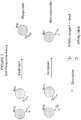
- FIGURE 1 graphically depicts a cell response assay constructed in accordance with the presently disclosed and claimed inventive concept(s).
- inventive concept(s) Before explaining at least one embodiment of the inventive concept(s) in detail by way of exemplary drawings, experimentation, results, and laboratory procedures, it is to be understood that the inventive concept(s) is not limited in its application to the details of construction and the arrangement of the components set forth in the following description or illustrated in the drawings, experimentation and/or results.
- inventive concept(s) is capable of other embodiments or of being practiced or carried out in various ways.
- the language used herein is intended to be given the broadest possible scope and meaning; and the embodiments are meant to be exemplary - not exhaustive.
- phraseology and terminology employed herein is for the purpose of description and should not be regarded as limiting.
- Enzymatic reactions and purification techniques are performed according to manufacturer's specifications or as commonly accomplished in the art or as described herein.
- the foregoing techniques and procedures are generally performed according to conventional methods well known in the art and as described in various general and more specific references that are cited and discussed throughout the present specification. See e.g., Sambrook et al. Molecular Cloning: A Laboratory Manual (2nd ed., Cold Spring Harbor Laboratory Press, Cold Spring Harbor, N.Y. (1989 ) and Coligan et al. Current Protocols in Immunology (Current Protocols, Wiley Interscience (1994 )).
- compositions and/or methods disclosed and claimed herein can be made and executed without undue experimentation in light of the present disclosure. While the compositions and methods of this presently disclosed and claimed inventive concept(s) have been described in terms of preferred embodiments, it will be apparent to those of skill in the art that variations may be applied to the compositions and/or methods and in the steps or in the sequence of steps of the method described herein without departing from the concept, spirit and scope of the presently disclosed and claimed inventive concept(s). All such similar substitutes and modifications apparent to those skilled in the art are deemed to be within the spirit, scope and concept of the inventive concept(s) as defined by the appended claims.
- At least one will be understood to include one as well as any quantity more than one, including but not limited to, 2, 3, 4, 5, 10, 15, 20, 30, 40, 50, 100, etc.
- the term “at least one” may extend up to 100 or 1000 or more, depending on the term to which it is attached; in addition, the quantities of 100/1000 are not to be considered limiting, as higher limits may also produce satisfactory results.
- the words “comprising” (and any form of comprising, such as “comprise” and “comprises”), “having” (and any form of having, such as “have” and “has”), "including” (and any form of including, such as “includes” and “include”) or “containing” (and any form of containing, such as “contains” and “contain”) are inclusive or open-ended and do not exclude additional, unrecited elements or method steps.
- probe as used herein will be understood to refer to any type of affinity reagent that binds to a specific biomarker as described herein.
- probes include, but are not limited to, antibodies (or binding fragments or derivatives thereof), receptors, organic molecules, inorganic molecules, ligands, nucleic acids (including but not limited to, DNA, RNA, microRNA, mRNA, siRNA, etc.), peptides, polypeptides, proteins, epitopes, antigens, ligands, receptors, complexes, lipids, glycoproteins, glycolipids, glycosaminoglycans, carbohydrates, polycarbohydrates, glycoconjugates, and any combination or derivative thereof.
- biomarker as used herein will be understood to refer to any target site on the surface of or inside of a cell that a probe can have affinity therefor and thus can bind to said moiety.
- the “biomarker” may be, for example but not by way of limitation, a nucleic acid, peptide, polypeptide, protein, epitope, antigen, ligand, receptor, complex (i.e., an MHC-peptide complex), lipid, glycoprotein, glycolipid, glycosaminoglycan, carbohydrate, polycarbohydrate, glycoconjugate, and any combination or derivative thereof.
- polypeptide refers to a polymer of amino acid residues.
- polypeptide as used herein is a generic term to refer to native protein, protein fragments, or analogs of a polypeptide sequence. Hence, native protein, protein fragments, and analogs are species of the polypeptide genus.
- polynucleotide and “nucleic acid” are used interchangeably. They refer to a polymeric form of nucleotides of any length, either deoxyribonucleotides or ribonucleotides, or analogs thereof.
- polynucleotides coding or non-coding regions of a gene or gene fragment, loci (locus) defined from linkage analysis, exons, introns, messenger RNA (mRNA), transfer RNA, ribosomal RNA, ribozymes, cDNA, recombinant polynucleotides, branched polynucleotides, plasmids, vectors, isolated DNA of any sequence, isolated RNA of any sequence, nucleic acid probes, and primers.
- a polynucleotide may comprise modified nucleotides, such as methylated nucleotides and nucleotide analogs.
- modifications to the nucleotide structure may be imparted before or after assembly of the polymer.
- the sequence of nucleotides may be interrupted by non-nucleotide components.
- a polynucleotide may be further modified, such as by conjugation with a labeling component.
- isolated nucleic acid and “isolated polynucleotide” are used interchangeably; a nucleic acid or polynucleotide is considered “isolated” if it: (1) is not associated with all or a portion of a polynucleotide in which the "isolated polynucleotide” is found in nature, (2) is linked to a polynucleotide to which it is not linked in nature, or (3) does not occur in nature as part of a larger sequence.
- antibody is used in the broadest sense, and specifically covers monoclonal antibodies (including full length monoclonal antibodies), polyclonal antibodies, multispecific antibodies (e.g., bispecific antibodies), and antibody fragments so long as they exhibit the desired biological activity.
- Antibody or “antibody peptide(s)” refer to a full length immunoglobulin molecule (i.e., an intact antibody), or a binding fragment thereof that competes with the intact antibody for specific antigen binding. Binding fragments may be produced by recombinant DNA techniques, or by enzymatic or chemical cleavage of intact antibodies.
- Binding fragments include Fab, Fab', F(ab') 2 , Fv, scFv, disulfide linked Fv, Fd, diabodies, single-chain antibodies, single domain antibodies (such as but not limited to, NANOBODIES®) and other antibody fragments that retain at least a portion of the variable region of an intact antibody. See, e.g., Hudson et al. (Nature Med., 9:129-134 (2003 )).
- antigen binding fragment or "antigen-binding portion" of an antibody, as used herein, refers to one or more fragments of an antibody that retain the ability to bind to an antigen.
- the antigen-binding function of an antibody can be performed by fragments of an intact antibody.
- binding fragments encompassed within the term "antigen-binding fragment” of an antibody include but are not limited to, Fab, Fab', F(ab')2, Fv, scFv, disulfide linked Fv, Fd, diabodies, single-chain antibodies, single domain antibodies (such as but not limited to, NANOBODIES®), isolated CDRH3, and other antibody fragments that retain at least a portion of the variable region of an intact antibody. These antibody fragments are obtained using conventional recombinant and/or enzymatic techniques and are screened for antigen binding in the same manner as intact antibodies.
- epitope includes any protein determinant capable of specific binding to an immunoglobulin or T-cell receptor.
- an epitope is a region of an antigen that is specifically bound by an antibody.
- Epitopic determinants usually include chemically active surface groupings of molecules such as amino acids, sugar side chains, phosphoryl, or sulfonyl groups.
- an epitope may have specific three dimensional structural characteristics (e.g., a "conformational epitope"), as well as specific charge characteristics.
- nanoparticle refers to a particle having dimensions of from about 1 to about 5000 nanometers, and having any size, shape or morphology.
- the nanoparticles utilized in accordance with the present invention may be naturally occurring, commercially available nanoparticles, or the nanoparticles may be synthesized for use in accordance with the present invention, as described herein below and as known in the art.
- Particular examples of nanoparticles that may be utilized in accordance with the present invention include, but are not limited to, poly(lactic-co-glycolic) acid (PLGA) nanoparticles, poly lactic acid (PLA) nanoparticles, Chitosen nanoparticles, liposomes, and derivatives or combinations thereof.
- label refers to incorporation of a detectable marker, e.g., by attachment of a fluorescent, enzymatic or colorimetric label or incorporation of a radiolabeled amino acid.
- Various methods of labeling polypeptides and glycoproteins are known in the art and may be used.
- labels for polypeptides include, but are not limited to, the following: radioisotopes or radionuclides (e.g., 3H, 14C, 15N, 35S, 90Y, 99Tc, 111In, 1251, 1311), fluorescent labels (e.g., FITC, rhodamine, lanthanide phosphors), enzymatic labels (e.g., horseradish peroxidase, ⁇ -galactosidase, luciferase, alkaline phosphatase), chemiluminescent, biotinyl groups, predetermined polypeptide epitopes recognized by a secondary reporter (e.g., leucine zipper pair sequences, binding sites for secondary antibodies, metal binding domains, epitope tags).
- labels are attached by spacer arms of various lengths to reduce potential steric hindrance.
- label means "label”, “detectable marker” and “detection moiety” are used interchangeably herein.
- a fluorophore may be employed in the methods of the present invention and detected via any of numerous colorimetric and fluorescence detection methods. Depending on the application and purpose, such methods include, but are not limited to, absorbance spectroscopy, fluorescence spectroscopy, fluorescence activated cytometry (FACS), fluorescence microscopy, fluorescence resonance energy transfer (FRET), and the like.
- fluorophores may be employed in accordance with the present invention. Examples of suitable fluorophores are described herein below. Examples of suitable fluorophores are described herein below. Other examples are given in US Patent Nos. 7,465,747 and 7,955,859, issued to Matsumoto et al. on December 16, 2008 and June 7, 2011, respectively; and US Publication No. US2007/0026407, published February 1, 2007 (the entire contents of which are expressly incorporated herein by reference in their entirety).
- substantially increase and “substantial decrease”, as well as grammatical equivalents thereof, will be understood herein to refer to at least a 12% increase or decrease, such as at least a 30% increase or decrease, at least a 50% increase or decrease, at least a 75% increase or decrease, or at least a 90% increase or decrease.
- cancer and “cancerous” refer to or describe the physiological condition in mammals that is typically characterized by unregulated cell growth.
- cancer include but are not limited to, carcinoma, lymphoma, blastoma, and sarcoma. More particular examples of such cancers include squamous cell cancer, small-cell lung cancer, non-small cell lung cancer, gastrointestinal cancer, pancreatic cancer, glioblastoma, cervical cancer, ovarian cancer, liver cancer, bladder cancer, hepatoma, breast cancer, colon cancer, colorectal cancer, endometrial carcinoma, salivary gland carcinoma, kidney cancer, renal cancer, prostate cancer, vulval cancer, thyroid cancer, hepatic carcinoma and various types of head and neck cancer.
- Metastasis as used herein will be understood to refer to the spread of cancer from a primary tumor to other parts of the body. Metastasis is a sequential, multistep process in which tumor cells detach from a primary tumor, migrate through the basement membrane and extracellular matrix, and invade the lymphatic and/or blood systems. This is followed by the establishment of secondary tumors at distant sites.
- “Mammal” for purposes of treatment refers to any animal classified as a mammal, including human, domestic and farm animals, nonhuman primates, and zoo, sports, or pet animals, such as dogs, horses, cats, cows, etc.
- a patient includes human and veterinary subjects.
- a patient is a mammal. In certain other embodiments, the patient is a human.
- biological sample as used herein will be understood to refer to a sample of biological fluid.
- biological samples include, but are not limited to, blood, plasma, serum, sputum, cerebrospinal fluid (CSF), tears, mucus, urine, tissue, other types of specimens, and the like.
- CSF cerebrospinal fluid
- providing a biological sample refers to obtaining a biological sample for use in methods described and claimed herein. Most often, this will be done by removing a sample of cells from an animal, but can also be accomplished by using previously isolated cells (e.g., isolated by another person, at another time and/or for another purpose).
- the step of "providing a biological sample” may further include various isolation and/or purification steps known in the art for providing a specific component of a biological sample for use in the methods described and claimed herein.
- the presently disclosed and claimed inventive concept(s) generally relates to multiplexed direct affinity conjugate assays of cells using probes able to determine a cellular response to treatments; this type of assay is herein termed a "cell response assay”.
- the presently disclosed and claimed inventive concept(s) generally relates to the use of a combination of biomarkers for cancer cell/tissue type, metastatic potential and chemo resistance in a multiplexed affinity assay for cancer cells and tissue.
- Fluorescent labels either conjugated to affinity molecules/reagents or capable of directly binding to the biomarker are used and are referred to herein as labeled probes.
- This combination of labeled probes for biomarkers allows determination of the cellular response to treatments by simultaneously measuring cell type, chemo resistance nature, and metastatic potential and/or cell viability, and allows simple multiplexed signals with high sensitivity.
- the biomarker affinity markers selected in this order solves the problem of "picking" the right antigen for cancer antigen type and also provides accurate information on the response to therapy. More particularly, the presently disclosed and claimed inventive concept(s) relates to methods of using said affinity molecules and molecular probes as compositions, dosage forms, and kits.
- the presently disclosed and claimed inventive concept(s) combines cell biomarker(s) for tissue type with biomarker(s) for metastatic potential and biomarker(s) for cell viability in an affinity assay for cancer cells and tissue.
- the biomarker for cancer cell type is represented as biomarker A and is always present in the disease state.
- the biomarker for metastatic potential is represented as biomarker B and is present in the disease state with metastatic invasiveness potential.
- the biomarker for chemo resistance is represented as biomarker C and is present in unresponsive cells.
- Fluorescent labels either conjugated to the affinity reagents or capable of directly binding the biomarker are used to simultaneously detect biomarkers A, B and C; simultaneous detection is obtained by using fluorescent labels that are detectable at different excitations and/or emission wavelengths.
- biomarker B i.e., metastatic potential
- biomarker C i.e. chemo resistance
- biomarkers B and C must be linked such that the presence of one facilitates the absence of the other.
- This combination of biomarkers allows the determination of the cell response to treatments; by simultaneously measuring cell type, chemo resistance nature, and metastatic potential (and possibly also cell viability), the "cell based response assay" can accurately monitor cancer treatment and/or monitor disease progression/relapse.
- one embodiment thereof is directed to a multiplexed assay for cancer.
- Said assay involves simultaneously measuring: (1) at least one cancer cell type biomarker (also referred to herein as Biomarker A) in a biological sample of a cancer patient utilizing a first labeled probe that binds to said cancer cell type biomarker; (2) at least one chemo resistance biomarker (also referred to herein as Biomarker C) in the biological sample utilizing a second labeled probe that binds to said chemo resistance biomarker; and (3) at least one metastatic potential biomarker (also referred to herein as Biomarker B) in the biological sample utilizing a third labeled probe that binds to said metastatic potential biomarker.
- the at least three labeled probes are measured at different excitations and emission wavelengths.
- the assays/methods described herein may be utilized for a specific cancer, or the assays/methods may be general assays for all types of cancers.
- the cancer cell type biomarker utilized in accordance with the presently disclosed and claimed inventive concept(s) may be a non-specific cancer cell biomarker, or may be specific for a certain type of cancer.
- cancers that may be detected/monitored by the currently disclosed and claimed inventive concept(s) include, but are not limited to, lung, bronchus, colon, rectum, pancreas, prostate, breast, liver, bile duct, bladder, ovary, brain, central nervous system (CNS), kidney, pelvis, uterine corpus, oral cavity or pharynx or melanoma cancers.
- lung bronchus, colon, rectum, pancreas, prostate, breast, liver, bile duct, bladder, ovary, brain, central nervous system (CNS), kidney, pelvis, uterine corpus, oral cavity or pharynx or melanoma cancers.
- CNS central nervous system
- kidney pelvis
- uterine corpus oral cavity or pharynx or melanoma cancers.
- Non-limiting examples of non-specific cancer cell type biomarkers include, but are not limited to, cytokeratins (CK), EpCAM, N-cadherin, E-cadherin and vimentin.
- the carcinoma cells may be indicated by cytokeratin (CK) as a biomarker A
- the first labeled probe may comprise multiple labeled antibodies for separate cytokeratins (such as but not limited to, cytokeratins 8/18/19.
- Cytokeratins (CK) are proteins of keratin-containing intermediate filaments found in the intracytoplasmic cytoskeleton of epithelial tissue and refer to a family of fibrous structural proteins. There are two fundamental types of cytokeratins: the acidic type I cytokeratins and the basic or neutral type II cytokeratins.
- Cytokeratins are usually found in pairs comprising a type I cytokeratin and a type II cytokeratin.
- Basic or neutral cytokeratins include (but are not limited to) CK1, CK2, CK3, CK4, CK5, CK6, CK7, CK8 and CK9.
- Acidic cytokeratins include (but are not limited to) CK10, CK12, CK 13, CK14, CK16, CK17, CK18, CK19 and CK20.
- EpCAM Epithelial cell adhesion molecule
- TACSTD1 tumor-associated calcium signal transducer 1
- CD326 cluster of differentiation 326
- cancer cells undergo an Epithelial to Mesenchymal transition (EMT) that may be indicated by an increase in cancer cell biomarkers such as but not limited to, Vimentin and Galectin-3, which signify cells with a loss of anchorage.
- EMT Epithelial to Mesenchymal transition
- MET Mesenchymal to Epithelial transition
- the EMT and MET stages for cancer cells signify a change in sub type of cells. It is important to detect as many cancer cells as possible and to additionally know their sub types to measure the entire tumorigenicity.
- cancer cell type biomarker also includes markers that identify specific genetic mutations that cause oncoproteins or oncogenes to be regulated (whether tumor promoting or tumor suppressing). These types of cancer cell type biomarkers may be utilized alone or in combination with any of the other cancer cell type biomarkers described herein above.
- cancer cell type biomarkers examples include, but are not limited to, HER2/neu, VEGF-165, KRAS, EGFr, WAF, BAX-1, PDGF, Rb, Jagged 1, Notch, VEGF, VEGHR, k-Ras, CAIX, MIB1, MDM, PR, ER, SEL5, SEM1, PI3K, Akt2, twist 1, EML-4, ALK, Braf, DRAFF, c-met, and combinations thereof.
- oncoproteins and oncogenes are used to direct targeted therapies based on their presence in/on cells.
- HER2/neu is used to prescribe Herceptin therapy; however, HER2/neu, as with other oncoprotein/oncogene markers, is often only expressed on a fraction of cancer cells, and not all patients have the specific genetic mutation leading to the biomarker.
- HER2/neu-positive expression is present in only 10-20% of breast cancer patients, and even in the HER2/neu-positive patients, the marker is only present in approximately 30% of their circulating tumor cells. Therefore these markers are often measured in combination with another cancer cell type biomarker.
- the carcinoma cells may be indicated by a tissue specific marker.
- Prostate-Specific Antigen is a protein that signifies the presence of cells of the prostate gland.
- specific cancer cell type biomarkers include, but are not limited to: prostate specific antigen (PSA) or prostate specific membrane antigen (PSMA) can be used to detect prostate cancer cells; MUC1, CA 15-3 and CA 27-29 can be used to detect breast cancer cells; Carcinoembryonic Antigen (CEA), CA19-9 or Galactosyl Transferase II can be used to detect colon cancer cells; MSLN (mesothelin) can be used to detect pancreatic cancer cells; CA 125 or Follicle-Stimulating Hormone (FSH) receptor can be used to detect ovarian cancer cells; Alpha-Fetoprotein can be used to detect liver cancer cells; Melan-A (MLANA), Tyrosinase (TYR), CSPG4, or MITF can be used to detect melanoma cancer cells
- the third labeled probe may be any probe capable of detecting said metastatic potential biomarker; for example but not by way of limitation, the third labeled probe may be one or more labeled antibodies against any of the markers of tumor cell invasiveness listed below. In some instances, the metastatic potential biomarker may be measured as a ratio of one of the below-listed biomarkers to the cancer cell type biomarker.
- biomarkers of metastatic potential include a marker(s) of tumor cell invasiveness where the marker measures cancer cell invasion into the extracellular membrane through proteolytic events, such as urokinase plasminogen activator (uPA), plasminogen activator inhibitor (PAI-1), CD95, serine proteases such as plasmin, ADAM, and others; serine protease inhibitors such as Bikunin; matrix metalloproteinases such as MMP9; matrix metalloproteinase inhibitors such a TIMP-1; and combinations thereof.
- uPA urokinase plasminogen activator
- PAI-1 plasminogen activator inhibitor
- CD95 serine proteases
- serine proteases such as plasmin, ADAM, and others
- serine protease inhibitors such as Bikunin
- matrix metalloproteinases such as MMP9
- matrix metalloproteinase inhibitors such a TIMP-1
- combinations thereof such as urokina
- the chemo resistance biomarker is a biomarker that detects the prevention of cell death (apoptosis), for example but not by way of limitation, a detection of the presence of a cancer stem cell biomarker, such as but not limited to, PL2L piwi like, ADLH, ⁇ -integrin, ⁇ 6 integrin, c-kit, c-met, LIF-R, CXCR4, ESA, CD 20, CD44, CD133, CK5, TRAF2 and ABC transporters.
- a cancer stem cell biomarker such as but not limited to, PL2L piwi like, ADLH, ⁇ -integrin, ⁇ 6 integrin, c-kit, c-met, LIF-R, CXCR4, ESA, CD 20, CD44, CD133, CK5, TRAF2 and ABC transporters.
- cancer cells that contain CD44 but lack CD24 are indicative of a cancer stem cell phenotype.
- cancer cells that lack CD45 and CD31 but contain CD34 are indicative of a cancer stem cell.
- cancer cells that contain CD44 and CD24 as well as ESA are indicative of a cancer stem cell.
- the presence of both CD24 and ESA are indicative of a cancer stem cell. Somatic stem cells in solid tumor carcinomas can become cancer stem cells. Other cancer cells resistant to CD95 induced-apoptosis are chemo resistant.
- Cancer stem cells are chemo resistant in nature, and contain a capacity for self renewal (asymmetric divisions) as well as being capable of differentiation into a hierarchy of progeny cells to form a tumor.
- Cancer stem cell self renewal is activated by the stem cell signaling pathways (Wnt, Sonic Hedge hog and Notch) and at the epigenetic level by Polycomb gener (BMI-1 and EZH2). This self renewal occurs at the expense of apopotosis signaling pathways (Caspase 3, 5, 8, p53). Measuring cancer stem cell biomarkers and combinations thereof as an indication of chemo resistance provides a measure of (a) drug resistance, (b) the inability to activate apoptosis, and/or (c) the inability to shutdown self renewal.
- the probes described and claimed herein may be labeled by any methods known in the art or otherwise contemplated herein.
- the labels of the first, second and third (and any additional fourth, fifth, sixth, etc.) labeled probes may be selected from a comprehensive catalogue of fluorescent (luminescent) dyes.
- a comprehensive catalogue exists online at http://www.fluorophores.org (the entire contents of which are expressly incorporated herein by reference).
- This catalogue includes commonly used labels such as fluorescein-5-isothiocyanate (FITC), phycoerythrin, sulforhodamine 101 (Texas Red), 2-[4-(aminoiminomethyl)phenyl]-1H-Indole-6-carboximidamide (DAPI), 3H-Indolium (Cy5), 1H-benz[e]indolium (Cy 5.5), 3H-Indolium (Cy 7), ALEXA FLUOR®488, ALEXA FLUOR®555, ALEXA FLUOR®647, and combinations and derivatives thereof that have been synthesized in order to provide better reagents. Additionally, rare earth metals and rare earth element-containing nanoparticles can be used as labels.
- FITC fluorescein-5-isothiocyanate
- DAPI 2-[4-(aminoiminomethyl)phenyl]-1H-Indole-6-carboximidamide
- Cy5 3H-
- biological samples utilized in the methods of the presently disclosed and claimed inventive concept(s) may be utilized in the form they are obtained (i.e., tissue sample), or they may be exposed to one or more isolating steps (i.e., isolation of cancer cells therefrom).
- the assay may further include measuring at least one additional biomarker in the biological sample utilizing a fourth labeled probe that binds to said additional biomarker, measuring a second additional biomarker in the biological sample utilizing a fifth labeled probe that binds to said additional biomarker, measuring a third additional biomarker in the biological sample utilizing a sixth labeled probe that binds to said additional biomarker, etc.
- a maximum of six to eight labeled probes are utilized; otherwise, it is difficult to detect all of the labeled probes at different, non-overlapping excitations and emission wavelengths.
- biomarkers may include biomarkers that detect other cellular properties, specifically the presence of cell nuclei or of intact cell membranes, for the cells that are positive for the cancer cell type biomarker.
- the presence of cell nuclei may be detected utilizing a DNA binding probe such as but not limited to, DAPI.
- the detection of intact cell membranes may involve the detection of anionic phospholipids (such as but not limited to, phosphatidylserine) on the surface of cells, and the second labeled probe may be bis(zinc2+dipicolylamine) and/or PSVueTM.
- the one or more additional biomarker(s)/labeled probe(s) may also be utilized to reduce any false positive results obtained with the cancer cell type biomarker; for example but not by way of limitation, white blood cells may be excluded from the assay by excluding cells positive for one or more markers of cluster of differentiation (also referred to as "cluster of designation"; often abbreviated as CD).
- markers of cluster of differentiation also referred to as "cluster of designation”; often abbreviated as CD.
- markers such as CD45, CTLA-4, CD4, CD68 and/or CD8 present on white blood cells can be used to indicate that a cell is not a cancer cell.
- CD45 antigen also known as PTPRC, Protein tyrosine phosphatase receptor type C, and originally called leukocyte common antigen (all terms used herein interchangeably) is useful in detecting all white blood cells. Additionally, CD45 can be used to differentiate the different types of white blood cells when combined with other CD markers.
- granulocytes are indicated by CD45+, CD15+; monocytes are indicated by CD45+, CD14+; T lymphocytes are indicated by CD45+, CD3+; T helper cells are indicated by CD45+,CD3+, CD4+; cytotoxic T cells are indicated by CD45+,CD3+, CD8+; B lymphocytes are indicated by CD45+, CD19+ or CD45+, CD20+; thrombocytes are indicated by CD45+, CD61+; and natural killer cells are indicated by CD16+, CD56+, CD3-.
- CD4 and CD8 are, in general, used as markers for helper and cytotoxic T cells, respectively. These molecules are defined in combination with CD3+, as some other leukocytes also express these CD molecules (some macrophages express low levels of CD4; dendritic cells express high levels of CD8).
- the presently disclosed and claimed inventive concept(s) is also directed to a kit that includes the first, second and third labeled probes (that bind to a cancer cell type biomarker, a chemo resistance biomarker, and a metastatic potential biomarker, respectively) as described in detail herein above. Additional labeled probes for detecting other biomarkers as further described herein above may also be included in the kit. All of the labeled probes present in the kit are measured at different excitations and emission wavelengths.
- the kit may also include means for isolating cancer cells from a biological sample.
- Said means are well known in the art (see for example, Lianidou and Markou, 2011; the entire contents of which are hereby expressly incorporated herein by reference); therefore, no additional discussion thereof is deemed necessary.
- the presently disclosed and claimed inventive concept(s) is also directed to methods of (1) monitoring cancer treatment in a cancer patient undergoing said treatment, and (2) monitoring progression/relapse of the disease in the cancer patient.
- a first biological sample is obtained from the patient prior to exposure to a cancer treatment
- a second biological sample is obtained from the patient following exposure to the cancer treatment.
- the first biological sample is obtained from the patient at a first time point
- the second biological sample is obtained from the patient at a subsequent time point.
- the levels of a cancer cell type biomarker (as described herein above), a chemo resistance biomarker (as described herein above), and a metastatic potential biomarker (as described herein above) are then measured utilizing first, second and third labeled probes, respectively (as described in detail herein above), in the first and second biological samples.
- the cells to which the first labeled probe is bound are identified in the first and second biological samples, and then the levels of (i) the chemo resistance biomarker and (ii) the metastatic potential marker in the cells to which the first labeled probe is bound are compared in the first and second biological samples.
- the cancer treatment is effective if the level of the chemo resistance biomarker is decreased and the level of the metastatic potential biomarker is increased in the second biological sample when compared to the first biological sample; alternatively, it is then determined in the method of (2) that the cancer has progressed/relapsed if the level of the chemo resistance biomarker is increased and the level of the metastatic potential biomarker has decreased in the second biological sample when compared to the first biological sample.
- biological samples utilized in the methods of the presently disclosed and claimed inventive concept(s) may be utilized in the form they are obtained (i.e., tissue sample), or they may be exposed to one or more isolating steps (i.e., isolation of cancer cells therefrom).
- the method may further include measuring at least one additional biomarker in the first and second biological samples utilizing a fourth labeled probe that binds to said additional biomarker, measuring a second additional biomarker in the first and second biological samples utilizing a fifth labeled probe that binds to said additional biomarker, measuring a third additional biomarker in the first and second biological samples utilizing a sixth labeled probe that binds to said additional biomarker, etc.
- the methods may also include the step of comparing the levels of the additional biomarker(s) in the cells to which the first labeled probe is bound in the first and second biological samples.
- biomarkers may include biomarkers that detect other cellular properties, specifically the presence of cell nuclei or of intact cell membranes, for the cells that are positive for the cancer cell type biomarker.
- the presence of cell nuclei may be detected utilizing a DNA binding probe such as but not limited to, DAPI.
- the detection of intact cell membranes may involve the detection of anionic phospholipids (such as but not limited to, phosphatidylserine) on the surface of cells, and the second labeled probe may be bis(zinc2+dipicolylamine) and/or PSVueTM.
- the one or more additional biomarker(s)/labeled probe(s) may also be utilized to reduce any false positive results obtained with the cancer cell type biomarker; for example but not by way of limitation, white blood cells may be excluded from the methods by excluding cells positive for one or more markers of cluster of differentiation (as described in detail herein above).
- the following procedure shows the multiplexing of Biomarkers A (cancer cell type), B (metastatic potential) and C (chemo resistance) using a unique combination of affinity reagents with fluorescent labels and an affinity label to provide a multiplexed assay for breast cancer.
- This allows the simultaneous measurement of cancer cell type, chemo resistance nature, and metastatic potential, as described herein above, in combination with a marker indicating the presence of cell nucleus (utilizing a nucleic acid binding probe).
- the data demonstrated that four simultaneous signals can be measured with this combination but not with the traditional combinations of the prior art.
- the presently disclosed and claimed inventive concept(s) has the potential to measure up to eight signals utilizing various fluorophores taught herein.
- lysis buffer 155mM NH 4 Cl, 20mM KHCO 3 , 0.1 mM Na 2 EDTA, pH 7.4
- the tubes were rolled tubes to assure that the cells were evenly distributed and that lysis was complete, and then the sample was spun down for 15 minutes at 12,000 rpm.
- the plasma was decanted as waste supernatant, and 4.0 mL of TES buffer (50 mM N-Tris [Hydroxymethyl]-2-aminoethane-sulfonic acid: TES, pH 7.4, 2150mM NaCl, 2 mM MgSO 4 and 1 mM KCO 3 ) and 1 % albumin was added, and the tube was rocked to mix the cells.
- the tube was spun down again for 15 minutes at 12,000 rpm, and the supernatant was decanted as waste.
- a final 2.0 mL of TES wash buffer was added with rocking to mix cells.
- the cells were then reacted with the reagents by taking 0.5 mL of washed blood to the centrifuge tube, and 40 ⁇ L of antibodies for HER2/neu labeled with Cy5 and 40 ⁇ L of antibodies for urokinase like plasminogen activator (uPA) or plasminogen activator inhibitor (PAI-1) labeled with Texas Red (at working stock of 0.001 to 0.1 mg/mL) and PL2L piwi like labeled with FITC (at working stock of 0.001 to 0.1 mg/mL) were added.
- uPA plasminogen activator
- PAI-1 plasminogen activator inhibitor
- DAPI Diamidino-2-phenylindole dihydrochloride
- the sample was centrifuged down for 5 minutes @ 7500 rpm, and the supernatant was decanted off as waste ( ⁇ 450 ⁇ L).
- 500 ⁇ L of TES buffer was then added, and the sample was vortexed and centrifuged down at 5 minutes @ 7500 rpm. Again, the supernatant was decanted off by tapping as waste ( ⁇ 450 ⁇ L).
- a final 500 ⁇ L of TES wash buffer was added for final imaging, and 5 ⁇ L of the solution was placed on a slide with a long cover slip of glass. Phase contrast and fluorescence microscopy was conducted with the Leica DM5000 (Leica Microsystems, Buffalo Grove, IL).
- a sample of cells was stained with the PSS-550 probe (ex 553, em 615nm) and measured with an excitation band pass filter at 540-580 nm and an emission band pass filter at 610-680 nm to determine cell membrane flopping as a measure of cell death in addition to cell counts .
- the Texas Red label (ex 592, em 614 nm) was measured with an excitation band pass filter at 540-580 nm and an emission band pass filter at 610-680 nm.
- the FITC label (ex 488, em 525nm) was measured with an excitation band pass filter at 460 - 500 nm and an emission band pass filter at 512 -543 nm.
- the DAPI probe (ex 355, em 460 nm) was measured with an excitation band pass filter at 340-380 nm and an emission band pass filter at 450-490 nm.
- the Cy5 label (ex 646, em 676 nm) was measured with an excitation band pass filter at 590-650 nm and an emission band pass filter at 665-735 nm.
- the ideal multiplexing was demonstrated by combination of the rare earth nanoparticles with FITC, Texas Red, Cy5 and DAPI.
- the europium nanoparticle (ex 355, em 617 nm) was measured at an excitation filter band pass at 340-380 nm and emission long pass filter of > 590 nm.
- Simultaneously measuring biomarkers for cancer cell type, metastatic potential and chemoresistance, along with a biomarker indicating the presence of cell nucleus was possible, along with the removal of false positives (i.e., white blood cells) by detecting the presence of the biomarker CD45. Current data allows for five signals.
- the assays/methods described and claimed herein have the potential for detection and measurement of six signals/biomarkers, if the rare earth nanoparticles can be multiplexed into three different emission signals.
- the europium label greatly increased signal over FITC and Texas Red by 2-3 orders of magnitude.
- Biomarker A tissue type marker for carcinoma breast cancer
- Biomarker B for metastatic potential utilized uPA and PAI antigen; these are markers of tumor invasiveness and are present in aggressive cancer cell differentiation and growth and increase with metastatic invasiveness potential of the cells.
- Biomarker C for chemo resistance utilized PL2L piwi like antigen (see for example, Gao et al., 2008).
- Example 1 The procedure shown in Example 1 was used to measure cancer cells before and after treatment with and without camptothecin. Camptothecin is known to activate apoptosis in cancer cells and kill cancer cells like a chemotherapy agent (See Gupta, 1997). Cells were tested by the cell response assay and by a traditional prior art assay.
- the novel "cell response assay” utilized HER2/neu as Biomarker A for cell type, the tumor invasiveness markers uPA & PAI-1 as Biomarker B for metastatic potential, and the cancer stem cell marker PL2L piwi like as Biomarker C for chemo resistance nature.
- the traditional assay used EpCAM as a marker for epithelial tissue type and cytokeratin (CK) as a marker of cells of cancer origin. Additionally, CD45, a marker for white blood cells, was used to reduce false results, with CD45 positive results being excluded from the analysis. Both the traditional and cell response assays used DAPI to determine the presence of cell nuclei.
- the camptothecin concentration could be adjusted to be sufficient to kill >80%, 60% and 40% of the cancer cells in cultures.
- Cellular analysis showed cancer cell count to decrease as the cell death % increased and phosphatidylserine flopping increased.
- Cellular analysis showed biomarker B, namely uPA and PA1, to increase (or was detected in a greater number of cancer cells) when the cell death % was highest.
- biomarker C namely PL2L piwi like to decrease (or was detected in a lesser number of cancer cells) when the cell death % was highest. All three biomarkers co-exist in the disease state and were fundamental to progression or relapse of the disease.
- the CD45 biomarker does not co-exist with EpCAM and CK. Both EpCAM and CK were present in all cells independent of the % of cells that were killed.
- the following procedure shows the multiplexing of Biomarkers A (cancer cell type), B (metastatic potential) and C (chemo resistance) using a unique combination of affinity reagents with fluorescent labels and an affinity label to provide a multiplexed assay for prostate cancer.
- This allows the simultaneous measurement of cancer cell type, chemo resistance nature, and metastatic potential, as described herein above, in combination with a marker indicating the presence of cell nucleus (utilizing a nucleic acid binding probe).
- the data demonstrated that four simultaneous signals can be measured with this combination but not with the traditional combinations of the prior art.
- the presently disclosed and claimed inventive concept(s) has the potential to measure up to eight signals utilizing various fluorophores taught herein.
- lysis buffer 155mM NH 4 Cl, 20mM KHCO 3 , 0.1 mM Na 2 EDTA, pH 7.4
- the tubes were rolled tubes to assure that the cells were evenly distributed and that lysis was complete, and then the sample was spun down for 15 minutes at 12,000 rpm.
- the plasma was decanted as waste supernatant, and 4.0 mL of TES buffer (50 mM N-Tris [Hydroxymethyl]-2-aminoethane-sulfonic acid: TES, pH 7.4, 2150mM NaCl, 2 mM MgSO 4 and 1 mM KCO 3 ) and 1 % albumin was added, and the tube was rocked to mix the cells.
- the tube was spun down again for 15 minutes at 12,000 rpm, and the supernatant was decanted as waste.
- a final 2.0 mL of TES wash buffer was added with rocking to mix cells.
- the cells were then reacted with the reagents by taking 0.5 mL of washed blood to the centrifuge tube, and 40 ⁇ L of antibodies for PSA labeled with Cy5 and 40 ⁇ L of antibodies for urokinase like plasminogen activator (uPA) or plasminogen activator inhibitor (PAI-1) labeled with Texas Red (at working stock of 0.001 to 0.1 mg/mL) and PL2L piwi like labeled with FITC (at working stock of 0.001 to 0.1 mg/mL) were added.
- uPA urokinase like plasminogen activator
- PAI-1 plasminogen activator inhibitor
- DAPI Diamidino-2-phenylindole dihydrochloride
- the sample was centrifuged down for 5 minutes @ 7500 rpm, and the supernatant was decanted off as waste ( ⁇ 450 ⁇ L).
- 500 ⁇ L of TES buffer was then added, and the sample was vortexed and centrifuged down at 5 minutes @ 7500 rpm. Again, the supernatant was decanted off by tapping as waste ( ⁇ 450 ⁇ L).
- a final 500 ⁇ L of TES wash buffer was added for final imaging, and 5 ⁇ L of the solution was placed on a slide with a long cover slip of glass. Phase contrast and fluorescence microscopy was conducted with the Leica DM5000 (Leica Microsystems, Buffalo Grove, IL).
- the Texas Red label (ex 592, em 614 nm) was measured with a excitation band pass filter at 540-580 nm and emission band pass filter at 610-680 nm.
- the FITC label (ex 488, em 525nm) was measured with an excitation band pass filter at 460 - 500 nm and an emission band pass filter at 512 -543 nm.
- the DAPI probe (ex 355, em 460 nm) was measured with an excitation band pass filter at 340-380 nm and an emission band pass filter at 450-490 nm.
- the Cy5 label (ex 646, em 676 nm) was measured with an excitation band pass filter at 590-650 nm and an emission band pass filter at 665-735 nm.
- the ideal multiplexing was demonstrated by combination of the rare earth nanoparticles with FITC, Texas Red, Cy5 and DAPI.
- the europium nanoparticle (ex 355, em 617 nm) was measured at an excitation filter band pass at 340-380 nm and emission long pass filter of > 590 nm.
- Simultaneously measuring biomarkers for cancer cell type, metastatic potential and chemoresistance, along with a biomarker indicating the presence of cell nucleus was possible, along with the removal of false positives (i.e., white blood cells) by detecting the presence of the biomarker CD45. Current data allows for five signals.
- the assays/methods described and claimed herein have the potential for detection and measurement of six signals/biomarkers, if the rare earth nanoparticles can be multiplexed into three different emission signals.
- the europium label greatly increased signal over FITC and Texas Red by 2-3 orders of magnitude.
- Protease like plasmin, is expressed during metastasis of malignant cells as part of the tissue regeneration and fibrinolysis process, indirectly promoting cell proliferation.
- Cancer cells use cell-bound plasmin to activate the plasminogen signaling for urokinase.
- Inhibitors of plasmin such as Bikunin and inhibitors of plasminogen activation such as PAI-1 prevent cell-bound plasmin activation and suppress tumor invasiveness.
- a novel cell response assay for prostate cancer was developed by using PSA as a tissue type marker for prostate carcinoma (Biomarker A) that is always present in the disease state.
- the markers used in this example for metastatic potential were urokinase plasminogen activator (uPA) and plasminogen activator inhibitor (PAI-1), which is a measure of tissue invasiveness and is present in an aggressive cancer cell differentiation and growth with increased metastatic invasiveness potential.
- Chemo resistance nature Biomarker C was determined using PL2L piwi like antigen to determine the inhibitory response to tissue invasiveness.
- Example 3 The procedure shown in Example 3 was used to measure prostate cancer cells before and after treatment with and without camptothecin. Camptothecin is known to activate apoptosis in cancer cells and kill cancer cells like a chemotherapy agent (See Gupta, 1997).
- Cells were tested by the cell response assay and by a traditional prior art assay.
- the novel "cell response assay” utilized PSA as Biomarker A for cell type, the tumor invasiveness markers uPA & PAI-1 as Biomarker B for metastatic potential, and the cancer stem cell marker PL2L piwi like as Biomarker C for chemo resistance nature.
- the traditional assay used EpCAM as a marker for epithelial tissue type and cytokeratin (CK) as a marker of cells of cancer origin. Additionally, CD45, a marker for white blood cells, was used to reduce false results, with CD45 positive results being excluded from the analysis.
- Both the traditional and cell response assays used DAPI to determine the presence of cell nuclei.
- the camptothecin concentration could be adjusted to be sufficient to kill >80%, 60% and 40% of the cancer cells in cultures.
- Cellular analysis showed cancer cell count to decrease as the cell death % increased and phosphatidylserine flopping increased.
- Cellular analysis showed biomarker B, namely uPA and PA1, to increase (or was detected in a greater number of cancer cells) when the cell death % was highest.
- biomarker C namely PL2L piwi like to decrease (or was detected in a lesser number of cancer cells) when the cell death % was highest. All three biomarkers co-exist in the disease state and were fundamental to progression or relapse of the disease.
- the CD45 biomarker does not co-exist with EpCAM and CK. Both EpCAM and CK were present in all cells independent of the % of cells that were killed.
- the following procedure demonstrates the multiplexing of Biomarkers A, B and C as described herein previously to provide a general assay for all types of cancers.
- This procedure is performed as described herein above in Examples 1/3, except as follows: (a) the cancer suspension added to the procedure is selected from one of the following cell lines - SKBR, MCF or MDA (breast cancer), UC3 or T24 (bladder cancer), PC3-9 (prostate cancer), and A549 (lung cancer); and (b) the non-specific cancer cell biomarker cytokeratin is utilized for Biomarker A.
- antibodies to the cytokeratins 8/18/19 are utilized as the first labeled probe that binds to Biomarker A.
- antibodies to uPA or PAI-1 labeled with Texas Ted are utilized as the second labeled probe that binds to Biomarker B, and antibodies to PL2L piwi like labeled with FITC is utilized as the third labeled probe that binds to Biomarker C.
- the nucleic acid binding probe DAPI is also utilized as described in Examples 1 and 3.
- This multiplexed assay utilizing a non-specific cancer cell biomarker allows the assays/methods described herein to be adapted for use with all types of cancers.
Landscapes
- Health & Medical Sciences (AREA)
- Life Sciences & Earth Sciences (AREA)
- Chemical & Material Sciences (AREA)
- Immunology (AREA)
- Engineering & Computer Science (AREA)
- Molecular Biology (AREA)
- Urology & Nephrology (AREA)
- Biomedical Technology (AREA)
- Hematology (AREA)
- General Health & Medical Sciences (AREA)
- Analytical Chemistry (AREA)
- Pathology (AREA)
- Medicinal Chemistry (AREA)
- Biotechnology (AREA)
- Microbiology (AREA)
- Physics & Mathematics (AREA)
- Biochemistry (AREA)
- Organic Chemistry (AREA)
- Proteomics, Peptides & Aminoacids (AREA)
- Cell Biology (AREA)
- Food Science & Technology (AREA)
- General Physics & Mathematics (AREA)
- Genetics & Genomics (AREA)
- Zoology (AREA)
- Wood Science & Technology (AREA)
- Oncology (AREA)
- Public Health (AREA)
- Pharmacology & Pharmacy (AREA)
- Epidemiology (AREA)
- Animal Behavior & Ethology (AREA)
- Veterinary Medicine (AREA)
- Biophysics (AREA)
- Hospice & Palliative Care (AREA)
- Bioinformatics & Cheminformatics (AREA)
- General Engineering & Computer Science (AREA)
- Measuring Or Testing Involving Enzymes Or Micro-Organisms (AREA)
- Investigating Or Analysing Biological Materials (AREA)
Claims (14)
- Zellreaktionstest für Krebs, umfassend die Schritte:- Isolieren von Krebszellen aus einer biologischen Probe eines Krebspatienten;- Messen mindestens eines Biomarkers für einen Krebszelltyp auf der Oberfläche von oder innerhalb der Zelle unter Verwendung einer ersten markierten Sonde, die an den Biomarker für einen Krebszelltyp bindet,
wobei der mindestens eine Biomarker für einen Krebszelltyp mindestens einen aus:(a) einer Kombination aus mindestens einem Typ-I-Zytokeratin und mindestens einem Typ-II-Zytokeratin;(b) einer Kombination aus Vimentin und Galectin-3; und(c) einer Kombination aus N-Cadherin und E-Cadherin; oder(d) einem Onkoprotein/Onkogen, ausgewählt aus der Gruppe bestehend aus HER2/neu, VEGF-165, KRAS, EGFr, WAF, BAX-1, PDGF, Rb, Jagged 1, Notch, VEGF, VEGHR, k-Ras, CAIX, MIB1, MDM, PR, ER, SEL5, SEM1, PI3K, Akt2, twist 1, EML-4, ALK, Braf, DRAFF, c-met und Kombinationen davon umfasst;- Messen mindestens eines Biomarkers für Chemoresistenz auf der Oberfläche von oder innerhalb der Zelle unter Verwendung einer zweiten markierten Sonde, die an den Biomarker für Chemoresistenz bindet, wobei der mindestens eine Biomarker für Chemoresistenz ausgewählt ist aus der Gruppe bestehend aus PL2L-piwi-like, ADLH, b-Integrin, a6-Integrin, c-kit, c-met, LIF-R, CXCR4, ESA, CD 20, CD44, CD133, CK5, TRAF2, ABC-Transportern und Kombinationen davon;- Messen des mindestens einen Biomarkers für Metastasierungspotential auf der Oberfläche von oder innerhalb der Zelle unter Verwendung einer dritten markierten Sonde, die an den Biomarker für Metastasierungspotential bindet, wobei der mindestens eine Biomarker für Metastasierungspotential ausgewählt ist aus der Gruppe bestehend auswobei die mindestens drei markierten Sonden bei verschiedenen Anregungs- und Emissionswellenlängen gemessen werden.- Urokinase-Plasminogen-Aktivator (uPA), Plasminogen-Aktivator-Inhibitor (PAI-1), CD95, einer Serinprotease, einem Serinproteaseinhibitor, einer Matrixmetalloproteinase, einem Matrixmetalloproteinaseinhibitor und Kombinationen davon; und - Test nach Anspruch 1, wobei der Test für einen Krebs ist, ausgewählt aus der Gruppe besehend aus Lungen-, Bronchial-, Darm-, Mastdarm-, Bauchspeicheldrüsen-, Prostata-, Brust-, Leber-, Gallengang-, Blasen-, Eiserstock-, Gehirn-, Zentralnervensystem(ZNS)-, Nieren, Becken-, Gebärmutter-, Mundhöhlen-, Rachenkrebs, Melanom und Kombinationen davon.
- Test nach Anspruch 1, wobei mindestens einer:(a) der Tests für Prostatakrebs ist, und der mindestens eine Biomarker für einen Krebszelltyp mindestens einen aus einem prostataspezifischen Antigen (PSA), prostataspezifischen Membranantigen (PSMA) und Kombinationen davon umfasst;(b) der Tests für Brustkrebs ist, und der mindestens eine Biomarker für einen Krebszelltyp mindestens einen aus MUC1, CA 15-3, CA 27-29 und Kombinationen davon umfasst;(c) der Test für Darmkrebs ist, und der mindestens eine Biomarker für einen Krebszelltyp mindestens einen aus karzinoembryonalem Antigen (CEA), CA19-9, Galactosyltransferase II und Kombinationen davon umfasst;(d) der Test für Bauchspeicheldrüsenkrebs ist, und der mindestens eine Biomarker für einen Krebszelltyp MSLN (Mesothelin) umfasst;(e) der Test für Eierstockkrebs ist, und der mindestens eine Biomarker für einen Krebszelltyp mindestens einen aus CA 125, follikelstimulierendes-Hormon(FSH)-Rezeptor und Kombinationen davon umfasst;(f) der Test für Leberkrebs ist, und der mindestens eine Biomarker für einen Krebszelltyp Alpha-Fetoprotein umfasst;(g) der Test für Melanom ist, und der mindestens eine Biomarker für einen Krebszelltyp mindestens einen aus Melan-A (MLANA), Tyrosinase (TYR), CSPG4, MITF und Kombinationen davon umfasst; und(h) der Test für Schilddrüsenkrebs ist, und der mindestens eine Biomarker für einen Krebszelltyp mindestens einen aus Parathyroid-related-Protein (PTHP), TSHR und Kombinationen davon umfasst.
- Test nach Anspruch 1, wobei der mindestens eine Biomarker für Chemoresistenz mindestens einen aus:(a) Gegenwart von CD44 und Abwesenheit von CD24;(b) Gegenwart von CD34 und Abwesenheit von CD45 und CD31;(c) Gegenwart von CD44, CD24 und ESA; und(d) Gegenwart von CD24 und ESA umfasst.
- Test nach Anspruch 1, wobei mindestens eine/einer:(a) der Serinproteasen ausgewählt ist aus der Gruppe bestehend aus Plasmin, ADAM und Kombinationen davon;(b) der Serinproteaseinhibitoren Bikunin umfasst;(c) der Matrixmetalloproteinasen MMP9 umfasst; und(d) der Matrixmetalloproteinaseinhibitoren TIMP-1 umfasst.
- Test nach Anspruch 1, wobei mindestens eine der ersten, zweiten und dritten markierten Sonden mindestens einen markierten Antikörper gegen einen Biomarker umfassen.
- Test nach Anspruch 1, wobei die Marker der ersten, zweiten und dritten markierten Sonden ausgewählt sind aus der Gruppe bestehend ausF luorescein-5-isothiocyanat (FITC), Phycoerythrin, Sulforhodamin 101 (Texasrot), 2-[4-(Aminoiminomethyl)phenyl]-1H-Indol-6-carboximidamid (DAPI), 3H-Indol (Cy5), 1H-Benz[e]indol (Cy 5.5), 3H-Indol (Cy7), ALEXA FLUOR®488, ALEXA FLUOR®555, ALEXA FLUOR®647, Seltenerdmetalle, Seltenerdelemententhaltende Nanopartikel und Kombinationen und Derivate davon.
- Test nach Anspruch 1, wobei die biologische Probe ferner als Gewebeprobe definiert ist.
- Test nach Anspruch 1, ferner umfassend das Messen mindestens eines zusätzlichen Biomarkers in der biologischen Probe unter Verwendung einer vierten markierten Sonde, die an den zusätzlichen Biomarker bindet.
- Test nach Anspruch 9, wobei der mindestens eine zusätzliche Biomarker mindestens einen Biomarker für weiße Blutzellen umfasst, ausgewählt aus der Gruppe bestehend aus- CD45, CTLA-4, CD4, CD68, CD8 und Kombinationen davon,und wobei der Test ferner den Schritt zum Ausschließen von Zellen umfasst, die für mindestens einen Biomarker für weiße Blutzellen positiv sind.
- Test nach Anspruch 9, wobei der mindestens eine zusätzliche Biomarker einen Biomarker umfasst, der die Gegenwart von Zellkernen anzeigt, und wobei die vierte markierte Sonde 4',6-Diamidino-2'-phenylindol-Dihydrochlorid (DAPI) umfasst.
- Test nach Anspruch 9, wobei der mindestens eine zusätzliche Biomarker Phosphatidylserin umfasst, und wobei die vierte markierte Sonde mindestens eine aus Bis (zink2+dipicolylamin) und PSVue™ umfasst.
- Verfahren zum Überwachen der Krebsbehandlung bei einem Krebspatienten, der sich der Behandlung unterzieht, wobei das Verfahren die folgenden Schritte umfasst:- Messen der Konzentrationen eines Biomarkers für einen Krebszelltyp in einer ersten biologischen Probe und in einer zweiten biologischen Probe unter Verwendung einer ersten markierten Sonde, die an den Biomarker für einen Krebszelltyp bindet, wobei die erste biologische Probe von dem Patienten erhalten wird, bevor er sich einer Krebsbehandlung unterzieht, wobei die zweite biologische Probe von dem Patienten erhalten wird, nachdem er sich der Krebsbehandlung unterzogen hat, wobei der mindestens eine Biomarker für einen Krebszelltyp mindestens einen aus:(a) einer Kombination aus mindestens einem Typ-I-Zytokeratin und mindestens einem Typ-II-Zytokeratin;(b) einer Kombination aus Vimentin und Galectin-3; und(c) einer Kombination aus N-Cadherin und E-Cadherin; oder(d) einem Onkoprotein/Onkogen, ausgewählt aus der Gruppe bestehend aus HER2/neu, VEGF-165, KRAS, EGFr, WAF, BAX-1, PDGF, Rb, Jagged 1, Notch, VEGF, VEGHR, k-Ras, CAIX, MIB1, MDM, PR, ER, SEL5, SEM1, PI3K, Akt2, twist 1, EML-4, ALK, Braf, DRAFF, c-met und Kombinationen davon umfasst;- Messen der Konzentrationen eines Biomarkers für Chemoresistenz in der ersten und der zweiten biologischen Probe unter Verwendung einer zweiten Sonde, die an den Biomarker für Chemoresistenz bindet, wobei der mindestens eine Biomarker für Chemoresistenz ausgewählt ist aus der Gruppe bestehend aus PL2L-piwi-like, ADLH, b-Integrin, a6-Integrin, c-kit, c-met, LIF-R, CXCR4, ESA, CD 20, CD44, CD133, CK5, TRAF2, ABC-Transportern und Kombinationen davon;- Messen der Konzentrationen eines Biomarkers für Metastasierungspotential in der ersten und der zweiten biologischen Probe unter Verwendung einer dritten Sonde, die an den Biomarker für Metastasierungspotential bindet, wobei der mindestens eine Biomarker für Metastasierungspotential ausgewählt ist aus der Gruppe bestehend ausund wobei die mindestens drei markierten Sonden bei verschiedenen Anregungs- und Emissionswellenlängen gemessen werden;- Urokinase-Plasminogen-Aktivator (uPA), Plasminogen-Aktivator-Inhibitor (PAI-1), CD95, einer Serinprotease, einem Serinproteaseinhibitor, einer Matrixmetalloproteinase, einem Matrixmetalloproteinaseinhibitor und Kombinationen davon,- Vergleichen der Konzentrationen des Biomarkers für Chemoresistenz in den Zellen, an den die zweite markierte Sonde in der ersten und der zweiten biologischen Probe gebunden ist;- Vergleichen der Konzentrationen des Biomarkers für Metastasierungspotential in den Zellen, an den die dritte markierte Sonde in der ersten und der zweiten biologischen Probe gebunden ist; und- Bestimmen, dass die Krebsbehandlung wirksam ist, wenn die Konzentration des Biomarkers für Chemoresistenz verringert ist und die Konzentration des Biomarkers für Metastasierungspotential in der zweiten biologischen Probe im Vergleich zur ersten biologischen Probe erhöht ist.
- Verfahren zum Überwachen des Forschreitens/der Wiedererkrankung bei einem Krebspatienten, wobei das Verfahren die folgenden Schritte umfasst:- Messen der Konzentrationen eines Biomarkers für einen Krebszelltyp in einer ersten biologischen Probe und in einer zweiten biologischen Probe unter Verwendung einer ersten markierten Sonde, die an den Biomarker für einen Krebszelltyp bindet, wobei die erste biologische Probe zu einem ersten Zeitpunkt von dem Patienten erhalten wird und die zweite biologische Probe zu einem nachfolgenden Zeitpunkt von dem Patienten erhalten wird, wobei der mindestens eine Biomarker für einen Krebszelltyp mindestens einen aus:(a) einer Kombination aus mindestens einem Typ-I-Zytokeratin und mindestens einem Typ-II-Zytokeratin;(b) einer Kombination aus Vimentin und Galectin-3; und(c) einer Kombination aus N-Cadherin und E-Cadherin; oder(d) einem Onkoprotein/Onkogen, ausgewählt aus der Gruppe bestehend aus HER2/neu, VEGF-165, KRAS, EGFr, WAF, BAX-1, PDGF, Rb, Jagged 1, Notch, VEGF, VEGHR, k-Ras, CAIX, MIB1, MDM, PR, ER, SEL5, SEM1, PI3K, Akt2, twist 1, EML-4, ALK, Braf, DRAFF, c-met und Kombinationen davon umfasst;- Messen der Konzentrationen eines Biomarkers für Chemoresistenz in der ersten und der zweiten biologischen Probe unter Verwendung einer zweiten Sonde, die an den Biomarker für Chemoresistenz bindet, wobei der mindestens eine Biomarker für Chemoresistenz ausgewählt ist aus der Gruppe bestehend aus PL2L-piwi-like, ADLH, b-Integrin, a6-Integrin, c-kit, c-met, LIF-R, CXCR4, ESA, CD 20, CD44, CD133, CK5, TRAF2, ABC-Transportern und Kombinationen davon;- Messen der Konzentrationen eines Biomarkers für Metastasierungspotential in der ersten und der zweiten biologischen Probe unter Verwendung einer dritten Sonde, die an den Biomarker für Metastasierungspotential bindet, wobei der mindestens eine Biomarker für Metastasierungspotential ausgewählt ist aus der Gruppe bestehend ausund wobei die mindestens drei markierten Sonden bei verschiedenen Anregungs- und Emissionswellenlängen gemessen werden;- Urokinase-Plasminogen-Aktivator (uPA), Plasminogen-Aktivator-Inhibitor (PAI-1), CD95, einer Serinprotease, einem Serinproteaseinhibitor, einer Matrixmetalloproteinase, einem Matrixmetalloproteinaseinhibitor und Kombinationen davon,- Vergleichen der Konzentrationen des Biomarkers für Chemoresistenz in den Zellen, an den die erste markierte Sonde in der ersten und der zweiten biologischen Probe gebunden ist;- Vergleichen der Konzentrationen des Biomarkers für Metastasierungspotential in den Zellen, an den die dritte markierte Sonde in der ersten und der zweiten biologischen Probe gebunden ist; und- Bestimmen, dass der Krebs fortgeschritten/zurückgekehrt ist, wenn die Konzentration des Biomarkers für Chemoresistenz erhöht ist und die Konzentration des Biomarkers für Metastasierungspotential in der zweiten biologischen Probe im Vergleich zur ersten biologischen Probe verringert ist.
Applications Claiming Priority (2)
| Application Number | Priority Date | Filing Date | Title |
|---|---|---|---|
| US201161538302P | 2011-09-23 | 2011-09-23 | |
| PCT/US2012/056668 WO2013044099A1 (en) | 2011-09-23 | 2012-09-21 | Cell response assay for cancer and methods of producing and using same |
Publications (3)
| Publication Number | Publication Date |
|---|---|
| EP2748337A1 EP2748337A1 (de) | 2014-07-02 |
| EP2748337A4 EP2748337A4 (de) | 2015-04-29 |
| EP2748337B1 true EP2748337B1 (de) | 2019-08-07 |
Family
ID=47914916
Family Applications (1)
| Application Number | Title | Priority Date | Filing Date |
|---|---|---|---|
| EP12833913.2A Not-in-force EP2748337B1 (de) | 2011-09-23 | 2012-09-21 | Zellreaktionstest für krebs sowie verfahren zu seiner herstellung und anwendung |
Country Status (4)
| Country | Link |
|---|---|
| US (3) | US20140235495A1 (de) |
| EP (1) | EP2748337B1 (de) |
| ES (1) | ES2754304T3 (de) |
| WO (1) | WO2013044099A1 (de) |
Families Citing this family (16)
| Publication number | Priority date | Publication date | Assignee | Title |
|---|---|---|---|---|
| DE102013010528B3 (de) * | 2013-06-25 | 2014-02-20 | FLACOD GmbH | Verfahren zur Erstellung einer Datenbank für die Vorabschätzung der Wirksamkeit von Wirkstoffen in der Tumortherapie |
| GB201322800D0 (en) | 2013-12-20 | 2014-02-05 | Univ Dublin | Prostate cancer biomarkers |
| CN113552350B (zh) * | 2015-04-21 | 2024-10-11 | 基因泰克公司 | 用于前列腺癌分析的组合物和方法 |
| WO2016201217A2 (en) * | 2015-06-10 | 2016-12-15 | Dae Joon Kim | GFR α 1 AS A BIOMARKER FOR CISPLATIN-INDUCED CHEMORESISTANCE AND METASTASIS |
| CN104931324B (zh) * | 2015-06-11 | 2018-05-25 | 宁波美晶医疗技术有限公司 | 一种循环肿瘤细胞的染色试剂盒 |
| CN105785017A (zh) * | 2016-05-11 | 2016-07-20 | 江苏省原子医学研究所 | 基于psa磁微粒的双标记时间分辨荧光免疫分析试剂盒 |
| CN107814846A (zh) * | 2016-11-14 | 2018-03-20 | 杭州华得森生物技术有限公司 | 针对EpCAM和Cytokeratin5的特异性抗体及其制备方法 |
| CN107741497A (zh) * | 2017-05-12 | 2018-02-27 | 中山大学肿瘤防治中心 | 用于评估肝癌患者治疗效果的免疫荧光检测试剂盒 |
| AU2018308088B2 (en) | 2017-07-25 | 2025-05-29 | Truebinding, Inc. | Treating cancer by blocking the interaction of TIM-3 and its ligand |
| CN107907684A (zh) * | 2017-09-29 | 2018-04-13 | 何皓 | 一种基于多重荧光寿命分辨检测技术的前列腺癌早期诊断系统 |
| AU2020214796A1 (en) | 2019-01-30 | 2021-07-29 | Truebinding, Inc. | Anti-Gal3 antibodies and uses thereof |
| IL316576A (en) | 2019-02-12 | 2024-12-01 | Pact Pharma Inc | Preparations and methods for identifying antigen-specific T cells |
| CN111073889B (zh) * | 2019-12-20 | 2023-07-28 | 中国人民解放军第四军医大学 | 人cspg5基因的用途及相关产品 |
| WO2021242776A2 (en) | 2020-05-26 | 2021-12-02 | Truebinding, Inc. | Methods of treating inflammatory diseases by blocking galectin-3 |
| CN113049344B (zh) * | 2021-04-20 | 2022-02-01 | 深圳天烁生物科技有限公司 | 细胞染色的质控品制备方法 |
| WO2025256538A1 (zh) * | 2024-06-12 | 2025-12-18 | 北京沙砾生物医药股份有限公司 | 一种细胞产品中检测肿瘤残留的方法 |
Family Cites Families (15)
| Publication number | Priority date | Publication date | Assignee | Title |
|---|---|---|---|---|
| US3772099A (en) | 1971-05-17 | 1973-11-13 | Westinghouse Electric Corp | Phosphor combination and method, particularly adapted for use with explosives, for providing a distinctive information label |
| US5196306A (en) | 1989-03-29 | 1993-03-23 | E. I. Du Pont De Nemours And Company | Method for the detection or quantitation of an analyte using an analyte dependent enzyme activation system |
| EP0984281B1 (de) | 1991-05-22 | 2007-01-03 | Dade Behring Marburg GmbH | Homogenes Testsystem zur Bestimmung eines Analytes |
| DE69501161T2 (de) | 1994-04-11 | 1998-03-19 | Nexins Research B.V., Hoeven | Verfahren zum nachweis und/oder zur bestimmung und/oder zur isolierung von apoptotischen zellen in oder aus einer probe |
| DE19756773A1 (de) | 1997-12-19 | 1999-06-24 | Dade Behring Marburg Gmbh | Neues Verfahren und Diagnosemittel zur Hämostase-Diagnostik |
| US20030129665A1 (en) * | 2001-08-30 | 2003-07-10 | Selvan Gowri Pyapali | Methods for qualitative and quantitative analysis of cells and related optical bio-disc systems |
| ATE423217T1 (de) | 2002-03-01 | 2009-03-15 | Siemens Healthcare Diagnostics | Assays zur überwachung von krebspatienten auf grundlage der spiegel von analytenkomponenten des plasminogen-aktivator-systems in proben von körperflüssigkeiten |
| US7179619B2 (en) | 2003-07-10 | 2007-02-20 | Irm Llc | Methods and compositions for inhibiting herpesviral replication |
| US20070026407A1 (en) | 2003-09-08 | 2007-02-01 | Kazuko Matsumoto | Novel fine fluorescent particle |
| WO2006060561A2 (en) * | 2004-12-01 | 2006-06-08 | Emory University | Methods and applications of molecular beacon imaging for cancer cell detection |
| EP2021491A4 (de) * | 2006-05-10 | 2010-02-24 | Univ Texas | Nachweis von tumorbiomarkern bei mundkrebs |
| WO2008048570A2 (en) | 2006-10-16 | 2008-04-24 | The Ohio State University Research Foundation | Piwil2-related biomarkers and cell lines useful therewith |
| JP5174040B2 (ja) | 2007-02-05 | 2013-04-03 | シーメンス・ヘルスケア・ダイアグノスティックス・インコーポレーテッド | 画像の構成要素と背景とを区別するためにコンピュータで実行される方法および画像の構成要素と背景とを区別するためのシステム |
| PL2271935T3 (pl) * | 2008-04-11 | 2013-05-31 | Univ Leipzig | Sposób i zestaw do ex vivo oceny reakcji nowotworu na warunki badania |
| WO2011109440A1 (en) * | 2010-03-01 | 2011-09-09 | Caris Life Sciences Luxembourg Holdings | Biomarkers for theranostics |
-
2012
- 2012-09-21 US US14/346,638 patent/US20140235495A1/en not_active Abandoned
- 2012-09-21 WO PCT/US2012/056668 patent/WO2013044099A1/en not_active Ceased
- 2012-09-21 EP EP12833913.2A patent/EP2748337B1/de not_active Not-in-force
- 2012-09-21 ES ES12833913T patent/ES2754304T3/es active Active
-
2017
- 2017-02-11 US US15/430,445 patent/US20170153241A1/en not_active Abandoned
-
2019
- 2019-03-13 US US16/352,684 patent/US10976319B2/en not_active Expired - Fee Related
Non-Patent Citations (2)
| Title |
|---|
| VERONIQUE NEUMEISTER ET AL: "In Situ Identification of Putative Cancer Stem Cells by Multiplexing ALDH1, CD44, and Cytokeratin Identifies Breast Cancer Patients with Poor Prognosis", AMERICAN JOURNAL OF PATHOLOGY., vol. 176, no. 5, 1 May 2010 (2010-05-01), US, pages 2131 - 2138, XP055388459, ISSN: 0002-9440, DOI: 10.2353/ajpath.2010.090712 * |
| YANJIE HU ET AL: "Detection of circulating tumor cells in breast cancer patients utilizing multiparameter flow cytometry and assessment of the prognosis of patients in different CTCs levels", CYTOMETRY. PART A, vol. 77A, no. 3, 1 March 2010 (2010-03-01), US, pages 213 - 219, XP055222260, ISSN: 1552-4922, DOI: 10.1002/cyto.a.20838 * |
Also Published As
| Publication number | Publication date |
|---|---|
| EP2748337A1 (de) | 2014-07-02 |
| US20190339278A1 (en) | 2019-11-07 |
| EP2748337A4 (de) | 2015-04-29 |
| US20170153241A1 (en) | 2017-06-01 |
| US20140235495A1 (en) | 2014-08-21 |
| WO2013044099A1 (en) | 2013-03-28 |
| US10976319B2 (en) | 2021-04-13 |
| ES2754304T3 (es) | 2020-04-16 |
Similar Documents
| Publication | Publication Date | Title |
|---|---|---|
| US10976319B2 (en) | Cell response assay for cancer and methods of producing and using same | |
| Kros et al. | Circulating glioma biomarkers | |
| US9671407B2 (en) | Devices and methods of cell capture and analysis | |
| US20150233927A1 (en) | Identifying taxane sensitivity in prostate cancer patients | |
| US20130203061A1 (en) | Methods and systems for isolating, storing, and analyzing vesicles | |
| US20190310172A1 (en) | Profiling extracellular vesicles | |
| JP2011517341A (ja) | 小細胞肺癌バイオマーカーパネル | |
| CN108064343B (zh) | 用于前列腺癌分析的组合物和方法 | |
| JP2002529704A (ja) | 単一細胞の複数マーカー特徴付け | |
| RU2641968C2 (ru) | Композиции и способы анализа рака предстательной железы | |
| JP2023179455A (ja) | Tas1r3タンパク質を発現する腫瘍に関する治療目的、診断目的及び/又は予後診断目的のマーカーとしてのtas1r3タンパク質の使用 | |
| WO2015184144A1 (en) | Rare molecule signal amplification | |
| Li et al. | Identification of the target protein of the metastatic colorectal cancer-specific aptamer W3 as a biomarker by aptamer-based target cells sorting and functional characterization | |
| Kim et al. | Clinical significance of circulating tumor cells after chemotherapy in unresectable pancreatic ductal adenocarcinoma | |
| US9746472B2 (en) | Methods and kits for the detection of cancer infiltration of the central nervous system | |
| US20150017638A1 (en) | Methods for assessing risk for cancer using biomarkers | |
| WO2018107930A1 (zh) | 外周血循环肿瘤细胞检测体系及其应用 | |
| WO2024203968A1 (ja) | がんを検出する方法、がんの予後を予測する方法、バイオマーカー及びその使用方法、並びに、がんの検出用又はがんの予後の予測用キット | |
| WO2013003898A1 (en) | Method for detection of cancer in a patient | |
| WO2021198303A1 (en) | New method of prostate cancer diagnosis | |
| WO2025197993A1 (ja) | テトラスパニン10陽性細胞外小胞の濃縮方法、及びがんを検出する又はがんの予後を予測する方法 | |
| Lee | Extracellular Vesicles as Biomarkers for Colorectal Cancer Metastasis | |
| Esteva et al. | Serum Tumor Markers and Circulating Tumor Cells | |
| Leis et al. | Circulating tumor stem cells as biomarkers for cancer progression | |
| HK1255484B (en) | Compositions and methods for prostate cancer analysis |
Legal Events
| Date | Code | Title | Description |
|---|---|---|---|
| PUAI | Public reference made under article 153(3) epc to a published international application that has entered the european phase |
Free format text: ORIGINAL CODE: 0009012 |
|
| 17P | Request for examination filed |
Effective date: 20140326 |
|
| AK | Designated contracting states |
Kind code of ref document: A1 Designated state(s): AL AT BE BG CH CY CZ DE DK EE ES FI FR GB GR HR HU IE IS IT LI LT LU LV MC MK MT NL NO PL PT RO RS SE SI SK SM TR |
|
| DAX | Request for extension of the european patent (deleted) | ||
| RA4 | Supplementary search report drawn up and despatched (corrected) |
Effective date: 20150330 |
|
| RIC1 | Information provided on ipc code assigned before grant |
Ipc: C12Q 1/68 20060101AFI20150324BHEP Ipc: G01N 33/574 20060101ALI20150324BHEP Ipc: G01N 33/53 20060101ALI20150324BHEP |
|
| 17Q | First examination report despatched |
Effective date: 20161019 |
|
| STAA | Information on the status of an ep patent application or granted ep patent |
Free format text: STATUS: EXAMINATION IS IN PROGRESS |
|
| GRAP | Despatch of communication of intention to grant a patent |
Free format text: ORIGINAL CODE: EPIDOSNIGR1 |
|
| STAA | Information on the status of an ep patent application or granted ep patent |
Free format text: STATUS: GRANT OF PATENT IS INTENDED |
|
| INTG | Intention to grant announced |
Effective date: 20190221 |
|
| GRAS | Grant fee paid |
Free format text: ORIGINAL CODE: EPIDOSNIGR3 |
|
| GRAA | (expected) grant |
Free format text: ORIGINAL CODE: 0009210 |
|
| STAA | Information on the status of an ep patent application or granted ep patent |
Free format text: STATUS: THE PATENT HAS BEEN GRANTED |
|
| AK | Designated contracting states |
Kind code of ref document: B1 Designated state(s): AL AT BE BG CH CY CZ DE DK EE ES FI FR GB GR HR HU IE IS IT LI LT LU LV MC MK MT NL NO PL PT RO RS SE SI SK SM TR |
|
| REG | Reference to a national code |
Ref country code: GB Ref legal event code: FG4D |
|
| REG | Reference to a national code |
Ref country code: CH Ref legal event code: EP Ref country code: AT Ref legal event code: REF Ref document number: 1163943 Country of ref document: AT Kind code of ref document: T Effective date: 20190815 |
|
| REG | Reference to a national code |
Ref country code: DE Ref legal event code: R096 Ref document number: 602012062781 Country of ref document: DE |
|
| REG | Reference to a national code |
Ref country code: IE Ref legal event code: FG4D |
|
| REG | Reference to a national code |
Ref country code: NL Ref legal event code: MP Effective date: 20190807 |
|
| REG | Reference to a national code |
Ref country code: LT Ref legal event code: MG4D |
|
| PG25 | Lapsed in a contracting state [announced via postgrant information from national office to epo] |
Ref country code: FI Free format text: LAPSE BECAUSE OF FAILURE TO SUBMIT A TRANSLATION OF THE DESCRIPTION OR TO PAY THE FEE WITHIN THE PRESCRIBED TIME-LIMIT Effective date: 20190807 Ref country code: PT Free format text: LAPSE BECAUSE OF FAILURE TO SUBMIT A TRANSLATION OF THE DESCRIPTION OR TO PAY THE FEE WITHIN THE PRESCRIBED TIME-LIMIT Effective date: 20191209 Ref country code: SE Free format text: LAPSE BECAUSE OF FAILURE TO SUBMIT A TRANSLATION OF THE DESCRIPTION OR TO PAY THE FEE WITHIN THE PRESCRIBED TIME-LIMIT Effective date: 20190807 Ref country code: HR Free format text: LAPSE BECAUSE OF FAILURE TO SUBMIT A TRANSLATION OF THE DESCRIPTION OR TO PAY THE FEE WITHIN THE PRESCRIBED TIME-LIMIT Effective date: 20190807 Ref country code: NL Free format text: LAPSE BECAUSE OF FAILURE TO SUBMIT A TRANSLATION OF THE DESCRIPTION OR TO PAY THE FEE WITHIN THE PRESCRIBED TIME-LIMIT Effective date: 20190807 Ref country code: BG Free format text: LAPSE BECAUSE OF FAILURE TO SUBMIT A TRANSLATION OF THE DESCRIPTION OR TO PAY THE FEE WITHIN THE PRESCRIBED TIME-LIMIT Effective date: 20191107 Ref country code: LT Free format text: LAPSE BECAUSE OF FAILURE TO SUBMIT A TRANSLATION OF THE DESCRIPTION OR TO PAY THE FEE WITHIN THE PRESCRIBED TIME-LIMIT Effective date: 20190807 Ref country code: NO Free format text: LAPSE BECAUSE OF FAILURE TO SUBMIT A TRANSLATION OF THE DESCRIPTION OR TO PAY THE FEE WITHIN THE PRESCRIBED TIME-LIMIT Effective date: 20191107 |
|
| REG | Reference to a national code |
Ref country code: AT Ref legal event code: MK05 Ref document number: 1163943 Country of ref document: AT Kind code of ref document: T Effective date: 20190807 |
|
| PG25 | Lapsed in a contracting state [announced via postgrant information from national office to epo] |
Ref country code: IS Free format text: LAPSE BECAUSE OF FAILURE TO SUBMIT A TRANSLATION OF THE DESCRIPTION OR TO PAY THE FEE WITHIN THE PRESCRIBED TIME-LIMIT Effective date: 20191207 Ref country code: LV Free format text: LAPSE BECAUSE OF FAILURE TO SUBMIT A TRANSLATION OF THE DESCRIPTION OR TO PAY THE FEE WITHIN THE PRESCRIBED TIME-LIMIT Effective date: 20190807 Ref country code: AL Free format text: LAPSE BECAUSE OF FAILURE TO SUBMIT A TRANSLATION OF THE DESCRIPTION OR TO PAY THE FEE WITHIN THE PRESCRIBED TIME-LIMIT Effective date: 20190807 Ref country code: RS Free format text: LAPSE BECAUSE OF FAILURE TO SUBMIT A TRANSLATION OF THE DESCRIPTION OR TO PAY THE FEE WITHIN THE PRESCRIBED TIME-LIMIT Effective date: 20190807 Ref country code: GR Free format text: LAPSE BECAUSE OF FAILURE TO SUBMIT A TRANSLATION OF THE DESCRIPTION OR TO PAY THE FEE WITHIN THE PRESCRIBED TIME-LIMIT Effective date: 20191108 |
|
| REG | Reference to a national code |
Ref country code: CH Ref legal event code: NV Representative=s name: SIEMENS SCHWEIZ AG, CH |
|
| PG25 | Lapsed in a contracting state [announced via postgrant information from national office to epo] |
Ref country code: TR Free format text: LAPSE BECAUSE OF FAILURE TO SUBMIT A TRANSLATION OF THE DESCRIPTION OR TO PAY THE FEE WITHIN THE PRESCRIBED TIME-LIMIT Effective date: 20190807 |
|
| REG | Reference to a national code |
Ref country code: ES Ref legal event code: FG2A Ref document number: 2754304 Country of ref document: ES Kind code of ref document: T3 Effective date: 20200416 |
|
| PG25 | Lapsed in a contracting state [announced via postgrant information from national office to epo] |
Ref country code: IT Free format text: LAPSE BECAUSE OF FAILURE TO SUBMIT A TRANSLATION OF THE DESCRIPTION OR TO PAY THE FEE WITHIN THE PRESCRIBED TIME-LIMIT Effective date: 20190807 Ref country code: RO Free format text: LAPSE BECAUSE OF FAILURE TO SUBMIT A TRANSLATION OF THE DESCRIPTION OR TO PAY THE FEE WITHIN THE PRESCRIBED TIME-LIMIT Effective date: 20190807 Ref country code: DK Free format text: LAPSE BECAUSE OF FAILURE TO SUBMIT A TRANSLATION OF THE DESCRIPTION OR TO PAY THE FEE WITHIN THE PRESCRIBED TIME-LIMIT Effective date: 20190807 Ref country code: AT Free format text: LAPSE BECAUSE OF FAILURE TO SUBMIT A TRANSLATION OF THE DESCRIPTION OR TO PAY THE FEE WITHIN THE PRESCRIBED TIME-LIMIT Effective date: 20190807 Ref country code: PL Free format text: LAPSE BECAUSE OF FAILURE TO SUBMIT A TRANSLATION OF THE DESCRIPTION OR TO PAY THE FEE WITHIN THE PRESCRIBED TIME-LIMIT Effective date: 20190807 Ref country code: EE Free format text: LAPSE BECAUSE OF FAILURE TO SUBMIT A TRANSLATION OF THE DESCRIPTION OR TO PAY THE FEE WITHIN THE PRESCRIBED TIME-LIMIT Effective date: 20190807 |
|
| PG25 | Lapsed in a contracting state [announced via postgrant information from national office to epo] |
Ref country code: CZ Free format text: LAPSE BECAUSE OF FAILURE TO SUBMIT A TRANSLATION OF THE DESCRIPTION OR TO PAY THE FEE WITHIN THE PRESCRIBED TIME-LIMIT Effective date: 20190807 Ref country code: SK Free format text: LAPSE BECAUSE OF FAILURE TO SUBMIT A TRANSLATION OF THE DESCRIPTION OR TO PAY THE FEE WITHIN THE PRESCRIBED TIME-LIMIT Effective date: 20190807 Ref country code: MC Free format text: LAPSE BECAUSE OF FAILURE TO SUBMIT A TRANSLATION OF THE DESCRIPTION OR TO PAY THE FEE WITHIN THE PRESCRIBED TIME-LIMIT Effective date: 20190807 Ref country code: SM Free format text: LAPSE BECAUSE OF FAILURE TO SUBMIT A TRANSLATION OF THE DESCRIPTION OR TO PAY THE FEE WITHIN THE PRESCRIBED TIME-LIMIT Effective date: 20190807 Ref country code: IS Free format text: LAPSE BECAUSE OF FAILURE TO SUBMIT A TRANSLATION OF THE DESCRIPTION OR TO PAY THE FEE WITHIN THE PRESCRIBED TIME-LIMIT Effective date: 20200224 |
|
| REG | Reference to a national code |
Ref country code: CH Ref legal event code: PL |
|
| REG | Reference to a national code |
Ref country code: DE Ref legal event code: R097 Ref document number: 602012062781 Country of ref document: DE |
|
| PLBE | No opposition filed within time limit |
Free format text: ORIGINAL CODE: 0009261 |
|
| STAA | Information on the status of an ep patent application or granted ep patent |
Free format text: STATUS: NO OPPOSITION FILED WITHIN TIME LIMIT |
|
| PG2D | Information on lapse in contracting state deleted |
Ref country code: IS |
|
| PG25 | Lapsed in a contracting state [announced via postgrant information from national office to epo] |
Ref country code: CH Free format text: LAPSE BECAUSE OF NON-PAYMENT OF DUE FEES Effective date: 20190930 Ref country code: LI Free format text: LAPSE BECAUSE OF NON-PAYMENT OF DUE FEES Effective date: 20190930 Ref country code: IE Free format text: LAPSE BECAUSE OF NON-PAYMENT OF DUE FEES Effective date: 20190921 Ref country code: LU Free format text: LAPSE BECAUSE OF NON-PAYMENT OF DUE FEES Effective date: 20190921 |
|
| 26N | No opposition filed |
Effective date: 20200603 |
|
| REG | Reference to a national code |
Ref country code: BE Ref legal event code: MM Effective date: 20190930 |
|
| PG25 | Lapsed in a contracting state [announced via postgrant information from national office to epo] |
Ref country code: BE Free format text: LAPSE BECAUSE OF NON-PAYMENT OF DUE FEES Effective date: 20190930 Ref country code: SI Free format text: LAPSE BECAUSE OF FAILURE TO SUBMIT A TRANSLATION OF THE DESCRIPTION OR TO PAY THE FEE WITHIN THE PRESCRIBED TIME-LIMIT Effective date: 20190807 |
|
| PG25 | Lapsed in a contracting state [announced via postgrant information from national office to epo] |
Ref country code: CY Free format text: LAPSE BECAUSE OF FAILURE TO SUBMIT A TRANSLATION OF THE DESCRIPTION OR TO PAY THE FEE WITHIN THE PRESCRIBED TIME-LIMIT Effective date: 20190807 |
|
| PG25 | Lapsed in a contracting state [announced via postgrant information from national office to epo] |
Ref country code: HU Free format text: LAPSE BECAUSE OF FAILURE TO SUBMIT A TRANSLATION OF THE DESCRIPTION OR TO PAY THE FEE WITHIN THE PRESCRIBED TIME-LIMIT; INVALID AB INITIO Effective date: 20120921 Ref country code: MT Free format text: LAPSE BECAUSE OF FAILURE TO SUBMIT A TRANSLATION OF THE DESCRIPTION OR TO PAY THE FEE WITHIN THE PRESCRIBED TIME-LIMIT Effective date: 20190807 |
|
| PG25 | Lapsed in a contracting state [announced via postgrant information from national office to epo] |
Ref country code: MK Free format text: LAPSE BECAUSE OF FAILURE TO SUBMIT A TRANSLATION OF THE DESCRIPTION OR TO PAY THE FEE WITHIN THE PRESCRIBED TIME-LIMIT Effective date: 20190807 |
|
| PGFP | Annual fee paid to national office [announced via postgrant information from national office to epo] |
Ref country code: FR Payment date: 20230918 Year of fee payment: 12 |
|
| PGFP | Annual fee paid to national office [announced via postgrant information from national office to epo] |
Ref country code: GB Payment date: 20231009 Year of fee payment: 12 |
|
| PGFP | Annual fee paid to national office [announced via postgrant information from national office to epo] |
Ref country code: ES Payment date: 20231218 Year of fee payment: 12 |
|
| PGFP | Annual fee paid to national office [announced via postgrant information from national office to epo] |
Ref country code: DE Payment date: 20231120 Year of fee payment: 12 |
|
| REG | Reference to a national code |
Ref country code: DE Ref legal event code: R119 Ref document number: 602012062781 Country of ref document: DE |
|
| GBPC | Gb: european patent ceased through non-payment of renewal fee |
Effective date: 20240921 |
|
| PG25 | Lapsed in a contracting state [announced via postgrant information from national office to epo] |
Ref country code: DE Free format text: LAPSE BECAUSE OF NON-PAYMENT OF DUE FEES Effective date: 20250401 |
|
| PG25 | Lapsed in a contracting state [announced via postgrant information from national office to epo] |
Ref country code: GB Free format text: LAPSE BECAUSE OF NON-PAYMENT OF DUE FEES Effective date: 20240921 |
|
| PG25 | Lapsed in a contracting state [announced via postgrant information from national office to epo] |
Ref country code: FR Free format text: LAPSE BECAUSE OF NON-PAYMENT OF DUE FEES Effective date: 20240930 |
|
| REG | Reference to a national code |
Ref country code: ES Ref legal event code: FD2A Effective date: 20251031 |
|
| PG25 | Lapsed in a contracting state [announced via postgrant information from national office to epo] |
Ref country code: ES Free format text: LAPSE BECAUSE OF NON-PAYMENT OF DUE FEES Effective date: 20240922 |