EP2748337B1 - Cell response assay for cancer and methods of producing and using same - Google Patents
Cell response assay for cancer and methods of producing and using same Download PDFInfo
- Publication number
- EP2748337B1 EP2748337B1 EP12833913.2A EP12833913A EP2748337B1 EP 2748337 B1 EP2748337 B1 EP 2748337B1 EP 12833913 A EP12833913 A EP 12833913A EP 2748337 B1 EP2748337 B1 EP 2748337B1
- Authority
- EP
- European Patent Office
- Prior art keywords
- biomarker
- cancer
- assay
- combinations
- cells
- Prior art date
- Legal status (The legal status is an assumption and is not a legal conclusion. Google has not performed a legal analysis and makes no representation as to the accuracy of the status listed.)
- Active
Links
- 206010028980 Neoplasm Diseases 0.000 title claims description 168
- 201000011510 cancer Diseases 0.000 title claims description 150
- 238000003556 assay Methods 0.000 title claims description 79
- 238000000034 method Methods 0.000 title claims description 75
- 230000036755 cellular response Effects 0.000 title claims description 21
- 239000000090 biomarker Substances 0.000 claims description 207
- 210000004027 cell Anatomy 0.000 claims description 193
- 239000000523 sample Substances 0.000 claims description 92
- 239000012472 biological sample Substances 0.000 claims description 54
- 230000001394 metastastic effect Effects 0.000 claims description 48
- 206010061289 metastatic neoplasm Diseases 0.000 claims description 48
- 238000002512 chemotherapy Methods 0.000 claims description 45
- 239000002105 nanoparticle Substances 0.000 claims description 28
- 210000001519 tissue Anatomy 0.000 claims description 28
- 238000011282 treatment Methods 0.000 claims description 28
- 102100037422 Receptor-type tyrosine-protein phosphatase C Human genes 0.000 claims description 24
- 101000738771 Homo sapiens Receptor-type tyrosine-protein phosphatase C Proteins 0.000 claims description 23
- 230000005284 excitation Effects 0.000 claims description 22
- -1 c-met Proteins 0.000 claims description 21
- 239000002797 plasminogen activator inhibitor Substances 0.000 claims description 21
- 102000010752 Plasminogen Inactivators Human genes 0.000 claims description 20
- 108010077971 Plasminogen Inactivators Proteins 0.000 claims description 20
- 108090000623 proteins and genes Proteins 0.000 claims description 19
- 102000004169 proteins and genes Human genes 0.000 claims description 17
- 101100408379 Drosophila melanogaster piwi gene Proteins 0.000 claims description 13
- 102000011782 Keratins Human genes 0.000 claims description 13
- 108010076876 Keratins Proteins 0.000 claims description 13
- 108010072866 Prostate-Specific Antigen Proteins 0.000 claims description 12
- 102100038358 Prostate-specific antigen Human genes 0.000 claims description 12
- 210000003855 cell nucleus Anatomy 0.000 claims description 12
- 210000000265 leukocyte Anatomy 0.000 claims description 12
- 101150029707 ERBB2 gene Proteins 0.000 claims description 11
- 206010060862 Prostate cancer Diseases 0.000 claims description 11
- 229910052761 rare earth metal Inorganic materials 0.000 claims description 11
- 206010006187 Breast cancer Diseases 0.000 claims description 10
- 102000000905 Cadherin Human genes 0.000 claims description 10
- 108050007957 Cadherin Proteins 0.000 claims description 10
- 208000000236 Prostatic Neoplasms Diseases 0.000 claims description 10
- MPLHNVLQVRSVEE-UHFFFAOYSA-N texas red Chemical compound [O-]S(=O)(=O)C1=CC(S(Cl)(=O)=O)=CC=C1C(C1=CC=2CCCN3CCCC(C=23)=C1O1)=C2C1=C(CCC1)C3=[N+]1CCCC3=C2 MPLHNVLQVRSVEE-UHFFFAOYSA-N 0.000 claims description 10
- TZCPCKNHXULUIY-RGULYWFUSA-N 1,2-distearoyl-sn-glycero-3-phosphoserine Chemical compound CCCCCCCCCCCCCCCCCC(=O)OC[C@H](COP(O)(=O)OC[C@H](N)C(O)=O)OC(=O)CCCCCCCCCCCCCCCCC TZCPCKNHXULUIY-RGULYWFUSA-N 0.000 claims description 9
- 208000026310 Breast neoplasm Diseases 0.000 claims description 9
- ZWZWYGMENQVNFU-UHFFFAOYSA-N Glycerophosphorylserin Natural products OC(=O)C(N)COP(O)(=O)OCC(O)CO ZWZWYGMENQVNFU-UHFFFAOYSA-N 0.000 claims description 9
- 102100032912 CD44 antigen Human genes 0.000 claims description 8
- 101000868273 Homo sapiens CD44 antigen Proteins 0.000 claims description 8
- 102000003990 Urokinase-type plasminogen activator Human genes 0.000 claims description 8
- 108090000435 Urokinase-type plasminogen activator Proteins 0.000 claims description 8
- 150000002910 rare earth metals Chemical class 0.000 claims description 8
- 101710164820 Flotillin-2 Proteins 0.000 claims description 7
- 238000012544 monitoring process Methods 0.000 claims description 7
- 102000005962 receptors Human genes 0.000 claims description 7
- 108020003175 receptors Proteins 0.000 claims description 7
- 239000012099 Alexa Fluor family Substances 0.000 claims description 6
- 101000884271 Homo sapiens Signal transducer CD24 Proteins 0.000 claims description 6
- 108700020796 Oncogene Proteins 0.000 claims description 6
- 102100038081 Signal transducer CD24 Human genes 0.000 claims description 6
- 108091008819 oncoproteins Proteins 0.000 claims description 6
- 229940012957 plasmin Drugs 0.000 claims description 6
- 101000716102 Homo sapiens T-cell surface glycoprotein CD4 Proteins 0.000 claims description 5
- 101000946843 Homo sapiens T-cell surface glycoprotein CD8 alpha chain Proteins 0.000 claims description 5
- 101000611023 Homo sapiens Tumor necrosis factor receptor superfamily member 6 Proteins 0.000 claims description 5
- 102000002274 Matrix Metalloproteinases Human genes 0.000 claims description 5
- 108010000684 Matrix Metalloproteinases Proteins 0.000 claims description 5
- 108050000637 N-cadherin Proteins 0.000 claims description 5
- 108010022999 Serine Proteases Proteins 0.000 claims description 5
- 102000012479 Serine Proteases Human genes 0.000 claims description 5
- 102100036011 T-cell surface glycoprotein CD4 Human genes 0.000 claims description 5
- 102100034922 T-cell surface glycoprotein CD8 alpha chain Human genes 0.000 claims description 5
- 102100040403 Tumor necrosis factor receptor superfamily member 6 Human genes 0.000 claims description 5
- 108010065472 Vimentin Proteins 0.000 claims description 5
- 102100035071 Vimentin Human genes 0.000 claims description 5
- 229940121386 matrix metalloproteinase inhibitor Drugs 0.000 claims description 5
- 239000003771 matrix metalloproteinase inhibitor Substances 0.000 claims description 5
- 201000001441 melanoma Diseases 0.000 claims description 5
- 239000003001 serine protease inhibitor Substances 0.000 claims description 5
- 210000005048 vimentin Anatomy 0.000 claims description 5
- 102000010400 1-phosphatidylinositol-3-kinase activity proteins Human genes 0.000 claims description 4
- RKJUIXBNRJVNHR-UHFFFAOYSA-N 3H-indole Chemical compound C1=CC=C2CC=NC2=C1 RKJUIXBNRJVNHR-UHFFFAOYSA-N 0.000 claims description 4
- FWBHETKCLVMNFS-UHFFFAOYSA-N 4',6-Diamino-2-phenylindol Chemical compound C1=CC(C(=N)N)=CC=C1C1=CC2=CC=C(C(N)=N)C=C2N1 FWBHETKCLVMNFS-UHFFFAOYSA-N 0.000 claims description 4
- 101150107888 AKT2 gene Proteins 0.000 claims description 4
- 108010006533 ATP-Binding Cassette Transporters Proteins 0.000 claims description 4
- 102000005416 ATP-Binding Cassette Transporters Human genes 0.000 claims description 4
- 102100031650 C-X-C chemokine receptor type 4 Human genes 0.000 claims description 4
- 108700012439 CA9 Proteins 0.000 claims description 4
- 101100421135 Caenorhabditis elegans sel-5 gene Proteins 0.000 claims description 4
- 102100024423 Carbonic anhydrase 9 Human genes 0.000 claims description 4
- 108010022366 Carcinoembryonic Antigen Proteins 0.000 claims description 4
- 102100025475 Carcinoembryonic antigen-related cell adhesion molecule 5 Human genes 0.000 claims description 4
- 206010009944 Colon cancer Diseases 0.000 claims description 4
- 102100022183 E3 ubiquitin-protein ligase MIB1 Human genes 0.000 claims description 4
- 108060006698 EGF receptor Proteins 0.000 claims description 4
- 102000012673 Follicle Stimulating Hormone Human genes 0.000 claims description 4
- 108010079345 Follicle Stimulating Hormone Proteins 0.000 claims description 4
- 108010001517 Galectin 3 Proteins 0.000 claims description 4
- 102100041003 Glutamate carboxypeptidase 2 Human genes 0.000 claims description 4
- 101000684297 Homo sapiens 26S proteasome complex subunit SEM1 Proteins 0.000 claims description 4
- 101000922348 Homo sapiens C-X-C chemokine receptor type 4 Proteins 0.000 claims description 4
- 101000973503 Homo sapiens E3 ubiquitin-protein ligase MIB1 Proteins 0.000 claims description 4
- 101000584612 Homo sapiens GTPase KRas Proteins 0.000 claims description 4
- 101000892862 Homo sapiens Glutamate carboxypeptidase 2 Proteins 0.000 claims description 4
- 101001133056 Homo sapiens Mucin-1 Proteins 0.000 claims description 4
- 101000873438 Homo sapiens Putative protein SEM1, isoform 2 Proteins 0.000 claims description 4
- 101000664418 Homo sapiens Secreted Ly-6/uPAR-related protein 1 Proteins 0.000 claims description 4
- 101000808011 Homo sapiens Vascular endothelial growth factor A Proteins 0.000 claims description 4
- 108700003486 Jagged-1 Proteins 0.000 claims description 4
- 102100021747 Leukemia inhibitory factor receptor Human genes 0.000 claims description 4
- 101710142062 Leukemia inhibitory factor receptor Proteins 0.000 claims description 4
- 108010010995 MART-1 Antigen Proteins 0.000 claims description 4
- 102100028389 Melanoma antigen recognized by T-cells 1 Human genes 0.000 claims description 4
- 102000003735 Mesothelin Human genes 0.000 claims description 4
- 108090000015 Mesothelin Proteins 0.000 claims description 4
- 102100034256 Mucin-1 Human genes 0.000 claims description 4
- 102000005650 Notch Receptors Human genes 0.000 claims description 4
- 108010070047 Notch Receptors Proteins 0.000 claims description 4
- 108091007960 PI3Ks Proteins 0.000 claims description 4
- 102100032702 Protein jagged-1 Human genes 0.000 claims description 4
- 108700037966 Protein jagged-1 Proteins 0.000 claims description 4
- 102000016971 Proto-Oncogene Proteins c-kit Human genes 0.000 claims description 4
- 108010014608 Proto-Oncogene Proteins c-kit Proteins 0.000 claims description 4
- 102100034920 Putative protein SEM1, isoform 2 Human genes 0.000 claims description 4
- 102100038583 Secreted Ly-6/uPAR-related protein 1 Human genes 0.000 claims description 4
- 108090000925 TNF receptor-associated factor 2 Proteins 0.000 claims description 4
- 102100034779 TRAF family member-associated NF-kappa-B activator Human genes 0.000 claims description 4
- 108060008724 Tyrosinase Proteins 0.000 claims description 4
- 102000005789 Vascular Endothelial Growth Factors Human genes 0.000 claims description 4
- 108010019530 Vascular Endothelial Growth Factors Proteins 0.000 claims description 4
- 210000000481 breast Anatomy 0.000 claims description 4
- 210000003169 central nervous system Anatomy 0.000 claims description 4
- 230000003247 decreasing effect Effects 0.000 claims description 4
- 229940028334 follicle stimulating hormone Drugs 0.000 claims description 4
- 102000058223 human VEGFA Human genes 0.000 claims description 4
- 210000002307 prostate Anatomy 0.000 claims description 4
- KXZQYLBVMZGIKC-UHFFFAOYSA-N 1-pyridin-2-yl-n-(pyridin-2-ylmethyl)methanamine Chemical compound C=1C=CC=NC=1CNCC1=CC=CC=N1 KXZQYLBVMZGIKC-UHFFFAOYSA-N 0.000 claims description 3
- 101800001691 Inter-alpha-trypsin inhibitor light chain Proteins 0.000 claims description 3
- 206010033128 Ovarian cancer Diseases 0.000 claims description 3
- 206010061535 Ovarian neoplasm Diseases 0.000 claims description 3
- 206010061902 Pancreatic neoplasm Diseases 0.000 claims description 3
- 108010004729 Phycoerythrin Proteins 0.000 claims description 3
- 102100032859 Protein AMBP Human genes 0.000 claims description 3
- 208000024770 Thyroid neoplasm Diseases 0.000 claims description 3
- PTFCDOFLOPIGGS-UHFFFAOYSA-N Zinc dication Chemical compound [Zn+2] PTFCDOFLOPIGGS-UHFFFAOYSA-N 0.000 claims description 3
- 210000001072 colon Anatomy 0.000 claims description 3
- 208000029742 colonic neoplasm Diseases 0.000 claims description 3
- 201000007270 liver cancer Diseases 0.000 claims description 3
- 208000014018 liver neoplasm Diseases 0.000 claims description 3
- 208000015486 malignant pancreatic neoplasm Diseases 0.000 claims description 3
- 201000002528 pancreatic cancer Diseases 0.000 claims description 3
- 208000008443 pancreatic carcinoma Diseases 0.000 claims description 3
- 201000002510 thyroid cancer Diseases 0.000 claims description 3
- PQLRKLNTODQYQC-UHFFFAOYSA-N 1h-benzo[e]indole Chemical compound C1=CC=CC2=C3CC=NC3=CC=C21 PQLRKLNTODQYQC-UHFFFAOYSA-N 0.000 claims description 2
- ORILYTVJVMAKLC-UHFFFAOYSA-N Adamantane Natural products C1C(C2)CC3CC1CC2C3 ORILYTVJVMAKLC-UHFFFAOYSA-N 0.000 claims description 2
- 102100023994 Beta-1,3-galactosyltransferase 6 Human genes 0.000 claims description 2
- 108010021064 CTLA-4 Antigen Proteins 0.000 claims description 2
- 229940045513 CTLA4 antagonist Drugs 0.000 claims description 2
- 102100028757 Chondroitin sulfate proteoglycan 4 Human genes 0.000 claims description 2
- 108010066371 Galactosylxylosylprotein 3-beta-galactosyltransferase Proteins 0.000 claims description 2
- 102100031573 Hematopoietic progenitor cell antigen CD34 Human genes 0.000 claims description 2
- 101000916489 Homo sapiens Chondroitin sulfate proteoglycan 4 Proteins 0.000 claims description 2
- 101000777663 Homo sapiens Hematopoietic progenitor cell antigen CD34 Proteins 0.000 claims description 2
- 101000934372 Homo sapiens Macrosialin Proteins 0.000 claims description 2
- 101000990902 Homo sapiens Matrix metalloproteinase-9 Proteins 0.000 claims description 2
- 101000772267 Homo sapiens Thyrotropin receptor Proteins 0.000 claims description 2
- 102100025136 Macrosialin Human genes 0.000 claims description 2
- 102100030412 Matrix metalloproteinase-9 Human genes 0.000 claims description 2
- 102100039364 Metalloproteinase inhibitor 1 Human genes 0.000 claims description 2
- 108010050345 Microphthalmia-Associated Transcription Factor Proteins 0.000 claims description 2
- 102000013760 Microphthalmia-Associated Transcription Factor Human genes 0.000 claims description 2
- 102100024616 Platelet endothelial cell adhesion molecule Human genes 0.000 claims description 2
- 102100029337 Thyrotropin receptor Human genes 0.000 claims description 2
- 108010031374 Tissue Inhibitor of Metalloproteinase-1 Proteins 0.000 claims description 2
- 108010026331 alpha-Fetoproteins Proteins 0.000 claims description 2
- 210000000013 bile duct Anatomy 0.000 claims description 2
- 210000004556 brain Anatomy 0.000 claims description 2
- 210000000621 bronchi Anatomy 0.000 claims description 2
- 210000003734 kidney Anatomy 0.000 claims description 2
- 210000004185 liver Anatomy 0.000 claims description 2
- 210000004072 lung Anatomy 0.000 claims description 2
- 210000000214 mouth Anatomy 0.000 claims description 2
- 210000001672 ovary Anatomy 0.000 claims description 2
- 210000000496 pancreas Anatomy 0.000 claims description 2
- 210000004197 pelvis Anatomy 0.000 claims description 2
- 210000003800 pharynx Anatomy 0.000 claims description 2
- 210000000664 rectum Anatomy 0.000 claims description 2
- COIVODZMVVUETJ-UHFFFAOYSA-N sulforhodamine 101 Chemical compound OS(=O)(=O)C1=CC(S([O-])(=O)=O)=CC=C1C1=C(C=C2C3=C4CCCN3CCC2)C4=[O+]C2=C1C=C1CCCN3CCCC2=C13 COIVODZMVVUETJ-UHFFFAOYSA-N 0.000 claims description 2
- 210000003932 urinary bladder Anatomy 0.000 claims description 2
- 102100023513 Flotillin-2 Human genes 0.000 claims 4
- 229940122055 Serine protease inhibitor Drugs 0.000 claims 4
- 101710102218 Serine protease inhibitor Proteins 0.000 claims 4
- 102000000802 Galectin 3 Human genes 0.000 claims 3
- 102000006495 integrins Human genes 0.000 claims 3
- 108010044426 integrins Proteins 0.000 claims 3
- 102000027450 oncoproteins Human genes 0.000 claims 3
- 102000003425 Tyrosinase Human genes 0.000 claims 2
- 102000008203 CTLA-4 Antigen Human genes 0.000 claims 1
- 102000013529 alpha-Fetoproteins Human genes 0.000 claims 1
- 230000000849 parathyroid Effects 0.000 claims 1
- 239000000427 antigen Substances 0.000 description 26
- 108091007433 antigens Proteins 0.000 description 26
- 102000036639 antigens Human genes 0.000 description 26
- 230000027455 binding Effects 0.000 description 20
- 102100031940 Epithelial cell adhesion molecule Human genes 0.000 description 17
- 239000003550 marker Substances 0.000 description 17
- 235000018102 proteins Nutrition 0.000 description 16
- 229910052693 Europium Inorganic materials 0.000 description 15
- 238000004458 analytical method Methods 0.000 description 15
- 238000001514 detection method Methods 0.000 description 15
- 108010066687 Epithelial Cell Adhesion Molecule Proteins 0.000 description 14
- OGPBJKLSAFTDLK-UHFFFAOYSA-N europium atom Chemical compound [Eu] OGPBJKLSAFTDLK-UHFFFAOYSA-N 0.000 description 14
- 108090000765 processed proteins & peptides Proteins 0.000 description 14
- 210000000130 stem cell Anatomy 0.000 description 14
- 230000007423 decrease Effects 0.000 description 13
- 201000010099 disease Diseases 0.000 description 13
- 208000037265 diseases, disorders, signs and symptoms Diseases 0.000 description 13
- MHMNJMPURVTYEJ-UHFFFAOYSA-N fluorescein-5-isothiocyanate Chemical compound O1C(=O)C2=CC(N=C=S)=CC=C2C21C1=CC=C(O)C=C1OC1=CC(O)=CC=C21 MHMNJMPURVTYEJ-UHFFFAOYSA-N 0.000 description 13
- 102000039446 nucleic acids Human genes 0.000 description 13
- 108020004707 nucleic acids Proteins 0.000 description 13
- 150000007523 nucleic acids Chemical class 0.000 description 13
- JOCBASBOOFNAJA-UHFFFAOYSA-N N-tris(hydroxymethyl)methyl-2-aminoethanesulfonic acid Chemical compound OCC(CO)(CO)NCCS(O)(=O)=O JOCBASBOOFNAJA-UHFFFAOYSA-N 0.000 description 12
- 239000007994 TES buffer Substances 0.000 description 12
- 239000012634 fragment Substances 0.000 description 12
- 108091033319 polynucleotide Proteins 0.000 description 12
- 102000040430 polynucleotide Human genes 0.000 description 12
- 239000002157 polynucleotide Substances 0.000 description 12
- 102000004196 processed proteins & peptides Human genes 0.000 description 11
- 210000004369 blood Anatomy 0.000 description 10
- 239000008280 blood Substances 0.000 description 10
- 230000001413 cellular effect Effects 0.000 description 10
- 239000003153 chemical reaction reagent Substances 0.000 description 10
- 238000005259 measurement Methods 0.000 description 9
- 229920001184 polypeptide Polymers 0.000 description 9
- 230000004044 response Effects 0.000 description 9
- 230000030833 cell death Effects 0.000 description 8
- 230000035945 sensitivity Effects 0.000 description 8
- 239000006228 supernatant Substances 0.000 description 8
- 239000002699 waste material Substances 0.000 description 8
- 210000000170 cell membrane Anatomy 0.000 description 7
- 125000003729 nucleotide group Chemical group 0.000 description 7
- KLWPJMFMVPTNCC-UHFFFAOYSA-N Camptothecin Natural products CCC1(O)C(=O)OCC2=C1C=C3C4Nc5ccccc5C=C4CN3C2=O KLWPJMFMVPTNCC-UHFFFAOYSA-N 0.000 description 6
- 201000009030 Carcinoma Diseases 0.000 description 6
- 108020004414 DNA Proteins 0.000 description 6
- VSJKWCGYPAHWDS-FQEVSTJZSA-N camptothecin Chemical compound C1=CC=C2C=C(CN3C4=CC5=C(C3=O)COC(=O)[C@]5(O)CC)C4=NC2=C1 VSJKWCGYPAHWDS-FQEVSTJZSA-N 0.000 description 6
- 229940127093 camptothecin Drugs 0.000 description 6
- 230000003833 cell viability Effects 0.000 description 6
- VSJKWCGYPAHWDS-UHFFFAOYSA-N dl-camptothecin Natural products C1=CC=C2C=C(CN3C4=CC5=C(C3=O)COC(=O)C5(O)CC)C4=NC2=C1 VSJKWCGYPAHWDS-UHFFFAOYSA-N 0.000 description 6
- 108010088842 Fibrinolysin Proteins 0.000 description 5
- 102000043276 Oncogene Human genes 0.000 description 5
- 230000006907 apoptotic process Effects 0.000 description 5
- 230000002255 enzymatic effect Effects 0.000 description 5
- 239000000463 material Substances 0.000 description 5
- 208000005443 Circulating Neoplastic Cells Diseases 0.000 description 4
- 108010021625 Immunoglobulin Fragments Proteins 0.000 description 4
- 102000008394 Immunoglobulin Fragments Human genes 0.000 description 4
- CSNNHWWHGAXBCP-UHFFFAOYSA-L Magnesium sulfate Chemical compound [Mg+2].[O-][S+2]([O-])([O-])[O-] CSNNHWWHGAXBCP-UHFFFAOYSA-L 0.000 description 4
- 241000124008 Mammalia Species 0.000 description 4
- 241001465754 Metazoa Species 0.000 description 4
- 108010003723 Single-Domain Antibodies Proteins 0.000 description 4
- FAPWRFPIFSIZLT-UHFFFAOYSA-M Sodium chloride Chemical compound [Na+].[Cl-] FAPWRFPIFSIZLT-UHFFFAOYSA-M 0.000 description 4
- 230000004069 differentiation Effects 0.000 description 4
- 239000007850 fluorescent dye Substances 0.000 description 4
- 108020004999 messenger RNA Proteins 0.000 description 4
- 239000000203 mixture Substances 0.000 description 4
- 238000012986 modification Methods 0.000 description 4
- 230000004048 modification Effects 0.000 description 4
- 239000002773 nucleotide Substances 0.000 description 4
- 230000008569 process Effects 0.000 description 4
- 239000000243 solution Substances 0.000 description 4
- 239000011534 wash buffer Substances 0.000 description 4
- QTBSBXVTEAMEQO-UHFFFAOYSA-N Acetic acid Chemical compound CC(O)=O QTBSBXVTEAMEQO-UHFFFAOYSA-N 0.000 description 3
- 108090000790 Enzymes Proteins 0.000 description 3
- 102000004190 Enzymes Human genes 0.000 description 3
- 102000003886 Glycoproteins Human genes 0.000 description 3
- 108090000288 Glycoproteins Proteins 0.000 description 3
- 206010027476 Metastases Diseases 0.000 description 3
- 108010001014 Plasminogen Activators Proteins 0.000 description 3
- 102000001938 Plasminogen Activators Human genes 0.000 description 3
- 239000007801 affinity label Substances 0.000 description 3
- 230000008901 benefit Effects 0.000 description 3
- 230000010261 cell growth Effects 0.000 description 3
- 239000013522 chelant Substances 0.000 description 3
- 150000001875 compounds Chemical class 0.000 description 3
- 238000005516 engineering process Methods 0.000 description 3
- 229940088598 enzyme Drugs 0.000 description 3
- 230000007705 epithelial mesenchymal transition Effects 0.000 description 3
- 210000000981 epithelium Anatomy 0.000 description 3
- 238000000799 fluorescence microscopy Methods 0.000 description 3
- 238000002866 fluorescence resonance energy transfer Methods 0.000 description 3
- 238000002955 isolation Methods 0.000 description 3
- 229910052747 lanthanoid Inorganic materials 0.000 description 3
- 150000002602 lanthanoids Chemical class 0.000 description 3
- 239000003446 ligand Substances 0.000 description 3
- 230000009401 metastasis Effects 0.000 description 3
- 239000003068 molecular probe Substances 0.000 description 3
- 210000002381 plasma Anatomy 0.000 description 3
- 229940127126 plasminogen activator Drugs 0.000 description 3
- 239000000126 substance Substances 0.000 description 3
- 239000000725 suspension Substances 0.000 description 3
- 238000012360 testing method Methods 0.000 description 3
- 238000002560 therapeutic procedure Methods 0.000 description 3
- 210000004881 tumor cell Anatomy 0.000 description 3
- 229960005356 urokinase Drugs 0.000 description 3
- 108091032973 (ribonucleotides)n+m Proteins 0.000 description 2
- UEKSZTNDVRLVAZ-UHFFFAOYSA-N 1-amino-3-hydroxy-2,2-bis(hydroxymethyl)propane-1-sulfonic acid Chemical compound OCC(CO)(CO)C(S(=O)(=O)O)N UEKSZTNDVRLVAZ-UHFFFAOYSA-N 0.000 description 2
- BWIUBDACZKSGBV-UHFFFAOYSA-N 2-phenyl-1h-indole-4,6-dicarboximidamide;dihydrochloride Chemical compound Cl.Cl.N1C2=CC(C(=N)N)=CC(C(N)=N)=C2C=C1C1=CC=CC=C1 BWIUBDACZKSGBV-UHFFFAOYSA-N 0.000 description 2
- 108010088751 Albumins Proteins 0.000 description 2
- 102000009027 Albumins Human genes 0.000 description 2
- NLXLAEXVIDQMFP-UHFFFAOYSA-N Ammonia chloride Chemical compound [NH4+].[Cl-] NLXLAEXVIDQMFP-UHFFFAOYSA-N 0.000 description 2
- 206010005003 Bladder cancer Diseases 0.000 description 2
- 230000004568 DNA-binding Effects 0.000 description 2
- BWGNESOTFCXPMA-UHFFFAOYSA-N Dihydrogen disulfide Chemical compound SS BWGNESOTFCXPMA-UHFFFAOYSA-N 0.000 description 2
- ZGTMUACCHSMWAC-UHFFFAOYSA-L EDTA disodium salt (anhydrous) Chemical compound [Na+].[Na+].OC(=O)CN(CC([O-])=O)CCN(CC(O)=O)CC([O-])=O ZGTMUACCHSMWAC-UHFFFAOYSA-L 0.000 description 2
- 229930186217 Glycolipid Natural products 0.000 description 2
- 229920002683 Glycosaminoglycan Polymers 0.000 description 2
- 108060003951 Immunoglobulin Proteins 0.000 description 2
- 208000008839 Kidney Neoplasms Diseases 0.000 description 2
- 108010013709 Leukocyte Common Antigens Proteins 0.000 description 2
- BPQQTUXANYXVAA-UHFFFAOYSA-N Orthosilicate Chemical compound [O-][Si]([O-])([O-])[O-] BPQQTUXANYXVAA-UHFFFAOYSA-N 0.000 description 2
- 108020004511 Recombinant DNA Proteins 0.000 description 2
- 206010038389 Renal cancer Diseases 0.000 description 2
- 210000001744 T-lymphocyte Anatomy 0.000 description 2
- 102100039094 Tyrosinase Human genes 0.000 description 2
- 208000007097 Urinary Bladder Neoplasms Diseases 0.000 description 2
- 230000002378 acidificating effect Effects 0.000 description 2
- 235000001014 amino acid Nutrition 0.000 description 2
- 150000001413 amino acids Chemical class 0.000 description 2
- 230000003321 amplification Effects 0.000 description 2
- FUWUEFKEXZQKKA-UHFFFAOYSA-N beta-thujaplicin Chemical compound CC(C)C=1C=CC=C(O)C(=O)C=1 FUWUEFKEXZQKKA-UHFFFAOYSA-N 0.000 description 2
- 235000014633 carbohydrates Nutrition 0.000 description 2
- 150000001720 carbohydrates Chemical class 0.000 description 2
- 230000024245 cell differentiation Effects 0.000 description 2
- 239000002771 cell marker Substances 0.000 description 2
- 210000001175 cerebrospinal fluid Anatomy 0.000 description 2
- 239000012829 chemotherapy agent Substances 0.000 description 2
- 230000009089 cytolysis Effects 0.000 description 2
- 210000001151 cytotoxic T lymphocyte Anatomy 0.000 description 2
- 239000000975 dye Substances 0.000 description 2
- 239000002360 explosive Substances 0.000 description 2
- 238000000684 flow cytometry Methods 0.000 description 2
- 230000006870 function Effects 0.000 description 2
- 239000011521 glass Substances 0.000 description 2
- 210000002443 helper t lymphocyte Anatomy 0.000 description 2
- 238000003384 imaging method Methods 0.000 description 2
- 102000018358 immunoglobulin Human genes 0.000 description 2
- 238000010348 incorporation Methods 0.000 description 2
- 238000011534 incubation Methods 0.000 description 2
- 239000003112 inhibitor Substances 0.000 description 2
- 201000010982 kidney cancer Diseases 0.000 description 2
- 238000002372 labelling Methods 0.000 description 2
- 150000002632 lipids Chemical class 0.000 description 2
- 239000012139 lysis buffer Substances 0.000 description 2
- 229910052943 magnesium sulfate Inorganic materials 0.000 description 2
- 239000002679 microRNA Substances 0.000 description 2
- 230000035772 mutation Effects 0.000 description 2
- 230000007935 neutral effect Effects 0.000 description 2
- 238000003199 nucleic acid amplification method Methods 0.000 description 2
- 239000002245 particle Substances 0.000 description 2
- 238000002135 phase contrast microscopy Methods 0.000 description 2
- 229920000642 polymer Polymers 0.000 description 2
- 239000011736 potassium bicarbonate Substances 0.000 description 2
- 229910000028 potassium bicarbonate Inorganic materials 0.000 description 2
- TYJJADVDDVDEDZ-UHFFFAOYSA-M potassium hydrogencarbonate Chemical compound [K+].OC([O-])=O TYJJADVDDVDEDZ-UHFFFAOYSA-M 0.000 description 2
- 230000001737 promoting effect Effects 0.000 description 2
- 238000000746 purification Methods 0.000 description 2
- 238000011160 research Methods 0.000 description 2
- 238000010079 rubber tapping Methods 0.000 description 2
- 239000011780 sodium chloride Substances 0.000 description 2
- 238000010561 standard procedure Methods 0.000 description 2
- 238000012546 transfer Methods 0.000 description 2
- 201000005112 urinary bladder cancer Diseases 0.000 description 2
- CVBUKMMMRLOKQR-UHFFFAOYSA-N 1-phenylbutane-1,3-dione Chemical compound CC(=O)CC(=O)C1=CC=CC=C1 CVBUKMMMRLOKQR-UHFFFAOYSA-N 0.000 description 1
- URDCARMUOSMFFI-UHFFFAOYSA-N 2-[2-[bis(carboxymethyl)amino]ethyl-(2-hydroxyethyl)amino]acetic acid Chemical compound OCCN(CC(O)=O)CCN(CC(O)=O)CC(O)=O URDCARMUOSMFFI-UHFFFAOYSA-N 0.000 description 1
- HVCOBJNICQPDBP-UHFFFAOYSA-N 3-[3-[3,5-dihydroxy-6-methyl-4-(3,4,5-trihydroxy-6-methyloxan-2-yl)oxyoxan-2-yl]oxydecanoyloxy]decanoic acid;hydrate Chemical compound O.OC1C(OC(CC(=O)OC(CCCCCCC)CC(O)=O)CCCCCCC)OC(C)C(O)C1OC1C(O)C(O)C(O)C(C)O1 HVCOBJNICQPDBP-UHFFFAOYSA-N 0.000 description 1
- 102000002260 Alkaline Phosphatase Human genes 0.000 description 1
- 108020004774 Alkaline Phosphatase Proteins 0.000 description 1
- 102100035248 Alpha-(1,3)-fucosyltransferase 4 Human genes 0.000 description 1
- 102100023635 Alpha-fetoprotein Human genes 0.000 description 1
- 102100024222 B-lymphocyte antigen CD19 Human genes 0.000 description 1
- 102100022005 B-lymphocyte antigen CD20 Human genes 0.000 description 1
- 102100026189 Beta-galactosidase Human genes 0.000 description 1
- 241000283690 Bos taurus Species 0.000 description 1
- 241000282472 Canis lupus familiaris Species 0.000 description 1
- 102000003952 Caspase 3 Human genes 0.000 description 1
- 108090000397 Caspase 3 Proteins 0.000 description 1
- 108090000994 Catalytic RNA Proteins 0.000 description 1
- 102000053642 Catalytic RNA Human genes 0.000 description 1
- 206010008342 Cervix carcinoma Diseases 0.000 description 1
- 108091026890 Coding region Proteins 0.000 description 1
- 208000001333 Colorectal Neoplasms Diseases 0.000 description 1
- 102100039498 Cytotoxic T-lymphocyte protein 4 Human genes 0.000 description 1
- 102000053602 DNA Human genes 0.000 description 1
- 206010061818 Disease progression Diseases 0.000 description 1
- 206010059866 Drug resistance Diseases 0.000 description 1
- KCXVZYZYPLLWCC-UHFFFAOYSA-N EDTA Chemical compound OC(=O)CN(CC(O)=O)CCN(CC(O)=O)CC(O)=O KCXVZYZYPLLWCC-UHFFFAOYSA-N 0.000 description 1
- 102000012804 EPCAM Human genes 0.000 description 1
- 101150084967 EPCAM gene Proteins 0.000 description 1
- 206010014733 Endometrial cancer Diseases 0.000 description 1
- 206010014759 Endometrial neoplasm Diseases 0.000 description 1
- 241000283086 Equidae Species 0.000 description 1
- 108700024394 Exon Proteins 0.000 description 1
- 102000010834 Extracellular Matrix Proteins Human genes 0.000 description 1
- 108010037362 Extracellular Matrix Proteins Proteins 0.000 description 1
- 241000282326 Felis catus Species 0.000 description 1
- 101100058548 Felis catus BMI1 gene Proteins 0.000 description 1
- 102100039558 Galectin-3 Human genes 0.000 description 1
- 206010017993 Gastrointestinal neoplasms Diseases 0.000 description 1
- 102000003693 Hedgehog Proteins Human genes 0.000 description 1
- 108090000031 Hedgehog Proteins Proteins 0.000 description 1
- 102100038970 Histone-lysine N-methyltransferase EZH2 Human genes 0.000 description 1
- 101001022185 Homo sapiens Alpha-(1,3)-fucosyltransferase 4 Proteins 0.000 description 1
- 101000980825 Homo sapiens B-lymphocyte antigen CD19 Proteins 0.000 description 1
- 101000897405 Homo sapiens B-lymphocyte antigen CD20 Proteins 0.000 description 1
- 101000882127 Homo sapiens Histone-lysine N-methyltransferase EZH2 Proteins 0.000 description 1
- 101001015004 Homo sapiens Integrin beta-3 Proteins 0.000 description 1
- 101000975474 Homo sapiens Keratin, type I cytoskeletal 10 Proteins 0.000 description 1
- 101000998011 Homo sapiens Keratin, type I cytoskeletal 19 Proteins 0.000 description 1
- 101000994460 Homo sapiens Keratin, type I cytoskeletal 20 Proteins 0.000 description 1
- 101000917858 Homo sapiens Low affinity immunoglobulin gamma Fc region receptor III-A Proteins 0.000 description 1
- 101000917839 Homo sapiens Low affinity immunoglobulin gamma Fc region receptor III-B Proteins 0.000 description 1
- 101000946889 Homo sapiens Monocyte differentiation antigen CD14 Proteins 0.000 description 1
- 101000581981 Homo sapiens Neural cell adhesion molecule 1 Proteins 0.000 description 1
- 101001012157 Homo sapiens Receptor tyrosine-protein kinase erbB-2 Proteins 0.000 description 1
- 108010001336 Horseradish Peroxidase Proteins 0.000 description 1
- 102000000426 Integrin alpha6 Human genes 0.000 description 1
- 108010041100 Integrin alpha6 Proteins 0.000 description 1
- 102100032999 Integrin beta-3 Human genes 0.000 description 1
- 108091092195 Intron Proteins 0.000 description 1
- 102100023970 Keratin, type I cytoskeletal 10 Human genes 0.000 description 1
- 102100033420 Keratin, type I cytoskeletal 19 Human genes 0.000 description 1
- 102100032700 Keratin, type I cytoskeletal 20 Human genes 0.000 description 1
- ROHFNLRQFUQHCH-YFKPBYRVSA-N L-leucine Chemical compound CC(C)C[C@H](N)C(O)=O ROHFNLRQFUQHCH-YFKPBYRVSA-N 0.000 description 1
- ROHFNLRQFUQHCH-UHFFFAOYSA-N Leucine Natural products CC(C)CC(N)C(O)=O ROHFNLRQFUQHCH-UHFFFAOYSA-N 0.000 description 1
- 102000017095 Leukocyte Common Antigens Human genes 0.000 description 1
- 102100029185 Low affinity immunoglobulin gamma Fc region receptor III-B Human genes 0.000 description 1
- 108060001084 Luciferase Proteins 0.000 description 1
- 239000005089 Luciferase Substances 0.000 description 1
- 206010058467 Lung neoplasm malignant Diseases 0.000 description 1
- 206010025323 Lymphomas Diseases 0.000 description 1
- 108700011259 MicroRNAs Proteins 0.000 description 1
- 102100035877 Monocyte differentiation antigen CD14 Human genes 0.000 description 1
- KWYHDKDOAIKMQN-UHFFFAOYSA-N N,N,N',N'-tetramethylethylenediamine Chemical compound CN(C)CCN(C)C KWYHDKDOAIKMQN-UHFFFAOYSA-N 0.000 description 1
- QPCDCPDFJACHGM-UHFFFAOYSA-N N,N-bis{2-[bis(carboxymethyl)amino]ethyl}glycine Chemical compound OC(=O)CN(CC(O)=O)CCN(CC(=O)O)CCN(CC(O)=O)CC(O)=O QPCDCPDFJACHGM-UHFFFAOYSA-N 0.000 description 1
- 102100027347 Neural cell adhesion molecule 1 Human genes 0.000 description 1
- 108020004711 Nucleic Acid Probes Proteins 0.000 description 1
- 108091034117 Oligonucleotide Proteins 0.000 description 1
- 238000012408 PCR amplification Methods 0.000 description 1
- 108091005804 Peptidases Proteins 0.000 description 1
- 102000035195 Peptidases Human genes 0.000 description 1
- 102000013566 Plasminogen Human genes 0.000 description 1
- 108010051456 Plasminogen Proteins 0.000 description 1
- 102100039418 Plasminogen activator inhibitor 1 Human genes 0.000 description 1
- 239000004793 Polystyrene Substances 0.000 description 1
- 229910052777 Praseodymium Inorganic materials 0.000 description 1
- 241000288906 Primates Species 0.000 description 1
- 206010036790 Productive cough Diseases 0.000 description 1
- 239000004365 Protease Substances 0.000 description 1
- 102100030086 Receptor tyrosine-protein kinase erbB-2 Human genes 0.000 description 1
- 108091028664 Ribonucleotide Proteins 0.000 description 1
- 206010061934 Salivary gland cancer Diseases 0.000 description 1
- 229910052772 Samarium Inorganic materials 0.000 description 1
- 206010039491 Sarcoma Diseases 0.000 description 1
- 206010041067 Small cell lung cancer Diseases 0.000 description 1
- 108020004459 Small interfering RNA Proteins 0.000 description 1
- 101710172711 Structural protein Proteins 0.000 description 1
- 108091008874 T cell receptors Proteins 0.000 description 1
- 102000016266 T-Cell Antigen Receptors Human genes 0.000 description 1
- 101150057140 TACSTD1 gene Proteins 0.000 description 1
- 229910052775 Thulium Inorganic materials 0.000 description 1
- 108020004566 Transfer RNA Proteins 0.000 description 1
- 208000006105 Uterine Cervical Neoplasms Diseases 0.000 description 1
- 206010047741 Vulval cancer Diseases 0.000 description 1
- 238000002835 absorbance Methods 0.000 description 1
- 239000002253 acid Substances 0.000 description 1
- 230000004913 activation Effects 0.000 description 1
- 150000001412 amines Chemical class 0.000 description 1
- 125000000539 amino acid group Chemical group 0.000 description 1
- 238000013459 approach Methods 0.000 description 1
- 210000003719 b-lymphocyte Anatomy 0.000 description 1
- 210000002469 basement membrane Anatomy 0.000 description 1
- 230000006399 behavior Effects 0.000 description 1
- 108010005774 beta-Galactosidase Proteins 0.000 description 1
- 230000004071 biological effect Effects 0.000 description 1
- 239000013060 biological fluid Substances 0.000 description 1
- 230000015572 biosynthetic process Effects 0.000 description 1
- 125000004057 biotinyl group Chemical group [H]N1C(=O)N([H])[C@]2([H])[C@@]([H])(SC([H])([H])[C@]12[H])C([H])([H])C([H])([H])C([H])([H])C([H])([H])C(*)=O 0.000 description 1
- 201000000053 blastoma Diseases 0.000 description 1
- 210000000601 blood cell Anatomy 0.000 description 1
- 210000001772 blood platelet Anatomy 0.000 description 1
- 238000000423 cell based assay Methods 0.000 description 1
- 230000004709 cell invasion Effects 0.000 description 1
- 230000004663 cell proliferation Effects 0.000 description 1
- 210000002421 cell wall Anatomy 0.000 description 1
- 230000005754 cellular signaling Effects 0.000 description 1
- 201000010881 cervical cancer Diseases 0.000 description 1
- 230000008859 change Effects 0.000 description 1
- 230000009920 chelation Effects 0.000 description 1
- 238000006243 chemical reaction Methods 0.000 description 1
- 238000003776 cleavage reaction Methods 0.000 description 1
- 239000011248 coating agent Substances 0.000 description 1
- 238000000576 coating method Methods 0.000 description 1
- 239000002299 complementary DNA Substances 0.000 description 1
- 230000021615 conjugation Effects 0.000 description 1
- 238000010276 construction Methods 0.000 description 1
- 238000007796 conventional method Methods 0.000 description 1
- 238000004163 cytometry Methods 0.000 description 1
- 210000004292 cytoskeleton Anatomy 0.000 description 1
- 230000007547 defect Effects 0.000 description 1
- 210000004443 dendritic cell Anatomy 0.000 description 1
- 239000005547 deoxyribonucleotide Substances 0.000 description 1
- 125000002637 deoxyribonucleotide group Chemical group 0.000 description 1
- 230000008021 deposition Effects 0.000 description 1
- 238000011161 development Methods 0.000 description 1
- 230000018109 developmental process Effects 0.000 description 1
- 125000005594 diketone group Chemical group 0.000 description 1
- 230000005750 disease progression Effects 0.000 description 1
- 238000010494 dissociation reaction Methods 0.000 description 1
- 230000005593 dissociations Effects 0.000 description 1
- 239000002552 dosage form Substances 0.000 description 1
- 239000003814 drug Substances 0.000 description 1
- 238000004520 electroporation Methods 0.000 description 1
- 201000008184 embryoma Diseases 0.000 description 1
- 201000003914 endometrial carcinoma Diseases 0.000 description 1
- 238000006911 enzymatic reaction Methods 0.000 description 1
- 102000052116 epidermal growth factor receptor activity proteins Human genes 0.000 description 1
- 108700015053 epidermal growth factor receptor activity proteins Proteins 0.000 description 1
- 230000001973 epigenetic effect Effects 0.000 description 1
- 210000002919 epithelial cell Anatomy 0.000 description 1
- 210000002744 extracellular matrix Anatomy 0.000 description 1
- 230000020764 fibrinolysis Effects 0.000 description 1
- 238000001917 fluorescence detection Methods 0.000 description 1
- 238000001506 fluorescence spectroscopy Methods 0.000 description 1
- 238000001943 fluorescence-activated cell sorting Methods 0.000 description 1
- 238000009472 formulation Methods 0.000 description 1
- 238000013467 fragmentation Methods 0.000 description 1
- 238000006062 fragmentation reaction Methods 0.000 description 1
- 208000005017 glioblastoma Diseases 0.000 description 1
- 210000003714 granulocyte Anatomy 0.000 description 1
- 201000010536 head and neck cancer Diseases 0.000 description 1
- 208000014829 head and neck neoplasm Diseases 0.000 description 1
- 230000002440 hepatic effect Effects 0.000 description 1
- 206010073071 hepatocellular carcinoma Diseases 0.000 description 1
- 229940022353 herceptin Drugs 0.000 description 1
- 238000009396 hybridization Methods 0.000 description 1
- 238000005286 illumination Methods 0.000 description 1
- NBZBKCUXIYYUSX-UHFFFAOYSA-N iminodiacetic acid Chemical compound OC(=O)CNCC(O)=O NBZBKCUXIYYUSX-UHFFFAOYSA-N 0.000 description 1
- 238000003125 immunofluorescent labeling Methods 0.000 description 1
- 230000005847 immunogenicity Effects 0.000 description 1
- 230000002401 inhibitory effect Effects 0.000 description 1
- 210000003963 intermediate filament Anatomy 0.000 description 1
- 150000002500 ions Chemical class 0.000 description 1
- 238000001638 lipofection Methods 0.000 description 1
- 239000002502 liposome Substances 0.000 description 1
- 201000005202 lung cancer Diseases 0.000 description 1
- 208000020816 lung neoplasm Diseases 0.000 description 1
- 230000001926 lymphatic effect Effects 0.000 description 1
- 210000002540 macrophage Anatomy 0.000 description 1
- 230000003211 malignant effect Effects 0.000 description 1
- 210000004379 membrane Anatomy 0.000 description 1
- 239000012528 membrane Substances 0.000 description 1
- 229910052751 metal Inorganic materials 0.000 description 1
- 239000002184 metal Substances 0.000 description 1
- 108091070501 miRNA Proteins 0.000 description 1
- 238000010369 molecular cloning Methods 0.000 description 1
- 210000001616 monocyte Anatomy 0.000 description 1
- 230000004660 morphological change Effects 0.000 description 1
- 210000003097 mucus Anatomy 0.000 description 1
- YOHYSYJDKVYCJI-UHFFFAOYSA-N n-[3-[[6-[3-(trifluoromethyl)anilino]pyrimidin-4-yl]amino]phenyl]cyclopropanecarboxamide Chemical compound FC(F)(F)C1=CC=CC(NC=2N=CN=C(NC=3C=C(NC(=O)C4CC4)C=CC=3)C=2)=C1 YOHYSYJDKVYCJI-UHFFFAOYSA-N 0.000 description 1
- 210000000822 natural killer cell Anatomy 0.000 description 1
- 208000002154 non-small cell lung carcinoma Diseases 0.000 description 1
- 239000002853 nucleic acid probe Substances 0.000 description 1
- 238000002515 oligonucleotide synthesis Methods 0.000 description 1
- 231100000590 oncogenic Toxicity 0.000 description 1
- 230000002246 oncogenic effect Effects 0.000 description 1
- 150000002894 organic compounds Chemical class 0.000 description 1
- 150000004880 oxines Chemical group 0.000 description 1
- 230000037361 pathway Effects 0.000 description 1
- 230000008823 permeabilization Effects 0.000 description 1
- 102000013415 peroxidase activity proteins Human genes 0.000 description 1
- 108040007629 peroxidase activity proteins Proteins 0.000 description 1
- 239000008194 pharmaceutical composition Substances 0.000 description 1
- 239000000825 pharmaceutical preparation Substances 0.000 description 1
- ISWSIDIOOBJBQZ-UHFFFAOYSA-N phenol group Chemical group C1(=CC=CC=C1)O ISWSIDIOOBJBQZ-UHFFFAOYSA-N 0.000 description 1
- UXXSRDYSXZIJEN-UHFFFAOYSA-N phosphanylidyneeuropium Chemical compound [Eu]#P UXXSRDYSXZIJEN-UHFFFAOYSA-N 0.000 description 1
- LFGREXWGYUGZLY-UHFFFAOYSA-N phosphoryl Chemical group [P]=O LFGREXWGYUGZLY-UHFFFAOYSA-N 0.000 description 1
- 230000004962 physiological condition Effects 0.000 description 1
- 239000013612 plasmid Substances 0.000 description 1
- 230000033885 plasminogen activation Effects 0.000 description 1
- 229920005646 polycarboxylate Polymers 0.000 description 1
- 239000004626 polylactic acid Substances 0.000 description 1
- 229920002223 polystyrene Polymers 0.000 description 1
- 230000002265 prevention Effects 0.000 description 1
- 201000001514 prostate carcinoma Diseases 0.000 description 1
- 210000005267 prostate cell Anatomy 0.000 description 1
- 230000002797 proteolythic effect Effects 0.000 description 1
- 239000000700 radioactive tracer Substances 0.000 description 1
- 230000001105 regulatory effect Effects 0.000 description 1
- PYWVYCXTNDRMGF-UHFFFAOYSA-N rhodamine B Chemical compound [Cl-].C=12C=CC(=[N+](CC)CC)C=C2OC2=CC(N(CC)CC)=CC=C2C=1C1=CC=CC=C1C(O)=O PYWVYCXTNDRMGF-UHFFFAOYSA-N 0.000 description 1
- 239000002336 ribonucleotide Substances 0.000 description 1
- 125000002652 ribonucleotide group Chemical group 0.000 description 1
- 108020004418 ribosomal RNA Proteins 0.000 description 1
- 108091092562 ribozyme Proteins 0.000 description 1
- 201000003804 salivary gland carcinoma Diseases 0.000 description 1
- 238000004621 scanning probe microscopy Methods 0.000 description 1
- 230000007017 scission Effects 0.000 description 1
- 208000011581 secondary neoplasm Diseases 0.000 description 1
- 210000002966 serum Anatomy 0.000 description 1
- 230000019491 signal transduction Effects 0.000 description 1
- 230000011664 signaling Effects 0.000 description 1
- 208000000587 small cell lung carcinoma Diseases 0.000 description 1
- 210000001988 somatic stem cell Anatomy 0.000 description 1
- 238000001179 sorption measurement Methods 0.000 description 1
- 125000006850 spacer group Chemical group 0.000 description 1
- 241000894007 species Species 0.000 description 1
- 230000009870 specific binding Effects 0.000 description 1
- 238000004611 spectroscopical analysis Methods 0.000 description 1
- 210000003802 sputum Anatomy 0.000 description 1
- 208000024794 sputum Diseases 0.000 description 1
- 206010041823 squamous cell carcinoma Diseases 0.000 description 1
- 208000017572 squamous cell neoplasm Diseases 0.000 description 1
- 125000000472 sulfonyl group Chemical group *S(*)(=O)=O 0.000 description 1
- 238000003786 synthesis reaction Methods 0.000 description 1
- 238000002626 targeted therapy Methods 0.000 description 1
- 210000001138 tear Anatomy 0.000 description 1
- TXBBUSUXYMIVOS-UHFFFAOYSA-N thenoyltrifluoroacetone Chemical compound FC(F)(F)C(=O)CC(=O)C1=CC=CS1 TXBBUSUXYMIVOS-UHFFFAOYSA-N 0.000 description 1
- 230000001225 therapeutic effect Effects 0.000 description 1
- 230000017423 tissue regeneration Effects 0.000 description 1
- 230000009466 transformation Effects 0.000 description 1
- 230000007704 transition Effects 0.000 description 1
- 238000012384 transportation and delivery Methods 0.000 description 1
- 208000029729 tumor suppressor gene on chromosome 11 Diseases 0.000 description 1
- 235000002374 tyrosine Nutrition 0.000 description 1
- 150000003668 tyrosines Chemical class 0.000 description 1
- 210000002700 urine Anatomy 0.000 description 1
- 239000013598 vector Substances 0.000 description 1
- 201000005102 vulva cancer Diseases 0.000 description 1
Images
Classifications
-
- G—PHYSICS
- G01—MEASURING; TESTING
- G01N—INVESTIGATING OR ANALYSING MATERIALS BY DETERMINING THEIR CHEMICAL OR PHYSICAL PROPERTIES
- G01N33/00—Investigating or analysing materials by specific methods not covered by groups G01N1/00 - G01N31/00
- G01N33/48—Biological material, e.g. blood, urine; Haemocytometers
- G01N33/50—Chemical analysis of biological material, e.g. blood, urine; Testing involving biospecific ligand binding methods; Immunological testing
- G01N33/53—Immunoassay; Biospecific binding assay; Materials therefor
- G01N33/574—Immunoassay; Biospecific binding assay; Materials therefor for cancer
- G01N33/57484—Immunoassay; Biospecific binding assay; Materials therefor for cancer involving compounds serving as markers for tumor, cancer, neoplasia, e.g. cellular determinants, receptors, heat shock/stress proteins, A-protein, oligosaccharides, metabolites
-
- A—HUMAN NECESSITIES
- A61—MEDICAL OR VETERINARY SCIENCE; HYGIENE
- A61K—PREPARATIONS FOR MEDICAL, DENTAL OR TOILETRY PURPOSES
- A61K31/00—Medicinal preparations containing organic active ingredients
- A61K31/33—Heterocyclic compounds
- A61K31/395—Heterocyclic compounds having nitrogen as a ring hetero atom, e.g. guanethidine or rifamycins
- A61K31/435—Heterocyclic compounds having nitrogen as a ring hetero atom, e.g. guanethidine or rifamycins having six-membered rings with one nitrogen as the only ring hetero atom
- A61K31/47—Quinolines; Isoquinolines
- A61K31/4738—Quinolines; Isoquinolines ortho- or peri-condensed with heterocyclic ring systems
- A61K31/4745—Quinolines; Isoquinolines ortho- or peri-condensed with heterocyclic ring systems condensed with ring systems having nitrogen as a ring hetero atom, e.g. phenantrolines
-
- C—CHEMISTRY; METALLURGY
- C12—BIOCHEMISTRY; BEER; SPIRITS; WINE; VINEGAR; MICROBIOLOGY; ENZYMOLOGY; MUTATION OR GENETIC ENGINEERING
- C12Q—MEASURING OR TESTING PROCESSES INVOLVING ENZYMES, NUCLEIC ACIDS OR MICROORGANISMS; COMPOSITIONS OR TEST PAPERS THEREFOR; PROCESSES OF PREPARING SUCH COMPOSITIONS; CONDITION-RESPONSIVE CONTROL IN MICROBIOLOGICAL OR ENZYMOLOGICAL PROCESSES
- C12Q1/00—Measuring or testing processes involving enzymes, nucleic acids or microorganisms; Compositions therefor; Processes of preparing such compositions
- C12Q1/68—Measuring or testing processes involving enzymes, nucleic acids or microorganisms; Compositions therefor; Processes of preparing such compositions involving nucleic acids
- C12Q1/6876—Nucleic acid products used in the analysis of nucleic acids, e.g. primers or probes
- C12Q1/6883—Nucleic acid products used in the analysis of nucleic acids, e.g. primers or probes for diseases caused by alterations of genetic material
- C12Q1/6886—Nucleic acid products used in the analysis of nucleic acids, e.g. primers or probes for diseases caused by alterations of genetic material for cancer
-
- G—PHYSICS
- G01—MEASURING; TESTING
- G01N—INVESTIGATING OR ANALYSING MATERIALS BY DETERMINING THEIR CHEMICAL OR PHYSICAL PROPERTIES
- G01N33/00—Investigating or analysing materials by specific methods not covered by groups G01N1/00 - G01N31/00
- G01N33/48—Biological material, e.g. blood, urine; Haemocytometers
- G01N33/50—Chemical analysis of biological material, e.g. blood, urine; Testing involving biospecific ligand binding methods; Immunological testing
- G01N33/53—Immunoassay; Biospecific binding assay; Materials therefor
- G01N33/574—Immunoassay; Biospecific binding assay; Materials therefor for cancer
- G01N33/57407—Specifically defined cancers
- G01N33/57415—Specifically defined cancers of breast
-
- G—PHYSICS
- G01—MEASURING; TESTING
- G01N—INVESTIGATING OR ANALYSING MATERIALS BY DETERMINING THEIR CHEMICAL OR PHYSICAL PROPERTIES
- G01N33/00—Investigating or analysing materials by specific methods not covered by groups G01N1/00 - G01N31/00
- G01N33/48—Biological material, e.g. blood, urine; Haemocytometers
- G01N33/50—Chemical analysis of biological material, e.g. blood, urine; Testing involving biospecific ligand binding methods; Immunological testing
- G01N33/53—Immunoassay; Biospecific binding assay; Materials therefor
- G01N33/574—Immunoassay; Biospecific binding assay; Materials therefor for cancer
- G01N33/57407—Specifically defined cancers
- G01N33/57434—Specifically defined cancers of prostate
-
- G—PHYSICS
- G01—MEASURING; TESTING
- G01N—INVESTIGATING OR ANALYSING MATERIALS BY DETERMINING THEIR CHEMICAL OR PHYSICAL PROPERTIES
- G01N33/00—Investigating or analysing materials by specific methods not covered by groups G01N1/00 - G01N31/00
- G01N33/48—Biological material, e.g. blood, urine; Haemocytometers
- G01N33/50—Chemical analysis of biological material, e.g. blood, urine; Testing involving biospecific ligand binding methods; Immunological testing
- G01N33/53—Immunoassay; Biospecific binding assay; Materials therefor
- G01N33/574—Immunoassay; Biospecific binding assay; Materials therefor for cancer
- G01N33/5748—Immunoassay; Biospecific binding assay; Materials therefor for cancer involving oncogenic proteins
-
- C—CHEMISTRY; METALLURGY
- C12—BIOCHEMISTRY; BEER; SPIRITS; WINE; VINEGAR; MICROBIOLOGY; ENZYMOLOGY; MUTATION OR GENETIC ENGINEERING
- C12Q—MEASURING OR TESTING PROCESSES INVOLVING ENZYMES, NUCLEIC ACIDS OR MICROORGANISMS; COMPOSITIONS OR TEST PAPERS THEREFOR; PROCESSES OF PREPARING SUCH COMPOSITIONS; CONDITION-RESPONSIVE CONTROL IN MICROBIOLOGICAL OR ENZYMOLOGICAL PROCESSES
- C12Q2537/00—Reactions characterised by the reaction format or use of a specific feature
- C12Q2537/10—Reactions characterised by the reaction format or use of a specific feature the purpose or use of
- C12Q2537/143—Multiplexing, i.e. use of multiple primers or probes in a single reaction, usually for simultaneously analyse of multiple analysis
-
- C—CHEMISTRY; METALLURGY
- C12—BIOCHEMISTRY; BEER; SPIRITS; WINE; VINEGAR; MICROBIOLOGY; ENZYMOLOGY; MUTATION OR GENETIC ENGINEERING
- C12Q—MEASURING OR TESTING PROCESSES INVOLVING ENZYMES, NUCLEIC ACIDS OR MICROORGANISMS; COMPOSITIONS OR TEST PAPERS THEREFOR; PROCESSES OF PREPARING SUCH COMPOSITIONS; CONDITION-RESPONSIVE CONTROL IN MICROBIOLOGICAL OR ENZYMOLOGICAL PROCESSES
- C12Q2565/00—Nucleic acid analysis characterised by mode or means of detection
- C12Q2565/10—Detection mode being characterised by the assay principle
- C12Q2565/102—Multiple non-interacting labels
-
- C—CHEMISTRY; METALLURGY
- C12—BIOCHEMISTRY; BEER; SPIRITS; WINE; VINEGAR; MICROBIOLOGY; ENZYMOLOGY; MUTATION OR GENETIC ENGINEERING
- C12Q—MEASURING OR TESTING PROCESSES INVOLVING ENZYMES, NUCLEIC ACIDS OR MICROORGANISMS; COMPOSITIONS OR TEST PAPERS THEREFOR; PROCESSES OF PREPARING SUCH COMPOSITIONS; CONDITION-RESPONSIVE CONTROL IN MICROBIOLOGICAL OR ENZYMOLOGICAL PROCESSES
- C12Q2600/00—Oligonucleotides characterized by their use
- C12Q2600/158—Expression markers
-
- G—PHYSICS
- G01—MEASURING; TESTING
- G01N—INVESTIGATING OR ANALYSING MATERIALS BY DETERMINING THEIR CHEMICAL OR PHYSICAL PROPERTIES
- G01N2333/00—Assays involving biological materials from specific organisms or of a specific nature
- G01N2333/81—Protease inhibitors
- G01N2333/8107—Endopeptidase (E.C. 3.4.21-99) inhibitors
- G01N2333/811—Serine protease (E.C. 3.4.21) inhibitors
- G01N2333/8121—Serpins
- G01N2333/8132—Plasminogen activator inhibitors
-
- G—PHYSICS
- G01—MEASURING; TESTING
- G01N—INVESTIGATING OR ANALYSING MATERIALS BY DETERMINING THEIR CHEMICAL OR PHYSICAL PROPERTIES
- G01N2333/00—Assays involving biological materials from specific organisms or of a specific nature
- G01N2333/82—Translation products from oncogenes
-
- G—PHYSICS
- G01—MEASURING; TESTING
- G01N—INVESTIGATING OR ANALYSING MATERIALS BY DETERMINING THEIR CHEMICAL OR PHYSICAL PROPERTIES
- G01N2333/00—Assays involving biological materials from specific organisms or of a specific nature
- G01N2333/90—Enzymes; Proenzymes
- G01N2333/91—Transferases (2.)
- G01N2333/912—Transferases (2.) transferring phosphorus containing groups, e.g. kinases (2.7)
- G01N2333/91205—Phosphotransferases in general
-
- G—PHYSICS
- G01—MEASURING; TESTING
- G01N—INVESTIGATING OR ANALYSING MATERIALS BY DETERMINING THEIR CHEMICAL OR PHYSICAL PROPERTIES
- G01N2333/00—Assays involving biological materials from specific organisms or of a specific nature
- G01N2333/90—Enzymes; Proenzymes
- G01N2333/914—Hydrolases (3)
- G01N2333/948—Hydrolases (3) acting on peptide bonds (3.4)
- G01N2333/95—Proteinases, i.e. endopeptidases (3.4.21-3.4.99)
- G01N2333/964—Proteinases, i.e. endopeptidases (3.4.21-3.4.99) derived from animal tissue
- G01N2333/96425—Proteinases, i.e. endopeptidases (3.4.21-3.4.99) derived from animal tissue from mammals
- G01N2333/96427—Proteinases, i.e. endopeptidases (3.4.21-3.4.99) derived from animal tissue from mammals in general
- G01N2333/9643—Proteinases, i.e. endopeptidases (3.4.21-3.4.99) derived from animal tissue from mammals in general with EC number
- G01N2333/96433—Serine endopeptidases (3.4.21)
- G01N2333/96441—Serine endopeptidases (3.4.21) with definite EC number
- G01N2333/96463—Blood coagulation factors not provided for in a preceding group or according to more than one of the proceeding groups
-
- G—PHYSICS
- G01—MEASURING; TESTING
- G01N—INVESTIGATING OR ANALYSING MATERIALS BY DETERMINING THEIR CHEMICAL OR PHYSICAL PROPERTIES
- G01N2800/00—Detection or diagnosis of diseases
- G01N2800/60—Complex ways of combining multiple protein biomarkers for diagnosis
Definitions
- the presently disclosed and claimed inventive concept(s) generally relates to cellular assays. More particularly, but not by way of limitation, the presently disclosed and claimed inventive concept(s) relates to methods of assaying biological samples to determine the cellular response to treatment/therapy and/or monitor progression of a disease state.
- Affinity assays using fluorescence compounds are commonly used for cell and tissue analysis for biomarkers.
- the fluorescent compounds (labels) are attached (conjugated) to affinity molecules.
- Said affinity molecules may be, for example but not by way of limitation, antibodies, antigens, nucleic acid or other molecules that associate with the biomarkers of interest.
- This direct affinity method is a commonly used method to stain cells and tissues (See Horan and http://www.crm.ed.ac.uk/facilities/flow-cytometry/protocols/immunofluorescence-staining ).
- the cells or tissues are incubated with conjugates, and unbound materials are washed away from cell bound materials.
- the conjugates are read cytometrically with a flow cytometer, scanning microscope or other device capable of detecting the fluorescence emission of the cellular material (see for example, US2008/0187198 ; Krivacic, 2004; and Herzenberg, 2002). Protocols vary and may or may not require cell or tissue fixation, depending on the conjugate used.
- cell counts are measured using flow cytometry or scanning microscopy technologies which capture signals of cells at the excitation and emission wavelengths required to detect the dye.
- Different conjugates can be simultaneously determined (multiplexed) by using fluorescent dyes with distinct excitation wavelengths and emission wavelengths. Multiplexing of fluorescence signals is commonly used to detect as many properties as possible at one time.
- biomarkers conjugated to phycoerythrin ex 592, em 614nm
- FITC fluorescein-5-isothiocyanate
- a list of known fluorophores is commonly available to researchers in the field (see http://www.fluorophores.org/).
- Cell and tissue assays are typically performed for various types of cancer assays. Both cell and tissue assays use affinity reagents to detect biomarkers on or inside the cell, such as peptides, proteins, nucleic acid and modifications thereof (see, for example, Punnoose, 2010). Nucleic acids are measured as messenger RNA, Micro RNA and/or DNA prior to or after PCR amplification. Antibodies are also used for detecting protein biomarkers such as, but not limited to, EGFR, IGFRE, ERBB2, PSA, PL2L, kRAS, EPCAM, CK, and CD. A variety of tumor markers have detected breast, colon, prostate and melanoma cancer cells (see, for example, Mocellin, 2006).
- biomarkers for monitoring cancer progression and/or treatment is also common practice.
- the use of the plasminogen activator system (uPA) is monitored by measuring uPA, the plasminogen inhibitor PAI-1, and the complex of both (see Carney et al., 2009).
- CTC circulating tumor cells
- the CTC must be isolated by enrichment or depletion to eliminate interference from normal cells (see, for example, Pantel et al., 2008).
- the isolated rare cells of interest then have to be assayed by methods that can detect each individual cell.
- the biomarkers expressed on these cells can be as low as 100 molecules per cell. Overall high sensitivity detection methods are required.
- Tyramide Signal Amplification uses peroxidase enzyme to catalyze the deposition of multiple fluorophores on tyrosines of proteins by formation of tyramide with phenolic labeled fluorophores.
- the enzyme is associated with an affinity label to direct which cells are reacted (see, for example, Bobrow et al., 1993).
- enzymatic amplification involves multiple lengthy steps that increase the difficulty of the measurements.
- Nanoparticles can be used to increase the number of fluorescent labels.
- multiple affinity molecules must also be attached to the nanoparticles, and adsorption of organic compounds onto nanoparticles also decreases the fluorescence signals.
- the nanoparticles with fluorescent labels (such as FITC) are no more sensitive than directly conjugated fluorescent labels. Additional problems with nanoparticles are that the size and coating must be carefully controlled for the particles to cross the cell walls (see, for example, Goodman et al., 2007).
- Rare earth elements like europium (Eu), when chelated, or in oxides or silicate lattices, have fluorescent or luminescent behaviors that allow detection thereof in a method with high sensitivity. These phosphors have been used as high sensitivity signals for detection of material.
- Eu europium
- These phosphors have been used as high sensitivity signals for detection of material.
- Ryan et al. (1973) first explained the use of rare-earth metal activated phosphors to detect explosive material. Nanoparticles containing the europium phosphor can be fluorescent in a 500 to 700 nm range and thus can be useful as a tracer for explosives.
- Eu 2+ resembles Ba 2+ and can be complexed by various amine poly carboxylate ions such as DTPA (log Ka 23), DCTA (log Ka 19), EDTA (log Ka 17), HEDTA (log Ka 15), HTA (log Ka 11), and IMDA (log Ka 6).
- DTPA log Ka 23
- DCTA log Ka 19
- EDTA log Ka 17
- HEDTA log Ka 15
- HTA log Ka 11
- IMDA log Ka 6
- Matsumoto et al. (2007) demonstrated that europium with a variety of chelates in silicate nanoparticles can be a fluorophor in the range of 500 to 700 nm.
- Ullman et al. (1994) and Kraus et al. (2001) demonstrate europium with a variety of chelates in polystyrene nanoparticles.
- N,N,N,N'- ⁇ 2,6-bis(3'-aminomethyl-1"-pyrazolyl)-4-phenylpyridine ⁇ tetra kis (acetic acid) (BBTA) and 3-(2-thienoyl)-1,1,1-trifluoroacetone (TTA) are useful.
- BBTA acetic acid
- TTA 3-(2-thienoyl)-1,1,1-trifluoroacetone
- lanthanide elements for example but not by way of limitation, europium (Eu)
- rare earth metal labels such as but not limited to, lanthanides like Eu, Sm, Tm, Pr
- Tb luminescent detection technology
- These labels have been applied to various analytical methods for clinical diagnostics using nanoparticles.
- Ullman et al. demonstrated that nanoparticles containing europium chelate were useful in diagnostics as biomarker labels that generate luminance.
- Harma et al. showed the advantages of time resolved fluorescence using europium-label detection technology. In general europium fluorescence is either an acceptable high sensitivity label when the signal is used directly or amplified with subsequent reactions.
- Another type of fluorescent label is a molecule which binds directly to the biomarker or a probe. These labels do not need affinity molecules.
- 4', 6-diamidino-2'-phenylindole, dihydrochloride (DAPI) is a probe that fluoresces blue (455 nm) when bound to double stranded DNA and excited by exposure to light at 345 nm (see, for example, Morikawa et al., 1981).
- DAPI 6-diamidino-2'-phenylindole, dihydrochloride
- the detection of DNA in cells is an indication of a living cell nucleus.
- probes containing bis(zinc 2+ dipicolylamine) groups bind to surfaces enriched with anionic phospholipids, especially phosphatidylserine (PS) exposed on cell membranes (see, for example, US Patent No. 7,179,616 ).
- PS phosphatidylserine
- the appearance of phosphatidylserine (PS) on the cell surface indicates cell apoptosis, prior to DNA fragmentation, morphological changes, and plasma membrane permeabilization.
- cell and tissue assays can be done by affinity assays whether using affinity reagents with fluorescent labels or fluorescent probes. It is also known that these signals can be multiplexed, and high sensitivity labels and probes are needed to detect rare events. Further, it is known that biomarkers can be selected for assay applications in the field of cancer.
- Chemo resistance is another important parameter that cell and tissue assays try to address by measuring cells and tissue during the treatment course to determine if cancer antigens are still present. Another method for determining chemo resistance is to culture or grow the cancer cells and see if the cancer antigens are expressed by living cells (see, for example, the so called EPISPOT method of Alix-anabieres et al., 2009). Another method is to measure the cancer stem cell antigens as a measure of chemo resistance (see for example, Pardal et al., 2003; and Gao et al., 2010). However, all three of these methods also rely on "picking" the right antigen and fail to give good information for all patients.
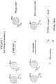
- FIGURE 1 graphically depicts a cell response assay constructed in accordance with the presently disclosed and claimed inventive concept(s).
- inventive concept(s) Before explaining at least one embodiment of the inventive concept(s) in detail by way of exemplary drawings, experimentation, results, and laboratory procedures, it is to be understood that the inventive concept(s) is not limited in its application to the details of construction and the arrangement of the components set forth in the following description or illustrated in the drawings, experimentation and/or results.
- inventive concept(s) is capable of other embodiments or of being practiced or carried out in various ways.
- the language used herein is intended to be given the broadest possible scope and meaning; and the embodiments are meant to be exemplary - not exhaustive.
- phraseology and terminology employed herein is for the purpose of description and should not be regarded as limiting.
- Enzymatic reactions and purification techniques are performed according to manufacturer's specifications or as commonly accomplished in the art or as described herein.
- the foregoing techniques and procedures are generally performed according to conventional methods well known in the art and as described in various general and more specific references that are cited and discussed throughout the present specification. See e.g., Sambrook et al. Molecular Cloning: A Laboratory Manual (2nd ed., Cold Spring Harbor Laboratory Press, Cold Spring Harbor, N.Y. (1989 ) and Coligan et al. Current Protocols in Immunology (Current Protocols, Wiley Interscience (1994 )).
- compositions and/or methods disclosed and claimed herein can be made and executed without undue experimentation in light of the present disclosure. While the compositions and methods of this presently disclosed and claimed inventive concept(s) have been described in terms of preferred embodiments, it will be apparent to those of skill in the art that variations may be applied to the compositions and/or methods and in the steps or in the sequence of steps of the method described herein without departing from the concept, spirit and scope of the presently disclosed and claimed inventive concept(s). All such similar substitutes and modifications apparent to those skilled in the art are deemed to be within the spirit, scope and concept of the inventive concept(s) as defined by the appended claims.
- At least one will be understood to include one as well as any quantity more than one, including but not limited to, 2, 3, 4, 5, 10, 15, 20, 30, 40, 50, 100, etc.
- the term “at least one” may extend up to 100 or 1000 or more, depending on the term to which it is attached; in addition, the quantities of 100/1000 are not to be considered limiting, as higher limits may also produce satisfactory results.
- the words “comprising” (and any form of comprising, such as “comprise” and “comprises”), “having” (and any form of having, such as “have” and “has”), "including” (and any form of including, such as “includes” and “include”) or “containing” (and any form of containing, such as “contains” and “contain”) are inclusive or open-ended and do not exclude additional, unrecited elements or method steps.
- probe as used herein will be understood to refer to any type of affinity reagent that binds to a specific biomarker as described herein.
- probes include, but are not limited to, antibodies (or binding fragments or derivatives thereof), receptors, organic molecules, inorganic molecules, ligands, nucleic acids (including but not limited to, DNA, RNA, microRNA, mRNA, siRNA, etc.), peptides, polypeptides, proteins, epitopes, antigens, ligands, receptors, complexes, lipids, glycoproteins, glycolipids, glycosaminoglycans, carbohydrates, polycarbohydrates, glycoconjugates, and any combination or derivative thereof.
- biomarker as used herein will be understood to refer to any target site on the surface of or inside of a cell that a probe can have affinity therefor and thus can bind to said moiety.
- the “biomarker” may be, for example but not by way of limitation, a nucleic acid, peptide, polypeptide, protein, epitope, antigen, ligand, receptor, complex (i.e., an MHC-peptide complex), lipid, glycoprotein, glycolipid, glycosaminoglycan, carbohydrate, polycarbohydrate, glycoconjugate, and any combination or derivative thereof.
- polypeptide refers to a polymer of amino acid residues.
- polypeptide as used herein is a generic term to refer to native protein, protein fragments, or analogs of a polypeptide sequence. Hence, native protein, protein fragments, and analogs are species of the polypeptide genus.
- polynucleotide and “nucleic acid” are used interchangeably. They refer to a polymeric form of nucleotides of any length, either deoxyribonucleotides or ribonucleotides, or analogs thereof.
- polynucleotides coding or non-coding regions of a gene or gene fragment, loci (locus) defined from linkage analysis, exons, introns, messenger RNA (mRNA), transfer RNA, ribosomal RNA, ribozymes, cDNA, recombinant polynucleotides, branched polynucleotides, plasmids, vectors, isolated DNA of any sequence, isolated RNA of any sequence, nucleic acid probes, and primers.
- a polynucleotide may comprise modified nucleotides, such as methylated nucleotides and nucleotide analogs.
- modifications to the nucleotide structure may be imparted before or after assembly of the polymer.
- the sequence of nucleotides may be interrupted by non-nucleotide components.
- a polynucleotide may be further modified, such as by conjugation with a labeling component.
- isolated nucleic acid and “isolated polynucleotide” are used interchangeably; a nucleic acid or polynucleotide is considered “isolated” if it: (1) is not associated with all or a portion of a polynucleotide in which the "isolated polynucleotide” is found in nature, (2) is linked to a polynucleotide to which it is not linked in nature, or (3) does not occur in nature as part of a larger sequence.
- antibody is used in the broadest sense, and specifically covers monoclonal antibodies (including full length monoclonal antibodies), polyclonal antibodies, multispecific antibodies (e.g., bispecific antibodies), and antibody fragments so long as they exhibit the desired biological activity.
- Antibody or “antibody peptide(s)” refer to a full length immunoglobulin molecule (i.e., an intact antibody), or a binding fragment thereof that competes with the intact antibody for specific antigen binding. Binding fragments may be produced by recombinant DNA techniques, or by enzymatic or chemical cleavage of intact antibodies.
- Binding fragments include Fab, Fab', F(ab') 2 , Fv, scFv, disulfide linked Fv, Fd, diabodies, single-chain antibodies, single domain antibodies (such as but not limited to, NANOBODIES®) and other antibody fragments that retain at least a portion of the variable region of an intact antibody. See, e.g., Hudson et al. (Nature Med., 9:129-134 (2003 )).
- antigen binding fragment or "antigen-binding portion" of an antibody, as used herein, refers to one or more fragments of an antibody that retain the ability to bind to an antigen.
- the antigen-binding function of an antibody can be performed by fragments of an intact antibody.
- binding fragments encompassed within the term "antigen-binding fragment” of an antibody include but are not limited to, Fab, Fab', F(ab')2, Fv, scFv, disulfide linked Fv, Fd, diabodies, single-chain antibodies, single domain antibodies (such as but not limited to, NANOBODIES®), isolated CDRH3, and other antibody fragments that retain at least a portion of the variable region of an intact antibody. These antibody fragments are obtained using conventional recombinant and/or enzymatic techniques and are screened for antigen binding in the same manner as intact antibodies.
- epitope includes any protein determinant capable of specific binding to an immunoglobulin or T-cell receptor.
- an epitope is a region of an antigen that is specifically bound by an antibody.
- Epitopic determinants usually include chemically active surface groupings of molecules such as amino acids, sugar side chains, phosphoryl, or sulfonyl groups.
- an epitope may have specific three dimensional structural characteristics (e.g., a "conformational epitope"), as well as specific charge characteristics.
- nanoparticle refers to a particle having dimensions of from about 1 to about 5000 nanometers, and having any size, shape or morphology.
- the nanoparticles utilized in accordance with the present invention may be naturally occurring, commercially available nanoparticles, or the nanoparticles may be synthesized for use in accordance with the present invention, as described herein below and as known in the art.
- Particular examples of nanoparticles that may be utilized in accordance with the present invention include, but are not limited to, poly(lactic-co-glycolic) acid (PLGA) nanoparticles, poly lactic acid (PLA) nanoparticles, Chitosen nanoparticles, liposomes, and derivatives or combinations thereof.
- label refers to incorporation of a detectable marker, e.g., by attachment of a fluorescent, enzymatic or colorimetric label or incorporation of a radiolabeled amino acid.
- Various methods of labeling polypeptides and glycoproteins are known in the art and may be used.
- labels for polypeptides include, but are not limited to, the following: radioisotopes or radionuclides (e.g., 3H, 14C, 15N, 35S, 90Y, 99Tc, 111In, 1251, 1311), fluorescent labels (e.g., FITC, rhodamine, lanthanide phosphors), enzymatic labels (e.g., horseradish peroxidase, ⁇ -galactosidase, luciferase, alkaline phosphatase), chemiluminescent, biotinyl groups, predetermined polypeptide epitopes recognized by a secondary reporter (e.g., leucine zipper pair sequences, binding sites for secondary antibodies, metal binding domains, epitope tags).
- labels are attached by spacer arms of various lengths to reduce potential steric hindrance.
- label means "label”, “detectable marker” and “detection moiety” are used interchangeably herein.
- a fluorophore may be employed in the methods of the present invention and detected via any of numerous colorimetric and fluorescence detection methods. Depending on the application and purpose, such methods include, but are not limited to, absorbance spectroscopy, fluorescence spectroscopy, fluorescence activated cytometry (FACS), fluorescence microscopy, fluorescence resonance energy transfer (FRET), and the like.
- fluorophores may be employed in accordance with the present invention. Examples of suitable fluorophores are described herein below. Examples of suitable fluorophores are described herein below. Other examples are given in US Patent Nos. 7,465,747 and 7,955,859, issued to Matsumoto et al. on December 16, 2008 and June 7, 2011, respectively; and US Publication No. US2007/0026407, published February 1, 2007 (the entire contents of which are expressly incorporated herein by reference in their entirety).
- substantially increase and “substantial decrease”, as well as grammatical equivalents thereof, will be understood herein to refer to at least a 12% increase or decrease, such as at least a 30% increase or decrease, at least a 50% increase or decrease, at least a 75% increase or decrease, or at least a 90% increase or decrease.
- cancer and “cancerous” refer to or describe the physiological condition in mammals that is typically characterized by unregulated cell growth.
- cancer include but are not limited to, carcinoma, lymphoma, blastoma, and sarcoma. More particular examples of such cancers include squamous cell cancer, small-cell lung cancer, non-small cell lung cancer, gastrointestinal cancer, pancreatic cancer, glioblastoma, cervical cancer, ovarian cancer, liver cancer, bladder cancer, hepatoma, breast cancer, colon cancer, colorectal cancer, endometrial carcinoma, salivary gland carcinoma, kidney cancer, renal cancer, prostate cancer, vulval cancer, thyroid cancer, hepatic carcinoma and various types of head and neck cancer.
- Metastasis as used herein will be understood to refer to the spread of cancer from a primary tumor to other parts of the body. Metastasis is a sequential, multistep process in which tumor cells detach from a primary tumor, migrate through the basement membrane and extracellular matrix, and invade the lymphatic and/or blood systems. This is followed by the establishment of secondary tumors at distant sites.
- “Mammal” for purposes of treatment refers to any animal classified as a mammal, including human, domestic and farm animals, nonhuman primates, and zoo, sports, or pet animals, such as dogs, horses, cats, cows, etc.
- a patient includes human and veterinary subjects.
- a patient is a mammal. In certain other embodiments, the patient is a human.
- biological sample as used herein will be understood to refer to a sample of biological fluid.
- biological samples include, but are not limited to, blood, plasma, serum, sputum, cerebrospinal fluid (CSF), tears, mucus, urine, tissue, other types of specimens, and the like.
- CSF cerebrospinal fluid
- providing a biological sample refers to obtaining a biological sample for use in methods described and claimed herein. Most often, this will be done by removing a sample of cells from an animal, but can also be accomplished by using previously isolated cells (e.g., isolated by another person, at another time and/or for another purpose).
- the step of "providing a biological sample” may further include various isolation and/or purification steps known in the art for providing a specific component of a biological sample for use in the methods described and claimed herein.
- the presently disclosed and claimed inventive concept(s) generally relates to multiplexed direct affinity conjugate assays of cells using probes able to determine a cellular response to treatments; this type of assay is herein termed a "cell response assay”.
- the presently disclosed and claimed inventive concept(s) generally relates to the use of a combination of biomarkers for cancer cell/tissue type, metastatic potential and chemo resistance in a multiplexed affinity assay for cancer cells and tissue.
- Fluorescent labels either conjugated to affinity molecules/reagents or capable of directly binding to the biomarker are used and are referred to herein as labeled probes.
- This combination of labeled probes for biomarkers allows determination of the cellular response to treatments by simultaneously measuring cell type, chemo resistance nature, and metastatic potential and/or cell viability, and allows simple multiplexed signals with high sensitivity.
- the biomarker affinity markers selected in this order solves the problem of "picking" the right antigen for cancer antigen type and also provides accurate information on the response to therapy. More particularly, the presently disclosed and claimed inventive concept(s) relates to methods of using said affinity molecules and molecular probes as compositions, dosage forms, and kits.
- the presently disclosed and claimed inventive concept(s) combines cell biomarker(s) for tissue type with biomarker(s) for metastatic potential and biomarker(s) for cell viability in an affinity assay for cancer cells and tissue.
- the biomarker for cancer cell type is represented as biomarker A and is always present in the disease state.
- the biomarker for metastatic potential is represented as biomarker B and is present in the disease state with metastatic invasiveness potential.
- the biomarker for chemo resistance is represented as biomarker C and is present in unresponsive cells.
- Fluorescent labels either conjugated to the affinity reagents or capable of directly binding the biomarker are used to simultaneously detect biomarkers A, B and C; simultaneous detection is obtained by using fluorescent labels that are detectable at different excitations and/or emission wavelengths.
- biomarker B i.e., metastatic potential
- biomarker C i.e. chemo resistance
- biomarkers B and C must be linked such that the presence of one facilitates the absence of the other.
- This combination of biomarkers allows the determination of the cell response to treatments; by simultaneously measuring cell type, chemo resistance nature, and metastatic potential (and possibly also cell viability), the "cell based response assay" can accurately monitor cancer treatment and/or monitor disease progression/relapse.
- one embodiment thereof is directed to a multiplexed assay for cancer.
- Said assay involves simultaneously measuring: (1) at least one cancer cell type biomarker (also referred to herein as Biomarker A) in a biological sample of a cancer patient utilizing a first labeled probe that binds to said cancer cell type biomarker; (2) at least one chemo resistance biomarker (also referred to herein as Biomarker C) in the biological sample utilizing a second labeled probe that binds to said chemo resistance biomarker; and (3) at least one metastatic potential biomarker (also referred to herein as Biomarker B) in the biological sample utilizing a third labeled probe that binds to said metastatic potential biomarker.
- the at least three labeled probes are measured at different excitations and emission wavelengths.
- the assays/methods described herein may be utilized for a specific cancer, or the assays/methods may be general assays for all types of cancers.
- the cancer cell type biomarker utilized in accordance with the presently disclosed and claimed inventive concept(s) may be a non-specific cancer cell biomarker, or may be specific for a certain type of cancer.
- cancers that may be detected/monitored by the currently disclosed and claimed inventive concept(s) include, but are not limited to, lung, bronchus, colon, rectum, pancreas, prostate, breast, liver, bile duct, bladder, ovary, brain, central nervous system (CNS), kidney, pelvis, uterine corpus, oral cavity or pharynx or melanoma cancers.
- lung bronchus, colon, rectum, pancreas, prostate, breast, liver, bile duct, bladder, ovary, brain, central nervous system (CNS), kidney, pelvis, uterine corpus, oral cavity or pharynx or melanoma cancers.
- CNS central nervous system
- kidney pelvis
- uterine corpus oral cavity or pharynx or melanoma cancers.
- Non-limiting examples of non-specific cancer cell type biomarkers include, but are not limited to, cytokeratins (CK), EpCAM, N-cadherin, E-cadherin and vimentin.
- the carcinoma cells may be indicated by cytokeratin (CK) as a biomarker A
- the first labeled probe may comprise multiple labeled antibodies for separate cytokeratins (such as but not limited to, cytokeratins 8/18/19.
- Cytokeratins (CK) are proteins of keratin-containing intermediate filaments found in the intracytoplasmic cytoskeleton of epithelial tissue and refer to a family of fibrous structural proteins. There are two fundamental types of cytokeratins: the acidic type I cytokeratins and the basic or neutral type II cytokeratins.
- Cytokeratins are usually found in pairs comprising a type I cytokeratin and a type II cytokeratin.
- Basic or neutral cytokeratins include (but are not limited to) CK1, CK2, CK3, CK4, CK5, CK6, CK7, CK8 and CK9.
- Acidic cytokeratins include (but are not limited to) CK10, CK12, CK 13, CK14, CK16, CK17, CK18, CK19 and CK20.
- EpCAM Epithelial cell adhesion molecule
- TACSTD1 tumor-associated calcium signal transducer 1
- CD326 cluster of differentiation 326
- cancer cells undergo an Epithelial to Mesenchymal transition (EMT) that may be indicated by an increase in cancer cell biomarkers such as but not limited to, Vimentin and Galectin-3, which signify cells with a loss of anchorage.
- EMT Epithelial to Mesenchymal transition
- MET Mesenchymal to Epithelial transition
- the EMT and MET stages for cancer cells signify a change in sub type of cells. It is important to detect as many cancer cells as possible and to additionally know their sub types to measure the entire tumorigenicity.
- cancer cell type biomarker also includes markers that identify specific genetic mutations that cause oncoproteins or oncogenes to be regulated (whether tumor promoting or tumor suppressing). These types of cancer cell type biomarkers may be utilized alone or in combination with any of the other cancer cell type biomarkers described herein above.
- cancer cell type biomarkers examples include, but are not limited to, HER2/neu, VEGF-165, KRAS, EGFr, WAF, BAX-1, PDGF, Rb, Jagged 1, Notch, VEGF, VEGHR, k-Ras, CAIX, MIB1, MDM, PR, ER, SEL5, SEM1, PI3K, Akt2, twist 1, EML-4, ALK, Braf, DRAFF, c-met, and combinations thereof.
- oncoproteins and oncogenes are used to direct targeted therapies based on their presence in/on cells.
- HER2/neu is used to prescribe Herceptin therapy; however, HER2/neu, as with other oncoprotein/oncogene markers, is often only expressed on a fraction of cancer cells, and not all patients have the specific genetic mutation leading to the biomarker.
- HER2/neu-positive expression is present in only 10-20% of breast cancer patients, and even in the HER2/neu-positive patients, the marker is only present in approximately 30% of their circulating tumor cells. Therefore these markers are often measured in combination with another cancer cell type biomarker.
- the carcinoma cells may be indicated by a tissue specific marker.
- Prostate-Specific Antigen is a protein that signifies the presence of cells of the prostate gland.
- specific cancer cell type biomarkers include, but are not limited to: prostate specific antigen (PSA) or prostate specific membrane antigen (PSMA) can be used to detect prostate cancer cells; MUC1, CA 15-3 and CA 27-29 can be used to detect breast cancer cells; Carcinoembryonic Antigen (CEA), CA19-9 or Galactosyl Transferase II can be used to detect colon cancer cells; MSLN (mesothelin) can be used to detect pancreatic cancer cells; CA 125 or Follicle-Stimulating Hormone (FSH) receptor can be used to detect ovarian cancer cells; Alpha-Fetoprotein can be used to detect liver cancer cells; Melan-A (MLANA), Tyrosinase (TYR), CSPG4, or MITF can be used to detect melanoma cancer cells
- the third labeled probe may be any probe capable of detecting said metastatic potential biomarker; for example but not by way of limitation, the third labeled probe may be one or more labeled antibodies against any of the markers of tumor cell invasiveness listed below. In some instances, the metastatic potential biomarker may be measured as a ratio of one of the below-listed biomarkers to the cancer cell type biomarker.
- biomarkers of metastatic potential include a marker(s) of tumor cell invasiveness where the marker measures cancer cell invasion into the extracellular membrane through proteolytic events, such as urokinase plasminogen activator (uPA), plasminogen activator inhibitor (PAI-1), CD95, serine proteases such as plasmin, ADAM, and others; serine protease inhibitors such as Bikunin; matrix metalloproteinases such as MMP9; matrix metalloproteinase inhibitors such a TIMP-1; and combinations thereof.
- uPA urokinase plasminogen activator
- PAI-1 plasminogen activator inhibitor
- CD95 serine proteases
- serine proteases such as plasmin, ADAM, and others
- serine protease inhibitors such as Bikunin
- matrix metalloproteinases such as MMP9
- matrix metalloproteinase inhibitors such a TIMP-1
- combinations thereof such as urokina
- the chemo resistance biomarker is a biomarker that detects the prevention of cell death (apoptosis), for example but not by way of limitation, a detection of the presence of a cancer stem cell biomarker, such as but not limited to, PL2L piwi like, ADLH, ⁇ -integrin, ⁇ 6 integrin, c-kit, c-met, LIF-R, CXCR4, ESA, CD 20, CD44, CD133, CK5, TRAF2 and ABC transporters.
- a cancer stem cell biomarker such as but not limited to, PL2L piwi like, ADLH, ⁇ -integrin, ⁇ 6 integrin, c-kit, c-met, LIF-R, CXCR4, ESA, CD 20, CD44, CD133, CK5, TRAF2 and ABC transporters.
- cancer cells that contain CD44 but lack CD24 are indicative of a cancer stem cell phenotype.
- cancer cells that lack CD45 and CD31 but contain CD34 are indicative of a cancer stem cell.
- cancer cells that contain CD44 and CD24 as well as ESA are indicative of a cancer stem cell.
- the presence of both CD24 and ESA are indicative of a cancer stem cell. Somatic stem cells in solid tumor carcinomas can become cancer stem cells. Other cancer cells resistant to CD95 induced-apoptosis are chemo resistant.
- Cancer stem cells are chemo resistant in nature, and contain a capacity for self renewal (asymmetric divisions) as well as being capable of differentiation into a hierarchy of progeny cells to form a tumor.
- Cancer stem cell self renewal is activated by the stem cell signaling pathways (Wnt, Sonic Hedge hog and Notch) and at the epigenetic level by Polycomb gener (BMI-1 and EZH2). This self renewal occurs at the expense of apopotosis signaling pathways (Caspase 3, 5, 8, p53). Measuring cancer stem cell biomarkers and combinations thereof as an indication of chemo resistance provides a measure of (a) drug resistance, (b) the inability to activate apoptosis, and/or (c) the inability to shutdown self renewal.
- the probes described and claimed herein may be labeled by any methods known in the art or otherwise contemplated herein.
- the labels of the first, second and third (and any additional fourth, fifth, sixth, etc.) labeled probes may be selected from a comprehensive catalogue of fluorescent (luminescent) dyes.
- a comprehensive catalogue exists online at http://www.fluorophores.org (the entire contents of which are expressly incorporated herein by reference).
- This catalogue includes commonly used labels such as fluorescein-5-isothiocyanate (FITC), phycoerythrin, sulforhodamine 101 (Texas Red), 2-[4-(aminoiminomethyl)phenyl]-1H-Indole-6-carboximidamide (DAPI), 3H-Indolium (Cy5), 1H-benz[e]indolium (Cy 5.5), 3H-Indolium (Cy 7), ALEXA FLUOR®488, ALEXA FLUOR®555, ALEXA FLUOR®647, and combinations and derivatives thereof that have been synthesized in order to provide better reagents. Additionally, rare earth metals and rare earth element-containing nanoparticles can be used as labels.
- FITC fluorescein-5-isothiocyanate
- DAPI 2-[4-(aminoiminomethyl)phenyl]-1H-Indole-6-carboximidamide
- Cy5 3H-
- biological samples utilized in the methods of the presently disclosed and claimed inventive concept(s) may be utilized in the form they are obtained (i.e., tissue sample), or they may be exposed to one or more isolating steps (i.e., isolation of cancer cells therefrom).
- the assay may further include measuring at least one additional biomarker in the biological sample utilizing a fourth labeled probe that binds to said additional biomarker, measuring a second additional biomarker in the biological sample utilizing a fifth labeled probe that binds to said additional biomarker, measuring a third additional biomarker in the biological sample utilizing a sixth labeled probe that binds to said additional biomarker, etc.
- a maximum of six to eight labeled probes are utilized; otherwise, it is difficult to detect all of the labeled probes at different, non-overlapping excitations and emission wavelengths.
- biomarkers may include biomarkers that detect other cellular properties, specifically the presence of cell nuclei or of intact cell membranes, for the cells that are positive for the cancer cell type biomarker.
- the presence of cell nuclei may be detected utilizing a DNA binding probe such as but not limited to, DAPI.
- the detection of intact cell membranes may involve the detection of anionic phospholipids (such as but not limited to, phosphatidylserine) on the surface of cells, and the second labeled probe may be bis(zinc2+dipicolylamine) and/or PSVueTM.
- the one or more additional biomarker(s)/labeled probe(s) may also be utilized to reduce any false positive results obtained with the cancer cell type biomarker; for example but not by way of limitation, white blood cells may be excluded from the assay by excluding cells positive for one or more markers of cluster of differentiation (also referred to as "cluster of designation"; often abbreviated as CD).
- markers of cluster of differentiation also referred to as "cluster of designation”; often abbreviated as CD.
- markers such as CD45, CTLA-4, CD4, CD68 and/or CD8 present on white blood cells can be used to indicate that a cell is not a cancer cell.
- CD45 antigen also known as PTPRC, Protein tyrosine phosphatase receptor type C, and originally called leukocyte common antigen (all terms used herein interchangeably) is useful in detecting all white blood cells. Additionally, CD45 can be used to differentiate the different types of white blood cells when combined with other CD markers.
- granulocytes are indicated by CD45+, CD15+; monocytes are indicated by CD45+, CD14+; T lymphocytes are indicated by CD45+, CD3+; T helper cells are indicated by CD45+,CD3+, CD4+; cytotoxic T cells are indicated by CD45+,CD3+, CD8+; B lymphocytes are indicated by CD45+, CD19+ or CD45+, CD20+; thrombocytes are indicated by CD45+, CD61+; and natural killer cells are indicated by CD16+, CD56+, CD3-.
- CD4 and CD8 are, in general, used as markers for helper and cytotoxic T cells, respectively. These molecules are defined in combination with CD3+, as some other leukocytes also express these CD molecules (some macrophages express low levels of CD4; dendritic cells express high levels of CD8).
- the presently disclosed and claimed inventive concept(s) is also directed to a kit that includes the first, second and third labeled probes (that bind to a cancer cell type biomarker, a chemo resistance biomarker, and a metastatic potential biomarker, respectively) as described in detail herein above. Additional labeled probes for detecting other biomarkers as further described herein above may also be included in the kit. All of the labeled probes present in the kit are measured at different excitations and emission wavelengths.
- the kit may also include means for isolating cancer cells from a biological sample.
- Said means are well known in the art (see for example, Lianidou and Markou, 2011; the entire contents of which are hereby expressly incorporated herein by reference); therefore, no additional discussion thereof is deemed necessary.
- the presently disclosed and claimed inventive concept(s) is also directed to methods of (1) monitoring cancer treatment in a cancer patient undergoing said treatment, and (2) monitoring progression/relapse of the disease in the cancer patient.
- a first biological sample is obtained from the patient prior to exposure to a cancer treatment
- a second biological sample is obtained from the patient following exposure to the cancer treatment.
- the first biological sample is obtained from the patient at a first time point
- the second biological sample is obtained from the patient at a subsequent time point.
- the levels of a cancer cell type biomarker (as described herein above), a chemo resistance biomarker (as described herein above), and a metastatic potential biomarker (as described herein above) are then measured utilizing first, second and third labeled probes, respectively (as described in detail herein above), in the first and second biological samples.
- the cells to which the first labeled probe is bound are identified in the first and second biological samples, and then the levels of (i) the chemo resistance biomarker and (ii) the metastatic potential marker in the cells to which the first labeled probe is bound are compared in the first and second biological samples.
- the cancer treatment is effective if the level of the chemo resistance biomarker is decreased and the level of the metastatic potential biomarker is increased in the second biological sample when compared to the first biological sample; alternatively, it is then determined in the method of (2) that the cancer has progressed/relapsed if the level of the chemo resistance biomarker is increased and the level of the metastatic potential biomarker has decreased in the second biological sample when compared to the first biological sample.
- biological samples utilized in the methods of the presently disclosed and claimed inventive concept(s) may be utilized in the form they are obtained (i.e., tissue sample), or they may be exposed to one or more isolating steps (i.e., isolation of cancer cells therefrom).
- the method may further include measuring at least one additional biomarker in the first and second biological samples utilizing a fourth labeled probe that binds to said additional biomarker, measuring a second additional biomarker in the first and second biological samples utilizing a fifth labeled probe that binds to said additional biomarker, measuring a third additional biomarker in the first and second biological samples utilizing a sixth labeled probe that binds to said additional biomarker, etc.
- the methods may also include the step of comparing the levels of the additional biomarker(s) in the cells to which the first labeled probe is bound in the first and second biological samples.
- biomarkers may include biomarkers that detect other cellular properties, specifically the presence of cell nuclei or of intact cell membranes, for the cells that are positive for the cancer cell type biomarker.
- the presence of cell nuclei may be detected utilizing a DNA binding probe such as but not limited to, DAPI.
- the detection of intact cell membranes may involve the detection of anionic phospholipids (such as but not limited to, phosphatidylserine) on the surface of cells, and the second labeled probe may be bis(zinc2+dipicolylamine) and/or PSVueTM.
- the one or more additional biomarker(s)/labeled probe(s) may also be utilized to reduce any false positive results obtained with the cancer cell type biomarker; for example but not by way of limitation, white blood cells may be excluded from the methods by excluding cells positive for one or more markers of cluster of differentiation (as described in detail herein above).
- the following procedure shows the multiplexing of Biomarkers A (cancer cell type), B (metastatic potential) and C (chemo resistance) using a unique combination of affinity reagents with fluorescent labels and an affinity label to provide a multiplexed assay for breast cancer.
- This allows the simultaneous measurement of cancer cell type, chemo resistance nature, and metastatic potential, as described herein above, in combination with a marker indicating the presence of cell nucleus (utilizing a nucleic acid binding probe).
- the data demonstrated that four simultaneous signals can be measured with this combination but not with the traditional combinations of the prior art.
- the presently disclosed and claimed inventive concept(s) has the potential to measure up to eight signals utilizing various fluorophores taught herein.
- lysis buffer 155mM NH 4 Cl, 20mM KHCO 3 , 0.1 mM Na 2 EDTA, pH 7.4
- the tubes were rolled tubes to assure that the cells were evenly distributed and that lysis was complete, and then the sample was spun down for 15 minutes at 12,000 rpm.
- the plasma was decanted as waste supernatant, and 4.0 mL of TES buffer (50 mM N-Tris [Hydroxymethyl]-2-aminoethane-sulfonic acid: TES, pH 7.4, 2150mM NaCl, 2 mM MgSO 4 and 1 mM KCO 3 ) and 1 % albumin was added, and the tube was rocked to mix the cells.
- the tube was spun down again for 15 minutes at 12,000 rpm, and the supernatant was decanted as waste.
- a final 2.0 mL of TES wash buffer was added with rocking to mix cells.
- the cells were then reacted with the reagents by taking 0.5 mL of washed blood to the centrifuge tube, and 40 ⁇ L of antibodies for HER2/neu labeled with Cy5 and 40 ⁇ L of antibodies for urokinase like plasminogen activator (uPA) or plasminogen activator inhibitor (PAI-1) labeled with Texas Red (at working stock of 0.001 to 0.1 mg/mL) and PL2L piwi like labeled with FITC (at working stock of 0.001 to 0.1 mg/mL) were added.
- uPA plasminogen activator
- PAI-1 plasminogen activator inhibitor
- DAPI Diamidino-2-phenylindole dihydrochloride
- the sample was centrifuged down for 5 minutes @ 7500 rpm, and the supernatant was decanted off as waste ( ⁇ 450 ⁇ L).
- 500 ⁇ L of TES buffer was then added, and the sample was vortexed and centrifuged down at 5 minutes @ 7500 rpm. Again, the supernatant was decanted off by tapping as waste ( ⁇ 450 ⁇ L).
- a final 500 ⁇ L of TES wash buffer was added for final imaging, and 5 ⁇ L of the solution was placed on a slide with a long cover slip of glass. Phase contrast and fluorescence microscopy was conducted with the Leica DM5000 (Leica Microsystems, Buffalo Grove, IL).
- a sample of cells was stained with the PSS-550 probe (ex 553, em 615nm) and measured with an excitation band pass filter at 540-580 nm and an emission band pass filter at 610-680 nm to determine cell membrane flopping as a measure of cell death in addition to cell counts .
- the Texas Red label (ex 592, em 614 nm) was measured with an excitation band pass filter at 540-580 nm and an emission band pass filter at 610-680 nm.
- the FITC label (ex 488, em 525nm) was measured with an excitation band pass filter at 460 - 500 nm and an emission band pass filter at 512 -543 nm.
- the DAPI probe (ex 355, em 460 nm) was measured with an excitation band pass filter at 340-380 nm and an emission band pass filter at 450-490 nm.
- the Cy5 label (ex 646, em 676 nm) was measured with an excitation band pass filter at 590-650 nm and an emission band pass filter at 665-735 nm.
- the ideal multiplexing was demonstrated by combination of the rare earth nanoparticles with FITC, Texas Red, Cy5 and DAPI.
- the europium nanoparticle (ex 355, em 617 nm) was measured at an excitation filter band pass at 340-380 nm and emission long pass filter of > 590 nm.
- Simultaneously measuring biomarkers for cancer cell type, metastatic potential and chemoresistance, along with a biomarker indicating the presence of cell nucleus was possible, along with the removal of false positives (i.e., white blood cells) by detecting the presence of the biomarker CD45. Current data allows for five signals.
- the assays/methods described and claimed herein have the potential for detection and measurement of six signals/biomarkers, if the rare earth nanoparticles can be multiplexed into three different emission signals.
- the europium label greatly increased signal over FITC and Texas Red by 2-3 orders of magnitude.
- Biomarker A tissue type marker for carcinoma breast cancer
- Biomarker B for metastatic potential utilized uPA and PAI antigen; these are markers of tumor invasiveness and are present in aggressive cancer cell differentiation and growth and increase with metastatic invasiveness potential of the cells.
- Biomarker C for chemo resistance utilized PL2L piwi like antigen (see for example, Gao et al., 2008).
- Example 1 The procedure shown in Example 1 was used to measure cancer cells before and after treatment with and without camptothecin. Camptothecin is known to activate apoptosis in cancer cells and kill cancer cells like a chemotherapy agent (See Gupta, 1997). Cells were tested by the cell response assay and by a traditional prior art assay.
- the novel "cell response assay” utilized HER2/neu as Biomarker A for cell type, the tumor invasiveness markers uPA & PAI-1 as Biomarker B for metastatic potential, and the cancer stem cell marker PL2L piwi like as Biomarker C for chemo resistance nature.
- the traditional assay used EpCAM as a marker for epithelial tissue type and cytokeratin (CK) as a marker of cells of cancer origin. Additionally, CD45, a marker for white blood cells, was used to reduce false results, with CD45 positive results being excluded from the analysis. Both the traditional and cell response assays used DAPI to determine the presence of cell nuclei.
- the camptothecin concentration could be adjusted to be sufficient to kill >80%, 60% and 40% of the cancer cells in cultures.
- Cellular analysis showed cancer cell count to decrease as the cell death % increased and phosphatidylserine flopping increased.
- Cellular analysis showed biomarker B, namely uPA and PA1, to increase (or was detected in a greater number of cancer cells) when the cell death % was highest.
- biomarker C namely PL2L piwi like to decrease (or was detected in a lesser number of cancer cells) when the cell death % was highest. All three biomarkers co-exist in the disease state and were fundamental to progression or relapse of the disease.
- the CD45 biomarker does not co-exist with EpCAM and CK. Both EpCAM and CK were present in all cells independent of the % of cells that were killed.
- the following procedure shows the multiplexing of Biomarkers A (cancer cell type), B (metastatic potential) and C (chemo resistance) using a unique combination of affinity reagents with fluorescent labels and an affinity label to provide a multiplexed assay for prostate cancer.
- This allows the simultaneous measurement of cancer cell type, chemo resistance nature, and metastatic potential, as described herein above, in combination with a marker indicating the presence of cell nucleus (utilizing a nucleic acid binding probe).
- the data demonstrated that four simultaneous signals can be measured with this combination but not with the traditional combinations of the prior art.
- the presently disclosed and claimed inventive concept(s) has the potential to measure up to eight signals utilizing various fluorophores taught herein.
- lysis buffer 155mM NH 4 Cl, 20mM KHCO 3 , 0.1 mM Na 2 EDTA, pH 7.4
- the tubes were rolled tubes to assure that the cells were evenly distributed and that lysis was complete, and then the sample was spun down for 15 minutes at 12,000 rpm.
- the plasma was decanted as waste supernatant, and 4.0 mL of TES buffer (50 mM N-Tris [Hydroxymethyl]-2-aminoethane-sulfonic acid: TES, pH 7.4, 2150mM NaCl, 2 mM MgSO 4 and 1 mM KCO 3 ) and 1 % albumin was added, and the tube was rocked to mix the cells.
- the tube was spun down again for 15 minutes at 12,000 rpm, and the supernatant was decanted as waste.
- a final 2.0 mL of TES wash buffer was added with rocking to mix cells.
- the cells were then reacted with the reagents by taking 0.5 mL of washed blood to the centrifuge tube, and 40 ⁇ L of antibodies for PSA labeled with Cy5 and 40 ⁇ L of antibodies for urokinase like plasminogen activator (uPA) or plasminogen activator inhibitor (PAI-1) labeled with Texas Red (at working stock of 0.001 to 0.1 mg/mL) and PL2L piwi like labeled with FITC (at working stock of 0.001 to 0.1 mg/mL) were added.
- uPA urokinase like plasminogen activator
- PAI-1 plasminogen activator inhibitor
- DAPI Diamidino-2-phenylindole dihydrochloride
- the sample was centrifuged down for 5 minutes @ 7500 rpm, and the supernatant was decanted off as waste ( ⁇ 450 ⁇ L).
- 500 ⁇ L of TES buffer was then added, and the sample was vortexed and centrifuged down at 5 minutes @ 7500 rpm. Again, the supernatant was decanted off by tapping as waste ( ⁇ 450 ⁇ L).
- a final 500 ⁇ L of TES wash buffer was added for final imaging, and 5 ⁇ L of the solution was placed on a slide with a long cover slip of glass. Phase contrast and fluorescence microscopy was conducted with the Leica DM5000 (Leica Microsystems, Buffalo Grove, IL).
- the Texas Red label (ex 592, em 614 nm) was measured with a excitation band pass filter at 540-580 nm and emission band pass filter at 610-680 nm.
- the FITC label (ex 488, em 525nm) was measured with an excitation band pass filter at 460 - 500 nm and an emission band pass filter at 512 -543 nm.
- the DAPI probe (ex 355, em 460 nm) was measured with an excitation band pass filter at 340-380 nm and an emission band pass filter at 450-490 nm.
- the Cy5 label (ex 646, em 676 nm) was measured with an excitation band pass filter at 590-650 nm and an emission band pass filter at 665-735 nm.
- the ideal multiplexing was demonstrated by combination of the rare earth nanoparticles with FITC, Texas Red, Cy5 and DAPI.
- the europium nanoparticle (ex 355, em 617 nm) was measured at an excitation filter band pass at 340-380 nm and emission long pass filter of > 590 nm.
- Simultaneously measuring biomarkers for cancer cell type, metastatic potential and chemoresistance, along with a biomarker indicating the presence of cell nucleus was possible, along with the removal of false positives (i.e., white blood cells) by detecting the presence of the biomarker CD45. Current data allows for five signals.
- the assays/methods described and claimed herein have the potential for detection and measurement of six signals/biomarkers, if the rare earth nanoparticles can be multiplexed into three different emission signals.
- the europium label greatly increased signal over FITC and Texas Red by 2-3 orders of magnitude.
- Protease like plasmin, is expressed during metastasis of malignant cells as part of the tissue regeneration and fibrinolysis process, indirectly promoting cell proliferation.
- Cancer cells use cell-bound plasmin to activate the plasminogen signaling for urokinase.
- Inhibitors of plasmin such as Bikunin and inhibitors of plasminogen activation such as PAI-1 prevent cell-bound plasmin activation and suppress tumor invasiveness.
- a novel cell response assay for prostate cancer was developed by using PSA as a tissue type marker for prostate carcinoma (Biomarker A) that is always present in the disease state.
- the markers used in this example for metastatic potential were urokinase plasminogen activator (uPA) and plasminogen activator inhibitor (PAI-1), which is a measure of tissue invasiveness and is present in an aggressive cancer cell differentiation and growth with increased metastatic invasiveness potential.
- Chemo resistance nature Biomarker C was determined using PL2L piwi like antigen to determine the inhibitory response to tissue invasiveness.
- Example 3 The procedure shown in Example 3 was used to measure prostate cancer cells before and after treatment with and without camptothecin. Camptothecin is known to activate apoptosis in cancer cells and kill cancer cells like a chemotherapy agent (See Gupta, 1997).
- Cells were tested by the cell response assay and by a traditional prior art assay.
- the novel "cell response assay” utilized PSA as Biomarker A for cell type, the tumor invasiveness markers uPA & PAI-1 as Biomarker B for metastatic potential, and the cancer stem cell marker PL2L piwi like as Biomarker C for chemo resistance nature.
- the traditional assay used EpCAM as a marker for epithelial tissue type and cytokeratin (CK) as a marker of cells of cancer origin. Additionally, CD45, a marker for white blood cells, was used to reduce false results, with CD45 positive results being excluded from the analysis.
- Both the traditional and cell response assays used DAPI to determine the presence of cell nuclei.
- the camptothecin concentration could be adjusted to be sufficient to kill >80%, 60% and 40% of the cancer cells in cultures.
- Cellular analysis showed cancer cell count to decrease as the cell death % increased and phosphatidylserine flopping increased.
- Cellular analysis showed biomarker B, namely uPA and PA1, to increase (or was detected in a greater number of cancer cells) when the cell death % was highest.
- biomarker C namely PL2L piwi like to decrease (or was detected in a lesser number of cancer cells) when the cell death % was highest. All three biomarkers co-exist in the disease state and were fundamental to progression or relapse of the disease.
- the CD45 biomarker does not co-exist with EpCAM and CK. Both EpCAM and CK were present in all cells independent of the % of cells that were killed.
- the following procedure demonstrates the multiplexing of Biomarkers A, B and C as described herein previously to provide a general assay for all types of cancers.
- This procedure is performed as described herein above in Examples 1/3, except as follows: (a) the cancer suspension added to the procedure is selected from one of the following cell lines - SKBR, MCF or MDA (breast cancer), UC3 or T24 (bladder cancer), PC3-9 (prostate cancer), and A549 (lung cancer); and (b) the non-specific cancer cell biomarker cytokeratin is utilized for Biomarker A.
- antibodies to the cytokeratins 8/18/19 are utilized as the first labeled probe that binds to Biomarker A.
- antibodies to uPA or PAI-1 labeled with Texas Ted are utilized as the second labeled probe that binds to Biomarker B, and antibodies to PL2L piwi like labeled with FITC is utilized as the third labeled probe that binds to Biomarker C.
- the nucleic acid binding probe DAPI is also utilized as described in Examples 1 and 3.
- This multiplexed assay utilizing a non-specific cancer cell biomarker allows the assays/methods described herein to be adapted for use with all types of cancers.
Description
- The present application is based on provisional application Serial No.
61/538,302, filed September 23, 2011 - Not Applicable.
- The presently disclosed and claimed inventive concept(s) generally relates to cellular assays. More particularly, but not by way of limitation, the presently disclosed and claimed inventive concept(s) relates to methods of assaying biological samples to determine the cellular response to treatment/therapy and/or monitor progression of a disease state.
- Affinity assays using fluorescence compounds are commonly used for cell and tissue analysis for biomarkers. The fluorescent compounds (labels) are attached (conjugated) to affinity molecules. Said affinity molecules may be, for example but not by way of limitation, antibodies, antigens, nucleic acid or other molecules that associate with the biomarkers of interest. This direct affinity method is a commonly used method to stain cells and tissues (See Horan and http://www.crm.ed.ac.uk/facilities/flow-cytometry/protocols/immunofluorescence-staining). The cells or tissues are incubated with conjugates, and unbound materials are washed away from cell bound materials. The conjugates are read cytometrically with a flow cytometer, scanning microscope or other device capable of detecting the fluorescence emission of the cellular material (see for example,
US2008/0187198 ; Krivacic, 2004; and Herzenberg, 2002). Protocols vary and may or may not require cell or tissue fixation, depending on the conjugate used. - In practice, cell counts are measured using flow cytometry or scanning microscopy technologies which capture signals of cells at the excitation and emission wavelengths required to detect the dye. Different conjugates can be simultaneously determined (multiplexed) by using fluorescent dyes with distinct excitation wavelengths and emission wavelengths. Multiplexing of fluorescence signals is commonly used to detect as many properties as possible at one time. For example, biomarkers conjugated to phycoerythrin (ex 592, em 614nm) can be measured independent from biomarkers conjugated to fluorescein-5-isothiocyanate (FITC) (ex 488, em 525nm) using different excitation and emission filters. A list of known fluorophores is commonly available to researchers in the field (see http://www.fluorophores.org/).
- Cell and tissue assays are typically performed for various types of cancer assays. Both cell and tissue assays use affinity reagents to detect biomarkers on or inside the cell, such as peptides, proteins, nucleic acid and modifications thereof (see, for example, Punnoose, 2010). Nucleic acids are measured as messenger RNA, Micro RNA and/or DNA prior to or after PCR amplification. Antibodies are also used for detecting protein biomarkers such as, but not limited to, EGFR, IGFRE, ERBB2, PSA, PL2L, kRAS, EPCAM, CK, and CD. A variety of tumor markers have detected breast, colon, prostate and melanoma cancer cells (see, for example, Mocellin, 2006). The use of biomarkers for monitoring cancer progression and/or treatment is also common practice. For example, the use of the plasminogen activator system (uPA) is monitored by measuring uPA, the plasminogen inhibitor PAI-1, and the complex of both (see Carney et al., 2009).
- Cell assays are often looking for rare but clinically significant events. For example, circulating tumor cells (CTC) are significant at 5 cancer cells in 80,000,000 normal blood cells. The CTC must be isolated by enrichment or depletion to eliminate interference from normal cells (see, for example, Pantel et al., 2008). The isolated rare cells of interest then have to be assayed by methods that can detect each individual cell. Additionally the biomarkers expressed on these cells can be as low as 100 molecules per cell. Overall high sensitivity detection methods are required.
- The sensitivity of traditional fluorescent labels for detecting biomarkers is generally poor for rare cell analysis. Excess affinity molecules must be used to increase the number of probes associated with the cell or tissue. This problem can be overcome partially using enzymes to catalytically increase the number of fluorescent labels. For example, Tyramide Signal Amplification (TSA) uses peroxidase enzyme to catalyze the deposition of multiple fluorophores on tyrosines of proteins by formation of tyramide with phenolic labeled fluorophores. The enzyme is associated with an affinity label to direct which cells are reacted (see, for example, Bobrow et al., 1993). However, enzymatic amplification involves multiple lengthy steps that increase the difficulty of the measurements.
- Nanoparticles can be used to increase the number of fluorescent labels. However, multiple affinity molecules must also be attached to the nanoparticles, and adsorption of organic compounds onto nanoparticles also decreases the fluorescence signals. As a result, the nanoparticles with fluorescent labels (such as FITC) are no more sensitive than directly conjugated fluorescent labels. Additional problems with nanoparticles are that the size and coating must be carefully controlled for the particles to cross the cell walls (see, for example, Goodman et al., 2007).
- Rare earth elements, like europium (Eu), when chelated, or in oxides or silicate lattices, have fluorescent or luminescent behaviors that allow detection thereof in a method with high sensitivity. These phosphors have been used as high sensitivity signals for detection of material. For example, Ryan et al. (1973) first explained the use of rare-earth metal activated phosphors to detect explosive material. Nanoparticles containing the europium phosphor can be fluorescent in a 500 to 700 nm range and thus can be useful as a tracer for explosives.
- Chelation of europium is critical to stability and strength of the fluorescence signal. Europium has long been shown to form a strong chelate with beta diketones. Shepard and Serigne (1934) have shown thenoyltriflouracetone (TTA) has an association constant (log Ka) of 8.0 when converted to enolate. Stary (1959) demonstrated that benzoylacetone (HBA) has a log K of 18.9. Haar and Umland (1962) showed 8-hyroxyquinoline (oxine) forms an oxinate with europium. Dyrssen (1956) describes Beta-isopropyltropolone (HIPT) log K - 6.24 dissociation constant. Eu2+ resembles Ba2+ and can be complexed by various amine poly carboxylate ions such as DTPA (log Ka 23), DCTA (log Ka 19), EDTA (log Ka 17), HEDTA (log Ka 15), HTA (log Ka 11), and IMDA (log Ka 6). Matsumoto et al. (2007) demonstrated that europium with a variety of chelates in silicate nanoparticles can be a fluorophor in the range of 500 to 700 nm. Ullman et al. (1994) and Kraus et al. (2001) demonstrate europium with a variety of chelates in polystyrene nanoparticles. In particular, N,N,N,N'-{2,6-bis(3'-aminomethyl-1"-pyrazolyl)-4-phenylpyridine} tetra kis (acetic acid) (BBTA) and 3-(2-thienoyl)-1,1,1-trifluoroacetone (TTA) are useful. In general, a wide variety of compounds has been shown to complex with lanthanide elements (for example but not by way of limitation, europium (Eu)), and this complex provides stability and strength to the fluorescence signals.
- In practice, rare earth metal labels (such as but not limited to, lanthanides like Eu, Sm, Tm, Pr) or others like Tb have been applied as high sensitivity fluorescents or luminescent detection technology in nanoparticles (Ullman et al. 1994; Kraus et al., 2001; and Harma et al., 2001). These labels have been applied to various analytical methods for clinical diagnostics using nanoparticles. Ullman et al. demonstrated that nanoparticles containing europium chelate were useful in diagnostics as biomarker labels that generate luminance. Kraus et al. also showed nanoparticles with Europium chelate (such as but not limited to Eu TTA3) were detectable above 500 nm and useful as diagnostic labels for biomarkers. Harma et al. showed the advantages of time resolved fluorescence using europium-label detection technology. In general europium fluorescence is either an acceptable high sensitivity label when the signal is used directly or amplified with subsequent reactions.
- Another type of fluorescent label is a molecule which binds directly to the biomarker or a probe. These labels do not need affinity molecules. For example, 4', 6-diamidino-2'-phenylindole, dihydrochloride (DAPI) is a probe that fluoresces blue (455 nm) when bound to double stranded DNA and excited by exposure to light at 345 nm (see, for example, Morikawa et al., 1981). The detection of DNA in cells is an indication of a living cell nucleus. Other probes containing bis(zinc2+ dipicolylamine) groups bind to surfaces enriched with anionic phospholipids, especially phosphatidylserine (PS) exposed on cell membranes (see, for example,
US Patent No. 7,179,616 ). The appearance of phosphatidylserine (PS) on the cell surface indicates cell apoptosis, prior to DNA fragmentation, morphological changes, and plasma membrane permeabilization. - Overall it is known that cell and tissue assays can be done by affinity assays whether using affinity reagents with fluorescent labels or fluorescent probes. It is also known that these signals can be multiplexed, and high sensitivity labels and probes are needed to detect rare events. Further, it is known that biomarkers can be selected for assay applications in the field of cancer.
- Cell and tissues assays in the field of cancer are used to direct treatment. With the entry of more antibody-based chemotherapies, a patient's cancer must be typed to assure the cancer antigen is present (personalized medicine). The number of ever increasing cancer antigens is becoming increasingly complex (see, for example, Cheever et al., 2009). As a result, the selection of biomarkers to test is a difficult choice; the expression level, number of epitopes, expression on cancer stem cells, cellular location of expression, and the number of patients with antigen positive cancers all must be considered. Antigens with therapeutic function, immunogenicity (T cell expression), oncogenicity (association with the oncogenic process) and specificity rank highest in value. Even with a ranking, selection is not straight forward and is in need of a simplification; picking the wrong antigen for a patient is still likely, and all antigens cannot be measured at one time.
- Chemo resistance is another important parameter that cell and tissue assays try to address by measuring cells and tissue during the treatment course to determine if cancer antigens are still present. Another method for determining chemo resistance is to culture or grow the cancer cells and see if the cancer antigens are expressed by living cells (see, for example, the so called EPISPOT method of Alix-anabieres et al., 2009). Another method is to measure the cancer stem cell antigens as a measure of chemo resistance (see for example, Pardal et al., 2003; and Gao et al., 2010). However, all three of these methods also rely on "picking" the right antigen and fail to give good information for all patients.
- Thus the current field lacks a comprehensive measure of the metastatic potential of a cancer cell/tissue. The presently disclosed and claimed inventive concept(s) provide a cell based response assay that overcomes the defects and disadvantages of the prior art.
-
FIGURE 1 graphically depicts a cell response assay constructed in accordance with the presently disclosed and claimed inventive concept(s). - Before explaining at least one embodiment of the inventive concept(s) in detail by way of exemplary drawings, experimentation, results, and laboratory procedures, it is to be understood that the inventive concept(s) is not limited in its application to the details of construction and the arrangement of the components set forth in the following description or illustrated in the drawings, experimentation and/or results. The inventive concept(s) is capable of other embodiments or of being practiced or carried out in various ways. As such, the language used herein is intended to be given the broadest possible scope and meaning; and the embodiments are meant to be exemplary - not exhaustive. Also, it is to be understood that the phraseology and terminology employed herein is for the purpose of description and should not be regarded as limiting.
- Unless otherwise defined herein, scientific and technical terms used in connection with the presently disclosed and claimed inventive concept(s) shall have the meanings that are commonly understood by those of ordinary skill in the art. Further, unless otherwise required by context, singular terms shall include pluralities and plural terms shall include the singular. Generally, nomenclatures utilized in connection with, and techniques of, cell and tissue culture, molecular biology, and protein and oligo- or polynucleotide chemistry and hybridization described herein are those well known and commonly used in the art. Standard techniques are used for recombinant DNA, oligonucleotide synthesis, and tissue culture and transformation (e.g., electroporation, lipofection). Enzymatic reactions and purification techniques are performed according to manufacturer's specifications or as commonly accomplished in the art or as described herein. The foregoing techniques and procedures are generally performed according to conventional methods well known in the art and as described in various general and more specific references that are cited and discussed throughout the present specification. See e.g., Sambrook et al. Molecular Cloning: A Laboratory Manual (2nd ed., Cold Spring Harbor Laboratory Press, Cold Spring Harbor, N.Y. (1989) and Coligan et al. Current Protocols in Immunology (Current Protocols, Wiley Interscience (1994)). The nomenclatures utilized in connection with, and the laboratory procedures and techniques of, analytical chemistry, synthetic organic chemistry, and medicinal and pharmaceutical chemistry described herein are those well known and commonly used in the art. Standard techniques are used for chemical syntheses, chemical analyses, pharmaceutical preparation, formulation, and delivery, and treatment of patients.
- All patents, published patent applications and non-patent publications mentioned in the specification are indicative of the level of skill of those skilled in the art to which this presently disclosed and claimed inventive concept(s) pertains.
- All of the compositions and/or methods disclosed and claimed herein can be made and executed without undue experimentation in light of the present disclosure. While the compositions and methods of this presently disclosed and claimed inventive concept(s) have been described in terms of preferred embodiments, it will be apparent to those of skill in the art that variations may be applied to the compositions and/or methods and in the steps or in the sequence of steps of the method described herein without departing from the concept, spirit and scope of the presently disclosed and claimed inventive concept(s). All such similar substitutes and modifications apparent to those skilled in the art are deemed to be within the spirit, scope and concept of the inventive concept(s) as defined by the appended claims.
- As utilized in accordance with the present disclosure, the following terms, unless otherwise indicated, shall be understood to have the following meanings:
The use of the word "a" or "an" when used in conjunction with the term "comprising" in the claims and/or the specification may mean "one," but it is also consistent with the meaning of "one or more," "at least one," and "one or more than one." The use of the term "or" in the claims is used to mean "and/or" unless explicitly indicated to refer to alternatives only or the alternatives are mutually exclusive, although the disclosure supports a definition that refers to only alternatives and "and/or." Throughout this application, the term "about" is used to indicate that a value includes the inherent variation of error for the device, the method being employed to determine the value, or the variation that exists among the study subjects. The use of the term "at least one" will be understood to include one as well as any quantity more than one, including but not limited to, 2, 3, 4, 5, 10, 15, 20, 30, 40, 50, 100, etc. The term "at least one" may extend up to 100 or 1000 or more, depending on the term to which it is attached; in addition, the quantities of 100/1000 are not to be considered limiting, as higher limits may also produce satisfactory results. - The term "about" is used to indicate that a value includes the inherent variation of error for the device, the method being employed to determine the value and/or the variation that exists among study subjects.
- As used in this specification and claim(s), the words "comprising" (and any form of comprising, such as "comprise" and "comprises"), "having" (and any form of having, such as "have" and "has"), "including" (and any form of including, such as "includes" and "include") or "containing" (and any form of containing, such as "contains" and "contain") are inclusive or open-ended and do not exclude additional, unrecited elements or method steps.
- The term "or combinations thereof" as used herein refers to all permutations and combinations of the listed items preceding the term. For example, "A, B, C, or combinations thereof" is intended to include at least one of: A, B, C, AB, AC, BC, or ABC, and if order is important in a particular context, also BA, CA, CB, CBA, BCA, ACB, BAC, or CAB. Continuing with this example, expressly included are combinations that contain repeats of one or more item or term, such as BB, AAA, MB, BBC, AAABCCCC, CBBAAA, CABABB, and so forth. The skilled artisan will understand that typically there is no limit on the number of items or terms in any combination, unless otherwise apparent from the context.
- The term "probe" as used herein will be understood to refer to any type of affinity reagent that binds to a specific biomarker as described herein. Examples of probes include, but are not limited to, antibodies (or binding fragments or derivatives thereof), receptors, organic molecules, inorganic molecules, ligands, nucleic acids (including but not limited to, DNA, RNA, microRNA, mRNA, siRNA, etc.), peptides, polypeptides, proteins, epitopes, antigens, ligands, receptors, complexes, lipids, glycoproteins, glycolipids, glycosaminoglycans, carbohydrates, polycarbohydrates, glycoconjugates, and any combination or derivative thereof.
- The term "biomarker" as used herein will be understood to refer to any target site on the surface of or inside of a cell that a probe can have affinity therefor and thus can bind to said moiety. The "biomarker" may be, for example but not by way of limitation, a nucleic acid, peptide, polypeptide, protein, epitope, antigen, ligand, receptor, complex (i.e., an MHC-peptide complex), lipid, glycoprotein, glycolipid, glycosaminoglycan, carbohydrate, polycarbohydrate, glycoconjugate, and any combination or derivative thereof.
- The terms "peptide", "polypeptide" and "protein" are used herein to refer to a polymer of amino acid residues. The term "polypeptide" as used herein is a generic term to refer to native protein, protein fragments, or analogs of a polypeptide sequence. Hence, native protein, protein fragments, and analogs are species of the polypeptide genus.
- The terms "polynucleotide", and "nucleic acid" are used interchangeably. They refer to a polymeric form of nucleotides of any length, either deoxyribonucleotides or ribonucleotides, or analogs thereof. The following are non-limiting examples of polynucleotides: coding or non-coding regions of a gene or gene fragment, loci (locus) defined from linkage analysis, exons, introns, messenger RNA (mRNA), transfer RNA, ribosomal RNA, ribozymes, cDNA, recombinant polynucleotides, branched polynucleotides, plasmids, vectors, isolated DNA of any sequence, isolated RNA of any sequence, nucleic acid probes, and primers. A polynucleotide may comprise modified nucleotides, such as methylated nucleotides and nucleotide analogs. If present, modifications to the nucleotide structure may be imparted before or after assembly of the polymer. The sequence of nucleotides may be interrupted by non-nucleotide components. A polynucleotide may be further modified, such as by conjugation with a labeling component. The terms "isolated nucleic acid" and "isolated polynucleotide" are used interchangeably; a nucleic acid or polynucleotide is considered "isolated" if it: (1) is not associated with all or a portion of a polynucleotide in which the "isolated polynucleotide" is found in nature, (2) is linked to a polynucleotide to which it is not linked in nature, or (3) does not occur in nature as part of a larger sequence.
- The term "antibody" is used in the broadest sense, and specifically covers monoclonal antibodies (including full length monoclonal antibodies), polyclonal antibodies, multispecific antibodies (e.g., bispecific antibodies), and antibody fragments so long as they exhibit the desired biological activity. Thus, the terms "Antibody" or "antibody peptide(s)" refer to a full length immunoglobulin molecule (i.e., an intact antibody), or a binding fragment thereof that competes with the intact antibody for specific antigen binding. Binding fragments may be produced by recombinant DNA techniques, or by enzymatic or chemical cleavage of intact antibodies. Binding fragments include Fab, Fab', F(ab')2, Fv, scFv, disulfide linked Fv, Fd, diabodies, single-chain antibodies, single domain antibodies (such as but not limited to, NANOBODIES®) and other antibody fragments that retain at least a portion of the variable region of an intact antibody. See, e.g., Hudson et al. (Nature Med., 9:129-134 (2003)).
- The term "antigen binding fragment" or "antigen-binding portion" of an antibody, as used herein, refers to one or more fragments of an antibody that retain the ability to bind to an antigen. The antigen-binding function of an antibody can be performed by fragments of an intact antibody. Examples of binding fragments encompassed within the term "antigen-binding fragment" of an antibody include but are not limited to, Fab, Fab', F(ab')2, Fv, scFv, disulfide linked Fv, Fd, diabodies, single-chain antibodies, single domain antibodies (such as but not limited to, NANOBODIES®), isolated CDRH3, and other antibody fragments that retain at least a portion of the variable region of an intact antibody. These antibody fragments are obtained using conventional recombinant and/or enzymatic techniques and are screened for antigen binding in the same manner as intact antibodies.
- The term "epitope" includes any protein determinant capable of specific binding to an immunoglobulin or T-cell receptor. In certain embodiments, an epitope is a region of an antigen that is specifically bound by an antibody. Epitopic determinants usually include chemically active surface groupings of molecules such as amino acids, sugar side chains, phosphoryl, or sulfonyl groups. In certain embodiments, an epitope may have specific three dimensional structural characteristics (e.g., a "conformational epitope"), as well as specific charge characteristics.
- The term "nanoparticle" as used herein refers to a particle having dimensions of from about 1 to about 5000 nanometers, and having any size, shape or morphology. The nanoparticles utilized in accordance with the present invention may be naturally occurring, commercially available nanoparticles, or the nanoparticles may be synthesized for use in accordance with the present invention, as described herein below and as known in the art. Particular examples of nanoparticles that may be utilized in accordance with the present invention include, but are not limited to, poly(lactic-co-glycolic) acid (PLGA) nanoparticles, poly lactic acid (PLA) nanoparticles, Chitosen nanoparticles, liposomes, and derivatives or combinations thereof.
- As used herein, the terms "label" or "labeled" refers to incorporation of a detectable marker, e.g., by attachment of a fluorescent, enzymatic or colorimetric label or incorporation of a radiolabeled amino acid. Various methods of labeling polypeptides and glycoproteins are known in the art and may be used. Examples of labels for polypeptides include, but are not limited to, the following: radioisotopes or radionuclides (e.g., 3H, 14C, 15N, 35S, 90Y, 99Tc, 111In, 1251, 1311), fluorescent labels (e.g., FITC, rhodamine, lanthanide phosphors), enzymatic labels (e.g., horseradish peroxidase, β-galactosidase, luciferase, alkaline phosphatase), chemiluminescent, biotinyl groups, predetermined polypeptide epitopes recognized by a secondary reporter (e.g., leucine zipper pair sequences, binding sites for secondary antibodies, metal binding domains, epitope tags). In some embodiments, labels are attached by spacer arms of various lengths to reduce potential steric hindrance.
- The terms "label", "detectable marker" and "detection moiety" are used interchangeably herein.
- A fluorophore may be employed in the methods of the present invention and detected via any of numerous colorimetric and fluorescence detection methods. Depending on the application and purpose, such methods include, but are not limited to, absorbance spectroscopy, fluorescence spectroscopy, fluorescence activated cytometry (FACS), fluorescence microscopy, fluorescence resonance energy transfer (FRET), and the like.
- Various types of fluorophores, depending on the application and purpose, may be employed in accordance with the present invention. Examples of suitable fluorophores are described herein below. Examples of suitable fluorophores are described herein below. Other examples are given in
US Patent Nos. 7,465,747 and7,955,859, issued to Matsumoto et al. on December 16, 2008 and June 7, 2011, respectively; and US Publication No.US2007/0026407, published February 1, 2007 (the entire contents of which are expressly incorporated herein by reference in their entirety). - Ample guidance regarding fluorophore selection, methods of linking fluorophores to various types of molecules, and methods of use thereof is available in the literature of the art [for example, refer to: Richard P. Haugland, "Molecular Probes: Handbook of Fluorescent Probes and Research Chemicals 1992-1994", 5th ed., Molecular Probes, Inc. (1994);
U.S. Pat. No. 6,037,137 to Oncoimmunin Inc.; Hermanson, "Bioconjugate Techniques", Academic Press New York, N.Y. (1995); Kay M. et al., 1995. Biochemistry 34:293; Stubbs et al., 1996. Biochemistry 35:937; Gakamsky D. et al., "Evaluating Receptor Stoichiometry by Fluorescence Resonance Energy Transfer," in "Receptors: A Practical Approach," 2nd ed., Stanford C. and Horton R. (eds.), Oxford University Press, UK. (2001);U.S. Pat. No. 6,350,466 to Targesome, Inc.]. Therefore, no further description is considered necessary. - The terms "substantial increase" and "substantial decrease", as well as grammatical equivalents thereof, will be understood herein to refer to at least a 12% increase or decrease, such as at least a 30% increase or decrease, at least a 50% increase or decrease, at least a 75% increase or decrease, or at least a 90% increase or decrease.
- The terms "cancer" and "cancerous" refer to or describe the physiological condition in mammals that is typically characterized by unregulated cell growth. Examples of cancer include but are not limited to, carcinoma, lymphoma, blastoma, and sarcoma. More particular examples of such cancers include squamous cell cancer, small-cell lung cancer, non-small cell lung cancer, gastrointestinal cancer, pancreatic cancer, glioblastoma, cervical cancer, ovarian cancer, liver cancer, bladder cancer, hepatoma, breast cancer, colon cancer, colorectal cancer, endometrial carcinoma, salivary gland carcinoma, kidney cancer, renal cancer, prostate cancer, vulval cancer, thyroid cancer, hepatic carcinoma and various types of head and neck cancer.
- The term "metastasis" as used herein will be understood to refer to the spread of cancer from a primary tumor to other parts of the body. Metastasis is a sequential, multistep process in which tumor cells detach from a primary tumor, migrate through the basement membrane and extracellular matrix, and invade the lymphatic and/or blood systems. This is followed by the establishment of secondary tumors at distant sites.
- "Mammal" for purposes of treatment refers to any animal classified as a mammal, including human, domestic and farm animals, nonhuman primates, and zoo, sports, or pet animals, such as dogs, horses, cats, cows, etc.
- The term "patient" includes human and veterinary subjects. In certain embodiments, a patient is a mammal. In certain other embodiments, the patient is a human.
- The term "biological sample" as used herein will be understood to refer to a sample of biological fluid. Biological samples include, but are not limited to, blood, plasma, serum, sputum, cerebrospinal fluid (CSF), tears, mucus, urine, tissue, other types of specimens, and the like.
- The phrase "providing a biological sample" as used herein refers to obtaining a biological sample for use in methods described and claimed herein. Most often, this will be done by removing a sample of cells from an animal, but can also be accomplished by using previously isolated cells (e.g., isolated by another person, at another time and/or for another purpose). The step of "providing a biological sample" may further include various isolation and/or purification steps known in the art for providing a specific component of a biological sample for use in the methods described and claimed herein.
- Numerous aspects and advantages of the inventive concept(s) will be apparent to those skilled in the art upon consideration of the following detailed description which provides illumination of the practice of the presently disclosed and claimed inventive concept(s).
- The presently disclosed and claimed inventive concept(s) generally relates to multiplexed direct affinity conjugate assays of cells using probes able to determine a cellular response to treatments; this type of assay is herein termed a "cell response assay".
- The presently disclosed and claimed inventive concept(s) generally relates to the use of a combination of biomarkers for cancer cell/tissue type, metastatic potential and chemo resistance in a multiplexed affinity assay for cancer cells and tissue. Fluorescent labels either conjugated to affinity molecules/reagents or capable of directly binding to the biomarker are used and are referred to herein as labeled probes. This combination of labeled probes for biomarkers allows determination of the cellular response to treatments by simultaneously measuring cell type, chemo resistance nature, and metastatic potential and/or cell viability, and allows simple multiplexed signals with high sensitivity.
- Surprisingly, the biomarker affinity markers selected in this order solves the problem of "picking" the right antigen for cancer antigen type and also provides accurate information on the response to therapy. More particularly, the presently disclosed and claimed inventive concept(s) relates to methods of using said affinity molecules and molecular probes as compositions, dosage forms, and kits.
- The presently disclosed and claimed inventive concept(s) combines cell biomarker(s) for tissue type with biomarker(s) for metastatic potential and biomarker(s) for cell viability in an affinity assay for cancer cells and tissue. In
Figure 1 , the biomarker for cancer cell type is represented as biomarker A and is always present in the disease state. The biomarker for metastatic potential is represented as biomarker B and is present in the disease state with metastatic invasiveness potential. The biomarker for chemo resistance is represented as biomarker C and is present in unresponsive cells. Fluorescent labels either conjugated to the affinity reagents or capable of directly binding the biomarker are used to simultaneously detect biomarkers A, B and C; simultaneous detection is obtained by using fluorescent labels that are detectable at different excitations and/or emission wavelengths. - The desired response to the treatment would be first for the number of cancer cells to decrease overall. However, the unexpected response identified by the presently disclosed and claimed inventive concept(s) is for biomarker B (i.e., metastatic potential) to increase or become present in the course of treatment in a greater proportion of cancer cells detected by biomarker A, and for biomarker C (i.e. chemo resistance) to decrease or become absent in the course of treatment in a greater proportion of cancer cells detected by biomarker A. In order for this combination of cell biomarkers to correctly predict the response, the biomarker for tissue type, metastatic potential and cell viability must be biochemically linked in a process. Biomarkers A, B and C must co-exist in the disease state and be fundamental to progression or relapse of the disease. Further, the presence of biomarkers B and C must be linked such that the presence of one facilitates the absence of the other. This combination of biomarkers allows the determination of the cell response to treatments; by simultaneously measuring cell type, chemo resistance nature, and metastatic potential (and possibly also cell viability), the "cell based response assay" can accurately monitor cancer treatment and/or monitor disease progression/relapse.
- Turning now to the particular embodiments of the presently disclosed and claimed inventive concept(s), one embodiment thereof is directed to a multiplexed assay for cancer. Said assay involves simultaneously measuring: (1) at least one cancer cell type biomarker (also referred to herein as Biomarker A) in a biological sample of a cancer patient utilizing a first labeled probe that binds to said cancer cell type biomarker; (2) at least one chemo resistance biomarker (also referred to herein as Biomarker C) in the biological sample utilizing a second labeled probe that binds to said chemo resistance biomarker; and (3) at least one metastatic potential biomarker (also referred to herein as Biomarker B) in the biological sample utilizing a third labeled probe that binds to said metastatic potential biomarker. The at least three labeled probes are measured at different excitations and emission wavelengths.
- The assays/methods described herein may be utilized for a specific cancer, or the assays/methods may be general assays for all types of cancers. Likewise, the cancer cell type biomarker utilized in accordance with the presently disclosed and claimed inventive concept(s) may be a non-specific cancer cell biomarker, or may be specific for a certain type of cancer. Examples of cancers that may be detected/monitored by the currently disclosed and claimed inventive concept(s) include, but are not limited to, lung, bronchus, colon, rectum, pancreas, prostate, breast, liver, bile duct, bladder, ovary, brain, central nervous system (CNS), kidney, pelvis, uterine corpus, oral cavity or pharynx or melanoma cancers.
- Non-limiting examples of non-specific cancer cell type biomarkers include, but are not limited to, cytokeratins (CK), EpCAM, N-cadherin, E-cadherin and vimentin.
- For example but not by way of limitation, the carcinoma cells may be indicated by cytokeratin (CK) as a biomarker A, and the first labeled probe may comprise multiple labeled antibodies for separate cytokeratins (such as but not limited to, cytokeratins 8/18/19. Cytokeratins (CK) are proteins of keratin-containing intermediate filaments found in the intracytoplasmic cytoskeleton of epithelial tissue and refer to a family of fibrous structural proteins. There are two fundamental types of cytokeratins: the acidic type I cytokeratins and the basic or neutral type II cytokeratins. Cytokeratins are usually found in pairs comprising a type I cytokeratin and a type II cytokeratin. Basic or neutral cytokeratins include (but are not limited to) CK1, CK2, CK3, CK4, CK5, CK6, CK7, CK8 and CK9. Acidic cytokeratins include (but are not limited to) CK10, CK12, CK 13, CK14, CK16, CK17, CK18, CK19 and CK20.
- In other instances the, Epithelial cell adhesion molecule (EpCAM) is a protein that signifies the presence of epithelial cells in a carcinoma. EpCAM has also been designated as TACSTD1 (tumor-associated calcium signal transducer 1) and CD326 (cluster of differentiation 326). Thus, the terms "EpCAM", "TACSTD1" and "CD326" are used herein interchangeably.
- In other instances, some cancer cells undergo an Epithelial to Mesenchymal transition (EMT) that may be indicated by an increase in cancer cell biomarkers such as but not limited to, Vimentin and Galectin-3, which signify cells with a loss of anchorage. Alternatively, certain other cancer cells undergo a Mesenchymal to Epithelial transition (MET) indicated by an increase in cancer cell biomarkers such as but not limited to, N-Cadherin, and E-Cadherin. The EMT and MET stages for cancer cells signify a change in sub type of cells. It is important to detect as many cancer cells as possible and to additionally know their sub types to measure the entire tumorigenicity.
- The term "cancer cell type biomarker" also includes markers that identify specific genetic mutations that cause oncoproteins or oncogenes to be regulated (whether tumor promoting or tumor suppressing). These types of cancer cell type biomarkers may be utilized alone or in combination with any of the other cancer cell type biomarkers described herein above. Examples of these types of cancer cell type biomarkers include, but are not limited to, HER2/neu, VEGF-165, KRAS, EGFr, WAF, BAX-1, PDGF, Rb, Jagged 1, Notch, VEGF, VEGHR, k-Ras, CAIX, MIB1, MDM, PR, ER, SEL5, SEM1, PI3K, Akt2, twist 1, EML-4, ALK, Braf, DRAFF, c-met, and combinations thereof. These oncoproteins and oncogenes are used to direct targeted therapies based on their presence in/on cells. For example, the presence of HER2/neu is used to prescribe Herceptin therapy; however, HER2/neu, as with other oncoprotein/oncogene markers, is often only expressed on a fraction of cancer cells, and not all patients have the specific genetic mutation leading to the biomarker. HER2/neu-positive expression is present in only 10-20% of breast cancer patients, and even in the HER2/neu-positive patients, the marker is only present in approximately 30% of their circulating tumor cells. Therefore these markers are often measured in combination with another cancer cell type biomarker.
- In other instances, the carcinoma cells may be indicated by a tissue specific marker. For example, Prostate-Specific Antigen (PSA) is a protein that signifies the presence of cells of the prostate gland. Non-limiting examples of specific cancer cell type biomarkers include, but are not limited to: prostate specific antigen (PSA) or prostate specific membrane antigen (PSMA) can be used to detect prostate cancer cells; MUC1, CA 15-3 and CA 27-29 can be used to detect breast cancer cells; Carcinoembryonic Antigen (CEA), CA19-9 or Galactosyl Transferase II can be used to detect colon cancer cells; MSLN (mesothelin) can be used to detect pancreatic cancer cells; CA 125 or Follicle-Stimulating Hormone (FSH) receptor can be used to detect ovarian cancer cells; Alpha-Fetoprotein can be used to detect liver cancer cells; Melan-A (MLANA), Tyrosinase (TYR), CSPG4, or MITF can be used to detect melanoma cancer cells; and Parathyoid related protein (PTHP) or TSHR can be used to detect thyroid cancer cells.
- Any metastatic potential biomarkers known in the art or otherwise contemplated herein may be utilized in accordance with the presently disclosed and claimed inventive concept(s). The third labeled probe may be any probe capable of detecting said metastatic potential biomarker; for example but not by way of limitation, the third labeled probe may be one or more labeled antibodies against any of the markers of tumor cell invasiveness listed below. In some instances, the metastatic potential biomarker may be measured as a ratio of one of the below-listed biomarkers to the cancer cell type biomarker. Particular non-limiting examples of biomarkers of metastatic potential that may be utilized in accordance with the presently disclosed and claimed inventive concept(s) include a marker(s) of tumor cell invasiveness where the marker measures cancer cell invasion into the extracellular membrane through proteolytic events, such as urokinase plasminogen activator (uPA), plasminogen activator inhibitor (PAI-1), CD95, serine proteases such as plasmin, ADAM, and others; serine protease inhibitors such as Bikunin; matrix metalloproteinases such as MMP9; matrix metalloproteinase inhibitors such a TIMP-1; and combinations thereof. For example but not by way of limitation, a prostate cancer assay may measure PSA as a cancer cell type biomarker, and measure a ratio of PAI-1/PSA for metastatic potential.
- Any chemo resistance biomarkers known in the art or otherwise contemplated herein may be utilized in accordance with the presently disclosed and claimed inventive concept(s). In certain embodiments, the chemo resistance biomarker is a biomarker that detects the prevention of cell death (apoptosis), for example but not by way of limitation, a detection of the presence of a cancer stem cell biomarker, such as but not limited to, PL2L piwi like, ADLH, β-integrin, α6 integrin, c-kit, c-met, LIF-R, CXCR4, ESA, CD 20, CD44, CD133, CK5, TRAF2 and ABC transporters. In another non-limiting example, cancer cells that contain CD44 but lack CD24 are indicative of a cancer stem cell phenotype. Also, cancer cells that lack CD45 and CD31 but contain CD34 are indicative of a cancer stem cell. Also, cancer cells that contain CD44 and CD24 as well as ESA are indicative of a cancer stem cell. Further, the presence of both CD24 and ESA are indicative of a cancer stem cell. Somatic stem cells in solid tumor carcinomas can become cancer stem cells. Other cancer cells resistant to CD95 induced-apoptosis are chemo resistant. Cancer stem cells are chemo resistant in nature, and contain a capacity for self renewal (asymmetric divisions) as well as being capable of differentiation into a hierarchy of progeny cells to form a tumor. Cancer stem cell self renewal is activated by the stem cell signaling pathways (Wnt, Sonic Hedge hog and Notch) and at the epigenetic level by Polycomb gener (BMI-1 and EZH2). This self renewal occurs at the expense of apopotosis signaling pathways (Caspase 3, 5, 8, p53). Measuring cancer stem cell biomarkers and combinations thereof as an indication of chemo resistance provides a measure of (a) drug resistance, (b) the inability to activate apoptosis, and/or (c) the inability to shutdown self renewal.
- The probes described and claimed herein may be labeled by any methods known in the art or otherwise contemplated herein. For example, but not by way of limitation, the labels of the first, second and third (and any additional fourth, fifth, sixth, etc.) labeled probes may be selected from a comprehensive catalogue of fluorescent (luminescent) dyes. For example but not by way of limitation, a comprehensive catalogue exists online at http://www.fluorophores.org (the entire contents of which are expressly incorporated herein by reference). This catalogue includes commonly used labels such as fluorescein-5-isothiocyanate (FITC), phycoerythrin, sulforhodamine 101 (Texas Red), 2-[4-(aminoiminomethyl)phenyl]-1H-Indole-6-carboximidamide (DAPI), 3H-Indolium (Cy5), 1H-benz[e]indolium (Cy 5.5), 3H-Indolium (Cy 7), ALEXA FLUOR®488, ALEXA FLUOR®555, ALEXA FLUOR®647, and combinations and derivatives thereof that have been synthesized in order to provide better reagents. Additionally, rare earth metals and rare earth element-containing nanoparticles can be used as labels.
- The biological samples utilized in the methods of the presently disclosed and claimed inventive concept(s) may be utilized in the form they are obtained (i.e., tissue sample), or they may be exposed to one or more isolating steps (i.e., isolation of cancer cells therefrom).
- While the assays described herein above describe the measurement of three biomarkers with three labeled probes, it is to be understood that one or more additional biomarker(s) may also be measured in the assay (utilizing additional one or more additional labeled probe(s)). For example, but not by way of limitation, the assay may further include measuring at least one additional biomarker in the biological sample utilizing a fourth labeled probe that binds to said additional biomarker, measuring a second additional biomarker in the biological sample utilizing a fifth labeled probe that binds to said additional biomarker, measuring a third additional biomarker in the biological sample utilizing a sixth labeled probe that binds to said additional biomarker, etc. Ideally, a maximum of six to eight labeled probes are utilized; otherwise, it is difficult to detect all of the labeled probes at different, non-overlapping excitations and emission wavelengths.
- These additional biomarkers may include biomarkers that detect other cellular properties, specifically the presence of cell nuclei or of intact cell membranes, for the cells that are positive for the cancer cell type biomarker. For example but not by way of limitation, the presence of cell nuclei may be detected utilizing a DNA binding probe such as but not limited to, DAPI. In another non-limiting example, the detection of intact cell membranes may involve the detection of anionic phospholipids (such as but not limited to, phosphatidylserine) on the surface of cells, and the second labeled probe may be bis(zinc2+dipicolylamine) and/or PSVue™.
- The one or more additional biomarker(s)/labeled probe(s) may also be utilized to reduce any false positive results obtained with the cancer cell type biomarker; for example but not by way of limitation, white blood cells may be excluded from the assay by excluding cells positive for one or more markers of cluster of differentiation (also referred to as "cluster of designation"; often abbreviated as CD). For example but not by way of limitation, markers such as CD45, CTLA-4, CD4, CD68 and/or CD8 present on white blood cells can be used to indicate that a cell is not a cancer cell. In a particular non-limiting example, CD45 antigen (also known as PTPRC, Protein tyrosine phosphatase receptor type C, and originally called leukocyte common antigen (all terms used herein interchangeably)) is useful in detecting all white blood cells. Additionally, CD45 can be used to differentiate the different types of white blood cells when combined with other CD markers. For example, granulocytes are indicated by CD45+, CD15+; monocytes are indicated by CD45+, CD14+; T lymphocytes are indicated by CD45+, CD3+; T helper cells are indicated by CD45+,CD3+, CD4+; cytotoxic T cells are indicated by CD45+,CD3+, CD8+; B lymphocytes are indicated by CD45+, CD19+ or CD45+, CD20+; thrombocytes are indicated by CD45+, CD61+; and natural killer cells are indicated by CD16+, CD56+, CD3-. Additionally, two commonly used CD molecules are CD4 and CD8, which are, in general, used as markers for helper and cytotoxic T cells, respectively. These molecules are defined in combination with CD3+, as some other leukocytes also express these CD molecules (some macrophages express low levels of CD4; dendritic cells express high levels of CD8).
- The presently disclosed and claimed inventive concept(s) is also directed to a kit that includes the first, second and third labeled probes (that bind to a cancer cell type biomarker, a chemo resistance biomarker, and a metastatic potential biomarker, respectively) as described in detail herein above. Additional labeled probes for detecting other biomarkers as further described herein above may also be included in the kit. All of the labeled probes present in the kit are measured at different excitations and emission wavelengths.
- The kit may also include means for isolating cancer cells from a biological sample. Said means are well known in the art (see for example, Lianidou and Markou, 2011; the entire contents of which are hereby expressly incorporated herein by reference); therefore, no additional discussion thereof is deemed necessary.
- The presently disclosed and claimed inventive concept(s) is also directed to methods of (1) monitoring cancer treatment in a cancer patient undergoing said treatment, and (2) monitoring progression/relapse of the disease in the cancer patient. In the method of (1) monitoring cancer treatment, a first biological sample is obtained from the patient prior to exposure to a cancer treatment, and a second biological sample is obtained from the patient following exposure to the cancer treatment. In the method of (2) monitoring progression/relapse of disease, the first biological sample is obtained from the patient at a first time point, and the second biological sample is obtained from the patient at a subsequent time point. In both methods, the levels of a cancer cell type biomarker (as described herein above), a chemo resistance biomarker (as described herein above), and a metastatic potential biomarker (as described herein above) are then measured utilizing first, second and third labeled probes, respectively (as described in detail herein above), in the first and second biological samples. The cells to which the first labeled probe is bound are identified in the first and second biological samples, and then the levels of (i) the chemo resistance biomarker and (ii) the metastatic potential marker in the cells to which the first labeled probe is bound are compared in the first and second biological samples. It is then determined in the method of (1) that the cancer treatment is effective if the level of the chemo resistance biomarker is decreased and the level of the metastatic potential biomarker is increased in the second biological sample when compared to the first biological sample; alternatively, it is then determined in the method of (2) that the cancer has progressed/relapsed if the level of the chemo resistance biomarker is increased and the level of the metastatic potential biomarker has decreased in the second biological sample when compared to the first biological sample.
- The biological samples utilized in the methods of the presently disclosed and claimed inventive concept(s) may be utilized in the form they are obtained (i.e., tissue sample), or they may be exposed to one or more isolating steps (i.e., isolation of cancer cells therefrom).
- While the methods described herein above describe the measurement of three biomarkers with three labeled probes, it is to be understood that additional biomarkers may also be included in the methods (utilizing additional labeled probes). For example, but not by way of limitation, the method may further include measuring at least one additional biomarker in the first and second biological samples utilizing a fourth labeled probe that binds to said additional biomarker, measuring a second additional biomarker in the first and second biological samples utilizing a fifth labeled probe that binds to said additional biomarker, measuring a third additional biomarker in the first and second biological samples utilizing a sixth labeled probe that binds to said additional biomarker, etc. Ideally, a maximum of six to eight labeled probes are utilized; otherwise, it is difficult to detect all of the labeled probes at different, non-overlapping excitations and emission wavelengths. When additional biomarkers are measured, the methods may also include the step of comparing the levels of the additional biomarker(s) in the cells to which the first labeled probe is bound in the first and second biological samples.
- These additional biomarkers may include biomarkers that detect other cellular properties, specifically the presence of cell nuclei or of intact cell membranes, for the cells that are positive for the cancer cell type biomarker. For example but not by way of limitation, the presence of cell nuclei may be detected utilizing a DNA binding probe such as but not limited to, DAPI. In another non-limiting example, the detection of intact cell membranes may involve the detection of anionic phospholipids (such as but not limited to, phosphatidylserine) on the surface of cells, and the second labeled probe may be bis(zinc2+dipicolylamine) and/or PSVue™.
- The one or more additional biomarker(s)/labeled probe(s) may also be utilized to reduce any false positive results obtained with the cancer cell type biomarker; for example but not by way of limitation, white blood cells may be excluded from the methods by excluding cells positive for one or more markers of cluster of differentiation (as described in detail herein above).
- Examples are provided hereinbelow. However, the present invention is to be understood to not be limited in its application to the specific experimentation, results and laboratory procedures. Rather, the Examples are simply provided as one of various embodiments and are meant to be exemplary, not exhaustive.
- The following procedure shows the multiplexing of Biomarkers A (cancer cell type), B (metastatic potential) and C (chemo resistance) using a unique combination of affinity reagents with fluorescent labels and an affinity label to provide a multiplexed assay for breast cancer. This allows the simultaneous measurement of cancer cell type, chemo resistance nature, and metastatic potential, as described herein above, in combination with a marker indicating the presence of cell nucleus (utilizing a nucleic acid binding probe). The data demonstrated that four simultaneous signals can be measured with this combination but not with the traditional combinations of the prior art. The presently disclosed and claimed inventive concept(s) has the potential to measure up to eight signals utilizing various fluorophores taught herein.
- The procedure to test the method was to collect 4 mL normal whole blood fresh into a VACUTAINER® tube (BD 6 mL VACUTAINER® K2EDTA tube, 10.8 mg, ref 367863, Becton Dickinson, Oakville, ON) and transfer the blood into a round centrifuge tube sealed with top. Cancer cells were added by 40 µL of breast cell cancer suspension to obtain a range of 3000 to 8000 cells/mL (8 cells/µL or 1-2 per high powered filed HRP (HRP=0.2 µL)). The blood was lysed by adding 3 mL of lysis buffer (155mM NH4Cl, 20mM KHCO3, 0.1 mM Na2EDTA, pH 7.4). The tubes were rolled tubes to assure that the cells were evenly distributed and that lysis was complete, and then the sample was spun down for 15 minutes at 12,000 rpm. Next, the plasma was decanted as waste supernatant, and 4.0 mL of TES buffer (50 mM N-Tris [Hydroxymethyl]-2-aminoethane-sulfonic acid: TES, pH 7.4, 2150mM NaCl, 2 mM MgSO4 and 1 mM KCO3) and 1 % albumin was added, and the tube was rocked to mix the cells. The tube was spun down again for 15 minutes at 12,000 rpm, and the supernatant was decanted as waste. A final 2.0 mL of TES wash buffer was added with rocking to mix cells.
- The cells were then reacted with the reagents by taking 0.5 mL of washed blood to the centrifuge tube, and 40 µL of antibodies for HER2/neu labeled with Cy5 and 40 µL of antibodies for urokinase like plasminogen activator (uPA) or plasminogen activator inhibitor (PAI-1) labeled with Texas Red (at working stock of 0.001 to 0.1 mg/mL) and PL2L piwi like labeled with FITC (at working stock of 0.001 to 0.1 mg/mL) were added. Next, 10 µl of a 100 µg/ml 4,6 Diamidino-2-phenylindole dihydrochloride (DAPI; a nucleic acid binding probe) was added from a solution in TES buffer. This was followed by incubation for 15 minutes at 37°C.
- The sample was centrifuged down for 5 minutes @ 7500 rpm, and the supernatant was decanted off as waste (∼450 µL). 500 µL of TES buffer was then added, and the sample was vortexed and centrifuged down at 5 minutes @ 7500 rpm. Again, the supernatant was decanted off by tapping as waste (∼450 µL). A final 500 µL of TES wash buffer was added for final imaging, and 5 µL of the solution was placed on a slide with a long cover slip of glass. Phase contrast and fluorescence microscopy was conducted with the Leica DM5000 (Leica Microsystems, Buffalo Grove, IL). A sample of cells was stained with the PSS-550 probe (ex 553, em 615nm) and measured with an excitation band pass filter at 540-580 nm and an emission band pass filter at 610-680 nm to determine cell membrane flopping as a measure of cell death in addition to cell counts .
- The Texas Red label (ex 592, em 614 nm) was measured with an excitation band pass filter at 540-580 nm and an emission band pass filter at 610-680 nm. The FITC label (ex 488, em 525nm) was measured with an excitation band pass filter at 460 - 500 nm and an emission band pass filter at 512 -543 nm. The DAPI probe (ex 355, em 460 nm) was measured with an excitation band pass filter at 340-380 nm and an emission band pass filter at 450-490 nm. The Cy5 label (ex 646, em 676 nm) was measured with an excitation band pass filter at 590-650 nm and an emission band pass filter at 665-735 nm.
- The ideal multiplexing was demonstrated by combination of the rare earth nanoparticles with FITC, Texas Red, Cy5 and DAPI. The europium nanoparticle (ex 355, em 617 nm) was measured at an excitation filter band pass at 340-380 nm and emission long pass filter of > 590 nm. Simultaneously measuring biomarkers for cancer cell type, metastatic potential and chemoresistance, along with a biomarker indicating the presence of cell nucleus was possible, along with the removal of false positives (i.e., white blood cells) by detecting the presence of the biomarker CD45. Current data allows for five signals. However, the assays/methods described and claimed herein have the potential for detection and measurement of six signals/biomarkers, if the rare earth nanoparticles can be multiplexed into three different emission signals. The europium label greatly increased signal over FITC and Texas Red by 2-3 orders of magnitude.
- A novel cell response assay for breast cancer was developed by using HER2/neu as a tissue type marker for carcinoma breast cancer (Biomarker A) that is always present in the disease state. Biomarker B for metastatic potential utilized uPA and PAI antigen; these are markers of tumor invasiveness and are present in aggressive cancer cell differentiation and growth and increase with metastatic invasiveness potential of the cells. Biomarker C for chemo resistance utilized PL2L piwi like antigen (see for example, Gao et al., 2008).
- The procedure shown in Example 1 was used to measure cancer cells before and after treatment with and without camptothecin. Camptothecin is known to activate apoptosis in cancer cells and kill cancer cells like a chemotherapy agent (See Gupta, 1997). Cells were tested by the cell response assay and by a traditional prior art assay. The novel "cell response assay" utilized HER2/neu as Biomarker A for cell type, the tumor invasiveness markers uPA & PAI-1 as Biomarker B for metastatic potential, and the cancer stem cell marker PL2L piwi like as Biomarker C for chemo resistance nature. The traditional assay used EpCAM as a marker for epithelial tissue type and cytokeratin (CK) as a marker of cells of cancer origin. Additionally, CD45, a marker for white blood cells, was used to reduce false results, with CD45 positive results being excluded from the analysis. Both the traditional and cell response assays used DAPI to determine the presence of cell nuclei.
- The camptothecin concentration could be adjusted to be sufficient to kill >80%, 60% and 40% of the cancer cells in cultures. Cellular analysis showed cancer cell count to decrease as the cell death % increased and phosphatidylserine flopping increased. Cellular analysis showed biomarker B, namely uPA and PA1, to increase (or was detected in a greater number of cancer cells) when the cell death % was highest. Cellular analysis showed biomarker C, namely PL2L piwi like to decrease (or was detected in a lesser number of cancer cells) when the cell death % was highest. All three biomarkers co-exist in the disease state and were fundamental to progression or relapse of the disease. This analysis of the specific combination of biomarkers described and claimed herein allows the determination of the cellular response to treatments by simultaneously measuring cancer cell type, chemo resistance nature, and metastatic potential (with or without the additional measurement of cell viability), and is herein termed a "cell based response assay".
- In contrast, in the traditional assay, the CD45 biomarker does not co-exist with EpCAM and CK. Both EpCAM and CK were present in all cells independent of the % of cells that were killed.
- The following procedure shows the multiplexing of Biomarkers A (cancer cell type), B (metastatic potential) and C (chemo resistance) using a unique combination of affinity reagents with fluorescent labels and an affinity label to provide a multiplexed assay for prostate cancer. This allows the simultaneous measurement of cancer cell type, chemo resistance nature, and metastatic potential, as described herein above, in combination with a marker indicating the presence of cell nucleus (utilizing a nucleic acid binding probe). The data demonstrated that four simultaneous signals can be measured with this combination but not with the traditional combinations of the prior art. The presently disclosed and claimed inventive concept(s) has the potential to measure up to eight signals utilizing various fluorophores taught herein.
- The procedure to test the method was to collect 4 mL normal whole blood fresh into a VACUTAINER® tube (BD 6 mL VACUTAINER® K2EDTA tube, 10.8 mg, ref 367863, Becton Dickinson, Oakville, ON) and transfer the blood into a round centrifuge tube sealed with top. Cancer cells were added by 40 µL of prostate cell cancer suspension to obtain a range of 3000 to 8000 cells/mL (8 cells/µL or 1-2 per high powered filed HRP (HRP=0.2 µL)). The blood was lysed by adding 3 mL of lysis buffer (155mM NH4Cl, 20mM KHCO3, 0.1 mM Na2EDTA, pH 7.4). The tubes were rolled tubes to assure that the cells were evenly distributed and that lysis was complete, and then the sample was spun down for 15 minutes at 12,000 rpm. Next, the plasma was decanted as waste supernatant, and 4.0 mL of TES buffer (50 mM N-Tris [Hydroxymethyl]-2-aminoethane-sulfonic acid: TES, pH 7.4, 2150mM NaCl, 2 mM MgSO4 and 1 mM KCO3) and 1 % albumin was added, and the tube was rocked to mix the cells. The tube was spun down again for 15 minutes at 12,000 rpm, and the supernatant was decanted as waste. A final 2.0 mL of TES wash buffer was added with rocking to mix cells.
- The cells were then reacted with the reagents by taking 0.5 mL of washed blood to the centrifuge tube, and 40 µL of antibodies for PSA labeled with Cy5 and 40 µL of antibodies for urokinase like plasminogen activator (uPA) or plasminogen activator inhibitor (PAI-1) labeled with Texas Red (at working stock of 0.001 to 0.1 mg/mL) and PL2L piwi like labeled with FITC (at working stock of 0.001 to 0.1 mg/mL) were added. Next, 10 µl of a 100 µg/ml 4,6 Diamidino-2-phenylindole dihydrochloride (DAPI; a nucleic acid binding probe) was added from a solution in TES buffer. This was followed by incubation for 15 minutes at 37°C.
- The sample was centrifuged down for 5 minutes @ 7500 rpm, and the supernatant was decanted off as waste (∼450 µL). 500 µL of TES buffer was then added, and the sample was vortexed and centrifuged down at 5 minutes @ 7500 rpm. Again, the supernatant was decanted off by tapping as waste (∼450 µL). A final 500 µL of TES wash buffer was added for final imaging, and 5 µL of the solution was placed on a slide with a long cover slip of glass. Phase contrast and fluorescence microscopy was conducted with the Leica DM5000 (Leica Microsystems, Buffalo Grove, IL).
- The Texas Red label (ex 592, em 614 nm) was measured with a excitation band pass filter at 540-580 nm and emission band pass filter at 610-680 nm. The FITC label (ex 488, em 525nm) was measured with an excitation band pass filter at 460 - 500 nm and an emission band pass filter at 512 -543 nm. The DAPI probe (ex 355, em 460 nm) was measured with an excitation band pass filter at 340-380 nm and an emission band pass filter at 450-490 nm. The Cy5 label (ex 646, em 676 nm) was measured with an excitation band pass filter at 590-650 nm and an emission band pass filter at 665-735 nm.
- The ideal multiplexing was demonstrated by combination of the rare earth nanoparticles with FITC, Texas Red, Cy5 and DAPI. The europium nanoparticle (ex 355, em 617 nm) was measured at an excitation filter band pass at 340-380 nm and emission long pass filter of > 590 nm. Simultaneously measuring biomarkers for cancer cell type, metastatic potential and chemoresistance, along with a biomarker indicating the presence of cell nucleus was possible, along with the removal of false positives (i.e., white blood cells) by detecting the presence of the biomarker CD45. Current data allows for five signals. However, the assays/methods described and claimed herein have the potential for detection and measurement of six signals/biomarkers, if the rare earth nanoparticles can be multiplexed into three different emission signals. The europium label greatly increased signal over FITC and Texas Red by 2-3 orders of magnitude.
- Protease, like plasmin, is expressed during metastasis of malignant cells as part of the tissue regeneration and fibrinolysis process, indirectly promoting cell proliferation. Cancer cells use cell-bound plasmin to activate the plasminogen signaling for urokinase. Inhibitors of plasmin such as Bikunin and inhibitors of plasminogen activation such as PAI-1 prevent cell-bound plasmin activation and suppress tumor invasiveness.
- A novel cell response assay for prostate cancer was developed by using PSA as a tissue type marker for prostate carcinoma (Biomarker A) that is always present in the disease state. The markers used in this example for metastatic potential (Biomarker B) were urokinase plasminogen activator (uPA) and plasminogen activator inhibitor (PAI-1), which is a measure of tissue invasiveness and is present in an aggressive cancer cell differentiation and growth with increased metastatic invasiveness potential. Chemo resistance nature (Biomarker C) was determined using PL2L piwi like antigen to determine the inhibitory response to tissue invasiveness.
- The procedure shown in Example 3 was used to measure prostate cancer cells before and after treatment with and without camptothecin. Camptothecin is known to activate apoptosis in cancer cells and kill cancer cells like a chemotherapy agent (See Gupta, 1997). Cells were tested by the cell response assay and by a traditional prior art assay. The novel "cell response assay" utilized PSA as Biomarker A for cell type, the tumor invasiveness markers uPA & PAI-1 as Biomarker B for metastatic potential, and the cancer stem cell marker PL2L piwi like as Biomarker C for chemo resistance nature. The traditional assay used EpCAM as a marker for epithelial tissue type and cytokeratin (CK) as a marker of cells of cancer origin. Additionally, CD45, a marker for white blood cells, was used to reduce false results, with CD45 positive results being excluded from the analysis. Both the traditional and cell response assays used DAPI to determine the presence of cell nuclei.
- The camptothecin concentration could be adjusted to be sufficient to kill >80%, 60% and 40% of the cancer cells in cultures. Cellular analysis showed cancer cell count to decrease as the cell death % increased and phosphatidylserine flopping increased. Cellular analysis showed biomarker B, namely uPA and PA1, to increase (or was detected in a greater number of cancer cells) when the cell death % was highest. Cellular analysis showed biomarker C, namely PL2L piwi like to decrease (or was detected in a lesser number of cancer cells) when the cell death % was highest. All three biomarkers co-exist in the disease state and were fundamental to progression or relapse of the disease. This analysis of the specific combination of biomarkers described and claimed herein allows the determination of the cellular response to treatments by simultaneously measuring cancer cell type, chemo resistance nature, and metastatic potential (with or without the additional measurement of cell viability), and is herein termed a "cell based response assay".
- In contrast, in the traditional assay, the CD45 biomarker does not co-exist with EpCAM and CK. Both EpCAM and CK were present in all cells independent of the % of cells that were killed.
- The following procedure demonstrates the multiplexing of Biomarkers A, B and C as described herein previously to provide a general assay for all types of cancers. This procedure is performed as described herein above in Examples 1/3, except as follows: (a) the cancer suspension added to the procedure is selected from one of the following cell lines - SKBR, MCF or MDA (breast cancer), UC3 or T24 (bladder cancer), PC3-9 (prostate cancer), and A549 (lung cancer); and (b) the non-specific cancer cell biomarker cytokeratin is utilized for Biomarker A. In this particular example, antibodies to the cytokeratins 8/18/19 (wherein said antibodies are labeled with Cy5) are utilized as the first labeled probe that binds to Biomarker A. As described in Examples 1 and 3, antibodies to uPA or PAI-1 labeled with Texas Ted are utilized as the second labeled probe that binds to Biomarker B, and antibodies to PL2L piwi like labeled with FITC is utilized as the third labeled probe that binds to Biomarker C. The nucleic acid binding probe DAPI is also utilized as described in Examples 1 and 3.
- This multiplexed assay utilizing a non-specific cancer cell biomarker allows the assays/methods described herein to be adapted for use with all types of cancers.
- Thus, in accordance with the present invention, there has been provided a cell response assay, as well as methods of producing and using same, that fully satisfies the objectives and advantages set forth hereinabove. Although the invention has been described in conjunction with the specific drawings, experimentation, results and language set forth hereinabove, it is evident that many alternatives, modifications, and variations will be apparent to those skilled in the art.
Claims (14)
- A cell response assay for cancer, comprising the steps of:- isolating cancer cells from a biological sample of a cancer patient;- measuring at least one cancer cell type biomarker on the surface of or inside of the cell utilizing a first labeled probe that binds to said cancer cell type biomarker, wherein the at least one cancer cell type biomarker comprises at least one of:(a) a combination of at least one type I cytokeratin and at least one type II cytokeratin;(b) a combination of vimentin and galectin-3; and(c) a combination of N-cadherin and E-cadherin; or(d) an oncoprotein/oncogene selected from the group consisting of HER2/neu, VEGF-165, KRAS, EGFr, WAF, BAX-1, PDGF, Rb, Jagged 1, Notch, VEGF, VEGHR, k-Ras, CAIX, MIB1, MDM, PR, ER, SEL5, SEM1, PI3K, Akt2, twist 1, EML-4, ALK, Braf, DRAFF, c-met, and combinations thereof;- measuring at least one chemo resistance biomarker on the surface of or inside of the cell utilizing a second labeled probe that binds to said chemo resistance biomarker, wherein the at least one chemo resistance biomarker is selected from the group consisting of PL2L piwi like, ADLH, b-integrin, a6 integrin, c-kit, c-met, LIF-R, CXCR4, ESA, CD 20, CD44, CD133, CK5, TRAF2, ABC transporters and combinations thereof;- measuring at least one metastatic potential biomarker on the surface of or inside of the cell utilizing a third labeled probe that binds to said metastatic potential biomarker, wherein the at least one metastatic potential biomarker is selected from the group consisting ofwherein the at least three labeled probes are measured at different excitations and emission wavelengths.- urokinase plasminogen activator (uPA), plasminogen activator inhibitor (PAI-1), CD95, a serine protease, a serine protease inhibitor, a matrix metalloproteinase, a matrix metalloproteinase inhibitor, and combinations thereof; and
- The assay of claim 1, wherein the assay is for a cancer selected from the group consisting of lung, bronchus, colon, rectum, pancreas, prostate, breast, liver, bile duct, bladder, ovary, brain, central nervous system (CNS), kidney, pelvis, uterine corpus, oral cavity, pharynx, melanoma, and combinations thereof.
- The assay of claim 1, wherein at least one of:(a) the assay is for prostate cancer, and the at least one cancer cell type biomarker comprises at least one of prostate specific antigen (PSA), prostate specific membrane antigen (PSMA) and combinations thereof;(b) the assay is for breast cancer, and the at least one cancer cell type biomarker comprises at least one of MUC1, CA 15-3, CA 27-29 and combinations thereof;(c) the assay is for colon cancer, and the at least one cancer cell type biomarker comprises at least one of Carcinoembryonic Antigen (CEA), CA19-9, Galactosyl Transferase II and combinations thereof;(d) the assay is for pancreatic cancer, and the at least one cancer cell type biomarker comprises MSLN (mesothelin);(e) the assay is for ovarian cancer, and the at least one cancer cell type biomarker comprises at least one of CA 125, Follicle-Stimulating Hormone (FSH) receptor and combinations thereof;(f) the assay is for liver cancer, and the at least one cancer cell type biomarker comprises Alpha-Fetoprotein;(g) the assay is for melanoma, and the at least one cancer cell type biomarker comprises at least one of Melan-A (MLANA), Tyrosinase (TYR), CSPG4, MITF and combinations thereof; and(h) the assay is for thyroid cancer, and the at least one cancer cell type biomarker comprises at least one of Parathyroid related protein (PTHP), TSHR and combinations thereof.
- The assay of claim 1, wherein the at least one chemo resistance biomarker comprises at least one of:(a) presence of CD44 and absence of CD24;(b) presence of CD34 and absence of CD45 and CD31;(c) presence of CD44, CD24 and ESA; and(d) presence of CD24 and ESA.
- The assay of claim 1, wherein at least one of:(a) the serine protease is selected from the group consisting of plasmin, ADAM, and combinations thereof;(b) the serine protease inhibitor comprises Bikunin;(c) the matrix metalloproteinase comprises MMP9; and(d) the matrix metalloproteinase inhibitors comprises TIMP-1.
- The assay of claim 1, wherein at least one of the first, second and third labeled probes comprise at least one labeled antibody to a biomarker.
- The assay of claim 1, wherein the labels of the first, second and third labeled probes are selected from the group consisting of- fluorescein-5-isothiocyanate (FITC), phycoerythrin, sulforhodamine 101 (Texas Red), 2-[4-(aminoiminomethyl)phenyl]-1H-Indole-6-carboximidamide (DAPI), 3H-Indolium (Cy5), 1H-benz[e]indolium (Cy 5.5), 3H-Indolium (Cy 7), ALEXA FLUOR®488, ALEXA FLUOR®555, ALEXA FLUOR®647, rare earth metals, rare earth element-containing nanoparticles, and combinations and derivatives thereof.
- The assay of claim 1, wherein the biological sample is further defined as a tissue sample.
- The assay of claim 1, further comprising measuring at least one additional biomarker in the biological sample utilizing a fourth labeled probe that binds to said additional biomarker.
- The assay of claim 9, wherein the at least one additional biomarker comprises at least one white blood cell biomarker selected from the group consisting of- CD45, CTLA-4, CD4, CD68, CD8, and combinations thereof,and wherein the assay further comprises the step of excluding cells positive for at least one white blood cell biomarker.
- The assay of claim 9, wherein the at least one additional biomarker comprises a biomarker indicating the presence of cell nuclei, and wherein the fourth labeled probe comprises 4',6-diamidino-2'-phenylindole, dihydrochloride (DAPI).
- The assay of claim 9, wherein the at least one additional biomarker comprises phosphatidylserine, and wherein the fourth labeled probe comprises at least one of bis(zinc2+dipicolylamine) and PSVue™.
- A method of monitoring cancer treatment in a cancer patient undergoing said treatment, the method comprising the steps of :- measuring the levels of a cancer cell type biomarker in a first biological sample and in a second biological sample utilizing a first labeled probe that binds to said cancer cell type biomarker, wherein the first biological sample is obtained from the patient prior to exposure to a cancer treatment, and wherein the second biological sample is obtained from the patient following exposure to the cancer treatment, wherein the at least one cancer cell type biomarker comprises at least one of:(a) a combination of at least one type I cytokeratin and at least one type II cytokeratin;(b) a combination of vimentin and galectin-3; and(c) a combination of N-cadherin and E-cadherin; or(d) an oncoprotein/oncogene selected from the group consisting of HER2/neu, VEGF-165, KRAS, EGFr, WAF, BAX-1, PDGF, Rb, Jagged 1, Notch, VEGF, VEGHR, k-Ras, CAIX, MIB1, MDM, PR, ER, SEL5, SEM1, PI3K, Akt2, twist 1, EML-4, ALK, Braf, DRAFF, c-met, and combinations thereof;- measuring the levels of a chemo resistance biomarker in the first and second biological samples utilizing a second labeled probe that binds to said chemo resistance biomarker, wherein the at least one chemo resistance biomarker is selected from the group consisting of PL2L piwi like, ADLH, b-integrin, a6 integrin, c-kit, c-met, LIF-R, CXCR4, ESA, CD 20, CD44, CD133, CK5, TRAF2, ABC transporters and combinations thereof;- measuring the levels of a metastatic potential biomarker in the first and second biological samples utilizing a third labeled probe that binds to said metastatic potential biomarker, wherein the at least one metastatic potential biomarker is selected from the group consisting ofand wherein the at least three labeled probes are measured at different excitations and emission wavelengths;- urokinase plasminogen activator (uPA), plasminogen activator inhibitor (PAI-1), CD95, a serine protease, a serine protease inhibitor, a matrix metalloproteinase, a matrix metalloproteinase inhibitor, and combinations thereof- comparing the levels of the chemo resistance biomarker in the cells to which the second labeled probe is bound in the first and second biological samples;- comparing the levels of the metastatic potential biomarker in the cells to which the third labeled probe is bound in the first and second biological samples; and- determining that the cancer treatment is effective if the level of the chemo resistance biomarker is decreased and the level of the metastatic potential biomarker is increased in the second biological sample when compared to the first biological sample.
- A method of monitoring progression/relapse of cancer in a cancer patient, the method comprising the steps of :- measuring the levels of a cancer cell type biomarker in a first biological sample and in a second biological sample utilizing a first labeled probe that binds to said cancer cell type biomarker, wherein the first biological sample is obtained from the patient at a first time point, and the second biological sample is obtained from the patient at a subsequent time point, wherein the at least one cancer cell type biomarker comprises at least one of:(a) a combination of at least one type I cytokeratin and at least one type II cytokeratin;(b) a combination of vimentin and galectin-3; and(c) a combination of N-cadherin and E-cadherin; or(d) an oncoprotein/oncogene selected from the group consisting of HER2/neu, VEGF-165, KRAS, EGFr, WAF, BAX-1, PDGF, Rb, Jagged 1, Notch, VEGF, VEGHR, k-Ras, CAIX, MIB1, MDM, PR, ER, SEL5, SEM1, PI3K, Akt2, twist 1, EML-4, ALK, Braf, DRAFF, c-met, and combinations thereof;- measuring the levels of a chemo resistance biomarker in the first and second biological samples utilizing a second labeled probe that binds to said chemo resistance biomarker, wherein the at least one chemo resistance biomarker is selected from the group consisting of PL2L piwi like, ADLH, b-integrin, a6 integrin, c-kit, c-met, LIF-R, CXCR4, ESA, CD 20, CD44, CD133, CK5, TRAF2, ABC transporters and combinations thereof;- measuring the levels of a metastatic potential biomarker in the first and second biological samples utilizing a third labeled probe that binds to said metastatic potential biomarker, wherein the at least one metastatic potential biomarker is selected from the group consisting ofand wherein the at least three labeled probes are measured at different excitations and emission wavelengths;- urokinase plasminogen activator (uPA), plasminogen activator inhibitor (PAI-1), CD95, a serine protease, a serine protease inhibitor, a matrix metalloproteinase, a matrix metalloproteinase inhibitor, and combinations thereof- comparing the levels of the chemo resistance biomarker in the cells to which the second labeled probe is bound in the first and second biological samples;- comparing the levels of the metastatic potential biomarker in the cells to which the third labeled probe is bound in the first and second biological samples; and- determining that the cancer has progressed/relapsed if the level of the chemo resistance biomarker is increased and the level of the metastatic potential biomarker is decreased in the second biological sample when compared to the first biological sample.
Applications Claiming Priority (2)
| Application Number | Priority Date | Filing Date | Title |
|---|---|---|---|
| US201161538302P | 2011-09-23 | 2011-09-23 | |
| PCT/US2012/056668 WO2013044099A1 (en) | 2011-09-23 | 2012-09-21 | Cell response assay for cancer and methods of producing and using same |
Publications (3)
| Publication Number | Publication Date |
|---|---|
| EP2748337A1 EP2748337A1 (en) | 2014-07-02 |
| EP2748337A4 EP2748337A4 (en) | 2015-04-29 |
| EP2748337B1 true EP2748337B1 (en) | 2019-08-07 |
Family
ID=47914916
Family Applications (1)
| Application Number | Title | Priority Date | Filing Date |
|---|---|---|---|
| EP12833913.2A Active EP2748337B1 (en) | 2011-09-23 | 2012-09-21 | Cell response assay for cancer and methods of producing and using same |
Country Status (4)
| Country | Link |
|---|---|
| US (3) | US20140235495A1 (en) |
| EP (1) | EP2748337B1 (en) |
| ES (1) | ES2754304T3 (en) |
| WO (1) | WO2013044099A1 (en) |
Families Citing this family (12)
| Publication number | Priority date | Publication date | Assignee | Title |
|---|---|---|---|---|
| DE102013010528B3 (en) * | 2013-06-25 | 2014-02-20 | FLACOD GmbH | Creating database for preliminary estimation of effectiveness of cytostatic active substance in chemotherapeutic treatment of tumor, by incubating tumor cells of previously removed tumor tissue sample from patients |
| GB201322800D0 (en) | 2013-12-20 | 2014-02-05 | Univ Dublin | Prostate cancer biomarkers |
| CN108064343B (en) * | 2015-04-21 | 2021-07-09 | 基因泰克公司 | Compositions and methods for prostate cancer analysis |
| WO2016201217A2 (en) * | 2015-06-10 | 2016-12-15 | Dae Joon Kim | GFR α 1 AS A BIOMARKER FOR CISPLATIN-INDUCED CHEMORESISTANCE AND METASTASIS |
| CN104931324B (en) * | 2015-06-11 | 2018-05-25 | 宁波美晶医疗技术有限公司 | A kind of staining kit of circulating tumor cell |
| CN105785017A (en) * | 2016-05-11 | 2016-07-20 | 江苏省原子医学研究所 | Double-tagging time resolution fluoroimmunoassay reagent kit based on PSA magnetic particle |
| CN107814846A (en) * | 2016-11-14 | 2018-03-20 | 杭州华得森生物技术有限公司 | Specific antibody for EpCAM and Cytokeratin5 and preparation method thereof |
| CN107741497A (en) * | 2017-05-12 | 2018-02-27 | 中山大学肿瘤防治中心 | For assessing the immunofluorescence detection agent box of liver cancer patient therapeutic effect |
| CN107907684A (en) * | 2017-09-29 | 2018-04-13 | 何皓 | A kind of prostatic cancer early diagnosis system based on multi-fluorescence service life resolved detection technology |
| CA3129316A1 (en) * | 2019-02-12 | 2020-08-20 | Pact Pharma, Inc. | Compositions and methods for identification of antigen specific t cells |
| CN111073889B (en) * | 2019-12-20 | 2023-07-28 | 中国人民解放军第四军医大学 | Use of human CSPG5 gene and related products |
| CN113049344B (en) * | 2021-04-20 | 2022-02-01 | 深圳天烁生物科技有限公司 | Preparation method of quality control product for cell staining |
Family Cites Families (15)
| Publication number | Priority date | Publication date | Assignee | Title |
|---|---|---|---|---|
| US3772099A (en) | 1971-05-17 | 1973-11-13 | Westinghouse Electric Corp | Phosphor combination and method, particularly adapted for use with explosives, for providing a distinctive information label |
| US5196306A (en) | 1989-03-29 | 1993-03-23 | E. I. Du Pont De Nemours And Company | Method for the detection or quantitation of an analyte using an analyte dependent enzyme activation system |
| EP1418430A3 (en) | 1991-05-22 | 2004-09-01 | Dade Behring Marburg GmbH | A homogeneous assay for determining analyte |
| US5834196A (en) | 1994-04-11 | 1998-11-10 | Nexins Research B.V. | Method for detecting and/or optionally quantifying and/or separating apoptotic cells in or from a sample |
| DE19756773A1 (en) | 1997-12-19 | 1999-06-24 | Dade Behring Marburg Gmbh | New procedure and diagnostic tool for hemostasis diagnostics |
| US20030129665A1 (en) * | 2001-08-30 | 2003-07-10 | Selvan Gowri Pyapali | Methods for qualitative and quantitative analysis of cells and related optical bio-disc systems |
| US20030180819A1 (en) | 2002-03-01 | 2003-09-25 | Carney Walter P. | Assays for cancer patient monitoring based on levels of analyte components of the plasminogen activator system in body fluid samples |
| US7179619B2 (en) | 2003-07-10 | 2007-02-20 | Irm Llc | Methods and compositions for inhibiting herpesviral replication |
| WO2005023961A1 (en) | 2003-09-08 | 2005-03-17 | Waseda University | Novel fine fluorescent particle |
| WO2006060561A2 (en) * | 2004-12-01 | 2006-06-08 | Emory University | Methods and applications of molecular beacon imaging for cancer cell detection |
| WO2007134189A2 (en) * | 2006-05-10 | 2007-11-22 | Board Of Regents, The University Of Texas System | Detecting tumor biomarker in oral cancer |
| WO2008048570A2 (en) | 2006-10-16 | 2008-04-24 | The Ohio State University Research Foundation | Piwil2-related biomarkers and cell lines useful therewith |
| CN101657840B (en) | 2007-02-05 | 2013-07-31 | 西门子医疗保健诊断公司 | System and method for cell analysis in microscopy |
| WO2009124997A1 (en) * | 2008-04-11 | 2009-10-15 | Universität Leipzig | Method and kit for the ex vivo evaluation of the response of a tumor to conditions to be tested |
| CN103237901B (en) * | 2010-03-01 | 2016-08-03 | 卡里斯生命科学瑞士控股有限责任公司 | For treating the biomarker of diagnosis |
-
2012
- 2012-09-21 ES ES12833913T patent/ES2754304T3/en active Active
- 2012-09-21 EP EP12833913.2A patent/EP2748337B1/en active Active
- 2012-09-21 WO PCT/US2012/056668 patent/WO2013044099A1/en active Application Filing
- 2012-09-21 US US14/346,638 patent/US20140235495A1/en not_active Abandoned
-
2017
- 2017-02-11 US US15/430,445 patent/US20170153241A1/en not_active Abandoned
-
2019
- 2019-03-13 US US16/352,684 patent/US10976319B2/en active Active
Non-Patent Citations (2)
| Title |
|---|
| VERONIQUE NEUMEISTER ET AL: "In Situ Identification of Putative Cancer Stem Cells by Multiplexing ALDH1, CD44, and Cytokeratin Identifies Breast Cancer Patients with Poor Prognosis", AMERICAN JOURNAL OF PATHOLOGY., vol. 176, no. 5, 1 May 2010 (2010-05-01), US, pages 2131 - 2138, XP055388459, ISSN: 0002-9440, DOI: 10.2353/ajpath.2010.090712 * |
| YANJIE HU ET AL: "Detection of circulating tumor cells in breast cancer patients utilizing multiparameter flow cytometry and assessment of the prognosis of patients in different CTCs levels", CYTOMETRY. PART A, vol. 77A, no. 3, 1 March 2010 (2010-03-01), US, pages 213 - 219, XP055222260, ISSN: 1552-4922, DOI: 10.1002/cyto.a.20838 * |
Also Published As
| Publication number | Publication date |
|---|---|
| WO2013044099A1 (en) | 2013-03-28 |
| EP2748337A1 (en) | 2014-07-02 |
| US10976319B2 (en) | 2021-04-13 |
| US20170153241A1 (en) | 2017-06-01 |
| EP2748337A4 (en) | 2015-04-29 |
| US20190339278A1 (en) | 2019-11-07 |
| ES2754304T3 (en) | 2020-04-16 |
| US20140235495A1 (en) | 2014-08-21 |
Similar Documents
| Publication | Publication Date | Title |
|---|---|---|
| US10976319B2 (en) | Cell response assay for cancer and methods of producing and using same | |
| Kros et al. | Circulating glioma biomarkers | |
| US9671407B2 (en) | Devices and methods of cell capture and analysis | |
| US9671405B2 (en) | Identifying taxane sensitivity in prostate cancer patients | |
| Labib et al. | Tracking the expression of therapeutic protein targets in rare cells by antibody-mediated nanoparticle labelling and magnetic sorting | |
| US20130203061A1 (en) | Methods and systems for isolating, storing, and analyzing vesicles | |
| JP2011517341A (en) | Small cell lung cancer biomarker panel | |
| JP7022191B2 (en) | Compositions and Methods for Prostate Cancer Analysis | |
| US20190310172A1 (en) | Profiling extracellular vesicles | |
| Špilak et al. | Implications and pitfalls for cancer diagnostics exploiting extracellular vesicles | |
| JP2002529704A (en) | Single cell multi-marker characterization | |
| Lowes et al. | Image cytometry analysis of circulating tumor cells | |
| RU2641968C2 (en) | Compositions and methods for prostate cancer analysis | |
| WO2015184144A1 (en) | Rare molecule signal amplification | |
| Kim et al. | Clinical significance of circulating tumor cells after chemotherapy in unresectable pancreatic ductal adenocarcinoma | |
| Li et al. | Identification of the target protein of the metastatic colorectal cancer-specific aptamer W3 as a biomarker by aptamer-based target cells sorting and functional characterization | |
| Zieren et al. | Diagnostic liquid biopsy biomarkers in renal cell cancer | |
| JP2023179455A (en) | Use of tas1r3 protein as marker for therapeutic, diagnostic and/or prognostic purposes for tumors that express tas1r3 protein | |
| WO2018107930A1 (en) | Peripheral-blood circulating tumor cell detection system and application thereof | |
| Prager et al. | Urinary a HGF, IGFBP3 and OPN as diagnostic and prognostic biomarkers for prostate cancer | |
| US9746472B2 (en) | Methods and kits for the detection of cancer infiltration of the central nervous system | |
| KR100983386B1 (en) | Novel use of AGR-2 for diagnosing ovarian carcinoma | |
| WO2023070121A1 (en) | Compositions and methods for treatment of mic60 depleted cancers and metastasis | |
| Lee | Extracellular Vesicles as Biomarkers for Colorectal Cancer Metastasis | |
| Esteva et al. | Serum Tumor Markers and Circulating Tumor Cells |
Legal Events
| Date | Code | Title | Description |
|---|---|---|---|
| PUAI | Public reference made under article 153(3) epc to a published international application that has entered the european phase |
Free format text: ORIGINAL CODE: 0009012 |
|
| 17P | Request for examination filed |
Effective date: 20140326 |
|
| AK | Designated contracting states |
Kind code of ref document: A1 Designated state(s): AL AT BE BG CH CY CZ DE DK EE ES FI FR GB GR HR HU IE IS IT LI LT LU LV MC MK MT NL NO PL PT RO RS SE SI SK SM TR |
|
| DAX | Request for extension of the european patent (deleted) | ||
| RA4 | Supplementary search report drawn up and despatched (corrected) |
Effective date: 20150330 |
|
| RIC1 | Information provided on ipc code assigned before grant |
Ipc: C12Q 1/68 20060101AFI20150324BHEP Ipc: G01N 33/574 20060101ALI20150324BHEP Ipc: G01N 33/53 20060101ALI20150324BHEP |
|
| 17Q | First examination report despatched |
Effective date: 20161019 |
|
| STAA | Information on the status of an ep patent application or granted ep patent |
Free format text: STATUS: EXAMINATION IS IN PROGRESS |
|
| GRAP | Despatch of communication of intention to grant a patent |
Free format text: ORIGINAL CODE: EPIDOSNIGR1 |
|
| STAA | Information on the status of an ep patent application or granted ep patent |
Free format text: STATUS: GRANT OF PATENT IS INTENDED |
|
| INTG | Intention to grant announced |
Effective date: 20190221 |
|
| GRAS | Grant fee paid |
Free format text: ORIGINAL CODE: EPIDOSNIGR3 |
|
| GRAA | (expected) grant |
Free format text: ORIGINAL CODE: 0009210 |
|
| STAA | Information on the status of an ep patent application or granted ep patent |
Free format text: STATUS: THE PATENT HAS BEEN GRANTED |
|
| AK | Designated contracting states |
Kind code of ref document: B1 Designated state(s): AL AT BE BG CH CY CZ DE DK EE ES FI FR GB GR HR HU IE IS IT LI LT LU LV MC MK MT NL NO PL PT RO RS SE SI SK SM TR |
|
| REG | Reference to a national code |
Ref country code: GB Ref legal event code: FG4D |
|
| REG | Reference to a national code |
Ref country code: CH Ref legal event code: EP Ref country code: AT Ref legal event code: REF Ref document number: 1163943 Country of ref document: AT Kind code of ref document: T Effective date: 20190815 |
|
| REG | Reference to a national code |
Ref country code: DE Ref legal event code: R096 Ref document number: 602012062781 Country of ref document: DE |
|
| REG | Reference to a national code |
Ref country code: IE Ref legal event code: FG4D |
|
| REG | Reference to a national code |
Ref country code: NL Ref legal event code: MP Effective date: 20190807 |
|
| REG | Reference to a national code |
Ref country code: LT Ref legal event code: MG4D |
|
| PG25 | Lapsed in a contracting state [announced via postgrant information from national office to epo] |
Ref country code: FI Free format text: LAPSE BECAUSE OF FAILURE TO SUBMIT A TRANSLATION OF THE DESCRIPTION OR TO PAY THE FEE WITHIN THE PRESCRIBED TIME-LIMIT Effective date: 20190807 Ref country code: PT Free format text: LAPSE BECAUSE OF FAILURE TO SUBMIT A TRANSLATION OF THE DESCRIPTION OR TO PAY THE FEE WITHIN THE PRESCRIBED TIME-LIMIT Effective date: 20191209 Ref country code: SE Free format text: LAPSE BECAUSE OF FAILURE TO SUBMIT A TRANSLATION OF THE DESCRIPTION OR TO PAY THE FEE WITHIN THE PRESCRIBED TIME-LIMIT Effective date: 20190807 Ref country code: HR Free format text: LAPSE BECAUSE OF FAILURE TO SUBMIT A TRANSLATION OF THE DESCRIPTION OR TO PAY THE FEE WITHIN THE PRESCRIBED TIME-LIMIT Effective date: 20190807 Ref country code: NL Free format text: LAPSE BECAUSE OF FAILURE TO SUBMIT A TRANSLATION OF THE DESCRIPTION OR TO PAY THE FEE WITHIN THE PRESCRIBED TIME-LIMIT Effective date: 20190807 Ref country code: BG Free format text: LAPSE BECAUSE OF FAILURE TO SUBMIT A TRANSLATION OF THE DESCRIPTION OR TO PAY THE FEE WITHIN THE PRESCRIBED TIME-LIMIT Effective date: 20191107 Ref country code: LT Free format text: LAPSE BECAUSE OF FAILURE TO SUBMIT A TRANSLATION OF THE DESCRIPTION OR TO PAY THE FEE WITHIN THE PRESCRIBED TIME-LIMIT Effective date: 20190807 Ref country code: NO Free format text: LAPSE BECAUSE OF FAILURE TO SUBMIT A TRANSLATION OF THE DESCRIPTION OR TO PAY THE FEE WITHIN THE PRESCRIBED TIME-LIMIT Effective date: 20191107 |
|
| REG | Reference to a national code |
Ref country code: AT Ref legal event code: MK05 Ref document number: 1163943 Country of ref document: AT Kind code of ref document: T Effective date: 20190807 |
|
| PG25 | Lapsed in a contracting state [announced via postgrant information from national office to epo] |
Ref country code: IS Free format text: LAPSE BECAUSE OF FAILURE TO SUBMIT A TRANSLATION OF THE DESCRIPTION OR TO PAY THE FEE WITHIN THE PRESCRIBED TIME-LIMIT Effective date: 20191207 Ref country code: LV Free format text: LAPSE BECAUSE OF FAILURE TO SUBMIT A TRANSLATION OF THE DESCRIPTION OR TO PAY THE FEE WITHIN THE PRESCRIBED TIME-LIMIT Effective date: 20190807 Ref country code: AL Free format text: LAPSE BECAUSE OF FAILURE TO SUBMIT A TRANSLATION OF THE DESCRIPTION OR TO PAY THE FEE WITHIN THE PRESCRIBED TIME-LIMIT Effective date: 20190807 Ref country code: RS Free format text: LAPSE BECAUSE OF FAILURE TO SUBMIT A TRANSLATION OF THE DESCRIPTION OR TO PAY THE FEE WITHIN THE PRESCRIBED TIME-LIMIT Effective date: 20190807 Ref country code: GR Free format text: LAPSE BECAUSE OF FAILURE TO SUBMIT A TRANSLATION OF THE DESCRIPTION OR TO PAY THE FEE WITHIN THE PRESCRIBED TIME-LIMIT Effective date: 20191108 |
|
| REG | Reference to a national code |
Ref country code: CH Ref legal event code: NV Representative=s name: SIEMENS SCHWEIZ AG, CH |
|
| PG25 | Lapsed in a contracting state [announced via postgrant information from national office to epo] |
Ref country code: TR Free format text: LAPSE BECAUSE OF FAILURE TO SUBMIT A TRANSLATION OF THE DESCRIPTION OR TO PAY THE FEE WITHIN THE PRESCRIBED TIME-LIMIT Effective date: 20190807 |
|
| REG | Reference to a national code |
Ref country code: ES Ref legal event code: FG2A Ref document number: 2754304 Country of ref document: ES Kind code of ref document: T3 Effective date: 20200416 |
|
| PG25 | Lapsed in a contracting state [announced via postgrant information from national office to epo] |
Ref country code: IT Free format text: LAPSE BECAUSE OF FAILURE TO SUBMIT A TRANSLATION OF THE DESCRIPTION OR TO PAY THE FEE WITHIN THE PRESCRIBED TIME-LIMIT Effective date: 20190807 Ref country code: RO Free format text: LAPSE BECAUSE OF FAILURE TO SUBMIT A TRANSLATION OF THE DESCRIPTION OR TO PAY THE FEE WITHIN THE PRESCRIBED TIME-LIMIT Effective date: 20190807 Ref country code: DK Free format text: LAPSE BECAUSE OF FAILURE TO SUBMIT A TRANSLATION OF THE DESCRIPTION OR TO PAY THE FEE WITHIN THE PRESCRIBED TIME-LIMIT Effective date: 20190807 Ref country code: AT Free format text: LAPSE BECAUSE OF FAILURE TO SUBMIT A TRANSLATION OF THE DESCRIPTION OR TO PAY THE FEE WITHIN THE PRESCRIBED TIME-LIMIT Effective date: 20190807 Ref country code: PL Free format text: LAPSE BECAUSE OF FAILURE TO SUBMIT A TRANSLATION OF THE DESCRIPTION OR TO PAY THE FEE WITHIN THE PRESCRIBED TIME-LIMIT Effective date: 20190807 Ref country code: EE Free format text: LAPSE BECAUSE OF FAILURE TO SUBMIT A TRANSLATION OF THE DESCRIPTION OR TO PAY THE FEE WITHIN THE PRESCRIBED TIME-LIMIT Effective date: 20190807 |
|
| PG25 | Lapsed in a contracting state [announced via postgrant information from national office to epo] |
Ref country code: CZ Free format text: LAPSE BECAUSE OF FAILURE TO SUBMIT A TRANSLATION OF THE DESCRIPTION OR TO PAY THE FEE WITHIN THE PRESCRIBED TIME-LIMIT Effective date: 20190807 Ref country code: SK Free format text: LAPSE BECAUSE OF FAILURE TO SUBMIT A TRANSLATION OF THE DESCRIPTION OR TO PAY THE FEE WITHIN THE PRESCRIBED TIME-LIMIT Effective date: 20190807 Ref country code: MC Free format text: LAPSE BECAUSE OF FAILURE TO SUBMIT A TRANSLATION OF THE DESCRIPTION OR TO PAY THE FEE WITHIN THE PRESCRIBED TIME-LIMIT Effective date: 20190807 Ref country code: SM Free format text: LAPSE BECAUSE OF FAILURE TO SUBMIT A TRANSLATION OF THE DESCRIPTION OR TO PAY THE FEE WITHIN THE PRESCRIBED TIME-LIMIT Effective date: 20190807 Ref country code: IS Free format text: LAPSE BECAUSE OF FAILURE TO SUBMIT A TRANSLATION OF THE DESCRIPTION OR TO PAY THE FEE WITHIN THE PRESCRIBED TIME-LIMIT Effective date: 20200224 |
|
| REG | Reference to a national code |
Ref country code: CH Ref legal event code: PL |
|
| REG | Reference to a national code |
Ref country code: DE Ref legal event code: R097 Ref document number: 602012062781 Country of ref document: DE |
|
| PLBE | No opposition filed within time limit |
Free format text: ORIGINAL CODE: 0009261 |
|
| STAA | Information on the status of an ep patent application or granted ep patent |
Free format text: STATUS: NO OPPOSITION FILED WITHIN TIME LIMIT |
|
| PG2D | Information on lapse in contracting state deleted |
Ref country code: IS |
|
| PG25 | Lapsed in a contracting state [announced via postgrant information from national office to epo] |
Ref country code: CH Free format text: LAPSE BECAUSE OF NON-PAYMENT OF DUE FEES Effective date: 20190930 Ref country code: LI Free format text: LAPSE BECAUSE OF NON-PAYMENT OF DUE FEES Effective date: 20190930 Ref country code: IE Free format text: LAPSE BECAUSE OF NON-PAYMENT OF DUE FEES Effective date: 20190921 Ref country code: LU Free format text: LAPSE BECAUSE OF NON-PAYMENT OF DUE FEES Effective date: 20190921 |
|
| 26N | No opposition filed |
Effective date: 20200603 |
|
| REG | Reference to a national code |
Ref country code: BE Ref legal event code: MM Effective date: 20190930 |
|
| PG25 | Lapsed in a contracting state [announced via postgrant information from national office to epo] |
Ref country code: BE Free format text: LAPSE BECAUSE OF NON-PAYMENT OF DUE FEES Effective date: 20190930 Ref country code: SI Free format text: LAPSE BECAUSE OF FAILURE TO SUBMIT A TRANSLATION OF THE DESCRIPTION OR TO PAY THE FEE WITHIN THE PRESCRIBED TIME-LIMIT Effective date: 20190807 |
|
| PG25 | Lapsed in a contracting state [announced via postgrant information from national office to epo] |
Ref country code: CY Free format text: LAPSE BECAUSE OF FAILURE TO SUBMIT A TRANSLATION OF THE DESCRIPTION OR TO PAY THE FEE WITHIN THE PRESCRIBED TIME-LIMIT Effective date: 20190807 |
|
| PG25 | Lapsed in a contracting state [announced via postgrant information from national office to epo] |
Ref country code: HU Free format text: LAPSE BECAUSE OF FAILURE TO SUBMIT A TRANSLATION OF THE DESCRIPTION OR TO PAY THE FEE WITHIN THE PRESCRIBED TIME-LIMIT; INVALID AB INITIO Effective date: 20120921 Ref country code: MT Free format text: LAPSE BECAUSE OF FAILURE TO SUBMIT A TRANSLATION OF THE DESCRIPTION OR TO PAY THE FEE WITHIN THE PRESCRIBED TIME-LIMIT Effective date: 20190807 |
|
| PG25 | Lapsed in a contracting state [announced via postgrant information from national office to epo] |
Ref country code: MK Free format text: LAPSE BECAUSE OF FAILURE TO SUBMIT A TRANSLATION OF THE DESCRIPTION OR TO PAY THE FEE WITHIN THE PRESCRIBED TIME-LIMIT Effective date: 20190807 |
|
| PGFP | Annual fee paid to national office [announced via postgrant information from national office to epo] |
Ref country code: FR Payment date: 20230918 Year of fee payment: 12 |
|
| PGFP | Annual fee paid to national office [announced via postgrant information from national office to epo] |
Ref country code: GB Payment date: 20231009 Year of fee payment: 12 |
|
| PGFP | Annual fee paid to national office [announced via postgrant information from national office to epo] |
Ref country code: ES Payment date: 20231218 Year of fee payment: 12 |
|
| PGFP | Annual fee paid to national office [announced via postgrant information from national office to epo] |
Ref country code: DE Payment date: 20231120 Year of fee payment: 12 |