EP2692509A1 - Method for manufacturing three-dimensional molded model and support tool for medical treatment, medical training, research, and education - Google Patents
Method for manufacturing three-dimensional molded model and support tool for medical treatment, medical training, research, and education Download PDFInfo
- Publication number
- EP2692509A1 EP2692509A1 EP12765020.8A EP12765020A EP2692509A1 EP 2692509 A1 EP2692509 A1 EP 2692509A1 EP 12765020 A EP12765020 A EP 12765020A EP 2692509 A1 EP2692509 A1 EP 2692509A1
- Authority
- EP
- European Patent Office
- Prior art keywords
- dimensional
- body part
- geometry
- internal structural
- feeling
- Prior art date
- Legal status (The legal status is an assumption and is not a legal conclusion. Google has not performed a legal analysis and makes no representation as to the accuracy of the status listed.)
- Ceased
Links
Images


Classifications
-
- G—PHYSICS
- G09—EDUCATION; CRYPTOGRAPHY; DISPLAY; ADVERTISING; SEALS
- G09B—EDUCATIONAL OR DEMONSTRATION APPLIANCES; APPLIANCES FOR TEACHING, OR COMMUNICATING WITH, THE BLIND, DEAF OR MUTE; MODELS; PLANETARIA; GLOBES; MAPS; DIAGRAMS
- G09B23/00—Models for scientific, medical, or mathematical purposes, e.g. full-sized devices for demonstration purposes
- G09B23/28—Models for scientific, medical, or mathematical purposes, e.g. full-sized devices for demonstration purposes for medicine
- G09B23/30—Anatomical models
-
- B—PERFORMING OPERATIONS; TRANSPORTING
- B29—WORKING OF PLASTICS; WORKING OF SUBSTANCES IN A PLASTIC STATE IN GENERAL
- B29C—SHAPING OR JOINING OF PLASTICS; SHAPING OF MATERIAL IN A PLASTIC STATE, NOT OTHERWISE PROVIDED FOR; AFTER-TREATMENT OF THE SHAPED PRODUCTS, e.g. REPAIRING
- B29C67/00—Shaping techniques not covered by groups B29C39/00 - B29C65/00, B29C70/00 or B29C73/00
-
- A—HUMAN NECESSITIES
- A61—MEDICAL OR VETERINARY SCIENCE; HYGIENE
- A61B—DIAGNOSIS; SURGERY; IDENTIFICATION
- A61B5/00—Measuring for diagnostic purposes; Identification of persons
- A61B5/05—Detecting, measuring or recording for diagnosis by means of electric currents or magnetic fields; Measuring using microwaves or radio waves
- A61B5/055—Detecting, measuring or recording for diagnosis by means of electric currents or magnetic fields; Measuring using microwaves or radio waves involving electronic [EMR] or nuclear [NMR] magnetic resonance, e.g. magnetic resonance imaging
-
- A—HUMAN NECESSITIES
- A61—MEDICAL OR VETERINARY SCIENCE; HYGIENE
- A61B—DIAGNOSIS; SURGERY; IDENTIFICATION
- A61B6/00—Apparatus for radiation diagnosis, e.g. combined with radiation therapy equipment
- A61B6/02—Devices for diagnosis sequentially in different planes; Stereoscopic radiation diagnosis
- A61B6/03—Computerised tomographs
-
- B—PERFORMING OPERATIONS; TRANSPORTING
- B29—WORKING OF PLASTICS; WORKING OF SUBSTANCES IN A PLASTIC STATE IN GENERAL
- B29C—SHAPING OR JOINING OF PLASTICS; SHAPING OF MATERIAL IN A PLASTIC STATE, NOT OTHERWISE PROVIDED FOR; AFTER-TREATMENT OF THE SHAPED PRODUCTS, e.g. REPAIRING
- B29C64/00—Additive manufacturing, i.e. manufacturing of three-dimensional [3D] objects by additive deposition, additive agglomeration or additive layering, e.g. by 3D printing, stereolithography or selective laser sintering
- B29C64/10—Processes of additive manufacturing
- B29C64/106—Processes of additive manufacturing using only liquids or viscous materials, e.g. depositing a continuous bead of viscous material
- B29C64/112—Processes of additive manufacturing using only liquids or viscous materials, e.g. depositing a continuous bead of viscous material using individual droplets, e.g. from jetting heads
-
- G—PHYSICS
- G09—EDUCATION; CRYPTOGRAPHY; DISPLAY; ADVERTISING; SEALS
- G09B—EDUCATIONAL OR DEMONSTRATION APPLIANCES; APPLIANCES FOR TEACHING, OR COMMUNICATING WITH, THE BLIND, DEAF OR MUTE; MODELS; PLANETARIA; GLOBES; MAPS; DIAGRAMS
- G09B23/00—Models for scientific, medical, or mathematical purposes, e.g. full-sized devices for demonstration purposes
- G09B23/28—Models for scientific, medical, or mathematical purposes, e.g. full-sized devices for demonstration purposes for medicine
-
- B—PERFORMING OPERATIONS; TRANSPORTING
- B33—ADDITIVE MANUFACTURING TECHNOLOGY
- B33Y—ADDITIVE MANUFACTURING, i.e. MANUFACTURING OF THREE-DIMENSIONAL [3-D] OBJECTS BY ADDITIVE DEPOSITION, ADDITIVE AGGLOMERATION OR ADDITIVE LAYERING, e.g. BY 3-D PRINTING, STEREOLITHOGRAPHY OR SELECTIVE LASER SINTERING
- B33Y50/00—Data acquisition or data processing for additive manufacturing
-
- B—PERFORMING OPERATIONS; TRANSPORTING
- B33—ADDITIVE MANUFACTURING TECHNOLOGY
- B33Y—ADDITIVE MANUFACTURING, i.e. MANUFACTURING OF THREE-DIMENSIONAL [3-D] OBJECTS BY ADDITIVE DEPOSITION, ADDITIVE AGGLOMERATION OR ADDITIVE LAYERING, e.g. BY 3-D PRINTING, STEREOLITHOGRAPHY OR SELECTIVE LASER SINTERING
- B33Y80/00—Products made by additive manufacturing
Definitions
- the present invention relates to a technique of forming a three-dimensional molded model with multiple types of materials based on three-dimensional digital data of a medical image obtained from a medical diagnostic apparatus such as X-ray CT or MRI.
- the simulator program is arranged to reconstruct body parts to be operated polygonally on a computer screen to simulate the surface of an organ such as a liver.
- Patent Document 1 As a related art, there has been proposed a three-dimensional molded object production method with which a multi-color and multi-material three-dimensional object having a complex structure and also having different hardness at different parts thereof can be molded by holding and fixing multiple types of model materials in one layer at their respective molding positions in a holder sheet having a web structure in which molding model materials can be held, placing a next-layer holder sheet on the fixed layer of the model materials, holding and fixing multiple types of next-layer model materials at their respective molding positions in the next-layer holder sheet, repeating fixing model materials sequentially to upper layers, and after laminating the layers, solving and removing the holder sheets (see Patent Document 1).
- the method disclosed in Patent Document 1 requires hard or soft model materials so that different parts have different hardness accordingly.
- the present invention provides a method for producing a three-dimensional molded model using a three-dimensional printer that uses at least two types of materials as model materials, the method including at least the steps of:
- the medical diagnostic apparatus may be a CT, an MRI, or a PET (Positron Emission Tomography) machine and is arranged to extract a three-dimensional geometry of a body part to be molded from brightness information of two-dimensional data using DICOM (Digital Imaging and Communications in Medicine), a standard for medical digital image format and communications protocol.
- DICOM Digital Imaging and Communications in Medicine
- MRI images have a higher resolution of soft tissues than CT images, with which the physical property value of many tissues can be obtained.
- PET is mainly used for cancer diagnoses or the like and PET images are utilized with CT/MRI images to produce a three-dimensional molded model of a cancerous organ.
- CT images, MRI images, or PET images, or a combination thereof may be used.
- One may be for the contour of a body part, while the other may be for tissue images of an internal structural part.
- the step 1) includes extracting a three-dimensional geometry of a body part to be molded from brightness information of two-dimensional data obtained from a medical diagnostic apparatus and creating three-dimensional geometry data of the body part and an internal structural part thereof.
- the medical diagnostic apparatus provides dot information of tomographic images including DICOM-format brightness information, and the tomographic images are overlaid to extract a three-dimensional geometry of a body part to be molded.
- commercially-available three-dimensional image CAD software is used to create three-dimensional geometry data of the body part and the internal structural part thereof.
- the three-dimensional geometry data of the body part and the internal structural part thereof created using the three-dimensional image CAD software undergoes adjustment of the patch surface orientation, removal of unnecessary shells, supplement of gaps, patch reduction, and smoothing of the contour geometry to thereafter be stored in, for example, an STL (Standard Triangulated Language) data format.
- STL Standard Triangulated Language
- the step 2) includes editing the three-dimensional geometry data created in the preceding step 1) using a modeling feature.
- the modeling feature which is included in commercially-available three-dimensional image CAD software, includes preparing a cross-section (including partial cross-section) geometry, preparing a plane, a curved surface, and/or a primitive geometry, geometry cutting, adding a sign/character, and adding a marking symbol, etc.
- the step of editing geometry data preferably includes at least one of character/sign addition processing, marking symbol addition processing, scaler addition processing, and barcode addition processing.
- the three-dimensional molded model is intended to be utilized in the field of medicine for three-dimensional visualization of affected parts and/or specific body parts in the processes of, for example, informed consent, decision on courses of treatment, medical education, and medical research.
- Addition processing of a character/sign to the three-dimensional geometry data allows patient's personal information and/or imaging date to be added to the three-dimensional molded model as character information.
- Addition processing of a marking symbol to the three-dimensional geometry data allows information useful for informed consent and/or decision on courses of treatment to be added to the three-dimensional molded model.
- addition processing of a scaler to the three-dimensional geometry data allows information useful for decision on courses of treatment and/or actual operations to be added to the three-dimensional molded model.
- addition processing of a barcode to the three-dimensional geometry data allows a patient's information to be added to the three-dimensional molded model.
- the step 3) includes preparing a feeling equivalence parameter table for each of the body part and the internal structural part.
- the feeling equivalence parameter table includes information data for use in reproducing the feeling of touch of the body part and the internal structural part.
- the feeling equivalence parameter table defines an internal structure pattern, a pattern size, and a pattern interval that specify a primitive geometry.
- the body part represents living tissues of not only humans and animals but also plants.
- the body part includes an organ such as a liver and a heart, and otherwise includes an organ of a pet, such as dogs and cats, and further a plant fruit.
- the internal structural part represents structural parts inside living tissues, including bone, fat, blood vessel, etc.
- Defining an internal structure pattern that specifies a primitive geometry means defining the identification number of a primitive geometry such as a cylinder or a rectangular parallelepiped as an internal structure.
- the primitive geometry may be a geometrically basic one such as a cylinder, an elliptic cylinder, a rectangular parallelepiped, a cube, a polygonal column, a sphere, a wedge, a pyramid, or a cone or may be an applied one such as a combination of basic geometries or a unique geometry.
- Defining a pattern size means defining the size of a primitive geometry, for example, the radius of a sphere, the height, width, and thickness of a rectangular parallelepiped.
- Defining a pattern interval means defining the arrangement intervals of primitive geometries, for example, the distance between the centers of gravity or the side surfaces of adjacent primitive geometries.
- the step 4) includes defining the types and the blend ratio of model materials used for molding for each of the body part and the internal structural part and adding the types and the blend ratio to the feeling equivalence parameter table.
- the materials may preferably include some resins, such as hard resin and flexible resin.
- some resins such as hard resin and flexible resin.
- resin plaster powder, plastic powder, metal powder, or wax may be used.
- the blend ratio of the materials is defined so that some resins can be blended.
- the model materials are molding materials forming the three-dimensional molded model.
- the support materials are for supporting the model materials during molding.
- the model materials include various characteristics such as translucency, color, flexibility,andstiffness.
- the support materials should be removed easily after being hardened.
- An ultrasonic cleaner and/or solution may be used to remove support materials from the molded object.
- Acrylic resins for example, are preferably used for the model materials and the support materials.
- the above-described support materials may be used as the materials for molding the structure.
- Defining the blend ratio means, in the case of using a three-dimensional printer capable of simultaneously forming jets of two types of resins serving as model materials, for example, controlling the blend ratio of the resins, whereby intended hardness or softness can be achieved.
- At least one type of the model materials is preferably composed of a translucent material.
- the translucent material causes the internal structure to be see-through, resulting in an improvement in the spatial recognition. This facilitates grasping the internal structure, for which at least one type of the model materials is thus composed of a translucent material to be useful for informed consent, decision on courses of treatment, medical research, and medical education, for example.
- the step 5) includes creating primitive geometry data with parameters in the feeling equivalence parameter table and performing a Boolean operation of either logical sum, logical difference, or logical product between part data of the body part and the internal structural part obtained in the preceding step of editing data and the primitive geometry data.
- the Boolean operation is an approach for molding with three-dimensional computer graphics and CAD through a set operation of sum, difference, and/or product between geometries with volume.
- Logical sum is an operation in which a geometry is integrated with another geometry.
- Logical difference is an operation in which a geometry trims another geometry.
- Logical product is an operation in which a portion of a geometry overlapped with another geometry is left.
- the present invention is characterized by defining feeling equivalence parameters such that the hardness of materials used in a molded model is approximate to the feeling of touch of the actual object and performing a Boolean operation according to the parameters to define a material providing a feeling of touch approximate to that of the actual object.
- the step 6) includes performing molding with the three-dimensional printer using the materials defined in the preceding step of defining the material types based on the three-dimensional geometry data of the body part and the internal structural part obtained in the preceding Boolean operation step.
- the three-dimensional printer is arranged to mold a three-dimensional object with an inkjet ultraviolet curing method using acrylic photo-curable resin, a fused deposition method using ABS resin, or a powder fixing method using powder, though not limited thereto.
- Various methods may be employed as long as they are capable of molding a three-dimensional object using multiple types of materials.
- the materials used as model materials are preferably selectable from a variety of materials with which parameters can be controlled, such as color, optical transparency, high softness, radiotransparency, ultrasonic sensitivity, scintillation sensitivity, heat ray sensitivity, and conductivity.
- the three-dimensional geometry size of the internal structural part may be reduced to be smaller than the volume of the internal structural part of the body part, so that only the internal structure can be extracted from the body part.
- the void resulting from the reduction in the three-dimensional geometry size of the internal structural part may be filled with the support materials during molding and the support materials may be removed after being hardened.
- the internal structure can be extracted separately through the divided faces. This allows the internal structure to be examined separately in more detail.
- the Boolean operation step may include performing a logical sum between the part data defined with a first soft material and the primitive geometry data defined with a second soft material.
- the logical sum between the part data defined with a first soft material and the primitive geometry data defined with a second soft material means, for example, adding a rubber-like soft material to another soft material like the support materials.
- Mixing a primitive geometry that provides a desired feeling of softness and/or cutting/sectioning with a rubber-like model material allows a feeling of touch approximate and equivalent to the actual softness to be reproduced.
- the primitive geometry may be selected from various types, that is, may be a geometrically basic one such as a cylinder, an elliptic cylinder, a rectangular parallelepiped, a cube, a polygonal column, a sphere, a wedge, a pyramid, or a cone or may be an applied one such as a combination of basic geometries or a unique geometry.
- the type, size, number/amount of mixing, and method of mixing of the primitive geometry are adjusted to control the softness and thereby provide the desired feeling of touch.
- This is mainly useful for reproducing the feeling of touch of soft tissues such as fat, muscle, and blood vessel.
- the Boolean operation step may include performing a logical difference between the part data defined with a first soft material and the primitive geometry data defined with no material.
- the support materials are to be automatically assigned to the primitive geometry data. If the part of the primitive geometry is a hermetically-closed internal structure, the support materials cannot be removed due to cleaning and the like. Accordingly, the softness of the support materials is to be added to the nature of the interior of the entire geometry.
- the part of the primitive geometry is not a hermetically-closed internal structure, for example, gaps are open in a grid-like manner, the support materials are to be removed due to cleaning and the like, which forms a void at the part of the primitive geometry.
- Subtracting the primitive geometry from a part that provides a desired feeling of softness and/or cutting/sectioning with a rubber-like material allows a feeling of touch approximate and equivalent to the actual softness to be reproduced.
- the type, size, number/amount of subtraction, and method of subtraction of the primitive geometry to be subtracted are adjusted to control the softness and thereby provide the desired feeling of touch.
- This is mainly useful for reproducing the feeling of touch of soft tissues such as fat, muscle, and blood vessel.
- the Boolean operation step may include performing a logical sum between the part data defined with a first hardmaterial and the primitive geometry data defined with a second hard material.
- the logical sum between the part data defined with a first hard material and the primitive geometry data defined with a second hard material means adding a hard molding material with a harder molding material.
- Mixing a primitive geometry that provides a desired feeling of hardness with a hard molding material allows a feeling of touch approximate and equivalent to the actual hardness to be reproduced.
- Thetype, size, number/amount of mixing, and method of mixing of the primitive geometry are adjusted to control the hardness and thereby provide the desired feeling of touch.
- This is mainly useful for reproducing the feeling of touch of hard tissues such as bone and feeling of cutting and/or sectioning.
- the Boolean operation step may include performing a logical difference between the part data defined with a first hard material and the primitive geometry data defined with no material.
- the logical difference between the part data defined with a first hard material and the primitive geometry data defined with no material means subtracting a detailed primitive geometry from the interior of a geometry part formed of a hard molding material to reproduce the feeling of touch approximate and equivalent to the actual feeling of cutting/sectioning.
- the type, size, number/amount of subtraction, and method of subtraction of the primitive geometry to be subtracted are adjusted to control the hardness and thereby provide the desired feeling of touch.
- This is mainly useful for reproducing the feeling of touch of hard tissues such as bone and feeling of cutting and/or sectioning.
- the body part is a liver
- one or more of hepatic parenchyma, hepatic vein, portal vein, bile duct, and the affected part may be selected as the internal structural part to define the internal structure pattern, the pattern size, and the pattern interval thereon.
- the definition is preferably made on at least hepatic artery, hepatic vein, portal vein, and the affected part. It is more preferable that the definition is made on hepatic parenchyma and bile duct.
- the body part is a pregnant abdomen
- one or more of uterus, fetus, umbilical cord (navel cord), placenta, amniotic fluid, blood vessel, and subcutaneous fat may be selected as the internal structural part to define the internal structure pattern, the pattern size, and the pattern interval thereon.
- Molding reproducing the feeling of touch of a fetus makes it possible to qualify prenatal diagnosis and safety of Cesarean section, and to determine whether or not the fetus can become tangled in the umbilical cord (navel cord) during childbirth, and reconfirm the sequence of childbirth.
- the produced three-dimensional molded model can also be utilized as a molded album as a memory of the childbirth.
- the body part is a breast
- one or more of subcutaneous fat, mammary gland, mammary duct, lymph duct, lymph node, and breast cancerpart maybe selectedas the internal structural part to define the internal structure pattern, the pattern size, and the pattern interval thereon.
- the definition is preferably made on subcutaneous fat, mammary gland, breast cancer part, and lymph duct.
- Molding reproducing the feeling of hardness of a breast cancer part can be utilized for training in breast cancer screening as part of medical education and explanation of disease presentation to an actual patient.
- the body part is a limb
- one or more of skin, subcutaneous fat, artery, vein, muscle, bone, and tendon forming the limb and joints may be selected as the internal structural part to define the internal structure pattern, the pattern size, and the pattern interval thereon.
- Molding reproducing the feeling of softness of skin, subcutaneous fat, and blood vessel can be utilized for training of doctors and nurses in injecting and blood sampling.
- the produced model can also be utilized as an artificial arm or an artificial leg.
- one or more of esophagus, trachea, and cartilage may be selected as the internal structural part to define the internal structure pattern, the pattern size, and the pattern interval thereon.
- Molding reproducing the geometry and the feeling of touch of esophagus and trachea can be utilized for training of doctors and nurses in inserting an aspirator tube into a patient's body with, for example, pneumonia.
- the body part is a face
- one or more of skin, subcutaneous fat, muscle, cartilage, bone, and blood vessel may be selected as the internal structural part to define the internal structure pattern, the pattern size, and the pattern interval thereon.
- the body part is a tooth and gum
- one or more of tooth, gum, alveolar bone, blood vessel, nerve, jaw bone, jaw muscle, and tongue may be selected as the internal structural part to define the internal structure pattern, the pattern size, and the pattern interval thereon.
- the inner wall or the outer wall of the digestive tract may be selected as the internal structural part to define the internal structure pattern, the pattern size, and the pattern interval thereon.
- the body part is a head
- one or more of hair, eyeball, brain, brain blood vessel, skin, subcutaneous fat, muscle, ear cartilage, nose cartilage, and skull may be selected as the internal structural part to define the internal structure pattern, the pattern size, and the pattern interval thereon.
- Another aspect of the present invention provides a support tool for medical treatment, medical training, research, and education, the support tool formed by slicing at a predetermined thickness a three-dimensional molded model obtained by the above-described method for producing a three-dimensional molded model or by overlaying the slices to form the original three-dimensional geometry and passing an axis through the geometry so that the geometry is freely rotatable about the axis.
- Slicing the produced three-dimensional molded model and overlaying the slices allows the entire geometry and the cross-section structure to be grasped at the same time, which is useful as a tool for medical care support and medical education.
- Another aspect of the present invention provides a support tool for medical treatment, medical training, research, and education, the support tool formed by slicing at a predetermined thickness a three-dimensional molded model obtained by a method for producing a three-dimensional molded model using a three-dimensional printer that uses at least two types of materials as model materials, overlaying the slices to form the original three-dimensional geometry, and passing an axis through the geometry so that the geometry is freely rotatable about the axis, the method including the steps of: extracting a three-dimensional geometry of a body part to be molded from brightness information of two-dimensional data obtained from a medical diagnostic apparatus and creating three-dimensional geometry data of the body part and an internal structural part thereof; editing the three-dimensional geometry data using a modeling feature; defining the types and the blend ratio of model materials used for molding for each of the body part and the internal structural part and adding the types and the blend ratio to the feeling equivalence parameter table; and performing molding with the three-dimensional printer using the materials defined in the preceding step of defining the
- Slicing the produced three-dimensional molded model and overlaying the slices allows the entire geometry and the cross-section structure to be grasped at the same time, which is useful as a tool for medical care support and medical education.
- Causing a computer to execute the steps a) to e) allows molding data of a molded model that provides an approximate feeling of touch to be created utilizing image data obtained from the medical diagnostic apparatus.
- a three-dimensional molded model that provides an approximate feeling of touch can be produced with a three-dimensional printer by sending three-dimensional geometry data of the body part and the internal structural part obtained in the step e) and material information defined in the step d) to the three-dimensional printer.
- a molded model providing a feeling of touch equivalent to that of an organ or the like that professionals including physicians have using a three-dimensional printer that uses at least two types of materials as model materials.
- the present invention is applicable to, for example, medical care services, in which disease states can be evaluated three-dimensionally with reference to a three-dimensional molded model of organs to make a diagnosis and a decision on courses of treatment as well as to determine the effect of treatment.
- the present invention is helpful for pre- and post-operation determination of an area of resection and planning of an operative procedure.
- the present invention is also useful for learning of diagnostic treatment procedures including operations.
- the present invention is applicable to capacity-dependent procedures with a spare organ, and also utilized for explanation to patients and their families.
- the procedure can be performed at ease by preparing a three-dimensional molded model reproducing the geometry of and the feeling of touch of a patient's body part itself, identifying the geometry of and the feeling of touch of the part to be treated through a doctor' s visual confirmation of the molded model, and then actually performing a treatment procedure on the patient.
- a three-dimensional molded model produced according to the present invention can actually be taken in hand to understand the three-dimensional structure of and the feeling of touch of organs and, in particular, to simulate the feeling of touch.
- FIG. 1 shows a flow of a method for producing a three-dimensional molded model according to the present invention.
- the method for producing a three-dimensional molded model according to the present invention performs S1 to S8 in the following steps 1 to 6.
- S2 and S3 are performed to configure a visualization setting of a three-dimensional geometry
- S4 to S7 are performed to configure a feeling equivalence setting.
- Dot information of tomographic images including DICOM-format brightness information is obtained from a CT or an MRI apparatus and the tomographic images are overlaid to extract a three-dimensional geometry of a body part to be molded.
- Three-dimensional geometry data of the body part and the internal structural part thereof created using commercially-available three-dimensional image CAD software undergoes adjustment of the patch surface orientation, removal of unnecessary shells, supplement of gaps, patch reduction, and smoothing processing of the contour geometry or the like to be in the STL data format.
- a character/sign is added to thereby add a patient's personal information and/or imaging date or the like to the three-dimensional geometry data as character information.
- a marking symbol is added to thereby add information useful for decision on courses of treatment or the like to the three-dimensional geometry data.
- a scaler is added to thereby add information useful for actual operations to the three-dimensional geometry data.
- a barcode is added to thereby add a patient' s information to the three-dimensional geometry data.
- the feeling equivalence parameter table defines an internal structure pattern, a pattern size, and a pattern interval that specify a primitive geometry. Some internal structure patterns are arranged in advance and selected by a user with, for example, the pattern number.
- (S5) Define the types and the blend ratio of model materials used for molding for each of the body part and the internal structural part.
- the model materials are selectable from a variety of resins, if used as an example, with which parameters can be controlled, such as color, optical transparency, high softness, radiotransparency, ultrasonic sensitivity, scintillation sensitivity, heat ray sensitivity, and conductivity. Support materials may also be used as model materials for molding.
- a primitive geometry is defined based on the feeling equivalence parameter table information.
- the primitive geometry may be formed of a model material, formed not of a model material but of a support material, or may be subtracted from a model material by removing a support material.
- a primitive geometry is added or subtracted to/from part data formed of a model material or a support material.
- the three-dimensional molded model is produced.
- a three-dimensional printer of OBJET Ltd. may be used.
- the intended three-dimensional molded model is molded with such a three-dimensional printer using the three-dimensional geometry data of the internal structures of the model and the defined materials.
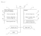
- FIG. 2 shows a configuration of an apparatus for producing a three-dimensional molded model according to a first embodiment.
- the apparatus for producing a three-dimensional molded model is a common computer including a CPU 1, a memory 3, a display unit 6, an input unit 5, and a HDD 7.
- the input unit 5 includes a keyboard and a mouse
- the display unit 6 includes a liquid-crystal display.
- the memory 3 is used for data storage, including a CAD data storage unit 31 for storing three-dimensional geometry data and the like, a primitive geometry data storage unit 32 used for Boolean operations, and a feeling equivalence parameter storage unit 33.
- the three-dimensional geometry modeling 2 is a program itself that the computer reads from the HDD 7 onto the memory 3 and runs.
- the three-dimensional geometry modeling 2 includes a resin type input unit 21 for prompting a user to input the type of resin through the display unit 6, an internal configuration parameter input unit 22 for prompting an input of an internal configuration parameter included in a feeling equivalence parameter table, and a Boolean operation unit 23 for performing a Boolean operation.
- FIG. 3 shows a flow of a method for producing a three-dimensional molded model according to the first embodiment.
- the flow of the method for producing a three-dimensional molded model includes two stages of setting for visualization (S12 to S14) and feeling approximation (S21 to S23) that are performed in parallel.
- a three-dimensional geometry of a body part to be molded is extracted from brightness information of output data (DICOM data) from a CT/MRI apparatus and processed into data (S11).
- DICOM data brightness information of output data
- STL data is then created for each of a body part and an internal organ thereof (S12).
- the STL data is modified (S13).
- the STL data is edited using a modeling feature (S14).
- the steps S13 and S14 are both performed. It is noted that the step S13 may be omitted to only perform the step S14.
- an internal structure pattern, a pattern size, and a pattern interval are defined in a feeling equivalence parameter table for each of the body part and the internal structure (S21).
- the types and the blend ratio of resins used for molding are defined in the feeling equivalence parameter table for each of the body part and the internal organ thereof (S22).
- a Boolean operation is performed (S23).
- Three-dimensional geometry data after the Boolean operation is sent to a three-dimensional printer to mold a three-dimensional model (S15).
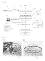
- FIG. 4 illustrates a processing flow of a program that runs on the apparatus for producing a three-dimensional molded model according to the first embodiment.
- DICOM data 42 is retrieved from a CT or an MRI apparatus (CT/MRI apparatus 41).
- CT/MRI apparatus 41 CT/MRI apparatus 41.
- the data is stored in the HDD in the computer as an STL data file in the geometry data creating step (S41).
- the STL data is read using commercially-available three-dimensional geometry software and edited using a modeling feature or the like (geometry data editing step: S42).
- a feeling equivalence parameter table is prepared in the computer using commercially-available table building software 45 (table preparing step: S43).
- the types and the blend ratio of resins are input as a table (resin type defining step: S44).
- a feeling equivalence parameter database (D/B) is thus created through the steps S43 and S44.
- the intended three-dimensional molded model is then produced in the Boolean operation step S45.
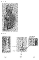
- FIG. 5 shows an exemplary three-dimensional molded model.
- FIG. 5(1) is a partially enlarged view of the three-dimensional molded model of a liver.
- a standard character/symbol 51 including a patient's information is drawn.
- FIG. 5(2) shows a state where a scaler 52 is embedded in the three-dimensional molded model.
- barcode information may also be embedded.
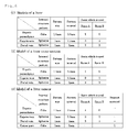
- FIG. 6 illustrates feeling equivalence parameter tables of three-dimensional molded models of a liver.
- FIG. 6 (1) is a table showing exemplary internal configuration parameters required for a three-dimensional molded model of a normal liver.
- FIG. 6(2) is a tables howing exemplary internal configuration parameters required for a three-dimensional molded model of a liver cross-section.
- FIG. 6 (3) is a table showing exemplary internal configuration parameters required for a three-dimensional molded model of a liver cancer.
- the table of FIG. 6(1) shows that using two types of resins A and B, hepatic parenchyma, hepatic vein, and portal vein are formed into a three-dimensional molded model as a liver internal structure. Hepatic parenchyma, hepatic vein, and portal vein are molded of the resin A or B or a blend of the resins A and B .
- hepatic parenchyma is molded of the resin A (at a blend ratio of 1: 0 (resin A:B)), hepatic vein is molded of the resin B (at a blend ratio of 0:1 (resin A:B)), and portal vein is molded of a composite resin having a blend ratio of 3:1 (resin A:B).
- a liver model can be produced by performing a Boolean operation of logical sum of the three-dimensional geometries of hepatic parenchyma, hepatic vein, and portal vein.
- each internal structure has an internal structure pattern, a pattern size, and a pattern interval as parameters.
- hepatic parenchyma has an internal structure pattern in which cubes with sides of 1.5 mm exist at intervals of 0.5 mm.
- a Boolean operation of logical sum or logical difference between the internal structure pattern and the three-dimensional geometry of hepatic parenchyma produces a model providing a feeling of touch approximate to that of the actual obj ect.
- a model material is used as the resin for the internal structure pattern.
- a support material is used as the resin for the internal structure pattern.
- the table of FIG. 6(2) includes the same information as that in the table of FIG. 6(1)
- FIG. 6(2) is for a three-dimensional molded model of a liver cross-section, with which the internal structure can be observed in detail through the cross-section.
- Respective moldedmodels of the internal structure, which are exposed to outside through the cross-section, are all molded of model materials. Molding using, for example, a support material is not allowed.
- the table of FIG. 6(3) includes information of a cancer part as an affected part, in addition to the information included in the table of FIG. 6(1) .
- the table of FIG. 6(3) shows that a support material is used for the cancer part.
- the cancer part which is entirely surrounded by hepatic parenchyma, can be molded using a support material.
- FIG. 7 illustrates feeling equivalence parameter tables of three-dimensional molded models of a bone.
- FIGs. 8 and 9 show exemplary three-dimensional molded models of a bone and a muscle, respectively.
- FIG. 7(1) (a) is an image of a molded model of a normal bone.
- FIG. 7(1)(b) is an exemplary feeling equivalence parameter table of the molded model of the normal bone.
- FIG. 7(1)(b) shows that a cube of a primitive geometry is defined as an internal structure pattern. The pattern size is 1.5 mm and the pattern interval is 1 mm.
- a resin is used having a blend ratio of 1:1 (resin A:B).
- the geometry of the bone itself as a body part is formed of a resin having a blend ratio of 1:1 (resin A:B) and a logical difference is performed with the cube of the primitive geometry to subtract the cube and thereby to produce a molded model providing a feeling of touch equivalent to the hardness of the bone.
- the pattern size is equal to the length of the sides of the cube.
- the pattern interval is equal to the distance between the side surfaces of the cube.
- FIG. 7(2)(c) is an image of a molded model of an osteoporoticbone.
- FIG. 7(2) (d) is an exemplary feeling equivalence parameter table of the molded model of the osteoporotic bone.
- FIG. 7(2)(d) shows that a cube of a primitive geometry is defined as an internal structure pattern. The pattern size is 4 mm and the pattern interval is 0.5 mm. A resin is used having a blend ratio of 1:1 (resin A:B).
- the geometry of the bone itself as a body part is formed of a resin having a blend ratio of 1:1 (resin A:B) and a logical difference is performed with the cube of the primitive geometry to subtract the cube and thereby to produce a molded model providing a feeling of touch equivalent to the hardness of the bone.
- the pattern size is equal to the length of the sides of the cube.
- the pattern interval is equal to the distance between the side surfaces of the cube.
- the molded model of the osteoporotic bone undergoes primitive geometry subtraction of a larger volume and has a larger gap to adjust the hardness.
- FIG. 8 shows cross-sections perpendicular to the longitudinal direction of a bone model 61 ( FIG. 8(2) ) and a schematic view of an internal structure ( FIG. 8(3) ).
- the cross-sections of the bone model 61 include a cross-section formed only with a resin C ( FIG. 8 (2) (a)) and cross-sections after a logical difference with different primitive geometry sizes ( FIG. 8 (2) (b) to (d)).
- a support material is to be used for molding and to be subtracted after the model material is hardened, that is, to be subtracted by the size of the primitive geometry. Accordingly, the hardness of the bone model reduces with the change from FIG. 8(2)(a) to FIG. 8(2)(d) .
- FIG. 9 shows cross-sections perpendicular to the longitudinal directionofamusclemodel 62 ( FIG. 9(2) ) and a schematic view of an internal structure ( FIG. 9(3) ).
- the cross-sections of the muscle model 62 include a cross-section formed only with a support material ( FIG. 9 (2) (a)) and cross-sections after a logical sum with different pattern intervals of a primitive geometry formed of a resin C harder than the support material ( FIG. 9(2)(b) to (d)).
- FIG. 9(2)(a) shows the highest flexibility, and the number of structure patterns of the primitive geometry increases and thereby the hardness of the muscle model also increases with the change from FIG. 9(2)(b) to FIG. 9(2)(d) .
- FIG. 10 is an external view of a support tool for medical treatment, medical training, research, and education, showing slices 73 with a predetermined thickness of a three-dimensional molded model from the waist to bust line of a body obtained by the method for producing a three-dimensional molded model according to the present invention. Cross-sections of the backbone and the lungs can be observed. These slices are overlaid 70 to form the original three-dimensional geometry, and an axis 71 corresponding to the backbone is passed through the geometry so that the geometry is freely rotatable about the axis 71. This allows the entire geometry and the cross-section structure to be grasped at the same time, which is useful as a tool for medical care support and medical education.
- FIG. 11 is an external view of a liver model.
- the whole of the geometry is hepatic parenchyma 80 formed of a translucent resin. Internal structures such as a hepatic vein can be observed inside the hepatic parenchyma 80.
- a soft translucent resin is used as a model material for the hepatic parenchyma 80 and, according to feeling equivalence parameters, an internal structure pattern is further provided inside the hepatic parenchyma 80 so that the hepatic parenchyma 80 provides a feeling of touch approximate to that of the actual object.
- FIG. 12 is an external view of a pregnant abdomen model.
- the whole of the geometry is a cut-out pregnant abdomen 91 formed of a translucent resin.
- the pregnant abdomen 91 and a fetus 92 therein can be observed.
- the shape, size, direction, limbs, and positional relationship in the geometry of the abdomen of the fetus can be identified at a glance.
- a soft translucent resin is used as a model material for the pregnant abdomen 91 and, according to feeling equivalence parameters, an internal structure pattern is further provided inside the pregnant abdomen 91 so that the pregnant abdomen 91 provides a feeling of touch approximate to that of the actual obj ect. Similarly, according to feeling equivalence parameters, an internal structure pattern is also provided inside the fetus 92 so that the fetus 92 provides a feeling of touch approximate to that of the actual object.
- FIGs. 13 and 14 are external views of a face and upper body model.
- the reference symbol 101 denotes a skull
- 102 denotes an ear cartilage
- 103 denotes a nose cartilage
- 104 denotes a humerus
- 105 denotes a heart.
- ribs, breastbones, other bones, teeth, lungs, etc. can be observed.
- the entire skin and the ear and nose cartilages are molded of a transparent resin.
- Internal structures including the skull, ribs, breastbones, and other bones are molded of a hard resin.
- the heart is molded of a soft resin. According to respective feeling equivalence parameters, an internal structure pattern is provided inside each of the molded models so that the molded model provides a feeling of touch approximate to that of the actual object.
- FIG. 15 illustrates a bone model.
- the bone model shown in FIG. 15 has an internal structure including a cortical bone 110 as a surficial portion of a bone and a cancellous bone (112, 114) as an interior portion.
- the cancellous bone includes a surficial portion 112 (adj acent to the cortical bone and having a predetermined thickness) and an interior portion 114.
- the cortical bone 110 is molded of a hard resin. According to feeling equivalence parameters, an internal structure pattern is provided inside the cortical bone 110 so that the cortical bone 110 provides feeling of hardness approximate to that of the actual object.
- the surficial portion 112 of the cancellous bone has a hardness slightly different from that of the cortical bone 110.
- an internal structure pattern is provided inside the surficial portion 112 of the cancellous bone different from that inside the cortical bone 110 so that the difference in the hardness approximates that of the actual object.
- the interior portion 114 of the cancellous bone is most brittle and, according to feeling equivalence parameters, an internal structure pattern is provided so that the brittleness approximates that of the actual object.
- the interior portion 114 of the cancellous bone has a grid-like structure in which the primitive geometry of the internal structure pattern is removed.
- Three-dimensional molded models produced according to the present invention are useful for informed consent, decision on courses of treatment, and medical care support, and also as support tools for medical education, medical research, and general education, and further as teaching materials for anatomy and surgery because such models can simulate the feeling of touch.
- models of a blood vessel and a parenchyma organ molded of a rubber-like soft resin are useful not only as support tools with which a three-dimensional dissection can be taken in hand to understand, but also as simulators most suitable for arthroscopy procedure training when installed in a laparoscopic surgery training box.
Abstract
Description
- The present invention relates to a technique of forming a three-dimensional molded model with multiple types of materials based on three-dimensional digital data of a medical image obtained from a medical diagnostic apparatus such as X-ray CT or MRI.
- In the field of medicine, there is a growing need for three-dimensional visualization of affected parts and/or specific body parts in the processes of, for example, informed consent, decision on courses of treatment, medical education, and medical research. In particular, in the case of three-dimensional visualization utilizing a three-dimensional molded model, not only the sense of vision but also the sense of touch of the actual three-dimensional geometry allows transmission of a large amount of information that cannot be transmitted completely through computer images.
- There has conventionally been known creating three-dimensional geometry data using DICOM (Digital Imaging and Communications in Medicine), a standard for medical diagnostic apparatuses such as X-ray CT and MRI (Magnetic Resonance Imaging), and based on the data, producing a three-dimensional molded model for medical use rapidly and precisely with a plaster-based material using a powder-laminating molding machine.
- There have, however, been problems that the softness of complex organs, such as a liver, having an internal structure cannot be simulated and that information on the feeling of touch of an organ or the like cannot be provided to doctors and/or nurses handling organs, etc.
- Combinations and collaborations between medical services and engineering having been rapidly developing, there has recently been proposed a simulator program utilizing an advanced engineering calculation method. The simulator program is arranged to reconstruct body parts to be operated polygonally on a computer screen to simulate the surface of an organ such as a liver.
- Meanwhile, there has been known a three-dimensional printer with which a three-dimensional molded model using hard and flexible resins having their respective different mechanical properties can be produced by simultaneously forming jets of the resins and combining them. With such a three-dimensional printer, it is possible to reproduce not only the surface but also the internal structure of the target form structure. However, there are only a few such printers that can reproduce the flexibility of complex organs, such as a liver, having an internal structure or the hardness of bones and the like.
- As a related art, there has been proposed a three-dimensional molded object production method with which a multi-color and multi-material three-dimensional object having a complex structure and also having different hardness at different parts thereof can be molded by holding and fixing multiple types of model materials in one layer at their respective molding positions in a holder sheet having a web structure in which molding model materials can be held, placing a next-layer holder sheet on the fixed layer of the model materials, holding and fixing multiple types of next-layer model materials at their respective molding positions in the next-layer holder sheet, repeating fixing model materials sequentially to upper layers, and after laminating the layers, solving and removing the holder sheets (see Patent Document 1). However, the method disclosed in
Patent Document 1 requires hard or soft model materials so that different parts have different hardness accordingly. -
- [Patent Document 1]
WO2005/037529 - The main purpose of using conventional three-dimensional molded models has been to determine the geometry. Product designers have demanded high-precision molded models with which even smooth geometries and detailed parts can be reproduced. It is however difficult to imagine the feeling of touch and/or the feeling of use only from the geometry. What is needed is a molded model not only having geometry but also providing a feeling of touch, feeling of use, texture, etc., approximate to those of the actual object.
- Under the circumstances described above, it is an object of the present invention to provide a three-dimensional molded model production method with which multiple types of materials can be produced into another material having different mechanical properties and, by thus combining materials, feeling of touch of an organ or the like that professionals including physicians have can be reproduced using a three-dimensional printer with which a three-dimensional molded model can be produced.
- In order to achieve the foregoing object, the present invention provides a method for producing a three-dimensional molded model using a three-dimensional printer that uses at least two types of materials as model materials, the method including at least the steps of:
- 1) extracting a three-dimensional geometry of a body part to be molded from brightness information of two-dimensional data obtained from a medical diagnostic apparatus and creating three-dimensional geometry data of the body part and an internal structural part thereof;
- 2) editing the three-dimensional geometry data of the body part and the internal structural part thereof using a modeling feature;
- 3) preparing a feeling equivalence parameter table including, as parameters, at least an internal structure pattern, a pattern size, and a pattern interval that specify a primitive geometry of the body part and the internal structural part;
- 4) defining the types and the blend ratio of model materials and the types of support materials used for molding for each of the body part and the internal structural part and adding the types and the blend ratio to the feeling equivalence parameter table;
- 5) creating primitive geometry data with parameters in the feeling equivalence parameter table and performing a Boolean operation of either logical sum, logical difference, or logical product between part data of the body part and the internal structural part obtained in the preceding step 2) and the primitive geometry data; and
- 6) performing molding with the three-dimensional printer using the materials defined in the preceding step 4) based on the three-dimensional geometry data of the body part and the internal structural part obtained in the preceding step 5).
- Here, the medical diagnostic apparatus may be a CT, an MRI, or a PET (Positron Emission Tomography) machine and is arranged to extract a three-dimensional geometry of a body part to be molded from brightness information of two-dimensional data using DICOM (Digital Imaging and Communications in Medicine), a standard for medical digital image format and communications protocol.
- Here, MRI images have a higher resolution of soft tissues than CT images, with which the physical property value of many tissues can be obtained. PET is mainly used for cancer diagnoses or the like and PET images are utilized with CT/MRI images to produce a three-dimensional molded model of a cancerous organ. CT images, MRI images, or PET images, or a combination thereof may be used. One may be for the contour of a body part, while the other may be for tissue images of an internal structural part.
- The step 1) includes extracting a three-dimensional geometry of a body part to be molded from brightness information of two-dimensional data obtained from a medical diagnostic apparatus and creating three-dimensional geometry data of the body part and an internal structural part thereof. The medical diagnostic apparatus provides dot information of tomographic images including DICOM-format brightness information, and the tomographic images are overlaid to extract a three-dimensional geometry of a body part to be molded. Further, commercially-available three-dimensional image CAD software is used to create three-dimensional geometry data of the body part and the internal structural part thereof. The three-dimensional geometry data of the body part and the internal structural part thereof created using the three-dimensional image CAD software undergoes adjustment of the patch surface orientation, removal of unnecessary shells, supplement of gaps, patch reduction, and smoothing of the contour geometry to thereafter be stored in, for example, an STL (Standard Triangulated Language) data format.
- Next, the step 2) includes editing the three-dimensional geometry data created in the preceding step 1) using a modeling feature. Here, the modeling feature, which is included in commercially-available three-dimensional image CAD software, includes preparing a cross-section (including partial cross-section) geometry, preparing a plane, a curved surface, and/or a primitive geometry, geometry cutting, adding a sign/character, and adding a marking symbol, etc.
- The step of editing geometry data preferably includes at least one of character/sign addition processing, marking symbol addition processing, scaler addition processing, and barcode addition processing. The three-dimensional molded model is intended to be utilized in the field of medicine for three-dimensional visualization of affected parts and/or specific body parts in the processes of, for example, informed consent, decision on courses of treatment, medical education, and medical research. Addition processing of a character/sign to the three-dimensional geometry data allows patient's personal information and/or imaging date to be added to the three-dimensional molded model as character information. Addition processing of a marking symbol to the three-dimensional geometry data allows information useful for informed consent and/or decision on courses of treatment to be added to the three-dimensional molded model. Also, addition processing of a scaler to the three-dimensional geometry data allows information useful for decision on courses of treatment and/or actual operations to be added to the three-dimensional molded model. Moreover, addition processing of a barcode to the three-dimensional geometry data allows a patient's information to be added to the three-dimensional molded model.
- Next, the step 3) includes preparing a feeling equivalence parameter table for each of the body part and the internal structural part. Here, the feeling equivalence parameter table includes information data for use in reproducing the feeling of touch of the body part and the internal structural part. Specifically, the feeling equivalence parameter table defines an internal structure pattern, a pattern size, and a pattern interval that specify a primitive geometry.
- The body part represents living tissues of not only humans and animals but also plants. For example, in the case of humans, the body part includes an organ such as a liver and a heart, and otherwise includes an organ of a pet, such as dogs and cats, and further a plant fruit. The internal structural part represents structural parts inside living tissues, including bone, fat, blood vessel, etc.
- Defining an internal structure pattern that specifies a primitive geometry means defining the identification number of a primitive geometry such as a cylinder or a rectangular parallelepiped as an internal structure. Here, the primitive geometry may be a geometrically basic one such as a cylinder, an elliptic cylinder, a rectangular parallelepiped, a cube, a polygonal column, a sphere, a wedge, a pyramid, or a cone or may be an applied one such as a combination of basic geometries or a unique geometry.
- Defining a pattern size means defining the size of a primitive geometry, for example, the radius of a sphere, the height, width, and thickness of a rectangular parallelepiped. Defining a pattern interval means defining the arrangement intervals of primitive geometries, for example, the distance between the centers of gravity or the side surfaces of adjacent primitive geometries.
- Next, the step 4) includes defining the types and the blend ratio of model materials used for molding for each of the body part and the internal structural part and adding the types and the blend ratio to the feeling equivalence parameter table.
- Here, the materials may preferably include some resins, such as hard resin and flexible resin. Without being limited to resin, plaster powder, plastic powder, metal powder, or wax may be used. Forexample, assuming using resin for the materials, it is not possible to freely select any resin (s) which can be used due to limitations of the three-dimensional printer that performs three-dimensional molding. To address this problem, the blend ratio of the materials is defined so that some resins can be blended.
- The model materials are molding materials forming the three-dimensional molded model. The support materials are for supporting the model materials during molding. The model materials include various characteristics such as translucency, color, flexibility,andstiffness. On the other hand, the support materials should be removed easily after being hardened. An ultrasonic cleaner and/or solution may be used to remove support materials from the molded object. Acrylic resins, for example, are preferably used for the model materials and the support materials. In the case of molding an internal structure surrounded by other structure(s), the above-described support materials may be used as the materials for molding the structure.
- Defining the blend ratio means, in the case of using a three-dimensional printer capable of simultaneously forming jets of two types of resins serving as model materials, for example, controlling the blend ratio of the resins, whereby intended hardness or softness can be achieved.
- At least one type of the model materials is preferably composed of a translucent material. The translucent material causes the internal structure to be see-through, resulting in an improvement in the spatial recognition. This facilitates grasping the internal structure, for which at least one type of the model materials is thus composed of a translucent material to be useful for informed consent, decision on courses of treatment, medical research, and medical education, for example.
- Next, the step 5) includes creating primitive geometry data with parameters in the feeling equivalence parameter table and performing a Boolean operation of either logical sum, logical difference, or logical product between part data of the body part and the internal structural part obtained in the preceding step of editing data and the primitive geometry data.
- Here, the Boolean operation is an approach for molding with three-dimensional computer graphics and CAD through a set operation of sum, difference, and/or product between geometries with volume. Logical sum is an operation in which a geometry is integrated with another geometry. Logical difference is an operation in which a geometry trims another geometry. Logical product is an operation in which a portion of a geometry overlapped with another geometry is left.
- The present invention is characterized by defining feeling equivalence parameters such that the hardness of materials used in a molded model is approximate to the feeling of touch of the actual object and performing a Boolean operation according to the parameters to define a material providing a feeling of touch approximate to that of the actual object.
- Next, the step 6) includes performing molding with the three-dimensional printer using the materials defined in the preceding step of defining the material types based on the three-dimensional geometry data of the body part and the internal structural part obtained in the preceding Boolean operation step.
- The three-dimensional printer is arranged to mold a three-dimensional object with an inkjet ultraviolet curing method using acrylic photo-curable resin, a fused deposition method using ABS resin, or a powder fixing method using powder, though not limited thereto. Various methods may be employed as long as they are capable of molding a three-dimensional object using multiple types of materials.
- The materials used as model materials are preferably selectable from a variety of materials with which parameters can be controlled, such as color, optical transparency, high softness, radiotransparency, ultrasonic sensitivity, scintillation sensitivity, heat ray sensitivity, and conductivity.
- In the step of creating geometry data in the method for producing a three-dimensional molded model, the three-dimensional geometry size of the internal structural part may be reduced to be smaller than the volume of the internal structural part of the body part, so that only the internal structure can be extracted from the body part. The void resulting from the reduction in the three-dimensional geometry size of the internal structural part may be filled with the support materials during molding and the support materials may be removed after being hardened.
- For example, if the body part can be divided into two parts, the internal structure can be extracted separately through the divided faces. This allows the internal structure to be examined separately in more detail.
- In the above-described method for producing a three-dimensional molded model according to the present invention, in the case of providing a feeling of softness for at least one of the body part and the internal structural part, the Boolean operation step may include performing a logical sum between the part data defined with a first soft material and the primitive geometry data defined with a second soft material.
- The logical sum between the part data defined with a first soft material and the primitive geometry data defined with a second soft material means, for example, adding a rubber-like soft material to another soft material like the support materials. Mixing a primitive geometry that provides a desired feeling of softness and/or cutting/sectioning with a rubber-like model material allows a feeling of touch approximate and equivalent to the actual softness to be reproduced. As mentioned above, the primitive geometry may be selected from various types, that is, may be a geometrically basic one such as a cylinder, an elliptic cylinder, a rectangular parallelepiped, a cube, a polygonal column, a sphere, a wedge, a pyramid, or a cone or may be an applied one such as a combination of basic geometries or a unique geometry. The type, size, number/amount of mixing, and method of mixing of the primitive geometry are adjusted to control the softness and thereby provide the desired feeling of touch.
- This is mainly useful for reproducing the feeling of touch of soft tissues such as fat, muscle, and blood vessel.
- Alternatively, in the above-described method for producing a three-dimensional molded model according to the present invention, in the case of providing a feeling of softness for at least one of the body part and the internal structural part, the Boolean operation step may include performing a logical difference between the part data defined with a first soft material and the primitive geometry data defined with no material. In the logical difference with the primitive geometry data defined with no material, the support materials are to be automatically assigned to the primitive geometry data. If the part of the primitive geometry is a hermetically-closed internal structure, the support materials cannot be removed due to cleaning and the like. Accordingly, the softness of the support materials is to be added to the nature of the interior of the entire geometry. On the contrary, if the part of the primitive geometry is not a hermetically-closed internal structure, for example, gaps are open in a grid-like manner, the support materials are to be removed due to cleaning and the like, which forms a void at the part of the primitive geometry. This means, for example, subtracting the primitive geometry from a part composed of, for example, a rubber-like soft model material. Subtracting the primitive geometry from a part that provides a desired feeling of softness and/or cutting/sectioning with a rubber-like material allows a feeling of touch approximate and equivalent to the actual softness to be reproduced. The type, size, number/amount of subtraction, and method of subtraction of the primitive geometry to be subtracted are adjusted to control the softness and thereby provide the desired feeling of touch.
- This is mainly useful for reproducing the feeling of touch of soft tissues such as fat, muscle, and blood vessel.
- Further, in the above-described method for producing a three-dimensional molded model according to the present invention, in the case of providing a feeling of hardness for at least one of the body part and the internal structural part, the Boolean operation step may include performing a logical sum between the part data defined with a first hardmaterial and the primitive geometry data defined with a second hard material.
- The logical sum between the part data defined with a first hard material and the primitive geometry data defined with a second hard material means adding a hard molding material with a harder molding material. Mixing a primitive geometry that provides a desired feeling of hardness with a hard molding material allows a feeling of touch approximate and equivalent to the actual hardness to be reproduced. Thetype, size, number/amount of mixing, and method of mixing of the primitive geometry are adjusted to control the hardness and thereby provide the desired feeling of touch.
- This is mainly useful for reproducing the feeling of touch of hard tissues such as bone and feeling of cutting and/or sectioning.
- Alternatively, in the above-described method for producing a three-dimensional molded model according to the present invention, in the case of providing a feeling of hardness for at least one of the body part and the internal structural part, the Boolean operation step may include performing a logical difference between the part data defined with a first hard material and the primitive geometry data defined with no material.
- The logical difference between the part data defined with a first hard material and the primitive geometry data defined with no material means subtracting a detailed primitive geometry from the interior of a geometry part formed of a hard molding material to reproduce the feeling of touch approximate and equivalent to the actual feeling of cutting/sectioning. The type, size, number/amount of subtraction, and method of subtraction of the primitive geometry to be subtracted are adjusted to control the hardness and thereby provide the desired feeling of touch.
- This is mainly useful for reproducing the feeling of touch of hard tissues such as bone and feeling of cutting and/or sectioning.
- Next will be described the definition of the internal structure for body parts for which a three-dimensional molded model can be produced and effectively used in particular.
- In the step of preparing a table in the above-described method for producing a three-dimensional molded model according to the present invention, if the body part is a liver, one or more of hepatic parenchyma, hepatic vein, portal vein, bile duct, and the affected part may be selected as the internal structural part to define the internal structure pattern, the pattern size, and the pattern interval thereon.
- If the body part is a liver, the definition is preferably made on at least hepatic artery, hepatic vein, portal vein, and the affected part. It is more preferable that the definition is made on hepatic parenchyma and bile duct.
- In the step of preparing a table in the above-described method for producing a three-dimensional molded model according to the present invention, if the body part is a pregnant abdomen, one or more of uterus, fetus, umbilical cord (navel cord), placenta, amniotic fluid, blood vessel, and subcutaneous fat may be selected as the internal structural part to define the internal structure pattern, the pattern size, and the pattern interval thereon.
- Molding reproducing the feeling of touch of a fetus makes it possible to qualify prenatal diagnosis and safety of Cesarean section, and to determine whether or not the fetus can become tangled in the umbilical cord (navel cord) during childbirth, and reconfirm the sequence of childbirth. The produced three-dimensional molded model can also be utilized as a molded album as a memory of the childbirth.
- In the step of preparing a table in the above-described method for producing a three-dimensional molded model according to the present invention, if the body part is a breast, one or more of subcutaneous fat, mammary gland, mammary duct, lymph duct, lymph node, and breast cancerpartmaybe selectedas the internal structural part to define the internal structure pattern, the pattern size, and the pattern interval thereon. The definition is preferably made on subcutaneous fat, mammary gland, breast cancer part, and lymph duct.
- Molding reproducing the feeling of hardness of a breast cancer part can be utilized for training in breast cancer screening as part of medical education and explanation of disease presentation to an actual patient.
- In the step of preparing a table in the above-described method for producing a three-dimensional molded model according to the present invention, if the body part is a limb, one or more of skin, subcutaneous fat, artery, vein, muscle, bone, and tendon forming the limb and joints may be selected as the internal structural part to define the internal structure pattern, the pattern size, and the pattern interval thereon.
- Molding reproducing the feeling of softness of skin, subcutaneous fat, and blood vessel can be utilized for training of doctors and nurses in injecting and blood sampling.
- The produced model can also be utilized as an artificial arm or an artificial leg.
- In the step of preparing a table in the above-described method for producing a three-dimensional molded model according to the present invention, if the body part is a throat, one or more of esophagus, trachea, and cartilage may be selected as the internal structural part to define the internal structure pattern, the pattern size, and the pattern interval thereon.
- Molding reproducing the geometry and the feeling of touch of esophagus and trachea can be utilized for training of doctors and nurses in inserting an aspirator tube into a patient's body with, for example, pneumonia.
- In the step of preparing a table in the above-described method for producing a three-dimensional molded model according to the present invention, if the body part is a face, one or more of skin, subcutaneous fat, muscle, cartilage, bone, and blood vessel may be selected as the internal structural part to define the internal structure pattern, the pattern size, and the pattern interval thereon.
- This can be utilized for pre-operation confirmation of a cosmetic surgery.
- In the step of preparing a table in the above-described method for producing a three-dimensional molded model according to the present invention, if the body part is a tooth and gum, one or more of tooth, gum, alveolar bone, blood vessel, nerve, jaw bone, jaw muscle, and tongue may be selected as the internal structural part to define the internal structure pattern, the pattern size, and the pattern interval thereon.
- In the step of preparing a table in the above-described method for producing a three-dimensional molded model according to the present invention, if the body part is a digestive tract, the inner wall or the outer wall of the digestive tract may be selected as the internal structural part to define the internal structure pattern, the pattern size, and the pattern interval thereon.
- In the step of preparing a table in the above-described method for producing a three-dimensional molded model according to the present invention, if the body part is a head, one or more of hair, eyeball, brain, brain blood vessel, skin, subcutaneous fat, muscle, ear cartilage, nose cartilage, and skull may be selected as the internal structural part to define the internal structure pattern, the pattern size, and the pattern interval thereon.
- This can be utilized for pre-operation confirmation of a cosmetic surgery.
- Another aspect of the present invention provides a support tool for medical treatment, medical training, research, and education, the support tool formed by slicing at a predetermined thickness a three-dimensional molded model obtained by the above-described method for producing a three-dimensional molded model or by overlaying the slices to form the original three-dimensional geometry and passing an axis through the geometry so that the geometry is freely rotatable about the axis.
- Slicing the produced three-dimensional molded model and overlaying the slices allows the entire geometry and the cross-section structure to be grasped at the same time, which is useful as a tool for medical care support and medical education.
- Another aspect of the present invention provides a support tool for medical treatment, medical training, research, and education, the support tool formed by slicing at a predetermined thickness a three-dimensional molded model obtained by a method for producing a three-dimensional molded model using a three-dimensional printer that uses at least two types of materials as model materials, overlaying the slices to form the original three-dimensional geometry, and passing an axis through the geometry so that the geometry is freely rotatable about the axis, the method including the steps of: extracting a three-dimensional geometry of a body part to be molded from brightness information of two-dimensional data obtained from a medical diagnostic apparatus and creating three-dimensional geometry data of the body part and an internal structural part thereof; editing the three-dimensional geometry data using a modeling feature; defining the types and the blend ratio of model materials used for molding for each of the body part and the internal structural part and adding the types and the blend ratio to the feeling equivalence parameter table; and performing molding with the three-dimensional printer using the materials defined in the preceding step of defining the material types.
- Slicing the produced three-dimensional molded model and overlaying the slices allows the entire geometry and the cross-section structure to be grasped at the same time, which is useful as a tool for medical care support and medical education.
- Next will be described a program for producing a three-dimensional molded model according to the present invention using a three-dimensional printer that uses at least two types of materials as model materials, the program causing a computer to execute the steps of:
- a) extracting a three-dimensional geometry of a body part to be molded from brightness information of two-dimensional data obtained from a medical diagnostic apparatus and creating three-dimensional geometry data of the body part and an internal structural part thereof;
- b) editing the three-dimensional geometry data using a modeling feature;
- c) prompting an input of feeling equivalence parameters including at least an internal structure pattern, a pattern size, and a pattern interval that respectively specify a primitive geometry of the body part and the internal structural part and generating a feeling equivalence parameter table;
- d) prompting an input of the types and the blend ratio of model materials used for molding for each of the body part and the internal structural part and additionally defining the types and the blend ratio to the feeling equivalence parameter table; and
- e) creating primitive geometry data with parameters in the feeling equivalence parameter table and performing a Boolean operation of either logical sum, logical difference, or logical product between part data of the body part and the internal structural part obtained in the preceding step of editing data and the primitive geometry data.
- Causing a computer to execute the steps a) to e) allows molding data of a molded model that provides an approximate feeling of touch to be created utilizing image data obtained from the medical diagnostic apparatus. A three-dimensional molded model that provides an approximate feeling of touch can be produced with a three-dimensional printer by sending three-dimensional geometry data of the body part and the internal structural part obtained in the step e) and material information defined in the step d) to the three-dimensional printer.
- In accordance with the present invention, it is possible to produce a molded model providing a feeling of touch equivalent to that of an organ or the like that professionals including physicians have using a three-dimensional printer that uses at least two types of materials as model materials.
- The present invention is applicable to, for example, medical care services, in which disease states can be evaluated three-dimensionally with reference to a three-dimensional molded model of organs to make a diagnosis and a decision on courses of treatment as well as to determine the effect of treatment. In particular, the present invention is helpful for pre- and post-operation determination of an area of resection and planning of an operative procedure. The present invention is also useful for learning of diagnostic treatment procedures including operations. Further, in the field of organ transplantation, the present invention is applicable to capacity-dependent procedures with a spare organ, and also utilized for explanation to patients and their families.
- In actual surgical operations, operative sites are likely to become visually blocked due to the effects of blood and/or other organs and it is often the case that the operator relies on his/her feeling of touch and/or experience. In such cases, the procedure can be performed at ease by preparing a three-dimensional molded model reproducing the geometry of and the feeling of touch of a patient's body part itself, identifying the geometry of and the feeling of touch of the part to be treated through a doctor' s visual confirmation of the molded model, and then actually performing a treatment procedure on the patient.
- Furthermore, in the field of medical education, a three-dimensional molded model produced according to the present invention can actually be taken in hand to understand the three-dimensional structure of and the feeling of touch of organs and, in particular, to simulate the feeling of touch.
-
-
FIG. 1 shows a flow of a method for producing a three-dimensional molded model according to the present invention. -
FIG. 2 shows a configuration of an apparatus for producing a three-dimensional molded model according to a first embodiment. -
FIG. 3 shows a flow of a method for producing a three-dimensional molded model according to the first embodiment. -
FIG. 4 illustrates a processing flow of a program that runs on the apparatus for producing a three-dimensional molded model according to the first embodiment. -
FIG. 5 shows an exemplary three-dimensional molded model. -
FIG. 6 illustrates feeling equivalence parameter tables of three-dimensional molded models of a liver. -
FIG. 7 illustrates feeling equivalence parameter tables of three-dimensional molded models of a bone. -
FIG. 8 shows exemplary three-dimensional molded models of a bone. -
FIG. 9 shows exemplary three-dimensional molded models of a muscle. -
FIG. 10 is an external view of a support tool for medical treatment, medical training, research, and education. -
FIG. 11 is an external view of a liver model. -
FIG. 12 is an external view of a pregnant abdomen model (1). -
FIG. 13 is external views of a face and upper body model (2). -
FIG.14 is external views of a face and upper body model. -
FIG. 15 illustrates a bone model. - Embodiments of the present invention will be described in detail below with reference to the drawings. The present invention is not limited to the following embodiment and examples of shown in the figure, and the present invention can be variously changed in design.
-
FIG. 1 shows a flow of a method for producing a three-dimensional molded model according to the present invention. As shown inFIG. 1 , the method for producing a three-dimensional molded model according to the present invention performs S1 to S8 in the followingsteps 1 to 6. In this flow, S2 and S3 are performed to configure a visualization setting of a three-dimensional geometry and S4 to S7 are performed to configure a feeling equivalence setting. - (S1) Extract a three-dimensional geometry of a molding pair from brightness information of two-dimensional data from a medical diagnostic apparatus.
- Dot information of tomographic images including DICOM-format brightness information is obtained from a CT or an MRI apparatus and the tomographic images are overlaid to extract a three-dimensional geometry of a body part to be molded.
- (S2) Create three-dimensional geometry data of the body part and an internal structural part thereof.
- Three-dimensional geometry data of the body part and the internal structural part thereof created using commercially-available three-dimensional image CAD software undergoes adjustment of the patch surface orientation, removal of unnecessary shells, supplement of gaps, patch reduction, and smoothing processing of the contour geometry or the like to be in the STL data format.
- (S3) Edit the three-dimensional geometry data using a modeling feature.
- Using commercially-available three-dimensional image CAD software, a character/sign is added to thereby add a patient's personal information and/or imaging date or the like to the three-dimensional geometry data as character information. A marking symbol is added to thereby add information useful for decision on courses of treatment or the like to the three-dimensional geometry data. A scaler is added to thereby add information useful for actual operations to the three-dimensional geometry data. A barcode is added to thereby add a patient' s information to the three-dimensional geometry data.
- (S4) Prepare a feeling equivalence parameter table for each of the body part and the internal structural part.
- Using commercially-available table building software, a feeling equivalence parameter table is prepared. The feeling equivalence parameter table defines an internal structure pattern, a pattern size, and a pattern interval that specify a primitive geometry. Some internal structure patterns are arranged in advance and selected by a user with, for example, the pattern number.
- (S5) Define the types and the blend ratio of model materials used for molding for each of the body part and the internal structural part.
- The model materials are selectable from a variety of resins, if used as an example, with which parameters can be controlled, such as color, optical transparency, high softness, radiotransparency, ultrasonic sensitivity, scintillation sensitivity, heat ray sensitivity, and conductivity. Support materials may also be used as model materials for molding.
- If it is possible to simultaneously form jets of two types of resins as model materials, two resins are selected as the model materials and the blend ratio is defined (e.g. (resin A):(resin B) = 1:3).
- (S6) Create primitive geometry data with parameters in the feeling equivalence parameter table.
- A primitive geometry is defined based on the feeling equivalence parameter table information. The primitive geometry may be formed of a model material, formed not of a model material but of a support material, or may be subtracted from a model material by removing a support material.
- (S7) Perform a Boolean operation between part data of the body part and the internal structural part and the primitive geometry data.
- A primitive geometry is added or subtracted to/from part data formed of a model material or a support material.
- (S8) Perform molding with a three-dimensional printer using the defined materials.
- Using a commercially-available three-dimensional printer, the three-dimensional molded model is produced. For example, a three-dimensional printer of OBJET Ltd. may be used. The intended three-dimensional molded model is molded with such a three-dimensional printer using the three-dimensional geometry data of the internal structures of the model and the defined materials.
-
FIG. 2 shows a configuration of an apparatus for producing a three-dimensional molded model according to a first embodiment. As shown inFIG. 2 , the apparatus for producing a three-dimensional molded model is a common computer including aCPU 1, amemory 3, adisplay unit 6, aninput unit 5, and aHDD 7. Specifically, theinput unit 5 includes a keyboard and a mouse, and thedisplay unit 6 includes a liquid-crystal display. Thememory 3 is used for data storage, including a CADdata storage unit 31 for storing three-dimensional geometry data and the like, a primitive geometrydata storage unit 32 used for Boolean operations, and a feeling equivalenceparameter storage unit 33. The three-dimensional geometry modeling 2 is a program itself that the computer reads from theHDD 7 onto thememory 3 and runs. - The three-
dimensional geometry modeling 2 includes a resintype input unit 21 for prompting a user to input the type of resin through thedisplay unit 6, an internal configurationparameter input unit 22 for prompting an input of an internal configuration parameter included in a feeling equivalence parameter table, and aBoolean operation unit 23 for performing a Boolean operation. -
FIG. 3 shows a flow of a method for producing a three-dimensional molded model according to the first embodiment. - As shown in
FIG. 3 , the flow of the method for producing a three-dimensional molded model includes two stages of setting for visualization (S12 to S14) and feeling approximation (S21 to S23) that are performed in parallel. - First, a three-dimensional geometry of a body part to be molded is extracted from brightness information of output data (DICOM data) from a CT/MRI apparatus and processed into data (S11). As a setting for visualization, STL data is then created for each of a body part and an internal organ thereof (S12). The STL data is modified (S13). The STL data is edited using a modeling feature (S14). In the flow shown in
FIG. 3 , the steps S13 and S14 are both performed. It is noted that the step S13 may be omitted to only perform the step S14. - As a setting for feeling approximation, an internal structure pattern, a pattern size, and a pattern interval are defined in a feeling equivalence parameter table for each of the body part and the internal structure (S21). The types and the blend ratio of resins used for molding are defined in the feeling equivalence parameter table for each of the body part and the internal organ thereof (S22).
- After the settings for visualization and feeling approximation, a Boolean operation is performed (S23). Three-dimensional geometry data after the Boolean operation is sent to a three-dimensional printer to mold a three-dimensional model (S15).
-
FIG. 4 illustrates a processing flow of a program that runs on the apparatus for producing a three-dimensional molded model according to the first embodiment. -
DICOM data 42 is retrieved from a CT or an MRI apparatus (CT/MRI apparatus 41). The data is stored in the HDD in the computer as an STL data file in the geometry data creating step (S41). The STL data is read using commercially-available three-dimensional geometry software and edited using a modeling feature or the like (geometry data editing step: S42). - Following or in parallel with these steps, a feeling equivalence parameter table is prepared in the computer using commercially-available table building software 45 (table preparing step: S43). The types and the blend ratio of resins are input as a table (resin type defining step: S44). A feeling equivalence parameter database (D/B) is thus created through the steps S43 and S44. The intended three-dimensional molded model is then produced in the Boolean operation step S45.
-
FIG. 5 shows an exemplary three-dimensional molded model.FIG. 5(1) is a partially enlarged view of the three-dimensional molded model of a liver. A standard character/symbol 51 including a patient's information is drawn.FIG. 5(2) shows a state where ascaler 52 is embedded in the three-dimensional molded model. In addition to the dimension indication with a scaler, barcode information may also be embedded. -
FIG. 6 illustrates feeling equivalence parameter tables of three-dimensional molded models of a liver.FIG. 6 (1) is a table showing exemplary internal configuration parameters required for a three-dimensional molded model of a normal liver.FIG. 6(2) is a tables howing exemplary internal configuration parameters required for a three-dimensional molded model of a liver cross-section.FIG. 6 (3) is a table showing exemplary internal configuration parameters required for a three-dimensional molded model of a liver cancer. - The table of
FIG. 6(1) shows that using two types of resins A and B, hepatic parenchyma, hepatic vein, and portal vein are formed into a three-dimensional molded model as a liver internal structure. Hepatic parenchyma, hepatic vein, and portal vein are molded of the resin A or B or a blend of the resins A and B . Specifically, hepatic parenchyma is molded of the resin A (at a blend ratio of 1: 0 (resin A:B)), hepatic vein is molded of the resin B (at a blend ratio of 0:1 (resin A:B)), and portal vein is molded of a composite resin having a blend ratio of 3:1 (resin A:B). A liver model can be produced by performing a Boolean operation of logical sum of the three-dimensional geometries of hepatic parenchyma, hepatic vein, and portal vein. - As shown in
FIG. 6(1) , each internal structure has an internal structure pattern, a pattern size, and a pattern interval as parameters. For example, hepatic parenchyma has an internal structure pattern in which cubes with sides of 1.5 mm exist at intervals of 0.5 mm. A Boolean operation of logical sum or logical difference between the internal structure pattern and the three-dimensional geometry of hepatic parenchyma produces a model providing a feeling of touch approximate to that of the actual obj ect. In the case of a Boolean operation of logical sum, a model material is used as the resin for the internal structure pattern. In the case of a Boolean operation of logical difference, a support material is used as the resin for the internal structure pattern. - The table of
FIG. 6(2) includes the same information as that in the table ofFIG. 6(1) FIG. 6(2) is for a three-dimensional molded model of a liver cross-section, with which the internal structure can be observed in detail through the cross-section. Respective moldedmodels of the internal structure, which are exposed to outside through the cross-section, are all molded of model materials. Molding using, for example, a support material is not allowed. - The table of
FIG. 6(3) includes information of a cancer part as an affected part, in addition to the information included in the table ofFIG. 6(1) . The table ofFIG. 6(3) shows that a support material is used for the cancer part. The cancer part, which is entirely surrounded by hepatic parenchyma, can be molded using a support material. -
FIG. 7 illustrates feeling equivalence parameter tables of three-dimensional molded models of a bone.FIGs. 8 and 9 show exemplary three-dimensional molded models of a bone and a muscle, respectively. -
FIG. 7(1) (a) is an image of a molded model of a normal bone.FIG. 7(1)(b) is an exemplary feeling equivalence parameter table of the molded model of the normal bone.FIG. 7(1)(b) shows that a cube of a primitive geometry is defined as an internal structure pattern. The pattern size is 1.5 mm and the pattern interval is 1 mm. A resin is used having a blend ratio of 1:1 (resin A:B). - In this case, the geometry of the bone itself as a body part is formed of a resin having a blend ratio of 1:1 (resin A:B) and a logical difference is performed with the cube of the primitive geometry to subtract the cube and thereby to produce a molded model providing a feeling of touch equivalent to the hardness of the bone. Here, the pattern size is equal to the length of the sides of the cube. The pattern interval is equal to the distance between the side surfaces of the cube.
-
FIG. 7(2)(c) is an image of a molded model of an osteoporoticbone.FIG. 7(2) (d) is an exemplary feeling equivalence parameter table of the molded model of the osteoporotic bone.FIG. 7(2)(d) shows that a cube of a primitive geometry is defined as an internal structure pattern. The pattern size is 4 mm and the pattern interval is 0.5 mm. A resin is used having a blend ratio of 1:1 (resin A:B). - In this case, the geometry of the bone itself as a body part is formed of a resin having a blend ratio of 1:1 (resin A:B) and a logical difference is performed with the cube of the primitive geometry to subtract the cube and thereby to produce a molded model providing a feeling of touch equivalent to the hardness of the bone. Here, the pattern size is equal to the length of the sides of the cube. The pattern interval is equal to the distance between the side surfaces of the cube.
- Compared to the molded model of the normal bone, the molded model of the osteoporotic bone undergoes primitive geometry subtraction of a larger volume and has a larger gap to adjust the hardness.
-
FIG. 8 shows cross-sections perpendicular to the longitudinal direction of a bone model 61 (FIG. 8(2) ) and a schematic view of an internal structure (FIG. 8(3) ). The cross-sections of thebone model 61 include a cross-section formed only with a resin C (FIG. 8 (2) (a)) and cross-sections after a logical difference with different primitive geometry sizes (FIG. 8 (2) (b) to (d)). In this case, if the primitive geometry is defined with no material, a support material is to be used for molding and to be subtracted after the model material is hardened, that is, to be subtracted by the size of the primitive geometry. Accordingly, the hardness of the bone model reduces with the change fromFIG. 8(2)(a) to FIG. 8(2)(d) . -
FIG. 9 shows cross-sections perpendicular to the longitudinal directionofamusclemodel 62 (FIG. 9(2) ) and a schematic view of an internal structure (FIG. 9(3) ). The cross-sections of themuscle model 62 include a cross-section formed only with a support material (FIG. 9 (2) (a)) and cross-sections after a logical sum with different pattern intervals of a primitive geometry formed of a resin C harder than the support material (FIG. 9(2)(b) to (d)). FIG. 9(2)(a) shows the highest flexibility, and the number of structure patterns of the primitive geometry increases and thereby the hardness of the muscle model also increases with the change fromFIG. 9(2)(b) to FIG. 9(2)(d) . -
FIG. 10 is an external view of a support tool for medical treatment, medical training, research, and education, showingslices 73 with a predetermined thickness of a three-dimensional molded model from the waist to bust line of a body obtained by the method for producing a three-dimensional molded model according to the present invention. Cross-sections of the backbone and the lungs can be observed. These slices are overlaid 70 to form the original three-dimensional geometry, and anaxis 71 corresponding to the backbone is passed through the geometry so that the geometry is freely rotatable about theaxis 71. This allows the entire geometry and the cross-section structure to be grasped at the same time, which is useful as a tool for medical care support and medical education. -
FIG. 11 is an external view of a liver model. The whole of the geometry ishepatic parenchyma 80 formed of a translucent resin. Internal structures such as a hepatic vein can be observed inside thehepatic parenchyma 80. A soft translucent resin is used as a model material for thehepatic parenchyma 80 and, according to feeling equivalence parameters, an internal structure pattern is further provided inside thehepatic parenchyma 80 so that thehepatic parenchyma 80 provides a feeling of touch approximate to that of the actual object. -
FIG. 12 is an external view of a pregnant abdomen model. The whole of the geometry is a cut-outpregnant abdomen 91 formed of a translucent resin. According to this model, thepregnant abdomen 91 and afetus 92 therein can be observed. The shape, size, direction, limbs, and positional relationship in the geometry of the abdomen of the fetus can be identified at a glance. - A soft translucent resin is used as a model material for the
pregnant abdomen 91 and, according to feeling equivalence parameters, an internal structure pattern is further provided inside thepregnant abdomen 91 so that thepregnant abdomen 91 provides a feeling of touch approximate to that of the actual obj ect. Similarly, according to feeling equivalence parameters, an internal structure pattern is also provided inside thefetus 92 so that thefetus 92 provides a feeling of touch approximate to that of the actual object. -
FIGs. 13 and14 are external views of a face and upper body model. Thereference symbol 101 denotes a skull, 102 denotes an ear cartilage, 103 denotes a nose cartilage, 104 denotes a humerus, and 105 denotes a heart. In addition, ribs, breastbones, other bones, teeth, lungs, etc., can be observed. From the head to the upper body and the upper arms, the entire skin and the ear and nose cartilages are molded of a transparent resin. Internal structures including the skull, ribs, breastbones, and other bones are molded of a hard resin. The heart is molded of a soft resin. According to respective feeling equivalence parameters, an internal structure pattern is provided inside each of the molded models so that the molded model provides a feeling of touch approximate to that of the actual object. -
FIG. 15 illustrates a bone model. The bone model shown inFIG. 15 has an internal structure including acortical bone 110 as a surficial portion of a bone and a cancellous bone (112, 114) as an interior portion. The cancellous bone includes a surficial portion 112 (adj acent to the cortical bone and having a predetermined thickness) and aninterior portion 114. Thecortical bone 110 is molded of a hard resin. According to feeling equivalence parameters, an internal structure pattern is provided inside thecortical bone 110 so that thecortical bone 110 provides feeling of hardness approximate to that of the actual object. Thesurficial portion 112 of the cancellous bone has a hardness slightly different from that of thecortical bone 110. According to feeling equivalence parameters, an internal structure pattern is provided inside thesurficial portion 112 of the cancellous bone different from that inside thecortical bone 110 so that the difference in the hardness approximates that of the actual object. Theinterior portion 114 of the cancellous bone is most brittle and, according to feeling equivalence parameters, an internal structure pattern is provided so that the brittleness approximates that of the actual object. As shown inFIG. 15(b) , theinterior portion 114 of the cancellous bone has a grid-like structure in which the primitive geometry of the internal structure pattern is removed. - In collaboration with a medical school hospital, the effectiveness of using a three-dimensional molded model produced according to the present invention is currently verified on, for example, a reduction in time for procedures in medical practices. As for an implant junction procedure for fractured pelvis, results of about five exemplary evaluations have shown that the time required for the procedure, which is commonly 6 to 7 hours, can be reduced to 5 to 6 hours (i.e. by 1 hour). A reduction in time for procedures is expected to reduce the burden on patients and also hasten postoperative recovery. In the future, three-dimensional molded models produced according to the present invention will be utilized in various medical practices.
- Three-dimensional molded models produced according to the present invention are useful for informed consent, decision on courses of treatment, and medical care support, and also as support tools for medical education, medical research, and general education, and further as teaching materials for anatomy and surgery because such models can simulate the feeling of touch. For example, models of a blood vessel and a parenchyma organ molded of a rubber-like soft resin are useful not only as support tools with which a three-dimensional dissection can be taken in hand to understand, but also as simulators most suitable for arthroscopy procedure training when installed in a laparoscopic surgery training box.
-
- 11
- Geometry data creating step
- 12
- Geometry data editing step
- 13
- Table preparing step
- 14
- Material type defining step
- 15
- Boolean operation step
- 16
- Molding step
- 51
- Standard character/symbol
- 52
- Scaler
- 61
- Bone model
- 62
- Muscle model
- 70
- Overlaid
- 71
- Axis
- 73
- Slices with a predetermined thickness
- 80
- Hepatic parenchyma
- 91
- Pregnant abdomen
- 92
- Fetus
- 101
- Skull
- 102
- Ear cartilage
- 103
- Nose cartilage
- 104
- Humerus
- 105
- Heart
- 110
- Cortical bone
- 112
- Cancellous bone as a surficial portion
- 114
- Cancellous bone as an interior portion
Claims (20)
- A method for producing a three-dimensional molded model using a three-dimensional printer that uses at least two types of materials as model materials, the method comprising at least the steps of:1) extracting a three-dimensional geometry of a body part to be molded from brightness information of two-dimensional data obtained from a medical diagnostic apparatus and creating three-dimensional geometry data of the body part and an internal structural part thereof;2) editing the three-dimensional geometry data of the body part and the internal structural part thereof using a modeling feature;3) preparing a feeling equivalence parameter table including, as parameters, at least an internal structure pattern, a pattern size, and a pattern interval that specify a primitive geometry of the body part and the internal structural part;4) defining the types and the blend ratio of model materials used for molding for each of the body part and the internal structural part and adding the types and the blend ratio to the feeling equivalence parameter table;5) creating primitive geometry data with parameters in the feeling equivalence parameter table and performing a Boolean operation of either logical sum, logical difference, or logical product between part data of the body part and the internal structural part obtained in the preceding step 2) and the primitive geometry data; and6) performing molding with the three-dimensional printer using the materials defined in the preceding step 4) based on the three-dimensional geometry data of the body part and the internal structural part obtained in the preceding step 5).
- The method for producing a three-dimensional molded model according to Claim 1, wherein at least one type of the model materials is composed of a translucent material.
- The method for producing a three-dimensional molded model according to Claim 1, wherein the step 2) includes at least one of character/sign addition processing, marking symbol addition processing, scaler addition processing, and barcode addition processing.
- The method for producing a three-dimensional molded model according to Claim 1, wherein the step 1) includes reducing the three-dimensional geometry size of the internal structural part to be smaller than the volume of the internal structural part of the body part.
- The method for producing a three-dimensional molded model according to any of Claims 1 to 3, wherein in the case of providing a feeling of softness for at least one of the body part and the internal structural part, the step 5) includes:performing a logical sum between the part data defined with a first soft material and the primitive geometry data defined with a second soft material; orperforming a logical difference between the part data defined with a first soft material and the primitive geometry data defined with no material.
- The method for producing a three-dimensional molded model according to any of Claims 1 to 3, wherein in the case of providing a feeling of hardness for at least one of the body part and the internal structural part, the step 5) includes:performing a logical sum between the part data defined with a first hard material and the primitive geometry data defined with a second hard material; orperforming a logical difference between the part data defined with a first hard material and the primitive geometry data defined with no material.
- The method for producing a three-dimensional molded model according to any of Claims 1 to 3, wherein in the case of providing a feeling of hardness for at least one of the body part and the internal structural part, the step 5) includes performing a logical sum or a logical difference between the part data defined with a first hard material and the primitive geometry data defined with a first soft material.
- The method for producing a three-dimensional molded model according to any of Claims 1 to 3, wherein in the step 3), if the body part is a liver, one or more of hepatic parenchyma, hepatic vein, portal vein, bile duct, and the affected part are selected as the internal structural part to define the internal structure pattern, the pattern size, and the pattern interval thereon.
- The method for producing a three-dimensional molded model according to any of Claims 1 to 3, wherein in the step 3), if the body part is a pregnant abdomen, one or more of uterus, fetus, umbilical cord (navel cord), placenta, amniotic fluid, blood vessel, and subcutaneous fat are selected as the internal structural part to define the internal structure pattern, the pattern size, and the pattern interval thereon.
- The method for producing a three-dimensional molded model according to any of Claims 1 to 3, wherein in the step 3), if the body part is a breast, one or more of subcutaneous fat, mammary gland, mammary duct, lymph duct, lymph node, and breast cancer part are selected as the internal structural part to define the internal structure pattern, the pattern size, and the pattern interval thereon.
- The method for producing a three-dimensional molded model according to any of Claims 1 to 3, wherein in the step 3), if the body part is a limb, one or more of skin, subcutaneous fat, artery, vein, muscle, bone, and tendon forming the limb and joints are selected as the internal structural part to define the internal structure pattern, the pattern size, and the pattern interval thereon.
- The method for producing a three-dimensional molded model according to any of Claims 1 to 3, wherein in the step 3), if the body part is a throat, one or more of esophagus, trachea, and cartilage are selected as the internal structural part to define the internal structure pattern, the pattern size, and the pattern interval thereon.
- The method for producing a three-dimensional molded model according to any of Claims 1 to 3, wherein in the step 3), if the body part is a face, one or more of skin, subcutaneous fat, muscle, cartilage, bone, and blood vessel are selected as the internal structural part to define the internal structure pattern, the pattern size, and the pattern interval thereon.
- The method for producing a three-dimensional molded model according to any of Claims 1 to 3, wherein in the step 3), if the body part is a tooth and gum, one or more of tooth, gum, alveolar bone, blood vessel, nerve, jaw bone, jaw muscle, and tongue are selected as the internal structural part to define the internal structure pattern, the pattern size, and the pattern interval thereon.
- The method for producing a three-dimensional molded model according to any of Claims 1 to 3, wherein in the step 3), if the body part is a digestive tract, the inner wall or the outer wall of the digestive tract is selected as the internal structural part to define the internal structure pattern, the pattern size, and the pattern interval thereon.
- The method for producing a three-dimensional molded model according to any of Claims 1 to 3, wherein in the step 3), if the body part is a head, one or more of hair, eyeball, brain, brain blood vessel, skin, subcutaneous fat, muscle, ear cartilage, nose cartilage, and skull are selected as the internal structural part to define the internal structure pattern, the pattern size, and the pattern interval thereon.
- A support tool for medical treatment, medical training, research, and education, the support tool formed by slicing at a predetermined thickness a three-dimensional molded model obtained by the method for producing a three-dimensional molded model according to any of Claims 1 to 3.
- A support tool for medical treatment, medical training, research, and education, the support tool formed by slicing at a predetermined thickness a three-dimensional molded model obtained by the method for producing a three-dimensional molded model according to any of Claims 1 to 3, overlaying the slices to form the original three-dimensional geometry, and passing an axis through the geometry so that the geometry is freely rotatable about the axis.
- A support tool for medical treatment, medical training, research, and education, the support tool formed by slicing at a predetermined thickness a three-dimensional molded model obtained by a method for producing a three-dimensional molded model using a three-dimensional printer that uses at least two types of materials as model materials, overlaying the slices to form the original three-dimensional geometry, and passing an axis through the geometry so that the geometry is freely rotatable about the axis,
the method comprising the steps of:extracting a three-dimensional geometry of a body part to be molded from brightness information of two-dimensional data obtained from a medical diagnostic apparatus and creating three-dimensional geometrydata of the body part and an internal structural part thereof;editing the three-dimensional geometry data using a modeling feature;defining the types and the blend ratio of model materials used for molding for each of the body part and the internal structural part and adding the types and the blend ratio to the feeling equivalence parameter table; andperforming molding with the three-dimensional printer using the materials defined in the preceding step of defining the material types. - A program for producing a three-dimensional molded model using a three-dimensional printer that uses at least two types of materials as model materials, the program causing a computer to execute the steps of:extracting a three-dimensional geometry of a body part to be molded from brightness information of two-dimensional data obtained from a medical diagnostic apparatus and creating three-dimensional geometry data of the body part and an internal structural part thereof ;editing the three-dimensional geometry data using a modeling feature;prompting an input of feeling equivalence parameters including at least an internal structure pattern, a pattern size, and a pattern interval that specify a primitive geometry of the body part and the internal structural part, respectively, and generating a feeling equivalence parameter table;prompting an input of the types and the blend ratio of model materials used for molding for each of the body part and the internal structural part and additionally defining the types and the blend ratio to the feeling equivalence parameter table; andcreating primitive geometry data with parameters in the feeling equivalence parameter table and performing a Boolean operation of either logical sum, logical difference, or logical product between part data of the body part and the internal structural part obtained in the preceding step of editing data and the primitive geometry data.
Applications Claiming Priority (2)
| Application Number | Priority Date | Filing Date | Title |
|---|---|---|---|
| JP2011078876 | 2011-03-31 | ||
| PCT/JP2012/002211 WO2012132463A1 (en) | 2011-03-31 | 2012-03-30 | Method for manufacturing three-dimensional molded model and support tool for medical treatment, medical training, research, and education |
Publications (2)
| Publication Number | Publication Date |
|---|---|
| EP2692509A1 true EP2692509A1 (en) | 2014-02-05 |
| EP2692509A4 EP2692509A4 (en) | 2014-10-22 |
Family
ID=46930233
Family Applications (1)
| Application Number | Title | Priority Date | Filing Date |
|---|---|---|---|
| EP12765020.8A Ceased EP2692509A4 (en) | 2011-03-31 | 2012-03-30 | Method for manufacturing three-dimensional molded model and support tool for medical treatment, medical training, research, and education |
Country Status (6)
| Country | Link |
|---|---|
| US (1) | US9183764B2 (en) |
| EP (1) | EP2692509A4 (en) |
| JP (1) | JP5239037B2 (en) |
| KR (1) | KR101458729B1 (en) |
| CN (1) | CN103153589B (en) |
| WO (1) | WO2012132463A1 (en) |
Cited By (9)
| Publication number | Priority date | Publication date | Assignee | Title |
|---|---|---|---|---|
| CN104269092A (en) * | 2014-09-19 | 2015-01-07 | 成都学创伟业科技有限公司 | Method for manufacturing operation auxiliary skeleton model |
| ES2608203A1 (en) * | 2015-10-05 | 2017-04-06 | Servicio Andaluz De Salud | Customizable device for intervention simulation (Machine-translation by Google Translate, not legally binding) |
| EP3179465A1 (en) * | 2015-12-07 | 2017-06-14 | Humanetics Innovative Solutions, Inc. | Three-dimensionally printed internal organs for crash test dummy |
| EP3273430A1 (en) * | 2016-07-19 | 2018-01-24 | Humanetics Innovative Solutions, Inc. | Three-dimensional components and method of three-dimensional printing of components for crash test dummy |
| EP3273431A1 (en) * | 2016-07-19 | 2018-01-24 | Humanetics Innovative Solutions, Inc. | Three-dimensionally printed internal flesh and organs for crash test dummy |
| CN108437469A (en) * | 2018-03-12 | 2018-08-24 | 保定翰阳科技有限公司 | A kind of individual customized type method based on 3D technology |
| WO2019008355A1 (en) * | 2017-07-06 | 2019-01-10 | Sony Interactive Entertainment Inc. | Generation of a 3d printable model comprising a 3d object encased in a transparent matrix |
| EP3297833A4 (en) * | 2015-05-22 | 2019-02-27 | C/o Canon Kabushiki Kaisha | Image processing apparatus, image processing method, and storage medium |
| US10733911B2 (en) | 2015-10-14 | 2020-08-04 | Humanetics Innovative Solutions, Inc. | Three-dimensional ribs and method of three-dimensional printing of ribs for crash test dummy |
Families Citing this family (102)
| Publication number | Priority date | Publication date | Assignee | Title |
|---|---|---|---|---|
| US8329694B2 (en) | 2005-06-24 | 2012-12-11 | Toyama Chemical Co., Ltd. | Quinoxalinones as antibacterial composition |
| WO2012044753A2 (en) | 2010-10-01 | 2012-04-05 | Applied Medical Resources Corporation | Portable laparoscopic trainer |
| JP5783627B2 (en) * | 2011-04-26 | 2015-09-24 | 国立大学法人 岡山大学 | Human body modeling system |
| KR101963610B1 (en) | 2011-10-21 | 2019-03-29 | 어플라이드 메디컬 리소시스 코포레이션 | Simulated tissue structure for surgical training |
| JP2015503961A (en) | 2011-12-20 | 2015-02-05 | アプライド メディカル リソーシーズ コーポレイション | Advanced surgery simulation |
| AU2013296222B2 (en) | 2012-08-03 | 2017-03-16 | Applied Medical Resources Corporation | Simulated stapling and energy based ligation for surgical training |
| KR102105979B1 (en) | 2012-09-26 | 2020-05-04 | 어플라이드 메디컬 리소시스 코포레이션 | Surgical training model for laparoscopic procedures |
| US10679520B2 (en) | 2012-09-27 | 2020-06-09 | Applied Medical Resources Corporation | Surgical training model for laparoscopic procedures |
| WO2014052478A1 (en) | 2012-09-27 | 2014-04-03 | Applied Medical Resources Corporation | Surgical training model for laparoscopic procedures |
| CA2885302C (en) | 2012-09-27 | 2022-08-02 | Applied Medical Resources Corporation | Surgical training model for laparoscopic procedures |
| US9898937B2 (en) | 2012-09-28 | 2018-02-20 | Applied Medical Resources Corporation | Surgical training model for laparoscopic procedures |
| CA2885314C (en) | 2012-09-28 | 2021-01-19 | Applied Medical Resources Corporation | Surgical training model for transluminal laparoscopic procedures |
| US9226797B2 (en) * | 2012-11-04 | 2016-01-05 | Miba Medical Inc. | Computer aided implantation of body implants |
| EP2938399B1 (en) | 2012-12-28 | 2020-04-15 | Cyberheart, Inc. | Blood-tissue surface based radiosurgical renal treatment planning |
| US9940849B2 (en) | 2013-03-01 | 2018-04-10 | Applied Medical Resources Corporation | Advanced surgical simulation constructions and methods |
| US11048829B2 (en) * | 2013-03-15 | 2021-06-29 | Kemeera Llc | 3D printing systems and methods for fabricating injection molds |
| JP6549100B2 (en) | 2013-05-15 | 2019-07-24 | アプライド メディカル リソーシーズ コーポレイション | Hernia model |
| KR101477909B1 (en) | 2013-06-03 | 2014-12-30 | 주식회사 만도 | Radar apparatus and antenna apparatus |
| CA3232626A1 (en) | 2013-06-18 | 2014-12-24 | Applied Medical Resources Corporation | Gallbladder model |
| US10198966B2 (en) | 2013-07-24 | 2019-02-05 | Applied Medical Resources Corporation | Advanced first entry model for surgical simulation |
| AU2014293036B2 (en) | 2013-07-24 | 2017-12-21 | Applied Medical Resources Corporation | First entry model |
| JP6331324B2 (en) * | 2013-10-21 | 2018-05-30 | 大日本印刷株式会社 | Method and system for adding and confirming unique code for output object output from 3D printer |
| CN103721334B (en) * | 2013-12-12 | 2016-03-09 | 殷琴 | A kind of manufacture method of bronchial catheter |
| JP5620594B1 (en) * | 2014-01-31 | 2014-11-05 | 丸井 智敬 | A three-dimensional structure produced by a 3D printer, an information transmission system using the three-dimensional structure. |
| WO2015148817A1 (en) | 2014-03-26 | 2015-10-01 | Applied Medical Resources Corporation | Simulated dissectible tissue |
| JP6579746B2 (en) * | 2014-04-16 | 2019-09-25 | 株式会社ミマキエンジニアリング | Manufacturing method of three-dimensional structure and three-dimensional structure |
| CN105096715A (en) * | 2014-05-15 | 2015-11-25 | 朱一帆 | Functional human organ model based on 3D printing technology and manufacturing method |
| JP6425916B2 (en) * | 2014-05-16 | 2018-11-21 | キヤノンメディカルシステムズ株式会社 | PROCESSING APPARATUS, IMAGE PROCESSING METHOD, PROGRAM, AND FORMING APPARATUS |
| CN104346491A (en) * | 2014-05-19 | 2015-02-11 | 宜春学院 | Three-dimensional model based on Top-Down machine part expressing method and preparation method of three-dimensional model |
| JP5759055B1 (en) * | 2014-05-26 | 2015-08-05 | サンアロー株式会社 | Organ model |
| KR101635188B1 (en) * | 2014-07-11 | 2016-07-04 | (주)쓰리디스토리 | Unborn child sculpture printing service system and a method thereof |
| US20160070822A1 (en) * | 2014-09-09 | 2016-03-10 | Primesmith Oy | Method, Apparatus and Computer Program Code for Design and Visualization of a Physical Object |
| JP6457785B2 (en) * | 2014-11-11 | 2019-01-23 | 株式会社ファソテック | Biological texture model production method and biological texture model production program |
| WO2016077195A1 (en) | 2014-11-13 | 2016-05-19 | Applied Medical Resources Corporation | Simulated tissue models and methods |
| WO2016077636A1 (en) * | 2014-11-13 | 2016-05-19 | The Board Of Trustees Of The Leland Stanford Junior University | Miniaturized phantoms for quantitative image analysis and quality control |
| WO2016119860A1 (en) * | 2015-01-29 | 2016-08-04 | Hewlett-Packard Development Company, L.P. | Processing an object for printing |
| CN104546222A (en) * | 2015-02-02 | 2015-04-29 | 深圳华明生物科技有限公司 | Artificial cornea and manufacturing method thereof |
| KR20230143198A (en) | 2015-02-19 | 2023-10-11 | 어플라이드 메디컬 리소시스 코포레이션 | Simulated tissue structures and methods |
| WO2016152356A1 (en) | 2015-03-24 | 2016-09-29 | 国立大学法人 筑波大学 | Model, manufacturing system, information processing device, manufacturing method, information processing method, program, and recording medium |
| KR101828033B1 (en) * | 2015-04-02 | 2018-02-12 | 울산대학교 산학협력단 | Apparatus for modeling three dimensional shape of internal body and method |
| JP6213516B2 (en) * | 2015-04-16 | 2017-10-18 | キヤノンマーケティングジャパン株式会社 | MEDICAL IMAGE MANAGEMENT SYSTEM, ITS CONTROL METHOD, AND PROGRAM, AND INFORMATION PROCESSING DEVICE, ITS CONTROL METHOD, AND PROGRAM |
| CN104783924B (en) * | 2015-04-24 | 2017-01-18 | 杭州捷诺飞生物科技有限公司 | Breast prosthesis manufacturing method based on three-dimensional printing technology |
| US10864659B1 (en) * | 2015-05-12 | 2020-12-15 | Jacques Zaneveld | Methods and systems for creating anatomical models |
| US10350833B1 (en) | 2015-05-12 | 2019-07-16 | Jacques Zaneveld | Methods and systems for creating anatomical models |
| JP2018514805A (en) | 2015-05-14 | 2018-06-07 | アプライド メディカル リソーシーズ コーポレイション | Synthetic tissue structure for electrosurgical training and simulation |
| JP6360003B2 (en) * | 2015-06-04 | 2018-07-18 | 満 入江 | Medical three-dimensional model and manufacturing method thereof |
| ES2925028T3 (en) | 2015-06-09 | 2022-10-13 | Applied Med Resources | hysterectomy model |
| WO2017010299A1 (en) * | 2015-07-14 | 2017-01-19 | 富士フイルム株式会社 | Method for producing vascular model of organism and method for producing model of organ of organism |
| JP6639501B2 (en) * | 2015-07-14 | 2020-02-05 | 富士フイルム株式会社 | Manufacturing method of living organ model |
| KR20180030178A (en) | 2015-07-16 | 2018-03-21 | 어플라이드 메디컬 리소시스 코포레이션 | Simulated incisive tissue |
| CN106710406A (en) * | 2015-07-21 | 2017-05-24 | 上海微创医疗器械(集团)有限公司 | Vascular model and manufacturing method thereof, and spraying equipment |
| ES2883261T3 (en) | 2015-07-22 | 2021-12-07 | Applied Med Resources | Appendectomy model |
| CN105160977A (en) * | 2015-08-05 | 2015-12-16 | 成都嘉逸科技有限公司 | Human body anatomy 3D teaching system |
| JP6526516B2 (en) * | 2015-08-06 | 2019-06-05 | キヤノンメディカルシステムズ株式会社 | Medical image processing device |
| JP6613727B2 (en) * | 2015-08-28 | 2019-12-04 | 大日本印刷株式会社 | Data reduction device for 3D object modeling |
| KR102649261B1 (en) | 2015-10-02 | 2024-03-20 | 어플라이드 메디컬 리소시스 코포레이션 | Hysterectomy Model |
| CN105390056A (en) * | 2015-10-16 | 2016-03-09 | 南方医科大学 | High-fidelity belly deformation mold for verifying precision of deformable registration algorithm and preparing method thereof |
| JP6480311B2 (en) * | 2015-11-18 | 2019-03-06 | 富士フイルム株式会社 | Support member design apparatus, method, program, structure forming apparatus, and structure manufacturing method |
| KR20180083919A (en) | 2015-11-20 | 2018-07-23 | 어플라이드 메디컬 리소시스 코포레이션 | Simulated incisive tissue |
| FR3047162B1 (en) * | 2016-02-03 | 2018-02-16 | Institut De Radioprotection Et De Surete Nucleaire | THYROIDIAN FANTOM, METHOD FOR MANUFACTURING SAME, GLOBAL FANTOM COMPRISING SUCH A THYROID FANTOME AND CORRESPONDING FANTOM FAMILIES |
| EP3401880A4 (en) * | 2016-02-09 | 2019-04-17 | PHC Holdings Corporation | Three-dimensional image processing device, three-dimensional image processing method, and three-dimensional image processing program |
| JP7446059B2 (en) * | 2016-02-17 | 2024-03-08 | コーニンクレッカ フィリップス エヌ ヴェ | Manufacturing of physical 3D anatomical models |
| CN105719550B (en) * | 2016-04-29 | 2019-06-28 | 王洛 | Tumor resection practices model and its manufacturing method |
| CN105997313B (en) * | 2016-05-03 | 2017-10-27 | 柴凡 | External prosthese of 3D printing breast and preparation method thereof |
| JP6918443B2 (en) * | 2016-05-09 | 2021-08-11 | キヤノンメディカルシステムズ株式会社 | Medical information processing device |
| CN105877990A (en) * | 2016-05-26 | 2016-08-24 | 上海金怡医疗科技有限公司 | Chest-variable heart pressure effect detection system for mechanical press equipment |
| JPWO2017216967A1 (en) | 2016-06-17 | 2019-04-04 | オリンパス株式会社 | Simulated living body, production apparatus for laminated resin structure, and production method for laminated resin structure |
| KR102405187B1 (en) | 2016-06-27 | 2022-06-07 | 어플라이드 메디컬 리소시스 코포레이션 | simulated abdominal wall |
| CN106182838A (en) * | 2016-07-25 | 2016-12-07 | 青岛三帝生物科技有限公司 | The anti-displacement breast prosthesis forming method printed based on 3D and breast prosthesis |
| CN107657880A (en) * | 2016-07-26 | 2018-02-02 | 上海光韵达数字医疗科技有限公司 | A kind of manufacturing process of human organ model |
| CN106384554A (en) * | 2016-10-08 | 2017-02-08 | 上海光韵达数字医疗科技有限公司 | Operation training model and manufacturing method thereof and operation navigation system |
| JP6864823B2 (en) * | 2017-01-18 | 2021-04-28 | 富士フイルムビジネスイノベーション株式会社 | Information processing equipment, information processing programs and 3D modeling systems |
| CN106827500A (en) * | 2017-01-19 | 2017-06-13 | 西安交通大学 | A kind of skull bone substitute multiple degrees of freedom 3D printing method |
| US10556418B2 (en) * | 2017-02-14 | 2020-02-11 | Autodesk, Inc. | Systems and methods of open-cell internal structure and closed-cell internal structure generation for additive manufacturing |
| WO2018152122A1 (en) | 2017-02-14 | 2018-08-23 | Applied Medical Resources Corporation | Laparoscopic training system |
| US10847057B2 (en) | 2017-02-23 | 2020-11-24 | Applied Medical Resources Corporation | Synthetic tissue structures for electrosurgical training and simulation |
| US10933579B2 (en) | 2017-03-10 | 2021-03-02 | Prellis Biologics, Inc. | Methods and systems for printing biological material |
| KR101918119B1 (en) * | 2017-03-10 | 2019-02-08 | 주식회사 모르페우스 | Method, system and non-transitory computer-readable recording medium for estimating anatomical layer of face |
| WO2018164329A1 (en) * | 2017-03-10 | 2018-09-13 | 주식회사 모르페우스 | Method and system for providing information on face by using anatomical layer, and non-transitory computer-readable recording medium |
| US11085018B2 (en) | 2017-03-10 | 2021-08-10 | Prellis Biologics, Inc. | Three-dimensional printed organs, devices, and matrices |
| CN108735061A (en) * | 2017-04-13 | 2018-11-02 | 中南大学 | The method for preparing transparent soft human body viscera model based on 3D printing |
| EP3635105A4 (en) | 2017-05-25 | 2021-03-31 | Prellis Biologics, Inc. | Three-dimensional printed organs, devices, and matrices |
| CN110546004B (en) * | 2017-05-30 | 2021-07-20 | 迪格尼提健康公司 | Method for constructing a synthetic anatomical model having predetermined anatomical, biomechanical and physiological properties |
| JP6939101B2 (en) * | 2017-06-06 | 2021-09-22 | 富士フイルムビジネスイノベーション株式会社 | Path data generation device for 3D modeling and route data generation program for 3D modeling |
| JP2019115984A (en) * | 2017-12-26 | 2019-07-18 | 株式会社びーふる | Three dimentional formed object and manufacturing method therefor |
| CN108288427A (en) * | 2018-01-06 | 2018-07-17 | 无锡市第二人民医院 | A kind of production method of 3 D-printing transtracheal mirror lymph node puncture training pattern |
| KR102078159B1 (en) * | 2018-04-27 | 2020-02-17 | 한림대학교 산학협력단 | System for manufacturing Breast Implant using 3D Printer |
| JP6637116B2 (en) * | 2018-06-21 | 2020-01-29 | 社会福祉法人兵庫県社会福祉事業団 | Medical three-dimensional model and manufacturing method thereof |
| GB2592486B (en) * | 2018-07-31 | 2022-09-14 | Prellis Biologics Inc | Optically-induced auto-encapsulation |
| CN109345932B (en) * | 2018-08-29 | 2021-09-28 | 中国科学院自动化研究所 | Medical model based on 3D printing and manufacturing method thereof |
| US11564848B2 (en) * | 2018-09-27 | 2023-01-31 | Exosseus Llc | Method of producing a custom-fit orthopedic immobilization device |
| JP7360125B2 (en) | 2018-10-10 | 2023-10-12 | Jsr株式会社 | Information processing devices, systems, object manufacturing methods, information processing methods, and programs |
| US10315353B1 (en) | 2018-11-13 | 2019-06-11 | SmileDirectClub LLC | Systems and methods for thermoforming dental aligners |
| CN109637315A (en) * | 2018-12-14 | 2019-04-16 | 王成燕 | A kind of prostate cancer simulation model and preparation method thereof |
| US11007042B2 (en) | 2019-02-06 | 2021-05-18 | Sdc U.S. Smilepay Spv | Systems and methods for marking models for dental aligner fabrication |
| US10482192B1 (en) | 2019-02-12 | 2019-11-19 | SmileDirectClub LLC | Systems and methods for selecting and marking a location on a dental aligner |
| JP7351127B2 (en) * | 2019-07-25 | 2023-09-27 | 富士フイルムビジネスイノベーション株式会社 | Information processing equipment and programs |
| US20220189101A1 (en) * | 2019-08-16 | 2022-06-16 | Hewlett-Packard Development Company, L.P. | Three-dimensional object marking |
| TWI709115B (en) * | 2019-09-27 | 2020-11-01 | 國立成功大學 | Method of fabricating light-transmitting blood vessel model |
| JP7092841B2 (en) * | 2020-10-13 | 2022-06-28 | 昌平 岩部 | Pediatric skeleton model |
| KR102293201B1 (en) | 2020-11-20 | 2021-08-25 | 이마고웍스 주식회사 | Method of generating three dimensional model for digital dentistry using virtual bridge based multi input boolean operation |
| WO2022265345A1 (en) * | 2021-06-18 | 2022-12-22 | 가톨릭대학교 산학협력단 | System for outputting model of fetal image by using 3d printer, and device and method which generate file for 3d printer output |
Citations (1)
| Publication number | Priority date | Publication date | Assignee | Title |
|---|---|---|---|---|
| EP1536395A1 (en) * | 2002-05-10 | 2005-06-01 | Nagoya Industrial Science Research Institute | Three-dimensional model |
Family Cites Families (30)
| Publication number | Priority date | Publication date | Assignee | Title |
|---|---|---|---|---|
| JPH0511689A (en) | 1991-06-29 | 1993-01-22 | Sekisui Chem Co Ltd | Manufacture of solid model of internal organ or the like |
| BE1008372A3 (en) * | 1994-04-19 | 1996-04-02 | Materialise Nv | METHOD FOR MANUFACTURING A perfected MEDICAL MODEL BASED ON DIGITAL IMAGE INFORMATION OF A BODY. |
| US7371400B2 (en) * | 2001-01-02 | 2008-05-13 | The General Hospital Corporation | Multilayer device for tissue engineering |
| JP2001030696A (en) * | 1999-07-21 | 2001-02-06 | Sony Corp | Apparatus and method for shaping |
| KR20010038344A (en) * | 1999-10-25 | 2001-05-15 | 김남국 | Method and Apparatus for Forming Objects Similar to Things in Human Body |
| JP2003011237A (en) * | 2001-07-03 | 2003-01-15 | Kuraray Co Ltd | Method for producing three-dimensional shaped article |
| KR100460831B1 (en) * | 2002-01-15 | 2004-12-09 | 유용석 | Inplant finite element method |
| EP1649833A4 (en) * | 2003-07-31 | 2011-08-17 | Riken | Artificial bone forming method by powder lamination method |
| WO2005037529A1 (en) | 2003-10-20 | 2005-04-28 | National Institute Of Advanced Industrial Science And Technology | Method for manufacturing three-dimensional shaped article and three-dimensional shaped article |
| US8639484B2 (en) * | 2003-11-14 | 2014-01-28 | Drexel University | Method and apparatus for computer-aided tissue engineering for modeling, design and freeform fabrication of tissue scaffolds, constructs, and devices |
| WO2006088946A2 (en) * | 2005-02-14 | 2006-08-24 | Mayo Medical Ventures | Tissue support structure |
| JP2006343434A (en) | 2005-06-07 | 2006-12-21 | Toin Gakuen | Method for manufacturing entity model of human body diseased part |
| JP2007021705A (en) * | 2005-07-21 | 2007-02-01 | Roland Dg Corp | Three-dimensional molding method and device |
| US20070118243A1 (en) * | 2005-10-14 | 2007-05-24 | Vantus Technology Corporation | Personal fit medical implants and orthopedic surgical instruments and methods for making |
| US7718351B2 (en) * | 2006-03-14 | 2010-05-18 | Agency For Science, Technology & Research | Three-dimensional fabrication of biocompatible structures in anatomical shapes and dimensions for tissue engineering and organ replacement |
| US9011754B2 (en) * | 2006-12-05 | 2015-04-21 | Nanyang Technological University | Manufacturing three-dimensional scaffolds using electrospinning at low temperatures |
| JP2010513977A (en) * | 2006-12-21 | 2010-04-30 | コーニンクレッカ フィリップス エレクトロニクス エヌ ヴィ | Anatomically and functionally accurate soft tissue phantom and method for producing the same |
| JP4902735B2 (en) * | 2007-04-24 | 2012-03-21 | オリンパスメディカルシステムズ株式会社 | Medical image processing apparatus and medical image processing method |
| EP2259247B1 (en) | 2008-03-28 | 2019-05-22 | Terumo Corporation | Three-dimensional model of body tissue and method of producing the same |
| EP2110079A1 (en) | 2008-04-14 | 2009-10-21 | DKFZ Deutsches Krebsforschungszentrum, Stiftung des Öffentlichen Rechts | Method to derive anatomical and/or pathological structures from data of imaging technologies |
| JP2010042065A (en) * | 2008-08-08 | 2010-02-25 | Toshiba Corp | Medical image processor, processing method |
| US20110250688A1 (en) * | 2008-11-24 | 2011-10-13 | Immunotrex Corporation | Three Dimensional Tissue Generation |
| KR100994935B1 (en) * | 2008-11-26 | 2010-11-19 | 한국과학기술원 | Fabrication method of large scale 3 dimensional structure by layer slicing |
| US9161822B2 (en) * | 2008-11-26 | 2015-10-20 | In'tech Industries, Inc. | Conformance model |
| JP2010184090A (en) * | 2009-02-13 | 2010-08-26 | Osaka Univ | Method and device for generating three-dimensional dentition image |
| US20110136162A1 (en) * | 2009-08-31 | 2011-06-09 | Drexel University | Compositions and Methods for Functionalized Patterning of Tissue Engineering Substrates Including Bioprinting Cell-Laden Constructs for Multicompartment Tissue Chambers |
| US8691974B2 (en) * | 2009-09-28 | 2014-04-08 | Virginia Tech Intellectual Properties, Inc. | Three-dimensional bioprinting of biosynthetic cellulose (BC) implants and scaffolds for tissue engineering |
| US8260589B1 (en) * | 2009-12-24 | 2012-09-04 | Indian Institute Of Technology Madras | Methods and systems for modeling a physical object |
| CA2812766C (en) * | 2010-10-21 | 2021-06-29 | Organovo, Inc. | Devices, systems, and methods for the fabrication of tissue |
| US8579620B2 (en) * | 2011-03-02 | 2013-11-12 | Andy Wu | Single-action three-dimensional model printing methods |
-
2012
- 2012-03-30 WO PCT/JP2012/002211 patent/WO2012132463A1/en active Application Filing
- 2012-03-30 KR KR1020137007081A patent/KR101458729B1/en active IP Right Grant
- 2012-03-30 US US13/823,652 patent/US9183764B2/en not_active Expired - Fee Related
- 2012-03-30 CN CN201280003256.7A patent/CN103153589B/en active Active
- 2012-03-30 EP EP12765020.8A patent/EP2692509A4/en not_active Ceased
- 2012-03-30 JP JP2012538117A patent/JP5239037B2/en not_active Expired - Fee Related
Patent Citations (1)
| Publication number | Priority date | Publication date | Assignee | Title |
|---|---|---|---|---|
| EP1536395A1 (en) * | 2002-05-10 | 2005-06-01 | Nagoya Industrial Science Research Institute | Three-dimensional model |
Non-Patent Citations (4)
| Title |
|---|
| ANTONIO ARMILLOTTA ET AL: "Modeling of porous structures for rapid prototyping of tissue engineering scaffolds", THE INTERNATIONAL JOURNAL OF ADVANCED MANUFACTURING TECHNOLOGY, SPRINGER, BERLIN, DE, vol. 39, no. 5-6, 17 October 2007 (2007-10-17), pages 501-511, XP019652034, ISSN: 1433-3015 * |
| F. RENGIER ET AL: "3D printing based on imaging data: review of medical applications", INTERNATIONAL JOURNAL OF COMPUTER ASSISTED RADIOLOGY AND SURGERY, vol. 5, no. 4, 15 May 2010 (2010-05-15), pages 335-341, XP055139721, ISSN: 1861-6410, DOI: 10.1007/s11548-010-0476-x * |
| HERMANN SEITZ ET AL: "Rapid Prototyping models for surgical planning with hard and soft tissue representation", INTERNATIONAL CONGRESS SERIES, vol. 1268, 1 June 2004 (2004-06-01), pages 567-572, XP055139855, ISSN: 0531-5131, DOI: 10.1016/j.ics.2004.03.139 * |
| See also references of WO2012132463A1 * |
Cited By (11)
| Publication number | Priority date | Publication date | Assignee | Title |
|---|---|---|---|---|
| CN104269092A (en) * | 2014-09-19 | 2015-01-07 | 成都学创伟业科技有限公司 | Method for manufacturing operation auxiliary skeleton model |
| EP3297833A4 (en) * | 2015-05-22 | 2019-02-27 | C/o Canon Kabushiki Kaisha | Image processing apparatus, image processing method, and storage medium |
| ES2608203A1 (en) * | 2015-10-05 | 2017-04-06 | Servicio Andaluz De Salud | Customizable device for intervention simulation (Machine-translation by Google Translate, not legally binding) |
| US10733911B2 (en) | 2015-10-14 | 2020-08-04 | Humanetics Innovative Solutions, Inc. | Three-dimensional ribs and method of three-dimensional printing of ribs for crash test dummy |
| EP3179465A1 (en) * | 2015-12-07 | 2017-06-14 | Humanetics Innovative Solutions, Inc. | Three-dimensionally printed internal organs for crash test dummy |
| US10395561B2 (en) | 2015-12-07 | 2019-08-27 | Humanetics Innovative Solutions, Inc. | Three-dimensionally printed internal organs for crash test dummy |
| EP3273430A1 (en) * | 2016-07-19 | 2018-01-24 | Humanetics Innovative Solutions, Inc. | Three-dimensional components and method of three-dimensional printing of components for crash test dummy |
| EP3273431A1 (en) * | 2016-07-19 | 2018-01-24 | Humanetics Innovative Solutions, Inc. | Three-dimensionally printed internal flesh and organs for crash test dummy |
| WO2019008355A1 (en) * | 2017-07-06 | 2019-01-10 | Sony Interactive Entertainment Inc. | Generation of a 3d printable model comprising a 3d object encased in a transparent matrix |
| US11529762B2 (en) | 2017-07-06 | 2022-12-20 | Sony Interactive Entertainment Inc. | System and method of 3D print modelling |
| CN108437469A (en) * | 2018-03-12 | 2018-08-24 | 保定翰阳科技有限公司 | A kind of individual customized type method based on 3D technology |
Also Published As
| Publication number | Publication date |
|---|---|
| US9183764B2 (en) | 2015-11-10 |
| WO2012132463A1 (en) | 2012-10-04 |
| JP5239037B2 (en) | 2013-07-17 |
| CN103153589A (en) | 2013-06-12 |
| EP2692509A4 (en) | 2014-10-22 |
| CN103153589B (en) | 2015-05-27 |
| JPWO2012132463A1 (en) | 2014-07-24 |
| KR20130063531A (en) | 2013-06-14 |
| US20140017651A1 (en) | 2014-01-16 |
| KR101458729B1 (en) | 2014-11-05 |
Similar Documents
| Publication | Publication Date | Title |
|---|---|---|
| US9183764B2 (en) | Method for manufacturing three-dimensional molded model and support tool for medical treatment, medical training, research, and education | |
| Haleem et al. | 3D scanning applications in medical field: a literature-based review | |
| Haleem et al. | Role of CT and MRI in the design and development of orthopaedic model using additive manufacturing | |
| Ebert et al. | Getting in touch—3D printing in forensic imaging | |
| CN103999129B (en) | For generating the technology of bone plate designs | |
| Singare et al. | Rapid prototyping assisted surgery planning and custom implant design | |
| Robb et al. | Computer-aided surgery planning and rehearsal at Mayo Clinic | |
| Tokgöz et al. | Cosmetic and reconstructive facial plastic surgery related simulation and optimization efforts | |
| Lambrecht et al. | Haptic model fabrication for undergraduate and postgraduate teaching | |
| Moiduddin et al. | Computer assisted design and analysis of customized porous plate for mandibular reconstruction | |
| Zachow | Computational planning in facial surgery | |
| Raos et al. | Additive manufacturing of medical models-applications in rhinology | |
| Lee et al. | Computer assisted fracture reduction and fixation simulation for pelvic fractures | |
| Nikitichev et al. | Patient-specific 3D printed models for education, research and surgical simulation | |
| Wyleżoł et al. | Computer aided and 3D printing in pre-operative planning of orbital reconstruction surgery | |
| WO2015037978A1 (en) | An anatomical model | |
| Haleem et al. | Clinical Epidemiology and Global Health | |
| Chabanas et al. | Computer aided planning for orthognatic surgery | |
| Ravi et al. | Haptic solid modeling for Pelvic bone tumor resection planning and prosthesis development | |
| Xiao | APPLICATION OF 3D-TECHNOLOGY IN MEDICAL TREATMENT | |
| Christensen et al. | Role of rapid digital manufacture in planning and implementation of complex medical treatments | |
| Shiaa et al. | 3D reconstruction from CT images utilizing the open-source tool | |
| Wan Harun et al. | Digital approach in reconstruction of craniofacial defect | |
| Zhang et al. | 3D treatment planning and simulating for craniofacial skeleton | |
| Deoghare et al. | Finite Element Analysis of Three Dimensional Medical Model Generated from CT Scan data. |
Legal Events
| Date | Code | Title | Description |
|---|---|---|---|
| PUAI | Public reference made under article 153(3) epc to a published international application that has entered the european phase |
Free format text: ORIGINAL CODE: 0009012 |
|
| 17P | Request for examination filed |
Effective date: 20130425 |
|
| AK | Designated contracting states |
Kind code of ref document: A1 Designated state(s): AL AT BE BG CH CY CZ DE DK EE ES FI FR GB GR HR HU IE IS IT LI LT LU LV MC MK MT NL NO PL PT RO RS SE SI SK SM TR |
|
| DAX | Request for extension of the european patent (deleted) | ||
| A4 | Supplementary search report drawn up and despatched |
Effective date: 20140922 |
|
| RIC1 | Information provided on ipc code assigned before grant |
Ipc: G09B 23/28 20060101ALI20140916BHEP Ipc: A61B 5/055 20060101ALI20140916BHEP Ipc: B29C 67/00 20060101AFI20140916BHEP Ipc: A61B 6/03 20060101ALI20140916BHEP |
|
| 17Q | First examination report despatched |
Effective date: 20160621 |
|
| REG | Reference to a national code |
Ref country code: DE Ref legal event code: R003 |
|
| STAA | Information on the status of an ep patent application or granted ep patent |
Free format text: STATUS: THE APPLICATION HAS BEEN REFUSED |
|
| 18R | Application refused |
Effective date: 20170608 |