EP2675151A1 - A method to evaluate the presence of a source of x-ray beam inhomogeneity during x-ray exposure. - Google Patents
A method to evaluate the presence of a source of x-ray beam inhomogeneity during x-ray exposure. Download PDFInfo
- Publication number
- EP2675151A1 EP2675151A1 EP12171474.5A EP12171474A EP2675151A1 EP 2675151 A1 EP2675151 A1 EP 2675151A1 EP 12171474 A EP12171474 A EP 12171474A EP 2675151 A1 EP2675151 A1 EP 2675151A1
- Authority
- EP
- European Patent Office
- Prior art keywords
- image
- shoulder
- ray
- value
- pixels
- Prior art date
- Legal status (The legal status is an assumption and is not a legal conclusion. Google has not performed a legal analysis and makes no representation as to the accuracy of the status listed.)
- Withdrawn
Links
Images


Classifications
-
- A—HUMAN NECESSITIES
- A61—MEDICAL OR VETERINARY SCIENCE; HYGIENE
- A61B—DIAGNOSIS; SURGERY; IDENTIFICATION
- A61B6/00—Apparatus or devices for radiation diagnosis; Apparatus or devices for radiation diagnosis combined with radiation therapy equipment
- A61B6/58—Testing, adjusting or calibrating thereof
- A61B6/582—Calibration
-
- G—PHYSICS
- G01—MEASURING; TESTING
- G01N—INVESTIGATING OR ANALYSING MATERIALS BY DETERMINING THEIR CHEMICAL OR PHYSICAL PROPERTIES
- G01N23/00—Investigating or analysing materials by the use of wave or particle radiation, e.g. X-rays or neutrons, not covered by groups G01N3/00 – G01N17/00, G01N21/00 or G01N22/00
- G01N23/02—Investigating or analysing materials by the use of wave or particle radiation, e.g. X-rays or neutrons, not covered by groups G01N3/00 – G01N17/00, G01N21/00 or G01N22/00 by transmitting the radiation through the material
- G01N23/04—Investigating or analysing materials by the use of wave or particle radiation, e.g. X-rays or neutrons, not covered by groups G01N3/00 – G01N17/00, G01N21/00 or G01N22/00 by transmitting the radiation through the material and forming images of the material
- G01N23/046—Investigating or analysing materials by the use of wave or particle radiation, e.g. X-rays or neutrons, not covered by groups G01N3/00 – G01N17/00, G01N21/00 or G01N22/00 by transmitting the radiation through the material and forming images of the material using tomography, e.g. computed tomography [CT]
-
- G—PHYSICS
- G01—MEASURING; TESTING
- G01T—MEASUREMENT OF NUCLEAR OR X-RADIATION
- G01T7/00—Details of radiation-measuring instruments
- G01T7/005—Details of radiation-measuring instruments calibration techniques
-
- G—PHYSICS
- G06—COMPUTING OR CALCULATING; COUNTING
- G06T—IMAGE DATA PROCESSING OR GENERATION, IN GENERAL
- G06T5/00—Image enhancement or restoration
- G06T5/40—Image enhancement or restoration using histogram techniques
-
- G—PHYSICS
- G06—COMPUTING OR CALCULATING; COUNTING
- G06T—IMAGE DATA PROCESSING OR GENERATION, IN GENERAL
- G06T5/00—Image enhancement or restoration
- G06T5/77—Retouching; Inpainting; Scratch removal
-
- G—PHYSICS
- G06—COMPUTING OR CALCULATING; COUNTING
- G06T—IMAGE DATA PROCESSING OR GENERATION, IN GENERAL
- G06T7/00—Image analysis
- G06T7/0002—Inspection of images, e.g. flaw detection
- G06T7/0012—Biomedical image inspection
-
- G—PHYSICS
- G06—COMPUTING OR CALCULATING; COUNTING
- G06T—IMAGE DATA PROCESSING OR GENERATION, IN GENERAL
- G06T7/00—Image analysis
- G06T7/80—Analysis of captured images to determine intrinsic or extrinsic camera parameters, i.e. camera calibration
-
- H—ELECTRICITY
- H04—ELECTRIC COMMUNICATION TECHNIQUE
- H04N—PICTORIAL COMMUNICATION, e.g. TELEVISION
- H04N23/00—Cameras or camera modules comprising electronic image sensors; Control thereof
- H04N23/30—Cameras or camera modules comprising electronic image sensors; Control thereof for generating image signals from X-rays
-
- A—HUMAN NECESSITIES
- A61—MEDICAL OR VETERINARY SCIENCE; HYGIENE
- A61B—DIAGNOSIS; SURGERY; IDENTIFICATION
- A61B6/00—Apparatus or devices for radiation diagnosis; Apparatus or devices for radiation diagnosis combined with radiation therapy equipment
- A61B6/40—Arrangements for generating radiation specially adapted for radiation diagnosis
-
- G—PHYSICS
- G06—COMPUTING OR CALCULATING; COUNTING
- G06T—IMAGE DATA PROCESSING OR GENERATION, IN GENERAL
- G06T2207/00—Indexing scheme for image analysis or image enhancement
- G06T2207/10—Image acquisition modality
- G06T2207/10116—X-ray image
-
- G—PHYSICS
- G06—COMPUTING OR CALCULATING; COUNTING
- G06T—IMAGE DATA PROCESSING OR GENERATION, IN GENERAL
- G06T2207/00—Indexing scheme for image analysis or image enhancement
- G06T2207/30—Subject of image; Context of image processing
- G06T2207/30004—Biomedical image processing
-
- G—PHYSICS
- G06—COMPUTING OR CALCULATING; COUNTING
- G06T—IMAGE DATA PROCESSING OR GENERATION, IN GENERAL
- G06T2207/00—Indexing scheme for image analysis or image enhancement
- G06T2207/30—Subject of image; Context of image processing
- G06T2207/30168—Image quality inspection
-
- H—ELECTRICITY
- H04—ELECTRIC COMMUNICATION TECHNIQUE
- H04N—PICTORIAL COMMUNICATION, e.g. TELEVISION
- H04N25/00—Circuitry of solid-state image sensors [SSIS]; Control thereof
- H04N25/60—Noise processing, e.g. detecting, correcting, reducing or removing noise
- H04N25/67—Noise processing, e.g. detecting, correcting, reducing or removing noise applied to fixed-pattern noise, e.g. non-uniformity of response
- H04N25/671—Noise processing, e.g. detecting, correcting, reducing or removing noise applied to fixed-pattern noise, e.g. non-uniformity of response for non-uniformity detection or correction
Definitions
- the present invention relates to direct and computed radiography.
- the invention more particularly relates to a method for preventing a sub-optimal gain map quality by detecting avoidable disturbances present in the x-ray beam-path during system (re)calibration.
- System calibration is extremely important for digital and computed projection radiography where flat-panel detectors and x-ray storage media in combination with digitizers are used to acquire digital images for clinical, veterinary or industrial use.
- image acquisition devices are rather complex hybrid (analog and digital) systems which are composed of a variety of highly interacting mechanical, electro-optical, physico-chemical, electronics, software and image-processing components and processes each having its typical tolerances and physical properties.
- the overall image quality performance of a radiographic system can also depend on the ambient temperature, the humidity, the atmospheric pressure as well as on the x-ray exposure history linked to the degree of system usage and the system's actual age.
- the image-acquisition system needs to be cleaned and recalibrated on a regular basis.
- the system calibration process not only delivers a better adjusted and cleaner state of the radiographic equipment but also generates one or multiple image-wide maps at pixel resolution for the reconstruction of unstable and or defective pixels, rows and columns in addition to one or more gain maps for the software- or hardware-based, pixel-wise sensitivity-correction of raw diagnostic images.
- a gain map of a detector system is an image-wide representation of the (relative) signal response of each individual detector-pixel to x-ray dose.
- the gain map which is determined as one of the outputs of the (re)calibration process, is often calculated from a set of non-x-ray exposed, raw dark-images in combination with a set of dedicated, homogeneously exposed raw flat field images.
- the flat-panel-detector is geometrically positioned these dedicated image sets, required for the gain map determination during calibration, can easily be acquired sequentially from the operator's control cabinet without the need for further manual interventions to the system itself.
- Disturbing objects can have various dimensions from very big (e.g. a screw-driver, a pull-over) to very small (e.g. a screw, a washer, a lost staple).
- very big e.g. a screw-driver, a pull-over
- very small e.g. a screw, a washer, a lost staple
- Some objects with a mixed material composition can locally introduce strongly fluctuating x-ray attenuation (e.g. a dosimeter) whereas others have hardly noticeable, fuzzy and noisy object-borders (e.g. a cleaning-cloth). Even an unexpected visit of an insect accidentally interfering with the x-ray beam path during system recalibration can't be totally excluded.
- non-corrected raw flat-panel detector images also in scope for this inspection, can exhibit a significant level of streakiness and strip-wise signal-variation due to the multiple ASICs-based electronic circuits used for the parallel image read-out of the detector array.
- the problem originating from a disturbed gain map passes the calibration process undetected in case no final flat field verification, using the new but object-disturbed gain map for correction of the raw flat field image, is performed.
- This invention proposes a fast and effective method to prevent the above discussed problems caused by a disturbed gain map correction resulting from an insufficiently homogeneous x-ray beam state due to the presence of avoidable objects or surface contamination present anywhere in the beam-path during the system-(re)calibration activities.
- An automatic method relying on the statistical analysis of a multitude of adjacent or partially overlapping, potentially object-disturbed regions-of-interest, can act on the raw (non-corrected), dedicated flat field images, acquired for the purpose of gain map calculation to optimize the overall image quality performance of direct and computed radiography image-acquisition systems.
- Costly time-loss and unnecessary tube-wear associated with the operator-assisted acquisition of a set of beam-path disturbed, useless flat field images can thus be avoided and a higher certainty-level regarding the validity of the calculated gain map, an output of the (re)calibration process, can be achieved.
- Fig. 1 explains a specific embodiment of the method of this invention by means of a process flow-chart acting on an individual image, acquired for the purpose of gain map determination, which might contain a disturbed, local region-of-interest.
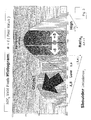
- the avoidable, disturbed x-ray beam path condition is in this exemplary embodiment caused by an elastic band which was left behind by accident on the detector-surface before starting with calibration-dedicated image acquisitions. This is depicted in Fig. 2 .
- a disturbed image-tile represented with high contrast-magnification for improved visibility of this low x-ray absorption object, shows how this 'forgotten to remove' object partially intersects with an arbitrarily chosen region-of-interest ROIij and how it locally influences the normally expected smooth background noise pattern which is typical for a homogeneously exposed detector image.
- the process of automatic detection of disturbing objects in homogeneously exposed flat field images starts by dividing the image, subjected to this inspection, in a plurality of much smaller, local regions-of-interest.
- the beam path inspection concept described in this embodiment uses 64x64 pixels ( 8 mm square ) inspection-ROIs with a 1/4 th ROI-size overlap in both image directions.
- a predetermined so-called defect map which flags the detector-array's unreliable pixels, rows and colums may be available for the purpose of image-reconstruction using neighbouring, reliable pixel data.
- the inspection-ROI's valid pixels subset is derived from which either the local median (in a preferred embodiment) or the average or modus signal value is calculated.
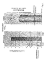
- That value represents the central signal value " C " for the further operations performed on the histogram of valid pixel values as depicted in Fig. 3 .
- the image impact of a local object disturbance in the inspection ROI introduces x-ray absorption which reduces the signal values of the affected pixels in a way that the histogram becomes asymmetrical with respect to the vertical dash-dot line, representing the central signal value without the presence of that disturbing object.
- a lower and a higher signal shoulder can be defined inside the local histogram as the collections of the lower and the higher valid pixel shoulder fractions composed of the spatially distributed pixels having signal-values below C-delta or above C+delta.
- the histogram-gap, determined by the signal delta, that separates the low and the high shoulders from the central value can be predetermined as either a fraction of the central value itself or as a predetermined factor times the standard deviation of the smooth background noise, calculated as the median noise deviation estimate from a limited set of spatially distributed image ROIs.
- the ROI's low and high shoulder fractions L and H are calculated as the amount of valid pixels with signals below the C-delta or above the C+delta value devided by the total amount of valid pixels locally present.
- the shoulder ratio is calculated by dividing the biggest of both shoulder fractions by the smallest. Undisturbed image noise will typically return a near equity shoulder ratio.
- the shoulder ratio will increase.
- the threshold for the shoulder fraction determines if the measured value is sufficiently significant to be flagged as one of the prerequisites for object detection.
- the threshold for the shoulder ratio determines if the measured value is sufficiently unbalanced to be flagged as one of the prerequisites for object detection.
- the decision logic for the detection of a ROI-disturbance is such that a local ROI is regarded as object-disturbed if a sufficient level of shoulder unbalance is present and if at the same time at least one shoulder fraction is sufficiently significant.
- the result of that local ROI decision can be stored in an image-wide disturbance memory for further decision making regarding the image-disturbance at a higher level.
- the decision about the object disturbance of the entire image is made by comparing the predetermined criteria with the content of the image-wide disturbance memory.
- An image-wide criterion could be that a very limited amount of solitary disturbed ROIs can still be accepted if these isolated disturbances all occur in ROI's adjacent to the image borders.
- the beam path is regarded as object disturbed. In that case additional inspection, cleaning and or a correction (e.g. the removal of the disturbing object ) of the x-ray beam path might be necessary before an image-retake can be performed.
Landscapes
- Engineering & Computer Science (AREA)
- Health & Medical Sciences (AREA)
- Physics & Mathematics (AREA)
- General Physics & Mathematics (AREA)
- Life Sciences & Earth Sciences (AREA)
- General Health & Medical Sciences (AREA)
- Theoretical Computer Science (AREA)
- Medical Informatics (AREA)
- Pathology (AREA)
- Radiology & Medical Imaging (AREA)
- Nuclear Medicine, Radiotherapy & Molecular Imaging (AREA)
- Computer Vision & Pattern Recognition (AREA)
- High Energy & Nuclear Physics (AREA)
- Molecular Biology (AREA)
- Chemical & Material Sciences (AREA)
- Immunology (AREA)
- Analytical Chemistry (AREA)
- Biochemistry (AREA)
- Quality & Reliability (AREA)
- Biophysics (AREA)
- Public Health (AREA)
- Multimedia (AREA)
- Signal Processing (AREA)
- Veterinary Medicine (AREA)
- Optics & Photonics (AREA)
- Biomedical Technology (AREA)
- Heart & Thoracic Surgery (AREA)
- Surgery (AREA)
- Animal Behavior & Ethology (AREA)
- Spectroscopy & Molecular Physics (AREA)
- Pulmonology (AREA)
- Apparatus For Radiation Diagnosis (AREA)
- Analysing Materials By The Use Of Radiation (AREA)
Priority Applications (6)
| Application Number | Priority Date | Filing Date | Title |
|---|---|---|---|
| EP12171474.5A EP2675151A1 (en) | 2012-06-11 | 2012-06-11 | A method to evaluate the presence of a source of x-ray beam inhomogeneity during x-ray exposure. |
| BR112014030423A BR112014030423A2 (pt) | 2012-06-11 | 2013-06-05 | método para avaliar presença de fonte de feixes de raios x em homogeneidade durante exposição a raios x |
| CN201380030642.XA CN104350738B (zh) | 2012-06-11 | 2013-06-05 | 用于在x射线曝光期间评估x射线射束不均匀性的源的存在的方法 |
| US14/402,363 US9741102B2 (en) | 2012-06-11 | 2013-06-05 | Method to evaluate the presence of a source of x-ray beam inhomogeneity during x-ray exposure |
| PCT/EP2013/061591 WO2013186099A1 (en) | 2012-06-11 | 2013-06-05 | A method to evaluate the presence of a source of x-ray beam inhomogeneity during x-ray exposure |
| IN9699DEN2014 IN2014DN09699A (https=) | 2012-06-11 | 2014-11-17 |
Applications Claiming Priority (1)
| Application Number | Priority Date | Filing Date | Title |
|---|---|---|---|
| EP12171474.5A EP2675151A1 (en) | 2012-06-11 | 2012-06-11 | A method to evaluate the presence of a source of x-ray beam inhomogeneity during x-ray exposure. |
Publications (1)
| Publication Number | Publication Date |
|---|---|
| EP2675151A1 true EP2675151A1 (en) | 2013-12-18 |
Family
ID=46245917
Family Applications (1)
| Application Number | Title | Priority Date | Filing Date |
|---|---|---|---|
| EP12171474.5A Withdrawn EP2675151A1 (en) | 2012-06-11 | 2012-06-11 | A method to evaluate the presence of a source of x-ray beam inhomogeneity during x-ray exposure. |
Country Status (6)
| Country | Link |
|---|---|
| US (1) | US9741102B2 (https=) |
| EP (1) | EP2675151A1 (https=) |
| CN (1) | CN104350738B (https=) |
| BR (1) | BR112014030423A2 (https=) |
| IN (1) | IN2014DN09699A (https=) |
| WO (1) | WO2013186099A1 (https=) |
Families Citing this family (4)
| Publication number | Priority date | Publication date | Assignee | Title |
|---|---|---|---|---|
| US20190243013A1 (en) * | 2017-11-09 | 2019-08-08 | Oceaneering International, Inc. | Estimation of material loss from 2D digital radiographs using Double Wall Single Imaging (DWSI) Technique |
| JP7508334B2 (ja) | 2020-10-21 | 2024-07-01 | キヤノンメディカルシステムズ株式会社 | 医用画像処理装置、x線診断装置及び医用画像処理方法 |
| JP2022114846A (ja) * | 2021-01-27 | 2022-08-08 | キヤノン株式会社 | 放射線撮像システム、制御装置、および、放射線撮像システムの制御方法 |
| CN116828289B (zh) * | 2022-12-12 | 2026-04-21 | 地太科特影像科技(上海)有限公司 | 防止平板探测器误触发上图的检测方法、设备及存储介质 |
Citations (6)
| Publication number | Priority date | Publication date | Assignee | Title |
|---|---|---|---|---|
| EP1115120A2 (en) | 2000-01-07 | 2001-07-11 | Lucent Technologies Inc. | Method and apparatus for temperature compensation of read-only memory |
| US20010033678A1 (en) * | 2000-04-06 | 2001-10-25 | Akira Hirai | Image processing apparatus |
| DE102005017491A1 (de) * | 2005-04-15 | 2006-10-19 | Siemens Ag | Verfahren zum Erzeugen eines gainkorrigierten Röntgenbildes und Röntgenbildaufnahmesystem |
| WO2007043974A1 (en) * | 2005-10-13 | 2007-04-19 | Agency For Science, Technology & Research | Computed tomography system and method |
| EP2050395A1 (en) * | 2007-10-18 | 2009-04-22 | Paracelsus Medizinische Privatuniversität | Methods for improving image quality of image detectors, and systems therefor |
| EP2407016A1 (en) | 2009-03-13 | 2012-01-18 | 4Energy Limited | Equipment enclosure |
Family Cites Families (12)
| Publication number | Priority date | Publication date | Assignee | Title |
|---|---|---|---|---|
| US5079698A (en) * | 1989-05-03 | 1992-01-07 | Advanced Light Imaging Technologies Ltd. | Transillumination method apparatus for the diagnosis of breast tumors and other breast lesions by normalization of an electronic image of the breast |
| US5151596A (en) * | 1990-03-28 | 1992-09-29 | Fuji Photo Film Co., Ltd. | Method and apparatus for detecting the location of a pattern in a radiation image stored on a stimulable phosphor sheet |
| US5359513A (en) * | 1992-11-25 | 1994-10-25 | Arch Development Corporation | Method and system for detection of interval change in temporally sequential chest images |
| JP2000070261A (ja) | 1998-08-31 | 2000-03-07 | Canon Inc | キャリブレーション成否判定装置、方法及びコンピュータ読み取り可能な記憶媒体 |
| US7317842B2 (en) * | 2003-10-30 | 2008-01-08 | Samsung Electronics Co., Ltd. | Global and local statistics controlled noise reduction system |
| CN1307944C (zh) * | 2004-07-16 | 2007-04-04 | 中国人民解放军第三军医大学野战外科研究所 | 数字异物定位仪 |
| US7639849B2 (en) * | 2005-05-17 | 2009-12-29 | Barco N.V. | Methods, apparatus, and devices for noise reduction |
| JP5341463B2 (ja) | 2008-10-17 | 2013-11-13 | キヤノン株式会社 | 制御装置、及び制御方法 |
| US8519348B2 (en) * | 2009-09-08 | 2013-08-27 | Carestream Health, Inc. | Image quality monitor for digital radiography system |
| EP2477153B1 (en) | 2011-01-18 | 2013-11-13 | Agfa Healthcare | Method of removing the spatial response signature of a detector from a computed radiography image |
| US8894280B2 (en) * | 2011-12-31 | 2014-11-25 | Carestream Health, Inc. | Calibration and correction procedures for digital radiography detectors supporting multiple capture modes, methods and systems for same |
| US9195899B2 (en) * | 2012-01-13 | 2015-11-24 | Carestream Health, Inc. | Self correcting portable digital radiography detector, methods and systems for same |
-
2012
- 2012-06-11 EP EP12171474.5A patent/EP2675151A1/en not_active Withdrawn
-
2013
- 2013-06-05 US US14/402,363 patent/US9741102B2/en not_active Expired - Fee Related
- 2013-06-05 WO PCT/EP2013/061591 patent/WO2013186099A1/en not_active Ceased
- 2013-06-05 BR BR112014030423A patent/BR112014030423A2/pt not_active IP Right Cessation
- 2013-06-05 CN CN201380030642.XA patent/CN104350738B/zh not_active Expired - Fee Related
-
2014
- 2014-11-17 IN IN9699DEN2014 patent/IN2014DN09699A/en unknown
Patent Citations (6)
| Publication number | Priority date | Publication date | Assignee | Title |
|---|---|---|---|---|
| EP1115120A2 (en) | 2000-01-07 | 2001-07-11 | Lucent Technologies Inc. | Method and apparatus for temperature compensation of read-only memory |
| US20010033678A1 (en) * | 2000-04-06 | 2001-10-25 | Akira Hirai | Image processing apparatus |
| DE102005017491A1 (de) * | 2005-04-15 | 2006-10-19 | Siemens Ag | Verfahren zum Erzeugen eines gainkorrigierten Röntgenbildes und Röntgenbildaufnahmesystem |
| WO2007043974A1 (en) * | 2005-10-13 | 2007-04-19 | Agency For Science, Technology & Research | Computed tomography system and method |
| EP2050395A1 (en) * | 2007-10-18 | 2009-04-22 | Paracelsus Medizinische Privatuniversität | Methods for improving image quality of image detectors, and systems therefor |
| EP2407016A1 (en) | 2009-03-13 | 2012-01-18 | 4Energy Limited | Equipment enclosure |
Also Published As
| Publication number | Publication date |
|---|---|
| IN2014DN09699A (https=) | 2015-07-31 |
| US20150146959A1 (en) | 2015-05-28 |
| WO2013186099A1 (en) | 2013-12-19 |
| CN104350738A (zh) | 2015-02-11 |
| US9741102B2 (en) | 2017-08-22 |
| CN104350738B (zh) | 2018-04-20 |
| BR112014030423A2 (pt) | 2017-06-27 |
Similar Documents
| Publication | Publication Date | Title |
|---|---|---|
| US6819786B2 (en) | Image processing apparatus | |
| EP2041606B1 (en) | Energy spectrum reconstruction | |
| US20040252874A1 (en) | Radiation imaging method, radiation imaging apparatus, computer program and computer-readable recording medium | |
| US20110155899A1 (en) | Method and apparatus for acquiring radiation data | |
| US7394925B2 (en) | Radiography apparatus and radiography method | |
| US6919568B2 (en) | Method and apparatus for identifying composite defective pixel map | |
| JP7041252B2 (ja) | デジタルポジトロン放出断層撮影における検出器ピクセルの性能変動への対処 | |
| EP0998137A2 (en) | Method and apparatus for defective pixel identification | |
| US9741102B2 (en) | Method to evaluate the presence of a source of x-ray beam inhomogeneity during x-ray exposure | |
| JP2020525064A (ja) | X線画像化システムにおける幾何学的不整合の管理 | |
| US7995828B2 (en) | Speckle reporting in digital radiographic imaging | |
| US10786222B2 (en) | Methods, systems, and apparatus for automatically assessing quality of imaging systems | |
| US20100215143A1 (en) | Method and systems for scanning a stream of objects | |
| JP2021509957A (ja) | 電荷共有キャリブレーション方法及びシステム | |
| CN113397577A (zh) | 动态品质管理装置、动态品质管理程序以及动态品质管理方法 | |
| US11426138B2 (en) | Radiographing apparatus, radiographing system, and dose index management method | |
| US8198596B2 (en) | Imaging system and image defect correcting method | |
| US9773318B2 (en) | Systems and methods for detecting camera defect caused by exposure to radiation | |
| CN110755105B (zh) | 检测方法及检测系统 | |
| US10311568B2 (en) | Image processing apparatus, control method thereof, and computer-readable storage medium | |
| JP5188255B2 (ja) | 放射線画像撮影装置および画像欠陥検出方法 | |
| US6466645B1 (en) | Methods and apparatus for tube-spit correction | |
| CN117179800A (zh) | 用于提高传感器成品率的光子计数计算机断层摄影(pcct)检测器传感器修复 | |
| CN115245342A (zh) | 动态品质管理装置、记录介质以及动态品质管理方法 | |
| CN115721327A (zh) | X射线诊断装置、医用信息处理装置和方法以及存储介质 |
Legal Events
| Date | Code | Title | Description |
|---|---|---|---|
| PUAI | Public reference made under article 153(3) epc to a published international application that has entered the european phase |
Free format text: ORIGINAL CODE: 0009012 |
|
| AK | Designated contracting states |
Kind code of ref document: A1 Designated state(s): AL AT BE BG CH CY CZ DE DK EE ES FI FR GB GR HR HU IE IS IT LI LT LU LV MC MK MT NL NO PL PT RO RS SE SI SK SM TR |
|
| AX | Request for extension of the european patent |
Extension state: BA ME |
|
| 17P | Request for examination filed |
Effective date: 20140618 |
|
| RBV | Designated contracting states (corrected) |
Designated state(s): AL AT BE BG CH CY CZ DE DK EE ES FI FR GB GR HR HU IE IS IT LI LT LU LV MC MK MT NL NO PL PT RO RS SE SI SK SM TR |
|
| 17Q | First examination report despatched |
Effective date: 20151111 |
|
| STAA | Information on the status of an ep patent application or granted ep patent |
Free format text: STATUS: THE APPLICATION IS DEEMED TO BE WITHDRAWN |
|
| 18D | Application deemed to be withdrawn |
Effective date: 20160524 |