EP2635295B1 - Control and characterization of memory function - Google Patents
Control and characterization of memory function Download PDFInfo
- Publication number
- EP2635295B1 EP2635295B1 EP11838856.0A EP11838856A EP2635295B1 EP 2635295 B1 EP2635295 B1 EP 2635295B1 EP 11838856 A EP11838856 A EP 11838856A EP 2635295 B1 EP2635295 B1 EP 2635295B1
- Authority
- EP
- European Patent Office
- Prior art keywords
- light
- memory
- neurons
- protein
- hippocampus
- Prior art date
- Legal status (The legal status is an assumption and is not a legal conclusion. Google has not performed a legal analysis and makes no representation as to the accuracy of the status listed.)
- Not-in-force
Links
Images


Classifications
-
- A—HUMAN NECESSITIES
- A61—MEDICAL OR VETERINARY SCIENCE; HYGIENE
- A61K—PREPARATIONS FOR MEDICAL, DENTAL OR TOILETRY PURPOSES
- A61K31/00—Medicinal preparations containing organic active ingredients
- A61K31/70—Carbohydrates; Sugars; Derivatives thereof
- A61K31/7088—Compounds having three or more nucleosides or nucleotides
-
- A—HUMAN NECESSITIES
- A01—AGRICULTURE; FORESTRY; ANIMAL HUSBANDRY; HUNTING; TRAPPING; FISHING
- A01K—ANIMAL HUSBANDRY; CARE OF BIRDS, FISHES, INSECTS; FISHING; REARING OR BREEDING ANIMALS, NOT OTHERWISE PROVIDED FOR; NEW BREEDS OF ANIMALS
- A01K67/00—Rearing or breeding animals, not otherwise provided for; New breeds of animals
- A01K67/027—New breeds of vertebrates
- A01K67/0275—Genetically modified vertebrates, e.g. transgenic
-
- A—HUMAN NECESSITIES
- A61—MEDICAL OR VETERINARY SCIENCE; HYGIENE
- A61K—PREPARATIONS FOR MEDICAL, DENTAL OR TOILETRY PURPOSES
- A61K38/00—Medicinal preparations containing peptides
- A61K38/16—Peptides having more than 20 amino acids; Gastrins; Somatostatins; Melanotropins; Derivatives thereof
- A61K38/17—Peptides having more than 20 amino acids; Gastrins; Somatostatins; Melanotropins; Derivatives thereof from animals; from humans
- A61K38/1703—Peptides having more than 20 amino acids; Gastrins; Somatostatins; Melanotropins; Derivatives thereof from animals; from humans from vertebrates
- A61K38/1709—Peptides having more than 20 amino acids; Gastrins; Somatostatins; Melanotropins; Derivatives thereof from animals; from humans from vertebrates from mammals
-
- A—HUMAN NECESSITIES
- A61—MEDICAL OR VETERINARY SCIENCE; HYGIENE
- A61P—SPECIFIC THERAPEUTIC ACTIVITY OF CHEMICAL COMPOUNDS OR MEDICINAL PREPARATIONS
- A61P25/00—Drugs for disorders of the nervous system
-
- A—HUMAN NECESSITIES
- A61—MEDICAL OR VETERINARY SCIENCE; HYGIENE
- A61P—SPECIFIC THERAPEUTIC ACTIVITY OF CHEMICAL COMPOUNDS OR MEDICINAL PREPARATIONS
- A61P25/00—Drugs for disorders of the nervous system
- A61P25/22—Anxiolytics
-
- A—HUMAN NECESSITIES
- A61—MEDICAL OR VETERINARY SCIENCE; HYGIENE
- A61P—SPECIFIC THERAPEUTIC ACTIVITY OF CHEMICAL COMPOUNDS OR MEDICINAL PREPARATIONS
- A61P25/00—Drugs for disorders of the nervous system
- A61P25/28—Drugs for disorders of the nervous system for treating neurodegenerative disorders of the central nervous system, e.g. nootropic agents, cognition enhancers, drugs for treating Alzheimer's disease or other forms of dementia
-
- A—HUMAN NECESSITIES
- A01—AGRICULTURE; FORESTRY; ANIMAL HUSBANDRY; HUNTING; TRAPPING; FISHING
- A01K—ANIMAL HUSBANDRY; CARE OF BIRDS, FISHES, INSECTS; FISHING; REARING OR BREEDING ANIMALS, NOT OTHERWISE PROVIDED FOR; NEW BREEDS OF ANIMALS
- A01K2217/00—Genetically modified animals
- A01K2217/05—Animals comprising random inserted nucleic acids (transgenic)
- A01K2217/052—Animals comprising random inserted nucleic acids (transgenic) inducing gain of function
-
- C—CHEMISTRY; METALLURGY
- C07—ORGANIC CHEMISTRY
- C07K—PEPTIDES
- C07K2319/00—Fusion polypeptide
- C07K2319/01—Fusion polypeptide containing a localisation/targetting motif
- C07K2319/04—Fusion polypeptide containing a localisation/targetting motif containing an ER retention signal such as a C-terminal HDEL motif
-
- C—CHEMISTRY; METALLURGY
- C07—ORGANIC CHEMISTRY
- C07K—PEPTIDES
- C07K2319/00—Fusion polypeptide
- C07K2319/01—Fusion polypeptide containing a localisation/targetting motif
- C07K2319/10—Fusion polypeptide containing a localisation/targetting motif containing a tag for extracellular membrane crossing, e.g. TAT or VP22
Definitions
- the consolidation of remote memories relies on both synaptic consolidation processes on the timescale of minutes to hours, and circuit consolidation over weeks to years (Frankland and Bontempi, 2005; Squire and Bayley, 2007).
- the process of long-term contextual fear memory consolidation requires early involvement of the hippocampus, followed by the neocortex; in the course of this process, an influence of hippocampus on neocortex may enable the hippocampus to facilitate the long-term cortical storage of memory, rather than stably store the memory itself.
- hippocampal lesions impair recent memory one day after training, but the same lesions had no effect on remote memory, several weeks after training (Anagnostaras et al., 1999; Bontempi et al., 1999; Debiec et al., 2002; Frankland et al., 2004; Kim and Fanselow, 1992; Kitamura et al., 2009; Maren et al., 1997; Maviel et al., 2004; Shimizu et al., 2000; Wang et al., 2003; Winocur et al., 2009). Additional studies suggest that both hippocampal and cortical memories are in continuous interplay.
- amnesia e.g., non-graded, graded retrograde, focal retrograde amnesia, etc.
- PTSD post traumatic stress disorder
- PTSD is a common debilitating psychiatric condition in which a single exposure to a traumatic event can lead to years of compromised function due to repeated re-experiencing of the trauma.
- Gradinaru et al, Science (2009) 324; 354-359 relates to the optical deconstruction of Parkinsonian neural circuitry.
- WO 2010/056970 relates to optically-based stimulation of target cells and modifications thereto.
- Fiala et al, Current Biology (2010) 20, R897-R903 reviews optogenetic approaches in neuroscience.
- US 2009/0093403 describes the stimulation of target cells using light.
- US 2009/0088680 relates to a system which electrically stimulates target cells of a living animal using an elongated structure, a modulation circuit and a light pathway such as provided by an optical fibre arrangement. Malin et al, Neurobiol Learn Mem.
- the present invention provides a polynucleotide encoding a light-activated protein for use in a method for treating post-traumatic stress disorder (PTSD) in an individual, wherein said method comprises: administering the polynucleotide encoding the light-activated protein to the dorsal CA1 field of the hippocampus, the anterior cingulated cortex of the individual, or the basolateral amygdala of the individual, wherein the light-activated protein is expressed on the cell membrane of the excitatory neurons in the dorsal CA1 field of the hippocampus, the anterior cingulated cortex, or the basolateral amygdala of the individual, and the protein is responsive to light and is capable of hyperpolarization of the neurons when the neurons are illuminated with the light, and activating the protein by the light to reversibly inhibit formation or retrieval of a fearful memory, thereby treating the PTSD in the individual.
- PTSD post-traumatic stress disorder
- the invention also provides a method of screening a pharmacological agent that affects retrieval or formation of a fearful memory, the method comprising: a) contacting excitatory neurons in the dorsal CA1 field of the hippocampus, the anterior cingulated cortex, or the basolateral amygdala during retrieval or formation of a fearful memory in a mouse with a pharmacological agent, wherein said mouse comprises a light-activated protein expressed on the cell membrane of excitatory neurons in the dorsal CA1 field of its hippocampus, its anterior cingulated cortex, or its basolateral amygdala, wherein the protein is responsive to light and is capable of hyperpolarizing the neurons when the neurons are illuminated with the light, wherein the illumination of the neurons inhibits formation or retrieval of a fearful memory; b) inducing hyperpolarizing the excitatory neurons in the dorsal CA1 field of the hippocampus during memory retrieval or formation of a fearful memory, by applying
- aspects of the present disclosure relates to control or characterization of memory function in living animals, as described herein. While the present disclosure is not necessarily limited in these contexts, embodiments of the invention may be appreciated through a discussion of examples using these and other contexts.
- Certain embodiments of the present disclosure are directed toward specially-targeted circuits that are associated with memory function. More particular embodiments relate to spatio-temporal control over neural circuitry to identify specific circuit targets associated and corresponding with memory function(s) (e.g., memory formation and/or retrieval).
- Particular embodiments of the present disclosure are directed toward temporally precise inhibition of neural circuits in the hippocampus (such as the neurons of the dorsal CA1 field of the hippocampus), the precision being sufficient to disrupt memory function. It has been discovered that temporal precision of neural inhibition is effective to disrupt remote memory retrieval, whereas prolonged inhibition has no significant effect on remote memory retrieval. Accordingly, aspects of the present disclosure relate to temporal aspects of such inhibition.
- methods for reversibly affecting memory function may comprise temporarily inhibiting neurons of the amygdala (e.g. basolateral amygdala) and/or neurons of the cingulate cortex (e.g., anterior cingulated cortex).
- this inhibition is performed using an optogenetic system that involves the expression of light-activated proteins (e.g., opsins) in the cells of the neural circuit.
- the inhibition can be performed using direct electrical stimulus. Still other embodiments allow for the use of temporally-precise pharmaceuticals.
- Various embodiments of the present disclosure relate to an optogenetic system or method that correlates temporal control over a neural circuit with measurable metrics. For instance, a particular memory function might be associated with a neurological disorder.
- the optogenetic system targets a neural circuit within an individual for selective control thereof.
- the optogenetic system involves monitoring the individual for metrics (e.g., symptoms) associated with the neurological disorder. In this manner the optogenetic system can provide detailed information about the neural circuit, its function and/or the neurological disorder.
- One or more methods for reversibly affecting memory function may be used to evaluate the effectiveness of pharmacological agents in treating PTSD and/or various memory disorders.
- the light-activated proteins may be configured to inhibit depolarization of a neuron in the presence of light having a specific wavelength. In some variations, the light-activated proteins may be configured to promote depolarization of a neuron in the presence of a light having a specific wavelength.
- non-human animal comprising a light-activated protein expressed on the cell membrane of excitatory neurons in the dorsal CA1 field of the hippocampus of the animal, wherein the protein is responsive to light and is capable of inhibiting depolarization of the neurons when the neurons are illuminated with the light, wherein the illumination of the protein reversibly affects memory function.
- a non-human animal comprising a light-activated protein expressed on the cell membrane of excitatory neurons in the anterior cingulated cortex of the animal, wherein the protein is responsive to light and is capable of inhibiting depolarization of the neurons when the neurons are illuminated with the light, wherein the illumination of the protein reversibly affects memory function.
- non-human animal comprising a light-activated protein expressed on the cell membrane of excitatory neurons in the basolateral amygdala of the animal, wherein the protein is responsive to light and is capable of inhibiting depolarization of the neurons when the neurons are illuminated with the light, wherein the illumination of the protein reversibly affects memory function.
- the memory function that is affected when the neurons are illuminated may be memory retrieval and/or memory formation.
- the memory is a fearful memory and/or a remote memory.
- a brain tissue slice comprising a brain region selected from the group consisting of the dorsal CA1 field of the hippocampus, the basolateral amygdala, and the anterior cingulated cortex, wherein a light-activated protein is expressed on the cell membrane of excitatory neurons of the brain region, wherein the protein is responsive to light and is capable of inhibiting depolarization of the neurons when the neurons are illuminated with the light, wherein the illumination of the protein reversibly affects memory function.
- the method for reversibly affecting memory retrieval or formation in an individual comprises: administering a polynucleotide encoding a light-activated protein to the dorsal CA1 field of the hippocampus in the individual, wherein light-activated protein is expressed on the cell membrane of the excitatory neurons in the dorsal CA1 field of the hippocampus and the protein is responsive to light and is capable of inhibiting depolarization of the neurons when the neurons are illuminated with the light, whereby activating the protein by the light reversibly affects memory retrieval or formation of an event in the individual.
- the method for reversibly affecting memory retrieval or formation comprises: inhibiting depolarization of excitatory neurons in the dorsal CA1 field of the hippocampus during memory retrieval or formation of an event in an individual, wherein a light-activated protein is expressed on the cell membrane of the excitatory neurons in the dorsal CA1 field of the hippocampus of the individual, wherein the protein is responsive to light and is capable of inhibiting depolarization of the neurons when the neurons are illuminated with the light.
- the method for reversibly affecting memory retrieval or formation in an individual comprises: administering a polynucleotide encoding a light-activated protein to the anterior cingulated cortex in the individual, wherein light-activated protein is expressed on the cell membrane of the excitatory neurons in the anterior cingulated cortex and the protein is responsive to light and is capable of inhibiting depolarization of the neurons when the neurons are illuminated with the light, whereby activating the protein by the light reversibly affects memory retrieval or formation of an event in the individual.
- the method for reversibly affecting memory retrieval or formation comprises: inhibiting depolarization of excitatory neurons in the anterior cingulated cortex during memory retrieval or formation of an event in an individual, wherein a light-activated protein is expressed on the cell membrane of the excitatory neurons in the anterior cingulated cortex of the individual, wherein the protein is responsive to light and is capable of inhibiting depolarization of the neurons when the neurons are illuminated with the light.
- the method for reversibly affecting memory retrieval or formation in an individual comprises: administering a polynucleotide encoding a light-activated protein to the basolateral amygdala in the individual, wherein light-activated protein is expressed on the cell membrane of the excitatory neurons in the basolateral amygdala and the protein is responsive to light and is capable of inhibiting depolarization of the neurons when the neurons are illuminated with the light, whereby activating the protein by the light reversibly affects memory retrieval or formation of an event in the individual.
- the method for reversibly affecting memory retrieval or formation comprises: inhibiting depolarization of excitatory neurons in the basolateral amygdala during memory retrieval or formation of an event in an individual, wherein a light-activated protein is expressed on the cell membrane of the excitatory neurons in the basolateral amygdala of the individual, wherein the protein is responsive to light and is capable of inhibiting depolarization of the neurons when the neurons are illuminated with the light.
- the method for treating post-traumatic stress disorder in an individual comprises: administering a polynucleotide encoding a light-activated protein to the dorsal CA1 field of the hippocampus in the individual, wherein light-activated protein is expressed on the cell membrane of the excitatory neurons in the dorsal CA1 field of the hippocampus and the protein is responsive to light and is capable of inhibiting depolarization of the neurons when the neurons are illuminated with the light, whereby activating the protein by the light reversibly affects memory retrieval or formation of an event in the individual.
- the method for treating post-traumatic stress disorder in an individual comprises: administering a polynucleotide encoding a light-activated protein to the anterior cingulated cortex in the individual, wherein light-activated protein is expressed on the cell membrane of the excitatory neurons in the anterior cingulated cortex and the protein is responsive to light and is capable of inhibiting depolarization of the neurons when the neurons are illuminated with the light, whereby activating the protein by the light reversibly affects memory retrieval or formation of an event in the individual.
- Also provided herein are methods of screening a pharmacological agent that affects memory retrieval or formation comprising: a) contacting excitatory neurons in the dorsal CA1 field of the hippocampus during memory retrieval or formation of an event in a non-human animal with a pharmacological agent, wherein the non-human animal comprises a light-activated protein expressed on the cell membrane of excitatory neurons in the dorsal CA1 field of the hippocampus of the animal, wherein the protein is responsive to light and is capable of inhibiting depolarization of the neurons when the neurons are illuminated with the light; b) inhibiting depolarization of the excitatory neurons in the dorsal CA1 field of the hippocampus during memory retrieval or formation of an event; and c) determining if the pharmacological agent affects memory retrieval or formation in the presence or absence of the light.
- Also provided herein are methods of screening a pharmacological agent that affects memory retrieval or formation comprising: a) contacting excitatory neurons in the anterior cingulated cortex during memory retrieval or formation of an event in a non-human animal with a pharmacological agent, wherein the non-human animal comprises a light-activated protein expressed on the cell membrane of excitatory neurons in the anterior cingulated cortex of the animal, wherein the protein is responsive to light and is capable of inhibiting depolarization of the neurons when the neurons are illuminated with the light; b) inhibiting depolarization of the excitatory neurons in the anterior cingulated cortex during memory retrieval or formation of an event; and c) determining if the pharmacological agent affects memory retrieval or formation in the presence or absence of the light.
- Also provided herein are methods of screening a pharmacological agent that affects memory retrieval or formation comprising: a) contacting excitatory neurons in the basolateral amygdala during memory retrieval or formation of an event in a non-human animal with a pharmacological agent, wherein the non-human animal comprises a light-activated protein expressed on the cell membrane of excitatory neurons in the basolateral amygdala of the animal, wherein the protein is responsive to light and is capable of inhibiting depolarization of the neurons when the neurons are illuminated with the light; b) inhibiting depolarization of the excitatory neurons in the basolateral amygdala during memory retrieval or formation of an event; and c) determining if the pharmacological agent affects memory retrieval or formation in the presence or absence of the light.
- the light-activated protein may be responsive to light and configured such that the protein is capable of inhibiting depolarization of the neurons when the neurons are illuminated with the light.
- the light-activated protein may be selected from the group consisting of NpHR, BR, AR, and GtR3 described herein.
- the light-activated protein is a NpHR protein comprising an amino acid sequence at least 95%, at least 96%, at least 97%, at least 98%, at least 99% or 100% identical to the sequence shown in SEQ ID NO:3.
- the NpHR protein further comprises an endoplasmic reticulum (ER) export signal and/or a membrane trafficking signal.
- ER endoplasmic reticulum
- the NpHR protein comprises an amino acid sequence at least 95% identical to the sequence shown in SEQ ID NO:3 and an endoplasmic reticulum (ER) export signal.
- the amino acid sequence at least 95% identical to the sequence shown in SEQ ID NO:3 is linked to the ER export signal through a linker.
- the ER export signal comprises the amino acid sequence FXYENE, where X can be any amino acid.
- the ER export signal comprises the amino acid sequence VXXSL, where X can be any amino acid.
- the ER export signal comprises the amino acid sequence FCYENEV.
- the NpHR protein comprises an amino acid sequence at least 95% identical to the sequence shown in SEQ ID NO:3, an ER export signal, and a membrane trafficking signal. In other embodiments, the NpHR protein comprises, from the N-terminus to the C-terminus, the amino acid sequence at least 95% identical to the sequence shown in SEQ ID NO:3, the ER export signal, and the membrane trafficking signal. In other embodiments, the NpHR protein comprises, from the N-terminus to the C-terminus, the amino acid sequence at least 95% identical to the sequence shown in SEQ ID NO:3, the membrane trafficking signal, and the ER export signal. In some embodiments, the membrane trafficking signal is derived from the amino acid sequence of the human inward rectifier potassium channel Kir2.1.
- the membrane trafficking signal comprises the amino acid sequence KSRITSEGEYIPLDQIDINV. In some embodiments, the membrane trafficking signal is linked to the amino acid sequence at least 95% identical to the sequence shown in SEQ ID NO:3 by a linker. In some embodiments, the membrane trafficking signal is linked to the ER export signal through a linker.
- the linker may comprise any of 5, 10, 20, 30, 40, 50, 75, 100, 125, 150, 175, 200, 225, 250, 275, 300, 400, or 500 amino acids in length.
- the linker may further comprise a fluorescent protein, for example, but not limited to, a yellow fluorescent protein, a red fluorescent protein, a green fluorescent protein, or a cyan fluorescent protein.
- the light-activated protein further comprises an N-terminal signal peptide. In some embodiments, the light-activated protein comprises the amino acid sequence of SEQ ID NO:5. In some embodiments, the light-activated protein comprises the amino acid sequence of SEQ ID NO:6.
- the present disclosure is believed to be useful for modifying memory function on a temporal basis. Specific applications of the present invention facilitate disrupting memory retrieval and/or emotional responses linked to memory retrieval. As many aspects of the example embodiments disclosed herein relate to and significantly build on previous developments in this field, the following discussion summarizes such previous developments to provide a solid understanding of the foundation and underlying teachings from which implementation details and modifications might be drawn. It is in this context that the following discussion is provided. While the present invention is not necessarily limited to such applications, various aspects of the invention may be appreciated through a discussion of various examples using this context.
- Control over the neural circuit can include inhibition or excitation, which can each include coordinated firing, and/or modified susceptibility to external circuit inputs.
- inhibition can be accomplished using a light-activated protein, such as an ion channel and/or ionic pump (e.g., NpHR and NpHR variants).
- ion channels move the membrane potential of the neuron away from its threshold voltage to dissuade or inhibit action potentials.
- excitation can be accomplished using a light-activated protein, such as an ion channel (e.g., ChR2 and ChR2 variants).
- ion channels can cause the membrane potential to move toward and/or past the threshold voltage, thereby exciting or encouraging action potentials.
- a light- activated protein can be used to (temporarily) shift the resting potential of a neuron to increase or decrease its susceptibility to external circuit inputs.
- the devices and methods provided herein may reversibly affect memory function.
- the methods described below may be used to control and/or characterize the neural circuitry that underlies long-term and short-term memory, as well as various types of memories, including fearful or stressful memories.
- the methods may also affect various stages of memory function (e.g., memory acquisition, consolidation, and recall).
- memory function is affected by applying light to neurons of the dorsal CA1 region of the hippocampus, in the basolateral amygdala (BLA), and/or in the anterior cingulated cortex (ACC) that express light-activated proteins.
- BLA basolateral amygdala
- ACC anterior cingulated cortex
- these light-activated proteins may inhibit depolarization of the neurons, thereby disturbing the formation and/or retrieval of memories. While the exemplary methods are described in the context of the acquisition and recall of contextual remote and recent fear-based memories, it should be understood that the devices and methods disclosed herein may be used to affect other stages of memory function, as well as other types of memories (e.g., cued memories).
- neuronal activity may be affected using a variety of mechanisms. Deterministic methods of affecting neuronal activity may be used to control and/or characterize the neural circuits that underlie various brain functions. For example, neuronal responses may be affected by applying pharmacological agents (e.g., tetrodotoxin (TTX), 6-cyano-7-nitroquinoxaline-2,3-dione (CNQX), picrotoxin, strychnine, etc.) and/or by electrical stimulation (e.g., electrodes). In some variations, neuronal activity may be affected by activating certain types of proteins on the membrane of the neuron, which may hyperpolarize or depolarize the cell membrane.
- pharmacological agents e.g., tetrodotoxin (TTX), 6-cyano-7-nitroquinoxaline-2,3-dione (CNQX), picrotoxin, strychnine, etc.
- electrical stimulation e.g., electrodes
- neuronal activity may be affected by activating certain
- light-activated proteins that become permeable to certain ions (e.g., cations, anions) in the presence of light with a certain wavelength may be expressed in a neuron.
- ions e.g., cations, anions
- Examples of light-activated proteins may include light-activated ion channels and/or pumps, which are further described below.
- microbial opsin genes may be adapted for uses in neuroscience. These opsins allow transduction of light pulse trains into millisecond-timescale membrane potential changes in specific cell types within the intact mammalian brain (e.g., channelrhodopsin (ChR2), Volvox channelrhodopsin (VChR1) and halorhodopsin (NpHR)).
- ChR2 is a rhodopsin derived from the unicellular green alga Chlamydomonas reinhardtii.
- the term "rhodopsin” as used herein is a protein that comprises at least two building blocks, an opsin protein, and a covalently bound cofactor, usually retinal (retinaldehyde).
- the rhodopsin ChR2 is derived from the opsin Channelopsin-2 (Chop2), originally named Chlamyopsin-4 (Cop4) in the Chlamydomonas genome.
- Chop2 opsin Channelopsin-2
- Cop4 Chlamyopsin-4
- the temporal properties of one depolarizing channelrhodopsin, ChR2 include fast kinetics of activation and deactivation, affording generation of precisely timed action potential trains.
- the normally fast off-kinetics of the channelrhodopsins can be slowed.
- certain implementations of channelrhodopsins apply 1 mW/mm 2 light for virtually the entire time in which depolarization is desired, which can be less than desirable.
- Light-activated proteins that generate hyperpolarization or inhibit depolarization of the membrane in response to light with certain wavelength(s) may be expressed in the excitatory neurons (e.g., glutamatergic neurons) of the dorsal CA1 region of the hippocampus (CA1), basolateral amygdala (BLA), and anterior cingulated cortex (ACC) regions.
- Table 1 below shows various examples of light-activated proteins that may be expressed in the excitatory neurons to inhibit depolarization or hyperpolarize the neurons in the presence of light of a certain wavelength. Further description of these and other light-activated proteins may be found in PCT App. No.
- Embodiments of the present disclosure include relatively minor amino acid variants of the naturally occurring sequences.
- the variants are greater than about 75% homologous to the protein sequence of the naturally occurring sequences.
- the homology is greater than about 80%.
- Yet other variants have homology greater than about 85%, greater than 90%, or even as high as about 93% to about 95% or about 98%.
- Homology in this context means sequence similarity or identity, with identity being preferred. This homology can be determined using standard techniques known in the field of sequence analysis.
- compositions of embodiments of the present disclosure include the protein and nucleic acid sequences provided herein, including variants which are more than about 50% homologous to the provided sequence, more than about 55% homologous to the provided sequence, more than about 60% homologous to the provided sequence, more than about 65% homologous to the provided sequence, more than about 70% homologous to the provided sequence, more than about 75% homologous to the provided sequence, more than about 80% homologous to the provided sequence, more than about 85% homologous to the provided sequence, more than about 90% homologous to the provided sequence, or more than about 95% homologous to the provided sequence.
- non-human animals comprising a light-activated protein expressed on the cell membrane of excitatory neurons in the dorsal CA1 field of the hippocampus, anterior cingulated cortex, and/or basolateral amygdala of the animal, wherein the protein is responsive to light and is capable of inhibiting depolarization of the neurons when the neurons are illuminated with the light, wherein the illumination of the protein reversibly affects memory function.
- the light-activated protein is selected from the group consisting of NpHR, BR, AR and GtR3 described herein.
- any of the NpHR proteins described herein may be expressed on the cell membrane of the target neurons.
- brain tissue slices comprising a brain region selected from the group consisting of the dorsal CA1 field of the hippocampus, the basolateral amygdala, and the anterior cingulated cortex, wherein a light-activated protein is expressed on the cell membrane of excitatory neurons of the brain region, wherein the protein is responsive to light and is capable of inhibiting depolarization of the neurons when the neurons are illuminated with the light, wherein the illumination of the protein reversibly affects memory function.
- the brain tissue slices are cultured tissue slices taken from the non-human animals described herein.
- the light-activated protein is selected from the group consisting of NpHR, BR, AR and GtR3 described herein. For example, any of the NpHR proteins described herein may be expressed on the cell membrane of the target neurons.
- neurons of the CA1, BLA, and/or ACC regions may express ChR2.
- the disclosure includes a number of similar variants. Examples include, but are not limited to, Chop2, ChR2-310, Chop2-310, and Volvox channelrhodopsin (VChR1).
- VChR1 Volvox channelrhodopsin
- similar modifications can be made to other opsin or light-activated molecules. For instance, modifications/mutations can be made to ChR2 or VChR1 variants.
- the modified variants can be used in combination with light-activated ion pumps.
- stimulation of a target cell is generally used to describe modification of properties of the cell.
- the stimulus of a target cell may result in a change in the properties of the cell membrane that can lead to the depolarization or polarization of the target cell.
- the target cell is a neuron and the stimulus may affect the transmission of impulses by facilitating or inhibiting the generation of impulses (action potentials) by the neuron.
- Embodiments of the present disclosure are directed toward implementation of bistable changes in the excitability of targeted populations.
- This includes, but is not necessarily limited to, the double-mutant ChR2-C128S/D156A.
- This double-mutant ChR2-C128S/D156A has been found to be well-tolerated in cultured hippocampal neurons and preserved the essential SFO properties of rapid step-like activation with single brief pulses of blue light, and deactivation with green or yellow light.
- the activation spectrum of ChR2- C128S/D156A peaks at 445 nm.
- a second deactivation peak was found at 390-400 nm, with faster but less complete deactivation by comparison with the 590 nm deactivation peak.
- the double-mutant gene is referred to as SSFO (for stabilized step-function opsin) gene.
- SSFO is also used as shorthand for the active protein. Both residues likely are involved in ChR2 channel closure (gating), and both mutations likely stabilize the open state configuration of the channel
- aspects of the present disclosure relate to the discovery that SSFO may be completely blocked in photocycle progression, and may therefore represent the maximal stability possible with photocycle engineering. For instance, in contrast to ChR2 C128X and ChR2-D156A, the SSFO photocycle does not appear to access additional inactive deprotonated side products which likely split off the photocycle at later photocycle stages not reached in this mutant, in turn making the SSFO even more reliable for repeated use in vivo than the parental single mutations.
- Embodiments of the present disclosure are directed toward the sensitivity of the SSFO to light. For instance, channelrhodopsins with slow decay constants effectively act as photon integrators. This can be particularly useful for more-sensitive, less-invasive approaches to optogenetic circuit modulation, still with readily titratable action on the target neuronal population via modulation of light pulse length. It has been discovered that, even at extraordinarily low light intensities (as low as 8 ⁇ W mm -2 ), hundreds of picoamps of whole-cell photocurrents could be obtained from neurons expressing SSFO, which increased with monoexponential kinetics in response to 470 nm light during the entire time of illumination.
- activation time constants that are linearly correlated with the activation light power on a log-log scale, which is indicative of a power-law relationship and suggesting that the SSFO is a pure integrator, with total photon exposure over time as the only determinant of photocurrent. For instance, it is believed that the number of photons per membrane area required for photocurrents to reach a given sub-maximal activation (time to ⁇ ) is constant regardless of activation light power.
- Example embodiments of the present disclosure relate to the use of a hybrid ChRI/VChRI chimera, which contains no ChR2 sequence at all and is derived from two opsins genes that do not express well individually, and is herein referred to as C1V1.
- Embodiments of the present disclosure also relate to improvements of the membrane targeting of VChR1 through the addition of a membrane trafficking signal derived from the K ir 2.1 channel. Confocal images from cultured neurons expressing VChR1-EYFP revealed a large proportion of intracellular protein compared with ChR2; therefore, to improve the membrane targeting of VChR1, we added a membrane trafficking signal derived from the Kir2.1 channel.
- VChR1-ts-EYFP Membrane targeting of this VChR1-ts-EYFP was slightly enhanced compared with VChR1-EYFP; however, mean photocurrents recorded from cultured hippocampal neurons expressing VChR1-ts-EYFP were only slightly larger than those of VChR1-EYFP. Accordingly, embodiments of the present disclosure relate VChR1 that is modified by exchanging helices with corresponding helices from other ChRs. For example, robust improvement has been discovered in two chimeras where helices 1 and 2 were replaced with the homologous segments from ChR1.
- ChR1V1 ChR1/VChR1 chimera
- C1V1-EYFP exhibits surprisingly improved average fluorescence compared with VChR1-EYFP.
- Whole cell photocurrents in neurons expressing C1V1 were much larger than those of VChRl-EYFP and VChR1-ts-EYFP, and ionic selectivity was similar to that of ChR2 and VChR1.
- the addition of the Kir2.1 trafficking signal between C1V1 and YFP further enhanced photocurrents by an additional 41%.
- C1V1- ts-EYFP mean photocurrents were extremely large, nearly tenfold greater than wild type (WT) VChR1).
- Mean fluorescence levels closely matched the measured photocurrents (mean fluorescence 33.32 ⁇ 33.32 ⁇ 33.32 ⁇ 2.8 and 33.32 ⁇ 3.8 for VChR1-EYFP, VChR1-ts-EYFP, C1V1-EYFP and C1V1-ts-EYFP, respectively), suggesting that the increase in photocurrent sizes resulted mainly from the improved expression of these channels in mammalian neurons.
- opsins or light-activated proteins with fast decay constants. This property can be particularly useful for providing precise control over spiking, e.g., in order to interfere minimally with intrinsic conductances, trigger single spikes per light pulse and/or minimize plateau potentials during light pulse trains.
- Experimental results suggest that the light-evoked photocurrents recorded in C1V1-ts-EYFP decayed with a time constant similar to that of VChR1.
- aspects of the present disclosure are therefore directed toward modifications in the chromophore region to improve photocycle kinetics, reduced inactivation and/or possible further red-shifted absorption.
- ChETA mutation E162T is directed toward a corresponding ChETA mutation E162T, which experiments suggest provides an accelerated photocycle (e.g., almost 3-fold), (reference can be made to Gunaydin, et al., Ultrafast optogenetic control, Nat Neurosci, 2010 ). Surprisingly, this mutation was shown to shift the action spectrum hypsochromic to 530 nm, whereas analogous mutations in ChR2 or other microbial rhodopsins have caused a red-shift.
- C1V1-E122T Another embodiment is directed toward a mutation of glutamate-122 to threonine (C1V1-E122T).
- Experimental tests showed that C1V1-E122T is inactivated only by 26% compared to 46% inactivation of ChR2; in addition, the spectrum was further red-shifted to 546 nm.
- Another embodiment of the present disclosure is directed toward a double mutant of C1V1 including both E122T and E162T mutations.
- Experimental tests have shown that the inactivation of the current was even lower than in the E122T mutant and the photocycle was faster compared to E162T. This suggests that multiple useful properties of the individual mutations were conserved together in the double mutant.
- Light-activated proteins or opsins described herein may be delivered into neurons by methods known in the art, such as by a polynucleotide comprising a sequence encoding the proteins.
- the polynucleotide comprises an expression cassette.
- the polynucleotide is a vector, such as a viral vector selected from the group consisting of an AAV vector, a retroviral vector, an adenoviral vector, an HSV vector, and a lentiviral vector.
- neurons may be contacted with a vector comprising a nucleic acid sequence encoding a light-activated protein operably linked to a cell specific promoter, wherein said neurons express the light-activated protein on the cell membrane.
- the cell specific promoter is a calcium/calmodulin-dependent protein kinase Ha (CaMKIIa) promoter.
- a nucleic acid sequence encoding light activatable eNpHR3.1 or eNpHR3.0 is operably linked to a CaMKIIa promoter in the vector.
- the light-activated protein is expressed in excitatory glutamatergic neuron in the CA1 region, BLA and/or ACC. Any vectors that may be used for gene delivery may be used.
- a viral vector such as AAV, adenovirus, lentivirus, a retrovirus may be used.
- the vector is a recombinant AAV vector.
- AAV vectors are DNA viruses of relatively small size that can integrate, in a stable and sitespecific manner, into the genome of the cells that they infect. They are able to infect a wide spectrum of cells without inducing any effects on cellular growth, morphology or differentiation, and they do not appear to be involved in human pathologies.
- the AAV genome has been cloned, sequenced and characterized. It encompasses approximately 4700 bases and contains an inverted terminal repeat (ITR) region of approximately 145 bases at each end, which serves as an origin of replication for the virus.
- ITR inverted terminal repeat
- the remainder of the genome is divided into two essential regions that carry the encapsidation functions: the left-hand part of the genome, that contains the rep gene involved in viral replication and expression of the viral genes; and the right-hand part of the genome, that contains the cap gene encoding the capsid proteins of the virus.
- AAV vectors may be prepared using standard methods in the art.
- Adeno-associated viruses of any serotype are suitable (see, e.g., Blacklow, pp. 165-174 of "Parvoviruses and Human Disease” J. R. Pattison, ed. (1988 ); Rose, Comprehensive Virology 3:1, 1974 ; P. Tattersall "The Evolution of Parvovirus Taxonomy” In Parvoviruses (JR Kerr, SF Cotmore. ME Bloom, RM Linden, CR Parrish, Eds.) p5-14, Hudder Arnold, London, UK (2006 ); and DE Bowles, JE Rabinowitz, RJ Samulski "The Genus Dependovirus” (JR Kerr, SF Cotmore.
- the replication defective recombinant AAVs according to the disclosure can be prepared by co-transfecting a plasmid containing the nucleic acid sequence of interest flanked by two AAV inverted terminal repeat (ITR) regions, and a plasmid carrying the AAV encapsidation genes ( rep and cap genes), into a cell line that is infected with a human helper virus (for example an adenovirus).
- ITR inverted terminal repeat
- rep and cap genes a plasmid carrying the AAV encapsidation genes
- the AAV recombinants that are produced are then purified by standard techniques.
- the vector(s) for use in the methods of the disclosure are encapsidated into a virus particle (e.g. AAV virus particle including, but not limited to, AAV1, AAV2, AAV3, AAV4, AAV5,AAV6, AAV7, AAV8, AAV9, AAV10, AAV11, AAV 12, AAV 13, AAV 14, AAV 15, and AAV 16).
- a virus particle e.g. AAV virus particle including, but not limited to, AAV1, AAV2, AAV3, AAV4, AAV5,AAV6, AAV7, AAV8, AAV9, AAV10, AAV11, AAV 12, AAV 13, AAV 14, AAV 15, and AAV 16.
- a recombinant virus particle recombinant because it contains a recombinant polynucleotide
- Methods of producing such particles are known in the art and are described in US Patent No. 6,596,535 .
- one or more vectors may be administered to neural cells, heart cells, or stem cells. If more than one vector is used, it is understood that they may be administered at the same or at different times to the animal cells.
- C1V1 opsin genes in neurons were carried out by generating lentiviral vectors encoding C1V1-ts-EYFP and various point mutation combinations discussed herein. The opsins were then expressed in cultured hippocampal neurons and recorded whole-cell photocurrents under identical stimulation conditions (2 ms pulses, 542 nm light, 5.5 mW/mm 2 ). Photocurrents in cells expressing C1V1, C1V1-E162T and C1V1-E122T/E162T were all robust and trended larger than the photocurrents of ChR2-H134R.
- the experiments also included a comparison of integrated somatic YFP fluorescence and photocurrents from cells expressing C1V1-E122T/E162T and from cells expressing ChR2- H134R.
- C1V1-E122T/E162T cells showed stronger photocurrents than ChR2-H134R cells at equivalent fluorescence levels. This suggests that C1V1 could possess a higher unitary conductance compared with ChR2-H134R.
- the test results suggest that the kinetics of C1V1-E122T were slower than those of C 1V1-E122T/E162T and that cells expressing C1V1-E122T responded more strongly to red light (630nm) than cells expressing the double mutant. This can be particularly useful for generating optogenetic spiking in response to red light.
- inhibitory and/or excitatory neurons residing within the same microcircuit are be targeted with the introduction of various light-activated proteins (e.g., opsins).
- Experimental tests were performed by separately expressed C1V1-E122T/E162T and ChR2-H134R under the CaMKIIa promoter in cultured hippocampal neurons.
- Cells expressing C1V1-E122T/E162T spiked in response to 2ms green light pulses (560 nm) but not to violet light pulses (405 nm).
- Various embodiments of the present disclosure relate to independent activation of two neuronal populations within living brain slices. Experimental tests were performed by CaMKIIa-C1V1-E122T/E162Tts-eYFP and EF1a-DIO-ChR2-H134R-EYFP in mPFC of 20 PV::Cre mice. In non-expressing PYR cells, 405 nm light pulses triggered robust and fast inhibitory postsynaptic currents (IPSCs) due to direct activation of PV cells, while 561 nm light pulses triggered only the expected long-latency polysynaptic IPSCs arising from C 1V1- expressing pyramidal cell drive of local inhibitory neurons.
- IPCs inhibitory postsynaptic currents
- any device that is capable of applying light having a wavelength to activate the light-activated proteins expressed in a neuron may be used to depolarize and/or hyperpolarize the neuron.
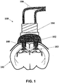
- a light-delivery device (100) for activating ion channels and/or ionic pumps to affect the membrane voltage of one or more neurons depicted in FIG. 1 may be used.
- the light-delivery device (100) is configured to provide optical stimulus to a target region of the brain.
- the light-delivery device (100) may comprise a base (102), a cannula guide (104) that is attached to the base, and one or more optical conduits (106) attached to the base via the cannula guide.
- the base (102) may comprise one or more light delivery ports (108) that are positioned to deliver light from the optical conduits (106) to targeted tissue regions (101), such as the CA1 region (103).
- the optical conduits (106) may be optical fibers, where the proximal end of the fiber is attached to an optical light source (not shown), and the distal end is in communication with the light delivery ports (108).
- the optical light source may be capable of providing continuous light and/or pulsed light, and may be programmable to provide light in predetermined pulse sequences.
- the light delivery device (100) may have any number of optical conduits (106) as may be desirable, e.g., 1, 2, 3, 4, 5, 10, 15, 20, etc.
- the optical conduits (106) may each carry light of the same or different wavelengths.
- the delivered light may have a wavelength between 450 nm and 600 nm, such as yellow or green light.
- the light delivery device (100) may have any number of light delivery ports (108) as may be desirable, e.g., 1, 2, 3, 4, 5, 10, 15, 20, etc. In some variations, there may be the same number of light delivery ports as optical conduits while in other variations, there may be different number of optical conduits and light delivery ports. For example, there may be a single optical conduit that conveys light to two or more light delivery ports. Alternatively or additionally, a single optical conduit may connect to a single light delivery port.
- the cannula guide (104) may be configured to help secure and align the optical conduits (106) with the light delivery ports (108).
- the light delivery device (100) is configured to deliver bilateral light to the CA1 region (103) to affect the formation and retrieval of memories.
- Light delivery devices may also comprise one or more measurement electrodes that may be configured for measuring neural activity.
- measurement electrodes may record changes in the membrane potential (e.g., action potentials) and/or current flow across a membrane of one or more neurons as the neurons respond to a stimulus.
- the measurement electrodes may measure the electrical response of one or more neurons to optical stimulation. Measurement electrodes may be extracellular or intracellular electrodes.
- the target tissue regions (101) may include neural tissue with cells that have light-activated proteins designed to modify the membrane voltage of the cells in response to light.
- light-activated proteins may be used to disrupt the formation and/or retrieval of memories by inhibiting the depolarization of the neurons in the CA1, BLA, and ACC regions of the brain.
- Embodiments of the present disclosure are directed towards disrupting memory acquisition, recall and/or associations between memory and emotional responses, such as fear.
- function of a neural circuit involved in memory is disrupted by activation of light-activated ion channels (e.g., using NpHR, BR, AR, etc.) and/or pumps (e.g., a proton pump GtR3).
- this disruption can be implemented during memory formation. In other implementations, this disruption can be implemented before or during memory retrieval. This can be particularly useful for psychiatric or neurological disorders involving memory recall, such as PTSD. Consistent with certain embodiments, the disruption can be triggered in response to a memory trigger event or other external stimulus that is presented and/or controlled for the disruption. For instance, the disruption can be provided in response to a trigger for a memory to an individual conditioned to respond to the trigger. In another instance, an individual can actively trigger the disruption. For instance, an individual may trigger the disruption when experiencing a memory associated with PTSD. Other embodiments of the present disclosure are directed toward encouraging memory acquisition, recall and/or associations between memory and emotional responses. The methods described herein may be used to ascertain the role of neuron(s) and/or neuronal circuits in memory function, and/or to treat disorders associated with memory impairment.
- the methods provided herein for reversibly affecting memory retrieval or formation in an individual comprise administering a polynucleotide encoding a light-activated protein to the dorsal CA1 field of the hippocampus, anterior cingulated cortex, or basolateral amygdala in the individual, wherein light-activated protein is expressed on the cell membrane of the excitatory neurons in the dorsal CA1 field of the hippocampus, anterior cingulated cortex, or basolateral amygdala and the protein is responsive to light and is capable of inhibiting depolarization of the neurons when the neurons are illuminated with the light, whereby activating the protein by the light reversibly affects memory retrieval or formation of an event in the individual.
- the methods provided herein for reversibly affecting memory retrieval or formation in an individual comprise inhibiting depolarization of excitatory neurons in the dorsal CA1 field of the hippocampus, anterior cingulated cortex, or basolateral amygdala during memory retrieval or formation of an event in an individual, wherein a light-activated protein is expressed on the cell membrane of the excitatory neurons in the dorsal CA1 field of the hippocampus, anterior cingulated cortex, or basolateral amygdala of the individual, wherein the protein is responsive to light and is capable of inhibiting depolarization of the neurons when the neurons are illuminated with the light.
- the event is a fearful event.
- kits for treating post-traumatic stress disorder in an individual comprising: administering a polynucleotide encoding a light-activated protein to the dorsal CA1 field of the hippocampus, anterior cingulated cortex, or basolateral amygdala in the individual, wherein light-activated protein is expressed on the cell membrane of the excitatory neurons in the dorsal CA1 field of the hippocampus, anterior cingulated cortex, or basolateral amygdala and the protein is responsive to light and is capable of inhibiting depolarization of the neurons when the neurons are illuminated with the light, whereby activating the protein by the light reversibly affects memory retrieval or formation of an event in the individual.
- an "individual” is a mammal including a human. Mammals include, but are not limited to, farm animals, sport animals, pets, primates, mice and rats. Individuals also include companion animals including, but not limited to, dogs and cats. In one aspect, an individual is a human. In another aspect, an individual is a non-human animal. As used herein, “non-human animals” include non-human mammals.
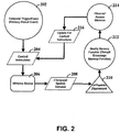
- a temporal-trigger event (202) provides a reference point for implementing control over memory function. As discussed herein, the temporal nature of the control can be particularly useful.
- the memory-trigger event (202) can be linked to a training event. For instance, an individual (e.g., non-human animals, mammals, humans) can be introduced to a stimulus designed to train the individual to respond to a particular stimulus. The memory-trigger event (202) could be the introduction of the particular stimulus to the individual.
- control instructions (204) determine how stimulus source (206) applies a stimulus (208) to a cell population (210). These control instructions can be determined and applied as a function of a desired target.
- the desired target can be defined by, for example, one or more of temporal attributes, spatial location and/or cell-type.
- the stimulus (208) results in the modification of memory function (212).
- the effect of the stimulus can then be monitored, observed and/or assessed (214). The monitoring can be used to adjust (216) the control instructions (204), thereby fine-tuning the stimulus for the intended result.
- Various embodiments discussed herein provide further examples that can be used in connection with (or in addition to) such a process for controlling and characterizing the neural circuits that underlie memory function.
- One variation of a method for disrupting memory retrieval may comprise inhibiting the excitatory neurons of the CA1 region (e.g., by blocking or reducing membrane depolarization, and/or by promoting membrane hyperpolarization).
- Light-activated ion channels such as eNpHR3.1 or NpHR3.0, may be expressed on neurons located in the CA1 region of an individual by administering a polynucleotide encoding the channel protein to the region.
- the eNpHR3.1 or NpHR3.0 ion channel is activated in the presence of yellow light (e.g., having a wavelength of about 591 nm).
- the individual may be provided with a light-delivery device, such as the light-delivery device (100) described above.
- the light-delivery device may be positioned on the individual such that yellow light is capable of being delivered to the CA1 neurons.
- a memory e.g., any undesired memory such as a fearful or stressful memory
- the light-delivery device may be activated to deliver yellow light to the CA1 neurons, thereby inhibiting their depolarization, and disrupting the recall of the memory. Once the memory recall has been sufficiently disrupted, the light-delivery device may be de-activated. Upon de-activation of the light-delivery device, the individual may regain the ability retrieve memories without disruption.
- This method may be used to disrupt recall of recent memories (e.g., memories of events that occurred less than one day in the past) and recall of remote memories (e.g., memories of events that occurred more than one day in the past, 1 week in the past, 2 weeks in the past, 4 weeks in the past, 8 weeks or more in the past, etc.).
- excitatory neurons of the ACC may express similar light-activated proteins, and may be similarly inhibited to disrupt the retrieval of remote memories.
- Methods for disrupting memory retrieval comprising inhibiting the neurons of the CA1 region may be used in a non-human animal, such as a mouse.
- a non-human animal such as a mouse.
- mice expressing eNpHR3.1 or NpHR3.0 in the neurons of the CA1 region were trained in a customized FC chamber, where they were introduced into context A and then presented twice with a tone followed by a foot-shock.
- green light delivered to the eNpHR3.1 or NpHR3.0 CA1 neurons interfered with the ability of the mice to recall the memory (i.e., a fearful or stressful memory), as measured by a reduction in freezing (e.g., contextual freezing).
- the mice are able to recall the fearful memory formed during the training session, as measured by normal rates of freezing.
- the testing session may occur one day or less after the training session, while in other variations, the testing session may occur four weeks or more after the training session.
- Applying green light to the eNpHR3.1 CA1 neurons of the mice reversibly inhibits the depolarization of the neurons, thereby disrupting the recall of recent and/or remote contextual fearful memories. Removing the green light from the eNpHR3.1 or NpHR3.0 CA1 neurons restores the ability of the mice to recall recent and/or remote contextual fearful memories.
- mice having CA1 neurons expressing eNpHR3.1 or NpHR3.0 may be trained as described above. In a testing session five weeks after the training session, the mice were able to recall the memory formed during training, however, when the eNpHR3.1 or NpHR3.0 CA1 neurons were exposed to green light, they were no longer able to recall the memory. Subsequent exposure of the eNpHR3.1 or NpHR3.0 CA1 neurons to green light disrupted retrieval of the fearful memory.
- memory recall may be disrupted by exposing the eNpHR3.1 or NpHR3.0 CA1 neurons to light upon initiation of the memory recall and/or during the memory recall. For example, applying green light to the eNpHR3.1 or NpHR3.0 CA1 neurons at the same time as recall initiation (e.g., at the beginning of the testing session) disrupts recall of the memory.
- green light was applied to the eNpHR3.1 or NpHR3.0 CA1 neurons during memory recall (e.g., applying the light some time after the testing session has begun, such as in the middle of the testing session), the mice initially recalled and responded to the fearful memory (by freezing), but then quickly ceased exhibiting the fear response after the light was applied.
- a light-delivery device may be activated at the same time and/or during the retrieval of a fearful memory in order to reversibly disrupt and/or discontinue recall of that fearful memory. Subsequent de-activation of the light-delivery device may restore the ability of the individual to recall this and other memories.
- Methods for disrupting memory retrieval comprising inhibiting the neurons of the ACC region may be used in a non-human, such as a mouse.
- mice expressing eNpHR3.1 in the neurons of the ACC may be trained as described above.
- green light delivered to the eNpHR3.1 ACC neurons interfered with the ability of the mice to recall the memory formed during training. Removing the green light from the eNpHR3.1 CA1 neurons restores the ability of the mice to remote fearful memories.
- One variation of a method for disrupting memory formation may comprise inhibiting the neurons of the CA1 region during the formation of a memory such as a contextual memory.
- Light-activated ion channels such as eNpHR3.1, may be expressed on neurons located in the CA1 region of an individual as previously described.
- the individual may be provided with a light-delivery device, such as the light-delivery device (100) described herein.
- the light-delivery device may be activated to deliver green light to the CA1 neurons, thereby inhibiting their depolarization and disrupting the formation of the memory. Once the memory formation has been sufficiently disrupted, the light-delivery device may be de-activated. Upon de-activation of the light-delivery device, the individual may regain the ability form memories without disruption.
- a memory e.g., a fearful or stressful memory
- Methods for disrupting memory formation comprising inhibiting the neurons of the CA1 region may be used in a non-human animal, such as a mouse.
- a non-human animal such as a mouse.
- mice expressing eNpHR3.1 in the neurons of the CA1 region were trained in a customized FC chamber, while delivering green light to the eNpHR3.1 CA1 neurons.
- the mice were introduced into a first context and then exposed to a tone followed by a foot-shock.
- the mice exhibited no memory of the training, as measured by a reduction in contextual freezing.
- the same mice underwent a separate training session where the eNpHR3.1 CA1 neurons were not exposed to light. The mice were then able to recall the memory in a subsequent testing session.
- the testing session may occur one day or less after the training session, while in other variations, the testing session may occur four weeks after the training session.
- Applying green light to the eNpHR3.1 CA1 neurons of the mice reversibly inhibited the depolarization of the neurons, thereby disrupting the formation of recent and/or remote memories. Removing the green light from the eNpHR3.1 CA1 neurons restored the ability of the mice to form fearful memories.
- Some variations of methods for disrupting memory formation may comprise delivering light to neurons expressing eNpHR3.1 in the BLA during memory formation.
- Light-activated ion channels such as eNpHR3.1, may be expressed on neurons located in the BLA of an individual.
- the individual may be provided with a light-delivery device, such as the light-delivery device (100) described above.
- the light-delivery device may be positioned on the individual such that green light is capable of being delivered to the BLA neurons.
- a memory e.g., a fearful or stressful memory
- the light-delivery device may be activated to deliver green light to the BLA neurons, thereby inhibiting their depolarization, and disrupting the formation of the memory.
- the light-delivery device may be de-activated. Upon de-activation of the light-delivery device, the individual may regain the ability acquire memories without disruption.
- Methods for disrupting memory acquisition comprising inhibiting the neurons of the BLA region may be used in a non-human animal, such as a mouse.
- a non-human animal such as a mouse.
- green light may be delivered to mice expressing eNpHR3.1 in the neurons of the BLA during a fear conditioning training session as described above. The mice may then be tested to determine whether they acquired the fearful memory of the training session. Green light delivered to the BLA during the training session may disrupt the ability of the mice to acquire a fearful or stressful memory.
- Controlling the neural circuit that underlies memory function may provide a tool for evaluating the effect of pharmacological agents on memory retrieval. For example, inhibiting the neurons expressing eNpHR3.1 of the CA1 region and/or ACC and/or BLA may be used to evaluate the effectiveness of various pharmacological agents for the restoration of memory recall.
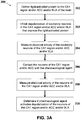
- FIG. 3A One example of a method for identifying a pharmacological agent that activates depolarization or excitation of non-human excitatory neurons in the CA1 region and/or ACC and/or BLA is depicted in FIG. 3A .
- the method (300) may comprise delivering a light-activated protein to the CA1 region and/or ACC and/or BLA of the brain (302) and inhibiting depolarization of excitatory neurons of the CA1 and/or ACC region (303).
- inhibiting depolarization may comprise applying light having a selected wavelength (e.g., yellow or green) to eNpHR3.1 ion channels expressed on the neurons of the CA1 and/or ACC region to prevent the generation of action potentials.
- Other types of light-activated channels may also be expressed to inhibit depolarization of these excitatory cells, such as variants of NpHR, BR, AR, and proton pumps such as GtR3.
- the effect of the inhibition from activating the eNpHR3.1 ion channels may be electrically measured by using loose-cell or whole-cell patch clamp methods (304).
- the electrical activity of the excitatory cells of the CA1 and/or ACC region may be measured using single electrodes and/or multielectrode arrays.
- the inhibited neurons of the CA1 and/or ACC region may then be contacted with a test pharmacological agent (306).
- the electrical activity of the neurons may be similarly measured (308).
- the electrical measurements of the excitatory neurons of the CA1 region and/or ACC and/or BLA before and after contacting with the test pharmacological agent may be compared to determine if the test agent activates and/or restores the depolarization of the neurons (310).
- the method (300) may be used repeatedly as desired to screen any number or variety of pharmacological agents.
- the method (320) may comprise delivering a light-activated protein to the CA1 region and/or ACC and/or BLA of the brain (322) and applying light have a selected wavelength (e.g., yellow or green) to eNpHR3.1 ion channels expressed on the neurons of the CA1 and/or ACC and/or BLA region to prevent the generation of action potentials (323).
- a selected wavelength e.g., yellow or green
- Other types of light-activated channels may also be expressed to inhibit depolarization of these excitatory cells, such as variants of NpHR, BR, AR, and proton pumps such as GtR3.
- the response of the non-human animal in the presence of the light during memory formation or retrieval may be measured (324).
- the memory may be formed during a training session where the individual is introduced into context A and exposed to a tone accompanied by a foot-shock, and the response to memory retrieval may be freezing when introduced into the context A and/or when the tone is played.
- the inhibited neurons of the CA1 and/or ACC region may then be contacted with a test pharmacological agent (326).
- the response of the non-human animal may be similarly measured (328).
- the response of the non-human animal before and after contacting with the test pharmacological agent may be evaluated to determine if the test agent affects memory formation or retrieval in the presence of light (330).
- the method (320) may be used during memory formation (e.g., a training session) to evaluate the effect of the pharmacological agent on memory formation.
- the method (320) may also be used during memory retrieval (e.g., a testing session some time after a training session) to evaluate the effect of the pharmacological agent on memory retrieval.
- the method (320) may be used repeatedly as desired to screen any number or variety of pharmacological agents.
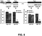
- inhibition of the neurons expressing the light-activated protein may be applied at a precise point in time.
- neurons expressing eNpHR3.1 in the CA1 region may illuminated by light during the testing session only.
- Temporally precise inhibition of neurons expressing eNpHR3.1 may disrupt memory recall.
- Precisely applying light to neurons expressing eNpHR3.1 in the CA1 region of mice during the testing session may inhibit remote and/or recent fear memory retrieval in an animal.
- inhibition of the neurons expressing eNpHR3.1 may be applied over a prolonged period of time.
- neurons expressing eNpHR3.1 in the CA1 region may be illuminated by light before the testing session (e.g., 30 minutes or more before the testing session).
- Prolonged inhibition of the neurons expressing eNpHR3.1 in the CA1 region of the hippocampus may affect the retrieval of memories differently from precise inhibition of the CA1 neurons.
- prolonged light application (i.e., prolonged inhibition) to CA1 neurons may affect recent contextual fear recall, but may not affect remote contextual memory recall.
- a method for treating PTSD may comprise administering a viral vector encoding a light-activated protein to an individual.
- the light-activated protein may be configured to inhibit depolarization of the neuron in the presence of light with a specific wavelength. Examples of such light-activated proteins may include NpHR, BR, AR, and GrR3.
- the viral vector may be delivered to any neuron population or type (e.g., the excitatory neurons of the CA1, ACC, and BLA brain regions).
- the neuron(s) expressing the light-activated protein may be inhibited from depolarizing, thereby disrupting the retrieval of the undesired memory.
- inhibiting depolarization of the neuron(s) may comprise applying light of the specific wavelength to the neurons expressing the light-activated proteins.
- the light may be removed. This may restore memory function such that memories may be recalled without disruption.
- memories related to drugs of abuse can be inhibited to reduce drug-seeking behavior.
- Other embodiments are directed toward the ability to instantaneously affect cognition by modulation of different brain areas in order to study the role of specific neuronal populations in memory processes. Inhibition of neurons by certain light-activated proteins and activation by other light-activated proteins may enable a finer temporal, genetic and spatial dissection of the neuronal circuits that underlie various brain function and behaviors.
- the method comprising: inhibiting the function of the dorsal CA1 hippocampus circuit with a temporal precision of the inhibition that is sufficient to disrupt the effects of remote memory retrieval.
- the step of inhibiting is responsive to a memory trigger event.
- the step of inhibiting includes activating light-responsive opsins expressed in cells of the dorsal CA1 hippocampus circuit.
- the step of inhibiting includes applying an electrical pulse through one or more electrodes positioned near the dorsal CA1 hippocampus circuit.
- the step of inhibiting includes releasing a drug at a location proximate to the dorsal CA1 hippocampus circuit.
- the effects of remote memory retrieval include emotional responses to a remote memory.
- Also provided herein are methods of disrupting memory creation comprising: inhibiting the function of the dorsal CA1 hippocampus circuit with a temporal precision of the inhibition that is sufficient to disrupt remote memory creation.
- the step of inhibiting is responsive to a memory trigger event.
- the step of inhibiting includes activating light-responsive opsins expressed in cells of the dorsal CA1 hippocampus circuit.
- the step of inhibiting includes applying an electrical pulse through one or more electrodes positioned near the dorsal CA1 hippocampus circuit.
- the step of inhibiting includes releasing a drug at a location proximate to the dorsal CA1 hippocampus circuit.
- the effects of remote memory retrieval include emotional responses to a remote memory.
- Also provided herein are methods of encouraging memory function comprising: exciting the function of the dorsal CA1 hippocampus circuit to promote remote memory creation or remote memory recall.
- Also provided herein are methods for treatment of a neurological disorder associated with remote memory recall comprising: in response to retrieval of the remote memory, inhibiting the function of the dorsal CA1 hippocampus circuit with a temporal precision of the inhibition that is sufficient to disrupt the effects of the retrieval of the remote memory.
- Various types of light-activated proteins may be used to control and characterize the neural circuits that underlie memory function.
- variants of NpHR may be used to inhibit depolarization and/or hyperpolarize a neuron.
- the third generation eNpHR has a trafficking signal between the gene and the fluorophore and has shown improved membrane targeting and increased light-induced hyperpolarizations. This third generation eNpHR was used to perturb the neurons in the CA1 region of the hippocampus to determine their role in both recent and remote memory acquisition and recall.
- eNpHR3.1 fused in-frame to enhanced yellow fluorescent protein (eNpHR3.1-EYFP) under control of the calcium/calmodulin-dependent protein kinase II ⁇ (CaMKII ⁇ ) promoter, selective for excitatory glutamatergic neurons in hippocampus was used.
- eNpHR3.1 is a truncated version of eNpHR3.0 with a deletion of the intrinsic N-terminal signal peptide that is similar to eNpHR3.0 in both the photocurrent and the hyperpolarization induced in neurons.
- mice C57BL6 mice aged 6 to 8 weeks were obtained from Charles River. Mice were housed four to five per cage in a colony maintained on a reversed 12 hr light/dark cycle and given food and water ad libitum. Experimental protocols were approved by Stanford University IACUC and meet guidelines of the National Institutes of Health guide for the Care and Use of Laboratory Animals.
- the CaMKII ⁇ -eNpHR3.1-EYFP lentivirus for in vivo injection was produced as previously described (Gradinaru et al., 2010; Zhang et al., 2007).
- the adeno-associated virus (AAV) CaMKII ⁇ -eNpHR3.0-EYFP plasmid was constructed by cloning eNpHR3.0-EYFP into an AAV backbone carrying the CaMKII ⁇ promoter using BamHI and EcoRI restriction sites.
- the recombinant AAV vectors were serotyped with AAV5 coat proteins and packaged by the Vector Core at the University of North Carolina; titers were 2x10¹² particles/mL.
- the maps for AAV CaMKII ⁇ ::eNpHR3.0 and Lenti CaMKII ⁇ ::eNpHR3.1 are available online at www.optogenetics.org.
- Stereotactic virus injection cannula/patchcord implantation, and light delivery.
- Mice were anesthetized with isoflurane, the head was placed in a stereotactic apparatus (Kopf Instruments, Tujunga, CA; Leica stereomicroscope). Ophthalmic ointment was applied to prevent eye drying. A midline scalp incision was made and then a small craniotomy was performed and the virus was delivered using a 10 ⁇ l syringe and a thin 34 gauge metal needle (World Precision Instruments, Sarasota, FL). The injection volume and flow rate (1 ⁇ l at 0.1 ⁇ l/min) were controlled by an injection pump (WPI). After injection the needle was left in place for 5 additional minutes and then slowly withdrawn.
- WPI injection pump
- a bilateral guide cannula (2.5 mm center to center; PlasticsOne, Roanoke, VA) was then placed 0.5 mm above CA1 (AP, -1.94 mm, ML, ⁇ 1.25 mm, DV -1 mm), and secured to the skull using dental cement (C&B metabond, Parkell, Edgwood, NY). The skin was glued back with Vetbond tissue adhesive. The animal was kept on a heating pad until it recovered from anesthetic. Buprenorphine (0.03 mg/kg) was given subcutaneously at the beginning of the surgical procedure to minimize discomfort.
- green light (561 nm, describe laser etc) was bilaterally delivered through two 300 ⁇ m thick optic fibers (Thorlabs, Newton, NJ) that were inserted through the guide cannulas, with a 0.5 mm projection.
- Control mice were either uninfected with eNpHR3.1 but still implanted with the cannula delivering light into CA1, or were infected with eNpHR3.1 and implanted, but connected to a dummy fiber that terminated the light delivery at the surface of the brain. Control mice therefore experienced identical visual cues and contextual information as the experimental mice associated with laser light delivery.
- BLA basolateral amygdala
- 1.5 ⁇ l of AAV5 CaMKII ⁇ ::eNpHR3.0-EYFP was microinjected into both left and right BLA (AP, -1.5 mm, ML, ⁇ 3.47 mm, DV -5 mm).
- a patchcord (a metal ferrule, 2.5 mm in diameter with a 200 ⁇ m thick, 5 mm long, cleaved bare optic fiber; Doric lenses Inc., Quebec, Canada) was then placed in each BLA (AP, -1.5 mm, ML, ⁇ 3.47mm, DV -4.8 mm), and secured to the skull using dental cement.
- Green light was bilaterally delivered through two 200 ⁇ m thick optic fibers (Doric lenses) that were attached to the patchcord using a connecting plastic sleeve.
- Doric lenses anterior cingulate cortex
- 1.0 ⁇ l of AAV5 CaMKII ⁇ ::eNpHR3.0-EYFP was microinjected into both left and right ACC (AP, +1 mm, ML, ⁇ 0.35 mm, DV -2.2 mm).
- a patchcord Doric lenses Inc.
- Green light was delivered through a 200 ⁇ m thick optic fiber (Doric lenses) attached to the patchcord.
- Doric lenses For olfactory bulb (OB) optogenetic inhibition, 1.0 ⁇ l of AAV5 CaMKII ⁇ ::eNpHR3.0-EYFP was microinjected into both left and right OB (AP, +4.5 mm, ML, ⁇ 0.75 mm, DV -3.25 and -2 mm).
- a patchcord (Doric lenses Inc.) was then unilaterally placed above one OB, as close as possible to the midline (AP, +4.5 mm, ML, ⁇ 0.15 mm, DV -1.4 mm), and secured to the skull using dental cement.
- Green light was delivered through a 200 ⁇ m thick optic fiber (Doric lenses) attached to the patchcord.
- mice were anesthetized with ketamine/xylazine and perfused transcardially with cold PBS followed by 4% paraformaldehyde (PFA) dissolved in phosphate-buffered saline (PBS, pH 7.4).
- PFA paraformaldehyde
- the brains were removed and post-fixed in 4% PFA in PBS for 3 hr at 4 °C, and then equilibrated in 30 % sucrose in PBS.
- Sections were then washed with PBS and incubated for 2 hr at room temperature with secondary antibodies (Donkey anti mouse conjugated to Cy3, donkey anti rabbit conjugated to either Cy3 or Cy5, all 1:1000, Jackson Laboratories, West grove, PA).
- Slices were then washed, incubated with DAPI (1:50,000) for 20 min, washed again, and mounted on slides with PVA-Dabco (Sigma).
- Confocal fluorescence images were acquired on a scanning laser microscope using a 5X or a 10X air objectives, or a 40X oil immersion objective. To determine the rate of viral transduction we calculated the percentage of CaMKII ⁇ -immunoreactive neurons per 40 X field that were also eNpHR-EYFP-positive.
- the optical fiber was coupled to a 473 nm solid-state laser diode with ⁇ 20 mW of output from the 200 ⁇ m fiber.
- Single unit recordings were done in mice anesthetized with a ketamine/xylazine mixture (ketamine, 80 mg/kg; xylazine, 15-20 mg/kg) diluted in PBS. Signals were recorded and band-pass filtered at 300Hz low/5 kHz high using an 1800 Microelectrode AC Amplifier.
- the fear conditioning apparatus consisted of a square conditioning cage (18x18x30 cm), with a grid floor wired to a shock generator and a scrambler, surrounded by an acoustic chamber (Coulbourn instruments, PA, USA.). The apparatus was modified to enable light delivery during training and/or testing. To induce fear-conditioning mice were placed in the cage for 120 seconds, and then a pure tone (2.9 kHz) was sound for 20 sec, followed by a 2 sec, foot-shock (0.5 mA for short-term memory, 1mA for long-term memory). This procedure was then repeated, and 30 sec after the delivery of the second shock mice were returned to their home cage.
- a pure tone 2.9 kHz
- mice were trained and tested as follows: Day 1- training with continuous 561 nm light administration (light ON). Day 2- contextual and cued tests (2 hr apart) without light administration (light OFF). Day 3- training, light OFF. Day 4 - test, light OFF. Day 5 - contextual and cued tests, light ON.
- Fig 6A Day 1- training, light OFF. Day 29 - contextual and cued tests, light ON. Day 30 - test light OFF.
- Fig 6C Day 1- training, light OFF. Day 64- contextual test, light ON.
- Fig. 8 Day 1- training, light OFF. Day 36 - test, light OFF. Day 37 - test light ON. Day 38 - test with 3 min light OFF followed by 3 min light ON.
- mice were trained on day 1 with light ON, and tested for contextual and cued fear on day 2 with light OFF.
- mice were trained on day 1 with the light OFF, tested on day 2 with the light ON, and then tested on day 29 with light ON.
- the optic fibers were passed through the conditioning cage into a regular housing cage with bedding, and light was delivered in this cage for 30 min. The mouse was then placed in the conditioning cage for a five min test, as light delivery continued without interruption.
- the results of the contextual- and cued-conditioning tests were analyzed by a Student's t-test or 2-way ANOVA, followed by post-hoc tests, as applicable.
- mice were implanted with a double cannula above CA1.
- the cannula, surgical procedure and location were the same as in the light delivery experiments.
- TTX Sigma, 20 ⁇ M
- CNQX Tocris Bioscience, Ellisville, MO; 3 mM
- saline were infused in a volume of 1 ⁇ l through a 28 gauge stainless steel internal cannula (PlasticsOne) that was 0.5 mm longer than the guide cannula.
- the internal cannula was connected to a micro-syringe pump (Harvard Apparatus, Holliston, MA) by a PE20 tube. Solutions were administered at a constant rate of 200 nl/min, and the injection cannula was removed 2 min following the termination of the injection to avoid spillage from the guide cannula.
- Open field test The open field test was conducted in an open plastic arena (50 cm long x 50 cm wide x 40 cm deep). Mice were individually placed in the center of the chamber and allowed to freely explore for 3 min. Activity in both the central and periphery of the field was measured using an automated video-tracking system (Biobserve, Bonn, Germany). Percentage of time in center is defined as the percent of total time that was spent in the central 35 x 35 cm area of the open field.
- Electrophysiological measurement of continuous inhibition of evoked spiking by eNpHR3.1 Four mice from the prolonged light exposure experiment were injected as described above, went through behavioral testing, and then sacrificed and sliced for physiology. Coronal slices containing dorsal CA1 were prepared by perfusing ice cold sucrose solution transcardially which contained (in mM): 26 NaHCO3, 2.5 KCl, 1.25 NaH2PO4, 10 MgSO4H14O7, .5 CaC12H4O2, 11 glucose, and 234 sucrose, and subsequently cutting 300 micron slices in the same ice cold sucrose solution.
- Induction of action potentials was done by injecting current ranging from 200pA at 10hz.
- Light for the activation of eNpHR3.1 was delivered using a X-Cite 120W halogen light source through a 531 ⁇ 20 nm filter and a 40x/0.8 NA water objective at 7 mW/mm 2 .
- Hippocampal Cultures Primary cultured hippocampal neurons were prepared from P0 Sprague-Dawley rat pups. The CA1 and CA3 regions were isolated, digested with 0.4 mg/mL papain (Worthington, Lakewood, NJ), and plated onto glass coverslips precoated with 1:30 Matrigel (Beckton Dickinson Labware, Bedford, MA) at a density of 65,000/cm 2 .
- DNA/CaCl2 mix for each well: 1.5-3 ⁇ g DNA (QIAGEN endotoxin-free preparation) + 1.875 ⁇ l 2M CaCl2 (final Ca2+ concentration 250 mM) in 15 ⁇ l total H20.
- DNA/CaCl2 was added 15 ⁇ l of 2X HEPES-buffered saline (pH 7.05), and the final volume was mixed well by pipetting.
- Electrophysiology Whole-cell patch clamp recordings were performed as previously described (intracellular solution: 129 mM K-gluconate, 10 mM HEPES, 10 mM KCl, 4 mM MgATP, 0.3 mM Na3GTP, titrated to pH 7.2; extracellular Tyrode: 125 mM NaCl, 2 mM KCl, 3 mM CaCl2, 1 mM MgCl2, 30 mM glucose, and 25 mM HEPES, titrated to pH 7.3). For voltage clamp recordings cells were held at -70mV.
- YFP control and eNpHR3.1 mice were trained with light administration during conditioning (without tone presentation, so that only fear of the context would be induced), and sacrificed 90 min later to test for c-Fos levels (described in detail in the immunohistochemistry section above).
- Two other groups of non-trained control and eNpHR3.1 mice were sacrificed from their home cages.
- YFP controls and eNpHR3.1 mice were fear-conditioned without light, exposed to the conditioning context with light 28 days later, and sacrificed 90 min afterwards to test for cFos levels.
- the control groups at this time point were control and eNpHR3.1 mice that were trained, and then sacrificed from their home cages 28 days later without being re-exposed to the conditioning context.
- eNpHR3.1 is a truncated version of eNpHR3.0 with a deletion of the intrinsic N-terminal signal peptide, that has comparable effects on membrane potential.
- eNpHR3.1 is targeted to the neuronal membrane, and is expressed around the soma, as well as in the apical and basal dendrites of CA1 neurons ( Fig. 4B ).
- CA1 optogenetic inhibition blocks contextual fear acquisition and retrieval.
- the involvement of the hippocampus in contextual fear conditioning is based on physical, pharmacological and genetic lesions to this structure, in which the interval between lesion and testing ranges from tens of minutes to several weeks (Anagnostaras et al., 1999; Kim and Fanselow, 1992; Kitamura et al., 2009; Shimizu et al., 2000; Wiltgen et al., 2010), which could allow for adaptation and compensation within the relevant neural circuitry.
- mice were introduced into context A, and then presented twice with a tone followed by a foot-shock, under continuous bilateral 561 nm light delivery, and mice were tested for their memory 24 hr later without light. Fear memory was then assessed the next day in the absence of optical inhibition.
- mice were then re-trained in the same context without light administration, and tested again on the next day; indeed, eNpHR3.1 mice exhibited intact contextual memory (64.6 ⁇ 6.6 vs. 49.7 ⁇ 11.7 % freezing; P>0.5) when no light was administered during training ( Fig. 5B , middle).
- mice were bilaterally injected in the basolateral amygdala (BLA; Fig. 5H ) instead of hippocampus and it was found that it was possible to optogenetically inhibit both contextual (Fig. 5I ; 65.5 ⁇ 7.2 vs. 9.6 ⁇ 5.5 % freezing; P ⁇ 0.001) and auditory-cued FC acquisition ( Fig. 5I ; 69.5 ⁇ 9.6 vs.
- CA1 optogenetic inhibition reversibly interferes with remote fear memory recall.
- the hippocampus would still be involved in contextual fear recall even at much longer time intervals, another population of mice were trained and this cohort was tested 9 weeks after contextual FC.
- mice from the prolonged group were re-tested on the next day with precise light administration (during the test only), the same mice displayed inhibited fear recall ( Fig. 7A right).
- the prolonged group mice were re-tested the next day with light during the test only, their recall was disrupted (Prolonged group, left, 55.5 ⁇ 8.5 vs. 27.6 ⁇ 8.6 % freezing; P ⁇ 0.05).
- CA1 optogenetic inhibition interferes with ongoing fear recall.
- the NpHR3.1 group failed to recall the contextual memory ( Fig. 8 left; (77.2 ⁇ 4.3 % vs. 12.8 ⁇ 4.4 % freezing; P ⁇ 0.0001).
- eNpHR3.1-mediated inhibition was delivered during training or remote recall, and assessed induction of the immediate early gene product c-Fos across the entire brain. Mice were fear-conditioned under light delivery, and brains were collected 90 min after training ( Fig. 9A ). Brain slices were stained for c-Fos and DAPI ( Fig. 9B ). Expression of YFP control and eNpHR3.1 are shown. The CA1 region from which these images were taken is marked by a white square in Fig. 9C . Following training, eNpHR3.1-expressing mice demonstrated markedly reduced c-Fos expression specifically in CA1 compared with trained control animals ( Fig.
- mice Another group of mice was conditioned, and then re-exposed to the context 28 days after conditioning in the presence or absence of CA1 optogenetic inhibition; as before, the eNpHR3.1-expressing mice demonstrated impaired remote recall. 90 min later the brains were collected and stained for c-Fos ( Fig. 9E ) to capture putative memory-related brain-wide activity patterns under control of the hippocampus at this remote timepoint. Intriguingly, a small but significant increase in CA1c-Fos was observed in control, but not eNpHR3.1 mice ( Fig. 9F-G ; P ⁇ 0.005) following remote recall. Representative CA1, ACC and BLA images following remote memory are shown. White scalebar: 150 ⁇ m.
- eNpHR3.1-expressing mice showed an elevation in prefrontal cortex activity equivalent to that of controls, and no significant changes in parietal cortex activity levels were observed in any of the groups.
- activity levels in the ACC were significantly recruited in remote memory only, and to a lesser extent in the setting of eNpHR3.1-mediated CA1 inhibition ( Fig. 9H middle), also in agreement with previous reports (Bontempi et al., 1999; Frankland et al., 2004; Hall et al., 2001; Maviel et al., 2004).
- Fig. 9H depicts global patterns in brain activity between conditioning (day 0) and remote recall (day 28).
- Activity levels in CA1 significantly decreased in control (P ⁇ 0.005) mice from day 0 to day 28.
- Activity levels in ACC significantly increased in both control (P ⁇ 0.0001) and eNpHR3.1 (P ⁇ 0.001) mice day 0 to day 28.
- Activity levels in BLA significantly increased in control (P ⁇ 0.001) but not in eNHR3.1 mice.
- ACC Optogenetic inhibition of ACC inhibits remote but not recent contextual memory. Since the population of CA1 neurons active during remote contextual memory was found to be causally involved in fully organizing ACC neuronal activity as shown above, and since previous research has implicated the ACC in remote fear memory storage (Bontempi et al., 1999; Fischer et al., 2007; Frankland et al., 2004; Maviel et al., 2004), optogenetic inhibition of memories was explored by targeting ACC directly either one day or one month following contextual FC.
- Fig. 10A depicts eNpHR3.0 expression in the anterior cingulate cortex (ACC).
- the finding that the hippocampus is still the default activator of contextual fear memory recall may be due to the fact that many place cells (Moser et al 2008) in CA1 remap in response to fear conditioning (Moita et al 2004), and may contribute to a faster recognition of the context. Indeed, hippocampal lesions were repeatedly shown to induce retrograde amnesia for spatial memory (Broadbent et al 2006; Martin et al 2005).
- the ability to instantaneously affect cognition by optogenetic modulation of different brain areas may serve as a basis for future studies re-examining the role of specific neuronal populations in memory processes and enable a finer temporal, genetic and spatial dissection of the neuronal circuits that underlie them.
Applications Claiming Priority (3)
| Application Number | Priority Date | Filing Date | Title |
|---|---|---|---|
| US41073210P | 2010-11-05 | 2010-11-05 | |
| US201161540926P | 2011-09-29 | 2011-09-29 | |
| PCT/US2011/059283 WO2012061681A1 (en) | 2010-11-05 | 2011-11-04 | Control and characterization of memory function |
Publications (3)
| Publication Number | Publication Date |
|---|---|
| EP2635295A1 EP2635295A1 (en) | 2013-09-11 |
| EP2635295A4 EP2635295A4 (en) | 2014-04-30 |
| EP2635295B1 true EP2635295B1 (en) | 2017-12-20 |
Family
ID=46024836
Family Applications (1)
| Application Number | Title | Priority Date | Filing Date |
|---|---|---|---|
| EP11838856.0A Not-in-force EP2635295B1 (en) | 2010-11-05 | 2011-11-04 | Control and characterization of memory function |
Country Status (8)
| Country | Link |
|---|---|
| US (2) | US10086012B2 (zh) |
| EP (1) | EP2635295B1 (zh) |
| JP (1) | JP6328424B6 (zh) |
| CN (2) | CN106376525A (zh) |
| AU (2) | AU2011323228B2 (zh) |
| CA (1) | CA2816972C (zh) |
| ES (1) | ES2661093T3 (zh) |
| WO (1) | WO2012061681A1 (zh) |
Families Citing this family (41)
| Publication number | Priority date | Publication date | Assignee | Title |
|---|---|---|---|---|
| WO2007024391A2 (en) | 2005-07-22 | 2007-03-01 | The Board Of Trustees Of The Leland Stanford Junior University | Light-activated cation channel and uses thereof |
| US9238150B2 (en) | 2005-07-22 | 2016-01-19 | The Board Of Trustees Of The Leland Stanford Junior University | Optical tissue interface method and apparatus for stimulating cells |
| US9274099B2 (en) | 2005-07-22 | 2016-03-01 | The Board Of Trustees Of The Leland Stanford Junior University | Screening test drugs to identify their effects on cell membrane voltage-gated ion channel |
| US20090093403A1 (en) | 2007-03-01 | 2009-04-09 | Feng Zhang | Systems, methods and compositions for optical stimulation of target cells |
| US10052497B2 (en) | 2005-07-22 | 2018-08-21 | The Board Of Trustees Of The Leland Stanford Junior University | System for optical stimulation of target cells |
| US8926959B2 (en) | 2005-07-22 | 2015-01-06 | The Board Of Trustees Of The Leland Stanford Junior University | System for optical stimulation of target cells |
| WO2008086470A1 (en) | 2007-01-10 | 2008-07-17 | The Board Of Trustees Of The Leland Stanford Junior University | System for optical stimulation of target cells |
| US8401609B2 (en) | 2007-02-14 | 2013-03-19 | The Board Of Trustees Of The Leland Stanford Junior University | System, method and applications involving identification of biological circuits such as neurological characteristics |
| US10035027B2 (en) | 2007-10-31 | 2018-07-31 | The Board Of Trustees Of The Leland Stanford Junior University | Device and method for ultrasonic neuromodulation via stereotactic frame based technique |
| US10434327B2 (en) | 2007-10-31 | 2019-10-08 | The Board Of Trustees Of The Leland Stanford Junior University | Implantable optical stimulators |
| KR20110005280A (ko) | 2008-04-23 | 2011-01-17 | 더 보드 어브 트러스티스 어브 더 리랜드 스탠포드 주니어 유니버시티 | 표적 세포의 광학적 자극을 위한 시스템, 방법 및 조성물 |
| EP2294208B1 (en) | 2008-05-29 | 2013-05-08 | The Board of Trustees of The Leland Stanford Junior University | Cell line, system and method for optical control of secondary messengers |
| MY156389A (en) | 2008-06-17 | 2016-02-15 | Univ Leland Stanford Junior | Methods, systems and devices for optical stimulation of target cells using an optical transmission element |
| JP5887136B2 (ja) | 2008-06-17 | 2016-03-16 | ザ ボード オブ トラスティーズ オブ ザ レランド スタンフォード ジュニア ユニバーシティー | 細胞発達を制御するための装置および方法 |
| WO2010006049A1 (en) | 2008-07-08 | 2010-01-14 | The Board Of Trustees Of The Leland Stanford Junior University | Materials and approaches for optical stimulation of the peripheral nervous system |
| NZ602416A (en) | 2008-11-14 | 2014-08-29 | Univ Leland Stanford Junior | Optically-based stimulation of target cells and modifications thereto |
| AU2011227131B2 (en) * | 2010-03-17 | 2014-11-13 | The Board Of Trustees Of The Leland Stanford Junior University | Light-sensitive ion-passing molecules |
| ES2716819T3 (es) | 2010-11-05 | 2019-06-17 | Univ Leland Stanford Junior | Opsinas quiméricas activadas por luz y métodos de uso de las mismas |
| EP2635109A4 (en) | 2010-11-05 | 2014-03-19 | Univ Leland Stanford Junior | OPTICALLY CONTROLLED CNS DYSFUNCTION |
| AU2011323228B2 (en) * | 2010-11-05 | 2016-11-10 | The Board Of Trustees Of The Leland Stanford Junior University | Control and characterization of memory function |
| WO2012061684A1 (en) | 2010-11-05 | 2012-05-10 | The Board Of Trustees Of The Leland Stanford Junior University | Upconversion of light for use in optogenetic methods |
| CN103476456B (zh) | 2010-11-05 | 2017-10-03 | 斯坦福大学托管董事会 | 奖赏相关行为的光遗传学控制 |
| CA2816990A1 (en) * | 2010-11-05 | 2012-05-10 | The Board Of Trustees Of The Leland Stanford Junior University | Stabilized step function opsin proteins and methods of using the same |
| US8696722B2 (en) | 2010-11-22 | 2014-04-15 | The Board Of Trustees Of The Leland Stanford Junior University | Optogenetic magnetic resonance imaging |
| US9365628B2 (en) | 2011-12-16 | 2016-06-14 | The Board Of Trustees Of The Leland Stanford Junior University | Opsin polypeptides and methods of use thereof |
| WO2013126521A1 (en) | 2012-02-21 | 2013-08-29 | The Board Of Trustees Of The Leland Stanford Junior University | Compositions and methods for treating neurogenic disorders of the pelvic floor |
| EP2968997B1 (en) | 2013-03-15 | 2019-06-26 | The Board of Trustees of the Leland Stanford Junior University | Optogenetic control of behavioral state |
| US9636380B2 (en) | 2013-03-15 | 2017-05-02 | The Board Of Trustees Of The Leland Stanford Junior University | Optogenetic control of inputs to the ventral tegmental area |
| EP2991491B1 (en) | 2013-04-29 | 2019-12-25 | The Board of Trustees of the Leland Stanford Junior University | Devices, systems and methods for optogenetic modulation of action potentials in target cells |
| CN105829538A (zh) | 2013-08-14 | 2016-08-03 | 小利兰·斯坦福大学托管委员会 | 用于控制疼痛的组合物和方法 |
| WO2015148974A2 (en) | 2014-03-28 | 2015-10-01 | The Board Of Trustees Of The Leland Stanford Junior University | Engineered light-activated anion channel proteins and methods of use thereof |
| US20180154170A1 (en) * | 2015-06-16 | 2018-06-07 | Massachusetts Institute Of Technology | Methods and compositions for treating mental disorders and conditions |
| US10820823B2 (en) * | 2015-06-18 | 2020-11-03 | Inter-University Research Institute Corporation National Institutes Of Natural Sciences | Evaluation of inhibitory circuit and use thereof |
| WO2016209654A1 (en) | 2015-06-22 | 2016-12-29 | The Board Of Trustees Of The Leland Stanford Junior University | Methods and devices for imaging and/or optogenetic control of light-responsive neurons |
| US10265497B2 (en) * | 2015-11-24 | 2019-04-23 | Massachusetts Institute Of Technology | Systems and methods for treating dementia |
| CN106226509B (zh) * | 2016-08-12 | 2019-09-06 | 中国人民解放军第四军医大学 | 一种在小鼠前扣带回皮层诱发dse现象的方法 |
| US11294165B2 (en) | 2017-03-30 | 2022-04-05 | The Board Of Trustees Of The Leland Stanford Junior University | Modular, electro-optical device for increasing the imaging field of view using time-sequential capture |
| US11241586B2 (en) | 2017-10-10 | 2022-02-08 | Massachusetts Institute Of Technology | Systems and methods for preventing, mitigating, and/or treating dementia |
| US11622948B2 (en) | 2017-11-09 | 2023-04-11 | The Trustees Of Columbia University In The City Of New York | Biomarkers for efficacy of prophylactic treatments against stress-induced affective disorders |
| WO2022034168A1 (en) | 2020-08-12 | 2022-02-17 | Borealis Ag | Multilayer film with low seal initiation temperature |
| CN114588263A (zh) * | 2022-03-28 | 2022-06-07 | 中国人民解放军军事科学院军事医学研究院 | 光敏通道蛋白在空间学习记忆障碍的哺乳动物中的用途 |
Family Cites Families (290)
| Publication number | Priority date | Publication date | Assignee | Title |
|---|---|---|---|---|
| US2968302A (en) | 1956-07-20 | 1961-01-17 | Univ Illinois | Multibeam focusing irradiator |
| US3131690A (en) | 1962-10-22 | 1964-05-05 | American Optical Corp | Fiber optics devices |
| US3499437A (en) | 1967-03-10 | 1970-03-10 | Ultrasonic Systems | Method and apparatus for treatment of organic structures and systems thereof with ultrasonic energy |
| US3567847A (en) | 1969-01-06 | 1971-03-02 | Edgar E Price | Electro-optical display system |
| US4343301A (en) | 1979-10-04 | 1982-08-10 | Robert Indech | Subcutaneous neural stimulation or local tissue destruction |
| US4559951A (en) | 1982-11-29 | 1985-12-24 | Cardiac Pacemakers, Inc. | Catheter assembly |
| US4616231A (en) | 1984-03-26 | 1986-10-07 | Hughes Aircraft Company | Narrow-band beam steering system |
| FR2580277B1 (fr) | 1985-04-15 | 1988-06-10 | Oreal | Nouveaux derives naphtaleniques a action retinoique, le ur procede de preparation et compositions medicamenteuse et cosmetique les contenant |
| US4865042A (en) | 1985-08-16 | 1989-09-12 | Hitachi, Ltd. | Ultrasonic irradiation system |
| EP0335326B1 (en) | 1988-03-28 | 1994-06-15 | Canon Kabushiki Kaisha | Ion permeable membrane and ion transport method by utilizing said membrane |
| US5082670A (en) | 1988-12-15 | 1992-01-21 | The Regents Of The University Of California | Method of grafting genetically modified cells to treat defects, disease or damage or the central nervous system |
| JP2882818B2 (ja) | 1989-09-08 | 1999-04-12 | 株式会社エス・エル・ティ・ジャパン | レーザ光の照射装置 |
| CA2028261C (en) | 1989-10-28 | 1995-01-17 | Won Suck Yang | Non-invasive method and apparatus for measuring blood glucose concentration |
| US5032123A (en) | 1989-12-28 | 1991-07-16 | Cordis Corporation | Laser catheter with radially divergent treatment beam |
| ATE158816T1 (de) | 1990-11-26 | 1997-10-15 | Genetics Inst | Expression von pace in wirtszellen und verfahren zu dessen verwendung |
| US5550316A (en) | 1991-01-02 | 1996-08-27 | Fox Chase Cancer Center | Transgenic animal model system for human cutaneous melanoma |
| US6497872B1 (en) | 1991-07-08 | 2002-12-24 | Neurospheres Holdings Ltd. | Neural transplantation using proliferated multipotent neural stem cells and their progeny |
| US5249575A (en) | 1991-10-21 | 1993-10-05 | Adm Tronics Unlimited, Inc. | Corona discharge beam thermotherapy system |
| SE9103752D0 (sv) | 1991-12-18 | 1991-12-18 | Astra Ab | New compounds |
| US5670113A (en) | 1991-12-20 | 1997-09-23 | Sibia Neurosciences, Inc. | Automated analysis equipment and assay method for detecting cell surface protein and/or cytoplasmic receptor function using same |
| US5739273A (en) | 1992-02-12 | 1998-04-14 | Yale University | Transmembrane polypeptide and methods of use |
| US5460954A (en) | 1992-04-01 | 1995-10-24 | Cheil Foods & Chemicals, Inc. | Production of human proinsulin using a novel vector system |
| US5330515A (en) | 1992-06-17 | 1994-07-19 | Cyberonics, Inc. | Treatment of pain by vagal afferent stimulation |
| US5382516A (en) | 1992-09-15 | 1995-01-17 | Schleicher & Schuell, Inc. | Method and devices for delivery of substrate for the detection of enzyme-linked, membrane-based binding assays |
| US5527695A (en) | 1993-01-29 | 1996-06-18 | Purdue Research Foundation | Controlled modification of eukaryotic genomes |
| CA2156251A1 (en) | 1993-03-25 | 1994-09-29 | George J. Turner | Expression of heterologous polypeptides in halobacteria |
| US5411540A (en) | 1993-06-03 | 1995-05-02 | Massachusetts Institute Of Technology | Method and apparatus for preferential neuron stimulation |
| GB2278783A (en) | 1993-06-11 | 1994-12-14 | Daniel Shellon Gluck | Method of magnetically stimulating neural cells |
| US6346101B1 (en) | 1993-07-19 | 2002-02-12 | Research Foundation Of City College Of New York | Photon-mediated introduction of biological materials into cells and/or cellular components |
| US5445608A (en) | 1993-08-16 | 1995-08-29 | James C. Chen | Method and apparatus for providing light-activated therapy |
| JPH07171162A (ja) | 1993-09-07 | 1995-07-11 | Olympus Optical Co Ltd | レーザプローブ |
| US6056738A (en) | 1997-01-31 | 2000-05-02 | Transmedica International, Inc. | Interstitial fluid monitoring |
| US5470307A (en) | 1994-03-16 | 1995-11-28 | Lindall; Arnold W. | Catheter system for controllably releasing a therapeutic agent at a remote tissue site |
| CA2187626C (en) | 1994-04-13 | 2009-11-03 | Michael G. Kaplitt | Aav-mediated delivery of dna to cells of the nervous system |
| US6436908B1 (en) | 1995-05-30 | 2002-08-20 | Duke University | Use of exogenous β-adrenergic receptor and β-adrenergic receptor kinase gene constructs to enhance myocardial function |
| US5495541A (en) | 1994-04-19 | 1996-02-27 | Murray; Steven C. | Optical delivery device with high numerical aperture curved waveguide |
| US5503737A (en) | 1994-07-25 | 1996-04-02 | Ingersoll-Rand Company | Air inflow restrictor for disc filters |
| US5807285A (en) | 1994-08-18 | 1998-09-15 | Ethicon-Endo Surgery, Inc. | Medical applications of ultrasonic energy |
| US5520188A (en) | 1994-11-02 | 1996-05-28 | Focus Surgery Inc. | Annular array transducer |
| US5795581A (en) | 1995-03-31 | 1998-08-18 | Sandia Corporation | Controlled release of molecular components of dendrimer/bioactive complexes |
| US6334846B1 (en) | 1995-03-31 | 2002-01-01 | Kabushiki Kaisha Toshiba | Ultrasound therapeutic apparatus |
| WO1996032076A1 (en) | 1995-04-11 | 1996-10-17 | Baxter Internatonal Inc. | Tissue implant systems |
| US6342379B1 (en) | 1995-06-07 | 2002-01-29 | The Regents Of The University Of California | Detection of transmembrane potentials by optical methods |
| US6480743B1 (en) | 2000-04-05 | 2002-11-12 | Neuropace, Inc. | System and method for adaptive brain stimulation |
| US5755750A (en) | 1995-11-13 | 1998-05-26 | University Of Florida | Method and apparatus for selectively inhibiting activity in nerve fibers |
| US5722426A (en) | 1996-02-26 | 1998-03-03 | Kolff; Jack | Coronary light probe and method of use |
| US5703985A (en) | 1996-04-29 | 1997-12-30 | Eclipse Surgical Technologies, Inc. | Optical fiber device and method for laser surgery procedures |
| US5939320A (en) | 1996-05-20 | 1999-08-17 | New York University | G-coupled receptors associated with macrophage-trophic HIV, and diagnostic and therapeutic uses thereof |
| US5898058A (en) | 1996-05-20 | 1999-04-27 | Wellman, Inc. | Method of post-polymerization stabilization of high activity catalysts in continuous polyethylene terephthalate production |
| US20040076613A1 (en) | 2000-11-03 | 2004-04-22 | Nicholas Mazarakis | Vector system |
| US7732129B1 (en) | 1998-12-01 | 2010-06-08 | Crucell Holland B.V. | Method for the production and purification of adenoviral vectors |
| US5741316A (en) | 1996-12-02 | 1998-04-21 | Light Sciences Limited Partnership | Electromagnetic coil configurations for power transmission through tissue |
| US5756351A (en) | 1997-01-13 | 1998-05-26 | The Regents Of The University Of California | Biomolecular optical sensors |
| US5782896A (en) | 1997-01-29 | 1998-07-21 | Light Sciences Limited Partnership | Use of a shape memory alloy to modify the disposition of a device within an implantable medical probe |
| US5904659A (en) | 1997-02-14 | 1999-05-18 | Exogen, Inc. | Ultrasonic treatment for wounds |
| US5816256A (en) | 1997-04-17 | 1998-10-06 | Bioanalytical Systems, Inc. | Movement--responsive system for conducting tests on freely-moving animals |
| US6436708B1 (en) | 1997-04-17 | 2002-08-20 | Paola Leone | Delivery system for gene therapy to the brain |
| US7276488B2 (en) | 1997-06-04 | 2007-10-02 | Oxford Biomedica (Uk) Limited | Vector system |
| US5984861A (en) | 1997-09-29 | 1999-11-16 | Boston Scientific Corporation | Endofluorescence imaging module for an endoscope |
| US6597954B1 (en) | 1997-10-27 | 2003-07-22 | Neuropace, Inc. | System and method for controlling epileptic seizures with spatially separated detection and stimulation electrodes |
| US6647296B2 (en) | 1997-10-27 | 2003-11-11 | Neuropace, Inc. | Implantable apparatus for treating neurological disorders |
| US6016449A (en) | 1997-10-27 | 2000-01-18 | Neuropace, Inc. | System for treatment of neurological disorders |
| US6790652B1 (en) | 1998-01-08 | 2004-09-14 | Bioimage A/S | Method and apparatus for high density format screening for bioactive molecules |
| EP1091685B1 (en) | 1998-04-07 | 2008-06-11 | Cytyc Corporation | Devices for the localization of lesions in solid tissue |
| US6319241B1 (en) | 1998-04-30 | 2001-11-20 | Medtronic, Inc. | Techniques for positioning therapy delivery elements within a spinal cord or a brain |
| US6108081A (en) | 1998-07-20 | 2000-08-22 | Battelle Memorial Institute | Nonlinear vibrational microscopy |
| US6632672B2 (en) | 1998-08-19 | 2003-10-14 | The Board Of Trustees Of The Leland Stanford Junior University | Methods and compositions for genomic modification |
| US6377842B1 (en) | 1998-09-22 | 2002-04-23 | Aurora Optics, Inc. | Method for quantitative measurement of fluorescent and phosphorescent drugs within tissue utilizing a fiber optic probe |
| US6253109B1 (en) | 1998-11-05 | 2001-06-26 | Medtronic Inc. | System for optimized brain stimulation |
| AU1344100A (en) | 1998-11-06 | 2000-05-29 | University Of Rochester | A method to improve circulation to ischemic tissue |
| US6303362B1 (en) | 1998-11-19 | 2001-10-16 | The Board Of Trustees Of The Leland Stanford Junior University | Adenoviral vector and methods for making and using the same |
| US6790657B1 (en) | 1999-01-07 | 2004-09-14 | The United States Of America As Represented By The Department Of Health And Human Services | Lentivirus vector system |
| US7507545B2 (en) | 1999-03-31 | 2009-03-24 | Cardiome Pharma Corp. | Ion channel modulating activity method |
| US6224566B1 (en) | 1999-05-04 | 2001-05-01 | Cardiodyne, Inc. | Method and devices for creating a trap for confining therapeutic drugs and/or genes in the myocardium |
| US6161045A (en) | 1999-06-01 | 2000-12-12 | Neuropace, Inc. | Method for determining stimulation parameters for the treatment of epileptic seizures |
| US7655423B2 (en) | 1999-06-14 | 2010-02-02 | Henry Ford Health System | Nitric oxide donors for inducing neurogenesis |
| US6662039B2 (en) | 1999-06-18 | 2003-12-09 | The Trustees Of Columbia University In The City Of New York | Optical probing of neuronal connections with fluorescent indicators |
| US20040034882A1 (en) | 1999-07-15 | 2004-02-19 | Vale Wylie W. | Corticotropin releasing factor receptor 2 deficient mice and uses thereof |
| US7674463B1 (en) | 1999-07-15 | 2010-03-09 | Research Development Foundation | Method of inhibiting angiogenesis by administration of a corticotropin releasing factor receptor 2 agonist |
| WO2001005306A1 (en) | 1999-07-19 | 2001-01-25 | Epicor, Inc. | Apparatus and method for ablating tissue |
| ES2152900B1 (es) | 1999-07-23 | 2001-08-16 | Palleja Xavier Estivill | Ratones transgenicos y modelo de sobreexpresion del gen ntrk3 (trkc) basado en los mismos para el estudio y monitorizacion de tratamientos de la ansiedad, depresion y enfermedades psiquiatricas relacionadas. |
| CN1249672C (zh) | 1999-08-06 | 2006-04-05 | 阿尔卑斯电气株式会社 | 磁盘用磁头的搬送用托盘 |
| ES2478635T3 (es) | 1999-08-09 | 2014-07-22 | Targeted Genetics Corporation | Incremento de la expresión de una secuencia de nucleótidos heteróloga monocatenaria de vectores virales recombinantes diseñando la secuencia de modo que forma pares de bases intracatenarios |
| GB9923558D0 (en) | 1999-10-05 | 1999-12-08 | Oxford Biomedica Ltd | Producer cell |
| GB9928248D0 (en) | 1999-12-01 | 2000-01-26 | Gill Steven S | An implantable guide tube for neurosurgery |
| EP1247093B1 (en) | 2000-01-14 | 2005-03-23 | MIGENIX Corp. | Screening assays using intramitochondrial calcium |
| US7706882B2 (en) | 2000-01-19 | 2010-04-27 | Medtronic, Inc. | Methods of using high intensity focused ultrasound to form an ablated tissue area |
| US6595934B1 (en) | 2000-01-19 | 2003-07-22 | Medtronic Xomed, Inc. | Methods of skin rejuvenation using high intensity focused ultrasound to form an ablated tissue area containing a plurality of lesions |
| EP1255868B1 (en) | 2000-02-18 | 2010-12-01 | The Board Of Trustees Of The Leland Stanford Junior University | Altered recombinases for genome modification |
| US6473639B1 (en) | 2000-03-02 | 2002-10-29 | Neuropace, Inc. | Neurological event detection procedure using processed display channel based algorithms and devices incorporating these procedures |
| AU2001295193A1 (en) | 2000-05-01 | 2001-11-12 | Novartis Ag | Vectors for ocular transduction and use thereof for genetic therapy |
| US6599281B1 (en) | 2000-05-03 | 2003-07-29 | Aspect Medical Systems, Inc. | System and method for adaptive drug delivery |
| US6551346B2 (en) | 2000-05-17 | 2003-04-22 | Kent Crossley | Method and apparatus to prevent infections |
| US7250294B2 (en) | 2000-05-17 | 2007-07-31 | Geron Corporation | Screening small molecule drugs using neural cells differentiated from human embryonic stem cells |
| ATE438414T1 (de) | 2000-06-01 | 2009-08-15 | Univ North Carolina | Verfahren und zusammensetzungen zur kontrollierter abgabe von rekombinant parvovirus vektoren |
| EP1307551A2 (en) | 2000-07-05 | 2003-05-07 | PHARMACIA & UPJOHN COMPANY | Human ion channels |
| US6686193B2 (en) | 2000-07-10 | 2004-02-03 | Vertex Pharmaceuticals, Inc. | High throughput method and system for screening candidate compounds for activity against target ion channels |
| US6921413B2 (en) | 2000-08-16 | 2005-07-26 | Vanderbilt University | Methods and devices for optical stimulation of neural tissues |
| US6567690B2 (en) | 2000-10-16 | 2003-05-20 | Cole Giller | Method and apparatus for probe localization in brain matter |
| US6536440B1 (en) | 2000-10-17 | 2003-03-25 | Sony Corporation | Method and system for generating sensory data onto the human neural cortex |
| US6584357B1 (en) | 2000-10-17 | 2003-06-24 | Sony Corporation | Method and system for forming an acoustic signal from neural timing difference data |
| US7350522B2 (en) | 2000-10-17 | 2008-04-01 | Sony Corporation | Scanning method for applying ultrasonic acoustic data to the human neural cortex |
| US6631283B2 (en) | 2000-11-15 | 2003-10-07 | Virginia Tech Intellectual Properties, Inc. | B/B-like fragment targeting for the purposes of photodynamic therapy and medical imaging |
| US6506154B1 (en) | 2000-11-28 | 2003-01-14 | Insightec-Txsonics, Ltd. | Systems and methods for controlling a phased array focused ultrasound system |
| SE525540C2 (sv) | 2000-11-30 | 2005-03-08 | Datainnovation I Lund Ab | System och förfarande för automatisk provtagning från ett provobjekt |
| US20070196838A1 (en) | 2000-12-08 | 2007-08-23 | Invitrogen Corporation | Methods and compositions for synthesis of nucleic acid molecules using multiple recognition sites |
| US6489115B2 (en) | 2000-12-21 | 2002-12-03 | The Board Of Regents Of The University Of Nebraska | Genetic assays for trinucleotide repeat mutations in eukaryotic cells |
| US6615080B1 (en) | 2001-03-29 | 2003-09-02 | John Duncan Unsworth | Neuromuscular electrical stimulation of the foot muscles for prevention of deep vein thrombosis and pulmonary embolism |
| US7047078B2 (en) | 2001-03-30 | 2006-05-16 | Case Western Reserve University | Methods for stimulating components in, on, or near the pudendal nerve or its branches to achieve selective physiologic responses |
| AU2002303283A1 (en) | 2001-04-04 | 2002-10-21 | Irm Llc | Methods for treating drug addiction |
| US7107996B2 (en) | 2001-04-10 | 2006-09-19 | Ganz Robert A | Apparatus and method for treating atherosclerotic vascular disease through light sterilization |
| US6961045B2 (en) | 2001-06-16 | 2005-11-01 | Che-Chih Tsao | Pattern projection techniques for volumetric 3D displays and 2D displays |
| US6810285B2 (en) | 2001-06-28 | 2004-10-26 | Neuropace, Inc. | Seizure sensing and detection using an implantable device |
| WO2003016486A2 (en) | 2001-08-16 | 2003-02-27 | Sloan Kettering Institute For Cancer Research | Bio-synthetic photostimulators and methods of use |
| CA2458224A1 (en) | 2001-08-23 | 2003-03-06 | The Regents Of The University Of California | A universal light-switchable gene promoter system |
| US6974448B2 (en) | 2001-08-30 | 2005-12-13 | Medtronic, Inc. | Method for convection enhanced delivery catheter to treat brain and other tumors |
| US7904176B2 (en) | 2006-09-07 | 2011-03-08 | Bio Control Medical (B.C.M.) Ltd. | Techniques for reducing pain associated with nerve stimulation |
| WO2003020103A2 (en) | 2001-09-04 | 2003-03-13 | Amit Technology Science & Medicine Ltd. | Method of and device for therapeutic illumination of internal organs and tissues |
| US20040054952A1 (en) | 2002-09-13 | 2004-03-18 | Morrow James W. | Device verification system and method |
| CA2481020A1 (en) | 2001-09-28 | 2003-04-03 | Saoirse Corporation | Localized non-invasive biological modulation system |
| US7175596B2 (en) | 2001-10-29 | 2007-02-13 | Insightec-Txsonics Ltd | System and method for sensing and locating disturbances in an energy path of a focused ultrasound system |
| US7303578B2 (en) | 2001-11-01 | 2007-12-04 | Photothera, Inc. | Device and method for providing phototherapy to the brain |
| US8308784B2 (en) | 2006-08-24 | 2012-11-13 | Jackson Streeter | Low level light therapy for enhancement of neurologic function of a patient affected by Parkinson's disease |
| WO2003040323A2 (en) | 2001-11-08 | 2003-05-15 | Children's Medical Center Corporation | Bacterial ion channel and a method for screening ion channel modulators |
| CA2465337A1 (en) | 2001-11-14 | 2003-05-22 | Yamanouchi Pharmaceutical Co., Ltd. | Transgenic animal |
| EP1456374A4 (en) | 2001-11-26 | 2005-08-17 | Advanced Cell Tech Inc | METHODS FOR THE PRODUCTION AND USE OF REPROGRAMME HUMAN SOMATIC CELL CORES AND AUTOLOGOUS AND ISOGENIC HUMAN STEM CELLS |
| US20030104512A1 (en) | 2001-11-30 | 2003-06-05 | Freeman Alex R. | Biosensors for single cell and multi cell analysis |
| US6873868B2 (en) | 2001-12-31 | 2005-03-29 | Infraredx, Inc. | Multi-fiber catheter probe arrangement for tissue analysis or treatment |
| US6721603B2 (en) | 2002-01-25 | 2004-04-13 | Cyberonics, Inc. | Nerve stimulation as a treatment for pain |
| US6666857B2 (en) | 2002-01-29 | 2003-12-23 | Robert F. Smith | Integrated wavefront-directed topography-controlled photoablation |
| US20050107753A1 (en) | 2002-02-01 | 2005-05-19 | Ali Rezai | Microinfusion device |
| JP4551090B2 (ja) | 2002-02-20 | 2010-09-22 | メディシス テクノロジーズ コーポレイション | 脂肪組織の超音波処理および画像化 |
| JP4363843B2 (ja) | 2002-03-08 | 2009-11-11 | オリンパス株式会社 | カプセル型内視鏡 |
| US20030186249A1 (en) | 2002-04-01 | 2003-10-02 | Zairen Sun | Human TARPP genes and polypeptides |
| US20070135875A1 (en) | 2002-04-08 | 2007-06-14 | Ardian, Inc. | Methods and apparatus for thermally-induced renal neuromodulation |
| DE10216005A1 (de) | 2002-04-11 | 2003-10-30 | Max Planck Gesellschaft | Verwendung von biologischen Photorezeptoren als direkt lichtgesteuerte Ionenkanäle |
| US7283861B2 (en) | 2002-04-30 | 2007-10-16 | Alexander Bystritsky | Methods for modifying electrical currents in neuronal circuits |
| US9592409B2 (en) | 2002-04-30 | 2017-03-14 | The Regents Of The University Of California | Methods for modifying electrical currents in neuronal circuits |
| US7298143B2 (en) | 2002-05-13 | 2007-11-20 | Koninklijke Philips Electronics N.V. | Reduction of susceptibility artifacts in subencoded single-shot magnetic resonance imaging |
| US7883846B2 (en) | 2002-05-31 | 2011-02-08 | Sloan-Kettering Institute For Cancer Research | Heterologous stimulus-gated ion channels and methods of using same |
| EP1513584A2 (en) | 2002-06-04 | 2005-03-16 | Cyberkinetics, Inc. | Optically-connected implants and related systems and methods of use |
| EP1519947A1 (de) | 2002-06-12 | 2005-04-06 | Fraunhofer-Gesellschaft Zur Förderung Der Angewandten Forschung E.V. | Pflanzliche proteinpräparate und deren verwendung |
| US7292890B2 (en) | 2002-06-20 | 2007-11-06 | Advanced Bionics Corporation | Vagus nerve stimulation via unidirectional propagation of action potentials |
| US20040049134A1 (en) | 2002-07-02 | 2004-03-11 | Tosaya Carol A. | System and methods for treatment of alzheimer's and other deposition-related disorders of the brain |
| US20050020945A1 (en) | 2002-07-02 | 2005-01-27 | Tosaya Carol A. | Acoustically-aided cerebrospinal-fluid manipulation for neurodegenerative disease therapy |
| EP1527407A2 (de) | 2002-08-09 | 2005-05-04 | Siemens Aktiengesellschaft | Verfahren sowie computerprogramm mit programmcode-mitteln und computerprogramm-produkt zur analyse einer wirksamkeit eines pharmazeutischen präparats |
| US7702395B2 (en) | 2002-08-19 | 2010-04-20 | Arizona Board Of Regents, A Body Corporate, Acting For And On Behalf Of Arizona State University | Neurostimulator |
| CA2501233A1 (en) | 2002-10-10 | 2004-04-22 | Merck & Co., Inc. | Assay methods for state-dependent calcium channel agonists/antagonists |
| US7355033B2 (en) | 2002-11-18 | 2008-04-08 | Health Research, Inc. | Screening for West Nile Virus antiviral therapy |
| AU2003297023A1 (en) | 2002-12-16 | 2004-07-29 | Genentech, Inc. | Transgenic mice expressing human cd20 |
| US20040122475A1 (en) | 2002-12-18 | 2004-06-24 | Myrick Andrew J. | Electrochemical neuron systems |
| EP1601965A4 (en) * | 2003-03-12 | 2010-11-10 | Samaritan Pharmaceuticals Inc | ANIMAL MODEL SIMULATING A NEUROLOGICAL DISEASE |
| US20040216177A1 (en) | 2003-04-25 | 2004-10-28 | Otsuka Pharmaceutical Co., Ltd. | Congenic rats containing a mutant GPR10 gene |
| US7377900B2 (en) | 2003-06-02 | 2008-05-27 | Insightec - Image Guided Treatment Ltd. | Endo-cavity focused ultrasound transducer |
| CA2432810A1 (en) | 2003-06-19 | 2004-12-19 | Andres M. Lozano | Method of treating depression, mood disorders and anxiety disorders by brian infusion |
| US7091500B2 (en) | 2003-06-20 | 2006-08-15 | Lucent Technologies Inc. | Multi-photon endoscopic imaging system |
| US8367410B2 (en) | 2003-06-20 | 2013-02-05 | Massachusetts Institute Of Technology | Application of electrical stimulation for functional tissue engineering in vitro and in vivo |
| JP2005034073A (ja) | 2003-07-16 | 2005-02-10 | Masamitsu Iino | ミオシン軽鎖リン酸化の測定用蛍光性プローブ |
| US20050153885A1 (en) | 2003-10-08 | 2005-07-14 | Yun Anthony J. | Treatment of conditions through modulation of the autonomic nervous system |
| EP1684861B1 (en) | 2003-10-21 | 2014-12-03 | The Regents Of The University Of Michigan | Intracranial neural interface system |
| US6952097B2 (en) | 2003-10-22 | 2005-10-04 | Siemens Aktiengesellschaft | Method for slice position planning of tomographic measurements, using statistical images |
| US20060034943A1 (en) | 2003-10-31 | 2006-02-16 | Technology Innovations Llc | Process for treating a biological organism |
| US20080119421A1 (en) | 2003-10-31 | 2008-05-22 | Jack Tuszynski | Process for treating a biological organism |
| JP4790624B2 (ja) | 2003-11-21 | 2011-10-12 | ジョンズ ホプキンス ユニバーシティ | 生体分子パーティションモチーフ及びそれらの使用 |
| US20050124897A1 (en) | 2003-12-03 | 2005-06-09 | Scimed Life Systems, Inc. | Apparatus and methods for delivering acoustic energy to body tissue |
| US7783349B2 (en) | 2006-04-10 | 2010-08-24 | Cardiac Pacemakers, Inc. | System and method for closed-loop neural stimulation |
| CN1236305C (zh) | 2004-02-03 | 2006-01-11 | 复旦大学 | 生物光敏蛋白-纳米半导体复合光电极的制备方法 |
| US7662114B2 (en) | 2004-03-02 | 2010-02-16 | Focus Surgery, Inc. | Ultrasound phased arrays |
| US20050215764A1 (en) | 2004-03-24 | 2005-09-29 | Tuszynski Jack A | Biological polymer with differently charged portions |
| ITMI20040598A1 (it) | 2004-03-26 | 2004-06-26 | Carlotta Giorgi | Metodo per la rilevazione di parametri intracellulari con sonde proteiche luminescenti per lo screening di molecole in grado di alterare detti parametri |
| US9801527B2 (en) | 2004-04-19 | 2017-10-31 | Gearbox, Llc | Lumen-traveling biological interface device |
| WO2005107858A1 (en) | 2004-04-30 | 2005-11-17 | Advanced Neuromodulation Systems, Inc. | Method of treating mood disorders and/or anxiety disorders by brain stimulation |
| US7670838B2 (en) | 2004-05-24 | 2010-03-02 | The Board Of Trustees Of The Leland Stanford Junior University | Coupling of excitation and neurogenesis in neural stem/progenitor cells |
| US20050279354A1 (en) | 2004-06-21 | 2005-12-22 | Harvey Deutsch | Structures and Methods for the Joint Delivery of Fluids and Light |
| US20060057614A1 (en) | 2004-08-04 | 2006-03-16 | Nathaniel Heintz | Tethering neuropeptides and toxins for modulation of ion channels and receptors |
| US7699780B2 (en) | 2004-08-11 | 2010-04-20 | Insightec—Image-Guided Treatment Ltd. | Focused ultrasound system with adaptive anatomical aperture shaping |
| US8409099B2 (en) | 2004-08-26 | 2013-04-02 | Insightec Ltd. | Focused ultrasound system for surrounding a body tissue mass and treatment method |
| WO2006024038A2 (en) | 2004-08-27 | 2006-03-02 | Codman & Shurtleff | Light-based implant for treating alzheimer’s disease |
| US7734340B2 (en) | 2004-10-21 | 2010-06-08 | Advanced Neuromodulation Systems, Inc. | Stimulation design for neuromodulation |
| US7544171B2 (en) | 2004-10-22 | 2009-06-09 | General Patent Llc | Methods for promoting nerve regeneration and neuronal growth and elongation |
| US7833257B2 (en) | 2004-11-12 | 2010-11-16 | Northwestern University | Apparatus and methods for optical stimulation of the auditory nerve |
| JP2008520280A (ja) | 2004-11-15 | 2008-06-19 | デチャームス,クリストファー | 光を使用した神経組織の刺激の適用 |
| US8109981B2 (en) | 2005-01-25 | 2012-02-07 | Valam Corporation | Optical therapies and devices |
| US7686839B2 (en) | 2005-01-26 | 2010-03-30 | Lumitex, Inc. | Phototherapy treatment devices for applying area lighting to a wound |
| US7553284B2 (en) | 2005-02-02 | 2009-06-30 | Vaitekunas Jeffrey J | Focused ultrasound for pain reduction |
| US9034650B2 (en) | 2005-02-02 | 2015-05-19 | Intrexon Corporation | Site-specific serine recombinases and methods of their use |
| JP2006217866A (ja) | 2005-02-10 | 2006-08-24 | Tohoku Univ | 光感受性を新たに賦与した神経細胞 |
| US7548780B2 (en) | 2005-02-22 | 2009-06-16 | Cardiac Pacemakers, Inc. | Cell therapy and neural stimulation for cardiac repair |
| US7288108B2 (en) | 2005-03-14 | 2007-10-30 | Codman & Shurtleff, Inc. | Red light implant for treating Parkinson's disease |
| US20070059775A1 (en) | 2005-03-29 | 2007-03-15 | The Trustees Of Columbia University In The City Of New York | Synthesis and conjugation of iron oxide nanoparticles to antibodies for targeting specific cells using fluorescence and MR imaging techniques |
| EP1871476B1 (en) | 2005-03-31 | 2018-01-03 | Esther Mayer | Probe device for photobiomodulation of tissue lining a body cavity |
| JP2006295350A (ja) | 2005-04-07 | 2006-10-26 | Sony Corp | 撮像装置及び撮像結果の処理方法 |
| US9445211B2 (en) | 2005-04-11 | 2016-09-13 | St. Jude Medical, Atrial Fibrillation Division, Inc. | Methods for manufacturing high intensity ultrasound transducers |
| GB0508254D0 (en) | 2005-04-23 | 2005-06-01 | Smith & Nephew | Ultrasound device |
| US7640057B2 (en) | 2005-04-25 | 2009-12-29 | Cardiac Pacemakers, Inc. | Methods of providing neural markers for sensed autonomic nervous system activity |
| CA2650213A1 (en) | 2005-04-26 | 2006-11-02 | University Of Victoria Innovation And Development Corporation | Production of light from sol-gel derived thin films made with lanthanide doped nanoparticles, and preparation thereof |
| AU2006242129A1 (en) | 2005-05-02 | 2006-11-09 | Genzyme Corporation | Gene therapy for spinal cord disorders |
| CN1879906A (zh) | 2005-06-15 | 2006-12-20 | 郑云峰 | 中枢神经系统磁刺激装置及其使用方法 |
| US20070027443A1 (en) | 2005-06-29 | 2007-02-01 | Ondine International, Ltd. | Hand piece for the delivery of light and system employing the hand piece |
| US9274099B2 (en) | 2005-07-22 | 2016-03-01 | The Board Of Trustees Of The Leland Stanford Junior University | Screening test drugs to identify their effects on cell membrane voltage-gated ion channel |
| US9238150B2 (en) | 2005-07-22 | 2016-01-19 | The Board Of Trustees Of The Leland Stanford Junior University | Optical tissue interface method and apparatus for stimulating cells |
| US8926959B2 (en) | 2005-07-22 | 2015-01-06 | The Board Of Trustees Of The Leland Stanford Junior University | System for optical stimulation of target cells |
| WO2007024391A2 (en) | 2005-07-22 | 2007-03-01 | The Board Of Trustees Of The Leland Stanford Junior University | Light-activated cation channel and uses thereof |
| US20090093403A1 (en) * | 2007-03-01 | 2009-04-09 | Feng Zhang | Systems, methods and compositions for optical stimulation of target cells |
| US10052497B2 (en) | 2005-07-22 | 2018-08-21 | The Board Of Trustees Of The Leland Stanford Junior University | System for optical stimulation of target cells |
| US7736382B2 (en) | 2005-09-09 | 2010-06-15 | Lockheed Martin Corporation | Apparatus for optical stimulation of nerves and other animal tissue |
| US8852184B2 (en) | 2005-09-15 | 2014-10-07 | Cannuflow, Inc. | Arthroscopic surgical temperature control system |
| US20080077200A1 (en) | 2006-09-21 | 2008-03-27 | Aculight Corporation | Apparatus and method for stimulation of nerves and automated control of surgical instruments |
| US8058509B2 (en) | 2005-12-21 | 2011-11-15 | Pioneer Hi-Bred International, Inc. | Methods and compositions for in planta production of inverted repeats |
| US7610100B2 (en) | 2005-12-30 | 2009-10-27 | Boston Scientific Neuromodulation Corporation | Methods and systems for treating osteoarthritis |
| US20070191906A1 (en) | 2006-02-13 | 2007-08-16 | Anand Iyer | Method and apparatus for selective nerve stimulation |
| US20070219600A1 (en) | 2006-03-17 | 2007-09-20 | Michael Gertner | Devices and methods for targeted nasal phototherapy |
| US20070282404A1 (en) | 2006-04-10 | 2007-12-06 | University Of Rochester | Side-firing linear optic array for interstitial optical therapy and monitoring using compact helical geometry |
| US20070253995A1 (en) | 2006-04-28 | 2007-11-01 | Medtronic, Inc. | Drug Delivery Methods and Devices for Treating Stress Urinary Incontinence |
| US8057464B2 (en) | 2006-05-03 | 2011-11-15 | Light Sciences Oncology, Inc. | Light transmission system for photoreactive therapy |
| EP2019588A4 (en) | 2006-05-04 | 2010-11-24 | Univ Wayne State | RECOVERY OF VISUAL REACTIONS BY IN VIVO ADMINISTRATION OF RHODOPSIN NUCLEIC ACIDS |
| US20080176076A1 (en) | 2006-05-11 | 2008-07-24 | University Of Victoria Innovation And Development Corporation | Functionalized lanthanide rich nanoparticles and use thereof |
| US20080262411A1 (en) | 2006-06-02 | 2008-10-23 | Dobak John D | Dynamic nerve stimulation in combination with other eating disorder treatment modalities |
| CA2656023A1 (en) | 2006-06-19 | 2007-12-27 | Highland Instruments, Inc. | Apparatus and method for stimulation of biological tissue |
| US7795632B2 (en) | 2006-06-26 | 2010-09-14 | Osram Sylvania Inc. | Light emitting diode with direct view optic |
| US20080027505A1 (en) | 2006-07-26 | 2008-01-31 | G&L Consulting, Llc | System and method for treatment of headaches |
| US9284363B2 (en) | 2006-07-26 | 2016-03-15 | Case Western Reserve University | System and method for controlling G-protein coupled receptor pathways |
| SG139588A1 (en) | 2006-07-28 | 2008-02-29 | St Microelectronics Asia | Addressable led architecure |
| US7848797B2 (en) | 2006-08-17 | 2010-12-07 | Neurometrix, Inc. | Motor unit number estimation (MUNE) for the assessment of neuromuscular function |
| US7521590B2 (en) | 2006-09-01 | 2009-04-21 | Korea Institute Of Science And Technology | Phospholipase C β1 (PLCβ1) knockout mice as a model system for testing schizophrenia drugs |
| US10420948B2 (en) | 2006-10-30 | 2019-09-24 | Medtronic, Inc. | Implantable medical device with variable data retransmission characteristics based upon data type |
| US20100021982A1 (en) | 2006-12-06 | 2010-01-28 | Stefan Herlitze | Light-sensitive constructs for inducing cell death and cell signaling |
| EE200600039A (et) | 2006-12-12 | 2008-10-15 | Tartu Ülikool | Transgeenne loommudel patoloogilise ärevuse modelleerimiseks, meetod patoloogilisest ärevusest p?hjustatud haiguste v?i seisundite ravimiseks sobilike ühendite tuvastamiseks ja meetod Wfs1 valgu kasutamiseks sihtmärgina patoloogilise ärevuse vastase |
| WO2008086470A1 (en) | 2007-01-10 | 2008-07-17 | The Board Of Trustees Of The Leland Stanford Junior University | System for optical stimulation of target cells |
| US7883536B1 (en) | 2007-01-19 | 2011-02-08 | Lockheed Martin Corporation | Hybrid optical-electrical probes |
| US8401609B2 (en) | 2007-02-14 | 2013-03-19 | The Board Of Trustees Of The Leland Stanford Junior University | System, method and applications involving identification of biological circuits such as neurological characteristics |
| US8282559B2 (en) | 2007-03-09 | 2012-10-09 | Philip Chidi Njemanze | Method for inducing and monitoring long-term potentiation and long-term depression using transcranial doppler ultrasound device in head-down bed rest |
| US8139339B2 (en) | 2007-03-16 | 2012-03-20 | Old Dominion University Research Foundation | Modulation of neuromuscular functions with ultrashort electrical pulses |
| US20080287821A1 (en) | 2007-03-30 | 2008-11-20 | Searete Llc, A Limited Liability Corporation Of The State Of Delaware | Computational user-health testing |
| CN101288768A (zh) * | 2007-04-20 | 2008-10-22 | 中央研究院 | 用于治疗渐进神经退化症的医药组合物 |
| WO2008137579A1 (en) | 2007-05-01 | 2008-11-13 | Neurofocus, Inc. | Neuro-informatics repository system |
| US20110165681A1 (en) | 2009-02-26 | 2011-07-07 | Massachusetts Institute Of Technology | Light-Activated Proton Pumps and Applications Thereof |
| US8097422B2 (en) | 2007-06-20 | 2012-01-17 | Salk Institute For Biological Studies | Kir channel modulators |
| US9138596B2 (en) | 2007-08-22 | 2015-09-22 | Cardiac Pacemakers, Inc. | Optical depolarization of cardiac tissue |
| US10434327B2 (en) | 2007-10-31 | 2019-10-08 | The Board Of Trustees Of The Leland Stanford Junior University | Implantable optical stimulators |
| US10035027B2 (en) | 2007-10-31 | 2018-07-31 | The Board Of Trustees Of The Leland Stanford Junior University | Device and method for ultrasonic neuromodulation via stereotactic frame based technique |
| AU2008329724B2 (en) | 2007-11-26 | 2011-10-13 | Microtransponder Inc. | Transfer coil architecture |
| EP2222372A2 (en) | 2007-12-06 | 2010-09-01 | Technion Research & Development Foundation Ltd. | Method and system for optical stimulation of neurons |
| US8883719B2 (en) | 2008-01-16 | 2014-11-11 | University Of Connecticut | Bacteriorhodopsin protein variants and methods of use for long term data storage |
| US20090254134A1 (en) | 2008-02-04 | 2009-10-08 | Medtrode Inc. | Hybrid ultrasound/electrode device for neural stimulation and recording |
| JP5544659B2 (ja) | 2008-03-24 | 2014-07-09 | 国立大学法人東北大学 | 改変された光受容体チャネル型ロドプシンタンパク質 |
| CN105288619A (zh) | 2008-04-04 | 2016-02-03 | 免疫之光有限责任公司 | 用于原位光生物调节的非侵入性系统和方法 |
| KR20110005280A (ko) | 2008-04-23 | 2011-01-17 | 더 보드 어브 트러스티스 어브 더 리랜드 스탠포드 주니어 유니버시티 | 표적 세포의 광학적 자극을 위한 시스템, 방법 및 조성물 |
| EP2910637A1 (en) | 2008-05-20 | 2015-08-26 | University of Florida Research Foundation, Inc. | Vectors for delivery of light-sensitive proteins and methods of use |
| EP2294208B1 (en) | 2008-05-29 | 2013-05-08 | The Board of Trustees of The Leland Stanford Junior University | Cell line, system and method for optical control of secondary messengers |
| US8636653B2 (en) | 2008-06-09 | 2014-01-28 | Capso Vision, Inc. | In vivo camera with multiple sources to illuminate tissue at different distances |
| JP5887136B2 (ja) | 2008-06-17 | 2016-03-16 | ザ ボード オブ トラスティーズ オブ ザ レランド スタンフォード ジュニア ユニバーシティー | 細胞発達を制御するための装置および方法 |
| MY156389A (en) | 2008-06-17 | 2016-02-15 | Univ Leland Stanford Junior | Methods, systems and devices for optical stimulation of target cells using an optical transmission element |
| WO2010006049A1 (en) | 2008-07-08 | 2010-01-14 | The Board Of Trustees Of The Leland Stanford Junior University | Materials and approaches for optical stimulation of the peripheral nervous system |
| CN107329280A (zh) | 2008-09-25 | 2017-11-07 | 纽约市哥伦比亚大学托管会 | 提供光刺激和结构成像的装置、设备和方法 |
| NZ602416A (en) | 2008-11-14 | 2014-08-29 | Univ Leland Stanford Junior | Optically-based stimulation of target cells and modifications thereto |
| CN102438698B (zh) | 2009-03-24 | 2014-09-10 | 脊髓调制公司 | 使用针对感觉异常的刺激亚阈值的疼痛治疗 |
| KR101081360B1 (ko) | 2009-03-25 | 2011-11-08 | 한국과학기술연구원 | 어레이형 광 자극 장치 |
| AR076361A1 (es) | 2009-04-21 | 2011-06-08 | Immunoligtht Llc | Composicion farmaceutica. kit. metodos y sistemas de conversion ascendente de energia no invasivos para la fotobiomodulacion in-situ |
| WO2011005978A2 (en) | 2009-07-08 | 2011-01-13 | Duke University | Methods of manipulating cell signaling |
| US20110112463A1 (en) | 2009-11-12 | 2011-05-12 | Jerry Silver | Compositions and methods for treating a neuronal injury or neuronal disorders |
| US8936630B2 (en) | 2009-11-25 | 2015-01-20 | Medtronic, Inc. | Optical stimulation therapy |
| JP5852969B2 (ja) | 2010-02-26 | 2016-02-03 | コーネル ユニヴァーシティー | 人工網膜 |
| AU2011227131B2 (en) | 2010-03-17 | 2014-11-13 | The Board Of Trustees Of The Leland Stanford Junior University | Light-sensitive ion-passing molecules |
| WO2011127088A2 (en) | 2010-04-05 | 2011-10-13 | Eos Neuroscience, Inc. | Methods and compositions for decreasing chronic pain |
| US10051240B2 (en) | 2010-06-14 | 2018-08-14 | Howard Hughes Medical Institute | Structured plane illumination microscopy |
| US9057734B2 (en) | 2010-08-23 | 2015-06-16 | President And Fellows Of Harvard College | Optogenetic probes for measuring membrane potential |
| SI2614079T1 (sl) | 2010-09-08 | 2015-10-30 | Max-Planck-Gesellschaft Zur Foerderung Der Wissenschaften E.V. | Mutantski kanalnirodopsin 2 |
| WO2012061684A1 (en) | 2010-11-05 | 2012-05-10 | The Board Of Trustees Of The Leland Stanford Junior University | Upconversion of light for use in optogenetic methods |
| CA2816990A1 (en) | 2010-11-05 | 2012-05-10 | The Board Of Trustees Of The Leland Stanford Junior University | Stabilized step function opsin proteins and methods of using the same |
| ES2716819T3 (es) | 2010-11-05 | 2019-06-17 | Univ Leland Stanford Junior | Opsinas quiméricas activadas por luz y métodos de uso de las mismas |
| EP2635109A4 (en) | 2010-11-05 | 2014-03-19 | Univ Leland Stanford Junior | OPTICALLY CONTROLLED CNS DYSFUNCTION |
| AU2011323228B2 (en) | 2010-11-05 | 2016-11-10 | The Board Of Trustees Of The Leland Stanford Junior University | Control and characterization of memory function |
| JP5986575B2 (ja) | 2010-11-05 | 2016-09-06 | ザ ボード オブ トラスティーズ オブ ザ レランド スタンフォード ジュニア ユニバーシティー | 精神病状態の制御および特徴付け |
| CN103476456B (zh) | 2010-11-05 | 2017-10-03 | 斯坦福大学托管董事会 | 奖赏相关行为的光遗传学控制 |
| US8957028B2 (en) | 2010-11-13 | 2015-02-17 | Massachusetts Institute Of Technology | Red-shifted opsin molecules and uses thereof |
| US8696722B2 (en) | 2010-11-22 | 2014-04-15 | The Board Of Trustees Of The Leland Stanford Junior University | Optogenetic magnetic resonance imaging |
| US9939449B2 (en) | 2011-02-01 | 2018-04-10 | The University Of Vermont And State Agricultural College | Diagnostic and therapeutic methods and products related to anxiety disorders |
| US20120253261A1 (en) | 2011-03-29 | 2012-10-04 | Medtronic, Inc. | Systems and methods for optogenetic modulation of cells within a patient |
| US20140128800A1 (en) | 2011-06-28 | 2014-05-08 | University Of Rochester | Photoactivatable receptors and their uses |
| US9782091B2 (en) | 2011-07-25 | 2017-10-10 | Neuronexus Technologies, Inc. | Opto-electrical device for artifact reduction |
| CN104011866B (zh) | 2011-07-27 | 2017-06-09 | 伊利诺伊大学评议会 | 用于生物分子表征的纳米孔传感器 |
| US9365628B2 (en) | 2011-12-16 | 2016-06-14 | The Board Of Trustees Of The Leland Stanford Junior University | Opsin polypeptides and methods of use thereof |
| WO2013126521A1 (en) | 2012-02-21 | 2013-08-29 | The Board Of Trustees Of The Leland Stanford Junior University | Compositions and methods for treating neurogenic disorders of the pelvic floor |
| WO2013126762A1 (en) | 2012-02-23 | 2013-08-29 | The United States Of America, As Represented By The Secretary, Department Of Health And Human Services Office Of Technology Transfer, National Institutes Of Health | Multi-focal structured illumination microscopy systems and methods |
| JP6396883B2 (ja) | 2012-03-20 | 2018-09-26 | ザ ボード オブ トラスティーズ オブ ザ レランド スタンフォード ジュニア ユニバーシティー | うつ病の非ヒト動物モデルおよびその使用方法 |
| EP2922476B1 (en) | 2012-11-21 | 2019-10-09 | Circuit Therapeutics, Inc. | System for optogenetic therapy |
| EP2949117A4 (en) | 2013-01-25 | 2016-10-05 | Univ Columbia | SLM MICROSCOPE FOR FIELD DEPTH 3D MODELING |
| US20150112411A1 (en) | 2013-10-18 | 2015-04-23 | Varaya Photoceuticals, Llc | High powered light emitting diode photobiology compositions, methods and systems |
| US20170182191A1 (en) | 2014-07-29 | 2017-06-29 | Circuit Therapeutics, Inc. | System and method for optogenetic therapy |
-
2011
- 2011-11-04 AU AU2011323228A patent/AU2011323228B2/en not_active Ceased
- 2011-11-04 CN CN201610831188.3A patent/CN106376525A/zh active Pending
- 2011-11-04 EP EP11838856.0A patent/EP2635295B1/en not_active Not-in-force
- 2011-11-04 ES ES11838856.0T patent/ES2661093T3/es active Active
- 2011-11-04 WO PCT/US2011/059283 patent/WO2012061681A1/en active Application Filing
- 2011-11-04 US US13/882,705 patent/US10086012B2/en active Active
- 2011-11-04 CN CN201180061696.3A patent/CN103298480B/zh not_active Expired - Fee Related
- 2011-11-04 JP JP2013537853A patent/JP6328424B6/ja not_active Expired - Fee Related
- 2011-11-04 CA CA2816972A patent/CA2816972C/en not_active Expired - Fee Related
-
2017
- 2017-02-09 AU AU2017200881A patent/AU2017200881B2/en not_active Ceased
-
2018
- 2018-08-24 US US16/112,202 patent/US20190046554A1/en not_active Abandoned
Non-Patent Citations (8)
| Title |
|---|
| CAITLIN A. ORSINI ET AL: "Neural and cellular mechanisms of fear and extinction memory formation", NEUROSCIENCE AND BIOBEHAVIORAL REVIEWS., vol. 36, no. 7, 1 August 2012 (2012-08-01), XX, pages 1773 - 1802, XP055381541, ISSN: 0149-7634, DOI: 10.1016/j.neubiorev.2011.12.014 * |
| CLARA TOURI?O ET AL: "Optogenetics in psychiatric diseases", CURRENT OPINION IN NEUROBIOLOGY., vol. 23, no. 3, 1 June 2013 (2013-06-01), GB, pages 430 - 435, XP055381544, ISSN: 0959-4388, DOI: 10.1016/j.conb.2013.03.007 * |
| JOSHUA P. JOHANSEN ET AL: "Optical activation of lateral amygdala pyramidal cells instructs associative fear learning", PROCEEDINGS OF THE NATIONAL ACADEMY OF SCIENCES, vol. 107, no. 28, 25 June 2010 (2010-06-25), US, pages 12692 - 12697, XP055283874, ISSN: 0027-8424, DOI: 10.1073/pnas.1002418107 * |
| MASANORI SAKAGUCHI ET AL: "Catching the engram: strategies to examine the memory trace", MOLECULAR BRAIN, BIOMED CENTRAL LTD, LONDON UK, vol. 5, no. 1, 21 September 2012 (2012-09-21), pages 32, XP021108216, ISSN: 1756-6606, DOI: 10.1186/1756-6606-5-32 * |
| MICHAEL S FANSELOW ET AL: "Why We Think Plasticity Underlying Viewpoint Pavlovian Fear Conditioning Occurs in the Basolateral Amygdala in the ABL is temporarily disrupted only during learning. Each of these conditions has been satisfied. Destruction of the ABL before Training Prevents Learning Lesions of the amygdala produce", NEURON, vol. 23, 1 June 1999 (1999-06-01), pages 229 - 232, XP055302219 * |
| NIEH EDWARD H ET AL: "Optogenetic dissection of neural circuits underlying emotional valence and motivated behaviors", BRAIN RESEARCH, ELSEVIER, AMSTERDAM, NL, vol. 1511, 8 November 2012 (2012-11-08), pages 73 - 92, XP028583667, ISSN: 0006-8993, DOI: 10.1016/J.BRAINRES.2012.11.001 * |
| OFER YIZHAR ET AL: "Optogenetics in Neural Systems", NEURON, vol. 71, no. 1, 14 July 2011 (2011-07-14), pages 9 - 34, XP028379910, ISSN: 0896-6273, [retrieved on 20110610], DOI: 10.1016/J.NEURON.2011.06.004 * |
| ROGERS J L ET AL: "Effects of ventral and dorsal CA1 subregional lesions on trace fear conditioning", NEUROBIOLOGY OF LEARNING AND MEMORY, SAN DIEGO, US, vol. 86, no. 1, 1 July 2006 (2006-07-01), pages 72 - 81, XP024917504, ISSN: 1074-7427, [retrieved on 20060701], DOI: 10.1016/J.NLM.2006.01.002 * |
Also Published As
| Publication number | Publication date |
|---|---|
| JP2014500717A (ja) | 2014-01-16 |
| AU2011323228B2 (en) | 2016-11-10 |
| AU2011323228A1 (en) | 2013-05-09 |
| CN106376525A (zh) | 2017-02-08 |
| JP6328424B6 (ja) | 2018-07-11 |
| CA2816972C (en) | 2019-12-03 |
| ES2661093T3 (es) | 2018-03-27 |
| CN103298480B (zh) | 2016-10-12 |
| AU2017200881B2 (en) | 2019-03-07 |
| US20190046554A1 (en) | 2019-02-14 |
| WO2012061681A1 (en) | 2012-05-10 |
| US20130343998A1 (en) | 2013-12-26 |
| JP6328424B2 (ja) | 2018-05-23 |
| AU2017200881A1 (en) | 2017-03-02 |
| CN103298480A (zh) | 2013-09-11 |
| US10086012B2 (en) | 2018-10-02 |
| EP2635295A4 (en) | 2014-04-30 |
| EP2635295A1 (en) | 2013-09-11 |
| CA2816972A1 (en) | 2012-05-10 |
Similar Documents
| Publication | Publication Date | Title |
|---|---|---|
| AU2017200881B2 (en) | Control and characterization of memory function | |
| AU2017279749B2 (en) | Optogenetic control of reward-related behaviors | |
| US20180333456A1 (en) | Optically-controlled cns dysfunction | |
| AU2011323199B2 (en) | Stabilized step function opsin proteins and methods of using the same | |
| US10974064B2 (en) | Optogenetic control of behavioral state | |
| Lopes | Regulation of dopamine release by striatal GABA and acetylcholine in health and disease |
Legal Events
| Date | Code | Title | Description |
|---|---|---|---|
| PUAI | Public reference made under article 153(3) epc to a published international application that has entered the european phase |
Free format text: ORIGINAL CODE: 0009012 |
|
| 17P | Request for examination filed |
Effective date: 20130521 |
|
| AK | Designated contracting states |
Kind code of ref document: A1 Designated state(s): AL AT BE BG CH CY CZ DE DK EE ES FI FR GB GR HR HU IE IS IT LI LT LU LV MC MK MT NL NO PL PT RO RS SE SI SK SM TR |
|
| DAX | Request for extension of the european patent (deleted) | ||
| A4 | Supplementary search report drawn up and despatched |
Effective date: 20140331 |
|
| RIC1 | Information provided on ipc code assigned before grant |
Ipc: A61K 38/17 20060101AFI20140325BHEP |
|
| RAP1 | Party data changed (applicant data changed or rights of an application transferred) |
Owner name: THE BOARD OF TRUSTEES OF THE LELAND STANFORD JUNIO |
|
| RAP1 | Party data changed (applicant data changed or rights of an application transferred) |
Owner name: THE BOARD OF TRUSTEES OF THE LELAND STANFORD JUNIO |
|
| 17Q | First examination report despatched |
Effective date: 20160122 |
|
| GRAP | Despatch of communication of intention to grant a patent |
Free format text: ORIGINAL CODE: EPIDOSNIGR1 |
|
| INTG | Intention to grant announced |
Effective date: 20170724 |
|
| GRAS | Grant fee paid |
Free format text: ORIGINAL CODE: EPIDOSNIGR3 |
|
| GRAA | (expected) grant |
Free format text: ORIGINAL CODE: 0009210 |
|
| AK | Designated contracting states |
Kind code of ref document: B1 Designated state(s): AL AT BE BG CH CY CZ DE DK EE ES FI FR GB GR HR HU IE IS IT LI LT LU LV MC MK MT NL NO PL PT RO RS SE SI SK SM TR |
|
| REG | Reference to a national code |
Ref country code: GB Ref legal event code: FG4D |
|
| REG | Reference to a national code |
Ref country code: CH Ref legal event code: EP |
|
| REG | Reference to a national code |
Ref country code: IE Ref legal event code: FG4D |
|
| REG | Reference to a national code |
Ref country code: AT Ref legal event code: REF Ref document number: 955752 Country of ref document: AT Kind code of ref document: T Effective date: 20180115 |
|
| REG | Reference to a national code |
Ref country code: DE Ref legal event code: R096 Ref document number: 602011044448 Country of ref document: DE |
|
| REG | Reference to a national code |
Ref country code: NL Ref legal event code: FP |
|
| REG | Reference to a national code |
Ref country code: ES Ref legal event code: FG2A Ref document number: 2661093 Country of ref document: ES Kind code of ref document: T3 Effective date: 20180327 Ref country code: SE Ref legal event code: TRGR |
|
| PG25 | Lapsed in a contracting state [announced via postgrant information from national office to epo] |
Ref country code: LT Free format text: LAPSE BECAUSE OF FAILURE TO SUBMIT A TRANSLATION OF THE DESCRIPTION OR TO PAY THE FEE WITHIN THE PRESCRIBED TIME-LIMIT Effective date: 20171220 Ref country code: NO Free format text: LAPSE BECAUSE OF FAILURE TO SUBMIT A TRANSLATION OF THE DESCRIPTION OR TO PAY THE FEE WITHIN THE PRESCRIBED TIME-LIMIT Effective date: 20180320 Ref country code: FI Free format text: LAPSE BECAUSE OF FAILURE TO SUBMIT A TRANSLATION OF THE DESCRIPTION OR TO PAY THE FEE WITHIN THE PRESCRIBED TIME-LIMIT Effective date: 20171220 |
|
| REG | Reference to a national code |
Ref country code: LT Ref legal event code: MG4D |
|
| REG | Reference to a national code |
Ref country code: AT Ref legal event code: MK05 Ref document number: 955752 Country of ref document: AT Kind code of ref document: T Effective date: 20171220 |
|
| PG25 | Lapsed in a contracting state [announced via postgrant information from national office to epo] |
Ref country code: LV Free format text: LAPSE BECAUSE OF FAILURE TO SUBMIT A TRANSLATION OF THE DESCRIPTION OR TO PAY THE FEE WITHIN THE PRESCRIBED TIME-LIMIT Effective date: 20171220 Ref country code: RS Free format text: LAPSE BECAUSE OF FAILURE TO SUBMIT A TRANSLATION OF THE DESCRIPTION OR TO PAY THE FEE WITHIN THE PRESCRIBED TIME-LIMIT Effective date: 20171220 Ref country code: BG Free format text: LAPSE BECAUSE OF FAILURE TO SUBMIT A TRANSLATION OF THE DESCRIPTION OR TO PAY THE FEE WITHIN THE PRESCRIBED TIME-LIMIT Effective date: 20180320 Ref country code: HR Free format text: LAPSE BECAUSE OF FAILURE TO SUBMIT A TRANSLATION OF THE DESCRIPTION OR TO PAY THE FEE WITHIN THE PRESCRIBED TIME-LIMIT Effective date: 20171220 Ref country code: GR Free format text: LAPSE BECAUSE OF FAILURE TO SUBMIT A TRANSLATION OF THE DESCRIPTION OR TO PAY THE FEE WITHIN THE PRESCRIBED TIME-LIMIT Effective date: 20180321 |
|
| PG25 | Lapsed in a contracting state [announced via postgrant information from national office to epo] |
Ref country code: EE Free format text: LAPSE BECAUSE OF FAILURE TO SUBMIT A TRANSLATION OF THE DESCRIPTION OR TO PAY THE FEE WITHIN THE PRESCRIBED TIME-LIMIT Effective date: 20171220 Ref country code: CY Free format text: LAPSE BECAUSE OF FAILURE TO SUBMIT A TRANSLATION OF THE DESCRIPTION OR TO PAY THE FEE WITHIN THE PRESCRIBED TIME-LIMIT Effective date: 20171220 Ref country code: SK Free format text: LAPSE BECAUSE OF FAILURE TO SUBMIT A TRANSLATION OF THE DESCRIPTION OR TO PAY THE FEE WITHIN THE PRESCRIBED TIME-LIMIT Effective date: 20171220 Ref country code: CZ Free format text: LAPSE BECAUSE OF FAILURE TO SUBMIT A TRANSLATION OF THE DESCRIPTION OR TO PAY THE FEE WITHIN THE PRESCRIBED TIME-LIMIT Effective date: 20171220 |
|
| PG25 | Lapsed in a contracting state [announced via postgrant information from national office to epo] |
Ref country code: AT Free format text: LAPSE BECAUSE OF FAILURE TO SUBMIT A TRANSLATION OF THE DESCRIPTION OR TO PAY THE FEE WITHIN THE PRESCRIBED TIME-LIMIT Effective date: 20171220 Ref country code: PL Free format text: LAPSE BECAUSE OF FAILURE TO SUBMIT A TRANSLATION OF THE DESCRIPTION OR TO PAY THE FEE WITHIN THE PRESCRIBED TIME-LIMIT Effective date: 20171220 Ref country code: SM Free format text: LAPSE BECAUSE OF FAILURE TO SUBMIT A TRANSLATION OF THE DESCRIPTION OR TO PAY THE FEE WITHIN THE PRESCRIBED TIME-LIMIT Effective date: 20171220 Ref country code: RO Free format text: LAPSE BECAUSE OF FAILURE TO SUBMIT A TRANSLATION OF THE DESCRIPTION OR TO PAY THE FEE WITHIN THE PRESCRIBED TIME-LIMIT Effective date: 20171220 Ref country code: IS Free format text: LAPSE BECAUSE OF FAILURE TO SUBMIT A TRANSLATION OF THE DESCRIPTION OR TO PAY THE FEE WITHIN THE PRESCRIBED TIME-LIMIT Effective date: 20180420 |
|
| REG | Reference to a national code |
Ref country code: DE Ref legal event code: R097 Ref document number: 602011044448 Country of ref document: DE |
|
| PLBE | No opposition filed within time limit |
Free format text: ORIGINAL CODE: 0009261 |
|
| REG | Reference to a national code |
Ref country code: FR Ref legal event code: PLFP Year of fee payment: 8 |
|
| STAA | Information on the status of an ep patent application or granted ep patent |
Free format text: STATUS: NO OPPOSITION FILED WITHIN TIME LIMIT |
|
| 26N | No opposition filed |
Effective date: 20180921 |
|
| PG25 | Lapsed in a contracting state [announced via postgrant information from national office to epo] |
Ref country code: DK Free format text: LAPSE BECAUSE OF FAILURE TO SUBMIT A TRANSLATION OF THE DESCRIPTION OR TO PAY THE FEE WITHIN THE PRESCRIBED TIME-LIMIT Effective date: 20171220 |
|
| PG25 | Lapsed in a contracting state [announced via postgrant information from national office to epo] |
Ref country code: SI Free format text: LAPSE BECAUSE OF FAILURE TO SUBMIT A TRANSLATION OF THE DESCRIPTION OR TO PAY THE FEE WITHIN THE PRESCRIBED TIME-LIMIT Effective date: 20171220 |
|
| PG25 | Lapsed in a contracting state [announced via postgrant information from national office to epo] |
Ref country code: MC Free format text: LAPSE BECAUSE OF FAILURE TO SUBMIT A TRANSLATION OF THE DESCRIPTION OR TO PAY THE FEE WITHIN THE PRESCRIBED TIME-LIMIT Effective date: 20171220 Ref country code: LU Free format text: LAPSE BECAUSE OF NON-PAYMENT OF DUE FEES Effective date: 20181104 |
|
| REG | Reference to a national code |
Ref country code: BE Ref legal event code: MM Effective date: 20181130 |
|
| PG25 | Lapsed in a contracting state [announced via postgrant information from national office to epo] |
Ref country code: BE Free format text: LAPSE BECAUSE OF NON-PAYMENT OF DUE FEES Effective date: 20181130 |
|
| PG25 | Lapsed in a contracting state [announced via postgrant information from national office to epo] |
Ref country code: MT Free format text: LAPSE BECAUSE OF NON-PAYMENT OF DUE FEES Effective date: 20181104 |
|
| PGFP | Annual fee paid to national office [announced via postgrant information from national office to epo] |
Ref country code: DE Payment date: 20191022 Year of fee payment: 9 Ref country code: NL Payment date: 20191014 Year of fee payment: 9 Ref country code: SE Payment date: 20191111 Year of fee payment: 9 Ref country code: IE Payment date: 20191111 Year of fee payment: 9 |
|
| PGFP | Annual fee paid to national office [announced via postgrant information from national office to epo] |
Ref country code: IT Payment date: 20191108 Year of fee payment: 9 Ref country code: FR Payment date: 20191029 Year of fee payment: 9 Ref country code: ES Payment date: 20191202 Year of fee payment: 9 |
|
| PG25 | Lapsed in a contracting state [announced via postgrant information from national office to epo] |
Ref country code: TR Free format text: LAPSE BECAUSE OF FAILURE TO SUBMIT A TRANSLATION OF THE DESCRIPTION OR TO PAY THE FEE WITHIN THE PRESCRIBED TIME-LIMIT Effective date: 20171220 |
|
| PGFP | Annual fee paid to national office [announced via postgrant information from national office to epo] |
Ref country code: CH Payment date: 20191116 Year of fee payment: 9 |
|
| PGFP | Annual fee paid to national office [announced via postgrant information from national office to epo] |
Ref country code: GB Payment date: 20191031 Year of fee payment: 9 |
|
| PG25 | Lapsed in a contracting state [announced via postgrant information from national office to epo] |
Ref country code: PT Free format text: LAPSE BECAUSE OF FAILURE TO SUBMIT A TRANSLATION OF THE DESCRIPTION OR TO PAY THE FEE WITHIN THE PRESCRIBED TIME-LIMIT Effective date: 20171220 |
|
| PG25 | Lapsed in a contracting state [announced via postgrant information from national office to epo] |
Ref country code: HU Free format text: LAPSE BECAUSE OF FAILURE TO SUBMIT A TRANSLATION OF THE DESCRIPTION OR TO PAY THE FEE WITHIN THE PRESCRIBED TIME-LIMIT; INVALID AB INITIO Effective date: 20111104 Ref country code: MK Free format text: LAPSE BECAUSE OF NON-PAYMENT OF DUE FEES Effective date: 20171220 |
|
| PG25 | Lapsed in a contracting state [announced via postgrant information from national office to epo] |
Ref country code: AL Free format text: LAPSE BECAUSE OF FAILURE TO SUBMIT A TRANSLATION OF THE DESCRIPTION OR TO PAY THE FEE WITHIN THE PRESCRIBED TIME-LIMIT Effective date: 20171220 |
|
| REG | Reference to a national code |
Ref country code: DE Ref legal event code: R119 Ref document number: 602011044448 Country of ref document: DE |
|
| REG | Reference to a national code |
Ref country code: SE Ref legal event code: EUG |
|
| REG | Reference to a national code |
Ref country code: CH Ref legal event code: PL |
|
| REG | Reference to a national code |
Ref country code: NL Ref legal event code: MM Effective date: 20201201 |
|
| GBPC | Gb: european patent ceased through non-payment of renewal fee |
Effective date: 20201104 |
|
| PG25 | Lapsed in a contracting state [announced via postgrant information from national office to epo] |
Ref country code: NL Free format text: LAPSE BECAUSE OF NON-PAYMENT OF DUE FEES Effective date: 20201201 Ref country code: CH Free format text: LAPSE BECAUSE OF NON-PAYMENT OF DUE FEES Effective date: 20201130 Ref country code: SE Free format text: LAPSE BECAUSE OF NON-PAYMENT OF DUE FEES Effective date: 20201105 Ref country code: LI Free format text: LAPSE BECAUSE OF NON-PAYMENT OF DUE FEES Effective date: 20201130 |
|
| PG25 | Lapsed in a contracting state [announced via postgrant information from national office to epo] |
Ref country code: FR Free format text: LAPSE BECAUSE OF NON-PAYMENT OF DUE FEES Effective date: 20201130 Ref country code: IT Free format text: LAPSE BECAUSE OF NON-PAYMENT OF DUE FEES Effective date: 20201104 Ref country code: IE Free format text: LAPSE BECAUSE OF NON-PAYMENT OF DUE FEES Effective date: 20201104 |
|
| PG25 | Lapsed in a contracting state [announced via postgrant information from national office to epo] |
Ref country code: DE Free format text: LAPSE BECAUSE OF NON-PAYMENT OF DUE FEES Effective date: 20210601 Ref country code: GB Free format text: LAPSE BECAUSE OF NON-PAYMENT OF DUE FEES Effective date: 20201104 |
|
| REG | Reference to a national code |
Ref country code: ES Ref legal event code: FD2A Effective date: 20220131 |
|
| PG25 | Lapsed in a contracting state [announced via postgrant information from national office to epo] |
Ref country code: ES Free format text: LAPSE BECAUSE OF NON-PAYMENT OF DUE FEES Effective date: 20201105 |